Faculdade de Medicina da Universidade de Coimbra Mestrado Integrado em Medicina Dentária Cytotoxicity and Biocompatibility of Root Canal Sealers: A Systematic Review Diogo André Afonso da Fonseca Orientador: Prof. Doutor Manuel Marques Ferreira Co-orientador: Doutora Anabela Baptista Pereira Paula Coimbra, 2019

Welcome message from author
This document is posted to help you gain knowledge. Please leave a comment to let me know what you think about it! Share it to your friends and learn new things together.
Transcript

Faculdade de Medicina da Universidade de Coimbra
Mestrado Integrado em Medicina Dentária
Cytotoxicity and Biocompatibility of Root Canal Sealers:
A Systematic Review
Diogo André Afonso da Fonseca
Orientador: Prof. Doutor Manuel Marques Ferreira
Co-orientador: Doutora Anabela Baptista Pereira Paula
Coimbra, 2019

Faculdade de Medicina da Universidade de Coimbra
Mestrado Integrado em Medicina Dentária
Cytotoxicity and Biocompatibility of Root Canal Sealers:
A Systematic Review
Diogo André Afonso da Fonseca1, Manuel Marques Ferreira2, Anabela Baptista Pereira
Paula3
1 Student of the Integrated Master in Dentistry, Faculty of Medicine of University of Coimbra
2 Associate Professor with Aggregation of Faculty of Medicine of University of Coimbra,
Institute of Endodontics, Institute of Clinical and Biomedical Research (iCBR), Center for
Innovative Biomedicine and Biotechnology (CIBB), CNC.IBILI, CIMAGO, Faculty of Medicine
of University of Coimbra
3 Teaching Fellow, DDS, MSc, PhD, Institute of Integrated Clinical Practice, Institute of
Clinical and Biomedical Research (iCBR), Center for Innovative Biomedicine and
Biotechnology (CIBB), CNC.IBILI, CIMAGO, Faculty of Medicine of University of Coimbra
Área de Medicina Dentária, Faculdade de Medicina da Universidade de Coimbra. Avenida
Bissaya Barreto, Bloco de Celas. 3000-075 Coimbra, Portugal. Tel: +351 239 249 151/2.
Fax: +351 239 402 910
E-mail: [email protected]

This work is licensed under a Creative Commons Attribution Non-
Commercial Share Alike (CC BY-NC-SA).

Table of Contents
Abbreviations ...................................................................................................................... V
List of Figures ................................................................................................................... VII
List of Tables ...................................................................................................................... IX
Resumo ............................................................................................................................... XI
Abstract ............................................................................................................................ XIII
1. INTRODUCTION ..............................................................................................................15
2. METHODS .......................................................................................................................17
2.1. Search Strategy and Study Selection .....................................................................17
2.2. Data Collection .........................................................................................................19
2.3. Risk of Bias ..............................................................................................................19
3. RESULTS ........................................................................................................................21
3.1. In Vitro Cytotoxicity .................................................................................................22
3.1.1. Cytotoxicity of root canal sealers .........................................................................25
3.1.2. Influence of condition and time of material setting on cytotoxicity ........................39
3.1.3. Influence of sealer concentration on cytotoxicity ..................................................39
3.1.4. Influence of exposure time to sealer on cytotoxicity .............................................39
3.2. In Vivo Biocompatibility ..........................................................................................41
3.2.1. Inflammatory tissue reaction to sealers ................................................................45
3.2.2. Time of exposure influence on biocompatibility ....................................................50
3.2.3. Influence of apical limit of root canal filling on biocompatibility .............................50

3.3. Risk of bias ...............................................................................................................51
4. DISCUSSION ...................................................................................................................53
5. CONCLUSION .................................................................................................................57
REFERENCES .....................................................................................................................59
APPENDIX ...........................................................................................................................71
V
Abbreviations
BC BioCeramic
BP Bioceramic Putty
CavitTM G CavitTM Gray
CCK-8 Cell Counting Kit-8
CONSORT Consolidated Standards of Reporting Trials
DMSO Dimethyl sulfoxide
ERRM Endosequence® BC Root Repair MaterialTM
ES Endodontic Sealer
EWT Extended Working Time
FS Fast Setting
G Gray
IRM® Intermediate Restorative Material
ISO International Organization for Standardization
MC3T3-E1 Mouse osteoblast-like cell line
MG63 Human osteoblast-like cell line
MTA Mineral Trioxide Aggregate
MTS 3-(4,5-dimethylthiazol-2-yl)-5-(3-carboxymethoxyphenyl)-2-(4-sulfophenyl)-
2H-tetrazolium
MTT 3-[4,5-dimethylthiazol-2-yl]-2,5- diphenyltetrazolium bromide
PCS Kerr’s Pulp Canal SealerTM
PICO Population, Intervention, Comparison and Outcome
VI
PMMA Polymethyl methacrylate
PRISMA Preferred Reporting Items for Systematic Reviews and Meta-Analyses
® Registered
RCS Root Canal Sealer
ROS 17/12.8 Rat osteossarcoma 17/12.8 cell line
RPC-C2A Rat clonal dental pulp cell line
SE Self-Etch
SP Sealing Paste
SuperEBATM Super ethoxybenzoic acid
SYRCLE SYstematic Review Centre for Laboratory animal Experimentation
TM Trademark
UDMA Urethane dimethacrylate
USA United States of America
V79 Chinese hamster fibroblasts
XTT 2,3-bis-(2-methoxy-4-nitro-5-sulfophenyl)-2H-tetrazolium-5-carboxanilide
WST Water Soluble Tetrazolium Salt
ZnO Zinc Oxide
ZOE Zinc Oxide-Eugenol

VII
List of Figures
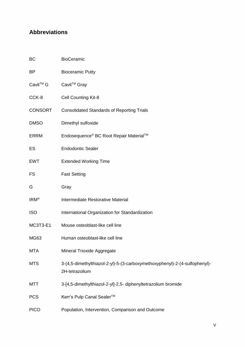
Figure 1 Workflow of root canal therapy of an infected tooth (A) by orthograde filling (B-C)
and retrograde filling (D-E). Reproduced from Ma et al.3 ......................................................15
Figure 2 Flow diagram of identification of studies for inclusion in this systematic review
according to PRISMA guidelines. .........................................................................................22
Figure 3 Methodological quality assessment of in vitro studies. ........................................51
Figure 4 Methodological quality assessment of in vivo studies. ........................................52

VIII

IX
List of Tables
Table 1 PICO strategy used for assessment of scientific literature. .................................17
Table 2 Search strategy for each of the databases. .........................................................18
Table 3 Item assessment according to the modified CONSORT checklist.22 ....................20
Table 4 Item assessment according to the SYRCLE’s risk of bias tool.23 .........................20
Table 5 Root canal sealers used in studies with in vitro and in vivo methodologies
included in this systematic review. ........................................................................................23
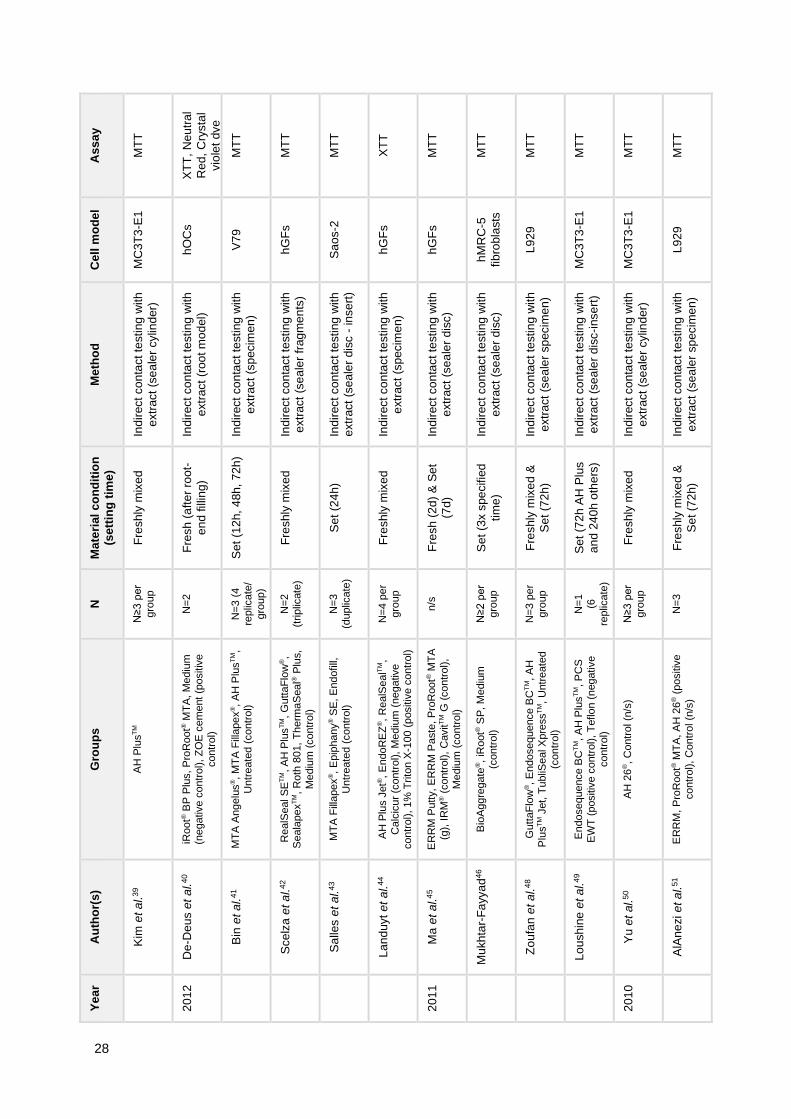
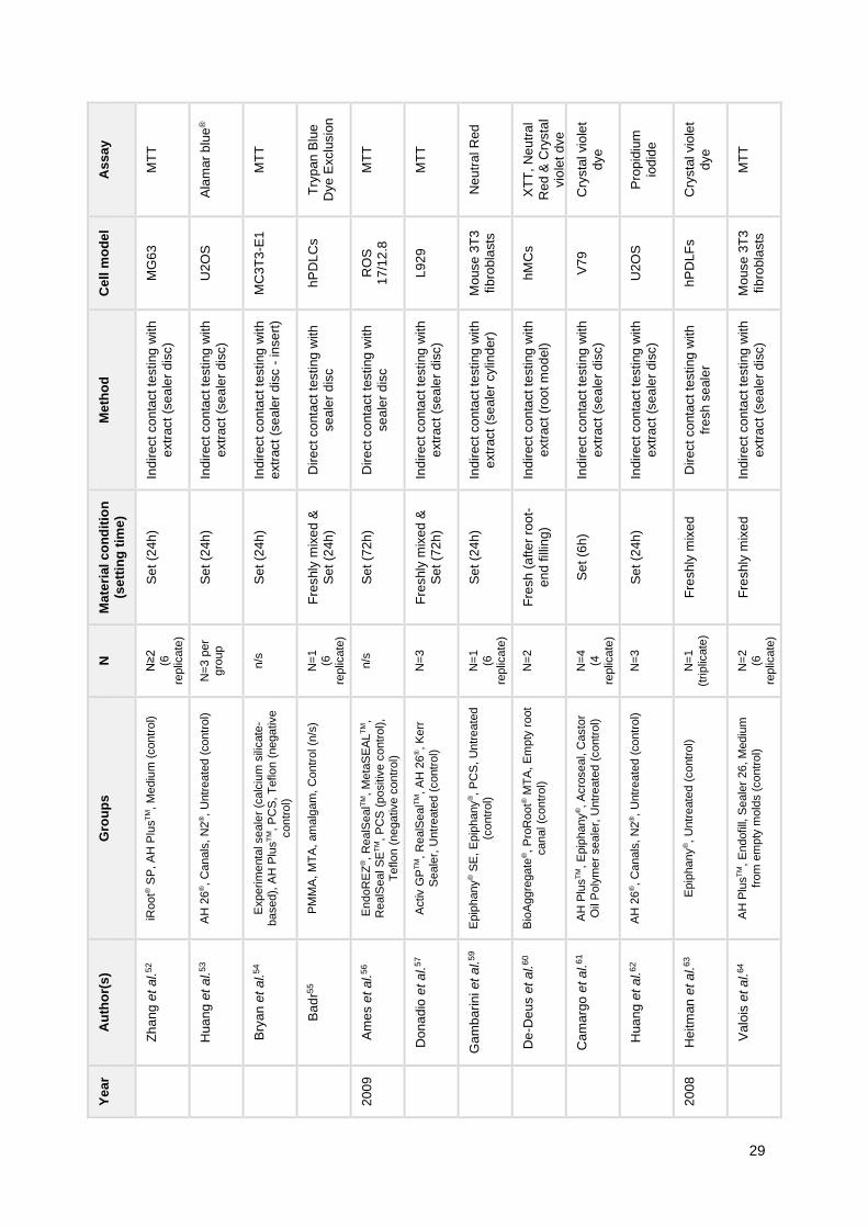
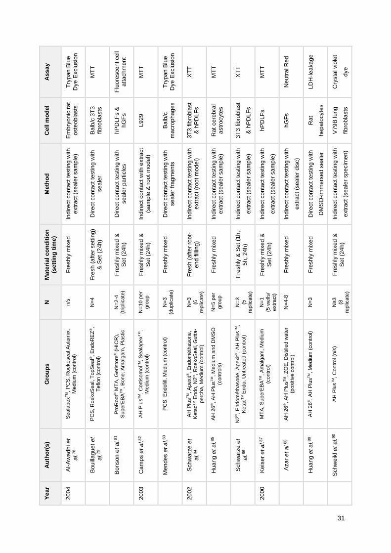
Table 6 General characteristics of included studies in regard to in vitro cytotoxicity. ........26
Table 7 Summary of parameters and results collected from included in vitro studies. ......33
Table 8 General characteristics of included studies in regard to in vivo biocompatibility. .42
Table 9 Summary of parameters and results collected from included in vivo studies. ......46

X
XI
Resumo
Introdução e Objetivos: A utilização de cimentos endodônticos de selagem canalar é
fundamental na prática clínica. No entanto, vários estudos têm evidenciado que o contacto
destes materiais com os tecidos periapicais pode determinar uma resposta citotóxica com
efeitos negativos no processo de cicatrização. Neste contexto, o nosso objetivo foi realizar
uma revisão sistemática da literatura acerca da citotoxicidade e biocompatibilidade dos
cimentos endodônticos de selagem canalar, a qual inclui os diversos tipos de cimentos
comercialmente disponíveis e evidência baseada em estudos in vitro e in vivo.
Métodos: Esta revisão sistemática foi realizada de acordo com as normas PRISMA
(Preferred Reporting Items for Systematic Reviews and Meta-Analyses), utilizando as
seguintes bases de dados: PubMed, Cochrane Library, ClinicalTrials.gov, Science Direct,
Web of Science Core Collection. Foram incluídos estudos que avaliaram a citotoxicidade
(avaliada como viabilidade/ proliferação celular) e a biocompatibilidade (avaliada como
resposta tecidular) de cimentos endodônticos de selagem canalar. O risco de viés dos
estudos in vitro foi avaliado com as normas CONSORT modificadas e dos estudos in vivo
com a ferramenta de risco de viés SYRCLE.
Resultados: A pesquisa inicial originou um total de 1382 estudos, dos quais foram incluídos
72 in vitro e 25 in vivo. Em geral, os estudos sugerem que os cimentos endodônticos de
selagem canalar induzem efeitos tóxicos ligeiros a graves a nível celular e tecidular. Os
cimentos biocerâmicos parecem exibir um menor potencial tóxico in vitro. Vários fatores
parecem influenciar a biocompatibilidade, nomeadamente a condição de presa do material e
o tempo e tipo de exposição.
Conclusões: A evidência disponível demonstra que os cimentos endodônticos de selagem
canalar exibem um potencial tóxico variável, embora a heterogeneidade entre os estudos
incluídos nesta revisão sistemática não permita concluir qual o tipo de cimento que
apresenta melhor biocompatibilidade. Desta forma, são essenciais futuras investigações no
sentido de uma melhor compreensão dos efeitos biológicos dos cimentos endodônticos de
selagem canalar.
Palavras-chave: “Endodontia”, “Cimentos de selagem canalar”, “Citotoxicidade”,
“Biocompatibilidade”, “Revisão sistemática”.

XII

XIII
Abstract
Background and Aims: The use of root canal sealers is crucial in clinical practice. However,
several studies have reported that the contact between these materials and the periapical
tissues may determine a toxic response which may hinder tissue healing. In this context, our
aim was to perform a systematic review of the literature on the cytotoxicity and
biocompatibility of root canal sealers which encompasses the several types of sealers that
are commercially available and both in vitro and in vivo evidence.
Methods: This systematic review was carried out according to the Preferred Reporting Items
for Systematic Reviews and Meta-Analyses (PRISMA) guidelines, using the following
databases: PubMed, Cochrane Library, ClinicalTrials.gov, Science Direct, Web of Science
Core Collection. Studies that evaluated the cytotoxicity (assessed as cell
viability/proliferation) and the biocompatibility (assessed as tissue response) of root canal
sealers were included. The risk of bias of in vitro studies was assessed with the modified
CONSORT guidelines and of in vivo studies with the SYRCLE’s risk of bias tool.
Results: The initial search retrieved a total of 1382 studies, from which 72 in vitro and 25 in
vivo studies were included. In general, studies suggest that root canal sealers elicit mild to
severe toxic effects at cellular and tissue level. Bioceramic sealers seem to exhibit a lower
toxic potential in vitro. Several factors may influence biocompatibility, e.g. material setting
condition and time and type of exposure.
Conclusions: The available evidence shows that root canal sealers exhibit variable toxic
potential, although the heterogeneity among studies included in this systematic review does
not allow to conclude which type of sealer presents higher biocompatibility. Thus, further
research is crucial to achieve a better understanding of the biological effects of root canal
sealers.
Keywords: “Endodontics”, “Root Canal Filling Materials”, “Cell Death”, “Biocompatibility”,
“Systematic Review”.

XIV
15
1. INTRODUCTION
Root canal therapy encompasses the sequence of procedures with the aim to treat
the infected root of a tooth, thus resulting in the resolution of the infectious process and in the
prevention of microbial invasion in the intervened tooth.1 Usually, root canal therapy is
performed by an orthograde method, which initiates with the removal of the infected pulp,
proceeds to the shaping of the root canal, its cleaning and decontamination and culminates
with the obturation (Fig. 1B-C). In some cases, however, periradicular surgery also known as
retrograde filling, which involves root-end preparation and root-end filling or retrobturation
(Fig. 1D-E), may be indicated. These include the presence of persistent periradicular
pathology, which did not respond to conventional orthograde treatment, or when this
orthograde technique is not viable, e.g. teeth with fiber post, which would be more prone to
root perforation or fracture.2,3
The usage of endodontic sealers to perform root canal filling in obturation procedures
is an established mainstay in Endodontics and plays a key role in the success of treatment 4.
Therefore, these materials should exhibit a set of characteristics that allow successful root
canal filling with resolution of periapical inflammatory and/or infectious processes and
prevent further microbial contamination.4 In this context, Grossman previously listed the
properties of an ideal sealer: (a) exhibits tackiness when mixed to provide good adhesion
with the canal wall, (b) establishes a hermetic seal, (c) is radiopaque, so that it can be
observed through radiographic observation, (d) is a very fine powder which can be easily
mixed with liquid, (e) does not shrink on setting, (f) does not stain tooth structure, (g) is
bacteriostatic (or at least does not promote bacterial growth), (h) displays a slow setting, (i) is
insoluble in host tissue fluids, (j) is biocompatible, i.e. without irritant potential to periradicular
tissue, and (k) is soluble in common solvent, allowing for removal when necessary.4,5
Figure 1 Workflow of root canal therapy of an infected tooth (A) by orthograde filling (B-C) and
retrograde filling (D-E). Reproduced from Ma et al.3
16
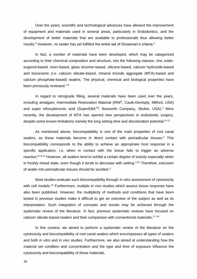
Over the years, scientific and technological advances have allowed the improvement
of equipment and materials used in several areas, particularly in Endodontics, and the
development of better materials that are available to professionals thus allowing better
results.6 However, no sealer has yet fulfilled the entire set of Grossman’s criteria.4
In fact, a number of materials have been developed, which may be categorized
according to their chemical composition and structure, into the following classes: zinc oxide-
eugenol-based, resin-based, glass ionomer-based, silicone-based, calcium hydroxide-based
and bioceramic (i.e. calcium silicate-based, mineral trioxide aggregate (MTA)-based and
calcium phosphate-based) sealers. The physical, chemical and biological properties have
been previously reviewed.7,8
In regard to retrograde filling, several materials have been used over the years,
including amalgam, Intermediate Restoration Material (IRM®, Caulk-Dentsply, Milford, USA)
and super ethoxybenzoic acid (SuperEBATM, Bosworth Company, Skokie, USA).3 More
recently, the development of MTA has opened new perspectives in endodontic surgery,
despite some known limitations namely the long setting time and discoloration potential.9–12
As mentioned above, biocompatibility is one of the main properties of root canal
sealers, as these materials become in direct contact with periradicular tissues.4 This
biocompatibility corresponds to the ability to achieve an appropriate host response in a
specific application, i.e. when in contact with the tissue fails to trigger an adverse
reaction.8,13,14 However, all sealers tend to exhibit a certain degree of toxicity especially when
in freshly mixed state, even though it tends to decrease with setting.4,15 Therefore, extrusion
of sealer into periradicular tissues should be avoided.4
Most studies evaluate such biocompatibility through in vitro assessment of cytotoxicity
with cell models.16 Furthermore, multiple in vivo studies which assess tissue response have
also been published. However, the multiplicity of methods and conditions that have been
tested in previous studies make it difficult to get an overview of the subject as well as its
interpretation. Such integration of concepts and results may be achieved through the
systematic review of the literature. In fact, previous systematic reviews have focused on
calcium silicate-based sealers and their comparison with conventional materials.17–19
In this context, we aimed to perform a systematic review of the literature on the
cytotoxicity and biocompatibility of root canal sealers which encompasses all types of sealers
and both in vitro and in vivo studies. Furthermore, we also aimed at understanding how the
material set condition and concentration and the type and time of exposure influence the
cytotoxicity and biocompatibility of these materials.
17
2. METHODS
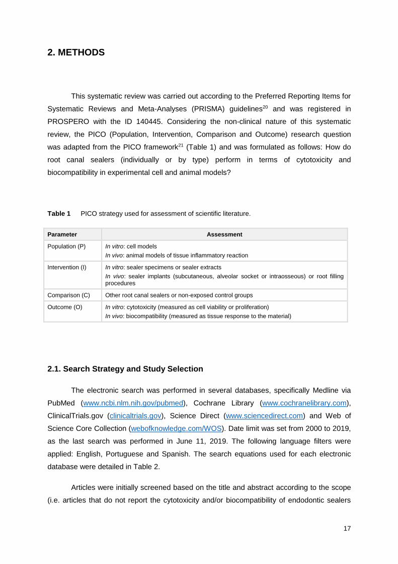
This systematic review was carried out according to the Preferred Reporting Items for
Systematic Reviews and Meta-Analyses (PRISMA) guidelines20 and was registered in
PROSPERO with the ID 140445. Considering the non-clinical nature of this systematic
review, the PICO (Population, Intervention, Comparison and Outcome) research question
was adapted from the PICO framework21 (Table 1) and was formulated as follows: How do
root canal sealers (individually or by type) perform in terms of cytotoxicity and
biocompatibility in experimental cell and animal models?
Table 1 PICO strategy used for assessment of scientific literature.
Parameter Assessment
Population (P) In vitro: cell models
In vivo: animal models of tissue inflammatory reaction
Intervention (I) In vitro: sealer specimens or sealer extracts
In vivo: sealer implants (subcutaneous, alveolar socket or intraosseous) or root filling procedures
Comparison (C) Other root canal sealers or non-exposed control groups
Outcome (O) In vitro: cytotoxicity (measured as cell viability or proliferation)
In vivo: biocompatibility (measured as tissue response to the material)
2.1. Search Strategy and Study Selection
The electronic search was performed in several databases, specifically Medline via
PubMed (www.ncbi.nlm.nih.gov/pubmed), Cochrane Library (www.cochranelibrary.com),
ClinicalTrials.gov (clinicaltrials.gov), Science Direct (www.sciencedirect.com) and Web of
Science Core Collection (webofknowledge.com/WOS). Date limit was set from 2000 to 2019,
as the last search was performed in June 11, 2019. The following language filters were
applied: English, Portuguese and Spanish. The search equations used for each electronic
database were detailed in Table 2.
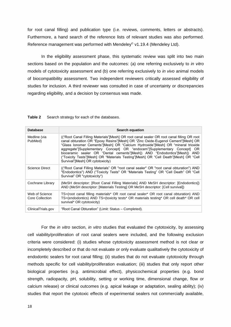
Articles were initially screened based on the title and abstract according to the scope
(i.e. articles that do not report the cytotoxicity and/or biocompatibility of endodontic sealers
18
for root canal filling) and publication type (i.e. reviews, comments, letters or abstracts).
Furthermore, a hand search of the reference lists of relevant studies was also performed.
Reference management was performed with Mendeley© v1.19.4 (Mendeley Ltd).
In the eligibility assessment phase, this systematic review was split into two main
sections based on the population and the outcomes: (a) one referring exclusively to in vitro
models of cytotoxicity assessment and (b) one referring exclusively to in vivo animal models
of biocompatibility assessment. Two independent reviewers critically assessed eligibility of
studies for inclusion. A third reviewer was consulted in case of uncertainty or discrepancies
regarding eligibility, and a decision by consensus was made.
Table 2 Search strategy for each of the databases.
Database Search equation
Medline (via PubMed)
((“Root Canal Filling Materials”[Mesh] OR root canal sealer OR root canal filling OR root canal obturation OR “Epoxy Resins”[Mesh] OR “Zinc Oxide-Eugenol Cement”[Mesh] OR “Glass Ionomer Cements”[Mesh] OR “Calcium Hydroxide”[Mesh] OR “mineral trioxide aggregate”[Supplementary Concept] OR “endocem”[Supplementary Concept] OR bioceramic sealer OR “Dental cements”[Mesh]) AND “Endodontics”[Mesh]) AND (“Toxicity Tests”[Mesh] OR “Materials Testing”[Mesh] OR “Cell Death”[Mesh] OR “Cell Survival”[Mesh] OR cytotoxicity)
Science Direct ((“Root Canal Filling Materials” OR "root canal sealer" OR "root canal obturation") AND "Endodontics") AND (“Toxicity Tests" OR “Materials Testing” OR “Cell Death” OR “Cell Survival” OR "cytotoxicity")
Cochrane Library (MeSH descriptor: [Root Canal Filling Materials] AND MeSH descriptor: [Endodontics]) AND (MeSH descriptor: [Materials Testing] OR MeSH descriptor: [Cell survival])
Web of Science Core Collection
TS=(root canal filling materials* OR root canal sealer* OR root canal obturation) AND TS=(endodontics) AND TS=(toxicity tests* OR materials testing* OR cell death* OR cell survival* OR cytotoxicity)
ClinicalTrials.gov “Root Canal Obturation” (Limit: Status – Completed).
For the in vitro section, in vitro studies that evaluated the cytotoxicity, by assessing
cell viability/proliferation of root canal sealers were included, and the following exclusion
criteria were considered: (i) studies whose cytotoxicity assessment method is not clear or
incompletely described or that do not evaluate or only evaluate qualitatively the cytotoxicity of
endodontic sealers for root canal filling; (ii) studies that do not evaluate cytotoxicity through
methods specific for cell viability/proliferation evaluation; (iii) studies that only report other
biological properties (e.g. antimicrobial effect), physicochemical properties (e.g. bond
strength, radiopacity, pH, solubility, setting or working time, dimensional change, flow or
calcium release) or clinical outcomes (e.g. apical leakage or adaptation, sealing ability); (iv)
studies that report the cytotoxic effects of experimental sealers not commercially available,

19
modified commercially-available root canal sealers, modified sealer components or dental
materials used as pulp capping materials and others (e.g. adhesive systems); and (v) studies
other than in vitro, e.g. in vivo or in silico.
For the in vivo section, in vivo animal studies that have evaluated the biocompatibility
of root canal sealers through the assessment of tissue reaction after subcutaneous,
intraosseous, alveolar socket or root canal implantation were included. For this section, the
following exclusion criteria were considered: (i) studies that do not report the biocompatibility
of endodontic sealers for root canal filling according to the methods described in the inclusion
criteria; (ii) studies that only report other biological properties or clinical outcomes; (iii) studies
that report the biocompatibility of experimental sealers not commercially available, modified
commercially-available root canal sealers or dental materials used as pulp capping materials;
and (iv) studies other than in vivo, e.g. in vitro.
Studies with missing data were excluded.
2.2. Data Collection
The following descriptive and quantitative information was extracted from each of the
eligible studies for both sections, i.e. in vitro and in vivo: authors and year of publication,
tested sealer(s) and controls, sample size, sealer material condition (i.e. fresh or set), the
setting time if set materials were used, method of sealer preparation (i.e. if in accordance to
manufacturer’s instructions), results and main conclusions. Relatively to in vitro studies, the
following information was also extracted: method (i.e. direct or indirect contact with sealer
specimens or extracts), extraction time and extracts concentration if extracts were obtained,
cell model and exposure time, cell viability/proliferation assay. In regard to in vivo studies, the
following information was also extracted: method of biocompatibility assessment (i.e.
subcutaneous, alveolar, intraosseous or root canal implantation), teeth used for root canal
filling if this method was used, animal model, exposure time and method of histologic
analysis (including staining method and outcomes measured).
2.3. Risk of Bias
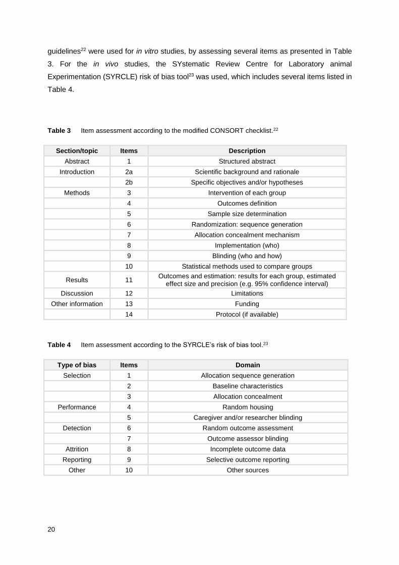
The methodologic quality of eligible studies was checked by assessing the risk of bias
of individual studies. The modified Consolidated Standards of Reporting Trials (CONSORT)
20
guidelines22 were used for in vitro studies, by assessing several items as presented in Table
3. For the in vivo studies, the SYstematic Review Centre for Laboratory animal
Experimentation (SYRCLE) risk of bias tool23 was used, which includes several items listed in
Table 4.
Table 3 Item assessment according to the modified CONSORT checklist.22
Section/topic Items Description
Abstract 1 Structured abstract
Introduction 2a Scientific background and rationale
2b Specific objectives and/or hypotheses
Methods 3 Intervention of each group
4 Outcomes definition
5 Sample size determination
6 Randomization: sequence generation
7 Allocation concealment mechanism
8 Implementation (who)
9 Blinding (who and how)
10 Statistical methods used to compare groups
Results 11 Outcomes and estimation: results for each group, estimated
effect size and precision (e.g. 95% confidence interval)
Discussion 12 Limitations
Other information 13 Funding
14 Protocol (if available)
Table 4 Item assessment according to the SYRCLE’s risk of bias tool.23
Type of bias Items Domain
Selection 1 Allocation sequence generation
2 Baseline characteristics
3 Allocation concealment
Performance 4 Random housing
5 Caregiver and/or researcher blinding
Detection 6 Random outcome assessment
7 Outcome assessor blinding
Attrition 8 Incomplete outcome data
Reporting 9 Selective outcome reporting
Other 10 Other sources

21
3. RESULTS
The full process of article retrieving, screening and eligibility assessment is presented
in Fig. 2. As can be seen, the initial search retrieved a total of 1382 studies, from which 188
were excluded after removal of duplicates. A total of 1194 studies were screened based on
the title and abstract, from which 1027 were excluded, resulting in 167 full-text studies that
were considered potentially eligible for inclusion, which included 135 in vitro studies, 30 in
vivo studies and 2 studies with both in vitro and in vivo testing. A total of 77 studies (65 in
vitro, 11 in vivo and 1 both in vitro and in vivo) was excluded because they did not meet the
inclusion criteria. Studies that did not specify the material condition, i.e. freshly mixed or set,
were excluded. After reviewing the full texts, 6 in vivo and 1 both in vitro and in vivo studies
were added to the analysis by hand searching. Finally, 70 in vitro24–93, 25 in vivo94–118 and 2
both in vitro and in vivo studies119,120 were included in this review. The studies with both in
vitro and in vivo methodologies were included only for the in vitro data, as the in vivo
methodology did not meet inclusion criteria. In Table 5, we listed the several root canal
sealers for orthograde and retrograde filling, the respective manufacturers and the included
articles in which they were studied.
As can be seen, the most studied sealers in vitro were: AH 26®, AH PlusTM,
EndoREZ®, Endosequence BCTM, Epiphany®, MTA Fillapex®, Kerr’s Pulp Canal SealerTM
(PCS), ProRoot® MTA and SealapexTM. Other materials included: Amalgam55,81,87, Castor Oil
Polymer (Poliquil, Brazil)61, CavitTM Gray or G (3M ESPE, Seefeld, Germany)45, CYMED
8410 (NANO, Kaohsiung, Taiwan)68, DiaketTM (3M ESPE, Seefeld, Germany)68,
Endosequence® BC Root Repair MaterialTM (ERRM, Brasseler, Savannah, USA)45,51,
Geristore® (DenMat® Corporation, Santa Maria, USA)66,81, IRM® 35,45, Retroplast (Retroplast
Trading, Dybesøvej, Denmark)66 and SuperEBATM.30,66,81,87
Regarding in vivo studies, AH PlusTM, EndoREZ®, Epiphany® and ProRoot® MTA
were the most studied. Other materials were also studied, such as the high-copper
amalgams Oralloy (Coltène AG, Altstatten, Switzerland)108 and Sinaalloy (Faghihi, Iran)102,
the calcium-hydroxide and polyethylene-glycol-based paste Calen® (S.S.White Artigos
Dentários Ltda., Rio de Janeiro, Brazil)114, the calcium phosphate-based sealers Capseal I
and II105, the zinc oxide-eugenol-based sealer Fillcanal (DG Ligas Odontológicas Ltda, Rio
de Janeiro, Brazil)110, Intrafill (Dentsply Ind. e Com. Ltda., Rio de Janeiro, Brazil)96, IRM® 109
and SuperEBATM.109

22
3.1. In Vitro Cytotoxicity
The characteristics of the included studies in respect to in vitro cytotoxicity of root
canal sealers is presented in Table 6. From the 72 studies, 18 used a direct contact testing
method with sealers prepared either as fresh sample, disc, layer or cylindrical
specimens27,31,32,38,55,56,58,63,65,74–77,79,81,83,89,93, as others used root models.26,40,60,82,84 In terms of
material setting condition, 22 studies evaluated root canal sealers in a fresh or freshly mixed
state, 16 in a set condition with 24h of incubation, 17 in both freshly mixed and set conditions
and 17 in a set condition with other or multiple times of incubation.
Figure 2 Flow diagram of identification of studies for inclusion in this systematic review according to
PRISMA guidelines.

23
Table 5 Root canal sealers used in studies with in vitro and in vivo methodologies included in this
systematic review.
Type Sealer Manufacturer In vitro In vivo
ZnO-eugenol PCS Kerr, Romulus, USA 25–27,54,56,59,65,78,79,83 105,116,117
PCS EWT Kerr, Romulus, USA 49 –
N2® Indrag-Agsa, Losone, Switzerland 53,62,67,70,72,84,86 –
Endofill Produits Dentaires, Vevey Switzerland
43,64,83,119 112
Canals Showa Pharmaceutical Co., Tokyo, Japan
53,62,67 –
Endométhasone Septodont, Saint-Maur-des-Fossés, France
84,86 99,106
Roth’s Sealer Roth International, Chicago, USA 36,42 –
Grossman’s Sultan Chemists, Englewood, USA
75 –
Zinc Oxide-Eugenol (ZOE)
Produits Dentaires, Vevey Switzerland
40,88 114
Tubli-SealTM Kerr, Romulus, USA 58 –
Tubli-Seal XpressTM Kerr, Romulus, USA 48 –
CortisomolTM Pierre Rolland, Merignac, France 82 –
Resin
(epoxy)
AH PlusTM Dentsply DeTrey Gmbh, Konstanz, Germany
24,28,33,34,36,39,41,42,49,52
,54,58,61,64,73,74,76,77,80,8
2,84–86,88–90,93,119
94,96,97,111,115
AH 26® Dentsply DeTrey Gmbh, Konstanz, Germany
50,51,53,62,67,70–
72,85,88,89
110
AH Plus Jet® Dentsply DeTrey Gmbh, Konstanz, Germany
31,37,44,48,65 –
Acroseal Septodont, Saint-Maur-des-Fossés, France
61,93 –
SimpliSeal® Discuss Dental LLC, Calver City, USA
47,119 –
TopSeal® Dentsply DeTrey Gmbh, Konstanz, Germany
79 –
Sealer Plus MK Life, Porto Alegre, Brazil 119 –
ThermaSeal® Dentsply/Maillefer, Konstanz, Germany
75 –
ThermaSeal® Plus Dentsply/Maillefer, Konstanz, Germany
42 –
Resin (methacrylate)
EndoREZ® Ultradent, South Jordan, USA 28,34,44,56,58,74,79 99,107,117,118
Epiphany® Pentron, Wallingford, USA 59,61,63,73–76 96,98,100,112,116
Epiphany® SE Pentron, Wallingford, USA 43,59 –
RealSealTM SybronEndo, Orange, USA 33,44,56,57 117
RealSeal SETM SybronEndo, Orange, USA 42,56 –

24
RealSeal XT SybronEndo, Orange, USA 31 95
MetaSEALTM Parkell, Inc., Farmington, USA 56,65 –
Glass ionomer KetacTM Endo 3M ESPE, St. Paul, USA 84,86 –
KetacTM Fil Plus 3M ESPE, St. Paul, USA 66 –
Activ GPTM Brasseler, Savannah, USA 57 –
Endion® VOCO, Cuxhaven, Germany 68 –
Silicone GuttaFlow® Roeko/Coltène/Whaledent, Langenau, Germany
28,42,48,76 –
GuttaFlow®2 Roeko/Coltène/Whaledent, Langenau, Germany
33,80 111
GuttaFlow® Bioseal Roeko/Coltène/Whaledent, Langenau, Germany
80 111
RoekoSeal Roeko/Coltène/Whaledent, Langenau, Germany
34,79,84 96
RoekoSeal Automix Roeko/Coltène/Whaledent, Langenau, Germany
74,77,78 97
Calcium hydroxide
SealapexTM Kerr, Romulus, USA 32,42,58,70,72,75,78,82 103
Apexit® Ivoclar Vivadent, Schaan, Liechtenstein
28,84,86 –
Sealapex XpressTM SybronEndo, Orange, USA – 95
Sealer 26 Dentsply/Maillefer, Konstanz, Germany
64 103,110
Bioceramic ProRoot® MTA Dentsply Tulsa Dental, Tulsa, USA
30,33,35,38,40,45,51,55,60,66
,68,81,91,92
101,102,104,113
ProRoot® ES Dentsply Tulsa Dental, Tulsa, USA
36 –
MTA Fillapex® Angelus, Londrina, Brazil 24,29,32,37,41,43,47,80,93 94
Endosequence BCTM Brasseler, Savannah, USA 24,29,36,38,48,49 –
iRoot® SP Innovative BioCeramix Inc., Vancouver, Canada
32,37,46,52,91 –
iRoot® BP Plus Innovative BioCeramix Inc., Vancouver, Canada
30,40,92 –
iRoot® FS Innovative BioCeramix Inc., Vancouver, Canada
30,92 –
BioRootTM RCS Septodont, Saint-Maur-des-Fossés, France
25–27,47 –
MTA Angelus® Angelus, Londrina, Brazil 38,41,120 –
BioAggregate® Innovative BioCeramix Inc., Vancouver, Canada
46,60 –
Endoseal® MTA Maruchi, Seoul, Korea 69 –
MTA High plasticity Angelus, Londrina, Brazil 120 120
Endocem Maruchi, Seoul, Korea 35 –
Sankin apatite root sealer
Sankin Kogyo, Tokyo, Japan 32 105

25
Abbreviations: BC, BioCeramic; BP, Bioceramic Putty; ES, Endodontic Sealer; EWT, Extended Working Time; FS, Fast Setting; GP, Gutta-percha; MTA, Mineral Trioxide Aggregate; PCS, Kerr’s Pulp Canal SealerTM; RCS, Root Canal Sealer; SE, Self-Etch; SP, Sealing Paste; ZnO, Zinc Oxide.
Concerning the cell models used for cell viability assessment, several studies used
cultures of human cells, namely: dental follicle-derived mesenchymal stem cells93, tooth
germ-derived stem cells37, gingival fibroblasts28,29,33,42,44,45,75,88, osteoblasts38,40,43,53,62,67,93,
periodontal ligament cells25,26,32,38,55,63,68,69,80,81,84,86,87, human osteoblast-like cells
(MG63)30,35,52 and cervical carcinoma cells or HeLa cells.73,77 Other cell lines were also used,
as can be seen in Table 6, e.g. L929 mouse fibroblasts, mouse osteoblast-like cells (MC3T3-
E1), RAW 264.7 mouse macrophages, Chinese hamster fibroblasts (V79), rat osteosarcoma
(ROS) 17/12.8 cells, Balb/c fibroblasts and rat clonal dental pulp cells (RPC-C2A).
Regarding the type of cell viability assay, most of the studies used assays that
measure metabolic activity, specifically: 36 studies used the 3-[4,5-dimethylthiazol-2-yl]-2,5-
diphenyltetrazolium bromide (MTT) assay, 3 used the 2,3-bis-(2-methoxy-4-nitro-5-
sulfophenyl)-2H-tetrazolium-5-carboxanilide (XTT) assay, 4 used the Alamar blue® assay, 3
used the Cell Counting Kit-8 (CCK-8/WST-8) assay, 2 used the Water Soluble Tetrazolium
Salt-1 (WST-1) assay, 1 used the 3-(4,5-dimethylthiazol-2-yl)-5-(3-carboxymethoxyphenyl)-2-
(4-sulfophenyl)-2H-tetrazolium (MTS) assay. Other methods included the Trypan blue dye
exclusion assay (5 studies), the Neutral Red uptake assay (2 studies), the ATP-based
luminescence assay (1 study), the Sulforhodamine B assay (1 study), the Live/Dead Viability
assay by flow cytometry (1 study), the crystal violet assay (3 studies), the propidium iodide
fluorescence assay (1 study), the Hoechst 33258 fluorescence assay (1 study), the Millipore
filter assay (1 study), fluorescent cell attachment with proprietary green fluorescent dye (1
study), the Nigrosin dye assay (1 study) and the lactate dehydrogenase-leakage assay (1
study). Also, 4 studies used multiple methods to assess cell viability.
3.1.1. Cytotoxicity of root canal sealers
In general, the tested root canal sealers exhibited cytotoxicity (Table 7).
The most studied sealer was the epoxy resin-based sealer AH Plus, which was
reported as cytotoxic in most of the studies in which it was tested. However, one study82
reported as noncytotoxic, one88 reported a cytotoxic effect only in early phase and one90
reported as cytotoxic when eluted in dimethyl sulfoxide (DMSO) but noncytotoxic when
eluted in sodium chloride.

26
Table 6 General characteristics of included studies in regard to in vitro cytotoxicity. A
ss
ay
WS
T-1
MT
T
AT
P-b
ase
d
Lu
min
escen
ce
Su
lfo
rho
dam
ine
B
MT
T
MT
T
MT
T
Ala
ma
r b
lue
®
MT
T
MT
T
CC
K-8
/WS
T-8
Ala
ma
r b
lue
®
Ce
ll m
od
el
MC
3T
3-E
1
hP
DL
Fs
IDG
-SW
3
NIH
/3T
3
L9
29
hP
DL
SC
s
hP
DL
SC
s
L9
29
RA
W 2
64
.7
ma
cro
ph
ag
es
L9
29
MC
3T
3-E
1
hO
Cs &
DF
-MS
Cs
Me
tho
d
Ind
irect co
nta
ct te
sting
with
extr
act
(se
ale
r d
isc)
Ind
irect co
nta
ct te
sting
with
extr
act
(sp
ecim
en
)
Ind
irect co
nta
ct te
sting
with
extr
act
(sp
ecim
en
)
Ind
irect co
nta
ct te
sting
with
extr
act
(sp
ecim
en
)
Dir
ect co
nta
ct te
sting
with
se
ale
r
Ind
irect co
nta
ct te
sting
with
extr
act
(se
ale
r d
isc)
Ind
irect co
nta
ct te
sting
with
extr
act
(se
ale
r d
isc)
Ind
irect co
nta
ct te
sting
with
extr
act
(se
ale
r d
isc)
Ind
irect co
nta
ct te
sting
with
extr
act
(se
ale
r d
isc)
Ind
irect co
nta
ct te
sting
with
extr
act
(se
ale
r d
isc)
Ind
irect co
nta
ct te
sting
with
extr
act
(se
ale
r d
isc)
Dir
ect co
nta
ct te
sting
with
se
ale
r
Ma
teri
al
co
nd
itio
n
(se
ttin
g t
ime
)
Se
t (2
4h
)
Fre
shly
mix
ed
Fre
shly
mix
ed
Se
t (4
8h
)
Fre
shly
mix
ed
Se
t (4
8h
)
Se
t (4
8h
)
Se
t (6
h)
Se
t (2
4h
)
Se
t (6
h)
Se
t (7
d)
Se
t (2
4h
)
N
N≥2 p
er
gro
up
(trip
licate
)
N=
3 p
er
gro
up
(trip
licate
)
N=
6-1
2
per
gro
up
N≥2 p
er
gro
up (
6
replic
ate
)
N=
3 p
er
gro
up
N=
1 p
er
gro
up (
5
replic
ate
)
N≥2 p
er
gro
up (
5
replic
ate
)
N=
1
(trip
licate
)
n/s
N=
1
(trip
licate
)
N=
3
N=
1
(trip
licate
)
Gro
up
s
AH
Plu
sT
M, M
TA
Fill
apex
®,
Endosequence B
CT
M, M
ediu
m (
contr
ol)
Bio
RootT
M R
CS
, P
CS
, M
ediu
m
(contr
ol)
Roth
´s S
eale
r, A
H P
lus
TM,
Endosequence B
CT
M, P
roR
oot®
ES
,
No c
ells
(contr
ol), M
ediu
m (
contr
ol)
Sim
pliS
eal®
, M
TA
Fill
apex
®,
Bio
RootT
M R
CS
, M
ediu
m (
contr
ol)
Tu
bli-
SealT
M, A
H P
lus
TM,
Seala
pex
TM, E
ndoR
EZ
®, M
ediu
m
(contr
ol) [
gro
ups w
ith p
achym
ic a
cid
]
Bio
RootT
M B
CS
, E
ndoseal®
, N
ano-
cera
mic
Seale
r (N
CS
), M
ediu
m
(contr
ol)
Gutt
aF
low
® B
ioseal, G
utt
aF
low
®2,
MT
A F
illa
pex
®, A
H P
lus
TM, M
ediu
m
(contr
ol)
MT
A H
igh p
lasticity (
HP
), M
TA
Angelu
s®, M
ediu
m (
contr
ol)
iRoot®
SP
, M
TA
, M
ediu
m (
contr
ol)
Seale
r P
lus, A
H P
lus
TM, E
ndofill,
Sim
pliS
eal®
, M
ediu
m (
contr
ol)
iRoot®
FS
, iR
oot®
BP
Plu
s, P
roR
oot®
MT
A, M
ediu
m (
contr
ol)
MT
A F
illa
pex
®, A
H P
lus
TM, A
cro
seal,
Pla
stic s
urf
ace (
contr
ol)
Au
tho
r(s
)
Le
e e
t a
l.2
4
Je
ann
ea
u e
t a
l.2
5
Gia
com
ino e
t a
l.3
6
Vo
uza
ra e
t a
l.47
Aru
n e
t a
l.5
8
Colla
do
-Go
nzá
lez
et
al.
69
Colla
do
-Go
nzá
lez
et
al.
80
Cin
tra
et
al.
12
0
Zh
u e
t a
l.91
Cin
tra
et
al.
11
9
Lv e
t a
l.9
2
Su
ciu
et
al.
93
Ye
ar
20
19
20
18
20
17
20
16

27
As
sa
y
MT
T
Try
pan
Blu
e
Dye
Exclu
sio
n
WS
T-1
Liv
e/D
ea
d
Via
bili
ty (
Flo
w
cyto
me
try)
MT
T
MT
T &
Neu
tra
l
Re
d
MT
T
CC
K-8
/WS
T-8
MT
T
MT
T
MT
S
Ala
ma
r b
lue
®
Ce
ll m
od
el
hP
DL
Cs
A4
mou
se
pu
lp S
Cs
hG
Fs
hG
Fs
L9
29
&
MG
63
L9
29
hP
DL
Cs
hG
Fs
V7
9
MG
63
hT
GS
Cs
hP
DL
Fs &
hO
Cs
Me
tho
d
Ind
irect co
nta
ct te
sting
with
extr
act
(roo
t m
od
el)
Dir
ect co
nta
ct te
sting
with
se
ale
r d
isc
Ind
irect co
nta
ct te
sting
with
extr
act
(se
ale
r dis
c)
Ind
irect co
nta
ct te
sting
with
extr
act
(se
ale
r dis
c)
Ind
irect co
nta
ct te
sting
with
extr
act
(se
ale
r dis
c)
Dir
ect co
nta
ct te
sting
with
se
ale
r
Dir
ect co
nta
ct te
sting
with
se
ale
r d
isc (
with
O.S
.)
Ind
irect co
nta
ct te
sting
with
extr
act
(se
ale
r dis
c)
Ind
irect co
nta
ct te
sting
with
extr
act
(se
ale
r la
ye
r)
Ind
irect co
nta
ct te
sting
with
extr
act
(se
ale
r dis
c)
Ind
irect co
nta
ct te
sting
with
extr
act
(se
ale
r dis
c -
in
se
rt)
Dir
ect co
nta
ct te
sting
with
se
ale
r d
isc
Ma
teri
al
co
nd
itio
n
(se
ttin
g t
ime
)
Se
t (2
4h
)
Se
t (2
4h
)
Se
t (i
mm
ed
iate
ly
aft
er,
24
h, 4
8h
, 7
d)
Fre
shly
mix
ed
& S
et
(3x s
pecifie
d tim
e)
Se
t (7
d)
Fre
sh
Se
t (2
4h
)
Fre
sh
& S
et
(72
h)
Fre
shly
mix
ed
& S
et
(12
h,
24
h)
Se
t (2
4h
)
Se
t (2
4h
)
Se
t (2
4h
)
N
N=
30
(N=
3/
gro
up)
N≥3
(trip
licate
)
N=
60
(tota
l)
N=
1
(trip
licate
)
n/s
N=
3 p
er
gro
up
N=
3
(4 w
ells
/ conditio
n)
N=
1
(5
replic
ate
)
N=
3
(4 w
ells
/
conditio
n)
n/s
N=
6 p
er
gro
up
N=
6 p
er
gro
up
Gro
up
s
Bio
RootT
M R
CS
, P
CS
, M
ediu
m (
contr
ol)
Bio
RootT
M R
CS
, P
CS
, U
ntr
eate
d c
ells
(c
ontr
ols
)
Gutt
aF
low
®, A
H P
lus
TM, A
pexit
®,
EndoR
EZ
®,
Contr
ol (n
/s)
Endosequence B
CT
M, M
TA
Fill
apex
®,
Me
diu
m (
contr
ol)
iRoot®
BP
Plu
s, iR
oot®
FS
, P
roR
oot®
MT
A,
SuperE
BA
TM, M
ediu
m (
contr
ol)
RealS
eal X
T, A
H P
lus J
et®
, U
ntr
eate
d
(contr
ol)
Seala
pex
TM, A
patite
Root S
eale
r, M
TA
Fill
apex
®, iR
oot®
SP
, M
ediu
m w
ith &
w
ithout
O.S
. (c
ontr
ol)
Gutt
aF
low
®2, P
roR
oot®
MT
A, A
H P
lus
TM,
RealS
ealT
M, M
ediu
m (
contr
ol)
AH
Plu
sT
M, E
ndoR
EZ
®,
RoekoS
eal,
Me
diu
m (
contr
ol)
Pro
Root®
MT
A, E
ndocem
, IR
M®, M
ediu
m
(contr
ol)
MT
A F
illa
pex
®,
iRoot®
SP
, A
H P
lus J
et®
,
Contr
ol (n
/s)
MT
A A
ngelu
s® (
gra
y &
white),
Pro
Root®
MT
A, E
ndosequence B
CT
M,
Untr
eate
d
(contr
ol)
Au
tho
r(s
)
Cam
ps e
t al.
26
Dim
itro
va
-Nakov
et
al.
27
Ko
njh
od
zic
-Prc
ic
et
al.
28
Zh
ou
et
al.
29
Jia
ng
et a
l.30
Cott
i e
t a
l.3
1
Cha
ng
et
al.
32
Ma
nda
l et
al.
33
Ca
ma
rgo
et a
l.3
4
Cho
i e
t a
l.35
Gü
ve
n e
t a
l.3
7
Will
ers
ha
use
n e
t
al.
38
Ye
ar
20
15
20
14
20
13
28
As
sa
y
MT
T
XT
T,
Ne
utr
al
Re
d,
Cry
sta
l
vio
let
dye
MT
T
MT
T
MT
T
XT
T
MT
T
MT
T
MT
T
MT
T
MT
T
MT
T
Ce
ll m
od
el
MC
3T
3-E
1
hO
Cs
V7
9
hG
Fs
Sa
os-2
hG
Fs
hG
Fs
hM
RC
-5
fib
robla
sts
L9
29
MC
3T
3-E
1
MC
3T
3-E
1
L9
29
Me
tho
d
Ind
irect co
nta
ct te
sting
with
extr
act
(se
ale
r cylin
der)
Ind
irect co
nta
ct te
sting
with
extr
act
(roo
t m
od
el)
Ind
irect co
nta
ct te
sting
with
extr
act
(sp
ecim
en
)
Ind
irect co
nta
ct te
sting
with
extr
act
(se
ale
r fr
ag
me
nts
)
Ind
irect co
nta
ct te
sting
with
extr
act
(se
ale
r dis
c -
in
se
rt)
Ind
irect co
nta
ct te
sting
with
extr
act
(sp
ecim
en
)
Ind
irect co
nta
ct te
sting
with
extr
act
(se
ale
r dis
c)
Ind
irect co
nta
ct te
sting
with
extr
act
(se
ale
r dis
c)
Ind
irect co
nta
ct te
stin
g w
ith
extr
act
(se
ale
r sp
ecim
en
)
Ind
irect co
nta
ct te
sting
with
extr
act
(se
ale
r dis
c-i
nse
rt)
Ind
irect co
nta
ct te
sting
with
extr
act
(se
ale
r cylin
der)
Ind
irect co
nta
ct te
sting
with
extr
act
(se
ale
r sp
ecim
en
)
Ma
teri
al
co
nd
itio
n
(se
ttin
g t
ime
)
Fre
shly
mix
ed
Fre
sh
(a
fte
r ro
ot-
en
d fill
ing
)
Se
t (1
2h,
48h
, 72
h)
Fre
shly
mix
ed
Se
t (2
4h
)
Fre
shly
mix
ed
Fre
sh
(2
d)
& S
et
(7d
)
Se
t (3
x s
pe
cifie
d
tim
e)
Fre
shly
mix
ed
&
Se
t (7
2h
)
Se
t (7
2h A
H P
lus
an
d 2
40
h o
the
rs)
Fre
shly
mix
ed
Fre
shly
mix
ed
&
Se
t (7
2h
)
N
N≥3 p
er
gro
up
N=
2
N=
3 (
4
replic
ate
/
gro
up)
N=
2
(trip
licate
)
N=
3
(duplic
ate
)
N=
4 p
er
gro
up
n/s
N≥2 p
er
gro
up
N=
3 p
er
gro
up
N=
1
(6
replic
ate
)
N≥3 p
er
gro
up
N=
3
Gro
up
s
AH
Plu
sT
M
iRoot®
BP
Plu
s, P
roR
oot®
MT
A, M
ediu
m
(negative c
ontr
ol), Z
OE
cem
ent
(positiv
e
contr
ol)
MT
A A
ngelu
s®, M
TA
Fill
apex
®, A
H P
lus
TM,
Untr
eate
d (
contr
ol)
RealS
eal S
ET
M, A
H P
lus
TM, G
uttaF
low
®,
Seala
pex
TM, R
oth
801,
Th
erm
aS
eal®
Plu
s,
Me
diu
m (
contr
ol)
MT
A F
illa
pex
®, E
pip
hany
® S
E, E
ndofill,
Untr
eate
d (
contr
ol)
AH
Plu
s J
et®
, E
ndoR
EZ
®,
RealS
ealT
M,
Calc
icur
(contr
ol), M
ediu
m (
negative
contr
ol), 1%
Trito
n X
-100 (
positiv
e c
ontr
ol)
ER
RM
Putty, E
RR
M P
aste
, P
roR
oot®
MT
A
(g),
IR
M® (
contr
ol),
Cavit
TM G
(contr
ol),
Me
diu
m (
contr
ol)
Bio
Ag
gre
gate
®,
iRoot®
SP
, M
ediu
m
(contr
ol)
Gutt
aF
low
®, E
ndosequence B
CT
M, A
H
Plu
sT
M J
et, T
ubliS
eal X
pre
ss
TM,
Untr
eate
d
(contr
ol)
Endosequence B
CT
M, A
H P
lus
TM, P
CS
EW
T (
positiv
e c
ontr
ol),
Teflo
n (
negative
contr
ol)
AH
26
®, C
ontr
ol (n
/s)
ER
RM
, P
roR
oot®
MT
A, A
H 2
6® (
positiv
e
contr
ol), C
ontr
ol (n
/s)
Au
tho
r(s
)
Kim
et a
l.3
9
De
-Deu
s e
t a
l.4
0
Bin
et a
l.41
Sce
lza
et a
l.4
2
Sa
lles e
t a
l.43
La
nd
uyt e
t a
l.4
4
Ma
et a
l.4
5
Mu
kh
tar-
Fa
yya
d4
6
Zo
ufa
n e
t a
l.4
8
Lo
ush
ine
et a
l.4
9
Yu
et
al.
50
AlA
ne
zi e
t a
l.51
Ye
ar
20
12
20
11
20
10
29
As
sa
y
MT
T
Ala
ma
r b
lue
®
MT
T
Try
pan
Blu
e
Dye
Exclu
sio
n
MT
T
MT
T
Ne
utr
al R
ed
XT
T,
Ne
utr
al
Re
d &
Cry
sta
l
vio
let
dye
Cry
sta
l vio
let
dye
Pro
pid
ium
iod
ide
Cry
sta
l vio
let
dye
MT
T
Ce
ll m
od
el
MG
63
U2
OS
MC
3T
3-E
1
hP
DL
Cs
RO
S
17
/12
.8
L9
29
Mo
use
3T
3
fib
robla
sts
hM
Cs
V7
9
U2
OS
hP
DL
Fs
Mo
use
3T
3
fib
robla
sts
Me
tho
d
Ind
irect co
nta
ct te
sting
with
extr
act
(se
ale
r dis
c)
Ind
irect co
nta
ct te
sting
with
extr
act
(se
ale
r dis
c)
Ind
irect co
nta
ct te
sting
with
extr
act
(se
ale
r dis
c -
in
se
rt)
Dir
ect co
nta
ct te
sting
with
se
ale
r d
isc
Dir
ect co
nta
ct te
sting
with
se
ale
r d
isc
Ind
irect co
nta
ct te
sting
with
extr
act
(se
ale
r dis
c)
Ind
irect co
nta
ct te
sting
with
extr
act
(se
ale
r cylin
der)
Ind
irect co
nta
ct te
sting
with
extr
act
(roo
t m
od
el)
Ind
irect co
nta
ct te
sting
with
extr
act
(se
ale
r dis
c)
Ind
irect co
nta
ct te
sting
with
extr
act
(se
ale
r dis
c)
Dir
ect co
nta
ct te
sting
with
fre
sh
se
ale
r
Ind
irect co
nta
ct te
sting
with
extr
act
(se
ale
r dis
c)
Ma
teri
al
co
nd
itio
n
(se
ttin
g t
ime
)
Se
t (2
4h
)
Se
t (2
4h
)
Se
t (2
4h
)
Fre
shly
mix
ed
&
Se
t (2
4h
)
Se
t (7
2h
)
Fre
shly
mix
ed
&
Se
t (7
2h
)
Se
t (2
4h
)
Fre
sh
(a
fte
r ro
ot-
en
d fill
ing
)
Se
t (6
h)
Se
t (2
4h
)
Fre
shly
mix
ed
Fre
shly
mix
ed
N
N≥2
(6
replic
ate
)
N=
3 p
er
gro
up
n/s
N=
1
(6
replic
ate
)
n/s
N=
3
N=
1
(6
replic
ate
)
N=
2
N=
4
(4
replic
ate
)
N=
3
N=
1
(trip
licate
)
N=
2
(6
replic
ate
)
Gro
up
s
iRoot®
SP
, A
H P
lus
TM, M
ediu
m (
contr
ol)
AH
26
®, C
anals
, N
2®,
Untr
eate
d (
contr
ol)
Experim
enta
l seale
r (c
alc
ium
sili
cate
-based),
AH
Plu
sT
M, P
CS
, T
eflo
n (
negative
contr
ol)
PM
MA
, M
TA
, am
alg
am
, C
ontr
ol (n
/s)
EndoR
EZ
®,
RealS
ealT
M, M
eta
SE
AL
TM,
RealS
eal S
ET
M, P
CS
(positiv
e c
ontr
ol),
Te
flon (
negative c
ontr
ol)
Activ G
PT
M,
RealS
ealT
M, A
H 2
6®, K
err
Seale
r, U
ntr
eate
d (
contr
ol)
Epip
hany
® S
E, E
pip
hany
®, P
CS
, U
ntr
eate
d
(contr
ol)
Bio
Ag
gre
gate
®, P
roR
oot®
MT
A, E
mp
ty r
oot
canal (c
ontr
ol)
AH
Plu
sT
M, E
pip
hany
®, A
cro
seal, C
asto
r
Oil
Poly
me
r seale
r, U
ntr
eate
d (
contr
ol)
AH
26
®, C
anals
, N
2®,
Untr
eate
d (
contr
ol)
Epip
hany
®,
Untr
eate
d (
contr
ol)
AH
Plu
sT
M, E
ndofill,
Seale
r 26, M
ediu
m
from
em
pty
mo
lds (
contr
ol)
Au
tho
r(s
)
Zh
an
g e
t al.
52
Hua
ng
et
al.
53
Bry
an
et
al.
54
Ba
dr5
5
Am
es e
t al.
56
Don
ad
io e
t a
l.57
Ga
mb
arin
i et
al.
59
De
-Deu
s e
t a
l.6
0
Cam
arg
o e
t a
l.6
1
Hua
ng
et
al.
62
Heitm
an
et a
l.63
Va
lois
et a
l.64
Ye
ar
20
09
20
08

30
As
sa
y
MT
T
MT
T
Ho
echst
33
25
8
flu
ore
sce
nce
MT
T &
Try
pan
Blu
e
CC
K-8
/WS
T-8
MT
T
MT
T
Mill
ipo
re f
ilte
r
assa
y
MT
T
Try
pan
Blu
e
Dye
Exclu
sio
n
MT
T
Nig
rosin
Dye
Ce
ll m
od
el
RO
S 1
7/1
2.8
Ba
lb/c
3T
3
fib
robla
sts
U2
OS
hP
DL
Fs
RA
W 2
64
.7
ma
cro
ph
ag
es
RP
C-C
2A
MC
3T
3-E
1
He
La
L9
29
hG
Fs
Ba
lb/c
3T
3
fib
robla
sts
He
La
& L
92
9
Me
tho
d
Dir
ect co
nta
ct te
sting
with
se
ale
r d
isc
Ind
irect co
nta
ct te
sting
with
extr
act
(se
ale
r dis
c)
Ind
irect co
nta
ct te
sting
with
extr
act
(se
ale
r dis
c)
Ind
irect co
nta
ct
testing
with
extr
act
(se
ale
r sp
ecim
en
)
Ind
irect co
nta
ct te
sting
with
extr
act
(se
ale
r sa
mp
le)
Ind
irect co
nta
ct te
sting
with
extr
act
(se
ale
r sa
mp
le)
Ind
irect co
nta
ct te
sting
with
extr
act
(se
ale
r sa
mp
le)
Ind
irect co
nta
ct te
sting
with
extr
act
(se
ale
r sp
ecim
en
)
Dir
ect co
nta
ct (s
am
ple
) &
Ind
irect co
nta
ct (e
xtr
act)
Dir
ect co
nta
ct te
sting
with
se
ale
r
Dir
ect co
nta
ct te
sting
with
se
ale
r d
isc
Dir
ect co
nta
ct te
sting
with
se
ale
r
Ma
teri
al
co
nd
itio
n
(se
ttin
g t
ime
)
Se
t (7
2h
)
Se
t (s
pe
cifie
d t
ime
)
Fre
shly
mix
ed
Se
t (2
4h
)
Fre
shly
mix
ed
Fre
shly
mix
ed
Fre
shly
mix
ed
Fre
shly
mix
ed
&
Se
t (2
4h,
48h
)
Fre
sh
& S
et
(24
h o
r
ligh
t-cu
rin
g)
Fre
sh
(1
h)
& S
et
(24
h)
Se
t (o
ve
rnig
ht)
Se
t (1
h, 1
d, 2
d, 7
d,
1m
)
N
n/s
N=
1
(10
replic
ate
)
N≥3
(t
rip
licate
)
N=
7
N=
1
(trip
licate
)
N=
1
(trip
licate
)
N=
1
(trip
licate
)
N=
3
N=
6-9
N=
1
(trip
licate
)
N=
4
N=
2 p
er
gro
up
Gro
up
s
Me
taS
EA
LT
M, A
H P
lus J
et®
, P
CS
, P
MM
A
(positiv
e c
ontr
ol), T
eflo
n (
negative c
ontr
ol)
Retr
opla
st, G
eristo
re®, K
eta
cT
M F
il,
SuperE
BA
TM, P
roR
oot®
MT
A, M
ediu
m
(contr
ol)
AH
26
®, C
anals
, N
2®,
Untr
eate
d (
contr
ol)
Pro
Root®
MT
A,
Dia
ketT
M, E
ndio
n®,
CY
ME
D 8
410,
Untr
eate
d (
contr
ol)
N2
®, S
eala
pex
TM, A
H 2
6®, C
ontr
ol (n
/s)
AH
26
®, U
DM
A, C
ontr
ol (n
/s)
N2
®, S
eala
pex
TM, A
H 2
6®, C
ontr
ol (n
/s)
Epip
hany
®, A
H P
lus
TM,
Filt
ers
with c
ells
and n
o s
eale
r and F
ilters
no c
ells
and w
ith
seale
r (c
ontr
ols
)
AH
Plu
sT
M, E
ndoR
EZ
®,
RoekoS
eal
Auto
mix
, E
pip
hany
®, M
ediu
m (
contr
ol)
Epip
hany
®,
Resilo
n, G
P, G
rossm
an,
Th
erm
aseal®
, S
eala
pex
TM.
Isoto
nic
salin
e
and 1
0%
form
ald
ehyde (
co
ntr
ols
)
AH
Plu
sT
M, E
pip
hany
®,
Gutt
aF
low
®,
Teflo
n
(contr
ol)
Roekoseal A
uto
mix
, A
H P
lus
TM, C
ontr
ol
(n/s
)
Au
tho
r(s
)
Pin
na
et
al.
65
Al-
Sa
´ee
d e
t a
l.6
6
Hua
ng
et
al.
67
Go
rdu
ysu
s e
t
al.
68
Le
e e
t a
l.7
0
Le
e e
t a
l.7
1
Le
e e
t a
l.7
2
Me
rdad
et a
l.73
Lo
die
ne
et
al.
74
Ke
y e
t a
l.75
Bo
uill
ag
ue
t e
t
al.
76
Mile
tic e
t a
l.7
7
Ye
ar
20
07
20
06
20
05
31
As
sa
y
Try
pan
Blu
e
Dye
Exclu
sio
n
MT
T
Flu
ore
sce
nt ce
ll
att
ach
men
t
MT
T
Try
pan
Blu
e
Dye
Exclu
sio
n
XT
T
MT
T
XT
T
MT
T
Ne
utr
al R
ed
LD
H-l
ea
ka
ge
Cry
sta
l vio
let
dye
Ce
ll m
od
el
Em
bry
on
ic r
at
oste
ob
lasts
Ba
lb/c
3T
3
fib
robla
sts
hP
DL
Fs &
hG
Fs
L9
29
Ba
lb/c
ma
cro
ph
ag
es
3T
3 f
ibro
bla
st
& h
PD
LF
s
Ra
t ce
reb
ral
astr
ocyte
s
3T
3 f
ibro
bla
st
& h
PD
LF
s
hP
DL
Fs
hG
Fs
Ra
t
he
pa
tocyte
s
V7
9B
lun
g
fib
robla
sts
Me
tho
d
Ind
irect co
nta
ct te
sting
with
extr
act
(se
ale
r sa
mp
le)
Dir
ect co
nta
ct te
sting
with
se
ale
r
Dir
ect co
nta
ct te
sting
with
se
ale
r p
art
icle
s
Ind
irect co
nta
ct w
ith
extr
act
(sa
mp
le &
ro
ot m
od
el)
Dir
ect co
nta
ct te
sting
with
se
ale
r fr
ag
men
ts
Ind
irect co
nta
ct te
sting
with
extr
act
(roo
t m
od
el)
Ind
irect co
nta
ct te
sting
with
extr
act
(se
ale
r sa
mp
le)
Ind
irect co
nta
ct te
sting
with
extr
act
(se
ale
r sa
mp
le)
Ind
irect co
nta
ct te
sting
with
extr
act
(se
ale
r sa
mp
le)
Ind
irect co
nta
ct te
sting
with
extr
act
(se
ale
r dis
c)
Dir
ect co
nta
ct te
sting
with
DM
SO
-im
me
rsed
se
ale
r
Ind
irect co
nta
ct te
stin
g w
ith
extr
act
(se
ale
r sp
ecim
en
)
Ma
teri
al
co
nd
itio
n
(se
ttin
g t
ime
)
Fre
shly
mix
ed
Fre
sh
(a
fte
r se
ttin
g)
& S
et
(24h
)
Fre
shly
mix
ed
&
Se
t (2
4h
)
Fre
shly
mix
ed
&
Se
t (2
4h
)
Fre
shly
mix
ed
Fre
sh
(a
fte
r ro
ot-
en
d fill
ing
)
Fre
shly
mix
ed
Fre
shly
& S
et
(1h
,
5h
, 2
4h
)
Fre
shly
mix
ed
&
Se
t (2
4h
)
Fre
shly
mix
ed
Fre
shly
mix
ed
Fre
shly
mix
ed
&
Se
t (2
4h
)
N
n/s
N=
4
N=
2-4
(t
rip
licate
)
N=
10 p
er
gro
up
N=
3
(duplic
ate
)
N=
3
(6
replic
ate
)
N=
5 p
er
gro
up
N=
3
(5
replic
ate
)
N=
1
(5 w
ells
/
extr
act)
N=
4-8
N=
3
N≥3
(8
replic
ate
)
Gro
up
s
Seala
pex
TM, P
CS
, R
oekoseal A
uto
mix
,
Me
diu
m (
contr
ol)
PC
S, R
oekoS
eal, T
opS
eal®
, E
ndoR
EZ
®,
Te
flon (
contr
ol)
Pro
Root®
MT
A, G
eristo
re® (
HIC
R),
S
uperE
BA
TM, B
one, A
ma
lgam
, P
lastic
AH
Plu
sT
M, C
ort
isom
olT
M, S
eala
pex
TM,
Me
diu
m (
contr
ol)
PC
S, E
ndofill,
Me
diu
m (
contr
ol)
AH
Plu
sT
M, A
pexit
®, E
ndom
éth
asone,
Keta
cT
M E
ndo,
N2
®,
RoekoS
eal, G
utt
a-
perc
ha, M
ediu
m (
contr
ol)
AH
26
®, A
H P
lus
TM, M
ediu
m a
nd D
MS
O
(contr
ols
)
N2
®, E
ndom
éth
asone, A
pexit
®, A
H P
lus
TM,
Keta
cT
M E
ndo,
Untr
eate
d (
contr
ol)
MT
A, S
uperE
BA
TM, A
ma
lgam
, M
ediu
m
(contr
ol)
AH
26
®, A
H P
lus
TM, Z
OE
, D
istille
d w
ate
r
(positiv
e c
ontr
ol)
AH
26
®, A
H P
lus
TM, M
ediu
m (
contr
ol)
AH
Plu
sT
M, C
ontr
ol (n
/s)
Au
tho
r(s
)
Al-
Aw
ad
hi e
t
al.
78
Bo
uill
ag
ue
t e
t
al.
79
Bo
nson
et a
l.81
Ca
mp
s e
t al.
82
Me
nde
s e
t al.
83
Sch
warz
e e
t
al.
84
Hua
ng
et
al.
85
Sch
warz
e e
t
al.
86
Ke
ise
r e
t al.
87
Aza
r e
t a
l.8
8
Hua
ng
et
al.
89
Sch
weik
l et
al.
90
Ye
ar
20
04
20
03
20
02
20
00

32
N represents the number of independent experiments. Setting time defined in hours (h), days (d) or months (m). Cell lines: DF-MSCs, dental follicle-derived adult mesenchymal stem cells; HeLa, human cervical carcinoma cells; hGFs, human gingival fibroblasts; hMCs, human mesenchymal cells; hMRC-5, human fibroblasts; hOCs, human osteoblastic cells; hPDLCs, human periodontal ligament cells; hPDLFs, human periodontal ligament fibroblasts; hPDLSCs, human periodontal ligament stem cells; hTGSCs, human tooth germ stem cells; IDG-SW3, murine osteoblast-precursor cells; L929, mouse fibroblasts; MG63, human osteoblast-like cells; MC3T3-E1, mouse osteoblast-like cells; NIH/3T3, mouse fibroblasts; RAW 264.7, mouse macrophages; ROS 17/12.8, rat osteosarcoma cells; Saos-2, human osteoblast-like cells; U2OS, human osteoblasts; V79, Chinese hamster fibroblasts. Abbreviations: GP, gutta-percha; HP, high plasticity; LDH, lactate dehydrogenase; n/s, non-specified; O.S., osteogenic supplementation (with ascorbic acid, β-glycerophosphate and dexamethasone); SCs, stem cells.
Similarly, PCS showed cytotoxicity in all the studies, except one.83 Also, the
formaldehyde-releasing epoxy resin-based sealer AH 26® and the zinc oxide-eugenol-based
sealer N2® showed cytotoxic effects in all the studies. Moreover, methacrylate resin-based
and silicone-based sealers, all showed cytotoxic effects.
In terms of sealer type, several studies reported no cytotoxic effect from bioceramic
sealers, e.g. BioRootTM RCS, ProRoot® MTA.25,27,29,32,40,41,51,52,60,66,68,69,91,92,120 However, a
cytotoxic effect has also been reported in comparison with other materials, either similar –
compared to epoxy resin-based29,36,47,80,93 or calcium hydroxide-based32 sealers – or lower –
compared to zinc oxide-eugenol-based25,26,36,40,43,48, epoxy resin-based24,33,36,37,47,48,93,
methacrylate resin-based33,43 or other materials.30,35,45,55,81,87 Some studies reported a higher
cytotoxic effect of MTA Fillapex® compared with epoxy resin-based sealers in set material
condition.29,37,41,93 Although one study49 showed a higher cytotoxicity of Endosequence BCTM
compared to epoxy resin-based sealers in set material conditions (although lower than PCS),
one study38 showed a lower cytotoxicity of this sealer compared with MTA-based materials.
In respect to other materials, no cytotoxic effect was reported for the silicone-based sealer
GuttaFlow® Bioseal80 and the glass ionomer-based sealer KetacTM Fil Plus and two root-end
filling materials (i.e. Retroplast, Geristore®).66 Also, Mendes et al.83 reported no cytotoxic
effect for zinc oxide-eugenol-based sealer Endofill, although other studies showed a
cytotoxicity.43,64,119 In addition, the cytotoxicity of urethane dimethacrylate (UDMA) and
polymethyl methacrylate (PMMA) has also been reported by Lee et al.71 and Pinna et al.65,
respectively. Furthermore, one study82 showed no cytotoxic effect for the calcium hydroxide-
based sealer Sealapex in set material condition. However, other studies showed lower58,70,72,
similar32 and higher32,82 cytotoxicity compared to other sealers. One study75 showed opposing
cytotoxic potential according to the setting condition, as Sealapex exhibited a lower cell
toxicity in fresh material conditions (1h after mixing) compared to set material conditions (24h
after preparation).
Generally, the results from the included studies suggested that bioceramic sealers
may exhibit a lower cytotoxic potential compared to other types of root canal sealer.

33
Table 7 Summary of parameters and results collected from included in vitro studies. C
yto
tox
ic p
ote
nti
al
En
do
se
qu
en
ce
BC
TM
< M
TA
Fill
ap
ex
® <
AH
Plu
sT
M
Bio
Ro
otT
M R
CS
(no
nto
xic
) <
PC
S
En
do
se
qu
en
ce
BC
TM
< P
roR
oo
t® E
S <
Ro
th´s
, A
H P
lus
TM
Bio
Ro
otT
M R
CS
< M
TA
Fill
ap
ex
®,
Sim
pliS
eal®
Se
ala
pe
xT
M <
AH
Plu
sT
M <
Tub
li-S
ea
lTM
< E
nd
oR
EZ
®
Bio
Ro
otT
M R
CS
(bio
co
mpa
tib
le)
< N
CS
< E
nd
ose
al®
Gu
tta
Flo
w® B
iose
al (n
on
toxic
) <
Gu
tta
Flo
w®2
, A
H P
lus
TM
, M
TA
Fill
ap
ex
®
MT
A H
igh
Pla
sticity (
no
nto
xic
) <
MT
A
An
ge
lus
®
iRo
ot®
SP
, M
TA
(n
on
toxic
)
Se
ale
r P
lus <
Sim
pliS
eal®
< A
H P
lus
TM
,
En
do
fill
iRo
ot®
FS
, iR
oot®
BP
Plu
s,
Pro
Ro
ot®
MT
A (
non
toxic
)
hO
Cs:
Acro
se
al, M
TA
Fill
ap
ex
® <
AH
Plu
sT
M.
DF
-MS
Cs:
Acro
se
al <
AH
Plu
sT
M <
MT
A F
illa
pex
®
Ce
ll e
xp
os
ure
tim
e
1d
3d
, 6
d,
9d
7d
1d
, 3
d
1d
1d
, 2
d,
3d
1d
, 2
d,
3d,
7d
6h
, 1
d,
2d,
3d
1d
, 2
d
6h
, 1
d,
2d,
3d
1d
, 2
d,
3d
2d
, 5
d,
9d,
14
d
Ex
trac
t
co
nc
en
tra
tio
n
1,
1:5
, 1
:10
, 1
:50
,
1:1
00
0.2
mg
/mL
Se
ve
ral d
ilutio
ns
1:1
, 1
:2
-
1:1
, 1
:2,
1:4
Und
ilute
d, 1
:2, 1
:4
1:5
0
Und
ilute
d
Und
ilute
d, 1
:2, 1
:4
Und
ilute
d, 1
:2, 1
:4
-
Ex
trac
tio
n
tim
e
7d
1d
3d
1d
, 1
w
- 1d
1d
3d
1d
3d
3d
-
Gro
up
s
AH
Plu
sT
M, M
TA
Fill
apex
®,
Endosequence B
CT
M, M
ediu
m (
contr
ol)
Bio
RootT
M R
CS
, P
CS
, M
ediu
m
(contr
ol)
Roth
´s S
eale
r, A
H P
lus
TM,
Endosequence B
CT
M, P
roR
oot®
ES
,
No c
ells
(contr
ol), M
ediu
m (
contr
ol)
Sim
pliS
eal®
, M
TA
Fill
apex
®,
Bio
RootT
M R
CS
, M
ediu
m (
contr
ol)
Tu
bli-
SealT
M, A
H P
lus
TM,
Seala
pex
TM, E
ndoR
EZ
®, M
ediu
m
(contr
ol) [
gro
ups w
ith p
achym
ic a
cid
]
Bio
RootT
M B
CS
, E
ndoseal®
, N
ano-
cera
mic
Seale
r (N
CS
), M
ediu
m
(contr
ol)
Gutt
aF
low
® B
ioseal, G
utt
aF
low
®2,
MT
A F
illa
pex
®, A
H P
lus
TM, M
ediu
m
(contr
ol)
MT
A H
igh P
lasticity,
MT
A A
ngelu
s®,
Me
diu
m (
contr
ol)
iRoot®
SP
, M
TA
, M
ediu
m (
contr
ol)
Seale
r P
lus, A
H P
lus
TM, E
ndofill,
Sim
pliS
eal®
, M
ediu
m (
contr
ol)
iRoot®
FS
, iR
oot®
BP
Plu
s, P
roR
oot®
MT
A, M
ediu
m (
contr
ol)
MT
A F
illa
pex
®, A
H P
lus
TM, A
cro
seal,
Pla
stic s
urf
ace (
contr
ol)
Au
tho
r(s
)
Le
e e
t a
l.2
4
Je
ann
ea
u e
t a
l.2
5
Gia
com
ino e
t a
l.3
6
Vo
uza
ra e
t a
l.47
Aru
n e
t a
l.5
8
Co
llado
-Go
nzá
lez
et
al.
69
Co
llado
-Go
nzá
lez
et
al.
80
Cin
tra
et
al.
12
0
Zh
u e
t a
l.91
Cin
tra
et
al.
11
9
Lv e
t a
l.9
2
Su
ciu
et
al.
93
Ye
ar
20
19
20
18
20
17
20
16

34
Cy
toto
xic
po
ten
tia
l
Bio
Ro
otT
M R
CS
< P
CS
Bio
Ro
otT
M R
CS
(no
nto
xic
) <
PC
S
All
slig
htly c
yto
toxic
En
do
se
qu
en
ce
BC
TM
(n
onto
xic
). F
resh
: M
TA
Fill
ap
ex
® <
AH
Plu
sT
M.
Se
t: A
H P
lus
TM
< M
TA
Fill
ap
ex
®
iRo
ot®
BP
Plu
s,
iRo
ot®
FS
, P
roR
oo
t® M
TA
<
Su
pe
rEB
AT
M
Re
alS
eal X
T <
AH
Plu
s J
et®
MT
A F
illap
ex
® (
non
toxic
) <
Sea
lap
ex
TM
, A
pa
tite
Ro
ot
Se
ale
r, iR
oo
t® S
P
Gu
tta
Flo
w®2
(n
on
toxic
), P
roR
oo
t® M
TA
< A
H
Plu
sT
M,
Rea
lSe
alT
M
Ro
eko
Sea
l <
AH
Plu
sT
M <
Endo
RE
Z®
Pro
Ro
ot®
MT
A,
End
oce
m <
IR
M®
iRo
ot®
SP
< A
H P
lus
TM
< M
TA
Fill
ap
ex
®
En
do
se
qu
en
ce
BC
TM
< M
TA
-ba
se
d m
ate
rials
Cell
ex
po
su
re
tim
e
2d
, 5
d,
7d
7d
, 1
0d
1d
3d
1d
1h
, 1
d,
2d,
3d
3d
, 7
d,
14d
1d
1d
12
h,
1d
, 2d
,
3d
1d
, 3
d,
7d,
14
d
6h
, 1
d,
2d,
3d
,
4d
Ex
trac
t
co
nc
en
tra
tio
n
Und
ilute
d
-
Und
ilute
d
1:2
, 1
:8,
1:3
2,
1:1
28
10
0%
, 5
0%
,
25
%
- -
0.5
, 1
, 1
.5
cm
2/m
L
1:1
, 1
:2,
1:4
,
1:8
, 1
:16
, 1
:32
Und
ilute
d
- -
Ex
trac
tio
n t
ime
1d
- 1d
Fre
sh
: 1
d.
Se
t: 1
d,
1w
, 2
w,
3w
, 4
w
1d
, 3
d,
7d,
14d
- -
1d
, 3
d
1d
3d
- -
Gro
up
s
Bio
RootT
M R
CS
, P
CS
, M
ediu
m
(contr
ol)
Bio
RootT
M R
CS
, P
CS
, U
ntr
eate
d
cells
(contr
ols
)
Gutt
aF
low
®, A
H P
lus
TM, A
pexit
®,
EndoR
EZ
®,
Contr
ol (n
/s)
Endosequence B
CT
M, M
TA
F
illa
pex
®, M
ediu
m (
contr
ol)
iRoot®
BP
Plu
s, iR
oot®
FS
,
Pro
Root®
MT
A, S
uperE
BA
TM,
Me
diu
m (
contr
ol)
RealS
eal X
T, A
H P
lus J
et®
,
Untr
eate
d (
contr
ol)
Seala
pex
TM, A
patite
Root S
eale
r,
MT
A F
illa
pex
®,
iRoot®
SP
, M
ediu
m
with &
without
O.S
. (c
ontr
ol)
Gutt
aF
low
®2, P
roR
oot®
MT
A, A
H
Plu
sT
M,
RealS
ealT
M, M
ediu
m
(contr
ol)
AH
Plu
sT
M, E
ndoR
EZ
®,
RoekoS
eal,
Me
diu
m (
contr
ol)
Pro
Root®
MT
A, E
ndocem
, IR
M®,
Me
diu
m (
contr
ol)
MT
A F
illa
pex
®,
iRoot®
SP
, A
H P
lus
Jet®
, C
ontr
ol (n
/s)
MT
A A
ngelu
s® (
gra
y &
white),
Pro
Root®
MT
A, E
ndosequence
BC
TM,
Untr
eate
d (
contr
ol)
Au
tho
r(s
)
Cam
ps e
t al.
26
Dim
itro
va
-Nakov
et
al.
27
Ko
njh
od
zic
-Prc
ic
et
al.
28
Zh
ou
et
al.
29
Jia
ng
et a
l.30
Cott
i e
t a
l.3
1
Cha
ng
et
al.
32
Ma
nda
l et
al.
33
Cam
arg
o e
t a
l.3
4
Cho
i e
t a
l.35
Gü
ve
n e
t a
l.3
7
Will
ers
ha
use
n e
t
al.
38
Ye
ar
20
15
20
14
20
13
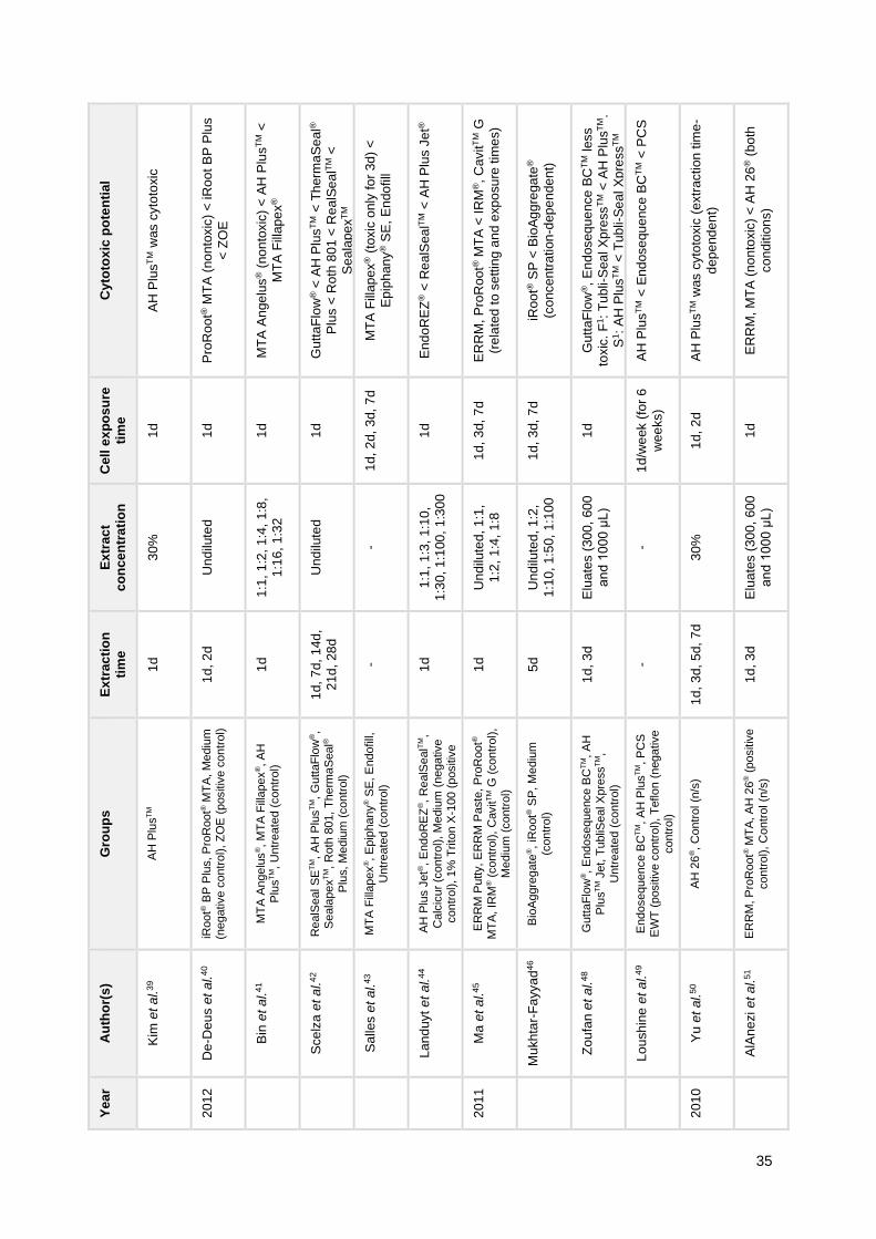
35
Cy
toto
xic
po
ten
tia
l
AH
Plu
sT
M w
as c
yto
toxic
Pro
Ro
ot®
MT
A (
no
nto
xic
) <
iR
oo
t B
P P
lus
< Z
OE
MT
A A
nge
lus
® (
no
nto
xic
) <
AH
Plu
sT
M <
MT
A F
illap
ex
®
Gu
tta
Flo
w® <
AH
Plu
sT
M <
Th
erm
aS
eal®
Plu
s <
Ro
th 8
01
< R
ea
lSe
alT
M <
Se
ala
pe
xT
M
MT
A F
illap
ex
® (
toxic
only
fo
r 3d
) <
Ep
iph
an
y® S
E,
En
do
fill
En
do
RE
Z® <
Re
alS
ea
lTM
< A
H P
lus J
et®
ER
RM
, P
roR
oo
t® M
TA
< I
RM
®,
Ca
vit
TM
G
(re
late
d t
o s
ettin
g a
nd e
xp
osu
re t
ime
s)
iRo
ot®
SP
< B
ioA
gg
rega
te®
(co
nce
ntr
ation
-dep
en
de
nt)
Gu
tta
Flo
w®,
En
doseq
ue
nce
BC
TM
less
toxic
. F
1:
Tub
li-S
ea
l X
pre
ss
TM
< A
H P
lus
TM
.
S1:
AH
Plu
sT
M <
Tub
li-S
ea
l X
pre
ss
TM
AH
Plu
sT
M <
En
dose
qu
ence
BC
TM
< P
CS
AH
Plu
sT
M w
as c
yto
toxic
(extr
action
tim
e-
de
pe
nd
en
t)
ER
RM
, M
TA
(n
on
toxic
) <
AH
26
® (
bo
th
co
nditio
ns)
Ce
ll e
xp
os
ure
tim
e
1d
1d
1d
1d
1d
, 2
d,
3d,
7d
1d
1d
, 3
d,
7d
1d
, 3
d,
7d
1d
1d
/we
ek (
for
6
we
eks)
1d
, 2
d
1d
Ex
trac
t
co
nc
en
tra
tio
n
30
%
Und
ilute
d
1:1
, 1
:2,
1:4
, 1:8
,
1:1
6, 1
:32
Und
ilute
d
-
1:1
, 1
:3,
1:1
0,
1:3
0, 1
:100
, 1
:30
0
Und
ilute
d, 1
:1,
1:2
, 1
:4,
1:8
Und
ilute
d, 1
:2,
1:1
0, 1
:50,
1:1
00
Elu
ate
s (
30
0,
60
0
an
d 1
00
0 μ
L)
-
30
%
Elu
ate
s (
30
0,
60
0
an
d 1
00
0 μ
L)
Ex
trac
tio
n
tim
e
1d
1d
, 2
d
1d
1d
, 7
d,
14d
,
21
d,
28
d
- 1d
1d
5d
1d
, 3
d
-
1d
, 3
d,
5d,
7d
1d
, 3
d
Gro
up
s
AH
Plu
sT
M
iRoot®
BP
Plu
s, P
roR
oot®
MT
A, M
ediu
m
(negative c
ontr
ol), Z
OE
(positiv
e c
ontr
ol)
MT
A A
ngelu
s®, M
TA
Fill
apex
®, A
H
Plu
sT
M,
Untr
eate
d (
contr
ol)
RealS
eal S
ET
M, A
H P
lus
TM, G
uttaF
low
®,
Seala
pex
TM, R
oth
801,
Th
erm
aS
eal®
Plu
s, M
ediu
m (
contr
ol)
MT
A F
illa
pex
®, E
pip
hany
® S
E, E
ndofill,
Untr
eate
d (
contr
ol)
AH
Plu
s J
et®
, E
ndoR
EZ
®,
RealS
ealT
M,
Calc
icur
(contr
ol), M
ediu
m (
negative
contr
ol), 1%
Trito
n X
-100 (
positiv
e
contr
ol)
ER
RM
Putty, E
RR
M P
aste
, P
roR
oot®
MT
A, IR
M® (
contr
ol),
Cavit
TM G
(contr
ol),
Me
diu
m (
contr
ol)
Bio
Ag
gre
gate
®,
iRoot®
SP
, M
ediu
m
(contr
ol)
Gutt
aF
low
®, E
ndosequence B
CT
M, A
H
Plu
sT
M J
et, T
ubliS
eal X
pre
ss
TM,
Untr
eate
d (
contr
ol)
Endosequence B
CT
M, A
H P
lus
TM, P
CS
EW
T (
positiv
e c
ontr
ol),
Teflo
n (
negative
contr
ol)
AH
26
®, C
ontr
ol (n
/s)
ER
RM
, P
roR
oot®
MT
A, A
H 2
6® (
positiv
e
contr
ol), C
ontr
ol (n
/s)
Au
tho
r(s
)
Kim
et a
l.3
9
De
-Deu
s e
t a
l.4
0
Bin
et a
l.41
Sce
lza
et a
l.4
2
Sa
lles e
t a
l.43
La
nd
uyt e
t a
l.4
4
Ma
et a
l.4
5
Mu
kh
tar-
Fa
yya
d4
6
Zo
ufa
n e
t a
l.4
8
Lo
ush
ine
et a
l.4
9
Yu
et
al.
50
AlA
ne
zi e
t a
l.51
Ye
ar
20
12
20
11
20
10

36
Cy
toto
xic
po
ten
tia
l
iRo
ot®
SP
(n
on
toxic
) <
AH
Plu
s
Ca
na
ls <
N2
® <
AH
26
®
(co
nce
ntr
ation
-dep
en
de
nt)
Exp
erim
enta
l se
ale
r <
AH
Plu
sT
M <
PC
S
(co
nce
ntr
ation
-dep
en
de
nt)
MT
A,
Bon
e c
em
en
t <
Am
alg
am
(e
ve
n m
ore
toxic
in
fre
sh
ly c
on
ditio
ns)
Re
alS
eal S
ET
M,
Me
taS
EA
LT
M (
bo
th ↓
with
tim
e)
< E
ndo
RE
Z®,
Rea
lSe
alT
M,
PC
S
F1:
Ke
rr <
Re
alS
ea
lTM
, A
ctiv G
PT
M <
AH
26
®
S1:
AH
26
®,
Ke
rr <
Activ G
PT
M <
Re
alS
ea
lTM
Ep
iph
an
y®,
Ep
iph
any
® S
E, P
CS
Pro
Ro
ot®
MT
A,
Bio
Ag
gre
ga
te® (
no
nto
xic
)
Ca
sto
r O
il P
oly
me
r <
< A
H P
lus
TM
,
Ep
iph
an
y® <
Acro
sea
l
Ca
na
ls <
AH
26
®,
N2
®
(co
nce
ntr
ation
-dep
en
de
nt)
Ep
iph
an
y® w
as c
yto
toxic
(co
nce
ntr
atio
n-
an
d e
xp
osu
re t
ime
-de
pe
nd
en
t)
All
cyto
toxic
(co
nce
ntr
atio
n-d
ep
en
den
t)
Ce
ll e
xp
os
ure
tim
e
1d
1d
3d
/we
ek (
for
5
we
eks)
2d
3d
/we
ek (
for
5
we
eks)
1d
1d
1d
1d
2d
1d
, 3
d,
7d
1d
Ex
trac
t
co
nc
en
tra
tio
n
1:1
, 1
:2,
1:4
1:2
, 1
:4,
1:8
- - -
Elu
ate
s (
20
0,
40
0,
80
0 a
nd
12
00
μL
)
Und
ilute
d
Und
ilute
d
1:1
, 1
:2,
1:4
, 1:8
,
1:1
6, 1
:32
1:2
, 1
:4,
1:8
25
, 5
0,
100
, 20
0,
40
0,
80
0 μ
g/m
L
20
%,
10
%, 5
%
Ex
trac
tio
n
tim
e
1d
1d
- - -
1d
, 3
d
1d
1d
, 2
d,
3d
1d
1d
- 1d
Gro
up
s
iRoot®
SP
, A
H P
lus
TM, M
ediu
m (
contr
ol)
AH
26
®, C
anals
, N
2®,
Untr
eate
d (
contr
ol)
Experim
enta
l seale
r (c
alc
ium
sili
cate
-based),
AH
Plu
sT
M, P
CS
, T
eflo
n (
negative
contr
ol)
PM
MA
, M
TA
, am
alg
am
, C
ontr
ol (n
/s)
EndoR
EZ
®,
RealS
ealT
M, M
eta
SE
AL
TM,
RealS
eal S
ET
M, P
CS
(positiv
e c
ontr
ol),
Te
flon (
negative c
ontr
ol)
Activ G
PT
M,
RealS
ealT
M, A
H 2
6®, K
err
Seale
r, U
ntr
eate
d (
contr
ol)
Epip
hany
® S
E, E
pip
hany
®, P
CS
, U
ntr
eate
d
(contr
ol)
Bio
Ag
gre
gate
®, P
roR
oot®
MT
A, E
mp
ty r
oot
canal (c
ontr
ol)
AH
Plu
sT
M, E
pip
hany
®, A
cro
seal, C
asto
r
Oil
Poly
me
r seale
r, U
ntr
eate
d (
contr
ol)
AH
26
®, C
anals
, N
2®,
Untr
eate
d (
contr
ol)
Epip
hany
®,
Untr
eate
d (
contr
ol)
AH
Plu
sT
M, E
ndofill,
Seale
r 26, M
ediu
m
from
em
pty
mo
lds (
contr
ol)
Au
tho
r(s
)
Zh
an
g e
t al.
52
Hua
ng
et
al.
53
Bry
an
et
al.
54
Ba
dr5
5
Am
es e
t al.
56
Do
na
dio
et a
l.57
Ga
mb
arin
i et
al.
59
De
-Deu
s e
t a
l.6
0
Cam
arg
o e
t a
l.6
1
Hua
ng
et
al.
62
He
itm
an
et a
l.63
Va
lois
et a
l.64
Ye
ar
20
09
20
08

37
Cy
toto
xic
po
ten
tia
l
AH
Plu
s J
et®
, P
MM
A <
Me
taS
EA
LT
M <
PC
S (
tim
e-d
ep
en
den
t, e
xce
pt fo
r P
CS
)
Re
tro
pla
st,
Ge
risto
re®, K
eta
cT
M F
il a
nd
Pro
Ro
ot®
MT
A (
no
nto
xic
); S
upe
rEB
AT
M
(cyto
toxic
)
Ca
na
ls <
AH
26
® <
N2
®
(co
nce
ntr
ation
-dep
en
de
nt)
Pro
Ro
ot®
MT
A (
no
nto
xic
) <
Dia
ke
tTM
,
En
dio
n,
CY
ME
D 8
41
0
Se
ala
pe
xT
M <
AH
26
® <
N2
®
(co
nce
ntr
ation
-dep
en
de
nt)
Cyto
toxic
ity w
as c
on
cen
tra
tio
n-
de
pe
nd
en
t (p
reve
nte
d b
y N
AC
)
Se
ala
pe
xT
M <
N2
® <
AH
26
®
(co
nce
ntr
ation
-dep
en
de
nt)
Ep
iph
an
y® <
AH
Plu
sT
M
En
do
RE
Z® <
AH
Plu
sT
M,
Ro
eko
Se
al <
Ep
iph
an
y®
F1:
Se
ala
pex
TM
< o
the
rs.
S1:
Th
erm
ase
al®
, E
pip
han
y® <
oth
ers
.
Gu
tta
Flo
w® <
AH
Plu
sT
M <
Epip
ha
ny
®
(exp
osu
re t
ime
-de
pe
nd
en
t)
Ro
eko
Sea
l <
AH
Plu
sT
M (
sett
ing
tim
e-
de
pe
nd
en
t fo
r A
H P
lus
TM
)
Ce
ll e
xp
os
ure
tim
e
3d
/we
ek (
for
5
we
eks)
1d
2d
1d
, 2
d,
3d
1d
1d
1d
2h
2h
1h
, 1
d
1d
, 3
d
5d
Ex
trac
t
co
nc
en
tra
tio
n
-
Und
ilute
d
1:2
, 1
:4,
1:8
Und
ilute
d
Dilu
tio
n f
acto
r:
10
– 8
0
5m
g/m
L a
nd
dilu
tio
ns
Dilu
tio
n f
acto
rs:
6-1
8,
1-7
, 5
-10
0
-
Und
ilute
d
- - -
Ex
trac
tio
n
tim
e
-
1d
, 2
d,
3d
1d
1d
1d
1d
1d
-
1d
(se
t)
- - -
Gro
up
s
Me
taS
EA
LT
M, A
H P
lus J
et®
, P
CS
, P
MM
A
(positiv
e c
ontr
ol), T
eflo
n (
negative c
ontr
ol)
Retr
opla
st, G
eristo
re®, K
eta
cT
M F
il,
SuperE
BA
TM, P
roR
oot®
MT
A, M
ediu
m
(contr
ol)
AH
26
®, C
anals
, N
2®,
Untr
eate
d (
contr
ol)
Pro
Root®
MT
A,
Dia
ketT
M, E
ndio
n®,
CY
ME
D 8
410,
Untr
eate
d (
contr
ol)
N2
®, S
eala
pex
TM, A
H 2
6®, C
ontr
ol (n
/s)
AH
26
®, U
DM
A, C
ontr
ol (n
/s)
N2
®, S
eala
pex
TM, A
H 2
6®, C
ontr
ol (n
/s)
Epip
hany
®, A
H P
lus
TM,
Filt
ers
with c
ells
and n
o s
eale
r and F
ilters
no c
ells
and w
ith
seale
r (c
ontr
ols
)
AH
Plu
sT
M, E
ndoR
EZ
®,
RoekoS
eal
Auto
mix
, E
pip
hany
®, M
ediu
m (
contr
ol)
Epip
hany
®,
Resilo
n, G
P, G
rossm
an,
Th
erm
aseal®
, S
eala
pex
TM;
Isoto
nic
salin
e
and 1
0%
form
ald
ehyde (
contr
ols
)
AH
Plu
sT
M, E
pip
hany
®,
Gutt
aF
low
®,
Teflo
n
(contr
ol)
Roekoseal A
uto
mix
, A
H P
lus
TM, C
ontr
ol
(n/s
)
Au
tho
r(s
)
Pin
na
et
al.
65
Al-
Sa
´ee
d e
t a
l.6
6
Hua
ng
et
al.
67
Go
rdu
ysu
s e
t al.
68
Le
e e
t a
l.7
0
Le
e e
t a
l.7
1
Le
e e
t a
l.7
2
Me
rdad
et a
l.73
Lo
die
ne
et
al.
74
Ke
y e
t a
l.75
Bo
uill
ag
ue
t e
t a
l.7
6
Mile
tic e
t a
l.7
7
Ye
ar
20
07
20
06
20
05
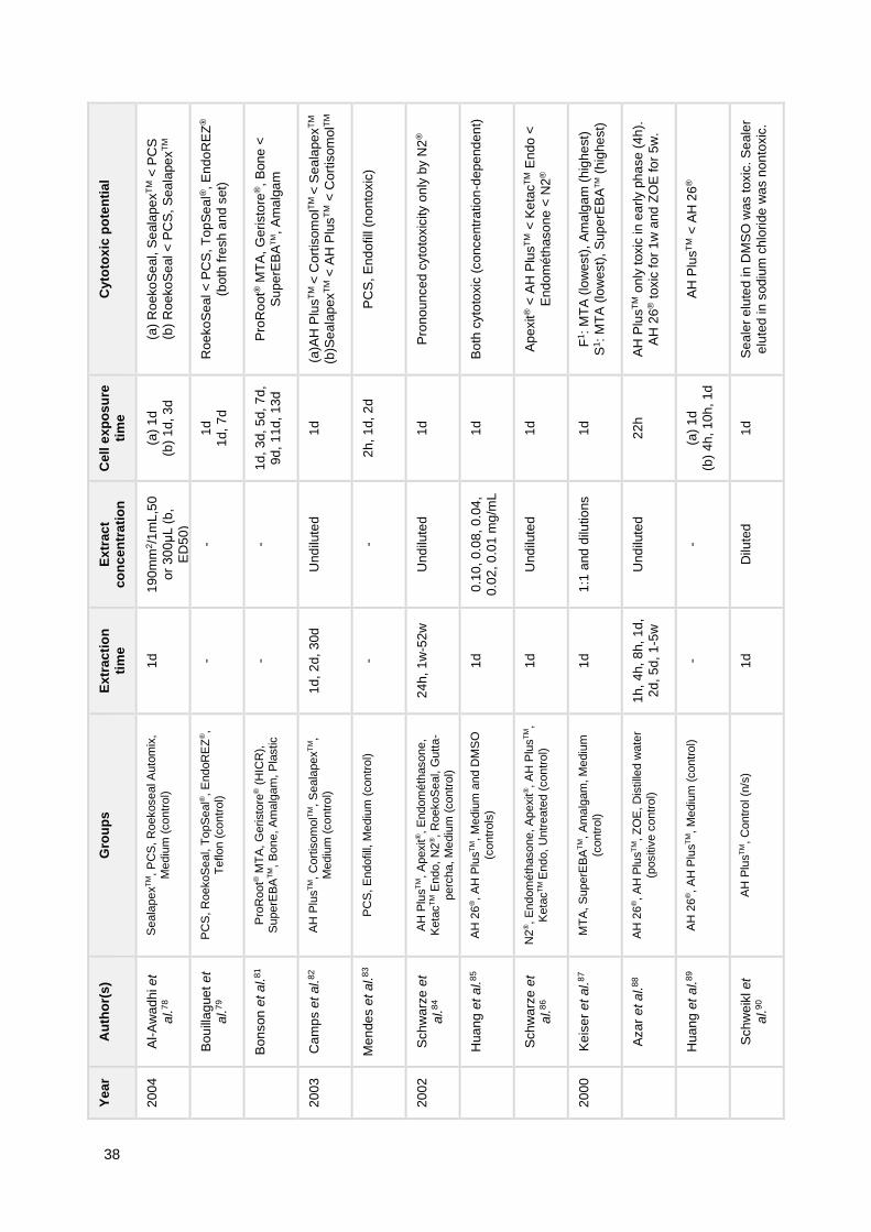
38
Cy
toto
xic
po
ten
tia
l
(a)
Ro
eko
Se
al, S
eala
pe
xT
M <
PC
S
(b)
Ro
eko
Se
al <
PC
S,
Sea
lape
xT
M
Ro
eko
Sea
l <
PC
S,
To
pS
ea
l®, E
nd
oR
EZ
®
(bo
th f
resh a
nd s
et)
Pro
Ro
ot®
MT
A,
Ge
risto
re®,
Bon
e <
Su
pe
rEB
AT
M,
Am
alg
am
(a)A
H P
lus
TM
< C
ort
isom
olT
M <
Se
ala
pe
xT
M
(b)S
ea
lap
ex
TM <
AH
Plu
sT
M <
Co
rtis
om
olT
M
PC
S,
En
do
fill
(no
nto
xic
)
Pro
no
unce
d c
yto
toxic
ity o
nly
by N
2®
Bo
th c
yto
toxic
(co
nce
ntr
atio
n-d
ep
end
en
t)
Ap
exit
® <
AH
Plu
sT
M <
Ke
tac
TM E
nd
o <
En
do
mé
thaso
ne
< N
2®
F1:
MT
A (
low
est)
, A
ma
lga
m (
hig
he
st)
S1:
MT
A (
low
est)
, S
up
erE
BA
TM (
hig
he
st)
AH
Plu
sT
M o
nly
toxic
in
ea
rly p
ha
se (
4h
).
AH
26
® t
oxic
fo
r 1
w a
nd
ZO
E fo
r 5
w.
AH
Plu
sT
M <
AH
26
®
Se
ale
r e
lute
d in
DM
SO
was t
oxic
. S
eale
r
elu
ted
in s
od
ium
ch
lorid
e w
as n
on
toxic
.
Ce
ll e
xp
os
ure
tim
e
(a)
1d
(b)
1d
, 3
d
1d
1d
, 7
d
1d
, 3
d,
5d,
7d
,
9d
, 1
1d
, 13
d
1d
2h
, 1
d,
2d
1d
1d
1d
1d
22
h
(a)
1d
(b)
4h
, 1
0h
, 1
d
1d
Ex
trac
t
co
nc
en
tra
tio
n
19
0m
m2/1
mL
,50
or
30
0μ
L (
b,
ED
50
)
- -
Und
ilute
d
-
Und
ilute
d
0.1
0, 0
.08,
0.0
4,
0.0
2, 0
.01 m
g/m
L
Und
ilute
d
1:1
and
dilu
tio
ns
Und
ilute
d
-
Dilu
ted
Ex
trac
tio
n
tim
e
1d
- -
1d
, 2
d,
30d
-
24
h,
1w
-52
w
1d
1d
1d
1h
, 4
h,
8h,
1d
,
2d
, 5
d,
1-5
w
- 1d
Gro
up
s
Seala
pex
TM, P
CS
, R
oekoseal A
uto
mix
,
Me
diu
m (
contr
ol)
PC
S, R
oekoS
eal, T
opS
eal®
, E
ndoR
EZ
®,
Te
flon (
contr
ol)
Pro
Root®
MT
A, G
eristo
re® (
HIC
R),
S
uperE
BA
TM, B
one, A
ma
lgam
, P
lastic
AH
Plu
sT
M, C
ort
isom
olT
M, S
eala
pex
TM,
Me
diu
m (
contr
ol)
PC
S, E
ndofill,
Me
diu
m (
contr
ol)
AH
Plu
sT
M, A
pexit
®, E
ndom
éth
asone,
Keta
cT
M E
ndo,
N2
®,
RoekoS
eal, G
utt
a-
perc
ha, M
ediu
m (
contr
ol)
AH
26
®, A
H P
lus
TM, M
ediu
m a
nd D
MS
O
(contr
ols
)
N2
®, E
ndom
éth
asone, A
pexit
®, A
H P
lus
TM,
Keta
cT
M E
ndo,
Untr
eate
d (
contr
ol)
MT
A, S
uperE
BA
TM, A
ma
lgam
, M
ediu
m
(contr
ol)
AH
26
®, A
H P
lus
TM, Z
OE
, D
istille
d w
ate
r
(positiv
e c
ontr
ol)
AH
26
®, A
H P
lus
TM, M
ediu
m (
contr
ol)
AH
Plu
sT
M, C
ontr
ol (n
/s)
Au
tho
r(s
)
Al-
Aw
ad
hi e
t
al.
78
Bo
uill
ag
ue
t e
t
al.
79
Bo
nson
et a
l.81
Ca
mp
s e
t al.
82
Me
nde
s e
t al.
83
Sch
warz
e e
t
al.
84
Hua
ng
et
al.
85
Sch
warz
e e
t
al.
86
Ke
ise
r e
t al.
87
Aza
r e
t a
l.8
8
Hua
ng
et
al.
89
Sch
weik
l et
al.
90
Ye
ar
20
04
20
03
20
02
20
00

39
Extraction time and cell exposure time were defined as hours (h), days (d) or weeks (w). 1 Material setting condition defined as fresh (F) or set (S). Abbreviations: DF-MSCs, dental follicle-derived adult mesenchymal stem cells; G, Gray; GP, gutta-percha; hOCs, human osteoblastic cells; n/s, non-specified; NAC, N-acetyl-L-cysteine; O.S., osteogenic supplementation (with ascorbic acid, β-glycerophosphate and dexamethasone).
3.1.2. Influence of condition and time of material setting on cytotoxicity
To understand how the material set condition influences cytotoxicity, we focused on
studies that used both set conditions, i.e. freshly mixed and set. Comparing AH PlusTM and
MTA Fillapex®, Zhou et al.29 showed that AH PlusTM was more toxic in freshly mixed
conditions but less toxic after setting. This decrease in cytotoxicity with setting has also been
confirmed by other authors.34,48 Similarly to AH PlusTM, Donadio et al.57 showed that AH 26®
was considerably more cytotoxic in freshly mixed conditions compared to set conditions (72h
after preparation). Also, Badr55 and Keiser et al.87 showed a higher cytotoxicity of amalgam in
freshly mixed conditions.
3.1.3. Influence of sealer concentration on cytotoxicity
In order to evaluate the influence exerted by the amount of sealer on cytotoxicity, we
focused on studies that used an indirect contact testing methodology with several
concentrations of sealer extract. In fact, a concentration-dependency of the cytotoxic effect
was demonstrated for Activ GPTM 57, AH PlusTM 29,34,36,41,44,64,85, AH 26® 57,62,67,70–72,85,
BioAggregate® and iRoot® SP46, Canals62,67, Endofill64, EndoREZ® 34,44, Endosequence BCTM
24,36, Epiphany® 63, MTA Fillapex® 24,29,41, N2® 62,67,70,72, ProRoot® ES36, RealSealTM 44,57,
RoekoSeal34, Roth’s Sealer36, SealapexTM 70,72 and Sealer 26.64 Lee et al.71 also showed a
concentration-dependent cytotoxicity for UDMA.
3.1.4. Influence of exposure time to sealer on cytotoxicity
To evaluate the influence of the time of exposure, we considered only studies which
tested more than one cell incubation time point. Accordingly, 33 studies fulfilled this criterion,
of which 18 used direct contact testing as a method of cell exposure to several materials.
From the 33 studies, 10 did not focus on comparing different incubation times.26,27,32,38,68,78,91–
93,119

40
A certain heterogeneity was observed in regard to this subject. Some studies showed
cell viability recovery over time of exposure for BioRootTM RCS69, GuttaFlow® Bioseal and
GuttaFlow®280, MTA120, MTA Fillapex® 43 and MetaSEALTM.65 A recovery of cell viability was
also denoted for PMMA after 5 weeks.65 Other studies showed decreased cell viability over
time of exposure for AH PlusTM 31,76,89, AH 26® 89, ProRoot® MTA35,45, Endocem35, GuttaFlow®
76, MTA Fillapex® 37, Epiphany® 63,76, EpiphanyTM SE43, RealSeal XT31 and for other materials,
namely IRM® 35,45, ERRM and CavitTM G.45
Key et al.75 showed a recovery of cell viability at 24h for Epiphany® and ThermaSeal®,
when compared to 1 hour of exposure time, but a loss of viability for SealapexTM. Jeanneau
et al.25 showed an increased proliferation with increasing exposure time only for BioRootTM
RCS, as the inverse relationship was observed for PCS. Bouillaguet et al.79 also showed a
higher cytotoxicity for PCS at a second 24h- and 1 week-incubation periods, and also for
RoekoSeal and EndoREZ® at 1 week-incubations, with all materials in fresh conditions.
Furthermore, some studies showed a maintenance of cytotoxicity over time for PCS49,56,65,
RealSealTM and EndoREZ®.56 Mendes et al.83 showed a maintenance of cell viability for PCS
and Endofill, which were classified as nontoxic.
Bonson et al.81 suggested that washed materials exhibited a lower cytotoxicity
compared to fresh materials, over a 13-day experimental period of observation, in particular
for ProRoot® MTA and Geristore®. Other studies that also used “aged” sealers (i.e. sealer
specimens immersed in culture media with renewal), also showed a general recovery of cell
viability over time for AH PlusTM and Endosequence BCTM after 6 weeks49, AH PlusTM after 5
weeks54, AH Plus Jet® after 5 weeks65 and RealSeal SETM and MetaSEALTM over 5 weeks of
observation.56 In fact, these findings appear to be partially confirmed by studies which used
different extraction time points.
Studies that performed cumulative extractions (i.e. same culture medium over the
entire period of extraction) showed an increase in cytotoxicity over time of extraction for
BioRootTM RCS, MTA Fillapex® and SimpliSeal® 47, ProRoot® MTA and BioAggregate®.60
Mandal et al.33 showed increasing cell viability over time (72h compared to 24h) for AH
PlusTM but decreased for GuttaFlow®2 and MTA in the higher concentration (all materials in
set conditions).
On the other hand, studies that performed separate extractions (i.e. culture medium
renewed after harvesting the extract from the previous time point) – which simulates
periodontal ligament clearance42 – showed a decrease in cytotoxicity over the time of
extraction for several sealers (e.g. GuttaFlow®, AH PlusTM).42,50 Also using similar extraction

41
methods, Zhou et al.29 showed a recovery of cell viability over time for AH PlusTM but not for
MTA Fillapex®, which showed increased toxicity in more concentrated extracts (1:2 and 1:8).
Al-Sa’eed et al.66 showed an increased cell viability for 3-day eluates of Retroplast,
Geristore® and KetacTM Fil, as SuperEBATM remained cytotoxic. Camps et al.82 showed a
decrease in cytotoxicity for Sealapex with no difference for AH PlusTM using a root-dipping
technique. However, these results were not confirmed by experiments with International
Organization for Standardization (ISO) Standards 10993-5 in the same study, as only
CortisomolTM had a decreasing cytotoxicity over time in this technique. Azar et al.88 showed a
decrease in cytotoxicity for both AH PlusTM (only toxic in first 4 hours) and AH 26® (toxicity
decreased after 1 week), as no decrease was observed for ZOE cement. Other studies did
not compare different extraction time points or did not show significant differences.30,40,51,57,84
3.2. In Vivo Biocompatibility
The general characteristics of the included studies are presented in Table 8. As can
be seen, the main reported methods were the subcutaneous tissue response to sealer
implants98–100,102,105,107,108,110–112,115,117 and the periapical tissue response to root canal filling
procedure95–97,101,106,114,116,118. Specifically, in relation to root filling procedures, these were
carried out primarily in premolars (both maxillary and mandibular) and also in maxillary
incisors in some studies. One study compared the tissue response by subcutaneous and
alveolar socket implantation113 and showed similar results for both implantation sites. The
alveolar socket-implantation method following tooth extraction was also reported by Cintra et
al.104 In one study103, Tanomaru-Filho et al. evaluated the periapical tissue response to
retrobturation, after the induction of a periapical lesion, by exposure to oral environment for a
period of 7 days. Furthermore, Assmann et al.94 and Morinaga et al.109 studied the bone
tissue response to intraosseous sealer implants in the femur and the mandible of Wistar rats,
respectively.
In regard to setting condition, most studies used the materials in a freshly mixed
state, except for Garcia et al.112 and Morinaga et al.109, who only used materials in a set
condition after photoactivation or after a period of one day and one night, respectively.
Campos-Pinto et al.98 and Ozbas et al.108 used both materials in freshly mixed and in set
conditions.
In terms of in vivo model, rats of different species or strains were used in 15 studies
and dogs in 9. In one study, New Zealand rabbits were used as animal model.

42
Table 8 General characteristics of included studies in regard to in vivo biocompatibility. A
nim
al
mo
de
l
Wis
tar
rat
Wis
tar
rat
Be
ag
le
do
g
Mo
ng
rel
do
g
(2)
Ra
t
Wis
tar
rat
Mo
ng
rel
do
g
(2)
Wis
tar
rat
Be
ag
le
do
g
(2)
Te
eth
fo
r ro
ot
ca
na
l fi
llin
g
- -
19
PM
s (
ma
x.
an
d m
an
d.)
INC
(m
ax.)
and
PM
s (
ma
x.
and
ma
nd.)
- -
PM
s (
ma
x.
and
ma
nd.)
-
PM
s (
ma
x.
and
ma
nd.)
Me
tho
d
Su
bcu
tan
eo
us
tissu
e r
espo
nse
to
imp
lan
t
Bo
ne
tis
su
e
resp
on
se t
o im
pla
nt
Pe
ria
pic
al tissu
e
resp
on
se t
o r
oo
t
ca
nal filli
ng
Pe
ria
pic
al tissu
e
resp
on
se t
o r
oo
t
ca
nal filli
ng
Su
bcu
tan
eo
us
tissu
e r
espo
nse
to
imp
lan
t
Su
bcu
tan
eo
us a
nd
alv
eo
lar
tissue
resp
on
se t
o im
pla
nt
Pe
ria
pic
al tissu
e
resp
on
se t
o r
oo
t
ca
nal filli
ng
Su
bcu
tan
eo
us
tissu
e r
espo
nse
to
imp
lan
t
Pe
ria
pic
al tissu
e
resp
on
se t
o r
oo
t
ca
nal filli
ng
Ma
teri
al
co
nd
itio
n
(se
ttin
g t
ime
)
Fre
shly
mix
ed
Fre
shly
mix
ed
Fre
shly
mix
ed
Fre
shly
mix
ed
Se
t
(ph
oto
activate
d)
Fre
shly
mix
ed
Fre
shly
mix
ed
Fre
shly
mix
ed
Fre
shly
mix
ed
N
N=
16
(4 im
pla
nts
pe
r a
nim
al)
N=
15
(5 p
er
tim
e
po
int)
N=
38
ca
na
ls
(SX
/GP
: 1
6,
RS
/R:
22
)
N=
20
ca
na
ls
(10
/gro
up
)
N=
15
(4 im
pla
nts
pe
r a
nim
al)
N=
40
(10
/gro
up
)
N=
40
ca
na
ls
(8-1
2/g
rou
p)
N=
30
(assig
ned
to
gro
ups)
N=
30
ca
na
ls
(dis
trib
ute
d
to 2
gro
ups)
Gro
up
s (
G)
G1
: E
mp
ty P
E t
ub
e (
co
ntr
ol)
; G
2:
Gu
tta
Flo
w®
Bio
sea
l; G
3: G
utt
aF
low
®2
; G
4: A
H P
lus
TM
G1
: M
TA
Fill
ap
ex
®
G2
: A
H P
lus
TM
G3
: E
mp
ty c
avity (
co
ntr
ol)
G1
: S
ea
lap
ex X
pre
ss
TM
/GP
G2
: R
ea
lSea
l X
T/R
esilo
n
G1
: E
nd
om
éth
ason
e/G
P (
sh
ort
of
apic
al
fora
me
n);
G2
: E
nd
om
éth
aso
ne
/GP
(ove
rfill
ing
)
Ep
iph
an
y/R
esilo
n (
G1:
with
self-e
tch p
rim
er,
G2
: w
ith
out
prim
er)
; G
3:
En
do
fill/
GP
; G
4:
Em
pty
de
ntin
tu
be
Em
pty
PE
tu
be
(G
1:
alv
eo
lar,
G2
:
su
bcu
tan
eo
us);
Pro
Roo
t M
TA
(G
3:
alv
eo
lar,
G4
: su
bcu
tan
eou
s
G1
: C
ale
n®
a t
hic
ke
ne
d w
ith
ZnO
; G
2:
IRC
P
pa
ste
; G
3:
ZO
E c
em
en
t; G
4:
Ste
rile
salin
e
G1
: A
H P
lus
TM
; G
2:
AH
Plu
sT
M w
ith
ca
lciu
m
hyd
roxid
e 5
% (
w/w
); G
3:
Con
tro
l (n
/s)
G1
: E
pip
ha
ny
®/R
esilo
n s
yste
m
G2
: P
ulp
Can
al S
ea
ler/
GP
Au
tho
r(s
)
Sa
nto
s e
t
al.
11
1
Assm
an
n e
t
al.
94
Silv
a e
t al.
95
Su
zuki e
t a
l.1
06
Ga
rcia
et
al.
11
2
Cin
tra
et
al.
11
3
Silv
a e
t al.
114
Oliv
eira
et
al.
11
5
Bra
sil
et a
l.1
16
Ye
ar
20
19
20
15
20
14
20
11
20
10
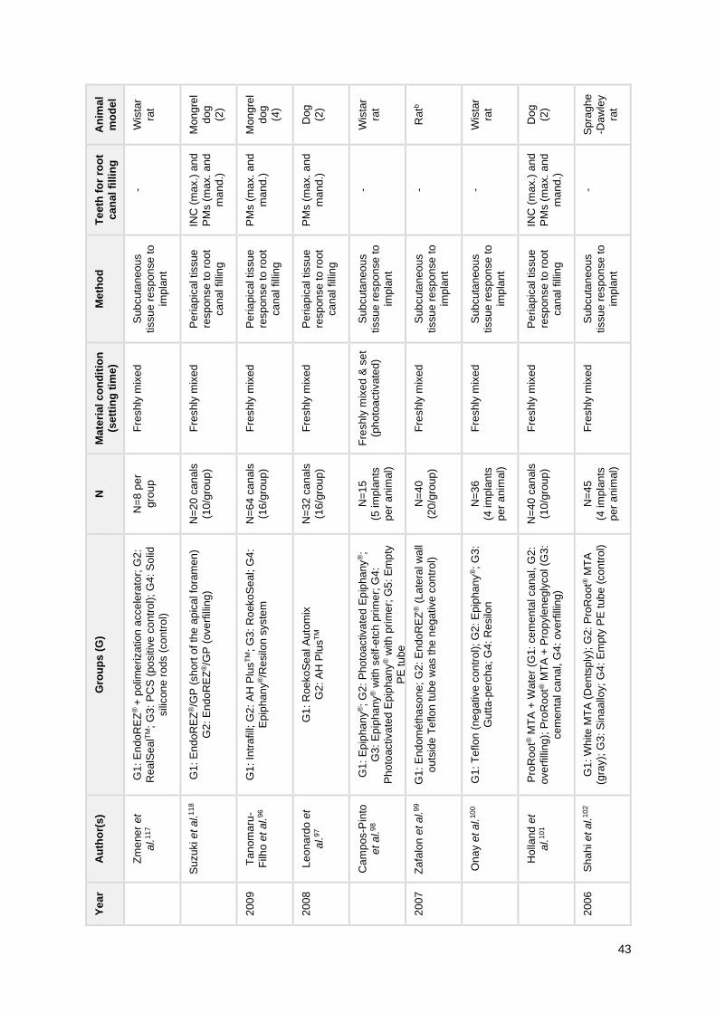
43
An
ima
l
mo
de
l
Wis
tar
rat
Mo
ng
rel
do
g
(2)
Mo
ng
rel
do
g
(4)
Do
g
(2)
Wis
tar
rat
Ra
tb
Wis
tar
rat
Do
g
(2)
Sp
rag
he
-Da
wle
y
rat
Te
eth
fo
r ro
ot
ca
na
l fi
llin
g
-
INC
(m
ax.)
and
PM
s (
ma
x.
and
ma
nd.)
PM
s (
ma
x.
and
ma
nd.)
PM
s (
ma
x.
and
ma
nd.)
- - -
INC
(m
ax.)
and
PM
s (
ma
x.
and
ma
nd.)
-
Me
tho
d
Su
bcu
tan
eo
us
tissu
e r
espo
nse
to
imp
lan
t
Pe
ria
pic
al tissu
e
resp
on
se t
o r
oo
t
ca
nal filli
ng
Pe
ria
pic
al tissu
e
resp
on
se t
o r
oo
t
ca
nal filli
ng
Pe
ria
pic
al tissu
e
resp
on
se t
o r
oo
t
ca
nal filli
ng
Su
bcu
tan
eo
us
tissu
e r
espo
nse
to
imp
lan
t
Su
bcu
tan
eo
us
tissu
e r
espo
nse
to
imp
lan
t
Su
bcu
tan
eo
us
tissu
e r
espo
nse
to
imp
lan
t
Pe
ria
pic
al tissu
e
resp
on
se t
o r
oo
t
ca
nal filli
ng
Su
bcu
tan
eo
us
tissu
e r
espo
nse
to
imp
lan
t
Ma
teri
al
co
nd
itio
n
(se
ttin
g t
ime
)
Fre
shly
mix
ed
Fre
shly
mix
ed
Fre
shly
mix
ed
Fre
shly
mix
ed
Fre
shly
mix
ed
& s
et
(ph
oto
activate
d)
Fre
shly
mix
ed
Fre
shly
mix
ed
Fre
shly
mix
ed
Fre
shly
mix
ed
N
N=
8 p
er
gro
up
N=
20
ca
na
ls
(10
/gro
up
)
N=
64
ca
na
ls
(16
/gro
up
)
N=
32
ca
na
ls
(16
/gro
up
)
N=
15
(5 im
pla
nts
pe
r a
nim
al)
N=
40
(20
/gro
up
)
N=
36
(4 im
pla
nts
pe
r a
nim
al)
N=
40
ca
na
ls
(10
/gro
up
)
N=
45
(4 im
pla
nts
pe
r a
nim
al)
Gro
up
s (
G)
G1
: E
nd
oR
EZ
® +
polim
eriza
tion
acce
lera
tor;
G2
:
Rea
lSe
alT
M;
G3
: P
CS
(po
sitiv
e c
on
tro
l);
G4
: S
olid
sili
con
e r
od
s (
co
ntr
ol)
G1
: E
nd
oR
EZ
®/G
P (
sh
ort
of
the
ap
ica
l fo
ram
en
)
G2
: E
nd
oR
EZ
®/G
P (
ove
rfill
ing
)
G1
: In
tra
fill;
G2
: A
H P
lus
TM
; G
3:
Roeko
Se
al; G
4:
Ep
iph
an
y®/R
esilo
n s
yste
m
G1
: R
oe
ko
Se
al A
uto
mix
G2
: A
H P
lus
TM
G1
: E
pip
ha
ny
®;
G2
: P
ho
toa
ctiva
ted
Ep
iph
any
®;
G3
: E
pip
ha
ny
® w
ith s
elf-e
tch
pri
me
r; G
4:
Ph
oto
activa
ted
Epip
han
y® w
ith p
rim
er;
G5
: E
mp
ty
PE
tu
be
G1
: E
nd
om
éth
ason
e;
G2
: E
ndo
RE
Z® (
La
tera
l w
all
ou
tsid
e T
eflo
n t
ub
e w
as t
he
neg
ative
co
ntr
ol)
G1
: T
eflo
n (
ne
ga
tive c
on
tro
l); G
2:
Ep
ipha
ny
®;
G3
:
Gu
tta
-pe
rch
a; G
4:
Re
silo
n
Pro
Roo
t® M
TA
+ W
ate
r (G
1:
ce
me
nta
l ca
na
l, G
2:
ove
rfill
ing
); P
roR
oo
t® M
TA
+ P
rop
yle
neg
lycol (G
3:
ce
me
nta
l ca
nal, G
4:
ove
rfill
ing)
G1
: W
hite
MT
A (
De
nts
ply
); G
2:
Pro
Roo
t® M
TA
(gra
y);
G3:
Sin
aa
lloy;
G4
: E
mpty
PE
tub
e (
co
ntr
ol)
Au
tho
r(s
)
Zm
ene
r e
t
al.
11
7
Su
zuki e
t a
l.1
18
Ta
nom
aru
-
Filh
o e
t al.
96
Le
on
ard
o e
t
al.
97
Cam
pos-P
into
et
al.
98
Za
falo
n e
t a
l.9
9
On
ay e
t a
l.10
0
Holla
nd
et
al.
10
1
Sh
ah
i e
t al.
10
2
Ye
ar
20
09
20
08
20
07
20
06
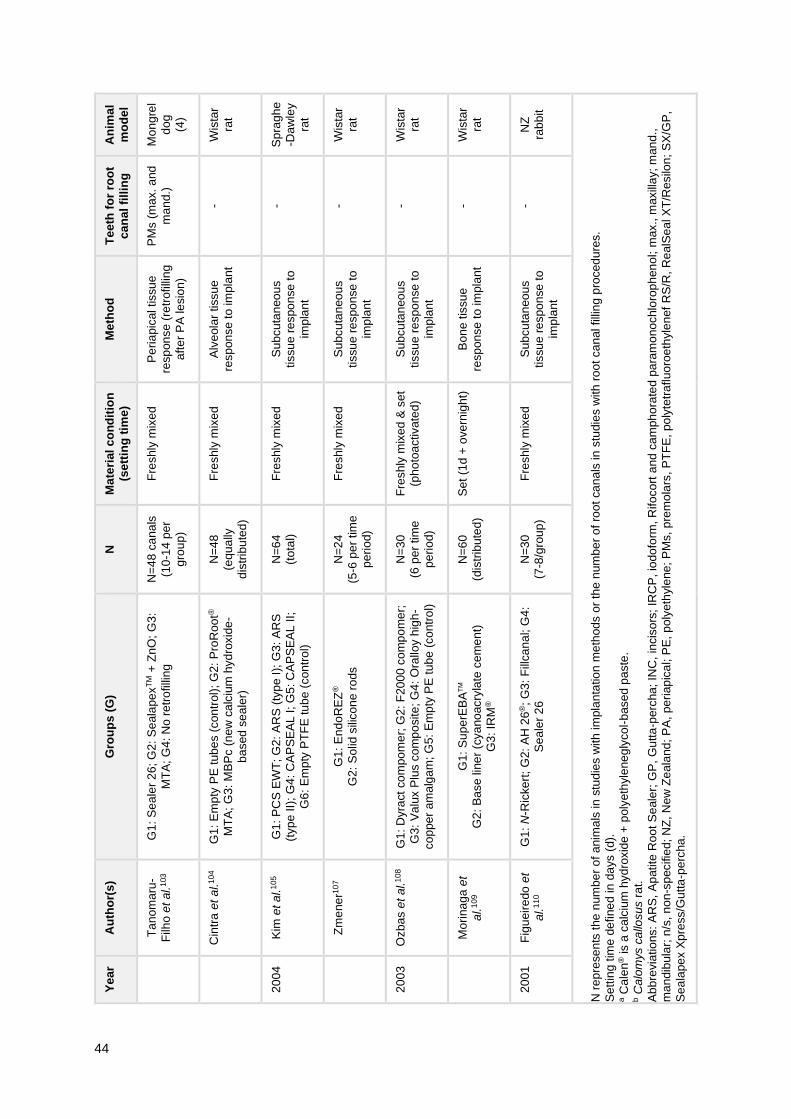
44
An
ima
l
mo
de
l
Mo
ng
rel
do
g
(4)
Wis
tar
rat
Sp
rag
he
-Da
wle
y
rat
Wis
tar
rat
Wis
tar
rat
Wis
tar
rat
NZ
rab
bit
N r
ep
rese
nts
the
num
be
r o
f anim
als
in
stu
die
s w
ith
im
pla
nta
tio
n m
eth
od
s o
r th
e n
um
be
r o
f ro
ot
ca
nals
in
stu
die
s w
ith
ro
ot ca
nal filli
ng
pro
ced
ure
s.
Se
ttin
g t
ime
de
fine
d in
da
ys (
d).
a C
ale
n® is a
ca
lciu
m h
yd
roxid
e +
poly
eth
yle
ne
gly
co
l-b
ased
pa
ste
. b C
alo
mys c
allo
sus r
at.
Ab
bre
via
tio
ns:
AR
S, A
patite
Ro
ot
Se
ale
r; G
P,
Gu
tta
-perc
ha
; IN
C,
incis
ors
; IR
CP
, io
do
form
, R
ifo
co
rt a
nd
ca
mph
ora
ted
pa
ram
ono
chlo
rop
hen
ol; m
ax.,
maxill
ay; m
an
d.,
ma
ndib
ula
r; n
/s, n
on
-sp
ecifie
d; N
Z,
New
Ze
ala
nd
; P
A,
pe
riap
ica
l; P
E,
poly
eth
yle
ne
; P
Ms,
pre
mo
lars
, P
TF
E,
po
lyte
tra
fluo
roe
thyle
nef
RS
/R,
Re
alS
ea
l X
T/R
esilo
n;
SX
/GP
,
Se
ala
pe
x X
pre
ss/G
utta
-pe
rcha
.
Te
eth
fo
r ro
ot
ca
na
l fi
llin
g
PM
s (
ma
x.
and
ma
nd.)
- - - - - -
Me
tho
d
Pe
ria
pic
al tissu
e
resp
on
se (
retr
ofilli
ng
aft
er
PA
le
sio
n)
Alv
eola
r tissu
e
resp
on
se t
o im
pla
nt
Su
bcu
tan
eo
us
tissu
e r
espo
nse
to
imp
lan
t
Su
bcu
tan
eo
us
tissu
e r
espo
nse
to
imp
lan
t
Su
bcu
tan
eo
us
tissu
e r
espo
nse
to
imp
lan
t
Bo
ne
tis
su
e
resp
on
se t
o im
pla
nt
Su
bcu
tan
eo
us
tissu
e r
espo
nse
to
imp
lan
t
Ma
teri
al
co
nd
itio
n
(se
ttin
g t
ime
)
Fre
shly
mix
ed
Fre
shly
mix
ed
Fre
shly
mix
ed
Fre
shly
mix
ed
Fre
shly
mix
ed
& s
et
(ph
oto
activate
d)
Se
t (1
d +
ove
rnig
ht)
Fre
shly
mix
ed
N
N=
48
ca
na
ls
(10
-14
pe
r
gro
up
)
N=
48
(eq
ua
lly
dis
trib
ute
d)
N=
64
(to
tal)
N=
24
(5-6
pe
r tim
e
pe
rio
d)
N=
30
(6 p
er
tim
e
pe
rio
d)
N=
60
(dis
trib
ute
d)
N=
30
(7-8
/gro
up
)
Gro
up
s (
G)
G1
: S
ea
ler
26
; G
2:
Se
ala
pe
xT
M +
Zn
O;
G3
:
MT
A;
G4:
No r
etr
ofilli
ng
G1
: E
mp
ty P
E t
ub
es (
con
trol)
; G
2:
Pro
Roo
t®
MT
A;
G3: M
BP
c (
ne
w c
alc
ium
hyd
roxid
e-
ba
sed
se
ale
r)
G1
: P
CS
EW
T;
G2
: A
RS
(ty
pe I
); G
3:
AR
S
(typ
e II)
; G
4:
CA
PS
EA
L I;
G5: C
AP
SE
AL
II;
G6
: E
mp
ty P
TF
E tu
be (
con
tro
l)
G1
: E
nd
oR
EZ
®
G2
: S
olid
sili
co
ne r
ods
G1
: D
yra
ct
co
mpo
me
r; G
2:
F20
00
co
mp
om
er;
G3
: V
alu
x P
lus c
om
po
site
; G
4:
Ora
lloy h
igh
-
co
ppe
r am
alg
am
; G
5:
Em
pty
PE
tu
be
(co
ntr
ol)
G1
: S
up
erE
BA
TM
G2
: B
ase
lin
er
(cya
no
acry
late
ce
me
nt)
G3
: IR
M®
G1
: N
-Ric
ke
rt;
G2:
AH
26
®;
G3
: F
illca
na
l; G
4:
Se
ale
r 2
6
Au
tho
r(s
)
Ta
nom
aru
-
Filh
o e
t al.
103
Cin
tra
et
al.
10
4
Kim
et a
l.1
05
Zm
ene
r10
7
Ozb
as e
t al.
10
8
Mo
rina
ga
et
al.
10
9
Fig
ueir
edo
et
al.
11
0
Ye
ar
20
04
20
03
20
01

45
3.2.1. Inflammatory tissue reaction to sealers
All studies showed a generalized inflammatory response to the materials tested, as
presented in Table 9. AH PlusTM, EndoREZ®, Epiphany® and ProRoot® MTA were the most
studied sealers. Relatively to the epoxy resin-based sealer AH PlusTM, Oliveira et al.115
reported a nonspecific chronic inflammatory response, which can be reduced with the
addition of calcium hydroxide. A slight to moderate inflammatory reaction was reported by
other authors.96 A similar inflammatory infiltrate was shown in comparison with silicone-based
sealers RoekoSeal96,97 and GuttaFlow®2111, although higher comparing to GuttaFlow® Bioseal
within 8 days of exposure.111 Nevertheless, the same study showed that such difference had
disappeared after 30 days. Assmann et al.94 showed a lower neutrophil infiltrate in
comparison to MTA Fillapex®, even though both sealers provided the re-establishment of
original bone structure.
In regard to the methacrylate resin-based sealer Epiphany®, Garcia et al.112 showed
that the addition of its self-etch primer decreases the inflammatory reaction to the
Epiphany/Resilon system. Similarly, Campos-Pinto et al.98 showed that photoactivated
Epiphany® without primer induced a moderate to severe inflammatory reaction with extensive
necrosis, whereas only slight chronic inflammatory reaction was observed in the presence of
the primer. Tanomaru-Filho et al.96 showed a slight to moderate inflammatory reaction of
Epiphany® comparable to AH PlusTM and RoekoSeal, as Onay et al.100 showed an
inflammatory reaction which varied from none to severe at first-week observation to none to
slight reaction at eighth-week observation. Comparing the Epiphany/Resilon with a system of
PCS/Gutta-percha, Brasil et al.116 showed a similar biocompatibility, as both elicited a mild
inflammatory reaction with macrophage and lymphocytes infiltrates.
Concerning EndoREZ®, Suzuki et al.118 showed a mild to severe inflammatory
reaction. A severe tissue reaction was also shown by Zmener et al.117 for EndoREZ®
combined with an accelerator (ACC, Ultradent Products Inc.), by Zmener107 at a 10-day
observation and by Zafalon et al. 99, who showed a high toxicity and late hypersensitive
reaction to this sealer.
As to the bioceramic sealer ProRoot® MTA, a mild to moderate inflammatory
response was shown by Cintra et al.104,113 In comparison with Sinaalloy, a high-copper
amalgam, Shahi et al.102 showed a superior biocompatibility of MTA-based materials in an
early phase (i.e. 3 days to 1 week), as no difference was reported for 3 weeks of exposure.
Furthermore, Holland et al.101 showed that the biocompatibility of this sealer was not affected
by the vehicle, i.e. distilled water or propyleneglycol.

46
Table 9 Summary of parameters and results collected from included in vivo studies. B
ioc
om
pa
tib
ilit
y
At
8d
, G
utta
Flo
w® B
iose
al
ha
d lo
we
r in
flam
ma
tory
rea
ctio
n th
an
Gu
tta
Flo
w®2
, A
H P
lus
TM
.
All
bio
com
pa
tible
at
30d
.
Bo
th s
ea
lers
pro
vid
ed
re
-
esta
blis
hm
en
t o
f o
rig
ina
l
bo
ne
tis
su
e s
tructu
re.
Inflam
ma
tory
re
actio
n
de
cre
ased
ove
r tim
e.
Bo
th s
ea
lers
allo
we
d
bio
log
ica
l a
pic
al sea
ling
with
dep
ositio
n o
f
min
era
lize
d t
issue
.
Ch
ron
ic in
flam
mato
ry
infiltra
te in
all
sp
ecim
ens.
Be
st
result o
bta
ine
d w
ith
filli
ng
sh
ort
of
the
ap
ica
l
fora
me
n (
vs. o
ve
rfill
ing
).
ER
syste
m w
ith
prim
er
ha
d lo
we
r in
flam
ma
tion
,
co
mp
are
d t
o s
yste
m
with
ou
t p
rim
er,
bu
t hig
he
r
co
mp
are
d t
o E
nd
ofill+
GP
.
No
diffe
rence
s r
eg
ard
ing
imp
lan
tation
site
. A
t 3
0d,
a m
ore
ma
ture
he
alin
g
an
d lo
we
r in
flam
ma
tory
ce
ll cou
nt
was s
ee
n.
Ca
len
®+
Zn
O >
IR
CP
pa
ste
(m
ild in
fla
mm
atio
n
an
d b
on
e r
eso
rption
) >
ZO
E (
alte
red
PA
reg
ion
an
d P
DL
, in
fla
mm
atio
n)
Ou
tco
me
s
Ma
cro
ph
ag
e in
filtra
te,
thic
kn
ess o
f
fib
rous c
apsu
le,
va
scu
lar
cha
ng
es
Inflam
ma
tory
in
filtra
te, fib
ers
an
d
ha
rd tis
su
e b
arr
ier
form
atio
n
Bio
logic
al ap
ica
l se
alin
g,
inflam
ma
tory
infiltra
te, ro
ot a
nd
bo
ne
re
so
rption
Bio
logic
al ap
ica
l se
alin
g,
roo
t
reso
rptio
n,
inflam
ma
tory
in
filtra
te,
pre
sen
ce o
f gia
nt
fore
ign
-bo
dy c
ells
an
d t
hic
kn
ess a
nd o
rgan
iza
tion
of
PD
L
Inflam
ma
tory
in
filtra
te, ca
pa
city o
f
ce
llula
rity
an
d v
ascu
larizatio
n,
ma
cro
ph
ag
ic a
ctivity
Inflam
ma
tory
in
filtra
te
Inte
nsity o
f in
fla
mm
ato
ry in
filtra
te,
thic
kn
ess o
f P
DL
, cem
entu
m
reso
rptio
n,
den
tin r
eso
rptio
n a
nd
bo
ne
re
so
rption
Ty
pe
of
an
aly
sis
His
tolo
gy (
H&
E)
His
tolo
gy (
H&
E)
His
tolo
gy (
H&
E
an
d I
HC
fo
r
min
era
liza
tio
n
ma
rke
rs)
His
tolo
gy (
H&
E)
His
tolo
gy (
H&
E)
His
tolo
gy (
H&
E)
His
tolo
gy (
H&
E,
Ma
llory
Tri
ch
rom
e)
Ex
po
su
re
tim
e
8d
, 3
0d
7d
, 3
0d
,
90
d
90
d
90
d
7d
, 2
1d
,
42
d
7d
, 3
0d
30
d
Gro
up
s
G1
: E
mp
ty P
E t
ub
e (
co
ntr
ol)
; G
2:
Gu
tta
Flo
w® B
iose
al; G
3:
Gu
tta
Flo
w®2
; G
4:
AH
Plu
sT
M
G1
: M
TA
Fill
ap
ex
®
G2
: A
H P
lus
TM
G3
: E
mp
ty c
avity (
co
ntr
ol)
G1
: S
ea
lap
ex X
pre
ss
TM
/GP
G2
: R
ea
lSea
l X
T/R
esilo
n
G1
: E
nd
om
éth
ason
e/G
P (
sh
ort
of
apic
al fo
ram
en
); G
2:
En
do
mé
thaso
ne
/GP
(ove
rfill
ing
)
Ep
iph
an
y/R
esilo
n (
G1:
with
se
lf-e
tch
prim
er,
G2:
witho
ut
pri
me
r);
G3
: E
nd
ofill/
GP
; G
4:
Em
pty
de
ntin
tu
be
Em
pty
PE
tu
be
(G
1:
alv
eo
lar,
G2
: su
bcu
tan
eou
s);
Pro
Roo
t
MT
A (
G3
: alv
eo
lar,
G4
:
su
bcu
tan
eo
us
G1
: C
ale
n®
a t
hic
ke
ne
d w
ith
Zn
O;
G2
: IR
CP
paste
; G
3:
ZO
E c
em
en
t; G
4: S
terile
sa
line
Au
tho
r(s
)
Sa
nto
s e
t a
l.1
11
Assm
an
n e
t
al.
94
Silv
a e
t al.
95
Su
zuki e
t a
l.1
06
Ga
rcia
et
al.
11
2
Cin
tra
et
al.
11
3
Silv
a e
t al.
114
Ye
ar
20
19
20
15
20
14
20
11
20
10

47
Bio
co
mp
ati
bil
ity
All
sh
ow
ed
no
nsp
ecific
ch
ronic
in
fla
mm
atio
n.
Ca
lciu
m h
yd
roxid
e
imp
roved
bio
com
patibili
ty
of
AH
Plu
sT
M.
Sim
ilar
bio
co
mpa
tib
ility
be
twe
en s
yste
ms:
mild
inflam
ma
tory
re
actio
n
(ma
cro
ph
ag
es a
nd
lym
ph
ocyte
s).
En
do
RE
Z® &
Rea
lSea
lTM
ha
d s
eve
re in
flam
ma
tio
n
rea
ctio
n (
resolv
ed
ove
r
tim
e).
PC
S h
ad s
eve
re
rea
ctio
n (
ove
r tim
e).
Bo
th g
rou
ps s
ho
we
d
inflam
ma
tio
n.
Best
result
ob
tain
ed
with
fill
ing
sh
ort
of
the
ap
ical fo
ram
en
(vs.
ove
rfill
ing
).
AH
Plu
sT
M,
Roe
ko
Se
al,
Ep
iph
an
y® (
slig
ht to
mo
de
rate
) >
In
tra
fill
(se
ve
re in
flam
ma
tio
n a
nd
PD
L t
hic
ken
ing
)
Fo
r b
iolo
gic
al ap
ica
l
se
alin
g: R
oeko
Se
al >
AH
Plu
sT
M.
Sim
ilar
infiltra
te,
PD
L t
hic
ken
ing
and
reso
rptio
n.
All
gro
up
s s
ho
we
d m
ild
inflam
ma
tio
n.
Gro
up
with
ph
oto
activatio
n+
no
prim
er
sh
ow
ed n
ecro
sis
an
d
mo
re in
fla
mm
atio
n.
Ou
tco
me
s
Inflam
ma
tory
re
sp
onse
(lym
ph
ocyte
s, p
lasm
ocyte
s,
ne
utr
op
hils
, e
osin
op
hils
,
ma
cro
ph
ag
es,
gia
nt fo
reig
n-b
od
y
ce
lls, b
lood
ve
sse
ls)
Rad
iog
rap
hic
evalu
atio
n (
qu
alit
y o
f
filli
ng
, a
pic
al lim
it a
nd
extr
ud
ed
ma
teria
l) &
His
tolo
gy (
bio
log
ica
l
ap
ica
l se
alin
g,
PD
L th
ickn
ess,
inflam
ma
tory
re
actio
n,
reso
rptio
n)
Fib
rous c
apsu
le f
orm
atio
n,
inflam
ma
tory
infiltra
te (
PM
N
leu
ko
cyte
s,
lym
ph
ocyte
s,
pla
sm
ocyte
s, m
acro
ph
age
s,
gia
nt
fore
ign
-bod
y c
ells
), c
ap
illa
ries
Bio
logic
al ap
ica
l se
alin
g,
ap
ical
ce
me
ntu
m r
eso
rptio
n,
inte
nsity o
f
inflam
ma
tory
infiltra
te, o
rgan
iza
tio
n
an
d t
hic
kn
ess o
f P
DL
Inte
nsity o
f in
fla
mm
ato
ry in
filtra
te,
PD
L t
hic
kne
ss,
bon
e a
nd
ap
ica
l
ce
me
ntu
m r
eso
rptio
n,
bio
log
ica
l
ap
ica
l se
alin
g
Ne
wly
min
era
lize
d f
orm
ed
tis
su
e,
pe
ria
pic
al in
fla
mm
ato
ry in
filtra
te,
ap
ica
l P
DL
th
ickne
ss, ce
me
ntu
m,
de
ntin
and
bon
e r
eso
rption
Ne
utr
op
hils
, le
uko
cyte
s,
ma
cro
ph
ag
es,
lym
ph
ocyte
s,
pla
sm
ocyte
s,
gia
nt
fore
ign
-body
ce
lls, d
isp
ers
ed
ma
teria
l, n
ecro
tic
tissu
e
Ty
pe
of
an
aly
sis
His
tolo
gy (
H&
E,
Ma
sso
n´s
Tri
ch
rom
e)
Rad
iog
rap
hic
eva
lua
tio
n &
His
tolo
gy (
H&
E)
His
tolo
gy (
H&
E)
His
tolo
gy (
H&
E,
Bro
wn a
nd B
ren
n
sta
inin
g)
His
tolo
gy (
H&
E,
Ma
llory
Tri
ch
rom
e)
His
tolo
gy (
H&
E,
Ma
llory
Tri
ch
rom
e,
Bro
wn a
nd B
ren
n
sta
inin
g)
His
tolo
gy (
H&
E)
Ex
po
su
re
tim
e
14
d
60
d
10
d,
30
d,
90
d
90
d
90
d
90
d
7d
, 2
1d
,
42
d
Gro
up
s
G1
: A
H P
lus
TM
; G
2:
AH
Plu
sT
M w
ith
ca
lciu
m h
yd
roxid
e 5
% (
w/w
); G
3:
Con
tro
l (n
/s)
G1
: E
pip
ha
ny
®/R
esilo
n s
yste
m
G2
: P
ulp
Can
al S
ea
ler/
GP
G1
: E
nd
oR
EZ
® +
polim
eriza
tion
acce
lera
tor;
G2
: R
ea
lSe
alT
M; G
3:
PC
S
(po
sitiv
e c
on
trol)
; G
4:
So
lid s
ilico
ne
rod
s (
co
ntr
ol)
G1
: E
nd
oR
EZ
®/G
P (
sh
ort
of
the
ap
ica
l
fora
me
n)
G2
: E
nd
oR
EZ
®/G
P (
ove
rfill
ing
)
G1
: In
tra
fill;
G2
: A
H P
lus
TM
; G
3:
Roe
ko
Sea
l; G
4: E
pip
ha
ny
®/R
esilo
n
syste
m
G1
: R
oe
ko
Se
al A
uto
mix
G2
: A
H P
lus
TM
G1
: E
pip
ha
ny
®;
G2
: P
ho
toa
ctiva
ted
Ep
iph
an
y®;
G3
: E
pip
han
y® w
ith
self-
etc
h p
rim
er;
G4
: P
ho
toa
ctivate
d
Ep
iph
an
y® w
ith
pri
me
r; G
5:
Em
pty
PE
tub
e
Au
tho
r(s
)
Oliv
eira
et
al.
11
5
Bra
sil
et a
l.1
16
Zm
ene
r e
t
al.
11
7
Su
zuki e
t a
l.1
18
Ta
nom
aru
-
Filh
o e
t al.
96
Le
on
ard
o e
t
al.
97
Cam
pos-P
into
et
al.
98
Ye
ar
20
09
20
08
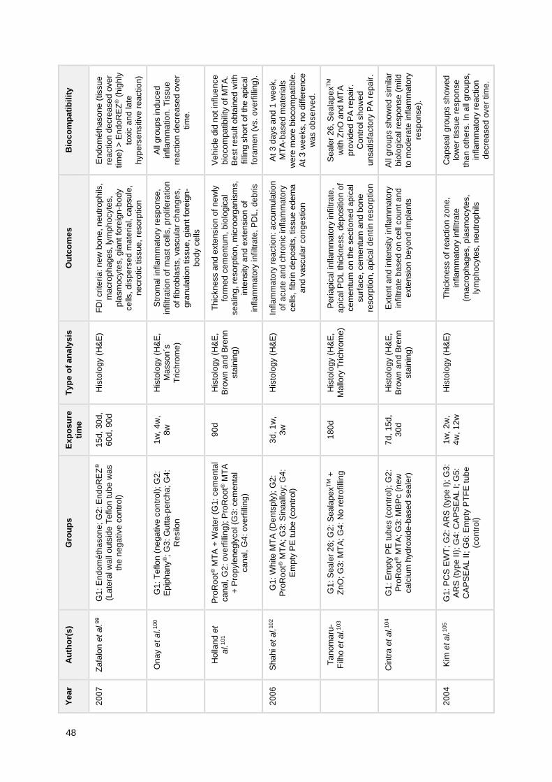
48
Bio
co
mp
ati
bil
ity
En
do
mé
thaso
ne
(tissu
e
rea
ctio
n d
ecre
ase
d o
ve
r
tim
e)
> E
ndo
RE
Z® (
hig
hly
toxic
an
d la
te
hyp
ers
ensitiv
e r
eactio
n)
All
gro
up
s ind
uce
d
inflam
ma
tio
n.
Tis
su
e
rea
ctio
n d
ecre
ase
d o
ve
r
tim
e.
Ve
hic
le d
id n
ot
influ
ence
bio
co
mpa
tib
ility
of M
TA
.
Be
st
result o
bta
ine
d w
ith
filli
ng
sh
ort
of
the
ap
ica
l
fora
me
n (
vs. o
ve
rfill
ing
).
At
3 d
ays a
nd
1 w
eek,
MT
A-b
ase
d m
ate
rials
we
re m
ore
bio
co
mpa
tible
.
At
3 w
eeks, n
o d
iffe
rence
wa
s o
bse
rve
d.
Se
ale
r 2
6,
Sea
lap
ex
TM
with
Zn
O a
nd
MT
A
pro
vid
ed
PA
re
pair
.
Co
ntr
ol sh
ow
ed
un
satisfa
cto
ry P
A r
ep
air
.
All
gro
up
s s
ho
we
d s
imila
r
bio
log
ica
l re
sp
onse
(m
ild
to m
od
era
te in
flam
ma
tory
resp
on
se
).
Ca
pse
al g
rou
ps s
ho
we
d
low
er
tissu
e r
espo
nse
tha
n o
the
rs.
In a
ll g
rou
ps,
inflam
ma
tory
re
actio
n
de
cre
ased
ove
r tim
e.
Ou
tco
me
s
FD
I cri
teri
a: n
ew
bo
ne
, n
eutr
op
hils
,
ma
cro
ph
ag
es,
lym
ph
ocyte
s,
pla
sm
ocyte
s, g
ian
t fo
reig
n-b
ody
ce
lls, d
isp
ers
ed
ma
teria
l, c
ap
su
le,
ne
cro
tic t
issue
, re
so
rptio
n
Str
om
al in
flam
mato
ry r
esp
onse
,
infiltra
tio
n o
f m
ast cells
, p
rolif
era
tio
n
of
fib
rob
lasts
, va
scu
lar
ch
ang
es,
gra
nu
latio
n tis
su
e, g
ian
t fo
reig
n-
bo
dy c
ells
Th
ickn
ess a
nd e
xte
nsio
n o
f n
ew
ly
form
ed
ce
men
tum
, bio
log
ica
l
se
alin
g, re
so
rptio
n, m
icro
org
an
ism
s,
inte
nsity a
nd
exte
nsio
n o
f
inflam
ma
tory
infiltra
te, P
DL
, de
bri
s
Inflam
ma
tory
re
actio
n:
accu
mu
latio
n
of
acu
te a
nd
ch
ron
ic in
flam
mato
ry
ce
lls,
fib
rin
dep
osits,
tissu
e e
de
ma
an
d v
ascu
lar
co
nge
stio
n
Pe
ria
pic
al in
flam
ma
tory
in
filtra
te,
ap
ica
l P
DL
th
ickne
ss,
de
positio
n o
f
ce
me
ntu
m o
n t
he s
ectio
ned
ap
ical
su
rface
, cem
en
tum
an
d b
one
reso
rptio
n,
apic
al de
ntin
reso
rptio
n
Exte
nt a
nd in
ten
sity in
fla
mm
ato
ry
infiltra
te b
ase
d o
n c
ell
cou
nt an
d
exte
nsio
n b
eyon
d im
pla
nts
Th
ickn
ess o
f re
actio
n z
on
e,
inflam
ma
tory
infiltra
te
(ma
cro
ph
ag
es,
pla
sm
ocyte
s,
lym
ph
ocyte
s, n
eu
tro
phils
Ty
pe
of
an
aly
sis
His
tolo
gy (
H&
E)
His
tolo
gy (
H&
E,
Ma
sso
n´s
Tri
ch
rom
e)
His
tolo
gy (
H&
E,
Bro
wn a
nd B
ren
n
sta
inin
g)
His
tolo
gy (
H&
E)
His
tolo
gy (
H&
E,
Ma
llory
Tri
ch
rom
e)
His
tolo
gy (
H&
E,
Bro
wn a
nd B
ren
n
sta
inin
g)
His
tolo
gy (
H&
E)
Ex
po
su
re
tim
e
15
d,
30
d,
60
d,
90
d
1w
, 4
w,
8w
90
d
3d
, 1
w,
3w
18
0d
7d
, 1
5d
,
30
d
1w
, 2
w,
4w
, 1
2w
Gro
up
s
G1
: E
nd
om
éth
ason
e;
G2
: E
ndo
RE
Z®
(La
tera
l w
all
outs
ide
Te
flon
tube
wa
s
the
neg
ative
con
tro
l)
G1
: T
eflo
n (
ne
ga
tive c
on
tro
l); G
2:
Ep
iph
an
y®;
G3
: G
utta
-pe
rch
a; G
4:
Resilo
n
Pro
Roo
t® M
TA
+ W
ate
r (G
1:
ce
me
nta
l
ca
nal, G
2: o
ve
rfill
ing
); P
roR
oot®
MT
A
+ P
rop
yle
ne
gly
co
l (G
3: cem
enta
l
ca
nal, G
4: o
ve
rfill
ing
)
G1
: W
hite
MT
A (
De
nts
ply
); G
2:
Pro
Roo
t® M
TA
; G
3:
Sin
aa
lloy; G
4:
Em
pty
PE
tu
be
(co
ntr
ol)
G1
: S
ea
ler
26
; G
2:
Se
ala
pe
xT
M +
Zn
O;
G3
: M
TA
; G
4:
No r
etr
ofilli
ng
G1
: E
mp
ty P
E t
ub
es (
con
trol)
; G
2:
Pro
Roo
t® M
TA
; G
3: M
BP
c (
new
ca
lciu
m h
yd
roxid
e-b
ase
d s
ea
ler)
G1
: P
CS
EW
T;
G2
: A
RS
(ty
pe I
); G
3:
AR
S (
typ
e II)
; G
4:
CA
PS
EA
L I; G
5:
CA
PS
EA
L I
I; G
6:
Em
pty
PT
FE
tu
be
(co
ntr
ol)
Au
tho
r(s
)
Za
falo
n e
t a
l.9
9
On
ay e
t a
l.10
0
Holla
nd
et
al.
10
1
Sh
ah
i e
t al.
10
2
Ta
nom
aru
-
Filh
o e
t al.
103
Cin
tra
et
al.
10
4
Kim
et a
l.1
05
Ye
ar
20
07
20
06
20
04
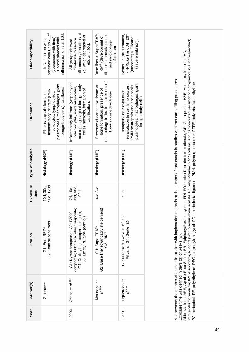
49
Bio
co
mp
ati
bil
ity
Inflam
ma
tion
wa
s
ob
se
rve
d w
ith
En
do
RE
Z®
(de
cre
ased
with
tim
e).
Co
ntr
ol sh
ow
ed
mild
inflam
ma
tio
n o
nly
at
10
d.
All
gro
up
s s
ho
we
d
mo
de
rate
to
se
ve
re
inflam
ma
tory
re
actio
ns a
t
7d
, w
hic
h d
ecre
ase
d a
t
60
d a
nd
90
d.
Ba
se
lin
er
> S
up
erE
BA
TM,
IRM
® (
develo
pm
en
t o
f
fib
rous c
on
ne
ctive t
issue
an
d m
acro
ph
ag
e
infiltra
tio
n).
Se
ale
r 2
6 (
mild
irr
ita
tio
n)
> N
-Ric
ke
rt a
nd
AH
26
®
(mo
de
rate
) >
Fill
ca
nal
(se
ve
re irr
ita
tio
n).
N r
ep
rese
nts
the
num
be
r o
f anim
als
in
stu
die
s w
ith
im
pla
nta
tio
n m
eth
od
s o
r th
e n
um
be
r o
f ro
ot
ca
nals
in
stu
die
s w
ith
ro
ot ca
nal filli
ng
pro
ced
ure
s.
Exp
osu
re tim
e w
as d
efin
ed in
da
ys (
d)
or
we
eks (
w).
Ab
bre
via
tio
ns:
AR
S, A
patite
Ro
ot
Se
ale
r; E
R, E
pip
ha
ny/R
esilo
n s
yste
m;
FD
I, F
éd
era
tion
De
nta
ire
In
tern
atio
na
le;
GP
, G
utta
-pe
rch
a;
H&
E, H
em
ato
xylin
-eo
sin
; IH
C,
imm
un
oh
isto
ch
em
istr
y;
IRC
P: io
do
form
, R
ifoco
rt (
5m
g p
redn
iso
lon
e a
ce
tate
+ 1
.5m
g R
ifa
mycin
SV
so
diu
m)
an
d c
am
ph
ora
ted
pa
ram
on
ochlo
roph
eno
l; n
/s,
no
n-s
pe
cifie
d;
PA
, p
eri
ap
ica
l; P
E,
poly
eth
yle
ne
; P
EG
, p
oly
eth
yle
ne
gly
co
l; P
DL,
pe
rio
do
nta
l lig
am
en
t; P
MN
, p
oly
mo
rph
on
ucle
ar;
PT
FE
, p
oly
tetr
aflu
oro
eth
yle
ne
.
Ou
tco
me
s
Fib
rous c
apsu
le f
orm
atio
n,
inflam
ma
tory
infiltra
te (
PM
N
leu
ko
cyte
s,
lym
ph
ocyte
s,
pla
sm
ocyte
s, m
acro
ph
age
s, g
ian
t
fore
ign
-bod
y c
ells
), c
ap
illa
ries
Inflam
ma
tory
in
filtra
te (
lym
ph
ocyte
s,
pla
sm
ocyte
s,
PM
N le
uko
cyte
s,
ma
cro
ph
ag
es,
gia
nt fo
reig
n-b
od
y
ce
lls),
ne
cro
sis
, fo
rma
tion
of
ca
lcific
atio
ns
Pre
se
nce
of
co
nne
ctive
tis
sue
or
bo
ne
fo
rma
tion
, p
resen
ce o
f
ma
cro
ph
ag
e in
filtra
tio
n, th
ickne
ss o
f
fib
rous c
on
ne
ctive t
issue
His
topa
tholo
gic
evalu
atio
n
(gra
nula
tio
n tis
su
e, ly
mp
ho
cyte
s,
PM
N n
eutr
op
hils
and
eosin
oph
ils,
pla
sm
ocyte
s, m
acro
ph
age
s, g
ian
t
fore
ign
-bod
y c
ells
)
Ty
pe
of
an
aly
sis
His
tolo
gy (
H&
E)
His
tolo
gy (
H&
E)
His
tolo
gy (
H&
E)
His
tolo
gy (
H&
E)
Ex
po
su
re
tim
e
10
d,
30
d,
90
d,
12
0d
7d
, 1
5d
,
30
d,
60
d,
90
d
4w
, 8
w
90
d
Gro
up
s
G1
: E
nd
oR
EZ
®
G2
: S
olid
sili
co
ne r
ods
G1
: D
yra
ct
co
mpo
me
r; G
2:
F20
00
co
mp
om
er;
G3
: V
alu
x P
lus c
om
posite;
G4
: O
rallo
y h
igh
-co
ppe
r am
alg
am
;
G5
: E
mp
ty P
E t
ub
e (
co
ntr
ol)
G1
: S
up
erE
BA
TM
G2
: B
ase
lin
er
(cya
no
acry
late
ce
me
nt)
G3
: IR
M®
G1
: N
-Ric
ke
rt;
G2:
AH
26
®;
G3
:
Fill
ca
na
l; G
4:
Se
ale
r 2
6
Au
tho
r(s
)
Zm
ene
r10
7
Ozb
as e
t al.
10
8
Mo
rina
ga
et
al.
10
9
Fig
ueir
edo
et
al.
11
0
Ye
ar
20
03
20
01
50
All other materials elicited an inflammatory tissue reaction of variable degree. In a
study of retrofilling, Tanomaru-Filho et al.103 showed that Sealer 26, SealapexTM associated
with zinc oxide and MTA provided periapical repair, despite the slight to moderate
inflammatory infiltrate.
3.2.2. Time of exposure influence on biocompatibility
In order to understand how the time of exposure influences the biocompatibility of root
canal sealers, we focused on studies that reported more than one exposure time points.
Based on the results from the included studies, a time-dependency (i.e. resolution of tissue
reaction over time) has been shown for the following sealers: AH PlusTM 94,111,
Endométhasone99, GuttaFlow®2111, GuttaFlow® Bioseal111, MTA Fillapex® 94 and
RealSealTM.117 The decrease in tissue reaction has also been shown for other materials105,108,
as presented in Table 9.
For Epiphany®, contrary evidence was found as Garcia et al.112 and Onay et al.100
showed a decrease in tissue reaction over time whereas Campos-Pinto et al.98 suggested a
resolution of the tissue reaction. Studies on EndoREZ® also showed conflicting results, as
the time-dependency has been shown either as isolated sealer 107 or associated with an
accelerator117, whereas Zafalon et al.99 showed evidence of severe inflammatory infiltrate
even 90 days after implantation.
Regarding ProRoot® MTA, the maturation of the healing process over time with a
decrease in inflammatory infiltrate and improvement of connective tissue organization has
been shown.104,113
3.2.3. Influence of apical limit of root canal filling on biocompatibility
Three studies aimed to evaluate the influence of the apical limit for root canal filling on
biocompatibility to root canal sealers.101,106,118 The three studies demonstrated a better
biocompatibility with root canal filling short of the apical foramen, in comparison with
overfilling for all sealers tested, i.e. Endométhasone, EndoREZ® and ProRoot MTA®.
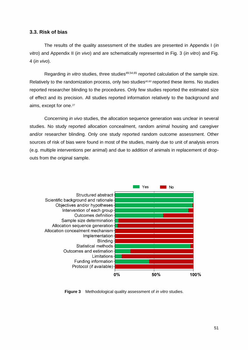
51
3.3. Risk of bias
The results of the quality assessment of the studies are presented in Appendix I (in
vitro) and Appendix II (in vivo) and are schematically represented in Fig. 3 (in vitro) and Fig.
4 (in vivo).
Regarding in vitro studies, three studies49,54,65 reported calculation of the sample size.
Relatively to the randomization process, only two studies40,60 reported these items. No studies
reported researcher blinding to the procedures. Only few studies reported the estimated size
of effect and its precision. All studies reported information relatively to the background and
aims, except for one.27
Concerning in vivo studies, the allocation sequence generation was unclear in several
studies. No study reported allocation concealment, random animal housing and caregiver
and/or researcher blinding. Only one study reported random outcome assessment. Other
sources of risk of bias were found in most of the studies, mainly due to unit of analysis errors
(e.g. multiple interventions per animal) and due to addition of animals in replacement of drop-
outs from the original sample.
Figure 3 Methodological quality assessment of in vitro studies.

52
Figure 4 Methodological quality assessment of in vivo studies.
53
4. DISCUSSION
In the context of root canal therapy, materials used for root canal filling may come in
contact with the periapical tissue.4 Ideally, these materials should allow or promote the
resolution of periapical inflammatory and/or infectious processes, also preventing further
contamination with microorganisms.4 Among the biological properties desirably shown by
sealers (e.g. antimicrobial effect, osteogenic potential), biocompatibility is considered a key
property of root canal sealers4,5,24, thus demonstrating the importance of the study of
biocompatibility of different endodontic materials.33
For root canal filling, the combination of a sealer with a central core material, such as
gutta-percha, has been a standard.4,7 Several reasons support the widespread use of gutta-
percha, namely its plasticity, low toxic potential, ease of manipulation, radiopacity and ease
of removal, even though the lack of adhesion to dentin and shrinkage after cooling are known
disadvantages of this material.4 Other core materials and/or obturation systems have also
been developed, such as resin-based obturation systems with the high-performance
synthetic polyester–based Resilon (e.g. in association with RealSealTM or Epiphany®) and
Activ GPTM, which consists of glass ionomer-impregnated gutta-percha cones.4,7
Here, we aimed to perform a systematic review of the literature on the cytotoxicity and
biocompatibility of root canal sealers, in order to understand how these materials (individually
or by type) perform in terms of biocompatibility in experimental cell and animal models.
Furthermore, we also aimed to understand how the material setting condition, concentration,
time and type of exposure influence the cytotoxicity and biocompatibility of these materials.
As a multiplicity of methods and conditions has been reported in previous studies in this
area, an overview on this subject could become difficult as well as the interpretation of the
results. Therefore, a systematic review of the literature may be a useful tool to integrate such
concepts and data.
Over the years, several materials have been developed for root canal filling.
According to chemical composition and structure, sealers may be classified into the following
types: zinc oxide-eugenol-based, resin-based, glass ionomer-based, silicone-based, calcium
hydroxide-based and bioceramic sealers, which includes calcium silicate-based, MTA-based
and calcium phosphate-based sealers. In regard to retrograde filling, MTA, amalgam, IRM®
and SuperEBATM are some of the materials that have been used.3 AH PlusTM has been the
most studied sealer over the last two decades, either as a test sealer or as reference

54
material, thus we applied a date limit to our search in order to retrieve articles since its
introduction as a new substitute to AH 26®.89
In this systematic review, the set of included studies assessed the cytotoxicity and
biocompatibility of multiple sealers of the different types. Among in vitro studies, the most
studied sealers were the zinc oxide-eugenol-based PCS, the epoxy resin-based AH 26® and
AH PlusTM, the methacrylate resin-based EndoREZ® and Epiphany®, the calcium hydroxide-
based SealapexTM and the bioceramic sealers Endosequence BCTM, MTA Fillapex® and
ProRoot® MTA. AH PlusTM, EndoREZ®, Epiphany® and ProRoot® MTA were also the most
studied in vivo.
Concerning in vitro cytotoxicity, results suggested a lower cytotoxic potential from
bioceramic sealers, even though some conflicting evidence was found, particular in regard to
MTA Fillapex®, which may be due to the release of lead in set conditions.29 This lower
cytotoxicity of bioceramic sealers is in accordance with previous systematic reviews on the
biological, physiochemical and clinical properties of calcium silicate-based sealers in
comparison with conventional materials.17–19
A considerable methodological heterogeneity was observed in relation to several
parameters, for example material setting condition, setting time and sealer extract dilution.
As to setting condition, several studies performed experiments with freshly mixed sealers,
others used set materials and others both freshly mixed and set conditions. Moreover,
multiple setting times were reported from 1 hour to 1 month. In general, studies that
assessed both conditions reported a differential in cytotoxic potential, with freshly mixed
materials exhibiting higher cytotoxic potential.
The important role of setting conditions on the biological properties of sealers has
been recognized, as differences have been reported between fresh and set sealers119,120,
which may account for some of the heterogeneity in the literature. However, such differences
seem to decrease with setting.4,15 The release of unconverted monomers may play a role in
the cytotoxicity of sealers in freshly mixed conditions, whereas, in set condition, a residual
toxic effect, amount- and elution kinetics-dependent, may be expected for these
compounds.31 However, the leaching of unconverted or partially converted constituents with
potential toxicity may also remain after setting of the material.58 In fact, the role of setting time
has been studied by Camargo et al.34, who suggested further research should be carried out
aiming at evaluating this setting time-dependency over longer experimental periods. Arun et
al.58 also suggested that long-term clinical studies are important to understand if these
materials maintain as cytotoxic over time or loose the initial toxic potential.
55
From a clinical perspective, the use of freshly mixed sealers is relevant because
these materials are applied in an unset condition when introduced in root canals, becoming
in contact with the periapical tissues.26,31
In studies which tested sealer extracts in multiple concentrations, results suggested a
concentration-dependency of the cytotoxicity, i.e. increasing cytotoxic potential with
increasing extract concentration, for several sealers.
Furthermore, different contact methods were used, specifically direct contact testing,
indirect contact testing with sealer extracts and indirect contact testing with incubation of
cells with sealer specimens (without direct contact, using for example inserts). Previous
studies have suggested that direct contact exposure may lead to increased toxicity, in
comparison with other methods and in spite of the acceptable clinical performance.56,82
However, as a direct contact between the sealer and the periapical tissue is possible during
and after root filling procedures4,82, such contact method may provide important information
on the cytotoxicity of these materials as it simulates the possible extrusion of unset sealer in
the periapical tissues.31,56,65,82 Furthermore, some studies used root models26,40,60,82,84, which
may represent a useful model as it attempts to simulate the reality of endodontic
treatments.26
Regarding the influence of exposure time on the cytotoxic effects of the materials,
studies showed a certain heterogeneity. Interestingly, studies that used washed-out or “aged”
sealers reported a general recovery of cell viability over 5-6 weeks of observation.49,54,56,65
Also, Bonson et al.81 reported a lower cytotoxicity of washed sealers over 13 days of
observation.
As mentioned, these findings seem to be supported by studies that tested sealers by
extraction methods, with different extraction time points. Methodologically, a difference in
studies was observed in this regard, as some studies performed cumulative extractions, i.e.
no medium renewal, and others carried out separate extractions, that is with medium
renewal. In general, the first method appears to be related to higher cytotoxic effects. In a
way, such findings may be related to the time-dependent release of compounds with setting,
as previously discussed.
The in vitro studies included in this systematic review are indicative of differences
between the several root canal sealers. Furthermore, most studies followed the ISO
Standards 10993-5:2009, which encompasses direct and indirect contact methods, fresh and
set materials and several extract concentrations. However, the concentrations tend to be

56
higher compared to the clinical context. Therefore, care should be taken when extrapolating
these results for clinical practice.
Relatively to the in vivo evidence, all studies showed an inflammatory reaction in
response to the several sealers, independently of type, that ranged from slight to severe
inflammatory reactions. Nevertheless, studies also generally suggested that the tested
sealers presented acceptable biocompatibility. The ability to provide the re-establishment of
original bone structure was also shown for some sealers, such as AH PlusTM and MTA
Fillapex®.94
Moreover, different methods were used for the assessment of tissue response to
sealers. The ISO Standards 7405 on the biological evaluation of dental materials were
followed in most studies. In this context, several studies assessed the tissue response to
subcutaneous or intraosseous sealer implantation and others the periapical tissue response
to root filling procedure. In one study, Cintra et al.113 compared the subcutaneous
implantation and the alveolar socket implantation (i.e. sealer implantation in alveolar socket
post-extraction) methods and showed the equivalency between methods. This method was
also reported in another study104, further suggesting it could be an interesting method to
study tissue response to dental materials. From the studies that performed root filling
procedures, Tanomaru-Filho et al.103 evaluated periapical tissue response to retrofilling, after
periapical lesion induction, in order to simulate the clinical conditions of endodontic surgery.
The influence of exposure time on biocompatibility was shown by several studies,
which showed that the initial inflammatory reaction tends to subside over time.94,99,105,108,111,117
In addition to the decrease in inflammatory infiltrate, ProRoot® MTA has been shown to
promote the improvement of connective tissue organization over time, thus suggesting the
maturation of the healing process.104,113 However, conflicting results were found for some
sealers, specifically Epiphany® and EndoREZ®.
In addition, three in vivo studies tested the biocompatibility of root canal fillings by
comparison of two apical limits, short of the apical foramen and overfilling.101,106,118 As
expected, a better biocompatibility was shown in fillings short of the apical foramen, in
accordance with previous literature.
In regard to the methodologic quality, eligible studies exhibited a considerable risk of
bias, with several studies lacking information on randomization processes, blinding and
outcome measures, including estimated size of effects and its precision.

57
5. CONCLUSION
In this study, we carried out a systematic review of the literature on the cytotoxicity
and biocompatibility of root canal sealers.
A joint analysis of the included in vitro and in vivo studies reveals that sealers elicit
variable toxic effects at cellular and tissue level. Although a tendency for lower cytotoxicity of
bioceramic sealers was noted, such finding was not observed in vivo, probably due to the
smaller number of studies and sealers and to the variability of conditions tested.
Nevertheless, results suggested that several factors may influence the biocompatibility of
these materials, particularly setting condition and time and type of exposure, among others.
Further research is warranted to achieve a better understanding of the biological
effects of root canal sealers, with precisely designed studies and accurate and complete
reporting.
From a clinical perspective, the extrapolation of these results must be taken into
caution due to several aspects, namely: biocompatibility was assessed in experimental
models; some methods do not correlate directly to the clinical reality of endodontic
treatments, e.g. testing only set materials; and other material properties should be taken into
account, e.g. antimicrobial and physicochemical properties.

58

59
REFERENCES
1. Hargreaves KM, Berman LH. Cohen’s Pathways of the Pulp. 11th ed. St. Louis,
Missouri: Elsevier Inc; 2016. 992 p.
2. Lee Y-L, Lee B-S, Lin F-H, Yun Lin A, Lan W-H, Lin C-P. Effects of physiological
environments on the hydration behavior of mineral trioxide aggregate. Biomaterials.
2004;25(5):787–93.
3. Ma X, Li C, Jia L, Wang Y, Liu W, Zhou X, et al. Materials for retrograde filling in root
canal therapy. Cochrane Database Syst Rev. 2016;(12).
4. Johnson W, Kulild JC, Tay F. Obturation of the Cleaned and Shaped Root Canal
System. In: Hargreaves KM, Berman LH, editors. Cohen’s Pathways of the Pulp. 11th
ed. St. Louis, Missouri: Elsevier Inc; 2016. p. 280–322.
5. Grossman L. Endodontics. 11th ed. Philadelphia: Lea & Febiger; 1988.
6. Kishen A, Peters OA, Zehnder M, Diogenes AR, Nair MK. Advances in endodontics:
Potential applications in clinical practice. J Conserv Dent. 2016;19(3):199–206.
7. Ørstavik D. Materials used for root canal obturation: technical, biological and clinical
testing. Endod Top. 2005;12(1):25–38.
8. AL-Haddad A, Che Ab Aziz ZA. Bioceramic-Based Root Canal Sealers: A Review. Int
J Biomater. 2016;2016:97532:1–10.
9. Torabinejad M, Watson TF, Ford TRP. Sealing ability of a mineral trioxide aggregate
when used as a root end filling material. J Endod. 1993;19(12):591–5.
10. Parirokh M, Torabinejad M. Mineral Trioxide Aggregate: A Comprehensive Literature
Review—Part I: Chemical, Physical, and Antibacterial Properties. J Endod.
2010;36(1):16–27.
11. Torabinejad M, Parirokh M. Mineral Trioxide Aggregate: A Comprehensive Literature
Review—Part II: Leakage and Biocompatibility Investigations. J Endod.
2010;36(2):190–202.
12. Parirokh M, Torabinejad M. Mineral Trioxide Aggregate: A Comprehensive Literature

60
Review—Part III: Clinical Applications, Drawbacks, and Mechanism of Action. J
Endod. 2010;36(3):400–13.
13. Browne RM. Animal tests for biocompatibility of dental materials—relevance,
advantages and limitations. J Dent. 1994;22:S21–4.
14. Williams DF. Biocompatibility: an overview. In: William DF, editor. Concise
Encyclopaedia of Medical and Dental Materials. Oxford: Pergamon; 1990. p. 51–9.
15. Langeland K. Root canal sealants and pastes. Dent Clin North Am. 1974;18:309.
16. Schmalz G. Use of cell cultures for toxicity testing of dental materials—advantages
and limitations. J Dent. 1994;22:S6–11.
17. Donnermeyer D, Bürklein S, Dammaschke T, Schäfer E. Endodontic sealers based on
calcium silicates: a systematic review. Odontology. 2018;doi: 10.1007/s10266-018-
0400-3.
18. de Oliveira NG, de Souza Araújo PR, da Silveira MT, Veras Sobral AP, Carvalho MV.
Comparison of the biocompatibility of calcium silicate-based materials to mineral
trioxide aggregate: Systematic review. Eur J Dent. 2018;12(2):317.
19. Silva Almeida LH, Moraes RR, Morgental RD, Pappen FG. Are Premixed Calcium
Silicate–based Endodontic Sealers Comparable to Conventional Materials? A
Systematic Review of In Vitro Studies. J Endod. 2017;43(4):527–35.
20. Liberati A, Altman DG, Tetzlaff J, Mulrow C, Gotzsche PC, Ioannidis JPA, et al. The
PRISMA statement for reporting systematic reviews and meta-analyses of studies that
evaluate healthcare interventions: explanation and elaboration. BMJ. 2009;339:b2700.
21. Riva JJ, Malik KMP, Burnie SJ, Endicott AR, Busse JW. What is your research
question? An introduction to the PICOT format for clinicians. J Can Chiropr Assoc.
2012;56(3):167–71.
22. Faggion CM. Guidelines for Reporting Pre-clinical In Vitro Studies on Dental Materials.
J Evid Based Dent Pract. 2012;12(4):182–9.
23. Hooijmans CR, Rovers MM, de Vries RB, Leenaars M, Ritskes-Hoitinga M,
Langendam MW. SYRCLE’s risk of bias tool for animal studies. BMC Med Res
Methodol. 2014;14(1):43.

61
24. Lee B-N, Hong J-U, Kim S-M, Jang J-H, Chang H-S, Hwang Y-C, et al. Anti-
inflammatory and Osteogenic Effects of Calcium Silicate–based Root Canal Sealers. J
Endod. 2019;45(1):73–8.
25. Jeanneau C, Giraud T, Laurent P, About I. BioRoot RCS Extracts Modulate the Early
Mechanisms of Periodontal Inflammation and Regeneration. J Endod. 2019;doi:
10.1016/j.joen.2019.04.003.
26. Camps J, Jeanneau C, Ayachi I El, Laurent P, About I. Bioactivity of a Calcium
Silicate–based Endodontic Cement (BioRoot RCS): Interactions with Human
Periodontal Ligament Cells In Vitro. J Endod. 2015;41(9):1469–73.
27. Dimitrova-Nakov S, Uzunoglu E, Ardila-Osorio H, Baudry A, Richard G, Kellermann O,
et al. In vitro bioactivity of BiorootTM RCS, via A4 mouse pulpal stem cells. Dent Mater.
2015;31(11):1290–7.
28. Konjhodzic-Prcic A, Gorduysus O, Kucukkaya S, Atila B, Muftuoglu S, Zeybek D. In
Vitro Comparison of Cytotoxicity of Four Root Canal Sealers on Human Gingival
Fibroblasts. Med Arch. 2015;69(1):24.
29. Zhou H, Du T, Shen Y, Wang Z, Zheng Y, Haapasalo M. In Vitro Cytotoxicity of
Calcium Silicate–containing Endodontic Sealers. J Endod. 2015;41(1):56–61.
30. Jiang Y, Zheng Q, Zhou X, Gao Y, Huang D. A Comparative Study on Root Canal
Repair Materials: A Cytocompatibility Assessment in L929 and MG63 Cells. Sci World
J. 2014;2014:1–8.
31. Cotti E, Petreucic V, Re D, Simbula G. Cytotoxicity Evaluation of a New Resin-based
Hybrid Root Canal Sealer: An In Vitro Study. J Endod. 2014;40(1):124–8.
32. Chang S-W, Lee S-Y, Kang S-K, Kum K-Y, Kim E-C. In Vitro Biocompatibility,
Inflammatory Response, and Osteogenic Potential of 4 Root Canal Sealers: Sealapex,
Sankin Apatite Root Sealer, MTA Fillapex, and iRoot SP Root Canal Sealer. J Endod.
2014;40(10):1642–8.
33. Mandal P, Zhao J, Sah SK, Huang Y, Liu J. In Vitro Cytotoxicity of GuttaFlow 2 on
Human Gingival Fibroblasts. J Endod. 2014;40(8):1156–9.
34. Camargo CHR, Oliveira TR, Silva GO, Rabelo SB, Valera MC, Cavalcanti BN. Setting
Time Affects In Vitro Biological Properties of Root Canal Sealers. J Endod.

62
2014;40(4):530–3.
35. Choi Y, Park S-J, Lee S-H, Hwang Y-C, Yu M-K, Min K-S. Biological Effects and
Washout Resistance of a Newly Developed Fast-setting Pozzolan Cement. J Endod.
2013;39(4):467–72.
36. Giacomino CM, Wealleans JA, Kuhn N, Diogenes A. Comparative Biocompatibility and
Osteogenic Potential of Two Bioceramic Sealers. J Endod. 2019;45(1):51–6.
37. Güven EP, Yalvaç ME, Kayahan MB, Sunay H, SahIn F, Bayirli G. Human tooth germ
stem cell response to calcium-silicate based endodontic cements. J Appl Oral Sci.
2013;21(4):351–7.
38. Willershausen I, Wolf T, Kasaj A, Weyer V, Willershausen B, Marroquin BB. Influence
of a bioceramic root end material and mineral trioxide aggregates on fibroblasts and
osteoblasts. Arch Oral Biol. 2013;58(9):1232–7.
39. Kim T-G, Lee Y-H, Lee N-H, Bhattarai G, Lee I-K, Yun B-S, et al. The Antioxidant
Property of Pachymic Acid Improves Bone Disturbance against AH Plus–induced
Inflammation in MC-3T3 E1 Cells. J Endod. 2013;39(4):461–6.
40. De-Deus G, Canabarro A, Alves GG, Marins JR, Linhares ABR, Granjeiro JM.
Cytocompatibility of the ready-to-use bioceramic putty repair cement iRoot BP Plus
with primary human osteoblasts. Int Endod J. 2012;45(6):508–13.
41. Bin C V., Valera MC, Camargo SEAA, Rabelo SB, Silva GO, Balducci I, et al.
Cytotoxicity and genotoxicity of root canal sealers based on mineral trioxide
aggregate. J Endod. 2012;38(4):495–500.
42. Scelza MZ, Coil J, Alves GG. Effect of time of extraction on the biocompatibility of
endodontic sealers with primary human fibroblasts. Braz Oral Res. 2012;26(5):424–
30.
43. Salles LP, Gomes-Cornélio AL, Guimarães FC, Herrera BS, Bao SN, Rossa-Junior C,
et al. Mineral Trioxide Aggregate–based Endodontic Sealer Stimulates Hydroxyapatite
Nucleation in Human Osteoblast-like Cell Culture. J Endod. 2012;38(7):971–6.
44. Landuyt KL Van, Geebelen B, Shehata M, Furche SL, Durner J, Meerbeek B Van, et
al. No Evidence for DNA Double-strand Breaks Caused by Endodontic Sealers. J
Endod. 2012;38(5):636–41.

63
45. Ma J, Shen Y, Stojicic S, Haapasalo M. Biocompatibility of Two Novel Root Repair
Materials. J Endod. 2011;37(6):793–8.
46. Mukhtar-Fayyad D. Cytocompatibility of new bioceramic-based materials on human
fibroblast cells (MRC-5). Oral Surgery, Oral Med Oral Pathol Oral Radiol
Endodontology. 2011;112(6):e137–42.
47. Vouzara T, Dimosiari G, Koulaouzidou EA, Economides N. Cytotoxicity of a New
Calcium Silicate Endodontic Sealer. J Endod. 2018;44(5):849–52.
48. Zoufan K, Jiang J, Komabayashi T, Wang Y-H, Safavi KE, Zhu Q. Cytotoxicity
evaluation of Gutta Flow and Endo Sequence BC sealers. Oral Surgery, Oral Med
Oral Pathol Oral Radiol Endodontology. 2011;112(5):657–61.
49. Loushine BA, Bryan TE, Looney SW, Gillen BM, Loushine RJ, Weller RN, et al.
Setting Properties and Cytotoxicity Evaluation of a Premixed Bioceramic Root Canal
Sealer. J Endod. 2011;37(5):673–7.
50. Yu M-K, Lee Y-H, Yoon M-R, Bhattarai G, Lee N-H, Kim T-G, et al. Attenuation of
AH26-Induced Apoptosis by Inhibition of SAPK/JNK Pathway in MC-3T3 E1 Cells. J
Endod. 2010;36(12):1967–71.
51. AlAnezi AZ, Jiang J, Safavi KE, Spangberg LSW, Zhu Q. Cytotoxicity evaluation of
endosequence root repair material. Oral Surgery, Oral Med Oral Pathol Oral Radiol
Endodontology. 2010;109(3):e122–5.
52. Zhang W, Li Z, Peng B. Effects of iRoot SP on Mineralization-related Genes
Expression in MG63 Cells. J Endod. 2010;36(12):1978–82.
53. Huang F-M, Yang S-F, Chang Y-C. Effects of Root Canal Sealers on Alkaline
Phosphatase in Human Osteoblastic Cells. J Endod. 2010;36(7):1230–3.
54. Bryan TE, Khechen K, Brackett MG, Messer RLW, El-Awady A, Primus CM, et al. In
Vitro Osteogenic Potential of an Experimental Calcium Silicate–based Root Canal
Sealer. J Endod. 2010;36(7):1163–9.
55. Badr AE. Marginal Adaptation and Cytotoxicity of Bone Cement Compared with
Amalgam and Mineral Trioxide Aggregate as Root-end Filling Materials. J Endod.
2010;36(6):1056–60.
56. Ames JM, Loushine RJ, Babb BR, Bryan TE, Lockwood PE, Sui M, et al.

64
Contemporary Methacrylate Resin–based Root Canal Sealers Exhibit Different
Degrees of Ex Vivo Cytotoxicity When Cured in Their Self-cured Mode. J Endod.
2009;35(2):225–8.
57. Donadio M, Jiang J, He J, Wang Y-H, Safavi KE, Zhu Q. Cytotoxicity evaluation of
Activ GP and Resilon sealers in vitro. Oral Surgery, Oral Med Oral Pathol Oral Radiol
Endodontology. 2009;107(6):e74–8.
58. Arun S, Sampath V, Mahalaxmi S, Rajkumar K. A Comparative Evaluation of the
Effect of the Addition of Pachymic Acid on the Cytotoxicity of 4 Different Root Canal
Sealers—An In Vitro Study. J Endod. 2017;43(1):96–9.
59. Gambarini G, Romeo U, Tucci E, Gerosa R, Nocca G, Lupi A, et al. Cytotoxicity of
epiphany SE endodontic sealer: a comparative in vitro study. Med Sci Monit.
2009;15(4):PI15-8.
60. De-Deus G, Canabarro A, Alves G, Linhares A, Senne MI, Granjeiro JM. Optimal
Cytocompatibility of a Bioceramic Nanoparticulate Cement in Primary Human
Mesenchymal Cells. J Endod. 2009;35(10):1387–90.
61. Camargo CHR, Camargo SEA, Valera MC, Hiller K-A, Schmalz G, Schweikl H. The
induction of cytotoxicity, oxidative stress, and genotoxicity by root canal sealers in
mammalian cells. Oral Surgery, Oral Med Oral Pathol Oral Radiol Endodontology.
2009;108(6):952–60.
62. Huang F-M, Lee S-S, Yang S-F, Chang Y-C. Up-regulation of Receptor Activator
Nuclear Factor–Kappa B Ligand Expression by Root Canal Sealers in Human
Osteoblastic Cells. J Endod. 2009;35(3):363–6.
63. Heitman EP, Joyce AP, McPherson JC, Roberts S, Chuang A. An In Vitro Evaluation
of the Growth of Human Periodontal Ligament Fibroblasts after Exposure to a
Methacrylate-based Endodontic Sealer. J Endod. 2008;34(2):186–9.
64. Valois CRA, Azevedo RB. Cell-cycle deregulation induced by three different root canal
sealers in vitro. Oral Surgery, Oral Med Oral Pathol Oral Radiol Endodontology.
2008;106(5):763–7.
65. Pinna L, Brackett MG, Lockwood PE, Huffman BP, Mai S, Cotti E, et al. In Vitro
Cytotoxicity Evaluation of a Self-adhesive, Methacrylate Resin–based Root Canal
Sealer. J Endod. 2008;34(9):1085–8.
65
66. Al-Sa’eed OR, Al-Hiyasat AS, Darmani H. The Effects of Six Root-end Filling Materials
and Their Leachable Components on Cell Viability. J Endod. 2008;34(11):1410–4.
67. Huang F-M, Yang S-F, Chang Y-C. Up-regulation of Gelatinases and Tissue Type
Plasminogen Activator by Root Canal Sealers in Human Osteoblastic Cells. J Endod.
2008;34(3):291–4.
68. Gorduysus M, Avcu N, Gorduysus O, Pekel A, Baran Y, Avcu F, et al. Cytotoxic
Effects of Four Different Endodontic Materials in Human Periodontal Ligament
Fibroblasts. J Endod. 2007;33(12):1450–4.
69. Collado-González M, García-Bernal D, Oñate-Sánchez RE, Ortolani-Seltenerich PS,
Lozano A, Forner L, et al. Biocompatibility of three new calcium silicate-based
endodontic sealers on human periodontal ligament stem cells. Int Endod J.
2017;50(9):875–84.
70. Lee DH, Kim NR, Lim B-S, Lee Y-K, Hwang K-K, Yang H-C. Effects of Root Canal
Sealers on Lipopolysaccharide-induced Expression of Cyclooxygenase-2 mRNA in
Murine Macrophage Cells. J Endod. 2007;33(11):1329–33.
71. Lee DH, Lim B-S, Lee Y-K, Yang H-C. In vitro biological adverse effects of dental resin
monomers and endodontic root canal sealers. Curr Appl Phys. 2007;7:e130–4.
72. Lee DH, Lim B-S, Lee Y-K, Yang H-C. Mechanisms of root canal sealers cytotoxicity
on osteoblastic cell line MC3T3-E1. Oral Surgery, Oral Med Oral Pathol Oral Radiol
Endodontology. 2007;104(5):717–21.
73. Merdad K, Pascon AE, Kulkarni G, Santerre P, Friedman S. Short-Term Cytotoxicity
Assessment of Components of the Epiphany Resin-Percha Obturating System by
Indirect and Direct Contact Millipore Filter Assays. J Endod. 2007;33(1):24–7.
74. Lodienė G, Morisbak E, Bruzell E, Ørstavik D. Toxicity evaluation of root canal sealers
in vitro. Int Endod J. 2008;41(1):72–7.
75. Key JE, Rahemtulla FG, Eleazer PD. Cytotoxicity of a New Root Canal Filling Material
on Human Gingival Fibroblasts. J Endod. 2006;32(8):756–8.
76. Bouillaguet S, Wataha JC, Tay FR, Brackett MG, Lockwood PE. Initial In Vitro
Biological Response to Contemporary Endodontic Sealers. J Endod.
2006;32(10):989–92.

66
77. Miletić I, Devčić N, Anić I, Borčić J, Karlović Z, Osmak M. The Cytotoxicity of
RoekoSeal and AH Plus Compared during Different Setting Periods. J Endod.
2005;31(4):307–9.
78. Al-Awadhi S, Spears R, Gutmann JL, Opperman LA. Cultured Primary Osteoblast
Viability and Apoptosis in the Presence of Root Canal Sealers. J Endod.
2004;30(7):527–33.
79. Bouillaguet S, Wataha JC, Lockwood PE, Galgano C, Golay A, Krejci I. Cytotoxicity
and sealing properties of four classes of endodontic sealers evaluated by succinic
dehydrogenase activity and confocal laser scanning microscopy. Eur J Oral Sci.
2004;112(2):182–7.
80. Collado-González M, Tomás-Catalá CJ, Oñate-Sánchez RE, Moraleda JM,
Rodríguez-Lozano FJ. Cytotoxicity of GuttaFlow Bioseal, GuttaFlow2, MTA Fillapex,
and AH Plus on Human Periodontal Ligament Stem Cells. J Endod. 2017;43(5):816–
22.
81. Bonson S, Jeansonne BG, Lallier TE. Root-end Filling Materials Alter Fibroblast
Differentiation. J Dent Res. 2004;83(5):408–13.
82. Camps J, About I. Cytotoxicity testing of endodontic sealers: A new method. J Endod.
2003;29(9):583–6.
83. Mendes ST de O, Sobrinho APR, De Carvalho AT, De Souza Côrtes MI, Vieira LQ. In
vitro evaluation of the cytotoxicity of two root canal sealers on macrophage activity. J
Endod. 2003;29(2):95–9.
84. Schwarze T, Leyhausen G, Geurtsen W. Long-Term Cytocompatibility of Various
Endodontic Sealers Using a New Root Canal Model. J Endod. 2002;28(11):749–53.
85. Huang T-H, Yang J-J, Li H, Kao C-T. The biocompatibility evaluation of epoxy resin-
based root canal sealers in vitro. Biomaterials. 2002;23(1):77–83.
86. Schwarze T, Fiedler I, Leyhausen G, Geurtsen W. The Cellular Compatibility of Five
Endodontic Sealers during the Setting Period. J Endod. 2002;28(11):784–6.
87. Keiser K, Chad Johnson C, Tipton DA. Cytotoxicity of mineral trioxide aggregate using
human periodontal ligament fibroblasts. J Endod. 2000;26(5):288–91.
88. Azar NG, Heidari M, Bahrami ZS, Shokri F. In Vitro Cytotoxicity of a New Epoxy Resin
67
Root Canal Sealer. J Endod. 2000;26(8):462–5.
89. Huang TH, Lii CK, Chou MY, Kao CT. Lactate dehydrogenase leakage of hepatocytes
with AH26 and AH plus sealer treatments. J Endod. 2000;26(9):509–11.
90. Schweikl H, Schmalz G. The induction of micronuclei in V79 cells by the root canal
filling material AH Plus. Biomaterials. 2000;21(9):939–44.
91. Zhu X, Yuan Z, Yan P, Li Y, Jiang H, Huang S. Effect of iRoot SP and mineral trioxide
aggregate (MTA) on the viability and polarization of macrophages. Arch Oral Biol.
2017;80:27–33.
92. Lv F, Zhu L, Zhang J, Yu J, Cheng X, Peng B. Evaluation of the in vitro
biocompatibility of a new fast-setting ready-to-use root filling and repair material. Int
Endod J. 2017;50(6):540–8.
93. Suciu I, Soritau O, Gheorghe I, Lazăr V, Bodnar DC, Delean AG, et al. Biocompatibility
testing on cell culture of some root canal sealers used in endodontics. Rom Biotechnol
Lett. 2016;21(3):11543–9.
94. Assmann E, Böttcher DE, Hoppe CB, Grecca FS, Kopper PMP. Evaluation of Bone
Tissue Response to a Sealer Containing Mineral Trioxide Aggregate. J Endod.
2015;41(1):62–6.
95. Silva LAB, Barnett F, Pumarola-Suñé J, Cañadas PS, Nelson-Filho P, Silva RAB.
Sealapex Xpress and RealSeal XT Feature Tissue Compatibility In Vivo. J Endod.
2014;40(9):1424–8.
96. Tanomaru-Filho M, Tanomaru JMG, Leonardo MR, da Silva LAB. Periapical repair
after root canal filling with different root canal sealers. Braz Dent J. 2009;20(5):389–
95.
97. Leonardo MR, Flores DSH, de Paula e Silva FWG, de Toledo Leonardo R, da Silva
LAB. A Comparison Study of Periapical Repair in Dogs’ Teeth Using RoekoSeal and
AH Plus Root Canal Sealers: A Histopathological Evaluation. J Endod.
2008;34(7):822–5.
98. Campos-Pinto MMD de, Oliveira DA de, Versiani MA, Silva-Sousa YTC, de Sousa-
Neto MD, da Cruz Perez DE. Assessment of the biocompatibility of Epiphany root
canal sealer in rat subcutaneous tissues. Oral Surgery, Oral Med Oral Pathol Oral
68
Radiol Endodontology. 2008;105(5):e77–81.
99. Zafalon EJ, Versiani MA, de Souza CJA, Moura CCG, Dechichi P. In vivo comparison
of the biocompatibility of two root canal sealers implanted into the subcutaneous
connective tissue of rats. Oral Surgery, Oral Med Oral Pathol Oral Radiol
Endodontology. 2007;103(5):e88–94.
100. Onay EO, Ungor M, Ozdemir BH. In vivo evaluation of the biocompatibility of a new
resin-based obturation system. Oral Surgery, Oral Med Oral Pathol Oral Radiol
Endodontology. 2007;104(3):e60–6.
101. Holland R, Mazuqueli L, de Souza V, Murata SS, Júnior ED, Suzuki P. Influence of the
Type of Vehicle and Limit of Obturation on Apical and Periapical Tissue Response in
Dogs’ Teeth After Root Canal Filling With Mineral Trioxide Aggregate. J Endod.
2007;33(6):693–7.
102. Shahi S, Rahimi S, Lotfi M, Yavari H, Gaderian A. A Comparative Study of the
Biocompatibility of Three Root-end Filling Materials in Rat Connective Tissue. J
Endod. 2006;32(8):776–80.
103. Tanomaru-Filho M, Luis MR, Leonardo MR, Tanomaru JMG, Silva LAB. Evaluation of
periapical repair following retrograde filling with different root-end filling materials in
dog teeth with periapical lesions. Oral Surgery, Oral Med Oral Pathol Oral Radiol
Endodontology. 2006;102(1):127–32.
104. Cintra LTA, de Moraes IG, Estrada BPF, Gomes-Filho JE, Bramante CM, Garcia RB,
et al. Evaluation of the Tissue Response to MTA and MBPC: Microscopic Analysis of
Implants in Alveolar Bone of Rats. J Endod. 2006;32(6):556–9.
105. Kim J-S, Baek S-H, Bae K-S. In Vivo Study on the Biocompatibility of Newly
Developed Calcium Phosphate-Based Root Canal Sealers. J Endod.
2004;30(10):708–11.
106. Suzuki P, Souza V de, Holland R, Gomes-Filho JE, Murata SS, Dezan Junior E, et al.
Tissue reaction to Endométhasone sealer in root canal fillings short of or beyond the
apical foramen. J Appl Oral Sci. 2011;19(5):511–6.
107. Zmener O. Tissue Response to a New Methacrylate-Based Root Canal Sealer:
Preliminary Observations in the Subcutaneous Connective Tissue of Rats. J Endod.
2004;30(5):348–51.
69
108. Ozbas H, Yaltirik M, Bilgic B, Issever H. Reactions of connective tissue to compomers,
composite and amalgam root-end filling materials. Int Endod J. 2003;36(4):281–7.
109. Morinaga K, Nakagawa K, Carr GB. Tissue reactions after intraosseous implantation
of three retrofilling materials. Bull Tokyo Dent Coll. 2003;44(1):1–7.
110. Figueiredo JAP, Pesce HF, Gioso MA, Figueiredo MAZ. The histological effects of four
endodontic sealers implanted in the oral mucosa: submucous injection versus implant
in polyethylene tubes. Int Endod J. 2001;34(5):377–85.
111. Santos JM, Pereira S, Sequeira DB, Messias AL, Martins JB, Cunha H, et al.
Biocompatibility of a bioceramic silicone-based sealer in subcutaneous tissue. J Oral
Sci. 2019;61(1):171–7.
112. Garcia L da FR, Marques AAF, Roselino L de MR, Pires-de-Souza F de CP, Consani
S. Biocompatibility Evaluation of Epiphany/Resilon Root Canal Filling System in
Subcutaneous Tissue of Rats. J Endod. 2010;36(1):110–4.
113. Cintra LTA, Bernabé PFE, de Moraes IG, Gomes-Filho JE, Okamoto T, Consolaro A,
et al. Evaluation of subcutaneous and alveolar implantation surgical sites in the study
of the biological properties of root-end filling endodontic materials. J Appl Oral Sci.
2010;18(1):75–82.
114. Silva LAB da, Leonardo MR, Oliveira DSB de, Silva RAB da, Queiroz AM de,
Hernández PG, et al. Histopathological evaluation of root canal filling materials for
primary teeth. Braz Dent J. 2010;21(1):38–45.
115. Oliveira RL de, Oliveira Filho RS, Gomes H de C, de Franco MF, Enokihara MMS e.
S, Duarte MAH. Influence of calcium hydroxide addition to AH Plus sealer on its
biocompatibility. Oral Surgery, Oral Med Oral Pathol Oral Radiol Endodontology.
2010;109(1):e50–4.
116. Brasil DS, Soares JA, Horta MCR, Ferreira CL, Nunes E, Chaves GG, et al. Periapical
Repair in Dog Teeth: Root Canal Adhesive Filling by Using the Resilon System. J
Endod. 2010;36(3):482–8.
117. Zmener O, Pameijer CH, Kokubu GA, Grana DR. Subcutaneous Connective Tissue
Reaction to Methacrylate Resin–based and Zinc Oxide and Eugenol Sealers. J Endod.
2010;36(9):1574–9.

70
118. Suzuki P, de Souza V, Holland R, Murata SS, Gomes-Filho JE, Dezan Junior E, et al.
Tissue reaction of the EndoREZ in root canal fillings short of or beyond an apical
foramenlike communication. Oral Surgery, Oral Med Oral Pathol Oral Radiol
Endodontology. 2010;109(5):e94–9.
119. Cintra LTA, Benetti F, de Azevedo Queiroz ÍO, Ferreira LL, Massunari L, Bueno CRE,
et al. Evaluation of the Cytotoxicity and Biocompatibility of New Resin Epoxy–based
Endodontic Sealer Containing Calcium Hydroxide. J Endod. 2017;43(12):2088–92.
120. Cintra LTA, Benetti F, de Azevedo Queiroz ÍO, de Araújo Lopes JM, Penha de Oliveira
SH, Sivieri Araújo G, et al. Cytotoxicity, Biocompatibility, and Biomineralization of the
New High-plasticity MTA Material. J Endod. 2017;43(5):774–8.
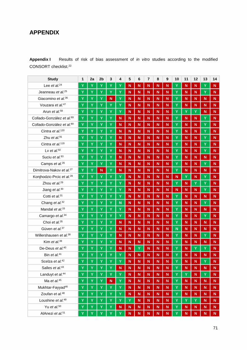
71
APPENDIX
Appendix I Results of risk of bias assessment of in vitro studies according to the modified
CONSORT checklist.22
Study 1 2a 2b 3 4 5 6 7 8 9 10 11 12 13 14
Lee et al.24 Y Y Y Y Y N N N N N Y N N Y N
Jeanneau et al.25 Y Y Y Y Y N N N N N Y N N Y N
Giacomino et al.36 Y Y Y N Y N N N N N Y N N N N
Vouzara et al.47 Y Y Y Y Y N N N N N Y N N N N
Arun et al.58 Y Y Y Y Y N N N N N Y Y Y N N
Collado-González et al.69 Y Y Y Y N N N N N N Y N N Y N
Collado-González et al.80 Y Y Y Y N N N N N N Y N N Y N
Cintra et al.120 Y Y Y Y N N N N N N Y N N Y N
Zhu et al.91 Y Y Y Y N N N N N N Y N N Y N
Cintra et al.119 Y Y Y Y N N N N N N Y N N Y N
Lv et al.92 Y Y Y Y N N N N N N Y N N Y N
Suciu et al.93 Y Y Y Y N N N N N N Y N N N N
Camps et al.26 Y Y Y Y N N N N N N Y N N Y N
Dimitrova-Nakov et al.27 Y Y N Y N N N N N N Y N N N N
Konjhodzic-Prcic et al.28 Y Y Y Y Y N N N N N N Y N Y N
Zhou et al.29 Y Y Y Y Y N N N N N Y N Y Y N
Jiang et al.30 Y Y Y Y Y N N N N N N N N Y N
Cotti et al.31 Y Y Y Y Y N N N N N Y N N N N
Chang et al.32 Y Y Y Y N N N N N N Y N N Y N
Mandal et al.33 Y Y Y Y Y N N N N N Y N N N N
Camargo et al.34 Y Y Y Y Y N N N N N Y N N Y N
Choi et al.35 Y Y Y Y N N N N N N Y N N N N
Güven et al.37 Y Y Y Y N N N N N N N N N N N
Willershausen et al.38 Y Y Y Y N N N N N N Y N N Y N
Kim et al.39 Y Y Y Y N N N N N N Y N N N N
De-Deus et al.40 Y Y Y Y N N Y N N N Y N Y Y N
Bin et al.41 Y Y Y Y Y N N N N N Y N N N N
Scelza et al.42 Y Y Y Y Y N N N N N Y N N Y N
Salles et al.43 Y Y Y Y N N N N N N Y N N N N
Landuyt et al.44 Y Y Y Y Y N N N N N Y Y N Y N
Ma et al.45 Y Y Y N Y N N N N N Y N N N N
Mukhtar-Fayyad46 Y Y Y Y Y N N N N N Y N N N N
Zoufan et al.48 Y Y Y Y Y N N N N N Y N N N N
Loushine et al.49 Y Y Y Y Y Y N N N N Y Y Y N N
Yu et al.50 Y Y Y Y N N N N N N Y N N N N
AlAnezi et al.51 Y Y Y Y Y N N N N N Y N N N N
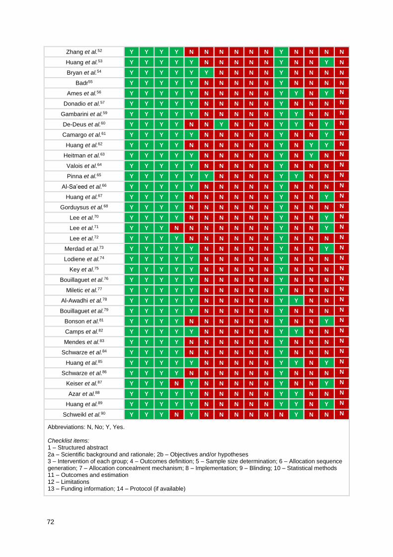
72
Zhang et al.52 Y Y Y Y N N N N N N Y N N N N
Huang et al.53 Y Y Y Y Y N N N N N Y N N Y N
Bryan et al.54 Y Y Y Y Y Y N N N N Y N N N N
Badr55 Y Y Y Y Y N N N N N Y N N N N
Ames et al.56 Y Y Y Y Y N N N N N Y Y N Y N
Donadio et al.57 Y Y Y Y Y N N N N N Y N N N N
Gambarini et al.59 Y Y Y Y Y N N N N N Y Y N N N
De-Deus et al.60 Y Y Y Y N N Y N N N Y Y N Y N
Camargo et al.61 Y Y Y Y Y N N N N N Y N N Y N
Huang et al.62 Y Y Y Y N N N N N N Y N Y Y N
Heitman et al.63 Y Y Y Y Y N N N N N Y N Y N N
Valois et al.64 Y Y Y Y Y N N N N N Y N N N N
Pinna et al.65 Y Y Y Y Y Y N N N N Y Y N N N
Al-Sa’eed et al.66 Y Y Y Y Y N N N N N Y N N N N
Huang et al.67 Y Y Y Y N N N N N N Y N N Y N
Gorduysus et al.68 Y Y Y Y N N N N N N Y N N N N
Lee et al.70 Y Y Y Y N N N N N N Y N N Y N
Lee et al.71 Y Y Y N N N N N N N Y N N Y N
Lee et al.72 Y Y Y Y N N N N N N Y N N N N
Merdad et al.73 Y Y Y Y Y N N N N N Y N N Y N
Lodiene et al.74 Y Y Y Y Y N N N N N Y N N N N
Key et al.75 Y Y Y Y Y N N N N N Y N N N N
Bouillaguet et al.76 Y Y Y Y Y N N N N N Y N N N N
Miletic et al.77 Y Y Y Y Y N N N N N Y N N N N
Al-Awadhi et al.78 Y Y Y Y Y N N N N N Y Y N N N
Bouillaguet et al.79 Y Y Y Y Y N N N N N Y N N N N
Bonson et al.81 Y Y Y Y N N N N N N Y N N Y N
Camps et al.82 Y Y Y Y Y N N N N N Y Y N N N
Mendes et al.83 Y Y Y Y N N N N N N Y N N N N
Schwarze et al.84 Y Y Y Y N N N N N N Y N N N N
Huang et al.85 Y Y Y Y Y N N N N N Y Y N Y N
Schwarze et al.86 Y Y Y Y N N N N N N Y N N N N
Keiser et al.87 Y Y Y N Y N N N N N Y N N Y N
Azar et al.88 Y Y Y Y Y N N N N N Y Y N N N
Huang et al.89 Y Y Y Y Y N N N N N Y Y N Y N
Schweikl et al.90 Y Y Y N Y N N N N N N Y N N N
Abbreviations: N, No; Y, Yes. Checklist items: 1 – Structured abstract 2a – Scientific background and rationale; 2b – Objectives and/or hypotheses 3 – Intervention of each group; 4 – Outcomes definition; 5 – Sample size determination; 6 – Allocation sequence generation; 7 – Allocation concealment mechanism; 8 – Implementation; 9 – Blinding; 10 – Statistical methods 11 – Outcomes and estimation 12 – Limitations 13 – Funding information; 14 – Protocol (if available)

73
Appendix II Results of risk of bias assessment of in vivo studies according to the
SYRCLE’s risk of bias.23
Study 1 2 3 4 5 6 7 8 9 10
Santos et al.111 U Y N N N N Y Y Y N
Assmann et al.94 Y Y N N N Y Y Y Y Y
Silva et al.95 N N N N N N Y Y Y N
Suzuki et al.106 Y Y N N N N Y Y Y N
Garcia et al.112 U Y N N N N N Y Y N
Cintra et al.113 N Y N N N N N Y Y Y
Silva et al.114 N U N N N N Y Y Y N
Oliveira et al.115 Y Y N N N N Y Y Y Y
Brasil et al.116 Y Y N N N N Y N Y N
Zmener et al.117 N Y N N N N Y U Y Na
Suzuki et al.118 Y Y N N N N Y Y Y N
Tanomaru-Filho et al.96 U Y N N N N Y Y Y N
Leonardo et al.97 U Y N N N N Y Y Y N
Campos-Pinto et al.98 U U N N N N N Y Y N
Zafalon et al.99 N Y N N N N N Y Y N
Onay et al.100 U Y N N N N N U Y N
Holland et al.101 U Y N N N N Y Y Y N
Shahi et al.102 U Y N N N N Y Y Y N
Tanomaru-Filho et al.103 U U N N N N Y Y Y N
Cintra et al.104 N Y N N N N Y Y Y Y
Kim et al.105 N N N N N N N Y Y Y
Zmener107 U U N N N N N Y Y Y
Ozbas et al.108 N Y N N N N N Y Y Y
Morinaga et al.109 N N N N N N N N Y N
Figueiredo et al.110 Y Y N N N N N U Y N
a The preparation of sealer (EndoREZ with accelerator) was performed with slight modifications of the manufacturer’s instructions. Also, one new animal was added to one of the groups (unspecified) to replace a drop-out from the original population (reasons were not specified). Abbreviations: N, No; U, Unclear; Y, Yes. Checklist items: 1 – Allocation sequence generation; 2 – Baseline characteristics; 3 – Allocation concealment 4 – Random housing; 5 – Caregiver and/or researcher blinding 6 – Random outcome assessment; 7 – Outcome assessor blinding 8 – Incomplete outcome data 9 – Selective outcome reporting 10 – Other sources of bias
Related Documents