Cytotoxicity and apoptotic effects of tea polyphenol-loaded chitosan nanoparticles on human hepatoma HepG2 cells Jin Liang a,b , Feng Li b , Yong Fang c , Wenjian Yang c , Xinxin An b , Liyan Zhao b , Zhihong Xin b , Lin Cao b , Qiuhui Hu b,c, ⁎ a Key Laboratory of Tea Biochemistry and Biotechnology of Ministry of Education and Ministry of Agriculture, Anhui Agricultural University, Hefei 230036, People's Republic of China b College of Food Science and Technology, Nanjing Agricultural University, Nanjing 210095, People's Republic of China c College of Food Science and Engineering, Nanjing University of Finance and Economics, Nanjing 210023, People's Republic of China abstract article info Article history: Received 26 November 2012 Received in revised form 24 October 2013 Accepted 26 November 2013 Available online 5 December 2013 Keywords: Tea polyphenols Chitosan Nanoparticles Hepatoma Apoptosis Tea polyphenols have strong antioxidant and antitumor activities. However, these health benefits are limited due to their poor in vivo stability and low bioavailability. Chitosan nanoparticles as delivery systems may provide an alternative approach for enhancing bioavailability of poorly absorbed drugs. In this study, tea polyphenol-loaded chitosan nanoparticles have been prepared using two different chitosan biomaterials, and their antitumor effects were evaluated in HepG2 cells, including cell cytotoxicity comparison, cell morphology analysis, cell apoptosis and cell cycle detection. The results indicated that the tea polyphenol-loaded chitosan nanoparticles showed a branch shape and heterogeneous distribution in prepared suspension. MTT assay suggested that tea polyphe- nol-loaded chitosan nanoparticles could inhibit the proliferation of HepG2 cells, and the cytotoxicity rates were increased gradually and appeared an obvious dose-dependent relationship. Transmission electron micro- scope images showed that the HepG2 cells treated with tea polyphenol-loaded chitosan nanoparticles exhibited some typical apoptotic features, such as microvilli disappearance, margination of nuclear chromatin, intracytoplasmic vacuoles and the mitochondrial swelling. In addition, the tea polyphenol-loaded chitosan nano- particles had relatively weak inhibitory effects on HepG2 cancer cells compared with tea polyphenols. Tea poly- phenols not only induced cancer cell apoptosis, but also promoted their necrosis. However, tea polyphenol- loaded chitosan nanoparticles exhibited their antitumor effects mainly through inducing cell apoptosis. Our re- sults revealed that the inhibition effects of tea polyphenol-loaded chitosan nanoparticles on tumor cells probably depended on their controlled drug release and effective cell delivery. The chitosan nanoparticles themselves as the delivery carrier showed limited antitumor effects compared with their encapsulated drugs. © 2013 Published by Elsevier B.V. 1. Introduction Nowadays, cancer is one of the major dread diseases and chemopre- vention is considered a valid approach to reduce the incidence of cancer [1]. Tea polyphenols (TP) from the dried leaves of the plant Camellia sinensis are known to possess strong antioxidant and anti-carcinogenic activities [2]. Although TP are widely used for the prevention and treat- ment of cancer, the therapeutic effects are limited due to their certain drawbacks, such as poor stability in gastrointestinal tract and limited bioavailability in vivo [3]. It has been reported that the biological activity of TP might depend on the form of their administration [4]. Therefore, a lot of research has been directed towards the development of effective methods to overcome the bottlenecks. Nanoparticles, especially those with highly controlled shapes, sizes and some unique physical and chemical properties including the antitumor activities [5], have been studied extensively as drug carriers for the improvement of the bio- availability of drug with poor absorption characteristics. Owing to their small size, prolonged circulation time, and sustained drug release profile, nano-sized polymeric nanoparticles bearing anticancer drugs have received increased attention for their ability to improve the effica- cy of anticancer drugs [6]. Nanoparticles can facilitate controlled and targeted drug delivery of the encapsulated anticancer drugs with high efficacy and low side effects [7]. They not only have the ability to over- come biological barriers and accumulate preferentially in tumors, but also can recognize single cancer cells, which are useful to the detection and treatment of cancer. Chitosan, a deacetylated derivative of chitin, has been applied exten- sively as a functional biopolymer in food and pharmaceutical industry, and is well known for its abundant, renewable, non-toxic and biode- gradable nature. Because of its unique chemical structure, chitosan has been investigated extensively in the development of controlled release drug delivery systems. Especially, the mucoadhesive property of chito- san can enhance drug transmucosal absorption and promote its Materials Science and Engineering C 36 (2014) 7–13 ⁎ Corresponding author at: College of Food Science and Technology, Nanjing Agricultural University, Nanjing 210095, People's Republic of China. Tel./fax: +86 25 84399086. E-mail address: [email protected] (Q. Hu). 0928-4931/$ – see front matter © 2013 Published by Elsevier B.V. http://dx.doi.org/10.1016/j.msec.2013.11.039 Contents lists available at ScienceDirect Materials Science and Engineering C journal homepage: www.elsevier.com/locate/msec

Welcome message from author
This document is posted to help you gain knowledge. Please leave a comment to let me know what you think about it! Share it to your friends and learn new things together.
Transcript

Materials Science and Engineering C 36 (2014) 7–13
Contents lists available at ScienceDirect
Materials Science and Engineering C
j ourna l homepage: www.e lsev ie r .com/ locate /msec
Cytotoxicity and apoptotic effects of tea polyphenol-loaded chitosannanoparticles on human hepatoma HepG2 cells
Jin Liang a,b, Feng Li b, Yong Fang c, Wenjian Yang c, Xinxin An b, Liyan Zhao b, Zhihong Xin b,Lin Cao b, Qiuhui Hu b,c,⁎a Key Laboratory of Tea Biochemistry and Biotechnology of Ministry of Education and Ministry of Agriculture, Anhui Agricultural University, Hefei 230036, People's Republic of Chinab College of Food Science and Technology, Nanjing Agricultural University, Nanjing 210095, People's Republic of Chinac College of Food Science and Engineering, Nanjing University of Finance and Economics, Nanjing 210023, People's Republic of China
⁎ Corresponding author at: College of Food ScienAgricultural University, Nanjing 210095, People's Repu84399086.
E-mail address: [email protected] (Q. Hu).
0928-4931/$ – see front matter © 2013 Published by Elsehttp://dx.doi.org/10.1016/j.msec.2013.11.039
a b s t r a c t
a r t i c l e i n f oArticle history:Received 26 November 2012Received in revised form 24 October 2013Accepted 26 November 2013Available online 5 December 2013
Keywords:Tea polyphenolsChitosanNanoparticlesHepatomaApoptosis
Tea polyphenols have strong antioxidant and antitumor activities. However, these health benefits are limited dueto their poor in vivo stability and low bioavailability. Chitosan nanoparticles as delivery systemsmay provide analternative approach for enhancing bioavailability of poorly absorbed drugs. In this study, tea polyphenol-loadedchitosan nanoparticles have been prepared using two different chitosan biomaterials, and their antitumor effectswere evaluated in HepG2 cells, including cell cytotoxicity comparison, cell morphology analysis, cell apoptosisand cell cycle detection. The results indicated that the tea polyphenol-loaded chitosan nanoparticles showed abranch shape and heterogeneous distribution in prepared suspension. MTT assay suggested that tea polyphe-nol-loaded chitosan nanoparticles could inhibit the proliferation of HepG2 cells, and the cytotoxicity rateswere increased gradually and appeared an obvious dose-dependent relationship. Transmission electron micro-scope images showed that the HepG2 cells treated with tea polyphenol-loaded chitosan nanoparticles exhibitedsome typical apoptotic features, such as microvilli disappearance, margination of nuclear chromatin,intracytoplasmic vacuoles and themitochondrial swelling. In addition, the tea polyphenol-loaded chitosan nano-particles had relatively weak inhibitory effects on HepG2 cancer cells compared with tea polyphenols. Tea poly-phenols not only induced cancer cell apoptosis, but also promoted their necrosis. However, tea polyphenol-loaded chitosan nanoparticles exhibited their antitumor effects mainly through inducing cell apoptosis. Our re-sults revealed that the inhibition effects of tea polyphenol-loaded chitosan nanoparticles on tumor cells probablydepended on their controlled drug release and effective cell delivery. The chitosan nanoparticles themselves asthe delivery carrier showed limited antitumor effects compared with their encapsulated drugs.
© 2013 Published by Elsevier B.V.
1. Introduction
Nowadays, cancer is one of themajor dread diseases and chemopre-vention is considered a valid approach to reduce the incidence of cancer[1]. Tea polyphenols (TP) from the dried leaves of the plant Camelliasinensis are known to possess strong antioxidant and anti-carcinogenicactivities [2]. Although TP are widely used for the prevention and treat-ment of cancer, the therapeutic effects are limited due to their certaindrawbacks, such as poor stability in gastrointestinal tract and limitedbioavailability in vivo [3]. It has been reported that the biological activityof TP might depend on the form of their administration [4]. Therefore, alot of research has been directed towards the development of effectivemethods to overcome the bottlenecks. Nanoparticles, especially thosewith highly controlled shapes, sizes and some unique physical and
ce and Technology, Nanjingblic of China. Tel./fax: +86 25
vier B.V.
chemical properties including the antitumor activities [5], have beenstudied extensively as drug carriers for the improvement of the bio-availability of drug with poor absorption characteristics. Owing totheir small size, prolonged circulation time, and sustained drug releaseprofile, nano-sized polymeric nanoparticles bearing anticancer drugshave received increased attention for their ability to improve the effica-cy of anticancer drugs [6]. Nanoparticles can facilitate controlled andtargeted drug delivery of the encapsulated anticancer drugs with highefficacy and low side effects [7]. They not only have the ability to over-come biological barriers and accumulate preferentially in tumors, butalso can recognize single cancer cells, which are useful to the detectionand treatment of cancer.
Chitosan, a deacetylated derivative of chitin, has been applied exten-sively as a functional biopolymer in food and pharmaceutical industry,and is well known for its abundant, renewable, non-toxic and biode-gradable nature. Because of its unique chemical structure, chitosan hasbeen investigated extensively in the development of controlled releasedrug delivery systems. Especially, the mucoadhesive property of chito-san can enhance drug transmucosal absorption and promote its
8 J. Liang et al. / Materials Science and Engineering C 36 (2014) 7–13
sustained release [8,9]. It has been reported that chitosan nanoparticlesas drug carriers with small particle size and positive surface charge canexhibit a higher antitumor activity [10].
Our previous studies showed that the tea polyphenol-loaded chito-san nanoparticles (TP-CNPs) could be obtained by ionic gelation usingtwo biocompatible materials, namely N-carboxymethyl chitosan andchitosan hydrochloride, and the optimization of particle size and en-trapment efficiency have been discussed [11]. However, the antitumorbiological activity of TP-CNPs is still unknown. Therefore, in this study,the antitumor effects of the TP-CNP, TP and chitosan nanoparticles(CNPs) have been evaluated using HepG2 cells, including cell cyto-toxicity comparison, cell morphology analysis, apoptosis detection,and cell cycle measurement, and the antitumor mechanism was alsohypothesized.
2. Materials and methods
2.1. Materials
TP extract with a purity of 93% was obtained from dried green tealeaves by the solvent extractionmethod using 50% ethanol and then pu-rified with H1020 resins (Nankai University Chemical Plant, Tianjin,China). HepG2 cells were obtained from Nanjing University (Nanjing,Jiangsu, China). All the other reagents used were of analytical grade.
2.2. The preparation of TP-CNPs
The TP-CNPs were prepared according to our previously reportedoptimized conditions [11,12]. Briefly, N-carboxymethyl chitosan(Mv = 61 kDa, Degree of deacetylation 83%) and chitosan hydrochlo-ride (Mv = 90 kDa, Degree of deacetylation 85%)were dissolved in dis-tilled water and the solutions of different concentrationswere preparedby adding the extracts of TP into carboxymethyl chitosan solution. Theoptimal levels of carboxymethyl chitosan concentration, chitosan hy-drochloride concentration and the amount of TP were 3.63 mg/mL,1.19 mg/mL, and 10.94 mg, respectively. The main preparation processwas the following steps. First, TP was added into the chitosan hydro-chloride solution (30 mL), and then the carboxymethyl chitosan solu-tion (12 mL) was added dropwise into the chitosan hydrochloride andTPmixed solution under stirring at room temperature, and continuous-ly stirred for 30 min. The formation of nanoclusters was based on anionic gelation interaction between positively-charged amine groups ofchitosan hydrochloride chitosan and negatively-charged carboxylgroups of carboxymethyl chitosan. The nanocluster suspensions wereimmediately subjected to further analysis for their structure and sizedistribution characterization. The nanocluster powders as experimentalsamples were obtained by high speed centrifugation (12,000 rpm,30 min) followed by freeze-drying process. Meanwhile, the CNPs with-out TP were prepared to use as the control group.
2.3. Cell culture
Human hepatomaHepG2 cells were cultured in Dulbecco'sModifiedEagle's Medium (DMEM) equilibrated with 5% CO2 and 95% air at 37 °C.The medium was supplemented with 10% fetal calf serum (FCS),50 mg/L streptomycin and 75 mg/L penicillin sulfate.
2.4. Cytotoxicity assay
The MTT [3-(4,5-dimethylthazol-2-yl)-2,5-diphenyltetrazoliumbromide blue-indicator dye]-based assay, a simple nonradioactive col-orimetric assay, was used to measure cell cytotoxicity, proliferation orviability. HepG2 cells were used for the analysis of cytotoxicity in vitro.100 μL HepG2 cells (1 × 10⁵ cells/mL) were placed into 96-well tissue-culture plates and incubated at 37 °C. After 24 h, the cells were treatedwith different concentrations of TP (1.0 mg/mL, 0.5 mg/mL and
0.25 mg/mL) and TP-CNPs loadedwith the same amount of TP. The un-treated cells were used as the control. The plates were incubated in ahumidified 5% CO2 balanced-air incubator at 37 °C for 48 h. Then10 μL of 5 mg/mLMTT solutionwas added into eachwell and the plateswere incubated for another 4 h, and then the medium was discarded.Dimethyl sulfoxide (100 μL) was added into each well of the 96-wellplate, and the solution was vigorously mixed to dissolve tetrazoliumdye. The absorbance of each well was measured by an enzyme-linkedimmunosorbent assay reader (BioTek; Austria) at a test wavelength of570 nm and the cell cytotoxicity rate was calculated by the followingequation:
Cellcytotoxicity ¼ 1–T½ �=C� 100%
where C is the number of viable cells after 48 h of incubation withoutnanoparticles and T is the number of viable cells after 48 h of incubationwith nanoparticles.
2.5. Morphological examination using microscope
100 μL HepG2 cells (1 × 10⁵ cells/mL) were seeded on 96-well cul-ture plates and TP-CNPs, CNPs and TP (1.0 mg/mL)were added into thecells, and after 48 h incubation,morphological changes of cells were ex-amined under a fluorescent microscope (CKX41, Olympus Corporation,Tokyo, Japan).
2.6. Transmission electron microscopy assay
Changes in the ultrastructure of HepG2 cells after treatmentwith TP-CNPs were observed using the transmission electronmicroscope (TEM)according to the previously published protocol [7]. Briefly, after incuba-tion with TP-CNPs (1 mg/mL) at 37 °C for 24 h, HepG2 cells were col-lected by centrifugation and washed twice with PBS, and then fixatedimmediately in Karnovsky solution (2% paraformaldehyde and 2.5% glu-taraldehyde in 0.1 M Na cacodylate/HCl, pH 7.4). The postfixation wasperformed in 1% OsO4 with 1.5% potassium ferrocyanide, followed bydehydration and drying. After being trimmed, mounted and coated,the ultrathin sections were observed and photographed by using theTEM (JEOL H-7650, Hitachi High-Technologies Corporation, Tokyo,Japan). At the same time, the TEM images of TP and CNPswere also cap-tured using the same method.
2.7. Determination of cell apoptosis
The cell apoptosis was detected by annexin V/PI double stain assay.The annexin V/PI staining was performed as described previously [13].Briefly, HepG2 cells (3 × 10⁵ cells/well) were seeded in 24-well platesand incubated at 37 °C in a 5% CO2 incubator overnight. After treatmentwith tea polyphenols at 1.0 mg/mL or the chitosan nanoparticles loadedwith the same amount of tea polyphenols and non-loaded chitosannanoparticles (untreated cells were used as controls), the cells werewashed with 0.1 M PBS, trypsinized, and fixed in 75% ethanol. TheHepG2 cells were stained with EGFP-tagged annexin V for 20 min atroom temperature and 15 μg/mL propidium iodide (PI) was alsoadded. The HepG2 cells after treatment were detected using flowcytometry (BD Biosciences, USA) and analyzed using the CELLQuestVersion 3.3 software.
2.8. Analysis of cell cycle distribution
Human hepatoma HepG2 cells were seeded into 24-well plate at adensity of 1 × 10⁶ cells/well. After 6 h incubation, the cells were treatedwith 1.0 mg/mL of TP, CNPs and TP-CNPs (which contain the sameamount of TP compared with TP group), and then harvested after 15 hof exposure. Treated HepG2 cells were resuspended in 100 μL PBS,and then trypsinized, and fixed in 75% ethanol at 4 °C overnight. The

Fig. 2. Cytotoxic effects of TP-CNPs, CNPs and TP in HepG2 cells. HepG2 cells(1 × 10⁵ cells/mL) were cultured with different concentration of TP-CNPs, CNPs and TPfor 48 h. All the data were obtained from three independent experiments (p b 0.05).
9J. Liang et al. / Materials Science and Engineering C 36 (2014) 7–13
cells were centrifuged at 2000 rpm for 3 min, and then discarded thesupernatant and washed with PBS, centrifuged again, digested withRNase for 15 min, pulsed stained with propidium iodide for 10 min atroom temperature, and then measured by the flow cytometry (BD Bio-sciences, USA).
3. Results and discussion
3.1. Characteristics of TP-CNPs
The structure and size distribution of TP-CNPs and CNPs in preparedsuspension are illustrated in Fig. 1. Themorphology of the CNPswithoutTP is characterized by a spherical structure and relatively uniform sur-face with a uniform particle size of around 400 ± 52 nm. However,the nanoclusters that composed of TP-CNPs showed a branch shapeand heterogeneous distribution. The microstructural features of TP-CNPs were consistent with the description reported by Tang et al. [14].Moreover, the mean diameter of the combined TP-CNP nanoclusterswas larger than the nanoclusters of CNPs. This could be due to theionic gelation interaction between the carboxymethyl chitosan and chi-tosan hydrochloride and aggregation among the CNPs after loadingwith TP, which resulted in the broader size distribution and heteroge-neous shape of TP-CNPs. In addition, the loading performance and thecontrol release characteristics of nanoparticles have been described inour previous report [12]. The drug content and encapsulation rate ofTP-CNPs were 16% and 83% respectively.
3.2. Cytotoxicity of TP-CNPs
The cytotoxicities of the TP and TP-CNPs on HepG2 cells at differentintervals were evaluated through an MTT assay. Fig. 2 shows thatthe growth of HepG2 cells was subjected to different degrees of inhibi-tion after 48 h treatment using TP and TP-CNP samples at differentconcentrations, and represented different mass concentrations of thedose–effect relationship. A comparison of the proliferation inhibitionrate of TP and TP-CNPs at the same concentration can be seen fromFig. 2, the TP-CNPs suppressed at a relatively low inhibition rate com-pared to TP. However, with the increasing concentration of TP-CNPs,
Fig. 1. (a) The TEM image and (b) Particle size distribu
the proliferation inhibition rate was also gradually enhanced. It cantherefore be concluded that the TP coated with CNPs showed a slow-release but significant inhibitory effect against cancer cells in vitro.This phenomenon indicated that the prepared TP-CNPs can effectivelyinhibit the proliferation of tumor cells. Previous reports show thatCNPs can enhance the intestinal absorption of the green tea catechinin mice [15,16], however, the shape and particle size of CNPs are alsoclosely related to their delivery effect [17,18]. So there was a necessityto prepare proper CNPs for the delivery of TP. Compared to microcap-sules, nanoparticles act as an effective delivery carrier owing to theirsmaller particle size, and suitable for the delivery of antitumor drugs.Some reports showed that the CNPs not only has the targeted deliverycapability for encapsulated drugs [19], but also could enhance their bio-availability [20]. In previous reports from our laboratory, the preparedCNPs, which are fabricated using the carboxymethyl chitosan and chito-san hydrochloride biomaterials, showed good controlled release charac-teristics under in vitro conditions [11,12]. These results suggested thatthe CNPs could be used for enhancing the bioavailability of TP.
3.3. Effect of TP-CNPs on nuclei morphology
The nuclei morphology changes of cancer cells treated with nano-particles were observed using fluorescence microscopes. Fig. 3 shows
tion of TP-CNPs and CNPs in prepared suspension.

Fig. 3. The image of HepG2 tumor cells by an inverted microscope in every group (×200) (a) Normal group, (b). TP group, (c) CNP group, and (d). TP-NP group. Scale bar: 50 μm.
Fig. 4.Ultrastructuralmorphology inHepG2 cells treatedwith TP, CNPs and TP-CNPs by TEM (magnification 10,000). (a)Normal group, (b) TP group, (c) CNP group, and (d) TP-CNPgroup.
10 J. Liang et al. / Materials Science and Engineering C 36 (2014) 7–13
11J. Liang et al. / Materials Science and Engineering C 36 (2014) 7–13
the representative microscopic images of HepG2 cells after treatmentwith TP-CNPs, CNPs and TP at a concentration equivalent to 1.0 mg/mL for 48 h. The tumor cells of control group (Fig. 3a) exhibited an elon-gated spindle-like shape, clear boundaries between cells and vigorousgrowth after incubation. The cells treated with TP (Fig. 3b) showedshrinkage, round shape, and deep nuclear staining pattern. Themajorityof cells exhibited typical apoptotic morphology. The cells treated withCNPs exhibited shrinkage compared to control group, however, asmall portion of the cells became round (Fig. 3c). In the case of tumorcells treated with TP-CNPs, the cells showed apoptotic morphologywith the concentrated cytoplasm and dense chromatin in the nucleusas shown in Fig. 3d. Compared to the TP group, the cells treated withTP-CNPs showed a relatively low degree of apoptosis, which could bedue to the slow release of TP in CNPs.
3.4. Effect of TP-CNPs on ultrastructure morphology
Representative TEM images (Fig. 4) show the ultrastructural chang-es of the HepG2 cells incubated with TP, CNPs and TP-CNPs for 24 h.Fig. 3a shows the nucleus of untreated HepG2 cells with clearly definedultrastructure, and evenly distributed chromatin. Also, the cell mem-brane surface possessed densely distributed microvilli. Whereas thecells treated with TP exhibited larger vacuoles, and the microvilli disap-peared (Fig. 4b). The TEM images of the cells treated with CNPs alsoshowed intact cell morphology, and no significant difference wasobserved when compared with the control group (Fig. 4c). However,the cancer cells treated with TP-CNPs exhibited the characteristic ultra-structural features of apoptosis, such as microvilli disappearance,margination, intracytoplasmic vacuoles, and themitochondrial swellingwith the formation of apoptotic bodies as shown in Fig. 4d. Li et al. [13]reported that paclitaxel-loaded chitosan nanoparticles can be internal-ized into cells and release the drug into the cytoplasm directly, therebyresulting in cancer cell apoptosis. It also can be inferred from theseobservations that TP-CNPs may have the potential to internalize intocells and then release of TP could induce cancer cell apoptosis.
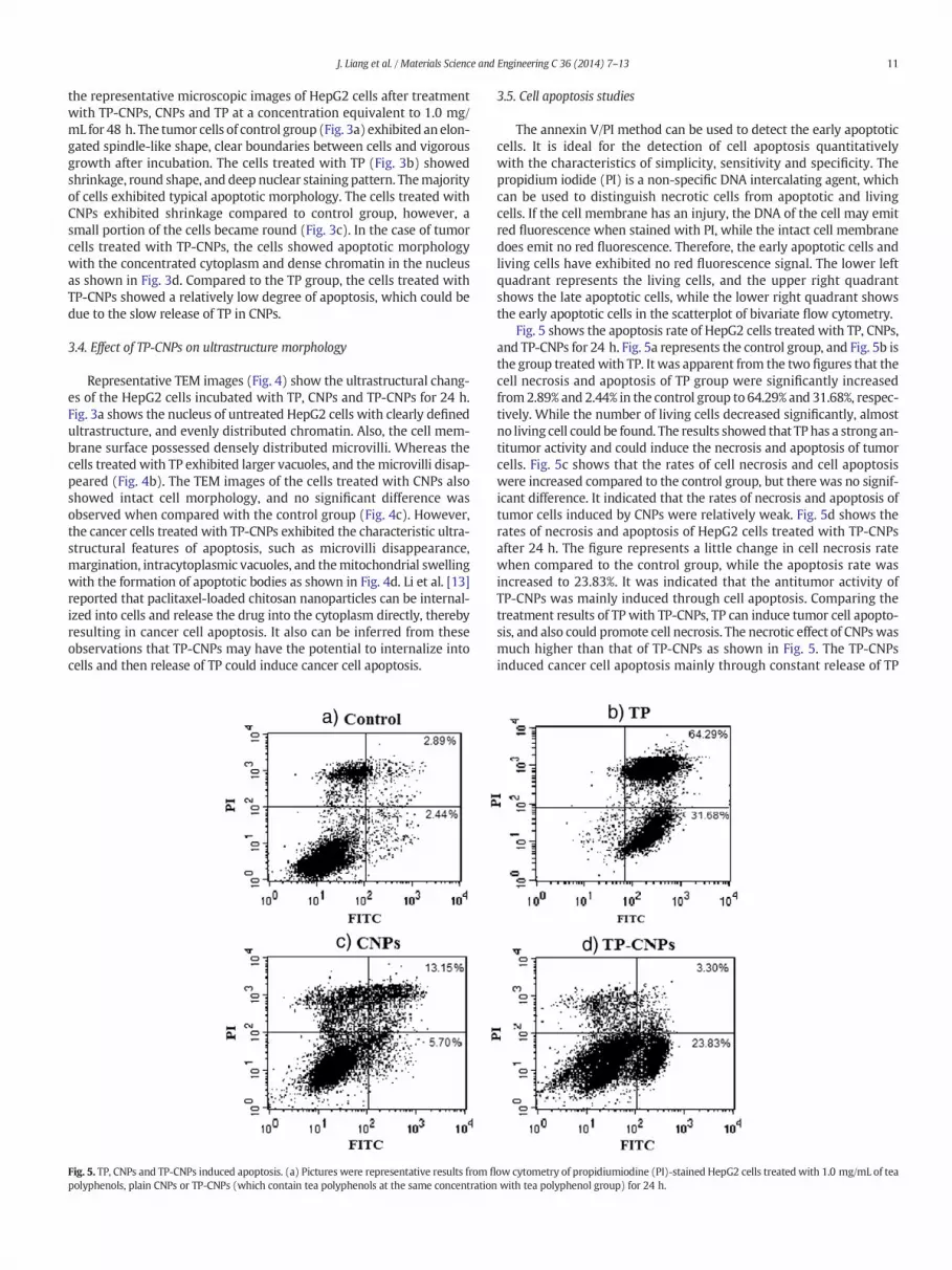
Fig. 5. TP, CNPs and TP-CNPs induced apoptosis. (a) Pictures were representative results from fl
polyphenols, plain CNPs or TP-CNPs (which contain tea polyphenols at the same concentration
3.5. Cell apoptosis studies
The annexin V/PI method can be used to detect the early apoptoticcells. It is ideal for the detection of cell apoptosis quantitativelywith the characteristics of simplicity, sensitivity and specificity. Thepropidium iodide (PI) is a non-specific DNA intercalating agent, whichcan be used to distinguish necrotic cells from apoptotic and livingcells. If the cell membrane has an injury, the DNA of the cell may emitred fluorescence when stained with PI, while the intact cell membranedoes emit no red fluorescence. Therefore, the early apoptotic cells andliving cells have exhibited no red fluorescence signal. The lower leftquadrant represents the living cells, and the upper right quadrantshows the late apoptotic cells, while the lower right quadrant showsthe early apoptotic cells in the scatterplot of bivariate flow cytometry.
Fig. 5 shows the apoptosis rate of HepG2 cells treated with TP, CNPs,and TP-CNPs for 24 h. Fig. 5a represents the control group, and Fig. 5b isthe group treated with TP. It was apparent from the two figures that thecell necrosis and apoptosis of TP group were significantly increasedfrom2.89% and 2.44% in the control group to 64.29% and 31.68%, respec-tively. While the number of living cells decreased significantly, almostno living cell could be found. The results showed that TPhas a strong an-titumor activity and could induce the necrosis and apoptosis of tumorcells. Fig. 5c shows that the rates of cell necrosis and cell apoptosiswere increased compared to the control group, but there was no signif-icant difference. It indicated that the rates of necrosis and apoptosis oftumor cells induced by CNPs were relatively weak. Fig. 5d shows therates of necrosis and apoptosis of HepG2 cells treated with TP-CNPsafter 24 h. The figure represents a little change in cell necrosis ratewhen compared to the control group, while the apoptosis rate wasincreased to 23.83%. It was indicated that the antitumor activity ofTP-CNPs was mainly induced through cell apoptosis. Comparing thetreatment results of TP with TP-CNPs, TP can induce tumor cell apopto-sis, and also could promote cell necrosis. The necrotic effect of CNPswasmuch higher than that of TP-CNPs as shown in Fig. 5. The TP-CNPsinduced cancer cell apoptosis mainly through constant release of TP
ow cytometry of propidiumiodine (PI)-stained HepG2 cells treated with 1.0 mg/mL of teawith tea polyphenol group) for 24 h.

12 J. Liang et al. / Materials Science and Engineering C 36 (2014) 7–13
from CNPs, and the cancer cell membrane showed integrity. While thecell necrosis caused by CNPs mainly manifested in the cell membranedamage and resultant enzyme leakage [21]. In addition, the CNPs them-selves in TP-CNPs have showed a limited anticancer activity than TP.Therefore, TP-CNPs could exhibit relatively stronger apoptosis andweaker necrosis effects on tumor cells than CNPs.
3.6. Cell cycle analysis
Flow cytometry can detect the percentage of different cells M1, M2,andM3, corresponding to the cells sub-G1, G0/G1 phase and S phase. Inthis study, the impact of TP-CNPs onHepG2 tumor cell cycle progressionand distribution was investigated by flow cytometry. Fig. 6 shows thecell cycle changes of HepG2 cells treated with TP, CNPs and TP-CNPsafter 24 h. Compared to the control group, the cells treated with TPhave exhibited increased in G0/G1 phase, and the cell cycle progressionwas increased to 23.75%, while the S phase was reduced to 17.99%(Fig 6b). The group treated with CNPs has shown a mild change in thecell cycle progression (Fig 6c), and a slight increase could be seen onlyin G0/G1 phase and S phase cells. Comparing the group of TP-CNPs(Fig 6d) with the control group, the number of cells in G0/G1 phasewas increased to 5.54%, while the total cells in G1 and S phase weredecreased to some extent. Some reports also showed that TP can inducecell cycle arrest in G0/G1 phase and trigger cell death by apoptoticmechanism [22–25]. In addition, we previously reported that the TPfrom TP-CNPs can release in a continuous manner for up to 48 h [12].Therefore, these results indicated that CNPs have the potential to useas the anticancer drug carriers.
4. Conclusion
CNPs have become attractive for their promising properties in theaspect of drug delivery systems during recent years. In this study, theCNPs prepared by using two different chitosan biomaterials could beconsidered as an effective carrier for the delivery of TP. The antitumoreffects of TP loaded CNPs on HepG2 cells have been manifested by
Fig. 6. TP, CNPs and TP-CNPs induced cell apoptosis. The picture (a) was normal HepG2 celpropidiumiodine (PI)-stained HepG2 cells treated with 1.0 mg/mL of TP, CNPs or TP-CNPs for
cytotoxicity, cell morphology analysis, apoptosis detection and cellcycle measurement. The results indicated that TP-CNPs can inhibit theproliferation of HepG2, and their antitumor effect was mainly achievedthrough necrosis and apoptosis induction in cancer cells. Compared toTP, the CNPs themselves showed limited antitumor activities and theirinhibitionmechanismwasmainly through inducing tumor cell necrosis.Further research will focus on the comparison of the antitumor effectsbetween TP-CNPs and TP using animal model.
Acknowledgments
This work was supported by the National Natural Science Founda-tion of China (30871743), the 111 Project of Education Ministry ofChina (B07030), and theNational High Technology Research and Devel-opment Program of China (2007AA100403). The authors would like tothank Dr. Pradeep Puligundla for refining the language.
References
[1] N. Hail Jr., M. Cortes, E.N. Drake, J.E. Spallholz, Free Radic. Biol. Med. 45 (2008)97–110.
[2] C.S. Yang, J.D. Lambert, S. Sang, Arch. Toxicol. 83 (2009) 11–21.[3] D. Chen, Q.P. Dou, Int. J. Mol. Sci. 9 (2008) 1196–1206.[4] S.M. Henning, Y. Niu, Y. Liu, N.H. Lee, Y. Hara, G.D. Thames, R.R. Minutti, C.L.
Carpenter, H. Wang, D. Heber, J. Nutr. Biochem. 16 (2005) 610–616.[5] Y. Yuan, C. Liu, J. Qian, J. Wang, Y. Zhang, Biomaterials 31 (2010) 730–740.[6] H.Y. Hwang, I.S. Kim, I.C. Kwon, Y.H. Kim, J. Control. Release 128 (2008) 23–31.[7] H. Luo, J. Li, X. Chen, Biomed. Pharmacother. 64 (2010) 521–526.[8] W. Ajun, S. Yan, G. Li, L. Huili, Carbohydr. Polym. 75 (2009) 566–574.[9] A.W. Pan, B.B. Wu, J.M. Wu, Chin. Chem. Lett. 20 (2009) 79–83.
[10] L. Qi, Z. Xu, X. Jiang, Y. Li, M. Wang, Bioorg. Med. Chem. Lett. 15 (2005) 1397–1399.[11] J. Liang, F. Li, Y. Fang, W. Yang, X. An, L. Zhao, Z. Xin, Q. Hu, Eur. Food Res. Technol.
231 (2010) 917–924.[12] J. Liang, F. Li, Y. Fang, W. Yang, X. An, L. Zhao, Z. Xin, L. Cao, Q. Hu, Colloids Surf. B:
Biointerfaces 82 (2011) 297–301.[13] F. Li, J. Li, X. Wen, S. Zhou, X. Tong, P. Su, H. Li, D. Shi, Mater. Sci. Eng. 29 (2009)
2392–2397.[14] D.W. Tang, S.H. Yu, Y.C. Ho, B.Q. Huang, G.J. Tsai, H.-Y. Hsieh, H.W. Sung, F.L. Mi, Food
Hydrocoll. 30 (2013) 33–41.[15] A. Dube, J.A. Nicolazzo, I. Larson, Eur. J. Pharm. Sci. 41 (2010) 219–225.[16] A. Dube, J.A. Nicolazzo, I. Larson, Eur. J. Pharm. Sci. 44 (2011) 422–426.
l, and the pictures (b), (c), and (d) were representative results from flow cytometry of24 h.

13J. Liang et al. / Materials Science and Engineering C 36 (2014) 7–13
[17] S.A. Agnihotri, N.N. Mallikarjuna, T.M. Aminabhavi, J. Control. Release 100 (2004)5–28.
[18] C. He, L. Yin, C. Tang, C. Yin, Biomaterials 33 (2012) 8569–8578.[19] O.C. Farokhzad, R. Langer, ACS Nano 3 (2009) 16–20.[20] B. Hu, Y. Ting, X. Yang, W. Tang, X. Zeng, Q. Huang, Chem. Commun. 48 (2012)
2421–2423.[21] J.W. Loh, G. Yeoh, M. Saunders, L.Y. Lim, Toxicol. Appl. Pharm. 249 (2010) 148–157.
[22] N. Ahmad, D.K. Feyes, A.L. Nieminen, R. Agarwal, H. Mukhtar, J. Natl Cancer Inst. 89(1997) 1881–1886.
[23] S. Gupta, T. Hussain, H. Mukhtar, Arch. Biochem. Biophys. 410 (2003) 177–185.[24] N. Ahmad, P.Y. Cheng, H. Mukhtar, Biochem. Biophys. Res. Commun. 275 (2000)
328–334.[25] J.D. Lambert, C.S. Yang, Mutat. Res. Fundam. Mol. Mech. Mutagen. 523 (2003)
201–208.
Related Documents


![Interaction of a recombinant form of apolipoprotein[a ... · Interaction of a recombinant form of apolipoprotein[a] with human fibroblasts and with the human hepatoma cell line HepG2](https://static.cupdf.com/doc/110x72/5d0ce32c88c993064c8b69eb/interaction-of-a-recombinant-form-of-apolipoproteina-interaction-of-a-recombinant.jpg)