Cytotoxic T lymphocytes kill multiple targets simultaneously via spatiotemporal uncoupling of lytic and stimulatory synapses Aurelie Wiedemann*, David Depoil*, Mustapha Faroudi* † , and Salvatore Valitutti* ‡§ *Lymphocyte Interaction Group, Institut National de la Sante ´ et de la Recherche Me ´ dicale, Unite ´ 563, Institut Claude de Pre ´ val, 31059 Toulouse, France; and ‡ Department of Immunology, University of Toulouse, 31059 Toulouse, France Edited by Arthur Weiss, University of California School of Medicine, San Francisco, CA, and approved June 2, 2006 (received for review January 25, 2006) A longstanding paradox in the activation of cytotoxic T lympho- cytes (CTL) arises from the observation that CTL recognize and rapidly destroy target cells with exquisite sensitivity despite the fact that cytokine production requires sustained signaling at the immunological synapse. Here we solve this paradox by showing that CTL establish sustained synapses with targets offering strong antigenic stimuli and that these synapses persist after target cell death. Simultaneously, CTL polarize lytic granules toward different cells without discrimination regarding antigenic potential. Our results show that spatiotemporal uncoupling of immunological synapse and lytic granule secretion allows multiple killing and sustained signaling by individual CTL. This unique mechanism of responding to multiple contacts provides remarkable efficiency to CTL function. confocal microscopy immunological synapse sustained signaling A n intriguing feature of CTL biology is that, although cyto- toxicity is a rapid and low-threshold response, activation to interleukin production requires a prolonged time and an ade- quately strong antigenic stimulation (1, 2). We have previously shown that individual CTL interacting with cognate target cells exhibit a dual activation threshold reflecting the formation of two distinct immunological synapses (IS) at the CTLtarget cell contact site: the lytic synapse and the stimulatory synapse (3). The term ‘‘lytic synapse’’ was used to refer to the polarization of the lytic machinery detectable at both low and high antigen concentrations, and the term ‘‘stimulatory synapse’’ was used to refer to the large-scale molecular segregation of surface mole- cules and signaling components characteristic of a mature IS and occurring only with target cells providing strong antigenic stimuli (3). The formation of a lytic synapse corresponded to full activation of CTL to cytotoxicity (whereas IFN- production was marginal, and calcium mobilization was low and erratic). The formation of a stimulatory synapse corresponded to activation to cytokine production (3). A large amount of information on the molecular dynamics occurring at the CTLtarget cell contact site is now available; however, CTL activation to biological responses is still an enigmatic process because of three major unresolved questions. The first question is how can CTL behave as efficient and rapid killers and at the same time gather the sustained signals required for cytokine production on the surface of their targets (2, 4)? Cytotoxicity is a very efficient phenomenon characterized by a high degree of sensitivity and rapidity. It has been previously estimated that target cells displaying as few as 1–10 specific peptideMHC complexes on their surface can trigger cytotox- icity (5). In a recent study Purbhoo et al. (6) measured the number of peptideMHC complexes present at the cell–cell contact site in living CTLtarget cell conjugates. They showed that CTL could indeed be activated to lethal hit delivery by as few as two to three specific peptideMHC complexes present at the cellular interface. Lethal hit delivery requires a very short time. Upon conjugation with cognate target cells, CTL rapidly polarize their lytic machinery toward the opposing cells (7). Moreover, CTL can kill outnumbering target cells (8), indicating that they may rapidly detach and recycle from one target to another (9 –12). How the process of CTL activation is compatible with rapid low-threshold cytotoxic responses and transient CTL target interactions is still unknown. The second unresolved question is how is the polarization of lytic machinery regulated in CTL interacting with multiple targets simultaneously? We have recently shown that CD4 helper T cells simultaneously interacting with different antigen- presenting cells (APC) rapidly polarize their secretory machin- ery toward the APC providing the strongest antigenic stimulus. This rapid polarization allows CD4 T cells to provide their help in a selective fashion (13). Although selective help delivery is instrumental for the development of adaptive immune re- sponses, selective polarization of CTL lytic machinery toward the targets offering strong antigenic stimuli could be detrimental for indiscriminate immune surveillance against all potentially dangerous cells. Whether CTL selectively polarize against de- fined targets or conversely are able to behave as indiscriminate killers is still unknown. A third question concerns the functional relation between the lytic and stimulatory synapses in individual CTL. The mature IS was originally described as a specialized signaling domain formed at the contact site between T cells and APC, character- ized by large-scale molecular clustering and segregation of surface molecules and signaling components (7, 14, 15). Current research has led to an expansion of this term, where IS now indicates a multitude of structures that are mediators of inter- cellular communication (3). It has been suggested that in CTL the IS may have the role of polarizing secretion of lytic granules toward target cells (7). However, results from our laboratory and other laboratories have recently shown that polarization of CTL lytic machinery toward targets can occur in the absence of the large-scale molecular clustering and segregation characteristic of a mature IS (3, 6, 16). Together these results suggest that lethal hit delivery and molecular rearrangement at the synapse are rather independent events that may possibly take place sepa- rately in different areas of the cell and at different time points of interaction. Conflict of interest statement: No conflicts declared. This paper was submitted directly (Track II) to the PNAS office. Freely available online through the PNAS open access option. Abbreviations: CTL, cytotoxic T lymphocyte(s); TCR, T cell receptor; IS, immunological synapse; APC, antigen-presenting cell; [Ca 2 ]i, intracellular concentration of Ca 2 . † Present address: National Institute for Medical Research, The Ridgeway, London NW7 1AA, United Kingdom. § To whom correspondence should be addressed at: Institut National de la Sante ´ et de la Recherche Me ´ dicale, Unite ´ 563, Institut Claude de Pre ´ val, Centre Hospitalier Universitaire de Toulouse, Purpan, 31059 Toulouse Cedex 3, France. E-mail: [email protected]. © 2006 by The National Academy of Sciences of the USA www.pnas.orgcgidoi10.1073pnas.0600651103 PNAS July 18, 2006 vol. 103 no. 29 10985–10990 IMMUNOLOGY

Welcome message from author
This document is posted to help you gain knowledge. Please leave a comment to let me know what you think about it! Share it to your friends and learn new things together.
Transcript
Cytotoxic T lymphocytes kill multiple targetssimultaneously via spatiotemporal uncouplingof lytic and stimulatory synapsesAurelie Wiedemann*, David Depoil*, Mustapha Faroudi*†, and Salvatore Valitutti*‡§
*Lymphocyte Interaction Group, Institut National de la Sante et de la Recherche Medicale, Unite 563, Institut Claude de Preval, 31059 Toulouse, France;and ‡Department of Immunology, University of Toulouse, 31059 Toulouse, France
Edited by Arthur Weiss, University of California School of Medicine, San Francisco, CA, and approved June 2, 2006 (received for review January 25, 2006)
A longstanding paradox in the activation of cytotoxic T lympho-cytes (CTL) arises from the observation that CTL recognize andrapidly destroy target cells with exquisite sensitivity despite thefact that cytokine production requires sustained signaling at theimmunological synapse. Here we solve this paradox by showingthat CTL establish sustained synapses with targets offering strongantigenic stimuli and that these synapses persist after target celldeath. Simultaneously, CTL polarize lytic granules toward differentcells without discrimination regarding antigenic potential. Ourresults show that spatiotemporal uncoupling of immunologicalsynapse and lytic granule secretion allows multiple killing andsustained signaling by individual CTL. This unique mechanism ofresponding to multiple contacts provides remarkable efficiency toCTL function.
confocal microscopy � immunological synapse � sustained signaling
An intriguing feature of CTL biology is that, although cyto-toxicity is a rapid and low-threshold response, activation to
interleukin production requires a prolonged time and an ade-quately strong antigenic stimulation (1, 2). We have previouslyshown that individual CTL interacting with cognate target cellsexhibit a dual activation threshold reflecting the formation oftwo distinct immunological synapses (IS) at the CTL�target cellcontact site: the lytic synapse and the stimulatory synapse (3).The term ‘‘lytic synapse’’ was used to refer to the polarization ofthe lytic machinery detectable at both low and high antigenconcentrations, and the term ‘‘stimulatory synapse’’ was used torefer to the large-scale molecular segregation of surface mole-cules and signaling components characteristic of a mature IS andoccurring only with target cells providing strong antigenic stimuli(3). The formation of a lytic synapse corresponded to fullactivation of CTL to cytotoxicity (whereas IFN-� production wasmarginal, and calcium mobilization was low and erratic). Theformation of a stimulatory synapse corresponded to activation tocytokine production (3).
A large amount of information on the molecular dynamicsoccurring at the CTL�target cell contact site is now available;however, CTL activation to biological responses is still anenigmatic process because of three major unresolved questions.
The first question is how can CTL behave as efficient and rapidkillers and at the same time gather the sustained signals requiredfor cytokine production on the surface of their targets (2, 4)?Cytotoxicity is a very efficient phenomenon characterized by ahigh degree of sensitivity and rapidity. It has been previouslyestimated that target cells displaying as few as 1–10 specificpeptide�MHC complexes on their surface can trigger cytotox-icity (5). In a recent study Purbhoo et al. (6) measured thenumber of peptide�MHC complexes present at the cell–cellcontact site in living CTL�target cell conjugates. They showedthat CTL could indeed be activated to lethal hit delivery by asfew as two to three specific peptide�MHC complexes present atthe cellular interface. Lethal hit delivery requires a very shorttime. Upon conjugation with cognate target cells, CTL rapidly
polarize their lytic machinery toward the opposing cells (7).Moreover, CTL can kill outnumbering target cells (8), indicatingthat they may rapidly detach and recycle from one target toanother (9–12). How the process of CTL activation is compatiblewith rapid low-threshold cytotoxic responses and transient CTL�target interactions is still unknown.
The second unresolved question is how is the polarization oflytic machinery regulated in CTL interacting with multipletargets simultaneously? We have recently shown that CD4�
helper T cells simultaneously interacting with different antigen-presenting cells (APC) rapidly polarize their secretory machin-ery toward the APC providing the strongest antigenic stimulus.This rapid polarization allows CD4� T cells to provide their helpin a selective fashion (13). Although selective help delivery isinstrumental for the development of adaptive immune re-sponses, selective polarization of CTL lytic machinery towardthe targets offering strong antigenic stimuli could be detrimentalfor indiscriminate immune surveillance against all potentiallydangerous cells. Whether CTL selectively polarize against de-fined targets or conversely are able to behave as indiscriminatekillers is still unknown.
A third question concerns the functional relation between thelytic and stimulatory synapses in individual CTL. The mature ISwas originally described as a specialized signaling domainformed at the contact site between T cells and APC, character-ized by large-scale molecular clustering and segregation ofsurface molecules and signaling components (7, 14, 15). Currentresearch has led to an expansion of this term, where IS nowindicates a multitude of structures that are mediators of inter-cellular communication (3). It has been suggested that in CTLthe IS may have the role of polarizing secretion of lytic granulestoward target cells (7). However, results from our laboratory andother laboratories have recently shown that polarization of CTLlytic machinery toward targets can occur in the absence of thelarge-scale molecular clustering and segregation characteristic ofa mature IS (3, 6, 16). Together these results suggest that lethalhit delivery and molecular rearrangement at the synapse arerather independent events that may possibly take place sepa-rately in different areas of the cell and at different time pointsof interaction.
Conflict of interest statement: No conflicts declared.
This paper was submitted directly (Track II) to the PNAS office.
Freely available online through the PNAS open access option.
Abbreviations: CTL, cytotoxic T lymphocyte(s); TCR, T cell receptor; IS, immunologicalsynapse; APC, antigen-presenting cell; [Ca2�]i, intracellular concentration of Ca2�.
†Present address: National Institute for Medical Research, The Ridgeway, London NW71AA, United Kingdom.
§To whom correspondence should be addressed at: Institut National de la Sante et de laRecherche Medicale, Unite 563, Institut Claude de Preval, Centre Hospitalier Universitairede Toulouse, Purpan, 31059 Toulouse Cedex 3, France. E-mail: [email protected].
© 2006 by The National Academy of Sciences of the USA
www.pnas.org�cgi�doi�10.1073�pnas.0600651103 PNAS � July 18, 2006 � vol. 103 � no. 29 � 10985–10990
IMM
UN
OLO
GY
In the present work we visualize molecular dynamics at theCTL�target cell contact site in CTL interacting with individualand multiple targets and provide time-lapse confocal microscopyrecords that shed light on the dynamics of CTL�target interac-tion. We show that CTL do not cycle between targets but ratherremain attached to annihilated targets. Moreover, CTL adhereto strongly antigenic targets for a prolonged period even afterdisintegration of these cells and continue to undergo sustainedsignaling and IS formation. Meanwhile, CTL can kill multipletargets encountered simultaneously by polarizing lytic granulestoward different targets with no discrimination of their antigenicpotential.
Our results illustrate an unexpected intercellular dynamicsthat allows both multiple killing and sustained CTL activation.By means of this mechanism CTL rapidly administer lethal hitsto various targets encountered simultaneously while establishingpreferential liaisons with targets that are still offering activationstimuli even after death.
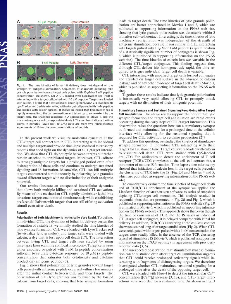
ResultsPolarization of Lytic Machinery Is Intrinsically Very Rapid. To define,in individual CTL, the dynamics of lethal hit delivery versus theformation of a stable IS, we initially studied the time kinetics oflytic synapse formation. CTL were loaded with LysoTracker red(to visualize lytic granules), and target cells were loaded withcalcein, a dye that is lost upon cell death (17). The interactionbetween living CTL and target cells was studied by usingtime-lapse laser scanning confocal microscopy. Target cells wereeither unpulsed or pulsed with 1 nM (a peptide concentrationsufficient to saturate cytotoxic response) or 10 �M (a peptideconcentration that saturates both cytotoxicity and cytokineproduction) antigenic peptide (3).
Fig. 1 shows that polarization of lytic granules toward targetcells pulsed with antigenic peptide occurred within a few minutesafter the initial contact between CTL and their targets. Thepolarization of CTL lytic granules was followed by the loss ofcalcein from target cells, showing that lytic synapse formation
leads to target death. The time kinetics of lytic granule polar-ization are better appreciated in Movies 1 and 2, which arepublished as supporting information on the PNAS web site,showing that lytic granule polarization was detectable within 3min after cell–cell contact. Interestingly, the time kinetics of lyticmachinery reorientation was independent of the strength ofantigenic stimulation, because it was similar in CTL interactingwith targets pulsed with 10 �M or 1 nM peptide (a quantificationof a statistically significant number of conjugates is shown Fig.6, which is published as supporting information on the PNASweb site). The time kinetics of calcein loss was variable in thedifferent CTL�target conjugates. This finding suggests that,although CTL deliver hits homogeneously rapid, the time re-quired to trigger individual target cell death is variable.
CTL interacting with unpulsed target cells formed conjugatesand crawled on target cell surface in the absence of calceinleakage and of any other evidence of target cell death (Movie 3,which is published as supporting information on the PNAS website).
Together these results indicate that lytic granule polarizationis intrinsically very rapid, allowing CTL to promptly attacktargets with no distinction of their antigenic potential.
Stimulatory Synapse and Sustained Signaling Keep Going After TargetCell Annihilation. The above results support the notion that lyticsynapse formation and target cell annihilation are rapid eventsoccurring during the early steps of CTL�target interaction. Thisobservation raises the question: how can a stimulatory synapsebe formed and maintained for a prolonged time at the cellularinterface while allowing for the sustained signaling that isrequired for CTL activation to cytokine production (2)?
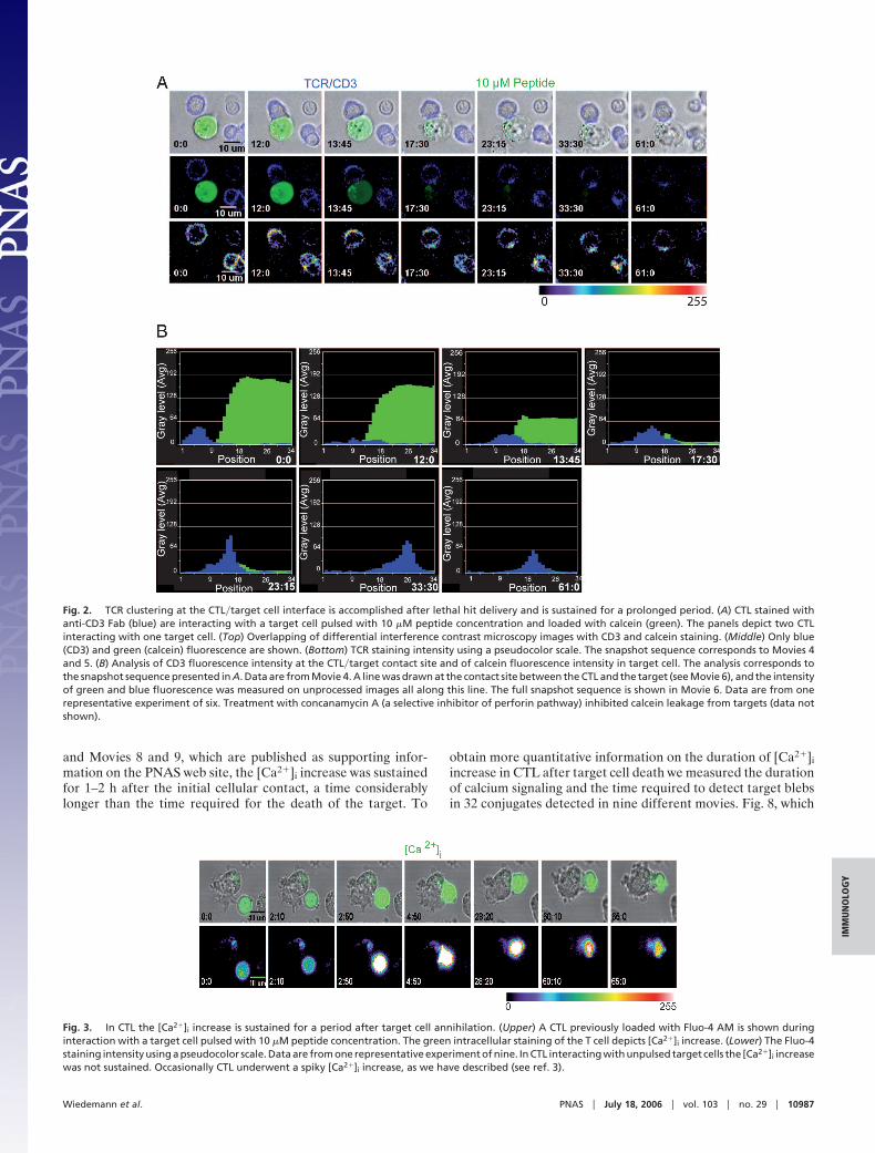
To address this question, we studied the kinetics of stimulatorysynapse formation in individual CTL interacting with theirtargets for a sustained time. Target cells were loaded with calceinto visualize cell death. CTL were stained with Cy5-labeledanti-CD3 Fab antibodies to detect the enrichment of T cellreceptor (TCR)�CD3 complexes at the cell–cell contact site, aparameter of mature IS formation. Time-lapse video microscopyshowed that initiation of calcein loss in the target cells precedesthe clustering of TCR into the IS (Fig. 2A and Movies 4 and 5,which are published as supporting information on the PNAS website).
To quantitatively evaluate the time kinetics of target cell deathand of TCR�CD3 enrichment at the synapse we applied theLineScan function of METAMORPH software to series of snapshotsdepicting CTL�target cell interaction. This analysis resulted insequential plots that are presented in Fig. 2B and Fig. 7, which ispublished as supporting information on the PNAS web site (Fig. 2Bis animated in Movie 6, which is published as supporting informa-tion on the PNAS web site). This approach shows that, even thoughthe time of enrichment of TCR into the IS varies in individualCTL�target cell conjugates, it is delayed compared with lethal hitdelivery. In addition, TCR�CD3 clustering at the cell–cell contactsite was sustained long after target annihilation (Fig. 2). When CTLwere conjugated with targets pulsed with a 1 nM concentration thetargets were readily killed in the absence of the TCR clusteringtypical of stimulatory IS (Movie 7, which is published as supportinginformation on the PNAS web site), in agreement with previouslyreported data (3, 6).
The unexpected observation that stimulatory synapse forma-tion can be sustained even after target cell annihilation suggeststhat CTL could receive prolonged activatory signals while in-teracting with fragments of disintegrating targets. We thereforeinvestigated whether CTL maintained sustained signaling for aprolonged time after the death of the opposing target cell.
CTL were loaded with Fluo-4 to detect the intracellular Ca2�
concentration ([Ca2�]i) increase (3, 13), and CTL�target inter-actions were recorded for a sustained time. As shown in Fig. 3
Fig. 1. The time kinetics of lethal hit delivery does not depend on thestrength of antigenic stimulation. Sequences of snapshots depicting lyticgranule polarization toward target cells pulsed with 10 �M or 1 nM peptideconcentration are shown. (A) A CTL loaded with LysoTracker red (red) isinteracting with a target cell pulsed with 10 �M peptide. Targets are loadedwith calcein, a probe that is lost upon cell death (green). (B) A CTL loaded withLysoTracker red (red) is interacting with a target cell pulsed with 1 nM peptideand loaded with calcein (green). It should be noted that LysoTracker red israpidly released into the culture medium and taken up to some extent by thetarget cells. The snapshot sequence in A corresponds to Movie 1, and thesnapshot sequence in B corresponds to Movie 2. The numbers indicate the timepoints in minutes. (Scale bar: 10 �m.) Data are from two representativeexperiments of 16 for the two concentrations of peptide.
10986 � www.pnas.org�cgi�doi�10.1073�pnas.0600651103 Wiedemann et al.
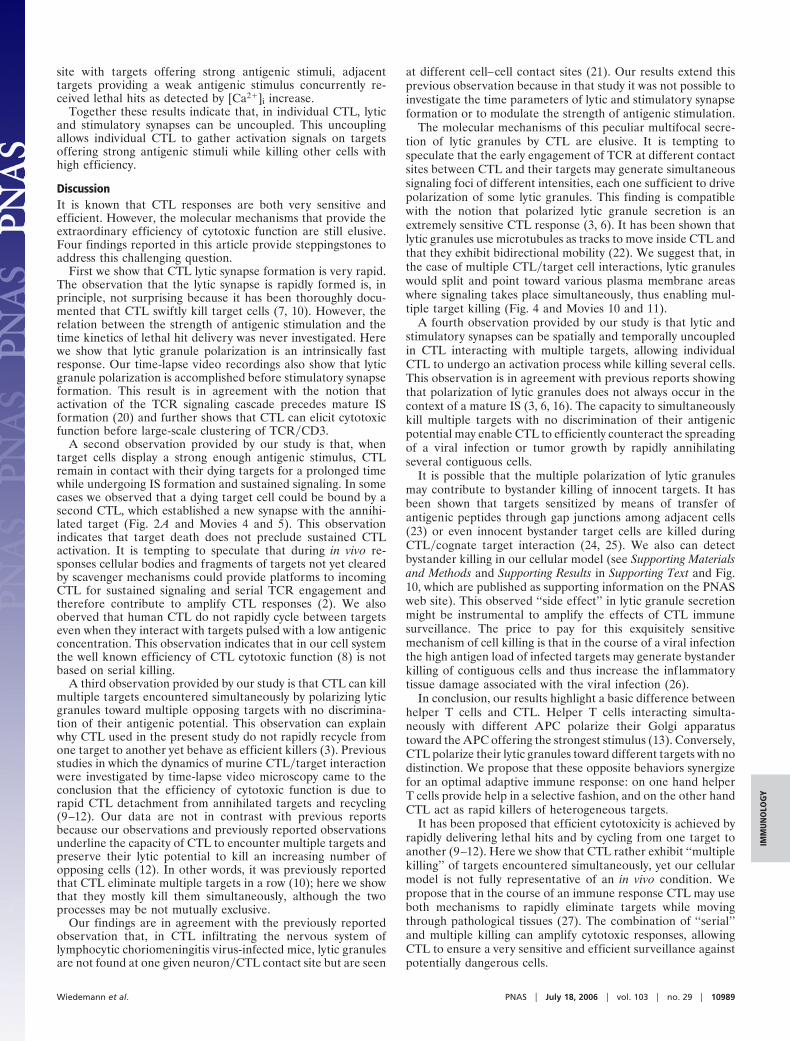
and Movies 8 and 9, which are published as supporting infor-mation on the PNAS web site, the [Ca2�]i increase was sustainedfor 1–2 h after the initial cellular contact, a time considerablylonger than the time required for the death of the target. To
obtain more quantitative information on the duration of [Ca2�]i
increase in CTL after target cell death we measured the durationof calcium signaling and the time required to detect target blebsin 32 conjugates detected in nine different movies. Fig. 8, which
Fig. 2. TCR clustering at the CTL�target cell interface is accomplished after lethal hit delivery and is sustained for a prolonged period. (A) CTL stained withanti-CD3 Fab (blue) are interacting with a target cell pulsed with 10 �M peptide concentration and loaded with calcein (green). The panels depict two CTLinteracting with one target cell. (Top) Overlapping of differential interference contrast microscopy images with CD3 and calcein staining. (Middle) Only blue(CD3) and green (calcein) fluorescence are shown. (Bottom) TCR staining intensity using a pseudocolor scale. The snapshot sequence corresponds to Movies 4and 5. (B) Analysis of CD3 fluorescence intensity at the CTL�target contact site and of calcein fluorescence intensity in target cell. The analysis corresponds tothe snapshot sequence presented in A. Data are from Movie 4. A line was drawn at the contact site between the CTL and the target (see Movie 6), and the intensityof green and blue fluorescence was measured on unprocessed images all along this line. The full snapshot sequence is shown in Movie 6. Data are from onerepresentative experiment of six. Treatment with concanamycin A (a selective inhibitor of perforin pathway) inhibited calcein leakage from targets (data notshown).
Fig. 3. In CTL the [Ca2�]i increase is sustained for a period after target cell annihilation. (Upper) A CTL previously loaded with Fluo-4 AM is shown duringinteraction with a target cell pulsed with 10 �M peptide concentration. The green intracellular staining of the T cell depicts [Ca2�]i increase. (Lower) The Fluo-4staining intensity using a pseudocolor scale. Data are from one representative experiment of nine. In CTL interacting with unpulsed target cells the [Ca2�]i increasewas not sustained. Occasionally CTL underwent a spiky [Ca2�]i increase, as we have described (see ref. 3).
Wiedemann et al. PNAS � July 18, 2006 � vol. 103 � no. 29 � 10987
IMM
UN
OLO
GY

is published as supporting information on the PNAS web site,shows that the [Ca2�]i increase is significantly sustained aftertarget cell death. These results show that CTL interacting withtarget cells providing strong antigenic stimulation do not rapidlydisassemble stimulatory synapse upon target annihilation.Rather, they remain in contact with the dead targets and sustainactive signal transduction for a long period.
Individual CTL Kill Multiple Targets Simultaneously in a NonselectiveFashion. The above results show that human CTL remain at-tached to their targets for a relatively prolonged time rather thanrapidly recycling from one target to another. This observationraises the question of how CTL may kill outnumbering hetero-geneous targets during an immune response. To address thisquestion we investigated the possibility that CTL may be able tokill multiple targets encountered simultaneously regardless oftheir antigenic potential.
CTL simultaneous interaction with multiple targets was in-vestigated by time-lapse confocal microscopy. Target cells werepulsed with a high (10 �M) or a low (1 nM) antigenic peptideconcentration and were loaded with different dyes to allow their
identification. We initially focused on lytic granule secretion.Movies 10 and 11 and Fig. 9, which are published as supportinginformation on the PNAS web site, and Fig. 4 show that CTLinteracting with targets pulsed with different antigenic concen-trations polarize their lytic granules toward both targets simul-taneously.
To obtain quantitative information on polarization of lyticgranules toward two different contact sites we took severalsnapshots of living cells by randomly changing the field duringmovie recording. A total of 136 snapshots depicting CTL insimultaneous contact with two targets were recorded in nineindependent sessions. The polarization of lytic granule accumu-lation was scored by visual inspection in a blind study onregistered images. T cells in simultaneous contact with twotargets offering different antigenic stimuli were initially scored.Eighty-four triplicates were scored: 44% of CTL exhibited adouble polarization, 29% exhibited lytic granule accumulationtoward one target cell, and 27% showed an uncertain phenotype.More precisely, they showed a tendency to relocate lytic granulestoward the two contact sites without clearly focused polarizationof lytic granules. A simultaneous polarization of lytic granules inCTL interacting with two targets offering the same antigenicstimulation was also observed. 52 triplicates were scored: 46% ofCTL exhibited a double polarization, 17% exhibited lytic granuleaccumulation toward one target cell, and 37% showed anuncertain phenotype.
Taken together the above results directly illustrate that indi-vidual CTL are indeed killers of multiple targets encounteredsimultaneously. A recent study showed that, in the case of CTLconjugated with two targets offering similar densities of anti-genic ligands, the tubulin cytoskeleton oscillates between the twotargets (18). In that study, the dynamics of lytic granule polar-ization in CTL conjugated with targets offering different den-sities of antigenic ligands was not investigated. Therefore, ourresults extend these described findings.
We next focused on TCR�CD3 dynamics on the surface ofCTL interacting with different targets. CTL were stained withCy5-labeled anti-CD3 Fab antibodies, and their interaction withtargets pulsed with low (1 nM) or high (10 �M) antigenicconcentration was visualized. Target cells were loaded withFluo-4 to have a rapid evidence of lethal hit delivery in parallelwith TCR�CD3 dynamics (10, 19). Fig. 5 and Movies 12 and 13,which are published as supporting information on the PNAS website, show that, although TCR�CD3 was enriched in the contact
Fig. 4. CTL polarize lytic granules toward different targets encounteredsimultaneously. Snapshots depict lytic granule polarization in CTL interactingsimultaneously with target cells pulsed with 10 �M (blue) or 1 nM (green)peptide concentration. Data are from one representative experiment of nine.
Fig. 5. Lytic and stimulatory synapses can be uncoupled in individual CTL. Sequences of snapshots depicting TCR�CD3 staining (blue) in CTL interactingsimultaneously with target cells pulsed with a high (10 �M; red) or a low (1 nM; green) peptide concentration. To rapidly detect lethal hit delivery in target cells,targets pulsed with 1 nM peptide were loaded with Fluo-4 AM. (Top) Overlapping of differential interference contrast microscopy images and fluorescencestaining. (Middle) Only fluorescence staining is shown. (Bottom) TCR staining intensity using a pseudocolor scale. The white arrows in Bottom indicate theTCR�CD3 enrichment at the CTL�target contact site. At the end of the time recording a second target cell receives the lethal hit by a CTL not visible in the movieand undergoes [Ca2�]i increase. The snapshot sequence corresponds to Movies 12 and 13. Data are from one representative experiment of three.
10988 � www.pnas.org�cgi�doi�10.1073�pnas.0600651103 Wiedemann et al.
site with targets offering strong antigenic stimuli, adjacenttargets providing a weak antigenic stimulus concurrently re-ceived lethal hits as detected by [Ca2�]i increase.
Together these results indicate that, in individual CTL, lyticand stimulatory synapses can be uncoupled. This uncouplingallows individual CTL to gather activation signals on targetsoffering strong antigenic stimuli while killing other cells withhigh efficiency.
DiscussionIt is known that CTL responses are both very sensitive andefficient. However, the molecular mechanisms that provide theextraordinary efficiency of cytotoxic function are still elusive.Four findings reported in this article provide steppingstones toaddress this challenging question.
First we show that CTL lytic synapse formation is very rapid.The observation that the lytic synapse is rapidly formed is, inprinciple, not surprising because it has been thoroughly docu-mented that CTL swiftly kill target cells (7, 10). However, therelation between the strength of antigenic stimulation and thetime kinetics of lethal hit delivery was never investigated. Herewe show that lytic granule polarization is an intrinsically fastresponse. Our time-lapse video recordings also show that lyticgranule polarization is accomplished before stimulatory synapseformation. This result is in agreement with the notion thatactivation of the TCR signaling cascade precedes mature ISformation (20) and further shows that CTL can elicit cytotoxicfunction before large-scale clustering of TCR�CD3.
A second observation provided by our study is that, whentarget cells display a strong enough antigenic stimulus, CTLremain in contact with their dying targets for a prolonged timewhile undergoing IS formation and sustained signaling. In somecases we observed that a dying target cell could be bound by asecond CTL, which established a new synapse with the annihi-lated target (Fig. 2 A and Movies 4 and 5). This observationindicates that target death does not preclude sustained CTLactivation. It is tempting to speculate that during in vivo re-sponses cellular bodies and fragments of targets not yet clearedby scavenger mechanisms could provide platforms to incomingCTL for sustained signaling and serial TCR engagement andtherefore contribute to amplify CTL responses (2). We alsooberved that human CTL do not rapidly cycle between targetseven when they interact with targets pulsed with a low antigenicconcentration. This observation indicates that in our cell systemthe well known efficiency of CTL cytotoxic function (8) is notbased on serial killing.
A third observation provided by our study is that CTL can killmultiple targets encountered simultaneously by polarizing lyticgranules toward multiple opposing targets with no discrimina-tion of their antigenic potential. This observation can explainwhy CTL used in the present study do not rapidly recycle fromone target to another yet behave as efficient killers (3). Previousstudies in which the dynamics of murine CTL�target interactionwere investigated by time-lapse video microscopy came to theconclusion that the efficiency of cytotoxic function is due torapid CTL detachment from annihilated targets and recycling(9–12). Our data are not in contrast with previous reportsbecause our observations and previously reported observationsunderline the capacity of CTL to encounter multiple targets andpreserve their lytic potential to kill an increasing number ofopposing cells (12). In other words, it was previously reportedthat CTL eliminate multiple targets in a row (10); here we showthat they mostly kill them simultaneously, although the twoprocesses may be not mutually exclusive.
Our findings are in agreement with the previously reportedobservation that, in CTL infiltrating the nervous system oflymphocytic choriomeningitis virus-infected mice, lytic granulesare not found at one given neuron�CTL contact site but are seen
at different cell–cell contact sites (21). Our results extend thisprevious observation because in that study it was not possible toinvestigate the time parameters of lytic and stimulatory synapseformation or to modulate the strength of antigenic stimulation.
The molecular mechanisms of this peculiar multifocal secre-tion of lytic granules by CTL are elusive. It is tempting tospeculate that the early engagement of TCR at different contactsites between CTL and their targets may generate simultaneoussignaling foci of different intensities, each one sufficient to drivepolarization of some lytic granules. This finding is compatiblewith the notion that polarized lytic granule secretion is anextremely sensitive CTL response (3, 6). It has been shown thatlytic granules use microtubules as tracks to move inside CTL andthat they exhibit bidirectional mobility (22). We suggest that, inthe case of multiple CTL�target cell interactions, lytic granuleswould split and point toward various plasma membrane areaswhere signaling takes place simultaneously, thus enabling mul-tiple target killing (Fig. 4 and Movies 10 and 11).
A fourth observation provided by our study is that lytic andstimulatory synapses can be spatially and temporally uncoupledin CTL interacting with multiple targets, allowing individualCTL to undergo an activation process while killing several cells.This observation is in agreement with previous reports showingthat polarization of lytic granules does not always occur in thecontext of a mature IS (3, 6, 16). The capacity to simultaneouslykill multiple targets with no discrimination of their antigenicpotential may enable CTL to efficiently counteract the spreadingof a viral infection or tumor growth by rapidly annihilatingseveral contiguous cells.
It is possible that the multiple polarization of lytic granulesmay contribute to bystander killing of innocent targets. It hasbeen shown that targets sensitized by means of transfer ofantigenic peptides through gap junctions among adjacent cells(23) or even innocent bystander target cells are killed duringCTL�cognate target interaction (24, 25). We also can detectbystander killing in our cellular model (see Supporting Materialsand Methods and Supporting Results in Supporting Text and Fig.10, which are published as supporting information on the PNASweb site). This observed ‘‘side effect’’ in lytic granule secretionmight be instrumental to amplify the effects of CTL immunesurveillance. The price to pay for this exquisitely sensitivemechanism of cell killing is that in the course of a viral infectionthe high antigen load of infected targets may generate bystanderkilling of contiguous cells and thus increase the inflammatorytissue damage associated with the viral infection (26).
In conclusion, our results highlight a basic difference betweenhelper T cells and CTL. Helper T cells interacting simulta-neously with different APC polarize their Golgi apparatustoward the APC offering the strongest stimulus (13). Conversely,CTL polarize their lytic granules toward different targets with nodistinction. We propose that these opposite behaviors synergizefor an optimal adaptive immune response: on one hand helperT cells provide help in a selective fashion, and on the other handCTL act as rapid killers of heterogeneous targets.
It has been proposed that efficient cytotoxicity is achieved byrapidly delivering lethal hits and by cycling from one target toanother (9–12). Here we show that CTL rather exhibit ‘‘multiplekilling’’ of targets encountered simultaneously, yet our cellularmodel is not fully representative of an in vivo condition. Wepropose that in the course of an immune response CTL may useboth mechanisms to rapidly eliminate targets while movingthrough pathological tissues (27). The combination of ‘‘serial’’and multiple killing can amplify cytotoxic responses, allowingCTL to ensure a very sensitive and efficient surveillance againstpotentially dangerous cells.
Wiedemann et al. PNAS � July 18, 2006 � vol. 103 � no. 29 � 10989
IMM
UN
OLO
GY

Materials and MethodsT Cells and Target Cells. An HLA-A2-restricted T cell line (CM-Vpp65) specific for the peptide NLVPMVATV of human cyto-megalovirus protein pp65 was used (3). HLA-A2-matched Ep-stein–Barr virus (EBV)-transformed B cells (JY) were used astarget cells (3). T cell line and EBV-B cell lines were generatedand maintained as described (2).
Dynamics of Lytic Granules and TCR�CD3 in Living Cells. To visualizelethal hit delivery T cells were loaded with LysoTracker Red(Molecular Probes) for 45 min at 37°C in RPMI medium1640�5% FCS. To visualize TCR�CD3 dynamics, T lymphocyteswere labeled with Cy5 TR66 Fab at 20 �g�ml in RPMI medium1640�5% FCS at 4°C for 30 min as described (13).
Target cells were labeled with either 1 �M calcein (MolecularProbes) in RPMI medium 1640�5% FCS for 30 min at roomtemperature or 0.5 �M Orange CMTMR or 0.5 �M Bodipy630 (both from Molecular Probes) at 37°C for 15 min. Targetcells were previously pulsed with 1 nM or 10 �M peptide for2 h at 37°C.
Target cells were seeded into microchambers (Lab-Tek Cham-
ber coverglass, Nalge Nunc) previously coated with poly-D-lysine(Sigma). Fluorescence measurements were done on a ZeissLSM-510 confocal microscope at 37°C and 5% CO2. Imagesequences of the time-lapse recording were processed withMETAMORPH software (13).
To have a quantitative evaluation of the time kinetics of targetcell death and of TCR�CD3 fluorescence at the synapse weapplied the LineScan function of METAMORPH software to seriesof snapshots depicting CTL�target cell interaction. This analysisresulted in sequential plots that are presented in Figs. 2B and 7and are animated in Movie 6.
In additional experiments either CTL (to detect TCR-mediated signaling) or target cells [to detect early cellulardamage (10, 19)] were loaded with 1 �M Fluo-4 AM for 30 minat 37°C. The green fluorescence emission of Fluo-4 reflecting the[Ca2�]i was monitored by time-lapse confocal microscopy asdescribed (3).
We thank Sabina Muller for help and advice and Daniel Coombs, SylvieGuerder, Jean-Charles Guery, and Loic Dupre for discussion and criticalreading of the manuscript. This work was supported by grants from LaLigue Contre le Cancer ‘‘Equipe Labellisee 2005.’’
1. Porgador, A., Yewdell, J. W., Deng, Y., Bennink, J. R. & Germain, R. N. (1997)Immunity 6, 715–726.
2. Valitutti, S., Muller, S., Dessing, M. & Lanzavecchia, A. (1996) J. Exp. Med. 183,1917–1921.
3. Faroudi, M., Utzny, C., Salio, M., Cerundolo, V., Guiraud, M., Muller, S. &Valitutti, S. (2003) Proc. Natl. Acad. Sci. USA 100, 14145–14150.
4. Huppa, J. B., Gleimer, M., Sumen, C. & Davis, M. M. (2003) Nat. Immunol.4, 749–755.
5. Sykulev, Y., Joo, M., Vturina, I., Tsomides, T. J. & Eisen, H. N. (1996)Immunity 4, 565–571.
6. Purbhoo, M. A., Irvine, D. J., Huppa, J. B. & Davis, M. M. (2004) Nat.Immunol. 5, 524–530.
7. Stinchcombe, J. C., Bossi, G., Booth, S. & Griffiths, G. M. (2001) Immunity 15,751–761.
8. Cerottini, J. C. & Brunner, K. T. (1974) Adv. Immunol. 18, 67–132.9. Sanderson, C. J. & Thomas, J. A. (1976) Proc. R. Soc. London B 194, 417–429.
10. Poenie, M., Tsien, R. Y. & Schmitt-Verhulst, A. M. (1987) EMBO J. 6,2223–2232.
11. Rothstein, T. L., Mage, M., Jones, G. & McHugh, L. L. (1978) J. Immunol. 121,1652–1656.
12. Isaaz, S., Baetz, K., Olsen, K., Podack, E. & Griffiths, G. M. (1995) Eur.J. Immunol. 25, 1071–1079.
13. Depoil, D., Zaru, R., Guiraud, M., Chauveau, A., Harriague, J., Bismuth, G.,Utzny, C., Muller, S. & Valitutti, S. (2005) Immunity 22, 185–194.
14. Monks, C. R., Freiberg, B. A., Kupfer, H., Sciaky, N. & Kupfer, A. (1998)Nature 395, 82–86.
15. Grakoui, A., Bromley, S. K., Sumen, C., Davis, M. M., Shaw, A. S., Allen, P. M.& Dustin, M. L. (1999) Science 285, 221–227.
16. O’Keefe, J. P. & Gajewski, T. F. (2005) J. Immunol. 175, 5581–5585.17. Lyubchenko, T. A., Wurth, G. A. & Zweifach, A. (2001) Immunity 15, 847–859.18. Kuhn, J. R. & Poenie, M. (2002) Immunity 16, 111–121.19. Keefe, D., Shi, L., Feske, S., Massol, R., Navarro, F., Kirchhausen, T. &
Lieberman, J. (2005) Immunity 23, 249–262.20. Lee, K. H., Holdorf, A. D., Dustin, M. L., Chan, A. C., Allen, P. M. & Shaw,
A. S. (2002) Science 295, 1539–1542.21. McGavern, D. B., Christen, U. & Oldstone, M. B. (2002) Nat. Immunol. 3,
918–925.22. Poenie, M., Kuhn, J. & Combs, J. (2004) Curr. Opin. Immunol. 16, 428–438.23. Neijssen, J., Herberts, C., Drijfhout, J. W., Reits, E., Janssen, L. & Neefjes, J.
(2005) Nature 434, 83–88.24. Lanzavecchia, A. (1986) Nature 319, 778–780.25. Duke, R. C. (1989) J. Exp. Med. 170, 59–71.26. Asquith, B. & Bangham, C. R. (2003) J. Theor. Biol. 222, 53–69.27. Friedl, P., den Boer, A. T. & Gunzer, M. (2005) Nat. Rev. Immunol. 5, 532–545.
10990 � www.pnas.org�cgi�doi�10.1073�pnas.0600651103 Wiedemann et al.
Related Documents