Research Article Cytotoxic Effects of Biosynthesized Zinc Oxide Nanoparticles on Murine Cell Lines Farideh Namvar, 1,2 Heshu Sulaiman Rahman, 3,4,5 Rosfarizan Mohamad, 1,6 Susan Azizi, 6 Paridah Mohd Tahir, 1 Max Stanley Chartrand, 7 and Swee Keong Yeap 5 1 Institute of Tropical Forestry and Forest Products (INTROP), Universiti Putra Malaysia (UPM), 43400 Serdang, Selangor, Malaysia 2 Research Center for Animal Development Applied Biology, Mashhad Branch, Islamic Azad University, Mashhad, Iran 3 Department of Clinic and Internal Medicine, College of Veterinary Medicine, University of Sulaimani, Sulaimani Nwe, Street 27, Sulaimani City, Kurdistan Region, Iraq 4 Department of Veterinary Laboratory Diagnosis, Faculty of Veterinary Medicine, Universiti Putra Malaysia (UPM), 43400 Serdang, Selangor, Malaysia 5 Institute of Bioscience (IBS), Universiti Putra Malaysia (UPM), 43400 Serdang, Selangor, Malaysia 6 Department of Bioprocess Technology, Faculty of Biotechnology and Biomolecular Sciences, Universiti Putra Malaysia (UPM), 43400 Serdang, Selangor, Malaysia 7 DigiCare Behavioral Research, Casa Grande, AZ, USA Correspondence should be addressed to Farideh Namvar; [email protected] and Heshu Sulaiman Rahman; [email protected] Received 25 September 2014; Revised 9 January 2015; Accepted 19 January 2015 Academic Editor: Jian-Guo Chen Copyright © 2015 Farideh Namvar et al. is is an open access article distributed under the Creative Commons Attribution License, which permits unrestricted use, distribution, and reproduction in any medium, provided the original work is properly cited. e aim of this study is to evaluate the in vitro cytotoxic activity and cellular effects of previously prepared ZnO-NPs on murine cancer cell lines using brown seaweed (Sargassum muticum) aqueous extract. Treated cancer cells with ZnO-NPs for 72 hours demonstrated various levels of cytotoxicity based on calculated IC 50 values using MTT assay as follows: 21.7 ± 1.3 g/mL (4T1), 17.45 ± 1.1 g/mL (CRL-1451), 11.75 ± 0.8 g/mL (CT-26), and 5.6 ± 0.55 g/mL (WEHI-3B), respectively. On the other hand, ZnO- NPs treatments for 72 hours showed no toxicity against normal mouse fibroblast (3T3) cell line. On the other hand, paclitaxel, which imposed an inhibitory effect on WEHI-3B cells with IC 50 of 2.25 ± 0.4, 1.17 ± 0.5, and 1.6 ± 0.09 g/mL aſter 24, 48, and 72 hours treatment, respectively, was used as positive control. Furthermore, distinct morphological changes were found by utilizing fluorescent dyes; apoptotic population was increased via flowcytometry, while a cell cycle block and stimulation of apoptotic proteins were also observed. Additionally, the present study showed that the caspase activations contributed to ZnO-NPs triggered apoptotic death in WEHI-3 cells. us, the nature of biosynthesis and the therapeutic potential of ZnO-NPs could prepare the way for further research on the design of green synthesis therapeutic agents, particularly in nanomedicine, for the treatment of cancer. 1. Introduction Nanoscience and nanotechnology have excellent potential in a broad spectrum of cancer research, such as in diagnostics, monitoring, and therapeutic strategies [1, 2]. Some nanocar- riers like liposome, dendrimer, micelle, carbon nanotube, and nanoparticles have attempted in the diagnosis and theranos- tics of several types of cancers [3, 4]. e Green approach for synthesis nanoparticles, using plant materials [5] for reducing and capping agents, could be quite attractive for use in nanobiotechnology [6, 7]. is technology, as compared to other mechanical strategies, is safe, simple, nontoxic, efficient, environmentally friendly, and efficacious as single- potreactions, without the need for additional surfactants and capping agents [2, 8]. Zinc oxide, due to physical and chemical properties, is considered a capable agent in cancer therapy [9]. A novel approach of biosynthesis, zinc oxide nanoparticles (ZnO- NPs) produced nanoparticles using a natural source, such as plant extracts to reduce metal ions, which are readily scalable Hindawi Publishing Corporation Evidence-Based Complementary and Alternative Medicine Volume 2015, Article ID 593014, 11 pages http://dx.doi.org/10.1155/2015/593014

Welcome message from author
This document is posted to help you gain knowledge. Please leave a comment to let me know what you think about it! Share it to your friends and learn new things together.
Transcript

Research ArticleCytotoxic Effects of Biosynthesized Zinc Oxide Nanoparticles onMurine Cell Lines
Farideh Namvar,1,2 Heshu Sulaiman Rahman,3,4,5 Rosfarizan Mohamad,1,6 Susan Azizi,6
Paridah Mohd Tahir,1 Max Stanley Chartrand,7 and Swee Keong Yeap5
1 Institute of Tropical Forestry and Forest Products (INTROP), Universiti Putra Malaysia (UPM), 43400 Serdang, Selangor, Malaysia2Research Center for Animal Development Applied Biology, Mashhad Branch, Islamic Azad University, Mashhad, Iran3Department of Clinic and Internal Medicine, College of Veterinary Medicine, University of Sulaimani, Sulaimani Nwe,Street 27, Sulaimani City, Kurdistan Region, Iraq4Department of Veterinary Laboratory Diagnosis, Faculty of Veterinary Medicine, Universiti Putra Malaysia (UPM),43400 Serdang, Selangor, Malaysia5Institute of Bioscience (IBS), Universiti Putra Malaysia (UPM), 43400 Serdang, Selangor, Malaysia6Department of Bioprocess Technology, Faculty of Biotechnology and Biomolecular Sciences, Universiti Putra Malaysia (UPM),43400 Serdang, Selangor, Malaysia7DigiCare Behavioral Research, Casa Grande, AZ, USA
Correspondence should be addressed to Farideh Namvar; [email protected] Heshu Sulaiman Rahman; [email protected]
Received 25 September 2014; Revised 9 January 2015; Accepted 19 January 2015
Academic Editor: Jian-Guo Chen
Copyright © 2015 Farideh Namvar et al.This is an open access article distributed under theCreativeCommonsAttribution License,which permits unrestricted use, distribution, and reproduction in any medium, provided the original work is properly cited.
The aim of this study is to evaluate the in vitro cytotoxic activity and cellular effects of previously prepared ZnO-NPs on murinecancer cell lines using brown seaweed (Sargassum muticum) aqueous extract. Treated cancer cells with ZnO-NPs for 72 hoursdemonstrated various levels of cytotoxicity based on calculated IC
50values using MTT assay as follows: 21.7± 1.3 𝜇g/mL (4T1),
17.45± 1.1 𝜇g/mL (CRL-1451), 11.75± 0.8 𝜇g/mL (CT-26), and 5.6± 0.55 𝜇g/mL (WEHI-3B), respectively. On the other hand, ZnO-NPs treatments for 72 hours showed no toxicity against normal mouse fibroblast (3T3) cell line. On the other hand, paclitaxel,which imposed an inhibitory effect on WEHI-3B cells with IC
50of 2.25± 0.4, 1.17± 0.5, and 1.6± 0.09 𝜇g/mL after 24, 48, and 72
hours treatment, respectively, was used as positive control. Furthermore, distinct morphological changes were found by utilizingfluorescent dyes; apoptotic population was increased via flowcytometry, while a cell cycle block and stimulation of apoptoticproteins were also observed. Additionally, the present study showed that the caspase activations contributed to ZnO-NPs triggeredapoptotic death inWEHI-3 cells. Thus, the nature of biosynthesis and the therapeutic potential of ZnO-NPs could prepare the wayfor further research on the design of green synthesis therapeutic agents, particularly in nanomedicine, for the treatment of cancer.
1. Introduction
Nanoscience and nanotechnology have excellent potential ina broad spectrum of cancer research, such as in diagnostics,monitoring, and therapeutic strategies [1, 2]. Some nanocar-riers like liposome, dendrimer,micelle, carbon nanotube, andnanoparticles have attempted in the diagnosis and theranos-tics of several types of cancers [3, 4]. The Green approachfor synthesis nanoparticles, using plant materials [5] forreducing and capping agents, could be quite attractive for use
in nanobiotechnology [6, 7]. This technology, as comparedto other mechanical strategies, is safe, simple, nontoxic,efficient, environmentally friendly, and efficacious as single-potreactions, without the need for additional surfactants andcapping agents [2, 8].
Zinc oxide, due to physical and chemical properties, isconsidered a capable agent in cancer therapy [9]. A novelapproach of biosynthesis, zinc oxide nanoparticles (ZnO-NPs) produced nanoparticles using a natural source, such asplant extracts to reduce metal ions, which are readily scalable
Hindawi Publishing CorporationEvidence-Based Complementary and Alternative MedicineVolume 2015, Article ID 593014, 11 pageshttp://dx.doi.org/10.1155/2015/593014
2 Evidence-Based Complementary and Alternative Medicine
and nontoxic compared with physical and chemical methods[10].
The mechanisms of cytotoxicity from ZnO-NPs are notyet entirely understood, but the generation of hydroxylradicals (OH∙), superoxide anion (O
2
−), and perhydroxylradicals (HO
2
∙) from the surface of ZnO are believed to bemajor components. When nanoparticles interact with cells,cellular protection mechanisms are activated to minimizeharm. However, if the highly active free radicals productionexceeds the antioxidative defensive ability of the cell, it resultsin oxidative harm of biomolecules which can lead to celldeath [11, 12]. Recent studies have confirmed that bioactivecompounds obtained from macroalgae provide an opportu-nity for discovering new pharmaceutical and versatile novelagents with more promise in cancer research, diagnostic andefficient treatment [13].
As mentioned in previous reports [14], seaweed as asubgroup of macroalgae is an available food source that isconsumed in many countries, most traditionally in southeastAsia [15]. Seaweed potentially has biologically active sub-strates like polysaccharides, proteins, lipid, vitamin, solublefiber, and minerals with multiple medicinal applicationsagainst cancer [16], inflammation [17], allergy [18], diabetes[19], thrombosis [20], reduction of obesity by bringing downthe caloric value of the diet [21], reduction of lipid absorptionand cardiovascular diseases [22], hypertension [23], andother degenerative chronic diseases [24]. These biomedicalapplications are mainly due to existing functional groups,which act as a capping agent in a Green single step process.Polysaccharides are the main constituents of biopolymers inseaweed water extract andwere found to be strong stabilizers,for increased biocompatibility, conferring chemical function-ality towards nanostructures including iron oxide magneticnanoparticles [25].
In the previous study [26], we synthesized and charac-terized zinc oxide nanoparticles (ZnO-NPs) using Sargassummuticum, brown seaweed extract in green method. The aimof this study is in evaluating the cytotoxic effect of ZnO-NPsprepared via biosynthesize green method on various humancancer cells using different experimental methods.
2. Material and Methods
2.1. Materials. Samples of the Sargassum muticum, brownseaweed, were collected from the waters of Persian Gulfcoastal areas. All aqueous solutions were prepared usingdistilled water.
2.2. Cell Lines and Cell Culture Conditions. All murinecancer cell lines (CT-26, WEHI-3, 4T1, and CRL-1451) andnormal mice fibroblast cell line (3T3) were purchased fromthe American Type Culture Collection (ATCC) (Maryland,USA). All cell lines (except CRL-1451) were cultured inRPMI-1640 (GIBCO,Germany)medium, supplementedwith10% (v/v) fetal bovine serum (FBS) (GIBCO, Germany)and 1% penicillin/streptomycin (GIBCO, Germany) (100units/mLpenicillin and 50𝜇g/mL streptomycin) according tothe supplier protocol, whereas ATCC-formulated Dulbecco’s
Modified Eagle’s Medium (DMEM) was used as a base med-ium for CRL-1451 cell line with same supplementation men-tioned before. Cell cultures were incubated in a humidifiedatmosphere of 95% air and 5% carbon dioxide at 37∘C.
2.3. Preparation of (Sargassum muticum) Extract. As in aprevious study [26], for the preparation of extract, about 2.0 gof seaweed specimens was ground, freeze-dried, and boiledin 100mL of double distilled water with continuous stirringfor 15 minutes.Then, the extract was left at 25∘C, filtered, andthen stored at −20∘C for further investigation.
2.4. Preparation of ZnO-NPs. Zinc acetate dehydrate(Zn(Ac)
22H2O) (2mM) solution was made to react with
50mL of the aqueous extract for 3-4 hours in an aqueousbath (Falc MF24, Progen scientific) system under continuousstirring at 70∘C. The pale white solid product was collectedthrough centrifugation at 4000 rpm (Avanti J25, Beckman)for 10 minutes and carefully washed with distilled water andthen dried at 100∘C overnight [26].
2.5. Cell Viability Assay. Cell viability of various cancercell lines and normal cell line treated with ZnO-NPs wereassessed with 3-(4,5-dimethylthiazol-2-yl)-2,5-diphenyltetr-azolium bromide (MTT, Sigma Aldrich, USA). Murine can-cer and noncancer cells were seeded at a density of 2 ×10⁵ cells/well in 96-well microplates for 24 hours. Variousconcentrations (1–100𝜇g) of ZnO-NPs in medium wereprepared and added to the cultured cells. After 72 hours ofincubation, 20𝜇L of the freshly prepared MTT solution wasadded to each well. After incubation for 4 hours at 37∘C,cell viability was measured spectrophotometrically at 570 nm(Microplate reader, Biotech Inc., USA). The concentration,which inhibited 50% of cellular growth (IC
50value), was
determined and calculated by the following formula:
Growth inhibition = ODcontrol −ODtreated sample ×100
ODcontrol.
(1)The cytotoxicity of ZnO-NPs on cells was expressed as IC
50
values (the drug concentration, reducing the absorbance oftreated cells by 50% with respect to untreated cells). Thisexperiment was carried out in triplicate. The DMSO (0.1%)was used as negative control.
2.6. Cellular Morphology Assay Using AO/PI Double Staining.Acridine orange/propidium iodide (AO/PI) double stainingwas used to check the ZnO-NPs-induced morphologicalalterations in murine myelocytic leukemia cells. Briefly, theWEHI-3B cells were plated at a concentration of 1 × 10⁶cells/mL in a 25 cm2 culture flask (TPP, Switzerland) andthen treated with IC
50of ZnO-NPs and incubated at 37∘C
in a 5% carbon dioxide incubator for 24, 48, and 72 hours,respectively. Then, cells were trypsinized, centrifuged, andwashed with ice-cold PBS and stained with AO/PI (1 : 1 v/v)at 100 𝜇g/mL. Cover slip fixed slides with antifade betweenthem were observed under a fluorescence microscope (Leica,Japan) in the dark with the Q-floro software installed.
Evidence-Based Complementary and Alternative Medicine 3
2.7. Measuring of Apoptotic Cells Using Annexin V-TITC. Theeffect of apoptosis was flow cytometrically measured usingan annexin V-FITC apoptosis detection kit (Sigma Aldrich,USA). In brief, the WEHI-3 cells (1 × 10⁶ cells/mL) wereexposed to nanoparticles for 12, 24, and 48 hours, whileuntreated cells were used as controls. After that, both floatingand attached cells were harvested, washed in prechilledPBS, centrifuged, and resuspended in 1x binding buffer.Then, the cells were double stained with ice in the dark for15min with the fluorescein isothiocyanate- (FITC-) labeledannexin V (5𝜇L) and PI (10 𝜇L) before being analyzed flowcytometrically using FACSCalibur (BD, USA). Data analysiswas performed using the Cell Quest Pro software.
2.8. Analysis of DNA Content by Flow Cytometry. Flow cyto-meter was used to support the cytotoxicity of ZnO-NPstowards WEHI-3 cells. Briefly, The WEHI-3 cells 2.0 ×10⁶ cells/mL were cultured with the IC
50of ZnO-NPs and
incubated for 24, 48, and 72 hours, respectively.WEHI-3 cellswere trypsinized and washed twice with ice-cold PBS. Cellswere stored in 600 𝜇L 70% ethanol at −20∘C. The next day,the cells were centrifuged, washed twice with prechilled PBS,and incubated with 1mL staining buffer containing 1% TritonX-100, 10 𝜇g RNase A, and 50 𝜇g/mL propidium iodide for20min in the dark. Then, the samples were analyzed usingFACSCalibur flow cytometry (BD, USA). Data analysis wasperformed using the Cell Quest Pro software.
2.9. Caspase Protease Activity Assay Using a MicroplateReader. To search for the mechanisms involved in apoptosis,the protease activities of caspase-3 and caspase-9 in expo-nentially growing WEHI-3 cells treated with ZnO-NPs weredetermined respectively, using a colorimetric assay kit (Genescript kit, Piscataway, NJ 08854, USA) according to theinstructions of the manufacturing company without anymodification. Briefly, WEHI-3 cells (1 × 10⁶ cells) were pre-treated with ZnO-NPs for 24, 48, and 72 hours, respectively.Then, the cells were collected andwashedwith prechilled PBSand cell lysates were prepared in a 100 𝜇L lysis buffer for 20minutes on ice. After centrifugation, the supernatants werecollected and the total protein was quantified by the Bradfordassay. Lastly, the lysates were incubated at 37∘C for 4 hours inthe dark and the absorption at 490 nm was measured usingan ELISA microplate reader (Biotech Inc., USA).
2.10. Whole Cell Extract Preparation and Immunoblot. Imm-unoblotting was conducted according to the earlier study[27]. Briefly, WEHI-3 cells were grown in 75 cm2 cultureflask and then treated with ZnO-NPs. After incubationfor 24, 48, and 72 hours, respectively, harvested cells werewashed twice with ice-cold PBS. Then, total cell proteinswere isolated using a RIPA lysis buffer, separated on an SDS-PAGE, and transferred to PVDF membranes. After beingblocked, the membranes were incubated with anti-Bcl-2(Santa Cruz, CA, USA) and anti-Bax (Santa Cruz, CA, USA)(1 : 1000) at 4∘C overnight. After washing for 30 minutes at10 minute intervals, the membranes were loaded with anHRP secondary antibody (Goat polyclonal to rabbit IgG,
Green synthesis
(a) (b)
Figure 1:The aqueous extract of S. muticum (a) before and (b) aftersynthesis of ZnO-NPs.
AB97051, Abcam, USA) (1 : 2000) at room temperature for1 hour. Finally, specific protein bands were detected usingchemiluminescence (ECL) detection kit (Abcam, USA). 𝛽-actin was used as the internal control (Santa Cruz, CA, USA).
2.11. Statistical Analysis. The assays were performed in trip-licate, and the results were expressed as mean ± SD. Thestatistical analysis was done using SPSS version 20.0 (SPSSInc., Chicago, USA). Probability values of less than 0.05 (𝑃 <0.05) were considered statistically significant.
3. Results and Discussion
3.1. Biosynthesis of ZnO-NPs Using Brown Seaweed Extract.In the previous investigation [26], ZnO-NPs were preparedusing Sargassum muticum aqueous extract by the greensynthesis approach, which is more reliable and less toxicwhen compared to other methods. For Green biosynthesisof ZnO-NPs, Zinc acetate dehydrate solution was added tothe brown seaweed extract containing sulfated polysaccha-rides with functional groups that led to the formation andstabilization of the ZnO nanoparticles.The formation of ZnOnanoparticles during the reaction was confirmed visually, asthe brownish color of themixture turned to a pale white colorwithin 3 hours of preparation, indicating the synthesis of ZnOnanoparticles was successful (Figure 1).
Nagarajan and Arumugam Kuppusamy [28] reportedbiosynthesis of zinc oxide nanoparticles using various sea-weeds such as green (Caulerpa peltata), red (Hypnea Valen-cia), and brown (Sargassum myriocystum). The preliminaryscreening of physicochemical parameters revealed that oneseaweed S. myriocystum was able to synthesize zinc oxidenanoparticles. It was confirmed through the initial colorchange of the reaction mixture and via UV visible spec-trophotometer. Based on their FTIR results, fucoidan watersoluble pigments present in S. myriocystum leaf extract wereresponsible for reduction and stabilization of zinc oxidenanoparticles [28]. Jegan and Ramasubbu [29] also reporteda novel agar-zinc oxide nanostructure. The morphological
4 Evidence-Based Complementary and Alternative Medicine
05001000150020002500300035004000
Abso
rban
ce (a
.u.)
BS
ZnO/BS
Wavenumber (cm−1)
(a)
20 30 40 50 60 70
Abso
rban
ce (a
.u.)
002
101
102
110
103
2𝜃 (deg)
(b)
300 400 500 600 700
Abso
rban
ce (a
.u.)
Wavelength (nm)
(c) (d)
Figure 2: (a) FTIR spectra and (b) XRD and (c) UV-visible and (d) TEM image of biosynthesized ZnO-NPs.
observation of the SEM results revealed that the ZnO nanos-tructures were between 50 and 100 nm in size and embeddedin the agar matrix.
3.2. Characterizations of the Synthesized Zinc OxideNPs. Pre-pared ZnO-NPs were characterized with FTIR spectroscopy,X-ray diffraction, UV-visible, and transmission electronmicroscope (TEM) observations. FTIR spectra showed thesulfate and hydroxyl moieties of polysaccharide in the for-mation of ZnO-NPs. X-ray diffraction was recognized withthe crystalline structure and phase purity of the ZnO-NPs.UV-visible A showed a sharp absorption in the wavelengthof 334 nm clarified the basic band gap absorption of ZnOcrystals. TEM observation indicated that ZnO-NPs hadhexagonal wurtzite structure and that the average size rangedfrom 10 nm to 15 nm (Figure 2).
3.3. ZnO-NPs Inhibits Proliferation ofMurineCancerCells. Toexamine the cytotoxicity effect of ZnO-NPs in vitro, variousmurine cancer cells and normal murine fibroblast cells were
incubated with various concentrations of ZnO-NPs. Theantiproliferative effect was determined using MTT assay,which is considered more reliable [30]. The results showedthat ZnO-NPs dose and time-dependently significantly (𝑃 <0.05) inhibited the proliferation of the various cancer celllines (Figure 3(a)). The IC
50values calculated for ZnO-NPs
on cells were 21.7 ± 1.3 𝜇g/mL (4T1), 17.45 ± 1.1 𝜇g/mL(CRL-1451), 11.75±0.8 𝜇g/mL (CT-26), and 5.6±0.55 𝜇g/mL(WEHI-3) after treatment for 72 hours. Thus, the WEHI-3B cell line was used for further investigations. Paclitaxel acontrol positive drug and drug of choice for treating leukemiaimposed an inhibitory effect on WEHI-3B cells with IC
50of
2.25 ± 0.4, 1.17 ± 0.5, and 1.6 ± 0.09 𝜇g/mL after 24, 48, and72 hours treatment, respectively (Figure 3(b)). On the otherhand, ZnO-NPs did not show any toxic effect on normalfibroblast cell line (Figure 3(c)).
Relevant to the evaluation of toxicity of metal nanoparti-cles against many animals, cancer cells have been reported.Akhtar et al. revealed that three types of cancer cells werekilled by the effect of ZnO-NPs, while normal rat astrocytesand hepatocytes were not affected [31].

Evidence-Based Complementary and Alternative Medicine 5
0
20
40
60
80
100
120
0 20 40 60 80 100 120
Cel
l via
bilit
y (%
)
4T1CRL-1453
CT-26WEHI-3B
ZnO-NPs concentrations (𝜇g/mL)
(a)
0
20
40
60
80
100
120
0 5 10 15 20 25 30 35
Cel
l via
bilit
y (%
)
Paclitaxel concentration (𝜇g/mL)
24h48h72h
(b)
0
20
40
60
80
100
120
0 20 40 60 80 100 120
Cel
l via
bilit
y (%
)
ZnO-NPs concentrations (𝜇g/mL)
(c)
Figure 3: (a) Cytotoxic effect of ZnO-NPs on various cancer cells at 72 h of treatment was evaluated through mitochondrial activity usingthe MTT assay. Each point is the mean value of three replicates. (b) Cytotoxic effects of paclitaxel on WEHI-3B cells at 24, 48, and 72 h oftreatment were evaluated through mitochondrial activity using the MTT assay. Each point is the mean value of three replicates. (c) Cytotoxiceffects of ZnO-NPs on normal mouse fibroblast cell line (3T3) at 72 h of treatment were evaluated with MTT assay. Each point is the meanvalue of three replicates.
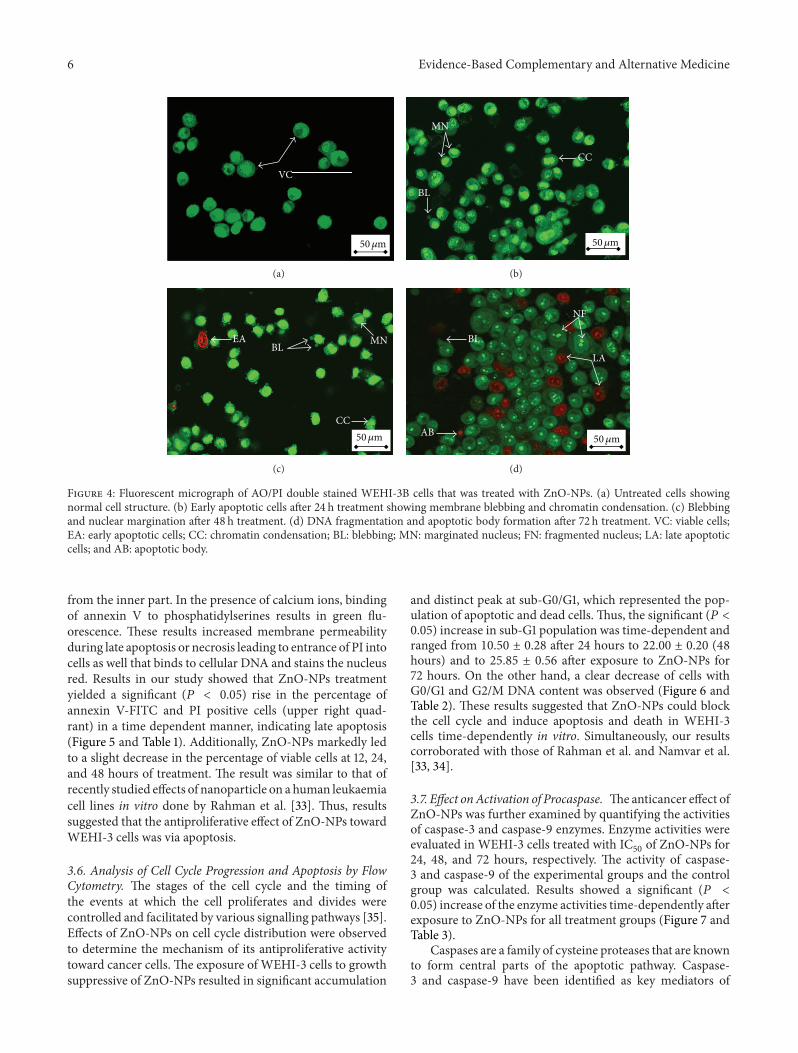
3.4. Morphological Changes Induced by ZnO-NPs in Leukae-miaCells. To confirmwhether the growth inhibition of ZnO-NPs was caused by apoptosis, annexin V-FITC/PI doublestaining was analyzed. After WEHI-3 cells were incubatedwithZnO-NPs for different timeperiods, it was demonstratedthat the death ofWEHI-3 cells indeed occurred via apoptosis.Thus, the treated cells were characterized by membraneblebbing, nuclear condensation, nuclear fragmentation, andapoptotic body formation.These abnormal cell features couldbe regarded as a morphological symbol of apoptosis [32].Simultaneously, there were no morphological changes foundin the control group. The results obtained in our studyindicated that ZnO-NPs initiated and provoked apoptosis inWEHI-3 cells (Figure 4).
Excluding of the PI and uptaking of AO by intact cellmembranes allowed double stranded DNA to show greenfluoresces under 488 nm excitation. On the other hand,apoptotic cells with condensed chromatin and affected cellmembrane, which resulted in clumps of intense green fluo-rescent spots within the cell. Similar results to this currentstudy were found by Rahman et al. and Namvar et al. [33, 34].
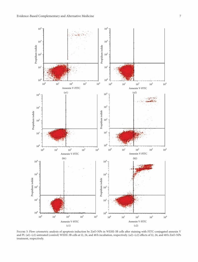
3.5. Effect on Annexin V-FITC Binding. The apoptotic effectof induction of ZnO-NPs was further confirmed by theevaluation of the number of apoptotic cells using flow cyto-metric analysis with the AV/PI double staining. Generally,an early event in apoptosis is started with translocation ofphosphatidylserine to the outer part of plasma membrane
6 Evidence-Based Complementary and Alternative Medicine
VC
50𝜇m
(a)
CC
BL
MN
50𝜇m
(b)
EABL MN
CC50𝜇m
(c)
AB
LA
BL
NF
50𝜇m
(d)
Figure 4: Fluorescent micrograph of AO/PI double stained WEHI-3B cells that was treated with ZnO-NPs. (a) Untreated cells showingnormal cell structure. (b) Early apoptotic cells after 24 h treatment showing membrane blebbing and chromatin condensation. (c) Blebbingand nuclear margination after 48 h treatment. (d) DNA fragmentation and apoptotic body formation after 72 h treatment. VC: viable cells;EA: early apoptotic cells; CC: chromatin condensation; BL: blebbing; MN: marginated nucleus; FN: fragmented nucleus; LA: late apoptoticcells; and AB: apoptotic body.
from the inner part. In the presence of calcium ions, bindingof annexin V to phosphatidylserines results in green flu-orescence. These results increased membrane permeabilityduring late apoptosis or necrosis leading to entrance of PI intocells as well that binds to cellular DNA and stains the nucleusred. Results in our study showed that ZnO-NPs treatmentyielded a significant (𝑃 < 0.05) rise in the percentage ofannexin V-FITC and PI positive cells (upper right quad-rant) in a time dependent manner, indicating late apoptosis(Figure 5 and Table 1). Additionally, ZnO-NPs markedly ledto a slight decrease in the percentage of viable cells at 12, 24,and 48 hours of treatment. The result was similar to that ofrecently studied effects of nanoparticle on a human leukaemiacell lines in vitro done by Rahman et al. [33]. Thus, resultssuggested that the antiproliferative effect of ZnO-NPs towardWEHI-3 cells was via apoptosis.
3.6. Analysis of Cell Cycle Progression and Apoptosis by FlowCytometry. The stages of the cell cycle and the timing ofthe events at which the cell proliferates and divides werecontrolled and facilitated by various signalling pathways [35].Effects of ZnO-NPs on cell cycle distribution were observedto determine the mechanism of its antiproliferative activitytoward cancer cells. The exposure of WEHI-3 cells to growthsuppressive of ZnO-NPs resulted in significant accumulation
and distinct peak at sub-G0/G1, which represented the pop-ulation of apoptotic and dead cells. Thus, the significant (𝑃 <0.05) increase in sub-G1 population was time-dependent andranged from 10.50 ± 0.28 after 24 hours to 22.00 ± 0.20 (48hours) and to 25.85 ± 0.56 after exposure to ZnO-NPs for72 hours. On the other hand, a clear decrease of cells withG0/G1 and G2/M DNA content was observed (Figure 6 andTable 2). These results suggested that ZnO-NPs could blockthe cell cycle and induce apoptosis and death in WEHI-3cells time-dependently in vitro. Simultaneously, our resultscorroborated with those of Rahman et al. and Namvar et al.[33, 34].
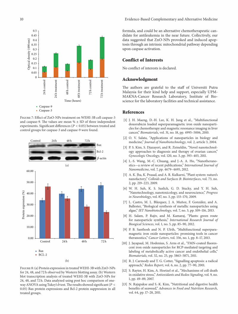
3.7. Effect onActivation of Procaspase. Theanticancer effect ofZnO-NPs was further examined by quantifying the activitiesof caspase-3 and caspase-9 enzymes. Enzyme activities wereevaluated in WEHI-3 cells treated with IC
50of ZnO-NPs for
24, 48, and 72 hours, respectively. The activity of caspase-3 and caspase-9 of the experimental groups and the controlgroup was calculated. Results showed a significant (𝑃 <0.05) increase of the enzyme activities time-dependently afterexposure to ZnO-NPs for all treatment groups (Figure 7 andTable 3).
Caspases are a family of cysteine proteases that are knownto form central parts of the apoptotic pathway. Caspase-3 and caspase-9 have been identified as key mediators of
Evidence-Based Complementary and Alternative Medicine 7
100 101 102 103 104100
101
102
103
104
Annexin V-FITC
Prop
idiu
m io
dide
100 101 102 103 104100
101
102
103
104
Annexin V-FITC
Prop
idiu
m io
dide
100 101 102 103 104100
101
102
103
104
Annexin V-FITC
Prop
idiu
m io
dide
100 101 102 103 104100
101
102
103
104
Annexin V-FITC
Prop
idiu
m io
dide
100 101 102 103 104100
101
102
103
104
Annexin V-FITC100 101 102 103 104
Annexin V-FITC
Prop
idiu
m io
dide
100
101
102
103
104
Prop
idiu
m io
dide
(a1) (a2)
(b1) (b2)
(c1) (c2)
Figure 5: Flow cytometric analysis of apoptosis induction by ZnO-NPs in WEHI-3B cells after staining with FITC-conjugated annexin Vand PI. (a1)–(c1) untreated (control) WEHI-3B cells at 12, 24, and 48 h incubation, respectively. (a2)–(c2) effects of 12, 24, and 48 h ZnO-NPstreatment, respectively.

8 Evidence-Based Complementary and Alternative Medicine
300
240
180
120
60
0
0 200 400 600
DNA content
Cou
nts
300
240
180
120
60
0
0 200 400 600
DNA content
Cou
nts
300
240
180
120
60
0
0 200 400 600
DNA content
Cou
nts
300
240
180
120
60
0
0 200 400 600
DNA content
Cou
nts
300
240
180
120
60
0
0 200 400 600
DNA content
Cou
nts
300
240
180
120
60
0
0 200 400 600
DNA content
Cou
nts
S
S
S S
S
S
(a1) (a2)
(b1) (b2)
(c1) (c2)
G0/G1
G0/G1
G0/G1
G0/G1
G0/G1
G0/G1G2 + M
G2 + M
G2 + M G2 + M
G2 + MG2 + M
Sub-G0/G1 Sub-G0/G1
Sub-G0/G1 Sub-G0/G1
Sub-G0/G1Sub-G0/G1
Figure 6: Cell cycle analysis of WEHI-3B cells treated with ZnO-NPs after staining with PI. (a1)–(c1) untreated WEHI-3B cells for 24, 48,and 72 h, respectively. (a2)–(c2) the effects of 24, 48, and 72 h, respectively, relative to exposure ofWEHI-3B cells to ZnO-NPs. G0/G1, G2/M,and S indicate the cell phase, and Sub-G0-G1 refers to the apoptotic cells.
apoptosis in mammalian cells. Their activities are consideredappropriate measures of cytotoxic responsiveness [36]. Thus,ZnO-NPs markedly activated an executioner caspase-3 andcaspase-9 in a time-response fashion, which is consistent
with the outcomes of other studies that have shown anumber of prepared nanoparticles induce apoptosis throughthe activation of procaspases and the mitochondrial intrinsicpathway [33].

Evidence-Based Complementary and Alternative Medicine 9
Table 1: Flow cytometric analysis ofWEHI-3B cells after treated with ZnO-NPs.The cells were stained with FITC-conjugated annexin V andPI and incubated at 37∘C for 12, 24, and 48 h, respectively.
Cells (%)Cellcondition
Control12 h
Treated12 h
Control24 h
Treated24 h
Control48 h
Treated48 h
Viable cells 96.4 ± 0.75 80.59 ± 0.65 92.33 ± 0.55 78.29 ± 0.15 90.78 ± 0.25 73.5 ± 0.13Early apoptosis 1.87 ± 0.15 8.75 ± 0.99∗ 2.98 ± 0.70 10.00 ± 0.30∗ 6.15 ± 0.45 15.5 ± 0.22∗
Late apoptosis necrosis 1.69 ± 0.35 10.66 ± 0.95∗∗ 4.69 ± 0.50 11.7 ± 0.80∗∗ 3.07 ± 0.25 10.9 ± 0.10∗∗
Values are expressed as mean ± SD of three different experiments. The data has been analyzed using post hoc comparison test one-way ANOVA; means werecompared by Tukey’s 𝑏 test. ∗Significant (𝑃 < 0.05) increased early apoptotic cells in ZnO-NPs treated groups compared to untreated controls. ∗∗Significant(𝑃 < 0.05) increased late apoptotic/necrotic cells in ZnO-NPs-treated groups compared to untreated controls.
Table 2: Flow cytometric analysis of WEHI-3B cells after treated with ZnO-NPs.The cells were stained with PI and incubated at 37∘C for 24,48, and 72 h.
Cells (%)Cell cyclephases
Control24 h
Treated24 h
Control48 h
Treated48 h
Control72 h
Treated72 h
G0/G1 55.25 ± 0.06 33.40 ± 0.45 51.10 ± 0.29 42.61 ± 0.52 50.64 ± 0.32 37.68 ± 0.68G2/M 18.85 ± 0.76 18.00 ± 0.41 19.16 ± 0.26 12.29 ± 0.35 9.30 ± 0.22 6.06 ± 0.93Synthesis 24.91 ± 0.06 38.30 ± 0.33 29.24 ± 0.06 22.93 ± 0.12 37.50 ± 0.61 30.35 ± 0.18Sub-G0/G1 0.9 ± 0.23 10.50 ± 0.28∗ 0.50 ± 0.34 22.00 ± 0.20∗ 2.20 ± 0.46 25.85 ± 0.56∗
Values are expressed as mean ± SD of three different experiments. The data has been analyzed using post hoc comparison test one-way ANOVA; meanscompared with Tukey’s 𝑏 test. ∗Significant (𝑃 < 0.05) increased cells in sub-G0/G1 phase in ZnO-NPs treated groups compared to untreated controls.
Table 3: Caspases spectrophotometric analysis of WEHI-3B cells after being treated with ZnO-NPs for 24, 48, and 72 h, respectively.
Cells %
Caspase Control24 h
Treated24 h
Control48 h
Treated48 h
Control72 h
Treated72 h
Caspase-3 0.059 ± 0.030 0.11 ± 0.01∗ 0.071 ± 0.051 0.16 ± 0.007∗ 0.09 ± 0.0032 0.2 ± 0.006∗
Caspase-9 0.06 ± 0.071 0.15 ± 0.003∗ 0.075 ± 0.001 0.18 ± 0.005∗ 0.099 ± 0.002 0.235 ± 0.0035∗
Values are expressed as mean ± SD of three different experiments. The data has been analyzed using post hoc comparison test one-way ANOVA; meanscompared with Tukey’s 𝑏 test. ∗Significant (𝑃 < 0.05) increase of apoptotic cells in ZnO-NPs-treated groups compared with untreated controls.
3.8. Effect on Apoptotic Proteins. To detect the extent of ZnO-NPs on apoptosis-regulating proteins, the expression levelsof both Bcl-2 and Bax proteins were investigated after incu-bation with ZnO-NPs in cultured WEHI-3 cells. Treatmentwith the ZnO-NPs significantly (𝑃 < 0.05) reduced thepercentage (downregulated) of Bcl-2 protein expression ina time-dependent manner. In contrast, it significantly (𝑃 <0.05) increased (upregulated) Bax expression (Figure 8).
Virtually, both proliferation and apoptosis are controlledby the mitochondrial pathway, which is mediated by theBcl-2 family protein. High-expression of Bcl-2 protein hasbeen found to protect cancer cells from apoptosis, whereaslow Bax protein expression promotes cancer cells to undergoapoptosis. This effect is of high importance due to thefact that downregulation of Bcl-2 is associated with bettertherapeutic outcomes [37].Themechanism of action of ZnO-NPs is unknown. However, different mechanisms of theanticarcinogenic activity of seaweed have been demonstrated.In addition, antiapoptotic activity of Bcl-2 has been foundto be provoked by a homologous Bax protein that is able to
form heterodimers with Bcl-2 [38, 39]. Thus, the ratio of Bcl-2 to Bax within the cell is the critical influential factor forthe propensity of a cell to undergo apoptosis. The currentstudy demonstrated thatWEHI-3-ZnO-NPs treatment led toa decrease in Bcl-2 expression and an increase in the level ofBax, suggesting disruption ofmitochondrialmembranes.Thedecreased Bcl-2 levels along with increased levels of Bax maybe sufficient to shift the balance toward apoptosis inWEHI-3cells.
4. Conclusion
Based on the observations in this study, it was concludedthat there was a time-dependent reduction of cell viabilityin treated cancer cells after exposure of ZnO-NPs, with noadverse effect on normal fibroblast cells. This provides newopportunities for the safe delivery and applications in anti-cancer therapy by ZnO-NPs. Moreover, ZnO-NPs demon-strated suppression in WEHI-3 cell growth and proliferationin vitro, which suggests that this is a potent and selective
10 Evidence-Based Complementary and Alternative Medicine
00.05
0.10.15
0.20.25
0.30.35
0.40.45
0.5
Time (hours)
Caspase-9Caspase-3
Opt
ical
den
sity
(490
nm)
24h
cont
rol
24h
treat
men
t
48
h co
ntro
l
48
h tre
atm
ent
72
h co
ntro
l
72
h tre
atm
ent
Figure 7: Effect of ZnO-NPs treatment onWEHI-3B cell caspase-3and caspase-9. The values are mean % ± SD of three independentexperiments. Significant differences (𝑃 < 0.05) between treated andcontrol groups for caspase-3 and caspase-9 were found.
Bax
Bcl-2
Control 24h 48h 72h
𝛽-actin
(a)
0.00
10.00
20.00
30.00
40.00
50.00
60.00
Control
Prot
ein
expr
essio
n (%
)
BaxBCL-2
24h 48h 72h
(b)
Figure 8: (a) Protein expression in treatedWEHI-3Bwith ZnO-NPsfor 24, 48, and 72 h observed byWestern blotting assay. (b) Westernblot transcription analysis of treated WEHI-3B with ZnO-NPs for24, 48, and 72 h. Data analyzed using post hoc comparison of one-wayANOVAusingTukey’s 𝑏 test.The results showed significant (𝑃 <0.05) Bax protein expressions and Bcl-2 protein suppression in alltreated groups.
formula, and could be an alternative chemotherapeutic can-didate for antileukemia in the near future. Collectively, ourdata suggested that ZnO-NPs provoked and induced apop-tosis through an intrinsic mitochondrial pathway dependingupon caspase activation.
Conflict of Interests
No conflict of interests is declared.
Acknowledgment
The authors are grateful to the staff of Universiti PutraMalaysia for their kind help and support, especially UPM-MAKNA-Cancer Research Laboratory, Institute of Bio-science for the laboratory facilities and technical assistance.
References
[1] J. H. Maeng, D.-H. Lee, K. H. Jung et al., “Multifunctionaldoxorubicin loaded superparamagnetic iron oxide nanoparti-cles for chemotherapy and magnetic resonance imaging in livercancer,” Biomaterials, vol. 31, no. 18, pp. 4995–5006, 2010.
[2] O. V. Salata, “Applications of nanoparticles in biology andmedicine,” Journal of Nanobiotechnology, vol. 2, article 3, 2004.
[3] P. S. Kim, S. Djazayeri, and R. Zeineldin, “Novel nanotechnol-ogy approaches to diagnosis and therapy of ovarian cancer,”Gynecologic Oncology, vol. 120, no. 3, pp. 393–403, 2011.
[4] L.-S. Wang, M.-C. Chuang, and J.-A. A. Ho, “Nanotherano-stics—a review of recent publications,” International Journal ofNanomedicine, vol. 7, pp. 4679–4695, 2012.
[5] A. K. Jha, K. Prasad, and A. R. Kulkarni, “Plant system: nature’snanofactory,” Colloids and Surfaces B: Biointerfaces, vol. 73, no.2, pp. 219–223, 2009.
[6] W. H. Suh, K. S. Suslick, G. D. Stucky, and Y. H. Suh,“Nanotechnology, nanotoxicology, and neuroscience,” Progressin Neurobiology, vol. 87, no. 3, pp. 133–170, 2009.
[7] L. Castro, M. L. Blazquez, J. A. Munoz, F. Gonzalez, and A.Ballester, “Biological synthesis of metallic nanoparticles usingalgae,” IET Nanobiotechnology, vol. 7, no. 3, pp. 109–116, 2013.
[8] H. Salam, P. Rajiv, and M. Kamaraj, “Plants: green routefor nanoparticle synthesis,” International Research Journal ofBioogical Sciences, vol. 1, no. 5, pp. 85–90, 2012.
[9] P. B. Santhosh and N. P. Ulrih, “Multifunctional superpara-magnetic iron oxide nanoparticles: promising tools in cancertheranostics,” Cancer Letters, vol. 336, no. 1, pp. 8–17, 2013.
[10] J. Jayapaul, M. Hodenius, S. Arns et al., “FMN-coated fluores-cent iron oxide nanoparticles for RCP-mediated targeting andlabeling of metabolically active cancer and endothelial cells,”Biomaterials, vol. 32, no. 25, pp. 5863–5871, 2011.
[11] R. J. Carmody and T. G. Cotter, “Signalling apoptosis: a radicalapproach,” Redox Report, vol. 6, no. 2, pp. 77–90, 2001.
[12] S. Rayter, H. Kim, A. Hoetzel et al., “Mechanisms of cell deathin oxidative stress,”Antioxidants and Redox Signaling, vol. 9, no.1, pp. 49–89, 2007.
[13] N. Rajapakse and S.-K. Kim, “Nutritional and digestive healthbenefits of seaweed,” Advances in Food and Nutrition Research,vol. 64, pp. 17–28, 2011.

Evidence-Based Complementary and Alternative Medicine 11
[14] F. Namvar, J. Baharara, and A. A. Mahdi, “Antioxidant andanticancer activities of selected persian gulf algae,” IndianJournal of Clinical Biochemistry, vol. 29, no. 1, pp. 13–20, 2014.
[15] F. Namvar, P. M. Tahir, R. Mohamad et al., “Biomedical proper-ties of edible seaweed in cancer therapy and chemopreventiontrials: a review,”Natural Product Communications, vol. 8, no. 12,pp. 1811–1820, 2013.
[16] F. Namvar, S. Mohamed, S. G. Fard et al., “Polyphenol-richseaweed (Eucheuma cottonii) extract suppresses breast tumourvia hormonemodulation and apoptosis induction,” Food Chem-istry, vol. 130, no. 2, pp. 376–382, 2012.
[17] M. N. A. Khan, J. S. Choi, M. C. Lee et al., “Anti-inflammatoryactivities of methanol extracts from various seaweed species,”Journal of Environmental Biology, vol. 29, no. 4, pp. 465–469,2008.
[18] A. W. Zuercher, R. Fritsche, B. Corthesy, and A. Mercenier,“Food products and allergy development, prevention and treat-ment,” Current Opinion in Biotechnology, vol. 17, no. 2, pp. 198–203, 2006.
[19] G. Perez, G. R. M. Zavala, S. M. Perez, and G. S. Perez, “Antidia-betic effect of compounds isolated from plants,” Phytomedicine,vol. 5, pp. 55–75, 1998.
[20] T. Nishino, A. Fukuda, T. Nagumo, M. Fujihara, and E. Kaji,“Inhibition of the generation of thrombin and factor Xa by afucoidan from the brown seaweed Ecklonia kurome,”Thrombo-sis Research, vol. 96, no. 1, pp. 37–49, 1999.
[21] K. Miyashita, “The carotenoid fucoxanthin from brown sea-weed affects obesity,” Lipid Technology, vol. 21, no. 8-9, pp. 186–190, 2009.
[22] S. Mohamed, S. N. Hashim, and H. A. Rahman, “Seaweeds: asustainable functional food for complementary and alternativetherapy,” Trends in Food Science & Technology, vol. 23, no. 2, pp.83–96, 2012.
[23] K. Wada, K. Nakamura, Y. Tamai et al., “Seaweed intake andblood pressure levels in healthy pre-school Japanese children,”Nutrition Journal, vol. 10, no. 1, article 83, 2011.
[24] F. Namvar, R. Mohamad, J. Baharara, S. Zafar-Balanejad, F.Fargahi, and H. S. Rahman, “Antioxidant, antiproliferative, andantiangiogenesis effects of polyphenol-rich seaweed (Sargassummuticum),” BioMed Research International, vol. 2013, Article ID604787, 9 pages, 2013.
[25] A. M. G. C. Dias, A. Hussain, A. S. Marcos, and A. C. A.Roque, “A biotechnological perspective on the application ofiron oxide magnetic colloids modified with polysaccharides,”Biotechnology Advances, vol. 29, no. 1, pp. 142–155, 2011.
[26] S. Azizi, M. B. Ahmad, F. Namvar, and R. Mohamad, “Greenbiosynthesis and characterization of zinc oxide nanoparticlesusing brown marine macroalga Sargassum muticum aqueousextract,”Materials Letters, vol. 116, pp. 275–277, 2014.
[27] A. Hague, G. D. Dıaz, D. J. Hicks, S. Krajewski, J. C. Reed, andC. Paraskeva, “bcl-2 and bak may play a pivotal role in sodiumbutyrate-induced apoptosis in colonic epithelial cells; howeveroverexpression of bcl-2 does not protect against bak-mediatedapoptosis,” International Journal of Cancer, vol. 72, no. 5, pp.898–905, 1997.
[28] S. Nagarajan and K. Arumugam Kuppusamy, “Extracellularsynthesis of zinc oxide nanoparticle using seaweeds of gulfof Mannar, India,” Journal of Nanobiotechnology, vol. 11, no. 1,article 39, 2013.
[29] A. Jegan and A. Ramasubbu, “Symtesis and charachterization ofzinc oxide agar nanocomposite,” International Journal of NanoDimension, vol. 2, no. 3, pp. 171–176, 2012.
[30] P. Price and T. J. McMillan, “Use of the tetrazolium assayin measuring the response of human tumor cells to ionizingradiation,” Cancer Research, vol. 50, no. 5, pp. 1392–1396, 1990.
[31] M. J. Akhtar, M. Ahamed, S. Kumar, M. A. Majeed Khan,J. Ahmad, and S. A. Alrokayan, “Zinc oxide nanoparticlesselectively induce apoptosis in human cancer cells throughreactive oxygen species,” International Journal of Nanomedicine,vol. 7, pp. 845–857, 2012.
[32] C. Fan, W. Wang, B. Zhao, S. Zhang, and J. Miao, “Chloroquineinhibits cell growth and induces cell death in A549 lung cancercells,” Bioorganic and Medicinal Chemistry, vol. 14, no. 9, pp.3218–3222, 2006.
[33] H. S. Rahman, A. Rasedee, A. B. Abdul et al., “Zerumbone-loaded nanostructured lipid carrier induces G2/M cell cyclearrest and apoptosis via mitochondrial pathway in a humanlymphoblastic leukemia cell line,” International Journal ofNanomedicine, vol. 9, no. 1, pp. 527–538, 2014.
[34] F. Namvar, H. S. Rahman, R. Mohamad et al., “Cytotoxic effectof magnetic iron oxide nanoparticles synthesized via seaweedaqueous extract,” International Journal of Nanomedicine, vol. 9,pp. 2479–2488, 2014.
[35] G. K. Schwartz and M. A. Shah, “Targeting the cell cycle: a newapproach to cancer therapy,” Journal of Clinical Oncology, vol.23, no. 36, pp. 9408–9421, 2005.
[36] B. J. Sonnemann, J. Gange, S. Pilz et al., “Comparative evaluationof the treatment efficacy of suberoylanilide hydroxamic acid(SAHA) and paclitaxel in ovarian cancer cell lines and primaryovarian cancer cells from patients,” BMC Cancer, vol. 6, article183, 2006.
[37] G. Gasparini, M. Barbareschi, C. Doglioni et al., “Expressionof bcl-2 protein predicts efficacy of adjuvant treatments ofoperable node-positive breast cancer,” Clinical Cancer Research,vol. 1, no. 2, pp. 189–198, 1995.
[38] J. C. Reed, “Mini-review: cellular mechanisms of disease series,”Journal of Cell Biology, vol. 124, pp. 1–6, 1994.
[39] P. A. Sandstrom, D. Pardi, P. W. Tebbey et al., “Lipid hydro-peroxide-induced apoptosis: lack of inhibition by Bcl-2 over-expression,” FEBS Letters, vol. 365, no. 1, pp. 66–70, 1995.

Submit your manuscripts athttp://www.hindawi.com
Stem CellsInternational
Hindawi Publishing Corporationhttp://www.hindawi.com Volume 2014
Hindawi Publishing Corporationhttp://www.hindawi.com Volume 2014
MEDIATORSINFLAMMATION
of
Hindawi Publishing Corporationhttp://www.hindawi.com Volume 2014
Behavioural Neurology
EndocrinologyInternational Journal of
Hindawi Publishing Corporationhttp://www.hindawi.com Volume 2014
Hindawi Publishing Corporationhttp://www.hindawi.com Volume 2014
Disease Markers
Hindawi Publishing Corporationhttp://www.hindawi.com Volume 2014
BioMed Research International
OncologyJournal of
Hindawi Publishing Corporationhttp://www.hindawi.com Volume 2014
Hindawi Publishing Corporationhttp://www.hindawi.com Volume 2014
Oxidative Medicine and Cellular Longevity
Hindawi Publishing Corporationhttp://www.hindawi.com Volume 2014
PPAR Research
The Scientific World JournalHindawi Publishing Corporation http://www.hindawi.com Volume 2014
Immunology ResearchHindawi Publishing Corporationhttp://www.hindawi.com Volume 2014
Journal of
ObesityJournal of
Hindawi Publishing Corporationhttp://www.hindawi.com Volume 2014
Hindawi Publishing Corporationhttp://www.hindawi.com Volume 2014
Computational and Mathematical Methods in Medicine
OphthalmologyJournal of
Hindawi Publishing Corporationhttp://www.hindawi.com Volume 2014
Diabetes ResearchJournal of
Hindawi Publishing Corporationhttp://www.hindawi.com Volume 2014
Hindawi Publishing Corporationhttp://www.hindawi.com Volume 2014
Research and TreatmentAIDS
Hindawi Publishing Corporationhttp://www.hindawi.com Volume 2014
Gastroenterology Research and Practice
Hindawi Publishing Corporationhttp://www.hindawi.com Volume 2014
Parkinson’s Disease
Evidence-Based Complementary and Alternative Medicine
Volume 2014Hindawi Publishing Corporationhttp://www.hindawi.com
Related Documents