933 Cytotoxic and proinflammatory effects of PVP-coated silver nanoparticles after intratracheal instillation in rats Nadine Haberl *1,2 , Stephanie Hirn 1,2 , Alexander Wenk 1 , Jörg Diendorf 3 , Matthias Epple 3 , Blair D. Johnston 1 , Fritz Krombach 4 , Wolfgang G. Kreyling 1,5 and Carsten Schleh 1,6 Full Research Paper Open Access Address: 1 Institute of Lung Biology and Disease, Helmholtz Center Munich, Neuherberg/Munich, Germany, 2 Current address: Walter Brendel Centre of Experimental Medicine, Ludwig-Maximilians-Universität München, Marchioninistr. 15, 81377 Munich, Germany, Phone: +49 89 2180 76540, Fax: +4949 89 2180 76532, 3 Inorganic Chemistry and Center of Nanointegration Duisburg-Essen, University of Duisburg-Essen, Essen, Germany, 4 Walter Brendel Centre of Experimental Medicine, Ludwig-Maximilians-Universität München, Munich, Germany, 5 Current address: Institute of Epidemiology 2, Helmholtz Center Munich, Neuherberg/Munich, Germany and 6 Current address: Berufsgenossenschaft Holz und Metall, Am Knie 8, 81241 München, Germany Email: Nadine Haberl * - [email protected] * Corresponding author Keywords: cytotoxicity; inflammation; pulmonary toxicity; silver nanoparticles Beilstein J. Nanotechnol. 2013, 4, 933–940. doi:10.3762/bjnano.4.105 Received: 19 July 2013 Accepted: 10 December 2013 Published: 19 December 2013 Guest Editor: R. Zellner © 2013 Haberl et al; licensee Beilstein-Institut. License and terms: see end of document. Abstract Silver nanoparticles (AgNP) are among the most promising nanomaterials, and their usage in medical applications and consumer products is growing rapidly. To evaluate possible adverse health effects, especially to the lungs, the current study focused on the cytotoxic and proinflammatory effects of AgNP after the intratracheal instillation in rats. Monodisperse, PVP-coated AgNP (70 nm) showing little agglomeration in aqueous suspension were instilled intratracheally. After 24 hours, the lungs were lavaged, and lactate dehydrogenase (LDH), total protein, and cytokine levels as well as total and differential cell counts were measured in the bronchoalveolar lavage fluid (BALF). Instillation of 50 μg PVP-AgNP did not result in elevated LDH, total protein, or cytokine levels in BALF compared to the control, whereas instillation of 250 μg PVP-AgNP caused a significant increase in LDH (1.9-fold) and total protein (1.3-fold) levels as well as in neutrophil numbers (60-fold) of BALF. Furthermore, while there was no change in BALF cytokine levels after the instillation of 50 μg PVP-AgNP, instillation of 250 μg PVP-AgNP resulted in significantly increased levels of seven out of eleven measured cytokines. These finding suggest that exposure to inhaled AgNP can induce moderate pulmonary toxicity, but only at rather high concentrations.

Welcome message from author
This document is posted to help you gain knowledge. Please leave a comment to let me know what you think about it! Share it to your friends and learn new things together.
Transcript

933
Cytotoxic and proinflammatory effects ofPVP-coated silver nanoparticles after
intratracheal instillation in ratsNadine Haberl*1,2, Stephanie Hirn1,2, Alexander Wenk1, Jörg Diendorf3,
Matthias Epple3, Blair D. Johnston1, Fritz Krombach4,Wolfgang G. Kreyling1,5 and Carsten Schleh1,6
Full Research Paper Open Access
Address:1Institute of Lung Biology and Disease, Helmholtz Center Munich,Neuherberg/Munich, Germany, 2Current address: Walter BrendelCentre of Experimental Medicine, Ludwig-Maximilians-UniversitätMünchen, Marchioninistr. 15, 81377 Munich, Germany, Phone: +4989 2180 76540, Fax: +4949 89 2180 76532, 3Inorganic Chemistry andCenter of Nanointegration Duisburg-Essen, University ofDuisburg-Essen, Essen, Germany, 4Walter Brendel Centre ofExperimental Medicine, Ludwig-Maximilians-Universität München,Munich, Germany, 5Current address: Institute of Epidemiology 2,Helmholtz Center Munich, Neuherberg/Munich, Germany and6Current address: Berufsgenossenschaft Holz und Metall, Am Knie 8,81241 München, Germany
Email:Nadine Haberl* - [email protected]
* Corresponding author
Keywords:cytotoxicity; inflammation; pulmonary toxicity; silver nanoparticles
Beilstein J. Nanotechnol. 2013, 4, 933–940.doi:10.3762/bjnano.4.105
Received: 19 July 2013Accepted: 10 December 2013Published: 19 December 2013
Guest Editor: R. Zellner
© 2013 Haberl et al; licensee Beilstein-Institut.License and terms: see end of document.
AbstractSilver nanoparticles (AgNP) are among the most promising nanomaterials, and their usage in medical applications and consumer
products is growing rapidly. To evaluate possible adverse health effects, especially to the lungs, the current study focused on the
cytotoxic and proinflammatory effects of AgNP after the intratracheal instillation in rats. Monodisperse, PVP-coated AgNP (70 nm)
showing little agglomeration in aqueous suspension were instilled intratracheally. After 24 hours, the lungs were lavaged, and
lactate dehydrogenase (LDH), total protein, and cytokine levels as well as total and differential cell counts were measured in the
bronchoalveolar lavage fluid (BALF). Instillation of 50 µg PVP-AgNP did not result in elevated LDH, total protein, or cytokine
levels in BALF compared to the control, whereas instillation of 250 µg PVP-AgNP caused a significant increase in LDH (1.9-fold)
and total protein (1.3-fold) levels as well as in neutrophil numbers (60-fold) of BALF. Furthermore, while there was no change in
BALF cytokine levels after the instillation of 50 µg PVP-AgNP, instillation of 250 µg PVP-AgNP resulted in significantly
increased levels of seven out of eleven measured cytokines. These finding suggest that exposure to inhaled AgNP can induce
moderate pulmonary toxicity, but only at rather high concentrations.

Beilstein J. Nanotechnol. 2013, 4, 933–940.
934
Beilstein J. Nanotechnol. 2013, 4, 933–940.
934
IntroductionSilver nanoparticles (AgNP) are among the most promising
nanomaterials, and their usage in medical applications and
consumer products is growing rapidly [1]. The antimicrobial
properties of AgNP render them useful as a component in
wound dressings or as coatings for catheters [2-5]. In addition,
they are used in deodorants or applied in textiles as a protection
against odor [6,7].
With regard to the use in hygiene and health care spray prod-
ucts, AgNP had become present in everyday life. In case of
such aerosolized AgNP, the lungs with their large surface area
are the first organs that come into contact with inhaled AgNP
[8,9].
Inhaled particles with a diameter of less than 100 nm mainly
deposit in the alveolar region [10-12]. Once deposited there,
nanoparticles are found to interact with the epithelial lining
fluid including pulmonary surfactant, lung macrophages and
epithelial cells [13-15]. Depending on their physico-chemical
properties, a small portion of the inhaled nanomaterials may
even be able to cross the air-blood-barrier (ABB), towards
circulation, and accumulate in secondary organs [16,17].
Some in vitro studies have demonstrated toxic effects of AgNP
on lung cells: In vitro incubation of a rat alveolar macrophage
cell line with AgNP induced a concentration- as well as a size-
dependent decrease in cell viability. In addition, a proinflamma-
tory response was shown by increased levels of tumor necrosis
factor-α (TNF-α), macrophage inflammatory protein-2 (MIP-2),
and interleukin-1β (IL-1β) [18]. Furthermore, AgNP caused
damage to mitochondria and an increased production of reac-
tive oxygen species (ROS) in human lung fibroblasts in a dose-
dependent manner [19]. In contrast, very little is known about
the cytotoxic and proinflammatory effects of AgNP in the respi-
ratory system in vivo [20]. Intratracheal instillation of slightly
agglomerated AgNP in mice resulted in progressively increased
levels of IL-1, TNF-α, and IL-6 by day 28 after a single instilla-
tion [21]. In another mouse study, however, only minimal lung
toxicity and inflammation were found after subacute inhalation
of AgNP [22]. In addition, there are only two in vivo studies
dealing with adverse pulmonary effects of AgNP in rats.
In these studies, the subchronic inhalation of AgNP caused lung
function changes as well as chronic alveolar inflammation and
small granulomatous lesions [23,24]. In two other studies from
the same group, however, acute and subchronic inhalation of
AgNP at lower doses and shorter inhalation times did not cause
adverse health effects in rats as measured by lung function,
hematology, and body weight chances [25,26].
The mechanisms of toxicity are proposed to be oxidative stress,
DNA damage, and the modulation of cytokine production [20].
In addition, Liu et al. showed that AgNP undergo profound
chemical transformations in biological environments that can
affect bioavailability and toxicity. In case of argyria, silver
deposits in the skin are not translocated engineered AgNP, but
rather secondary particles formed of silver metabolites resulting
from partial AgNP dissolution and subsequent metabolization
of silver ions. Thus, the dissolution of AgNP and release of
silver ions as well as the subsequent biochemical transforma-
tions are an important issue in AgNP toxicity [27]. However,
most of the information available about the mechanisms of
AgNP toxicity has been derived from in vitro studies. The aim
of the current study was to assess the adverse health effects of
AgNP in vivo, more specifically, after the intratracheal instilla-
tion in rats, with a focus on cytotoxicity and cytokine induction.
Therefore, monodisperse polyvinylpyrrolidone (PVP)-coated
AgNP (70 nm mean diameter) were instilled intratracheally into
healthy rats, and cytotoxic and proinflammatory effects were
determined by measuring lactate dehydrogenase (LDH),
protein, and cytokine levels as well as total and differential cell
counts in bronchoalveolar lavage fluid (BALF).
ResultsParticle characterizationThe mean diameter of the PVP-coated AgNP was 70 nm as
measured by electron microscopy (Figure 1). The z-average was
123 nm and the polydispersity index 0.18 as determined by
dynamic light scattering. The size distribution by number and
the particle intensity distribution are shown in Figure 2 and
Figure 3.
Figure 1: Representative transmission (A) and scanning electron (B)images of PVP-coated AgNP.
LDH and total protein levels in BALFThe stable enzyme LDH is localized in the cytoplasm of cells.
Consequently, the destruction of cell membranes results in an
increased LDH release from cells. This release due to the loss of

Beilstein J. Nanotechnol. 2013, 4, 933–940.
935
Figure 2: Particle number distribution of PVP-AgNP by dynamic light scattering (Nano Zetasizer ZS, Malvern, Herrenberg, Germany).
Figure 3: Particle intensity distribution of PVP-AgNP by dynamic lightscattering (Nano Zetasizer ZS, Malvern, Herrenberg, Germany).
membrane integrity is, therefore, a marker of cytotoxic effects.
As shown in Figure 4, there was no significant increase in LDH
levels in BALF after instillation of 50 µg PVP-AgNP into the
rat’s lung. However, instillation of 250 µg PVP-AgNP caused a
significant increase in LDH release as compared to controls
(0.19 ± 0.01 U/mL to 0.36 ± 0.05 U/mL).
Lung injury allows proteins to overcome the ABB, resulting in
increased BALF protein concentrations. Similar to the results
from the LDH measurements, there was no change in BALF
protein levels after the instillation of 50 µg of PVP-AgNP when
compared to the controls. However, the instillation of
250 µg PVP-AgNP caused a low but significant increase in
BALF protein levels (Figure 5).
Figure 4: LDH levels in BALF 24 hours after intratracheal instillation ofPVP-AgNP. Values are mean ± SEM; n = 5 for each treatment group;*p < 0.05 vs control.
Figure 5: Total protein levels in BALF 24 hours after the intratrachealinstillation of PVP-AgNP. Values are mean ± SEM; n = 5 for each treat-ment group; *p < 0.05 vs control.

Beilstein J. Nanotechnol. 2013, 4, 933–940.
936
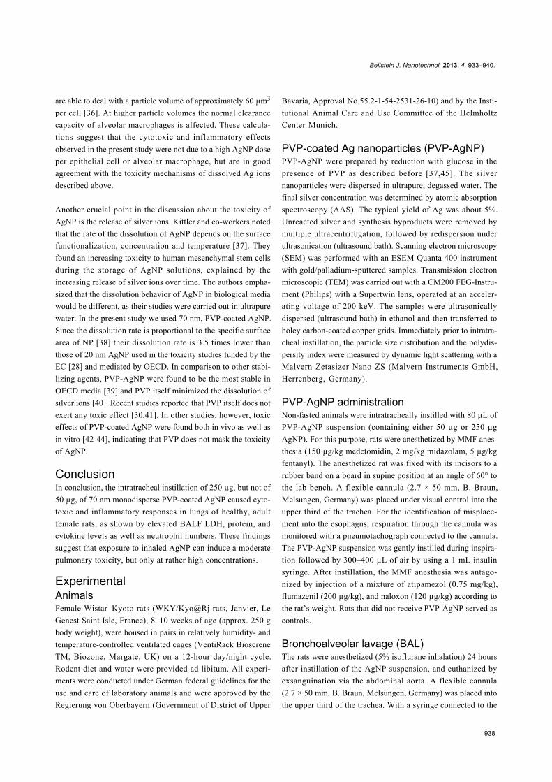
Figure 6: Cytokine levels in BALF 24 hours after the intratrachealinstillation of PVP-AgNP (A: proinflammatory cytokines; B:chemokines; C: colony-stimulating factors). For the lower limits ofdetection see “Experimental”. Values are mean ± SEM; n = 5 for eachtreatment group; *p < 0.05 vs control. UDL = under detection limit.
Cytokine levels in BALFTo assess the proinflammatory effects of PVP-AgNP, we deter-
mined the BALF levels of several cytokines and chemokines
after intratracheal instillation. Macrophage activators such as
IL-1α, IL-1β, IL-6, and IL-12p70 act as proinflammatory
cytokines. Figure 6A shows that the insti l lation of
250 µg PVP-AgNP caused an increase in the BALF levels of all
four cytokines as compared to controls that was significant for
IL-1β, IL-6, and IL-12p70. TNF-α, another proinflammatory
cytokine, was not detectable in BALF. Similarly, significantly
elevated levels of CINC-1 as well as of the macrophage inflam-
matory proteins 1-α and 2 (MIP-1α, MIP-2) were found after
the instillation of 250 µg (Figure 6B). Moreover, the instilla-
tion of 250 µg PVP-AgNP resulted also in a significantly
increased level of the macrophage-/colony stimulating factor
(M-CSF) as compared to controls (Figure 6C).
Cell counts in BALFLungs were lavaged 24 hours after the instillation of 50 or
250 µg PVP-AgNP and total as well as differential cell counts
were determined as described before. The pulmonary influx of
neutrophils was considered to be a marker of inflammation.
While there was no significant difference in total cell counts
between control rats and rats exposed to 50 µg PVP-AgNP, the
instillation of 250 µg PVP-AgNP resulted in a 2-fold increase in
BALF total cell counts. Furthermore, while the instillation of
50 µg PVP-AgNP caused a slight increase (17-fold) in the
number of neutrophils that did not reach statistical significance,
the instillation of 250 µg PVP-AgNP produced a significant
influx (60-fold) of neutrophils into the lungs (Figure 7).
Representative images of BAL cells show the presence of PVP-
AgNP both in alveolar macrophages and free particles/agglom-
erates in BALF after the instillation of 250 µg PVP-AgNP
(Figure 8).
Figure 7: Cell counts in BALF 24 hours after the instillation of PVP-AgNP. AM: Alveolar macrophages. Values are mean ± SEM; n = 5 foreach treatment group; *p < 0.05 vs control.
DiscussionAccording to the Woodrow–Wilson-Center database of
nanotechnology-based products [28], silver is one of the most
frequently used nanomaterials for consumer products. Due to

Beilstein J. Nanotechnol. 2013, 4, 933–940.
937
Figure 8: Representative BAL cell image after the intratracheal instilla-tion of 250 µg PVP-AgNP. Gray arrows indicate free particles; blackarrows indicate alveolar macrophages with internalized PVP-AgNP.
the use of AgNP as aerosols in healthcare and hygiene spray
products, the lungs are considered to be the main portal of entry
for AgNP into the human body [29]. With regard to the lack of
knowledge of the in vivo pulmonary toxicity of AgNP, the aim
of the current study was to assess the adverse health effects of
AgNP after the intratracheal instillation in rats, with a focus on
cytotoxicity and cytokine induction. Here, we demonstrate that
the intratracheal instillation of 250 µg, but not of 50 µg, of
monodisperse PVP-coated 70 nm AgNP in rats caused cyto-
toxic and inflammatory responses of the lungs, as shown by
elevated BALF LDH, protein, and cytokine levels as well as
neutrophil numbers.
These findings are in line with previous mouse studies: The
intratracheal instillation resulted in increased BALF levels of
IL-1, TNF-α, and IL-6 [21] and the inhalation caused increased
BALF levels of IL-12(p40) and keratinocyte chemoattractant
(KC) [22]. In contrast to the present study, cytotoxicity was not
assessed in the instillation study of Park and co-workers [21]
nor were found elevated BALF LHD and protein levels in the
inhalation study of Stebounova et al. [22]. In contrast to these
mouse studies, information about cytotoxic and inflammatory
effects after the inhalation of AgNP in rats is very scarce. Two
studies demonstrated adverse pulmonary effects such as lung
function changes as well as chronic alveolar inflammation and
small granulomatous lesions in histopathological examinations
[23,24]. But in contrast to our study, BALF cytokine levels
were not determined. In addition, the results after the inhalation
of AgNP in rats are diverging. In two other studies from the
same group, acute and subchronic inhalation of AgNP at lower
doses and shorter inhalation times did not cause adverse health
effects in rats as measured by lung function, hematology, and
body weight chances [25,26]. The lower doses used and the
shorter inhalation times in comparison to the studies where
adverse health effects occurred might be the reasons for the
diverging results. Unfortunately, direct pulmonary responses
such as LDH, protein, and cytokine levels in BALF were not
assessed in these studies.
Several in vitro studies dealt with the mechanism of cytokine
induction after AgNP exposure. Incubation of human
mesenchymal stem cells and of peripheral blood monocytes
with the same PVP-AgNP that were used in the present study
induced a concentration-dependent uptake of particles into the
cells and a subsequent release of the proinflammatory cytokines
IL-6 and IL-8 [30,31]. Moreover, Greulich et al. described the
internalization of AgNP to be a clathrin-mediated process [31].
The clathrin-mediated process of internalization after incuba-
tion of human macrophages with AgNP was stated to be a
trigger for immune responses [32] and responsible for the
production of IL-8 [33]. AshaRani and co-workers demon-
strated the involvement of the NFκB and MAP kinase path-
ways after exposure of human lung cells (IMR-90) to AgNP.
Consequently, the activation of these pathways resulted in the
transcription of genes involved in proliferation and inflamma-
tory responses. Likewise, an up-regulation of IL-6, IL-8,
M-CSF and MIP-1β following AgNP exposure was demon-
strated [34]. Since comparable proinflammatory responses were
found in the present study, we suggest that the inflammatory
reaction induced by the intratracheal instillation of 250 µg
AgNP was caused by similar mechanisms as those described
previously in the in vitro studies.
In the present study, adverse pulmonary effects occurred only
after the instillation of 250 µg PVP-AgNP. As the intratracheal
instillation of 50 µg silver nanoparticles did not result in cyto-
toxic or inflammatory effects, the rationale to use an additional
dose of 250 µg (1 mg/kg body weight) was to induce potential
toxic effects but not death or severe suffering to the animal. To
estimate whether the toxicity is driven by AgNP doses that are
so high that cells cannot deal with, we assessed cellular doses.
Although the intratracheal instillation will not lead to a uniform
distribution in the lungs, for simplicity we assume a nearly
uniform distribution of PVP-AgNP in the present study. As
Stone and co-workers [35] determined the total number of cells
in the alveolar region to be 8.9·108 in the rat’s lung, the average
concentration after the intratracheal instillation of 250 µg AgNP
is estimated to be 0.28 pg AgNP/cell corresponding to
150 AgNP per cell in the alveolar region. When presuming that
all AgNP would have been phagocytized by the 27·106 alveolar
macrophages in the rat lungs, each of the macrophages will
have received an average dose of 9.6 pg AgNP corresponding to
about 5.3·103 AgNP/cell and about 1 µm3 AgNP volume per
cell while the volume of a rat alveolar macrophage of 12 µm is
900 µm3. Tran and co-workers found that alveolar macrophages
Beilstein J. Nanotechnol. 2013, 4, 933–940.
938
are able to deal with a particle volume of approximately 60 µm3
per cell [36]. At higher particle volumes the normal clearance
capacity of alveolar macrophages is affected. These calcula-
tions suggest that the cytotoxic and inflammatory effects
observed in the present study were not due to a high AgNP dose
per epithelial cell or alveolar macrophage, but are in good
agreement with the toxicity mechanisms of dissolved Ag ions
described above.
Another crucial point in the discussion about the toxicity of
AgNP is the release of silver ions. Kittler and co-workers noted
that the rate of the dissolution of AgNP depends on the surface
functionalization, concentration and temperature [37]. They
found an increasing toxicity to human mesenchymal stem cells
during the storage of AgNP solutions, explained by the
increasing release of silver ions over time. The authors empha-
sized that the dissolution behavior of AgNP in biological media
would be different, as their studies were carried out in ultrapure
water. In the present study we used 70 nm, PVP-coated AgNP.
Since the dissolution rate is proportional to the specific surface
area of NP [38] their dissolution rate is 3.5 times lower than
those of 20 nm AgNP used in the toxicity studies funded by the
EC [28] and mediated by OECD. In comparison to other stabi-
lizing agents, PVP-AgNP were found to be the most stable in
OECD media [39] and PVP itself minimized the dissolution of
silver ions [40]. Recent studies reported that PVP itself does not
exert any toxic effect [30,41]. In other studies, however, toxic
effects of PVP-coated AgNP were found both in vivo as well as
in vitro [42-44], indicating that PVP does not mask the toxicity
of AgNP.
ConclusionIn conclusion, the intratracheal instillation of 250 µg, but not of
50 µg, of 70 nm monodisperse PVP-coated AgNP caused cyto-
toxic and inflammatory responses in lungs of healthy, adult
female rats, as shown by elevated BALF LDH, protein, and
cytokine levels as well as neutrophil numbers. These findings
suggest that exposure to inhaled AgNP can induce a moderate
pulmonary toxicity, but only at rather high concentrations.
ExperimentalAnimalsFemale Wistar–Kyoto rats (WKY/Kyo@Rj rats, Janvier, Le
Genest Saint Isle, France), 8–10 weeks of age (approx. 250 g
body weight), were housed in pairs in relatively humidity- and
temperature-controlled ventilated cages (VentiRack Bioscrene
TM, Biozone, Margate, UK) on a 12-hour day/night cycle.
Rodent diet and water were provided ad libitum. All experi-
ments were conducted under German federal guidelines for the
use and care of laboratory animals and were approved by the
Regierung von Oberbayern (Government of District of Upper
Bavaria, Approval No.55.2-1-54-2531-26-10) and by the Insti-
tutional Animal Care and Use Committee of the Helmholtz
Center Munich.
PVP-coated Ag nanoparticles (PVP-AgNP)PVP-AgNP were prepared by reduction with glucose in the
presence of PVP as described before [37,45]. The silver
nanoparticles were dispersed in ultrapure, degassed water. The
final silver concentration was determined by atomic absorption
spectroscopy (AAS). The typical yield of Ag was about 5%.
Unreacted silver and synthesis byproducts were removed by
multiple ultracentrifugation, followed by redispersion under
ultrasonication (ultrasound bath). Scanning electron microscopy
(SEM) was performed with an ESEM Quanta 400 instrument
with gold/palladium-sputtered samples. Transmission electron
microscopic (TEM) was carried out with a CM200 FEG-Instru-
ment (Philips) with a Supertwin lens, operated at an acceler-
ating voltage of 200 keV. The samples were ultrasonically
dispersed (ultrasound bath) in ethanol and then transferred to
holey carbon-coated copper grids. Immediately prior to intratra-
cheal instillation, the particle size distribution and the polydis-
persity index were measured by dynamic light scattering with a
Malvern Zetasizer Nano ZS (Malvern Instruments GmbH,
Herrenberg, Germany).
PVP-AgNP administrationNon-fasted animals were intratracheally instilled with 80 µL of
PVP-AgNP suspension (containing either 50 µg or 250 µg
AgNP). For this purpose, rats were anesthetized by MMF anes-
thesia (150 µg/kg medetomidin, 2 mg/kg midazolam, 5 µg/kg
fentanyl). The anesthetized rat was fixed with its incisors to a
rubber band on a board in supine position at an angle of 60° to
the lab bench. A flexible cannula (2.7 × 50 mm, B. Braun,
Melsungen, Germany) was placed under visual control into the
upper third of the trachea. For the identification of misplace-
ment into the esophagus, respiration through the cannula was
monitored with a pneumotachograph connected to the cannula.
The PVP-AgNP suspension was gently instilled during inspira-
tion followed by 300–400 µL of air by using a 1 mL insulin
syringe. After instillation, the MMF anesthesia was antago-
nized by injection of a mixture of atipamezol (0.75 mg/kg),
flumazenil (200 µg/kg), and naloxon (120 µg/kg) according to
the rat’s weight. Rats that did not receive PVP-AgNP served as
controls.
Bronchoalveolar lavage (BAL)The rats were anesthetized (5% isoflurane inhalation) 24 hours
after instillation of the AgNP suspension, and euthanized by
exsanguination via the abdominal aorta. A flexible cannula
(2.7 × 50 mm, B. Braun, Melsungen, Germany) was placed into
the upper third of the trachea. With a syringe connected to the

Beilstein J. Nanotechnol. 2013, 4, 933–940.
939
cannula, 5 mL PBS were gently instilled into the lungs, while
the thorax was massaged carefully. Then, the lavage fluid was
gently aspirated and the procedure was replicated four times.
The lavages were pooled and centrifuged at 400 g for 30 min at
4 °C. The cell free supernatant (BALF) was used for biochem-
ical measurements of LDH, total protein, and cytokine levels.
Determination of protein and LDH levels inBALFThe total protein content in the BALF supernatant was deter-
mined spectrophotometrically at a wavelength of 560 nm with
the Pierce® BCA Protein Kit Assay (Thermo scientific, Rock-
ford, USA). LDH activity was determined spectrophotometri-
cally at a wavelength of 490 nm with the Cytotoxicity Detec-
tion KitPLUS (LDH; Roche Diagnostics, Mannheim,
Germany). Both assays were performed according to the manu-
facturers’ instructions.
Quantification of TNF-α and CINC-1 levels inBALF by ELISATNF-α and cytokine-induced neutrophil chemoattractant-1
(CINC-1) were measured in BALF supernatant by using a
commercially available enzyme-linked immunosorbent assay
kit (ELISA DuoSet, R&D, Wiesbaden-Nordenstadt, Germany).
The ELISA was performed according to the manufacturer’s
specifications. Lower limits of detection were 30 pg/mL for
TNF-α and 15 pg/mL for CINC-1. Absorbance was determined
at a wavelength of 450 nm.
Measurement of cytokines and chemokinesin BALF by a multiplex bead array assayThe levels of nine relevant cytokines and chemokines, including
granulocyte-colony stimulating factor (G-CSF), granulocyte
macrophage-colony stimulating factor (GM-CSF), macrophage-
colony stimulating factor (M-CSF), interleukin (IL)-1α, IL-1β,
IL-6, IL-12p70, macrophage inflammatory protein (MIP)-1α,
and MIP-2, were determined in BALF supernatant. The
cytokine multiplex bead array assay kit was purchased from
BioRad (Bioplex cytokine assay, Munich, Germany). The kit
was used according to the manufacturer’s specifications. Lower
limits of detection were 16 pg/mL for G-CSF, 22 pg/mL for
GM-CSF, 95 pg/mL for M-CSF, 35 pg/mL for IL-1α,
116 pg/mL for IL-1β, 150 pg/mL for IL-6, 67 pg/mL for
IL-12p70, 96 pg/mL for MIP-1α, and 72 pg/mL for MIP-2.
Cytological analysis of BALThe remaining cell pellet was resuspended in 1 mL RPMI 1640
medium (BioChrome, Berlin, Germany) supplemented with
10% fetal calf serum (Seromed, Berlin, Germany). Cell counts
were measured by a CASY cell counter system (Schärfe System
GmbH, Reutlingen, Germany). Cytospins were prepared by
cytocentrifugation at 35g for 6 min and stained with
May–Grünwald–Giemsa. 200 cells per each slide were differen-
tially counted under a light microscope.
Statistical analysisData in the figures are given as mean ± SEM. Statistical
analysis was performed by one-way analysis of variance
(ANOVA) on ranks followed by a post hoc Tukey’s multiple
comparison test or Dunn’s test (Software: SigmaStat for
Windows, Jandel Scientific, Erkrath, Germany). Differences
between rats exposed to PVP-AgNP and controls were consid-
ered statistically significant at p < 0.05.
AcknowledgementsThe authors appreciate the technical assistance of Sebastian
Kaidel and Nadine Senger. This study was supported by the
Deutsche Forschungsgemeinschaft within the Priority Program
NanoBioResponses (SPP 1313).
References1. Chernousova, S.; Epple, M. Angew. Chem., Int. Ed. 2013, 52,
1636–1653. doi:10.1002/anie.2012059232. Fichtner, J.; Güresir, E.; Seifert, V.; Raabe, A. J. Neurosurg. 2010, 112,
840–846. doi:10.3171/2009.8.JNS0912973. Pollini, M.; Paladini, F.; Catalano, M.; Taurino, A.; Licciulli, A.;
Maffezzoli, A.; Sannino, A. J. Mater. Sci.: Mater. Med. 2011, 22,2005–2012. doi:10.1007/s10856-011-4380-x
4. Toy, L. W.; Macera, L. J. Am. Acad. Nurse Pract. 2011, 23, 183–192.doi:10.1111/j.1745-7599.2011.00600.x
5. Wiegand, C.; Heinze, T.; Hipler, U.-C. Wound Repair Regen. 2009, 17,511–521. doi:10.1111/j.1524-475X.2009.00503.x
6. Edwards-Jones, V. Lett. Appl. Microbiol. 2009, 49, 147–152.doi:10.1111/j.1472-765X.2009.02648.x
7. Walser, T.; Demou, E.; Lang, D. J.; Hellweg, S. Environ. Sci. Technol.2011, 45, 4570–4578. doi:10.1021/es2001248
8. Foss Hansen, S.; Maynard, A.; Baun, A.; Tickner, J. A.Nat. Nanotechnol. 2008, 3, 444–447. doi:10.1038/nnano.2008.198
9. Quadros, M. E.; Marr, L. C. Environ. Sci. Technol. 2011, 45,10713–10719. doi:10.1021/es202770m
10. Kreyling, W. G.; Möller, W.; Semmler-Behnke, M.; Oberdörster, G. InParticle Toxicology; Donaldson, K.; Borm, P., Eds.; CRC Press, Taylor& Francis Croup: Boca Raton, 2007; pp 47–74.
11. Kreyling, W. G.; Semmler-Behnke, M.; Moeller, W. In Nanomaterials –Toxicity, Health and Environmental Issues; Kumar, C., Ed.; Wiley-VCH:Weinheim, Germany, 2005; Vol. 5, pp 81–107.
12. Kreyling, W. G.; Semmler-Behnke, M.; Möller, W. J. Aerosol Med.2006, 19, 74–83. doi:10.1089/jam.2006.19.74
13. Mühlfeld, C.; Rothen-Rutishauser, B.; Blank, F.; Vanhecke, D.;Ochs, M.; Gehr, P. Am. J. Physiol. 2008, 294, L817–L829.doi:10.1152/ajplung.00442.2007
14. Schleh, C.; Hohlfeld, J. M. Inhalation Toxicol. 2009, 21 (Suppl. 1),97–103. doi:10.1080/08958370903005744
15. Schleh, C.; Mühlfeld, C.; Pulskamp, K.; Schmiedl, A.; Nassimi, M.;Lauenstein, H. D.; Braun, A.; Krug, N.; Erpenbeck, V. J.; Hohlfeld, J. M.Respir. Res. 2009, 10, No. 90. doi:10.1186/1465-9921-10-90

Beilstein J. Nanotechnol. 2013, 4, 933–940.
940
16. Geiser, M.; Kreyling, W. Part. Fibre Toxicol. 2010, 7, No. 2.doi:10.1186/1743-8977-7-2
17. Kreyling, W. G.; Semmler-Behnke, M.; Takenaka, S.; Möller, W.Acc. Chem. Res. 2013, 46, 714–722. doi:10.1021/ar300043r
18. Carlson, C.; Hussain, S. M.; Schrand, A. M.; Braydich-Stolle, L. K.;Hess, K. L.; Jones, R. L.; Schlager, J. J. J. Phys. Chem. B 2008, 112,13608–13619. doi:10.1021/jp712087m
19. AshaRani, P.; Low Kah, M.; Hande, M. P.; Valiyaveettil, S. ACS Nano2009, 3, 279–290. doi:10.1021/nn800596w
20. Stensberg, M. C.; Wei, Q.; McLamore, E. S.; Porterfield, D. M.; Wei, A.;Sepúlveda, M. S. Nanomedicine 2011, 6, 879–898.doi:10.2217/nnm.11.78
21. Park, E.-J.; Choi, K.; Park, K. Arch. Pharmacal Res. 2011, 34,299–307. doi:10.1007/s12272-011-0216-y
22. Stebounova, L. V.; Adamcakova-Dodd, A.; Kim, J. S.; Park, H.;O'Shaughnessy, P. T.; Grassian, V. H.; Thorne, P. S.Part. Fibre Toxicol. 2011, 8, No. 5. doi:10.1186/1743-8977-8-5
23. Sung, J. H.; Ji, J. H.; Park, J. D.; Yoon, J. U.; Kim, D. S.; Jeon, K. S.;Song, M. Y.; Jeong, J.; Han, B. S.; Han, J. H.; Chung, Y. H.;Chang, H. K.; Lee, J. H.; Cho, M. H.; Kelman, B. J.; Yu, I. J.Toxicol. Sci. 2009, 108, 452–461. doi:10.1093/toxsci/kfn246
24. Sung, J. H.; Ji, J. H.; Yoon, J. U.; Kim, D. S.; Song, M. Y.; Jeong, J.;Han, B. S.; Han, J. H.; Chung, Y. H.; Kim, J.; Kim, T. S.; Chang, H. K.;Lee, E. J.; Lee, J. H.; Yu, I. J. Inhalation Toxicol. 2008, 20, 567–574.doi:10.1080/08958370701874671
25. Ji, J. H.; Jung, J. H.; Kim, S. S.; Yoon, J. U.; Park, J. D.; Choi, B. S.;Chung, Y. H.; Kwon, I. H.; Jeong, J.; Han, B. S.; Shin, J. H.;Sung, J. H.; Song, K. S.; Yu, I. J. Inhalation Toxicol. 2007, 19,857–871. doi:10.1080/08958370701432108
26. Sung, J. H.; Ji, J. H.; Song, K. S.; Lee, J. H.; Choi, K. H.; Lee, S. H.;Yu, I. J. Toxicol. Ind. Health 2011, 27, 149–154.doi:10.1177/0748233710382540
27. Liu, J.; Wang, Z.; Liu, F. D.; Kane, A. B.; Hurt, R. H. ACS Nano 2012,6, 9887–9899. doi:10.1021/nn303449n
28. Johnston, H. J.; Hutchison, G.; Christensen, F. M.; Peters, S.;Hankin, S.; Stone, V. Crit. Rev. Toxicol. 2010, 40, 328–346.doi:10.3109/10408440903453074
29. Müller, L.; Gasser, M.; Raemy, D. O.; Herzog, F.; Brandenberger, C.;Schmid, O.; Gehr, P.; Rothen-Rutishauser, B.; Clift, M. J. Insci. J. 2011,1, 30–64. doi:10.5640/insc.010130
30. Greulich, C.; Diendorf, J.; Geßmann, J.; Simon, T.; Habijan, T.;Eggeler, G.; Schildhauer, T. A.; Epple, M.; Köller, M. Acta Biomater.2011, 7, 3505–3514. doi:10.1016/j.actbio.2011.05.030
31. Greulich, C.; Diendorf, J.; Simon, T.; Eggeler, G.; Epple, M.; Köller, M.Acta Biomater. 2010, 7, 347–354. doi:10.1016/j.actbio.2010.08.003
32. Park, J.; Lim, D.-H.; Lim, H.-J.; Kwon, T.; Choi, J.-s.; Jeong, S.;Choi, I.-H.; Cheon, J. Chem. Commun. 2011, 47, 4382–4384.doi:10.1039/c1cc10357a
33. Kim, S.; Choi, I.-H. Yonsei Med. J. 2012, 53, 654–657.doi:10.3349/ymj.2012.53.3.654
34. AshaRani, P. V.; Sethu, S.; Lim, H. K.; Balaji, G.; Valiyaveettil, S.;Hande, M. P. Genome Integr. 2012, 3, No. 2.doi:10.1186/2041-9414-3-2
35. Stone, K. C.; Mercer, R. R.; Gehr, P.; Stockstill, B.; Crapo, J. D.Am. J. Respir. Cell Mol. Biol. 1992, 6, 235–243.doi:10.1165/ajrcmb/6.2.235
36. Tran, C. L.; Buchanan, D.; Cullen, R. T.; Searl, A.; Jones, A. D.;Donaldson, K. Inhalation Toxicol. 2000, 12, 1113–1126.doi:10.1080/08958370050166796
37. Kittler, S.; Greulich, C.; Diendorf, J.; Köller, M.; Epple, M. Chem. Mater.2010, 22, 4548–4554. doi:10.1021/cm100023p
38. Kreyling, W. G. J. Aerosol Sci. 1990, 21, 371–374.doi:10.1016/0021-8502(90)90061-2
39. Tejamaya, M.; Römer, I.; Merrifield, R. C.; Lead, J. R.Environ. Sci. Technol. 2012, 46, 7011–7017. doi:10.1021/es2038596
40. Bryaskova, R.; Pencheva, D.; Nikolov, S.; Kantardjiev, T.J. Chem. Biol. 2011, 4, 185–191. doi:10.1007/s12154-011-0063-9
41. El Badawy, A. M.; Silva, R. G.; Morris, B.; Scheckel, K. G.;Suidan, M. T.; Tolaymat, T. M. Environ. Sci. Technol. 2011, 45,283–287. doi:10.1021/es1034188
42. Bilberg, K.; Hovgaard, M. B.; Besenbacher, F.; Baatrup, E. J. Toxicol.2012, No. 293784. doi:10.1155/2012/293784
43. George, S.; Lin, S.; Ji, Z.; Thomas, C. R.; Li, L.; Mecklenburg, M.;Meng, H.; Wang, X.; Zhang, H.; Xia, T.; Hohman, J. N.; Lin, S.;Zink, J. I.; Weiss, P. S.; Nel, A. E. ACS Nano 2012, 6, 3745–3759.doi:10.1021/nn204671v
44. Greulich, C.; Kittler, S.; Epple, M.; Muhr, G.; Köller, M.Langenbeck's Archives of Surgery 2009, 394, 495–502.doi:10.1007/s00423-009-0472-1
45. Wang, H.; Qiao, X.; Chen, J.; Ding, S. Colloids Surf., A 2005, 256,111–115. doi:10.1016/j.colsurfa.2004.12.058
License and TermsThis is an Open Access article under the terms of the
Creative Commons Attribution License
(http://creativecommons.org/licenses/by/2.0), which
permits unrestricted use, distribution, and reproduction in
any medium, provided the original work is properly cited.
The license is subject to the Beilstein Journal of
Nanotechnology terms and conditions:
(http://www.beilstein-journals.org/bjnano)
The definitive version of this article is the electronic one
which can be found at:
doi:10.3762/bjnano.4.105
Related Documents