Research Article Cytoskeleton Modifications and Autophagy Induction in TCam-2 Seminoma Cells Exposed to Simulated Microgravity Francesca Ferranti, 1,2 Maria Caruso, 2 Marcella Cammarota, 3 Maria Grazia Masiello, 4,5 Katia Corano Scheri, 2 Cinzia Fabrizi, 2 Lorenzo Fumagalli, 2 Chiara Schiraldi, 3 Alessandra Cucina, 5,6 Angela Catizone, 2 and Giulia Ricci 3 1 Italian Space Agency (ASI), Via del Politecnico snc, 00133 Rome, Italy 2 Department of Anatomy, Histology, Forensic Medicine and Orthopedics, Sapienza University of Rome, Viale Regina Elena 336, 00161 Rome, Italy 3 Department of Experimental Medicine, Second University of Naples, Via Santa Maria di Costantinopoli 16, 80138 Naples, Italy 4 Department of Clinical and Molecular Medicine, Sapienza University of Rome, Viale Regina Elena 291, 00161 Rome, Italy 5 Systems Biology Group, Sapienza University of Rome, Via A. Scarpa 16, 00161 Rome, Italy 6 Department of Surgery “Pietro Valdoni,” Sapienza University of Rome, Viale del Policlinico 155, 00161 Rome, Italy Correspondence should be addressed to Giulia Ricci; [email protected] Received 12 May 2014; Revised 4 July 2014; Accepted 4 July 2014; Published 17 July 2014 Academic Editor: Mariano Bizzarri Copyright © 2014 Francesca Ferranti et al. is is an open access article distributed under the Creative Commons Attribution License, which permits unrestricted use, distribution, and reproduction in any medium, provided the original work is properly cited. e study of how mechanical forces may influence cell behavior via cytoskeleton remodeling is a relevant challenge of nowadays that may allow us to define the relationship between mechanics and biochemistry and to address the larger problem of biological complexity. An increasing amount of literature data reported that microgravity condition alters cell architecture as a consequence of cytoskeleton structure modifications. Herein, we are reporting the morphological, cytoskeletal, and behavioral modifications due to the exposition of a seminoma cell line (TCam-2) to simulated microgravity. Even if no differences in cell proliferation and apoptosis were observed aſter 24 hours of exposure to simulated microgravity, scanning electron microscopy (SEM) analysis revealed that the change of gravity vector significantly affects TCam-2 cell surface morphological appearance. Consistent with this observation, we found that microtubule orientation is altered by microgravity. Moreover, the confocal analysis of actin microfilaments revealed an increase in the cell width induced by the low gravitational force. Microtubules and microfilaments have been related to autophagy modulation and, interestingly, we found a significant autophagic induction in TCam-2 cells exposed to simulated microgravity. is observation is of relevant interest because it shows, for the first time, TCam-2 cell autophagy as a biological response induced by a mechanical stimulus instead of a biochemical one. 1. Introduction An increasing number of experimental observations have demonstrated that tissue homeostasis could be strongly influenced and regulated by physical forces, such as the modulation of gravity vector. In the recent years, many efforts have been made to elucidate the effect of microgravity on cell behavior, and accumulating data show that micrograv- ity alters, permanently or transiently, important biological processes such as mitosis, differentiation, survival, cell mor- phology, and gene expression profiles [1–7]. However, how cells sense these signals and convert them into a biochemical response remains an important question that needs to be addressed. Recent studies have focused on the cytoskeleton as initial gravity sensor [1, 8]. Cytoskeleton plays important roles in cell physiology being responsible for chromosomal segregation during mitosis, providing a mechanical support to dividing cells, contributing to maintain cell shape and Hindawi Publishing Corporation BioMed Research International Volume 2014, Article ID 904396, 14 pages http://dx.doi.org/10.1155/2014/904396

Welcome message from author
This document is posted to help you gain knowledge. Please leave a comment to let me know what you think about it! Share it to your friends and learn new things together.
Transcript

Research ArticleCytoskeleton Modifications and Autophagy Induction inTCam-2 Seminoma Cells Exposed to Simulated Microgravity
Francesca Ferranti,1,2 Maria Caruso,2 Marcella Cammarota,3
Maria Grazia Masiello,4,5 Katia Corano Scheri,2 Cinzia Fabrizi,2 Lorenzo Fumagalli,2
Chiara Schiraldi,3 Alessandra Cucina,5,6 Angela Catizone,2 and Giulia Ricci3
1 Italian Space Agency (ASI), Via del Politecnico snc, 00133 Rome, Italy2 Department of Anatomy, Histology, Forensic Medicine and Orthopedics, Sapienza University of Rome,Viale Regina Elena 336, 00161 Rome, Italy
3 Department of Experimental Medicine, Second University of Naples, Via Santa Maria di Costantinopoli 16, 80138 Naples, Italy4Department of Clinical and Molecular Medicine, Sapienza University of Rome, Viale Regina Elena 291, 00161 Rome, Italy5 Systems Biology Group, Sapienza University of Rome, Via A. Scarpa 16, 00161 Rome, Italy6Department of Surgery “Pietro Valdoni,” Sapienza University of Rome, Viale del Policlinico 155, 00161 Rome, Italy
Correspondence should be addressed to Giulia Ricci; [email protected]
Received 12 May 2014; Revised 4 July 2014; Accepted 4 July 2014; Published 17 July 2014
Academic Editor: Mariano Bizzarri
Copyright © 2014 Francesca Ferranti et al. This is an open access article distributed under the Creative Commons AttributionLicense, which permits unrestricted use, distribution, and reproduction in any medium, provided the original work is properlycited.
The study of how mechanical forces may influence cell behavior via cytoskeleton remodeling is a relevant challenge of nowadaysthat may allow us to define the relationship between mechanics and biochemistry and to address the larger problem of biologicalcomplexity. An increasing amount of literature data reported thatmicrogravity condition alters cell architecture as a consequence ofcytoskeleton structuremodifications. Herein, we are reporting themorphological, cytoskeletal, and behavioral modifications due tothe exposition of a seminoma cell line (TCam-2) to simulatedmicrogravity. Even if no differences in cell proliferation and apoptosiswere observed after 24 hours of exposure to simulatedmicrogravity, scanning electronmicroscopy (SEM) analysis revealed that thechange of gravity vector significantly affects TCam-2 cell surface morphological appearance. Consistent with this observation, wefound that microtubule orientation is altered by microgravity. Moreover, the confocal analysis of actin microfilaments revealed anincrease in the cell width induced by the low gravitational force. Microtubules and microfilaments have been related to autophagymodulation and, interestingly, we found a significant autophagic induction in TCam-2 cells exposed to simulated microgravity.This observation is of relevant interest because it shows, for the first time, TCam-2 cell autophagy as a biological response inducedby a mechanical stimulus instead of a biochemical one.
1. Introduction
An increasing number of experimental observations havedemonstrated that tissue homeostasis could be stronglyinfluenced and regulated by physical forces, such as themodulation of gravity vector. In the recent years, many effortshave been made to elucidate the effect of microgravity oncell behavior, and accumulating data show that micrograv-ity alters, permanently or transiently, important biological
processes such as mitosis, differentiation, survival, cell mor-phology, and gene expression profiles [1–7]. However, howcells sense these signals and convert them into a biochemicalresponse remains an important question that needs to beaddressed. Recent studies have focused on the cytoskeletonas initial gravity sensor [1, 8]. Cytoskeleton plays importantroles in cell physiology being responsible for chromosomalsegregation during mitosis, providing a mechanical supportto dividing cells, contributing to maintain cell shape and
Hindawi Publishing CorporationBioMed Research InternationalVolume 2014, Article ID 904396, 14 pageshttp://dx.doi.org/10.1155/2014/904396

2 BioMed Research International
spatially organizing cell proteins and organelles in cell cyto-plasm. Moreover, cytoskeleton is involved in cell motility,membrane trafficking, signal transduction, and cell adhesion.In addition, cytoskeletal proteins can transduce and amplifymembrane receptor-captured signals, transmitting the infor-mation to the nucleus and finally regulating gene expression[2, 9, 10]. Considering all these observations, it appearseasy to understand why cytoskeleton disorganization couldcompromise a lot of cell functions leading, in some cases, tocell death. It is well known that microgravity exposure couldstrongly influence cytoskeleton organization [10–17] and itis commonly accepted that cellular tensegrity alteration inmicrogravity exposed cells could explain, at least in part, theconversion of a mechanical cue into a biological response.In this regard, recent studies have revealed the importanceof cytoskeletal integrity, such as F-actin and microtubules,in the physiological specific aspects of autophagy, and somepapers described the capability of microgravity to induceautophagy in living cells [18–22]. Autophagy is an importanthousekeeping physiological process that is involved in cellu-lar remodeling during development, elimination of aberrantorganelles, or misfolded proteins and in the recycling ofunnecessary cellular components to compensate for thelimitation of nutrients during starvation. It is of interestingnotice that this biological process is highly conserved fromyeast to mammals. Despite several studies suggested a tumorsuppressive role for autophagy, other reports support thehypothesis that this process is instead exploited by cancercells to prime their proliferation and promote their survival[23–27].
Microgravity condition is a stressful change in the physi-cal microenvironment for living cells; however, they seem tobe able to adapt to this change of gravitational force sincein the major part of studies available in the literature, thebehavioral cellular modifications induced by microgravityare transient. This observation has led to the intriguinghypothesis that cells, in response to gravity changes, reacttriggering adaptive biological processes and autophagy couldbe one of them.
Testicular cells appear to be sensitive to microgravity: ithas been demonstrated, in fact, that testicular function isimpaired by microgravity exposure [28–34]. Moreover, somein vitro observations revealed that microgravity influencescell proliferation, apoptosis, and testosterone secretion oftesticular organ cultures [35, 36]. In addition, microgravitycondition has differentiating effect in cultured spermatocytesand influences germ cell survival [37, 38]. This effect onmale germ cell lineage has triggered the hypothesis that alsotesticular cancer germ cells could be altered by microgravitycondition. For this reason, we decided to study the effect ofmicrogravity on TCam-2 cells that are the only accreditedseminoma cell line [39–42]. These cells have been recentlycharacterized at molecular and biochemical level [43–51]and thus represent a good tool to investigate male germcell behavior modification in response to a mechanical forcemodification. In this paper we report, for the first time,cytoskeletal modifications and the activation of autophagicprocess induced by acute exposure to microgravity in TCam-2 cell line.
2. Materials and Methods
2.1. Random Positioning Machine. The random positioningmachine (RPM; desktop RPM, Dutch Space, Leiden, theNetherlands), we used in the investigation, is a particularkind of 3D clinostat. It consists of two independently rotatingframes. One frame is positioned inside the other givinga very complex net change of orientation to a biologicalsample mounted in the middle. The degree of microgravitysimulation depends on angular speed and on the inclinationof the disk. These tools do not actually eliminate the gravitybut it is a microweight simulator based on the principleof “gravity-vector averaging”: it allows you to apply a 1 gstimulus omnidirectionally rather than unidirectionally andthe sum of the gravitational force vectors tends to zero.Effects generated by the RPM are comparable to those ofthe real microgravity, provided that the direction changes arefaster than the response time of the system to gravity field.The desktop RPM we used has been positioned within anincubator (for maintaining temperature, CO
2, and humidity
levels) and connected to the control console through standardelectric cables.
2.2. TCam-2 Cell Cultures. The TCam-2 human cell line wasderived in 1993 from a primary testicular tumor sample ofpure classical seminoma [42]. TCam-2 cells were culturedin RPMI 1640 (Lonza) supplemented with 10% fetal bovineserum (FBS, Lonza) and penicillin/streptomycin (Invitrogen)at 37∘C in a humidified atmosphere with 5% carbon dioxide[41]. The time 0 plating cell density is 3 × 10⁴/cm2. Asdescribed in the paragraph above, microgravity conditionwas simulated using the randompositioningmachine (RPM).Experiments were performed on cells cultured for 24 and 48hours at 1 g or in RPM, after additional 24 hours of preplatingon glass slides or IBIDImicroscopy chambers (IBIDI, 80826).Glass slides were silicone fixed to the culture dishes at least 48hours before plating. Cell culture dishes, in both 1 g and RPMculture conditions, were completely filled with fresh culturemedium in order to avoid air bubbles and to minimize liquidflow, thus making negligible the effects of both buoyancy andshear stress during rotation.
2.3. Proliferation, Apoptosis, and Autophagy Quantification.Cells cultured at 1 g or under microgravity conditions (asdescribed above) were fixed in 4% paraformaldehyde (PFA)in phosphate buffered saline (PBS) 1X for 10 minutes at 4∘Cand permeabilized with 1% bovine serum albumin (BSA),0.1%-Triton X-100 in PBS 1X for 1 hour at room temperature(RT). Nonspecific antibody binding was blocked with glycine1M pH 8.8 and with 1% BSA, 0.1% Triton X-100, and 5%donkey serum (Jackson ImmunoResearch Laboratories) inPBS 1X. Cells were incubated overnight (ON) in PBS 1Xadded with 1% BSA/0.1% Triton X-100 at 4∘C with thefollowing primary antibodies: anticleaved Caspase-3 (CellSignaling, rabbit polyclonal #9661, 1 : 200 dilution), anti-p-histone H3 (Santa Cruz Biotechnology, mouse monoclonalsc-374669, 1 : 50 dilution), or anti-LC3 (Sigma-Aldrich, L75431 : 120 dilution). After rinsing, samples were incubated with

BioMed Research International 3
the opportune secondary antibody (FITC-conjugated donkeyanti-rabbit 711-095-152 or donkey anti-mouse 715-095-150IgG, Jackson ImmunoResearch Laboratories, 1 : 200 dilution)in PBS 1X for 90min at RT. In negative controls primaryantibody was omitted. After secondary antibody incubation,samples were washed and mounted in buffered glycerol(0.1M, pH 9.5). All experiments were performed at least intriplicate.
For proliferation and apoptosis analyses, samples werephotographed with a Zeiss fluorescence microscope (Axio-scope) and positive cells were counted. For LC3 immunolo-calization a Leica confocal microscope (Laser Scanning TCSSP2) equipped with Ar/ArKr and He/Ne lasers was used.Images were acquired utilizing the Leica confocal software.The laser line was at 488 nm for FITC excitation. Theimages were scanned under a 20x objective or 40x oilimmersion objective. In order to get a quantitative analysis offluorescence, optical spatial series, each composed of 23/25optical sections with a step size of 2𝜇m, were performed inareas in which cells reached confluence both in nonrotatedand in RPM cultured samples. The fluorescence intensitywas determined by the Leica confocal software, using thefollowing parameters: the maximum amplitude of fluores-cence (MAX Amplitude), the sum of intensity (SUM (I)),and the mean amplitude of fluorescence intensity (MEAN(A)), of LC3 positive areas. The MAX Amplitude representsthe maximum intensity of fluorescence of each series. TheSUM (I) represents the total amount of fluorescence intensityrecovered within the entire 𝑧-axis of each series. The MEAN(A) represents the arithmeticalmean of fluorescence intensityrecovered within the entire 𝑧-axis of each series. We analyzed12 equivalent sized regions (regions of interest (ROI)) for eachexperiment both in 1 g and in RPM culture conditions (36total ROI for each experimental condition).
2.4. Western Blotting of LC3 Autophagy Marker. Cells cul-tured at 1 g and in RPM condition for 24 and 48 hourswere lysed in RIPA buffer (Sigma-Aldrich). Samples werethen clarified by centrifugation at 10000 rpm for 10min.Equivalent amount of protein (10 𝜇g) from each samplewas electrophoretically resolved on 12.5% precast SDS-polyacrylamide gels (ExcelGel, GE Healthcare Biosciences)using horizontal apparatus (Pharmacia Biotech, Uppsala,Sweden). Then, separated proteins were electrotransferredonto nitrocellulose membranes (Schleicher & Schuell) by asemidry system (Novablot, Pharmacia Biotech). Membraneswere blocked with 3% nonfat milk in PBS and then wereincubated (ON at 4∘C) with the LC3B monoclonal antibody(1 : 2000; Sigma). After extensive washing with PBS contain-ing 0.1% tween-20 (TBST), blots were incubated with 1 : 2000dilution of HRP-conjugated secondary antibody (AmershamBiosciences) for 1 hour at RT. Immunopositive bands weredetected with a chemiluminescence’s detection system (GEHealthcare Biosciences). To check for equal loading of thegel, membranes were stripped and reprobedwithmouse anti-𝛽-actin antibody (1 : 20000, Sigma) and with anti-GAPDHantibody (1 : 1000, Cell Signalling Technology). Densitomet-ric analysis was performed with the Quantity One software(BioRad Laboratories).
2.5. F-Actin and Tubulin Distribution Pattern. For F-actinvisualization Rhodamine Phalloidin (Invitrogen MolecularProbes Eugene, 1 : 40 dilution) was used. Cells were fixed in4% paraformaldehyde (PFA) in PBS for 10 minutes at 4∘Cand then permeabilized with cold ethanol : Acetone 1 : 1 for10 minutes at 4∘C. After rinsing, cells were incubated withRhodamine Phalloidin for 25min in the dark. Cells were thenwashed in PBS and mounted in buffered glycerol (0.1M, pH9.5).
Cell height analysis (𝑧-axis) was performed using theconfocal microscope already described (Leica IRE SP2, LaserScanningTCS SP2) equippedwithAr/ArKr andHe/Ne lasers.Images of the optical sections were acquired using the Leicaconfocal software. The Laser Line was at 543 nm for TRITCexcitation. Images were scanned under a 40x oil objective. Inorder to evaluate cell height three different experiments wereperformedusing cells cultured 1 g and inRPMconditions. Foreach experiment 4/5 optical spatial series with a step size of2 𝜇mwere recovered and a total of at least 80 optical sectionswere analyzed for each experimental condition. Cell heightof the examined samples was calculated using Leica confocalsoftware.
For microtubules localization immunofluorescenceexperiments, using anti-𝛼-tubulin (Biomeda, mousemonoclonal V10178, 1 : 75 dilution) as primary antibody,were performed. The protocol used is the same alreadydescribed in the paragraph above. Donkey anti-mouse(715-095-150 IgG, Jackson ImmunoResearch Laboratories,1 : 200 dilution), as secondary antibody, was used. Sampleswere then observed using both fluorescence microscope(Axioscope, Zeiss) and confocal microscope (Leica).
2.6. Scanning Electron Microscopy. Samples were fixed inGlutaraldehyde 2.5% in cacodylate buffer 0.1M pH 7.3 ONand then postfixed with 1% osmium tetroxide in cacodylatebuffer 1M, dehydrated with increasing ethanol percentage(30–90% in water for 5min, twice 100% for 15min), treatedin Critical Point Dryer (EMITECH K850), sputter coatedwith platinum-palladium (Denton Vacuum DESKV), andobserved with Supra 40 FESEM (Zeiss).
2.7. Statistical Analysis. All experiments were performed atleast in triplicate. All quantitative data are presented as themean value ± standard error (SEM). Student’s 𝑡-test andANOVA test for multigroup comparison were carried out,when appropriate, to evaluate the significance of differences.The significance level was fixed at a 𝑃 value < 0.05.
3. Results and Discussion
3.1. Microgravity Does Not Affect TCam-2 Cell Proliferationand Apoptosis. Microgravity exposure is known to influencecell proliferation and apoptosis in normal and cancer cells[52]. In order to asses proliferation rate of TCam-2 seminomacells, maintained at 1 g or in RPM culture conditions for 24and 48 hours, we performed immunofluorescence analysesof the M-phase marker p-histone H3. We observed that,actually, this acute microgravity exposure does not affect

4 BioMed Research International
the number of mitotic cells at all the culture times considered(Figure 1). Literature data have demonstrated that TCam-2cells do not have a high proliferation rate (58 hours doublingtime) when compared with JKT1 (27 hours doubling time),that is, another germ cell tumor cell line [40]. Since thepercentage of proliferating cells we expect in the time frameof 24 and 48 hours is not high, we can hypothesize that thisaltered gravitational stimulus is not long enough to determinea modification of cell proliferation in this particular cell line.Interestingly after 48 hours of culture the number of mitoticcells decreases significantly, in a similar amount, both in 1 gand in RPM cultured samples (Figure 1), indicating that cellproliferation, in this particular cell line, starts to be inhibitedby cell-to-cell contact even if these cells are cancer cells. It hasto be noticed that we chose to plate cells at high density inorder to let them attach each other before the RPM exposureand react, thanks to their tensional forces, to the changes ofgravitational field. Due to the high density of plating, at theend of the longer culture time we analyzed, cell culture dishesare crowded of cells so it appears not possible to prolongmore the culture without detaching and replate cells. To thisregard it is fair to say that we cannot exclude that TCam-2 cellproliferationmight be altered by RPM exposure if they wouldhave been cultured at a different density.
To test whether microgravity would be able to modifyTCam-2 cell apoptosis, we performed immunofluorescencesfor the active fragment of the apoptosis marker Caspase-3.We found that the change of gravity vector does not affect thenumber of apoptotic cells after 24 hours of culture (Figure 2).However, it has to be noticed that, after 48 hours of culture,the number of apoptotic cells increases significantly in theRPM cultured samples, even if the large majority of cellsappear to tolerate this mechanical stress (Figure 2) and tosurvive. The latter observation indicates that a small part ofTCam-2 cells appears more sensible to the change of gravityvector, when the mechanical stimulus is prolonged a bit,but this sensibility does not seem related to mechanical cellstability because, due to the high density of plating, all cellsare stably attached to each other and to the substrate. Inaddition, apoptotic cells are observable uniformly dispersedin the culture dish. On the basis of this observation, wehypothesized that TCam-2 cells need to trigger rescue pro-cesses that let them survive after a prolonged change of gravityvector. Possibly, rescue processes are not correctly inducedor exploited by the whole population of TCam-2 cells andthis hypothesis may explain why a small percentage of themappears not able to survive to the change of gravity. Thechange of physical forces is sensed by the cells through theircytoskeleton components and one of the first features thatreveal a cytoskeletal modification is the change in the plasmamembrane morphology. We studied first membrane surfaceand cytoskeletal modifications, due to RPM exposure, to besure the TCam-2 cells are able to sense and modify theirshape in response to this mechanical stress. Then we evalu-ated, in the same culture conditions, the autophagic processmodulation in response to RPM exposure, since autophagyis the most known biological rescue mechanism that let cellto change rapidly and survive to suddenmicroenvironmentalchanges.
3.2. Microgravity Strongly Influences TCam-2 Cell MembraneSurface. To study if the alteration of the mechanical forcesacting on TCam-2 cells during microgravity simulation maymodify cell membrane surface morphology, samples wereanalyzed by scanning electron microscopy. We observed thepresence of two morphologically distinguishable cell popula-tions in the 1 g cultured samples: one has smooth membranesurface and the other one is characterized by the presence ofmembrane expansions morphologically similar to microvilli(Figure 3). Noteworthy, we found that microgravity stronglyaffects membrane surface appearance after 24 hours ofculture: microvilli appeared collapsed and the differencesbetween the two cell populations are less evident (Figure 3).It is of interesting notice that cell microvilli are considered tobe an important site of mechanotransduction both in sensoryspecialized cells and not-sensory cells [53]. After 48 hours ofculture the membrane surface differences appear recoveredand microvilli-like structures appear reconstituted in RPMcultured samples (Figure 3). On the basis of these obser-vations, we hypothesized that cell mechanosensor-systemwas transiently altered by RPM exposure and this stronglysuggested the occurrence of cytoskeleton remodeling due toan acute exposure to gravitational vector change.
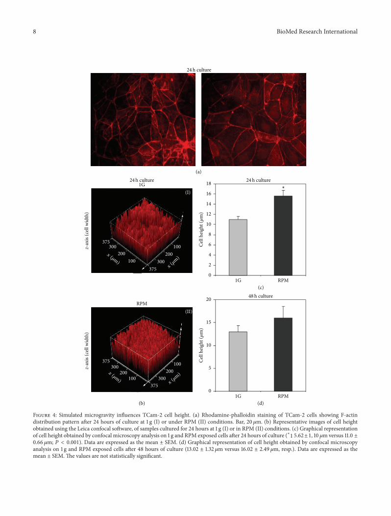
3.3. Microgravity Induces TCam-2 Cytoskeleton Remodeling.A huge amount of literature data demonstrated that micro-gravity is able to influence cell cytoskeletal architecture, pro-moting cell morphofunctional alterations [54]. In the light ofthese observations and on the basis of our scanning electronmicroscopy data, we decided to evaluate the possible effectsof simulated microgravity on TCam-2 microfilament andmicrotubule organization. Herein, we report microfilamentdistribution pattern analyzed by F-actin staining of TCam-2 cells cultured at 1 g or in RPM culture conditions. Even ifno apparent significant alterations in the actin cytoskeletonorganization were found both in 24 (Figure 4(a)) and 48hours of culture (not shown), a more detailed analysis byconfocal microscopy using Leica confocal software allowedus to evaluate cell height (cell 𝑧-axis) (Figures 4(b), 4(c),and 4(d)) in all the considered experimental conditions. Weobserved that simulated microgravity significantly increasesTCam-2 cell height after 24 hours of RPM exposure withrespect to 1 g cultured cells (15.62 ± 1.10 𝜇m versus 11.0 ±0.66 𝜇m; 𝑃 < 0.001) indicating that RPM culture conditionwas able to modify TCam-2 cell shape. Noteworthy, after48 hours of culture the differences in cell height in 1 gand RPM cultured cells are no more statistically significant(Figure 4(d)), indicating that TCam-2 cells are able to recoverrapidly after the exposure to this mechanical stress.The latterobservation appears consistent with the reported recovery ofsurface membrane microvilli-like structures after 48 hours ofRPM exposure (Figure 3).
Microtubule distribution pattern was studied by anti-𝛼-tubulin immunofluorescence staining. After 24 hours ofculture, we observed that microtubule distribution is alteredinTCam-2 cells exposed toRPMculture condition: centriolarpolarization is much less visible in these samples and micro-tubules appear to be distributed in an apparently random

BioMed Research International 5
p-H
iston
e H3
posit
ive c
ells
(%)
0
1
2
3
4
5
6
7
1GRPM
aa
bb
(h)24 48
(a)
(I) (II)
(b)
Figure 1: RPM exposure does not influence TCam-2 cell proliferation. (a) Graphical representation of the percentage of proliferating cells(p-histone H3 positive cells) at 24 and 48 hours of culture. No differences were observed between TCam-2 cells cultured at 1 g or in RPMculture conditions. Data are expressed as the mean ± SEM. Same letters indicate no statistical difference. Different letters indicate 𝑃 < 0.05.(b) Representative images of TCam-2 cells cultured for 24 hours at 1 g (I) and in RPM condition (II) after p-histone H3 immunofluorescence.Bar, 50 𝜇m.
manner within the cells (Figure 5). Microtubules are keyregulators of membrane trafficking; organelle distributioninside the cells and together with actin microfilaments seemsto regulate autophagosome formation [55–57]. In additionit is of interesting notice that LC3, the marker protein ofthe autophagic process, is a microtubule associated protein(MAP). As well as actin filaments, after 48 hours of culturethe microtubule distribution pattern appears recovered inRPM exposed samples since it is not possible to observesignificant differences between 1 g and RPM cultured cells.These observations again clearly indicate the capability ofTCam-2 cell to sense the change of physical forces in theirmicroenvironment and also to recover rapidly from thisphysical stress. These data strongly suggest the trigger ofrescue mechanisms due to TCam-2 RPM exposure.
It is worth mentioning that the reported microtubulealteration does not appear to significantly alter the properformation of the mitotic spindle (Figure 5(g) white box).
This observation is consistent with the results reported inFigure 1 in which we observed that TCam-2 cell proliferationdoes not appear to be affected by RPM exposure.
3.4. Microgravity Induces TCam-2 Cell Autophagy. Somepapers in the literature reported that, in other cellular sys-tems, microgravity is involved in autophagy induction [18–20] and, as previously stated, cytoskeleton plays importantroles in autophagy regulation [22]. In particular, in mam-mals, microtubules appear to be involved in the fusion ofautophagosome with late endosome and to bind and trans-port autophagosomes, once terminally completed.The role ofactin filaments on mammalian autophagy process regulationis still a matter of debate, but it is worth mentioning thatmicrofilaments depolymerization agents are able to blockautophagosome formation.
TCam-2 cells cultured at 1 g and in RPM conditionswere immunostained to detect the autophagic marker LC3.

6 BioMed Research International
123456789
10
Clea
ved
casp
ase-
3 po
sitiv
e cel
ls (%
)
aa a
b
0
1GRPM
(h)24 48
(a)
(II)
(IV)
(I)
(III)
(b)
Figure 2: RPM exposure and TCam-2 cell apoptosis. (a) Graphical representation of the percentage of apoptotic cell number (anticleavedCaspase-3 positive cells). No differences were observed between TCam-2 cells cultured for 24 hours at 1 g or in RPM culture conditions. Onthe contrary a slight increase in apoptotic cell percentage is observed after 48 hours of culture. Data are expressed as the mean ± SEM. Sameletters indicate no statistical difference. Different letters indicate 𝑃 < 0.01. (b) Representative images of 1 g (I, III) and RPM (II, IV) exposedTCam-2 cells in 24 (I, II) and 48 (III, IV) hours of culture after cleaved Caspase-3 immunofluorescence. Bar: 50𝜇m (I and II); 35 𝜇m (III andIV).

BioMed Research International 7
RPM 24h1G 24h
RPM 48h
(a)
(b)
(c)
(d)
(e)
(f)
(g)
(h)
1G 48h
(i)
(l)
(m)
(n)
∗
∗
∗
Figure 3: Microgravity effect on TCam-2 cell membrane surface. Scanning electron microscopy pictures with increasing magnificationshowing cell membrane surfacemorphology of TCam-2 cells cultured for 24 (a, b, c, and d) and 48 hours (i, l) at 1 g or for 24 (e, f, g, and h) and48 hours (m, n) inRPMculture conditions. In (a)white asterisks indicate TCam-2 cells with smoothmembrane surfacewhile the other TCam-2 cells of the same image are characterized by the presence of microvilli-like structures. In (b) the boundary between one smooth membraneand one microvilli membrane presenting cells is reported. (c) and (d) represent higher magnifications of the microvilli-like structures ofTCam-2 cells maintained at 1 g. In (e), (f), (g), and (h) it is well evident that, in RPM cultured cells, membrane surface is more similar in allthe cells and it is difficult to clearly identify the two cell populations. In particular in (h) it is possible to observe that microvilli-like structuresappeared collapsed in RPM exposed TCam-2 cells. The morphological appearance of cell surface (i, m) and microvilli-like structures (l, n)appeared indistinguishable in 1 g (i, l) and RPM exposed cells (m, n) after 48 hours of culture.
8 BioMed Research International
(I)
100200
300375
(I) (II)
RPM
0
5
10
15
20
1G RPM
48h culture
Cell
hei
ght (𝜇
m)
z-ax
is (c
ell w
idth
)z-
axis
(cel
l wid
th)
(I)(I)(I)(I) (II((II((IIII((( ))))))
1G
24h culture
24h culture
0
2
4
6
8
10
12
14
16
18 24h culture
1G RPM
Cell
hei
ght (𝜇
m)
(a)
(b)
(c)
(d)
∗
100200
300375
x (𝜇m)x
(𝜇m)
100200
300375
100200
300375
(II)
x (𝜇m)x
(𝜇m)
Figure 4: Simulated microgravity influences TCam-2 cell height. (a) Rhodamine-phalloidin staining of TCam-2 cells showing F-actindistribution pattern after 24 hours of culture at 1 g (I) or under RPM (II) conditions. Bar, 20𝜇m. (b) Representative images of cell heightobtained using the Leica confocal software, of samples cultured for 24 hours at 1 g (I) or in RPM (II) conditions. (c) Graphical representationof cell height obtained by confocal microscopy analysis on 1 g and RPM exposed cells after 24 hours of culture (∗1 5.62±1, 10𝜇m versus 11.0 ±0.66 𝜇m; 𝑃 < 0.001). Data are expressed as the mean ± SEM. (d) Graphical representation of cell height obtained by confocal microscopyanalysis on 1 g and RPM exposed cells after 48 hours of culture (13.02 ± 1.32𝜇m versus 16.02 ± 2.49 𝜇m, resp.). Data are expressed as themean ± SEM.The values are not statistically significant.

BioMed Research International 9
1G 24h
(a)
RPM 48h1G 48h
(b)
(c)
(g)
(h)
RPM 24h
(d)
(e)
(f)
(i)
(l)
Figure 5: Microtubule distribution pattern in TCam-2 cells exposed to simulated microgravity. Immunodetection of 𝛼-tubulin in TCam-2cells cultured for 24 hours (a, b, c, d, e, and f) and 48 hours (g, h, i, and l) at 1 g (a, b, c, g, and h) or under RPM conditions (d, e, f, i, and l). Inimages (g) and (i), in the white box, representative images of mitotic spindles are also shown. Bar, 20𝜇m.

10 BioMed Research International
As shown in Figures 6(a)(II) and 6(a)(IV), LC3 is detectableboth in 1 g and in RPM cultured samples and it ismainly localized in cytoplasmic vesicles. Interestingly, thenumber of these LC3 positive vesicles appears stronglyincreased in TCam-2 cells exposed to microgravity con-ditions (Figure 6(a)(IV)) with respect to 1 g cultured cells(Figure 6(a)(II)) after 24 hours of culture.Moreover, a quanti-tative analysis, carried out using the Leica confocal software,allows us to quantify the fluorescence intensity increase ofLC3 stained cells exposed to simulated microgravity (Figures6(b) and 6(c)). In particular, Figure 6(b) shows a stackprofile of 12 regions of interest (ROI) of a representativeexperiment both in 1 g (I) and in RPM cultured samples (II).The two groups of peaks reported in this figure representthe Max amplitude of fluorescence detected by the confocalmicroscope from the beginning to the end of the sample (total𝑧-axis). It is well evident thatMax amplitude of fluorescence isincreased in simulated RPM exposed samples. We evaluatedalso the SUM (I) and the MEAN (A) of fluorescence.Consistent with the data reported in Figure 6(b), we observedalso an increase of both the SUM (I) and the MEAN (A) inRPM cultured cells after 24 hours of culture (Figure 6(c)).According to the described confocal quantitative analyses,western blots performed with the anti-LC3 antibody showedthat, besides the increase of LC3-I protein amount, LC3-II (the LC3 active isoform) protein content is increased inRPM with respect to 1 g cultured samples (Figure 7). Sameresults were obtained normalizing the LC3 bands versus 𝛽-actin (Figure 7) and versus GAPDH signal (not shown).Autophagy induction is a naturally transient process: thisphenomenon is called autophagic flux [58], since, when itworks, autophagy protein machinery has to be degraded vialysosomes or proteasome together with the portion of the cellthat needs to be eliminated. On the contrary, when autophagyis blocked, the autophagy protein machinery is not degradedand is maintained at high level in the cytoplasm. In oursamples, after 48 hours of culture autophagy active proteinLC3-II, together with LC3-I, appears quantitatively similar in1 g and RPM cultured cells, demonstrating that autophagy isrestored at the same level with respect to 1 g culture condition.Same results were obtained normalizing the LC3 bands versus𝛽-actin (Figure 7) and versus GAPDH signal (not shown).Consistent with this observation, the LC3 cytoplasmic fluo-rescence is lowered in the RPM exposed cells demonstratingthat autophagy was not blocked by this mechanical stress(Figure 6(a)(VI)). It has to be mentioned that LC3-II proteinis present at basal level at 24 and 48 hours of culture aswell as cytoplasmic LC3 dots, even in cells cultured at 1 g,indicating that autophagy is a housekeeping process thatworks inTCam-2 cells even in control samples and suggestingthat this cancer cell line may exploit autophagy as a survivalmechanism.
There is a common agreement indicating that thereis a relationship between autophagy and apoptosis: whenautophagy is not able to rescue cell frommicroenvironmentalchanges, apoptotic process is triggered. On the light of thistheory we might interpret the small increase in the apoptoticindex at 48 hours of culture in RPM cultured samples(Figure 2) as the autophagy efficiency threshold or the limit
of autophagy efficiency in the rescue of cell survival aftermechanical stress exposure.
All together these qualitative and quantitative analysesallow us to conclude that microgravity is able to positivelymodulate the autophagic process in TCam-2 seminomacell line. Autophagy induced in TCam-2 cells by Estrogenexposure through ER𝛽 activation was recently reported [59].Herein we reported, for the first time, autophagy induced inTCam-2 cells by a mechanical cue (or, more precisely, by aremoval of a mechanical stimulus) instead of a biochemicalone. The analysis of the autophagy related pathways inducedby RPM exposure and the direct role of microtubules andmicrofilaments in this process, as well as the other possiblebiological meanings of RPM induced TCam-2 autophagy,deserves further investigations.
4. Conclusions
Gravitational biology could be considered part ofmechanobi-ology, the science that investigates the impact of forces onliving organisms. At cellular level, cytoskeleton elements arelikely candidates for force sensing and transduction pro-cesses. These biomechanical properties of cell cytoskeletonexplain the capability to propagate a mechanical stimulusover long distances in living tissues and represent the basisof the intriguing hypothesis that many, if not all, reportedchanges in ion fluxes, protein phosphorylation, membranepotential changes, and so forth are indeed provoked by amechanical modification somewhere within the cell or onits membrane [60, 61]. This paper is in line with this theoryand adds experimental data supporting the importance ofmechanotransduction and cell behavior. In this paper, in fact,we reported the effects of the exposure to changes of gravityvector on TCam-2 seminoma cells. In this experimentalmodel, simulated microgravity is able to induce TCam-2 cellsurfacemodifications andmicrovilli-like structure alteration.Moreover, microtubules and microfilaments organizationresult to be influenced by microgravity: (a) TCam-2 cellsshow actin cytoskeleton remodeling and cell height increase;(b) centriolar polarization becomes much less visible in thesesamples and microtubules appear to be distributed in anapparent random manner within the cells. All these modi-fications appear to be transient, indicating that cells modifytheir cytoskeletal components in response to gravitationalforce change, but that are also able to recover their shapewhen the gravitational change is prolonged. Interestingly,RPM exposure is able to induce TCam-2 cell autophagy.The latter observation allows us to hypothesize that TCam-2 cells are able to rapidly respond to acute exposure tomicrogravity, inducing adaptive biological processes suchas autophagy, that probably allow them to survive in thechanging physical microenvironment. Since autophagy isconsidered a biological survival mechanism the apoptosisinduction in a small percentage of TCam-2 cells after 48 hoursof culture might be speculated as the limit in the efficiencyof this survival process. All together these data provideevidences of TCam-2 sensitivity to changes of gravitationalforce direction and lay the groundwork to further studies onTCam-2 cell autophagy and its biological meaning.

BioMed Research International 11
(I) (III)
(IV) (VI)(II)
(V)
(a)
(𝜇m)
90
80
70
60
50
40
30
20
10
5 10 15 20 25 30 35 40
(I)Max amplitude
(𝜇m)
90
80
70
60
50
40
30
20
10
5 10 15 20 25 30 35 40
(II)Max amplitude
(b)
16
14
12
10
8
6
4
2
01G RPM
SUM
(I) (
a.u.)
∗
35
30
25
20
15
10
5
01G RPM
Mea
n(A
) (a.u
.)
∗
(c)
Figure 6: Autophagy induction in TCam-2 cells exposed to microgravity. (a) Immunodetection of LC3 in TCam-2 cells cultured for 24 hoursat 1 g (II) or under RPM (IV) conditions. In VI LC3 immunodetection of TCam-2 cells cultured in RPM condition for 48 hours is reported. InI, III, and V the respective bright fields are shown. (b) Stack profile of 24 hours of culture representative experiment showing the maximumamplitude (MAXAmplitude) of fluorescence in 12 regions of interest (ROI), randomly drawn in an area in which the cells reached confluence,in nonrotated (I) and RPM cultured samples (II). It is evident an increase of maximum amplitude of fluorescence in microgravity exposedsamples (II) with respect to the 1 g-cultured cells (I). (c) MEAN (A) (∗2 7.62±1.04 versus 14.34 ± 0.59; 𝑃 < 0.001) and SUM (I) (∗1 2.92±0.85versus 6.95 ± 1.52; 𝑃 < 0.05) confirm an increase of LC3 positivity in RPM exposed sample with respect to 1 g cells after 24 hours of culture.Data are expressed as the mean ± SEM.

12 BioMed Research International
LC3-I
LC3-II
1G RPM
𝛽-Actin
24hours 48hours1G RPM
(a)
20
40
60
80
100
120
140
160
0
LC3-ILC3-II
1G RPM
Relat
ive i
nten
sity
(LC3
/𝛽-a
ctin
)
24hours
∗P ≤ 0.05
∗
(b)
20
40
60
80
100
120
140
0
LC3-ILC3-II
1G RPM
Relat
ive i
nten
sity
(LC3
/𝛽-a
ctin
)
48hours
(c)
Figure 7: Western blot analysis of LC3 autophagy marker. (a) Representative images of the bands revealed by anti-LC3 western blot analysison 24 and 48 hours cultured samples. As expected anti-LC3 antibody detected both the LC3 isoforms (LC3-I cytosolic isoform; LC3-IIautophagosomal membrane-conjugated isoform). (b) Graphical representation summarizing the densitometric analysis of the LC3-I andLC3-II bands, normalized versus 𝛽-actin in 24 hours cultured samples. Data are expressed as the mean ± DS. ∗versus 1 g 𝑃 < 0.05. (c)Graphical representation summarizing the densitometric analysis of the LC3-I and LC3-II bands, normalized versus 𝛽-actin in 48 hourscultured samples. Data are expressed as the mean ± DS. The values are not statistically significant.
Conflict of Interests
The authors declare that there is no conflict of interestsregarding the publication of this paper.
Authors’ Contribution
Angela Catizone and Giulia Ricci are equal senior authors.
Acknowledgments
The authors wish to thank Professor Mariano Bizzarri forthe valuable suggestions and the Italian Space Agency for thesupport received to carry out this research project.
References
[1] D. Vorselen, W. H. Roos, F. C. MacKintosh, G. J. Wuite, and J.J. van Loon, “The role of the cytoskeleton in sensing changes ingravity by nonspecialized cells,”The FASEB Journal, vol. 28, no.2, pp. 536–547, 2014.
[2] G. Aleshcheva, J. Sahana, X. Ma et al., “Changes inmorphology,gene expression and protein content in chondrocytes culturedon a random positioning machine,” PLoS ONE, vol. 8, no. 11,Article ID e79057, 2013.
[3] S. Li, Z. Ma, Z. Niu et al., “NASA-approved rotary bioreactorenhances proliferation and osteogenesis of human periodontalligament stem cells,” Stem Cells and Development, vol. 18, no. 9,pp. 1273–1282, 2009.
[4] R. Tamma, G. Colaianni, C. Camerino et al., “Microgravityduring spaceflight directly affects in vitro osteoclastogenesisand bone resorption,” The FASEB Journal, vol. 23, no. 8, pp.2549–2554, 2009.
[5] S. J. Pardo,M. J. Patel,M.C. Sykes et al., “Simulatedmicrogravityusing the Random Positioning Machine inhibits differentiationand alters gene expression profiles of 2T3 preosteoblasts,”American Journal of Physiology-Cell Physiology, vol. 288, no. 6,pp. C1211–C1221, 2005.
[6] A. Guignandon, M. H. Lafage-Proust, Y. Usson et al., “Cellcycling determines integrin-mediated adhesion in osteoblasticROS 17/2.8 cells exposed to space-related conditions,” TheFASEB Journal, vol. 15, no. 11, pp. 2036–2038, 2001.

BioMed Research International 13
[7] D. Ingber, “How cells (might) sense microgravity,” The FASEBJournal, vol. 13, pp. S3–S15, 1999.
[8] D. Grimm, P. Wise, M. Lebert, P. Richter, and S. Baatout, “Howand why does the proteome respond to microgravity?” ExpertReview of Proteomics, vol. 8, no. 1, pp. 13–27, 2011.
[9] T. D. Ross, B. G. Coon, S. Yun et al., “Integrins inmechanotrans-duction,” Current Opinion in Cell Biology, vol. 25, no. 5, pp. 613–618, 2013.
[10] B. Geiger, A. Bershadsky, R. Pankov, and K. M. Yamada,“Transmembrane extracellular matrix-cytoskeleton crosstalk,”Nature Reviews Molecular Cell Biology, vol. 2, no. 11, pp. 793–805, 2001.
[11] M. Y. Kapitonova, N. Salim, and S. Othman, “Alteration of cellcytoskeleton and functions of cell recovery of normal humanosteoblast cells caused by factors associated with real spaceflight,” Malaysian Journal of Pathology, vol. 35, no. 2, pp. 153–163, 2013.
[12] G. Pani, N. Samari, R. Quintens et al., “Morphological andphysiological changes in mature in vitro neuronal networkstowards exposure to short-, middle- or long-term simulatedmicrogravity,” PLoS ONE, vol. 8, no. 9, Article ID e73857, 2013.
[13] J. Nakashima, F. Liao, J. A. Sparks, Y. Tang, and E. B. Blancaflor,“The actin cytoskeleton is a suppressor of the endogenous skew-ing behaviour of Arabidopsis primary roots in microgravity,”Plant Biology, vol. 16, supplement 1, pp. 142–150, 2013.
[14] C. Nouri, J. A. Tuszynski, M. W. Wiebe, and R. Gordon, “Simu-lation of the effects of microtubules in the cortical rotation ofamphibian embryos in normal and zero gravity,” BioSystems,vol. 109, no. 3, pp. 444–449, 2012.
[15] J. Li, S. Zhang, J. Chen, T. Du, Y. Wang, and Z.Wang, “Modeledmicrogravity causes changes in the cytoskeleton and focaladhesions, and decreases in migration in malignant humanMCF-7 cells,” Protoplasma, vol. 238, no. 1–4, pp. 23–33, 2009.
[16] M. A. Meloni, G. Galleri, P. Pippia, and M. Cogoli-Greuter,“Cytoskeleton changes and impaired motility of monocytes atmodelled low gravity,” Protoplasma, vol. 229, no. 2–4, pp. 243–249, 2006.
[17] C. Papaseit, N. Pochon, and J. Tabony, “Microtubule self-organization is gravity-dependent,” Proceedings of the NationalAcademy of Sciences of the United States of America, vol. 97, no.15, pp. 8364–8368, 2000.
[18] H. W. Ryu, S. H. Choi, S. Namkoong et al., “Simulated micro-gravity contributes to autophagy induction by regulating AMP-activated protein kinase,” DNA and Cell Biology, vol. 33, no. 3,pp. 128–135, 2014.
[19] Y. Sambandam,M. T. Townsend, J. J. Pierce et al., “Microgravitycontrol of autophagy modulates osteoclastogenesis,” Bone, vol.61, pp. 125–131, 2014.
[20] Y. C. Wang, D. Y. Lu, F. Shi et al., “Clinorotation enhancesautophagy in vascular endothelial cells,” Biochemistry and CellBiology, vol. 91, no. 5, pp. 309–314, 2013.
[21] D. Sandona, J. Desaphy, G. M. Camerino et al., “Adaptation ofmouse skeletal muscle to long-term microgravity in the MDSmission,” PLoS ONE, vol. 7, no. 3, Article ID e33232, 2012.
[22] I. Monastyrska, E. Rieter, D. J. Klionsky, and F. Reggiori,“Multiple roles of the cytoskeleton in autophagy,” BiologicalReviews, vol. 84, no. 3, pp. 431–448, 2009.
[23] J. H. Choi, Y. S. Cho, Y. H. Ko, S. U. Hong, J. H. Park, andM. A. Lee, “Absence of autophagy-related proteins expressionis associated with poor prognosis in patients with colorectaladenocarcinoma,” Gastroenterology Research and Practice, vol.2014, Article ID 179586, 10 pages, 2014.
[24] N. Orfali, S. L. McKenna, M. R. Cahill, L. J. Gudas, and N. P.Mongan, “Retinoid receptor signaling and autophagy in acutepromyelocytic leukemia,” Experimental Cell Research, vol. 324,no. 1, pp. 1–12, 2014.
[25] C. Cerella, M. H. Teiten, F. Radogna, M. Dicato, and M.Diederich, “From nature to bedside: Pro-survival and celldeath mechanisms as therapeutic targets in cancer treatment,”Biotechnology Advances, 2014.
[26] C. Fabrizi, V. S. De, F. Somma et al., “Lithium improves survivalof PC12 pheochromocytoma cells in high-density cultures andafter exposure to toxic compounds,” International Journal of CellBiology, vol. 2014, Article ID 135908, 7 pages, 2014.
[27] L. Yu, L. Strandberg, and M. J. Lenardo, “The selectivity ofautophagy and its role in cell death and survival,” Autophagy,vol. 4, no. 5, pp. 567–573, 2008.
[28] F. Strollo, G. Riondino, B. Harris et al., “The effect of micro-gravity on testicular androgen secretion,” Aviation Space andEnvironmental Medicine, vol. 69, no. 2, pp. 133–136, 1998.
[29] F. Strollo, M. A. Masini, M. Pastorino et al., “Microgravity-induced alterations in cultured testicular cells.,” Journal ofGravitational Physiology, vol. 11, no. 2, pp. P187–188, 2004.
[30] Y. Ding, J. Tang, J. Zou et al., “The effect of microgravity ontissue structure and function of rat testis,” Brazilian Journal ofMedical and Biological Research, vol. 44, no. 12, pp. 1243–1250,2011.
[31] T. Kaneko, S. Sasaki, Y. Umemoto, Y. Kojima, T. Ikeuchi,and K. Kohri, “Simulated conditions of microgravity increasesprogesterone production in I-10 cells of Leydig tumor cell line,”International Journal ofUrology, vol. 15, no. 3, pp. 245–250, 2008.
[32] M.A.H.Motabagani, “Morphological andmorphometric studyon the effect of simulated microgravity on rat testis,” ChineseJournal of Physiology, vol. 50, no. 4, pp. 199–209, 2007.
[33] F. Strollo, G. Strollo, M. More et al., “Changes in humanadrenal and gonadal function onboard Spacelab,” Journal ofGravitational Physiology, vol. 4, no. 2, pp. 103–104, 1997.
[34] U. Engelmann, F. Krassnigg, and W.-. Schill, “Sperm motilityunder conditions of weightlessness,” Journal of Andrology, vol.13, no. 5, pp. 433–436, 1992.
[35] G. Ricci, A. Catizone, R. Esposito, and M. Galdieri, “Micro-gravity effect on testicular functions,” Journal of gravitationalphysiology, vol. 11, no. 2, pp. 61–62, 2004.
[36] G. Ricci, R. Esposito, A. Catizone, and M. Galdieri, “Directeffects of microgravity on testicular function: analysis of hys-tological, molecular and physiologic parameters,” Journal ofEndocrinological Investigation, vol. 31, no. 3, pp. 229–237, 2008.
[37] S. di Agostino, F. Botti, A. di Carlo, C. Sette, and R. Geremia,“Meiotic progression of isolated mouse spermatocytes undersimulatedmicrogravity,” Reproduction, vol. 128, no. 1, pp. 25–32,2004.
[38] M. Pellegrini, S. di Siena,G.Claps et al., “Microgravity promotesdifferentiation andmeiotic entry of postnatal mouse male germcells,” PLoS ONE, vol. 5, no. 2, Article ID e9064, 2010.
[39] J. de Jong, H. Stoop, A. J. M. Gillis et al., “Further char-acterization of the first seminoma cell line TCam-2,” GenesChromosomes and Cancer, vol. 47, no. 3, pp. 185–196, 2008.
[40] D. Eckert, D. Nettersheim, L. C. Heukamp, S. Kitazawa, K. Bier-mann, and H. Schorle, “TCam-2 but not JKT-1 cells resembleseminoma in cell culture,” Cell and Tissue Research, vol. 331, no.2, pp. 529–538, 2008.
[41] N. C. Goddard, A. McIntyre, B. Summersgill, D. Gilbert, S.Kitazawa, and J. Shipley, “KIT and RAS signalling pathways

14 BioMed Research International
in testicular germ cell tumours: new data and a review of theliterature,” International Journal of Andrology, vol. 30, no. 4, pp.337–348, 2007.
[42] Y.Mizuno, A. Gotoh, S. Kamidono, and S. Kitazawa, “Establish-ment and characterization of a new human testicular germ celltumor cell line (TCam-2),”Nihon Hinyokika Gakkai Zasshi, vol.84, no. 7, pp. 1211–1218, 1993.
[43] D. Nettersheim, L. C. Heukamp, F. Fronhoffs et al., “Analysisof TET expression/activity and 5mC oxidation during normaland malignant germ cell development,” PLoS ONE, vol. 8, no.12, Article ID e82881, 2013.
[44] F. Ferranti, B. Muciaccia, G. Ricci et al., “Glial cell line-derivedneurotrophic factor promotes invasive behaviour in testicularseminoma cells,” International Journal of Andrology, vol. 35, no.5, pp. 758–768, 2012.
[45] F. Ferranti, F. D'Anselmi, M. Caruso et al., “Correction: TCam-2 seminoma cells exposed to egg-derived microenvironmentmodify their shape, adhesive pattern and migratory behaviour:a molecular and morphometric analysis,” PLoS ONE, vol. 8, no.10, 2013.
[46] S. M. Russell, M. G. Lechner, A. Mokashi et al., “Establishmentand characterization of a new human extragonadal germ cellline, SEM-1, and its comparison with TCam-2 and JKT-1,”Urology, vol. 81, no. 2, pp. 464–e9, 2013.
[47] R. Franco, F. Boscia, V. Gigantino et al., “GPR30 is overex-pressed in post-puberal testicular germ cell tumors,” CancerBiology &Therapy, vol. 11, no. 6, pp. 609–613, 2011.
[48] F. Esposito, F. Boscia, V. Gigantino et al., “The high-mobilitygroup A1-estrogen receptor 𝛽 nuclear interaction is impairedin human testicular seminomas,” Journal of Cellular Physiology,vol. 227, no. 12, pp. 3749–3755, 2012.
[49] D.Nettersheim,A. J.M.Gillis, L.H. J. Looijenga, andH. Schorle,“TGF-𝛽1, EGF and FGF4 synergistically induce differentiationof the seminoma cell line TCam-2 into a cell type resemblingmixed non-seminoma,” International Journal of Andrology, vol.34, no. 4, part 2, pp. e189–e203, 2011.
[50] D. Nettersheim, A. Gillis, K. Biermann, L. H. J. Looijenga,and H. Schorle, “The seminoma cell line TCam-2 is sensitiveto HDAC inhibitor depsipeptide but tolerates various otherchemotherapeutic drugs and loss of NANOG expression,”Genes Chromosomes and Cancer, vol. 50, no. 12, pp. 1033–1042,2011.
[51] U. Eppelmann, F. Gottardo, J. Wistuba et al., “Ramanmicrospectroscopic discrimination of TCam-2 culturesreveals the presence of two sub-populations of cells,” Cell andTissue Research, vol. 354, no. 2, pp. 623–632, 2013.
[52] J. Pietsch, J. Bauer, M. Egli et al., “The effects of weightlessnesson the human organism and mammalian cells,” Current Molec-ular Medicine, vol. 11, no. 5, pp. 350–364, 2011.
[53] O. P. Hamill and B. Martinac, “Molecular basis of mechan-otransduction in living cells,” Physiological Reviews, vol. 81, no.2, pp. 685–740, 2001.
[54] S. J. Crawford-Young, “Effects of microgravity on cell cytoskele-ton and embryogenesis,” International Journal of DevelopmentalBiology, vol. 50, no. 2-3, pp. 183–191, 2006.
[55] A. Aplin, T. Jasionowski, D. L. Tuttle, S. E. Lenk, and W. A.Dunn Jr., “Cytoskeletal elements are required for the formationand maturation of autophagic vacuoles,” Journal of CellularPhysiology, vol. 152, no. 3, pp. 458–466, 1992.
[56] E. Fass, E. Shvets, I. Degani, K. Hirschberg, and Z. Elazar,“Microtubules support production of starvation-induced
autophagosomes but not their targeting and fusion withlysosomes,” Journal of Biological Chemistry, vol. 281, no. 47, pp.36303–36316, 2006.
[57] R. Kochl, X. W. Hu, E. Y. W. Chan, and S. A. Tooze,“Microtubules facilitate autophagosome formation and fusionof autophagosomes with endosomes,” Traffic, vol. 7, no. 2, pp.129–145, 2006.
[58] N. Mizushima, T. Yoshimori, and B. Levine, “Methods inMammalian Autophagy Research,” Cell, vol. 140, no. 3, pp. 313–326, 2010.
[59] C.Guido, S. Panza,M. Santoro et al., “Estrogen receptor𝛽 (ER𝛽)produces autophagy and necroptosis in human seminoma cellline through the binding of the Sp1 on the phosphatase andtensin homolog deleted fromchromosome 10 (PTEN) promotergene,” Cell Cycle, vol. 11, no. 15, pp. 2911–2921, 2012.
[60] J. J. W. A. van Loon, “Mechanomics and physicomics ingravisensing,” Microgravity Science and Technology, vol. 21, no.1-2, pp. 159–167, 2009.
[61] M. Bizzarri, A. Cucina, A. Palombo, and M. G. Masiello,“Gravity sensing by cells: mechanisms and theoretical grounds,”Rendiconti Lincei, vol. 25, pp. 29–38, 2014.
Related Documents