CYTOLOGY & HISTOLOGY Lecture Four DR. ASHRAF SAID

CYTOLOGY & HISTOLOGY Lecture Four DR. ASHRAF SAID.
Jan 04, 2016
Welcome message from author
This document is posted to help you gain knowledge. Please leave a comment to let me know what you think about it! Share it to your friends and learn new things together.
Transcript
CYTOLOGY & HISTOLOGYLecture Four
DR. ASHRAF SAID
Review
Of the third lecture

Concept 3
The eukaryotic cell’s genetic instructions are housed in the nucleus and carried out by the ribosomes
Start
Of this lecture
Objectives of lecture threeThe Endoplasmic Reticulum: Biosynthetic FactoryLysosomes: Digestive Compartments

Concept 4
The endomembrane system regulates protein traffic and performs metabolic functions in the cell The endomembrane system
– Includes many different structures
The Endoplasmic Reticulum: Biosynthetic Factory
The endoplasmic reticulum (ER)–Accounts for more than half
the total membrane in many eukaryotic cells
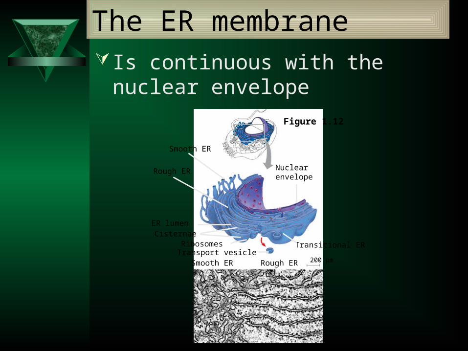
The ER membrane Is continuous with the nuclear envelope
Smooth ER
Rough ER
ER lumenCisternae
RibosomesTransport vesicle
Smooth ER
Transitional ER
Rough ER 200 µm
Nuclearenvelope
Figure 1.12

The ER membrane
There are two distinct regions of ER–Smooth ER, which lacks
ribosomes
–Rough ER, which contains ribosomes
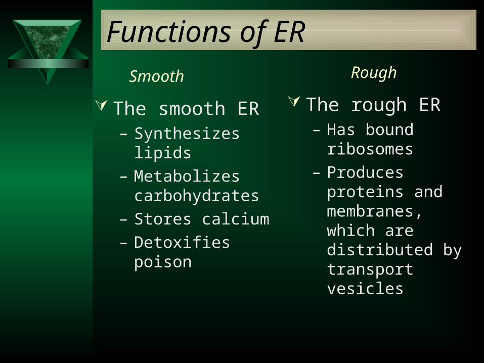
Functions of ER
The smooth ER– Synthesizes lipids
– Metabolizes carbohydrates
– Stores calcium
– Detoxifies poison
The rough ER– Has bound ribosomes
– Produces proteins and membranes, which are distributed by transport vesicles
Smooth Rough
The Golgi apparatus– Receives many of the transport vesicles produced in
the rough ER– Consists of flattened membranous sacs called
cisternae
The Golgi Apparatus: Shipping and Receiving Center
Functions of the Golgi apparatus include
Modification of the products of the rough ER
Manufacture of certain macromolecules

Golgiapparatus
TEM of Golgi apparatus
cis face(“receiving” side ofGolgi apparatus)
Vesicles movefrom ER to Golgi Vesicles also
transport certainproteins back to ER
Vesicles coalesce toform new cis Golgi cisternae
Cisternalmaturation:Golgi cisternaemove in a cis-to-transdirection
Vesicles form andleave Golgi, carryingspecific proteins toother locations or tothe plasma mem-brane for secretion
Vesicles transport specificproteins backward to newerGolgi cisternae
Cisternae
trans face(“shipping” side ofGolgi apparatus)
0.1 0 µm16
5
2
3
4
Functions of the Golgi apparatus
Figure 1.13

Membrane proteins and lipids– Are synthesized in the ER and Golgi apparatus
ER
Concept 7.5:
Bulk transport across the plasma membrane occurs by exocytosis and endocytosis
Large proteins– Cross the membrane by different
mechanisms

Exocytosis In exocytosis
– Transport vesicles migrate to the plasma membrane, fuse with it, and release their contents

Endocytosis
In endocytosis– The cell takes in macromolecules by forming
new vesicles from the plasma membrane

EXTRACELLULARFLUID
PseudopodiumCYTOPLASM
“Food” or other particle
Foodvacuole
1 µm
Pseudopodiumof amoeba
Bacterium
Food vacuole
An amoeba engulfing a bacterium viaphagocytosis (TEM).
PINOCYTOSIS
Pinocytosis vesiclesforming (arrows) ina cell lining a smallblood vessel (TEM).
0.5 µm
In pinocytosis, the cell “gulps” droplets of extracellular fluid into tinyvesicles. It is not the fluiditself that is needed by the cell, but the molecules dissolved in the droplet. Because any and all included solutes are taken into the cell, pinocytosisis nonspecific in the substances it transports.
Plasmamembrane
Vesicle
In phagocytosis, a cellengulfs a particle by Wrapping pseudopodia around it and packaging it within a membrane-enclosed sac large enough to be classified as a vacuole. The particle is digested after the vacuole fuses with a lysosome containing hydrolytic enzymes.
Three types of endocytosis
Figure 7.20
PHAGOCYTOSIS

0.25 µm
RECEPTOR-MEDIATED ENDOCYTOSIS
Receptor
Ligand
Coat protein
Coatedpit
Coatedvesicle
A coated pitand a coatedvesicle formedduringreceptor-mediatedendocytosis(TEMs).
Plasmamembrane
Coatprotein
Receptor-mediated endocytosis enables the cell to acquire bulk quantities of specific substances, even though those substances may not be very concentrated in the extracellular fluid. Embedded in the membrane are proteins with specific receptor sites exposed to the extracellular fluid. The receptor proteins are usually already clustered in regions of the membrane called coated pits, which are lined on their cytoplasmic side by a fuzzy layer of coat proteins. Extracellular substances (ligands) bind to these receptors. When binding occurs, the coated pit forms a vesicle containing the ligand molecules. Notice that there are relatively more bound molecules (purple) inside the vesicle, other molecules (green) are also present. After this ingested material is liberated from the vesicle, the receptors are recycled to the plasma membrane by the same vesicle.

Lysosomes:Digestive Compartments
A lysosome– Is a membranous sac of
hydrolytic enzymes
– Can digest all kinds of macromolecules
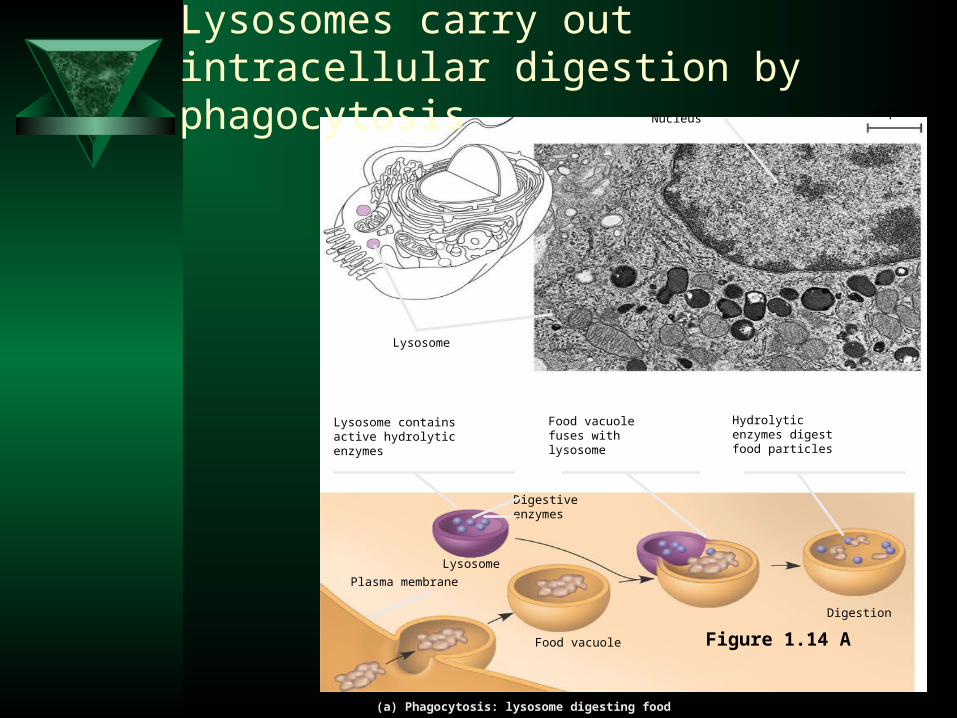
(a) Phagocytosis: lysosome digesting food
1 µm
Lysosome containsactive hydrolyticenzymes
Food vacuole fuses with lysosome
Hydrolyticenzymes digestfood particles
Digestion
Food vacuole
Plasma membrane
Lysosome
Digestiveenzymes
Lysosome
Nucleus
Lysosomes carry out intracellular digestion by phagocytosis
Figure 1.14 A

(b) Autophagy: lysosome breaking down damaged organelle
Lysosome containingtwo damaged organelles 1 µ m
Mitochondrionfragment
Peroxisomefragment
Lysosome fuses withvesicle containingdamaged organelle
Hydrolytic enzymesdigest organellecomponents
Vesicle containingdamaged mitochondrion
Digestion
Lysosome
Autophagy
Figure 1.14 B
Intercellular Junctions

Animals: Tight Junctions, Desmosomes, and Gap Junctions
In animals, there are three types of intercellular junctions
–Tight junctions
–Desmosomes
–Gap junctions
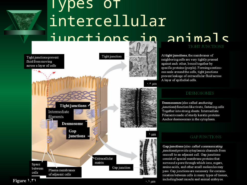
Types of intercellular junctions in animals

Related Documents