cells Article Cytokines Differently Define the Immunomodulation of Mesenchymal Stem Cells from the Periodontal Ligament Christian Behm 1,2 , Alice Blufstein 1 , Johannes Gahn 1 , Michael Nemec 2 , Andreas Moritz 1 , Xiaohui Rausch-Fan 1 and Oleh Andrukhov 1, * 1 Department of Conservative Dentistry and Periodontology, University Clinic of Dentistry, Medical University of Vienna, 1090 Vienna, Austria; [email protected] (C.B.); [email protected] (A.B.); [email protected] (J.G.); [email protected] (A.M.); [email protected] (X.R.-F.) 2 Department of Orthodontics, University Clinic of Dentistry, Medical University of Vienna, 1090 Vienna, Austria; [email protected] * Correspondence: [email protected]; Tel.: +43(0)1-40070-2620 Received: 11 March 2020; Accepted: 11 May 2020; Published: 14 May 2020 Abstract: Human periodontal ligament stem cells (hPDLSCs) play an important role in periodontal tissue homeostasis and regeneration. The function of these cells in vivo depends largely on their immunomodulatory ability, which is reciprocally regulated by immune cells via cytokines, particularly interferon (IFN)-γ, tumor necrosis factor (TNF)-α, and interleukin (IL)-1β. Different cytokines activate distinct signaling pathways and might differently affect immunomodulatory activities of hPDLSCs. This study directly compared the effect of IFN-γ, TNF-α, or IL-1β treated primary hPDLSCs on allogenic CD4 + T lymphocyte proliferation and apoptosis in an indirect co-culture model. The effects of IFN-γ, TNF-α, and IL-1β on the expression of specific immunomodulatory factors such as intoleamine-2,3-dioxygenase-1 (IDO-1), prostaglandin E 2 (PGE 2 ), and programmed cell death 1 ligand 1 (PD-L1) and ligand 2 (PD-L2) in hPDLSCs were compared. The contribution of different immunomodulatory mediators to the immunomodulatory effects of hPDLSCs in the indirect co-culture experiments was assessed using specific inhibitors. Proliferation of CD4 + T lymphocytes was inhibited by hPDLSCs, and this effect was strongly enhanced by IFN-γ and IL-1β but not by TNF-α. Apoptosis of CD4 + T lymphocytes was decreased by hPDLSCs per se. This effect was counteracted by IFN-γ or IL-1β. Additionally, IFN-γ, TNF-α, and IL-1β differently regulated all investigated immunomediators in hPDLSCs. Pharmacological inhibition of immunomediators showed that their contribution in regulating CD4 + T lymphocytes depends on the cytokine milieu. Our data indicate that inflammatory cytokines activate specific immunomodulatory mechanisms in hPDLSCs and the expression of particular immunomodulatory factors, which underlies a complex reciprocal interaction between hPDLSCs and CD4 + T lymphocytes. Keywords: mesenchymal stem cells; periodontal ligament; immunomodulation; cytokines; CD4-positive T-lymphocytes 1. Introduction Human mesenchymal stem cells (MSCs) are multipotent, non-hematopoietic progenitor cells having self-renewal potential [1], expressing specific surface markers, and possessing a multilineage differentiation potential in vitro [2]. Initially found in bone marrow [3], MSCs reside in various tissues of the human body [4,5]. In 2004, Seo et al. first isolated a heterogenous population of MSCs from the periodontal ligament (hPDLSCs) [6], a highly specialized connective tissue surrounding the tooth’s root, Cells 2020, 9, 1222; doi:10.3390/cells9051222 www.mdpi.com/journal/cells

Welcome message from author
This document is posted to help you gain knowledge. Please leave a comment to let me know what you think about it! Share it to your friends and learn new things together.
Transcript
cells
Article
Cytokines Differently Define the Immunomodulationof Mesenchymal Stem Cells from thePeriodontal Ligament
Christian Behm 1,2 , Alice Blufstein 1, Johannes Gahn 1 , Michael Nemec 2, Andreas Moritz 1,Xiaohui Rausch-Fan 1 and Oleh Andrukhov 1,*
1 Department of Conservative Dentistry and Periodontology, University Clinic of Dentistry, MedicalUniversity of Vienna, 1090 Vienna, Austria; [email protected] (C.B.);[email protected] (A.B.); [email protected] (J.G.);[email protected] (A.M.); [email protected] (X.R.-F.)
2 Department of Orthodontics, University Clinic of Dentistry, Medical University of Vienna, 1090 Vienna,Austria; [email protected]
* Correspondence: [email protected]; Tel.: +43(0)1-40070-2620
Received: 11 March 2020; Accepted: 11 May 2020; Published: 14 May 2020�����������������
Abstract: Human periodontal ligament stem cells (hPDLSCs) play an important role in periodontaltissue homeostasis and regeneration. The function of these cells in vivo depends largely on theirimmunomodulatory ability, which is reciprocally regulated by immune cells via cytokines, particularlyinterferon (IFN)-γ, tumor necrosis factor (TNF)-α, and interleukin (IL)-1β. Different cytokines activatedistinct signaling pathways and might differently affect immunomodulatory activities of hPDLSCs.This study directly compared the effect of IFN-γ, TNF-α, or IL-1β treated primary hPDLSCs onallogenic CD4+ T lymphocyte proliferation and apoptosis in an indirect co-culture model. The effectsof IFN-γ, TNF-α, and IL-1β on the expression of specific immunomodulatory factors such asintoleamine-2,3-dioxygenase-1 (IDO-1), prostaglandin E2 (PGE2), and programmed cell death 1ligand 1 (PD-L1) and ligand 2 (PD-L2) in hPDLSCs were compared. The contribution of differentimmunomodulatory mediators to the immunomodulatory effects of hPDLSCs in the indirect co-cultureexperiments was assessed using specific inhibitors. Proliferation of CD4+ T lymphocytes was inhibitedby hPDLSCs, and this effect was strongly enhanced by IFN-γ and IL-1β but not by TNF-α. Apoptosisof CD4+ T lymphocytes was decreased by hPDLSCs per se. This effect was counteracted by IFN-γ orIL-1β. Additionally, IFN-γ, TNF-α, and IL-1β differently regulated all investigated immunomediatorsin hPDLSCs. Pharmacological inhibition of immunomediators showed that their contribution inregulating CD4+ T lymphocytes depends on the cytokine milieu. Our data indicate that inflammatorycytokines activate specific immunomodulatory mechanisms in hPDLSCs and the expression ofparticular immunomodulatory factors, which underlies a complex reciprocal interaction betweenhPDLSCs and CD4+ T lymphocytes.
Keywords: mesenchymal stem cells; periodontal ligament; immunomodulation; cytokines;CD4-positive T-lymphocytes
1. Introduction
Human mesenchymal stem cells (MSCs) are multipotent, non-hematopoietic progenitor cellshaving self-renewal potential [1], expressing specific surface markers, and possessing a multilineagedifferentiation potential in vitro [2]. Initially found in bone marrow [3], MSCs reside in various tissuesof the human body [4,5]. In 2004, Seo et al. first isolated a heterogenous population of MSCs from theperiodontal ligament (hPDLSCs) [6], a highly specialized connective tissue surrounding the tooth’s root,
Cells 2020, 9, 1222; doi:10.3390/cells9051222 www.mdpi.com/journal/cells
Cells 2020, 9, 1222 2 of 18
linking it to the alveolar bone [7]. Quiescent undifferentiated hPDLSCs reside in the perivascular nicheof the periodontal ligament [8,9] and are homed to inflamed or injured periodontal tissue by sensingspecific chemoattractant stimuli. At the injury site, hPDLSCs participate in regulating periodontaltissue regeneration, tissue homeostasis, and local inflammatory processes [4,10,11].
Similarly to other MSCs, hPDLSCs exert mainly immunosuppressive effects and influencedifferent immune cells, such as inhibiting T lymphocyte proliferation and influencing T lymphocyteapoptosis [4,5]. Immunomodulation is currently considered as the major mechanism of MSCs’therapeutic effect, since differentiation ability of transplanted MSCs in vivo is limited [5]. The mostimportant factors involved in the immunomodulatory function of hPDLSCs are indoleamine-2,3-dioxygenase 1 (IDO-1), prostaglandin E2 (PGE2), tumor necrosis factor-inducible gene 6 protein(TSG-6), programmed cell death 1 ligand 1 (PD-L1), and programmed cell death 1 ligand 2 (PD-L2) [4,12].
The immunomodulatory activity is usually low in resting hPDLSCs and is enhanced byenvironmental factors, first of all by inflammatory cytokines produced by activated immune cells [13].Hence, there is a bidirectional interaction between MSCs and immune cells, leading mainly toan immunosuppressive MSC phenotype, which dampens excessive local immune responses [5,14].The most important inflammatory cytokines affecting MSCs are interferon (IFN)-γ, tumor necrosisfactor (TNF)-α, and interleukin (IL)-1β [13,15].
Although the role of inflammatory mediators in the activation of immunomodulatory propertiesin MSCs is well recognized [4], the contribution of specific cytokines is rather poorly known. Severalstudies already recognized the variable effects of IFN-γ, TNF-α, and IL-1β on the expression of certainimmunomediators in MSC-like cells [16–18]. However, to date, the effect of IFN-γ, TNF-α, and IL-1βon the immunomodulatory activities of hPDLSCs has not been directly compared.
Therefore, the main aim of the present study was to directly compare the effects of hPDLSCson the proliferation and the apoptosis of allogenic CD4+ T lymphocytes in the presence of differentinflammatory cytokines using an indirect in vitro co-culture model. Particularly, we investigatedthe effect of IFN-γ, TNF-α, and IL-1β on the ability of hPDLSCs to modulate allogenic CD4+ Tlymphocytes, since these three cytokines activate different signaling pathways and consequentlymight differently affect immunomodulatory activities of hPDLSCs. Hence, we further directlycompared the influence of IFN-γ, TNF-α, and IL-1β on the expression of IDO-1, PD-L1, PD-L2,and prostaglandin-endoperoxide synthase 2 (PTGS-2) in hPDLSCs in vitro. Additionally, to verify therole of IDO-1, PD-L1, and PTGS-2 in hPDLSCs’ caused effects on CD4+ T lymphocytes under differentmicroenvironmental conditions, these immunomediators were inhibited pharmacologically in indirectco-culture experiments. The results of this study highlight that immunomodulation by hPDLSCs isstrongly affected by the local microenvironment. Depending on the presence of the cytokine type,specific immunomodulatory activities of hPDLSCs are boosted, which differently influence CD4+ Tlymphocyte proliferation.
2. Materials and Methods
2.1. Ethics
The whole study protocol, including the isolation of primary hPDLSCs from patients and CD4+ Tlymphocyte isolation from volunteers’ whole blood, was approved by the Ethics Committee of theMedical University of Vienna (EK Nr. 1694/2015, extended 2019). The study was performed accordingto the Declaration of Helsinki and to the Good Scientific Practice Guidelines of the Medical Universityof Vienna.
2.2. hPDLSCs Culture and Verification of MSC Surface Marker Expression
Primary hPDLSCs were isolated from the mid-third surface of the third molars’ roots, whichwere extracted due to orthodontic reasons from periodontal healthy individuals aged between 18 and30 years. Before the surgical procedure, the patients gave their informed written consent. Isolated
Cells 2020, 9, 1222 3 of 18
hPDLSCs were cultured under humidified conditions in Dulbecco’s modified Eagles Medium (DMEM,Sigma-Aldrich, St. Louis, USA), which was supplemented with 10% fetal bovine serum (FBS, Gibco,Carlsbad, USA), 50 µg/ml streptomycin (S, Gibco, Carlsbad, USA), and 100 U/ml penicillin (P, Carlsbad,USA). The stemness of isolated hPDLSCs was verified by analyzing hematopoietic and mesenchymalstem/-stromal cells surface marker expression, as described in our previous study [19]. Additionally,cell suspensions were stained using phycoerythrin (PE)-conjugated mouse anti-human CD73 antibody(eBioscience, San Diego, USA).
2.3. CD4+ T Lymphocyte Isolation
Human whole blood was collected from one single volunteer, using the lithium- and heparincontaining VACUETTE®blood collection system (Greiner Bio-one, Kremsmünster, Austria). In order toconcentrate solely on the variable effects of hPDLSCs from different individuals and to reduce CD4+ Tlymphocyte donor variability, CD4+ T lymphocytes were isolated from the same volunteer throughoutthe study. Whole blood was diluted 1:1 with Hank’s Balanced Salt Solution (HBSS, Life Technologies,Carlsbad, USA), which was followed by Ficoll-Paque (Ge Healthcare, Chicago, USA) density gradientcentrifugation. From harvested allogenic peripheral blood mononuclear cells (PBMCs), CD4+ Tlymphocytes were isolated by immunomagnetic negative selection using MagniSortTM Human CD4+
T cell enrichment kit (Invitrogen, Carlsbad, USA).
2.4. Experimental Protocols
2.4.1. hPDLSCs Treatment
24 h after seeding an appropriate number of hPDLSCs in 6-well plates, these cells were stimulatedwith either 5 ng/ml IL-1β [18] or 10 ng/ml TNF-α [20] or 100 ng/ml IFN-γ [21] (all from Invivogen,San Diego, USA) in FBS-free DMEM for all performed experiments. After 48 h incubation, hPDLSCswere proceeded as indicated below. hPDLSCs were additionally treated with either 50 µM PF-06840003(IDO-1 inhibitor) or 1 µM BMS202 (PD-1/PD-L1 inhibitor) [22] or 1 µM Celecoxib (PTGS-2 inhibitor) [23]before and during indirect co-culture (all from Selleck Chemicals, Houston, USA). PF-06840003concentration was chosen due to its effect on IDO-1 enzymatic activity in hPDLSCs, presented inFigure S1.
2.4.2. hPDLSCs/CD4+ T Cell Indirect Co-Culture
Primary hPDLSCs were seeded in 6-well plates at a density of 2.5 × 10⁵ cells per well in 3 mlDMEM (Sigma-Aldrich, St. Louis, USA) containing 10% FBS (Gibco, Carlsbad, USA) and 1% P/S(Gibco, Carlsbad, USA). After 24 h, hPDLSCs were stimulated as indicated above. After 48 h, mediumwas changed to RPMI-1640 (Sigma-Aldrich, St. Louis, USA) containing 10% FBS and 1% P/S. Transwell(TC) inserts (0.4 µm pore size, Sarstedt, Nürnbrecht, Germany) containing 1 × 10⁶ allogenic CD4+
T cells were inserted into each well. CD4+ T lymphocyte proliferation was induced by 10 µg/mlphytohemagglutinin-L (PHA-L, eBioscience, San Diego, USA) in the presence or the absence of differentcytokines. After five days, CD4+ T lymphocytes proliferation and apoptosis were analyzed by flowcytometry. CD4+ T lymphocytes cultured in the absence of hPDLSCs served as reference.
Additionally, hPDLSCs in co-culture were treated with pharmacological inhibitors against IDO-1,PD-L1, and PTGS-2, as mentioned above. We aimed to clarify the role of these immunomediators onhPDLSCs’ caused effects on CD4+ T lymphocyte proliferation and apoptosis at different conditions.After 5 days incubation, CD4+ T lymphocyte proliferation and apoptosis were assessed by flowcytometry analysis.
2.4.3. Immunomediators Expression
Primary hPDLSCs were seeded at 6-well plates in 3 ml DMEM supplemented with 10% FBS and1% P/S at a density of 2.5 × 10⁵ cells per well. After 24 h, hPDLSCs were treated as indicated above.
Cells 2020, 9, 1222 4 of 18
Then, 48 h later, IDO-1, PD-L1, PD-L2, and PTGS-2 gene and protein expression levels were measured.Additionally, IDO-1 enzymatic activity was measured.
2.5. Analysis of Co-Culture Experiments
2.5.1. CD4+ T Cell Proliferation and Apoptosis
Isolated CD4+ T lymphocytes were labeled using CellTrace CFSE Cell Proliferation Kit(ThermoFischer Scientific, Waltham, USA) to measure CD4+ T lymphocyte proliferation. Briefly,isolated CD4+ T lymphocytes were resuspended in pre-warmed 5% FBS containing 1xPBS, gettinga final CD4+ T lymphocyte concentration of 1 × 10⁶ cells per milliliter. CD4+ T lymphocytes werelabeled with 2.5 µM carboxyfluorescein succinimidyl ester (CFSE) for 5 minutes at room temperature.Labeled CD4+ T lymphocytes were resuspended in complete RPMI-1640 and applied to indirectco-culture. After 5 days, CD4+ T lymphocytes were harvested and stained for apoptotic cells using20 µg/ml propidium iodide (Pi, Affymetrix, Santa Clara, USA). CD4+ T lymphocytes were analyzedby flow cytometry using FACSCalibur Flow Cytometer (Becton Dickinson, Franklin Lakes, USA)equipped with an argon laser to excite fluorescence at 488 nm. Cell count was limited to 10,000 cells persample, and analysis was performed using CellQuest 3.3. software (Becton Dickinson, Franklin Lakes,USA). The percentage of at least once divided CD4+ T lymphocytes was calculated. Additionally,the percentage of apoptotic cells was determined by measuring Pi positive cells. Representative dotplots that outline the gating/analysis strategy are shown in the “Supplementary Materials” (Figure S2).
2.5.2. Immunomediators Expression Analysis in hPDLSCs
Quantitative Polymerase Chain Reaction (qPCR)
Cell lysis, mRNA extraction, reverse transcription, and qPCR were performed using TaqMan GeneExpression Cells-to-CT kit (Applied Biosystems, Foster City, USA). Primus 96 advanced thermocycler(PeqLab/VWR, Darmstadt, Germany) was used for reverse transcription, heating the samples to 37 ◦Cfor 1 h followed by 95 ◦C for 5 minutes. qPCR was performed on a QuantStudio 3 device (AppliedBiosystems, Foster City, USA) using the following settings: 1 x 95 ◦ C for 10 minutes followed by 50cycles of 15 seconds at 95 ◦C and 1 minute at 60 ◦C. The following Taqman Gene Expression Assays(Applied Biosystems, Foster City, USA) were used for target genes amplification: IDO, Hs00984148_m1;PTGS-2, Hs00153133_m1; PD-L1, Hs00204257_m1; PD-L2, Hs00228839_m1 and GAPDH, Hs99999905.GAPDH served as internal reference. Target gene amplification was performed in paired reactionsfollowed by determining Ct values. The n-fold gene expression of the target genes compared tountreated control was quantified by the 2−∆∆Ct method.
IDO-1 Immunostaining
The 2.5 × 10⁵ hPDLSCs were fixed and permeabilized using the Intracellular Fixation andPermeabilization Buffer Set (ebioscience, Waltham, USA) and were subsequently stained withPE-conjugated mouse anti-human IDO-1 antibody (clone eyedio, ebioscience, Waltham, USA).Cells stained with PE-conjugated mouse IgG1 K immunoglobulin isotype control (ebioscience,Waltham, USA) served as reference. After staining, hPDLSCs were resuspended in 200 µl FACSbuffer (3% bovine serum albumin and 0.09% sodium azid in 1xPBS) and analyzed by flow cytometryusing the FACSCalibur Flow Cytometer. Fluorescence was excited by an argon laser at 488 nm.In total, 10,000 cells were counted per group. The percentage of IDO-1 positive cells and thecorresponding mean fluorescence intensity (m.f.i.) were determined using CellQuest 3.3. software asdescribed previously [24]. Representative dot plots and one-parameter histograms that demonstratethe gating/analysis strategy are provided in the “Supplementary Materials” (Figure S3).
Cells 2020, 9, 1222 5 of 18
Measuring L-Kynurenine Levels
L-kynurenine levels were determined in conditioned media and in cell lysates. For cell lysates,conditioned media were harvested, and hPDLSCs were incubated for 3 h in 0.5 ml 1xPBS supplementedwith 800 µM L-tryptophan (Sigma-Aldrich, St. Louis, USA). Conditioned media and cell lysates weremixed 1:3 (v/v) with 30% trichloroacetic acid (Sigma-Aldrich, St. Louis, USA) and were incubated at 65 ◦Cfor 30 minutes. 125 µl supernatant was mixed 1:1 with Ehrlich’s Reagent (0.8% P-dimethylbenzaldehydein glacial acetic acid, Sigma-Aldrich, St. Louis, USA). After incubation for 10 minutes at roomtemperature, samples were measured at OD492 in duplicates. L-kynurenine concentrations weredetermined by plotting measured OD492 against known L-kynurenine (Sigma-Aldrich, St. Louis, USA)concentrations ranging from 1000 µM to 7.8 µM.
Total protein amounts were determined using Pierce BCA Protein Assay Kit (ThermoFischerScientific, Waltham, USA). Protein amounts were determined by plotting measured OD562 againstbovine serum album standards (GE Healthcare, Chicago, USA) ranging from 2000 µg/ml to 31.25 µg/ml.Determined L-kynurenine concentrations were normalized to total protein amounts.
PD-L1 and PD-L2 Immunostaining
1.25 × 10⁵ harvested hPDLSCs were resuspended in 50 µl FACS buffer and stained with thefollowing antibodies for 20 minutes: PE-conjugated mouse anti-human CD274 antibody (cloneB7-H1, ebioscience, Waltham, USA) or PE-conjugated mouse anti-human CD273 antibody (cloneB7-DC, ebioscience, Waltham, USA). Unlabeled cells stained with (PE)-conjugated mouse IgG1 Kimmunoglobulin isotype control (ebioscience, Waltham, USA) served as control. After staining, cellswere resuspended in 200 µl FACS buffer. Flow cytometry analysis was performed as describedabove. The percentage of PD-L1 or PD-L2 positive cells was determined using CellQuest 3.3 software.Representative dot plots that outline the used gating/analysis strategy are shown in the “SupplementaryMaterials” (Figure S4).
Prostaglandin E2 ELISA
Prostaglandin E2 levels were detected in the appropriate conditioned media using ProstaglandinE2 Parameter Assay Kit (R&D Systems, Minneapolis, USA).
2.6. Statistical Analysis
All statistical analysis was performed using SPSS 24.0 (IBM, Armonk, USA). Calculating thestatistical significances between groups was performed by Friedmann test followed by Wilcoxon testfor pairwise comparison. All data are presented as mean values ± S.E.M (standard error of the mean).p-values < 0.05 were considered to be statistically significant.
3. Results
3.1. Mesenchymal and Hematopoietic Surface Marker Expression in hPDLSCs
According to the International Society for Cell and Gene Therapy [2,25], the stem cell character ofhPDLSCs was verified by determining the expression of typical mesenchymal and hematopoietic stemcell surface markers (Table 1). hPDLSCs were stained positively (> 95%) for MSC surface markersCD29, CD73, CD90, and CD105. Approximately 61% of hPDLSCs were stained positive for MSCsurface marker CD146. Additionally, hPDLSCs were stained negatively (< 3%) for hematopoieticsurface markers CD31, CD34 and CD45.

Cells 2020, 9, 1222 6 of 18
Table 1. Human periodontal ligament stem cells’ (hPDLSCs) mesenchymal and hematopoietic surfacemarker expression analysis. Data are presented as mean values ± S.E.M. from five experiments usingcells from five different donors. *MSC: human mesenchymal stem cells.
MSC Marker Hematopoietic Marker
CD29 97.7 ± 0.2 -CD73 96.1 ± 0.2 -CD90 97.9 ± 0.2 -
CD105 97.1 ± 0.6 -CD146 61.3 ± 5.7 -
CD31 - 0.5 ± 0.1CD34 - 0.6 ± 0.2CD45 - 2.7 ± 0.2
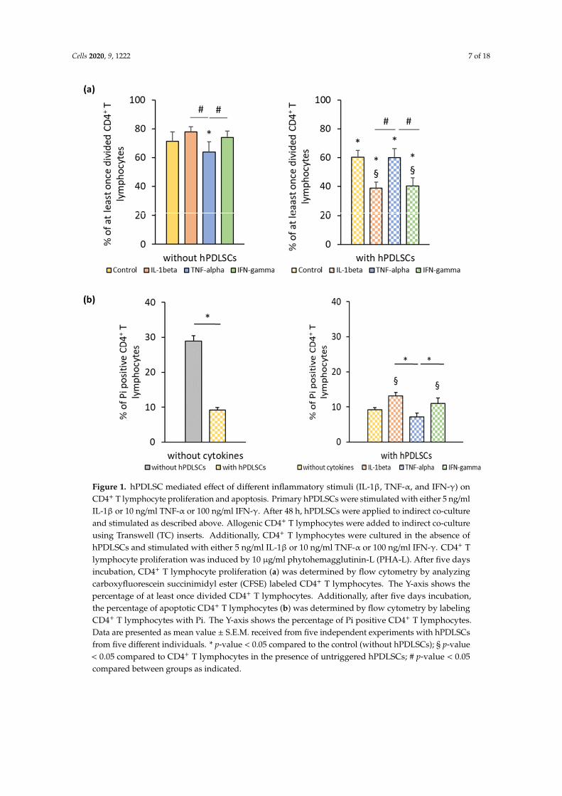
3.2. hPDLSC Mediated Effect of Different Inflammatory Stimuli on CD4+ T Lymphocytes
Figure 1a shows the effect of different inflammatory cytokines on PHA-induced CD4+ T lymphocyteproliferation in the absence and the presence of hPDLSCs. In the absence of hPDLSCs, CD4+ Tlymphocyte proliferation was significantly inhibited by TNF-α and not affected by IL-1β and IFN-γ.hPDLSCs significantly inhibited CD4+ T lymphocyte proliferation, even in the absence of cytokines.The inhibition of CD4+ T lymphocyte proliferation by hPDLSCs was significantly enhanced by IL-1βand IFN-γ, while it was not affected by TNF-α. Figure 1b shows the effect of different inflammatorycytokines on the apoptosis of PHA-activated CD4+ T lymphocytes in the presence of hPDLSCs.In the absence of any cytokine, hPDLSCs significantly decreased the PHA-induced apoptosis of CD4+
T lymphocytes. However, IL-1β and IFN-γ treatment of hPDLSCs counteracted the reduction ofCD4+ T lymphocyte apoptosis, causing a significant increase in the percentage of Pi positive CD4+
T lymphocytes to a similar extent. Both cytokines resulted in a significantly higher apoptosis thanTNF-α stimulated hPDLSCs, which did not affect the percentage of apoptotic CD4+ T lymphocytes.
3.3. Effect of Different Inflammatory Cytokines on the Expression of Immunomodulatory Proteins in hPDLSCs
3.3.1. IDO-1 Expression and Activity
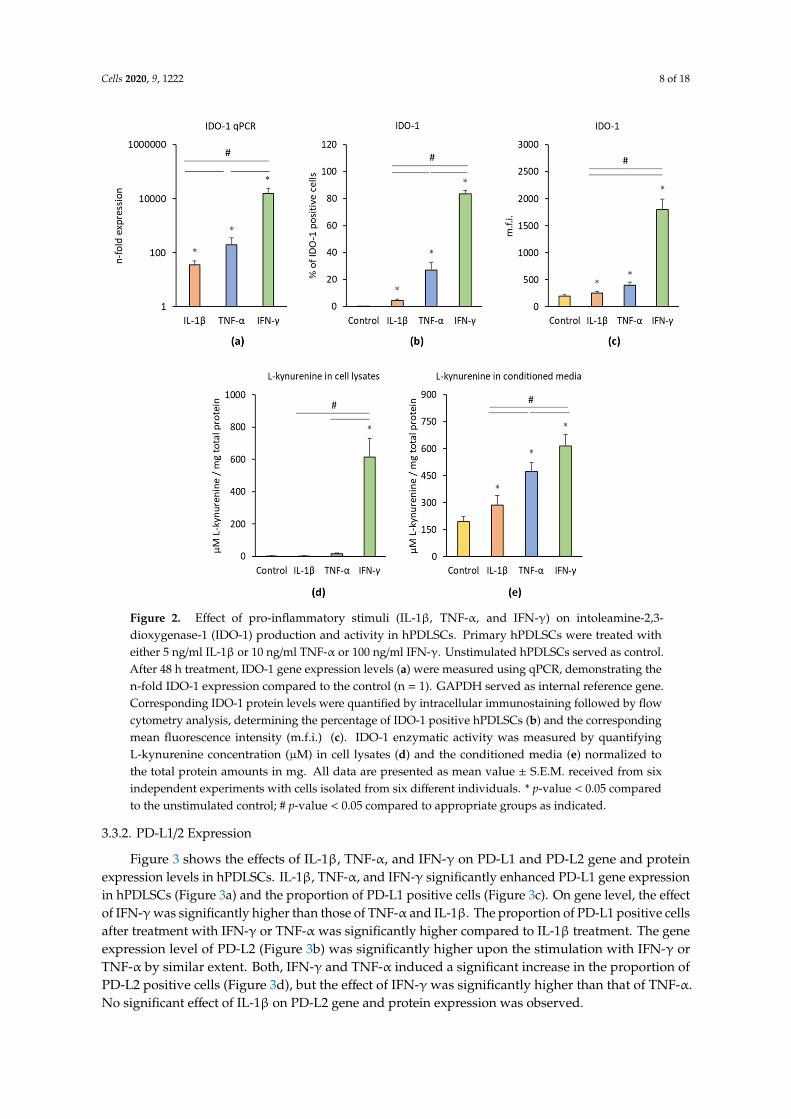
Figure 2 shows the effects of IL-1β, TNF-α, and IFN-γ on the IDO-1 gene and protein expressionlevels as well as on the IDO-1 enzymatic activity. Each cytokine induced significantly enhancedIDO-1 gene expression levels compared to the control (Figure 2a). Significant differences were foundbetween different treatments; the highest IDO-1 gene expression level was observed after IFN-γtreatment followed by TNF-α and IL-1β treatment. All inflammatory stimuli significantly increased thepercentage of IDO-1 positive hPDLSCs (Figure 2b) and the corresponding m.f.i. (Figure 2c). Significantdifferences were observed between different cytokines; the highest IDO-1 protein level was inducedby IFN-γ followed by TNF-α and IL-1β. IDO-1 enzymatic activity was determined by measuringL-kynurenine concentrations in cell lysates (Figure 2d) and conditioned media (Figure 2e). In celllysates, only IFN-γ was able to induce a significant increase in L-kynurenine production, whereasTNF-α and IL-1β had no significant effect. All three stimuli induced significantly higher L-kynurenineproduction in conditioned media; the highest effect was observed for IFN-γ followed by TNF-α andIL-1β.
Cells 2020, 9, 1222 7 of 18
Cells 2020, 9, x FOR PEER REVIEW 6 of 19
CD73 96.1 ± 0.2 - CD90 97.9 ± 0.2 -
CD105 97.1 ± 0.6 - CD146 61.3 ± 5.7 -
CD31 - 0.5 ± 0.1 CD34 - 0.6 ± 0.2 CD45 - 2.7 ± 0.2
3.2. hPDLSC Mediated Effect of Different Inflammatory Stimuli on CD4+ T Lymphocytes
Figure 1a shows the effect of different inflammatory cytokines on PHA-induced CD4+ T lymphocyte proliferation in the absence and the presence of hPDLSCs. In the absence of hPDLSCs, CD4+ T lymphocyte proliferation was significantly inhibited by TNF-α and not affected by IL-1β and IFN-γ. hPDLSCs significantly inhibited CD4+ T lymphocyte proliferation, even in the absence of cytokines. The inhibition of CD4+ T lymphocyte proliferation by hPDLSCs was significantly enhanced by IL-1β and IFN-γ, while it was not affected by TNF-α. Figure 1b shows the effect of different inflammatory cytokines on the apoptosis of PHA-activated CD4+ T lymphocytes in the presence of hPDLSCs. In the absence of any cytokine, hPDLSCs significantly decreased the PHA-induced apoptosis of CD4+ T lymphocytes. However, IL-1β and IFN-γ treatment of hPDLSCs counteracted the reduction of CD4+ T lymphocyte apoptosis, causing a significant increase in the percentage of Pi positive CD4+ T lymphocytes to a similar extent. Both cytokines resulted in a significantly higher apoptosis than TNF-α stimulated hPDLSCs, which did not affect the percentage of apoptotic CD4+ T lymphocytes.
Figure 1. hPDLSC mediated effect of different inflammatory stimuli (IL-1β, TNF-α, and IFN-γ) on CD4+ T lymphocyte proliferation and apoptosis. Primary hPDLSCs were stimulated with either 5
Figure 1. hPDLSC mediated effect of different inflammatory stimuli (IL-1β, TNF-α, and IFN-γ) onCD4+ T lymphocyte proliferation and apoptosis. Primary hPDLSCs were stimulated with either 5 ng/mlIL-1β or 10 ng/ml TNF-α or 100 ng/ml IFN-γ. After 48 h, hPDLSCs were applied to indirect co-cultureand stimulated as described above. Allogenic CD4+ T lymphocytes were added to indirect co-cultureusing Transwell (TC) inserts. Additionally, CD4+ T lymphocytes were cultured in the absence ofhPDLSCs and stimulated with either 5 ng/ml IL-1β or 10 ng/ml TNF-α or 100 ng/ml IFN-γ. CD4+ Tlymphocyte proliferation was induced by 10 µg/ml phytohemagglutinin-L (PHA-L). After five daysincubation, CD4+ T lymphocyte proliferation (a) was determined by flow cytometry by analyzingcarboxyfluorescein succinimidyl ester (CFSE) labeled CD4+ T lymphocytes. The Y-axis shows thepercentage of at least once divided CD4+ T lymphocytes. Additionally, after five days incubation,the percentage of apoptotic CD4+ T lymphocytes (b) was determined by flow cytometry by labelingCD4+ T lymphocytes with Pi. The Y-axis shows the percentage of Pi positive CD4+ T lymphocytes.Data are presented as mean value ± S.E.M. received from five independent experiments with hPDLSCsfrom five different individuals. * p-value < 0.05 compared to the control (without hPDLSCs); § p-value< 0.05 compared to CD4+ T lymphocytes in the presence of untriggered hPDLSCs; # p-value < 0.05compared between groups as indicated.
Cells 2020, 9, 1222 8 of 18Cells 2020, 9, x FOR PEER REVIEW 8 of 19
Figure 2. Effect of pro-inflammatory stimuli (IL-1β, TNF-α, and IFN-γ) on intoleamine-2,3-dioxygenase-1 (IDO-1) production and activity in hPDLSCs. Primary hPDLSCs were treated with either 5 ng/ml IL-1β or 10 ng/ml TNF-α or 100 ng/ml IFN-γ. Unstimulated hPDLSCs served as control. After 48 hours treatment, IDO-1 gene expression levels (a) were measured using qPCR, demonstrating the n-fold IDO-1 expression compared to the control (n = 1). GAPDH served as internal reference gene. Corresponding IDO-1 protein levels were quantified by intracellular immunostaining followed by flow cytometry analysis, determining the percentage of IDO-1 positive hPDLSCs (b) and the corresponding mean fluorescence intensity (m.f.i.) (c). IDO-1 enzymatic activity was measured by quantifying L-kynurenine concentration (µM) in cell lysates (d) and the conditioned media (e) normalized to the total protein amounts in mg. All data are presented as mean value ± S.E.M. received from six independent experiments with cells isolated from six different individuals. * p-value < 0.05 compared to the unstimulated control; # p-value < 0.05 compared to appropriate groups as indicated.
3.3.2. PD-L1/2 Expression
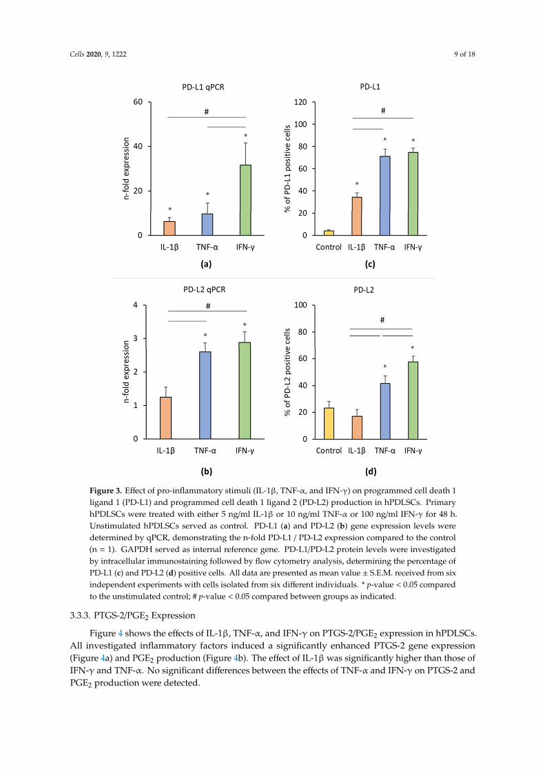
Figure 3 shows the effects of IL-1β, TNF-α, and IFN-γ on PD-L1 and PD-L2 gene and protein expression levels in hPDLSCs. IL-1β, TNF-α, and IFN-γ significantly enhanced PD-L1 gene expression in hPDLSCs (Figure 3a) and the proportion of PD-L1 positive cells (Figure 3c). On gene level, the effect of IFN-γ was significantly higher than those of TNF-α and IL-1β. The proportion of PD-L1 positive cells after treatment with IFN-γ or TNF-α was significantly higher compared to IL-1β treatment. The gene expression level of PD-L2 (Figure 3b) was significantly higher upon the stimulation with IFN-γ or TNF-α by similar extent. Both, IFN-γ and TNF-α induced a significant increase in the proportion of PD-L2 positive cells (Figure 3d), but the effect of IFN-γ was significantly higher than that of TNF-α. No significant effect of IL-1β on PD-L2 gene and protein expression was observed.
Figure 2. Effect of pro-inflammatory stimuli (IL-1β, TNF-α, and IFN-γ) on intoleamine-2,3-dioxygenase-1 (IDO-1) production and activity in hPDLSCs. Primary hPDLSCs were treated witheither 5 ng/ml IL-1β or 10 ng/ml TNF-α or 100 ng/ml IFN-γ. Unstimulated hPDLSCs served as control.After 48 h treatment, IDO-1 gene expression levels (a) were measured using qPCR, demonstrating then-fold IDO-1 expression compared to the control (n = 1). GAPDH served as internal reference gene.Corresponding IDO-1 protein levels were quantified by intracellular immunostaining followed by flowcytometry analysis, determining the percentage of IDO-1 positive hPDLSCs (b) and the correspondingmean fluorescence intensity (m.f.i.) (c). IDO-1 enzymatic activity was measured by quantifyingL-kynurenine concentration (µM) in cell lysates (d) and the conditioned media (e) normalized tothe total protein amounts in mg. All data are presented as mean value ± S.E.M. received from sixindependent experiments with cells isolated from six different individuals. * p-value < 0.05 comparedto the unstimulated control; # p-value < 0.05 compared to appropriate groups as indicated.
3.3.2. PD-L1/2 Expression
Figure 3 shows the effects of IL-1β, TNF-α, and IFN-γ on PD-L1 and PD-L2 gene and proteinexpression levels in hPDLSCs. IL-1β, TNF-α, and IFN-γ significantly enhanced PD-L1 gene expressionin hPDLSCs (Figure 3a) and the proportion of PD-L1 positive cells (Figure 3c). On gene level, the effectof IFN-γ was significantly higher than those of TNF-α and IL-1β. The proportion of PD-L1 positive cellsafter treatment with IFN-γ or TNF-α was significantly higher compared to IL-1β treatment. The geneexpression level of PD-L2 (Figure 3b) was significantly higher upon the stimulation with IFN-γ orTNF-α by similar extent. Both, IFN-γ and TNF-α induced a significant increase in the proportion ofPD-L2 positive cells (Figure 3d), but the effect of IFN-γ was significantly higher than that of TNF-α.No significant effect of IL-1β on PD-L2 gene and protein expression was observed.
Cells 2020, 9, 1222 9 of 18Cells 2020, 9, x FOR PEER REVIEW 9 of 19
Figure 3. Effect of pro-inflammatory stimuli (IL-1β, TNF-α, and IFN-γ) on programmed cell death 1 ligand 1 (PD-L1) and programmed cell death 1 ligand 2 (PD-L2) production in hPDLSCs. Primary hPDLSCs were treated with either 5 ng/ml IL-1β or 10 ng/ml TNF-α or 100 ng/ml IFN-γ for 48 hours. Unstimulated hPDLSCs served as control. PD-L1 (a) and PD-L2 (b) gene expression levels were determined by qPCR, demonstrating the n-fold PD-L1 / PD-L2 expression compared to the control (n = 1). GAPDH served as internal reference gene. PD-L1/PD-L2 protein levels were investigated by intracellular immunostaining followed by flow cytometry analysis, determining the percentage of PD-L1 (c) and PD-L2 (d) positive cells. All data are presented as mean value ± S.E.M. received from six independent experiments with cells isolated from six different individuals. * p-value < 0.05 compared to the unstimulated control; # p-value < 0.05 compared between groups as indicated.
3.3.3. PTGS-2/PGE2 Expression
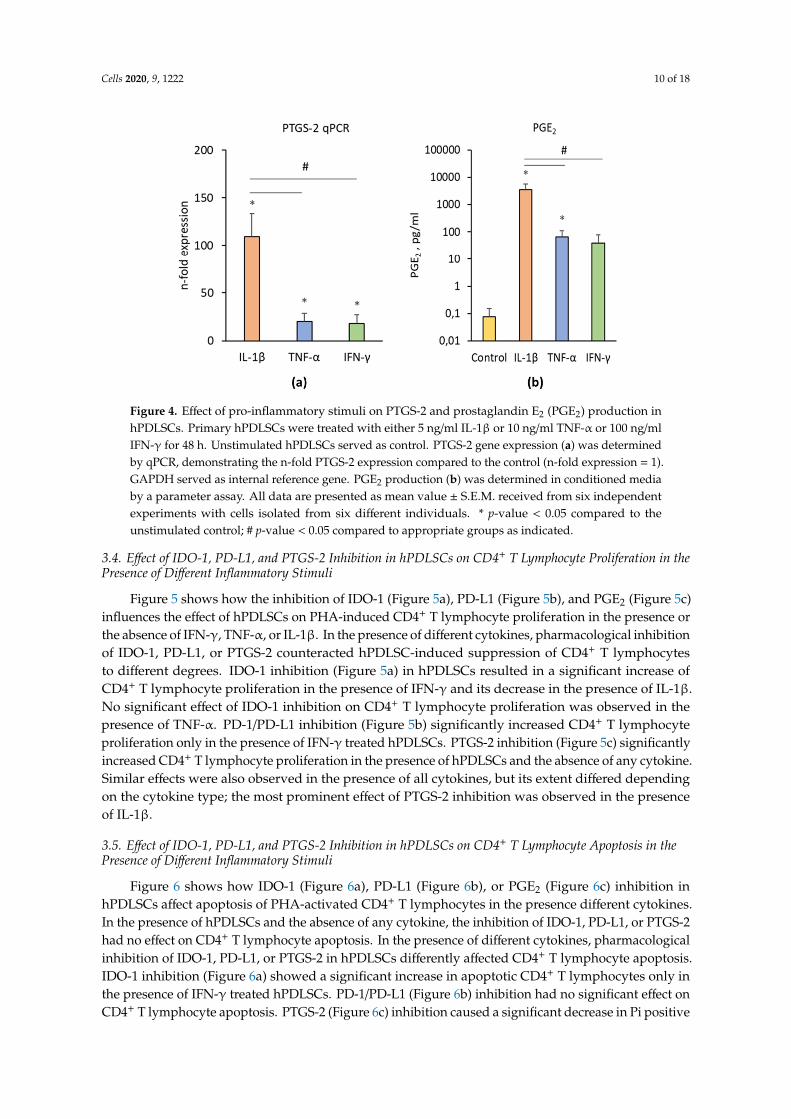
Figure 4 shows the effects of IL-1β, TNF-α, and IFN-γ on PTGS-2/PGE2 expression in hPDLSCs. All investigated inflammatory factors induced a significantly enhanced PTGS-2 gene expression (Figure 4a) and PGE2 production (Figure 4b). The effect of IL-1β was significantly higher than those of IFN-γ and TNF-α. No significant differences between the effects of TNF-α and IFN-γ on PTGS-2 and PGE2 production were detected.
Figure 3. Effect of pro-inflammatory stimuli (IL-1β, TNF-α, and IFN-γ) on programmed cell death 1ligand 1 (PD-L1) and programmed cell death 1 ligand 2 (PD-L2) production in hPDLSCs. PrimaryhPDLSCs were treated with either 5 ng/ml IL-1β or 10 ng/ml TNF-α or 100 ng/ml IFN-γ for 48 h.Unstimulated hPDLSCs served as control. PD-L1 (a) and PD-L2 (b) gene expression levels weredetermined by qPCR, demonstrating the n-fold PD-L1 / PD-L2 expression compared to the control(n = 1). GAPDH served as internal reference gene. PD-L1/PD-L2 protein levels were investigatedby intracellular immunostaining followed by flow cytometry analysis, determining the percentage ofPD-L1 (c) and PD-L2 (d) positive cells. All data are presented as mean value ± S.E.M. received from sixindependent experiments with cells isolated from six different individuals. * p-value < 0.05 comparedto the unstimulated control; # p-value < 0.05 compared between groups as indicated.
3.3.3. PTGS-2/PGE2 Expression
Figure 4 shows the effects of IL-1β, TNF-α, and IFN-γ on PTGS-2/PGE2 expression in hPDLSCs.All investigated inflammatory factors induced a significantly enhanced PTGS-2 gene expression(Figure 4a) and PGE2 production (Figure 4b). The effect of IL-1β was significantly higher than those ofIFN-γ and TNF-α. No significant differences between the effects of TNF-α and IFN-γ on PTGS-2 andPGE2 production were detected.
Cells 2020, 9, 1222 10 of 18Cells 2020, 9, x FOR PEER REVIEW 10 of 19
Figure 4. Effect of pro-inflammatory stimuli on PTGS-2 and prostaglandin E2 (PGE2) production in hPDLSCs. Primary hPDLSCs were treated with either 5 ng/ml IL-1β or 10 ng/ml TNF-α or 100 ng/ml IFN-γ for 48 hours. Unstimulated hPDLSCs served as control. PTGS-2 gene expression (a) was determined by qPCR, demonstrating the n-fold PTGS-2 expression compared to the control (n-fold expression = 1). GAPDH served as internal reference gene. PGE2 production (b) was determined in conditioned media by a parameter assay. All data are presented as mean value ± S.E.M. received from six independent experiments with cells isolated from six different individuals. * p-value < 0.05 compared to the unstimulated control; # p-value < 0.05 compared to appropriate groups as indicated.
3.4. Effect of IDO-1, PD-L1, and PTGS-2 Inhibition in hPDLSCs on CD4+ T Lymphocyte Proliferation in the Presence of Different Inflammatory Stimuli
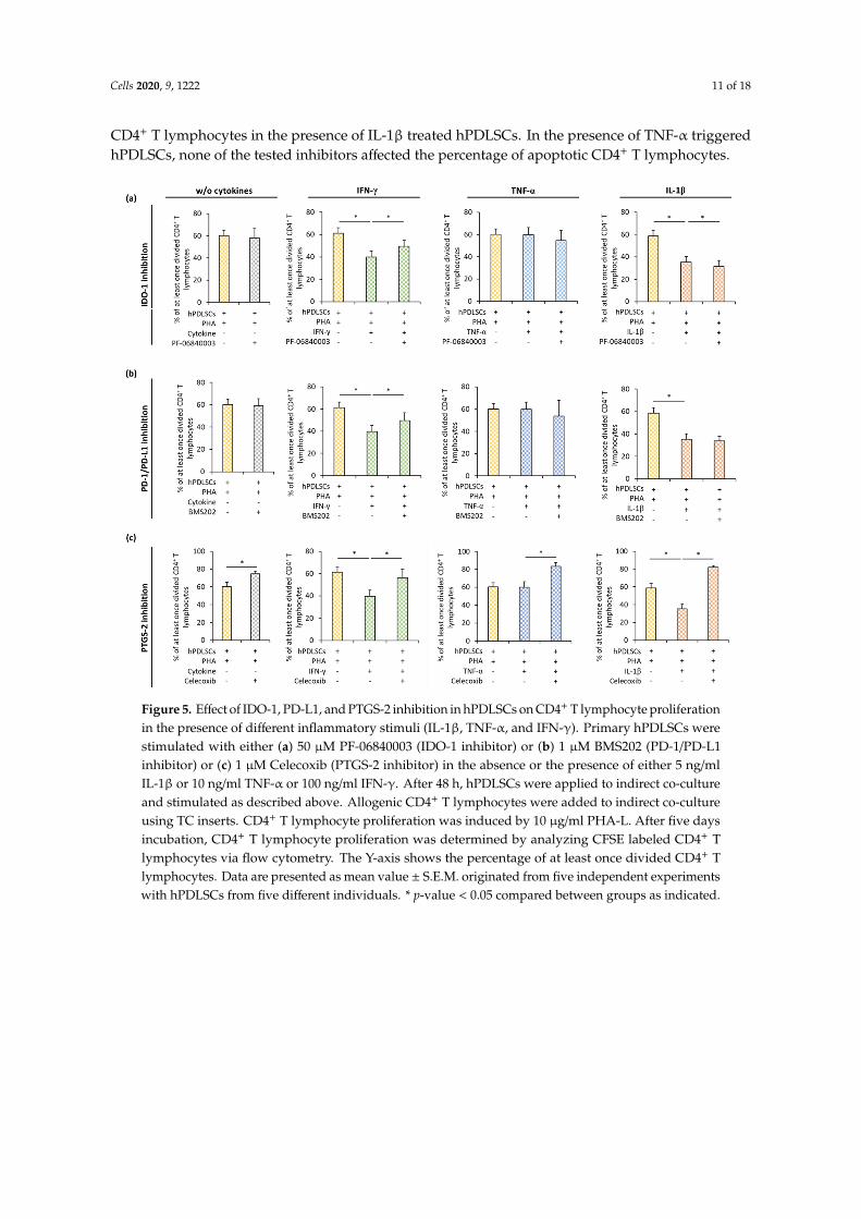
Figure 5 shows how the inhibition of IDO-1 (Figure 5a), PD-L1 (Figure 5b), and PGE2 (Figure 5c) influences the effect of hPDLSCs on PHA-induced CD4+ T lymphocyte proliferation in the presence or the absence of IFN-γ, TNF-α, or IL-1β. In the presence of different cytokines, pharmacological inhibition of IDO-1, PD-L1, or PTGS-2 counteracted hPDLSC-induced suppression of CD4+ T lymphocytes to different degrees. IDO-1 inhibition (Figure 5a) in hPDLSCs resulted in a significant increase of CD4+ T lymphocyte proliferation in the presence of IFN-γ and its decrease in the presence of IL-1β. No significant effect of IDO-1 inhibition on CD4+ T lymphocyte proliferation was observed in the presence of TNF-α. PD-1/PD-L1 inhibition (Figure 5b) significantly increased CD4+ T lymphocyte proliferation only in the presence of IFN-γ treated hPDLSCs. PTGS-2 inhibition (Figure 5c) significantly increased CD4+ T lymphocyte proliferation in the presence of hPDLSCs and the absence of any cytokine. Similar effects were also observed in the presence of all cytokines, but its extent differed depending on the cytokine type; the most prominent effect of PTGS-2 inhibition was observed in the presence of IL-1β.
Figure 4. Effect of pro-inflammatory stimuli on PTGS-2 and prostaglandin E2 (PGE2) production inhPDLSCs. Primary hPDLSCs were treated with either 5 ng/ml IL-1β or 10 ng/ml TNF-α or 100 ng/mlIFN-γ for 48 h. Unstimulated hPDLSCs served as control. PTGS-2 gene expression (a) was determinedby qPCR, demonstrating the n-fold PTGS-2 expression compared to the control (n-fold expression = 1).GAPDH served as internal reference gene. PGE2 production (b) was determined in conditioned mediaby a parameter assay. All data are presented as mean value ± S.E.M. received from six independentexperiments with cells isolated from six different individuals. * p-value < 0.05 compared to theunstimulated control; # p-value < 0.05 compared to appropriate groups as indicated.
3.4. Effect of IDO-1, PD-L1, and PTGS-2 Inhibition in hPDLSCs on CD4+ T Lymphocyte Proliferation in thePresence of Different Inflammatory Stimuli
Figure 5 shows how the inhibition of IDO-1 (Figure 5a), PD-L1 (Figure 5b), and PGE2 (Figure 5c)influences the effect of hPDLSCs on PHA-induced CD4+ T lymphocyte proliferation in the presence orthe absence of IFN-γ, TNF-α, or IL-1β. In the presence of different cytokines, pharmacological inhibitionof IDO-1, PD-L1, or PTGS-2 counteracted hPDLSC-induced suppression of CD4+ T lymphocytesto different degrees. IDO-1 inhibition (Figure 5a) in hPDLSCs resulted in a significant increase ofCD4+ T lymphocyte proliferation in the presence of IFN-γ and its decrease in the presence of IL-1β.No significant effect of IDO-1 inhibition on CD4+ T lymphocyte proliferation was observed in thepresence of TNF-α. PD-1/PD-L1 inhibition (Figure 5b) significantly increased CD4+ T lymphocyteproliferation only in the presence of IFN-γ treated hPDLSCs. PTGS-2 inhibition (Figure 5c) significantlyincreased CD4+ T lymphocyte proliferation in the presence of hPDLSCs and the absence of any cytokine.Similar effects were also observed in the presence of all cytokines, but its extent differed dependingon the cytokine type; the most prominent effect of PTGS-2 inhibition was observed in the presenceof IL-1β.
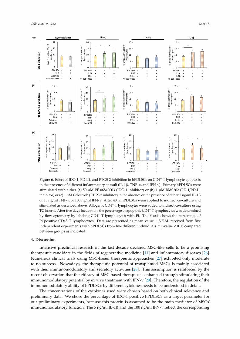
3.5. Effect of IDO-1, PD-L1, and PTGS-2 Inhibition in hPDLSCs on CD4+ T Lymphocyte Apoptosis in thePresence of Different Inflammatory Stimuli
Figure 6 shows how IDO-1 (Figure 6a), PD-L1 (Figure 6b), or PGE2 (Figure 6c) inhibition inhPDLSCs affect apoptosis of PHA-activated CD4+ T lymphocytes in the presence different cytokines.In the presence of hPDLSCs and the absence of any cytokine, the inhibition of IDO-1, PD-L1, or PTGS-2had no effect on CD4+ T lymphocyte apoptosis. In the presence of different cytokines, pharmacologicalinhibition of IDO-1, PD-L1, or PTGS-2 in hPDLSCs differently affected CD4+ T lymphocyte apoptosis.IDO-1 inhibition (Figure 6a) showed a significant increase in apoptotic CD4+ T lymphocytes only inthe presence of IFN-γ treated hPDLSCs. PD-1/PD-L1 (Figure 6b) inhibition had no significant effect onCD4+ T lymphocyte apoptosis. PTGS-2 (Figure 6c) inhibition caused a significant decrease in Pi positive
Cells 2020, 9, 1222 11 of 18
CD4+ T lymphocytes in the presence of IL-1β treated hPDLSCs. In the presence of TNF-α triggeredhPDLSCs, none of the tested inhibitors affected the percentage of apoptotic CD4+ T lymphocytes.Cells 2020, 9, x FOR PEER REVIEW 11 of 19
Figure 5. Effect of IDO-1, PD-L1, and PTGS-2 inhibition in hPDLSCs on CD4+ T lymphocyte proliferation in the presence of different inflammatory stimuli (IL-1β, TNF-α, and IFN-γ). Primary hPDLSCs were stimulated with either (a) 50 µM PF-06840003 (IDO-1 inhibitor) or (b) 1 µM BMS202 (PD-1/PD-L1 inhibitor) or (c) 1 µM Celecoxib (PTGS-2 inhibitor) in the absence or the presence of either 5 ng/ml IL-1β or 10 ng/ml TNF-α or 100 ng/ml IFN-γ. After 48 hours, hPDLSCs were applied to indirect co-culture and stimulated as described above. Allogenic CD4+ T lymphocytes were added to indirect co-culture using TC inserts. CD4+ T lymphocyte proliferation was induced by 10 µg/ml PHA-L. After five days incubation, CD4+ T lymphocyte proliferation was determined by analyzing CFSE labeled CD4+ T lymphocytes via flow cytometry. The Y-axis shows the percentage of at least once divided CD4+ T lymphocytes. Data are presented as mean value ± S.E.M. originated from five independent experiments with hPDLSCs from five different individuals. * p-value < 0.05 compared between groups as indicated.
3.5. Effect of IDO-1, PD-L1, and PTGS-2 Inhibition in hPDLSCs on CD4+ T Lymphocyte Apoptosis in the Presence of Different Inflammatory Stimuli
Figure 6 shows how IDO-1 (Figure 6a), PD-L1 (Figure 6b), or PGE2 (Figure 6c) inhibition in hPDLSCs affect apoptosis of PHA-activated CD4+ T lymphocytes in the presence different cytokines. In the presence of hPDLSCs and the absence of any cytokine, the inhibition of IDO-1, PD-L1, or PTGS-2 had no effect on CD4+ T lymphocyte apoptosis. In the presence of different cytokines, pharmacological inhibition of IDO-1, PD-L1, or PTGS-2 in hPDLSCs differently affected CD4+ T lymphocyte apoptosis. IDO-1 inhibition (Figure 6a) showed a significant increase in apoptotic CD4+ T lymphocytes only in the presence of IFN-γ treated hPDLSCs. PD-1/PD-L1 (Figure 6b) inhibition had no significant effect on CD4+ T lymphocyte apoptosis. PTGS-2 (Figure 6c) inhibition caused a
Figure 5. Effect of IDO-1, PD-L1, and PTGS-2 inhibition in hPDLSCs on CD4+ T lymphocyte proliferationin the presence of different inflammatory stimuli (IL-1β, TNF-α, and IFN-γ). Primary hPDLSCs werestimulated with either (a) 50 µM PF-06840003 (IDO-1 inhibitor) or (b) 1 µM BMS202 (PD-1/PD-L1inhibitor) or (c) 1 µM Celecoxib (PTGS-2 inhibitor) in the absence or the presence of either 5 ng/mlIL-1β or 10 ng/ml TNF-α or 100 ng/ml IFN-γ. After 48 h, hPDLSCs were applied to indirect co-cultureand stimulated as described above. Allogenic CD4+ T lymphocytes were added to indirect co-cultureusing TC inserts. CD4+ T lymphocyte proliferation was induced by 10 µg/ml PHA-L. After five daysincubation, CD4+ T lymphocyte proliferation was determined by analyzing CFSE labeled CD4+ Tlymphocytes via flow cytometry. The Y-axis shows the percentage of at least once divided CD4+ Tlymphocytes. Data are presented as mean value ± S.E.M. originated from five independent experimentswith hPDLSCs from five different individuals. * p-value < 0.05 compared between groups as indicated.
Cells 2020, 9, 1222 12 of 18
Cells 2020, 9, x FOR PEER REVIEW 12 of 19
significant decrease in Pi positive CD4+ T lymphocytes in the presence of IL-1β treated hPDLSCs. In the presence of TNF-α triggered hPDLSCs, none of the tested inhibitors affected the percentage of apoptotic CD4+ T lymphocytes.
Figure 6. Effect of IDO-1, PD-L1, and PTGS-2 inhibition in hPDLSCs on CD4+ T lymphocyte apoptosis in the presence of different inflammatory stimuli (IL-1β, TNF-α, and IFN-γ). Primary hPDLSCs were stimulated with either (a) 50 µM PF-06840003 (IDO-1 inhibitor) or (b) 1 µM BMS202 (PD-1/PD-L1 inhibitor) or (c) 1 µM Celecoxib (PTGS-2 inhibitor) in the absence or the presence of either 5 ng/ml IL-1β or 10 ng/ml TNF-α or 100 ng/ml IFN-γ. After 48 hours, hPDLSCs were applied to indirect co-culture and stimulated as described above. Allogenic CD4+ T lymphocytes were added to indirect co-culture using TC inserts. After five days incubation, the percentage of apoptotic CD4+ T lymphocytes was determined by flow cytometry by labeling CD4+ T lymphocytes with Pi. The Y-axis shows the percentage of Pi positive CD4+ T lymphocytes. Data are presented as mean value ± S.E.M. received from five independent experiments with hPDLSCs from five different individuals. * p-value < 0.05 compared between groups as indicated.
4. Discussion
Intensive preclinical research in the last decade declared MSC-like cells to be a promising therapeutic candidate in the fields of regenerative medicine [11] and inflammatory diseases [26]. Numerous clinical trials using MSC-based therapeutic approaches [27] exhibited only moderate to no success. Nowadays, the therapeutic potential of transplanted MSCs is mainly associated with their immunomodulatory and secretory activities [28]. This assumption is reinforced by the recent observation that the efficacy of MSC-based therapies is enhanced through stimulating their immunomodulatory potential by ex vivo treatment with IFN-γ [29]. Therefore, the regulation of the immunomodulatory ability of hPDLSCs by different cytokines needs to be understood in detail.
Figure 6. Effect of IDO-1, PD-L1, and PTGS-2 inhibition in hPDLSCs on CD4+ T lymphocyte apoptosisin the presence of different inflammatory stimuli (IL-1β, TNF-α, and IFN-γ). Primary hPDLSCs werestimulated with either (a) 50 µM PF-06840003 (IDO-1 inhibitor) or (b) 1 µM BMS202 (PD-1/PD-L1inhibitor) or (c) 1 µM Celecoxib (PTGS-2 inhibitor) in the absence or the presence of either 5 ng/ml IL-1βor 10 ng/ml TNF-α or 100 ng/ml IFN-γ. After 48 h, hPDLSCs were applied to indirect co-culture andstimulated as described above. Allogenic CD4+ T lymphocytes were added to indirect co-culture usingTC inserts. After five days incubation, the percentage of apoptotic CD4+ T lymphocytes was determinedby flow cytometry by labeling CD4+ T lymphocytes with Pi. The Y-axis shows the percentage ofPi positive CD4+ T lymphocytes. Data are presented as mean value ± S.E.M. received from fiveindependent experiments with hPDLSCs from five different individuals. * p-value < 0.05 comparedbetween groups as indicated.
4. Discussion
Intensive preclinical research in the last decade declared MSC-like cells to be a promisingtherapeutic candidate in the fields of regenerative medicine [11] and inflammatory diseases [26].Numerous clinical trials using MSC-based therapeutic approaches [27] exhibited only moderateto no success. Nowadays, the therapeutic potential of transplanted MSCs is mainly associatedwith their immunomodulatory and secretory activities [28]. This assumption is reinforced by therecent observation that the efficacy of MSC-based therapies is enhanced through stimulating theirimmunomodulatory potential by ex vivo treatment with IFN-γ [29]. Therefore, the regulation of theimmunomodulatory ability of hPDLSCs by different cytokines needs to be understood in detail.
The concentrations of the cytokines used were chosen based on both clinical relevance andpreliminary data. We chose the percentage of IDO-1 positive hPDLSCs as a target parameter forour preliminary experiments, because this protein is assumed to be the main mediator of MSCs’immunomodulatory function. The 5 ng/ml IL-1β and the 100 ng/ml IFN-γ reflect the corresponding
Cells 2020, 9, 1222 13 of 18
cytokine levels in the gingival crevicular fluid (GCF) of periodontitis patients [30,31]. Additionally,the chosen IL-1β and IFN-γ levels caused the highest percentage of IDO-1 positive hPDLSCs within ourexperimental in vitro setting (Figure S5). The used TNF-α concentration (10 ng/ml) was clearly higherthan in the GCF of periodontitis patients (up to 100 pg/ml) [32]. The percentage of IDO-1 positivehPDLSCs caused by 10 ng/ml TNF-α was near the maximal response (90% of the response caused by100 ng/ml TNF-α). Furthermore, most previous studies used TNF-α at a concentration of 10 ng/ml [20].
Our data showed that the effect of hPDLSCs on the proliferation of CD4+ T lymphocytes isdifferently affected by various inflammatory cytokines. hPDLSCs inhibited the proliferation of CD4+
T lymphocytes, even in the absence of any cytokine. This inhibitory effect was strongly enhancedby 100 ng/ml IFN-γ and 5 ng/ml IL-1β. The additional inhibitory effect is mediated by hPDLSCsbecause these cytokines had no significant effect on CD4+ T lymphocyte proliferation in the absenceof hPDLSCs. In contrast, 10 ng/ml TNF-α showed no potentiating effect of the hPDLSCs mediatedsuppression of CD4+ T cell proliferation. This was in contrast to the direct inhibitory effect of 10 ng/mlTNF-α on CD4+ T lymphocyte proliferation observed in the absence of hPDLSCs. Thus, the presenceof hPDLSCs modifies the effect of all investigated cytokines on CD4+ T lymphocyte proliferation.
The changes of immunomodulatory properties of MSCs on CD4+ T lymphocyte proliferation byinflammatory cytokines was already partially indicated in previous studies. The inhibitory effectsof MSCs on CD4+ T lymphocyte proliferation has only been reported in simultaneous presence ofIFN-γ with TNF-α or IL-1β using 20 ng/ml for each cytokine [33]. Using bone marrow-derivedMSCs from mice and antibody-activated CD4+ T lymphocyte may be the major reasons for thesepartial discrepancies in our data. Additionally, using a fivefold lower IFN-γ concentrations mayalso be the reason for the unaffected T lymphocyte proliferation by IFN-γ treated MSCs. Anotherstudy reports no significant effect of 1 U/ml IFN-γ and TNF-α on peripheral blood-derived humanMSCs mediated suppression of antibody-triggered CD4+ T lymphocyte proliferation [34]. It seemsas if suppressive effects of such cytokines depend on several factors, such as MSC origin or CD4+ Tlymphocyte activation method.
Our results further indicated that the effect of hPDLSCs on PHA-triggered apoptosis of CD4+
T lymphocytes is differently influenced by various inflammatory cytokines. In the absence of anycytokine, hPDLSCs per se reduced the number of apoptotic CD4+ T lymphocytes. Consequently, thisfinding implies that hPDLSCs protect CD4+ T lymphocytes from PHA-induced apoptosis. Furthermore,it seems that hPDLSC-mediated inhibition of CD4+ T lymphocyte proliferation is not due to CD4+ Tlymphocyte apoptosis. This is in accordance with previous studies that observed that bone marrowderived MSC-mediated suppression of T lymphocytes does not depend on cellular apoptosis [35]but rather on arresting T lymphocytes in the G0/G1 cell cycle phase [36]. In contrast, other studiesconducted in dental pulp stem cells [37] and hPDLSCs [16] showed an increase in the apoptosis of Tlymphocytes in the absence of any cytokine. Using dental pulp stem cells [37] and concanavalin A [16]stimulated PBMCs, respectively, may be the major reasons for the discrepancies in our data.
5 ng/ml IL-1β or 100 ng/ml IFN-γ partially counteracted the hPDLSCs’ induced reduction ofapoptotic CD4+ T lymphocytes, whereas 10 ng/ml TNF-α slightly increased the anti-apoptotic effectof hPDLSCs on CD4+ T lymphocytes. Thus, adding various exogenous cytokines to our co-culturemodel partially caused the usual known immunosuppressive properties of hPDLSCs and significantlymodified the hPDLSCs’ ability to influence apoptosis of CD4+ T lymphocytes. Increased CD4+ Tlymphocytes apoptosis by 100 ng/ml IFN-γ or 5 ng/ml IL-1β might partially underline the enhancedanti-proliferative effect of hPDLSCs on CD4+ T lymphocytes in the presence of these cytokines.Although 10 ng/ml TNF-α showed no effect on CD4+ T lymphocyte proliferation in the presence ofhPDLSCs, this cytokine caused a decrease in the proportion of apoptotic CD4+ T lymphocytes. Thisanti-apoptotic effect of 10 ng/ml TNF-α treated hPDLSCs might partially explain the observationthat 10 ng/ml TNF-α caused an inhibition of CD4+ T lymphocyte proliferation in the absence ofhPDLSCs but did not affect it in the presence of hPDLSCs; the anti-proliferative effect of TNF-α mightbe counterweighed by its anti-apoptotic effect. Summarizing, it seems that, in the presence of cytokine
Cells 2020, 9, 1222 14 of 18
treated hPDLSCs, apoptosis and proliferation of CD4+ T lymphocytes are highly connected to eachother, which also may depend on the present cytokine type. This raises the question if cytokinetreatment of hPDLSCs changes their way to suppress CD4+ T lymphocyte proliferation from anapoptosis independent to a partially apoptosis dependent mechanism.
The most important factors mediating hPDLSCs immunomodulatory activities toward Tlymphocytes are IDO-1, PD-L1, PD-L2, and PTGS-2 [5]. The expression of most of theseimmunomediators in hPDLSCs was increased by stimulation with 100 ng/ml IFN-γ, 10 ng/ml TNF-α,and 5 ng/ml IL-1β. However, prominent quantitative differences were observed between differentcytokines. Few previous studies compare the effect of different cytokines in hPDLSCs, and their resultsare partly in line with our data. Most previous studies suggest that IDO-1 protein expression andactivity could be enhanced by all cytokines, such as 0.5–50 ng/ml IL-1β and TNF-α but could bedramatically increased only by IFN-γ at concentrations of 10–25 ng/ml [16–18]. Although partiallyusing different concentrations and incubation times, ranging from 24 h to 72 h, these studies are in linewith our data. Another study shows that 10 ng/ml TNF-α induces significantly higher PD-L1 surfaceexpression in hPDLSCs compared to 10 ng/ml IFN-γ or IL-1β after 48 h [20]. In contrast, in our study,10 ng/ml TNF-α and 100 ng/ml IFN-γ caused quantitatively similar high PD-L1 protein expressionlevels, whereas 5 ng/ml IL-1β induced PD-L1 expression was rather low. Further studies imply thatTNF-α (1 – 10 ng/ml) is a strong activator of PGE2 production in hPDLSCs [38]. Another study showedsimilar PGE2 levels after stimulating MSCs with 100 ng/ml IFN-γ or 10 ng/ml TNF-α for 48 h [21],which is comparable to our data. However, this study further reported that the production of PGE-2by MSCs was drastically increased by 50 ng/ml TNF-α. We found the strongest production of PGE-2protein after stimulation with IL-1β. The expression level of PD-L2 was also different after stimulationwith various cytokines, which was not shown previously.
The mechanisms involved in the immunomodulatory activity of hPDLSCs after activation bydifferent cytokines are complex and seem to depend on the inflammatory environment. Our resultsidentified PGE2 as a potent immunomediator of CD4+ T lymphocyte proliferation. Pharmacologicalinhibition of PTGS-2 counteracted the suppression of CD4+ T lymphocyte proliferation in the presenceof all three investigated cytokines. PGE2 mediated mechanisms almost fully reversed the hPDLSCsmediated suppression of CD4+ T lymphocytes proliferation in the presence of 5 ng/ml IL-1β. This findingis in agreement with observation that 5 ng/ml IL-1β induced the highest PGE2 expression levelsin hPDLSCs compared to other investigated cytokines. Additionally, PTGS-2 inhibitor reversedhPDLSC mediated effects on CD4+ T lymphocyte proliferation, even in the absence of any cytokine.This observation could be explained by the assumption of the reciprocal interaction between hPDLSCsand CD4+ T lymphocytes in our in vitro co-culture model. Particularly, cytokines produced by activatedCD4+ T lymphocytes might activate PGE2 production by hPDLSCs, which, in turn, might suppressCD4+ T lymphocyte proliferation. A previous study implies that PGE2 production is an importantmechanism involved in the immunomodulatory effects of MSCs mainly toward macrophages [5].Our study additionally underlined the importance of this factor in MSCs-mediated effects on CD4+
T lymphocytes.Previous reports suggest that MSC-mediated suppression of CD4+ T lymphocyte proliferation is
mainly regulated by the IFN-γ–IDO-1 axis [4,39,40]. This statement is only partially in line with our data.On the one hand, the suppression of CD4+ T cell proliferation by hPDLSCs was strongly promoted by100 ng/ml IFN-γ. Moreover, gene and protein expression of IDO-1 in hPDLSCs was mostly enhancedby 100 ng/ml IFN-γ stimulation compared to other cytokines. On the other hand, pharmacologicalinhibition of IFN-γ-induced IDO-1 expression only partially counteracted the suppression of CD4+
T lymphocytes by hPDLSCs. Furthermore, IDO- inhibitor had no effect on the hPDLSCs-mediatedinhibition of CD4+ T lymphocytes proliferation in the presence of 10 ng/ml TNF-α and 5 ng/mlIL-1β as well as in the absence of any cytokines. Moreover, 10 ng/ml TNF-α induced high levelsof IDO-1 expression but failed to enhance the inhibitory effect of hPDLSCs on CD4+ T lymphocyte
Cells 2020, 9, 1222 15 of 18
proliferation. Thus, although IDO-1 is an important immunomodulatory factor, it cannot account forall hPDLSCs-mediated effects on CD4+ T lymphocytes proliferation.
PD-L1 is an important transmembrane protein involved in suppressing immune responses.Previous studies also show its implication in MSC-mediated immunosuppressive effects on Tlymphocytes [20]. Zhang et al. demonstrated that PD-L1 is a crucial factor in mediating the suppressionof T lymphocytes by MSCs derived from the adipose tissue in the absence of any cytokine [41]. This isin contrast with our data, which show that only the inhibition of 100 ng/ml IFN-γ induced PD-L1 inhPDLSCs counteracted the suppression of CD4+ T lymphocytes. This is in accordance with our geneexpression analysis data showing that 100 ng/ml IFN-γ caused the highest PD-L1 expression levels.Hence, it seems that 100 ng/ml IFN-γ induced PD-L1 is another important axis for suppressing CD4+
T lymphocyte proliferation by hPDLSCs. However, this mechanism seems to be less relevant in thepresence of 10 ng/ml TNF-α and 5 ng/ml IL-1β, although its expression in hPDLSCs is substantiallyincreased by these cytokines.
In contrast to CD4+ T lymphocytes proliferation, the effect of different pharmacological inhibitorson the hPDLSCs-mediated effect on CD4+ T lymphocytes apoptosis was less pronounced. None of theinhibitors influenced CD4+ T lymphocytes apoptosis in the presence of hPDLSCs and the absence ofcytokines. The IDO-1 inhibitor marginally increased the proportion of apoptotic CD4+ T lymphocytesin the presence of 100 ng/ml IFN-γ. The PTGS-2 inhibitor markedly increased the proportion ofapoptotic CD4+ T lymphocytes in the presence of 100 ng/ml IFN-γ, while it was decreased in thepresence of 5 ng/ml IL-1β. The inhibition of PD-L1 caused no influence on the hPDLSCs-mediatedCD4+ T lymphocyte apoptosis, although Zhang et al. showed a contribution of 10 ng/ml TNF-αinduced PD-L1 expression in hPDLSCs on CD4+ T lymphocytes apoptosis. This discrepancy might beexplained by the use of PBMCs in a direct co-culture model [20] instead of pure CD4+ T lymphocytesin our indirect co-culture model. Summarizing, it seems that the mechanisms of hPDLSCs-mediatedeffects on CD4+ T lymphocytes apoptosis depend on the inflammatory microenvironment conditions.
Our study underlies the complexity of the interaction between hPDLSCs and CD4+ T lymphocytesand further indicates a high plasticity of the reciprocal interaction between hPDLSCs and immunecells mainly regulated by the local cytokine milieu. IFN-γ, TNF-α, and IL-1β are mainly secretedby various activated immune cells under inflammatory conditions. These cytokines activate restinghPDLSCs and trigger the expression of various immunomediators in hPDLSCs and consequentlytheir immunosuppressive activities. This causes lower levels of inflammatory cytokines andsubsequently a partial abolishment of hPDLSCs’ immunosuppression. Thus, a continuous interactionbetween hPDLSCs and immune cells seems to determine the intensity of the local immune response(reviewed in [5]). Our results indicate that inflammatory cytokines differently affect the productionof immunomodulatory factors in hPDLSCs and consequently their immunomodulatory activities.It can be hypothesized that a certain inflammatory cytokine predominantly activates only a particularimmunomodulatory mechanism and consequently only a specific immunomodulatory function.This could lead to variable immunosuppressive actions on immune cells and subsequently to variablecytokine levels. Hence, the plasticity of this tight bidirectional interaction is not only affected by thecytokine level [28] but also by the kind of cytokine. Both parameters significantly change during theinflammation process. With regard to the variable cytokine levels during the inflammation progress,one limitation of our study is the use of only one concentration per cytokine.
The complex interaction between hPDLSCs and CD4+ T lymphocytes might have an implicationin pathological states, particularly in periodontal disease. Periodontitis is an inflammatory diseaseof the tooth supporting tissues that might lead to tooth loss if it remains untreated. T lymphocytesplay a crucial role in periodontitis progression by contributing to osteoclast formation and alveolarbone resorption and by contributing to a pro-inflammatory environment [42]. It is also knownthat tissue destruction in periodontal disease is mainly caused by a dysregulated immune response.All used cytokines are involved in a host destructive inflammatory response and are usually associatedwith alveolar bone resorption by triggering osteoclastogenesis and soft tissue degradation by matrix
Cells 2020, 9, 1222 16 of 18
metalloproteinases [43–45]. Further, our data implies that, besides destructive effects, these cytokinesmight also activate some immunosuppressive mechanisms. Modulation of the activity of immune cellsby hPDLSCs and influencing this effect differently by inflammatory cytokines represents a fine-tuningmechanism of the local inflamed periodontal microenvironment.
5. Conclusions
In conclusion, this in vitro study underlines that the immunomodulatory activity of hPDLSCsstrongly depends on the local microenvironment. Depending on the presence of different inflammatorycytokines, the expression of several immunomediators is upregulated, which causes variable effects onCD4+ T cells. By translating our results into the in vivo situation, we suggest that the regulation oflocal immune responses by hPDLSCs in the presence of various cytokines is a crucial mechanism ofon-site immune and periodontal tissue homeostasis.
Supplementary Materials: The following are available online at http://www.mdpi.com/2073-4409/9/5/1222/s1.Figure S1: Effect of different PF-06840003 (IDO-1 inhibitor) concentrations on the IDO-1 enzymatic activity, FigureS2: Representative dot plots which outline the gating / analysis strategy for CFSE proliferation (a) and Pi apoptosis(b) assays, Figure S3: Representative dot plots and one-parameter histogram which outline the gating / analysisstrategy for calculating the % of IDO-1 positive hPDLSCs and the corresponding m.f.i., Figure S4: Representativedot plots with outline the gating / analysis strategy for calculating the % of PD-L1 (a) or PD-L2 (b) positivehPDLSCs, Figure S5: Effect of different IL-1β, TNF-α and IFN-γ concentrations on the percentage of IDO-1positive hPDLSCs.
Author Contributions: Conceptualization, O.A. and C.B.; methodology, O.A. and C.B.; validation, O.A. and C.B.;formal analysis, O.A. and C.B.; investigation, C.B., A.B., J.G. and M.N.; resources, O.A. and A.M.; writing—originaldraft preparation, C.B. and O.A.; writing—review and editing, O.A., A.B., M.N., X.R.-F. and A.M.; visualization,C.B.; supervision, O.A.; project administration, O.A. and X.R.-F.; funding acquisition, O.A. All authors have readand agreed to the published version of the manuscript.
Funding: This research was funded by Austrian Science Fund (FWF), grant number P29440 (to Oleh Andrukhov).
Acknowledgments: The authors acknowledge Phuong Quynh Nguyen for excellent technical assistance.Open Access Funding by the Austrian Science Fund (FWF).
Conflicts of Interest: The authors declare no conflict of interest. The funders had no role in the design of thestudy; in the collection, analyses, or interpretation of data; in the writing of the manuscript, or in the decision topublish the results.
References
1. Prockop, D.J. Marrow Stromal Cells as Stem Cells for Nonhematopoietic Tissues. J. Sci. 1997, 276, 71–74.[CrossRef]
2. Dominici, M.; Le Blanc, K.; Mueller, I.; Slaper-Cortenbach, I.; Marini, F.C.; Krause, D.S.; Deans, R.J.; Keating, A.;Prockop, D.J.; Horwitz, E.M. Minimal Criteria for Defining Multipotent MesenChymal Stromal Cells. TheInternational Society for Cellular Therapy Position Statement. Cytotherapy 2006, 8, 315–317. [CrossRef][PubMed]
3. Chamberlain, G.; Fox, J.; Ashton, B.; Middleton, J. Concise Review: Mesenchymal Stem Cells: TheirPhenotype, Differentiation Capacity, Immunological Features, and Potential for Homing. Stem Cells 2007, 25,2739–2749. [CrossRef] [PubMed]
4. Wada, N.; Gronthos, S.; Bartold, P.M. Immunomodulatory Effects of Stem cells. Periodontol. 2000 2013, 63,198–216. [CrossRef] [PubMed]
5. Andrukhov, O.; Behm, C.; Blufstein, A.; Rausch-Fan, X. Immunomodulatory Properties of DentalTissue-Derived Mesenchymal Stem Cells: Implication in Disease and Tissue Regeneration. World J. StemCells 2019, 11, 604–617. [CrossRef] [PubMed]
6. Seo, B.M.; Miura, M.; Gronthos, S.; Bartold, P.M.; Batouli, S.; Brahim, J.; Young, M.; Robey, P.G.; Wang, C.Y.;Shi, S. Investigation of Multipotent Postnatal Stem Cells from Human Periodontal Ligament. Lancet 2004,149–155. [CrossRef]
7. Beertsen, W.; McCulloch, C.A.G.; Sodek, J. The Periodontal Ligament: A Unique, Multifunctional ConnectiveTissue. Periodontol. 2000 1997, 13, 20–40. [CrossRef] [PubMed]
Cells 2020, 9, 1222 17 of 18
8. Shi, S.; Gronthos, S. Perivascular Niche of Postnatal Mesenchymal Stem Cells in Human Bone Marrow andDental Pulp. J. Bone Miner. Res. 2003, 18, 696–704. [CrossRef]
9. Sharpe, P.T.; Yianni, V. Perivascular-Derived Mesenchymal Stem Cells. Crit. Rev. Oral Biol. Med. 2019,98, 1066–1072.
10. Racz, G.Z.; Kadar, K.; Foldes, A.; Kallo, K.; Perczel-Kovach, K.; Keremi, B.; Nagy, A.; Varga, G.Immunomodulatory and Potential Therapeutic Role of Mesenchymal Stem Cells in Periodontitis. J. Physiol.Pharmacol. 2014, 65, 327–339.
11. Xiao, L.; Nasu, M. From Regenerative Dentistry to Regenerative Mmedicine: Progress, Challenges,and Potential Applications of Oral Stem Cells. Stem Cells Cloning Adv. Appl. 2014, 7, 89–99. [CrossRef]
12. Tipnis, S.; Viswanathan, C.; Majumdar, A.S. Immunosuppressive Properties of Human UmbilicalCord-derived Mesenchymal Stem Cells: Role of B7-H1 and IDO. Immunol. Cell Biol. 2010, 88, 795–806.[CrossRef]
13. Krampera, M. Mesenchymal Stromal Cell Licensing: A Multistep Process. J. Leukemia 2011, 25, 1408–1414.[CrossRef] [PubMed]
14. Bernardo, M.E.; Fibbe, W.E. Mesenchymal Stromal Cells: Sensors and Switchers of Inflammation. Cell StemCell 2013, 13, 392–402. [CrossRef] [PubMed]
15. Fawzy El-Sayed, K.M.; Elahmady, M.; Adawi, Z.; Aboushadi, N.; Elnaggar, A.; Eid, M.; Hamdy, N.; Sanaa, D.;Dörfer, C.E. The Periodontal Stem/Progenitor Cell Inflammatory-Regenerative Cross Talk: A new Perspective.J. Periodontal Res. 2019, 54, 81–94. [CrossRef] [PubMed]
16. Wada, N.; Menicanin, D.; Shi, S.; Bartold, P.M.; Gronthos, S. Immunomodulatory Properties of HumanPeriodontal Ligament Stem cells. J. Cell. Physiol. 2009, 219, 667–676. [CrossRef] [PubMed]
17. Takegawa, D.; Nakanishi, T.; Hirao, K.; Yumoto, H.; Takahashi, K.; Matsuo, T. Modulatory Roles ofInterferon-γ Through Indoleamine 2,3-Dioxygenase Induction in Innate Immune Response of Dental PulpCells. J. Endod. 2014, 40, 1382–1387. [CrossRef] [PubMed]
18. Nisapakultorn, K.; Makrudthong, J.; Sa-Ard-Iam, N.; Rerkyen, P.; Mahanonda, R.; Takikawa, O. Indoleamine2,3-Dioxygenase Expression and Regulation in Chronic Periodontitis. J. Periodontol. 2009, 80, 114–121.[CrossRef]
19. Andrukhov, O.; Andrukhova, O.; Özdemir, B.; Haririan, H.; Müller-Kern, M.; Moritz, A.; Rausch-Fan, X.Soluble CD14 Enhances the Response of Periodontal Ligament Stem Cells to P. Gingivalis Lipopolysaccharide.PLoS ONE 2016, 11, e0160848. [CrossRef]
20. Zhang, J.; Wang, C.M.; Zhang, P.; Wang, X.; Chen, J.; Yang, J.; Lu, W.; Zhou, W.; Yuan, W.; Feng, Y. Expressionof Programmed Death 1 Ligand 1 on Periodontal Tissue Cells as a Possible Protective Feedback MechanismAgainst Periodontal Tissue Destruction. Mol. Med. Rep. 2016, 13, 2423–2430. [CrossRef]
21. Hegyi, B.; Kudlik, G.; Monostori, É.; Uher, F. Activated T-cells and Pro-inflammatory Cytokines DifferentiallyRegulate Prostaglandin E2 Secretion by Mesenchymal Stem Cells. J. Biochem. Biophys. Res. Commun. 2012,419, 215–220. [CrossRef] [PubMed]
22. Guzik, K.; Zak, K.M.; Grudnik, P.; Magiera, K.; Musielak, B.; Törner, R.; Skalniak, L.; Dömling, A.; Dubin, G.;Holak, T.A. Small-Molecule Inhibitors of the Programmed Cell Death-1/Programmed Death-Ligand 1(PD-1/PD-L1) Interaction via Transiently Induced Protein States and Dimerization of PD-L1. Med. Chem.2017, 60, 5857–5867. [CrossRef] [PubMed]
23. Kawashima, M.; Ogura, N.; Akutsu, M.; Ito, K.; Kondoh, T. The Anti-Inflammatory Effect of CyclooxygenaseInhibitors in Fibroblast-like Synoviocytes from The Human Temporomandibular Joint Results from TheSuppression of PGE2 Production. J. Oral Pathol. Med. 2013, 42, 499–506. [CrossRef] [PubMed]
24. Andrukhov, O.; Hong, J.S.-A.; Andrukhova, O.; Blufstein, A.; Moritz, A.; Rausch-Fan, X. Response of HumanPeriodontal Ligament Stem Cells to IFN-gamma and TLR-agonists. Sci. Rep. 2017, 7, 12856. [CrossRef]
25. Viswanathan, S.; Shi, Y.; Galipeau, J.; Krampera, M.; Leblanc, K.; Martin, I.; Nolta, J.; Phinney, D.G.; Sensebe, L.Mesenchymal Stem Versus Stromal Cells: International Society for Cellular Therapy Mesenchymal StromalCell Committee Position Statement on Nomenclature. Cytotherapy 2019, 21, 1019–1024. [CrossRef]
26. Klinker, M.W. Mesenchymal Stem Cells in The Treatment of Inflammatory and Autoimmune Diseases inExperimental Animal Models. World J. Stem Cells 2015, 419, 215–220. [CrossRef]
27. Squillaro, T.; Peluso, G.; Galderisi, U. Clinical Trials with Mesenchymal Stem Cells: An Update. Cell Transplant.2016, 25, 829–848. [CrossRef]
Cells 2020, 9, 1222 18 of 18
28. Wang, Y.; Chen, X.; Cao, W.; Shi, Y. Plasticity of Mesenchymal Stem Cells in Immunomodulation: Pathologicaland Therapeutic Implications. Nat. Immunol. 2014, 15, 1009–1016. [CrossRef]
29. García, J.R.; Quirós, M.; Han, W.M.; O’Leary, M.N.; Cox, G.N.; Nusrat, A.; García, A.J. IFN-γ-TetheredHydrogels Enhance Mesenchymal Stem Cell-based Immunomodulation and Promote Tissue Repair.Biomaterials 2019, 220, 119403.
30. Engebretson, S.P.; Hey-Hadavi, J.; Ehrhardt, F.J.; Hsu, D.; Celenti, R.S.; Grbic, J.T.; Lamster, I.B. GingivalCrevicular Fluid Levels of Interleukin-1β and Glycemic Control in Patients with Chronic Periodontitis andType 2 Diabetes. J. Periodontol. 2004, 75, 1203–1208. [CrossRef]
31. Dutzan, N.; Vernal, R.; Hernandez, M.; Dezerega, A.; Rivera, O.; Silva, N.; Aguillon, J.C.; Puente, J.; Pozo, P.;Gamonal, J. Levels of Interferon-Gamma and Transcription Factor T-Bet in Progressive Periodontal Lesionsin Patients With Chronic Periodontitis. J. Periodontol. 2009, 80, 290–296. [CrossRef] [PubMed]
32. Dag, A.; Firat, E.T.; Kadiroglu, A.K.; Kale, E.; Yilmaz, M.E. Significance of Elevated Gingival CrevicularFluid Tumor Necrosis Factor-α and Interleukin-8 Levels in Chronic Hemodialysis Patients with PeriodontalDisease. J. Periodontal Res. 2010, 45, 445–450. [PubMed]
33. Ren, G.; Zhang, L.; Zhao, X.; Xu, G.; Zhang, Y.; Roberts, A.I.; Zhao, R.C.; Shi, Y. Mesenchymal StemCell-Mediated Immunosuppression Occurs via Concerted Action of Chemokines and Nitric Oxide. Cell StemCell 2008, 2, 141–150. [CrossRef] [PubMed]
34. Hemeda, H.; Jakob, M.; Ludwig, A.-K.; Giebel, B.; Lang, S.; Brandau, S. Interferon-γ and Tumor NecrosisFactor-α Differentially Affect Cytokine Expression and Migration Properties of Mesenchymal Stem Cells.Stem Cells Dev. 2010, 19, 693–706. [CrossRef]
35. Di Nicola, M.; Carlo-Stella, C.; Magni, M.; Milanesi, M.; Longoni, P.D.; Matteucci, P.; Grisanti, S.; Gianni, A.M.Human Bone Marrow Stromal Cells Suppress T-lymphocyte Proliferation Induced by Cellular or NonspecificMitogenic Stimuli. Blood 2002, 99, 3838–3843. [CrossRef]
36. Glennie, S.; Soeiro, I.; Dyson, P.J.; Lam, E.W.F.; Dazzi, F. Bone Marrow Mesenchymal Stem Cells InduceDivision Arrest Anergy of Activated T Cells. Blood 2005, 105, 2821–2827. [CrossRef]
37. Zhao, Y.; Wang, L.; Jin, Y.; Shi, S. Fas ligand Regulates the Immunomodulatory Properties of Dental PulpStem Cells. J. Dent. Res. 2012, 91, 948–954. [CrossRef]
38. Su, W.R.; Zhang, Q.Z.; Shi, S.H.; Nguyen, A.L.; Le, A.D. Human Gingiva-Derived Mesenchymal StromalCells Attenuate Contact Hypersensitivity via Prostaglandin E2-dependent Mechanisms. J. Stem Cells 2011,29, 1849–1860. [CrossRef]
39. Li, Z.; Jiang, C.M.; An, S.; Cheng, Q.; Huang, Y.F.; Wang, Y.T.; Gou, Y.C.; Xiao, L.; Yu, W.J.; Wang, J.Immunomodulatory Properties of Dental Tissue-derived Mesenchymal Stem Cells. J. Oral Dis. 2014, 20,25–34. [CrossRef]
40. Laing, A.G.; Fanelli, G.; Ramirez-Valdez, A.; Lechler, R.I.; Lombardi, G.; Sharpe, P.T. Mesenchymal StemCells Inhibit T-Cell Function through Conserved Induction of Cellular Stress. PLoS ONE 2019, 14, e0213170.[CrossRef]
41. Zhou, K.; Guo, S.; Tong, S.; Sun, Q.; Li, F.; Zhang, X.; Qiao, Y.; Liang, G. Immunosuppression of HumanAdipose-Derived Stem Cells on T Cell Subsets via the Reduction of NF-kappaB Activation Mediated byPD-L1/PD-1 and Gal-9/TIM-3 Pathways. Stem Cells Dev. 2018, 27, 1191–1202. [CrossRef] [PubMed]
42. Alvarez, C.; Monasterio, G.; Cavalla, F.; Córdova, L.A.; Hernández, M.; Heymann, D.; Garlet, G.P.; Sorsa, T.;Pärnänen, P.; Lee, H.-M.; et al. Osteoimmunology of Oral and Maxillofacial Diseases: TranslationalApplications Based on Biological Mechanisms. Front. Immunol. 2019, 10, 1664. [CrossRef] [PubMed]
43. Garlet, G.P. Critical Reviews in Oral Biology & Medicine: Destructive and Protective Roles of Cytokines inPeriodontitis: A Re-appraisal from Host Defense and Tissue Destruction Viewpoints. J. Dent. Res. 2010, 89,1349–1363. [PubMed]
44. Graves, D.T.; Cochran, D. The Contribution of Interleukin-1 and Tumor Necrosis Factor to Periodontal TissueDestruction. J. Periodontol. 2003, 74, 391–401. [CrossRef]
45. Graves, D. Cytokines That Promote Periodontal Tissue Destruction. J. Periodontol. 2008, 79, 1585–1591.[CrossRef]
© 2020 by the authors. Licensee MDPI, Basel, Switzerland. This article is an open accessarticle distributed under the terms and conditions of the Creative Commons Attribution(CC BY) license (http://creativecommons.org/licenses/by/4.0/).
Related Documents