Cytokines, chemokines and soluble adhesion molecules in aqueous humor of children with uveitis Karen M. Sijssens a, * , Ger T. Rijkers b , Aniki Rothova a , Jan S. Stilma a , Peter A.W.J.F. Schellekens a , Joke H. de Boer a a F.C. Donders Institute of Ophthalmology, University Medical Center Utrecht, E.03.136, Heidelberglaan 100, 3584 CX Utrecht, The Netherlands b Department of Pediatric Immunology, Wilhelmina Children’s Hospital, University Medical Center Utrecht and Department of Medical Microbiology and Immunology, St. Antonius Hospital, Nieuwegein, The Netherlands Received 19 January 2007; accepted in revised form 10 June 2007 Available online 22 June 2007 Abstract Uveitis in childhood is a visual threatening disease with a complication rate of more than 75%. Despite extensive research, the etiology of uveitis is still unclear although the general opinion is now that uveitis is a T-cell mediated disease. The purpose of this study was to investigate the profile of cytokines, chemotactic cytokines (chemokines) and soluble adhesion molecules in the aqueous humor (AqH) of children with uveitis in order to identify the factors that control the immune response in the eye. In this clinical laboratory investigation we analyzed, with a multiplex immunoassay, 16 immune mediators in the AqH of 25 children with uveitis and 6 children without uveitis. Increased levels of interleukin-2 (IL-2), IL-6, IL-10, IL-13, IL-18, interferon-g, tumor necrosis factor-a, soluble intercellular adhesion molecule-1, RANTES, IL-8 and inter- feron-inducible 10-kDa protein were found in the AqH of children with uveitis compared with controls. No significant differences were found for IL-1b, IL-4, IL-12 p-70, soluble vascular cell adhesion molecule 1 and Eotaxin. Lower levels of IL-10 and IL-8 were found in quiet stage uveitis (surgical) samples compared with active uveitis (diagnostic) samples and in samples of patients treated with methotrexate (MTX) com- pared with samples of patients not treated with MTX. Lower levels of IL-10 were as well found in samples taken during the first 3 months after the diagnosis of uveitis than samples taken later during the disease process. No significant differences were found between patients treated with or without topical or systemic (perioperative and long term) corticosteroids. In conclusion, in children with uveitis, multiple intraocular cyto- kines, chemokines and soluble adhesion molecules are increased in the AqH regardless of active or inactive inflammation. Whether the IL-8 and IL-10 levels in AqH of children with uveitis are correlated with uveitis activity, early or late phase of the course of the disease and systemic treatment with MTX needs further investigation in a bigger study population. Ó 2007 Elsevier Ltd. All rights reserved. Keywords: uveitis; childhood; aqueous humor; cytokines; chemokines; soluble adhesion molecules; juvenile idiopathic arthritis; methotrexate 1. Introduction Uveitis in childhood is a visual threatening disease with a complication rate of more than 75% (de Boer et al., 2003). Despite extensive research, the etiology of uveitis is still un- clear although the general opinion is now that uveitis is a T- cell mediated disease (Lacomba et al., 2000). Several studies have shown that several cytokine levels are elevated in the aqueous humor (AqH) of patients with uveitis (Curnow et al., 2005; Takase et al., 2006). However, these studies are based on adult study populations but the immune mediator profile in AqH of childhood uveitis is unknown. Uveitis in childhood differs in several aspects of that in adults. The clinical presentation, underlying cause and complications of uveitis differs between children and adults (Kanski and Shun-Shin, 1984; Rothova et al., 1996; Tugal-Tutkun et al., 1996; de Boer et al., 2003; Evereklioglu, 2005). Whether these differences reflex the cytokine profile in AqH is not yet clear. * Corresponding author. Tel.: þ31 30 250 7880; fax: þ31 30 250 5417. E-mail address: [email protected] (K.M. Sijssens). 0014-4835/$ - see front matter Ó 2007 Elsevier Ltd. All rights reserved. doi:10.1016/j.exer.2007.06.011 Experimental Eye Research 85 (2007) 443e449 www.elsevier.com/locate/yexer

Welcome message from author
This document is posted to help you gain knowledge. Please leave a comment to let me know what you think about it! Share it to your friends and learn new things together.
Transcript
Experimental Eye Research 85 (2007) 443e449www.elsevier.com/locate/yexer
Cytokines, chemokines and soluble adhesion molecules inaqueous humor of children with uveitis
Karen M. Sijssens a,*, Ger T. Rijkers b, Aniki Rothova a, Jan S. Stilma a, PeterA.W.J.F. Schellekens a, Joke H. de Boer a
a F.C. Donders Institute of Ophthalmology, University Medical Center Utrecht, E.03.136, Heidelberglaan 100, 3584 CX Utrecht, The Netherlandsb Department of Pediatric Immunology, Wilhelmina Children’s Hospital, University Medical Center Utrecht and Department of Medical
Microbiology and Immunology, St. Antonius Hospital, Nieuwegein, The Netherlands
Received 19 January 2007; accepted in revised form 10 June 2007
Available online 22 June 2007
Abstract
Uveitis in childhood is a visual threatening disease with a complication rate of more than 75%. Despite extensive research, the etiology of uveitisis still unclear although the general opinion is now that uveitis is a T-cell mediated disease. The purpose of this study was to investigate theprofile of cytokines, chemotactic cytokines (chemokines) and soluble adhesion molecules in the aqueous humor (AqH) of children with uveitisin order to identify the factors that control the immune response in the eye. In this clinical laboratory investigation we analyzed, with a multipleximmunoassay, 16 immune mediators in the AqH of 25 children with uveitis and 6 children without uveitis. Increased levels of interleukin-2(IL-2), IL-6, IL-10, IL-13, IL-18, interferon-g, tumor necrosis factor-a, soluble intercellular adhesion molecule-1, RANTES, IL-8 and inter-feron-inducible 10-kDa protein were found in the AqH of children with uveitis compared with controls. No significant differences were foundfor IL-1b, IL-4, IL-12 p-70, soluble vascular cell adhesion molecule 1 and Eotaxin. Lower levels of IL-10 and IL-8 were found in quiet stageuveitis (surgical) samples compared with active uveitis (diagnostic) samples and in samples of patients treated with methotrexate (MTX) com-pared with samples of patients not treated with MTX. Lower levels of IL-10 were as well found in samples taken during the first 3 months afterthe diagnosis of uveitis than samples taken later during the disease process. No significant differences were found between patients treated withor without topical or systemic (perioperative and long term) corticosteroids. In conclusion, in children with uveitis, multiple intraocular cyto-kines, chemokines and soluble adhesion molecules are increased in the AqH regardless of active or inactive inflammation. Whether the IL-8 andIL-10 levels in AqH of children with uveitis are correlated with uveitis activity, early or late phase of the course of the disease and systemictreatment with MTX needs further investigation in a bigger study population.� 2007 Elsevier Ltd. All rights reserved.
Keywords: uveitis; childhood; aqueous humor; cytokines; chemokines; soluble adhesion molecules; juvenile idiopathic arthritis; methotrexate
1. Introduction
Uveitis in childhood is a visual threatening disease witha complication rate of more than 75% (de Boer et al., 2003).Despite extensive research, the etiology of uveitis is still un-clear although the general opinion is now that uveitis is a T-cell mediated disease (Lacomba et al., 2000). Several studies
* Corresponding author. Tel.: þ31 30 250 7880; fax: þ31 30 250 5417.
E-mail address: [email protected] (K.M. Sijssens).
0014-4835/$ - see front matter � 2007 Elsevier Ltd. All rights reserved.
doi:10.1016/j.exer.2007.06.011
have shown that several cytokine levels are elevated in theaqueous humor (AqH) of patients with uveitis (Curnowet al., 2005; Takase et al., 2006). However, these studies arebased on adult study populations but the immune mediatorprofile in AqH of childhood uveitis is unknown. Uveitis inchildhood differs in several aspects of that in adults. Theclinical presentation, underlying cause and complications ofuveitis differs between children and adults (Kanski andShun-Shin, 1984; Rothova et al., 1996; Tugal-Tutkun et al.,1996; de Boer et al., 2003; Evereklioglu, 2005). Whether thesedifferences reflex the cytokine profile in AqH is not yet clear.
444 K.M. Sijssens et al. / Experimental Eye Research 85 (2007) 443e449
The purpose of this study was to investigate the profile ofcytokines, chemotactic cytokines (chemokines) and solubleadhesion molecules in the AqH of children with uveitis in or-der to identify the factors that control the immune response inthe eye of this young population.
2. Materials and methods
2.1. Patients
This research followed the tenets of the Declaration of Hel-sinki and was approved by our institutional review board.Written informed consent was obtained from each child and/or parent after explanation of the nature of the study.
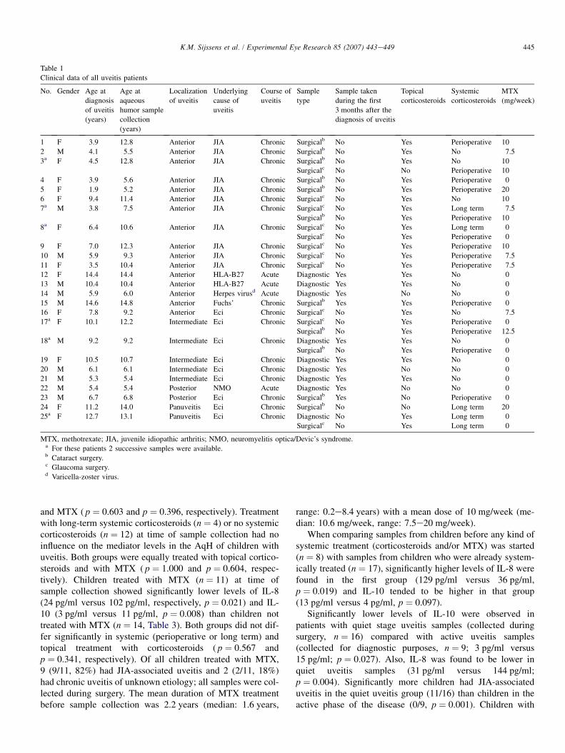
AqH samples were collected as prescribed previously,stored immediately at �80 �C in sterile screw-cap tubes andthawed directly before analysis within 4 years of collection(van der Lelij and Rothova, 1997). This technique is to pre-serve the sample for analysis without degradation. AqH sam-ples of 25 children with uveitis under the age of 16 were used.These samples were obtained between 2002 and 2005 duringsurgery for cataract (n ¼ 8) or glaucoma (n ¼ 8). In the peri-operative period patients were treated with systemic cortico-steroids (1 mg/kg) starting 1 day to 1 week before surgery(n ¼ 9), long term systemic corticosteroids (n ¼ 3) or no sys-temic corticosteroids (n ¼ 4). The remaining 9 samples werecollected for diagnostic purposes. These 9 samples were ana-lyzed for herpes simplex virus, varicella zoster virus and Toxo-plasma gondii antibodies and DNA by PCR (de Groot-Mijneset al., 2006). The clinical data of all patients are shown inTable 1. AqH samples of 6 children under the age of 16,who had an operation for congenital cataract (n ¼ 3), glau-coma not associated with uveitis (n ¼ 2) or lens subluxationdue to Marfan’s syndrome (n ¼ 1), were collected as controls.
In 6 children with uveitis (nos. 3, 7, 8, 17, 18 and 25), morethan one sample was available of the same eye. We comparedin these 6 pairs of samples the levels of cytokines, chemokinesand soluble adhesion molecules before and after surgery.
We recorded the following clinical data for each patient:age at diagnosis of uveitis, age at the time of sample collec-tion, localization and course of uveitis (acute or chronic), un-derlying systemic disease and (systemic) treatment at the timeof sample collection (Table 1).
2.2. Immunoassay
We analyzed the AqH samples in a multiplex immunoassayas described previously (de Jager et al., 2005). In each 50-mlsample, we analyzed 16 mediators; interleukin-1b (IL-1b),IL-2, IL-4, IL-6, IL-10, IL-12 p-70, IL-13, IL-18, interferon-g (IFN-g), tumor necrosis factor-a (TNF-a), soluble intercel-lular adhesion molecule 1 (sICAM-1; CD54), soluble vascularcell adhesion molecule 1 (sVCAM-1; CD106), RANTES(Regulated on Activation, Normal T-cell Expressed, andSecreted; CCL5), Eotaxin (CCL11), IL-8 (CXCL8) andinterferon-inducible 10-kDa protein (IP-10; CXCL10). Con-centrations above or below the detection limit were given as
the highest or lowest detectable value. For statistical analysisconcentrations below the detection limit were converted toa value of 0.5 � the lowest point of the calibration curve.
2.3. Statistical analysis
Statistical analysis of the data was performed by using theSPSS statistical software package version 12.0.1 (SPSS Inc.,Chicago, IL). The KruskaleWallis and the ManneWhitneyU-tests were used to compare the means of the (non-paramet-ric) different groups. The Pearson Chi-square test or Fisher’sexact test was used to compare possible associations betweencategorical variables where appropriate. We used the Wil-coxon Signed Rank test to compare paired samples. p valuesof less than.05 were considered to be statistically significant.
3. Results
Aqueous humor was collected from 25 children with uveitisassociated with juvenile idiopathic arthritis (JIA; n ¼ 11), her-pes virus infection (n ¼ 1), Fuchs’ heterochromic cyclitis(n ¼ 1), HLA-B27 (n ¼ 2), Devic’s neuromyelitis optica(NMO; n ¼ 1) or of unknown etiology (n ¼ 9, Table 1). Thechild with a herpes virus infection had a keratouveitis andthe diagnosis was confirmed by a positive PCR for varicella-zoster virus in AqH. Both patients with HLA-B27 positiveuveitis had acute alternating anterior uveitis. The mean ageat sample collection was 9.6 years (range 5.2e14.8 years)for children with uveitis and 3.3 years (range 0.2e9.5 years,p ¼ 0.002) for the control patients. The mean duration of uve-itis till sampling was 2.3 years (median: 1.4 years, range: 0e9 years).
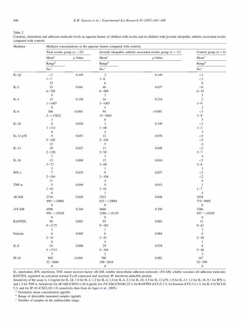
Significantly higher levels of IL-2, IL-6, IL-10 IL-13, IL-18, IFN-g, TNF-a, sICAM-1, RANTES, IL-8 and IP-10were found in children with uveitis than in children withoutuveitis (Table 2). The levels of IL-1b, IL-4, IL-12 p-70,sVCAM-1 and Eotaxin did not significantly differ in AqH ofuveitis compared with controls (Table 2).
No significant differences were found between anterior(n ¼ 16) and intermediate (n ¼ 5) uveitis for all the 16 medi-ators studied. There were only 2 children with posterior uveitisand 2 children with panuveitis therefore no comparison wasmade between all 4 anatomic classifications. Furthermore, be-tween samples from chronic (n ¼ 21) and acute (n ¼ 4) uveitisno significant differences were found for all 16 mediators, butlevels of IP-10 and IL-2 tended to be higher in chronic uveitis(1013 pg/ml versus 361 pg/ml; p ¼ 0.068 and 47 pg/ml versus5 pg/ml; p ¼ 0.081 respectively).
Treatment with or without topical corticosteroids had no in-fluence on the mediator levels in AqH of children with uveitis(n ¼ 20 versus n ¼ 5, respectively). Both groups were equallytreated with systemic corticosteroids (perioperative or longterm) and with MTX ( p ¼ 0.821 and p ¼ 0.341, respectively).Cytokine levels of children treated with perioperative cortico-steroids (n ¼ 9) did not differ significantly from those nottreated with systemic corticosteroids (n ¼ 12). These twogroups did not differ in treatment with topical corticosteroids
445K.M. Sijssens et al. / Experimental Eye Research 85 (2007) 443e449
Table 1
Clinical data of all uveitis patients
No. Gender Age at
diagnosis
of uveitis
(years)
Age at
aqueous
humor sample
collection
(years)
Localization
of uveitis
Underlying
cause of
uveitis
Course of
uveitis
Sample
type
Sample taken
during the first
3 months after the
diagnosis of uveitis
Topical
corticosteroids
Systemic
corticosteroids
MTX
(mg/week)
1 F 3.9 12.8 Anterior JIA Chronic Surgicalb No Yes Perioperative 10
2 M 4.1 5.5 Anterior JIA Chronic Surgicalb No Yes No 7.5
3a F 4.5 12.8 Anterior JIA Chronic Surgicalb No Yes No 10
Surgicalc No No Perioperative 10
4 F 3.9 5.6 Anterior JIA Chronic Surgicalb No Yes Perioperative 0
5 F 1.9 5.2 Anterior JIA Chronic Surgicalb No Yes Perioperative 20
6 F 9.4 11.4 Anterior JIA Chronic Surgicalc No Yes No 10
7a M 3.8 7.5 Anterior JIA Chronic Surgicalc No Yes Long term 7.5
Surgicalb No Yes Perioperative 10
8a F 6.4 10.6 Anterior JIA Chronic Surgicalc No Yes Long term 0
Surgicalc No Yes Perioperative 0
9 F 7.0 12.3 Anterior JIA Chronic Surgicalc No Yes Perioperative 10
10 M 5.9 9.3 Anterior JIA Chronic Surgicalc No Yes Perioperative 7.5
11 F 3.5 10.4 Anterior JIA Chronic Surgicalc No Yes Perioperative 7.5
12 F 14.4 14.4 Anterior HLA-B27 Acute Diagnostic Yes Yes No 0
13 M 10.4 10.4 Anterior HLA-B27 Acute Diagnostic Yes Yes No 0
14 M 5.9 6.0 Anterior Herpes virusd Acute Diagnostic Yes No No 0
15 M 14.6 14.8 Anterior Fuchs’ Chronic Surgicalb Yes Yes Perioperative 0
16 F 7.8 9.2 Anterior Eci Chronic Surgicalc No Yes No 7.5
17a F 10.1 12.2 Intermediate Eci Chronic Surgicalc No Yes Perioperative 0
Surgicalb No Yes Perioperative 12.5
18a M 9.2 9.2 Intermediate Eci Chronic Diagnostic Yes Yes No 0
Surgicalb No Yes Perioperative 0
19 F 10.5 10.7 Intermediate Eci Chronic Diagnostic Yes Yes No 0
20 M 6.1 6.1 Intermediate Eci Chronic Diagnostic Yes No No 0
21 M 5.3 5.4 Intermediate Eci Chronic Diagnostic Yes Yes No 0
22 M 5.4 5.4 Posterior NMO Acute Diagnostic Yes No No 0
23 M 6.7 6.8 Posterior Eci Chronic Surgicalb Yes No Perioperative 0
24 F 11.2 14.0 Panuveitis Eci Chronic Surgicalb No No Long term 20
25a F 12.7 13.1 Panuveitis Eci Chronic Diagnostic No Yes Long term 0
Surgicalc No Yes Long term 0
MTX, methotrexate; JIA, juvenile idiopathic arthritis; NMO, neuromyelitis optica/Devic’s syndrome.a For these patients 2 successive samples were available.b Cataract surgery.c Glaucoma surgery.d Varicella-zoster virus.
and MTX ( p ¼ 0.603 and p ¼ 0.396, respectively). Treatmentwith long-term systemic corticosteroids (n ¼ 4) or no systemiccorticosteroids (n ¼ 12) at time of sample collection had noinfluence on the mediator levels in the AqH of children withuveitis. Both groups were equally treated with topical cortico-steroids and with MTX ( p ¼ 1.000 and p ¼ 0.604, respec-tively). Children treated with MTX (n ¼ 11) at time ofsample collection showed significantly lower levels of IL-8(24 pg/ml versus 102 pg/ml, respectively, p ¼ 0.021) and IL-10 (3 pg/ml versus 11 pg/ml, p ¼ 0.008) than children nottreated with MTX (n ¼ 14, Table 3). Both groups did not dif-fer significantly in systemic (perioperative or long term) andtopical treatment with corticosteroids ( p ¼ 0.567 andp ¼ 0.341, respectively). Of all children treated with MTX,9 (9/11, 82%) had JIA-associated uveitis and 2 (2/11, 18%)had chronic uveitis of unknown etiology; all samples were col-lected during surgery. The mean duration of MTX treatmentbefore sample collection was 2.2 years (median: 1.6 years,
range: 0.2e8.4 years) with a mean dose of 10 mg/week (me-dian: 10.6 mg/week, range: 7.5e20 mg/week).
When comparing samples from children before any kind ofsystemic treatment (corticosteroids and/or MTX) was started(n ¼ 8) with samples from children who were already system-ically treated (n ¼ 17), significantly higher levels of IL-8 werefound in the first group (129 pg/ml versus 36 pg/ml,p ¼ 0.019) and IL-10 tended to be higher in that group(13 pg/ml versus 4 pg/ml, p ¼ 0.097).
Significantly lower levels of IL-10 were observed inpatients with quiet stage uveitis samples (collected duringsurgery, n ¼ 16) compared with active uveitis samples(collected for diagnostic purposes, n ¼ 9; 3 pg/ml versus15 pg/ml; p ¼ 0.027). Also, IL-8 was found to be lower inquiet uveitis samples (31 pg/ml versus 144 pg/ml;p ¼ 0.004). Significantly more children had JIA-associateduveitis in the quiet uveitis group (11/16) than children in theactive phase of the disease (0/9, p ¼ 0.001). Children with
446 K.M. Sijssens et al. / Experimental Eye Research 85 (2007) 443e449
Table 2
Cytokine, chemokine and adhesion molecule levels in aqueous humor of children with uveitis and in children with juvenile idiopathic arthritis associated uveitis
compared with controls
Mediator Mediator concentrations in the aqueous humor compared with controls
Total uveitis group (n ¼ 25) Juvenile idiopathic arthritis associated uveitis group (n ¼ 11) Control group (n ¼ 6)
Meana p-Value Meana p-Value Meana
Rangeb Rangeb Rangeb
No.c No.c No.c
IL-1b <1 0.105 2 0.149 <1
1e7 1e6 <1
15 6 6
IL-2 33 0.041 46 0.037 <6
6e705 6e589 6e35
9 3 5
IL-4 15 0.158 24 0.216 2
1e1407 1e1407 1e9
8 4 2
IL-6 106 <0.001 94 <0.001 <3
3e>13822 13e5603 3e9
1 0 3
IL-10 6 0.020 3 0.149 <1
1e114 1e68 1e2
6 4 3
IL-12 p70 9 0.053 12 0.078 <5
5e105 5e105 <5
12 5 6
IL-13 10 0.027 11 0.048 <2
2e128 2e92 2e7
7 3 4
IL-18 13 0.009 15 0.010 <3
3e72 3e68 3e8
3 1 3
IFN-g 7 0.035 9 0.037 <2
2e104 2e104 <2
11 4 6
TNF-a 5 0.046 9 0.015 1
1e61 1e61 1e7
6 1 4
sICAM 3710 0.020 3523 0.048 1028
490e>10001 621e>10001 374e5669
0 0 0
sVCAM 4506 0.364 4660 0.350 3286
956e>10105 1286e>10 05 947e>10105
0 0 0
RANTES 96 0.001 93 0.002 13
9e1175 9e282 9e61
3 1 3
Eotaxin 6 0.865 6 0.884 6
2e41 2e41 2e48
8 4 1
IL-8 54 0.006 29 0.078 8
5e1715 5e304 5e46
3 2 2
IP-10 859 <0.001 780 0.002 147
52e3680 198e2018 52e399
0 0 0
IL, interleukin; IFN, interferon; TNF, tumor necrosis factor; sICAM, soluble intercellular adhesion molecule; sVCAM, soluble vascular cell adhesion molecule;
RANTES, regulated on activation normal T-cell expressed and secreted; IP, interferon-inducible protein.
Sensitivity of the assay is 1.4 pg/ml for IL-1b, 1.8 for IL-2, 1.2 for IL-4, 2.4 for IL-6, 2.3 for IL-10, 4.3 for IL-12 p70, 1.0 for IL-13, 1.2 for IL-18, 9.1 for IFN-g,
and 1.2 for TNF-a. Sensitivity for sICAM (CD54) is 26.4 pg/ml, for sVCAM (CD106) 22.3, for RANTES (CCL5) 1.5, for Eotaxin (CCL11) 1.3, for IL-8 (CXCL8)
5.3, and for IP-10 (CXCL10) 1.0 (sensitivity data from de Jager et al., 2005).a Geometric mean concentration (pg/ml).b Range of detectable measured samples (pg/ml).c Number of samples in the undetectable range.

447K.M. Sijssens et al. / Experimental Eye Research 85 (2007) 443e449
Table 3
Cytokine, chemokine and adhesion molecule levels in aqueous humor of children with
uveitis treated with methotrexate compared with children with uveitis not treated with
methotrexate
Mediator Mediator concentrations in the aqueous
humor of children with uveitis
Treated with
methotrexate (n ¼ 11)
Not treated with
methotrexate (n ¼ 14)
p-Value
Meana Meana
Rangeb Rangeb
No.c No.c
IL-1b 2 1 0.202
1e6 1e7
5 10
IL-2 60 21 0.244
6e614 6e705
3 6
IL-4 29 9 0.344
1e1407 1e1407
4 4
IL-6 74 140 0.344
11e5603 3e>13822
0 1
IL-10 3 11 0.008
1e17 1e114
4 2
IL-12
p70
175e1054 65e988 0.107
IL-13 14 7 0.317
2e104 2e128
3 4
IL-18 14 12 0.536
3e68 3e72
2 1
IFN-g 11 4 0.183
2e104 2e89
4 7
TNF-a 9 3 0.107
1e61 1e37
2 4
sICAM 3298 4069 0.344
621e>10001 490e>10001
0 0
sVCAM 4537 4482 0.809
1286e>10105 956e>10105
0 0
RANTES 64 132 0.134
9e261 9e1175
2 1
Eotaxin 6 6 0.809
2e18 2e41
4 4
IL-8 24 102 0.021
5e99 5e1715
2 1
IP-10 863 856 0.767
204e2018 52e3680
0 0
IL, interleukin; IFN, interferon; TNF, tumor necrosis factor; sICAM, soluble intercellular
adhesion molecule; sVCAM, soluble vascular cell adhesion molecule; RANTES, regu-
lated on activation normal T-cell expressed and secreted; IP, interferon-inducible protein.
Sensitivity of the assay is 1.4 pg/ml for IL-1b, 1.8 for IL-2, 1.2 for IL-4, 2.4 for IL-6, 2.3
for IL-10, 4.3 for IL-12 p70, 1.0 for IL-13, 1.2 for IL-18, 9.1 for IFN-g, and 1.2 for TNF-
a. Sensitivity for sICAM (CD54) is 26.4 pg/ml, for sVCAM (CD106) 22.3, for RANTES
(CCL5) 1.5, for Eotaxin (CCL11) 1.3, for IL-8 (CXCL8) 5.3, and for IP-10 (CXCL10)
1.0 (sensitivity data from de Jager et al., 2005).a Geometric mean concentration (pg/ml).b Range of detectable measured samples (pg/ml).c Number of samples in the undetectable range.
active uveitis were significantly less treated with MTX (0/9)than children with quiet uveitis (11/16, p ¼ 0.001). The defi-nite diagnosis of patients with diagnostic AqH taps were her-pes virus infection (n ¼ 1), HLA-B27 (n ¼ 2), Devic’sneuromyelitis optica (n ¼ 1) and unknown etiology (n ¼ 5).All active samples, except one, were taken during the first3 months after the diagnosis of uveitis.
Samples collected during surgery (quiet uveitis samples)had significantly higher levels of IL-2, IL-6, IL-13, IL-18,IFN-g, TNF-a, sICAM-1, RANTES, IL-8 and IP-10 thanAqH samples of controls.
Interleukin-10 was also found at a significantly lower levelin AqH samples taken after more than 3 months after the diag-nosis of uveitis (n ¼ 11) than in AqH samples taken during thefirst 3 months after the diagnosis of uveitis (n ¼ 10; 3 pg/mlversus 28 pg/ml, respectively, p ¼ 0.016). The first group wassignificantly more often treated with MTX at time of sampletaking (11/15 versus 0/10, p ¼ 0.001). Of all children inwhom the samples were taken during the first 3 months afterthe diagnosis of uveitis none had JIA-associated uveitis and11 out of 15 children in the group in whom samples were takenafter more than 3 months had JIA-associated uveitis ( p ¼0.001). All other 15 mediators tested were equal in both groups.
JIA was the most common systemic disease in our popula-tion (n ¼ 11). Significantly higher levels of IL-2, IL-6, IL-13,IL-18, IFN-g, TNF-a, sICAM-1, RANTES and IP-10 werefound in AqH of children with JIA-associated uveitis than incontrols (Table 2). Interleukin-8 and IL-10 levels in AqH ofthese children did not differ significantly from controls,whereas these cytokines were significantly increased in the to-tal uveitis group compared with controls. All AqH mediatorsfrom children with JIA-associated uveitis were similar tolevels measured in children with other uveitis entities(n ¼ 14). Children with JIA-associated uveitis were signifi-cantly more often treated with MTX (9/11) than childrenwith uveitis not associated with JIA (2/14, p ¼ 0.001). Whencomparing AqH samples of children with JIA-associated uve-itis (n ¼ 11) with only those with uveitis of unknown etiology(n ¼ 9), no significant differences were found as well.
No differences were found between cytokine levels in AqHsamples of patients with different types of JIA: persistent oli-goarticular JIA (n ¼ 6), extended oligoarticular JIA (n ¼ 2) orpolyarticular JIA (n ¼ 3).
At the time of sample collection only one girl with JIA-associated uveitis was treated with anti-TNF (etanercept twicea week 12 mg subcutaneously) and MTX. Her TNF level inAqH was 14 pg/ml.
Repeated samples of the same eye were available for 6 pa-tients (nos. 3, 7, 8, 17, 18 and 25; Table 1). The first sampleswere taken during surgery (n ¼ 4) or for a diagnostic proce-dure (n ¼ 2). All second samples were taken during surgery(n ¼ 6). The median time interval between the first and secondsample was 1.5 years (range: 0.2e2.3 years). Significantlyhigher levels of IL-6 were observed in the first samples com-pared with the second samples (122 pg/ml versus 12 pg/ml;p ¼ 0.046). This significant difference was not found whenwe excluded the diagnostic samples.
448 K.M. Sijssens et al. / Experimental Eye Research 85 (2007) 443e449
4. Discussion
This is the first study that shows that multiple intraocularcytokines, chemokines and soluble adhesion molecules are el-evated in AqH of children with uveitis. The elevated levelswere observed for cytokines associated with T helper 1(Th1; IL-2, IFN-g, TNF-a), Th2 (IL-13) and T regulatory(Tr; IL-10) response. The reason that we did not find a predom-inance of Th1 cytokine profile, as previously described in theliterature, might be explained by the mixed study population(Lacomba et al, 2000; Takase et al., 2006). However, border-line higher levels of IP-10 and IL-2 were found for the chroniccourse ( p ¼ 0.068 and p ¼ 0.069, respectively). This observa-tion might suggest a predominant Th1 response in childrenwith chronic uveitis. Furthermore, in AqH of children inwhom the samples were collected before start of systemictreatment, we saw higher levels of IL-8, a pro-inflammatorycytokine, compared with AqH of children in whom treatmentwith systemic corticosteroids and/or MTX was already started.
Interleukin-8 (CXCL8) is a chemokine with profound ef-fects on neutrophils, is chemotactic for T lymphocytes andcan induce surface expression of adhesion molecules and isknown as a pro-inflammatory cytokine (de Vos et al., 1992;Dinarello, 1999; Nussenblatt, 2004). Interleukin-10 is ananti-inflammatory cytokine which prevents for the develop-ment of autoimmune diseases (Roncarolo et al., 2001; Thomp-son and Powrie, 2004). The observation that higher levels ofIL-10 and IL-8 were detected in samples collected for diag-nostic purpose (active uveitis) compared with samples col-lected during ocular surgery (quiet uveitis) might implicatethat these cytokines are mainly involved in active uveitis. In-terleukin-10 was higher in samples taken during the first3 months after the diagnosis of uveitis. These samples weremainly (8 out of 10) collected for diagnostic purposes beforethe start of systemic treatment. Therefore IL-10 might beinvolved in the early stage of the disease process, in activeuveitis, or both. The lower levels of IL-8 in uveitis samplescollected during ocular surgery compared with diagnosticsamples might be explained by the fact that the ocular inflam-mation was at that point quiet (due to treatment). However, IL-8 and IL-10 were found in a lower range in children treatedwith MTX. From our data it is not clear whether the earlystages or active uveitis are responsible for higher levels ofIL-8 and IL-10 or, alternatively, whether the absence of treat-ment with MTX causes this difference.
Despite the absence of clinical signs of active inflamma-tion, elevated cytokine levels were found in the samples ob-tained during surgical procedures compared with controls.This might implicate that when the uveitis is in clinical remis-sion (no inflammatory cells in the anterior chamber or otherinflammatory features), there is an ongoing immune responsein the eye based on the elevated cytokines. When comparingmediator levels in AqH collected during successive surgicalinterventions in the same eye, no change in (inflammatory) cy-tokines was found. As such, this is reassuring because it indi-cates that the surgical intervention itself does not lead toa trauma induced cytokine response.
In the present study we did not find differences in cytokinepattern between the different types of JIA. Patients with sys-temic JIA have higher serum levels of IL-6 than other formsof JIA (Rooney et al., 1995). Our series does not include sam-ples of uveitis patients with systemic JIA, because uveitis isa very rare feature of systemic JIA. Furthermore, the JIA sam-ples used in this study were collected during cataract or glau-coma surgery. So all these patients had surgery requiringcomplications of JIA-associated uveitis and therefore theymight not be representative of the total JIA-associated uveitispopulation.
Limitations of this study include the restricted number ofsamples which precludes meaningful comparison of specificdisorders or subcategories of disorders such as anatomic clas-sification, course of uveitis and systemic diseases. Further-more, our control group was not perfectly age-matched andlow in number. The reason for this is that AqH samples of chil-dren with or without uveitis are not readily available. At thetime of sample collection the children in the control groupwere younger than the children with uveitis. However, whenwe compared our control samples with the adult controls ofthe study of our colleagues, no major differences were found,except for IL-6, sICAM-1 and sVCAM-1 (van Kooij et al.,2006). More detailed studies on the age dependency of cyto-kine profiles in AqH of uveitis patients are currentlyunderway.
For future studies, it will be important to collect and exam-ine larger series of AqH samples of children with uveitis, inorder to make comparisons between different systemic dis-eases (for example JIA-associated versus HLA-B27 positiveuveitis) among the 4 anatomical classifications of uveitis andother comparisons possible. We did not find significant differ-ences between cytokine levels in AqH of JIA-associated uve-itis and other uveitis entities. Based on this relatively smallstudy population we cannot conclude whether there is a com-mon pathway in juvenile uveitis, irrespective of the underlyingcause. It would be interesting to examine this in a bigger studypopulation with a special regard for IL-8 and IL-10. Further-more, it would be interesting to compare cytokine and totalIgG levels in AqH with those in serum of children with uveitis,to make a discrepancy between local production and influxfrom elsewhere due to blood aqueous barrier breakdown.
In the last decades the knowledge of the immune systemhas improved and treatment of inflammatory diseases withbiological agents has become available. Biologics are drugsdirected against specific cytokines or their receptors (Dunn,2004). In children, treatment of JIA with anti-TNF has beensuccessful for arthritis. In the near future, treatment with sev-eral other biologics might become available. Therefore moreinsight into the cytokine profile in childhood uveitis iswarranted.
In conclusion, our data suggest that a spectrum of cyto-kines, chemokines and soluble adhesion molecules in theAqH of children with uveitis are involved in the pathogenesisof uveitis. These mediators are present irrespective of active orinactive uveitis and without clear predominance of a Th1 orTh2 associated cytokine profile. Whether the IL-10 and IL-8

449K.M. Sijssens et al. / Experimental Eye Research 85 (2007) 443e449
levels in AqH appear to be related to uveitis activity, early orlate phase of the course of uveitis or systemic treatment withMTX needs further investigation. Further studies will be re-quired to elucidate the exact role of these mediators in thepathogenesis of different uveitis entities, especially JIA, andspecific treatment strategies.
Acknowledgement
K.M.S. was supported by the Dr. F.P. Fischer Foundation,Utrecht, The Netherlands.
References
Curnow, S.J., Falciani, F., Durrani, O.M., 2005. Multiplex bead immunoassay
analysis of aqueous humor reveals distinct cytokine profiles in uveitis.
Invest. Ophthalmol. Vis. Sci. 46, 4251e4259.
de Boer, J., Wulffraat, N., Rothova, A., 2003. Visual loss in uveitis of child-
hood. Br. J. Ophthalmol. 87, 879e884.
de Groot-Mijnes, J.D.F., Rothova, A., van Loon, A.M., et al., 2006. Polymerase
chain reaction and Goldmann-Witmer coefficient analysis are complementary
for the diagnosis of infectious uveitis. Am. J. Ophthalmol. 141, 313e318.
de Jager, W., Prakken, B.J., Bijlsma, J.W., et al., 2005. Improved multiplex im-
munoassay performance in human plasma and synovial fluid following re-
moval of interfering heterophilic antibodies. J. Immunol. Methods 300,
124e135.
de Vos, A.F., Hoekzema, R., Kijlstra, A., 1992. Cytokines and uveitis, a review.
Curr. Eye Res. 11, 581e597.
Dinarello, C.A., 1999. IL-18: A TH1-inducing, proinflammatory cytokine
and new member of the IL-1 family. J. Allergy Clin. Immunol. 103,
11e24.
Dunn, J.P., 2004. Review of immunosuppressive drug therapy in uveitis. Curr.
Opin. Ophthalmol. 15, 293e298.
Evereklioglu, C., 2005. Current concepts in the etiology and treatment of Beh-
cet disease. Surv. Ophthalmol. 50, 297e350.
Kanski, J.J., Shun-Shin, G.A., 1984. Systemic uveitis syndromes in childhood:
an analysis of 340 cases. Ophthalmology 91, 1247e1252.
Lacomba, M.S., Martin, C.M., Chamond, R.R., et al., 2000. Aqueous and se-
rum interferon gamma, interleukin (IL) 2, IL-4, and IL-10 in patients with
uveitis. Arch. Ophthalmol. 118, 768e772.
Nussenblatt, R.B., 2004. Uveitis. Fundamentals and Clinical Practice, third ed.
Elsevier, Philadelphia, PA.
Roncarolo, M.G., Bacchetta, R., Bordignon, C., et al., 2001. Type 1 T regula-
tory cells. Immunol. Rev. 182, 68e79.
Rooney, M., David, J., Symons, J., et al., 1995. Inflammatory cytokine re-
sponses in juvenile chronic arthritis. Br. J. Rheumatol. 34, 454e460.
Rothova, A., Suttorp-van Schulten, M.S., Treffers, W.F., Kijlstra, A., 1996.
Causes and frequency of blindness in patients with intraocular inflamma-
tory disease. Br. J. Ophthalmol. 80, 332e336.
Takase, H., Futagami, Y., Yoshida, T., et al., 2006. Cytokine profile in aqueous
humor and sera of patients with infectious or noninfectious uveitis. Invest.
Ophthalmol. Vis. Sci. 47, 1557e1561.
Thompson, C., Powrie, F., 2004. Regulatory T cells. Curr. Opin. Pharmacol. 4,
408e414.
Tugal-Tutkun, I., Havrlikova, K., Power, W.J., Foster, C.S., 1996. Changing
patterns in uveitis of childhood. Ophthalmology 103, 375e383.
van der Lelij, A., Rothova, A., 1997. Diagnostic anterior chamber paracentesis
in uveitis: a safe procedure? Br. J. Ophthalmol. 81, 976e979.
van Kooij, B., Rothova, A., Rijkers, G.T., de Groot-Mijnes, J.D., 2006. Distinct
cytokine and chemokine profiles in the aqueous of patients with uveitis and
cystoid macular edema. Am. J. Ophthalmol. 142, 192e194.
Related Documents