Cytocompatibility of novel extracellular matrix protein analogs of biodegradable polyester polymers derived from α-hydroxy amino acids Shimon Lecht a , Naomi Cohen-Arazi b , Gadi Cohen b , Keren Ettinger b , Tatjana Momic b , Michal Kolitz b , Majdi Naamneh b , Jehoshua Katzhendler b , Abraham J. Domb b , Philip Lazarovici b and Peter I. Lelkes a * a Department of Bioengineering and Temple Institute for Regenerative Medicine and Engineering, Temple University, Philadelphia, PA 19122, USA; b Faculty of Medicine, School of Pharmacy Institute for Drug Research, The Hebrew University of Jerusalem, Jerusalem 91120, Israel (Received 16 October 2013; accepted 24 January 2014) One of the challenges in regenerative medicine is the development of novel biodegradable materials to build scaffolds that will support multiple cell types for tis- sue engineering. Here we describe the preparation, characterization, and cytocompati- bility of homo- and hetero-polyesters of α-hydroxy amino acid derivatives with or without lactic acid conjugation. The polymers were prepared by a direct condensation method and characterized using gel permeation chromatography, 1 H-nuclear magnetic resonance spectroscopy, Fourier transform infrared spectroscopy, differential scanning calorimetry, optical activity, and solubility. The surface charge of the polymers was evaluated using zeta potential measurements. The polymers were coated onto glass cover slips followed by characterization using nano-surface profiler, thin film reflec- tometry, and atomic force microscopy (AFM). Their interaction with endothelial and neuronal cells was assessed using adhesion, proliferation, and differentiation assays. Of the characterized polymers, Poly-HOVal-LA, but not Poly-(D)HOPhe, signifi- cantly augmented nerve growth factor (NGF)-induced neuronal differentiation of the PC12 pheochromcytoma cells. In contrast, Poly-HOLeu increased by 20% the adhesion of endothelial cells, but did not affect PC12 cell differentiation. NGF- induced Erk1/2 phosphorylation in PC12 cells grown on the different polymers was similar to the effect observed for cells cultured on collagen type I. While no significant association could be established between charge and the differentiative/ proliferative properties of the polymers, AFM analysis indicated augmentation of NGF-induced neuronal differentiation on smooth polymer surfaces. We conclude that overall selective cytocompatibility and bioactivity might render α-hydroxy amino acid polymers useful as extracellular matrix-mimicking materials for tissue engineering. Keywords: α-hydroxy amino acid polyester; zeta potential; cytocompatibility; neuronal cells; endothelial cells; adhesion; proliferation; differentiation 1. Introduction Engineering tissue-like constructs requires tissue-specific interactions of somatic cells and biomimetic scaffolds emulating the complex endogenous environment, i.e. the *Corresponding author. Email: [email protected] Shimon Lecht and Naomi Cohen-Arazi contributed equally to this work. © 2014 Taylor & Francis Journal of Biomaterials Science, Polymer Edition, 2014 http://dx.doi.org/10.1080/09205063.2014.888303 Downloaded by [Temple University Libraries] at 20:12 27 February 2014

Welcome message from author
This document is posted to help you gain knowledge. Please leave a comment to let me know what you think about it! Share it to your friends and learn new things together.
Transcript
Cytocompatibility of novel extracellular matrix protein analogs ofbiodegradable polyester polymers derived from α-hydroxy amino
acids
Shimon Lechta, Naomi Cohen-Arazib, Gadi Cohenb, Keren Ettingerb, Tatjana Momicb,Michal Kolitzb, Majdi Naamnehb, Jehoshua Katzhendlerb, Abraham J. Dombb,
Philip Lazarovicib and Peter I. Lelkesa*
aDepartment of Bioengineering and Temple Institute for Regenerative Medicine and Engineering,Temple University, Philadelphia, PA 19122, USA; bFaculty of Medicine, School of PharmacyInstitute for Drug Research, The Hebrew University of Jerusalem, Jerusalem 91120, Israel
(Received 16 October 2013; accepted 24 January 2014)
One of the challenges in regenerative medicine is the development of novelbiodegradable materials to build scaffolds that will support multiple cell types for tis-sue engineering. Here we describe the preparation, characterization, and cytocompati-bility of homo- and hetero-polyesters of α-hydroxy amino acid derivatives with orwithout lactic acid conjugation. The polymers were prepared by a direct condensationmethod and characterized using gel permeation chromatography, 1H-nuclear magneticresonance spectroscopy, Fourier transform infrared spectroscopy, differential scanningcalorimetry, optical activity, and solubility. The surface charge of the polymers wasevaluated using zeta potential measurements. The polymers were coated onto glasscover slips followed by characterization using nano-surface profiler, thin film reflec-tometry, and atomic force microscopy (AFM). Their interaction with endothelial andneuronal cells was assessed using adhesion, proliferation, and differentiation assays.Of the characterized polymers, Poly-HOVal-LA, but not Poly-(D)HOPhe, signifi-cantly augmented nerve growth factor (NGF)-induced neuronal differentiation of thePC12 pheochromcytoma cells. In contrast, Poly-HOLeu increased by 20% theadhesion of endothelial cells, but did not affect PC12 cell differentiation. NGF-induced Erk1/2 phosphorylation in PC12 cells grown on the different polymers wassimilar to the effect observed for cells cultured on collagen type I. While nosignificant association could be established between charge and the differentiative/proliferative properties of the polymers, AFM analysis indicated augmentation ofNGF-induced neuronal differentiation on smooth polymer surfaces. We conclude thatoverall selective cytocompatibility and bioactivity might render α-hydroxy aminoacid polymers useful as extracellular matrix-mimicking materials for tissueengineering.
Keywords: α-hydroxy amino acid polyester; zeta potential; cytocompatibility;neuronal cells; endothelial cells; adhesion; proliferation; differentiation
1. Introduction
Engineering tissue-like constructs requires tissue-specific interactions of somatic cellsand biomimetic scaffolds emulating the complex endogenous environment, i.e. the
*Corresponding author. Email: [email protected] Lecht and Naomi Cohen-Arazi contributed equally to this work.
© 2014 Taylor & Francis
Journal of Biomaterials Science, Polymer Edition, 2014http://dx.doi.org/10.1080/09205063.2014.888303
Dow
nloa
ded
by [
Tem
ple
Uni
vers
ity L
ibra
ries
] at
20:
12 2
7 Fe
brua
ry 2
014

extracellular matrix (ECM), in which cells reside, differentiate and function. To date, alarge variety of materials, natural and synthetic, have been investigated as potentialscaffolds for tissue engineering. The design of unique materials compatible with a num-ber of distinct cell types, cumulatively representing a particular tissue, has spurred thediscovery of novel compounds, both of natural and synthetic origins.[1] Synthetic poly-mers offer a number of advantages over natural ones for developing tissue scaffolds,mainly due to the ability to concomitantly tailor a number of important parameters,such as biocompatibility, biodegradability, and viscoelastic properties, which cumula-tively suit particular regenerative applications. Synthetic polymers are also attractivebecause they can be fabricated into various shapes and geometries according to tissue-specific requirements. Among these synthetic polymers, poly(α-hydroxy acids) such aspoly(glycolic acid) (PGA), poly(lactic acid) (PLA), poly(caprolactone), poly(propylene-fumarates), polydioxanone, polyvalerolactone, polyanhydrides, polycarbonates, polyure-thanes, and polyphosphazenes as well as their copolymers have most commonly beenused in tissue engineering.[2,3]
Polyesters are attractive for tissue engineering applications because of their biode-gradability in vivo as a result of endogenous esterases, which hydrolyze their ester link-age. A large number of these polyesters have been designed from monomers generatedduring normal human metabolism (e.g. lactic acid) and synthesized in a manner thatmaximizes their in vitro cytocompatibility and in vivo biocompatibility (e.g. ester links),and minimizes cytotoxicity by using natural metabolites as monomeric buildingblocks.[4] Bioactive polyesters can be designed with functional groups that can guidecell proliferation and assembly into a tissue-like organization.[3,5] Amino acid-basedpolymers, such as self-assembling peptide amphiphiles,[6] α-helices,[7] β-sheets,[8] andβ-amino acid helices,[9] mimicking certain aspects of the three-dimensional (3-D)ECM, represent promising design components that facilitate generation of nanofibersand/or hydrogels for cell encapsulation and injectable delivery of cells.
Rat PC12 pheochromocytoma cells have been extensively used as a cellular modelfor studying axonal development and neuronal differentiation on a variety of naturaland synthetic substrates. The ability of PC12 cells to develop axon-like neurites upontreatment with nerve growth factor (NGF) was used to study modulatory effects of crit-ical engineering parameters, such as 2-D vs. 3-D culture conditions,[10] matrix-derivedbiophysical cues such as micro- and nano-topography,[11,12] and substrate stiff-ness.[13] Furthermore, PC12 cells were used to explore the micro-environmentaladequacy for neuronal cells of new bioengineered materials such as agarose scaffolds,[14] peptide scaffolds,[15] neuron–microelectrode interfaces,[12,16] micro-channel-containing substrates,[17] and micro-fabricated silicon-based nanostructures.[12,18]Since the endothelium is vital for the formation of blood capillaries capable of deliver-ing oxygen and nutrients throughout engineered tissue constructs,[19] we also evaluatedthe cytocompatibility of newly developed poly-α-hydroxy amino acids as substrates forbrain microcapillary-derived bEnd.3 endothelial cells (EC).[20–22] These EC have beenpreviously used as a model for cerebral angiogenesis by virtue of expressing andresponding to NGF.[23,24] In the past, others and we have explored interactionsbetween the neural and the vascular systems (‘neurovascular’ crosstalk), which share anumber of important common growth and guidance factors, such as vascular endothe-lial growth factor (VEGF), NGF, semaphorins, ephrins, and others.[25] Therefore,PC12 and bEnd.3 cells appear to be excellent model systems for studying this crosstalkin vitro in the context of permissive substrates and matrices.
2 S. Lecht et al.
Dow
nloa
ded
by [
Tem
ple
Uni
vers
ity L
ibra
ries
] at
20:
12 2
7 Fe
brua
ry 2
014

We have previously reported the preparation of new α-hydroxy acids derived fromamino acids, their corresponding optically active polyesters,[26,27] and described theirhydrolytic degradation behavior.[27,28] In our quest for creating new biomaterials fortissue engineering, we evaluated the cytocompatibility of polymeric surfaces made fromα-hydroxy amino acid polymers, i.e. their ability to support in vitro the adhesion, pro-liferation, and differentiation of cultured neuronal and endothelial cells.
2. Materials and methods
2.1. Materials
Lactic acid (LA) was purchased from J.T.Baker (Deventer, Holland). Amino acids werepurchased from Fluka (Buchs, Switzerland) and Sigma-Aldrich (Milwaukee, WI)(98–99% pure), as were sodium nitrite (99.5%) and p-toluenesulfonic acid-monohydrate(98.5%). All solvents were of analytical grade and purchased from Biolab (Jerusalem,Israel), Frutarom (Haifa, Israel), or Gadot (Or Aqiva, Israel), and were used withoutfurther purification. Mouse NGF was purchased from Alomone Labs (Jerusalem,Israel).
2.2. Methods
2.2.1. Synthesis of α-hydroxy acids by diazotization of α-amino acids
The α-hydroxy acid derivatives of L-isoleucine, L- and D-leucine, phenylalanine,valine, L-aspartic acid, L-glutamic acid, L-serine, and L-threonine were prepared by achemical diazotization method, as previously described.[26–28] The α-hydroxy aminoacids were characterized by 1H-NMR (DMSO-d6), molecular weight (Mp), elementalanalysis, optical activity and mass spectrometry, as previously described.[26,27]
2.2.2. Polymerization of α-hydroxy amino acids
The hydroxy amino acids were condensed by direct condensation in bulk as previouslydescribed.[29,30] The polymers were characterized by 1H-NMR, IR, GPC, opticalactivity, and solubility in several solvents, as previously detailed.[26–28]
2.2.3. Fourier transform infrared (FT-IR) spectroscopy
To measure the functional groups on the surface area of the polymer film, FT-IRspectra were collected using a 2000 FT-IR, PerkinElmer instrument (Waltham, MA).Measurements of polymers film were performed on NaCl plates employing achloroform solution of the polymers, as previously reported.[26]
2.2.4. Molecular weight determination
The molecular weights of the polyesters were estimated in a gel permeation chromatog-raphy (GPC) system consisting of a Waters 1515 isocratic HPLC pump with a Waters2410 refractive index detector and a Rheodyne (Cotati, CA) injection valve with a20 μL-loop (Milford, MA). The samples were eluted with CHCl3 through a linearStyragel HR1 column (Waters) at a flow rate of 1 ml/min. The molecular weights were
Journal of Biomaterials Science, Polymer Edition 3
Dow
nloa
ded
by [
Tem
ple
Uni
vers
ity L
ibra
ries
] at
20:
12 2
7 Fe
brua
ry 2
014

determined relative to polystyrene standards (Polyscience, Warrington, PA) with amolecular weight range of 100–5000 Dalton using the Breeze computer program.
2.2.5. Differential scanning calorimetry
Differential scanning calorimetry (DSC) was used to measure the heat flux of thermalphase transitions of the polymers using a Mettler TA 4000-DSC differential scanningcalorimeter (Mettler-Toledo, Schwerzenbach, Switzerland), heated at a rate of10 °C/min under nitrogen atmosphere.[27]
2.2.6. Zeta-potential measurement
To measure the electric potential in the interfacial double layer at the surface of theshear (or slipping) plane of particles in suspension, the zeta-potential of the polymerswas measured upon dispersion in 0.1M phosphate buffer, pH 7.4 using a MalvernZetasizer (Malvern Instruments, UK).
2.2.7. LogP measurement
To measure the hydrophobicity of the polymers, logP values were calculated usingBernard Testa’s virtual logP calculator (http://nova.disfarm.unimi.it/vlogp.htm).
2.2.8. Preparation of polymer film coatings on glass cover slips
The effects of the synthesized polymers on cellular properties were evaluated uponcoating 13 mm circular glass cover slips (Sigma-Aldrich) with the polymers. The coat-ing was performed by single dip of the coverslips into the polymer coating solution atconcentrations up to 100 μg/ml (dissolved in chloroform). The polymer-coated glasscover slips were then dried in a heated (37 °C) vacuum oven for 1 h, washed threetimes with water, and inserted into conventional 24-well tissue culture plates (Nunc,Rochester, NY). The surface analyses of the polymer-coated cover slips were performedbefore and after UV sterilization for 15 min in a tissue culture hood. In one approach,the thickness of polymer films was measured with a Dektak 150 Surface Profiler(Veeco, Plainview, NY). In the second approach, the polymer-coating parameters wereanalyzed by thin film reflectometry (NanoCalc, Ocean Optics, Dunedin, FL), whichallows for the analysis of the thickness and roughness of film coating from 1 nm to250 μm, with a resolution of 0.1 nm. The polymer-coated cover slips were used for fur-ther characterization by atomic force microscopy (AFM) and for culturing endothelialand neuronal cells.
2.2.9. Atomic force microscopy analysis
To evaluate the surface topology, AFM images were obtained using a NanoscopeDimension 3100 Scanning Probe Microscope along with a Nanoscope V controller(Digital Instruments Veeco Metrology Group, Plainview, NY) in tapping mode using aTRESP probe (Si probe with res. frequency around 300 kHz) with radius of curvatureof 5–10 nm.[31]
4 S. Lecht et al.
Dow
nloa
ded
by [
Tem
ple
Uni
vers
ity L
ibra
ries
] at
20:
12 2
7 Fe
brua
ry 2
014

2.2.10. Cell cultures
PC12 pheochromocytoma cells and brain-derived bEnd.3 microvascular endothelial celllines were cultured, as previously described.[24] Briefly, the basal growth medium forboth cell types consisted of Dulbecco’s modified Eagle’s medium (DMEM) supple-mented with 4.5 mg/ml glucose, 2 mM L-glutamine, penicillin 10,000 U/ml, and strep-tomycin 100 μg/ml (Beit Haemek, Israel). For bEnd.3, the medium was supplementedwith 10% fetal calf serum (FCS), for PC12 with 7.5% FCS and 7.5% horse serum (BeitHaemek, Israel). All cell cultures were maintained at 37 °C in a humidified incubator ina mixture of 5% CO2/95% air. The medium was changed every other day. Visual evalu-ation of the cells was performed using a Nikon Eclipse TS-100 (Japan) inverted phase-contrast light microscope. Digital photomicrographs were taken at 20× magnification.
2.2.11. Proliferation and adhesion assays
Endothelial cells were trypsinized using 0.25% trypsin solution (Beit Haemek, Israel),collected, and counted using hemocytometer. A total of 100,000 cells were suspendedin 1 ml of serum-free media, introduced into the wells of 24-well plates containingglass cover slips coated with different polymers, left to adhere for 4 h, and washed toremove non-adherent cells. The number of adherent cells at this time point (set as day0) was evaluated using Alamar Blue (AB, from BD Biosciences), as previouslydescribed.[32] Briefly, following the removal of the non-adhered cells, the wells wereincubated for 4 h with 1 ml of the AB reagent (10% v/v in serum-free media). At theend of the incubation period, 100 μl aliquots of media were collected and their fluores-cence intensity was evaluated in triplicates in a SPECTRAFluor Plus plate reader at anexcitation of 560 nm and emission of 595 nm (Tecan, Switzerland). Upon removal ofthe AB-containing media, the wells were washed three times with basal media contain-ing 1% serum, and then re-fed with the experimental media, returned to the incubator,and left in culture for three days. At that time the cultures were again incubated withAB as described above. For each polymer coating, cell proliferation was calculated asthe increase in AB fluorescence relative to day 0 and normalized to the proliferation ofthe cells on uncoated cover slips.
Adhesion kinetics of the cells were measured essentially as described previouslywith minor changes.[33] One day prior to the experiment, each well of 96-well plateswas coated with polymers. Thereafter, non-specific binding was blocked by incubatingthe wells with 1% (w/v) bovine serum albumin (BSA) in Hank’s balanced salt solution(HBSS) containing 5 mM MgCl2, at room temperature for 1 h prior to use. Cells werelabeled by incubation with 12.5 μM 5-chloromethylfluorescein diacetate (CMFDA,Invitrogen, Carlsbad, CA) in HBSS without 1% BSA at 37 °C for 30 min. The labeledcells were then centrifuged at 1000 rpm and washed twice with HBSS containing 1%BSA to remove excess CMFDA. Labeled cells (1 × 10⁵) were added to each well andincubated at 37 °C for up to 90 min. Unbound cells were removed by washing the wellsthree times with 1% (w/v) BSA in HBSS, and bound cells were lysed by the additionof 100 μl 0.5% Triton X-100 (diluted in water). The fluorescent signal in each well wasquantified using a SPECTRAFluor Plus plate reader (Tecan), at λex = 485 nm andλem = 530 nm and is presented in arbitrary relative fluorescence units (RFU).
Journal of Biomaterials Science, Polymer Edition 5
Dow
nloa
ded
by [
Tem
ple
Uni
vers
ity L
ibra
ries
] at
20:
12 2
7 Fe
brua
ry 2
014

2.2.12. Western blotting
Western blotting to assess Erk1/2 phosphorylation was performed as previouslydescribed.[24] Briefly, cells cultured for 24 h on different polymers were collected, sol-ubilized, and equal amounts of protein (40 μg) were loaded on 10% polyacrylamidegels, separated by SDS-PAGE (100 V for 1.5 h), transferred on ice to nitrocellulosemembranes (90 V for 1.5 h) (Whatman, Germany). Immunodetection was performedusing primary antibodies against phospho- or pan-Erk1/2 (Cat. No.: 4377 and 4695,respectively; both antibodies: 1:1000; Cell Signaling Technology Inc, Beverly, MA) fol-lowed by incubation with HRP conjugated secondary antibody. The blots were visual-ized using an ECL reagent (Pierce, Rockford, IL). Thereafter, the membranes werestripped and reprobed with an anti-pan-ERK-specific antibody. Densitometric analysiswas performed with the Quantity One 1-D software (Bio-Rad, Hercules, CA). The rela-tive density of the bands of the phosphorylated protein was divided by the density ofthe bands of the pan-protein; for further comparisons the control densitometric ratios ofthe untreated samples were set at an arbitrary value of 10.
2.2.13. PC12 cell differentiation, neuronal outgrowth, quantitative assay
PC12 pheochromocytoma cells were plated at a density of 10,000 cells per well oncover slips coated with the different polymers and treated with 50 ng/ml NGF(Alomone, Israel) for 5–7 days with media being replenished every other day. Neuriteslength was quantitated using fractal dimension (Df) as a suitable parameter for quantita-tive assessment of neuronal differentiation, as previously described.[10] Briefly, digitalimages of the cells and neurite outgrowths were acquired and network overlay was gen-erated using Photoshop™. Thereafter, the network layer was measured using ImageJsoftware, utilizing fractal box count analysis to facilitate visual inspection of the neuro-nal network morphology, and ‘skeletonized’ to calculate the fractal dimension (Df).[10]
2.2.14. Statistics
Unless indicated otherwise, the results are presented as the mean ± SD of at least threeindependent experiments. The data were then evaluated using the InStat3 statistics pro-gram (GraphPad, La Jolla, CA). Statistically significant differences between experimen-tal groups were determined by analysis of variance (ANOVA) with Dunnett post-testand considered significant for p < 0.05.
3. Results
3.1. Synthesis and chemical characterizations of α-hydroxy amino acids and derivedpolymers
Hydroxy acid derivatives of the amino acids isoleucine, leucine, phenylalanine, valine,glutamic acid, aspartic acid, serine, and threonine were prepared with 60–70% yields,as shown in Figure 1(A) (the amino acids are all L-isomers unless otherwisestated).[34,35] Polyesters were synthesized from the α-hydroxy amino acids, as well ascopolymers including lactic acid with a molar ratio of 1:1. The polyesters wereprepared by direct condensation in bulk with p-toluene sulfonic acid as catalyst(Figure 1(B)). The molecular weights of the synthesize polymers ranged between 1000and 3000 Da (Table 1). At room temperature the hydrophobic polymers appeared
6 S. Lecht et al.
Dow
nloa
ded
by [
Tem
ple
Uni
vers
ity L
ibra
ries
] at
20:
12 2
7 Fe
brua
ry 2
014

viscous-yellow to off-white. They were ointment-like and soluble in chloroform, THF,and acetonitrile, but not in water. By contrast, the hydrophilic polymers (Table 1) wereeasily dissolved in water (due to their hydrophilic side chains OH and/or COOH), sug-gesting that these polymers are either branched or linear, rather than cross-linked. Incontrast to Poly-HOSer and Poly-HOThr, the Poly-HOAsp and Poly-HOGlu were firm
Figure 1. Synthesis of the α-hydroxy amino acids (A) and derived polyesters (B).
Table 1. Molecular weight (Mw, Mn), zeta potential (Zp), and logP of the synthesizedpolyesters.
Polymer name Mw (Da) Mn (Da) Zp(mV) /Buffera LogPb
DPLA 2800 2600 −35.45 0.18Poly-HOIle 1000 800 −22.15 9.54Poly-HOIle-LA 1800 1600 −28.15 11.73Poly-HOLeu 2300 1900 −49.2 19.19Poly-HOLeu- LA 2300 1900 −39.03 13.90Poly-HOPhe 2500 2200 −58.75 20.54Poly-HOPhe 2200 2300 −54.45 18.34Poly-HOPhe-LA 2500 2300 −41.05 14.572Poly-HOVal 1000 700 −56.0 6.57Poly-(D)HOVal 1100 800 −51.71 7.01Poly-HOVal-LA 1700 1400 −39.1 7.69Poly-HOAsp 1300 1100 −11.8 −4.31Poly-HOAsp-LA 2000 1900 −20.8 −2.67Poly-HOGlu 1500 1400 −13.55 −3.419Poly-HOGlu-LA 1200 1100 −23.55 −1.18Poly-HOSer 1100 1000 −13.06 −8.58Poly-HOSer-LA 2200 1500 −22.94 −8.22Poly-HOThr 3300 3200 −14.35 −11.50Poly-HOSer-HOIle 2200 2000 −17.6 4.36Poly-HOSer-HOLeu 2100 2000 −28.1 3.12Poly-HOSer-HOPhe 2500 2300 −23.15 5.57Poly-HOSer-HOVal 2000 1700 −27.9 0.19Poly-HOGlu-HOPhe 2500 2500 −31.1 9.43
a0.1M phosphate buffer, pH 7.4.bLogP was calculated for oligomers of the referred molecular weight (Mw). The compositions of the co-poly-mers were defined in 1:1 ratio.
Journal of Biomaterials Science, Polymer Edition 7
Dow
nloa
ded
by [
Tem
ple
Uni
vers
ity L
ibra
ries
] at
20:
12 2
7 Fe
brua
ry 2
014

solid polymers that exhibited melting points in the DSC, in addition to Tg. For Poly-HOAsp, the Tm was 133.37°C (ΔH = −420.42 J/g) and for Poly-HOGlu the Tm was81.14 °C (ΔH = −127.20 J/g).[26–28]
Polymerization of the monomers was validated by 1H-NMR spectroscopy and char-acterized by the appearance of a peak between 5.1 and 5.3 ppm, which can be assignedto the hydrogen ester group CH(R)COOCH of the polymer form, and the notable disap-pearance of the CH(R)OH peak of the monomer hydroxy acid at 4–4.3 ppm, as previ-ously described.[26] By IR spectroscopy, all polymers exhibited a characteristic majorester peak between 1750 and 1765 cm−1 and a small acid peak around 1640 cm−1 indi-cating the presence of a polyester backbone, as depicted in Figure 2 for several repre-sentative polymers.
3.2. Polymers charge and hydrophobicity
Since the charge and hydrophobicity of a given surface are commonly used to rational-ize cell attachment to different materials, we characterized the zeta-potential and logPof the polymers (Table 1). For the hydrophobic polymers (Phe, Val, Leu, and Ile), val-ues for the zeta potential were in the range of −22.0 to −58.7, while in the case of themore hydrophilic and anionic polymers (Ser, Thr, Asp, and Glu) zeta potential valuesranged between −11.8 and −14.35. logP, the partitioning coefficient between water andoctanol is an indicator of hydrophobicity and lipophilicity.[36] As seen in Table 1, thecalculated logP values for all the polyesters derived from hydroxy amino acids were inthe range of −11.5 to + 20.5, with logP values for Poly-HOVal-LA, Poly-HOLeu, andPoly-HOPhe were 7.6, 19.1 and 20.5, respectively, which can be attributed to aheterogeneous mild hydrophobicity.
3.3. Cell adhesion, proliferation and differentiation on polyester films
As a first step towards assessing cytocompatibility of the newly synthesized polymers,we evaluated the adhesion, proliferation and differentiation of neuronal and endothelialcells on all synthesized polymeric coatings. In a first set of experiments, PC12 cellswere seeded onto cover slips, which were coated with polymers at a concentration of100 μg/ml. Using lactate dehydrogenase release into the medium, as an indicator of cell
Figure 2. Surface IR spectra of polymer film of three representative polymers: blue –Poly-HOVal-LA; purple – Poly-(D)HOPhe; and green – Poly-HOLeu. (Please see the onlinearticle for the colour version of this figure: http://dx.doi.org/10.1080/09205063.2014.888303.)
8 S. Lecht et al.
Dow
nloa
ded
by [
Tem
ple
Uni
vers
ity L
ibra
ries
] at
20:
12 2
7 Fe
brua
ry 2
014

viability,[23] we found no difference between PC12 cells cultured on uncoated vs.polyester-coated cover slips, indicating lack of toxicity of the polymers (data notshown). We subsequently assessed the ability of the various poly-amino acid polymersto support NGF-induce neuronal differentiation of PC12 cells. Table 2 lists the degreeof differentiation of PC12 cells on the different polyesters upon treatment for 7 dayswith 50 ng/ml NGF, as inferred from neurite outgrowths (see Figure 3(A)). The level of‘control’ NGF-induced differentiation, calculated from neurite outgrowth of cells grownon uncoated cover slips is indicated by a Df value of ~0.87. The polymers, Poly-HOThrand Poly-(D)HOPhe significantly inhibited NGF-induced differentiation. By contrast,some of the other polymers (e.g. Poly-HOVal-LA) significantly enhanced theneuritogenic effect of NGF, while others had no effect (e.g. Poly-HOLeu) (Table 2).
Table 2. Cytocompatibility of the polymers as evaluated using neuronal differentiation andendothelial cell adhesion and proliferation.
Neuronal cellsa Endothelial cellsb
Differentiation (Df) Proliferation (%) Adhesion (%)
M SD Sc M SD Sc M SD Sc
No coating 0.867 0.007 100 1 100 2Poly-HOThr 0.832 0.016 ** 67 4 ** 115 3 **Poly-(D)HOPhe 0.846 0.013 * 92 1 ns 115 5 **Poly-HOPhe 0.855 0.011 ns 106 2 ns 107 4 nsPoly-HOSer-HOIle 0.863 0.012 ns 92 4 ns 111 4 *Poly-HOLeu 0.869 0.009 ns 84 6 * 117 4 **Poly-HOIle 0.870 0.013 ns 82 5 * 108 2 nsPoly-HOPhe-HOGlu 0.870 0.008 ns 98 1 ns 110 1 *Poly-HOGlu-LA 0.879 0.009 ns 48 4 ** 125 3 **Poly-HOVal 0.881 0.010 ns 71 5 ** 117 3 **Poly-HOSer-LA 0.885 0.010 ns 69 18 ** 108 9 nsDPLA 0.885 0.007 ns 82 4 * 110 7 *Poly-HOSer 0.889 0.004 * 73 1 ** 114 5 **Poly-HOAsp 0.890 0.013 ** 105 9 ns 107 2 nsPoly-HOSer-HOPhe 0.892 0.009 ** 107 5 ns 112 8 **Poly-HOAsp-LA 0.894 0.012 ** 60 8 ** 113 2 **Poly-(D)HOVal 0.895 0.003 ** 88 10 ns 106 3 nsPoly-HOGlu 0.895 0.010 ** 87 12 ns 115 2 **Poly-HOIle-LA 0.896 0.009 ** 89 3 * 113 3 **Poly-HOPhe-LA 0.896 0.014 ** 85 4 * 116 2 **Poly-HOVal-HOSer 0.897 0.014 ** 91 10 ns 105 1 nsPoly-HOSer-HOLeu 0.897 0.014 ** 95 19 ns 109 2 nsPoly-HOLeu-LA 0.903 0.015 ** 59 9 ** 114 4 **Poly-HOVal-LA 0.903 0.011 ** 85 3 * 99.7 2 ns
aPC12 cells were cultured on different polymers and the extent of neuronal outgrowth lengths and networkcomplexity was calculated (Df). The values of Df obtained on different polymers were compared to controlcultures grown on uncoated cover slips (No coating). The results are presented in increasing order of the Df
values.*p < 0.05 vs. control uncoated surfaces.**p < 0.01 vs. control uncoated surfaces.bbEnd.3 endothelial cells were evaluated for adhesion and proliferation on different polymers. The values ofboth adhesion and proliferation were compared to uncoated surfaces. The values are expressed as percentageof control cultures and presented in the same order as for PC12 cells.*p < 0.05 vs. control uncoated surfaces.**p < 0.01 vs. control uncoated surfaces.cS – Statistical significance.
Journal of Biomaterials Science, Polymer Edition 9
Dow
nloa
ded
by [
Tem
ple
Uni
vers
ity L
ibra
ries
] at
20:
12 2
7 Fe
brua
ry 2
014

We subsequently focused on selected polymers representing inhibition (Poly-(D)HOPhe), enhancement (Poly-HOVal-LA), and no effect (Poly-HOLeu) of NGF-induceddifferentiation. The ability of NGF treatment to induce Erk1/2 phosphorylation in PC12cells grown on these selected polyesters is shown in Figure 3(B). NGF-induced Erk1/2
Figure 3. Morphology and signaling of NGF-induced differentiation of PC12 cells on selectedpolymers. (A) Photomicrographs of naïve, undifferentiated PC12 cells (back panel) and PC12 cellsdifferentiated with 50 ng/ml of NGF for 7 days (front panel). (B) Western blotting of phospho- andpanErk1/2 protein bands resolved from equal amounts of total cellular protein collected fromcontrol C and NGF-simulated (50 ng/ml for 15 min) PC12 cells cultured for 24 h on differentpolymers. PC12 cells were cultured either on collagen I (positive control) or on different polymers,as indicated. The western blots were quantitated by densitometry. The data are expressed asrelative Erk1/2 phosphorylation normalized to panErk1/2 (Erk1 – white bar; Erk2 – black bar),with an arbitrary value of 10 assigned for the negative control. The data are mean + SD of threeindependent experiments (n = 3). *p < 0.01 vs. respective Erk control value; **p < 0.05 Poly-HOVal-LA vs. collagen type I.
10 S. Lecht et al.
Dow
nloa
ded
by [
Tem
ple
Uni
vers
ity L
ibra
ries
] at
20:
12 2
7 Fe
brua
ry 2
014
activation on Poly-(D)HOPhe and Poly-HOLeu was similar to the effect measured onPC12 cells grown on collagen type I, and while the level of Erk1/2 activation of cellscultured on Poly-HOVal-LA was higher. These findings are in accordance with theincreased neuronal differentiation induced by these polymers (expressed as Df inTable 2).
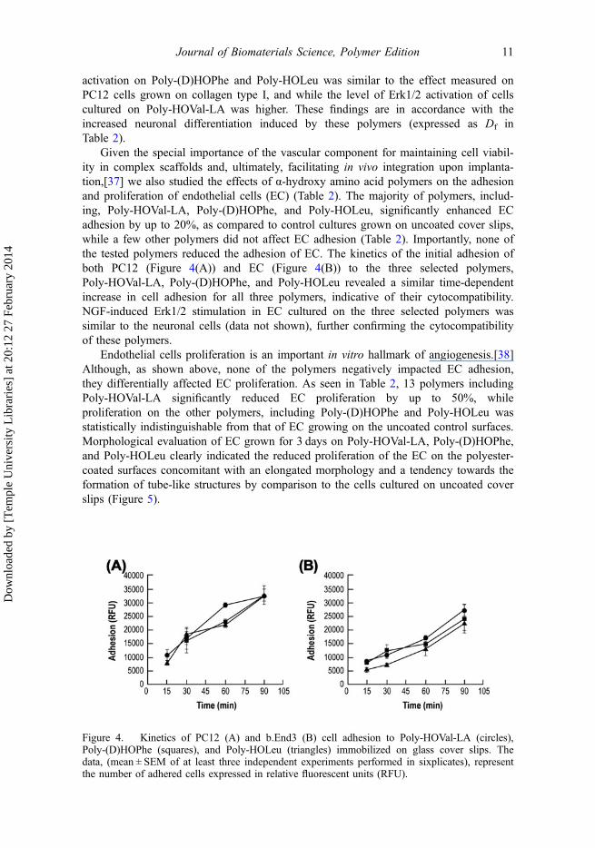
Given the special importance of the vascular component for maintaining cell viabil-ity in complex scaffolds and, ultimately, facilitating in vivo integration upon implanta-tion,[37] we also studied the effects of α-hydroxy amino acid polymers on the adhesionand proliferation of endothelial cells (EC) (Table 2). The majority of polymers, includ-ing, Poly-HOVal-LA, Poly-(D)HOPhe, and Poly-HOLeu, significantly enhanced ECadhesion by up to 20%, as compared to control cultures grown on uncoated cover slips,while a few other polymers did not affect EC adhesion (Table 2). Importantly, none ofthe tested polymers reduced the adhesion of EC. The kinetics of the initial adhesion ofboth PC12 (Figure 4(A)) and EC (Figure 4(B)) to the three selected polymers,Poly-HOVal-LA, Poly-(D)HOPhe, and Poly-HOLeu revealed a similar time-dependentincrease in cell adhesion for all three polymers, indicative of their cytocompatibility.NGF-induced Erk1/2 stimulation in EC cultured on the three selected polymers wassimilar to the neuronal cells (data not shown), further confirming the cytocompatibilityof these polymers.
Endothelial cells proliferation is an important in vitro hallmark of angiogenesis.[38]Although, as shown above, none of the polymers negatively impacted EC adhesion,they differentially affected EC proliferation. As seen in Table 2, 13 polymers includingPoly-HOVal-LA significantly reduced EC proliferation by up to 50%, whileproliferation on the other polymers, including Poly-(D)HOPhe and Poly-HOLeu wasstatistically indistinguishable from that of EC growing on the uncoated control surfaces.Morphological evaluation of EC grown for 3 days on Poly-HOVal-LA, Poly-(D)HOPhe,and Poly-HOLeu clearly indicated the reduced proliferation of the EC on the polyester-coated surfaces concomitant with an elongated morphology and a tendency towards theformation of tube-like structures by comparison to the cells cultured on uncoated coverslips (Figure 5).
Figure 4. Kinetics of PC12 (A) and b.End3 (B) cell adhesion to Poly-HOVal-LA (circles),Poly-(D)HOPhe (squares), and Poly-HOLeu (triangles) immobilized on glass cover slips. Thedata, (mean ± SEM of at least three independent experiments performed in sixplicates), representthe number of adhered cells expressed in relative fluorescent units (RFU).
Journal of Biomaterials Science, Polymer Edition 11
Dow
nloa
ded
by [
Tem
ple
Uni
vers
ity L
ibra
ries
] at
20:
12 2
7 Fe
brua
ry 2
014

3.4. Characterization of polymer surfaces
In the first step, we characterized the thickness and the roughness of select polymerfilm coatings on the glass cover slips using nano surface profiler and thin film reflec-tometry. For example, the thickness of films made of Poly-HOLeu, Poly-(D)HOPhe,and Poly-HOVal-LA was 444, 2033, and 175 nm, respectively, and UV sterilization hadno effect on these values.
In the second step, we used AFM to characterize the topography (surfaceroughness) of select polymer film coatings on the glass cover slips (Figure 6). For thesestudies we focused on the three selected polymers, i.e. Poly-HOVal-LA (strongenhancement of neuronal differentiation, no effect on EC adhesion, and EC prolifera-tion), Poly-HOLeu (without effects on neuronal differentiation and EC proliferation, butenhanced EC adhesion) and Poly-HO(D)Phe (inhibits effects on neuronal differentia-tion, without effect on EC proliferation but increasing EC adhesion). Analysis by AFMrevealed significant morphological differences between the various polyester-coated sur-faces (Figure 6). The surface of Poly-HOVal-LA appeared smooth as compared toPoly-HOLeu, which showed some irregular grooved structures at the nanometer scale,while Poly-(D)HOPhe exhibited ample islet-like protrusions exceeding 10 nm in height.Our data suggest an enhanced differentiation of PC12 cells on smooth surface, such asPoly-HOVal-LA (Table 2).
Figure 5. Morphology of bEnd.3 cells on selected polymers. bEnd.3 were cultured for 3 dayson the indicated polymers. White asterisks indicate the tube-like structures. Photomicrographswere taken at 20× magnification.
12 S. Lecht et al.
Dow
nloa
ded
by [
Tem
ple
Uni
vers
ity L
ibra
ries
] at
20:
12 2
7 Fe
brua
ry 2
014

4. Discussion
In this study, we synthesized homo- and co-polyesters comprised of α-hydroxy aminoacids by direct condensation and evaluated their cytocompatibility as thin films depos-ited on glass cover slips. The polymers were non-cytotoxic and supported neuronal andendothelial cell growth for up to seven days in culture. Several polymers significantlyenhanced NGF-induced neuronal differentiation and endothelial adhesion, while othersexhibited an inhibitory effect on both NGF-induced neuronal differentiation and endo-thelial proliferation (Figure 3, Table 2). It is well established that physicochemicalproperties, such as charge, hydrophobicity, and surface topography effectively modulatecell–biomaterials interactions and control cell attachment, migration, proliferation, anddifferentiation.[39–43] Previous studies of amino acid poly (ester)-amide-basedbiomaterials have shown that positively charged surfaces are more favorable for cellattachment and proliferation than negatively charged surfaces.[44,45] In our hands,Poly-HOVal-LA (neutral, hydrophobic) stimulated neuronal differentiation and reducedendothelial proliferation, as compared to control. By contrast other hydroxy polymerssuch as Poly-(D)HOPhe (neutral, hydrophobic) inhibited neuronal differentiation, butdid not affect endothelial proliferation. It is established that the initial cell interactionswith positively charged material surfaces are mediated by a negatively charged hyaluro-nan-rich cell coat called glycocalyx/‘pericellular matrix’, before integrins and focaladhesions are involved.[46] However, our results failed to establish a straightforwardrelationship between the overall charge of the hydroxy amino acids polymers and thecellular effects studied (adhesion, proliferation, and differentiation) (Tables 1 and 2),suggesting the potential involvement of integrin receptor-mediated bioactivity, a notionwhich warrants further investigation. A previous study demonstrated that the hydropho-bicity of polyelectrolyte multilayer films greatly affected the attachment of cultured rataortic smooth muscle cells [39]; however, in the case of poly-hydroxy amino acids, nei-ther the different molecular weights of the polymers nor their logP values appeared tobe determinants of cell adhesion, proliferation and differentiation (Tables 1 and 2). Cor-relation curves using the data in Tables 1 and 2 failed to establish statistically signifi-cant relationships between charge (zeta potential) or hydrophobicity (logP) and the
Figure 6. Atomic force microscopy visualization of the surface topography of three representativepolymers: Poly-HOVal-LA, Poly-(D)HOPhe, and Poly-HOLeu. White arrowheads indicate groovedstructures and islet-like protrusions of Poly-HOLeu and Poly-(D)HOPhe surfaces, respectively.
Journal of Biomaterials Science, Polymer Edition 13
Dow
nloa
ded
by [
Tem
ple
Uni
vers
ity L
ibra
ries
] at
20:
12 2
7 Fe
brua
ry 2
014

measured cellular effects (adhesion, proliferation and differentiation) for both cell types(data not shown). Based on the nano surface profiler, thin film reflectometry, and AFMdata (Figure 6), we surmise that the surface topography of the resultant films may bean important factor guiding their bioactivity. NGF-induced neuronal differentiation wasenhanced on polymers with a smooth surface topography, such as Poly-HOVal-LA. Bycomparison to endothelial cells, PC12 cells appear to be less sensitive to the surfacetexture (Figures 3(A)-inset and 5). In addition to the surface topography, the stereo-chemistry of our polyesters has significant implications for neuronal differentiation. Forexample, Poly-(D)HOVal enhanced neuronal differentiation, while the effect ofPoly-(L)HOVal was much less pronounced. In general, copolymers containing lacticacid (LA) enhanced neuronal differentiation, an observation that corresponds well withthe prior use of PLA as scaffolds for neural tissue engineering.[47]
Some of the α-hydroxy amino acid polymers promoted EC adhesion and modulatedtheir proliferation, while the majority of the polyesters tested had either no effect orenhanced neuronal differentiation. We did not find a correlation between the ability ofneuronal cells to adhere to the polymers and the extent of their differentiation (data notshown). The enhanced Erk1/2 phosphorylation in PC12 cells grown on Poly-HOVal-LAwas in accordance with NGF-induced neuritogenesis on this polymer. Taken together,these results suggest that polymers that support both neuronal differentiation and do notinterfere with endothelial proliferation, such as Poly-HOVal-LA, might be useful forregenerative engineering purposes and should be studied further, using additionalin vitro and in vivo models. In this context, we conducted preliminary in vivo toxicitystudies injecting two of the polymers, Poly-(D)HOPhe and Poly-HOVal-LA subcutane-ously into mice at a dose of 100 μg/kg. Preliminary results, obtained after two weeks fol-low-up, indicated that these polymers were non-toxic, as inferred from a lack of edema,irritation, or other symptoms of inflammation (Cohen and Lecht, unpublished data).
The ability to synthesize hydroxy amino acids polymers, bearing functionalizedgroups as (or in) their side chains, is in line with ongoing studies to enlarge the rangeof applications of this important family of bioactive copolymers.[42] As a caveat, whilethe results of this study are promising, the molecular weights of these first generationpolymers are still too low to allow the formation of biomimetic hydrogels by self-assembly. Using ring opening polymerization, we are currently synthesizing a secondgeneration of higher molecular weight polymers (20–30 kDa), which might be moresuitable to serves as tissue scaffolds.[48] Alternatively, our current hydroxy amino acidpolyesters could be admixed to other natural materials like alginate, which are beingused for cell encapsulation, but do not support/induce neuronal differentiation.[40] Thecytocompatibility and biodegradability of our polymers might render them useful as anECM-mimicking platform for enhanced culture of neuronal and endothelial cells.Traditionally, cell binding to ECM proteins involves specific integrin receptors.[49]Therefore, we surmise that certain ‘bioactive’ polyesters composed of relevant hydroxyamino acids motif sequences could serve as potential analogs to integrin receptors bind-ing sites for a variety of applications in tissue engineering and drug delivery.
AcknowledgmentsPIL is the Laura H. Carnell Professor at Temple University. We would like to acknowledgeMr Doron Greental, from the Center of Nanotechnology of The Hebrew University ofJerusalem for technical assistance.
14 S. Lecht et al.
Dow
nloa
ded
by [
Tem
ple
Uni
vers
ity L
ibra
ries
] at
20:
12 2
7 Fe
brua
ry 2
014

Funding
This study was supported by The Israel Science Foundation [grant number 591/08] and the AlexGrass Center for Drug Design and Synthesis of Novel Therapeutics at The Hebrew University.
References[1] Slomkowski S. Biodegradable polyesters for tissue engineering. Macromol. Symp.
2007;253:47–58.[2] Gunatillake P, Mayadunne R, Adhikari R. Recent developments in biodegradable synthetic
polymers. Biotechnol. Annu. Rev. 2006;12:301–347.[3] Gunatillake PA, Adhikari R. Biodegradable synthetic polymers for tissue engineering. Eur.
Cell. Mater. 2003;5:1–16.[4] Barrett DG, Yousaf MN. Design and applications of biodegradable polyester tissue scaffolds
based on endogenous monomers found in human metabolism. Molecules. 2009;14:4022–4050.[5] Kim BS, Mooney DJ. Development of biocompatible synthetic extracellular matrices for tis-
sue engineering. Trends Biotechnol. 1998;16:224–230.[6] Hartgerink JD, Beniash E, Stupp SI. Self-assembly and mineralization of peptide-amphiphile
nanofibers. Science. 2001;294:1684–1688.[7] Ryadnov MG, Woolfson DN. Engineering the morphology of a self-assembling protein fibre.
Nat. Mater. 2003;2:329–332.[8] Haines-Butterick L, Rajagopal K, Branco M, Salick D, Rughani R, Pilarz M, Lamm MS,
Pochan DJ, Schneider JP. Controlling hydrogelation kinetics by peptide design for three-dimensional encapsulation and injectable delivery of cells. Proc. Natl. Acad. Sci. USA.2007;104:7791–7796.
[9] Pomerantz WC, Yuwono VM, Pizzey CL, Hartgerink JD, Abbott NL, Gellman SH. Nanofibersand lyotropic liquid crystals from a class of self-assembling beta-peptides. Angew. Chem. Int.Ed. Engl. 2008;47:1241–1244.
[10] Arien-Zakay H, Lecht S, Perets A, Roszell B, Lelkes PI, Lazarovici P. Quantitative assess-ment of neuronal differentiation in three-dimensional collagen gels using enhanced greenfluorescence protein expressing PC12 pheochromocytoma cells. J. Mol. Neurosci.2009;37:225–237.
[11] Foley JD, Grunwald EW, Nealey PF, Murphy CJ. Cooperative modulation of neuritogenesisby PC12 cells by topography and nerve growth factor. Biomaterials. 2005;26:3639–3644.
[12] Moxon KA, Hallman S, Aslani A, Kalkhoran NM, Lelkes PI. Bioactive properties of nano-structured porous silicon for enhancing electrode to neuron interfaces. J. Biomater. Sci.Polym. Ed. 2007;18:1263–1281.
[13] Leach JB, Brown XQ, Jacot JG, Dimilla PA, Wong JY. Neurite outgrowth and branching ofPC12 cells on very soft substrates sharply decreases below a threshold of substrate rigidity.J. Neural. Eng. 2007;4:26–34.
[14] Yu X, Dillon GP, Bellamkonda RB. A laminin and nerve growth factor-laden three-dimensionalscaffold for enhanced neurite extension. Tissue Eng. 1999;5:291–304.
[15] Holmes TC, de Lacalle S, Su X, Liu G, Rich A, Zhang S. Extensive neurite outgrowth andactive synapse formation on self-assembling peptide scaffolds. Proc. Natl. Acad. Sci. USA.2000;97:6728–6733.
[16] Bieberich E, Anthony GE. Neuronal differentiation and synapse formation of PC12 andembryonic stem cells on interdigitated microelectrode arrays: contact structures for neuron-to-electrode signal transmission (NEST). Biosens. Bioelectron. 2004;19:923–931.
[17] Mahoney MJ, Chen RR, Tan J, Saltzman WM. The influence of microchannels on neuritegrowth and architecture. Biomaterials. 2005;26:771–778.
[18] Lopez CA, Fleischman AJ, Roy S, Desai TA. Evaluation of silicon nanoporous membranesand ECM-based microenvironments on neurosecretory cells. Biomaterials. 2006;27:3075–3083.
[19] Moon JJ, West JL. Vascularization of engineered tissues: approaches to promote angio-genesisin biomaterials. Curr. Top. Med. Chem. 2008;8:300–310.
[20] Montesano R, Pepper MS, Mohle-Steinlein U, Risau W, Wagner EF, Orci L. Increased pro-teolytic activity is responsible for the aberrant morphogenetic behavior of endothelial cellsexpressing the middle T oncogene. Cell. 1990;62:435–445.
Journal of Biomaterials Science, Polymer Edition 15
Dow
nloa
ded
by [
Tem
ple
Uni
vers
ity L
ibra
ries
] at
20:
12 2
7 Fe
brua
ry 2
014

[21] Zhu Y, Sun Y, Xie L, Jin K, Sheibani N, Greenberg DA. Hypoxic induction of endoglinvia mitogen-activated protein kinases in mouse brain microvascular endothelial cells. Stroke.2003;34:2483–2488.
[22] Brown RC, Morris AP, O’Neil RG. Tight junction protein expression and barrier propertiesof immortalized mouse brain microvessel endothelial cells. Brain Res. 2007;1130:17–30.
[23] Lecht S, Arien-Zakay H, Marcinkiewicz C, Lelkes PI, Lazarovici P. Nerve growth factor-induced protection of brain capillary endothelial cells exposed to oxygen-glucose deprivationinvolves attenuation of Erk phosphorylation. J. Mol. Neurosci. 2009;41:183–192.
[24] Lecht S, Arien-Zakay H, Wagenstein Y, Inoue S, Marcinkiewicz C, Lelkes PI, Lazarovici P.Transient signaling of Erk1/2, Akt and PLCgamma induced by nerve growth factor in braincapillary endothelial cells. Vascul. Pharmacol. 2010;53:107–114.
[25] Quaegebeur A, Lange C, Carmeliet P. The neurovascular link in health and disease: molecu-lar mechanisms and therapeutic implications. Neuron. 2011;71:406–424.
[26] Cohen-Arazi N, Katzhendler J, Kolitz M, Domb AJ. Preparation of new α-hydroxy acidsderived from amino acids and their corresponding polyesters. Macromolecules.2008;41:7259–7263.
[27] Kolitz M, Cohen-Arazi N, Hagag I, Katzhendler J, Domb AJ. Biodegradable polyestersderived from amino acids. Macromolecules. 2009;42:4520–4530.
[28] Cohen-Arazi N, Domb AJ, Katzhendler J. New biocompatible polyesters derived fromα-amino acids: hydrolytic degradation behavior. Polymers. 2010;2:418–439.
[29] Kajiyama T, Kobayashi H, Taguchi T, Kataoka K, Tanaka J. Improved synthesis with highyield and increased molecular weight of poly(alpha, beta-malic acid) by direct polyconden-sation. Biomacromolecules. 2004;5:169–174.
[30] Simmons TL, Baker GL. Poly(phenyllactide): synthesis, characterization, and hydrolyticdegradation. Biomacromolecules. 2001;2:658–663.
[31] Slager J, Domb AJ. Heterostereocomplexes prepared from d-poly(lactide) and leuprolide. I.Charact. Biomacromolecules. 2003;4:1308–1315.
[32] Lecht S, Arien-Zakay H, Kohan M, Lelkes PI, Lazarovici P. Angiostatic effects of K252a, aTrk inhibitor, in murine brain capillary endothelial cells. Mol. Cell. Biochem.2010;339:201–213.
[33] Momic T, Cohen G, Reich R, Arlinghaus FT, Eble JA, Marcinkiewicz C, LazaroviciP. Vixapatin (VP12), a c-type lectin-protein from Vipera xantina palestinae venom:characterization as a novel anti-angiogenic compound. Toxins (Basel). 2012;4:862–877.
[34] Bauer T, Gajewiak J. α-hydroxy carboxylic acids as ligands for enantioselective diethylzincadditions to aromatic and aliphatic aldehydes. Tetrahedron. 2004;60:9163–9170.
[35] Shin I, Lee MR, Lee J, Jung M, Lee W, Yoon J. Synthesis of optically active phthaloylD-aminooxy acids from L-amino acids or L-hydroxy acids as building blocks for thepreparation of aminooxy peptides. J. Org. Chem. 2000;65:7667–7675.
[36] Rawsterne RE, Todd SJ, Gough JE, Farrar D, Rutten FJ, Alexander MR, Ulijn RV. Cellspreading correlates with calculated logP of amino acid-modified surfaces. Acta Biomater.2007;3:715–721.
[37] Baiguera S, Ribatti D. Endothelialization approaches for viable engineered tissues. Angio-genesis. 2013;16:1–14.
[38] Ribatti D, Conconi MT, Nussdorfer GG. Nonclassic endogenous novel [corrected] regulatorsof angiogenesis. Pharmacol. Rev. 2007;59:185–205.
[39] Salloum DS, Olenych SG, Keller TCS, Schlenoff JB. Vascular smooth muscle cells onpolyelectrolyte multilayers: hydrophobicity-directed adhesion and growth. Biomacromole-cules. 2004;6:161–167.
[40] Lazarovici P, Li M, Perets A, Mondrinos M, Lecht S, Koharski C, Bidez P, CM F, Lelkes P.Intelligent biomatrices and engineered tissue constructs: in vitro models for drug discoveryand toxicity testing. In: Marx, U, Sandig V, editor. Drug testing in vitro: breakthroughs andtrends in cell culture technology. Weinheim: Wiley-VCH Verlag GmbH & Co; 2006.p. 3–52.
[41] Sun H, Gao C. Facile synthesis of multiamino vinyl poly(amino acid)s for promising bioap-plications. Biomacromolecules. 2010;11:3609–3616.
[42] Saulnier B, Ponsart S, Coudane J, Garreau H, Vert M. Lactic acid-based functionalized poly-mers via copolymerization and chemical modification. Macromol. Biosci. 2004;4:232–237.
16 S. Lecht et al.
Dow
nloa
ded
by [
Tem
ple
Uni
vers
ity L
ibra
ries
] at
20:
12 2
7 Fe
brua
ry 2
014

[43] Hemperly JJ. Signaling by cell adhesion molecules in the nervous system. Adv. Mol. Cell.Biol. 1999;28:303–320.
[44] Horwitz JA, Shum KM, Bodle JC, Deng M, Chu CC, Reinhart-King CA. Biological perfor-mance of biodegradable amino acid-based poly(ester amide)s: Endothelial cell adhesion andinflammation in vitro. J. Biomed. Mater. Res. A. 2010;95:371–380.
[45] Deng M, Wu J, Reinhart-King CA, Chu CC. Biodegradable functional poly(ester amide)swith pendant hydroxyl functional groups: synthesis, characterization, fabrication and in vitrocellular response. Acta Biomater. 2010;7:1504–1515.
[46] Fotia C, Messina GM, Marletta G, Baldini N, Ciapetti G. Hyaluronan-based pericellularmatrix: substrate electrostatic charges and early cell adhesion events. Eur. Cell. Mater.2013;26:133–149.
[47] Corey JM, Gertz CC, Wang BS, Birrell LK, Johnson SL, Martin DC, Feldman EL. Thedesign of electrospun PLLA nanofiber scaffolds compatible with serum-free growth ofprimary motor and sensory neurons. Acta Biomater. 2008;4:863–875.
[48] Cohen-Arazi N, Domb AJ, Katzhendler J. Poly(alpha-hydroxy alkanoic acid)s Derived Fromalpha-Amino Acids. Macromol. Biosci. 2013;13:1689–1699.
[49] Gahmberg CG, Fagerholm SC, Nurmi SM, Chavakis T, Marchesan S, Gronholm M. Regulationof integrin activity and signalling. Biochim. Biophys. Acta. 2009;1790:431–444.
Journal of Biomaterials Science, Polymer Edition 17
Dow
nloa
ded
by [
Tem
ple
Uni
vers
ity L
ibra
ries
] at
20:
12 2
7 Fe
brua
ry 2
014
Related Documents