Cystine-knot peptides targeting cancer- relevant human cytotoxic T lymphocyte- associated antigen 4 (CTLA-4) Franziska Maaß, a§ Joycelyn Wüstehube-Lausch, b§ Stephan Dickgießer, a§ Bernhard Valldorf, a§ Michael Reinwarth, a Hans-Ulrich Schmoldt, b Matin Daneschdar, b Olga Avrutina, a Ugur Sahin b and Harald Kolmar a * Cystine-knot peptides sharing a common fold but displaying a notably large diversity within the primary structure of flanking loops have shown great potential as scaffolds for the development of therapeutic and diagnostic agents. In this study, we dem- onstrated that the cystine-knot peptide MCoTI-II, a trypsin inhibitor from Momordica cochinchinensis, can be engineered to bind to cytotoxic T lymphocyte-associated antigen 4 (CTLA-4), an inhibitory receptor expressed by T lymphocytes, that has emerged as a target for the treatment of metastatic melanoma. Directed evolution was used to convert a cystine-knot trypsin inhibitor into a CTLA-4 binder by screening a library of variants using yeast surface display. A set of cystine-knot peptides possessing dissociation constants in the micromolar range was obtained; the most potent variant was synthesized chemically. Successive conjugation with neutravidin, fusion to antibody Fc domain or the oligomerization domain of C4b binding protein resulted in oligovalent variants that possessed enhanced (up to 400-fold) dissociation constants in the nanomolar range. Our data indicate that display of multiple knottin peptides on an oligomeric scaffold protein is a valid strategy to improve their functional affinity with ramifications for applications in diagnostics and therapy. Copyright © 2015 European Peptide Society and John Wiley & Sons, Ltd. Additional supporting information may be found in the online version of this article at the publisher’s web site. Keywords: cystine-knot peptides; avidity; CTLA-4; combinatorial screening; peptide oligomerization Introduction In recent years, cystine-knot peptides (often named knottins or miniproteins) emerged as a promising class of biomolecules with certain characteristics that make them particularly attractive for var- ious diagnostic and therapeutic applications. These miniproteins are small (around 30 amino acids) peptidic molecules with a well- defined three-dimensional structure comprising three disulfide bonds (Cys I -Cys IV , Cys II -Cys V , Cys III -Cys VI ) [1,2] that form a pseudo- knotted structural scaffold. Because of this unique architecture, knottins exhibit an extraordinary thermal, proteolytic, and chemical stability [3,4]. These peptides are easily accessible by recombinant and chemical synthesis [5], and their loops are variable for ex- change and insertion of amino acids without the loss of structure and function [6,7]. Combined with their potential for oral delivery [8,9], knottins have attracted considerable interest as frameworks for pharma-inspired peptide engineering [10–12]. Cystine-knot peptides with desired binding characteristics have been obtained by rational design and via combinatorial library screening. For example, potent inhibitors of human matriptase-1 with inhibition constants in the low nanomolar to subnanomolar range were recently generated via screening of combinatorial li- braries by yeast surface display [13]. Further, screening of combina- torial libraries derived from the cystine-knot scaffold of kalata B1 cyclotide was used to gain peptides that antagonize the growth factor receptors neuropilin-1 and neuropilin-2 epitopes involved in the antagonism of vascular endothelial growth factor [14]. Using various cystine-knot peptides as a framework for engineering, Cochran and coworkers isolated potent binders of different integrins that were particularly useful for tumor imaging because the radiolabeled or fluorescently labeled knottins were shown to selectively target murine tumors [15–18]. Recently, the cyclic pep- tide MCoTI-I [19] has been designed to efficiently antagonize intra- cellular p53 degradation. The resulting cyclotide combined high stability in human serum with pronounced cytotoxicity because of the activation of the p53 tumor suppressor pathway in tumor cells, thus inhibiting tumor growth in a xenograft model [20]. A synthetic cystine-knot peptide Ziconotide derived from a cone snail toxin was approved by both the US Food and Drug Adminis- tration (FDA) and European Commision as an analgesic agent and is currently on the market under the brand name Prialt [21]. Knottins were also engineered towards improved oral availability [8,9,22–24]. * Correspondence to: Harald Kolmar, Institute of Organic Chemistry and Biochemistry, Technische Universität Darmstadt, Alarich-Weiss-Str. 4, 64287 Darmstadt, Germany. E-mail: [email protected] § These authors contributed equally to this work a Institute of Organic Chemistry and Biochemistry, Technische Universität Darmstadt, Darmstadt, Germany b BioNTech AG, Mainz, Germany J. Pept. Sci. (2015) Copyright © 2015 European Peptide Society and John Wiley & Sons, Ltd. Research Article Received: 28 January 2015 Revised: 15 March 2015 Accepted: 16 March 2015 Published online in Wiley Online Library (wileyonlinelibrary.com) DOI 10.1002/psc.2782

Welcome message from author
This document is posted to help you gain knowledge. Please leave a comment to let me know what you think about it! Share it to your friends and learn new things together.
Transcript
Research Article
Received: 28 January 2015 Revised: 15 March 2015 Accepted: 16 March 2015 Published online in Wiley Online Library
(wileyonlinelibrary.com) DOI 10.1002/psc.2782
J. Pept. Sci. (2015)
Cystine-knot peptides targeting cancer-relevant human cytotoxic T lymphocyte-associated antigen 4 (CTLA-4)
Franziska Maaß,a§ Joycelyn Wüstehube-Lausch,b§ Stephan Dickgießer,a§
Bernhard Valldorf,a§ Michael Reinwarth,a Hans-Ulrich Schmoldt,b
Matin Daneschdar,b Olga Avrutina,a Ugur Sahinb and Harald Kolmara*
Cystine-knot peptides sharing a common fold but displaying a notably large diversity within the primary structure of flankingloops have shown great potential as scaffolds for the development of therapeutic and diagnostic agents. In this study, we dem-onstrated that the cystine-knot peptide MCoTI-II, a trypsin inhibitor from Momordica cochinchinensis, can be engineered tobind to cytotoxic T lymphocyte-associated antigen 4 (CTLA-4), an inhibitory receptor expressed by T lymphocytes, that hasemerged as a target for the treatment of metastatic melanoma. Directed evolution was used to convert a cystine-knot trypsininhibitor into a CTLA-4 binder by screening a library of variants using yeast surface display. A set of cystine-knot peptidespossessing dissociation constants in the micromolar range was obtained; the most potent variant was synthesized chemically.Successive conjugation with neutravidin, fusion to antibody Fc domain or the oligomerization domain of C4b binding proteinresulted in oligovalent variants that possessed enhanced (up to 400-fold) dissociation constants in the nanomolar range. Ourdata indicate that display of multiple knottin peptides on an oligomeric scaffold protein is a valid strategy to improve theirfunctional affinity with ramifications for applications in diagnostics and therapy. Copyright © 2015 European Peptide Societyand John Wiley & Sons, Ltd.
Additional supporting information may be found in the online version of this article at the publisher’s web site.
Keywords: cystine-knot peptides; avidity; CTLA-4; combinatorial screening; peptide oligomerization
* Correspondence to: Harald Kolmar, Institute of Organic Chemistry andBiochemistry, Technische Universität Darmstadt, Alarich-Weiss-Str. 4, 64287Darmstadt, Germany. E-mail: [email protected]
§ These authors contributed equally to this work
a Institute of Organic Chemistry and Biochemistry, Technische UniversitätDarmstadt, Darmstadt, Germany
b BioNTech AG, Mainz, Germany
Introduction
In recent years, cystine-knot peptides (often named knottins orminiproteins) emerged as a promising class of biomolecules withcertain characteristics thatmake them particularly attractive for var-ious diagnostic and therapeutic applications. These miniproteinsare small (around 30 amino acids) peptidic molecules with a well-defined three-dimensional structure comprising three disulfidebonds (CysI-CysIV, CysII-CysV, CysIII-CysVI) [1,2] that form a pseudo-knotted structural scaffold. Because of this unique architecture,knottins exhibit an extraordinary thermal, proteolytic, and chemicalstability [3,4]. These peptides are easily accessible by recombinantand chemical synthesis [5], and their loops are variable for ex-change and insertion of amino acids without the loss of structureand function [6,7]. Combined with their potential for oral delivery[8,9], knottins have attracted considerable interest as frameworksfor pharma-inspired peptide engineering [10–12].Cystine-knot peptides with desired binding characteristics have
been obtained by rational design and via combinatorial libraryscreening. For example, potent inhibitors of human matriptase-1with inhibition constants in the low nanomolar to subnanomolarrange were recently generated via screening of combinatorial li-braries by yeast surface display [13]. Further, screening of combina-torial libraries derived from the cystine-knot scaffold of kalata B1cyclotide was used to gain peptides that antagonize the growthfactor receptors neuropilin-1 and neuropilin-2 epitopes involvedin the antagonism of vascular endothelial growth factor [14]. Using
various cystine-knot peptides as a framework for engineering,Cochran and coworkers isolated potent binders of differentintegrins that were particularly useful for tumor imaging becausethe radiolabeled or fluorescently labeled knottins were shown toselectively target murine tumors [15–18]. Recently, the cyclic pep-tide MCoTI-I [19] has been designed to efficiently antagonize intra-cellular p53 degradation. The resulting cyclotide combined highstability in human serum with pronounced cytotoxicity becauseof the activation of the p53 tumor suppressor pathway in tumorcells, thus inhibiting tumor growth in a xenograft model [20]. Asynthetic cystine-knot peptide Ziconotide derived from a conesnail toxin was approved by both the US Food and Drug Adminis-tration (FDA) and European Commision as an analgesic agent andis currently on the market under the brand name Prialt [21].Knottins were also engineered towards improved oral availability[8,9,22–24].
Copyright © 2015 European Peptide Society and John Wiley & Sons, Ltd.

MAAß ET AL
Multivalent interactions of biological molecules play an impor-tant role in living systems. A multivalent ligand comprises multiplecopies of respective monomers conjugated in a certain manner,thus allowing for their simultaneous binding tomultiple target mol-ecules located, e.g. on the surface of cells. Many research groupshave successfully generated multivalent ligands to increase thenet binding affinity and specificity of the ligand to the receptor[25,26]. Direct peptide oligomerization or polymeric display of pep-tides on an oligomeric scaffold was successfully used to enhancethe functional affinity of binding molecules to their targets becauseof an avidity effect [27–30]. This strategy was also applied to thebinding peptides with cystine-knot architecture. Thus, chemicaldimerization of a knottin that recognized the thrombopoietinreceptor resulted in the conversion of a receptor antagonist intoan agonist [31]. However, to the best of our knowledge, to date,no systematic study has been reported for cystine-knot peptidesaimed at improving their functional affinity by scaffold-mediatedoligomerization, which implies diverse protein scaffolds and resultsin different geometry and copy numbers.
In this work, we studied the avidity effects of knottin bindingwhendisplayed on oligomeric domains using a peptidemonomerwith lowspecific affinity to human cytotoxic T lymphocyte-associated antigen4 (CTLA-4, CD152). CTLA-4 plays an important role in the natural im-mune response and is expressed on the surface of T cells as a dimerafter their activation [32,33]. Because of its negative regulation ofthe immune response [34–36], CTLA-4 became an attractive targetfor immunotherapy of cancer [37–39] and other diseases [40–42].CTLA-4 signaling results in down-regulation of T cell function andinhibition of activated T cell expansion. Upon immunization with anon-stimulatory antibody against CTLA-4,mice showed an enhancedresistance to tumor challenge andwere evenable to clear establishedtumors [37]. An anti-CTLA-4 monoclonal antibody, Ipilimumab, isFDA-approved for the treatmentofmelanomasince2011 [43].Herein,we report the development of engineered, selective CTLA-4 bindersbased on cystine-knot peptide oMcoTI-II, the acyclic derivative ofthe protease inhibitor MCoTI-II from Momordica cochinchinensis[44,45]. The avidity effects of various dimeric, tetrameric andheptameric variants are discussed.
Materials and Methods
Library Cloning
A randomized library of gene variants based on the oMCoTI-IIframework was generated with pre-made codon mixtures [46].The encoding gene pool was amplified using Taq polymerase andprimers with 50-bp overlap to the pCT yeast display plasmid up-stream or downstream of the NheI and BamHI restriction sites[47]. Subsequently, the polymerase chain reaction (PCR) productwas purified by phenol/chloroform extraction. The pCT vector wasdigested with NheI and BamHI and purified using sucrose densitygradient centrifugation. Saccharomyces cerevisiae strain EBY100was transformed with DNA insert (12μg) and linearized plasmid(4μg) as described [48].
Flow Cytometry and Library Screening
The yeast library (approximately 3× 10⁸ transformants) was grownat 30 °C in selective media synthetic dextrose growth medium withcasamino acids (SD-CAA, 5.4 g/l Na2HPO4, 8.6g/l NaH2PO4 · H2O,6.7g/l yeast nitrogen base without amino acids, 5 g/l Bactocasamino acids, and 20g/l glucose) and induced to express
wileyonlinelibrary.com/journal/jpepsci Copyright © 2015 Europ
oMCoTI-II mutants on the yeast cell surface at 20 °C in selectivemedia containing galactose synthetic galactose growth mediumwith casamino acids (SG-CAA). Expression level and CTLA4 bindingof yeast-displayed miniprotein variants were determined by flowcytometry. Therefore, 1 : 20 dilutions of anti-cMyc antibody (mono-clonal, mouse, Abcam; Cambridge, UK), anti-mouse IgG biotin-conjugate (polyclonal, goat, Sigma–Aldrich; St. Louis, MO, USA),and Streptavidin-R-phycoerythrin (SPE)-conjugate (Invitrogen)were added consecutively to 1×10⁷ cells for 10min on ice.For the first selection round, approximately 6×10⁸ cells were an-
alyzed for binding to CTLA-4-Ig (Abatacept/Orencia®, Bristol MyersSquibb; New York City, NY, USA). A two-color fluorescence activatedcell sorting (FACS) was used to select for cell surface presentationand target binding. Therefore, 1 × 10⁸ cells were incubated consec-utively with fluorescein-labeled 1μM CTLA-4-Ig for 30min on iceand with 1 : 20 dilutions of mouse monoclonal anti-cMyc antibody,anti-mouse IgG biotin conjugate, and streptavidin, R-phycoerythrinconjugate (SPE) for 10min on ice, respectively. To ensure librarydiversity, next screening rounds were performed with at least tentimes the number of yeast cells collected in the previous round.Collected yeast cells were cultured after each screening round inSD-CCA medium, induced for expression in SG-CCA medium, andsorted by subsequent rounds of FACS to obtain an enriched popu-lation of CTLA-4 binders (parameters: trigger side scatter 650,FL1 600, and FL2 600). Labeling was altered in screening roundthree. To this end, the cells were incubated consecutively with1μM CTLA-4-Ig for 30min on ice and with 1 : 20 dilutions of anti-human IgG phycoerythrin (PE)-conjugate (polyclonal, goat, eBio-science; San Diego, CA, USA), anti-cMyc antibody, and anti-mouseIgG-fluorescein conjugate (polyclonal, goat, Sigma–Aldrich) for10min on ice.Plasmid DNA from four isolated single clones of screening round
4 was recovered from yeast clones and transformed into DH5αcompetent Escherichia coli cells for plasmid preparation. DNAsequencing was performed by Seqlab (Göttingen, Germany).
Recombinant Production of Cystine-knot Peptides
The pools derived from the third and fourth yeast screening roundswere subcloned into a pET-32a based expression vector (pET-32K)to screen for CTLA-4 binding in a yeast-display independent setting.In this plasmid, the peptide coding sequence is fused to His-taggedthioredoxin separated by a thrombin protease cleavage site [49]. Tothis end, the plasmid DNA was isolated from the yeast cells; theminiprotein gene fragments were amplified via PCR and digestedwith EheI and ApaI. Miniprotein gene pools were cloned intoNruI/ApaI digested expression vector pET32K and transferred intoE. coli SHuffle T7® (NEBiolabs). Single clones was cultured in 1ml ly-sogeny broth (LB) medium supplemented with 100μg/ml ampicil-lin and 0.4% (w/v) glucose at 37 °C using microwell plates. At anoptical density of ~0.8 at 600nm the cells were centrifuged andsuspended in fresh LB medium containing 100μg/ml ampicillinand 1mM isopropyl-β-d-thiogalactopyranoside (IPTG) for inductionof protein expression that was performed overnight at 25 °C. Subse-quently, cells were harvested by centrifugation, resuspended in200μl lysis buffer [20mM Tris pH8.0, 2mM MgCl2, 20mM NaCl,1mg/ml chicken egg white lysozyme (Merck, Darmstadt, Germany),and 15U/ml benzonase nuclease (Merck)] and the cell suspensionwas stored at �20 °C for at least 1 h. Cells were thawed afterwards,incubated for 15min at room temperature and 10min at 80 °C. Thesuspensions were centrifuged at 5000×g for 30min, and the super-natant comprising soluble thioredoxin fusion proteins was used for
ean Peptide Society and John Wiley & Sons, Ltd. J. Pept. Sci. (2015)
POTENT CTLA-4 BINDERS: COMBINATORIAL SCREENING AND AVIDITY MODULATION
subsequent enzyme-linked immunosorbent assay (ELISA)-basedbinding analysis.Fusion proteins derived from 96-well expressions were
immobilized overnight in 50mM Na-carbonate buffer (pH9.4) onwells of ELISA plates (MaxiSorp 96-well plates, Nunc; Rochester,NY, USA). The plates were blockedwith 1% casein blocking solution(Sigma) in TBS (150mM NaCl, 100mM Tris-HCl, and pH7.4) and sub-sequently incubated with 250nM CTLA-4-Ig in TBS with 1% caseinfor 1 h at 37 °C. Bound CTLA4-Ig was detected via an anti-humanIgG-horseradish peroxidase (HRP) conjugate (Sigma Aldrich) diluted1 : 5000 in TBS with 1% casein. 3,3′,5,5′-tetramethylbenzidine (TMB,Sigma Aldrich) was used as a chromogenic HRP substrate. After10min incubation time at room temperature, the reaction wasstopped by the addition of 0.2 M HCl, and absorbance was mea-sured at 450 nm in a Victor 3 V ELISA reader (Perkin Elmer, Waltham,MA, USA).
Cloning of Expression Vectors for Fusion Proteins 4, 5, and 6
pEXPR-IBA42 (IBA BioTAGnology GmbH, Göttingen, Germany) withsequences encoding a human Fc was received as a kind gift fromChristian Heinis, Lausanne, Switzerland. This vector contained anampicillin resistance gene, a cytomegalovirus (CMV) promoter, aBM40 signal sequence for protein secretion, and an IgG1 Fcfragment with Asn297Ala mutation (based on the nucleotidesequence of human IgG1 heavy chain constant regions CH2 andCH3; amino acids 236 to 446, Kabat numbering; accession number:AAL96263). The hinge regionwas shortened to 12 residues with thetwo cysteines replaced by serines [50].The Fc fragment encoding sequence was PCR-amplified to intro-
duce ApaI and BamHI restriction sites at the 5′ and 3′ end of the Fc-coding sequence, respectively, and cloned into pEXPR-IBA42 usingNheI and HindIII. The sequence encoding for peptide 1 was ampli-fied by PCR and placed at the 3′ end of the Fc fragment coding re-gion using the restriction sites BamHI and HindIII resulting in aconstruct encoding for fusion protein 4. Likewise, the sequenceencoding for peptide 1 was placed at the 5′ terminus via NheI andApaI cloning leading to 5. Transfection-pure vectors were purifiedby using PureYield Plasmid Midiprep System (Promega, Madison,WI, USA).For expression of the fusion protein 6, the peptide-coding se-
quence was amplified, digested with the restriction enzymes BglIIand NcoI, and ligated with pET32a-Trx-C4Bp [51] linearized withthe same restriction enzymes. Protein production and purificationwere performed as described [51].
Peptide Synthesis and Biotinylation
The CTLA-4-binding cystine-knot peptide 2 was assembled onRAM resin following the Fmoc strategy using a fully automatedmicrowave-assisted CEM Liberty peptide synthesizer. Aminotermi-nal biotin coupling was achieved on-support using (+)-biotin uponpreactivation with 2-(1H-benzotriazole-1-yl)-1,1,3,3-tetramethyluro-nium hexafluorophosphate (HBTU) in the presence of diisopropyle-thylamine (DIPEA). After acidolytic cleavage from the solid support,oxidative folding was performed as described [52]. Reaction prog-ress was monitored by analytical reverse phase high performanceliquid chromatography (RP-HPLC) and of liquid chromatography-electrospray ionization-mass spectrometry (LC-ESI-MS), and thefolded peptide 2 was isolated from the reaction mixture by HPLCupon completion of the reaction after 12 h (Fig. S1 in ElectronicSupplementary Information) [52].
J. Pept. Sci. (2015) Copyright © 2015 European Peptide Society and Jo
Binding Studies Using Peptide 2
Affinity analysis was performed by ELISA. CTLA-4-Ig in PBS(140mM NaCl, 10mM KCl, 6.4mM Na2HPO4, 2mM KH2PO4, pH7.4)was immobilized on wells of an ELISA plate. To minimize nonspe-cific binding, the plate was blocked with 3% bovine serum albu-min (BSA) in PBS and subsequently incubated with biotinylatedpeptide 2 in PBS with 1% BSA for 1 h. Bound miniprotein wasdetected via ExtrAvidin-HRP-conjugate (Sigma–Aldrich), diluted1 : 5000 in PBS with 1% BSA. TMB was used as chromogenicHRP substrate. After 15-min incubation time at room temperature,the reaction was stopped by the addition of diluted HCl, andabsorbance was measured at 450nm in an ELISA reader (GENios,Tecan, Männedorf , Switzerland). The monoclonal antibody ce-tuximab served as a control. For affinity measurement usingpeptide-neutravidin conjugate 3, biotinylated cystine-knot pep-tide 2 and neutravidin were mixed in a molar ratio of 4 : 1 andincubated in 1% casein in PBS for 30min at room temperature.The resulting construct 3 was immobilized on wells of an ELISAplate. The plate was blocked with 3% casein in PBS andsubsequently incubated with CTLA-4-Ig in 1% casein in PBS for1 h. Bound CTLA-4-Ig was detected via anti-hFc-HRP (Antikörper-Online), diluted 1 : 5000 in PBS with 1% casein. TMB was usedas chromogenic HRP substrate.
Expression, Purification, and Affinity Titration of Fusion Pro-teins 4, 5, and 6
Fusion proteins 4 and 5 were expressed in transiently transfectedHEK293-6E suspension cells. Cells were cultured in FreeStyle™ F17Expression Medium (Life Technologies, Carlsbad, CA, USA) supple-mented with 4mM l-glutamine (Sigma) and 50μg/mlG418 at100 rpm shaking speed, 37 °C, and 5% CO2. Prior to transfection,cells were resuspended in medium without antibiotics at a den-sity of 2 × 10⁶ viable cells/ml overnight. For transfection 0.5μgplasmid DNA per 1.7× 10⁶ viable cells were preincubatedwith 3μg 25 kDa linear polyethylenimine (Polysciences) for15min and added to the cells. After 24h, tryptone was addedto a final concentration of 0.5%. After 5 days of expression,cells were removed by centrifugation (10min, 4000 rpm) andfiltration (0.45μm). Fusion proteins 4 and 5 were purified usingprotein A affinity column (1ml HiTrap Protein A HP column;GE Healthcare, Uppsala, Sweden) and an Äkta FPLC purifiersystem (GE Healthcare) and subsequently dialysed against PBS.Protein production and purification of 6 were performed as de-scribed [51].
For ELISA affinity titration, fusion proteins 4 and 5 were biotinyl-ated using EZ-Link® Sulfo-NHS-LC-Biotin (Thermo Scientific, Wal-tham, MA, USA) according to the manufacturers instructions andafterwards purified by polyacrylamide spin desalting columns (7 KMWCO, Thermo Scientific). Then CTLA-4-Ig in PBS was immobilizedon wells of an ELISA plate. The plate was blocked with 3% BSA inPBS and subsequently incubated with biotinylated 4 and 5 in PBSwith 1% BSA for 1 h. Bound fusion proteins were detected upon in-cubation with ExtrAvidin-HRP conjugate (Sigma Aldrich), diluted1 : 5000 in PBS with 1% BSA. Binding of 6 to CTLA-4 was detectedwith a mouse monoclonal anti-oligohistidine antibody (Qiagen) inPBSwith 1%BSA followed by incubationwith anti-mouse IgG biotinconjugate diluted 1 : 5000 in PBS with 1% BSA (Sigma Aldrich).Bound fusion protein 6 was detected as described above usingExtrAvidin-HRP conjugate; TMB was applied as a chromogenicHRP substrate.
hn Wiley & Sons, Ltd. wileyonlinelibrary.com/journal/jpepsci

Table 1. Residue randomization of the oMCoTI library
Amino acids Residue randomization (%)
Red Blue Yellow Green
A 1.4 0.7 4.2 50
C 0.0 0.0 0.0 0
D 7.5 4.1 0.0 5
E 7.5 4.1 0.0 5
F 1.4 0.7 4.2 0
G 7.5 4.1 50 5
H 7.5 4.1 4.2 5
I 1.4 0.7 4.2 0
K 7.5 4.1 4.2 5
L 1.4 0.7 4.2 0
M 1.4 0.7 4.2 0
N 7.5 4.1 0.0 5
P 7.5 50 0.0 0
Q 7.5 4.1 0.0 5
R 7.5 4.1 4.2 5
S 7.5 4.1 0.0 5
T 7.5 4.1 4.2 5
V 1.4 0.7 4.2 0
W 1.4 0.7 4.2 0
Y 1.4 0.7 4.2 0
MAAß ET AL
Results
Library Design and Screening for CTLA-4 Binders
To obtain cystine-knot peptides that bind CTLA-4, a library basedon the trypsin inhibitor oMCoTI-II framework was generated uponrandomization of the inhibitor loop and neighboring residues(Figure 1(A–C)). The inhibitor loop and the two CysI-preceding resi-dues were exchanged against randomized amino acid sequenceshaving 6, 9, and 12 residues, respectively, to create peptide variantswith new molecular recognition properties. Previous studies withthe structurally similar miniprotein EETI-II showed high toleranceof length and sequence diversity in non-conserved regions[6,13,15]. A codon distribution with underrepresented hydrophobicresidues was chosen (Table 1). Because residues PGA (Figure 1(A))may be of relevance for oMCoTI-II folding and stability, simulta-neous full randomization was avoided by maintaining the originalresidue at each position for 50% of the variants. Overall, therandomization scheme applied here included 11 to 17 out of 34to 40 residues.
The oMCoTI-II library-encoding DNA was genetically fused to theS. cerevisiae Aga2p coding sequence. S. cerevisiae is a particularlywell-suited host for the synthesis of miniproteins because itcontains in the endoplasmic reticulum a set of folding helpers thatmediate disulfide bond formation together with a quality controlmachinery to remove misfolded variants [53]. The resulting con-structs are under control of the galactose promoter [54]. Inductionwith galactose yielded a fusion protein comprising Aga2p, aglycine-serine linker, an HA-epitope, the cystine-knot peptide, anda cMyc epitope (Figure 2(A)). The fusion is covalently bound tothe surface-anchored Aga1p. The resulting miniprotein librarydisplayed a clonal diversity of 3× 10⁸.
To select for the full-size miniprotein variants that are presentedon the yeast surface and show binding to CTLA-4, a two-color
Figure 1. (A) Sequence and (B) structure of cystine-knot trypsin inhibitor oMresidues are depicted as yellow sticks. Cystine-forming residues are markedaccording to their appearance in the sequence. Randomized residues are coScreening against human CTLA4-Ig. Fluorescence activated cell sorting histog(1μM) for enrichment of cytotoxic T lymphocyte-associated antigen 4 (CTLA-4shown. Display level (cMyc) is monitored by incubation with the antibodiesstreptavidin, R-phycoeythrin conjugate (SPE). CTLA-4 binding is monitored bystaining, see Section on Methods and Ref. [13].
wileyonlinelibrary.com/journal/jpepsci Copyright © 2015 Europ
staining and FACS procedure was performed (Figure 1(C) andFigure 2(A)). In the initial sorting round, in total 6 × 10⁸ yeast cellswere labeled with 1μM CTLA4-Ig (a fusion protein consisting ofthe extracellular domain of CTLA-4 and a human IgG Fc part) andsorted by FACS to collect the 0.3% of yeast cells that displayedthe oMCoTI-II variants (detected through cMyc-antibody) andshowed the highest binding to fluorescein-labeled CTLA4-Ig
CoTI-II (pdb: 1 ha9). Secondary structure is shown as cartoon, and cysteinein the sequence in yellow, and the numbering of respective cysteines islor coded according to the residue distribution depicted in Table 1. (C)rams showing four rounds of sorting with constant target concentration) binders. R1 to R4 denote the sorting round, and actual sorting gates areanti-cMyc and anti-mouse-biotin followed by fluorescence labeling withincubation with fluorescein-labeled CTLA-4-Ig. For details of two-color cell
ean Peptide Society and John Wiley & Sons, Ltd. J. Pept. Sci. (2015)

Figure 2. Characterization of cytotoxic T lymphocyte-associated antigen 4 (CTLA-4)-binding peptide MC-CT-010 (1). (A) Schematic illustration of Aga1p/Aga2p surface-displayed oMCoTI-II derived miniproteins (orange) flanked by the N-terminal HA (Human influenza hemagglutinin) epitope (blue) and theC-terminal cMyc epitope (yellow). Display level (cMyc) is monitored by incubation with the antibodies anti-cMyc and anti-mouse-biotin followed byfluorescence labeling with streptavidin, R-phycoeythrin conjugate (SPE, pink). Functional display of CTLA-4 binding of oMCoTI-II variants is monitored byincubation with fluorescein-labeled CTLA-4-Ig (green). (B) Fluorescence activated cell sorting histograms for CTLA-4 binding of control cells and cellsdisplaying 1. Cells were labeled with 1μM CTLA4-Ig-FITC and cMyc-antibody. (C) ELISA for determination of the binding properties and specificity of 1. (D)Enzyme-linked immunosorbent assay for determination of the binding affinity of 1. Error bars represent three independent measurements.
POTENT CTLA-4 BINDERS: COMBINATORIAL SCREENING AND AVIDITY MODULATION
(Figure 1(C)). To exclude isolation of binders against streptavidin,labeling was alternated for the third screening round fromfluorescein-bearing CTLA-4-Ig to CTLA-4-Ig detection using ananti-human PE-conjugated antibody. After four screening rounds,an enrichment of potential binding candidates was observed.
Binding of Monomeric Cystine-knot Peptides to CTLA-4
The pools from the third and fourth screening rounds weresubcloned into pET32K expression vector for expression as solubleproteins in E. coli cells. Miniproteins from 224 randomly picked sin-gle clones of the third round and 239 clones of the fourth screeninground were produced as fusions to thioredoxin [49] and subjectedto a qualitative ELISA analysis to examine CTLA-4 binding. While nopositive clones were obtained from the third screening round, sev-eral putative CTLA-4 binders were identified from the fourth one(Table 2); among them, peptide called MC-CT-010 (1) was pre-sented 13 times, and the other nine sequences were unique.To characterize the affinities and specificities of the engineered
oMCoTI-II peptides towards CTLA-4, four candidates (MC-CT-010to MC-CT-040, Table 2) were expressed as thioredoxin fusions,and an ELISA affinity titration was performed. In these experiments,the chosen variants showed the respective apparent dissociationconstants of 3, 17, 7, and 20μM, approximately. The best candidate,MC-CT-010 1, was assembled by Fmoc-SPPS (IBM, Armonk, NY, USA)followed by biotinylation and oxidative folding resulting in the
J. Pept. Sci. (2015) Copyright © 2015 European Peptide Society and Jo
biotinylated synthetic cystine-knot 2 (for the details, see Fig. S1 inElectronic Supplementary Information). The binding constant ofthe peptide 2 was estimated using ELISA. The analysis showed aspecific and concentration-dependent binding against humanCTLA-4-Ig and revealed a KD value of 3.7μM (Figure 2).
Oligomerization of Peptidic Monomers
To study a possible avidity upon binding to CTLA-4, which is adimer, the peptide monomers were oligomerized using diverseoligovalent proteins as scaffolds. Thus, the tetramer of synthetic bi-otinylated cystine-knot MC-CT-010 1 was generated through theformation of a stable non-covalent complex 3 with the tetravalentprotein neutravidin (for numbering of the constructs, see Figure 4upper row). This oligomer 3 showed a specific and concentration-dependent binding to CTLA-4 and exhibited a KD in the nanomolarrange (Figure 3(A)).
Relying on these results, we designed several oligomeric archi-tectures comprising the knottin monomer in different copies andgeometry (Figure 4). In these constructs, the cystine-knot counter-part was genetically fused either to the C-terminus of a humanIgG1 Fc fragment or to the N-terminus and the C-terminus simulta-neously. Because of the dimeric nature of an antibody Fc fragment,it resulted in the formation of di- or tetramers of 1, namely, 4 and 5,respectively (Figure 4). Following the expression in HEK293-6E cellsand purification by affinity chromatography, the dimer formation
hn Wiley & Sons, Ltd. wileyonlinelibrary.com/journal/jpepsci
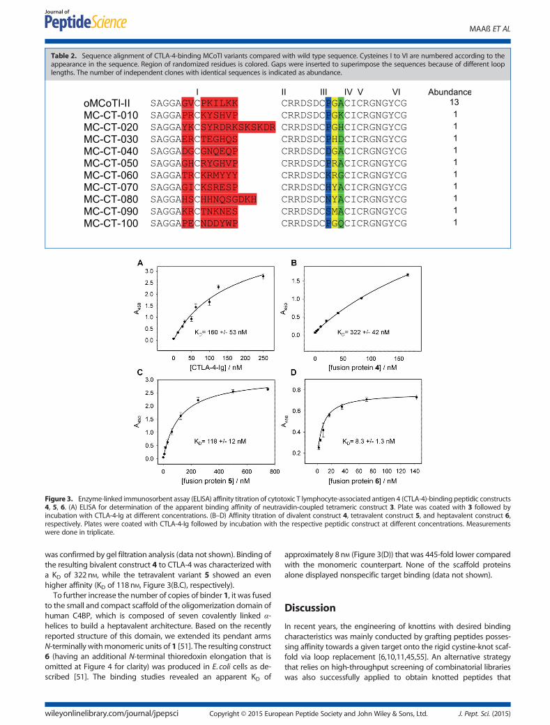
Table 2. Sequence alignment of CTLA-4-binding MCoTI variants compared with wild type sequence. Cysteines I to VI are numbered according to theappearance in the sequence. Region of randomized residues is colored. Gaps were inserted to superimpose the sequences because of different looplengths. The number of independent clones with identical sequences is indicated as abundance.
MAAß ET AL
Figure 3. Enzyme-linked immunosorbent assay (ELISA) affinity titration of cytotoxic T lymphocyte-associated antigen 4 (CTLA-4)-binding peptidic constructs4, 5, 6. (A) ELISA for determination of the apparent binding affinity of neutravidin-coupled tetrameric construct 3. Plate was coated with 3 followed byincubation with CTLA-4-Ig at different concentrations. (B–D) Affinity titration of divalent construct 4, tetravalent construct 5, and heptavalent construct 6,respectively. Plates were coated with CTLA-4-Ig followed by incubation with the respective peptidic construct at different concentrations. Measurementswere done in triplicate.
was confirmed by gel filtration analysis (data not shown). Binding ofthe resulting bivalent construct 4 to CTLA-4 was characterized witha KD of 322nM, while the tetravalent variant 5 showed an evenhigher affinity (KD of 118 nM, Figure 3(B.C), respectively).
To further increase the number of copies of binder 1, it was fusedto the small and compact scaffold of the oligomerization domain ofhuman C4BP, which is composed of seven covalently linked α-helices to build a heptavalent architecture. Based on the recentlyreported structure of this domain, we extended its pendant armsN-terminally withmonomeric units of 1 [51]. The resulting construct6 (having an additional N-terminal thioredoxin elongation that isomitted at Figure 4 for clarity) was produced in E. coli cells as de-scribed [51]. The binding studies revealed an apparent KD of
wileyonlinelibrary.com/journal/jpepsci Copyright © 2015 Europ
approximately 8 nM (Figure 3(D)) that was 445-fold lower comparedwith the monomeric counterpart. None of the scaffold proteinsalone displayed nonspecific target binding (data not shown).
Discussion
In recent years, the engineering of knottins with desired bindingcharacteristics was mainly conducted by grafting peptides posses-sing affinity towards a given target onto the rigid cystine-knot scaf-fold via loop replacement [6,10,11,45,55]. An alternative strategythat relies on high-throughput screening of combinatorial librarieswas also successfully applied to obtain knotted peptides that
ean Peptide Society and John Wiley & Sons, Ltd. J. Pept. Sci. (2015)
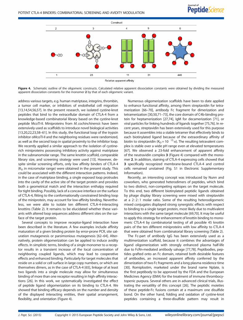
Figure 4. Schematic outline of the oligomeric constructs. Calculated relative apparent dissociation constants were obtained by dividing the measuredapparent dissociation constants for the monomer 2 by that of each oligomeric variant.
POTENT CTLA-4 BINDERS: COMBINATORIAL SCREENING AND AVIDITY MODULATION
address various targets, e.g. humanmatriptase, integrins, thrombin,a tumor cell marker, or inhibitors of endothelial cell migration[13,14,54,56,57]. In the present research, we isolated cystine-knotpeptides that bind to the extracellular domain of CTLA-4 from aknowledge-based combinatorial library based on the cystine-knotpeptide McoTI-II. Miniproteins from M. cochinchinensis have beenextensively used as scaffolds to introduce novel biological activities[13,20,22,23,58–61]. In this study, the functional loop of the trypsininhibitor oMcoTI-II and the neighboring residues were randomized,as well as the second loop in spatial proximity to the inhibitor loop.We recently applied a similar approach to the isolation of cystine-rich miniproteins possessing inhibitory activity against matriptasein the subnanomolar range. The same knottin scaffold, comparablelibrary size, and screening strategy were used [13]. However, de-spite similar screening efforts, only low affinity binders of CTLA-4(KD in micromolar range) were obtained in the present study. Thiscould be associated with the different interaction patterns. Indeed,in the case of matriptase binding, a single exposed loop protrudesinto the cavity of the active site of the target protein and providesboth a geometrical match and the interaction enthalpy requiredfor tight binding. Possibly, lack of a concave interface on the surfaceof CTLA-4, fitting to the conformationally constrained binding loopof the miniprotein, may account for low-affinity binding. Neverthe-less, we were able to isolate ten different CTLA-4-interactingknottins (Table 2). It remains to be elucidated whether these vari-ants with altered loop sequences address different sites on the sur-face of the target protein.Several concepts to improve receptor-ligand interaction have
been described in the literature. A few examples include affinitymaturation of a given binding protein by error-prone PCR, site sat-uration mutagenesis or parsimonious mutagenesis [62–64]. Alter-natively, protein oligomerization can be applied to induce avidityeffects. In simplistic terms, binding of a single monomer to a recep-tor results in a transient increase of the local concentration ofneighboring coupled ligands, which may lead to cooperativeeffects and enhanced binding. Particularly for target molecules thatreside on a solid or cell surface in large copy numbers, or which arethemselves dimers, as in the case of CTLA-4 [65], linkage of at leasttwo ligands into a single molecule may allow for simultaneousbinding ofmore than one receptor resulting in high-affinity interac-tions [26]. In this work, we systematically investigated the effectof peptide ligand oligomerization on its binding to CTLA-4. Weshowed that binding efficacy depends on the number and densityof the displayed interacting entities, their spatial arrangement,flexibility, and orientation (Figure 4).
J. Pept. Sci. (2015) Copyright © 2015 European Peptide Society and Jo
Numerous oligomerization scaffolds have been to date appliedto enhance functional affinity, among them streptavidin for tetra-merization [66–70], antibody Fc fragment for dimerization andtetramerization [30,50,71–73], the core domain of C4b-binding pro-tein for heptamerization [27,74], IgM for decamerization [71], orviral particles for linking hundreds of ligands together [75,76]. In re-cent years, streptavidin has been extensively used for this purposebecause it assembles into a stable tetramer that effectively binds toeach biotinylated ligand because of the extraordinary affinity ofbiotin to streptavidin (KD= 10
�15M). The resulting tetravalent com-
plex is stable over a wide pH range even at elevated temperatures[67]. We observed a 23-fold enhancement of apparent affinityof the neutravidin complex 3 (Figure 4) compared with the mono-mer 2. In addition, staining of CTLA-4 expressing cells showed that3 specifically recognized membrane-bound CTLA-4 and controlcells remained unstained (Fig. S1 in Electronic SupplementaryInformation).
Recently, an interesting concept was introduced by Nunn andcoworkers, who generated heterodimers of peptides, which bindto two distinct, non-competing epitopes on the target molecule.To this end, two different biotinylated peptidic ligands obtainedby phage display library screening were mixed with streptavidinat a 2 : 2 : 1 molar ratio. Some of the resulting heterooligomericmixed conjugates displayed strong synergistic effects with respectto binding to a single target protein, most likely due to multivalentinteractions with the same target molecule [69,70]. It may be usefulto apply this strategy for enhancement of knottin binding tomono-meric CTLA-4 by combinatorial testing of all possible 45 knottinpairs of the ten different miniproteins with low affinity to CTLA-4that were obtained from combinatorial library screening (Table 2).
The Fc-part of antibody IgG has been extensively used as amultimerization scaffold, because it combines the advantages ofligand oligomerization with strongly enhanced plasma half-lifedue to FcRn-mediated antibody salvage [77,78]. Peptibodies, pep-tides grafted onto an Fc domain, retained both desirable featuresof antibodies, an increased apparent affinity conferred by thedimerization of two Fc fragments and a long plasma residence time[30]. Romiplostim, marketed under the brand name Nplate, isthe first peptibody to be approved by the FDA and the EuropeanMedicines Agency (EMA) for the treatment of immune thrombocy-topenic purpura. Several others are in advanced clinical trials, illus-trating the versatility of this concept [26]. The peptidic moietiesof these peptide-Fc fusions contain at a maximum one disulfidebond. On the other hand, folding and oxidation of cystine-knotpeptides containing a three-disulfide pattern may result in
hn Wiley & Sons, Ltd. wileyonlinelibrary.com/journal/jpepsci
MAAß ET AL
disulfide-scrambled products. It is particularly complicated whenfused to Fc, where two more cysteines that reside in the adjacenthinge region may affect proper folding. As a safety catch, cysteinesin the Fc hinge region were replaced by serines. Additionally, thehinge regionwas shortened to a length of 12 amino acids, as shownin another study with cysteine-rich peptides displayed on Fc [50].Gel filtration analysis confirmed that this Fc construct lacking inter-molecular disulfides still formed homodimers, mainly because ofhydrophobic interactions of the CH3 domains [79,80]. Recently, ithas been shown that a derivative of the cystine-knot peptideEETI-II (engineered for integrin binding and used for tumor imag-ing) was directly fused to the Fc region of mouse IgG2a withoutremoval of hinge region cysteines. In this construct, the ability fortumor imaging was retained, which indicates that presence ofhinge region cysteines may not negatively interfere with formationof the three disulfides in the grafted knottin peptide. No data on theimprovement of apparent affinity were reported in this paper [17].We found that efficacy of the CTLA-4 binding was improved 11-foldfor the dimer 4 and 31-fold for the tetravalent variant 5 comparedwith the parent peptide 1 (Figure 4).
It is difficult to assess the extent of possible disulfide scram-bling in the CTLA-4 binding miniprotein when fused to Fc. Toinvestigate, whether MCoTI-II derived miniproteins have the gen-eral capability to form the correct disulfide pattern upon recombi-nant expression as fusion protein, we constructed an Fc fusion ofan acyclic MCoTI-II-based trypsin inhibitor miniprotein (Fig. S1 inElectronic Supplementary Information) that shares with peptide 1the same cystine-knot framework. For that miniprotein, it is knownthat only the particular disulfide pattern, CI-CIV, CII-CV, CIII-CVI,mediates biological activity, i.e. inhibition of trypsin-like protease[81]. Thus, the inhibitory activity of the synthetic miniprotein withdefined cystine-knot topology was compared with that of therecombinantly produced fusion protein bearing the same mini-protein counterpart (Fig. S1 in Electronic Supplementary Informa-tion). Comparable inhibition constants imply that a significantfraction of the oMCoTI-II-based CTLA-4-binding miniprotein 1 infusion proteins 4, 5, and 6 very likely has the canonical cystine-knot architecture (Fig. S1 in Electronic Supplementary Information).
An over 400-fold improvement of apparent affinity was observedupon miniprotein fusion to the C4BP core domain. The crystalstructure of the human C4BP oligomerization domain revealed asymmetric heptamer, which because of its small size of only 57 res-idues per a monomer unit allows for the display of up to sevenfused ligands, positioned in close proximity to each other on thedonut-like nanoscaffold [51]. This domain was used recently forthe multivalent display of the HIV-1 fusion inhibitory peptide C46and of two peptides that inhibited IgG binding to human factor VIII[27,74]. Both constructs displayed prolonged in vivo half-life relativeto synthetic peptides, but only the mimotope peptides graftedonto the C4bpα domain displayed a 20-fold enhancement of bind-ing potency. This finding corroborates the notion that the extent ofbinding improvement through multivalency cannot be predictedquantitatively and requires systematic experimental evaluation ofeach individual ligand-receptor pair.
In conclusion, we have converted a cystine-knot protease inhibi-tor into a receptor binder bymolecular engineering and significantlyenhanced its binding to the dimeric target through oligomerization.The resulting oligovalent conjugates bind the extracellular domainof CTLA-4 with dissociation constants in the low nanomolar range.It remains to be elucidated, whether these oligomers retain en-hanced affinity in experiments with CTLA-4 overexpressing cellsand whether these constructs are able to alleviate the inhibitory
wileyonlinelibrary.com/journal/jpepsci Copyright © 2015 Europ
signaling between CTLA-4 and its receptor B-7. Moreover, recentdata indicated that the activity of anti–CTLA-4 antibody is mediatedat least in part via selective depletion of T regulatory cells within tu-mor lesions, which is dependent on the presence of Fcγ receptor–expressing macrophages [82]. Because 1 fused to antibody Fc (butnot to C4BP) potentially induces antibody-dependent cell cytotoxity(ADCC), it will be interesting to investigate whether differences pre-vail in the enhancement of intratumoral T eff/T reg cell ratio upon tu-mor treatment with these compounds.
Acknowledgements
This work was supported in part by Deutsche Forschungsgemeins-chaft through grant KO1390/10-1 to S.D. in the frame of the priorityprogram SPP1623.
References1 Kolmar H. Biological diversity and therapeutic potential of natural and
engineered cystine knot miniproteins. Curr. Opin. Pharmacol. 2009; 9:608–614.
2 Craik DJ, Daly NL, Waine C. The cystine knot motif in toxins andimplications for drug design. Toxicon 2001; 39: 43–60.
3 Kolmar H. Alternative binding proteins: biological activity andtherapeutic potential of cystine-knot miniproteins. FEBS J. 2008; 275:2684–2690.
4 Heitz A, Avrutina O, Le-NguyenD, DiederichsenU, Hernandez JF, Gracy J,Kolmar H, Chiche L. Knottin cyclization: impact on structure anddynamics. BMC Struct. Biol. 2008; 8: 54.
5 Reinwarth M, Nasu D, Kolmar H, Avrutina O. Chemical synthesis,backbone cyclization and oxidative folding of cystine-knot peptides:promising scaffolds for applications in drug design. Molecules 2012; 17:12533–12552.
6 Lahti JL, Silverman AP, Cochran JR. Interrogating and predictingtolerated sequence diversity in protein folds: application to E. elateriumtrypsin inhibitor-II cystine-knot miniprotein. PLoS Comput. Biol. 2009; 5:e1000499.
7 Kimura RH, Jones DS, Jiang L, Miao Z, Cheng Z, Cochran JR.Functional mutation of multiple solvent-exposed loops in theEcballium elaterium trypsin inhibitor-II cystine knot miniprotein. PLoSOne 2011; 6: e16112.
8 Werle M, Kafedjiiski K, Kolmar H, Bernkop-Schnurch A. Evaluation andimprovement of the properties of the novel cystine-knot microproteinMcoEeTI for oral administration. Int. J. Pharm. 2007; 332: 72–79.
9 Werle M, Schmitz T, Huang HL, Wentzel A, Kolmar H,Bernkop-Schnurch A. The potential of cystine-knot microproteins asnovel pharmacophoric scaffolds in oral peptide drug delivery. J. DrugTarget. 2006; 14: 137–146.
10 Kolmar H. Natural and engineered cystine knot miniproteins fordiagnostic and therapeutic applications. Curr. Pharm. Des. 2011; 17:4329–4336.
11 Moore SJ, Cochran JR. Engineering knottins as novel binding agents.Methods Enzymol. 2012; 503: 223–251.
12 Poth AG, Chan LY, Craik DJ. Cyclotides as grafting frameworks forprotein engineering and drug design applications. Biopolymers 2013;100: 480–491.
13 Glotzbach B, Reinwarth M, Weber N, Fabritz S, Tomaszowski M, Fittler H,Christmann A, Avrutina O, Kolmar H. Combinatorial optimization ofcystine-knot peptides towards high-affinity inhibitors of humanmatriptase-1. PLoS One 2013; 8: e76956.
14 Getz JA, Cheneval O, Craik DJ, Daugherty PS. Design of a cyclotideantagonist of neuropilin-1 and -2 that potently inhibits endothelial cellmigration. ACS Chem. Biol. 2013; 8: 1147–1154.
15 Kimura RH, Cheng Z, Gambhir SS, Cochran JR. Engineered knottinpeptides: a new class of agents for imaging integrin expression inliving subjects. Cancer Res. 2009; 69: 2435–2442.
16 Liu S, Liu H, Ren G, Kimura RH, Cochran JR, Cheng Z. PET Imaging ofintegrin positive tumors using F labeled knottin peptides. Theranostics.2011; 1: 403–412.
17 Moore SJ, Hayden Gephart MG, Bergen JM, Su YS, Rayburn H, Scott MP,Cochran JR. Engineered knottin peptide enables noninvasive optical
ean Peptide Society and John Wiley & Sons, Ltd. J. Pept. Sci. (2015)

POTENT CTLA-4 BINDERS: COMBINATORIAL SCREENING AND AVIDITY MODULATION
imaging of intracranial medulloblastoma. Proc. Natl. Acad. Sci. U. S. A.2013; 110: 14598–14603.
18 Moore SJ, Leung CL, Norton HK, Cochran JR. Engineering agatoxin, acystine-knot peptide from spider venom, as a molecular probe forin vivo tumor imaging. PLoS One 2013; 8: e60498.
19 Felizmenio-Quimio ME, Daly NL, Craik DJ. Circular proteins in plants:solution structure of a novel macrocyclic trypsin inhibitor fromMomordica cochinchinensis. J. Biol. Chem. 2001; 276: 22875–22882.
20 Ji Y, Majumder S, Millard M, Borra R, Bi T, Elnagar AY, Neamati N,Shekhtman A, Camarero JA. In vivo activation of the p53 tumorsuppressor pathway by an engineered cyclotide. J. Am. Chem. Soc.2013; 135: 11623–11633.
21 Klotz U. Ziconotide� a novel neuron-specific calcium channel blockerfor the intrathecal treatment of severe chronic pain� a short review.Int. J. Clin. Pharmacol. Ther. 2006; 44: 478–483.
22 D’Souza C, Henriques ST, Wang CK, Craik DJ. Structural parametersmodulating the cellular uptake of disulfide-rich cyclic cell-penetratingpeptides: MCoTI-II and SFTI-1. Eur. J. Med. Chem. 2014; 88: 10–18.
23 Huang YH, Chaousis S, Cheneval O, Craik DJ, Henriques ST. Optimizationof the cyclotide framework to improve cell penetration properties. Front.Pharmacol. 2015; 6: 17.
24 Wong CT, Rowlands DK, Wong CH, Lo TW, Nguyen GK, Li HY, Tam JP.Orally active peptidic bradykinin B1 receptor antagonists engineeredfrom a cyclotide scaffold for inflammatory pain treatment. Angew.Chem. Int. Ed. Engl. 2012; 51: 5620–5624.
25 Chittasupho C. Multivalent ligand: design principle for targetedtherapeutic delivery approach. Ther. Deliv. 2012; 3: 1171–1187.
26 Handl HL, Vagner J, Han H, Mash E, Hruby VJ, Gillies RJ. Hittingmultiple targets with multimeric ligands. Expert Opin. Ther. Targets2004; 8: 565–586.
27 Dervillez X, Huther A, Schuhmacher J, Griesinger C, Cohen JH,von Laer D, Dietrich U. Stable expression of soluble therapeuticpeptides in eukaryotic cells by multimerisation: application to the HIV-1 fusion inhibitory peptide C46. ChemMedChem 2006; 1: 330–339.
28 Lassabe G, Rossotti M, Gonzalez-Techera A, Gonzalez-Sapienza G. Shiga-like toxin B subunit of Escherichia coli as scaffold for high-avidity displayof anti-immunocomplex peptides. Anal. Chem. 2014; 86: 5541–5546.
29 McNerny DQ, Kukowska-Latallo JF, Mullen DG, Wallace JM, Desai AM,Shukla R, Huang B, Banaszak Holl MM, Baker JR, Jr. RGD dendronbodies; synthetic avidity agents with defined and potentiallyinterchangeable effector sites that can substitute for antibodies.Bioconjug. Chem. 2009; 20: 1853–1859.
30 Shimamoto G, Gegg C, Boone T, Queva C. Peptibodies: a flexiblealternative format to antibodies. MAbs. 2012; 4: 586–591.
31 Krause S, Schmoldt HU, Wentzel A, Ballmaier M, Friedrich K, Kolmar H.Grafting of thrombopoietin-mimetic peptides into cystine knotminiproteins yields high-affinity thrombopoietin antagonists andagonists. FEBS J. 2007; 274: 86–95.
32 Chmielowski B. Ipilimumab: a first-in-class T-cell potentiator formetastatic melanoma. J. Skin Cancer 2013; 2013: 423829.
33 Hodi FS. Cytotoxic T-lymphocyte-associated antigen-4. Clin. Cancer Res.2007; 13: 5238–5242.
34 Krummel MF, Allison JP. CD28 and CTLA-4 have opposing effects on theresponse of T cells to stimulation. J. Exp. Med. 1995; 182: 459–465.
35 LenschowDJ, Zeng Y, Thistlethwaite JR, Montag A, BradyW, Gibson MG,Linsley PS, Bluestone JA. Long-term survival of xenogeneic pancreaticislet grafts induced by CTLA4lg. Science 1992; 257: 789–792.
36 Linsley PS, Wallace PM, Johnson J, Gibson MG, Greene JL, Ledbetter JA,Singh C, Tepper MA. Immunosuppression in vivo by a soluble form ofthe CTLA-4 T cell activation molecule. Science 1992; 257: 792–795.
37 Leach DR, Krummel MF, Allison JP. Enhancement of antitumor immunityby CTLA-4 blockade. Science 1996; 271: 1734–1736.
38 Pardoll DM. The blockade of immune checkpoints in cancerimmunotherapy. Nat. Rev. Cancer 2012; 12: 252–264.
39 Phan GQ, Yang JC, Sherry RM, Hwu P, Topalian SL, Schwartzentruber DJ,Restifo NP, Haworth LR, Seipp CA, Freezer LJ, Morton KE, Mavroukakis SA,Duray PH, Steinberg SM, Allison JP, Davis TA, Rosenberg SA. Cancerregression and autoimmunity induced by cytotoxic T lymphocyte-associated antigen 4 blockade in patients with metastatic melanoma.Proc. Natl. Acad. Sci. U. S. A. 2003; 100: 8372–8377.
40 Kaufmann DE, Kavanagh DG, Pereyra F, Zaunders JJ, Mackey EW,Miura T, Palmer S, Brockman M, Rathod A, Piechocka-Trocha A,Baker B, Zhu B, Le Gall S, Waring MT, Ahern R, Moss K, Kelleher AD,Coffin JM, Freeman GJ, Rosenberg ES, Walker BD. Upregulation ofCTLA-4 by HIV-specific CD4+ T cells correlates with disease progression
J. Pept. Sci. (2015) Copyright © 2015 European Peptide Society and Jo
and defines a reversible immune dysfunction. Nat. Immunol. 2007; 8:1246–1254.
41 Ruderman EM, Pope RM. The evolving clinical profile of abatacept(CTLA4-Ig): a novel co-stimulatory modulator for the treatment ofrheumatoid arthritis. Arthritis Res. Ther. 2005; 7(Suppl 2): S21–25.
42 Zubairi S, Sanos SL, Hill S, Kaye PM. Immunotherapy with OX40L-Fc oranti-CTLA-4 enhances local tissue responses and killing of Leishmaniadonovani. Eur. J. Immunol. 2004; 34: 1433–1440.
43 Sondak VK, Smalley KS, Kudchadkar R, Grippon S, Kirkpatrick P.Ipilimumab. Nat. Rev. Drug Discov. 2011; 10: 411–412.
44 Heitz A, Hernandez JF, Gagnon J, Hong TT, Pham TT, Nguyen TM,Le-Nguyen D, Chiche L. Solution structure of the squash trypsininhibitor MCoTI-II. A new family for cyclic knottins. Biochemistry 2001;40: 7973–7983.
45 Hernandez JF, Gagnon J, Chiche L, Nguyen TM, Andrieu JP, Heitz A,Trinh Hong T, Pham TT, Le Nguyen D. Squash trypsin inhibitors fromMomordica cochinchinensis exhibit an atypical macrocyclic structure.Biochemistry 2000; 39: 5722–5730.
46 Van den Brulle J, Fischer M, Langmann T, Horn G, Waldmann T, Arnold S,Fuhrmann M, Schatz O, O’Connell T, O’Connell D, Auckenthaler A,Schwer H. A novel solid phase technology for high-throughput genesynthesis. Biotechniques 2008; 45: 340–343.
47 Boder ET, Wittrup KD. Yeast surface display for screening combinatorialpolypeptide libraries. Nat. Biotechnol. 1997; 15: 553–557.
48 Benatuil L, Perez JM, Belk J, Hsieh CM. An improved yeast transformationmethod for the generation of very large human antibody libraries.Protein Eng. Des. Sel. 2010; 23: 155–159.
49 Schmoldt HU, Daneschdar M, Kolmar H, Blind M. Microbodies. MethodsMol. Biol. 2009; 535: 361–372.
50 Angelini A, Diderich P, Morales-Sanfrutos J, Thurnheer S, Hacker D,Menin L, Heinis C. Chemical macrocyclization of peptides fused toantibody Fc fragments. Bioconjug. Chem. 2012; 23: 1856–1863.
51 Hofmeyer T, Schmelz S, Degiacomi MT, Dal Peraro M, Daneschdar M,Scrima A, van den Heuvel J, Heinz DW, Kolmar H. Arranged sevenfold:structural insights into the C-terminal oligomerization domain ofhuman C4b-binding protein. J. Mol. Biol. 2013; 425: 1302–1317.
52 Reinwarth M, Glotzbach B, Tomaszowski M, Fabritz S, Avrutina O,Kolmar H. Oxidative folding of peptides with cystine-knot architectures:kinetic studies and optimization of folding conditions. Chembiochem2013; 14: 137–146.
53 Feige MJ, Hendershot LM. Disulfide bonds in ER protein folding andhomeostasis. Curr. Opin. Cell Biol. 2011; 23: 167–175.
54 Silverman AP, Levin AM, Lahti JL, Cochran JR. Engineered cystine-knotpeptides that bind avb3 integrin with antibody-like affinities. J. Mol.Biol. 2009; 385: 1064–1075.
55 Northfield SE, Wang CK, Schroeder CI, Durek T, Kan MW, Swedberg JE,Craik DJ. Disulfide-rich macrocyclic peptides as templates in drugdesign. Eur. J. Med. Chem. 2014; 77: 248–257.
56 Getz JA, Rice JJ, Daugherty PS. Protease-resistant peptide ligands from aknottin scaffold library. ACS Chem. Biol. 2011; 6: 837–844.
57 Zoller F, Markert A, Barthe P, Zhao W, Weichert W, Askoxylakis V,Altmann A, Mier W, Haberkorn U. Combination of phage display andmolecular grafting generates highly specific tumor-targetingminiproteins. Angew. Chem. Int. Ed. Engl. 2012; 51: 13136–13139.
58 Aboye TL, Ha H, Majumder S, Christ F, Debyser Z, Shekhtman A,Neamati N, Camarero JA. Design of a novel cyclotide-based CXCR4antagonist with anti-human immunodeficiency virus (HIV)-1 activity.J. Med. Chem. 2012; 55: 10729–10734.
59 Austin J, Wang W, Puttamadappa S, Shekhtman A, Camarero JA.Biosynthesis and biological screening of a genetically encoded librarybased on the cyclotide MCoTI-I. Chembiochem 2009; 10: 2663–2670.
60 Chan LY, Gunasekera S, Henriques ST, Worth NF, Le SJ, Clark RJ,Campbell JH, Craik DJ, Daly NL. Engineering pro-angiogenicpeptides using stable, disulfide-rich cyclic scaffolds. Blood 2011; 118:6709–6717.
61 Sommerhoff CP, Avrutina O, Schmoldt HU, Gabrijelcic-Geiger D,Diederichsen U, Kolmar H. Engineered cystine knot miniproteins aspotent inhibitors of human mast cell tryptase b. J. Mol. Biol. 2010; 395:167–175.
62 Labrou NE. Random mutagenesis methods for in vitro directed enzymeevolution. Curr. Protein Pept. Sci. 2010; 11: 91–100.
63 Tee KL, Wong TS. Polishing the craft of genetic diversity creation indirected evolution. Biotechnol. Adv. 2013; 31: 1707–1721.
64 Sheedy C, MacKenzie CR, Hall JC. Isolation and affinity maturation ofhapten-specific antibodies. Biotechnol. Adv. 2007; 25: 333–352.
hn Wiley & Sons, Ltd. wileyonlinelibrary.com/journal/jpepsci
MAAß ET AL
65 Darlington PJ, Kirchhof MG, Criado G, Sondhi J, Madrenas J.Hierarchical regulation of CTLA-4 dimer-based lattice formation andits biological relevance for T cell inactivation. J. Immunol. 2005; 175:996–1004.
66 Cloutier SM, Couty S, Terskikh A, Marguerat L, Crivelli V, Pugnieres M,Mani JC, Leisinger HJ, Mach JP, Deperthes D. Streptabody, a highavidity molecule made by tetramerization of in vivo biotinylated,phage display-selected scFv fragments on streptavidin. Mol. Immunol.2000; 37: 1067–1077.
67 Kipriyanov SM, Little M, Kropshofer H, Breitling F, Gotter S, Dubel S.Affinity enhancement of a recombinant antibody: formation ofcomplexes with multiple valency by a single-chain Fv fragment-corestreptavidin fusion. Protein Eng. 1996; 9: 203–211.
68 Leisner C, Loeth N, Lamberth K, Justesen S, Sylvester-Hvid C, Schmidt EG,Claesson M, Buus S, Stryhn A. One-pot, mix-and-read peptide-MHCtetramers. PLoS One 2008; 3: e1678.
69 Shrivastava A, von Wronski M, Tweedle MF, Nunn AD. Identification ofideal peptides for heterovalent ligands. Methods Mol. Biol. 2014; 1088:97–105.
70 Shrivastava A, von Wronski MA, Sato AK, Dransfield DT, Sexton D,Bogdan N, Pillai R, Nanjappan P, Song B, Marinelli E, DeOliveira D,Luneau C, Devlin M, Muruganandam A, Abujoub A, Connelly G, Wu QL,Conley G, Chang Q, Tweedle MF, Ladner RC, Swenson RE, Nunn AD. Adistinct strategy to generate high-affinity peptide binders to receptortyrosine kinases. Protein Eng. Des. Sel. 2005; 18: 417–424.
71 Cannon JP, O’Driscoll M, Litman GW. Construction, expression, andpurification of chimeric protein reagents based on immunoglobulin Fcregions. Methods Mol. Biol. 2011; 748: 51–67.
72 Jazayeri JA, Carroll GJ. Fc-based cytokines: prospects for engineeringsuperior therapeutics. BioDrugs 2008; 22: 11–26.
73 Ronnmark J, Hansson M, Nguyen T, Uhlen M, Robert A, Stahl S,Nygren PA. Construction and characterization of affibody-Fcchimeras produced in Escherichia coli. J. Immunol. Methods 2002;261: 199–211.
74 Kessel C, Kreuz W, Klich K, Becker-Peters K, Vorpahl F, Dietrich U,Klingebiel T, Konigs C. Multimerization of peptide mimotopes forblocking of factor VIII neutralizing antibodies. ChemMedChem 2009; 4:1364–1370.
wileyonlinelibrary.com/journal/jpepsci Copyright © 2015 Europ
75 Ashley CE, Carnes EC, Phillips GK, Durfee PN, Buley MD, Lino CA,Padilla DP, Phillips B, Carter MB, Willman CL, Brinker CJ, Caldeira Jdo C,Chackerian B, Wharton W, Peabody DS. Cell-specific delivery of diversecargos by bacteriophage MS2 virus-like particles. ACS Nano 2011; 5:5729–5745.
76 Rademacher C, Guiard J, Kitov PI, Fiege B, Dalton KP, Parra F,Bundle DR, Peters T. Targeting norovirus infection-multivalent entryinhibitor design based on NMR experiments. Chemistry 2011; 17:7442–7453.
77 Brambell FW, Halliday R, Morris IG. Interference by human and bovineserum and serum protein fractions with the absorption of antibodiesby suckling rats and mice. Proc. R. Soc. Lond. B Biol. Sci. 1958; 149:1–11.
78 Mould DR, Sweeney KR. The pharmacokinetics and pharmacodynamicsof monoclonal antibodies – mechanistic modeling applied to drugdevelopment. Curr. Opin. Drug Discov. Dev. 2007; 10: 84–96.
79 Deisenhofer J. Crystallographic refinement and atomic models of ahuman Fc fragment and its complex with fragment B of protein Afrom Staphylococcus aureus at 2.9- and 2.8 Å resolution. Biochemistry1981; 20: 2361–2370.
80 Miller S. Protein-protein recognition and the association ofimmunoglobulin constant domains. J. Mol. Biol. 1990; 216: 965–973.
81 Wentzel A, Christmann A, Kratzner R, Kolmar H. Sequence requirementsof the GPNG b-turn of the Ecballium elaterium trypsin inhibitor IIexplored by combinatorial library screening. J. Biol. Chem. 1999; 274:21037–21043.
82 Simpson TR, Li F, Montalvo-Ortiz W, Sepulveda MA, Bergerhoff K, Arce F,Roddie C, Henry JY, Yagita H, Wolchok JD, Peggs KS, Ravetch JV,Allison JP, Quezada SA. Fc-dependent depletion of tumor-infiltratingregulatory T cells co-defines the efficacy of anti-CTLA-4 therapy againstmelanoma. J. Exp. Med. 2013; 210: 1695–1710.
Supporting Information
Additional supporting information may be found in the online ver-sion of this article at the publisher’s web site.
ean Peptide Society and John Wiley & Sons, Ltd. J. Pept. Sci. (2015)
Related Documents