49 Original Article Cutaneous Manifestations in Egyptian Patients with Chronic Renal Failure on Regular Hemodialysis Maha M. Sultan, M.D.*, Hayam H. Mansour, M.D.†, Iman M. Wahby, M.D.‡ and Ali S. Houdery, M.D.§ *Department of Dermatology and Vener eology , †Department of Internal Medicine (Nephrology Unit), ‡Department of Community Medicine, Faculty of Medicine for Girls and §Department of Pathology, Faculty of Medicine, Al-Azhar University, Egypt. Background. Hemodialysis patients ex perience frequent and various c utaneous manifestations of often hypothetical pathogenesis. Chronic renal failure (CRF) presents with an array of cutaneous manifestations. Objective. To evaluate the prevalence and nature of cutaneous lesions, associated with CRF patients on hemodialysis in Egyptian patients. Patients and methods. One hundred patients with CRF on regular hemodialysis from nephrology units were examined for cutaneous changes. Specic investigations like skin biopsy , culture and sensitivity for bacterial infections, potassium hydroxide mount and fungal culture were done when indicated. Results. All patients included in this study had at least one cutaneous manifestation attributable to CRF. The most prevalent ndings was pruritus (55%), followed by xerosis (54%), hyperpigmentation (54%) and pallor (45%). Other cutaneous manifes tations were wrinkles (40%), fungal infections (33%), ecchymosis (27%), dermatitis (23%), yellow face (22%), Petichae (19%), delayed wound healing (11%), follicular hyperkeratosis (10%), bacterial infections (5%), viral infections (2%) and uremic frost (1%). Nail changes were koilonychia (39%), half and half nail (28%), splinter hemorrhages (16%), Mue hrcke’s lines (12%), subungual hyperkeratosis (10%), Mees’ lines (8%), brown nail (6%), onycholysis (3%) and Beau’s lines (2%). Hair changes were brittle and lusterless hair (47%), sparse scalp hair (46%) and sparse body hair (27%). Oral changes were macroglossia (42%), xerostomia (35%), coated tongue (27%), angular cheilitis (15%), ulcerative stomatitis (9%), acquired perforating dermatosis (3%). Some rare manifestations of CRF like calc iphylaxis (2%) were seen. There was no association between any particular etiology of CRF and certain mucocutaneous, nail or hair abnormalities. Conclusion. At least one cutaneous manifestation is found in all CRF patients. The most prevalent ndings were pruritus followed by xerosis and hyper-pigmentation then pallor. With the advent of hemodialysis, the life expectancy of these patients has increased giving time for more and newer cutaneous changes to manifest. The aetiology of CRF does not affect the development of cutaneous, nail or hair abnormalities. ( J Egypt Women Dermatol Soc 2010; 7: 49 - 55) Keywords. Chronic renal failure, hemodialysis, cutaneous manifestations, calciphylaxis Corresponding Author. Maha M. Sultan, M.D., Lecturer of Dermatology and Venereology, Faculty of Medicine for Girls, Al-Azhar University , Cairo, Egypt. E-mail. dr.maha_derm@y ahoo.com Conict of interest. None declared. Copyright © 2009 Egyptian Women Dermatologic Society. All rights reserved. T he skin is a mirror for many systemic diseases including renal diseases 1 . Chronic renal failure (CRF) often produces specic skin changes, which can develop long before failure manifests clinically 2 . These symptoms tend to alter and are aggravated relatively quickly when chronic renal insufciency leads to compulsor y dialysis treatment 3 . There are many reports of cutaneous changes in CRF from different parts of the world 4 . The aim of this study was to elucidate the prevalence and nature of cutaneous manifestations in Egyptian patients with CRF on regular hemodialysis. P A TIENTS AND METHODS One hundred patients of CRF on regular hemodialysis were selected from Nephrology Units in Al-Zahraa University Hospital, Al-Aziz Bellah and Al-Ber wa El-Takwa Hospitals in Cairo. All patients were subjected to full history and clinical examination. Complete dermatological examination including skin, hair, nails and oral mucosa was done. Complete blood picture, kidney function tests, liver function tests and fasting and postprandial blood glucose levels were done. Patients with elevated liver enzymes were excluded. Specic investigations like skin biopsy, culture and sensitivity for bacterial infections, Gram’s stain, potassium hydroxide mount and fungal culture were done when indicated, after taking patients’ informed consent. Statistical Analysis. Data was analyzed by Statistical Package for Social Science (SPSS) version 12. Parametric data

Welcome message from author
This document is posted to help you gain knowledge. Please leave a comment to let me know what you think about it! Share it to your friends and learn new things together.
Transcript
8/7/2019 Cutaneous Manifestations in Egyptian Patients with Chronic Renal Failure on Regular Hemodialysis
http://slidepdf.com/reader/full/cutaneous-manifestations-in-egyptian-patients-with-chronic-renal-failure-on 1/7
49
Original Article
Cutaneous Manifestations in Egyptian Patients with ChronicRenal Failure on Regular Hemodialysis
Maha M. Sultan, M.D.*, Hayam H. Mansour, M.D.†, Iman M. Wahby, M.D.‡ and
Ali S. Houdery, M.D.§
*Department of Dermatology and Venereology, †Department of Internal Medicine (Nephrology Unit),
‡Department of Community Medicine, Faculty of Medicine for Girls and §Department of Pathology,
Faculty of Medicine, Al-Azhar University, Egypt.
Background. Hemodialysis patients experience frequent and various cutaneous manifestations of often hypothetical
pathogenesis. Chronic renal failure (CRF) presents with an array of cutaneous manifestations. Objective. To evaluate
the prevalence and nature of cutaneous lesions, associated with CRF patients on hemodialysis in Egyptian patients.
Patients and methods. One hundred patients with CRF on regular hemodialysis from nephrology units were examined
for cutaneous changes. Specic investigations like skin biopsy, culture and sensitivity for bacterial infections, potassiumhydroxide mount and fungal culture were done when indicated. Results. All patients included in this study had at
least one cutaneous manifestation attributable to CRF. The most prevalent ndings was pruritus (55%), followed by
xerosis (54%), hyperpigmentation (54%) and pallor (45%). Other cutaneous manifestations were wrinkles (40%),
fungal infections (33%), ecchymosis (27%), dermatitis (23%), yellow face (22%), Petichae (19%), delayed wound
healing (11%), follicular hyperkeratosis (10%), bacterial infections (5%), viral infections (2%) and uremic frost (1%).
Nail changes were koilonychia (39%), half and half nail (28%), splinter hemorrhages (16%), Muehrcke’s lines (12%),
subungual hyperkeratosis (10%), Mees’ lines (8%), brown nail (6%), onycholysis (3%) and Beau’s lines (2%). Hair
changes were brittle and lusterless hair (47%), sparse scalp hair (46%) and sparse body hair (27%). Oral changes
were macroglossia (42%), xerostomia (35%), coated tongue (27%), angular cheilitis (15%), ulcerative stomatitis (9%),
acquired perforating dermatosis (3%). Some rare manifestations of CRF like calciphylaxis (2%) were seen. There
was no association between any particular etiology of CRF and certain mucocutaneous, nail or hair abnormalities.
Conclusion. At least one cutaneous manifestation is found in all CRF patients. The most prevalent ndings were
pruritus followed by xerosis and hyper-pigmentation then pallor. With the advent of hemodialysis, the life expectancy of
these patients has increased giving time for more and newer cutaneous changes to manifest. The aetiology of CRF does
not affect the development of cutaneous, nail or hair abnormalities. ( J Egypt Women Dermatol Soc 2010; 7: 49 - 55)
Keywords. Chronic renal failure, hemodialysis, cutaneous manifestations, calciphylaxis
Corresponding Author. Maha M. Sultan, M.D., Lecturer of
Dermatology and Venereology, Faculty of Medicine for Girls, Al-Azhar
University, Cairo, Egypt. E-mail. [email protected]
Conict of interest. None declared.
Copyright © 2009 Egyptian Women Dermatologic Society. All rights
reserved.
The skin is a mirror for many systemicdiseases including renal diseases1. Chronicrenal failure (CRF) often produces specic
skin changes, which can develop long before failuremanifests clinically2. These symptoms tend to alter and
are aggravated relatively quickly when chronic renalinsufciency leads to compulsory dialysis treatment3.There are many reports of cutaneous changes in CRFfrom different parts of the world4. The aim of thisstudy was to elucidate the prevalence and nature of cutaneous manifestations in Egyptian patients withCRF on regular hemodialysis.
PATIENTS AND METHODS
One hundred patients of CRF on regular hemodialysis were selected from Nephrology Units
in Al-Zahraa University Hospital, Al-Aziz Bellahand Al-Ber wa El-Takwa Hospitals in Cairo. All
patients were subjected to full history and clinicalexamination. Complete dermatological examinationincluding skin, hair, nails and oral mucosa was done.
Complete blood picture, kidney function tests, liver function tests and fasting and postprandial bloodglucose levels were done. Patients with elevatedliver enzymes were excluded. Specic investigationslike skin biopsy, culture and sensitivity for bacterialinfections, Gram’s stain, potassium hydroxide mountand fungal culture were done when indicated, after taking patients’ informed consent.
Statistical Analysis.Data was analyzed by Statistical Package for
Social Science (SPSS) version 12. Parametric data

8/7/2019 Cutaneous Manifestations in Egyptian Patients with Chronic Renal Failure on Regular Hemodialysis
http://slidepdf.com/reader/full/cutaneous-manifestations-in-egyptian-patients-with-chronic-renal-failure-on 2/7
Cutaneous Manifestations in Egyptian Patients with Chronic
Renal Failure on Regular Hemodialysis
50
J Egypt Women Dermatol Soc. Vol. 7, No. 1, 2010
was expressed as mean ± SD and non parametricdata was expressed as number and percentage of thetotal.
RESULTS
They were 66 males and 34 females. Their ageranged from 15 - 73 years with mean age of 49.53+ 18.54 years. The total duration of hemodialysisranged from 0.08 - 20 years with a mean of 4.95 +4.03 years (Table 1). The various causes leading torenal failure are shown in table (2).
Table 1. Data of patients.
Variables
Sex
- Male 66 (66%)
- Female 34 (34%)
Age- Range 15 - 73 years- Mean ± SD 49.53 ± 18.54 years
Duration of hemodialysis
- Range 0.08 - 20 years- Mean ± SD 4.95 ± 4.03 years
Hemoglobin level
- Range 5.3 - 14 gm %
- Mean ± SD 10.45 ± 1.64 gm %
Blood urea before hemodialysis
- Range 160 - 350 mg/dL
- Mean ± SD 253.74 ± 58.38 mg/dL
Blood urea during hemodialysis- Range 60 - 160 mg/dL
- Mean ± SD 109.75 ± 28.88 mg/dL
Serum creatinine before hemodialysis
- Range 6 - 22 mg/dL
- Mean ± SD 13.20 ± 5.41 mg/dL
Serum creatinine during hemodialysis
- Range 6 - 11 mg/dL
- Mean ± SD 18.6 ± 1.63 mg/dL
Table 2. Etiology of chronic renal failure.
Causes n %
HTN 60 (60%)
DM 14 (14%)
Obstruction 7 (7%)
Analgesic 5 (5%)
Unknown 4 (4%)
SLE 3 (3%)
Polycystic kidney 2 (2%)
RUTI 2 (2%)
Reux 1 (1%)
FMF 1 (1%)
Wegener 1 (1%)
HTN: hypertension, DM: diabetes mellitus, FMF: familialmediterranean fever, RUTI: recurrent urinary tract infection, SLE:systemic lupus erythematosus.
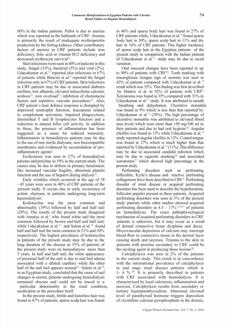
All patients examined in the study showed at leastone cutaneous manifestation. The most prevalentnding was pruritus (55%) (Figure 1), followed byxerosis (54%), hyper-pigmentation (54%) (Figure2) and pallor (45%). Other cutaneous manifestationswere wrinkles (40%) (Figure 3), fungal infections(33%), ecchymosis (27%), dermatitis (23%), yellowface (22%), petichae (19%), delayed wound healing(11%), follicular hyperkeratosis (10%), bacterial
infections (5%), viral infections (2%) and uremicfrost (1%). Nail changes were koilonychia (39%) (Figure
4), half and half nail (28%) (Figure 5), splinter hemorrhages (16%), Muehrcke’s lines (narrowwhite transverse bands occurring in pairs) (12%),Mees’ lines (white transverse bands, single or multiple) (8%) (Figure 6), subungual hyperkeratosis(10%), brown nail (6%), onycholysis (3%) andBeau’s lines (transverse furrows that begin in the
Table 3. Percentage of cutaneous manifestations in relation to etiology.
Skin HTN DM Obstructive Analgesic FMF Polycystic R UTI Reux SLE Wegener
Yellow face 16.67 14.29 14.29 40.00 - 100.00 100.00 100.00 33.33 -Pallor 38.33 28.57 57.14 20.00 - 100.00 50.00 - 33.33 100.00
Hyperpigmentation 61.67 64.29 42.86 40.00 100.00 - 50.00 - 33.33 -Pruritus 51.67 42.86 28.57 60.00 - - 100.00 - 33.33 -Xerosis 58.33 71.43 42.86 60.00 100.00 - 50.00 - - 100.00Dermatitis 25.00 21.43 28.57 - - - - - - -Wound healing 8.33 7.14 14.29 20.00 100.00 - 50.00 - - -Uremic frost 1.67 7.14 - - - - - - - -Follicular hyperkeratosis 10.00 14.29 14.29 40.00 - - - - - -Echymosis 33.33 50.00 57.14 40.00 - - - - - -Petichae 18.33 28.57 28.57 40.00 - - - - - 100.00Jaundice 23.33 28.57 14.29 20.00 - - - - - -Wrinkles 33.33 35.71 57.14 60.00 100.00 - 50.00 - - -
Bacterial infection 5.00 14.29 - - - - - - - -TC 6.67 7.14 - - - - - - - -TP 11.67 7.14 28.57 - - - 50.00 - - 100.00TV 16.67 21.43 14.29 20.00 - - - - 33.33 100.00HZ - - 14.29 - - - - - - -Wart 1.67 - - - - - - - - -
Sparse body hair 21.67 35.71 14.29 20.00 - 50.00 - - - -Sparse scalp hair 40.00 64.29 14.29 80.00 100.00 50.00 50.00 - 33.33 100.00Brittle and lusterless hair 43.33 57.14 14.29 80.00 100.00 50.00 50.00 - 33.33 100.00
HTN: hypertension, DM: diabetes mellitus, FMF: familial mediterranean fever, RUTI: recurrent urinary tract infection, SLE: systemic lupus
erythematosus, TC: tinea circinata, TP: tinea pedis, TV: tinea versicolor, HZ: herpes zoster.

8/7/2019 Cutaneous Manifestations in Egyptian Patients with Chronic Renal Failure on Regular Hemodialysis
http://slidepdf.com/reader/full/cutaneous-manifestations-in-egyptian-patients-with-chronic-renal-failure-on 3/7
Maha M. Sultan et al.51
J Egypt Women Dermatol Soc. Vol. 7, No. 1, 2010
matrix and progress distally as the nail grows)(2%). Hair changes included brittle and
lusterless hair (47%), sparse scalp hair (46%)and sparse body hair (27%). Oral changesincluded macroglossia (42%), xerostomia(35%), coated tongue (27%), angular cheilitis(15%) and ulcerative stomatitis (9%). Somerare manifestations of CRF like calciphylaxiswas found in 2% (Figures 7,8) and acquired
perforating dermatosis secondary to CRF wasfound in 3% of patients (Figure 9).
Biopsies from lesional skin in patients withCRF with acquired perforating dermatosisshowed broad crater in epidermis withdegenerated collagen in dermis with starting
transepidermal elimination (Figure 10).Skin, nail and oral manifestations in relation
to causes of CRF are shown in tables 3, 4 and5 respectively.
Table 4. Percentage of nail changes in relation to etiology.
Nail HTN DM Obstructive Analgesic FMF Polycystic R UTI Reux SLE Wegener
Koilonychia 40.00 35.71 57.14 40.00 100.00 50.00 50.00 - 33.33 -
Half & half 33.33 35.71 28.57 20.00 - - 50.00 - 66.67 -
Splinter hemorrhage 16.67 7.14 14.29 - - - - - - -
Subungual hyperkeratosis 13.33 - 14.29 - - 50.00 - - - -
Muehrcke’s line 10.00 14.29 14.29 - - 50.00 - - - -
Mees’ line 8.33 - - - - - - - - -
Brown nail 6.67 - - 40.00 - - - - - -
Beau’s line 3.33 - - - - - - - - -
Oncholysis 1.67 7.14 14.29 - - - - - - -
HTN: hypertension, DM: diabetes mellitus, FMF: familial mediterranean fever, RUTI: recurrent urinary tract infection, SLE:systemic lupus
erythematosus.
Table 5. Percentage of oral changes in relation to etiology.
Oral HTN DM Obstructive Analgesic FMF Polycystic R UTI Reux SLE Wegener
Macroglossia 36.67 42.86 28.57 60.00 100.00 50.00 100.00 - - 100.00
Xerostomia 36.67 35.71 57.14 40.00 100.00 50.00 100.00 - 33.33 100.00
Coated tongue 23.33 50.00 28.57 80.00 100.00 - - - - 100.00
Angular cheilitis 18.33 7.14 - 20.00 - - - - - 100.00
Ulcerative stomatitis 11.67 7.14 - - - 50.00 - - - 100.00
HTN: hypertension, DM: diabetes mellitus, FMF: familial mediterranean fever, RUTI: recurrent urinary tract infection, SLE: systemic lupus
erythematosus.
Figure 1. Chronic scratching resulting from uremic pruritus.
Figure 2. Diffuse hyperpigmentation of the face.
Figure 3. Extensive wrinkling as a sign of actinic elastosis in 42 aged female.

8/7/2019 Cutaneous Manifestations in Egyptian Patients with Chronic Renal Failure on Regular Hemodialysis
http://slidepdf.com/reader/full/cutaneous-manifestations-in-egyptian-patients-with-chronic-renal-failure-on 4/7
Cutaneous Manifestations in Egyptian Patients with Chronic
Renal Failure on Regular Hemodialysis
52
J Egypt Women Dermatol Soc. Vol. 7, No. 1, 2010
Figure 4. Koilonychia secondary to chronic renal failure.
Figure 5. Half and half nail secondary to chronic renal failure.
Figure 6. Mees’ line (white transverse band) secondary to chronic
renal failure.
Figure 7. Calciphylaxis secondary to chronic renal failure.
Figure 8. Calciphylaxis secondary to chronic renal failure, causing loss
of little toe and distal phalanx of the 2nd toe.
Figure 9. Acquired perforating dermatosis (Kyrle’s) secondary to
chronic renal failure.

8/7/2019 Cutaneous Manifestations in Egyptian Patients with Chronic Renal Failure on Regular Hemodialysis
http://slidepdf.com/reader/full/cutaneous-manifestations-in-egyptian-patients-with-chronic-renal-failure-on 5/7
Maha M. Sultan et al.53
J Egypt Women Dermatol Soc. Vol. 7, No. 1, 2010
DISCUSSIONPruritus was the most common cutaneous
abnormality (55%) in Egyptian CRF patients onhemodialysis. Its prevalence among hemodialysis
patients ranges from 19 to 90% in the other previousstudies5,7. Udayakumar et al.17 reported pruritus in53% of indian patients while Avermaete et al.3 reported
pruritus in 48% of patients with CRF. It is one of themost characteristic and annoying cutaneous symptomsof CRF6. It is not present in acute renal failure anddoes not necessarily subside with dialysis although itimproves with kidney transplantation7. The etiology
of pruritus in CRF is unknown. However, it has beenassociated with the degree of renal insufciency (urineoutput of < 500 mL)8. Hypervitaminosis A may bea cause as CRF patients exhibit increased epidermallevels of retinol (pre-formed vitamin A). The regular ingestion of fat-soluble vitamins (i.e. vitamin A) asreplacement for what is lost with dialysis, placesthe patient at increased risk for accumulation andtoxicity secondary to impaired excretion9. Another
proposed origin for pruritus in CRF patients is dryskin. In a study by Kato et al.10 the skin water contentwas quantied by using hygrometer, nal analysis
concluded that dialysis patients have less water content in the stratum corneum of their skin. Retentionof middle molecules (molecular weight range 300 -12.000 daltons) is thought to cause pruritic symptoms
in CRF patients. Beta2
microglobulin, advancedglycosylation end products and parathyroid hormoneare middle molecules that have been evaluated buttheir role is still uncertain11. A study by Chou et al.12 examined the effect of parathyroidectomy on uremic
pruritus and found that the best indicator was thelevel of calcium and phosphate product (Ca x P). Ahigher Ca x P was associated with a greater degree of
pruritus after parathyroidectomy. Neuronal theory isalso considered a probable cause for CRF pruritus.There is an abnormal pattern of cutaneous innervationin end stage renal failure and this led to the neurogenichypothesis of uremic pruritus. Neuron-specic,enolase-positive bres may sprout throughout theepidermis in uremic patients in contrast to healthycontrols, in whom these nerve endings reachedonly the stratum basale. Many patients on dialysissuffer from peripheral neuropathy and are prone to
nerve damage. Itching may be a manifestation of theuremic neuropathy13. Another suggested cause of uremic prutitus is increased serum histamine levelswhich may be due to allergic sensitization to variousdialyzer membrane components and due to impairedrenal excretion of histamine. So, UVB radiationis effective in uremic pruritus by suppressinghistamine-releasing factors in the sera of uremic
patients. Also, it reduces vitamin A levels in theepidermis. Lastly, other possible causes for pruritusinclude increased serum levels of magnesium andalbumin (due to inadequate excretion by the failing
kidneys), iron deciency anemia which are presentin CRF patients14.Xerosis was found in 54% of patients in the
present study as observed in previous reports (46-90%)9,15,16. Xerosis was predominantly seen over the extensor surfaces of the forearms, legs andthighs. The abdomen and chest showed ne scaling.The etiology of xerosis in CRF may be due tocomplication of diabetes; a reduction in the size of eccrine sweat glands may be contributory althoughhigh dose diuretic regimens are also implicated16.
Two types of pigmentary changes were observedin the present study: hyperpigmentation seen in
54% and a yellowish tinge to the skin (22%). Inthe present study, the result of hyperpigmentationis higher than other studies (20 - 43%)5,6,9,17.Diffuse hyperpigmentation on sun-exposed areas isattributed to an increase in melanin in the basal layer and supercial dermis due to failure of the kidneysto excrete beta-melanocyte stimulating hormone(β-MSH)18. A yellowish tinge to the skin has beenreported in 22% in the current study; whereas other studies reported it in 10 %17 and 40% 5. Yellow tingemay be due to accumulation of carotenoids andnitrogenous pigments (urochromes) in the dermis19
or the presence of lipochromes and carotenoids in theepidermis and subcutaneous tissues20.
Pallor was seen in 45% of patients, which is lessthan the result of Udayakumar et al.17 which was
Figure 10. Acquired perforating dermatosis. Epidermis shows broad
crater and the dermis shows degenerated collagen with starting
transepidermal elimination (H&E x200).
8/7/2019 Cutaneous Manifestations in Egyptian Patients with Chronic Renal Failure on Regular Hemodialysis
http://slidepdf.com/reader/full/cutaneous-manifestations-in-egyptian-patients-with-chronic-renal-failure-on 6/7
Cutaneous Manifestations in Egyptian Patients with Chronic
Renal Failure on Regular Hemodialysis
54
J Egypt Women Dermatol Soc. Vol. 7, No. 1, 2010
60% in the indian patients. Pallor is due to anemiawhich was reported as the hallmark of CRF. Anemiais primarily the result of inadequate erythropoietin
production by the failing kidneys. Other contributoryfactors of anemia in CRF patients include irondeciency, folic acid or vitamin B12 deciency anddecreased erythrocyte survival11.
Skin infections were seen in 40% of patients in thisstudy, fungal (33%), bacterial (5%) and viral (2%).Udayakumar et al.17 reported skin infections in 67%of patients while Bencini et al.8 reported the fungalinfection only in 67% of CRF patients. Skin infectionsin CRF patients may be due to associated diabetesmellitus, low albumin, elevated intracellular calcium,acidosis21, iron overload17, inhibition of chemotacticfactors and repetitive vascular procedures21. Also,CRF patient´s host defence response is disrupted bydepressed neutrophil function, leucopenia related
to complement activation, impaired phagocytosis,diminished T and B lymphocytes function and areduction in natural killer cell activity. In additionto these, the presence of inammation has beensuggested as a cause for reduced immunity.Inammation in hemodialysis patients may be dueto the use of non sterile dialysate, non biocompatiblemembranes and evidenced by accumulation of pro-inammatory agents5.
Ecchymosis was seen in 27% of hemodialysis patients and petichae in 19% in the current study. Thecauses may be due to defects in primary hemostasis
like increased vascular fragility, abnormal plateletfunction and the use of heparin during dialysis22.Early wrinkles which occurred at the age of 38
- 45 years were seen in 40% of CRF patients of the present study. It occurs due to early occurrence of actinic elastoses in patients undergoing long-termhaemodialysis3.
Koilonychia was the most common nailabnormality (39%) followed by half and half nail(28%). The results of the present study disagreedwith Amatya et al.1 who found white nail the mostcommon followed by brown and half and half nail;while Udayakumar et al.17 and Salem et al.23 found
half and half nail the most common in 21% and 20%respectively. The highest prevalence of koilonychiain patients of the present study may be due to thelong duration of the disease as 55% of patients of the present study were on hemodialysis more than5 years. In half and half nail, the white appearanceof proximal half of the nail is due to nail bed edemaassociated with a dilated capillary while the other half of the nail bed appears normal21. Salem et al.23,in an Egyptian study, concluded that the cause of nailchanges in uremic patients undergoing hemodialysisremained obscure and could not be traced to a
particular abnormality in the renal condition,medication or the procedure itself.
In the present study, brittle and lusterless hair wasfound in 47% of patients, sparse scalp hair was found
in 46% and sparse body hair was found in 27% of CRF patients while, Udayakumar et al.17found sparse
body hair in 30%, sparse scalp hair in 11% and dryhair in 16% of CRF patients. This higher incidenceof sparse scalp hair in the Egyptian patients of the
present study in comparison with the Indian patientsof Udayakumar et al.17 study may be due to racialvariation.
Oral mucosal changes have been reported in upto 90% of patients with CRF24. Teeth marking withmacroglossia (tongue sign of uremia) was seen in42% of patients compared with Udayakumar et al.17 result which was 35%. This nding was rst described
by Mattew et al. in 92% of patients with CRF25.Xerostomia was found in 35% compared with 31% inUdayakumar et al.17 study. It was attributed to mouth
breathing and dehydration. Ulcerative stomatitiswas found in 9% which is less than that reported by
Udayakumar et al.17 (29%). The high percentage of ulcerative stomatitis was attributed to elevated bloodurea levels which were more than 150 mg/100 ml intheir patients and due to bad oral hygiene24. Angular cheilitis was found in 15% while Udayakumar et al.17 study reported angular cheilitis in 12%. Coated tonguewas found in 27% which is much higher than thatreported by Udayakumar et al.17(11%). This differencemay be due to associated candidal infection whichmay be due to cigarette smoking26 and associatedxerostomia27 which showed high percentage in the
present study.
Perforating disorders such as perforatingfolliculitis, Kyrle’s disease and reactive perforatingcollagenosis have been described in CRF3. Perforatingdisorder of renal disease or acquired perforatingdisorders has been used to describe the hyperkeratoticfollicular papules present in these patients. Acquired
perforating disorders was seen in 3% of the presentstudy patients while other studies showed acquired
perforating disorders in 4.5 – 17% 5,15,17 of patientson hemodialysis. The exact pathophysiologicalmechanism of acquired perforating disorders in CRF
patients is unknown, but it may occur as a resultof dermal connective tissue dysplasia and decay.
Microvascular deposition of calcium may interrupt blood ow to connective tissue in the dermal layer causing death and necrosis. Trauma to the skin in
patients with pruritus secondary to CRF could bethe inciting agent in producing these lesions28.
Calciphylaxis was seen in 2% of the patientsin the current study. This result is in concordancewith the international prevalence of calciphylaxisin end stage renal disease patients which is1- 4 % 29. It is primarily described in patientswith CRF associated with hemodialysis. It ischaracterized by local calcinosis, inammation and
necrosis. Calciphylaxis results from secondary or tertiary hyperparathyroidism. Abnormal elevatedlevel of parathyroid hormone triggers depositionof crystalline calcium pyrophosphate in the dermis,

8/7/2019 Cutaneous Manifestations in Egyptian Patients with Chronic Renal Failure on Regular Hemodialysis
http://slidepdf.com/reader/full/cutaneous-manifestations-in-egyptian-patients-with-chronic-renal-failure-on 7/7
Maha M. Sultan et al.55
J Egypt Women Dermatol Soc. Vol. 7, No. 1, 2010
subcutaneous fat or arterial wall. Calcied vesselsmay thrombose acutely, resulting in calciphylaxis30.
ConclusionAt least one cutaneous manifestation is found in
all CRF patients. The most prevalent ndings were pruritus followed by xerosis and hyper-pigmentationthen pallor. With the advent of hemodialysis, the lifeexpectancy of these patients has increased giving timefor more and newer cutaneous changes to manifest.The aetiology of CRF does not affect the developmentof cutaneous, nail or hair abnormalities.
REFERENCES
1. Amatya B, Agrawal S, Dhali T, Sharma S, Pandey SS. Pattern of
skin and nail changes in chronic renal failure in Nepal: a hospital-
based study. J Dermatol 2008; 35: 140 - 5.
2. Mazuryk HA, Brodkin RH. Cutaneous clues to renal disease.Cutis 1991; 47:241 - 8.
3. Avermaete A, Altmeyer P, Bacharach Buhles M. Skin changes
in dialysis patients: a review. Nephrol Dial Transplant 2001; 16:
2293 - 6.
4. Nunley JR. Dermatologic manifestations of renal disease. eMed
J 2002: 550 - 5.
5. Pico MR, Lugo Somolinos A, Sanchez JL, Burgos Calderon R.
Cutaneous alterations in patients with chronic renal failure. Int J
Dermatol 1992; 31: 860 - 3.
6. Ponticelli C, Bencini PL. The skin in uremia. In: Massry
SG, Glassock RJ, editors. Massry and Glassock Textbook of
nephrology. 2nd ed.: williams & Wilkins; 1989. p. 1422 - 6.
7. Gupta AK, Gupta MA, Cardella CJ, Haberman HF. Cutaneous
associations of chronic renal failure and dialysis. Int J Dermatol
1986; 25: 498 - 504.
8. Bencini PL, Montagnino G, Citterio A, Graziani G, Crosti C,
Ponticelli C. Cutaneous abnormalities in uremic patients. Nephron
1985; 40: 316 - 21.
9. Morton CA, Lafferty M, Hau C, Henderson I, Jones M, Lowe
JG. Pruritus and skin hydration during dialysis. Nephrol Dial
Transplant 1996; 11: 2031 - 6.
10. Kato A, Hamada M, Maruyama T, Maruyama Y, Hishida A.
Pruritus and hydration state of stratum corneum in hemodialysis
patients. Am J Nephrol 2000; 20: 437 - 42.
11. Graham RM, Cox NH. Systemic disease and the skin. In: Burns
DA, Breathnach SM, Cox N, Grifths CE, editors. Rook´s
textbook of dermatology. 7th ed.: wiley-Blackwell; 2004. p. 59
- 75.
12. Chou FF, Ho JC, Huang SC, Sheen Chen SM. A study on pruritus
after parathyroidectomy for secondary hyperparathyroidism. J
Am Coll Surg 2000; 190: 65 - 70.
13. Etter L, Myers SA. Pruritus in systemic disease: Mechanisms and
management. Dermatol Clin 2002; 20: 459,72.
14. Imazu LE, Tachibana T, Danno K, Tanaka M, Imamura S.
Histamine-releasing factor(s) in sera of uraemic pruritus patients
in a possible mechanism of UVB therapy. Arch Dermatol Res 1993;
285: 423 - 7.
15. Tawade YV, Gokhale BB. Dermatological manifestations of
chronic renal failure. Indian J Dermatol Venereol Leprol 1996;
62: 155 - 6.
16. Siddappa K, Nair BK, Ravindra K, Siddesh ER. Skin in systemic
disease. In: valia RG, Valia AR, editors. IADVL Textbook and
Atlas of Dermatology. 2nd ed. Mumbai: bhalani Publishing
House; 2000. p. 938 - 84.
17. Udayakumar P, Balasubramanian S, Ramalingam KS, Lakshmi C,
Srinivas CR, Mathew AC. Cutaneous manifestations in patients
with chronic renal failure on hemodialysis. Indian J Dermatol
Venereol Leprol 2006; 72: 119 - 25.
18. Smith AG, Shuster S, Thody AJ, Alvarez Ude F, Kerr DN.
Role of the kidney in regulating plasma immunoreactive beta-
melanocyte-stimulating hormone. Br Med J 1976; 1: 874 - 6.19. Sweeny S, Cropley TG. Cutaneous changes in renal disorders.
In: Freedberg IM, Eisen AZ, Wolff K, Austen KF, Goldsmith LA,
Katz SI, editors. Fitzpatrick´s Dermatology in General Medicine.
6th ed. New York, McGraw-Hill; 2003. p. 1622 - 34.
20. Comaish JS, Ashcroft T, Kerr DNS. The pigmentation of chronic
renal failure. Acta Dermato-Venereol 1975; 55: 215 - 7.
21. Headley CM, Wall B. ESRD-associated cutaneous manifestations
in a hemodialysis population. Nephrol Nurs J 2002; 29: 525 - 39.
22. Remuzzi G. Bleeding in renal failure. Lancet 1988; 1: 1205 - 8.
23. Salem A, Al Mokadem S, Attwa E, Abd El Raoof S, Ebrahim HM,
Faheem KT. Nail changes in chronic renal failure patients under
haemodialysis. J Eur Acad Dermatol Venereol 2008; 22:1326 - 31.
24. Cohen GS. Renal disease. In: Lynch MA, editor. Burkett´s
Oral Medicine: diagnosis and treatment . 9th ed. Philadelphia:
Lippincott-Raven; 1997. p. 487 - 9.
25. Mathew MT, Rajarathnam K, Rajalaxmi PC, Jose L. The tongue
sign of CRF: Further clinical and histopathological features of this
new clinical sign of chronic renal failure. J Assoc Phy Ind 1986; 34 - 52.
26. Al Karaawi ZM, Manfredi M, Waugh AC, McCullough MJ, Jorge J,
Scully C, et al. Molecular characterization of Candida spp. isolated from
the oral cavities of patients from diverse clinical settings. Oral Microbiol
Immunol 2002; 17: 44 - 9.
27. Scully C, el Kabir M, Samaranayake LP. Candida and oral candidosis: a
review. Crit Rev Oral Biol Med 1994; 5: 125 - 57.
28. Dyachenko P, Shustak A, Rozenman D. Hemodialysis-related pruritus
and associated cutaneous manifestations. Int J Dermatol 2006; 45:
664 - 7.
29. Julia R. Calciphyalxis. e medicine Dermatology 2009: http://emedicine.
medscape.com/article/109548-overview.
30. Rustad OJ, Vance JC. Punctate keratoses of the palms and soles and
keratotic pits of the palmar creases. J Am Acad Dermatol 1990; 22:
468 - 76.
Related Documents


![[Reiew Article]a Guide for Dermatologists - Cutaneous Manifestations of Endocrine Disorders](https://static.cupdf.com/doc/110x72/577cc1481a28aba71192a074/reiew-articlea-guide-for-dermatologists-cutaneous-manifestations-of-endocrine.jpg)


