FROM THE ACADEMY Current status of surgery in dermatology C. William Hanke, MD, a Ronald L. Moy, MD, b Randall K. Roenigk, MD, z Henry H. Roenigk, Jr, MD, y James M. Spencer, MD, MS, c Emily P. Tierney, MD, d Cynthia L. Bartus, MD, e,f,g Robert M. Bernstein, MD, MBA, h,i Marc D. Brown, MD, j Mariano Busso, MD, PA, k Alastair Carruthers, MD, l Jean Carruthers, MD, m Omar A. Ibrahimi, MD, PhD, n,o Arielle N. B. Kauvar, MD, p,q Kathryn M. Kent, MD, r Nils Krueger, PhD, s Marina Landau, MD, t Aimee L. Leonard, MD, d,u Stephen H. Mandy, MD, v,w,x Thomas E. Rohrer, MD, aa,bb Neil S. Sadick, MD, cc and Luitgard G. Wiest, MD, PhD dd Carmel, Indiana; Los Angeles, California; New York and Rochester, New York; Boston, Springfield, and Chestnut Hill, Massachusetts; Allentown and Center Valley, Pennsylvania; Coconut Grove, Miami Beach, and Miami, Florida; Vancouver, British Columbia, Canada; Stamford, Connecticut; Hamburg and Munich, Germany; Holon, Israel; Chicago, Illinois; Rochester, Minnesota; and Providence, Rhode Island An article titled ‘‘Current issues in dermatologic office-based surgery’’ was published in the JAAD in October 1999 (volume 41, issue 4, pp. 624-634). The article was developed by the Joint American Academy of Dermatology/American Society for Dermatologic Surgery Liaison Committee. A number of subjects were addressed in the article including surgical training program requirements for dermatology residents and selected advances in dermatologic surgery that had been pioneered by dermatologists. The article concluded with sections on credentialing, privileging, and accreditation of office-based surgical facilities. Much has changed since 1999, including more stringent requirements for surgical training during dermatology residency, and the establishment of 57 accredited Procedural Dermatology Fellowship Training Programs. All of these changes have been overseen and approved by the Residency Review Committee for Dermatology and the Accreditation Committee for Graduate Medical Education. The fertile academic environment of academic training programs with interaction between established dermatologic surgeons and fellows, as well as the inquisitive nature of many of our colleagues, has led to the numerous major advances in dermatologic surgery, which are described herein. ( J Am Acad Dermatol 10.1016/j.jaad.2013.04.067.) Learning objectives: Dermatologists have been responsible for multiple advances and refinements in dermatologic office-based surgery over many decades. Dermatologists receive extensive training in office- based surgical procedures during residency, fellowships, and continuing medical education courses. The last update on this subject appeared in the Journal in 1999. This article will document the multitude of advances that have occurred since 1999. Key words: fellowship; office-based; quality; surgery; training. From the Laser and Skin Surgery Center of Indiana a ; David Geffen School of Medicine, University of California-Los Angeles b ; Clinical Dermatology, Mount Sinai School of Medicine, New York c ; Department of Dermatology, Tufts University School of Medicine, Boston d ; Advanced Dermatology Associates, Allen- town e ; Lehigh Valley Health Network, Allentown f ; DeSales University, Center Valley g ; Bernstein Medical Center for Hair Restoration, New York h ; Columbia University, New York i ; Uni- versity of Rochester Medical Center j ; private practice, Coconut Grove k ; Departments of Dermatology and Skin Science l and Ophthalmology, m University of British Columbia; Connecticut Skin Institute n ; Wellman Center for Photomedicine, Massachu- setts General Hospital, Harvard Medical School, Boston o ; New York Laser and Skin Care p ; New York University School of Medicine q ; Boston University School of Medicine r ; Division of Cosmetic Science, University of Hamburg s ; Dermatology, Wolf- son Medical Center, Holon t ; New England Dermatology and Laser Center, Springfield u ; South Beach Dermatology, Miami Beach v ; Skin and Cancer Associates, Miami Beach w ; University of Miami x ; Feinberg School of Medicine, Northwest- ern University, Chicago y ; Department of Dermatology, Mayo Clinic, Rochester z ; SkinCare Physicians, Chestnut Hill aa ; Brown University School of Medicine, Providence bb ; Weill Cornell Medical College, New York cc ; and Privat€ arztliches Zentrum M€ unchen, Munich. dd Funding sources: None. Conflicts of interest: None declared. Reprint requests: American Academy of Dermatology, Attn: Kristina M. Finney, MPH, 1445 New York Ave, Suite 800, Washington, DC 20005. Published online October 5, 2013. 0190-9622/$36.00 Ó 2013 by the American Academy of Dermatology, Inc. http://dx.doi.org/10.1016/j.jaad.2013.04.067 1

Welcome message from author
This document is posted to help you gain knowledge. Please leave a comment to let me know what you think about it! Share it to your friends and learn new things together.
Transcript

FROM THE ACADEMY
Current status of surgery in dermatology
C. William Hanke, MD,a Ronald L. Moy, MD,b Randall K. Roenigk, MD,z Henry H. Roenigk, Jr, MD,y
James M. Spencer, MD, MS,c Emily P. Tierney, MD,d Cynthia L. Bartus, MD,e,f,g
Robert M. Bernstein, MD, MBA,h,i Marc D. Brown, MD,j Mariano Busso, MD, PA,k Alastair Carruthers, MD,l
Jean Carruthers, MD,m Omar A. Ibrahimi, MD, PhD,n,o Arielle N. B. Kauvar, MD,p,q Kathryn M. Kent, MD,r
Nils Krueger, PhD,s Marina Landau, MD,t Aimee L. Leonard, MD,d,u Stephen H. Mandy, MD,v,w,x
Thomas E. Rohrer, MD,aa,bb Neil S. Sadick, MD,cc and Luitgard G. Wiest, MD, PhDdd
Carmel, Indiana; Los Angeles, California; New York and Rochester, New York; Boston, Springfield, and
Chestnut Hill, Massachusetts; Allentown and Center Valley, Pennsylvania; Coconut Grove, Miami Beach,
and Miami, Florida; Vancouver, British Columbia, Canada; Stamford, Connecticut; Hamburg and
Munich, Germany; Holon, Israel; Chicago, Illinois; Rochester, Minnesota; and Providence, Rhode Island
From
Sc
C
Yo
M
to
U
Re
ve
G
O
Sk
se
Yo
M
C
so
An article titled ‘‘Current issues in dermatologic office-based surgery’’ was published in the JAAD inOctober 1999 (volume 41, issue 4, pp. 624-634). The article was developed by the Joint American Academyof Dermatology/American Society for Dermatologic Surgery Liaison Committee. A number of subjects wereaddressed in the article including surgical training program requirements for dermatology residentsand selected advances in dermatologic surgery that had been pioneered by dermatologists. Thearticle concluded with sections on credentialing, privileging, and accreditation of office-based surgicalfacilities. Much has changed since 1999, including more stringent requirements for surgical trainingduring dermatology residency, and the establishment of 57 accredited Procedural DermatologyFellowship Training Programs. All of these changes have been overseen and approved by the ResidencyReview Committee for Dermatology and the Accreditation Committee for Graduate Medical Education. Thefertile academic environment of academic training programs with interaction between establisheddermatologic surgeons and fellows, as well as the inquisitive nature of many of our colleagues, has ledto the numerous major advances in dermatologic surgery, which are described herein. ( J Am AcadDermatol 10.1016/j.jaad.2013.04.067.)
Learning objectives: Dermatologists have been responsible for multiple advances and refinements indermatologic office-based surgery over many decades. Dermatologists receive extensive training in office-based surgical procedures during residency, fellowships, and continuing medical education courses. Thelast update on this subject appeared in the Journal in 1999. This article will document the multitude ofadvances that have occurred since 1999.
Key words: fellowship; office-based; quality; surgery; training.
the Laser and Skin Surgery Center of Indianaa; David Geffen
hool of Medicine, University of California-Los Angelesb;
linical Dermatology, Mount Sinai School of Medicine, New
rkc; Department of Dermatology, Tufts University School of
edicine, Bostond; Advanced Dermatology Associates, Allen-
wne; Lehigh Valley Health Network, Allentownf; DeSales
niversity, Center Valleyg; Bernstein Medical Center for Hair
storation, New Yorkh; Columbia University, New Yorki; Uni-
rsity of Rochester Medical Centerj; private practice, Coconut
rovek; Departments of Dermatology and Skin Sciencel and
phthalmology,m University of British Columbia; Connecticut
in Instituten; Wellman Center for Photomedicine, Massachu-
tts General Hospital, Harvard Medical School, Bostono; New
rk Laser and Skin Carep; New York University School of
edicineq; Boston University School of Mediciner; Division of
osmetic Science, University of Hamburgs; Dermatology, Wolf-
n Medical Center, Holont; New England Dermatology and
Laser Center, Springfieldu; South Beach Dermatology,
Miami Beachv; Skin and Cancer Associates, Miami Beachw;
University of Miamix; Feinberg School of Medicine, Northwest-
ern University, Chicagoy; Department of Dermatology, Mayo
Clinic, Rochesterz; SkinCare Physicians, Chestnut Hillaa; Brown
University School of Medicine, Providencebb; Weill Cornell
Medical College, New Yorkcc; and Privat€arztliches Zentrum
M€unchen, Munich.dd
Funding sources: None.
Conflicts of interest: None declared.
Reprint requests: American Academy of Dermatology, Attn:
Kristina M. Finney, MPH, 1445 New York Ave, Suite 800,
Washington, DC 20005.
Published online October 5, 2013.
0190-9622/$36.00
� 2013 by the American Academy of Dermatology, Inc.
http://dx.doi.org/10.1016/j.jaad.2013.04.067
1
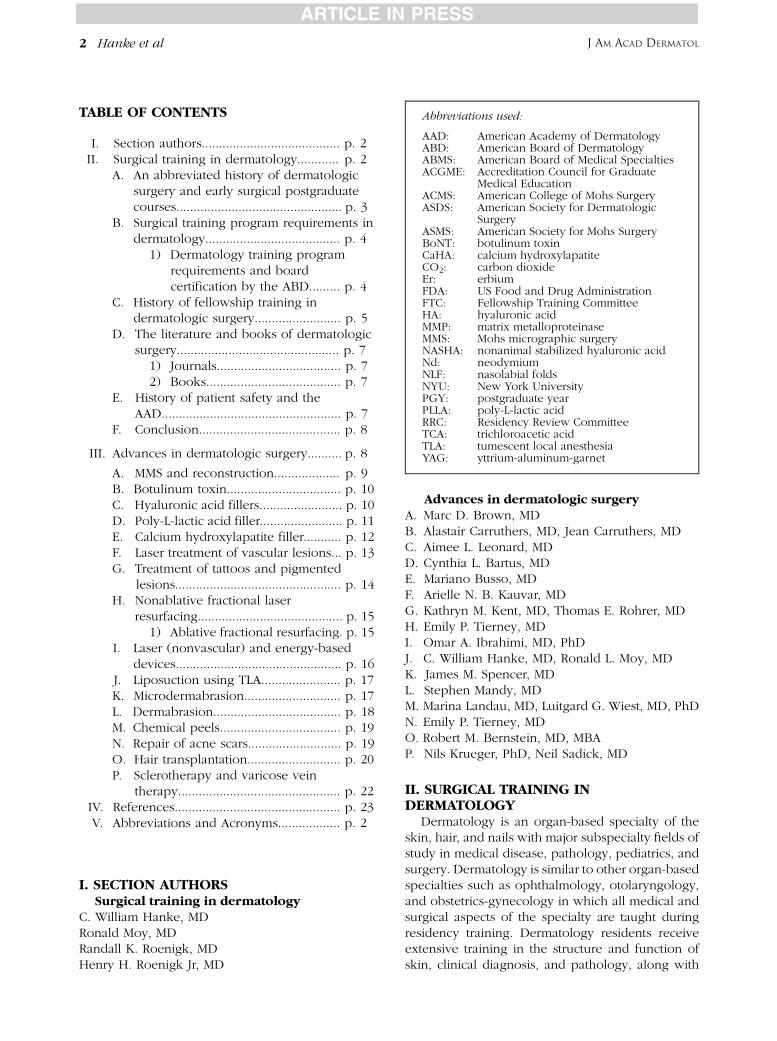
TABLE OF CONTENTS
I. Section authors........................................ p. 2II. Surgical training in dermatology............ p. 2
A. An abbreviated history of dermatologicsurgery and early surgical postgraduatecourses................................................ p. 3
B. Surgical training program requirements indermatology....................................... p. 4
1) Dermatology training programrequirements and boardcertification by the ABD......... p. 4
C. History of fellowship training indermatologic surgery......................... p. 5
D. The literature and books of dermatologicsurgery............................................... p. 7
1) Journals.................................... p. 72) Books....................................... p. 7
E. History of patient safety and theAAD.................................................... p. 7
F. Conclusion......................................... p. 8
III. Advances in dermatologic surgery.......... p. 8
A. MMS and reconstruction................... p. 9B. Botulinum toxin................................. p. 10C. Hyaluronic acid fillers........................ p. 10D. Poly-L-lactic acid filler........................ p. 11E. Calcium hydroxylapatite filler........... p. 12F. Laser treatment of vascular lesions... p. 13G. Treatment of tattoos and pigmented
lesions................................................ p. 14H. Nonablative fractional laser
resurfacing.......................................... p. 151) Ablative fractional resurfacing. p. 15
I. Laser (nonvascular) and energy-baseddevices................................................ p. 16
J. Liposuction using TLA....................... p. 17K. Microdermabrasion............................ p. 17L. Dermabrasion..................................... p. 18M. Chemical peels................................... p. 19N. Repair of acne scars........................... p. 19O. Hair transplantation........................... p. 20P. Sclerotherapy and varicose vein
therapy............................................... p. 22IV. References................................................ p. 23V. Abbreviations and Acronyms.................. p. 2
Abbreviations used:
AAD: American Academy of DermatologyABD: American Board of DermatologyABMS: American Board of Medical SpecialtiesACGME: Accreditation Council for Graduate
Medical EducationACMS: American College of Mohs SurgeryASDS: American Society for Dermatologic
SurgeryASMS: American Society for Mohs SurgeryBoNT: botulinum toxinCaHA: calcium hydroxylapatiteCO2: carbon dioxideEr: erbiumFDA: US Food and Drug AdministrationFTC: Fellowship Training CommitteeHA: hyaluronic acidMMP: matrix metalloproteinaseMMS: Mohs micrographic surgeryNASHA: nonanimal stabilized hyaluronic acidNd: neodymiumNLF: nasolabial foldsNYU: New York UniversityPGY: postgraduate yearPLLA: poly-L-lactic acidRRC: Residency Review CommitteeTCA: trichloroacetic acidTLA: tumescent local anesthesiaYAG: yttrium-aluminum-garnet
J AM ACAD DERMATOL2 Hanke et al
I. SECTION AUTHORSSurgical training in dermatology
C. William Hanke, MDRonald Moy, MDRandall K. Roenigk, MDHenry H. Roenigk Jr, MD
Advances in dermatologic surgeryA. Marc D. Brown, MDB. Alastair Carruthers, MD, Jean Carruthers, MDC. Aimee L. Leonard, MDD. Cynthia L. Bartus, MDE. Mariano Busso, MDF. Arielle N. B. Kauvar, MDG. Kathryn M. Kent, MD, Thomas E. Rohrer, MDH. Emily P. Tierney, MDI. Omar A. Ibrahimi, MD, PhDJ. C. William Hanke, MD, Ronald L. Moy, MDK. James M. Spencer, MDL. Stephen Mandy, MDM. Marina Landau, MD, Luitgard G. Wiest, MD, PhDN. Emily P. Tierney, MDO. Robert M. Bernstein, MD, MBAP. Nils Krueger, PhD, Neil Sadick, MD
II. SURGICAL TRAINING INDERMATOLOGY
Dermatology is an organ-based specialty of theskin, hair, and nails with major subspecialty fields ofstudy in medical disease, pathology, pediatrics, andsurgery. Dermatology is similar to other organ-basedspecialties such as ophthalmology, otolaryngology,and obstetrics-gynecology in which all medical andsurgical aspects of the specialty are taught duringresidency training. Dermatology residents receiveextensive training in the structure and function ofskin, clinical diagnosis, and pathology, along with

Table I. Milestones in surgical training in dermatology
1970 ASDS is founded by Leonard Lewis, Sorrel Resnick, and 11 other founding member dermatologists. NormanOrentreich is elected as first president.
1970 First 1-year fellowship training program is started by Perry Robins at NYU.1972 ASDS holds its first course: Basic surgical techniques for dermatologists.1975 Journal of Dermatologic Surgery is founded by Perry Robins and George Popkin. Dr Robins is first editor-in-chief.1982 First ASDS Core Curriculum for Dermatologic Surgery is completed by Ed Krull.1987 ASDS Core Curriculum for Dermatologic Surgery is revised by C. William Hanke.1988 Association of Academic Dermatologic Surgeons is founded by Ed Krull.1990 Residency Review Committee for Dermatology (Ed Krull, Chair) receives approval from ACGME for special training
requirements for all dermatology residency training programs to include complex closures, flaps, grafts, lasersurgery, and nail surgery. ACGME also approves requirement for designated surgical program director for eachdermatology residency program.
1991 ASDS Core Curriculum for Dermatologic Surgery is revised by C. William Hanke and Tom Meek.1991 Testimony in favor of board certification in Mohs micrographic surgery and cutaneous oncology is given at
COCERT by Ed Krull, C. William Hanke, and Martin Braun.1998 Residency Review Committee for Dermatology (Randall Roenigk, Chair) receives approval from ACGME for revised
surgical training requirements. Revised program requirements state ‘‘Residents should become familiar with hairtransplantation, dermabrasion, sclerotherapy, laser resurfacing, liposuction, chemical peel, and tissueaugmentation. In addition, residents should gain experience with Mohs micrographic surgery.Dermatologicsurgery training should include appropriate anesthesia, electrosurgery, cryosurgery, laser surgery, biopsytechniques, and excisional surgery with appropriate closures, including flaps and grafts when indicated.’’ Thenewly revised requirements for the surgical program director require ‘‘at least 5 years of experience (followingresidency) in the care of dermatology patients and as a teacher in a dermatology residency.’’
2003 ACGME approves 1-year procedural dermatology fellowship training program.2004 First ACGME-approved 1-year procedural dermatology fellowship training programs begin.2012 ACGME approves 57th procedural dermatology fellowship training program.
ACGME, Accreditation Council for Graduate Medical Education; ASDS, American Society for Dermatologic Surgery; COCERT, Committee on
Certification; NYU, New York University.
J AM ACAD DERMATOL Hanke et al 3
medical and surgical treatment of over 3000 cutane-ous diseases and tumors. Dermatologists have beenresponsible for many advances in dermatologicsurgery over many decades.1
A postgraduate year (PGY)-1 (medicine, rotating,surgical) is completed before 3 years of dermatologyresidency training (PGY 2-4). Some dermatology resi-dents complete elective rotations in general surgery,otolaryngology, plastic surgery, and other surgicaldisciplines during PGY-1, PGY-2, PGY-3, and PGY-4.The surgical training in dermatology is taught duringresidency as is required and documented by theAccreditation Council for Graduate Medical Education(ACGME). Some residents choose to receive addedfellowship training (PGY 5), which is currentlyavailable in pathology and surgery (proceduraldermatology) as overseen by the ACGME andpediatrics, which is overseen by the American BoardofDermatology (ABD). TheABD tests and certifies thatdermatologists are competent in all aspects of thespecialty including surgery. Subspecialty certificationthrough the ABD is available in dermatopathology andpediatric dermatology, but is not currently available inprocedural dermatology.
Many of the surgical procedures that areperformed by dermatologists today did not exist
25 years ago. This is also the case for most specialtiesin medicine. The new procedures are learned byall physicians in the same way after residency. Thisis done through postgraduate medical education,which includes courses, seminars, and live surgeryworkshops. The postgraduate courses are attendedby practicing physicians and also residents intraining. These courses are rich learning experiencesthat are attended by thousands of dermatologistseach year.
A. An abbreviated history of dermatologicsurgery and early surgical postgraduatecourses
Although surgery has long been part of thespecialty of dermatology, a new era of dermato-logic surgery began at New York University (NYU,New York, NY) in the 1950s. Dermatologistsat NYU became leading practitioners of dermabra-sion (Kurtin, Orentreich), chemical peel (MacKee),hair transplantation (Orentreich), MohsChemosurgery (Robins), and excisional skin sur-gery (Popkin). Goldman began performing cuta-neous laser surgery at the University of Cincinnatiin Cincinnati, Ohio in the early 1960s. The firstsurgery course to be held at the American

J AM ACAD DERMATOL4 Hanke et al
Academy of Dermatology (AAD) AnnualMeeting was directed by Krull in 1967, the sameyear that Mohs founded the American College ofChemosurgery (now the American College ofMohs Surgery [ACMS]). The American Society forDermatologic Surgery (ASDS) was started byLeonard Lewis and Sorrel Resnick and 11 otherfounding members in 1970 (Table I). The ASDSnow has more than 5000 members.
The first comprehensive postgraduate courses ondermabrasion/chemical peel were held in NewOrleans, LA, in the late 1970s. The course evolvedinto rotating postgraduate courses that were held invarious parts of the country, directed by HenryRoenigk, Sam Stegman, and John Yarborough. Thecourse that was held at Northwestern UniversityMedical Center, Chicago, IL in 1983 included mul-tiple live surgery demonstrations for the first time.Northwestern University later collaborated with theCleveland Clinic Foundation and the University ofCalifornia-San Francisco to hold courses in Florida,California, and Arizona. Live surgery demonstra-tions were beamed via satellite to packed lecturehalls. The Skin Disease Educational Foundationlater took over administration of the courses.Accreditation of postgraduate courses for continu-ing medical education became necessary for physi-cian medical licensure and renewal.
HughGreenway began a comprehensive cadaver-based cutaneous anatomy course at Scripp’s Clinicin San Diego, CA, in 1981. The course has been heldcontinuously for the last 31 years. Surgeryworkshopsusing pig’s feet were also organized in many parts ofthe country.
Many dermatologists traveled to Madison, WI,for short preceptorships with Fred Mohs in the1960s and 1970s. The first formal 1-year fellowshiptraining program in Mohs micrographic surgery(MMS) was founded by Robins at NYU in 1970.Shortly thereafter, training programs were startedby Tromovitch and Stegman at University ofCalifornia-San Francisco and Bailin at ClevelandClinic Foundation under the auspices of theAmerican College of Chemosurgery (now theACMS), which had been established in 1967.Another organization, the American Society forMohs Surgery (ASMS) was established in 1990.The ASMS does not require that members havefellowship training in MMS, whereas the ACMSrequires it. Tromovitch and Stegman2 later popu-larized the Mohs fresh-tissue technique in theearly 1970s. This advance allowed immediatereconstruction of skin cancer wounds, whichwas not possible with the older fixed-tissuetechnique.
B. Surgical training program requirements indermatology
Surgical training in dermatology takes placeduring the 3 years of dermatology residency inprograms accredited by the Residency ReviewCommittee (RRC) for dermatology on behalf of theACGME. Surgical training in dermatology hasexpanded over the years to encompass a largevariety of cosmetic, reconstructive, and dermatologicsurgery procedures. The 3 years of dermatologyresidency is preceded by an introductory year ofresidency in internal medicine, pediatrics, or surgery.
Dermatology residents have always been taughtsurgical operative techniques necessary to performexcisional surgery for the treatment of skin cancers.Suturing techniques, local anesthesia, sterilization ofinstruments, skin preparation, and scar revisiontechniques have always been a part of dermatologytraining. The development of the 1-year surgicalfellowship programs by the ACMS, beginning withPerry Robins’ training program at NYU in 1970, hadthe effect of greatly expanding the number ofacademic surgical faculty dedicated to teachingresidents skin surgery in dermatology residencyprograms and providing surgical procedures todermatology patients. The number of full-timedermatologic surgeons at every major medicalcenter had the effect of exposing most dermatologyresidents to sophisticated procedures such ascomplicated skin flaps, skin grafts, liposuction, laserprocedures, scar revision techniques, and MMS.Ed Krull used survey results from dermatologyresidency programs to change the core ACGMErequirements so that all programs were required toteach complex repairs, transposition flaps, rotationflaps, advancement flaps, and skin grafts. MMS wasadded in 1998, when Randall Roenigk chairedthe RRC.
This surgical foundation established during medi-cal school and dermatology residency and aug-mented by fellowship programs in proceduraldermatology has been sustained and built upon insubsequent years of practice through courses andsymposia accredited by the Accreditation Councilon Continuing Medical Education and offered byeducational institutions and professional societiessuch as the AAD, ASDS, ACMS, and ASMS. Somedermatologists also complete 1- to 2-year cosmeticsurgery fellowship training programs administeredby the American Academy of Cosmetic Surgeryand other non-ACGME-approved fellowships incosmetic laser procedures.
Dermatology training program requirementsand board certification by the ABD. The core ofdermatology and dermatologic surgery knowledge

J AM ACAD DERMATOL Hanke et al 5
and skills for all graduates of dermatology residencyprograms is derived from the standard requirementsfor dermatology residency training. These require-ments are established and monitored by the RRCfor Dermatology on behalf of the ACGME. The ABDrequires graduation from an accredited ACGME-approved residency and passing the ABD certifyingexamination. The ABD outlines for graduates thesurgical subjects that are contained in the certifyingexamination. These surgical subjects include sutur-ing techniques, anesthesia, electrosurgery, nailsurgery, cryosurgery, excisions, hemostasis, steri-lization, MMS, flaps (advancement, rotation, andtransposition), grafts, scar revisions, complex clo-sures, lasers, phlebology, tumescent liposuction, hairrestoration, use of neurotoxins, and use of fillers.These surgical subjects are developed by surgicaldermatologists who are experts in these surgicalsubjects. An educational in-training examination isgiven yearly to all dermatology residents in additionto the certifying examination, which leads to thedistinction of being board certified in dermatology.
Training as outlined by the RRC/ACGME shouldbe sufficient to ensure knowledge or competencein the performance of cryosurgery, dermatologicsurgery, cosmetic surgery, and laser surgery.Dermatologic surgery should be given emphasisand should include appropriate local anesthesia,electrosurgery, cryosurgery, laser surgery, nailsurgery, biopsy techniques, excisional surgery,advancement flaps, transposition flaps, rotationflaps, and grafts. Residents should have exposurethrough observation or assisting with hair restora-tion, Mohs micrographic surgery, sclerotherapy,laser resurfacing, and use of neurotoxins, chemicalpeels and tissue augmentation. In addition, residentsshould be provided education relating to cosmeticprocedures such as tumescent liposuction, scar revi-sion and dermabrasion. Didactic training in cosmeticsurgery is required by the RRC/ACGME. This surgicaltraining is documented both by the program and theACGME case log system by the residents. TheACGME case logs are used by all surgical specialtiesto document resident experience. Based on theprogram requirements, a lack of experience byresidents in key surgical procedures can result in aprogram citation. The ACGME also uses this systemto monitor the surgical experience in a specialty andhas plans to make these data public.
The level of training in dermatologic surgery isdivided into 3 categories. Dermatology residentsshould achieve competency in biopsy techniques,destruction of benign and malignant tumors, use oflasers for the treatment of superficial vascular tumors(eg, port wine stains), and excision of benign
and malignant tumors with simple, intermediate,and complex repair techniques including flaps andgrafts.3 Significant exposure to other procedureseither through direct observation or as an assistantat surgery is required. Examples in this categoryinclude MMS and reconstruction of surgical defects,the application of a wide range of lasers and otherenergy sources, sclerotherapy, botulinum toxin(BoNT) injection, soft-tissue augmentation, andchemical peels.
Program faculty must provide education relatingto certain cosmetic techniques without necessarilyaffording direct exposure. Among these are lipo-suction, scar revision, and dermabrasion. The pro-gram’s experience in cosmetic surgery may varydepending on the nature and experience of thepractice; however, didactic training in this area isrequired.
Dermatologists are specialists with expertise inthe diagnosis and treatment of pediatric andadolescent patients with benign and malignantdisorders of the skin, mouth, external genitalia,hair, and nails. Dermatologists have extensivetraining and experience in the diagnosis and treat-ment of all types of skin cancers. The dermatologistshave expertise in the management of cosmeticdisorders of the skin such as removal of excesshair, wrinkles, sun-damaged skin, hair loss, scars,and loose skin. Among the techniques usedby dermatologists for the correction of cosmeticdefects are laser resurfacing, tumescent liposuction,dermabrasion, chemical peels, hair transplantation,phlebology, and tightening procedures includingface, brow and neck lifting, and injections of fillers.
C. History of fellowship training indermatologic surgery
The ACMS was formed in 1967 with a principalmission to advocate for and train physicians in MMS.Soon thereafter, the Fellowship Training Committee(FTC) of ACMS was formed and program require-ments developed. In the early years the trainingrequirements were modest, but over time and underthe leadership of the chairs of the FTC such as DrsPhilip Bailin, Ted Tromovitch, Sam Stegman, RexAmmonette, John Zitelli, Barry Leshin, and Ron Moy,the requirements evolved into a 1-year structuredprogram that required a minimum of 500 MMScases to be performed by the program with thefellow, along with a diverse array of other topics incutaneous oncology and reconstructive surgery. RonMoy, who chaired the FTC for 8 years, and the FTCmembers implemented the site visits, nationalmatching program (1995), frozen section qualityreview, more stringent facility criteria, and the

J AM ACAD DERMATOL6 Hanke et al
transition to the future ACGME fellowship trainingprograms that are now in existence.
In the early years there were a very few fellowshiptraining programs, most notably at the Universityof California-San Francisco, the Cleveland Clinic,and NYU followed later by the University ofWisconsin at Madison where Fred Mohs developedthe technique.
In the late 1980s, the ABD sought to create asubspecialty in MMS. In the 1970s the ABD hadsuccessfully codified the subspecialty of dermatopa-thology by developing ACGME-accredited standardsfor training and a subspecialty certifying exami-nation, which greatly benefits all dermatologiststo this day. Ed Krull founded the Associationof Academic Dermatologic Surgeons in 1988, topromote, support, and critically assess the qualityand scope of surgical teaching of residents andfellows by academic-based dermatologic surgeons.The group provided support for the concept ofACGME accreditation of surgical fellowships indermatology. In 1991, Drs Ed Krull and IrwinFriedberg, on behalf of ABD, with testimony toAmerican Board of Medical Specialties (ABMS)Committee on Certification by C. William Hankeand Martin Braun, proposed the new subspecialty toABMS. However, the representatives from generalsurgery, plastic surgery, and otolaryngology werestrongly opposed for a variety of reasons and theproposal was defeated.
In the 1990s, Dr Krull was chair of the RRC forDermatology of the Accreditation Council forGraduate Medical Education (ACGME), which ischarged with the oversight of all dermatology spe-cialty and subspecialty training programs in theUnited States. Dr Randall Roenigk joined the com-mittee in 1994 and succeeded Dr Krull as chair of thecommittee. After much discussion, Drs Krull andRoenigk led the effort to seek accreditation of asubspecialty training program in dermatologic sur-gery. The proposal was developed and first finalizedin 1997. Unfortunately at the same time Medicarepublished IL-372, the intermediary letter clarifyingthe role of teaching physicians at academic medicalcenters for billing purposes. This ruling was followedby a series of audits (Physicians at TeachingHospitals [PaTH] Audits). Program directors anddepartment chairs were concerned about the impactof these new rules on their ability to be reimbursedfor patient care provided in teaching programs. As aresult, the creation of the new subspecialty wasdelayed as it was thought that ACGME accreditationof the new program might limit the ability ofdepartments to bill for patient care services. Therewas also opposition from some dermatologists who
thought that accreditation would put those withoutfellowship training at a disadvantage.
In 2000, the RRC decided to reopen the proposalfor accreditation in the subspecialty. Between theyears of 2000 through 2003, a series of negotiationsoccurred between specialties represented by theACGME to vet concerns expressed by generalsurgery, plastic surgery, and otolaryngology. Someof the concerns centered on the amount of surgicaltraining in dermatology and the name ‘‘dermatologicsurgery.’’ At that time, general surgeons thought thatthe word ‘‘surgery’’ should be limited to specialistswho complete training in general surgery. ACGMEpolicy at that time allowed ownership of certainterms by a specialty and hence required that the useof the term ‘‘surgery’’ be limited to the title ofspecialties that required a general surgery residency;just as the term ‘‘dermatology’’ could not be used byother specialties. For example, surgical specialtiessuch as otolaryngology, ophthalmology, andobstetrics-gynecology do not use the term ‘‘surgery’’to describe their specialty or subspecialties.After much discussion, a program in proceduraldermatology was approved by the ACGME in 2003allowing the first programs to become accredited in2004.4
Between the years of 2004 and 2012, 57 programsin procedural dermatology were accredited. Animportant part of the process during this periodincluded dual accreditation/approval from both theACGME and ACMS. Some members of ACMS werestill concerned about giving up the approval processfor these programs. Under the leadership of Roenigk,Moy, the ACMS Fellowship Training Committee, theACMS Board, and ACMS President Whitaker, it wasrecognized that ACGME accreditation of the fellow-ships would be a better process in the long run andconsistent with that used by almost all specialties andsubspecialties in organized medicine. There was atransition from the ACMS site visit accreditation to theACGME accreditation process that includes ongoingprogram review, site visits, and review by the RRCand the use of the ACGME case log system todocument the fellows surgical experience. Theincorporation of cosmetic surgery into the trainingwas a change for some ACMS programs. Fellowshiptraining programs in private practice settings wererequired to adopt the same policies and proceduresthat were required of large university residencyprograms. ACMS completely discontinued theprocess of approving US programs by 2013 butcontinued to approve a small number of interna-tional programs. The ACMS continues to provideother resources including sponsorship of the annualmatch, conducting a slide review program needed

Fig 1. Number of new dermatologic surgery/oncologytextbooks published during recent 5-year periods.
J AM ACAD DERMATOL Hanke et al 7
for accreditation, receiving grievances from fellowswho are trained in private practice programs,and maintaining a forum for discussion betweenprogram directors.
After several years of preparation the ABD sub-mitted its application for subspecialty certification inprocedural dermatology to the ABMS in 2009. Whathappened in 2009 was quite different compared withthe process in the late 1980s. General surgery, plasticsurgery, otolaryngology, and other specialties nowrecognized that dermatologists had developed asurgical subspecialty in MMS, cutaneous reconstruc-tion, and related procedures that was unique andvaluable. They had very few concerns about giving acertification examination in this subspecialty spon-sored by ABMS. Concerns about certification camemainly from the specialty of dermatology. One issuecentered on the name ‘‘procedural dermatology’’because it was considered to be too generic and didnot properly reflect the practice of those who hadbeen trained. All dermatologists are trained in skinsurgery during their residency and most performsurgical procedures daily. In addition, therewas concern about whether or not subspecialtycertification would have an impact on a dermato-logists’ ability to bill insurance companies forsurgical services. The ABD put a pause on theprocess after its first review by Committee onCertification to examine these concerns inmore detail. As of this writing, the future of subspe-cialty certification in procedural dermatology isuncertain.
Subspecialty programs in procedural dermato-logy remain accredited by the ACGME. Manyprograms have undergone several reviews andcontinue to improve through this process. Theprogram requirements have also undergone reviewand will continue to be improved with input fromorganized medicine.
Based on a review of claims data from the Centerfor Medicare and Medicaid Services it is clear thatdermatologists perform more surgery on the skinthan any other specialty.5 This is in great partbecause of our aging population and the epidemicof skin cancer. MMS and the other dermatologicsurgical procedures for the treatment of skin cancercontinue to be important tools to manage an increa-singly significant health care problem in this country.Access to dermatologic surgical care for the treat-ment of skin cancer and other cosmetic proceduresunique to our specialty must be taught in residencyand fellowship programs, and be a part of continuingmedical education for physicians engaged in lifelonglearning. Regardless of whether a dermatologist hastaken an added year of fellowship training, it is the
expectation of the ACGME, ABMS, and ABD thatall dermatologists be competent in the surgicaltreatment of skin disease.
D. The literature and books of dermatologicsurgery
Journals. The founding of the Journal ofDermatologic Surgery by Robins and Popkin in1975 was an important event in the evolution ofdermatologic surgery. The journal has published asteady stream of instructional and research articlescovering dermatologic oncology, reconstructivesurgery, laser and cosmetic surgery, and cosmeticdermatology. The journal was purchased fromElsevier Science Publishers by the ASDS in 1995.The journal name has evolved from the Journal ofDermatologic Surgery (1975) to the Journal ofDermatologic Surgery and Oncology (1977) toDermatologic Surgery (1995). Special topic issuessuch as lasers and fillers are frequently publishedalongside regular monthly issues.
Other major dermatology journals such as theJournal of the American Academy of Dermatologyand the Archives of Dermatology regularly publisharticles on many aspects of dermatologic surgery.Other dermatology journals such as the Journal ofDrugs in Dermatology also publish surgical articlesand special surgical issues.
Books. A multitude of books on dermatologicsurgery and oncology have been authored or editedby dermatologists over many decades. The numberof new books has steadily increased from 28(1990-1995) to more than 70 (2007-2012) (Fig 1).
Many of these journals and books are formallyreviewed by dermatology residents and proceduraldermatology fellows, along with academic facultyand physicians in private practice.
E. History of patient safety and the AADThe safety of the patient is the most important
thing, and the AAD has been active in the

J AM ACAD DERMATOL8 Hanke et al
area of patient safety for a number of years.Dermatologists have pioneered minimally invasiveoutpatient dermatologic surgery procedures that areperformed safely and effectively using topical,local, or tumescent local anesthesia (TLA). Theoutcomes are superior and complications areinfrequent and minor. MMS for skin cancer andliposuction using TLA are 2 well-documentedexamples whereby patients are sparedmore invasiveprocedures under general anesthesia.
The AAD was one of the first national specialtyorganizations to develop a patient safety initiative.The AAD held a Patient Safety Summit on August 7,2007, in New York, NY. More than 100 AAD leadersand many leaders in other areas of medicineattended. Many patient safety subject areas werecovered by experts in the field including:‘‘Establishing a strategy for patient safety in a medicalspecialty: lessons from the anesthesia experience’’(Jeffrey B. Cooper, PhD, Executive Vice-President,Anesthesia Patient Safety Foundation); ‘‘Scope ofpractice in medicine’’ (James N. Thompson, MD,President/CEO, Federation of State LicensingBoards); ‘‘Scope of practice panel’’ (William H.Beeson, MD, Vice-President, Indiana MedicalLicensing Board); ‘‘Scope of practice expansion bynonphysicians’’ (Stephen H. Mandy, MD); ‘‘Lasersand scope of practice’’ (Roy G. Geronemus, MD);‘‘Day spa nightmares’’ (Deborah Sarnoff, MD);‘‘Supervision of PAs and NPs’’ (Roger I. Ceilley,MD); ‘‘Mandatory adverse event reporting’’ (BrettM. Coldiron, MD); ‘‘Office accreditation panel’’(Duane C. Whittaker, MD, Roy C. Grekin, MD, W.Patrick Davey, MD, Pat Ferrigno, MS); ‘‘Dermatologymalpractice: a 20-year analysis of malpractice claimsagainst dermatologists’’ (Sandra I. Read, MD);‘‘Patient safety organizations’’ (William H. Beeson,MD); ‘‘Patient safety research’’ (Doral Rosauer, MBA,Katie B. Baeverstad, MD); and ‘‘The case for adermatology credentialing verification organization’’(C. William Hanke, MD, Pat Ferrigno, MS, William H.Beeson, MD).
After the Patient Safety Summit, an AAD Ad HocTask Force on Patient Safety was formedwith James S.Taylor, MD, as Chair. The charge of the ad hoc taskforce was to: (1) define the current state of patientsafety in dermatology; (2) evaluate existing academyactivities; (3) identify gaps and priorities; (4) recom-mend changes in the existing portfolio; and (5)develop a strategic plan for patient safety for theAAD.A strategicplan forpatient safety for theAADwascompleted. The ad hoc task force was then disbandedand an AAD Committee on Patient Safety and Qualitywas established in 2009with C.WilliamHanke,MD, asChair. Themission of the committee was to: (1) define
the current state of patient safety and quality indermatology; (2) evaluate existing academy activities,identify gaps and priorities, and recommend changesin the existing portfolio; and (3) develop a compre-hensive strategy for developing a culture of patientsafety and quality in dermatology within the AAD.Three task forces of the committee were established:(1) performance measurement; (2) patient safetycurriculum; and (3) data collecting and reporting.
The AAD has been an advocate for mandatoryadverse event reporting in every state. It is onlythrough verifiable data collection on adverse eventsthat problems can be identified and quantified.6
Solutions can then be crafted to prevent futureadverse events. Coldiron et al7-9 reported multipletimes on the Florida database for mandatory report-ing of adverse events for offices that has been inplace since 2000. The most recent report alsoincluded adverse event reporting for Alabama.10
Reporting of surgical complications that occurredin the offices were reported by physicians to a centralagency in both states. Cosmetic procedures wereresponsible for roughly half of all adverse eventsreported in Florida and Alabama. Plastic surgeonsreported the greatest number of complications. Themajority of the fatalities and complications occurredin patients who received general anesthesia. Therewere no fatalities reported by dermatologists, andthe number of complications was extremely small.
F. ConclusionThe fertile academic environment of academic
training programs with interaction between esta-blished dermatologic surgeons and fellows, alongwith the inquisitive and innovative nature ofmany of our colleagues, has led to numerousadvances in dermatologic surgery, which aredescribed subsequently.
III. ADVANCES IN DERMATOLOGICSURGERY
In October 1999, an article titled ‘‘Current issuesin dermatologic office-based surgery’’ was publishedin this journal.1 The article chronicled the myriad ofcutaneous surgical techniques that have beendeveloped and pioneered by dermatologists. Manymore advances have occurred since 1999. InMarch 2012, the Board of Directors of the AADcommissioned the AAD Ad Hoc Task Force onOffice-based Surgery to update the previous article.This article is intended as an update, and the reader isreferred to the previous article for historical details.The authors and co-authors of the various sections ofthe new article are all dermatologists who have

J AM ACAD DERMATOL Hanke et al 9
been involved in the developments and refinementsthat have occurred since 1999.
Dermatologists and dermatologic surgeons havecontinued to demonstrate leadership in office-basedsurgery and cutaneous oncology. Training in derma-tologic surgery is required for all dermatologyresidents. Some residents undergo additional surgi-cal training through 1-year ACGME-approvedfellowship-training programs in procedural derma-tology. Of these programs, 57 are currentlyaccredited by ACGME.
Dermatologists are the only specialists in medi-cine who receive comprehensive training in theclinical diagnosis, basic science, pathophysiology,dermatopathology, and medical and surgical treat-ment of over 3000 cutaneous diseases and tumors.This foundation is the key to the advancements thathave occurred in dermatologic surgery and the otherareas of dermatology as well.
A. MMS and reconstructionMMS has advanced significantly since Fred
Mohs11 first described the surgical technique withzinc chloride paste more than 60 years ago.Tromovich and Stegman12 reported on the successof MMS using the fresh tissue technique. Over thepast 3 decades, MMS has expanded and flourishedand has given us new insights into the managementof complex and challenging skin cancers. Althoughthe terms ‘‘gold standard’’ and ‘‘standard of care’’should be used wisely and sparingly, it is likely thecase that MMS has established itself as such for thetreatment of nonmelanoma skin cancers. Reviews byRowe et al13,14 established a low recurrence rate of1% for primary basal cell carcinomas and 5.6% forrecurrent basal cell carcinomas treated with MMS.For squamous cell carcinoma, the success rate forprimary tumors treated with MMS is approximately95%.15
MMS is highly successful because of precisehorizontal sectioning and meticulous microscopicinspection of tumor margins. The most uniqueaspect of the Mohs procedure is the fact that thephysician serves as both the surgeon and patholo-gist, a role dermatology embraced because of ourunique training in both skin surgery and dermato-pathology. MMS has grown in popularity anddemand because of the exploding incidence ofskin cancer (approximately 3.5 million new casesper year of nonmelanoma skin cancer) and increasednumbers of highly skilled trained Mohs surgeonsperforming the procedure. In addition, MMShas been established as an extremely safe, well-tolerated, and highly accepted surgical procedurewith a very low incidence of nonlife-threatening
complications.16 Patients who might otherwise beat risk undergoing general anesthesia can safelycomplete MMS under local anesthesia.
MMS has the potential risk to be an overusedprocedure, which is why recent appropriate usecriteria for MMS have been published.17 MMS is bestused for high-risk basal cell carcinomas includingrecurrent tumors, those in high-risk anatomiclocations, aggressive histologic subtypes, incom-pletely excised tumors, larger size tumors, and forpatients who are immunosuppressed. Likewise,invasive squamous cell carcinomas of the head andneck are indications for MMS. Melanoma in situ([50,000 new cases per year) especially the lentigomaligna subtype, can be challenging because of thelocation on the face, ill-defined margins, andsubclinical extension. MMS (especially with the useof immunostains) has been shown to be highlyeffective for lentigo maligna with a very lowrecurrence rate.18 MMS has been described for thetreatment of several rare cutaneous tumors.Although studies can be hampered by smallernumbers and retrospective reviews, there clearly isa robust literature to support MMS as a superiortreatment versus standard wide excisions for theseunusual cutaneous neoplasms. Rare tumors that areamenable to treatment with MMS include dermato-fibrosarcoma protuberans, microcystic adnexalcarcinoma, atypical fibroxanthoma, superficialleiomyosarcomas, sebaceous carcinomas, and extra-mammary Paget disease.19-23 Merkel cell carcinomas,especially those on the head and neck, have beentreated with MMS24 but new standards alsorecommend the performance of sentinel lymphnode biopsies, typically performed by surgicaloncologists.
In summary, MMS has been documented to besafe and effective. Its use has increased 400% andcurrently 1 of every 4 skin cancers is treated withMMS.17 It is a procedure of ongoing innovationand improvement. Its distinct advantages include:(1) the highest documented cure rate; (2) tissueconservation as a result of smaller margins; (3)safety and tolerability in an outpatient setting; and(4) immediate reconstruction with confidence thatclear margins have been ascertained. Challengesinclude a longer procedure for the patient becauseof waiting time for the frozen sections and theneed for excellent quality control of the labora-tory. The preparation of excellent quality frozensections is a highly skilled procedure and a Mohssurgeon is only as good as the technician andlaboratory. MMS has been reported to be a moreexpensive procedure but studies have docu-mented that it is very cost-effective, especially

J AM ACAD DERMATOL10 Hanke et al
compared with excision in an ambulatory surgerycenter.25,26
Along with the increased use of MMS has comethe remarkable advancements and refinements infacial reconstruction. From the earliest days ofallowing Mohs defects to heal by second intention,dermatologic surgeons have led the way in creativeand novel reconstructions with advanced flaps andgrafts. Zitelli27 revolutionized the use of a bilobedflap for nasal reconstruction by redefining its arc ofrotation.
Likewise, Zitelli28 modified the melial labialtransposition flap to make it a cosmetically accep-table 1-stage procedure. Dzubow29 refined andpopularized the dorsal nasal rotation flap for nasaldefects. Skouge30 described the use of the islandpedicle flap for cutaneous lip reconstruction andothers defined its use for the forehead, cheek, andnose. Papadopoulous and Triner31 described amyocutaneous island pedicle flap for nasal tipreconstruction. More complex staged interpolationflaps, including paramedian forehead flaps andcheek-to-nose flaps were popularized byMellette,32 Brodland,33 and Fader et al.34 Cook35
explained modifications of the Spear turnover flapand other creative approaches to alar reconstruc-tions. Advanced skin grafting techniques withcartilage support have been described by Otley36
and Adams and Ratner.37 Finally, dermatologistshave always been on the leading front to definewhen primary closures or even second-intentionhealing is not only simpler but likely to result inbetter cosmesis.38
B. Botulinum toxinAlthough originally developed for therapeutic
use, BoNT received international attention whenJean Carruthers39 and her dermatologist spouse,Alastair Carruthers, published their landmark trialin 1992, demonstrating that small amounts of BoNTinjected into the forehead could improve theappearance of glabellar rhytides for up to 3 months.A decade later, Carruthers et al40 publishedthe results of a multicenter, double-blind, placebo-controlled randomized trial that led to the Food andDrug Administration (FDA) approval of BoNT for thetreatment of glabellar rhytides. In addition to furtherresearch by Carruthers and Carruthers,41,42 manydermatologistseincluding Becker-Wegerich et al43
and Lowe and Yamauchi44econtributed to the earlyresearch that furthered the understanding of thetoxin and its use for the treatment of a variety ofhyperkinetic facial lines in the upper and loweraspect of the face, neck, and chest with a high degreeof patient satisfaction.
Over the next 10 years, the use of BoNT expandedto include facial sculpting and the restoration ofsymmetry: Flynn et al45 widened eyes, whereasHuilgol et al46 and Carruthers and Carruthers47
induced a chemical brow lift. Moreover, it becameapparent that BoNT was particularly effective whenused in combination with soft-tissue augmentation,laser resurfacing, light-based therapies, and surgery,as reported by dermatologists West and Alster,48
Fagien and Brandt,49 Carruthers et al,50-52 andKhoury et al.53
In 1994, researchers first demonstrated that BoNTproduced localized anhidrosis in the faces of patientstreated for hemifacial spasm.54 Since then, a numberof dermatologists have demonstrated the therapeuticefficacy of BoNT in patients with palmar and axillaryhyperhidrosis, including Glogau,55 Shelley et al,56
Heckmann et al,57 Lowe et al,58 Glaser et al,59 andSolish et al.60
Twenty years of cosmetic use has yielded a wealthof information regarding the efficacy and safetyof long-term treatment. Dermatologist ArnoldKlein61 meticulously detailed side effects and moreseriousealthough rareecomplications, whereasRzany et al,62 Carruthers et al,40,63 and Cohenet al64 analyzed large pools of data to demonstrateminimal adverse effects and establish the long-termsafety of BoNT for cosmetic indications.
C. Hyaluronic acid fillersIn the past 15 years, there has been an explosion
of interest, use, and availability of hyaluronic acid(HA) filling agents. HAs have become the leadingfiller agents worldwide, surpassing collagenproducts, which had previously dominated the fillermarket. ThemostwidelyusedHAfillers areproducedfrom bacterial (Streptococcus) fermentation, withthis class of dermal fillers designated as nonanimalstabilized HA (NASHA), distinguishing them fromearlier animal-sourced products. Currently, thereare dozens of NASHA formulations availableworldwide. Variations in product performance,tolerance, and durability have been associated withdifferences in cross-linking, molecular weight, con-centration, particle size, and addition of anestheticagents.
Initial studies establishing the biologic compa-tibility and stability of HA as a filler materialwere pioneered by dermatologists Piacquadioet al65,66 in both a guinea pig model and in amulticenter clinical study. Dermatologists Narinset al67 led a pivotal randomized double-blindmulticenter split-face study in 138 patients that firstdemonstrated superior efficacy and comparablesafety of NASHA (Restylane) compared with

J AM ACAD DERMATOL Hanke et al 11
collagen (Zyplast) over 6 months. Subsequentstudies led by dermatologists explored differencesin efficacy and tolerability among the various HAfillers emerging in clinical use. In a randomized,double-blind comparison of 150 patients, Carrutherset al68 demonstrated superior durability and compa-rable safety of the NASHA (Restylane Perlane) at6 months compared with Hylaform, a cross-linkedHA sourced from rooster combs. In a prospective,randomized, comparative, multicenter study of 248patients, Dover et al69 compared a large-particleNASHA-based filler with a small-particle NASHAfiller and found similar efficacy, durability, and safetyprofiles. Narins et al70,71 further explored the long-term efficacy and effects of different NASHA(Restylane) retreatment schedules, demonstratingpersistence of correction beyond the expected 6 to12 months when retreatment was performed beforedissolution of the initial treatment product.Dermatologists were the first to describe in vivodeposition of new collagen after dermal injections ofboth HA filler and calcium hydroxylapatite (CaHA)via histologic and biochemical analysis. It is postu-lated that this stimulatory effect may be induced bymechanical stretching of the dermis thereby leadingto activation of dermal fibroblasts.72,73
Dermatologists were among the first to establishsafety data on HA fillers. Friedman et al74 reportedthe first retrospective review of adverse reactionsassociated with early NASHA injection, whichincluded data from over 144,000 patients worldwide.The authors found that localized hypersensitivityreactions were the major adverse event associatedwith injection, and that these declined dramaticallyafter more purified source material becameavailable.
Dermatologists made significant contributions tothe early evolving literature on HA side effects andtheir appropriate management.75-79 One benefit ofHA fillers is the unique ability to correct and reversecomplications through the injection of hyaluroni-dase to enzymatically degrade the HA material.80
Glogau and Kane81 demonstrated a direct correla-tion between injector techniques such as rapidinjection and higher volumes with the rate of localadverse events in a prospective, blinded, controlledstudy of 283 patients undergoing NASHA injection.Taylor et al82 and Grimes et al83 have establishedsafety and efficacy of HA filler injections in over 300patients with skin of color in 2 prospective ran-domized clinical trials. Dermatologists have beenleaders in establishing guidelines and consensusrecommendations for appropriate use of HAdermal fillers to optimize patient safety and satis-faction.84-86
HA fillers are FDA approved for correction of‘‘moderate to severe facial wrinkles and folds such asthe nasolabial folds’’ (NLF). However, they havebeen widely and successfully used for off-labelvolume enhancement of the vermilion lip, perioralarea, suprabrow region, earlobes, back of hands,prejowl sulcus, and tear troughs.87-89 DermatologistsAlam et al,90 Goldman et al,91 and Carruthers et al51,92
have also explored the safety and synergistic effectsof combining HAs with other treatment modalitiessuch as radiofrequency,90 intense pulsed light,91 andBoNT.51,93 Dermatologists have helped to establishmore recent advances in formulation and techniqueincluding combining HA fillers with lidocaine tooptimize tolerability93 and injection through a blunt-tipped cannula to minimize pain, bruising, andedema.94
As the demand for noninvasive cosmetic proce-dures continues to grow, the science of injectablesoft-tissue fillers, including HAs, will continue toevolve. Dermatologists have been, and will continueto be, at the forefront of evolving HA filler practice,safety, and material technology.
D. Poly-L-lactic acid fillerInjectable poly-L-lactic acid (PLLA) is a filler
device used to stimulate collagen production torestore lost facial volume. It differs from other facialfillers such as HA in that PLLA does not directly fill inlines or depressions, but instead induces a hostresponse resulting in a gradual correction of thevolume-depleted area.95 As volume is restored,however, the NLF, marionette lines, and otherundesirable lines are corrected.96
Injectable PLLA has been used worldwide formore than a decade to treat soft-tissue atrophyrelated to aging, photoaging, and HIV-relatedlipoatrophy. Safe use of PLLA requires workingknowledge of facial anatomy including locationof major nerves, vessels, and fat pads. An under-standing of the changes to the face that occur withaging/photoaging and HIV-related lipoatrophy isimperative. Full comprehension and awareness ofthe different properties of the skin and softtissue in the various cosmetic units of the face isnecessary for the safe injection of PLLA.85
This knowledge is not unique to dermatologists,but dermatologists have received extensivetraining on these topics through residencytraining, fellowship training, and continuingeducation.
Dermatologists have played an essential role inestablishing the safety and efficacy of injectablePLLA. European dermatologists and skin specialistsdeveloped the safe injection techniques for this

J AM ACAD DERMATOL12 Hanke et al
unique device before the introduction of PLLA in theUnited States. Early work by Vleggaar96 and Lowe97
helped establish the injection techniquesand demonstrated efficacy of PLLA for sculptingand rejuvenating the aging face. One ofthe noted frequent side effects of injectablePLLA was the delayed formation of subcutaneousnodules. Through the collective experiences ofdermatologists, reconstitution protocols and injec-tion techniques have evolved leading to a notablereduction in the formation of subcutaneousnodules.98-102
Injectable PLLA has been approved in the UnitedStates since 2004. The product was initially approvedfor treating HIV lipoatrophy associated with highlyactive antiretroviral therapies. Again, dermatologistsplayed a pivotal part in understanding the role ofPLLA in the management of this stigmatizing sideeffect of HIV treatment.103 Hanke, Redbord, andLevy99,100 published an extensive case series on thesafety and efficacy of injectable PLLA for treating HIVlipoatrophy. Dermatologists have continued toprovide their expertise in treating HIV faciallipoatrophy.
In 2009 PLLA was approved by the FDA foraesthetic use.104 The experience of dermatologistswas imperative in gaining FDA approval of thedevice. Work by dermatologists includingFitzgerald and Vleggaar,105 Palm et al,102 Hankeand Redbord,99 Narins et al,101 and Burgess andQuiroga103 helped establish the efficacy of PLLA forcosmetic use and has continued to demonstrate thesafety of this facial volume restoration device.
Injectable PLLA is a safe and effective filler torestore the lost facial volume that contributes tothe appearance of the aged face. The skills andknowledge that are needed to perform PLLAinjections in the office are taught to dermatologyresidents during their core training. Frequentpublications in the dermatology literature onthe use of PLLA further contribute to theknowledge base and keep dermatologists at theforefront of using this unique facial filler.Without the work of dermatologists, this productwould not have its established safety profile or bewidely available for treating facial soft-tissueatrophy.
E. Calcium hydroxylapatite fillerCaHA is one of the natural components found in
human bone. Synthetic CaHA is manufactured asRadiesse by Merz Aesthetics (San Mateo, CA). Itconsists of microspheres of CaHA, 25 to 45 �m indiameter, in a soluble carboxymethylcellulose gelcarrier. Although it is now a popular compound
for aesthetic use, synthetic CaHA was originallydeveloped for the treatment of stress urinary incon-tinence and oropharyngeal repair.106
In 2002, Dr Mariano Busso was the first derma-tologist in the United States to explore the use ofCaHA for aesthetic enhancement. He developed anovel 3-dimensional vectoring filler injectiontechnique, in which the face is conceptualized indifferent mobility zones. Vectoring can addresssurplus skin reduction and structural enhance-ment.107 Busso developed a series of injectiontechniques for optimum and safe use of CaHA formalar,108 zygomatic, temporal, and supraciliaryregions109; the lower aspect of the face110; andnonfacial areas such as hands.111
CaHA has several properties that make it animportant tool in the facial recontouring armamen-tarium.112,113 These include lack of allergenicity,longer tissue residence, and high elasticity (G9).Limiting factors include its radiopacity and tenden-cies to produce nodules when injected in lips and toshow filler visibility after superficial injections.Instead, it should be injected at the junction ofthe dermis and the subcutaneous level or, whenvolumization is required, supraperiosteally.
The pivotal trial compared CaHA with human-based collagen for correction of NLF in 117patients.112 In the randomized, bilateral, andprospective study, subjects were treated with CaHAin one fold and with collagen in the other; they werefollowed up for as long as 6months. Of subjects, 79%had superior improvement on the CaHA side through6 months (P \ .000) using the Global AestheticImprovement Scale (P \ .0001). Significantly lessvolume of CaHA was needed for optimal correction,compared with collagen (P\.0001). Adverse eventswere generally comparable between the 2 groups,though increases in bruising and edema were notedin the CaHA-treated fold.
Subsequent results of this same trial were reportedextending more than 3 years.113 Of the original 117subjects, 99 subjects were available for all 3 years.Using the Global Aesthetic Improvement Scale,researchers found that, at 30 months, 40% of theCaHA-treated folds were rated as ‘‘improved’’ orbetter. No long-term or delayed-onset adverse eventswere reported, including ‘‘no reports of nodules,granulomata, or infections.’’
Patients reported immediate post-CAHA injectiondiscomfort. To address the issue of pain control,Busso and Applebaum114,115 developed a method ofmixing lidocaine with CaHA immediately beforeinjection. A Luer lock-to-Luer lock connectorbetween a syringe containing lidocaine and asyringe containing CaHA allows the lidocaine to

J AM ACAD DERMATOL Hanke et al 13
mix with the CaHA without altering the duration ofthe correction. In 2009, the FDA approved thetechnique for mixing lidocaine with CaHA.
In the controlled, randomized, patient-blinded,split-face clinical trial, 50 subjects received CaHAwithout lidocaine in one NLF and CaHA premixedwith 2% lidocaine in the other NLF.116 Subjectsreported statistically significantly less pain in theNLF treated with CaHA premixed with lidocaineimmediately posttreatment (P\.001) and 48 subjects(96%) expressed a preference for CaHA premixedwith lidocaine.
In nonfacial areas, the Busso hand injectiontechnique of CaHA was the basis of the first clinicaltrial tomeasure the efficacy of a dermal filler for handaugmentation.117 A multicenter, blinded, random-ized clinical trial addressed the use of CaHA inhand rejuvenation. In this study, 101 subjects wererandomized to receive CaHA (76 patients) or toreceive no treatment. Using the validated BussoHand Volume Severity Scale, researchers found thatthe treatment group was statistically significantlyimproved compared with the control (P \ .0001).No adverse effect on hand function was noted inthe study.
The Busso method of CaHA mixed with lidocainehas since been expanded to adjust rheologicalproperties of CaHA118 and other fillers as well.119
Today, use of a Luer lock-to-Luer lock connector totailor filler characteristics has become a familiartechnique in the field of tissue augmentation.
F. Laser treatment of vascular lesionsThe treatment of congenital and acquired vascular
lesions is one of the most commonly requested andperformed cutaneous laser and light-based officeprocedures. The pulsed dye laser was the firstlaser that was developed based on the theory ofselective photothermolysis, a conceptual frameworkproposed by dermatologists Anderson and Parrish120
in the 1980s. Their theory revolutionized thetreatment of vascular lesions, and continues to serveas the basis for most cutaneous laser and energy-based treatments today.
Clinical studies of the original pulsed dye laserand its subsequent refinements by dermatologistsGarden et al,121,122 Ashinoff and Geronemus123 andothers124 demonstrated its efficacy and high safetyprofile for treating pediatric and adult port winestains, infantile hemangiomas, and other vascularlesions. Before the development of pulsed lasertherapy, there was no adequate treatment methodfor port wine stain birthmarks that characteristicallyhypertrophy with time, leading to cosmetic disfig-urement, psychological impairment, and other
medical complications.125,126 Numerous studieshave demonstrated that the best results are achievedwhen treatment is initiated early in infancy orchildhood, and that even with multiple, repetitivetreatment sessions in a pediatric population, the riskof scarring or long-term pigment alteration is rare.127
Recent studies by dermatologists Izikson et al128 andTierney and Hanke129 demonstrated the benefit ofthe more deeply penetrating alexandrite laser forthe treatment of hypertrophic and recalcitrantport wine stains. Dermatologists are currentlyinvestigating novel techniques to improve portwine stain treatment, including the combination oflaser and antiangiogenic or immunomodulatorydrugs.130
Infantile hemangiomas are common, benignvascular lesions of infancy that are often present onthe head and neck. During their growth phase,hemangiomas can obstruct vital organs; they oftenheal with residual fibrofatty tissue and theynegatively impact the psychosocial development ofthe affected children. Dermatologists Garden et al,124
Morelli et al,131 and Geronemus and Kauvar132
showed that pulsed dye laser can slow the prolife-ration of hemangiomas during their growth phase,speed their involution, and stimulate epithelializa-tion of ulcerated lesions. Fractional resurfacinglasers are now being used to treat the residdum ofinvoluted hemangiomas.133
Dermatologists developed and improved thetechniques to treat telangiectases, rosacea, poikilo-derma, spider angiomas, cherry angiomas, pyogenicgranulomas, and venous lakes.134 Bernstein andKligman135 showed that laser treatment ofrosacea-associated telangiectasia and erythema alsoimproves the symptomology associated with theflushing and blushing and reduces the number ofinflammatory lesions. Studies by dermatologistsrevealed that the pulsed dye laser, used withrepetitive high-energy pulses, effectively treatsrecalcitrant warts, including plantar and periunguallesions, without the morbidity and scarring observedwith other destructive methods.136 Pioneering workby Alster and Williams137,138 using the pulseddye laser at low fluences transformed the treatmentof erythematous and hypertrophic scars, andMcDaniel et al139 demonstrated its efficacy in treatingstretch marks.
In the 1990s, the 532-nm pulsed KTP, 755-nmlong-pulsed alexandrite, and 1064-nm pulsedneodymium (Nd):yttrium-aluminum-garnet (YAG)lasers were explored for vascular lesion treatment.Goldman et al140 pioneered the use of intense pulsedlight, noncoherent light delivered by a flashlampwith millisecond domain pulses, for the treatment

J AM ACAD DERMATOL14 Hanke et al
of vascular and pigmented lesions. Studies bydermatologists Dierickx et al141 showed that longerpulse durations were ideally suited for the treatmentof telangiectasia and enabled purpura-free treatmentof most acquired vascular lesions and greater patientacceptance of these procedures. The introductionof skin cooling techniques using cryogen, cold air,or contact cooling devices improved treatmentoutcomes for congenital and acquired vascularlesions by protecting the epidermis and allowingthe safe use of higher energy laser pulses.142,143
Kauvar and Lou,144 Kauvar and Khrom,145 Omuraet al,146 and Parlette et al,147 optimized parametersfor the treatment of spider and reticular leg veins byusing the more deeply penetrating, near-infraredalexandrite and Nd:YAG lasers. Goldman and Weisswere instrumental in developing endovenous clo-sure, a technique that has largely replaced venousstripping for greater saphenous vein incompetence,whereby a bare laser fiber or radiofrequency-emitting catheter is introduced directly into thevein.148
G. Treatment of tattoos and pigmented lesionsThree years after Dr Theodore Maiman149 created
the first laser using a synthetic ruby crystal,dermatologist Dr Leon Goldman150 became the firstphysician to use therapeutic laser energy on the skin.Goldman observed highly selective injury ofpigmented structures after treatment with the laser,prompting further investigation into its potentialapplications. In the years that followed these initialobservations, Goldman et al150 conducted experi-ments demonstrating the ability of the ruby laser toselectively destroy pigmented lesions, tattoo, andblood vessels. In 1983, dermatologists Anderson andParrish120 described their groundbreaking theory ofselective photothermolysis, shedding light on thephysics at work in earlier observations of Goldman.Their groundbreaking theory that set the stage forthemodern laser era states that through the use of theappropriate wavelength of light and pulse durationone may precisely target and destroy a givenchromophore.
Since the field’s inception, dermatologists havebeen at the forefront of laser skin surgery. Armedwith the knowledge of selective photothermolysis,in the late 1980s and 1990s pioneering dermatol-ogists such as, Geronemus, Kilmer, Wheeland,Goldberg, and Anderson151,152 continued toexplore the clinical use of lasers for the treatmentof both endogenous and exogenous pigment inthe skin. The theory of selective photothermolysispredicted that the small size of melanosomes andtattoo particles would benefit from the use of very
short pulse durations, in the submillisecondrange. Clinically, this has been accomplishedthrough the use of Q-switched lasers. Todaythe Q-switched ruby, Q-switched Nd:YAG, andQ-switched alexandrite lasers remain the work-horses for the treatment of pigmented lesions.However, over the past decade there have beenmany exciting discoveries, innovations, and newadditions to the armamentarium.
Although Q-switched lasers emit laser light withpulse durations in the nanosecond range, data nowsuggest tattoo pigment particles may be moreprecisely targeted and completely destroyed byeven shorter pulse durations, in the picosecondrange. In 1998, dermatologists Ross et al153 treated16 black tattoos in a split lesion study with aQ-switched Nd:YAG laser and a picosecondNd:YAG laser. While holding all other parametersconstant, picosecond laser pulses were moreefficient at clearing tattoo pigment than nanoseconddomain pulses. One year later, dermatologists Herdet al154 demonstrated superior tattoo clearance in aguinea pig model with a picosecond titanium:sap-phire (795-nm) laser versus the Q-switchedalexandrite (755-nm) laser. In a more recent study,Izikson et al155 showed superiority of a picosecondlaser when treating tattoo in a pig model whencompared with a nanosecond laser of similarwavelength.
Another study by Saedi et al156 also showedsuperiority of the picosecond alexandrite laser intattoo removal. Benign pigmented lesions and col-ored tattoos also respond favorably to treatment withpicosecond laser pulses. Dover et al157 achieved100% clearance of pigmented lesions by 2 treatmentswith a picosecond alexandrite laser. Brauer158 foundthis same laser to be effective in clearing difficult-to-treat blue and green tattoos.
Depending on color, age, and mechanism ofplacement, tattoo removal may require 6 to 10treatments or more. Typically treatment sessionsare spaced at 4- to 6-week intervals, necessitatingthat the patient return for multiple visits over atreatment period that may span years. However,recent work by dermatologists Kossida et al159
suggest that this observed time between treatmentsessions may be unnecessary. In their study 18tattoos were divided in half and randomized. Onehalf was treated with a single pass from a Q-switchedalexandrite laser while the other half was treatedwith 4 passes with the same laser separated by 20minutes. At 3 months the 4-pass treatment showedsignificantly greater clearing than the conventionalsingle-pass treatment, often with complete tattooclearing after the single treatment. This new

J AM ACAD DERMATOL Hanke et al 15
technique may significantly reduce the number oftreatment visits to clear tattoos.
In 2004, another breakthrough in laser skinsurgery introduced a different approach to theelimination of unwanted skin pigment. Fractionalphotothermolysis, first described by Manstein etal,160 delivers laser light to only a fraction of thetreated surface area by splitting it into many micro-beams. Vertical microscopic columns of tissue dam-age are created leaving intervening tissue intact andallowing more rapid healing. Using a novel fraction-ated 1540-nm nonablative laser, dermatologistsLaubach et al161 observed the microepidermalnecrotic debris generated by the laser to beeliminated transepidermally. As the microepidermalnecrotic debris was eliminated it carried excess andunwanted pigment with it, acting as a vehicle forcontrolled melanin release. Later it was confirmedthat dermal necrotic debris created may beeliminated in the same manner.162
Fractional photothermolysis has proved to beeffective for many disorders of hyperpigmentation.Dermatologists Kouba et al163 successfully treatednevus of Ota in Asian skin with a fractionated1440-nm Nd:YAG laser without resultant postinflam-matory hyperpigmentation. Dermatologists Weissand Geronemus164 showed improved tattoo clearingwhen conventional Q-switched pigment lasers werecombined with fractionated devices.
One of the benefits of nonablative fractionalphotothermolysis as a means of pigment eliminationis the low rate of posttreatment pigmentarycomplications, especially in darker skin. Thisattribute may be particularly useful in a conditionsuch as melasma, which typically occurs inFitzpatrick skin types III to V. Rokhsar andFitzpatrick165 showed a high rate of response inmelasma treated with 1550-nm erbium (Er)-dopedfiber laser. Rokhsar and Ciocon166 and Katz et al167
both demonstrated the 1550-nm Er-doped fiberlaser to be effective in the treatment of post-inflammatory hyperpigmentation as well. Thefractionated 1927-nm laser was also shown to bevery effective in the treatment of melasma byPolder and Bruce.168 Recently, the use of verylow fluence Q-switched Nd:YAG devices to treatmelasma has been shown to be very effectivein studies by Polnikorn,169 Jeong et al,170 andKauvar.171
Dyschromia, universally a component of photo-damaged skin, responds to nonablative fractionalphotothermolysis as well. Dermatologists Wanneret al172 observed improvement in the dyspigmenta-tion of photodamaged skin after treatment with1550-nm Er-doped fiber laser. Other non-Q-
switched devices such as intense pulsed light andpulsed dye lasers operating in the milliseconddomain have also been shown be effective in thetreatment of epidermal pigmented lesions.173-175
H. Nonablative fractional laser resurfacingDermatologists Manstein et al160 pioneered the
science of fractional resurfacing, a novel concept inwhich a device emits light in a pixilated fashion,producing a field of microthermal zones and smallcolumns of thermal injury to the skin. Fractionalemission of light to the skin promotes rapid collagenremodeling and re-epithelization and clinicallymanifests in a more advantageous side-effect profileandgreater improvement in skin texture and tighteningas a result of the increased depth of dermal injury.
Manstein et al160 first postulated that fractionalresurfacing may have a role in the treatment of skinlaxity and textural anomalies as a result of their workwith a prototype device that demonstrated tissueshrinkage in tattoos. In 2006, Hantash et al162 furtherexpanded upon the work of Manstein et al160 andvalidated with histologic studies the mechanism oftissue injury and repair after fractional resurfacing.They used immunohistochemistry with humanantielastin antibodies to identify that the product ofthermal damage, degenerated dermal material, isshuttled up through the epidermis and ultimately,exfoliated through the stratum corneum within7 days after injury.162 This hallmark discoveryidentified that fractional resurfacing is the firstnonablative laser technology to result in removal ofdamaged dermal material through a perforateddermoepidermal junction.160 In 2006, Laubachet al161 provided the clinical correlate to this studydescribing how fractional resurfacing results inimprovement in dyschromia through creation ofmicroscopic necrotic columns of epidermal debriscontaining melanocytes destroyed by thermalcolumns of injury. With this novel mechanism ofinjury and repair of dermal and epidermal zones ofinjury, a number of dermatologic investigators haveused fractional resurfacing as a unique therapeuticoption for the treatment of a diverse numberof conditions of epidermal and dermal biology,including photoaging of the face, neck, andhands161,176-178; melasma176; and acne scarring.179
Ablative fractional resurfacingAlthough many studies have demonstrated signifi-
cant benefits of nonablative fractional resurfacing,with minimal adverse effects,160,162,176-179 the result-ing improvement in skin texture and pigmentaryvariation fell significantly behind those of traditionalablative carbon dioxide (CO2) and Er:YAG laser

J AM ACAD DERMATOL16 Hanke et al
resurfacing and medium to deep chemical peels. As aresult of the need for greater results with fractionalphotothermolysis with similar minimal side-effectprofiles, Hantash et al180,181 published the firstresults with an ablative fractional CO2 device in2007. The excitement of this discovery stemmedfrom initial reports of the efficacy of ablative fractionalphotothermolysis in skin tightening, with similarresults to those of traditional ablative CO2 andEr:YAG resurfacing, with advantages of only 5 to 7days of downtime and less risk of permanent scarringand dyspigmentation. Ablative fractional resurfacinghas recently demonstrated significant promise inmoderate to severe acne scarring in terms ofclinical improvement and topographic mapping ofdecreases in individual scar volume.182 In addition,ablative fractional resurfacing has shown great prom-ise in reducing skin surface and texture abnormalities,including moderate to severe rhytides and skin laxityof the face, including theneck, chest, andhands.183-185
Hantash et al181 introduced the first ablativefractional CO2 device in 2007, with a similar columnof thermal coagulation as with the nonablativefractional device of Manstein et al160 with thedifferentiation of technology of a confluent columnof tissue ablative injury from the dermis through thestratum corneum. Using immunohistochemistry,Hantash et al180 demonstrated persistent collagenremodeling that occurred for at least 3 months afterinjury with an ablative fractional resurfacing device.Subsequently, dermatologists Goldberg et al186
demonstrated clinical improvement in photoagingand histologic and ultrastructural change in collagendeposition on both light and electron microscopy.
Resurfacing of the face with ablative fractionalresurfacing marks a significant advantage overtraditional ablative resurfacing, where significantrisks of prolonged erythema, scarring, and dyspig-mentation complicate treatment. Stebbins andHanke183 treated a series of patients with photoagingof the hands and noted significant improvement inwrinkles, pigment, and texture, with no adverseeffects. Resurfacing of the neck is complicated byunpredictable and prolonged wound healing owingto the ultrastructure of the skin with a decreasednumber and density of pilosebaceous units, which isthought to delay re-epithelialization and increasescarring risk. Tierney andHanke184 treated a series ofpatients with moderate to severe photoaging andlaxity of neck skin, and found significant improve-ment in skin texture, skin laxity, and overall cosmeticoutcome with ablative fractional resurfacing.
Sukal et al187 first evaluated treatment of eyelidskin laxity with nonablative fractional resurfacingwhere investigators noted that patients experienced
improvement in eyelid skin tightening and had asignificant improvement in eyelid aperture as aresult of skin tightening achieved with fractionatedresurfacing. Tierney et al185 performed a singleblinded study with an ablative fractional CO2 deviceand observed significant improvement in lowereyelid laxity with 2 to 3 sessions.
I. Laser (nonvascular) and energy-baseddevices
Dermatologist Leon Goldman et al188 was the firstadopter of laser technology in medicine applying thework of American physicists such as Charles Townesand Theodore Maiman to treat a variety of skinconditions. In 1983, Anderson and Parrish120
revolutionized the specialty of laser surgery byproposing the theory of selective photothermolysis.The Department of Dermatology at Harvard MedicalSchool encompasses the Wellman Center forPhotomedicine, headed by Anderson, and isresponsible for the majority of advances in modernlaser medicine and energy-based devices. Notably,roughly half of the technology developed at theWellman Center of Photomedicine has applicationsbeyond the skin, and has led to major advancesin other specialties including ophthalmology,gastroenterology, and cardiology, thus highlightingthe leading role dermatologists have played inadvancing medicine in general.
The development of pulsed lasers based on thetheory of selective photothermolysis has led to safeand effective treatment for vascular lesions,including many pediatric vascular malformations,pigmented lesions, tattoos, the permanent removalof hair, and photoaging/resurfacing.189 Lasers havealso been found to be useful for medical dermato-logic conditions, such as psoriasis.190 Current inves-tigations by the dermatologists Hongcharu et al191
are developing novel future applications of selectivephotothermolysis, including the treatment of fatand acne with lasers. The use of photodynamictherapy to treat acne, actinic keratoses, and non-melanoma skin cancer has also been pioneered bymany dermatologists. Fractional photothermolysisdeveloped by dermatologists Anderson et al192
has revolutionized the treatment of scarring.Interestingly, fractional laser treatments hold thepotential to impact medicine beyond dermatology,as it allows for the systemic delivery of largemolecules across the skin barrier.193 Numerouscontributions to laser surgery have also come fromthe Beckman Laser Institute at University ofCalifornia-Irvine, where dermatologists, other phy-sicians, and basic scientists collaborate to advancelaser medicine.

J AM ACAD DERMATOL Hanke et al 17
Innovation by dermatologists has resulted in thedevelopment of other devices within the electro-magnetic spectrum or through controlled cooling.Dermatologists Manstein et al160,194 developed anovel method of noninvasive localized fat removalthrough the controlled cooling of skin. The use offocused ultrasound for skin tightening was alsodeveloped by Laubach et al,195 and clinical trialsperformed by dermatologists such as Alam et al.196
The fractional treatment model has also been ex-tended to radiofrequency to result in skin tighteningand elastin production. Clinical studies have beenled by dermatologists Hruza et al197 and Alexiades-Armenakas et al.198 A microwave-based device totreat axillary hyperhidrosis was also guided in theearliest clinical testing by several dermatologists.199
J. Liposuction using TLASeveral US dermatologists, including Saul
Asken200,201 and Sam Stegman and TedTromovitch,202 began performing liposuction usinglocal anesthesia in the mid-1980s. Asken200,201 wasthe first dermatologist to author 2 elegantly illus-trated books on the subject. Liposuction safety took amajor step forward when Jeffrey Klein203 describedthe tumescent technique in 1987. The work of Kleinhas been truly revolutionary in that liposuction couldbe performed safely and effectively on awake pa-tients thereby avoiding the risks of general anesthe-sia. Bleeding was minimized with the technique ofKlein and the cosmetic results were excellent.Multiple studies by dermatologists have demon-strated the safety and effectiveness of the tumescenttechnique.204-206 The tumescent technique was alsoadapted by nondermatologists who performed lipo-suction using general anesthesia. However, thesafety profile for liposuction changes when generalanesthesia is added. Guidelines for the safe use ofTLA have been published on multiple occasions bythe AAD and the ASDS.207,208 When these guidelinesare followed, the safety of patients undergoingliposuction using TLA is maximized, and significantpostoperative complications areminimized.209,210 Todate, there have been no documented fatalities fromliposuction using strictly TLA on awake patientswhen safety guidelines are followed.
Tumescent Technique was the title of Klein’s211
hallmark book on microcannular liposuctionpublished in 2000. A more descriptive term, ‘‘tumes-cent local anesthesia,’’ was first used in the title of aGerman textbook on liposuction in 1999.212 Thebook Tumescent Local Anesthesia was published inEnglish in 2001.213 Klein’s book is currently out ofprint, but other books have been published on thesubject.214,215
K. MicrodermabrasionSince ancient times it has been appreciated that
removing the superficial layers of the skin ultimatelyresults in improved appearance of the skin. Duringthe 20th century dermatologists led the way inresurfacing techniques such as chemical peels andwire-brush dermabrasion, then into the 21st centurywith ablative and fractionated laser resurfacing.Among the gentler resurfacing techniques ismicrodermabrasion. The first mention of this tech-nique in the medical literature is found in thedermatology journal Dermatologic Surgery whenTsai et al216 discussed the use of this device foracne scars. Since that time microdermabrasion hasfound widespread acceptance in cosmetic practice.
Microdermabrasion originally consisted of a handpiece connected to a suction line that drew the skingently into the hand piece itself. Then, a second lineblew aluminum oxide crystals onto the skin toproduce a gentle abrasion. Histologic studies carriedout by dermatologists suggest the techniqueremoves only the stratum corneum,217,218 but theremay be a contribution of the suction itself to thesuccess of the technique.218 Detailed molecularstudies, carried out by dermatologists at theUniversity of Michigan,219 have convincinglydemonstrated biochemical changes associated withdermal remodeling with this procedure. Thesechanges include increased gene expression ofc-Jun component of activator protein 1, interleukin1-beta, tumor necrosis factor-alfa, and matrixmetalloproteinases (MMP) associated with dermalremodeling (MMP-1, MMP-3, MMP-9). Furtherstudies from the same group also found molecularchanges associated with the wound-healing processincluding induction of cytokeratin 16 and activationof the activator protein-1 transcription factor in theepidermis.220 Again, induction of MMP-mediateddegradation of the extracellular matrix in the dermiswas observed, and significant dermal remodeling asevidenced by induction of type I and type IIIprocollagen, and collagen production enhancersheat shock protein 47 and prolyl 4-hydroxylasewas seen.
Since the original aluminum oxide crystal deviceswere introduced, a number of modifications haveappeared on the market. These modifications wereinduced by a desire to avoid the use of aluminum,which has been suggested to be associated with thedevelopment of Alzheimer disease,221 but not con-vincingly so. One modification simply substitutedsodium chloride for aluminum oxide crystals. Morerecently, small metal plates coated with diamonddust have been incorporated into the hand piece toreplace any crystals at all. The skin is drawn into the

J AM ACAD DERMATOL18 Hanke et al
hand piece, and then rubbedwith the diamond fraiseplate to produce a superficial abrasion.
Beyond cosmetic use, dermatologists have usedthese devices to increase penetration of topicalmedications. For example, dermatologists Katzet al222 demonstrated microdermabrasion usedbefore application of topical d-aminolevulinic acidfor photodynamic therapy dramatically improvedclinical response to the photodynamic therapyprocedure, presumably by enhanced penetration ofthe aminolevulinic acid. It has now become commonpractice to combine microdermabrasion with topicalmedications to enhance penetration of the topicalitself. Dermatologists have combined microdermab-rasion with medications as diverse as 5-fluorouracilin a report that suggested the combination was moreeffective in treating vitiligo compared with using5-fluorouracil alone.223 Microdermabrasion is nowfirmly established as a gentle cosmetic procedureand a device to enhance topical medication andproduct delivery.
L. DermabrasionOver the past century, dermatologists originated,
developed, and mastered the techniques of resur-facing the skin by the use of dermabrasion. ‘‘Coldsteel’’ dermabrasion is a means of removing theepidermis by means of a rotating diamond fraise orwire brush to create a papillary or mid-dermalwound. Subsequent healing results in the formationof new collagen and a renewed epidermis gener-ated from cells deep within the follicles, without ascar. The reorganized collagen and fresh epidermisprovide superb cosmetic improvement in actinicallydamaged or scarred skin. This was first described in1905 by Kronmayer,224 a dermatologist in Germany.He adapted the use of power tools and skinrefrigeration to the technique. Abner Kurtin,225 aNew York dermatologist, developed the wire brushand its adaptation to a power dental tool to treatscars and wrinkles. Burks226 treatise modernizedthe wire-brush technique and expanded the scopeof the procedure in the late 1950s. These advancesenergized Orentrich, Ayres, and others to refine theprocedure further in the 1970s and 1980s.227 Nelsonet al228 contrasted the effects of wire-brush anddiamond fraise methods, and then used immuno-histology to show the development of transforminggrowth factor b, procollagen, and type I and IIIcollagen after dermabrasion in photoaged skin.Mandy and others229,230 improved the preoperativeand postoperative management of dermabrasion bythe preoperative use of topical retinoids, and thepostoperative use occlusive dressings as describedby Maibach and Rovee.231 Rubenstein et al232
created controversy by reporting that treatment ofacne with isotretinoin in proximity to dermabrasionadversely affected healing, possibly resulting inhypertrophic scarring. Other studies by Moyet al233 failed to confirm an affect on dermalhealing. Coleman et al234 expanded the indicationsfor dermabrasion demonstrating its benefit in themanagement of precancerous skin to prevent thedevelopment to carcinoma.
Thoroughdescriptionsofdermabrasive techniqueshave been written by Burks,226 Yarborough,235 Alt,236
and Mandy and Monheit.237 Many practitioners of theart have different preferences as to the use of wirebrush versus diamond fraise for abrasion, but themostimportant element is prior hands-on training in apreceptor environment. An area in which dermabra-sion has recently changed is in pain managementduring the procedure. In prior years, chlorofluorocar-bon refrigerant aerosol spray was used to anesthetizeand solidify the skin.
By freezing the skin, it became simultaneouslynumb and rigid, facilitating the sanding whilepreventing pain. Controversy about the possiblescarring caused by freezing was described byHanke et al238 but became mute after chlorofluoro-carbons were banned from production in theUnited Stated in 1996 because of their theoreticalatmospheric impact.
Consequentially, analgesia today involves re-gional nerve blocks described by Countryman andHanke239 and supplementation with modified TLAalso described by Hanke.240 These techniquescan provide adequate full-face anesthesia fordermabrasion and laser procedures as well.
As lasers have become the most frequently usedmethod of resurfacing, some believe that dermabra-sion will soon be absent from the dermatologic toolbox. Yet the cost of the equipment is far less thanlasers, is technologically stable, and consumes littlespace. The procedure has broad application in thetreatment of actinic damage, tattoos, and traumatic,burn, surgical, and acne scars. Fitzpatrick et al241 hasshown that the ultrastructural changes seen afterlaser, trichloroacetic acid (TCA) peeling, and derma-brasion are all comparable. Giese et al242 showedthat at 6 months dermabraded skin was stronger andmore supple than skin after phenol peel.Dermabrasion remains an effective, economical,and somemight argue superior, resurfacing modalityaddressing many indications. Perhaps the singleimpediment to its remaining a vital part of thedermatology armamentarium is that as the artisansin this procedure retire, there will be few left toprovide the hands-on training necessary to be pro-ficient in its application.

J AM ACAD DERMATOL Hanke et al 19
M. Chemical peelsDuring 1980s Stegman243 paved the way for
a scientific investigation of chemical peels byanalyzing the histologic sections of human skin afterTCA and phenol peels. He further compared theeffect of occluded versus unoccluded procedures.
Unfortunately dermatologists, with a few excep-tions, gave up almost completely in recent years theprivilege of performing deep chemical peels, leavingthis field to other medical specialties.244
From the end of the 20th century dermatologistsconcentrated their effort in the optimization of themedium-depth peels. Various combinations havebeen used to increase the efficacy of TCA peelwithout affecting its safety. Brody and Hailey245
implemented the combination of TCA with CO2
freeze. Monheit246 publicized the combination ofTCA and Jessner solution, whereas Coleman andFutrell247 suggested skin pretreatment with glycolicacid to create a more even penetration of TCA.CROSS technique (chemical reconstruction of skinscars), in which highly concentrated TCA is appliedtopically into ice-pick acne scars, was described in2002 by a group of Korean dermatologists.248
Dermatologists Van Scott and Yu249 revealed theproperties of alpha hydroxy acids as both topicalskincare and peeling agent. Kligman and Kligman250
revived the salicylic acid as a superficial peelingagent by increasing its concentration above thatavailable in Jessner solution. After light-basedtechnologies entered the cosmetic market, thenecessity of chemical peels was questioned.Rubin251 reviewed combination techniques forfull-face medium-depth peel with perioral andperiorbital CO2 laser resurfacing. Grimes252,253
assessed the safety of chemical peeling proceduresin darker-skinned racial-ethnic groups. However,without the triple combination of tretinoin, hydro-quinone, and dexamethasone, developed byKligman and Willis254 performance of deeper peelson dark-skinned individuals would be impossible. Inthe recent years the effect of chemical peeling onphotodamaged skin with ultraviolet irradiation hasbeen studied by a group of dermatologists fromNippon Medical School in Tokyo, Japan.255
They demonstrated in an animal model a posi-tive preventive effect of chemical peeling onphotocarcinogenesis.
N. Repair of acne scarsFacial scarring associated with moderate to severe
acne in teenage and early adult years is a commonconcern and reason for presentation to dermatolo-gists. Acne scars arise from decreased collagenformation in the normal wound-healing and
remodeling process resulting in visibly depressedscars. Treatments for acne scars are highly specific tothe type of scar morphology (ie, rolling, boxcar, icepick), patient’s skin type, and patient’s age anddistribution of scars. Dermatologists have developeda comprehensive understanding of which patientswill benefit from which treatment approach as aresult of their understanding of wound remodelingand scar pathophysiology.
The current armamentarium of treatment ap-proaches for acne scarring ranges from superficialapproaches, such as glycolic acid, chemical peels,topical tretinoin, and topical hydroquinone for skinsurface improvement and pigmentary variation, tomore moderate approaches to address deeperand widespread scarring, such as scar subcision,dermabrasion, punch grafts, ablative CO2, andEr:YAG laser resurfacing and most recently,fractional photothermolysis.
Dermatologists Alam et al256 reported on thespecificity of the technique of subcision for patientswith extensive rolling scars, where significantimprovement in textural abnormalities can beachieved with this minimally invasive, low-risktechnique. Fulchiero et al257 evaluated the effectsof combination therapy with subcision and the1320-nm Nd:YAG nonablative laser in patients withsevere rolling and boxcar acne scars and foundthe combination treatment provided significantimprovement in scar topography with addedimprovement in scar pigmentation and overallsoftening of scar morphology with the addition ofthe laser resurfacing. For more extensive scarring,ablative laser resurfacing, using CO2 and Er:YAGlasers, has been performed by dermatologists foracne scarring for the last 2 decades. DermatologistsAlster and West258 reported on a series of 50 patientswith moderate to severe acne scars with improve-ment of 81.4% in all patients. However, the adverseeffects experienced were significant, where 36% ofpatients had transient hyperpigmentation, and allpatients had a significant degree of posttreatmenterythema that persisted at 3-month follow-up.
In the last decade, fractional resurfacing for acnescars has been developed by dermatologistsManstein et al.160 The significance of fractionalresurfacing for acne scarring is associated with asignificantly lesser degree of risk of posttreatmentinduction of pigmentation and scarring. Thepixilated nature of fractional resurfacing, in whichthe laser stimulates the adjacent stem cells inadjacent intact columns of tissue, promotes collagenremodeling and neocollagenesis and results inthe clinical improvement in the atrophic componentof acne scarring. Dermatologist Roy Geronemus176

J AM ACAD DERMATOL20 Hanke et al
was the first to report on fractional photothermolysisfor acne scarring, where 17 subjects with ice-pick,boxcar, and rolling scars received a series of 5treatments. No instances of posttreatment hyperpig-mentation, hypopigmentation, or scarring wereobserved.
A recent consensus statement about the use of thenonablative fractionated laser by Sherling et al259
divided acne scars into 2 categories: distensible ornondistensible scars. Distensible scars improve morereadily with fillers and laser resurfacing thannondistensible scars. Physicians may consider doingpunch excisions of nondistensible acne scars,especially narrow, deep acne scars (ice-pick scars)before nonablative laser resurfacing. The panelcontended that nonablative fractional lasersimproved the appearance of acne scars by as muchas 50%; however, treatment requires an extensiveseries of 4 to 5 treatments, each spaced 1 monthapart. Uniquely, nonablative fractional resurfacinghas been shown to improve acne scars in patientswith darker skin phototypes (IV-VI) with minimalrisks of postinflammatory hyperpigmentation.260
Although the initial reports of nonablativefractional laser technology for acne scarring werepromising, recent work has shown that this techno-logy provides limited efficacy in the treatment fordeeper scars, such as those of the ice-pick morphol-ogy, with a rapid drop-off in depth from the surface.Most recently, the advent of ablative fractionalresurfacing as a safe and effective treatment foracne scarring represents a significant advance. Itpromotes greater efficacy in atrophic scars throughthe delivery of high fluences to reticular dermaltissue and results in efficacy in a decreased numberof treatments compared with nonablative fractionalresurfacing.
Ortiz et al261 presented the first results of afractionated CO2 device for the treatment of acnescarring. A total of 15 subjects underwent up to 3treatments. Patients with a diversity of skin types(I-V) were treated with no complications suchas short- or long-term hyperpigmentation reported.Of patients, 87% sustained significant improvementin the appearance of acne scarring at 3-monthfollow-up visits.
In 2008, dermatologists Chapas et al182 publishedthe results of the largest study of an ablative fractionalresurfacing device to date, which resulted in signifi-cant improvement in patientswithmoderate to severeacne scarring. In vivo studies by Hantash et al181 withthis device have shown tissue ablation and thermaleffects as deep as 1 mm into this skin. This likelyaccounts for the effect on moderate to severe acnescarring observed. Side effects with the ablative
fractional device were mild to moderate, includingposttreatment erythema, edema, and petechiae, all ofwhich resolved within 7 days after treatment. Mostimportantly, unlike traditional ablative CO2 resurfac-ing, no incidence of delayed dyspigmentation wasnoted during the treatment interval or during the 3months of follow-up posttreatment.
The high degree of efficacy in the absence ofsignificant adverse side effects makes fractionalresurfacing a novel and safe addition to the treatmentarmantarium for acne scarring.
O. Hair transplantationThe dermatology residency is unique in that it
trains its physicians in the biology of hair follicles,encourages research in both the basic science andclinical aspects of hair, and teaches its residents toperform hair transplantations. Hair transplantationwas pioneered by the dermatologist NormanOrentreich. Dermatologists continue to make majorcontributions to this very specialized branch ofmedicine.
Dermatologist Bobby Limmer262 had the novelidea of using a microscope to aid in the dissection ofgrafts to avoid follicular transection. His method wasdescribed in his 1994 article, ‘‘Elliptical donor ste-reoscopically assisted micrografting as an approachto further refinement in hair transplantation.’’
The following year, dermatologists Bernsteinet al263 laid down the conceptual framework forfollicular unit transplantation in their 1995 article,‘‘Follicular transplantation.’’ In 1997, they detailed itsclinical application in the paired articles, ‘‘Folliculartransplantation: patient evaluation and surgical plan-ning’’264 and ‘‘The aesthetics of folliculartransplantation.’’265
The 2 advances, the application of the stereo-microscope to follicular dissection and the use offollicular units as the basic element of hair transplan-tation, arose from a background in dermatology.They moved the field of hair restoration surgery fromplugs and mini-micrografting, where this basicanatomical feature of the hair follicle was ignored,to follicular unit transplantation, where the follicularunit became sacrosanct. These 2 ideas, when put toclinical use, allowed the once elusive goal of acompletely natural-looking hair transplant to finallybe achieved.
Stereomicroscopic dissection is a powerful toolfor avoiding follicular unit damage when isolatingthe units from a donor strip; however, it is unable toprevent transection when the strip is first removedfrom the scalp. For more than 25 years the donorstrip had been excised from the surrounding tissueusing a scalpel. It took dermatologist Robert

J AM ACAD DERMATOL Hanke et al 21
Haber266 to design a spreading device that couldremove the strip using blunt manipulation. Thisinnovation helps surgeons reduce follicular transec-tion in the important first step of a follicular unittransplantation procedure.
A number of hair-implanting devices have beendevised over the years, but none have been aspopular as the Choi hair transplanter. This ingenioushand-held device, created by the dermatologist YungChoi and his colleague Jung Kim267 in 1992,simultaneously creates a recipient site and inserts ahair-bearing graft that had been loaded into itschamber. It was equally as useful for the micrograftsof 20 years ago as it is for the follicular unit hairtransplantations performed today.
Limmer268 suspected that the time grafts wereheld outside the body was an important variable ingraft survival. In a landmark study, he showed ahigh, but diminishing, survival for micrografts heldin chilled saline for the first 8 hours. DermatologistJerry Cooley269 took it a step further, exploringwhether the characteristics of the holding solutionitself can be modified to enhance the survival offollicular unit grafts. With his work on bothischemia-reperfusion injury and storage injury,Cooley269 has shown that the use of antioxidants tolower free radical activity can significantly increasegraft survival time. This is an important modificationof the hair transplantation procedure because, overthe years, the number of grafts transplanted persession and the length of time grafts are held outsidethe body continue to grow.
Dermatologist Dow Stough,270 appreciating theinexorable progression of androgenetic alopecia,was one of the first physicians to stress a conserva-tive, long-term approach to hair transplantation. Thisincluded: creating an irregular pattern of single-hairgrafts at the frontal hairline; using a mature, adultpattern for its position; and focusing on restoring hairto the frontal scalp. Most importantly, he encourageddoctors to delay hair transplantation in youngerpatients until their hair-loss patterns could be betterassessed and their expectations set appropriately.
Stough, with dermatologist O’tar Norwood,founded the International Society for HairRestoration Surgery, an organization with over 800physician members that has become the fore-most international association of hair restorationsurgeons. Norwood also launched the bimonthlyjournal Hair Transplant Forum International thatnow serves as the educational hub throughwhich hair restoration surgeons around the globecommunicate new ideas and present preliminaryscientific data in an informal, but timely, way.Stough, with fellow dermatologist Haber,271,272 has
published 2 concise, but excellent, texts on hairreplacement.
Dermatologist Walter Unger et al273 edited HairTransplantation, the first comprehensive multiphy-sician reference textbook dedicated to hairtransplantation surgery. Now in its fifth iteration,this encyclopedic series of textbooks has becomethe standard reference text in the field. Unger et al273
has served as an important cautionary influence onthe impetuosity of many newer members of ourprofession. He astutely warned that ideas, whichinitially seem to hold promise, warrant furtherscientific investigation before being adopted.
The office clinician is unable to precisely mea-sure the natural progression of hair loss and itsresponse to treatment. Densitometry can assess thepercent of hair affected by miniaturization, but isunable to quantify the wide range of hair diametersseen in androgenetic alopecia. DermatologistBernard Cohen274 cleverly solved this problemwith an instrument called the cross-section trich-ometer. This instrument measures hair mass: thecross-sectional area of a bundle of hair present in apremeasured area of scalp. It detects small changesin both hair density and diameter, and is anobjective way to measure the effectiveness ofvarious therapies provided by the hair restorationphysician.
A method of removing follicular unit grafts di-rectly from the scalp, without the need for a linearincision, had been worked out by an Australianphysician in the 1990s. He was, however, secretivewith his techniques, and few other doctors attemp-ted to duplicate this new procedure. With the pub-lication of the article, ‘‘Follicular unit extraction,’’ byRassman et al275 in 2002, the follicular unit extractionprocedure gained popular appeal and was rapidlyadopted by doctors worldwide. The authors cau-tioned on the limitations of this harvesting techniqueand the risk of follicular damage. DermatologistsBerman, Zering, and Bernsteinealong withtheir colleagues in other specialtiesecontinue towork on the problem of harvesting in follicularunit extraction, with the application of robotictechnology showing particular promise.276
Although donor dominance has been the guid-ing principle for hair transplantation surgeons overthe past half century, this did not deter dermato-logist Hwang et al277 from challenging the veryconcept. Hwang et al277 showed that when hairwas transplanted from one part of the body toanother, the recipient site can influence suchfactors as hair growth and survival, hair shaftdiameter, and length. His work has profoundimplications for transplanting hair into a balding

J AM ACAD DERMATOL22 Hanke et al
scalp from other parts of the bodyesuch as thetrunk, legs, and beardepotentially expanding aperson’s supply of donor hair.
Going forward, the field of hair transplantationwill be shaped by advances in biotechnology thatwill, in time, enable the cloning of human hair andpossibly make a person’s donor supply unlimited.Although it is not clear whowill be the first to achievethis elusive goal, important research is currentlyunderway by a number of dermatologist investiga-tors.278-281
This article has surveyed some of the importantcontributions dermatologists have made to the fieldof hair restoration surgery over the past 2 decades.Because of space constraints, the contributions of anumber of other notable dermatologists have notbeen mentioned including James Arnold, MarcAvrum, Pierre Bouhanna, Francisco Jimenez, MattLeavitt, William Parsley, Paul Rose, and ArthurTykocinski. That this writing is a snippet ofcontributions, rather than a continuous story,underscores the fact that many of the great stridesin hair transplantation are attributable to the hardwork of so many physicians who have not beenacknowledged in this very brief text.
P. Sclerotherapy and varicose vein therapyVascular lesions including varicose veins or
telangiectases are common and affect up to 50% ofthe adult population. Several studies show that theselesions, even if they are typically not painful, canhave a great impact on quality of life.282 Although leglesions with symptoms such as aching, discomfort, ormuscle cramps primarily affect the health-relatedquality of life, facial lesions such as rosacea ortelangiectasia can lead to psychological discom-fort.283 This can lead to embarrassment, anxiety,decreased self-esteem, and avoidance of socialsituations in those affected. Seen from this perspec-tive it becomes apparent that the treatment ofvascular lesions has a high medical relevance andis far from just being cosmetically important.
An early development was the use of injectablesclerosants to block or shrink vessels. After firstattempts with several agents beginning at the endof the 19th century, the foundation of modernsclerotherapy began in 1916, when the dermatologistPaul Linser284 reported successful treatmentsusing perchloride of mercury with an intravasculartechnique. Over the years the procedure wasimproved steadily and became fully accepted bythe medical community. Even today, innovations arestill evolving, making the procedure safer and moreeffective for patients and physicians.
An example of the steady evolution of sclerother-apy is the recently FDA-approved sclerosantpolidocanol, a mixture of ethers, macrogols, andfatty alcohols, which produces endothelial damageby multiple mechanisms. It shows equal clinicalefficacy to sodium tetradecyl sulfate, but with lesssevere complications.285 A pivotal study actuallyshowed a higher treatment success rate and statisticalsuperiority in patient satisfaction of polidocanol oversodium tetradecyl sulfate and isotonic saline. Theincidence of side effects was generally lowerfor patients treated with polidocanol than forpatients treated with sodium tetradecyl sulfate.286
Experimental studies show that polidocanol has alower probability for tissue necrosis than any othersclerosant.287 Furthermore, because of its anestheticeffect, it does not cause pain.288
Another innovation that enhanced the spectrumof minimally invasive treatment options for varicoseveins was the use of endovenous radiofrequencyablation, first described by Weiss and Goldman289 in1999. Radiofrequency energy is delivered through aspecial catheter with deployable electrodes at the tip;the electrodes contact the vein walls and deliverenergy directly into the tissues, where the radiofre-quency is converted into heat and causes irreversiblelocalized tissue damage. The endovenous radio-frequency ablation procedure can be performedentirely under local tumescent anesthesia, withpatients resuming normal activities 1 to 2 dayspostoperatively.
In 2001, only a short time after the introduction ofendovenous radiofrequency ablation, another endo-venous procedure was introduced by Navarroet al.290 The procedure is based on laser energydelivered endovenously via a fiberoptic laser fiber.The laser energy leads to the formation of steambubbles at the tip of the laser fibers, which causesthermal damage to the venous endotheliumand results in thrombotic occlusion of the vessellumen. Endovenous laser treatment is a safe andwell-tolerated alternative in the management ofuncomplicated varicose veins and has subsequentlyto its introduction undergone a rapid increase inpopularity and use with a concomitant decrease intraditional operative saphenectomy.291
The goal of several studies in recent years was toidentify the influence of using a foamed sclerosant. Astudy published by Ouvry et al292 in 2008 showedthat with 3% polidocanol foam complete eliminationof reflux was obtained in 85% of patients after 3weeks, whereas 3% liquid polidocanol was effectivein only 35%. There was no difference in theincidence of ecchymosis, inflammatory reactions,

J AM ACAD DERMATOL Hanke et al 23
or other side effects. A large multicenter study byRabe et al293 confirmed these results by finding 3%polidocanol foam to be more efficient and equallysafe compared with 3% liquid polidocanol fortreatment of the greater saphenous vein. Aftershowing the superiority of foam sclerotherapy formany indications, the latest research focuses on theideal gas for the foaming process. Peterson andGoldman294,295 identified room air as being perfectlysuited to be used for foam sclerosing, as it has thesame efficacy and safety profile as CO2, but thehalf-life is 3 times longer and less sclerosant isneeded.
Another goal in the recent years has beento identify the relevance of compression aftersclerotherapy, a technique that has been used sincethe 1960s but is still controversial. Although Weisset al296 and Kern et al297 found that wearingcompression stockings for 3 weeks enhances theefficacy of sclerotherapy of leg telangiectasias, astudy by Hamel-Desnos et al298 found no differencebetween compression and control groups whencomparing efficacy, side effects, satisfaction scores,symptoms, and quality of life after foam sclerothe-rapy of the saphenous veins.
The management of both cosmetic telangiectasiasand medical indications has been advanced bydermatologists and encompasses amultiple modalityapproach using sclerosants, radiofrequency, lightsources, and minimally invasive techniques. Thismultimodality approach has changed the scope ofmanagement of both cosmetic and medical venousdisorders.
IV. REFERENCES
1. Geronemus R, Bissacia E, Brady JH, Gaston DA, Glogau RJ,
Hanke CW, et al. Current issues in dermatologic office-based
surgery. J Am Acad Dermatol 1999;41:624-32.
2. Tromovitch TA, Stegman SJ. Microscopically controlled
excision of skin tumors. Arch Dermatol 1974;110:231-2.
3. Accreditation Council for Graduate Medical Education. Medical
accreditation. Available from: URL: http://www.acgme.
org/acgmeweb/tabid/130/ProgramandInstitutionalGuidelines/
MedicalAccreditation/Dermatology.aspx. Accessed September
11, 2013.
4. Nestler SP, Roenigk RK. Accreditation and certification
in dermatologic surgery. Semin Cutan Med Surg 2005;24:
133-6.
5. Roenigk RK. Dermatologists perform more skin surgery than
any other specialist: implications for health care policy,
graduate and continuing medical education. Dermatol Surg
2007;34:1-8.
6. Hanke CW. Advocating for mandatory adverse event
reporting. Dermatol Surg 2012;38:178-9.
7. Coldiron B. Patient injuries from surgical procedures
performed in medical offices. JAMA 2001;285:2582.
8. Coldiron B. Office surgical incidents: 19 months of Florida
data. Dermatol Surg 2002;28:710-2.
9. Coldiron BM, Healy C, Bene NI. Office surgery incidents: what
seven years of Florida data show us. Dermatol Surg 2007;34:
285-91.
10. Starling J, Thosani MK, Coldiron BM. Determining the safety
of office-based surgery: what 10 years of Florida data and 6
years of Alabama data reveal. Dermatol Surg 2012;38:
L171-7.
11. Mohs FE. Chemosurgery: A microscopically controlled
method of cancer excision. Arch Dermatol 1941;42:279.
12. Tromovich TA, Stegman SJ. Microscopically controlled
excision of skin tumors: Chemosurgery (Mohs). Fresh tissue
technique. Arch Dermatol 1974;110:231-2.
13. Rowe DE, Carroll RJ, Day CL. Long-term recurrence rates
in previously untreated (primary) basal cell carcinoma:
implications for patient follow-up. J Dermatol Surg Oncol
1989;15:315-28.
14. Rowe DR, Carroll RJ, Day CL. Mohs surgery is the treatment of
choice for recurrent (previously treated) basal cell carcinoma.
J Dermatol Surg Oncol 1989;15:424-31.
15. Leibovitc I, Huilgol SC, Selva D, Hill D, Richards S, Paver R.
Cutaneous squamous cell carcinoma treated with Mohs
micrographic surgery in Australia: experience over 10 years.
J Am Acad Dermatol 2005;53:253-60.
16. Bordeaux JS, Martires KJ, Goldberg D, Pattee SF, Fu P,
Maloney ME. Prospective evaluation of dermatologic surgery
complications including patients on multiple antiplatelet and
anticoagulant medications. J Am Acad Dermatol 2011;65:
576-83.
17. Ad Hoc Task Force: Connolly SM, Baker DR, Coldiron BM,
Fazio MJ, Storrs PA, Vidimos AT, et al. AAD/ACMS/
ASDSA/ASMS 2012 appropriate use criteria for Mohs micro-
graphic surgery: A report of the American Academy of
Dermatology, American College of Mohs Surgery, American
Society for Dermatologic Surgery Association, and the Amer-
ican Society for Mohs Surgery. J Am Acad Dermatol 2012;67:
531-50.
18. Bene NI, Healy C, Coldiron BM. Mohs micrographic surgery is
accurate 95.1% of the time for melanoma in situ: a prospec-
tive study of 167 cases. Dermatol Surg 2008;34:660-4.
19. Spencer JM, Nossa R, Tse DT, Sequeira M. Sebaceous carci-
noma of the eyelid treated with Mohs micrographic surgery.
J Am Acad Dermatol 2001;44:1004-9.
20. O’Connor WJ, Lim KK, Zalla MG, Gagnot M, Otley CC, Nguyen
TH, et al. Comparison of Mohs micrographic surgery and
wide excision for extramammary Paget’s disease. Dermatol
Surg 2003;29:723-7.
21. Chiller K, Passaro D, Scheuller M, Singer M, McCalmont T,
Grekin RC. Microcystic adnexal carcinoma: 48 cases, their
treatment and their outcome. ArchDermatol 2000;136:1355-9.
22. Snow SN, Gordon EM, Larson PO, Bagheri MM, Bentz ML,
Sable DB. Dermatofibrosarcoma protuberans: a report on 29
patients treated with Mohs micrographic surgery with
long-term follow-up and review of the literature. Cancer
2004;101:28-38.
23. Ang GC, Roenigk RK, Otley CC, Phillips PK, Weaver AL. More
than two decades of treating atypical fibroxanthoma at Mayo
Clinic: what have we learned from 91 patients? Dermatol
Surg 2009;35:765-72.
24. Boyer JD, Zitelli JA, Brodland DG, D’Angelo G. Local control of
primary Merkel cell carcinoma: review of 45 cases treated
with Mohs micrographic surgery with and without adjuvant
radiation. Dermatol Surg 2002;47:885-92.
25. Tierney EP, Hanke CW. Cost effectiveness of Mohs micro-
graphic surgery: review of the literature. J Drugs Dermatol
2009;8:914-22.

J AM ACAD DERMATOL24 Hanke et al
26. Cook J, Zitelli JA. Mohs micrographic surgery: a cost analysis.
J Am Acad Dermatol 1998;39:698-703.
27. Zitelli JA. The bilobed flap for nasal reconstruction. Arch
Dermatol 1989;125:957-9.
28. Zitelli JA. The nasolabial flap as a single stage procedure.
Arch Dermatol 1990;126:1445-8.
29. Dzubow LM. Dorsal nasal flaps. In: Balcer SR, Swanson NA,
editors. Local flaps in facial reconstruction. St Louis (MO):
Mosby; 1995.
30. Skouge JW. Upper lip reconstruction: the subcutaneous
island pedicle flap. J Dermatol Surg Oncol 1990;16:63-8.
31. Papadopoulous DJ, Triner FA. Superiorly based nasalis
myocutaneous island pedicle flap with bilevel undermining
for nasal tip and superior tip reconstruction. Dermatol Surg
1999;25:530-6.
32. Mellette JR. The paramedian forehead flap for nasal
reconstruction. Facial Plast Surg Clin North Am 1998;6:
365-78.
33. Brodland DG. Paramedian forehead flap reconstruction for
nasal defects. Dermatol Surg 2005;31:1046-52.
34. Fader DJ, Baker SR, Johnson TM. The staged cheek-to-nose
interpolation flap for reconstruction of the nasal alar
rim/lobule. J Am Acad Dermatol 1997;37:614-9.
35. Cook JL. Reconstruction of a full thickness alar wound with a
single operative procedure. Dermatol Surg 2003;29:956-62.
36. Otley CC. Alar batten cartilage grafting. Dermatol Surg 2000;
26:969-72.
37. Adams C, Ratner D. Composite and free cartilage grafting.
Dermatol Clin 2005;23:129-40.
38. Leonard AL, Hanke CW. Second intention healing for
intermediate and large post-surgical defects of the lip.
J Am Acad Dermatol 2007;57:832-5.
39. Carruthers JDA, Carruthers JA. Treatment of glabellar frown
lines with C. botulinum A exotoxin. J Dermatol Surg Oncol
1992;18:17-21.
40. Carruthers JA, Lowe NJ, Menter MA, Gibson J, Nordquist M,
Mordaunt J, et al. A multicenter, double-blind, randomized,
placebo-controlled study of the efficacy and safety of botuli-
num toxin type A in the treatment of glabellar lines. J AmAcad
Dermatol 2002;46:840-9.
41. Carruthers A, Carruthers J. Botulinum toxin type A: history
and current cosmetic use in the upper face. Semin Cutan
Med Surg 2001;20:71-84.
42. Carruthers J, Carruthers A. Botulinum toxin A in the mid and
lower face and neck. Dermatol Clin 2004;22:151-8.
43. Becker-Wegerich PM, Rauch L, Ruzicka T. Botulinum toxin A:
successful d�ecollet�e rejuvenation. Dermatol Surg 2002;28:
168-71.
44. Lowe NJ, Yamauchi P. Cosmetic uses of botulinum toxins for
lower aspects of the face and neck. Clin Dermatol 2004;22:
18-22.
45. Flynn TC, Carruthers JA, Carruthers JA. Botulinum-A toxin
treatment of the lower eyelid improves infraorbital rhytides
and widens the eye. Dermatol Surg 2001;27:703-8.
46. Huilgol S, Carruthers JA, Carruthers JDA. Raising eyebrows
with botulinum toxin. Dermatol Surg 1999;25:373-6.
47. Carruthers A, Carruthers J. Eyebrow height after botuli-
num toxin type A to the glabella. Dermatol Surg 2007;33:
26-32.
48. West TB, Alster TS. Effect of botulinum toxin type A on
movement-associated rhytides following CO2 laser resurfac-
ing. Dermatol Surg 1999;25:259-61.
49. Fagien S, Brandt FS. Primary and adjunctive use of botulinum
toxin type A (Botox) in facial aesthetic surgery: beyond the
glabella. Clin Plast Surg 2001;28:127-48.
50. Carruthers J, Carruthers A, Zelichowska A. The power of
combined therapies: Botox and ablative facial laser resurfac-
ing. Am J Cosmet Surg 2000;17:129-31.
51. Carruthers J, Carruthers A. A prospective, randomized, par-
allel group study analyzing the effect of BTX-A(Botox) and
nonanimal sourced hyaluronic acid (NASHA, Restylane) in
combination compared with NASHA (Restylane) alone in
severe glabellar rhytides in adult female subjects: treatment
of severe glabellar rhytides with a hyaluronic acid derivative
compared with the derivative and BTX-A. Dermatol Surg
2003;29:802-9.
52. Carruthers J, Carruthers A. The effect of full-face broadband
light treatments alone and in combination with bilateral
crow’s feet botulinum toxin type A chemodenervation.
Dermatol Surg 2004;30:355-66.
53. Khoury JG, Saluja R, Goldman MP. The effect of botulinum
toxin type A on full-face intense pulsed light treatment: a
randomized, double-blind, split-face study. Dermatol Surg
2008;34:1062-9.
54. Bushara KO, Park DM. Botulinum toxin and sweating.
Psychiatry 1994;59:1437-8.
55. Glogau RG. Botulinum A neurotoxin for axillary hyperhidrosis:
no sweat Botox. Dermatol Surg 1998;24:817-9.
56. Shelley WB, Talanin NY, Shelley ED. Botulinum toxin
therapy for palmar hyperhidrosis. J Am Acad Dermatol
1998;38:227-9.
57. Heckmann M, Ceballos-Baumann AO, Plewig G, Hyperhidro-
sis Study Group. Botulinum toxin A for axillary hyperhidrosis
(excessive sweating). N Engl J Med 2001;344:488-93.
58. Lowe NJ, Yamauchi PS, Lask GP, Patnaik R, Iyer S. Efficacy
and safety of botulinum toxin type A in the treatment
of palmar hyperhidrosis: a double-blind, randomized,
placebo-controlled study. Dermatol Surg 2002;28:822-7.
59. Glaser DA, Hebert AA, Pariser DM, Solish N. Palmar and
plantar hyperhidrosis: best practice recommendations and
special considerations. Cutis 2007;79(Suppl):18-28.
60. Solish N, Bertucci V, Dansereau A, Hong HC, Lynde C, Lupin
M, et al, Canadian Hyperhidrosis Advisory Committee. A
comprehensive approach to the recognition, diagnosis, and
severity-based treatment of focal hyperhidrosis: recommen-
dations of the Canadian hyperhidrosis advisory committee.
Dermatol Surg 2007;33:908-23.
61. Klein AW. Complications, adverse reactions, and insights
with the use of botulinum toxin. Dermatol Surg 2003;29:
549-56.
62. Rzany B, Dill-M€uller D, Grablowitz D, Heckmann M, Caird D.
Repeated botulinum toxin A injections for the treatment of
lines in the upper face: a retrospective study of 4,103
treatments in 945 patients. Dermatol Surg 2007;33:S18-25.
63. Carruthers J, Carruthers A. Complications of botulinum toxin
A. Facial Plast Surg Clin North Am 2007;15:51-4.
64. Cohen JL, Schlessinger J, Cox SE, Lin X, Reloxin Investiga-
tional Group. An analysis of the long-term safety data of
repeat administrations of botulinum neurotoxin type A-ABO
for the treatment of glabellar lines. Aesthet Surg J 2009;
29(Suppl):S43-9.
65. Piacquadio D. Crosslinked hyaluronic acid (hylan gel) as a soft
tissue augmentation material: a preliminary assessment. In:
Elson ML, editor. Evaluation and treatment of the aging face.
New York: Springer-Verlag; 1995. pp. 304-8.
66. Piacquadio D, Jarcho M, Goltz R. Evaluation of hylan b gel as
a soft tissue augmentation implant material. J Am Acad
Dermatol 1997;36:544-9.
67. Narins RS, Brandt F, Leyden J, Lorenc ZP, Rubin M, Smith S. A
randomized, double-blind, multicenter comparison of the

J AM ACAD DERMATOL Hanke et al 25
efficacy and tolerability of Restylane versus Zyplast for the
correction of nasolabial folds. Dermatol Surg 2003;29:588-95.
68. Carruthers A, Carey W, De Lorenzi C, Remington K, Schachter
D, Sapra S. Randomized, double-blind comparison of the
efficacy of two hyaluronic acid derivatives, Restylane, Perlane
and Hylaform, in the treatment of nasolabial folds. Dermatol
Surg 2005;31:1591-8.
69. Dover JS, Rubin MG, Bhatia AC. Review of the efficacy,
durability, and safety data of two nonanimal stabilized
hyaluronic acid fillers from a prospective, randomized, com-
parative, multicenter study. Dermatol Surg 2009;35(Suppl):
322-31.
70. Narins RS, Dayan SH, Brandt FS, Baldwin EK. Persistence
and improvement of nasolabial fold correction with
nonanimal-stabilized hyaluronic acid 100,000 gel particles/mL
filler on two retreatment schedules: results up to 18 months
on two retreatment schedules. Dermatol Surg 2008;34(Suppl):
S2-8.
71. Narins RS, Brandt FS, Dayan SH, Hornfeldt CS. Persistence of
nasolabial fold correction with a hyaluronic acid dermal filler
with retreatment: results of an 18-month extension study.
Dermatol Surg 2011;37:644-50.
72. Wang F, Garza LA, Kang S, Varani J, Orringer JS, Fisher GJ,
et al. In vivo stimulation of de novo collagen production
caused by cross-linked hyaluronic acid dermal filler injections
in photodamaged human skin. Arch Dermatol 2007;143:
155-63.
73. Marmur ES, Phelps R, Goldberg DJ. Clinical, histologic and
electron microscopic findings after injection of a calcium
hydroxylapatite filler. J Cosmet Laser Ther 2004;6:223-6.
74. Friedman PM, Mafong EA, Kauvar AN, Geronemus RG.
Safety data of injectable nonanimal stabilized hyaluronic
acid gel for soft tissue augmentation. Dermatol Surg 2002;28:
491-4.
75. Lowe NJ, Maxwell CA, Lowe P, Duick MG, Shah K. Hyaluronic
acid skin fillers: adverse reactions and skin testing. J Am Acad
Dermatol 2001;45:930-3.
76. Lupton JR, Alster TS. Cutaneous hypersensitivity reaction to
injectable hyaluronic acid gel. Dermatol Surg 2000;26:135-7.
77. Schanz S, Schippert W, Ulmer A, Rassner G, Fierlbeck G.
Arterial embolization caused by injection of hyaluronic acid
(Restylane). Br J Dermatol 2002;14:928-9.
78. Duffy DM. Complications of fillers: overview. Dermatol Surg
2005;31:1626-33.
79. Glaich AS, Cohen JL, Goldberg LH. Injection necrosis of the
glabella: protocol for prevention and treatment after use of
dermal fillers. Dermatol Surg 2006;32:276-81.
80. Brody HJ. Use of hyaluronidase in the treatment of granu-
lomatous hyaluronic acid reactions or unwanted hyaluronic
acid misplacement. Dermatol Surg 2005;31:893-7.
81. Glogau RG, Kane MA. Effect of injection techniques on the
rate of local adverse events in patients implanted with
nonanimal hyaluronic acid gel dermal fillers. Dermatol Surg
2008;34(Suppl):S105-9.
82. Taylor SC, Burgess CM, Callender VD. Safety of nonanimal
stabilized hyaluronic acid dermal fillers in patients with skin
of color: a randomized, evaluator-blinded comparative trial.
Dermatol Surg 2009;35(Suppl):1653-60.
83. Grimes PE, Thomas JA, Murphy DK. Safety and effectiveness
of hyaluronic acid fillers in skin of color. J Cosmet Dermatol
2009;8:162-8.
84. Matarosso SL, Carruthers JD, Jewell ML. Consensus recom-
mendations for soft-tissue augmentation with nonanimal
stabilized hyaluronic acid (Restylane). Plast Reconstr Surg
2006;117(Suppl):3-43S.
85. Alam M, Gladstone H, Kramer EM, Murphy JP, Nouri K,
Neuhaus IM, et al. ASDS guidelines of care: injectable fillers.
Dermatol Surg 2008;34:S115-48.
86. Hanke CW, Rohrich RJ, Busso M, Carruthers A, Carruthers J,
Fagien S, et al. Facial soft-tissue fillers conference: assessing
the state of the science. J Am Acad Dermatol 2011;64(Suppl):
S66-85, S85e1-136.
87. Dover JS, Carruthers A, Carruthers J, Alam M. Clinical use of
Restylane. Skin Therapy Lett 2005;10:5-7.
88. Carruthers J, Klein AW, Carruthers A, Glogau RG, Canfield D.
Safety and efficacy of nonanimal stabilized hyaluronic acid
for improvement of mouth corners. Dermatol Surg 2005;31:
276-80.
89. Carruthers J, Rzany B, Sattler G, Carruthers A. Anatomic
guidelines for augmentation of the cheek and infraorbital
hollow. Dermatol Surg 2012;38:1223-33.
90. Alam M, Levy R, Pajvani U, Ramierez JA, Guitart J, Veen H,
et al. Safety of radiofrequency treatment over human skin
previously injected with medium-term injectable soft-tissue
augmentation materials: a controlled pilot trial. Lasers Surg
Med 2006;38:205-10.
91. Goldman MP, Alster TS, Weiss R. A randomized trial to
determine the influence of laser therapy, monopolar
radiofrequency treatment, and intense pulsed light therapy
administered immediately after hyaluronic acid gel implan-
tation. Dermatol Surg 2007;33:535-42.
92. Carruthers A, Carruthers J, Monheit GD, Davis PG, Tardie G.
Multicenter, randomized, parallel-group study of the safety
and effectiveness of onabotulinumtoxinA and hyaluronic
acid dermal fillers (24-mg/mL smooth cohesive gel) alone
and in combination for lower facial rejuvenation. Dermatol
Surg 2010;36(Suppl):2121-34.
93. Weinkle SH, Bank DE, Boyd CM, Gold MH, Thomas JA, Murphy
DK. A multi-center, double-blind, randomized controlled
study of the safety and effectiveness of Juvederm injectable
gel with and without lidocaine. J Cosmet Dermatol 2009;8:
205-10.
94. Hexsel D, Soirefmann M, Porto MD, Siega C, Schilling-Souza J,
Brum C. Double-blind, randomized, controlled clinical trial to
compare safety and efficacy of a metallic cannula with that of
a standard needle for soft tissue augmentation of the
nasolabial folds. Dermatol Surg 2012;38:207-14.
95. Gogolewski S, Jovanovic M, Perren SM, Dillon JG, Hughes MK.
Tissue response and in vivo degradation of selected poly-
hydroxyacids: polylactides (PLA), poly(3-hydroxybutyrate)
(PHB), and poly(3-hydroxybutyrate-co-3-hydroxyvalerate)
(PHB/VA). J Biomed Mater Res 1993;27:1135-48.
96. Vleggaar D. Facial volumetric correction with injectable
poly-L-lactic acid. Dermatol Surg 2005;31:1511-8.
97. Lowe NJ. Dispelling the myth: appropriate use of poly-l-lactic
acid and clinical considerations. J Eur Acad Dermatol
Venereol 2006;20:2-6.
98. Woerle B, Hanke CW, Sattler G. Poly-L-lactic acid: temporary
filler for soft tissue augmentation. J Drugs Dermatol 2004;3:
385-9.
99. Hanke CW, Redbord KP. Safety and efficacy of poly-L-lactic
acid in HIV lipoatrophy and lipoatrophy of aging. J Drugs
Dermatol 2007;6:123-8.
100. Levy RM, Redbord KP, Hanke CW. Treatment of HIV lipoa-
trophy and lipoatrophy of aging with poly-L-lactic acid: a
prospective 3-year follow-up study. J Am Acad Dermatol
2008;59:923-33.
101. Narins RS, Baumann L, Brandt FS, Fagien S, Glazer S, Lowe NJ,
et al. A randomized study of the efficacy and safety of
injectable poly-L-lactic acid versus human-based collagen

J AM ACAD DERMATOL26 Hanke et al
implant in the treatment of nasolabial fold wrinkles. J Am
Acad Dermatol 2010;62:448-62.
102. Palm MD, Woodhall KE, Butterwick KJ, Goldman MP.
Cosmetic use of poly-l-lactic acid: a retrospective study of
130 patients. Dermatol Surg 2010;36:161-70.
103. Burgess CM, Quiroga RM. Assessment of the safety and
efficacy of poly-L-lactic acid for the treatment of
HIV-associated facial lipoatrophy. J Am Acad Dermatol
2005;52:233-9.
104. US Food and Drug Administration. Available from: URL: http://
www.accessdata.fda.gov/cdrh_docs/pdf3/P030050S002a.pdf.
Accessed July 22, 2012.
105. Fitzgerald R, Vleggaar D. Facial volume restoration of the
aging face with poly-l-lactic acid. Dermatol Ther 2011;24:
2-27.
106. Yamashita Y, Uchida A, Yamakawa T, Shinto Y, Araki N, Kato
K. Treatment of chronic osteomyelitis using calcium
hydroxyapatite ceramic implants impregnated with anti-
biotic. Int Orthop 1998;22:247-51.
107. Busso M. Vectoring approach to facial recontouring using
calcium hydroxylapatite (Radiesse�) and hyaluronic acid
(Restylane�). Cosmet Dermatol 2009;22:522-8.
108. Busso M, Karlsberg P. Cheek augmentation and rejuvenation
using injectable calcium hydroxylapatite (Radiesse). Cosmet
Dermatol 2006;19:583-8.
109. Busso M, Howell DJ. Forehead recontouring with calcium
hydroxylapatite. Dermatol Surg 2010;36(Suppl):1910-3.
110. Busso M. Soft tissue augmentation: non-surgical approaches
to treatment of the mid and lower facial regions. Dermatol
Nurs 2008;20:211-8.
111. Werschler WP, Busso M. Prepackaged injectable soft-tissue
rejuvenation of the hand and other nonfacial areas. In: Alam
M, Pongprutthipan M, editors. Body rejuvenation (part 7).
New York: Springer; 2010:30:221-5.
112. Smith S, Busso M, McClaren M, Bass LS. A randomized,
bilateral, prospective and multi-center comparison of
calcium hydroxylapatite microspheres in gel versus
human-based collagen for the correction of nasolabial folds.
Dermatol Surg 2007;33(Suppl):S112-21.
113. Bass LS, Smith S, Busso M, McClaren M. Calcium hydroxyl-
apatite (Radiesse) for treatment of nasolabial folds:
long-term safety and efficacy results. Aesthetic Surg J
2010;30:235-8.
114. Busso M. Calcium hydroxylapatite (Radiesse): safety, tech-
niques and pain reduction. J Drugs Dermatol 2009;8(Suppl):
s21-3.
115. Busso M, Applebaum D. Hand augmentation with Radiesse.
J Dermatol Ther 2007;20:385-7.
116. Busso M, Moers-Carpi M, Storck R, Ogilvie P, Ogilvie A.
Multicenter, randomized trial assessing the effectiveness and
safety of calcium hydroxylapatite for hand rejuvenation.
Dermatol Surg 2010;36:790-7.
117. Marmur M, Green L, Busso M. Controlled, randomized study
of pain levels in subjects treated with calcium hydroxylap-
atite premixed with lidocaine for correction of nasolabial
folds. Dermatol Surg 2010;36:309-15.
118. Busso M, Voigts R. An investigation of changes in physical
properties of injectable calcium hydroxylapatite in a carrier
gel when mixed with lidocaine and with lidocaine/epinephr-
ine. Dermatol Surg 2008;34:S16-24.
119. Busso M. Additional commentary on "addition of lidocaine to
fillers.’’ Dermatol Surg 2010;36:1795-6.
120. Anderson RR, Parrish JA. Selective photothermolysis: precise
microsurgery by selective absorption of pulsed radiation.
Science 1983;220:524-7.
121. Garden JM, Polla LL, Tan OT. The treatment of port-wine
stains by the pulsed dye laser: analysis of pulse
duration and long-term therapy. Arch Dermatol 1988;
124:889.
122. Ashinoff R, Geronemus RG. Flashlamp-pumped pulsed dye
laser for port wine stains in infancy: earlier versus later
treatment. J Am Acad Dermatol 1991;24:467-72.
123. Ashinoff R, Geronemus RG. Capillary hemangiomas and
treatment with the flashlamp-pumped pulsed dye laser.
Arch Dermatol 1991;127:202-5.
124. Garden JM, Bakus AD, Paller AS. Treatment of cutaneous
hemangiomas by the flashlamp-pumped pulsed dye laser:
prospective analysis. J Pediatr 1992;120:555-60.
125. Geronemus RG, Ashinoff R. The medical necessity of
evaluation and treatment of port-wine stains. J Dermatol
Surg Oncol 1991;17:76-9.
126. McClean K, Hanke CW. The medical necessity for treatment of
port-wine stains. Dermatol Surg 1997;23:663-7.
127. Kauvar ANB, Geronemus RG. Repetitive pulsed dye laser
treatments improve persistent port-wine stains. Dermatol
Surg 1995;21:515-21.
128. Izikson L, Nelson JS, Anderson RR. Treatment of hypertrophic
and resistant port wine stains with a 755 nm laser: a case
series of 20 patients. Lasers Surg Med 2009;41:427-32.
129. Tierney EP, Hanke CW. Alexandrite laser for the treatment of
port wine stains refractory to pulsed dye laser. Dermatol Surg
2011;37:1268-78.
130. Chen JK, Ghasri P, Aguilar G, van Drooge AM, Wolkerstorfer A,
Kelly KM, et al. An overview of clinical and experimental
treatment modalities for port wine stains. J Am Acad
Dermatol 2012;67:289-304.e29.
131. Morelli JG, Tan OT, Yohn JJ, Weston WI. Treatment of
ulcerated hemangiomas in infancy. Arch Pediatr Adolesc
Med 1994;148:1104-5.
132. Geronemus RG, Kauvar AN. The pulsed-dye laser for infantile
hemangiomas. Plast Reconstr Surg 1996;97:1302-3.
133. Brightman LA, Brauer JA, Terushkin V, Hunzeker C, Reddy KK,
Weiss ET, et al. Ablative fractional resurfacing for involuted
hemangioma residuum. Arch Dermatol 2012;148:1294-8.
134. Dover JS, Arndt KA. New approaches to the treatment of
vascular lesions. Lasers Surg Med 2000;26:158-63.
135. Bernstein EF, Kligman A. Rosacea treatment using the
new-generation, high-energy, 595 nm, long pulse-duration
pulsed-dye laser. Lasers Surg Med 2008;40:233-9.
136. Kauvar ANB, Geronemus RG, McDaniel DH. Pulsed dye laser
treatment of warts. Arch Fam Med 1995;4:1035-40.
137. Alster TS. Improvement of erythematous and hypertrophic
scars by the 585 nm flashlamp-pumped pulsed dye laser.
Ann Plast Surg 1994;32:186-90.
138. Alster TS, Williams CM. Improvement of hypertrophic and
keloidal median sternotomy scars by the 585 nm
flashlamp-pumped pulsed dye laser: a controlled study.
Lancet 1995;345:1198-200.
139. McDaniel DH, Ash K, Zukowski M. Treatment of stretch marks
with the 585 nm flashlamp-pumped pulsed dye laser.
Dermatol Surg 1996;22:332-7.
140. Goldman MP, Weiss RA, Weiss MA. Intense pulsed light as a
nonablative approach to photoaging. Dermatol Surg 2005;
31:1179-87.
141. Dierickx CC, Casparian JM, Venugopalan V, Farinelli WA,
Anderson RR. Thermal relaxation of port wine stain vessels
probed in vivo: the need for 1-10 ms laser. J Invest Dermatol
1995;105:709-14.
142. Nelson JS, Milner TE, Anvari B, Tanenbaum BS, Kimel S,
Svaasand LO, et al. Dynamic epidermal cooling during pulsed

J AM ACAD DERMATOL Hanke et al 27
laser treatment of port-wine stain: a new methodology with
preliminary clinical evaluation. Arch Dermatol 1995;131:
695-700.
143. Geronemus RG, Quintana AT, Lou WW, Kauvar AN. High-
fluence modified pulsed dye laser photocoagulation
with dynamic cooling of port-wine stains in infancy. Arch
Dermatol 2000;136:942-3.
144. Kauvar ANB, Lou WW. Pulsed alexandrite laser for the
treatment of leg telangiectasia and reticular veins. Arch
Dermatol 2000;136:1371-5.
145. Kauvar AN, Khrom T. Laser treatment of leg veins. Semin
Cutan Med Surg 2005;24:184-92.
146. Omura NE, Dover JS, Arndt KA, Kauvar AN. Treatment of
reticular leg veins with a 1064 nm long-pulsed Nd:YAG laser.
J Am Acad Dermatol 2003;48:76-81.
147. Parlette EC, Groff WF, Kinshella MJ, Domankevitz Y, O’Neill J,
Ross EV. Optimal pulse durations for the treatment of leg
telangiectasias with a neodymium YAG laser. Lasers Surg
Med 2006;38:98-105.
148. Nijsten T, van den Bos RR, Goldman MP, Kockaert MA,
Proebstle TM, Rabe E, et al. Minimally invasive techniques in
the treatment of saphenous varicose veins. J Am Acad
Dermatol 2009;60:110-9.
149. Maiman T. Stimulated optical radiation in ruby. Nature 1960;
187:493-4.
150. Goldman L, Rockwell R, Meyer R, Otten R, Wilson RG, Kitzmiller
KW. Laser treatment of tattoos: a preliminary survey of three
year’s clinical experience. JAMA 1967;201:841-4.
151. Geronemus R. Q-switched ruby laser therapy of nevus of Ota.
Arch Dermatol 1992;128:1618-22.
152. Kilmer S, Wheeland R, Goldberg D, Anderson R. Treatment of
epidermal pigmented lesions with the frequency-doubled
Q-switched Nd:YAG laser: a controlled, single-impact,
dose-response, multicenter trial. Arch Dermatol 1994;130:
1515-9.
153. Ross EV, Naseef G, Lin C, Kelly M, Michaud N, Flotte
TJ, et al. Comparison of responses of tattoos to
picosecond and nanosecond Q-switched neodymium:
YAG lasers. Arch Dermatol 1998;134:167-71.
154. Herd R, Alora M, Smoller B, Arndt K, Dover J. A clinical and
histologic prospective controlled comparative study of the
picosecond titanium:sapphire (795 nm) laser versus the
Q-switched alexandrite (752 nm) laser for removing tattoo
pigment. J Am Acad Dermatol 1999;40:603-6.
155. Izikson L, Farinelli W, Sakamoto F, Tannous Z, Anderson R.
Safety and effectiveness of black tattoo clearance in a pig
model after a single treatment with a novel 758 nm 500
picosecond laser: a pilot study. Lasers SurgMed 2010;42:640-6.
156. Saedi N, Metelitsa A, Petrell K, Arndt KA, Dover JS. Treatment
of tattoos with a picosecond alexandrite laser: a prospective
trial. Arch Dermatol 2012;148:1360-3.
157. Dover J, Arndt K, Metelitsa A, Petrell K. Picosecond 755nm
alexandrite laser for treatment of tattoos and benign
pigmented lesions: a prospective trial. American Society for
Laser Medicine and Surgery Annual Meeting. Kissimmee, FL.
April 18-22, 2012.
158. Brauer JA, Reddy KK, Anolik R, Weiss ET, Karen JK, Hale EK,
et al. Successful and rapid treatment of blue and green
tattoo pigment with a novel picosecond laser. Arch Dermatol
2012;148:820-3.
159. Kossida T, Rigopoulos D, Katsambas A, Anderson R. Optimal
tattoo removal in a single laser session based on the method
of repeated exposures. J Am Acad Dermatol 2012;66:271-7.
160. Manstein D, Herron G, Sink R, Tanner H, Anderson R.
Fractional photothermolysis: a new concept for cutaneous
remodeling using microscopic patterns of thermal injury.
Lasers Surg Med 2004;34:426-38.
161. Laubach H, Tannous Z, Anderson R, Manstein D. Skin
responses to fractional photothermolysis. Lasers Surg Med
2006;38:142-9.
162. Hantash BM, Bedi VP, Sudireddy V, Struck SK, Herron GS,
Chan KF. Laser-induced transepidermal elimination of dermal
content by fractional photothermolysis. J Biomed Opt 2006;
11:041115.
163. Kouba D, Fincher E, Moy R. Nevus of Ota successfully
treated by fractional photothermolysis using a fraction-
ated 1440-nm Nd:YAG laser. Arch Dermatol 2008;144:
156-8.
164. Weiss ET, Geronemus R. Combining fractional resurfacing
and Q-switched ruby laser for tattoo removal. Dermatol Surg
2011;37:97-9.
165. Rokhsar C, Fitzpatrick R. The treatment of melasma with
fractional photothermolysis: a pilot study. Dermatol Surg
2005;31:1645-50.
166. Rokhsar C, Ciocon D. Fractional photothermolysis for the
treatment of postinflammatory hyperpigmentation after
carbon dioxide laser resurfacing. Dermatol Surg 2009;35:
535-7.
167. Katz T, Goldberg L, Firoz B, Friedman P. Fractional photo-
thermolysis for the treatment of postinflammatory hyper-
pigmentation. Dermatol Surg 2009;35:1844-8.
168. Polder KD, Bruce S. Treatment of melasma using a novel
1927nm fractionated thulium fiber laser. Dermatol Surg 2012;
38:199-206.
169. Polnikorn N. Treatment of refractory dermal melasma with
the MedLite C6 Q-switched Nd:YAG laser. J Cosmet Laser
Ther 2008;10:167-73.
170. Jeong SY, Shin JB, Yeo UC, Kim WS, Kim IH. Low-fluence
Q-switched neodymium-doped yttrium aluminum garnet
laser for melasma with pre- or post-treatment triple
combination cream. Dermatol Surg 2010;36:909-18.
171. Kauvar AN. Successful treatment of melasma using a
combination of microdermabrasion and Q-switched Nd:
YAG lasers. Lasers Surg Med 2012;44:117-24.
172. Wanner M, Tanzi E, Alster T. Fractional photothermolysis:
treatment of facial and nonfacial cutaneous photodamage
with a 1,550-nm erbium-doped fiber laser. Dermatol Surg
2007;33:23-8.
173. Bitter PH. Noninvasive rejuvenation of photodamaged
skin using serial, full-face intense pulsed light treatments.
Dermatol Surg 2000;26:835-42.
174. Ross EV, Smirnov M, Pankratov M, Altshuler G. Intense pulsed
light and laser treatment of facial telangiectasias and
dyspigmentation: some theoretical and practical compari-
sons. Dermatol Surg 2005;31:1188-98.
175. Galeckas KJ, Collins M, Ross EV, Uebelhoer NS. Split-face
treatment of facial dyschromia: pulsed dye laser with a
compression handpiece versus intense pulsed light. Derma-
tol Surg 2008;34:672-80.
176. Geronemus R. Fractional photothermolysis: current and
future applications. Lasers Surg Med 2006;38:169-76.
177. Jih MH, Goldberg LH, Kimyai-Asadi A. Fractional photo-
thermolysis for photoaging of hands. Dermatol Surg 2008;
34:73-8.
178. Rahman Z, Alam M, Dover JS. Fractional laser treatment for
pigmentation and texture improvement. Skin Ther Lett 2006;
11:7-11.
179. Alster TS, Tanzi EL, Lazarus M. The use of fractional
laser photothermolysis for the treatment of atrophic scars.
Dermatol Surg 2007;33:295-9.

J AM ACAD DERMATOL28 Hanke et al
180. Hantash BM, Bedi VP, Chan KF, Zachary CB. Ex vivo histo-
logical characterization of a novel ablative fractional
resurfacing device. Lasers Surg Med 2007;39:87-95.
181. Hantash BM, Bedi VP, Kapadia B, Rahman Z, Jiang K, Tanner
H, et al. In vivo histological evaluation of a novel ablative
fractional resurfacing device. Lasers Surg Med 2007;39:
96-107.
182. Chapas AM, Brightman L, Sukal S, Hale E, Daniel D, Bernstein
LJ, et al. Successful treatment of acneiform scarring with CO2
ablative fractional resurfacing. Lasers Surg Med 2008;40:
381-6.
183. Stebbins WG, Hanke CW. Ablative fractional CO2 resurfacing
for photoaging of the hands: pilot study of 10 patients.
Dermatol Ther 2011;24:62-70.
184. Tierney EP, Hanke CW. Ablative fractionated CO2,
laser resurfacing for the neck: prospective study and review
of the literature [review]. J Drugs Dermatol 2009;8:723-31.
185. Tierney EP, Hanke CW, Watkins L. Treatment of lower eyelid
rhytids and laxity with ablative fractionated carbon-dioxide
laser resurfacing: case series and review of the literature
[review]. J Am Acad Dermatol 2011;64:730-40.
186. Goldberg DJ, Berlin AL, Phelps R. Histologic and ultrastruc-
tural analysis of melasma after fractional resurfacing. Lasers
Surg Med 2008;40:134-8.
187. Sukal SA, Chapas AM, Bernstein LJ, Hale EK, Kim KH,
Geronemus RG. Eyelid tightening and improved eyelid
aperture through nonablative fractional resurfacing.
Dermatol Surg 2008;34:1454-8.
188. Goldman L, Blaney DJ, Kindel DJ Jr, Richfield D, Franke EK.
Pathology of the effect of the laser beam on the skin. Nature
1963;197:912-4.
189. Grossman MC, Dierickx C, Farinelli W, Flotte T, Anderson RR.
Damage to hair follicles by normal-mode ruby laser pulses.
J Am Acad Dermatol 1996;35:889-94.
190. Asawanonda P, Anderson RR, Chang Y, Taylor CR. 308-nm
Excimer laser for the treatment of psoriasis: a dose-response
study. Arch Dermatol 2000;136:619-24.
191. Hongcharu W, Taylor CR, Chang Y, Aghassi D, Suthamjariya K,
Anderson RR. Topical ALA-photodynamic therapy for the
treatment of acne vulgaris. J Invest Dermatol 2000;115:
183-92.
192. Anderson RR, Farinelli W, Laubach H, Manstein D, Yaroslavsky
AN, Gubeli J III, et al. Selective photothermolysis of lipid-rich
tissues: a free electron laser study. Lasers Surg Med 2006;38:
913-9.
193. Haedersdal M, Sakamoto FH, Farinelli WA, Doukas AG, Tam J,
Anderson RR. Fractional CO(2) laser-assisted drug delivery.
Lasers Surg Med 2010;42:113-22.
194. Manstein D, Laubach H, Watanabe K, Farinelli W, Zura-
kowski D, Anderson RR. Selective cryolysis: a novel method
of non-invasive fat removal. Lasers Surg Med 2008;40:
595-604.
195. Laubach HJ, Makin IR, Barthe PG, Slayton MH, Manstein D.
Intense focused ultrasound: evaluation of a new treatment
modality for precise microcoagulation within the skin.
Dermatol Surg 2008;34:727-34.
196. Alam M, White LE, Martin N, Witherspoon J, Yoo S, West DP.
Ultrasound tightening of facial and neck skin: a rater-blinded
prospective cohort study. J Am Acad Dermatol 2010;62:
262-9.
197. Hruza G, Taub AF, Collier SL, Mulholland SR. Skin rejuvena-
tion and wrinkle reduction using a fractional radiofrequency
system. J Drugs Dermatol 2009;8:259-65.
198. Alexiades-Armenakas M, Rosenberg D, Renton B, Dover J,
Arndt K. Blinded, randomized, quantitative grading
comparison of minimally invasive, fractional radio frequency
and surgical face-lift to treat skin laxity. Arch Dermatol 2010;
146:396-405.
199. Glaser DA, Coleman WP III, Fan LK, Kaminer MS, Kilmer SL,
Nossa R, et al. A randomized, blinded clinical evaluation of a
novel microwave device for treating axillary hyperhidrosis:
the dermatologic reduction of underarm perspiration study.
Dermatol Surg 2012;38:185-91.
200. Asken S. Manual of liposuction surgery and autologous fat
transplantation under local anesthesia. Irvin (CA): Terry and
Associates; 1986.
201. Asken S. Liposuction surgery and autologous fat transplan-
tation. Norwalk (CT): Appleton and Longe; 1988.
202. Stegman SJ, Tromovitch TA, editors. Cosmetic dermatologic
surgery. Chicago: Year Book Medical Publishers; 1984.
203. Klein JA. The tumescent technique for liposuction surgery.
Am J Cosmet Surg 1987;4:263-7.
204. Hanke CW, Bernstein G, Bullock S. Safety of liposuction in
15,336 patients. J Dermatol Surg Oncol 1995;21:459-62.
205. Housman TS, Lawrence N, Mellen BG, George MN, San Filippo
J, Cerveny KA Jr, et al. The safety of liposuction: results of a
national survey. Dermatol Surg 2002;39:971-8.
206. Habbema L. Safety of liposuction using exclusively tumes-
cent local anesthesia in 3240 consecutive cases. Dermatol
Surg 2009;35:1728-35.
207. Coleman WP, Glogau RG, Klein JA, Moy RL, Narins RS, Chuang
T, et al. Guidelines of care for liposuction. J Am Acad
Dermatol 2001;45:438-47.
208. Svedman KJ, Coldiron B, Coleman WP III, Cox SE, Jacob C,
Lawrence N, et al. ASDS guidelines of care for tumescent
liposuction. Dermatol Surg 2006;32:709-16.
209. Klein JA. Tumescent technique for regional anesthesia per-
mits lidocaine doses of 35 mg/kg is safe for liposuction
surgery. J Dermatol Surg Oncol 1990;16:248-63.
210. Ostad A, Kageyamam N, Moy RL. Tumescent anesthesia with
lidocaine dose of 55 mg/kg is safe for liposuction. J Dermatol
Surg Oncol 1988;14:112-4.
211. Klein JA. Tumescent technique: tumescent anesthesia and
microcannular liposuction. St Louis (MO): Mosby; 2000.
212. Sommer B, Sattler G, Hanke CW, editors. Tumescenz Lokala-
nasthesie. Berlin (Germany): Springer; 1999.
213. Hanke CW, Sommer B, Sattler G, editors. Tumescent local
anesthesia. Berlin (Germany): Springer; 2001.
214. Hanke CW, Sattler S, Sommer B. Textbook of liposuction.
Abingdon (United Kingdom): Informa Healthcare; 2007.
215. Hanke CW, Sattler G. Liposuction procedures in cosmetic
dermatology series. Philadelphia (PA): Elsevier; 2005.
216. Tsai RY, Wang CN, Chan HL. Aluminum oxide crystal micro-
dermabrasion: a new technique for treating facial scarring.
Dermatol Surg 1995;21:539-42.
217. Shim EK, Barnette D, Hughes K, Greenway HT. Microdermabra-
sion: a clinical andhistopathologic study. Dermatol Surg 2001;27:
524-30.
218. Tan MH, Spencer JM, Pires LM, Ajmeri J, Skover G. The
evaluation of aluminum oxide microdermabrasion for photo-
damage. Dermatol Surg 2001;27:943-9.
219. Karimipour DJ, Kang S, Johnson TM, Orringer JS, Hamilton T,
Hammerberg C, et al. Microdermabrasion with and without
aluminum oxide crystal abrasion: a comparative molecular
analysis of dermal remodeling. J Am Acad Dermatol 2006;54:
405-10.
220. Karimipour DJ, Ritti�e L, Hammerberg C, Min VK, Voorhees JJ,
Orringer JS, et al. Molecular analysis of aggressive micro-
dermabrasion in photoaged skin. Arch Dermatol 2009;145:
1114-22.

J AM ACAD DERMATOL Hanke et al 29
221. Rifat SL, Eastwood MR, McLachlan DR, Corey PN. Effect of
exposure of miners to aluminum powder. Lancet 1996;336:
1162-5.
222. Katz BE, Truong S, Maiwald DC, Frew KE, George D. Efficacy of
microdermabrasion preceding ALA application in reducing
the incubation time. J Drugs Dermatol 2007;6:140-2.
223. Garg T, Chandler R, Jain A. Combination of microderma-
brasion and 5-fluorouracil to induce repigmentation in
vitiligo: and observational study. Dermatol Surg 2011;37:
1763-6.
224. Kronmayer E. Did Heilung der Akne durch ein Never Narbe
Lascs Operations ver Faren: das Strazen. Ilustr Monatssehr
Aerztl Poly Tech 1905;27:101.
225. Kurtin A. Corrective surgical planning of skin. AMA Arch
Derm Syphilol 1953;68:389-97.
226. Burks JW. Wire brush surgery. Springfield (IL): Charles C.
Thomas; 1956.
227. Coleman W, Hanke CW, Alt T, Asken S. Dermabrasion:
cosmetic surgery of the skin. Phladelphia: BC Decker; 1991.:
pp.147-197.
228. Nelson BR, Metz RD, Majmudar G, Hamilton TA, Gillard MO,
Railan D, et al. A comparison of wire brush and diamond
fraise superficial dermabrasion for photoaged skin: a clinical
immunohistologic and biochemical study. J Am Acad
Dermatol 1996;34:235-43.
229. Mandy S. Tretinoin in the pre and postoperative
management of dermabrasion. J Am Acad Dermatol
1986;115:878.
230. Mandy S. A new primary wound dressing made of poly-
ethylene oxide gel. J Dermatol Surg Oncol 1987;9:153.
231. Maibach H, Rovee D. Epidermal wound healing. Chicago:
Year Book Medical Publishers; 1972.
232. Rubenstein R, Roenigk H, Stegman SJ, Hanke CW. Atypical
keloids after dermabrasion of patients taking isotretinoin.
J Am Acad Dermatol 1986;15:280-5.
233. Moy R, Zitelli J, Uitto J. Effect of 13C is retinoic acid on dermal
wound healing [abstract]. J Invest Dermatol 1987;88:508.
234. Coleman W, Yarborough J, Mandy S. Dermabrasion for
prophylaxis and treatment of actinic keratoses. J Dermatol
Surg Oncol 1996;22:17-21.
235. Yarborough J. Dermabrasive surgery state of the art. In:
Millikan LE, editor. Clinics in dermatology, advances in
surgery. Philadelphia: JB Lippincott; 1987. pp. 75-80.
236. Alt T. Facial dermabrasion: advantages of the diamond fraise
technique. J Dermatol Surg Oncol 1987;13:618.
237. Mandy S, Monheit G. Dermabrasion and chemical peels. In:
Papel I, editor. Facial plastic and reconstructive surgery. New
York: Thieme; 2002. pp. 223-40.
238. Hanke CW, Roenigk H, Pinsky J. Complications of dermabra-
sion resulting from excessively cold skin refrigeration.
J Dermatol Surg Oncol 1985;11:896.
239. Countryman N, Hanke CW. Practical review of nerve blocks
in dermatologic surgery of the face. Current Dermatology
Reports. http://doi.dx.org/10.1007/s1367-012-007-9. pp.
49-54.
240. Hanke CW. The tumescent facial block: tumescent local
anesthesia and nerve block anesthesia for full-face laser
resurfacing. Dermatol Surg 2001;27:1003-5.
241. Fitzpatrick RE, Tope WD, Goldman MP, Satur NM. Pulsed CO2
laserecutaneous resurfacing vs TCA peel, dermabrasion and
chemical peel: a clinical and histological study in a porcine
model. Arch Dermatol 1996;132:469-71.
242. Giese SY, McKinney P, Roth SI, Zukowski M. The effect of
chemosurgical peels and dermabrasion on dermal elastic
tissue. Plast Reconstr Surg 1997;100:489-98.
243. Stegman SJ. A comparative histologic study of the effects of
three peeling agents and dermabrasion on normal and sun
damaged skin. Aesth Plast Surg 1982;6:123-35.
244. Landau M. Cardiac complications in deep chemical peels.
Dermatol Surg 2007;33:190-3.
245. Brody HJ, Hailey CW. Medium depth chemical peeling of the
skin: a variation of superficial chemosurgery. J Dermatol Surg
Oncol 1986;12:1268-75.
246. Monheit G. The Jessner’s1TCA peel: a medium-depth
chemical peel. J Dermatol Surg Oncol 1989;15:945-50.
247. Coleman WT III, Futrell JM. The glycolic acid trichloroacetic
acid peel. J Dermatol Surg Oncol 1994;20:76-80.
248. Lee JB, Chung WG, Kwahck H, Lee KH. Focal treatment of
acne scars with trichloroacetic acid: chemical reconstruction
of skin scars method. Dermatol Surg 2002;28:1017-21.
249. Van Scott EJ, Yu RJ. Alpha hydroxyl acids: procedures for use
in clinical practice. Cutis 1989;43:222-9.
250. Kligman D, Kligman AM. Salicylic acid peels for the treatment
of photoaging. Dermatol Surg 1998;24:325-8.
251. Rubin MG. A peeler’s thoughts on skin improvement with
chemical peels and laser resurfacing. Clin Plast Surg 1997;24:
407-9.
252. Grimes PE. The safety and efficacy of salicylic acid chemical
peels in darker racial-ethnic groups. Dermatol Surg 1999;25:
18-22.
253. Grimes PE. Agents for ethnic skin peeling. Dermatol Ther
2000;13:159-64.
254. Kligman AM, Willis I. A new formula for depigmenting the
skin. Arch Dermatol 1975;111:40-8.
255. Abdel-Daim M, Funasaka Y, Kamo T, Ooe M, Matsunaka H,
Yanagita E, et al. Preventive effect of chemical peeling on
ultraviolet induced skin tumor formation. J Dermatol Sci
2010;60:21-8.
256. Alam M, Omura N, Kaminer MS. Subcision for acne scarring:
technique and outcomes in 40 patients. Dermatol Surg 2005;
31:310-7.
257. Fulchiero GJ Jr, Parham-Vetter PC, Obagi S. Subcision and
1320-nm Nd:YAG nonablative laser resurfacing for the
treatment of acne scars: a simultaneous split-face single
patient trial. Dermatol Surg 2004;30:1356-60.
258. Alster TS, West TB. Resurfacing of atrophic facial acne scars
with a high-energy, pulsed carbon dioxide laser. Dermatol
Surg 1996;22:151-5.
259. SherlingM, FriedmanPM,AdrianR, BurnsAJ, ConnH, Fitzpatrick
R, et al. Consensus recommendations on the use of an
erbium-doped 1,550-nm fractionated laser and its applications
in dermatologic laser surgery. Dermatol Surg 2010;36:461-9.
260. Mahmoud BH, Srivastava D, Janiga JJ, Yang JJ, Lim HW, Ozog
DM. Safety and efficacy of erbium-doped yttrium aluminum
garnet fractionated laser for treatment of acne scars in type
IV to VI skin. Dermatol Surg 2010;36:602-9.
261. Ortiz A, Elkeeb L, Truitt A, Tournas J, Zachary C. Evaluation of
a novel fractional resurfacing device for the treatment of
acne scarring. Abstract presented at: American Society
for Laser Medicine and Surgery Conference; April 2-6, 2008;
Kissimmee, FL.
262. Limmer BL. Elliptical donor stereoscopically assisted
micrografting as an approach to further refinement in hair
transplantation. Dermatol Surg 1994;20:789-93.
263. Bernstein RM, Rassman WR, Szaniawski W, Halperin A.
Follicular transplantation. Int J Aesthetic Rest Surg 1995;3:
119-32.
264. Bernstein RM, Rassman WR. Follicular transplantation: patient
evaluation and surgical planning. Dermatol Surg 1997;23:
771-84.

J AM ACAD DERMATOL30 Hanke et al
265. Bernstein RM, Rassman WR. The aesthetics of follicular
transplantation. Dermatol Surg 1997;23:785-99.
266. Haber RS. Tissue spreader: analysis of transection reduction
(abstract). Presented at: International Society of Hair Resto-
ration Surgery Annual Meeting; September 26-30, 2007; Las
Vegas, NV.
267. Choi YC, Kim JC. Single-hair and bundled-hair transplantation
using the Choi hair transplanter. In: Stough DB, Haber RS,
editors. Hair replacement: surgical and medical. St Louis
(MO): Mosby-Year Book Inc; 1996. pp. 125-7.
268. Limmer BL. Micrograft survival. In: Stough DB, Haber RS,
editors. Hair replacement: surgical and medical. St Louis
(MO): Mosby-Year Book Inc; 1996. pp. 147-9.
269. Cooley JE. Ischemia-reperfusion injury and graft storage
solutions. Hair Transplant Forum Int 2004;14: 121, 127,
130.
270. Stough D. Single hair grafting for advanced male pattern
alopecia. Cosmet Dermatol 1993;6:11-5.
271. Stough DB, Haber RS. Hair replacement: surgical and medical.
St Louis: Mosby-Year Book Inc; 1996.
272. Stough DB, Haber RS. Hair transplantation. Philadelphia (PA):
Elsevier Inc; 2006.
273. Unger WP, Shapiro R, Unger R, Unger M. Hair transplanta-
tion. 5th ed. London (United Kingdom): Informa HealthCare;
2011.
274. Cohen BH. The cross-section trichometer: a new device for
measuring hair quantity, hair loss and hair growth. Dermatol
Surg 2008;34:900-11.
275. Rassman WR, Bernstein RM, McClellan R, Jones R, Worton E,
Uyttendaele H. Follicular unit extraction: minimally invasive
surgery for hair transplantation. Dermatol Surg 2002;28:
720-7.
276. Canales MG, Berman DA. The age of surgical robots. Hair
Transplant Forum Int 2008;18:95-6.
277. Hwang S, Kim JC, Ryu HS, Cha YC, Lee SJ, Na YG, et al. Does
the recipient site influence the hair growth characteristics in
hair transplantation? Dermatol Surg 2002;28:795-8.
278. Zheng Y, Hsieh J, Washenik K. The promise of cell therapy. In:
Unger W, et al, editor. Hair transplantation. 5th ed (1F:
23e28). New York: Marcel Dekker Inc; 2011.
279. Morris RJ, Liu Y, Marles L, Yang Z, Trempus C, Li S. Capturing
and profiling hair follicle stem cells. Nat Biotechnol 2004;22:
411-7.
280. McElwee KJ, Kissling S, Wenzel E, Huth A, Hoffmann R.
Cultured peribulbar dermal sheath cells can induce
hair follicle development and contribute to the dermal
sheath and dermal papilla. J Invest Dermatol 2003;121:
1267-75.
281. Zimber MP, Ziering C, Ziegler F, Hubka M, Mansbridge JN,
Baumgartner M, et al. Hair regrowth following a Wnt- and
follistatin containing treatment: safety and efficacy in a
first-in-man phase 1 clinical trial. J Drugs Dermatol 2011;10:
1308-12.
282. van Korlaar I, Vossen C, Rosendaal F, Cameron L, Bovill E,
Kaptein A. Quality of life in venous disease. Thromb Haemost
2003;90:27-35.
283. Aksoy B, Altaykan-Hapa A, Egemen D, Karag€oz F, Atakan N.
The impact of rosacea on quality of life: effects of
demographic and clinical characteristics and various treat-
ment modalities. Br J Dermatol 2010;163:719-25.
284. Linser P. €Uber die konservative Behandlung der Varizen. Med
Klinik 1916:12897-8.
285. Conrad P, Malouf GM, Stacey MC. The Australian polidocanol
(aethoxysklerol) study: results at 2 years. Dermatol Surg 1995;
21:334-8.
286. Rabe E, Schliephake D, Otto J, Breu FX, Pannier F. Sclero-
therapy of telangiectases and reticular veins: a double-blind,
randomized, comparative clinical trial of polidocanol,
sodium tetradecyl sulphate and isotonic saline (EASI study).
Phlebology 2010;25:124-31.
287. Kobayashi S, Crooks S, Eckmann DM. Dose- and
time-dependent liquid sclerosant effects on endothelial cell
death. Dermatol Surg 2006;32:1444-52.
288. Guex JJ, Schliephake DE, Otto J, Mako S, Allaert FA. The
French polidocanol study on long-term side effects: a survey
covering 3,357 patient years. Dermatol Surg 2010;36(Suppl):
993-1003.
289. Weiss R, Goldman M. Controlled radiofrequency-mediated
endovenous shrinkage and occlusion. In: Goldman M, Weiss
R, Bergan J, editors. Varicose veins and telangiectasias:
diagnosis and treatment. 2nd ed. St Louis (MO): Quality
Medical Publishing; 1999. pp. 217-24.
290. Navarro L, Min RJ, Bon�e C. Endovenous laser: a new
minimally invasive method of treatment for varicose veinse
preliminary observations using an 810 nm diode laser.
Dermatol Surg 2001;27:117-22.
291. Goldman MP, Mauricio M, Rao J. Intravascular 1320-nm laser
closure of the great saphenous vein: a 6- to 12-month
follow-up study. Dermatol Surg 2004;30:1380-5.
292. Ouvry P, Allaert F-A, Desnos P, Hamel-Desnos C. Efficacy of
polidocanol foam versus liquid in sclerotherapy of the great
saphenous vein: a multicenter randomized controlled trial
with a 2-year follow-up. Eur J Vasc Endovasc Surg 2008;36:
366-70.
293. Rabe E, Otto J, Schliephake D, Pannier F. Efficacy and safety
of great saphenous vein sclerotherapy using standardized
polidocanol foam (ESAF): a randomized controlled multicen-
ter clinical trial. Eur J Vasc Endovasc Surg 2008;35:238-45.
294. Peterson JD, Goldman MP. An investigation into the
influence of various gases and concentrations of sclerosants
on foam stability. Dermatol Surg 2011;37:12-7.
295. Peterson JD, Goldman MP. An investigation of side-effects
and efficacy of foam-based sclerotherapy with carbon
dioxide or room air in the treatment of reticular leg veins:
a pilot study. Phlebology 2012;27:73-6.
296. Weiss RA, Sadick NS, Goldman MP, Weiss MA. Post-scler-
otherapy compression: controlled comparative study of
duration of compression and its effects on clinical outcome.
Dermatol Surg 1999;25:105-8.
297. Kern P, Ramelet A-A, W€utschert R, Hayoz D. Compression
after sclerotherapy for telangiectasias and reticular leg veins:
a randomized controlled study. J Vasc Surg 2007;45:1212-6.
298. Hamel-Desnos CM, Guias BJ, Desnos PR, Mesgard A. Foam
sclerotherapy of the saphenous veins: randomized controlled
trial with or without compression. Eur J Vasc Endovasc Surg
2010;39:500-7.
Related Documents