Current Pharmaceutical Biotechnology Mimouni et al. Title: The POTENTIAL of MICROALGAE for the PRODUCTION of BIOACTIVE MOLECULES of PHARMACEUTICAL INTEREST Running title: BIOACTIVE MOLECULES FROM MICROALGAE 1 1 2 3 4

Welcome message from author
This document is posted to help you gain knowledge. Please leave a comment to let me know what you think about it! Share it to your friends and learn new things together.
Transcript

Current Pharmaceutical Biotechnology Mimouni et al.
Title: The POTENTIAL of MICROALGAE for the PRODUCTION of BIOACTIVE MOLECULES of
PHARMACEUTICAL INTEREST
Running title: BIOACTIVE MOLECULES FROM MICROALGAE
1
1
2
3
4
Current Pharmaceutical Biotechnology Mimouni et al.
Title: THE POTENTIAL OF MICROALGAE FOR THE PRODUCTION OF BIOACTIVE MOLECULES OF
PHARMACEUTICAL INTEREST
Virginie MIMOUNI1,2, Lionel ULMANN1,2, Virginie PASQUET2, Marie MATHIEU2, Laurent PICOT3, Gaël
BOUGARAN4, Jean-Paul CADORET4, Annick MORANT-MANCEAU1, Benoît SCHOEFS1
1Mer Molécules Santé, LUNAM Université, University of Maine, EA 2160, Avenue Olivier Messiaen, 72085 Le
Mans Cedex 9, France;
2IUT de Laval, Rue des Drs Calmette et Guérin, 53020 Laval Cedex 9, France;
3Université de la Rochelle, UMR CNRS 7266 LIENSs, La Rochelle, 17042, France;
4IFREMER Laboratoire PBA, Centre IFREMER de Nantes, Nantes, 44311, France
Corresponding author: Benoît SCHOEFS, Mer Molécules Santé, EA 2160, LUNAM University of Maine, EA
2160, Avenue Olivier Messiaen, 72085 Le Mans Cedex 9, France
Tel/Fax: + 33 2 43 83 37 72/39 17; [email protected]
2
5
6
7
8
9
10
11
12
13
14
15
16
17
18
19

Current Pharmaceutical Biotechnology Mimouni et al.
ABSTRACT
Through the photosynthetic activity, microalgae process more than 25% of annual inorganic carbon dissolved in
oceans into carbohydrates that ultimately, serve to feed the other levels of the trophic networks. Besides,
microalgae synthesize bioactive molecules such as pigments and lipids that exhibit health properties. In addition,
abiotic stresses, such as high irradiance, nutrient starvation, UV irradiation, trigger metabolic reorientations
ending with the production of other bioactive compounds such as ω-3 fatty acids or carotenoids. Traditionally,
these compounds are acquired through the dietary alimentation. The increasing, and often unsatisfied, demand
for compounds from natural sources, combined with the decrease of the halieutic resources, forces the search for
alternative resources for these bioactive components. Microalgae possess this strong potential. For instance, the
diatom Odontella aurita is already commercialized as dietary complement and compete with fish oil for human
nutrition. In this contribution, the microalga world is briefly presented. Then, the different types of biologically
active molecules identified in microalgae are presented together with their potential use. Due to space limitation,
only the biological activities of lipids and pigments are described in details. The contribution ends with a
description of the possibilities to play with the environmental constrains to increase the productivity of
biologically active molecules by microalgae and by a description of the progresses made in the field of alga
culturing.
3
20
21
22
23
24
25
26
27
28
29
30
31
32
33
34
35
36

Current Pharmaceutical Biotechnology Mimouni et al.
KEYWORDS: bioactive compounds, algae, pigment, lipid, health benefit, abiotic stress, metabolic
reorientation, diatom
4
37
38
39
Current Pharmaceutical Biotechnology Mimouni et al.
INTRODUCTION
More than 70% of Earth is covered with water, in which the most dominant group of living organisms is that of
algae. Algae belong to the plant phylum. They are mostly living in water while they have colonized every type of
ecological niche. The preferences of individual algal species, which determine their geographical distribution,
are based on their environmental tolerance and their responses to abiotic interaction. On the other hand, natural
populations are morphologically, physiologically and biochemically diverse because of genetic variability and
abiotic conditions [1].
Algae have a tremendous impact on the sustainability of the marine ecosystem as being the primary producers
[2] and, therefore, a food source for other marine organisms. Their potential is not restricted to this point as
through feeding of other organisms placed at higher levels in the food chain can take benefit from particular
metabolites such as photoprotective compounds [3]. On the basis of their constituting number of cells, algae can
be grouped as unicellular or pluricellular organisms, these terms being often taken as synonym for microalgae or
phytoplankton and macroalgae, respectively. Algae represent a few percentage among the total number of spe-
cies described so far (Fig. S1) even though the number of species is probably largely underestimated [4]. This is
especially true for microalgae. The use of algae as fertilizers and food is established since the antiquity. Consid-
ering the increasing need of food, bioenergy, pharmaceutical and cosmetic compounds, a particular attention has
been paid for the last decade to sustainable resources that do not compete with usual food resources. Microalgae
are pretty good candidates for such a purpose and their long evolutionary and adaptive diversification has led to
a large and diverse array of biochemical constituents. Amazingly, the development of industrial processes using
algae remains weak (15 106 T produced/year) when compared to the field production (4 109 T produced/year) [4],
probably because of their typical weak growth rate compared [5]. Therefore, the improvement of culturing per-
formances constitutes the best way to make alga cost-competitive. This can be achieved through a deep know-
ledge of algal biochemistry and physiology and obviously through optimization of bioreactors. Nevertheless, nu-
merous new molecules are isolated, described at the atomic level and tested for their biological activities, as test-
ified by the increasing number of publications on this topic found in databases (total number of papers published
between 1964 and 2011 = 705) (Fig. S2). This amount remains however very small when compared with the
number of papers published about molecules originating from higher plants (> 13000) [1, 6-10]. Until recently, it
was thought that the metabolism of algae is close to that of higher plants. However, the interpretation of se-
quenced genomes established the originality of the algal metabolism and will bring information about primary
and secondary metabolisms, and the presence of key molecules (e.g., [11]).
5
40
41
42
43
44
45
46
47
48
49
50
51
52
53
54
55
56
57
58
59
60
61
62
63
64
65
66
67
68
69
Current Pharmaceutical Biotechnology Mimouni et al.
In this contribution, the microalga world is first briefly overviewed. Then the different types of biologically
active molecules identified in microalgae are presented together with their potential use. Due to space limitation,
only the biological activities of lipids and pigments are discussed in details. The contribution ends with a
description of the possibilities to play with the environmental constrains to increase the productivity of
biologically active molecules by microalgae and of the progresses made in the field of alga culturing. The data
presented in this manuscript are limited to the eukaryotic microalgae producing molecules with a biological
activity. Molecules isolated from macroalga or dealing with other usages will not be covered here and the
interested reader is invited to read the excellent papers published on these topics (e.g., [3,6-7,12-14]).
THE MICROALGA WORLD: A BRIEF OVERVIEW
Algae is a generic term used to designate eukaryotic organisms sharing photoautotrophy (most of the species)
and the absence of land plant characteristics such as trachea. From the evolution point of view, alga is a
polyphyletic group of taxons, all deriving from the internalization of a cyanobacterium-type organism into a
eukaryotic heterotrophic cell. This explains why actual chloroplasts are surrounded by two envelopes [15-17].
On the basis of the chloroplast pigments, three lineages are currently considered as distinct evolutionary clusters
of taxa [15-17]:
- The blue lineage of primary endosymbionts in which chlorophyll a (Chl a) is the only Chl-type of molecule
and the chloroplast still contains a peptidoglycan cell wall typical of cyanobacteria. These organisms being
not eukaryotes, this lineage is not presented here.
- The red lineage of primary endosymbionts in which Chl a is also the only Chl-type of molecule. Belong to
this lineage more than 6,000 species, mostly unicellular and marine, including many notable seaweeds, of red
algae or Rhodophyta. Subcellular and phylogenetic analyses revealed that red algae are one of the oldest
groups of algae [18-19]. The oldest fossil eukaryote so far identified is a red alga and was found in rocks
dating to 1,200 million years ago [20].
- The green lineage of primary endosymbionts in which Chl a is associated to Chl b. Belongs to this lineage the
green algae or Chlorophyta (more than 6,000 species), from which the higher plants emerged. Chlorophyta
forms a paraphyletic group of unicellular, colonial, coccoid, caenobial and filamentous forms as well as
seaweeds.
6
70
71
72
73
74
75
76
77
78
79
80
81
82
83
84
85
86
87
88
89
90
91
92
93
94
95
96
97
98

Current Pharmaceutical Biotechnology Mimouni et al.
To explain the presence of additional membranes around the chloroplasts, a secondary endosymbiotic act is
usually invoked. The members of the red lineage of secondary endosymbionts constitute a very diverse group of
organisms, the most important from the pharmaceutical point of view being the diatoms (Heterokonta) and the
dinoflagellates (Alveolata).
Diatoms
With 250 orders and more than 105 species, the diatom taxon is one of the most diverse group of microalgae [21].
Diatoms are thought to contribute as much as 25% of the Earth primary productivity [22]. Diatoms have the
unique property to have a siliceous cell wall and are characterized by a typical pigment composition: chlorophyll
c as accessory Chl molecule and fucoxanthin as the main carotenoid [23-24]. Diatoms are used in aquaculture to
feed mollusks whereas several intracellular metabolites such as lipids (eicosapentaenoic acid (EPA),
triacylglycerols) and amino acids are extracted and used by pharmaceutical and cosmetic industries [25-26].
Beside these compounds, diatoms may excrete toxins, pigments and antibiotics.
Dinoflagellates
It is a large group of photosynthetic organisms thought a large fraction are in fact mixotrophic cells i.e.
combining photosynthesis with ingestion of prey [27]. Some species live in symbiosis with marine animals
(called zooxanthellae). As diatoms, dinoflagellates use Chl c as an accessory pigment. Dinoflagellata are mostly
known for red tides and the neurotoxins released during such a phenomenon.
MICROALGAE: NATURAL FACTORIES FOR BIOLOGICAL MOLECULES IMPORTANT FOR
HEALTH
Toxins
Toxic compounds are mostly produced by dinoflagellates and diatoms, although not every specie produces this
type of compound. For instance, the dinoflagellates Alexandrium sp., Karenia brevis (previously Gymnodinium
breve) produces paralytic shellfish toxins saxitoxin (1) [28] and brevetoxin-B (2). This last toxin is responsible
for neurologic disorders [29]. A single taxon can synthesize several toxins (Table S1). The blooms may cause hu-
man irritation of eyes and throat in the coastal area. Occasionally, the consumption of contaminated shellfishs
results in human poisoning, the prominent symptoms being gastrointestinal disorders. Beside these toxins, the
7
99
100
101
102
103
104
105
106
107
108
109
110
111
112
113
114
115
116
117
118
119
120
121
122
123
124
125
126
127
128
Current Pharmaceutical Biotechnology Mimouni et al.
dinoflagellate Amphidinium klebii produces different groups of macrolides such as amphidinol-7 (3) [30] exhibit-
ing extremely potent cytotoxicity against L1210 cells i.e. mouse lymphocytic leukemia cells and antifungus
activity [29]. Goniodoma pseudogonyaulax excretes antimicrobial and antifungal substances such as goniodom-
in-A (4) [31-32]. In addition, goniodomoin A has been shown to inhibit angiogenesis [33]. Prorocentrum lima
and Dinophysis sp. synthesize okadaic acid, a protein dephosphorylation inhibitor and Gambierdiscus toxicus
produces ciguatoxin and maitotoxin that cause diarrhetic disturbances (Table S1). Gambierdiscus toxicus also
produces fungus growth inhibitors, the gambieric acids [29] (Table S1).
Several diatom species have been reported to synthesize domoic acid (5) (Table S1) [34], a tricarboxylic acid
antagonist of the neuroexcitatory glutamate insecticidal properties [25] that can be fatal after accumulating in
shellfish, some of which being able to retain high level of this compound [35]. Domoic acid was found to be
very effective in expelling ascaris and pinworms [29].
Pigments: As mentioned earlier, most of the algae are photoautotrophs. Consequently, their chloroplasts are rich
in pigmented molecules such as tetrapyrroles and carotenoids. The molecules are able to absorb light thanks to
their characteristic conjugated double bonds. Each photosynthetizing microalga contains at least the close
tetrapyrrole Chl a (6). Except in red algae, in which Chl a is accompanied by the open tetrapyrroles
phycoerythrin, phycocyanin and allophycocyanin, green and brown algae contain another type of Chl molecule
(Table 1). The set of light harvesting molecules is complemented with several carotenoids (Car) (Table 1). As it
will be explained below in details, these molecules have a great health and therapeutic potential.
The diatom Haslea ostrearia synthesizes and excretes a hydrosoluble blue pigment, the so-called
marrenine, responsible for the greening of oyster gills [7]. This pigment exhibits an antiproliferative effect on
lung cancer model [36] and has potential antiviral and anticoagulant properties [37].
Amino acids: Beside the universal functions of amino acids in proteins, they are important for skin hydration,
elasticity, photoprotection (see below) and are included in cosmetics [7]. Amino acids from diatoms exhibit
dermatological properties [38].
Photoprotectants: The best known photoprotectants synthesized by microalgae are mycosporine-like amino
acids (MAAs) (Fig. S3). MAAs act as sunscreens to reduce UV-induced damage and also as ROS scavengers
[39]. Mycosporine-like amino acids have been found in more than 380 marine species, including microalgae
8
129
130
131
132
133
134
135
136
137
138
139
140
141
142
143
144
145
146
147
148
149
150
151
152
153
154
155
156
157
158

Current Pharmaceutical Biotechnology Mimouni et al.
[40]. A database referencing the studies in microalgae, cyanobacteria and macroalgae is available at the Univer-
sity of Erlangen, Germany (http://www.biologie.univ-erlangen.de/botanik1/html/eng/mar-
database.htm). A recent study reported the screening of 33 different species belonging to 13 classes of microal-
gae for MAAs [40]. The highest concentrations were found in dinoflagellates whereas diatoms contained only
low amounts. MAAs have the potential to replace or supplement today’s available sunscreens and particularly
those based on petrochemical products. More recently, other photoprotective molecules such as pyropheophytin
a (Eicenia bicyclis: [41]), fucoxanthin (Fuco) (Hijikia fusiformis: [42]) have been isolated from brown macroal-
gae [3,29]. Because these molecules are also present in microalgae, they have been also considered here. Jeffrey
et al. [43] have reported the occurrence of such compounds in 206 strains of 152 microalgae. In many microal-
gae, the cell is made more resistant to UV by the accumulation in the cell wall of sporopollenin [44], a Car-poly-
mer absorbing UV light.
Lipids: In animal cells, essential fatty acids and specifically polyunsaturated fatty acids (PUFAs) are
incorporated into lipid membranes in which they increase the fluidity and exchanges between extra and
intracellular compartments. Numerous studies have demonstrated that dietary ω3 PUFAs have a protective effect
against atherosclerotic heart disease [45-48]. The two principal ω3 fatty acids in marine oils, eicosapentaenoic
acid (EPA; 20:5ω3) (7) and docosahexaenoic acid (DHA; 22:6 ω3) (8), have a wide range of biological effects.
Both EPA and DHA are known to influence lipoprotein metabolism, platelet and endothelial function,
coagulation, and blood pressure. More specifically, EPA performs many vital functions in biological membranes,
and is a precursor of several lipid regulators involved in the cellular metabolism. In addition, the effect of ω3
fatty acids may depend, to some extent at least, on the presence of underlying disorders such as dyslipidemia,
hypertension, diabetes mellitus, and vascular diseases [48]. DHA is a major component of brain, eye retina and
heart muscle, it has been considered as important for brain and eye development and also good cardiovascular
health [49]. ω3 fatty acid supplementation in animals and humans results in substantial increases in the plasma
and tissue levels of EPA and DHA, as well as variable incorporation of the phospholipid classes in various
tissues. These differences may be important for the subsequent use and metabolism of EPA and DHA. Although
both fatty acids are thought to be biologically active, most studies have focused on the relative importance and
effects of EPA, primarily because of its predominance in marine oils and fish species. Because animal cells are
unable to synthesize these molecules, they must be acquired through the diet. So far, the main source for PUFAs,
free or methyl ester derivatives, fatty alcohols, fatty amines and glycerol is fishes. However, fish oil depends on
9
159
160
161
162
163
164
165
166
167
168
169
170
171
172
173
174
175
176
177
178
179
180
181
182
183
184
185
186
187
188
Current Pharmaceutical Biotechnology Mimouni et al.
fish quality and fish resources, which are declining and fish tends to accumulate poisonous subtances via the
food chain. Therefore, alternate sources have to be exploited. Microalgae present an excellent potential for this
purpose because (i) their fatty acid profile is simpler than that of fish oil, (ii) the production condition can be
controlled and last but not least, (iii) the algal species can be selected according to the PUFA required (see
below). In contrast to EPA, molecules from fish oil products are unstable and exhibit a poor taste, EPA esters
from microalgae are of better quality and more stable [50]. Importantly, selected PUFA can be favored through
choosing culture conditions. Some species, such as Phaeodactylum tricornutum produce mainly EPA [51].
Among the lipids, arachidonic acid (Ara), an essential fatty acids, is produced by some algae such as Nitzschia
conspicua [52]. Ara is also a precursor of prostaglandins and leukotrienes and, is also a component of mature
human milk [53]. All these molecules can be used for different activities such as nutrition (human and animal),
pharmaceutics, cosmetics, aquaculture and biodiesel production.
Polysaccharides
Best producers of polysaccharides of interest are brown and red seaweeds. Among the different types of
polysaccharides synthesized by these algae and also synthesized by red microalgae such as Porphyridium sp.,
those that are highly sulfated present an antiviral activity [54-55].
Miscellaneous
In addition to their used in flavor and fragrance industries, monoterpenes have drawn increasing commercial
attention because of their putative action as natural insecticides and antimicrobial agents [56]. Little is known
about the production of these molecules in microalgae but their use as biotransformant has been reported [56].
Water extract of the marine diatom Haslea ostrearia exhibited anticoagulant activity [37].
Due to space limitation for this review and the availability of the data, only the lipids and pigments, as molecules
with biological activities, are detailed in the next section.
LIPIDS AND PIGMENTS, TWO TYPES OF BIOLOGICALLY ACTIVE COMPONENTS
SYNTHESIZED BY MICROALGAE
Lipids
10
189
190
191
192
193
194
195
196
197
198
199
200
201
202
203
204
205
206
207
208
209
210
211
212
213
214
215
216
217
Current Pharmaceutical Biotechnology Mimouni et al.
Fishes and marine microalgae are the primary producers of ω3 PUFA. While microalgae synthesize ω3 PUFA,
fishes usually obtain EPA via bioaccumulation in the food chain. So far, two of the questions that have been
addressed are: (i) is it cheaper to produce ω3 fatty acids from algae is than from fishes? and (ii) are ω3 fatty
acids obtained (EPA and DHA in particular) from microalgae as effective as those obtained from fish oil?
Regarding the first question, it was shown that ω3 fatty acid production from microalgae would indeed be less
expensive than the one from fishes. In addition, unlike fish oil, microalgal ω3 fatty acid extracts have no odour,
are less susceptible to be contaminated by heavy metals, and do not contain cholesterol [57]. Finally, when
microalgae are grown under controlled conditions, the composition of the fatty acids shows no seasonal variation
[58]. As fish oil fails to meet the increasing demand for purified PUFA, alternative sources are being sought,
especially from microalgae. Microalgae contain lipid levels between 20-50% (Table 2), but in stress conditions
such as N-deprivation or an irradiance or temperature increase, some species of microalgae are able, to
accumulate up to 80% of their dry weight in fat [59-60], including large quantities of high-quality ω3 PUFAs
(Table 2). Thus, algae are gaining increasing attention because of their important values for human health as well
as for aquaculture.
So far several algae are already used as dietary supplements. Chlorella sp., a freshwater unicellular green alga, is
known to be a good source of proteins, lipid soluble vitamins, pigments, choline, and essential minerals in a
bioavailable form. The administration of Chlorella affects some biochemical and physiological functions [71].
As algal sources of DHA come the brown alga Schizochytrium sp. (40% DHA, 17% docosapentaenoic acid
(DPA)), the green alga Ulkenia sp. and the red alga Crypthecodinium cohnii (40-50% DHA) [72]. The
production from the latter species is especially well described [73] and marketed by Martek company. DHA
produced from microalgae is mainly used for child and adult dietary supplements [74]. Moreover, C. cohnii have
effects in aquaculture [75]. It has already been showed that algal oils rich in DHA are nutritionally equivalent to
fish oils in several tests [76-77], suggesting that algal oils could constitute a susbtitution to fish oils. In addition,
new algal sources for the ω3 very long chain PUFAs (VLCPUFA) are being examined. These include the
production of EPA from other strains such as marine diatoms. P. tricornutum, a marine diatom, has been widely
used as a food organism in aquaculture and considered as a potential source for EPA production [77]. The sole
marine microalga known to be rich in EPA used as a dietary supplement is the marine diatom O. aurita. It has
been shown that extracts of this microalga have an anti-proliferative effect on cultures of bronchopulmonary and
epithelial cells [78]. Different experimental models are used to conduct studies in relation with use of ω3 fatty
acids from microalga sources. Using freeze-dried microalgae, animals and specifically murine models are often
11
218
219
220
221
222
223
224
225
226
227
228
229
230
231
232
233
234
235
236
237
238
239
240
241
242
243
244
245
246
247

Current Pharmaceutical Biotechnology Mimouni et al.
used as previously described by several authors. Normal or modified (chemically and genetically) strains of mice
and rats have been already used to study the effects of Chlorella sp. on myelosupression induced by lead [79], on
glycogenesis improvement in diabetic mices [71] and on dyslipidemia prevention in rats fed with high fat diet
treatments [80]. The comparison of rats fed with freeze-dried O. aurita or with fish oil shows that the plasma
triacylglycerol concentration in rats fed microalgae was lower than in the control group and also than in the fish
oil group (Fig. 1). The plasma concentrations of HDL- and LDL-cholesterol were significantly higher by
comparison with the control rats. For the rats fed with fish oil, LDL cholesterol was similar to the rats fed with
control diet, while HDL cholesterol was higher than in the group of control rats. Nevertheless, the HDL/LDL
cholesterol was statistically similar in both the control and microalga-fed groups of rats, whereas this ratio is
greater in the rats fed with fish oil.
According to the use of microalga as an alternate to fish oil, differences in the enrichment of tissue in ω3 fatty
acids and specifically in EPA were mentioned. Indeed, results reported in Fig. 2 show that the levels of EPA,
obtained for each organ are significantly different from ones obtained in the two other groups (for all studied
organs). In fact, whatever the organ considered (liver, heart or kidney), EPA levels were significantly higher in
rats fed with the freeze-dried microalga diet than in those fed with fish oil or control diets. Moreover, significant
higher amount of DPA was found in the liver and kidney total lipid of the rats fed with the diatom diet than in
those from rats fed with fish oil or with the control diet. The n-6/n-3 ratio in liver, heart or kidney, were
significantly different in the three experimental groups, the rats fed the control diet being systematically higher
than in the two other groups. In addition, this ratio tends to be lower in the rats fed the freeze-dried microalga
diet by comparison with those fed the fish oil one. These results showed that a freeze-dried O. aurita diet could
be considered as an alternate source to fish oil in regulation of blood parameters involved in lipid metabolism
and in the enrichment of tissue in ω3 fatty acids and specifically in EPA. This enrichment into EPA at the
expense of Ara incorporation into total lipids of liver, heart and kidney could have beneficial effects in the
cardiovascular disease prevention as described with fish oil. Moreover, when intact microalgae are used in diet,
the effect of the ω3 fatty acid role could be potentiated with pigment content such as Fuco or other Cars. These
results are in line with those published by Rao & Rao [81] and Micallef & Garg [82], who found a synergistic
action between pigments, fatty acids and phytosterols on plasma lipid concentration decrease, on inflammatory
response and thus on cardiovascular disease risk prevention. These molecules that are naturally contained in O.
aurita make this organism a major actor in human nutrition as an alternate to fish oil.
12
248
249
250
251
252
253
254
255
256
257
258
259
260
261
262
263
264
265
266
267
268
269
270
271
272
273
274
275
276
277
Current Pharmaceutical Biotechnology Mimouni et al.
Pigments
Three major classes of photosynthetic pigments occur among microalgae: Chls and derivatives, Cars (carotenes
and xanthophylls) and phycobilins, which together represent hundreds of molecule purification [83]. Considering
their high structural diversity and the possibility to pharmacomodulate these molecules, the potential of
microalga pigments to obtain molecules of therapeutical interest is very high. Because of their lability and
difficult purification, the biological activity of most molecules remains unstudied [27,59]. A large number of
studies designed to purify and identify bioactive molecules from microalgae have lead to the isolation of
pigments. These purified pigments usually have a high activity on pharmacological and cellular effectors, at very
low concentrations.
Antioxidant, anti-inflammatory and antimutagenic activities
Oxidative stress is a major cause of inflammatory events implicated in a large number of diseases, such as
cancer, neurodegenerative and cardio-vascular diseases, or diabetes. The antioxidant and anti-inflammatory
activities of microalga pigments is widely demonstrated and evidenced in numerous in vitro free radical
scavenging assays and in vivo assays. The antiradical capacity of metal-free Chl-derivatives such as chlorins,
pheophytins, and pyropheophytins is much weaker that the corresponding metallo-derivatives. Protoporphyrin
methyl ester and its magnesium chelated derivative, as well as pheophorbide b and pheophytin b, were also
identified as strong antioxidant molecules [84]. The ability of the porphyrin ring to transfer electrons explains the
antioxidant activity of Chls and derivatives. The high antioxidant activity of pheophorbide b, compared to
pheophorbide a, suggests that the presence of the aldehyde function may also be critical to this activity [85]. The
antioxidant properties of Chls and Chl-derivatives disappear in the presence of light [86]. Metal-free and
metallo-Chl derivatives have also antimutagenic activities, as demonstrated using a bacterial mutagenesis assay
[87-88]. Microalgal carotenoids (e.g., zeaxanthin (Zea), astaxanthin (Asta) (9)) and epoxycarotenoids (e.g.,
neoxanthin) have strong antioxidant activities in vitro and in vivo in animal models. Particularly, Asta has a great
potential to prevent cancer, diabetes and cardiovascular diseases [89-90]. The presence of the hydroxyl and keto
endings on each ionone ring explains Asta unique features, such as the ability to be esterified [91], a higher
antioxidant activity and a more polar configuration than other Cars [92]. Epidemiologic studies demonstrate an
inverse relationship between cancer incidence and dietary Car intake or blood carotenoid levels, but intervention
trials using a high dose of carotene supplements did not show protective effects against cancer or cardiovascular
disease. Rather, the high risk population (smokers and asbestos workers) showed an increase in cancer cases in
13
278
279
280
281
282
283
284
285
286
287
288
289
290
291
292
293
294
295
296
297
298
299
300
301
302
303
304
305
306
307
Current Pharmaceutical Biotechnology Mimouni et al.
these trials [93]. Phycocyanin c and phycoerythrin also exhibit antioxidant and anti-inflammatory activities [94-
96]. As a conclusion, most microalga pigments exerts strong in vitro antioxidant activity, but additional
intervention trials are required to precise their absorption, metabolism and potential as natural antioxidant, anti-
inflammatory and antimutagenic compounds in vivo.
Cytotoxicity
A large number of studies performed in cancer cells grown in vitro clearly demonstrate the antiproliferative,
cytotoxic and pro-apoptotic activities of Chl derivatives, Cars, and phycobilins [27]. Moreover, several studies
designed to purify antiproliferative molecules from marine microalgae have led to the isolation of carotene (Zea)
[83,91] and epoxyCars (e.g., Fuco, violaxanthin (Viola) (10)) [78,92]. Fuco is the prototypical example of a
microalgal cytotoxic pigment with an important therapeutic potential. Its strong antiproliferative, cytotoxic and
pro-apoptotic activities , at concentrations inferior to 1 µM, have been widely studied and demonstrated on a
large number of human cancer cell lines from various tissular origin (lung, breast, prostate, lymphoma, gastric,
uterine, neuroblastoma,etc) [98-102]. The molecular mechanisms involved in the cytotoxic activity of Fuco are
not completely understood, but various cellular targets of Fuco have been identified. Because of its
hydrophobicity, Fuco easily crosses and integrates cell membranes. It inhibits mammalian DNA-dependent DNA
polymerases [103], protects against ROS and UV-induced DNA injury [99,104-107], down regulates cyclins and
CDK expression, disturbs major transduction pathways controlling cell survival and transcriptional activation of
genes involved in resistance to apoptosis and anticancer drugs in cancer cells. (MAPK, NF-κB [99,101],
p21WAF/Cip1 CDK inhibitor [108], Bcl-xL [109-110]). Fuco also enhances Gap junction intracellular
communication, an important process in the control of cell growth, differentiation, apoptosis induction and
diffusion of anticancer drugs [111]. Intestinal absorption and metabolism of dietary Fuco into its major
metabolite fucoxanthinol was demonstrated in mices, but not in humans. Absorption studies in humans indicated
that less than 1 nmol.L-1 is found in plasma after a 1 week diet containing Fuco- rich diet [112]. In the same way
as Fuco, a large number of microalga pigments were identified as cytotoxic at very low concentrations in cancer
cells. They belong to the epoxyCars class (e.g., Viola [96], halocynthiaxanthin [100,103,113-114], peridinin
[114-117]), to Chl derivatives (e.g., Chl a, pheophytin a, pheophytin b, pheophorbide a) or to phycobilins (e.g.,
phycocyanin) [92]. Moreover, for some of them, their anticancer activity was confirmed after per os absorption.
As an example, in the pathogen-free ddY strain mice, the development of skin tumors induced by 12-O-
tetradecanoylphorbol-13-acetateis suppressed when 1 µmol peridinin is added in dietary water [118]. For most
14
308
309
310
311
312
313
314
315
316
317
318
319
320
321
322
323
324
325
326
327
328
329
330
331
332
333
334
335
336
337

Current Pharmaceutical Biotechnology Mimouni et al.
molecules, intestinal resorption, bioavailability and metabolism are unknown. Besides, their effect in noncancer
cells and immune cells is mostly unexplored. Understanding their pharmacological activity in human cells may
allow to obtain potent selective anticancer pharmaceutics.
Ref 95
Multi-drug resistance reversion in cancer cells
Microalgae pigments may have interest to restore drug sensitivity or reverse multi-drug resistance in cancer
cells, as some of them inhibit or down-regulate drug efflux pumps. As examples, neoxanthin increases
rhodamine 123 accumulation in multi-drug resistance (MDR) colon cancer cells [113], inhibits the P-
glycoprotein (P-gp) efflux pump and reverses MDR in doxorubicin-resistant MCF-7 cells and hmdr1- transfected
L1210, at 4 and 40 µg.mL-1, respectively [119]. Viola and violeoxanthin are effective MDR modulators in Colo
320, at 4 and 40 µg.mL-1, respectively [120]. Mod erate P-gp inhibition by Viola was observed in hMDR1-
transfected L1210 and MDA-MB-231 expressing the MRP1 pump (HTB26) at 20 µg.mL -1 [121-122]. In the
same way, a significant reduction of P-glycoprotein expression R-HepG2 cells, at both transcriptional and
translational levels, was observed when cells were treated with pheophorbide a [123].
Antiangiogenic activity
Fuco and its physiological metabolite fucoxanthinol have antiangiogenic effects, as demonstrated in the blood
vessels and HUVEC tube formation assays. In SCID mice injected subcutaneously with 107 HUT-102 cells,
fucoxanthinol did not affect tumor incidence, but significantly slowed tumor growth. It also significantly
decreased microvessels outgrowth, in a dose-dependent manner, in an ex vivo angiogenesis assay.
Use as fluorescent probes
The physicochemical characteristics of phycobilins, Chl and Chl catabolites make them suitable for use as
fluorescent probes for cellular and tissular analysis (e.g., cell sorting, cytofluorescence, flow cytometry,
histofluorescence, binding assays, ROS detection, labeling of pathological or apoptotic cells, etc). Phycocyanin
or phycoerythrin-coupled antibodies are common reagents available for research and medical use, in which
phycobilins act as powerful and highly sensitive fluorescent probes (for reviews, see [96]).
15
338
339
340
341
342
343
344
345
346
347
348
349
350
351
352
353
354
355
356
357
358
359
360
361
362
363
364
365
366
367
Current Pharmaceutical Biotechnology Mimouni et al.
Other preventive or therapeutical use
Microalgal pigments have demonstrated their lack of toxicity and biological activity in a wide range of
biological applications, including prevention of acute and chronic coronary syndromes, atherosclerosis,
rheumatoid arthritis, muscular dystrophy, cataract and neurological disorders. They are also recommended to
protect the skin and eyes against UV radiation [124-125]. Lutein is one of the major xanthophylls found in green
microalgae. It selectively accumulates in the macula of the human retina, protects the eyes from oxidative stress,
and acts as a filter of the blue light involved in macular degeneration and age-related cataract [112,126-127].
Fuco anti-allergic activity was recently evidenced using a rodent mast cells model [127]. It could also have
interest to limit the risk of obesity [127,129]). Because of their antioxidant and anti-inflammatory activity, most
microalga pigments have neuroprotective effects in cultured rat cerebellar neurons, and hepatoprotective effects
in hepatocytes grown in vitro (e.g., phycocyanin, phycoerythrin) [96]. Besides, some studies have demonstrated
antiviral and antifungal activities for some pigments (e.g., allophycocyanin, phycocyanin) [96, 130].
Potential and obstacles to a possible pharmaceutical development of microalgae pigments and derivatives
The lack of oral toxicity of microalgae pigments may be due to a weak intestinal resorption but also suggests a
possible pharmaceutical development for these molecules (e.g, [24]). Most microalga pigments are labile
molecules, sensitive to light and oxygen, and it is highly probable that their half-life in a physiological context is
short [131]. This lability has interest when considering new applications, but is also a limit to their
pharmaceutical development. It also explains the high price and low availability of pigments standards,
necessary to set up intervention trials and clinical assays. Consequently, there is a lack of in vivo studies on
absorption, metabolism and pharmacokinetics of microalga pigments [27]. Moreover, dozens of pigments and
derivatives are unstudied because no standard is available. It is essential to carry on the development of
economically viable industrial processes to obtain high amounts of pigments and derivatives (selection of over-
producing species and strains, definition of physiological conditions giving the best production yields,
optimization of eco-extraction and purification processes, development of chemical and chimioenzymatic
synthesis). As an example, the average carotenoid concentration in dry microalgae is 0.1-2% (w:w). When grown
under optimized conditions of salinity and light intensity, Dunaliella produces up to 14% β-carotene [72,130-
132]. Purification from natural sources is much more expensive than chemical synthesis, but has the advantage
of providing pigments in their natural isomer proportions (e.g., carotene) [72,130-132]
16
368
369
370
371
372
373
374
375
376
377
378
379
380
381
382
383
384
385
386
387
388
389
390
391
392
393
394
395
396
397
Current Pharmaceutical Biotechnology Mimouni et al.
ABIOTIC STRESSES: A CONVENIENT WAY TO INDUCE THE METABOLIC REORIENTATION
AND INCREASE THE PRODUCTION OF SELECTED BIOACTIVE COMPOUNDS.
As microalgae play a major role in CO2 uptake [2,22], numerous studies deal with effects of abiotic stresses on
algal biology and metabolism. The main objectives of some of those studies were to predict how and what algae
will cope with climatic change and increasing pollution. The commercial exploitation of the natural microalgal
diversity for the production of carotenoids and PUFAs has already started up with few strains such as Chlorella
vulgaris (Trebouxiophyceae), Dunaliella salina (Chlorophyceae), Haematococcus. pluvialis (Chlorophyceae)
[133-134] and O. aurita (Bacillariophyceae). In this section, the impacts of abiotic stresses such as light, UV-ra-
diation, nutrient starvation, temperature and metals on microalgal metabolism and on the production of biologic-
al active compounds is reviewed.
Light
More than terrestrial plants, microalgae display a diversity of light harvesting pigments (Table 1) allowing
photosynthesis at different depths according to pigment content. Photosynthetic apparatus of most microalgae
acclimates to light level and light quality by optimizing pigment content and composition [135-141]. Microalgae
are confronted with variations of light by movements in the water column and emersion for coastal benthic
species. To cope with high sunlight intensities, microalgae have developed different photoprotective
mechanisms. One of these, the xanthophyll cycle, consists in the reversible conversion of Viola, antheraxanthin
and Zea in green algae and in the reversible conversion diadinoxanthin and diatoxanthin in brown algae [91,141-
142]. Acclimation to low irradiance intensity or blue enriched light increases Car synthesis such as Fuco [140].
The photoprotection or the low photoacclimation leads carbon to Cars whereas in nonstressfull conditions, C
serves mainly to growth (cell wall edification). In the marine diatom Haslea ostrearia, C fixation by β-
carboxylation is almost equal to that in the C3 pathway whereas under low irradiation C3 fixation dominates
[144]. Light intensity has also an impact on lipid synthesis, PUFAs: EPA, was significantly higher under low
light, and saturating fatty acids and DHA levels were higher under high light in Pavlova lutheri [140]. EPA and
DHA are now recognized as having a number of important neutraceutical and pharmaceutical applications.
UV-radiation
Microalgae experience high levels of UV-radiation in shallow areas, low turbide habitats or during low tides
when they are deposited on intertidal flats. Several authors have shown that UV exposure increases carotenoid
17
398
399
400
401
402
403
404
405
406
407
408
409
410
411
412
413
414
415
416
417
418
419
420
421
422
423
424
425
426
427

Current Pharmaceutical Biotechnology Mimouni et al.
content [145-146] and, in some Bacillariophyceae, MMAs synthesis [147-148]. Guihéneuf et al. [149] have
shown that a 8-day exposure to UV decreases the PUFA content, EPA by 20% and DHA by 16% in Pavlova
lutheri but not in Odontella aurita in which the PUFA content remains unchanged. As other environmental
stresses, UV radiation stimulates the intracellular ROS production [150-151] triggering antioxidative defence
such as antioxidative enzyme activities and antioxidant compounds (glutathione, α-tocopherol, ascorbate, etc).
Nutrient starvation
The reorientation of the metabolism induced by nutrient starvation is illustrated by the accumulation of Asta in
H. pluvialis under N-limitation and P- or S-starvation [133,152-153]. Asta accumulation is related to a massive
increase in carbohydrate content up to 63% of the cell dry weight [154] and an increase of lipid content in the
cytoplasm. In the Crytophyceae Rhodomonas sp., N-starvation triggers a rapid decline in N-containing
compounds causing an almost complete loss of phycoerythrin [155]. Riyahi et al. [156] have shown that the
production of β-carotene in Dunaliella salina was increased with nitrate 1 mM and salinity 30%, On the other
hand, in the microalga Tradydiscus minutus (Eustigmatophyceae), N-starvation brings about a nearly 50% drop
in triacylglycerols (TAGs) containing EPA, and also a decrease of TAGs containing Ara, while P-starvation has a
sizable effect on those TAGs that contain two or three Ara [157]. Many microalgae promote a shift in lipid
metabolism by producing substantial amount (20-50% of dry weight) of TAGs as lipid storage during the
stationary phase when nitrate becomes depleted [158]. The amount of EPA partitioning into TAGs varies
according to strains and also during the different phases of growth within a species.
Metals
Some metals such as Cu, Fe, Zn are essentials for cell metabolism since they are components of electron
transporters involved in photosynthesis and respiration, some enzymes, etc. When metals are present in excess,
they induce an oxidative stress [159-160] and antioxidant defense mechanisms already cited above. To cope with
metals in excess, many microalgae excrete exopolysaccharides that adsorb metals in the medium and prevent
them to enter inside the cells [161-162]. Polysaccharide depolymerization by different procedures allows the
obtention a variety of oligomers with potential applications in therapeutics and in biotechnology [10]. However,
in the presence of Cd, the xanthophyll cycle in Phaeodactylum tricornutum is inhibited [163]. The impact of
metals depends on their speciation and the growth medium pH [164].
18
428
429
430
431
432
433
434
435
436
437
438
439
440
441
442
443
444
445
446
447
448
449
450
451
452
453
454
455
456
457
Current Pharmaceutical Biotechnology Mimouni et al.
Temperature
Microalgae can synthetize VLCPUFA as major fatty acid components [165]. Experiments in controlled
conditions are necessary in order to select species producing those PUFAs, in what conditions, at what stage of
their growth, and in what lipid class. Tonon et al. [158] have shown than fatty acids accumulate during the
stationary phase of growth when nitrate concentration in the growth medium was low. EPA production is higher
at 8°C than at 25°C in the red microalga P. purpureum [166]. In the marine diatom Nitzschia leavis cultivated at
15, 19 and 23°C, growth is enhanced at the highest temperature but the lowest temperature favours the
distribution of PUFAs in phospholipids and increases EPA content in TAGs [167]. As in terrestrial plants, the
increase of PUFAs in membrane was suggested to be a strategy to maintain membrane fluidity under low
temperature.
LARGE-SCALE CULTURE AND BIOMOLECULE PRODUCTION
Microalgae are a source for a variety of bioactive compounds. However, they remain largely unexplored and,
until now, very few commercial achievements of microalgal biotechnology have emerged [168]. During the last
decades, researchers and engineers have developed several cultivation technologies that are still in use today.
Seen very often as obvious, the subsequent culture of a given microalgae can be more difficult than expected in
the attempt to up-scaling. Numerous drawbacks and difficulties await the entrepreneur wishing to set up a
commercial production. The choice of photobioreactors, light systems, control for pH, CO2 etc.. batch or
continuous cultures, management of nutrients, water supply, water treatment onward and outward as well as the
energy needed will constitute a strategic debate. Concerning the biological aspects, once the proper selected
strain is chosen, the type of culture system, the feeding strategies (photoautotrophy, heterotrophy, mixotrophy
reviewed hereafter), the confrontation with pathogens, contaminants and predators will enter in the game.
Photoautotrophic production
Photoautotrophic productions use light as the source of energy thank to photosynthesis and CO 2 as the source of
carbon. They are currently processed with either open ponds or closed systems, that can use sun light and
artificial light. However, the major constraint that phototrophic production must address is how efficiently light
is used. Indeed, productivity is tightly related to the surface to volume ratio of the cultivation system and many
recent technological developments tended to improve this ratio.
19
458
459
460
461
462
463
464
465
466
467
468
469
470
471
472
473
474
475
476
477
478
479
480
481
482
483
484
485
486
Current Pharmaceutical Biotechnology Mimouni et al.
Originally, open-ponds and raceways were used for microalgae production, but the quest for increased
productivity and better control led to the development of closed photobioreactors. The latter are usually
recognized as achieving higher biomass productivity than open systems [60,169-170]. Nevertheless, the
maximum biomass productivity does not necessarily match the maximum productivity for a particular molecule,
neither the maximal economic efficiency [171]. It is beyond the scope of this article to enter into the
argumentation of the pro and contra open ponds versus photobioreactors. The solution might lie in between when
the two technologies will be integrated in the same production line. Controlled production system like
photobioreactors renders easier to explore the metabolic versatility of microalgae with different production
strategies. Despite their high initial investment, photobioreactors provide a variety of attractive benefits for
bioactive molecule production, when compared to open systems. First, they make possible monospecific and
axenic cultures as well. They also are characterized by reproducible cultivation conditions and accurate control
for abiotic factors such as temperature, pH, irradiance, evaporation and hydrodynamics. The production of a
particular molecule can take advantage of these controls since abiotic factors can substantially impact the
biochemical composition of microalgae, as discussed above.
Most of the commercial productions use photoautotrophic cultivation processes, with pigments, health food and
aquaculture being the main markets. Several commercial companies produce Asta with Haematoccoccus : Mera
Pharmaceuticals (Hawaii) reports a biomass production of about 6.6 T/year using closed tubular
photobioreactors. Similar culture systems have been used by Algatechnologies (Israel) and Fuji Health Science
(Hawaii). However, the production cost of Asta with Haematoccocus is still high because of physiological (slow
growth rate) and technical (two-stage production process) constraints. Thus from the economic point of view,
Asta produced with Haematoccocus can hardly compete with the synthetic pigment [92].
Dunaliella natural β-carotene is another widely distributed pigment from microalgae. Its global production
through autotrophic cultivation is estimated at about 1.2 T/year [12]. Two cultivation processes are currently
used for β-carotene production. Betaten (Adelaide Australia) or Aquacaroten (Subiaco, Australia) grow this
microalgae in unmixed open ponds and Betaten reported a β-carotene production of about 13 T/year (about 400
ha of culture area). The associated production costs appear relatively low considering the optimal climate and,
unlike other systems, no pumping is required [172]. Raceway ponds (intensive mode) are operated by the Nature
Beta Technologies company (Eilat, Israel), reporting a β-carotene production of 3 tonnes per year. Several
studies have been attempted to grow Dunaliella in closed photobioreactors, although up to date, none of these
trials led to any significant production even at the pilot scale [173]. Several other little companies commercialize
20
487
488
489
490
491
492
493
494
495
496
497
498
499
500
501
502
503
504
505
506
507
508
509
510
511
512
513
514
515
516

Current Pharmaceutical Biotechnology Mimouni et al.
a variety of microalgae grown photoautotrophically for their high amount in EPA and DHA. For example,
Isochrysis sp. is produced by Innovative Aquaculture Products Ltd (Lasqueti Island, Canada) and the diatom O.
aurita is produced by BlueBiotech InT (Kollmar Germany) and Innovalg (Bouin, France). In the latter, O.
aurita is grown photoautotrophically in open air 1,000 m2 raceways and co-cultured with the macroalgae
Chondrus cripus, for increased productivity [65].
Heterotrophic production
Studies on microalgae heterotrophy were initiated in the 60s and demonstrated that some species could grow on
organic carbon sources, such as sugars or organic acids, replacing the traditional support of light energy. The
number of studies further increased in the 2000s with the growing interest for biofuel from microalgae. Among
the microalgae species currently cultivated, only a few (e.g., Chlorella protothecoides, Crypthecodinium cohnii,
Schizochytrium limacinum, Haematococcus pluvialis) have been successfully grown heterotrophically [174].
Conversely to photoautotrophy where productivity is related to the illuminated area of the culture, productivity
for heterotrophic cultures relies on organic carbon concentration in the bulk volume of the culture. This
facilitates the up-scaling for commercial production and usually results in higher productivity, with biomass
production being one order of magnitude higher than for photoautotrophically grown cultures [175] and in
reduced production, harvest and maintenance costs. For instance, high biomass concentration (45 g L −1) and
volumetric productivity (20 g L−1 d−1) were achieved in heterotrophic cultures of Nitzschia alba [176].
Heterotrophic culture requires axenic conditions, a major drawback when compared to photoautotrophy. As
pointed out by Bumback et al. [177], any, even minor, contamination introduced with the inoculum could easily
outcompete the microalgal species for the organic carbon source. The prerequisite for axenicity and the
additional care for its maintenance necessarily impact the production costs. Additionally, heterotrophic culture
might not bring the same diversity and the same biochemical composition as reached with photoautotrophy. Yet,
Perez-Garcia et al. [174] reported the possibility to produce lutein with Dunaliella sp. and Asta with Chlorella
zofingiensis grown heterotrophically. Wang & Peng [178] reported the first growth-associated biosynthesis of
Asta with Chlorella zofingiensis heterotrophic cultures using glucose as organic carbon source. This study
suggested that maximal biomass and Asta production could be obtained simultaneously by a one stage culturing
rather than the two stage process that was proposed for Haematococcus. Although commercial production of
Asta with heterotrophic Chlorella zofingiensis culture has not yet been reported, this species may be a promising
alternative to Haematococcus for the mass production of Asta. Besides, commercial production of
21
517
518
519
520
521
522
523
524
525
526
527
528
529
530
531
532
533
534
535
536
537
538
539
540
541
542
543
544
545
546
Current Pharmaceutical Biotechnology Mimouni et al.
heterotrophically grown Chlorella in fermentor is common in Japan and Korea, mainly for aquaculture and
health food applications [179]. Martek (USA) also successfully produces DHA health food with heterotrophic
Crypthecodinium cohnii cultivation [180].
Mixotrophic production
If mixotrophy is defined so as to include osmotrophy, most of microalgae can be considered as mixotrophic.
Many microalgae can grow on dissolved organic carbon [181] and, under inorganic nitrogen stress, use organic
sources of nitrogen [182].
When microalgae are grown with CO2 as the sole carbon source, light provides the energy necessary for biomass
production. However, under photoautotrophic conditions, growth is often limited by light availability and, during
the night, the productivity is further reduced by respiration. Mixotrophic microalgae can concurrently drive
phototrophy and heterotrophy to utilize organic energy and both inorganic and organic carbon substrates, thus
leading to a synergetic effect of the two processes that enhances the culture productivity. Yang et al. [183]
demonstrated that biomass yield on the supplied energy was four folds higher for true mixotrophically grown
Chlorella pyrenoidosa than for the photoautotrophic culture. They also highlighted that cyclic autotrophic/
heterotrophic cultivations, could lead to even more efficient utilization of energy for biomass production than the
true mixotrophy. Moreover, mixotrophy can overcome light limitation occurring at high densities. This
mechanism has been demonstrated to be important for Scenedesmus obliquus [184] and is suggested to be widely
spread among mixotrophic microalgae in general.
Hence, high productivity is one of the major benefits associated with mixotrophy. For some microalgae, the
growth performance under mixotrophic conditions can even exceed that achieved with heterotrophic cultures.
Indeed, Pulz & Gross [12] pointed out that the maximum specific growth rate of Chlorella vulgaris and
Haematococcus pluvialis growing mixotrophically was the sum of the photosynthetic and heterotrophic specific
growth rates. Besides, Stadnichuck et al. [185] reported higher Chl a, carotenoids, phycocyanin and
allophycocyanin content in Galdieria partita grown mixotrophically than in heterotrophically cultures.
Mixotrophy can therefore overcome some of the drawbacks experienced with heterotrophic cultures [186] and
might be an efficient means for enhanced production of light-induced pigments in microalgae. However, as for
heterotrophic cultures, mixotrophic cultures require axenic conditions to prevent bacteria from outcompeting
microalgae for organic substrates. Research will be needed to cope with the risk of favouring the prokaryotic part
in the culture. To date, the processing of mixotrophic cultivation implies the availability and maintenance of
22
547
548
549
550
551
552
553
554
555
556
557
558
559
560
561
562
563
564
565
566
567
568
569
570
571
572
573
574
575
576
Current Pharmaceutical Biotechnology Mimouni et al.
axenic strains, the investment for sterilizable photobioreactors and higher operation costs. However, the higher
productivity achieved with mixotrophy cultures could balance these drawbacks.
It is well documented that some economically important microalgae can be grown mixotrophically
(Haematococcus pluvialis, Scenedesmus acutus, Chlorella vulgaris, Nannochloropsis sp.). However, despite the
indisputable assets of mixotrophy, only one company reported the use of mixotrophic processes for industrial
Asta production. Indeed, BioReal (Sweden) was the first company in the world to produce and commercialize
from 15 to 30 T/year of Asta-rich biomass using mixotrophy culture in indoor closed photobioreactors [172].
CONCLUSIONS AND FUTURE DIRECTIONS
Microalgae represent a subset of single cell microorganisms that generally grow autotrophically using carbon
dioxide as the sole carbon source and light as energy. They are ubiquitous in nature, occupying every type of
ecological niche. Microalgae represent a major untapped resource of genetic potential for valuable bioactive
agents and fine biochemicals. Screening studies should reveal the existence of new molecules potentially
interesting for their biological activities. From the basic point of view, the mechanisms of action of the already
marketed products should be better established. For instance, it has been reported that, beyond ω-3 and
antioxidants, fish oil also contain peptides having bioactive activity. Many of them have an interest for health
and pharmaceutical industries. In their natural environment, algae are subjected simultaneously to different
abiotic factors with daily and seasonal variations that may be stressful, such as tidal movements, temperature,
light levels or UV radiations. To cope with stress, the synthesis of molecules of interest such as antioxidants,
PUFAs and glycerol is increased in tolerant microalgae. More basic research on this point should be performed
to elucidate the metabolic and regulation circuits involved in these productions. This will help to discover what
are the interactions between several abiotic factors and mechanisms involved in the biochemical responses. In
silico research, biochemical characterization of microalgal products and in the same way the research of
biological activities of algal extracts seem promising for biotechnology applications. Many molecules produced
by microalgae show a high structural diversity and should be considered as potent bioactive molecules able to
significantly modulate human cell functions, in a physiological or pathological context, at very low
concentrations. Additional studies of their biological activity in vivo are required to precise their absorption,
metabolism and interest as potential natural anticancer or cardioprotective agents. The development of efficient
purification processes will stimulate their study and pharmaceutical development.
23
577
578
579
580
581
582
583
584
585
586
587
588
589
590
591
592
593
594
595
596
597
598
599
600
601
602
603
604
605
Current Pharmaceutical Biotechnology Mimouni et al.
The cultivation means to produce bioactive compounds are various. Important are the source of energy and the
biomass yield. The selection for high producing strains, the optimization of culture modes and harvesting and the
management of molecule expression in cultures are crucial steps for the future. Whatever the species and
molecules produced, the harvesting system is an expensive and limiting step that has to be adapted to preserve
together the algae integrity but also the one of the molecule. Ideally, microalgae producers look for strains with a
high valuable-product productivity. However, until now, the main commercial productions rely on a few wild-
type strains and the selection for original strains with a high potential for biotechnology remains a challenge for
the industry. Pioneer studies for strain selection were initiated in the 90s. The combination of mutagenesis to a
selection procedure resulted in substantially increased production for pigments [187], PUFAs [188] or
triacylglycerides [189]. These techniques offer an appealing alternative to GMOs.
Transgenic microalgae can be also used as bioreactor for production of therapeutic and industrial recombinant
proteins [190-191]. To date, a variety of recombinant proteins have been expressed from nucleus and chloroplast
of Chlamydomonas reinhardtii. These include pharmaceutical proteins, antibodies, vaccines, and others that
showed a biological activity comparable to the same proteins produced by traditional commercial techniques
[192]. Our groups were quickly intrigued by the potential of microalgae as a means to produce therapeutic
proteins [193]. A private company was born from this research: Algenics, which is, to date, the first European
privately-held biotechnology company focusing on innovative uses of microalgae to produce recombinant
biotherapeutics (http://www.algenics.com). Concerning the use of microalgae as a platform of recombinant
proteins, the recent success led to several patents [194-197] with the successful production of erythropoietin in
Phaeodactylum tricornutum (unpublished work). The production costs for microalgal therapeutic proteins are
very attractive (i.e., the cost for recombinant antibody is estimated to 0.002 US$ and 150 US$ per gram from
microalgae and mammalian cell culture respectively [198]). Moreover, this cost could fall provided that
recombinant protein production is coupled with recovery of valuable natural product. However, to the best of our
knowledge, no microalgal therapeutic proteins have been yet commercially used.
Microalgae can also be used in biotransformation experiments. In such experiments, immobilized microalgae are
incubated with particular substrates to use the in situ enzymes to produce products. Such a method has been used
to study the potential of green microalgae such as Chlamydomonas sp. and Oocystis sp. to produce new
monoterpenes. The molecular engineering described above combined with biotransformation principle opens
many new avenues for algal biotechnology.
24
606
607
608
609
610
611
612
613
614
615
616
617
618
619
620
621
622
623
624
625
626
627
628
629
630
631
632
633
634
635
Current Pharmaceutical Biotechnology Mimouni et al.
ABBREVIATIONS: Asta: astaxanthin, Car: carotenoids, Chl: chlorophyll, DHA: docosahexaenoic acid, EPA:
all-Z-eicosa-5,8,11,14,17-pentaenoic acid, Fuco: fucoxanthin, MAAs: mycosporine-lide amino acids, P-gp: P-
glycoprotein, PUFA: polyunsaturated fatty acids, MDR : multi-drug resistance, TAGS: triacylglycerols, Viola:
violaxanthin, VLC: very long chain, Zea: zeaxanthin
ACKNOWLEDGMENTS: This research was supported by European grants from the “Fond Européen de
Développement Régional FEDER”, the European research program GIAVAP and VOLUBILIS and the ’Contrat
de Projet Etat-Region CPER’‘ (Project ’Extraction of anticancer pigments from marine microalgae” XPIG). The
authors thank the French cancer league (Ligue Nationale contre le Cancer), the French Ministery for Education
and Scientific Research, the University of Le Mans for financial supports.
SUPPLEMENTARY MATERIAL
Fig. S1. Algae represent less than 10% of the total number of identified species.
The original data [S1] were not including the unicellular species. In order to take into account these organisms,
we have substituted the number of original species by the number of species found in the AlgaeBase database
(http://www.algaebase.org/) although this number is probably largely underestimated.
[S1] The World Conservation Union. IUCN Red List of Threatened Species. Summary Statistics for Globally
Threatened Species (1996–2010).
http://www.iucnreditlist.org/documents/summarystatistics/2010_1RL_Strats_Table_1.pdf. Accessed 31/01/2012.
Fig S2. Number of publications describing a compound from algae having a biological actvity.
The numbers of publications were taken from the Web of knowledge database
(http://www.webofknowledge.com/). Search performed in December 2011.
Fig. S3. Exemple of mycosporine-like amino acids.
REFERENCES
[1] Stengel, D.B.; Connan, S.; Popper, Z.A. Algal chemodiversity and bioactivity: Sources of natural variability
and implications for commercial application. Biotechnol. Adv., 2011, 29, 483-501.
25
636
637
638
639
640
641
642
643
644
645
646
647
648
649
650
651
652
653
654
655
656
657
658
659
660
661
662
663
664
Current Pharmaceutical Biotechnology Mimouni et al.
[2] Granum, E.; Raven, J.A.; Leegood, R.C. How do marine diatoms fix 10 billion tons of inorganic carbon per
year? Can. J. Bot., 2005, 83, 898-908.
[3] Pallela, R.; Na-Young, Y.; Kim, S-K. Anti-photoaging and photoprotective compounds derived from marine
organisms. Mar. Drugs, 2010, 8, 1189-1202.
[4] Pearson, J. Algues, filières du futur. Adebiotech: Romainville, France, 2010.
[5] Bajpai, P.; Bajpai, P.K. Eicosapentaenoic acid (EPA) production from microorganisms: a review. J. Bioctech.,
1993, 30, 161-183.
[6] Blumden, G. Biologically active compounds from marine organisms. Physiotherapy Res., 2001, 15, 89-94.
[7] Lebeau, T.; Robert, J-M. Diatom cultivation and biotechnologicall relevant products. Part II: Current and
putative products. Appl. Microbiol. Biotechnol., 2003, 60, 624-632
[8] Guschina, I.A.; Harwood, J.L. Lipids and lipid metabolism in eukaryotic algae. Progress in Lipid Res., 2006,
45, 160-186.
[9] Lam, K.S. New aspects of natural products in drug discovery. Trends Microbiol., 2007. 15, 279–289.
[10] Courtois, J. Oligosaccharides from land plants and algae: production and applications in therapeutics and
biotechnology. Curr. Opin. Microbiol., 2009, 12, 261-273.
[11] Kroth, P.G.; Chiovitti, A.; Gruber, A.; Martin-Jézéquel, V.; Mock, T.; Parker, M.S.; Stanley, M.S. ; Kaplan,
A.; Caron, L.; Weber, T. ; Maheswari, U.; Armbrust, E.V.; Bowler, C. A model for carbohydrate metabolism
in the diatom Phaeodactylum tricornutum deduced from comparative whole genome analysis. Plos One, , 3,
e1426.
[12] Pulz, O.; Gross, W. Valuable products from biotechnology of microalgae. Appl. Microbiol. Biotechnol.,
2004, 65, 635-648.
[13] Gerwich, W.H.; Moore, B.S. Lessons from the past and chating the future of marine natural products drug
discovery and chemical biology. Chemistry & Biology, 2012, 19, 85-98.
[14] Borowitzka, M.A. Microalgae as sources of pharmaceuticals and other biologically active compounds. J.
Appl. Phycol., 1995, 7, 3-15.
[15] Bizanz, C.; Botté, C.; Saïdani, N.; Bastien, O.; Cesbron-Delauw, M-F.; Maréchal, E. Structure, function and
biogenesis of the secondary plastid of apicomplexan parasites. In: Plant Cell Compartments - Selected
Topics; Schoefs, B., Ed.; Research Signpost: Trivandrum, 2008; pp. 251-284.
[16] Keeling, P.J. Diversity and evolutionary history of plastids and their hosts. Amer. J. Bot., 2004, 91, 1481-
1493.
26
665
666
667
668
669
670
671
672
673
674
675
676
677
678
679
680
681
682
683
684
685
686
687
688
689
690
691
692
693
694
Current Pharmaceutical Biotechnology Mimouni et al.
[17] Dorrell, R.G.; Smith, A.G. Do red and green make brown?: Perspectives on plastid acquisitions with
Chromalveolates. Eukaryot. Cell, 2011, 10, 856-868.
[18] Lee, R.E. Phycology, 4th ed; Cambridge University Press: Cambridge, 2008.
[19] Solymosi, K.; Keresztes, A.Plastid structure, diversification and interconversions in algae and land plants.
Current Chemical Biology – Special issue: Plastids: a family of crucial actors in plant physiology, Schoefs, B.
Ed., 2012. Accepted.
[20] Butterfield, N.J. Bangiomorpha pubescens n. gen., n. sp.: implications for the evolution of sex,
multicellularity, and the Mesoproterozoic/Neoproterozoic radiation of eukaryotes. Paleobiology, 2000, 26,
386–404.
[21] Norton, T.A.; Melkonian, M.; Andersen, R.A. Algal biodiversity. Phycologia, 1996, 35, 308-326.
[22] Scala, S.; Bowler, C. Molecular insights into the novel aspects of diatom biology. Cell. Mol. Life Sci., 2001,
58, 1666-1673.
[23] Bertrand, M. Carotenoid biosynthesis in diatoms. Photosynth. Res., 2010, 106, 89–102.
[24] Schoefs, B; Chlorophylls, chlorophyll-related molecules, and open-chain tetrapyrroles. In: Handbook of
Analysis of Active Compounds in Functional Foods; Nollet, L.; Toldrà, F., Eds; CRC Press: Boca Raton, pp.
665-685.
[25] Lincoln, R.A.; Strupinski, K.; Walter, J.M. Biologically active compounds from diatoms. Diatom Res.,
1990, 5, 337-349.
[26] Lebeau, T.; Robert, J.M. Diatoms: Cultivation and biotechnologically relevant products. Part II: Diatoms of
biotechnological interest. Appl. Microbiol. Biotechnol., 2003, 60, 624-632.
[27] Stoecker, D K. Mixotrophy among dinoflagellates. J. Eukaryotic. Microbiol., 1999, 46, 397-401.
[28] Faulkner, D.J. Marine natural products. Nat. Prod. Rep., 1998, 16, 113-158.
[29] Bhakuni, D.S.; Rawat, D.S. Bioactive Marine Natural Products; Springer: New York, 2005.
[30] Morsy, N.; Matsuoka, S.; Houdai, T.; Matsumori, N.; Adachi, S.; Murata, M.; Iwashita, T.; Fujita, T.
Isolation and structure elucidation of a new amphidinol with a truncated polyhydroxyl chain from
Amphidinium klebsii. Tetrahedron, 2005, 61, 8606-8610.
[31] Saito, T.; Fuwa, H.; Sasaki M. Toward the total synthesis of goniodomin A, an actin-targeting marine
polyether macrolide: convergent synthesis of the C15−C36 segment. Organ. Lett., 2009, 11, 5274–5277.
[32] Murakami, M.; Makabe, K.; Yamaguchi, S.; Konosu, S.; Walchi, R. A novel polyether macrolide from the
dinoflagellate Goniodoma pseudogoniaulax. Tetrahedron Lett., 1988, 29, 1149-1152.
27
695
696
697
698
699
700
701
702
703
704
705
706
707
708
709
710
711
712
713
714
715
716
717
718
719
720
721
722
723
724
Current Pharmaceutical Biotechnology Mimouni et al.
[33] Abe, M.; Inoue, D.; Matsunaga, K.; Ohizumi, Y.; Ueda, H.; Asano, T.; Murakami, M.; Sato, Y. Goniodomin
A, an antifungal polyether macrolide, exhibits antiangiogenic activities via inhibition of actin reorganization
in endothelial cells. J Cell Physiol., 2002, 190, 109-116.
[34] Wright, J.L.L.; Boyd, R.C.; De Freitas, A.S.W.; Falk, M.; Foxall, R.A.;, Jamieson, W.D.; Laycock, M.V.;
McCulloch, A.W,.; McInnes, A.G.; Odense, P.; Pathwak, V.P.; Quillam, Q.A.; Ragan, M.A.; Sim, P.G.;
Thibault, P.; Walter, J.A.; Gilgan, Q.A.; Richard, D.J.A.; Dwar, D. Identification of domoic acid, a
neuroexcitatory amino acid, in toxic mussels from Eastern Prince Edward Island. Can. J. Chem., 1989, 67,
481-490.
[35] Trainer, V.L.; Bill, B.D. Characterization of a domoic acid binding site from Pacific razor clam. Aquat
Toxicol. 2004, 69, 125-132.
[36] Carbonelle, D.; Pondaven, P.; Morançais, M.; Massé, G.; Bosch, S.; Jacquot, C.; Briand, G.; Robert, J.M.;
Roussakis, C., Antitumor and antiproliferative effects of an aqueous extract form the marine diatom Haslea
ostrearia (Simonsen) against solid tumors: lung carcinoma (NSCLC-N6), kidney carcinoma (E39) and
melanoma (M96) cell lines. Anticancer Res., 1999, 19, 621-624.
[37] Bergé, J.P.; Bourgougnon, N.; Alban, S.; Pojer, F.; Billaudel, S.; Chermann, J.-C.; Robert, J. M.; Franz,
G.Antiviral and anticoagulant activities of a water-soluble fraction of the marine diatom Haslea ostrearia.
Planta Med., 1999, 65, 604-609.
[38] Derrien, A.; Coiffard, L.J.M.; Coiffard, C.; De Roeck-Holtzhauer, Y. Free amino acid analysis of five
microalgae. J. Appl. Phycol., 1998, 10, 131-134.
[39] Oren, A.; Gunde-Cimerman, N. Mycosporines and mycrosporine-like amino acids: UV protectants or
multipurpose secondary metabolism? FEMS Microbiol. Lett., 2007, 269, 1-10.
[40] Llewellyn ,C.A.; Airs, R.L. Distribution and abundance of MAAS in 33 species of microalgae across 13
classes. Mar. Drugs, 2010, 8, 1273-1291.
[41] Cahyana, A.H.; Shuto, Y.; Kinoshita, Y. Pyropheophytin A as an antioxidative substance from the marine
alga, Arame (Eicenia bicyclis). Biosc. Biotechnol. Agrochem., 1992, 56, 1533-1535.
[42] Yan, X.; Chuda, Y.; Suzuki, M.; Nagata, T. Fucoxanthin as the major antioxidant in Hijikia fusiformis, a
common edible seaweed. Biosci. Biotechnol. Biochem., 1999, 63, 605-607.
[43] Jeffrey, S.W.; McTavish, H.S.; Dunlap, W.C.;, Vesk, M.; Groenewood, K. Occurrence of UVA- and UVB-
absorbing compounds in 152 species (206 strains) of marine microalgae. Mar. Ecol. Prog. Ser., 1999, 189,
35-51.
28
725
726
727
728
729
730
731
732
733
734
735
736
737
738
739
740
741
742
743
744
745
746
747
748
749
750
751
752
753
754
Current Pharmaceutical Biotechnology Mimouni et al.
[44] Xiong, X.; Lederer, F.; Lukavský, J.; Nedbal, L. Screening of freshwater algae (Chlorophyta, Chromophyta)
for ultraviolet-B sensitivity of the photosynthetic apparatus. J. Plant Physiol., 1996, 148, 42-48.
[45] Dyerberg, J.; Jorgensen, K.A. Marine oils and thrombogenesis. Prog. Lipid Res., 1982, 21: 255-269.
[46] Lagarde, M.; Croset, M.; Sicard, B.; Dechavanne, M. Biological activities and metabolism of eicosaenoic
acids in relation to platelet and endothelial function. Prog Lipid Res., 1986, 25: 269-271.
[47] Sakai, K.; Okuzama, H.; Kon, K.; Maeda, N.; Sekiya, M.; Shiga, T.; Reitz, R.C. Effects of high γ-linolenate
and linoleate diets on erythrocyte deformability and hematological indices in rats. Lipids, 1990, 25, 793-797.
[48] Rousseau, D.; Helies-Toussaint, C.; Moreau, D.; Raederstorff, D.; Grynberg, A. Dietary n-3 PUFAs affect
the blood pressure rise and cardiac impairments in a hyperinsulinemia rat model in vivo. Am. J. Physiol.
Heart Circ. Physiol., 2003; 285, 1294–1302.
[49] Horrocks, L.; Yeo, Y. Health benefits of docosahexaenoic acid (DHA), Pharmacol. Res., 1999, 40, 211-225.
[50] Belarbi, H.; Molina, E.; Chisti, Y.A. Process for high yield and scaleable recovery of high purity
eicosapentaenoic acid esters from microalgae and fish oil. Proc. Biochem. 2000, 35: 951-969.
[51] Molina Grima, E.; Belarbi, H.; Acien Fernandez, F.G.; Robles Medina, A.; Chisti, Y. Recovery of microalgal
biomass and metabolites: process options and economics. Biotechnol. Adv., 2003, 20, 491-515.
[52] Chu, W.L.; Phang, S.M.; Goh, S.H. Studies on the production of useful chemicals, especially fatty acids in
the marine diatom Nitschia conspicua. Hydrobiology, 1994, 285, 33-40.
[53] Koletzko, B.; Decsi, T.; Demmelmair, H. Arachidonic acid supply and metabolism in human infants born at
full term. Lipids, 1996, 31, 79-83.
[54] Witvrouw, M.; De Clercq, E. Sulfated polysaccharides extracted from sea algae as potential antiviral drugs.
Gen. Pharmacol., 1997, 29, 497-511.
[55] Huheihel, M.; Ishanu, V.; Tal, J.; Arad, S. Activity of Porphyridium sp. polysaccharide against herpes
simplex viruses in vitro and in vivo. J. Biochem. Biophys. Meth., 2002, 50, 189-200.
[56] de Carvalho, C.C.C.R.; da Fonseca, M.M.R. Biotransformation of terpenes. Biotechnol. Adv., 2006, 24,134–
142.
[57] Molina Grima, E.; Sanchez Perez, J.A.; Garcia Camacho, F.; Robles Medina, A.; Gimenez Gimenez, A.;
Lopez Alonso, D. The production of polyunsaturated fatty acids by microalgae: from strain selection to
product purification. Proc. Biochem., 1995, 30, 711-719.
29
755
756
757
758
759
760
761
762
763
764
765
766
767
768
769
770
771
772
773
774
775
776
777
778
779
780
781
782

Current Pharmaceutical Biotechnology Mimouni et al.
[58] Pernet, F.; Tremblay, R.; Demers, E.; Roussy, M. Variation of lipid class and fatty acid composition of
Chaetoceros muelleri and Isochrysis sp. grown in a semicontinuous system. Aquaculture, 2003, 221, 393-
406.
[59] Bigogno, C.; Khozin-Goldberg, I.; Boussiba, S.; Vonshak, A.; Cohen Z. Lipid and fatty acid composition of
the green oleaginous alga Parietochloris incisa, the richest plant source of arachidonic acid. Phytochemistry,
2002, 60, 497-503.
[60] Chisti, Y. Biodiesel from microalgae. Biotechnol. Adv., 2007, 25, 294-306.
[61] Brown, M.R.; Jeffrey, S.W.; Volkman, J.K.; Dunstan, G.A. Nutritional properties of microalgae for
mariculture. Aquaculture Fish Nutrition and Feeding - Proceedings of the Sixth International Symposium on
Feeding and Nutrition in Fish, 1997, 151, 315-331.
[62] Reitan, K.I.; Rainuzzo, J.R.; Øie, G.; Olsen, Y. A review of the nutritional effects of algae in marine fish
larvae. Aquaculture, 1997, 155, 207-221.
[63] Guihéneuf, F.; Mimouni, V.; Ulmann, L.; Tremblin, G.. Environmental factors affecting growth and omega 3
fatty acid composition in Skeletonema costatum. The influences of irradiance and carbon source. Diatom
Res., 2008, 23, 93-103.
[64] Reitan, K.I.; Rainuzzo, J.R.; Olsen, Y. Effect of nutrient limitation on fatty acid and lipid content of marine
microalgae. J. Phycol., 1994, 30, 972-979.
[65] Braud, J.P. Simultaneous culture in pilot tanks of the macroalga Chondrus crispus (Gigartinaceae) and the
microalga Odontella aurita (Eupodiscaceae) producing EPA. Marine Microorganisms for Industry; Le Gal
Y., Muller-Feuga A.; Eds; Ifremer: Plouzané, 1997, pp. 39-47.
[66] Brown, M.R.; Dunstan, G.A.; Norwood, S.J.; Miller, K.A. Effects of harvest stage and light on the
biochemical composition of the diatom Thalassiosira pseudonana. J. Phycol., 1996, 32, 64-73.
[67] Alonso, D.L.; Belarbi, E.H.; Rodriguez-Ruiz, J.; Segura, C.I.; Gimenez, A. Acyl lipids of three microalgae.
Phytochemistry, 1998, 47, 1473-1481.
[68] Budge, S.M.; Parrish, C.C. Lipid class and fatty acid composition of Pseudonitzschia multiseries and
Pseudonitzschia pungens and effects of lipolytic enzyme deactivation, Phytochemistry, 1999, 52, 561-566.
[69] Jiang, Y.; Chen F.; Liang, S.Z. Production potential of docosahexaenoic acid by the heterotrophic marine
dinoflagellate Crypthecodinium cohnii. Proc. Biochem., 1999, 34, 633-637.
[70] Pratoomyot, J.; Srivilas, P.; Noiraksar, T. Fatty acids composition of 10 microalgal species. Songklanakarin
J. Sci. Technol., 2005, 27, 1179-1187.
30
783
784
785
786
787
788
789
790
791
792
793
794
795
796
797
798
799
800
801
802
803
804
805
806
807
808
809
810
811
812
Current Pharmaceutical Biotechnology Mimouni et al.
[71] Cherng, J.Y.; Shih, M.F. Improving glycogenesis in streptozocin (STZ) diabetic mice after administration of
green algae Chlorella. Life Sci., 2006, 78, 1181-1186.
[72] Spolaore, P.; Joannis-Cassan, C.; Duran, E.; Isambert, A. Commercial applications of microalgae. J. Biosci.
Bioeng., 2006, 101, 87-96.
[73] Ratledge, C. Fatty acid biosynthesis in microorganisms being used for single cell oil production. Biochimie,
2004, 86, 807-815.
[74] Ganuza, E.; Benitez-Santana, T.; Atalah, E.; Vega-Orellana, O.; Ganga, R.; Izquierdo, M.S.
Crypthecodinium cohnii and Schizochytrium sp. as potential substitutes to fisheries derived oils in Sparus
aurata microdiets, Aquaculture, 2008, 277, 109-116.
[75] Arterburn, L.M.; Oken, H.A.; Hoffman, J.P.; Bailey-Hall, E.; Chung, G.; Rom, D.; Hamersley, J.; McCarthy,
D. Bioequivalence of docosahexaenoic acid from different algal oils in capsules and in a DHA fortified food.
Lipids, 2007, 42, 1011-1024.
[76] van Beelen, V.A.; Roeleveld, J.; Mooibroek, H.; Sijtsma, L.; Bino, R.J.; Bosch, D.; Rietjens, I.M.C.M.;
Alink, G.M. A comparative study on the effect of algal and fish oil on viability and cell proliferation of
Caco-2 cells. Food Chem. Toxicol., 2007, 45, 716-724.
[77] Jiang, H.; Gao, K. Effects of lowering temperature during culture on the production of polyunsaturated fatty
acids in marine diatom Phaeodactylum tricornutum (Bacillariophyceae). J. Phycol., 2004, 40, 651-654.
[78] Moreau, D.; Tomasoni, C.; Jacquot, C.; Kaas, R.; Le Guedes, R.; Cadoret, J.P.; Muller-Feuga, A.; Kontiza,
I.; Vagias, C.; Roussis, V.; Roussakis, C. Cultivated microalgae and the carotenoid fucoxanthin from
Odontella aurita as potent anti-proliferative agents in bronchopulmonary and epithelial cell lines. Environ.
Toxicol. Pharmacol., 2006, 22, 97-103.
[79] Queiroz, M.L.S.; Rodrigues, A.P.O.; Bincoletto, C.; Figueiredo, C.A.V.; Malacrida, S. Protective effects of
Chlorella vulgaris in lead-exposed mice infected with Listeria monocytogenes. Int. Immunopharmacol.,
2003, 3, 889-900.
[80] Cherng, J.Y.; Shih, M.F. Preventing dyslipidemia by Chlorella pyrenoidosa in rats and hamsters after
chronic high fat diet treatment. Life Sci., 2005, 76, 3001–3013.
[81] Rao, A.V.; Rao, L.G. Carotenoids and human health. Pharmacol. Res., 2007, 55, 207-216.
[82] Micallef, M.A.; Garg, M.L. Beyond blood lipids: phytosterols, statins and omega-3 polyunsaturated fatty
acid therapy for hyperlipidemia. J. Nutr. Biochem., 2009, 20, 927-939.
[83] Schoefs, B. Determination of pigments in vegetables. J. Chromatogr., 2004, 1054, 217-226.
31
813
814
815
816
817
818
819
820
821
822
823
824
825
826
827
828
829
830
831
832
833
834
835
836
837
838
839
840
841
842
Current Pharmaceutical Biotechnology Mimouni et al.
[84] Schoefs, B.; Chlorophyll and carotenoid analysis in food products. Method of analysis, a case by case.
Trends Anal Chem, 2003, 22, 335-339;
[85] Lanfer-Marquez, U. M.; Barros, R. M. C.; Sinnecker, P. Antioxidant activity of chlorophylls and their deriv -
atives. Food Res. Int., 2005, 38, 885-891.
[86] Schoefs, B.; Franck, F. Protochlorophyllide reduction: Mechanisms and evolution. Photochem. Photobiol.,
2003, 78, 543-557.
[87] Ferruzzi, M. G.; Blakeslee, J. Digestion, absorption, and cancer preventative activity of dietary chlorophyll derivatives. Nutr. Res., 2007, 27, 1-12.
[88] Ferruzzi, M. G.; Failla, M. L.; Schwartz, S. J. Sodium copper chlorophyllin: In vitro digestive stability and accumulation by Caco-2 human intestinal cells. J. Agr. Food Chem., 2002, 50, 2173-2179.
[89] Hussein, G.; Sankawa, U.; Goto, H.; Matsumoto, K.; Watanabe, H. Astaxanthin, a carotenoid with potential
in human health and nutrition. J. Nat. Prod., 2006, 69, 443-449.
[90] Yuan, J. P.; Peng, J.; Yin, K.; Wang, J. H. Potential health-promoting effects of astaxanthin: A high-value
carotenoid mostly from microalgae. Mol. Nutr. Food Res., 2011,55, 150-165.
[91] Moulin, P.; Lemoine, Y.; Schoefs, B. Carotenoids and stress in higher plants and algae. In: Handbook of
Plant and Crop Stress, Pessarakli, M., Edt.; Taylor and Francis: New York, 2011; 3rd Edt., pp. 407-433.
[92] Guerin, M.; Huntley, M. E.; Olaizola, M. Haematococcus astaxanthin: Applications for human health and
nutrition. Trends Biotechnol., 2003, 21, 210-216.
[93] Paiva, S. A. R.; Russell, R. M.; Dutta, S. K. β-carotene and other carotenoids as antioxidants. J. Am. Coll.
Nutr., 1999, 18, 426-433.
[94] Bhat, V. B.; Madyastha, K. M. C-Phycocyanin: A potent peroxyl radical scavenger in vivo and in vitro.
Biochem. Biophys. Res. Commun., 2000, 275, 20-25.
[95] González, R.; Rodríguez's, S.; Romay, C.; González, A.; Armesto, J.; Remirez, D.; Merino, N. Anti-
inflammatory activity of phycocyanin extract in acetic acid-induced colitis in rats. Pharmacol. Res., 1999, 39,
55-59.
[96] Sekar, S.; Chandramohan, M. Phycobiliproteins as a commodity: Trends in applied research, patents and
commercialization. J. Appl. Phycol., 2008, 20, 113-136.
[97] Prasanna, R.A.; Sood, A.; Suresh, S.; Nayak, S.; Kaushik, B.D. Potentials and applications of algal pigments
in biology and industry. Acta Bot. Hung, 2007, 49, 131-156.
[98] Pasquet, V.; Morisset, P.; Ihammouine, S.; Chepied, A.; Aumailley, L.; Berard, J. B.; Serive, B.; Kaas, R.;
Lanneluc, I.; Thiery, V.; Lafferriere, M.; Piot, J. M.; Patrice, T.; Cadoret, J. P.; Picot, L. Antiproliferative
32
843
844
845
846
847
848
849850
851852
853
854
855
856
857
858
859
860
861
862
863
864
865
866
867
868
869
870
871
872
873
Current Pharmaceutical Biotechnology Mimouni et al.
activity of violaxanthin isolated from bioguided fractionation of Dunaliella tertiolecta extracts. Mar. Drugs,
2011, 9, 819-831.
[99] Ishikawa, C.; Tafuku, S.; Kadekaru, T.; Sawada, S.; Tomita, M.; Okudaira, T.; Nakazato, T.; Toda, T.;
Uchihara, J.-N.; Taira, N.; Ohshiro, K.; Yasumoto, T.; Ohta, T.; Mori, N. Antiadult T-cell leukemia effects of
brown algae fucoxanthin and its deacetylated product, fucoxanthinol. Int. J. Cancer, 2008, 123, 2702-2712.
[100] Nishino, H.; Tsushima, M.; Matsuno, T.; Tanaka, Y.; Okuzumi, J.; Murakoshi, M.; Satomi, Y.; Takayasu, J.;
Tokuda, H.; Nishino, A.; Iwashima, A. Anti-neoplastic effect of halocynthiaxanthin, a metabolite of
fucoxanthin. Anti-Cancer Drug., 1992, 3, 493-497.
[101] Yamamoto, K.; Ishikawa, C.; Katano, H.; Yasumoto, T.; Mori, N. Fucoxanthin and its deacetylated product,
fucoxanthinol, induce apoptosis of primary effusion lymphomas. Cancer Lett., 2011, 300, 225-234.
[102] Yu, R. X.; Hu, X. M.; Xu, S. Q.; Jiang, Z. J.; Yang, W. Effects of fucoxanthin on proliferation and
apoptosis in human gastric adenocarcinoma MGC-803 cells via JAK/STAT signal pathway. Eur. J.
Pharmacol., 2011, 657, 10-19.
[103] Murakami, C.; Takemura, M.; Sugiyama, Y.; Kamisuki, S.; Asahara, H.; Kawasaki, M.; Ishidoh, T.; Linn,
S.; Yoshida, S.; Sugawara, F.; Yoshida, H.; Sakaguchi, K.; Mizushina, Y. Vitamin A-related compounds, all-
trans retinal and retinoic acids, selectively inhibit activities of mammalian replicative DNA polymerases.
Biochim. Biophys. Acta, 2002, 1574, 85-92.
[104] Heo, S. J.; Jeon, Y. J. Protective effect of fucoxanthin isolated from Sargassum siliquastrum on UV-B
induced cell damage. J. Photochem. Photobiol. B: Biol., 2009, 95, 101-107.
[105] Shimoda, H.; Tanaka, J.; Shan, S. J.; Maoka, T. Anti-pigmentary activity of fucoxanthin and its influence
on skin mRNA expression of melanogenic molecules. J. Pharm. Pharmacol., 2010, 62, 1137-1145.
[106] Das, S. K.; Hashimoto, T.; Kanazawa, K. Growth inhibition of human hepatic carcinoma HepG2 cells by
fucoxanthin is associated with down-regulation of cyclin D. Biochim. Biophys. Acta, 2008, 1780, 743-749.
[107] Yoshiko, S.; Hoyoko, N. Fucoxanthin, a natural carotenoid, induces G1 arrest and GADD45 gene
expression in human cancer cells. In Vivo, 2007, 21, 305-309.
[108] Das, S. K.; Hashimoto, T.; Shimizu, K.; Yoshida, T.; Sakai, T.; Sowa, Y.; Komoto, A.; Kanazawa, K.
Fucoxanthin induces cell cycle arrest at G0/G1 phase in human colon carcinoma cells through up-regulation
of p21WAF1/Cip1. Biochim. Biophys. Acta, 2005, 1726, 328-335.
[109] Kim, K. N.; Heo, S. J.; Kang, S. M.; Ahn, G.; Jeon, Y. J. Fucoxanthin induces apoptosis in human
leukemia HL-60 cells through a ROS-mediated Bcl-xL pathway. Toxicol. in Vitro, 2010, 24, 1648-1654.
33
874
875
876
877
878
879
880
881
882
883
884
885
886
887
888
889
890
891
892
893
894
895
896
897
898
899
900
901
902
903
Current Pharmaceutical Biotechnology Mimouni et al.
[110] Kotake-Nara, E.; Asai, A.; Nagao, A. Neoxanthin and fucoxanthin induce apoptosis in PC-3 human
prostate cancer cells. Cancer Lett., 2005, 220, 75-84.
[111] Zhang, L. X.; Cooney, R. V.; Bertram, J. S. Carotenoids enhance gap junctional communication and inhibit
lipid peroxidation in C3H/10T1/2 cells: Relationship to their cancer chemopreventive action. Carcinogenesis,
1991, 12, 2109-2114.
[112] Kotake-Nara, E.; Nagao, A. Absorption and metabolism of xanthophylls.Mar. Drugs, 2011, 9, 1024-1037.
[113] Konishi, I.; Hosokawa, M.; Sashima, T.; Kobayashi, H.; Miyashita, K. Halocynthiaxanthin and
fucoxanthinol isolated from Halocynthia roretzi induce apoptosis in human leukemia, breast and colon cancer
cells. Comp. Biochem. Phys. C, 2006, 142, 53-59.
[114] Yoshida, T.; Maoka, T.; Das, S. K.; Kanazawa, K.; Horinaka, M.; Wakada, M.; Satomi, Y.; Nishino, H.;
Sakai, T. Halocynthiaxanthin and peridinin sensitize colon cancer cell lines to tumor necrosis factor-related
apoptosis-inducing ligand. Mol. Cancer Res., 2007, 5, 615-625.
[115] Shaker, K. H.; Müller, M.; Ghani, M. A.; Dahse, H. M.; Seifert, K. Terpenes from the soft corals
Litophyton arboreum and Sarcophyton ehrenbergi. Chem. Biodiver., 2010, 7, 2007-2015.
[116] Sugawara, T.; Yamashita, K.; Asai, A.; Nagao, A.; Shiraishi, T.; Imai, I.; Hirata, T. Esterification of
xanthophylls by human intestinal Caco-2 cells. Arch. Biochem. Biophys., 2009, 483, 205-212.
[117] Sugawara, T.; Yamashita, K.; Sakai, S.; Asai, A.; Nagao, A.; Shiraishi, T.; Imai, I.; Hirata, T. Induction of
apoptosis in DLD-1 human colon cancer cells by peridinin isolated from the dinoflagellate, Heterocapsa
triquetra. Biosci. Biotech. Biochem., 2007, 71, 1069-1072.
[118] Nishino, H. Cancer prevention by carotenoids. Mutat. Res-Fund. Mol. M., 1998, 402, 159-163.
[119] Gyémánt, N.; Tanaka, M.; Molnár, P.; Deli, J.; Mándoky, L.; Molnár, J. Reversal of multidrug resistance of
cancer cells in vitro: Modification of drug resistance by selected carotenoids. Anticancer Res., 2006, 26, 367-
374.
[120] Ugocsai, K.; Varga, A.; Molnár, P.; Antus, S.; Molnár, J. Effects of selected flavonoids and carotenoids on
drug accumulation and apoptosis induction in multidrug-resistant colon cancer cells expressing MDR1/LRP.
In Vivo, 2005, 19, 433-438.
[121] Molnár, J.; Gyémánt, N.; Mucsi, I.; Molnár, A.; Szabó, M.; Körtvélyesi, T.; Varga, A.; Molnár, P.; Tóth, G.
Modulation of multidrug resistance and apoptosis of cancer cells by selected carotenoids. In Vivo, 2004, 18,
237-244.
34
904
905
906
907
908
909
910
911
912
913
914
915
916
917
918
919
920
921
922
923
924
925
926
927
928
929
930
931
932
Current Pharmaceutical Biotechnology Mimouni et al.
[122] Molnár, J.; Engi, H.; Hohmann, J.; Molnár, P.; Deli, J.; Wesolowska, O.; Michalak, K.; Wang, Q. Reversal
of multidrug resistance by natural substances from plants. Curr. Top. Med. Chem., 2010, 10, 1757-1768.
[123] Tang, P. M. K.; Chan, J. Y. W.; Zhang, D. M.; Au, S. W. N.; Fong, W. P.; Kong, S. K.; Tsui, S. K. W.; Waye,
M. M. Y.; Mak, T. C. W.; Fung, K. P. Pheophorbide a, an active component in Scutellaria barbata, reverses
P-glycoprotein-mediated multidrug resistance on a human hepatoma cell line R-HepG2. Cancer Biol. Ther.,
2007, 6, 504-509.
[124] Sies, H.; Stahl, W., Nutritional protection against skin damage from sunlight. Annu. Rev. Nutr., 2004, 24,
173-200.
[125] Vílchez, C.; Forján, E.; Cuaresma, M.; Bédmar, F.; Garbayo, I.; Vega, J. M. Marine carotenoids: Biological
functions and commercial applications. Mar. Drugs, 2011, 9, 319-333.
[126] Alves-Rodrigues, A.; Shao, A. The science behind lutein. Toxicol. Lett., 2004, 150, 57-83.
[127] Granado, F.; Olmedilla, B.; Blanco, I. Nutritional and clinical relevance of lutein in human health. Brit. J.
Nutr., 2003, 90, 487-502.
[128] Sakai, S.; Sugawara, T.; Matsubara, K.; Hirata, T. Inhibitory effect of carotenoids on the degranulation of
mast cells via suppression of antigen-induced aggregation of high affinity IgE receptors. J. Biol. Chem.,
2009, 284, 28172-28179.
[129] Maeda, H.; Hosokawa, M.; Sashima, T.; Miyashita, K. Dietary combination of fucoxanthin and fish oil
attenuates the weight gain of white adipose tissue and decreases blood glucose in obese/diabetic KK-A y
mice. J. Agric. Food Chem., 2007, 55, 7701-7706.
[130] Milledge, J. J. Commercial application of microalgae other than as biofuels: A brief review. Rev. Environ.
Sci. Biotechnol., 2011, 10, 31-41.
[131] Schoefs, B. Chlorophyll and carotenoid analysis in food products. Properties of the pigments and methods
of analysis. Trends Food Sci. Technol., 2002, 13, 361-371.
[132] Singh, S.; Kate, B. N.; Banecjee, U. C. Bioactive compounds from cyanobacteria and microalgae: An
overview. Crit. Rev. Biotechnol., 2005, 25, 73-95.
[133] Lemoine, Y.; Schoefs, B. Secondary ketocarotenoid astaxanthin biosynthesis in algae: a multifunctional
response to stress. Photosynth. Res., 2010, 106, 155-177.
[134] Kopecky, J.; Stys, D.; Loetz, K.; Poulz, O.; Schoefs, B.Microalgae as a source for secondary carotenoid
production. A screening study. Arch. Hydrobiol. Algol. Stud., 2000, 98, 153-167.
35
933
934
935
936
937
938
939
940
941
942
943
944
945
946
947
948
949
950
951
952
953
954
955
956
957
958
959
960
961
Current Pharmaceutical Biotechnology Mimouni et al.
[135] Mouget, J.L.; Tremblin, G.; Morant-Manceau, A.; Morançais, M.; Robert, J.M. Long-term
photoacclimation of Haslea ostrearia (Bacillariophyta) : effect of irradiance on growth rates, pigment content
and photosynthesis. Eur. J. Phycol., 1999, 34, 109-115.
[136] Mercado, J.M.; del Pilar Sanchez-Saavedra, M.; Correa-Reyes, G. ; Lubian, L. ; Montero, O. ; Figueroa,
F.L. Blue light effect on growth, light absorption characteristics and photosynthesis of five benthic diatom
strains. Aquat. Bot., 2004, 78, 265-277.
[137] Jeon, Y.C.; Cho, C.W.; Yun, Y.S. Measurement of microalgal photosynthetic activity depending on light
intensity and quality. Biochem. Eng. J., 2005, 27, 127-131.
[138] Rodriguez, F.; Chauton, M.; Johnsen, G.; Andresen, K.; Olsen, L.M.; Zapata, M. Photoacclimation in
phytoplankton: implication for biomass estimates, pigment functionality and chemotaxonomy. Mar. Biol.,
2006, 148, 963-971.
[139] Wagner, H.; Jakob, T.; Wilhelm, C. Balancing the energy flow from captured light to biomass under
fluctuating light conditions. New Phytol., 2006, 169, 95-108.
[140] Guihéneuf, F.; Mimouni, V.; Ulmann, L.; Tremblin, G. Combined effects of irradiance level and carbon
source on fatty acid and lipid class composition in the microalga Pavlova lutheri commonly used in
mariculture. J. Exp. Mar. Bio. Ecol., 2009, 369, 136-143.
[141] Kulk, G.; van de Poll, W.H.; Visser, R.J.W.; Buma, A.G.J. Distinct differences in photoacclimation
potential between prokaryotic and eukaryotic oceanic phytoplankton. J.Exp. Mar. Biol. Ecol., 2011, 398, 63-
72.
[142] Demers, S.; Roy, S., Gognon, R.; Vignault, C. Rapid light induced changes in cell fluorescence and in
xanthophylls-cycle pigments of Alexandrium excavatum (Dinophyceae) and Thalassiosira pseudonana
(Bacillaryophyceae): a photoprotection mechanism. Mar. Ecol.Prog. Ser., 1991, 76, 185-196.
[143] Wright, S.W.; Jeffrey, S.W.; Mantoura, R.F.C.; Llewellyn, C.A.; Bjornland, T.; Repeta, D.; Welschmeyer,
N. Improved HPLC method for the analysis of chlorophylls and carotenoids from marine phytoplankton.
Mar. Ecol. Prog. Ser., 1991, 77, 183-196.
[144] Rech, M.; Morant-Manceau, A.; Tremblin, G. Carbon fixation and anhydrase carbonic activity in Haslea
ostrearia (Bacillariophyceae) in relation to growth irradiance. Photosynthetica, 2008, 46, 56-62.
[145] Rech, M.; Mouget, J.L.; Morant-Manceau, A.; Rosa, P.; Tremblin, G. Long-term acclimation to UV
radiation: effects on growth, photosynthesis and carbonic anhydrase activity in marine diatoms. Bot. Mar.,
2005, 48, 407-420.
36
962
963
964
965
966
967
968
969
970
971
972
973
974
975
976
977
978
979
980
981
982
983
984
985
986
987
988
989
990
991
Current Pharmaceutical Biotechnology Mimouni et al.
[146] Rijstenbil, J.W. UV- and salinity-induced oxidative effects in the marine diatom Cylindrotheca closterium
during simulated emersion. Mar. Biol., 2005, 147, 1063-1073.
[147] Hebling, E.W.; Chalker, B.E.; Dunlap, W.C.; Holm-Hansen, O.; Villafan, V.E. photoacclimation of antartic
marine diatoms to solar ultraviolet radiation. J. Exp. Mar. Biol. Ecol., 1996, 204, 85-101.
[148] Klish, M.; Hader, D.P. Mycosporine-like amino acids in dinoflagellate Gyrodinium dorsum: induction by
ultraviolet irradiation. J. Phytochem. Photobiol.B: Biol., 2000, 55, 178-182.
[149] Guihéneuf, F.; Fouqueray, M.; Mimouni, V.; Ulmann, L.; Jacquette, B.; Tremblin, G. Effect of UV stress
on the fatty acid and lipid class composition in two marine microalgae Pavlova lutheri (Pavlovophyceae) and
Odontella aurita (Bacillariophyceae). J. Appl. Phycol., 2010, 22, 629-638.
[150] Mallick, N.; Mohn, F.H. Reactive oxygen species: response of algal cells. J. Plant Physiol., 2000, 157,
183-193.
[151] Rijstenbil, J.W. Effect of UVB radiation and salt stress on growth, pigments and antioxidative defence of
the marine ditom Cylindrotheca closterium. Mar. Ecol. Prog. Ser., 2003, 254, 37-48.
[152] Boussiba, S. Carotenogenesis in the green alga Haematococcus pluvialis: cellular physiology and stress
response. Physiol. Plant., 2000, 108, 111-117.
[153] Lemoine, Y.; Rmiki, N.E.; Créach, A.; Rachidi, J.; Schoefs, B. In: Plant Cell Compartments; B. Schoefs,
Ed.; Research Signpost: Kerala, 2008; pp 251-284
[154] Boussiba, S.; Fan, L.; Vonshak, A. Enhancement and determination of astaxanthin accumulation in green
alga Haematococcus pluvialis. Meth. Enzymol., 1992, 213, 386-391.
[155] da Silva, A.F.; Lorencio, S.O.; Chaloub, R.M. Effects of nitrogen starvation on the photosynthetic
physiology of a tropical marine microalga Rhodomonas sp. (Cryptophyceae). Aquat. Bot., 2009, 91, 291-297.
[156] Riyahi, J.; Haouazine, Y.; Akallal, R.; Mouradi, A.; Creach, A.; Givernaud, R.; Mouradi, A. Influence des
nitrates, de la salinité et du stress lumineux sur la teneur en acides gras et en β-carotène de Dunaliella salina.
Bull. Soc. Pharm. Bordeaux, 2007, 146, 235-250.
[157] Rezanka, T.; Lukavsky, J.; Nedbalova, L.; Sigler, K. Effect of nitrogen and phosphorus starvation on the
polyunsaturated triacylglycerol composirion, including positional isomer distribution, in the alga Tradydiscus
minutus. Phytochemistry, 2011, 72, 2342-2351.
[158] Tonon, T.; Harvey, D.; Larson, T.R.; Graham, I.A. Long chain polyunsaturated fatty acid production and
partitioning to triacylglycerols in four microalgae. Phytochemistry, 2002, 61, 15-24.
37
992
993
994
995
996
997
998
999
1000
1001
1002
1003
1004
1005
1006
1007
1008
1009
1010
1011
1012
1013
1014
1015
1016
1017
1018
1019
1020
Current Pharmaceutical Biotechnology Mimouni et al.
[159] Pinto, E.; Sigaud-Kutner, T.C.S.; Leitao, M.A.S.; Okamota, O.K.; Morse, D.; Colepicolo, P. Heavy metal-
induced oxidative stress in algae. J. Phycol., 2003, 39, 1018-1018.
[160] Morelli, E.; Scarano, G. Copper-induced changes of non-protein thiols and antioxidant enzymes in the
marine microalgae Phaeodactylum tricornutum. Plant Sci., 2004, 167, 289-296.
[161] Gaur, J.P.; Rai, L.C. In: Algal Adaptation to Environmental Stresses. Physiological, Biochemical and
Molecular Mechenisms; Rai, L.C.; Daur, J.P., Eds; Springer-Verlag: Berlin, 2001; pp. 363-388.
[162] Nguyen-Deroche, T.L.N.; Caruso, A.; Le, T.T.; Bui, T.V.; Schoefs, B.; Tremblin, G.; Morant-Manceau,
Zinc affects differently growth, photosynthesis, antioxidant enzyme activities and phytochelatin synthase
expression of four marine diatoms. The Scientific World J., 2012, Article ID 982957,15 pages.
[163] Bertrand, M.; Schoefs, B.; Siffel, P.; Rohacek, K.; Molnar, I. Cadmium inhibits epoxidation of diatoxanthin
to diadinoxanthin in the xanthophyll cycle of the marine diatom Phaeodactylum tricornutum. FEBS Lett.,
2001, 508, 153-156.
[164] Bertrand, M.; Poirier, I. Photosynthetic organisms and excess of metals.Photosynthetica, 2005, 43, 345-
353.
[165] Cagliary, A.; Margis, R.; dos Santos Maraschin, F.; Turchetto-Zolet, A.C.; Loss, G.; Margis-Pinheiro, M.
Biosynthesis of triacylglycerols (TAGs) in plant and algae. Int. J. Plant Biol., 2011, 2:e10, 40-51.
[166] Nuutila, A.M.; Aura, A.M.; Kiesvaara, M. ; Kauppinen, V. The effect of salinity, nitrate concentration, pH
and temperature on eicosapentaenoic acid (EPA) production by the red unicellular alga Porphyridium
purpureum. J. Biotech., 1997, 55, 55–63.
[167] Chen, G.Q.; Jiang, Y.; Chen, F. Variation of lipid class composition in Nitzschia laevis as a response to
growth temperature change. Food Chem., 2008, 109, 88–94.
[168] Olaizola, M. Commercial development of microalgal biotechnology: from the test tube to the market place.
Biomol. Eng., 2003. 20, 459-466.
[169] Pulz, O. Photobioreactors: Production systems for phototrophic microorganisms. Appl. Microbiol;
Biotechnol., 2001, 57, 287-293.
[170] Molina Grima, E.; Acién Fernández, F.G.; Garcı}a Camacho, F. ; Chisti Y. Photobioreactors: light regime,
mass transfer, and scale up. J. Biotechnol., 1999, 70, 231-247.
[171] Jorquera, O.; Kiperstok, A.; Sales, E.A.; Embiruçu, M.; Ghirardi, M.L. Comparative energy life-cycle
analyses of microalgal biomass production in open ponds and photobioreactors. Bioresource Technol., 2010,
101, 1406-1413.
38
1021
1022
1023
1024
1025
1026
1027
1028
1029
1030
1031
1032
1033
1034
1035
1036
1037
1038
1039
1040
1041
1042
1043
1044
1045
1046
1047
1048
1049
1050
Current Pharmaceutical Biotechnology Mimouni et al.
[172] Del Campo, J.; Garcia-Gonzalez, M.; Guerrero, M. Outdoor cultivation of microalgae for carotenoid
production: current state and perspectives. Appl. Microbiol. Biotechnol., 2007, 74, 1163-1174.
[173] Garcia Gonzalez, M.; Moreno, J.; Manzano, J.C.; Florencio, F.J.; Guerrero, M.G. Production of Dunaliella
salina biomass rich in 9-cis-beta-carotene and lutein in a closed tubular photobioreactor. J. Biotechnol., 2005,
115, 81-90.
[174] Perez-Garcia, O.; Escalante, F. M.E.; de-Bashan, L.E.; Bashan, Y. Heterotrophic cultures of microalgae:
Metabolism and potential products. Water Res., 2011, 45, 11-36.
[175] Lee, Y.-K. Microalgal mass culture systems and methods: Their limitation and potential. J. Appl. Phycol.
2001, 13, 307-315.
[176] Gladue, R.; Maxey, J. Microalgal feeds for aquaculture. J. Appl. Phycol., 1994, 6, 131-141.
[177] Bumbak, F.; Cook, S.; Zachleder, V.; Hauser, S.; Kovar, K. Best practices in heterotrophic high-cell-
density microalgal processes: achievements, potential and possible limitations. Appl. Microbiol. Biotechnol..
2011, 91, 31-46.
[178] Wang, Y.; Peng, J. Growth-associated biosynthesis of astaxanthin in heterotrophic Chlorella zofingiensis
(Chlorophyta). World J. Microbiol. Biotechnol., 2008, 24, 1915-1922.
[179] Lee, Y.-K. Commercial production of microalgae in the Asia-Pacific rim. J. Appl. Phycol., 1997, 9, 403-
411.
[180] Mendes, A; Reis, A.; Vasconcelos, R.; Guerra, P.; Lopes da Silva, T. Crypthecodinium cohnii with
emphasis on DHA production: a review. J. Appl. Phycol., 2009, 21, 199-214.
[181] Znachor, P.; Nedoma, J. Importance of dissolved organic carbon for phytoplankton nutrition in a eutrophic
reservoir. J. Plankton Res., 2010, 32, 367-376.
[182] Berman, T.; Chava, S.; Kaplan, B.; Wynne, D. Dissolved organic substrates as phosphorus and nitrogen
sources for axenic batch cultures of freshwater green algae. Phycologia, 1991, 30, 339-345.
[183] Yang, C.; Hua, Q.; Shimizu, K. Energetics and carbon metabolism during growth of microalgal cells under
photoautotrophic, mixotrophic and cyclic light-autotrophic/dark-heterotrophic conditions. Biochem. Eng. J.,
2000, 6, 87-102.
[184] Combres, C.; Laliberté, G.; Sevrin Reyssac, J.; de la Noüe, J. Effect of acetate on growth and ammonium
uptake in the microalga Scenedesmus obliquus. Physiol. Plant., 1994, 91, 729-734.
39
1051
1052
1053
1054
1055
1056
1057
1058
1059
1060
1061
1062
1063
1064
1065
1066
1067
1068
1069
1070
1071
1072
1073
1074
1075
1076
1077
1078

Current Pharmaceutical Biotechnology Mimouni et al.
[185] Stadnichuk, I.N.; Rakhimberdieva, M.; Bolychevtseva, Y.V.; Yurina, N.P.; Karapetyan, N.V.; Selyakh, I.O.
Inhibition by glucose of chlorophyll a and phycocyanobilin biosynthesis in the unicellular red alga Galdieria
partita at the stage of coproporphyrinogen III formation. Plant Sci., 1998. 136, 11-23.
[186] Ogbonna, J.C.; Masui, H.; Tanaka, H. Sequential heterotrophic/autotrophic cultivation - An efficient
method of producing Chlorella biomass for health food and animal feed. J. Appl. Phycol., 1997. 9, 359-366.
[187] Shaish, A.; Ben-Amotz, A.; Avron, M. Production and selection of high beta-carotene mutants of
Dunaliella bardawil (Chlorophyta). J. Phycol., 1991, 27, 652-656.
[188] Meireles, L.; Guedes, A.C.; Malcata, F.X. Increase of the yields of eicosapentaenoic and docosahexaenoic
acids by the microalga Pavlova lutheri following random mutagenesis. Biotechnol. Bioeng., 2003. 81, 50-55.
[189] Rouxel, C., et al., Novel Isochrysis sp tahitian clone and uses therefore. 2011, EP 11006712.1.: EU.
[190] Potvin, G.; Zhang, Z. Strategies for high-level recombinant protein expression in transgenic microalgae: A
review. Biotechnol. Adv., 2010. 28, 910-918.
[191] Hempel, F.; Bozarth, A.S.: Lindenkamp, N.; Klingl, A.; Zauner, S.; Linne, U.; Steinbüchel, A.; Maier, U.G.
Microalgae as bioreactors for bioplastic production. Microbial Cell Factories, 2011, 10, 81.
[192] Specht, E.; Miyake-Stoner, S.; Mayfield, S. Micro-algae come of age as a platform for recombinant protein
production. Biotechnol. Lett., 2010, 32, 1373–1383.
[193] Cadoret, J.-P.; Bardor, M.; Lerouge, P.; Cabigliera, M.; Henriquez, V.; Carlier, A. Les microalgues - Usines
cellulaires productrices de molécules commerciales recombinantes. Medecine/sciences, 2008, 24, 380-382.
[194] Cadoret, J.-P.; Carlier, A.; Lerouge, P.; Bardor, M.; Burel, C.; Maury, F. Production of glycosylated
polypeptides in microalgae. 2009, EU 0830090.1: EU. Patent
[195] Bardor, M.; Louvet,R.; Saint-Jean, B.; Burel, C.; Balet, B.; Carlier, A.; Cadoret, J.P.; Leourge, P.N.
Glycosylation in transformed Phaeodactylum tricornutum. 2010, EU 10007813.8.
[196] Carlier, A., et al., Production of high mannose glycosylated proteins stored in the plastid of microalgae.
2010, EP 10016162.9.
[197] Lejeune, A., et al., Secretion of recombinant polypeptides in the extracellular medium of diatoms. 2010, EP
10013808.0.
[198] Mayfield, S.P.; Franklin, S.E.; Lerner, R.A. Expression and assembly of a fully active antibody in algae.
Proc. Natl Acad. Sci. U.S.A., 2003, 100, 438-442.
40
1079
1080
1081
1082
1083
1084
1085
1086
1087
1088
1089
1090
1091
1092
1093
1094
1095
1096
1097
1098
1099
1100
1101
1102
1103
1104
1105
1106
1107

Current Pharmaceutical Biotechnology Mimouni et al.
Fig. 1. Main plasma biochemical parameters in rats fed with different diets .Glucose, triacylglycerol and cholesterol levels were determined using colorimetric kits (glucose RTU, cholesterol RTU, triglycerides enzymatique PAP 150, respectively, from bioMerieux, Marcy-l’Etoile, France). Results are expressed (mmol L-1) as mean ± SEM for n = 4 animals. After analysis of variance, the means were compared by Fisher's least significant difference test. Means assigned different superscript letters were significantly different (p < 0.05).
41
1108110911101111111211131114
Current Pharmaceutical Biotechnology Mimouni et al.
Fig. 2. Effects of 3 fatty acid marine sources on total lipid 3 fatty acid composition in plasma, liver, heart and kidneys in rats fed with different diets.After extraction of lipids, fatty acid methyl esters were obtained according to the method of Slover and Lanza [81]. Fatty acid composition was performed on a GC-Focus apparatus as previously described [82]. Results are expressed (% molar) as mean ± SEM for n = 4 animals. After analysis of variance, the means were compared by Fisher's least significant difference test. Means assigned different superscript letters were significantly different (p< 0.05).
42
111511161117111811191120112111221123
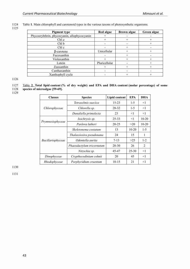
Current Pharmaceutical Biotechnology Mimouni et al.
Table 1. Main chlorophyll and carotenoid types in the various taxons of photosynthetic organisms.
Pigment type Red algae Brown algae Green algaePhycoeryhthrin, phycocyanin, allophycocyanin + - -
Chl a + + +Chl b - - +Chl c - + -
β-carotene Unicellular + +Fucoxanthin - + -Violaxanthin + + +
Lutein Pluricellular - +Zeaxanthin + + +
Canthaxanthin - - -Xanthophyll cycle - + +
Table 2 . Total lipid content (% of dry weight) and EPA and DHA content (molar percentage) of some species of microalgae [59-69].
Classes Species Lipid content EPA DHA
Chlorophyceae
Tetraselmis suecica 15-23 1-5 <1
Chlorella sp. 28-32 1-5 <1
Dunaliella primolecta 23 <1 <1
PrymnesiophyceaeIsochrysis sp. 25-33 <1 10-20
Pavlova lutheri 20-25 >20 10-20
Bacillariophyceae
Skeletonema costatum 13 10-20 1-5
Thalassiosira pseudonana 24 15 1
Odontella aurita 7-13 >25 1-2
Phaeodactylum tricornutum 20-30 26 2
Nitzschia sp. 45-47 25-30 <1
Dinophyceae Crypthecodinium cohnii 20 45 <1
Rhodophyceae Porphyridium cruentum 10-15 21 <1
43
11241125
1126
112711281129
1130
1131

Current Pharmaceutical Biotechnology Mimouni et al.
44
1132
Related Documents