1 Current Fluid Therapy Topics and Recommendations During Anesthetic Procedures Andrew Claude, DVM, DACVAA Mississippi State University Mississippi State, MS • Intravenous fluid administration is recommended during general anesthesia, even during short procedures. • The traditional IV fluid rate of 10 mls/kg/hr during general anesthesia is under review. • Knowledge of a variety of IV fluids, and their applications, is essential when choosing anesthetic protocols for different medical procedures. Anesthetic drug effects on the cardiovascular system • Almost all anesthetic drugs have the potential to adversely affect the cardiovascular system. • General anesthetic vapors (isoflurane, sevoflurane) cause a dose-dependent, peripheral vasodilation. • Alpha-2 agonists initially cause peripheral hypertension with reflex bradycardia leading to a dose-dependent decreased patient cardiac index. As the drug effects wane, centrally mediated bradycardia and hypotension are common side effects. • Phenothiazine (acepromazine) tranquilizers are central dopamine and peripheral alpha receptor antagonists. This family of drugs produces dose-dependent sedation and peripheral vasodilation (hypotension). • Dissociative NMDA antagonists (ketamine, tiletamine) increase sympathetic tone soon after administration. When dissociative NMDA antagonists are used as induction agents in patients with sympathetic exhaustion or decreased cardiac reserve (morbidly ill patients), these drugs could further depress myocardial contractility. • Propofol can depress both myocardial contractility and vascular tone resulting in marked hypotension. Propofol’s negative effects on the cardiovascular system can be especially problematic in ill patients. • Potent mu agonist opioids can enhance vagally induced bradycardia. Why is IV fluid therapy important during general anesthesia? • Cardiac output (CO) equals heart rate (HR) X stroke volume (SV); IV fluids help maintain adequate fluid volume, preload, and sufficient cardiac output. • Oxygen delivery to the tissues (DO2) equals CO X arterial blood oxygen content (CaO2); without adequate blood volume (relative and/or absolute hypovolemia) cardiac output decreases, which results in decreased peripheral oxygen delivery, thus tissue ischemia. • General anesthesia, by nature, depresses (shocks) autonomic, cardiovascular responses and homeostasis. Cardiovascular problems related to general anesthesia occur even with ASA status 1 patients. Intra-operative blood loss will contribute to total circulatory volume loss and therefore exaggerate the cardiovascular depressant effects of general anesthesia. • In response to hypovolemia, the body preferentially centralizes blood circulation toward the vital organs and away from peripheral tissues. • Subcutaneous fluid administration during general anesthesia does not replace IV administration as a means to maintain blood volume. Subcutaneous fluids are absorbed poorly during general anesthesia due both to a circulatory shift away from peripheral circulation and an inevitable hypothermia. • Intravenous fluids can help maintain a patent IV catheter during general anesthesia, which allows for emergency drug administration, if needed. Perioperative fluid therapy should be tailored to patient requirements • Appropriate fluid type, rate, and volume should be considered important elements of a patient’s overall anesthetic protocol. Each patient is unique and every anesthetic protocol should be tailored to individual patient anesthetic requirements. • Patient history, thorough physical exam, and subjective and objective data (laboratory, radiographic) are necessary to plan appropriately an anesthetic protocol. • Ideally, patient stabilization, including fluid losses, electrolyte imbalances, trauma, and respiratory and cardiovascular diseases should occur prior to anesthesia; however, in emergency situations, anesthetic patient stabilization may not be possible.

Welcome message from author
This document is posted to help you gain knowledge. Please leave a comment to let me know what you think about it! Share it to your friends and learn new things together.
Transcript
1
Current Fluid Therapy Topics and Recommendations During Anesthetic Procedures
Andrew Claude, DVM, DACVAA Mississippi State University
Mississippi State, MS
• Intravenous fluid administration is recommended during general anesthesia, even during short procedures. • The traditional IV fluid rate of 10 mls/kg/hr during general anesthesia is under review. • Knowledge of a variety of IV fluids, and their applications, is essential when choosing anesthetic protocols for different
medical procedures.
Anesthetic drug effects on the cardiovascular system • Almost all anesthetic drugs have the potential to adversely affect the cardiovascular system. • General anesthetic vapors (isoflurane, sevoflurane) cause a dose-dependent, peripheral vasodilation. • Alpha-2 agonists initially cause peripheral hypertension with reflex bradycardia leading to a dose-dependent decreased
patient cardiac index. As the drug effects wane, centrally mediated bradycardia and hypotension are common side effects.
• Phenothiazine (acepromazine) tranquilizers are central dopamine and peripheral alpha receptor antagonists. This family of drugs produces dose-dependent sedation and peripheral vasodilation (hypotension).
• Dissociative NMDA antagonists (ketamine, tiletamine) increase sympathetic tone soon after administration. When dissociative NMDA antagonists are used as induction agents in patients with sympathetic exhaustion or decreased cardiac reserve (morbidly ill patients), these drugs could further depress myocardial contractility.
• Propofol can depress both myocardial contractility and vascular tone resulting in marked hypotension. Propofol’s negative effects on the cardiovascular system can be especially problematic in ill patients.
• Potent mu agonist opioids can enhance vagally induced bradycardia. Why is IV fluid therapy important during general anesthesia?
• Cardiac output (CO) equals heart rate (HR) X stroke volume (SV); IV fluids help maintain adequate fluid volume, preload, and sufficient cardiac output.
• Oxygen delivery to the tissues (DO2) equals CO X arterial blood oxygen content (CaO2); without adequate blood volume (relative and/or absolute hypovolemia) cardiac output decreases, which results in decreased peripheral oxygen delivery, thus tissue ischemia.
• General anesthesia, by nature, depresses (shocks) autonomic, cardiovascular responses and homeostasis. Cardiovascular problems related to general anesthesia occur even with ASA status 1 patients. Intra-operative blood loss will contribute to total circulatory volume loss and therefore exaggerate the cardiovascular depressant effects of general anesthesia.
• In response to hypovolemia, the body preferentially centralizes blood circulation toward the vital organs and away from peripheral tissues.
• Subcutaneous fluid administration during general anesthesia does not replace IV administration as a means to maintain blood volume. Subcutaneous fluids are absorbed poorly during general anesthesia due both to a circulatory shift away from peripheral circulation and an inevitable hypothermia.
• Intravenous fluids can help maintain a patent IV catheter during general anesthesia, which allows for emergency drug administration, if needed.
Perioperative fluid therapy should be tailored to patient requirements
• Appropriate fluid type, rate, and volume should be considered important elements of a patient’s overall anesthetic protocol. Each patient is unique and every anesthetic protocol should be tailored to individual patient anesthetic requirements.
• Patient history, thorough physical exam, and subjective and objective data (laboratory, radiographic) are necessary to plan appropriately an anesthetic protocol.
• Ideally, patient stabilization, including fluid losses, electrolyte imbalances, trauma, and respiratory and cardiovascular diseases should occur prior to anesthesia; however, in emergency situations, anesthetic patient stabilization may not be possible.
2
Anesthesia fluid therapy; crystalloids (Dibartola) • Isotonic, polyionic replacement fluids, such as LRS, are popular IV fluids used during general anesthesia • Replacement fluids resemble extra-cellular fluid composition and are designed to resupply body fluids and electrolytes
within the cardiovascular and interstitial spaces. Within 30 minutes after replacement fluid administration, nearly 80% is lost from the vascular space into the interstitium.
• Replacement fluids can be used to help alleviate acute hypovolemia. • Maintenance fluids are designed to fill rapidly the interstitial space. Maintenance fluids should NOT be used for volume
resuscitation. • There are many different formulations of crystalloid fluids available. Indications of each kind depend on individual
patient needs such as hypovolemia, dehydration, illness, electrolyte, and acid-base imbalances. • In the last six years the volume of perioperative crystalloid administration has come under scrutiny. An article written in
2008 by Chappell, et al., questioned the existence of a third space and the research that first established fluid rates during general anesthesia. Traditionally, perioperative fluid administration for veterinary patients has largely mimicked, without solid scientific basis, human recommendations. A publication in 2010 by Boscon, et al., in demonstrated that not only did urine production in healthy, anesthetized dogs consistently decrease, it was coupled with an increase in body water weight. In 2013 an article in JAHAA provided new recommendations for fluid therapy with veterinary anesthesia patients. Based on these recommendations, canine fluid rates should start at 5 ml/kg/hr, feline rates at 3 ml/kg/hr, and fluid formulation, volumes, and rates should be adjusted according to individual patient needs.
Anesthesia fluid therapy; colloids
• Replacement crystalloids are beneficial to help expand rapidly the vascular space when increased blood volume is needed. Unfortunately large volumes of crystalloid potentially can lead to issues such as dilutional hypoalbuminemia, dilutional coagulopathies, decreased pulmonary function, decreased tissue oxygenation, and increased water weight. Approximately 80% of the volume of intravenous crystalloids equilibrate with the interstitial space within 30 to 45 minutes after administration. Unless the underlying cause of hypovolemia is corrected, more crystalloid therapy will be required to help maintain cardiac output, which, in turn, worsens tissue edema.
• Colloids are fluids that contain large, complex molecules. Like crystalloids, colloids can be used for intravenous fluid expansion; however, unlike crystalloids, colloids remain intravascular as long as the endothelial barrier remains intact.
• There are two major categories of colloids, natural and synthetic. Natural colloids are blood components including packed RBCs, plasma, platelet-rich plasma, etc. Generally, the primary synthetic colloids used in modern medicine are hydroxyl ethyl starches (HES). The two most common HES products used in veterinary medicine are Hetastarch® and Vetstarch®. Vetstarch® is the only HES colloid approved for veterinary use.
• There are two principles the general practitioner should understand regarding HES colloids: molecular weight (MW) and C2/C6 substitution ratios. HES colloids are divided into 3 groups according to their average molecular weights: high MW (>400 kDa); medium MW (200-400 kDa); and low MW (<200 kDa) solutions. The molecular weight determines duration of action, the larger the MW the longer the duration of action. The C2/C6 ratio is the ratio of carbon position 2 substitutions to carbon position 6 substitutions. The C2/C6 ratio determines the adverse side effects. The larger the C2/C6 ratio the greater the coagulopathic potential. An ideal HES product would be one with a large MW (long DOA) and small C2/C6 ratio (fewer side effects). Unfortunately, the MW of the product mirrors the C2/C6 ratio. Larger MW products have larger C2/C6 ratio and vice versa for smaller MW products.
o Hetastarch®: 450/0.7 (MW = 450 kDa, C2/C6 ratio = 0.7) o Vetstarch®: 130/0.4 (Mw = 130 kDa, C2/C6 ratio = 0.4)
• Indications for colloid administration include hypovolemia, hypoalbuminemia, and hypotension. Because HES colloids are large molecules, similar to albumin, they tend to remain in the vascular space adding to the colloidal oncotic pressure. Administration of HES will contribute its own volume, plus a third of its volume in water drawn from the interstitial space, to the total blood volume. Some practitioners prefer to use HES plus a crystalloid combination (50:50), which can be very effective for rapid IV volume loading. Another option, which provides even more rapid vascular expansion, is HES plus hypertonic saline.
• HES can be used as the primary fluid therapy in hypoalbuminemic patients during general anesthesia with or without crystalloids. HES can also be given as intermittent IV boluses to help mitigate hypotension.
• Coagulopathies are the primary, adverse effects of HES products dictated by the C2/C6 molecular substitution ratio. All HES products have the potential to inhibit the Von Willdebrand factor (vWF) and factor VIII resulting in platelet dysfunction, or type 1 Von Willdebrand-like syndrome. Because of these concerns, an anecdotal, maximum dose HES colloids of 20 ml/kg/day was established for human patients. Veterinary medicine simply borrowed this dose and
3
applied it to animal patients. Based on the principle of the C2/C6 molecular substitution ratio, an across-the-board, “maximum” dose for all HES products in all patients does not make medical sense. In addition, multiple studies have demonstrated the coagulopathic effects of HES products are clinically irrelevant unless the patient has a preexisting coagulopathy (vWD in Doberman Pinschers).
• Recently, there have been concerns with the administration of HES in human, septic patients, which resulted in acute renal failure. Although there has not been a cause and effect established, the FDA has issued a warning regarding HES use in humans with septicemia. Acute renal failure associated with HES use in septic veterinary patients has NOT been documented. The FDA warning does NOT apply to veterinary medical practice.
• Acute fluid overload, especially in cardiac patients, can occur when colloids are administered rapidly in large volumes. Care should be taken when using colloids (any IV fluids) in patients with known cardiac disease.
Mitigating hypotension during anesthesia in the small animal patient
• Most organ systems in the body autoregulate their own blood perfusion within a systemic mean arterial pressure (MAP) range of 60 – 150 mmHg. Outside this range blood perfusion autoregulation becomes a product of systemic blood pressure. When MAPs fall below 60 mmHg, the risk of tissue ischemia increases.
• The number one cause of hypotension in anesthetized veterinary patients is excessive anesthetic depth. Having one person dedicated to monitoring the anesthetized patient and who understands how to assess depth of anesthesia is essential for safe anesthetic practice.
• Bradycardia can contribute to hypotension because CO is a function of HR X SV. Several factors contribute to bradycardia during general anesthesia, including hypothermia and the pharmacodynamics of anesthetic drugs. Patients should be kept warm (> 97 oF) during general anesthesia, and an anticholinergic can be administered to help treat bradycardia resulting from high vagal tone.
• Absolute hypovolemia results in systemic hypotension. Ongoing surgical blood loss should be treated with IV fluid administration, including crystalloids and colloids. Extensive hemorrhage (> 20% patient blood volume) can be managed with IV hypertonic saline, HES, and crystalloids until replacement blood therapy can be conducted.
• One cause of relative hypovolemia is systemic vasodilation and/or depressed myocardial contraction. It is advisable to secure adequate blood volume (rule out absolute hypovolemia) before treating hypotension pharmaceutically. Systemic vasodilatation can be treated with a vascular pressor agent (ephedrine, dopamine, vasopressin), whereas depressed mycocardial contractility can be treated with a positive inotrope (dobutamine).
References Boscan P, Pypendop BH, Siao KT, Fluid balance, glomerular filtration rate, and urine output in dogs anesthetized for an orthopedic surgical procedure, AJVR, 2010, May:71(5): 501-07 Branson K: Injectable anesthetics, In Adams R, editor: Veterinary Pharmacology and therapeutics, ed 8, Ames, IA, Blackwell Publishing Professional, pp 213-67. Chappell D, Matthias J, Hofmann-Klefer K, Conzen P, Rehm M, A rational approach to perioperative fluid management, Anesthesiology, 2008; 109: 723-40. Davis H, Jensen T, Johnson A, 2012 AAHA/AAFP fluid guidelines for dogs and cats, JAAHA, 2013, May/June:43(3): 149-59 Hughes D, Boag A: Fluid therapy with macromolecular plasma volume expanders, In DiBartola S, editor: Fluid, electrolyte and acid-base disorders, ed 4, St. Louis, MO, 2012, Elsevier-Saunders, pp 647-64. Pascoe P, The cardiopulmonary effects of dexmedetomidine infusions in dogs during isoflurane anesthesia, JVAA, 2014, July 31. doi: 10.1111/vaa.12220. [Epub ahead of print].
4
Anesthesia Ventilators and Ventilation Techniques Andrew Claude, DVM, DACVAA
Mississippi State University Mississippi State, MS
• Intermittent positive pressure ventilation using mechanical ventilators has not been used traditionally in veterinary
practice. • Modern mechanical ventilators have become more affordable and easier to operate, allowing an increase use in clinical
practice. • Understanding the mechanics, function, and physiological effects of mechanical, intermittent, positive pressure
ventilation is necessary in order to safely, and effectively, ventilate anesthetized veterinary patients. Terminology and physiology
• Minute ventilation (VE) = Respiratory rate (f) X Tidal volume (TV). • Under normal physiological conditions PCO2 dictates minute ventilation (VE). Oxygen has little effect on VE unless the
PO2 falls below 60 - 70 mmHg. • CO2 crosses the blood brain barrier where it combines with water in the CSF. Carbonic anhydrase in the CSF facilitates
the formation of carbonic acid which then dissociates into hydrogen and bicarbonate ions. The hydrogen ions then interact with the chemoreceptors of the dorsal respiratory group:
CO2 + H2O ↔ H2CO3 ↔ H+ + HCO3- • Hypoventilation is synonymous with increased PCO2 whereas hyperventilation is synonymous with decreased PCO2. • With increased PCO2, respiratory drive will increase, with decreased PCO2, respiratory drive will decrease. • IPPV = intermittent positive pressure ventilation, PIP = peak inspiratory pressure, PEEP = positive end expiratory
pressure • There are many ways one can control ventilation with anesthetized patients: the reservoir bag, a demand valve, or a
mechanical ventilator to name a few. Indications for controlled ventilation
• Hypoventilation: Hypercapnea, drug induced respiratory depression, trauma, disease, and others. • Poor oxygenation: Five causes of hypoxemia include: low fraction/pressure of inspired oxygen; inadequate VT; O2
diffusion impairment; ventilation to perfusion mismatch (V/Q mismatch), and pulmonic/anatomic cardiac shunt. • Depth of inhalant anesthesia: Anesthetized patients, while breathing anesthetic vapors spontaneously, cycle naturally
between levels of light and deep planes of general anesthesia. Controlled ventilation provides a constant rate of inhaled anesthetics, thus eliminating the variability of inhalant general anesthesia.
• Surgeries that involve the loss of negative pressure and mechanical tethering between the visceral and parietal pleurae require intermittent positive pressure ventilation.
• Specific pulmonary diseases require assisted ventilation during general anesthesia, examples include: chest trauma, diaphragmatic hernia repair, severe alveolar diseases, and pleural diseases.
• Patients with conditions that may significantly limit VT, such as pregnancy or obesity, should receive ventilatory support during general anesthesia.
• In reality, indications for controlled ventilation are not always well defined. Ventilators are useful tools during general anesthesia, however; they should be used according to each patient’s individual and should never replace human intervention. Always monitor patients under general anesthesia receiving mechanical ventilation closely. Mechanical ventilators can induce serious patient pulmonary damage, even death, if not set-up and monitored correctly
Controlled ventilation
• Mechanical ventilation is based on VE, which is function of f X VT • Adjustment of VE requires changes in ventilation frequency and or volume. • Volume mode: Volume mode ventilator will deliver a controlled volume of gas (patient’s VT), regardless of the peak
inspiratory pressure. The variable factor is pressure. Small animal patient VT is approximately 10-20 ml/kg. Most anesthetic mechanical ventilators are set volume mode or have a volume mode option. During long periods of mechanical ventilation volume mode ventilators can cause pathological changes to the pulmonary tissues.
• Pressure mode: Pressure mode ventilator will deliver a volume gas until a set pressure is reached. The variable factor is volume. Most mechanical ventilators that have pressure mode also have volume mode option. Pressure mode ventilation causes fewer pathologic changes to pulmonary tissues than volume mode ventilation.
5
• Time-cycled ventilation: Despite volume vs. pressure mode ventilation, almost all mechanical ventilators are time-cycle controlled based on respiratory frequency (breaths per minute). Typically, timing is controlled electronically.
Basic anatomy of an anesthesia mechanical ventilator
• Most anesthetic mechanical ventilators have two gas sources. The driving gas is any type of high pressure gas that drives the bellows, from outside, thus pushing (positive pressure) the tidal volume into the patient (compressed O2, medical gas, N2 CO2). Maximum pressure of the driving gas should not exceed 50 psi. The breathing system gas is on the inside of the bellows and is continuous with the patient’s breathing circuit. Remember, the driving gas and breathing gas are two, separate gases and should not mix.
• Bellows. Most anesthetic mechanical ventilators use a bellows to push the breathing gas VT into the patient. Bellows are classified as ascending or descending, based on the direction the bellows move during exhalation.
• Control panel. Anesthetic mechanical ventilators have a control panel that allows adjustment of patient VT, breathing frequency, and sometimes I:E ratios.
• Scavenging system. Because the inside of the ventilator bellows is continuous with the patient’s breathing gases, the ventilator attaches to the anesthetic machine scavenging system for evacuation of waste gases.
• Connecting hose and wall plug-in. Anesthetic mechanical ventilators have a hose that connects to the high pressure gas driving the bellows. The hose should be color-coded according to the driving gas; for example, oxygen is green, and medical air is yellow.
Capnography
• Under normal physiological conditions the primary indication for mechanical ventilation during general anesthesia is patient CO2. There are two ways to monitor patient PCO2: arterial blood gas analysis and/or end-tidal PCO2 (PETCO2, capnography). Although arterial blood gas analysis is more accurate, it is also expensive and impractical. Capnography provides a useful, and practical, means to monitor patient PCO2, and is recommended for all anesthetized patients undergoing mechanical ventilation under general anesthesia.
• There are two categories of capnographs: main-stream, which analyzes the patient’s exhaled breath adjacent to the endotracheal tube, and side-stream, which removes a sample of the patient’s breath and delivers it to an analyzer away from the patient.
• Capnography is based on the principle that end-tidal exhaled PCO2 (PACO2) is roughly equal to pulmonary arterial PCO2 (PaCO2)
• Graphical illustration of the PETCO2 over time is called a capnogram. Capnograms are useful for visually monitoring an anesthetized patient’s PCO2 and other problems that can develop, such as a leak in the breathing system.
Final considerations
• A patient’s delivered VT should be set according to a desired PIP and PETCO2 rather than to the calculated VT. • Maximum PIP for small animal patients is 20 cm H2O; otherwise, barotrauma could occur to the patient’s pulmonary
tissues (alveoli). • IPPV causes a decrease in mean arterial pressure due to a reversal of the physiological thoracic blood pump. • Positive end-expiratory pressure can be used to help facilitate oxygenation via maintaining opened alveoli.
References Hammond R: Automatic ventilators, In Seymour C, Duke-Novakovski T, editors: BSAVA Manual of Canine and Feline Anaesthesia and Analgesia, ed 2, BSAVA Gloucester, pp 49-60. Hartsfield S, Airway management and ventilation, In Tranquilli W, Thurmon J, Grimm K, editors: Lumb & Jones’ Veterinary Anesthesia and Analgesia, ed 4, Ames IA Blackwell, pp 512-31. Mosley C: Anesthesia equipment, In Grimm K, Tranquilli W, Leigh L, editors: Essentials of Small Animal Anesthesia and Analgesia, ed 2, Ames IA Wiley-Blackwell, pp 187-96.
6
Acute Pain Management: Local and Regional Anesthesia
Andrew Claude, DVM, DACVAA Mississippi State University
Mississippi State, MS
• Local and regional anesthesia are common practices in large animal veterinary medicine. In the past, locoregional techniques have been underutilized in small; however, recently there has been a surge in their use with small animal practice.
• Most common locoregional anesthetic techniques used for large animal surgery can also be adapted to small animals. • A good understanding of basic anatomy, pharmacology of local anesthetic drugs, and patient physiology is essential in
order to safely, and effectively, utilize local and regional anesthetic techniques. • Combining local and regional anesthetic techniques with parenteral analgesics can provide small animal practitioners
more flexibility and better options for pre-, intra- and post-operative pain management. Physiology of and concepts regarding pain
• Acute pain is considered a normal, healthy, and protective physiological response to noxious stimuli. Chronic, centralized pain, or wind-up pain is considered a pathological, abnormal expression of pain.
• The dose of general anesthetics needed to abolish the effects of nociception is close to that which can abolish autonomic responses. High doses of general anesthetic drugs significantly depress the cardiovascular, respiratory, and thermoregulatory systems in the body. Analgesic modalities before, and during, surgery help decrease the dose of general anesthetics needed to provide immobility without loss of autonomic tone.
• Transduction: Mechanical, chemical, or thermal injury is converted to an electrical impulse by Aβ (quick pain) and C nociceptors (slow pain).
• Transmission: The noxious electrical impulse is transmitted from the periphery to the spinal cord via Aβ and C sensory neurons. The synapse between the sensory neurons and the spinal cord occurs at lamina II (substantia gelatinosa) in the dorsal spinal horn.
• Primary (spinal) modulation: Within the spinal cord the afferent, noxious sensory impulse undergoes initial analysis. The spinal cord upgrades or downgrades the severity of the noxious stimulus and communicates that information to the brain. An unconscious reflex arc is the result of primary (spinal) modulation.
• Projection: After primary modulation, the noxious information is then projected to the brain via several tracts: two examples are the spinocervicothalamic (fast pain) and spinoreticular (slow pain) tracts.
• Secondary (cerebral) modulation: Within the conscious brain noxious afferent input is perceived as pain. Unconsciousness (anesthesia) blunts, or abolishes, secondary nociceptive modulation.
• Animals and humans share similar anatomical and physiological nociceptive structures for the production, conduction, and modulation of pain.
• Pain assessment in animals is based on anthropomorphic comparisons, subjective, and objective criteria. • Pain is the conscious perception of nociception. Nociception is the physiological processes that involves the conversion
of a noxious stimulus to an electro-chemical impulse and modulation in the CNS. • The perception of pain does not occur during general anesthesia; however, without analgesic modalities the process of
nociception still occurs, which can lead to centralized, or wind-up pain. • Providing analgesics before surgery is called pre-emptive analgesia. Studies have shown that preemptive analgesia
significantly decreases the likelihood of hypersensitiity associated with surgical pain. • Preventive analgesia is a term that describes a comprehensive pain control plan that includes pre-, intra- and
postoperative therapies. Preventive analgesia has been well established in human medicine but not yet in veterinary medicine.
Local and regional anesthetic techniques in small animal practice
• Lidocaine and bupivacaine are the most common local anesthetics used in small animal practice. • Local anesthetics are fast-sodium channel blocking agents. In their bottles local anesthetics are acidic and inactive.
When injected into the body (comparatively alkaline), the local anesthetic molecules dissolve into HCl salts and active bases. The active bases diffuses across the nerve epineurium and cell membrane into the cytoplasm and block sodium channels.
7
• Toxic effects of local anesthetic depend on the drug. Lidocaine causes dose-dependent neuro- and cardio-toxic effects. Bupivacaine has potent cardio-toxic effects. Inadvertent intravenous injection of local anesthetics must be avoided; therefore, always aspirate before injecting.
• Most locoregional anesthetic techniques can be performed blindly; however, a peripheral nerve locating device (nerve stimulator) can help increase the success and safety of the procedures.
• Quincke needles are designed specifically for locoregional techniques. Quincke needle bevels are blunter which allows for a better feel as the needle dissects through tissue planes.
• Common regional techniques for dental procedures include mental, infra-orbital, maxillary, and mandibular nerve blocks. Auriculopalpebral and the greater auricular nerve blocks can be useful for procedures involving the ear such as, ear flushes and surgery.
• The brachial plexus infiltration block can be used for surgeries involving the distal forelimb. A carpal ring block can be used for surgeries involving the forepaw such as declaws and digit amputations.
• Lumbosacral epidural regional techniques are very useful for surgeries involving the hips and distal rear legs. The most common drugs used for lumbosacral epidurals is the combination of preservative free (PF) morphine and PF bupivacaine. Feline lumbosacral epidurals using PF morphine and PF bupivacaine can be done also; however, it is important to remember the feline spinal cord ends at S1 compared with the canine spinal cord, which ends at L5-6.
• Caudal epidural techniques can be used to provide regional anesthesia during perineal surgeries and facilitate urethral relaxation for catheter placement in blocked male cats.
• Infiltration catheters (soaker catheters) have manufactured fenestrations at their distal ends so that, when buried in the surgical wound, local anesthetics can be injected into the tissues providing a field of anesthesia.
References Muir W, Physiology and pathophysiology of pain, In Gaynor J, Muir W, editors: Handbook of Veterinary Pain Management, ed 2, St. Louis MO, Mosby Elsevier, pp13-41.
8
Acute Pain Management: Pharmaceutical Options
Andrew Claude, DVM, DACVAA Mississippi State University
Mississippi State, MS
• Systemic analgesic drugs are the mainstay of small animal veterinary pain management. • Options regarding parenteral analgesics in small practice are often governed by cost and clinician experience. • Utilizing the same analgesic drugs, regardless of the surgical procedures, is not good analgesic case management.
Having a good understanding of a variety of analgesic options, for dogs and cats, can be a valuable addition to help expand clinical practice.
Rules of thumb
• Animals share similar anatomical and physiological nociceptive properties as humans; therefore, animals likely have similar pain experiences as humans.
• Pain in animals is difficult to quantify and evaluation is based on a combination of objective and subjective anthropomorphic attributes.
• Pain is the conscious perception of nociception. General anesthesia abolishes consciousness; therefore, pain is not perceived during general anesthesia. Nociception is the physiological process by which a noxious stimulus is transduced into an electro-chemical impulse and carried to the central nervous system. Nociceptive, physiological processes continue to occur during general anesthesia unless analgesics are employed.
• According to the Veterinarian’s Oath, veterinarians have an obligation to prevent and relieve animal suffering, including pain.
• If there is a suspicion an animal patient is painful, it is better to treat for pain than to ignore the concern. Concepts regarding nociception and pain management in veterinary patients
• Transduction: Mechanical, chemical, or thermal injury is converted to an electrical impulse by Aβ (quick pain) and C nociceptors (slow pain).
• Transmission: The noxious electrical impulse is transmitted from the periphery to the spinal cord via Aβ and C sensory neurons. The synapse between the sensory neurons and the spinal cord occur at lamina II (substantia gelatinosa) in the dorsal spinal horn.
• Primary (spinal) modulation: Within the spinal cord the afferent, noxious sensory impulse undergoes initial analysis. The spinal cord upgrades or downgrades the severity of the noxious stimulus and communicates that information to the brain. An unconscious reflex arc is the result of primary (spinal) modulation.
• Projection: After primary modulation the noxious information is then projected to the brain via several tracts; two examples are the spinocervicothalamic (fast pain) and spinoreticular (slow pain) tracts.
• Secondary (cerebral) modulation: Within the conscious brain noxious afferent input is perceived and translated into pain. Unconsciousness (anesthesia) blunts, or abolishes, secondary nociceptive modulation.
• Providing analgesics before surgery is called pre-emptive analgesia. Studies have shown that preemptive analgesia decreases significantly the likelihood of hypersensitivity associated with surgical pain.
• Preventive analgesia is term that describes a comprehensive pain control plan, which includes pre-, intra-, and postoperative nociceptive therapies. Preventive analgesia has been well established in human medicine but not yet in veterinary medicine.
• Analgesic drugs help reduce/abolish pain by interfering with the nociceptive process(es). • The dose of general anesthetics needed to produce unconsciousness is far less than what is required to abolish the effects
of nociception. The dose of general anesthetics needed to abolish the effects of nociception is close to that which can abolish autonomic responses. High doses of general anesthetic drugs significantly depress the cardiovascular, respiratory and thermoregulatory systems in the body. Analgesic modalities before, and during, surgery help decrease the dose of general anesthetics needed to provide immobility without loss of autonomic tone.
• A pre-emptive pain scale evaluation can help the clinician formulate a patient’s analgesic therapy plan. A pre-emptive pain scale is a subjective pain assessment done pre-operatively based on the anticipated degree of pain. Analgesic drug potency, dose, and frequency of administration can be tailored according to the pre- and post-operative pain evaluation.
9
Parenteral analgesics in veterinary small animal practice • Common concerns with parenteral analgesic drugs in small animal practice include unwanted sedation, extra expense,
controlled drug issues, unpredictable results, drug knowledge of the attending veterinarian, and client compliance. • Opioids are the primary parenteral analgesic used for human and veterinary surgery. Mu agonist opioids are an excellent
choice to help provide effective pre- intra- and post-operative pain relief for animal patients. There are many mu agonist opioid drugs available, including opioid products that are absorbed transdermal.
• Butorphanol, a mu antagonist, kappa agonist opioid, has limited analgesic capabilities and a short duration of action. Butorphanol should not be considered a primary analgesic for surgical pain, especially in dogs.
• Buprenorphine is a partial mu agonist opioid, has a good analgesic profile, and long duration of action for both dogs and cats.
• NSAIDs relieve pain via their anti-inflammatory abilities making them extremely versatile analgesic drugs. There are many NSAID options for both dogs and cats; however, judicial use of these drugs should be limited to normal, healthy patients. Contraindications for NSAID include concurrent steroid administration, concurrent other NSAIDs, renal and hepatic diseases, gastrointestinal diseases, coagulopathies, pregnancy, dehydration, and other circulatory diseases.
• Common, and effective, adjunctive analgesic choices include lidocaine, ketamine, and alpha 2 agonists. References Muir W, Physiology and pathophysiology of pain, In Gaynor J, Muir W, editors: Handbook of Veterinary Pain Management, ed 2, St. Louis MO, Mosby Elsevier, pp13-41.
10
Complications, Troubleshooting, and Best Monitoring Options during Anesthesia
Andrew Claude, DVM, DACVAA Mississippi State University
Mississippi State, MS
• Complications that can occur during small animal anesthesia range from minor annoyances to major, life threatening events.
• Troubleshooting intra-operative anesthetic problems begins with pre-anesthetic planning and anticipating possible adversities.
Small animal pre-anesthetic preparation
• Preparation is the key to successful anesthetic outcomes. Anesthetic preparation includes equipment upkeep such as vaporizer calibration, and breathing system and machine leak checks. Monitoring equipment should be kept clean and in good working order.
• The anesthetic record is considered a legal document, an integral component of a patient’s complete medical record, and is signed by a licensed veterinarian. The anesthetic record chronologically should record all events that occur during the anesthetic procedure, including complications.
• Having one person dedicated to patient monitoring during the anesthetic procedure is an important contribution toward avoiding intra-operative complications.
Complications related to anesthetic drugs
• All anesthetic drugs are potentially harmful or fatal. • No anesthetic drug is perfect for a specific patient or procedure. Unpredictable patient response to anesthetic drugs
should always be considered a potential complication. • All anesthetic drugs given to a patient, including mistaken administrations, should be recorded in the patient’s anesthetic
record in milligram or microgram per kilogram form. • A thorough, working knowledge of anesthetic drugs, their combinations, and their pharmacodynamics in multiple
animal species is absolutely essential in order to help secure safe and reliable veterinary anesthetic procedures. Complications: Pre- to early anesthetic period
• Anesthetizing an animal patient to the point of loss of righting reflex (stage 3 anesthesia) abolishes the gag and swallowing reflexes requires intubation. Without proper intubation, an anesthetized patient risks aspiration pneumonia, severe esophagitis, upper airway obstruction, and inadequate ventilation and oxygenation.
• Tracheal intubation issues can be related to esophageal intubation, improper endotracheal tube size, and endotracheal tube cuff leaks.
• On rare occasions, a vagal reflex can occur during endotracheal intubation. Stimulation of the parasympathetic fibers in the larynx can initiate a vagovagal response which, in-turn, causes a sudden increase in vagal tone, clinically seen as a dramatic, sometimes lethal, bradycardia.
• Hypoxia shortly after induction can be related to anesthetic drug-related apnea, respiratory disease, inadequate fresh gas flow, inadequate oxygen delivery (empty O2 tank) or disconnected patient breathing system or common gas outlet.
• During the induction a patient may awaken suddenly or struggle. Stage 2 anesthesia during induction can be related to inadequate pre-anesthetic medications, improper induction procedure, improper ET intubation, and lack of inhalant anesthetic administration.
• Hypoventilation is defined as a PCO2 greater than 50 - 60 mmHg. An anesthetized patient on 100% O2, and breathing spontaneously 2 breaths per minute will have adequate oxygenation but severe hypoventilation.
• Oxygenation is a poor indicator of ventilation. Complications: Anesthesia period
• Most anesthetic drugs cause a dose-dependent depression of both the peripheral chemoreceptor responses and the central respiratory ventilatory drive, therefore; hypoventilation is a common patient complication during general anesthesia.
• Watching chest wall or reservoir bag movement and/or lung auscultating confirms the patient is breathing and provides a respiratory rate; however, these parameters do not adequately assess ventilation. In order to adequately monitor a patient’s ventilation, CO2 must be quantified.
11
• End-tidal PCO2 (PECO2) is an effective means to monitor anesthetized patient ventilation (PaCO2). PECO2 is based on the principle that the end-tidal, alveolar PCO2 (PACO2) is equivalent to pulmonary arterial PCO2 (PaCO2). Patients receiving IPPV should be monitored using PECO2.
• Cardiac arrhythmias can occur unexpectedly in normal, healthy anesthetized patients. Electrocardiogram does not evaluate adequately cardiac function; however, it does provide an assessment of electro-myocardial conduction and heart rate. Electro-myocardial conduction (ECG waveform) does not indicate myocardial contraction; however, myocardial contraction (pulses, etc.) necessarily indicates electrical conduction.
• Anesthetic drugs, especially inhalant anesthetics, cause a dose-dependent depression of vasomotor tone. Patient blood pressure monitoring during general anesthesia is necessary to assess adequately systemic arterial pressures. Mean arterial blood pressure (MAP) is an indirect evaluation of tissue perfusion. Hypotension, low MAP, can be caused by poor cardiac output, hypovolemia, and vasodilation. The primary cause of patient hypotension during general anesthesia is excessive anesthetic depth causing poor cardiac output and/or vasodilation. Hypertension, elevated MAP, can be caused by inadequate anesthesia, analgesia, diseases (pheochromocytoma), or certain anesthetic drugs (alpha 2 agonists).
• Anesthetic depth should be assessed using multiple factors rather than relying on one or two parameters (jaw tone, eye position, response to stimulus, respiratory rate, blood pressures, etc.). Patients that lack anesthesia may have coordinated muscle movement and signs of increased sympathetic tone. Inadequate anesthesia means a patient is reversing from stage 3 anesthesia back into stage 2 anesthesia; therefore, the patient requires more anesthetic (injectable and/or inhalant) to maintain surgical unconsciousness. Anesthetized patients that lack analgesia are those with uncoordinated muscle movements (reflexes) and/or those that require large doses of anesthetics to maintain stage 3 anesthesia (>2% isoflurane, repeated doses of propofol), which risks the loss of sympathetic tone (hypotension). Patients that lack analgesia during anesthesia require a MAC reducing modality such as a mu agonist opioid or locoregional block.
• Hypothermia is the primary complication during general anesthesia. Anesthetic drugs cause a dose dependent depression of the thermoregulatory centers and blunt the body’s response to cold environments. Untreated hypothermia can lead to increased vagal tone, bradycardia, delayed recoveries, and poor drug metabolism.
• Intra-operative patient death is the most extreme anesthetic-related complication. Most anesthetic deaths in veterinary medicine occur post-operatively within the first 3 hours. When signs of onset of acute death are observed during general anesthesia, the following steps should be indicated: confirm cardiac arrest, turn-off and reverse all anesthetic drugs, and begin CPR.
References Muir W, Cardiovascular System, In Tranquilli W, Thurmon J, Grimm K, editors: Lumb & Jones’ Veterinary Anesthesia and Analgesia, ed 4, Ames IA Blackwell, pp 61-151.
12
Analgesic Considerations in Cats Andrew Claude, DVM, DACVAA
Mississippi State University Mississippi State, MS
• The pharmacokinetic and pharmacodynamic effects of anesthetic and analgesic drugs differ between dogs and cats. Cats
are NOT small dogs. • Unfortunately, analgesic options for feline patients are limited compared with analgesic options for canine patients.
Important points regarding feline patients
• Due to feline aloof behavior, it can be difficult for clients to notice subtle changes with their cat’s health. Cats typically do not show obvious signs of pain; instead, they become withdrawn and hide.
• Generally speaking, feline responses to anesthetic and analgesic drugs are unpredictable compared with canine responses.
Physiology of nociception and feline pain
• Transduction: Mechanical, chemical, or thermal injury is converted to an electrical impulse by Aβ (quick pain) and C nociceptors (slow pain).
• Transmission: The noxious electrical impulse is transmitted from the periphery to the spinal cord via Aβ and C sensory neurons. The synapse between the sensory neurons and the spinal cord occurs at lamina II (substantia gelatinosa) in the dorsal spinal horn.
• Primary (spinal) modulation: Within the spinal cord the afferent, noxious sensory impulse undergoes initial analysis. The spinal cord upgrades or downgrades the severity of the noxious stimulus and communicates that information to the brain. An unconscious reflex arc is the result of primary (spinal) modulation.
• Projection: After primary modulation, the noxious information is then projected to the brain via several tracts: two examples are the spinocervicothalamic (fast pain) and spinoreticular (slow pain) tracts.
• Secondary (cerebral) modulation: Within the conscious brain noxious afferent input is perceived as pain. Unconsciousness (anesthesia) blunts, or abolishes, secondary nociceptive modulation.
• Providing analgesics before surgery is called pre-emptive analgesia. Studies have shown that preemptive analgesia significantly decreases the likelihood of hypersensitiity associated with surgical pain.
• Preventive analgesia is term that describes a comprehensive pain control plan which includes pre-, intra- and postoperative therapies. Preventive analgesia has been well established in human medicine but not yet in veterinary medicine.
• The Brondani multi- dimensional composite feline pain scale was recently validated for the English language. Before the Brondani feline pain scale there was no validated pain scale for cats.
• Pain is not always considered a major component of many feline diseases. Saddle thrombosis, for example, is a clinical condition secondary to feline cardiac disease and causes extensive, acute ischemic muscle pain. Regardless of the disease, pain evaluation, and therapy, should always be part of the clinical plan.
• Identifying pain in cats can be difficult. Cats do not outwardly express pain. Sometimes an owner noticing a change in his or her cat’s behavior is the only indication of discomfort. Clinical signs of acute pain in cats include a tucked or crouched posture, reluctance to move, ears facing forward, focused eyes, lip licking, guarding, and purring.
Analgesic options for feline patients
• Opioids are considered the backbone of analgesia in both human and veterinary medicine. Mu agonist opioids have been known to cause opioid-related hysteria (dysphorea) and hyperthermia in cats. Although both conditions merit concern their clinical relevance is questionable and both can be reversed using naloxone. Morphine and hydromorphone are mu agonist opioids that are most likely to cause side effects in cats whereas oxymorphone, methadone, and fentanyl are the least likely.
• Butorphanol is a mu antagonist, kappa agonist opioid and has good effects in cats; however, its duration of action is only 30 – 45 minutes.
• Buprenorphine is a partial mu agonist opioid and, in cats, provides excellent analgesia for up to 6 to 8 hours in cats. • Alpha 2 agonists provide both sedation and analgesia. Dexmedetomidine is an excellent major tranquilizer for cats
because it provides predictable results, good analgesia, can be combined with other drugs, and is the choice tranquilizer for cats with hypertrophic cardiomyopathy (HCM).

13
• Dissociative NMDA antagonists (ketamine, tiletamine) also provide consistent sedation and analgesia in cats; however, this family of major tranquilizers is contra-indicated in cats with HCM.
• NSAIDs are good analgesic choices in healthy cats. It is recommended practitioners administer these drugs judiciously, monitor their patients closely, and communicate to their clients regarding potential adverse side effects from NSAIDs.
• In addition to parenteral analgesics, locoregional techniques can be extremely valuable when used for pain management in cats. Examples of common locoregional procedures in cats include nerve blocks of the mouth and eyes, brachial plexus blocks, forepaw and rear-paw ring blocks, and lumbosacral and caudal epidurals.
References Brondani J, Khursheed M, Luna S, Validation of the English version of the UNESP-Botucatu multidimensional composite pain scale for assessing postoperative pain in cats, BMC Vet Res, 2013, 143(9)2-15. Muir W, Physiology and pathophysiology of pain, In Gaynor J, Muir W, editors: Handbook of Veterinary Pain Management, ed 2, St. Louis MO, Mosby Elsevier, pp13-41.
14
Neonatal, Pediatric, and Geriatric Anesthesia Andrew Claude, DVM, DACVAA
Mississippi State University Mississippi State, MS
• Neonates are not considered routine candidates for veterinary anesthesia. Due to their size and anatomical and
physiological differences, puppies and kittens be challenging to anesthetize safely. • Early spays and neuters before adoption is a common practice, and knowledge regarding pediatric physiology and
pharmacokinetics of anesthetic drugs is essential for successful anesthesia. • Like us, our pets are living longer. Although most of the customary principles of veterinary general anesthesia are
applicable to geriatric patients; there are important differences that should be considered. Definitions: neonatal, pediatric puppies and kittens
• In humans, the neonatal period is from birth to 4 weeks and the pediatric period is 4 weeks to approximately 2 years old. Adults are considered twenty years and older.
• In small animal veterinary medicine, puppies and kittens are considered neonates from birth to 2 to 4 weeks old, and pediatric patients are 4 to 8 weeks old. Beyond 8 to 12 weeks, puppies and kittens are considered young adults.
Physiological differences of neonatal and pediatric small animal patients.
• Respiratory system: Neonatal puppies and kittens have a greater prevalence of upper airway obstruction due to their large tongues and small airway openings. As they age and tissues grow, these unique anatomical challenges improve in most species except in brachycephalic breeds.
• Rapidly growing puppies and kittens have a high oxygen demand; therefore, they require a high minute ventilation compared with adults. Their tidal volume and functional residual capacity are fixed; therefore they depend on respiratory frequency to meet metabolic oxygen demands. Respiratory control and autonomic responses are immature and easily depressed by anesthetic drugs. Puppies and kittens under the age of 8 weeks old are highly susceptible during anesthetic procedures to apnea and hypoxia.
• Cardiovascular system: Neonates and puppies/kittens (<8 – 12 weeks old) depend on HR to alter cardiac output. They have limited ability to adjust their mycocardial contractility, thus stroke volumes are fixed. Unfortunately, they are also prone to bradycardia due to immature sympathetic responses and susceptibility to hypoxemia. Because of their immature vascular and autonomic systems, they cannot rely on vascular tone to help regulate mean arterial pressures (MAP) or tissue perfusion. Neonatal and pediatric blood pressure is almost entirely a function of cardiac output.
• Bottom line: Neonatal and pediatric puppies and kittens require oxygen supplementation and ventilation support, whereas bradycardia should be avoided during general anesthesia.
• Hematology: Neonatal and especially pediatric puppies and kittens under 12 weeks old, do not tolerate blood loss. Hematopoesis does not begin effectively until approximately 12 weeks of age and fetal hemoglobin is rapidly being removed, making these young patients highly susceptible to anemia. Adult small animal patients can tolerate up to a 20% surgical blood loss, while neonatal and pediatric patients are limited to a loss of 4%.
• Renal and hepatic systems: Although neonatal, pediatric puppy/kitten kidneys and livers are anatomically developed, they are immature functionally until 8 to 12 weeks of age. Their ability to biodegrade anesthetic drugs is slow, resulting in rapid pharmacodynamic effects and slow recoveries. Their renal function, fluid balance, and ability to concentrate urine are undeveloped, making these young animals susceptible to dehydration and intolerant of excessive fluid administration. Glycogen production and storage are inadequate making them susceptible to hypoglycemia.
• Thermoregulation: Neonatal and pediatric small animal veterinary patients have a high surface area with underdeveloped ability to thermoregulate. Severe hypothermia is of great concern during general anesthesia in small patients and may cause brady-arrhythmias, delayed recoveries and possibly death.
Anesthetic considerations for neonatal and pediatric small animal patients.
• Do not fast neonatal and pediatric patients before anesthesia; otherwise, there is a risk of hypoglycemia. The current recommendations are to allow the baby to nurse or feed until anesthesia for patients < 6 weeks old, withhold food no more than 2-3 hours for 6 to 8 week olds, monitor blood glucose at least every 30 to 60 minutes, and administer IV 2.5% dextrose if blood glucose drops below 80-100 mg/dl.
• Anesthetic drugs will produce profound effects and last longer in neonatal and pediatric veterinary patients. Use injectable and premedications judicially. More often it is better to mask induce neonatal patients, intubate, and place an IV catheter without using premedications.
15
• When using injectable drugs, it is recommended to avoid those known to have slow half-lives and require extensive biodegradation (acepromazine for example). Water soluble, short acting drugs, with known antagonists (midazolam, methadone, butorphanol, for example), at lower doses are better choices. Due to the risk of marked bradycardia and decrease cardiac output, alpha two agonists are not recommended in puppies and kittens under 8 weeks old.
• Avoid blood loss and use caution when administering IV fluids so as to not overload delicate cardiovascular systems. • Monitor heart rate, ventilation, and oxygenation, body temperature, and blood glucose closely during general anesthesia.
Employ external warming devices and bubble wrap extremities to help maintain body temperatures near normal. • Post-operative care should include supplemental oxygen and heat, monitor blood glucose, and supplemental dextrose, as
needed, and provide appropriate analgesics. Definitions: geriatric dogs and cats
• Dorland’s Medical Dictionary (27th ed.) defines geriatric as “old age” or elderly. Most people consider human geriatrics as 65 years old because Medicare eligibility begins. Technically, there is no specific age that defines “geriatric” in humans.
• Dogs that have lived 75 – 80% of their lifespan are considered geriatric, which for small breeds is greater than 10 years old, large breeds 6 – 10 years old.
• Cats are considered geriatric when they are 12 years old and older. Important considerations for veterinary geriatric anesthesia patients
• Increased age is NOT equivalent to increased risk of general anesthesia unless there are concurrent disease processes. Brodbelt, et al., estimated the risk of anesthetic depth increased up to 7 times for veterinary geriatric patients greater than 12 years old.
• Biological and physiological age is more important than chronological age when considering anesthesia in older patients.
• Geriatric veterinary patients have blunted homeostatic responses, including autonomic and somatic reflexes. • Underlying disease processes and urgent care should be treated before commencing to general anesthesia.
Physiological/pharmacological considerations for veterinary geriatric anesthesia patients
• Due to decreased metabolic demand, minute ventilation and cardiac output are reduced. The geriatric pulmonary system is less compliant, resulting in an increased work of breathing. Assisted ventilation during general anesthesia is recommended.
• Increased age results in a greater influence of vagal tone and reduced cardiac sympathetic responses. Myocardial and/or degenerative cardiac changes are seen more frequently in elderly veterinary patients, including valvular endocardiosis in small breed dogs and HCM in hyperthyroid cats.
• Although renal and hepatic organ systems continue to work sufficiently in older patients, with age there is a gradual loss of functional capacity. It is advisable to include a CBC and plasma chemistries as part of the pre-anesthetic work-up with geriatric patients. Decreased cardiac output results in decreased hepatic blood flow, which can lead to prolonged drug metabolism and slower patient recoveries.
• Geriatric veterinary patients generally handle most anesthetic drugs and protocols without concern. Clinical differences include increased sensitivities to anesthetic drugs, decreased MAC of inhalant anesthetics, and prolonged recoveries. The exact cause of increased sensitivities to anesthetic drugs seen with geriatric patients is unknown.
• Anesthetic drug recommendations for veterinary geriatric anesthesia patients include lower drug doses and use of short acting, water soluble anesthetic drugs that have known antagonists. Examples of anesthetic drugs used commonly with geriatric veterinary patients include benzodiazepines, opioids, propofol, alfaxalone, isoflurane, sevoflurane, and others.
• Low doses of alpha two agonists are safe to use in geriatric dogs with normal cardiac function. Dexmedetomidine is the drug of choice for elderly cats with HCM; however, ketamine should be avoided.
• Geriatric patients have a higher risk of cognitive dysfunction, which may make them more susceptible to emergence delirium and confusion during anesthesia recovery.
• Judicial dosing of Tramadol is necessary for geriatric patients receiving serotonin/norepinephrine uptake, or MAO inhibitors (selegiline), to avoid serotonin syndrome.
References Baetge C, Matthews N, Anesthesia and Analgesia for Geriatric Veteterinary Patients, Vet Clin Small Anim 42 (2012) 643–653 Holden, D: Paediatric patients, In Seymour C, Duke-Novakovski T, editors: BSAVA Manual of Canine and Feline Anaesthesia and Analgesia, ed 2, BSAVA Gloucester, pp 293-301.

16
Magrini F, Haemodynamic determinants of the arterial blood pressure rise during growth in conscious puppies. Cardiovasc Res. 1978 Jul;12(7):422-8. Neiger-Aeschbacher: Geriatric patients, In Seymour C, Duke-Novakovski T, editors: BSAVA Manual of Canine and Feline Anaesthesia and Analgesia, ed 2, BSAVA Gloucester, pp 303-09.
17
Postanesthetic Care of Small Animal Patients Andrew Claude, DVM, DACVAA
Mississippi State University Mississippi State, MS
• The recovery period is not always regarded as a vital component of an anesthetic procedure. • In veterinary anesthesia the majority of adverse events occur during recovery.
Anesthetic recovery
• Anesthetic recovery is the interval from the cessation of anesthetic drug delivery to the point at which the patient is extubated and has voluntary motor control.
• Factors that affect the length of recovery include patient health, length of the anesthetic procedure, anesthetic protocol, and patient, post-anesthetic body temperature.
• According to a Brodbelt, et al., study in 2007, greater than 50% of the canine and feline anesthetic related adversities occur during recovery.
• Thorough planning of the anesthetic event, anticipating problems, and keeping good anesthetic records during the pre-, intra-, and post-operative periods is essential.
• All anesthetic patients have the potential for poor recoveries. Difficult anesthetic recoveries can be due to multiple factors, including emergence delirium, dysphoria, inadequate analgesia, and general patient discomfort. In these cases, it is often advisable to delay the recovery to avoid further stress or injury to the recovering patient.
Anesthetic recovery: patient monitoring
• In the 2008, Brodbelt, et al., article, the authors speculated that inadequate patient monitoring may have been the primary factor behind anesthetic recovery periods being over represented by increased mortality rates in small animal anesthetic procedures.
• Anesthetic monitoring should NOT end at recovery; instead, it should continue until the patient is extubated and has returned voluntary muscle control.
• The degree of monitoring, and parameters evaluated, depend on the procedure performed and the patient’s health. Patient monitoring should include at least cardiovascular and respiratory status, body temperature, analgesia, and patient (dis)comfort. Post anesthetic, patient monitoring parameters should be included within the patient’s anesthetic records.
Anesthetic recovery: extubation
• Indications for patient intubation include decreasing the risk of aspiration, securing the patient’s airway, and providing a means for assisted ventilation. Patient intubation should be included with any procedure that involves a level of sedation or anesthesia in which the patient has lost motor control and therefore the ability to guard the larynx.
• Extubation should be performed when the recovering patient has regained laryngeal or pharyngeal sensation and reflexes, such as gagging, swallowing and chewing.
• Brachycephalic breeds have an increased risk of post-extubation, upper airway obstruction. During sedation and anesthesia excessive peri-laryngeal tissues and hypoplastic tracheas predispose these patients to pharyngeal obstruction. Ventilatory function should be monitored closely with brachycephalic breeds during pre-operative sedation and post-operative recovery, and it is prudent to have induction agent, a laryngoscope, and an endotracheal tube immediately available in case of upper airway obstruction.
Anesthetic recovery: other breed/species issues
• Alaskan malamutes, Siberian huskies and Labrador retrievers have a genetic polymorphism that predisposes these breeds to a high incidence of opioid-related dysphoria. Problems related to opioid use in those breeds tend to be individualistic; however, it is advisable to use lower doses, especially in Nordic breed dogs. Opioid dysphoria in any breed (or species) can be reversed using naloxone.
• Post-anesthesia related feline blindness (deafness) was reported as early as 2001. Unlike the dog, which has two arterial blood supplies to the brain (internal carotid and basilar arteries), cats have only one cerebral blood supply (maxillary artery). Spring-loaded mouth gags, used during procedures requiring mandibular extension (dentals), in cats can result in obstruction of the maxillary arterial blood flow causing cerebral ischemia, central blindness, and/or deafness.
18
Anesthetic recovery: supplemental oxygen Post-operative oxygen supplementation is most beneficial in patients with compromised respiratory function, sick patients, obese and pregnant patients, and brachycephalic breeds. Anesthetic recovery: patient welfare
• Post-anesthetic monitoring goes beyond recording a patient’s physiological and analgesic parameters. Post-anesthetic monitoring, more importantly, includes observing the patient’s general welfare.
• Post-anesthetic patient welfare considerations encompass the entire patient-condition during recovery, including physiological, analgesic, patient comfort, body temperature, and human interaction.
• Human touch and voice have a calming effect on animal patients recovering from general anesthesia. It is important that an individual remain with the recovering animal patient in order to maintain post-anesthetic monitoring and provide patient comfort.
Anesthetic recovery: body temperature
• Post-anesthetic patient hypothermia is the number one complication related to general anesthesia in human and veterinary medicine. The combination of dose dependent depression of the thermoregulatory centers, due to anesthetic drugs, and a cold surgical environment can result in significant loss of body heat.
• In human medicine, the discomfort of post-anesthetic hypothermia and uncontrollable shivering is well documented. • Hypothermia can predispose to bradycardia, delayed recovery, and post-operative shivering. • It is imperative to mitigate patient hypothermia throughout the entire anesthetic event, including recovery, by employing
external heat sources such as warm water circulating blankets and forced warm air blowers. • Intra- and post-operative patient hyperthermia is uncommon in veterinary medicine. Primary causes of anesthesia-
related hyperthermia in animal patients include preoperative fever and iatrogenic sources such as excessive external heating.
• Malignant hyperthermia-like syndrome (MH) has not been proven to be a genetic condition in dogs or cats; however, there have been documented cases involving grey hounds and a Siberian husky that demonstrated a clinical condition similar to MH in humans.
Anesthetic recovery: reversal agents There are times when it is beneficial to reverse anesthetic drugs and hasten recovery; however, judgment is necessary weighing the advantages of drug reversal versus allowing slower recoveries. When reversing the sedative effects of some anesthetic drugs, opioids and alpha 2 agonists for example, analgesic properties will be reversed also. References Adami C, Axiak S, Raith K, Spadavecchia C. Unusual perianesthetic malignant hyperthermia in a dog. J Am Vet Med Assoc. 2012 Feb 15;240(4):450-3. Barton-Lamb L, Marin-Flores M, Scrivani P, et al., Evaluation of maxillary arterial blood flow in anesthetized cats with the mouth closed and open, Vet J, 2013, 196:325-31. Brodbelt C, Blissitt K, Hammond R, et al., The risk of death: the confidential enquiry into perioperative small animal fatalities, Vet Anaesth Analg, 2008, 35:365-73. Hawley A, Wetmore L, Identification of single nucleotide polymorphisms within exon 1 of the canine mu-opioid receptor gene, Vet Anaesth Analg, 2010, 37:79-82. Jurk IR, Thibodeau MS, Whitney K et al. Acute vision loss after general anesthesia in a cat. Vet Ophthalmol, 2001, 4:155-58.
19
Pain Medication: A Win, Win Situation for You,
Patients, and Clients Andrew Claude, DVM, DACVAA
Mississippi State University Mississippi State, MS
Karen Felsted, CPA, MS, DVM, CVPM
PantheraT Veterinary Management Consulting Dallas, TX
• Historically, it was believed animals did not feel pain or perceived pain differently than did humans. An example of a
misconception regarding post-operative pain in animal patients was that pain following surgery benefited animals because it limited movement thus preventing further injury.
• Animals and humans share similar anatomical and physiological nociceptive structures for the production, conduction, and modulation of pain.
• Pain assessment in animals is based on anthropomorphic comparisons, subjective, and objective criteria. Ethical principles of pain management in veterinary medicine
• The Veterinarian’s Oath states, “...the protection of animal health and welfare, the prevention and relief of animal suffering...” Does the Veterinarian’s Oath still apply today?
• Since recorded history humans have consistently demonstrated a keenness toward domesticating and caring for animals. Unfortunately, the historical relationship between humans and animals is tainted with various forms of animal cruelty.
• Modern biology presented similarities between humans and animals, thus proving animals were not distinct from humans.
• Charles Darwin’s theory of evolution transformed the perception of the relationship between animals and humans. • In United States, the 1966 Animal Welfare Act and The National Institutes of Health Reauthorization Acts set the stage
for social, economic, and legislative actions leading to the modernization of the concept of animal welfare. • As modern medicine became more scientifically based, pain, although always recognized as an entity of pathology, was
difficult to accept because it never completely had a scientific explanation. • Veterinary medicine was founded originally to benefit the animal agricultural industry and military use of horses.
Anesthesia and analgesia were primarily means to help control large animals, protect personnel, and the value of the patient.
• Although human medicine has made tremendous advancements in pain management veterinary medicine still lags behind.
• Society’s views of animal pain and welfare have changed dramatically since the Animal Welfare Act was passed in 1966. Today, society no longer tolerates unnecessary animal suffering. The ease of information from the world-wide internet allows people to self-educate on subjects in pet health and welfare. Clients no longer consider pain management options as a luxury for their pet but instead as a mandatory part of an overall procedure.
• Two primary factors that will contribute to the veterinary industry losing significance in society are refusal to change and refusal to charge. Each one of us, as a representative of the veterinary industry, has an obligation to remain educated regarding pet health issues (including pain management), and be the primary source of information about pet welfare for clients, and clients have an obligation to realize financially the importance of veterinarians’ expertise in the health and welfare of their pets.
References ACVAA website: http://www.acvaa.org, American College of Veterinary Anesthesiologists' position paper on the treatment of pain in animals, 2006 http://www.acvaa.org/docs/Pain_Treatment Carroll G, Analgesics and pain, Vet Clin North Am Small Anim Pract. 1999 May;29(3):701-17 Fajt V, Wagner S, Norby B, Analgesic drug administration and attitudes about analgesia in cattle among bovine practitioners in the United States, JAVMA, 2011, March, 238(6):755-67 Hellyer P, Rodan I, Downing R, AAHA/AAFP Pain Management Guidelines for Dogs and Cats, JAAHA, 2007, Sept/Oct, 43:235-48 Hewson C, Dohoo I, Lemke K, Perioperative use of analgesics in dogs and cats by Canadian veterinarians in 2001, Can Vet J, 2006, Apr;47(4):352-9.
20
Ferret Medicine and Surgery: Venipuncture, Urinary Catheterization,
GI Disease, Cutaneous Tumors Teresa Lightfoot, DVM, DABVP BluePearl Veterinary Partners
Tampa, FL
Note: The lecture will concentrate on techniques that are best described with visual aids: urinary catheterization, venipuncture, catheter placement and anesthesia. The following written information contains additional facts regarding ferret husbandry and disease.
Ferrets are strict carnivores (which leads to a short GI track and very frequent defecation). They are subject to several diseases that may have a genetic component, including adrenal gland disease that produces excessive gonadal hormones, insulinoma and lymphoma. Their life span is generally 7-9 years with good medical care. Additional medical and behavioral oddities of ferrets include
1. The development of marked splenomegaly with age, that is not malignant, but does coincide with an increased PCV and an increased propensity to bruise severely post-operatively. The bruising remarkably disappears on the 6th day as rapidly as it appeared.
2. Development of mast cell tumors which are often multicentric, tend to recur, but are generally not malignant. 3. Canine teeth that may often fracture or discolor. Due to the short length of the pulp, these do not generally cause pain or
infection. 4. Gingivitis is much more common in ferrets than is significant tartar accumulation. 5. Possess the body structure of a slinky; allowing them to crawl into small spaces from which it is difficult to retrieve them. *
They will also squeeze through cage bars and either get stuck or get loose. Some primary points regarding hospitalization include
1. Providing a secure cage environment 2. Since ferrets prefer to sleep buried, always providing a towel or T-Shirt in the hospital cage for this purpose. 3. Being aware that ferrets will
a. Spill any water bowls that are not ceramic and wide-based b. “Wick” the water out of the bowls they don’t spill by putting their towels or T-shirts into them.
4. May be accustomed to only dry food, but when sick can be encouraged to ingest A/D or Oxbow carnivore care by adding very warm/hot water and producing a thin broth. Almost all sick ferrets will drink this broth from the top, but stop when they get to the A/D with texture. Repeatedly adding hot water will allow the ferret to rehydrate and consume needed calories. This simple nursing step cannot be overemphasized! It can and has saved many ferrets’ lives, especially since it can be continued by the owner at home.
Venipuncture A pre-caval sample is generally the easiest to obtain. Jugular and cephalic are also possible. After scruffing the ferret, hold the head nearly 90 degrees from the vertical body, with the neck supported over your wrist. Palpate the U shaped divot where the clavicle meets the manubrium. Aim your needle towards the opposite back leg. Pre-caval venous access is usually more superficial than anticipated; keep backpressure on the syringe as it is introduced and advanced or retracted. Anesthesia Ferrets are relatively easy to intubate – though most can only accommodate 2.5-3.0 mm E.T tubes.
A non-rebreathing system should be utilized to ensure sufficient ventilation. Intermittent positive pressure ventilation should be used even if the ferret appears to be breathing well on its own. Both isoflurane and sevoflurane are suitable inhalant anesthetics.
The most common anesthetic problem in ferrets undergoing either prolonged or open body cavity surgery is hypothermia. Be sure to monitor body temperature and provide thermal support.
Post-anesthetic vomiting is fairly common. Fasting for at least 4 hrs prior to anesthesia is recommended, and we often administer metoclopramide as a pre-anesthetic to reduce the chances of regurgitation. IV catheter placement Cephalic catheters – 23-25 gauge. Best performed under anesthesia – scoring the skin with a 20 gauge needle prior to introduction of the catheter needle to increase success – ferret skin is tough!
21
Jugular catheters can be used for extremely ill ferrets or when higher volumes of IV fluid are needed rapidly. A 21 - 22 gauge catheter is generally selected. Gastrointestinal presentations in ferrets
(Diarrhea, lack of appetite and vomiting) Numerous gastrointestinal conditions occur in ferrets. These include infection with Helicobacter mustelidae, proliferative bowel disease, inflammatory bowel disease, foreign body ingestion and the rapidly contagious diarrhea associated with a coronavirus, referred to as epizootic catarrhal enteritis (E.C.E.). Intestinal lymphoma also occurs with some frequency in ferrets. Differentiation between these diseases is not always easily accomplished. Having a short, carnivorous GI track also predisposes ferrets to diarrhea from an array of metabolic diseases (insulinoma and renal disease being common in adult ferrets).
E.C.E. (Epizootic catarrhal enteritis, corona viral enteritis, or green slime disease) An apparent latent carrier state is established in many recovered ferrets, that persists for an indefinite period of time. Recurrences of the disease in the same ferret have been documented. Clinical signs generally include severe, fluorescent, watery, light green diarrhea, generally after recent exposure to a new, but asymptomatic ferret. In young, healthy ferrets, very little treatment other than supportive care is required. Older ferrets with concurrent problems are the ones at risk for complications, including severe dehydration, emaciation, and death. The incubation period is extremely short, and the disease is highly contagious. Realize that ferrets often have a green, (especially dark green) stool or diarrhea for a variety of other reasons.
Foreign body ingestion Young ferrets present with a multitude of problems, including frequent foreign body ingestion. However, the occurrence of projectile (violent) vomiting, or any vomiting, with gastro-intestinal foreign bodies is not consistently found in ferrets. Diarrhea, lack of appetite, and lethargy are the most common presenting signs. Luckily, the abdomen of ferrets is amenable to palpation and detection can often be made in this manner. Both plain film and contrast radiography (X-rays) and ultrasound may be utilized to help confirm the diagnosis.
Inflammatory bowel disease – (both lymphocytic-plasmacytic and eosinophilic enteritis) This disease, or group of diseases, appears as a more insidious and chronic condition. Mild to moderate loss of appetite, diarrhea and decreased activity are often noted. The disease involves reaction of the gastrointestinal tract to some agent to which it is sensitive – this may be a food allergy, bacterial sensitivity, or even the development of sensitivity to agents that are normal within the ferret’s body. Peripheral eosinophila is often associated with eosinophilic enteritis. The bowel loops are generally palpably thickened and uncomfortable but not acutely painful on palpation. Multi-systemic eosinophilic disease also occurs, which may involve the liver, respiratory tract, lymphatic system, and isolated eosinophilic infiltrates. The specific causes of these syndromes are often not identified, but most cases are responsive to glucocorticoids (prednisone). Some ferrets will recur with inflammatory bowel disease when the glucocorticoid therapy is discontinued, while others may be successfully tapered off of therapy. Recently, the production of cysteine urinary stones has been related to the consumption of some diets designed for IBD treatment in cats. This author has seen three young adult ferrets from the same household develop multiple, and in two cases fatal, cystic, renal and ureteral calculi after being on a rabbit and pea diet for several months.
Helicobacter mustelidae With Helicobacter gastritis, gastric ulceration and hemorrhage may cause either the vomitus or the stool to contain denatured blood (black or coffee ground appearance). These ferrets are generally very painful upon gastric palpation. The prevalence of Helicobacter m. enteritis seems to be rising, though that impression may be skewed by an increased awareness of its existence, (partially due to the pathophysiology of the same genus in gastric ulcers of people) and an increase in histopathology submissions. Affected ferrets are often adults, although the syndrome is also reported in juveniles. Often the stress of a concurrent syndrome, such as an insulinoma or adrenal disease, may precipitate the clinical manifestation of an infection. These ferrets seem to be in considerable distress, and may exhibit facial and ear “twitching” and pawing at the mouth. Antibiotic combinations may be prescribed such as Flagyl (metronidazole) and amoxicillin. Additional medications that have been found useful include sulcrafate (Carafate) and H2 receptor blockers such as cimetidine (Tagamet).
Non-gastrointestinal syndromes may cause clinical signs of diarrhea, anorexia, nausea, and occasionally vomiting. These include renal failure, liver disease (including hepatic lipidosis and lymphoma) insulinoma, systemic viruses, autoimmune diseases, and various neoplastic (cancerous) conditions. References Ferrets, Rabbits and Rodents – Clinical Medicine and Surgery. Hillyer EV, Quesenberry KE, eds. Philadelphia: Saunders Silverman S, Tell LA. Radiology of Rodents, Rabbits and Ferrets – An Atlas of Normal Anatomy and Positioning. Philadelphia: Elsevier Saunders.
22
Ferret Medicine and Surgery: Adrenal Disease, Insulinoma, and Lymphoma
Teresa Lightfoot, DVM, DABVP BluePearl Veterinary Partners
Tampa, FL
Adrenal gland disease in ferrets differs from adrenal disease in other species. In ferrets, the hyperplastic or neoplastic adrenal cortex produces an excess of gonadal hormones rather than glucocorticoids. Clinical signs of adrenal disease in ferrets vary according to the particular hormone(s) that the affected glands are producing, although alopecia is the most common presenting complaint. Male ferrets with an increase in serum testosterone levels may exhibit aggression, sexual behavior, increased odor and prostatic enlargement (often leading to dysuria). These clinical signs in the male ferret may occur with or without the more common finding of progressive alopecia. Diagnosis of adrenal disease
Hormone assays The University of Tennessee developed a commercially available assay for these hormones, and it is now available at several laboratories. The three hormones that are currently measured are estradiol, 17-OH progesterone and androstenedione. (http://www.vet.utk.edu/diagnostic/endocrinology/)
Ultrasound The identification of enlarged and/or abnormal adrenal gland(s) via ultrasound is often possible when performed by a skilled ultrasonographer (i.e. not me). However, the failure of either adrenal to be enlarged does not rule out adrenal disease. Abnormal adrenals may be normal in size (< 0.4 cm) but abnormal in texture and function. Ultrasound is recommended when surgery is being considered; the risk of surgery is higher if the right adrenal gland is affected, and even higher if this gland is invading the caudal vena cava. (which can be detected with Doppler ultrasound). Medical treatment options
1. Benign neglect: Clinical signs of adrenal disease in female ferrets are related to alopecia, possibly pruritus and vulvar swelling. Male ferrets may develop dysuria from prostatomegaly which can be life threatening. Owners who decline treatment can be educated to monitor their ferret for dysuria. *Note: Owners often comment that their ferret is lethargic with untreated adrenal disease. Often, this lethargy is due to concurrent insulinoma. The degree of lethargy and muscle wasting that is directly attributable to adrenal disease is unknown.
2. Melatonin PO vs. Melatonin subq implantsMelatonin is being used, orally and as an implant, for adrenal gland disease. Reports of its efficacy are mixed. Many clients have reported that there is a good initial response in their ferret, but after 4-9 months, the response is decreased or absent. Melatonin implants are available commercially and have growing anecdotal evidence of a positive effect in ferret adrenal gland disease.
3. Depo-LupronR, (leuprolide acetate, depot form) a GnRH receptor agonist, suppresses adrenal production of gonadal hormones, and is used in conjunction with or in lieu of surgical resection of the affected gland(s). The usual dose is 100-200 ug/kg IM, of the one-month depository form, given q 4-6 weeks.
4. Implant form of GnRH receptor agonist: Deslorelin acetate or Suprelorin®, sold through Virbac. Available in a depot implant formulation containing 4.7 mg of the drug. Effects seem to last 6-18 months.
5. Finasteride - ProscarR A useful medication in male ferrets with concurrent prostatic cystic disease. Designed for prostatic hypertrophy in human males, it prevents the conversion of testosterone to its active form. (Empirical dose – 0.5 mg/kg ferret)
IMPORTANT NOTE: If a male ferret has cystic prostatic hyperplasia secondary to adrenal gland secretion of testosterone: • Anesthesia/sedation and cystocentesis often remove the pressure on the urethra and relieve the obstruction without the
need for catheterization. • In these cases, it is important to treat the prostatic/bladder infection as well as starting finasteride or it’s equivalent, prior
to administering a GnRH receptor agonist. The initial agonist effect of the GnRH receptor agonist may cause re-obstruction if the cystic prostatic disease is not addressed first.
Insulinoma Insulinoma is one of the most common diseases affecting ferrets. The frequency of occurrence is approximately equal to that of adrenal gland disease. This means that this disease may affect over half of the ferrets in the U.S.
Insulinoma forms in the B- cells of the pancreas, which produce both insulin and glucagon. Other names for insulinoma include beta cell pancreatic tumor and islet cell tumor.
23
Affected ferrets are usually between the ages of 2 to 8 years. Some ferrets develop sudden onset of severe hypoglycemia, and the resulting low blood sugar causes the ferret to collapse, and present hypothermic and moribund. In other ferrets, the onset of hypoglycemia is more gradual and the ferret will show intermittent signs varying from decreased activity and “spaciness”, to rear leg weakness. Some ferrets will be nauseated and paw at their mouths, or have spasmodic twitching of their face and ears. If the blood sugar becomes markedly low, a ferret with insulinoma may have tremors, seizures, or present moribund and hypothermic due to lack of sufficient glucose supply to the brain.
The presence of both the above clinical signs and a low blood sugar level are usually diagnostic for insulinoma. Most veterinarians agree that a blood sugar level less than 70mg/dl is very suggestive of insulinoma, although at this level they may not be demonstrating any signs of illness. Blood glucose levels of ferrets with signs of hypoglycemia are usually less than 50g/dl, with levels in the 20-40g/dl range commonly present in seizing or comatose ferrets.
Treatment recommendations for insulinoma include surgery, medical therapy and dietary modification. The choice of therapy depends on the severity of clinical signs, the age and overall health of the ferret and owner’s preference.
Surgery involves removing the nodule or nodules of cancer within the pancreas. Often a partial pancreatectomy is performed to increase the chance of removing all affected tissue. Ferrets that have surgical removal of the affected pancreatic tissue usually have a longer period of time before they need any medical treatment, and a longer total survival time.
Medical therapy involves the daily administration of drugs that will help the body produce more glucose (sugar) and use it more efficiently. Most veterinarians start with twice daily prednisone or prednisolone @ 0.5-1 mg/kg PO q 12 hrs. Response to therapy is much more important than attempting to return the ferret’s blood glucose to normal. Most ferrets on prednisolone therapy will have an improved but still subnormal glucose level, and be clinically normal. If or when prednisone is no longer effective, another medication, diazoxide (Proglycem) may be added. Ferrets are prone to disgestive upset, Helicobacter infection and GI ulceration, so GI protectants and H-2 blockers may be advisable.
Ferrets with insulinoma should be fed many small meals throughout the day. A diet containing high quality protein and moderate levels of fat is preferred. Food with processed sugar or high levels of simple carbohydrates (such as fruit, semi moist cat food, cookies, etc.) should be avoided. Lymphoma Lymphoma is most common in two age groups of ferrets, much as in cats. The juvenile ferret (frequently 6-12 months of age) is often affected with mediastinal lymphoma, and may present with dyspnea, lethargy, and coughing. Multi-systemic lymphoma with abdominal organ and lymph node involvement is possible at all ages.
Gastrointestinal lymphoma may be found in both sub adult and older adult ferrets A less common, leukemic form of lymphoma occurs most often in young adult ferrets. Acute onset is common and these ferrets
often present febrile and markedly depressed. Severe blood dyscrasias (leukemic form) are often present, including anemia, thrombocytopenia, lymphocytosis, and neutropenia. A pathology review of the CBC may be suggestive or diagnostic, or bone marrow biopsy may be needed for diagnosis. Prognosis with treatment is not well documented; many ferrets seem to succumb before diagnosis is obtained and treatment is initiated.
Older ferrets with lymphoma have more variable presentations. Peripheral lymphadenopathy does occur, but the practitioner should be cautious when palpating peripheral lymph nodes to differentiate between the pronounced accumulation of fat that commonly surrounds these lymph nodes and the actual node itself lying within the fat. Lymph node excision usually provides conclusive histopathology, whereas aspirates are difficult due to the surrounding fat and the relatively small size of even enlarged nodes. The popliteal lymph node in ferrets is easily accessible for resection and not as vascular as in dogs and cats.
Splenic lymphoma may occur, but without FNA or biopsy and histopathology it is difficult to even tentatively diagnose due to the nearly universal splenic enlargement (usually benign extra-medullary hematopoesis) that occurs as ferrets of the U.S. gene pool as they age.
Involvement of the liver in lymphoma is relatively common in older ferrets, and an ultrasound guided FNA is often diagnostic. Cardiac (hilar) lymphadenopathy occurs with some frequency in ferrets with lymphoma, and may be confused with cardiomyopathy on radiographs. A thoracic ultrasound, fine needle aspirate and echocardiogram are
often needed to help determine the nature of the thoracic radiographic changes. The prognosis with chemotherapy is good, assuming an initial remission is achieved. Older literature often notes a poor response to
chemotherapy, which unfortunately has dissuaded many practitioners from suggesting that owners pursue treatment.
24
What REALLY Poisons Birds: Avian Toxicology
Teresa Lightfoot, DVM, DABVP BluePearl Veterinary Partners
Tampa, FL
Birds have historically served as sentinels for human toxicity in the environment. Birds were used as sentinels for coal miners in the U.S. and England until the middle of the last century. Dangerous levels of carbon monoxide, methane and other poisonous gasses would affect sentinel canaries before they affected the miners, thus warning them of a dangerous environment. As our knowledge of avian toxicology increases, so likely will knowledge regarding the interrelationship of toxicity and its sequelea in birds and humans. As practitioners, we need to be aware of these potential links, share our findings with our colleagues, both veterinary and human, and inform owners of potential concerns with animal and human health.
On a larger environmental scale, our wild birds are objects of studies that detect dangerous levels of heavy metals, pesticides and other chemicals in bodies of water and sources of food for both animals and humans. Affects on their health and fecundity are of conservational import, and have great significance for the environment and human health. For purposes of this presentation, the emphasis will be on toxins that are most commonly encountered in pet birds and those that are specific to avian species. Air-borne /inhalant toxins
Polytetrafluoroethylene (PTFE) The avian respiratory tract is particularly sensitive to air-borne irritants and toxins due to specific anatomic and physiologic features of birds. Polytetrafluoroethylene (PTFE) is one of the most common causes of air-borne toxicity encountered in pet avian species. This product is found in non-stick cookware, irons, covers for ironing boards, heat lamps, among others. When PTFE is heated above 280 degrees Celsius it decomposes into particulates and fluorinated, acidic gasses, which are toxic when inhaled. Clinical signs may include dyspnea, incoordination, weakness, coma and death. Pulmonary lesions include severe edema, hemorrhage, and necrosis. Treatment consists of oxygen therapy, bronchodilators, anti-inflammatory drugs, diuretics, antimicrobials to prevent secondary infection, and analgesics. In clinically affected birds, the prognosis is generally poor. Smaller birds such as budgerigars seem to be most sensitive to the effects of PTFE toxicity.
Smoke Smoke is another source of air-borne toxins. Smoke is the general term used for the solid and liquid matter released into the air by combustion (pyrolysis). Exposure to fires, malfunctioning furnaces, engine exhaust, burning food or cooking oil, self-cleaning ovens, or other sources of smoke may induce toxicity. Carbon monoxide, hydrogen cyanide, acidic fumes and particulate matter are components of smoke that cause similar clinical signs to those seen in PTFE toxicity. With smoke inhalation toxicity, dyspnea may not be immediately apparent. It may be several hours before exposed birds demonstrate clinical signs. Smoke inhalation may also lead to immunosuppression and increased susceptibility to infectious disease. In addition to oxygen and bronchodilator therapy, corticosteroids and diuretics are used to treat dyspnea; however, long-term use of corticosteroids may predispose affected individuals to secondary respiratory infections like Aspergillosis.
Nicotine Nicotine in tobacco smoke may be toxic. Birds most likely to be affected are those chronically exposed, usually pets that live in smoking house-hold [12]. One study demonstrated that cotinine, a nicotine metabolite, was significantly higher in the plasma of birds housed in environments with chronic exposure to tobacco smoke than it was in controls. In humans this metabolite is linked to allergies, asthma, lower respiratory illnesses and heart disease. Clinical signs in avian patients may include conjunctivitis, rhinitis, other respiratory disease, and
Miscellaneous airborne toxins Other air-borne toxins include air fresheners, hair products, nail polish, scented candles, aerosols, gasoline fumes, glues, paints, mothballs, fumigants, and cleaning products such as ammonia or bleach. Sodium hypochlorite (bleach) was shown to cause death within 6-12 days in seven birds housed in an aviary that was cleaned with this product. While all inhaled toxins have the potential to cause irritation and damage to the respiratory tract, they may also compromise the immune system. Ingested toxins
Heavy metal toxicity The definition of a heavy metal depends on its usage. In the strict chemical designation, heavy metals are defined as metals that do not normally occur in living organisms (i.e. mercury, lead, cadmium) and can cause illness. In a medical context, the term heavy metal generally refers to any metal that is potentially toxic. This discussion does not include the radioactive heavy metals, such as uranium and plutonium.
25
Heavy metal toxicosis is commonly seen in both pet and wild birds, with lead (Pb) and zinc (Zn) toxicity being the most frequently diagnosed in pet birds. Historically, lead was the most common metal toxicity seen in pet birds. However, in recent years as lead is used less frequently in home products and knowledge of its toxicity in children has expanded, the incidence of lead toxicity (also known as plumbism) in humans and other animals has decreased.
Zinc Common sources of avian zinc toxicity include the coating on galvanized wire cages, galvanized toys, food and water dishes and hardware (note: the process of galvanization may include coating with a metal alloy that is > 98% zinc and can contain 1% lead). Larger birds may ingest pennies, and those minted after 1982 have a core containing a high percentage of zinc, [4, 21]. Clinical signs of zinc toxicity may include lethargy, weakness, polydipsia and polyuria, diarrhea, regurgitation (particularly passive regurgitation of water), and less commonly neurologic signs or hemoglobinurea. Radiographs may demonstrate metal density in the GI tract, usually in the ventriculus. However, it is possible in zinc toxicities to find no radiographic evidence of metal. Plasma zinc concentrations above 2-4 part per million are suggestive of toxicosis. It is important that blood samples be collected in royal blue top tubes (non-rubber) to prevent contamination of the sample from zinc found in other rubber stoppers. Post mortem diagnosis is best made through analysis of zinc levels in tissue samples of the pancreas, liver, and kidney.
Lead Prior to 1955, house paints in the U.S. often contained up to 50% lead. The sweet taste of lead encourages ingestion in both children and birds. Lead is one of the most common sources of toxicity seen in water birds and raptors, however, it may be seen in pet psittacines as well. In pet birds, sources of lead include stained glass, lead solder, curtain weights, fishing sinker weights, and in older houses, Venetian blinds, linoleum and paint may contain lead. Bird ‘toys’ are sometimes, through oversight or ignorance, produced using lead solder. In acute cases, severe hemoglobinuria, PU/PD, depression, anorexia, and seizures often occur and death is common within 24 hours of ingestion.
Chronic or low level lead toxicity often mimics the clinical signs seen with Zn. For determination of blood lead concentration, whole blood may be submitted as described for zinc above. Non-toxic levels in whole blood are reported to be <0.02ppm, while >0.2ppm is suspect and >0.5ppm is diagnostic. Lead concentrations may also be measured in tissues at necropsy, most importantly the liver in bone when lead exposure has been chronic.
Chelation therapy is the principal means of treatment for lead and often for zinc toxicity, accompanied by rehydration and supportive care. Chelating agents work by binding the heavy metal, forming a non-toxic chelate that is excreted.
Ca EDTA is the primary parenteral agent used for lead (and zinc) toxicity in humans and animals. Recommended dose in birds is 35-40mg/kg IM or IV every 12 hours for 5 days and repeated as needed. Fluid therapy should be included to prevent possible nephrotoxicity, although this has not been documented to occur in avian species. Meso-dimercaptosuccinic acid (DMSA) is another chelator that may be used. Administer at 30mg/kg BID PO for 7 days. D-penicillamine (cupramine) is another chelator that can be administered for heavy metal toxicity at 55mg/kg PO q12h for 7 to 14 days. In children, it is used for low to moderate levels of lead toxicity.
If large metal particles are identified within the GI tract, removal may be attempted via gastric lavage, endoscopically, retrieval with long biopsy forceps or a magnet, or by surgical removal. If particles are too small to be retrieved, cathartics such as lactulose or psyllium can be administered to accelerate excretion.
Rarely, avian metal toxicities may involve cadmium, copper, mercury or iron. Except for iron, these rarely occur in pet birds, as they are often a result of environmental contamination. Mercury is of increasing concern in wild life that feeds from aquatic species such as bivalves and fish, which accumulate mercury. Mercury toxicity may be treated with DMSA. (Note: mercury found in glass thermometers is not absorbed by the GI tract, and is therefore non-toxic). Human
Food
Human foods can be a source of toxins for avian pets, as owners frequently share table food with their birds. Chocolate Few cases of chocolate toxicosis have been reported in birds. Treatment includes removal of any remaining chocolate
from the GI tract. This is accomplished by crop and proventricular lavage, followed by administration of activated charcoal at 1-3g/kg. Both of these procedures must be performed with care to prevent aspiration and generally require anesthesia and tracheal intubation. Emesis should generally not be induced in avian patients due to the likelihood of resulting aspiration.
Avocado (Persea americana) is known to be toxic to some avian species. All parts of the plant, including fruit, seeds, leaves, and bark can induce signs of toxicity. Not all species of birds are equally affected by the toxins, nor are all avocados equally toxic. Larger parrots are more likely to demonstrate ante-mortem clinical signs such as lethargy, fluffed feathers, and increased respiratory effort. The most consistent necropsy findings include pericardial effusion, subcutaneous edema and generalized congestion of organs, including the lungs and liver.
Onion and garlic (Allim sp.) have occasionally been cited as toxic to pet birds when excessive amounts are consumed.
26
Toxic plants Exposure to toxic plants may also occur when a curious bird ingests house plants or landscaping. There are few reports of pet bird plant toxicosis; however, those listed include Crown vetch (Coronilla varia), Rhododendron (Ericaeae family), Kalanchoe species, Oleander (Nerium) Convallaria majalis (Lily of the Valley), Digitalis purpurea (Foxgloves). Thee cardiac glycoside containing plants may cause increased contractility, bradycardia, wide QRS complexes, ventricular arrhythmias, and death.
Oxalate containing plants produce severe oral irritation. Common houseplants that contain oxalates include Schefflera (Umbrella plant), Spathephyllum (Peace lily), Dieffenbachia (Dumb Cane), Epiprenum (Pothos), and Philodendron.
The listing of house plants that are potentially toxic is long and beyond the allotted length of these Proceedings. Please see the Chapter in Vet Clinics of North America for a more detailed list of plants of concern.
In any plant toxicity, after initial stabilization, removal of any remaining plant from the GI tract is indicated and is accomplished through crop and GI lavage. Activated charcoal is also effective in most plant intoxications, neutralizing toxic components remaining in the GI tract. Further treatment is directed at the organ systems affected and clinical signs.
Mycotoxins Mycotoxins are toxins produced by fungi and commonly occur in fungal-contaminated grain products. Avian species are more susceptible than other affected species. Aflatoxin and fusariotoxin are often responsible for avian mycotoxicosis and are usually associated with cereal grains, corn, and peanuts that have been exposed to or kept in humid, moist conditions. Aspergillus flavus produces aflatoxins and Fusarium produces fusariotoxins. Mycotoxins are hepatotoxic. Clinical signs of chronic aflalotoxicosis often include lethargy, weight loss, anorexia, regurgitation, and polydipsia. Testing for mycotoxins in food and in the patient can be difficult due to variation in toxic concentration and the inconsistent production of toxins. The best way to protect pet birds from exposure to mycotoxins is to feed only human-grade grain, corn, and peanut products, avoid spoiled foods, and store grain products in cool, dry places.
Pesticides Pesticides are commonly encountered in free-ranging avian patient; however, pet birds may be exposed to pesticides used in a home setting. Organophosphate and carbamate toxicity is less common now than in the 1900's. When encountered diagnosis and treatment follow those for other species.
There are anecdotal reports of pyretherins toxicosis in birds, but few if any confirmed cases in pet birds. Treatment follows dog and cat parameters.
Rodenticides Anti-coagulent rodenticides work by inhibition of the extrinsic, vitamin K-dependant pathway, in particular factor VII. Since birds rely more on the extrinsic pathway, factor VII may play a less important role in avian species and thus explain their apparent decreased sensitivity to anticoagulant rodenticides. Treatment is similar to that in mammals and consists of vitamin K supplementation.
Hypercalcemic rodenticides have been reported to cause death in birds. These calciferol derivatives produce hypercalcemia, leading to increased ionized calcium levels, metastatic calcification, cardiac conduction disturbances, renal failure and death. Some rodenticides are a combination of anticoagulant and calciferol, with these two agents producing a synergistic effect. Clinical signs of exposure to hypercalcemic rodenticides vary widely. So in addition to a markedly elevated serum calcium, a history of potential exposure is needed for diagnosis. Treatment of rodenticide hypercalcemia includes intense diuresis, glucocorticoids and an antihypercalcemic. Pamidronate, acts to inhibit bone-resorbtion. This drug was designed for the hypercalcemia of malignancy in humans, and is preferred over calcitonin for treatment of rodenticide
Iatrogenic toxicities Any drug, medication, or supplement can be toxic at high enough doses. Avian patients may be more likely to suffer from iatrogenic toxicities due to their small size and resulting dosing mistakes. Furthermore, medications in aviary situations may be administered in food or water, which does not allow control over individual dosages. Anecdotal reports of toxicity in cockatiels following repeated fenbendazole administration have been frequently reported. Conclusion Birds may be exposed to toxins through a variety of sources in their everyday environment. Toxicity may occur through inhalation, oral or dermal exposures. It is the clinician’s responsibility to diagnose and treat these toxicities to the best of their ability in an effort to correct the disease of the individual patient. Recognition of toxicity in the avian patient has further significance as it relates to the patient’s environment, including the health of other animals, humans, and the ecosystem. Veterinarians diagnosing poisoning in animals have a responsibility to consider human health implications as well as treating their patient(s). While some toxicities, such as lead and zinc toxicosis, are well-documented in avian species, others are limited to anecdotal reports and extrapolation from other species. Continued research is needed in this area of avian medicine in order to expand our knowledge and improve our ability to diagnose and treat toxic conditions in birds.

27
References Pet Bird Toxicity and Related Environmental Concerns, Vet Clin North Am Exot Anim Pract. May 2008;11(2):229-59, vi. 143 Refs Other references available upon request
28
Avian Radiography: What’s Normal?
Teresa Lightfoot, DVM, DABVP BluePearl Veterinary Partners
Tampa, FL
Proper positioning is critical. Use of anesthesia and/or restraint boards will reduce the human exposure to radiation but may pose increased risks to the compromised patient. These risks should be assessed prior to obtaining radiographs.
For a ventro-dorsal radiograph, proper positioning will be reflected by alignment (superimposition) of the vertebral column and the carina of the keel. To thoroughly examine lateral aspects of the cranial and mid-coelom, the wings should be extended symmetrically. The acetabula and scapulae should be symmetrical.
In positioning for the lateral position, the carina of the keel should be parallel to the table. On viewing the radiographs, the acetabula, scapulae and coracoids should be superimposed.
Digital radiography has made communication between practitioners, referring veterinarians and radiologists much more efficient. The image detail with digital radiology is not as great as with some available films but there is increased image contrast and ability to manipulate the image. However, with some structures in smaller birds, the greater detail inherent in mammography and dental films still has advantages.
Identifying normal structures on avian radiographs is the first step toward utilizing these for diagnosis of illness or injury. A list of structures that will be identified in radiographs and in line drawings during the Power Point presentation follows. Some bullet points regarding these structures are included below: Skeletal
1. Head/skull a. Scleral ossciles of the orbit b. Jugal bone
2. Hyoid apparatus Shoulder girdle
a. Coracoids i. (Commonly fractured in free-ranging raptors)
b. Clavicles – In psittacines these are not fused into a furcula c. Scapulae d. Sternum (keel) e. Carina of the keel
3. Thoracic limb a. Humerus b. Radius/ulna c. Radial and ulnar carpal bones d. Digits
i. Alular (I) or bastard wing ii. Major (II)
iii. Minor (III) 4. Vertebrae
a. Cervical (ribs present) b. (Fused notarium present in some species) c. Thoracic (ribs present) d. (Synsacrum) e. Free caudal vertebrae
5. Pygostyle (fused caudal vertebrae) Pelvic Limb
a. Femurs b. Tibiotarsi c. Tarsometatarsi d. Digits (numbered one through four in psittacines - each containing one more phalange than the
digit number) Cardiovascular
6. Heart and great vessels including the aorta a. Average measurement parameters
29
Respiratory 7. Trachea 8. Syrinx
a. difficult to visualize – usually between 2nd and 3rd thoracic vertebrae 9. Lungs
a. honeycomb pattern and location 10. Air sacs (borders of ii-iv below only visible if abnormal)
i. cervicocephalic ii. clavicular
iii. cranial thoracic iv. caudal thoracic v. abdominal
Gastrointestinal
11. Ingluvies (crop) (in species where present) 12. Esophagus 13. Proventriculus 14. Isthmus 15. Ventriculus 16. Intestines 17. Cloaca
Other organs 18. Liver (note cardio-hepatic waist and species variation) 19. Spleen 20. Uropygial gland (when present) 21. Note: Pancreas is not radiographically visible but is present in the duodenal loop
Species /gender differences 22. Macaw and some cockatoos - smaller liver 23. Ducks and geese
a. Syringeal bulla in males b. Reduced cardio-hepatic waist
24. Penguins bifurcated trachea 25. Trumpeter swan redundant trachea 26. Testicles 27. Ovaries 28. Pneumatic bones (with species variation) 29. Hyperostotic polyostosis (estrogen stimulation in females)
Thanks to Dr. Jon Rubinstein for use of material from his presentation at the Florida Veterinary Medical Association Conference, 2009 and to Dr. Greg Harrison and Dr. Majorie C. MCMillan for the incredible radiographic examples provided in her chapter “Imaging Techniques” in Avian Medicine, Principles and Application, currently available through Iowa State University Extension. References/recommended reading Silverman S, Tell LA, Radiology of Birds: An Atlas of Normal Anatomy and Positioning, Saunders/Elsevier, St. Louis, MO, USA, 2010. Helmer P, Advances in Diagnostic Imaging, in Clinical Avian Medicine, Harrison GJ, Lightfoot TL, (eds) Spix Publishing, Lake Worth, FL, USA, 2006 Rubel GA, Isenbugel, Wolvekamp P, Atlas of Diagnostic Radiology of Exotic Pets, Saunders, Philadelphia PA, 1991. Smith SA, Smith BJ, Atlas of Avian Radiographic Anatomy, Saunders, Blackberry, VA, USA, 1992.
30
Avian Radiography: Disease Diagnosis
Teresa Lightfoot, DVM, DABVP BluePearl Veterinary Partners
Tampa, FL
The following is a systemic approach to avian radiology and disease. Skeletal
1. Metabolic bone disease: Inadequate calcium and Vitamin D3 intake may present as generalized osteopenia and/or folding fractures. This is most often seen in young birds, and in females with high egg production. Some species such as African Gray parrots have higher requirements for D3 and calcium and are therefore more susceptible to MBD.1
2. Fractures a. Long bone fractures are common and readily identifiable on radiographs. The most common long bone
fracture of companion psittacines is the tibiotarsal bone. In raptors, coracoid fractures are common and can be difficult to identify.
b. Skull fractures can be difficult to identify. Fractures of the jugal arch, pterygoid bone and quadrate bone may be identifiable radiographically. Fractures of the cranium can seldom be identified unless they are displaced, and even then multiple views may be needed.
3. Polyostotic hyperostosis is found to varying degrees in many species when the female is ovulatory. The calcium storage in bone changes the pneumatic appearance to a solid mineral dense appearing medullary canal. The long bones of the legs are most often affected, and the long bones of the thoracic limbs are also involved in more pronounced cases. In females where the calcium/Vitamin D3 is insufficient, a moth-eaten appearance to these bones often occurs.
4. Arthritis is common in older birds as a degenerative process, and septic arthritis may also occur. The amount of bony proliferation with degenerative joint disease in birds is less obvious radiographically than in mammals. Septic arthritis in birds often presents radiographically as an enlarged joint space and requires arthrocentesis to confirm.
5. Osteosarcoma is not as often reported in birds as it is in mammals. It does not metastasize readily, though it is locally aggressive. Radiographically, most cases are predominately lytic in appearance.
Respiratory disease
1. Air sacculitis may present radiographically as thickeneda ir sac walls or granulomas within the air sacs. 2. Ruptured air sacs are obvious on physical examination, and this finding is reflected by the radio-opaque space
occupying subcutaneous areas on radiographs. 3. Syringeal foreign bodies are difficult to detect radiographically. The syrinx is a “busy” area and superimposition of
surrounding structures occurs. The syrinx generally lies between the second and third thoracic vertebrae. 4. Pneumonia can be detected by a patchiness in the lungs and loss of the typical honeycomb pattern. Air bronchograms
cannot occur due to the presence of air capillaries rather than aveloi. Cardiovascular
1. Microcardia is usually associated with hypovolemia. 2. Cardiac enlargement due to chamber enlargement can be determined by cardiac/thoracic width ratio measurement on the
V/D view. Echocardiology is generally required to confirm functional cardiac disease. 3. Pericardial effusion can be suspected on radiographs but confirmation usually requires ultrasound 4. Athlerosclerosis can be suspected radiographically when the greater vessels are enlarged (increased diameter) and when
there is increased radio- opacity. Unfortunately, without angiography, atherosclerosis cannot be confirmed antemortem. Gastrointestinal
1. Crop foreign bodies are common in young birds. While still hand feeding, aggressive bobbing may dislodge tubing from the syringe. This tubing may become lodged in the crop, or migrate farther down the GI tract. Birds may also ingest a variety of foreign materials. Size, length or texture may prevent the material from moving out of the crop.
2. Esophageal strictures may occur due to trauma, chemical or heat burns, infections or neoplasia 3. Proventricular Dilatation Disease will often affect the neural ganglia of the GI tract, causing abnormal dilation and stasis
of the proventriculus. 4. Mycobacterial infections in psittacines are often concentrated in the GI tract, and significant thickening of the GI may
occur.
31
5. Ventricular foreign bodies result when foreign material is ingested and is able to pass through the crop. The outflow of the ventriculus is of a diameter that does not generally allow particles to pass that are too large to continue to exit the GI. Therefore, GI obstruction of the intestine from foreign boy ingestion is uncommon
6. Intestines – note than in a normal bird, no gas should be detectable in the small or large intestines. Hepatomegaly
1. Hepatic lipidosis is commonly found in two situations. a. Young birds, particularly cockatoos that are fed a formula excessively high in calories. b. Adult birds with chronic malnutrition and obesity, with Amazon parrots overrepresented.
2. Hepatomegaly is also associated with several systemic diseases, including: a. Chlamydiosis b. Herpes virus c. Polyomavirus d. Chronic active hepatitis (early)
3. Spleen of psittacines is spherical and is a hemopoetic organ only (no blood storage). Therefore, enlargement generally indicates infection.
Urinary
1. Nephromegaly may occur due to tumor, infection, granuloma, cyst or post-renal obstruction. The kidneys are normally surrounded by air, and with nephromegaly, the surrounding air is reduced or absent. With no central medullary area to the kidney divisions, contrast media does not collect. Therefore excretory urograms are less distinct, but still can be useful. Injection of contrast media into the medial metatarsal vein produces a more pronounced opacification than does injection of the contrast into a vein in the cranial part of the body.
2. Increased renal density may be radiographically evident with renal gout, renal calcification or dehydration. Reproductive
1. Testes of males enlarge to some degree with seasonal hormonal activity. The degree of this enlargement is somewhat species-specific. Testicular tumors occur that may present as increased mass-effect in the area of the cranial division of the kidney. If a Sertoli cell tumor is present, it may produce estrogen and result in cere color change to brown in budgerigars and possible polyostotic hyperostosis in other avian species.
2. Ovarian disease is very common in captive psittacines. If the ovary is enlarged it will cause cranioventral displacement of the ventriculus. Chronic hyperestrogenism often leads to ovarian disease, which may include cystic ovaries, soft-shelled retained eggs, egg yolk peritonitis, ovarian neoplasia and abdominal herniation. Ultrasound may be helpful in differentiating these conditions.
32
Testing Pet Birds for Disease: What’s New and What’s Valid
Teresa Lightfoot, DVM, DABVP BluePearl Veterinary Partners
Tampa, FL
There are many laboratory tests available for evaluation of specific psittacine diseases. These tests can be important tools in aiding the avian practitioner in diagnosis, when used and interpreted appropriately. Chlamydophila spp, Include C. psittici, formerly (Chlamydia psittici), C. felis, and C. pneumonia. National Association of State Public Health Veterinarians (NASPHV) defines confirmed case as
• Isolation (culture) of organism from clinical specimen • Identification of Chlamydial antigen by immunofluorescence of the bird’s tissue • 4-fold change in serologic titer in 2 specimens from the bird obtained at least 2 weeks apart and assayed simultaneously at
the same lab • Identification of the organism within macrophages in smears of the tissue stained with Gimenez or Macchiavello stain.
Antibody tests: Indicate exposure to the organism with host immune response • IFA (Immunofluorescent antibody Testing)
o Samples required-Serum o Technique-Fluorescent-labled anti-chlamyophila antibodies bind to host antibody o Notes-Polyclonal antibodies used, in some species titers may be falsely low
• CF (Compliment fixation) o Samples required-Serum o Technique-Compliment binds to ABY-AG complexes in positive samples o Notes-May be more sensitive than IFA
• EBA (Elementary body agglutination) o Samples required-Serum o Technique-Detects anti elementary body IgM, elementary bodies are infectious form o Notes-May be useful in detection of early infection
Antigen tests: Indicates presence of chlamydial antigen in host • IFA (Immunofluorsecent antibocy test)
o Samples required-Serum, tissue impression smears o Technique-Similar to antibody test o Notes-Useful in combination with other tests, false negatives and positives possible
• ELISA (Enzyme-linked immunosorbent assay) o Sample required-Serum o Technique-Enzyme-activated color change caused by the presence of AG-ABY complexes o Notes-Developed for human testing of C. trachomatis, false positives& negatives occur
DNA tests • PCR (Polymerase chain reaction)
o Sample required-Whole blood, choanal, cloacal, or fecal swabs o Technique-Amplification of target DNA to produce detectable levels o Notes-Sensitive and specific, method not standardized, does not indicate live organism
Isolation of organism • Culture
o Sample required-Infected tissue(liver) or exudates(choanal swab)/excrement (feces) o Technique-Organism grown in tissue culture or chicken embryo o Notes-Sample must contain organism, difficult to culture, special requirements for shipping and handling
• Hisopathology o Sample required-Infected tissue (biopsy or necropsy samples) o Technique-Micrscopic identification in tissues using special stains o Notes-Best in post mortem diagnosis, samples must contain organism
Proventricular Dilitation Disease (PDD): Recent research has demonstrated compelling evidence of avian bornavirus (ABV) etiology
Histopathology-characteristic lesions suggestive of disease
33
• Biopsy o Sample required- Full thickness crop or proventricular sample containing vessel and associated nerve ganglia o Technique-Microscopic demonstration of lymphoplasmacytic ganglioneuritis o Notes-Proventricular biopsy may be contraindicated due to poor healing, negative results do not rule out
disease • Post mortem biopsy
o Samples required-Proventriculus, crop, brain, possibly adrenal glands, heart, spine, and mesenteric plexus o Notes-Most useful in multi-bird households or aviaries
DNA probe-identifies ABV DNA • PCR (Polymerase chain reaction)
o Samples required-Whole blood, verify with lab before sending o Technique-Amplification of bornavirus-specific DNA sequence to produce detectable levels o Notes-Still under development, best used in conjunction with clinical and radiographic signs, as well as crop
biopsy for ante-mortem diagnosis Polyoma virus: clinical disease occurs in young psittacines
Serology-useful in documenting exposure, does not indicate viral shedding • Antibody titers
o Samples required-serum o Technique-Virus neutralizing (VN) or ELISA, contact lab for specific method o Notes-Vaccination will not interfere with test in neonates (no production of neutralizing antibody); adults do,
however titer usually lower than those after infection DNA Probe-identifies polyoma virus DNA
• PCR (Polymerase chain reaction) o Samples required-Whole blood, cloacal and choanal swabs o Technique-Amplification of avian polyoma DNA sequence to produce detectable levels o Notes-Swab PCR may be more useful in detecting shedding, both PCR and serology together of most
diagnostic value Psittacine beak and feather disease (PBFD): Avian circovirus, two variants (PsCV1, PsCV2) vary in pathogenicity
DNA robe-identifies avain circovirus DNA • PCR (Polymerase chain reaction)
o Samples Required-Whole blood, environmental swab o Technique-Amplification of PsCV DNA o Notes-Some labs use sequence common to both, others specific for each variant, birds may still shed virus
after PCR negative until next molt, positive PCR in clinically normal birds should be retested in 3 months Histopathology
• Feather biopsy o Samples required-Blood feather with surrounding skin o Technique-Microscopic demonstration of viral inclusion bodies o Notes-Positive birds remain infectious until affected feathers have molted
Aspergillosis: A. fumigatus and other species ubiquitous in the environment can cause infection
• Multiple tests exist and definitive diagnosis may be difficult • Tests run as a panel increase likelihood of diagnosis in infected patients
Antibody titers • ELISA (Enzyme-linked immunosorbant assay)
o Sample required-Serum o Technique-Enzyme-activated color change caused by the presence of AG-ABY complexes o Notes-Many infected birds have negative or weak positive titers
Antigen titers • ELISA (Enzyme-linked immunosorbant assay), Antigen capture assays
o Sample required-Serum o Technique-Similar to antibody technique
34
o Notes-Infected birds may have negative or weak positive titers, strong positive antigen titers often associated with negative antibody titers
• Galactomannan-Dominant Aspergillus antigen o Sample required-Serum o Technique-Commercial assay, reported as an index o Notes-Positive results depend on course of and location of infection, repeated testing may be necessary
Culture, cytology or biopsy may also yield diagnosis, but may be difficult to obtain Mycobacterium: Several species capable infection, including M. avium and subspecies, M. intracellulare, M. genevense, difficult to diagnose
Cytology • Acid-fast staining
o Sample required-Feces o Technique-Acid-fast stains used to identify organisms o Notes-Must be distinguished from non-pathogenic saphrophytes, may not be detected in fecal samples
Histology • Biopsy or post-mortem diagnosis
o Samples required-Infected tissue (often GIT) o Technique-Demonstration of acid-fast organisms in paraffin-embedded tissues o Notes-Samples may be difficult to obtain ante-mortem
Organism isolation • Culture
o Samples required-Infected tissue or fecal sample o Technique-Mycobacterial or other special media used to grow organism o Notes-Takes several months to grow, many labs do not offer this test
DNA probe • PCR (Polymerase chain reaction)
o Samples required-feces, fresh tissue, paraffin-embedded tissue o Technique- Mycobacterium DNA sequence to produce detectable levels o Notes-Can differentiate between species/subspecies and much quicker than other tests, false negative if
organism not present in sample Laboratories offering specific disease testing* Infectious Disease Laboratory, College of Veterinary Medicine, University of Georgia Chlamydophila, Polyoma, PBFD Web address: http://www.vet.uga.edu/SAMS/IDL/ Comparative Pathology Laboratory, Miller School of Medicine, University of Miami Chlamydophila, Polyoma, PBFD, Aspergillus Web address: http://www.cpl.med.miami.edu Schubot Exotic Bird Health Center, Veterinary Pathobiology, College of Veterinary Medicine, Texas A&M University PDD (PCR samples only accepted in clinically-diseased cases and with biopsy, contact for details) Web address: http://www.cvm.tamu.edu/schubot/index.shtml Veterinary Molecular Diagnostics Inc. Offer many PCR tests, including PDD Web address: http://www.vmdlabs.com/
35
Avian Oncology and our Aging Parrots Teresa Lightfoot, DVM, DABVP BluePearl Veterinary Partners
Tampa, FL
Note regarding treatment of avian neoplasia The presentation of anecdotal treatments in the literature, as well as personal experience, is problematic. Preliminary information regarding clinical response may expand the practitioner’s ability to attempt treatment. However, future studies may either reinforce these protocols, or demonstrate a lack of efficacy or serious side effects. Xanthomas Xanthomas are generally friable, yellow-colored fatty-appearing masses that may be located anywhere on the body, but are often seen on the distal wing, in the sterno-pubic area and on the keel. The origin of xanthomas is unknown, however, dietary improvement, including sufficient Vitamin A or Vitamin A precursors, has been noted to be curative in less advanced cases. Weight loss may reduce the size of the xanthoma in obese birds. Xanthomas tend to be very vascular and surgical excision, when necessary, should be undertaken with due attention to hemostasis. Diffuse xanthomas may be amenable to cryotherapy, but attention must be paid to maintenance of cutaneous vascular supply. Lipomas Lipomas occur most frequently in budgerigars, but are also seen in Amazona spp, Ara spp., cockatiels (Nymphicus hollandicus) and other psittacines, and are often associated with excessive body fat. These masses are usually located on the keel or in the sterno-pubic area. Malignant liposarcomas are rare in psittacines.
In older psittacines, both xanthomas and lipomas may become life threatening when they are present in the sterno-pubic area. Concurrent abdominal herniation is often present, and when combined with an extensive mass, may result in difficulty in evacuation of the cloaca, abrasion, hemorrhage and infection. This is most prevalent in female birds, believed to be due to the effects of estrogen. A combination of weight loss, altered environment to prevent trauma to the area, and surgery may be required. The practitioner must bear in mind that these older birds often have hepatic lipidosis, decreased hepatic function, coagulopathies and cardiovascular disease. Surgery, if necessary, should be as kept as non-invasive and as short duration as is possible.
If the bird is a good candidate for abdominal surgery, a gastrointestinal contrast study or ultrasound should be performed to determine if bowel loops are present in the herniated sterno-pubic area prior to surgical intervention. Fibrosarcomas Fibrosarcomas can occur anywhere on the body, but are most commonly seen on the face, in the oral cavity, associated with long bones, or in the abdominal cavity. They tend to be locally invasive and often recur with conservative surgical excision. Local treatment with radiation therapy is often indicated for providing long-term control. The metastatic rate is low, so local disease management is paramount. Surgical excision followed by both radiation and chemotherapy has been reported with some success.
Strontium radiation therapy, although limited by depth of penetration, has been anecdotally reported as efficacious in several instances. Squamous cell carcinomas Squamous cell carcinomas (SCC) may also occur anywhere on the body, being most prevalent at mucocutaneous junctions, in the oral cavity, in the infraorbital sinus, on the distal wing, the phalanges, and the uropygial gland. Squamous cell carcinomas tend to be aggressively locally invasive, and complete excision is often not accomplished in the oral cavity. Radiation therapy has been attempted with some success, however squamous cell carcinoma appears to be an exceptionally radio-resistant tumor and long-term control is rare. Anecdotal reports indicate that radio-resistance may be even greater in birds than in mammals. Strontium therapy when tumor depth is not a limiting factor has shown some promise in selected cases. Distant metastasis is rare, therefore systemic chemotherapy is not commonly utilized. Photodynamic therapy has been attempted in two reported cases. One case of SCC in the beak of a hornbill showed a positive result in decreasing tumor size but failure to eliminate the neoplasia. Other case reports had equivocal results.
Cases of oropharyngeal SCC are the most common avian neoplastic presentation in this author’s practice. These are difficult to diagnose and treat.
Confirmatory diagnosis can be difficult, and multiple biopsies are often performed to obtain even a tentative diagnosis of oral SCC. Total resection is generally not possible. Affected birds are usually older (mean age of 15 cases – 29 years) and historically these
birds had been fed a poor diet, with squamous metaplasia likely. Intralesional cisplatin and intralesional carboplatin combined with cryosurgery have been effective in inducing partial remissions in six cases of oral and one case of submandibular SCC in psittacines.
36
Species involved included a 26-year old Congo African Grey (Psittacus erithacus), an 8-year old male Eclectus sp. a greater than 30 year old Green wing macaw (Ara chloroptera) a 36 year old Wagler’s conure (Aratinga wagleri), and four Amazonia spp > 25 yrs of age. (Maldonado, Lightfoot, Stevenson – unpublished data). Although to my knowledge, age-related data has not been collated, subjectively SCC does seem to occur with greater frequency in geriatric psittacines. The Exotic Species Cancer Research Alliance is gathering information on various neoplasias. Their website can give you more information.
The constant necrosis caused by the SCC itself and by the chemo and cryotherapy produce a fertile breeding ground for bacteria, yeast and fungus. Appropriate antimicrobial therapy should be continued throughout the duration of treatment to avoid septicemia. Melanomas (not covered in PowerPoint presentation) Melanomas are not common in birds, but are one of the few tumors where distant metastasis is noted. Primary malignant melanoma has been diagnosed on the beak, in the liver, on the skin of the face and in the oral cavity of psittacines. Metastatic melanoma lesions have been noted in the cardiac muscle, kidneys, and brain. Aggressive local invasion of a malignant melanoma was also seen in the sinus of an African Gray parrot (Psittacus erithacus). Musculoskeletal system Chondroma, hemangioma, and malignant tumors including osteosarcoma, chondrosarcoma, and leiomyosarcoma have all been reported. Wide surgical resection or amputation are generally the suggested methods of treatment, as benign lesions are often cured with complete excision and a decrease in tumor burden can be accomplished in malignant lesions. Extrapolation from canine and feline oncology may suggest other modalities, such as radiation therapy for additional local treatment and chemotherapy for systemic control.
A biopsy should be obtained from patients where radiographic bony lesions are present. Under inhalant anesthesia, a 23 – 20 gauge needle can be surgically introduced into the bone. A sufficient sample is usually obtained and subsequently retained in the hub of the needle. The sample can them be dislodged with smaller gauge wire and submitted. Internal carcinomas Ovarian neoplasias, (various cell origins), renal carcinomas, hepatic adenocarcinoma, hepatobiliary and pancreatic adenocarcinoma, splenic, and esophageal and gastric carcinomas have all been reported in older psittacines.
A few case reports and anecdotal reports exist indicating intralesional cisplatin or carboplatin therapy may be useful in ovarian and renal adenocarcinoma, generally following surgical debulking and confirmation of the neoplasia via histopathology. Bile duct carcinoma has also been treated with carboplatin successfully in one report. Toxicity studies with cisplatin and carboplatin in cockatoos indicate that psittacine tolerance for these drugs may be greater than that of mammals. Ovarian neoplasia Similar to human ovarian adenocarcinoma, p53 tumor suppressor gene alterations are common in chicken ovarian adenocarcinomas and correlate with the number of lifetime ovulations. Aspirin treatment may inhibit the progression of ovarian cancer in the hen.
Tamoxifen has been used for its anti-estrogen effect in chicken ovaries in dozens of studies relating to human female reproduction. One study conducted in budgerigars demonstrated minimal side effects of tamoxifen administration, the main one being leucopenia. Although the study was designed to assess safety, the change in cere color of the hens from brown to blue implies that estrogen was inhibited. GnRH receptor agonists have been effective empirically, however, confirmation of neoplasia (as opposed to cystic ovarian disease) has often not been obtained. One recent study involving two cockatiels with ovarian carcinoma demonstrated control with GnRH receptor agonist administration. Gastric, biliary, hepatic and pancreatic neoplasias Gastric carcinomas can be seen in the esophagus and at the proventricular/ventricular junction. There is an apparent predilection for this neoplasia in Amazonas spp, and budgerigars (Melopsittacus undulates). Diagnosis can be difficult, since they tend to be flat to ulcerative lesions, (not proliferative). Radiographic gastrointestinal contrast studies may demonstrate an irregular mucosa and suggest a neoplastic process. Isthmus carcinomas can ulcerate through the serosa, causing coelomitis. Esophageal carcinomas often present initially much as gastroesophageal reflux does in people. Regurgitation in response to certain foods or after fasting may occur, and improve with H-2 blockers and/or intestinal protectants (sucralfate, barium).
Biliary and pancreatic carcinomas are frequently diagnosed in the genus Amazona and to a lesser degree, Ara, in conjunction with internal papillomatosis. The herpes virus of Pacheco’s disease has been identified in birds with internal papillomatosis. The genotype of the herpes virus and the species of bird contribute to susceptibility to both the acute disease of Pachecho’s or the development of internal papillomatosis.
Reports of various tumors in the liver of birds include: hepatocellular carcinoma, cholangiocarcinoma, lipoma, sarcoma, hemangioma/sarcoma, and adenocarcinoma. Carboplatin has been used in several cases with equivocal results, but with no apparent
37
toxicity. Tumors that metastasize to the liver are uncommon, but include lymphoma and pancreatic carcinoma. The reported incidence of primary heptocellular carcinoma is low in Old World species. Endocrine neoplasias Pituitary adenomas have been documented in multiple avian species but are most prevalent in budgerigars and cockatiels. Affected animals may present with acute neurologic conditions (seizures/opisthotonos/blindness). They may also present with conditions related to the pituitary hormone(s) that are affected. Usually, this will be pronounced polydypsia and polyuria. Occasional presentations will be that of a retrobulbar mass and subsequent exophthalmia. A recent review of 11 pituitary tumors in budgerigars demonstrated that all were strongly positive for growth hormone. Affected birds were young to middle age. These findings led the authors to conclude that a genetic predisposition in budgerigars is likely. In human medicine, surgical resection and radiation therapy (if needed) are utilized for treatment. Size and monetary constraints make routine treatment by these methods unlikely in our small psittacine patients. Lymphoma/lymphosarcoma Lymphoma may have many presentations in older pet birds, including lymphatic, hemopoetic, hepatic and cutaneous lymphoma. The incidence of oral lymphosacoma in Amazona spp. and retrobulbar or periorbital presentations in Congo African Grays (Psittacus erithacus)is overrepresented. Chemotherapy is the treatment of choice for systemic disease. Surgery and radiation therapies have been successfully employed in cases of solitary lymphoma. To date, no evidence of retroviral activity has been associated with psittacine lymphoma.
Chronic lymphocytic leukemia is rarely reported in birds. The author has had 2 cases diagnosed. One bird received chlorambucil and prednisolone for 14 months prior to succumbing to the disease; however, the degree of control achieved judging by the CBCs obtained during that period, was not ideal. Respiratory neoplasia Primary respiratory neoplasia is uncommon in psittacines. An exception seems to be an intra thoracic neoplasia reported in cockatiels (Nymphicus hollandicus). It is characterized by the inclusion of two cell types, having both mesenchymal and epithelial cell components. Few other primary pulmonary neoplasias have been reported in the literature. Metastatic pulmonary neoplasia may occur, but it is not noted with the same frequency as is documented in dogs.
Axillary cystadenocarcinomas are occasionally reported in psittacines, most often in cockatoos. The first reported case was in a Moluccan cockatoo (Cacatua moluccensis) in 1998 (L. Powers). This author had a case in a Goffin’s cockatoo (Cacatua goffiniana) in 2003 that was refractory to both radiation and chemotherapy. Note regarding treatment of avian neoplasia For localized tumors, when surgical excision is incomplete or impossible, alternative forms of therapy, including external beam radiation (Cobalt 60 or linear accelerator), cryotherapy, photodynamic therapy or hand-held radiation applicators bear consideration.
Rapid advances in treatment recommendations warrant a current literature search prior to making recommendations for therapy. Consultation with other avian veterinarians and veterinary oncologists is advisable. Administration of medication and quality of life In many if not most birds, direct oral administration is a stressful event for both the bird and the owner. Helping the owner to find alternatives – the most common being admixing the medication in a favorite soft food, can greatly enhance both compliance and quality of life. Ideally, owners should find a favorite soft food that is given occasionally as a treat prior to the need for medication. The importance of this quality of life issue is often overlooked in practice. References: available upon request
38
Feather Destructive Behavior in Birds Teresa Lightfoot, DVM, DABVP BluePearl Veterinary Partners
Tampa, FL
Feather loss is one of the more frustrating conditions with which avian veterinarians are presented. This lecture will address the more common causes of feather loss due to feather destructive behavior, and possible treatments.Missing or damaged feathers warrant a comprehensive review of husbandry as well as a medical workup and behavioral assessment. The first step is a thorough anamnesis. A prepared questionnaire can be emailed to the owner prior to the office visit. This encourages the owner to present more thoughtful and accurate responses and allows the practitioner time to review the information.
The type of enclosure, location, light cycle, humidity, perches, and daily activity must all be determined. The diet should be discussed. Honest replies are more likely when the owner is given credit for the diet offered, THEN questioned on what is actually consumed.
Unless an obvious cause of feather damage can be determined by the history (such as damage to the tail feathers due to cage trauma) a medical workup is indicated. A complete blood count, chemistry panel including bile acids, cholesterol and triglyceride levels, and whole body radiographs should be performed.
On physical examination, any abnormalities of the feathers or skin should be noted. Digital images will help the practitioner to re-evaluate the bird’s progress at recheck. Environmental factors Toxins, both airborne (cigarette smoke, scented candles, perfume, air fresheners, hairspray) and topical (hand lotions) have been implicated in feather destructive behavior, though the association remains anecdotal. Low humidity levels may increase skin dryness and cause pruritis. Abnormal photoperiods can cause both excessive sexual stimulation and delayed or accelerated molts. Malnutrition Nutritional feather disorders can be considered to be both environmentally and medically induced. Nutritional status has a significant impact on feather health, and deficiencies may manifest as feather disorders. Birds undergoing molt have increased need for energy and sulfur containing amino acids. However, a direct link between malnutrition and FDB has not been established. Medical causes of FDB Anything that causes a bird pain or discomfort can cause feather destruction. Hepatic disease, long known to be an underlying cause of pigmentary changes and darkening of feathers, is suspected to be a cause of generalized feather plucking. Recently, pruritis in people with cholestatic disease was linked to the increased expression of an enzyme that activates itch fibers. A similar mechanism may cause pruritis in birds with hepatic disease..
Hypothyroidism has been documented to cause abnormal molts and plumage patterns, but a direct link to FDB has not been established.
Allergic causes of FDB have been proposed. To date, intradermal skin testing in birds has proved to be unreliable. Fungal dermatitis has been considered a cause of feather loss and FDB, though controlled studies are lacking. Anecdotal reports of FDB responding to antifungal drugs are not uncommon.
Bacterial disease of the skin generally manifests as a folliculitis or a generalized dermatitis, both of which can cause local inflammation, pruritis and feather destruction. Lesions are often reddened and exudative with superficial crust formation and, in severe cases, necrosis extending into the dermis. Definitive diagnosis is obtained via biopsy and histology, as the presence of bacteria or yeast is not sufficient to prove causality. Treatment involves long-term administration of antibiotics and/or antifungals; often treatment for six weeks or greater is required. Avianborna virus Avian bornavirus (ABV) is the agent of proventricular dilation disease (PDD) a common infectious disease of many bird species. A link between ABV infection and FDB (as well as acute and severe mutilation syndromes) has been postulated. Mutilation syndromes Species-specific ‘syndromes’ of self-trauma that may involve feather destructive behaviors have been reported, though controlled studies of these syndromes are lacking. Cockatoos often present for severe self-mutilation involving the breast or patagium. Chronic ulcerative dermatitis (CUD) in lovebirds presents as severe self-trauma to the feathers and skin of the patagium, neck and/or back. A severe and often fatal self-mutilation, Quaker (or Monk) parakeet (Myiopsitta monachus) mutilation syndrome has been anecdotally reported for many years, though no etiology has been identified.
39
Treatment of self-mutilation involves provision of a mechanical barrier via collaring or vesting, long-term antimicrobial therapy to treat and prevent secondary infections, and pain control. Treatment is often unrewarding as recurrence is common. Sadly, many of these birds are euthanized due to the recurrent nature of these diseases. Further research, including the submission by practitioners of biopsy samples for histopathology, may help elucidate the etiologies of these syndromes. Psychological causes of feather destructive behavior Some veterinarians and behaviorists consider FDB to be a stereotypical behavior disorder, similar to impulse control disorders such as trichotillomania in humans. These behaviors are typically observed in situations of conflict and/or frustration. Stereotypical behaviors are often abnormal expressions of normal behavior: vocalization (screaming behavior), reproduction and mate interaction (excessive masturbation and/or cockatoo prolapse syndrome), or grooming (FDB).
The fact that FDB is only observed in, and therefore is in some manner caused by captivity, indicates fundamental limitations in the environment of captive psittacines. The provision by a ‘good’ bird owner of a stable environment and abundant food, free from the normal stressors that the bird would encounter in the wild, may well be the basis for most cases of FDB.
In order to produce eustress, changing the location and furnishings of the cage, requiring the bird to accommodate to new surroundings. Weather permitting, owners should be encouraged to allow their birds controlled, supervised access to the outdoors. Here the bird can observe and react to novel stimuli, such as cloud movement, barometric pressure changes, traffic, wild birds, etc.
It has been shown that providing mentally stimulating challenges and tasks can provide relief from behavioral FDB. This has been referred to as giving the bird a job. One simple way to provide the bird with a job is training.
Providing enrichment through foraging can be as simple as the provision of relativity complicated food items such as corn-on-the-cob, pineapples or pomegranates. Owners can try paper-bags, cardboard boxes, plastic water bottles, and other items that can be manipulated..
Other treatments that have been used for behavioral FDB include Elizabethan collars, vests and psychotropic medications. Many birds do not tolerate collaring readily; in our hospital, collaring is an in-patient procedure. Owners are warned that multiple modifications may be required to find a functional, well-tolerated collar. Midazolam is often administered prior to collar placement to facilitate acclimation.
Multiple pharmacologic agents have been advocated for use in FDB. Opioid blockade with receptor antagonists has been shown to be effective in treating self-injurious behavior in people and as such has been advocated for treatment of FDB. A naltrexone dose of 1.5mg/kg BID orally has been advocated, but pharmacokinetic studies are lacking.
Antipsychotic drugs act as antagonists to dopaminergic D2 receptors and also interrupt the endogenous opioid feedback cycle; of these drugs haloperidol has received the most attention in avian medicine. Haloperidol is used most often for self-mutilation, as opposed to feather destructive behavior. Doses from 0.01-2mg/kg have been published. Serotonergic agents include selective serotonin reuptake inhibitors; SSRIs and the less selective tricyclic antidepressants (TCAs). Selective serotonin reuptake inhibitors used in psittacine behavioral medicine include fluoxetine and paroxetine. Clomipramine is the most commonly used TCA in veterinary medicine.
Behavioral FDB is a multi-factorial and complicated entity and is unlikely to respond to a single intervention or treatment, but instead requires a combination of approaches, and even then success is not guaranteed. Dedicated owners should be encouraged to work with a veterinary behaviorist to maximize the information they receive and supply the necessary feedback as they attempt to modify behavior. Despite a guarded prognosis for completely extinguishing this behavior, owners should be encouraged to implement change, as increasing appropriate interaction and stimulation will result in improvement in the overall well being of the bird, regardless of the degree of improvement in the feather destructive behavior.

40
Canine Body Language John Ciribassi, DVM, DACVB
Chicagoland Veterinary Behavior Consultants Carol Stream, IL
Why is it so critical to understand body postures in dogs? There are several reasons why this is an important topic with any discussion of dog behavior. By understanding how dogs communicate we can diminish the amount of miscommunication that occurs between people and dogs, it can help us better predict future behaviors in the dogs we interact with, understanding how dogs communicate can help reduce the incidence of dog bites, and it can increase the enjoyment people can have in their relationships with their dogs.
Behavior evolves just as body type evolves. Behavior can change over time as a dog learns what behaviors work in a given situation and which do not. As a result the successful behaviors will flourish while those that are less successful will tend to fade. This evolution can be seen in the individual animal by observing body posture since this is the principle means by which dogs communicate.
The eyes, ears, tail, mouth and overall posture can give us the best indications of what dogs are trying to communicate. These structures can convey relaxation, anxiety, tension, or confidence and by understanding the subtleties of their expressions, much ambiguity can be eliminated.
Because aggressive can greatly influence the bond and attachment we have with our pets, an understanding of the progression of aggressive responses can help in minimizing exacerbation of problem behaviors. The “Ladder of Aggression” serves to provide a good model of how aggressive behavior can develop from relatively benign “calming signals” to more overt aggressive displays culminating in snapping and biting.
41
Resource Guarding John Ciribassi, DVM, DACVB
Chicagoland Veterinary Behavior Consultants Carol Stream, IL
The focus of the discussion
• Which individual in a dyad (pair of animals) is considered to be dominant in the relationship? • What criteria is used to make that determination (acquisition of resource vs. defense of resource)? • Does aggression over the control of resources equate with dominance based aggression?
“Dominance: the assertion of one member of a group over another in acquiring access to a piece of food, a mate, a place to display, a sleeping site or any other requisite that adds to the genetic fitness of the dominant individual…” E.O. Wilson from Sociobiology: The New Synthesis Belknap Press of Harvard University Press, 1975. pg 257 Resource holding potential “……examples of "aggressiveness" are far more likely to represent long-term differences in subjective resource value.” Hurd PL. “Resource holding potential, subjective resource value, and game theoretical models of aggressiveness signaling.” J Theor Biol. 2006 Aug 7;241(3):639-48. Epub 2006 Feb 9 “Dominance is a concept found in traditional ethology that pertains to an individual’s ability, generally under controlled conditions, to maintain or regulate access to some resource.” Karen Overall (“Clinical Behavioral Medicine for Small Animals” Mosby 1997. pg. 115
“Relative dominance is usually tested by giving two dogs access to one bone. The dog that gets possession is considered the
higher-ranking dog.” Katherine Houpt (“Domestic Animal Behavior for Veterinarians and Animal Scientists” Iowa State U. Press 1982 pg 65)
“…a single bone was brought in, shown to the puppies, and laid between them….”
“…we defined a completely dominant animal as one that kept possession of the bone the majority of the time and was able to repossess it at will.” John Paul Scott and John L. Fuller (“Dog Behavior: The Genetic Basis” The University of Chicago Press 1965 pg. 156)
“The dominant dog shows a self-assured gait, a large, confident body posture, raised head, raised ears, large eyes and curled lips, all in different intensities and combinations depending upon the degree of dominance, superiority, or self-confidence.” Roger Abrantes (“Dog Language” Wakan Tanka Publishers 1997 pg. 93)
“…Once everyone knows his place, the alpha male need only move toward a lower-ranking male to have that individual hurry out of the way or otherwise signal submissiveness…” John Alcock (“Animal Behavior” Sinauer Associates, Inc. Publishers 2005 pg. 332) Equal opportunity tests (EO tests) “In equal opportunity tests (EO tests), both members of a pair had equal chance to seize the bone when it was tossed into the arena” Beach, Beuhler and Dunbar (“Competitive behavior in male, female, and pseudohermaphroditic female dogs.” J Comp Physiol Psychol. 1982 Dec;96(6):855-74) Established possession tests (EP tests) “During an EP test, the loser of the preceding EO test was given possession of the bone before the former winner was returned to the test arena” Beach, Beuhler and Dunbar (“Competitive behavior in male, female, and pseudohermaphroditic female dogs.” J Comp Physiol Psychol. 1982 Dec;96(6):855-74) “…for a meaningful formal test of dominance, and to rule out differential motivation as a confounding factor contaminating the results, both animals must be motivated equally for the same resource.” Wendy van Kerkhove (“A Fresh Look at the Wolf-Pack Theory of Companion-Animal Dog Social Behavior” JOURNAL OF APPLIED ANIMAL WELFARE SCIENCE, 7(4), 279–285)
“A reasonable hypothesis is that the physical restrictions and limitations of captivity define environmental circumstances, engendering the formation of dominance hierarchies in wolves. Much the same might be said for dogs living together in a household.” Wendy van Kerkhove (“A Fresh Look at the Wolf-Pack Theory of Companion-Animal Dog Social Behavior” JOURNAL OF APPLIED ANIMAL WELFARE SCIENCE, 7(4), 279–285) Possessive aggression Aggressively guarding or maintaining control of a valued object (bone, chew item, stolen items or food, etc.). Guarding is considered to be normal behavior but can increase with opportunities for learning or can be exaggerated as a consequence of fear or defensive behavior/conflict.
42
“….food guarding was the most common circumstance for bites to familiar children (42%) and territory guarding for bites to unfamiliar children (53%). Behavioral screening of the 103 dogs examined revealed resource guarding (61%) and discipline measures (59%) as the most common stimuli for aggression.” Reisner IR, Shofer FS, Nance ML; “Behavioral assessment of child-directed canine aggression.” Department of Clinical Studies, School of Veterinary Medicine, University of Pennsylvania, Philadelphia, PA 19104-6010, USA.
• Food Guarding • Resource Guarding • Possessive Aggression
These are all terms describing the use of aggressive behaviors to maintain possession of valued items. The aggression can be directed towards humans or other animals. Items can include anything which motivates an individual animal. In companion dogs these can be:
• Food • Bones • Rawhide • Stolen Items
Possessive aggression The sphere of guarding (critical distance in which a dog may react to approaching individuals) can increase over time to the point of the animal guarding a space that the valued object is contained within
The behavior can be seen concurrently with Conflict Aggression and Territorial Aggression Punishment or forced removal of items or food can increase the likelihood of the animal escalating aggressive displays to maintain
control of items. This fear based response can result in the aggressive guarding of benign items that may not contain the same value as the original objects possessed by the dog
The aggressive behaviors can be directed to both familiar and unfamiliar individuals when the appropriate circumstances exist to motivate the guarding response.
Fear based body postures may be present initially but over time, as the dog learns the value of using aggression, body language may appear more confident.
Other possible diagnoses
• Disease Conditions - Is there a medical condition causing the dog to use aggression to prevent pain inducing activities • Conflict Related Aggression - Does the aggression extend to other situations where the dog is using aggression to have
an individual cease certain activities • Dominance Related Aggression - Does the dog displace another individual from a valued resource?
Medical examination Always begin with having the animal evaluated medically and appropriate testing should be performed. Conditions which cause pain or conditions which increase appetite may result in an increase in food acquisition and guarding behaviors. Treatment
• Avoid known triggers (secure food, control access to toys and highly valued items, isolate during feeding and feed small meals)
• Consistent periods of play and exercise • Avoid confrontation over retrieval of objects • “Nothing in Life is Free” routine in order to increase consistency of interactions and put control of resources in owner’s
hands • Provide alternate items and activities, especially at high risk times, to substitute for the animal focusing on other valued
items • Trade for valued items that must be retrieved • Utilize a leash and head collar to facilitate redirecting the dog’s behavior when needed
Once the level of tension has reduced between the dog and owner, if desired, the owner can work on teaching: • “Drop It” and “Leave It” commands for managing object possession • Desensitization to the presence of the owner around the food bowl in order to manage food guarding behaviors
Possessive Aggression is typically managed and controlled and not cured. As with most forms of aggression, the only guarantee can be made with a recommendation of euthanasia. Short of this option, the owner is always accepting some degree of risk.
43
Fear-Based Aggression John Ciribassi, DVM, DACVB
Chicagoland Veterinary Behavior Consultants Carol Stream, IL
Aggression is the most common behavior problem presented to veterinary behaviorists followed anxiety related disorders (separation anxiety, phobias). Traditionally, dominance aggression is most often diagnosed, especially when evaluating owner directed aggression. As a result of the label “dominance” being applied in these cases, owners were often directed to establish themselves as higher ranking over the dog through the use of a variety of physical means (punishment, alpha rolls, leash hangs, pinch and shock collars, etc.). Escalation of aggressive responses often followed this approach. By examining the situations in which the aggression occurs, body posture exhibited by the dog and evaluating the early history of the behaviors it becomes evident that not all aggression is related to a question of dominance hierarchy. In many, if not most, of these cases a definite fear component seems to be the driving force behind the aggressive displays. This presentation is meant to clarify terms, differentiate possible diagnoses of aggression and offer thoughts on treatment of fear associated aggression.
Aggression is a normal canine behavior when displayed in the proper context. As a tool, aggression is utilized by dogs for a variety of purposes such as acquisition of food, defense of resource (food, territory, mating access), establishment of pack hierarchy, and self defense when threatened. In addition, submissive displays (averting stares, exposure of the underbelly, urination and retreat) are often utilized when a dog is presented with an overwhelming threat. If these signals are not recognized, a subordinate individual may be forced to rely on aggression (growling, barking, snarling or biting) as a last resort.
When examining these behaviors in the context of human-canine interactions, several factors must be considered. Do dogs and humans communicate in the same manner? While both are social species, methods of exchanging information differ. Often submissive signals are missed by observers not familiar with canine body language. As a result, dogs may be put in a position to use aggression when more subtle signals of submission are missed. Over time, learning can occur such that some dogs will totally abandon these submissive cues and instead more quickly elect to utilize these more offensive strategies to alleviate perceived threats.
Secondly, when punishment is used by humans as a means of exerting dominance, fearful dogs may be forced to respond aggressively while more confident animals may see the use of punishment as an incentive to engage in a so-called “arms race”. This involves raising the bar by showing higher and higher degrees of aggression in response to ever increasing levels of punishment. In addition, punishment is often applied in the inconsistently creating an increased anxiety in the fearful animal. Not knowing whether to expect reward or punishment, conflicting emotions result lowering the threshold of reactivity and increasing the chance the dog will resort to the use of aggression.
It also appears that fear can be highly inherited so that fearful, anxious or timid parents can produce a higher number of similarly behaved puppies in a litter. Combine this genetic component with the previously described communication breakdown and the true meaning of nature and nurture can be seen. In addition, failure to positively socialize during the sensitive period (up to 14 weeks of age) results in the genetic prophecy of fearful behavior being fulfilled.
Diagnosis Body posture at the time surrounding the aggressive episode can be most valuable in determining etiology. Typical signs include:
• Tail dropped or tucked • Ears laid back • Dorsal Piloerection (evidence of arousal and non-specific for fear) • Weight positioned over hind legs, head and neck lowered • Gaze dorsally or via sideway glance at target • Autonomic responses (urination, defecation, anal sac expression) • Lip retraction (Vertical)
This may be the early presentation in a younger dog. Over time, the body language may suggest a more confident dog as it learns to deal with its fear and anxiety by adopting a more offensive strategy:
• Tail raised • Ears forward • Piloerection • Weight shifted forward with head raised • Staring directly at target • Lunging at or chasing target
44
In a fearful animal, the target is often an unfamiliar person or can be a very familiar person when conflict exists. It can be sometimes seen where an initially offensive aggressive dog can revert to a more defensive body posture if the threat does not retreat or is sudden and overwhelming.
The situation often also helps determine etiology. A typical presentation where fear is induced and has the potential to result in aggression includes:
• Approach from a stranger while on leash walk (leash can transmit owner anxiety, prevents escape by the dog, and also prevents canine specific communication in cases of Interdog aggression).
• Situations where persons are bitten on the hand while reaching toward the dog • Being bitten on the backside or caudal thighs/feet (common with herding breeds) • Secondary to punishment by strangers or owners • Commonly seen with strangers entering the home or moving suddenly • Young, mobile, active children. Unpredictability breeds anxiety in the dog and can cause biting to prevent movement.
Abuse can cause fearful behavior but commonly is displayed as fear toward a specific trigger as opposed to more generalized responses.
Dominant behavior over another individual normally is not seen until a dog reaches social maturity (12-18 months) whereas fearful behavior is often seen very early (at times as early as 8 weeks of age). Body postures associated with dominance are usually more offensive in appearance, they never have an early defensive presentation and is often associated with control of resources (food, space, items) or secondary to attempts to direct the animal’s behavior (commands, pushing, wiping feet, approaches, etc.). Dominant animals can also attempt to block movement of individuals. Dominant behavior can be very calculated and purposeful whereas fear responses are much more sudden and reactionary.
The successful use of aggression in a defensive situation can become a learned behavior. Over time, this response can be used in similar situations with greater confidence. As a result, the aggression can be displayed with increasing efficiency.
The principles of reinforcement and conditioning apply to the use of aggression. Need to know the situation in which the aggression is occurring and the past history of aggressive behavior in order to make a
proper diagnosis. Aggression is not static. Constant interaction of genetics and environmental influences can determine behavior at any one point in time. Conflict aggression
• Often Diagnosed as Dominance Aggression • Often show submissive posture. Not confident. • Ambivalent body language (wagging tail while growling). May show “remorse” after aggression. • Conflict occurs when put in confrontational situation or when cannot predict interaction. • Dog learns to use aggression to get out of uncomfortable situation and is reinforced • Owner directed aggression can occur in fear based situations: • Inappropriate use of punishment • Attempt to create owner: canine dominance structure in household • Inconsistent interactions
Treatment of fear based and conflict behavior The basis of treatment is to remove exposure to inciting stimuli, utilize counter-conditioning/desensitization and at times prescribe anti anxiety medication. Removing stimuli – can be accomplished in several ways:
• Response Substitution - Discontinue all forms of punishment. Focus instead on distraction and redirection of inappropriate behavior to more appropriate responses which can be reinforced.
• Head Halter – Can be used to help facilitate response substitution with the use of an indoor drag leash. Head halter decreases arousal and allows safe, efficient, non-emotional interruption of problem behaviors.
• Avoid reinforcement of the behavior by withdrawing in response to aggression or giving positive attention (telling the dog, “it’s all right").
• Have unfamiliar people ignore dog at first greeting to allow more time for the dog to assess the situation without feeling threatened.
• Identify any fear inducing triggers and avoid. For example, if house has several young children, isolating dog can avoid potentially negative interactions.
• Increase consistency of owner and dog interaction. Always give a command, wait for a response and reward. • Avoid inconsistent, casual interactions by ignoring all attention seeking behaviors. Punishment should never be used.
45
• Often called “Nothing in Life is Free” or “No Free Lunch” Counter conditioning Counter Conditioning is the proactive relaxation techniques in all environments that the dog will be in without presence of offending stimuli.
Make use of a palatable treats made available by visitors (while still ignoring dog) as a means of accomplishing Classical Conditioning (associate visitors with positive results). Desensitization By using fear inducing triggers that gradually increase exposure while asking for, and rewarding, relaxed behaviors taught during the counter conditioning phase. Examples would be people entering the home or approaches from strangers or unfamiliar dogs. Medication The use of medication addresses anxiety issues which can accompany fearful behavior. Anti-anxiety medications are indicated when the degree of anxiety is great enough to interfere with the ability to learn as behavior modification techniques are applied. Common side effects include sedation, anorexia, gastrointestinal disturbances, increased aggression and anxiety. Typical anxiolytics include
Tricyclic antidepressants (TCA’s) • Clomipramine (Clomicalm) 2-4 mg/kg BID • Amitriptyline (Elavil)1-3 mg/kg BID-TID
Common side effects include Sedation, anorexia, gastrointestinal disturbances, increased aggression, anxiety and drug tolerance.
Selective serotonin reuptake inhibitors (SSRI’s) • Fluoxetine (Prozac) 1 mg/kg SID • Paroxetine (Paxil) 1 mg/kg SID
Common side effects include • Sedation and anorexia • Long half life results in delay (6-8 weeks) to effect
Benzodiazepines (BZD’s) Benzodiazepines are contraindicated due to potential for disinhibition of fear and possibly heightening the aggression. Conclusion Aggression, even when directed at owners, should not be automatically classified as Dominance Related Aggression. Often, the origin is a fear based response directed at unfamiliar people or, when conflicting signals are displayed by the dog’s owners, can also be directed toward more familiar people. Understanding the animal’s history and body language can be valuable in making the correct diagnosis. Treatment can include avoiding trigger stimuli, utilizing counter conditioning and desensitization (after grading the stimuli) as well as adding appropriate medication where indicated.
46
Feline Aggression John Ciribassi, DVM, DACVB
Chicagoland Veterinary Behavior Consultants Carol Stream, IL
Feline social behavior
• Cats are not asocial animals nor are they small dogs • Cats are social animals and are individuals.
For free ranging cats
• Home Ranges (area traveled during normal activities) • Time shares (Overlap) • Social groups • Stable • Co-operative parenting • Little is definitively known about hierarchy • Not “pack”
Affiliative gestures Behaviors that ↓ distance between cats
• Characterized by Allogrooming and allorubbing • Proximity • Food sharing • Play
Agonistic behaviors Behaviors that ↑ distance between animals
• Vocalization • Piloerection • Body language • Facial expressions • Facial Expressions
Types of feline aggression
• Redirected aggression • Territorial aggression • Fear-related aggression • Play-related aggression • Petting-induced (Status-related) aggression • Aggression in the Veterinary Office
Treatment options
• Desensitization and counterconditioning (DS/CC) • “House of Plenty”/Proper Play Activities • Remote punishment • +/- medications • +/- NILIF • Desensitization and Counterconditioning • Gradual (re)introductions • Start introductions through closed door • Screen door/carriers/leashes • Gradually increase time together and proximity • Rotate cats • Create a group scent • Towels
47
• Feliway? Counter conditioning and desensitization (CC/DS) CC/DS is characterized by reintroduction over good things, use of delicious food, brushing and petting, use of catnip and play. “House of plenty”
• Enough of everything for all cats (food, litter boxes, hiding/resting areas, toys, food based toys, videos, and bird feeders). Functions to decrease competition over resources.
• Decreases competition Remote punishers
• Used at FIRST sign of aggression • Squirt gun, “Spray Shield” (Citronella Spray), SSSCAT ®, Compressed air, Double-sided tape, upside-down carpet
runner, Scat Mat ®, Snappy Trainer ® Redirected aggression
• Signalment: Any gender, breed, age • Target: Person or other animal • Cat aggressively aroused & redirects aggression on closest target with arousal lasting hours to days
Treatment • DO NOT ATTEMPT TO INTERACT • Isolation when unsupervised • Prevent exposure to arousing stimulus • Outside cats (Scarecrow, close blinds) • Odors from other cats
Territorial aggression
• Signalment: Any gender (MC most likely), Any breed but usually adults • Target: Other animals, people
o Guarding specific location o New cats introduced to a stable group o Similar to dispersion in wild ancestor o Spacing is critical o Personal territory vs. claiming an area as is seen in dogs o Can be among littermates
Treatment • “House of Plenty” – Provide ample food and litter • DS/CC • Prevent exposure to outside cats
Fear-related aggression
• Signalment: Any gender, breed, age • Target: Other cats, people, can occur between “friends”
o See a fearful-looking cat (“Halloween Cat”) o Hissing, growling o Same house and avoids other cats when possible (w/ fear based intercat aggression) o Inter-male: testosterone dependent o Does not seek out target, but may or may not actively avoid the target o Can be classically conditioned
Treatment • Separate from target (other cat or people) • “House of Plenty” • DS/CC • Anxiolytics • Fluoxetine/Reconcile ®
48
Play-related aggression • Signalment: Kittens & young cats, any gender, breed, and may be more common in orphans • Target: People or other cats
• Threatening posture, stalking, ambushes • Usually no vocalization • Becomes a problem when injurious • Bites, Scratches, Falls • Victims may become afraid of cat
Treatment • Encourage object directed play • Add a playmate • Encourage independent play • Redirect to more appropriate play • Remote punishment
Petting-induced aggression (“don’t pet me” bites)
• Signalment: Any gender, breed, age • Target: People
o Owners may notice change in body posture o Cat may solicit petting & tolerate some petting
Many consider petting-induced aggression to be part of Status Related Aggression which is owner or cat directed and is stimulated by attempting to control or dictate some aspect of the cat’s behavior (petting, being picked up or moving the cat).
Treatment • Stop petting at earliest sign • DS/CC • Remote punishment
Aggression in veterinary office Signalment: Any gender, breed, age
• Can occur as kittens or following neutering surgery • Posture is consistent with fear (hissing, piloerection, arched back, flight) • Hissing, Growling, Swatting, Biting • May develop over time into offensive display • May be exacerbated by painful experience • Associated with rushed veterinary visits • Excessive Restraint • Anxious or Socially Inadequate Cats
Treatment • Alter handling techniques • Remove from carrier by dumping or taking carrier apart • Move slowly with handling • Use towel to cover head • Remove from kennel using slip lead and “scoop” technique
Adding a new cat
• Gradual Introductions • Separate the newcomer • Start introductions through closed door • Screen door/carriers/leashes • Gradually ↑ time and proximity • Rotate cats • Create a group scent • Towels • Feliway • DS/CC

49
Retrospective study on adopted cats Compared introducing cats:
• immediately • after a week • after a month
Equal success rate! Outcome seems to be dependent on resident cat
50
Feline Elimination Disorders John Ciribassi, DVM, DACVB
Chicagoland Veterinary Behavior Consultants Carol Stream, IL
Medical causes – LUTD
• Cystic Calculi • Crystaluria • Bacterial Infection • Neoplasia • Interstitial Cystitis • Viral, Stress Induced, Idiopathic
Medical causes – PU/PD
• Chronic Renal Failure • Diabetes Mellitus • Pyometra • Estrus • Hyperthyroid
Medical causes – fecal abnormalities
• Inflammatory Bowel Disease • Dietary Intolerance • Gastrointestinal Parasitism • Neurological or Locomotion Abnormalities
Minimum database
• Urinalysis • Urine Culture if indicated by U/A or blood work (ex. If Azotemic) • CBC • Chem. Profile • Total T4
The goal in making a behavioral diagnosis is deciding between: marking vs. toileting
Minimum behavioral database • Location of elimination and substrate - Marking typically occurs on vertical surfaces vs. horizontal • Along walls, center of room, near windows or doors - Marking can often occur along perimeters • Personal items vs. flooring - Horizontal marking can occur on personal items • Type of elimination - Stool vs. urine (domestic cats do not mark with stool) • Volume of urine - Marking commonly associated with small volumes • Length of time problem has been occurring (Chronic vs. acute) - Can give an indication of prognosis • Began as adult or kitten - Marking usually begins as kitten ages (after successfully using the litter box) • Frequency of housesoiling incidents - increased frequency can be seen with marking behavior • Number/Types of surfaces - marking commonly involves multiple surfaces • Number of litter boxes and location – (Rule of Thumb: 1 box per cat + 1 and boxes should be separated in space to
increase number of “core areas” • Type of box - Covered vs. Uncovered • Liners Used • Size of box • Litter types used (scented vs. unscented, clay vs. clumping) • How long were the litters used • Cat’s response to each litter • Cats in household
o Number of cats in household - Increased marking with increased # of cats o Correctly ID problem cat - Use of fluorescein and non-toxic crayons
51
o Relationship between cats • Access to outdoor animal activity - Territorial marking near viewing areas • Changes in household (people and pets) • Routine change in the home prior to onset of problem • Previous treatments and results
Behavioral causes
Toileting issues • Substrate Preference - Cats will strive to reach proper substrate material. • Substrate Aversion - Unacceptable litter type and can also occur secondary to LUTD or de-claw • Location Preference - Cat finds an alternate location that it prefers in place of where litter box is located. Could be an
area where cat feels safe or prefers secretive elimination. • Location Aversion - Cat may have been frightened in the litter box area or had been attacked by another cat in the home
while using the litter box. Marking behavior
• Vertical Marking (Spraying) - Typical Posture with tail raised, quivering and urine projected in a horizontal fashion • Horizontal Marking - not as common. Characterized by depositing urine on personnel items • Middling (Fecal Marking) not suspected to occur in domestic cats.
Characteristics of marking • Small Amounts of Urine • Deposited on vertical surfaces (spraying) or on personal items (horizontal marking). • Locations - No commonality of surface types (carpet, tile, wood, etc) • Litter Use - Normal frequency of litter use. There is typically no issue with acceptance of litter. Remember, marking is
for communication purposes. • Elimination Posture - Spraying (tail raised and quivering)
Treatment options
• Toileting Issues • Place Litter Box in Cat’s Preferred Location - consider placing a litter box in this area in order to determine if the
problem is location-related. • Litter Trial - Offer several litter choices and record frequency of use of each. • Confine with Preferred Litter - The goal is to increase the likelihood of the cat re-acclimating to the litter of choice • Prevent Access to Soiled Areas • Enzymatic Cleaners (Anti- Icky Poo, KOE) • Litter Box Care
o scoop daily o open litter boxes o no liners o clean with hot water only o 3-4” of unscented litter
• Appropriate Number of Litter Boxes - 1 box per cat plus 1 additional and distributed around the home. It is important to gradually reintroduce cat to living area after proper interval of confinement. Slowly increase access to increased number of areas of the home. Be sure to provide additional litter boxes (with the preferred litter) in those areas to increase the likelihood of the cat using the box with the proper litter material. Treatment options
Marking behavior • Treat as for Toileting Issues - Evidence suggests that, even for marking behavior, proper litter management (#of boxes,
dispersed throughout the home, proper litter cleaning protocol) can increase the tendency to utilize the litter box for elimination
• Medication o Clomipramine – 0.25-0.5 mg/kg bid o Fluoxetine – 0.5-1.0 mg/kg sid
• More effective, safer and less recidivism rates as compared to Diazepam and Buspirone • Treatment Options

52
• Feliway – synthetic Feline Facial Pheromone. Apply to marked areas and prominent spots in the home. Available as a spray or a plug-in diffuser.
• Provide alternate marking opportunities o scratching posts or scratch boxes (in a proper location) o scratching combs (Cat A Comb)
• Manage relationship issues in the home - Address aggression issues between cats (indoor and outdoor) as well as relationship with human members of the household.
53
Housesoiling in Dogs John Ciribassi, DVM, DACVB
Chicagoland Veterinary Behavior Consultants Carol Stream, IL
One of the most common reasons for pet owners to relinquish a new puppy is due to the inability to accomplish consistent housetraining. Most dog owners recognize that puppies do not come pre-trained and there will be some effort required to communicate the appropriate location for these most basic of needs. However, some new owners fail to realize just how difficult and time-consuming the process can be. Combine this with the difficulty to train some breeds, the presence of distractions which occupy time away from the training process (such as having young children or time-consuming occupations) and uncooperative weather (rain or snow) and it can be easy to see how this simple task can become quite complicated.
The basic goal of housetraining a puppy involves the development of Surface Preferences. This refers to the tendency for puppies to seek out preferred elimination surfaces (soil, grass, carpet, tile, etc) based on early experiences with these surfaces. So, if the puppy has consistent access to grass in your yard, and is rewarded for eliminating on this surface from an early age, it will preferentially seek out this material long term. Conversely, if the pup is allowed the freedom to choose a surface on its own, such as your new oriental rug, this may end up becoming the preferred surface. It’s all about what feels good and what is familiar. Eliminating on surfaces that you find objectionable is not related to the puppy being vindictive, or un-trainable. It is just about a biological need to eliminate waste material (urine and stool) and the animal looking for the best place to accomplish this. It is the pet owner’s responsibility to help direct the pup to an area that is mutually acceptable for both. So, how do we accomplish this? Confinement It is nearly impossible to achieve housetraining by allowing a puppy to roam the house freely when alone or overnight. This will allow the pup the opportunity to try various surfaces in the home until it finds one to its liking. As a result, it is imperative that the pup be restricted to an area of the home where it is least likely that it will choose to eliminate. Most commonly the choice is to use a cage or crate. However, some owners find that they can successfully use a small area to accomplish the same goal. Bathrooms, utility rooms or gated off areas of other rooms can serve to provide deterrent for the puppy. The idea is that most puppies typically do not soil in the same area that it rests. Whatever method you choose, the idea is that the puppy cannot have room enough to have a “bedchamber” (a place to sleep) and a “bathroom” (a place to eliminate). If using a cage, the cage should large enough to allow the puppy to lie down, stand up and turn around without giving enough room for the aforementioned multi-room dwelling. Some owners will purchase a cage that is large enough to accommodate their particular breed as an adult and then use a partition to narrow the cage down then gradually increase the space as the dog grows. Some cages now have these partitions built or you can use a board which is secured to the sides of the cage.
Whatever method you use, it is critical that the pup is not forced to remain confined for a period beyond what it is capable of in terms of the ability to retain urine or stool. The rule of thumb is that you take the puppy’s age in months, convert to hours and then add one more. So, for example, a 2 month old puppy can be expected to last 3 hours (2 months, plus one) before it will need to eliminate. This time period should be kept in mind when not only the puppy is alone but also overnight while sleeping. Consider setting an alarm clock overnight so that you get up to take your puppy outside with a time frame based on the time frame suggested above. While pups prefer not to lie in their own waste material, they will if they have no other choice (if the puppy is not allowed to eliminate in the proper time), thus making confinement less effective. Supervision If allowed to proceed without close supervision, puppies will choose whatever feels right to eliminate on. In order to prevent this natural instinct from dominating the process it is imperative for you to closely supervise your dog at all times. NEVER, EVER should the pup be out of sight of a responsible person when loose in the home. If strict supervision is not possible for whatever reason, place the pup in the crate or appropriate confined space. Otherwise use one of these methods to achieve good supervision:
• Close doors to keep the puppy in the same room as the person supervising • Put up baby gates to restrict the puppy’s access to the house • Use the Umbilical Cord Technique. This is where the puppy is tethered to the supervising person by attaching a leash
from the puppy to the waist of the person. This technique ensures that the puppy stays within range of the pet owner’s view.
If, while monitoring the puppy, you notice signs that it needs to eliminate you should quickly lead the pup to the desired location. You may notice the puppy sniffing the ground, circling a spot, moving towards the door where you take it outside or just behaving in an anxious manner. If you happen to find urine or stool in the house, there is no need to use any form of punishment. Punishment, even verbal correction, can merely result in the puppy learning to eliminate more secretly in order to avoid being punished. In addition, punishment after the fact results in confusion and anxiety for the pup since the act has no relation to the elimination that
54
occurred earlier. I also believe in the use of a rolled up newspaper at these times….used to hit yourself in the head for not watching your puppy closely enough! Scheduling Like anything in a puppy’s life, consistency is critical to get a change in a behavior. That goes for housetraining as well. The first point to keep in mind is that a puppy’s urge to eliminate is often tied to eating and drinking. In general, a puppy will have an increased urge to urinate after drinking and will show a similar urgency to defecate after eating. This does not mean that what they drink immediately is urinated out nor does it mean that their food immediately turns to stool. The body has a natural reflex to void accumulated waster material in response to consumption of liquids and food. You can use this to help with training. I typically recommend feeding puppies three times per day and to remove the food AND water bowls within 20-30 minutes. Once the pup is finished eating and drinking, immediately take it out to the desired elimination spot. In this way, the puppy can anticipate having three clear opportunities to eliminate.
It should be routine to also allow the puppy an opportunity to eliminate before leaving it home alone and when you return. In addition, this should be repeated when waking in the morning and before going to bed at night. Reinforcing elimination The challenge is being patient. Reinforcement only works when you deliver the reward (praise and food work best) immediately after your pup eliminates. So, you have to wait with it so you can be present to deliver the reward. This usually means having the puppy on a leash so that you can keep it near you and in the spot you desire for the puppy to eliminate at. By having it consistently eliminating at this one spot, this location will acquire a sort of “bathroom” quality for the puppy and will increase the likelihood of it using this spot long term. Reward appropriate elimination behavior by using praise, food treats and access to free exercise (allowing your pup to play in the yard) immediately AFTER it eliminates). Finally, do not allow your puppy to have free access to the house if it has not eliminated in the proper place. If it does not eliminate within a few minutes in the desired location, return to the house with the puppy on leash or in its cage then repeat the process until it eliminates. Other causes of housetraining failure There are other reasons for a new puppy to have accidents in the home besides difficulty with housetraining. This is why your first move when you have a puppy having elimination issues is to contact your veterinarian. Your vet will be able to rule out problems such as urinary tract infections, urinary bladder stones, congenital abnormalities, diabetes and other hormonal abnormalities.
In addition, a common behavioral cause of elimination problems in puppies is Separation Anxiety in which anxiety associated with being separated from the owner can cause urinary or stool accidents in the home when the puppy is alone. Other symptoms of Separation Anxiety include destructive behavior, vocalization (howling and barking) and excessive drooling. Again, these symptoms occur when the puppy is alone if associated with Separation Anxiety. Key points
• See your veterinarian to rule out medical causes of elimination problems • Choose an appropriate method of confinement for use when you cannot supervise your puppy • Choose an appropriate method of supervising your puppy • Relocate puppy to proper location if notice it beginning to eliminate • Do not punish for elimination accidents • Reward proper elimination immediately afterwards using praise, treats and play
55
Behavior Case Presentations John Ciribassi, DVM, DACVB
Chicagoland Veterinary Behavior Consultants Carol Stream, IL
Approach to case management Cases typically referred via area veterinary practices but occasionally through previous clients, trainers or shelter/rescue groups. Obtain patient behavior history and veterinary history prior to appointment and review to establish preliminary diagnosis and plan Consultation Acquire specific history during appointment focusing on the Who, What, When, Where approach to questioning (30 to 45 minutes)
• Observe patient and interaction between patient, client and others in consult room • Record history and observations as progress through interview process • Perform exam and collect lab samples if needed • Review observations, diagnosis and basis for diagnosis with client.
o (20-30 minutes) • Discuss treatment plan (20 minutes)
Have assistant or yourself review equipment management (head collars, harnesses, muzzles, etc) and review behavior modification plan Discharge report
• Write up report while assistant is working with clients or after appointment and send to client at later date • Send a copy to the referring veterinarian • Schedule follow up visit with client • Touch base with client by email or phone within a few weeks of consult and update RDVM
Total time for appointment is typically 2 hours but can go longer in complicated cases or shorter in more straight forward cases. Allow extra time (include an extra hour between appointments) when first doing consults to reduce rushing yourself Charges Two options for billing:
1. Bill based on procedure (pro: no one is looking at their watches. con: can consume excessive time if case is complicated)
2. Bill based on time Can include first follow up in initial fee Include phone or email follow up time for a certain period (for example, 6 months post consult)
Case number one “Liam”
• 5 mos. M/N Toy Poodle • 3 kg • Presenting Complaint:
o Separation Related Disorder • Diagnosis: Separation Anxiety
Symptoms of anxiety, distress or panic exhibited when animals are left alone. Separation anxiety can be characterized by pacing, drooling, vocalization, destruction, and elimination which are not related to other behavioral disorders. All or some of these behaviors can be present.
Treatment recommendations (Liam) Ignore Liam’s attempts to gain attention in order to increase his ability to manage anxiety
Ignore Liam for 30 minutes prior to departure and until he is relaxed when the owners return home If they notice any activities they perform as they get ready to leave home that increases Liam’s level of nervousness, they should
attempt to habituate Liam to these activities by performing them at times when they are not planning on leaving the house. Practice having Liam remain on a bed or mat while the owners move to another room for gradually longer periods. Present Liam with a palatable treat (for example a stuffed Kong Toy) about 5 minutes before departure After being on the higher dose of medication for about 3 weeks begin leaving Liam home alone for gradually longer periods to
evaluate his response. Begin with about 15 minutes and increase if he seems relaxed when returning home
56
Videotape Liam when alone in order to evaluate his response to being alone Leave him out of the cage when alone in order to avoid an escalation of Treatment recommendations (Liam)
• 20 mg Clomicalm. ½ tablet bid. Dose: 2-4 mg/kg bid • Progress:
o 1 year post consultation – Doing very well. Can leave Liam alone for a few hours with no issues. Still on 10 mg Clomicalm. Suggest consider gradual weaning of dose by about 25% every 2 weeks.
Resistant anxiety cases
“Captain” • 2 yr old M/N Border Collie X • Diagnosed with Separation Anxiety (destructive behavior and elimination when alone).Treated with Clomicalm • Progress (Captain)
o Did not demonstrate appreciable improvement with current behavioral treatment plan and use of Clomipramine
o Added Trazodone (SARI - Serotonin 2A receptor Antagonist and Reuptake Inhibitor) to treatment plan o Within 2 weeks demonstrated significant improvement except for need to add a belly wrap due to urine
marking behavior which persisted with treatment. • Trazodone (Dosing) – 2-5 mg/kg prn to tid starting dose range. Increase to upper end of range (19 mg/kg/day) as needed
and once sure there are no side effect problems. o Potential Side Effects: Vomiting, diarrhea, anorexia, sedation and behavioral disinhibition o Use of trazodone as an adjunctive agent in the treatment of canine anxiety disorders: 56 cases (1995–2007)
Gruen ME, Sherman BS JAVMA, Vol 233, No. 12, December 15, 2008 p. 1902-1907 Case number two
• “Chloe”, 3 yr old F/S Shih Tzu, 5.2 kg o Presenting Complaint: o Chasing tail
• Diagnosis (Chloe) o Compulsive Disorder o Redirected Aggression
Treatment (Chloe) Do not use punishment to manage problem, increase the number and consistency of walks, play and training sessions in order to reduce the opportunity to engage in tail chasing, attach a thin indoor leash to facilitate interrupting and redirecting spinning behavior. Also increases safety when trying to stop the behavior, once the behavior is interrupted, utilize Response Substitution (Counter Conditioning) by inserting more appropriate behaviors to compete with problem behavior, increase consistency of interactions by making all interactions occur as Command (ask for a sit) – Response (she sits) – Reward (give a treat), provide a stuffed Kong Toy in situations where cannot supervise behavior in order to minimize the problem behavior from occurring
• Medication (Chloe) o Clomicalm, 15 mg (3 mg/kg) bid
• Follow Up (Chloe) o 2 months post consult: o Owner tried to reduce the dose of Clomicalm but the behavior returned. Owner elects to remain on current
dose long term Case number three “Riley”, 1 yr old F/S German Wirehaired Pointer, 23 kg
• Presenting Complaint: o Aggression towards family members o Aggression towards unfamiliar people
• Diagnosis (Riley) o Fear Based Aggression, Territorial Aggression and Conflict Aggression
Behavior directed towards unfamiliar people or situations. Intensified by multiple opportunities for aggressive behavior to be reinforced by successful outcomes. Often initially demonstrated by subtle stress signs with escalation to more overt aggressive signs as confrontational situations continue to present themselves.
Territorial Aggression is associated with possession of familiar spaces. For the fearful animal, the home also serves as a protective influence and increases motivation to use aggression to maintain the safety that the territory provides.
57
Conflict Aggression is a learned set of behaviors in which aggression is used as a means of achieving desired goals. This can be cessation of certain activities (petting, movement, brushing, etc.) or to obtain resources (food, resting areas, etc.). Punishment creates conflict due to the anxiety experienced from not being certain how various encounters will occur. At times the dog may receive attention and at others, punishment.
Treatment (Riley) Gentle Leader – allows more efficient redirection of problem behavior, do not use punishment which can increase anxious behavior and create negative associations. Response Substitution – Interrupt problem behavior and redirect to more appropriate behaviors. Applied on walks when meeting dogs and people and in the home with arriving guests or those moving about the home. Command (Ask for a sit)-Response (Sit Occurs)-Reward (Treat Reward) interactions to increase consistency of interactions between owners and Riley. Four Paws on the Floor – Do not allow on elevated surfaces in order to avoid confrontation. Provide alternate lying areas, change feeding routine to discrete feeding times in order to avoid guarding opportunities and feed in isolated locations, prevent free access to the cage so that Riley cannot occupy and guard the cage at will. When putting in the cage, give a treat, close the door and give a treat. Do not use the cage for punishment. Use Response Substitution instead. Use a basket muzzle in any situation in which you have concern over possible uncontrolled aggression. Also confine in any situation in which owners do not have control over Riley’s interactions with guests
• Medication o Fluoxetine – 24 mg (3/4 of a 32 mg Reconcile tablet) q 24 hours (1 mg/kg)
• Progress: o 8 weeks post consult – Going well with exercises. They are using response substitution when people enter a
room that he is in and this helps. Will bark at their other dog when Riley is in the crate and the other dog passes by.
o Follow up at 1 year post consult – owners elected to place Riley with an older couple and they use him for hunting. He seems to be calmer with the increased activity level. Lost to further follow up.
Case number four Odessa, 7 yr old F/S Dalmatian, 22 kg
• Presenting Complaint: o Fearful of Thunderstorms and Fireworks
• Diagnosis (Odessa) o Thunderstorm Phobia
Treatment plan (Odessa) Ignore attention demanding behavior in general and more specifically during storms if the anxious behavior is mild, if expecting a storm, place Gentle Leader on Odessa and keep her active in focusing or relaxation exercises if simply ignoring the anxious behavior is insufficient (Response Substitution), practice these exercises during times when there is no storm activity as well as during storms, create a safe place in the home where Odessa might feel more comfortable during a storm when her level of anxiety is more excessive, use of a Audio CD of storm sounds (Sounds Scary Program)
• Medication: o Clomipramine, 80 mg bid (3.5 mg/kg) o Diazepam, 20 mg (1 mg/kg) starting dose 1-2 hours prior to an expected storm, especially if storm is expected
overnight or when Odessa is alone • Progress (Odessa)
o 4months post consultation o Doing better with the higher dosage of diazepam and the continued dosing of clomipramine. Has allowed her
to sleep without pacing. Begin weaning process for Clomipramine. Case number five “Cerval”, 2 yr old M/N DSH, 6.2 kg
Presenting Complaint – Aggression directed towards toddler and other cat in home • Diagnosis (with Zazi)
o Play Based Aggression o (Aggression towards Jarod) o Defensive Aggression – based on history of “threatening” interactions by Jarod towards Cerval
Recommendations (Cerval) Continue to confine Cerval when unsupervised, allow Cerval out of confinement periodically with him and Jarod under supervision, engage Cerval in play at these times to reduce focus on Zazi. Encourage Jarod to play properly at these times using appropriate toys, do not use punishment

58
• Medication: o Lorazepam 0.125 mg bid (1/4 tab)
• Progresses (Cerval) – 4 weeks post consult o Calmer when out of room and less fixated on Zazi o Easier to distract with food or toys o Able to allow him out of the room more for play o Still jumps at the door but has not tried SSSCAT yet o Rec. Continue on meds for 2-3 months then gradually wean down the dose and observe response
59
Separation Anxiety John Ciribassi, DVM, DACVB
Chicagoland Veterinary Behavior Consultants Carol Stream, IL
Symptoms of anxiety, distress or panic exhibited when animals are left alone. Separation anxiety can be characterized by pacing, drooling, vocalization, destruction, and elimination which are not related to other behavioral disorders. All or some of these behaviors can be present. Behavioral symptoms
• Monotonal Vocalization/Barking - Typified by barking and whining which begins soon before or after departure and persists for a large percentage of the time the dog is alone. Often is reported to the owners by neighbors.
• Inappropriate Elimination - Depositing of urine and/or stool in various locations around the home (as opposed to in a single, consistent location). Only occurs when the dog is alone or perceives that they are alone. Stool may be abnormal in appearance (is commonly mucoid).
• Destructive Behavior - Characterized by damage to exit points from the home (doors and windows) or destruction of personal items (pillows, clothing, remote control units). Confinement in a cage often escalates the destruction and can result in injury to the animal (tooth or toenail fracture for example)
• Hypersalivation - Is often considered to be highly suggestive of separation anxiety when the behavior is restricted to those times when the dog is alone or perceives to be alone.
Data collection
• Physical Examination • CBC • Chemistry Profile • Thyroid Profile • Urinalysis • Fecal Exam
Behavioral history - who, what, when, where Who is present at the time of the behavior (is the pet alone or are there people present), before the behavior begins (departure) and afterwards (arrival).
Who is the primary caretaker of the animal and how does the pet interact with this person (follows the person or is willing to be voluntarily separated from that person)
Describe the behavior. What does the pet do when alone? Videotaping the dog’s activity when alone can help to verify whether the pet appears anxious (panting, pacing, etc)
When does the behavior occur? Is the pet alone or does it perceive to be alone (while owner is sleeping or in the shower, for example). Or does the pet have full access to the owner when the behavior occurs.
Where does the behavior occur? Are the behaviors directed toward exit points or are there multiple locations vs. single locations in the home.
Previous history • Age of onset and character of the behavior at onset • Changes in the pet’s environment at onset such as a move, work schedule change, or loss of a house member • Treatments attempted previously and outcome
Medical differential diagnosis
Hypersalivation • Dental Disease • Oral Foreign Body • Oral Toxin • GI Distress • Medical Differential Diagnosis
Vocalization - any condition resulting in pain • Otitis • Osteoarthritis
60
• Dental Disease • Severe Dermatitis • Etc
Inappropriate elimination • Lower Urinary Tract Disease • Diabetes Mellitus • Cushing’s Disease • Renal Failure • Colitis • Inflammatory Bowel Disease
Behavior differential diagnosis
Hypersalivation • Only known behavioral cause of hypersalivation is anxiety, most commonly separation anxiety
Vocalization • Territorial Behavior • Attention Seeking Behavior • Hyperactivity • Play Behavior • Behavior Differential Diagnosis
Destructive behavior • Normal Puppy Behavior • Exploratory Behavior • Food Acquisition Behavior
Inappropriate elimination • Failure to Housetrain or Loss of Housetraining • Marking Behavior
Co-morbidity • High probability of dogs with noise phobia or thunderstorm phobia to also have separation anxiety • If any of these conditions are present in a pet, carefully evaluate the animal for the other conditions
Treatment The overall goals of treating separation anxiety are to reduce dependence on the owners…..
Attention seeking behavior Owners should not respond in ANY way to the pet’s attempts to get attention from them by such behaviors as barking, whining, jumping up, pawing, etc. They should not look at, talk to or touch their dog at these times. Expect the behavior to initially get worse and more physical.
Departure and arrival routine Have the owners ignore the dog for 30 minutes prior to leaving home. This is meant to prevent inadvertent reinforcement of anxious behavior as they prepare to leave.
Ignore dog upon arrival until it is relaxed Arrival routine
The owners should not interact with their dog when they arrive home until the pet is completely calm. Distraction at departure
Use a Kong Toy stuffed with a treat, or some similar product, at the time of departure. This is meant to distract the dog away from the act of the owners departing from the home. The toy should be given approximately 5-10 minutes before departure.
Use of punishment The owners should not use physical or verbal punishment in response to destructive behavior or elimination. These behaviors are symptoms of anxiety and punishment, especially after the fact, will increase the level of anxiety.
Uncoupling departure cues (habituation) This refers to making a list of activities the owners perform prior to leaving home which signals to the pet that they are leaving and results in the dog getting more and more anxious. These activities are then performed at times when there is no intention of leaving the home.
Indoor relaxation exercises Have the owners train the dog to assume a calm, relaxed behavior during gradually increasing periods of separation. This is commonly done when moving casually from room to room.
61
Graduated departure exercises Have the owners train the dog to assume calm, relaxed behavior during gradually increasing periods of separation as they leave the home. They may need a “bridge” cue to signal “safe” departures.
Exercise Consistent exercise in the form of walks and play can serve to reduce anxiety by decreasing the dog’s focus on the owner’s departure from the home.
Anti-anxiety medication The judicious use of medication can decrease the overall level of anxiety and enable the pet to respond better to the behavioral tasks just outlined
Clomipramine • A Tricyclic Antidepressant (TCA) that functions primarily to elevate the levels of serotonin and norepinephrine in the
synaptic cleft of brain neuropathways • 1-4 mg/kg bid • Allow at least 2-4 weeks for onset of action • Expect sedation and anorexia as common side effects. Increased anxiety, aggression and hepatic disturbances are less
common • Preliminary CBC/Chemistry Profile and Thyroid Panel pre-treatment • CBC/Chemistry Profile 4 weeks post-treatment • Allow 2-3 months on the medication with the behavior being relatively normal • Begin weaning by decreasing the dose by 25% every 3-4 weeks until off the medication or when symptoms return. Then
return to the previously effective dose. Fluoxetine
• Fluoxetine is a Selective Serotonin Reuptake Inhibitor (SSRI). Only has an effect on Serotonin and not on other neurotransmitters
• 1-2 mg/kg SID • Allow at least 6-8 weeks for onset of action • Expect sedation and anorexia as common side effects. Increased anxiety, aggression and hepatic disturbances are less
common • Preliminary CBC/Chemistry Profile and Thyroid Panel pre-treatment • CBC/Chemistry Profile 4 weeks post-treatment • Allow 2-3 months on the medication with the behavior being relatively normal • Begin weaning by decreasing the dose by 25% every 4-6 weeks until off the medication or when symptoms return. Then
return to the previously effective dose. Benzodiazepines
• These are typically used in Separation Anxiety to treat panic behavior seen at time of departure to help ease the transition
• Diazepam (Valium) • Alprazolam (Xanax) • Clorazepate (Tranxene)
All have short onset, short half-lives and are used in conjunction with TCA’s and SSRI’s Trazodone
• It is a serotonin agonist at 5HT1A receptor and a weak serotonin reuptake inhibitor. It is unclear which of these effects is responsible for the reduction in anxiety that occurs with its use.
• 1-3 mg/kg dose either as needed or up to 3 times per day • Begin at the low end of the dose range for 3 days then increase dose gradually as needed • Can be used along with an SSRI or TCA but use carefully to minimize possible side effects • drowsiness, nausea/vomiting, headache and dry mouth, dizziness, constipation, urinary retention • Hypotension, tachycardia, syncope, arrhythmias
Factors effecting outcome
• The older the patient at the time of onset or presentation, the poorer the prognosis • Multiple diagnoses will decrease the prognosis • The ability of the owners to follow through on recommendations • The ability to administer medication and the patient’s response to that medication • The living situation of the owners (neighbor complaints or degree of damage to the home)
62
Thunderstorm/Noise Phobias John Ciribassi, DVM, DACVB
Chicagoland Veterinary Behavior Consultants Carol Stream, IL
Thunderstorm and Noise Phobias are defined as a persistent fear or anxiety response that is out of proportion to the stimulus itself (sudden noise, wind, rain, thunder, lightening, etc.). Fear is the response that results from direct confrontation with a perceived threat while anxiety results from the anticipation of that perceived threat.
Symptoms often associated with phobic responses include many autonomic nervous system associated responses including increased heart rate, respiratory rate, and dilated pupils. In addition, we can see increased vocalization, pacing, panting, hiding, attempts to escape, seeking close attention from the owner(s), aggression if interfered with and elimination or destructive behavior.
Symptoms may occur as a result of a sudden exposure to a storm/noise or via gradual exposure over time to these events. Predictive events which may signal the onset of a storm to the animal can include thunder, lightening, change in barometric pressure, rain, wind, or darkening skies.
Co-morbidity may exist such that dogs showing signs of Thunderstorm/Noise Phobia and also displaying Separation Anxiety so it is important to evaluate patients for the co-existence of these behaviors. Be particularly aware of the possibility of worsening of the response to storms and noises at times when the patient is alone if Separation Anxiety is also present. Overall K, Dunham AE, Frank D, J Am Vet Med Assoc. 2001 Aug 15; 219(4):467-73
Phobic and anxious behaviors are suspected to be related to alterations in neurotransmitter levels (particularly serotonin) and how this may affect the functioning of areas of the brain related to emotionality, such as the limbic system. When evaluating the behaviors in question consider a behavioral differential diagnosis listing. For the symptoms described for phobic responses, other possible behavioral etiologies include separation anxiety, cognitive dysfunction, territorial behavior, Housesoiling, and play based behavior. A medical based differential list can include ear or skin abnormalities, seizure disorders, intestinal disturbances and hypothyroidism.
Diagnosis (assuming there are no medical issues present) is then based on typical history of the behavior occurring in connection with the onset of storms, activity during the storm, delayed recovery following the storm and possible co-existing anxiety based conditions.
Treatment centers first on determining a reward system (food, play, attention) that is appropriate for the individual animal, using this system in training relaxation in non-distracting situations then teaching this relaxation protocol in “safe locations” where the pet seems most comfortable during a storm, working on desensitization to storm cues (such as using storm or noise audio CDs) during off season for storms (to avoid unintended exposures to the stimuli), counter conditioning by engaging the pet in a pleasurable experience only during storm onset, and being certain not to punish or comfort the pet during phobic episodes.
Some other options for managing storms can include using a head collar with an indoor drag leash to enable the owner to interrupt and redirect anxious behavior to more appropriate activities (training for example). The use of pheromone therapy such as DAP (Dog Appeasing Pheromone) diffusers may also be beneficial. The use of devices such as anxiety wraps or the Storm Defender cape have been praised by some as being helpful. Homeopathic or herbal remedies have also been tried by some and occasionally seem to provide some benefit.
The use of anti-anxiety medications is often the backbone of successful management of thunderstorm and noise phobia. The goal of therapy is to reduce the apprehension that the animal experiences during unpredictable events such as storms and noise exposure. In this way we attempt to achieve drug desensitization such that the animal learns to accept these stimuli with experiencing the physiologic aftermath which can serve to reinforce the fear and anxiety. To this end, medication is dosed both chronically (given regardless of predicted storms events) and acutely (given as an add-on drug when these events are predicted). Chronically dosed drugs are often meant to increase serotonin levels while acutely dosed medications are more short acting and function to further manipulate the animal’s emotional state.
The two most commonly used drugs for chronic dosing are fluoxetine (0.5-2.0 mg/kg sid) and Clomipramine (1-4 mg/kg bid). You can start in the middle of the dosage range, and adjust the dose upwards or downwards depending on the patient’s response or you can start at the low end of the range and increase as needed to achieve the desirable response. For fluoxetine adjust the dose about every 4-6 weeks while Clomipramine (due to its shorter half life) can be adjusted about every 2-3 weeks. Common side effects include sedation, anorexia, hepatic disturbances or increased anxious behavior. With Clomipramine, there is also a concern about lowering the seizure threshold for those patients with previous history of seizures. Clomipramine can also lower TT4 levels and can result in urine retention as well as increase intraocular pressure.
Acute dosing medications commonly include the benzodiazepines (diazepam, alprazolam, lorazepam and clonazepam), Clonidine (an alpha 2 agonist) and trazodone (a serotonin receptor antagonist and weak reuptake inhibitor). Side effects of these medications include sedation and potentially an increase in aggression. Clonidine is dosed at 0.01-0.05 mg/kg prn or up to bid. The benzodiazepines (BZD) have varying dose ranges and are often used to effect. The development of idiosyncratic hyperactivity is

63
possible with the BZDs and can be managed by a dose adjustment or commonly by changing to an alternate BZD. Trazodone is dosed at 1-3 mg/kg prn or up to tid.
64
New Kitten Visit Topics John Ciribassi, DVM, DACVB
Chicagoland Veterinary Behavior Consultants Carol Stream, IL
Topics typically covered
• Litter Training • Nutrition • Vaccines • Intestinal Parasites • Play Behavior • Grooming/Dental Care • Leukemia/FIV • Destructive Scratching/Nail Care Options • Identification Methods • Neutering • Dental Care • Flea Control/Heartworm Disease
Sequence of topics covered
First visit • Litter Training • Nutrition • Vaccination • Intestinal Parasites • Play Behavior • Leukemia
Second visit • Destructive Scratching/Nail Care • Grooming • Dental Hygiene • Flea Control/Heartworm Disease
Third visit • Neutering • Identification Methods
Consider developing a label for the patient’s chart which lists
• the topics to be covered • order to be covered • Check-off box adjacent to each item in order to indicate if items were discussed
Litter training
• Surface Preference Development: occurs within a few weeks of birth • Provide one litter box per cat + 1 add’l • Divide boxes up to create separate core areas • Scoop box daily (urine and stool) • Wash completely once per week (granular) vs. once per month (clumping) • Use hot water only to clean litter box • Boxes should be large, 3-4 “ of litter • No covers if possible and no liners
Nutrition
• Brands of cat food available (Premium vs. Name Brand vs. Generic) • Types of food available (Canned vs. Dry) • Feeding human food (Nutritional and Behavioral Issues) • Vitamin supplementation
65
Vaccination protocols • Discuss vaccines used (Rabies, FVRCP, Feleuk) • Discuss intervals for vaccination • (1 yr. vs. 3 yr) • Review potential side effects/reactions • Indoor vs. Outdoor cats
Intestinal parasites
• Review Protocol for Exams/Prophylactic Deworming • Frequency for fecal checks • Proper use of deworming agents • Educate on zoonotic potential and source of infestation
Play behavior
Types of play Social play
Directed towards other individuals. Typically littermates but can also be towards persons in the household. Object play
• Directed towards items. Occurs later in kitten hood but can overlap with social play. • If opportunities for object play is not provided, play can become aggressive and person-directed
Appropriate play Interactive play
• Planned, organized play sessions. Use of play toys (NOT BODY PARTS) for play. • Feather toys, aluminum foil balls, plush toys for retrieval games, clicker training
Solitary play • Provide opportunities for self-play. The goal is to stimulate cat’s drive towards various targets. • Float toys, paper bags, cardboard boxes (puzzle feeders), food caches, large hair bands.
Destructive scratching/nail care
Function of scratching • Claw Maintenance • Helping to shed old nail tissue • Visual Marking • Leaving claw marks in conspicuous areas
o The motivation to mark persists beyond the possession of claws • Scent Marking - Inter-digital scent glands • Nail Trimming • Nail Caps
o Can prevent damage o Requires frequent nail trimming and application of nail caps
• Scratching Posts or Scratch Boxes o Correct Placement o Correct Type of Post
(Needs to be pliable) o Train the cat to use o Use of Cat Nip to facilitate training
• Alternate Marking Devices o Cat-A-Combs
• Surgical Options o Onchyectomy o Flexor Tenotomy
Both procedures are controversial. Each veterinarian needs to decide where they stand on this issue. There is no behavioral evidence to support increased aggression in cats which have undergone onchyectomy.
66
Grooming and dental care Have owners work on desensitization procedures early in kitten’s life:
• Offer small, frequent, soft treats while performing tasks such as: o nail trimming o brushing (especially in long haired cats) o dental care
Flea and heartworm control Introduce this topic during kitten visits. Stress need for parasite prevention versus post infestation management. Discuss options for control. Identification options
Identification tags • Inexpensive • Easily lost • Problem in cats which do not where collars
Use of tattoos • Not universal • May require sedation
Microchip • Permanent • Few Side Effects • May not always be universal frequencies
Neutering Topic is typically introduced during the 2nd or third visit. If owners elect declaw for their cat, consider scheduling both procedures in place of third kitten visit so that onchyectomy can be performed at a younger age.
Introduce what will take place during the surgery, what possible complications may exist and what follow up care will entail.
67
Family Counseling for Fixing Fighting Family Fidos Wayne Hunthausen, DVM
Animal Behavior Consultations Westwood, KS
Dogs can be aggressive towards other dogs for some of the same reasons that they are aggressive towards people (territorial, possessive, fear, redirected). Inadequate socialization and interaction with other dogs during early sensitive periods of development can be an underlying cause of intraspecies aggression. Most often interdog aggression in the household involves same-sex pairs, female pairs being more highly represented than male pairs in my practice. Fighting between female dogs in the same household is often very intense, resulting in serious injuries. Intact male and female dogs who fight often begin to show this behavior at one and three years of age respectively. Functional types of aggression
• Social status • Possessive/Competitive • Defensive/Fear-related • Pain • Territorial • Redirected • Protective • Maternal • Pathophysiologic • Predatory
In most cases, dogs are aware of their status in the hierarchy and settle their differences without injury. This is usually accomplished through social posturing and minor skirmishes. Problems tend to arise when there are changes in the social structure or when family interference maintains artificial social relationships and leads to tension between the dogs. In some situations, there may be clear differences in the social status of the dogs. The subordinate is exhibiting appropriate submissive signaling, but the dominant animal is inappropriate and excessive in using aggression to socially control the subordinate. Dogs that have not received adequate social contact with other members of the species during early sensitive periods of development appear to be at a higher risk for social problems with other dogs
Social Status Problems • Unstable hierarchy
-New dog -Reintroduction of family dog -Maturation of young dog -Old dog with declining health -Loss of human or canine family member
• Alliance aggression (Luescher 2001) -Aggression only in presence of family members -Subordinate dog is confident and acts offensively
Risk Factors • Social deprivation • Social trauma • Hormones
- Intact males - Females in estrus
• Lack of training
Family Issues • Doesn’t allow normal social rituals • Reinforces aggressive behavior • Increases tension and arousal
- Frequently gets upset, scolds, yells - Responds inconsistently
• No Rules, no boundaries • Lack of training, control, exercise
Situations in which fights are most likely to occur are usually either competitive in nature (e.g. food, toys, resting area, access to a family member) or ones in which there is high arousal (e.g. greetings, play, territorial barking, running through the home, exiting through a door into the yard, preparation of food). Intervention is necessary when injuries are likely or when one dog is being mercilessly bullied. The problem can usually be successfully resolved if a stable hierarchy can be established amongst all the dogs in the household. Reliable verbal control and a very strong leadership role for the family in respect to all dogs is extremely important. Advanced cases of intraspecies aggression can be very difficult and frustrating problems to correct. Female-female situations are typically the most challenging with a poorer prognosis and more serious injuries. The family needs to be sufficiently educated about canine social behavior and to be committed to training.
68
Management and treatment The classic approach to treatment has been to determine what status each animal should have within the hierarchy, then promote that animal’s status while demoting all others. In my experience, this is usually a less than satisfactory approach. Problems with Classic Approach to Treatment
• “Natural” hierarchy may be difficult to accurately determine -Inadequate family history -Poor observational skills by family members -Family has conditioned social signaling in ‘out of context’ situations
• Family members may be reluctant to “play favorites” -Young dog favored over an older dog
• -New dog favored over the resident dog • Supporting a “bully” dog will make the problem worse
Avoid all potentially aggressive situations
• Identify all situations in which the aggression occurs so that the possibility of conflicts and aggression can be minimized.
• Feed the dogs in different parts of the home or on tie downs. • Do not allow the dogs to greet visitors or family members excitedly together. Teach
them to sit and stay quietly during greetings. • Do not allow the dogs to aggressively run the fence line together. Block access to
windows where the pets demonstrate territorial aggression. • Deny free access to highly desirable objects for which the pets might compete
(rawhide, bones, resting areas). • The family should have very strict control over the pets when giving them attention by
using repeated sit, down and stay commands so there is no nudging or jostling for position.
Separation and confinement • If the risk of injury is very high whenever the pets are together, they will have to initially be separated or muzzled as conditioning begins.
• Confinement areas should be set up so that the pets are unable to posture aggressively toward each other through windows, doors or gates.
Safety • Use whatever devices are necessary to ensure that no family members or pets are injured.
• Muzzles, head halters and leashes may all be helpful. • Spray Shield® (Premier Pet Products) is a safe, nonirritating, citronella spray that can
be used to break up most fights. Neuter • Intermale aggression might be decreased by castration. Intermale fighting decreases in
about 60% of dogs post-castration. • In female dogs, there does not appear to be any benefit to spaying unless the problem
is manifested during estrus or as maternal aggression. Exercise • Large amount of aerobic exercise can have a calming effect.
• Unless the problem is very severe, most family dogs are less likely to show aggression toward each other when walked on lead, so this presents an optimum time to begin rebuilding the bond between the pets.
• Using head halters, two family members can walk the dogs with the people in between and the dogs on the outside. Next, they are walked with one dog on the outside and the other between the family members. Finally, the dogs are walked together between the family members. The dogs should be required to sit stay at a safe distance when waiting to cross streets.
Re-establish stable hierarchy with family members well established at the top
• The family must assert full social control of all dogs in the home. The more absolute verbal control the family has, the better the prognosis.
• The goal is to take control away from the dogs within the family’s social group and to give control to family members in all social situations.
• The pets must be trained to reliably come, sit and stay. • Family members should control access to everything the pets want by requiring a
response to an obedience command first. The pets should also be required to stay and wait for a release command before being allowed to follow family members around the home. During conditioning, the pets are not allowed to obtain social attention by being pushy or assertive with family members.
Desensitization and counterconditioning
• The dogs should be under absolute control using leashes, head halters and muzzles as necessary. Each handler and dog moves to a distance that is just beyond where either pet shows any sign of aggression. The pets are asked to respond to obedience commands for very tasty food rewards. The distance between the dogs is gradually
69
decreased. • The tone of voice of the handlers should be very upbeat.
Punishment • This should be avoided. The goal is to make the dogs more relaxed around each other and this is less likely to be accomplished if either dog receives something aversive in the presence of the other. For example, if a dog receives a harsh correction with a pinch collar for growling as the other dog approaches, it may stop growling, but will be more aroused on the next encounter. Also, the owner may only succeed in removing the warning or threat behavior and not the aggression.
• Punishment of either pet may escalate the aggression or cause redirected aggression toward a handler.
Drug therapy • Selective serotonin reuptake inhibitors may be helpful. • Progestins, such as megestrol acetate or medroxyprogesterone acetate, may be useful
in rare cases but have serious side effects • Treat for pain-related problems
Pheromones • Adaptil™ (formerly D.A.P®) may be helpful in reducing mild anxiety in victim dogs. Medication
Selective serotonin reuptake inhibitors (SSRIs) • Mode of action: Block serotonin reuptake at receptor sites
o May take four weeks or longer to see behavior effects o Uses: anxiety disorders, compulsive disorders, aggression, urine marking
• Caution: o Side effects may include sedation, paradoxical agitation, constipation, vomiting, anorexia, seizures, o Do not use concurrently with monoamine oxidase inhibitors (e.g. amitraz, selegiline, Anipryl ®), selective
serotonin reuptake inhibitors (e.g. fluoxetine), o Use cautiously in dogs with a history of seizures or on medications that lower the seizure threshold, and with
tramadol • Fluoxetine, sertraline, paroxetine
o Dogs: 0.5 - 2.0 mg/kg PO q 24 hr o Cats: 0.5 - 1.0 mg/kg q 24 hrs
Prevention The owner should begin obedience training in puppyhood so that all pets are under dependable verbal control. Care should be taken to ensure that all family pets are well socialized to other dogs throughout life, but especially during the first few months. Frequent social interaction with adult dogs and peers can help facilitate normal social communication. Dogs that have been well socialized and allowed to engage in a lot of interdog play as puppies are much less likely to do serious damage to other dogs in a fight because they learn bite inhibition during play. They become aware of the pain caused by hard bites and are less likely to use excessive later in life. Taking a puppy away from its littermates and preventing interaction with other canids prior to six weeks of age may impede its ability for normal social interaction with other dogs throughout life.
Neutering may be helpful for some. This is most effective for intermale aggression but may also be helpful for interfemale aggression that is associated with estrous cycles. Some behaviorists believe that aggression may actually increase following ovariohysterectomy in a small subset of female dogs, but this is debatable and information needs to be collected from a larger number of animals to appraise this notion accurately. References available upon request
70
How Dangerous is He? Assessing the Risk of Injury by Aggressive Dogs
Wayne Hunthausen, DVM Animal Behavior Consultations
Westwood, KS
One of the most crucial steps in working up a dog aggression case is assessing the danger inherent in the situation. A very complete history should be taken from all family members and others involved with the pet. But even when meticulous care is taken to collect information, it may not be possible to obtain all pertinent details. For example, no adults may have been present to see what triggered the bite of a very young child. Or, the family may know how the pet interacts with adults, but since the pet has never been around children, the danger the pet might pose to them is unknown. The less that is known about the pet’s social behavior around different types of people in various situations, the more questions remain about the amount of danger it poses. In cases where significant danger is obvious or in those where the data is insufficient, exceptionally safe and conservative management will be required. Variables correlated with danger
• Predictability • Potential to cause damage • Family variables • Overall complexity of the situation
Predictability The ability to safely manage an aggressive pet depends a great deal on knowing when it will bite. In order to determine predictability, behavior patterns and triggers for aggression must be identified. It must also be determined whether the pet’s behavior in these situations is consistent. If it is known that touching the pet’s head causes it to bite, but not all of the time, danger may increase because people tend to let their guard down when the pet is not consistently aggressive.
The type of stimulus that causes the pet to be aggressive is also important. Most people realize that a strong stimulus, such as kicking a dog, will likely cause aggression. On the other hand, most would not expect to be bitten when calmly bending down, eye-to-eye to a dog and patting it on the head. So, danger increases when “benign” stimuli trigger aggression.
The absence of warning signals also increases the risk of injury. A person is less likely to avoid being bitten when there are no signs predicting aggressive behavior. Another issue is the latency period between the beginning of the warning and the attack. It doesn’t help the victim if the pet gives a warning, but attacks a millisecond after the warning begins.
In situations where the triggers for bite behavior are completely unknown, it must be assumed that the pet could be aggressive at any time. No contact with people can be permitted, and the pet may need to be muzzled at all times or locked in a safe confinement area.
Potential to cause damage The physical aspects of the dog are certainly important factors in assessing the potential for damage. It’s obvious that large, strong dogs can cause the most damage, but the degree of bite inhibition the animal exhibits is also important. In assessing risk of injury, the amount of bite inhibition the dog displays is typically more important than the frequency with which bites occur. If a large pet has bitten a variety of people in a variety of situations many times and has caused nothing more than light contusions, it is in all probability a safer pet than a smaller one that is unable to inhibit the force of its bite and, even though it has only bitten a few times, has caused serious injuries such as deep tears or broken bones. The number of bites per incident is another important variable. Dogs that bite multiple times during aggressive incidents are likely to be more dangerous than those that bite once and retreat. Dogs that pursue the victim are more dangerous than those that only bite when the victim initiates interaction.
The intensity of focus and level of arousal the dog exhibits toward the target during aggressive situations are also important. When these are mild, the owner is more likely to be able to intervene and control the pet. Interrupting a dog that is very aroused and orienting strongly can be exceptionally difficult and an injury will be more likely. The amount of training and dependability of command responses also has an effect on safety.
The target of the aggression is another consideration. Young children and babies are more easily injured with less force than are adults. The type of aggression being displayed can determine the amount of damage done and influence the amount of danger that exists. Predatory-related aggression is the most dangerous type, since killing is part of the behavioral sequence. Territorial aggression is usually more dangerous than fear-related aggression because a dog exhibiting territorial aggression often will pursue the victim. A dog with fear-related aggresion is more likely to avoid interaction and only be aggressive when its personal space in penetrated and there is no opportunity for avoidance.
71
The human element Characteristics of the family can be very important variables influencing the danger of the situation. Some families are in denial about the pet’s behavior and take more risks. The amount of activity and complexity of schedules in some households makes safe control and management of the pet difficult. Large families or those with young children often have difficulty providing safe supervision or confinement of the pet. Doors are left open, locks on gates are forgotten, and supervisory duties are not consistent. Families with children may have visiting children in and out without adults knowing. Homes with cognitively impaired adults or young children may have family members that are more likely to put themselves at risk without realizing it and less likely to understand treatment regimens.
The experience of the family with dogs in general is important. The more experience family members have living with dogs, the more they are likely to know about what types of behavior to expect and how to appropriately interact with the pet. They are also more likely to be aware of subtle signs of threatening or aggressive behavior, as well as what constitutes a dangerous situation. Overall complexity of the situation Finally, the degree of complexity of the entire situation can have an effect on the danger that is present. If there are many types of aggression being displayed by the pet, and if there are a wide variety of stimuli that trigger aggressive behavior, the danger increases. The presence of other concurrent behavior problems also may increase the risk that aggression will occur. For example, if the owner of a pet with a fear-related aggression problem is upset about destructive behavior or housesoiling, the person might be likely to react impulsively in a way that will elicit an aggressive response from the pet. As the number of confrontations with the pet increases, the likelihood of aggression and injuries increases. Summary Once a behavioral history is obtained for an aggressive dog, the next critical step involves assessing the level of danger and, in particular, whether that danger can be controlled. This has to be done before entering into a discussion of a treatment plan. Variables relating to the risk of injury the pet poses and whether the owners can control the opportunity for interaction with target people or animals will determine if the pet should stay in the home, be rehomed or be euthanized.
A large, strong dog that bites children unpredictably without significant bite inhibition in a busy home with many small children, and poor supervision by adults who cannot comprehend the danger of the situation, will obviously pose an extremely high risk for a serious injury. Removing the pet from the home will be a priority in this case. Euthanasia may be a necessary choice, although rehoming may be an option in select cases. In situations where the risk of injury is mild to moderate and can be dependably managed,
the consultant can progress on to a discussion of treatment options.
(Reprinted with modification from Handbook of Behavior Problems of the Dog and Cat, 3rd Edition, Landsberg, Hunthausen, Ackerman (2013) Saunders Pub.
72
Walking the Dog: Solutions for Reactive Dogs on Lead
Wayne Hunthausen, DVM Animal Behavior Consultations
Westwood, KS
Underlying causes • Aggression (protective, territorial, inter-canine, fear-related) • Fear (noise & visual phobias, social fear) • Predatory behavior • Training issues (unruly, inadequate leash training, play-soliciting behavior)
Goals
• Gain more control of the pet on walks • Replace lunging, barking, aggressive or fearful behavior with calm quiet, relaxed behavior
Introduction
• Training beyond the reactive threshold (see below) is absolutely necessary • Dogs are less manageable and have difficulty learning when they are highly aroused. • The handler should remain as relaxed as possible and give commands in an upbeat tone of voice. It helpful to look as
these exercises as a game the family plays with the dog. • If the pet has been or might be aggressive, allow no opportunity for physical contact with other dogs or people • The handler is responsible for the pet’s behavior and safety of everyone in the immediate environment. • Leashes and halters should frequently be checked to ascertain they are secure and so there is never any opportunity for
physical contact with other dogs or people. Preparation
• Obedience o Sit, Stay, Recall, Heal on lead o May require the help of a private trainer since these dogs usually do poorly in a class situation o Train with an upbeat, relaxed tone of voice
• Social structure and leadership exercises - increase general control of the pet, and get better compliance and dependability from obedience commands. Consistent, structured interaction gives owner more control.
o Nothing In Life Is Free o Don’t tell me what to do - don’t give the pet anything on demand o Don’t move without permission - frequent sit/stays as the pet moves about with family
• Train to wear a head halter What to avoid
• Harsh scoldings • Physical punishment • Allowing the pet to “fail” • “Millaning” the dog - pushing with fingers, tap with toe, roll it over • Getting agitated • Preemptive scoldings
Tools
• Reinforcers o Food
High value food used only for the pet’s social conditioning training. It can be helpful to always say a specific word or phrase, such as "Good dog," as the food is given
o Toys • Physical control
o Leash 4’ or 6’ - one hand on the loop and the other hand holding the leash a few feet from the collar o Avoid retractable lead
73
o Head Halter (e.g. Gentle Leader™, Snoot Loop™, Halti™)
o Choke collar - less control, don’t use in a punitive manner o Avoid pinch/prong collar o Avoid shock collars
Response thresholds
• Dogs are less manageable, have difficulty learning and may be dangerous when they are highly aroused. • Orienting distance - Distance where dog first notices stimulus • Reactive distance - Distance where dog pulls, lunges, barks, growls, etc. • Threshold variance
o Environment - novel or presence of other fear/aggression eliciting stimuli o Stimulus - appearance (size, sex, color, behavior, sound, clothing, cane, walker, skate board) additive effective
Phase I - Sit/stay and move on
• Walk in relatively quiet areas. Avoid crowds • When the pet orients toward the stimulus
o Pet’s name Food in front of nose Sit/Stay Release dog, treat and “Good Dog” o Slowly walk ahead. When the pet orients to the stimulus again, repeat the exercise.
• Before crossing the reactive threshold, turn and walk the opposite direction or down a side street • As days, weeks, months go by, gradually move closer to the stimulus before avoiding • Hint
: The dog getting close to the threshold if it takes the food slower, is slower to sit whines, yawns or maintains a very strong focus on the stimulus even while taking the food
Phase II - Pass-bys across the street • Begin when the pet will consistently do a relaxed sit/stay about 30 feet from the stimulus • Continue to walk straight ahead as the stimulus passes in the opposite direction across the street • Do several “Sit/Stays” as you walk toward the stimulus, then at about 30 feet, place a large treat in front the pet’s nose,
repeatedly say “Heal” in an upbeat tone and briskly walk forward keeping the pet oriented to the food and the nose pointing straight ahead
Phase III - Sit-stays beside sidewalk
• Begin when the pet consistently ignores a stimulus across the street • As the stimulus approaches directly from the front, the dog is put in a “Sit/Stay” at a position about 20 feet from
sidewalk • The treat is held in front of the pet’s nose until the stimulus passes, then released • Alternatively, a series of “Sit/Stays” could be requested as the stimulus passes • Slowly and gradually move closer to the sidewalk during subsequent passes
Working away from the neighborhood
• Work near walking trails o Take a position in an open area near the trail at the orienting threshold distance o Ask the pet to “Sit/Stay” for a treat whenever it sees the trigger stimulus o Gradually move closer
• Work near malls o Work during quiet times of the day o Take a position along the edge of the parking lot at the orienting threshold o Ask the pet to sit/stay for a treat whenever it sees the trigger stimulus step out of a car or walk out of the mall
toward the pet o Gradually move closer
Other considerations for dogs that react on lead
• Avoid pre-emptive scoldings o Avoid scolding or saying “Leave it”, “Watch me”, “Look at me", etc. in tense or agitated tone as the stimulus
approaches.
74
o Maintain safe control, but keep the leash as loose as possible • The pet exhibits reactive behavior (lunging, barking, growling, etc.)
o Immediately turn and briskly walk or jog out of the situation. o Be firm and calm. Do not yell, scold, give a leash correction or punish the pet
• Don’t allow anyone to pet the dog o If someone requests to pet your dog, and if you, the person and the pet are relaxed, you can say: “I’m
training so you can’t pet her, but would you mind tossing treats to her when I ask her to sit?” and allow the person to toss a treat to the dog
o If anyone• Territorial behavior
or the pet is not relaxed, avoid the situation and walk away
o Allowing the pet to aggressively lunge, bark, and growl while standing at windows, fence lines, or on tie-downs in the yard will undo the training done on walks. The pet should not be given the opportunity to do this, and should be interrupted when it does. This can be done using novel, loud noises that are appropriate for the pet’s temperament
(e.g. shake can, hiker’s emergency whistle, InteroSTOP™, airhorn) or by having the pet wear a citronella spray anti-bark collar. Shock collars should be avoided. Very importantly, the dog should always be rewarded with praise or a treat if it notices dogs, people or other stimuli passing by the home and does not react
Medication Indications
• Unstable temperaments • Difficult or impossible to stay beyond the reactive threshold and avoid reactive behavior
o Extremely long reactive threshold distances, o Dogs that live in condominiums or apartment complexes o Dogs that must be walked in crowded urban areas
Drugs • Selective Serotonin Reuptake Inhibitor (SSRI) - Fluoxetine, paroxetine or sertraline can be given at 0.5 to 2 mg per kg
SID; Take 2 to 4 weeks to become effective • Clonidine (0.01 – 0.05 mg per kg BID) can be helpful for some dogs, given about an hour prior to walks. Start at a low
doses and increase to effect. • Trazodone (2 - 3 mg per kg q 8 hours) can be helpful, given about an hour prior to walks. Start at a low doses and
increase to effect. Trazodone also has an effect on the serotonin system, so lower doses may be required for effect and to avoid ‘serotonin syndrome’ when given with a SSRI or Tricyclic antidepressant.
ABCs for treating reactive behaviors
1. Avoidance
2.
: Manage the pet and its environment so that it has little or no opportunity to perform the undesirable behaviors. This keeps situations safe and prevents the problem from worsening. Behavioral control
3.
: Review obedience training, set social boundaries and institute a ‘Nothing in Life is Free’ program to give the family more control of an aggressive or unruly pet. This also takes the weight off the shoulders of the fearful pet as it learns the family controls social situations and it doesn’t need to. Condition desirable behavior: Replace problem behaviors with acceptable behaviors using desensitization, counterconditioning and operant training methods that emphasize positive methods and avoid aversive ones.
75
Catzilla: Human-Directed Aggression Problems
Wayne Hunthausen, DVM Animal Behavior Consultations
Westwood, KS
Aggression is second only to housesoiling as one of the more common behavior problems for which cat owners in my practice seek help. In order to help the family, the practitioner must rule out medical problems, take a complete history, make a sound diagnosis and provide sensible advice. Keeping an aggressive pet in the home always presents some risk. It should never be suggested that treatment will completely eliminate any chance for future injuries to occur. The family should understand that euthanizing or rehoming (depending on the severity of the problem) may be the most prudent choices, and that they are not obligated to keep a cat in the home that might seriously injure someone. Medical evaluation Medical conditions can act in concert with environmental, genetic, and health factors to lower thresholds and result in aggression. Pain, conditions affecting the central nervous system, sensory decline, cognitive dysfunction and endocrine imbalances can all have a direct effect on behavior. Pain-related aggression Any handling that elicits pain or discomfort can lead to aggression. This may be seen when a cat’s hair or tail is pulled, when its is stepped on or when a painful area of the body is touched. All cases of aggression should be well explored for underlying, painful diseases such as dental disease, abscesses, otitis musculoskeletal problems, pancreatitis, etc.
Although physical corrections are sometimes used to discourage behaviors, they are not satisfactory methods for modifying behavior or gaining control, and this is especially true of a cats. In most cases, physical punishment will either elicit aggression or intensify the aggression that is already present. At the very least it usually leads to fear and social avoidance behaviors.
Treatment If possible, it is best to avoid handling a cat when it is in pain. However, this is not always practical, especially when applying medications or performing physical therapy. Thus, the approach must be to control the pet to reduce danger to the handler. Care should be taken to minimize discomfort during handling. For some cases, the use of analgesics might be helpful.
Habituation, desensitization, and counterconditioning exercises that are done when the pet is a kitten may increase the cat's tolerance for being handled when it is uncomfortable or in mild pain later in life. Instruct the family to gently handle various parts of the cat’s body, while it is being hand-fed. As days go by, the family should increase the intensity and variety of handling. Although the effects of all painful stimuli cannot be anticipated, a cat that is trained to be relaxed during a wide variety of types of handling should be more likely to tolerate handling when it is in pain. Fear-related aggression Fear-related aggression occurs when a cat is exposed to someone or something it perceives as being a threat, especially if there is no opportunity to escape. The more threatening the stimulus is to the cat, the more heightened the fear response. The behavior may be displayed when a cat is approached, touched, looked at, or stared at. Fear-related aggression is sometimes referred to as defensive aggression.
Factors contributing to the development of fear-related aggression include genetics, environmental experience and learning. Cats that were not socialized when very young are more likely to be fearful of people and aggressive when approached or handled. Cats that have aversive experiences associated with humans (such as punishment) may become frightened of them or similar individuals. Aggressive behavior can be reinforced when growling, threatening, or biting drives away the stimulus that triggered the fear reaction.
Treatment Treatment involves patiently employing controlled exposure to triggering stimuli using desensitization and counterconditioning. Highly palatable food is offered when the cat is at a relaxed distance from fear-eliciting stimuli. If the stimulus is a young woman approaching the cat with a fear response of 10 feet, a woman approaches to 15 feet and the owner gives a very tasty food treat. Very gradually over the course of many exposures the person is brought closer. Withholding high value food at all times except during exposure training can enhance the value of the food. Some owners will begin by immediately asking a visitor to hand food to the pet. This usually doesn’t work since the reward of the food is initially not strong enough to overcome the fear produced by the close proximity of the stimulus. This may worsen the cat’s fear. Visitors should be quiet, move slowly, avoid eye contact and ignore the cat as they casually toss food.
76
Medication can be helpful in reducing fear and anxiety to a level that is low enough to allow behavior modification to begin. • Selective Serotonin Reuptake Inhibitors: Fluoxetine, Paroxetine: 0.5 – 1.0 mg/kg PO q 24 hr • Tricyclic Antidepressants Clomipramine, Amitriptyline: 0.25-1.0 mg/kg PO q 24 hr • Azapirones: Buspirone: 5.0-10 mg/cat BID • Benzodiazepines: Alprazolam: 0.125-0.25 mg/cat BID; Lorazepam: 0.125-0.25 mg/cat BID • Diazepam: 1-2 mg/cat SID-BID(*Acute, fatal hepatopathy documented in a few cats*) • Progestins Not recommended due to low efficacy and serious side effects
Redirected aggression This type of aggression can very dangerous involving very frightening, vicious and damaging attacks. It occurs when the cat is stimulated to an aggressive state of arousal, and then redirects its aggression to a person or animal that was not the cause of the arousal. Territorial, fear-related and pain-related are types of aggression that are likely to be redirected. Attacks usually occur when a person or animal approaches or touches the aroused cat.
Stimuli that can cause an aggressive state of arousal include the sight or sound of another cat, unusual noises, odors of other animals, unfamiliar people and unfamiliar environments. The aroused cat may exhibit growling, yowling, nervous pacing, piloerection, tail lashing, dilated pupils, and a fixed gaze directed toward the stimulus for arousal. A common situation is one in which the pet becomes aroused upon seeing or hearing an unfamiliar cat. An attempt to touch, pick up or nudge the pet triggers an attack. An indoor cat that escapes to the yard may become frightened and, if the arousal level is high, may bite when the owner attempts to pick it up. The victim does not necessarily need to make contact with the cat. An aroused cat may charge across the room and attack a person moving about and paying no attention to it. Attacks may seem unprovoked because the arousing stimulus was present earlier, when the owner was not present, yet the pet remained in a state of high arousal long afterwards.
Since episodes involve sudden, severe expressions of aggression, pathophysiologic etiologies must be ruled out with a thorough medical workup. A good history is important in identifying arousal. Cats can stay in a high state of arousal for long periods after stimulus exposure. This may make identification of earlier trigger stimulus a challenge.
The decision regarding whether to keep the pet in the household should be based on the frequency,, predictability and severity of the attacks, the ability of family members to recognize and avoid the aroused cat, and the ability to recognize and control arousing stimuli. If there are people in the household who cannot recognize arousal and avoid the cat, serious consideration should be given to removing the pet from the home or euthanizing it. This will likely be the choice for those cats that actually pursue the victim, and when they attack, they bite multiple times with great force.
Treatment Treatment involves removing the pet’s access to the stimuli and/or modifying the response to the stimuli. If the pet becomes highly aroused when it goes outdoors, it should be confined indoors. If it becomes aroused watching outdoor cats through the windows, that opportunity should be removed. Intact males may pay less attention to cats visiting their territory if they are castrated. Medication (fluoxetine, paroxetine) may help reduce the animal’s response to stimuli. Desensitization and counterconditioning can sometimes be effective for changing the cat’s response to arousal stimuli. This approach is more successful when the stimulus is a nonsocial, environmental stimulus (visual, auditory) rather than an unfamiliar cat in the territory. Feliway™ may also be helpful in reducing the reactive behavior of some cats.
Since owners often respond to the aggression with behaviors that make things worse, such as screaming, hitting the cat, etc., the consequences of these behaviors may need to be addressed. Fearful behavior can be treated with systematic desensitization and counterconditioning, as well as the medications mentioned above in the section on fear-related aggression.
It’s very important that family members are educated about all aspects of the problem. They need to understand what causes the aggression, how to recognize signs of arousal, how to prevent situations that lead to aggressive arousal, and how to handle the aroused cat. When the pet is in a high state of arousal, the ideal way to respond is to leave it in a darkened room and close the door without touching it. If it must be handled, thick leather gloves, a fish net or a large towel can be used for protection. Play-related aggression The most common type of biting and attacking behavior that cats exhibit toward family members is play. Young cats or kittens are most frequently presented for this problem. Kitten play typically involves elements of predation (stalking, chasing, attacking, catching and biting), exploration and investigation. Play objects are often swatted, pounced on and bitten. Sometimes, the pet will arch its back and hop sideways toward the play object. Bites are usually inhibited and swatting tends to be done with retracted claws. But this is not always the case and serious injuries can occur during uninhibited play bouts. Some cats can be quite wild and frightening to family members, but this type of problem usually has a favorable prognosis. It is important to note that vocalizations are rare compared to other forms of feline aggression. The lack of hissing, growling and screaming is fairly unique during these types of attacks and can usually be used to differentiate them from more serious types of aggression.
77
Problems are likely to occur when the kitten is the only pet in the home and the family is out of the home most of the day. Most kittens engage their peers in rough and tumble play. When feline playmates are not available, they will engage humans in similar activities. Injuries may be serious if a family member has fragile skin, an immune-suppressive disorder, takes an anticoagulant or if the play attacks are directed toward the face. Owners often contribute to the problem by playing with kittens in a way that encourages attacks toward hands or feet.
Treatment Adding a second cat of the same age and temperament will usually solve the problem by satisfying the pet’s need for active play. The family should be told to stop engaging the cat in rough play. It is important that the cat have an acceptable outlet for play behavior. All chase and attack behaviors should be directed away from the owner. Play interaction should involve tossing or dangling toys for the cat to chase and catch. Interactive toys that dispense treats or catnip may help. Yelling, thumping or swatting the pet should be avoided since these may cause the pet to either fear the person or encourage rougher play. A blast of air from a compressed air can (photography store) directed over the cat’s head, a water gun or a sharp noise may be successful in discouraging the behavior. Aversive techniques should always be matched to the temperament of the individual and nothing should be used that causes fear or a strong avoidance response. Maternal aggression The queen can be quite aggressive in defending her young. Intense displays are quite common when people or animals approach the queen and her litter. This type of aggressive activity is likely related to the hormonal state of the female during lactation as well as the presence of the young.
Because of the relatively short duration of this type of aggression, simply avoiding the queen may be the most prudent and practical solution. Adequate socialization of the female when it is young may help prevent problems. Gentle handling of the queen and hand feeding of food throughout pregnancy and following parturition may also be helpful. Learned aggression When an aggressive display successfully removes the source of fear, pain or territorial intrusion, the behavior is reinforced and the cat learns to use aggression in future encounters. Idiopathic aggression This category includes aggressive behaviors that are not typical for domestic felids. Neurologic disorders stemming from such conditions as infection, trauma, and infestation of parasites can all lead to abnormal behaviors. Aggression due to psychomotor seizures is another rare type of pathophysiological aggression References and Additional Reading: Available on request
78
Whirling and Licking: The Mechanisms and Treatment of Compulsive Disorders
Wayne Hunthausen, DVM Animal Behavior Consultations
Westwood, KS
Compulsive disorders involve repetitive, nonfunctional behaviors that are troublesome for the pet as well as the family. Treatment of lick granulomas, tail chasing/chewing, fly snapping, spinning, checking, wool sucking, psychogenic alopecia and flank sucking presents a formidable challenge. The success rate varies significantly from individual to individual with the prognosis for successful treatment being guarded to poor for most cases of long duration. Domestic animal compulsive disorders
• Acral lick dermatitis • Tail chasing/chewing • Locomotor patterns, pacing, circling • Light and shadow chasing • Feline psychogenic alopecia • Fly snapping • Checking • Wool chewing/eating • Flank sucking • Feline hyperesthesia syndrome
Human obsessive-compulsive disorders
• Checking • Counting • Hand washing • Showering • Hair pulling • Nail biting
Compulsive disorders are often rooted in situations involving chronic stress or anxiety. During stressful situations, especially those that result in motivational or emotional conflict, animals are inclined to perform displacement behaviors. Displacement behaviors are normal behaviors that are performed at an inappropriate time, out of context for the situation. It is possible that the performance of a displacement behavior decreases arousal and helps the animal cope with a stressful situation. Grooming, yawning, eating, physical movement and vocalizations may be performed in stressful situations as displacement behaviors. In time, these behaviors can become stereotyped in that they become unvarying, repetitive or constant in pattern with no obvious goal or apparent function. Over time and following repeated bouts of stereotyped behavior, the threshold for performing the behaviors may be lowered so that they are performed during any state of arousal, and not only during times of stress. In chronic cases, these behavioral changes are accompanied by physiologic changes in the central nervous system. The behaviors become compulsive as the animal loses control over initiating or terminating them, and they become sufficiently ritualistic, invasive and disruptive that the disorder interferes with the animal’s life. The resulting dilemma is that they begin extracting certain costs from the individual by way of wasting energy, interfering with the maintenance of normal body weight, hindering essential behavior patterns and stifling normal social interactions. They may also involve behavior patterns which are self injurious.
Displacement behavior Normal behavior shown at an inappropriate time, appearing out of context for the situation
Stereotypical behavior Unvarying, repetitive or constant behavior pattern that has no obvious goal or apparent function
Compulsive behavior A nonproductive, repetitive or constant behavior pattern that becomes sufficiently ritualistic and invasive, cognitively or physically, that it interferes with normal function
Although much is still unknown about the pathophysiology of compulsive disorders, it appears that changes in certain neurotransmitter systems play a roll in the development of these problems. Altered activity of the dopaminergic and serotonergic endorphin systems seem to be involved to some degree. Also, drugs that affect these neurotransmitters (e.g. selective serotonin reuptake inhibitors, tricyclic antidepressants) can be effective for the treatment of these problems. Increased release of endorphins
79
have also been linked to compulsive disorders, and while drugs which have an influence on central nervous system endorphin activity have been shown to have an inhibitory effect on the performance of compulsive behaviors, none are practical for general clinical use. Diagnosis The diagnosis of compulsive disorder involves ruling out medical problems (especially in cases involving skin disorders) and similar-appearing behavior disorders. This includes a complete medical workup and a comprehensive behavioral history. For self-traumatic problems, a minimum workup should include skin scrapings, cultures (bacterial, fungal), radiographs, and a skin biopsy. For some oral behaviors, such as object licking and fly-snapping, GI problems should be ruled out. An important behavioral rule out is conditioned behavior. The clinician should explore whether the family has played a role in reinforcing the behavior by providing frequent, positive responses. This should be suspected if the behavior only takes place in the presence of the owner. The family might also contribute to the problem by responding inconsistently to the pet’s behavior, causing stress that can play a role in maintaining the behavior.
Once medical problems and conditioned behaviors have been ruled out, a diagnosis of compulsive disorder should be considered. Factors leading to the development of compulsive disorder include inadequate stimulation, chronic anxiety, inappropriate punishment, harsh or inconsistent training, environmental changes, schedule changes and barrier frustration. Treatment Successful treatment depends on uncovering the underlying cause of the behavior. Once this is done, the treatment approach can be aimed at reducing stress or finding methods of decreasing sources of arousal and conflict. Unfortunately, long-standing problems typically are resistant to the sole use of environmental manipulation and behavior modification. For these cases, psychoactive drugs are often the most important part of the treatment protocol. Acral lick granuloma All types of strategies have been employed for treatment of this problem including corticosteroids, antibiotics, bandages, radiation, cobra venom, acupuncture, aversive sprays and a variety of topical medications. In most cases, these treatment approaches are at the best palliative because underlying issues are not addressed.
Treatment • ID and address the cause • Treat secondary medical problems
o Restraints, bandages, antibiotics, corticosteroids, acupuncture, cryosurgery, surgical excision • Change the owner’s behavior
o Avoid reinforcement o Avoid punishment o Consistent social interaction
• Change the pet’s behavior o Provide for the pet’s needs (exercise, play, mental stimulation, environmental enrichment) o Distractions o Obedience o Behavior modification (counterconditioning, desensitization) o Psychoactive drug therapy
Restraints such as bandages and Elizabethan collars may be necessary during the early treatment stages of self-mutilation disorders to provide some control and allow healing. Owners should be cautioned against providing either positive attention or scolding during the performance of the behaviors. One of the worst things the owner can do is to be inconsistent with the pet. If the pet is given positive attention sometimes and punished at other times for the behavior, the owner may cause conflict, increase stress and make the situation worse. If the behavior occurs in the owner’s presence and can’t be easily ignored, the pet can be distracted with an abrupt, novel sound (squeak, whistle, thump a wall) and then engaged in some competing behavior. For example, if the pet is licking his leg, the owner can tap on a table or toss a small object across the room. This will usually elicit an orienting response from the pet, taking attention away from the leg and toward the object. Once the behavior stops for 20 seconds or more, the owner can provide a toy, engage the pet in play, exercise or another enjoyable, competitive behavior. Providing a toy stuffed with food can be a very successful way of providing a competing, desirable oral behavior that will inhibit the pet from returning to the licking behavior. This approach may not always be successful with pets that become extremely focused on performing the repetitive behavior and ignore everything else in the environment. Be sure not to overlook the benefits of exercise. Several long periods of aerobic exercise each day can have a calming and beneficial effect for most patients.
80
Medication No medication is currently labeled for treating compulsive disorders in companion animals. Owners should be appraised of this, and pretreatment lab evaluation should be considered (serum chemistries, CBC, urinalysis). When medication is necessary, selective serotonin reuptake inhibitors (SSRIs), tricyclic antidepressants (TCAs), or anxiolytics may be helpful. Selective serotonin reuptake inhibitors (SSRIs)
Mode of action Block serotonin reuptake at receptor sites
• May take four weeks or longer to see behavior effects • Uses: anxiety disorders, compulsive disorders, aggression, urine marking
Caution • Side effects may include sedation, paradoxical agitation, constipation, vomiting, anorexia, seizures, • Do not use concurrently with monoamine oxidase inhibitors (e.g. amitraz, selegiline, Anipryl ®), selective serotonin
reuptake inhibitors (e.g. fluoxetine), • Use cautiously in dogs with a history of seizures or on medications that lower the seizure threshold, and with tramadol
Fluoxetine, sertraline, paroxetine • Dogs: 0.5 - 2.0 mg/kg PO q 24 hr • Cats: 0.5 - 1.0 mg/kg q 24 hrs
Tricyclic antidepressants
Mode of action • Enhances serotonin activity by blocking serotonin reuptake at presynaptic receptor sites. • Other effects: Antihistaminic activity, peripheral and central anticholinergic action
Uses • anxiety disorders, phobias, compulsive disorders, urine marking
Caution • Side effects may include vomiting(20%), lethargy(14%), diarrhea(9%), increased thirst(3%), decreased activity(3%),
aggression(2%), seizures(1%) (Novartis 1998) • Do not use concurrently with monoamine oxidase inhibitors (e.g. amitraz, selegiline, Anipryl ®) • May cause cardiac arrhythmias including tachycardia and syncope • Use cautiously with anticholinergic, sympathomimetics other CNS-active drugs including general anesthetics and
neuroleptics • Safety unknown in breeding dogs and in dogs under 6 months • Use with caution in dogs with a history of seizures, dogs with increased intraocular pressure, narrow angle glaucoma,
urinary retention, reduced gastrointestinal motility due to anticholinergic activity, and in conjunction with tramadol Clomipramine CLOMICALM™, amitriptyline
• Dogs: 2.2 - 4.4 mg/kg SID or split BID • Cats: 0.25 - 1.0 mg/kg SID • Anxiolytics such as benzodiazepines or buspirone may be of some help early in the development of mild compulsive
disorder, but may be of less help for longstanding, serious problems. • Progestins have frequently been used in the past for some compulsive disorders (e.g. feline psychogenic alopecia).
Their use cannot be condoned except for cases in which the pet remains unresponsive to safer, more conventional treatment protocols due to the low efficacy and serious side effects of these drugs.
Summary Each case deserves a comprehensive medical workup involving a physical exam, neurological exam and appropriate lab tests. Underlying medical problems must be ruled out or treated. A thorough behavioral history is essential to confirm the appropriate behavioral diagnosis and reveal the cause of the compulsive behavior so appropriate environmental and management changes can be made, and appropriate medical therapy may be instituted. References available on request
81
Behavioral Tips to Implement in Your Practice that Take Less than 5 Minutes
Emily Levine, DVM, MRCVS, DACVB Animal Emergency and Referral Associates
Fairfield, NJ The recommendations below are not intended just for problematic pets. These are intended for well-behaved pets, puppies and kittens, so they do not develop behavior problems at the veterinary office. Remember: An Ounce Of Prevention Is Worth A Pound Of Cure. I. Make the waiting area less stressful
A. Using mats on the weight scales Many scales have slippery surfaces and for any pet who is anxious about getting on the scale, not having solid footing will only reinforce to the pet that the scale is scary which will only make getting a weight at the next visit, that much more challenging. There are a variety of on slip mats that can be purchased commercially for just this purpose or using mats such as yoga mats or bath mats can suffice.
B. Treats by scale or in receptionist’s or technician’s pocket As long as it is not medically contraindicated, it would be great for a pet to get a treat just for entering the clinic. Treats by the scale or carried by the technicians can be used to facilitate getting the pet on the scale. How you use the treats is very important. First, ask the pet owner if the dog knows any of the following commands – sit, stay, down. If they do know these commands, the pet can be lured onto the scale, and once up there, asked for a sit/stay or down/stay and rewarded once a weight is obtained. If the pet does not know these commands well or at all, do not try and use them! Saying these words to a dog that doesn’t know them will only cause the dog to be confused or anxious. If the dog doesn’t know sit and or stay, it is best just to lure the dog onto the scale with treats and continue to give them quickly to keep the pet still until a weight is obtained. To minimize movement and maximize the dog keeping still, the hand giving the treats should be very close to the dog’s mouth and treats should be given rapidly until the weight is obtained.
Treats can also be used to keep the dogs/cats occupied while they “wait” to enter the exam room. Having the clients ask dogs to sit, down, etc… while waiting, can keep the pet focused on the client rather than focusing on the other pets/people. For cats, clients can simply give treats in the carrier.
C Barriers to separate reactive dogs, cats from dogs, exotics from predators Many pets are simply calmer and less stressed when there are visible barriers between animals. This is extremely helpful, and necessary, for anxious and or reactive pets.
D. Towels for cat carriers Clients often put cat carriers on the ground and the cat that is likely already stressed is now on the floor with dogs. Keeping them higher up OR providing covers will help minimize the cats’ stress. Even for clients who keep the carrier on their lap or in a chair next to them would be better off covering the crate with a towel. Towels for these purposes should be kept in the reception area.
E. Lavender scent There is some evidence that lavender can reduce anxiety in pets. This scent can be placed on the mat on the scale, on towels used to cover cat carriers or even a little lavender scent in the waiting room area itself. II. Making the exam room less stressful
A Toy box (or toys in a drawer/cabinet) Keeping a toy box filled with toys in the exam room and putting out appropriate toys for the patient will allow the patient to see the exam room as a playroom. After the physical exam and vaccines the toys should be put out again as well. When appropriate, use foraging toys.
B. Kitten and puppy drawer filled different types of toys, treats, and foraging devices. There should be a drawer filled with different kinds of treats so a variety of rewards can be offered. Each pet will have a different preference. These treats and toys can be used prior to the exam, during the exam at times, and after the exam.
C. Behavior labels in charts to note favorite treat, pet’s preferred location (table, floor) etc.. Keep track of each patient’s favorite treat/toy/game so when that patient returns, the staff knows what to use for rewards. The clients will no doubt be very impressed you remembered their pet’s favorite treat/toy.
D. Giving treats while examining or taking blood Between different part of the exam or at any time before, during, or after the blood-draw (this will be patient and location of venipuncture dependent), rewards should be given. The better job you are doing to help the pet associate positive feelings about veterinary visits, the better behaved that patient will be and the better the client’s perception will be of patient care.
E. Pheromones Although there is controversy to the effectiveness about pheromones, there is a biological basis for them to work. Therefore, having pheromones plugged in or sprayed on your lab coat may be helpful.
82
F Litter-box out in kitten/cat appointments Having a proper sized litter box out in the exam room for new kitten/cat patients can be very helpful in preventing house soiling. This gives the practice the opportunity to show just how large a little box should be. (Safest to use an under the bed storage box with low sides and most of these are larger than a large litter box).
G. Cat shelves Have cat shelves built into your waiting room or exam rooms to showcase to clients the different kinds of environmental enrichment they can do at home for their cats.
H. Handout on how to pill a cat; Teaching clients how to pill a cat that doesn’t involve shoving it down their throat. This handout can be given out at new cat/kitten appointment and or every time medication is dispensed. III. Reducing stress in hospitalized patients:
A. Cats boxes Provide boxes in the cages with cats so they can choose to hide or hang a towel on the outside of the cage such that it covers only half of the door. These strategies give the cat the opportunity to “hide” which can help decrease stress. Clearly any hospitalized patient will need to be actively checked on on a regular basis.
B. Calming music Often times the staff likes to play music that they enjoy listening to; however certain forms of music have been shown to lower the heart rates of animals whereas other types of music increases heart the heart rate. Music that is one instrument with a very simply melody is the most calming.
C. Wash towels in lavender scent D. Cover half the cage with a towel for anxious dogs E. Try and decrease noise of clipboard sounds (vet wrap on the metal part).
As the staff marks off treatments on the clipboard, the clipboard can make a sharp sudden noise very close the patient. These intermittent sounds can induce stress.
F. Scents Having the clients bring something with their scent on it and or something with the pets scent on it (a favorite toy etc…) may help. IV. Equipment for fractious cats Wild child squeeze chute to admin an IM sedative. Video will be shown using this in a clinical practice setting. There is no one perfect tool but there are different tools that are better suited for certain patients than others. It is up to the clinical staff to learn which tools are most appropriate for which feline patients. The Wild Child Squeeze chute is one of these tools. V. Equipment for dogs
1. Harnesses (Easy Walk, Freedom harness, Sensible Harness) 2. Head collar (Gentle Leader, Halti, Snoot loop) 3. Basket muzzle kit 4. Counterconditioning to wearing a muzzle
VI. Things to teach a cat (* may help clients bond with their cats )
• Sit on command • Touch it
* Low stress handling courses are highly encouraged. These courses take a significant amount of time to watch and practice, but it is an investment that will benefit your patients and staff. Sophia Yin’s low stress handling course is offered online.
83
Supplements to Treat Behavior Problems: Which to Use, Which Not To
Emily Levine, DVM, MRCVS, DACVB Animal Emergency and Referral Associates
Fairfield, NJ
Anxitane Theanine is a neurologically active amino acid naturally found in green tea. There are two forms of this amino acid, D-theanine and L-theanine. Studies have found that pure L-theanine is the isomer that is responsible for therapeutic effects and absorption and metabolism of L-theanine is maximized when given alone vs. is a racemic mixture of both D and L forms. Various theanine products have been tested and many contain both the D and L forms. Therefore using a brand that contains only the L-theanine structure is important. Suntheanine is the “brand” of L-theanine that uses a patented enzymatic method to obtain pure L-theanine.
L-theanine, also known as gabba-ethylamino-L-glutamic acid, is a structural analogue of glutamate, a brain excitatory neurotransmitter. This structural similarity allows theanine to block glutamate. Studies in rats have shown that L-theanine also increases the concentration of GABA, an inhibitory neurotransmitter in the brain. L-theanine also affects dopamine levels. Theanine is absorbed though the GI tract and reaches maximum concentration in the brain in 5 hours (in rats). Theanine is eliminated via the kidney within 24 hours and does not appear to accumulate. This compound has been studied in people with anxiety disorders and the anxious individuals report feeling less anxious and their EEG’s confirm an increase in alpha waves and no increases in theta waves (therefore no drowsiness).
Anxitane is the veterinary product of L-theanine and has been studied in dogs. There is evidence that Anxitane can be useful for certain anxiety issues but not for anxiety issues related to separation or hyperactivity. This product should take effect in 3-6 weeks and should be given 1 hour prior to a meal. The initial safety studies in dogs show no change in blood parameters or physical findings on exam when used at the 5mg/kg total daily dose. When going up to 25mg/kg total daily dose, some dos experiences excitation, watery feces, dysorexia. A pilot study in cats shows potential for Anxitane to help decrease anxiety in cats. Harmonese Harmonese is a natural proprietary blend of a patented extract of both Magnolia officinalis and a Phellodendron amurense. This combination works in a synergistic fashion to work on a variety of nueromodulator systems. The targets are Adenosine A1, Adrenergic alpha 2. GABAa agonist, Glutamate NMDA, and serotonin transporters. Magnollia officinalis contains a mixture of honokiol and magnolol, both of which have been identified in vitro as modualtors of GABAa receptors for activity to decrease stress. This combination has been studied in rats with chronic mild stress and the results found that it did affect serotonin levels in the brain and coticosterone concentrations in the blood. This product was tested on 10 kennel dogs showing behaviors such as spinning, cowering, and lick granulomas. Seven of the ten dogs stopped these behaviors after 6 days of starting this agent. A palatability study with 20 dogs given the tablets for 2 days showed that all of the dogs ate the tablets for the two days. A safety study with 32 dogs were given different doses of the harmonese with the highest dose being 5x the recommended dose. Mild and self-limiting vomiting and diarrhea were seen with most of the cases being in the 5X group. The product is intended to be used with behavior modification. The dose is as follows: For dogs up to 50lbs, ½ a tablet PO SID. For dogs over 50lbs, give 1 tablet PO SID.
Zylkene is molecule derived from a milk protein called alpha-casozepine (alpha-S1 Tryptic-Casein). Studies have been done in France using this agent to help with anxiety issues and these studies show the product to be efficacious. The manufactures state that there is no contraindication with any other medication and the recommended dose is 15 mg/kg PO SID. The capsule sizes available are 75 and 225 mg. The manufactures claim that this agent is very palatable in both dogs and cats. The manufacturer gives contradictory claims about how it can be used. They state the minimum time to effect is 8 weeks but also claims it can be used in acute situations (given one day prior to anxiety inducing event and during the anxiety event). A study comparing dogs with various anxiety issues looked at the treatment efficacy of Zylkene compared to dogs who received selegiline for treatment. It took about 6 weeks into the treatment period before owners started to report improvement ( in both treatment groups). Zylene and selgiline has the same rate of successes and failures in this study. Another study looked at the effects of Zylkene in cats. In this study, positive outcomes were seen for cats that had social anxieties and different fear based aggressions.
Zylkene
S-adenoasylmethionine (SAM-e) This agent is typically reserved for use in senior pets with behavior problems. S-adenoasylmethionine (SAM-e) is produced naturally in the body and every cell has the ability to synthesize it. In fact de-novo synthesis of SAM-e is required because of dietary sources do not provide sufficient amounts. It is just as versatile as ATP with respects t cellular functions. It is formed when ATP and methionine combine. The function of SAM-e is to donate a methyl group to small molecules such as the neurotransmitters dopamine
84
and epinerphrine and to larger molecules such as DNA, proteins, and lipids. Because methylation is involved in a myriad of processes and pathways it is unknown what the exact mechanism is by which SAM-e helps with anxiety problems in senior pets. There is data suggesting that SAM-e levels decline in the elderly and deficiencies are associated with increases in compounds that are toxic to the brain. SAM-e also plays an antioxidant role. Ideally a tosylated salt form would be used as it is more stable and has a longer shelf life. The tablets should also be stored in sealed foil strips. Novofit has been studied in senior dogs with behavior problems and the reports show that this product can be efficacious. A client should wait 8 weeks but may see results starting at 2 weeks. The most common side effects reported are gastrointestinal in nature. Pheromones Pheromones are chemicals released from the external surfaces of the body from where they diffuse into the environment and affect behaviors of other individuals. Mammals have several chemosensory systems that are capable of detecting pheromones. Both the main olfactory epithelium and the vomeronasal organ are involved in this process. Not all substances inhaled gains access to the vomeronasal organ. There is some specificity to molecules inhaled that will dictate whether or not it will enter the VNO. The VNO, a part of the accessory olfactory tract. There are three nerves that innervate the VNO: vomeronasal nerve, nasopalantine nerve, and the nervus terminalis. The VNO is linked to the limbic system which is the key system in the brain responsible for emotions such as fear and anxiety. Pheromones can therefore influence the emotional state of animals which is why they are being studied and used in our pet dogs and cats with certain behavioral problems rooted in anxiety.
The term proposed for using pheromones to treat behavioral problems is pheromonotherapy. In order for them to work most effectively the pheromone needs to be emitted at the right time, in the right concentration, at the right place. In addition, it must be remembered that this is a form of chemical communication to help bias the animal’s emotional response to a certain stressful situation so pheromones are unlikely to be helpful alone in certain situations, but rather useful as part of an overall behavioral treatment plan. Because the manufactured pheromone is being used without any of the other signals that an animal would normally couple with pheromone release, the manufactured products are designed to diffuse a higher concentration of pheromones than would occur naturally.
Commercially available pheromone products that have been studied most thoroughly are Dog Appeasing Pheromone ( D.A.P. ) and Feliway ( FWY). Feliway mimics the F3 pheromone in cats and is antagonistic to urine marking. DAP mimics the appeasing pheromones bitches produce to calm their offspring. Both DAP and FWY come in sprays and diffuser forms. DAP also comes in a collar form. The form chosen to use will depend upon the nature of the behavior problem and the environment in which the problem occurs. It is important that the practitioner understand the environmental constraints of each form of delivering the pheromones. For example, the diffusers are only good for a certain amount of square footage; therefore, a very large or open floor plan or use during seasons in which windows may be kept open may make pheromones distributed in this form less efficacious or ineffective. The use of DAP has been studied in various situations some of which include: separation anxiety, firework noise fears, helping puppies to adjust to new homes, evaluating effects of dogs during a clinical examination, and training with police dogs. Feliway has also been studied in various situations some of which include marking, assessing its use as part of anaesthetic protocols prior to surgery, and in cases with interstitial cystitis. These studies have shown pheromone products to be efficacious in particular circumstances and for particular behaviors.
Another pheromone releasing device that is new to the market is called NutureCALM. The company claims that these pheromone collars contain a higher concentration of pheromone than any other pheromone collar on the market and that the collars last for 2 months as opposed to one month. An independent test was done to determine the rate of pheromone release compared to the DAP collars and the results revealed that a higher concentration was released on a week to week basis with the nurtur calm collars. The nuturecalm collars are time released vs. heat body released. With the cat collar, an independent study reported that a steady state release of the pheromone was also released for sixty days. An independent study showed that compared to the plug in Feliway, more behavior improvements were noted. 67.6% of owners using Feliway diffusers reported improvement with a behavior problem. 87.1 % of owners reported improvement with the nurture calm collar. The author is not aware of any of the detail of this study with respect to what behavior problems, what the methodology was etc…These are studies reported in the product literature. Music therapy There have been multiple studies looking at different types of music and their effect on animals. In general classical music appears to be the most relaxing. A pilot study was done on dogs with different types of classical music and it was found that compositions with one instrument and a simple melody induce a lower heart rate than other types of music. The people that did this pilot study, wrote a book called Through a Dogs Ear and have produced music cds that can be used for anxious dogs. Touch therapy Certain types of touch or massage can have a calming effect in pets. One of the more popular types of touch used to help with this is the Linda Tellington Jones Touch. It is recommended that if the pet responds well to any type of touch or massage that a word be

85
used as the owner is administering it so the owner can classically condition a calm relaxed state with a word. This word can then be used as part of an overall anxiety reducing treatment plan. There are people certified in Linda Tellington Touch Therapy. Body wraps No formal studies have been done on the two body wraps available for dogs but clinical use has shown some promising results for some pets. The underlying mechanism on how these shirts work is not known. The risk of wearing a shirt must be evaluated by the owner and clinician together as some pets should not wear shirts unsupervised. The two products available are the anxiety wrap and the thundershirt. Relaxation exercises It should not be underestimated how much doing relaxation training exercises can help a dog learn how to relax. It is crucial that much of this work be done while the dog is in a down position as when dogs are in a down position there heart rate is slower than when in a sit position. We will discuss how to do relaxation training during the lecture. References available upon request
86
Behavioral Advice for Puppy Owners Emily Levine, DVM, MRCVS, DACVB
Animal Emergency and Referral Associates Fairfield, NJ
It is essential that as veterinarians, we have a solid foundation on what advice to give and what advice not to give to puppy owners. There are some common myths about puppies that need to be debunked. These notes are written in a manner such that this information can be directly repeated to dog owners. Some of the more common myths are as follows
1. Crates will prevent all house-soiling accidents 2. All puppies love crates 3. Socializing means bring your puppy everywhere with you. 4. A puppy must learn that the humans are dominant/ alpha. 5. Put a puppy on his or her back so they know who is dominant 6. Puppies should “know better” 7. Getting a puppy is a great way to teach kids responsibility 8. You cannot start training a puppy until they are fully vaccinated (www.AVSABonline.org)
Myths 1 & 2 A crate is not a magical device that will stop the need to urinate or defecate. There are some basic guidelines for using the crate for house soiling. Go to www.dacvb.org to download the college of veterinary behaviorist’s tip sheet on crate training. Not all puppies/dogs love crates. Some may experience anxiety being in the crate in which case the use of a crate is contraindicated!
Myth 3 Socialization is a process by which a puppy is exposed to social situations, sounds, visual stimuli, touch etc. such that it is a positive experience for the puppy. If a puppy is fearful, aggressive etc.socialization is not a treatment or a cure. Socialization is the process by which we proceed with mentally healthy puppies to maximize proper neuropsychological development and to “immunize” them against developing behavioral problems. Owners of puppies showing behavior problems such as anxiety or aggression, need to seek a professional for help.
Myth 4 Wolf experts now acknowledge that when they were studying wolf behavior, they were using models that have now been invalidated. Puppies need to have instructions on what is expected of them in a humane, science-based manner. They are not to be dominated, bullied, and or abused into a submissive state. Our puppies need to learn rules of the household and which behaviors are expected of them and which behaviors are not okay. No one is stating that puppies/dogs should be able to do whatever they want without consequences. However, how we teach them how we want them to behave requires basic knowledge of puppy behavior and basic learning theory. Without this basic knowledge the risk of creating behavior problems increases.
Myth 5 No puppy/dog should ever be forced into a vulnerable position such as being put on his or her back. If a dog wants show submission, they voluntarily choose to go on their back. This is different then being forced into that position. This will often just scare the dog and put a person in a high risk scenario for being bitten.
Myth 6 Puppies are like toddlers. Their job is to explore the world and under no circumstance should they be expected to know that they shouldn’t chew your shoes, the electrical cord, etc…(Just as a toddler is not expected to know not to put their finger in an electrical socket). It is the client’s job to protect the puppy from harmful situations and use basic management practices to keep the puppy safe. Scolding a puppy for exploring could have negative behavioral repercussions.
Myth 7 Studies have shown that getting a pet to teach kids responsibilities often fail and ultimately it is the parent(s) who end up taking care of the pet. If the parent is not prepared to take on that responsibility, they should not get the pet.
Myth 8 So many dogs are relinquished and euthanized due to behavioral problems. It is essential that all puppies be given the opportunity to be socialized during the sensitive periods. There is a position statement written by the American Society of Veterinary Behaviorist (a group of general practitioners as well as boarded veterinary behaviorists) to provide a different perspective and some early evidence that perhaps we should not be telling clients to wait until puppies are completely vaccinated before socializing them.
87
Common puppy behavioral “problems” 1. Chewing on items
This is a normal behavior for puppies! This is how they explore their environment. They should not be verbally scolded or physically scolded for chewing on items. Puppy proof!! Any item the puppy shouldn’t chew should be put away. The puppy should be out to explore under adult supervision and “exploratory stations” should be set up so the puppy is drawn to appropriate things to explore. Exploratory stations are multiple little baskets or spaces on the floor filled with puppy toys of different materials. Should the puppy get something he shouldn’t have, CALMLY say “no”, remove the forbidden item and replace with a proper item. This way you are starting to teach a meaning to the word “no” vs. just screaming at the top of your lungs, which will simply scare your puppy as opposed to being able to use “no” as a cue word. If we just yell and take things away, we may be teaching these young dogs to develop possessive aggression to keep any item they have.
2. Mouthing people This is how puppies get our attention to play! They are not trying to be mean, dominant, or control anyone. They are simply being puppies. If they mouth a person, simply redirect to a proper object such as a toy. This means that family members will have to carry a toy in their pocket so they have an appropriate object nearby at all times to which they can redirect the puppy. If a puppy is using his or her mouth to play and a person yells at the puppy, rolls the puppy on their back, holds the mouth shut, hits the puppy, fear and anxiety are being instilled in the puppy and this could lead to aggression. Just think how confused the puppy must feel if he is in a happy emotional state and is just trying to play the way his genetics tell him to play and he ends up being punished? This is a recipe for creating an anxious dog.
If a puppy puts his mouth on a person, one can use a high pitch yelp when teeth hit the skin and then replace with a proper toy. We want the puppy to learn that skin is no fun (there is a loud shriek and the hand is taken away) and toys are fun because there is no loud shriek and the play continues. Note that with this method, we are teaching the puppy what we want him to do: use his mouth on a toy. We are not simply telling the puppy what we don’t want. Not all puppies need the shriek, but some do. Don’t expect to have to just do this once or twice. How many times do parents have to tell a toddler not to put objects in their mouths? It is important to set the clients expectation that these are lesions that need to be repeated throughout puppyhood. If this method doesn’t work, which it will not always work, there are other humane methods of teaching puppies not to mouth people. Please refer to a knowledgeable trainer, behavior veterinary technician, or veterinary behaviorist. You don’t want to allow this to go on too long because if there is no improvement, clients tend to get frustrated and resort to punishment based methods.
3. Housetraining Two basic concepts are involved with house training a puppy. Supervision and consistency. Puppies develop substrate preferences as early as 8 weeks; therefore this should often be the job of the breeder to get puppies used to eliminating on grass. Puppies should be taken out first thing in the morning, before going to bed, after a meal, a nap, and periods of play. When they eliminate outside, they should be given a reward immediately after and not waiting until they get back inside. When they make a mistake and eliminate inside, there is to be absolutely no punishment at all. Think of potty training a toddler. You never want the toddler to associate anything negative with the potty. The same concept needs to apply to puppies.
If the client can’t be there to supervise the puppy, they can be kept in a small confined space to minimize the chances of them eliminating but these small spaces do not replace the need for them to be taken out frequently. The rule of thumb is that a puppy can wait the number of hours equal to his age in months plus two. Therefore a 2 months old puppy should be taken out every 4 hours. Puppies should never be kept confined to crates for extended periods of time. Resources The Puppy Primer by Patricia McConnell PhD Life Skills For Puppies by Daniel Mills
88
Learning Theory and Training Techniques: Staying Informed to Judge “Safety” of Clients’ Trainers
Emily Levine, DVM, MRCVS, DACVB Animal Emergency and Referral Associates
Fairfield, NJ
Reinforcement A consequence that makes a behavior more likely to occur in the future.
Positive reinforcement Something pleasurable is given to the animal (so that the behavior is more likely to occur)
Example • Behavior desired: Sit • Positive reinforcement: When the pet sits, the pet is given a treat.
Negative reinforcement Something aversive is removed/kept away from the animal (so that the behavior is more likely to occur)
Example • Behavior desired: Sit • Negative reinforcement: Pressure of a choke collar is removed once the pet sits.
Punishment A consequence that makes a behavior less likely to occur in the future.
Positive punishment Something aversive is applied to the pet (so that the behavior is less likely to occur in the future.
Example • Behavior trying to be stopped/decreased: sit • Positive punishment: a shock or neck pressure is applied when the animal sits.
Negative punishment Something pleasurable is removed/kept away from the pet (so that the behavior is less likely to occur in the future.
Example • Behavior trying to be stopped/decreased: sit • Negative punishment: the dog is asked for a down command and instead the dog does a sit command. A treat is
withheld until the dog goes into a down position. Positive reinforcement and negative punishment is used all the time with humane training methods. If any trainer doesn’t understand that withholding a treat is a “punishment” and claims to only use positive methods, this is an indication that they do not know basic learning theory. Saying that, trainers may know the difference but advertise as positive only because that is most important message for the public to know. It is up to you to determine if the trainer knows basic learning theory.
• Generally recommend are: positive reinforcement and negative punishment • Generally discourage are: positive punishment and negative reinforcement
This is not to say that positive punishment should never be used. For a mentally/behaviorally healthy animal positive punishment techniques for certain behaviors can be used. If it is used, it should only need to be done once or twice and the behavior should stop.
Examples of situations it is okay to use positive punishment A healthy cat jumps in the kitchen counter to walk around. When the cat jumps up on the counter a loud sound is emitted from a
motion dection device. The noise causes the cat to jump off the counter. A dog tries to get into the garbage can but a noise is emitted from a motion detection device and this noise stops the dog from
getting to the trashcan. Positive punishment should never be used in animal with behavior problems rooted in anxiety, aggression, compulsions, cognitive
decline etc.….see the list of risks of punishment at the end of these notes. Habituation The same stimulus is repeated over and over and the animal learns to ignore it.
Example The dog hears an owner laugh a high pitch laugh and initially the dog may have oriented towards that owner but quickly was able to process that the laugh is not a relevant noise/stimulus so the dog no longer has any reaction. Sensitization The same stimulus is repeated over and over but the animal’s response gets worse.
89
Example The dog hears popcorn being made and each time the popcorn is made the dog shows more and more anxiety. (a dog with normal processing should be able to habituate to this noise). Flooding Constantly exposing an animal to a stimulus that may provoke anxiety/fear without the opportunity for escape until the animal is no longer anxious or reactive.
Example A dog is scared of thunderstorms so you play a loud recording of a thunderstorm until the pet is no longer scared.
Example A dog is reactive to a person wearing a hat so you have someone wearing a hat until the dog stops reacting.
This is a high-risk procedure that is generally NOT recommended. There is a high risk that the dog’s behavior will actually get worse. In addition, the animal may stop responding due to exhaustion or learned helplessness and NOT because they have learned not to be scared. Learned helplessness An animal is repeatedly subjected to an aversive stimulus that it cannot escape (or change) and thereby “shuts down” “becomes passive”. This is very detrimental the animal and very inhumane to be put in a circumstance where learned helplessness occurs. Extinction Diminishing a behavior when no reinforcement is given after a response that is normally reinforced.
Example A dog jumps up on people to get attention. Attention is not given (no eye contact, touching, or talking to the dog) and the jumping on people stops. The jumping has been extinguished. Extinction burst The intensification of a behavior when there is no more reinforcement for that behavior. This burst occurs right before the behavior is extinguished.
Example Attention is suddenly is not given to the dog when he jumps on the persons so in frustration the dog tries harder (jumps higher) to get the attention. After this increase in jumping behavior, if the person continues to ignore, the jumping behavior stops. Classical (Pavlovian) conditioning A neutral stimulus comes to elicit a reflex response by being paired with a stimulus that regularly elicits that response. OR learning that one event (bell) predicts another ( food) that causes an instinctive response (drool).
• Neutral stimulus: bell • Stimulus that regularly elicits drool: food
So if a bell is rung and then food is presented, the dog will learn that the bell predicts food coming. Therefore, the sound of the bell will cause a dog to drool.
* Note with classical conditioning, no one is asking the dog to do any specific behavior. This is the main difference between classical and operant conditioning. Operant (instrumental) conditioning This is also called trial and error learning. Learning as a result of ones actions. Animals learn that their behaviors are associated with certain outcomes. Counterconditioning A process whereby an animal is trained to perform a behavior or response that is incompatible with the problem response when the animal is presented with the problem invoking stimulus. Example: A dog is trained to do a relaxed down stay as opposed to pacing when there is a storm. Classical counterconditioning Uses an unconditioned response such as eating or playing. Operant counter-conditioning ( a.k.a response substitution) Uses a conditioned response such as a sit stay as the training goal.
90
It is common to use both classical and operant counterconditioning in order to achieve a dog who can engage in a relaxed down stay while it is raining instead of pacing. Stimulus discrimination When certain cues (commands) elicit a certain response. If this occurs, the behavior is said to be under stimulus control. Desensitization Presenting a stimulus that causes an aversive response and presenting it in a manner such that the intensity of that stimulus is low enough that it does not elicit the aversive response. Thunderstorms can be an aversive stimulus to many dogs. If you record a thunderstorm and play it at very low levels such that the dog does not show any or minimal aversive responses, this is an example of desensitization.
Desensitization is often combined with counterconditioning for treating certain behavior problems. Blocking Failure of a stimulus to become a conditioned stimulus (CS) when it is part of a compound stimulus that includes an effective CS that has been previously learned. The effective CS is said to block the ineffective CS.
Example A dog has been taught that a certain hand signal means the dog should sit. A person now uses a hand signal AND a verbal cue. The dog only sits however when the hand signal is shown. The hand signal, which the dog previously learned, blocks the dog from learning the verbal cue. Overshadowing Failure of a stimulus to become a CS when it is part of a compound stimulus that includes an effective CS. Both of these CS’s are new but one is more salient than the other so that it is the CS the animal pays attention to.
Example A dog is being taught to sit for the first time and the person gives two cues at the same time, a hand signal and a verbal signal. The dog pays more attention to the hand signal because it is more salient to the dog. The hand signal is said to overshadow the verbal signal. Risks of punishment are as follows
1. Difficult to time correctly. If not timed correctly, punishment will likely induce anxiety/aggression. 2. Can strengthen undesirable behaviors (aggression/anxiety). 3. Identifying the appropriate intensity is difficult. If the intensity is to low, punishment wont work, if the intensity is to high,
punishment could make aggression/anxiety worse and could physically harm the animal. 4. Regardless of strength of punishment, it can cause some individuals to become extremely fearful and this fear can generalize
to other contexts. 5. Facilitate or cause aggressive behaviors. 6. Can suppress behavior including those that warn that a bite may occur (e.g. punishing a growl). 7. Can lead to bad associations. 8. Does not teach more appropriate behaviors (does not teach the animal what we want them to do).
More can be found at www.avsabonline.org Risk of using treats
1. Timing. If the timing is not done correctly, the dog may not be learning what we want them to learn. 2. Gaining weight. 3. Client is not educated on the difference between using treats as “lures” vs. “reinforcers” and so no real learning occurs. 4. May be to distracting to use as a reinforcers so the dog doesn’t pay attention to the learning task at hand. 5. Client does not progress from a continuous rate of reinforcement to a variable rate of reinforcement; thereby the dog may
not retain the desired behavior and or is not given the time to develop ideal coping skills. 6. Client does not fade out treats when appropriate so the dog may depend on getting that treat to perform the desired behavior. 7. Random or excessive treats just because the dog is cute may lead to anxiety in some dogs if there is no stimulus
differentiation. In other words, the dog has no idea when or why he is getting reinforced.

91
Good references for learning theory BSAVA Manual of Canine and Feline Behavioural Medicine 2nd ed. Edited by Debra Horwitz and Daniel Mills. Excelerated Learning by Dr. Patricia McConnell How To Behave So Your Dog Behaves by Dr. Sophia Yin Learning and Behavior by Paul Chance (this is not a clinical book but rather a more in depth and detailed book about learning)
92
Antimicrobial Use in Food-Producing Animals Dusty Nagy, DVM, PhD, DACVIM
University of Missouri Columbia, MO
Antibiotic selection is generally based on a range factors including tentative diagnosis, culture and sensitivity data, pharmacokinetic constraints, economics, and legality. Many diagnoses are predominated by a single pathogen with well described, efficacious treatment protocols that may be used for antibiotic selection. When culture data is available the known behavior of the etiologic agent may be used to select a potentially efficacious antibiotic. The availability of sensitivity data may also point to potentially efficacious antibiotics. While this selection method seems fool proof there are limitations. The time lag involved for the necessary data may hinder prompt treatment of the patient. In addition in vivo and in vitro results may differ in the ability of an antimicrobial to kill a pathogen. Knowledge of the pharmacology and pharmacokinetics of the antimicrobial of choice is also important. The ability of the drug to penetrate into the target tissues and the activity of the drug in the host environment may limit efficacy even if the face of a susceptible pathogen. The persistence of therapeutic concentrations must also be considered as frequent dosing in some settings may lead to poor administration compliance. Economics of drug administration are often ignored or underestimated. It is possible for the most inexpensive antibiotics to have the highest treatment costs when labor associated with dosing intervals and meat and milk withholding times are factored in to the total cost.
In order for drugs to be approved by the FDA manufacturers must demonstrate that the drug is safe and efficacious for its intended purpose in its intended species. Any variation from the label including dosage, dosing interval, or disease treated is considered extra label use. In general physicians and veterinarians have the ability to use drugs in an extra label fashion. However, restrictions to that use are imposed when prescribing products to food producing animals. In addition some products are banned from use in food producing animals regardless of the veterinarian’s judgment. The most obvious benefit of regulating drug use in food producing animals is to limit potential contamination of the food supply. Regulation also serves to limit the development of antimicrobial resistance to drugs that may have a use in human medicine and to limit the potential for meat and milk residues of drugs that may harm humans ingesting tainted products.
In addition to federally mandated bans, national organizations such as the American Association of Bovine Practitioners, the American Veterinary Medical Association, and the Academy of Veterinary Consultants will occasionally recommend a voluntary moratorium on the use of specific drugs in food producing animals. The use of aminoglycosides in food producing animals currently falls in this category. Requirements for extralabel drug use
• Permitted only under the supervision of a veterinarian • For FDA approved human and animal drugs • Valid veterinarian/client/patient relationship • Therapeutic use only • Rules apply to dosage form drugs administered in water • Prohibited in feed • Prohibited if it results in violative food residue • FDA prohibition of a specific extralabel drug use precludes use
Medical record requirements
• Identify animal or group of animals • Species identification • Number of animals treated • Conditions being treated • Established name of the drug and active ingredient • Dosage prescribed or used • Duration of treatment • Specified withdrawal, withholding, or dicard time for meat, milk, eggs, or animal derived food • Keep records for 2 years • FDA may have access to records to estimate public health risk
Drug label requirements
• Name and address of the prescribing veterinarian • Established name of the drug
93
• Directions for use including species or identification of the animal or herd, flock pen, lot, or other group; dosage frequency; route of administration; and the duration of therapy
• Cautionary statements • Specified withdrawal, withholding, or discard time for meat, milk, eggs, or any other food
Drugs prohibited for extralabel use
• Chloramphenicol • Clenbuterol • Diethylstilbesterol • Dimetridazole • Ipronidazole • Other nitroimidazoles • Furizolidone, nitrofurazone, and other nitrofurans • Sulfonamides in lactating dairy cattle (except approved use of sulfadimethoxine, sulfabromomethazine, and
sulfaethoxypyridazine) • Flouriquinilones • Glycopeptides • Phenylbutazone in female dairy cattle 20 months of age and older • Cephalosporins (not including cephapirin) in cattle, swine, chickens, turkeys
o For disease prevention o At unapproved doses, frequencies, duration, routes of administration o If drug not approved for species or production class
• Adamantane and neuraminidase inhibitor classes of drugs approved for use in treating influenza A are prohibited for use in chickens, turkeys, and ducks
Suggested reading/websites with reliable and useful information Animal Medicinal Drug Use Clarification Act Brochure and Extralabel drug use algorithm http://www.avma.org/reference/amduca/amduca1.asp United States Food and Drug Administration Animal and Veterinary Webpage. http://www.fda.gov/AnimalVeterinary/default.htm Food Animal Residue Avoidance D atabank http://www.farad.org
94
Providing Adequate Pain Relief in the Field Dusty Nagy, DVM, PhD, DACVIM
University of Missouri Columbia, MO
As consumer concern rises about the relief of pain and suffering in our animal species, production animal veterinarians and producers are often scrutinized on standards of care. Despite the increasing demand for pain control many practitioners still do not use analgesics for routine procedures. There are a variety of challenges in cattle medicine that do not affect our counterparts in equine and small animal practice. Among them are the expected timeliness of animal processing and the use of controlled drugs in a field setting. In addition, the lack of compounds specifically labelled for pain relief in cattle often leaves the veterinarian in a position to determine an appropriate withdrawal time for the use of analgesic and anesthetic agents. Local and regional nerve blocks Local and regional blocks are commonly used to provide analgesia for standing or recumbent procedures. Line block, inverted L block, proximal and distal paravertebral blocks can all successfully be used for flank analgesia. The cornual block can be used to desensitize the horn. Intravenous region blocks can be used to desensitize the foot. Epidural anesthesia can be used to decrease pelvic and perineal pain. Anesthesia of the eye can be accomplished using a Peterson nerve block or a retrobulbar block. These will typically require additional anesthesia of the eyelids which can be accomplished by an auriculopalpebral or ring block of the eyelids.
All of these methodologies can be used alone or in conjunction with additional compounds to successfully provide surgical pain relief to cattle patients. Local and regional blocks are most often accomplished with lidocaine which has a rapid onset of action at approximately 5 minutes with a duration of about 90 minutes. Lidocaine hydrochloride is approved for use in cattle and carries a 4 day meat and 3 day milk discard for volumes standardly in practice. For cattle the maximum dose to avoid toxicity should be considered to be 10mg/kg. Chemical restraint Xylazine, butorphanol, and ketamine are perhaps the most common agents used alone or in combination for chemical restraint and anesthesia in cattle. Benzodiazepines can also be used successfully in these protocols.
Xylazine (0.01mg/kg-0.05mg/kg IV; 0.02mg/kg-0.1mg/kg IM) will provide a dose dependent degree of sedation and analgesia for approximately 30 minutes. It is important to consider the patient demeanor prior to administration as excessive sympathetic tone may override the effects. The more quiet and calm the patient the lower the effective dose. Most tractable cattle will become recumbent with a dose of 0.1 mg/kg IV or 0.2mg/kg IM. The effects of xylazine can be reversed by the use of tolazoline (2 mg/kg IM). This dose can be split half IV and half IM to speed the recovery process but still limit the likelihood of resedation.
Butorphanol (0.02mg/kg – 0.05 mg/kg IV or IM) is an opiod analgesic with sedative effects. It can be used alone or in conjunction with other compounds for analgesia or mild sedation. Doses up to 0.25mg/kg have been investigated for use in cattle. Withdrawal of butorphanol for meat and milk is suggested at 5 days and 72 hours.
Ketamine is a short acting dissociative anesthetic that is most often used in combination with other compounds. As such recommended doses vary (0.1mg/kg – 5mg/kg). Withdrawal for meat and milk is recommended at 72 hours for each.
The ketamine stun is a combination of ketamine, xylazine, and butorphanol that is described for use in standing and recumbent procedures. The route of administration and dose will determine recumbency and duration of action. Most animals will exhibit a high level of analgesia. They may seem away but unconcerned about their surroundings. Local or regional blocks may be necessary for particularly painful procedures.
Butorphanol Xylazine Ketamine Duration IV recumbent 0.05-0.1 mg/kg 0.025-0.05 mg/kg 0.3-0.5 mg/kg 15 minutes IM/SQ recumbent 0.1 mg/kg 0.05 mg/kg 0.5 mg/kg 45 minutes, less
intense IM/SQ standing 0.01 mg/kg 0.02 mg/kg 0.04 mg/kg 60-90 minutes
Nonsteroidal anti-inflammatory agents Nonsteroidal anti-inflammatory agents are also commonly used. Flunixin meglumine is most commonly used in the United States as it has a label for beef and dairy cattle for the control of pyrexia associated with bovine respiratory disease and mastitis and the control of inflammation due to endotoxemia. When used according to label directions (2.2 mg/kg IV) the meat and milk withdrawals are 4 days and 36 hours, respectively. The extralabel use of meloxicam has also gained traction as an analgesic agent. Doses ranging from 0.5mg/kg – 1.0mg/kg have been evaluated and found effective. Recommended meat withdrawal is 21 days but it is important to recognize that tissue drug levels after repeated dosing have not been critically evaluated and this practice would likely extend withdrawal times.

95
Recommended reading and resources Abrahamsen EJ. Chemical Restraint and Injectable Anesthesia of Ruminants. Vet Clin Food Anim 2013;29:209-227. Smith G. Extralabel Use of Anesthetic and Analgesic Compounds in Cattle. Vet Clin Food Anim 2013;29:29-45. Anderson DE, Edmondson MA. Prevention and Management of Surgical Pain in Cattle. Vet Clin Food Anim 2013;29:157-184. Food Animal Residue Avoidance Databank http://www.farad.org
96
Suspicion and Diagnosis of Micronutrient Deficiencies in Cattle Dusty Nagy, DVM, PhD, DACVIM
University of Missouri Columbia, MO
Most micronutrients have multiple, far reaching, effects on the body and its function. As such deficiencies often present as a series of presumably unconnected illness events within the herd. The types of illnesses seen may be specific to the micronutrient involved and can be related back to its base function. Because most work through enzyme systems the lag time to see effects of deficiency is often prolonged which allows diseases and production declines to often go somewhat unnoticed in the beginning. In most cases tissue levels deplete followed by plasma concentrations while biochemical functions are retained until the very end. As such animals must completed deplete body stores prior to developing signs of disease. Copper deficiency Copper deficiency can be primary due to inadequate dietary copper concentration or it may be a secondary due to interactions with other microminerals such as molybdenum, iron, sulfates, and zinc, all of which impair copper absorption at the level of the rumen.
Copper is involved in a variety of enzyme systems such as superoxide dismutase, cytochrome oxidase, lysol oxidase, ascorbic acid oxidase and ceruloplasmin. Additionally, adequate copper nutrition is required for iron metabolism and the prevention of cellular oxidative damage. Decreased function of these systems allows for a wide variety of clinical syndromes to manifest. A variety of syndromes can be seem animals with copper deficiency including hypochromotrichia, diarrhea, spontaneous fractures, hypomyelination/demyelination of the spinal cord, anemia, reduced disease resistance, and reduced reproductive performance. No one sign may predominate on any farm which contributes to the difficulty of the diagnosis. Selenium deficiency Selenium deficiency is due to inadequate dietary selenium concentration. Selenium content of the soil and plants is highly variable across the country. Some areas selenium is extremely deficient while some areas the levels are toxic. In addition, selenium supplementation of the feed is tightly regulated and legal limits may still be inadequate in some areas where the soils are replete. In addition factors such as soil alkalinity and high sulfur content adversely affect selenium availability. Plant type may play a role as well. Some plants will concentrate selenium absorbed whereas others may fail to absorb selenium. For instance legumes tend to be lower in selenium concentration than do grasses. Seasonal variation in rainfall has an effect on plant selenium concentration. As plants begin to dry in the summer and fall selenium concentrations decrease thereby limiting selenium available in feedstuffs.
Selenium is an essential component of and exerts its effects through the enzyme glutathione peroxidase. This enzyme functions to reduce potentially harmful peroxides and super oxides within the cytosol and functions in concert with vitamin E to protect cellular membranes. A variety of clinical syndromes are associated with selenium deficiency. Nutritional myodegeneration is perhaps the most phenomenal, but decreased selenium concentration may also lead to decreased reproductive performance, retained fetal membranes, increased disease susceptibility, increased gram – mastitis, and general ill thrift. Zinc deficiency Like the other deficiencies zinc can be deficient due to inadequate intake. In addition, phytate which is found in plant seeds, roots, and tubers, can significantly impair absorption through the abomasum. Iron may also impair zinc absorption. Zinc is involved a large number of enzymatic functions and plays a role in the stabilization of biomembranes. In cattle zinc deficiency is most often attributed to foot lesions and an increase susceptibility to mastitis. In other species reduction in cell replication has been attributable to zinc deficiency. Dermatitis, impaired immune function, impaired glucose metabolism and teratogenic effects have also been linked to zinc deficiency. Bringing it into focus Disease predominance in at risk groups may help aid bring deficiency onto the clinician radar. Young animals with accelerated growth rates often have inadequate intake to meet demands, animal on all milk diets, predominant forage diets, and those with no mineral supplementation are all high risk groups for many micronutrient deficiencies.
Many micronutrients play a role in proper function of the immune system. While flagrant immune dysfunction associated with deficiencies is uncommon, diseases, even common ones that present in an atypical group, at an atypical time of year, or often have atypical outcome and response to therapy should put micronutrient deficiencies in the forefront of the clinicians mind. Diagnosis Diagnosis of micronutrient deficiency is often complex. As a herd based disease that manifests in a proportion of the animals, deficiency is a sliding scale and is rarely complete in all animals. As such a subset of the herd (5-10 animals) should always be tested
97
to ensure an accurate diagnosis. The choice of animals to choose should be dictated by the question being asked. If we are looking for a cause of poor performance a random sample from the group in question should be obtained. If we are wondering if the herd is at risk, high production animals should be sampled. If we want to know if the supplementation program is adequate animals should be sampled before and approximately 60 days after supplementation.
Blood/plasma With most micronutrients acting through enzyme systems, the majority of their bioactivity is in a form we can easily test for. Blood or plasma is one of the most labile pools of many micronutrients and may not always reflect the true body status. In the case of copper large hepatic stores keep the plasma copper close to normal despite severely declining body stores.
Liver biopsy Liver biopsy will give the most accurate snapshot into the most micronutrients. Many newer analyzers allow for adequate analysis with samples that can be obtained from a 14 gauge biopsy needle. Percutaneous liver biopsy in the cow can reliably be performed in the standing animal. An ultrasound can be used if available to locate the liver. The liver can be accessed from the right side of the animal at the 10th or 11th intercostal space. A line should be drawn from the tuber coxae to the point of the shoulder. The biopsy instrument should enter at the 10th or 11th intercostal space along the line aiming cranially and ventrally at the left shoulder.
Feed analysis For micronutrients with significant interactions that limit uptake, forage or soil analysis may be necessary to complete the picture of the herd. Concentrations of copper, sulfates, and molybdenum may be helpful adjuncts in the recognition of copper deficiency. While alkalinity and sulfur content may aid in selenium interpretation.
Treatment and prevention Treatment of micronutrient deficiencies can be accomplished by multiple methodologies depending on the deficiency and the operations of the farm. If rations are being mixed, specific deficiencies can be met by addition them to the ration. Injectable preparations are available for some micronutrients but injectable protocols can become laborious if continuous supplementation is required. Oral supplementation outside of feed such as mineral tubs or in the case of copper, boluses can also be used. If competitive binding agents are high in the feed, alternative feed sources may be necessary to improve the problem. Recommended reading and resources R.G. Clark, R.S. Ellison. Mineral testing–the approach depends on what you want to find out. N Z Vet J, 41 (2) (1993), pp. 98–100 Swecker WS. Trace mineral feeding and assessment. Vet Clin North Am Food Anim 2014;30:3, 671-688.
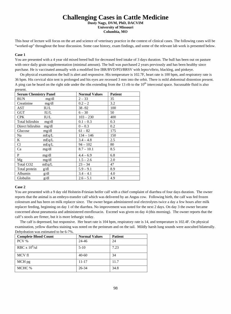
98
Challenging Cases in Cattle Medicine Dusty Nagy, DVM, PhD, DACVIM
University of Missouri Columbia, MO
This hour of lecture will focus on the art and science of veterinary practice in the context of clinical cases. The following cases will be “worked-up” throughout the hour discussion. Some case history, exam findings, and some of the relevant lab work is presented below. Case 1 You are presented with a 4 year old mixed breed bull for decreased feed intake of 3 days duration. The bull has been out on pasture with once daily grain supplementation (minimal amount). The bull was purchased 2 years previously and has been healthy since purchase. He is vaccinated annually with a modified live IBR/BVD/PI3/BRSV with lepto/vibrio, blackleg, and pinkeye.
On physical examination the bull is alert and responsive. His temperature is 102.7F, heart rate is 100 bpm, and respiratory rate is 36 bpm. His cervical skin tent is prolonged and his eyes are recessed 3 mm into the orbit. There is mild abdominal distention present. A ping can be heard on the right side under the ribs extending from the 13 rib to the 10th intercostal space. Sucussable fluid is also present.
Serum Chemistry Panel Normal Values Patient BUN mg/dl 2 – 33 35 Creatinine mg/dl 0.2 – 2 3.2 AST IU/L 38 -92 100 GGT IU/L 6 – 30 50 CPK IU/L 103 – 230 400 Total bilirubin mg/dl 0.1 – 0.3 0.3 Direct bilirubin mg/dl 0 – 0.3 0.2 Glucose mg/dl 61 – 82 175 Na mEq/L 134 – 146 150 K mEq/L 3.4 – 4.8 2.5 Cl mEq/L 94 – 102 80 Ca mg/dl 8.7 – 10.1 8.5
P mg/dl 4.4 – 6.9 6.8 Mg mg/dl 1.5 – 2.6 2.0 Total CO2 mEq/L 23 – 34 47 Total protein g/dl 5.9 – 9.1 8.9 Albumin g/dl 3.4 – 4.1 4.0 Globulin g/dl 2.6 – 5.1 4.9
Case 2 You are presented with a 9 day old Holstein-Friesian heifer calf with a chief complaint of diarrhea of four days duration. The owner reports that the animal is an embryo-transfer calf which was delivered by an Angus cow. Following birth, the calf was fed frozen colostrum and has been on milk replacer since. The owner began administered oral electrolytes twice a day a few hours after milk replacer feeding, beginning on day 1 of the diarrhea. No improvement was noted for the next 2 days. On day 3 the owner became concerned about pneumonia and administered enrofloxacin. Excenel was given on day 4 (this morning). The owner reports that the calf’s stools are firmer, but it is more lethargic today.
The calf is depressed, but responsive. Her heart rate is 104 bpm, respiratory rate is 14, and temperature is 102.4F. On physical examination, yellow diarrhea staining was noted on the perineum and on the tail. Mildly harsh lung sounds were ausculted bilaterally. Dehydration was estimated to be 6-7%.
Complete Blood Count Normal Values Patient PCV % 24-46 24
RBC x 106/ul 5-10 7.23
MCV fl 40-60 34
MCH pg 11-17 11.7
MCHC % 26-34 34.8
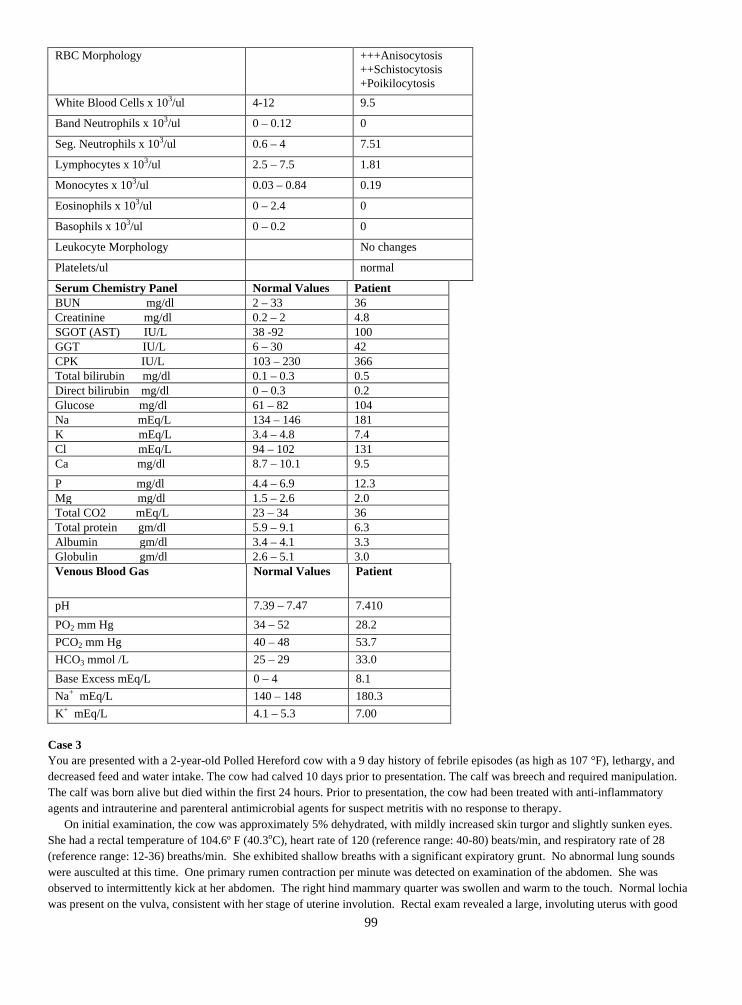
99
RBC Morphology +++Anisocytosis ++Schistocytosis +Poikilocytosis
White Blood Cells x 103/ul 4-12 9.5
Band Neutrophils x 103/ul 0 – 0.12 0
Seg. Neutrophils x 103/ul 0.6 – 4 7.51
Lymphocytes x 103/ul 2.5 – 7.5 1.81
Monocytes x 103/ul 0.03 – 0.84 0.19
Eosinophils x 103/ul 0 – 2.4 0
Basophils x 103/ul 0 – 0.2 0
Leukocyte Morphology No changes
Platelets/ul normal
Serum Chemistry Panel Normal Values Patient BUN mg/dl 2 – 33 36 Creatinine mg/dl 0.2 – 2 4.8 SGOT (AST) IU/L 38 -92 100 GGT IU/L 6 – 30 42 CPK IU/L 103 – 230 366 Total bilirubin mg/dl 0.1 – 0.3 0.5 Direct bilirubin mg/dl 0 – 0.3 0.2 Glucose mg/dl 61 – 82 104 Na mEq/L 134 – 146 181 K mEq/L 3.4 – 4.8 7.4 Cl mEq/L 94 – 102 131 Ca mg/dl 8.7 – 10.1 9.5
P mg/dl 4.4 – 6.9 12.3 Mg mg/dl 1.5 – 2.6 2.0 Total CO2 mEq/L 23 – 34 36 Total protein gm/dl 5.9 – 9.1 6.3 Albumin gm/dl 3.4 – 4.1 3.3 Globulin gm/dl 2.6 – 5.1 3.0 Venous Blood Gas
Normal Values Patient
pH 7.39 – 7.47 7.410 PO2 mm Hg 34 – 52 28.2 PCO2 mm Hg 40 – 48 53.7 HCO3 mmol /L 25 – 29 33.0 Base Excess mEq/L 0 – 4 8.1 Na+ mEq/L 140 – 148 180.3 K+ mEq/L 4.1 – 5.3 7.00
Case 3 You are presented with a 2-year-old Polled Hereford cow with a 9 day history of febrile episodes (as high as 107 °F), lethargy, and decreased feed and water intake. The cow had calved 10 days prior to presentation. The calf was breech and required manipulation. The calf was born alive but died within the first 24 hours. Prior to presentation, the cow had been treated with anti-inflammatory agents and intrauterine and parenteral antimicrobial agents for suspect metritis with no response to therapy.
On initial examination, the cow was approximately 5% dehydrated, with mildly increased skin turgor and slightly sunken eyes. She had a rectal temperature of 104.6º F (40.3oC), heart rate of 120 (reference range: 40-80) beats/min, and respiratory rate of 28 (reference range: 12-36) breaths/min. She exhibited shallow breaths with a significant expiratory grunt. No abnormal lung sounds were ausculted at this time. One primary rumen contraction per minute was detected on examination of the abdomen. She was observed to intermittently kick at her abdomen. The right hind mammary quarter was swollen and warm to the touch. Normal lochia was present on the vulva, consistent with her stage of uterine involution. Rectal exam revealed a large, involuting uterus with good
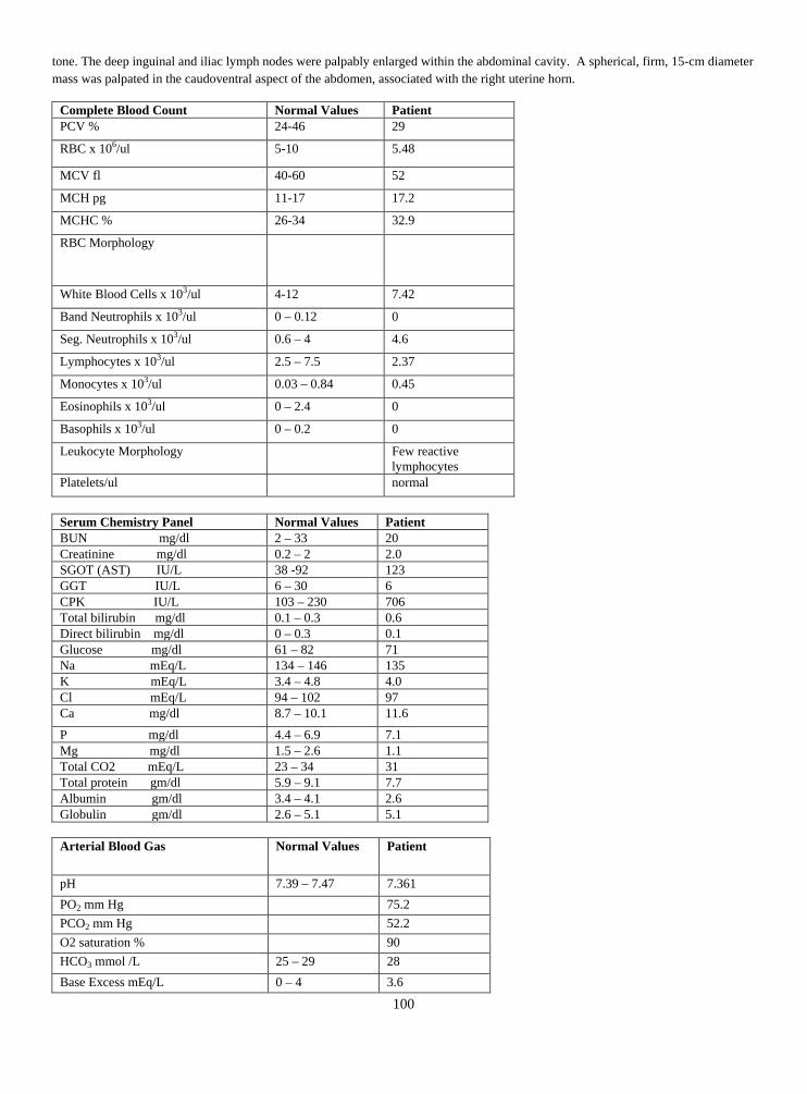
100
tone. The deep inguinal and iliac lymph nodes were palpably enlarged within the abdominal cavity. A spherical, firm, 15-cm diameter mass was palpated in the caudoventral aspect of the abdomen, associated with the right uterine horn.
Complete Blood Count Normal Values Patient PCV % 24-46 29
RBC x 106/ul 5-10 5.48
MCV fl 40-60 52
MCH pg 11-17 17.2
MCHC % 26-34 32.9
RBC Morphology
White Blood Cells x 103/ul 4-12 7.42
Band Neutrophils x 103/ul 0 – 0.12 0
Seg. Neutrophils x 103/ul 0.6 – 4 4.6
Lymphocytes x 103/ul 2.5 – 7.5 2.37
Monocytes x 103/ul 0.03 – 0.84 0.45
Eosinophils x 103/ul 0 – 2.4 0
Basophils x 103/ul 0 – 0.2 0
Leukocyte Morphology Few reactive lymphocytes
Platelets/ul normal
Serum Chemistry Panel Normal Values Patient BUN mg/dl 2 – 33 20 Creatinine mg/dl 0.2 – 2 2.0 SGOT (AST) IU/L 38 -92 123 GGT IU/L 6 – 30 6 CPK IU/L 103 – 230 706 Total bilirubin mg/dl 0.1 – 0.3 0.6 Direct bilirubin mg/dl 0 – 0.3 0.1 Glucose mg/dl 61 – 82 71 Na mEq/L 134 – 146 135 K mEq/L 3.4 – 4.8 4.0 Cl mEq/L 94 – 102 97 Ca mg/dl 8.7 – 10.1 11.6
P mg/dl 4.4 – 6.9 7.1 Mg mg/dl 1.5 – 2.6 1.1 Total CO2 mEq/L 23 – 34 31 Total protein gm/dl 5.9 – 9.1 7.7 Albumin gm/dl 3.4 – 4.1 2.6 Globulin gm/dl 2.6 – 5.1 5.1
Arterial Blood Gas
Normal Values Patient
pH 7.39 – 7.47 7.361 PO2 mm Hg 75.2 PCO2 mm Hg 52.2 O2 saturation % 90 HCO3 mmol /L 25 – 29 28 Base Excess mEq/L 0 – 4 3.6

101
Na+ mEq/L 140 – 148 147 K+ mEq/L 4.1 – 5.3 4.4 iCa 1.95
Case 4 You are presented with a 4-year-old Red Angus bull for presumptive tooth root abscess. The bull has been previously treated for 3 weeks with antibiotics based on the results of a culture and sensitivity with no response to therapy.
On initial physical examination the bull’s vitals were within normal limits. There was a large swelling on the left maxilla centered over the last premolar/1st few molars. The mass was firm with superficial soft areas. Oral examination revealed that the mass extended within the mouth and there were no obviously loose teeth. Suggested reading Russell KE and Roussel AJ. Evaluation of the ruminant serum chemistry profile. Vet Clin Food Anim 2007; 23(3):403-426. Jones ML and Allison RW. Evaluation of the ruminant complete blood cell count. Vet Clin Food Anim 2007; 23(3):377-402.
102
Selected Viral Diseases of Cattle (BLV, Bluetongue, EHD)
Dusty Nagy, DVM, PhD, DACVIM University of Missouri
Columbia, MO
A variety of viral diseases affect cattle herds across the United States. Bovine leucosis virus is a retroviral disease that is prevalent within many dairy and beef herds in the country and is often blamed for a variety of production limiting and life ending diseases in cattle. Bluetongue and epizootic hemorrhagic disease are both orbiviral diseases of cattle and other ruminants that occasionally appear and cause some clinical illness as well as malformed or stillborn calves. Bovine leucosis virus Bovine leukosis virus (BLV) is an oncogenic retrovirus of cattle. The transfer of lymphocytes from infected animals to naïve cattle spreads the virus. Many common practices on farms including dehorning, eartagging, and tattooing have been implicated as sources of spread of the virus. Alternatively, the virus may also be transmitted by insects, natural service, transplacental transmission from the infected dam to the fetus, and through the ingestion of infected colostrum. It is difficult to determine the prevalence of infection in the United States, as whole herd testing is not required by regulatory authorities. There is also significant regional variation in disease prevalence. In 1989 it was estimated that 86% of United States dairy herds contained infected animals, with 40% of all cattle being infected. In infected herds the prevalence of disease ranges from 20-100%.
Potential outcomes of infection with BLV are variable. Most infected animals will remain apparently healthy. These animals will be sources of virus for noninfected animals, but will remain free of clinical signs. A subset will develop persistent lymphocytosis (PL), a benign lymphoproliferative condition associated with BLV infection. The estimated prevalence of PL in BLV infected cattle is 29%. However, PL has been shown to be more prevalent in cattle of certain lineage. PL rates of up to 88% have been documented within these familial groups. The development of PL does not appear to correlate with disease outcome. However, cows with PL are important reservoirs for BLV transmission because their blood contains higher concentrations of infected lymphocytes. Consequently, blood from PL cows is likely to be more infectious than blood from PL-, BLV+ cows. The last potential outcome is the development of lymphosarcoma. Only 1-5% of animals infected with BLV will develop lymphosarcoma. However, this is the outcome that generates significant financial loss associated with BLV infection.
BLV associated lymphosarcoma is one of the great imitators of bovine disease. Clinical signs associated with the development of lymphosarcoma are highly variable, as the affected organ will dictate the predominant clinical signs. Animals with BLV associated lymphosarcoma will commonly show lesions in the central or peripheral lymph nodes leading to lymphadenopathy. This rarely adversely affects the animal unless the lymph nodes impinge on vital structures. Lesions of the abomasum will lead to signs of cranial abdominal pain and potentially melena, or abomasal outflow obstruction. Pelvic limb paresis progressing to paralysis can occur in animals with extradural lesions. Retrobulbar lesions will cause protrusion of the globe resulting in exposure keratitis and eventually proptosis. Lesions of the right atrium may be mild and undetectable clinically, or may present with asymptomatic or symptomatic arrhythmias or murmurs. These lesions may eventually result in right heart failure. Lesions of the uterus may present as cases of reproductive failure. Lastly, lesions of the internal organs typically will involve the spleen, liver, or kidneys and ureters. Lesions of the spleen are often initially asymptomatic, but may result in rupture of the spleen and exsanguination into the peritoneal cavity. Lymphosarcoma of the liver also is often asymptomatic. Disease of the kidney and ureter can lead to abdominal pain and the subsequent development of hydroureter or hydronephrosis and signs associated with kidney failure.
Interest and effort in BLV research has surged in recent years. Clear evidence is emerging to support decreased longevity and production of infected animals in dairy herds. In addition newer molecular diagnostics have provided a better glimpse into the detrimental effects of animals with high proviral load on proviral movement on a farm. These new insights once again fuel the fire of stricter control strategies on farms to promote overall cattle health.
Current control strategies for heavily infected farms include the use of sterile needles on each cow, clean and disinfect blood contaminated equipment between animals, new rectal sleeves for each cow, AI instead of bull breeding, fly control, separation of youngstock from heavily infected cattle herds, feed pasteurized or heat treated colostrum, segregation of positive and negative animals, cull cattle with lymphocytosis. Bluetongue virus Bluetongue is an Orbivirus within the family Reoviridae that is transmitted by the Culicoides biting midge. There are at least 26 serotypes and bluetongue has an almost worldwide distribution. Many species are susceptible to infection, but the severity of disease is variable among species. Bluetongue typically presents as a reproductive syndrome or as a clinical vasculitic disease that involves multiple organ systems. Vasculitic disease in cattle is less common than the reproductive syndromes. The vasculitic disease is characterized by excessive salivation and nasal discharge. Hyperemia of the muzzle with hemorrhages, erosions, and ulcers may
103
occur. Coronitis leading to lameness and possible sloughing of feet may also occur. Reproductive syndromes include abortions, stillbirths, and weak born calves. The virus may be teratogenic and malformed calves (arthrogryposis, hydranencephaly) may also be found.
Diagnosis of bluetongue is hampered by cross reactivity of the serologic tests with nonpathogenic Orbiviruses. In addition antibody levels may remain high for years after infection. Virus isolation from blood during the viremic stage is the most definitive diagnostic. In aborted or dead calves isolation of virus from the spleen is most diagnostic. However, PCR based diagnostics are being used with increasing frequency. The PCR may detect viral RNA for an extended period of time after infection so it may not always accurately predict infection. In addition, in cases of reproductive syndromes, the inciting incident is often long past, making diagnosis difficult.
Treatment for clinical bluetongue is primarily supportive focusing on provision of soft palatable feeds, inflammation and pain control. Prevention secondary problems such as pneumonia or downer scow syndromes must also be considered for more severely affected animals.
Prevention is disfficult and focused on vector control and limiting vector contact. Culicoides are most active in the evening. If possible housing indoors during these times may be somewhat protective. Obviously this has more utility in small ruminants than large. Breeding grounds for Culicoides include wet areas with lots of organic matter. Avoiding such areas in the evening and limiting access to the heat of the day may help decrease transmission. Larvacidal treatments may also decrease numbers.
Epizootic hemorrhagic disease Epizootic hemorrhagic disease is caused by an Orbivirus closely related to Bluetongue virus. There are at least 10 serotypes currently identified and distribution, like bluetongue, is expanding. Similar to Bluetongue this virus is transmitted by Culicoides. Deer and cattle are most commonly affected with clinical disease. Morbidity and mortality in deer may approach 90% and 60% respectively. Similar to Bluetongue prevalence studies have shown that seroconversion is common but clinical disease is rarely seen.
Diagnosis is made using serology or PCR. The AGID will cross react with Bluetongue. As such it is not considered to be an adequate test. The cELISA is sensitive and specific for detection of this disease. Clinical signs, treatment, and prevention strategies mirror that for Bluetongue virus. Recommended reading Caporale M, DiGiallaeonorado L, Janowicz A, et al. Virus and host factors affecting clinical outcome of Bluetongue Virus infection. J Virol 2014;88:18, 10399-10411. Savini G, Afonso A, Mellor P, et al. Epizootic hemorrhagic disease. Res Vet Sci 2011;91;1-17 Bartlett PC, Sordillo LM, Byrem TM. Et al. Options for the control of bovine leukemia virus in dairy cattle. J Am Vet Med Assoc 2014;244:8, 914-922.
104
Neurologic Disease in Food Animal Dusty Nagy, DVM, PhD, DACVIM
University of Missouri Columbia, MO
Neurology in a clinical setting is frustrating for many veterinarians. Most of this frustration stems from adopting an overly complex diagnostic approach. Rather than emphasizing clinical signs and disease manifestations, the traditional approach emphasizes the memorization of anatomical pathways. Excessive numbers of anatomical pathways often leads to frustration with developing an appropriate anatomic diagnosis and list of differential diagnoses. The result is often that no diagnosis is made and empirical treatment regimes are instituted. This limits the potential for intervention in herd problems and improvement of management practices on the farm. Decreasing the number of anatomical pathways to 4 major compartments (cortex, cerebellum, brainstem, spinal cord) of the nervous system simplifies lesion localization while allowing adequate definition to develop a reasonable list of differential diagnoses.
Physical examination is the cornerstone of the diagnosis of neurologic disease. Cattle with neurologic disease can be dangerous to themselves and others. However, failure to adequately examine the patient is a direct path to empirical treatment. Examination at a distance followed by a cranial nerve examination, postural reactions, spinal reflexes, and palpation can provide the information necessary to determine a gross anatomical diagnosis.
The following is a simple, diagnostic approach to neurologic diseases of cattle with less stress and frustration to the veterinarian. This approach allows the practitioner more latitude in anatomic diagnosis and focuses more on clinical signs and disease manifestations.
For this lecture we will review the process and focus most attention to video for case assessment. Step 1 – Gross anatomic localization. Many clinical signs of neurologic disease are nonspecific for lesion localization. Avoid getting hung up on signs that don’t localize to a single compartment. The physical examination should be focused on identifying clinical signs that aid in lesion localization. Table 1 – Neurologic signs by anatomic location
Anatomic site Highly specific clinical signs
Cortex
depression, coma, or other changes in mentation, head pressing, propulsive walking, convulsions, opisthotonus
Cerebellum head tremor, abnormal gait – ataxia with normal strength and proprioception, truncal sway, hypermetria
Brainstem/Cranial nerves head tilt, anisocoria, mydriasis, miosis, ptosis, strabismus, nystagmus, flaccid tongue, facial paralysis, assorted cranial nerve deficits, circling
Spinal cord dysuria, flaccid tail and/or anus, loss of spinal reflexes, paresis, paralysis Step 2 – Determine the broad category of disease etiology During the course of the diagnostic work-up, it is often useful to determine the etiology of the disease process. This will often aid in narrowing the list of possible differential diagnoses and provide some direction in the ultimate therapeutics of the case. The most useful test for providing this information in neurologic disease is the cerebrospinal fluid analysis. Table 2 – Anticipated cerebrospinal fluid analysis results by disease status
Disease Protein g/L Cells/µL Cells-type Normal <0.4 <10 lymphocytes Viral 0.40-1.0 50-200 monocytes
lymphocytes Bacterial >1.0 >200 neutrophils Degenerative <0.4 <10 monocytes
Step 3 – Define the problem This step is simply a combination of steps 1 and 2. Problem definition is often skipped, but clear definition of the problem may limit frustration. At this point you have a problem such as bacterial cortical disease or viral brain stem disease. Step 4 – Build a list of differential diagnoses At this point a list of differential diagnoses that fit the defined problem should be generated. Nuances in the physical examination and information previously collected from the owner such as signalment, diet, and other historical information should be used to add, subtract and prioritize this list.
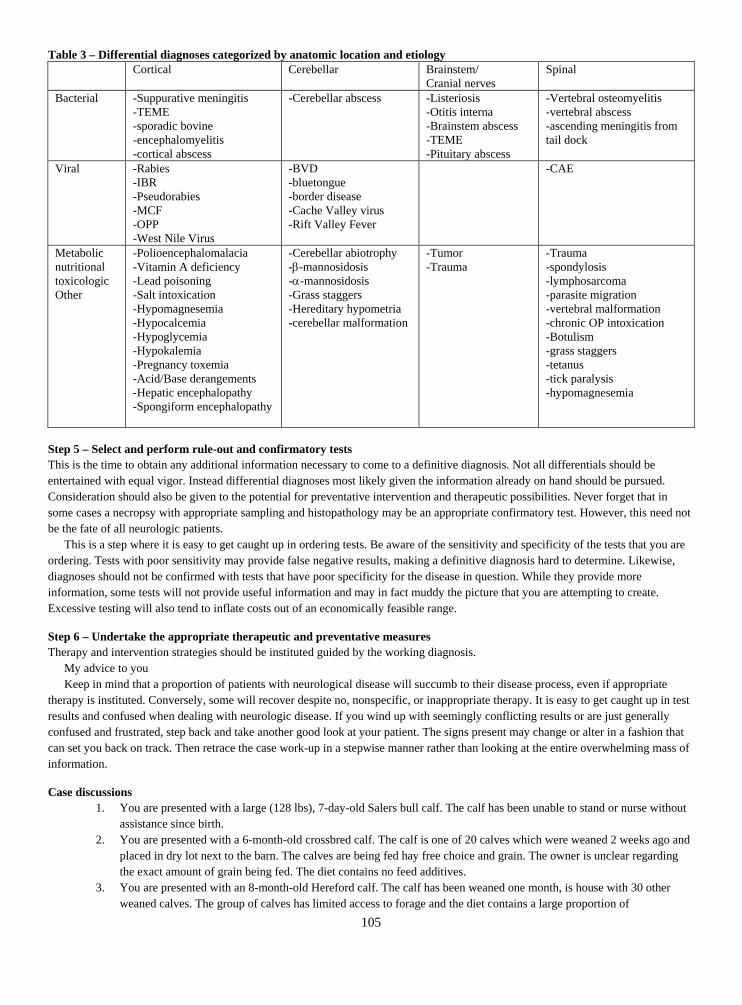
105
Table 3 – Differential diagnoses categorized by anatomic location and etiology Cortical Cerebellar Brainstem/
Cranial nerves Spinal
Bacterial -Suppurative meningitis -TEME -sporadic bovine -encephalomyelitis -cortical abscess
-Cerebellar abscess -Listeriosis -Otitis interna -Brainstem abscess -TEME -Pituitary abscess
-Vertebral osteomyelitis -vertebral abscess -ascending meningitis from tail dock
Viral -Rabies -IBR -Pseudorabies -MCF -OPP -West Nile Virus
-BVD -bluetongue -border disease -Cache Valley virus -Rift Valley Fever
-CAE
Metabolic nutritional toxicologic Other
-Polioencephalomalacia -Vitamin A deficiency -Lead poisoning -Salt intoxication -Hypomagnesemia -Hypocalcemia -Hypoglycemia -Hypokalemia -Pregnancy toxemia -Acid/Base derangements -Hepatic encephalopathy -Spongiform encephalopathy
-Cerebellar abiotrophy -β-mannosidosis -α-mannosidosis -Grass staggers -Hereditary hypometria -cerebellar malformation
-Tumor -Trauma
-Trauma -spondylosis -lymphosarcoma -parasite migration -vertebral malformation -chronic OP intoxication -Botulism -grass staggers -tetanus -tick paralysis -hypomagnesemia
Step 5 – Select and perform rule-out and confirmatory tests This is the time to obtain any additional information necessary to come to a definitive diagnosis. Not all differentials should be entertained with equal vigor. Instead differential diagnoses most likely given the information already on hand should be pursued. Consideration should also be given to the potential for preventative intervention and therapeutic possibilities. Never forget that in some cases a necropsy with appropriate sampling and histopathology may be an appropriate confirmatory test. However, this need not be the fate of all neurologic patients.
This is a step where it is easy to get caught up in ordering tests. Be aware of the sensitivity and specificity of the tests that you are ordering. Tests with poor sensitivity may provide false negative results, making a definitive diagnosis hard to determine. Likewise, diagnoses should not be confirmed with tests that have poor specificity for the disease in question. While they provide more information, some tests will not provide useful information and may in fact muddy the picture that you are attempting to create. Excessive testing will also tend to inflate costs out of an economically feasible range. Step 6 – Undertake the appropriate therapeutic and preventative measures Therapy and intervention strategies should be instituted guided by the working diagnosis.
My advice to you Keep in mind that a proportion of patients with neurological disease will succumb to their disease process, even if appropriate
therapy is instituted. Conversely, some will recover despite no, nonspecific, or inappropriate therapy. It is easy to get caught up in test results and confused when dealing with neurologic disease. If you wind up with seemingly conflicting results or are just generally confused and frustrated, step back and take another good look at your patient. The signs present may change or alter in a fashion that can set you back on track. Then retrace the case work-up in a stepwise manner rather than looking at the entire overwhelming mass of information. Case discussions
1. You are presented with a large (128 lbs), 7-day-old Salers bull calf. The calf has been unable to stand or nurse without assistance since birth.
2. You are presented with a 6-month-old crossbred calf. The calf is one of 20 calves which were weaned 2 weeks ago and placed in dry lot next to the barn. The calves are being fed hay free choice and grain. The owner is unclear regarding the exact amount of grain being fed. The diet contains no feed additives.
3. You are presented with an 8-month-old Hereford calf. The calf has been weaned one month, is house with 30 other weaned calves. The group of calves has limited access to forage and the diet contains a large proportion of

106
concentrates. The calf was found in the corner of storage shed after it apparently escaped from a pasture overnight. None of the other calves show any abnormalities. The owner reports that this calf has always seemed stunted.
4. You are presented with a 5 year old Simmental bull for the complaint of ataxia. Clinical signs were first observed 1 week previously. The bull has been treated with antibiotics and nonsteroidal anti-inflammatory agents with no resolution of clinical signs.
References/suggested reading Constable PD. Clinical examination of the ruminant nervous system. Vet Clin Food Anim 2004; 20:185-214. George LW. Diseases of the nervous system. In: Smith BP (ed.). Large animal internal medicine. 4th edition. St. Louis, Mosby 1996, Pg. 1001-1175. Welles EG, Tyler JW, Sorjonen DC, Whatley EM. Composition and analysis of cerebrospinal fluid in clinically normal adult cattle. Am J Vet Res 1992; 53:2050-2057
107
The Use of Radiography and Ultrasonography in Bovine Practice Dusty Nagy, DVM, PhD, DACVIM
University of Missouri Columbia, MO
Diagnostic imaging is a commonly used tool in both small animal and equine medicine to aid in diagnosis of veterinary patients. Both radiography and ultrasound are used in cattle practice, but the scope of the use is limited compared to our counterparts in other areas of veterinary medicine. Like any other tool the old adage “garbage in-garbage out” applies in diagnostic imaging. Appropriate patient selection, modality selection, as well as proper image capture and identification are all critical to successful outcomes using both radiography and ultrasound in practice. Radiography Training on image capture and evaluation using radiography is widespread in veterinary colleges. However, the nuances of the bovine patient are not always focused upon. Most equipment that can be accessed in the field and non-large animal specialty practices has its best utility on youngstock and extremities from the hock distally.
In young calves the lungs, limbs, and portions of the skull can be adequately imaged and interpreted. However, as they grow the capacity of many machines will not have the ability to image the thorax.
In the adult bovine radiography is best used to evaluate bony structures of the distal limb. The mandible can also be imaged in some finer boned adult cattle. Fractures, luxations, metabolic bone disease, infectious diseases of the bones and joints are readily evaluated in bovine patients. In most cases images can be acquired in animals that are standing and well restrained to limit motion. If necessary animals can be imaged in a tilt chute or in lateral recumbency with sedation. Standing radiographs are preferred because they will allow for evaluation of joint space while images obtained in the recumbent animal will not. Similar to our other species standard anterio-palmar and lateral images should be acquired as well as both dorsomedial palmerolateral and dorsolateral palmeromedial obliques when imaging joints to allow for appropriate interpretation.
Hock and carpus – 4 views are required for adequate interpretation. Flexed and skyline views may be helpful in isolated cases. Metatarsus and metacarpus – 2 view are typically adequate Digit – 4 views of the digit are required for adequate interpretation. Placing a spacer between the toes (roll of brown gauze,
cardboard core of vetwrap) will decrease the overlay and highlight the toes for better image quality.
Ultrasonography The use of ultrasonography in cattle practice has historically been limited to pregnancy diagnosis. The ultrasound is uniquely capable of giving an image of tissue architecture in the awake animal. It is important to recognize the limitations with ultrasound and size of the mature bovine is a significant one. Despite this good quality images up to 20-25 cm in depth can be acquired. In recent years descriptions of appropriate image capture and normal and abnormal findings have been described for a variety of organs in the bovine. Using ultrasound is an acquired skill and takes practice to incorporate it usefully into clinical practice.
Lung – Best done with a 3.5 – 7.5MHz linear or sector probe in the intercostal spaces. The lung borders of the lung field can be outlines from the top of the 11th rib. Middle of the 9th, and point of the elbow. Ultrasound is best used to diagnose superficial lesions unless significant consolidation is present. Thoracic wall and pleural lesions are easily visualized as are superficial abscesses, masses, and pulmonary consolidation.
Heart – Best done with a 2.5 – 3.5MHz probe in adults or a 3.75 – 5 MHz probe in calves. The position of the heart under the thoracic limb and narrow rib spaces at this location may make imaging difficult especially in adults. The heart can be found in the 3rd – 5th intercostal space (ICS) under the elbow. The standard 4 chambered view can be obtained in the 4th ICS on the right in the parasternal region. On the left the caudal long axis view can be obtained in the 4th or 5th ICS just dorsal to the olecranon with the probe angled slightly caudally. Evaluation of the heart is the most difficult of all ultrasounds to gain proficiency. However, evaluation for pericarditis is relatively easy even for novice ultrasonographers.
Liver – This is best accomplished using a 3.5 – 5.0 MHz linear or convex transducer. The liver is located on the right side from caudal to the last rib cranial to the 5th ICS.
Gastrointesinal tract - This is best accomplished using a 3.5 – 5.0 MHz linear or convex transducer. • Abomasum – 10 cm caudal to the xiphoid on the ventral body more on the right but spans both sides. Some of the folds
may be visible within the abomasal content. • Reticulum – imaged on the ventral thorax lateral to the xiphoid on both sides extend up to the level of the elbow. • Omasum – ICS 6-11 on the right. No inherent motility and laminae are not visible in the normal animal. • Small intestine – entire right side from the tuber coxae to the 8 ICS from the transverse processes ventrally to the fold of
the flank. The wall should be 2-3 mm thick with a lumen of 2-4 cm.

108
Spleen – This is best accomplished using a 3.5 – 5.0 MHz linear or convex transducer. The spleen can be found in the ICS 7-12 on the left craniodorsal to the rumen.
Urinary system – The left kidney, urethra, ureter, and urinary bladder can be imaged transrectally using a 5.0 MHz linear transducer. In thin cows or smaller calves the left kidney can be imaged in the right caudodorsal paralumbar fossa. However, gas from the colon often overlies the area and limits image quality. The right kidney kidney can be imaged percutaneously in the right paralumbar fossa in the last ICS using a 5 MHz or lower transducer depending on required depth.
Mammary gland and teats – Ultrasonography of the teat has become commonplace while that of the mammary gland is not quite as commonly described. The use of 3.5 and 5.0 MHz transducers has been described with the linear transducers providing superior image quality. While these have been described image quality will improve dramatically with higher frequency transducers. A 7.5 – 10 MHz transducer would be considered ideal. Imaging of the teat is most often perused when milk flow is diminished or absent. A standoff can be used to improve imaging of the teat canal.
Umbilicus – This is best accomplished with a 3.5 – 5.0 MHz linear or convex transducer. Ultrasound is an excellent technique to image the umbilicus of neonatal calves. It is possible to determine the presence and extent of internal remnant swelling in animals with omphalitis.
Tendons – The distal tendons of the limbs can also be imaged by ultrasound using a minimum 5.0 MHz linear transducer. A 7.5 – 10 MHz transducer would be considered ideal.
109
Implications of Having a Reportable Disease in Your Hospital Dusty Nagy, DVM, PhD, DACVIM
University of Missouri Columbia, MO
There are a variety of reportable diseases in the United States on both the state and the national level. In production animal medicine these diagnoses are often made on farm and may lead to a temporary quarantine of the premises until confirmatory testing can be completed. This quarantine typically results in a cessation of animal movement and a limitation to farm travel for the in contact family and workers. As the veterinarian, we recognize the inconvenience to the client but understand the importance in the protection of animal and public health.
Now envision the farm at which the diagnosis is made is your hospital and the in contact “family” are your associates and staff. This now turns from a minor inconvenience, to a catastrophe of epic proportions. Once you are grounded (7-10 days maybe more) that leaves appointments unseen and clients untended to.
This hour we will focus on people and animal movement throughout the hospital and ways to minimize the potential transfer and exposure of disease.
Personnel Personnel fall into 2 main categories - animal caregivers and non-caregivers. The movement of non-caregivers should be extremely limited (nonexistant) in an animal facility to prevent the unnecessary exposure of a staff member. Animal caregivers should practice appropriate barrier nursing and infection control when dealing with all patients. Animals in isolation should have contact with only people critical to the care of the animal. Personnel flow Many facilities offer both in house and ambulatory services. Occasionally accessing ambulatory vehicles requires/allows for travel through the hospital as one leaves or returns. This again is a point where pathogens can be exchanged and moved. Animal location True isolation stalls are not common in many practice settings. Often, hospitals that have them available, fail to use them on a routine basis due to the extra time and labor associated with the protocols of animal handling in that segment of the facility. Animal flow Animal flow thorough the hospital often puts animals in contact with animals from other farms. This poses a point where pathogens can be exchanged and moved. Barrier nursing Clean coveralls unique to the patient, disinfection of boots between patients, and the use of latex gloves (changed in between patients) are the minimum requirements for preventing the movement of microorganisms between patients in the facility. Infection control Disinfectant mats (charged with disinfectant and uncontaminated by organic debris) will help minimize the transmission of microorganisms between patients. Damage control in absence of dedicated isolation facility In the advent that an animal with a potentially infectious disease enters the facility, recognition of the potential and partitioning the animal to a naturally low traffic area may prevent unnecessary exposures of personnel and contamination of equipment. Other random logistics In addition to the clear issues of biosecurity many other logistical problems may exist that are often overlooked. Many personnel have animals at home that may pose an at risk population. Animals actively hospitalized in the facility may become part of a quarantine when traffic in and out of the facility is prohibited. This can cause significant disruptions in the business including but not limited to deliveries, shipping of samples out of the practice, and disruption of service to clientele.
Part of Veterinary Accreditation is understanding of and mandatory reporting of potential diseases of concern. A variety of mandatory reporting exists for veterinarians within this program. The following should all be reported:
• All foreign animal diseases must be reported to Federal and State Animal Health Officials (SAHO) • USDA Program Diseases must immediately be reported to Federal and State Animal Health Officials • States may have additional diseases of interest that they monitor, and thus are reportable at the state level
110
• Zoonotic diseases may need to be reported to the State Health or Public Health department as well as the Centers for Disease Control and Prevention (CDC)
• Bioterrorism disease agents may be reportable to Federal and/or State Animal Health Officials Each state has an independent list of reportable diseases. There is currently a proposal for the development of a national list. Below
is the list of diseases for the state of Missouri: • Anthrax • Bluetongue • Bovine babesiosis (Texas fever, piroplasmosis) • Bovine spongiform encephalopathy (BSE) • Brucellosis • Contagious bovine pleuropneumonia • Foot-and-mouth disease • Heartwater • Pseudorabies • Rift valley fever • Rinderpest (cattle plague) • Screwworm • Tuberculosis • Trichomoniasis • Vesicular stomatitis
In addition to state reporting lists the World Organization for Animal Health maintains a list of reportable diseases from nations across the world. Each year the United Stated puts together a cumulative report detailing the presence and extent of disease prevalence for each disease on the list. Recommended resources United States Department of Agriculture. www.aphis.usda.gov Missouri Department of Agriculture
111
Anaplasmosis in Cattle Dusty Nagy, DVM, PhD, DACVIM
University of Missouri Columbia, MO
Anaplasmosis is an infectious Rickettsial disease of cattle caused by Anaplasma marginale. The organisms infects the red blood cells of cattle and produces a round structure that can be seen at the periphery of infected erythrocytes. The organism can be transmitted by ticks or by blood transfer between animals. All ages of animals can be infected, but the severity of clinical signs increase with age with animals over 3 years of age showing more severe clinical signs. Phases of disease Anaplasmosis goes through 4 distinct phases in the cow including incubation, developmental, convalescent, and carrier. Once infected, animals will incubate the organism without showing clinical signs for approximately 4-8 weeks. Once approximately 1% of the animal’s erythrocytes are infected the animal enters the developmental stage and will begin to show early clinical signs of infection. Early signs are often indistinct. Fever, weakness, and anorexia, along with a drop in production often predominate at this stage.
Approximately 15% of the erythrocytes must be parasitized to show clinical signs associated with anemia. Anemia occurs in cases of anaplasmosis due to the removal of infected erythrocytes by the immune system. The majority of the clinical signs in the cow can be attributed to anemia caused by the extravascular hemolysis. These signs include pale mucus membranes, tachypnea, and tachycardia. As an extravascular hemolysis hemoglobinuria and hemoglobinemia are absent in cases of bovine anaplasmosis. As the disease progresses and animals begin to process heme pigments pale membranes may become jaundiced. Mania, likely induced by cerebral hypoxia is also commonly associated with anaplasmosis in cattle. If left untreated, death may result.
The animal enters the convalescent stage once regenerative changes appear in the blood. And continues until the blood smear appears normal. It is still possible to have animals showing severe clinical signs early in the convalescent stage. In addition some animals may have a phase of chronic unthriftyness as they recover from a clinical anaplasmosis episode.
The carrier stage begins once organisms can no longer be found in the peripheral blood. This phase likely extends for the life of the animal. Carrier animals may be clinically normal with low levels of parasitemia which precludes diagnosis via blood smear. These animals remain infectious to their herdmates and are a lifelong source of the organism.
Diagnosis Diagnosis of anaplasmosis can be made using several different testing methodologies. It is important to recognize that no one test will detect all of the potential stages of disease.
Blood Smears have utility in animals during the developmental phase of the infection. Animals with clinical signs consistent with anaplasmosis that have evidence of organisms on a blood smear are considered to be clinically affected. Early in disease (incubation) smears may be negative because a critical load of erythrocytes may not have been reached allowing them to be detected on blood smear. Similarly animals in the carrier state will typically not have enough infected erythrocyted to be detectable by this method. During the convalescent phase organisms may be detectable early. Signs consistent with regenerative anemia (anisocytosis and basophilic stippling) will be present during this phase and may be suggestive of anaplasmosis, but are not considered diagnostic.
Serologic tests (cELISA) will be positive in animals during the developmental and convalescent stages of infection. It will also be positive in many animals in the carrier state of disease. As such these tests will determine exposure of the animal to the organism, but alone it can not adequately separate active infections from carrier animals. A positive serologic test in the presence of clinical signs is suggestive of active infection. The major limitations to the cELISA are detection of early infection prior to the animal mounting an adequate immune response, prolonged recognition of the immune response in animals that have been chemosterilized, and cross reactivity with other Anaplasma species.
Polymerase chain reaction based diagnostics have the ability to pick up very small amounts of Rickettsial DNA and have proven helpful in identifying carrier animals. Some assays have been reported to detect as few as 30 infected cells per milliliter of blood. These assays lack formal validation and results may vary depending on laboratory protocol.
Treatment Treatment is centered on antibiotic therapy and supportive care. Many treatment protocols have been published, but most center on oxytetracyline therapy. Most animals are suffering from significant anemia by the time they are presented for treatment. Minimizing stress and exertion are important to prevent decompensation from the anemia. Oxytetracycline 200 mg/ml can be administered at 9 mg/lb SQ every 72 hours. Blood transfusion may be indicated in some patients with severe life threatening anemia.
112
When an animal is diagnosed or in the case of a herd outbreak do not forget that other animals are at risk. At risk animals can also be treated with injectable oxytetracycline, oral chlortetracycline at a rate of 0.5mg/lb BW, or combinations of both injectable and oral oxytetracycline. Prevention and control Vector control – Tick and fly control is imperative in preventing movement of anaplasmosis through a herd. Insecticide impregnated ear tags, fly blocks, sprays, back scratchers, ect can be used to decrease the potential for vector transmission. It is important that these methods be reapplied, replaced, or recharged intermittently throughout the vector season for them to remain effective.
Medicated feed and mineral supplements, pulse dosing antibiotics – these methods can be used to prevent clinical disease in endemic herds. It is important to recognize that animals must eat the feed or use the mineral for them to be effective.
Vaccine – In some states a killed vaccine is available for use. It I currently sold as an experimental vaccine and is not licensed by the USDA. However, the USDA has approved the sale and use of the vaccine in multiple states including Missouri, Kansas, and Iowa.
Eliminate carriers – many treatment regimens to eliminate carriers have been suggested. Most of these have failed to hold up in recent evaluations using more stringent test methodology. Several new protocols have shown promise, but have yet to stand up to rigorous reevaluation. Chortetracycline fed at 4.4; 11; and 22mg/kg/day for 80 days were all effective at eliminating the carrier state in tested animals. An additional study reported success treating with a single SQ injection of oxytetracycline followed by 30 days of chlortetracycline fed at 4.4 mg/kg/day. An additional study showed failure of the 4.4 mg/kg/day chlortetracycline to chemosteralize animals when fed for only 45 days.
Instruments – Blood contaminated instruments including needles, dehorners, tattooing equipment, ear taggers, and surgical equipment are effective at moving anaplasmosis from infected to naïve animals.
Animal movement – It is important to recognize that in some areas of the country and within some herds anaplasmosis is endemic. Care should be taken when moving animals from endemic areas to non-endemic herds. Introduction of a carrier animal to a naïve herd may result in significant numbers of clinically ill cattle since the herd has no background immunity. Conversely, moving a naïve animal to an endemic herd may result in clinical disease in the new introduction.
Recommended reading and resources anaplasmosisvaccine.com Coetzee, J. F., P. L. Schmidt, M. D. Apley, J. B. Reinbold, and K. M. Kocan, 2007: Comparison of the complement fixation test and competitive ELISA for serodiagnosis of Anaplasma marginale infection in experimentally infected steers. Am. J. Vet. Res. 68, 872–878. Reinbold, J. B., J. Coetzee, and R. Ganta, 2009a: Comparison of three tetracycline antibiotic treatment regimens for carrier clearance of persistent Anaplasma marginale infection derived under field conditions. Proceedings of the 42nd Annual Conference of the American Association of Bovine Practitioners (AABP), Omaha, NE Reinbold, J. B., J. F. Coetzee, L. C. Hollis, J. S. Nickell, C. Riegel, K. C. Olson, and R. R. Ganta, 2010b: The efficacy of three chlortetracycline regimens in the treatment of persistent Anaplasma marginale infection. Vet. Microbiol. 145,69–75. Aubry P, Geale DW. A review of bovine anaplasmosis. Transboundary and Emerging Diseases. 2010;58;1-30
113
It All Starts with the Diagnosis: Physical Examination (Parts 1 and 2)
Allen Roussel, DVM, MS, DACVIM Texas A&M University
College Station, TX
I fear that the physical examination is becoming a lost art. Perhaps every generation of veterinarians has had the same feeling as they see more and more technology enhancing our ability to reach a diagnosis, but at the same time replacing some of the time-tested techniques of the physical examination. I am certainly not against technological advances-most of us at academic institutions are drawn there because of the advanced diagnostic equipment available to us. I am, however, chagrined by the growing dependence upon imaging, laboratory evaluation, and other sophisticated techniques to make a diagnosis when often a physical examination and a very simple confirmatory test would reach the same conclusion in less time and for less cost. My goal in this presentation is to review the techniques of physical examination both for the part-time bovine veterinarian as well as the experienced bovine veterinarian. There is no question that the combination of excellent physical examination and rational use of sophisticated diagnostic equipment will achieve the optimal results.
We often hear the term “complete physical examination,” but how often do we perform one? The truth of the matter is that we do not need to perform a complete physical examination on every patient, nor do we have the time. We routinely perform what might be called a “standard physical examination” which includes a brief review of all important body systems. Based on the history and the results of the standard physical examination, we then perform one or more focused physical examinations. If we performed every one of the focused physical examinations that we knew, we would then perform a “complete physical examination.” But let’s not argue over semantics. Let’s try to learn how to efficiently evaluate an animal by use of the standard physical examination and how to focus on particular areas to gain the most information possible from a physical examination.
There are many ways to approach a physical examination; many correct ways. The approach that I will use in this paper is to begin with observation at a distance and then examination of the restrained animal. I’ll then discuss the acquisition of vital signs and basic auscultation, concluding with regional focused examinations beginning at the head. Because neurological examination is frequently difficult and confusing, I’ll spend a bit more time on that aspect.
The exam at a distance I believe that physical examination of cattle should begin with observation of the animal from a distance. This is particularly important when one suspects neurological or musculoskeletal disease. The animal should be observed at rest for several minutes and then in motion. Note the general condition of the animal and the breed, as some neurological diseases are heritable. When the animal is at rest, pay particular attention to the animal’s awareness of its surroundings which reflects cerebral function. Note if the animal is depressed, hyper-excited, or otherwise responsive to external stimuli, if it is head pressing, wandering aimlessly, vocalizing abnormally, behaving abnormally or aggressively. Diseases such as polioencephalomalacia, lead poisoning, nervous ketosis, bovine spongiform encephalopathy, rabies, brain or pituitary abscess, nervous coccidiosis, and salt poisoning/water deprivation cause these signs. Before the animal is disturbed, observe the character and rate of respiration, look for a jugular pulse (indicative of right heart failure), and for signs of abdominal pain like bruxism, restlessness, kicking at the belly, or straining. Also look carefully for muscle fasciculation, twitching of the ears or eyelids, tail position and switching and abnormal attempts at swallowing which may indicate nervous system or metabolic disease such as hypomagnesemia, lead toxicity, tetanus, or rabies. Lameness is often detectable in cattle at rest by observing how the animal bears or shifts weight on the limbs. An easy way to assess weight bearing is to observe how far the dewclaws are from the ground. If the dewclaws are higher on one side, the animal is not bearing full weight on that side. Abdominal contour should also be assessed at a distance and from behind the animal. While the animal is in the open and not confined in a chute, careful attention should be paid to the muscle mass, particularly over the rump and hindquarters. In unilateral neurological disease, as well as chronic upper limb lameness, atrophy of the muscles will occur, and asymmetry of the muscles will be obvious.
If the animal is recumbent, observe if and how it rises. It is best to observe an animal in motion as it moves away from and towards the examiner, as well as from each side. To optimally evaluate gait, it should move at its own pace with only slight prompting from an assistant. It should be driven and not led (unless it is very well halter broken) so that the head and neck are free to move. The carriage of the head and neck sometimes give important clues about neurological disease. Observation should be carried out from directly behind the animal and then from each side, with particular attention being paid to the carriage and placement of the legs, to ability of the animal to walk in a straight line, to knuckling, and to other signs of weakness. If hind limb ataxia is suspected, the animal should be pulled from side to side by the tail so that the examiner can assess if the animal is able to place its back feet under itself correctly. Another important observation to make when the animal is moving is to assess its vision. The menace response can be misleading in cattle, particularly young cattle. Therefore, cattle suspected of blindness should be moved through a maze or an obstacle course where they will have to turn to avoid running into objects. In this way their visual capacity can be properly assessed.
114
The exam up close The next part of the physical examination is conducted with the animal restrained in a head chute. The order in which most of the examination is conducted is not important except that the rectal exam should be conducted at or near the end of the examination. The following description is the sequence that I usually follow. In dairy cattle particularly, it is often important to collect urine to check for ketonuria. This can most easily be accomplished without catheterization if it is done before the cow is “disturbed” by the physical examination. Stroke the vulva or perineum without touching any other part of the cow. Bulls will often urinate if their sheath is grasped at the orifice and “shaken” vigorously for 30 seconds. If the animal is lying in a stall and rises when the examiner approaches, it will frequently urinate and defecate spontaneously. Rectal temperature, pulse and respiration should always be measured. If the examiner stands on the left side of the animal while taking the temperature, rumen motility can be assessed simultaneously. After measuring and recording the temperature and assessing rumen motility, auscult the heart for rate, rhythm and murmurs. Remember that in order to auscult the heart, the head of the stethoscope must be pushed cranially behind the elbow and humerus. This is especially true in heavily muscled beef cattle. Next, listen to the lung fields and record the respiratory rate. Reference ranges for mature cattle are as follows: RR (12-36 bpm); HR (50-80 bpm); rectal temp (100.5-102.5°F, 38-39°C); Rumen contractions (2-3 in 2 minutes). For calves, these values are: RR (20-50 bpm); HR (90-112 bpm); rectal temp (101.4-103.4°F, 38.5-39.5°C) It is important to remember that lung sounds in cattle are usually quieter than they are in horses and small ruminants. Therefore, careful attention must be paid to detect abnormalities. The most frequent change in the lung sounds of cattle (except feedlot cattle perhaps) is simply an increase in the normal breath sounds which is caused by tachypnea. Heart failure, pulmonary disease, excitement, exertion, or elevated body temperature (which may be due to infection, exertion or high environmental temperature) may cause tachypnea. Except for pulmonary disease and pulmonary edema secondary to left heart failure, all of these other conditions will cause a simple elevation in respiratory rate and effort which is accompanied by louder-than-normal sounds, but which is not accompanied by crackles, wheezes, increased bronchial sounds or areas of dullness. In my experience the most frequent abnormal lung sound is increased large airway or bronchial sounds which are indicative of lung consolidation. It is a misconception that consolidated bovine lungs produce areas of dullness on auscultation. Often severe pneumonia in cattle is accompanied simply by increased large airway sounds but not crackles and wheezes. If areas of diminished or absent lung sounds are noted, one should suspect pleural effusion or lung abscess. It is critical to differentiate between true lung sounds and upper airway (nasal, laryngeal, pharyngeal and tracheal) sounds. Referred upper airway sounds can be heard loudly in the thorax, but if one listens over the trachea and pharyngeal area, the sounds are louder. Also, most sounds associated with breathing that are audible without a stethoscope are associated with the upper airway. Inspiratory sounds are almost always associated with a narrowing of the lumen of the upper airway. Audible grunts are occasionally heard, and these are consciously made sounds that usually reflect pain or severe disease that may not involve the respiratory tract. In young cattle, percussion of the thorax may help detect lung consolidation or pleural fluid, but this technique has been of limited value to me in older cattle, particularly beef cattle. After ausculting the thoracic cavity, move to the abdominal cavity and perform simultaneous auscultation and percussion (pinging) on both sides of the abdomen. Tests for abdominal pain can be conducted at this point. These include the withers pinch test and the xyphoid pressure test. The withers pinch test is performed by abruptly and firmly squeezing the animal rights dorsal midline over the withers. The interpretation of the test is as follows: the animal ventro-flexes and grunts-positive for cranial abdominal pain; the animal ventro-flexes and does not grunt-negative for cranial abdominal pain; animal neither ventro-flexes or grunts or shows signs of discomfort- inconclusive results. Table 1. Assessing hydration in calves (from Walker and Constable, JAVMA, 1998)
% dehydration
0
2
4
6
8
10
12
14
eyeball recession (mm)
0
1
2
3
4
6
7
8
skin tent duration (secs )
2
3
4
5
6
7
8
9
After completion of examination of the thoracic and abdominal cavities, one moves to the head of the animal. Hydration is best assessed by measuring eyeball recession and tenting of the skin of the neck. The values for assessing dehydration (Table 1) have been validated for calves by Constable, et al, but not for mature cattle. Anecdotally, I feel that the values for skin tent are probably similar in calves and cattle.
In suspected neurological cases, it is very important to do a thorough examination of the head, mouth and neck region. Begin by observing the animal from directly in front. One can observe the positions of the ears, eyelids, lips and eyeballs. After observing the animal’s head, the examination of the head in neck begins by noting the temperature of the ears. Cold ears indicate hypocalcemia or shock. Look in the ears for otitis externa. The oral examination follows. One should always be mindful of rabies before examining the mouth of cattle, especially those suspected to have neurological disease, choke, or bloat. Gloves and protective clothing should be worn before examining the mouth of any animal with central nervous system disease. Look at the lips, gums, dental pad, hard palate and tongue for color, vesicles and ulcerations. Gingival mucous membrane color and capillary refill time are much more difficult to
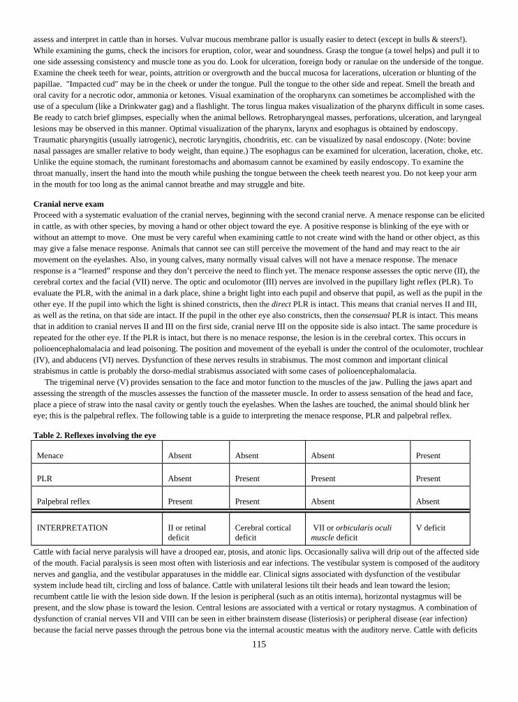
115
assess and interpret in cattle than in horses. Vulvar mucous membrane pallor is usually easier to detect (except in bulls & steers!). While examining the gums, check the incisors for eruption, color, wear and soundness. Grasp the tongue (a towel helps) and pull it to one side assessing consistency and muscle tone as you do. Look for ulceration, foreign body or ranulae on the underside of the tongue. Examine the cheek teeth for wear, points, attrition or overgrowth and the buccal mucosa for lacerations, ulceration or blunting of the papillae. "Impacted cud" may be in the cheek or under the tongue. Pull the tongue to the other side and repeat. Smell the breath and oral cavity for a necrotic odor, ammonia or ketones. Visual examination of the oropharynx can sometimes be accomplished with the use of a speculum (like a Drinkwater gag) and a flashlight. The torus lingua makes visualization of the pharynx difficult in some cases. Be ready to catch brief glimpses, especially when the animal bellows. Retropharyngeal masses, perforations, ulceration, and laryngeal lesions may be observed in this manner. Optimal visualization of the pharynx, larynx and esophagus is obtained by endoscopy. Traumatic pharyngitis (usually iatrogenic), necrotic laryngitis, chondritis, etc. can be visualized by nasal endoscopy. (Note: bovine nasal passages are smaller relative to body weight, than equine.) The esophagus can be examined for ulceration, laceration, choke, etc. Unlike the equine stomach, the ruminant forestomachs and abomasum cannot be examined by easily endoscopy. To examine the throat manually, insert the hand into the mouth while pushing the tongue between the cheek teeth nearest you. Do not keep your arm in the mouth for too long as the animal cannot breathe and may struggle and bite. Cranial nerve exam Proceed with a systematic evaluation of the cranial nerves, beginning with the second cranial nerve. A menace response can be elicited in cattle, as with other species, by moving a hand or other object toward the eye. A positive response is blinking of the eye with or without an attempt to move. One must be very careful when examining cattle to not create wind with the hand or other object, as this may give a false menace response. Animals that cannot see can still perceive the movement of the hand and may react to the air movement on the eyelashes. Also, in young calves, many normally visual calves will not have a menace response. The menace response is a “learned” response and they don’t perceive the need to flinch yet. The menace response assesses the optic nerve (II), the cerebral cortex and the facial (VII) nerve. The optic and oculomotor (III) nerves are involved in the pupillary light reflex (PLR). To evaluate the PLR, with the animal in a dark place, shine a bright light into each pupil and observe that pupil, as well as the pupil in the other eye. If the pupil into which the light is shined constricts, then the direct PLR is intact. This means that cranial nerves II and III, as well as the retina, on that side are intact. If the pupil in the other eye also constricts, then the consensual PLR is intact. This means that in addition to cranial nerves II and III on the first side, cranial nerve III on the opposite side is also intact. The same procedure is repeated for the other eye. If the PLR is intact, but there is no menace response, the lesion is in the cerebral cortex. This occurs in polioencephalomalacia and lead poisoning. The position and movement of the eyeball is under the control of the oculomoter, trochlear (IV), and abducens (VI) nerves. Dysfunction of these nerves results in strabismus. The most common and important clinical strabismus in cattle is probably the dorso-medial strabismus associated with some cases of polioencephalomalacia.
The trigeminal nerve (V) provides sensation to the face and motor function to the muscles of the jaw. Pulling the jaws apart and assessing the strength of the muscles assesses the function of the masseter muscle. In order to assess sensation of the head and face, place a piece of straw into the nasal cavity or gently touch the eyelashes. When the lashes are touched, the animal should blink her eye; this is the palpebral reflex. The following table is a guide to interpreting the menace response, PLR and palpebral reflex. Table 2. Reflexes involving the eye Menace
Absent
Absent
Absent
Present
PLR
Absent
Present
Present
Present
Palpebral reflex
Present
Present
Absent
Absent
INTERPRETATION
II or retinal deficit
Cerebral cortical deficit
VII or orbicularis oculi muscle deficit
V deficit
Cattle with facial nerve paralysis will have a drooped ear, ptosis, and atonic lips. Occasionally saliva will drip out of the affected side of the mouth. Facial paralysis is seen most often with listeriosis and ear infections. The vestibular system is composed of the auditory nerves and ganglia, and the vestibular apparatuses in the middle ear. Clinical signs associated with dysfunction of the vestibular system include head tilt, circling and loss of balance. Cattle with unilateral lesions tilt their heads and lean toward the lesion; recumbent cattle lie with the lesion side down. If the lesion is peripheral (such as an otitis interna), horizontal nystagmus will be present, and the slow phase is toward the lesion. Central lesions are associated with a vertical or rotary nystagmus. A combination of dysfunction of cranial nerves VII and VIII can be seen in either brainstem disease (listeriosis) or peripheral disease (ear infection) because the facial nerve passes through the petrous bone via the internal acoustic meatus with the auditory nerve. Cattle with deficits
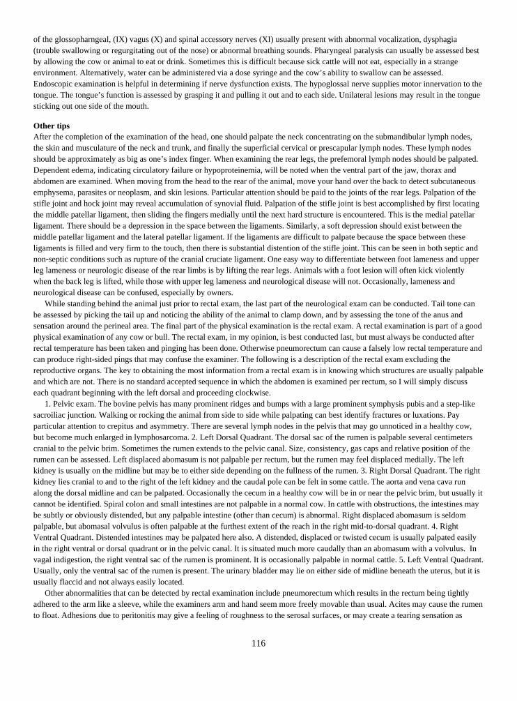
116
of the glossopharngeal, (IX) vagus (X) and spinal accessory nerves (XI) usually present with abnormal vocalization, dysphagia (trouble swallowing or regurgitating out of the nose) or abnormal breathing sounds. Pharyngeal paralysis can usually be assessed best by allowing the cow or animal to eat or drink. Sometimes this is difficult because sick cattle will not eat, especially in a strange environment. Alternatively, water can be administered via a dose syringe and the cow’s ability to swallow can be assessed. Endoscopic examination is helpful in determining if nerve dysfunction exists. The hypoglossal nerve supplies motor innervation to the tongue. The tongue’s function is assessed by grasping it and pulling it out and to each side. Unilateral lesions may result in the tongue sticking out one side of the mouth. Other tips After the completion of the examination of the head, one should palpate the neck concentrating on the submandibular lymph nodes, the skin and musculature of the neck and trunk, and finally the superficial cervical or prescapular lymph nodes. These lymph nodes should be approximately as big as one’s index finger. When examining the rear legs, the prefemoral lymph nodes should be palpated. Dependent edema, indicating circulatory failure or hypoproteinemia, will be noted when the ventral part of the jaw, thorax and abdomen are examined. When moving from the head to the rear of the animal, move your hand over the back to detect subcutaneous emphysema, parasites or neoplasm, and skin lesions. Particular attention should be paid to the joints of the rear legs. Palpation of the stifle joint and hock joint may reveal accumulation of synovial fluid. Palpation of the stifle joint is best accomplished by first locating the middle patellar ligament, then sliding the fingers medially until the next hard structure is encountered. This is the medial patellar ligament. There should be a depression in the space between the ligaments. Similarly, a soft depression should exist between the middle patellar ligament and the lateral patellar ligament. If the ligaments are difficult to palpate because the space between these ligaments is filled and very firm to the touch, then there is substantial distention of the stifle joint. This can be seen in both septic and non-septic conditions such as rupture of the cranial cruciate ligament. One easy way to differentiate between foot lameness and upper leg lameness or neurologic disease of the rear limbs is by lifting the rear legs. Animals with a foot lesion will often kick violently when the back leg is lifted, while those with upper leg lameness and neurological disease will not. Occasionally, lameness and neurological disease can be confused, especially by owners.
While standing behind the animal just prior to rectal exam, the last part of the neurological exam can be conducted. Tail tone can be assessed by picking the tail up and noticing the ability of the animal to clamp down, and by assessing the tone of the anus and sensation around the perineal area. The final part of the physical examination is the rectal exam. A rectal examination is part of a good physical examination of any cow or bull. The rectal exam, in my opinion, is best conducted last, but must always be conducted after rectal temperature has been taken and pinging has been done. Otherwise pneumorectum can cause a falsely low rectal temperature and can produce right-sided pings that may confuse the examiner. The following is a description of the rectal exam excluding the reproductive organs. The key to obtaining the most information from a rectal exam is in knowing which structures are usually palpable and which are not. There is no standard accepted sequence in which the abdomen is examined per rectum, so I will simply discuss each quadrant beginning with the left dorsal and proceeding clockwise.
1. Pelvic exam. The bovine pelvis has many prominent ridges and bumps with a large prominent symphysis pubis and a step-like sacroiliac junction. Walking or rocking the animal from side to side while palpating can best identify fractures or luxations. Pay particular attention to crepitus and asymmetry. There are several lymph nodes in the pelvis that may go unnoticed in a healthy cow, but become much enlarged in lymphosarcoma. 2. Left Dorsal Quadrant. The dorsal sac of the rumen is palpable several centimeters cranial to the pelvic brim. Sometimes the rumen extends to the pelvic canal. Size, consistency, gas caps and relative position of the rumen can be assessed. Left displaced abomasum is not palpable per rectum, but the rumen may feel displaced medially. The left kidney is usually on the midline but may be to either side depending on the fullness of the rumen. 3. Right Dorsal Quadrant. The right kidney lies cranial to and to the right of the left kidney and the caudal pole can be felt in some cattle. The aorta and vena cava run along the dorsal midline and can be palpated. Occasionally the cecum in a healthy cow will be in or near the pelvic brim, but usually it cannot be identified. Spiral colon and small intestines are not palpable in a normal cow. In cattle with obstructions, the intestines may be subtly or obviously distended, but any palpable intestine (other than cecum) is abnormal. Right displaced abomasum is seldom palpable, but abomasal volvulus is often palpable at the furthest extent of the reach in the right mid-to-dorsal quadrant. 4. Right Ventral Quadrant. Distended intestines may be palpated here also. A distended, displaced or twisted cecum is usually palpated easily in the right ventral or dorsal quadrant or in the pelvic canal. It is situated much more caudally than an abomasum with a volvulus. In vagal indigestion, the right ventral sac of the rumen is prominent. It is occasionally palpable in normal cattle. 5. Left Ventral Quadrant. Usually, only the ventral sac of the rumen is present. The urinary bladder may lie on either side of midline beneath the uterus, but it is usually flaccid and not always easily located.
Other abnormalities that can be detected by rectal examination include pneumorectum which results in the rectum being tightly adhered to the arm like a sleeve, while the examiners arm and hand seem more freely movable than usual. Acites may cause the rumen to float. Adhesions due to peritonitis may give a feeling of roughness to the serosal surfaces, or may create a tearing sensation as

117
immature fibrinous adhesions are broken down, or may severely restrict movement in the abdomen if extensive firm adhesions are present.
Of course, a complete physical examination is not warranted in every case but it is important to know how to perform one for those cases in which the diagnosis is not obvious.
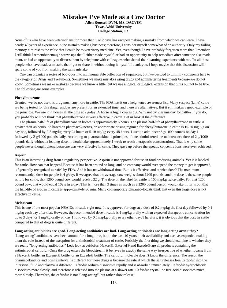
118
Mistakes I’ve Made as a Cow Doctor Allen Roussel, DVM, MS, DACVIM
Texas A&M University College Station, TX
None of us who have been veterinarians for more than 1 or 2 days has escaped making a mistake from which we can learn. I have nearly 40 years of experience in the mistake-making business; therefore, I consider myself somewhat of an authority. Only my failing memory diminishes the value that I could be to veterinary medicine. Yet, even though I have probably forgotten more than I member, I still think I remember enough screw-ups that I either made myself, or had an opportunity to help remediate after someone else made them, or had an opportunity to discuss them by telephone with colleagues who shared their learning experience with me. To all those people who have made a mistake that I got to share in without doing it myself, I thank you. I hope maybe that this discussion will spare some of you from making the same mistake.
One can organize a series of boo-boos into an innumerable collection of sequences, but I've decided to limit my comments here to the category of Drugs and Treatments. Sometimes we make mistakes using drugs and administering treatments because we do not know. Sometimes we make mistakes because we know a little, but we use a logical or illogical extension that turns out not to be true. The following are some examples. Phenylbutazone Granted, we do not use this drug much anymore in cattle. The FDA has it on a heightened awareness list. Many suspect (lame) cattle are being tested for this drug, residues are present for an extended time, and there are alternatives. But it still makes a good example of the principle. We use it in horses all the time at 2 g/day. A horse is big; a cow is big. Why not try 2 grams/day for cattle? If you do, you probably will not think that phenylbutazone is very effective in cattle. Let us look at the difference.
The plasma half-life of phenylbutazone in horses is approximately 6 hours. The plasma half-life of phenylbutazone in cattle is greater than 48 hours. So based on pharmacokinetics, an appropriate dosing regimen for phenylbutazone in cattle is 10-20 mg /kg on day one, followed by 2-5 mg/kg every 24 hours or 5-10 mg/kg every 48 hours. I used to administer 8 g/1000 pounds on day 1 followed by 2 g/1000 pounds daily. According to pharmacokinetic principles, if one administered the maintenance dose of 2 g/1000 pounds daily without a loading dose, it would take approximately 1 week to reach therapeutic concentrations. That is why some people never thought phenylbutazone was very effective in cattle. They gave up before therapeutic concentrations were ever achieved. Aspirin This is an interesting drug from a regulatory perspective. Aspirin is not approved for use in food producing animals. Yet it is labeled for cattle. How can that happen? Because it has been around so long, and no company would ever spend the money to get it approved, is "generally recognized as safe" by FDA. And it has no withdrawal time. But is it effective; and at what dose? The maximum recommended dose for people is 4 g/day. If we agree that the average cow weighs about 1200 pounds, and the dose is the same people as it is for cattle, that 1200 pound cow would receive 32 g. The dose on the label for cattle is 100 mg/kg twice daily. For that 1200 pound cow, that would equal 109 g in a day. That is more than 3 times as much as a 1200 pound person would take. It turns out that the half-life of aspirin in cattle is approximately 30 min. Many contemporary pharmacologists think that even this large dose is not effective in cattle. Meloxicam This is one of the most popular NSAIDs in cattle right now. It is approved for dogs at a dose of 0.2 mg/kg the first day followed by 0.1 mg/kg each day after that. However, the recommended dose in cattle is 1 mg/kg orally with an expected therapeutic concentration for up to 3 days; or 1 mg/kg orally on day 1 followed by 0.5 mg/kg orally every other day. Therefore, it is obvious that the dose in cattle compared to that of dogs is quite different. Long-acting antibiotics are good. Long-acting antibiotics are bad. Long-acting antibiotics are long-acting aren't they? "Long-acting" antibiotics have been around for a long time, but in the past 10 years, their availability and use has expanded making them the rule instead of the exception for antimicrobial treatment of cattle. Probably the first thing we should examine is whether they are really "long-acting antibiotics." Let's look at ceftiofur. Naxcel®, Excenel® and Excede® are all products containing the antimicrobial ceftiofur. Once the drug enters the bloodstream, it behaves in exactly the same way irrespective of whether it came from a Naxcel® bottle, an Excenel® bottle, or an Excede® bottle. The ceftiofur molecule doesn't know the difference. The reason the pharmacokinetics and dosing interval is different for these drugs is because the rate at which the salt releases free Ceftiofur into the interstitial fluid and plasma is different. Ceftiofur sodium dissociates rapidly and is absorbed immediately. Ceftiofur hydrochloride dissociates more slowly, and therefore is released into the plasma at a slower rate. Ceftiofur crystalline free acid dissociates much more slowly. Therefore, the ceftiofur is not “long-acting”, but rather slow release.
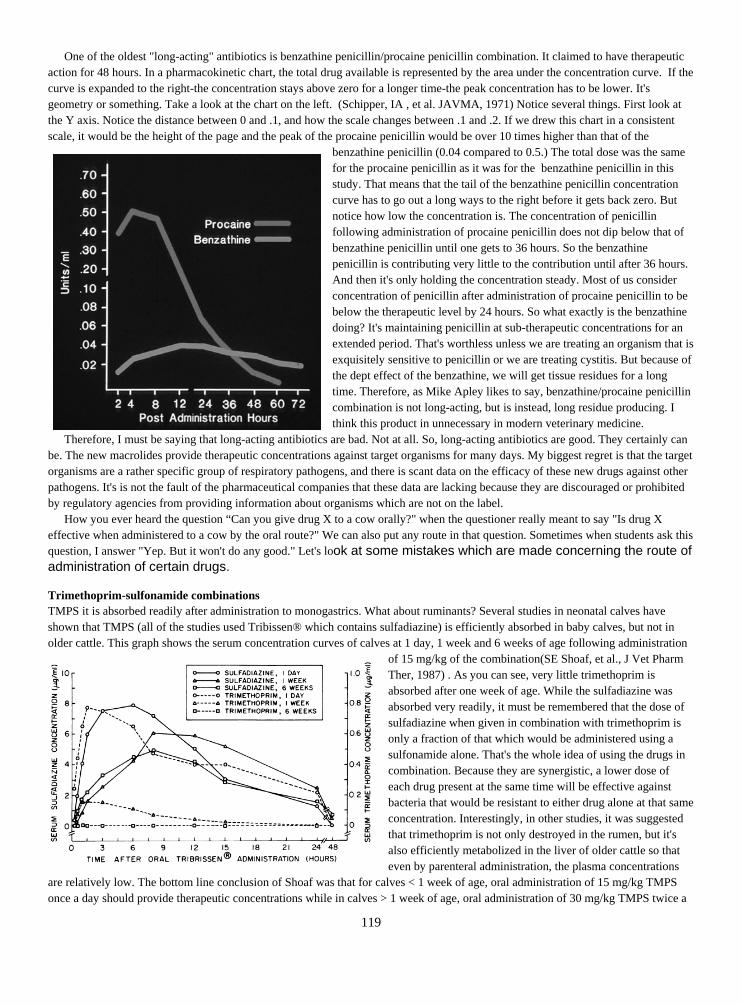
119
One of the oldest "long-acting" antibiotics is benzathine penicillin/procaine penicillin combination. It claimed to have therapeutic action for 48 hours. In a pharmacokinetic chart, the total drug available is represented by the area under the concentration curve. If the curve is expanded to the right-the concentration stays above zero for a longer time-the peak concentration has to be lower. It's geometry or something. Take a look at the chart on the left. (Schipper, IA , et al. JAVMA, 1971) Notice several things. First look at the Y axis. Notice the distance between 0 and .1, and how the scale changes between .1 and .2. If we drew this chart in a consistent scale, it would be the height of the page and the peak of the procaine penicillin would be over 10 times higher than that of the
benzathine penicillin (0.04 compared to 0.5.) The total dose was the same for the procaine penicillin as it was for the benzathine penicillin in this study. That means that the tail of the benzathine penicillin concentration curve has to go out a long ways to the right before it gets back zero. But notice how low the concentration is. The concentration of penicillin following administration of procaine penicillin does not dip below that of benzathine penicillin until one gets to 36 hours. So the benzathine penicillin is contributing very little to the contribution until after 36 hours. And then it's only holding the concentration steady. Most of us consider concentration of penicillin after administration of procaine penicillin to be below the therapeutic level by 24 hours. So what exactly is the benzathine doing? It's maintaining penicillin at sub-therapeutic concentrations for an extended period. That's worthless unless we are treating an organism that is exquisitely sensitive to penicillin or we are treating cystitis. But because of the dept effect of the benzathine, we will get tissue residues for a long time. Therefore, as Mike Apley likes to say, benzathine/procaine penicillin combination is not long-acting, but is instead, long residue producing. I think this product in unnecessary in modern veterinary medicine.
Therefore, I must be saying that long-acting antibiotics are bad. Not at all. So, long-acting antibiotics are good. They certainly can be. The new macrolides provide therapeutic concentrations against target organisms for many days. My biggest regret is that the target organisms are a rather specific group of respiratory pathogens, and there is scant data on the efficacy of these new drugs against other pathogens. It's is not the fault of the pharmaceutical companies that these data are lacking because they are discouraged or prohibited by regulatory agencies from providing information about organisms which are not on the label.
How you ever heard the question “Can you give drug X to a cow orally?" when the questioner really meant to say "Is drug X effective when administered to a cow by the oral route?" We can also put any route in that question. Sometimes when students ask this question, I answer "Yep. But it won't do any good." Let's look at some mistakes which are made concerning the route of administration of certain drugs. Trimethoprim-sulfonamide combinations TMPS it is absorbed readily after administration to monogastrics. What about ruminants? Several studies in neonatal calves have shown that TMPS (all of the studies used Tribissen® which contains sulfadiazine) is efficiently absorbed in baby calves, but not in older cattle. This graph shows the serum concentration curves of calves at 1 day, 1 week and 6 weeks of age following administration
of 15 mg/kg of the combination(SE Shoaf, et al., J Vet Pharm Ther, 1987) . As you can see, very little trimethoprim is absorbed after one week of age. While the sulfadiazine was absorbed very readily, it must be remembered that the dose of sulfadiazine when given in combination with trimethoprim is only a fraction of that which would be administered using a sulfonamide alone. That's the whole idea of using the drugs in combination. Because they are synergistic, a lower dose of each drug present at the same time will be effective against bacteria that would be resistant to either drug alone at that same concentration. Interestingly, in other studies, it was suggested that trimethoprim is not only destroyed in the rumen, but it's also efficiently metabolized in the liver of older cattle so that even by parenteral administration, the plasma concentrations
are relatively low. The bottom line conclusion of Shoaf was that for calves < 1 week of age, oral administration of 15 mg/kg TMPS once a day should provide therapeutic concentrations while in calves > 1 week of age, oral administration of 30 mg/kg TMPS twice a
120
day should be administered. Of course, taken literally, this recommendation is ridiculous because calves do not change overnight. I have always recommended 30 mg/kg TMPS twice a day, and only used it in calves less than two weeks of age. In round figures, that is usually about one 960 mg tablet twice a day. With the advent of newer antibiotics on the market, I have used this drug less and less. Percutaneous injection of poloxalene into the rumen I've never done this, but thanks to some other people who have, I won't have to in order to find out what happens. I've had two cases where someone injected poloxalene into the left paralumbar fossa thinking it was going into a bloated rumen. In both cases it created a massive abscess. Poloxalene must be an irritant because I know that a lot of needles and trocars get stuck into rumens and have very little adverse effect. I've never seen anything written about this, but because I believe this occurred due to more than bad luck, I'm going to avoid it. Pulling out the big guns Expensive animals with serious diseases deserve high-powered, expensive antibiotics with full-page ads and certainly justify using something-well, not "ordinary". Maybe not. We've all been faced with an owner who goes on about how wonderful a particular animal is as were thinking that the prognosis for the beast in front of us is not so good. And we might get the statement "I don't care what it costs Doc; save her." That's when it's time to pull out the stops and use the newest, greatest, most expensive, least "legal" antibiotic we can find. I have to admit, sometimes it's great to have the economic constraints lifted, but the biology of the organism and the animal doesn't really change. Let's look at a few quick examples. Let's take the case of a show heifer that was administered mineral oil retropharyngeally by a well-meaning Ag teacher who was trying to help a kid whose show heifer had bloat. First of all, it was probably a free gas bloat that didn't need mineral oil or poloxalene, but might've benefited from something as innocuous as some baking soda in the feed. Now it's got a raging retropharyngeal and peri-pharyngeal phlegmon. And who knows, maybe it aspirated a little bit and has "a touch of pneumonia" as some people often say. So that opens the door to enrofloxacin. Why not? It's a broad-spectrum antibiotic that must be good because extra label use is illegal. Well what are we trying to treat? The main problem is inflammation, not infection. Mineral oil is the major component of Freund's adjuvant. That is such an inflammatory substance that it takes special permission to be able to use it in a research environment to produce antibody in rabbits and others laboratory animals. Along with the inflammation, it's probably going to cause some necrosis. The ischemic environment caused by the oil and the necrosis is going to be a perfect place for anaerobic organisms to live and grow. Enrofloxacin, while a great drug, has poor activity against anaerobic organisms. What has good activity against anaerobes? Penicillin. For the infectious process associated with the phlegmon, penicillin would probably be much more effective than enrofloxacin. It’s not new or terribly expensive, but it might do the job. While we’re on the subject of this animal, the number one priority is for the animal to be able to breathe. A tracheostomy may be necessary. Cattle do quite well with tracheostomies, but I'd like to avoid that if possible. If the strider is severe, to me it's time to pull out the dexamethasone. But that might cause immunosuppression and possible secondary infection! I say to my critics, if this heifer's infection is worse in three days, I'll be happy because that means she is still alive. She may not want to (or be able to) eat or drink, and passing a stomach tube repeatedly is not a good idea. So more than expensive antibiotics, a small rumenostomy, which is very simple to perform, might benefit this heifer more.
Let's take a quick look at another case. Last week a practitioner called and told me about a rancher who had a couple of calves die acutely. and the one he performed a necropsy on had blackleg. The owner had actually found it alive and treated with enrofloxacin. First of all that's against federal law. Secondly it's against all the principles of pharmacology. The calf looked very sick so he decided to use a very "strong" antibiotic when the antibiotic of choice would've been penicillin. The value of the animal and the severity of the condition do not dictate which antibiotic is most likely to be effective. That's a fact. Have you ever said any of the following?
• We seem to have really good luck with that. Luck, chance, spontaneous cure-thank goodness we have all of these that help us. It just seems to me that the statement should be reserved for therapeutic regimens that involve horseshoes or shamrocks or something similar. Now I'll accept luck any day, but I like a drug or vaccine to offer more.
• I have been doing it this way for 30 years and I have never had a problem. I can't think of anything I have been doing for 30 years that has not resulted in a problem or at least given the impression of causing a problem at least once. That includes not only veterinary treatments and procedures but other things like breathing and getting up in the morning. Maybe I'm a cynic, but when someone tells me they never had a problem, I think there's a problem.
• Well, it seems to work in my hands. So we need to discuss how we define when something "works." Most of us define it as having a favorable outcome following an intervention. It varies with the practice type and the clientele, but I think for most of us, we are called to intervene on patients that have a high probability of a spontaneous recovery. Yet, many times we cannot determine at first examination if a particular patient is one that's going to recover spontaneously or one that is going to require treatment-therefore we treat all of them. If the disease has a 20% probability of resulting in death and an 80% probability of self-resolution, and we can save 50% of them that would die

121
otherwise with an intervention, will we be able to distinguish if our intervention was really the cause of the animal living? At first blush you say "My goodness. I should be able to tell if I can save 50% of them." But think about it. 80% live anyway. If you treat them, 90% live. And these are averages over time. How long would it take to figure out if 80% or 90% were living? The owner will probably give you credit for (and you're not going to argue) with saving 80% to 90% either way. If you ever have an opportunity to go back through medical records and actually test your "feelings" about the performance of the treatment, it can be quite revealing and surprising. We only remember the really good stuff and the really bad stuff. And let me be clear. It only makes sense to continue doing what seems to be successful. It doesn't make any sense to do something that seems to be unsuccessful. I just think we’re more likely to be better veterinarians if we understand that sometimes we make a difference (to the negative or to the positive), and sometimes we don't, and sometimes we have trouble telling the difference. When we start always believing ourselves, that's when we run into trouble. And it's easy to believe yourself if you never get the opinion of someone else.
122
Getting the Most Out of Clinical Chemistries: The Basics
Allen Roussel, DVM, MS, DACVIM Texas A&M University
College Station, TX
Serum protein Serum contains many different proteins, but the two components of diagnostic significance relative to the chemistry profile are albumin and globulin. Albumin is synthesized in the liver and is the protein primarily responsible for the oncotic pressure of plasma. A large portion of the globulin fraction is made up of immunoglobulins which are synthesized by lymphoid tissue. The ratio of albumin to globulin (A:G ratio) is fairly constant in healthy cattle (reference range 0.84 -0.94). Most chemistry profiles include measured albumin and total serum protein, while the value for globulin is typically derived by subtracting the former from the later. One must remember that if plasma is used instead of serum, fibrinogen will be included in the value for total protein, and hence, the derived globulin value; this may obfuscate the interpretation of the total protein and globulin. The discrepancy cannot be totally rectified by simply subtracting the value for fibrinogen, which is often determined by refractometry, from the value for plasma protein on the profile because of the difference in methods of analysis. Because most reference ranges are established for serum proteins, it is our opinion that serum is the sample of choice when evaluating the blood proteins. More specific and detailed evaluation of the globulin fraction can be achieved using electrophoresis, radial immunoassay and other methods which will not be discussed here.
Hyperproteinemia can result from an increase in albumin, globulin or both. The only cause of hyperalbuminemia is dehydration. In dehydration, both albumin and globulin rise, but whether they exceed the reference range is determined by the degree of dehydration and the original protein concentration in the serum. Hyperproteinemia without dehydration is almost always the result of hyperglobulinemia. Globulin increases substantially with age in dairy cows. The difference between two year-olds and five year-olds was about 1.5 g/dl, potentially a clinically relevant difference. Causes of hyperglobulinemia include chronic inflammatory diseases (traumatic reticuloperitonitis, liver abscess, chronic pneumonia) and hepatic disease. In chronic inflammatory disease, the A:G ratio usually decreases because of an increase in globulin which is often accompanied by a small decrease in albumin. In chronic hepatic disease, the decrease in albumin may be more substantial. Serum globulin may be one of the most overlooked values on the routine chemistry profile. Changes in the hemogram are often rather subtle and transient in inflammatory disease of cattle, compared to other species. Therefore, the evaluation of serum globulin is of great value in chronic inflammatory disease.
In mature cattle, hypoproteinemia is usually the result of hypoalbuminemia or panhypoproteinemia. Hypoalbuminemia occurs when 1) hepatic production is insufficient to meet demand, either as a result of insufficient production or increased consumption or 2) there is excessive loss of albumin. Insufficient production can occur in animals with chronic severe hepatic disease or as a result of inadequate protein intake, digestion, or absorption. Because bovine albumin has a half-life of 16.5 days and the reserve capacity of hepatic tissue is so great, liver disease must be chronic and severe to result in severe hypoalbuminemia. In the authors' experience, cattle with chronic debilitating disease of many causes may be hypoalbuminemic with low or normal total protein. If the A:G ratio is low, chronic inflammatory disease should be suspected. In acute and subacute disease, hypoalbuminemia frequently results from loss of albumin. Avenues of albumin loss include the kidney (particularly the glomerulus), the gastrointestinal tract, hemorrhage and exudation. In many instances, loss of albumin may be accompanied by loss of globulin, resulting in panhypoprotienemia.
Renal amyloidosis can result in severe albumin loss in the urine due to glomerular damage. In one report, 5 of 6 cattle with amyloidosis had hypoglobulinemia along with hypoalbuminemia. Panhypoproteinemia is the rule in protein-losing gastro-enteropathies such as nematode parasitism, paratuberculosis and salmonellosis. Because digestion and absorption may be impaired in these diseases, decreased production due to amino acid deficiency may contribute to the hypoproteinemia in chronic cases. Acute hemorrhage results in panhypoproteinemia accompanied by anemia.
Hypoglobulinemia is infrequent in cattle except neonates, either as a result of failure of passive transfer of maternal antibody, or severe infection when transferred antibodies are consumed rapidly prior to the efficient production of endogenous antibody by the young calf. Hepatic tests The leakage enzymes aspartate transaminase (AST, formerly SGOT), L-iditol (formerly sorbitol) dehydrogenase(IDH), ornithine carbamoyltransferase (OCT), glutamate dehydrogenase (GDH) and lactate dehydrogenase (LDH) have been used to evaluate the liver. Of these, AST, LDH and IDH are the most popular in the United States. Both AST and LDH are found in a wide variety of tissues, the most important of which are liver and muscle. Muscle damage, especially due to recumbency in cattle, may result in marked increases of both; hence, AST and LDH should be interpreted in conjunction with a liver-specific enzyme ( such as GGT), or a muscle-specific enzyme such as creatine kinase (CK) to determine the source of the tissue insult. Usually, high AST or LDH and normal CK indicates liver disease. If serum is allowed to remain on the clot too long or the sample is hemolyzed, the AST and LDH
123
may be falsely elevated because both enzymes are found in red blood cells. Because concentrations of these enzymes are high in serum when damaged cell membranes allow their escape from hepatic cytosol, they indicate cell damage, not abnormal hepatic function. In fact, in chronic or slowly progressive hepatic disease, these enzymes may be within or below reference ranges because few hepatocytes are being damaged at one time, or because hepatocellular mass is substantially reduced. Consequently, these enzymes may be more sensitive indicators of acute disease such as some toxicities and infectious hepatitis. They may also be high in cattle with hepatic lipidosis, passive venous congestion and diseases that cause distension of the forestomachs and abomasum. IDH is a sensitive and specific indicator of hepatocellular damage. Unfortunately, its usefulness is limited by its relatively instability in vitro.
The "cholestatic", enzymes gamma glutamyltransferase (GGT) and serum alkaline phosphatase (SAP), are more sensitive to biliary obstruction caused by conditions such as fascioliasis or cholelithiasis. The cholestatic enzymes are more likely to be high in chronic hepatic disease than are the leakage enzymes because fibrosis constricts and blocks some bile ducts.
Although GGT is found in many tissues, the source of essentially all of the GGT in the serum is the biliary and hepatocellular membranes. Therefore, it is one of the most liver-specific tests available to veterinarians. Serum GGT rises principally in cholestatic disease, although hepatocellular diseases in which cholestasis is a secondary feature, also causes an increase in GGT. Because it tends to decrease less rapidly than the other leakage enzymes, it may be of more value in identifying cattle with chronic hepatic disease. Serum GGT of pre-colostral calves is similar to that of mature cattle, but serum concentrations rise sharply following consumption of colostrum, which is rich in GGT. By 24 hours after colostral intake, serum GGT concentration is 50 to 100 times that of colostrum-deprived calves. In fact, serum GGT can be used to estimate the success of passive transfer, but not to detect hepatic disease in neonates.
Serum alkaline phosphatase, a useful indicator of hepatic or cholestatic disease of dogs, is often included in chemistry profiles of cattle. Several isoenzymes from different tissues have been identified, and almost all of the SAP in healthy cattle is of osseous origin. In cattle with hepatic disease, SAP of hepatic origin increases, but the increase is not large in magnitude. Therefore, SAP is of limited diagnostic value for hepatic disease of cattle. Interestingly, though not clearly explainable, SAP was found to be useful as a prognostic indicator in cattle with abomasal volvulus.
Bilirubin is a breakdown product of hemoglobin that is conjugated and excreted by the liver. Unconjugated (or direct) bilirubin is the result of rapid breakdown of hemoglobin which occurs in acute hemolysis. Conjugated (or indirect bilirubin) accumulates in plasma when there is intra- or extrahepatic biliary obstruction. The plasma concentration of bilirubin in healthy cattle is very low compared to that of the other species; and the magnitude of increase is relatively small, even in severe liver disease. Severe bilirubinemia and icterus in cattle is almost always a result of hemolysis, and hence, is primarily due to unconjugated bilirubin.
Though usually considered an index of renal function, blood or serum urea nitrogen (BUN or SUN) is also an indicator of hepatic function. In the liver, ammonia is converted to urea. In severe hepatic failure or partial vascular anomaly, SUN is low while ammonia is high. However, low SUN is not associated only with hepatic disease. Because rumen microbes use urea to synthesize protein, the rumen acts as a "sponge" for urea in cattle that are anorectic or protein-deprived.
Laboratory reference ranges for mature cattle are invalid for neonatal calves, especially those under a week of age. Neonatal calves have somewhat higher concentrations of bilirubin, AST, SAP, and SBA and markedly higher concentrations of GGT than do mature ruminants. Electrolytes The serum electrolyte profile typically includes sodium (Na), potassium (K), chloride (Cl), and total carbon dioxide (TCO2) or bicarbonate (HCO3). From these values, the anion gap (AG) can be calculated. Although there is a nominal difference between the TCO2 and HCO3, the HCO3 usually being slightly smaller, we will consider them equivalent in this paper. Serum electrolytes are useful in the evaluation of several body systems, as well as for the formulation and monitoring of fluid and electrolyte therapy. Due to the abundance of K and scarcity of Na in erythrocytic fluid relative to serum, hemolysis can falsely increase serum K and decrease serum Na in cattle.
Because their concentrations change in concert in a number of conditions, the electrolytes will be discussed together. Sodium is the major extracellular cation, while Cl and HCO3 are the major extracellular anions. Chloride and HCO3 often maintain a reciprocal relationship in extracellular fluid. Because the majority of the exchangeable Na and Cl are found in the extracellular fluid, measuring serum Na and Cl provides an accurate assessment of the total body status of these electrolytes. Serum potassium, on the other hand, provides a less reliable and sometimes paradoxical reflection of total body K status because only a small portion (approximately 5%) of the animal's K is in the extracellular fluid. Changes in blood pH greatly affect serum K by causing the movement of K across cell membranes; K moves into cells during alkalosis and out of cells during acidosis. Therefore, serum K should be interpreted along with serum HCO3. Serum HCO3 is a measure of metabolic acid-base balance; concentrations above the reference range indicating metabolic alkalosis and those below indicating metabolic acidosis.
Hypernatremia and hyperchloremia occur in salt toxicity/water deprivation, but are not commonly present in cases of dehydration because typically fluid loss in cattle occurs with concurrent loss of electrolytes. Cattle with clinical "salt toxicity" may have normal
124
serum Na because clinical signs often do not occur until after the cattle drink, and serum Na concentration and osmolality return to normal. Hyperkalemia is almost always secondary to acidosis as K moves out of the intracellular fluid into the extracellular fluid. Therefore, serum K is an unreliable index of total body K. For example, diarrheic calves often are acidotic and hyperkalemic, but they have total-body K depletion because of fecal K loss. In these cases, as in most cases where serum K is increased secondarily to acidosis, K supplementation may be indicated during or immediately following correction of acidosis. Hypochloremia, hypokalemia, metabolic alkalosis and, to a lesser degree, hyponatremia, are typical findings in obstructive gastrointestinal diseases including abomasal volvulus, displaced abomasum, vagal indigestion, intussusception and cecal torsion. In these diseases, HCl is sequestered in the abomasum, causing hypochloremia, metabolic alkalosis, and secondary hypokalemia. In general, the more orad the lesion (abomasal impaction vs jejunal intussusception), and the more complete the obstruction (abomasal volvulus vs LDA), the more severe the alkalosis and hypochloremia. Hypochloremia and metabolic alkalosis are fairly non-specific abnormalities in sick cattle however. In a study of over 500 mature cattle in the authors' hospital, over 40% of the dehydrated cattle were hypochloremic and/or alkalotic.
Serum Na and Cl are consistently low in uroperitoneum, and often are low in diarrhea and renal failure, while serum HCO3 is variable in these conditions. Urinary obstruction and uroperitoneum are associated with hyperkalemia in non-ruminant species, but not in cattle. Renal tests Elimination of nitrogenous wastes, such as urea and creatinine (Cr), and concentration of urine to conserve body water are two of the many vital functions performed by the kidney. Evaluation of these functions is exploited in the diagnosis of renal disease. Serum or blood urea nitrogen (SUN or BUN) and serum creatinine (Cr) are rough indices of glomerular filtration rate. The generous reserve capacity of the kidney makes SUN and Cr insensitive indicators of renal function; 75% loss of functional renal mass is required for azotemia to occur. Slightly more sensitive than SUN and Cr, the urinary specific gravity (USG) can detect about a 67% loss of functional renal tissue. The USG is most easily estimated by refractometry. By convention, USG of > 1.025 is considered indicative of appropriate concentrating ability in the face of dehydration or azotemia. It is quite common, however, for normally hydrated cattle, especially dairy cattle, to have USG <1.025.
Azotemia, the accumulation of nitrogenous wastes in the blood, is reflected in the serum chemistry profile as high SUN and Cr. Remember - AZOTEMIA DOES NOT EQUAL RENAL DISEASE! Azotemia can be classified as renal (due to renal disease), prerenal (due to sluggish renal blood flow, as in shock or dehydration), or postrenal (due to obstruction of urine outflow, as in urolithiasis). Though not without exception, the simplest way to distinguish among the three is by measuring the USG. In azotemic cattle, if the USG is >
Urea is formed in the liver by the detoxification of ammonia, a by product of protein metabolism. Therefore SUN is influenced by diet and hepatic function. Urea is recycled in a functional rumen, a process which may tend to moderate the rise in SUN in renal disease and result in a low SUN/Cr ratio. Although Cr, a product of energy metabolism in muscle, can be very low in emaciated cattle with little muscle mass, it tends to be less influenced by extraneous factors than SUN. For this reason, Cr is the test of choice over SUN.
1.025, the azotemia is prerenal; if the USG is <1.025, the azotemia is renal. In postrenal azotemia, urine is often difficult or impossible to obtain, and the diagnosis is based on physical examination. In our experience, cattle with prerenal azotemia eliminate urea and Cr rapidly when rehydrated, often returning to or near the reference range in 24-48 hours if appropriate fluid therapy and correction of the primary problem is accomplished.
While SUN, Cr and USG can identify renal disease, the final diagnosis cannot be obtained from this information. For example, acorn toxicity, pyelonephritis, and amyloidosis are diseases which cause renal failure in cattle. These diseases cannot be distinguished from one another simply based on the results of the chemistry profile. However, the characteristics of the urine in each of these diseases is very different. Whenever renal disease is suspected, a complete urinalysis should be performed, as well as, rectal palpation of the kidneys. Ultrasonography and renal biopsy may also be informative. Glucose Glucose metabolism is unique in ruminants because they absorb essentially no pre-formed glucose from the gut. The reference range for serum glucose in adult cattle is lower than for calves and non-ruminant species. Erythrocytes metabolize glucose in vitro in a blood tube at a rate of about 10% per hour at room temperature. Serum should be separated from the clot within 30 minutes, or sodium fluoride-containing tubes should be used if timely separation is not possible. Hyperglycemia occurs in stress, milk fever, and following administration of dextrose solution, xylazine, or corticosteriods. It is interesting that most milk fever remedies contain dextrose, even though hypocalcemia prevents the release of insulin from the islet cells of the pancreas resulting in hyperglycemia. Endogenous and exogenous steroids increase gluconeogenesis and increase serum glucose. Xylazine causes a dose dependant hyperglycemia that persists for over 6 hours. Diabetes mellitus although uncommon in cattle, causes permanent hyperglycemia if untreated.

125
Muscle enzymes As previously mentioned, LDH and AST are released into plasma as a result of muscle damage, but they are not muscle-specific enzymes. Serum CK, on the other hand, is a very sensitive and specific indicator of muscle damage. Subtle increases can occur due to intramuscular injection, exercise or struggling. Recumbent mature cattle may have >100-fold increases due to the secondary pressure damage that is a part of the downer cow syndrome. Very high concentrations of CK in the absence of recumbency, or in young recumbent cattle suggest primary myopathy, such as white muscle disease or Senna toxicity. The half-life of CK in serum is short, and CK concentrations fall rapidly in recumbent animals even if they remain recumbent. Because AST concentration rises and falls more slowly, it can be used in combination with CK to stage muscle damage. In recumbent cattle, if the AST is very high and the CK is not, the damage is likely several days old. In an attempt to use laboratory tests for prognosis, New Zealand investigators found that fewer than 5% of cows with an AST value > 7.4 times the upper limit of the reference range survived. For CK, the "critical" value above the reference range was related to the duration of recumbency (Table 2).
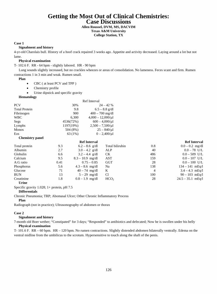
126
Getting the Most Out of Clinical Chemistries: Case Discussions
Allen Roussel, DVM, MS, DACVIM Texas A&M University
College Station, TX
Case 1 Signalment and history
4-yr-old Charolais bull. History of a hoof crack repaired 3 weeks ago. Appetite and activity decreased. Laying around a lot but not lame.
Physical examination T- 102.6 F. RR - 64 bpm - slightly labored. HR - 90 bpm
Lung sounds slightly increased, but no crackles wheezes or areas of consolidation. No lameness. Feces scant and firm. Rumen contractions 1 in 3 min and weak. Rumen small.
Plan • CBC ( at least PCV and TPP ) • Chemistry profile • Urine dipstick and specific gravity
Hematology Ref Interval PCV 30% 24 – 42 % Total Protein 9.8 6.5 – 8.8 g/dl Fibrinogen 900 400 – 700 mg/dl WBC 6,300 4,000 – 12,000/µl Segs 4536(72%) 600 – 4,000/µl Lymphs 1197(19%) 2,500 – 7,500/µl Monos 504 (8%) 25 – 840/µl Eos 63 (1%) 0 – 2,400/µl
Chemistry panel Ref Interval Ref Interval Total protein 9.3 6.2 – 8.6 g/dl Total bilirubin 0.8 0.0 – 0.2 mg/dl Albumin 2.7 3.0 – 4.2 g/dl ALP 40 0.0 – 78 U/L Globulin 6.6 3.2 – 4.4 g/dl CK 486 0.0 – 509 U/L Calcium 9.5 8.3 – 10.9 mg/dl AST 159 0.0 – 107 U/L A:G ratio 0.41 0.75 – 0.85 GGT 28 0.0 – 100 U/L Phosphorus 5.6 4.3 – 8.6 mg/dl Na 138 134 – 141 mEq/l Glucose 71 40 – 74 mg/dl K 4 3.4 – 4.3 mEq/l BUN 13 5 – 29 mg/dl Cl 100 90 – 103 mEq/l Creatinine 1.8 0.8 – 1.9 mg/dl HCO2 28 24.5 – 35.1 mEq/l
Urine Specific gravity 1.028; 1+ protein, pH 7.5
Differentials Chronic Pneumonia; TRP; Abomasal Ulcer; Other Chronic Inflammatory Process
Plan Radiograph (not in practice); Ultrasonography of abdomen or thorax Case 2
Signalment and history 7-month old Boer wether; “Constipated” for 3 days; “Responded” to antibiotics and defecated; Now he is swollen under his belly
Physical examination T- 101.6 F. RR – 60 bpm. HR – 120 bpm. No rumen contractions. Slightly distended abdomen bilaterally ventrally. Edema on the ventral midline from the umbilicus to the scrotum. Hypersensitive to touch along the shaft of the penis.
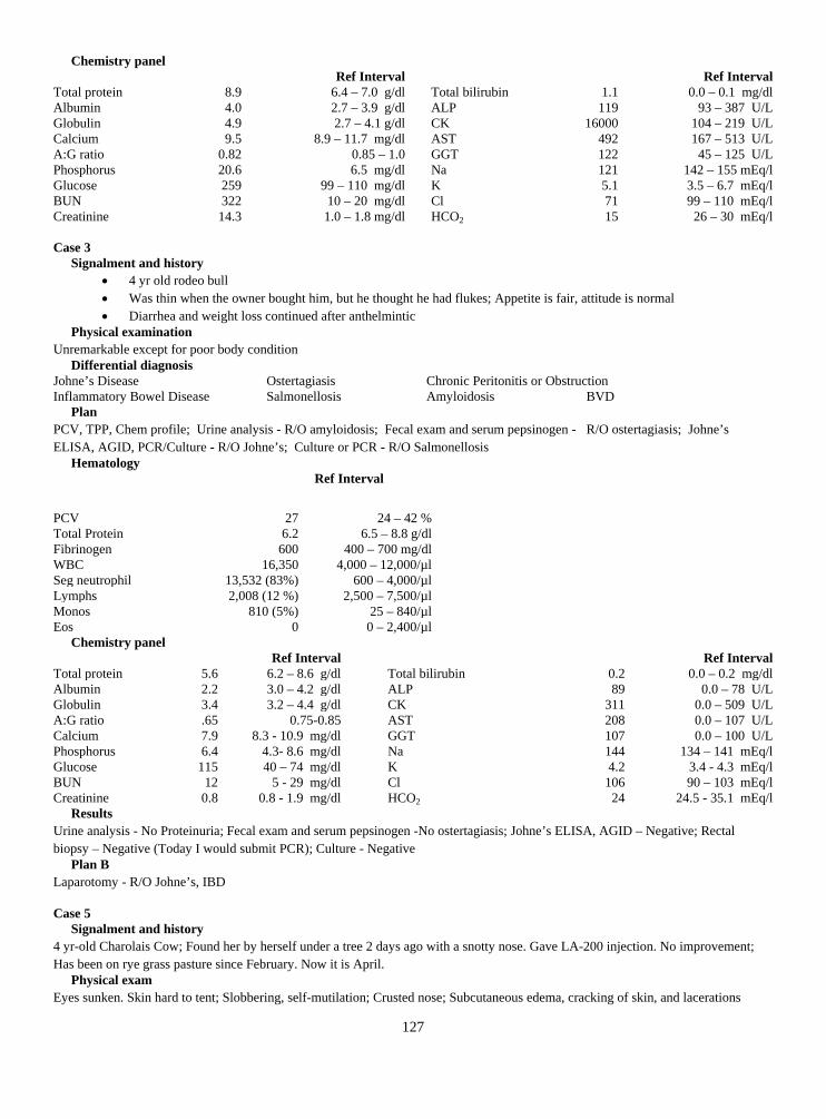
127
Chemistry panel Ref Interval Ref Interval Total protein 8.9 6.4 – 7.0 g/dl Total bilirubin 1.1 0.0 – 0.1 mg/dl Albumin 4.0 2.7 – 3.9 g/dl ALP 119 93 – 387 U/L Globulin 4.9 2.7 – 4.1 g/dl CK 16000 104 – 219 U/L Calcium 9.5 8.9 – 11.7 mg/dl AST 492 167 – 513 U/L A:G ratio 0.82 0.85 – 1.0 GGT 122 45 – 125 U/L Phosphorus 20.6 6.5 mg/dl Na 121 142 – 155 mEq/l Glucose 259 99 – 110 mg/dl K 5.1 3.5 – 6.7 mEq/l BUN 322 10 – 20 mg/dl Cl 71 99 – 110 mEq/l Creatinine 14.3 1.0 – 1.8 mg/dl HCO2 15 26 – 30 mEq/l Case 3
Signalment and history • 4 yr old rodeo bull • Was thin when the owner bought him, but he thought he had flukes; Appetite is fair, attitude is normal • Diarrhea and weight loss continued after anthelmintic
Physical examination Unremarkable except for poor body condition
Differential diagnosis Johne’s Disease Ostertagiasis Chronic Peritonitis or Obstruction Inflammatory Bowel Disease Salmonellosis Amyloidosis BVD
Plan PCV, TPP, Chem profile; Urine analysis - R/O amyloidosis; Fecal exam and serum pepsinogen - R/O ostertagiasis; Johne’s ELISA, AGID, PCR/Culture - R/O Johne’s; Culture or PCR - R/O Salmonellosis
Hematology Ref Interval
PCV 27 24 – 42 % Total Protein 6.2 6.5 – 8.8 g/dl Fibrinogen 600 400 – 700 mg/dl WBC 16,350 4,000 – 12,000/µl Seg neutrophil 13,532 (83%) 600 – 4,000/µl Lymphs 2,008 (12 %) 2,500 – 7,500/µl Monos 810 (5%) 25 – 840/µl Eos 0 0 – 2,400/µl
Chemistry panel Ref Interval Ref Interval Total protein 5.6 6.2 – 8.6 g/dl Total bilirubin 0.2 0.0 – 0.2 mg/dl Albumin 2.2 3.0 – 4.2 g/dl ALP 89 0.0 – 78 U/L Globulin 3.4 3.2 – 4.4 g/dl CK 311 0.0 – 509 U/L A:G ratio .65 0.75-0.85 AST 208 0.0 – 107 U/L Calcium 7.9 8.3 - 10.9 mg/dl GGT 107 0.0 – 100 U/L Phosphorus 6.4 4.3- 8.6 mg/dl Na 144 134 – 141 mEq/l Glucose 115 40 – 74 mg/dl K 4.2 3.4 - 4.3 mEq/l BUN 12 5 - 29 mg/dl Cl 106 90 – 103 mEq/l Creatinine 0.8 0.8 - 1.9 mg/dl HCO2 24 24.5 - 35.1 mEq/l
Results Urine analysis - No Proteinuria; Fecal exam and serum pepsinogen -No ostertagiasis; Johne’s ELISA, AGID – Negative; Rectal biopsy – Negative (Today I would submit PCR); Culture - Negative
Plan B Laparotomy - R/O Johne’s, IBD Case 5
Signalment and history 4 yr-old Charolais Cow; Found her by herself under a tree 2 days ago with a snotty nose. Gave LA-200 injection. No improvement; Has been on rye grass pasture since February. Now it is April.
Physical exam Eyes sunken. Skin hard to tent; Slobbering, self-mutilation; Crusted nose; Subcutaneous edema, cracking of skin, and lacerations

128
Hematology Ref Interval
PCV 37 24-42 % Total Protein 6.6 6.5-8.8 g/dl Fibrinogen 900 400-700 mg/dl WBC 22,700 4,000-12,000/µl Seg neutrophil 18,610 (82%) 600-4000/µl Lymphs 3,860 (17%) 2500-7,500/µl Monos 270 (1%) 25-840/µl Eos 0 0-2,400/µl PCV 40 TPP 8.3 Fibrinogen 500
Chemistry profile Ref Interval Ref Interval Total protein 5.7 6.2 - 8.6 g/dl Total bilirubin 2.8 0.0 - 0.2 mg/dl Albumin 1.8 3.0 - 4.2 g/dl ALP 373 0.0 – 78 U/L Globulin 3.9 3.2 – 4.4 g/dl CK 311 0.0 – 509 U/L A:G ratio .46 0.75-0.85 AST 861 0.0 – 107 U/L Calcium 7.3 8.3 - 10.9 mg/dl GGT 553 0.0 – 100 U/L Phosphorus 3.6 4.3- 8.6 mg/dl Na 144 134 – 141 mEq/l Glucose 119 40 – 74 mg/dl K 1.7 3.4 - 4.3 mEq/l BUN 11 5 - 29 mg/dl Cl 102 90 – 103 mEq/l Creatinine 0.8 0.8 - 1.9 mg/dl HCO2 32 24.5 - 35.1 mEq/l
Diagnosis Photosensitization - Secondary
Signalment and history • 2 yr-old Fleckveih Bull; Recurrent bloat for 1 week 5 weeks prior to presentation. • Suspected poloxalene injection into rumen. Bloat resolved, but bull became anorexic and lost weight.
Physical exam Good body condition; Slightly dehydrated; Moderate distension; Rectal exam - gas-filled viscus right mid-dorsal abdomen, rumen hard
Differentials RDA; Cecal displacement
Plan CBC; Chem profile; Abdominocentesis; Exploratory laparotomy
Hematology Ref Interval
PCV 27 24-42 % Total Protein 8.7 6.5-8.8 g/dl Fibrinogen 400 400-700 mg/dl WBC 13,400 4,000-12,000/µl Seg neutrophil 9782 (73%) 600-4000/µl Lymphs 2948 (22% 2500-7,500/µl Monos 268 (2%) 25-840/µl Eos 402 (3%) 0-2,400/µl
Chemistry panel Ref Interval Ref Interval Total protein 8.6 6.2 - 8.6 g/dl Total bilirubin 1.0 0.0 - 0.2 mg/dl Albumin 2.5 3.0 - 4.2 g/dl ALP 62 0.0 – 78 U/L Globulin 5.1 3.2 – 4.4 g/dl CK 55 0.0 – 509 U/L A:G ratio 0.49 AST 72 0.0 – 107 U/L Calcium 8.6 8.3 - 10.9 mg/dl GGT 36 0.0 – 100 U/L Phosphorus 7.1 4.3- 8.6 mg/dl Na 141 134 – 141 mEq/l Glucose 77 40 – 74 mg/dl K 4.1 3.4 - 4.3 mEq/l BUN 10 5 - 29 mg/dl Cl 104 90 – 103 mEq/l Creatinine 1.4 0.8 - 1.9 mg/dl HCO2 30 24.5 - 35.1 mEq/l

129
Revised diagnosis Not GI obstruction; Chronic inflammatory disease
Rectal exam - Day 2 • Gas-filled viscus smaller and more ventral; Rumen very hard, adhered to left body wall • Left kidney not palpable
Ultrasonography day 2 Per rectum and transabdominal: Rumen not on left side; Fluid filled mass with fibrin; Aspiration - foul smelling fluid
Revised diagnosis and plan Periruminal abscess; Left paramedical approach to open abscess; Periruminal abscess; Perioperative antibiotics; Lavage of the abscess
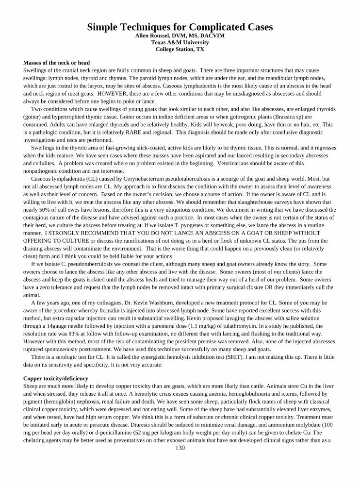
130
Simple Techniques for Complicated Cases Allen Roussel, DVM, MS, DACVIM
Texas A&M University College Station, TX
Masses of the neck or head Swellings of the cranial neck region are fairly common in sheep and goats. There are three important structures that may cause swellings: lymph nodes, thyroid and thymus. The parotid lymph nodes, which are under the ear, and the mandibular lymph nodes, which are just rostral to the larynx, may be sites of abscess. Caseous lymphadenitis is the most likely cause of an abscess in the head and neck region of meat goats. HOWEVER, there are a few other conditions that may be misdiagnosed as abscesses and should always be considered before one begins to poke or lance.
Two conditions which cause swellings of young goats that look similar to each other, and also like abscesses, are enlarged thyroids (goiter) and hypertrophied thymic tissue. Goiter occurs in iodine deficient areas or when goitrogenic plants (Brassica sp) are consumed. Adults can have enlarged thyroids and be relatively healthy. Kids will be weak, poor-doing, have thin or no hair, etc. This is a pathologic condition, but it is relatively RARE and regional. This diagnosis should be made only after conclusive diagnostic investigations and tests are performed.
Swellings in the thyroid area of fast-growing slick-coated, active kids are likely to be thymic tissue. This is normal, and it regresses when the kids mature. We have seen cases where these masses have been aspirated and our lanced resulting in secondary abscesses and cellulites. A problem was created where no problem existed in the beginning. Veterinarians should be aware of this nonpathogenic condition and not intervene.
Caseous lymphadenitis (CL) caused by Corynebacterium pseudotuberculosis is a scourge of the goat and sheep world. Most, but not all abscessed lymph nodes are CL. My approach is to first discuss the condition with the owner to assess their level of awareness as well as their level of concern. Based on the owner’s decision, we choose a course of action. If the owner is aware of CL and is willing to live with it, we treat the abscess like any other abscess. We should remember that slaughterhouse surveys have shown that nearly 50% of cull ewes have lesions, therefore this is a very ubiquitous condition. We document in writing that we have discussed the contagious nature of the disease and have advised against such a practice. In most cases when the owner is not certain of the status of their herd, we culture the abscess before treating at. If we isolate T. pyogenes or something else, we lance the abscess in a routine manner. I STRONGLY RECOMMEND THAT YOU DO NOT LANCE AN ABSCESS ON A GOAT OR SHEEP WITHOUT OFFERING TO CULTURE or discuss the ramifications of not doing so in a herd or flock of unknown CL status. The pus from the draining abscess will contaminate the environment. That is the worse thing that could happen on a previously clean (or relatively clean) farm and I think you could be held liable for your actions
If we isolate C. pseudotuberculosis we counsel the client, although many sheep and goat owners already know the story. Some owners choose to lance the abscess like any other abscess and live with the disease. Some owners (most of our clients) lance the abscess and keep the goats isolated until the abscess heals and tried to manage their way out of a herd of our problem. Some owners have a zero tolerance and request that the lymph nodes be removed intact with primary surgical closure OR they immediately cull the animal.
A few years ago, one of my colleagues, Dr. Kevin Washburn, developed a new treatment protocol for CL. Some of you may be aware of the procedure whereby formalin is injected into abscessed lymph node. Some have reported excellent success with this method, but extra capsular injection can result in substantial swelling. Kevin proposed lavaging the abscess with saline solution through a 14gauge needle followed by injection with a parenteral dose (1.1 mg/kg) of tulathromycin. In a study he published, the resolution rate was 83% at follow with follow-up examination, no different than with lancing and flushing in the traditional way. However with this method, most of the risk of contaminating the president premise was removed. Also, none of the injected abscesses ruptured spontaneously posttreatment. We have used this technique successfully on many sheep and goats.
There is a serologic test for CL. It is called the synergistic hemolysis inhibition test (SHIT). I am not making this up. There is little data on its sensitivity and specificity. It is not very accurate. Copper toxicity/deficiency Sheep are much more likely to develop copper toxicity than are goats, which are more likely than cattle. Animals store Cu in the liver and when stressed, they release it all at once. A hemolytic crisis ensues causing anemia, hemoglobulinuria and icterus, followed by pigment (hemoglobin) nephrosis, renal failure and death. We have seen some sheep, particularly flock mates of sheep with classical clinical copper toxicity, which were depressed and not eating well. Some of the sheep have had substantially elevated liver enzymes, and when tested, have had high serum copper. We think this is a form of subacute or chronic clinical copper toxicity. Treatment must be initiated early in acute or peracute disease. Diuresis should be induced to minimize renal damage, and ammonium molybdate (100 mg per head per day orally) or d-penicillamine (52 mg per kilogram body weight per day orally) can be given to chelate Cu. The chelating agents may be better used as preventatives on other exposed animals that have not developed clinical signs rather than as a
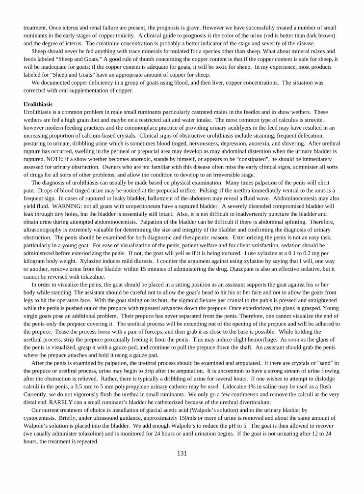
131
treatment. Once icterus and renal failure are present, the prognosis is grave. However we have successfully treated a number of small ruminants in the early stages of copper toxicity. A clinical guide to prognosis is the color of the urine (red is better than dark brown) and the degree of icterus. The creatinine concentration is probably a better indicator of the stage and severity of the disease.
Sheep should never be fed anything with trace minerals formulated for a species other than sheep. What about mineral mixes and feeds labeled “Sheep and Goats.” A good rule of thumb concerning the copper content is that if the copper content is safe for sheep, it will be inadequate for goats; if the copper content is adequate for goats, it will be toxic for sheep. In my experience, most products labeled for “Sheep and Goats” have an appropriate amount of copper for sheep.
We documented copper deficiency in a group of goats using blood, and then liver, copper concentrations. The situation was corrected with oral supplementation of copper. Urolithiasis Urolithiasis is a common problem in male small ruminants particularly castrated males in the feedlot and in show wethers. These wethers are fed a high grain diet and maybe on a restricted salt and water intake. The most common type of calculus is struvite, however modern feeding practices and the commonplace practice of providing urinary acidifyers in the feed may have resulted in an increasing proportion of calcium-based crystals. Clinical signs of obstructive urolithiasis include straining, frequent defecation, posturing to urinate, dribbling urine which is sometimes blood tinged, nervousness, depression, anorexia, and shivering. After urethral rupture has occurred, swelling in the perineal or prepucial area may develop as may abdominal distention when the urinary bladder is ruptured. NOTE: if a show whether becomes anorexic, stands by himself, or appears to be “constipated”, he should be immediately assessed for urinary obstruction. Owners who are not familiar with this disease often miss the early clinical signs, administer all sorts of drugs for all sorts of other problems, and allow the condition to develop to an irreversible stage.
The diagnosis of urolithiasis can usually be made based on physical examination. Many times palpation of the penis will elicit pain. Drops of blood tinged urine may be noticed at the prepucial orifice. Pulsing of the urethra immediately ventral to the anus is a frequent sign. In cases of ruptured or leaky bladder, ballotment of the abdomen may reveal a fluid wave. Abdominocentesis may also yield fluid. WARNING: not all goats with uroperitoneum have a ruptured bladder. A severely distended compromised bladder will leak through tiny holes, but the bladder is essentially still intact. Also, it is not difficult to inadvertently puncture the bladder and obtain urine during attempted abdominocentisis. Palpation of the bladder can be difficult if there is abdominal splinting. Therefore, ultrasonography is extremely valuable for determining the size and integrity of the bladder and confirming the diagnosis of urinary obstruction. The penis should be examined for both diagnostic and therapeutic reasons. Exteriorizing the penis is not an easy task, particularly in a young goat. For ease of visualization of the penis, patient welfare and for client satisfaction, sedation should be administered before exteriorizing the penis. If not, the goat will yell as if it is being tortured. I use xylazine at a 0.1 to 0.2 mg per kilogram body weight. Xylazine induces mild diuresis. I counter the argument against using xylazine by saying that I will, one way or another, remove urine from the bladder within 15 minutes of administering the drug. Diazepam is also an effective sedative, but it cannot be reversed with tolazaline.
In order to visualize the penis, the goat should be placed in a sitting position as an assistant supports the goat against his or her body while standing. The assistant should be careful not to allow the goat’s head to hit his or her face and not to allow the goats front legs to hit the operators face. With the goat sitting on its butt, the sigmoid flexure just cranial to the pubis is pressed and straightened while the penis is pushed out of the prepuce with repeated advances down the prepuce. Once exteriorized, the glans is grasped. Young virgin goats pose an additional problem. Their prepuce has never separated from the penis. Therefore, one cannot visualize the end of the penis-only the prepuce covering it. The urethral process will be extending out of the opening of the prepuce and will be adhered to the prepuce. Tease the process loose with a pair of forceps, and then grab it as close to the base is possible. While holding the urethral process, strip the prepuce proximally freeing it from the penis. This may induce slight hemorrhage. As soon as the glans of the penis is visualized, grasp it with a gauze pad, and continue to pull the prepuce down the shaft. An assistant should grab the penis where the prepuce attaches and hold it using a gauze pad.
After the penis is examined by palpation, the urethral process should be examined and amputated. If there are crystals or "sand" in the prepuce or urethral process, urine may begin to drip after the amputation. It is uncommon to have a strong stream of urine flowing after the obstruction is relieved. Rather, there is typically a dribbling of urine for several hours. If one wishes to attempt to dislodge calculi in the penis, a 3.5 mm to 5 mm polypropylene urinary catheter may be used. Lidocaine 1% in saline may be used as a flush. Currently, we do not vigorously flush the urethra in small ruminants. We only go a few centimeters and remove the calculi at the very distal end. RARELY can a small ruminant’s bladder be catheterized because of the urethral diverticulum.
Our current treatment of choice is installation of glacial acetic acid (Walpole’s solution) and to the urinary bladder by cystocentesis. Briefly, under ultrasound guidance, approximately 150mls or more of urine is removed and about the same amount of Walpole’s solution is placed into the bladder. We add enough Walpole’s to reduce the pH to 5. The goat is then allowed to recover (we usually administer tolazoline) and is monitored for 24 hours or until urination begins. If the goat is not urinating after 12 to 24 hours, the treatment is repeated.

132
A short-term alternative is perineal urethrotomy. I’ve become very hesitant to perform these because of their tendency to stricture after several months. We only recommend this procedure in goats that will be slaughtered in six months or less. Other adjunctive therapy includes the administration of anti-inflammatory drugs and antibiotics and fluids if indicated. We consider our success rate with Walpole’s solution to be very satisfactory and the cost is very reasonable.
From small ruminants of high-value, particularly rams and bucks, abdominal exploration and the creation of a tube cystostomy is a good alternative. A Foley catheter is placed in the bladder and exteriorized through the ventral abdominal wall. The catheter is left in place for 10 days or more to provide outflow for urine while the calculi in the urethra slough out, and the urethra regains its patency. The catheter can that be removed and the cystostomy will heal spontaneously.
Urinary acidification is recommended to prevent or reduce the formation of crystals. Ammonia chloride is the most frequently used substance to acidify the urine. It has been observed that after a period of weeks, the ammonium chloride is no longer effective, and the urine pH returns to above 7. We have some preliminary data that suggests dosing of ammonium chloride for 3 days per week with a 4-day rest period may prolong the period of efficacy. If the pH can be reduced to less than 6.5, small crystals that have formed may be dissolved, assuming that the crystals are struvite. Some calcium containing crystals do not dissolve in acidic urine and therefore are not amenable to treatment with Walpole's solution or prevention with the ammonium chloride.
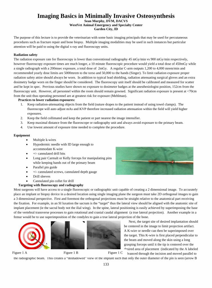
133
Imaging Basics in Minimally Invasive Osteosynthesis Sean Murphy, DVM, DACVS
WestVet Animal Emergency and Specialty Center Garden City, ID
The purpose of this lecture is to provide the veterinarian with some basic imaging principals that may be used for percutaneous procedures such as fracture repair and bone biopsy. Multiple imaging modalities may be used in such instances but particular attention will be paid to using the digital x-ray and fluoroscopy units. Radiation safety The radiation exposure rate for fluoroscopy is lower than conventional radiography 45 mGy/min vs 900 mGy/min respectively, however fluoroscopy exposure times are much longer, a 10 minute fluoroscopic procedure would yield a total dose of 450mGy while a single radiograph with a 200msec exposure, a total dose of 2mGy. A regular C-arm outputs 1,200 to 4,000 mrem/min and recommended yearly dose limits are 5000mrem to the torso and 50,000 to the hands (Singer). To limit radiation exposure proper radiation safety attire should always be worn. In addition to typical lead shielding, radiation attenuating surgical gloves and an extra dosimetry badge worn on the finger should be considered. The fluoroscopy unit itself should be calibrated and measured for scatter and be kept in spec. Previous studies have shown no exposure to dosimeter badges at the anesthesiologist position, 152cm from the fluoroscopy unit. However, all personnel within the room should remain gowned. Significant radiation exposure is present at <70cm from the unit thus operating personnel are at greatest risk for exposure (Mehlman).
Practices to lower radiation exposures: 1. Keep radiation-attenuating objects from the field (suture drapes to the patient instead of using towel clamps). The
fluoroscope will auto adjust mAs and KVP therefore increased radiation attenuation within the field will yield higher exposures.
2. Keep the field collimated and keep the patient or part nearest the image intensifier. 3. Keep maximal distance from the fluoroscope or radiography unit and always avoid exposure to the primary beam. 4. Use lowest amount of exposure time needed to complete the procedure.
Equipment
• Multiple k-wires • Hypodermic needle with ID large enough to
accommodate K-wire • +/- cannulated drill bits • Long pair Carmalt or Kelly forceps for manipulating pins
while keeping hands out of the primary beam • Parallel pin guide • +/- cannulated screws, cannulated depth gauge • Drill sleeves • Cannulated pin collet for drill
Targeting with fluoroscopy and radiography Most surgeons will have access to a single fluoroscopic or radiographic unit capable of creating a 2 dimensional image. To accurately place an implant or biopsy device in a desired location using single imaging plane the surgeon must take 2D orthogonal images to gain a 3 dimensional perspective. First and foremost the orthogonal projections must be straight relative to the anatomical part receiving the fixation. For example, in an SI luxation the sacrum is the “target” thus the lateral view should be aligned with the anatomic site of implant placement (ie the sacral body not the ilial wing). In the spine, lateral positioning is easily achieved by superimposing the base of the vertebral transverse processes to gain rotational and cranial caudal alignment (a true lateral projection). Another example in a femur would be to use superimposition of the condyles to gain a true lateral projection of the bone.
Next, the target site of desired implantation should be centered in the image to limit projection artifact. A K-wire or needle can then be superimposed over the target. This K-wire is first placed perpendicular to the beam and moved along the skin using a long grasping forceps until it the tip is centered over the desired area of placement (indicated by the A labeled
arrow in Fig 1A). A stab incision is then made at the tip of the pin and the pin is advanced through the incision and moved parallel to the radiographic beam. This creates a “deshadowed” view of the implant such that only the outer diameter of the pin is seen (arrow B
Figure 1 A Figure 1 B Figure 1 C
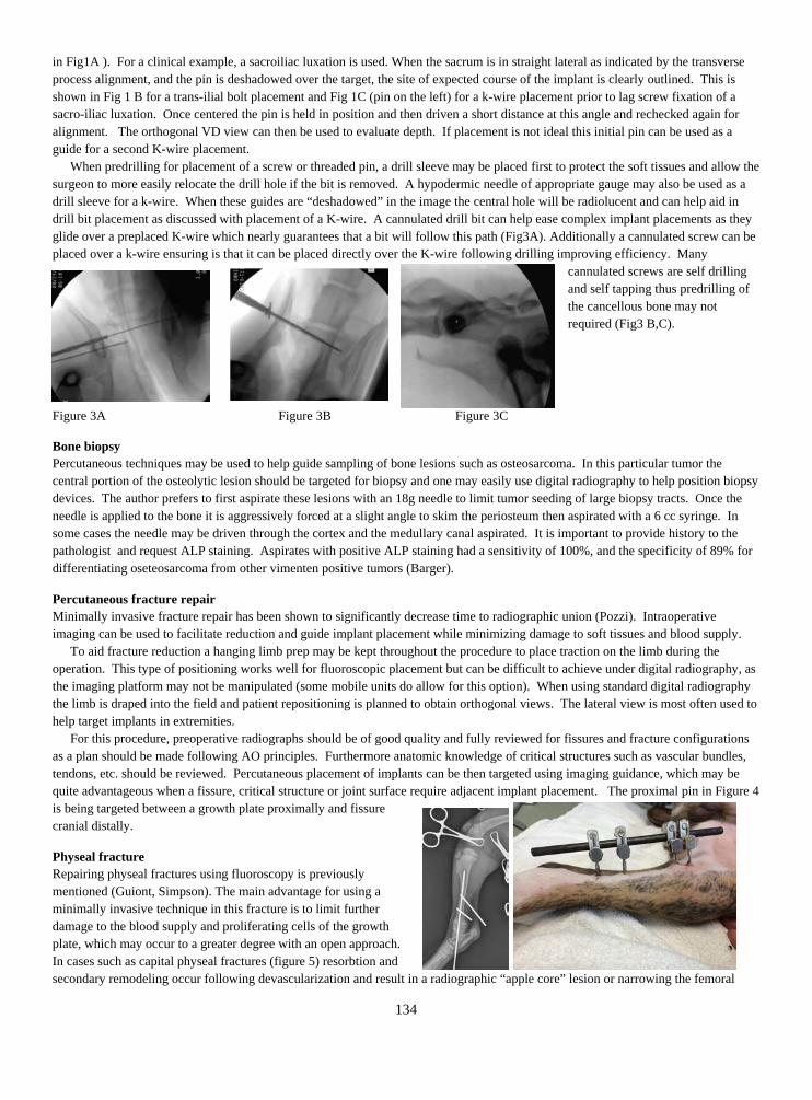
134
in Fig1A ). For a clinical example, a sacroiliac luxation is used. When the sacrum is in straight lateral as indicated by the transverse process alignment, and the pin is deshadowed over the target, the site of expected course of the implant is clearly outlined. This is shown in Fig 1 B for a trans-ilial bolt placement and Fig 1C (pin on the left) for a k-wire placement prior to lag screw fixation of a sacro-iliac luxation. Once centered the pin is held in position and then driven a short distance at this angle and rechecked again for alignment. The orthogonal VD view can then be used to evaluate depth. If placement is not ideal this initial pin can be used as a guide for a second K-wire placement.
When predrilling for placement of a screw or threaded pin, a drill sleeve may be placed first to protect the soft tissues and allow the surgeon to more easily relocate the drill hole if the bit is removed. A hypodermic needle of appropriate gauge may also be used as a drill sleeve for a k-wire. When these guides are “deshadowed” in the image the central hole will be radiolucent and can help aid in drill bit placement as discussed with placement of a K-wire. A cannulated drill bit can help ease complex implant placements as they glide over a preplaced K-wire which nearly guarantees that a bit will follow this path (Fig3A). Additionally a cannulated screw can be placed over a k-wire ensuring is that it can be placed directly over the K-wire following drilling improving efficiency. Many
cannulated screws are self drilling and self tapping thus predrilling of the cancellous bone may not required (Fig3 B,C).
Figure 3A Figure 3B Figure 3C Bone biopsy Percutaneous techniques may be used to help guide sampling of bone lesions such as osteosarcoma. In this particular tumor the central portion of the osteolytic lesion should be targeted for biopsy and one may easily use digital radiography to help position biopsy devices. The author prefers to first aspirate these lesions with an 18g needle to limit tumor seeding of large biopsy tracts. Once the needle is applied to the bone it is aggressively forced at a slight angle to skim the periosteum then aspirated with a 6 cc syringe. In some cases the needle may be driven through the cortex and the medullary canal aspirated. It is important to provide history to the pathologist and request ALP staining. Aspirates with positive ALP staining had a sensitivity of 100%, and the specificity of 89% for differentiating oseteosarcoma from other vimenten positive tumors (Barger). Percutaneous fracture repair Minimally invasive fracture repair has been shown to significantly decrease time to radiographic union (Pozzi). Intraoperative imaging can be used to facilitate reduction and guide implant placement while minimizing damage to soft tissues and blood supply.
To aid fracture reduction a hanging limb prep may be kept throughout the procedure to place traction on the limb during the operation. This type of positioning works well for fluoroscopic placement but can be difficult to achieve under digital radiography, as the imaging platform may not be manipulated (some mobile units do allow for this option). When using standard digital radiography the limb is draped into the field and patient repositioning is planned to obtain orthogonal views. The lateral view is most often used to help target implants in extremities.
For this procedure, preoperative radiographs should be of good quality and fully reviewed for fissures and fracture configurations as a plan should be made following AO principles. Furthermore anatomic knowledge of critical structures such as vascular bundles, tendons, etc. should be reviewed. Percutaneous placement of implants can be then targeted using imaging guidance, which may be quite advantageous when a fissure, critical structure or joint surface require adjacent implant placement. The proximal pin in Figure 4 is being targeted between a growth plate proximally and fissure cranial distally. Physeal fracture Repairing physeal fractures using fluoroscopy is previously mentioned (Guiont, Simpson). The main advantage for using a minimally invasive technique in this fracture is to limit further damage to the blood supply and proliferating cells of the growth plate, which may occur to a greater degree with an open approach. In cases such as capital physeal fractures (figure 5) resorbtion and secondary remodeling occur following devascularization and result in a radiographic “apple core” lesion or narrowing the femoral

135
neck in up to 70% of cases which may progress to fracture and collapse. Most physeal fractures are repaired in a minimally invasive fashion using percutaneous pins in parallel or cross pin fashion and intraoperative image guidance can be quite helpful to achieve pin guidance. References Singer. J. Occupational Radiation Exposure to the Surgeon. J AAOS. 2005 13(1). Mehlman CT, et al. Radiation exposure to the orthopaedic surgical team during fluoroscopy: "how far away is far enough?". J Orthop Trauma. 1997 Aug;11(6). Tomlinson JL, et al. Closed Reduction and Lag Screw Fixation of Sacroiliac Luxations and Fractures. Vet Surg 1999 Leasure CS, et al. Limited Open Reduction and Stabilization of Sacroiliac Fracture-Luxations Using Fluoroscopically Assisted Placement of a Trans-Iliosacral Rod in Five Dogs. Vet Surg 2007 Simpson, DJ and Lewis DD: Fractures of the femur. In Slatter DH: Text of Small Animal Surgery Vol 2, Saunders, 2002 Guiot et al. Fractures of the femur. In Tobia KM, editor: Veterinary Surgery Small Animal, Vol1, Saunders, 2012 Barger, A, et al. Use of Alkaline Phosphatase Staining to Differentiate Canine Osteosarcoma from Other Vimentin-positive Tumors Vet Pathol 42:161, 2005.
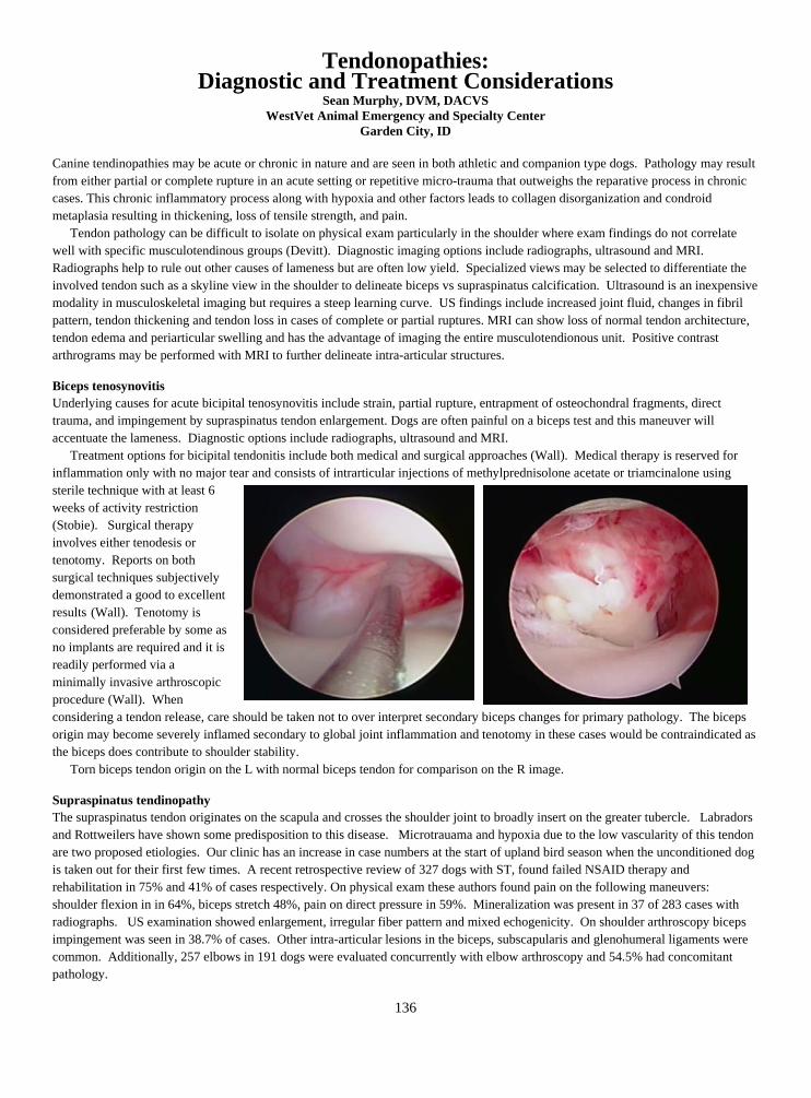
136
Tendonopathies: Diagnostic and Treatment Considerations
Sean Murphy, DVM, DACVS WestVet Animal Emergency and Specialty Center
Garden City, ID
Canine tendinopathies may be acute or chronic in nature and are seen in both athletic and companion type dogs. Pathology may result from either partial or complete rupture in an acute setting or repetitive micro-trauma that outweighs the reparative process in chronic cases. This chronic inflammatory process along with hypoxia and other factors leads to collagen disorganization and condroid metaplasia resulting in thickening, loss of tensile strength, and pain.
Tendon pathology can be difficult to isolate on physical exam particularly in the shoulder where exam findings do not correlate well with specific musculotendinous groups (Devitt). Diagnostic imaging options include radiographs, ultrasound and MRI. Radiographs help to rule out other causes of lameness but are often low yield. Specialized views may be selected to differentiate the involved tendon such as a skyline view in the shoulder to delineate biceps vs supraspinatus calcification. Ultrasound is an inexpensive modality in musculoskeletal imaging but requires a steep learning curve. US findings include increased joint fluid, changes in fibril pattern, tendon thickening and tendon loss in cases of complete or partial ruptures. MRI can show loss of normal tendon architecture, tendon edema and periarticular swelling and has the advantage of imaging the entire musculotendionous unit. Positive contrast arthrograms may be performed with MRI to further delineate intra-articular structures. Biceps tenosynovitis Underlying causes for acute bicipital tenosynovitis include strain, partial rupture, entrapment of osteochondral fragments, direct trauma, and impingement by supraspinatus tendon enlargement. Dogs are often painful on a biceps test and this maneuver will accentuate the lameness. Diagnostic options include radiographs, ultrasound and MRI.
Treatment options for bicipital tendonitis include both medical and surgical approaches (Wall). Medical therapy is reserved for inflammation only with no major tear and consists of intrarticular injections of methylprednisolone acetate or triamcinalone using sterile technique with at least 6 weeks of activity restriction (Stobie). Surgical therapy involves either tenodesis or tenotomy. Reports on both surgical techniques subjectively demonstrated a good to excellent results (Wall). Tenotomy is considered preferable by some as no implants are required and it is readily performed via a minimally invasive arthroscopic procedure (Wall). When considering a tendon release, care should be taken not to over interpret secondary biceps changes for primary pathology. The biceps origin may become severely inflamed secondary to global joint inflammation and tenotomy in these cases would be contraindicated as the biceps does contribute to shoulder stability.
Torn biceps tendon origin on the L with normal biceps tendon for comparison on the R image. Supraspinatus tendinopathy The supraspinatus tendon originates on the scapula and crosses the shoulder joint to broadly insert on the greater tubercle. Labradors and Rottweilers have shown some predisposition to this disease. Microtrauama and hypoxia due to the low vascularity of this tendon are two proposed etiologies. Our clinic has an increase in case numbers at the start of upland bird season when the unconditioned dog is taken out for their first few times. A recent retrospective review of 327 dogs with ST, found failed NSAID therapy and rehabilitation in 75% and 41% of cases respectively. On physical exam these authors found pain on the following maneuvers: shoulder flexion in in 64%, biceps stretch 48%, pain on direct pressure in 59%. Mineralization was present in 37 of 283 cases with radiographs. US examination showed enlargement, irregular fiber pattern and mixed echogenicity. On shoulder arthroscopy biceps impingement was seen in 38.7% of cases. Other intra-articular lesions in the biceps, subscapularis and glenohumeral ligaments were common. Additionally, 257 elbows in 191 dogs were evaluated concurrently with elbow arthroscopy and 54.5% had concomitant pathology.
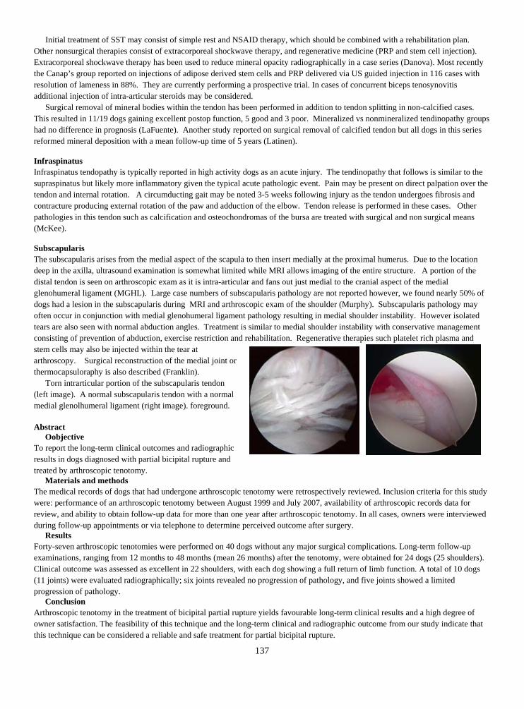
137
Initial treatment of SST may consist of simple rest and NSAID therapy, which should be combined with a rehabilitation plan. Other nonsurgical therapies consist of extracorporeal shockwave therapy, and regenerative medicine (PRP and stem cell injection). Extracorporeal shockwave therapy has been used to reduce mineral opacity radiographically in a case series (Danova). Most recently the Canap’s group reported on injections of adipose derived stem cells and PRP delivered via US guided injection in 116 cases with resolution of lameness in 88%. They are currently performing a prospective trial. In cases of concurrent biceps tenosynovitis additional injection of intra-articular steroids may be considered.
Surgical removal of mineral bodies within the tendon has been performed in addition to tendon splitting in non-calcified cases. This resulted in 11/19 dogs gaining excellent postop function, 5 good and 3 poor. Mineralized vs nonmineralized tendinopathy groups had no difference in prognosis (LaFuente). Another study reported on surgical removal of calcified tendon but all dogs in this series reformed mineral deposition with a mean follow-up time of 5 years (Latinen). Infraspinatus Infraspinatus tendopathy is typically reported in high activity dogs as an acute injury. The tendinopathy that follows is similar to the supraspinatus but likely more inflammatory given the typical acute pathologic event. Pain may be present on direct palpation over the tendon and internal rotation. A circumducting gait may be noted 3-5 weeks following injury as the tendon undergoes fibrosis and contracture producing external rotation of the paw and adduction of the elbow. Tendon release is performed in these cases. Other pathologies in this tendon such as calcification and osteochondromas of the bursa are treated with surgical and non surgical means (McKee). Subscapularis The subscapularis arises from the medial aspect of the scapula to then insert medially at the proximal humerus. Due to the location deep in the axilla, ultrasound examination is somewhat limited while MRI allows imaging of the entire structure. A portion of the distal tendon is seen on arthroscopic exam as it is intra-articular and fans out just medial to the cranial aspect of the medial glenohumeral ligament (MGHL). Large case numbers of subscapularis pathology are not reported however, we found nearly 50% of dogs had a lesion in the subscapularis during MRI and arthroscopic exam of the shoulder (Murphy). Subscapularis pathology may often occur in conjunction with medial glenohumeral ligament pathology resulting in medial shoulder instability. However isolated tears are also seen with normal abduction angles. Treatment is similar to medial shoulder instability with conservative management consisting of prevention of abduction, exercise restriction and rehabilitation. Regenerative therapies such platelet rich plasma and stem cells may also be injected within the tear at arthroscopy. Surgical reconstruction of the medial joint or thermocapsuloraphy is also described (Franklin).
Torn intrarticular portion of the subscapularis tendon (left image). A normal subscapularis tendon with a normal medial glenolhumeral ligament (right image). foreground. Abstract
Oobjective To report the long-term clinical outcomes and radiographic results in dogs diagnosed with partial bicipital rupture and treated by arthroscopic tenotomy.
Materials and methods The medical records of dogs that had undergone arthroscopic tenotomy were retrospectively reviewed. Inclusion criteria for this study were: performance of an arthroscopic tenotomy between August 1999 and July 2007, availability of arthroscopic records data for review, and ability to obtain follow-up data for more than one year after arthroscopic tenotomy. In all cases, owners were interviewed during follow-up appointments or via telephone to determine perceived outcome after surgery.
Results Forty-seven arthroscopic tenotomies were performed on 40 dogs without any major surgical complications. Long-term follow-up examinations, ranging from 12 months to 48 months (mean 26 months) after the tenotomy, were obtained for 24 dogs (25 shoulders). Clinical outcome was assessed as excellent in 22 shoulders, with each dog showing a full return of limb function. A total of 10 dogs (11 joints) were evaluated radiographically; six joints revealed no progression of pathology, and five joints showed a limited progression of pathology.
Conclusion Arthroscopic tenotomy in the treatment of bicipital partial rupture yields favourable long-term clinical results and a high degree of owner satisfaction. The feasibility of this technique and the long-term clinical and radiographic outcome from our study indicate that this technique can be considered a reliable and safe treatment for partial bicipital rupture.

138
References Cannapp S, et al. Suprasinatus Tendinopathy and Other Shoulder Problems. 17th ESVOT Congress 2014, Venice (Italy). Wall CR, Taylor R. Arthroscopic biceps brachii tenotomy as a treatment for canine bicipital tenosynovitis. J Am Anim Hosp Assoc 38:169-75, 2002 LaFuente et al. Surgical Treatment of Mineralized and Nonmineralized Supraspinatus Teninopathy in Tewnty-four Dogs. Vet Surg 38:380, 2009 Latinen O, Flo G. Mineralization of the Supraspinatus Tendon in Dogs: A Long-Term Follow-Up . JAHA 36:26, 2000 Murphy S, et al. Magnetic Resonance Imaging Findings in Dogs with Confirmed Shoulder Pathology. Veterinary Surgery 37:631–638, 2008. Stobie D, Wallace LJ, Lipowitz AJ, et al: Chronic bicipital tenosynovitis in dogs: 29 cases (1985-1992). J Am Vet Med Assoc 207:201-7, 1995 Kramer M, Gerwing M, Sheppard C, et al: Ultrasonography for the diagnosis of diseases of the tendon and tendon sheath of the biceps brachii muscle. Vet Surg 30:64-71, 2001 Muir P, KA Johnson: Supraspinatus and biceps brachii tendinopathy in dogs. J Small Animal Pract 35:239-243, 1994 Davidson EB, Griffey SM, Vasseur PB: Histopathologic, radiographic and arthrographic comparison of the biceps tendon in normal and dogs with biceps tenosynovitis. J Am Anim Hosp Assoc 36:522-30, 2000 McKee WM, et al. Ossification of the infraspinatus tendon-bursa in 13 dogs. Vet Record 161: 846-852, 2007 Danova NA, Muir, P. (2003) Extracorporeal shock wave therapy for supraspinatus calcifying tendinopathy in two dogs. Vet Record 152:208-209, 2003 PILAR LAFUENTE, M., FRANSSON, B. A., LINCOLN, J. D., MARTINEZ, S. A., GAVIN, P. R., LAHMERS, K. K. and GAY, J. M. (2009), Surgical Treatment of Mineralized and Nonmineralized Supraspinatus Tendinopathy in Twenty-four Dogs. Veterinary Surgery, 38: 380–387. doi: 10.1111/j.1532-950X.2009.00512.x Study performed at the College of Veterinary Medicine, Washington State University, Pullman, WA. Presented at the 16th Annual Scientific Meeting ECVS, Dublin, Ireland, June 28–30, 2007; and at the 2007 ACVS Veterinary Symposium, Chicago, IL, October 18–21, 2007. Vet Comp Orthop Traumatol. 2010;23(1):51-5. doi: 10.3415/VCOT-09-01-0005. Epub 2009 Dec 8. Long-term follow-up after arthroscopic tenotomy for partial rupture of the biceps brachii tendon. Bergenhuyzen AL1, Vermote KA, van Bree H, Van Ryssen B.
139
Forelimb Lameness: Case-Based Presentation
Sean Murphy, DVM, DACVS WestVet Animal Emergency and Specialty Center
Garden City, ID
The canine shoulder has the largest rage of motion of any joint. Passive stabilizers such as glenohumeral ligaments, joint capsule and joint fluid provide static stability while muculotendinous units consisting of the supraspinatus, infraspinatus, biceps, subscapularis, and teres minor provide active stability. Damage to any of the anatomic structures may result in lameness. However, sorting out pathology in this complex set of ligaments and tendons can be challenging. In addition, concurrent elbow pathology often may coincide further complicating these cases.
A detailed physical exam is crucial and first consists with a gait exam. Most commonly forelimb lameness is associated with decreased contact time during the stride phase of the gait resulting in reduced stride length. This may cause cervical motion to occur creating the always useful “down on sound” acronym to characterize the head bob. In many cases the lameness may be mild and difficult to characterize. Flexion and extension tests much like that used in equine medicine may help to induce lameness or isolate pain and should start distally with the foot and progress to the shoulder as it is more difficult to isolate range of motion in the proximal joints. A gait exam follows each joint manipulation to see if the lameness is increased with these motions. In occult lameness it may also be beneficial to have the owner exercise the animal prior to exam and withhold pain medications.
Next the standing orthopedic exam is performed, palpation and comparison of the spinatus musculature and prominence of the spine of the scapula and acromion will help to define atrophy in the affected limb. The animal must be in a square stance with equal weight bearing. Each joint is then palpated for swelling, joint effusion and periarticular fibrosis when moving up the limb. This palpation is again performed in the recumbent exam and each joint is tested for instability and ROM recalling that one starts distally.
Specialized tests in the forelimb exam involve internal rotation of the antebrachium with combined elbow flexion, which may increase pressure over the medial coronoid process to help further define pain in elbow dysplasia. The flexors on the medial aspect of the elbow are also palpated carefully for pain as an insertional tendinopathy is often present in conjuction with elbow dysplasia. In the shoulder, abduction angles may be measured using a goniometer with the animal in lateral recumbencey to evaluate medial shoulder stabilizers (Cook). Additionally internal and external rotation of the shoulder will evaluate lateral and medial stabilizers respectively. A biceps test places the shoulder in flexion and the elbow in extension to elongate the biceps yielding pain in animals with tendonitis or a partial tear.
In occult cases of lameness intra-articular anesthesia may be considered to block the lameness as well. An intra-articular injection of mepivicaine has improved lameness in 87% of dogs with elbow pathology and 28/30 dogs with shoulder lameness (Van Vynckt). Finally, gait analysis with pressure sensitive walkways or force plates may be considered in occult cases and to monitor treatment progress and provide objective gait information.
While many of these tests seem specific they were limited in their ability to predict arthroscopic assessed pathology (Devitt). Furthermore a full neurologic exam is performed as well paying close attention to neck discomfort References Van Vynckt D, et al. Diagnostic inta-articular anaethesia of the elbow in dogs with medial coronoid disease. VCOT 25(4):307, 2012. Van Vynckt D, et al. Anaesthetic arthrography of the shoulder joint in dogs. VCOT 26(4):291, 2013. Devitt C, et al. Relationship of Physical Examination Test of Shoulder Instability to Arthroscopic Findings in Dogs. Vet Surg 36:661, 2007 Cook JL, et al. Measurement of angles of abduction for diagnosis of shoulder instability in dogs using goniometry and digital image analysis. Vet Surg. 34:463, 2005
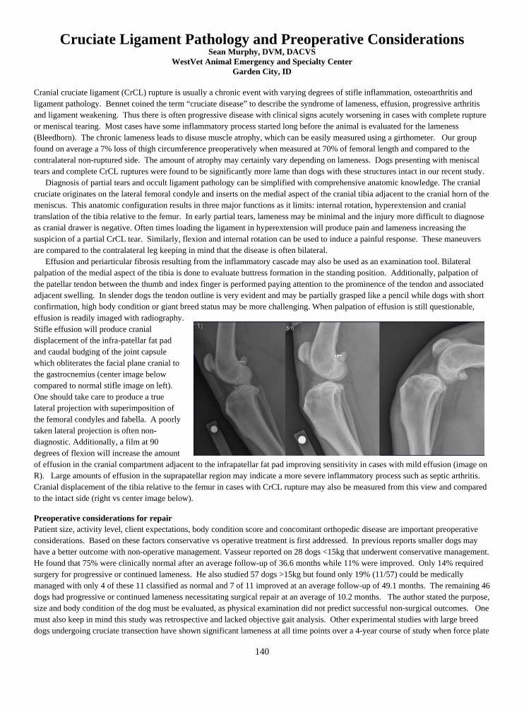
140
Cruciate Ligament Pathology and Preoperative Considerations Sean Murphy, DVM, DACVS
WestVet Animal Emergency and Specialty Center Garden City, ID
Cranial cruciate ligament (CrCL) rupture is usually a chronic event with varying degrees of stifle inflammation, osteoarthritis and ligament pathology. Bennet coined the term “cruciate disease” to describe the syndrome of lameness, effusion, progressive arthritis and ligament weakening. Thus there is often progressive disease with clinical signs acutely worsening in cases with complete rupture or meniscal tearing. Most cases have some inflammatory process started long before the animal is evaluated for the lameness (Bleedhorn). The chronic lameness leads to disuse muscle atrophy, which can be easily measured using a girthometer. Our group found on average a 7% loss of thigh circumference preoperatively when measured at 70% of femoral length and compared to the contralateral non-ruptured side. The amount of atrophy may certainly vary depending on lameness. Dogs presenting with meniscal tears and complete CrCL ruptures were found to be significantly more lame than dogs with these structures intact in our recent study.
Diagnosis of partial tears and occult ligament pathology can be simplified with comprehensive anatomic knowledge. The cranial cruciate originates on the lateral femoral condyle and inserts on the medial aspect of the cranial tibia adjacent to the cranial horn of the meniscus. This anatomic configuration results in three major functions as it limits: internal rotation, hyperextension and cranial translation of the tibia relative to the femur. In early partial tears, lameness may be minimal and the injury more difficult to diagnose as cranial drawer is negative. Often times loading the ligament in hyperextension will produce pain and lameness increasing the suspicion of a partial CrCL tear. Similarly, flexion and internal rotation can be used to induce a painful response. These maneuvers are compared to the contralateral leg keeping in mind that the disease is often bilateral.
Effusion and periarticular fibrosis resulting from the inflammatory cascade may also be used as an examination tool. Bilateral palpation of the medial aspect of the tibia is done to evaluate buttress formation in the standing position. Additionally, palpation of the patellar tendon between the thumb and index finger is performed paying attention to the prominence of the tendon and associated adjacent swelling. In slender dogs the tendon outline is very evident and may be partially grasped like a pencil while dogs with short confirmation, high body condition or giant breed status may be more challenging. When palpation of effusion is still questionable, effusion is readily imaged with radiography. Stifle effusion will produce cranial displacement of the infra-patellar fat pad and caudal budging of the joint capsule which obliterates the facial plane cranial to the gastrocnemius (center image below compared to normal stifle image on left). One should take care to produce a true lateral projection with superimposition of the femoral condyles and fabella. A poorly taken lateral projection is often non-diagnostic. Additionally, a film at 90 degrees of flexion will increase the amount of effusion in the cranial compartment adjacent to the infrapatellar fat pad improving sensitivity in cases with mild effusion (image on R). Large amounts of effusion in the suprapatellar region may indicate a more severe inflammatory process such as septic arthritis. Cranial displacement of the tibia relative to the femur in cases with CrCL rupture may also be measured from this view and compared to the intact side (right vs center image below). Preoperative considerations for repair Patient size, activity level, client expectations, body condition score and concomitant orthopedic disease are important preoperative considerations. Based on these factors conservative vs operative treatment is first addressed. In previous reports smaller dogs may have a better outcome with non-operative management. Vasseur reported on 28 dogs <15kg that underwent conservative management. He found that 75% were clinically normal after an average follow-up of 36.6 months while 11% were improved. Only 14% required surgery for progressive or continued lameness. He also studied 57 dogs >15kg but found only 19% (11/57) could be medically managed with only 4 of these 11 classified as normal and 7 of 11 improved at an average follow-up of 49.1 months. The remaining 46 dogs had progressive or continued lameness necessitating surgical repair at an average of 10.2 months. The author stated the purpose, size and body condition of the dog must be evaluated, as physical examination did not predict successful non-surgical outcomes. One must also keep in mind this study was retrospective and lacked objective gait analysis. Other experimental studies with large breed dogs undergoing cruciate transection have shown significant lameness at all time points over a 4-year course of study when force plate
141
gait analysis is used (Budsberg). Contrast this data with a study by Ballagas where experimental CrCL transection was followed by a TPLO, at 18 weeks there was no significant difference in weight bearing from baseline preoperative values.
A recent prospective clinical trial randomized overweight large breed dogs with unilateral CrCL rupture to undergo conservative therapy with weight loss, physical therapy and pain control versus TPLO surgery with these same treatments (Wucherer). Overall the surgical dogs had lower pain scores and significantly higher weight bearing on some outcome measures but not all. Dogs that had at least 85% of normal weight bearing were defined as having a successful outcome in the study. At 6 months and one year 33% and 64% of dogs treated non-surgically, and 93% and 75% of surgically treated dogs were defined as a successful outcome. Bilateral rupture was the most common reason dogs were taken out of the study and this may complicate conservative management. Future contralateral CrCL rupture is reported in 37% of cases at an average of 17 months and 59% if there are radiographic changes at initial evaluation (Vasseur).
Finally limb alignment, coexisting patellar luxation and patient size are important factors in choosing a surgical type. Osteotomy procedures may be considered in cases with malalignment or patellar luxation. Such surgeries become more complex often involving advanced imaging and planning. In giant breed dogs it is best to avoid lateral suture stabilization due to poorer outcomes in this group. Additionally, while there are many opinions on the “best technique” and the ”best technique in my hands”, discussion with a non-bias surgeon can point owners in a direction that is best for both the patient and owner. One should always use an evidence-based approach when possible.
A previous systematic review of the literature showed no one procedure as being superior. However, a more recent systematic review of additional studies favored TPLO over lateral suture for restoring dogs to normal function (Bergh). Regarding TTA outcomes, a recent Cornell study showed TTA restored dogs to normal function at a walk. However, when trotting over a force plate dogs receiving a TTA were significantly more lame than control dogs. Cases receiving a TPLO had no difference in weight bearing when compared to control dogs at 150–299 days, and >300 days. Dogs receiving a lateral suture repair had significantly lower weight bearing at a walk and trot at all time points throughout the study. Thus TPLO outperformed both procedures, and while TTA improved dogs, there was residual lameness at a trot.
TTA and TPLO were compared in a prospective randomized study with our group. Dogs receiving a TTA did well with short-term recovery, but weight bearing at 24wks was significantly higher in TPLO dogs. At 48 weeks TPLO had higher weight bearing that approached significance. Latent meniscal tears were a major complication in dogs receiving TTAs. No meniscal release was performed in either study group however 50% of dogs that had a complete CrCL rupture and an intact meniscus at the time of surgery went on to have a latent tear in the TTA group which was significantly higher than TPLO cases. Interesting a torn vs intact meniscus or a torn vs partially torn CrCL had no effect on weight bearing as measured by force plate gait analysis. We concluded that a meniscal release should be considered in dogs receiving a TTA and owners with higher performance expectations may consider a TPLO. References Vasseur P. Clinical Results Following Nonoperative Management for Rupture of the Cranial Cruciate Ligament in Dogs. Vet Surg 13:243, 1984. Nelson, S et al. Long-Term Functional Outcome of Tibial Plateau Leveling Osteotomy Versus Extracapsular Repair in a Heterogeneous Population of Dogs. Vet Surg 00:1, 2012. Budsberg, SC. Long-term temporal evaluation of ground reaction forces during developmentof experimentally induced osteoarthritis in dogs. AJVR 62(8):1207, 2001. Ballagas AJ, et al. Pre- and postoperative force plate analysis of dogs with experimentally transected cranial cruciate ligaments treated using tibial plateau leveling osteotomy. Vet Surg. 33(2):187, 2004. Gordon-Evans W, et al. Comparison of lateral fabellar suture and tibial plateau leveling osteotomy techniques for treatment of dogs with cranial cruciate ligament disease. JAVMA. 243:675, 2013. Wucherer KL, et al. Short-term and long-term outcomesfor overweight dogs with cranial cruciate ligament rupture treated surgically or nonsurgically. JAVMA. 242:1364, 2013 Bergh MS, et al. Systematic Review of Surgical Treatments for Cranial Cruciate Ligament Disease in Dogs. JAHA. 50:315, 2014 Krotscheck U, et al. Long‐Term Functional Outcome of Tibial Tuberosity Advancement Versus Tibial Plateau Leveling Osteotomy and Extracapsular Repair in a Heterogeneous Population of Dogs. Scientific Presentation Abstracts 2014 ACVS. San Diego, California Aragon CL, Budsberg SC: Applications of evidence-based medicine: cranial cruciate ligament injury repair in the dog. Vet Surg 34:93-8, 2005
142
Stifle Arthroscopy Sean Murphy, DVM, DACVS
WestVet Animal Emergency and Specialty Center Garden City, ID
Arthroscopy in veterinary medicine continues to develop and recent evidence shows increasing acceptance in clinical cases. The major benefits of arthroscopy are that it is minimally invasive and improves visualization. 1) Minimally invasive The standard stifle arthroscope diameter is 2.7mm, requiring only a small parapatellar portal for placement. Arthroscopic instruments (shaver, grasper, punch and knives) are of similar size and a contralateral portal is required for their use. This small form factor allows for significantly smaller incisions to be made over a typical arthrotomy. The proposed benefit is less tissue disruption resulting in early return to function and possibly less progression of OA.
The evidence: Millis found dogs returned to function sooner when a arthroscopic assisted approach was used to place a lateral suture over a typical open approach. Regarding OA development, a full parapatellar arthrotomy alone produced long standing degenerative change nearly equal to that of an arthrotomy with cruciate transection in experimental models (Järvinen M, Clin Orthopedics 1995). In clinical cases of CrCL rupture a full medial parapatellar arthrotomy was compared to a limited caudal medial arthrotomy in dogs undergoing TPLO. The limited arthrotomy group had significantly less OA progression on follow-up radiographs (Lineberger). 2) Improved visualization While the limited arthrotomy may limit OA development, the trade off is less visualization. A caudal medial arthrotomy was shown to be the least accurate technique in assessing meniscal tears in a cadaver model of a CrCL deficient stifle. A cranial medial approach had improved accuracy while arthroscopic examination had the highest (Pozzi). This is very important clinically and economically as missing a meniscal tear at the time of the initial surgery may result in progression of the tear and lameness with the need for additional surgery. Thieman showed cases explored with an arthrotomy were 3.8 times more likely to develop a meniscal tear after the initial surgery vs those undergoing arthroscopy without a meniscal release.
Improved visualization is also dependent on equipment and operator use. Newer and scope optics allow for a high definition image to be displayed to the user and current technology takes resolution to 4K. Image quality of in addition to magnification allow the trained clinician to gain a detailed analysis of the joint. Veterinary specific instruments and techniques are also improving allowing joint pathology to not only be defined but also treated. Currently, isolated meniscal tears, OCD lesions, traumatic (non congenital) patellar luxation are treated with arthroscopy alone. References Pozzi A, et al. Comparison of Arthroscopy and Arthrotomy for Diagnosis of Medial Meniscal Pathology: An Ex Vivo Study. Vet Surg 37:749, 2008. Thieman K, et al. Effect of meniscal release on rate of subsequent meniscal tears and owner-assessed outcome in dogs with cruciate disease treated with tibial plateau leveling osteotomy. Vet Surg. 35:705, 2006. Lineberger J, et al. Comparison of radiographic arthritic changes associated with two variations of tibial plateau leveling osteotomy. VCOT 18:13, 2005. Lineberger JA1, Allen DA, Wilson ER, Tobias TA, Shaiken LG, Shiroma JT, Biller DS, Lehenbauer TW. Jarvinen M, et al. Effect of anterior cruciate ligament reconstruction with patellar tendon or prosthetic ligament on the morphology of the other ligaments of the knee joint. An experimental study in dogs. Clin Orthop Relat Res. 311:176, 1995.
http://www.ncbi.nlm.nih.gov/pubmed/?term=Shaiken%20LG%5BAuthor%5D&cauthor=true&cauthor_uid=16594211�
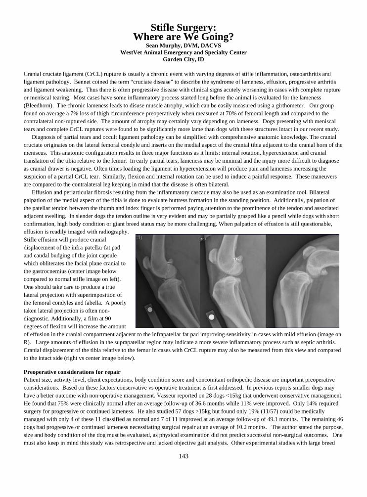
143
Stifle Surgery: Where are We Going?
Sean Murphy, DVM, DACVS WestVet Animal Emergency and Specialty Center
Garden City, ID
Cranial cruciate ligament (CrCL) rupture is usually a chronic event with varying degrees of stifle inflammation, osteoarthritis and ligament pathology. Bennet coined the term “cruciate disease” to describe the syndrome of lameness, effusion, progressive arthritis and ligament weakening. Thus there is often progressive disease with clinical signs acutely worsening in cases with complete rupture or meniscal tearing. Most cases have some inflammatory process started long before the animal is evaluated for the lameness (Bleedhorn). The chronic lameness leads to disuse muscle atrophy, which can be easily measured using a girthometer. Our group found on average a 7% loss of thigh circumference preoperatively when measured at 70% of femoral length and compared to the contralateral non-ruptured side. The amount of atrophy may certainly vary depending on lameness. Dogs presenting with meniscal tears and complete CrCL ruptures were found to be significantly more lame than dogs with these structures intact in our recent study.
Diagnosis of partial tears and occult ligament pathology can be simplified with comprehensive anatomic knowledge. The cranial cruciate originates on the lateral femoral condyle and inserts on the medial aspect of the cranial tibia adjacent to the cranial horn of the meniscus. This anatomic configuration results in three major functions as it limits: internal rotation, hyperextension and cranial translation of the tibia relative to the femur. In early partial tears, lameness may be minimal and the injury more difficult to diagnose as cranial drawer is negative. Often times loading the ligament in hyperextension will produce pain and lameness increasing the suspicion of a partial CrCL tear. Similarly, flexion and internal rotation can be used to induce a painful response. These maneuvers are compared to the contralateral leg keeping in mind that the disease is often bilateral.
Effusion and periarticular fibrosis resulting from the inflammatory cascade may also be used as an examination tool. Bilateral palpation of the medial aspect of the tibia is done to evaluate buttress formation in the standing position. Additionally, palpation of the patellar tendon between the thumb and index finger is performed paying attention to the prominence of the tendon and associated adjacent swelling. In slender dogs the tendon outline is very evident and may be partially grasped like a pencil while dogs with short confirmation, high body condition or giant breed status may be more challenging. When palpation of effusion is still questionable, effusion is readily imaged with radiography. Stifle effusion will produce cranial displacement of the infra-patellar fat pad and caudal budging of the joint capsule which obliterates the facial plane cranial to the gastrocnemius (center image below compared to normal stifle image on left). One should take care to produce a true lateral projection with superimposition of the femoral condyles and fabella. A poorly taken lateral projection is often non-diagnostic. Additionally, a film at 90 degrees of flexion will increase the amount of effusion in the cranial compartment adjacent to the infrapatellar fat pad improving sensitivity in cases with mild effusion (image on R). Large amounts of effusion in the suprapatellar region may indicate a more severe inflammatory process such as septic arthritis. Cranial displacement of the tibia relative to the femur in cases with CrCL rupture may also be measured from this view and compared to the intact side (right vs center image below). Preoperative considerations for repair Patient size, activity level, client expectations, body condition score and concomitant orthopedic disease are important preoperative considerations. Based on these factors conservative vs operative treatment is first addressed. In previous reports smaller dogs may have a better outcome with non-operative management. Vasseur reported on 28 dogs <15kg that underwent conservative management. He found that 75% were clinically normal after an average follow-up of 36.6 months while 11% were improved. Only 14% required surgery for progressive or continued lameness. He also studied 57 dogs >15kg but found only 19% (11/57) could be medically managed with only 4 of these 11 classified as normal and 7 of 11 improved at an average follow-up of 49.1 months. The remaining 46 dogs had progressive or continued lameness necessitating surgical repair at an average of 10.2 months. The author stated the purpose, size and body condition of the dog must be evaluated, as physical examination did not predict successful non-surgical outcomes. One must also keep in mind this study was retrospective and lacked objective gait analysis. Other experimental studies with large breed
144
dogs undergoing cruciate transection have shown significant lameness at all time points over a 4-year course of study when force plate gait analysis is used (Budsberg). Contrast this data with a study by Ballagas where experimental CrCL transection was followed by a TPLO, at 18 weeks there was no significant difference in weight bearing from baseline preoperative values.
A recent prospective clinical trial randomized overweight large breed dogs with unilateral CrCL rupture to undergo conservative therapy with weight loss, physical therapy and pain control versus TPLO surgery with these same treatments (Wucherer). Overall the surgical dogs had lower pain scores and significantly higher weight bearing on some outcome measures but not all. Dogs that had at least 85% of normal weight bearing were defined as having a successful outcome in the study. At 6 months and one year 33% and 64% of dogs treated non-surgically, and 93% and 75% of surgically treated dogs were defined as a successful outcome. Bilateral rupture was the most common reason dogs were taken out of the study and this may complicate conservative management. Future contralateral CrCL rupture is reported in 37% of cases at an average of 17 months and 59% if there are radiographic changes at initial evaluation (Vasseur).
Finally limb alignment, coexisting patellar luxation and patient size are important factors in choosing a surgical type. Osteotomy procedures may be considered in cases with malalignment or patellar luxation. Such surgeries become more complex often involving advanced imaging and planning. In giant breed dogs it is best to avoid lateral suture stabilization due to poorer outcomes in this group. Additionally, while there are many opinions on the “best technique” and the ”best technique in my hands”, discussion with a non-bias surgeon can point owners in a direction that is best for both the patient and owner. One should always use an evidence-based approach when possible.
A previous systematic review of the literature showed no one procedure as being superior. However, a more recent systematic review of additional studies favored TPLO over lateral suture for restoring dogs to normal function (Bergh). Regarding TTA outcomes, a recent Cornell study showed TTA restored dogs to normal function at a walk. However, when trotting over a force plate dogs receiving a TTA were significantly more lame than control dogs. Cases receiving a TPLO had no difference in weight bearing when compared to control dogs at 150–299 days, and >300 days. Dogs receiving a lateral suture repair had significantly lower weight bearing at a walk and trot at all time points throughout the study. Thus TPLO outperformed both procedures, and while TTA improved dogs, there was residual lameness at a trot.
TTA and TPLO were compared in a prospective randomized study with our group. Dogs receiving a TTA did well with short-term recovery, but weight bearing at 24wks was significantly higher in TPLO dogs. At 48 weeks TPLO had higher weight bearing that approached significance. Latent meniscal tears were a major complication in dogs receiving TTAs. No meniscal release was performed in either study group however 50% of dogs that had a complete CrCL rupture and an intact meniscus at the time of surgery went on to have a latent tear in the TTA group which was significantly higher than TPLO cases. Interesting a torn vs intact meniscus or a torn vs partially torn CrCL had no effect on weight bearing as measured by force plate gait analysis. We concluded that a meniscal release should be considered in dogs receiving a TTA and owners with higher performance expectations may consider a TPLO. References Vasseur P. Clinical Results Following Nonoperative Management for Rupture of the Cranial Cruciate Ligament in Dogs. Vet Surg 13:243, 1984. Nelson, S et al. Long-Term Functional Outcome of Tibial Plateau Leveling Osteotomy Versus Extracapsular Repair in a Heterogeneous Population of Dogs. Vet Surg 00:1, 2012. Budsberg, SC. Long-term temporal evaluation of ground reaction forces during developmentof experimentally induced osteoarthritis in dogs. AJVR 62(8):1207, 2001. Ballagas AJ, et al. Pre- and postoperative force plate analysis of dogs with experimentally transected cranial cruciate ligaments treated using tibial plateau leveling osteotomy. Vet Surg. 33(2):187, 2004. Gordon-Evans W, et al. Comparison of lateral fabellar suture and tibial plateau leveling osteotomy techniques for treatment of dogs with cranial cruciate ligament disease. JAVMA. 243:675, 2013. Wucherer KL, et al. Short-term and long-term outcomesfor overweight dogs with cranial cruciate ligament rupture treated surgically or nonsurgically. JAVMA. 242:1364, 2013 Bergh MS, et al. Systematic Review of Surgical Treatments for Cranial Cruciate Ligament Disease in Dogs. JAHA. 50:315, 2014 Krotscheck U, et al. Long‐Term Functional Outcome of Tibial Tuberosity Advancement Versus Tibial Plateau Leveling Osteotomy and Extracapsular Repair in a Heterogeneous Population of Dogs. Scientific Presentation Abstracts 2014 ACVS. San Diego, California Aragon CL, Budsberg SC: Applications of evidence-based medicine: cranial cruciate ligament injury repair in the dog. Vet Surg 34:93-8, 2005
145
Dogmas of Clinical Pathology: Adjusted Calcium, Modified Transudates,
Acidemias of Acidoses, and More Steve Stockham, DVM, MS, DACVP
Kansas State University Manhattan, KS
Ken Jenning’s wrote Because I Said So! The Truth Behind the Myths, Tales, and Warnings Every Generation Passes Down to its Kids. In his book, he considered the evidence that supports or refutes common dogmas within the United States. For example, “Was the nail you stepped on rusty? You’ll get tetanus!” Is the statement true, sometimes true, sometimes false, or false? Or, “Your first answer is usually the right one.” Is the statement true, sometimes true, sometimes false, or false?
A dogma is “a settled or established opinion, belief, or principle.” “Blind belief in authority is the greatest enemy of truth.” (Albert Einstein) “Education has failed in a very serious way to convey the most important lesson science can teach: skepticism.” (David Suzuki) “Most institutions demand unqualified faith; but the institution of science makes skepticism a virtue.” (Robert King Merton)
Are dogmas of clinical pathology valid? The following sections explore a few dogmas that have been or are being passed down from one generation to the next generation of veterinarians.
Adjusted calcium formula can be used to determine if the hypocalcemia is due to hypoalbuminemia. This dogma initially arose from a retrospective study that was published in 1982 (JAVMA 180: 63-67, 1982) in which two formulas were derived from measured concentrations of serum tCa2+, albumin, and total protein (values expressed in non-SI units).
• Canine-adjusted [tCa2+] = measured [tCa2+] – [Alb] + 3.5 • Canine-adjusted [tCa2+] = measured [tCa2+] – 0.4 × [TP] + 3.3
The proposed concept for the calculated adjusted [tCa2+] values was that if the value was within the reference interval for [tCa2+], the hypocalcemia was due to hypoalbuminemia (or hypoproteinemia) and there is not a decrease in the [fCa2+]. If the calculated adjusted [tCa2+] was decreased, then there was a decreased [fCa2+]. [Note: fCa2+ (free calcium ion) is frequently called ionized calcium even though all calcium in the body is ionized; some Ca2+ ion exists as free ions; other Ca2+ ions are bound to a variety of anions.]
There are three major aspects of the 1982 article that are frequently ignored. The derived formulas represented the regression lines for the raw data that contained considerable individual animal variation. Considering the 95 %–confidence intervals for the regression line, the formulas should be as follows. Accordingly, there is considerable variability in the calculated adjusted [tCa2+] values.
• Canine-adjusted [tCa2+] = measured [tCa2+] – [Alb] + 3.5 ± 1.3 • Canine-adjusted [tCa2+] = measured [tCa2+] – 0.4 × [TP] + 3.3 ± 1.6
Second, the authors stated that about one-third of the variability in the [tCa2+] was due to changes in albumin concentrations. Lastly, the formulas were derived from data obtained in one clinical laboratory many years ago and people use the formulas for data obtained from different analytical methods without establishing analytical agreement.
Some of the same authors of the 1982 article wrote another article that was published in 2005 (Am J Vet Res 66: 1330 – 1336, 2005). They concluded that adjusted total Ca2+ concentrations are unacceptable for predicting free Ca2+ (ionized calcium) status in dogs.
The adjusted calcium statement is mostly false. However, adjusted calcium formulas do emphasize that the total protein and albumin concentrations do influence a patient’s [tCa2+] and thus should be considered when interpreting laboratory data. Pseudohypocalcemia is present when the hypocalcemia is due to hypoalbuminemia (or hypoproteinemia). This more recent statement should not be used. When the [tCa2+] is decreased, there is a hypocalcemia if there is or is not a hypoalbuminemia or hypoproteinemia present. Those who wish to use the term “pseudohypocalcemia” in this context should consider what they should call the neutrophilia that occurs due to shifting of cells from marginated to circulating pools, or the hyperproteinemia that occurs due to decreased plasma water, or the erythrocytosis that occurs due to splenic contraction. Just because there is not a convenient term for a decreased [fCa2+], let’s not use terms that are incorrect. In acute inflammation, the release of endogenous cortisol causes the lymphopenia or Stress of the acute inflammatory disease causes a lymphopenia. These statements have been in the veterinary literature for decades – but where is the evidence that they are true? The statements reflect the concept that the inflammatory state stresses the animal sufficiently to cause a release of cortisol which induces the movement of lymphocytes from the circulating blood and thus a lymphopenia develops.
Increased cortisol activity (or activity of other glucocorticoids) are known to create a lymphopenia. If the lymphopenia in an acute inflammatory state is due to excess cortisol, should we also see other evidence of excess cortisol such as mature neutrophilia,
146
monocytosis, hyperglycemia, increased ALP activity (dogs), hypercholesterolemia, or polyuria? Why do we accept stress as the cause of the lymphopenia when we do not find other clinical abnormalities that are attributable to excess cortisol?
In a 1995 article in Adv. Immunol., B.A. Imhof described the effects of inflammatory cytokines on blood leukocytes. There is evidence that the cytokines promote the homing of blood lymphocytes to lymph nodes and the migration of lymphocytes to inflamed tissues; these processes can create the inflammatory lymphopenia.
We have evidence that the acute inflammatory reaction alters the movement of blood lymphocytes to create a lymphopenia. To my knowledge, we do not have evidence that inflammation creates a sufficient increase on plasma cortisol to cause a lymphopenia.
A transudate occurs because of hypoalbuminemia; usually when plasma [albumin] is < 1.5 g/dL (or < 1.2 g/dL, or < 1.8 g/dL) Or A pure transudate is hypocellular (< 1000/µL) and has a TS concentration < 2.5 g/dL. First, let’s consider the statement that “a transudate occurs because of hypoalbuminemia.” An inherited disorder is recognized in people in which there is no synthesis of albumin by hepatocytes; i.e., analbuminemia. Their albumin concentrations are < 0.1 g/dL and they typically do not develop pleural or peritoneal transudates. How can we attribute the formation of transudates to hypoalbuminemia when people with analbuminemia do not have transudative effusions? Also, how can we state that certain albumin concentrations lead to transudation when analbuminemia does not lead cavitary transudates?
A transudate is an effusion produced by changes in mechanic factors such as oncotic pressure or hydraulic pressure in capillary beds. Basically, the determining factor for the accumulation of cavitary transudates is the difference between the hydraulic pressure gradient (hydraulic pressure within vessels – hydraulic pressure in interstitial fluid) and the oncotic pressure gradient (oncotic pressure within vessels – oncotic pressure in interstitial fluid). When this difference leads to more fluid leaving the vascular bed than what can be removed by lymphatic vessels, a transudate forms. If transudation occurs in blood vessels that have minimal protein permeability, then a protein-poor transudate accumulates.
It is important to recognize that the plasma oncotic pressure is due to both albumin and globulins; albumin molecules are the major contributors to oncotic pressure but combined contributions of the globulin molecules are also important.
Two common canine disorders that cause the formation of protein-poor transudates are protein-losing nephropathies and hepatic cirrhosis. In these disorders, hypoproteinemia does reduce the plasma oncotic pressure but there also is an increased hydraulic pressure gradient in the portal blood vessels created by the retention of Na+ and H2O. The combination results in transudation and the formation of protein-poor transudates; the transudation is not solely caused by hypoalbuminemia.
A less common reason for the formation of a protein-poor transudate is presinusoidal portal hypertension. In this state, there is an increased hydraulic pressure gradient in the portal blood vessels but not a hypoproteinemia. Accordingly, the transudation is not caused by hypoalbuminemia.
For the second statement, (A pure transudate is hypocellular (< 1000/µL) is typically true as there is no reason for the migration of leukocytes from blood to the cavitary fluid. However, the second portion of the statement (TS concentration < 2.5 g/dL) may or may not be true.
It is important to recognize that a serum or plasma “total solids concentration” is not equal to a “total protein concentration.” The total protein concentration is due to the concentrations of albumin and globulins. The total solids concentration includes the total protein concentration plus the concentrations of all other solids in the serum or plasma; i.e., glucose, urea, electrolytes, and other solutes. This data in the following table was extracted from a complete table in Wolf AV:
Aqueous solutions and body fluids. their concentrative properties and conversion tables, 1966. Human plasma [TP] (g/dL) 0.8 1.0 2.0 3.0 Human plasma [TS] (g/dL) 2.5 2.7 3.6 4.7
There are clinical refractometers that have a calibrated total solids scale (e.g., TS Meter Refractometer Model 10400B, Leica Microsystems). Most clinical refractometers have a calibrated total protein scale (even those that are called “TS Meters”) and the lowest unit commonly on the scale is 2.5 g/dL. The [TS] for a protein-poor transudate may be < 2.5 g/dL, but that should not be confused with a [TP] of < 2.5 g/dL. A modified transudate is a transudate that has been modified by the addition of cells or protein. A modified transudate has a higher TS concentration than a pure transudate and moderate cellularity. A modified transudate has 1,000–7,000 cells/µL and a variable protein concentration (2.5–5.0 g/dL). Using one or more of these definitions or criteria, a variety of cavitary effusions have been classified as modified transudates including the effusions of heart failure, feline infectious peritonitis, noninfectious exudates, hemorrhagic effusions, chylous effusions, uroperitoneum, neoplastic effusions, and bilious exudates. If we define a transudate as “an effusion produced by changes in mechanic factors such as oncotic pressure or hydraulic pressure
The heart-failure effusions form when there is an increased hydraulic pressure gradient within blood vessels that are permeable to proteins. The classic mechanism occurs when central vein or hepatic vein congestion lead to increased hydraulic pressure with hepatic
in capillary beds,” then only the heart failure effusion qualifies as a transudate. None of the other effusions form via transudation and thus should not be called transudates or modified transudates.
147
sinusoids and the pressure forces out an excess amount of protein-rich fluid. When lymphatic vessels are not able to compensate adequately, then a protein-rich transudate accumulates. Pulmonary vessels are also protein-permeable, but not to the same degree as the hepatic sinusoids. The acidemia of a lactic acidosis is due to increased production of lactic acid by cells. This statement sounds logical but it does not reflect the true changes in biochemical pathways that occur in lactic acidosis. The cause of the acidemia was addressed in an article by S.C. Dennis, et al (J. Mol. Cell Cardiol. 23: 1077–1086, 1991).
When tissues have an inadequate supply of oxygen (i.e., when hypoxia is present), the cells attempt to generate ATP via anaerobic respiration (fermentation) (also called anaerobic glycolysis). In the final reaction and for each glucose molecule, this reaction occurs and is catalyzed by lactate dehydrogenase: 2 pyruvate− + 2 NADH + 2 H+ 2 L-lactate− + 2 NAD+. It should be noted that L-lactate (an anion) is formed and not lactic acid; it should also be noted that H+ is consumed in the reaction and thus makes the medium more alkaline, not more acidic.
Anaerobic respiration is an inefficient method of generating ATP from glucose; only 2 ATP molecules are produce for each glucose molecule. When there is an inadequate formation of ATP, the cells start the rapid hydrolysis of ATP to ADP and finally AMP. For each ATP molecule that is converted to AMP, 2 H+ ions are formed.
One might say – if there is excessive formation of L-lactate and the excessive formation of H+, doesn’t that mean there is excessive formation of lactic acid? Considering the pKa of lactic acid is 3.86, the ratio of lactate to lactic acid at a physiologic pH is greater than 1000:1.
The acidemia that occurs in animals with a lactic acidosis is due to excessive ATP hydrolysis in hypoxic tissues; not excessive formation of lactic acid. The acidemia of a ketoacidosis is due to increased production of ketoacids by hepatocytes. This statement sounds logical but it does not reflect the true changes in biochemical pathways that occur in ketoacidosis. The cause of the acidemia was addressed in an article by K.G. Alberti (Ciba Found. Symp. 87: 1–19, 1982).
The process called ketogenesis involves the conversion of 3-hydroxy-3-methylglutaryl-CoA (3HMGCoA) to acetoacetate, β-hydroxybutyrate, and acetone (the traditional ketone bodies). This process actually consumes H+ and the molecules formed are not acids (i.e., not acetoacetic acid or β-hydroxybutyric acid).
As explained by Alberti, the excess generation of H+ it ketoacidosis occurs before ketogenesis and not during ketogenesis. The greatest amount of H+ is formed from triglyceride molecules when there is β-oxidation of fatty acids to AcCoA in hepatocytes. The processes of triglyceride lipolysis in adipose tissue and the conversion of AcCoA to 3HMGCoA also generate H+.
The acidemia in animals with a ketoacidosis is due to the excessive formation of H+ during the mobilization and catabolism of triglycerides when there is a negative energy status; not due to ketogenesis or the formation of ketoacids.
The increased anion gap seen with renal failure is due the accumulation of uremic acids. This statement sounds logical. When there is a true increase in the anion gap concentration, there is an increased concentration of anions other than Cl− or HCO3
− in the serum/plasma. Are the acids anions? When an animal is in renal failure, the decreased glomerular filtration rate leads to an accumulation of phosphates, sulfates, and
citrate in plasma. At a pH of 7.4, most of the phosphates exist as HPO42− and a lesser amount of H2PO4
− (both anions and both acids). The sulfates exist mostly as SO4
2− and a minute amount of HSO4− (both anions, SO4
2− is not an acid). Citrate exists as an anion, there is very little citric acid present at a pH of 7.4; citric acid is not an anion.
As some of the “uremic acids” do exist as anions at a pH of 7.4, the statement is partially true. However to reduce confusion, I attempt to consistently state that increased anion gap concentration is due to anions other than Cl− and HCO3
−. The increased serum osmolality is due to dehydration (i.e., ↓ plasma H2O). It is important to recognize that serum osmolality represents the total concentration of the solutes in the serum and usually dehydration is not the reason for an increase concentration of solutes. The three major reasons for hyperosmolal serum are azotemia (increase urea concentration), hyperglycemia, and presence of exogenous solutes (e.g., ethylene glycol or mannitol). Dehydration does not cause hyperglycemia or an excess of exogenous solutes. Dehydration can lead to azotemia, but only when dehydration creates sufficient hypovolemia to lead to a prerenal azotemia. Decreased plasma H2O by itself does not create a significant increase in urea concentration.
When dehydration results in hypernatremia and hyperchloremia, then dehydration is the cause of the increased serum osmolality. However, most dehydrated animals do not have hypernatremia and hyperchloremia. Hypernatremic dehydration occurs when there is a loss of “pure water” as it occurs in central and renal diabetes insipidus and when there is an insensible loss of water via respiration. Another cause of hypernatremic dehydration occurs when an animal does not have access to water (e.g., frozen water tank).
148
A measured or a calculated osmolality should not be used to establish the presence or absence of dehydration in an animal. Dehydration is usually not the cause of hyperosmolal serum. An increase in [Pi] will cause the [tCa2+] to decrease because of the calcification of tissues. Or When Ca X P is > 70, soft tissue calcification is likely; mineralization occurs if when > 90. The concept of the Ca/P product is based on the mass-law concepts in which higher concentrations of Ca2+ or PO4 will shift this reaction (Ca2+ + PO4
3− Ca3(PO4)3 ) to the right and thus more Ca3(PO4)3 forms. On the surface, this concept is flawed because not all of the measured [tCa2+] is present as free Ca2+ and thus is not available to participate in the reaction. Second, very little of the serum inorganic phosphorus concentration exists as PO4
3−. Also, when Ca2+ & PO43− were added to human plasma, precipitation did
not occur until the Ca/P product was > 200 (O’Neill W.C.: Kidney International 72: 2007). If the Ca/P product concept is not valid, is it true that “An increase in [Pi] will cause the [tCa2+] to decrease”?
If there is a prolonged increase in plasma [PO4] (as it occurs in chronic renal disease), the PO4 inhibits renal 1-hydroxylase and thus there is less conversion of calcidiol to calcitriol. Lower calcitriol concentrations do lead to lower [fCa2+] (thus lower [tCa2+]) due to less intestinal absorption of Ca2+, less mobilization of Ca2+ from bone, and more renal excretion of Ca2+.
If there is a rapid increase in plasma [PO4], colloidal complexes of Ca2+ and PO4 form in plasma and the complexes are engulfed by macrophages and the plasma [tCa2+] decreases.
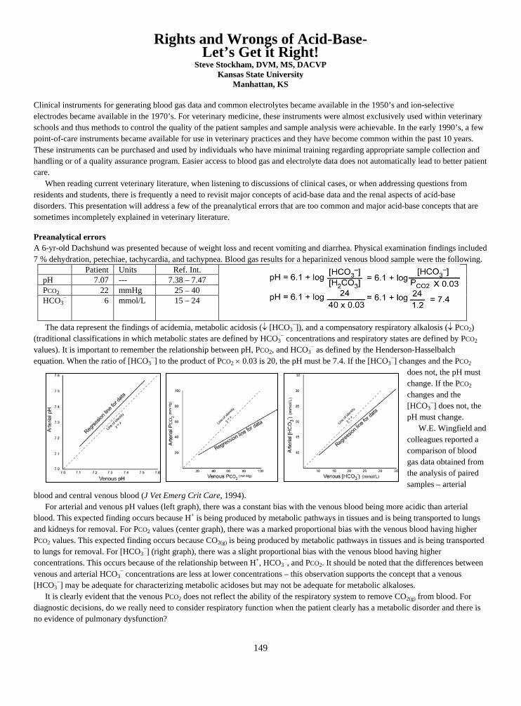
149
Rights and Wrongs of Acid-Base- Let’s Get it Right!
Steve Stockham, DVM, MS, DACVP Kansas State University
Manhattan, KS
Clinical instruments for generating blood gas data and common electrolytes became available in the 1950’s and ion-selective electrodes became available in the 1970’s. For veterinary medicine, these instruments were almost exclusively used within veterinary schools and thus methods to control the quality of the patient samples and sample analysis were achievable. In the early 1990’s, a few point-of-care instruments became available for use in veterinary practices and they have become common within the past 10 years. These instruments can be purchased and used by individuals who have minimal training regarding appropriate sample collection and handling or of a quality assurance program. Easier access to blood gas and electrolyte data does not automatically lead to better patient care.
When reading current veterinary literature, when listening to discussions of clinical cases, or when addressing questions from residents and students, there is frequently a need to revisit major concepts of acid-base data and the renal aspects of acid-base disorders. This presentation will address a few of the preanalytical errors that are too common and major acid-base concepts that are sometimes incompletely explained in veterinary literature. Preanalytical errors A 6-yr-old Dachshund was presented because of weight loss and recent vomiting and diarrhea. Physical examination findings included 7 % dehydration, petechiae, tachycardia, and tachypnea. Blood gas results for a heparinized venous blood sample were the following.
Patient Units Ref. Int.
pH 7.07 --- 7.38 – 7.47 PCO2 22 mmHg 25 – 40 HCO3
− 6 mmol/L 15 – 24
The data represent the findings of acidemia, metabolic acidosis (↓ [HCO3−]), and a compensatory respiratory alkalosis (↓ PCO2)
(traditional classifications in which metabolic states are defined by HCO3− concentrations and respiratory states are defined by PCO2
values). It is important to remember the relationship between pH, PCO2, and HCO3− as defined by the Henderson-Hasselbalch
equation. When the ratio of [HCO3−] to the product of PCO2 × 0.03 is 20, the pH must be 7.4. If the [HCO3
−] changes and the PCO2 does not, the pH must change. If the PCO2 changes and the [HCO3
−] does not, the pH must change.
W.E. Wingfield and colleagues reported a comparison of blood gas data obtained from the analysis of paired samples – arterial
blood and central venous blood (J Vet Emerg Crit Care, 1994). For arterial and venous pH values (left graph), there was a constant bias with the venous blood being more acidic than arterial
blood. This expected finding occurs because H+ is being produced by metabolic pathways in tissues and is being transported to lungs and kidneys for removal. For PCO2 values (center graph), there was a marked proportional bias with the venous blood having higher PCO2 values. This expected finding occurs because CO2(g) is being produced by metabolic pathways in tissues and is being transported to lungs for removal. For [HCO3
−] (right graph), there was a slight proportional bias with the venous blood having higher concentrations. This occurs because of the relationship between H+, HCO3
−, and PCO2. It should be noted that the differences between venous and arterial HCO3
− concentrations are less at lower concentrations – this observation supports the concept that a venous [HCO3
−] may be adequate for characterizing metabolic acidoses but may not be adequate for metabolic alkaloses. It is clearly evident that the venous PCO2 does not reflect the ability of the respiratory system to remove CO2(g) from blood. For
diagnostic decisions, do we really need to consider respiratory function when the patient clearly has a metabolic disorder and there is no evidence of pulmonary dysfunction?
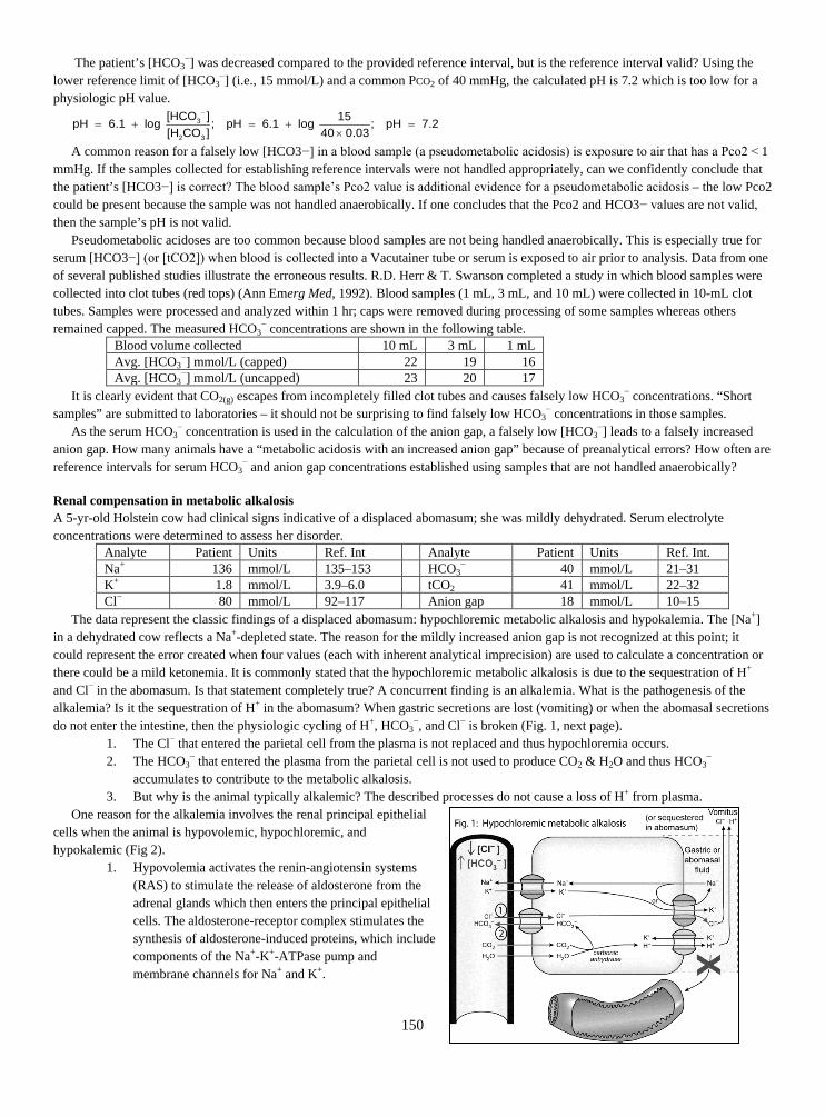
150
The patient’s [HCO3−] was decreased compared to the provided reference interval, but is the reference interval valid? Using the
lower reference limit of [HCO3−] (i.e., 15 mmol/L) and a common PCO2 of 40 mmHg, the calculated pH is 7.2 which is too low for a
physiologic pH value. −
= + = + =×
3
2 3
[HCO ] 15pH 6.1 log ; pH 6.1 log ; pH 7.2[H CO ] 40 0.03
A common reason for a falsely low [HCO3−] in a blood sample (a pseudometabolic acidosis) is exposure to air that has a Pco2 < 1
mmHg. If the samples collected for establishing reference intervals were not handled appropriately, can we confidently conclude that the patient’s [HCO3−] is correct? The blood sample’s Pco2 value is additional evidence for a pseudometabolic acidosis – the low Pco2 could be present because the sample was not handled anaerobically. If one concludes that the Pco2 and HCO3− values are not valid, then the sample’s pH is not valid.
Pseudometabolic acidoses are too common because blood samples are not being handled anaerobically. This is especially true for serum [HCO3−] (or [tCO2]) when blood is collected into a Vacutainer tube or serum is exposed to air prior to analysis. Data from one of several published studies illustrate the erroneous results. R.D. Herr & T. Swanson completed a study in which blood samples were collected into clot tubes (red tops) (Ann Emerg Med, 1992). Blood samples (1 mL, 3 mL, and 10 mL) were collected in 10-mL clot tubes. Samples were processed and analyzed within 1 hr; caps were removed during processing of some samples whereas others remained capped. The measured HCO3
− concentrations are shown in the following table. Blood volume collected 10 mL 3 mL 1 mL Avg. [HCO3
−] mmol/L (capped) 22 19 16 Avg. [HCO3
−] mmol/L (uncapped) 23 20 17 It is clearly evident that CO2(g) escapes from incompletely filled clot tubes and causes falsely low HCO3
− concentrations. “Short samples” are submitted to laboratories – it should not be surprising to find falsely low HCO3
− concentrations in those samples. As the serum HCO3
− concentration is used in the calculation of the anion gap, a falsely low [HCO3−] leads to a falsely increased
anion gap. How many animals have a “metabolic acidosis with an increased anion gap” because of preanalytical errors? How often are reference intervals for serum HCO3
− and anion gap concentrations established using samples that are not handled anaerobically?
Renal compensation in metabolic alkalosis A 5-yr-old Holstein cow had clinical signs indicative of a displaced abomasum; she was mildly dehydrated. Serum electrolyte concentrations were determined to assess her disorder.
Analyte Patient Units Ref. Int Analyte Patient Units Ref. Int. Na+ 136 mmol/L 135–153 HCO3
− 40 mmol/L 21–31 K+ 1.8 mmol/L 3.9–6.0 tCO2 41 mmol/L 22–32 Cl− 80 mmol/L 92–117 Anion gap 18 mmol/L 10–15
The data represent the classic findings of a displaced abomasum: hypochloremic metabolic alkalosis and hypokalemia. The [Na+] in a dehydrated cow reflects a Na+-depleted state. The reason for the mildly increased anion gap is not recognized at this point; it could represent the error created when four values (each with inherent analytical imprecision) are used to calculate a concentration or there could be a mild ketonemia. It is commonly stated that the hypochloremic metabolic alkalosis is due to the sequestration of H+ and Cl− in the abomasum. Is that statement completely true? A concurrent finding is an alkalemia. What is the pathogenesis of the alkalemia? Is it the sequestration of H+ in the abomasum? When gastric secretions are lost (vomiting) or when the abomasal secretions do not enter the intestine, then the physiologic cycling of H+, HCO3
−, and Cl− is broken (Fig. 1, next page). 1. The Cl− that entered the parietal cell from the plasma is not replaced and thus hypochloremia occurs. 2. The HCO3
− that entered the plasma from the parietal cell is not used to produce CO2 & H2O and thus HCO3−
accumulates to contribute to the metabolic alkalosis. 3. But why is the animal typically alkalemic? The described processes do not cause a loss of H+ from plasma.
One reason for the alkalemia involves the renal principal epithelial cells when the animal is hypovolemic, hypochloremic, and hypokalemic (Fig 2).
1. Hypovolemia activates the renin-angiotensin systems (RAS) to stimulate the release of aldosterone from the adrenal glands which then enters the principal epithelial cells. The aldosterone-receptor complex stimulates the synthesis of aldosterone-induced proteins, which include components of the Na+-K+-ATPase pump and membrane channels for Na+ and K+.
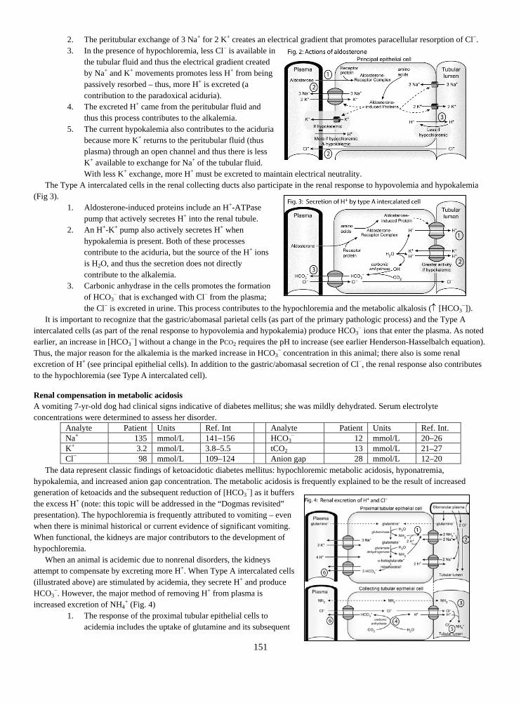
151
2. The peritubular exchange of 3 Na+ for 2 K+ creates an electrical gradient that promotes paracellular resorption of Cl−. 3. In the presence of hypochloremia, less Cl− is available in
the tubular fluid and thus the electrical gradient created by Na+ and K+ movements promotes less H+ from being passively resorbed – thus, more H+ is excreted (a contribution to the paradoxical aciduria).
4. The excreted H+ came from the peritubular fluid and thus this process contributes to the alkalemia.
5. The current hypokalemia also contributes to the aciduria because more K+ returns to the peritubular fluid (thus plasma) through an open channel and thus there is less K+ available to exchange for Na+ of the tubular fluid. With less K+ exchange, more H+ must be excreted to maintain electrical neutrality.
The Type A intercalated cells in the renal collecting ducts also participate in the renal response to hypovolemia and hypokalemia (Fig 3).
1. Aldosterone-induced proteins include an H+-ATPase pump that actively secretes H+ into the renal tubule.
2. An H+-K+ pump also actively secretes H+ when hypokalemia is present. Both of these processes contribute to the aciduria, but the source of the H+ ions is H2O, and thus the secretion does not directly contribute to the alkalemia.
3. Carbonic anhydrase in the cells promotes the formation of HCO3
− that is exchanged with Cl− from the plasma; the Cl− is excreted in urine. This process contributes to the hypochloremia and the metabolic alkalosis (↑ [HCO3
−]). It is important to recognize that the gastric/abomasal parietal cells (as part of the primary pathologic process) and the Type A
intercalated cells (as part of the renal response to hypovolemia and hypokalemia) produce HCO3− ions that enter the plasma. As noted
earlier, an increase in [HCO3−] without a change in the PCO2 requires the pH to increase (see earlier Henderson-Hasselbalch equation).
Thus, the major reason for the alkalemia is the marked increase in HCO3− concentration in this animal; there also is some renal
excretion of H+ (see principal epithelial cells). In addition to the gastric/abomasal secretion of Cl−, the renal response also contributes to the hypochloremia (see Type A intercalated cell). Renal compensation in metabolic acidosis A vomiting 7-yr-old dog had clinical signs indicative of diabetes mellitus; she was mildly dehydrated. Serum electrolyte concentrations were determined to assess her disorder.
Analyte Patient Units Ref. Int Analyte Patient Units Ref. Int. Na+ 135 mmol/L 141–156 HCO3
− 12 mmol/L 20–26 K+ 3.2 mmol/L 3.8–5.5 tCO2 13 mmol/L 21–27 Cl− 98 mmol/L 109–124 Anion gap 28 mmol/L 12–20
The data represent classic findings of ketoacidotic diabetes mellitus: hypochloremic metabolic acidosis, hyponatremia, hypokalemia, and increased anion gap concentration. The metabolic acidosis is frequently explained to be the result of increased generation of ketoacids and the subsequent reduction of [HCO3
−] as it buffers the excess H+ (note: this topic will be addressed in the “Dogmas revisited” presentation). The hypochloremia is frequently attributed to vomiting – even when there is minimal historical or current evidence of significant vomiting. When functional, the kidneys are major contributors to the development of hypochloremia.
When an animal is acidemic due to nonrenal disorders, the kidneys attempt to compensate by excreting more H+. When Type A intercalated cells (illustrated above) are stimulated by acidemia, they secrete H+ and produce HCO3
−. However, the major method of removing H+ from plasma is increased excretion of NH4
+ (Fig. 4) 1. The response of the proximal tubular epithelial cells to
acidemia includes the uptake of glutamine and its subsequent
152
deamination to form NH3. The NH3 quickly combines with H+ that entered the cell from plasma to form NH4+. The
NH4+ enters the tubular fluid in a Na+ exchange.
2. The presence of NH4+ in the tubular fluid obligates the excretion of an anion – the major anion in the tubular fluid is
Cl−. This process leads to increased renal excretion of Cl− (without a corresponding Na+) and contributes to the hypochloremia.
3. In the collecting duct epithelial cells, there is a diffusion of NH3 from plasma to tubular fluid. 4. As a response to acidemia, there is increased carbonic anhydrase activity that generates H+ ions that are secreted with
Cl− via a membrane pump. 5. The H+ ions combine with NH3 to form NH4
+ that is excreted with Cl−. This process leads to increased renal excretion of Cl− (without a corresponding Na+) and contributes to the hypochloremia.
6. In response to the acidemia, the proximal tubular epithelial cells and the collecting duct epithelial cells produce HCO3−;
this production represents a compensatory metabolic alkalosis to the nonrenal acidemic state. Vomiting may contribute to the hypochloremia in an animal that has a metabolic acidosis. However, the expected renal
compensation for an acidemia of nonrenal origin is the excretion of NH4+ and Cl−; the increased renal Cl− excretion contributes to the
hypochloremia. Calculated electrolyte data: SID, anion gap, and others The measured electrolyte concentrations frequently are used to calculate other data; i.e., Na+: K+ ratio, anion gap, SID, corrected Cl− concentration. However, there are reasons for interpreting the calculated data cautiously.
True changes in the strong-ion difference (SID) basically represent changes in plasma/serum HCO3− concentrations. As it not
clinically practical to measure the true SID, a variety of formulas have been proposed to estimate the SID. However, the formulas are based on assumptions that may or may not be true and thus the calculated SIDs may or may not be reliable estimates. Another factor that should be considered is that the validity of calculated results is dependent on the accuracy of the measured concentrations used for the calculation. The simplest SID formula is: SID = [Na+] + [K+] – [Cl−]. Considering the analytical precision of the Na+ and Cl− methods, the measured concentrations should be considered the reported values ± 1 mmol/L (or perhaps ± 2 mmol/L); the precision of the K+ methods are better. For a cautious perspective, the calculated SID is the value ± 4 mmol/L. Thus, any minor difference when compared to appropriate SID reference intervals (or interpretive guidelines) should be interpreted cautiously.
The potential for imprecision to affect the calculated value increases with each added variable. For example, a common formula for anion gap is: anion gap = ([Na+] + [K+]) – ([Cl−] + [HCO3
−]). The addition of patient’s HCO3− concentration (again, at best ± 1
mmol/L) adds to the uncertainty of the calculated anion gap concentration. And as mentioned earlier, the HCO3− concentration can
easily be falsely decreased if the blood/plasma/serum samples are not collected and processed properly.
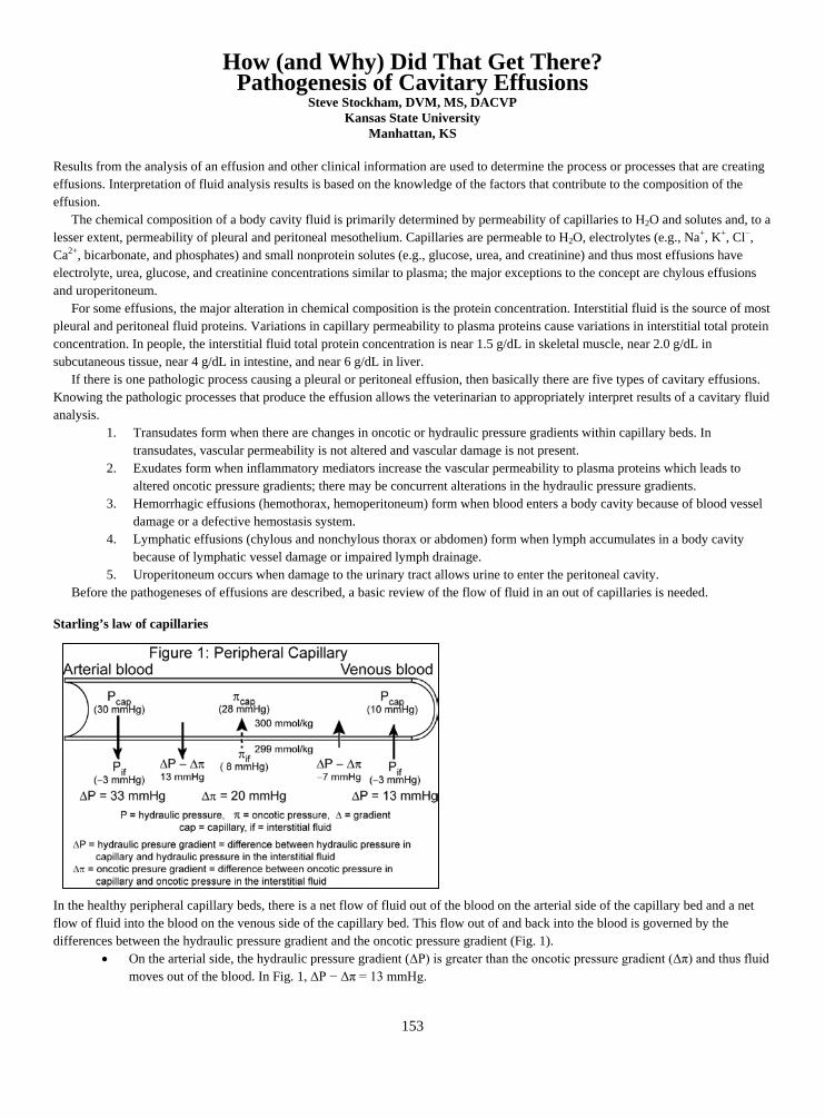
153
How (and Why) Did That Get There? Pathogenesis of Cavitary Effusions
Steve Stockham, DVM, MS, DACVP Kansas State University
Manhattan, KS
Results from the analysis of an effusion and other clinical information are used to determine the process or processes that are creating effusions. Interpretation of fluid analysis results is based on the knowledge of the factors that contribute to the composition of the effusion.
The chemical composition of a body cavity fluid is primarily determined by permeability of capillaries to H2O and solutes and, to a lesser extent, permeability of pleural and peritoneal mesothelium. Capillaries are permeable to H2O, electrolytes (e.g., Na+, K+, Cl−, Ca2+, bicarbonate, and phosphates) and small nonprotein solutes (e.g., glucose, urea, and creatinine) and thus most effusions have electrolyte, urea, glucose, and creatinine concentrations similar to plasma; the major exceptions to the concept are chylous effusions and uroperitoneum.
For some effusions, the major alteration in chemical composition is the protein concentration. Interstitial fluid is the source of most pleural and peritoneal fluid proteins. Variations in capillary permeability to plasma proteins cause variations in interstitial total protein concentration. In people, the interstitial fluid total protein concentration is near 1.5 g/dL in skeletal muscle, near 2.0 g/dL in subcutaneous tissue, near 4 g/dL in intestine, and near 6 g/dL in liver.
If there is one pathologic process causing a pleural or peritoneal effusion, then basically there are five types of cavitary effusions. Knowing the pathologic processes that produce the effusion allows the veterinarian to appropriately interpret results of a cavitary fluid analysis.
1. Transudates form when there are changes in oncotic or hydraulic pressure gradients within capillary beds. In transudates, vascular permeability is not altered and vascular damage is not present.
2. Exudates form when inflammatory mediators increase the vascular permeability to plasma proteins which leads to altered oncotic pressure gradients; there may be concurrent alterations in the hydraulic pressure gradients.
3. Hemorrhagic effusions (hemothorax, hemoperitoneum) form when blood enters a body cavity because of blood vessel damage or a defective hemostasis system.
4. Lymphatic effusions (chylous and nonchylous thorax or abdomen) form when lymph accumulates in a body cavity because of lymphatic vessel damage or impaired lymph drainage.
5. Uroperitoneum occurs when damage to the urinary tract allows urine to enter the peritoneal cavity. Before the pathogeneses of effusions are described, a basic review of the flow of fluid in an out of capillaries is needed.
Starling’s law of capillaries
In the healthy peripheral capillary beds, there is a net flow of fluid out of the blood on the arterial side of the capillary bed and a net flow of fluid into the blood on the venous side of the capillary bed. This flow out of and back into the blood is governed by the differences between the hydraulic pressure gradient and the oncotic pressure gradient (Fig. 1).
• On the arterial side, the hydraulic pressure gradient (ΔP) is greater than the oncotic pressure gradient (Δπ) and thus fluid moves out of the blood. In Fig. 1, ΔP − Δπ = 13 mmHg.
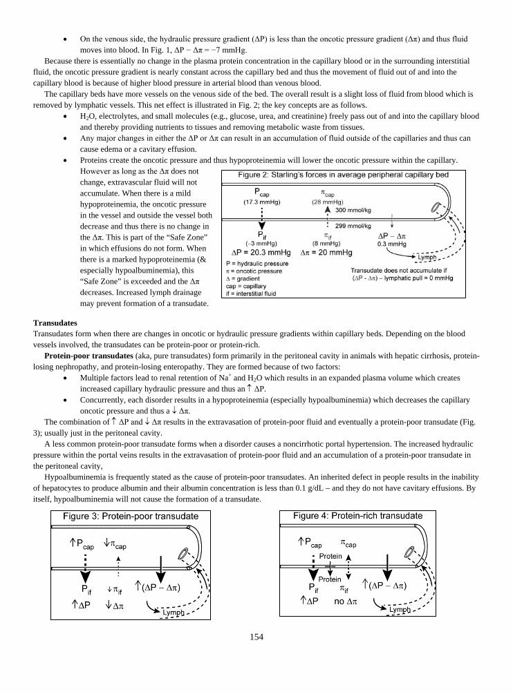
154
• On the venous side, the hydraulic pressure gradient (ΔP) is less than the oncotic pressure gradient (Δπ) and thus fluid moves into blood. In Fig. 1, ΔP − Δπ = −7 mmHg.
Because there is essentially no change in the plasma protein concentration in the capillary blood or in the surrounding interstitial fluid, the oncotic pressure gradient is nearly constant across the capillary bed and thus the movement of fluid out of and into the capillary blood is because of higher blood pressure in arterial blood than venous blood.
The capillary beds have more vessels on the venous side of the bed. The overall result is a slight loss of fluid from blood which is removed by lymphatic vessels. This net effect is illustrated in Fig. 2; the key concepts are as follows.
• H2O, electrolytes, and small molecules (e.g., glucose, urea, and creatinine) freely pass out of and into the capillary blood and thereby providing nutrients to tissues and removing metabolic waste from tissues.
• Any major changes in either the ΔP or Δπ can result in an accumulation of fluid outside of the capillaries and thus can cause edema or a cavitary effusion.
• Proteins create the oncotic pressure and thus hypoproteinemia will lower the oncotic pressure within the capillary. However as long as the Δπ does not change, extravascular fluid will not accumulate. When there is a mild hypoproteinemia, the oncotic pressure in the vessel and outside the vessel both decrease and thus there is no change in the Δπ. This is part of the “Safe Zone” in which effusions do not form. When there is a marked hypoproteinemia (& especially hypoalbuminemia), this “Safe Zone” is exceeded and the Δπ decreases. Increased lymph drainage may prevent formation of a transudate.
Transudates Transudates form when there are changes in oncotic or hydraulic pressure gradients within capillary beds. Depending on the blood vessels involved, the transudates can be protein-poor or protein-rich.
Protein-poor transudates (aka, pure transudates) form primarily in the peritoneal cavity in animals with hepatic cirrhosis, protein-losing nephropathy, and protein-losing enteropathy. They are formed because of two factors:
• Multiple factors lead to renal retention of Na+ and H2O which results in an expanded plasma volume which creates increased capillary hydraulic pressure and thus an ↑ ΔP.
• Concurrently, each disorder results in a hypoproteinemia (especially hypoalbuminemia) which decreases the capillary oncotic pressure and thus a ↓ Δπ.
The combination of ↑ ΔP and ↓ Δπ results in the extravasation of protein-poor fluid and eventually a protein-poor transudate (Fig. 3); usually just in the peritoneal cavity.
A less common protein-poor transudate forms when a disorder causes a noncirrhotic portal hypertension. The increased hydraulic pressure within the portal veins results in the extravasation of protein-poor fluid and an accumulation of a protein-poor transudate in the peritoneal cavity,
Hypoalbuminemia is frequently stated as the cause of protein-poor transudates. An inherited defect in people results in the inability of hepatocytes to produce albumin and their albumin concentration is less than 0.1 g/dL – and they do not have cavitary effusions. By itself, hypoalbuminemia will not cause the formation of a transudate.
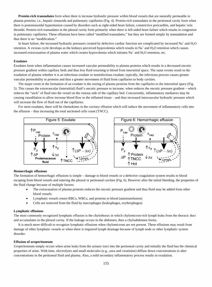
155
Protein-rich transudates form when there is increase hydraulic pressure within blood vessels that are naturally permeable to plasma proteins; i.e., hepatic sinusoids and pulmonary capillaries (Fig. 4). Protein-rich transudates in the peritoneal cavity form when there is postsinusoidal hypertension caused by disorders such as right-sided heart failure, constrictive pericarditis, and hepatic vein thrombi. Protein-rich transudates in the pleural cavity form primarily when there is left-sided heart failure which results in congestion in pulmonary capillaries. These effusions have been called “modified transudates,” but they are formed simply by transudation and thus there is no “modification.”
In heart failure, the increased hydraulic pressures created by defective cardiac function are complicated by increased Na+ and H2O retention. A vicious cycle develops as the kidneys perceived hypovolemia which results in Na+ and H2O retention which causes increased extravasation of plasma water which creates hypovolemia which initiates Na+ and H2O retention, etc.
Exudates Exudates form when inflammation causes increased vascular permeability to plasma proteins which results in a decreased oncotic pressure gradient within capillary beds and thus less fluid returning to blood from interstitial space. The same events result in the exudation of plasma whether it is an infectious exudate or noninfectious exudate; typically, the infectious process causes greater vascular permeability to proteins and thus a greater movement of fluid from capillaries to body cavities.
The major event in the formation of an exudate is the oozing of plasma proteins from the capillaries to the interstitial space (Fig. 5). This causes the extravascular (interstitial) fluid’s oncotic pressure to increase, when reduces the oncotic pressure gradient – which reduces the “suck” of fluid into the vessel on the venous side of the capillary bed. Concurrently, inflammatory mediators may be causing vasodilation to allow increase blood flow to the inflamed tissue – and thus increased intravascular hydraulic pressure which will increase the flow of fluid out of the capillaries.
For most exudates, there will be chemokines in the cavitary effusion which will induce the movement of inflammatory cells into the effusion – thus increasing the total nucleated cells count (TNCC).
Hemorrhagic effusions The formation of hemorrhagic effusions is simple – damage to blood vessels or a defective coagulation system results in blood escaping from blood vessels and entering the pleural or peritoneal cavities (Fig. 6). However after the initial bleeding, the properties of the fluid change because of multiple factors.
• The extravasation of plasma proteins reduces the oncotic pressure gradient and thus fluid may be added from other blood vessels.
• Lymphatic vessels return RBCs, WBCs, and proteins to blood (autotransfusion) • Cells are removed from the fluid by macrophages (leukophages, erythrophages)
Lymphatic effusions The most commonly recognized lymphatic effusion is the chylothorax in which chylomicron-rich lymph leaks from the thoracic duct and accumulates in the pleural cavity. If the leakage occurs in the abdomen, then a chyloabdomen forms.
It is much more difficult to recognize lymphatic effusions when chylomicrons are not present. These effusions may result from damage of other lymphatic vessels or when there is impaired lymph drainage because of lymph node or other lymphatic system disorder. Effusion of uroperitoneum Uroperitoneum simply occurs when urine leaks from the urinary tract into the peritoneal cavity and initially the fluid has the chemical properties of urine. With time, electrolytes and small molecules (e.g., urea and creatinine) diffuse down concentrations to alter concentrations in the peritoneal fluid and plasma. Also, a mild secondary inflammatory process results in exudation.

156
Neoplastic effusions Effusions caused by the neoplastic process can result from transudation, exudation, hemorrhage, lymphatic damage, or a combination of factors. Modified transudates Modified transudates have been a part of the effusion classifications in veterinary literature for nearly 30 years; there is not a similar category in the human effusions. The classification has been based on the results of fluid analysis; that is, the fluid had a total protein concentration or total nucleated cell count “too high” for a pure transudate and it did not have the features of an exudate. Thus, it became a “catch all” or “lumper” classification for effusions that were not “pure transudates” or exudates.
One “modified transudate” is the effusion of heart failure – that effusion is a protein-rich transudate described above. Other “modified transudates” described in textbooks include the FIP exudate, hypocellular exudates, chylous effusions, uroperitoneum, bilious peritonitis, and uroperitoneum – these are not transudates; they are not transudates modified by the addition of protein or cells. Nucleated cells in effusions The total nucleated cell concentration in an effusion and the type of nucleated cells also aid in the identification of effusions. Their value will be described in the case analyses in subsequent presentations.
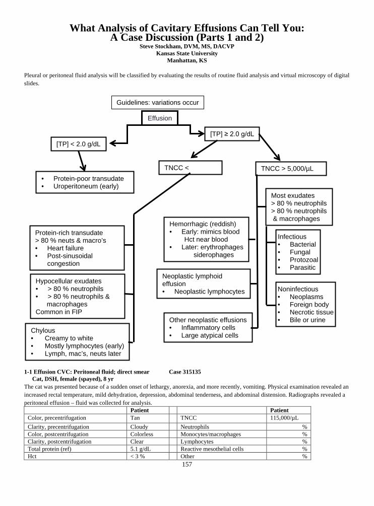
157
What Analysis of Cavitary Effusions Can Tell You: A Case Discussion (Parts 1 and 2)
Steve Stockham, DVM, MS, DACVP Kansas State University
Manhattan, KS
Pleural or peritoneal fluid analysis will be classified by evaluating the results of routine fluid analysis and virtual microscopy of digital slides. 1-1 Effusion CVC: Peritoneal fluid; direct smear Case 315135
Cat, DSH, female (spayed), 8 yr The cat was presented because of a sudden onset of lethargy, anorexia, and more recently, vomiting. Physical examination revealed an increased rectal temperature, mild dehydration, depression, abdominal tenderness, and abdominal distension. Radiographs revealed a peritoneal effusion – fluid was collected for analysis.
Patient Patient Color, precentrifugation Tan TNCC 115,000/µL Clarity, precentrifugation Cloudy Neutrophils % Color, postcentrifugation Colorless Monocytes/macrophages % Clarity, postcentrifugation Clear Lymphocytes % Total protein (ref) 5.1 g/dL Reactive mesothelial cells % Hct < 3 % Other %
Effusion
[TP] < 2.0 g/dL [TP] ≥ 2.0 g/dL
• Protein-poor transudate • Uroperitoneum (early)
TNCC < TNCC > 5,000/µL
Hypocellular exudates • > 80 % neutrophils • > 80 % neutrophils & macrophages Common in FIP
Chylous • Creamy to white • Mostly lymphocytes (early) • Lymph, mac’s, neuts later
Most exudates > 80 % neutrophils > 80 % neutrophils & macrophages
Hemorrhagic (reddish) • Early: mimics blood Hct near blood • Later: erythrophages siderophages
Neoplastic lymphoid effusion • Neoplastic lymphocytes
Infectious • Bacterial • Fungal • Protozoal • Parasitic
Noninfectious • Neoplasms • Foreign body • Necrotic tissue • Bile or urine
Protein-rich transudate > 80 % neuts & macro’s • Heart failure • Post-sinusoidal
congestion
Guidelines: variations occur
Other neoplastic effusions • Inflammatory cells • Large atypical cells
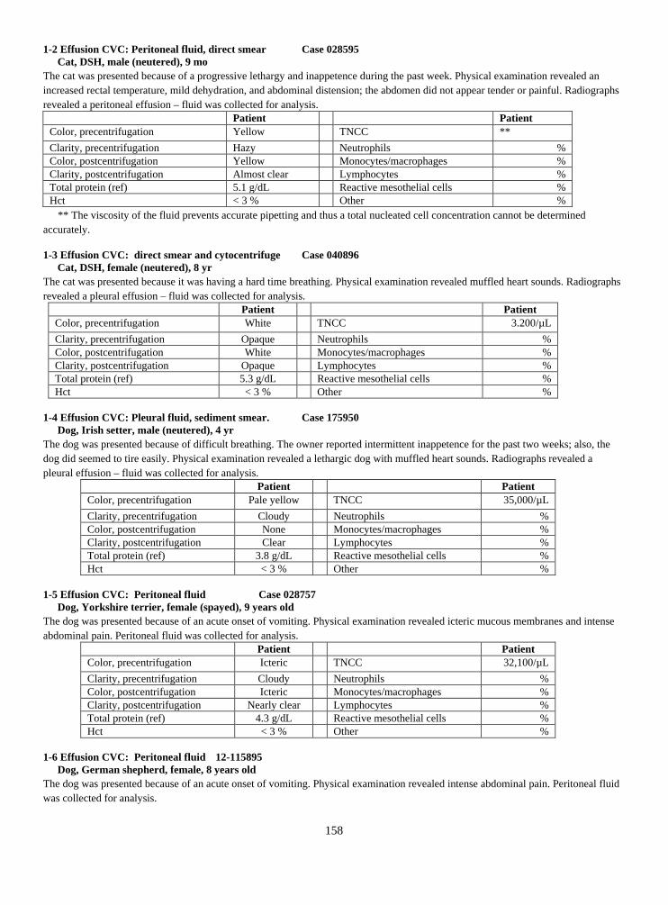
158
1-2 Effusion CVC: Peritoneal fluid, direct smear Case 028595 Cat, DSH, male (neutered), 9 mo
The cat was presented because of a progressive lethargy and inappetence during the past week. Physical examination revealed an increased rectal temperature, mild dehydration, and abdominal distension; the abdomen did not appear tender or painful. Radiographs revealed a peritoneal effusion – fluid was collected for analysis.
Patient Patient Color, precentrifugation Yellow TNCC ** Clarity, precentrifugation Hazy Neutrophils % Color, postcentrifugation Yellow Monocytes/macrophages % Clarity, postcentrifugation Almost clear Lymphocytes % Total protein (ref) 5.1 g/dL Reactive mesothelial cells % Hct < 3 % Other %
** The viscosity of the fluid prevents accurate pipetting and thus a total nucleated cell concentration cannot be determined accurately. 1-3 Effusion CVC: direct smear and cytocentrifuge Case 040896
Cat, DSH, female (neutered), 8 yr The cat was presented because it was having a hard time breathing. Physical examination revealed muffled heart sounds. Radiographs revealed a pleural effusion – fluid was collected for analysis.
Patient Patient Color, precentrifugation White TNCC 3.200/µL Clarity, precentrifugation Opaque Neutrophils % Color, postcentrifugation White Monocytes/macrophages % Clarity, postcentrifugation Opaque Lymphocytes % Total protein (ref) 5.3 g/dL Reactive mesothelial cells % Hct < 3 % Other %
1-4 Effusion CVC: Pleural fluid, sediment smear. Case 175950
Dog, Irish setter, male (neutered), 4 yr The dog was presented because of difficult breathing. The owner reported intermittent inappetence for the past two weeks; also, the dog did seemed to tire easily. Physical examination revealed a lethargic dog with muffled heart sounds. Radiographs revealed a pleural effusion – fluid was collected for analysis.
Patient Patient Color, precentrifugation Pale yellow TNCC 35,000/µL Clarity, precentrifugation Cloudy Neutrophils % Color, postcentrifugation None Monocytes/macrophages % Clarity, postcentrifugation Clear Lymphocytes % Total protein (ref) 3.8 g/dL Reactive mesothelial cells % Hct < 3 % Other %
1-5 Effusion CVC: Peritoneal fluid Case 028757
Dog, Yorkshire terrier, female (spayed), 9 years old The dog was presented because of an acute onset of vomiting. Physical examination revealed icteric mucous membranes and intense abdominal pain. Peritoneal fluid was collected for analysis.
Patient Patient Color, precentrifugation Icteric TNCC 32,100/µL Clarity, precentrifugation Cloudy Neutrophils % Color, postcentrifugation Icteric Monocytes/macrophages % Clarity, postcentrifugation Nearly clear Lymphocytes % Total protein (ref) 4.3 g/dL Reactive mesothelial cells % Hct < 3 % Other %
1-6 Effusion CVC: Peritoneal fluid 12-115895
Dog, German shepherd, female, 8 years old The dog was presented because of an acute onset of vomiting. Physical examination revealed intense abdominal pain. Peritoneal fluid was collected for analysis.
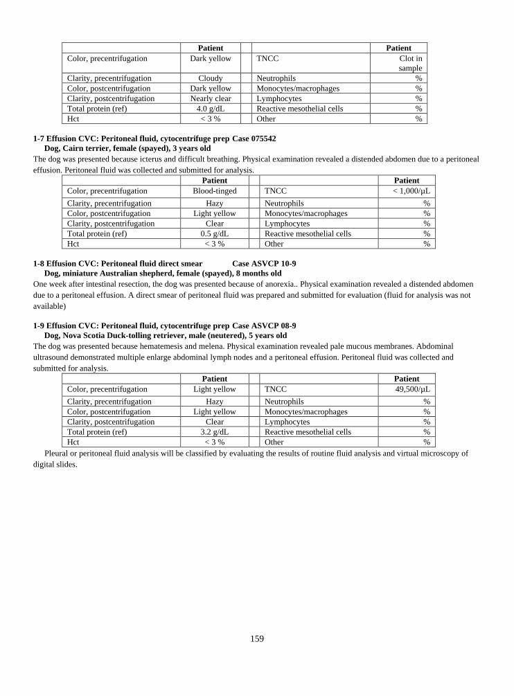
159
Patient Patient Color, precentrifugation Dark yellow TNCC Clot in
sample Clarity, precentrifugation Cloudy Neutrophils % Color, postcentrifugation Dark yellow Monocytes/macrophages % Clarity, postcentrifugation Nearly clear Lymphocytes % Total protein (ref) 4.0 g/dL Reactive mesothelial cells % Hct < 3 % Other %
1-7 Effusion CVC: Peritoneal fluid, cytocentrifuge prep Case 075542
Dog, Cairn terrier, female (spayed), 3 years old The dog was presented because icterus and difficult breathing. Physical examination revealed a distended abdomen due to a peritoneal effusion. Peritoneal fluid was collected and submitted for analysis.
Patient Patient Color, precentrifugation Blood-tinged TNCC < 1,000/µL Clarity, precentrifugation Hazy Neutrophils % Color, postcentrifugation Light yellow Monocytes/macrophages % Clarity, postcentrifugation Clear Lymphocytes % Total protein (ref) 0.5 g/dL Reactive mesothelial cells % Hct < 3 % Other %
1-8 Effusion CVC: Peritoneal fluid direct smear Case ASVCP 10-9
Dog, miniature Australian shepherd, female (spayed), 8 months old One week after intestinal resection, the dog was presented because of anorexia.. Physical examination revealed a distended abdomen due to a peritoneal effusion. A direct smear of peritoneal fluid was prepared and submitted for evaluation (fluid for analysis was not available) 1-9 Effusion CVC: Peritoneal fluid, cytocentrifuge prep Case ASVCP 08-9
Dog, Nova Scotia Duck-tolling retriever, male (neutered), 5 years old The dog was presented because hematemesis and melena. Physical examination revealed pale mucous membranes. Abdominal ultrasound demonstrated multiple enlarge abdominal lymph nodes and a peritoneal effusion. Peritoneal fluid was collected and submitted for analysis.
Patient Patient Color, precentrifugation Light yellow TNCC 49,500/µL Clarity, precentrifugation Hazy Neutrophils % Color, postcentrifugation Light yellow Monocytes/macrophages % Clarity, postcentrifugation Clear Lymphocytes % Total protein (ref) 3.2 g/dL Reactive mesothelial cells % Hct < 3 % Other %
Pleural or peritoneal fluid analysis will be classified by evaluating the results of routine fluid analysis and virtual microscopy of digital slides.
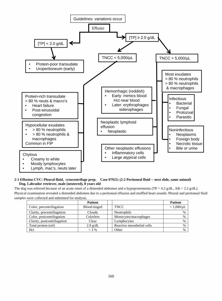
160
2-1 Effusion CVC: Pleural fluid, cytocentrifuge prep. Case 07923; (2-2 Peritoneal fluid -- next slide, same animal)
Dog, Labrador retriever, male (neutered), 8 years old The dog was referred because of an acute onset of a distended abdomen and a hypoproteinemia (TP = 4.2 g/dL, Alb = 2.2 g/dL). Physical examination revealed a distended abdomen due to a peritoneal effusion and muffled heart sounds. Pleural and peritoneal fluid samples were collected and submitted for analysis.
Patient Patient Color, precentrifugation Blood-tinged TNCC < 1,000/µL Clarity, precentrifugation Cloudy Neutrophils % Color, postcentrifugation Colorless Monocytes/macrophages % Clarity, postcentrifugation Clear Lymphocytes % Total protein (ref) 2.9 g/dL Reactive mesothelial cells % Hct < 3 % Other %
Effusio
[TP] < 2.0 g/dL [TP] ≥ 2.0 g/dL
• Protein-poor transudate • Uroperitoneum (early)
TNCC < 5,000/µL TNCC > 5,000/µL
Hypocellular exudates • > 80 % neutrophils • > 80 % neutrophils & macrophages Common in FIP
Chylous • Creamy to white • Mostly lymphocytes • Lymph, mac’s, neuts later
Most exudates > 80 % neutrophils > 80 % neutrophils & macrophages
Hemorrhagic (reddish) • Early: mimics blood Hct near blood • Later: erythrophages siderophages
Neoplastic lymphoid effusion • Neoplastic
Infectious • Bacterial • Fungal • Protozoal • Parasitic
Noninfectious • Neoplasms • Foreign body • Necrotic tissue • Bile or urine
Protein-rich transudate > 80 % neuts & macro’s • Heart failure • Post-sinusoidal
congestion
Guidelines: variations occur
Other neoplastic effusions • Inflammatory cells • Large atypical cells
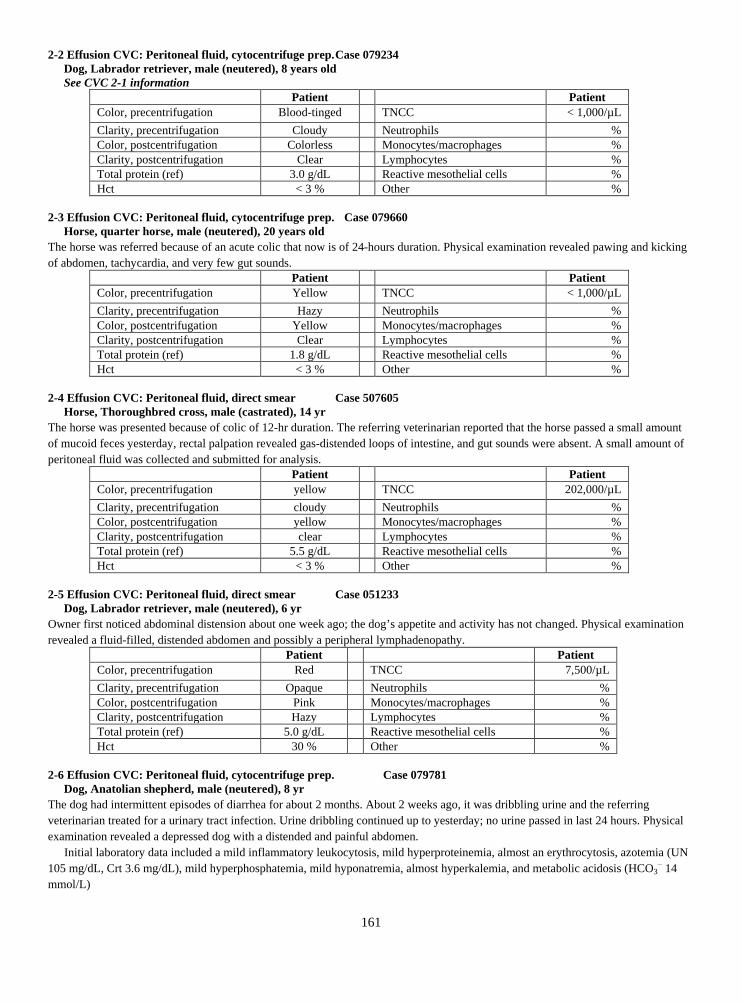
161
2-2 Effusion CVC: Peritoneal fluid, cytocentrifuge prep. Case 079234 Dog, Labrador retriever, male (neutered), 8 years old See CVC 2-1 information
Patient Patient Color, precentrifugation Blood-tinged TNCC < 1,000/µL Clarity, precentrifugation Cloudy Neutrophils % Color, postcentrifugation Colorless Monocytes/macrophages % Clarity, postcentrifugation Clear Lymphocytes % Total protein (ref) 3.0 g/dL Reactive mesothelial cells % Hct < 3 % Other %
2-3 Effusion CVC: Peritoneal fluid, cytocentrifuge prep. Case 079660
Horse, quarter horse, male (neutered), 20 years old The horse was referred because of an acute colic that now is of 24-hours duration. Physical examination revealed pawing and kicking of abdomen, tachycardia, and very few gut sounds.
Patient Patient Color, precentrifugation Yellow TNCC < 1,000/µL Clarity, precentrifugation Hazy Neutrophils % Color, postcentrifugation Yellow Monocytes/macrophages % Clarity, postcentrifugation Clear Lymphocytes % Total protein (ref) 1.8 g/dL Reactive mesothelial cells % Hct < 3 % Other %
2-4 Effusion CVC: Peritoneal fluid, direct smear Case 507605
Horse, Thoroughbred cross, male (castrated), 14 yr The horse was presented because of colic of 12-hr duration. The referring veterinarian reported that the horse passed a small amount of mucoid feces yesterday, rectal palpation revealed gas-distended loops of intestine, and gut sounds were absent. A small amount of peritoneal fluid was collected and submitted for analysis.
Patient Patient Color, precentrifugation yellow TNCC 202,000/µL Clarity, precentrifugation cloudy Neutrophils % Color, postcentrifugation yellow Monocytes/macrophages % Clarity, postcentrifugation clear Lymphocytes % Total protein (ref) 5.5 g/dL Reactive mesothelial cells % Hct < 3 % Other %
2-5 Effusion CVC: Peritoneal fluid, direct smear Case 051233
Dog, Labrador retriever, male (neutered), 6 yr Owner first noticed abdominal distension about one week ago; the dog’s appetite and activity has not changed. Physical examination revealed a fluid-filled, distended abdomen and possibly a peripheral lymphadenopathy.
Patient Patient Color, precentrifugation Red TNCC 7,500/µL Clarity, precentrifugation Opaque Neutrophils % Color, postcentrifugation Pink Monocytes/macrophages % Clarity, postcentrifugation Hazy Lymphocytes % Total protein (ref) 5.0 g/dL Reactive mesothelial cells % Hct 30 % Other %
2-6 Effusion CVC: Peritoneal fluid, cytocentrifuge prep. Case 079781
Dog, Anatolian shepherd, male (neutered), 8 yr The dog had intermittent episodes of diarrhea for about 2 months. About 2 weeks ago, it was dribbling urine and the referring veterinarian treated for a urinary tract infection. Urine dribbling continued up to yesterday; no urine passed in last 24 hours. Physical examination revealed a depressed dog with a distended and painful abdomen.
Initial laboratory data included a mild inflammatory leukocytosis, mild hyperproteinemia, almost an erythrocytosis, azotemia (UN 105 mg/dL, Crt 3.6 mg/dL), mild hyperphosphatemia, mild hyponatremia, almost hyperkalemia, and metabolic acidosis (HCO3
− 14 mmol/L)

162
Patient Patient Color, precentrifugation Blood-tinged TNCC 2,700/µL Clarity, precentrifugation Cloudy Neutrophils % Color, postcentrifugation Pink Monocytes/macrophages % Clarity, postcentrifugation Clear Lymphocytes % Total protein (ref) 1.3 g/dL Reactive mesothelial cells % Hct < 3 % Other %
2-7 Effusion CVC: Peritoneal fluid, line prep. Case 08-69874
Dog, mixed breed, female, 1 yr A veterinarian in NE Kansas submitted pleural and peritoneal fluid from a dog. Historical or physical examination findings were not provided.
Patient Patient Color, precentrifugation Blood-tinged TNCC 10,200/µL Clarity, precentrifugation Cloudy Neutrophils % Color, postcentrifugation Colorless Monocytes/macrophages % Clarity, postcentrifugation Clear Lymphocytes % Total protein (ref) 4.7 g/dL Reactive mesothelial cells % Hct < 3 % Other % Other microscopic findings:
Note: A line prep. tends concentrate cells in the line, but also makes that area thick. Note: The analysis of pleural fluid yielded essentially the same results except the TNCC was 5,000/µL.
2-8 Effusion CVC: Pleural effusion, cytocentrifuge preparation Case 047859 Cat, Birman, male (neutered), 16 yr
The cat was presented because of dyspnea. The owner reported intermittent inappetence during past week. Physical examination revealed muffled heart sounds. Radiographs revealed a pleural effusion – fluid was collected for analysis.
Patient Patient Color, precentrifugation Pink TNCC 8,000/µL Clarity, precentrifugation Hazy Neutrophils % Color, postcentrifugation Light yellow Monocytes/macrophages % Clarity, postcentrifugation Clear Lymphocytes % Total protein (ref) 2.6 g/dL Reactive mesothelial cells % Hct < 3 % Other %
2-9 Effusion CVC: Peritoneal fluid, cytocentrifuge prep. Case 080190
Dog, fox terrier, male (neutered), 6 yr The dog was referred because of abdominal ascites that might be due to heart failure. Physical examination revealed a grade 2-3, left-sided systolic murmur and a fluid-filled abdomen. Preliminary laboratory data found UN of 16 mg/dL, Crt 3.6 mg/dL), hypoproteinemia (TP 2.5 g/dL, albumin 1.2 g/dL), hypocalcemia (tCa2+ 5.7 g/dL), mild hyponatremia (144 mmol/L), normochloremia, decreased anion gap, and urine with a specific gravity of 1.009, and negative chemistry results.
Patient Patient Color, precentrifugation colorless TNCC < 1,000/µL Clarity, precentrifugation clear Neutrophils % Color, postcentrifugation colorless Monocytes/macrophages % Clarity, postcentrifugation clear Lymphocytes % Total protein (ref) 0.1 g/dL Reactive mesothelial cells % Hct < 3 % Other %
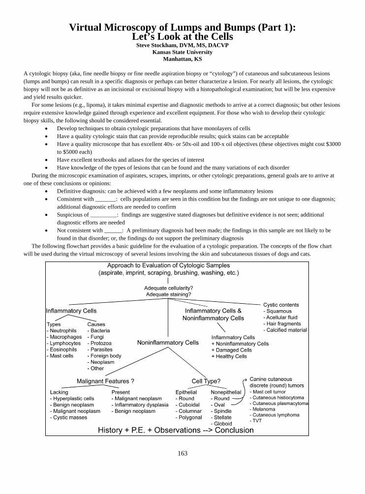
163
Virtual Microscopy of Lumps and Bumps (Part 1): Let’s Look at the Cells
Steve Stockham, DVM, MS, DACVP Kansas State University
Manhattan, KS
A cytologic biopsy (aka, fine needle biopsy or fine needle aspiration biopsy or “cytology”) of cutaneous and subcutaneous lesions (lumps and bumps) can result in a specific diagnosis or perhaps can better characterize a lesion. For nearly all lesions, the cytologic biopsy will not be as definitive as an incisional or excisional biopsy with a histopathological examination; but will be less expensive and yield results quicker.
For some lesions (e.g., lipoma), it takes minimal expertise and diagnostic methods to arrive at a correct diagnosis; but other lesions require extensive knowledge gained through experience and excellent equipment. For those who wish to develop their cytologic biopsy skills, the following should be considered essential.
• Develop techniques to obtain cytologic preparations that have monolayers of cells • Have a quality cytologic stain that can provide reproducible results; quick stains can be acceptable • Have a quality microscope that has excellent 40x- or 50x-oil and 100-x oil objectives (these objectives might cost $3000
to $5000 each) • Have excellent textbooks and atlases for the species of interest • Have knowledge of the types of lesions that can be found and the many variations of each disorder
During the microscopic examination of aspirates, scrapes, imprints, or other cytologic preparations, general goals are to arrive at one of these conclusions or opinions:
• Definitive diagnosis: can be achieved with a few neoplasms and some inflammatory lesions • Consistent with _______: cells populations are seen in this condition but the findings are not unique to one diagnosis;
additional diagnostic efforts are needed to confirm • Suspicious of _________: findings are suggestive stated diagnoses but definitive evidence is not seen; additional
diagnostic efforts are needed • Not consistent with ______: A preliminary diagnosis had been made; the findings in this sample are not likely to be
found in that disorder; or, the findings do not support the preliminary diagnosis The following flowchart provides a basic guideline for the evaluation of a cytologic preparation. The concepts of the flow chart
will be used during the virtual microscopy of several lesions involving the skin and subcutaneous tissues of dogs and cats.
164
1-1 L&B CVC: Smear of fluid from subcutaneous lesion Case: 176517 Dog, mixed breed, 3-yr-old, female (spayed)
A smear of serosanguineous to purulent fluid was submitted; the fluid was collected from a subcutaneous swelling that had a draining tract. 1-2 L&B CVC: fine-needle aspirate of cutaneous mass Case: 02-1975
Dog, Labrador retriever, 4-yr-old A 2x4x3 cm mass was located in the lateral skin of the left hind thigh or hip. The owner first noticed the mass a few weeks ago and it has been getting larger. The mass protruded slightly and felt like it extended into the subcutaneous tissue. A fine-needle aspirate of the mass was collected and a smear was prepared for examination. 1-3 L&B CVC: fine-needle aspirate of cutaneous mass Case: 02885
Dog, Golden retriever, male, 12-year-old The dog was presented because of a mass located on the dorsal aspect of the tail head. Physical examination revealed 2-cm, soft mass in the dermis and was covered with haired skin. A fine-needle aspirate of the rear leg mass was collected and a smear was prepared for examination. 1-4 L&B CVC: fine-needle aspirate of cutaneous mass Case: 030056
Dog, basset hound, male (neutered), 7-year-old The dog was presented because of perianal masses. Physical examination revealed a small perianal mass and possibly enlarged regional lymph node. A fine-needle aspirate of the mass was collected and a smear was prepared for examination. 1-5 L&B CVC: fine-needle aspirate of cutaneous mass Case: 02-2357
Dog; breed, age, and gender not provided A smear of an aspirate obtained from a mass in the skin of a foot was submitted for evaluation. 1-6 L&B CVC: fine-needle aspirate of cutaneous mass Case: 024854
Dog, schipperke, male (neutered), 15-yr-old The dog had been coughing for 2-3 weeks. During a physical exam, a mass was found in the subcutaneous tissues of the left lateral thoracic; it appeared to be firmly attached to underlying tissues. A fine-needle aspirate of the mass was collected and a smear was prepared for examination. 1-7 L&B CVC: Imprint of moist cutaneous lesion Case: 256285
Dog, mixed breed, male (neutered), 4-yr-old The dog was presented because of a swelling of the left flank that broke open yesterday and yellowish red material oozed out. The preparation is an imprint of the ulcerated area after superficial debris and hair were removed. 1-8 L&B CVC: Imprints of cutaneous mass Case: ASVCP 1988-11
Cat, domestic short hair The cat was presented because of skin lesions. Physical examination revealed several, pea-size, cutaneous masses. One mass was excised and imprints of the mass were submitted for evaluation. . Additional slides will be reviewed if time permits
165
Virtual Microscopy of Lumps and Bumps (Part 2): Let’s Look at More Cells
Steve Stockham, DVM, MS, DACVP Kansas State University
Manhattan, KS
A cytologic biopsy (aka, fine needle biopsy or fine needle aspiration biopsy or “cytology”) of cutaneous and subcutaneous lesions (lumps and bumps) can result in a specific diagnosis or perhaps can better characterize a lesion. For nearly all lesions, the cytologic biopsy will not be as definitive as an incisional or excisional biopsy with a histopathological examination; but will be less expensive and yield results quicker.
Please see previous proceeding’s document (Part 1) for an introduction to the goals and approach of a cytologic biopsy. 2-1 L&B CVC: fine-needle aspirate of vulvar mass Case: 026163
Dog, mixed breed, female, 5-yr-old A 1x1 pink mass was protruding slightly from the vulvar mucosa. The owner first noticed the mass yesterday. The mass protruded into the vaginal vault; it might be extending into the submucosa. A fine-needle aspirate of the mass was collected and a smear was prepared for examination. 2-2 L&B CVC: fine-needle aspirate of cutaneous mass Case: 02-7821
Dog, boxer, 1-yr-old A 1x1x1 cm, pink mass was located in the lateral skin of the right shoulder. The owner first noticed the mass a few days ago. The mass seemed to involve the dermis and epidermis and did not extend into the subcutaneous tissues. A fine-needle aspirate of the mass was collected and a smear was prepared for examination. 2-3 L&B CVC: fine-needle aspirate of cutaneous mass Case: 028857
Dog, Golden retriever, male (neutered), 5-yr-old The dog was presented because of a 2x2x1 cm mass located in the lateral thoracic skin. The preparation is a smear of the sample aspirated from the mass. 2-4 L&B CVC: fine-needle aspirate of cutaneous mass Case: 039973
Cat, domestic short hair The preparation is a smear of a sample aspirated from one of several small (< 1 cm) cutaneous masses. 2-5 L&B CVC: fine-needle aspirate of cutaneous mass Case: 56535-98
Dog, mixed breed A smear of serosanguineous to purulent fluid was submitted; the fluid was collected from a subcutaneous swelling that had a draining tract. 2-6 L&B CVC: fine-needle aspirate of cutaneous mass Case: 028729
Dog, terrier-mix, female (spayed), 14-year-old A 5x4x23 cm mass was found in the dorsal thoracic skin. The dog has several other similar masses in its thoracic and abdominal skin. The mass extends above the skin surface, the surface is ulcerated, and appears to involve dermal and possibly subcutaneous tissues. A fine-needle aspirate of the mass was collected and a smear was prepared for examination. 2-7 L&B CVC: fine-needle aspirate of cutaneous mass Case: 029402
Cat, Persian, female (spayed), 19-year-old The cat was presented because of a large (about 8 cm), broad-based mass located in the area of the 3rd to 4th left mammary gland. Physical examination revealed was covered with haired skin and appeared to involve the dermis and subcutaneous tissues. A fine-needle aspirate of the mass was collected and a smear was prepared for examination. 2-8 L&B CVC: fine-needle aspirate of cutaneous mass Case: 028874
Dog, shar pei, male (neutered), 9-year-old The dog was presented because of a mass in its skin. Physical examination revealed a dermal or subcutaneous mass of the right thorax. A fine-needle aspirate of the mass was collected and a smear was prepared for examination. Additional slides will be reviewed if time permits
166
Virtual Microscopy of Lymph Nodes: Lymphoma or Just Reactive?
Steve Stockham, DVM, MS, DACVP Kansas State University
Manhattan, KS
The major reason for a cytologic biopsy of lymph node aspirates is looking for the reason for an enlarged lymph node. Lymph nodes become enlarged from many diseases and typically are classified into one of the following groups. Hyperplastic lymph node Lymph node hyperplasia is characterized by increased numbers of lymphocytes: B-lymphocytes, T-lymphocytes, or both. The proportions of different types of lymphocytes may appear normal, in which case hyperplasia is suggested by normal cell populations in association with lymphadenomegaly. There may be increases in large lymphocytes and/or plasma cells, in which case the terms reactive or reactive hyperplasia are often used in place of hyperplasia, though the nodes are enlarged because of hyperplasia. A variety of infectious and noninfectious diseases, including bacterial, viral, fungal, and neoplastic disorders, can lead to the stimulation and proliferation of lymphocytes. If there is generalized lymph node hyperplasia, a systemic illness should be considered. If only one node is hyperplastic, a disease within the drainage field of that node should be considered. Reactive lymph node A node classified as reactive typically has increased numbers of plasma cells and/or large lymphocytes. The percentage of large lymphocytes is expected to be less than 50 % in a reactive node and is usually less than 10 %. An increase in plasma cells indicates B-lymphocyte stimulation.
The causes of a reactive lymph node are essentially the same as those for lymph node hyperplasia. Lymphadenitis Lymphadenitis is characterized by an increased number of nonlymphoid inflammatory cells in a lymph node. One inflammatory cell type might dominate (e.g., neutrophils), or there can be a mixture of inflammatory cells (e.g., neutrophils, macrophages, and eosinophils) The cause of the inflammatory state may be within the lymph node or, more commonly, in the node’s drainage field. For example, an allergic dermatitis may result in an eosinophilic lymphadenitis, or a lymph node draining a necrotic hemorrhagic lesion may have many macrophages containing cell debris and Fe pigments. Lymphadenitis is often associated with reactive (proplastic) changes, and the term reactive lymphadenitis is sometimes used to reflect both changes Lymphoma Cytologically, lymphoma can be diagnosed when there is nearly a single population of atypical lymphocytes rather than the heterogeneous mixture of typical cell types present in normal, reactive, or inflamed lymph nodes. However, depending on the appearance of the cells, lymphoma can be an easy or difficult diagnosis cytologically.
When cytologic preparations consist of single populations of large lymphocytes with prominent nucleoli, the diagnosis of lymphoma is clear.
It is more difficult when the cells are of small to intermediate size or when substantial numbers of non-neoplastic cells are intermixed with neoplastic cells because of a nondiffuse form or a recent onset. In these cases, histologic examination may be necessary for a diagnosis. Metastatic neoplasm Lymph nodes can be enlarged because of the growth of non-lymphoid neoplastic cells in the node. Metastatic cells can also be found during biopsies of lymph nodes that do not appear enlarged. Many neoplasms have the potential to spread to regional lymph nodes. Those seen more frequently in the peripheral lymph nodes included squamous cell carcinoma, mammary carcinoma or adenocarcinoma, melanoma, mast cell neoplasia, and some hemic neoplasms. Cell Populations in lymphadenopathies other than lymphoma Typical lymph nodes include popliteal, inguinal, and prescapular lymph nodes. Percentages are provided to illustrate the differences between the pathologic states. They are not firm decision limits; a true differential count is rarely completed.
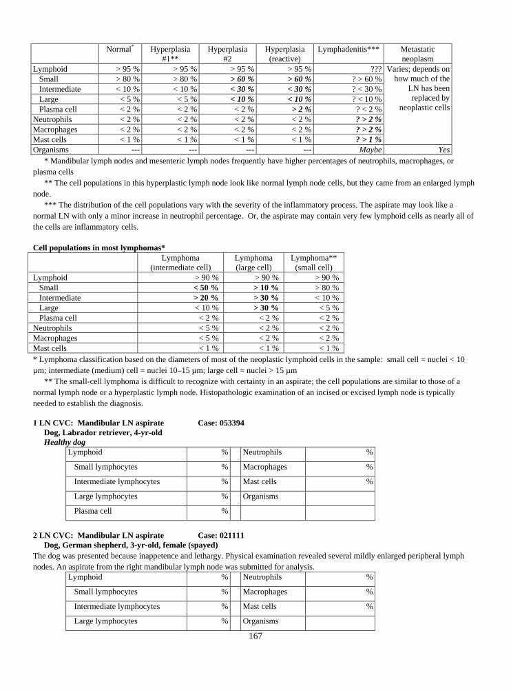
167
Normal* Hyperplasia
#1** Hyperplasia
#2 Hyperplasia
(reactive) Lymphadenitis*** Metastatic
neoplasm Lymphoid > 95 % > 95 % > 95 % > 95 % ??? Varies; depends on
how much of the LN has been replaced by
neoplastic cells
Small > 80 % > 80 % > 60 % > 60 % ? > 60 % Intermediate < 10 % < 10 % < 30 % < 30 % ? < 30 % Large < 5 % < 5 % < 10 % < 10 % ? < 10 % Plasma cell < 2 % < 2 % < 2 % > 2 % ? < 2 % Neutrophils < 2 % < 2 % < 2 % < 2 % ? > 2 % Macrophages < 2 % < 2 % < 2 % < 2 % ? > 2 % Mast cells < 1 % < 1 % < 1 % < 1 % ? > 1 % Organisms --- --- --- --- Maybe Yes
* Mandibular lymph nodes and mesenteric lymph nodes frequently have higher percentages of neutrophils, macrophages, or plasma cells
** The cell populations in this hyperplastic lymph node look like normal lymph node cells, but they came from an enlarged lymph node.
*** The distribution of the cell populations vary with the severity of the inflammatory process. The aspirate may look like a normal LN with only a minor increase in neutrophil percentage. Or, the aspirate may contain very few lymphoid cells as nearly all of the cells are inflammatory cells.
Cell populations in most lymphomas* Lymphoma
(intermediate cell) Lymphoma (large cell)
Lymphoma** (small cell)
Lymphoid > 90 % > 90 % > 90 % Small < 50 % > 10 % > 80 % Intermediate > 20 % > 30 % < 10 % Large < 10 % > 30 % < 5 % Plasma cell < 2 % < 2 % < 2 % Neutrophils < 5 % < 2 % < 2 % Macrophages < 5 % < 2 % < 2 % Mast cells < 1 % < 1 % < 1 % * Lymphoma classification based on the diameters of most of the neoplastic lymphoid cells in the sample: small cell = nuclei < 10 µm; intermediate (medium) cell = nuclei 10–15 µm; large cell = nuclei > 15 µm
** The small-cell lymphoma is difficult to recognize with certainty in an aspirate; the cell populations are similar to those of a normal lymph node or a hyperplastic lymph node. Histopathologic examination of an incised or excised lymph node is typically needed to establish the diagnosis. 1 LN CVC: Mandibular LN aspirate Case: 053394
Dog, Labrador retriever, 4-yr-old Healthy dog
Lymphoid % Neutrophils %
Small lymphocytes % Macrophages %
Intermediate lymphocytes % Mast cells %
Large lymphocytes % Organisms
Plasma cell %
2 LN CVC: Mandibular LN aspirate Case: 021111
Dog, German shepherd, 3-yr-old, female (spayed) The dog was presented because inappetence and lethargy. Physical examination revealed several mildly enlarged peripheral lymph nodes. An aspirate from the right mandibular lymph node was submitted for analysis.
Lymphoid % Neutrophils %
Small lymphocytes % Macrophages %
Intermediate lymphocytes % Mast cells %
Large lymphocytes % Organisms
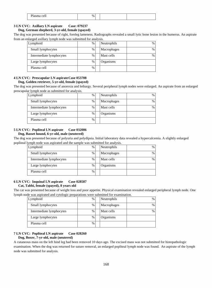
168
Plasma cell %
3 LN CVC: Axillary LN aspirate Case: 079237
Dog, German shepherd, 3-yr-old, female (spayed) The dog was presented because of right, foreleg lameness. Radiographs revealed a small lytic bone lesion in the humerus. An aspirate from an enlarged axillary lymph node was submitted for analysis.
Lymphoid % Neutrophils %
Small lymphocytes % Macrophages %
Intermediate lymphocytes % Mast cells %
Large lymphocytes % Organisms
Plasma cell %
4 LN CVC: Prescapular LN aspirate Case 053708
Dog, Golden retriever, 5-yr-old, female (spayed) The dog was presented because of anorexia and lethargy. Several peripheral lymph nodes were enlarged. An aspirate from an enlarged prescapular lymph node as submitted for analysis.
Lymphoid % Neutrophils %
Small lymphocytes % Macrophages %
Intermediate lymphocytes % Mast cells %
Large lymphocytes % Organisms
Plasma cell %
5 LN CVC: Popliteal LN aspirate Case 032086
Dog, Basset hound, 6-yr-old, male (neutered) The dog was presented because of polyuria and polydipsia. Initial laboratory data revealed a hypercalcemia. A slightly enlarged popliteal lymph node was aspirated and the sample was submitted for analysis.
Lymphoid % Neutrophils %
Small lymphocytes % Macrophages %
Intermediate lymphocytes % Mast cells %
Large lymphocytes % Organisms
Plasma cell %
6 LN CVC: Inquinal LN aspirate Case 028587
Cat, Tabbi, female (spayed), 8 years old The cat was presented because of weight loss and poor appetite. Physical examination revealed enlarged peripheral lymph node. One lymph node was aspirated and cytologic preparations were submitted for examination.
Lymphoid % Neutrophils %
Small lymphocytes % Macrophages %
Intermediate lymphocytes % Mast cells %
Large lymphocytes % Organisms
Plasma cell %
7 LN CVC: Popliteal LN aspirate Case 028260
Dog, Boxer, 7-yr-old, male (neutered) A cutaneous mass on the left hind leg had been removed 10 days ago. The excised mass was not submitted for histopathologic examination. When the dog was returned for suture removal, an enlarged popliteal lymph node was found. An aspirate of the lymph node was submitted for analysis.
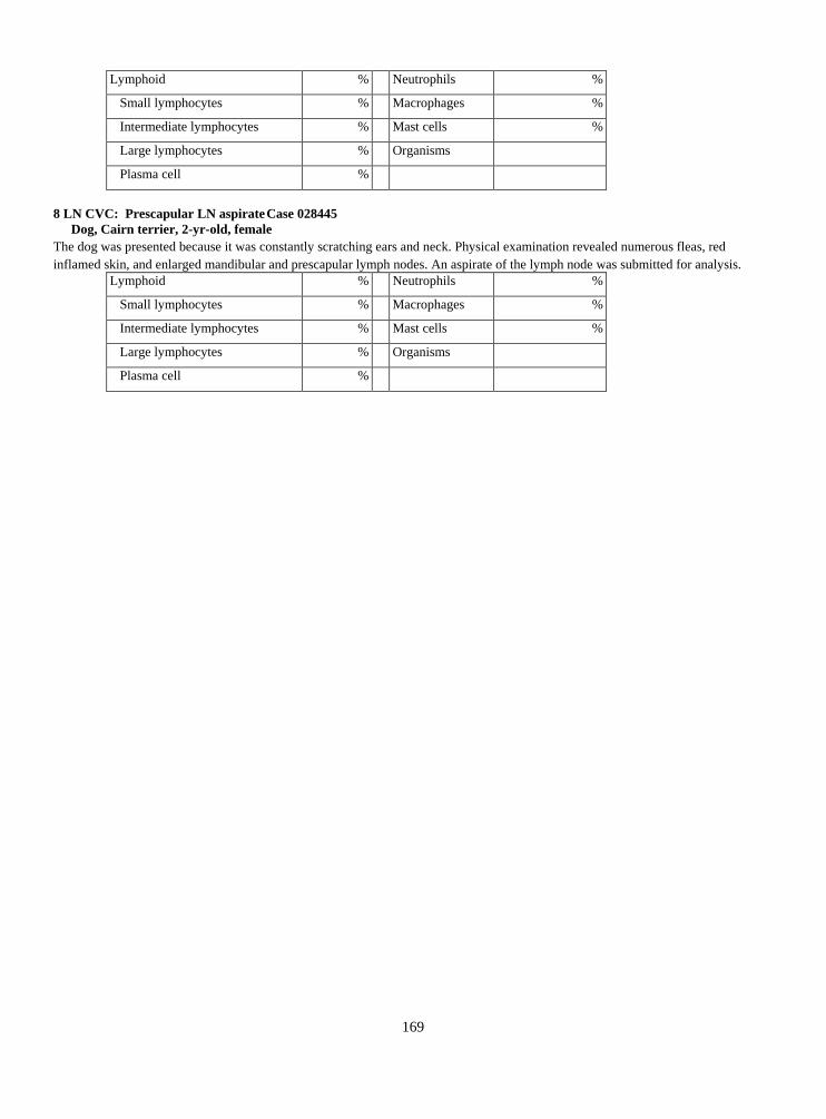
169
Lymphoid % Neutrophils %
Small lymphocytes % Macrophages %
Intermediate lymphocytes % Mast cells %
Large lymphocytes % Organisms
Plasma cell %
8 LN CVC: Prescapular LN aspirate Case 028445
Dog, Cairn terrier, 2-yr-old, female The dog was presented because it was constantly scratching ears and neck. Physical examination revealed numerous fleas, red inflamed skin, and enlarged mandibular and prescapular lymph nodes. An aspirate of the lymph node was submitted for analysis.
Lymphoid % Neutrophils %
Small lymphocytes % Macrophages %
Intermediate lymphocytes % Mast cells %
Large lymphocytes % Organisms
Plasma cell %
170
NSAIDs: Maximize Efficacy and Minimize Adverse Effects
Butch KuKanich, DVM, PhD, DACVCP Kansas State University
Manhattan, KS
Nonsteroidal Anti-Inflammatory Drugs (NSAIDs) are commonly used in veterinary medicine due to their efficacy in managing pain of multiple origins and their overall safety. However NSAIDs are the drugs that are most commonly reported to the Food and Drug Administration (FDA) Center of Veterinary Medicine for adverse effects. The most common organ systems affected by adverse effects are gastrointestinal, followed by renal, then hepatic. Other adverse effects occur less commonly. Gastrointestinal adverse effects Gastrointestinal (GI) adverse effects are the most common adverse effects reported to the FDA and drug approval studies. Gastrointestinal adverse effects can range from vomiting and diarrhea to GI erosions and ulcers and even progress to perforated ulcers and death. The development of cyclooxygenase (COX) COX-2 preferential and selective NSAIDs have decreased the overall occurrence of GI adverse effects, but GI adverse effects are still the most common adverse effects. COX-2 selective or preferential NSAIDs are recommended to decrease the GI adverse effects of NSAIDs, but do not eliminate the risk. Both COX-1 and COX-2 are constitutively expressed in the canine gastrointestinal tract and inhibition of both isoforms (nonselective inhibition) results in the highest frequency of GI adverse effects. Prostaglandins produced by COX-1 (prostaglandin E, PGE) and COX-2 (PGE and PGI) provide GI mucosal protectant effects. Selectively inhibiting COX-2 still provides analgesic effects, but decreases GI adverse effects due to maintaining some PGE production by COX-1. However since COX-2 is still constitutively expressed in the GI tract, adverse GI effects still occur. COX-2 is also induced in injured (including surgical) and diseased GI tissues to promote and enhance healing. Therefore a COX-2 inhibitor will decrease the healing and have detrimental effects on damages and injured GI tissues.
There are conditions, diseases and drugs that NSAID use should be avoided. By far the biggest drug interaction is with glucocorticoids (prednisone, dexamethasone, methylprednisolone, et al.) and concurrent or even sequential use substantially increase the risk for GI adverse effects. Therefore NSAIDs and glucocorticoids should not be used together or sequentially. Although there are many recommendations of a “washout” time between use of NSAIDs and glucocorticoids there are no studies documenting an ideal period of time to minimize adverse effects. Conservatively, many people recommend a week, but again the ideal period of time is unknown. Concurrent use of multiple NSAIDs should be avoided as the risk of GI adverse effects may be increased. Although “washout” periods are often recommended, studies documenting the efficacy of different “washout” times are lacking. Many experts conservatively recommend a week for a washout period, but it is unknown if that is an appropriate time period. Similar to glucocorticoids, uncontrolled hyperadrenocorticism (Canine Cushing’s Disease) markedly increases the potential GI adberse effects of NSAIDs due to the high concentrations of endogenous cortisol. Dogs with uncontrolled hyperadrenocorticism are essentially on high doses of endogenous glucocorticoids and the risk of GI adverse effects of NSAIDs are increased. Animals that have gastrointestinal disease are also at an increased risk of GI adverse effects of NSAIDs. Inflammatory bowel disease, parasitism, erosions, foreign bodies, enterotomies and gastrotomies are all conditions in which NSAID use should be avoided as NSAIDs increase the damage and decrease the healing of GI tissues. Animals with renal disease also appear to be at a higher risk of NSAID GI adverse effects. Renal disease results in uremia which produces toxic effects on the GI mucosa and animals may have subclinical erosions and ulcerations of the GI mucosa. Administration of an NSAID will worsen the erosions and ulcers and should be avoided if possible in animals with renal disease.
There are several strategies for minimizing GI adverse effects. The approved dose of the NSAID may be higher than the specific patient needs to provide beneficial effects. The typical dose-response curve has a log-normal distribution and a dose to produce desirable effects in 90% of the population will be higher than needed in some patients. Decreasing the daily dose by 10-25%, even up to 50% may still produce beneficial effects in some patients. However some patients will lose beneficial effects, except at the highest approved dose. Limited data are available on the efficacy of GI “protectants” in combination with approved NSAIDs in dogs. Omeprazole and other proton pump inhibitors are the highest efficacy acid suppression therapies and are among the highest efficacy and best tolerated therapy to decrease GI adverse effects of NSAIDs in humans, but little data are available in dogs. Despite the lack of data, proton pump inhibitors are commonly recommended in dogs to minimize NSAID GI adverse effects. Histamine (H2) antagonists are also effective acid suppression therapy in dogs and have decreased GI adverse effects of NSAIDs in humans. Therefore drugs such as famotidine are also commonly recommended in conjunction with NSAIDs in dogs, but specific data are lacking demonstrating decreased NSAID GI effects in dogs when concurrently administered. Sucralfate is a GI protectant that has demonstrated decreased GI adverse effects of NSAIDs in humans, but specific data are again lacking in dogs. Misoprostol is a synthetic prostaglandin that has demonstrated decreased NSAID GI adverse effects in humans and dogs. Misoprostol does have more adverse effects such as diarrhea than the other protective therapies. However the biggest concern with misoprostol is that it can cause
171
abortion in humans (and animals) and as such may present a large liability risk. When misoprostol is prescribed, most experts recommend dispensing gloves to the clients to wear during drug administration to minimize inadvertent human exposure. Another limiting factor with misoprostol is that is the most costly of the GI “protectants” for minimizing NSAID GI adverse effects. Renal adverse effects Cyclooxygenase produces prostaglandins (PGE and PGI) which maintain renal blood flow during periods of renal hypotension and hypoperfusion. Although COX-1 produces renal prostaglandins in some species, COX-2 is up-regulated producing the beneficial hemodynamic effects during renal hypotension. Therefore the COX-2 selective and preferential inhibitors that produce less GI adverse effects have little to no beneficial effects for minimizing renal NSAID adverse effects.
Conditions resulting in renal hypotension including, but not limited to anesthetic hypotension, shock, dehydration and hemorrhage increase the risk of renal adverse effects of NSAIDs. COX-2 is typically upregulated in renal hypotension resulting in production of PGE and PGI causing localized renal vasodilation, counteracting the sympathetic mediated vasoconstriction to maintain critical blood flow to the kidneys. However administration of an NSAID inhibits COX-2 and the local vasodilation resulting in renal hypoperfusion, hypoxemia and subsequent renal injury. Therefore NSAIDs should be avoided when animals are hypotensive or have the potential to become and remain hypotensive.
Although little data are available, many experts recommend avoiding NSAIDs in patients with chronic kidney disease with the thought that NSAIDs may worsen or result in progression of CKD. However there are little data supporting this recommendation. A few studies have examined some specific NSAIDs which appeared to be tolerated in patients with CKD. However the majority of recommendations are to avoid NSAIDs in CKD when possible.
Diuretics increase the risk of renal adverse effects as their desired effect, decreasing fluid and sodium retention are factors that increase the risk of renal adverse effects of NSAIDs. Additionally, NSAIDs decrease the effect of furosemide induced vasodilation (mediated by PGE) and can result in therapeutic failure of the diuretics. Likewise, NSAIDs decrease the effectiveness of angiotensin converting enzyme (ACE) inhibitors and therapeutic failure of the ACE inhibitors may occur with addition of an NSAID. Nephrotoxic drugs such as aminoglycosides and certain chemotherapeutics and amphotericin B should be avoided in combination with NSAIDs to decrease the risk of renal damage. Hepatic adverse effects Hepatic adverse effects can be dose-dependent or dose-independent. Dose-dependent adverse effects typically occur with drug over dosages and can be avoided by administering appropriate dosages. However with the marketing of many flavored and chewable NSAID formulations inadvertent exposures may occur, therefore these formulations should not be stored in an area the animal may have access.
Dose-independent (aka idiosyncratic) hepatic adverse effects occur when appropriate dosages are administered, but hepatic adverse effects still occur. The exact mechanisms are not known, but are often hypothesized due to formation of reactive metabolites that are toxic to the liver. The occurrence of dose-independent adverse effects is rare, but the exact numbers are not known. Dose-independent hepatic adverse effects most often occur early in the course of NSAID therapy, often in the first 3 weeks. A strategy to minimize these adverse effects is to establish baseline hepatic serum chemistries and repeat around week after starting the NSAID. If marked increases in hepatic enzymes or bilirubin occur NSAID therapy should be immediately discontinued. Severe changes and damage require hospitalization and symptomatic treatment including fluids and hepatic “protectant” drugs such as SAMe and silybinin, although the efficacy of the protectants have not been validated in large controlled clinical trials.
Client communications – critical component of decreasing NSAID adverse effects One of the best methods to decrease NSAID adverse effects are to have well informed clients about the benefits and risks of NSAIDs and appropriate at home monitoring. Clients should be informed to stop NSAIDs and notify their veterinarian if adverse effects are observed including: nausea, vomiting, diarrhea, lethargy, anorexia, depression, increased or decreased urination, discolored urine or discolored mucous membranes or sclera. Regardless of the adverse effect, the sooner it is identified, the NSAID is discontinued and appropriate diagnostics and treatments started as appropriate the better the overall prognosis. The FDA has available a free client handout detailing NSAID use and adverse effects which can be downloaded from their internet sight.
Disclaimer: The information is accurate to the best of the author’s knowledge. However recommendations change as new data become available and errors are possible. The author recommends double checking the accuracy of all information including dosages.
172
Veterinary Clinical Pharmacology Myths Butch KuKanich, DVM, PhD, DACVCP
Kansas State University Manhattan, KS
Morphine cannot be used in cats due to CNS excitement, aka morphine mania. This is false. Morphine is commonly used in cats without producing morphine mania. Morphine mania was termed when CNS excitement was noted after doses of 5-20 mg/kg SC, which are at least 20 times higher than the clinically recommended dose. High doses or rapid administration to any species can result in CNS excitement and even seizures. Morphine is slowly metabolized and eliminated in cats. This is false. The pharmacokinetics of morphine in cats demonstrate it is rapidly metabolized and has a short elimination half-life (just over an hour). The myth came about because cats are deficient in some glucuronide conjugations enzymes which metabolize morphine in most species. However cats rapidly metabolize morphine through a different pathway, sulfate conjugation and as a result morphine has a short half-life in cats. Morphine cannot be administered IV to dogs due to histamine release and severe hypotension. This myth is false. Morphine can be administered IV to dogs, resulting in some histamine release, but hypotension does not occur at clinically relevant doses. The high end of clinically relevant IV morphine doses are 0.5 mg/kg as a bolus. Higher doses can cause more profound histamine release, but marked hypotension does not occur until around 3 mg/kg IV bolus in dogs. However the effect of IV morphine on histamine release and cardiovascular status have not been investigated in dogs with mast cell tumors. Other IV opioids may be better choices for dogs with mast cell tumors until further data are available. Morphine and other opioids cause cardiovascular and respiratory depression in animals This is partially true, but rarely clinically relevant. Morphine and other opioids at clinically recommended doses have minimal detrimental effects on cardiovascular function in animals. Even massive overdoses have minor cardiovascular effects in healthy animals.
Although opioids do cause dose dependent respiratory depression, the magnitude of the depression is small and plateaus at relatively minimal respiratory depression. If substantial respiratory depression occurs it is often due to other factors contributing such as other drugs (e.g. inhalant or injectable anesthetics), concurrent disease (pulmonary disease) or head trauma in which the respiratory centers are affected. If the animal is at great risk for respiratory depression, which is clinically very rare, than constant rate infusions can be administered. The CRI minimizes peak drug concentrations while maintaining effective concentrations resulting in little to no effect on the respiratory function if appropriate doses are administered. Fluoroquinolones are broad spectrum antimicrobials This is false. Fluoroquinolones have little to no activity against anaerobes (except pradofloxacin) and very poor activity against Streptococcus species. Therefore most fluoroquinolones are not broad spectrum, but have a limited spectrum that are effective against many gram negative and some gram positive aerobic bacteria. Acepromazine lowers the seizure threshold This myth appears false because of the way acepromazine in used in veterinary medicine. Phenothioazines administered at high doses for long periods of time decrease the seizure threshold in human psychiatric patients. However acepromazine is used as single doses or for very short periods of time in veterinary medicine, which does not appear to increase the risk of seizures. A retrospective study demonstrated potential anticonvulsant effects of acepromazine or at least no worsening of seizures in 36 dogs with a history of seizure activity.
Disclaimer: The information is accurate to the best of the author’s knowledge. However recommendations change as new data become available and errors are possible. The author recommends double checking the accuracy of all information including dosages.
173
Common Drug Interactions to Know in Dogs and Cats Butch KuKanich, DVM, PhD, DACVCP
Kansas State University Manhattan, KS
Drug interactions can occur due to multiple reasons. Drug metabolism may be inhibited or induced, and drug absorption may be increased or decreased. Drugs may exaggerate or decrease the effects of others drugs. Here are some common drug interactions veterinarians need to be aware.
Drug metabolism inhibitors
• Chloramphenicol decreases elimination resulting in enhanced effects of phenobarbital, propofol, methadone if dosages are not decreased.
• Ketoconazole (itraconazole to a lesser extent) decreases elimination resulting in enhanced absorption and decreased elimination of cyclosporine, digoxin
• Enrofloxacin, marbofloxacin, ciprofloxacin decrease elimination of theophylline resulting in enhanced effects of theophylline.
• Cimetidine, although frequently mentioned as a drug metabolism inhibitor, the extent of inhibition in dogs is less than the other drug and as such does not cause many substantial drug interactions.
Drug metabolism inducers
• Phenobarbital results in increased metabolism and elimination of digoxin, chloramphenicol, glucocorticoids, ketoconazole, theophylline
• Phenytoin induces its own metabolism (and other drugs), which is the reason it is not effective as an anticonvulsant in dogs.
P-glycoprotein inhibitors
• Spinosad results in increased drug penetration of ivermectin (deworming doses, not heartworm preventive doses) into the brain resulting in ivermectin toxicity.
• Ketoconazole results in increased drug absorption of cyclosporine and digoxin. Decreased drug absorption
• Sucralfate, antacids, and iron supplements decrease absorption of doxycycline, minocycline and ciprofloxacin (not enrofloxacin) due to chelation.
• Omeprazole, famotidine and other acid suppressors decrease the absorption of ketoconazole. Pharmacodynamic
• NSAIDs with glucocorticoids result in enhanced GI adverse effects. • NSAIDs with furosemide and ACE inhibitors (e.g. enalapril) result in decreased activity of furosemide and ACE
inhibitors. • Opioids enhance sedation from sedatives and tranquilizers. • Glucocorticoids decrease tolerance development to beta agonists (terbutaline, albuterol). • Diuretics (furosemide et al) increase renal toxicity of aminoglycosides and NSAIDs • Tramadol, fluoxetine, selegeline, meperidine and clomipramine result in serotonin toxicity, which can manifest as
autonomic dysfunction, nausea, vomiting, diarrhea, hypertension, arrhythmias, seizures and death. Disclaimer: The information is accurate to the best of the author’s knowledge. However recommendations change as new data become available and errors are possible. The author recommends double checking the accuracy of all information including dosages.
174
Antiemetic Drugs: What to Use and When Butch KuKanich, DVM, PhD, DACVCP
Kansas State University Manhattan, KS
Antiemetics are for the symptomatic management of vomiting until an underlying condition is diagnosed and treated. It is important to differentiate vomiting from dysphagia and regurgitation. Antiemetics are not effective for dysphagia and regurgitation. The vomiting pathway is complex. It can be initiated from the gastrointestinal (GI) tract, pharyngeal stimulation, stimulation of the chemoreceptor trigger zone (CRTZ), vestibular system, intracranial and psychogenic causes. Depending on the source and cause of vomiting, some therapeutics may be effective and others ineffective. Antiemetics Maropitant. Maropitant is a neurokinin (NK1) antagonist that is a high efficacy antiemetic. The veterinarian must remember that maropitant can mask signs of severe or progressing disease due to its high efficacy. It can be effective for GI, pharyngeal, vestibular, CRTZ, and intracranial initiated vomiting. Maropitant is approved for use in dogs and cats and is available as injectable and oral formulations. SC administration results in high bioavailability and the oral bioavailability in dogs and cats is ~30 and 50%, respectively. Once daily dosing is typically effective. Maropitant undergoes saturable hepatic metabolism in dogs in which higher doses may result in greater than expected increases in plasma concentrations. Drugs such as ketoconazole, itraconazole, fluconazole, fluoxetine, and paroxetine may decrease metabolism whereas phenobarbital may increase metabolism. Adverse effects of maropitant can include pain and swelling at injection sight, lethargy, depression, weakness, ataxia, and sedation. Maropitant has cardiac potassium and calcium channel blocker effects which in healthy dogs are expected to produce minor adverse effects. However animals with decreased cardiac function may have worsening function and arrhythmias. Drug interactions including severe and potentially lethal arrhythmias could occur if maropitant is combined with antiarrhythmic drugs such as diltiazem, propranolol, atenolol, and sotalol among others, but studies documenting interactions are lacking. Additionally, the safety of maropitant has not been fully addressed when combined with doxorubicin.
Ondansetron is a serotonin (5HT3) antagonist and is a high efficacy antiemetic for GI causes of vomiting including chemotherapy. It will have lower efficacy for pharyngeal, vestibular and CRTZ vomiting. As with maropitant, ondansetron may mask the signs of progressing GI disease due to its high efficacy. There are no veterinary approved formulations of ondansetron in the USA, but it is used in an extra label manner for dogs and cats. The oral bioavailability of ondansetron in dogs is very poor and variable due to first pass metabolism, therefore PO is not a recommended route of administration. It has a short half-life and requires q 6-8 hr administration for the most consistent antiemetic effect. Due to its specific action of 5HT3 receptors, ondansetron is well tolerated with few adverse effects. Constipation can occur. In humans, headaches and dizziness have been reported. High doses of ondansetron have also been demonstrated to produce blockade of cardiac potassium channels, therefore adverse reaction may occur if combined with antiarrhythmic drugs such as diltiazem, propranolol, atenolol, and sotalol among others. Other drugs in this class include dolasetron and granisetron, but they are not commonly used due to their much higher cost.
Metoclopramide is an antiemetic producing effects through inhibition of dopamine (DA2) receptor, gastrointestinal prokinetic effects and at high doses 5HT3 antagonist effects. In the USA, there are no veterinary approved formulations. However high doses increase the risk of adverse effects such as CNS excitement and gastrointestinal hypermotility and pain. Due to its prokinetic effects, metoclopramide is contraindicated with GI foreign bodies or blockages due to the risk of perforation. Metoclopramide can be effective for GI, CRTZ induced vomiting and may provide some effects for intracranial and pharyngeal vomiting. Metoclopramide may also be beneficial in cases in which ileus contributes to GI vomiting such as opioid induced ileus. Other adverse effects can include peripheral and pulmonary edema due to aldosterone release and metoclopramide decreases the seizure threshold. Metoclopramide is available as injectable (administered IM, SC, IV and IV CRI) and oral formulations. Administration as an IV CRI may be more effective than other administration techniques.
Antihistamines are limited in efficacy to vestibular induced vomiting such as vestibular disease and motion sickness. Diphenhydramine is the most common antihistamine used in veterinary medicine as an antiemetic, but others are available.
Butorphanol and fentanyl also produce central antiemetic effects. However if opioids are administered for long durations or at high doses, ileus may occur which can result in vomiting.
Disclaimer: The information is accurate to the best of the author’s knowledge. However recommendations change as new data become available and errors are possible. The author recommends double checking the accuracy of all information including dosages.
175
Glucocorticoids: Use and Misuse
Butch KuKanich, DVM, PhD, DACVCP Kansas State University
Manhattan, KS
Glucocorticoids Prednisone is a commonly used glucocorticoid. Prednisone is a pro-drug, therefore it must be metabolized to prednisolone to elicit its pharmacologic effect. Cats are variable metabolizers of prednisone to prednisolone, so the effect will also be variable and less predictable in cats.
Prednisolone is recommended for use in cats as it is pharmacologically active and does not need to undergo metabolism to be effective. Prednisone/prednisolone has predominant glucocorticoid effects, but does have some mineralocorticoid effects as well. As such fluid and sodium retention occurs which can result in pulmonary edema in patients even with subclinical and undiagnosed cardiac dysfunction.
Dexamethasone is a glucocorticoid with essentially no mineralocorticoid effects, therefore the retention of fluid and sodium is much less compared to prednisone/prednisolone. Dexamethasone is the glucocorticoid of choice in most patients with cardiac dysfunction. Gastric adverse effects (vomiting, diarrhea, gastritis, erosions and ulcerations) appear to more frequent with dexamethasone compared to prednisone/prednisolone. It is unclear if the increased risk is inherent to the drug or due to incorrect dosages. Dexamethasone is approximately 10x more potent than prednisone/prednisolone, therefore an equivalent dose is 1/10. Additionally, the duration of effect of dexamethasone is longer then prednisone/prednisolone, therefore it needs to be dosed less frequently. For example, 2 mg of dexamethasone PO q 48 hours produces near equivalent glucocorticoid effects as 20 mg of prednisone/prednisolone q 24 hours in dog/cats.
Injectable dexamethasone formulations are often confusing in their use in veterinary medicine. Dexamethasone solution is a 2 mg/mL solution of dexamethasone (free base) dissolved in polyethylene glycol due to the poor water solubility dexamethasone (free base). Dexamethasone sodium phosphate is a water soluble formulation of dexamethasone solution in water. The onset of effect and duration of effect for IV administration of either formulation will be near identical if equal doses are administered. IM or SC administration of dexamethasone solution may have a slightly delayed absorption, but is unlikely to be clinically appreciated.
Repository formulations of glucocorticoids (methylprednisolone acetate, triamcinolone acetonide) should not be used systemically in dogs and should only be used as a last resort in cats due to the increased risk and severity compared to oral glucocorticoids. Oral glucocorticoids can be rapidly discontinued if adverse effects occur, but once a repository formulation is administered it cannot be discontinued.
Glucocorticoid physiologic replacement therapy Glucocorticoid physiologic replacement therapy is used when endogenous glucocorticoids are insufficient to maintain normal processes. The primary indication is hypoadrenocorticism (Addison’s Disease). The recommended prednisone/prednisolone dose in dogs is 0.2 mg/kg/d. An equivalent dexamethasone dose would be 0.02 mg/kg q 48 hours. Anti-inflammatory therapy Anti-inflammatory dosages are used for inflammatory conditions such as atopy. Anti-inflammatory dosages start at 1 mg/kg/d (equivalent to 0.1 mg/kg q 48 for dexamethasone) and are decreased to every other day for chronic administration and titrated down to the lowest effective dose for the shortest duration when needed. Immunosuppressive therapy Immunosuppressive dosages are used to control immune mediated diseases such as autoimmune hemolytic anemia and immune mediate thrombocytopenia among others. Prednisone/prednisolone starting dose is 2 mg/kg/d (equivalent to 0.2 mg/kg q 48 for dexamethasone) and may be titrated to every other day administration and or doses decreased. Immunosuppressive doses are recommended for the treatment of anaphylactic shock and Addison’s crisis as well. Shock therapy A shock dose of glucocorticoids is only indicated for spinal trauma (methylprednisolone sodium succinate, 30 mg/kg IV) and data suggest may only provide a benefit within the first 3-8 hours after the trauma. Other glucocorticoids have not demonstrated efficacy for spinal trauma.
Current data suggest shock doses of glucocorticoids are detrimentalCurrent data do not indicate there is a benefit for shock doses of glucocorticoids for generalized trauma such as hit by car, sepsis or
endotoxemia, or heat stroke. Glucocorticoids may be detrimental in heat stroke due to the damage in the GI tract from the
for head trauma and increase death and disability in humans.

176
hyperthermia. Fluid therapy (isotonic crystalloids, hypertonic crystalloids, colloids) and analgesia (opioids are the treatments of choice for shock due to trauma and pain) and fluid therapy for sepsis endotoxemia and heat stroke.
As stated in the immunosuppressive therapy section, immunosuppressive doses of glucocorticoids can be beneficial for anaphylactic shock and Addison’s crisis, but not shock doses.
Disclaimer: The information is accurate to the best of the author’s knowledge. However recommendations change as new data become available and errors are possible. The author recommends double checking the accuracy of all information including dosages.
177
Labels on Approved Veterinary Drugs that are Wrong! Butch KuKanich, DVM, PhD, DACVCP
Kansas State University Manhattan, KS
There are numerous drugs that were approved many years ago prior to the rigors that are required for FDA approval needed to achieve safe and effective dosing. Unfortunately, many of these drugs are still available despite incorrect label recommendations and dosages. In order to change the labels or dosages, drug companies would have undergo an entirely new approval process which would cost upwards of hundreds of thousands of dollars. Therefore there is little incentive for manufacturers to change or update the labels Acepromazine Acepromazine is a phenothiazine tranquilizer. The label dose 0.5-1.1 mg/kg in dogs (IV, SC, IM) and 1.1-2.2 mg/kg in cats. Unfortunately this is much higher than appropriate. The current recommended doses in dogs and cats are 0.05 and 0.1 mg/kg. The higher dose results in increased adverse effects such as hypotension and prolonged durations of effect, but does not provide increased sedation. Prednisolone sodium succinate Prednisolone sodium succinate (Solu-Delta-Cortef ®) is labeled for use in dogs in cats for “shock” at a dose of 2.5-5 mg/# (5.5-11 mg/kg). This is not an appropriate dose for any condition! (see glucocorticoids lecture) It is between an immunosuppressive and “shock” dose. Additionally, immunosuppressive glucocorticoids should not be used in septic patients, despite the label recommending use for conditions such as sepsis, pneumonia, peritonitis mastitis, etc. Most causes of shock are not responsive to glucocorticoids and the adverse effects of glucocorticoids are too severe to warrant use (e.g. trauma, pain, heat stroke, endotoxemia, septicemia). For example, shock doses of glucocorticoids increase the rate of death and disability in human head trauma patients. The label states that the benefit may be limited unless used with plasma volume expanders (e.g. IV fluids), but the reality is the animals will respond to analgesics (if needed) and fluids and addition of glucocorticoids only worsens the animals’ condition due to their adverse effects. Glucocorticoids worsen the progression of osteoarthritis (OA) due to their catabolic effects on joint cartilage. The use of glucocorticoids are not recommended for use in snakebites as there are no data supporting there use and may increase the risk of secondary infections (despite the label recommendations). Methylprednisolone acetate Methylprednisolone acetate (Depo Medrol ®) is labeled for IM use in dogs and cats, but most experts do not recommend using it in dogs systemically due to high risk of adverse effects. Despite its recommendation for OA, glucocorticoids worsen the progression due to their catabolic effects on joint cartilage. It is labeled for use in severe infection, but is actually contraindicated in most infectious diseases. The use of glucocorticoids are not recommended for use in snakebites as there are no data supporting their use and may increase the risk of secondary infections (despite the label recommendations). Procaine penicillin G Procaine penicillin G is labeled for use in dogs for a variety of infections including otitis externa. Otitis external often involve Staphylococcus spp. which are most often resistant and Malassezia (yeast) which are not susceptible to penicillin. The dose listed 6000 IU per kg q 24h is too low and increases the risk of treatment failure and resistance. The current dosage recommendations are 20,000 – 40,000 IU/kg q 12-24 h IM for susceptible infections. Procaine penicillin G / benzathine penicillin G Procaine penicillin G / benzathine penicillin G is labeled for use in dogs and cattle as an extended release formulation of penicillin due to the slow release of penicillin form the benzathine component. However the problem is that the release is so slow, it does not provide high enough plasma concentrations to extend the dosing interval for most infections except maybe uncomplicated urinary tract infections. Additionally, the labeled dose is much lower than currently recommended for penicillin and as such increased the risk for treatment failure and selection for resistant bacteria. Many people refer to this formulation tongue in cheek as long residue penicillin in food animals.
Disclaimer: The information is accurate to the best of the author’s knowledge. However recommendations change as new data become available and errors are possible. The author recommends double checking the accuracy of all information including dosages.
178
Small Animal Coagulation Disorders Karl Jandrey, BS, DVM, MAS, DACVECC
University of California Davis, CA
Primary hemostasis Primary hemostasis is the formation of a platelet plug at the site of endothelial damage. Damage to a blood vessel causes vasoconstriction and exposure of endothelial collagen. When platelets contact exposed collagen and collagen-bound von Willebrand Factor (vWF), they change their shape, become sticky and release a variety of chemicals that promote adhesiveness with other platelets. They adhere together to form a loose platelet plug. This platelet plug is then reinforced with fibrin formed by secondary hemostasis. Treatment of impaired primary hemostasis The treatment of a defect in primary hemostasis involves the removal of the underlying cause if possible. Transfusion of platelets is challenging due to their very reactive nature and special handling required. The drawing of blood and placing it into collection bags can activate platelets to form clumps. Therefore, filters should always be used for administration. Platelets also activate at cold temperatures. All platelet transfusions must be done with fresh blood, either fresh whole blood or platelet-rich plasma.
Fresh whole blood transfusions are a source of platelets, but whole blood provides fewer platelets than a platelet-rich product and can cause hypervolaemia. Recently, a frozen platelet concentrate (PC) has been produced and is commercially available for dogs (Midwest Animal Blood Services, Inc. - Stockbridge, Michigan, USA). This PC is prepared from a single donor by automated blood cell processors using apheresis technology, and is cryopreserved in 6% dimethyl sulfoxide (DMSO).
In human medicine, platelets have been cryopreserved with 6% DMSO, resulting in conservation for up to 3 years. However, this method of storage also resulted in loss of platelet viability, decreased aggregation and reduced clinical efficacy compared to fresh platelets. Our lab recently published a study which studied platelet function, but not clinical efficacy, of this commercially available canine frozen PC.
With all these difficulties or clinical unknowns, there is no universal recommendation for the transfusion of platelets in dogs. It is very difficult to give a platelet number-based transfusion trigger. As a basic rule, the risk for hemorrhage is inversely proportional to the platelet count, and bleeding times in humans are prolonged in a linear fashion as platelet count drops from 100,000 to 10,000/µL. The most common trigger for prophylactic transfusion is 20,000/µL in human medicine. In veterinary medicine, numbers of 5,000/µL in cats and 10,000/µL in dogs have also been published. Published platelet numbers adequate for surgery are between 30,000 to 80,000/µL.
The current recommendation from our service is to treat on-going life threatening hemorrhage that is secondary to thrombocytopenia with fresh whole blood or fresh platelet rich plasma. As a rule of thumb in veterinary medicine, the recommendation is to transfuse platelet-rich products only in case of life-threatening bleeding, especially pulmonary or intra-cranial hemorrhage. Fresh platelet rich plasma is not commercially available and must be made from fresh whole blood just before administration.
Three products are available for platelet replacement: platelet concentrate (PC), platelet rich plasma (PRP) and fresh whole blood (FWB). These are suggested initial doses but the individual dose must be guided by the response to therapy.
• Fresh whole blood: 20 mL/kg within 4 hours of collection. • Platelet concentrate: 1 unit/10 kg. The transfusion should be finished in an hour. • Platelet-rich plasma: 10 mL/ kg. The transfusion should be finished in an hour.
Fresh whole blood is the most commonly available blood product to be used in case of life threatening bleeding due to thrombocytopenia.
Platelet rich plasma is prepared by centrifugation of fresh whole blood at a relatively slow speed. The supernatant is therefore "rich" in platelets. It is a fresh product so needs to be processed right before administration. Due to the presence of some red cells, a cross match is essential before administration.
Platelet concentrate is prepared by platelet-apheresis, a process whereby blood is removed from the donor and separated by an automated device into plasma, platelets, white blood cells and red blood cells. The platelet portion is saved and forms one unit of PC and the remaining components are returned back to the donor. According to the manufacturer, one unit of PC can be stored for 6 months at -20°C. PC can also be prepared by centrifugation of platelet rich plasma from different donors (from 4 to 10), separation of the platelet-rich buffy coat and reconstitution in a sterile blood collection bag. Secondary hemostasis The main purpose of secondary hemostasis is the production of fibrin to stabilize the platelet plug. Initiators of secondary hemostasis include the platelet plug and endothelial damage. It ends in the formation of thrombin, which cross-links fibrin and strengthens the
179
clot. Thrombin also activates fibrinolysis, to limit excessive fibrin accumulation. Coagulopathies can be caused by a congenital defect or deficiency in any coagulation factor, lack of production of coagulation factors, excessive consumption of factors such as in the late phase of DIC, or by dilution. Assessment of secondary hemostasis Disorders of secondary hemostasis are recognized by hemorrhage into body cavities (e.g. hemothorax, hemoabdomen, hemomediastinum) or into organs (e.g. brain, urinary bladder, intestinal tract). Lower platelets counts generally can be found due to consumption, but they are rarely low enough (i.e. less than 20,000/µL) to suggest that they were the primary cause of the hemorrhage.
The main purpose of secondary hemostasis is the production of fibrin to stabilize the platelet plug. Initiators of secondary hemostasis include the platelet plug and endothelial damage. It ends in the formation of thrombin, which cross-links fibrin and strengthens the clot. Thrombin also activates fibrinolysis, to limit excessive fibrin accumulation. Coagulopathies can be caused by a congenital defect or deficiency in any coagulation factor, lack of production of coagulation factors, excessive consumption of factors such as in the late phase of DIC, or by dilution.
The following tests can only be used to assess hypocoagulability: • Coagulation time • Activated clotting time (ACT) • Activated partial thromboplastin time (aPTT) • Prothrombin time (PT) • PIVKA (proteins induced by vitamin K absence or antagonism)
These coagulation tests are timed tests and are reported in seconds. Fast times of tests, such as the ACT, PT and aPTT, do not represent excessive or fast coagulation within the patient; they only represent the absence of hypocoagulability. An increase in the length of time to clot formation would indicate a hypocoagulable state. Often the increased result of a bleeding time test may not yet manifest on the physical exam. Some of these tests are more sensitive or specific than others. For instance the PIVKA has nearly a 98% specificity for an anticoagulant rodenticide intoxication when the result is >300 seconds, Treatment of impaired secondary hemostasis Since most of the above tests are abnormal due to a lack of sufficient coagulation factors, the goal of therapy is to restore coagulation factors to levels that normalize these tests and, more importantly, reduce the chance for hemorrhage to persist. In general, 10-20 mL/kg of fresh frozen plasma (see Table 1) will restore coagulation factors to a level to normalize the coagulation tests. This should be administered within 4 hours of the thaw at a rate at which the patient is not at risk for fluid overload.
Fresh plasma is obtained by centrifugation from FWB and separation into packed red blood cells (pRBC) and plasma within 6 hours of collection. After fresh plasma has been frozen, it is called FFP for up to 1 year. After one year of storage it is then referred to as Frozen Plasma due to the natural decrease in the labile factors (FV, FVIII) factors during storage.
The major indication is the treatment of coagulopathies, either acquired (e.g. rodenticide intoxication, DIC) or inherited (e.g. von Willebrand disease, hemophilias).
The required FFP dose in an individual patient is impossible to predict and must be based on response to therapy, availability of blood products and financial limitations. A general guideline is an initial dose between 10–20 mL/kg.
Fresh frozen plasma is not the fluid of choice for low albumin/low osmotic pressure in dogs and cats because it contains very low albumin (less than 5%, i.e. 5 mg/dL). It takes over 40ml/kg of plasma to raise albumin 1g/dL in the patient. Synthetic colloids can be used to increase the colloid osmotic pressure. Human serum albumin can be used to effectively increase albumin concentration in dogs and cats, but questions remain regarding safety of this product in our patients.
During the process of thawing fresh frozen plasma at 4°C, a white precipitate forms due to the run-off of thawed portion; that is the cryoprecipitate (CP), which can be separated and stored. The major indication for the use of CP is von Willebrand disease and Hemophilia A. The published dose is 10 mL/kg.
180
Advances in Coagulation Testing Karl Jandrey, BS, DVM, MAS, DACVECC
University of California Davis, CA
Primary hemostasis Primary hemostasis is the formation of a platelet plug at the site of endothelial damage. Damage to a blood vessel causes vasoconstriction and exposure of endothelial collagen. When platelets contact exposed collagen and collagen-bound von Willebrand Factor (vWF), they change their shape, become sticky and release a variety of chemicals that promote adhesiveness with other platelets. They adhere together to form a loose platelet plug. This platelet plug is then reinforced with fibrin formed by secondary hemostasis. Assessment of primary hemostasis Petechiae and ecchymoses are found on the physical examination of the patient with a disorder of primary hemostasis. It is uncommon to have hemorrhage into large body cavities. Epistaxis and other mucosal bleeding are common.
Platelet function and number must be scrutinized upon suspicion of a bleeding defect. Platelets are formed from megakaryocytes in the bone marrow and are the smallest cellular component in the blood. Platelets are anucleate, disc-shaped, cytoplasmic fragments that play an essential role in primary hemostasis, the initiation of a platelet plug, and the preservation of vascular integrity. Their exterior surface is highly reactive to external stimuli, ready to undergo a shape change to facilitate hemostasis on the damaged endothelial surface. Placed near the site of initial response to vascular injury, platelets react to stimuli, alter their shape, spread and adhere to the endothelium (and each other), secrete their granular contents (e.g. ADP, serotonin) to amplify the response, and interact with the hemostatic system to bind fibrinogen (onto GP IIbIIIa) and solidify into a fibrin clot.
Thrombocytopenia is a relatively common finding in veterinary patients; however, platelet dysfunction (thrombocytopathia) is much less common. In people, thrombocytopathia (reduced adhesion, altered aggregation, and poor clot retraction) is common secondary to neoplasia or dysproteinemias. In domestic species, the common causes of thrombocytopathia are renal or hepatic disease, zootoxins, and infectious or therapeutic agents. Decreased platelet function should be considered when superficial bleeding (i.e. petechiae, ecchymoses) occurs in the absence of profound thrombocytopenia.
For all patients suspected of having a coagulation defect, a standard blood smear should be evaluated under the microscope for the estimation of a platelet count in the monolayer. A single platelet found on a 100X field equates to ~15,000 platelets/µL in circulation. It is unlikely that spontaneous bleeding will occur until platelet counts drop below 20,000/µL. Platelet clumps will alter your interpretation of the estimated number of platelets. Also, platelet numbers that are found to be low on automated, in-house blood cell counters should always be checked against a manual estimation of platelet numbers using this blood smear technique. Discordant results are often found because the automated cell counters are unable to count platelet clumps as individual cells, artificially lowering the platelet count.
When decreased platelet numbers do not account for bleeding, a buccal mucosal bleeding test (BMBT) can be considered. This in vivo test is completed by the measurement of the time for a stable platelet plug to form from the time an incision is created on the upper lip of a dog or cat. The test uses a template device that creates 1 or 2 standardized superficial incisions (1mm deep by 5mm long). Normal BMBT results are less than 4 minutes. A prolonged result would be consistent with thrombopathia or von Willebrand disease. This test is highly operator dependent; variable results have been reported in animals and in humans. BMBT measurements are poorly standardized, labor intensive, subjective, and have not been shown to correlate with clinical outcomes. In fact, this test has been all but abandoned in human medicine due to the lack of specificity and sensitivity to detect or predict clinical perioperative bleeding.
The Platelet Function Analyzer-100® (PFA-100) is a bench-top instrument that evaluates platelet function in whole blood. The PFA-100 simulates primary hemostasis by aspirating citrate-anticoagulated blood under a high shear rate through a small aperture in a collagen membrane coated with platelet agonists (ADP or epinephrine). This design mimics the in vivo organization of the subendothelial matrix: the initial site of platelet deposition and aggregation. Closure time (CT) is the time it takes for a platelet plug to form and occlude flow. The CT is highly sensitive to qualitative and quantitative defects in platelet receptors that mediate adhesion (GPIb-V-IX) and aggregation (GPIIbIIIa).
The PFA-100 detects inherited, acquired, or induced platelet dysfunction. It has most commonly been used for analysis of coagulopathies (e.g. von Willebrand Disease) in dogs, horses, and humans as well as assessing aspirin therapy and affects of various fluids (saline, artificial colloids). Studies have shown that the PFA-100 can be used as an indicator of platelet function and dysfunction in many species that are not anemic or thrombocytopenic. In addition, the CT may be inaccurate when high hematocrits (>60%) or platelet counts (>500,000)/ µL) are present.
Turbidometric aggregometry was invented in the 1960s and is regarded as the gold standard for the diagnosis of primary hemostatic defects. It is able to detect many different aspects of platelet function and biochemistry through the use of agonists (ADP,
181
thrombin, or collagen) at various concentrations. Platelet rich plasma from citrated whole blood is used in parallel chambers through which light is transmitted during the test. The opaque solution is stimulated to aggregate by way of the introduction of an agonist. As the platelets aggregate in response to the agonist, the sample becomes clearer as the platelets aggregate. The clarity of the sample is detected as an increase in the transmission of light across the sample. The difference between baseline and after agonist addition and aggregation is used to calculate the response. The specialized aggregometers graphically display the response as a curve that can be further analyzed to detect speed of aggregate formation. The major drawback is that it does not mimic in vivo physiologic conditions of adhesion, activation or aggregation to endothelial damage. Besides the limited availability to comparative hemostasis laboratories, it also requires large volumes of fresh blood and technical expertise. Aggregometry has been validated for use with canine and feline platelets as well as canine platelet concentrate.
To overcome some of the technical problems with aggregometry, alternatives such as the cartridge-based system of the VerifyNow® have been developed for humans. This instrument measures platelet aggregation in whole citrated blood via changes in light transmission. The basis of this assay is that coated polystyrene microparticles will agglutinate in whole blood in direct proportion to the degree of platelet activation. Specific cartridges are employed to monitor for the effects on primary hemmostasis by the following drugs: aspirin, P2Y12 inhibitors (clopidogrel), GP IIbIIIa antagonists (abciximab). These limitations and expense of the machine make it unlikely to be used in clinical veterinary medicine. This analyzer has received little use in research studies in domestic animals.
The cone and plate(let) analyzer technology was developed to test platelet function under near-physiologic conditions. The benefits of this system (Impact-R) are that it employs a small blood volume, requires no blood processing, and is simple to operate. Basically, this system mimics an extracellular matrix over which the blood sample flows. Since platelets are the only cell to adhere to this matrix under these conditions, the elongated aggregates align in laminar flow lines. After simulation of blood flow conditions, the sample is stained and optically analyzed. The effects of variable hematocrit and platelet counts have been investigated in people. No published studies have been found that employed this technology in domestic animals.
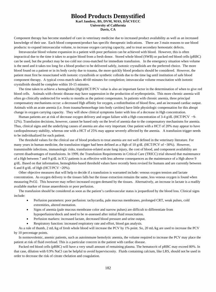
182
Blood Products Demystified Karl Jandrey, BS, DVM, MAS, DACVECC
University of California Davis, CA
Component therapy has become standard of care in veterinary medicine due to increased product availability as well as an increased knowledge of their use. Each blood component/product has specific therapeutic indications. There are 3 main reasons to use blood products: to expand intravascular volume, to increase oxygen carrying capacity, and to treat secondary hemostatic defects.
Intravascular blood volume expansion in a patient with poor perfusion can be achieved with blood. However, this is often impractical due to the time it takes to acquire blood from a fresh donor. Stored whole blood (SWB) or packed red blood cells (pRBC) can be used, but the product may be too cold nor cross-matched for immediate transfusion. In the emergency situation when volume is the need and it takes too long for a blood product to be delivered safely, isotonic crystalloids are the preferred choice. The more blood found on a patient or in its body cavity due to trauma, the more quickly blood products should be considered. However, the patient must first be resuscitated with isotonic crystalloids or synthetic colloids due to the time lag until institution of safe blood component therapy. A typical cross-match takes 40-60 minutes for completion; intravascular volume resuscitation with isotonic crystalloids should be complete within 10-15 minutes.
The time taken to achieve a hemoglobin (Hgb)/HCT/PCV value is also an important factor in the determination of when to give red blood cells. Animals with chronic disease may have suppression in the production of erythropoietin. This more chronic anemia will often go clinically undetected for weeks to months as the patient compensates. In patients with chronic anemia, three principal compensatory mechanisms occur: a decreased Hgb affinity for oxygen, a redistribution of blood flow, and an increased cardiac output. Animals with an acute anemia (i.e. from trauma/hemorrhage into body cavities) have little physiologic compensation for this abrupt change in oxygen carrying capacity. They will show clinical symptoms faster with less of a decrease in Hgb concentration.
Human patients are at risk of decrease oxygen delivery and organ failure with a Hgb concentration of 3-4 g/dL (HCT/PCV ~9-12%). Transfusion decisions, however, cannot be based only on the level of anemia due to the compensatory mechanisms for anemia. Thus, clinical signs and the underlying causes of anemia are also very important. One patient with a HCT of 20% may appear to have cardiopulmonary stability, whereas one with a HCT of 25% may appear severely affected by the anemia. A transfusion trigger needs to be individualized for each patient.
The threshold values for the clinical use of blood products to treat anemia are not well defined in the veterinary literature. For many years in human medicine, the transfusion trigger had been defined as a Hgb of 10 g/dL (HCT/PCV of ~30%). However, transmissible infections, immunologic risks, transfusion-related acute lung injury, the cost of blood, and component availability are current disadvantages of transfusions. In 1999, the Transfusion Requirements in Critical Care (TRICC) trial showed that maintenance of a Hgb between 7 and 9 g/dL in ICU patients is as effective with less adverse consequences as the maintenance of a Hgb above 9 g/dL. Based on that information, hemoglobin-based threshold values have recently been revised for humans and are currently between 6 and 8 g/dL of Hgb (HCT/PCV ~20%).
Other objective measures that will help to decide if a transfusion is warranted include: venous oxygen tension and lactate concentration. As oxygen delivery to the tissues falls but the tissue extraction remains the same, less venous oxygen is found when measuring PvO2. This however may reflect increased oxygen demand by the tissues. Alternatively, an increase in lactate is a readily available marker of tissue anaerobiosis or poor perfusion.
The transfusion should be considered as soon as the patient’s cardiovascular status is jeopardized by the blood loss. Clinical signs include:
• Perfusion parameters: poor perfusion: tachycardia, pale mucous membranes, prolonged CRT, weak pulses, cold extremities, altered mentation.
• Signs of anemia (pale mucous membrane color and narrow pulses) are difficult to differentiate from hypoperfusion/shock and need to be re-assessed after initial fluid resuscitation.
• Perfusion markers: increased lactate, decreased blood pressure and urine output. • Respiratory function: increased respiratory rate and effort, blood gas analysis.
As a rule of thumb, 2 mL/kg of fresh whole blood will increase the PCV by 1% point. So, 20 mL/kg are used to increase the PCV by 10 percentage points.
In normovolemic, anemic patients, such as autoimmune hemolytic anemia, the volume required to increase the PCV may place the patient at risk of fluid overload. This is a particular concern in the patient with cardiac disease.
Packed red blood cells (pRBC) will have a very small amount of remaining plasma. The hematocrit of pRBC may exceed 80%. In that case, dilution with 0.9% NaCl can be helpful to avoid hyperviscosity. Fluids containing calcium, like LRS, should not be used in order to decrease the risk of citrate chelation and coagulation.
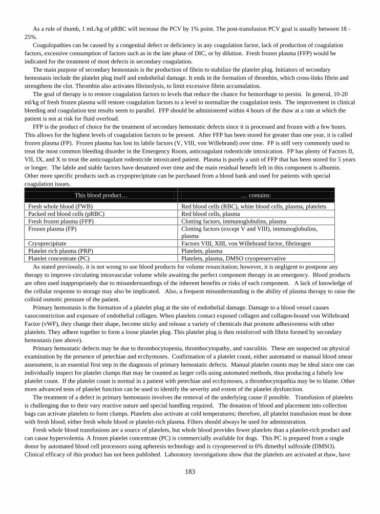
183
As a rule of thumb, 1 mL/kg of pRBC will increase the PCV by 1% point. The post-transfusion PCV goal is usually between 18 - 25%.
Coagulopathies can be caused by a congenital defect or deficiency in any coagulation factor, lack of production of coagulation factors, excessive consumption of factors such as in the late phase of DIC, or by dilution. Fresh frozen plasma (FFP) would be indicated for the treatment of most defects in secondary coagulation.
The main purpose of secondary hemostasis is the production of fibrin to stabilize the platelet plug. Initiators of secondary hemostasis include the platelet plug itself and endothelial damage. It ends in the formation of thrombin, which cross-links fibrin and strengthens the clot. Thrombin also activates fibrinolysis, to limit excessive fibrin accumulation.
The goal of therapy is to restore coagulation factors to levels that reduce the chance for hemorrhage to persist. In general, 10-20 ml/kg of fresh frozen plasma will restore coagulation factors to a level to normalize the coagulation tests. The improvement in clinical bleeding and coagulation test results seem to parallel. FFP should be administered within 4 hours of the thaw at a rate at which the patient is not at risk for fluid overload.
FFP is the product of choice for the treatment of secondary hemostatic defects since it is processed and frozen with a few hours. This allows for the highest levels of coagulation factors to be present. After FFP has been stored for greater than one year, it is called frozen plasma (FP). Frozen plasma has lost its labile factors (V, VIII, von Willebrand) over time. FP is still very commonly used to treat the most common bleeding disorder in the Emergency Room, anticoagulant rodenticide intoxication. FP has plenty of Factors II, VII, IX, and X to treat the anticoagulant rodenticide intoxicated patient. Plasma is purely a unit of FFP that has been stored for 5 years or longer. The labile and stable factors have denatured over time and the main residual benefit left in this component is albumin. Other more specific products such as crypoprecipitate can be purchased from a blood bank and used for patients with special coagulation issues.
This blood product… … contains:
Fresh whole blood (FWB) Red blood cells (RBC), white blood cells, plasma, platelets Packed red blood cells (pRBC) Red blood cells, plasma Fresh frozen plasma (FFP) Clotting factors, immunoglobulins, plasma Frozen plasma (FP) Clotting factors (except V and VIII), immunoglobulins,
plasma Cryoprecipitate Factors VIII, XIII, von Willebrand factor, fibrinogen Platelet rich plasma (PRP) Platelets, plasma Platelet concentrate (PC) Platelets, plasma, DMSO cryopreservative
As stated previously, it is not wrong to use blood products for volume resuscitation; however, it is negligent to postpone any therapy to improve circulating intravascular volume while awaiting the perfect component therapy in an emergency. Blood products are often used inappropriately due to misunderstandings of the inherent benefits or risks of each component. A lack of knowledge of the cellular response to storage may also be implicated. Also, a frequent misunderstanding is the ability of plasma therapy to raise the colloid osmotic pressure of the patient.
Primary hemostasis is the formation of a platelet plug at the site of endothelial damage. Damage to a blood vessel causes vasoconstriction and exposure of endothelial collagen. When platelets contact exposed collagen and collagen-bound von Willebrand Factor (vWF), they change their shape, become sticky and release a variety of chemicals that promote adhesiveness with other platelets. They adhere together to form a loose platelet plug. This platelet plug is then reinforced with fibrin formed by secondary hemostasis (see above).
Primary hemostatic defects may be due to thrombocytopenia, thrombocytopathy, and vasculitis. These are suspected on physical examination by the presence of petechiae and ecchymoses. Confirmation of a platelet count, either automated or manual blood smear assessment, is an essential first step in the diagnosis of primary hemostatic defects. Manual platelet counts may be ideal since one can individually inspect for platelet clumps that may be counted as larger cells using automated methods, thus producing a falsely low platelet count. If the platelet count is normal in a patient with petechiae and ecchymoses, a thrombocytopathia may be to blame. Other more advanced tests of platelet function can be used to identify the severity and extent of the platelet dysfunction.
The treatment of a defect in primary hemostasis involves the removal of the underlying cause if possible. Transfusion of platelets is challenging due to their vary reactive nature and special handling required. The donation of blood and placement into collection bags can activate platelets to form clumps. Platelets also activate at cold temperatures; therefore, all platelet transfusion must be done with fresh blood, either fresh whole blood or platelet-rich plasma. Filters should always be used for administration.
Fresh whole blood transfusions are a source of platelets, but whole blood provides fewer platelets than a platelet-rich product and can cause hypervolemia. A frozen platelet concentrate (PC) is commercially available for dogs. This PC is prepared from a single donor by automated blood cell processors using apheresis technology and is cryopreserved in 6% dimethyl sulfoxide (DMSO). Clinical efficacy of this product has not been published. Laboratory investigations show that the platelets are activated at thaw, have
184
fewer numbers of platelets than expected, and have storage lesions. Lyophylized canine platelets are also commercially available, but published information is scant.
In human medicine, platelets have been cryopreserved with 6% DMSO, resulting in conservation for up to 3 years. However, this method of storage also resulted in loss of platelet viability, decreased aggregation and reduced clinical efficacy compared to fresh platelets.
With all these difficulties or clinical unknowns, there is no universal recommendation for the transfusion of platelets in dogs. My current recommendation is to treat on-going life threatening hemorrhage that is secondary to thrombocytopenia with fresh whole blood or fresh platelet concentrates (not quickly or widely available, however). Fresh platelet rich plasma is not commercially available and must be made from fresh whole blood just before administration. Due to the presence of some red cells, a cross match is recommended before administration.
Plasma colloid osmotic pressure (COP) of a patient is mainly due to the contribution of albumin. A low albumin usually is associated with a low COP. Globulins contribute a smaller portion in health, but can contribute to the COP to a greater degree when the patient is hyperglobulinemic or hypoalbuminemic. In practices that can measure COP, the choice of therapy to raise the COP in general is a synthetic colloid due to its higher COP and lower cost compared to plasma. However, the half-life of the synthetic colloids is far shorter than albumin. When COP is not measureable and colloid therapy is directed by albumin concentrations, many clinicians use FFP, FP, or plasma to raise the albumin concentration or to reverse the clinical signs that accompany a low COP (i.e. interstitial edema). In general 50-60 ml/kg of plasma is needed to raise the albumin concentration by 1 g/dL. This is not only costly, it is potentially very antigenic and a large volume load. Currently, I do not recommend the administration of plasma to patients with low COP for the sole purpose to increase COP or albumin concentration.
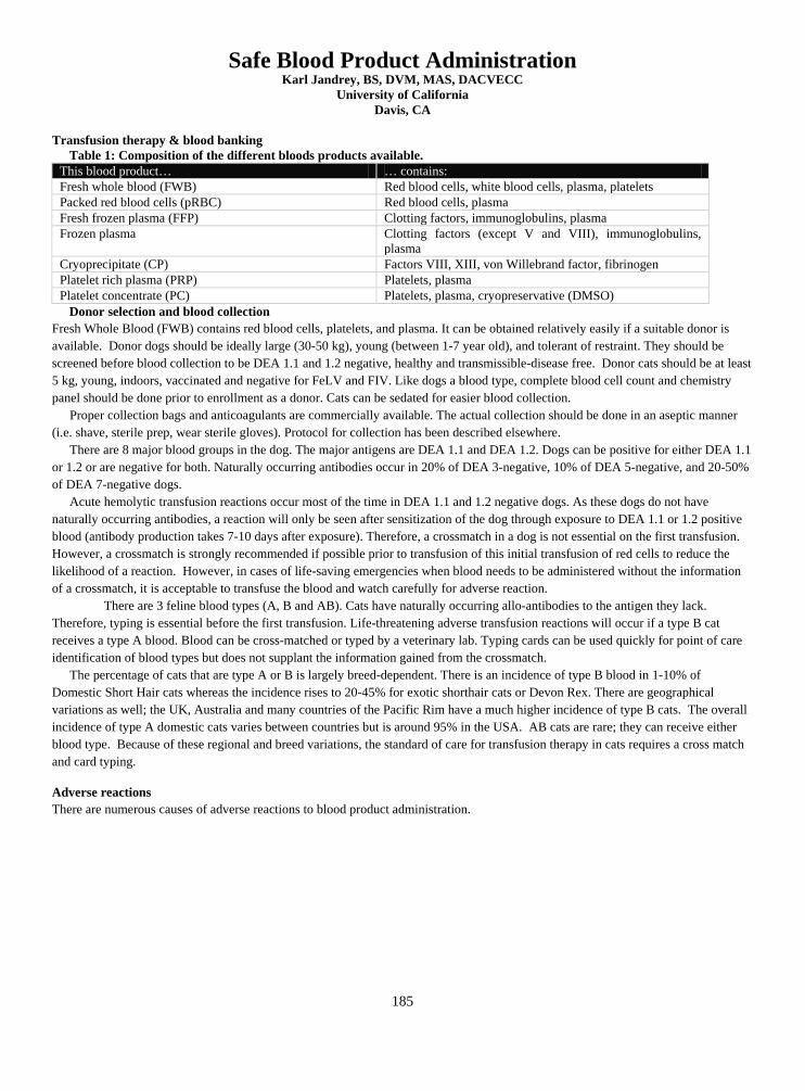
185
Safe Blood Product Administration Karl Jandrey, BS, DVM, MAS, DACVECC
University of California Davis, CA
Transfusion therapy & blood banking
Table 1: Composition of the different bloods products available. This blood product… … contains: Fresh whole blood (FWB) Red blood cells, white blood cells, plasma, platelets Packed red blood cells (pRBC) Red blood cells, plasma Fresh frozen plasma (FFP) Clotting factors, immunoglobulins, plasma Frozen plasma Clotting factors (except V and VIII), immunoglobulins,
plasma Cryoprecipitate (CP) Factors VIII, XIII, von Willebrand factor, fibrinogen Platelet rich plasma (PRP) Platelets, plasma Platelet concentrate (PC) Platelets, plasma, cryopreservative (DMSO)
Donor selection and blood collection Fresh Whole Blood (FWB) contains red blood cells, platelets, and plasma. It can be obtained relatively easily if a suitable donor is available. Donor dogs should be ideally large (30-50 kg), young (between 1-7 year old), and tolerant of restraint. They should be screened before blood collection to be DEA 1.1 and 1.2 negative, healthy and transmissible-disease free. Donor cats should be at least 5 kg, young, indoors, vaccinated and negative for FeLV and FIV. Like dogs a blood type, complete blood cell count and chemistry panel should be done prior to enrollment as a donor. Cats can be sedated for easier blood collection.
Proper collection bags and anticoagulants are commercially available. The actual collection should be done in an aseptic manner (i.e. shave, sterile prep, wear sterile gloves). Protocol for collection has been described elsewhere.
There are 8 major blood groups in the dog. The major antigens are DEA 1.1 and DEA 1.2. Dogs can be positive for either DEA 1.1 or 1.2 or are negative for both. Naturally occurring antibodies occur in 20% of DEA 3-negative, 10% of DEA 5-negative, and 20-50% of DEA 7-negative dogs.
Acute hemolytic transfusion reactions occur most of the time in DEA 1.1 and 1.2 negative dogs. As these dogs do not have naturally occurring antibodies, a reaction will only be seen after sensitization of the dog through exposure to DEA 1.1 or 1.2 positive blood (antibody production takes 7-10 days after exposure). Therefore, a crossmatch in a dog is not essential on the first transfusion. However, a crossmatch is strongly recommended if possible prior to transfusion of this initial transfusion of red cells to reduce the likelihood of a reaction. However, in cases of life-saving emergencies when blood needs to be administered without the information of a crossmatch, it is acceptable to transfuse the blood and watch carefully for adverse reaction.
There are 3 feline blood types (A, B and AB). Cats have naturally occurring allo-antibodies to the antigen they lack. Therefore, typing is essential before the first transfusion. Life-threatening adverse transfusion reactions will occur if a type B cat receives a type A blood. Blood can be cross-matched or typed by a veterinary lab. Typing cards can be used quickly for point of care identification of blood types but does not supplant the information gained from the crossmatch.
The percentage of cats that are type A or B is largely breed-dependent. There is an incidence of type B blood in 1-10% of Domestic Short Hair cats whereas the incidence rises to 20-45% for exotic shorthair cats or Devon Rex. There are geographical variations as well; the UK, Australia and many countries of the Pacific Rim have a much higher incidence of type B cats. The overall incidence of type A domestic cats varies between countries but is around 95% in the USA. AB cats are rare; they can receive either blood type. Because of these regional and breed variations, the standard of care for transfusion therapy in cats requires a cross match and card typing. Adverse reactions There are numerous causes of adverse reactions to blood product administration.
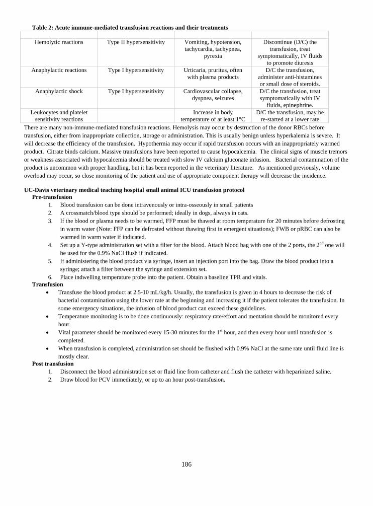
186
Table 2: Acute immune-mediated transfusion reactions and their treatments Clinical signs Treatment
Hemolytic reactions Type II hypersensitivity Vomiting, hypotension, tachycardia, tachypnea,
pyrexia
Discontinue (D/C) the transfusion, treat
symptomatically, IV fluids to promote diuresis
Anaphylactic reactions Type I hypersensitivity Urticaria, pruritus, often with plasma products
D/C the transfusion, administer anti-histamines or small dose of steroids.
Anaphylactic shock Type I hypersensitivity Cardiovascular collapse, dyspnea, seizures
D/C the transfusion, treat symptomatically with IV
fluids, epinephrine. Leukocytes and platelet
sensitivity reactions Increase in body
temperature of at least 1°C D/C the transfusion, may be
re-started at a lower rate There are many non-immune-mediated transfusion reactions. Hemolysis may occur by destruction of the donor RBCs before transfusion, either from inappropriate collection, storage or administration. This is usually benign unless hyperkalemia is severe. It will decrease the efficiency of the transfusion. Hypothermia may occur if rapid transfusion occurs with an inappropriately warmed product. Citrate binds calcium. Massive transfusions have been reported to cause hypocalcemia. The clinical signs of muscle tremors or weakness associated with hypocalcemia should be treated with slow IV calcium gluconate infusion. Bacterial contamination of the product is uncommon with proper handling, but it has been reported in the veterinary literature. As mentioned previously, volume overload may occur, so close monitoring of the patient and use of appropriate component therapy will decrease the incidence. UC-Davis veterinary medical teaching hospital small animal ICU transfusion protocol
Pre-transfusion 1. Blood transfusion can be done intravenously or intra-osseously in small patients 2. A crossmatch/blood type should be performed; ideally in dogs, always in cats. 3. If the blood or plasma needs to be warmed, FFP must be thawed at room temperature for 20 minutes before defrosting
in warm water (Note: FFP can be defrosted without thawing first in emergent situations); FWB or pRBC can also be warmed in warm water if indicated.
4. Set up a Y-type administration set with a filter for the blood. Attach blood bag with one of the 2 ports, the 2nd one will be used for the 0.9% NaCl flush if indicated.
5. If administering the blood product via syringe, insert an injection port into the bag. Draw the blood product into a syringe; attach a filter between the syringe and extension set.
6. Place indwelling temperature probe into the patient. Obtain a baseline TPR and vitals. Transfusion
• Transfuse the blood product at 2.5-10 mL/kg/h. Usually, the transfusion is given in 4 hours to decrease the risk of bacterial contamination using the lower rate at the beginning and increasing it if the patient tolerates the transfusion. In some emergency situations, the infusion of blood product can exceed these guidelines.
• Temperature monitoring is to be done continuously: respiratory rate/effort and mentation should be monitored every hour.
• Vital parameter should be monitored every 15-30 minutes for the 1st hour, and then every hour until transfusion is completed.
• When transfusion is completed, administration set should be flushed with 0.9% NaCl at the same rate until fluid line is mostly clear.
Post transfusion 1. Disconnect the blood administration set or fluid line from catheter and flush the catheter with heparinized saline. 2. Draw blood for PCV immediately, or up to an hour post-transfusion.
187
Annual Review of the Top 5 Coagulation Articles Karl Jandrey, BS, DVM, MAS, DACVECC
University of California Davis, CA
The wealth of information published in veterinary medical journals on the topic of coagulation and hemostasis has risen considerably over the past few years. The author has provided 5 of his favorite articles from the past year that have significant impact and application to daily practice for general practitioners and specialists alike. No attempt was made to search and score the medical literature as a whole; the information explosion in human medicine and the basic and translational research areas for coagulation and hemostasis is huge. Many of those studies are referenced in these articles. The interested reader is encouraged to independently investigate some of the best journals for this: Blood; Journal of Thrombosis and Haemostasis; Blood, Coagulation and Fibrinolysis; Current Opinions in Hematology. Article #1
An ex vivo evaluation of efficacy of refrigerated canine plasma • AUTHORS: Grochowsky AR, Rozanski EA, de Laforcade AM, et al • REFERENCE: J Vet Emerg Crit Care 2014, 24(4): 388-397. • HYPOTHESIS/OBJECTIVES: To determine thawing times of fresh frozen plasma (FFP), and to evaluate the activity of
hemostatic proteins (coagulation factors V, VII, VIII, IX, X, and fibrinogen), clotting times (prothrombin time and activated partial thromboplastin time), and sterility of canine plasma stored refrigerated.
• STUDY DESIGN: Prospective, laboratory-based study • RESULTS: Time to thaw for FFP units was 34.7 +/- 1.38 minutes. Refrigerated storage resulted in significant
decreases in activity of all clotting factors and a subsequent prolongation in clotting times. However, no values were outside the reference interval. All bacterial cultures were negative.
• DISCUSSSION/CLINICAL RELEVANCE: Refrigerated storage results in only minor loss of coagulation factory activity in canine plasma. The use of refrigerated canine plasma, therefore, may be a viable option in high-volume veterinary hospitals for rapid correction of coagulopathy in critical care patients.
Article #2
Evaluation of tranexamic acid and epsilon-aminocaproic acid concentrations required to inhibit fibrinolysis in plasma of dogs and humans
• AUTHORS: Fletcher DJ, Blackstock KJ, Epstein K, Brainard BM • REFERENCE: Am J Vet Res 2014, 75(8):731-738. • HYPOTHESIS/OBJECTIVES: To determine minimum plasma concentrations of the fibrinolytic agents tranexamic acid
(TEA) and epsilon-aminocaproic acid (EACA) needed to completely inhibit fibrinolysis in canine and human plasma after induction of hyperfibrinolysis. It was hypothesized the minimum required concentrations of TEA and EACA to inhibit this hyperfibrinolytic state would be higher in dogs plasma than in human plasma.
• STUDY DESIGN: Prospective, laboratory-based study • RESULTS: Minimum plasma concentrations necessary for complete inhibition of fibrinolysis by EACA and TEA in
pooled canine plasma were estimated as 511.7 µg/ml (95% confidence interval [CI], 433.2 to 590.3 µg/ml and 144.7µg/ml (95%CI, 125.2 to 164.2 µg/ml), respectively. Concentrations of EACA and TEA necessary for complete inhibition of fibrinolysis in pooled human plasma were estimated as 122.0 µg/ml (95% confidence interval [CI], 106.2 to 137.8 µg/ml and 14.7µg/ml (95%CI, 13.7 to 15.6 µg/ml), respectively.
• DISCUSSSION/CLINICAL RELEVANCE: Results supported the concept that dogs are hyperfibrinolytic, compared to humans. Higher doses of EACA and TEA may be required to fully inhibit fibrinolysis in dogs.
Article #3
Effect of duration of packed red blood cell storage on morbidity and mortality in dogs after transfusion: 3,905 cases (2001-2010)
• AUTHORS: Hann L, Brown DC, King LG, Callan MB • REFERENCE: J Vet Intern Med 2014, 28:1830-1837. • HYPOTHESIS/OBJECTIVES: To determine if duration of packed red blood cell (PRBC) storage has an effect on
morbidity and mortality in dogs after transfusion. • STUDY DESIGN: Retrospective 10-year case review
188
• RESULTS: A total of 3,905 dogs received 5,412 PRBC units. Longer duration of PRBC storage was associated with development of new or progressive coagulation failure (P=0.001) and thromboembolic disease (P=0.005). There was no association between duration of PRBC storage and survival for all dogs overall. However, a logistic regression model indicated that for dogs with hemolysis, 90% of which had immune-mediated hemolytic anemia, longer duration of PRBC storage was a negative risk factor for survival. For every 7 day increase in storage, there was a 0.79 lesser odds of 30 day survival (95% CI, 0.64-0.97; P=0.024).
• DISCUSSSION/CLINICAL RELEVANCE: Duration of PRBC storage does not appear to be a major contributing factor to mortality in the overall canine population. However, the longer duration of PRBC storage may negatively impact outcome in dogs with immune-mediated hemolytic anemia, thus warranting further investigation with prospective studies.
Article #4
Recurrent episodes of severe bleeding caused by congenital factor XIII deficiency in a dog • AUTHORS: Kong LR, Snead ECR, Burgess H, Dhumeaux MP • REFERENCE: J Am Vet Med Assoc 2014, 245(10):1147-1152. • HYPOTHESIS/OBJECTIVES: To describe a dog with a congenital bleeding disorder due to factor XIII deficiency. • STUDY DESIGN: Case report • RESULTS: Previously during this dogs evaluations, all standard coagulation testing and factor analysis was normal:
prothrombin time, activated partial thromboplastin time, buccal mucosal bleeding time, activated clotting time, von Willebrand factor concentration, platelet function testing, and plasma factors VII, VIII, IX, X, XI, XII. Rotational thromboelastometry revealed that clotting times were within reference limits, with abnormal clot formation times and clot firmness. The results of a factor XIII (FXIII) clot solubility assay confirmed FXIII deficiency.
• DISCUSSSION/CLINICAL RELEVANCE: This is the first case report to describe a dog with FXIII deficiency. FXIII deficiency should be on the differential diagnosis list for any dog with excessive bleeding episodes and apparently normal results on the common, standard coagulation tests.
Article #5
Effect of synthetic colloid administration on coagulation in healthy dogs and dogs with systemic inflammation • AUTHORS: Gauthier V, Holowaychuk MK, Kerr CL et al • REFERENCE: J Vet Intern Med 2015, 29:276-285. • HYPOTHESIS/OBJECTIVES: To compare the effects of an isotonic crystalloid and synthetic colloid on coagulation in
healthy dogs and dogs with systemic inflammation. • STUDY DESIGN: Randomized, placebo-controlled, blinded, cross-over laboratory study • RESULTS: Administration of either fluid to healthy dogs and dogs with systemic inflammation resulted in similar
increases in prothrombin time and activated clotting time. In comparison to saline administration, tetrastarch administration resulted in significantly decreased R (P=0.017) in healthy dogs, as well as significantly increased activated partial thromboplastin time (P<= 0.016), CL30% (P<= 0.016), and K (P< 0.001) and significantly decreased platelet count (P= 0.019), alpha (P<= 0.001), MA (P< 0.001), and von Willebrand factor antigen (P< 0.001), and collagen binding activity (P<= 0.003), in both healthy dogs and dogs with systemic inflammation.
• DISCUSSSION/CLINICAL RELEVANCE: Tetrastarch bolus administration to dogs with systemic inflammation resulted in a transient hypocoagulability characterized by a prolonged activated partial thromboplastin time, decreased clot formation speed and clot strength, ad acquired Type I von Willebrand disease.
189
Case-Based Approach to Pallor Karl Jandrey, BS, DVM, MAS, DACVECC
University of California Davis, CA
Secondary hemostasis The main purpose of secondary hemostasis is the production of fibrin to stabilize the platelet plug. Initiators of secondary hemostasis include the platelet plug and endothelial damage. It ends in the formation of thrombin, which cross-links fibrin and strengthens the clot. Thrombin also activates fibrinolysis, to limit excessive fibrin accumulation. Coagulopathies can be caused by a congenital defect or deficiency in any coagulation factor, lack of production of coagulation factors, excessive consumption of factors such as in the late phase of DIC, or by dilution. Assessment of secondary hemostasis Disorders of secondary hemostasis are recognized by hemorrhage into body cavities (e.g. hemothorax, hemoabdomen, hemomediastinum) or into organs (e.g. brain, urinary bladder, intestinal tract). Lower platelets counts generally can be found due to consumption, but they rarely are low enough (i.e. less than 20,000/µL) to suggest that they were the primary cause of the hemorrhage. Treatment of impaired secondary hemostasis Since most of the above tests are abnormal due to a lack of sufficient coagulation factors, the goal of therapy is to restore coagulation factors to levels that normalize these tests and, more importantly, reduce the chance for hemorrhage to persist. In general, 10-20 mL/kg of fresh frozen plasma will restore coagulation factors to a level to normalize the coagulation tests. This should be administered within 4 hours of the thaw at a rate at which the patient is not at risk for fluid overload.
Fresh plasma is obtained by centrifugation from FWB and separation into packed red blood cells (pRBC) and plasma within 6 hours of collection. After fresh plasma has been frozen, it is called FFP for up to 1 year. After one year of storage it is then referred to as Frozen Plasma due to the natural decrease in the labile factors (FV, FVIII, von Willebrand Factor) during storage.
The major indication is the treatment of coagulopathies, either acquired (e.g. rodenticide intoxication, DIC) or inherited (e.g. von Willebrand disease, hemophilias).
The required FFP dose in an individual patient is impossible to predict and must be based on response to therapy, availability of blood products and financial limitations. A general guideline is an initial dose between 10–20 mL/kg.
Fresh frozen plasma is not the fluid of choice for low albumin/low osmotic pressure in dogs and cats because it contains very low albumin (less than 5%, i.e. 5 mg/dL). It takes over 40ml/kg of plasma to raise albumin 1g/dL in the patient. Synthetic colloids can be used to increase the colloid osmotic pressure. Human serum albumin can be used to effectively increase albumin concentration in dogs and cats, but questions remain regarding safety of this product in our veterinary patients.
During the process of thawing fresh frozen plasma at 4°C, a white precipitate forms due to the run-off of thawed portion; that is the cryoprecipitate (CP), which can be separate and stored. The major indication for the use of CP is von Willebrand disease and Hemophilia A. The published dose is 10 mL/kg. Treatment of anemia Human patients are at risk of decrease oxygen delivery and organ failure with a hemoglobin of 3-4 g/dL (hematocrit around 9-12%). Transfusion decisions, however, cannot be based only on the level of anemia due to compensatory mechanisms for anemia. Thus, clinical signs and the underlying causes of anemia are also very important. The transfusion should be considered as soon as the patient cardiovascular status is jeopardized by the blood loss. Clinical signs include:
• Physical examination perfusion parameters: poor perfusion: tachycardia, pale mucous membranes, prolonged capillary refill time, weak pulses, cold extremities, altered mentation.
• Physical examination signs of anemia (pale mucous membrane color and narrow pulses) are difficult to differentiate from hypoperfusion/shock and need to be re-assessed after initial fluid resuscitation.
• Perfusion markers: increased lactate, decreased blood pressure and urine output. • Respiratory function: increased respiratory rate and effort, blood gas analysis.
The variable clinical response to anemia in patients suggests different mechanisms of adaptation over time. Therefore, many parameters need to be determined prior to instituting red cell transfusion. A patient with an acute splenic rupture from abdominal trauma can manifest shock and severe blood loss with a small drop in hematocrit. Some patients walk in the Emergency Room with a hematocrit of 6% and appear clinically stable. When chronic anemia occurs, three principal compensatory mechanisms occur:
1. Decreased haemoglobin affinity for oxygen: A rightward shift of the oxyhemoglobin dissociation curve is caused by decreased pH (acidity), increased 2,3-DPG, increased temperature, and increased PCO2. Red blood cells produce more 2,3-DPG under conditions of chronic hypoxia (i.e. anemia); therefore, oxygen is more easily released at the tissue level.
190
2. Redistribution of blood flow: In anemia, selective vasoconstriction of blood vessels underserves certain non-vital areas (skin for example) and allows more blood to flow into critical areas like the heart or brain.
3. Increased cardiac output: The increased output is matched by decreased peripheral vascular resistance and decreased blood viscosity, so that cardiac output can rise without an increase in blood pressure. Generally, anemia must be fairly severe (hemoglobin < 7 g/dL) before cardiac output rises.
Thus, duration (acute versus chronic) of anemia needs to be taken into account because of normal adaptation of the body to the blood loss. The transfusion trigger The threshold values for the clinical use of blood products to treat anemia are not well defined in the veterinary literature. For many years in human medicine, the transfusion trigger had been set at a hemoglobin (Hgb) of 10 g/dL (hematocrit [HCT] of 30%). However, transmissible infections, immunologic risks, transfusion-related acute lung injury, the cost of blood, and component availability are current disadvantages of transfusions. In 1999, the Transfusion Requirements in Critical Care (TRICC) trial showed that maintenance of a Hgb between 7 and 9 g/dL in ICU patients is as effective with less adverse consequences as the maintenance of a Hgb above 9 g/dL. Based on that information, hemoglobin-based threshold values have recently been reviewed for humans and are currently between 6 and 8 g/dL of Hgb (HCT or packed cell volume [PCV] around 20%).
The time it took to achieve that particular Hgb/HCT/PCV value is also an important factor in the determination of when to give red blood cells. Animals with chronic disease may have suppression in the production of, or an insensitivity to, erythropoietin. This more chronic anemia will often go clinically undetected for weeks to months as the patient compensates. Animals with an acute anemia, i.e. from trauma/hemorrhage into body cavities, have little physiologic compensation for this abrupt change in oxygen carrying capacity.
Other objective measures that will help to decide if a transfusion is warranted include: venous oxygen tensions and lactate. As oxygen delivery to the tissues falls but the tissue extraction remains the same, less venous oxygen is present when measuring PvO2. This however may reflect increased oxygen demand by the tissues. Alternatively, an increase in lactate is a readily available marker of tissue anaerobiosis or poor perfusion. Red blood cell dosage As a rule of thumb, 2 mL/kg of FWB will increase the PCV by 1% point. So, 20 mL/kg are used to increase the PCV by 10 percentage points.
In normovolemic, anemic patients, such as autoimmune hemolytic anemia, the volume required to increase the PCV may place the patient at risk of fluid overload. This is a particular concern in the cardiac patient.
Packed red blood cells (pRBC) will have a very small amount of remaining plasma. The hematocrit of pRBC may exceed 80%. In that case, dilution with 0.9% NaCl can be helpful to avoid hyperviscosity. Fluids containing calcium, like LRS, should not be used in order to decrease the risk of citrate chelation and coagulation.
As a rule of thumb, 1 mL/kg of pRBC will increase the PCV by 1% point. The post-transfusion PCV goal is usually between 18 - 25%.
191
Case-Based Approach to Hemoabdomen Karl Jandrey, BS, DVM, MAS, DACVECC
University of California Davis, CA
Emergency management A patient in shock in the ER often has abdominal effusion. If this effusion is found by palpation, radiography, or ultrasonography, a diagnostic abdominocentesis can be completed quickly to identify the fluid as blood. Due to the generally large amount of blood in the peritoneal cavity to cause these clinical signs, many patients present in severe shock. The patient will likely have vasoconstricitve signs on physical examination that can be resolved by improving the effective circulating blood volume. The first step is to provide isotonic crystalloids to resolve these vasoconstrictive signs (pallor, prolonged CRT, tachycardia, weak femoral pulses, mental dullness and cool extremity temperature). Blood product delivery, in an expedient time frame, is typically unavailable and the urgency necessitates acellular fluid therapy. It is true that the patient will become relatively hemodiluted. However, improved perfusion is essential while you seek a more appropriate blood product for the patient.
The blood product of choice could be plasma if the patient has anticoagulant rodenticide or post-operative bleeding from von Willebrand Disease. Fresh whole blood or packed red blood cells are more effective for patients with a splenic or hepatic bleed (traumatic or pathologic secondary to a neoplasm). All natural blood products will have a much more potent effect on improvement of perfusion; however, continued loss will continue until the bleeding is controlled regardless of which fluid is used. Abdominocentesis Indications for abdominocentesis are 1) radiographic loss of serosal detail, 2) abdominal injury without obvious peritoneal entry wounds, 3) shock, multiple injuries, or signs of abdominal injury after blunt trauma, 4) head or spinal injury precluding reliable abdominal examination, 5) persistent abdominal pain or fluid distention of unknown cause, 6) post-operative complications possibly caused by leakage from an enterotomy or anastamotic site. Periumbilical ecchymosis (Cullen’s sign) may indicate hemorrhage in the peritoneum or retroperitoneum. Contraindications to abdominocentesis include coagulopathy, organomegaly, or distention of an abdominal viscus. Intestinal or uterine penetration is rare unless the viscus is dilated and adherent to the abdominal wall. Complications include the introduction or spread of infection, laceration of a viscus and hemorrhage from a punctured vessel. Following the techniques described below will reduce the risk of complications. FAST (focused assessment with sonography for trauma) The focused assessment with sonography for trauma (FAST) protocol was studied in dogs to prove it is a rapid and simple technique to detect free abdominal fluid in the emergency room by veterinary clinicians with minimal previous ultrasound experience. This technique scanned four regions in longitudinal and transverse planes of the abdomen with dogs in lateral recumbency. These regions are areas where fluid accumulation more commonly occurs: caudal to the xyphoid process, midline over the urinary bladder, and each flank Medical or surgical management? After the patient is stabilized, further monitoring is essential to determine if the following management is to be surgical or medical. Continued and on-going bleeding and a return of shock would suggest severe and persistent bleeding that often necessitates surgery. However, a few more diagnostic tests may need to be completed to ensure a normal hemostatic axis. Other options to surgical therapy include: autotransfusion or an abdominal bandage. Surgery is typically indicated if the patient has poor response to fluid resuscitation, a rising intra-abdominal PCV, or continues to effuse into the peritoneum.
192
Case-Based Approach to Petechia and Ecchymosis Karl Jandrey, BS, DVM, MAS, DACVECC
University of California Davis, CA
Treatment of impaired primary hemostasis The treatment of a defect in primary hemostasis involves the removal of the underlying cause if possible. Transfusion of platelets is challenging due to their vary reactive nature and special handling required. The drawing of blood and placing it into collection bags can activate platelets to clump. Therefore, filters should always be used for administration. Platelets also activate at cold temperatures. All platelet transfusions must be done with fresh blood, either fresh whole blood, platelet-rich plasma, or platelet concentrates.
Fresh whole blood transfusions are a source of platelets, but whole blood provides fewer platelets than a platelet-rich product and can cause hypervolemia. Recently, a frozen platelet concentrate (PC) has been produced and is commercially available for dogs (Animal Blood Resources, International; www.abrint.net, Dixon, CA). This PC is prepared from a single donor by automated blood cell processors using apheresis technology, and is cryopreserved in 6% dimethyl sulfoxide (DMSO).
In human medicine, platelets have been cryopreserved with 6% DMSO, resulting in conservation for up to 3 years. However, this method of storage also resulted in loss of platelet viability, decreased aggregation and reduced clinical efficacy compared to fresh platelets. Our lab recently published our findings on the product’s platelet function, but not clinical efficacy, of this commercially available canine frozen PC.
With all these difficulties or clinical unknowns, there is no universal recommendation for the transfusion of platelets in dogs. It is very difficult to give a platelet number-based transfusion trigger. As a basic rule, the risk for hemorrhage is inversely proportional to the platelet count, and bleeding times in humans are prolonged in a linear fashion as platelet count drop from 100,000 to 10,000/µL. The most common trigger for prophylactic transfusion is 20,000/µL in human medicine. In veterinary medicine, numbers of 5,000/µL in cats and 10,000/µL in dogs have also been published. Published platelet numbers adequate for surgery are between 30,000 to 80,000/µL.
The current recommendation from our service is to treat on-going life threatening hemorrhage that is secondary to thrombocytopenia with fresh whole blood or fresh platelet rich plasma. As a rule of thumb in veterinary medicine, the recommendation is to transfuse platelet-rich products only in case of life-threatening bleeding, especially pulmonary or intra-cranial hemorrhage. Fresh platelet rich plasma is not commercially available and must be made from fresh whole blood just before administration. Platelet concentrates are commercially available but have to be ordered at the tie of need and shipped directly due to their 5 day shelf life.
Three products are available for platelet replacement: platelet concentrate (PC), platelet rich plasma (PRP) and fresh whole blood (FWB). These are suggested initial doses but the individual dose must be guided by the response to therapy.
• Fresh whole blood: 20 mL/kg within 4 hours of collection. • Platelet concentrate: 1 unit/10kg. The transfusion should be finished in an hour. • Platelet-rich plasma: 10 mL/kg. The transfusion should be finished in an hour.
Fresh whole blood is the most commonly available blood product to be used in case of life threatening bleeding due to thrombocytopenia.
Platelet rich plasma is prepared by centrifugation of fresh whole blood at a relatively slow speed. The supernatant is therefore "rich" in platelets. It's a fresh product so needs to be processed right before administration. Due to the presence of some red cells, a cross match is essential before administration.
Platelet concentrate is prepared by platelet-apheresis, a process whereby blood is removed from the donor and separated by an automated device into plasma, platelets, white blood cells and red blood cells. The platelet portion is saved and forms one unit of PC and the remaining components are returned back to the donor. Fresh PCs are stored at 20C on a gentle continuous rocker and have must be used within 5 days of processing. According to the manufacturer, one unit of frozen PC can be stored for 6 months at -20°C. PC can also be prepared by centrifugation of platelet rich plasma from different donors (from 4 to 10), separation of the platelet-rich buffy coat and reconstitution in a sterile blood collection bag.
193
Reproductive Emergencies Ari Jutkowitz, VMD, DACVECC
Michigan State University East Lansing, MI
Reproductive problems often arise after normal business hours, so it is not uncommon for them to fall into the domain of the emergency veterinarian. As most owners lack medical knowledge, they frequently look to the veterinarian to answer questions and to identify potential problems. The emergency clinician must therefore be familiar with normal reproductive behavior in addition to the common emergencies that may arise. With this goal, we will review the events surrounding normal parturition as well as the common complications that may develop during this period. Normal reproductive physiology Normal gestation length in the dog may range from 57-72 days from the time of first breeding, with an average length of 65 days.1-2 Because cats are induced ovulators, there is generally less variability in gestation length, which ranges from 63-65 days. Ovulation may not take place after the first breeding however, so in the event of multiple breedings, uncertainties with regards to gestation length may still be present in the cat. As the whelping date approaches, a number of clues may point toward impending parturition. Mammary development, vulvar enlargement, mucous vaginal discharge, and relaxation of the pelvic ligaments are early signs of approaching parturition. Onset of lactation may be noted in primiparous bitches within 24 hours of parturition, but in multiparous bitches may occur several days before parturition. A sudden drop in body temperature (>2°F) is generally noted within 24 hours of parturition3 in dogs and cats as a result of decreases in progesterone levels, but this finding is not always reliable. In one recent study, nadir temperature occurred >48 hours before parturition in 24% of dogs, and an appreciable drop in temperature (>1° F) was not seen in 35% of dogs.4
Normal parturition proceeds in three stages. The first stage is characterized by subclinical uterine contractions and progressive dilation of the cervix. During this stage, which typically lasts for 6-12 hours, bitches may show signs of restlessness, apprehension, panting, nesting behaviors, hiding, and anorexia. Queens may be tachypneic, restless, and vocal, or may lay in their nesting boxes, purring. Active expulsion of the fetuses occurs during the second stage of labor. The first fetus is usually delivered within 1 hour of onset of stage 2 labor in cats, and within 4 hours in dogs, with subsequent deliveries every 15 minutes to 3 hours.5,6 Active straining generally results in expulsion of a fetus within 15 minutes. The entire process generally occurs over 2-12 hours, but may take as long as 24 hours with large litter sizes. The third stage of labor results in expulsion of the placenta. One placenta should be identified for each fetus delivered. Placentas are usually still attached to the fetus by the umbilical cord and emerge with the fetus, but may emerge within 15 minutes to several hours if they become detached. Lochia, a greenish vaginal discharge, indicates placental separation and may be seen during all stages of labor. Following parturition, the discharge gradually becomes red-brown, decreasing in volume over 4-6 weeks as uterine involution takes place. Dystocia Historical and physical exam findings that should prompt a clinician to suspect dystocia are as follows:1
• A definite cause is apparent (ie. fetus lodged in birth canal, pelvic fractures) • Gestation is prolonged (>70 days) with no evidence of labor • Temperature has dropped to <100° F and returned to normal with no evidence of labor within 24 hours • Lochia is noted and 2 hours have elapsed without expulsion of a fetus • Strong and persistent contractions fail to result in the delivery of a puppy within 30 minutes • Weak and infrequent contractions fail to produce a fetus within 4 hours. • More than 4 hours have elapsed since the birth of a puppy with no evidence of ongoing labor • Signs of systemic illness or severe pain are present
Dystocia may result from either maternal or fetal factors that prevent delivery from taking place. Uterine inertia is the most common maternal cause of dystocia,3,7-9 seen when the myometrium produces only weak and infrequent contractions that fail to expel a normal fetus through a normal birth canal. Primary uterine inertia is considered complete when gestation that has exceeded its expected length with no evidence of progression into active labor. Primary uterine inertia is termed partial if the bitch initiates parturition and expels one or more healthy fetuses, but then subsequently fails to deliver the remaining fetuses as a result of myometrial fatigue. Uterine inertia may also be considered secondary if myometrial failure results from prolonged attempts to expel an obstructed fetus, and persists following relief of obstruction. Morphologic causes of dystocia are those in which an anatomic abnormality of the bitch or queen results in obstruction of the birth canal (eg. small birth canal, pelvic fractures)
Fetal factors that may result in dystocia include malpresentations, oversize, fetal malformations, and fetal death. Some of the commonly described malpresentations include transverse presentation, lateral or ventral flexion of the neck, anterior presentation with flexion of one or both forelimbs, posterior presentation with retention of both hindlimbs, and simultaneous presentation of two fetuses.
194
It should be noted that posterior presentations are considered to be a normal variation in dogs and cats, occurring in approximately 40% of deliveries.3 Fetal oversize is another potential cause of dystocia, most commonly seen with single pup pregnancies. Fetal death is an infrequent cause of dystocia, increasing the likelihood of malpresentation because of failure to rotate and extend the head and legs, which commonly occurs immediately prior to parturition. Fetal malformations are another potential cause of dystocia, with anasarca (generalized subcutaneous edema), hydrocephalus, cerebral and cerebrospinal hernias, abdominal hernias, duplications, and rib cage malformations among the more commonly noted.7,8
Diagnosis of dystocia Workup of a patient that is presented for dystocia begins with a complete history and physical exam, including digital vaginal exam. If a fetus is lodged within the birth canal, digital manipulation should be attempted. The fetus may be grasped around the head and neck, around the pelvis, or around the proximal portions of the hind limbs, depending on fetal presentation. Excessive traction should never be applied to a single extremity because of the ease with which these may be avulsed. With the dam restrained in a standing position, traction is applied in a posterior-ventral direction. The fetus may be gently rocked back and forth, and twisted diagonally to free shoulders and hips “locked” in the pelvic canal. If flexion of head or extremities is preventing delivery, a finger may be used to extend them. One cannot overemphasize the importance of using copious amounts of sterile lubricant during obstetrical maneuvers, applied digitally or infused around the fetus using a red rubber catheter.
Radiographs should be obtained in any animal experiencing dystocia. Radiographs are accurate for assessing the number, size, location, and position of fetuses, as well as maternal pelvic morphology and general status of the abdomen. Fetal viability is more difficult to assess from radiographs, unless evidence of fetal decomposition is present. Signs of decomposition include intrafetal or intrauterine gas patterns, awkward fetal postures, collapse of the spinal column due to loss of muscular support, and overlapping of the bones of the skull. Ultrasound may be a more useful tool for assessment of fetal viability, fetal malformations, and fetal distress. Normal fetal heart rates have been reported at 180-245 beats per minute in dogs and up to approximately 265 bpm in cats.10 Deceleration of fetal heart rates to less than 180 beats per minute and the presence of fetal bowel movements on ultrasound have been shown to correlate with severe fetal distress, and may indicate a need for rapid intervention.11
Medical management should be considered if there is no evidence of obstruction, and fetal and pelvic size appear normal. Oxytocin is a peptide hormone that increases the frequency and strength of uterine contractions by promoting influx of calcium into myometrial cells. Oxytocin also promotes post partum uterine involution, aids in control of uterine hemorrhage, and assists in expulsion of retained placentas. The dose for oxytocin has traditionally been reported at 5-20 units IM in the dog and 2-4 units IM in the cat. However, with an increase in the use of uterine contraction monitoring (Whelpwise, Veterinary Perinatal Specialties Inc, Wheat Ridge, CO) in veterinary patients, there is a growing body of evidence to suggest that traditional doses may be too high, potentially causing uterine tetany, ineffective contractions, and decreased fetal blood flow. Recent data suggests that doses of 0.5-2 units are effective in increasing the frequency and quality of contraction.4,12 The oxytocin dose may be repeated in 30 minutes if expulsion of a fetus has not resulted. If labor proceeds and a fetus is delivered, oxytocin may be repeated every 30 minutes as needed to assist in expulsion of the remaining fetuses.
Calcium gluconate may be considered if weak, infrequent contractions are noted 4,12 or when labwork reveals hypocalcemia. Retrospective studies have indicated that many patients who fail to respond to oxytocin alone may respond to a combination of calcium and oxytocin.3,8 The dose for calcium gluconate (10% solution) as a uterotonic agent is 11 mg/kg diluted in saline and given subcutaneously, or added to IV fluids and given slowly while monitoring an ECG for arrhythmias. If hypocalcemia is documented, a dose of 50-150 mg/kg intravenously should be used. Subcutaneous administration has been reported to result in irritation and potential granuloma formation, though this is an infrequent complication. Dextrose infusion should also be initiated if hypoglycemia is evident on labwork.
Surgical management should be considered for the following conditions:1
• Complete primary uterine inertia • Partial primary uterine inertia or secondary uterine inertia where large numbers of fetuses remain and response to drugs
is unsatisfactory, • Fetal oversize • Gross abnormalities of maternal pelvis (fractures, masses) • Fetal malformations • Malpresentation that is not amenable to manipulation • Past history of dystocia or c-section • Fetal putrefaction • Maternal evidence of systemic illness • Suspicion of uterine torsion, rupture, prolapse, or herniation • Evidence of fetal distress with poor response to medical intervention
195
An anesthetic protocol for caesarian section should be selected with the goal of maximizing survival of neonates and dam. Attempts should be made to minimize exposure of the fetus to anesthetics by keeping the time from induction to delivery as short as possible. Ideally, the dam should be clipped and prepped prior to induction, equipment should be out, and the surgeon should be scrubbed and ready. Induction agents should be given to effect. Regional techniques such as line blocks and epidurals may help to minimize the need for other drugs. A line block can be performed using 2 mg/kg lidocaine infused along the ventral midline. Alternately, epidural lidocaine may be administered in dogs at a dose of 2-3 mg/kg, not to exceed a total volume of 6 ml. Propofol (4-6 mg/kg IV) or mask inductions are most commonly used for caesarian section at this time, and have been associated with reduced neonatal mortality in dogs. Anesthetic agents that have been associated with increased neonatal mortality include thiopental, ketamine, xylazine, medetomidine, and methoxyflurane.13-15
Neonatal resuscitation A warm (90° F) incubator, hemostats, suture material, suction bulb syringes, emergency drugs, and an adequate supply of soft dry towels should be prepared beforehand. As each neonate is handed off, the umbilical cord should be clamped and ligated 1-2 cm from the umbilicus. Fetal fluids and amnion should be removed by rubbing briskly with a soft, clean towel. The oral cavity and nares may be suctioned with a bulb syringe. The old practice of “swinging” puppies to clear their airways is best avoided because of the potential for cerebral hemorrhage due to concussive injury. If vigorous rubbing is not successful at stimulating respiration, positive pressure ventilation may be initiated with a snug fitting mask, keeping the neonates head and neck extended to ensure adequate inflation of the lungs. Alternately, intubation may be accomplished using a catheter or small, uncuffed endotracheal tube. Because isoflurane is minimally metabolized, ventilation is the primary route of elimination. Thus, its depressant effects can not be reversed until the neonate breathes. Cardiac massage may be instituted if a heart beat is not detected once warming and ventilation measures have been instituted. Epinephrine (0.1 mg/kg) may be given intratracheally, intraosseously, or intravenously if cardiac massage is unsuccessful. Naloxone (0.1 mg/kg) should be considered if the dam received opioid analgesics as part of the anesthetic regimen. Although doxapram (dopram) is routinely administered in many practices as a respiratory stimulant, it is not used for this purpose in the resuscitation of human neonates and there is no evidence to support its use in veterinary patients.
The prognosis for medical management of dystocia is guarded, with success rates of 20-40% in the veterinary literature.3,7-9 Additionally, stillbirth rates have been shown to rise when dystocia is allowed to continue for greater than 4.5-6 hours from the time of onset of second stage labor in the dog.3,7 For these reasons, the decision to proceed to caesarian section should not be delayed if response to medical management is poor or unlikely to result in successful delivery. In recent studies, neonatal survival rates following surgical treatment of dystocia have been reported at 92% at birth, with 80% still alive at 7 days post c-section.13,14 Periparturient emergencies
Mastitis Mastitis is a postpartum complication seen in both dogs and cats that results from bacterial infection of the mammary glands. Bacteria most commonly enter through the nipple as a result of nursing, trauma, or poor hygiene, but may also be spread hematogenously. In mild cases, discomfort, swelling, and inflammation may be seen, while in severe cases, signs of systemic illness such as fever, anorexia, and lethargy frequently develop. Dogs often refuse to allow their young to nurse and may be reluctant to lie down. Severe mastitis often progresses to abscessation and necrosis.
Diagnosis of mastitis is generally based on history and clinical signs (fever and swollen, painful glands in the postpartum animal), but baseline CBC and chemistry as well as milk cytology and culture are useful for assessing severity of illness and appropriateness of antibiotic selection. Milk expressed from the gland may be purulent and cytology typically shows large numbers of white blood cells and intracellular bacteria. The most common bacteria isolated on culture include E. coli, Staphylococci, and Streptococci.
Treatment is initiated immediately with broad spectrum antibiotics. Amoxicillin-clavulanic acid or cephalexin are good first choices and are safe for nursing neonates. Other measures that may be useful in the management of mastitis include warm compresses, hydrotherapy, and frequent milk stripping. If a fluctuant abscess pocket is identified on palpation, early lancing and flushing may limit the degree of skin necrosis that follows. Large, ruptured mammary abcesses may be successfully managed as open wounds with warm compresses, hydrotherapy, and systemic antibiotics, but in these cases mastectomy may provide a more rapid and cosmetic resolution of the problem.
Endometritis Endometritis is a bacterial infection of the uterus that is generally seen within the first three days (up to one week) after whelping, though it may develop during pregnancy as well. Potential causes include retained fetuses or placentas, abortions, uterine trauma secondary to dystocia or obstetrical manipulation, and ascending infection from the vaginal canal. Typical signs include fever, lethargy, anorexia, vomiting, diarrhea, poor lactation, neglect of offspring, and foul-smelling vaginal discharge. Just as in the non-pregnant dog, any purulent vaginal discharge noted during or after pregnancy is abnormal and should prompt investigation.
Labwork abnormalities consistent with sepsis may be seen, including leukocytosis with a left shift or leukopenia, thrombocytopenia, elevated liver values, and hypoalbuminemia. Coagulation testing should be performed to rule out disseminated
196
intravascular coagulation. Radiographs or ultrasound are indicated to evaluate for fetal death, retained placentas, or evidence of uterine enlargement. Cytology of vaginal discharge typically shows degenerate neutrophils and macrophages with intracellular bacteria. The most common organisms associated with uterine infections include Staphylococci, Streptococci, E. coli, Salmonella, Campylobacter, and Chlamydia.
An animal suspected of having septic metritis should be treated aggressively with IV fluids. Broad spectrum antibiotic combinations such as ampicillin-enrofloxacin, ampicillin-aminoglycoside, or cefazolin-aminoglycoside-metronidazole, should be administered. Following stabilization, ovariohysterectomy is the treatment of choice for metritis. If the animal is not showing signs of sepsis and the owner wishes to use her for breeding purposes in the future, evacuation of the uterine contents using PGF2α (Lutalyse) may also be attempted in conjunction with broad spectrum antibiotics. PGF2α is typically administered at doses of 0.1-0.25 mg/kg SQ once daily for 5 days. If initial dosing does not result in adequate expulsion of uterine contents, the author generally increases treatment frequency to twice daily. Potential complications of PGF2α include vomiting, abdominal discomfort, uterine rupture, and septic peritonitis. Because PGF2α treatment may require several days to achieve a good effect, animals that are severely ill should always be treated with ovariohysterectomy. Ovariohysterectomy is also the best choice when the animal is not intended for future breeding or if the health of the dam is a higher priority than possible future breedings.
Eclampsia Eclampsia or puerperal tetany is a life threatening condition that results from the development of hypocalcemia in the periparturient period. It is one of the more common complaints noted following parturition, accounting for roughly 1/4 of periparturient emergencies. Eclampsia is results from the loss of calcium through lactation and fetal skeletal mineralization, in excess of that entering the extracellular fluid through gastrointestinal absorption and bone resorption. Other factors such as inadequate diet or parathyroid atrophy resulting from oversupplementation of calcium may also contribute, though diet in affected animals has not been reported to be significantly different from non-affected animals. Increasing litter size to maternal body weight ratio has also been identified as a significant factor in the development of periparturient hypocalcemia.
Eclampsia is most commonly seen in small dogs, first-time whelpings, and dogs with large litter sizes. It typically develops 2-4 weeks after parturition but is occasionally seen in late gestation. Clinical signs in dogs most commonly include stiff gait, trembling, twitching, seizures, tachycardia, panting, and hyperthermia, but some dogs may present with atypical signs such as whining, vomiting, diarrhea, and behavior changes. If untreated, death may result from respiratory impairment, or from hyperthermia and cerebral edema. Cats may present with clinical signs similar to dogs, but unlike dogs, are more prone to hypothermia, and may present with hyperexcitability, hypersensitivity, or flaccid paralysis in place of clonic-tonic muscle spasms.
Diagnosis of eclampsia is made on the basis of history and physical exam findings in conjunction with low total or ionized calcium levels. Ionized calcium represents the physiologically active portion of calcium within the body, and is involved in muscular contraction, as well as neurologic and cardiovascular function. Ionized calcium levels are therefore believed to be a more sensitive indicator of extracellular calcium levels than total calcium, and typically fall below 0.8 mmol/L in dogs with eclampsia (reference range: 1.2-1.4 mmol/L). However, total calcium levels have been found to be decreased in all dogs with eclampsia, suggesting that total calcium levels may provide sufficient information in this disease if ionized calcium measurement is not available.
Animals presenting with eclampsia should have an IV catheter placed and intravenous fluids administered to address fever, dehydration, and tachycardia. Calcium gluconate (10%) should immediately be administered intravenously slowly to effect. Most animals will have tremors controlled at doses ranging from 0.5 to 1.5 ml/kg. An ECG should be monitored during calcium administration and the infusion stopped if bradycardia or arrhythmias develop. Ionized calcium levels should be rechecked post administration to make sure that ionized calcium levels remain within the normal range. Temperature should be carefully monitored in animals presenting with tremors, and active cooling measures (cool fluids, alcohol applied to footpads) should be instituted for patients with severe hyperthermia. Body temperature generally falls quickly once tremors are controlled, so active cooling measures should be discontinued once the temperatures falls below 103° F. Oral calcium carbonate (Tums) supplementation should be continued at a dose of 100 mg/kg/day throughout lactation. Up to 20% of dogs may have recurrence of eclampsia despite supplementation if puppies are allowed to nurse, so bottle feeding and early weaning of the puppies is recommended.
Supplementation of calcium prior to whelping is not recommended, as this may downregulate parathyroid hormone secretion, decreasing intestinal calcium absorption and increasing the risk of eclampsia during lactation. Instead, calcium administration (100 mg/kg/day divided) should be instituted following whelping in dogs at risk and dogs with a previous history of eclampsia. References Jutkowitz LA. Reproductive Emergencies. Vet Clin North Am Sm An Prac 2005;35:397-420. Concannon P, Whaley S, Lein D, et al. Canine gestation length: variation related to time of mating and fertile life of sperm. Am J Vet Res 1983;44:1819-1821. Gaudet DA. Retrospective study of 128 cases of canine dystocia. J Am Anim Hosp Assoc 1985;21:813-818. Copley K. Comparison of traditional methods for evaluating parturition in the bitch versus using external fetal and uterine monitoring. In: Proceedings for the Society of Theriogeneology Annual Conference. Colorado Springs: 2002, p. 375-382.

197
Concannon PW, McCann JP, Temple M. Biology and endocrinology of ovulation, pregnancy, and parturition in the dog. J Reprod Fertil Suppl 1989;39:3-25. Linde-Forsberg C, Eneroth A. Parturition. In: Simpson GM, editor. Manual of Small Animal Reproduction and Neonatology. Cheltenham: British Small Animal Veterinary Association; 1998, p. 127-142. Darvelid AW, Linde-Forsberg C. Dystocia in the bitch: A retrospective study of 182 cases. J Small Anim Pract 1994;35:402-407. Eckstrand C, Linde-Forsberg C. Dystocia in the cat: A retrospective study of 155 cases. J Small Anim Pract 1994;35:459-464. Gunn-Moore DA, Thrusfield MV. Feline dystocia: prevalence, and association with cranial conformation and breed. Vet Record 1995;136:350-353. Verstegen JP, Silva LDM, Onclin K, et al. Echocardiographic study of heart rate in dog and cat fetuses in utero. J Reprod. Fertil Suppl 1993;47:175-180. Zone MA, Wanke MM. Diagnosis of canine fetal health by ultrasonography. J Reprod. Fertil Suppl 2001;57:215-219. Davidson AP. Uterine and fetal monitoring in the bitch. Vet Clin North Am 2001;31:305-313. Moon PF, Erb HN, Ludders JW, et al. Perioperative management and mortality rates of dogs undergoing cesarian section in the United States and Canada. J Am Vet Med Assoc 1998;213:365-369. Moon-Massat PF, Erb HN. Perioperative factors associated with puppy vigor after delivery by cesarian section. J Am Anim Hosp Assoc 2002;38:90-96. Funkquist PME, Nyman GC, Lofgren AMJ, et al. Use of propofol-isoflurane as an anesthetic regimen for cesarian section in dogs. J Am Vet Med Assoc 1997;211:313-317.
198
Managing the Difficult Urethral Obstruction Ari Jutkowitz, VMD, DACVECC
Michigan State University East Lansing, MI
Overview and pathophysiology Feline urethral obstruction is one of the most common emergency presentations in the cat, accounting for approximately 9% of feline emergency admissions.1 While there are many factors that may play into the development of lower urinary tract diseases in the cat, matrix-crystalline plugs and urolithiasis are the most common causes of obstruction.2 Cats with urethral obstruction may have signs localized to the lower urinary tract including dysuria, stranguria, pollakiuria, hematuria, vocalizing, and pain, or they may show signs of systemic illness such as vomiting, lethargy, or collapse. Cats with obstructive urinary tract diseases may or may not have demonstrated preceding signs of lower urinary tract disease.
Following the development of urethral obstruction, clinical signs of uremia typically develop within 24 hours.3 Dehydration occurs due to decreased water intake and ongoing fluid losses secondary to vomiting. Acid-base (metabolic acidosis) and electrolyte disturbances (hyperkalemia and hyperphosphatemia) develop due to impaired excretion. Accumulation of metabolic wastes leads to post renal azotemia. Bladder capacity is reached, leading to rising intravesicular pressure and subsequently falling glomerular filtration rate (GFR). Prolonged obstruction may result in intrinsic renal failure. Damage to the urothelium and detrusor muscle may also develop during this time. If left untreated, death secondary to cardiopulmonary failure or hyperkalemia may occur within 3-6 days. Damage to bladder mucosa or urethra may shorten survival times.3
Diagnosis of urethral obstruction Diagnosis of urethral obstruction is generally made on the basis of history and physical exam findings. Abdominal palpation typically reveals a turgid, painful bladder, though in rare cases, the bladder may be moderate in size if the cat is presented to the veterinarian shortly after clinical signs develop. Blood and/or crystalline debris may be visualized at the urethral orifice. The presence of bradycardia frequently indicates hyperkalemia, and severe systemic signs in conjunction with free abdominal fluid should prompt consideration of bladder leakage or rupture. In contrast, cats that present with stranguria but appear systemically healthy and have palpably small bladders typically have non-obstructive lower urinary tract disease.
At the time of presentation, a peripheral IV catheter is placed and blood is collected for complete blood count, serum biochemistry panel, and venous blood gas/electrolyte panel. The blood gas/electrolyte panel is particularly helpful as it provides rapid information on parameters such as potassium concentration (as well as acid-base status and renal values) that may affect initial interventions. Electrocardiography can also be helpful in the initial evaluation of the patient with urethral obstruction. Early ECG changes suggestive of hyperkalemia include bradycardia, dampened P-waves, tented T-waves, and prolongation of the P-R interval. As hyperkalemia worsens, loss of P-waves (atrial standstill) and widening of the QRS complex may develop. Electrocardiographic changes typically do not develop until potassium levels are greater than 7 mEq/L, but there is a great deal of individual variation in terms of patient response to hyperkalemia. Metabolic acidosis, hyponatremia, and hypocalcemia may contribute to the likelihood of hyperkalemic cardiotoxicity.
Once the animal has been medically stabilized and deobstructed, urine is submitted for urinalysis and culture. Because crystalline and cellular composition of the urine may change over time, evaluation of a fresh, undiluted sample is preferred. Diagnostic imaging should be performed to rule out cystic or urethral calculi. If a urolith or crystalline-matrix plug is retrieved at the time of deobstruction, composition should be determined as this may impact future therapies.
If free abdominal fluid is identified, fluid chemistry may be helpful in determining whether urinary tract rupture has occurred. An abdominal fluid:serum creatinine ratio of 2:1, or abdominal fluid:serum potassium ratio of 1.9:1 (cat) or 1.4:1 (dog) is predictive of uroperitoneum.4 Cytology of the fluid sample should also be performed to rule out urosepsis. Contrast cystourethrography is used to determine location and severity of the rupture. Treatment of urethral obstruction
Fluid therapy Initial management of urethral obstruction in the cat should focus on correction of hypovolemia, hyperkalemia, and other acid-base and electrolyte disturbances. In most cases, appropriate fluid therapy followed by restoration of urine flow will effectively correct these abnormalities. A peripheral IV catheter should be placed and fluid therapy instituted immediately using 0.9% sodium chloride or balanced electrolyte solution such as lactated Ringer’s solution (LRS). A shock rate of fluids (66 ml/kg/hour in the cat) is calculated and then administered to effect in increments of approximately ¼ of the calculated dose, reassessing major body systems after each bolus. For example, the calculated shock rate in a 5 kg cat is approximately 330 ml, and should be administered in individual boluses of 50-100 ml every 10-15 minutes until cardiovascular status is restored. The goal of fluid therapy should be normalization of vital signs such as heart rate, level of consciousness, pulse quality, blood pressure, and capillary refill time. The specific type of intravenous
199
fluid selected is of lesser importance than the administration of appropriate volume. Although 0.9% sodium chloride has traditionally been selected due to its lack of potassium, studies in both experimental and clinical cases have shown that potassium containing solutions (LRS, Normosol-R) do not adversely affect the rate of resolution of hyperkalemia in cats with urethral obstruction when compared with 0.9% saline.5,6 Additionally, the buffered solutions are more efficient at restoring electrolyte and acid-base balance in severely affected animals.
Hyperkalemia Relative or absolute bradycardia should be immediately investigated by monitoring electrocardiography and serum electrolyte concentrations. Severe electrocardiographic changes such as atrial standstill, widened QRS complexes, or sine wave formation provide strong indication for the administration of calcium gluconate. Calcium gluconate (10%) is given slowly at a dose of 0.5-1.5 ml/kg IV while carefully watching the patient’s ECG for arrhythmias. Although calcium gluconate does not lower the serum potassium level, it has the immediate effect of buffering the myocardium from the toxic effects of hyperkalemia by restoring the normal difference between resting and threshold membrane potentials. Other intermediate to long-term interventions for hyperkalemia include the administration of regular insulin/dextrose and sodium bicarbonate, though these therapies are rarely warranted in animals with urethral obstruction as fluid therapy followed by timely restoration of urine flow are generally effective at reversing the hyperkalemia. However, if needed, 50% dextrose may be diluted 1:1 with saline and given at a dose of 1 gm/kg body weight to promote endogenous insulin release with subsequent potassium uptake by the cells through stimulation of sodium-potassium pumps. If regular insulin is used, it should be given at a rate of 1 unit insulin per 3 gm dextrose, though this is generally unnecessary and creates the need for careful blood glucose monitoring thereafter to avoid hypoglycemia. Sodium bicarbonate may also be given at a dose of 1 mEq/kg intravenously to facilitate intracellular potassium shifting in exchange for hydrogen ions.
Techniques for urethral deobstruction During the initial exam, the urethra may be gently massaged, followed by careful palpation of the bladder to potentially dislodge superficial plugs. Extreme care should be taken to avoid accidental bladder rupture. While this technique is rarely effective, it is a simple extension of the initial physical exam and therefore may be worth trying in less severely affected cats prior to catheter deobstruction.
Although severely depressed patients may be deobstructed without the need for chemical restraint, sedation/analgesia is employed in the majority of “blocked” cats to improve patient comfort, facilitate deobstruction, and avoid urethral or bladder trauma secondary to patient struggling. Ketamine (100 mg/ml) may be combined with diazepam (5 mg/ml) in equal parts by volume and given at a dose of 1 ml/10 kg of the 50:50 mix. However, this combination should be avoided in cats with known or suspected hypertrophic cardiomyopathy, or when an undiagnosed murmur or gallop rhythm is present. In these cases, hydromorphone (0.05 mg/kg) in combination with diazepam (0.2 mg/kg) may provide a safer option.
Following sedation, the cat is positioned in dorsal recumbency with the legs pulled forward over the head. In this position, the prepuce may be retracted and the penis extruded by simply pushing the prepuce downward towards the anus. A further advantage to this technique is that it allows the urethra to be maximally straightened to facilitate deobstruction. The author’s preferred technique for deobstruction uses an olive tip catheter (FUS needle 21 g x 1”, Jorgensen Laboratories, Loveland, CO). This is a metal, bulb-tipped catheter that can be used to flush the urethra and either break down matrix-crystalline plugs or hydropulse them atraumatically into the bladder. Initially, the olive tip catheter is lubricated and inserted gently into the urethra to the site of the obstruction, approximately 1-2 cm. A 3 cc syringe is then used to lavage and break down the plug. Bits of the plug will often be seen emerging from the urethral orifice during the lavage. When the catheter is withdrawn, a strong stream of urine will frequently force the remainder of the plug from the urethra. Gentle bladder palpation may be used at this point to assist in the expulsion of the plug. To avoid urethral trauma, the catheter should not be forced past the obstruction. Instead, the lavage solution should be allowed to do the work. Additionally, acidic solutions should not be used for lavage as these have not been shown to be effective at plug dissolution and may further traumatize the urethral mucosa. If lavage alone is not successful at dislodging the urethral plug, the tip of the urethra can be pinched around the bulb tip of the catheter and hydropulsion used to push the plug back into the bladder.
Many clinicians use polypropylene “tomcat” catheters for the purposes of unblocking cats. These have the potential to cause additional trauma to the urethra when the rigid catheter is forced past the site of obstruction. If used, a number of steps may help to minimize iatrogenic urethral damage and maximize chances of success. (1) Completely straighten the urethra by pushing the prepuce dorsally towards to anus until the penis is parallel to the spine. (2) Use copious amounts of lubrication. (3) Hydropulse with sterile saline prior to advancing the catheter to assist in dislodging the plug. (4) Use a very light touch when advancing the catheter. Hold the catheter between index finger and thumb and twirl gently while advancing. Think about “picking a lock” when attempting to advance the catheter. Use finesse instead of force. (5) Once the catheter is well seated in the urethra, the penis may be allowed to retract into the prepuce. The prepuce may then be pulled caudally (toward tail tip) to further straighten the urethra while the catheter is advanced.
Some experienced clinicians advocate the use of cystocentesis prior to deobstruction to decompress the bladder and to potentially facilitate hydropulsion of urethral plugs. The author prefers to reserve this technique for use only as a last resort due to the number of
200
cats presenting to the emergency service with uroperitoneum and apical bladder tears following cystocentesis of overdistended bladders. However, it should be noted that our institution may see a biased population of more severely affected animals.
Cats that are critically ill, and those demonstrating large amounts of “sandy” crystalline debris in the urine, blood clots, uroliths, plugs hydropulsed into the bladder, bladder atony, or urethral narrowing are particularly at risk for reobstruction post-unblocking. For this reason, a soft, indwelling, 3.5-5 French red rubber catheter is placed following deobstruction to facilitate urine drainage overnight and to assist in quantitation of urine output. Indwelling catheters should be placed using liberal clipping and scrubbing of the perineum and aseptic technique to minimize risk of catheter-induced urinary tract infection. The tip of the catheter should sit just past the bladder neck to reduce risk of kinking or knotting. The catheter should then be connected to a sterile, closed collection system. To decrease the likelihood of premature catheter removal, careful attention should be given to suture placement. A piece of butterfly tape is placed around the catheter and appositional sutures are placed at the margin of the butterfly tape to prevent kinking of the catheter. The catheter body is then taped to the tail. An Elizabethan collar should be placed prior to anesthetic recovery. Hospital management
Fluid therapy Following initial stabilization and correction of hypovolemia, fluid rates should be adjusted to account for remaining fluid deficits, daily maintenance requirements, and ongoing losses. Deficits can be estimated as follows based upon clinical signs of dehydration: mild (5-6%), moderate (7-8%), and severe (8-10%). Multiplying the estimated percent dehydration by body weight gives the fluid deficit, which may then be replaced over the next 24 hours. For example, a 5 kg cat estimated to be 8% dehydrated would have an estimated deficit of 400 ml. To this value must be added maintenance needs (approximately 60 ml/kg/day) and ongoing losses. Ongoing losses following “unblocking” result from post-obstructive diuresis and can be estimated most easily by quantitating urine output. Normal urine output is approximately 1-2 ml/kg/hour (5-10 ml/hour in the average 5 kg cat). Urine output in excess of this amount typically results from post-obstructive diuresis. During the first 24 hours of therapy, a fluid rate should be selected that accounts for these ongoing losses. In other words, the intravenous fluids administered should slightly exceed measured urinary losses.
Urine output is quantified every four hours. Inadequate urine production (<1 ml/kg/hr) indicates inadequate fluid administration or urinary catheter occlusion with debris. After troubleshooting the catheter, a fluid bolus followed by an increase in fluid rate is indicated if urine output remains low.
Fluid therapy is typically tapered over the next 24-36 hours. Daily monitoring of electrolytes and renal values should be performed to ensure that azotemia resolves and electrolytes normalize. Potassium supplementation may be required during post-obstructive diuresis should hypokalemia develop.
Urinary catheter care Indwelling urinary catheters and tubing should be cleaned externally once daily with a dilute chlorhexidine solution. Gloves should be worn and aseptic technique used when handling the catheters to avoid nosocomial infection. Bladder palpation should be performed every 4-6 hours to ensure that the bladder remains decompressed. When moving the patient, the urine collection system tubing should be clamped and the bag held below the level of the patient to prevent retrograde flow of urine into the bladder.
To minimize likelihood of catheter-induced urethral irritation or urinary tract infection, catheters should be removed as soon as possible. For most cats, the catheter is removed within 48 hours, but the presence of excessive crystalline debris or blood clots in the urine may necessitate longer indwelling catheter duration to avoid reobstruction. Use of antibiotics during hospitalization is not recommended as this is unlikely to prevent catheter-related infection, but may contribute to antibiotic resistance of organisms protected by the catheter biofilm. Culture should be performed prior to catheter removal, with antibiotic therapy initiated as indicated based upon results of culture and sensitivity.
Following catheter removal, patients should be monitored for an additional 12-24 hours to ensure that the urethra remains patent. Cats will typically urinate small volumes frequently following catheter removal due to irritation resulting from obstruction and catheterization. Although they may appear to strain in the litterbox, the bladder should remain small on palpation. A progressively distending bladder post-catheter removal typically indicates reobstruction (firm bladder, difficult to express) or bladder atony (large, flaccid, expressible). Cats with suspected urethrospasm post catheter removal may benefit from a smooth muscle relaxant following catheter removal (prazocin 0.5 mg/cat q24h).
Pain management Urinary obstruction and initial management are frequently associated with significant discomfort. In our practice, buprenorphine (0.01 mg/kg IV q6h) is commonly used to provide analgesia for the first 24-48 hours. Long term management Strategies for long-term prevention of recurrence focus primarily on environmental modification and dietary changes. Occasionally, pharmacologic intervention may be warranted. An ample number of litterboxes should be provided, particularly in multi-cat households, and litterboxes should be cleaned regularly to encourage more frequent use. Canned or moistened food may decrease frequency of lower urinary tract episodes by promoting a more dilute urine and increasing frequency of urination. Fresh water should
201
be available at all times. In cases where obstruction was caused by struvite-matrix plugs, an acidifying diet may be of benefit. Antibiotics, anti-inflammatories, and antispasmotics have not been associated with reduction in frequency of episodes and their routine use is not recommended. Perineal urethrostomy Perineal urethrostomy may be considered in cases where frequency of urethral obstruction is unacceptable despite appropriate medical management or when irreversible changes in the urethra (stricture, scarring, urolithiasis) cause recurrent or persistent obstruction. Perineal urethrostomy has been associated with significant short and long term complications including recurrent urinary tract infection and stricture, and as such should not be considered a first line recommendation for cats with urethral obstruction. References Lee JA, Drobatz KJ. Characterization of the clinical characteristics, electrolytes, acid-base, and renal parameters in male cats with urethral obstruction. J Vet Emerg Crit Care 2003;13:227-233. Osborne CA, Kruger JP, Lulich JP, et al. Feline matrix-crystalline urethral plugs: A unifying hypothesis of causes. J Small Anim Prac 1992;33:172-177. Bartges JW, Finco DR, Polzin DJ, et al.. Pathophysiology of Urethral Obstruction. Vet Clin North Am Sm Anim Prac 1996;26:255-264. Aumann M, Worth LT, Drobatz, KJ. Uroperitoneum in cats: 26 cases (1986-1995). J Am Anim Hosp Assoc 1998;34:315-324. Cunha MG, Freitas CG, Carregaro AB, et al. Renal and cardiorespiratory effects of treatment with lactated Ringer's solution or physiologic saline (0.9% NaCl) solution in cats with experimentally induced urethral obstruction. Am J Vet Res 2010;71:840-846. Drobatz KJ, Cole SG. The influence of crystalloid type on acid-base and electrolyte status of cats with urethral obstruction. J Vet Emerg Crit Care 2008;18:355-361.
202
Diagnosing and Treating Pericardial Effusion Ari Jutkowitz, VMD, DACVECC
Michigan State University East Lansing, MI
Pericardial effusion is defined as the accumulation of fluid within the pericardial space. As the pressure within the pericardial space increases, right sided cardiac filling is impaired, resulting in decreased stroke volume with subsequent decreases in cardiac output and ultimately decreased oxygen delivery to the tissues (shock). These manifestations of pericardial effusion are referred to as cardiac tamponade. Successful emergency management of dogs with life threatening pericardial effusion depends on early triage, a thorough physical examination, point of care diagnostic imaging techniques, and subsequent pericardiocentesis or placement of an indwelling pericardial drain. Key etiologic and pathophysiologic points Pericardial fluid accumulation and cardiac tamponade in the dog most often occurs secondary to a neoplastic process. Hemangiosarcoma (HSA) is most commonly identified in the region of the right atrium or right atrial appendage while chemodectoma (common in brachycephalic breeds) is most often identified at the heart base. Mesothelioma and any metastatic tumor are additional neoplastic causes. Although location and breed are frequently suggestive of tumor type, definitive diagnosis is dependent on a biopsy specimen.
Idiopathic pericardial effusion tends to be an inflammatory process and is frequently recognized in similar breeds to those that frequently develop HSA. Significant efforts in recent years have been directed towards developing diagnostic tests to help differentiate malignant from benign pericardial effusion (idiopathic). Pericardial fluid pH was initially thought to aid in making this differentiation, however, pericardial fluid pH has now been clearly shown to be of little diagnostic value.5-6 Recent evidence suggests that blood concentrations of cardiac troponin I (cTnI) are significantly higher in dogs with masses consistent with HSA than in dogs without evidence of an underlying cause (idiopathic).7
Vitamin K1 antagonists (anticoagulant rodenticides and coumadin) can also result in pericardial effusion.8 Therefore; it is the authors’ practice to always perform an ACT or other point-of-care coagulation assessment at the cage side prior to pericardiocentesis. If significant coagulopathy is present and patient condition permits, correction of coagulopathy with blood products (fresh frozen plasma or fresh whole blood) is indicated prior to pericardiocentesis. Subsequent institution of Vitamin K1 therapy for 4weeks is indicated.
Left atrial tear is an uncommon consequence of chronic mitral regurgitation and left atrial dilatation, however, it has been recognized as a cause of acute pericardial effusion in the dog. An infectious cause of pericardial effusion is fungal disease (coccidiomycosis). Bacterial pericarditis and pericardial effusion secondary to trauma also occur, but are uncommon.
Numerous additional conditions such as congestive heart failure, uremia, decreased oncotic pressure, and a host of systemic inflammatory processes frequently result in small volume pericardial effusion accumulations without evidence of cardiac tamponade. Key Clinical ciagnostic points
Triage and physical examination in pericardial effusion The most common presenting complaints from the owners of dogs with pericardial effusion and cardiac tamponade are lethargy, anorexia, collapse or syncope, abdominal distention, and dyspnea.1 Major body systems assessment of the dog with pericardial effusion will likely reveal compromise to one or all of the major body systems. Assessment of the cardiovascular system may frequently reveal the following:
• Pale mucous membranes: due to vasoconstriction and poor peripheral perfusion • Slow CRT: due to decreases in cardiac output • Increased heart rate: due to compensatory activation of the sympathetic nervous system • Poor pulse quality: due to decreased stroke volumes and low blood pressure
Assessment of the respiratory system will frequently reveal increased respiratory rate and effort. Assessment of the central nervous system will frequently reveal a decreased level of consciousness secondary to decreased oxygen
delivery to the brain. Any one or combination of these findings should necessitate movement to the treatment area for further assessment including full physical examination, measurement of blood pressure, oxygen saturation, cardiac rhythm (ECG), and placement of an intravenous catheter from which a small blood sample for PCV / TS / Blood Glucose +/- Venous Blood Gas and Electrolytes can be rapidly acquired. If possible, blood for CBC, serum biochemical profile, and coagulation profile or ACT should also be collected. Concurrently, a second team member will be able to collect a full medical history.
Physical examination should still be centered on the major body systems, but subtle findings supportive of pericardial effusion may be noted including:
• Jugular venous distention: due to right sided congestive heart failure.
203
• Muffled heart sounds normal lung sounds: unlike pleural effusion which will frequently cause decreased heart and lung sounds, pericardial effusion will frequently only cause decreased heart sounds.
• Abdominal distention: ascites and hepatic engorgement may result from longstanding (days) pericardial effusion due to right sided congestive heart failure. Abdominocentesis will frequently reveal a relatively clear fluid will low cellularity and a protein concentration greater than 2.5g/dL but less than 3.5g/dL most consistent with a modified transudate.
• Pulsus paradoxus: An inspiratory fall of arterial systolic blood pressure of more than 10mmHg resulting in variation in pulse intensity with respiratory cycle due to increased venous return during inspiration, increased right sided filling, shifting of the interventricular septum to the left with decreased left sided diastolic filling and subsequent decreased left sided stroke volume.2
• Other physical examination findings specific to the underlying cause of the effusion such as fever in septic or fungal pericarditis.
Pericardial effusion causing cardiac tamponade should be HIGHLY suspected based on signalment, history, and physical examination findings, supported by diagnostic testing such as abdominocentesis and electrocardiography (+/- radiography) and confirmed through point of care diagnostic imaging techniques. Diagnostic techniques
Abdominocentesis See above.
Electrocardiography Assessment of ECG in patients with pericardial effusion may reveal sinus tachycardia +/- ventricular arrhythmias. Ventricular arrhythmias may result from decreased myocardial oxygen delivery or aberrant conduction associated with the underlying cause of the effusion. QRS complexes <1mV in amplitude and the presence of electrical alternans (regular or irregular variation in QRS complex amplitude associated with the heart moving within the pericardium to and from the positive pole of lead II) are supportive of pericardial effusion.4
Echocardiogram Echocardiogram is the diagnostic test of choice for confirmation of the presence of pericardial effusion in the dog. Many dogs with pericardial effusion have SEVERE cardiovascular compromise and can be on the verge of death. The stresses associated with radiographic imaging may put cause these patients to decompensate. Consequently, in the ideal world, radiographic imaging should be avoided initially. The authors have found that the presence of a small, portable ultrasound machine with a mid-range frequency transducer placed at the primary treatment station in the emergency room / treatment area to be of great utility for identifying conditions like pericardial effusion, pleural effusion, and to assess patients with acute abdomen for the presence of abdominal fluid. Echocardiographically, pericardial effusion appears as a hypoechoic space located between the hyperechoic pericardium and the right ventricular wall when viewed through the right cardiac notch. The presence of pericardial effusion provides excellent contrast to aid in the diagnosis of cardiac masses, however, pericardiocentesis should NOT be delayed in a patient with signs of shock simply to aid the diagnosis.
Thoracic radiography As previously mentioned, thoracic radiography can be an extremely stressful procedure for dogs with cardiac tamponade. However, not all practices are equipped with ultrasound capabilities. If thoracic radiography is performed in dogs with suspected pericardial effusion, ventrodorsal positioning should be avoided. A dorsoventral projection can be acquired with minimal stress. Lateral thoracic radiographs may also be performed. Supportive radiographic findings include an enlarged, globoid cardiac silhouette. Acute effusions may not cause severe enlargement of the cardiac silhouette because the pericardium has not had time to stretch. Concurrent pleural effusion may be present. The other primary differential for a globoid heart is dilated cardiomyopathy (DCM) or other underlying cardiac disease. Key findings to try to differentiate DCM from pericardial effusion include:
• Heart sounds: Heart sounds in dogs with DCM are frequently normal in contrast to the decreased heart sounds seen in pericardial effusion. A systolic murmur may be noted in dogs with DCM and is uncommon in dogs with pericardial effusion.
• ECG: Atrial fibrillation is common in dogs with DCM. Atrial fibrillation is uncommon in dogs with pericardial effusion. Electrical alternans may be seen in dogs with pericardial effusion.4
• Cardiac Silhouette: The silhouette of the heart on thoracic radiographs of dogs with pericardial effusion tends to be extremely round with sharp borders. The silhouette of the heart in dogs with cardiomyopathy can be round, but often, there are still some dimples or “waist” associated with the divisions between the chambers and the borders of the cardiac silhouette tend not to be as sharp because of motion artifact.
• Pulmonary infiltrate: Pulmonary edema is common in DCM and uncommon in pericardial effusion. • Pulsus paradoxus: Common in pericardial effusion, uncommon in DCM.
204
Key therapeutic points Pericardiocentesis
Pericardiocentesis can be a stressful procedure. Use of cardiovascularly sparing sedatives (narcotics and benzodiazepines) may alleviate patient stress and facilitate safe pericardiocentesis. Numerous techniques have been described for pericardiocentesis in the dog including, but not limited to the use of a large-gauge over-the-needle catheter, through the needle catheter, and catheters placed using the Seldinger technique. Numerous commercial pericardiocentesis trays / kits are also available. The authors prefer to use a 14-16g, 5.5” over-the-needle catheter (Abbocath T, Hospira Inc. Lake Forest, IL) with two additional small side-holes or a commercial multi-lumen intravenous catheter placed using the Seldinger technique (Arrow Triple Lumen Central Venous Catheter, Arrow International, Reading, PA). The former is much less expensive while the latter may be left in place for ongoing drainage.
ECG should be monitored during and after pericardiocentesis for the presence of arrhythmias induced by catheter-associated irritation of the epicardium and decreased myocardial oxygen delivery experienced during cardiac tamponade. Lidocaine should be readily available, as should a defibrillator.
Pericardiocentesis is most often performed from the right hemithorax because injury to the left coronary artery is unlikely, and the cardiac notch is slightly larger. The patient can be positioned in sternal recumbency (preferred by most) or laterally. Full surgical preparation should be performed between the 2nd to the 8th ribs and from the mid-thorax to the level of the sternum. A fenestrated drape should be placed. Aseptic technique should be practiced at all times. The apex beat of the heart should be palpated (most often between the 4th and 5th ribs just above the costochondral junction) and lidocaine should be infiltrated locally off of the cranial edge of the rib (to avoid the intercostal neurovascular bundle). Ultrasound guidance can also be used to identify the optimal location for pericardiocentesis. A small skin incision (<5mm) should be made in the proposed insertion site and the catheter advanced through this incision (off the cranial edge of the rib). Upon the appearance of fluid in the flash chamber, the catheter and stylet should be advanced together for 2-3mm and the catheter fed over the stylet into the pericardium. Initially, a small fluid sample should be placed in an ACT or clot tube. A sample retrieved from the ventricle should clot (unless the underlying condition is anticoagulant rodenticide intoxication) while one that has been in the pericardial space for any appreciable period of time should not. A fluid sample should be saved for cytologic analysis and culture and the pericardium should be evacuated.
Monitoring Patient response to decompression of significant pericardial effusion is often very rapid and very gratifying as vital signs and physical examination findings improve dramatically. Monitoring for recurrence of fluid accumulation by frequent reassessment of major body systems, physical examination and echocardiography is useful. Placement of a central venous catheter and monitoring of central venous pressure can also be a useful technique in that re-accumulation of pericardial fluid will result in a rise in central venous pressure. Key prognostic points Prognosis for dogs with pericardial effusion will depend on the underlying cause of the disease. Surgical removal of a mass on the right atrial appendage will at least temporarily alleviate signs of recurrent pericardial effusion. Surgical removal of right atrial / appendage HSA followed by chemotherapy will prolong life in dogs with pericardial effusion.9 Pericardectomy will temporarily palliate clinical signs of pericardial effusion for most neoplastic processes, and will most often be curative for idiopathic pericardial effusion. Thoracoscopic pericardectomy or creation of a pericardial window may have similar effects.10-12 Treatment with fresh frozen plasma, vitamin K1, and pericardiocentesis will be curative for dogs with anticoagulant rodenticide intoxication. Culture and sensitivity based antimicrobial therapy +/- surgical debridement is indicated for the management of infectious pericarditis. Dogs with left atrial tear secondary to chronic mitral valve regurgitation and left atrial dilation carry a guarded prognosis. Surgical repair of such a lesion has been described.13
Summary Triage and careful attention to physical examination findings supported by ancillary diagnostic tests and point-of-care diagnostic imaging are the keys to the rapid identification of pericardial effusion in the dog. Rapid identification of problems and institution of treatment will maximize the likelihood of a positive outcome. References/suggested reading Berg RJ, Wingfield W. Pericardial effusion in the dog: a review of 42 cases. J Am Anim Hosp Assoc 1984;20: 721-730 Bouvy BM, Bjorling DE. Pericardial effusion in dogs and cats. Part I. Normal pericardium and causes and pathophysiology of pericardial effusion. Compendium 1991; 13: 417-424. Bouvy BM, Bjorling DE. Pericardial effusion in dogs and cats. Part II. Diagnostic approach and treatment. Compendium 1991; 13: 633-341. Bonagura JD. Electrical alternans associated with pericardial effusion in the dog. J Am Vet Med Assoc 1981;178: 574-579. Fine DM, Tobias AH, Jacob KA. Use of pericardial fluid pH to distinguish between idiopathic and neoplastic effusions. J Vet Intern Med 2003;17: 525-529. Edwards NJ. The diagnostic value of pericardial fluid pH determination. J Am Anim Hosp Assoc 1996;32: 63-67.

205
Shaw SP, Rozanski EA, Rush JE. Cardiac troponins I and T in dogs with pericardial effusion. J Vet Intern Med 2004;18: 322-324. Petrus DJ, Henik RA. Pericardial effusion and cardiac tamponade secondary to brodifacoum toxicosis in a dog. J AM Vet Med Assoc 1999;215: 647-648. Weisse C, Soares N, Beal MW et al. Survival times for dogs with right atrial hemangiosarcoma following surgical resection with and without adjuvant chemotherapy. In press. Jackson J, Richter KP, Launer DP. Thoracoscopic partial pericardiectomy in 13 dogs. J Vet Intern Med 1999;215: 529-533. Dupre GP, Corlouer JP, Bouby B. Thoracoscopic pericardectomy performed without pulmonary exclusion in 9 dogs. Vet Surg 2001; 30:21-27. Sidley JA, Atkins CE, Keene BW et al. Percutaneous balloon pericardiotomy as a treatment for recurrent pericardial effusion in 6 dogs. J Vet Intern Med 2002;16: 541-546. Sadanaga KK, MacDonald MJ, Buchanan JW. Echocardiography and surgery in a dog with left atrial rupture and hemopericardium. J Vet Intern Med 1990; 4: 216-21.
206
Where’s there’s Smoke, there’s Fire: Emergency Treatment of House-Fire Victims
Ari Jutkowitz, VMD, DACVECC Michigan State University
East Lansing, MI
The emergency clinician is frequently called upon to treat burn wounds secondary to thermal, chemical, electrical, or radiation injury. Most burn wounds seen in veterinary medicine are relatively minor, possibly because animals with severe burns and smoke inhalation are less likely to be rescued from the scene of a house fire. However, life threatening burns and inhalation injury are being seen with increasing frequency and the emergency clinician should therefore be familiar with their pathophysiology and management. Classification of burns Burns are commonly classified according to the extent of body surface involved and the depth of injury to the skin. Extent of injury is initially estimated in human burn patients using “the rule of nines”. This rule divides the adult human body into areas corresponding to 9% of the total body surface area, or multiples of 9%. For example, each forelimb comprises approximately 9% of total body surface area; each hind limb, 18%; head and neck, 9%; chest and abdomen, 18%; back, 18%; and perineum, 1%. Body surface area percentages vary in children, and as such, the rule of nines is not typically used in children less than 10 years of age. Although the rule of nines has been cited in veterinary texts, it seems similarly unlikely that these percentages accurately describe the majority of veterinary patients. Other methods of estimating extent of injury include serial halving (Do burns cover more than half the patient’s surface area? If not, do burns cover ¼- ½ the surface area? and so forth), or measuring the burn area in centimeters and using a chart to calculate meters2 from the patient’s body weight in kilograms.
Depth of injury may be described as first-, second-, or third-degree, or using the more recent terms, partial- and full-thickness. First-degree burns involve only the epidermis (like a sunburn), and are bright red, non-blistered, and painful. First-degree burns typically heal within 5 days without scarring, and are therefore not included in the calculation of extent of burn injury unless they exceed 25% of body surface area. Second-degree, or partial-thickness, burns involve all epidermal layers and extend to various depths within the dermis. Superficial partial-thickness burns involve the epidermis and less than ½ of the dermis, and are characterized by blisters, pain, blanching in response to pressure, and intact hairs. The surface may appear moist, red, or mottled. Injuries of this depth typically heal without serious scarring within 2-3 weeks. Deep partial-thickness burns involve destruction of the deep dermal layers and may appear dry, or blistered and moist. As skin thickness is not uniform, partial-thickness burns may interdigitate with full thickness burns, appearing mottled-red intermixed with whitish areas. Deep partial-thickness burns do not blanch, lose hair easily, and heal more slowly, producing scarring and loss of function. They may easily progress to full-thickness injuries as a result of edema, infection, thrombosis, or mechanical injury. Third-degree, or full-thickness, burns involve destruction of the entire dermis, usually extending into the subcutaneous tissues. They are dry, leathery, lack sensation, and appear white or charred. Healing of these injuries can occur only by contracture and epithelial migration from the periphery, or through excision and grafting. Burn injuries that extend into the muscle, fascia, or bone can be seen as well, and are termed fourth-degree burns. These appear similar to third-degree burns, but may result in severe systemic illness if unrecognized due to severe underlying tissue necrosis. Depth of injury can be difficult to assess initially, and usually requires repeated evaluation over the first 24 hours for accurate determination. Once this information is collected, burned patients may be divided into minor, moderate, or severe categories for the purposes of treatment planning. Pathophysiology of burn shock Following severe burns (>20% TBSA), a severe systemic inflammatory response may develop within minutes, leading to cardiovascular collapse and multiorgan system failure if not quickly addressed. These systemic manifestations are driven by loss of the protective skin barrier, as well as release of inflammatory mediators from within the damaged tissues. Release of prostaglandins, leukotrienes, and other vasoactive substances leads to a diffuse “capillary leak” syndrome, increasing in proportion to size of burn injury, delay in initiation of resuscitation, age of the patient, and the presence of inhalation injury. This increased vascular permeability results in marked decreases in effective circulating volume as well of the development of edema in injured and non-injured tissues. Edema is further exacerbated by the development of hypoalbuminemia, resulting from loss of albumin through “leaky” vessels, compounded by decreased hepatic albumin production in favor of acute phase protein synthesis. Extensive tissue edema leads to tissue hypoxia at the junction between burned and non-burned tissues (the “zone of ischemia”), and may have adverse effects on depth of burn injury. Thromboxane A2 and B2, prostaglandins, cytokines, and reactive oxygen species are produced at the burn site and are associated with local ischemia and further tissue damage. Cardiac output decreases within the first eight hours of burn injury secondary to hypovolemia and myocardial depression associated with release of inflammatory mediators. Arterial blood pressure may be misleading however, as burn patients may have normal or increased blood pressure despite significant hypovolemia due to vasoconstrictive substances released from the burn wound.
207
Following successful resuscitation, microvascular leak typically “seals” after 18-24 hours. Hypermetabolic response develops during this time with near doubling of cardiac output and resting energy expenditure. Increased gluconeogenesis, protein catabolism, insulin resistance, and weight loss may also be seen. These changes are believed to result from increased cortisol, glucagon, catecholamine, and cytokine release, GI mucosal barrier dysfunction, bacterial translocation, burn wound sepsis, and heat loss. The hypermetabolic response typically persists until all wounds are closed, and continues for some time afterwards.
Sepsis is one of the major causes of death among burn patients. In addition to wound infections, respiratory infections, and catheter-related infections, decreased gastrointestinal perfusion in the first 24 hours following burn injury leads to compromised integrity of the mucosal barrier and allows passage of bacteria and endotoxin. Peak endotoxin levels have been reported to develop as early as 12 hours post-burn,1 and may contribute to the development of multiorgan failure. It has been reported that patients with extensive burns also have altered humoral and cell mediated immunity attributed to increased levels of cortisol and inflammatory mediators such as TNF, IL-1, and IL-6. This immunosuppression may further contribute to the development of septic complications in these patients.
Inhalation injury contributes significantly to morbidity and mortality in the burned patient. Smoke inhalation triggers release of thromboxane, causing pulmonary vasoconstriction and pulmonary hypertension. Chemical and thermal injuries directly damage the respiratory epithelium, leading to sloughing of the tracheobronchial mucosa, impairment of the mucociliary escalator, and formation of cellular casts that may obstruct the airways and promote bacterial growth. Disruption of respiratory epithelium and vascular endothelium leads to exudation of proteinaceous fluid into the terminal airways and further contributes to respiratory compromise, impaired surfactant production, and bacterial proliferation. Acute lung injury or ARDS may also result indirectly from systemic inflammation related to the burn wound or from sepsis arising from various sources including the lungs, burn wounds, GI tract, or catheters. Prehospital treatment of the burned patient The first consideration in treatment of the burned patient is to stop the burning process. Flames should be extinguished and any collars or harnesses that may become constrictive should be removed. Because the skin is slow to cool, the burning process may continue for some time after the patient is removed from the heat source. For this reason burned areas should be cooled with running water for up to 10 minutes. Alternatively, cool wet towels can be placed over the burn areas. Ointments should not be applied at this time as these may hinder the subsequent assessment of extent of injury. Cold water or ice should also not be used as this can rapidly decrease the patient’s body temperature and may contribute to increased wound depth by inducing vasoconstriction. To avoid hypothermia during transport, the patient should be wrapped in several clean, dry sheets or blankets.
Primary and secondary surveys A primary survey should be performed to determine the extent of injury and to institute treatment as needed. Ensuring a patent airway and supporting breathing should be the first priority, followed by shock resuscitation. 100% oxygen should be administered to any patient suspected to have smoke inhalation injury to hasten the elimination of carbon monoxide. Intubation or emergency tracheostomy may be required if airway edema is severe. In the event of orotracheal intubation, tubes should be carefully secured, as worsening edema may make re-intubation more difficult.
Vascular access may be difficult in hypovolemic, burned patients. Ideally, short peripheral catheters should be placed in non-burned areas, though burned areas may be used in the first 24 hours. If burned sites are used for catheterization, the catheters should be removed within 24-48 hours due to bacterial colonization of these areas. Intraosseous catheters are another good alternative for patients in whom vascular access is limited. Central lines may be required in patients with large burns, those needing parenteral nutrition, or those requiring central venous pressure monitoring, but their use should be avoided whenever possible due to the risks associated with hypercoagulability in burned patients.
Following initial stabilization, a secondary survey should be performed to identify concurrent injuries. Patients should be assessed for neurologic injuries secondary to trauma, hypoxemia, or carbon monoxide poisoning. The abdomen should be assessed for compartment syndrome, gastric distension, or other traumatic injuries. The airways and thorax should be carefully ausculted for stridor, crackles, or wheezes, and adequacy of ventilation should be assessed. The face, oral cavity, and pharynx should be examined for the presence of burns or particulate debris that may indicate inhalation injury. Baseline radiographs should be obtained to evaluate for changes related to smoke inhalation or traumatic injury. Chest radiographs may be normal initially, or bronchial markings may be present. The development of pulmonary infiltrates or lobar consolidation may suggest pneumonia. Arterial blood gas evaluation is useful for determination of parameters related to oxygenation and perfusion. However, because both partial pressure of oxygen (pO2) and oxygen saturation can be misleading in the presence of carbon monoxide (pulse oximetry will misread carboxyhemoglobin as oxyhemoglobin), cooximetry should also be performed if available to determine carboxyhemoglobin levels. Baseline complete blood count, serum biochemistry panel, and urinalysis should be obtained upon admission. The presence of myoglobinuria may indicate a need for higher fluid rates to avoid renal tubular damage. Coagulation testing should be performed, as burned patients may suffer from
208
hyper- or hypocoagulable states. Blood typing may be indicated if surgery is anticipated for large burns, as these procedures frequently result in significant blood loss. The eyes should be evaluated for the presence of conjunctivitis, particulate material, or corneal ulceration. Corneal ulcers are common secondary to thermal injury or abrasion by particulate material, so fluorescein staining should always be performed. A topical anesthetic such as proparacaine may be used to facilitate examination behind the third eyelids for foreign material, and the eyes should be copiously flushed with sterile saline. Corneal ulcers may be treated with triple antibiotic ophthalmic ointment and atropine ophthalmic drops.
Fluid therapy The goal of fluid therapy in the burn patient is to restore and maintain perfusion to the tissues while keeping edema fluid to a minimum. The greatest amount of fluid loss in burn patients occurs during the first 24 hours as a result of increased microvascular permeability. Fluids given during this time rapidly leave the vasculature, with colloids having no benefit over crystalloids due to the leakiness of the endothelium. Crystalloids, such as lactated Ringer’s solution, are therefore usually the fluids of choice for the first 24 hours.2 Hypertonic saline, used in some human institutions to decrease crystalloid requirements, is also of questionable benefit and has been associated with adverse outcomes in burn patients.3 Fluid requirements can be estimated based on percentage of body surface area burned using the Parkland formula. LRS is given at 4 ml/kg x % TBSA, with one half of the calculated volume given within the first eight hours, and the second half given over the next 16 hours. The starting point is the time of injury, not the time of hospital admission. Urine output should reach 0.5-1 ml/kg/hr within the first three hours. If it falls below 0.5 ml/kg/hr, more fluid is needed. Lasix should not be used to increase urine output, as this will further deplete effective circulating volume as well as invalidate the use of urine output as an indicator of shock resuscitation. If total resuscitation needs are estimated to exceed 6 ml/kg/% TBSA, central venous pressure (CVP) measurement should be performed to assess intravascular volume. If blood volume is assessed as adequate, dopamine (5-15 ug/kg/min) or dobutamine (3-10 ug/kg/min) may be added to maintain cardiac output and arterial blood pressure.
Many resuscitation formulas recommend adding colloids at 0.5 ml/kg/day x % TBSA after 24 hours, as colloids are more likely to be retained within the vasculature at that time. (Note: some formulas advocate colloid supplementation as early as 8 hours post-burn). Hetastarch, fresh frozen plasma, or albumin may be used, though it is interesting to note that albumin supplementation in burn patients has not been associated with decreased mortality nor mobilization of tissue edema within the first week.4 Crystalloids are continued only at doses needed to maintain urine output, approximately 1.5 ml/kg/day x %TBSA.
It is important to emphasize that these fluid formulas should be used only as guidelines, and should be frequently reevaluated and adjusted based on physiologic parameters. Additionally, because these formulas have been derived from experiences with human patients and experimental models in animals, they should be applied cautiously in clinical veterinary patients, and dose reduction may be appropriate in cats. Wound care Patients with small burns rarely develop overwhelming wound sepsis, and medical management for several days usually allows better determination of wound depth and extent. Wounds should be gently clipped of hair and then rinsed or soaked in dilute povidone-iodine solution. Animals with thick coats may hide more extensive wounds than initially suspected, so liberal clipping should be performed in these cases. After the wounds are cleaned, topical agents may be applied to decrease pain, prevent desiccation, and delay bacterial growth. Silver sulfadiazine is used most commonly as it has broad antibacterial activity, is soothing, and has no systemic effects. Eschar penetration is poor however. In contrast, mafenide acetate has excellent eschar penetration and similarly broad antibacterial effects, but can be painful when applied. Topical agents can be applied directly to wounds with a clean tongue depressor, or the burn can be covered with impregnated dressings. Gloves should be worn at all times during wound care to avoid spread of resistant organisms.
The choice of dressing is a much-debated topic. Of critical importance is the maintenance of a moist environment to promote rapid wound healing. This may be accomplished through the use of semi-occlusive dressings, or with various types of hydrogel shown to speed healing and to decrease scarring of partial thickness wounds. Wounds with heavy exudation may be managed with dry, absorbent bandaging material applied in layers. Following application of silver sulfadiazine, a non-adherent and porous inner layer is applied, allowing passage of fluid and exudates. Absorbent padding or gauze should then be applied, followed by an elastic outer layer. Bandages should be loose enough to avoid putting additional pressure on the wounds.
Patients with more extensive burns generally do better if full thickness wounds are excised within the first week, starting 24-48 hours following burn injury. Early wound excision has been shown to circumvent the development of wound sepsis and SIRS, attenuate the hypermetabolic response, and reduce morbidity and mortality, length of hospital stay, and pain in patients with large burn wounds.5,6 Burns >20% total body surface area may require staged procedures, and burns > 50% TBSA make closure with autograft impossible. Once autograft closure is no longer feasible, temporary closure may be performed using cadaver allografts, porcine xenograft, or synthetic skin substitute, though these procedures are not routinely performed in veterinary medicine. Research is currently underway to evaluate the use of synthetic membranes such as Integra (Integra Life Sciences, Plainboro, NJ) that mimic
209
vapor transmission characteristics of normal skin and allow fibrovascular ingrowth from the host, ultimately undergoing biodegradation.7
Prophylactic antibiotic usage is controversial as penetration of the eschar is unlikely and the potential for development of antibiotic resistance exists.8 As such, antibiotic therapy is generally reserved only for documented infections and should be based upon culture and sensitivity of full thickness eschar biopsies. Excision of eschar has been associated with bacteremia however, so intraoperative antibiotic administration has been recommended.
Inhalation injury Management of smoke inhalation is typically supportive. The head should be elevated and excessive fluid therapy avoided to minimize development of edema. However, it should be noted that patients with inhalation injury typically have higher fluid requirements than those with burn injury alone due to increased severity of systemic inflammatory response. Bronchospasm may be treated with systemic β agonists such as terbutaline, or inhaled albuterol administered via spacer (Aerokat, Trudell Medical, London, Ontario). Prophylactic antibiotics have not been shown to reduce morbidity or mortality associated with smoke inhalation, and may contribute to resistant infections. Antibiotics should therefore be reserved for documented infections, and should be based on tracheal wash culture and sensitivity when possible.
Supplemental oxygen should be provided as needed, based on blood gas analysis. Carbon monoxide poisoning, if present, may be treated with hyperbaric oxygen therapy, but in most cases administration of 100% oxygen for 6 hours9 constitutes appropriate therapy without the increased risks and cost involved in transporting a critically ill patient to a facility with a hyperbaric oxygen chamber. Administration of 100% oxygen has been shown to shorten the half life of carboxyhemoglobin from several hours to approximately 74 minutes (range 26 to 148 minutes).10
If ventilation is required, lung protective strategies should be use to minimize ventilator induced lung injury. Peak airway pressures greater than 40 cm H2O and FiO2 greater than 0.60 should be avoided, using PEEP, faster rate, and permissive hypercapnea to maintain an oxygen saturation greater than 90% with a PCO2 less than 65 mmHg. Strict attention should be given to suctioning of airways, and asepsis should be maintained to minimize the likelihood of nosocomial infection. Nutritional support and the hypermetabolic response Nutritional support is an important component of burn care, and should ideally be provided as soon after resuscitation as possible. Enteral nutrition using a nasogastric or esophagostomy tube is ideal, as this is believed to decrease gut atrophy, possibly decreasing bacterial translocation and subsequent sepsis. Resting energy requirements may be calculated using the formula [RER= Weight (kg) x 30 + 70]. Although the use of an illness energy requirement calculation (IER) has largely fallen by the wayside in veterinary medicine, multiplying resting energy requirements by an IER of 1.3-1.7 may be appropriate in the burned patient to compensate for the anticipated hypermetabolic response. The use of such formulas has been shown to correlate poorly with actual energy requirements in both human and veterinary patients however, and as such, indirect calorimetry would be a more accurate method of determining resting energy requirements if available. Critically ill patients or those with very large burns may not tolerate their full nutritional requirements because of ileus or vomiting, and these patients may benefit from the supplementation of parenteral nutrition through a designated central line. Pain management Pain can be reduced initially using cool compresses and soothing ointments such as silver sulfadiazine. Once burn shock has been adequately controlled, narcotics may be administered. Pure agonists such as fentanyl (CRI: 3-5 ug/kg/hr), hydromorphone (CRI: 0.025 mg/kg/hr), or morphine (0.5-1 mg/kg SQ q4h) are recommended for patients with moderate to severe pain. Ketamine can be useful for the relief of somatic pain, and may be used in conjunction with narcotics at a constant rate infusion of 0.15-0.6 mg/kg/hr. Lidocaine may provide adjunctive analgesia in addition to free radical scavenging properties, and may also be added at a rate of 1.5-3 mg/kg/hr. If using constant rate infusions, a loading dose equal to the hourly rate should initially be administered. References Zhang C, Sheng ZY, Hu S, et al. Translocation of intestinal endotoxin and bacteria induced by the apoptosis of enterocytes in scald rats after delayed resuscitation. Zhongguo Wei Zhong Bing Ji Jiu Yi Xue 2003;15:150-3. Hagstrom M, Wirth GA, Evans GR, et al. A review of emergency department fluid resuscitation of burn patients transferred to a regional verified burn center. Ann Plast Surg 2003;51:173-176. Huang PP, Stucky FS, Dimick AR, et al. Hypertonic sodium resuscitation is associated with renal failure and death. Ann Surg 1995;221:543-557. Zdolsek HJ, Lisander B, Jones AW, et al. Albumin supplementation during the first week after a burn does not mobilize tissue oedema in humans. Int Care Med 2001;27:844-852. Tompkins RG, Remensnyder JP, Burke JF, et al. Significant reductions in mortality for children with burn injuries through the use of prompt eschar excision. Ann surg 1988;208:577-585. Barret JP, Herndon DN. Effects of burn wound excision on bacterial colonization and invasion. Plast Reconstr Surg 2003;111:744-752.

210
Boyce ST, Kagan RJ, Meyer NA, et al. Cultured skin substitutes combined with Integra artificial skin to replace native skin autograft and allograft for the closure of excised full-thickness burns. J burn Care Rehabil 1999;20:453-461. Wickman K. Burns, bacteria, and antibiotics. Swed J Defense Med 1970;6:202-206. Piantadosi CA. Hyperbaric oxygen for acute carbon monoxide poisoning. N Engl J Med 2002;347:1053-1055. Weaver LK, Howe S, Hopkins R, et al. Carboxyhemoglobin half-life in carbon monoxide-poisoned patients treated with 100% oxygen at atmospheric pressure. Chest 2000;117:801–808
211
The Addisonian Crisis Ari Jutkowitz, VMD, DACVECC
Michigan State University East Lansing, MI
Hypoadrenocorticism is an uncommon disease in small animals, with relatively non-specific clinical signs and laboratory changes that may mimic other disease processes. Characteristic alterations in sodium and potassium are often present, but electrolyte concentrations may be normal in dogs with secondary or atypical primary hypoadrenocorticism. Early diagnosis may therefore pose a challenge, particularly when atypical signs are present. Etiology and pathophysiology The adrenal gland is made up of an outer cortex subdivided into three layers, and an inner medulla. The outer layer of the adrenal cortex (zona glomerulosa) is involved with synthesis & secretion of the mineralocorticoid hormone, aldosterone. The middle layer (zona fasciculata) synthesizes glucocorticoids, and the inner layer of the cortex (zona reticularis) secretes adrenal sex steroids. The adrenal medulla, not affected in hypoadrenocorticism, secretes catecholamines such as epinephrine and norepinephrine.
Adrenocortical insufficiency results from atrophy or destruction of the adrenal cortex and may be classified as either primary or secondary. Primary hypoadrenocorticism results from bilateral destruction of the adrenal cortices. Most cases are presumed to have an immune-mediated basis, though other causes include infections (fungal or mycobacterial), infarctions, neoplasia, surgical trauma, and amyloidosis. Iatrogenic destruction may also result from lysodren, ketoconazole, or megestrol acetate therapy. Secondary hypoadrenocorticism results from lack of adrenal stimulation via CRH or ACTH. Most cases of secondary hypoadrenocorticism are caused by inflammation, tumors, trauma, or congenital abnormalities of the hypothalamus or pituitary gland. Exogenous steroid administration may also suppress ACTH release, resulting in adrenal atrophy. Most dogs and cats have normal ACTH stimulation tests within 2 weeks of steroid withdrawal, but this is dependent on chronicity of treatment.
Most reports of dogs with hypoadrenocorticism have noted an increased prevalence in young to middle aged female dogs.1-3 Some of the breeds reported to be at greater risk include Standard Poodles, Leonbergers, Portugese Water Dogs, Labrador Retrievers, Bearded Collies, Old English Sheepdogs, Standard Schnauzers, Soft Coated Wheaten Terriers, Basset Hounds, English Springer Spaniels, German Shorthaired Pointers, Nova Scotia Duck Tolling Retrievers, Great Danes, German Shepherds, West Highland White Terriers, and Rottweilers. Primary hypoadrenocorticism is considered rare in cats.4
The pathophysiologic changes seen with hypoadrenocorticism are a direct result of glucocorticoid and mineralocorticoid deficiency. Glucocorticoids have effects on nearly every tissue in the body. Some of these effects include the provision of a sense of well-being, stimulation of appetite, modulation of white blood cell function, and maintenance of blood pressure. Glucocorticoids also maintain fasting blood sugar levels by promoting gluconeogenesis, impairing uptake by peripheral tissues, and augmenting lipolysis. They are involved in maintaining calcium balance by augmenting renal excretion, reducing GI absorption, and decreasing resorption of calcium from bone. Decreased cortisol levels consequently lead to signs of lethargy, inappetance, vomiting, diarrhea, depression, and weight loss. Decreased sensitivity of the vasculature to catecholamines may contribute to hypotension. Hypoglycemia and hypercalcemia may develop. Failure to mount a stress leukogram or the presence of a “reverse stress leukogram” characterized by neutropenia, lymphocytosis, and/or eosinophilia may also result from cortisol deficiency.
Aldosterone is critically involved in the maintenance of sodium and water balance, acting primarily upon the distal nephron to promote reabsorption of sodium and chloride and excretion of potassium and hydrogen ions. Inability to release aldosterone therefore results in a number of adverse effects. Loss of sodium and chloride results in severe water losses, leading to polydipsia, polyuria, isosthenuria, and decreased effective circulating volume. Antidiuretic hormone (ADH) release is initially enhanced in response to hypovolemia, exacerbating hyponatremia by promoting free water retention. Ultimately, further ADH secretion may become impaired as the osmotic stimulus for ADH release is removed by the severity of hyponatremia. As salt and water losses continue, dehydration, hypovolemia, and azotemia become progressively more severe.
Failure to secrete potassium in exchange for sodium results in hyperkalemia. This is worsened by decreased renal perfusion (further impairing potassium excretion) and accompanying metabolic acidosis (promotes extracellular potassium shifts). Hyperkalemia causes signs of muscle weakness, bradycardia, hypotension, and ultimately death. A profound metabolic acidosis is also frequently seen, and results from loss of aldosterone stimulation of H+ secretion by the intercalated cells of the distal nephron, as well as by decreased perfusion & azotemia.
Diagnosis of hypoadrenocorticism A tentative diagnosis of hypoadrenocorticism can frequently be made on the basis of history and physical examination findings. Common presenting complaints may include vomiting, diarrhea, abdominal pain, lethargy, weakness, and weight loss. Polyuria, polydipsia, and shaking or shivering are also frequently reported. Gastrointestinal ulceration is sometimes seen, and is believed to result from insufficient cortisol for normal maintenance of the gastric mucosa, coupled with poor gastrointestinal perfusion secondary
212
to hypovolemia. Severe gastrointestinal ulceration may result in signs of hematemesis, melena, and profound anemia. In some animals, symptoms of hypoadrenocorticism may develop acutely without prior signs of illness, or following a period of stress such as boarding at a kennel. In others, symptoms may have been present chronically, waxing and waning in intensity.
Patients presenting in Addisonian crisis typically manifest signs of shock. They frequently appear markedly dehydrated, hypothermic, depressed, and weak. Prolonged capillary refill time and weak pulses may be noted. Rapid respiratory rate may be seen in compensation for severe metabolic acidosis. One of the most striking findings, however, is the presence of a relative bradycardia, rather than tachycardia, in the face of signs of circulatory shock.
Electrocardiography can be helpful in the initial evaluation of the Addisonian patient. Early ECG changes suggestive of hyperkalemia include bradycardia, dampened P-waves, tented T-waves, and prolongation of the P-R interval. As hyperkalemia worsens, loss of P-waves (atrial standstill) and widening of the QRS complex may develop. Electrocardiographic changes typically do not develop until potassium levels are greater than 7 mEq/L, but there is a great deal of individual variation in terms of patient response to hyperkalemia.
Results of complete blood counts, serum biochemical profiles, and urinalysis may further support a diagnosis of hypoadrenocorticism. The typical Addisonian has hyperkalemia and hyponatremia, with a Na:K ratio that is <27:1. It should be remembered that a number of other diseases may result in altered sodium:potassium ratios including oliguric or anuric renal failure, urinary obstruction, uroabdomen, pregnancy, chylothorax, and primary gastrointestinal diseases such as trichuriasis, duodenal ulceration, and salmonellosis.5-12
Other common laboratory abnormalities include azotemia (present in over 80%), hypochloremia, hyperphosphatemia, and metabolic acidosis. Even in the face of significant prerenal azotemia, isosthenuria is a frequent finding and results from osmotic diuresis secondary to sodium losses.1 Hypoglycemia has been reported in 16-33% of dogs with hypoadrenocorticism, and may be the only clinical finding in dogs with atypical Addison’s disease.1-3 Hypercalcemia occurs in approximately 30% of dogs.1 Hypoalbuminemia is also common, though the mechanism by which this occurs is unclear. Gastrointestinal protein losses, anorexia, and loss of glucocorticoid stimulation of hepatic synthesis are speculated to play a role.1-3 Hypocholesterolemia may also develop secondary to decreased fat metabolism as a result of hypocortisolemia. Elevations in ALT, AST, and alkaline phosphatase are reported in 20-30% of cases and may result from hypoperfusion or cholestasis. 1-3
Hematologic changes reported in Addisonian dogs include hemoconcentration, normocytic-normochromic non-regenerative anemia, failure to mount a stress leukogram, and the presence of a reverse stress leukogram (characterized by an absolute lymphocytosis or eosinophilia). The anemia in animals with hypoadrenocorticism is multifactorial, resulting from a combination of chronic disease and the development of gastrointestinal ulceration. Glucocorticoids are also believed to have a facilitatory role in the responsiveness of the bone marrow to erythropoietin.13 The anemia is frequently more severe than initially suspected based on packed cell volume (PCV) due to concurrent hemoconcentration. Reverse stress leukogram has been reported in 10-20% of dogs with hypoadrenocorticism and results from withdrawal of the effects of cortisol on maturation (glucocorticoids are known to stimulate neutrophil progenitors and inhibit eosinophil progenitors) and sequestration of leukocytes.14
Thoracic radiographs, if taken, may reveal changes consistent with hypovolemia such as microcardia and attenuation of the pulmonary vasculature and caudal vena cava. A small percentage of patients with hypoadrenocorticism may also have megaesophagus, and aspiration pneumonia may be present in these dogs due to regurgitation.1,3
Definitive diagnosis of hypoadrenocorticism is made on the basis of ACTH stimulation test. Venous blood is collected in either a heparinized or serum separator tube (depending on the laboratory) for baseline cortisol determination. Cortrosyn (synthetic cosyntropin) 5 µg/kg is given intravenously and a second blood sample is collected 60 minutes later. Minimal or absent cortisol secretion in response to ACTH is consistent with hypoadrenocorticism.
Other diagnostics that may be warranted in the atypical Addisonian include aldosterone levels and endogenous ACTH levels. Aldosterone levels may be useful in differentiating primary from secondary hypoadrenococorticism as dogs with secondary disease generally have normal aldosterone levels. Dogs with primary hypoadrenocorticism can also initially have glucocorticoid deficiency without mineralocorticoid deficiency. In these dogs, it is assumed that mineralocorticoid deficiency may develop in the future as adrenal atrophy progresses. Aldosterone levels may therefore be helpful in these cases to detect occult mineralocorticoid deficiency. However, whether aldosterone levels may be used to predict the development of mineralocorticoid deficiency in atypical Addison’s disease is not known at this time. Endogenous ACTH levels are the most reliable means of differentiating primary from secondary disease. Animals with primary hypoadrenocorticism will have endogenous ACTH concentrations that are very high, while animals with secondary (ie hypothalamic or pituitary) forms of hypoadrenocorticism will have low ACTH levels. Treatment of the Addisonian crisis Treatment of patients suspected to be experiencing an Addisonian crisis should be instituted immediately with intravenous fluid therapy. 0.9% sodium chloride is the fluid of choice and should be administered to effect through a large bore intravenous catheter. A shock rate of fluids in the dog may be calculated at 90 ml/kg over the first hour. This is typically given in increments of one-quarter to
213
one-third of the calculated dose, reassessing every 10-15 minutes. The goal of fluid therapy should be normalization of vital signs such as heart rate, level of consciousness, pulse quality, blood pressure, capillary refill time, body temperature, and urine output, rather than the administration of an arbitrary fluid volume. Care should be taken in patients with severe hyponatremia not to correct sodium levels too quickly as neurological deficits may result.15 An increase in sodium concentration of 12-14 mEq/L per 24 hours is generally accepted to be a safe rate of correction.
Relative or absolute bradycardia should be immediately investigated by monitoring electrocardiography and serum electrolyte concentrations. The presence of severe echocardiographic changes such as atrial standstill, widened QRS complexes, or sine wave formation provide strong indication for the administration of calcium gluconate. Calcium gluconate (10%) is given slowly at a dose of 0.5-1.5 ml/kg IV while carefully watching the patient’s ECG for arrhythmias. Although calcium gluconate does not lower the serum potassium level, it has the immediate effect of buffering the myocardium from the toxic effects of hyperkalemia by restoring the normal difference between resting and threshold membrane potentials. Other intermediate to long-term interventions for hyperkalemia include the administration of regular insulin/dextrose and sodium bicarbonate. Regular insulin is given intravenously at a dose of 0.1 unit/kg to promote potassium uptake by the cells through stimulation of sodium-potassium pumps. It is typically followed by a 50% dextrose “chaser” of 2 gm per unit of insulin to avoid hypoglycemia related to insulin administration. Blood glucose should then be monitored carefully for several hours afterward. Sodium bicarbonate is given at a dose of 1 mEq/kg intravenously to effect intracellular potassium shifting in exchange for hydrogen ions.
Glucocorticoid therapy should be rapidly initiated in the Addisonian crisis patient. Dexamethasone sodium phosphate is preferred in the emergency setting, because unlike prednisone, it will not interfere with cortisol assays. Dexamethasone may be administered before or during the ACTH stimulation testing at a dose of 0.2-0.5 mg/kg. Remember that dexamethasone is 6-7 times as potent as prednisone, so this dose is substantially above the physiologic range. Subsequent glucocorticoid requirements may be fulfilled using prednisone at a dose of 0.2-0.5 mg/kg twice daily for the remainder of the hospital stay.
Although electrolyte abnormalities can generally be corrected with fluid therapy alone, mineralocorticoid supplementation should be initiated once stable to maintain sodium reabsorption and potassium excretion. Fludrocortisone acetate (Florinef) is most commonly used pending results of the ACTH stimulation test, and can be given orally at a starting dose of 0.01-0.02 mg/kg every 12 hours. Because of the inconvenience of multiple daily pill administrations, many patients are switched to desoxycorticosterone pivalate (DOCP) at the time of discharge. DOCP is labeled for use at a dose of 2.2 mg/kg subcutaneously every 25 days though clinical experience suggests that much lower doses are actually needed, particularly in larger dogs. Most dogs are successfully treated with a monthly dose of no greater than 1 ml of DOCP (25 mg/ml) with giant breed dogs receiving 1.5 ml. Electrolytes are typically measured at 12 and 25 days after each of first 2-3 treatments and the dosage adjusted downward in 10% increments or the interval between doses extended by 48 hr increments. Outcome The prognosis for dogs and cats with primary hypoadrenocorticism is generally good, and a normal quality of life can be expected as long as hormone replacement therapy is provided. This can generally be accomplished with either daily florinef or monthly DOCP as described above. Physiologic doses of prednisone are often required, though many dogs will tolerate dose reduction, requiring approximately 0.2 mg/kg every 24-48 hours. Prednisone supplementation may not be required in dogs receiving florinef as this drug possesses some glucocorticoid effects. Owners should be advised to increase the prednisone dose during boarding, hospitalization for non-adrenal illness, travel, other stressful situations, or if symptoms consistent with hypoadrenocorticism are noted. References Peterson ME, Kintzer PP, Kass PH. Pretreatment clinical and laboratory findings in dogs with hypoadrenocorticism: 225 cases (1979-1993). J Am Vet Med Assoc 1996;208:85-91. Shaer M, Chen CL. A clinical survey of 48 dogs with adrenocortical hypofunction. J Am An Hosp Assoc 1983;19:443-452. Lifton SJ, King LG, Zerbe CA. Glucocorticoid deficient hypoadrenocorticism in dogs: 18 cases (1986-1995). J Am Vet Med Assoc 1996;209:2076-2081. Peterson ME, Greco DS, Orth DN. Primary hypoadrenocorticism in ten cats. J Vet Intern Med 1989;3:55-58. Roth L, Tyler RD. Evaluation of low sodium:potassium ratios in dogs. J Vet Diagn Invest 1999;11:60-64. Pak SI. The clinical implication of sodium:potassium ratios in dogs. J Vet Sci 2000;1:61-65. Lee JA, Drobatz KJ. Characterization of the clinical characteristics, electrolytes, acid-base, and renal parameters in male cats with urethral obstruction. J Vet Emerg Crit Care 2003;13(4)227-233. Schaer M, Halling KB, Collins KE, et al. Combined hyponatremia and hyperkalemia mimicking acute hypoadrenocorticism in three pregnant dogs. J Am Vet Med Assoc 2001;218:897-899. Willard MD, Fossum TW, Torrance A, et al. Hyponatremia and hyperkalemia associated with idiopathic or experimentally induced chylothorax in four dogs. J Am Vet Med Assoc 1991;199:353-358. Graves TK, Schall WD, Refsal K, et al. Basal and ACTH-stimulated plasma aldosterone concentration are normal or increased in dogs with trichuriasis-associated pseudohypoadrenocorticism. J Vet Internal Med 1994;8:287-289.

214
Ruckstuhl N, Hoerauf A, Tomsa K, et al. Pseudohypoadrenocorticism in two Siberian huskies with gastrointestinal parasitoses. Schweiz Arch Tierheilkd 2002;144:75-81. DiBartola SP, Johnson SE, Davenport DJ, et al. Clinicopathologic findings resembling hypoadrenocorticism in dogs with primary gastrointestinal disease. J Am Vet Med Assoc 1985;187:60-63. King DJ, Koekebakker M, Barr RD. Modulation of human erythropoiesis by hydrocortisone in vitro. Eur J Haematol 1987;38:137-40. Bjornson BH, Harvey JM, Rose L. Differential effect of hydrocortisone on eosinophil and neutrophil proliferation. J Clin Invest 1985;76:924-929. Brady CA, Vite CH, Drobatz KJ. Severe neurologic sequellae in a dog after treatment of hypoadrenal crisis. J Am Vet Med Assoc 1999;215:222-225. Kintzer PP, Peterson ME. Treatment and long term follow-up of 205 dogs with hypoadrenocorticism. J Vet Intern Med 1997;11:43-49.
215
Common Household Toxins Ari Jutkowitz, VMD, DACVECC
Michigan State University East Lansing, MI
Overview
• The initial approach to the poisoned patient includes major organ system assessment and stabilization. • Gastric evacuation can be achieved by induction of emesis, gastric lavage, or whole bowel irrigation. • Activated charcoal is an adsorbent that binds toxins and facilitates excretion through the gastrointestinal tract. • Cathartics often are used in combination with activated charcoal and decrease the absorption of toxins by shortening
gastrointestinal transit time. • Other methods exist to enhance elimination of toxins but are not widely recommended because of their
potential harmful effects or limited availability. Initial management Immediate assessment of the patient should be performed to evaluate vital signs and major body systems. Appropriate therapy should be initiated to stabilize the cardiovascular, respiratory, and neurologic systems. In the event of a witnessed ingestion, it may be appropriate to administer an antidote immediately. However, in many cases an antidote does not exist or the exact toxic agent is not known. Therefore, when treating the poisoned patient, decontamination often becomes the most effective means of therapy. Depending on the route of exposure, several methods of decontamination may be used to eliminate the toxin and prevent continued absorption. Further symptomatic and supportive care should be tailored to the needs of the individual patient.
Gastrointestinal decontamination is used to limit exposure to ingested toxins and traditionally has consisted of gastric emptying followed by the administration of agents to hasten toxin elimination. Methods of gastric evacuation include emesis induction and gastric lavage. Following gastric evacuation, activated charcoal may be administered to adsorb residual toxin. Cathartics such as sorbitol are commonly used in conjunction with activated charcoal to shorten gastrointestinal transit time and hasten elimination of ingested toxins. Emesis Emetics act either locally to cause gastric irritation or centrally at the chemoreceptor trigger zone to induce vomiting. A number of factors should be considered before inducing emesis including time of ingestion, agent ingested, and clinical status of the patient. Most emetics are effective only if given within 1 to 2 hours of ingestion, and induction of emesis does not eliminate the need for additional therapies, because emetics are successful at retrieving only a fraction of the gastric contents. Emesis is contraindicated with ingestion of petroleum distillates, corrosive agents, or sharp objects because of the risk of aspiration and damage to the esophagus. Emesis should not be induced in animals with altered mentation or seizures because of the possibility of aspiration, in animals that are already vomiting, and in animals with preexisting health conditions such as significant cardiac disease, epilepsy, or recent abdominal surgery.
Hydrogen peroxide administered orally is used frequently to induce vomiting in cats and dogs. Because of its availability and low cost, hydrogen peroxide is often recommended to pet owners for use at home to promote rapid removal of ingested toxins. The dosage is 1 to 2 ml/kg, (not to exceed 3 tbsp even in large dogs), administered with a syringe or turkey baster. Administration of hydrogen peroxide results in vomiting by triggering gastric irritation and is usually effective within minutes. The dose may be repeated once if emesis is not achieved. The use of more concentrated or higher doses of hydrogen peroxide is not advised, because it may lead to severe vomiting, mucosal irritation or ulceration, and salivation.
Apomorphine hydrochloride is a synthetic opiate that stimulates dopamine receptors in the chemoreceptor trigger zone to induce vomiting. Apomorphine is considered by many veterinarians to be the emetic of choice in dogs, but its use in cats is unreliable. The recommended dosage in dogs is 0.03-0.04 mg/kg intravenously or intramuscularly. Apomorphine may also be administered conjunctivally by crushing a portion of a tablet (approximately 0.25 mg/kg) and dissolving it in a few drops of water. The conjunctival sac is then rinsed after emesis has occurred to prevent ongoing vomiting. Adverse effects associated with apomorphine include protracted vomiting, restlessness, excitement, and central nervous system depression. Naloxone may be used to the reverse the central nervous system depression but does not inhibit the emetic effects.
Another emetic that can be used is xylazine hydrochloride, an α2-adrenergic agonist. The recommended dosage for emesis in cats is 0.44 to 1.1 mg/kg administered intramuscularly or subcutaneously. Adverse effects include sedation, bradycardia, arrhythmias, and muscle tremors. Once emesis has been achieved, the effects of the drug can be reversed with yohimbine hydrochloride at a dosage of 0.25 to 0.5 mg/kg intramuscularly. Xylazine does not reliably produce emesis in dogs.
The administration of syrup of ipecac, liquid dishwashing detergents, and table salt (sodium chloride) are not recommended. Syrup of ipecac has a narrow therapeutic index in dogs and safer options are available. Excessive quantities of salt may result in
216
hypernatremia and seizures. Attempts by pet owners to "gag" their pets are also unreliable and may be dangerous to both the patient and the pet owner. Gastric lavage Gastric lavage may be indicated when emesis has failed, when emesis is contraindicated (depressed mental state, loss of gag reflex), or when administration of charcoal is critical and emesis would delay its administration. Contraindications to gastric lavage include ingestion of hydrocarbons because of high aspiration potential, ingestion of corrosive substances, and risk of hemorrhage or gastrointestinal perforation resulting from pathology or recent surgery. As with induction of emesis, the effectiveness of this procedure is dependent on the time of ingestion and is likely to be most effective within the first 1 to 2 hours post intoxication.
In the conscious animal, gastric lavage is performed after induction of general anesthesia with the patient intubated to prevent aspiration. The cuff of the endotracheal tube should be assessed before initiation of gastric lavage to ensure a snug fit. The patient is positioned in lateral recumbency with the head lower than the thorax. A large-bore gastric tube with a fenestrated end is placed alongside the patient and the distance measured from the tip of the nose to the last rib. The fenestrated end of the tube may then be lubricated and gently passed down the esophagus into the stomach to the marked distance on the tube. Tube placement may be confirmed by aspiration of gastric contents, air insufflation with a stethoscope placed over the stomach, or by radiographic confirmation. Warm water or saline is infused into the tube, with approximately 5 to 10 ml/kg per cycle to moderately distend the stomach. The fluid is then allowed to drain from the tube via gravity flow. The procedure is repeated until clear fluid is returned. Activated charcoal may then be administered through the tube. The end of the tube should be occluded before removal to prevent spillage of tube contents into the pharynx. Activated charcoal Activated charcoal is available in the form of granules, tablets, capsules, or as a suspension. The suspension may also be obtained in combination with a cathartic such as sorbitol. In human patients the suspension is significantly more effective than the tablets or capsules. The recommended dosage of activated charcoal is 1 to 4 gm/kg administered by syringe. Pets exhibiting no clinical symptoms may drink the charcoal freely. A small amount of food may be added to the solution to enhance palatability. In animals exhibiting clinical symptoms, charcoal may be administered through an orogastric tube with a cuffed endotracheal tube in place to protect against aspiration. Another useful technique, especially in cats, is administration of the charcoal through a nasogastric tube. Tube placement should always be confirmed before administration.
As with most decontamination procedures, activated charcoal is most effective if given soon after toxin ingestion. In the human literature, it has been shown that administration is most effective within 1 hour of toxin ingestion. However, the potential for benefit beyond 1 hour cannot be excluded. Multiple doses of activated charcoal may be appropriate when managing toxins that undergo enterohepatic circulation or with drugs that diffuse into the intestinal tract from the systemic circulation down their concentration gradients. In these situations charcoal is repeated every 4 to 8 hours and may be beneficial for several days. In human patients, substantial decreases in serum half-life with the use of multidose activated charcoal have been reported for theophylline, phenobarbital, digitoxin, dapsone, and antidepressants.
When other methods of decontamination such as emesis induction or gastric lavage are employed, charcoal administration may be delayed. Human studies have questioned the need for gastric emptying procedures before administration of activated charcoal, and several have shown that charcoal alone is as effective as gastric emptying and charcoal combined. Further studies in animals are currently underway at Michigan State University to determine the effectiveness of charcoal alone in the management of acute poisoning. Cathartics Cathartics such as sorbitol decrease the absorption of substances by accelerating their expulsion from the gastrointestinal tract. Cathartics often are administered with charcoal to hasten toxin elimination from the gastrointestinal tract before absorption. If multiple doses of activated charcoal are given, a cathartic should be used only with the first dose of charcoal to prevent diarrhea and dehydration. Cathartics are contraindicated in cases of recent abdominal trauma, recent bowel surgery, intestinal obstruction or perforation, or with ingestion of a corrosive substance. Enhanced elimination Other methods that have been used to enhance the elimination of certain toxins and drugs from the body include diuresis, ion trapping, hemodialysis, and hemoperfusion. Forced diuresis using large volumes of intravenous fluids may be effective for toxins that are eliminated primarily through the kidneys. However, this management carries some risk of volume overload with resulting pulmonary and cerebral edema. Therefore as a general principle this technique should be avoided unless specifically indicated by the nature of the toxin ingested. Manipulation of urine pH with alkalinizing or acidifying agents has been proposed to trap substances in the ionized form and enhance elimination. However, the agents used to accomplish these goals may induce a systemic alkalosis or acidosis that
217
may be more detrimental to the patient than the toxin itself. Therefore, this therapy as a general management should be avoided. Hemodialysis and hemoperfusion are techniques that allow blood to be removed from the body, filtered through a membrane or an adsorbent material, and then returned to the patient. These techniques are highly effective for certain toxins, but are unfortunately not widely available in veterinary practice. Supportive care The goals of therapy in any toxicologic emergency should include prevention of further toxin absorption, administration of appropriate antidotes when applicable, and supportive care. Many toxins or their metabolic byproducts have the potential to cause secondary organ damage beyond the initial insult. Therefore, in addition to general decontamination procedures and specific antidotes, intoxicated patients may require further symptom-targeted treatments and supportive care. These include maintenance of respiratory and cardiovascular function, control of acid–base disorders, maintenance of body temperature, and control of central nervous system disorders. Regardless of the toxin ingested, a problem-oriented approach should always be employed to determine the most effective and appropriate therapies for each individual patient.
If unsure about the management of a specific toxin, The ASPCA Animal Poison Control Center should be contacted at (888) 426-4435. This service provides detailed information about assessment and management of specific toxins for a cost of $60.
References/suggested reading Bond RB: The role of activated charcoal and gastric emptying in gastrointestinal decontamination: a state-of-the-art review. Ann Emerg Med 39(3):273-286, 2002 Beasley VR, Dorman DC: Management of toxicoses. Vet Clin North Am: Small Anim Pract 20(2):307-337,1990 Abdallah AH, Tye A: A comparison of the efficacy of emetic drugs and stomach lavage. Am J Dis Child 113(5):571-575, 1967 Corby DG, Lisciandro RC, Lehman RH, et al: The efficiency of methods used to evacuate the stomach after acute ingestions. Pediatrics 40(5):871-874, 1967 Yeary RA: Syrup of ipecac as an emetic in the cat. J Am Vet Med Assoc 161(12):1677-1678, 1972 American Academy of Clinical Toxicology/European Association of Poison Control Centres and Clinical Toxicologists: Position paper: ipecac syrup. J Toxicol Clin Toxicol 42(2):133-143, 2004 Kornberg AE, Dolgin J: Pediatric ingestions: charcoal alone versus ipecac and charcoal. Ann Emerg Med 20(6):648-651,1991 Albertson TE, Derlet RW, Foulke GE, et al: Superiority of activated charcoal alone compared with ipecac and activated charcoal in the treatment of acute toxin ingestions. Ann Emerg Med 18(1):56-59, 1989 American Academy of Clinical Toxicology/European Association of Poison Control Centres and Clinical Toxicologists: Position paper: gastric lavage. J Toxicol Clin Toxicol 42(7):933-43, 2004 Seger D: Single-dose activated charcoal-backup and reassess. J Toxicol Clin Toxicol 42(1):101-111, 2004 Derlet RW, Albertson TE: Activated charcoal-past, present and future. West J Med 145(4):493-496, 1986 Remmert HP, Olling M, Slob W, et al. Comparative antidotal efficacy of activated charcoal tablets, capsules and suspension in healthy volunteers. Eur J Clin Pharmacol 39(5):501-5, 1990 Yeates PJ, Thomas SH: Effectiveness of delayed activated charcoal administration in simulated paracetamol (acetaminophen) overdose. Br J Clin Pharmacol 49(1):11-14, 2000 Green R, Grierson R, Sitar DS, et al: How long after drug ingestion is activated charcoal still effective? J Toxicol Clin Toxicol 39(6):601-605, 2001 Christophersen AB, Levin D, Hoegberg LG, et al: Activated charcoal alone or after gastric lavage: a simulated large paracetamol intoxication. Br J Clin Pharmacol 53(3):312-317, 2002 American Academy of Clinical Toxicology/European Association of Poison Control Centres and Clinical Toxicologists: Position paper: single-dose activated charcoal. J Toxicol Clin Toxicol 43(2):61-87, 2005 American Academy of Clinical Toxicology/European Association of Poison Control Centres and Clinical Toxicologists: Position paper: cathartics. J Toxicol Clin Toxicol 42(3):243-254, 2004
218
Cardiopulmonary Cerebral Resuscitation: Current Guidelines Ari Jutkowitz, VMD, DACVECC
Michigan State University East Lansing, MI
Cardiopulmonary cerebral resuscitation (CPCR) refers to the re-establishment of circulation and preservation of neurologic function following an arrest.1 Since its inception in the late 1800’s, CPCR has saved the lives of countless human and veterinary patients. However, low overall survival rates following CPCR indicate that there is still much room for improvement in these practices. This session reviews current practices and updates on CPCR in the veterinary patient with an emphasis on evidence-based guidelines derived from the RECOVER initiative. Basic life support Basic life support refers to the process of establishing an airway, initiating positive pressure ventilation, and performing chest compressions. Because cardiopulmonary arrest (CPA) in veterinary patients is frequently initiated by respiratory arrest, an ABC approach is generally taken as described below. In recent years, there has been a paradigm shift prioritizing chest compressions above all other measures (CAB approach).
Circulation Chest compressions are initiated at a rate of 100-120 per minute, compressing the circumference of the chest by approximately 30-50%. The patient should be in lateral recumbency during compressions. In smaller dogs, where the cardiac pump theory is believed to predominate, hands should be placed over the ventral third of the chest just behind the point of the elbow, corresponding to a position directly over the heart. In larger dogs, the thoracic pump theory is believed to be most important in generating blood flow, and hands should therefore be placed over the widest part of the thorax to create a maximal rise in intrathoracic pressure.
Airway Orotracheal intubation is easily achieved in dogs, as the larynx can be directly visualized by retractig the tongue. The head and neck should be gently extended and a laryngoscope may be used to improve visualization of the larynx. In cases where hemorrhage, saliva, or gastric contents interfere with visualization, suction may be helpful. Alternately, the glottis may be palpated with one finger used to guide tube placement. Once tube placement is verified, the tube should be secured by tying to the nose or around the back of the head. The cuff should be inflated, and assisted ventilation provided. If chest wall excursion is not seen, lung sounds are absent, or abdominal distension is noted, tube placement should be reconfirmed by direct visualization and the cuff should be reinflated. Improper tube placement and tube dislodgement are common causes of CPCR failure.
Breathing Once an endotracheal tube is in place, breathing is initiated at a rate of 10 breaths per minute with 100% oxygen to a tidal volume of approximately 10 ml/kg. An ambu bag with attached oxygen line is ideal for this purpose. If only one person is available to perform CPR, 2 breaths should be given for each 30 chest compressions. If several trained personnel are available, then breaths may be delivered independent of compressions. Chest wall excursion should be seen with each delivered breath. Airway pressures ideally should not exceed 20-30 cm H2O. High airway pressures or inadequate chest wall excursion should prompt a search for pleural space disease, tube malposition, or tube occlusion.
A number of alternative techniques have been investigated that may help to augment blood flow during CPCR. Those that are directly applicable in veterinary patients include circumferential chest compression and interposed abdominal compressions. Circumferential chest compression is most commonly performed in cats and small dogs by encircling the chest with both hands to maximize the rise in intrathoracic pressure during chest compression. In larger animals, interposed abdominal compression may be implemented by having an additional person perform abdominal compressions during the relaxation phase between chest compressions. Interposed abdominal compressions increase venous return to the heart, leading to greater stroke volumes and cardiac output, and have been associated with increased survival to discharge in human patients. Advanced life support Advanced life support consists of drug administration, determination of cardiac electrical activity, and application of electrical defibrillation if indicated. These techniques build upon basic life support to increase the likelihood of successful resuscitation.
Drugs Establishing vascular access is one of the first priorities during advanced life support. While central lines are preferable for rapid distribution of drugs, peripheral catheters are acceptable, and drug delivery may be facilitated by following drug administration with a 10-20 ml IV fluid “chaser”. If vascular access is not immediately obtained, surgical cutdown or intraosseous techniques should be considered. The intratracheal route may also be used initially to deliver drugs. Epinephrine, atropine, vasopressin, lidocaine, and
219
naloxone may all be given in this way by administering twice the normal dose of the drug (or using the “high” dose for epinephrine) and administering several large breaths to disperse the drug.
Drugs administered during CPCR include intravenous fluids, narcotic reversal agents, vasopressors, vagolytics, antiarrhythmics, and potentially sodium bicarbonate. Shock doses of intravenous fluids should be provided in cases where hypovolemia is believed to have played a role in the arrest. Moderate fluid rates should be used in euvolemic patients or patients with underlying heart disease, as rapid administration in these cases may excessively elevate right atrial pressure and consequently decrease myocardial and cerebral perfusion pressure.
Patients who have received narcotic pain relievers or other sedative/anesthetic drugs prior to arrest should immediately be given the reversal agent for that drug. Naloxone may be used to reverse most narcotics at a dose that is isovolumetric to the dose of the original narcotic, or at 0.02-0.04 mg/kg IV if the original dose is unknown. Flumazenil (0.02 mg/kg IV) may be used to reverse benzodiazepines, and yohimbine (0.1 mg/kg) or atipamazole (0.2 mg/kg or isovolumetric) may be used to reverse xylazine and medetomidine respectively. Any anesthetic gases, if still in use, should be discontinued and the anesthetic circuit flushed with fresh oxygen.
Vasopressors are commonly used during CPCR to increase blood pressure and redistribute blood flow to vital organs like the brain and heart. Epinephrine continues to be the vasopressor of choice during CPCR in veterinary patients, though its use is largely extrapolated from clinical studies in human patients. Both low dose and high dose epinephrine protocols are described in human medicine. While high dose epinephrine has been associated with increases in early return of spontaneous circulation, no long-term benefits have been identified. High dose epinephrine has additionally been associated with increased myocardial oxygen demand and worse neurologic outcomes.2 For these reasons, it is recommended that low dose epinephrine initially be administered every 3-5 minutes during CPCR, switching to the high dose only if there is a lack of response to the lower doses. Epinephrine dosing may be rapidly calculated according to the following rule of thumb: 0.1 ml per 20 lb of the 1:1,000 formulation for low dose, or 1 ml per 20 lb for high dose.
Vasopressin is another potent vasoconstrictor that is increasingly used in resuscitation of human patients. Unlike epinephrine, it does not increase myocardial workload, and its effect is not blunted by acidosis. Although clinical data in veterinary patients is currently lacking, animal models and human clinical trials suggest that vasopressin may be as effective as epinephrine.3 Vasopressin (0.8 units/kg IV) may therefore be considered as an alternative to epinephrine in dogs.4
Atropine is another drug frequently administered during CPCR to reverse parasympathetic contribution to the arrest or to treat sinus bradycardia. Atropine is administered at a dose of approximately 1 ml per 20 lb (0.04 mg/kg) for asystole or pulseless electrical activity. When treating sinus bradycardia, only half this dose is needed.
Sodium bicarbonate use in CPCR is controversial, as it has been associated with numerous adverse effects including hypernatremia, paradoxical CNS acidosis, and decreased resuscitation rates in people. However, its use should still be considered during long duration (>10 minutes) arrests, as control of acidosis may improve response to catecholamines as well as post-arrest neurologic outcomes. Bicarbonate is typically given only after 10 minutes of CPCR at a dose of 1 mEq/kg and is repeated every 5 minutes thereafter.
Electrical activity ECG leads should be attached as soon as feasible to assess electrical activity. Connecting the leads to the skin of the lower forelimbs and hindlimbs will help to minimize motion artifact associated with resuscitation efforts. Four rhythms are commonly seen during cardiopulmonary arrest in dogs. Asystole and pulseless electrical activity are the initial arrest rhythms most commonly seen in dogs, followed by ventricular fibrillation and sinus bradycardia.5,6 Accurate ECG diagnosis is vital to a successful code. The presence of sinus bradycardia or suspicion of a vagal arrest should prompt administration of atropine. Asystole should be confirmed in more than one lead, to rule out the possibility of artifact related to poor contact. While some dogs in asystole will convert directly to sinus rhythm following resuscitation, many develop ventricular fibrillation and require electrical shock for conversion. Once ventricular fibrillation is identified, electrical defibrillation should immediately be administered, temporarily bypassing all other resuscitation measures. The greater the time that a dog spends in fibrillation, the lower the likelihood of successful conversion.
Defibrillation Early application of electrical shock is the only effective method for converting VF to sinus rhythm. VF is a form of disorganized electrical activity with various portions of the heart muscle firing at different times. Electrical shock essentially "resets" the cardiac cells so that organized activity can resume. Practically speaking, applied current must pass through at least 30% of cardiac myocytes to effectively convert VF.
To accomplish defibrillation, the dog is flipped into dorsal recumbency immediately preceding defibrillation and handheld paddles are placed on either side of the chest directly over the heart. Ample conducting gel should be applied to the paddles to ensure good contact and prevent dispersion of current. The chest should be compressed between the paddles, minimizing impedance by narrowing the distance between paddles. If using a monophasic defibrillator, the energy for the first shock should be set at 3-5 J/kg. If defibrillation is not successful, CPCR is resumed for 60-90 seconds and a subsequent shock should then be given at the same energy
220
setting. Electrical shock is discontinued once the rhythm converts from VF. Lower energy biphasic shock waveforms have been shown to be as effective as higher energy monophasic waveforms and exclusively used at this time in human patients. If using a biphasic defibrillator, the pediatric settings should be used (2-4 J/kg). 7
For shock-refractory VF, a search should be undertaken to identify problems such as improper paddle position, inadequate contact, insufficient conduction gel, or the presence of pleural space disease that may increase impedance. Drug-shock techniques may then be considered, administering epinephrine or amiodarone (5 mg/kg IV) prior to shock to lower defibrillation threshold. Lidocaine was previously used for this purpose as well, but has been reclassified as a therapy of indeterminate benefit in the most recent ACLS guidelines.7
Open chest CPCR There are a number of absolute indications for open chest CPCR. These include cardiac arrest caused by or associated with pleural space disease (pneumothorax, pleural effusion, diaphragmatic hernia), pericardial effusion, or penetrating injury resulted in cardiac arrest. However, debate exists in veterinary medicine as to other indications for performing open chest CPCR. Some advocate open chest CPCR immediately in large breed dogs because of the limited success of restoring adequate circulation with external compressions while others prefer to perform external CPCR for 5 minutes and then open the chest if there is little or no evidence of effective circulation. Open chest CPCR has the advantage of allowing the clinician to directly compress the heart and improve stroke volume. In addition, opening the chest makes assessment of ventricular filling feasible aiding in the decision of volume delivery.
When opening the chest, it is critical to auscult the chest just prior to the incision to rule out ECG dysfunction as the cause of asystole. The left chest should be crudely clipped of hair at the left 5th-6th intercostals space and a chlorhexidine based antiseptic solution should be briskly applied. An incision should be made through the skin and subcutaneous tissues from just below the spinal musculature to the level of the costochondral junction. Between positive pressure breaths, mayo scissors should be used to poke through the intercostal musculature and the pleura and the chest is opened by sliding the mayo scissors dorsally and ventrally along the cranial border of the rib (to avoid the neurovascular bundle). The pericardium is opened at the pericardio-diaphragmatic ligament and the heart is compressed from the apex to the base. In large dogs, the heart can be compressed against the opposite chest wall.
In the event of return of spontaneous circulation, antibiotics should be instituted immediately, the chest should be lavaged with copious amounts of warm saline, and should be closed using sterile technique over a chest tube. ICU care Following a successful code, a search for underlying causes or complications should be performed and any problems corrected. Blood gases, hematocrit and total solids, blood pressure, and oxygen saturation are carefully monitored and optimized during this time. This tends to be the most challenging phase of arrest management, as complications and recurrence of CPA are common. Neurologic recovery is promoted by maintaining arterial blood pressure and oxygen saturation. Because elevation in carbon dioxide levels leads to cerebral vasodilation and consequently increased intracranial pressure, hypercarbia should be prevented by employing mechanical ventilation if needed. Once cardiovascularly stable, mannitol (0.25-0.5 g/kg IV over 20 minutes) may also be indicated to treat cerebral edema and resultant elevations in intracranial pressure. Corticosteroids are associated with potentially deleterious hyperglycemia in post-arrest patients, and current protocols do not support their use.7
Prognosis Recurrence of CPA in the post-arrest period is common, occurring in up to 70% of successfully resuscitated dogs. Intensive care and monitoring during this time is therefore essential. Survival to discharge following cardiopulmonary arrest has been reported in 4-11% of cases.5,6,8 Transient blindness, seizures, circling, ataxia, and decreased level of consciousness are common for some period of time following CPA, but the majority of survivors have a good prognosis for functional recovery.6
References Otto CM. Cardiopulmonary cerebral resuscitation and ACLS guidelines. In Proceedings of the 8th International Veterinary Emergency and Critical Care Symposium. San Antonio, TX. September 2002. p. 478-481. Gueugniaud PY, Mols P, Goldstein P. et al. A comparison of repeated high doses and repeated standard doses of epinephrine for cardiac arrest outside the hospital. N Engl J Med 1998;339:1595-1601. Wenzel V, Krismer AC, Lindner KH, et al. Comparison of vasopressin and epinephrine for out-of-hospital cardiopulmonary resuscitation. N Engl J Med 2004;350:105-113. Schmittinger CA, Astner S, Astner L, et al. Cardiopulmonary resuscitation with vasopressin in a dog. Vet Anaest Analg 2005;32:112-114. Wingfield WA, Van Pelt DR. Respiratory and cardiopulmonary arrest in dogs and cats:265 cases (1986-1991). J Am Vet Med Assoc 1992;200:1993-1996 Waldrop JE, Rozanski EA, Swanke ED, et al. Causes of cardiopulmonary arrest, resuscitation management, and functional outcome in dogs and cats surviving cardiopulmonary arrest. J Vet Emerg Crit Care 2004;14:22-29. 2005 American Heart Association Guidelines for cardiopulmonary resuscitation and emergency cardiovascular care. Circulation 2005;112(suppl.) Kass PH, Haskins SC. Survival following cardiopulmonary resuscitation in dogs and cats. J Vet Emerg Crit Care 1992;2:57-65.
221
Immune-Mediated Hemolytic Anemia: Current Perspectives
Ari Jutkowitz, VMD, DACVECC Michigan State University
East Lansing, MI
Immune-mediated hemolytic anemia (IMHA) is one of the most common hematologic diseases seen in dogs with a reported mortality rate that ranges between 29% and 77% in the veterinary literature.1-9 Hemolysis results from the binding of immunoglobulins to red blood cell surface antigens, causing those cells to be lysed by complement intravascularly or removed from circulation by mononuclear phagocytes. IMHA may be a particularly frustrating disease for both owners and clinicians because of its waxing and waning clinical course, the potential for sudden complications, and the expense associated with treatment.
Immune-mediated red blood cell destruction may be classified in a number of ways. Primary or idiopathic autoimmune hemolytic anemia (AIHA) refers to immune-mediated hemolysis in the absence of an identifiable trigger factor, whereas secondary IMHA results from an underlying process such as neoplasia, infectious disease, or drug reaction. IMHA may also be categorized based on whether it results in intravascular or extravascular hemolysis. Intravascular hemolysis results from the lysis of red blood cells by complement within the vasculature, and may be identified by the presence of free hemoglobin within the plasma and urine. Extravascular hemolysis results when there are insufficient antibodies present to cause complement fixation, and antibody-labeled red blood cells are removed by the reticuloendothelial system within the spleen and liver. Extravascular hemolysis tends to be a more gradual process and may be identified by the presence of bilirubin, rather than hemoglobin within the plasma and urine. IMHA may also be classified based on the presence or absence of autoagglutination. Autoagglutination is the spontaneous clumping of red blood cells and results from the cross-linking of erythrocytes by large numbers of antibodies. We have noted autoagglutination in approximately 70% of dogs treated for IMHA.
IMHA is typically a disease of middle-aged to older pets. As with other types of immune mediated disease, a female gender predisposition has been reported. At Michigan State University in the past eight years, approximately 2/3 of IMHA cases were seen in female dogs. Although any breed may develop IMHA, a number of breed predispositions have also been reported and include Cocker Spaniels, Poodles, Shih Tzus, Lhasas, Old English Sheepdogs, Border Collies, and Springer Spaniels. A seasonal predilection has also been suspected, as some studies have observed a larger number of cases presenting in spring and summer months. This may be a result of increased exposure to outdoor allergens or antigenic stimulation, or may simply reflect the overall increase in patient admissions seen during these months.
Clinical signs of IMHA may be acute or chronic, depending upon the rate of hemolysis. With chronic disease, symptoms such as lethargy, weakness, inappetance, vomiting, diarrhea, and pigmenturia are most commonly reported, whereas with more rapid hemolysis, acute collapse may be the first symptom noted. It is not uncommon for dogs to be brought in for “possible urinary tract infection” because the owners have noted discoloration of the urine with hemoglobin or bilirubin. Symptoms related to anemia, including tachycardia, tachypnea, and systolic ejection murmurs may also be noted on physical exam. Hepatosplenomegaly is not unusual as these organs are common sites for extramedullary hematopoiesis as well as clearance of antibody-labeled erythrocytes. Fevers are frequently seen as a result of release of endogenous pyrogens like IL-1 and IL-8. Reactive lymphadenopathies may also be seen.
Initial in-house diagnostics should include PCV/TS, blood smear, and slide agglutination test, as these are inexpensive, easy to perform, and will frequently provide a great deal of information about the cause of the anemia. The importance of interpreting the PCV in conjunction with the total solids (TS) cannot be overemphasized. If the PCV and TS are both low, blood loss (rather than hemolysis) should be suspected. In contrast, a low PCV with a normal TS would be consistent with hemolysis or decreased red blood cell production. To differentiate these two clinical entities, the plasma of the spun sample should be carefully evaluated for the presence of hemoglobin or bilirubin that may suggest hemolysis. Blood smears may also be useful in differentiating hemolysis from decreased production anemia, as the presence of significant polychromasia and anisocytosis indicates the presence of a regenerative response. Blood smears should also be evaluated for blood parasites and telltale alterations in red blood cell morphology. Spherocytes are small, round erythrocytes with loss of central pallor, that result when antibodies bound to red blood cell membranes lead to a portion of the membrane being phagocytized or “pinched off” by macrophages. Large numbers of these cells are typically seen in dogs with immune-mediated hemolysis. Ghost cells, which appear as “empty” cell membranes may be seen with intravascular hemolysis. Finally, a slide agglutination test should be performed when hemolysis is suspected. In this test, a drop of anticoagulated blood from a purple top tube or capillary tube is mixed with several drops of saline. Autoagglutination may be evidenced by the development of obvious flecks within the drop of blood. Autoagglutination is caused by cross-linking of antibodies bound to the erythrocyte membranes, and as such is diagnostic for an immune-mediated component to the hemolysis.
A number of other diagnostics should be considered in the evaluation of animals suspected to have IMHA. CBC, chemistry, and urinalysis should be run as part of a minimum database. The presence of hemoglobinemia/hemoglobinuria or
222
bilirubinemia/bilirubinuria may suggest intravascular or extravascular hemolysis, respectively. Leukocytosis is frequently noted on the CBC from patients with IMHA and may result from non-specific “gearing up” of the bone marrow, or from tissue damage secondary to hypoxia and thrombosis. White blood cell counts in excess of 45,000/µl have been associated with a more guarded prognosis.10 Platelet counts should also be evaluated. Moderate thrombocytopenias may suggest consumptive coagulopathy or tick-borne illness, while severe thrombocytopenias (<50,000/µl) should prompt consideration of a concurrent immune-mediated thrombocytopenia. Reticulocyte count should always be performed to assess regenerative response. Immune-mediated hemolytic anemias are typically strongly regenerative, though it may take three days for regenerative response to be noted. Non-regenerative anemias should prompt suspicion of red cell aplasia, precursor-directed immune-mediated anemia (PIMA), or other form of decreased production anemia. A Coombs test is indicated if hemolysis is suspected but autoagglutination is not present. The Coombs test, or direct antiglobulin test, is essentially a test for the presence of antibodies or complement bound to erythrocyte membranes. It is performed by adding anti-dog antibodies (immunoglobulins directed against canine IgG, IgM, or complement) to a sample of the patient’s red blood cells. If autoantibodies are present on the patient’s blood cells, the antiserum binds to them and cross-linking occurs. Because the end (positive) result of this test is agglutination, the Coombs test need not be run if the patient is already autoagglutinating.
A search should also be conducted for possible trigger factors. History taking should include questioning about recent vaccinations or medications. Recent vaccination (ie. within 4 weeks) has been associated with the development of IMHA.3 Sulfa drugs, penicillins, and cephalosporins may also cause IMHA by acting as haptens, substances that become adsorbed to erythrocyte membranes. If these haptens are targeted by the immune system, the entire red blood cell may be destroyed. Neoplasias such as hemangiosarcoma, lymphoma, myeloproliferative diseases, and hemophagic histiocytosis are another common trigger factor, and chest radiographs and abdominal ultrasound are frequently performed to rule out these entities. Testing should also be performed for tick-borne illnesses such as Ehrlichiosis and Babesiosis.
Treatment of IMHA consists of improving tissue oxygen delivery, suppressing the immune response, preventing some of the major complications of IMHA (such as thromboembolic disease), and hopefully preventing future recurrence. In the emergent patient, tissue oxygenation may be improved greatly by the administration of intravenous fluids. Although some clinicians worry about “diluting” an already anemic patient with IV fluids, in actuality, fluids will improve tissue oxygen delivery in the hypovolemic patient by maximizing cardiac output. However, the majority of dogs with IMHA will also require blood transfusion or oxyglobin during the course of their hospitalization, as immunosuppressive therapies are not rapidly effective in stopping the hemolytic process. The decision to transfuse is based on a number of factors, including hematocrit values, clinical signs, and the chronicity of the anemia. Clinical signs of anemia such as reluctance to eat, tachycardia unresponsive to fluids, tachypnea, dyspnea, lethargy, and altered mentation should prompt consideration for transfusion.
A number of drugs may be considered for the purpose of immunosuppression. Prednisone is the mainstay of therapy in dogs with IMHA, and at this time no other drug has been proven to work better than prednisone alone. Clinical experience suggests that 2 mg/kg/day in dogs provides adequate immunosuppression in the dog. Higher doses are not necessarily more immunosuppressive but may be associated with an increased risk of gastrointestinal complications. Prednisolone, rather than prednisone, should be used in cats as the bioavailability of prednisone is limited in this species.11 Additionally, cats may require higher doses than dogs, and the author typically uses 4 mg/kg/day in cats. A growing number of retrospective studies have suggested that azathioprine may improve long-term survival, and that cyclophosphamide may be associated with a poorer outcome. 2,4,9,12,13 Caution should be used in interpreting these studies as inherent bias may be present due to their retrospective or small scale nature. In our clinic, we frequently use azathioprine (2 mg/kg q24h for 7 days then q48h), cyclosporine (Atopica 5-10 mg/kg/day divided), or mycophenolate (10 mg/kg PO q12h) as adjunct therapies and to facilitate prednisone weaning later in the course of treatment. Intravenous immunoglobulin (IVIG) is also occasionally used in patients who are slow to respond to conventional therapy. IVIG is essentially purified IgG antibodies collected from the pooled plasma of over 2000 human donors. It is believed to act primarily by blocking macrophage Fc-receptors, thereby decreasing phagocytosis of red blood cells. Downregulation of antibody production, enhanced catabolism of antibodies, and suppression of cytokine release are other possible mechanisms of action. Although one small prospective study did not demonstrate more rapid response times in IMHA patients receiving IVIG at presentation, clinical experience and a number of retrospective studies have demonstrated its utility as a rescue therapy in individual patients.14-17 IVIG is typically dosed at 0.5-1 mg/kg given over 6 hours. Side effects include vomiting, fever, potential for anaphylaxis, and possible increased risk of thrombosis.
Thromboembolic disease (TE) is a frequent complication of IMHA. In studies of dogs with IMHA that underwent necropsy, TE was identified in 60-80% of cases.8-10, 18-20 Sites most commonly affected were the pulmonary and splenic vasculature. Although exact mechanisms for the prothrombotic state have not been elucidated, increased concentrations of procoagulant factors, decreased concentrations of anticoagulant and fibrinolytic factors, vasculitis, enhanced platelet reactivity, the presence of antiphospholipid antibodies, liberation of RBC stroma, blood transfusion, and administration of steroids have all been hypothesized to play a role in the development of TE. Changes in primary hemostasis are also thought to play a role in the development of a pro-thrombotic state. Weiss & Brazzell demonstrated increased platelet P-selectin expression in dogs with IMHA, supporting the hypothesis that platelets circulate in an activated state.21 Documentation of the pro-thrombotic state remains challenging in clinical cases and has traditionally
223
been based upon detection of increased fibrinolysis (increased fibrin degradation products (FDPs) and D-dimers) and decreased endogenous anticoagulants (antithrombin) rather than rate of clot formation. Recently, our group has documented hypercoagulability as assessed by thromboelastography in this patient population. 26/26 dogs with idiopathic IMHA enrolled in this study all had an MA (maximal amplitude; a reflection of clot strength) that was significantly greater than normal.
Though antemortem identification of thromboembolic events can be challenging, data from 110 dogs treated for IMHA at the Michigan State University Veterinary Teaching Hospital between 2004 and 2007 showed that 34% were suspected to have developed TE during their hospital stay. Of the dogs with suspected TE, 51% had pulmonary thromboembolism (PTE) alone, 8% had portal venous thrombosis (PVT) alone, and 41% had both PTE and PVT. The development of TE appears to significantly contribute to the morbidity and mortality of IMHA. In our data, survival to discharge in dogs with TE was significantly lower than in dogs without TE (49% vs 81%) and median duration of hospitalization was longer (7 days vs 4 days). Of note however, 7 of 7 dogs with PVT identified on ultrasound whose owners opted for aggressive therapy all survived, suggesting that early identification and management of this problem may improve outcome. We are currently evaluating CT angiography as a technique for definitive identification and monitoring of pulmonary and portal clots.
Because hemostatic abnormalities are common in dogs with IMHA, obtaining baseline coagulation testing at the time of admission is strongly recommended. In our critical care unit, dogs with IMHA are then treated with heparin sodium at a loading dose of 150 units/kg IV followed by a continuous infusion of 30-60 units/kg/hour. The heparin dose is adjusted daily to prolong the activated partial thromboplastin time (aPTT) to 1.5-2 times the baseline value. Twenty-six dogs prospectively enrolled in a coagulation study and heparinized based upon this protocol all survived to discharge and serial evaluation of thromboelastography showed normalization of parameters related to clot formation by 30 days, once hemolysis was no longer taking place. Low dose aspirin (0.5 mg/kg PO BID)22 may also be started during hospitalization, particularly in cases where there is failure to achieve a target aPTT. Plavix (2 mg/kg q24h) or aspirin (0.5 mg/kg q12h) are frequently started at the time of discharge to prevent rebound hypercoagulation associated with heparin withdrawal.
Gastrointestinal protectants, such as pepcid (0.5 mg/kg q24h) or sucralfate, are used by many clinicians in hopes of preventing GI ulceration. At this time there is no evidence to suggest that these medications are effective in preventing ulcers, and in our hospital, they are typically administered only once ulceration is suspected to have occurred. Gastric ulceration should be suspected if melena, vomiting, or reluctance to eat develop, or if serum total protein begins to fall in conjunction with the hematocrit. It is important to recognize the development of GI blood loss, because the resulting drop in hematocrit can otherwise be easily confused with treatment failure.
Dogs with idiopathic IMHA are at risk for recurrence of disease, and care should be taken not to wean the immunosuppressive drugs too quickly. Prednisone is typically maintained within the immunosuppressive range for at least one month following hospital discharge, and then may be decreased by approximately 20-25% each month, provided that the hematocrit remains stable. If azathioprine or other adjunctive agent is being administered in conjunction with the prednisone, it may be discontinued one month after discontinuing prednisone. In total, the weaning process should span at least 4-6 months. Labwork should be rechecked one week after each decrease in drug dosage to make sure that the change is tolerated. If relapse occurs during the weaning process, immunosuppressive dose prednisone should be reinstituted, then gradually weaned back to the lowest effective dose. Following weaning, it is frequently recommended that vaccines be avoided, though the association between vaccines and IMHA development is still unproven. Splenectomy may be considered for dogs with recurrent or refractory disease.
References Allyn ME, Troy GC. Immune mediated hemolytic anemia. A retrospective study: Focus on treatment and mortality (1988-1996). J Vet Intern Med 1997;11:131. Burgess K, Moore A, Rand W, Cotter SM. Treatment of immune-mediated hemolytic anemia in dogs with cyclophosphamide. J Vet Intern Med 2000;14:456-462. Duval D, Giger U. Vaccine-associated immune-mediated hemolytic anemia in the dog. J Vet Intern Med 1996;10(5):290-295. Grundy SA, Barton C. Influence of drug treatment on survival of dogs with immune-mediated hemolytic anemia: 88 cases (1989-1999). J Am Vet Med Assoc 2001;218:543-546. Jackson ML, Kruth SA. Immune mediated hemolytic anemia and thrombocytopenia in the dog: A retrospective study of 55 cases diagnosed from 1969 through 1983 at the Western College of Veterinary Medicine. Can Vet J 1985;26:245-250. Klag AR, Giger U, Shofer FS. Idiopathic immune-mediated hemolytic anemia in dogs: 42 cases (1986-1990). J Am Vet Med Assoc 1993;202:783-788. Reimer ME, Troy GC, Warnick LD. Immune-mediated hemolytic anemia: 70 Cases (1988-1996). J Am Anim Hosp Assoc 1999;35(5):384-91. Thompson MF, Scott-Moncrief MA, Brooks MB. Effect of a single plasma transfusion on thromboembolism in 13 dogs with primary immune-mediated hemolytic anemia. J Am Anim Hosp Assoc 2004;40:446-454. Weinkle TK, Center SA, Randolph JF, et al. Evaluation of prognostic factors, survival rates, and treatment protocols for immune-mediated hemolytic anemia in dogs: 151 cases (1993-2002). J Am Vet Med Assoc 2005;226:1869-1880. McManus PM, Craig LE. Correlation between leukocytosis and necropsy findings in dogs with immune-mediated hemolytic anemia: 34 cases (1994-1999). J Am Vet Med Assoc 2001;218:1308-13.
224
Graham-Mize CA, Rosser EJ. 2004. Bioavailability and activity of prednisone and prednisolone in the feline patient. Veterinary Dermatology 15(suppl 1):7-10. Goggs R, Boag AK, Chan DL. 2008. Concurrent immune-mediated hemolytic anemia and severe thrombocytopenia in 21 dogs. The Veterinary Record 163:323-327. Mason NJ, Duval D, Shofer FS, Giger U. Cyclophosphamide exerts no beneficial effect over prednisone alone in the initial treatment of acute immune-mediated hemolytic anemia in dogs: A randomized controlled clinical trial. J Vet Int Med 2003;17:206-212. Whelan MF, O’Toole TE, Chan DL, et al. Use of human immunoglobulin in addition to glucocorticoids for the initial treatment of dogs with immune-mediated hemolytic anemia. J Vet Emerg Crit Care 2009;19:158-164. Scott-Moncrieff JC, Reagan WJ, Snyder PW, et al. Intravenous administration of human immune globulin in dogs with immune mediated hemolytic anemia. J Am Vet Med Assoc 1997;210:1623-1627. Scott-Moncrieff JC, Reagan WJ. Human intravenous immunoglobulin therapy. Sems Vet Med Surg 1997;12:178-185. White HL, O’Toole TE, Rozanski EA, et al. Early treatment of canine immune-mediated hemolytic anemia with intravenous immunoglobulin: 11 cases. Proceedings of the 20th Annual ACVIM Symposium. Dallas, TX 2002:785. Klein MK, Dow SW, Rosychuk RAW. Pulmonary thromboembolism associated with immune-mediated hemolytic anemia in dogs: Ten cases (1982-1987). J Am Vet Med Assoc 1989;195:246-250. Scott-Moncrieff JC, Treadwell NG, McCullough SM, Brooks MR. Hemostatic abnormalities in dogs with primary immune-mediated hemolytic anemia. J Am Anim Hosp Assoc 2001;37(3):220-227. Carr AP, Panciera DL, Kidd L. Prognostic factors for mortality and thromboembolism in canine immune-mediated hemolytic anemia: a retrospective study of 72 dogs. J Vet Intern Med 2002;16(5):504-9. Weiss DJ, Brazzell JL. Detection of activated platelets in dogs with primary immune-mediated hemolytic anemia. J Vet Intern Med 2006;20:682-686. Rackear D, Feldman B, Farver T, et al. The effect of three different doses of acetylsalicylic acid on canine platelet aggregation. J Am Anim Hosp Assoc 1988;24:23-26.
225
Hypercoagulation in Canine IMHA Ari Jutkowitz, VMD, DACVECC
Michigan State University East Lansing, MI
Immune-mediated hemolytic anemia (IMHA) is one of the most common hematologic disorders seen in dogs. Hemolysis results from the binding of immunoglobulins to red blood cell surface antigens, causing cell lysis by complement intravascularly or phagocytosis within the liver or spleen. IMHA carries a guarded prognosis with mortality rates that have ranged between 29% and 77%.1-8
Thromboembolic disease (TE) is a frequent complication of IMHA and appears to be a major contributor to morbidity and mortality.9,10 In studies of dogs with IMHA that underwent necropsy, TE was identified in 60-80% of cases.8-11 Sites most commonly affected were the pulmonary, portal, and splenic vasculature, with many dogs having thrombi in multiple organs.9-12 Although exact mechanisms for the prothrombotic state have not been elucidated, increased concentrations of procoagulant factors such as tissue factor and fibrinogen, decreased concentrations of antithrombin and fibrinolytic factors, vasculitis, enhanced platelet reactivity, the presence of antiphospholipid antibodies, liberation of RBC stroma, circulating microparticles, blood transfusion, and administration of steroids have all been hypothesized to play a role in the development of TE.9-15
The antemortem prevalence of thromboembolic disease in dogs with IMHA has not been well described due to difficulties involved in making an accurate diagnosis. However, preliminary data from 110 dogs treated for IMHA at the Michigan State University Veterinary Teaching Hospital between 2004 and 2007 suggested that 34% had a clinical diagnosis of TE during their hospital stay.16 Clinical diagnosis of pulmonary thromboembolism (PTE) was made on the basis of hypoxemia, thoracic radiographs excluding other respiratory diseases, and laboratory evidence of a prothrombotic state. Clinical diagnosis of portal vein thrombosis (PVT) was based upon the presence of ascites, vomiting or diarrhea, ultrasonographic findings consistent with altered portal blood flow or visualized thrombus, and laboratory evidence of a prothrombotic state. Of these dogs with a clinical diagnosis of TE, 51% had pulmonary thromboembolism (PTE) alone, 8% had portal venous thrombosis (PVT) alone, and 41% had both PTE and PVT.
The development of TE appears to significantly contribute to the morbidity and mortality of IMHA.5,9,10,12 In our data, survival to discharge in dogs with TE was significantly lower than in dogs without TE (49% vs 81%) and median duration of hospitalization was longer (7 days vs 4 days).16 However, 5 of 5 dogs with suspected PTE and confirmed PVT (by ultrasound or CT angiography) that were treated with thrombolytic therapy and thromboprophylaxis with warfarin survived > 1 year from the time of diagnosis. This suggests that accurate and prompt identification and treatment of TE may result in improved survival in this compromised patient population.
Definitive diagnosis of PTE remains challenging. Radiographic changes suggestive of PTE may include interstitial or alveolar infiltrates, small volume pleural effusion, regional oligemia resulting from reduced pulmonary blood flow distal to the thrombus, wedge-shaped pulmonary opacities, and enlarged or truncated pulmonary arteries. Unfortunately, radiographic changes associated with PTE in dogs are neither sensitive nor specific and may be absent in some cases.12,17 Ventilation-perfusion (V-Q) scintigraphy has been evaluated in experimental PTE in dogs and was reported to be helpful in supporting a diagnosis of PTE in one dog with IMHA.10 However, V-Q scanning is not widely available and the need for a 24 hour isolation period at a nuclear medicine holding facility makes this technique unfeasible in animals requiring oxygen therapy and critical care monitoring. In human medicine, CT angiography (CTA) is considered the test of choice for diagnosing pulmonary embolism. Recently, we developed pulmonary and portal angiographic techniques using a 16 slice multidector CT unit that have been successfully used to detect PTE and/or PVT in dogs with IMHA.
Documentation of the pro-thrombotic state that is responsible for the TE disease is also challenging, and has traditionally been based upon detection of increased fibrinolysis (increased FDPs and D-dimers) and decreased endogenous anticoagulants (antithrombin) rather than rate of clot formation. Thromboelastography (TEG) has shown promise in demonstrating hypercoagulability in dogs with IMHA.18,19 Recently, our group has documented hypercoagulability as assessed by TEG in this population and demonstrated resolution of the prothrombotic state once hemolysis has ceased. (unpublished data)
Because of the close association between TE and mortality in dogs with IMHA, thromboprophylaxis is commonly instituted. Low-dose aspirin, clopidogrel (Plavix), and parenteral unfractionated heparin are the drugs most frequently used in veterinary patients at this time. Despite the frequent use of these drugs in dogs with IMHA, no large scale prospective randomized clinical studies exist and the use of various drug and dosing regimens remains controversial.
Aspirin irreversibly inhibits the formation of thromboxane A2 thus inhibiting platelet aggregation. It has traditionally been used in disease states associated with arterial thromboembolic disease such as heart disease, as the composition of arterial thrombi tend to be more “platelet-rich” than venous thrombi. However, several human studies have reported a decrease in the occurrence of TE in patients at high risk for venous TE when they have been prescribed low dose aspirin in addition to other antithrombotic drugs. Additionally, inhibition of platelet aggregation with aspirin appeared to be beneficial in one retrospective study evaluating treatment protocols in 151 dogs with IMHA.7 The optimal aspirin dose in dogs with IMHA has not been determined. However, a comparison of
226
aspirin doses in normal dogs showed that 0.5 mg/kg q12 hours was more effective than 0.5 mg/kq q24 hours or 10 mg/kg q24 hours at inhibiting platelet aggregation.20
Clopidogrel inhibits ADP receptors P2Y12 on the platelet membrane, offering a different mechanism for platelet inhibition than does aspirin. A daily dose of 1 mg/kg was shown to effectively inhibit platelet aggregation in normal dogs.21 Clopidogrel was also evaluated in a small prospective study and resulted in similar short-term survival rates when compared to low dose aspirin.22 Whether clopidogrel offers additional benefits over aspirin in dogs with IMHA remains to be determined.
Heparin inhibits secondary hemostasis through activation of antithrombin (AT) and subsequent inhibition of the proteases (factors II, IX, X, XI, XII) necessary for the formation of a clot. Because fibrin-rich pulmonary and venous thrombi are most common in IMHA, drugs like heparin that target coagulation would appear to be the most logical choice for clot prevention. However, unfractionated heparin was not shown to be beneficial in retrospective studies2,7 although the dose administered (75-125 U/kg subcutaneously every six to eight hours) in these studies was lower than that shown to prolong activated partial thromboplastin time (aPTT) in healthy dogs.23 In dogs with IMHA, heparin doses of 300 u/kg every 6 hours were insufficient to achieve therapeutic anti-Xa activity (>0.35 u/ml) in a majority of clinical cases, suggesting that significantly higher doses may be required in this patient population.24 It is also clear that titration of heparin to a therapeutic endpoint is most appropriate due to variations in individual response to heparinization. Dogs with IMHA that had their heparin doses individually adjusted based upon anti-Xa activity demonstrated significantly longer survival times when compared with dogs on fixed dose heparin.25 However, the optimal test for monitoring heparinization and the appropriate therapeutic endpoints that should be employed are not well established in dogs. Anti-Xa activity appears to be a likely candidate, but is not widely available at most institutions and therapeutic endpoints are currently extrapolated from human patients. Activated partial thromboplastin time (aPTT) is readily available, but has shown questionable correlation with anti-Xa activity in dogs. Further studies comparing therapeutic endpoints and outcome in clinical patients are necessary.
The current protocol at our institution is a 150 U/kg intravenous bolus of unfractionated heparin followed by 30-60 U/kg/hr constant rate intravenous infusion. The heparin dose is then adjusted daily in 10 U/kg/hr increments, to achieve target prolongation of aPTT (1.5-2.5x upper limit of reference interval). Aspirin (0.5 mg/kg q12h) is added in the event of failure to achieve a target aPTT by day 2. In a pilot study, 26 consecutive dogs with IMHA were admitted to the hospital and treated with this heparin protocol before being transitioned to oral low-dose aspirin before discharge. In this population, no significant TE or bleeding complications were reported and 60 day survival was 100%. (unpublished data) Conclusion Despite the frequency with which TE is suspected in dogs with IMHA, definitive diagnosis is rare. Consequently, effective preventative and therapeutic options may be withheld due to concerns about side effects such as bleeding. However, current evidence suggests that thromboprophylaxis is an important consideration in the management of dogs with IMHA. Further studies are required to better define the optimal drugs, dosages, and monitoring strategies in this patient population. References Allyn ME, Troy GC. Immune mediated hemolytic anemia. A retrospective study: Focus on treatment and mortality (1988-1996). J Vet Intern Med 1997;11:131. Burgess K, Moore A, Rand W, Cotter SM. Treatment of immune-mediated hemolytic anemia in dogs with cyclophosphamide. J Vet Intern Med 2000;14:456-462. Grundy SA, Barton C. Influence of drug treatment on survival of dogs with immune-mediated hemolytic anemia: 88 cases (1989-1999). J Am Vet Med Assoc 2001;218:543-546. Jackson ML, Kruth SA. Immune mediated hemolytic anemia and thrombocytopenia in the dog: A retrospective study of 55 cases diagnosed from 1969 through 1983 at the Western College of Veterinary Medicine. Can Vet J 1985;26:245-250. Klag AR, Giger U, Shofer FS. Idiopathic immune-mediated hemolytic anemia in dogs: 42 cases (1986-1990). J Am Vet Med Assoc 1993;202:783-788. Reimer ME, Troy GC, Warnick LD. Immune-mediated hemolytic anemia: 70 Cases (1988-1996). J Am Anim Hosp Assoc 1999;35(5):384-91. Weinkle TK, et al. Evaluation of prognostic factors, survival rates, and treatment protocols for immune-mediated hemolytic anemia in dogs: 151 cases (1993-2002). J Am Vet Med Assoc 2005;226:1869-1880. Thompson MF, Scott-Moncrieff JC, Brooks MJ. Effect of a single plasma transfusion on thromboembolism in 13 dogs with primary immune-mediated hemolytic anemia. J Am Anim Hosp Assoc 2004;40:446-454. Carr AP, Panciera DL, Kidd L. Prognostic factors for mortality and thromboembolism in canine immune-mediated hemolytic anemia: a retrospective study of 72 dogs. J Vet Intern Med 2002;16(5):504-9. Scott-Moncrieff JC, Treadwell NG, McCullough SM, Brooks MR. Hemostatic abnormalities in dogs with primary immune-mediated hemolytic anemia. J Am Anim Hosp Assoc 2001;37(3):220-227. McManus PM, Craig LE. Correlation between leukocytosis and necropsy findings in dogs with immune-mediated hemolytic anemia: 34 cases (1994-1999). J Am Vet Med Assoc 2001;218:1308-1313. Klein MK, Dow SW, Rosychuk RAW. Pulmonary thromboembolism associated with immune-mediated hemolytic anemia in dogs: Ten cases (1982-1987). J Am Vet Med Assoc 1989;195:246-250.

227
Weiss DJ, Brazzell JL. Detection of activated platelets in dogs with primary immune-mediated hemolytic anemia. J Vet Intern Med 2006;20:682-686. Stone MS, Johnstone IB, Brooks M, et al. Lupus-type anticoagulant in a dog with hemolysis and thrombosis. J Vet Intern Med 1994;8:57-61. Piek CJ, Brinkhof B, Teske E, et al. High intravascular tissue factor expression in dogs with idiopathic immune-mediated haemolytic anaemia. Vet Immunol and Immunopathol 2011;144:346–354. Garcia-Pereira BL, Jutkowitz LA, McBride BL, Brown AJ. Prevalence of portal vein thrombosis and pulmonary thromboembolism in dogs with immune-mediated hemolytic anemia. J Vet Emerg Crit Care 2009;19:A8.(abstract) Fluckiger MA, Gomez JA. Radiographic findings in dogs with spontaneous pulmonary thrombosis or embolism. Vet Radiol Ultrasound 1984;25:124-131. Fenty RK, deLaforcade A, Shaw SP, O'Toole TE. Identification of hypercoagulability in dogs with primary immune-mediated hemolytic anemia by means of thromboelastography. J Am Vet Med Assoc 2011;238(4):463-467. Sinnott VB, Otto CM. Use of thromboelastography in dogs with immune-mediated hemolytic anemia: 39 cases (2000-2008). J Vet Emerg Crit Care 2009;19(5):484-488. Rackear D, Feldman B, Farver T, et al. The effect of three different dosages of acetylsalicylic acid on canine platelet aggregation. J Am Anim Hosp Assoc 1986;24:23-26. Brainard BM, Kleine SA, Papich MG, Budsberg SC. Pharmacodynamic and pharmacokinetic evaluation of clopidogrel and the carboxylic acid metabolite SR 26334 in healthy dogs. Am J Vet Res 2010 Jul;71:822-30. Mellett AM. Nakamura RK, Bianco D. A prospective study of clopidogrel therapy in dogs with primary immune-mediated hemolytic anemia. J Vet Intern Med 2011;25:71–75. Diquelou, A. Pharmacokinetics and pharmacodynamics of a therapeutic dose of unfractionated heparin (200 U/kg) administered subcutaneously or intravenously to healthy dogs. Vet Clin Pathol 2005;34(3):237-242. Breuhl EL, Moore G, Brooks MJ, et al. A prospective study of unfractionated heparin therapy in dogs with primary immune-mediated hemolytic anemia. J Am Anim Hosp Assoc 2009;45:125-133. Helmond SE, Polzin DJ, Armstrong PJ, et al. Treatment of immune-mediated hemolytic anemia with individually adjusted heparin dosing in dogs. J Vet Intern Med 2010;24:597–605.
228
Periodontal Basics: What Not to Forget
Heidi Lobprise, DVM, DAVDC Main Street Veterinary Hospital and Dental Clinic
Flower Mound, TX
With the high prevalence of periodontal disease in pets, and the potential for impacting systemic health, most of us are aware that providing periodontal care is good medicine. It is also good business, as a complete dental care program encompasses the entire life of the pet, particularly in the ‘micro-dog’ breeds (under 20-25 pounds as adults), as the prevalence of disease in these patients is higher than in larger breeds. An emphasis on senior care entails sound dental care also. Dental care is one facet of our practice that is preventable in most cases, so wellness programs that encourage regular visits should always include evaluation of oral and dental health. In the exam room Though some patients are presented for specific dental problems, it is more common that the veterinary team will examine, discover and point out areas of concern to the owner. The extent of plaque and calculus can be ascertained in most patients, and providing a photo to the client (on their own cellphone?) can help point out the problems seen. While some might not be impressed with the presence of plaque, calculus or even oral malodor, education as to the importance of managing the extent of infection in the oral cavity can help them decide to get professional care. At times, pointing out additional lesions – or red flags – such as tooth resorptions, broken teeth or areas of advanced infections, may be necessary to encourage care. Complete dental examination Having the patient under general anesthesia is the only way a complete evaluation – with intraoral radiographs – and thorough treatment can be provided. The tissue of the periodontium surrounding the tooth are the structures that are evaluated, and their loss can lead to tooth loss.
• Attached gingiva – the first line of defense, the attached gingival is secured to underlying alveolar bone by connective tissue rete pegs. A minimum of 2-3 mm of attached gingival is preferred for optimal periodontal health
o Free gingival margin – borders the gingival sulcus Normal sulcus: 2-3 mm for dogs; 0.5mm for cats
o At the mucogingival border /junction– transitions to looser alveolar mucosa • Cementum – the outer layer of the tooth root, is partially cellular, allowing for the attachment of the periodontal
ligament that suspends the tooth in the alveolar socket • Alveolus – indentation in the jaw as the tooth socket – provides support for the tooth as it is suspended in the alveolus
by the periodontal ligament • Periodontal ligament – a connective tissue shock absorber that keeps the tooth root in the alveolus; evaluation of the
periodontal ligament space is key in periodontal assessment. Progression of periodontal disease The term periodontitis refers to inflammation of these tissues, and is initiated by bacteria, collecting with a matrix of salivary glycoproteins and extracellular polysaccharides deposit on the tooth surfaces. This plaque then becomes mineralized to form calculus, upon which additional plaque accumulated. While the calculus can be quite extensive, it is not as active as the plaque in the actual progression of oral disease. The bacteria in supragingival plaque on the crown tend to be Gram-positive, non-motile, aerobic cocci, but as the debris accumulates and the infection progresses deeper into the sulcus, the population evolves into Gram-negative, motile, anaerobic rods and flagellates that are more virulent. The direct effect of the bacteria and toxins cause significant periodontal inflammation and destruction, but it is also the hosts’ response to the bacteria that can cause additional loss of attachment.
The different levels of periodontal inflammation can be determined by complete evaluation of the tissues, including a thorough oral examination, probing of periodontal pockets and oral radiography. The levels of plaque, calculus and gingival inflammation are all important markers on the extent of debris accumulation and inflammation, but it is attachment level that determines the actual stage of disease. One of the most important ways to assess periodontal disease is to determine the depth of the periodontal sulcus or pocket.
When the bacterial plaque and host response cause inflammation of the periodontal tissues, their destruction can lead to formation of periodontal pockets. The periodontal probe should be used around the tooth to determine the depth of any pockets, and these are then accurately recorded. At times, there will be sufficient gingival and bone loss that the levels recede down the root, causing root exposure and even furcation (the space between two roots of the same tooth) exposure. The true level of attachment loss is a summation of root exposure and periodontal pocket – measuring the loss of attachment from where it once was.
229
With all cases, it is vital to take intraoral radiographs to see the extent of bone loss, as well as the type of bone loss. A level amount of bone loss across the roots of several adjacent teeth is termed horizontal bone loss. If the gingival is not loss, this will cause the formation of periodontal pockets. If the gingival recedes as well, then root surfaces will be exposed, and sometimes no pocket will be formed. If the attachment loss extends down a specific root or area, a deep infrabony pocket is formed between the tooth root and the wall of the pocket. With enough tissue loss, a tooth may become mobile and even be lost eventually.
Stages of periodontal disease
Stage I Periodontal disease refers to those cases with inflammation primarily in the gingiva itself, with no actual loss of attachment in the sulcus - soft or osseous. At times the sulcus depth will be greater than normal, but this is an increased height of the gingival margin due to inflammation and edema. Once the area is thoroughly cleaned, the inflammation should resolve, returning pocket depth to normal values. As such, this is the one stage of periodontal disease that is considered reversible. Therapy consists of professional cleaning as needed, with regular home care to minimize further damage. The term “prophylaxis” to describe dental cleaning is probably accurate only at this stage, since it is true prevention; once periodontal attachment loss is realized, prevention is no longer possible, so “periodontal therapy” is a more accurate term.
Stage II Periodontal disease, or early periodontitis, is the first stage with measurable amounts of attachment loss. Amounts of loss up to 25% in this stage necessitates a thorough cleaning and evaluation, in order to adequately treat the areas and arrest any further loss. More frequent cleanings and more advanced periodontal therapy (root planing, perioceutics) can minimize any further damage, and home care to keep plaque and calculus from extensive accumulation can be vital in preserving the teeth.
Stage III Periodontal disease includes cases with up to 50% attachment loss, as determined by periodontal probing and radiographs. Some teeth in this category will start to become mobile, and if continued care cannot be given, occasionally extractions may be necessary. This can be appropriate with particular teeth (non-strategic) such as lower corner incisors or fourth premolars or upper third premolars that are adjacent to larger, more strategic teeth. If the smaller teeth continue to contribute to bone loss that could also affect their neighbors, sometimes the smaller teeth should be extracted to be able to maintain the larger teeth’s health. More extensive periodontal therapy, including perioceutic therapy and even regenerative therapy may be selected to improve the prognosis on important teeth such as the canines or carnassial teeth.
Stage IV Periodontitis involves teeth that have greater than 50% attachment loss, and as such are often candidates for extraction. With such extensive loss, particularly if osseous, heroic attempts at salvaging will require more advanced periodontal therapy and owners committed to regular care, both professional and at home. Without such care, retention of such teeth will often result in the persistent presence of plaque, calculus and bacteria in the deeper pockets, putting the patient’s overall health at risk. While saving teeth is a noble cause, if extraction will improve the general health, it is sometimes the best choice.
Periodontal therapy When looking at periodontal disease, therapy is determined by a number of factors, such as the stage of the disease, and the desired outcome. There are several goals to set, including removal of all debris or biofilm (plaque, calculus), keeping the maximum amount of attached gingiva, minimizing attachment loss and minimizing the pocket depth. Certainly, all foreign material, from bacteria to desquamated cells must be removed from the tooth surfaces and pockets in order to attain healing. Since the attached gingiva is the first line of defense against bacteria, a minimum of 2-3 mm is necessary to protect underlying tissues, as the looser alveolar mucosa doesn’t afford that protection. The inability to halt attachment loss will eventually lead to tooth loss, and with smaller cat teeth, tooth loss can occur quickly, due to small alveolar bone mass. Minimizing pocket depth is related to attachment loss, but is a more specific parameter, because pocket depth in itself directly affects the ability for effective home care and maintenance. There are even times where excessive gingiva will be removed to decrease pocket depth (hyperplastic gingiva) or the gingiva will be sutured further down the root (apically repositioned flap) for the same effect.
These goals are best realized with a comprehensive program of dental care for the patient. At every patient visit, the oral exam should be evaluated. When appropriate, professional care under general anesthesia should be administered, and effective home care can help keep the tissues as healthy as possible. Periodontal disease is ultimately a preventable disease, with a lifetime of dental care. Professional dental care The most common dental procedure performed in practice is usually called a prophylaxis. Since this term means “prevention”, about the only time it is truly applicable is in Stage I Periodontal disease cases with just gingivitis present. All other procedures would more correctly termed “periodontal therapy”, because you truly are treating the periodontium. By staying with a methodical process following the correct steps, you can offer the best treatment possible for your patients. Since the aerosolization of bacteria occurs during dental cleaning, both operators and patients should be protected. Flush the oral cavity at the beginning of the procedure with chlorhexidine solutions can help decrease the bacterial assault.
230
Certainly, the gross removal of calculus and plaque is the most obvious step of cleaning, but this is literally only scraping the surface! Most practices have some form of dental scaler, often an ultrasonic type. There are many units available, and you should be familiar with your particular unit. Most units can generate some heat, and so should be used with adequate water flow. Some newer units have tips with water flow that can be introduced under the gumline in shallow pockets, but for the most part this should be avoided with other models, as damage can be caused to the root surface. Sonic units (on a high-speed handpiece, air-driven unit) don’t generate as much heat, but require adequate air pressure for maximum effectiveness. Rotary burs on a highspeed handpiece can be quite damaging, especially to feline teeth, and should be avoided.
A periodontal probe is one of the most vital tools in dealing with periodontal disease. The probe is marked in millimeters, so the depths of pockets can be accurately measured. Especially with inflamed pockets, it is important to use the probe gently, as force can push the probe tip through the fragile junctional epithelium at the bottom of a pocket. Measuring pocket depth at six points around the tooth will give a fairly accurate picture of the extent of the pocket. The explorer tip of the instrument is a thin, sharp-tipped hook that can be used as a tactile instrument in pockets (gently) to detect remaining calculus or debris. The tip can also be used to determine if the pulp canal is exposed in broken teeth, if a carious lesion is present (soft enamel), or if a resorptive lesion is present on the tooth surface.
For most areas of subgingival scaling, periodontal curettes are the instrument of choice. They differ from hand periodontal scalers in that the curettes have a rounded back and toe as compared to the scalers’ sharp tip and back (triangular in cross-section). While pocket formation is not as common in cats as in dogs, it is important to clean these areas. You should choose a curette with a small, delicate working head that can be inserted gently into the pocket without causing more damage or stretching of the gingiva. The curette is introduced gently into the depth of the pocket and pulled against the tooth with its cutting edge (regular sharpening is essential) to scrape biofilm off the tooth and root surfaces. Overlapping strokes help clean the root surface thoroughly (root planing), avoiding excessive force. Curettes can be used in closed root planing (pockets up to 5 mm – a pretty deep pocket for most cat teeth), and the upper edge of the curette can also gently scrape the inner lining of the gingival sulcus or pocket, termed subgingival curettage. Both root planing and subgingival curettage should not be done too aggressively. Placing a doxycycline hyclate gel in moderate pockets that have been scaled has been shown to help decrease pocket depth.
With pockets deeper than 5-6 mm, often a gingival flap must be elevated to allow further exposure of the lesion. Hand curettes can’t reach further than that effectively, and visualization is nearly impossible. Teeth with this much attachment loss should be thoroughly assessed, because few teeth with pockets of this depth are salvageable.
Polishing should always follow a scaling procedure, to smooth the roughened tooth/root surface, but damage can be caused if it is done improperly. The rotational speed of the prophy cup should not exceed 3000 RPM (watch the speed on variable slowspeed units), and the foot of the cup should be gently splayed, with adequate amounts of prophy paste used. Complete irrigation of the teeth, including reaching into the sulci or pockets (use a blunt-tipped needle), with anything from water to saline to dilute chlorhexidine or fluoride will wash away any remnants of biofilm and even prophy paste. If any material is left in the sulcus, a periodontal abscess may result. Complete charting of any lesions is essential, not only for good medical records, but also to be able to follow out the progression of the disease n the future. Home care
The level of home care attainable will depend on both the pet and the owner, and their ability to “cooperate”. Cats in particular can pose a variety of problems with attempts at brushing, but if the owner starts out slowly, gently rubbing the side of the face with a small cat toothbrush and using solutions with good flavors initially (save the water from a can of tuna), many pets can tolerate at least some brushing. There are many products available for use, so become familiar with a few that you can keep in stock. Soft-bristled toothbrushes or finger cots are useful, and specific toothpastes formulated for pets come in many flavors. Clients should be instructed never to use human toothpastes on their pets, as they contain detergents for foaming and fluorides, which can upset the stomach and cause renal toxicity, if ingested on a regular basis. Oral antimicrobials, such as chlorhexidine in paste or solution, and oral cleaning solutions with zinc ascorbate can be used in patients that resist brushing efforts. However, chlorhexidine products can sometimes be bitter, and gels that are more viscous are more effective than solutions. Newer gel products have removed the ascorbic acid that can sometimes have a negative taste effect. Fluoride gels can be used in select cases with sensitive or worn teeth, but sparingly with monitoring for renal function.
Whether an uncomplicated case of periodontal disease or a patient with advanced disease, regular and systematic examination and therapy can help provide optimal oral and dental health for these patients. Education of the clients is critical is getting them involved in the total process. Treating the oral cavity can also have a positive effect on the rest of the patient. Providing a broader range of dental care to more patients can help make your practice healthier as well!
231
Advanced Periodontal Management for Every Clinic Heidi Lobprise, DVM, DAVDC
Main Street Veterinary Hospital and Dental Clinic Flower Mound, TX
The extent of periodontal disease you might encounter in patients can vary from patient to patient and even from tooth to tooth in the same patient. From minimal inflammation and no attachment loss in Stage 1 Periodontal Disease to the beginnings of attachment loss (up to 25%) in Stage 2, then deeper pockets (up to 50% attachment loss in Stage 3) and even compromised teeth (greater than 50% loss) in Stage 4, you must be able to tailor the treatment to the problem. Beyond the dental cleaning, being able to provide advanced peridontal management for your patients is not only good medicine, but good business. By adding simple instruments, materials and skills to your dental armamentarium, you can identify and treat those teeth that may have been extracted in the past. Therapy goals When looking at periodontal disease, therapy is determined by a number of factors, such as the stage of the disease, the involved tooth, the client’s commitment and the desired outcome. There are several goals to set, including removal of all debris or biofilm (plaque, calculus), keeping the maximum amount of attached gingiva, minimizing attachment loss and minimizing the pocket depth. 1 Certainly, all foreign material, from bacteria to desquamated cells must be removed from the tooth surfaces and pockets in order to attain healing. Since the attached gingiva is the first line of defense against bacteria, a minimum of 2-3 mm is necessary to protect underlying tissues, as the looser alveolar mucosa doesn’t afford that protection. The inability to halt attachment loss will eventually lead to tooth loss. Minimizing pocket depth is related to attachment loss, but is a more specific parameter, because pocket depth in itself directly affects the ability for effective home care and maintenance, and deeper pockets can harbor more virulent strains of bacteria. There are even times where excessive gingiva will be removed to decrease pocket depth (hyperplastic gingiva) or the gingiva will be sutured further down the root (apically repositioned flap) for the same effect. Attachment loss without pocket formation occurs when gingival tissue and bone is lost at the same time, exposing the roots and even furcation areas.
The ability to take intraoral radiographs is essential, in order to determine the extent and characteristics of bone loss. With recession of gingiva and bone across several roots and/or teeth, a horizontal bone loss pattern will often result in exposed roots. With a deeper osseous loss down one root surface, an infrabony pocket may result from the vertical bone loss, and specific therapy may be needed to address that specific defect. These deeper pockets are more difficult to treat and maintain, and anaerobic infections may persist. Attachment loss – treatment decisions In evaluating teeth at either end of the spectrum – minimal disease with stage 1 or 2 teeth, or extensive stage 4 disease – the decision process is pretty straight forward. With stage 3 periodontal disease affected teeth – there is more of a challenge to decide whether to extract or try to save. The extent and type of attachment loss is a part of the decision process, as is the consideration of the relative importance of the tooth itself. Major teeth (canines, carnassials) will often be considered for advanced procedures, and adjacent, smaller teeth that are contributing to the infection should be considered for extraction, as their removal will allow better access to the strategic tooth. By extracting the middle tooth in the middle of three rotated, crowded premolars can often enhance the health of the remaining two teeth.
If the attachment loss results in root exposure with minimal pocket formation, professional cleaning and home care may be easier. Any involvement of the furcation puts the tooth at higher risk, due to challenges of continued care. If a pocket is present, it should be thoroughly evaluated: how deep is it? is it suprabony or infrabony?
Patient health status is also evaluated: patients with systemic disease would like benefit more from extraction with the immediate removal of the infection, and a decreased anesthetic time. Clients also are involved in the decision: advanced periodontal therapy requires excellent home care and more frequent professional visits.
Advanced periodontal therapy Periodontal therapy initially concerns itself with removing all plaque, calculus and debris possible. This is of particular importance if there is any attachment loss or pocket formation, because the surfaces must be thoroughly cleaned to help remove the destructive action of the bacteria and moderate the host response as well. With addition of a few instruments and materials, most procedures can be done in most practices.
Supportive care Additional care beyond the periodontal work is often necessary to maximize the outcome. Assistance with various antimicrobial agents can help the patient fight off the bacterial onslaught, by using everything from oral rinses and gels to medically appropriate prescriptions of systemic oral antibiotics. Even pain management must be considered, because the conditions alone can be painful, and any surgical procedures must be covered as well.
232
Non-surgical periodontal therapy With suprabony pockets (soft tissue only) of up to 5 mm in depth, closed root planing and placement of a perioceutic can provide excellent care for the defect.
Root planing/ subgingival curettage This is by far the most important aspect of periodontal therapy. 3 If the debris is not thoroughly removed from the pocket depths, the disease will remain and intensify. The rounded tip of the curette, and it’s rounded back, makes it ideal for subgingival therapy, as opposed to the sharp tip and back of a hand scaler. Certain ultrasonic scalers are modified for subgingival treatments, but most are not. If root surfaces are exposed, or if the pocket depth is less than five mm, closed root planing and subgingival curettage may be performed. Using a curette subgingivally with overlapping strokes in horizontal, vertical and oblique directions, root planing removes calculus, debris and necrotic cementum to provide a clean, smooth surface. The curette can also be angled slightly to engage the gingival surface for removal of diseased or microorganism-infiltrated tissues. When pocket depth exceeds 5 mm, or other pathology exists, more invasive procedures are warranted. Perioceutic therapy In moderate pockets of up to 5 mm in depth (and generally deeper than 2 mm), once the area is debrided, placement of a local perioceutic gel containing doxycycline hyclate can not only provide a direct antibacterial affect against any remaining bacteria, but the anticollagenase activity can help “rejuvenate” the soft tissue of the pocket. The combination of the cleaning and therapy can often help reduce the pocket depth in moderate situations.
Once mixed, the tip of the cannula is gently placed to the depth of the treated pocket, and the material is slowly inserted into the pocket, until a small amount extrudes from underneath the gingival edge. By using light digital pressure on top of the gum, and by gently scraping the cannula tip on the tooth surface, the cannula can be removed without taking the gel with it.
The gel firms up on its own within a minute or two, or a drop of water can be placed on the material to speed up the process. Once firm, the visible material should be gently packed into the pocket, using an instrument such as a W-3, or beaver tail instrument. The owner should be instructed not to brush for about a week in the region (gels and solutions are recommended), nor to pick at the ridge of material that may become visible (light yellow-brown). The material is biodegradable and does not need removal. Sometimes periodontal sealants can be placed after a procedure. Surgical periodontal therapy Many standard pieces of equipment and supplies can be used, including scalpel blades (15C works well), scissors (sharp/sharp for gingival remodeling), and sutures (usually absorbable, from 3-0 to 5-0). It is important to other equipment as well for unique oral situations, including periodontal curettes for scaling root surfaces and periosteal elevators (Molt No. 2 or No.4) for elevating gingival.
When pocket depths exceed 5 mm but with minimal bone loss or diseased soft tissue that needs removal, a simple envelope flap allows access and improved visibility for open curettage and root planing. Insert the scalpel blade into the sulcus and follow the scalloped contour to sever the epithelial attachment and use the periosteal elevator to expose the area to be treated. For large areas requiring treatment, vertical-releasing incisions can be made at the mesial and distal ends of the initial incision (at line angles of adjacent teeth). Using a periosteal elevator, the gingiva is reflected to expose the root surfaces. A polishing of the root surfaces and irrigation with dilute chlorhexidine follows thorough root planing and subgingival curettage. After repositioning the flap, sometimes further apically down the roots, it is sutured interdentally with absorbable, interrupted sutures. While this procedure is most commonly performed on facial and lingual surfaces, deep pockets on the palatal aspect of the maxillary canines can be exposed using a similar technique for treatment.
If an adjacent, smaller tooth is involved in the area of attachment loss, its extraction is sometimes the best way to get access to the larger, more strategic tooth’s surfaces. The releasing incision is made away from the tooth being treated, allowing a complete attached gingival coverage of the treated site. Extraction of the middle of three crowded teeth also allows better exposure and treatment of the remaining teeth. Guided tissue regeneration In an infrabony defect, where the attachment loss has occurred down the surface of a tooth, forming a deep pocket in between the root and alveolar bone, inadequate therapy can lead to even further attachment loss and even tooth loss. While attachment loss is generally considered irreversible, materials can be place that can help encourage regrowth of bone at the site. Typically the soft tissues (gingival epithelium, gingival connective tissue) will grow back into a defect faster than the more important supportive tissues of the periodontium (alveolar bone, periodontal ligament).
By placing a barrier between the instrumented root surface and the gingival flap, it can act as a deterrent to exclude the gingival epithelium or gingival connective tissue from populating the root structure. This barrier then provides an area for the progenitor cells of the periodontal ligament and/or alveolar bone to have free access for migration. Bone development being slower than the soft
233
tissues of the periodontal ligament, it is hoped that it should develop prior to bony incursion. It is generally believed that periodontal cells have the greatest potential to promote new attachment, but that bone also plays a significant role.
While some barriers are actual membranes, bulk material can also be placed to keep the soft tissue out. When a substance that promotes osseous growth is placed, alveolar bone stands a better chance of filling the defect. There are even products that stimulate periodontal ligament re-growth. An essential key to such a procedure is adequate exposure and debridement of the area. A gingival flap is necessary to allow for thorough curettage of all material in the infrabony pocket in between the tooth and root, including the removal of any granulation tissue. Once healthy bone and tooth surfaces are clean, the bone graft material is packed into the defect, and the gingival closed over it.
Post operatively vigorous home care and plaque control is essential. Antibiotics for up to three weeks post surgically are generally recommended. Non-absorbable membranes are normally removed one to nine months following surgery. Some materials do not need removal.
Two sites that are most commonly selected for GTR involve the distal root of the mandibular first molar (often with extraction of the second molar for exposure) and the palatal aspect of the maxillary canine, before the defect results in an oronasal fistula. Mesial and distal releasing incisions can be made extending out from the maxillary canine towards the adjacent teeth, on the gingival papillae. Exposure with this method can be somewhat limited, like an envelope flap, and closure involves using a sling suture technique, running the suture in a semi-circle pattern within the palatal mucosa from a mesial to distal direction, exiting distal to the canine and re-entering near the same site, reversing the semi-circle pattern to exit mesial to the tooth, and tying off the two ends, tightening the flap against the tooth. Incisions made directly into the palatal mucosa not only can cut the palatine artery, but make a flap that is more difficult to hold against the canine.
One alternate method is making a crescent-shaped flap in the palatal mucosa, extending from a point just mesial to the canine in the incisor-canine interdental space, and running medial to the canine to a point just distal to it. When the flap is elevated, there will be hemorrhage from the rostral severing of the palatine artery, but it can be tied off at that extent and preserved within the flap itself. Once elevated, good exposure allows for thorough cleaning of the infrabony pocket, though care must be taken to avoid puncturing the remaining alveolar bone separating the pocket from the nasal cavity (oronasal fistulation), else the tooth would have to be extracted. Once the pocket is cleaned and filled, simple interrupted sutures can hold the crescent flap in place. If some gaps appear, a small amount of the mesial extent of the flap can be trimmed, to bring the gingival margin closer to the tooth. Sutures can be placed to join the cut edge of the flap back to the palatal mucosa, as long as no tension is placed on the flap that would cause it to pull away from the tooth. A small gap between the cut edge of the flap and the remaining palatal mucosa will typically heal without complication.
In some areas there will be horizontal bone loss and suprabony pockets (bone loss occurs at same level of attachment loss but no defect in between the tooth and alveolar pocket). Once the area is exposed, all root surfaces areas should be thoroughly cleaned using curettes. In some cases, if the bone loss includes interdental spaces, the flap can be sutured in place so the gingival margin is actually placed further down the root than originally positioned (apically repositioned flap). This can help minimize the pocket depth, though the actual level of attachment is still the same, just more root structure is left exposed. These sites are not amenable to osseopromotive products. Special conditions
Gingival hyperplasia (gingival enlargement) Gingivectomy/gingivoplasty
Occasionally, significant local or generalized increases in pocket depths without attachment loss will occur with conditions such as gingival hyperplasia or associated with an epulis. In these cases, gingivectomy removes redundant gingiva to reduce the suprabony pseudopocket depths to facilitate the cleaning of tooth surfaces while maintaining at least 2 mm of attached gingiva. Pocket depth is measured and a corresponding bleeding point is made with the probe at several junctures around the affected teeth. A beveled incision is made with a scalpel blade, connecting the bleeding points, maintaining a scalloped edge gingival appearance and preserving adequate tissue. A 12-fluted bur can be used to contour the gingival margins, and Tincture of Myrrh and Benzoin placed (several layers) as a gingival dressing.
234
Senior Dental Care: Never too Old for Good Dental Health
Heidi Lobprise, DVM, DAVDC Main Street Veterinary Hospital and Dental Clinic
Flower Mound, TX
It has been shown that periodontal disease increases in prevalence as age increases, and as body weight decreases (small dogs vs large dogs). With any chronic process, particularly one with loss of tissues (gingival and bone), the disease is likely to get worse without intervention until the final phase of periodontal disease, which is actually tooth loss. The co-influence relationship of dental disease with diabetes and even renal disease underscores the importance of addressing issues in senior pets before they cause more problems. Senior dental issues Periodontal disease has in increased incidence in older pets, as does any of the conditions that can increase over time, such as tooth resorption or stomatitis in cats. Extensive periodontal disease that has destroyed mandibular bone at the level of the first molar can lead to pathological fractures, sometimes bilaterally, that have insufficient osseous structure for stabilization. Older cats may exhibit a thickening of the alveolar bone surrounding the canine teeth, especially the maxillary ones, with a concurrent super-eruption of the teeth, making them look longer than normal. This chronic osteitis/alveolitis may be minor, with periodontal management sufficient for treatment. If the tooth is mobile or the surrounding tissues inflamed, extraction may be the best route.
Oral tumors are also seen more frequently in mature/older patients, and early detection and identification of any mass can provide the only possibility for adequate management. In dogs, melanocytic tumors, fibrosarcoma and squamous cell carcinomas (SCC) are some of the most frequent types found, while the three forms of SCC (gingival, lingual, tonsillar) are the most common types found in cats. Treatment concerns Yet, as the increase in periodontal disease would warrant professional care, it is the presence of the co-morbidities that can make the necessary anesthetic procedure potentially riskier. In very few instances is the level of disease so severe, or unresponsive to management, that the dental care should be avoided completely. Most cases can be evaluated pre-operatively to identify underlying issues, and those identified disease processes can be treated to return the patient to a more stable level, to decrease the risk an anesthetic procedure would entail. In each patient, the risk of retaining the dental disease its potential effects on the rest of the body typically is outweighed by the benefits of treatment.
Individualized treatment plans are essential for senior and geriatric patients: from the pre-operative evaluation and stabilization therapy (if needed) to the immediately pre-operative period and peri-operative time frames. Many comparisons can be made to guidelines for human patients for dental procedures, including the benefit of pre-operative laboratory screening, but we have to realize that our patients can give us details on how they are feeling. In fact, dentistry and blood work can help support each other’s efforts: if a recent senior screening has been done, that might be a good time to get dental work accomplished, and if dental care is needed, it is a good time to update that patient’s lab work profile (especially if it has been declined in the past). While not always common, it is possible to pick up on underlying, inapparent disease of a patient when doing the pre-op screening.
For those patients in the mature-senior-geriatric categories, utilizing the patient anesthetic risk classification is a good starting point for evaluation and for determination of what level of assessment should be done. ASA levels of I and II might require basic blood work, UA and ECG, while adding additional chemistries to the levels III-V. Monitoring urine output (1-2 mg/kg/hr) is seldom done, but can provide beneficial information.
Pre-operative medications often play a role in these patients, including evaluating what medications could have an impact on anesthetic and analgesic drugs utilized. Decisions may have to made about what medications need to be given on the day of the procedure, and how fasting may influence diabetic patients. For most patients, while food should be taken up the evening before, small amounts of water can be given until they are admitted to the hospital.
Antibiotic use and selection will always generate plenty of discussion, and again, while human dental recommendations are to be considered, adding in the complications of anesthesia, with possible hypovolemia, hypotension and hypothermia, should be considered in each patient. If it is determined that the individual has some systemic risk (cardiac disease, borderline renal disease, etc), it may be appropriate to use a broad spectrum antibiotic (such as amoxicillin-clavulanic acid) just prior to the procedure, or to consider interoperative administration of an IV ampicillin/amoxicillin. In some patients with extreme dental infection, prior use of an antibiotic such as clindamycin has greatly improved the health of the dental tissues, and also the patient, in this author’s opinion.
Pain management Another very important aspect of dental care is pain management. By customizing the analgesia and anesthesia protocols for each patient, appropriate use of pre-operative agents can reduce the anxiety and stress on the patient in the pre-operative stage, which could
235
have a positive effect on stress-induced immunosuppression. With good pre-operative, multi-modal analgesia, combined with local and regional blocks, the level of general anesthetic needed for the patient can be reduced significantly. If NSAIDs are chosen (renal-healthy), perfusion with fluids is important.
For local and regional blocks, the total dose should be calculated, particularly in small dogs and cats. Bupivacaine (0.5%) premixed with epinephrine (1:200,000) provides a longer time for analgesia, with some hemorrhage control, but should not be used in cases with contraindications (cardiac arrhythmias, hyperthyroidism). It also needs to be placed 10-20 minutes before the extraction or periodontal procedure for maximum effectiveness. Lidocaine doesn’t last as long, but does provide quicker analgesic effects. Patient care Perfusion before and throughout the procedure is critical in dental anesthetic cases, to maintain adequate blood volume, particularly for renal function. An initial bolus (5-10mg/kg) may be provided preoperatively, with 5-10mg/kg/hr for a maintenance dose. Cardiac patients might have a decreased fluid capacity, so monitor patients closely for any signs of overhydration, including increased pulmonary sounds or even monitoring HCT. This interoperative replacement of fluids will offset loss of water by evaporation, third space losses into traumatized tissues, and even volume replacement for hemorrhage loss in some cases.
Maintain body temperature in dental cases can be quite challenging at times: most are older, smaller, and the oral cavity is constantly wet, or being rinsed. Geriatric patients in particular can have exaggerated hypothermia with a decreased basic metabolism rate. Body temperatures less than 98 degrees can alter mentation, the immune competency of the patient, and can affect wound healing. Decreased body temperature can also impact recovery time. Keep the patient as dry as possible and provide patient warming devices where appropriate. Passive and active surface rewarming with warm water blankets, air warming devices or conductive fabric blankets can be helpful, as can active core rewarming with warmed isotonic fluids. Patient monitoring The reason we have more confidence in safer anesthesia events is the combination of individualized analgesia/anesthesia protocols and the level of patient monitoring that can be provided. General anesthesia depresses many systems of the patient that may already be compromised: respiratory, cardiovascular, CNS, thermoregulatory, hepatic and renal, to name a few. Monitoring should be constant throughout the procedure, and into the post-operative period as well, where most unexpected deaths occur.
With all the advances in monitoring equipment available, the best monitor is still a good technician. Observation of general parameters, in addition to readings from monitoring equipment can provide the best assessment of the depth of anesthesia, or when changes indicate a need for intervention. Heart rate and respiration recorded every 5 minutes can be combined with pulse oximetry, blood pressured, CO2 levels, body temperature and continuous ECG readout. CNS evaluation of the muscle tone of the jaw and eye position/palpebral reflex are more subtle indicators of anesthetic depth.
A dental procedure can sometimes be lengthy, and in particular with older patients, this can lead to concerns about decreasing body functions as the time goes on. Maintaining perfusion and blood pressure with fluids can decrease body temperature, as can moisture associated with the procedure. Anesthetic levels should be kept to as low of a level as possible to help maintain blood pressure, without waking the patient. There are situations, either due to the patient’s body systems, the length of time needed, or the extent of treatment needed that could necessitate ‘staging’ the procedure and completing a portion of the surgery at a later date.
Emergency situations should be anticipated ahead of time with printed protocols for the common drugs that may be needed in such events. Regular monitoring should consider any trends in parameter changes that could precede an emergent event, and if patient response is inadequate, immediate recovery should be instigated.
Recovery Patient management and monitoring should not end when the anesthesia is turned off, or when the endotracheal tube is removed. In fact, since the patient is not observed as closely as during the peri-operative period, the recovery time is when many adverse events happen, sometimes leading to patient death. Brachycephalic patients in particular should be closely monitored, as the challenges to their air passages return once the tube is removed, so the tube should remain in place for as long as possible. Any swelling, hemorrhage or pain flare-up can add to the morbidity of the case. In patients with emergent delirium, a very low dose of dexmedetomidine may be administered (if not contraindicated) to help relieve the anxiety, stress and pain for a smoother, slower recovery. If a patient show significant pain beyond that, additional opioids may be required.
If a patient had issues with hypotension, fluid administration and even inotropes may be considered in the post-operative period, with close monitoring. Bracycardia may be present due to the effects of anesthesia, as well as any prolongation of hypothermia. If any medication (alpha 2) was used, a reversal agent would be recommended, and an anticholinergic may be used, with caution. Providing a safe means of keeping the patient warm – and dry - is also recommended.
Monitoring urine output, either a specific measurement, or encouraging conscious voiding, can assess if addition fluids are needed. With smaller patients, and certainly those with diabetes mellitus, monitoring blood glucose during and after anesthesia can point out those that might need supplementation.

236
Post-operative Returning the patient to normal function as quickly as possible helps in the recovery process. Post-operative medications from analgesics to antibiotics should be discussed with the owner for proper administration. Eating and drinking small amounts should be encouraged that evening, though the food may need to be softened for a period of time after the procedure. Supplemental feeding may be necessary, to include anything from syringe feeding to a peg tube, depending on the case. Phone recheck the next day and a physical exam in two weeks allows for continued monitoring of the patient with plans for ongoing management. Summary While senior pets may present with particular circumstances that make anesthesia planning more complicated, in most instances appropriate patient evaluation and care will provide the opportunity for good dental care. If dental health can be improved in a senior patient, their overall health is likely to improve as well.
237
Interactive Oral Radiograph Reading Session Heidi Lobprise, DVM, DAVDC
Main Street Veterinary Hospital and Dental Clinic Flower Mound, TX
The use of intraoral radiography is critical in being able to accurately diagnose oral and dental disease, to assess the results of treatment, and to monitor therapy success in the long run. While films don’t have to be perfect to be diagnostic, adequate technique is necessary to be able to determine if changes are present. Practice in taking radiographs can increase the quality of the images provided, and practice in reading the radiographs taken will improve diagnostic skills. Identification of teeth or region Most digital intraoral radiography software systems have precise ways of taking images to correspond with the appropriate teeth. While this is very helpful in record keeping, if anesthetic time needs to minimized, or if images are unlabeled or mislabeled, it is important to be able to identify a tooth or structure in any image taken.
With actual digital films, part of this identification process deals with how the film is placed in the mouth. A dot is embossed on the film (through the packet), so the raised dot faces the xray beam source. In reviewing hard films, placing the film so the raised dot is facing you orients the image in the same way as digital films are viewed, as if you are looking onto the outward surfaces of the patient. Having models or skulls are helpful guides when starting out, until you become familiar with structures, including the differences between maxillary and mandibular images.
With either digital or actual films, there are a few quick steps to take to be able to identify what teeth are being viewed: • First, orient or rotate the film/image until the roots are pointing in the appropriate direction
o Maxillary roots pointing up o Mandibular roots pointing down
• If the teeth imaged are incisors or canines – “Shake hands” o The patient’s right is on your left, and vice-versa
• If the teeth images are premolars and molars - o Ask – “which way is the nose?” o If the nose is to the right – it is the right side, and vice-versa
• It is VERY important to only rotate the image digitally – NEVER “FLIP” o Flipping the image – horizontally or vertically – reverses right and left
• However, for images taken with the sensor or film placed extraorally: o Then right and left are reversed o This should be noted on the film/image that it was taken extraorally
Know normal By reading lots of films/images, you will become more familiar with normal structures of the oral cavity. Superimposition of the nasal cavity, the mandibular canal, foramina and osseous structures such as the zygomatic arch can complicate evaluation of the films. An apex of a tooth superimposed over a less dense structure, such as the nasal cavity or mandibular canal, may give the impression of a wider periodontal ligament space, or even bone loss. This chevron effect should be verified by imaging the tooth on the opposite side, or taking multiple views at different angles. Further evaluation for tooth vitality, such as transillumination, can provide additional input. Imaging both sides can also help identify lucencies that may appear as lesions that are actually mental foramina. Adjusting technique and angles to ‘move’ the zygomatic arch away from maxillary premolars can allow you to visualize certain tooth portions better. Evaluation of periodontal bone In the evaluation of periodontal disease, it is important to be able to assess the extent of periodontal bone loss, as well as the type of bone loss. This information, along with probing depth and visual assessment, will give a complete picture of the staging of the disease for that tooth or region, and will guide treatment decisions. Each tooth in a patient’s oral cavity can have a different bone loss pattern, and the pattern can differ from root to root of the same tooth.
• Stage of disease – with each subsequent stage of disease, there is an increase in the percentage of attachment loss, which included bone o Stage 1 – no attachment loss o Stage 2 – up to 25% attachment loss o Stage 3 – 26 to 50% attachment loss o Stage 4 – greater than 50% attachment loss
238
• Type of bone loss o Crestal bone loss – initial loss of the rounded alveolar crest in between teeth
There is typically little periodontal pocket formation o Horizontal bone loss – bone loss proceeds in a linear fashion across a tooth or several teeth
If accompanied by gingival recession, roots can be exposed, and even the furcations of multi-rooted teeth, with variable extents of soft tissue pockets that will be suprabony
If there is no gingival recession, the horizontal bone loss will result in the formation of soft tissue or suprabony pockets
o Vertical bone loss – bone loss extends down the length of a root or roots This will form an infrabony pocket that can be challenging to access without gingival flaps or surgery If the vertical bone loss extends to the apex of a root, the infection will enter the root canal system at that
point and infect the pulp, eventually killing the pulp o This may lead to endodontic or apical bone loss of additional roots of a multirooted
tooth Endodontic disease evaluation There are several ways to assess the health of the endodontic system: if the pulp is exposed by fracture, resorption or caries, treatment (extraction or root canal) must be performed, even in the absence of radiographs signs or lack of transillumination. Discolored teeth should likely be considered to be non-vital, though transillumination may help in the evaluation. The absence of radiographic signs does not mean the tooth is vital, as osseous changes may be very subtle, may take extended periods of time to occur, or may be missed. When present, however, radiographic signs are confirmation of pulpal compromise and can also be used to determine the best course of therapy.
• Apical bone changes – apical periodontitis o If the periodontal ligament at the apex is wide, this may be an early indication that infection or compromise is
present o The typical ‘mushroom’ area of bone loss – often termed an apical abscess – won’t be found in every case,
and in theory, cannot be termed an abscess unless histopathology or culture is done. Some lesions could be sterile granulomas
o Chronic lesions may also show resorption of the root itself Significant changes would decrease the likelihood that an endodontic treatment would be successful,
so extraction may be needed. • Canal width – normal aging changes includes a narrowing of the pulp canal as the dentinal walls increase in width with
a healthy pulp and odontoblasts o A wide canal, in comparison to a relatively more narrow canal of a similar tooth, may indicate the pulp
became non-vital at some time in the past (the tooth stopped growing) This comparison is used to assess teeth that have sustained injury (pulpitis) or have been treated
(vital pulpotomty) to make sure they continue to mature o Internal resorption – irregular areas of wider canal
Indicative of an inflammatory process occurring in the pulp – likely non-vital or compromised • Combination periodontal and endodontic diseases
o Type 1 Perio-endo lesion – an initial endodontic lesion at the apex extends up the root length coronally until it reaches the base of the sulcus (J-shaped)
o Type 2 Perio-endo lesion – an initial periodontal lesion (deep infrabony pocket) extends down the root to the extent that the infection reaches the apex of the tooth and the infection compromises the pulp; a periapical bone loss pattern may occur on other roots of multirooted teeth
o Type 3 Perio-endo lesion – concurrent periodontal lesion and endodontic lesion – either separate or eventually coalescing
Tooth resorption While classically thought of as feline odontoclastic lesions (FORL), the term tooth resorption (TR) refers to any resorptive or erosive lesion of the hard tissues of the teeth (enamel, dentin, cementum), internal or external, dog or cat. Both the type and extent of resorption should be determined radiographically. (AVDC Website)
• Severity of resorption o Stage 1 – mild dental hard tissue loss (cementum or enamel)
239
o Stage 2 – moderate dental hard tissue loss (cementum or cementum and enamel with loss of dentin) that does not extend to the pulp cavity
o Stage 3 - deep dental hard tissue loss (cementum/enamel/dentin) – extends to pulp cavity but most of the tooth retains its integrity
o Stage 4 – extensive dental hard tissue loss, extends to the pulp cavity, most of the tooth has lost its integrity o Stage 5 – Remnants of dental hard tissue are visible only as irregular radiopacities and gingival covering is
complete (usually odontoclastic) • Types of resorption
o Type 1 – focal or multifocal radiolucency is present in the tooth with otherwise normal radiopacity and normal periodontal ligament space
o Type 2 – there is narrowing or disappearance of the periodontal ligament space in at least some areas and decreased opacity of part of the tooth
o Type 3 – features of both 1 and 2
Oral masses Radiographic evaluation of the osseous tissues surrounding any oral mass can be important in trying to determine the extent of involvement of the mass beyond visual review. Full skull radiographs can be helpful to look at broader involvement or extensive into parts of the calvarium. Advanced imaging is preferred for complete evaluation, as radiographic changes may be subtle or less apparent in some aggressive tumors. Tooth position should be compared to other teeth, or to a model, skull or other radiographs if an entire region is involved. Any tooth displacement may indicate a more aggressive lesion. The extent of osseous destruction or proliferation should be noted, including the pattern of excessive bone production. TMJ While the temporomandibular joints of smaller patients may be imaged on dental radiographs, even the smaller sensor, standard survey films of the entire skull would be an simpler method of evaluating the TMJs bilaterally for comparison. Open mouth technique, dorsal-ventral or ventral dorsal views, and oblique films can be taken for full evaluation.
240
Is Seven the New Senior? Realities of Senior Care
Heidi Lobprise, DVM, DAVDC Main Street Veterinary Hospital and Dental Clinic
Flower Mound, TX
As the pet population continues to get more ‘gray’, with care that can extend the lifespan of our patients, we have to pay closer attention to issue that can arrive with these mature, senior and geriatric pets. Certainly, monitoring these individuals for medical parameters that can indicate a disease state is crucial, as early detection of any problem, from cancer to diabetes, can be helpful to the outcome of treatment. What is aging? In articles, aging is described as “the progressive changes that occur after maturity in various organs, leading to a decrease in their functional ability”, and “the sum of deleterious effects of time on the cellular function, microanatomy, and physiology of each body system… not a specific disease, but rather a complex process of genetic, biological, nutritional and environmental factors all contributing to the progressive regression.” There is high degree of variability of aging amongst species, breeds, individuals, and even organ systems within an individual. As an individual ages, the physiological reserves of an organ system, or multiple organ systems, are depleted.
While most of these specific alterations and mechanisms are not well defined, on a cellular level, cumulative DNA damage leading to genomic instability, oxidative damage and telomere shortening have all been discussed.1 Specific organ systems are often evaluated extensively, both on an individual basis (tracking renal function), and on a population basis (looking at the prevalence and risk of chronic renal disease within an age group).
Since the process of aging ultimately results in the demise of the patient, much attention has been given to the trends of aging and relative life expectancy for particular breeds, and in relation to dog size, or healthy weight range for a breed or individual. Since excess weight, taken to the extreme in obese pets, is known to have direct impact on the health of an individual, this should also be taken into account when evaluating a pet. Relative age For an owner, and the veterinary staff, the first step is to determine the relative age of the pet, as compared to human years. The old adage of one dog year equaling 7 human years can provide an estimate, but dogs and cats age at different rates, primarily based on their size. Smaller pets have longer expected life spans and giant breeds are often considered senior at 5-6 years of age. Several resources have tables that allow you to determine your pets’ relative age – but each animal is an individual, so these are starting guidelines to assess their senior status. Keep in mind, if the pet was adopted as an adult, there is a chance that the age on record might be an estimate, sometimes on the low side, when trying to determine their relative age. Starting the care early – wellness from maturity And just because a pet is growing older doesn’t mean those twilight years can’t be healthy ones. Think of the concept of “healthspan” when dealing with mature pets, not just “lifespan”: recognizing changes that are within normal limits for the pet and dealing with those changes that are not healthy. Within that “healthspan”, it is important to determine when the level of care for the different life stages needs to be adjusted. According to the AAHA Senior Care Guidelines for Dogs and Cats1
, the comparable start to the senior years in humans is around 56 – 60 years of age, or approximately the last 25% of the expected lifespan of the pet. The clinical screening of healthy pets prior to this stage can set baselines for comparison when the pet’s systems being to experience changes. From a chart estimating the relative age of a dog or a cat in human years, this senior stage is reached at around 10 years of age for pets up to 20 pounds, 9 years of age for those between 21 and 50 pounds, 8 years for those between 51 and 90 pounds, and 7 years for dogs over 90 pounds.
Body condition and senior nutrition Many practices now enjoy assessment of the Body Condition Score (BCS) to determine if a patient is in its correct weight range. Certainly, excess body weight can be accompanied by higher risks of osteoarthritis, diabetes and other metabolic diseases. On the other hand, with aging pets, weight loss can be a significant issue as well. While the basal metabolism rate of dogs continues to slowly
1 J Am Anim Hospt Assoc 2005; 41: 81-91.
241
decline with age, at around 11 years of age in cats, that decline changes to an increase in BMR, and an increased need for high quality nutrients. This is another reason, besides chronic disease, to also monitor a patient’s lean body mass or LBM. By tracking both BCS and LBM, nutritional adjustments can be made for that particular patient’s needs. In some older cats, increased protein of a high quality might be recommended in the absence of renal failure. In fact, even in renal cases, dietary protein levels do not cause or alter the course of kidney disease. Low dietary protein only decreases the symptoms associated with kidney failure, not slow it or cure it. Geriatric pets require the same or more protein than younger animals, especially active seniors. Old pets may be special, but not with regards to protein.
Other conditions may require nutritional adjustments as well, from sodium restriction in cardiac disease to special gastrointestinal needs or an increase in antioxidants. Unfortunately, there are no specific guidelines from AAFCo for senior nutritional needs (as there are for growth vs maintenance), so the wide variation in nutrients can be quite confusing. Mobility/exercise/enrichment If a patient has a high BCS, managing the diet might be accompanies by increased exercise, but it is important to do a full evaluation on the musculoskeletal health of the individual. If they are already overweight, osteoarthritis may limit their mobility, and this has been identified in many dogs and even cats. Starting an exercise program gradually, with supplements or medications to ameliorate any discomfort can help that patient reach an ideal weight much more quickly. Exercise and environment enrichment (that can also adjust food intake appropriately) is also thought to help with the patient’s overall health and attitude. Cognitive dysfunction As many pets age, there can be a noticeable change in activity and attitude and in some pets, certain signs may not be attributable to a medical cause, and “he’s just getting old” isn’t enough of an explanation. Just as in humans, dogs and cats can experience diminished cognitive function, beyond what can be expected in the normal aging process. The DISHA acronym found in many publications can help alert you and the client to potential issues:
• D – Disorientation – may appear lost. confused • I – Interaction – may not respond to familiar faces, or be clingy • S – Sleep-wake cycles – sleep more during day, less during night • H – Housetraining – eliminates inappropriately • A – Activity levels – aimlessly wanders or decreased focus
Cognitive Dysfunction Syndrome (CDS) can be devastating to a family, when their life-long friend is disoriented, gets ‘lost’, forgets housetraining, doesn’t interact with others, or has a disrupted sleep cycle that can impact everyone. Most of the previous studies and data have been focused on canine patients, and while many of the signs are similar, excessive vocalization, irritability and decreased self-hygiene seem to be more prevalent signs in felines. This new emphasis on feline patients is supported by is a recent study that investigated cognitive decline in cats. Behavior modification, environment enrichment, various supplements and even prescriptions can help decrease some of these signs, but the best results are found with earlier intervention Dental health Dental care and senior care often go hand-in-hand, as dental disease can affect appetite, comfort levels and associations with organ disease. Using the need for dental care is a reason to complete diagnostic recommendations, and when a thorough senior health care check has been done, that can be a good time to catch up on dental care. Senior care programs There are many recommendation for starting and implementing senior care programs, but one important aspect that is often overlooked is the ability to measure how well your program is performing, or if you have met the goals you set at the beginning. Here is an example of one approach:
• Bi-annual exam • Annual CBC, U/A, chemistries
o Mini-chem at 6-7 years of age (or at ‘mature’ status) o Full chem at 8-10 years of age ( or at ‘senior’ status) o Add in Thyroid profile for cats at 8, dogs at 10
• Add in disease related • Chest radiographs, ECG • Behavior and Nutrition counseling
If you are just starting a senior wellness program, trying to do too much at once can be challenging and some client might be resistant.
• Sudden introduction with additional costs may be challenging to implement
242
o Phased-in program with Mature Wellness first “Silver Elite” status
o Step-up to Senior Wellness with more comprehensive evaluation and testing “Gold Elite”
o Promotion to Geriatric Care – likely with disease related therapy “Platinum Elite”
Diary – daily functions • Body condition, skin condition, masses - photos • Appetite – increased, decreased, change in food type preference, difficulty prehending, chewing, swallowing? • Water consumption/elimination – increase, decrease, change in habits? • Activity –amount, frequency, type
o Encourage interaction • Alertness – Cognition or sensory (sight, hearing?) • Sleep patterns – increased, decreased, change patterns, vocalization
o Resting parameters – respiratory rate, cardiac rate • Senses – sight, hearing • Comfort level – watch gradual changes, response to medication • Regular pictures for comparisons
Client education and involvement The key for having a successful senior care program – and healthier senior patients – is getting the clients involved with every stage of patient care; and that takes education. Discussing wellness and preventive care throughout the pet’s life stages will help prepare the owner for the increased needs as their pets’ age. Using the tools such as the Daily Diary will keep the owner aware of gradual, subtle changes, and can help prepare them when those changes add up to conditions that need management. Working as a team, with the owners’ input and clinical diagnostics and therapies, will help provide optimal care for your senior patients.
243
Dental Emergencies: A Misnomer?
Heidi Lobprise, DVM, DAVDC Main Street Veterinary Hospital and Dental Clinic
Flower Mound, TX
Most dental conditions found in practice are often chronic in nature – from progressive periodontal disease or tooth resorption to the tooth that has been broken for years. Occasionally a patient is presented with a true dental emergency – often due to some type of trauma – or due to the fact that the owner finally noticed something that had been happening for some time. If trauma has occurred, it is important to quickly assess the extent of injury and the stability of the entire patient. A life-threatening condition should be addressed first. Injuries should be "staged" as to their relative importance, and handled accordingly, until the patient is stabilized. Pain management before, after, and during surgery is an integral part of the treatment plan.
There are occasions where trauma in the oral region can endanger the pet, such as when the airway becomes obstructed, or when hemorrhage cannot be controlled. Once stable, it is then important to regain normal function of the oral cavity as quickly as possible. Getting a patient to eat, drink, and even groom normally is an important part of the healing process. Oral trauma An event of oral trauma requires complete evaluation, from soft tissue that might require hemorrhage control or reconstruction to osseous damage such as fractures and luxations. Dental trauma may involve individual teeth, or teeth involved in more extensive damage, making decisions for combined care necessary. No matter how the mouth is fixed, it is important to be able to maintain a functional occlusion.
Soft tissue trauma A degloving trauma to the mandible can often be managed with conservative means, gently cleaning and debriding the area before replacing the soft tissue. Areas of necrotic tissue should be debrided, but remain on the conservative side, as blood supply to the oral tissues is usually extensive. The tissue can either be sutured to remaining soft tissue, or stay sutures can be placed around the lower canines to hold the lower lip in place until it is healed.
Tears in the gingiva sometimes require repair as well. The entire site should be thoroughly evaluated, to determine if any underlying pathology exists (fractures, pockets, etc). Often a simple interrupted suture pattern of a small, absorbable material is sufficient. Attempts should be made to preserve as much viable attached gingiva as possible, because this is an important tissue in periodontal defenses.
Damage to the tongue, such as is seen in electric cord trauma from chewing on it, should include conservative debridement of noticeably necrotic regions, and suturing cut areas. Salvage as much of the tongue as possible in the cat, as it plays important roles both in eating and grooming.
Osseous The first step with osseous tissues is to completely determine the extent of damage, as multiple injuries may be present. In cats, the most common osseous oral fracture is that of the mandibular symphysis. Always evaluate the temporomandibular joints (TMJ), because luxation in the presence of another fracture is not uncommon. It is essential to have proper occlusion throughout, as even a small discrepancy in the distal oral cavity can translate into a large variation in the front of the mouth. Any occlusal interference will then disrupt the stability of the fracture repair, further complicating matters. If necessary, a pharyngostomy tube should be placed, so occlusion can be assessed at regular intervals perioperatively.
With mandibular body fractures, the direction of the fracture (especially mid-body) will determine the form of stabilization. A fracture from the ventral surface running caudally to the dorsal surface benefits from muscle placement that helps to keep the fracture reduced (Favorable). Often a single interosseous wire is sufficient here. On the other hand, a fracture line from dorsum to ventrum (caudally) will have muscular forces working to separate the two pieces, so a triangulation of two interosseous wires will be necessary.
Every tooth at a fracture site should be closely examined for viability. If severe periodontal bone loss around a tooth had contributed to the fracture, the tooth might have to be extracted anyway, but most in a fracture line should be preserved to help with stabilization, at least temporarily. With non-invasive methods, the presence of teeth is essential, especially with wire or splint placement. If the injury involved the apical region of a tooth, future endodontics may be necessary if the blood supply was compromised.
One of the most common, and most challenging, areas of mandibular fracture can be the region around the lower first molars. Not only can extensive periodontal bone loss around these teeth significantly compromise the strength of the jaw, but any extraction attempts can potentially result in jaw fracture. With any injury in the area, stablization with interosseous wiring or suturing (use osseopromotive substance to help strengthen bone) can often be supplemented with a splint if surrounding teeth remain.
244
Wiring
The steps of osseous wiring are not too complicated, but basic rules are essential – such as avoiding tooth roots, apical structures and other vital landmarks such as the mandibular canal. In cats, it is more challenging to place wires, so sometimes large gauge suture material (non-absorbable or long-lasting) may be used. Obviously, intraoral radiographs are necessary during these procedures. Either with gingival flaps, or external to the gingiva, holes can be drilled used round burs on a highspeed handpiece, IM pins, or even larger 16 to 18 gauge needles. These methods can provide sufficient stability without having to go to more invasive methods with external fixators, plates or screws in most cases.
Interdental wiring methods are even less invasive, even when placed through the gingiva in between teeth (soft tissue healing occurs after removal). Learning the technique of wiring is the difficult part, but the benefits are great. Splints (acrylic or composite) may also be used as a non-invasive technique, either alone, or in combination with wiring.
• Stout’s Multiple Loop – 2-3 teeth on either side of a fracture o Static wire – long lead – facial aspect o Working wire – lingual/palatal – passed in interdental spaces – loops (IV tubing) o Two ends tightened, then tighten loops o Acrylic or composite splint to reinforce and cover loops
• Circumferential wiring – mandibular symphysis o Midline ventral incision – use large gauge needle to guide wire distal to canines o Tighten ends, cover tip with composite
Splints Splints for fixation of fractures in the oral cavity can be a good conservative way to provide stability, with minimal invasiveness or complications. With adequate ventilation, nearly any practice can use the dental acrylics for splints. Once the teeth are flour pumiced and acid-etched, and the surrounding soft tissue protected with petroleum jelly, the jaw fracture should be reduced, and the pieces held into place (with proper occlusion). The “salt and pepper” technique of adding small sequential amounts of acrylic powder and liquid allow for a directed placement of the material, as well to help to minimize the hyperthermic reaction that takes place during the polymerization of acrylics. This type of splint material can be “molded” into desired shapes before the full set-up, and acrylic burs can be used later to trim down excess amounts or to smooth rough edges.
Composite materials, especially temporary ones, are also suited for splint placement. Again, the teeth should be cleaned, flour pumiced and acid etched (rinse well) before the placement of the product and/or a bonding agent below it. Most products require a special dispenser or mixer and are self-cure, allowing sufficient time for shaping of the material. Even without the bonding agent, some of these material will cause staining of the teeth once removed. (Acid-etch and bond lingual/palatal surfaces only; this minimizes staining labially)
Complicated fractures Comminuted or non-union fractures pose special problems, particularly if there is extensive bone loss, either prior to injury (periodontal disease) or after (gunshot, necrosis). With a gap in the bone, it is difficult to place interosseous wiring, and there may be too many missing teeth to provide a framework for a splint. IM pins placed at several sites, distal and mesial, to a fracture site may be joined with tubing filled with acrylic or composite, to form a type of external fixation, but care must be taken to avoid further injury to tooth roots.
With some of the newer osseous implant materials, if some level of stability can be attained, osseous bridging may occur in some cases. Working with a modified splint, tape muzzles, or even bonding opposing canines in a locked position (mouth slightly open to allow the lapping of water or liquid diets), enough stability may be possible in order for these materials to be effective.
With more severe unstable fractures, especially those with poor bone quality and missing teeth, wiring is usually impossible, and the possibility of a partial mandibulectomy may arise. Most animals tolerate such a procedure fairly well, and a commisureplasty may be performed to close the mouth a little further to help keep the jaw from hanging down. Some patients may even tolerate the long term use of a tape muzzle device, with owners that can periodically remove and change the muzzle.
TMJ injuries A good percentage of patients with injuries to the temporomandibular joint (TMJ) come in after trauma such as being hit-by-car, and will present with an open mouth, unable to close it. The condyle can be luxated, either caudally or rostrally (most common), and can often be reduced by using a dowel placed between the upper and lower carnassial teeth with gentle force to press the jaws back together (distal pressure if luxated rostrally, and vice-versa).
Fractures of the condyle will often be painful, and lead to chronic arthritic changes and pain, even if “repaired”. Mandibular condylectomy will help to remove the source of pain, and most animals recover well
245
Tooth injuries While most dental or oral injuries may enjoy the luxury of not requiring immediate attention, a few situations occur where the prognosis of the treatment is enhanced with timely intervention. Other than cases such as osseous fractures that need stabilization as soon as the patient can undergo treatment, cases of tooth avulsion or fractures of immature teeth also benefit from prompt response.
Tooth avulsion/luxation Complete avulsion
If the tooth is completely avulsed from the mouth, it is essential to handle it properly to have any chance of it being saved. As soon as the owner reports the incidence, they should be instructed to place the tooth in a container of fresh milk, to keep it moist and to help preserve any periodontal ligament (PDL) cells that may be present on the root. Sterile saline is the preferred storage medium, if it is available. Once the patient and tooth are presented, the tooth should be gently flushed with sterile saline, and the alveolus flushed (dexamethasone) and gently debrided. Care should be taken with these tissues, as you want to keep as many PDL cells viable as possible. With radiographs, evaluate the area for signs of advanced periodontal disease (including chronic osteitis/alveolitis with extrusion of maxillary canines) or osseous changes associated with neoplasia, that may have predisposed the patient to tooth loss. Such teeth are not viable candidates for reimplantation.
With a completely avulsed tooth, it is often easier to do a retrograde endodontic procedure since it is already out, if you have the capability. The tooth is then replanted into the alveolus, any fractures reduced, and the site stabilized. Stabilization of the fracture alveolus or jaw is best done with non-invasive techniques, with interdental wiring and acrylic or composite splints. Soft tissue defects should also be closed at this time. The interdental wiring will actually allow some of the normal minute movements of the tooth within the alveolus – so rigidity is not necessarily the best option.
If an endodontic procedure was not performed initially, standard root canal technique may be performed 2 to 3 weeks after this time. This gives the patient time to recover from the first anesthetic event, and to give supportive tissues time to start to heal, but removes the chance of a periapical abscess from interfering with continued healing. The wire and splint can usually be removed in 4 to 6 weeks, once radiographic and physical signs of healing are present.
Partial avulsion Teeth that retain a portion of their attachment are treated similarly, especially if the apex is completely separated from its bed. If just the coronal portion is avulsed, there may be a chance that the apical blood supply was not disrupted, so replantation and stabilization may be the only treatments necessary. Such teeth must be monitored on a regular basis, to determine if apical blood supply remains viable. If any signs indicate that the pulp was injured or has become non-vital, endodontics must be performed. If any tooth avulsion or even invulsion are due primarily to severe periodontal disease that compromised the periodontal tissues, often extraction is the best treatment option.
Tooth fracture In most cases a fractured tooth has been present for some length of time in a pet, and unless it abscesses, will seldom seem to cause discomfort (though it needs treatment). Occasionally, a tooth with periapical abscess will have an active episode, or suddenly flare up, and these “phoenix abscesses” can be quite painful. At other times, the infection turns into an area of swelling or a draining fistula (suborbital for upper fourth premolars), which may be the first time an owner even realizes there is a problem.
Acute tooth fracture – mature teeth With very astute owners, sometimes the actual fracture event is noticed immediately, and in these cases, there is a window of opportunity to treat the fracture and exposed pulp. With mature teeth, if the pulp is treated within 5 days (see below), with immediate administration of antibiotics and anti-inflammatories (to reduce infection and inflammation potential), sometimes the remaining pulp can be kept alive, and the tooth can remain vital. Because of the narrow canal and subsequently a smaller population of odontoblasts, sometimes even timely therapy is not sufficient to keep the remainder of the pulp viable in a mature tooth. If this is the case, and even as a primary decision in a number of cases, immediately therapy with a standard root canal procedure or even extraction is always an option with mature teeth.
Acute tooth fracture – immature teeth With teeth in patients under 18 months of age, lack of apical closure, thin dentinal walls, and a richer blood/odontoblast supply make options other than standard root canal more likely. In fact, if the fracture happened up to 2 weeks prior to presentation (or if the pulp is exposed iatrogenically during a crown reduction), the chance to treat the tooth with hopes of keeping the pulp alive should be taken. Administration of oral antibiotics and anti-inflammatories is an important step to start until the patient can be seen, though medications that interfere with normal platelet function should be avoided (hemorrhage control is an important step in the therapy).
In these patients, therapy is aimed at removing the coronal portion of the pulp that has been exposed to the environment and bacteria, and placing medicaments so the remaining healthy pulp can form dentin at the exposure site, and continue to help in the tooth’s maturation process, including apical closure (apexigenesis) and continued dentinal wall deposition (odontoblasts).
• Partial pulpectomy – sterile round bur to remove exposed pulp to a depth of at least 3-5 mm • Hemorrhage control – sterile saline flush, then apply blunt end of a paper point
o Hemostatic agents – local anesthetic with epinephrine, oxymetazoline

246
o Persistent hemorrhage – remove additional pulp • Place MTA to stimulate the pulp to form a dentinal layer • Intermediate layer – glass ionomer or flowable composite • Restorative closure of opening
It is essential to follow these patients closely, in order to assess the continued vitality of the pulp. Intraoral radiographs taken every six months for the first year or two will allow the practitioner to monitor continued maturation of the tooth. Comparison with both the opposite tooth and previous radiographs will show if the canal continues to narrow, and if the apex continues to mature and close, signs that the pulp and odontoblasts are still alive and healthy. A more subjective evaluation would include the appearance of a dentinal bridge, though this may be less distinct. Certainly, there should be no periapical bone loss, which might indicate pulpal death and infection. Summary It is important to be able to thoroughly assess traumatic injuries to the oral cavity and decide when and how to treat. Often, more conservative methods work well, so be sure not to cause more damage with invasive techniques, preserving teeth and occlusion at all times.
247
Extractions: Headache or Triumph?
Heidi Lobprise, DVM, DAVDC Main Street Veterinary Hospital and Dental Clinic
Flower Mound, TX Extraction decisions Sometimes it is easy to decide when to keep a tooth and when to extract, but at others time, the choice is not as obvious. Of the three criteria to evaluate, examining the tooth in question is the first step. If periodontal attachment loss is greater than 50% or the pulp is compromised or there is extensive tooth resorption, then it is typically best to remove. If periodontal disease is moderate, then you consider the relative importance of the tooth and if the disease around it can impact a more strategic tooth. For instance, if either the fourth premolar or second molar adjacent to the large mandibular first molar can compromise the health of that important tooth, it may benefit the patient to extract the smaller tooth, thus giving better access to treat the adjacent surface of the first molar. The same would apply to the mandibular third incisor or even the maxillary third premolar. If the decision is still up in the air, the health of the patient is to be considered: any patient with an ongoing systemic issue (heart murmur, diabetes, renal disease) would likely benefit more from an extraction that will remove the source of infection in one visit, as compared to extended anesthetic times and more frequent procedures. And third, consider the owner: if an advanced periodontal procedure or root canal is to be done, are they willing to consider the additional expense, and be committed to thorough home care and regular re-treatments? If not, then again, extraction may be optimal
Requirements Equipment
• Periosteal elevator – Molt #2 and Molt #4 – for elevating flaps • Means of sectioning –
o High speed handpiece/unit is preferable, but sectioning teeth can be done with a lowspeed unit, just have someone dripping water on the site for cooling
Set up regular maintenance schedule, including daily oiling o Sectioning burs – replace regularly, they get dull quickly
700L – dog teeth 699 – cat teeth #2 or #4 – round burs for alveoloplasty
• Dental elevators o Winged, not too thick – to fit in the PDL space o SHARPEN on a regular basis, even during the procedure
If used and sharpened regularly, they will wear down and will need to be replaced • Dental luxators – thinner, more delicate – be careful not to bend • Extraction forceps – small breed • Blade – 15C • Suture – 4-0 to 5-0 poliglecaprone
o Reverse cutting for dogs o Tapered for cats
• Magnification – better posture Pain management Apply general principles of surgical pain management to every dental patient, even if not performing extractions. Pre-operative analgesia with opioids, alpha 2 agents,and NSAIDs when appropriate, with post-operative dispensing of NSAIDS, opioids. Peri-operative regional, local and splash blocks can minimize the amount of general anesthesia used, help keep the patient more stable, and provide better post-operative analgesia for a smoother recovery. While lidocaine and bupivacaine can be mixed, if surgery sites are identified early in the procedure, use bupivacaine alone if it can be administered 20 minutes prior to extraction. Bupivacaine with 1:200,000 epinephrine premixed will provide longer analgesic effect and reduce bleeding. Watch total dose, not to exceed 1 mg/kg for cats and 2mg/kg for dogs.
Regional blocks can be very effective when placed accurately and not causing damage. Adequate training should proceed any attempts on patients, as nerve damage can result. If a regional block cannot be placed (infraorbital on brachycephalic, inflamed purulent tissue, etc), then at least place a linear local block in the alveolar mucosa above/below the tooth, and you can place additional material directly at the site when open (splash block).
248
Radiographs Extractions are one of the major reasons to use intraoral radiographs, particularly when challenging procedures are encountered. Pre-operative radiographs should be closely evaluated to determine the presence and condition of the periodontal ligament (PDL), as this is the structure that elevation attempts to impact. If there is no periodontal ligament space, indication of ankylosis or even tooth/root resorption, then elevation will not go as planned. Radiographs will also alert you to abnormal root structure (or number), and if there is any compromise to the jaw strength. Radiographs will not always be decisive in evaluation teeth with compromised pulps, so use transillumination and examination to assess those teeth. Post operative radiographs are a good medical and legal record, to show the correct tooth was extracted completely, without any complications (root tip, fractured jaw). Steps of extractions
Flaps With few exceptions (very loose incisors, premolars where envelope flaps are sufficient), most extraction sites benefit from full thickness mucoperiosteal flaps with releasing incison(s).
• Flap design – broad base, not directly over bone defect if possible o Extend releasing incision just past mucogingival junction, into alveolar mucosa o Maxillary canine – two releasing incision o Maxillary fourth premolar – one releasing incision mesially (rostral) o Mandibular canine – T- or Y- shaped distal incision, mesial incision
Follow the ‘path’ of the root – angled lingually Elevate buccal flap completely Elevate lingually to expose distal aspect of root
o Maxillary first molar – if extracted on its own, a flap will not be reasonable • Flap elevation and release
o Debride gingival margin before elevating – cut 1-2mm away o Periosteal elevation to lift full thickness flap off of bone – past MGJ
Only elevate as far as you need for adequate access o Use blade or iris scissors to snip the fibers of the periosteum on the under side of the flap
Alveoloplasty/sectioning • Maxillary Canine
o Make a groove at mesial and distal aspects of the root – place for elevator – to the widest part of the root, then connect across
• Mand Canine o Remove bone from buccal, distal and lingual surfaces, as well as a groove at the buccal-mesial aspect
• Multi-rooted teeth o Shave away buccal bone until furcation is visualized o Using crosscut fissure bur – section from furcation through the crown
Max fourth premolar – one cut from furcation into developmental groove; second cut from furcation mesially to remove ‘diamond’ shaped piece of crown
• Access to furcation between two mesial roots now visible, section those two apart Mand first molar – section from furcation to just past mesial crown, but not at too much of an angle Max molars – section palatal root away from two buccal roots, then separate the two buccal roots
Elevation – the goal is to fatigue the periodontal ligament to the extent that the tooth can be elevated from the socket • Advancing the sharpened tip of the dental elevator down the root, in the periodontal ligament space, with rotational
hold, is the best force to use • Elevating in between crown portions with the fulcrum of force below the alveolar ridge – teeth may break • Elevate tooth/section against adjacent tooth – make sure that tooth is very stable • Gently grasping the tooth/segment with the extraction forceps and putting rotational force can help fatigue the ligament
and/or tell you where you need further elevation • If there is no movement and Radiographically the PDL was healthy, remove more buccal or interseptal bone.
o In the maxilla, additional buccal bone removal is reasonable (window washer movement of the bur on the bone surface)
o In the mandible, particularly of small dogs, preserve as much buccal bone as possible (cortical bone) To access adjacent roots, remove one first, then remove the cancellous bone that was in between the
roots to get better access for elevation without having to remove buccal bone • Once fully elevated, radiograph to confirm
249
Finishing • Elevate the lingual/palatal mucosa once the tooth is gone for better exposure for alveoloplasty and to facilitate suturing • Smooth any rough edges of the alveolar bone (alveoloplasty) • Curette any debris or infected tissue from the alveoli • Determine if any bone graft material is needed • Small breed dog – mandibular canines and first molars, incisor? • Osseconductive or promotive? • Scarify any epithelial edges • Simple interrupted, bite through palatal, lingual mucosa first, then buccal flap
Complications One of the most important resources in performing extractions is a load of patience. As soon as you lose focus or are distracted, that’s when you hear the ‘crack’. If that sound is a root tip breaking off, go through these steps to manage the situation:
• On radiographs – was the PDL intact and healthy o Elevation should continue – more bone may have to been removed
Buccal bone removal at maxillary teeth – ‘shave’ the cortical bone away to expose the root further Mandibular teeth – try to preserve buccal bone, but remove the cancellous bone that was in between
the teeth for better access Palatal root – dig a trench around the root and make sure there are no overhangs
o If there is any periapical bone loss (and the pulp is dead or infected), the root HAS to come out Avoid aggressive elevation toward the apex – the root could punch through into the nasal cavity or
mandibular canal Work the root tip from side to side – use a root tip pick
• If the root tip goes into the nasal cavity or mandibular canal, every effort should be made to remove it THEN! – this is your best chance to remove it while it is still loose and not encased in scar or fibrous tissue
o Take radiographs at several angles to localize where the tip is o Open the hole it pushed through even more (watch for important vessels) o If you can gently grasp it without damaging other structures, attempt to do so – but it will usually move
further away o Once the hole is wider than the root tip without overhangs, uses copious water to flush the area, and adjust the
head to allow ventral drainage o Many times you won’t even see the tip flush out – so re-radiograph often.
If you hear the big ‘crack’ – the jaw breaking – hopefully you had pre-operative radiographs and have told the owner that the jaw could be fragile. If this is a pathological fracture due to extensive periodontal disease, it will be a difficult area to stabilize, as the affected teeth usually have to be extracted anyway. Sometimes a partial rostral mandibulectomy is the best option for the patient. Tooth resorptions The term Tooth Resorption (TR) is now used to describe any level of root and/or crown erosion or loss due to a variety of processes. While this is most commonly seen in cats, dogs can also exhibit signs of TR. The ‘typical’ tooth resorptive lesions that are diagnosed are those in cats, frequently in the premolars (mandibular third premolar) where radiographically it appears as if the root is being turned into bone. This odontoclastic lesion is a Type 2 TR, and should be distinguished from the less common Type 1 inflammatory lesion. The inflammatory lesions may appear similar to odontoclastic lesions in the physical appearance of the crowns (some crown loss with gingival tissue growing into the defect), but radiographs will show roots with intact periodontal ligament space(s) and intact roots, other than where the resorption is taking place. If this type is diagnosed, careful extraction of the entire root(s) is necessary.
If the radiograph shows root structure that is not distinct, with no clear periodontal ligament (PDL) space (as the root is being converted into bone, the PDL space is obliterated), and if there is no indication of apical bone loss or infection, then a modified extraction technique may be appropriate. While some of these roots can still be gently elevated, if the PDL is damaged, elevation will not be able to fatigue the ligament for extraction. If this is the case, after radiographic evaluation and initial attempts at elevation result in the crown breaking off, the modified technique may be done: remove the remainder of the crown and coronal aspects of the root (if possible), and smooth the alveolar bone before suturing the gingival closed. These areas should be radiographed post-operatively, the client should be informed that there was intentional root retention of the resorbing roots, and that the patient should be monitored for any persistent inflammation in the area.

250
Post-operative Most patients benefit from appropriate pain medications, and some may require antibiotics after the oral surgery. Depending on the extent of surgery, a softened diet may be needed, and in rare instances, supplemental feeding may be needed. Active tooth brushing may be delayed for two weeks, until the oral recheck, but oral rinses and gels may be used immediately post-operatively to help with tissue healing and antimicrobial needs. Summary With the right equipment, training and patience, extractions in practices can be successful surgical procedures with minimal complications. Often these patients will clinically be much healthier once the infection in their oral cavities have been managed with extractions.
251
Intraoral Radiography: Not Just a Fancy Coat Rack
Heidi Lobprise, DVM, DAVDC Main Street Veterinary Hospital and Dental Clinic
Flower Mound, TX
Karen Felsted, CPA, MS, DVM, CVPM PantheraT Veterinary Management Consulting
Dallas, TX
Radiology is a vital tool in veterinary dentistry assisting in diagnosis, treatment planning, and monitoring of oral disease. Diagnostically, being able to assess normal anatomy helps to determine if abnormalities exist, including variations in development (missing or aberrant teeth) or acquired diseases that may affect the bone and tooth structure (CMO, hyperparathyroidism, neoplasia). When determining the possible treatment for problems such as feline cervical line lesions, endodontically compromised teeth and periodontal disease, radiology can help the practitioner make a more accurate assessment. Preoperative radiographs can help monitor extractions by revealing abnormal root structures, impacted teeth, tooth resorptionand ankylosed roots. Post-operative films check treatment success. Endodontics requires several films during the procedure to evaluate routine treatment and reveal complications. Basics of equipment
Radiographic unit The most commonly used x-ray generator is a standard dental model, which is either wall-mounted or supported by a mobile stand. There are also hand-held units available for greater ease in transportation or use in multiple sites. Staff should minimize their exposure by standing at least 6 feet from the tube head and always at an angle of 90 to 135 degrees from the path of the primary beam.
Films Intraoral films provide isolation of a specific tooth with excellent detail, with a non-screen, double-coated emulsion film. The No.2 periapical film is the most commonly used, and is similar in size to most digital sensors. Occlusal films (No.4,) are 2 1/4 X 3 inches in size and often used for imaging the incisal areas, to include the canines of larger dogs, and can be useful for nasal imaging. A raised dot imprinted on the film and packet indicates the side that should face the X-ray tube, placing the concave "dimple" away from the tube. Once developed, this dot helps determine the orientation and identification of the teeth. The film is encased in an inner black paper sheet with a layer of lead foil on the backside that reduces backscatter from deeper oral tissues, all in a plastic or paper cover. These films can be hand developed in small containers in a dark room, using a chairside developer, or an automatic developer.
Direct digital For convenience, increased use and decreased patient anesthetic time, investing in a digital dental system often pays for itself in a matter of months, and greatly increases the learning curve for new users. While the sensors are not inexpensive, being able to immediately see the image on the computer screen is of great benefit for both diagnostic purposes and to be able to adjust the angulation or technique to get a reasonable image. A downside to direct digital is the single size (No.2) of the sensor.
Indirect digital As a compromise between standard films and direct digital, indirect digital radiography may be accomplished using phosphor plates that are photostimulatable. The phosphorus sensor uses an image plate that can be reused (the outer sleeve is replaced), then the plate is placed in a scanner, so the image can be transferred to a computer. There are more steps with the indirect method and it takes longer than the direct method, but varying sizes of plates can be utilized.
Technique
There are many ways to teach and take dental radiographs; the author’s preference is to have the patient in lateral recumbency and slightly adjust the head position using towels, depending on the image needed. Others prefer dorsal and ventral recumbency for taking radiographs - determine what works best for you and your staff
Parallel While a parallel technique (film and object parallel with x-ray beam perpendicular) would be ideal to minimize distortion, most areas of the oral cavity do not lend themselves easily to this positioning. The only region where the film can be placed parallel to the teeth is that of the mandibular premolars and molars, with a corner of the film pressing into the intermandibular space. The most mesial (rostral) roots and teeth may not be visible on this view, as the film may be limited by the mandibular symphysis, but aiming the radiographic beam from a slightly rostral oblique position may allow these roots to be imaged.
Bisecting angle technique For the rest of the teeth in the oral cavity, a parallel positioning is not possible, so, a film is placed as close to a parallel plane to the object (root or tooth) as possible. Remember to place the film so the roots will be imaged, not necessarily the crown. One option is to use a bisecting angle technique for these films by aiming the beam at a line that bisects the angle formed by the long axis of the object (tooth) and the film.
252
Modified technique Another way of determining beam position is to first line up the beam (or similar object such as a 2-inch roll of tape) perpendicular to the film. This would result in an image that is too short (shadow of a tree at noon). Next, line up the beam perpendicular to the root (tooth); this image would be too long (shadow of a tree at daybreak). Then, split the difference between these two positions, and the resulting image will be approximately the same size as the object, thus minimizing the distortion (and the beam will be perpendicular to that bisecting line mentioned earlier). Helpful devices, such as connecting two tongue depressors with a pushpin, and using a roll of tape to invisualize where the beam will travel , can help you determine the two positions (perpendicular to film; perpendicular to tooth), so you can aim the beam halfway between the two. This perspective will also help you make appropriate adjustments to an image; if you want to make the image shorter, move the beam to a position more perpendicular to the film. Challenging radiographs – the cat quick 6
• With the cat in lateral recumbency (e.g. – left side down), take the first image of the mandibular premolars and molar with a parallel technique.
o If the mesial (rostral) root of the mandibular third premolar does not show, adjust the xray head further ventral and forward
• Take an image of the lower canines and incisors: roll the tongue back into the pharyngeal area to keep the sensor in place better; use the modified technique
• Take an image of the upper canine and incisors with the sensor ‘wide’ across the palate o If you need to isolate the right canine tooth apex better, come slightly off midline o Take an image of the maxillary premolars o Place the sensor up against the palate o Using a tape roll, visualize where the beam would be, if aimed directly perpendicular to the teeth: you will
not be coming directly laterally to the maxilla, but slightly from in front o Then visualize where the beam would be perpendicular to the film o Split the difference o The zygomatic arch will always be in the way – if you elongate the image by moving the xray beam more
perpendicular to the teeth, the arch ‘moves’ a little more out of the way. • Using a clear feline mouth gag (cut part of a tuberculin syringe); place the sensor under the head on the left side
(extraoral); the left maxillary premolars will be placed nearly flat on the sensor in this position. o Using the tape roll, and angled from the back of the head, look across the arch at an oblique/angle, until you
see the palatal surfaces of the left maxillary premolars without the right premolars superimposed over them Make sure the sensor is placed far enough forward and dorsal that the angled beam will go through
the teeth and hit the plate. • 5 of the 6 films are done!
o Adjust the cat to left lateral recumbency and take the left mandibular premolars Challenging dog radiographs
• Maxillary incisors – in most dogs with a normal head shape, then ventral portion of the nares will be lined up with the base of the xray cone when positioned
• Maxillary canine apex – palpate where the apex is positioned by running your finger up the buccal jugae to the tip (it is usually somewhere over the second premolar
o Place the sensor centered at the maxillary second premolar o Adjust the xray beam from midline to a slight oblique so the canine is not superimposed over the premolars in
the image; make sure it is centered on the spot where you palpated the canine apex • Maxillary molars – with a skull or model, observe how the molars are in a different ‘line’ than the premolars
o Place the sensor in the mouth lined up with the two molars (usually angled in a palatal direction) o Aim the beam almost directly onto the sensor (just a slight adjustment)
• Mandibular canines o If you place the sensor across both lower second premolars and aim the beam perpendicular to the sensor, you
will have both canine apices for good comparison • Mandibular premolars
o Since the symphysis restricts the sensor from going far enough forward to get a true parallel image of the first and second premolars, adjust the beam to come from in front of and below the teeth to ‘push’ them onto the image (or take it extraorally)
• Brachcephalic dogs

253
o Use extraoral shots as is done for cats
Troubleshooting radiographs • Teeth are too long, or the apex is not on the film
o Place the sensor deeper into the palate – you want to see the roots, not the crown o Adjust the beam to be more perpendicular to the film – ‘shortens’ the teeth
• Teeth are too short o Adjust the beam to be more perpendicular to the tooth – ‘enlongates’ the teeth
• Image shows unexpected bone loss (and crowns are burnt out) o Decrease time of exposure; if at lowest time, move xray cone an inch or two away from object
254
Update on Diagnosing and Treating Malassezia Dermatitis and Demodicosis in Dogs
Paul Bloom, DVM, DACVD, DABVP Allergy, Skin and Ear Clinic for Pets
Livonia, MI
Demodex canis is the dog follicular mite, while Demodex injai is found within sebaceous glands and ducts. D. cornei lives in the stratum corneum.
Neonates are thought to acquire mites from their dam during nursing. Direct transmission, other than from dam to the pup rarely occurs.
Dogs may have either localized or generalized disease. There is no universally accepted definition of localized vs generalized disease but recently it has been suggested that with localized disease there are no more than four lesions with a maximum diameter of to 2.5 cm. Demodicosis is also categorized based on age of onset- those less than 12 months of age (18 months in large or giant breeds .) are considered juvenile onset while older dogs are considered adult onset .. The prognosis is excellent for the localized form either in puppies or adult dogs while the generalized form carries a more guarded prognosis.
Demodex causes disease when there is an overgrowth of the commensal mites either associated with a genetic defect (juvenile onset) or immune suppression (adult onset). In the adult dog, hyperadrenocorticism (iatrogenic or spontaneous), hypothyroidism, leishmaniasis, or chemotherapy are the most identifiable causes of adult onset generalized demodicosis.
The lesions include non pruritic alopecia, scaling, follicular casts, follicular papules/pustules (if a secondary bacterial infection is present), comedones, crusts, erythema, hyperpigmentation, and lichenification. Pruritus is variable but is mild except in cases with a secondary bacterial folliculitis.
Lesions frequently involve the face and/or forelegs and may progress to affect other body sites. Since the lining of the external ear canal is epidermis, demodicosis may cause a bilateral ceruminous otitis externa. As the disease progress dogs may develop a deep bacterial folliculitis and furunculosis and draining tracts. In those cases peripheral lymphadenopathy, lethargy and fever are commonly present. In some patients their presentation is exclusively pododemodicosis. In these cases a deep bacterial folliculitis and furunculosis is frequently present and the feet are swollen and painful leading to lameness.
In contrast to D.canis and cornea, D. injai tends to be associated with a greasy hair coat on the dorsum of the trunk. Many times alopecia is not present and only a low number of mites may be found on skin scrapings. It has been reported that terriers, especially wire haired fox terrier and West Highland white terrier, are at risk of developing this form of demodicosis.
Since demodicosis is a folliculocentric disease it will look identical to follicular lesions caused by a bacterial pyoderma and dermatophytosis. Superficial (for D.cornea) and deep skin scrapings (for the other species of demodex) are the most reliable method to diagnose demodicosis
To perform a deep skin scraping it is best to squeeze the skin prior to and during the scraping to push the mites out of the hair follicles. Scrape the skin in the direction of hair growth until capillary bleeding occurs. When lesions are present on the face or paws the animal should either be sedated before scraping or a hair pluck/trichogram may be performed in an awake animal. Hair plucks are performed with mosquito hemostat forceps that grasp and pull out hairs. It is best to collect hairs from the leading edge of the lesion. To increase your yield, squeeze the skin as you are plucking the hairs and be sure to collect a large number of hairs (50–100). Take the collected hairs and lay them on a slide containing a drop of mineral oil and add a cover slip. Sample multiple sites in each patient. Trichograms, or in cases of pustular demodicosis examination of the exudate, will detect Demodex mites in about 85% and 100% of dogs respectively with demodicosis. If the trichogram is negative but other sites are positive, sedation and skin scrapings of the feet should be performed since the mites may be present even if the feet appear alesional. It has been the author’s experience that pododemodicosis, if present, is usually the hardest component of generalized demodicosis to resolve and so should be used as one of the monitoring sites.
Recently it has been reported that applying tape to a skin lesion and then squeezing the skin is as an effective way to identify demodex mites in dogs. A study was performed to confirm this observation. Specifically the study was to evaluate and compare the sensitivities of acetate tape impression deep skin scraping for the diagnosis of canine demodicosis. They concluded that squeezing the skin followed by acetate tape prep was found to be as sensitive as deep skin scraping for the diagnosis of canine demodicosis. Unfortunately the author has not had the same experience.
Be sure to collect samples from multiple sites and note the site that the sample is collected from since localized disease is treated differently than generalized disease. When examining the slides you need to evaluate for the approximate number of each stage that is present (eggs, larva, nymph and adults). Also note how many of the mites alive vs are dead. These results will be important to compare to future skin scrapings as you are monitoring the dog’s response to therapy. With effective treatment a decreasing number of immature mites and the disappearance of eggs should occur. The number of live mites should also decrease. In all cases of demodicosis be sure to perform an examination of an otic swab. Otodemodicosis is identified by collecting roll swabs from each ear
255
using a cotton swab that has been dipped in mineral oil. The sample collected is place onto a glass slide that also has a drop of mineral oil on its surface. A cover slip is applied and then the sample is examined. .
If samples are collected as described it would be extremely uncommon to miss the presence of demodex mites. Occasionally this may occur, even with properly performed skin scrapings and hair plucks, if the dog has scarring due to chronic disease or because of the thickness of their dermis (therefore the deeper depth of their hair follicle making expulsion of the mite more difficult) (i.e. Shar-Peis)Error! Bookmark not defined.. If demodicosis is strongly suspected, but no mites are found on skin scrapings and hair plucks, skin biopsy is recommended to rule in or rule out their presence.
How to treat a dog with demodicosis depends on whether it is localized or generalized. In cases of localized demodicosis, less is best. In many cases, especially juvenile onset, the disease will spontaneously resolve within a couple months. Miticidal therapy is not required unless the disease becomes generalized. Since the progression of localized disease to more generalized form is not influenced by whether the localized form is treated or not, treatment of localized disease is not necessary. However, in the author’s practice “benign” topical treatment is prescribed. This is done so that if the disease does progress, the owner feels that something had been done to try to prevent for occurring. Topical therapy with benzoyl peroxide shampoo and/or gel can theoretically be helpful due to its antibacterial properties and follicular flushing activity. Due to its suppressive effect on the immune system you should avoid using any steroid containing product (topically or systemically) in patients with demodicosis (localized or generalized). Ensuring a proper diet and intestinal deworming program should also be part of the treatment of dogs with demodicosis. To evaluate the effectiveness of treatment, a follow up examination, including repeating skin scrapings, should be performed in 30 days.
Treating a dog with generalized demodicosis requires much more aggressive therapy than localized. Multimodal therapy, a common approach that is used to treat other diseases (eg arthritis, atopic dermatitis or congestive heart failure) will be necessary when treating generalized demodicosis. Acaricidal therapy and treating secondary bacterial infections if present is required for both adult and juvenile onset disease. In adult onset cases attempts should be made to identify and treat the underlying systemic disease.
Dogs with juvenile onset generalized demodicosis, in addition to the above mentioned treatment should be neutered. This is important not only to prevent the propagation of this genetic defect but also estrus may trigger recurrence of clinical disease ..
As mentioned previously, in cases of adult onset generalized demodicosis attempts should be made to identify and treat the underlying disease. Evidence shows that successful treatment of an underlying cause increases the likelihood that adult onset demodicosis can be cured. .. In the author’s practice, diagnostics performed in cases of adult onset generalized demodicosis include a CBC, serum chemistry profile and a urinalysis. Depending on the age of onset, abdominal ultrasound and thoracic radiographs may be included in the minimum data base. Because of the influence that bacterial pyoderma or generalized demodicosis has on evaluating thyroid or adrenal gland disease, evaluation of these organs is delayed until any secondary bacterial infection has been resolved and the demodicosis has improved or is in remission.
Specific treatment of generalized demodicosis is outline in table 1. This table is the result of the most recent consensus guidelines written by an international group of dermatologists. The author has indicated in bold the approach used in his practice.
Since dogs may look normal clinically but still have active disease (as determined by the presence of mites on skin scrapings) . treatment must be continued beyond clinical resolution. Parasitic cure is defined as multiple negative skin scrapings, including lack of dead or fragmented mites, on 3 consecutive monthly visits. Skin scrapings should be used to determine the therapeutic end-point. This end point is reached when the dog looks normal clinically and skin scrapings have been performed monthly on the 4-6 most severely affected areas and have been negative for 3 consecutive visits. If during a visit the skin scraping is positive, it is important to compare the number of live and dead mites and the number of each stage of the mite life cycle to the previous visit. An indication of effective treatment is that during therapy the number of live mites found on skin scrapings and the number of immature mites should be reduced from the previous visit. If this doesn’t occur, therapy should be re-examined and possibly changed.
Table 1- Summarized treatment of canine demodicosis * Treatment of a dog with severe generalized disease
1. Perform cytology and if there is evidence of a deep bacterial skin infection or the dog has been treated previously with antibiotics a bacterial culture and sensitivity. With inflammatory cells and bacteria present, appropriate oral antibiotic therapy is required.
2. Use topical therapy with chlorhexidene or benzoyl peroxide shampoo weekly to possibly twice weekly. (Unless amitraz is being applied)
3. There are several treatment options for the treatment of canine demodicosis. The best option will depend on the legalities pertaining to the use of veterinary pharmaceutical products in the country of residence, the finances of the owner and the clinical situation. However, independent of the treatment specifics the dog should be neutered because dogs in need of mite treatment should not be allowed to breed, and the disease may relapse in cycling bitches.
a. Amitraz weekly or every 2 weeks in a concentration of (0.025–0.06% can be used. Dogs with a medium to long hair coat need to be clipped, and skin should stay dry between rinses to avoid washing off the drug.
256
Rinsing should be performed in well-ventilated areas. The author only uses this therapy if the dog has failed to respond to ivermectin or is a herding breed. Please note that amitraz is EPA registered and doesn’t EVER allow any off label use (label states 1 bottle/2 gallons every 14 days)
b. Milbemycin oxime may be administered orally at a dose of 1–2 mg⁄ kg ⁄ day. Moxidectin orally (see below) is in the milbemycin family, is much less expensive than milbemycin, and is used if the dog fails to respond to ivermectin (again a non herding breed)
c. Moxidectin as a spot-on in combination with imidacloprid may be used weekly.
d. Ivermectin at an oral dose of 0.3–0.6 mg⁄ kg (0.4 mg/kg) or moxidectin at 0.2–0.5 mg⁄ kg p.o. daily are further options. With both drugs, a gradual increase from an initial dose of 0.05 mg ⁄ kg to the final dose (of 0.4 mg/kg) within a few days is recommended to identify dogs that cannot tolerate those drugs. Monitoring for neurological adverse effects should occur throughout the course of therapy. Ivermectin is the treatment of choice in the author’s practice.
This spot-on formulation has a markedly higher success rate in dogs with milder disease.
e. Doramectin weekly at 0.6 mg⁄ kg p.o. or SQ is a possible treatment. A gradual increase from an initial dose of 0.1 mg⁄ kg to the final dose seems prudent to identify dogs that cannot tolerate the drug and will show neurological adverse effects.
So to summarize- this report states that “There is good evidence for the efficacy of weekly amitraz rinses and daily oral macrocyclic lactones such as milbemycin oxime, ivermectin and moxidectin for the treatment of canine demodicosis.”
Other recommendations are Dogs should be evaluated monthly, and treatment should be continued until 3 consecutive visits with multiple negative skin scrapings have been achieved.
Treat secondary bacterial infections Factors predisposing to demodicosis, such as malnutrition, endoparasites, endocrine disease, neoplasia and chemotherapy, should
be identified and corrected to maximize response to therapy. * Modified from - Mueller, R. S., Bensignor, E., Ferrer, L. et al Treatment of demodicosis in dogs: 2011 clinical practice
guidelines. Veterinary Dermatology, 2012; 23: 86 Diagnosis and management of malassezia dermatitis
Overview Malassezia is a genus of lipophilic yeast found as a commensal of the skin and mucosal surfaces that may cause skin disease in a variety of mammalian species. In normal dogs these organisms are present in very small numbers on the skin (fold areas-lip, vulvar, axillae, interdigital), oral and anal mucosal surfaces, in the ear canals and in the anal sacs. In contrast to Candida, MD is not associated w/recent antibiotic administration, in fact, there appears to be a symbiotic relationship between the surface staphylococcal organisms and the yeast. It is theorized that the organisms produce growth factors and micro-environmental changes (eg inflammation) that are beneficial to each so it is not uncommon to see concurrent infections w/Malassezia and staphylococcus. Why do animals develop Malassezia dermatitis (MD)? There have been numerous studies comparing the strain of Malassezia organisms found on skin of affected dogs vs. the skin of unaffected dogs. To date there has not been an identifiable difference in virulence and/or adhesion in Malassezia organisms found on skin of affected dogs vs. the skin of unaffected dogs. Since the organism virulence doesn’t explain MD, the explanation seems to be the host response to Malassezia organism. Both type I and type IV hypersensitivity reactions to Malassezia have been identified in dogs w/MD. Disorders that affect the barrier function of the skin (eg pruritic skin disease) or the cutaneous lipid content (eg hypothyroidism) are risk factors for developing MD
Signalment There is no age or sex predilection
History MD is always secondary to another skin disease. A clue that MD may be present is that the clinical features and/or the previously effective therapy of the underlying disease become ineffective. For example, pruritus that was seasonal becomes nonseasonal; the distribution of the pruritus changes, responsiveness to previously effective antibiotic and/or glucocorticoid therapy is decreased. Any allergic animal whose pruritus (intensity or distribution) or the therapeutic responsiveness of the pruritus changes suddenly should be evaluated for MD, pyoderma and ectoparasites.
Clinical findings On physical examination lichenification, erythema, greasy exudate, dry scale, papules, plaques, alopecia or hyperpigmentation may be present. A moist dermatitis with a musty odor is not an uncommon clinical finding. Pruritus may vary from mild to intense and erythema may be present with minimal pruritus especially interdigitally.
The lesions may be focal or generalized and the distribution of the lesions overlaps with other pruritic diseases. Affected areas include interdigitally, intertriginous areas, face, nail folds, perioral (lateral muzzle), pinna and flexor surface of the elbow
257
Diagnosis MD may cause a folliculitis that is clinically identical to staph pyoderma. Therefore if there are follicular papules, epidermal collarettes or lichenification you can’t assume that there is a bacterial component to the skin disease without performing skin cytologies. Remember to include skin scrapings for ectoparasites as part of your minimum data base.
Identifying Malassezia organisms budding yeast from the affected area is necessary to establish a diagnosis of MD. Tape impression or direct impression smear are the most common method used for sampling affected areas.
The question is “how many is too many organisms?” A previous report found that normal dogs had 1 yeast per 2700 oil field. MD is confirmed when, on cytology, you find ANY field that has more 1
The ACVD task force on atopic dermatitis discussed MD as a complication of atopic dermatitis. The task force states that “Surface cytology of the skin and ear is useful to determine whether or not Malassezia or Staphylococci are present at lesional sites. Making antimicrobial treatment decisions based solely on microbe numbers is incorrect and inappropriate.” The article goes on to discuss that the host response to these normal organisms determines the severity of clinical signs. Their recommendation was “the result of cytology might better be limited to the sole report of ‘presence’ or ‘absence’ of detectable bacteria or yeast”.
organism OR if there is 1 organism every 1-3 fields (1000X).
Treatment In order to prevent recurrence of MD the underlying cause must be identified and treated. As previously mentioned any disease that disrupts the barrier function, the lipid content of the skin surface, the cutaneous microclimate or host defense mechanisms may predispose the animal to MD. These include hypersensitivities (atopy, cutaneous adverse food reactions), ectoparasites (demodex, sarcoptes, and fleas), endocrinopathies (hypothyroidism, hyperadrenocorticism), metabolic diseases (metabolic epidermal necrosis), neoplasia (cutaneous T-cell lymphoma) and excessive skin folds. Genetic factors, as seen in Bassett hounds, predispose a dog to maintaining higher number of Malassezia organisms on their skin, putting them at greater risk for developing MD.
Unless the MD is very focal, the author prefers both topical and systemic therapy. This combination will be the most successful treatment of MD. Eliminating MD as the cause of pruritus is important so that when the dog is rechecked any remaining pruritus is a result of the underlying hypersensitivity reaction, not the MD.
There are a variety of effective topical agents including selenium sulfide, miconazole, ketoconazole, clotrimazole and chlorhexidene. In the authors experience any shampoo that contains at least 3% chlorhexidene or contains 2% chlorhexidene combined w/an azole is effective. Shampooing should be followed by a leave on conditioner containing an antifungal ingredient such as 2% miconazole. Depending on the severity and extensiveness of the lesions the frequency of application varies from daily to 3x/week.
Ketoconazole (KCZ) 5-10 mg/kg sid was the systemic drug of choice. Since the drug is now unavailable fluconazole (5-10 mg/kg/day). Another choice, especially for hard to medicate dogs is itraconazole 5 mg/kg given 2 consecutive days/week. A less costly therapy is terbinafine (30-40 mg/kg sid w/food). Regardless of which treatment is chosen the treatment should be continued for 14 days beyond clinical resolution BASED ON YOUR examination (not a phone call) with a minimum treatment time of 21 days. Please note that griseofulvin is ineffective against Malassezia.
Be sure to evaluate the dog for concurrent superficial bacterial pyoderma since MD and pyoderma occur simultaneously in dogs. In cases of concurrent superficial bacterial pyoderma, antibiotic therapy should be used simultaneously.
258
Autoimmune Skin Diseases Paul Bloom, DVM, DACVD, DABVP Allergy, Skin and Ear Clinic for Pets
Livonia, MI
Pemphigus Pemphigus foliaceus (PF) is the most common form of pemphigus and is probably the most frequently diagnosed autoimmune skin disease (AISD) affecting cats and dogs. In general, PF is a disease of young to middle aged animals. Any dog may develop PF but Chow Chows and Akitas have a higher incidence in the author’s practice.
Historically, the owner may report that the lesions wax and wane or are progressive. The progression of the disease may be slow, especially cases with only facial involvement, or the dog may develop acute eruptions (most commonly associated with generalized disease). With the generalized form the dogs frequently will be febrile, may have limb edema and have constitutional signs. Pruritus with any form varies from non-existent to moderately intense.
There are 3 primary distribution patterns of PF -facial (most common) form which involves the bridge of the nose, nasal planum, periorbitally, pinnae (especially in cats); a footpad form (cats may present only with paronychia) and a generalized form where lesions usually begin on the face and then spread.
Because there is involvement of the hair follicles, multi-focal to diffuse alopecia is frequently present. The primary lesions of PF are large nonfollicular pustules (there are also follicular pustules present). The pustules that are present in a bacterial pyoderma usually involve the ventral abdomen and/or trunk and are much smaller than those seen with PF. Other lesions include epidermal collarettes, yellow brown crusts and erosions.
Differential diagnosis would include any pustular, crusting and scaling disease such as: pemphigus erythematosus; zinc responsive dermatosis (especially with foot pad involvement); metabolic epidermal necrosis (especially with foot pad involvement); bacterial and fungal (dermatophytosis) infections; demodicosis, DLE (facial/nasal form); erythema multiformae; mycosis fungoides; Leishmaniasis; and sebaceous adenitis.
Diagnosis A cytologic prep of a pustule or crust should be performed. Microscopic findings would include acantholytic keratinocytes, either individually or in clusters, surrounded by NON-degenerative neutrophils and/or eosinophils- bacteria should not be seen. Histopathology is the only definitive means to diagnose pemphigus. An intact pustule (or if none are present, a crusted lesion) should be biopsied. Infectious diseases that produce proteases, such as a bacterial pyoderma or a dermatophyte infection (Trichophyton mentagrophytes), can breakdown the intracellular glycoproteins (desmoglein) leading to acantholysis. Because these infectious diseases mimic PF histologically, you should request special stains for both bacteria (gram stain) and fungi (GMS, PAS) anytime a there is a histopathologic diagnosis of PF.
Prognosis PF may be drug related, either drug-induced or drug-triggered. The drug-induced form PF is caused by a drug and upon removal of the drug, sometimes with a short course of immunosuppressive treatment, the disease resolves. Drug-triggered PF occurs when a drug stimulates a genetically predisposed individual to develop PF. Typically, this form of PF must be managed long term, similar to idiopathic PF. Currently there is no way to identify which cases of drug related PF are drug induced and which ones are drug triggered. In fact there is no test that can be used to predict how well a case of PF will respond to treatment.
A study at NCSU revealed that 6 of 51 dogs (11.7%) with PF were weaned off all medication and stayed in remission for >1 year. Recognizing that PF is a sunlight aggravated disease, it was interestingly the dogs in this study were from areas (NC or Sweden) with high UV light exposure. In this report the dogs took 1.5–5 months of therapy before the disease was in remission. The drug(s) were then slowly tapered and then all therapy was stopped. The total duration of immunosuppressive therapy varied between 3 and 22 months. These dogs stayed in remission for the entire follow up period (1.5–6 years after treatment). Supporting this finding is a study from the University of Pennsylvania that reported that 10% of their cases went into long-term remission after weaning off medication.
This study performed at the University of Pennsylvania suggests that dogs with PF survived longer when given antibiotics (usually cephalexin) in addition to their immunosuppressive regimen. This is in contrast to the author’s clinical observation that if dogs with PF do develop a concurrent pyoderma it only occurs AFTER being placed on immunosuppressive therapy. Supporting the author’s observations is a study from CSU that reported that there was no difference in survival when antibiotics were part of the initial treatment. In the study from University of Pennsylvania the survival rate was approximately 40% with 92% of the deaths occurring by 1 year. Other researchers have reported having a long-term survival rate of approximately 70%.
Cats may have a better prognosis than dogs with this disease. In the same report from the University of Pennsylvania, only 4/44 cats treated died (from their disease or therapy) during the study period. In the author’s practice, survival at 1 year also exceeds 90%. In addition, a significant number of the cats are eventually able to have all medications discontinued without suffering a subsequent relapse.
259
Treatment Managing any AISD takes frequent rechecks and alertness to complications associated with immunosuppressive therapy such as demodicosis, dermatophytosis and bacterial pyoderma. Interestingly the author has rarely seen a dog with PF that had a secondary pyoderma at initial presentation. It is more common to develop after beginning immunosuppressive therapy. If a patient was controlled and then has a relapse or if the patient has been improving and suddenly worsens, there are 2 possibilities. The PF (which does wax/wane) is flaring up OR that the dog developed a secondary infection due to immunosuppression. If the new lesions are folliculocentric you must also rule the big 3 folliculocentric infections – bacteria, demodex and dermatophyte. Skin scrapings, Wood’s light examination (screening test) and impression smears are the minimum data based that should be performed when a dog is presented with these lesions. Whether or not you need to do a fungal culture at this time depends on the how frequently you see dermatophytosis in your practice and what is seen on cytology (acantholytic keratinocytes, cocci, demodex mites). If dermatophytosis is commonly seen in your practice then a fungal culture should be performed. Otherwise a fungal culture and a repeat skin biopsy can be considered second tier tests to be performed if the case doesn’t respond to appropriate therapy (eg antibiotics)
In addition to the treatment options listed below, shampoo therapy should be included for symptomatic treatment of the crusting dermatitis. Pending biopsy results, if intracellular cocci are seen on cytology the author will dispense cephalexin (10-15 mg/# bid-tid), unless there is a suspicion that it is a case of cephalexin induced PF. If only extra cellular cocci are seen, then topical shampoo therapy with a antiseptic (eg chlorhexidene, benzoyl peroxide, etc)
Treatment must be individualized for each patient since there is no “best” treatment that works in all PF patients. This is why monitoring the progress of the disease closely by PHYSICALLY examine the dog or cat is critical for successful management of PF. It is especially important to recheck the patient prior to any adjustment in medication. When devising a treatment plan, be sure to consider the severity of the disease so that the treatment side effects are not worse than the disease itself.
There may be regional differences in how aggressively PF needs to be treated. Some of this may be due to the differences in the gene pools of the patients. But since PF is a sunlight aggravated disease, it also may be related to the differences in sun exposure. Regardless of the locale, sun avoidance should be part of the treatment for PF.
Because diet has been implicated as a cause of PF (endemic) in humans, the author will review the dietary history and consider dietary modification if the initial response to therapy is poor
Vitamin E (400-800 IU bid) and essential fatty acids may be used as part of the treatment since these nutrients have anti-inflammatory properties and anti-oxidant activities.
Glucocorticoids (GC) are the main stay of therapy for AISD. They may be applied topically or administered systemically depending on the severity of the disease and the amount of the body involved. Since some cats can't metabolize inactive prednisone to the active form, prednisolone, ONLY PREDNISOLONE should be used in cats. In dogs either prednisone or prednisolone may be used. The author has seen cases of feline PF, which were well controlled on prednisolone, but when prednisone was dispensed relapsed, only to go back into remission once the cat was placed back on prednisolone- all at the exact SAME dosage and frequency.
For localized disease the author will apply a potent topical steroid product bid until clinical remission (not to exceed 21 days) and then tapered slowly over the next few months. Be sure to have the owners wear gloves when applying this product. If this treatment is unsuccessful the one of the following systemic therapies will be instituted.
In dogs with more extensive disease or those that fail topical therapy, prednisone or prednisolone is administered at 1 mg/# bid for 4 days then ½ mg/# bid for another 10 days. The dog is rechecked every 14 days. If the disease is in remission, the dose is decreased 25% at each recheck examination. The author defines “remission” as the absence of any active lesions (no pustules and any crusts that are present are easily removed with the underlying epidermis appearing pink rather than erosive). DON’T TAPER THE DOSE TOO QUICKLY. The goal is to maintain the dog on 0.25 mg/# or less every other day of prednisone/prednisolone. If this is not achievable, then azathioprine is added to the therapy (see below). Some dermatologist will use the combination therapy from the onset, but because at least 75% of the dogs in the author’s practice can be maintained on just GC and there are additional risks and costs associated with this drug the author considers this a second tier therapy. Only if the dog fails to respond to GC, or can’t be managed with every other day administration, will the author add azathioprine to the therapy.
For cats, ONLY prednisolone is used and in fact only prednisolone is stocked in the author’s pharmacy- this is to avoid the inadvertent administration of prednisone to a cat. The dose for cats is 1 mg/# bid for 14 days. From that point forward the management of the cat with prednisolone is the same as the dog. If the disease is not controlled with prednisolone then CHLORAMBUCIL (see below) is added to the therapy NOT AZATHIOPRINE!!!
If an animal fails to respond to prednisolone other immunosuppressive agents (see below) will be added to the therapy Animals on chronic GC, regardless of dose should have a CBC, serum chemistry profile, urinalysis and urine CULTURE
(monitoring for asymptomatic bacteriuria) every 6 months. The recommendation for performing a urine culture, even with a normal urinalysis, is best exemplified in 2 reports. In these reports, dogs had been receiving steroids for a minimum of 6 months. The incidence of UTI ranged from 21%-39%. In addition, pyuria was not identified in 48% of the samples that yielded growth. There was
260
not a correlation between the incidence of UTI and the frequency of drug administration (eg alternate-day versus daily), the type of GC or dosage administered or the duration of therapy (minimum 6 months). Lastly, clinical signs of UTI ranged from 0-32% of the cases. These 2 studies support the recommendations of performing urine cultures on dogs who receive steroids for at least 6 months whether or not they are symptomatic of a UTI. Also it stresses the need for a urine culture whether the urinalysis is normal or not since urine sediment analysis alone was not an adequate means of detecting urinary tract infections in these dogs
Azathioprine (AZA) is an antimetabolite that is a competitive inhibitor of purine. Purine is necessary for DNA formation, so in the presence of AZA, defective DNA is formed preventing cell replication. It has a lag phase of four to six weeks before it reaches its full effectiveness. The drug is administered concurrently with GC. The initial dose of azathioprine is 1.0 mg/# sid. Once remission is achieved, and the dog is either off of GC, or the lowest dose of GC has been obtained, AZA is then tapered every 60-90 days. Usually the author will decrease the frequency, not the dose of azathioprine, first decreasing it to every other day and then if the disease is still in remission, to every 72 hours. A CBC, platelet count, serum chemistry profile are performed every 14 days for 2 months, then q 30 days for 2 months then q 3 months for as long as the dog is on azathioprine. Potential adverse effects include anemia, leukopenia, thrombocytopenia, hypersensitivity reactions (especially of the liver) and/or pancreatitis. AZA should not be used in cats- it may cause irreversible bone marrow suppression.
Chlorambucil (CAL) is used in cats and in dogs who failure to respond to azathioprine or can’t tolerate it. The protocol/precautions/monitoring for CAL is the same as w/AZA. The induction dose is 0.1-0.2 mg/KG/day.
Because tetracycline and niacinamide (T/N) have a variety of anti-inflammatory & immunomodulating properties the combination has been used in treating a variety of immune mediated skin diseases, such as discoid lupus erythematosus, vesicular cutaneous lupus erythematosus (idiopathic ulcerative dermatosis of collies and Shelties), lupoid onychodystrophy, pemphigus erythematosus, German Shepard Dog metatarsal fistulae, sterile panniculitis, sterile periadnexal granulomatous dermatitis (idiopathic sterile granuloma-pyogranuloma syndrome), vasculitis, dermatomyositis and cutaneous histiocytosis. The author used to use this combination for any of the previous mentioned diseases if the disease is relatively mild. If any of these diseases fail to respond well to immunosuppressive therapy, T/N may also have been added to the therapy in dogs. Since the unavailability of tetracycline, the author has replaced it with either doxycycycline or minocycline. Currently the author uses subantimicrobial doses of doxycycline. This has 2 advantages- 1 has minimum impact on oral and intestinal bacterial resistance and secondly makes the product cost effective. The dose is 2 mg/kg sid. At this dose the author has not seen the side effects that have occurred with tetracycline (anorexia, vomiting and diarrhea). The dosage for niacinamide in dogs <10 kg is 250 mg, q 8 hours and for dogs >10kg - 500 mg q 8 hours. If there is clinical response, which may take a few months, the niacinamide is slowly decreased from tid, to bid to sid while maintaining sid doxycycline. Side effects are rare but when they occur as usually due to niacinamide. These side effects include vomiting, anorexia, lethargy, diarrhea and elevated liver enzymes. The author has not tried the low dose doxycycline in cats yet but will try 10 mg sid (1/2 of a 20 mg tablet crushed in the food). When administering doxycylcline be sure to use a liquid form or administer a pill in a meat bolus followed immediately with food. ESOPHAGEAL STRICTURES have occurred as a sequele to doxycycline use in cats!!!
Cyclosporine A (CSA), a calcineurin inhibitor, has been used orally at a dose of 5 mg/kg sid in cases of PF with poor results in dogs. Recently the author has used CSA at 5 mg/kg sid- bid with success either as monotherapy or as steroid sparing agent. Others report that using at 5–10 mg kg every 24 hours along with ketoconazole 5 mg kg every 24 hours has increased the treatment success rate. In a retrospective study of cases in which either CSA or chlorambucil was used concurrently with steroids (steroids alone were ineffective) the author concluded that CSA appeared to be as effective as chlorambucil for controlling feline PF when used in combination with steroids.
Recently topical tacrolimus has been reported to be effective in the treatment of facial PF and PE. The author has limited experience with this product.
Sulfasalazine (SSZ) is a sulfa that has both anti-inflammatory and/or immunomodulatory properties due to its prostaglandin synthetase and leukotriene inhibition. In the past it has been used for the treatment of colitis but more recently it has been used for neutrophilic vasculitis. SSZ is metabolized by colonic bacteria to 5-aminosalicylic acid (5ASA) and sulfapyridine (SP). SP is well absorbed, metabolized in the liver, and excreted by the kidney while 5-ASA is much less well absorbed. Because SSZ is metabolized to aminosalicylic (“aspirin”) this drug should be used cautiously in cats. The biggest concern with this medication is the possibility of developing irreversible keratoconjunctivitis sicca. This appears to be an idiosyncratic reaction that occurs more in smaller dogs but may occur in any dog. It is essential that you warn the owner that if the eyes become red or they notice an ocular discharge or squinting to contact you immediately so that you can do tear testing. Other side-effects associated with this drug include anemia, KCS and hepatotoxicity so a CBC, serum chemistry profile and Schrimer tear test are performed every 14 days for 2 months, then q 30 days for 2 months then q 3 months for as long as the dog is on SZA. In cases of neutrophilic vasculitis that fail SZA treatment w/dapsone may be effective, however, dapsone appears to be more toxic than SZA. The dose is 20-50 mg/kg tid (maximum 1 gm/dose), usually beginning with 20-30 mg/kg tid. Once the disease is in remission, the dose is slowly tapered
Specific treatment approach- for mild cases of facial PF (or cases of pemphigus erythematosus), a topical glucocorticoid is used. For generalized forms, or in cases with severe facial and/or footpad involvement, prednis(ol)one should be used as described above.
261
As long as the disease is in remission at each recheck, the steroids are tapered as previously described. If the disease is not in remission at the first 14 day recheck or it can’t be kept in remission with steroids at a dose of <0.25 mg/# q 48 hrs, then either azathioprine (dogs) or chlorambucil (cats) is added to the treatment.
If the disease is not responding to the above treatment, CONFIRM that the diagnosis is correct (be sure to have ruled out dermatophytosis, demodicosis and bacterial pyoderma) then , changing to either dexamethasone or triamcinolone may be helpful. Use 0.05-0.1 mg/# bid of either drug, as the starting dose, and then taper as previously discussed.
As a “rescue” treatment for refractory cases of PF, high dose GC pulse therapy has been reported to be successful. Pulse therapy is followed by ½ mg/# bid of prednisolone and then taper as described previously. There are 2 protocols for pulse therapy:
1. 11 mg/kg of methylprednisolone sodium succinate (mixed w/250 ml of D5W) IV sid x 3-5 days 2. 10 mg/kg once daily for 3 days of prednisone ORALLY
Lymphoplasmacytic lichenoid dermatitis Historically discoid lupus erythematosus (DLE) was considered an auto-immune disease whose symptoms were localized to the skin. Diagnosis was made using the same approach as in cases of PF- signalment, detailed history, physical findings, histopathology changes and response to therapy. In the dog, DLE is the 2nd most common autoimmune skin diseaseError! Bookmark not defined.. The author has never recognized it in a cat. It has been suggested that there is no age predilection, but in the author’s experience it seems to be more common in young to middle aged-dog. Collies, Shelties, German shepherd dogs, Siberian huskies and Brittany spaniels are at risk breedsError! Bookmark not defined..
Clinical findings include depigmentation, erythema, erosions, crusts and alopecia. When the nasal planum is first affected there is loss of its normal cobblestone appearance and it develops a slate gray appearance. Depigmentation, erythema, erosions and crusts may occur over time. DLE usually begins on the nasal planum and may process to involve the bridge of the nose. It may also involve the lips, periocular region, pinnae, and genitalia. Dogs affected with DLE are not clinically ill.
Differential diagnoses may include mucocutaneous pyoderma
Mucocutaneous pyoderma (MCP) (the author feels a better name is “antibiotic responsive dermatitis” since bacteria are not seen histologically) is a crusting disease that may affect the lips, nasal planum (exclusively), the bridge of the nose, periocular region, genitals or anus. Clinically it is indistinguishable from DLE. There is no identifiable cause for this disease and the diagnosis is based on the signalment (adult dog, most commonly in German Shepard Dogs (or mixes)), clinical appearance and distribution of the lesions and most importantly response to antibiotic therapy.
, pemphigus complex, cutaneous drug reaction, erythema multiformae, cutaneous lymphoma, uveodermatologic syndrome, SSC, solar dermatitis/collie nose and systemic fungal infections
In the past MCP was differentiated from DLE based on histopathologic findings. DLE was diagnosed when a lichenoid lymphocytic to lymphoplasmacytic interface dermatitis with hydropic degeneration and/or individual necrotic keratinocyte involving the basal cell layer, pigmentary incontinence and a thickened basement membrane was present. Mucocutaneous pyoderma would be diagnosed histologically when a lichenoid plasmacytic to lympho-plasmacytic infiltration was present without an interface change and without basal cell damage. HOWEVER, this criterion has been called into question with a study that reported that histologically mucocutaneous pyoderma and DLE are indistinguishable! In that study, dogs were separated, based on histologic findings, into 3 groups, ones with lymphocytic lichenoid interface dermatitis with hydropic degeneration; ones with plasmacytic lichenoid dermatitis, and lastly ones with a mixture of the first 2 patterns- lymphoplasmacytic lichenoid, interface dermatitis with hydropic degeneration. The authors then evaluated whether the group responded to antibiotics or immunomodulating therapy. There was no statistical difference when histopathologic features were compared between the 2nd and 3rd groups! The author now believes that all cases of canine nasal dermatitis should have a 30 day course of cephalexin prior to immunomodulating therapy- in fact prior to biopsy a 3-4 week course of a cephalosporin is appropriate and may establish a diagnosis without needing to biopsy the lesion!
A better way to approach cases of nasal dermatitis that presents clinically as the “typical” DLE is to recognize that this is a reaction pattern rather than a disease. This reaction pattern (lymphoplasmacytic lichenoid dermatitis) may be antibiotic responsive or may require immunomodulating therapy. Since the biopsy findings will be identical in both cases, a 30 day trial of a cephalosporin prior to biopsy should be administered. This is the same approach I would apply to those cases of “DLE” that involve other areas, such the perivulvar region or in cases of chelitis.
Diagnosis Dogs with DLE are clinically healthy and are normal hematologically and serologically (including a negative ANA). Historically the histopathologic changes consistent w/DLE included a lymphocytic to lymphoplasmacytic lichenoid interface dermatitis w/hydropic degeneration of basal keratinocytes. Scattered apoptotic keratinocytes may also be present. Failure to respond to a 30 day course of a cephalosporin is also required for the diagnosis.
Treatment When treating dogs with DLE it is important to avoid aggressive therapy since it is primarily a cosmetic disease. Occasionally the lesions seem to bother the dog because of pruritus. It is therefore important to treat cases in proportion to the severity of the

262
symptoms. Be sure that the therapy is not worse than the disease. The author treats this disease in a stepwise progression with each step added to the previous therapy except where noted. The steps are as follows: Cephalexin 10-15 mg/# bid- tid for 30 days (since DLE and MCP are indistinguishable); if the dog does not respond to the cephalexin, then the cephalexin is discontinued and the following treatment is begun, sun avoidance, sun screens and vitamin E and omega 3 fatty acids. Niacinamide and doxycycline are as begun as previously described. If after 60 days the dog doesn’t respond to this treatment the next step is topical GC (beginning with a moderately potent GC). If after 60 days there is no response then stop the doxycycline and niacinamide and begin systemic prednisone (anti-inflammatory doses) that is slowly weaned over a period of months to achieve the lowest possible dose.
263
New Drugs in Veterinary Dermatology Paul Bloom, DVM, DACVD, DABVP Allergy, Skin and Ear Clinic for Pets
Livonia, MI
Antibiotics A consensus statement has been released with the purpose of guiding practitioners in the diagnosis, treatment and prevention of superficial bacterial folliculitis(SPF). These guidelines, like the previous guidelines published concerning antibiotic use for treating urinary tract infections, are the result of a committee consisting of veterinary internists, pharmacologists, microbiologists and dermatologists. In this article it is stated that “there is concern among some members of this panel about the potential selective effects of third generation cephalosporins (cefpodoxime and cefovecin) on the Gram-negative microbiota, due to their broader spectrum of activity compared with first generation cephalosporins”. The following is the author’s position on the use of these broad spectrum antibiotics in the treatment of SPF.
Cefpodoxime is a 3rd generation cephalosporin (broad spectrum) effective for most Staphylococcus infections that occur in dogs. The company believes that this drug should be a first line antibiotic instead of using the narrower spectrum antibiotic, cephalexin, in the treatment of SPF. The concern about using a broad-spectrum antimicrobial is that they affect a wider variety of microorganisms and their use may select relatively resistant strains of non targeted microorganisms. Even if these microorganisms are non pathogenic, they can be a source of resistance genes for pathogens. A cited advantage of cefpodoxime over cephalexin is that it is a once a day antibiotic leading to better owner compliance. This belief of higher compliance rate with once daily medication vs. twice daily has been dispelled in a study that revealed there is no difference in compliance with once daily versus twice daily dosing. Also be aware that there are numerous studies showing that once daily cephalexin at 30-40 mg /kg is as effective as splitting this dose and administering q 12 hours. However these were not peer reviewed studies so this is NOT my recommendation. But these studies do suggest that missing 1 dose of cephalexin is not catastrophic. Recognizing that missing one dose of a once daily pill would be the same as missing TWO doses of a twice daily pill the author believes that there is no advantage of medications that are given once daily vs. twice daily. Note if once daily dosing is important there are other antibiotics that would be more appropriate to dispense when treating SPF such as clindamycin (5-10 mg/#) or one of the potentiated sulfas. Another advantage mentioned is that the cefpodoxime pill is easier to administer than cephalexin capsules. Cephalexin is now available as a chewable tablet (Rilexine® Virbac) that helps make administration of cephalexin much easier. Other concerns about cefpodoxime as a 3rd generation cephalosporin will be discussed below.
Cefovecin is a parenterally administered 3rd generation cephalosporin that has tremendous value when used properly (selectively). It too is a broad spectrum antibiotic when compared to cephalexin. In New Zealand it is approved for infections due to Staphylococcus intermedius, ß-haemolytic Streptococci, Escherichia coli and/or Pasteurella multocida and Proteus spp. In Canada it is approved for skin infections in dogs due to Staphylococcus (pseudo)intermedius, Streptococcus canis and Escherichia coli. It is also approved for canine urinary tract infections caused by Escherichia coli and Proteus mirabilis. In cats it is for skin infections caused by Pasteurella multocida, Prevotella bivia, Bacteroides fragilis, and Staphylococcus (pseudo)intermedius. This wide sprectrum is in contrast to is compared to chewable cephalexin (Rilexine® Virbac) which is only approved for the treatment of superficial bacterial pyoderma caused by Staphylococcus (pseudo)intermedius . Because of the previously mentioned issues, the author believes that this drug should be reserved for cases where the owner is unable to orally medicate the dog or cat or the animal can’t tolerate oral antibiotics. The concern about using this medication is that therapeutic drug concentrations (above MIC) are only maintained for 7-14 days post injection, depending on the infectious agent, while sub-MIC tissue levels persist for up to 65 days. The question is whether this prolonged subtherapeutic blood (tissue?) level will enrich the environment for the proliferation of resistant bacteria. Will adverse reactions require prolonged treatment due to the prolonged systemic drug clearance? What are the long-term effects on injection sites, especially in cats? Most of these questions have not been answered, even by the company. The following is from the Convenia drug insert (New Zealand)
“Cefovecin is a long acting broad spectrum fourth group cephalosporin. Cefovecin may persist in the body for approximately 4 to 5 weeks; therefore, adverse event monitoring should be carried out for a similar amount of time”. (note USA insert states that reactions may require prolonged treatment due to the prolonged systemic drug clearance (65 days) “Prudent Use: It is prudent to reserve third generation cephalosporins for the treatment of clinical conditions which have responded poorly, or are expected to respond poorly, to other classes of antimicrobials including first generation cephalosporins. Use of the product should be based on susceptibility testing and take into account official, and local, antimicrobial policies. Indiscriminate use of Convenia could contribute to the development of antibiotic resistance.”
An additional concern about 3rd and 4th generation cephalosporins is that they are a risk factor for developing extended spectrum beta- lactamase (ESBL) producing bacterial infections. Extended-spectrum beta-lactamases (ESBLs) are mutant beta lactamases found in Enterobacteriaceae (E. coli, K. pneumoniae, etc) and are a concern in human medicine because they cause serious, potentially
264
life threatening infections. These bacteria are not only resistant to beta lactam antibiotics but are frequently multi- drug resistant being resistant to non beta lactam antibiotics such as aminoglycosides, fluoroquinolones, tetracyclines, chloramphenicol, and sulfamethoxazole-trimethoprim. This wide ranging resistance greatly limits effective treatment options. The genes encoding this resistance are mediated by plasmids and/or mobile elements which allows horizontal transfer between the same and different species of Enterobacteriaceae. Horizontal transmission allows wide spread dissemination between human bacteria or between human and animal bacteria. In contrast to FQ and 3rd generation cephalosporins, first generation cephalosporins have not been reported to be a risk factor for such resistance.
Bottom line – we should be very selective when dispensing any antibiotic but especially third- and fourth-generation cephalosporins in the treatment of SPF. The most convincing argument against using these newer drugs as a first line antibiotic is since there are disagreements about the long term impact of these drugs on bacteria, and since cephalexin works well in most cases, why would you change? Antiprupritic Oclacitinib is a JAK inhibitor approved for the treatment of canine pruritus. Cytokines bind to unique cell membrane receptors and activates specific intracellular pathways. JAK is one such intracellular pathway. Once triggered, the JAK pathway activates, via phosphorylation, intracellular proteins call Signal Transducer and Activator of Transcription (STAT). These proteins bind to specific DNA regulatory sequences in the nucleus to activate or repress cytokine production. JAK 1 is involved in the production of cytokines (IL-2, IL-4, IL-6, IL-13 and IL-31) that trigger and perpetuate the clinical signs of pruritus and cutaneous inflammation. Oclacitinib inhibits the activation of JAK 1 thereby decreasing the amount of pro-inflammatory and pruritogenic cytokines produced. It is approved for use in dogs as an antipruritic agent. This oral medication is dosed at 0.4-0.6 mg/kg bid for 14 days then sid. It appears that this drug, when effective, works very quickly, sometimes within hours. However a noticeable number of dogs will become pruritic when switching from bid to sid. In those cases, make sure you are using the 0.6 mg/kg dose- if not, then increase to that dose. If that dose is not effective when given sid, then try splitting the daily dose into bid. Please be aware that this drug will mask pruritic diseases such as sarcoptes, flea allergy dermatitis, pyoderma and Malassezia dermatitis. These are diseases that should be treated with ectoparasiticides for the former 2 or antimicrobials for the latter 2 rather than masking the pruritus with medication. As is true with any drug used in the treatment of atopic dermatitis, it should be used as a temporary therapy as you are trying to identify and manage the underlying cause (eg adverse food reaction (food trial), ASIT for environmental atopic dermatitis). The author monitors cbc, serum chemistry profile, urinalysis and urine culture q 6 months for dogs on prolonged treatment. To date only a few dogs have had adverse events (neutropenia/leucopenia) that resolved with discontinuation of the drug. Sublingual immunotherapy Recently sublingual immunotherapy (SLIT) has become available to veterinarians for the treatment of canine atopic dermatitis (cAD). The author has some reservations about the use of this therapy for cAD. Recognizing that SLIT has been used for many years in Europe for the treatment of human asthma we can review the information that is available in that species. The vast majority of studies and protocols in humans are for rhinitis/asthma and NOT atopic dermatitis. A review in human medicine (2006) found the following
1. Dosing summary a. The studies included doses that varied by 30,000-fold b. Frequency of dosing varying from daily to weekly c. Duration of treatment varying from 2 months to 5 years
Their conclusion was that SLIT is an effective treatment (for rhinitis or asthma) but it was unclear what the proper dose, treatment schedule and overall duration of treatment was to be effective.
Other review articles found that the cumulative monthly dose varied between 0.017 and >500 times the customary subcutaneous maintenance dose. In addition that each manufacturer uses its own standardization, formulation, and administration schedules. In a review of SLIT for human atopic dermatitis the authors could only find 1 DBPCR. That study evaluated the efficacy and safety of SLIT using housedust mite containing drops. They concluded that for mild–moderate disease there was significant improvement but there was no improvement in cases of severe disease. But it went on to say that standardized treatment was essential to ensure therapeutic efficacy. They used 80 umg protein concentration/day once daily with instructions to Patients were instructed to keep the drops under the tongue for 1–3 minutes and then swallow. Note in this study the treatment group had a total efficacy rate of 77.78% (cured + marked improvement) vs. 53.85% in the control group. These were statistically significant but look at the placebo effect! The other important finding was that during the first year of immunotherapy there was no difference between placebo and SLIT response and the difference was only noticeable at 2 years. In 2015 there was a systematic review to evaluate the evidence supporting the use of SLIT for hAD? They could only find 5 studies to fit their criteria. They found that in 4/5 studies there was an improvement in AD but in 2/4 there was a substantial placebo effect making the true effect of SLIT difficult to determine. They found serious
265
shortcomings such as lack of control group, lack of randomization, data analysis was not by intention to treat. The group graded 1 of the studies to have moderate quality, 2 to have low quality and 2 to have very low quality.
As you review the studies in veterinary medicine concerning SLIT and eAD you will note that all studies except for 1 have the same very serious limitations- they are open studies, there are no placebo groups and only the study only applies to mite sensitive dogs. Also the studies state that there are statistically significant changes in CADESI and PVAS but don’t state if this translated into CLINICAL improvement- for example pruritus may go from +10/10 to a +7/10- statistically different but not clinically different. In the 1 DBPCR study that has been done to date in veterinary medicine, they found that overall the percentage of dogs that improved >40% were 50% in the control and 66% in the active group. Once again look at that placebo response! Two problems with this study- 1 they don’t state if the response rate is statistically different and also the criteria that has been establish states there must be at least a 50% improvement to be considered clinically significant- so why did that study use a 40% cutoff?
Lastly, things that give the author great pause about this whole subject is that there are some companies that refuse to tell the veterinarian what is in the SLIT formula that they expect us to give to our patients. In addition the different antigen companies are using different strengths in their SLIT (one company offers a dilution of 20,000 pnu or 40,000 pnu whichever you want – but doesn’t give guidelines how to chose), different volumes and different frequency (sid vs bid). So how can they all be effective? The author uses SLIT in very limited, specific situations such as when owners are absolutely adamant that they won’t give SCIT and won’t bring the pet in for you to give the injection, an animal that has had a severe reaction to SCIT or if the animal fails to respond to SCIT after 1- 1 ½ years. I tell the owner that we really don’t know how successful this method is but that it is very safe to try. Antifungal Itraconazole (Sporonax ®-Janssen Pharmaceuticals- 100 capsules and 10 mg/ml oral solution)) is a member of the azole family of antifungal agents. Imidazoles (Imidazole family (thiabendazole, clotrimazole, ketaconazole, miconazole and enilconazole) and triazoles (itraconazole and fluconazole) make up this family of drugs. All azoles are potent inhibitors of ergosterol synthesis (a main membrane lipid of fungi) via inhibition of a microsomal cytochrome P450 enzyme (14 sterol demethylase) (see table 1). Since mammalian cytochrome P450 is involved in glucocorticoid and sex hormone synthesis (androgens), depending on which azole, the dog’s cortisol and androgen levels may decrease during therapy. This is more of a potential problem w/ketaconazole then w/itraconazole (ITZ) because ITZ is more selective for the fungal enzyme than the mammalian form. Itraconazole has been used in veterinary dermatology for many years to treat subcutaneous (eg Sporotrichosis) or systemic (eg cryptococcus, histoplasmosis) mycotic infections. More recently it has been used for treating cats (and occasionally dogs) for dermatophyte infections. It has also been found to be very effective for the management of Malassezia dermatitis. For cats w/dermatophytosis the author uses "pulse" therapies (i.e., daily therapy for 1 week, then one week off, then one week on, etc) at a dosage of 5-10 mg/kg/day. It is better absorbed if given with food. Side effects of itraconazole in dogs or cats include anorexia, GI disturbances, hepatopathies and in dogs (when using higher doses (10 mg/kg)) vasculitis. It is teratogenic so it is not to be used in pregnant animals. For dog’s w/Malassezia dermatitis, 5 mg/kg, 2 consecutive days/week is as effective as daily administration.
Fluconazole (Diflucan®, Pfizer Pharmaceuticals) is another alternative for the systemic treatment of Malassezia but until recently has been more expensive than either ketoconazole or itraconazole. The dosage is similar to ketoconazole 5-10/kg once daily- GI absorption is unaffected by food intake. The residual effect of fluconazole is similar to itraconazole. Fluconazole is eliminated primarily via the kidneys so administering this drug to a dog w/hepatic disease could be advantageous over the other azoles. Dosage adjustments for dog’s w/renal compromise are necessary.
Terbinafine is an allylamine antifungal agent used in human medicine for the treatment of dermatophyte infections. An advantage of terbinafine over the azoles is that terbinafine has minimal effect on the cytochrome P450 enzyme system as opposed to the azoles. Clinically this translates into fewer drug interactions especially compared to ketoconazole. This drug is effective for dermatophytes (when used w/lime sulfur) and Malassezia and can be used in both cats and dogs. The dose is cats and dogs is 30-40 mg/kg sid however there is a study that used the following dose for dermatophytosis (used w/lime sulfur dips)= cats < 2.8 kg – 62.5 mg, 2.8-5.5 kg- 125 mg and in cats > 5.5 kg 1 tablet.
266
Diagnosis and Treatment of Otitis Externa in the Real World (or What they Don’t Teach you in Veterinary School)
Paul Bloom, DVM, DACVD, DABVP Allergy, Skin and Ear Clinic for Pets
Livonia, MI
It is important to understand that ear disease is only a symptom (no more specific than “pruritus”). As Dr Flemming Kristensen stated “A patient showing ear problems is a dermatology case until proven otherwise”. It is appropriate therefore to approach the diagnosis of ear disease just as you would for any other skin disease.
Obtaining a detailed history is an important first step in trying to identify the underlying cause of the ear disease. Specific questions that should be asked include:
1. When did the symptoms first occur? This is an important question, because many owners will only tell you when this current episode of symptoms occurred, not the very first time it occurred;
2. Other than the problem the owner presents the patient for, you must ask all owners if the dog has EVER had problems with excessive licking, scratching, chewing, biting or rubbing. Has the dog ever had ear problems before this episode? If so, when, with what medication and what was the response to treatment;
3. Where does the dog live- indoor, outdoors, both? Describe the environment, especially the outdoor environment; 4. Is the dog on heartworm and flea preventative? If so, what product, how often is it administered and is it year round or
seasonal? 5. Are there any other pets in the household? If so, what kind and are they symptomatic. If they are cats, do they go
outside? ; 6. Are any of the humans in the household showing “new” skin problems? If so, what kind; 7. Do they board the dog, take him to obedience school, training or to the groomers? If so, when was the last time? ; 8. Do they know if the parents of the dog or any siblings have ear or pruritic skin problems? If so, what was done and
what was the response? ; 9. What does the dog eat? 10. How do the ears seem today- is today’s presentation the best, worse or average since the problem began? 11. Do you notice if the symptoms were better, worse or no different or not sure between the different seasons.
After reviewing signalment and thoroughly questioning the owner, the next step is to do a complete physical examination – be sure to note any constitutional signs that may be present that could explain the ear problem (eg fever associated with pemphigus, lethargy associated with vasculitis, etc)
This is followed by a complete dermatologic examination. Because ears are really just skin attached to the skull many diseases that affect the ears frequently will be affect the rest of the skin and vice versa. Therefore even when a dog is presented only for otic pruritus you still need to examine the rest of the body. And the opposite also holds true, when a dog is presented for truncal pruritus be sure to do an otic examination. .
In order not to miss an abnormality, an otic exam should be done in a systematic manner beginning with the pinna. You should note any alopecia, erythema, ulceration, crusting, scaling or swelling. Then palpate the canals for pain, calcification or thickening. This is followed by an otoscopic examination of the ear canals. Due to the curve in the external ear canal, the ear canal must be straightened in order to see the horizontal canal and the tympanic membrane. This is accomplished by placing the tip of the cone of the otoscope in the opening of the external ear canal. As you advance the cone is proximally you need to pull the pinna laterally (outward). By “stretching” the pinna laterally into a straight line horizontally the ear canal becomes straight and allows examination of the horizontal canal and the tympanic membrane
The presence, degree and location of inflammation, ulceration & proliferative changes should be noted (i.e. cobblestone hyperplasia). Describing the size of both the vertical and horizontal canals along with the type, location and quantity of debris or exudate should also be included in the medical record. Next it should be documented whether the tympanic membrane is visualized. If it is not, then note why the membrane is not seen- is it due to swelling in the ear canal, the presence of a ceruminolith or is there debris in the proximal horizontal canal obstructing the view? Sometimes it is because the animal is too painful to allow deep examination of the ear canal. If you can visualize the tympanic membrane (TM) you need describe if it is normal in appearance or not. Changes that may be noted include discoloration or bulging.
It is important to then evaluate for concurrent middle or inner ear disease. This is because dogs with chronic recurrent otitis externa (OE) may have concurrent otitis media (OM). This step may require heavy sedation or general anesthesia. Evidence of middle ear involvement include a ruptured TM or an abnormal appearing TM (i.e. thickened, change in lucency (opaque), bulging or discolored).
267
Horner’s syndrome (damage to sympathetic innervation); keratoconjunctivitis sicca (damage to the parasympathetic component of the facial nerve) and facial nerve paralysis may be present in cases of OM due to the close association of the respective nerves to the middle ear. Deafness may also be present with OM.
Some veterinarians will have their staff collect ear cytology samples prior to examining the ear (as a time saver) but this makes it more difficult to evaluate the true appearance of the ear canal. Debris may be pushed into the horizontal canal thereby limiting visualization of the tympanic membrane due to the compacting of debris in the canal.
Now diagnostics and treatment needs to be pursued. The first step is to identify and treat the primary (underlying) cause(s) of the ear disease. These would include:
1. Parasitic (including Demodex, Otodectes, Sarcoptes); 2. Foreign bodies; 3. Hypersensitivities (atopy- NOTE OE may be the ONLY symptom in 3-5% of the environmentally triggered atopic
dermatitis cases and it may be UNILATERAL!!; it may be seen in cutaneous adverse food reactions where it too may be the ONLY symptom in up to 20% of the cases and also may be unilateral or flea allergy dermatitis. In cases of FAD there should be involvement of the posterior 1/3 of the body in addition to the OE;
4. Allergic or irritant contact dermatitis; 5. Endocrinopathies, keratinization or sebaceous gland disorders leading to an altered lipid layer in the epidermis, alteration in
normal keratinization or glandular function; idiopathic seborrhea (is there such a disease?); 6. Autoimmune or immune mediated diseases (eg pemphigus complex, vasculitis- note these diseases involve the pinna >>>
canals); 7. Zinc responsive dermatosis (will involve more than the pinna); 8. Juvenile cellulitis; 9. Immunosuppressive diseases (distemper, FeLV, FIV, parvo virus); 10. Neoplasia (adenoma, adenocarcinoma) ; 11. Dermatophytosis (affects the pinna rather than the ear canal).
In addition to identifying the primary cause, secondary factors must be addressed if possible. Secondary factors don’t cause ear disease but increases the risk of developing ear disease and may make successful treatment more difficult. Secondary factors are: anatomical factors (eg- long pendulous ears in the Basset Hound or stenotic ear canals in Shar Peis); excessive moisture in ears (swimming); and iatrogenic trauma (plucking hairs from the ear canals, cleaning ear canals with cotton tip applicators).
Lastly perpetuating factors must be identified and treated. These factors don’t initiate the problem, but will cause the disease to continue, even with the elimination of the primary factor, once it has been established until these factors have also been addressed. Perpetuating factors include:
1. Bacteria (cocci most commonly Staphylococcus intermedius (acute infections), beta hemolytic streptococci and rods most commonly E. coli, Pseudomonas spp (chronic infections); Proteus spp, Klebsiella spp and Corynebacterium spp);
2. Fungi (Malassezia pachydermatis (which may cause a hypersensitivity reaction so that small numbers may be significant) ; 3. Progressive pathological changes; 4. Otitis media; 5. Contact hypersensitivity/irritant; 6. Treatment errors (most commonly due to under treating the infection).
Laboratory tests are a necessary component to the proper workup of a case of canine ear disease. CBC, serum chemistry profile, urinalysis, skin scrapings, fungal culture, endocrine testing and skin biopsies may be necessary depending on what the differential diagnoses are for that patient.
Cytologic examination of a roll swab sample should be performed on any exudate. The numbers & type of bacteria, yeast and inflammatory cells should be quantitated. In cases of OE the question of what is an abnormal number of organisms, per oil field, has not been settled. Depending on the study, cutoff numbers, per oil immersion field, that differentiates between normal and abnormal ears ranges from >1 Malassezia to >4 Malassezia and from >1 bacteria to >10 bacteria. It is the author’s opinion that the number of organisms present to be considered significant is not just a “number”. The author doesn’t perform cytology on normal ears – it is only done if the ears that are inflamed or have exudate. Therefore ANY organism seen will be considered significant and will be treated as part of the therapy regardless of the number present. As for follow-up cytologies, the only time cytology is performed during therapy is when the ear is not clinically improving OR if the initial cytology had rods. If there is a mixed population of organisms present at the initial examination without rods and the ear is clinically normal at the recheck examination, follow-up cytology is not performed.
Bacterial culture and susceptibility (c/s) should only be rarely, if ever, performed in cases of OE. If a c/s is performed, it should be done in conjunction with cytology. One reason that the author doesn’t perform cultures in OE cases is that with a culture the
268
susceptibility is based on systemically achieved antibiotic levels (measured in microgram/ml) not topically. Since topical medication has a 1000 fold higher concentration (milligrams/ml) the resistance reported on the culture can’t be extrapolated to topical therapy.
Other concerns include poor reproducibility of c/s results when culturing the ear. In a study where two samples were taken for bacterial c/s from the same location in the external ear canal of dogs who had otitis externa, there were different bacterial isolates identified 20% of the time and the same isolate with different susceptibility patterns another 20% of the time. Eleven percent of the P. aeruginosa isolates had different susceptibility patterns. A second study took triplicate samples and sent the samples to 3 different laboratories. There were 18 samples that had Pseudomonas spp. Identified. All three laboratories only agreed on the presence of Pseudomonas in 15 (83.35) of the ears while 2 agreed on 2 (11.1%) of the samples and on one occasion (5.5%) only 1 laboratory identified Pseudomonas but none
There are a few possible reasons for these discrepancies. These include:
of the samples had identical patterns of antibiotic susceptibility. A 3rd study was performed in which duplicate samples were sent to the same lab1. Seventy percent of the Pseudomonas aeruginosa had different susceptibility profiles.
1. Multiple strains with different susceptibilities 2. Single strain with heteroresistances
In both of the cases the selection of which colonies are selected to be tested for susceptibility may vary from technician to technician. . A 3rd study was performed in which duplicate samples were sent to the same lab. Seventy percent of the Pseudomonas aeruginosa had different susceptibility profiles.
These results should give you great pause as to the reliability of cultures. The author will only take a culture in cases of OE when there are proliferative changes present AND there are numerous rods present on cytology AND the dog has failed to respond to empirical antimicrobial therapy. This is a very uncommon scenario. This approach is supported by a study in which the author evaluated if there was any correlation between topical antibiotic selection, in vitro bacterial antibiotic sensitivity and clinical response in 16 cases of canine otitis externa complicated by Pseudomonas aeruginosa. For these cases empirically selected topical antibiotic therapy was dispensed after collecting bacterial cultures from the affected ears. All dogs had Pseudomonas aeruginosa isolated on culture. In 10 cases, the antibiotic selected was deemed to be resistant based on the culture, yet 8/10 responded to the selected antibiotic. One of the 10 resistant cases needed to have a second antibiotic selected to successfully treat the infection. This supports the observation that there is no value to performing cultures in cases of canine otitis externa.
The MIC (broth microdilution technique) method is the “gold standard” for culture technique therefore if a c/s is submitted, the MIC method should be used to determine the susceptibility of the organism(s) rather than the disc diffusion method (Kirby-Bauer). This is because the disk-diffusion susceptibility test (DDST) is only semi quantitative. This means that the drug concentration achieved in the agar surrounding the disc can be roughly correlated with the concentration achieved in the patient’s serum. It will only report the organism’s susceptibility (susceptible, intermediate or resistant) based on an approximation of the effect of an antibiotic on bacterial growth on a solid medium. Tube dilution (MIC) is quantitative, not only reporting SIR but also the amount of drug necessary to inhibit microbial growth. The MIC is reported as the amount of antibiotic (in µmg/ml) necessary to inhibit 90% of the tested bacteria (the lowest concentration in the tube that is clear). This allows a clinician to not only decide susceptible or resistant but also the proper dosage and frequency of administration of the antibiotic. Note that if the MIC for the bacterial isolate is reported to be susceptible, there is a greater likelihood of successful treatment (cure) than if the isolate was classified as resistant. Treatment failure is still possible due to other drug or patient factors such as the location of the infection and the immunologic status of the host. If the MIC value is in the intermediate category, therapy with this drug at the usual dose will likely be unsuccessful in establishing a cure. However, successful therapy is possible when doses higher than the label dose is used or if the drug is concentrated in the affected organ (eg urine) or is used topically (ear). If the MIC is in the resistant category, treatment failure is more likely because of resistance mechanisms or inadequate drug concentrations. Lastly not only does the MIC method indicate susceptibility, but it also implies the relative risk of emerging resistance and thus the need for a high dose.
The other limitation to the Kirby-Bauer results in regards to Pseudomonas susceptibility is the discrepancy between it and MIC. In two studies, Kirby-Bauer underestimated P. aeruginosa sensitivity to enrofloxacin (when compared with MIC) whereas in 2 other studies Kirby-Bauer overestimated enrofloxacin susceptibility. Since Pseudomonas infections is one of the most common reasons cultures are performed in cases of otitis externa, and enrofloxacin is a commonly used antibiotic for this infection, this inability to properly identify susceptible vs resistance to enrofloxacin is an important limitation in using Kirby-Bauer testing..
With the information gathered above, the treatment is directed toward the primary cause(s) (eg parasiticidal treatment, food trial, intradermal testing and allergen specific immunotherapy, etc) and perpetuating factors. Ear cleaning is performed in the clinic with a bulb syringe, AuriFlushTM system or by retrograde tube flushing (under anesthesia). If on the initial examination the ear canals are swollen and painful, ear cleaning may not be performed on the first visit, preferring to use topical glucocorticoids (GC) and systemic GC for 10-14 days to decrease the swelling. Once the swelling has decreased it will be much easier to examine the ear canals and visualize the TM.
269
Cleaning agents contain substances that soften and emulsify wax and lipids. This initial cleaning is necessary in order to remove debris that may interfere with the effectiveness of topical agents and to reduce inflammatory debris (bacterial toxins). The author doesn’t usually have the owner do cleaning after the initial exam since it seems that many owners have trouble with just medicating the ear, let alone cleaning too. Many of the cleaners have a low pH leading to discomfort if used in an inflamed ear. A study comparing 2 ear cleaners (original formulation and then a new formulation) noted that in 38% of the cases with the old formulation and 37.5% of the cases with the new formulation dogs had a moderate to marked avoidance to having the cleaner instilled. This behavior was believed to be due to either a reaction to the ear cleaner or just overall animal irritability. Also the base in the otic ointments/suspensions (mineral oil, liquid paraffin) acts as a ceruminolytic agent. In addition, a recent study calls into question whether any of the ear cleaners have any ceruminolytic activity. In this study the ceruminolytic activity of 13 ear cleansers was evaluated using a standardized synthetic cerumen (SSC) that mimics the composition and texture of canine cerumen. Of the tested products only Cerumene®, Epiotic® and Vet Solutions Ear Cleaner® are available in the US. The test products were incubated with mild agitation for 20 min with 500 mg of SSC previously compacted at the bottom of a test tube. Ceruminolytic activity was then assessed by quantifying the SSC removed by decantation. Overall, Otoclean® (OT) was most efficacious, reaching an activity of 86–90% followed by Netaural® (NET) with a 39%, Specicare® (SP) with a 23% and Cerumene® (CE) with an 8% ceruminolytic activity. None of the other products displayed any ceruminolytic activity. It was concluded that, in the experimental conditions used in this study, only 1/13 products had significant ceruminolytic activity. Please note that the company that manufactures OT funded this study A follow up study by Robson, et al using Australian and US products revealed that 15/24 cleaners had <5% efficacy while only 6/24 ear cleaners had >80% efficacy-none of which are available in the US
There is frequently a discussion of the ototoxicity of agents put into ears. Remember that it is inner ear damage, specifically vestibular and/or cochlear damage that occurs with ototoxic agents, not middle ear damage. In order for a drug to cause damage to the inner ear it must either get to the inner ear hematogenously or by traveling thru the middle ear and entering the inner ear thru the vestibular (oval) or cochlear (round) window(s).
In humans because ofloxacin otic solution (Floxin Otic®) is the only topical agent to be labeled by the U.S. Food and Drug Administration (FDA) for use when the tympanic membrane is perforated, oral antibiotics have traditionally been used in this situation. However, according to otolaryngologists because the risk of cochlear damage with the use of other topical medications seems quite small, perforation alone is not an indication for oral antibiotics.
The opinion of this author is that the concern for ototoxicity due to topical medications is overstated. This position is supported by a consensus panel on reviewing the use of ototopical antibiotics. In their report they stated “There have been very few irrefutable cases of ototoxicity reported (after proper use of a topical otic preparation). Under many circumstances, it is difficult to separate the underlying disease process, which is also known to cause ototoxicity, from ototopical drug use.” They go on to state “For more than 40 years, the most common treatment has been aminoglycocide combination drops. A longstanding debate over the safety of these drops centers on ototoxicity. Even though the theoretical risk exists, there have been few reported cases in the literature, considering the millions of doses given”.
The author has only seen one ototoxic reaction that was suspected to be due to a topical agent and in that case the TM was intact! Therefore, agents are chosen more for their effectiveness than the concern about ototoxicity, especially since there are very few agents that have been proven to be safe in cases of a ruptured TM. It is more important to get rid of the infection than to avoid (effective) drugs because of ototoxicity concerns. Also, just because the TM is intact doesn’t mean that the barrier function is complete, therefore, even in the presence of an intact TM it is possible to get drugs into the middle/inner ear.
After ear cleaning topical agents are dispensed. The author prefers ointments over drops because of the impression that ointments get the drugs to the region of the tympanic membrane better than drops do (this may be a volume issue more than the formulation- it has been reported that it takes 1.0 cc of medication to get down to the TM in a medium sized (40 pound) sized dog - personal communication). The other advantage of ointments is that the base vehicle in the otic ointments (mineral oil/liquid paraffin) acts as a ceruminolytic agent.
Most topical products contain a combination of glucocorticoids, antibacterial and antifungal agents. Antibacterial agents used topically include:
1. Broad spectrum agents (gram positive and negative organisms) – a. Aminoglycocides
i. Decreased effectiveness in an acidified ear ii. Inactivated by purulent debris (so they must be put in a clean ear) ii. Examples of first line
a. Neomycin b. Gentamicin – note injectable water based gentamicin is non toxic even if the dog has
a ruptured tympanic membrane- this has not been studied when using commercial ear products that contain more than just gentamicin.
iii. Silver sulfadiazine - inactivated by purulent debris so they must be put in a clean ear. It needs
270
to be compounded to a 1% solution a. Spectrum also includes yeast b. Inactivated by purulent debris so they must be put in a clean ear
2. Narrow spectrum agents (gram negative rods) – most are reserved for resistant gram negative infections a. Polymyxin B - inactivated by purulent material b. Fluoroquinolone –- decreased effectiveness in an acidified ear
i. Never a first line choice ii. Enrofloxacin
iii. Orbifloxacin c. Extended-spectrum penicillins (anti- Pseudomonas penicillins)
i. Susceptible to beta lactamase ii. Penetrate Pseudomonas cell wall better than other antibiotics
iii. Increase gram negative activity but less activity gram positive and anaerobes compared to other penicillins
iv. Carboxypenicillin a. Ticarcillin
v. Ureidopenicillins a. Piperacillin b. More effective against Pseudomonas than are the Carboxypenicillin
d. Aminoglycocide i. Amikacin and tobramycin
a. Gram negative bacteria (including some Pseudomonas) have less resistance to amikacin or tobramycin then gentamicin or neomycin
b. Decreased effectiveness in an acidified ear c. Inactivated by purulent debris so they must be put in a clean ear
Antifungal agents used include thiabendazole (anecdotally reported to have poor efficacy against Malassezia- is it volume related?), nystatin, clotrimazole 1%, miconazole 1 or 2%, posaconazole 0.1% and ketoconazole 1 or 2%
When gram negative organisms are present treatment of OE should include EDTA. To understand the action of ethylenediaminetetraacetic acid (EDTA) solution we need to review some microbiology. A capsule surrounds bacteria. Under the capsule is the cell wall that contains peptidoglycans. Under the cell wall is the cytoplasmic membrane (plasma membrane, cell membrane). The cytoplasmic membrane surrounds the cytoplasm and nuclear body. Gram negative have 2 additional layers. The outer most is the outer cell membrane that lies between the capsule and the cell wall. The outer cell membrane is composed of lipopolysaccharides. The other additional layer is between the cell wall and cytoplasmic membrane, called the periplasmic space. This space contains a variety of enzymes and other proteins that help digest and move nutrients into the cell. Gram positives do not have the outer cell membrane (and therefore no lipopolysaccharides) or a periplasmic space but do have a thick layer of peptidoglycans in the cell wall (vs. gram negatives which only have a thin layer). Note the peptidoglycans are the site of action for beta-lactam antibiotics.
Topical EDTA solution has a direct bactericidal action against bacteria by chelating metal ions important for the integrity of the bacterial cell wall. EDTA also stimulates the release of outer cell membrane lipopolysaccharides (LPS), proteins, and other cell contents. The end result of these actions is the leakage of cell solutes leading to cell death and better drug penetration and antimicrobial activity. Note - since EDTA stimulates the release of LPS from the outer membrane it is less effective at inhibiting gram-positive than gram-negative bacteria because gram-positive bacteria lack an outer membrane.
Pseudomonas bacteria have an efflux pump that is mediated by the MEX gene. This protein pumps the drugs out the bacteria, rendering the antibiotic ineffective. EDTA blocks this pump thereby allowing the antibiotic to accumulate in the bacteria.
To maximize its bactericidal activity it is essential for EDTA to be in an environment with an alkaline pH. Appropriate pH (8.0) is maintained by combining it with buffers such as tromethamine (TRIS) hydrochloride. This alkaline pH also decreases the bacterial MIC for an aminoglycocide or a fluoroquinolone. It is therefore useful to use TrisEDTA prior to instilling either of these antibiotics. Two commercial veterinary preparations are available - TrizEDTA®, (Dechra) or Tris Flush® (Sogeval). The ear canal should be filled with the solution prior to instilling the topical antibiotic (15-30 minutes before is ideal). This is done q 12 hrs. EDTA is used primarily for treatment of otitis externa and/or media caused by gram-negative organisms especially Pseudomonas.
A product made by Dechra, TrizChlor® contains 0.15% chlorhexidene in addition to the trisEDTA. The combination of these 2 ingredients is beneficial due to the synergistic effect between EDTA and chlorhexidene. The addition of the chlorhexidene extends the antimicrobial spectrum to include cocci in addition to the rods. There are 2 studies that support the effectiveness of this combination. The limitations of these studies are they in vitro studies and they used a 30 minute contact time. Whether these results can be repeated in vivo has not been studied. Since the author uses this product in combination with other topical agents, it is impossible to draw an accurate conclusion.
271
In regards to safety of the chlorhexidene in otic products, a study reported the effects of instilling 0.2% chlorhexidene into the ear canals of dogs with experimentally ruptured tympanic membranes. In this study, 0.2% chlorhexidene was instilled in greyhound’s ear canals bid for 21 days. At the end of the study there were neither clinical vestibular signs nor BAER changes noted. THIS DOESN’T APPLY TO CATS!!!. A study instilling 0.05% chlorhexidene once every other day for 3 treatments into the middle ear of cats concluded that even this concentration of chlorhexidene may cause hearing loss in a cat. The authors did a subsequent study in which they evaluated vestibular effects of infusing chlorhexidene into the middle ear of cats. That study concluded that exposure of the middle ear to even dilute concentrations of chlorhexidene (0.05%) were likely to cause vestibular disturbances.
Any otic cleaner that contains EDTA-Tris would be appropriate to use when otitis externa/media is complicated by both rod shaped bacteria and Malassezia. Some contain ketoconazole. An unanswered concern about using ketoconazole chronically as a maintenance treatment is whether (when?) resistance will to ketoconazole will develop. Also acidifying the ear canal is one of the best treatments/prevention for Malassezia otitis and these products alkalinize the ear.
GC's are an essential component of topical treatment. Successful treatment of OE frequently requires topical GC and in fact the author has seen cases resolve where the only change in therapy was the addition of topical GC. GC are antipruritic, anti-inflammatory, decreases glandular secretions (cerumen), decreases pain and swelling and decreases hyperplasia- all properties that can help restore the normal barrier function to the epithelium of the ear canal. When using topical GC it is best to begin with the most potent form and if GC are needed long term go to less potent (and less side effects) forms (in decreasing potency- mometasone>betamethasone= hydrocortisone aceponate > fluocinolone> triamcinolone>dexamethasone> prednisolone> hydrocortisone). Note- even though hydrocortisone aceponate is classified as an intermediate potent glucocorticoid, equal to that of betamethasone 17-valerate, it has an improved benefit/risk ratio due to its decrease incidence of skin atrophy. REMEMBER topical steroids are systemically absorbed and can lower thyroid hormone concentrations; elevate liver enzymes, suppress the hypothalamus- pituitary-adrenal axis and even cause pu/pd.
The author has rarely used systemic antibiotics when treating OE. This approach is supported by the previously mentioned consensus panel who stated “In most cases of uncomplicated AOE, topical antibiotics are the first-line treatment choice. There is no evidence that systemic antibiotics alone or combined with topical preparations improve treatment outcome compared with topical antibiotics alone”.
In addition systemic antibiotics increase the risks of adverse effects and enhancing the environment for the production of resistant organisms. In humans it has been reported to increase the time to clinical cure and do not improve outcomes compared with a topical agent alone in uncomplicated otitis externa, In humans systemic antibiotics are recommended to be used only when the infection has spread beyond the ear canal, or when there is uncontrolled diabetes, immunocompromise, a history of local radiotherapy, or an inability to deliver topical antibiotics.
Systemic antibiotics or antifungal agents are used only if otitis media with bacteria, other than Pseudomonas (see below about Pseudomonas), or Malassezia are present on cytology, compliance and follow up has been good and topical treatment has been unsuccessful (very rare occurrence). Once again this approach is supported by the consensus panel (for humans) in which they state “The initial therapy o otherwise normal, healthy patients with CSOM (chronic suppurative otitis media)… should consist of ototopical drops and thorough cleaning of the canal.”
Empirical choices for cocci include cephalosporins, amoxicillin–clavulanic acid, clindamycin and potentiated sulfas. Empirical choices for rods include cephalosporins, amoxicillin–clavulanic acid (use TID vs. BID for gram negative organisms) and potentiated sulfas. Fluoroquinolones should be reserved for culture-proven resistant gram-negative rods. The antifungal agents that the author prefers include ketoconazole (5 to 10 mg/kg sid, given with food to enhance absorption), fluconazole (10 mg/kg sid), and itraconazole (5 mg/kg sid).
If the OM infection is due to Pseudomonas it is unlikely that systemic antibiotics will be useful. This is because systemic administration of antibiotics, including the fluoroquinolones, can’t exceed the MIC for P. aeruginosa in the ear canal. Since P. aeruginosa is the most common pathogen associated with OM in dogs, systemic administration of antibiotics will only select for more resistant organisms. Since it has been documented in humans that high drug concentration may be achieved in the middle ear when topical antibiotics are used, in cases of OM, topical treatment is the author’s mainstay therapy.
Systemic glucocorticoids are used if the ear canals are edematous, ulcerated and/or stenotic. Even proliferative changes may decrease with steroid administration since secondary edema may be present. Prednisone at 0.25-.50 mg/# bid for 7-14 days is dispensed and a reassessment is made in 7-14 days. At that time if the canals are completely open and the ulcers are healed, the prednisone can be discontinued. If the ears are better but not normal then make a clinical decision is made whether to maintain or decrease the dose for another 7-14 days. Again reassessment should be done in 7-14 days. If the ear canals are not opened by this second recheck, a total ear canal ablation with a bullae osteotomy would most likely need to be performed.
Specific scenarios 1. Acute otitis (and/or infrequent) externa treatment overview. It is important to differentiate whether this is a first time
occurrence, a recurrence or an unresolved infection. The only way to know this is to do follow-up examinations on
272
ALL cases of OE. Remember that the absence of symptoms is not synonymous with resolution of the disease. This means that owners are unable to determine whether the infection is resolved and the dog must be rechecked. If this is the first episode, discuss the possible predisposing, primary and perpetuating causes and foreshadow that additional testing may be necessary in the future. In this situation, begin with eliminating easily diagnosed primary causes (foreign bodies, parasites, masses, etc). During the examination be sure to evaluate the status of the tympanic membrane. Perform a cytology to identify secondary infections. Treatment should be directed toward both the infectious component and the inflammatory component. Treatment should be for 7-14 days, unless using Easotic® (Virbac). At the end of the treatment, while still on therapy, a recheck examination should be performed!! Traditionally once the OE has clinically resolved the author has treated cases for an additional 7 days. More recently the author has begun to use a product with a unique delivery system- Easotic® (Virbac). When using this product, the dog is only treated for 5 days, in contrast to the 7-14 day schedule as previously mentioned, and then rechecked. The author has found this product to be very effective- most likely due to better compliance. Unless contraindicated, a topical GC containing product should be used as part of the therapy. The author prefers ointments over drops when treating otitis externa. Since all the otic ointments contain steroids and an antimicrobial agent, the author uses a combination product.
a. In cases of an acute infection there are a variety of products that are effective and would be appropriate to dispense (note most products will contain a combination of 3 of these drugs- antifungal, antibiotic and steroid). Typical ingredients include miconazole polymyxin B, prednisolone, nystatin,neomycin sulfate, thiostrepton, triamcinolone acetonide , gentamicin, hydrocortisone aceponate, betamethasone valerate and clotrimazole.
b. The only time this is altered is if there are heavy rods or just rods present, which is very rare in this scenario. In that case the author would use TrisEDTA, silver sulfadiazine and either gentamicin or polymyxin B (see below – Pseudomonas)
c. If the dog is painful, systemic GC and analgesics (tramadol, gabapentin and/or Tylenol with codeine) are added to the treatment.
2. If initially TM the is not visible due to swelling of the ear canals oral prednisone ½-1mg/#/day for 10-14 days will be added to the topical treatment. Because of the potency of fluocinolone or mometasone, Synotic® (fluocinolone with DMSO) and/or Mometamax® (mometasone) will be included in the therapy. Many times an analgesic is added as previously described (NO NSAID!).
a. A recheck examination will be performed in 10-14 days. If the TM is visible and the swelling resolved, then only the prednisone can be stopped. All the other treatment should be continued.
b. If the TM is not visible but the swelling has resolved, then an ear lavage via FEVO under general anesthesia should be performed.
c. If at the 10-14 day recheck the TM is not visible and the swelling has NOT resolved, continue the prednisone for another 10-14 days and then recheck.
i. If the ear canals are still narrowed at the next recheck, perform (or refer) a total ear ablation with a bullae osteotomy.
3. In cases of chronic (recurrent and/or unresolved) otitis externa, it is essential to determine if it is recurrent or unresolved. If it is unresolved is it because of owner compliance? If it is poor compliance then this problem must be resolved! If it is recurrent (or unresolved with good owner compliance) in addition to the above, a very aggressive search is performed to identify and treat the primary, perpetuating and secondary factors. Treatment should be for a minimum of 30 days. As above, GC will be an important component of therapy.
a. If there is only yeast, then the depending on what products have already been used, consider using clotrimazole 1%, miconazole 2%, 0.1% posaconazole or 2% ketoconazole lotion compounded with dexamethasone 0.1%.
b. If cocci are the only organism present then use gentamicin, mupirocin or 5% cefazolin (1 gm vial mixed with20 cc Triz-Edta plus).
c. If rods +/- cocci are present then use-Triz-Edta (+/- chlorhexidene if cocci are present) along with gentamicin or polymyxin B and silver sulfadiazine
d. Because of the association of the use of fluoroquinolones and the development of MRSA, and E.coli, the author rarely uses fluoroquinolones for the treatment of otitis externa. This concern is supported by many different sources. In the BSAVA “Guide to the Use of Veterinary Medicines” it discusses the prudent use of antimicrobial agents. In regards to all fluoroquinolones (FQ) it states “that in all species fluoroquinolones and third- and fourth-generation cephalosporins should be used judiciously and never considered as first-choice options”. The concern with using FQ is that, according to information from the CDC website, “a major
273
limitation of fluoroquinolones is that resistant mutants can be selected with relative ease, leading to relapse and treatment failure”. In addition it has been observed that there is a significant association between total fluoroquinolone use within human hospitals and percentage of S. aureus isolates that were MRSA and between total fluoroquinolone use in the community and percentage of E. coli isolates that were fluoroquinolone-resistant E. coli. Association between fluoroquinolone exposure and the induction of mecA-positive S. aureus (MRSA) and the increase in the resistance index for methicillin resistance has been noted. Lastly it has been widely reported that there is an association between FQ use and clinically significant MRSA
i. The only time the author will use enrofloxacin or orbifloxacin is when the infection has failed to respond to the author’s aggressive therapy. The author prefers the later product due to the inclusion of steroids in the lotion. If using the former, dexamethasone should be added to achieve a final concentration if 0.1% dexamethasone.
Pseudomonas infections are especially challenging because of Pseudomonas’ intrinsic multidrug resistance (MDR). Many of the clinically relevant resistance mechanisms in Pseudomonas aeruginosa are attributed to synergy between its outer membrane that has a very low permeability to drugs and the presence of an active drug efflux pump (MEX). Because of the intrinsic MDR, Pseudomonas infections successful treatment must be aggressive before other resistance develops.

274
What Would You Do? Paul Bloom, DVM, DACVD, DABVP Allergy, Skin and Ear Clinic for Pets
Livonia, MI
What would you do? An interactive session where a real case is presented for the audience to diagnose and manage This case presentation will focus on a typical allergic dog that is not responding to therapy as he has in the past. During this discussion we will focus on the step by step approach that should be taken to help address this dog’s problems in the most cost effective manner. Also we will discuss common pitfalls that occur in managing these cases and how to avoid them. We will discuss both the short term and long term therapy of an allergic dog. During the session we will learn which questions to ask, which tests to perform and which therapies should you use and which you should avoid. We will delve into how to interpret bacterial cultures using the MIC data and how it applies clinically to dosing and frequency of antibiotics. We will discuss which antibiotics are considered first tier and which are considered second tier when dealing with bacterial pyodermas in the dog.
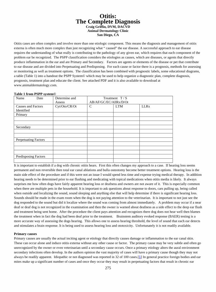
275
Otitis: The Complete Diagnosis
Craig Griffin, DVM, DACVD Animal Dermatology Clinic
San Diego, CA
Otitis cases are often complex and involve more than one etiologic component. This means the diagnosis and management of otitis externa is often much more complex than just recognizing what “ caused” the ear disease. A successful approach to ear disease requires the understanding of what really is contributing to the pathology of any given ear, which requires that each component of the problem ear be recognized. The PSPP classification considers the etiologies as causes, which are diseases, or agents that directly produce inflammation in the ear and are Primary and Secondary. Factors are agents or elements of the disease or pet that contribute to ear disease and are divided into Perpetuating and Predisposing. For each cause or factor there is a prognosis, methods for assessing or monitoring as well as treatment options. The classification has been combined with prognostic labels, some educational diagrams, a table (Table 1) into a handout the PSPP System© which may be used to help organize a diagnostic plan, complete diagnosis, prognosis, treatment plan and educate the client. See attached PDF and it is also available to download at www.animaldermatology.com. Table 1 from PSPP system©
Name Date
Determine and Assess
Treatment T / S AB/AF/GC/EC/AllRx/D/Ot
Causes and Factors Identified
Cyt/Oto/CR/Ot
C LTM LLRx
Primary Secondary Perpetuating Factors Predisposing Factors
It is important to establish if a dog with chronic otitis hears. First this often changes my approach to a case. If hearing loss seems permanent and non reversible then total ear canal ablations and bulla osteotomy become better treatment options. Hearing loss is the main side effect of the procedure and if this were not an issue I would spend less time and expense trying medical therapy. In addition hearing needs to be determined prior to ear flushing and medicating with topical medications when otitis media is likely. It always surprises me how often dogs have fairly apparent hearing loss or deafness and owners are not aware of it. This is especially common when there are multiple pets in the household. It is important to ask questions about response to doors, cars pulling up, being called when outside and localizing the sound, sound sleeping and anything else that will help determine if there is significant hearing loss. Sounds should be made in the exam room when the dog is not paying attention to the veterinarian. It is important to not just see the dog responded to the sound but did it localize where the sound was coming from almost immediately. A problem may occur if a near deaf or deaf dog is not recognized in the examination and then the owner is warned about deafness as a side effect to the deep ear flush and treatment being sent home. After the procedure the client pays attention and recognizes there dog does not hear well then blames the treatment when in fact the dog had been deaf prior to the treatment. Brainstem auditory evoked response (BAER) testing is a more accurate way of assessing the dogs hearing. This allows one to assess hearing threshold, the level of sound that each ear detects and stimulates a brain response. It is being used to assess hearing loss and ototoxicity. Unfortunately it is not readily available. Primary causes Primary causes are usually the actual inciting agent or etiology that directly causes damage or inflammation to the ear canal skin. These can occur alone and induce otitis externa without any other cause or factor. The primary cause may be very subtle and often go unrecognized by the owner or even veterinarian until a secondary cause occurs. Once a primary etiology alters the aural environment secondary infections often develop. In the authors opinion the vast majority of cases will have a primary cause though they may not always be readily apparent. Idiopathic or not diagnosed was reported in 32 of 100 cases.[1] In general practice foreign bodies and ear mites make up a significant number of cases and once they occur they may result in perpetuating factors that result in chronic ear
276
disease. If not seen early in the process they may then present without the primary cause being readily diagnosed. That and atopic otitis without obvious skin disease likely are responsible for many of these cases called idiopathic or not diagnosed. Some of these may also occur when predisposing factors combine with secondary causes, but it is likely most of these cases have a primary cause that was unrecognized. The most common causes seen in a dermatology referral practice are atopic disease, food allergy, epithelialization or metabolic disorders. In general practice foreign bodies and ear mites are relatively more prevalent. It is critical too successful long-term management that a primary cause be found and either eliminated or control be secured. The diagnosis of the primary cause often is determined from the otoscopic exam, cytology, complete dermatologic history and examination as well as diet or therapeutic trials, and possible organ testing or biopsy of the skin in other areas or the external ear. Secondary causes The secondary causes do not create disease in a normal ear; they contribute to or cause pathology only in the abnormal ear. As such they occur in combination with primary causes or predisposing factors. Generally secondary causes of otitis externa are easy to eliminate once identified and when they are chronic or recurrent it is usually because primary causes or perpetuating factors have not been adequately addressed. Secondary causes in the past were often considered as primary causes or the “main” diagnosis of an ear case. (ie. Pseudomonas or Malassezia otitis) Even today many clinicians direct all their efforts at diagnosing and treatment of secondary causes. Although their treatment may be important, other causes and factors must be looked for. In some cases such as Malassezia, eliminating the concurrent predisposing factor or primary disease may result in the resolution of the secondary problem. Secondary causes are most often diagnosed with cytologic examination and culture and sensitivity testing when indicated.
A more recently recognized concern in otitis cases is the presence of biofilms. Biofilms are a community of bacteria that live in an extracellular polymeric matrix that increases resistance to antibiotics and host defense mechanisms. Biofilms are different than the planktonic or individual cells of bacteria that are what is most commonly studied when evaluating infectious diseases that fulfill Koch’s postulates. The extracellular matrix is composed of polysaccharides, DNA and proteins and is often referred to as SLIME, a physical characteristic that is associated with some biofilms seen in nature. These communities, originally associated with adhesion to solid surfaces, are known to occur in aggregates in some tissues.[2] The tissue aggregate form may further enhance mechanisms of survival in the affected tissue.[3, 4] The slime may also contribute to damage of the tissue and pathologic responses that occur. The biofilm increases resistance to antimicrobial agents by more than producing SLIME. In addition metabolic adaptations occur at a higher frequency in biofilms and the communities stimulate the development of persister cells. Persister cells are slow growing and do not grow in the presence of an antibiotic. They persist and are able to grow again once the antibiotic is gone. These biofilm infections are most often associated with chronic diseases and in humans middle ear and possible the external ear are sites of predilection.[5, 6] Forty percent of canine otitis strains of Staph. intermedius and Pseudomonas are capable of producing the extracellular polymeric substance of biofilms.[7, 8] Malassezia may also form biofilms.[9] So far biofilms have not been documented in canine otitis cases by two of the best methods for detecting biofilm infections, peptide nucleic acid-fluorescent in situ hybridization (PNA-FISH) and confocal laser scanning microscopy (CLSM). However I have seen cases that have aggregates present on cytologic examination of ear exudate. Seeing these three dimensional aggregates is suggestive and in humans the otitis media aggregates vary from 4-80 uM.[4]
Culture and sensitivity is not routinely recommended and should never be done without cytology. A culture is typically only done if systemic therapy is being prescribed. It has been shown that response to topical therapy does not correlated with culture results.[10] If the cytology reveals suppurative inflammation with relatively pure populations of rods or cocci and the animal has not responded to appropriate topical and systemic antibiotic then a culture and sensitivity may be indicated. The lab should also be sent a cytology slide and any information regarding the organisms seen at time of collection so they know if multiple organisms should be identified. Perpetuating factors Perpetuating factors are changes in the anatomy and physiology of the ear that occur in response to otitis externa, they occur after ear disease. These factors may be subtle at first but over time can develop into the most severe component of chronic ear disease. These factors are not disease specific and are most commonly seen in chronic cases. Once present, they accentuate or permit the development of secondary causes by providing environments and microscopic niches that favor their persistence. In many cases perpetuating factors prevent the resolution of otitis externa when treatments are only directed at primary and secondary causes. They cause much frustration to clinicians for several reasons. They often result in animals presenting repetitively with different causes present at each subsequent visit. These factors can become self-perpetuating and lead to progressive worsening of disease. They can become severe and end up causing the majority of symptoms exhibited by a pet or be so mild appearing that to many veterinarians as well as owners a pet and its ear canal appear normal. Yet left untreated perpetuating factors, even though primary and secondary causes are controlled or eliminated, result in recrudescence of clinical disease.
In chronic cases often more than one of these factors will be present. Standard treatments of the primary and secondary diseases present often times will not immediately eliminate the perpetuating factors. In early cases, treating the primary cause may be
277
sufficient in controlling a case, but after the establishment of perpetuating factors treatment may need to be directed at them. The treatment for perpetuating factors is often different that what is required to control primary and secondary causes of otitis externa. Their treatment should be continued until they have resolved which may take months of continuous therapy and in some cases they are permanent and will require life long therapy or a surgical solution.
Perpetuating factors are the most common reasons otitis externa cases require surgery. Perpetuating factors are diagnosed otoscopic examination; repetitive otoscopic examination timed appropriately, tube palpation and other imaging techniques (radiology, CT scans, MRI).
Diagnosis of otitis media can be made when a ruptured tympanic membrane is seen. A technique of tube palpation and flushing can aid in the diagnosis of otitis media. This technique also may reveal false middle ear cavities. The method is greatly enhanced with FOVEO and the ear canal filled with water, which increases magnification by 4/3 thus appearing 25% larger. Also air bubble may be seen coming through some small tears. The soft tube can be used to palpate any material located at the approximate level of the tympanic membrane. Both depth of the canal and location of the tip of the tube are utilized to determine if a false middle ear or otitis media is present. The feeding tube is passed under visualization with a surgical otoscope head down the ear canal to the level where the tympanic membrane is expected to be located. Predisposing factors Predisposing factors are present prior to the development of ear disease but alone do not cause otitis externa. They increase the risk of development. These factors work in conjunction with either primary causes or secondary causes to become a significant problem. In rare cases a predisposing factor may combine with a secondary cause to create disease even when no primary cause is present. The best example of this is a dog that gets water in its ear that leads to epidermal maceration or damage and then a secondary bacterial or yeast infection occurs. It is possible this is how environment, increased heat and humidity, also contribute to otitis. However in the authors experience these animals often do have a subtle but mild primary disease still present but controlling that disease does not appear to be necessary. Some predisposing factors relate to the normal anatomy of the dog and as such are not something that is cureable unless surgery may alleviate it, such as a stenotic external orifice in a Chinese shar pei. Pendulous pinnae have been shown to be a statistically significant predisposing factor for otitis externa though no studies have adjusted for this finding based on the presence of breed predisposition to other primary causes of otitis[11]. References Saridomichelakis, M.N., et al., Aetiology of canine otitis externa: a retrospective study of 100 cases. Vet Dermatol, 2007. 18(5): p. 341-7. Alhede, M., et al., Phenotypes of non-attached Pseudomonas aeruginosa aggregates resemble surface attached biofilm. PLoS One, 2011. 6(11): p. e27943. Haaber, J., et al., Planktonic aggregates of Staphylococcus aureus protect against common antibiotics. PLoS One, 2012. 7(7): p. e41075. Bjarnsholt, T., The role of bacterial biofilms in chronic infections. APMIS Suppl, 2013(136): p. 1-51. Wessman, M., et al., Mucosal biofilm detection in chronic otitis media: a study of middle ear biopsies from Greenlandic patients. Eur Arch Otorhinolaryngol, 2014. Fusconi, M., et al., Is biofilm the cause of chronic otitis externa? Laryngoscope, 2011. 121(12): p. 2626-33. Pye, C.C., A.A. Yu, and J.S. Weese, Evaluation of biofilm production by Pseudomonas aeruginosa from canine ears and the impact of biofilm on antimicrobial susceptibility in vitro. Vet Dermatol, 2013. 24(4): p. 446-9, e98-9. Moreira, C.A., et al., Biofilm production by clinical staphylococci strains from canine otitis. Braz J Microbiol, 2012. 43(1): p. 371-4. Cannizzo, F.T., et al., Biofilm development by clinical isolates of Malassezia pachydermatis. Med Mycol, 2007. 45(4): p. 357-61. Robson, D., G. Burton, and R. Bassett. Correlation between topical antibiotic selection, in vitro bacterial antibiotic sensitivity and clinical response in 16 cases of canine otitis externa complicated by Pseudomonas aeruginosa. in Nt Am Vet Derm Forum. 2010. Portland, Oregon. Lehner, G., C.S. Louis, and R.S. Mueller, Reproducibility of ear cytology in dogs with otitis externa. Vet Rec, 2010. 167(1): p. 23-6.
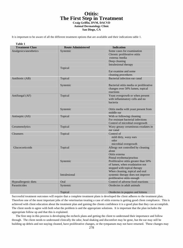
278
Otitis: The First Step in Treatment
Craig Griffin, DVM, DACVD Animal Dermatology Clinic
San Diego, CA
It is important to be aware of all the different treatment options that are available and their indications table 1.
Table 1 Treatment Class Route Administered Indication
Analgesics/anesthetics Systemic Topical
Some cases for examination Chronic proliferative otitis externa /media Deep cleaning Intralesional therapy Ear examine and some cleaning procedures
Antibiotic (AB)
Topical Systemic
Bacterial infection ear canal Bacterial otitis media or proliferative changes over 50% lumen, topical reactions
Antifungal (AF) Topical Systemic
Yeast overgrowth or when present with inflammatory cells and no bacteria Otitis media with yeast present from middle ear
Antiseptic (AS) Topical With or following cleaning For resistant bacterial infections Control of microbial overgrowth
Cerumenolytics Topical Waxy greasy ceruminous exudates in ear canal
Cleansers Topical Control of mild dirty, waxy ears odor microbial overgrowth
Glucocorticoids
Topical Systemic Intralesional
Allergy not controlled by cleaning alone Otitis externa Pinnal erythema/pruritus Proliferative otitis greater than 50% of lumen, when exudatation not stopped with topical therapy When cleaning, topical and oral systemic therapy does not improve proliferative otitis enough
Hypoallergenic diets Oral Control of adverse food reactions Parasiticides Systemic
Topical
Otodectes in adult animals Otodectes in puppies and kittens
Successful treatment outcomes will require that a complete treatment plans is developed the client adheres to the treatment plan. Therefore one of the most important jobs of the veterinarian treating a case of otitis externa is getting good client compliance. This is achieved with client education about the treatment plan and gaining the clients confidence it is a good plan that they can accomplish. The client needs to agree with both what the problem is and the appropriate solutions. It is important that the plan includes the appropriate follow up and that this is explained.
The first step in this process is developing the recheck plans and getting the client to understand their importance and follow through. The client needs to understand clinically the odor, head shaking and discomfort may be gone, but the ear may still be building up debris and not staying cleaned, have proliferative changes, or the tympanum may not have returned. These changes may
279
eventually lead to another infection or acute flare up of otitis. The return to a healthy ear canal can only be determined with otoscopic and cytologic examination of the ear canal. Follow up examinations are also important to determine if cleaning is being done effectively and when normal self-cleaning returns. Scheduling the follow up examination is critical and has to be done differently to answer the preceding questions. To determine if home cleaning is effective then the examination should be done within 24 hours of the cleaning procedure. To determine if the interval is to long between cleanings or that self cleaning may have returned the examination needs to occur when the ear has not been cleaned for at least the interval between current cleanings or longer. Clients need to understand there are different types of follow up for chronic ear cases and that multiple visits will be required.
The number one rule of topical therapy is the active ingredient(s) must reach the site to be treated. This means if only one treatment was allowed to manage ear cases then it would definitely be cleaning, as no single topical product is as effective. Cleaning techniques that are most effective occur in the sedated or anesthesized dog and generally that is the preferred initial therapy when there is a a lot of proliferation, exudate or otitis media. There are cases where initial therapy will make this more effective or unnecessary but in some cases there will be a poor response without the in clinic cleaning. Deep ear cleaning in clinic also allows for the false middle ear pockets or middle ear to be cleaned. Tube flushing may be an effective non-surgical method for cleaning these deeper sites. It is also the least expensive method. A variety of tubes such as polypropylene tomcat catheters have been recommended for use but my preference is a soft rubber feeding tube (Sovereign® feeding tube and urethral catheter) of several sizes (3.5, 5.0, 8.0 and 10.0 French) though the 5 and 8 are my most commonly used sizes. These may be prepared for use then kept in cold sterilization solutions. They are cut short (5-7 inches in length and the ends are trimmed so that the tube will fit over a syringe hub. A 6-12cc syringe is attached. Some clinicians prefer to use a three-way valve so that fluids can be run through one port and suction from a separate port. Which action (flush or suction) is being done is determined by the position of the three-way valve. Water or saline may be utilized as the flushing solution. It should be at roughly body temperature. Saline has the advantage of causing less swelling if repetitive flushing is performed. Usually multiple flushes are required and a bowl of flushing solution should be available. Cleansers and antiseptics may be used in the flushing solution though the author rarely does this and utilizes these products only as the final rinse.
The feeding tube is passed down through a surgical otoscope head and attached cone or through a video otoscope. Under visualization the tube is passed down to the level of the middle ear. If possible the tip is then passed ventrally towards the bottom of the tympanic bulla. The objective is to place the tip of the tube at the most ventral aspect so that the exudate and organisms are flushed directly out towards the external ear canal. In other cases the tube may be placed within the exudate, which may be inspissated, and aids in dislodging and removing it. The passing of the tube into the dorsal or middle aspect of the middle ear has a greater risk of damaging the vestibular (oval) or cochlear (round window) that lies within the promontorium. Actual placement in the ventral bulla is difficult due to the boney ridge that separated the ventral from middle parts of the middle ear cavity. Soft tubes are more likely to reach this location due the ability for the tip to bend. Therefore attempts to get the tube below the ridge should be made and is easier when using 5 French or smaller tubes. Trying to bounce the tube off the dorsal aspect of the external acoustic meatus just prior to entering the area of the dilated or ruptured tympanum may help in achieving this. Once the tip is placed in an appropriate location the flushing solution is gently infused into the ear and this will fill the otoscopic cone and any debris is seen floating in the solution. The flush solution is then aspirated out and it along with the aspirated debris is discarded. Flushing by infusing fluid and aspirating is repetitively done until no debris is seen floating up in the solution. Ear cleaning units that combine flushing and suction are very helpful thought not required for tube flushing.
Vestibular syndrome or deafness may occur after ear flushing, even when no ototoxic drugs are utilized. These side effects are uncommon. In one study of 44 cases that had the middle ear flushed no side effects were reported[31]. Another study of 105 otitis ears flushed none had hearing loss and some even improved following cleaning[32].
Home ear cleaning may also be essential, particularly in cases where epithelial migration is not occurring. Generally I do not have clients begin to clean until the dogs ears are not painful and then usually only once weekly. Most often I have clients do an ear wash by filling the ear canal to the opening of the external orifice with a mild antiseptic cleanser. The clients massage the ear both vertical and horizontal ear canal for a few minutes if possible. To effectively massage the annular cartilage the client must be educated about the location and need for deep digital palpation. Following several minutes of massage the material is allowed to be shaken out and then the external orifice and concave pinna is wiped clean with tissue or cotton balls. If they get more than a little debris they should fill and rinse the ear again and repeat until only a small amount of debris is obtained. Do not allow excessive use of cotton tipped applicators down the ear canal as these commonly push debris deeper into the ear canal. Antiseptics are sometimes utilized as an ear rinse daily for some infected ears or following the home cleaning. My favorites contain acetic or other acids or tris edta with 0.15% chlorhexidine which in the US is usually labeled as a flush not ear product.
280
References Abelardo, E., et al., A double-blind randomised clinical trial of the treatment of otitis externa using topical steroid alone versus topical steroid-antibiotic therapy. Eur Arch Otorhinolaryngol, 2009. 266(1): p. 41-5. Bensignor, E. and E. Grandemange, Comparison of an antifungal agent with a mixture of antifungal, antibiotic and corticosteroid agents for the treatment of Malassezia species otitis in dogs. Vet Rec, 2006. 158(6): p. 193-5. Ghubash, R., R. Marsella, and G. Kunkle, Evaluation of Adrenal Function in Small-Breed Dogs Receiving Otic Glucocorticoids. Vet Dermatol, 2004. 15(6): p. 363-368. Reeder, C., et al., Comparative adrenocortical suppression in dogs with otitis externa following topical otic administration of four different glucocorticoid-containing medications. Vet Ther, 2008. 9(2): p. 111-121. Moriello, K.A., et al., Adrenocortical suppression associated with topical otic administration of glucocorticoids in dogs. J Am Vet Med Assoc, 1988. 193(3): p. 329-31. Aniya, J.S. and C.E. Griffin, The effect of otic vehicle and concentration of dexamethasone on liver enzyme activities and adrenal function in small breed healthy dogs. Vet Dermatol, 2008. 19(4): p. 226-31. Robson, D., G. Burton, and R. Bassett. Correlation between topical antibiotic selection, in vitro bacterial antibiotic sensitivity and clinical response in 16 cases of canine otitis externa complicated by Pseudomonas aeruginosa. in Nt Am Vet Derm Forum. 2010. Portland, Oregon. Pietschmann, S., et al., Synergistic effects of Miconazole and Polymyxin B on microbial pathogens. Vet Res Commun, 2009. 33: p. 489-505.
281
Otitis: Tips You Can Use
Craig Griffin, DVM, DACVD Animal Dermatology Clinic
San Diego, CA
Do a hearing evaluation It is important to establish if a dog with chronic otitis hears. First this often changes my approach to a case. If hearing loss seems permanent and non reversible then total ear canal ablations and bulla osteotomy become better treatment options. Hearing loss is the main side effect of the procedure and if this were not an issue I would spend less time and expense trying medical therapy. In addition hearing needs to be determined prior to ear flushing and medicating with topical medications when otitis media is likely. It always surprises me how often dogs have fairly apparent hearing loss or deafness and owners are not aware of it. This is especially common when there are multiple pets in the household. It is important to ask questions about response to doors, cars pulling up, being called when outside and localizing the sound, sound sleeping and anything else that will help determine if there is significant hearing loss. Sounds should be made in the exam room when the dog is not paying attention to the veterinarian. It is important to not just see the dog responded to the sound but did it localize where the sound was coming from almost immediately. A problem may occur if a near deaf or deaf dog is not recognized in the examination and then the owner is warned about deafness as a side effect to the deep ear flush and treatment being sent home. After the procedure the client pays attention and recognizes there dog does not hear well then blames the treatment when in fact the dog had been deaf prior to the treatment. Brainstem auditory evoked response (BAER) testing is a more accurate way of assessing the dogs hearing. This allows one to assess hearing threshold, the level of sound that each ear detects and stimulates a brain response. It is being used to assess hearing loss and ototoxicity. Unfortunately it is not readily available. Dilating the ear for cleaning With proliferative end stage ears it is difficult to impossible to really get cleansers down the ear. To achieve this when the dog is anesthetized use the 3mm otoscope cone to dilate the ear and force the cone down as far as possible. An ear loop can be passed down the canal just past the tip of the cone and then the cone is filled with the cleanser and slowly pulled out. This will allow a layer of cleanser to be deposited on many of the folds as they fall back in place as the cone is removed. Glucocorticoids Topical glucocorticoids are the most common prescription item used in treating ear disease. This makes sense when one considers the most common causes of chronic otitis are allergic diseases such as atopic disease or adverse food reaction, which is likely an allergic reaction. Even ear mites are known to stimulate an allergic reaction. In addition many cases of otitis become secondarily infected with bacteria or Malassezia and glucocorticoids at least topically are believed to improve the response to topical antimicrobial therapy. This has been shown in dogs with Malassezia otitis and is supported by the fact that most topical antibiotic ear products labeled for the treatment of otitis do contain a glucocorticoid.[1] Eliminating or decreasing inflammation in the ear canal is an essential component of treating secondary infections and often is also indicated as it helps control the primary allergic disease as well. What this means is most cases should be treated with some glucocorticoid and so the real question is when to you avoid using them. I really have come to where the only time I do not use glucocorticoids in otitis cases is when 1. Cleaning ears alone is effective, 2. Infections are not responding or 3. Ulcers are not healing even though the infections appear to be controlled.
Glucocorticoids available for topical use in veterinary products are, from generally the weakest to more potent, 1% hydrocortisone, 0.1% or 0.015% triamcinolone, 0.1% betamethasone, 0.1% dexamethasone, 0.1% fluocinolone acetonide and 0.1% mometasone furoate. The initial therapy or during acute exacerbations a potent topical glucocorticoid v may be required, but once the inflammation or allergic reaction is controlled prophylactic or long term therapy should utilize the least potent topical glucocorticoid possible. Long-term therapy is safer with products containing 1.0% or .5% hydrocortisone. A topical triamcinolone product (0.015% triamcinolone spray (Genesis®, Virbac) that has reduced systemic absorption has been useful, particularly for pinnal inflammation associated with allergic otitis. In cases of atopy or food allergy induced otitis externa, the pinna is frequently affected and should also be treated. Low dose dexamethasone 0.01 to 0.05% has also been formulated in hospital and used effectively for long-term control of allergic otitis or Malassezia otitis. Combination therapy the key to killing organisms Combinations are the key to eliminating resistant bacteria. Three different topical agents, antiseptics, synergistic agents and topical antibiotics, may be used for the purpose of killing the resistant bacteria. When 16 Pseudomonas cases were treated empirically, 90% reported resistant responded to an topical containing the antibiotic the organism was supposedly resistant to and 83% responded when the empiric treatment was reported used for a sensitive strain.[2] The favorable response regardless of what the sensitivity says may have been due to the combination approach and use of the synergist Tris EDTA as well as the high concentration we achieve when
282
using topical antibiotics. Topical antiseptics include such ingredients as certain acids (acetic, boric, citric, lactic), alcohols, aluminum hydroxide, chlorhexidine (0.25% or lower concentration), povidone iodine, silver sulfadiazine and sodium chlorite. Micronized silver is the newest addition to our topical antibacterial solutions. Antiseptics kill organisms by methods other than antibiotics, generally are inexpensive ingredients and can work in conjunction with antibiotics. Resistance is generally not a problem though this may be changing which is another reason to employ combination therapy. Some ingredients that look promising for destroying biofilms are chlorhexidine, acetic acid, and tris EDTA, N-acetyl-L-cysteine and sulfhydryl compounds. In cases resistant to all antibiotics antiseptics may end up being the treatment of choice. The drawback to using antiseptics is they often need to have contact time in clean ears and be used multiple times a day for a good effect. Some are also irritating which limits their use. The antiseptic should be left in the ear canal for 5 minutes. In difficult cases that are being cleaned under sedation/anesthesia then I may leave acetic/boric acid in the ear canal for five minutes then follow with a five-minute soak with Tris edta/chlorhexidine. When antiseptics are the only topical antibacterial used then they often should be applied 4-6 times a day.
Synergistic agents improve the killing effect of what they are mixed with in a way that is more than the additive effect of the two ingredients. Tromethamine-ethylenediaminetetra acetic acid (Tris edta) is the synergist used the most in veterinary otitis cases. It has been shown to enhance the effects of antibiotics as well as the low safe concentration antiseptic chlorhexidine (0.15%).[3, 4] Has been shown to be synergistic with tris EDTA.[3, 4] A very interesting agent is polymyxin as it is not only and antibiotic but also a synergistic agent. Polymyxin has a cationic detergent effect and similar to tris EDTA disrupts the outer membrane of bacteria, particularly gram-negative bacteria. It is synergistic with some other antibiotics but also has a synergistic effect with miconazole. When polymyxin is mixed with miconazole it is synergistic for killing Malassezia but also highly synergistic for the killing of Pseudomonas.[5] By combining synergistic agents with antibiotics even resistant strains of Pseudomonas are killed. Repetitive ear flushes in clinic Since many chronic end stage ear cases will require multiple ear flushes in clinic it is common to encounter clients reluctant to do general anesthesia. Instead it is common to use sedatives and pain medication to allow some of the follow up ear flushes. In these cases even though the laryngeal reflex may be present it can be suppressed enough that care must be taken to prevent inhalation pneumonia. Those resistant Pseudomonas and MR Staphylococcus do not do well in the lungs. Remember any time an ear, with access to the middle ear, is being flushed in a sedated dog and an endotracheal tube is not in place the head should be angled down. We have the racks on the wet table raised at one end with the dog lying in lateral recumbency and the nose at the low end of the rack. Malassezia in ears When dealing with possible resistant Malassezia then Posaconazole is reported to be more effective though it was not as potent as some papers described in one recent study. Miconazole is most often found at 1% but when dealing with difficult cases should higher concentrations such as the 2.3 percent or 1.7% would be more effective. Also polymyxin is synergistic with miconazole for killing Malassezia. Follow up cytology Antiseptics, antibiotics or anti yeast topical therapy is not discontinued until reasonably normal self cleaning has returned and cytology shows no inflammatory cells or DNA strands. It is common for practitioners to discontinue therapy to early, especially if the ear looks reasonably good and there is no obvious odor or discharge. I see many cases when I think it is time to quit but based on cytology I do not. This is something else it is wise to warn owners to expect and if it does not happen they will be pleased and think you or they did a better job than usual. References Bensignor, E., Treatment of Malassezia otitis in dogs: a comparative field trial. Vet Dermatol, 2004. 15(s): p. 45. Robson, D., G. Burton, and R. Bassett. Correlation between topical antibiotic selection, in vitro bacterial antibiotic sensitivity and clinical response in 16 cases of canine otitis externa complicated by Pseudomonas aeruginosa. in Nt Am Vet Derm Forum. 2010. Portland, Oregon. Ghibaudo, G., L. Cornegliani, and P. Martino, Evaluation of the in vivo effects of Tris-EDTA and chlorhexidine digluconate 0.15% solution in chronic bacterial otitis externa: 11 cases. Vet Dermatol, 2004. 15(s): p. 65. Farca, A.M., et al., Potentiating effect of EDTA-Tris on the activity of antibiotics against resistant bacteria associated with otitis, dermatitis and cystitis. J Small Anim Pract, 1997. 38(6): p. 243-5. Pietschmann, S., et al., Synergistic effects of Miconazole and Polymyxin B on microbial pathogens. Vet Res Commun, 2009. 33: p. 489-505.
283
Ears: What You Need to Know
Craig Griffin, DVM, DACVD Animal Dermatology Clinic
San Diego, CA
Recognizing the components of otitis requires an understanding of the normal anatomy and physiology of the external and middle ear, which has been reviewed. Otoscopic examination and cytology are critical in recognizing pathologic findings and for these to be performed well the veterinarian needs to have adequate equipment and training.
The external ear is formed from two pieces of cartilage and a boney canal that are covered by skin, which ends at a specialized epithelial structure the tympanic membrane. The external ear canal is variable in length (5-10cm) and classically divided into the vertical and horizontal portions. As one proceeds down the vertical canal there is another ridge or fold in the cartilage that is called the auricular projection. It creates the “corner” around which one must proceed to allow access down the canal and when otitis is present the lumen ridge is often inflamed and when pushed against by otoscope cones, especially the edge of the cone, may result in pain and the dog resisting examination. The smaller second cartilage is the annular cartilage. It overlaps with the external acoustic meatus. The external acoustic meatus varies but in mid size dogs is about 1 cm long. The skin lining the acoustic meatus lies on bone and therefore is not subject to movement and massage as the skin lining the cartilaginous canal. The medial ring of the acoustic meatus is the location of the tympanic membrane. Often there are larger primary hairs located in the skin adjacent to the tympanum and this is more often seen on the ventral wall of the lumen, a helpful landmark for locating the ventral tympanum with diseased ears.
The skin and adnexa are constantly producing exfoliating corneocytes, intercellular material and glandular secretions. This material forms the earwax and cerumen that is believed to play some protective role. This cerumen is constantly being produced throughout the ear canal. If this material were to build up blockage could result. However there is a normal clearing mechanism. The material produced in the ear canal is cleaned or cleared out by the movement of the epidermis, epithelial migration. The surface of the skin of the tympanum and ear canal is constantly moving from the tympanic membrane laterally to the external orifice of the ear canal. This process starts on the tympanum and this was shown to occur in dogs.[1] The tympanic membrane is an epithelial structure that separates the external ear laterally from the middle ear cavity located medially. The tympanic membrane of the dog is made up of the pars flaccida and pars tensa. The pars flaccida is a small area of the dorsal to anterior-dorsal aspect of the tympanum, which is relatively flaccid and quite vascular. The majority of what is seen of the tympanum when it is examined through the otoscope is the large pars tensa. A normal pars tensa is translucent, with striations seen extending from the manubrium of the malleus outward to the periphery. A whitish appearing discoloration can sometimes be seen through the lower to mid section of the tympanum. This whitish structure is the bony ridge that separates the tympanic cavity from the tympanic bulla. The manubrium of the malleus is “C” shaped with the open end of the “C” pointing toward the nose. It is located over the anterior- medial aspect of the tympanum.
The middle ear consists of the tympanic cavity and the medial wall of the tympanic membrane, the auditory ossicles and associated ligaments, muscles and nerves (chorda tympani and other smaller nerves), and the auditory tube. The tympanic cavity is divided into three parts: dorsal, middle, and ventral. The dorso-medial surface of this is primarily made up of the barrel shaped, cochlear promontory. The promontory is situated opposite to about the mid dorsal aspect of the tympanum. At the caudal end of the promontory is the cochlear window, which communicates with the bony labyrinth of the cochlea. This is the structure one must avoid when doing a myringotomy and flushing the middle ear. The ventral portion is the tympanic bulla and is the largest portion. The tympanic bulla is somewhat egg-shaped, with the dorsal aspect open to communicate with the middle part. It is separated dorsally from the tympanic cavity by the septum bulla, which is most prominent over the medial and anterior aspects of the bulla and responsible for making passing tubes into the ventral bullae very difficult. It may have many bony ossicles or projections along it lateral free edge in the lumen of the bulla.
Microscopic anatomy of ear canal cerumen Ear samples are routinely collected from abnormal ears for cytologic examination and sometimes for culture and sensitivity testing. The samples should reflect the material exudate from the skin of the ear canal or the middle ear cavity. A technique, which will get a deep sample, is to pass a soft rubber tube down the canal and aspirate once the tip is deep into the canal. Cytologic examination of discharge usually does not establish a definitive diagnosis, but it is valuable in determining what infectious agents, if any, are present. With waxy discharges heat the author has preferred fixation though two studies suggest it may not be necessary. Modified wrights stain (Wright's Dip Stat) is a rapid method that adequately stains specimens and has two colors to help differentiate stained items. Cytologic evaluation is the preferred method to ascertain the role of Malassezia and probably bacteria. Two published studies specifically evaluated cytology in normal dog and cat ears and one study in comparison to otitis externa cases but at 400x.[2, 3] Though they did not have that similar results one important observation they made is normal ears never had inflammatory cells. The Tater study did not find rods and Ginel was not able to separate rods and cocci as 400x. An unpublished study at 1000x is the basis of
284
what I use. Normal dogs and cats then greater than 3 yeast per oil immersion field is considered highly suspect and for bacteria greater than 5 cocci or 1 rod per oil immersion field would be very suspect. But as or more important is the presence of inflammatory cells which is highly suggestive that secondary infection is present.
Cytology allows evaluation of the cellular make-up of the discharge as well as microbial agents present. The degree of wax, lipids, keratin (nucleated corneocytes) can also be determined. Ceruminous otitis externa is seen with endocrinopathies and seborrhea; the discharge in this condition is keratin and glandular secretions. Eosinophils may be seen with parasitic disease, topical drug reactions and some food allergic animals. Cytology determines what secondary infections or microbial overgrowth is present. In addition the presence of mixed bacterial aggregates that are three dimensional may be a way to evaluate for biofilm infections. Biofilm formation has been identified as a common problem in human otitis media and may play a role in some otitis externa cases. Canine ear isolates have been shown capable of forming biofilms.[4-6] Toxic neutrophils indicate that the ear canal must be flushed to remove the toxins. The presence of white blood cells as well as phagocytosis of bacteria indicates that the body is responding to the infection and treatment for the bacteria is warranted. The presence of blue staining nuclear strands indicates that some inflammatory cells are present even though intact cells may not be identified. If any neutrophils or nuclear strands are found then it is likely there is still a bacterial component to the disease even if bacteria are not found.
Cytologic evaluation is the preferred method to ascertain the role of Malassezia in a particular case for two reasons. In one study by the author 18% of the cases that had Malassezia detected by cytology were sterile on culture by a commercial laboratory culturing specifically for Malassezia at 37 degree C. Magnifying the ear canal Otoscopes must have a strong light and power source combined with at least 10x magnification that allows focusing within the normal length of the ear canal. If any of these components is not present otoscopic examinations may not be totally effective. We have borrowed this equipment from human medicine where there are two main types of otoscope heads the diagnostic or medical and the surgical. They differ in the size of the magnifying lens that one looks through as well as the shape of the cone holders. Many practitioners purchase the diagnostic otoscope head. In general we prefer the surgical otoscope head, which allows more manipulation and angulations as well as easier use with cleaning and therapeutic procedures that require passing instruments or tubes into the ear canal with concurrent visualization. One of the most common mistakes made in practice is the use of hand help battery operated otoscopes that no longer have enough power to adequately light the deep ear canal. In general every clinic should have at least one plug in otoscope, which is not dependent of having fresh fully charged batteries. The battery operated is valuable for being readily moveable to different locations in the practice but for abnormal ears that require work a strong well-lighted otoscope is preferred.
Various sizes of otoscope cones are needed to be able to examine the different size and breeds of dogs and cats seen in practice. This equipment is essential to practice and even if the newer fiberoptic video enhanced otoscopes are available the traditional otoscopes are still necessary. These allow larger instruments and tubes to be passed into the ear canal and allow much faster deep ear cleaning. Smaller 3mm cones are fine for routine examinations but when working on ears it is important to use the largest diameter cone that you can get down the ear canal. This will improve visualization and allow more room for manipulating instruments and tubes. A clean cone, which is at least 10minute soaking in cold sterilization fluids, should be used in each ear for examination and performing procedures. At least 10minute soaking in cold sterilization fluids
The advent of fiber optics, improved lighting and miniaturization of video cameras combined with a rigid endoscope has led to the development of Fiberoptic Video Enhanced Otoscopy( FOVOE). This equipment can be connected to a video monitor and printer, digital recorder or video camera. The fiber optic tip with camera also magnifies and with a focal length of several centimeters can improve the visualization of the ear canal. Besides improving visualization it allows permanent recordings of what is present as well as allowing clients or other veterinarians to see the pathology of the ear canal. In some cases small tears of the tympanic membrane not readily seen with the normal 10x magnified otoscope will be apparent with FOVEO. In addition filling the ear canal with water or saline is sometimes used with FOVEO as it further enhances magnification and keeps the tip of the camera lens from fogging. This cannot be done with normal otoscopes. With water or saline in the ear canal, perforations not even visible with FOVEO will sometimes be found and are recognized by the air bubbles coming from the middle ear cavity. This equipment is relatively expensive but considering the improved diagnostics and more importantly the client education and benefits on gaining client support for recommended procedures makes this a worthwhile investment in a busy practice. The fiberoptic scopes also have made assessment of the abnormal tympanic membrane more effective.
The technique for doing proper otoscopic examination is one that allows complete as visualization as possible with minimal pair or trauma. Many dogs or cats will allow a carefully done otoscopic exam but resist or make it impossible to complete an examination if the technique is not optimum. Examinations are best done on a table to allow for appropriate orientation of the scope. Though large breed dogs may be able to be done on the floor is the head is held high enough and the operator is kneeling on the floor. The head should be high enough to allow the observed to move the otoscope into a more horizontal position. Occasionally it is easier to examine an ear of a dog lying in lateral recumbence on a table. The muzzle should be directed slightly towards the thoracic inlet. It may also
285
be necessary to have someone else hold the dog or cats muzzle as the natural tendency is for the head to be tilted as the examination starts. This will redirect the cone tip resulting in more pain. The pinna should be pulled up and out from the base of the skull, which helps to straighten the ear canal and minimize the blocking of the lumen by the cartilage fold that occurs near the junction of the vertical and horizontal canal. In addition the cone is passed down the lumen of the ear canal while the operator is visualizing the canal through the otoscope cone. Attempting to insert the cone without visualization is a sure way to “hit” the canal epithelium, which can be painful even in a normal ear. The cone is then moved slowly into the vertical canal, visualizing as you go, then the otoscope handle is rotated downward so the cone approaches a horizontal position. The movement is best accomplished when the ear is also pulled up and out over the tip of the cone so that the two processes happen simultaneously. Proper placement at the junction often allows visualization into the horizontal canal and if necessary advancement into the horizontal canal. Deep penetration into the horizontal canal is only done if necessary to visualize the tympanum. One problem often encountered in practice is the extremely painful ulcerated swollen ear that one cannot adequately exam. Even with anesthesia these cases may not be adequately examined. It may be necessary to treat the animal and reduce the swelling and inflammation and have the patient return in 4 - 7 days so that an otoscopic exam can be properly performed. References Tabacca, N.E., et al., Epithelial migration on the canine tympanic membrane. Vet Dermatol, 2011. 22(6): p. 502-10. Ginel, P.J., et al., A semiquantitative cytological evaluation of normal and pathological samples from the external ear canal of dogs and cats. Vet Derm, 2002. 13(3): p. 151-156. Tater, K., et al., The cytology of the external ear canal in the normal dog and cat. J Vet Med, 2003. 50: p. 370-374. Moreira, C.A., et al., Biofilm production by clinical staphylococci strains from canine otitis. Braz J Microbiol, 2012. 43(1): p. 371-4. Cannizzo, F.T., et al., Biofilm development by clinical isolates of Malassezia pachydermatis. Med Mycol, 2007. 45(4): p. 357-61. Pye, C.C., A.A. Yu, and J.S. Weese, Evaluation of biofilm production by Pseudomonas aeruginosa from canine ears and the impact of biofilm on antimicrobial susceptibility in vitro. Vet Dermatol, 2013. 24(4): p. 446-9, e98-9.
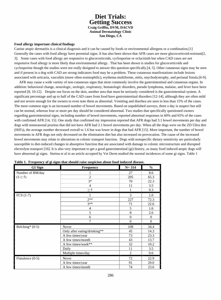
286
Diet Trials: Getting Success
Craig Griffin, DVM, DACVD Animal Dermatology Clinic
San Diego, CA
Food allergy important clinical findings Canine atopic dermatitis is a clinical diagnosis and it can be caused by foods or environmental allergens or a combination.[1] Generally the cases with food allergy have perennial signs. It has also been shown that AFR cases are more glucocorticoid-resistant[2, 3]. Some cases with food allergy are responsive to glucocorticoids, cyclosporine or oclacitinib but when CAD cases are not responsive food allergy is more likely than environmental allergy. That has been shown is studies for glucocorticoids and cyclosporine though the studies were not really designed to answer this question specifically.[4, 5]. Other cutaneous signs may be seen and if present in a dog with CAD are strong indicators food may be a problem. These cutaneous manifestations include lesions associated with urticaria, vasculitis (more often eosinophilic), erythema multiforme, otitis, onychodystrophy, and perianal fistula.[6-9].
AFR may cause a wide variety of non-cutaneous signs that most commonly involve the gastrointestinal and cutaneous organs. In addition: behavioral change, neurologic, urologic, respiratory, hematologic disorders, pseudo lymphoma, malaise, and fever have been reported [8, 10-12]. Despite our focus on the skin, another area that must be seriously considered is the gastrointestinal system. A significant percentage and up to half of the CAD cases from food have gastrointestinal disorders [12-14], although they are often mild and not severe enough for the owners to even note them as abnormal. Vomiting and diarrhea are seen in less than 15% of the cases. The most common sign is an increased number of bowel movements. Based on unpublished surveys, three a day is suspect but still can be normal, whereas four or more per day should be considered abnormal. Two studies that specifically questioned owners regarding gastrointestinal signs, including number of bowel movements, reported abnormal responses in 60% and 65% of the cases with confirmed AFR [14, 15]. One study that confirmed my impression reported that AFR dogs had 3.1 bowel movements per day and dogs with nonseasonal pruritus that did not have AFR had 2.1 bowel movements per day. When all the dogs were on the ZD Ultra diet (Hill's), the average number decreased overall to 1.6 but was lower in dogs that had AFR [15]. More important, the number of bowel movements in AFR dogs not only decreased on the elimination diet but also increased on provocation. The cause of the increased bowel movements may relate to alterations in colonic transport function. Dogs with nonspecific dietary sensitivity are particularly susceptible to diet-induced changes in absorptive function that are associated with damage to colonic microstructure and disrupted electrolyte transport [16]. It is also very important to get a good gastrointestinal (gi) history, as many food induced atopic dogs will have abnormal gi signs. Stetina et al in an article accepted by Vet Derm studied the normal incidences of some gi signs. Table 1 Table 1. Frequency of gi signs that should raise suspicion about food induced disease.
GI Sign Frequency N= 314 % Number of BM/day (1-≥ 5)
1 27 8.6 2 205 65.3
3* 70 22.3 4 11 3.5
≥ 5 1 0.3 FCS (1-7)
1 3 1.0 2** 227 72.3 3** 71 22.6 4 5 1.6 5 8 2.6 6 0 0 7 0 0
Belching* (0-5)
Never 108 34.4 Only after eating/drinking** 45 14.3 A few times/year 73 23.3 A few times/month 43 13.7 A few times/week** 32 10.2 Daily 11 3.5 Multiple times/day 2 0.6
Flatulence (0-5) Never 72 22.9 A few times/year 91 29.0 A few times/month 74 23.6
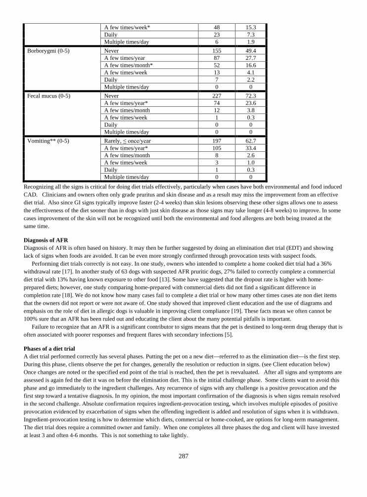
287
A few times/week* 48 15.3 Daily 23 7.3 Multiple times/day 6 1.9
Borborygmi (0-5) Never 155 49.4 A few times/year 87 27.7 A few times/month* 52 16.6 A few times/week 13 4.1 Daily 7 2.2 Multiple times/day 0 0
Fecal mucus (0-5) Never 227 72.3 A few times/year* 74 23.6 A few times/month 12 3.8 A few times/week 1 0.3 Daily 0 0 Multiple times/day 0 0
Vomiting** (0-5) Rarely, ≤ once/year 197 62.7 A few times/year* 105 33.4 A few times/month 8 2.6 A few times/week 3 1.0 Daily 1 0.3 Multiple times/day 0 0
Recognizing all the signs is critical for doing diet trials effectively, particularly when cases have both environmental and food induced CAD. Clinicians and owners often only grade pruritus and skin disease and as a result may miss the improvement from an effective diet trial. Also since GI signs typically improve faster (2-4 weeks) than skin lesions observing these other signs allows one to assess the effectiveness of the diet sooner than in dogs with just skin disease as those signs may take longer (4-8 weeks) to improve. In some cases improvement of the skin will not be recognized until both the environmental and food allergens are both being treated at the same time. Diagnosis of AFR Diagnosis of AFR is often based on history. It may then be further suggested by doing an elimination diet trial (EDT) and showing lack of signs when foods are avoided. It can be even more strongly confirmed through provocation tests with suspect foods.
Performing diet trials correctly is not easy. In one study, owners who intended to complete a home cooked diet trial had a 36% withdrawal rate [17]. In another study of 63 dogs with suspected AFR pruritic dogs, 27% failed to correctly complete a commercial diet trial with 13% having known exposure to other food [13]. Some have suggested that the dropout rate is higher with home-prepared diets; however, one study comparing home-prepared with commercial diets did not find a significant difference in completion rate [18]. We do not know how many cases fail to complete a diet trial or how many other times cases ate non diet items that the owners did not report or were not aware of. One study showed that improved client education and the use of diagrams and emphasis on the role of diet in allergic dogs is valuable in improving client compliance [19]. These facts mean we often cannot be 100% sure that an AFR has been ruled out and educating the client about the many potential pitfalls is important.
Failure to recognize that an AFR is a significant contributor to signs means that the pet is destined to long-term drug therapy that is often associated with poorer responses and frequent flares with secondary infections [5]. Phases of a diet trial A diet trial performed correctly has several phases. Putting the pet on a new diet—referred to as the elimination diet—is the first step. During this phase, clients observe the pet for changes, generally the resolution or reduction in signs. (see Client education below) Once changes are noted or the specified end point of the trial is reached, then the pet is reevaluated. After all signs and symptoms are assessed is again fed the diet it was on before the elimination diet. This is the initial challenge phase. Some clients want to avoid this phase and go immediately to the ingredient challenges. Any recurrence of signs with any challenge is a positive provocation and the first step toward a tentative diagnosis. In my opinion, the most important confirmation of the diagnosis is when signs remain resolved in the second challenge. Absolute confirmation requires ingredient-provocation testing, which involves multiple episodes of positive provocation evidenced by exacerbation of signs when the offending ingredient is added and resolution of signs when it is withdrawn. Ingredient-provocation testing is how to determine which diets, commercial or home-cooked, are options for long-term management. The diet trial does require a committed owner and family. When one completes all three phases the dog and client will have invested at least 3 and often 4-6 months. This is not something to take lightly.
288
The elimination diet The elimination diet should ideally comprise no ingredients that the pet has been exposed to. However, this is usually impossible and the concern of cross reactivity also impacts how diet decisions should be made. Cross-reacting allergens do occur, although the true extent is unknown. A study suggested that the high incidence of reactions to a venison diet in a country where venison is rarely fed may be due to cross-reactivity [20]. Cross-reactivity can occur to more than other foods. A dog with atopic dermatitis (AD) that developed oral allergy syndrome to tomato was shown to cross-react with the Japanese cedar pollen [21].
Since proteins are the most common offending allergen, the primary goal is to feed a protein and carbohydrate source that the pet has not been routinely exposed to. Pure carbohydrates are not of primary concern, except that most carbohydrate sources do have low levels of protein in them that may be allergenic. Even cornstarch, which is commonly found in medication and other tablets, has a low level of protein. Fat supplements may also be contaminated with protein. This may be one reason that three studies that have looked at diet contamination have shown this is a serious problem and the hydrolyzed diets appear to be better choices to avoid contamination.[22-24] What is not known is how often a low level of contamination will cause a failure in a diet trial.
There are three main sources for the hydrolyzed diets: Hills, Purina, and Royal Canin. These diets have lower average molecular weights than nonhydrolyzed diets and these smaller proteins do decrease but not eliminate reactions to those proteins. The theory behind these diets is that food allergy is generally due to large complex proteins or glycoproteins. Hydrolyzing reduces the molecular weight so they are no longer allergenic. Human evidence has yielded some evidence to support this theory. The use of these diets has been reviewed in veterinary medicine and the diets are not totally effective in eliminating reactions.[25] However the diets did work for some dogs sensitive to the parent ingredient.
Because of the difficulty doing diets, long time needed to complete multiple trials, potential for cross-reactions, problems with contamination and difficulty in balancing home made diets it is better to do diet trials with the high quality commercial hydrolyzed diets. They are also expensive which is an advantage when if comes time get clients to do ingredient challenges, as that is the way they can find a less expensive high quality diet they can feed for the rest of the pets life. The presence of gi signs is valuable because they can be assessed at four weeks and if not improving an alternative diet be selected. If all the hydrolyzed diets are ineffective in resolving the gi signs the a home make diet such as pumpkin and pinto beans may be utilized but is not a balanced diet and this needs to be discussed with the owner. Home cooking is not be done in growing dogs and should be only done for the 8 weeks then supplements added to balance the diet. Client education Key points need to be emphasized to owners before starting a diet trial. Establish baseline symptom scores before the trial is started because this provides the comparison for future responses and exacerbations. It is preferable to establish these scores without concurrent microbial disease. Therefore, baseline signs are often determined at a recheck when the pet is on other medications, such as antibiotics and ant yeast medicine. Symptom scores should record the extent and the pattern of pruritus as well as what gastrointestinal and other signs are present. The Hill visual analog score is helpful, but it is important to pay attention to the extent of pruritus in all affected areas. Some areas may change when the overall pruritus score does not in cases of canine AD with both food and environmental components. Grade at least lesions and pruritus of the paws, perineum, dorsal trunk, and ears. For example, the dog may still have grade-10 disease at the end of the trial even though the pruritus of the ear or dorsal lumbar have totally resolved; such cases often indicate that the dog has a combination of allergies. Clients must be educated on how to recognize a response in their pet, and this can only be done if they pay close attention during the challenge phase. Owners of nonresponsive cases must always be counseled that though they do not think the diet has helped they need to see whether any changes occur after reintroduction of the old diet.
An important but often difficult aspect to control is other sources of foods the animal may ingest. Preventing consumption of other foods often means keeping animals confined indoors with outside exposure controlled, such as on a leash. I have also had cases that require a muzzle to prevent inappropriate food consumption. The owners must be aware that the dog should consume nothing but what is in the diet on a regular basis, so that precludes other pets’ food, treats, medication wrapped in food and even alone, chewable forms of dog vitamins, and supplements. It takes very little for signs to flare, as was shown in a study where 12 dogs that were allergic to soy were challenged with one tablet of Interceptor Flavor Tabs, which contain pork liver, soy, and 2.3 mg milbemycin. Clinical scores increased significantly in 10 of 12 dogs, with peak scores seen 2 days after challenge in five dogs and 5 days after challenge in five dogs [26]. So dogs eating inappropriate foods weekly may not respond to the diet trial. This does mean a monthly flavored heartworm or flea control product is okay, but the owner needs to pay close attention at the monthly application times, as this is essentially a challenge with that ingredient. The fluralaner flea product is only given every three months and contains hydrolyzed pork protein allowing a diet trial to be completed without the challenge. The whole family must be aware of the dietary requirements. Dogs need to be prevented from cleaning floors of crumbs. The presence of young children in a household often precludes any chance of controlling a diet in an indoor pet. Coprophagia must also be prevented. There is a report that a food-allergic dog that ate cat feces did not respond until the housecat was also put on a similar hypoallergenic diet.
289
Challenge and specific provocation testing True confirmation of AFR occurs when feeding the offending diet induces signs (challenge phase), which resolve on the elimination diet with no other changes in concurrent therapies. Many clients are resistant to this proposal until I explain its value. The first is the best, money. The limited ingredient hydrolyzed diets are generally expensive so most owner will be motivated to find a new food, especially once it is explained the cost is not for a extra high quality diet but just a highly processed diet. This is a big reason I use hydrolyzed diets and describe their expense as the cost of a test. Some pets will develop new allergies and by doing multiple challenges the client will not have to start from scratch to determine what else the pet can eat. In addition, knowing what foods the pet is not sensitive increases feeding options. This is particularly helpful for the long-term management, especially in multiple-pet households.
How the challenge is done can vary depending on the client. Some like to try and prove exactly what the dog is allergic to. For these we do ingredient challenges. One ingredient at a time is added to the elimination diet for up to a week. The other is new commercial food challenges with different ingredients. Here I generally pick a less expensive limited ingredient diet and switch to that. If the dog does not react then we have either found another diet it can eat or it was not food allergic. So I also then try a treat that contains lots of ingredients so we hopefully can confirm the food allergy is present but maybe not the exact ingredients. With either approach there are some rules to follow. The client keeps a diary on which ingredients cause reactions. They should watch for how long and what signs are seen with each ingredient. After signs have improved significantly or have resolved completely, the pet should be challenged with the diet being fed before the diet trial. If there is no increase in signs, then all other treats, etc. should be fed. Each challenge should be given only until a recurrence is obvious to the client or for 7 days. If there is no recurrence after 7 days, that food is not likely to be the problem. When signs recur, it is usually just an increase in pruritus. This will usually occur rapidly if the exacerbation is noted in the first 2 or 3 days on the challenge. With true allergy, signs recur rapidly and in my experience, most well-confirmed food allergies are worse in 1 to 2 days. If this occurs, then the elimination diet is again fed until the signs resolve. The sooner a client observes a sign of disease occurring and goes back on the elimination diet, the sooner the pet will respond. Fortunately, when they only get one to several meals of the offending diet the response is usually rapid again and often does not need a treatment to resolve it. References 1. Olivry, T., et al., Food for thought: Pondering the relationship between canine atopic dermatitis and cutaneous adverse food reactions. . Vet Derm, 2007. 18(6): p. 390-391. 2. Picco, F., et al., A prospective study on canine atopic dermatitis and food-induced allergic dermatitis in Switzerland. Vet Dermatol, 2008. 19(3): p. 150-155. 3. Favrot, C., et al., A prospective study on the clinical features of chronic canine atopic dermatitis and its diagnosis. Vet Dermatol, 2010. 21(1): p. 23-31. 4. Rosser, E., Diagnosis of food allergy in dogs. J Am Vet Med Assoc, 1993. 203: p. 259-262. 5. Jackson, H., et al. The effect of oral cyclosporine on acute reactions following oral allergen challenge in a spontaneous canine model of food allergy. in ESVD-ECVD. 2006. Lisbon, Portugal. 6. Nichols, P.R., D.O. Morris, and K.M. Beale, A retrospective study of canine and feline cutaneous vasculitis. Vet Dermatol, 2001. 12(5): p. 255-64. 7. Mueller, R.S., et al., Diagnosis of canine claw disease a prospective study of 24 dogs. Vet Dermatol, 2000. 11(2): p. 133-141. 8. Kennis, R., Use of Atopic Dogs to Investigate Adverse Reactions to Food. J Am Vet Med Assoc, 2002. 221(5): p. 638-640. 9. Itoh, T., et al., Erythema multiforme possibly triggered by food substances in a dog. J Vet Med Sci, 2006. 68(8): p. 869-871. 10. Vaden, S.L., et al., Food hypersensitivity reactions in Soft Coated Wheaten Terriers with protein-losing enteropathy or protein-losing nephropathy or both: gastroscopic food sensitivity testing, dietary provocation, and fecal immunoglobulin E. J Vet Intern Med, 2000. 14(1): p. 60-7. 11. Wills, J. and R. Harvey, Diagnosis and management of food allergy and intolerance in dogs and cats. Aust Vet J, 1994. 71(10): p. 322-6. 12. Scott, D.W., W.H. Miller, Jr,, and C.E. Griffin, Muller and Kirk's Small Animal Dermatology. 6 ed. 2001, Philadelphia: W,B. Saunders. 13. Loeffler, A., et al., Dietary trials with a commercial chicken hydrolysate diet in 63 pruritic dogs. Vet Rec, 2004. 154(17): p. 519-22. 14. Patterson, S., Food hypersensitivity in 20 dogs with skin and gastrointestinal signs. J Small Anim Pract, 1995. 36: p. 529-534. 15. Loeffler, A., et al., Dietary trials iwth a commercial chicken hydrolysate diet in 63 pruritic dogs. Vet Rec, 2004. 154: p. 519-522. 16. Rolfe, V.E., et al., Relationships between fecal consistency and colonic microstructure and absorptive function in dogs with and without nonspecific dietary sensitivity. Am J Vet Res, 2002. 63(4): p. 617-22. 17. Tapp, T., et al., Comparison of a commercial limited antigen diet vs a home prepared diet in the diagnosis of canine food adverse reaction. Veterinary Therapeutics, 2002. 3(3): p. 244-251. 18. Loeffler, A., et al., A retrospective analysis of case series using home-prepared and chicken hydrolysate diets in the diagnosis of adverse food reactions in 181 pruritic dogs. Vet Dermatol, 2006. 17(4): p. 273-9. 19. Chesney, C., Food sensitivity in the dog: a quantitative study. J Small Anim Pract, 2002. 43(5): p. 203-207. 20. Leistra, M.H.G., P.J. Markwell, and T. Willemse, Evaluation of selected-protein-source diets for management of dogs with adverse reactions to foods. J Am Vet Med Assoc, 2001. 219(10): p. 1411-1414. 21. Fujimura, M., et al., Oral allergy syndrome induced by tomato in a dog with Japanese cedar (Cryptomeria japonica) pollinosis. J Vet Med Sci, 2002. 64(11): p. 1069-1070.

290
22. Raditic, D.M., R.L. Remillard, and K.C. Tater, ELISA testing for common food antigens in four dry dog foods used in dietary elimination trials. J Anim Physiol Anim Nutr (Berl), 2011. 95(1): p. 90-7. 23. Ricci, R., et al., Identification of undeclared sources of animal origin in canine dry foods used in dietary elimination trials. J Anim Physiol Anim Nutr (Berl), 2013. 97 Suppl 1: p. 32-8. 24. Widman, K., S. Handl, and C. Horvath-Ungerboeck. Detection of food antigens in commercial elimination diets for dogs using PCR. in ESVD-ECVD. 2014. Salzburg, Austria. 25. Olivry, T. and P. Bizikova, A systematic review of the evidence of reduced allergenicity and clinical benefit of food hydrolysates in dogs with cutaneous adverse food reactions. Vet Dermatol, 2010. 21(1): p. 32-41. 26. Jackson, H.A. and B. Hammerberg. The clinical and immunological reaction to a flavoured monthly oral heartworm prophylactic in 12 dogs with spontaneous food allergy. in AAVD and ACVD. 2002. New Orleans.
291
Pruritus: What’s New?
Craig Griffin, DVM, DACVD Animal Dermatology Clinic
San Diego, CA
Pruritus is an uncomfortable sensation of the skin that provokes the desire to rub or scratch, the popular term for this action is itching. Pruritus is a primary neurologic sensation that is a symptom associated with many diseases. The diseases result in the production of pruritic mediators that stimulate itch specific C fiber nerve receptors that are found in the epidermis and dermis. The classic pruritus mediator that has been studied the most is histamine. The discovery of a fourth histamine receptor( H4) has stimulated a new evaluation. There is evidence the H4 receptor is important in both T helper cell type 2 (TH2) inflammation and pruritus. H4 has been shown to occur in dogs but its role is just being investigated. However there are many other mediators believed more important in the dog. There is a long list of pruritic mediators that come from a variety of classes such as neuropeptides, peptides, proteases, cytokines and leukotrienes. We have known for a long time that mast cells contain many pruritic mediators but sensory nerves, lymphocytes and endothelial cells are also sources as is the keratinocyte. Keratinocytes can produce proteases, cytokines and neuropeptides, especially acetylcholine now considered an important mediator of itch. Work in dogs has shown that serum IL-31 is elevated in over half of atopic dermatitis dogs but not in normal, flea allergic or experimentally induced house dust mite allergic beagles.[1] IL-31 is produced by canine TH2 lymphocytes and this production is increased when house dust sensitized T cells are co-stimulated with Staphylococcus enterotoxin B.[2] It is also important to note that two different groups have found IL-31 receptors in the dorsal root ganglia of dogs.[2-4] Pruritus is induced in dogs with exposure to IL-31 and with this model pruritus can be reduced with treatment with prednisolone or the Janus kinase inhibitor, oclacitinib.[1, 5]
Oclacitinib maleate is a new synthetic drug that that has been developed for the treatment of allergic diseases in dogs. It is a member of a relatively new class of drugs called Janus kinase (JAK) inhibitors. Janus kinases are a group of 4 enzymes (Janus kinase 1-3 and tyrosine kinase) that function by facilitating transmission of signals form cell membrane receptors intracellular. They function in pairs with certain cytokine receptors being acted on by various combinations of paired enzymes. Oclacitinib mainly acts on JAK 1 and at higher serum levels on JAK 2. The Cmax does relate to the dose and at 0.6mg/kg daily for 168 days is 273-406 ng/ml. Cmax at 0.4mg/kg was around 200ng/ml for beagle and mongrel dogs after a single dose.[6] Oclacitinib is considered an immune modulating drug because it suppresses cytokine function, particularly IL 31 and to some extent IL 2.
Efficacy of oclacitinib has been shown for pruritus associated with allergic disease.[7] A blinded placebo controlled study-evaluated oclacitinib in 436 dogs with a variety of allergic diseases. Pruritus scores decreased from 7.39 to 2.59 and 7.58 to 5.54 in the oclacitinib and placebo treated groups respectively. The response in the treated group was significant and significantly better than the placebo group. Another blinded placebo study done by board certified veterinary dermatologists studied 299 AD dogs.[8] Pruritus scores decreased from 7.8 to 2.6 and from 7.7 to 7.4 in at the 14 days scoring in the oclacitinib and placebo treated groups respectively. This was very significant difference between groups. Following 14 to 28 days all dogs were allowed to go into an open label study and at the end of 112 days the pruritus score averaged 3.2. The skin lesions as graded by the canine atopic dermatitis extent and severity index (CADESI) had also dropped from a pre treatment score of 62 to 32 and 58 to 57 in the oclacitinib and placebo groups respectively. When all dogs finished the open label phase the CADESI was 26 at day 112. Another study showed efficacy in flea allergy dermatitis.[9] Apoquel was also shown to decrease pruritus as fast as glucocorticoid therapy and in IL 31 pruritus model even better than glucocorticoids.[10, 11]
There is one 84-day randomized controlled trial comparing Apoquel® (oclacitinib) to Atopica® (cyclosporine) in 226 dogs. Veterinary dermatology specialists performed this study with client owned dogs in Australia. Dogs were evaluated for PVAS and CADESI-02 scoring on days 1,2,7, 14, 28, 56 and 84 days. Differences were significant at all time points up to day 28 regarding PVAS scoring. By day 56, cyclosporine treated dogs had a similar PVAS scoring to Apoquel treated dogs. As expected the Apoquel treated dogs had much quicker onset of activity regarding the pruritus reduction (See Graph below). More adverse events were found in the cyclosporine treated group which was largely gastrointestinal (vomiting and diarrhea Atopica group (44 and 15%) vs. Apoquel group (14 and 4%)[12] (See table below comparing side effects). Atopica is FDA approved to be used with low dose prednisolone and a recent study showed no increase in adverse events when combined with low dose prednisolone therapy during the first 3 weeks of therapy. Combination therapy expedited the reduction of pruritus during the first 3 weeks during Atopica induction[13].
Initial results at the Animal Dermatology Clinic in San Diego were evaluated in August 2014. Apoquel was prescribed for 107 dogs, 13 (12%) of 107 stopped the drug because of poor efficacy, 2 stop for the development of
masses, one for UTI, 11 went off for alternative therapy and 9 was because ASIT was working. 3 cases were deceased. One at 9-year age with lymphoma, one at 14year age and was reported weak prior to expiring and one with a Histiocytic sarcoma. We had followed up on 47 dogs treated with16mg tabs and 30 (64%) were still on therapy, 43 dogs on 5.4mg and 26 (60%) still on therapy and 17 dogs on 3.6mg with 12 (71%) still on therapy.
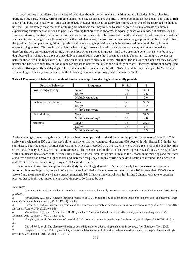
292
In dogs pruritus is manifested by a variety of behaviors though most classic is scratching but also includes: biting, chewing, dragging body parts, licking, rolling, rubbing against objects, scooting, and shaking. Clients may indicate that a dog is not able to itch a part of its body but in reality any area can be itched. However the location partly determines which one of the described methods is utilized. Unfortunately these methods of itching are behaviors that may be seen to some degree in normal animals or animals experiencing another sensation such as pain. Determining that pruritus is abnormal is typically based on a number of criteria such as severity, intensity, duration, induction of skin lesions, or not being able to be distracted from the behavior. Pruritus may occur without visible cutaneous changes, may be associated with a rash that caused the pruritus, or have skin changes present that have resulted from the pruritus. So complete recognition of pruritus and where a dog is pruritic can only be determined by a good history from an observant dog owner. This leads to a problem when trying to assess all pruritic locations as some may not be as affected and therefore the behavior considered normal. For example when surveyed in groups I find there are some veterinarians who believe a dog observed to lick its paws once or twice daily is normal but all agree that 100 times a day is abnormal. Coming to a consensus between those two numbers is difficult. Based on an unpublished survey it is very infrequent for an owner of a dog that they consider normal and has never been treated for skin or ear disease to answer that question with daily or more! Recently Stetina et al completed a study in 314 apparently healthy dogs. The results have been presented at the 2015 NAVDF and the paper accepted by Veterinary Dermatology. This study has revealed that the following behaviors regarding pruritic behaviors. Table 1 Table 1 Frequency of behaviors that should make you suspicious the dog is abnormally pruritic A visual analog scale utilizing those behaviors has been developed and validated for assessing pruritus by owners of dogs.[14] This scale was evaluated in 305 dogs that were either healthy or had non cutaneous disease and 408 dogs with skin disease.[15] In the non-skin disease dogs the median pruritus sore was zero, which was recorded by 214 (70.2%) owners with 228 (75%) of the dogs having a score ≤ 0.5. Ninety dogs (29.5%) had scores above 0. The median score in the skin disease group was 5.5 and only 26 (6.4%) of 408 with skin disease had a score of 0. Stetina study showed a lower level though similar results for 0 scores in normal dogs and there was a positive correlation between higher scores and increased frequency of many pruritic behaviors. Stetina et al found 60.2% scored 0 and 92.1% were 2 or less and only 9 dogs (2.8%) scored > than 3.
Fleas are also known to cause pruritus particularly in flea allergy dermatitis. A recently study has also shown fleas are very important in non-allergic dogs as well. When dogs were identified to have at least ten fleas on them 100% were given PVAS scores above 0 and most were above what is considered normal.[16] Effective flea control with fast killing Spinosad was able to decrease pruritus dramatically but improvement was taking up to 90 days to be seen. References 1. Gonzales, A.J., et al., Interleukin-31: its role in canine pruritus and naturally occurring canine atopic dermatitis. Vet Dermatol, 2013. 24(1): p. 48-e12. 2. McCandless, E.E., et al., Allergen-induced production of IL-31 by canine Th2 cells and identification of immune, skin, and neuronal target cells. Vet Immunol Immunopathol, 2014. 157(1-2): p. 42-8. 3. Rossbach, K. and W. Baumer, Expression of different receptors possibly involved in pruritus in canine dorsal root ganglia. Vet Derm, 2012. 23 (suppl 1Abst WCVD 2012): p. 98-99. 4. McCandless, E.E., et al., Production of IL-31 by canine Th2 cells and identification of inflammatory and neuronal target cells. Vet Dermatol, 2012. 23(suppl 1 WCVD abst): p. 52. 5. Humphry, W., et al., Development of a model of IL-31 induced pruritus in beagle dogs. Vet Dermatol, 2012. 23(suppl 1 WCVD abst): p. 35. 6. Collard, W.T., et al., The pharmacokinetics of oclacitinib maleate, a Janus kinase inhibitor, in the dog. J Vet Pharmacol Ther, 2013. 7. Cosgrove, S.B., et al., Efficacy and safety of oclacitinib for the control of pruritus and associated skin lesions in dogs with canine allergic dermatitis. Vet Dermatol, 2013. 24(5): p. 479-e114.
Pruritic Behavior Frequency N= 314 % Paw licking/chewing Never 106 33.8
Daily* 24 7.6 Multiple times/day 8 2.6
Facial/muzzle rubbing Never 121 38.5 Daily* 29 9.2 Multiple times/day 6 1.9
Head shaking Never 102 32.5 Multiple times/day* 15 4.8
Sneezing Never 73 23.3 Daily* 17 5.4 Multiple times/day 4 1.3

293
8. Cosgrove, S.B., et al., A blinded, randomized, placebo-controlled trial of the efficacy and safety of the Janus kinase inhibitor oclacitinib (Apoquel®) in client-owned dogs with atopic dermatitis. Veterinary Dermatology, 2013. 24(6): p. 587-e142. 9. Wheeler, D.W., et al., Oclacitinib for the treatment of pruritus and lesions associated with canine flea-allergic dermatitis. Vet Dermatol, 2012. 23(suppl 1 WCVD abst): p. 38,39. 10. Fleck, T.J., et al., Comparison of the janus kinase (JAK) inhibitor, oclacitinib, and prednisolone in canine models of pruritus. Vet Dermatol, 2012. 23(suppl 1 WCVD abst): p. 38. 11. Fleck, T.J., et al. Comparison of the onset and anti-pruritic activity of the JAK inhibitor oclacitinib to prednisone and dexamethasone in an IL-31 canine model of pruritus. in NAVDF. 2013. 12. Little, P., et al. Efficacy and safety of oclacitinib compared to ciclosporin for the control of atopic dermatitis in client-owned dogs. NAVDF, Phoenix AZ 2014 13. Dip, R., et al., Concurrent short-term use of prednisolone with cyclosporine A accelerates pruritus reduction and improvement in clinical scoring in dogs with atopic dermatitis. BMC Vet Res, 2013. 9: p. 173. 14. Hill, P.B., P. Lau, and J. Rybnicek, Development of an owner-assessed scale to measure the severity of pruritus in dogs. Vet Dermatol, 2007. 18(5): p. 301-8. 15. Rybnícek, J., et al., Further validation of a pruritus severity scale for use in dogs. Vet Dermatol, 2009. 20(2): p. 115-122. 16. Dryden, M.W., et al., Assessment of owner-administered monthly treatments with oral spinosad or topical spot-on fipronil/(S)-methoprene in controlling fleas and associated pruritus in dogs. Vet Parasitol, 2013. 191: p. 340-346.
294
Pyoderma: How Complex is it?
Craig Griffin, DVM, DACVD Animal Dermatology Clinic
San Diego, CA
Skin infections with bacteria are often found in dogs secondary to other diseases such as seborrhea, endocrine diseases and allergic diseases. Many of these cases have abnormalities in skin barrier function or desquamation. When the primary disease is controlled if this defect is not corrected the dog may still be prone to recurrent infections though episodes may be less severe or less frequent. In chronic or recurrent infections other factors may develop which are referred to as perpetuating factors. The most common bacteria to cause skin infections in dogs is Staphylococcus pseudintermedius though occasionally other bacteria such as Staphylococcus aureus or schleiferi, Enterococcus, Corynebacteria, E. coli, and Pseudomonas may be pathogenic. The emergence of methicillin resistant Staphylococcus including pseudintermedius in dogs is now recognized around the world. Prior antibiotic therapy generally has been shown to be a risk factor for its occurrence though this was not the case in a recent study in Germany.[1] The Staph pseudintermedius associated with infections has very similar virulence factors with the only difference shown was increased protein A in dogs with pyoderma.
The diagnosis of pyoderma requires a skin lesion that has neutrophils with bacteria present that is preferably found intracellular within inflammatory cells. The classic primary lesions of pyoderma are: Pustules, furuncles, and fistula. Other lesions suggestive or compatible with pyoderma include: Crusts, papules, nodules, and lichenification. The spreading ring of scale (epidermal collarette) associated with some erythema, exudate or crusting is also very typical of pyoderma. If cocci are seen then most commonly the pyoderma is due to Staphylococcus though definitive identification requires a culture. Based on the simplest definition of a pyoderma I prefer to diagnose bacterial overgrowth when no inflammatory cells are present but bacteria are present in abnormally high numbers. High is greater than one cocci or 0.5 rods per OIF (1,000X) based on unpublished work by Dr Colombo. It has also been proposed that 5 cocci may be an appropriate number to use. Further work evaluating this is indicated and should look at various sites commonly involved with pyoderma. Histopathology is also helpful in diagnosing pyoderma though bacteria are not often seen. Histopathology is also used to identify primary diseases as well as perpetuating factors. Most suppurative folliculitis and perifolliculitis occur because of pyoderma. The presence of bacteria in a crust or the stratum corneum is also significant. Determination of resistance does require sensitivity testing and should be performed whenever cases have not responded to empiric therapy.
Predisposing and perpetuating factors Since pyoderma is most often secondary successful long-term management will require that underlying primary diseases are identified and managed, but it is also important to realize other aspects of the dog may predispose such as inappropriate friction or alteration in skin microenvironment from things such as skin folds. Both friction and skin folds may be associated with genetically selected traits or obesity. Chronic trauma to the skin results in changes of the affected hair follicles. This is best exemplified by the formation of a callus. In some cases this response can predispose to pyoderma.
The role of chronic skin disease and the development of recurrent and also resistant pyoderma are well accepted. What is not often discussed is what role does the pyoderma have on causing recurrent pyoderma. Does the presence of a pyoderma result in changes that may perpetuate the development of chronic inflammation and more pyoderma. There are some clinical observations that support this but studies are needed to answer this question. These perpetuating factors occur because the pyoderma has damaged cutaneous structures. The histopathology of chronic pyoderma cases will often have follicular hyperkeratosis. What has not been studied is what causes the follicular hyperkeratosis, is it always just the primary disease or is it the pyoderma? Many atopic dogs that have had chronic pyoderma will have follicular hyperkeratosis, but that is not a classic lesion of atopic dermatitis. It is common for a dog with deep pyoderma to have a history of chronic superficial pyoderma that eventually progresses to a case with both superficial and deep lesions. Why does this occur? Other aspects than just the primary disease may be involved. In some cases maybe drugs the dog is on contribute. How do corticosteroids impact chronic pyoderma cases? Folliculitis often results in foci of alopecia. The loss of hair now exposes the skin to ultraviolent radiation and in some dogs they do not have the ability to pigment the skin. What role does the ultraviolet radiation have on the local immune response or even the hair follicle structure or cutaneous inflammation? When an infected hair follicle does rupture it releases keratin and hair shafts into the dermis. That material also stimulates inflammation and in some cases fibrosis and scarring. Though normally this material is broken down and eventually eliminated some cases develop persistent hair shaft sequestrum that appears to be associated with chronic or recurrent cases. In others it may not be hair shafts but remnants of corneocytes are found in the center of microabcesses or scars and the possibility of cocci that may adhere to corneocytes being protected inside a folded or rolled up corneocyte is another possible site for sequestering bacteria and protecting them from tissue levels of antibiotics or the body’s immune defenses. Abscess or granuloma formation may alter the ability of some antimicrobials to effectively reach or kill the microorganisms. Another pathologic change that may be less apparent is fibrosis unless
295
it occurs grossly. Fibrosis more often occurs at the microscopic and not the gross level. The fibrosis may be perifollicular or more diffuse throughout the dermis. Certain breeds (Doberman pinscher, bull and Staffordshire terriers, Rottweiler) seem more predisposed to excessive scarring that appears to make resolution of the pyoderma more difficult.
Clinically cases occur that the primary disease is well controlled or eliminated yet recurrent infections may continue for some time. One could argue in the atopic dog that this is because the barrier defect that was present even before the atopic disease is not really controlled. How does one explain it in the testicular tumor dog that still gets recurrent pyoderma after the testicular tumor is removed? Studies evaluating causes of chronic recurrent pyoderma other than primary diseases are needed. If perpetuating factors are important then how we manage these cases may need to change. If we can prevent infections from causing perpetuating factors or find ways to reverse perpetuating factors we may improve the chances of eliminating recurrent infections. Treatment Success treating skin infections requires appropriate antimicrobial therapy and systemic antibiotic therapy has been the main emphasis of veterinarians for many years. Topical therapy though considered helpful can actually be essential to successful therapy and in some cases with resistant bacteria such as methicillin resistant Staph (MRS) may become the main or sole method to eliminate the infection. Even following therapy is it common to find Staphylococcus either persisting on the skin or in carriage sites and often these will still be resistant strains though fluctuations in this pattern are seen.
Additionally any pathologic changes in the normal anatomy or physiology of the skin that occur because of the inflammation from the infection need to be reversed or controlled. If any part of these components is not addressed then more antimicrobial therapy will be required and success will be limited. Some treatments may need to be directed at reversing pathologic changes or long-term therapy may be required until the body naturally remodels or reverses those changes. In others surgical correction or removal of localized fibrotic or granulomatous lesions can be an effective and cost saving procedure. Long-term pentoxifylline may help to reverse scarring in some cases with widespread lesions not amenable to surgical therapy. Glucocorticoids have been used in some cases with residual granulomas but this should only be done after antibiotic therapy has eliminated the bacteria and the granulomas are sterile based on culture of ground up tissue samples.
Cleaning the skin promotes desquamation, which removes surface bacteria and yeast as well as irritants and allergens. In some cases ingredients may be used to normalize keratinization or improve barrier function. Inflammation may be decreased by addition of anti-inflammatory ingredients or just the use of cool water. This along with moisturizing and cooling the skin will also decrease pruritus. Cleansing the skin is most readily accomplished by bathing the pet and is also the most effective way to topically treat large body areas. Bathing also lends itself to the use of rinses after the bath that may contain topical antimicrobials. In general the more frequent the bathing the better and in some cases 2-3 times a week is very effective in preventing recurrent pyoderma and bacterial overgrowth. Daily is required in some cases to get complete resolution then less frequent may maintain remission.
Antiseptics are often incorporated into shampoos and other topical therapies (leave on conditioners and gels, lotions, spays and wipes) used to treat pyoderma. These are particularly useful for more localized areas such as the chin, paws and fold areas. Similar to antibiotics one might expect natural selection to eventually favor the development of resistant strains of bacteria. A group of gene mutations have been recognized that confer some resistance to a wide variety of lipophilic cationic compounds including quaternary ammonium compounds which is what the genes have been named after (QACs) though many other antiseptics are also seeing resistance due to these genes.[2] So the problem with resistance is not just to antibiotics but also antiseptics. These have not yet been found in canine S pseudintermedius.[3] Strategies for reducing resistance and mitigating the problems it can present have been described for parasites for a number of years. Integrated pest control is a process of using multiple different types of anti parasitic agents and rotating and or combining there use. Apparently this is a strategy that we should incorporate into out approach to canine recurrent pyoderma. This approach has been more used for years in dealing with chronic otitis cases and now that we see some similarities between chronic recurrent pyoderma chronic recurrent otitis we should be incorporating a similar approach.
The most common active antimicrobial ingredients used in veterinary medicine are: benzoyl peroxide, chlorhexidine, ethyl lactate, mupirocin, neomycin, polymyxin, phytosphingosine, salicylic acid, sulfur and triclosan. Multiple studies have shown chlorhexidine and benzoyl peroxide to be particularly effective though some have show benefit with other antiseptic ingredients. Based on how we approach ear cases it is preferable to use synergists or combinations of antiseptics as long as their effects are not antagonistic. In addition using systemic antimicrobials that target the bacteria by pathways that do not share the same gene mutations for resistance will be more similar to how integrated pest control is done, we should consider integrated antibacterial therapy as a way to try and minimize the risk of or slow down the development of even more resistant strains. References Lehner, G., et al., Case-control risk factor study of methicillin-resistant Staphylococcus pseudintermedius (MRSP) infection in dogs and cats in Germany. Vet Microbiol, 2014. 168(1): p. 154-60. Jaglic, Z. and D. Cervinkova, Genetic basis of resistance to quaternary ammonium compounds – the qac genes and their role: a review. Veterinarni Medicina, 2012. 57(6): p. 275-281.

296
Murayama, N., et al., In vitro antiseptic susceptibilities for Staphylococcus pseudintermedius isolated from canine superficial pyoderma in Japan. Vet Dermatol, 2013. 24(1): p. 126-9 e29.
297
Pyodermatitis: Another Perspective
Craig Griffin, DVM, DACVD Animal Dermatology Clinic
San Diego, CA
Pododermatitis is often defined as inflammation of the skin of the foot. However several dictionaries actually use the term related to only inflammation of the dermal tissue underlying the horny layers of the hoof and generally are referring to diseases most often seen in cattle. If one uses the broader definition often applied to dogs then any foot (paw) epidermal or dermal tissue that is inflamed would be a form of pododermatitis. This would include interdigital spaces, footpads, nail folds (paronychia), and claws. The diseases that may affect any of these structures would include most of the diseases that affect dog skin, which results in an extremely long and not very helpful differential diagnosis. Therefore another approach is to separate the disorders based on more specific anatomic regions as well as to those diseases that may affect the paws along with other body areas from those diseases that are limited to the paws. Even in this context there are diseases that tend to affect only one paw or even one digit versus diseases that typically affect all or at least most paws and digits. When one narrows the presenting features based on this approach then the differential diagnosis is much more limited.
The author uses definitions based on these more specific anatomic sites and therefore different differentials. Paronychia is those diseases that are limited to the skin of the claw folds of the digits. Any involvements of the claw are superficial and from deposition of exudate or debris from the claw fold accumulating or contacting the adjacent claw. Pad diseases are those that affect the pads, which are located on the palmer/plantar surface of each digit, and under the metacarpal- and metatarsal-phalangeal joints, and the carpus of dogs and cats. The skin of the pad is non-haired, thickened, tough, and rough surfaced. It is most often hyperpigmented and the hypodermis contains large amounts of adipose tissue, as most pads are weight-bearing surfaces. Claw diseases are those that result in changes in the claws and can include the dermal or deep structures of P3. That leaves the rest of the digital and interdigital haired skin, as the last anatomic region of the paw which when inflamed is what this author defines as pododermatitis. We can then take this one more level and that is disorders that affect the hair follicles of the interdigital haired skin, podofolliculodermatitis.
Chronic interdigital pododermatitis has been described for years. Though it is often idiopathic it has been proposed that friction, scarring and trauma may predispose or cause follicular damage and lead to infection and inflammation. Podofolliculitis is one form of pododermattitis, which is defined as follicular disease (most often hyperkeratosis) with perifolliculitis and or folliculitis or furunculosis. These cases may involve one or multiple paws. Once there is follicular involvement in multi paw symmetrical disease then the most common differentials are secondary bacterial podofolliculitis, demodex and follicular hyerpkeratotosis and furuncular granulomatosis.
Pododemodicosis is most common in young dogs with generalized demodectic mange. Occasionally cases are seen that following resolution of generalized demodex will have persistent pododemodecosis or podofolliculitis and sterile furuncular granulomatosis. These cases are generally very apparent with the history of generalized demodex prior to the pododermatitis. Rarely a case of adult onset demodicosis or iatrogenic demodicosis from long term immune suppressive therapy will present with lesions confined to the paws. It is important to look close as perioral disease is often seen with the pododemodecosis in these cases. Pododemodicosis is tentatively ruled out with properly performed skin scrapings and hair plucks. In rare cases demodex pododermatitis will only be diagnosed with a skin biopsy. Treatment of pododemodicosis is systemic ivermectin 450-600ug/kg q 48-24 hours. Some difficult cases may respond better to ivermectin twice weekly combined with weekly or twice weekly amitraz paw soaks.
Bacterial podofolliculitis may be seen secondarily to most diseases that affect the paws, including demodex pododermatitis. It also has been described as occurring as an idiopathic disease. Many cases likely described as idiopathic likely occur secondary to conformational disease. Two syndromes have been described that likely reflect the same or similar syndrome. Canine interdigital palmar and plantar comedones and follicular cysts (IPPCFc) was described as generally localized form of chronic pododermatitis.[1] This syndrome most often affected the 4th/5th interdigital space (26/36 lesions) or the 3rd/4th space (7/36 lesions) and mostly on the front paws. The same or at least very similar lesions called interdigital cysts had been reported many years ago.[2] It has also been shown that the type of flooring the dog is maintained on and trauma does play a role in the formation of interdigital cysts.[3] Another syndrome called immunomodulatory-responsive lymphocytic-plasmacytic pododermatitis (ImR-LPP) shares similar features though often is not localized to one or two interdigital spaces.[4-6] This syndrome has been associated with purebred dogs and the presence of Staph pseudintermedius.[6] Multiple authors have theorized that the lesions may be induced, at least in some cases or partially, by trauma from friction or haired skin being becoming weight bearing.[1, 7] There are some similarities between these lesions and callus formation in other regions where haired skin is exposed to chronic weight bearing trauma. This theory is certainly supported for the interdigital palmer planter comedones, which occur in the spaces that are most weight bearing in dogs.[1, 8] Cases are also seen that the onset of disease is associated with increases in weight, which is also supported by the average age of onset being middle-aged dogs. In some cases conformational abnormalities are obvious and may also be associated with the development of joint laxity and
298
“flat footedness”. In others it is possible to see the digital pads projecting anterior. What is also interesting some dogs and even affected dogs with some lesions may develop effective calluses or even modified pad tissue that does not result in perifolliculitis and granulomatous furunculosis. What determines the development of that response is unknown.
What complicates the diagnosis of these disorders is that they can occur secondary to other diseases that result in pododermatitis, pain and altered weight bearing. Even chronic infections lead to follicular hyperkeratosis therefore these syndromes can be associated with other diseases or occur with no predisposing condition other than conformational changes or apparently be truly idiopathic though this is very infrequent in the author experience. Another complicating factor is some dogs with deeply recessed folds in the palmar plantar skin will develop infections related to the fold dermatitis, often aggravated by concurrent allergic dermatitis. Diagnosis thus may be limited to the presence of interdigital palmar plantar comedone and follicular cysts and IrR-LPP or they may be associated with another disease in which case maybe the diagnosis of that name is not appropriate. However once present treating the primary disease will not resolve the pododermatitis. The primary causes that need to be ruled out are the potential causes for bacterial podofolliculitis, such as allergy, hormonal, parasitic, keratinization, metabolic and immune mediated disorders. Once all those are ruled out then it may be appropriate to diagnose IPPCFc or IrR-LPP if there is follicular disease and appropriate histopathology. Certainly a conformational component needs to be addressed as an underlying cause because when present medical therapy is rarely successful without long term anti-inflammatory therapy.[4, 9] Once conformational disease is diagnosed then the treatment of choice is surgical removal of the diseased tissue and creating non-haired weight bearing surfaces. Both syndromes may present with secondary infection but eliminating the infection does not result in complete resolution of the lesions. All drainage, fistulous tracts and pain may resolve with antibiotics leading some owners and even veterinarians to believe the lesions are healed, only to recur following the discontinuation of antibiotic therapy. Even successful removal of the diseased tissue has had recurrence if the dog ends up weight bearing on haired skin that is sutured into the defect. For localized lesions focal surgical excision or laser therapy may be successful. The key is to remove all foreign hair and epithelial debris and then allow the lesion to granulate in so there is no haired skin brought back into the weight bearing area. Cases with generalized pododermatitis may respond best to a complete podoplasty.[10, 11]
References 1. Duclos, D.D., A.M. Hargis, and P.W. Hanley, Pathogenesis of canine interdigital palmar and plantar comedones and follicular cysts, and their response to laser surgery. Veterinary Dermatology, 2008. 19(3): p. 134-141. 2. Whitney, J.C., Some aspects of interdigital cysts in the dog. J Small Anim Pract, 1970. 11(2): p. 83-92. 3. Kovacs, M.S., et al., An epidemiological study of interdigital cysts in a research Beagle colony. Contemp Top Lab Anim Sci, 2005. 44(4): p. 17-21. 4. Breathnach, R.M., et al., Canine pododermatitis and idiopathic disease. Vet J, 2008. 176(2): p. 146-57. 5. Breathnach, R.M., et al., Clinical, immunological and histopathological findings in a subpopulation of dogs with pododermatitis. Vet Dermatol, 2005. 16(6): p. 364-72. 6. Breathnach, R.M., et al., Association between skin surface pH, temperature and Staphylococcus pseudintermedius in dogs with immunomodulatory-responsive lymphocytic-plasmacytic pododermatitis. Vet Dermatol, 2011. 22(4): p. 312-8. 7. Gross, T., et al., Skin Diseases of the Dog and Cat, Clinical and Histopathologic Diagnosis. Second ed. 2005, Ames: Blackwell Science. 8. Besancon, M.F., et al., Distribution of vertical forces in the pads of Greyhounds and Labrador Retrievers during walking. Am J Vet Res, 2004. 65(11): p. 1497-501. 9. Bloom, P., Idiopathic pododermatitis in the dog: an uncommon but frustrating disease. Vet J, 2008. 176(2): p. 123-4. 10. Papazoglou, L.G., et al., Fusion podoplasty for the management of chronic pedal conditions in seven dogs and one cat. J Am Anim Hosp Assoc, 2011. 47(6): p. e199-205. 11. Swaim SF, et al., Fusion podoplasty for the treatment of chronic fibrosing interdigital pyoderma in the dog. J Am Anim Hosp Assoc 1991;27:264–74. J Am Anim Hosp Assoc, 1991. 27(3): p. 264-274.
299
Food Allergy Tips and Tidbits Thomas Lewis, DVM, DACVD
Dermatology for Animals Gilbert, AZ
Food hypersensitivity, food intolerance and other adverse reactions to food (ARF) could be the subject of a carrier of study. Food hypersensitivity in the dog and cat can cause a myriad of effects on several different systems of the body, with the integument and digestive system being most commonly affected. This article will hopefully give insight into how ARF will affect the skin in dogs and cats.
Because food hypersensitivy can be the manifestation of a type I, III or IV hypersensitivity reaction, the onset of clinical signs can range from minutes to days after ingestion of the offending allergen. In humans the allergen usually has a molecular weight above 12,000 Daltons, although this has not been confirmed in domestic animals, where the offending allergen may be smaller. A number of studies published over the years have listed the most common food allergens in dogs and cats. Summarizing these reports has led many dermatologists to conclude that animals have the potential or ability to become allergic to any food stuff to which they are exposed, especially proteins. In a 1996 report (Jeffers) from the United States, the most common allergens were beef, chicken, chicken egg, cow milk, wheat, soy and corn. In this report 80% of the dogs reacted to just one or two items although there are reports of dogs allergic to as many as nine food items. Additional published reports will list fish, rice and potato as foods known to cause adverse reactions. The food items most commonly known to cause ARF in cats include chicken, fish and dairy products. A few minutes spent reading ingredient labels of most commercial cat foods will show these are the most common ingredients used in formulating the diets.
One common misconception by clients and many veterinarians is that food allergy is more likely to develop only after a recent diet change. In fact when food allergies develop the offending allergen has often been fed for over two years, and some patients will eat the same protein for many years before the allergy develops. To further complicate the workup of a suspect food allergic patient is the recognition that some patients will have cross reactions between related food ingredients. This phenomenon is well recognized in human medicine as well. Examples include patients allergic to chicken who will not tolerate duck or turkey. Some patients who are allergic to beef will cross-react or show clinical signs when exposed to other ruminants such as lamb or venison. Fortunately not all food allergic patients will have cross reactions, but some will, which further complicates the workup of these patients Food hypersensitivity in dogs No age or sex predisposition is known to exist regarding the development of food allergy in the dog, but as many as 50% of food allergic patients may exhibit clinical signs at less than a year of age. There may be a higher rate of food allergy in “allergic breeds” such as the Cocker spaniel, Springer spaniel, Labrador retriever, Miniature schnauzer, Shar Pei, West Highland white terrier, Wheaten terrier, German Shepherd, and Golden retriever. Three breeds of dogs this author associates with a higher rate of food allergy are German Shepherds, Rhodesian ridgebacks, and the Shar Pei dog. Clinical signs are variable but nonseasonal pruritus, otitis, and dermatitis are frequently seen in dogs suffering from a food allergy. Sometimes the clinical signs are as simple (or vague) as recurring pyoderma or a nondescript keratinization disorder (seborrhea). Food allergy should always be considered as a cause for any patient with recurring urticaria, and eosinophilic vasculitis has also been associated with ARF.
In general the clinical signs of food allergy are non-seasonal, although they could be episodic if due to sporadic treat administration. It is also possible for the effects of a food allergy to be low or subclinical (below a pruritic threshold) and only with the addition of environmental allergens will the patient flare.
Any dog with a non-seasonal pruritic dermatosis should have food allergy ruled out as a contributing cause of the skin disease. In addition there are several other clues which may raise the index of suspicion that a patient is suffering from a food allergy. One is the pattern of skin disease. Food allergies are known to commonly affect the “ears and rears” of the patient. Another potentially useful clue is the response to corticosteroids. Atopic dermatitis is usually responsive to corticosteroids at anti-inflammatory doses. While some patients with a food allergy will be very steroid responsive, some will not, and when the pruritus is not steroid responsive, food allergy should be considered. Nearly half of this authors patients will have gastrointestinal manifestations of their ARF. Dramatic GI signs include vomiting and diarrhea, but it may be as subtle as flatulence or frequent (more than 2/day) bowel movements. Rarely reported clinical signs of ARF include seizures and respiratory signs including bronchitis, rhinitis and chronic obstructive pulmonary disease, all of which have been recognized by this author. Feline food allergy No age or sex predisposition has been reported or recognized in food allergic cats. White (1989) reported the offending allergen had been fed on average over two years. Siamese and Siamese crosses may be a breed predisposed to food hypersensitivity. The classic or hallmark clinical sign for food allergy in the cat is pruritus, especially of the head. Others will manifest as “self induced alopecia”, or any manifestation of the eosinophilic granuloma complex.
300
Diagnostic tests The diagnosis of food allergy in the dog and cat remains a challenge. Unfortunately as of 2012 the only method to accurately identify patients which suffer from food allergy is by performing an elimination diet trial for sufficient time while controlling all concurrent allergies and secondary infections. This is easier said than done. Both intradermal allergy testing and serology testing for food allergies remain unreliable with both false positive and false negative reactions occurring.
Three types of diets are available and useful in a veterinary dermatology setting. Novel protein and hydrolyzed protein diets are useful for the diagnosis and long-term management of a food allergic patient. Therapeutic diets are formulated with higher and “balanced” levels of omega 3 &6 fatty acids and are most useful for the atopic patient. They will not necessarily be formulated with novel proteins. Most of the major manufactures of prescription dog food now provide a line of “hypoallergenic” foods. There is no foolproof “works every time” test diet. Choosing the “best” diet to feed a suspected food allergic patient requires careful and detailed questioning of the client regarding previous and current diets, treats, and flavored medications. Once that information is known, the practitioner must choose a diet that 1) consists of proteins to which the patient has not had exposure 2) with minimal chance of cross reactions with previously fed proteins 3) that the patient will eat 4) and that the client is able and willing to feed. Because of all these factors, rabbit, kangaroo, and occasionally fish are the first diet of choice for the majority of our suspected food allergic patients. The clinician should also have confidence the manufacturer of the food has truly kept the food limited to what is stated on the label, and not allowed contamination with other feeds or proteins.
In addition to determining which novel protein is appropriate for the test diet, it is also necessary to counsel the owners on what to avoid feeding. We frequently deal with situations where the owners have fed an appropriate test diet, but continued to feed treats and protein based supplements. Some of our food allergic patients will flare, or continue to exhibit clinical signs simply from beef or pork based additives in chewable medications. Hydrolyzed diets are also available to be fed, with hydrolyzed chicken and soy based foods being the most common. Several published studies have reported the majority of patients fed hydrolyzed diets have improvement in clinical signs, even if they are allergic to the parent protein. Yet other studies show up to 50% of food allergic patients flare or fail to improve on a hydrolyzed diet. In 2010 Olivry summarized all of the various (and sometimes conflicting) articles on the subject and concluded hydrolyzed diets not be used if the patient could potentially be hypersensitive to the parent (non-hydrolyzed) protein. This author prefers novel proteins for the test phase. Occasionally a food allergic cat will refuse to eat novel proteins, and hydrolyzed chicken diets are the second choice.
There is a plethora of over the counter novel proteins which claim to be restricted in their protein sources. The veterinarian and client need to read labels closely to insure they are consistent with the goals of the food trial, or management of the food allergic patient. Because of price and convenience these OTC foods are often preferred by the client. Unfortunately close scrutiny and evaluation has revealed many of the OTC “novel protein diets” contain several ingredients not listed on the label. Raditic et al (2011) published an evaluation of four popular OTC venison diets which were tested for soy, beef and chicken. Three of the four diets contained soy, beef and/or chicken, and the fourth contained rice protein. Continued studies in both the United States and Europe confirm these findings. For this reason using prescription diets from reputable companies with stringent quality control remains the method of choice for determining if a patient is food allergic. Once the food allergic patient is stable one can always “work backwards” and challenge the patient with an OTC novel protein diet and monitor for a flare. Whereas improvement on a diet may require weeks, most dogs flare within days if not hours.
One will find variable recommendations regarding the length of time necessary to see improvement once the patient is placed on the hypoallergenic diet, with some recommending a twelve week diet trial. In this authors experience it is rare for a food allergic patient to not show measurable improvement within 4-6 weeks, therefore six weeks is our normal recommended length. Requiring a client and patient to struggle on for 12 weeks without seeing improvement in clinical signs can cause many owners to lose faith with the entire process, leading to abandonment of the food trial and possibly seeking out a different opinion. It may require more than six weeks for the maximum improvement to be seen, but at least the patient is improving during the process which provides encouragement to continue the trial.
During the food trial it is very important to minimize the other causes of pruritus which will interfere with the ability of the client and veterinarian to determine the success or failure of the food trial. Zealous flea control in flea endemic areas is necessary. Monitoring and treating secondary infections (pyoderma and Malassezia) are also necessary. These infections are often times the reason a food trial is being performed in the first place, so it is not uncommon to treat the patient with appropriate antimicrobial therapy for potentially the first half of the food trial. Further counseling is then needed to insure the medications are not administered in a “treat”.
At times a client will desire to utilize a diet cooked at home. In these cases the challenge is to find a novel protein which fits the previously discussed criteria that is available and not cost prohibitive. I typically will utilize white or sweet potato as the carbohydrate. If the patient has not had exposure to fish, I might recommend tilapia as the protein source. We recommend a ratio of one part protein and 2 parts carbohydrate. Since this diet is intended to be used as a test diet and not long-term maintenance, we do not attempt to balance the diet with various micronutrients. If the patient is to be fed a home-cooked diet long-term then we will suggest a resource
301
such as www.balanceit.com for advice regarding a proper balance of nutrients. There are several new companies providing frozen or freeze dried novel exotic proteins for feeding dogs or cats which may provide alternative options for clients.
One last pitfall for successfully implementing an effective food trial is the “unbelievers” at home whom cannot comprehend the detriment a little snack can have. Small children who drop food and other dogs at home eating different diets can also provide challenges the owner will have to overcome. The cost of the prescription diets can also be an obstacle in performing a food trial. Supplementing the diet with home cooked ingredients which are already allowed (such as potato) can help buffer the cost of the food trial and is preferable to OTC foods. Flavored medications have become an increasingly common challenge to overcome when enforcing a food trial. Many flavored medications contain beef and pork protein. I have observed patients flare from their once monthly flavored heart worm preventative. Glucosamine chondroitin is another potential allergen commonly administered.
It is not uncommon for an atopic dog or cat to have multiple triggers for their disease, with both food and environmental allergens playing a role. The clinician trying to sort out these multiple triggers will also sometimes have to make compromises when developing a comprehensive treatment plan for the pruritic patient. Feeding a large dog such as a Labrador retriever a novel prescription diet long-term may leave nothing else in the budget for control of the environmental triggers. In such cases I will frequently recommend some of the OTC fish based diets in an attempt to find an OTC food that will not trigger the food allergy, and possibly provide some supportive care for the atopic dermatitis due to the omega three fatty acids. This of course assumes the patient is not allergic to fish, and that there are not other protein contaminants in the food but not on the label.
Even though we utilize handouts to help educate clients on the principles of the food trial, we do not rely on them alone. It requires time to properly educate a client on how to perform the food trial. We will schedule a follow up from one of our office staff after a few days of initiating the trial, as well as after 4-6 weeks of starting the trial to schedule a recheck so that progress, or lack thereof, can be assessed. References Jeffers JC. Et al: Responses of dogs with food allergies to single-ingredient dietary provocation. JAVMA 1996 Paterson S: Food hypersensitivity in 20 dogs with skin and gastrointestinal signs. J Sm An Practice 1995 Mueller R. Tsohalis J. Evaluation of serum allergen-specific IgE for the diagnosis of food adverse reactions in the dog. Veterinary Dermatology 1998 9, 167–171. Olivry T, Bizikova P. A systematic review of the evidence of reduced allergenicity and clinical benefit of food hydrolysates in dogs with cutaneous adverse food reactions. Veterinary Dermatology 2010, 21,32-41 Raditic DM, Remillard RL, Tater KC. ELISA testing for common food antigens in four dry dog foods used in dietary elimination trials. Journal of Animal Physiology and Animal Nutrition 95 (2011) 90–97
302
Dermatology Emergencies: Yes They Do Exist Thomas Lewis, DVM, DACVD
Dermatology for Animals Gilbert, AZ
Dermatology “emergencies” can range from irritated or inflamed skin which is disturbing the patients and owners quality of life, to truly life threatening dermatological disorders. Pruritus/pyotraumatic dermatitis One of the more common reasons for a patient to be presented on an urgent or critical basis is intense pruritus and/or pyotraumatic dermatitis (aka “hotspot”). Clinically the pruritus is often acute and may progress into secondary Staphylococcal or Malassezia dermatitis due to the self-trauma. Underlying causes of the pruritus are oftentimes flea allergy although mites, atopy and food allergy can also be a cause of the initial pruritus. Clinically these patients are pruritic or even painful with rapidly enlarging areas of erythema, alopecia and exudative dermatitis. Lesions are most common on the trunk, tail base, lateral thigh, neck and face. Lesions on the lateral face are known to be associated with a deeper bacterial folliculitis and furunculosis. Treatment includes finding the underlying cause of the pruritus, aggressive parasite control. It can be helpful to clip/clean lesion with dilute chlorhexidine or dilute betadine although pain control +/- sedation may be needed. Topical antibiotic/steroid products are indicated and usually systemic steroids. Systemic oclacitinib would also be indicated for 7-10 days. If lesions include papules, pustules or thickened skin, or if the lesions is on the lateral face, a 3-4 week course of oral antibiotics are indicated. Otitis +/- aural hematoma Otitis most commonly is due to a primary allergic trigger (parasite, food, atopy) and less commonly due to hypothyroidism, keratinization disorders and foreign bodies such as foxtails. Once the pinnae and ear canal are inflamed, they are often complicated by secondary bacterial or Malassezia infection. Factors which perpetuate the otitis include thickening of skin, stenosis of the canal and otitis media. Even though identification and treatment of the secondary infection gets much of the attention when dealing with otitis, it is more important for the emergency clinician to also deal with the inflammation and pain which is present. This usually requires topical and systemic steroids with the dose and duration based on the severity of the inflammation. Control of the secondary infections are always based on cytology (one cannot treat ear disease without a microscope) and not odor, texture of the exudate, or other factors. Ultimately identification and control of the underlying allergic disease will lead to successful prevention of recurring otitis but this may not be the focus of an emergency visit. Corticosteroids are also useful when dealing with an auricular hematoma. Some clinicians advocate immediate surgical intervention of an ear hematoma, but it is also possible in the short-term to drain the hematoma with an 18 or 20 gauge needle attached to an appropriate sized syringe and aspirate the fluid within the hematoma. Surgi-sox® Dogleggs head wrap can be useful in reducing trauma from head shaking and then surgical correction can be performed once the otitis externa is controlled. Juvenile cellulitis (aka puppy strangles, juvenile pyoderma, or juvenile sterile granulomatous dermatitis and lymphadenitis) is a granulomatous and pustular disorder of the face, pinnae and submandibular lymph nodes. The cause is uncertain although the favorable response to steroids suggests an immune mediated component, while breed predisposition suggests a heritable component may also be involved. Patients with juvenile cellulitis tend to be young (3 weeks to 6 months) and multiple members of a litter may be affected. Breeds which seem over represented include golden retriever, dachshund, Labrador retriever, Gordon setter, beagle and pointers. Patients present with facial swelling, pustules and papules on the eyelids, lips, muzzle, bridge of the nose and medial pinnae. Patients are frequently febrile, and often have submandibular lymphadenopathy. The lesions can fistulate, drain and produce crust. The diagnosis is based on the clinical features and ruling out other causes of folliculitis including demodicosis, deep pyoderma, dermatophytosis and drug reactions. Cytology of a pustule (or crust) show pyogranulomatous cellular infiltrate with no bacteria. Histopathology helps confirm the diagnosis, but the distinct presentation of this condition often makes histopathology unnecessary after the infectious causes mentioned above have been ruled out. Corticosteroids are essential in the treatment of juvenile cellulitis, with a starting dose of 1-2 mg/kg/day until lesions are inactive. A slow taper over the next 6-12 weeks is then indicated. Relapses are rare unless the prednisone is tapered to quickly. Doxycycline may have some steroid-sparring affects, but keep in mind the age of the patient and potential for staining of the teeth. Urticaria and angioedema It is not uncommon for patients to present on an emergency basis with acute swelling of the face. The pathogenesis is thought to be a Type I and orType III hypersensitivity disorder. Clinical signs include erythematous wheals (which disappear on diascopy),
303
edematous swelling of face and ventral neck, and variable pruritus. An extensive list of triggers should be explored including adverse reactions to foods, drugs, vaccines, insects, intestinal parasitism, skin parasites, infection and atopy. Treatment includes identification of triggering factor if possible, along with antihistamines IM, glucocorticoids IV, IM, SQ, PO and epinephrine in severe cases. Post-grooming furunculosis The pathogenesis is thought to be due to short coated breeds being predisposed with mechanical trauma from vigorous grooming. Other causes or in longer-coated breeds being due to contaminated shampoos applied at grooming. Patients typically present 24 to 48 hours post grooming with the dorsal midline being most severely affected. Lesions include marked erythema, induration, pustules (hemorrhagic), papules, furuncles, ulcers and draining tracts. Patients are usually febrile and initially may mimic spinal pain. The diagnosis is based on the history, physical exam and cytology findings showing a marked pyogranulomatous inflammatory response with intercellular rod-shaped bacterial organisms. If rods are seen, a culture for sensitivity testing is indicated. Histopathology reveals a deep pyogranulomatous to suppurative inflammation with folliculitis, furunculosis, panniculitis and cellulitis; bacteria may or may not be seen. Treatment is with systemic antibiotics based on culture, but fluoroquinolones are a good first empirical choice while results are pending. Pain management with Tramadol or NSAIDs may also be indicated. Sterile pustular erythroderma of miniature schnauzers (aka superficial suppurative necrolytic dermatitis, Schnauzer syndrome) is a rare cutaneous and visceral disease with an unknown etiology. It can be due to adverse drug reactions, or unusual allergens or immunogens, especially components of shampoos or other topical chemical products, especially “herbal” topical products. Even though usually seen in miniature schnauzers, other breeds have been recognized with similar clinical presentations. Patients present with localized erythematous macules and papules with coalescing edematous plaques with crust. Lesions usually spread rapidly, become painful, and the patients become obviously ill with fever, anorexia. Patients may have hypoalbumenemia and a neutrophilic leukocytosis. The prognosis can be poor and patients can die without aggressive supportive treatment as would be appropriate for a burn patient. Erythema multiforme (EM) is considered to be a host specific, cell-mediated hypersensitivity reaction induced by various antigens including drugs, chemicals, infections, neoplasia and even food allergy. Many are idiopathic. Toxic epidermal necrolysis (TEN) is considered to be a more severe manifestation of EM. As the name suggest, EM can present with a variety of cutaneous lesions ranging from erythematous macules to slightly raised papules which spread peripherally and clear centrally. Urticarial plaques, vesicles, bullae, ulcers can occur, and lesions most commonly affect the ventrum, mucocutaneous junctions, pinnae and footpads. With TEN lesions are even more extensive and can include full thickness necrosis of the epidermis. Both can be acute onset and life threatening and the presumptive diagnosis is based on history and physical examination and confirmed with histopathology. Treatment includes identifying and treating the underlying cause if possible. Any suspect drugs should be discontinued if possible. Mild cases may spontaneously resolve within 2-4 weeks but immunosuppressive treatment may be needed for severe or refractory cases. Corticosteroids, cyclosporine and pentoxifylline at immunosuppressive doses are this author’s first treatment of choice, but keep in mind severe cases may require supportive therapy including hospitalization. Glucocorticoid use is controversial with TEN and patients should ideally be referred to a facility skilled with the treatment of intensive care. Necrotizing fasciitis (aka “flesh eating” infection, Toxic shock syndrome) is a fulminant, rapidly progressive, life threatening infection of fascial and SQ tissues which in dogs is usually caused by Streptococcus canis (Group G streptococcus). Fascial tissue infection may occur if the normal skin barrier is compromised such as with a penetrating wound or blunt trauma. Tissue destruction and extension occurs due to bacterial exotoxins and proteases, resulting in localized erythema, edema, swelling, heat, marked pain with subsequent sloughing. A malodorous discharge may develop in the SQ tissue leading to the term “Murky dishwater.” The limbs and trunk are most commonly affected and patients are febrile, lethargic and tachycardic. The diagnosis should be suspected based on the clinical presentation combined with cytological findings of pairs or chains of gram positive coccoid bacteria. A positive “finger test” is where the SQ tissue is easily separated from fascia by blunt dissection. Early tissue culture and sensitivity is critical and successful treatment hinges on early and complete surgical debridement. Limb amputation may be required. Medical treatment alone is usually not successful due to poor antibiotic penetration of affected areas and continued production of bacterial exotoxins. It is recommended to start a combination of penicillin, aminoglycoside and clindamycin pending culture results, and also to avoid fluoroquinolones as they may induce bacteriophages encoding superantigen genes thus increasing bacterial virulence. Pain control with opioids is also important for these patients but one should avoid NSAIDS as they may enhance disease progression via neutrophils inhibition. Intensive supportive, nutritional and hemodynamic therapies similar to care for a severe burn patient are essential.
304
Thermal burn Superficial burns affecting the epidermis only lead to painful erythematous desquamation which heals in 3-5 days via re-epithelialization. Partial to full thickness burns affects the epidermis and varying degrees of the dermis and sub cutis, resulting in blister, edema, and eschar formation if the entire dermis is involved. Burns can be caused by strong chemicals, electric currents, solar and microwave radiation and heat (fire, boiling liquids, heating pads, dryers). Damage from solar or heat can be hidden by fur and not become evident for a week post injury when necrotic skin appears, thus making history important with any necrotic lesion. Treatment is similar as for toxic epidermal necrolysis. Vasculitis An inflammatory disorder of blood vessels which is usually due to immune complex deposition in vessel walls, therefore any antigenic stimulation can trigger vasculitis. The clinical presentation consists of purpura, necrosis, punctate ulcers, alopecia, crusting and erosions most often on the ears, tail tip and footpad. The diagnosis is confirmed with histopathology. Therapy includes finding and treating the underlying cause when possible and immunosuppressive therapy. Pentoxifylline is an important immune modulating drug which is usually indicated with vasculitis. Pemphigus foliaceus May present in an emergency setting and the challenge is for the busy emergency clinician to not assume the lesions are due to a bacterial pyoderma since the primary lesion of pemphigus is a superficial pustule, which then leads to secondary crust, scale and alopecia. With generalized skin disease, fever, lethargy, depression, limb edema, lymphadenopathy may occur. Cytology of a pustule can be very helpful in distinguishing pemphigus (with acantholytic keratinocytes seen on cytology) vs pyoderma (which will reveal intercellular coccoid bacteria) but of course the diagnosis is confirmed with histopathology. On an emergency basis, treatment with oral or injectable corticosteroids would be indicated (after the biopsy) but if the diagnosis is confirmed, additional therapy with azathioprine, cyclosporine, or mycophenolate can have significant steroid-sparring affects. Eosinophilic furunculosis of the face Seen in dogs and is often suspected to be due to insect or arachnid envenomation although in practice, the cause in most cases remains speculative. Patients present with an acute onset erythematous papular eruption of the bridge of the nose which leads to alopecia, erosions and ulcers, with serous and hemorrhagic crusting. Differential diagnosis would be nasal bacterial furunculosis or pemphigus complex, but cytology should show eosinophils without bacteria or acantholytic keratinocytes. Treatment with systemic corticosteroids over 2-4 weeks usually leads to resolution. Feline mosquito bite hypersensitivity An uncommon, seasonal dermatosis in cats that predominantly affects the bridge of the nose, pinnal, and footpad areas. Initially after the bite a focal wheal may develop, which progresses to a popular eruption with serous crusting, alopecia and potentially erosions or ulcers. Black skin seems overly predisposed for unknown reasons, and is obviously seasonal and correlates with mosquito season. Treatment involves keeping the patient inside, especially dawn to dusk and topical and or systemic corticosteroids.
305
Equine Dermatology: What Triggers an Itch in Trigger-
How to Work up and Treat a Pruritic Horse Thomas Lewis, DVM, DACVD
Dermatology for Animals Gilbert, AZ
Pruritus is the most common manifestation of skin disease in the horse. Pruritus is exhibited in a number of ways including the obvious scratching, rubbing, chewing and biting, but also in more subtle fashion such as head shaking, foot stamping or “irritability”. When a clinician begins the work up for a pruritic horse, the history should include questions such as length of time of the skin disease; does the condition appear to be contagious? Seasonal? Recurring? Are multiple horses affected? The most common causes of pruritus in horses include hypersensitivity reactions, bacterial and fungal infections, infestations with mites or lice, and irritation or hypersensitivity reactions caused by biting insects. This lecture will cover only some of the more common causes of pruritus in horses seen by this author. Cutaneous infections (folliculitis) Two common infections of the skin include bacterial folliculitis and dermatophytosis which may also involve hair follicles. Many different bacteria are known to infect or colonize the skin, and most infections will stimulate some degree of pruritus, however Staphylococcus species accounts for much of the true skin infections seen in horses. Clinically lesions can be localized or widespread and start as a papular to pustular dermatosis which can progress into serous our hemorrhagic crusting lesions. Alopecia is also common and may produce a “moth eaten” appearance to the coat. A Staphylococcal infection may be secondary to other underlying dermatopathies, particularly hypersensitivity reactions. Cytology of lesions (crust or pustules) revealing intracellular coccoid bacteria will confirm the diagnosis. Culture and sensitivity testing is normally only performed if the patient fails to respond to appropriate systemic antibiotics. Unfortunately methicillin resistant Staphylococcal infections are becoming increasingly common around the world in both veterinary and human medicine. At the Veterinary Microbiological Diagnostic Center, the Netherlands, the percentage of methicillin-resistant Staphylococcus aureus (MRSA) isolates found in equine clinical samples increased from 0% in 2002 to 37% in 2008. Their study found that nosocomial transmission occurs in equine clinics and that personnel played a role in the transmission. An increased awareness of this epidemic should motivate all veterinary personnel to utilize more complete sanitation practices between handling patients, especially hand sanitation with frequent washing and antiseptic rinses or gels. If a patient with folliculitis is failing to respond to appropriate, empirically chosen antibiotics, then a resistant strain should be suspected and cultures of an intact pustule, or fresh exudate underneath a crust should be obtained. Topical therapy is also useful when treating superficial skin infections. Chlorhexidene shampoos, mupirocin ointment and 0.4% stannous fluoride gel all have efficacy, especially for localized infections, or as adjunctive therapy with systemic antibiotics. The author commonly examines horses with bacterial folliculitis which have been treated with antibiotics for seven to ten days, instead of the necessary 21 days. Dermatophytosis Microsporum and Trichophyton are the two most common genus of ringworm in the horse. Clinically lesions are similar to bacterial induces lesions, although the sites affected are most commonly at points of friction or under saddle or tackle. Cytology (trichogram) of infected hairs is difficult to perform, and most patients are diagnosed with Dermatophyte cultures. Sabouraud’s agar is required to recover some of dermatophyte species which infect horses, such as Trichophyton equinum. Sab-Duet™ (Hardy diagnostics) are ideal culture plates since they contain both Sabouraud’s on one side, and a DTM on the other. Others advocate placing 1-2 drops of an injectable multi-B vitamin on the media. Due to the large number of saprophytic mold spores on the coat of horses, cleaning the hair coat prior to sampling is recommended. Wiping the coat clean with alcohol, or even washing the site with a gentle shampoo or detergent is recommended to minimize the numbers of contaminants on the culture. Because many cases spontaneously resolve, therapy may be conservative and limited to topical antifungal products, many of which are available in shampoo, spay, ointment and rinse formulations. Parasites One of the most common sources of pruritus in the horse is irritation or allergic reactions to parasites. Common mite infestations include Chorioptes which is most commonly found in the distal limbs of horses, especially those with heavily haired fetlocks. Psoroptes mites in horses will frequently affect the head/ears and or tail head and cause moderate or severe pruritus at these sites. Chigger mites (Trombicula) will affect lower limbs, and the head area of horses which graze grass. In all cases mites are recovered by skin scrapings. Lice (both biting and sucking) are another parasitic cause of pruritus, and can be diagnosed by visualization of eggs (nits) or adults on the skin or hair. The incidence of all these parasites has been reduced with the widespread use of avermectin drugs.
306
Culicoides can cause dermatitis in any horse simply due to frequent biting (especially ventral midline) resulting in an irritationreaction. When a horse develops a hypersensitivity reaction to Culicoides, a more severe dermatosis will develop. Culicoides feeds mostly at dawn or dusk, and different species prefer to feed on the dorsal vs. ventral midline region of the horse. The flanks and legs are usually spared, which can be a diagnostic clue. Papules and excoriations develop initially which might progress to widespread alopecia or pustules and crust, especially when complicated by a secondary pyoderma. The diagnosis is based on history (time of year associated with Culicoides), physical examination, response to insect control, and ruling out other causes of pruritus, although atopy can potentially mimic insect hypersensitivity. Intradermal testing for Culicoides is sensitive and specific for diagnosing Culicoides hypersensitivity. Ideal therapy includes insect control or insect repellants as well as control of the inflammatory response, usually with glucocorticoids. Treatment of a secondary pyoderma may also be necessary. Atopy The most common cause of pruritus in horses at Dermatology for Animals in Arizona is atopic dermatitis. This prevalence may partly be due to a lower parasite burden in the Southwestern United States. Two presentations are recognized, the pruritic horse, and the horse with urticaria (hives) which may or may not be pruritic. After ruling out other causes of pruritus, a tentative diagnosis of atopy should be considered. Other causes of hives include adverse drug reactions, vasculitis, and even folliculitis or pemphigus foliaceous which may mimic urticaria due to the hairs sticking straight out as opposed to lying flat on the skin.
The International Task Force on Atopic Dermatitis developed guidelines in 2010 for the treatment of atopic dermatitis which involve a multifaceted approach including
• Treatment of acute flares • Attempt to ID and avoid all triggers of flare • Improve skin & coat hygiene • Treat ongoing pruritus with drug therapy • Allergen specific immunotherapy should be offered when feasible
Even though this is directed towards atopic canines, the principles certainly apply to the atopic horse. The diagnosis of atopic dermatitis is not based on any laboratory or skin test but is based on a combination of signalment, history, clinical signs and the ruling out other causes of inflammatory skin. When attempting to effectively help a patient with atopic dermatitis it is necessary to understand the pathogenesis of the disease, and teach the client these basic concepts. Atopic dermatitis is known to be an inherited type 1 hypersensitivity reaction to percutaneously absorbed antigens. Research is also showing conclusively that epidermal barrier defects contribute to the pathogenesis. Bacterial and yeast infections provide additional antigens or mediators which exacerbate pruritus.
Treatment of acute flares usually requires systemic and possibly topical corticosteroids. Drugs such as antihistamines are often ineffective when dealing with an intense flare. When attempting to indentify and avoid the triggers of the flare, remember that multiple triggers may be present. The atopic horse is likely to be more sensitive or predisposed to developing hypersensitivity reactions to insects, therefore insect control is particularly important. Intradermal skin testing can identify reactions to both airborne allergens as well as insects such as Culicoides.
Improvement of skin and coat hygiene is multifaceted yet simple. Frequent baths or simply hosing down the coat on a frequent basis (weather permitting) is useful for removing allergens from the coat and skin. Several products have been developed for dogs which claim to restore intercellular lipids within the epidermis, thus enhancing the barrier function. They include ceramides with fatty acids (Virbac), Phytosphingosine (Solgenol) and EFAs (Dermoscent). Clinical trials are ongoing, but these products make sense if they are in fact able to restore the epidermal barrier, reduce transepidermal water loss, and reduce percutaneous absorption of allergens. For the horse prone to secondary bacterial folliculitis, the use of shampoos containing chlorhexidine can be useful in prevention o f these infections. Many clients (and veterinarians!) have the misconception that frequent baths will cause adverse reactions to the skin such as excessive drying, when in practice this is quite rare. Overall frequent baths are part of the solution, not part of the problem.
The use of allergy specific immunotherapy (ASIT) or desensitization is underutilized in the treatment of equine atopic dermatitis. In our practice, compared to dogs and cats, horses are easily the “best” responders to ASIT. The skin test tend to be easier to interpret since positive reactions can be more dramatic compared to other species. Improvement in clinical signs also tends to be much faster in an atopic horse compared to the other species, with many patients showing improvement within the first few months. Immunotherapy seems equally helpful for both the pruritic horse as is does for the atopic horse with hives. At our practice, the presence of hives is not a contraindication to administration of an allergen injection. For ASIT to be its most effective, three things need to be determined. The proper “recipe” of the allergen vial, the volume injected, and the frequency of the injections.
We rely on intradermal skin testing exclusively when we work up an atopic horse. The contents of the vial are based partially on the skin test results, but other factors must be considered. It is wishful thinking to expect formulating a vial based exclusively on test results will give maximum results. The correlation between results and the seasonality of the patient should “match”. For example a
307
horse which demonstrates the most clinical signs every fall, then weed pollens should be given a higher priority in the formulation of the extract. Another factor to consider is the likelihood of exposure. This requires the clinician, or whoever is formulating the recipe, to have knowledge of the local flora, their pollinating seasons and the bouncy of the pollens and spores. Consideration should be given regarding the quantity of pollen produced. If the clinician is relying on an outside source (such as a serology company) to formulate vials without vital historical and exposure data, the success of the immunotherapy program may suffer. For these reasons referral to a clinician who is trained and experienced in the diagnosis and management of allergic patients should be offered when available. If this specialized service is not available, then it is likely that serology (blood) testing is the only alternative. In such cases a complete history regarding seasonality of clinical signs, where and when the horse is symptomatic (and when not) should all be available to whoever is formulating the recipe.
The other critical components of ASIT which need to be determined is the injection schedule. Allergy specific immunotherapy is most definitely not a “one size fits all” but the optimal schedule needs to be determined for each individual patient. Normally we utilize a “rush” schedule for our immunotherapy where patients are administered 0.25 ml of concentrated (20,000 pnu/ml) extract weekly, with the volume increasing by 0.25 ml each week until a total volume of 1.0-1.5 ml is administered every 2-4 weeks. Through this process the owners are instructed to watch closely for patterns such as more, or less, pruritus. A horse which flares immediately after 1.0 ml of allergen should have the volume reduced. A patient flaring by the third week after an injection should have the frequency of the injections increased. Balancing this volume and frequency requires good communication and a hands-on approach, but will lead to improved success in the immunotherapy program. References Scott DW, Miller WH, Equine Dermatology 2003 WB Sanders Pascoe RR, Knottenbelt DC, Manual of Equine Dermatology 1999 WB Saunders E. van Duijkeren a, M. Moleman et al Methicillin-resistant Staphylococcus aureus in horses and horse personnel: An investigation of several outbreaks. Veterinary Microbiology 141 (2010) 96–102 Ferroglio E, Pregel P et al Equine Culicoides hypersensitivity: evaluation of a skin test and of humoral response. J Vet Med A Physiol Pathol Clin Med. 2006 Feb;53(1):30-3. Morgan EE, Miller WH. A comparison of intradermal testing and detection of allergen-specific immunoglobulin E in serum by enzyme-linked immunosorbent assay in horses affected with skin hypersensitivity. Vet Immunol Immunopathol. 2007 Dec 15;120(3-4):160-7. Epub 2007 Aug 19. Olivry T, DeBoer DJ, Favrot C et al Treatment of canine atopic dermatitis: 2010 clinical practice guidelines from the International Task Force on Canine Atopic Dermatitis. Veterinary Dermatology 2010; 21: 1-16
308
Allergen-Specific Immunotherapy: One Size Does Not Fit All
Thomas Lewis, DVM, DACVD Dermatology for Animals
Gilbert, AZ
Allergen specific immunotherapy (desensitization or “allergy shots”) has been one of the mainstays of care in specialized dermatology practice for years. In the mid 1980s serology (RAST) testing was marketed to veterinarians, and since then numerous companies have developed their own RAST or ELISA tests. Intradermal allergy testing (skin testing) is the traditional test performed by most veterinary dermatologist. The number, purity, and specificity of extracts available for skin testing and immunotherapy have improved over the years. The diagnosis of atopy should be made based on history, clinical presentation and the ruling out of other hypersensitivities such as parasite and food allergy and not based on any type of allergy test.
Allergen specific immunotherapy is most definitely not a “one size fits all” program. If a veterinarian wants to become proficient at administering immunotherapy, she or he should first become familiar with the regional pollen producing plants, when they bloom, how long they bloom, and how prevalent the plant (and allergen) is in the area. An awareness of the prevalence of indoor, potentially year round allergens, such as house and storage mites, mold spores, animal and human dander and insect particles is also necessary. This knowledge will enable the veterinarian to more effectively determine or prioritize what each individual patient should be desensitized to. The first critical step in achieving success with immunotherapy is determining accurately and completely what the patient is allergic to. In our practice we utilize intradermal skin testing almost exclusively for defining what an atopic patient is allergic to. We find we get the most specific and sensitive results from intradermal testing. This also allows us to customize the list for which we are testing based on specific location, not just region. Does it really make sense to lump Southern Arizona in the same region as northern Montana when considering what allergens to test for? Intradermal allergy testing is expensive to set up and maintain, and requires practice and skill interpreting results and is therefore mostly performed only in a specialty setting. If intradermal testing is not available, then serology testing must be utilized.
It should be emphasized that the only reason to perform any type of allergy (blood or skin) testing is to follow up with immunotherapy. Once allergy test results are obtained, these results should always be critically analyzed to insure that the results are consistent with the patients’ pruritus history. This determination will include historical information regarding seasonality. If allergy testing reveals positive reactions only to seasonal pollens in a patient which is pruritic year-round, then something is being missed! Choosing the allergens to be included in the extract is something the veterinarian should personally direct based on the specifics of each individual patient. This is where knowledge of the regional allergens is necessary. For the outdoor working dog that is pruritic only in the summer and fall, then positive reactions to grasses and weeds should be present, and they need to be emphasized or prioritized when formulating the extract. For the indoor Chihuahua which sleeps under the covers at night and who is pruritic year round, then indoor allergens such as dander, mold spores, house dust and house mites need a higher priority in the extract recipe. Yet another factor to consider when developing the “correct” mix or recipe is how long particular pollen is present. In our practice Bermuda grass is one of the dominant pollens, and Bermuda will bloom for over six months in our area. Most tree pollens are present for 2-6 weeks. Does it make sense to put equal levels of a tree pollen and Bermuda grass? Or equal levels of house mites and Ash tree pollen in the patient pruritic year round? One should not assume that the allergens in a vial all have to be equal quantity or volume. If Oak is a significant reaction in a dog living in central California who is the most pruritic in spring, why not double (or more) the quantity of Oak pollen compared to some of the other ingredients. Our current skin test panel includes 70 different allergens.
A number of our patients will have significantly strong skin test reactions to over twenty different positive allergens and some will have over 50 significantly positive reactions. In such cases we will often utilize two different vials of allergen to more fully incorporate all the allergens into the immunotherapy program. Another reason to utilize two different vials of allergens is when significant reactions to mold spores occur. Some molds may have proteolytic enzymes which have the potential to degrade pollen proteins when mixed in the same vial. In such cases, placing the molds in a second, separate vial can alleviate this concern.
The volume, concentration, and frequency of the allergen injections are additional variables which will affect the success of the immunotherapy program. At Dermatology for Animals we have utilized a “rush protocol” in over 5,000 patients over 20 years. With this schedule, patients receive therapeutically effective levels of allergen (10,000 pnu) within two weeks. We find patients respond more quickly to this program, which can be important for the suffering patient and impatient owner. Yet each patient will respond differently to immunotherapy so there is no “set in stone” protocol. Determining the most effective volume and frequency of injections requires close observations by the owners and the ability of the clinician to make proper adjustments of the protocol. Finding the minimum effective volume with the maximum duration of effect is our goal when administering immunotherapy. For patients under 20 pounds, we are especially careful as we increase the quantity of protein given, and will usually limit the maximum amount of protein to 10,000 pnu.
309
Occasionally during the course of immunotherapy, owners will observe a flare of pruritus after exposure to certain allergens such as a walk in the park, or a trip to the mountains. Such observations by the owner can be helpful in “fine tuning” the extract contents. For these patients we will make slight or moderate adjustments in the contents of the extract to specifically address the cause of the flare. For example, when an owner reports the patient flares after going outside and walking on the lawn, increasing the grass content in the extract of that particular grass would be indicated.
Immunotherapy continues to improve partly due to advances in allergen purification as well as isolation of specific allergen isotypes. We also continue to have additional allergens available for testing and treatment. Significant additions in the last several years include Malassezia allergen, human dander and storage mites.
Keep in mind that immunotherapy will never be effective if the wrong diagnosis has been made, or if additional concurrent allergies are present but not identified or treated. We find many of our atopic patients to have a concurrent food allergy or parasite (flea) allergy. It is not uncommon to have to repeat food trials, or reinstitute parasite control if or when immunotherapy has failed to help after an “adequate” amount of time. Many of our patients will respond favorably to the ASIT program within the first 2-5 months of starting injections, yet we recognize the occasional patient which requires over 18 months before improvement is seen. Obviously in such cases it is imperative that the diagnosis be accurate and complete.
Because ASIT requires some time before efficacy is seen, and because there is only partial improvement for some patients receiving ASIT, it is often appropriate to treat an atopic patient with concurrent medication, especially in the induction phase of the immunotherapy program. Fortunately there is no evidence that medications such as corticosteroids or cyclosporine interfere with ASIT. Therefore ASIT is rarely utilized as the single therapy for atopic dermatitis initially. Once the positive effects of ASIT are seen, the concurrent therapies are often reduced and then eliminated.
Management of adverse events or reactions to ASIT is also occasionally necessary. In our practice less than one percent of patients undergoing immunotherapy need to have the program discontinued do to adverse reactions. Potential adverse reactions include pain or swelling at the injection site, lethargy or increased pruritus immediately after an injection. More serious events include urticaria or hives after the injection. Signs of anaphylaxis or collapse are rare but could occur. Owners are instructed to always monitor the patient for at least one hour after an injection is given. If mild adverse events are observed, we simply reduce the volume of the injection to the previous, well tolerated level. If a serious event occurs, this is not an indication to discontinue the program, but closer monitoring is necessary. In these cases we will hospitalize the patient on the day the injection is due, and pretreat with diphenhydramine (2 mg/kg) orally 1-2 hours before the injection is given. We will administer 50% less volume of the quantity which triggered a reaction, and observe the patient for the day before discharging later that afternoon. Once a quantity of allergen is found which is tolerated by the patient we will not exceed that level. It may be necessary to adjust the frequency of the injections if it is determined the effects are “wearing off” before the next injection is due. Our clinical impression is these patients often time do very well with the ASIT program, which is why patients who have adverse events need their program modified, not discontinued.
For many atopic patients, immunotherapy is one of the more safe, cost effective and medically effective options for managing their disease. In general it is easy for most owners to administer. It is an excellent choice in large and or young patients where the long term lower maintenance costs are best realized. It is also an excellent choice for the non-seasonal patient where treatment with corticosteroids or cyclosporin on a long-term basis would have medical or financial drawbacks. Consequently it is not as good a choice for the geriatric patient, or patient with short-term seasonal disease. Immunotherapy does not lend itself to starting and stopping (using as needed) unlike the other medical options.
Below is an example of a one vial and two vial induction schedule which we would use for a patient in our office. Immunotherapy: Induction schedule It is very important that all injections be given subcutaneously (SQ) and that they be given on schedule. Side effects are rare but may include hives, difficulty breathing, vomiting and weakness. Animals should be watched closely for 1 to 2 hours after each injection. If any adverse reactions (side effects) occur, they should be treated as a potential emergency. Our office and your regular veterinarian should be notified
immediately. If a reaction does occur, it does not necessarily mean that desensitization must be stopped, although a change in the dose may be required and the next infection should be given under the direct supervision and observation of a veterinarian. Occasionally, a small lump may occur at the site of the injection. Since this is normal and will slowly resolve, it can be ignored unless it is warm or painful to your pet. If the animal develops recurring painful lumps, then our office should be notified.
Please keep allergens refrigerated at all times. Injections are given only once during the week and a new syringe and needle need to be used each time.
Week volume injected date given commentsEEK VOLUME INJECTED DATE GIVEN COMMENTS 1 0.25 cc 2 0.50 cc 3 0.75 cc
310
4 1.00 cc 5 SKIP 6 1.0 cc 7 SKIP 8 SKIP 9 1.0 cc 10 SKIP 11 SKIP 12 1.0 cc 13 SKIP 14 SKIP 15 SKIP 16 1.0 cc
Booster injections of 1.0 cc are then given every 3-4 weeks although this can vary with each patient. When you have finished the above schedule, please call for an appointment so that we may reevaluate your pet so that a
maintenance program may be started. This usually involves a booster injection every 2 to 6 weeks, depending on the response of your animal. It occasionally requires over 12 months of desensitization before a good response is seen. Immunotherapy: Induction schedule for 2 vial set It is very important that all injections be given subcutaneously (SQ) and that they be given on schedule. Side effects are rare but may include hives, difficulty breathing, vomiting and weakness. Animals should be watched closely for 1 to 2 hours after each injection. If any adverse reactions (side effects) occur, they should be treated as a potential emergency. Our office and your regular veterinarian should be notified immediately. If a reaction does occur, it does not necessarily mean that desensitization must be stopped, although a change in the dose may be required and the next injection should be given under the direct supervision and observation of a veterinarian.
Occasionally, a small lump may occur at the site of the injection. Since this is normal and will slowly resolve, it can be ignored unless it is warm or painful to your pet. If the animal develops recurring painful lumps, then our office should be notified. Please keep allergens refrigerated at all times LEASE KEEP ALLERGENS REFRIGERATED AT ALL TIMES. Injections are given only once during the week and a new syringe and needle need to be used each time.
WEEK VIAL VOLUME INJECTED DATE COMMENTS 1 A & B 0.10 CC EACH 2 A & B 0.20 CC EACH 3 A & B 0.40 CC EACH 4 A 0.60 CC 6 B 0.60 CC 8 A 0.80 CC 10 B 0.80 CC 12 A 1.00 CC 14 B 1.00 CC 16 A 1.00 CC 18 B 1.00 CC Booster injections of 1.00 cc are then given every 2 weeks by alternating between vial A & B although this can vary with each
patient. When you have finished the above schedule, please call for an appointment so that we may reevaluate your pet so that a
maintenance program may be started. It occasionally requires over 12 months of desensitization before a good response is seen.
311
Atopic Dermatitis: Treatment Recommendations of the
International Task Force on Atopic Dermatitis Thomas Lewis, DVM, DACVD
Dermatology for Animals Gilbert, AZ
Atopy or Atopic dermatitis continues to be one of the most common dermatological disorders afflicting both dogs and cats. At our referral dermatology specialty practice, 75% of our patients have atopic dermatitis as one of the final diagnosis. The problem is so common and severe that many drugs have been utilized in an attempt to offer relief to the suffering patient. The challenge for the clinician is to try and find the right balance between all of the therapy options, their cost, efficacy and safety. The disease continues to generate research, with new therapies being developed. The International Task Force on Atopic Dermatitis developed guidelines in 2010 for the treatment of atopic dermatitis which involve a multifaceted approach including
• Treatment of acute flares • Attempt to ID and avoid all triggers of flare • Improve skin & coat hygiene • Treat ongoing pruritus with drug therapy • Allergen specific immunotherapy should be offered when feasible
The diagnosis of atopic dermatitis is not based on any laboratory or skin test but is based on a combination of signalment, history, clinical signs and the ruling out other causes of inflammatory skin. Obtaining a certain and complete diagnosis for the pruritic patient can be challenging, but is a necessity if efficient and effective care is to be delivered.
When attempting to effectively help a patient with atopic dermatitis it is necessary to understand the pathogenesis of the disease, and teach the client these basic concepts.
• In dogs, atopic dermatitis is known to be an inherited type 1 hypersensitivity reaction to percutaneously absorbed antigens
• Epidermal barrier defects contribute to the pathogenesis • Bacterial and yeast infections provide additional antigens which may exacerbate pruritus
I try and simplify options with clients and explain there are four groups of options for the treatment of atopic dermatitis. They include supportive therapy, corticosteroids, cyclosporine and allergen specific immunotherapy. The point of this lecture is how to minimize the corticosteroids and cyclosporine (C&C). Allergen specific immunotherapy is covered in more detail in a separate lecture. These options are frequently used in combination in order to obtain synergistic effects, which is an important concept to teach clients. In order to use less C&C clients must administer more intensive supportive therapy.
Supportive therapy is always a good place to start when treating a “mildly” affected atopic patient and includes antihistamines, essential fatty acids, bathing, restoration of the epidermal barrier, control of secondary infections, and potentially topical anti-inflammatory products.
A number of antihistamines have been utilized to control pruritus in dogs. Good clinical trials with placebo controls show the benefits of reducing pruritus ranging from zero to 30%. Many dermatologists will utilize antihistamines as part of the ongoing maintenance control of atopic dermatitis, but recognize their limited value when treating an acute or intense flare. Antihistamines which we currently recommend at our practice include cetirizine, amitrpytilline, clemastine, diphenhydramine, and chlorpheniramine. Most are available in generic formulation, and are over the counter, which helps keep the cost low. I usually try 2-3 different antihistamines, but expectations need to be realistic in understanding the value of these drugs may be in their steroid sparring effects. Remind owners to avoid formulas which contain decongestants and pain relief products.
There are many published reports regarding efficacy of essential fatty acids (EFAs) for the treatment of atopic dermatitis. Unfortunately many of these studies failed to control, or account for the amount of EFAs in the diet which makes interpretation and comparison of these studies difficult. Most dermatologist support the use of EFAs in the treatment of chronic atopic dermatitis. Despite claims to the contrary, currently it is the position of the Task for on Atopic Dermatitis that there is no evidence of superiority of any particular EFA combination, dosage, ratio or formulation (including enriched diets) to improve skin and coat quality. As with antihistamines, EFAs are not adequate as a single therapy for atopic dermatitis except in mildly affected patients. I recommend minimizing other oils or fats such as olive oil or animal fat to minimize competition for absorption of the EFAs.
Improvement of the epidermal barrier has recently been getting more investigation and implementation. Simply bathing the atopic patients has many benefits including physical removal of antigens, reduction of bacterial and yeast populations, repair of epidermal barrier defects and the anti-pruritic effects of cool water cooling hot inflamed skin.
312
Despite the widespread belief that frequent baths will dry out the skin, it is this authors belief that a client cannot over bathe an allergic dog. The biggest drawback of frequent baths is the concern of washing away some of the flea control products. In such case recommendation of flea control products which are not washed off are appropriate.
A plethora of OTC and prescription antipruritic shampoos are available with ingredients including oatmeal, corticosteroids, diphenhydramine, pramoxine, lidocaine and coal tar just to name a few. It is the feeling of this author that the higher cost and short-term benefit of these products usually do not justify their use. Instead, at our practice we utilize products with antiseptic and epidermal restoration effects. Knowledge of any and all infections of the skin should influence the choice of antimicrobial shampoo. Chlorhexidene, triclosan with ethyl lactate, or benzoyl peroxide are chosen for most allergic patients prone to recurring pyoderma. If the skin is oily, or the infection is deeper than a superficial folliculitis, ethyl lactate or benzoyl peroxide is chosen since they are more potent “degreasers” and have follicle flushing activity. Shampoos with miconazole or ketoconazole are chosen if the skin is infected only with Malassezia, otherwise a shampoo with multiple ingredients may be needed for a mixed infection of bacteria and yeast. Recently we have utilized a shampoo and spray containing Tris EDTA with a 4% chlorhexidine, particularly when dealing with methicillin resistant Staphylococcal infections of the skin.
Formulations which extend or prolong the antimicrobial effects of the product include “Leave on” lotions/sprays/conditioners. Also the active ingredient can be formulated into “Spherulites™” or “Liposomes” which adhere to the skin and hair with a slow prolonged release
The final “goal” of shampoo therapy is to repair or restore the epidermal barrier. Products marketed for this function include L-Rhamnose and phytosphingosine, both of which also contain chlorhexidine . There are also a number of new topical “pour on” products available which attempt to mimic and replace the endogenous lipid barrier of the epidermis. They include ceramides with fatty acids (Virbac), phytosphingosine (Sogeval) and EFAs (Dermoscent). Clinical trials are ongoing, but these products make sense if they are in fact able to restore the epidermal barrier, reduce transepidermal water loss, and reduce percutaneous absorption of allergens.
Simple management techniques can be employed to reduce overall allergen load on the skin surface. In addition to frequent baths, the coat can be wiped down on a daily (or more often) basis in an attempt to wipe off allergens. Keeping the hair coat short can reduce the “dust mop” affect of a longer coat. Wearing T-shirts and boots or socks can act as a physical barrier to the allergens.
The advantages of the supportive care options outlined above include safety and benefits which are seen relatively quickly, although EFA supplementation may require two months before a benefit is seen. Another benefit is that no specific diagnostic testing is required once the diagnosis of atopic dermatitis has been made. There is no cost for monitoring of blood work, or even examinations if OTC products are used. Drawbacks include rather lower efficacy, moderate (or more) cost, and they are labor intensive.
Another significant therapy option for the control of atopic dermatitis is allergy specific immunotherapy (ASIT) or “desensitization” injections. With the increased “popularity” of drugs such as cyclosporine, it seems that ASIT is considered “only if Atopica fails.” It is the opinion of this author and of the International Task Force on Canine Atopic Dermatitis that this is a mistake. For many atopic patients ASIT can become one of the easier, safer, more cost effective therapies. For ASIT to be its most efficacious, several factors should be considered. This subject is covered in more depth in a different lecture.
Other Nontraditional therapies which are frequently promoted for use in treating atopic dogs includes yucca extract, local bee pollen, biotin, herbs such as “Skin-eze” (Tang-Kuei; Articum; Calamus Gum; Salvia; Rehmannia; Forsynthia; Sophora Root; Cicada; Kochia; Schizonepta; Siler; Licorice). This author has utilized many of these products with no success, nor are there any published scientific studies to support their use. If effective, I would be one of the best customers of these products. References Olivry T, DeBoer DJ, Favrot C et al Treatment of canine atopic dermatitis: 2010 clinical practice guidelines from the International Task Force on Canine Atopic Dermatitis. Veterinary Dermatology 2010; 21: 1-16
313
Folliculitis: The Big 3 and How I Treat Them
Thomas Lewis, DVM, DACVD Dermatology for Animals
Gilbert, AZ
As all veterinarians know, the big three causes of follicle infections are demodicosis, dermatophytosis and bacterial pyoderma. It may be surprising to learn that I probably receive more telephone requests for advice for the up to date treatment options regarding demodicosis and dermatophytosis than any other topic. This fact plus the rise in incidence of methicillin resistant Staphylococcus makes this discussion timely. Demodicosis Without a doubt the number one telephone call received at Dermatology for Animals (D4A) from veterinarians is how to treat a refractory case of demodicosis. The short answer is usually ivermectin, but there are certainly cases where this is not an appropriate or acceptable therapy. Typically demodex is considered to be either generalized or localized, although the distinction in practice is not always obvious. If a patient, especially one under a year of age presents with just a few small patches of alopecia I will typically attempt topical therapy only. Products such as rotenone or benzoyl peroxide gel can be applied until skin scrapes are negative. Realize the alopecia will persist for a few weeks to months even after the mites are cleared.
If the mite infestation is more severe, with more numerous lesions present, then I will initiate some type of systemic therapy along with supportive topical treatment. Despite the fact amitraz dips are one of the few approved therapies for treatment of generalized demodicosis, I rarely utilize this product. In my opinion there are usually safer options for both the dog and the human household members. I usually recommend weekly (or more) bathing with a product designed to have follicle flushing and antibacterial properties. Benzoyl peroxide and ethyl lactate shampoo both are helpful adjunctive therapies for a patient with demodicosis. DermaBenSs™ from Dechra is a nice choice as it contains 2.5% benzoyl peroxide with ceramides which minimize the drying effects of benzoyl peroxide while repairing the epidermal barrier. If treatment includes amitraz dips, then prewashing with a benzoyl peroxide product is again recommended. Most patients with generalized demodicosis require several months minimum before control or “cure” is achieved, so owners need to be educated to expect this.
The majority of patients treated at D4A with generalized demodicosis receive ivermectin orally at a dose ranging from 0.4-0.6 mg/kg daily. This therapy is used because for most patients it is safe, inexpensive and effective. There are well known published list of dog breeds where ivermectin should not be used, or used with caution. The adage “white feet don’t treat” while not a very precise screening process, is actually a consideration.
Ideally patients will be screened for a MDR1 gene defect which leads to cytochrome P450 liver enzymes unable to properly metabolize ivermectin. This results in higher blood levels of ivermectin, which may then cross the blood brain barrier and lead to side effects. Gingival swabs or blood may be submitted to the diagnostic lab at Washington State University. Forms and instructions for submission of samples can be downloaded from their website at www.vetmed.wsu.edu/VCPL Owners should still be aware of clinical signs of ivermectin toxicity and we always have owners sign consent forms for its usage. The clinician should also keep all the other drug interactions in mind. Concurrent use of cyclosporine might increase ivermectin blood levels and lead to toxicity in breeds not usually prone to such reactions.
Several options exist for a patient where ivermectin is not or cannot be used. Milbemycin oxime is safe in dogs which might not tolerate ivermectin. The orally given dose is 1.6-3.8 mg/kg and the main drawback of the drug is the expense. Another macrocylic lactone similar to ivermectin, but with perhaps fewer side effects is doramectin. The efficacy appears to be similar to ivermectin, and we have used this drug successfully both in ivermectin “failures” and in patients which did not tolerate ivermectin. However reactions can still occur in ivermectin sensitive breeds. The dose is 0.2 mg/kg given weekly by SQ injections although there are anecdotal reports of giving as much as 0.6 mg/kg weekly, or giving 0.3 mg/kg orally every other day. Another product approved for the treatment of generalize demodicosis contains a combination of metaflumizone and amitraz (Promeris®) and is applied every four weeks. This product is currently off the market although some practitioners may have product in stock. Efficacy in patients with naturally occurring demodicosis was 43% and 65% at three months in patients treated monthly or biweekly, respectively. Side effects included lethargy, temporary hyperglycemia, and an unpleasant odor and texture after application. There have also been many documented cases of a pemphigus foliaceus like drug reactions caused by this product. Finally one study of imidacloprid + moxidectin (Advantage Multi®) showed efficacy at killing demodex mites, especially with off label weekly dosing, although ivermectin at 0.5 mg/kg daily showed superior efficacy. As a final note, both lime sulfur and Selemectin are ineffective for the treatment of canine demodicosis
314
Dermatophytosis Although some advocate that treatment of a local dermatophyte lesion, especially in a dog, is self limiting, due to the zoonotic potential of this disease I am usually uncomfortable treating to conservatively. I therefore prefer to treat dermatophytosis in dogs and cats with both topical and systemic therapies. Lyme dip remains one of the most effective and safe topical products available for the treatment of dermatophytosis, but due to the objectionable odor, many clients and veterinarians prefer alternative options. Various “azoles” are available in formulations including cream, shampoo, and leave on conditioner. Chlorhexidine is not effective in the treatment of dermatophytosis. I prefer the lyme dip or a shampoo with miconazole or ketoconazole to avoid simply “chasing the spots” and have owners apply terbinafine (OTC) to local lesions if they are few in number. The systemic options for treating dermatophytosis include ketoconazole, fluconazole, itraconazole, griseofulvin and terbinafine. I never use ketoconazole (to many side effects), or griseofulvin (to toxic and to expensive). Generic fluconazole (5-10 mg/kg daily) is inexpensive and many patients have responded favorably. Compared to itraconazole and terbinafine, fluconazole does not reach the same levels in the skin, and now that terbinafine is available in a generic form we have started using this drug more frequently. A dose of 30-40 mg/kg daily is our starting dose, and the pharmacokinetics of this drug suggest that pulse dosing will be effective. Several protocols have been used, and we have seen regiments of “one week on, one week off” succeed. Terbinafine may cause vomiting, diarrhea, or elevation in liver enzymes, so monthly monitoring of liver values is suggested. Itraconazole is also very effective for the treatment of dermatophytosis, but a generic option is unavailable. Itraconazole is frequently compounded from bulk drugs or the proprietary capsules, however, during compounding, inactivation may occur. Itraconazole is insoluble in water and cannot be formulated into aqueous vehicles. Itraconazole may also adsorb to plastic and glassware, decreasing product drug concentrations. For these reasons this author never uses compounded itraconazole. For small patients (especially cats) the brand name itraconazole is available in a liquid formulation, and we use this product frequently at 5-10 mg/kg daily. When multiple pets are in the home, it is wise to culture all in house pets, and segregate those positive from negative. Weekly prophylactic lyme dips or “azole” baths is also recommended for all pets. All positive pets are treated until two negative DTM cultures have been obtained, and we generally culture every four weeks. Since therapy may slow the growth of the dermatophyte, we hold all cultures three weeks before concluding they are negative.
Treatment of the environment is usually indicated as well, especially when multiple pets are involved. Effective but reasonable environmental control is usually the challenge. At a minimum we recommend frequent vacuuming of carpeted areas, and disinfection of hard surfaces with a diluted bleach solution. It is also recommended to discard all grooming instruments (brushes) and cat trees if they are present. In summary the optimum therapy for the treatment of dermatophytosis includes:
• Clipping the hair coat • Twice weekly topical antifungal therapy • Concurrent systemic antifungal therapy • Environmental decontamination • Fungal culture monitoring every 2–4 weeks until mycological cure
Therapies which have fallen out of favor and shown ineffective for the treatment of dermatophytosis include lufeneron, and dermatophyte vaccines. Pyoderma Over the last several years there has been a worldwide increase in cases of multidrug and methicillin resistant Staphylococcus pseudintermedius (MRSP) and S. aureus (MRSA) infections. While MRSA is the dominant infection in people, it is MRSP that is responsible for most of the drug resistant infections in veterinary patients. Some regions of North America are reporting that as many as 50% of skin infections treated are due to MRSP. The escalating incidence of both MRSA and MRSP are causing some medical and public officials to call for regulation and restriction of the usage of certain antibiotics in veterinary medicine. Veterinarians must become even more judicious in the usage of antibiotics in the future, and should employ some of the basic and reasonable methods to reduce the spread of MRSP and MRSA within their practice and facilities. Fortunately even though the incidence of MRSP has clearly increased, the virulence of these bacteria has not seemed to worsen in our dermatology patients. Much of the pathology that Staphylococcus causes is due to the various toxins these bacteria can produce.
For many of the “milder” forms of pyoderma in the dog, I have become much more of an advocate of topical therapy. Numerous chlorhexidine shampoos, sprays, and leave on conditioners are now available, with strengths from 2%-4% and combined with either Tris-EDTA (TrisChlor 4™ Dechra), or phytosphingosine, or formulated to “stick” to the epidermis and hair for as long as a week after bathing. If the skin is particularly greasy, then benzoyl peroxide is indicated (DermaBenSs™ Dechra). The clinician should utilize these products in all cases of pyoderma. Even dogs which are difficult to bathe can at least be sprayed with these products. Obviously not all clients are able to comply with a vigorous topical regiment, and not all patients will respond to topicals as the sole therapy, but with the rising rate of MRSP it should compel us to try. When antibiotics are necessary, it is more imperative than ever to utilize proper doses and treatment duration. We are not doing our patients, clients or society any favors by utilizing inadequate doses in an attempt to save clients money. Remember “Dead bugs don’t mutate.” The other issue which many times is overlooked is

315
investigation of the underlying cause of the pyoderma. Even with successful control of the active infection, if the underlying etiology is not identified and controlled, the patient is susceptible to further infections. There has been a true “paradigm shift” regarding our approach to the use of antibiotics. In previous years, it was “do everything reasonable” to avoid the chronic use of corticosteroids, but now we attempt everything reasonable to avoid the repeated use of antibiotics. Techniques such as “pulse dosing” are no longer utilized for our dermatology patients.
There are several studies to support the hypothesis that the frequent use of fluoroquinolone antibiotics has been one of the factors responsible for the rise in MRSP. Therefore they should be used for cases of pyoderma only when there are no other feasible or effective options. I still prefer the cephalosporins such as cephalexin or cefpodoxime as my first line of antibiotics, although clavulonic acid potentiated amoxicillin, Trimethoprim sulfas, and clindamycin all remain appropriate first line empirical choices. I am now much quicker to recommend a culture if a patient is not responding to an empirically chosen antibiotic. Simply choosing a different antibiotic empirically risks wasting more time and money, especially since most MRSP are resistant to many of the empirically chosen antibiotics. For patients who do have a MRSP infection, there still may be oral antibiotics to which the bacteria are sensitive. My preference in order that I will use is clindamycin, Trimethoprim/sulfa, doxycycline and chloramphenicol only as a last resort. Injectable aminoglycosides are only used in the most serious and refractory cases. Drugs such as vancomycin are not absorbed if given orally, and are used in life-threatening infections in people. Most feel they should not be used in veterinary patients to minimize the risk of this drug loosing efficacy in people.
316
Alopecia: What to do when the Thyroid is Normal
Thomas Lewis, DVM, DACVD Dermatology for Animals
Gilbert, AZ
When a patient presents to a veterinarian with the complaint of hair loss, we have had it ingrained in us to investigate the thyroid and cortisol levels. When these tests reveal normal thyroid and adrenal gland function, a second tier of differential diagnosis needs to be considered.
There are normally several phases to the hair cycle. Anagen is when the follicle is in an active growing stage. Catagen is a transition phase between anagen and telogen which is the resting phase. Exogen is the shedding of the hair. Breeds in which anagen is the predominant phase will have minimal shedding but require regular grooming or cutting of the hair. Examples are poodles, Old English sheepdogs, Angora rabbits and humans. Breeds in which telogen is predominant will exhibit continuous or seasonal shedding. Labrador retrievers have continuous shedding while malamutes will be more seasonal with their shedding. Clients and others will use the term “blow the coat” to describe this dramatic shedding.
As with many dermatological abnormalities, the signalment and history can be important when determining the cause of hair loss. Questions regarding age of onset, sex of the patient, reproductive history, the medical history prior to onset of the alopecia are all potentially important.
When examining a patient with alopecia, the clinician should evaluate for the presence or absence of inflammation and look for lesions suggestive of pruritus with hair barbering. Changes in skin or coat color or texture might prove helpful diagnostically. Note the pattern, is the alopecia bilaterally symmetrical suggestive of hormonal or congenital abnormalities, or is there asymmetry or focal or multifocal lesions suggestive of inflammation of the skin or follicle. Lesions such as papules, pustules, crust or scale are also important to note and typically suggest an inflammatory cause to the hair loss, and/or secondary bacterial infection of the follicle. Approach to hair loss diagnostics A Trichogram, or hair pluck, is a vastly under-utilized diagnostic tool when evaluating a patient with hair loss. The technique involves plucking a group of hairs with a hemostat and placing the hair on a glass slide which has a drop of oil to hold the hairs in place. The microscope diaphragm should be closed. The hair shaft should be evaluated, is there breakage at the tip suggesting broken or barbered hairs? The presence of melanin clumping should be noted, as well as the shape of the hair bulb and the stage of hair growth. With practice a clinician can identify ectothrix hyphae of dermatophytes, and demodex mites can also be found with trichograms. Congenital and heritable causes of alopecia Examples of congenital or heritable alopecia include the well-known alopecic dog breeds such as the Mexican hairless (Xoloitzcuintle), Chinese crested, as well as the lesser-known American Hairless terrier and Inca hairless dogs. Cats with heritable alopecia include the Sphinx, Donskoy, Bramble cat, Dossow and Peterbald. In these breeds, the hairlessness is thought to have an autosomal dominant mode of inheritance, and some individuals may also have features of ectodermal dysplasia or may also exhibit abnormal dentition, glandular formation and/or function.
Black hair follicular dysplasia An uncommon condition where the alopecia is confined to the black haired portions of the coat. The early clinical appearance may be broken or dull appearing hair which later develops more obvious alopecia and scaling. The condition is thought to be an autosomal recessive trait and susceptible breeds include Bearded collie, Saluki, Border collie, King Charles spaniel, Jack Russell terrier, Gordon setter and Yorkshire terriers. The diagnosis can be supported with a trichogram which reveals clumping of melanin in the hair shaft and confirmed with histopathology showing large clumps of melanin within the melanocytes of the hair matrix as well as atrophic or distorted hair follicles.
Color dilution alopecia Affects blue, gray, fawn and red coats (aka “dilute coats) with clinical signs occurring between 3-24 months of age. These individuals typically have full hair coats as puppies and young adults. Alopecia is thought to be due to abnormal transfer of pigment into the hair which results in broken hairs and subsequent alopecia. Trichogram and histopathology findings are identical to that of black-haired follicle dysplasia, and no effective therapy exists for either condition.
Other even more uncommon or rare congenital or hereditary causes of alopecia will be shown during the lecture including melanoderma and alopecia in Yorkshire terriers, “Bald thigh syndrome” in greyhounds, and breeds with hair cycle abnormalities leading to alopecia such as Irish water spaniels and Portuguese water dogs.
317
Immune-mediated alopecia One of the more common immune-mediated causes of alopecia is sebaceous adenitis. Breeds which are predisposed include the Standard poodle, Samoyed, Akita, Havanese, German shepherd, vizsla and dachshund. Clinically the condition may be generalized and symmetrical, or more focal or multifocal. There is often a change in both the color and texture of the skin and coat, and the clinicians should note the presence of follicular casts. Pruritus is variable. On histopathology the pathologist will see inflammation around sebaceous glands or even more commonly complete absence of these glands. Because the hair loss can be symmetrical, and dramatic in long-coat breeds, the disorder is frequently misdiagnosed as hypothyroidism.
Other rare immune-mediated causes of hair loss will be shown including alopecia areata, alopecia universalis, and canine familial dermatomyositis.
Hypothyroidism The first test usually called for with symmetrical alopecia is resting thyroid. Thyroid hormone influences cell cycle kinetics of the hair bulb, and low thyroid levels leads to decrease proliferative activity. Thyroid receptors are found on sebocytes, cells of the outer root sheath and dermal papilla. With hypothyroidism the alopecia is a result of slower elongation of the hair shafts (prevention of anagen) and hair cycle arrest. Clinically the hair coat may appear dull and brittle, with nonpruritic, non-inflammatory and symmetrical truncal alopecia, although alopecia may be first noted in areas of wear or failure to regrow post-clipping. In some breeds such as boxers and Doberman pinschers, hair retention may lead to a “bleached out” appearance. Average age of onset is between 6-10 years of age, and dermatologic changes are seen in approximately 60-80% of cases. Other clinical signs include lethargy, weight gain, mental dullness and myxedematous changes (tragic facial expression). It is important to differentiate between true hypothyroidism and euthyroid sick syndrome, and to keep in mind the drugs that will interfere with thyroid levels when testing. An in depth discussion of thyroid testing is beyond the scope of these notes and lecture.
Hyperadrenocorticism (Cushing’s disease) is the second most common endocrine disorder resulting in alopecia in the dog. In addition to symmetrical alopecia, other cutaneous abnormalities can include thin skin, comedones, hair color changes (bleaching) and hyperpigmentation. Non-dermatologic clinical signs include PU/PD, polyphagia, muscle wasting and pot-belly formation, excessive panting and lethargy. The diagnosis and treatment of Cushing’s disease is beyond the scope of this presentation.
Alopecia X This is yet a different endocrine abnormality resulting in alopecia. The exact etiology is unknown, but thought to be related to imbalance of adrenal gland steroid hormone intermediates such as 17-hydroxyprogesterone. Plush coated breeds are predisposed such as Pomeranians, Alaskan malamutes, Chow chows, Keeshonds, Samoyeds, Siberian huskies. Age of onset can be younger than other endocrinopathies, often between 1-3 years of age. Initially loss of guard hairs results in a dry, dull coat which progresses to hair loss, especially in frictional areas, that becomes more wide-spread. The underlying skin may become scaly or hyperpigmented. The diagnosis is generally one of exclusion. Histopathology will show the classic changes of an endocrinopathy, but may also have features of follicular dysplasia or flame follicles. The sex-hormone stimulation test available at the University of Tennessee can also help rule out other sex-hormone disorders. Since this condition is not known to cause other systemic illness (unlike hypothyroidism or Cushing’s disease), conservative therapy is called for. Melatonin and flax seed oil with lignans may cause regrowth in about 40% (caution with diabetics). Mitotane and Trilostain can cause hair growth but have a higher risk of side effects.
Transdermal absorption of topical sex hormones A growing cause of alopecia in dogs is being recognized due to accidental or unintended, transcutaneous absorption of human topically applied hormone replacement cream. This is often a testosterone cream for males or more commonly a combination cream (progesterone and/or estrogen and/or testosterone) for female application. In our experience the affected dogs are usually smaller (less than 15 kg) and have direct exposure to the human skin where the sex hormone replacement has been applied. The resulting alopecia in dogs is usually “patchy” and hyperpigmentation may also be seen. Other clinic signs have included onset of sexual behavior, enlarged nipples, or estrous like behavior in females. The diagnosis is based on history and the University of Tennessee sex-hormone panel can also confirm abnormal sex hormone levels. It is critical to question owners about exposure to exogenous sex-hormone replacement products in dogs with abnormal blood levels, especially before expensive imaging or invasive exploratory surgeries.
Pattern alopecia Aka pattern baldness, is a non-inflammatory, non-pruritic, slowly progressive alopecic dermatosis which most commonly affects the convex pinna, periaural areas, ventral trunk and caudal thighs, usually in a bilateral symmetrical pattern. Breeds most commonly affected include Boston terrier, boxer, Chihuahua, dachshund, Italian greyhound, miniature pinscher and whippet. No affected therapy is known, although there are anecdotal reports of melatonin having some efficacy.
Traction alopecia Can develop after application of a hair clip or rubber band that is too tight, causing disruption of cutaneous blood flow and follicular atrophy. The alopecia is localized, typically on the dorsum of the head.
318
Flank alopecia Aka seasonal flank alopecia, cyclic flank alopecia or recurrent flank alopecia is a localized, potentially seasonally recurring, non-inflammatory alopecia which is often accompanied with hyperpigmentation. Some consider this another variation of Alopecia X. It is often bilateral but not necessarily symmetrical. Melatonin and Flax seed oil with lignans is the normal recommended therapy, but this has variable and sometimes poor efficacy.
Post-vaccination panniculitis Occur most commonly, but not limited to, a rabies vaccination. Lesions are most commonly alopecic patches at or near the injection site, but can be wide spread, and even multiple sites. Alopecia may be noted 2-4 months after an injection and small dogs (under 10 kg) are predisposed. Topical tacrolimus and systemic pentoxifylline are therapeutic options.
Chemotherapy induced hair follicle dystrophy Is caused by chemotherapeutic agents which affect cell division, resulting in impaired mitotic and metabolic processes in actively growing hair follicles. Hair loss begins 7-10 days following initiation of treatment and is most dramatic within 1-2 months. Breeds with hair follicle in which anagen is the dominant stage (poodles, Old English sheepdogs and terriers) are at increased risk.
Post-clipping alopecia is A term to describe failure of regrowth within 3 months of clipping. “Plush coated” breeds are at higher risk such as the Alaskan malamute, American eskimo, chow chow, pomeranian, samoyed, Siberian husky and keeshond. Because complete regrowth can occur within a year, it is also speculated these breeds normally have a very slow hair growth rate.
Paraneoplastic alopecia A rare syndrome which is most commonly associated with pancreatic malignancy. The alopecia is generally acute with rapid onset spreading over the entire body and is accompanied with a characteristic shiny skin and large scale.
Telogen effluvium Results from a stressful occurrence (pregnancy, shock, drugs and anesthesia) which results in abrupt premature cessation of growth and synchronization of hair follicles into catagen and then telogen. Hair loss typically occurs within 1-3 months of insult a trichogram may help identify large numbers of telogen follicles. The condition will spontaneously resolve.
319
Allergy Mimickers Thomas Lewis, DVM, DACVD
Dermatology for Animals Gilbert, AZ
When a clinician is presented with a pruritic patient, it is correct to initially consider, and rule out, the more common hypersensitivity disorders. Atopic dermatitis, adverse food reactions, and parasite hypersensitivities (especially flea allergy dermatitis) are seen on a daily basis. The challenge is to not overlook other dermatological conditions which might cause pruritus. An overview of some of the “allergy mimickers” will be presented with emphasis on specific clinical changes which should alert the clinician to consider these mimickers.
Four common conditions misdiagnosed as allergic skin disease are sebaceous adenitis, folliculitis (specifically demodicosis and dermatophytosis), cutaneous epitheliotropic T cell lymphoma and pemphigus foliaceus Sebaceous adenitis (SA) is an inflammatory disease of the sebaceous glands which can lead to their destruction. An inheritance mode is suspected, especially in breeds such as the Standard Poodle. Breeds predisposed for development include the Akita, Standard poodle, Vizsla, Samoyed, German shepherd and Havanese. However SA is seen in many other breeds, as well as mixed breeds.
When sebaceous glands are damaged or destroyed by SA, resulting changes are predictable. Lesions include alopecia (patchy or generalized), scale and dry skin, follicular cast formation, variable amounts of erythema, and nodule or plaque formation in some patients. Occasionally the affected skin and hair will become discolored or hyperpigmented. Bacterial pyoderma is common in these patients as sebum from sebaceous glands is important for both barrier function of the epidermis and for the bactericidal properties.
Sebaceous adenitis is misdiagnosed as allergic skin because these patients can be pruritic, both because of the actual disease, and also because of the concurrent secondary infections. Keys to help distinguish SA from allergic disease is the amount of scale tends to be greater in SA, as well as the alopecia tends to be more dramatic compared to allergic skin. The skin is actually dry (hypohidrosis) where as in most allergic patients there is often increased amounts of sebum. Remember that scaly skin is not necessarily dry skin. Because the alopecia can be generalized and bilaterally symmetrical, SA can also be misdiagnosed an endocrine disorder. Finally, the presence of follicular casts are very suggestive of SA and warrants biopsy. Sebaceous adenitis is confirmed with histopathology.
Therapy basically attempts to replace sebum and its function, as well as potentially allow regeneration of sebaceous glands. Supportive care of the skin includes anti-seborrhea baths and rinses. If the shampoo also contains chlorhexidine, antibacterial benefits will be achieved which could reduce the need for systemic antibiotics. New products such as ceramides or phytosphingosine act as the mortar of a brick wall which improves barrier function and improves clinical signs and should be one of the foundations of treatment for an SA patient. Concurrent therapy with Vitamin A (600-1,000 IU/kg daily) and omega 3/6 fatty acid supplementation are also encouraged. Baby oil (or other oils) applied to the skin as a “soak’ for 30-60 minutes are labor intensive, but many owners are pleased with the results. The oil is washed off with a gentle shampoo after the soak. Topical humectants such as Propylene glycol, urea, lactic acid, or glycerin can be applied daily or as desired by the owner. Cyclosporin (5-10 mg/kg daily) has been shown to cause improvement of clinical signs and there is documentation that sebaceous glands can regenerate when patients are receiving cyclosporin therapy. In my experience complete control or “cures” are uncommon, and balancing the therapy with clinical signs, patient comfort, and cost is the goal and challenge for the owner and clinician.
Other conditions which mimic allergic skin disease are two causes of folliculitis, namely Demodex and dermatophytosis. Demodicosis It is infestation by Demodex mites. In addition to D. canis, several additional Demodex mites have been described in the dog and include the large bodied D. injai which lives in sebaceous glands and the short-bodied D. cornei which is found in the superficial epidermis. Demodex injai is only present in low numbers and is associated more with a greasy or oily dermatitis on the dorsal neck and trunk. Because all species of Demodex can cause inflammation and subsequent pruritus, patients with Demodex are misdiagnosed as suffering from atopic dermatitis or other allergic diseases. The obvious and simple way to prevent this is to “Always Scrape.”
Clinical signs of demodicosis include alopecia, erythema, papules, comedone formation and potentially deeper bullae, fistulous tracts and pustules when secondary pyoderma develop. Serous and or hemorrhagic crust can also be present. With the follicular species, a subtle dark “sheen” may develop due to follicular plugging (less dramatic than a comedone). Two locations where demodex mites tend to be overlooked are when present in the feet or ears. When localized in the feet, a pruritic pododermatitis can develop which is frequently misclassified as allergic disease. Demodex mites can cause a dramatic ceruminous otitis and if patchy alopecia is not present elsewhere, the diagnosis is easily overlooked.
Diagnosis is usually made with skin scrapings. Shaving the hair, placing mineral oil on the site to be scraped, and gently squeezing the skin to promote extrusion of the mites to the surface can all enhance recovery of mites. In areas such as interdigital folds where it is difficult to scrape effectively, plucking hair and placing on a slide with mineral oil may yield mites. Swabbing or scraping
320
ceruminous material from the ear pinnae and placing in mineral oil is also indicated. Numerous treatment options are now available and are beyond the scope of these notes to discuss. Dermatophytosis It is the second cause of folliculitis which will occasionally present with significant pruritus and be misdiagnosed. The three species of dermatophytes commonly diagnosed in dogs and cats are Microsporum canis, M. gypseum and Trichophyton mentagrophytes with the latter being able to cause the most inflammation and most likely to mimic allergic skin disease.
Clinical signs include patchy or generalized alopecia, erythema and scale. The diagnosis is best made with fungal (DTM) cultures of the skin and hair. The toothbrush technique will increase the sensitivity of the testing. Remember Trichophyton species can be slower growing the Microsporum which is why we always hold our cultures a minimum of three weeks. Trichophyton may form macroconidia in low numbers which can make the correct speciation of the fungus difficult. In such cases if the color of the colony remains light (white, light tan or yellowish) it may be prudent to submit the culture to a commercial laboratory. Because treatment may require many months it is especially prudent the diagnosis be made correctly. Treatment involves both topical and systemic antifungal medications and is beyond the scope of these lecture notes. Cutaneous epitheliotropic T-cell lymphoma (CETL) or mycosis fungoides is defined as a spontaneous neoplasm of skin and mucous membranes in which neoplastic T lymphocytes infiltrate the epidermis and adnexal structures. The average age at onset is 9-11 years which should be the first clue when trying to distinguish from allergic disease. Several clinical forms or manifestations will occur in the dog and include an exfoliative erythroderma, plaques and nodules, ulcers or erythema of the oral mucosa and mucocutaneous lesions. An exfoliative erythroderma is defined as erythematous scaly skin along with alopecia and potential hypopigmentation. The pruritus can be variable but up to 50% of the patients with a CETL are pruritic which is why it is misdiagnosed as allergic disease. Clinically because this is a neoplastic disorder there are often subtle (and not so subtle) physical changes of the skin which should raise the index of suspicion regarding CETL. The size of the scale itself is different with CETL. The scale is larger, even to the point of being described as “sheets” of scale and also appears more “shiny.” Many patients with CETL will exhibit hypopigmentation which would be unusual in allergic disease. Distribution of lesions can also be helpful. In some reports, up to 50% of patients had involvement of the mucosa. Foot pad lesions are not uncommon with CETL whereas in allergic disease the footpad is usually sparred (although interdigital skin is certainly affected with allergic disease). Patients with CETL may also develop plaques or nodules in the skin which would be unusual for allergic disease. Finally with close scrutiny the skin itself (even hypopigmented skin) reveals subtle thickening or swelling suggesting an infiltration process.
Diagnosis is based on histopathology although cytology of lesions will sometimes reveal numerous lymphocytes which raise the index of suspicion for CETL and further mandate biopsies. Biopsies of scale, plaques, hypopigmentation or significantly erythematous lesions are the best for sampling.
There are conflicting reports regarding correlation of chronic inflammatory skin disease with the development of CETL. Santoro et al (Vet Derm 2007) found that atopic dogs were 12 times more likely to develop CETL, however Fontaine et al (Vet Derm 2010) found no association between CETL and previous chronic dermatosis. The long-term prognosis of CETL is poor with an average survival time of six months after the diagnosis is made. Treatment is not known to extend the survival rate, but is known to improve the quality of life. Corticosteroids and lomustine are two of the drugs of choice when treating CETL but consultation with an oncologist is suggested, especially if the clinician is not familiar with the use of lomustine. Pemphigus foliaceus (PF) is one of the most common autoimmune skin diseases of dogs and cats and is the final allergy mimicker which can be misdiagnosed as allergic disease. Pemphigus is a bullous autoimmune skin disease that affects the epidermis and hair follicles. In dogs and cats, 5 forms of pemphigus have been recognized: Pemphigus foliaceus, pemphigus erythematosus, panepidermal pustular pemphigus, pemphigus vulgaris, and paraneoplastic pemphigus. Pemphigus foliaceus is the most common form and may be further divided into spontaneous forms and drug induced PF.
In dogs, breeds such as the Akita, Chow Chow, Doberman pincher, schipperke and others are predisposed to the development of spontaneous PF. Some of the drugs implicated in triggering drug-induced PF in humans include the Thiol compounds and sulph-hydryl (-SH) groups. In dogs, some of the drugs more commonly linked to a drug eruption include Trimethoprim/sulfonamides, other antibiotics such as penicillins and cephalosporins, Rifampin, captopril, enalapril, piroxicam, phenylbutazone, and phenobarbital. Doberman pinchers and Labrador retrievers may be predisposed to drug induced pemphigus foliaceus.
Cats can be especially challenging to make a correct diagnosis if pemphigus foliaceus. Pustules are not always as obvious or as stable (they quickly dry into crust). Crust on the dorsal nasal area, pinnae, digits (paronychia) and nipples or areola area should always prompt the clinician to consider PF and perform appropriate diagnostic tests to confirm or rule out this possibility.
321
The hallmark lesion of PF is a pustule which may be larger (bullous) and not necessarily centered around hair follicles. Because pruritus can also be present, these patients are frequently “assumed” to be allergic with a secondary pyoderma. If treated with anti-inflammatory doses of corticosteroids the disease can be partially subdued but not ideally controlled due to insufficient dose. The “classic” presentation of PF is for pustules to develop on the nasal planum, as well as the nasal bridge, pinnae, and then become generalized from there. When patients do not have the facial distribution of lesions, the disease is more likely to be overlooked or missed. The development of pustules can wax and wane (or “come in waves”) and concurrent pruritus can be variable but may be intense. Affected patients may also be anorexic, febrile, lethargic, and may present with lameness if the footpads are affected.
The diagnosis is based on microscopic evidence. Cytology is helpful in distinguishing PF from a superficial pyoderma. The presence of acantholytic cells and absence of bacteria from cytology samples can help raise the index of suspicion regarding PF but confirmation should be based on histopathology of an intact pustule. If pustules are not present for sampling, then biopsy of crust can be diagnostic, but care should be taken to leave the crust attached to the underlying epidermis.
When treating a patient with PF the primary goal is to balance the drugs with their efficacy, cost, and tolerance (side effects) by the patient. The goal is not necessarily to prevent every pustule from forming. Corticosteroids are the mainstay of therapy for PF, but better control of the disease can be achieved when multiple different drugs are used, and it is possible to see synergistic effects between the drugs. Azathioprine and cyclosporine are two systemic drugs often combined with the steroid. Once control is achieved, the clinician should start to reduce the medication, and this will partially be based on any side effects from the drugs being used. Regular monitoring of a CBC and chemistry panel will be necessary. Most patients will start to improve and allow a reduction in dose after 2-4 weeks of initial therapy. Gentle shampoo therapy may be useful in crust removal, but caution owners to avoid intensive scrubbing of the skin.
The prognosis is variable, but most cases with PF respond reasonably well to therapy, especially when multiple therapies are utilized so that the corticosteroids can be minimized. Patients should be rechecked at least two times/year and monitoring parameters include physical exam, clinical signs, secondary skin infections, CBC, chemistry panel, urinalysis, urine culture and sensitivity. Long-term treatment is usually required, however some patients may remain in remission and discontinuing immunosuppressive therapy is a possibility. Long-term immunosuppressive therapy may lead to recurrent pyoderma, demodicosis, or dermatophytosis, which are more reasons to try and find the minimal amount of drug therapy necessary for acceptable control of the disease.
322
Guidelines for Diagnosing and Treating Superficial Bacterial Folliculitis
Thomas Lewis, DVM, DACVD Dermatology for Animals
Gilbert, AZ
Over the last several years there has been a worldwide increase in cases of multidrug and meticillin resistant Staphylococcus pseudintermedius (MRSP) Staphylococcus schleiferi (MRSS) and S. aureus (MRSA) infections. While MRSA is the dominant infection in people, it is MRSP and MRSS that is responsible for most of the drug resistant infections in veterinary patients. Some regions of North America are reporting that as many as 50% of skin infections treated are due to MRSP or MRSS. The escalating incidence of both MRSA and MRSP are causing some medical and public officials to call for regulation and restriction of the usage of certain antibiotics in veterinary medicine. Veterinarians must become even more judicious in the usage of antibiotics in the future, and should utilize basic and reasonable methods of reducing the spread of meticillin resistant Staphylococcus (MRS) within their practice and facilities.
Guidelines for the diagnosis and antimicrobial therapy of canine superficial bacterial folliculitis was developed and published by the International Society for Companion Animal Infectious Diseases ( ISCAID) . The working group which developed the guidelines was chaired by Dr. Scott Weese and included veterinary dermatologist, microbiologist and pharmacologist. The guidelines were published in Veterinary Dermatology in 2014.
Fortunately even though the incidence of MRS has clearly increased, the virulence of these bacteria has not seemed to worsen in our dermatology patients. Much of the pathology that Staphylococcus causes is due to the various toxins these bacteria can produce. The toxins that cause disease with sensational headlines such as “flesh eating” disease are still fortunately rare even in our referral dermatology practice. It is vital veterinarians remember this when they are making treatment recommendations.
Resistance to meticillin is due to a mecA gene, which is present in both S. pseudintermedius and S. aureus. The mecA gene encodes for production of an altered penicillin binding protein (PBP2a or PBP2’) that has a low affinity for all beta-lactam antimicrobials such as the penicillins and cephalosporins. Therefore the bacteria are able to produce a normal cell wall despite the presence of these antibiotics.
Phylogenetic analysis of members of the Staphylococcus intermedius group (SIG) has revealed the existence of three closely related species (S. intermedius, S. pseudintermedius and S. delphini), and S. pseudintermedius turned out to be commonly misidentified as S. intermedius in the past.
For many of the “milder” and superficial forms of pyoderma in the dog, the guidelines and myself have become much more of an advocate of topical therapy. Three agents are commonly used by veterinary dermatologist include chlorhexidine shampoos, sprays, and leave on conditioners are now available, with strengths from 2%-4% and combined with either Tris-EDTA, or phytosphingosine, or formulated to “stick” to the epidermis and hair for as long as a week after bathing. If the skin is particularly greasy, then benzoyl peroxide is indicated. The clinician should utilize these products in all cases of pyoderma. Even dogs which are difficult to bathe can at least be sprayed with these products. Another topical product which is being used by some veterinary dermatologist is bleach, diluted 1:30 with water. Diluted bleach is unstable and should be mixed fresh each time it is applied. We will have owners sponge or spray on directly to affected skin, and it is surprisingly well tolerated. Mupirocin is a topical antibacterial agent that in humans is used both for the treatment of skin infections and for the suppression or elimination of nasal carriage of Staphylococcus aureus, including meticillin-resistant S. aureus. In veterinary patients it is ideal for the treatment of localized or focal lesions, and may be used when just a few lesions are present. Resistance to mupirocin is being documented in human medicine, and unfortunately sensitivity testing is not routinely available for our patients. Obviously not all clients are able to comply with a vigorous topical regiment, and not all patients will respond to topicals as the sole therapy, but with the rising rate of MRS we should be compelled to recommend more topical therapies.
The guidelines of the working group recommend a 3 tier classification when systemic antimicrobial drugs (AMDs) are necessary. The 1st tier AMDs are the first choice for empirical therapy, when no risk factors for resistance are present. Possibilities include clindamycin on lincomycin, first generation cephalosporins, amoxicillin-clavulanate, and trimethoprim and ormetoprim potentiated sulfonamides. 2nd tier AMDs are recommended when 1st tier choices and topicals are not appropriate or tolerated and should be based on culture and sensitivity. These drugs include doxycycline, minocycline and chloramphenicol. 3rd generation cephalosporins such as cefovecin and cefpodoxime require special consideration. Neither veterinary dermatologist nor the Antimicrobial Guidelines Working Group of the ISCAID could agree on 1st vs 2nd tier classification of these drugs. Fluoroquinolones should be considered a second tier group only when based on culture, and only when no other options exist. Other 2nd-tier options include aminoglycosides and rifampin, but do to their potential for toxicity, the recommendation was to refer prior to usage of these drugs.
The use of 3rd tier AMDs (linezolid, teicoplanin, vancomycin) is strongly discouraged, their use should be reserved for the treatment of MRSA in humans.
323
Prevention of superficial bacterial folliculitis When antibiotics are necessary, it is more imperative than ever to utilize proper doses and treatment duration. We are not doing our patients, clients or society any favors by utilizing inadequate doses in an attempt to save clients’ money. Remember the adage “Dead bugs don’t mutate.” Despite these recommendations, we continue to see patients with a pyoderma treated for less than the recommended three weeks for superficial infections and four weeks plus for the deeper infections. It has become imperative to educate owners if a positive response is not seen to an empirically chosen antibiotic, then a culture for sensitivity testing should be obtained before simply prolonging the use of an ineffective drug.
The other issue which many times is overlooked is investigation and treatment of the underlying cause of the pyoderma. Even with successful control of the active infection, if the underlying etiology is not identified and controlled, the patient is susceptible to further infections. The majority of the patients seen with recurring pyoderma suffer from an underlying allergy, or on occasion an endocrinopathy. There has been a true “paradigm shift” regarding our approach to the use of antibiotics. In previous years, it was “do everything reasonable” to avoid the chronic use of corticosteroids, but now we attempt everything reasonable to avoid the repeated use of antibiotics. Techniques such as “pulse dosing” of antibiotics to reduce recurrence of pyodermas are no longer utilized for our dermatology patients. There are some patients where the underlying cause of the pyoderma cannot be ascertained. Even more commonly we may know the patient is atopic but skin infections continue to recur despite our best attempts at treatment of the atopy. In such cases we have been quicker to recommend Staphage Lysate (SPL) ® injections. Staphage Lysate is useful for preventing new infections, not necessarily in treating an active infection. For a patient with recurring pyoderma, we will start SPL injections when we initiate antibiotic therapy, and then continue SPL injections on a weekly basis while monitoring for a relapse. It is still more effective to identify and deal with the underlying allergy more directly such as allergy specific immunotherapy for the atopic patient, and diet restriction for the food allergic patient, but SPL does offer an additional option when control of the underlying allergy is not effective.
Besides exposure to antibiotics, especially fluoroquinolones, the other notable risk factor for our patients acquiring a MRSP infection is veterinary visits which require hospitalization, especially surgery. Because of this, recommendations for prevention and control of bacterial resistance have been developed. Recommendations by the Canadian Committee on Antibiotic Resistance (2008) Infection Prevention and Control Best Practices for Small Animal Veterinary Clinics are available on the web at www.wormsandgermsblog.com and give more in-depth guidance for veterinary clinic and hospital policies. The highlights of these recommendations are as follows:
Summary of infection prevention and control best practices for small animal veterinary clinics
1. Infection prevention and control strategies are designed to protect patients, owners, veterinary personnel and the community. All veterinary personnel should play an active role in protecting every person and animal associated with the veterinary clinic.
2. Every veterinary clinic, regardless of type or size, should have a formal infection control program, a written infection control manual, and an infection control practitioner (ICP) to coordinate the program.
3. Some form of surveillance (either passive or active) should be practiced by all veterinary facilities. The keys to passive surveillance are to centralize the available data, and to have a designated ICP who compiles and evaluates the data on a regular basis.
4. Routine Practices that are critical to infectious disease prevention and control include: a. Hand hygiene, b. Risk reduction strategies, particularly those related to: i. Use of personal protective equipment (PPE) ii. Cleaning and disinfection iii. Laundry iv. Waste management 5. All surgical procedures cause breaks in the normal defensive barriers of the skin or mucous membranes, and therefore
carry an inherent risk of surgical site infection (SSI). Good general infection control practices (e.g. hand hygiene, cleaning and disinfection) are important for prevention of SSIs, but there are also specific infection control measures pertaining to surgery that should be considered.
6. Every veterinary clinic should have an isolation area for caring for and housing animals with potentially contagious infectious diseases.
7. Proper wound care is critical to preventing transmission of bacteria, particularly multidrug-resistant pathogens, between animals, personnel and the environment.
8. Animals from shelters and similar facilities should be considered high risk from an infectious disease standpoint and managed appropriately to prevent transmission of disease.

324
9. Safety of personnel and animal owners should always be a priority. Personnel should take all necessary precautions to prevent animal-related injuries (e.g. bites, scratches), and all bite wounds should be taken seriously. Proper sharps handling practices should be emphasized to reduce the risk of needle-stick injuries.
10. Education of personnel and clients about zoonotic and infectious disease risks and prevention is crucial.
Reference Andrew Hillier, David H. Lloyd, J. Scott Weese, Joseph M. Blondeau, Dawn Boothe, Edward Breitschwerdt, Luca Guardabassi, Mark G. Papich, Shelley Rankin, John D. Turnidge, Jane E. Sykes. Guidelines for the diagnosis and antimicrobial therapy of canine superficial bacterial folliculitis (Antimicrobial Guidelines Working Group of the International Society for Companion Animal Infectious Diseases) Veterinary Dermatology 2014 DOI: 10.1111/vde.12118 Useful Websites www.wormsandgermsblog.com www.cdc.gov
325
Donkey, Draft Horse, and Mule Practice: Overview
David Pugh, DVM, MS, MAg, DACT, DACVN, DACVM Alabama Veterinary Diagnostic Laboratory
Auburn, AL
Mules are the offspring of a male donkey (Jack or Jackass) and a female horse (mare). Their size, shape and use are often determined by breed characteristics of both the sire and dam. Thus, mules can come in all statures, colors, and types of conformations, etc. They once were used primarily as riding, packing and/or working animals. Today, mules are used for the previously described purposes, as well as for guards (eg small ruminants), shows, recreation, and pets. The female mule is traditionally referred to as a female, mare mule, Molly or Molly mule, whereas the male mule is traditionally referred to as a male, gelded/stud mule or John mule. Mules are considered more sure-footed, patient, hardier, and slower than horses, and less obstinate than donkeys. As donkeys have 62 chromosomes and horses have 64, the mule hybrids are rarely fertile. The cross between a stallion and a Jenny is a Hinny, which tend to be more donkey-like and much less common than mules
There are approximately 30 breeds of draft or draft horses found in the world today. These horses (1400 -2600 lbs) are utilized in farming and logging industries, blood/plasma donation, biological and pharmaceutical production, advertising campaigns, as carriage horses, show horses and pets. Of the most popular breeds of draft horses used in the United States, the Belgians, Clydesdales, Percherons, and Shires all originated in Western Europe. These breeds were selected for their overall tall stature, heavy bone/frame structure, muscular hindquarters and patience in order to haul large loads. Traditionally, these working animals are thought to have a similar nutrient metabolism as pony breeds.
Donkeys or asses (Equus africanus asinus or Equus asinus) traditionally have also been considered working animals. In North America, donkeys or “burros” are used for work, show, cart and/or carriage pulling, competitive riding, drug smuggling, as guard animals, training animals and pets. There are 15 to 20 breeds of donkeys, including miniatures, standards, large standards, and mammoth stock, which vary greatly in size (32-62 in). The female is commonly referred to as a Jenny or Jennet, and the intact male is commonly called a Jack or Jackass. These animals characteristically have longer ears and make loud vocal noises (“bray”), as ompared to horses. quality. Geriatric care Whenever presented with the geriatric Draft horse/Donkey/Mule, a complete physical examination, including but not limited to a thorough oral, lamness/orthopedic/ophthalmic, skin, etc examination, should be performed. Although ill health and disease may exist, the practice of geriatric medicine in most instances should emphasize: 1) dentistry, 2) medical maladies, 3) arthritic conditions, 4) dietary modification to accommodate existing problems, and 5) general health maintenance/husbandry. As behavior, competition for feed, arthritis, failing eye sight may decrease feed intake in the geriatric, the clinician should help the owner maintain adequate feeding space, safe feeding areas, and that water supplies, mineral container/feeders and feed bunks are designed for adequate nutrient intake.
On the initial visit, blood should be collected for a complete blood count and serum biochemistry panel in order to help identify medical or metabolic conditions that may be present. Aging Draft Horse/Donkey/Mule nutrition may be similar to that of young growing horses, as more digestible protein, energy, and mineral/vitamins may be required. Supplemental reading & references Wilborn RR, DG Pugh. 2011 Chapter 299: Donkey Reproduction, in, Equine Reproduction,2nd ed, McKinnon Angus, Edward Squires, Wendy Vaala, Dickson Varner editors; Wiley-Blackwell Press, West Sussex,pp 2835-2838 Pugh DG: General Draft Horse Mule & Donkey Medicine. Proceed WVC EQ37, 2014, pp1-3 Pugh DG: A short discussion of Draft Horse, Mule, and Donkey Feeding, JT Vaughn Horse Course, 2013, pp1-3
326
Donkey, Draft Horse, and Mule Practice: Herd Health
David Pugh, DVM, MS, MAg, DACT, DACVN, DACVM Alabama Veterinary Diagnostic Laboratory
Auburn, AL
Parasite control Small strongyles (cyathosomes, cyathostomins) are the most significant pathogenic internal parasite of Draft Horses, Donkeys, and Mules in North America. Small strongyle resistance to anthelmintics by cyasthastomes has occurred due the over and inappropriate use of anthelmintics. When a horse is administered an anthelmintic, it is unlikely that it will be 100% efficacious, and the parasites not killed by the deworming agent survive and breed. This results in the modification to the parasite gene pool within that horse, as well as on the farm. The more frequently the animal is dewormed with a particular class of anthelmintic, the greater potential for survival of an increased number of parasites that are not killed by that particular agent, resulting in a genetic shift or selection for parasites not killed by the particular class of anthelmintic occurs. On a farm, if all animals are dewormed frequently then parasite resistance in all or most horses on a particular farm may occur. Resistance has now been documented to all classes of anthelmintics by small strongles in north American horse populations.
Parasites should be controlled when they negatively affect the horses health. But the formulas of the past (monthly or bimonthly administration of anthelmintics), have contributed to the level of anthelmintic resistance that now threatens modern horse production.
In order to help reduce the continued onset of anthelmintic resistance, programs which reduce the frequentcy of deworming or using anthelmintics only on a portion of the horse population on any given farm or facility.
By performing fecal egg counts (McMasters), horse/donkey/mules can usually be placed into one of three categories: high (>500 eggs), moderate (200-500 eggs), and low (<200 eggs). By identifying low shedders and reducing the number of treatments in this group, another source of refugia may be created. The clinician can then apply a more aggressive parasite control program on horse/donkey/mules in the higher shedding categories. Treating adult horse/donkey/mules in this low egg shedding group minimally should result in effective parasite control and allowing for a source of refugia on the farm. Conversely, treating horse/donkey/mules in the high egg count class (>500 epg) more aggressively should result in effective parasite control and reduced pasture contamination.
Good practices for manure and other waste disposal, in order to eliminate breeding habitats is the most effective method of fly control. Removing manure twice weekly, avoiding straw as a stall bedding material, removal of spilled feedstuffs, using tightly closed garbage containers, regularly cleaning all garbage cans or dumpsters, and placing garbage/manure holding sites or containers as far as practical from the stable or horse/donkey/mules facilities are all critical points in reducing fly breeding sites.
Waste materials can be disposed of by proper composting, spreading over pastures in a manner that allows drying, or by hauling away, burying, or storing in properly maintained lagoons. Compost piles should be properly managed to allow heat production and/or covered with a fly barrier material such as plastic. Cropland, hay fields, and non-horse/donkey/mules grazing areas should be used for spreading manure. Mowing or removing plants in these areas may reduce fly activity will reduce fly ‘harborage areas’. Fly traps or baits may be of value in reducing numbers of adult house flies, and to a lesser extent, face flies, but work poorly for stable fly control. Insecticidal baits should be kept away from domestic animals and children. All fly traps should be cleaned 1-2 times per week. Stabling horse/donkey/mules during peak fly activity may also be of benefit if barns or stalls have fans and/or are equipped with fly proof screens.
Application of insecticides to horse/donkey/mules has minimal long term fly control value. Sprays or whips applied to the abdomen and legs of horse/donkey/mules can help reduce stable fly bites, and can be very effective in reducing face fly feeding when applied around the eyes nose, and mouth.
Residual insecticides used in fly resting areas (ceiling beams, and also to cracks or crevices), tends to be more effective than spraying the animals. Spraying should begin in the early spring, and should be repeated as necessary.
Mange mites are best controlled by clipping and/or shaving the long hair or ‘feathers’ on the pasterns of affected horse/donkey/mules. Removing this hair will enhance the contac ttime for mite controlling chemicals time and further improve the efficacy of mite control chemicals. Cleaning of the affected areas with keratolytic shampoos will aid in mite removal and the control of secondary fungal or bacterial infections, and can help minimize the clinical signs or symptoms. Feeding These large work animals are thought to have a similar nutrient metabolism as pony breeds and are usually fed as if they require slightly less feed per kg of body weight than do light breed horses. The NRC suggests that idle, mature, healthy draft horses could subsist on 30.3kcal of digestible energy (DE)/kg of body weight, which is slightly lower than recommendations for light horse breeds. The mature draft horse should be fed a minimum of 1.5% of their body weight in roughage daily. The draft breeds should be fed by using many of the general guidelines applicable to lighter breeds of horses. Good quality grass-legume mixed pastures or hay will
327
usually suffice for maintenance animals, while feeding to maintain a body condition score of 5-6 ( BCS 1-9 scale). Supplemental feeds should be added in order to help maintain optimal BCS.
The amount of fed energy required will depend on the type of work, duration of the work, and weight of loads or amount of force required to do the work. Draft horses may be prone to Polysaccharide Storage Myopathy, Exertional Rhabdomyolysis, metabolic Syndrome, and other diet related conditions. Therefore energy supplementation should be done very
The large size of draft horses and amounts of feedstuffs required present many management problems.. The author has observed a more feed related founder and a more difficult time handling heat stress in obese draft horses as compared to lighter breeds
The donkeys or asses (Equus africanus asinus or Equus asinus) appear to readily adapt to new environments and feedstuffs. They are commonly fed less than horses on a body mass basis., They are selective browsers, and appear to consume higher quality portions of available forages., Donkeys appear to have a lower dry matter intake than do ponies, on poor quality forage diets., Dry matter intakes of between 1.75 and 2.25 % of body weight of moderate to good quality forage will support maintenance in mature donkeys. When offered moderate to good quality forages, donkeys will readily adapt to consume complete diets.
Donkeys may have higher apparent digestibility’s for dietary DM and fiber portions of diets than ponies and horses particularly, when fed poor quality forages, which may be due to a greater gut retention time, as compared to ponies or horses.
Donkeys also appear to be efficient in dietary protein utilization. Dietary programs that are applicable to light breed horses should meet requirements for donkey, with respect to protein intake.
Donkeys fed to obesity will develop fat deposits on their neck, back and sides; and like ponies are prone to hyperlipemia. A body condition scoring systems for donkeys have been described.by Pearson and by Vall; and are: is a 1 to 4 scale from emaciate, (score of 1) to good (score of 4). Diets useful and practical for horses can usually easily be applied to donkeys, as long as obesity is avoided. Enough energy fed to maintain fit, non obese animals, enough dietary protein offered at a rate of 6 and10% of the diet for maintenance, free access to fresh clean water and a good quality mineral mixture designed for horses.
Mules are commonly fed less than horses on a body mass basis, yet more than similar sized donkey. Feeding practices should be designed to take this in to account that genetically, the mule is more closely related to the horse. Like donkeys, obesity can be a major problem when feeding mules, thus caution should be exercised when offering high energy supplements. Energy, protein, mineral requirements are similar to that of the horse, but mules may be more efficient. Supplemental reading & references Pugh DG: Equine Medical Update –Anthelmintic Resistance in the Horse. Fort Dodge Animal Health, 2009 DG Pugh, Xing Ping Hu, Byron Blagburn: Habronemiasis: Treatment and Prevention of the Inciting Nematodes and the Vector Flies. Eq Vet Sci 2013. Pugh, DG, S Ziska, N Passler. Feeding Draft Horses, Mules, and Donkeys, in Equine Clinical Nutrition, Pagan ed, Blackwell Press, in press. Pugh DG, Ping Hu, KH Bourke: Control of Parasitic Chorioptic Mange Mites on the Horse, Donkey, and Mule. ACES ANR-1444, 2013.
328
Donkey, Draft Horse, and Mule Practice: Reproduction
David Pugh, DVM, MS, MAg, DACT, DACVN, DACVM Alabama Veterinary Diagnostic Laboratory
Auburn, AL
Draft horse reproduction is quite similar to light horse breeds. There appears to be a greater incidence of dystocia and twin births, however the clinician with ability in routine equine theriogenology can easily practice this art and science to the larger, draft horses. There are approximately 30 breeds of draft or draft horses found in the world today. These horses (1400 -2600 lbs) are utilized in farming and logging industries, blood/plasma donation, biological and pharmaceutical production, advertising campaigns, as carriage horses, show horses and pets. Of the most popular breeds of draft horses used in the United States, the Belgians, Clydesdales, Percherons, and Shires all originated in Western Europe. The draft breeds should be fed by using many of the general guidelines applicable to lighter breeds of horses. Good quality grass-legume mixed pastures or hay will usually suffice for maintenance animals, while feeding to maintain a body condition score of 5-6 ( BCS 1-9 scale). Supplemental feeds should be added in order to help maintain optimal BCS. The amount of fed energy required will depend on the type of work, duration of the work, and weight of loads or amount of force required to do the work
Donkey reproduction is similar to that horse reproductive medicine, with some noted differences. In North America, donkeys or “burros” are used for work, show, cart and/or carriage pulling, competitive riding, drug smuggling, as guard animals, training animals and pets. There are 15 to 20 breeds of donkeys, including miniatures, standards, large standards, and mammoth stock, which vary greatly in size (32-62 in). The female is commonly referred to as a Jenny or Jennet, and the intact male is commonly called a Jack or Jackass The jenny reaches puberty in 1-2 years, which appears to be associated with body size. This author prefers to breed females after they have are 60-70% of their mature body weight, with a body condition score of no more than 6-7 (1-9 scale). The jennies reproductive life will rarely exceed 16-18 years. Donkeys display less seasonality than that of the horse. The jenny's cervix is longer than the mare’s, with a smaller diameter, and protrudes into the vagina. The cervix of older females undergoes less estral modified softening than young jennies. Because of cervical size, artificial insemination appears more difficult and the incidence of post-dystocia cervical adhesions are greater than that seen in horses. The estrous cycle ranges from 20 to 40 days, but is usually 23-30 days. Estrus behavior has been characterized by mouth opening and closing or chomping with salivary dribbling, (occasionally with the neck extended with ears back), winking, urinating, tail rising and lowering the hind quarters. Diestrual behavior includes running away and/or trying the kick the jack, and clamping the tail. Estrus averages between 6 and 9 days, with ovulation 5-7 days after the onset of estrus. Approximately 80% of jennies will ovulate within a 48 hour period prior to the end of estrus with a 25-35 mm or greater follicle. Follicles > 25-30 mm should be considered potentially ovulatory. Multiple ovulations is of higher incidence in mammoth jennies. If natural breeding systems are used, the jenny should be mated the second day of estrus, and then at 48 hour intervals until the end of "standing heat.
Pregnancy diagnosis is very similar to the mare, both from a palpation (when possible) and ultrasonographic standpoint. The embryonic vesicle may be detected as early as day 9 to 11 post-ovulation. Embryo mobility in the jenny occurs up until day 13 to day 18 post-ovulation.
The author prefers to vaccinate on the 3rd, 5th, 7th, and 9th months of gestation for EHV type 1 (Pnheumnobort K) and one month prepartum for tetanus, EEE, WEE, WNV, and influenza. In the pregnant jenny, secondary corpora lutea are formed from day 39 to 46, and hormonal profiles are similar to mares. Gestational length has been reported to be 372 to 374 days. Prepartum behavior is similar to a horse in that the jenny may become restless, have engorged teats, and a lengthened vulva. She will usually lay down to deliver the foal. Foal heat usually occurs between 5 and 13 days postpartum. Metritis, endometritis, retained fetal membranes, etc. are less common than in the horse. Vaginal prolapse in a 'molly' mule is not an uncommon medical condition. This author performs an epidural, carefully replaces the vagina, and places a modified Buhner or deep Caslick’s suture to close to the vulva.
The jack (jackass), like the jenny, has many reproductive similarities to the horse. The testes, epididymis, ampullae, and penis of the jack are larger than that of comparably sized horses. The jack takes a longer time to tease, achieve an erection, and ejaculate as compared to the stallion. The jack will vocalize to all the females, he may also sniff or nibble the female’s' vulva and perineal area, nibble at her head, neck, flank or legs, exhibit a flehmaen, and mount the female prior to achieving an erection; and may usually require 5-30 minutes (compared to 10-11 minutes in the stallion) from teasing to completion of ejaculation. Ejaculation will take ~30 seconds, and is accompanied by 4-9 ejaculatory urethral waves, with an average volume of 25-45 ml. Jack ejaculates usually have higher concentrations and lower volume when compared to stallions. The Clinician can use the same principles when assessing semen quality for the jack as they do with the stallion. Donkey semen should be handled using the same temperature, light, and cleanliness techniques employed to ensure stallion semen quality.

329
References & supplemental readings Pugh, DG, S Ziska, N Passler. Feeding Draft Horses, Mules, and Donkeys, in Equine Clinical Nutrition, Pagan ed, Blackwell Press, in press. Wilborn RR, DG Pugh. 2011 Chapter 299: Donkey Reproduction, in, Equine Reproduction,2nd ed, McKinnon Angus, Edward Squires, Wendy Vaala, Dickson Varner editors; Wiley-Blackwell Press, West Sussex,pp 2835-2838
330
Vaccines for Happiness: Ten Tips to Take Home Today for Practice Positivity
Steve Noonan, DVM, CPCC Effecti-Vet Corporation
Campbellville, ON
In order to work on the 40% of our happiness we have under our control, there are tools. These tools can be used to increase the positivity ratio, to tip towards and over that 3:1 barrier and to lift us past our genetic set point and circumstances into that area where WE are controlling our own destinies, WE are controlling our mind and WE are choosing positivity, happiness and success. When we do this we improve our emotions, our health, our minds and our businesses. These tools work, as simple as they are, because our minds are plastic and we can change them.
Here is a little reminder of what we already know: “Feelings 101 – in order to have a feeling or emotion, it must be preceded by a thought – a good thought will create a good feeling, a bad thought will create a bad feeling – these thoughts can be real or imaginary, conscious of subconscious, but the feelings that result are very real. In order to change your feelings, you must change the thought”. 1.Gratitude has been shown to increase positivity I think that is one of the reasons why we felt so good visiting the seniors. Clearly they were grateful to us. There are 2 gratitude exercises that have been studied extensively and work well.
Martin Seligman, the founder of positive psychology describes the ‘gratitude visit ‘: write a 300 word letter thanking someone who has been influential in your life, specifying what they did to influence you and why it was important. Meet with this person, and read the letter to them in person. There will probably be tears. Not only will you feel better but they will too and the effects of this single exercise can last months because gratitude expressed is a powerful catalyst to happiness. Take a moment to think about an influential person in your life that you may like to thank. Can you write down a name or 2? How did they affect you? What was the result? What would it take to conduct a gratitude visit to this person? How might you both benefit? What is stopping you?
Also in this category is the Gratitude Journal, also called Three Blessings. It is most beneficial to keep journals on our nightstand because what we write before we sleep will go into our subconscious mind more effectively. Write down 3 things for which you are grateful. It can be as simple as your comfortable bed, good health, the ice cream you ate or something deeper, like a nice thing someone said or did at work today or the safe return of a group of hostages. To maintain this journal for at least 30 days will cause positivity and happiness to increase significantly over that of control groups. Some people have found that maintaining this daily becomes boring for much longer than 30 days although this has not been the case for me. A suggestion for this is after a couple of weeks perhaps start entering their ‘gratitudes’ every other day or perhaps every third day. Regardless it is scientifically proven that honing our recollection of things to be grateful for will sharpen our mind to look for other things to be grateful for. Gratitude is a very powerful form of positivity and to seek it out is very effective. Here’s why:
Tetris effect Have you ever played a video game for so long that the pattern in the game was all you could see? Of course psychologists have studied this effect called ‘cognitive afterimage’. They did this with the addictive video game Tetris where the goal is to fill all the gaps with tiles. Participants played as much as they could non-stop for 3 days; afterwards all they could see were patterns with gaps and would search for something to fill the gap, like looking at a city skyline, and moving a skyscraper to fill the gap between 2 buildings. Harvard psychologist Shawn Achor describes this as the ‘Tetris Effect’. Now imagine you are in a profession that constantly scans for errors/mistakes/loopholes, like a tax accountant, a lawyer or a medical person scanning for abnormalities. This constant scanning for WHAT IS WRONG, makes it very hard to see WHAT IS RIGHT! In fact, this behaviour can spill into their everyday life so they are spending their time looking for what is wrong with their spouse, their kids, their life; it’s no wonder these professions have high levels of depression, lawyers at 4 times the rate of the general population. They have high levels of pessimism where they are more apt to see things as permanent, pervasive and beyond their control. The very same traits that make them good lawyers or doctors can make them prone to depression. Achor calls this the negative Tetris effect. This is exactly WHY exercises like gratitude journals can be effective to retrain the brain to look for what is Right, what is Good, correcting the negative Tetris Effect and trying to create a positive Tetris effect.
2.Positive events journal/what went well and why (W4) This is another strategy to retrain the brain to scan for the positive events that have happened. On those days when not writing about 3 things for which one could be grateful, try writing about something that went well today and why it went well. This takes a bit more work and can be quite fulfilling. The extra thought to determine why things went well bolsters positivity significantly. According to the research the positive effects of maintaining a Positive Events Journal for 30 days can last for months.
331
3. Kindness Studies show that intentionally boosting our kindness will increase our positivity. Everyone has likely heard about random acts of kindness where gestures such as paying for another’s highway toll, or cup of coffee makes both the giver and receiver feel good. Studies have shown exactly this. What is wonderful is that it has now been proven by research that kindness is a sure-fire positivity booster. Kindness is a self-fulfilling prophecy because as we think about what nice, kind thing we may do next for someone, we are actually performing a kindness on yourself, scanning for a good thing, a kind of Positive Tetris Effect, increasing your own positivity. When you perform a kindness, your levels of oxytocin and progesterone, the bonding hormones, increase, in the same way that makes you feel good to be hugged and the stress hormone cortisol lowers, in both giver and receiver. The latest findings in neuroscience show that when we connect with others in a positive fashion there is a neural synchrony where both the giver and receiver have similar brain wave patterns. In fact kindness and positivity feed on each other to create a positive upward spiral.
It’s suggested and Seligman determined that boosts in your positivity are most profound when we create a “kindness day” each week where the plan is to perform a number of kindness acts, such as helping at a shelter, delivering meals and so on. Volunteerism is strongly linked to positivity. I would highly recommend regular volunteer work as an integral part of everyone’s life. Seligman showed that participating in even one kindness day would have lasting positive effects for participants, up to months. Clearly a constant focus towards kindness is not only a pretty good strategy for the health of this planet but also is a wonderful self-help tool to grow positivity. 4. Positivity ratio.com There is a 20 question brief positivity questionnaire created by Fredrickson on –line which will rapidly calculate our positivity ratio. Not only does it create awareness of our current status but we can also track our own progress as we work to improve our positivity ratio. Fredrickson also suggests a great way to boost your ratio is to “TRIPLE YOUR PLEASURE/SAVOUR THE GOODNESS”. I like this idea. Remember that we want to improve our positivity so the ratio of positive emotions to negative emotions we experience is 3 to 1 or greater. With this strategy we anticipate a positive event with great enthusiasm, we experience it well, and then we savour it, remembering it fondly, sharing it with our friends, reminding ourselves with pictures or memorabilia. It is too easy to quickly forget a positive event, a vacation, a family outing. Squeeze all the positivity juice possible out of every good event and triple the pleasure; anticipate eagerly, enjoy enthusiastically and savour frequently. There are four kinds of savouring, basking in it if it was congratulatory, thankfulness if it was a blessing, marvelling if it was something to awe and luxuriating if it was a sensory experience. 5. Commune with nature Studies out of the University of Michigan showed that spending time outside in good weather increased positivity – as little as 20 minutes per day increased positivity, increased the openness of their thinking and expanded working memory. Isn’t it great that something physically healthy is also emotionally healthy. That’s why looking at glaciers and hiking all feel so good. It feels good to get physically close to the earth by walking barefoot, getting our hands in the soil gardening, laying on a beach, floating in a lake or river or ocean, sleeping on the ground, anything that puts us in contact with Mother Earth. These feel good mentally, may also have physical benefits such as better oxygenation and may possibly be linked with a grounding effect and a flow of electrons into the earth. Regardless science shows it is good for us to get outside. What a great way to improve memory! 6. Connect with people Be social! Psychologist George Vaillant study of Harvard Men was a longitudinal study of hundreds of Harvard men beginning in the 1930’s. The study showed very clearly that social connections, good relationships and friendships were a key factor in health, longevity, marital success and business success. Subsequent studies have duplicated these results. While people can be also a source of stress, surrounding one with good, successful, loving people will contribute dramatically to positivity. Other studies have shown that social interactions increase one’s resiliency and lateral thinking. Make the extra effort to connect and re-connect with loved ones, pick up old friendships and stretch out to make new friendships. Social connections can help keep us happy, healthy and alive for a long time. It may seem pretty obvious yet the science shows we are a social species who are at our best when we are with others. 7. Flow The state of Flow is the “mental state of operation in which a person performing an activity is fully immersed in a feeling of energized focus, full involvement, and enjoyment in the process of the activity”. In essence, flow is characterized by complete absorption in what one does. Proposed by Mihály Csíkszentmihályi, this positive psychology concept has been widely referenced across a variety of fields. Everyone has at least one thing they do well that is difficult and requires concentration, be it surgery, fly-fishing, needlepoint, playing a musical instrument or bridge. It was said that Edison was in a state of flow when he developed the light bulb and Michaelangelo when he painted the Cistine Chapel. They went for days with very little sleep or food, completely absorbed in their
332
work. When truly in flow it is impossible to think of something else because the task at hand requires complete focus and time stands still.
There is great deal of literature showing a direct correlation between experiencing flow and having a positive effect. A very successful happiness strategy, is to be certain to incorporate into most days something that allows one to go into flow. If we can incorporate it into our job, even better!
Take a moment to write down one or several things that you do that put you into flow. Is it singing, surgery, a musical instrument, fishing, running, flying? What do you need to do to incorporate flow into your life regularly? Find a way to make it easy to access the opportunity, schedule it and have the equipment or whatever you need nearby and ready 8. Mindfulness To quote the founder of MBSR Jon Kabat-Zinn, ’ to be mindful is to be in the present moment, on purpose, by choice and without judgement’. That can be quite a tall order. In the early 80’s at the University of Massachusetts Medical School, Kabat -Zinn volunteered to take on a group of patients that conventional medicine could not treat. There were chronic pain patients, psychiatric patients with depression and anxiety disorders and medicine patients including cardiac patients, hypertensives and so on. He taught them to meditate and to be mindful, developing an 8 week course that is taught to this day. It involves a weekly 2.5 hour class and daily homework. Involvement in this course was found to significantly improve pain scores, psychological scores and other medical parameters over the control group. In simple terms learning to be mindful and in the present moment was more beneficial controlling medical symptoms for these patients than pharmaceuticals. This same course has now been taught to tens of thousands over the past 30 years.
There are now over 1300 peer-reviewed papers which chronicle the medical, physical and psychological benefits of meditation. It has been shown that daily meditation is correlated with a better sense of well-being, less anxiety. Meditation increases compassion, forgiveness and self-forgiveness. It improves working memory, improves executive decision- making and task performance – pretty important stuff for a vet – and a myriad of other mental functions.
The thing about mindfulness, is simply this; when we are in the present moment it is not possible to think about other things. This by definition is a very sublime form of happiness (serenity); the concept is elegant in its simplicity. Just let the nervous system have a rest, just let it be, instead of functioning in overdrive all the time. Mindfulness may be more important now than ever before in a world of constant distraction where our phones have us on an endless treadmill of information and interruptions.
Those who meditate feel more positive and optimistic. In Fredrickson’s study, meditation alone was able to shift the positivity ratio from less than 3:1 to greater than 3:1 in a few short months. The most powerful meditations are modifications of ancient Buddhist teachings of loving kindness and compassion. In my view this single tool, the loving kindness meditations that are freely available on-line, are the most effective positivity tools.
This is the same loving kindness meditations the Tibetan monks did in the Stanford study. With loving kindness meditation the purpose is to imagine the warm feelings you have towards a loved one and to send these same feelings towards everyone in your sphere starting with your immediate loved ones and extending into friends, community and even the entire world. A mantra such as ‘may they be safe, may they be healthy, may they be happy, may they live with ease’ is traditionally used.
There are countless websites, books and apps on learning how to meditate. It requires discipline and a small amount of time each day but the rewards are immeasurable.
Breathing, learning to breathe properly, fully and deeply will lower stress and increase positivity. This is a form of ‘mini-meditation’, as all meditations have a focus on the breath as a component. When you are feeling stressed, stop and take in 10 deep breaths, inhalations and exhalations. It will lower your heart rate and blood pressure, release oxytocin, improve your vagal tone, lower cortisol and you will feel better after. 9. Signature strengths www.authentichappiness.org signature strength – Martin Seligman at the University of Pennsylvania, has made available a website with a large number of questionnaires and it includes one which can determine your strengths. This is a 240 question survey to determine which of the 24 strengths are more predominant for an individual and in what order. These 24 strengths include social intelligence, bravery, diligence, kindness and many others. The 5 for which one is rated highest are that person’s signature strengths. Studies show that to incorporate our signature strengths into our job and into our daily life is very satisfying and fulfilling. In fact just completing a single task using our signature strengths fully will have a lasting positive effect for a couple of months. If we can incorporate these into our job, our marriage, our family, we can have a dramatic positive effect that can be very long lasting and even permanent. 10. Positivity portfolios Fredrickson devised the strategy of creating a portfolio or memorabilia to demonstrate how any of the 10 aspects of positivity, such as joy, gratitude or love, play a role in one’s life. It might be a series of photos, a screensaver, a collection of paraphernalia, a montage or

333
any of a number of possible ways to commemorate positivity that is meaningful to the person. These portfolios should be built with care and consideration. It is suggested to take up to a week to build one. After all it is designed to be a testimony to a particular element of positivity in your life. Fredrickson describes how to build positivity portfolios in more detail in her book, ‘Positivity’.
Positivity has been documented to contribute to help build better physical and mental health as well as business success. Almost half of the positivity or happiness we experience is within our control. We can modify the way we think and create lasting patterns of happiness and positivity using a large number of well researched positivity tools. It is not for the faint-hearted; it takes work and dedication in the same way one would embark on a weight-loss or exercise program or learn to play a musical instrument, yet is well worth the effort. I challenge you to take on the task of improving your own personal positivity. Leadership starts at the top. What can you do to improve your own positivity? What effect could it have on your business, your happiness, your health? I encourage you to explore the work of the authors discussed above, look at their websites, do your own research and find the strategies that work best for you.
334
The Evidence-Based Power of Mindfulness Steve Noonan, DVM, CPCC
Effecti-Vet Corporation Campbellville, ON
The purpose of this article and accompanying discussion is to share some of my own experiences with happiness and positivity, look at the evidence-based science that shows the benefits of positivity in the workplace and in our personal lives, and then offer some research based strategies that have worked for thousands, including me. While we enjoy this conference it is an opportune time for reflection. Modern life has created a hectic treadmill where the demands of commerce and technology have torn us mercilessly in many directions. Everyone wants to be happy yet excessive, destructive stress is prevalent for many members of our profession. I believe the pursuit of happiness and positivity is important for many reasons. In no particular order here are a few:
1. Clients gravitate to a pleasant environment – people want to do business with happy, positive people 2. A positive business environment is conducive to higher profitability, higher client satisfaction and greater team
productivity and performance 3. Happiness and positivity are clearly linked to physical, mental and emotional health and any efforts to improve this for
ourselves and our loved ones is an effort well-spent 4. The evidence –based strategies for developing positivity work not only to help someone who is languishing lift
themselves from depression and negativity, but also to help someone who is already doing well do even better and flourish.
5. Happiness and positivity are contagious to the point that we can become an ‘epi-center ‘ of positivity, radiating and infecting those around us, creating a ripple effect to help make the world a better place
My vet school visits to nursing homes with dogs back in 1980 were heartwarming and humbling. The most memorable thing for me was a woman who was bed-ridden and seemed catatonic to me. My dog started nuzzling her hand and she started moving and talking, even though incoherently. Her caregiver was brought to tears as she said the woman hadn’t moved for weeks. Two things dawned on me later. First that it was better to give than to be selfish, but more importantly it was then I realized we weren’t just in the business of animal health but we were in the business of happiness. If we could keep animals healthy so they could bring this much joy to people then what we did was exceedingly important.
I used this new found knowledge to support me in vet school. I knew I would graduate because I had a higher calling, to keep animals healthy so they could help make people happy. What could be more important than helping people be happy?
As a young veterinarian and business owner I became intrigued with happiness and positivity because I noted that business success seemed to be intricately linked with happiness. The positive owners I met were more likely to have successful businesses and they had cheerful team members. The ones who were negative or sour just didn’t seem to be as successful, plus they seemed miserable. I took a Dale Carnegy course, then helped teach it, and read positive motivational literature by authors like Norman Vincent Peale, Napolean Hill, Dale Carnegie – How to Win Friends and Influence People, The Amazing Results of Positive Thinking, Think and Grow Rich. And the results were pretty good. Diane and I had 3 veterinary clinics by the time we were 27 and by most measures were pretty successful. In hindsight our clinics exuded positivity and attracted positive employees and clients.
Fast forward 25 years and I became so consumed by life, family, practice and veterinary association work that I forgot all the positivity I had learned and the success it had helped create. Eventually I became burned out and retired from active practice. It turns out I was not alone. Surveys in our profession report very high levels of stress. The suicide rate for veterinarians is 4-7 times that of the general population. In a large Harvard study people reported being unhappy 47% of the time.
For the past 6 years I have been a consultant relying on my business skills to help clients. Perhaps it was because of the experience I had been through, but I found I was spending more time helping the owners deal with their unhappiness than their business. During this time I have studied positive psychology, how to work on one’s happiness and how to cope with stress. Seeking more tools, I became a certified life coach and attended a number of courses on stress reduction through mindfulness. In my studies I uncovered lots of work showing how deliberate efforts to improve positivity created happiness, but also improved business success.
Whereas the early positive thinking that I had read about in the 80’s was anecdotal and opinion-based, the new positive psychology and business literature was rigorously evidence based. There are many people in veterinary medicine talking about how stressed we are, but very few talking about what we can do about it. There is plenty of work on positivity in the workplace but few are talking about how critical this is to the success of a veterinary team.
Harvard researcher Shawn Achor describes research about the relationship between positivity and success. A team of researchers studied 60 business teams for several years. Research assistants were trained to code every single statement made during business meetings as to whether they were positive or negative. Positive statements (P) tended to be more uplifting, other-focused and based on inquiry whereas negative statements (N) were deflating, self-focused and self-advocating. It was determined the high-performing successful teams had significantly higher P/N ratios vs those low performance teams.
335
In one specific example, a mining company was losing 10% per year and the ratio was 1.15:1 The researchers trained the executives to increase their praise and positive comments. The company profits improved by over 40% when the ratio increased to 3.56:1. It was shown there was a direct correlation between positivity and current and future business success in not only this company but many others. Companies with low ratios also had very few resources to cope with adversity. One major business hurdle like an economic turndown, a new competitor or loss of a key client may be all it would take to topple these companies with low positivity.
Studies show positive teams make higher sales, have better customer satisfaction and perform better on 360 degree reviews. Negotiators are more successful when trained to be positive. It’s not all that surprising when you think of it. Subsequently it has been shown in studies at numerous business schools that positivity fuels creativity, energy, motivation, resiliency, engagement and productivity, all precursors to success. Efforts of management to foster positivity using techniques such as Appreciative Inquiry are successful not only in team-building but financially as well.
University of North Carolina positive psychologist Barb Fredrickson has studied positivity extensively for almost 20 years. She defined, quantified and has been able to create in the laboratory 10 positive states, namely: joy, gratitude, serenity, hope, interest, pride, amusement, inspiration, awe, and love. She is renowned for her ‘broaden and build’ theory which proves that positivity broadens ones mental, psychological, physical and social resources and that positivity enables one to create/build a better future life.
She sought to determine the relationship between positivity and negativity and whether a higher P/N would separate those who are truly flourishing from those who are merely existing (languishing)? First she conducted a survey with participants to determine their baseline psychological state. Next, over a period of several months she tallied the ratio between the number of positive and negative emotions participants experienced each day. She found a similar phenomenon to that of the successful companies. In order to flourish emotionally, one must experience at least 3 times as many positive emotions as negative. She then worked with those participants with scores lower than 3:1 to increase their positivity using a number of tools described below and was successful in doing so. Other researchers have duplicated these results and provided additional information regarding the relationship between positivity and mental well-being.
Positivity and happiness are terms that are often used interchangeably. What is happiness? Wikipedia calls it a state of positive well-being ranging from contentment to intense joy. According to University of California psychologist Sonja Lyubomirsky , author of “The How of Happiness”, about 50% of our happiness is genetically predetermined , 10% is due to current circumstances and about 40% by intentional activities. Some of us are genetically pre-programmed to have higher dopamine and serotonin levels than others and therefore be more positive than others. There is nothing we can do about this 50% genetic set point.
The next portion of our happiness is the 10% of our happiness related to our current circumstances. I find this work fascinating. As one might imagine it’s easier to be happy while on holiday than while un-plugging the toilet. But all the studies have shown that happiness OR unhappiness, related to circumstances, is temporary. The most telling study as described by Harvard psychologist Dan Gilbert compared mega-lottery winners with acute paraplegics. While understandably there was a large increase in the happiness of the lottery winners and a large decrease in the happiness of the paraplegics, within 6 months each group settled back to their original genetic set point of happiness. In other words, adversity or good fortune had no lasting effect! Other studies have shown that age, health, education, geography, sex and many other variables have no lasting effect on happiness. To say, I’ll be happy when, I get that job, if I was more beautiful, younger, more intelligent or lived in a better climate, is only true temporarily – one will revert back to the basic level of happiness. That’s why people fall madly in love only to separate a few months later. A change in circumstance will only buy a few months happiness at best. One study showed that the thrill obtained from buying something may only last for as little as 11 minutes. This can be seen perfectly at holiday resorts. In the beginning people are delighted to be there and to be waited on and have delicious food and beverages…….. but within a few days we see righteous indignation while waiting a few extra seconds for a beer, or IMAGINE forgetting our dessert fork, or the maid did not leave a mint on the pillow!
To recap 50 % of our happiness is predetermined genetically and 10% is circumstantial. This leaves 40% that we CAN do something about . We CAN intentionally increase our happiness, by using the large number of evidence-based tools that have been proven to improve happiness and positivity, help people to feel fulfilled and improve well-being . We can change how our brain works and new findings in neuroscience substantiate this.
NEUROSCIENCE: it is very well evidenced now that our nervous system is plastic and the term neuroplasticity is recognizable. It used to be thought that once we reached adolescence our intelligence and reasoning abilities were cast in stone; we could only look forward to diminishing capacity with aging. This is not true. This is the very reason why brain training companies like Luminosity exist. Extensive research has shown that we absolutely can modify our nervous system, our thought processes, and subsequently our feelings and emotions.
If one considers that when we multiply the number of neurons times the number of synapses and interneuronal microtubules there are 10 x 27th possible interneuronal connections. That’s more than the 10 x 23rd stars that Google estimates are in the universe. There are so many possible firing sequences and each time we learn a new thing and habitualize it , be it piano or a surgery technique or learning to appreciate things more often, new neural pathways are being created that can be documented by fMRI. My analogy is this is like creating a new trail through deep snow. Each time we pass through this trail the path becomes easier to traverse.

336
It is now known we can and do grow new brain cells. Just as we replace skin and blood cells, there are neural stem cells in the hippocampus and lateral ventricles that differentiate into neurons as required. Essentially we replace all the cells in our body every 3 months so that we become a ‘new person’! Some people have hypothesized as we retrain the brain we are essentially training new cells?
We can train new pathways for our emotions just as we can for motor skills. Functional magnetic resonance imaging/fMRI shows different areas of the brain lighting up after training . The best example of this is the Stanford Tibetan monk study where monks laid in MRI machines and meditated on loving kindness. They had 4 standard deviations greater left pre-frontal cortex activity than is normal or average, suggesting that thinking about love and compassion will forge a new neural pathway. It is said in neuroscience, ‘neurons that fire together, wire together’.
It is now known that happiness activates the vagus nerve which controls and calms heart rate and respiration ie improves ‘vagal tone’. Vagal activation triggers the secretion of oxytocin, the ‘bonding hormone’ which creates warm feelings of attachment and inclusiveness. Oxytocin dampens the amygdala, the trip switch that enables our brain to flip from cool logical thinking into panicked flight and fright and as a result cortisol production is reduced. A happy person, one with a strong positivity ratio, has better health, is calmer and is able to think more clearly.
Neuroscience says and researchers have proven that positivity and happiness can become a habit. Because we have developed and worked on them, we have created new neural pathways to support them, and the benefits to us are greater contentment, better health and greater success in our life and business.
337
The Sometimes Tricky Art of Diagnosing Hyperadrenocorticism in Dogs
David Bruyette, DVM, DACVIM VCA West Los Angeles Animal Hospital
Los Angeles, CA
1. Introduction A. Cushing's syndrome refers to all causes of hyperadrenocorticism with overproduction of cortisol.
a. ACTH-dependent i. Cushing's disease: Pituitary hypersecretion of ACTH which results in bilateral adrenal hyperplasia
(90% of cases) ii. Ectopic ACTH production: Non-pituitary tumors secreting ACTH resulting in bilateral adrenal
hyperplasia. Has not been completely documented in dogs or cats. b. ACTH independent
i. Adrenocortical adenoma or carcinoma: Hypersecretion of cortisol with atrophy of normal adrenal and suppressed ACTH concentrations (10% of cases).
c. Iatrogenic i. Excessive or prolonged administration of glucocorticoids. Clinically indistinguishable from natural
disease. Results in adrenal atrophy and suppressed ACTH levels. 2. Signalment
A. Poodles, Dachshunds, Schnauzers, Boston Terriers, Boxers. B. Middle to old age. Average 12 years; range 6 months to 17 years. C. No sex predilection. D. Rare in cats. Usually seen with insulin resistant diabetes mellitus and/or cats with severe dermal atrophy/ulceration.
3. Clinical signs
A. PU / PD B. Pendulous, "pot-bellied abdomen": Due to muscle catabolism by glucocorticoids and hepatomegaly. C. Bilaterally symmetric alopecia: Head and extremities spared. D. Thin skin E. Muscle weakness and muscle atrophy; cruciate ruptures F. Mineralization of skin (calcinosis cutis) G. Hyperpigmentation: ACTH similar to MSH, co-existing hypothyroidism, chronic skin irritation. H. Reproductive abnormalities
a. Anestrus b. Clitoral hypertrophy c. Testicular atrophy d. Perianal adenomas in females and neutered males.
I. Respiratory signs a. Panting: Pulmonary hypertension and decreased compliance, primary CNS disturbance, pulmonary
mineralization. b. Dyspnea: Rare; seen with pulmonary thromboembolism and concurrent congestive heart failure.
J. Central nervous system a. Seen with large pituitary tumors (macroadenomas). Present at time of diagnosis or following therapy for
Cushing's disease as microscopic pituitary tumors enlarge into macroadenomas. b. Signs due to compression/invasion of pituitary and/or hypothalamus:
K. Seizures a. Pacing b. Lethargy c. Inappetence d. Behavior change e. Head pressing f. Circling
338
4. Diagnosis of Hyperadrenocorticism A. History and clinical signs B. R/O iatrogenic disease with questions concerning current or past medications. These medications can include oral,
ophthalmic, otic, and topical medications. Make sure the owner tells you about everything and anything that went on or in their pet.
C. Laboratory data a. Hemogram
i. Polycythemia (PCV 45-55%) ii. Stress leukogram
1. Lymphopenia 2. Eosinopenia 3. Neutrophilia (mature)
b. Biochemistry profile i. Elevations in:
1. Serum alkaline phosphatase (SAP) 2. Cholesterol 3. Serum alanine aminotransferase (ALT) 4. Fasting blood glucose: Diabetes in 5-10%.
c. Thyroid function tests i. T3 and T4 basal levels are generally decreased.
ii. Response to TSH parallels normal. iii. Secondary to negative feedback of cortisol on pituitary. iv. 80% have a normal fT4ED v. Does not require thyroid supplementation.
d. Blood pressure: 50 – 80% are hypertensive, cause unknown. i. Recent study demonstrated normal or decreased levels of atrial natriuretic factor (ANF) in
dogs with hyperadrenocorticism. Argues against hypervolemia as the etiology of the hypertension.
e. Urinalysis i. Decreased urine specific gravity.
ii. Proteinuria D. Radiographic abnormalities
a. Thoracic films i. Bronchial calcification
ii. Metastases from adrenal adenocarcinoma b. Abdominal films
i. Hepatomegaly ii. Osteopenia
iii. 50% of adrenal tumors are visualized as soft tissue or calcified masses. iv. Subcutaneous calcification
E. Adrenal function tests a. Three tests used to diagnose hyperadrenocorticism. They do not differentiate between PDH or AT.
i. ACTH stimulation test 1. Look for exaggerated cortisol response in response to ACTH. 2. See protocols at the end of this discussion. 3. Diagnostic in 85% of pituitary-dependent cases (PDH) 4. Diagnostic in 70% of adrenal tumors (AT) 5. Overall accuracy 80-85 % 6. A suppressed response to ACTH in animals with clinical signs of
hyperadrenocorticism suggests iatrogenic disease. b. Low-dose dexamethasone suppression test
i. Low doses of dexamethasone inhibit ACTH release from the pituitary via negative feedback and decrease plasma cortisol concentrations in normal dogs.
339
ii. Dogs with Cushing's are more resistant to steroid suppression. Therefore, lack of suppression following dexamethasone = hyperadrenocorticism.
iii. Diagnostic in 95% of PDH iv. Diagnostic in 100% of AT
c. Overall 90-95% i. May also be used to distinguish PDH from AT (see below)
ii. See protocols d. Urine cortisol/creatinine ratio
i. Assessment of cortisol production and excretion rate. ii. Sensitivity of this test is greater than that of the LDDS (some animals with clinical signs of
hyperadrenocorticism may have normal LDDS response tests but elevated urine cortisol to creatinine ratios). Used as a screening test.
iii. Test is easy to perform. iv. As with all adrenal function tests, elevated results may occur in animals with non-adrenal
disease. v. Positive tests confirmed with a LDDS.
vi. Must be performed on urine obtained at home, preferably in the AM e. Tests to differentiate PDH from AT (performed after confirming diagnosis of hyperadrenocorticism).
i. High-dose dexamethasone suppression test 1. With PDH, a high dose of dexamethasone results in a decrease in ACTH release
from the pituitary and a decrease in plasma cortisol. 2. With AT, the tumor secretes cortisol autonomously thereby suppressing ACTH
production. With low ACTH concentrations already present, dexamethasone has no effect on plasma cortisol.
3. 70% of patients with PDH suppress plasma cortisol to less than 50% of the pre-treatment value.
4. 100% of patients with AT do not suppress. 5. Therefore: Suppression = PDH; Lack of suppression = Inconclusive 6. See protocol
f. Endogenous ACTH concentration i. PDH: Levels normal or high
ii. AT: Levels low to undetectable iii. Contact lab regarding sample handling and collection. Use of the preservative (Aprotinin)
allows for greater utilization of this test. iv. Excellent method to differentiate PDH from AT.
Testing protocols These are suggested protocols that are used in the evaluation of patients with hyperadrenocorticism. You must use the protocol and normal values from the laboratory to whom you are submitting samples to properly evaluate endocrine tests.
A. ACTH Stimulation Test a. Synthetic ACTH (Cortrosyn) 5 ug/kg IV or IM; collect serum at 0 and 1 hour, or b. ACTH gel (Acthar) 2.2 U/kg IM; collect serum at 0 and 2 hours. c. Hyperadrenocorticism if post-cortisol > 20 ug/dl (530 nmol/L)
B. Low-Dose Dexamethasone Suppression Test a. 8 A.m: Baseline serum cortisol. Administer 0.01 mg/kg dexamethasone sodium phosphate (0.015 mg/kg
dexamethasone) IV. b. 12 p.m: Collect 4 hour post-dexamethasone cortisol. c. 4 p.m: Collect 8 hour post-dexamethasone cortisol. d. In normal animals cortisol suppresses to less than 1.0 ug/dl (27.5 mmol/L) at 8 hours. e. 50% or greater suppression at either 4 or 8 hours together with lack of suppresion at 8 hours is diagnostic for
PDH and additional tests are not necessary. C. Urine Cortisol/Creatinine Ratio
a. First morning urine sample is preferred. Sample should be obtained at home. Requires 1 – 2 mls. b. Stable at room temperature or refrigerated for 3 days.
340
c. Normal range 2.8 - 4.8. A normal result effectively rules-out hyperadrenocorticism, an abnormal result should be confirmed with a LDDS or ACTH stimulation test.
Differentiating PDH From AT A. Low-Dose Dexamethasone Suppression Test
a. See above. B. High-Dose Dexamethasone Suppression Test
a. 8 a.m: Obtain serum cortisol. Administer 0.1 mg/kg dexamethasone sodium phosphate (0.15 mg/kg dexamethasone) IV.
b. 4 p.m: Collect post-dexamethasone cortisol. c. Suppression defined as greater than a 50% reduction of cortisol. d. Suppression = PDH, non-suppression = Inconclusive
C. Endogenous ACTH Concentration a. Check with lab on sample collection and handling. b. Normal: 20-100 pg/ml (4.4-22.0 pmol/L) c. PDH: 40-500 pg /ml (8.8-110 pmol/L) d. AT: < 20 pg/ml (<4.4 pmol/L)
341
Exploring Treatment Options for Canine Hyperadrenocorticism David Bruyette, DVM, DACVIM
VCA West Los Angeles Animal Hospital Los Angeles, CA
Treatment options
A. Pituitary-dependent hyperadrenocorticism 1. Surgical management
a. Bilateral adrenalectomy i. Technically difficult
ii. Poor surgical/anesthetic risk iii. Permanently hypoadrenal and require lifelong replacement therapy
b. Hypophysectomy i. See discussion at the end of this section
ii. Lifelong therapy with thyroid hormone and prednisone necessary. 2. Medical therapy
Prognosis Most dogs with PDH live normal lives (average 2.2 years, but remember most are geriatric to begin with.)
1. Complications a. Recurrence of disease. b. CNS signs. c. Pulmonary thromboembolism. d. Infections. e. Hypertension. f. Congestive heart failure.
2. Adrenal tumors: a. Adenomas: Good if no evidence of local invasion. b. Carcinomas: Guarded to grave with metastases.
Trilostane therapy of canine hyperadrenocorticism The efficacy and safety of trilostane in the treatment of canine PDH were evaluated in a multicentre study at the Royal Veterinary College in London, the Veterinary Teaching Hospital in Dublin and Small Animal Hospital in Glasgow. Seventy-eight dogs with confirmed PDH were treated with trilostane for up to 3 years. The starting dose varied from 1.8 to 20 mg/kg (mean = 5.9 mg/kg).
Trilostane appeared to be well tolerated by almost all dogs with only 2 dogs developing signs and biochemical evidence of hypoadrenocorticism. One of these dogs recovered with appropriate therapy. The other died despite withdrawal of trilostane and administration of appropriate therapy. A further two dogs died within one week of starting trilostane but in neither case could a direct link with the trilostane therapy be established. The low prevalence of side effects compared favourably to those reported with mitotane.
Trilostane was found to be nearly as effective as mitotane in resolving the signs of hyperadrenocorticsm. Polyuria, polydipsia and polyphagia had dissipated in 40 dogs within 3 weeks after starting trilostane. Within 2 months, a further 20 dogs showed decreases in their water and food consumption. These improvements were maintained as long as the dogs remained on adequate doses of trilostane. Skin changes resolved in 24 out of 39 (62%) of dogs that initially presented with dermatological signs. All of these improvements were maintained as long as the dogs remained on adequate doses of trilostane. Only 8 dogs that were treated with trilostane for more than 2 months showed poor control of clinical signs. In contrast, mitotane is effective in about 80% of cases of pituitary dependent hyperadrenocorticism (PDH).
Trilostane caused a significant (p<0.001) reduction in both the mean basal and post-ACTH stimulation cortisol concentrations after 10 days of treatment. The post ACTH cortisol concentration decreased to less than 250 nmol/l (9 µg/dl) in 81% of dogs within one month and in another 15% at some time whilst on treatment. These improvements were also maintained in the study population for the duration of the trial.
Thirty-five dogs had at least one dose adjustment over the treatment period. The dose was increased in 23 dogs up to four times the starting dose. In one dog the dose was increased nine fold over a period of six months. The dose was decreased in nine dogs to as low as a quarter of the starting dose.
342
The mean survival of all trilostane treated dogs was 661 days. Direct comparison with mitotane was difficult as 65% of the dogs were still alive at the time of censor and therefore the mean survival may still increase.By comparison, the mean survival of mitotane treated dogs has been reported to be 810 to 900 days.
Dosage and administration The current suggested initial starting dose range for dogs with PDH is 1-2 mg/kg once daily. This needs to be adjusted according to clinical signs and serum cortisol values (see below). Doses up to 40-50 mg/kg (divided twice daily) have been given with no unwanted side effects. In some dogs twice daily dosing may be necessary. The drug is given with food. Transsphenoidal hypophysectomy A variety of treatments are available for PDH. Medical treatment options include drugs that chemically destroy the adrenals (lysodren or op-DDD) inhibit enzymes in the adrenal leading to the synthesis of cortisol (ketoconazole, trilostane) or inhibit the release of ACTH from the pituitary gland (Anipryl or selegiline). While these treatments can improve the clinical signs in 40-80% of patients they need to be chronically administered, necessitate frequent monitoring and do not cure or address the primary cause of the disease (the pituitary tumor). In humans, surgery to remove the tumor is the most successful long-term therapy. The most common approach used is the transsphenoidal method, in which a passage way is made in the sphenoid sinus, an air space behind the back of the nose, which is just below the pituitary gland. Surgical cure rates for PDH are reported to be in the range of 65-85%, although more recent long-term follow up data suggest that the recurrence rate is as high as 25 % within 5 years. When no discrete adenoma can be identified, remission of hypercortisolism is observed in only about 40%. Surgery has also been used to treat PDH in dogs. Several groups, most notably in the Netherlands have performed these surgeries with success rates paralleling those reported for humans. However, these surgeries have generally not been performed in the US. Veterinarians at VCAWLAAH, in collaboration with human neurosurgeons that regularly perform transsphenoidal surgery in humans have developed the methods to perform these surgeries in the US and are conducting a research study to determine how effectively these surgeries can be performed.
343
How I Treat Diabetes in Cats David Bruyette, DVM, DACVIM
VCA West Los Angeles Animal Hospital Los Angeles, CA
Diabetes mellitus is a common endocrine disorder in dogs and cats. Recent data has shed light on the pathogenesis of the disorder in dogs and cats and has highlighted the role of diet, insulin and novel hypoglycemic therapies. In the majority of cases, the most appropriate therapy in both dog and cats includes the administration of insulin.
The key to successful management of the diabetic patient lies in close communication with the pet owner and prompt recognition and treatment of concurrent disorders. Key facts
1. Insulin is still the mainstay of therapy in the majority of dogs and cats with diabetes mellitus. 2. Diet is an important part of diabetic management especially in obese patients and cats. 3. Auto-immune disease, pancreatitis and amyloidosis are the most common causes of diabetes in dogs and cats.
Successful management of the diabetic patient involves many factors. An understanding of dietary therapy, insulin preparations, oral and novel hypoglycemic agents and management of concurrent illness, are all required to optimize glycemic control. The goals of therapy are to control clinical signs, prevent or slow the progression of cataracts, avoid hypoglycemia and maintain ideal body weight. An additional goal in cats is to obtain remission. The challenge is to address these concerns while attempting to help the owners deal with what they may consider a time consuming, expensive and chronic medical condition.
Diabetes Mellitus in dogs and cats results from a decrease in insulin secretion from the beta cells of the pancreas and/or a decrease in insulin action. There are three classifications of diabetes:
Type I diabetes is comparable to insulin dependent diabetes mellitus (IDDM) in humans. It results in low basal insulin concentrations with impaired insulin secretion following a glucose load. Treatment requires insulin injections. It is the most common form of diabetes in dogs.
Type II diabetes is similar to non-insulin dependent diabetes (NIDDM) in humans and is managed with dietary therapy and oral hypoglycemics. It causes normal to increased basal insulin concentrations with decreased secretion following a glucose load. Insulin may or may not be required for animals with Type II diabetes.
Type III diabetes is seen most commonly in hormonally-induced diabetes in dogs and cats and is similar to impaired glucose tolerance (IGT) in humans. Diabetogenic hormones (epinephrine, cortisol, glucagon and growth hormone) or medications interfere with insulin action and cause glucose intolerance, which can lead to diabetes.
Etiology and signalment
Feline The most common causes of diabetes in cats are obesity, pancreatitis and most commonly, amyloidosis of the pancreatic beta cells. There appears to be very little gender predisposition to this disease in cats, although it is slightly more common in males than females. As with dogs, the onset of diabetes in cats occurs most often in middle age. Clinical signs The clinical signs of diabetes include PU/PD (polyuria and polydipsia) from hyperglycemia, resulting in glycosuria and a resultant osmotic diuresis. Polyphagia and weight loss is common although many animals will still be obese upon presentation. In addition to the polyphagia, there may be variable degrees of dehydration especially in the cat. Cataract formation is very common in dogs with diabetes, but rare in cats. Cats often present with icterus as a result of concurrent hepatic lipidosis and/or pancreatitis. Icterus is not common in dogs unless they have pancreatitis. Cats may also exhibit a plantigrade stance (peripheral neuropathy) that is directly related to the severity and duration of hyperglycemia. Clinical neuropathies do occur in dogs, but are extremely rare.
Differential diagnoses include: hyperthyroidism (in cats), gastrointestinal lymphoma, hepatic disease, renal disease, pancreatitis, hyperadrenocorticism, and acromegaly. Diagnosis Diagnosis involves testing for persistent fasting hyperglycemia, with fasting blood glucoses greater than 200mg/dl. Clinicians also will need to rule out transient hyperglycemia that may be due to: post-prandial hyperglycemia; diabetogenic hormones (endogenous or exogenous); and stress hyperglycemia. Stress hyperglycemia can be a problem in cats due to the release of epinephrine when stressed or handled.
344
Laboratory abnormalities include: • Hemogram
o non-specific o signs of dehydration
• Biochemistry profile o hyperglycemia o increases in SAP and ALT o increases in bilirubin (usually in cats)
hepatic lipidosis pancreatitis
• Urinalysis o glycosuria
renal threshold for glucose • canine 180-220mg/dl • feline 240-300 mg/dl
o ketonuria o up to 40% of patients will have positive urine cultures in the absence of an active urine sediment.
Treatment The number one cause of death in diabetic dogs and cats is not the disease itself, rather, it is the owner's frustration with the disease. This is an extremely important point to remember when treating diabetic animals. Good communication with the pet owner is perhaps the most important component of managing the disease.
It is recommended that clinicians schedule a 30-minute appointment with the client at the time of discharge before sending the diabetic patient home for the first time. During this appointment, clinicians should thoroughly discuss the care required for the patient. Include the following instructions in that discussion: how to give the animal injections; how to store insulin, what types of food to feed and how often; how to recognize the signs of hypoglycemia and how t react to this condition. Also include information on what clinical signs to look for in terms of monitoring water intake and urine production. The client should be give written instructions for use as a reference once they are caring for the patient at home. It is essential that the clinician and veterinary staff strive to educate the caregiver and motivate them to get involved in the care of their diabetic pet.
The goals of treatment include elimination of the clinical signs of diabetes, prevention or slowing of cataract formation and resulting blindness, prevention of potentially dangerous hypoglycemia, and prevention and/or treatment of concurrent illness.
Therapy for diabetes centers on three main areas: Treatment of concurrent illness (i.e., urinary tract infections, pydermas, etc.), insulin therapy, and dietary management.
Concurrent illness Monitoring for concurrent illness is very important in effectively managing diabetic dogs and cats. Clinicians must effectively recognize and treat the other disorders because the concurrent illness will impact the diabetic regulation and many common diseases have similar clinical signs to diabetes mellitus. Even simple problems such as UTI’s and pyodermas can result in activation of stress hormones and result in insulin resistance.
Insulin therapy There has been a considerable amount of confusion over the various insulin preparations that are available. In general, animal origin insulins are being discontinued as the desire and ability to treat people with human derived insulin preparations has progressed.
There is concern that animals receiving human insulin will develop antibodies resulting in decreased insulin activity and/or effectiveness. Dogs receiving any insulin product that is not derived from pork may make antibodies. However, studies have shown that those antibodies do not interfere with the glucose control. In fact, dogs that made antibodies against insulin had a longer duration of insulin action, which actually enhanced the effect of the insulin rather than decreased its efficacy. A recent study in cats should that 13% developed anti-insulin antibodies. None of the cats should signs of insulin resistance.
The options with human insulin include ultra short acting, short acting, intermediate acting, and long-acting insulins. The short acting insulins are primarily used for ketoacidosis, and therefore, are not covered in this article. The intermediate acting insulins are classified as either NPH or Lente. It is important to note however, that even though they are classified as intermediate, they do not behave the same way in the dog or cat. Lente is actually a mixture of two different insulin preparations, which results in a bimodal onset of actions. This is helpful in some patients because is helps block post- prandial hyperglycemia. Conversely, a lente insulin is not recommended for use in an animal that does not develop post prandial hyperglycemia. It is recommended that NPH be used in the majority of dogs and cats with diabetes and it is also understood that most patients will require two injections a day to achieve glycemic control.
345
Feline patients Newly diagnosed patients
1. Insulin glargine (Lantus): Glargine is a modified, recombinant, long acting insulin analog. A study presented at ACVIM in 2005 showed a very high rate of remission (8/8 in remission within 4 months with 6/7 still in remission at 1 year) in feline diabetics with the use of glargine and a low carbohydrate-high protein diet. The recommended starting dose is 0.5 units/kg BID if the fasting blood sugar is greater than 360 mg/dl and 0.25 units/kg BID if the initial fasting blood glucose is less than 360 mg/dl. For additional product information see: www.lantus.com. Glargine highlights:
a. Should not be diluted or mixed as this will affect pH b. Should be kept refrigerated. Once open the vial has a shelf life of 4 weeks at room temperature. I would
discard any remaining insulin after 8 weeks of refrigeration pending further clinical data. 2. PZI: As with dogs we only recommend the use of PZIR from BI. 3. Humulin N and Novolin N: Similar to PZI with remission rates of 40-50 % when used with a low carbohydrate-high
protein diet. Starting doses are generally 1-3 units/cat once a day. 4. Vetsulin: Again similar to PZI and Humulin N with remission rates of 40-50 % when used with a low carbohydrate-
high protein diet. Starting doses are generally 1-3 units/cat once a day. Transitioning feline patients
If you have patients currently taking either Humulin L or Humulin U, I would switch them to either Vetsulin or Humulin N. The initial starting dose will remain the same with re-assessment of clinical signs and a serial blood glucose curve performed 1 week after changing insulin preparations. If you wish to transition them to glargine, I would follow the dosage recommendations as outlined above under newly diagnosed patients. It is important to note that remission rates will be much lower with glargine and a low carbohydrate-high protein diet in long standing diabetic patients (cats with diabetes for more than 6 months) than in newly diagnosed patients.
With the recent introduction of the AlphaTrak Blood Glucose Monitoring System (Abbott) we have the ability to very accurately measure blood glucose concentrations in both dogs and cats using very small quantities of blood. This will allow both veterinarians and pet owners to obtain very reliable results in both the hospital and home setting. This information can then be used to make informed decisions regarding the management of diabetic patients. These decisions impact the type and dose of insulin selected, the frequency of insulin administration, aid in the assessment of glycemic control, help in preventing hypoglycemic episodes and monitor for remission of diabetes especially in feline patients.
Glycemic control can be evaluated in a numbers of ways. Owner assessment of clinical signs (polyuria, polydipsia, weight gain or loss), progression of diabetic cataracts (dogs), presence of peripheral neuropathy (cats), and episodes of hypoglycemia are often the best indicators of glycemic control. Changes in insulin dosage or documenting remission of diabetes, is best determined by blood glucose measurement. Recognizing that the measurement of blood glucose concentrations can be problematic in the hospital setting (especially in cats as a result of stress induced hyperglycemia) recent work has evaluated the practicality and value of at home blood glucose monitoring in dogs and cats. At home blood glucose monitoring is essential in the management of human patients with diabetes given that a number of the complications associated with long term diabetes are directly related to persistent hyperglycemia. While diabetic retinopathy, nephropathy, painful neuropathies and cardiovascular disease are rare in our veterinary patients, adequate glycemic control is required to eliminate clinical signs and decrease morbidity and mortality in dogs and cats. Control of clinical signs does not require the restoration of euglycemia but rather involves keeping the blood glucose levels below renal threshold for the majority of the day. Renal threshold for glucose is 180 mg/dl in the dog and approximately 280 mg/dl in the cat. It is very important that we remember the owners of diabetic dogs and cats are being asked to do a great deal to help in the management of their pet’s chronic illness and we need to do whatever we can to make the clients job easier while at the same time taking steps to assure maximal diabetic control. Using the information derived using at home or in hospital glucose monitoring The data obtained with at home blood glucose monitoring in conjunction with clinical signs is used to adjust the dose of insulin and to monitor for remission of diabetes. We will look at scenarios for both cats and dogs. The recommendations for cats are based on our experience as well as the data generated by Dr Jacquie Rand at the University of Queensland.
Cats 1. Cats on Glargine and PZI Insulins
a. If the preinsulin blood glucose concentration is > 360 mg/dl and/or the nadir blood glucose (PZI) or 4 hour (glargine) post blodd glucose concentration is > 180 mg/dl the dose of insulin is increased by 0.5 to 1 unit BID.
b. If the preinsulin blood glucose concentration is 270 to 360 mg/dl and/or the nadir glucose (PZI) or 4 hour (glargine) post blood glucose blood glucose concentration is 90 - 180 mg/dl the dose of insulin is maintained.
346
c. If the preinsulin blood glucose concentration is 190 - 270 mg/dl and/or the nadir glucose (PZI) or 4 hour (glargine) post blood glucose blood glucose concentration is 54 - 90 mg/dl use the nadir, clinical signs and the next preinsulin glucose concentration to determine if the dose is decreased or maintained.
d. If the preinsulin blood glucose concentration is < 180 mg/dl and/or the nadir blood glucose (PZI) or 4 hour (glargine) post blood glucose glucose concentration is < 54 mg/dl the dose of insulin is decreased by 0.5 to 1 unit BID. If the total insulin dose is already 0.5 – 1 unit BID, stop the insulin and check for diabetic remission.
2. Cats on NPH, Lente or Ultralente Insulins a. If preinsulin blood glucose is < 210 mg/dl withhold insulin and check for diabetic remission. b. If preinsulin blood glucose is 234 - 288 mg/dl total insulin dose should not be higher than 1 unit BID. c. If nadir blood glucose is < 54 mg/dl insulin dose should be reduced by 50%. d. If nadir blood glucose is 54 - 90 mg/dl dose should be reduced by 1 unit BID. e. If nadir blood glucose is 91 - 162 mg/dl insulin dose should remain the same. f. If nadir blood glucose is > 180 mg/dl insulin dose should be increased by 1 unit BID.
Diet There is a considerable amount of reliable research data showing that diets high in carbohydrates, low in fat and high in fiber are helpful in regulating diabetic dogs. These types of diets lower the average insulin dose, the average blood sugar, the amount of urine being produced and glycosolated hemoglobins and fructosamine levels.
The carbohydrates in these diets are complex carbohydrates. It is important to avoid diets high in simple sugars, which includes any commercial semi-moist food, primarily those packaged in foil packets. Diets high in simple sugars are absorbed very rapidly before the insulin has time to work. The goal with diet is to balance the absorption of sugar with the onset of action of the insulin. A high carbohydrate/low fat diets also decreases plasma free fatty acid and cholesterol concentrations, and increases the number and activity of insulin receptors.
High fiber diets reduce insulin resistance. The fiber acts to decrease post prandial hyperglycemia, primarily because it delays gastric emptying. A high fiber diet also decreases absorption of glucose and increases insulin action at the receptor.
It has recently been suggested that diabetic cats be fed a high protein/low carbohydrate diet. This can be accomplished with several commercially available canned diets (Hill’s M/D, IVD Development, Purina DM, many other canned kitten diets). These diets may result in remission of the diabetes and elimination of the need for exogenous insulin and/or oral hypoglycemic agents. High protein/low carbohydrate diets more closely resemble the diet of felines in the wild and may help reduce glucose intolerance, insulin resistance and obesity.
Feeding Ideally, the feeding schedule should be coordinated with the onset of action of the insulin. With dogs, this is fairly easy to regulate, but with cats, it is nearly impossible due to their "grazing" style of eating. For cat owners who may not be able to follow a strict feeding schedule or those with multiple pet households, insulin therapy will have to be adjusted to meet the owner's needs. The most important component of the dietary plan is to stress consistency in the diet. The following feeding schedule can be used for dogs and some cats. With insulin given once a day, feed three meals a day (of equal calories) at six-hour internals. Give the first meal at the time of the insulin injection. For animals receiving insulin twice a day, feed four meals a day. Schedule them to coincide with the insulin injections and feed mid-afternoon and late evening.
If the owner is unable to follow this schedule, advise them to feed twice a day, at the time of injection and 8-10 hours later (for once a day insulin patients); or at the times of insulin injections (for twice a day insulin patients).
Home management 1. Instruct owner on proper injection techniques, injection locations, storage and handling of insulin. 2. Instruct owner on how to monitor clinical signs. 3. Continue feeding schedule and dietary therapy. 4. Instruct owners to initially monitor urine glucose/ketone levels daily, usually in the morning or evening prior to
feeding. If persistent glycosuria or ketonuria is observed, ask owner to contact the veterinary hospital. 5. Advise owners of the signs of and treatment for hypoglycemia. Have owners keep a bottle of Karo syrup on hand if
signs occur (i.e., weakness, ataxia, seizures) so they can rub syrup on the gums immediately. Instruct them to call the veterinary hospital.
6. Home monitoring of a diabetic cat is frequently based on observance of clinical signs only. 7. Serial sugars after the first week of home management.
Re-check evaluations 1. Obtain owner assessment of clinical signs. 2. Serial blood sugars are helpful due to:
347
a. Variability of insulin action in a given patient. b. Inaccuracy of random blood or urine sugars in monitoring the degree of glycemic control. c. Not particularly helpful as a routine procedure in animals that are well controlled clinically.
3. Body weight 4. Physical examination/ophthalmic exam 5. Discuss urine log book with owner 6. Laboratory work as clinically indicated 7. Role of glycosylated hemoglobin and frustosamine: 8. Fructosamine may be helpful in distinguishing stress-induced hyperglycemia from diabetes in cats. These tests can be
used every 3 – 4 months as an indicator of long term (2-3 weeks fructosamine; 4-6 weeks glycosylated hemoglobin) glucose control. Rising values indicate the need for further evaluation.
Problems with insulin therapy 1. Insulin induced hyperglycemia (Somogyi phenomenon)
a. Hypoglycemia (<65mg/dl) followed by hyperglycemia (>300mg/dl) within 24 hours of insulin injection. b. Suspect when insulin requirements exceed 2 U/kg and clinical signs persist. c. Suspect when animal has signs of hypoglycemia in afternoon. d. Diagnosis with serial sugars. e. Treat by decreasing insulin dose 25-50% and review insulin administration with the owner to rule out
management problems. f. Re-check serial sugars in one week.
2. Rapid insulin metabolism a. Duration of insulin less than 18 hours. b. Signs return in the evening. c. Diagnosis is with serial sugars. Hyperglycemia (>250) within 18 hours of insulin injection without previous
hypoglycemia. d. Treatment: e. -Review management with owner f. -Switch to twice daily insulin administration. Most dogs and cats require insulin twice a day to achieve
adequate glycemic control. Consider switching to PZI in cats. 3. Insulin Resistance
a. Hyperglycemia (>300) throughout the day, despite insulin dosages > 2 U/kg. b. Diagnosis based on serial sugars. c. Potential causes of insulin resistance: d. Management problems e. Hyperadrenocoticism f. Steroid or Ovaban administration g. Diestrus or pregnancy h. Acromegaly i. Concurrent illness, infection j. Anti-insulin antibodies k. Hypothyroidism (dogs), hyperthyroidism (cats) l. If insulin dose exceeds 2U/kg, the animal should be evaluated for one of these causes of resistance.
4. Hypoglycemia a. Insulin overdosage b. Suspect if animal shows weakness, shaking, ataxia, seizures at time of insulin's peak effect. c. Therapy (instructions for owners) d. Mild signs - give food and call veterinarian e. Moderate signs - apply Karo syrup to the mouth, offer food when alert and then notify veterinarian. f. Comatose - apply Karo syrup to mouth and take animal to hospital. g. When hypoglycemia occurs, serial sugars should be performed to re-assess insulin dose
348
Insulin-Resistant Diabetes: What to do When Your Insulin Therapy Stops Working
David Bruyette, DVM, DACVIM VCA West Los Angeles Animal Hospital
Los Angeles, CA
Insulin resistance is a condition in which a normal amount of insulin produces a suboptimal biologic response. Insulin resistance may result from problems occurring before the interaction of insulin with its receptor (e.g., insulin-binding antibodies), at the receptor (e.g., altered insulin receptor binding affinity or concentration), or at steps distal to the interaction of insulin and its receptor. Post-receptor problems are difficult to differentiate clinically from receptor problems, and both often coexist. In dogs and cats, receptor and post-receptor abnormalities are usually attributable to obesity, inflammation (such as occurs with pancreatitis or gingivitis), a disorder causing excessive secretion of a potentially insulin-antagonistic hormone (such as cortisol in dogs and cats or growth hormone and T4 in cats), or a disorder that causes a deficiency of hormone necessary for insulin action (such as thyroid hormone).
No insulin dose clearly defines insulin resistance. For most diabetic dogs and cats, control of glycemia can usually be attained using 1.0 U or less of NPH, lente insulin or glargine (cats) per kilogram of body weight given twice daily. Insulin resistance should be suspected if control of glycemia is poor despite an insulin dosage in excess of 1.5 U/kg, when excessive amounts of insulin (i.e., insulin dosage >1.5 U/kg) are necessary to maintain the blood glucose concentration below 300 mg/dL, or when control of glycemia is erratic and insulin requirements are constantly changing in an attempt to maintain control of glycemia. Failure of the blood glucose concentration to decrease below 300 mg/dL during a serial blood glucose curve is suggestive of but not definitive for the presence of insulin resistance. An insulin resistance-type blood glucose curve can also result from stress-induced hyperglycemia (cats), the Somogyi response, and other problems with insulin therapy, and a decrease in the blood glucose concentration below 300 mg/dL can occur with disorders causing relatively mild insulin resistance. Serum fructosamine concentrations are typically greater than 500 µmol/L in animals with insulin resistance and can exceed 700 µmol/L if resistance is severe.
Two diseases that have the potential to cause the most severe insulin resistance are hyperadrenocorticism and hypersomatotropism (acromegaly), although insulin resistance may also be mild or variable. Approximately 80% of cats with hyperadrenocorticism and nearly all cats with hypersomatotropism will develop diabetes mellitus. Hyperadrenocorticism is rare: 75% to 80% of cats have pituitary-dependent disease and 20% to 25% have cortisol secreting adrenocortical tumors. In rare circumstances, adrenocortical tumors secrete other steroid hormones (e.g.,progesterone). However, clinical signs are identical to those of hypercortisolism, and diabetes mellitus may develop as well.In addition to PU/PD and weight loss, which are usually due to concurrent diabetes mellitus, typical clinical signs are abdominal enlargement, an unkempt seborrheic hair coat, thinning of the hair coat, failure of hair to regrow, or alopecia and muscle weakness. Severe cases may have thin, fragile skin that tears easily. Cats with large pituitary masses may have CNS disturbances. However, clinical signs may also be mild and hyperadrenocorticism is often not suspected until it becomes evident that the diabetes is difficult to regulate. The dexamethasone suppression test is the preferred screening test. Whether poorly regulated diabetics do indeed have hyperactivity of the hypothalamus-pituitary-adrenal gland axis that leads to abnormal test results is controversial. Based on recent studies, the dexamethasone test (0.1 mg/kg dexamethasone IV with a pre, 4 and 8 hour post) appears to be a suitable part of the diagnostic workup in diabetic cats suspected of having hyperadrenocorticism and should be carried out only after insulin therapy has been instituted for 6-8 weeks to mitigate the effects of poor glycemic control on the HPA axis.
Hypersomatotropism in cats is caused by a growth hormone (GH)-producing tumor (usually an adenoma) in the pars distalis of the pituitary gland. GH has catabolic and anabolic effects; the latter are in part mediated by insulin-likemgrowth factor-1 (IGF-1). The catabolic effects are mainly due to insulin antagonism and are the reason for the diabetes mellitus. The anabolic effects include proliferation of bone, cartilage, soft tissue, and organs resulting in a large body size, broad head and large paws, weight gain, prognathia inferior, respiratory difficulties because of thickening of pharyngeal tissues, degenerative arthropathy, and organomegaly with potential organ dysfunction. Growth of the tumor may lead to signs of CNS disease. As previously mentioned for hyperadrenocorticism, clinical signs may also be very subtle or even absent. Acromegaly has long been considered a rare disorder. However, it was recently suggested that acromegaly occurs more frequently than previously thought and is most likely underdiagnosed. Because the availability of a validated GH assay for cats is inconsistent, diagnosis is usually based on the finding of high IGF-1 concentration. Two important points should be kept in mind. First, circulating IGF-1 is bound to proteins, which must be removed before measurement. Not all assay methods are equally effective, and intra assay inference of binding proteins may lead to false high IGF-1 levels. Therefore, only assays validated for the cat should be used. Second, IGF-1 concentrations are often low in newly diagnosed diabetic cats and increase markedly after initiating insulin therapy. Low IGF-1 levels have also been seen initially in untreated diabetic cats with acromegaly. This observation is explained by the fact that relatively high insulin concentrations are required in the portal vein for the expression and function of GH receptors on hepatocytes, and this mechanism is impaired in insulin-deficient states. IGF-1 is therefore measured 6 to 8 weeks after initiating insulin therapy.
349
Problems with insulin therapy • Inactive insulin • Improper insulin syringe • Diluted insulin • Improper administration technique • Inadequate dose • Somogyi response • Inadequate frequency of insulin administration • Impaired insulin absorption • Anti-insulin antibody formation (rare)
Caused by concurrent disorder
• Diabetogenic drugs • Hyperadrenocorticism • Diestrus (intact female dogs) • Infection, especially of skin, oral cavity and urinary tract • Chronic inflammation, especially pancreatitis and oral cavity • Severe obesity • Hyperlipidemia • Hypothyroidism • Hyperthyroidism (cat) • Acromegaly (cat) • Renal insufficiency • Liver insufficiency • Cardiac insufficiency • Pancreatic exocrine insufficiency • Neoplasia • Glucagonoma • Pheochromocytoma
Many disorders can interfere with the effectiveness of insulin therapy. The most common disorders causing insulin resistance in dogs include severe obesity, use of diabetogenic drugs (glucocorticoids), hyperadrenocorticism, diestrus, chronic pancreatitis, renal insufficiency, oral and urinary tract infections, hyperlipidemia, and antiinsulin antibodies in dogs receiving beef source insulin. Obtaining a complete history and a thorough physical examination are the most important steps in identifying these concurrent disorders. Abnormalities identified on the physical examination may suggest a concurrent insulin-antagonistic disorder or infectious process, which will give the clinician direction in the diagnostic evaluation of the dog. If the history and physical examination are unremarkable, a CBC, serum biochemical analysis, serum progesterone concentration (intact female dog), abdominal ultrasound, and urinalysis with bacterial culture should be obtained to further screen for concurrent illness. Additional tests will be dependent on results of the initial screening tests.
Diagnostic tests to consider for the evaluation of insulin resistance in diabetic dogs and cats • Complete blood count, serum chemistry profile, UA and UMIC • cPLI (pancreatitis) • TLI (if suspect EPI) • Adrenal Function Testing
o Dexamathsone suppression test (cats) o ACTH stimulation (likely less affected by concurrent diabetes in dogs)
• Thyroid Function Testing o TT4 o fT4 (if TT4 is less than 1.5 ug/dl in a dog or between 2.5 – 4.0 ug/dl in a cat)
• Serum progesterone levels (diestrus in dogs) • Serum IGF-1 concentrations (cats with suspected acromegaly) • Fasting triglycerides and cholesterol • Abdominal ultrasonography (pancreatitis, neoplasia, adrenal masses or enlargement) • Thoracic radiographs (cardiopulmonary disease, neoplasia) • MRI (if document PDH or acromegaly)
350
A Fool-Proof Method for Managing Hypothyroidism in Dogs David Bruyette, DVM, DACVIM
VCA West Los Angeles Animal Hospital Los Angeles, CA
Canine hypothyroidism, while a common endocrinopathy in the dog, may be over diagnosed due to confusion/inconsistencies in establishing a definitive diagnosis. Etiology/pathophysiology Hypothyroidism is due to decreased thyroidal production of the thyroid hormones thyroxine (T4) and triiodothyronine (T3). Greater than 90% of cases are primary and are due to acquired immune mediated destruction of the thyroid gland which is preceded by thyroiditis, idiopathic atrophy or less commonly neoplasia. Secondary forms of the disease include thyroid stimulating hormone (TSH) deficiency, pituitary neoplasia, and cystic Rathke’s pouch, are uncommon clinical entities. Tertiary hypothyroidism with thyrotropin releasing hormone (TRH) deficiency has not been documented in dogs. Congenital cases have been reported in both dogs and cats. Signalment/history Hypothyroidism most commonly occurs in young to middle aged dogs with an average age of 7 years. Dogs with autoimmune disease tend to develop hypothyroidism at a younger age. While thyroid values decrease within the reference range in senior dogs, hypothyroidism is very uncommon and other factors (see below) are likely responsible for the observed decreased thyroid concentrations in euthyroid older patients. Spayed females and neutered males are at an increased risk when compared to sexually intact animals. Breed predispositions have been reported for golden retrievers and Doberman pinschers. Thyroiditis is heritable in the beagle, Borzoi, golden retriever, great Dane, Irish setter, Doberman pinscher, and old English sheepdogs.
Risk factors No known environmental factors have been identified. Breed predispositions as outlined above.
Historical findings As thyroid hormone regulates the metabolic rate and influences the functions of many organs, clinical signs are often non-specific and insidious in onset. Many other diseases can have similar clinical signs to hypothyroidism, which may lead to an incorrect diagnosis. As such laboratory testing of thyroid function is often performed as part of the diagnostic work in animals with non-thyroidal illness. Clinical features Common clinical signs include lethargy, mental dullness, weight gain, exercise intolerance, alopecia, and obesity. Differential diagnosis Many metabolic, infectious, neoplastic, congenital, degenerative, and inflammatory diseases can cause similar clinical signs and biochemical abnormalities seen with hypothyroidism. Diagnostics
Laboratory diagnosis Thyroxine is the major secretory product of the thyroid while the majority of T3 is derived from extra-thyroidal sources. Both T4 and T3 are highly protein bound to serum carrier proteins such as thyroid binding globulin, transthyretin and albumin. Only unbound (free) hormone is able to penetrate cell membranes, bind to receptors and result in biologic activity. Protein bound hormone acts as a reservoir to maintain steady concentrations of free hormone in the plasma despite rapid alterations in release and metabolism of T3 and T4 and changes in the plasma protein concentrations.
Serum total T4 Serum T4 is a sensitive (>90-95%), but not specific test (70-75%) for the diagnosis of canine hypothyroidism. The vast majority of dogs with hypothyroidism have a serum T4 below normal, but some normal dogs and those with a variety of other problems may have a low serum T4. A diagnosis of hypothyroidism can be ruled out if the T4 is in the upper 50% of the reference range. Autoantibodies to T4 occur in about 15% of hypothyroid dogs, and these antibodies may falsely increase the serum T4 concentration from below normal into or above the normal range. In house testing of TT4 is not recommended.
Serum total T3 Serum T3 concentration is an unreliable test for evaluation of thyroid function.
Serum free T4 (fT4) Thyroxine is highly (99.9%) protein bound in the circulation. Protein binding can be altered by many nonthyroidal illnesses and by certain drugs. Measurement of the unbound or free hormone can provide a more accurate assessment of thyroid function in these cases (sensitivity > 95%, specificity > 97%). The sensitivity of fT4 is equivalent to or slightly better than total T4 in diagnosing hypothyroidism in routine cases. More importantly, fT4 is more specific, particularly when non-thyroidal factors that can influence
351
total T4 are present. Free T4 is less affected by most non-thyroidal illness and drugs, but still can be altered in cases of moderate to severe illness. In addition, fT4 by equilibrium dialysis is not affected by the presence of T4 autoantibodies that will falsely elevate total T4. Measurement of fT4 by equilibrium dialysis should be performed when uncommon clinical signs of hypothyroidism are present, the dog is being treated with a drug that may affect thyroid function, when non-thyroidal illness is present, and if autoantibodies to T4 are detected.
Serum TSH Primary hypothyroidism results in a decrease in T4 and thus decreased negative feedback on the pituitary gland. In response, the pituitary secretes more TSH and plasma TSH levels increase. In man, TSH is elevated prior to any decrease of T4 or fT4 outside the normal range. In the dog, TSH concentration is elevated in only 65-75% of cases of hypothyroidism, as such it lacks sensitivity for use as a screening test. The combination of decreased total T4 or fT4 with an elevated serum TSH is diagnostic of hypothyroidism (specificity > 95%). Therefore, a normal TSH does not rule out hypothyroidism, but an elevated TSH combined with a low T4 or fT4 provides a definitive diagnosis.
Diagnosis of thyroiditis Antibodies against either T4 or T3 or both are sometimes present in dogs with thyroiditis with or without hypothyroidism. The presence of these antibodies does not indicate that the dog is hypothyroid, but suggests that autoimmune thyroid disease is present. These antibodies frequently cause false elevation of T4 or T3 concentrations that can result in marked elevation of the hormones. Autoantibodies to T4 are present in about 10-15% of hypothyroid dogs.
Dogs with autoimmune thyroiditis may have circulating antibodies to thyroglobulin, the primary protein in the colloid of the thyroid gland. This is not a test of thyroid function, but rather a marker for the presence of autoimmune thyroiditis. In one long-term study at Michigan State University, 20% of asymptomatic, antithyroglobulin positive dogs with normal thyroid function progressed to hypothyroidism in 1 year. The presence of these antibodies in a dog with borderline laboratory evidence of hypothyroidism and clinical signs supports a diagnosis of hypothyroidism.
Additional considerations Breeds
Certain breeds have normal ranges of thyroid hormones that are different from most other breeds. Few have been evaluated, but greyhounds have serum total T4 and fT4 concentrations that are considerably lower than most other breeds. Scottish deerhounds, Saluki’s and whippets also have total T4 concentrations that are well below the mean concentration of dogs in general. Alaskan sled dogs have serum T4, T3, and fT4 concentrations that are below the reference range of most pet dogs, particularly during periods of intense training or racing.
Time of day In one study 50% of normal dogs had a low serum T4 concentration at some time during the day.
Medications The drugs that are known to commonly alter thyroid function tests are glucocorticoids, phenobarbital, sulfonamides, clomipramine, aspirin, and some other NSAIDs. Glucocorticoids suppress total T4 and sometimes fT4 as well. Phenobarbital causes decreased total T4 and mild increases in TSH. Sulfonamides can induce overt primary hypothyroidism with clinical signs and thyroid function tests that support the diagnosis. The changes may be reversible when the medication is discontinued. There are dozens of drugs that affect thyroid function and thyroid function tests in man, so many others likely affect the dog as well.
Nonthyroidal illness Illness not involving the thyroid gland can alter thyroid function tests and has been labeled "non-thyroidal illness" or "euthyroid sick syndrome". Any illness can alter thyroid function tests, causing a fairly consistent decrease in total T4 and T3 concentrations in proportion to the severity of illness. Serum TSH concentration is increased in 8-10% of dogs with non-thyroidal illness. Serum fT4 measured by equilibrium dialysis is less likely to be affected, but can also be increased or decreased. However, in dogs with substantial non-thyroidal illness, the fT4 is likely to be decreased. It is recommended that testing of thyroid function be postponed until the non-thyroidal illness is resolved. If this is not possible, measurement of T4, TSH and fT4 are indicated.
Ancillary testing Thyroid gland ultrasound
Although rarely necessary, ultrasound of the thyroid glands (by an experienced ultrasonographer) can be used to aid in differentiating dogs with primary hypothyroidism from those with non-thyroidal illness. Thyroid glands of hypothyroid dogs tend to be smaller, less homogeneous, and hypoechoic than those of euthyroid dogs. There is considerable overlap with the ultrasonographic appearance and size of the thyroid glands of euthyroid and hypothyroid dogs. Thyroid ultrasound can only be used to help support a diagnosis of hypothyroidism if the thyroid glands are quite small.
352
Therapeutics Drugs
Levothyroxine is the only hormone that appears necessary for treatment of hypothyroidism. The frequency of levothyroxine dosing is controversial, and the only study to closely evaluate the response to treatment showed that once daily treatment is adequate. However, in clinical practice some dogs seem to respond better to twice-daily treatment.
The initial starting dose is 0.02 mg/kg PO q 24 h. In general you will never have to exceed exceed 0.8 mg as an initial daily dosage even in very large dogs. If the dog has significant cardiovascular disease, diabetes mellitus, or hypoadrenocorticism, treatment should be instituted at 25% of the standard dose, with the dosage increased by 25% every 2 weeks based on clinical response and post-pill testing. Most dogs show improvement within the first 1-2 weeks, with increased activity, improved attitude, and partial or complete resolution of neurologic signs. The cutaneous manifestations of hypothyroidism may take several weeks to months to resolve. Post treatment monitoring may be carried out but clinical response is the most important monitoring tool. Peak T4 concentrations generally occur 4-6 hours after administration of levothyroxine and should be in the high normal to slightly above normal range (40-70 nmol/L). However, the bioavailability of thyroxine ranges from 13 to 87% in the same dog from day to day bringing into the question the utility of random post pill monitoring of TT4. It is likely more meaningful (though more expensive) to measure TSH (especially if the TSH concentration was elevated pre-treatment) or fT4 concentrations after replacement therapy has been started, especially in animals that show a poor clinical response to therapy. Serum TSH concentrations should be in the normal range or undetectable and fT4 concentrations should be in the normal range. Serum concentrations of TSH and fT4 should not be performed until the patient has been on supplementation for at least 2 weeks. If the patient was initially started on twice daily therapy, treatment can be reduced to once daily treatment when a good clinical response has been obtained.
Hyperthyroidism is the most common complication of treatment with levothyroxine, but it is rare in dogs. Clinical signs are similar to those of hyperthyroidism in cats and the diagnosis is confirmed by documenting a substantial elevation of serum T4. Treatment consists of stopping levothyroxine treatment for 2-3 days, then instituting treatment at a lower dose. Comments
Expected course and prognosis Response to therapy should be observed in the first 4- 8 weeks post treatment. Improvements in mentation and physical activity may be noted within the first week though some abnormalities, especially dermatologic signs, may take several months to resolve. An absent or incomplete response to therapy may be due to an incorrect diagnosis, poor owner compliance, inadequate dosing, or poor absorption.
353
Feline Hyperthyroidism: Management and Options for Treatment
David Bruyette, DVM, DACVIM VCA West Los Angeles Animal Hospital
Los Angeles, CA
Hyperthyroidism is recognized as the most common endocrinopathy of older cats. Despite worldwide occurrence, the pathogenesis of feline hyperthyroidism remains unclear. Traditional methods of managing feline hyperthyroidism include thyroidectomy, anti-thyroid medications, and radioactive iodine. Recent studies document that another option now exists for hyperthyroid cats; feeding a limited-iodine food normalizes thyroid hormone concentrations and alleviates clinical signs of hyperthyroidism. Surgery and radioactive iodine are designed to provide permanent solutions, whereas, oral anti-thyroid drugs and nutritional management control hyperthyroidism and are needed daily to achieve/maintain their effect. All management options are effective and each has its pros and cons. It’s important to discuss all options with pet owners so the appropriate management can be selected for each hyperthyroid cat. Diagnosis Diagnosis most often is based on the presence of one or more typical clinical signs and increased serum total thyroxine (T4) concentration. However, up to 10% of all hyperthyroid cats and 40% of those with mild disease have serum T4 values within reference range 1,2 The diagnosis of hyperthyroidism should not be excluded on the basis of a single normal serum T4 value, especially in a cat with typical clinical signs, a palpable thyroid nodule and serum T4 in the upper half of the normal range.3 In these cases, serum free T4 (fT4), measured by equilibrium dialysis, may provide an alternative means of diagnosing hyperthyroidism in cats with normal serum total T4 values. Studies document that up to 20% of sick euthyroid cats can have increased fT4 concentration.4 Therefore, it is most appropriate and reliable to interpret the two values together. Mid-to-high reference range total T4 and increased fT4 concentration is consistent with hyperthyroidism. In contrast, low total T4 and increased fT4 values are usually associated with non-thyroidal illness. Management options Once hyperthyroidism has been diagnosed, all management options (thyroidectomy, radioactive iodine, anti-thyroid drugs, nutritional management) should be discussed with pet owners. All options can be ≥ 90% effective for controlling hyperthyroidism when used appropriately. The selected management option will differ for each cat based on several considerations (Table 1). Radioactive iodine therapy is considered the gold standard for treatment of hyperthyroidism; however, most pet owners currently opt for medical management. Until recently, this included oral or transdermal anti-thyroid drugs. Now nutritional management using a limited-iodine food is another option for cats with hyperthyroidism. Radioactive iodine Radioiodine treatment is often considered the best option for many hyperthyroid cats because:
• It has the potential to eliminate a benign thyroid tumor or abnormal thyroid tissue with a single treatment • It treats extra-thyroidal thyroid tissue, which may occur in 10 to 20% of hyperthyroid cats • No general anesthesia is required • Reported side effects are minimal
Cats should be stable prior to radioiodine therapy; those with clinically significant cardiovascular, renal, gastrointestinal, or endocrine (e.g., diabetes mellitus) disease may not be very good candidates, especially because of the time necessary for boarding after treatment.5
After administration, radioactive iodine is actively concentrated by the thyroid gland and has a half-life of 8 days. It emits both ß-particles and γ-radiation; the ß-particles are responsible for the majority of tissue destruction, but are only locally destructive, traveling a maximum of 2 mm. Therefore, no significant damage to adjacent parathyroid tissue, atrophic thyroid tissue, or other cervical structures is expected. The main limitation to widespread use of radioactive iodine is the requirement for special licensing and the isolation of the cat for variable periods after treatment. This can range from several days to several weeks depending on state or local radiation regulations and the dose administered.6
The goal of treatment is to restore euthyroidism with the smallest possible single dose of radioactive iodine, while avoiding development of hypothyroidism.6 Controversy exists as to the best method of calculating the optimum dose for individual cats. 5,6 Based on the majority of reported cases, post-treatment hypothyroidism is transient and generally uncommon (2 to 7% of cases); even fewer cats have clinical signs or appear to require thyroid hormone replacement. 7-11 However, up to 30% (50 of 165 cats) were hypothyroid 3 months after radioactive iodine therapy in one study; of these, 56% (19 of 34 hypothyroid cats with available information) had clinical signs of hypothyroidism and 52% (23 of 44 cats) were given thyroid hormone supplementation.12 Thyroid hormone replacement may be needed in some cats, especially those with concurrent kidney disease, since hypothyroidism has been
354
associated with azotemia and decreased survival time in previously hyperthyroid cats.13 Owners should be advised of this possibility, particularly if their motivation is to avoid long-term oral medication.
Anti-thyroid drugs Anti-thyroid drugs (e.g., methimazole, carbimazole) are commonly used for treatment of hyperthyroidism in cats. 14-21 If administered appropriately, they reliably inhibit the synthesis of thyroid hormones and thereby lower serum thyroid hormone concentrations. These drugs do not affect the thyroid gland’s ability to trap inorganic iodide or release preformed hormones. They are widely recommended to stabilize hyperthyroid cats prior to surgery and are the only drugs that can be used chronically for management of hyperthyroidism.6 Almost all cats are potential candidates unless thyroid carcinoma is suspected.
Anti-thyroid drugs used most often in cats include methimazole and carbimazole; both can be given orally or formulated for transdermal application. Custom formulation of transdermal products may increase expense of therapy and stability of the product is not guaranteed. Results of a recent prospective study conducted in New Zealand showed that once daily treatment for 12 weeks with transdermal methimazole in a novel lipophilic vehicle was as effective as twice-daily carbimazole administered orally.14
While many cats have been successfully managed long-term with anti-thyroid drugs, it’s important to monitor for potential side effects that have been associated with their use.15,18,19,21 In the study with the largest number of cats, 18% had side effects associated with methimazole; a more recent study revealed that 44% of 39 cats had side effects.15,19 In 44 cats receiving carbimazole for 1 year, 44% had associated side effects with gastrointestinal signs (decreased appetite, vomiting, diarrhea) being most common. In another study, 13% of 39 cats treated with carbimazole experienced side effects.18 It’s difficult to determine what % of side effects are caused by the drug versus something else such as concurrent disease.21
Most adverse reactions occur within the first few weeks to months after beginning therapy and include depression, inappetence, vomiting, and self-induced excoriations of the head and neck (facial pruritus). Gastrointestinal signs are less common with transdermal administration of methimazole.16 Mild to serious hematological complications, including agranulocytosis and thrombocytopenia either alone or concurrently, and more rarely immune-mediated hemolytic anemia may also occur. Hepatic toxicity with marked increases in bilirubin concentration and hepatic enzyme activities has been described in less than 2% of cats treated with methimazole. Cessation of therapy is required if either serious hematologic or hepatic reactions develop. Serum antinuclear antibodies develop in approximately 50% of cats treated with methimazole for longer than 6 months, usually in cats on high-dose therapy (> 15 mg/day). Although clinical signs of a lupus-like syndrome have not been reported, decreasing the daily dosage is recommended.6
Nutritional management Production of thyroid hormone requires uptake by the thyroid gland of sufficient amounts of iodine, which is provided by dietary intake. The only function for ingested iodine is for thyroid hormone synthesis.5 This observation led to the hypothesis that limiting dietary iodine intake could be used to control thyroid hormone production and potentially manage hyperthyroidism in cats. After more than a decade of research and development, a limited-iodine therapeutic food (Hill’s® Prescription Diet® y/dTM Feline) containing < 0.3 ppm (mg/kg) iodine on a dry matter basis (DMB), is now available as an option for managing cats with hyperthyroidism.
Iodine content of commercial cat foods Iodine occurs naturally in many ingredients typically used in the manufacture of commercial pet foods (particularly fish, shellfish and fresh meats) and unless steps are taken to strictly control the iodine content of ingredients, the final iodine concentration in pet foods varies widely.22-25 Commercial cat foods in New Zealand had iodine amounts ranging from 0.19 to 21.2 ppm in one study whereas in Germany a range of 0.22 to 6.4 ppm was reported.22,26 Evaluation of 28 canned cat foods in the US revealed an iodine content ranging from 1.09 to 52.3 ppm (mean = 7.83) and 14 dry cat foods contained iodine amounts ranging from 1.34 to 5.94 ppm (mean = 2.77). 25 Based on these studies, the amount of iodine is much higher in many canned foods compared with dry foods and variability of iodine content is much greater in canned food. 22,25-26
Multiple feeding trials have been conducted in a research colony using over 100 cats with naturally occurring hyperthyroidism to determine the safety and effectiveness of limited dietary iodine in the management of the disease. The results of all studies support that a therapeutic food with dietary iodine ≤ 0.3 ppm iodine (dry matter basis) provides a safe and effective management option for cats with naturally occurring hyperthyroidism. Serum total thyroxine concentrations return to the normal range within 4 to 12 weeks of initiating nutritional management and 90% hyperthyroid cats maintained on the limited-iodine food as the sole source of nutrition become euthyroid.
Three studies were designed to determine the magnitude of iodine control necessary to return newly diagnosed cats to a euthyroid state;27 the maximum level of dietary iodine that maintains cats in a euthyroid state;28 and the effectiveness of a therapeutic food formulated based on the previous studies to control naturally occurring hyperthyroidism in cats.29 In summary, results of these studies demonstrated that a food with 0.17 or 0.32 ppm iodine (DMB) maintained normal thyroid hormone concentrations in hyperthyroid cats, helping to further define the range of iodine effective for managing hyperthyroidism.
355
We have treated 22 cats to date with feline y/d with follow-up data for at least 6 months. All of the cats found at least one form of the diet (dry or canned) to be palatable. Nineteen of 22 (86%) cats experienced clinical improvement with normalization of their TT4 concentrations. Of the three cats that failed to achieve remission, 2 cats were discovered to be eating foods other than y/d and when the owners switched them to y/d exclusively remission of hyperthyroidism was achieved. One cat (5%) failed to respond to dietary therapy and was subsequently treated with 131-I.
We are currently conducting a prospective study evaluating the efficacy of feline y/d in managing feline hyperthyroidism to include monitoring of thyroid function (TT4, fT4ED, TSH), clinical signs, body weight, renal function and blood pressure pre and post-treatment. The study should be completed in 2015. Newly diagnosed patients After confirming the diagnosis and performing a thorough patient evaluation, nutritional management should be discussed along with other options for managing hyperthyroidism. If selected as the management option, gradual transition to the limited-iodine food (Hill’s® Prescription Diet® y/dTM Feline) over at least 7 days is recommended. It is very important to counsel owners so they understand that success of nutritional management depends on the limited-iodine food being the sole source of nutrition for their cat.
The first recheck evaluation should be done 4 weeks after completing the transition to y/d Feline (i.e., once the cat has eaten y/d exclusively for 4 weeks) and as a minimum should include physical examination and measurement of T4, BUN, serum creatinine, and urine specific gravity. All cats should have decreased T4 concentrations compared with baseline and many will have returned to normal by the 4-week evaluation. Clinical improvement including weight gain, improved hair coat and decreased tachycardia/cardiac murmur also may be noted by the first evaluation. Clinical signs should continue improving by the next re-evaluation at 8 weeks and most cats will be euthyroid. Some cats require slightly longer to become euthyroid; however, it’s expected that 90% will have normal T4 concentrations if the limited-iodine food is their sole source of nutrition.
If euthyroidism is not achieved within 4 to 12 weeks, a thorough history is indicated to confirm that only the limited-iodine food is being fed. Managing hyperthyroid cats with concurrent kidney disease Chronic kidney disease (CKD) and hyperthyroidism are more likely to be diagnosed in older cats so it’s not surprising that many hyperthyroid cats have CKD. Untreated hyperthyroidism complicates the diagnosis of CKD because it’s associated with increased glomerular filtration rate (GFR) and therefore often masks biochemical markers of CKD. Regardless of the therapeutic modality (methimazole, surgical thyroidectomy, or radioiodine), decreased GFR, increased serum urea and creatinine concentrations and development of overt clinical signs of kidney disease have been reported after successful treatment of hyperthyroidism.4,33-36 The presence of underlying CKD may affect the prognosis - one study documented a shorter survival time in hyperthyroid cats with azotemia.7 However, two recent studies comparing survival of cats that developed azotemia with those that did not after treatment of hyperthyroidism found no significant difference between the two groups if cats did not become hypothyroid post-treatment.38,39
The reported occurrence of azotemia after treatment of hyperthyroidism ranges from 15 to 49%. 31,35-37,40 Iatrogenic hypothyroidism has been reported to decrease GFR in human patients.41 Post-treatment iatrogenic hypothyroidism has been reported in cats after radioiodine therapy and bilateral thyroidectomy, which constituted the predominant therapeutic modalities in previous studies.40 In one recent study, cats with iatrogenic biochemical hypothyroidism were almost twice as likely to develop azotemia post-treatment as euthyroid cats.38 The hypothyroid cats with azotemia had shorter survival times than cats without azotemia, whereas, consistent with previous reports, there was no difference in survival times of euthyroid cats with or without azotemia.
It’s not possible to consistently predict which cats will develop overt CKD after treatment of hyperthyroidism or have progression of their kidney disease. This should be considered when deciding on treatment options, particularly those that are irreversible (thyroidectomy, radioactive iodine). Regardless of the option selected for managing hyperthyroidism, it’s important to remember that the only intervention shown to improve quality of life and prolong survival time in cats with naturally occurring CKD is feeding a therapeutic renal food.42,43 Until recent availability of limited-iodine food, nutritional recommendations have not generally been considered for hyperthyroid cats without azotemia. In cats with compromised renal function, but without azotemia (IRIS Stage 1), the decrease in GFR associated with normalizing serum T4 levels may be sufficient to prevent effective clearing of protein metabolic by-products (BUN and creatinine) when dietary intake of protein and phosphorus is high. This could contribute to the occurrence of post-therapy azotemia in hyperthyroid cats.
In our work with 22 cats with hyperthyroidism treated with feline y/d, 4/22 cats (18%) were azotemic (IRIS Stage 1 and 2 CKD) prior to starting the diet. All 4 cats experienced normalization of their BUN and creatinine within 30-150 days along with normalization of their TT4’s. One potential explanation is that the expected decrease in GFR associated with normalizing serum T4 may be offset by the nutrient profile of the limited-iodine food which is similar foods for mature adult cats or cats with early CKD. Additional study is needed to better understand the effects of using limited-iodine food on hyperthyroid cats with concurrent kidney disease.

356
Conclusions/summary Hyperthyroidism is the most common endocrine disease of older cats worldwide. While the pathogenesis is unclear, several effective management options are available. All should be discussed with pet owners, including pros/cons, so that the best option can be selected for individual patients and their owners. Feeding a limited-iodine food is now available as an option for effective management of hyperthyroid patients. When fed as the sole source of nutrition, approximately 90% of hyperthyroid cats become euthyroid within 4 to 12 weeks. To date, over 150 cats with naturally occurring hyperthyroidism have been managed successfully by feeding a limited-iodine food, most for 2-3 years and some cats for as long as 6 years.
357
Getting to the Bottom of Polyuria and Polydipsia David Bruyette, DVM, DACVIM
VCA West Los Angeles Animal Hospital Los Angeles, CA
Introduction
A. Polyuria and polydipsia (PU / PD) refer to excessive water consumption and urine production respectively. These are common clinical signs in both dogs and cats.
B. Water consumption exceeding 100 ml/kg or urine production exceeding 50 ml/kg body weight per day is considered abnormal and should be pursued. These numbers have been established in laboratory reared dogs and may not reflect "normal" water consumption in pets. They are to be used only as guidelines.
C. Water consumption can vary greatly from day to day so it is important to have owners subjectively assess water consumption in the home environment for several consecutive days in order to obtain an accurate picture before beginning unnecessary and expensive diagnostic tests. Actual quantification of water consumption can be very difficult and may not be practical for the majority of pet owners.
Normal water homeostasis
A. Extracellular fluid volume is maintained by regulation of fluid intake and urine production. B. The thirst center is stimulated by an increase in plasma osmolality (sodium concentration) and/or a decrease in blood
volume (hypovolemia) resulting in an increase in water consumption. C. Increasing plasma osmolality and hypovolemia also stimulate osmoreceptors in the anterior hypothalamus and
baroreceptors in the aortic arch resulting in the release of antidiuretic hormone (ADH) from the anterior pituitary. D. ADH circulates and binds to receptors on the renal tubular cells of the distal tubules and collecting ducts resulting in
the production of cAMP. This causes the opening of pores in the luminal membrane of the tubular cells and allows for reabsorption of water from the glomerular filtrate resulting in a concentrated urine. In order for water to be pulled out of the tubule it must move along a concentration gradient maintained by the hypertonic renal medullary interstitium. Loss of this gradient (medullary washout), will result in an inability to concentrate urine even in the face of normal ADH activity. Urea and sodium are largely responsible for maintaining the hypertonicity of the interstitium.
E. The sensation of thirst and secretion of ADH are suppressed when plasma osmolality and blood volume are returned to normal.
Differential diagnosis: Mechanisms of PU/PD
A. Renal disease: a. Chronic renal failure: A decrease in the number of functional nephrons causes an increase in tubular flow in the
remaining nephrons and leads to a solute diuresis. A decrease in urine concentrating ability may be the only laboratory abnormality indicating renal disease (especially in feline patients) presented for PU/PD.
b. Pyelonephritis: Bacterial induced tubular destruction and an increase in renal blood flow cause a decrease in medullary hypertonicity.
c. Primary renal glycosuria (Fanconi's Syndrome): A proximal tubular defect results in renal glycosuria leading to an osmotic diuresis. The blood glucose is normal.
d. Post-Obstructive diuresis: May be seen in previously blocked cats. Due to osmotic diuresis from loss of large amounts of sodium and urea into the urine following relief of urethral obstruction.
B. Diabetes mellitus: a. Hyperglycemia results in glycosuria and an osmotic diuresis. Threshold for renal glycosuria is a blood glucose
of 180 – 220 mg/dl (dog) and 240 – 300 mg/dl (cat). C. Liver disease:
a. PU/PD may occur as the result of: (1) decreased production of urea which is a major component of the hypertonic medullary interstitium, (2) increased renin and cortisol levels due to a lack of hepatic degradation, (3) increased aldosterone concentration leading to increased sodium concentration, and (4) hypokalemia (see hypokalemic nephropathy).
D. Hyperthyroidism: a. Increased total renal blood flow reducing the tonicity of the medullary interstitium. b. Psychogenic polydipsia or primary polydipsia is reported in humans with hyperthyroidism.
E. Hypercalcemia: a. Interference with cAMP activation by ADH,damage to ADH receptors, and mineralization of renal tubular
358
cells. F. Hyperadrenocorticism:
a. Glucocorticoids interfere with the action of ADH at the renal tubule and decrease b. ADH secretion by reducing osmoreceptor sensitivity to rising plasma osmolality.
G. Hypoadrenocorticism: a. Renal sodium wasting leads to decreased medullary hypertonicity.
H. Pyometra: a. coli endotoxins interfere with sodium reabsorption and damage ADH receptors and may result in an immune-
complex glomerulonephritis. I. Hypokalemia:
a. Degeneration of renal tubular cells, (2) decreased medullary hypertonicity, stimulation of thirst, and (4) stimulation of renin release.
J. Polycythemia: a. Mechanism unknown; may be related to sluggish blood flow in kidney or hypothalamus.
K. Medications: a. Exogenous steroids, diuretics, salt supplementation, primidone, phenobarbital, KBr and vitamin D.
L. Pituitary or central diabetes insipidus (CDI): a. Due to inadequate production, storage or release of ADH. May occur as a congenital defect or secondary to
trauma, mass lesions, infection or infarction of the pituitary or hypothalamus. M. Nephrogenic diabetes insipidus (NDI):
a. Congenital structural or functional defects in ADH receptor. Rare in dogs and cats. N. Primary polydipsia or psychogenic polydipsia:
a. Underlying cause unknown (possible CNS lesion); results in increased renal blood flow and a decrease in medullary hypertonicity. Extremely uncommon in dogs and cats and is largely a diagnosis of exclusion.
Diagnostic approach to PU / PD
A. Document PU/PD actually exists. Recommend assessment of water consumption in the home environment. Hospitalized animals frequently do not drink as much as they would in their natural surroundings.
B. Quick evaluation of urine specific gravity and glucose is cheap, easy, and very helpful in evaluating animals for possible pathologic PU/PD. If the urine specific gravity of a non- glycosuric sample, obtained from a dog or cat without signs of dehydration, is greater than 1.030 (dog) or 1.035 (cat), the likelihood of pathologic PU/PD is small and further work-up may not be required.
C. Most causes of PU/PD will be identified following a good history, physical examination, and an initial data base consisting of a CBC, chemistry profile, and urinalysis with bacteriologic culture.
D. If a cause has not been discovered after step C, the most likely diagnoses are hyperadrenocorticism (dog only, cats with Cushing's are usually overtly diabetic), central and nephrogenic diabetes insipidus, and primary polydipsia. As hyperadrenocorticism is far more common than either of the other causes, an ACTH stimulation test, urine cortisol/creatinine ratio or low-dose dexamethasone suppression test should be performed before proceeding to the modified water deprivation test (See Canine Hyperadrenocorticism).
Modified water deprivation test (MWDT)
A. This test is designed to help differentiate CDI, NDI, and primary polydipsia. It is not very helpful unless other causes of PU/PD have been ruled out.
B. The test is designed to determine whether ADH is released in response to dehydration and whether the kidneys can respond to the circulating ADH.
C. VERY IMPORTANT !! THE TEST SHOULD NEVER BE PERFORMED ON AN ANIMAL WITH PRE-EXISTING AZOTEMIA OR OBVIOUS DEHYDRATION. DOING SO IN ANIMALS WITH RENAL INSUFFICIENCY MAY RESULT IN DECOMPENSATION AND THE DEVELOPMENT OF OLIGURIC RENAL FAILURE OR ANURIC RENAL FAILURE.
D. Severe dehydration can occur very rapidly (4-6 hours) especially in animals with diabetes insipidus. Leaving them unattended without water for several hours or overnight may result in severe hyperosmolality, coma, and death.
E. Gradual water restriction should be instituted at home for 2-3 days prior to performing the MWDT in order to help minimize medullary washout from long-standing PU/PD.
Phase one 1. Animal is weighed, bladder emptied and urine saved for specific gravity and osmolality (if available).
359
2. Blood is obtained for BUN and osmolality. 3. Water is withheld. BUN, plasma osmolality and body weight are obtained hourly. The bladder is emptied every hour
and a sample is saved for specific gravity and osmolality. 4. Test concluded with either a 5% loss in body weight, azotemia (BUN > 30), or urine specific gravity > 1.030 (1.035
cats). The bladder is emptied and urine is saved for specific gravity and osmolality, and plasma is obtained for osmolality.
Phase two 1. Aqueous vasopressin (Pitressin) 2 - 3 units (dog) or 0.25 U/# (cat) is given SQ. Alternatively DDAVP may
administered into the conjunctival sac (1 – 2 drops for dogs and 1 drop for cats). 2. Urine and plasma osmolality and urine specific gravity are obtained every 30 min for 90 minutes. 3. Bladder must be emptied at every 30 minute sampling period. 4. Water is withheld throughout the test.
Interpretation of the MWDT A. Normal Animals: Following water deprivation will concentrate urine to > 1.030 (dog) or 1.035 (cat). Urine osmolality
in excess of 1,200 mOsm/kg. B. CDI: Unable to concentrate urine in excess of 1.008 (< 300 mOsm/kg). After ADH administration, urine specific
gravity should increase to greater than 1.012 with a 50 - 500 % increase in urine osmolality. C. NDI: Similar to CDI following water deprivation. No further response following ADH injection. D. Partial CDI: Results depend on how much ADH is available. Following water deprivation urine specific gravity
between 1.008-1.019 and urine osmolality between 300 to 1,000 mOsm/kg. Urine specific gravity and osmolality increase after ADH administration. Similar response seen with hyperadrenocorticism and a number of the other causes of PU/PD. This is why it is important to rule-out these processes prior to a MWDT.
E. Primary polydipsia: Depends on degree of medullary washout. With minimal washout results are similar to normal animals. More severe washout gives results similar to partial diabetes insipidus.
Treatment of polyuria and polydipsia
A. Treat the underlying disorder ! B. Treatment of CDI
a. DDAVP (Desmopressin acetate) 1-2 drops into the conjunctival sac or 0.01 to 0.05 mls subcutaneously SID or BID. May also dose orally with 0.1 to 0.2 mg once or twice a day.
i. 1 drop = 1.5 to 4.0 ug. Can use TB syringe to dose. ii. Duration 8 - 24 hours.
iii. Redosed when polyuria returns. iv. Most commonly used treatment today. v. Use the intranasal preparation.
b. Chlorpropamide (Diabenese) i. Oral hypoglycemic. Stimulates ADH release and potentiates ADH action. Hypoglycemia is the
limiting factor. ii. 25 - 40 mg once or twice a day (cat). Limited experience.
C. Treatment of NDI a. Salt restriction b. Thiazide diuretics:
i. Natriuresis results in a decrease in blood volume and increased sodium reabsorption in the proximal tubule.
ii. Hydrochlorothiazide 12.5 - 25 mg once or twice a day (cat). iii. Chlorthiazide 20 - 40 mg/kg BID (dogs). iv. May also help with partial CDI.
D. Treatment of Primary Polydipsia a. Treatment to restore hypertonic renal medullary interstitium. b. Gradual water restriction over several days. c. Behavioral modification or referral to a behaviorist may be needed.

360
Diabetes Mellitus in Dogs: Acute Care and Long-Term Management and
Helping Clients Pay for it David Bruyette, DVM, DACVIM
VCA West Los Angeles Animal Hospital Los Angeles, CA
Karen Felsted, CPA, MS, DVM, CVPM
PantheraT Veterinary Management Consulting Dallas, TX
Diabetes mellitus is a common endocrine disorder in dogs and cats. Recent data has shed light on the pathogenesis of the disorder in dogs and cats and has highlighted the role of diet, insulin and novel hypoglycemic therapies. In the majority of cases, the most appropriate therapy in both dog and cats includes the administration of insulin.
The key to successful management of the diabetic patient lies in close communication with the pet owner and prompt recognition and treatment of concurrent disorders. Key facts
1. Insulin is still the mainstay of therapy in the majority of dogs and cats with diabetes mellitus. 2. Diet is an important part of diabetic management especially in obese patients and cats. 3. Auto-immune disease, pancreatitis and amyloidosis are the most common causes of diabetes in dogs and cats.
Successful management of the diabetic patient involves many factors. An understanding of dietary therapy, insulin preparations, oral and novel hypoglycemic agents and management of concurrent illness, are all required to optimize glycemic control. The goals of therapy are to control clinical signs, prevent or slow the progression of cataracts, avoid hypoglycemia and maintain ideal body weight. An additional goal in cats is to obtain remission. The challenge is to address these concerns while attempting to help the owners deal with what they may consider a time consuming, expensive and chronic medical condition.
Diabetes Mellitus in dogs and cats results from a decrease in insulin secretion from the beta cells of the pancreas and/or a decrease in insulin action. There are three classifications of diabetes:
Type I diabetes is comparable to insulin dependent diabetes mellitus (IDDM) in humans. It results in low basal insulin concentrations with impaired insulin secretion following a glucose load. Treatment requires insulin injections. It is the most common form of diabetes in dogs.
Type II diabetes is similar to non-insulin dependent diabetes (NIDDM) in humans and is managed with dietary therapy and oral hypoglycemics. It causes normal to increased basal insulin concentrations with decreased secretion following a glucose load. Insulin may or may not be required for animals with Type II diabetes.
Type III diabetes is seen most commonly in hormonally-induced diabetes in dogs and cats and is similar to impaired glucose tolerance (IGT) in humans. Diabetogenic hormones (epinephrine, cortisol, glucagon and growth hormone) or medications interfere with insulin action and cause glucose intolerance, which can lead to diabetes.
Etiology and signalment
Canine There are some distinct differences in the etiology of canine and feline diabetes. In dogs, it is generally thought to be an immune mediated disease with gradual destruction of beta cells. The progression from normal, to glucose intolerant, to diabetes, is generally slow so that most islets (over 90%) are lost before diabetes occurs. Other causes of diabetes in dogs include genetic predisposition, chronic pancreatitis and medication-induced diabetes (glucocorticoids and megestrol acetate).
Genetic predisposition to diabetes is most common in the following breeds: German Shepherd dogs, Schnauzers, Beagles, and Poodles. Golden Retrievers and Keeshonds are more prone to juvenile diabetes.
Gender is a factor in dogs with females being three times more likely to develop diabetes than males. Generally, diabetes occurs in dogs in middle age (6-9 years) but can also present earlier for specific breeds, particularly the Golden Retriever and Keeshond.
Feline The most common causes of diabetes in cats are obesity, pancreatitis and most commonly, amyloidosis of the pancreatic beta cells. There appears to be very little gender predisposition to this disease in cats, although it is slightly more common in males than females. As with dogs, the onset of diabetes in cats occurs most often in middle age.

361
Clinical signs The clinical signs of diabetes include PU/PD (polyuria and polydipsia) from hyperglycemia, resulting in glycosuria and a resultant osmotic diuresis. Polyphagia and weight loss is common although many animals will still be obese upon presentation. In addition to the polyphagia, there may be variable degrees of dehydration especially in the cat. Cataract formation is very common in dogs with diabetes, but rare in cats. Cats often present with icterus as a result of concurrent hepatic lipidosis and/or pancreatitis. Icterus is not common in dogs unless they have pancreatitis. Cats may also exhibit a plantigrade stance (peripheral neuropathy) that is directly related to the severity and duration of hyperglycemia. Clinical neuropathies do occur in dogs, but are extremely rare.
Differential diagnoses include: hyperthyroidism (in cats), gastrointestinal lymphoma, hepatic disease, renal disease, pancreatitis, hyperadrenocorticism, and acromegaly. Diagnosis Diagnosis involves testing for persistent fasting hyperglycemia, with fasting blood glucoses greater than 200mg/dl. Clinicians also will need to rule out transient hyperglycemia that may be due to: post-prandial hyperglycemia; diabetogenic hormones (endogenous or exogenous); and stress hyperglycemia. Stress hyperglycemia can be a problem in cats due to the release of epinephrine when stressed or handled.
Laboratory abnormalities include: • Hemogram
o non-specific o signs of dehydration
• Biochemistry profile o hyperglycemia o increases in SAP and ALT o increases in bilirubin (usually in cats)
hepatic lipidosis pancreatitis
• Urinalysis o glycosuria
renal threshold for glucose • canine 180-220mg/dl • feline 240-300 mg/dl
o ketonuria o up to 40% of patients will have positive urine cultures in the absence of an active urine sediment.
Treatment The number one cause of death in diabetic dogs and cats is not the disease itself, rather, it is the owner's frustration with the disease. This is an extremely important point to remember when treating diabetic animals. Good communication with the pet owner is perhaps the most important component of managing the disease.
It is recommended that clinicians schedule a 30-minute appointment with the client at the time of discharge before sending the diabetic patient home for the first time. During this appointment, clinicians should thoroughly discuss the care required for the patient. Include the following instructions in that discussion: how to give the animal injections; how to store insulin, what types of food to feed and how often; how to recognize the signs of hypoglycemia and how t react to this condition. Also include information on what clinical signs to look for in terms of monitoring water intake and urine production. The client should be give written instructions for use as a reference once they are caring for the patient at home. It is essential that the clinician and veterinary staff strive to educate the caregiver and motivate them to get involved in the care of their diabetic pet.
The goals of treatment include elimination of the clinical signs of diabetes, prevention or slowing of cataract formation and resulting blindness, prevention of potentially dangerous hypoglycemia, and prevention and/or treatment of concurrent illness.
Therapy for diabetes centers on three main areas: Treatment of concurrent illness (i.e., urinary tract infections, pydermas, etc.), insulin therapy, and dietary management.
Concurrent illness Monitoring for concurrent illness is very important in effectively managing diabetic dogs and cats. Clinicians must effectively recognize and treat the other disorders because the concurrent illness will impact the diabetic regulation and many common diseases have similar clinical signs to diabetes mellitus. Even simple problems such as UTI’s and pyodermas can result in activation of stress hormones and result in insulin resistance.
362
Insulin therapy There has been a considerable amount of confusion over the various insulin preparations that are available. In general, animal origin insulins are being discontinued as the desire and ability to treat people with human derived insulin preparations has progressed.
There is concern that animals receiving human insulin will develop antibodies resulting in decreased insulin activity and/or effectiveness. Dogs receiving any insulin product that is not derived from pork may make antibodies. However, studies have shown that those antibodies do not interfere with the glucose control. In fact, dogs that made antibodies against insulin had a longer duration of insulin action, which actually enhanced the effect of the insulin rather than decreased its efficacy. A recent study in cats should that 13% developed anti-insulin antibodies. None of the cats should signs of insulin resistance.
The options with human insulin include ultra short acting, short acting, intermediate acting, and long-acting insulins. The short acting insulins are primarily used for ketoacidosis, and therefore, are not covered in this article. The intermediate acting insulins are classified as either NPH or Lente. It is important to note however, that even though they are classified as intermediate, they do not behave the same way in the dog or cat. Lente is actually a mixture of two different insulin preparations, which results in a bimodal onset of actions. This is helpful in some patients because is helps block post- prandial hyperglycemia. Conversely, a lente insulin is not recommended for use in an animal that does not develop post prandial hyperglycemia. It is recommended that NPH be used in the majority of dogs and cats with diabetes and it is also understood that most patients will require two injections a day to achieve glycemic control. Canine patients
Newly diagnosed patients 1. Vetsulin (porcine origin lente): A zinc, porcine, intermediate acting insulin. Canine and porcine insulin have an identical
amino acid sequence thereby eliminating the theoretical complication of anti-insulin antibodies and their effect on glycemic control. The suggested, initial starting dose is 0.5 units/kg BID. This insulin is only available at a concentration of 40 iu/ml (U-40) so please make sure that proper insulin syringes are provided to the owner. Re-assessment of clinical signs and a serial blood glucose curve should be performed 1 week after starting therapy. This insulin must be thoroughly shaken before administration. For additional information see: www.vetsulin.com.
2. Humulin N or Novolin N; These are both intermediate acting, human origin insulins. Suggested starting doses are 0.5 units/kg BID. Re-assessment of clinical signs and a serial blood glucose curve should be performed 1 week after starting therapy. I would avoid NPH insulins from Wal Mart due to product inconsistencies.
3. Glargine: 4. Detemir: 5. PZI:
Transitioning canine patients If you have canine patients currently taking Humulin L lente insulin, I would switch them to either Vetsulin or Humulin N. The initial dose of Vetsulin or Humulin N will remain the same with re-assessment of clinical signs and a serial blood glucose curve performed 1 week after changing insulin preparations.
With the recent introduction of the AlphaTrak Blood Glucose Monitoring System (Abbott) we have the ability to very accurately measure blood glucose concentrations in both dogs and cats using very small quantities of blood. This will allow both veterinarians and pet owners to obtain very reliable results in both the hospital and home setting. This information can then be used to make informed decisions regarding the management of diabetic patients. These decisions impact the type and dose of insulin selected, the frequency of insulin administration, aid in the assessment of glycemic control, help in preventing hypoglycemic episodes and monitor for remission of diabetes especially in feline patients.
Glycemic control can be evaluated in a numbers of ways. Owner assessment of clinical signs (polyuria, polydipsia, weight gain or loss), progression of diabetic cataracts (dogs), presence of peripheral neuropathy (cats), and episodes of hypoglycemia are often the best indicators of glycemic control. Changes in insulin dosage or documenting remission of diabetes, is best determined by blood glucose measurement. Recognizing that the measurement of blood glucose concentrations can be problematic in the hospital setting (especially in cats as a result of stress induced hyperglycemia) recent work has evaluated the practicality and value of at home blood glucose monitoring in dogs and cats. At home blood glucose monitoring is essential in the management of human patients with diabetes given that a number of the complications associated with long term diabetes are directly related to persistent hyperglycemia. While diabetic retinopathy, nephropathy, painful neuropathies and cardiovascular disease are rare in our veterinary patients, adequate glycemic control is required to eliminate clinical signs and decrease morbidity and mortality in dogs and cats. Control of clinical signs does not require the restoration of euglycemia but rather involves keeping the blood glucose levels below renal threshold for the majority of the day. Renal threshold for glucose is 180 mg/dl in the dog and approximately 280 mg/dl in the cat. It is very important that we remember the owners of diabetic dogs and cats are being asked to do a great deal to help in the management of their pet’s chronic illness and we need to do whatever we can to make the clients job easier while at the same time taking steps to assure maximal diabetic control.

363
Using the information derived using at home or in hospital glucose monitoring Dogs
• Dogs on NPH or Lente Insulins o If the preinsulin blood glucose concentration is > 360 mg/dl and/or the nadir blood glucose concentration is >
180 mg/dl the dose of insulin is increased by 25%.. o If the preinsulin blood glucose concentration is 270 to 360 mg/dl and/or the nadir blood glucose concentration
is 90 - 180 mg/dl the dose of insulin is maintained. o If the preinsulin blood glucose concentration is 190 - 270 mg/dl and/or the nadir blood glucose concentration
is 54 - 90 mg/dl use the nadir, clinical signs and the next preinsulin glucose concentration to determine if the dose is decreased (50%) or maintained.
o If the preinsulin blood glucose concentration is < 180 mg/dl and/or the nadir blood glucose concentration is < 54 mg/dl the dose of insulin is decreased by 50%.
The use of the AlphaTrak Blood Glucose Monitoring System both in the clinic and at home will greatly improve our ability to assess glyemic control and improve insulin therapy. In conjunction with close observation of clinical signs, at home glucose monitoring should go a long way towards improving the quality of life of diabetic pets and their owners.
Diet There is a considerable amount of reliable research data showing that diets high in carbohydrates, low in fat and high in fiber are helpful in regulating diabetic dogs. These types of diets lower the average insulin dose, the average blood sugar, the amount of urine being produced and glycosolated hemoglobins and fructosamine levels.
The carbohydrates in these diets are complex carbohydrates. It is important to avoid diets high in simple sugars, which includes any commercial semi-moist food, primarily those packaged in foil packets. Diets high in simple sugars are absorbed very rapidly before the insulin has time to work. The goal with diet is to balance the absorption of sugar with the onset of action of the insulin. A high carbohydrate/low fat diets also decreases plasma free fatty acid and cholesterol concentrations, and increases the number and activity of insulin receptors.
High fiber diets reduce insulin resistance. The fiber acts to decrease post prandial hyperglycemia, primarily because it delays gastric emptying. A high fiber diet also decreases absorption of glucose and increases insulin action at the receptor.
It has recently been suggested that diabetic cats be fed a high protein/low carbohydrate diet. This can be accomplished with several commercially available canned diets (Hill’s M/D, IVD Development, Purina DM, many other canned kitten diets). These diets may result in remission of the diabetes and elimination of the need for exogenous insulin and/or oral hypoglycemic agents. High protein/low carbohydrate diets more closely resemble the diet of felines in the wild and may help reduce glucose intolerance, insulin resistance and obesity.
Feeding Ideally, the feeding schedule should be coordinated with the onset of action of the insulin. With dogs, this is fairly easy to regulate, but with cats, it is nearly impossible due to their "grazing" style of eating. For cat owners who may not be able to follow a strict feeding schedule or those with multiple pet households, insulin therapy will have to be adjusted to meet the owner's needs. The most important component of the dietary plan is to stress consistency in the diet. The following feeding schedule can be used for dogs and some cats. With insulin given once a day, feed three meals a day (of equal calories) at six-hour internals. Give the first meal at the time of the insulin injection. For animals receiving insulin twice a day, feed four meals a day. Schedule them to coincide with the insulin injections and feed mid-afternoon and late evening.
If the owner is unable to follow this schedule, advise them to feed twice a day, at the time of injection and 8-10 hours later (for once a day insulin patients); or at the times of insulin injections (for twice a day insulin patients). Home management
1. Instruct owner on proper injection techniques, injection locations, storage and handling of insulin. 2. Instruct owner on how to monitor clinical signs. 3. Continue feeding schedule and dietary therapy. 4. Instruct owners to initially monitor urine glucose/ketone levels daily, usually in the morning or evening prior to
feeding. If persistent glycosuria or ketonuria is observed, ask owner to contact the veterinary hospital. 5. Advise owners of the signs of and treatment for hypoglycemia. Have owners keep a bottle of Karo syrup on hand if
signs occur (i.e., weakness, ataxia, seizures) so they can rub syrup on the gums immediately. Instruct them to call the veterinary hospital.
6. Home monitoring of a diabetic cat is frequently based on observance of clinical signs only. 7. Serial sugars after the first week of home management.

364
Re-check evaluations 1. Obtain owner assessment of clinical signs. 2. Serial blood sugars are helpful due to:
a. Variability of insulin action in a given patient. b. Inaccuracy of random blood or urine sugars in monitoring the degree of glycemic control. c. Not particularly helpful as a routine procedure in animals that are well controlled clinically.
3. Body weight 4. Physical examination/ophthalmic exam 5. Discuss urine log book with owner 6. Laboratory work as clinically indicated
a. Role of glycosylated hemoglobin and frustosamine: b. Fructosamine may be helpful in distinguishing stress-induced hyperglycemia from diabetes in cats. These
tests can be used every 3 – 4 months as an indicator of long term (2-3 weeks fructosamine; 4-6 weeks glycosylated hemoglobin) glucose control. Rising values indicate the need for further evaluation.
Problems with insulin therapy
• Insulin induced hyperglycemia (Somogyi phenomenon) o Hypoglycemia (<65mg/dl) followed by hyperglycemia (>300mg/dl) within 24 hours of insulin injection. o Suspect when insulin requirements exceed 2 U/kg and clinical signs persist. o Suspect when animal has signs of hypoglycemia in afternoon. o Diagnosis with serial sugars. o Treat by decreasing insulin dose 25-50% and review insulin administration with the owner to rule out
management problems. o Re-check serial sugars in one week.
• Rapid insulin metabolism o Duration of insulin less than 18 hours. o Signs return in the evening. o Diagnosis is with serial sugars. Hyperglycemia (>250) within 18 hours of insulin injection without previous
hypoglycemia. o Treatment:
Review management with owner Switch to twice daily insulin administration. Most dogs and cats require insulin twice a day to
achieve adequate glycemic control. Consider switching to PZI in cats. • Insulin Resistance
o Hyperglycemia (>300) throughout the day, despite insulin dosages > 2 U/kg. o Diagnosis based on serial sugars. o Potential causes of insulin resistance:
Management problems Hyperadrenocoticism Steroid or Ovaban administration Diestrus or pregnancy Acromegaly Concurrent illness, infection Anti-insulin antibodies Hypothyroidism (dogs), hyperthyroidism (cats)
o If insulin dose exceeds 2U/kg, the animal should be evaluated for one of these causes of resistance. • Hypoglycemia
o Insulin overdosage o Suspect if animal shows weakness, shaking, ataxia, seizures at time of insulin's peak effect. o Therapy (instructions for owners)
Mild signs - give food and call veterinarian Moderate signs - apply Karo syrup to the mouth, offer food when alert and then notify veterinarian. Comatose - apply Karo syrup to mouth and take animal to hospital.
o When hypoglycemia occurs, serial sugars should be performed to re-assess insulin dose
365
Rectal Tears: Act Now!
R. Reid Hanson, DVM, DACVS, DACVECC Auburn University
Auburn, AL
Clinical signs Most rectal tears occur in association with rectal palpation and should be suspected when a sudden decrease in the resistance to palpation is felt or when fresh blood is observed on the palpation sleeve. With grade three or four tears the horse will begin to sweat, develop an increased heart rate, fever, abdominal pain and splinted abdomen in 2-3 hours consistent with signs characteristic of septicemia, endotoxic shock, and peritonitis. Most rectal tears occur dorsally, in a longitudinal direction 25 to 30 cm cranial to the anus in the intra peritoneal portion of the rectum and dissect obliquely for a variable distance along the lateral wall. In this region there is a decrease in the circular muscle thickness that corresponds with the increase in thickness of the mesenteric taenial band of the small colon. The decrease in circular muscle thickness along with a lack of serosal surface of the bowel enclosed in the mesorectum could contribute to the inherent weakness at this site. Rectal tears that occur caudal to the peritoneal reflection may lead to a retroperitoneal abscess that could extend into the abdominal cavity or require draining into the rectum, vagina, or perineum. Diagnosis Rectal tears have been classified on a four-grade system. Grade one tears are restricted to the mucosa and submucosa and palpate as a small roughening or defect in the rectal wall associated with bleeding. Grade two tears involve only the muscular layers of the rectal wall while the mucosa and serosa remain intact. No blood is seen on the rectal sleeve and these are considered to be incidental findings. Grade three lesions involve all tissue layers except the serosa or mesorectum. There are deep defects that are often filled with feces. Grade 3A tears have the serosal covering of the intact bowel, whereas Grade 3B tears occur dorsally into the fat filled mesorectum. Grade four tears involve a perforation of all layers of the rectal wall which permit direct communication between bowel contents and the abdominal cavity. Palpation of the abdominal organs directly is possible through a Grade 4 tear. Circumferential retroperitoneal rectal tears have been reported but are infrequent.
When a rectal tear occurs, prompt action will often improve the patient’s chance of survival and the veterinarian’s defense against litigation. An epidural anesthetic and sedation (xylazine 0.4 mg/kg intravenously and butorphanol tartrate 0.05 mg/kg IV) will help facilitate a careful examination of the rectum. Careful determination of the extent and exact location of the tear can be facilitated by bare arm palpation or cutting the fingers off a normal rectal sleeve and using a latex exam glove over the exposed fingers. The feces should be carefully removed from the rectum before beginning the rectal examination. The tear may then be located by sequentially inserting a hand into the rectum further each time until blood is observed. A vaginal speculum can help to visualize the tear; however the rectal mucosa usually folds around the speculum, making direct visualization difficult. Careful palpation will determine the severity of the tear once it is located. The tear should be very gently felt for position, distance from the anal sphincter, size and depth. Any feces in or around the tear should be very carefully removed. The improper healing of grade one or two rectal tears can lead to abscess and/or fistula formation. Grade two tears may be felt upon subsequent rectal palpations as a variable-sized diverticulum that is more accurately described as a mucosal-submucosal hernia. These tears are usually incidental findings, as luminal bleeding is not associated with these tears. Grade 3A tears usually form a diverticulum lined by serosa, which fills with feces shortly after the injury. The 3B tears allow fecal contamination of the mesocolon. Fecal contamination of Grade 3 tears may progress to a Grade 4 tear. Grade four tears are easily detected with direct palpation of abdominal organs. Peritoneal fluid changes occur quickly in horses with Grade 3 and 4 rectal tears with nucleated cell counts exceeding 50,000 cells/µl in 30 minutes.
Treatment Accurate and early treatment has a marked influence on case survival. Horses with Grade 1 tears are amenable to either medical treatment alone or can be treated using epidural anesthesia with direct suturing techniques in the standing animal. Broad-spectrum antibiotics should be administered with serial hemograms and peritoneal fluid analysis to aid in monitoring the patient. The horse should be monitored closely for one week and fed a laxative diet such as water-soaked alfalfa pellets, combined with regular administration of mineral oil by nasogastric tube. Oral or intravenous fluid replacement may be required to restore circulating volume, ensure tissue perfusion, and prevent bowel stasis and possible colon impaction.
Grade two tears are discovered as an incidental finding on subsequent rectal palpations as a variable-sized diverticulum that is commonly described as a mucosal-submucosal hernia. Horses with Grade 2 tears may present with signs of tenesmus or with rectal impactions. The hernia or diverticulum is usually detected after manual evaluation of the rectum. These tears occasionally lead to the formation of a pararectal abscess. These tears are frequently manageable with conservative measures such as dietary control aimed at keeping the feces soft. Horses with Grade 3 rectal tears require prompt and aggressive medical and surgical intervention. Early recognition of the condition, along with aggressive precautionary measures to arrest the further development of the tear are indicated.
366
Surgical intervention should be instituted immediately, for delaying repair only increases the mortality rate associated with the disease.
The immediate goal is to prevent enlargement and development of a Grade 4 tear. This is accomplished by tranquilizing the horse, providing epidural anesthesia to eliminate straining, manually removing feces from the rectum, and packing the rectum to prevent fecal contamination and diverticulum formation. Epidural anesthesia should be maintained to prevent the horse from straining against the pack. A combination of xylazine and carbocaine may be useful to provide a long-acting effect. All feces within reach are removed from the rectum and small colon, and the rectum is packed with moist cotton inside a well lubricated 3 inch stockinette. The purpose of the packing is to fill, but not distend, the rectum. The pack should extend from the anus to approximately 10 cm cranial to the site of the tear. No material should be packed into the tear itself. The anus is then closed with towel clamps or a purse string suture to prevent the packing from exiting. Vigorous medical management should be instituted. Atropine, a parasympatholytic drug, has been recommended by some clinicians to depress intestinal motility. When used correctly as a single dose (0.044 mg/kg IM or subcutaneously [20 mg for a 450-kg horse]), atropine will decrease intestinal motility for up to 12 hours. This can be a safe and excellent way to depress intestinal motility. Broad-spectrum systemic antimicrobials, tetanus toxoid, and fecal laxatives such as mineral oil should also be administered. Balanced polyionic fluids should be administered to rehydrate the horse in anticipation of an extensive surgical procedure and to counter the hemodynamic effects of endotoxins.
Several surgical techniques have been described to repair rectal tears. These include direct surgical repair via a rectal approach, partial prolapse of the rectum, placement of a temporary diverting colostomy, placement of a temporary rectal liner and direct surgical approach and repair via a celiotomy. The technique chosen to repair the rectal injury depends largely on the location of the tear, the preference and expertise of the surgeon, and the availability of specialized surgical equipment.
Techniques used to repair Grade 3 tears also apply for Grade 4 tears. Because there is more direct contamination of the abdomen in Grade 4 tears, there is an increased expense due to the likelihood of multiple postoperative complications. As a result, a poorer prognosis is associated with Grade 4 tears so the value of the animal in perspective to the expense incurred should be taken into consideration.
Direct closure of Grade 3 tears can be done in the standing animal if the tear is less than 15 cm from the anus. The horse must be cooperative and have a rectum that is easily dilated. The surgeon must have good patience and the manual dexterity to close the wound without further damaging the edges of the tear or incorporating nearby mucosal folds into the tear that would reduce the rectal diameter.
An interrupted cruciate pattern utilizing size 0 or 1 absorbable suture material with a swaged-on taper point, half-circle needle incorporating all layers is the method of choice. The tear can be repaired blindly with the sewing hand inserted into the rectum. Incomplete suturing of the tear, however, will allow for continual packing of the defect with feces and eventual breakdown or submucosal dissection. The use of an expandable rectal speculum or wire basket (Robert A. Roland, Davis, CA) and long-handled instruments can facilitate closure of these tears for individuals inexperienced with the blind suturing method. Transection of the anus will improve access to the tear and facilitate defecation after surgery. Contraction of the wide muscular bands and circular smooth muscle increases the risk of dehiscence of the sutured tears. With proper patient selection (Grade 3-A tears), primary closure of the tear with sutures has yielded excellent results in six of 7 patients.
Direct suturing of the tear can also be achieved by prolapsing the small colon until the tear is exposed outside the anus. A hand is passed through the rectum to grasp the colon wall with a gauze sponge placed by an assistant through a laparotomy incision. Surgical stapling equipment (TA 90 premium, United States Surgical Corporation, Norwalk, CN) has been successfully used to repair rectal tears exteriorized via suture traction of the rectum and wound edges. This procedure is more easily accomplished in thin horses as less pressure is applied on the mesenteric vessels during traction of the small colon.
Placement of a temporary rectal liner via a ventral midline celiotomy has been described for the treatment of Grade 3 or 4 tears. Each end of a 5 x 10 cm plastic rectal ring (Rectal Ring, Regal Plastic Co, Detroit Lakes, MN) is trimmed to form a 5 x 7-cm ring. Holes are drilled 1.5 cm apart in one edge of the central groove around the circumference of the ring and #5 Dacron suture material is laced through the holes, forming a continuous anchor suture. The rectal ring is inserted into the small end of an arthroscopy camera sleeve (Video Camera Cover, Surgical Resources, Inc. Darlington, MD). Modern rectal palpation sleeves have proven too unreliable creating the need to use the more durable camera sleeve. A rubber band is placed around the sleeve and over the center groove in the ring at the opposite end from the anchor suture. The sleeve is fastened to the end of the ring with cyanoacrylic and the sleeve is inverted over the ring and fastened to itself. Inversion of the sleeve protects the intestine against irritation by the rubber band, the cut edges of the sleeve, and the cyanoacrylic. An assistant passes the plastic rectal ring and sleeve through the anus and small colon until it can be surgically placed oral to the tear. Number 3 surgical catgut is passed around the colon and over the groove on the ring and is tied to constrict the serosal surface. Four interrupted retention sutures are placed equidistant through the colon wall to include the circumferential catgut suture and the Dacron suture in the rectal ring. Absorbable retention sutures (2-0) in a Lembert pattern are then oversewn over all previous sutures so as to infold the wall.
367
The large colon should be evacuated by a pelvic flexure enterotomy, and a stomach tube is passed retrograde from the anus up the sleeve to thoroughly flush the small colon with water and to infuse 4 L of mineral oil into the right dorsal colon. Feces that enter the ring are contained within the liner until passed through the anus. The anastomosis maintains continuity of the intestinal tract until the ring and circumferential suture slough nine to 12 days after surgery. The temporary indwelling liner effectively protects Grade 3 tears during healing, unless the tear converts to a Grade 4 tear. The horse is kept standing until the rectal tear heals because the sleeve could retract into the rectum if the horse becomes recumbent. Mineral oil and a pelleted ration are fed until the ring and liner detach.
Diverting colostomies can be performed with the horse standing, using sedation and local anesthesia or with the horse under general anesthesia. Advantages of the standing procedure include the elimination of risk of damage to the stoma during recovery from anesthesia, less expense and the elimination of possible complications incurred during general anesthesia. Advantages of performing the colostomy with the horse under general anesthesia include the greater ease of tissue handling with the horse in lateral or dorsal recumbency. In the presence of peritoneal inflammation, the un-anesthetized horse may be reluctant and unwilling to permit any traction and manipulation of the bowel.
Ileus is a common complication after repair of a rectal tear. Peritonitis from the tear and surgical manipulation of the small colon, as well as postoperative anesthetic depression of bowel motility contribute to this complication. Although these concerns will subside with time, neostigmine can be administered early to prevent ileus and decrease patient morbidity and mortality. It is generally administered intravenously via a slow-drip system (Travenol infusor, Deerfield, IL) at 2 ml per hour (0.01 mg/kg/hour) connected to the IV catheter. If the horse shows signs of discomfort, the rate of neostigmine administration can be decreased. Neostigmine administration is stopped in horses that continue to be painful. The drip is used for 48-96 hours postoperatively to enhance propulsive activity of the large colon. Metaclopromide, which enhances gastroduodenal motor activity, may be used separately or in combination to prevent or treat ileus. Correcting all electrolyte disturbances, walking the horse routinely, feeding a diet of lush green grass, administration of analgesics and control of peritonitis all play equally critical roles in minimizing occurrence of ileus. Waiting for the bowel to become severely distended or for other metabolic problems to arise impairs the intestinal motility and jeopardizes survival of the patient.
A warm water enema to keep the lumen open and to prevent reimpaction may be necessary as the colostomy site becomes edematous within the first 24 hours after surgery. It is important to treat local abscesses as they arise at the stoma by drainage and lavage so as to allow the tissues to heal before it is time for the reanastomosis procedure. Serial peritoneal fluid analysis can be used to assess the abdominal response to the tear, and peritoneal lavage can be performed as an adjunct to control inflammation. Serious consideration should be given to the treatment of peritonitis if the patient is febrile, depressed, anorectic, or has ileus. Further evidence of peritonitis would be indicated by increased quantities of peritoneal fluid with a nucleated cell count exceeding 150,000 cells per mm3, karyolysis of the neutrophils or the presence of bacteria and a high total protein concentration in the fluid. A large bore (30 French) mushroom catheter may be inserted through the linea alba into the cranial abdomen and used to infuse three to 10 L of warmed lactated Ringers solution into the abdomen. After an hour, the fluid is allowed to drain out the same catheter.
The method of closure for the loop colostomy involves taking down the stoma. The attachments to the skin and external abdominal oblique muscles are carefully dissected free from the edge of the small colon. The antimesenteric band is closed with double-inverting layers, utilizing 2-0 absorbable suture, and the small colon is vigorously cleaned and replaced in the abdomen. If the small colon is excessively traumatized in this dissection, the effected small colon should be brought through the incision and a resection and end-to-end anastomosis performed. The flank incision is then closed in a routine manner.
Loop colostomy may be of benefit to horses with rectal tears provided it is done soon after the tear occurs. In two retrospective studies of 13 horses with grade 3 rectal tears, seven survived. Complications after loop colostomy include septic peritonitis, laminitis, incisional infections of the colostomy site, peristomal hernia and prolapse. Complications associated with colostomy include stoma abscesses, prolapse, dehiscence, disuse atrophy of the distal portion of the small colon and intra-abdominal adhesion formation.
References 1.Claes, A, Ball, BA, Brown, JA, Kass, PH. (2008) Evaluation of risk factors, management, and outcome associated with rectal tears in horses: 99 cases (1985-2006). J Am Vet Med Assoc. 233, 1605-1609. 2.Eastman TG, Taylor TS, Hooper RN, Honnas CM. (1999) Treatment and prognosis for horses with rectal tears: 83 cases (1986-1998). AAEP proceedings. 45, 87-88. 3.Eastman TG, Taylor, TS, Hooper, RN. (2000) Treatment of grade 3 rectal tears in horses by direct suturing per rectum. Equine Vet Educ. 12, 32-34. 4.Freeman, DE (2012) Rectum and Anus. In: Equine Surgery. Ed. Auer, JA, Stick, JA. Saunders Elsevier, St. Louis, Missouri. pp. 494-505. 5.Katz LM, Ragle CA. (1999) Repeated manual evacuation for treatment of rectal tears in four horses. J Am Vet Med Assoc. 215, 1473-1477. 6.Schumacher, J (2002) Diseases of the rectum. In: Manual of Equine Gastroenterology. Ed. Mair, T, Divers, T, Ducharme, N. WB Saunders, London. Pp. 305-314. 7.Taylor, TS, Watkins, JP, Schumacher, J. (1987) Temporary indwelling rectal liner for use in horses with rectal tears. J Am Vet Med Assoc. 6, 677-680. 8.Watkins, JP, Taylor, TS, Schumacher, J, Taylor, JR, Gillis, JP. (1989) Rectal tears in the horse: an analysis of 35 cases. Equine Vet J. 3, 186-188.
368
Reproductive Emergencies in Stallions and Mares (Parts 1 and 2) R. Reid Hanson, DVM, DACVS, DACVECC
Auburn University Auburn, AL
Castration is one of the most common surgical procedures performed in equine practice. Although an elective and routine procedure, surgical complications of castration constitute the most common cause of malpractice claims against equine practitioners. Evisceration through the vaginal ring and open scrotal incision is uncommon and potentially fatal. Evisceration generally occurs within 4 hours, but may occur up to 6 days after castration. Evisceration of the small intestine makes up 67% of cases while omental prolapse compromises the remainder. A survival rate of 85 to 100% can be expected where appropriate treatment is carried out.
Post-castration evisceration is always a risk following open castrations, but the risk is increased in certain breeds with large inguinal rings, or after castration of an adult stallion. Standardbreds, Tennessee Walking Horses and Belgians are at greater risk because they have larger inguinal rings. Other predisposing factors include a pre-existing undetected inguinal hernia, presence of visceral structures adjacent to the internal inguinal ring, and increased abdominal pressure after surgery. Palpation of the scrotum and inguinal structures for hernias prior to castration is recommended.
Evisceration of omentum or small intestine can occur and must be appropriately identified prior to treatment. The main objective is to clean and protect the intestine and return it to the abdomen before it is excessively traumatized or contaminated. Prior to the veterinarian’s arrival it is important to instruct the owner to keep the horse quiet. The structure should be supported by a moistened towel in a sling fashion to support the eviscerated structure to avoid further stretching or damage. Examination will quickly reveal what structure is involved so that treatment can be initiated.
Excessive hemorrhage is usually the result of an emasculator that is improperly applied or is in imperfect working order. Reversing the emasculator by placing the cutting edge toward the abdomen usually results in severe hemorrhage because the cord is crushed distal to the site of transection. The emasculator should be applied perpendicular to the cord because transection of the cord other than at a right angle increases the diameter of the severed ends of the testicular vessels. The blade of the emasculator should not be so sharp that the testicular vessels are severed before they are crushed properly. A blade that is too sharp can be dulled by using it several times with a cotton rope.
The testicular vessels may be insufficiently crushed if scrotal skin is inadvertently included in the emasculator jaws. The thick cord of a mature stallion may require double emasculation to sufficiently crush the vessels. Using this technique, the parietal tunic and cremaster muscle are crushed and transected separately from the testicular vessels and ductus deferens.
Treatment
Omental evisceration Prolapse of the omentum through the scrotal incision after castration generally is not an immediate emergency but signals the possibility for potential evisceration. A rectal palpation should be performed to ensure that there is no associated small intestinal involvement. Prolapse of the omentum through the inguinal ring can usually be managed using sedation and transecting the prolapsed omentum as far proximal as possible. In more severe cases a short-term general anesthetic is given. The omentum and scrotum are cleaned and prepped, and the omental segment is emasculated. The scrotum is packed with gauze and closed, and the horse is given systemic antibiotics. The packing can be removed after 2 days, and antibiotics are continued for 24 hours after removal of the pack.
Small intestinal evisceration It is important to replace the intestine within the abdomen as soon as possible after evisceration. Delay in repair of the evisceration puts undue stress on the mesenteric vessels leading to avulsion of the mesenteric vessels, thrombosis, and further damage to the intestine proper. In the field the intestine should lavaged and where possible placed back within the scrotum which is then sutured.
The horse should be anesthetized immediately to minimize contamination and damage to prolapsed intestine. Intravenous fluids and hypertonic saline should be administered to minimize hypotension. The intestine is copiously lavaged and examined for damage. If avulsion of mesenteric vessels or strangulation has occurred, requiring intestinal resection, the scrotum is sutured closed containing the intestine and the horse is referred to a surgical facility.
If the intestine is clean and appears healthy, it is replaced in the abdomen. To replace the intestine in the abdomen, the internal inguinal ring often must be dilated. Care must be made that the intestine is replaced within the abdomen through the inguinal canal, and not through a separate iatrogenic opening. If the herniation cannot be reduced confidently, or if there is avulsion of mesenteric vessels or strangulation requiring resection the intestine is replaced in the scrotum, packed with gauze and the horse is referred to a surgical facility. The horse is placed in dorsal recumbency under general anesthesia. A ventral midline celiotomy is used to expose the abdomen for the presence of further damage to the intestine and associated mesenteric structures. To replace the intestine into the abdomen, dilation of the vaginal ring and traction on the intestines through the abdominal incision are usually necessary. Devitalized intestine outside the abdomen is resected and healthy intestine anastomosed prior to replacing the intestine through the vaginal canal and into the abdomen. Lesions involving the ileum may require resection of the ileum and jejunocecostomy.
369
If the herniation can be reduced successfully, the fundus of the vaginal sac is identified if it has not been shredded during the initial castration or reduction of the prolapse. The fundus of the vaginal sac is ligated with absorbable suture and transfixed to the edge of the superficial inguinal ring. This procedure seals the exit of the intestine. The superficial inguinal ring is then closed with double absorbable suture in a continuous pattern. The superficial layers of the wound are left unsutured if the wound is grossly contaminated. Sterile gauze can be packed into the inguinal canal and scrotum rather than suturing the superficial inguinal ring. Care must be taken to avoid introducing gauze into the abdomen. A short segment of gauze is left exposed through the scrotal closure. . If the horse progresses well, the gauze packing can be removed in 48 hours, and the antibiotics discontinued 24 hours after removal of the packing. The deep inguinal ring should be palpated per rectum before the packing is removed to confirm that intestine is not adhered to the pack.
Broad spectrum antimicrobial therapy should be initiated, analgesic doses of flunixin meglumine (1 mg/kg IV) administered and the horse immediately referred to a surgical facility to be monitored closely for development of colic or ileus, indicating intestinal devitalization has occurred requiring immediate exploratory celiotomy
Hemorrhage A ligature placed around the entire spermatic cord or around the testicular vessels can be used alone or in conjunction with an emasculator, to prevent hemorrhage. Although a ligature, with or without an emasculator may be more effective than the emasculator alone in preventing hemorrhage, the use of a ligature may increase the incidence of infection at the surgery site. The increase risk of infection associated with the use of a ligature is likely to be the result of reduced resistance of tissue contaminated with bacteria to infection in the presence of foreign material, especially if nonabsorbable suture is used.
Dripping of blood from the wound for several minutes after emasculation is expected and should cause no concern. Continuous streaming of blood for 15 to 30 minutes is abnormal and an indication for concern. The testicular artery is the usual source of severe hemorrhage. Because the testicular veins are valved, hemorrhage from these vessels is usually mild. Hemorrhage form scrotal vessels is usually not serious and soon ceases spontaneously. If, the horse horses to stand quietly for 15 to 30 minutes, hemorrhage does not diminish, the end of the cord can be grasped using fingers and stretched to allow application of a crushing forceps or an emasculator. A crushing forceps with curved jaws, such as a kidney clamp, is easier to apply and maintain in position than a strait forceps. If the horse is castrated while standing, the end of the cord is likely to be desensitized, and the forceps or emasculator can usually be applied without causing serious discomfort to the horse. The forceps is removed the next day. If the horse is castrated while recumbent, the cord is not desensitized, so to safely grasp and crush the end of the cord the horse may need to be reanaesthetized.
If the end of the cord is inaccessible through the scrotal incision, hemorrhage can be stopped by ligating the testicular vessels intra-abdominally using the procedure described for laparoscopic removal of an abdominal testis. Laparoscopic surgery to stop hemorrhage after castration can be performed with the horse standing or anesthetized and positioned in dorsal recumbency. The testicular artery can be coagulated using electrocoagulation, or occluded with a laparoscopic suture loop or vascular clip.
If the end of the cord is inaccessible, and if intra-abdominal ligation of the testicular vessels using laparoscopy is not an option, sterile gauze can be packed tightly into the inguinal canal and scrotum, the scrotum closed with sutures or towel clamps. The pack is removed the next day.
Ten percent formalin (1 part 37% formaldehyde and 9 parts water) has been used with questionable success to stop hemorrhage. In one study, 8-16 ml of a 4%-12% formaldehyde solution administered intravenously to average size horses decreased time of coagulation by 67% in 24 hours. However, another study demonstrated no variation in time of coagulation after intravenous administration of formaldehyde solution.
Formaldehyde solution is pyretogenetic and accelerates pulse and respiration. Other side effects include restlessness, lacrimation, salivation, elevation of the tail, nasal discharge, increased peristalsis with frequent defecation, sweating, quivering of muscles, signs of severe abdominal pain and tenesmus. Physical reaction is minimal when 10 ml of 4% formaldehyde solution (i.e., 10% formalin) diluted in a liter or more of physiological saline administered intravenously. Although empirical evidence clearly demonstrates its clinical effect in decreasing hemorrhage minutes after administration convincing scientific evidence of the safety and efficacy of formaldehyde solution in reducing hemorrhage is lacking.
Vaginal lacerations secondary to breeding Vaginal lacerations incurred during breeding most commonly involve the cranial dorsal vaginal wall close to the cervix. They are generally less than 5 cm long and are accompanied by minor transient hemorrhage. Minimal hemorrhage in maiden mares may result from perforation of a persistent hymen, does not require treatment, and must be differentiated from vaginal laceration. If not for the presence of fresh blood on the penis of the stallion after dismounting most of these lesions would go unnoticed. These lacerations have been attributed to the disproportionate size of the stallion’s penis and mare’s vagina or may be related to the copulatory technique of some stallions. The associated hemorrhage mixed with semen could have the same effect as hemospermia, which has been associated with reduced fertility. With minor lacerations spontaneous healing is rapid and complete as most lesions are usually undetectable by the next estrous cycle.
370
Extreme lacerations to the vagina after breeding can result in rupture of the vaginal wall. Evisceration of bowel or urinary bladder may present as bulging of these structures from the vulvar lips. Most commonly this affects the dorsal aspect of the cranial vagina. Unless the mare is examined after detection of fresh blood at the vulva or on the stallion’s penis, the possibility of vaginal rupture and contamination of the peritoneal cavity may be overlooked. A manual examination with a sterile glove and sleeve can help determine whether the peritoneal cavity is penetrated. Peritoneal centesis may reveal the presence of peritonitis or spermatozoa. Discovery of the injury warrants prompt preventive antimicrobial therapy. If a vaginal rupture is overlooked, the mare becomes depressed in 2 to 3 days after breeding and shows signs of acute peritonitis. The peritoneal cavity is contaminated with bacteria from the stallion’s penis and mycolic acid from sperm cells.
For wounds not entering the peritoneal cavity the vagina should be gently lavaged with a sterile saline solution and infusion of a local antibiotic (Furacin). An epidural anesthetic may be indicated if tenesmus is present (5-8 ml 2% lidocaine). Systemic antibiotics are indicated for 7 days (procaine penicillin 22,000U/kg IM q12h; gentamicin, 6.6 mg/kg q24h)
For wounds entering the peritoneal cavity, local and systemic antibiotics should be initiated as described with the addition of metronidazole (15 mg/kg PO q8h). If a portion of the bowel eventrates through the rent, it should be washed with normal saline solution containing non irritating antimicrobials before replacement in the abdominal cavity. The vagina should be flushed with normal saline solution. Unless surgical repair of vaginal damage can be easily achieved it is not indicated. Peritoneal lavage with large volumes of sterile saline solution is indicated if severe inflammation of the peritoneum is present. Vaginal lacerations heal by second intention in 7 to 10 days. However, to prevent the evisceration of abdominal viscera, it is wise to keep the mare from lying down for 5 days with close observation during this time.
If extensive trauma to the herniated small intestine or gross contamination of the peritoneal cavity has occurred, the mare should be referred to a surgical facility. Triage prior to referral is indicated. The herniated intestine should be cleansed and replaced in the abdomen with interim suturing of the vaginal lips for transport. Intravenous fluids should be administered prior to and during shipment if the mare is showing signs of shock
Acute septic metritis Septic metritis occurs most commonly when there is extensive trauma and resulting contamination of the reproductive tract during a difficult dystocia. Because of the severe consequences, this condition must be managed rapidly and aggressively. Clinical signs may begin as early as 12 to 24 hours after foaling, with the mare becoming severely depressed, anorectic and painful. Signs of septicemia include increased temperature, pulse, and respiration, injected mucous membranes, dehydration, and cool extremities. Clinical signs of laminitis may become evident 12 hours to 5 days after the onset of acute septic metritis. Vaginal discharge is usually not copious but a thin watery discharge with a variable smell may be seen. Closer examination within the uterus reveals an enlarged thin-walled uterus distended with a chocolate colored, fetid fluid.
The treatment goals for acute septic metritis are directed at reducing the bacterial growth and eliminating toxins by supportive systemic therapy and removing the fluid accumulating in the uterus. Intravenous fluids are needed to correct shock and dehydration. Flunixin meglumine (0.3 mg/kg IV q8h) is indicated to lessen the effects of endotoxemia. Aggressive systemic therapy should be initiated. Penicillin, gentamicin, and metronidazole are indicated until blood culture and sensitivities are reported. The predominant anaerobic bacteria cultured is Bacteroides fragilis and frequently is resistant to penicillins and aminoglycosides but is inhibited by systemic administration of metronidazole.
Large volumes of a warm 380 C saline solution or dilute povidone iodine solution can be infused into the uterus by gravity flow using a large bore nasogastric tube and funnel. Before lavage, the uterus should be palpated per rectum to evaluate the amount of fluid accumulating in the lumen between treatments. The uterine contents are then siphoned and repeated until the fluid drained out of the uterus is similar to the fluid being pumped into the uterus. The procedure is repeated 2 to 3 times daily, depending on the severity of the condition. Uterine involution can be evaluated by rectal palpation after lavage. Mares responding to treatment, with uterine involution, have a thickened corrugated uterine wall, whereas mares not responding have a thin, flaccid uterine wall. Uterine lavage is discontinued when intrauterine fluid is clear to slightly cloudy and the systemic WBC count is greater than 5000 cells/ ul. Removal of the toxic uterine fluid should resolve systemic signs. Because the uterine fluid may continue to accumulate with fluid and cause toxemia, careful monitoring is needed until the infection is controlled.
Mares with acute septic metritis typically have severe leucopenia with WBC counts less than 2-3,000 cells/ul. There is usually a left shift, with toxic neutrophils and fibrinogen levels in excess of 600 mg/dl. As the acute toxemia resolves with therapy, the WBC count will return to normal levels. The fibrinogen with become normal 2 to 3 days after WBC count becomes normal.
Laminitis is an aggravating and infrequent complication with acute septic metritis. Laminitis can develop suddenly with acute septic metritis and frequently has dire consequences. Laminitis should be treated with soft footing, caudal heel support, aspirin (90 grains /450-kg horse PO q48 h), acepromazine (0.02-0.05 mg/kg q 8h), pentoxyfylline (8.4 mg/kg PO q12h), nitroglycerine cream (topically q12h) and supportive therapy. Severe rotation of the third phalanx frequently results necessitating euthanasia.
371
Uterine torsion Aggressive rolling or trauma may play a role in the onset of uterine torsion. The exact cause, however, is not known. Suspension of the equine uterus from the broad ligament attached to the dorsolateral body wall makes torsion of the gravid uterus uncommon. Signs of abdominal discomfort in mares late in pregnancy suggest uterine torsion as a differential diagnosis. Colic signs may be mild to severe and related to tension on the broad ligaments or pressure on the uterine wall. Secondary gastrointestinal disturbances may result from altered position of the displaced uterus. Necrosis of the uterus, with subsequent rupture, may occur spontaneously.
The most diagnostic findings on transrectal examination are those related to tension and position of the broad ligaments. For a clockwise torsion of the uterus, the left broad ligament is stretched across the dorsal aspect of the uterus from left to right and the right broad ligament disappears ventrally down the right body wall. The fetus usually is displaced cranially by torsion in the uterine body. Occasionally this twisting can be palpated just cranially to the cervix. Vaginal signs of uterine torsion are inconclusive. For torsions less than 180 degrees the cranial vagina may have signs of twisting to the point where the cervix cannot be palpated readily or observed through a speculum.
The foremost approach to correction of uterine torsion in the mare is surgical although they can be corrected with rolling in a manner similar to that used for cows. In cases of uterine torsion without uterine tissue necrosis the objective is to return the uterus to a normal position and allow pregnancy to continue to term. Most cases treated in this manner result in a normal delivery. Correction of uterine torsion in mares at term often results in the immediate delivery of a normal foal.
A standing flank laparotomy is the best approach for correction of uterine torsion. Torsion of 180 degrees or less can often be corrected through this approach simply by rolling the twisted uterus back into a normal position. The torsion is reduced by elevation from beneath and repulsion of the fetus rather than by grasping and pulling the fetus through the uterine wall. Pulling on the uterus and fetus incurs a greater risk of uterine rupture. Attempting to correct uterine torsions from a ventral midline approach in the gravid mare is counterproductive since the weight of the uterus prevents the safe and proper correction of the torsion.
The prognosis is grave when uterine rupture and escape of the fetus into the peritoneal cavity has occurred. If the laceration is small with only partial exposure of the fetus, these cases are occasionally successfully managed by removing the fetus and closing the defect in the uterus. Formation of adhesions or other damage to the peritoneal cavity and abdominal viscera typically result in loss of the mare.
Uterine prolapse Uterine prolapse may follow dystocia, retained placenta or normal delivery particularly in multiparous mares and should be treated as an emergency situation because mares are particularly predisposed to shock and hemorrhage under such circumstances. Treatment of shock associated with uterine prolapse is as essential as replacing the prolapsed uterus. Immediate attention at the time of injury includes elevating and covering the prolapsed uterus in a moistened towel to avoid further trauma or dehydration and to reduce edema of the uterine tissues until veterinary assistance is available.
Cleansing and replacement of the prolapsed uterus should be attempted as soon as possible. Epidural anesthesia (xylazine, 0.25 mg /kg, mixed in 8 ml saline solution) greatly facilitates replacement by reducing straining. Minimizing trauma to the exposed endometrium reduces straining. Large volumes of warm, mild antiseptic solution should be employed to cleanse the endometrial surface thoroughly. Carefully palpate to confirm that the bladder is not within the prolapsed uterus prior to attempting to replace the uterus. A distended bladder must be drained before attempting to replace the uterus by passing a soft rubber stallion catheter through the urethra or placing a 5 cm 14-gauge needle through the uterine wall into the bladder.
Replacing the uterus is achieved by applying pressure first near the cervix and gradually working the everted uterus back through the cervix. Elevating the uterus with the help of an assistant greatly facilitates replacement of the uterus. It is important to be sure that the tips of the uterus are not inverted once the uterus is passed through the cervix. Using a long arm or extending the arm with the flat base of an empty clean wine bottle to elongate the tip of each uterine horn will facilitate this process. General anesthesia may be indicated in fractious mares.
Once the uterus is replaced, infusing 2 to 3 liters of warm saline solution should be repeated two times a day for 3 days using the siphoning technique previously described for acute septic metritis. Systemic treatment includes oxytocin (20 units IM) to involute the uterus and systemic antibiotics (gentamicin, 6.6 mg/kg IV SID and procaine penicillin, 44,000 IU/kg IM BID) along with flunixin meglumine (0.30 mg/kg IV TID) to prevent metritis and laminitis.
Hemorrhage can occur as a result of stretching of the broad ligaments after uterine prolapse. The combination of shock, hemorrhage, contamination, and/or uterine trauma warrants a poor prognosis in most cases.
Ruptured uterine arteries Hemorrhage from the uterine artery or external celiac artery is common in multiparous broodmares older than 11 years of age and is a significant cause of death. Postpartum hemorrhage may occur in young mares as well. Once the mare has a history of periparturient hemorrhage, she is more likely to bleed in future pregnancies.
Hemorrhage can occur into the abdomen or into the broad ligament and is not always fatal. The hemorrhage may slowly dissect into a broad ligament between the myometrium and the serosa of the uterus, forming a hematoma. The resulting clot stops the arterial
372
bleeding and the mare may not exsanguinate. If the broad ligament ruptures or the serosal surface of the uterus tears during the formation of the hematoma, the mare quickly bleeds to death.
As the tension in the broad ligament increases and uterine serosa stretches, the mare shows sign of colic with sweating, an increase in the pulse rate and pale mucous membranes. Transrectal examination reveals hemorrhage into the broad ligament. Palpation causes severe discomfort and the degree of enlargement of the uterus indicates the extent of the hemorrhage. Mares may not exhibit signs of colic if parturition was normal. The post foaling pain is mistakenly thought to be due to uterine contractions. It is not uncommon that many mares with post foaling hemorrhage are not discovered until they are weak or dead.
Confining the mare to a dark, quiet stall, using mild sedation usually results in the most successful treatment. Acepromazine, (0.01–0.02 mg/kg) should be administered only if the mare is anxious. It is important to allow for “permissive hypotension” to allow the systemic blood pressure to fall between 70 to 90 mm Hg until it is clear that the bleeding has stopped. Therefore, crystalloids and colloid fluid therapy should be used with caution during this initial episode.
Administer fluids only if the mare is hypotensive. Such indicators would include tachycardia, poor pulse quality, cold extremities, or systolic blood pressure less than 70 mmHg measured with an indirect blood pressure cuff applied to the tail. Aminocaproic acid (Amicar, 10-20 mg/kg IV) is administered slowly in the fluids or by means of slow infusion if fluids are not being administered. Blood transfusions, plasma volume expanders and fluid therapy are controversial and may even be contraindicated if the mare becomes excited by the procedures. Other treatments that have been used include naloxone, formaldehyde and hypertonic saline solution.
The foal should be moved to an adjoining stall if the mare appears weak and a danger to the foal. Oxytocin decreases bleeding from the myometrium and intraluminal bleeding only. Since it does not affect bleeding form the external iliac or uterine artery it should not be used if a hematoma is present in the broad ligament. Surgical correction is unlikely to be successful because of the acute and rapid ongoing bleeding. The prognosis is poor with any treatment if there is uncontrolled bleeding into the abdominal cavity.
Hydrops of fetal membranes Excessive fluid accumulation in either the amniotic (hydramnios) or allantoic (hydroallantois) cavity is not a common occurrence in mares but can be fatal if not diagnosed and managed quickly. Hydramnios occurs most often in pregnancies with congenitally abnormal foals. Hydroallantois is caused by an abnormal chorioallantois and more commonly affects multiparous mares. Hydroallantois occurs more commonly than hydramnios in the mare. Distinguishing the two conditions does not alter the therapeutic regime.
Clinical signs generally become apparent at 7 – 10 months of gestation. The pregnant uterus is grossly distended with fluid, filling the abdominal cavity dorsally, sometimes into the pelvic inlet. This distention can be so severe as to prevent the successful ballottement of the fetus on transrectal examination. The sudden increase in abdominal distention usually occurs over a 10 to 14 day period. Severe ventral edema develops with associated abdominal pain. The mare is often reluctant to move, has an altered gait with dyspnea on recumbency. Inguinal herniation, rupture of the abdominal muscles, prepubic tendon or uterus can develop.
Abortion should be induced by means of gradual dilation of the cervix over 15 to 20 minutes. Intravenous fluids should be provided as the uterine fluid is removed to prevent cardiovascular collapse. A total of 120 to 220 L of allantoic fluid may be expelled on rupture of the chorioallantoic membrane. Hypertonic saline solution and hetastarch is generally indicated to stabilize the cardiovascular system. Forced extraction of the fetus is often necessary because uterine inertia is often present.
Induction of parturition with oxytocin (20-40 IU) is effective in some but not all cases. It is more effective if the fetus is near term. During parturition, the abdominal contractions are weak and assistance with delivery is often necessary. Following delivery, affected mares may develop hypovolemic shock and need to be treated accordingly.
Placental edema and cystic changes of the allantoic membrane have been observed on examination of the placenta. The prognosis for the future ability of the mare to have foals varies depending on uterine involution. Since most cases of hydramnios are caused by congenital abnormalities of the foal, it is necessary to breed the mare to a different stallion. References 1.Hutchins DR, Rawlinson RJ: Eventration as a sequel to castration in the horse. Aust Vet J 1972;48:288. 2.Trumble TN, Ingle-Fehr J, Hendrickson DA: Laparoscopic intra-abdominal ligation of the testicular artery following castration in a horse. J Am Vet Med Assoc 2000;216:1596. 3.Roberts SJ: The effects of various intravenous injections on the horse. AM J Vet Res 1943;4:226. 4. Schumacher J: Testis. In Auer JA, Stick JA, editors: Equine Surgery, ed 3, St. Louis, 2006, WB Saunders
373
It’s Not the End: What’s New in Ileal Impactions
R. Reid Hanson, DVM, DACVS, DACVECC Auburn University
Auburn, AL
Etiology Ileal impaction is the most common cause of non-strangulating obstruction of the small intestine in the adult horse. In the southeastern United States these impactions are associated with consumption of coastal Bermudagrass hay, which is often dry, fine, and has a high lignin content and thus poor digestibility. Poor digestibility is especially the case in hay cut either late in summer or stored in round bales. The disease has been associated with Strongylus vulgaris infection and, more commonly, Anoplocephala perfoliata infection. Ileal impaction occurs in other parts of the United States and in Europe where coastal Bermudagrass hay is not fed. The disease appears to be more common in the United States from June to November, especially in the fall, although a seasonal effect is not consistent. The risk for impaction is not generally reduced by combining costal Bermudagrass hay with other hay, however, appears to be increased by the recent introduction of feeding poor quality hay, limited water usage associated with cooler weather, and appears to be lowered by feeding a pelleted-concentrate feed in addition to hay. The disease has been reported in a wide range of horses, including newborn and older foals. In a large series of cases, mares and Arabian horses were significantly over represented. A study of 78 horses reported that feeding coastal Bermudagrass hay and failure to administer an anthelmintic effective against tapeworms placed horses at risk for ileal impaction. Orbatid mites are the intermediate hosts for Anoplocephala perfoliata and their preference for humid regions such as the southeastern United States could contribute to the geographic distribution of ileal impaction in the United States. Clinical findings and clinical pathology Abdominal pain, which is a result of small intestinal distension and spasm at the site of the impaction, is moderate to severe and intermittent. Distended small intestinal loops are usually palpable on trans-rectal examination. Sometimes the impaction of the ileum can be palpated in the right dorsal abdominal quadrant, at approximately the 1 o’clock position, as a sausage-shaped, firm structure which can be tracked to the medial aspect of the cecum. Gastric reflux can be absent in the very early phase of the disease, but upon initial evaluation, most horses have a moderate volume of yellow-green reflux that has a gastric odor. Pain is not alleviated by gastric decompression. By comparison horses with proximal enteritis or with strangulating lesions, horses with ileal impactions will maintain better cardiovascular function and not deteriorate systemically as rapidly. Signs of mild to moderate dehydration, i.e., CRT 2-3 seconds, prolonged skin tenting and tacky, dry mucous membranes; are common and become more obvious if the impaction persists. Hematology and serum chemistry abnormalities, if any, are secondary to the level of dehydration. These can include high PCV and total protein, high BUN or creatinine values, mild metabolic acidosis, mildly elevated lactate, or increased anion gap. Diagnostic testing Abdominocentesis yields straw-colored to clear fluid with normal (<2.0 g/dL) to mildly increased protein and normal nucleated cell count and distribution (< 5,000 cells/µL). Compared to horses with strangulating lesions, the changes in peritoneal fluid occur later in the course of the disease, when the ileum becomes compromised. Treatment Medical treatment is the preferred course of therapy in horses diagnosed with ileal impaction. Treatment consists of intravenous fluid therapy, anti-inflammatory and analgesic drugs (flunixin meglumine 1.1 mg/kg IV), and spasmolytics (Buscopan™, 0.3 mg/kg IV). Horses should be closely monitored and no food or water allowed while gastric reflux is present. In this instance, a degree of hyper-hydration is beneficial because fluids will cross into the intestinal lumen which will help soften the impaction and allow its transit. Balanced polyionic fluids should be administered intravenously at twice to three times maintenance rate (120-180 mL/kg/day). Repeated doses of sedatives such as xylazine (0.2 to 1 mg/kg IV) or detomidine (0.01-0.02 mg/kg IV), with or without butorphanol (0.01-0.02 mg/kg IV), are usually needed during the early spasmodic portion of the intestinal obstruction.
Most ileal impactions resolve medically, and surgery is performed only in horses with signs of progressive abdominal disease and unrelenting pain, in which instance a strangulating lesion of the small intestine rather than ileal impaction becomes of concern. If surgery is required, the impaction is broken down by manual massage, added by mixing it with fluid from the proximal bowel and intraluminal injection of carboxymethylcellulose while manipulations are being conducted. Several techniques have been used to relieve the impaction if manual reduction is not successful such as enterotomy, ileocolostomy and jejunocecostomy with or without ileal resection. The surgical latter techniques reduce the successful long term prognosis.
374
Prognosis Aggressive medical treatment is typically uncomplicated and successful if started early. Medical treatment reduces hospital stay, cost of treatment, and expense of lost time for recuperation and hastens return to athletic activity as compared to surgical intervention. Resolution of small intestinal distension and gastric reflux are good indicators of successful response to medical treatment.
After resolution of the impaction, horses should be started gradually on low bulk, easily digestible feed such as grass or mashes. Poor quality coastal Bermudagrass hay should be avoided. Because tapeworms are thought to play a role in this disease, anthelmintics are recommended, such as a pyrantel pamoate (double dose 13.2 mg base/kg) combined with praziquantel to provide a more complete treatment in the fall and again in late spring. Three combination deworming products: Equimax, Quest Plus, and Zimectrin Gold all three contain praziquantel, a dewormer effective against tapeworms as well. References 1. Hanson RR, Schumacher J, Humburg J, et al: Medical treatment of horses with ileal impactions: 10 cases (1990-1994). J Am Vet Med Assoc 1996; 208: 898-900. 2. Hanson RR, Baird AN, Pugh DG: Ileal impaction in horses. Compend Contin Educ Pract Vet.1998; 17: 1287–1296. 3. Hanson RR, Wright JC, Schumacher J, et al: Surgical reduction of ileal impaction in the horse: 28 cases. Vet Surg 1998; 27: 555-560. 4. Vastistas NJ, Snyder JR, Wilson WD: Surgical treatment for colic in the foal (67 cases): 1980-1992. Equine Vet J 1996; 28:139-145. 5. Embertson RM, Colahan PT, Brown MP, et al: Ileal impaction in the horse. J Am Vet Med Assoc 1985;186: 570-572. 6. Little D, Blikslager AT: Factors associated with development of ileal impaction in horses with surgical colic: 78 cases (1986-2000). Equine Vet J 2002;34: 464-468.

375
Equine Wounds: Triage to Treatment
R. Reid Hanson, DVM, DACVS, DACVECC Auburn University
Auburn, AL
Exposed bone Exposed or denuded bone is a common complication of wounds of the distal aspect of the limb. Exposed cortical bone in which the periosteum has been removed, is prone to desiccation of the superficial layers of the cortex, which may result in infectious superficial osteitis and sequestrum formation. Exposed bone within a wound can delay wound healing directly if the bone becomes infected, or indirectly because its rigid structure can delay the formation of granulation tissue and wound contraction.
Distal limb avulsion wounds with exposed bone increase in wound size for 14 to 21 days. Wound expansion is due predominantly to the distraction forces applied across the wound during the inflammatory and debridement stages of wound healing, and the lack of a granulation tissue bed in the center of the wound to neutralize the tensile forces exerted on the wound margins from the surrounding skin. Wounds with a small amount of exposed bone, or wounds without exposed bone, expand for a shorter period because less time is required for granulation tissue to seal the wound. Larger wounds with exposed bone take longer to form a granulation bed and subsequently wound contraction is postponed.
Periosteal insults from blunt trauma, tendon/joint capsule strain, surgical manipulation, or laceration/degloving injuries may result in extensive periosteal exostosis. Injuries involving bones in horses stimulate more periosteal new bone growth than similar wounds in other species and ponies. More extensive periosteal reaction in young compared to adult horses has been attributed to a more active osteoblastic activity of the periosteum in young horses. The extensive periosteal new bone growth seen in adult horses is poorly understood. Deferred collagen lysis compared to other species may be a contributing factor. The more extensive periosteal new bone formation in horses compared to ponies is alleged to be the result of a slower onset and longer duration of the periosteal response and prolonged extensive limb swelling in horses, as compared to ponies.
Despite the common occurrence of exposed bone associated with trauma to the distal aspect of the limb, there has been little investigation into methods of stimulating coverage of granulation tissue over exposed bone in horses. Granulation tissue development is a very important role in second-intention healing because it provides a barrier to infection and mechanical trauma for the underlying tissues. Healthy granulation tissue is resistant to infection and provides a moist surface for epithelialization. The delay in wound healing caused by exposed bone has prompted the search for different methods to promote granulation tissue coverage of bone in other species.
Head trauma, thermal injury, and surgical oncology often results in exposed bone of the cranium in humans. In these cases the outer cortex of the uncovered portion of the cranium is fenestrated with drill holes, burrs, or lasers to expose the medullary cavity from which granulation tissue grows to cover the exposed bone. Similarly, exposed cortices of long bones in humans have been fenestrated with drill holes to promote granulation tissue formation. It has been suggested that the drill holes promote healing by allowing osteogenic factors from the medullary cavity access to the wound, or by the enhancement of healing of bone and soft tissue by a nonspecific response known as “the regional acceleratory phenomenon”. Cortical fenestration combined with drugs that promote topical granulation tissue may accelerate granulation tissue coverage compared to control wounds, but further investigation is needed.
Cortical fenestration of 1.6 mm drill holes in the cortex of the second metacarpal bone in experimentally created wounds in dogs resulted in clot formation over the bone that promoted granulation tissue formation and may have protected the bone’s outer layers from desiccation. The effects of cortical fenestration with 3.2 mm drill holes were evaluated in experimentally created wounds of the distal aspect of the limb of horses. Cortical fenestrated wounds became covered with granulation tissue earlier than control wounds, and fenestration had no significant effect on sequestrum formation. The granulation tissue growing directly from the bone surface also contributed to granulation tissue formation. If the wounds are not large (< 6 x 6 cm) it may be difficult to realize a significant contribution from the granulation tissue growing from the cortical fenestration sites alone. Cortical fenestration may also be advantageous if it is used with other methods of promoting granulation tissue. Splinting of the limb is usually not necessary for the recovery from general anesthesia unless there are associated traumatic injuries to the limb that would suggest instability. Degloving injuries Degloving or avulsion injuries are not uncommon in equine practice, and their management can be challenging because of prolonged treatment, cost, and sometimes unknown outcome. The body that becomes entrapped in hazards or a limb that becomes intertwined in fencing or can quickly sustain tissue damage. The most common sites for this type of trauma are the hemi thorax, dorsal aspect of the metacarpus and/or metatarsus and the cranial aspect of the tarsus. Vascular, soft tissue and bone damage is directly proportional to the length of time and effort the horse uses to free itself. Some injuries that seem to be superficial and innocuous on the surface may involve vital structures surrounding the wound and/or later develop cutaneous and internal abscesses and/or ulcerative cellulitis. Local
376
wound care should be an integral part of the initial treatment. The severity and duration and location of the laceration determines the best approach to the treatment of degloving injuries as healing of wounds involving the distal limb is often delayed when compared with other areas of the body, further complicating the healing process.
Primary repair of the wound is the preferred treatment for wounds that involve detachment of skin with maintenance of an intact blood supply. Complications such as sequestrum formation are lessened and healing is improved when the exposed bone and tendons are covered with skin and soft tissue in the immediate post trauma period. Closing as much of the wound as possible improves the cosmetic and functional outcome and lessens the amount of healing having to occur by second intention.
Delayed closure of a degloving injury is preferred when there is significant contamination, swelling and trauma of the wound without loss of skin. Initial treatment for the first 2-3 days after injury include debridement and lavage of the wound followed by wet to dry bandages to facilitate further debridement. Pressure bandaging is indicated to remove edema associated with the injury. Debridement of the wound edges and appropriately applied tension sutures facilitate closure of the wound as skin retraction is a complication of delayed closure.
Second intention healing is indicated for degloving injuries in which there is a considerable loss of skin immediately at the time of injury or in which a closed degloving injury has developed avascular necrosis of the skin with subsequent sloughage. The wound is sharply debrided until only healthy tissue remains. A hydrogel Carradress®, Carrington, Irving, TX) dressing is applied to the region of the wound that remains open. These dressings are able to contribute moisture to dehydrated tissue, augment autolytic debridement and absorb some moisture from an exudating wound. The dressing is applied to the wound bed followed by application of a conformable absorptive dressing (Kerlix®, Kendall, Mansfield, MA). A firm cotton bandage is used to provide warmth, support and to minimize excessive movement of the limb and associated wound area. Depending on the size and location of the wound, skin grafting may be indicated to facilitate complete healing. Grafting should be delayed to permit maximum wound contraction which, depending on the location and size of the wound, may be 4-8 weeks after injury.
Dorsal knuckling of the fetlock and an inability to extend the digit is a common complication of distal limb wounds that is usually associated the loss of the extensor tendon of the distal limb. Supporting the dorsal aspect of the limb to counteract the pull of the flexor tendons on the palmar and/or plantar aspect of the limb is the premise for management of extensor tendon disruption. The wound and extensor tendon laceration is managed by second intention healing without suturing the extensor tendon. A rigid polyvinyl chloride (PVC) splint is applied to the dorsal or palmar and/or plantar aspect of the distal limb after wound bandaging. The bandage and splint, which maintains the limb in extension and prevents dorsal knuckling of the fetlock, are retained until normal limb function returns which may vary from 7 days to 6 weeks. Excessive skin tension Skin sutured with excessive tension is likely to have complications of healing due to local ischemia with pressure necrosis of the surrounding skin and the pull through of sutures at the skin edge with subsequent wound disruption. Undermining the surrounding skin, relief incisions, and appropriately applied tension sutures are the most common methods that can be used to lessen tension along the skin margins.
The surrounding skin can be undermined up to 4 cm from the wound edge without associated complications. Relief incisions can be closed after the primary incision is closed or left to heal by second intention.
In order not to interrupt the blood supply to the primary suture line, tension sutures are positioned well away from the wound margin. Once the tension suture is in place, the primary incision line is sutured to close the wound edges. Tension suture patterns include vertical mattress, horizontal mattress, far-far-near-near, and far-near-near-far patterns. Vertical mattress sutures with or without skin support to prevent laceration of the wound edges such as polyethylene or rubber tubing, are useful in reducing tension on the primary suture line. This tension suture support method is used in areas that cannot be bandaged well such as the upper limb, body and neck region. It is contraindicated to use tension suture supports under a limb cast or heavy bandage as these supports may cause tissue necrosis and suture line failure. Tension sutures are not effective after 7 to 10 days and should be removed in a staggered fashion with one-half removed initially followed by the remaining sutures later. Movement The extent of movement of the skin relative to the underlying bed of granulation tissue is usually much higher in the limb regions than in the trunk. This is possibly exacerbated by the relative lack of skin elasticity as well as the obvious proximity of the limb skin to structures with a high degree of motion such as joints and tendons. Trunk wounds have a better available reparative blood supply than those of the distal limb.
An injury to the distal limb metacarpal or metatarsal region of a horse which involves the flexor tendons and/or their sheaths requires healing by the ingress of blood vessels from adjacent structures. However, as healing attempts to progress, repeated tendon contraction and limb movement moves the injury away from the site of the skin wound leaving the damaged tissues with no effective mechanism for healing.
377
Rigid limb casting of a distal limb wound is very effective in facilitating wound contraction and epithelialization if the tissues are initially sharply debrided and lavaged. The mechanisms for this may be more complex than merely controlling movement. Although movement of the limb and wound is limited, added surrounding pressure applied to the wound may also facilitate the healing process. Warmth, restriction of movement and the presence of a moist healing environment in conjunction with a cast are probably significant factors that contribute to wound healing. Which aspects of the exudate are desirable and enhancing of wound healing and which are inhibitory is not known in the horse. Heat, pain, swelling, or lameness created by the cast indicate attentive reevaluation of the wound and the consideration of cast removal and or cast change. Self-mutilation Significant self-mutilation of wounds through rubbing, biting, and pawing can occur if the horse is not adequately restrained or medicated. Usually the most intense pruritic episodes occur in the first weeks of wound healing during the inflammatory phase of repair and during eschar sloughing but can be a later complication associated with burn wounds. To prevent extreme self-mutilation, the horse should be cross tied and/or sedated at this time and use of a neck collar may be considered. Delayed healing, poor epithelialization, and complications of second intention healing may limit return of the animal to their previous use. Skin grafting Skin grafting decreases healing time and is one of the best techniques for covering a wound that has been chronically affected by exuberant granulation tissue. Skin grafting of lower limb wounds should be considered to cover the granulating wound bed if contraction has ceased and the wound bed is large. Frequently, however, wounds in horses are treated for several weeks before skin grafting is initiated. At this point granulation tissue is mature, fibrous and has less of a blood supply than newly formed granulation tissue. Other complications of graft acceptance and healing are wound infection and sequestra formation.
Chronic inflammation, inherently present during second intention healing of wounds on the distal portion of limbs of horses may be at least as important as infection because it reduces the quality of the granulation bed and results in the production of a moderate amount of purulent exudate, both of which negatively influence acceptance of grafts. As a result the ability of a wound bed to accept a graft is lessened. It is therefore imperative that chronic granulating wounds be debrided to a level below the skin surface down to a level of healthy granulation tissue prior to graft application.
To increase the success of graft acceptance wound bacteria must be minimized. Beta hemolytic Streptococcus spp., Proteus spp., and Pseudomonas spp. are capable of producing destructive proteolytic enzymes and excessive purulent discharge which breakdown fibrinous attachments between the graft and recipient bed. Topical antiseptics have better efficacy than antibiotics in reducing bacterial wound load as the latter increase the risk of patient sensitization and the development of resistant organisms especially when used routinely over prolonged periods in uninfected wounds. Infected wounds, however, should be treated with broad-spectrum antibiotics while awaiting culture results. The bone underlying the wound should be radiographed for evidence of sequestra and excessive pericortical dystrophic mineralization. Large wounds often develop healthy granulating tissue around the perimeter before a sequestrum completely defines itself.
Donor site is influenced by the method of grafting, color, and texture of the donor hair, cosmesis of the donor site, and ease of obtaining skin. Common sites for obtaining donor skin include pectoral, dorsal neck region, perineum, ventral midline, ventral lateral abdomen and sternal region caudal to the girth area.
Pinch grafts Pinch grafts are distinct pieces of skin (3 mm in diameter) produced by excising an elevated cone of skin. Graft acceptance is as high as 75% using pinch grafts partially due to the fact that the pockets of granulation tissue hold the graft in contact with the wound. Complications include necrosis of the graft, slower wound healing, improper orientation of hair, and thin skin coverage of the wound.
Necrotic spots along the top of the granulation pockets normally occur during healing, after which the graft epithelializes circumferentially. Because pinch grafts are small, complete epithelialization of the wound often requires greater than 70 days. Improper orientation of hair growth is a complication of pinch graft application despite repeated efforts to properly align the hair to match that of the recipient area. A cobblestone appearance with thin subcutaneous tissue is sequelae of pinch graft applications that may not be cosmetically acceptable for show horses.
Punch grafts Punch grafts are circular pieces of skin that are directly removed from the locally anesthetized donor site or by obtaining biopsies from an excised piece of donor skin. Common complications of punch graft failure are incomplete removal of the underlying subcutaneous tissue from the graft, recipient site hemorrhage, and motion.
As punch grafts are full thickness they must have the subcutaneous tissue and fascia removed from the dermis with a surgical blade before implanting as these layers will prevent revascularization and subsequent graft failure. Placing grafts in saline soaked sponge gauze for a short period of time minimizes graft desiccation while recipient beds are created. Accumulation of blood and serum beneath the graft displaces the grafts from the recipient site. Hemorrhage can be avoided by ensuring that it is controlled before grafting. Displacement of the grafts can also be minimized by using a biopsy punch a size smaller than used to obtain donor graft to
378
ensure a snug fit in the recipient bed. Displacement of the graft by motion can be minimized by securing the wound under a heavy bandage. Displacement of grafted tissue at wrap changes can be reduced by soaking the primary bandage prior to removal. Casting is not indicated for punch graft techniques as punch grafts are not indicated for grafting over moveable areas of the body.
Tunnel grafts Tunnel grafts are useful for healing of wounds that are hard to immobilize or bandage as on the dorsal surface of the hock or fetlock. Graft survival rates of 80% have been reported with excellent cosmetic results. Complications of tunnel grafting include the placement of tunnel grafts too close to one another, failure of the graft to become exposed and accidental removal of the tunnel graft when removing the overlying granulation tissue.
This technique requires harvesting of full-thickness or spit-thickness strips of skin 2 to 5 mm wide and slightly longer than the length of the wound’s edges. These grafts are placed in granulation tissue that has been allowed to develop 4 to 8 mm above skin level. These tunnels can be created using a cutting needle, flattened K-wire with a trocar point, or malleable alligator forceps. The graft is then tunneled approximately 6 mm below the surface of the granulation tissue at the recipient site ensuring that the epidermal side of the graft faces the surface of the wound. Tunnel grafts should not be placed closer than 2 cm apart to prevent excessive necrosis of granulation tissue. The cut ends of the skin strips are sutured to the skin on either side of the granulation bed. A tourniquet may be useful to control hemorrhage and improve visualization of the strips for procedures that involve grafting on a limb. If placed the correct depth, the granulation tissue overlying the graft should slough in 7 to 10 days. If this does not occur, it should be excised at this time. Adjacent granulation tissue that is raised should be excised at this time. Most tunnel graft failures are attributable to accidental removal of the graft during removal of the overlying granulation tissue or failure of the graft to become exposed. Exposure of the graft if necessary may be facilitated by placing malleable probes or wires through the tunnels to cut through the overlying granulation tissue.
Full thickness sheet graft Full thickness or split thickness grafts can be applied as a sheet or expanded before transplantation. The full thickness sheet graft is the most cosmetic type of free sheet graft as it contains all the properties of the surrounding skin, provides maximum piliation, and can withstand pressure and friction. Full thickness grafts are not as readily accepted because there are less exposed blood vessels available for imbibition of plasma and for inosculation.
No specialized equipment is needed for harvesting, and the procedure can often be performed in the standing sedated horse using local anesthesia. Donor sites of full thickness grafts should be sutured. The graft should be cut slightly larger than the recipient bed to allow for shrinkage after the graft is excised because of recoil of elastic fibers in the deep dermal layers of the of the graft. The full thickness graft should be sutured to the donor site with some tension to prevent occlusion of the dermal vessels that may occur if the graft is allowed to fully contract.
A high oxygen gradient between the wound and the graft is essential for neovascularization of the graft and graft acceptance. Full-thickness grafts treated with hyperbaric oxygen therapy developed less granulation tissue, edema, and neovascularization, but more inflammation. The superficial portion of these full-thickness grafts was also less viable than the superficial portion of those not treated with hyperbaric oxygen therapy.
Full thickness sheet grafts are often considered compromised because they often require more nourishment than can be supplied by the granulating recipient wound. As a result full thickness grafts are usually reserved for fresh uncontaminated wounds. The upper layers of a full thickness graft are more likely to slough because full thickness grafts require more nourishment and have fewer exposed vessels for this purpose. Because of the lack of abundant donor skin in the horse, the graft often must be meshed and expanded to achieve coverage of the wound larger than the donor area.
Split thickness grafts Split thickness grafts are more readily accepted than full thickness grafts, and may be used to cover granulation beds that are less than ideal. Since blood vessels branch as they become more superficial in the dermis more vessels are cut and exposed with split thickness grafts. The greater the number of exposed vessels the better the absorption of nutrients will be from the granulation bed. A split thickness sheet graft is more cosmetic than a pinch or punch graft because the thickness of the graft and orientation of the hair are uniform and coverage by the graft is more complete.
A mechanical dermatome or a free hand knife (Watson Skin graft knife, Down’s Surgical, Sheffield, England) is used to split the dermis. The latter is preferred as it is easy to use and economical to employ. General anesthesia is necessary to obtain the graft as split thickness donor sites are very painful to the horse, since many nerve endings are exposed. Grafts less than 0.5 mm thickness in the horse lack strength, durability, and have sparse or no hair follicles or exocrine glands which results in less sebaceous secretion. Grafts harvested between 0.63 mm and 0.75 mm have good coverage of hair and greater durability than do thinner grafts. Unlike full thickness grafts suturing of the donor site is not required and primary graft contraction is minimal since a portion of the dermis remains intact and heals with a scarred appearance.
The grafts can be applied to the wound after the horse has recovered from general anesthesia. This reduces anesthesia time and the possibility of damage to the graft during the recovery process. The graft can then be affixed to the wound with the horse standing
379
without using local anesthesia by overlapping and gluing the graft with cyanoacrylate to the skin surrounding the wound. To increase graft success in an area that is difficult to immobilize, such as the fetlock or hock, the graft can be further secured by suturing the graft to its recipient bed with simple interrupted absorbable sutures. Meshing grafts greatly enhances graft acceptance by preventing mechanical disruption of the graft from its vascular supply by exudate. Fenestration of the graft also enables topically applied antimicrobial agents to contact the graft bed and allow for the escape of fluid produced by the wound.
Although proper graft bed preparation and grafting techniques are important for successful graft application, successful graft acceptance depends greatly on attention to postoperative care. During the initial 4-10 days the graft may become edematous and pale. These changes are from a loss of blood supply due to vessel constriction and the expulsion of erythrocytes while the graft is nourished by passive imbibing nutrients onto its open vessels from the granulating bed via plasmatic imbibition. By day 10 the graft typically has a complete union to the graft bed. The epidermis might necrose and slough in some regions of the graft. Generally only the superficial areas of the graft have been lost and small areas of dermis surrounded by granulation tissue are present. The epidermis will regenerate from migration of epithelial cells present in the remaining sebaceous glands, sweat glands and hair follicles.
Periodic bandage changes allow for a clean environment and recognition of graft failure. For many horses frequent bandage changes aid in comfort. Soaking the inner bandage with sterile saline for 5 minutes and the carefully removing the bandage prevents destruction of many grafts. The presence of purulent material on the initial bandage change does not have a detrimental effect on acceptance of individual grafts. Silver sulfadiazine in a 1.0% water-miscible cream is effective against most Gram-positive and Gram-negative organisms and may enhance wound epithelialization. Additional immobilization gained with a cast is usually unnecessary to facilitate acceptance of grafts after 10-14 days. Immobilization may, however, lessen edema and decrease the possibility of self-mutilation. Persistence in re-grafting on horses that self-mutilate wounds has resulted in satisfactory wound healing in the majority of cases. References Hendrix SM, Baxter GM. Management of complicated wounds. Vet Clin North Am Equine Pract 2005;21(1):217–230. Clem MF, Debowes RM, Yovich JV. Osseous sequestration in horses, a review of 68 cases. Vet Surg 1988;11:2–5. Stashak TS. Wound management and reconstructive surgery of problems associated with the distal limbs. In: Stashak TS, editor. Equine Wound Management. Philadelphia: Lea & Febiger; 1991, pp. 163–217. Latenser J, Snow SN, Mohs FE. Power drills to fenestrate exposed bone to stimulate wound healing. J Dermatol Surg Oncol 1991;17:265–270. Specht TE, Colahan PT. Osteostixis for incomplete cortical fracture of the third metacarpal bone: results in 11 horses. Vet Surg 1990;19:34–40. Lee AH, Swaim SF, Newton JC. Wound healing over denuded bone. J Am Anim Hosp Assoc 1987;23:75–84. Johnson RJ. The effects of cortical fenestration on second intention healing of wounds over exposed bone of the distal aspect of the limb of horses. Master’s Thesis, Auburn University July 11, 2000. Adam EN, Southwood LL. Surgical and traumatic wound infections, cellulitis, and myositis in horses. Vet Clin North Am Equine Pract 2006;12:335–361. Farstvedt EG, Hendrickson DA, Dickenson CE. Treatment of suppurative facial cellulitis and panniculitis caused by Corynebacterium pseudotuberculosis in two horses. J Am Vet Med Assoc 2004;224:1139–1142. Bertone AL. Tendon lacerations. Vet Clin North Am Equine Pract 1995;11(2):293–314.
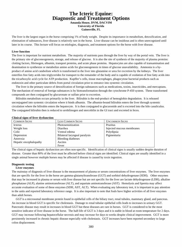
380
The Icteric Equine: Diagnostic and Treatment Options
Amanda House, DVM, DACVIM University of Florida
Gainesville, FL
The liver is the largest organ in the horse comprising 1% of body weight. Despite its importance in metabolism, detoxification, and elimination of substances, liver disease is relatively rare in the horse. Liver disease can be insidious and is often unrecognized until later in its course. This lecture will focus on etiologies, diagnosis, and treatment options for the horse with liver disease. Liver function The liver is important for nutrient metabolism. The majority of nutrients pass through the liver by way of the portal vein. The liver is the primary site of gluconeogenesis, storage, and release of glucose. It is also the site of synthesis of the majority of plasma proteins: clotting factors, fibrinogen, albumin, transport proteins, and acute phase proteins. Hepatocytes are also capable of transamination and deamination to synthesize or metabolize amino acids for gluconeogenesis in times of glucose unavailability. Ammonia is a by-product of amino acid catabolism which is transformed in the liver into glutamine or urea for excretion by the kidneys. The liver esterifies free fatty acids into triglycerides for transport to the remainder of the body and is capable of oxidation of free fatty acids into the tricarboxylic acid cycle for ATP production. Kupffer’s cells, tissue macrophages, phagocytose bacterial products such as endotoxin and other particulate debris from portal circulation prior to entrance into systemic circulation.
The liver is the primary source of detoxification of foreign substances such as medications, toxins, insecticides, and mercaptans. The mechanism of removal of foreign substances is by biotransformation through the cytochrome P-450 system. These transformed compounds are then conjugated by glucuronate or sulfate prior to excretion.
Bilirubin metabolism occurs primarily in the liver. Bilirubin is the end-product of hemoglobin degradation. It is released unconjugated into systemic circulation where it binds albumin. The albumin-bound bilirubin enters the liver through systemic circulation where the bilirubin enters the hepatocyte. It is then conjugated to glucuronide and is excreted into the bile canaliculus. The conjugated bilirubin then is reduced to urobilinogen and stercobilin in the GI tract and excreted in feces.
Clinical signs of liver dysfunction
COMMON SIGNS LESS COMMON SIGNS UNCOMMON SIGNS Icterus Weight loss Colic Depression Anorexia Hepatic encephalopathy
Photosensitization Diarrhea Ventral edema Bilateral laryngeal paralysis Bleeding diatheses Ascites Fever
Pruritus Injected mucous membranes Polydipsia Pigmenturia
The clinical signs of hepatic dysfunction are often non-specific. Identification of clinical signs is usually sudden despite duration of disease. Greater than 80% of the liver must be affected before clinical signs are identified. Clinical signs are usually identified in a single animal however multiple horses may be affected if disease is caused by toxin ingestion. Diagnostic testing
Liver enzymes The mainstay of diagnosis of liver disease is the measurement of plasma or serum concentrations of liver enzymes. The liver enzymes that are specific for the liver in the horse are gamma-glutamyltransferase (GGT) and sorbitol dehydrogenase (SDH). Other enzymes that may be increased in plasma or serum with liver disease but are not specific for the liver are lactate dehydrogenase (LDH), alkaline phosphatase (ALP), alanine aminotransferase (ALT), and aspartate aminotransferase (AST). Hemolysis and lipemia may affect accurate evaluation of some of these enzymes (SDH, AST, ALT). When evaluating any laboratory test, it is important to pay attention to the units and reported laboratory reference range. It is also important to note that foals have higher activities of all liver enzymes than adult horses.
GGT is a microsomal membrane protein found in epithelial cells of the biliary tract, renal tubules, mammary gland, and pancreas. An increase in blood GGT is specific for cholestasis. Damage to renal tubular epithelial cells leads to increases in urinary GGT. Pancreatic disease may result in increases in blood GGT but these diseases are rare in horses. GGT is considered to be the most sensitive indicator of liver disease in the horse. The half-life of GGT is 3 days and it is stable in blood at room temperature for 2 days. GGT may increase following hepatocellular necrosis and may increase for days to weeks despite clinical improvement. GGT is more persistently increased in chronic hepatic disease especially with cholestasis. GGT increases have been reported secondary to large colon displacement.
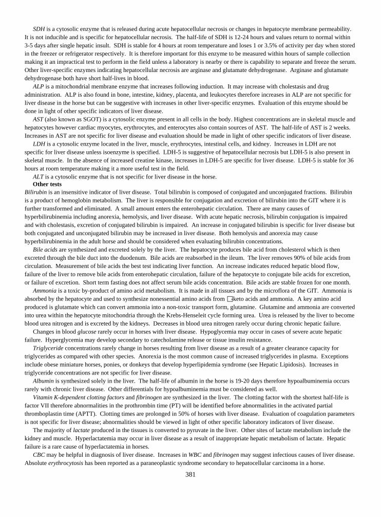
381
SDH is a cytosolic enzyme that is released during acute hepatocellular necrosis or changes in hepatocyte membrane permeability. It is not inducible and is specific for hepatocellular necrosis. The half-life of SDH is 12-24 hours and values return to normal within 3-5 days after single hepatic insult. SDH is stable for 4 hours at room temperature and loses 1 or 3.5% of activity per day when stored in the freezer or refrigerator respectively. It is therefore important for this enzyme to be measured within hours of sample collection making it an impractical test to perform in the field unless a laboratory is nearby or there is capability to separate and freeze the serum. Other liver-specific enzymes indicating hepatocellular necrosis are arginase and glutamate dehydrogenase. Arginase and glutamate dehydrogenase both have short half-lives in blood.
ALP is a mitochondrial membrane enzyme that increases following induction. It may increase with cholestasis and drug administration. ALP is also found in bone, intestine, kidney, placenta, and leukocytes therefore increases in ALP are not specific for liver disease in the horse but can be suggestive with increases in other liver-specific enzymes. Evaluation of this enzyme should be done in light of other specific indicators of liver disease.
AST (also known as SGOT) is a cytosolic enzyme present in all cells in the body. Highest concentrations are in skeletal muscle and hepatocytes however cardiac myocytes, erythrocytes, and enterocytes also contain sources of AST. The half-life of AST is 2 weeks. Increases in AST are not specific for liver disease and evaluation should be made in light of other specific indicators of liver disease.
LDH is a cytosolic enzyme located in the liver, muscle, erythrocytes, intestinal cells, and kidney. Increases in LDH are not specific for liver disease unless isoenzyme is specified. LDH-5 is suggestive of hepatocellular necrosis but LDH-5 is also present in skeletal muscle. In the absence of increased creatine kinase, increases in LDH-5 are specific for liver disease. LDH-5 is stable for 36 hours at room temperature making it a more useful test in the field.
ALT is a cytosolic enzyme that is not specific for liver disease in the horse. Other tests
Bilirubin is an insensitive indicator of liver disease. Total bilirubin is composed of conjugated and unconjugated fractions. Bilirubin is a product of hemoglobin metabolism. The liver is responsible for conjugation and excretion of bilirubin into the GIT where it is further transformed and eliminated. A small amount enters the enterohepatic circulation. There are many causes of hyperbilirubinemia including anorexia, hemolysis, and liver disease. With acute hepatic necrosis, bilirubin conjugation is impaired and with cholestasis, excretion of conjugated bilirubin is impaired. An increase in conjugated bilirubin is specific for liver disease but both conjugated and unconjugated bilirubin may be increased in liver disease. Both hemolysis and anorexia may cause hyperbilirubinemia in the adult horse and should be considered when evaluating bilirubin concentrations.
Bile acids are synthesized and excreted solely by the liver. The hepatocyte produces bile acid from cholesterol which is then excreted through the bile duct into the duodenum. Bile acids are reabsorbed in the ileum. The liver removes 90% of bile acids from circulation. Measurement of bile acids the best test indicating liver function. An increase indicates reduced hepatic blood flow, failure of the liver to remove bile acids from enterohepatic circulation, failure of the hepatocyte to conjugate bile acids for excretion, or failure of excretion. Short term fasting does not affect serum bile acids concentration. Bile acids are stable frozen for one month.
Ammonia is a toxic by-product of amino acid metabolism. It is made in all tissues and by the microflora of the GIT. Ammonia is absorbed by the hepatocyte and used to synthesize nonessential amino acids from -keto acids and ammonia. A key amino acid produced is glutamate which can convert ammonia into a non-toxic transport form, glutamine. Glutamine and ammonia are converted into urea within the hepatocyte mitochondria through the Krebs-Henseleit cycle forming urea. Urea is released by the liver to become blood urea nitrogen and is excreted by the kidneys. Decreases in blood urea nitrogen rarely occur during chronic hepatic failure.
Changes in blood glucose rarely occur in horses with liver disease. Hypoglycemia may occur in cases of severe acute hepatic failure. Hyperglycemia may develop secondary to catecholamine release or tissue insulin resistance.
Triglyceride concentrations rarely change in horses resulting from liver disease as a result of a greater clearance capacity for triglycerides as compared with other species. Anorexia is the most common cause of increased triglycerides in plasma. Exceptions include obese miniature horses, ponies, or donkeys that develop hyperlipidemia syndrome (see Hepatic Lipidosis). Increases in triglyceride concentrations are not specific for liver disease.
Albumin is synthesized solely in the liver. The half-life of albumin in the horse is 19-20 days therefore hypoalbuminemia occurs rarely with chronic liver disease. Other differentials for hypoalbuminemia must be considered as well.
Vitamin K-dependent clotting factors and fibrinogen are synthesized in the liver. The clotting factor with the shortest half-life is factor VII therefore abnormalities in the prothrombin time (PT) will be identified before abnormalities in the activated partial thromboplastin time (APTT). Clotting times are prolonged in 50% of horses with liver disease. Evaluation of coagulation parameters is not specific for liver disease; abnormalities should be viewed in light of other specific laboratory indicators of liver disease.
The majority of lactate produced in the tissues is converted to pyruvate in the liver. Other sites of lactate metabolism include the kidney and muscle. Hyperlactatemia may occur in liver disease as a result of inappropriate hepatic metabolism of lactate. Hepatic failure is a rare cause of hyperlactatemia in horses.
CBC may be helpful in diagnosis of liver disease. Increases in WBC and fibrinogen may suggest infectious causes of liver disease. Absolute erythrocytosis has been reported as a paraneoplastic syndrome secondary to hepatocellular carcinoma in a horse.
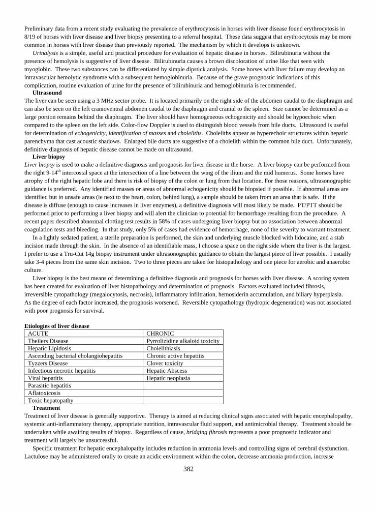
382
Preliminary data from a recent study evaluating the prevalence of erythrocytosis in horses with liver disease found erythrocytosis in 8/19 of horses with liver disease and liver biopsy presenting to a referral hospital. These data suggest that erythrocytosis may be more common in horses with liver disease than previously reported. The mechanism by which it develops is unknown.
Urinalysis is a simple, useful and practical procedure for evaluation of hepatic disease in horses. Bilirubinuria without the presence of hemolysis is suggestive of liver disease. Bilirubinuria causes a brown discoloration of urine like that seen with myoglobin. These two substances can be differentiated by simple dipstick analysis. Some horses with liver failure may develop an intravascular hemolytic syndrome with a subsequent hemoglobinuria. Because of the grave prognostic indications of this complication, routine evaluation of urine for the presence of bilirubinuria and hemoglobinuria is recommended.
Ultrasound The liver can be seen using a 3 MHz sector probe. It is located primarily on the right side of the abdomen caudal to the diaphragm and can also be seen on the left cranioventral abdomen caudal to the diaphragm and cranial to the spleen. Size cannot be determined as a large portion remains behind the diaphragm. The liver should have homogeneous echogenicity and should be hypoechoic when compared to the spleen on the left side. Color-flow Doppler is used to distinguish blood vessels from bile ducts. Ultrasound is useful for determination of echogenicity, identification of masses and choleliths. Choleliths appear as hyperechoic structures within hepatic parenchyma that cast acoustic shadows. Enlarged bile ducts are suggestive of a cholelith within the common bile duct. Unfortunately, definitive diagnosis of hepatic disease cannot be made on ultrasound.
Liver biopsy Liver biopsy is used to make a definitive diagnosis and prognosis for liver disease in the horse. A liver biopsy can be performed from the right 9-14th intercostal space at the intersection of a line between the wing of the ilium and the mid humerus. Some horses have atrophy of the right hepatic lobe and there is risk of biopsy of the colon or lung from that location. For those reasons, ultrasonographic guidance is preferred. Any identified masses or areas of abnormal echogenicity should be biopsied if possible. If abnormal areas are identified but in unsafe areas (ie next to the heart, colon, behind lung), a sample should be taken from an area that is safe. If the disease is diffuse (enough to cause increases in liver enzymes), a definitive diagnosis will most likely be made. PT/PTT should be performed prior to performing a liver biopsy and will alert the clinician to potential for hemorrhage resulting from the procedure. A recent paper described abnormal clotting test results in 58% of cases undergoing liver biopsy but no association between abnormal coagulation tests and bleeding. In that study, only 5% of cases had evidence of hemorrhage, none of the severity to warrant treatment.
In a lightly sedated patient, a sterile preparation is performed, the skin and underlying muscle blocked with lidocaine, and a stab incision made through the skin. In the absence of an identifiable mass, I choose a space on the right side where the liver is the largest. I prefer to use a Tru-Cut 14g biopsy instrument under ultrasonographic guidance to obtain the largest piece of liver possible. I usually take 3-4 pieces from the same skin incision. Two to three pieces are taken for histopathology and one piece for aerobic and anaerobic culture.
Liver biopsy is the best means of determining a definitive diagnosis and prognosis for horses with liver disease. A scoring system has been created for evaluation of liver histopathology and determination of prognosis. Factors evaluated included fibrosis, irreversible cytopathology (megalocytosis, necrosis), inflammatory infiltration, hemosiderin accumulation, and biliary hyperplasia. As the degree of each factor increased, the prognosis worsened. Reversible cytopathology (hydropic degeneration) was not associated with poor prognosis for survival.
Etiologies of liver disease
ACUTE CHRONIC Theilers Disease Pyrrolizidine alkaloid toxicity Hepatic Lipidosis Cholelithiasis Ascending bacterial cholangiohepatitis Chronic active hepatitis Tyzzers Disease Clover toxicity Infectious necrotic hepatitis Hepatic Abscess Viral hepatitis Hepatic neoplasia Parasitic hepatitis Aflatoxicosis Toxic hepatopathy
Treatment Treatment of liver disease is generally supportive. Therapy is aimed at reducing clinical signs associated with hepatic encephalopathy, systemic anti-inflammatory therapy, appropriate nutrition, intravascular fluid support, and antimicrobial therapy. Treatment should be undertaken while awaiting results of biopsy. Regardless of cause, bridging fibrosis represents a poor prognostic indicator and treatment will largely be unsuccessful.
Specific treatment for hepatic encephalopathy includes reduction in ammonia levels and controlling signs of cerebral dysfunction. Lactulose may be administered orally to create an acidic environment within the colon, decrease ammonia production, increase
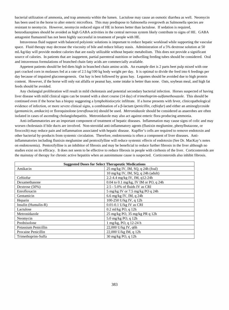
383
bacterial utilization of ammonia, and trap ammonia within the lumen. Lactulose may cause an osmotic diarrhea as well. Neomycin has been used in the horse to alter enteric microflora. This may predispose to Salmonella overgrowth as Salmonella species are resistant to neomycin. However, neomycin reduced signs of HE in horses better than lactulose. If sedation is required, benzodiazepines should be avoided as high GABA activities in the central nervous system likely contribute to signs of HE. GABA antagonist flumazenil has not been highly successful in treatment of people with HE.
Intravenous fluid support with balanced polyionic solutions is important to reduce hepatic workload while supporting the vascular space. Fluid therapy may decrease the viscosity of bile and reduce biliary stasis. Administration of a 5% dextrose solution at 50 mL/kg/day will provide modest calories that are easily utilizable without hepatic metabolism. This does not provide a significant source of calories. In patients that are inappetent, partial parenteral nutrition or indwelling feeding tubes should be considered. Oral and intravenous formulations of branched chain fatty acids are commercially available.
Appetent patients should be fed diets high in branched chain amino acids. An example diet is 2 parts beet pulp mixed with one part cracked corn in molasses fed at a rate of 2.5 kg/100 kg body weight per day. It is optimal to divide the feed into 6 feedings per day because of impaired gluconeogenesis. Oat hay is best followed by grass hay. Legumes should be avoided due to high protein content. However, if the horse will only eat alfalfa or peanut hay, some intake is better than none. Oats, soybean meal, and high fat feeds should be avoided.
Any cholangial proliferation will result in mild cholestasis and potential secondary bacterial infection. Horses suspected of having liver disease with mild clinical signs can be treated with a short course (14 day) of trimethoprim-sulfamethoxazole. This should be continued even if the horse has a biopsy suggesting a lymphohistiocytic infiltrate. If a horse presents with fever, clinicopathological evidence of infection, or more severe clinical signs, a combination of a β-lactam (penicillin, ceftiofur) and either an aminoglycoside (gentamicin, amikacin) or floroquinolone (enrofloxacin) should be used. Metronidazole should be considered as anaerobes are often isolated in cases of ascending cholangiohepatitis. Metronidazole may also act against enteric flora producing ammonia.
Anti-inflammatories are an important component of treatment of hepatic diseases. Inflammation may cause signs of colic and may worsen cholestasis if bile ducts are involved. Non-steroidal anti-inflammatory agents (flunixin meglumine, phenylbutazone, or firocoxib) may reduce pain and inflammation associated with hepatic disease. Kupffer’s cells are required to remove endotoxin and other bacterial by-products from systemic circulation. Therefore, endotoxemia is often a component of liver diseases. Anti-inflammatories including flunixin meglumine and pentoxifylline will reduce systemic effects of endotoxin (See Dr. MacKay’s notes on endotoxemia). Pentoxifylline is an inhibitor of fibrosis and may be beneficial to reduce further fibrosis in the liver although no studies exist on its efficacy. It does not seem to be effective to reduce fibrosis in people with cirrhosis of the liver. Corticosteroids are the mainstay of therapy for chronic active hepatitis when an autoimmune cause is suspected. Corticosteroids also inhibit fibrosis.
Suggested Doses for Select Therapeutic Medications Amikacin 25 mg/kg IV, IM, SQ, q 24h (foal) 10 mg/kg IV, IM, SQ, q 24h (adult) Ceftiofur 2.2-4.4 mg/kg IV, IM, q12-24h Dexamethasone 0.04 to 0.1 mg/kg, IV IM or PO, q 24h Dextrose (50%) 2.5 - 5.0% of fluids IV as CRI Enrofloxacin 5 mg/kg IV or 7.5 mg/kg PO q 24h Gentamicin 6.6 mg/kg IV, IM, q 24h Heparin 100-250 U/kg IV, q 12h Insulin (Humulin-R) 0.01-0.1 U/kg IV as CRI Lactulose 0.2 ml/kg PO, q 12h Metronidazole 25 mg/kg PO, 35 mg/kg PR q 12h Neomycin 5.0 mg/kg PO, q 12h Prednisolone 1 mg/kg, PO, q 12-24 h Potassium Penicillin 22,000 U/kg IV, q6h Procaine Penicillin 22,000 U/kg IM, q 12h Trimethoprim-Sulfa 30 mg/kg PO, q 12h
384
Emerging Equine Infectious Diseases Amanda House, DVM, DACVIM
University of Florida Gainesville, FL
Equine infectious diseases continue to emerge and re-emerge, infecting horses across the US and beyond. Infectious disease identification and treatment remain a cornerstone of equine practice. This lecture will discuss infectious disease updates and will review pigeon fever, piroplasmosis, MRSA, and others. The clinical syndromes, diagnostic techniques, and therapy will be included. Equine piroplasmosis Piroplasmosis is caused by the protozoan parasites Babesia caballi and Theileria equi (formerly called Babesia equi). It also can affect donkeys, mules, and zebras; but clinical disease in those equids is rare. The disease is transmitted by ticks and other biting insects; however, shared needles and/or blood contamination has been implicated in several disease outbreaks. Once horses are infected with T. equi, carrier status may be lifelong. Carrier horses are also capable of transmitting the disease to ticks—vectors that can transmit it to other horses. The disease is considered endemic in Africa, Central and South America, Asia, the Middle East, the Caribbean, and the Mediterranean. The U.S. has not been considered an endemic region. When infection occurs, T. equi tends to be the most common agent, rather than B. caballi. However, infection with both parasites can occur simultaneously.
Once horses become infected with the parasite, it usually takes between 5 and 30 days for any signs of the disease to appear. As previously stated, infected horses may not have any signs of EP at all. Generally, affected horses display nonspecific signs that can look similar to other diseases. Fever, depression, anorexia, pale or icteric mucous membranes, and edema of the limbs or along the ventral abdomen have been commonly reported. Reddish-brown or discolored urine may also be observed. Laboratory abnormalities typically include anemia and thrombocytopenia.
Several laboratory tests are available for diagnosis of EP. Occasionally, the parasite can be seen on microscopic examination of a blood smear. The U.S. Department of Agriculture (USDA) standard test is the cELISA (competitive enzyme-linked immunosorbent assay). Specific laboratories (the National Veterinary Services Laboratories, Texas Veterinary Diagnostic Services Laboratories, Florida’s State Diagnostic Laboratory) have been identified to run the tests and report the results. The National Veterinary Services Laboratories is still testing all international transport samples.
Horses that test positive for equine piroplasmosis MUST be quarantined. Local veterinarians can work with state and federal veterinarians to ensure that manageable quarantine guidelines are being followed and are in place. Although there are several drugs (imidocarb, etc) that have been identified for treatment of piroplasmosis, the organisms can be refractory to treatment, and the carrier state is difficult to clear. Euthanasia for positive horses is not required, nor is it being recommended in every case by the USDA, especially since so many positive horses are asymptomatic. State and USDA veterinarians are working in conjunction with local veterinarians and owners to determine the best recommendations for each positive horse. Some owners elect to transport positive horses out of the country—to countries that have endemic piroplasmosis—but that is not a palatable option for most. In addition to quarantine, there is a treatment research program available for positive horses. This program is in conjunction with Washington State University and Dr. Don Knowles. Owners and their local veterinarians work with the USDA and Dr. Knowles to determine if they have a horse that is eligible for enrollment. Recent published research out of that program is showing promise for clearing infection in horses treated with imidocarb.
Fortunately, it does not appear that tick transmission has been significantly involved in EP transmission outside of the affected premises in Texas. However, people can spread this disease from horse to horse, and we can prevent that mode of transmission. All dental, surgical, and tattoo equipment must be thoroughly disinfected between horses. Horses have contracted the disease though the use of shared needles and/or syringes, as well as from blood transfusions. A new sterile needle and syringe should be used for each injection, whether into a muscle or a vein. Additionally, a previously used needle should never be inserted into a drug or vaccine multidose vial—and owners/trainers should be reminded of these infection control measures. Work with your veterinarian to ensure that all equipment is thoroughly cleaned and disinfected between horses. EP is still a very uncommon disease in the U.S., but it is critical to be vigilant and follow preventative measures.
Pigeon fever This disease is caused by Corynebacterium pseudotuberculosis, which is a gram positive rod shaped bacteria. Horses and small ruminants typically get different strains of the infection, but cattle can get both types. In goats, the disease is known as caseous lymphadenitis, and affected animals will have external abscesses (the head, behind the ears, on the neck, shoulder or flank are some typical locations). Abscesses also occur in horses and cattle infected with this bacteria. Natural transmission from horses to goats or vice versa is not thought to commonly occur. Corynebacterium pseudotuberculosis is a soil organism that can survive for months to even years in direct sun. The largest numbers of cases are typically reported in the dry months of fall and winter.
385
Many things about this disease in horses are still not completely understood, such as the incubation period. The incubation period is the time it takes to develop clinical symptoms of the disease after being infected with the bacteria, and can be variable…. from weeks to even months. The bacteria can enter the horse through the skin, wounds, or abrasions in the mucous membranes. Horses with pigeon fever may have a poor appetite, fever, lethargy, swelling along the chest or ventral abdomen, and/or lameness. Three forms of the disease can occur in the horse: external abscesses, internal abscesses, and ulcerative lymphangitis. The most common form of pigeon fever is the development of external abscesses. These occur in about 90% of the cases. The disease got its name because abscesses will commonly occur in the pectoral region which becomes swollen and painful. In addition to the pectorals, abscesses may form on the prepuce, mammary gland, axilla, limbs, inguinal region, head, and other areas. There is no breed or sex predisposition for acquiring the infection, although young horses may have some increased risk. The second form of the disease is internal abscessation, which has been reported in about 8% of cases. The most common site of internal abscesses is the liver, although they can be associated with other organs as well. The third form, ulcerative lymphangitis, is a severe cellulitis that occurs in the fewest number of cases. Clinical cases of ulcerative lymphangitis have severe lameness and swelling of the limb.
Definitive diagnosis of pigeon fever is made by culturing the bacteria from an abscess or draining wound. There is a blood test available (called the synergistic hemolysis inhibition test) but the results depend on the severity and length of infection. This means that a negative blood test (titer) does not rule out the disease. In fact, early in the disease horses may have a negative blood test. The blood test is helpful in horses with internal abscesses, as the titers are typically very high (>1:512). Ultrasound examination may be a helpful diagnostic tool in these cases as well, especially for identifying internal abscesses in the abdominal cavity.
Treatment of pigeon fever is accomplished with drainage of external abscesses. The primary veterinarian should always be consulted about treatment. Abscesses should be allowed to mature and then drained. They should be flushed with antiseptic solutions. Purulent material drained from abscesses is highly infectious and must be carefully handled and disposed of. Collecting as much purulent material as possible into a waste bag for disposal is critical to reduce the risk of other horses being exposed. Bedding of infected horses should be properly disposed of as well. Pain medication may be indicated for horses with severe or deep abscesses or lameness. Topical fly treatment around wounds and draining areas is critical to reduce the possibility of biting insects transmitting the infection. Systemic antibiotics may be utilized for treatment on a case by case basis. In routine cases with external abscesses, antibiotics may prolong the course of the disease and are typically not required. However, antibiotics are appropriate in cases with severe disease or reoccurrence of infection. Long term systemic antibiotics are required for treatment of horses with internal abscesses. Fortunately, Corynebacterium pseudotuberculosis is usually sensitive to most antibiotics (including penicillin), but culture and sensitivity of a sample of purulent material is recommended to direct therapy.
A conditionally licensed vaccine was released and then removed from the veterinary market this past spring. It is recommended to isolate infected animals, especially if draining wounds/abscesses are present. Stalling affected horses will help reduce contamination of the pasture environment with infectious material. Horses should be treated in an area ideally with concrete or rubber flooring that can be disinfected. Although no reports exist of humans being infected from horses, there are reports of humans being infected with the sheep strain of the disease. Infection in people has occurred from the consumption of infected unpasteurized milk or milk products, close contact with infected animals, handling contaminated equipment, or exposure of wounds with infected material. Therefore, wearing gloves when handling infected horses is recommended. Fly sprays and feed through fly control may both be beneficial for insect control. If you suspect your horse is exhibiting signs of pigeon fever, contact your veterinarian for a thorough examination.
MRSA The prevalence of Methicillin-resistant Staphylococcus aureus (MRSA) in routine infections in people and hospital outbreaks has initiated world-wide concern. Staphylococcus aureus is a common bacterium that colonizes the skin and has been found to cause disease in many species. Staphylococcus aureus began developing resistance to antibiotics almost as soon as they were introduced, beginning with penicillin, and progressing to methicillin resistance. MRSA is typically resistant to all beta-lactam antibiotics (penicillin and cephalosporin families) and often many other antibiotics as well. This antimicrobial resistance can make MRSA infections a challenge to treat. The percentage of community-associated infection (milder, outpatient type illness like skin infections/abscesses) from MRSA had risen from 35.1% in 2003 to 50.0% in 2006 in Florida.
Methicillin-resistant Staph aureus infections in the horse have manifested as wound and surgical site infections, cellulitis (soft tissue infections, typically of the limb), catheter-site infections, pneumonia, septic arthritis, and skin infections, among others. Historically, equine MRSA infections were uncommonly reported, and began to increase in prevalence in the late 1990s. More recent studies have demonstrated that MRSA is an important emerging pathogen in horses and can be zoonotic. It is also possible for humans to transmit the bacteria to horses as well. Approximately 25% of healthy children and adults can carry the Staphylococcus aureus bacteria in their nose or on the skin. For most people and horses, carrying the bacteria in the nose or on the skin causes no ill-effects. Certain circumstances such as a wound or an illness requiring hospitalization can result in active infection.
386
Studies done in horses have found that approximately 0-5% of horses carry the MRSA bacteria in their nasal passages, which is the most common site for colonization (Weese et al, 2005). Horses can also have the bacteria on their skin or in their intestinal tract. Although generally very few horses carry the bacteria, some farms with a history of MRSA infections in horses have demonstrated carrier rates of 0-45% (Weese et al, 2005). Horses that carry the bacteria in their nasal passages may not ever develop a clinical MRSA infection. However, these horses may transmit MRSA to other horses or people, and will sometimes develop active infections under certain conditions. People who work with horses seem to have a higher carrier rate of MRSA. Studies of equine veterinarians have reported colonization rates ranging from 10-14%, with predominantly the equine strain of the bacterium. This provides further evidence that carrier horses can transmit MRSA to humans.
Clinical infection with MRSA is certainly concerning. Fortunately, a multicenter study reported that 83% of horses with MRSA infections survived (Anderson et al, 2009). Many horses with clinical infections such as pneumonia or wound infections required prolonged hospital stays and needed additional surgeries. However, acceptable antibiotic options exist in most cases, especially when infection is identified early. In this author’s experience, catheter site infections with MRSA, while uncommon, result in the highest mortality rates if the infection spreads through the bloodstream and into the lungs or other sites. Early detection and treatment is certainly critical for the best outcome for the horse. Currently, there is no evidence that horses that carry MRSA need to be treated with antibiotics. Farms with documented MRSA prevalence have successfully eradicated MRSA with good hygiene and infection control practices.
Further references 1.http://www.aphis.usda.gov/animal_health/animal_diseases/piroplasmosis/downloads/ep_protect_your_horses_en_sp.pdf 2. http://www.aphis.usda.gov/publications/animal_health/content/printable_version/fs_equine_piro.pdf 3. http://www.doacs.state.fl.us/ai/index.shtml 4. Anderson et al. Retrospective multicenter study of MRSA infections in 115 horses. Equine Vet J; 2009, Nov; 41(4): 401-405. 5. Weese et al. Community-associated MRSA in horses and humans who work with horses. JAVMA, 2005; Feb 15; 226(4): 580-583. 6. http://www.freshfromflorida.com/ai/pdf/WebsiteAnnouncement-PigeonFever.pdf 7. Aleman MR and Spier SJ. Corynebacterium pseudotuberculosis infection. In Large Animal Internal Medicine, 3rd Ed. Smith BP, 2002, Mosby, Inc, St. Louis, MO; pp1078-1083.
387
Practical Approach to Coughing Horses Amanda House, DVM, DACVIM
University of Florida Gainesville, FL
Cough and poor performance are common complaints among pleasure and sport horses in equine practice. This lecture will discuss a practical diagnostic approach to the horse with a cough, and treatment of non-infectious lower respiratory disease. History and examination A complete history and physical examination are critical for evaluating the patient with cough. Duration, severity, nasal discharge, presence or absence of fever, and inciting causes can all be helpful in determining the underlying etiology. A rebreathing examination should be performed as part of a complete respiratory examination. Horses with inflammatory, non-infectious lower airway disease may have a prolonged recovery and the presence of crackles and/or wheezes on examination. Horses with infectious pneumonia may have abnormal adventitial sounds as well, and as such, this examination will not point to a specific etiology. A complete blood cell count may be beneficial to determine whether evidence of infection is present, but can be normal in horses with pneumonia. Changes in the white blood cell count or fibrinogen concentration should prompt further diagnostic consideration.
Horses with a history of fever and changes on the CBC should be suspected of having infectious lower airway disease/pneumonia. Definitive determination of a bacterial etiology is best accomplished with a percutaneous trans-tracheal aspiration sample submitted for cytology and culture/sensitivity. Pneumonia is often characterized cytologically with degenerative neutrophils (with or without the presence of intracellular bacteria). Broad spectrum antibiotics should be initiated if cytology is suggestive of pneumonia, while culture results are pending. Tracheal aspiration cytology can also be suggestive of inflammatory lower airway disease, when non-degenerative neutrophils are the predominant cellular population. Thoracic ultrasound examination and radiographs can be helpful in the evaluation of the horse with lower airway disease. Practically speaking in a field setting, ultrasound examination of the thorax can be accomplished with a trans-rectal probe. This technique is most useful to determine if pleural fluid or pulmonary infiltrates are present. RAO (heaves, equine asthma) Recurrent airway obstruction (also known as heaves, equine asthma, broken wind, and chronic airway reactivity) is a common respiratory disease of horses characterized by periods of reversible airway obstruction caused by neutrophil accumulation, mucus production, and bronchospasm. The classic clinical syndrome includes chronic cough, nasal discharge and respiratory difficulty. The term COPD is no longer used to describe this condition in horses, because the pathophysiologic and morphologic aspects of the disease are different from human chronic obstructive pulmonary disease.
Most evidence suggests that RAO is the result of pulmonary hypersensitivity to inhaled antigens, although multiple theories exist regarding the exact pathophysiology. The most common antigens are mold, organic dust, and endotoxin present in hay and straw. Periods of reversible small airway obstruction are caused by bronchoconstriction and accumulation of mucus and neutrophils. RAO occurs worldwide, with the highest prevalence in stabled horses fed hay in the northeastern and midwestern United States. A similar condition that can occur in horses in the southeastern United States is termed summer pasture associated obstructive disease (SPAOD). However, horses with SPAOD typically improve when stabled. RAO is a common respiratory disease of mature horses (typically > 7 years old). The average age of onset in RAO affected horses is 9-12 years, and both genders are commonly affected. Winter and spring appear to be the most common seasons for exacerbation of RAO. There does appear to be a heritable component to the etiology of this condition. The incidence of RAO in horses with healthy parents is approximately 10%, which increases to 44% if two parents are affected. Clinical signs Clinical signs of RAO typically include a chronic cough, nasal discharge and a prolonged labored phase of expiration. The classic “heave line” is due to hypertrophy of the abdominal muscles which are assisting in respiration. Flared nostrils and tachypnea are frequently observed. On thoracic auscultation, wheezes, tracheal rattles, and over-expanded lung fields may be present. Crackles may also be heard secondary to excessive mucus production in the lower airways. Severe cases may also exhibit weight loss, cachexia, and exercise intolerance. Horses are typically afebrile with normal complete blood cell count and serum biochemical profile, unless a secondary bacterial pneumonia has occurred. Diagnostic evaluation Diagnosis of RAO can be done on the basis of history and characteristic clinical examination findings in the majority of horses. Additional diagnostics to confirm and characterize the pulmonary inflammation include transtracheal aspiration (TTA), bronchoalveolar lavage (BAL), thoracic radiographs and ultrasound examination.
388
Transtracheal aspiration can be done to characterize inflammation in the lower airways and to determine if sepsis is present. The presence of degenerative neutrophils and intracellular bacterial organisms suggests sepsis, and warrants culture as well as appropriate antimicrobial therapy. Typical RAO cases have no evidence of sepsis, and TTA results are consistent with mucopurulent (neutrophilic) inflammation.
Bronchoalveolar lavage is indicated in horses with poor performance and coughing, and is not compulsory in horses with severe disease and suggestive clinical signs. Neutrophilic inflammation (with 20-70% of neutophils in total cell count, normal neutrophil count is <5-10%) confirms the presence of lower airway inflammation and is suggestive of RAO. Curschmann’s spirals may be present on cytologic evaluation of TTA and BAL samples, and represent inspissated mucus plugs from the obstructed small airways.
Thoracic radiographs will often demonstrate an increased broncho-interstitial pattern throughout the lung fields. These changes may be difficult to differentiate from normal ageing changes in older adults. Radiographs are recommended for horses that fail to respond to standard therapy, or to further characterize pulmonary inflammation. Horses that have more respiratory difficulty on inspiration rather than expiration may have interstitial pneumonia or pulmonary fibrosis, and radiographs are indicated to better characterize lung disease in these cases. Ultrasound may be utilized if primary or secondary infectious pulmonary disease is suspected.
Lung function tests in horses with RAO typically demonstrate hypoxemia without hypercarbia, due to V/Q mismatch. Function abnormalities will often include high resistance and poor compliance of the lungs, with increased dead space ventilation. Lung function testing is not widely available, but can be performed at The University of Florida. It is beneficial in more subtle cases of poor performance and inflammatory airway disease (IAD). Treatment The most important treatment for RAO is environmental management to reduce exposure to organic dusts and mold. As previously mentioned, the most common antigens are organic dusts, mold, and endotoxin present in hay and straw. Round bale hay is high in endotoxin and organic dust content, and the presence of round bale hay is a potential cause of treatment failure in horses on pasture. Maintaining horses on pasture full-time is generally recommended. Horses that must be stalled should be kept in a clean, well-ventilated environment and ideally be transitioned to a complete pelleted feed. Straw is not recommended as bedding for RAO affected horses. Soaking hay (for at least 2 hours) and feed may alleviate the signs in mildly affected individuals, however, soaked hay may still exacerbate RAO in more severely affected cases. It is important to remember that although medications will alleviate the clinical signs of disease, respiratory disease will return if the horse remains in a mold/dust-filled environment once the medications are discontinued.
Systemic corticosteroids and aerosolized bronchodilators are the most immediately helpful therapy for a horse in respiratory distress. Intravenous administration of Dexamethasone (.1 mg/kg) should improve lung function within 2 hours of administration. Dexamethasone has an oral bioavailability of about 60%, and will improve pulmonary function within 6 hours if given by this route. Dexamethasone may be continued for one to several weeks at a tapering dose (usually quarter the dose every 3-5 days) for severe cases. Isoflupredone acetate (.03 mg/kg IM every 24h for 14 days) and a single dose of triamcinolone acetonide (0.05-0.09 mg/kg IM) have also been utilized in horses with significant respiratory difficulty and acute airway obstruction. For management of less severely affected cases of RAO, prednisolone is generally considered to be less potent and less toxic than the previously mentioned drugs. Prednisolone should be administered at 1-2.2 mg/kg orally once daily for a week and then gradually tapered. Oral prednisone is poorly bioavailable, and not recommended for treatment of RAO in horses.
Corticosteriods will not provide immediate relief of acute, severe airway obstruction, and rapidly acting β2-adrenergic bronchodilators (such as albuterol sulfate) are indicated for treatment in those cases. Aerosolized albuterol sulfate (.8-2 µg/kg in MDI, metered-dose inhaler; typically 5-10 puffs/500kg of 100 µg/puff MDI every 4-6h) improves pulmonary function by 70% within 5 minutes of administration; however, the beneficial effects last only 1-3 hours. Administration of albuterol will improve the pulmonary distribution of other aerosolized medications, such as aerosolized corticosteroids, and speed mucociliary clearance. Salmeterol (210 µg dose, 1-3 times daily) may also be used as an inhaled bronchodilator. Ipratroprium bromide (20 µg/puff, 5-10 puffs/500 kg every 6-8 hours) has also been utilized for inhaled bronchodilator therapy.
Clenbuterol (.8-3.2 mcg/kg PO every 12 h), a β2-adrenergic agonist, provides long-acting bronchodilation in horses with moderate to severe RAO. Side effects include tachycardia and sweating, which are more common at higher doses and with intravenous administration. The clinical efficacy of clenbuterol is inconsistent at lower dosages if exposure to a dusty environment is maintained. However, it does appear to improve the objective parameters of pulmonary function. Down-regulation of β2-receptors has been documented in horses after administration of clenbuterol for 12 days (.8 µg/kg IV, every 12h). Administration of corticosteroids prevents clenbuterol-induced desensitization when administered concurrently. Since β2-agonists have minimal to no anti-inflammatory activity, they should not generally be used alone for the treatment of RAO.
Systemic anti-cholinergic drugs are not recommended for long-term management of RAO due to their potentially severe side effects (CNS toxicity, ileus, mydriasis, tachycardia, etc). Atropine (5-7 mg IV/450 kg) can be administered as a rescue medication
389
during a severe airway obstruction episode. Ipratroprium bromide is a synthetic anti-cholinergic drug that is administered via inhalation and produces bronchodilation, inhibits cough, and protects against bronchoconstrictive stimuli. Its duration of effect is 4-6 hours and it is administered at 90-180 µg/horse in an MDI (AeroHippus and the Equine Haler).
Aerosolized corticosteroids are effective in horses with mild-moderate RAO, and can be used in conjunction with systemic therapy in severe cases. The two aerosolized preparations for administration to horses via the Equine Haler™ are beclomethasone diproprionate (3500 µg/horse every 12h via MDI) and fluticasone propionate (2000 µg/horse every 12 hours via MDI; typically 8-10 puffs/500 kg of a 220 µg/puff MDI twice daily). Fluticasone is the most potent and most expensive inhaled corticosteroid, and due to its low oral bioavailability, it has the least potential for adrenal suppression. In affected horses, fluticasone proprionate reduces pulmonary neutrophilia, improves parameters of pulmonary function, reduces responsiveness to histamine challenge, and speeds clinical recovery. Although the therapeutic effect is not immediate, pulmonary function typically begins to improve within 24 hours after administration of aerosolized corticosteroids. Additionally, horses in apparent “remission” from RAO may benefit from low dose, long term, aerosolized corticosteroid treatment. References 1. Rush B. ACVIM Forum Proceedings 2006, pp 177-182. 2. Ainsworth DM, Hackett RP. Equine Internal Medicine, 2004, pp 333-336. 3. Lavoie, JP. Current Therapy in Equine Medicine 5, 2003, pp 417-421. 4. Couteil L et al. J Am Vet Med Assoc. 2003; 223 (11): 1645. 5. Couteil L et al. Am J Vet Res. 2005; 66 (10):1665. 6. Abraham G et al. Equine Vet J, 2003; 34 (6):587. 7. http://www.aeromask.com 8. http://equinehaler.com 9. http://www.trudellmed.com/animal-health/aerohippus

390
Strategic Parasite Control: A Reality?
Amanda House, DVM, DACVIM University of Florida
Gainesville, FL
Strategic parasite control through targeted deworming programs based on fecal egg count testing is currently the recommended approach for equine parasite management. This lecture will review equine parasites and the use of a basic targeted deworming program. This lecture will be based on the AAEP’s Parasite Control Guidelines, which can be accessed free online at: http://www.aaep.org/custdocs/ParasiteControlGuidelinesFinal.pdf

391
Foal Diarrhea: What Next?
Amanda House, DVM, DACVIM University of Florida
Gainesville, FL
Foal diarrhea can be very concerning for owners as well as a diagnostic challenge for veterinarians. This lecture will review the most common etiologies, diagnostic, and treatment options for diarrhea in the foal. This lecture is based on the publication co-authored by the speaker: Mallicote M, House AM, and Sanchez C (2012). A Review of Foal Diarrhea from Birth to Weaning. Equine Veterinary Education, April 2012, 24(4): 206–214. doi: 10.1111/j.2042-3292.2011.00358.x The article will be included in the proceedings pending copyright permissions.
392
Hemorrhage in Field Situations Amelia Munsterman, DVM, MS, DACVS, DACVECC
Auburn University Auburn, AL
Severe, acute hemorrhage can be a life-threatening situation that requires immediate. The practitioner must first identify the source of bleeding, arrest further blood loss if possible, and ensure adequate tissue perfusion. While the causes of acute bleeding are numerous, treatment can be focused with this step-wise plan for resuscitation. Pathophysiology of hemorrhagic shock Hemorrhagic shock is technically a subset of hypovolemic shock, defined separately from hypovolemia by a loss of oxygen carrying capacity as a result of loss of red blood cells. As blood volume decreases due to the loss of both plasma and cells, cardiac output and total oxygen delivery to the tissues decreases. In response, cardiac output is increased by autonomic responses that result in tachycardia, vasoconstriction, improved cardiac contractility, and redistribution of blood flow to the vital organs. In time, the oxyhemoglobin dissociation curve is shifted to the right, and tissue oxygen extraction increases from the red blood cells that remain. Severe or sustained blood losses eventually overwhelm these compensatory mechanisms and the body can no longer meet the needs of systemic oxygen demand. This stage of hemorrhagic shock is called the critical total oxygen delivery point, and is consistent whether it is caused by anemia, hypoxemia or hypovolemia.1
Clinical signs of dysoxia are related to the organ systems affected and the stage of hemorrhagic shock.2 The respiratory, hepatic and gastrointestinal systems are the first organs affected, due to redistribution of the circulating blood to the heart, brain and kidneys. For the respiratory system, clinical manifestations of lung injury can include tachypnea and signs of pulmonary edema (dyspnea, frothy mucous from the lower airways), due to intrapulmonary shunting and reduced compliance. Hepatic injury may be identified by elevations of both bilirubin and alkaline phosphatase on serum chemistries. In the gastrointestinal tract, ischemia is characterized by signs of abdominal pain. Compromise of blood flow resulting in mucosal ischemia may allow for ulceration, bacterial translocation to the venous circulation and, ultimately, sepsis.
As the compensatory mechanisms for hemorrhagic shock begin to fail subsequent to continued blood loss, the function of the brain, heart and kidneys begins to deteriorate. Clinical signs of hypoxia affecting the central nervous system are seen initially as agitation, followed by a lack of response to simple commands, depression, coma and death. While the cardiovascular system may attempt compensation early in the course of hemorrhage by tachycardia and increasing stroke volume, continued hemorrhage results in hypoperfusion of the myocardium, causing cardiac ischemia and eventually cardiac failure. Cardiac murmurs and arrhythmias may be noted in the late stages of hypovolemic shock. Activation of the renin-angiotensin-aldosterone system by the kidneys provides some support to the cardiovascular system early on. Urine specific gravity is initially increased, but as renal perfusion decreases, oliguria is noted as compensation fails. The goals for treatment of acute hemorrhage are limit further organ damage and prevent multi-organ failure. The method of treatment is to restore an adequate circulating volume and oxygen carrying capacity with fluids, plus or minus cells.
Treatment of hemorrhagic shock The first step in treatment of acute blood loss is to classify the type of hemorrhage involved. Hemorrhage is defined as controlled if it is caused by blood loss from a vessel or organ that can be stopped using physical means. In situations of controlled hemorrhage, the solution is fairly straightforward in the form of applied pressure, bandages, or ligation of the bleeding vessel. In contrast, the source of bleeding in uncontrolled hemorrhage is either difficult to access, or so extensive that continued blood loss is expected. If bleeding is internal, inaccessible, or the source cannot be identified, mitigation is the key.
Medications that have been evaluated in horses to inhibit uncontrolled hemorrhage are aminocaproic acid, formalin, and yunnan baiyao. Aminocaproic acid, a lysine derivative, may reduce clot disruption through inhibition of fibrinolysis. In horses, administration can result in a reduction in partial thromboplastin time (PTT), which is the measure of the efficacy of the intrinsic and common coagulation pathways.3 Due to its short half-life, this medication is currently recommended as a constant rate infusion, and has been shown to provide adequate circulating plasma levels using 3.5 mg/kg/min for 15 minutes, followed by 0.25 mg/kg/min.4 This dose was based on efficacy of this medication in humans, and recent research in horses has identified that this dose may be twenty times too high, based on the sensitivity of the equine clotting system to this medication.5 Further evaluation of the efficacy of this dose in a live animal model will be required, but its use should be cautioned if an underlying coagulopathy is suspected. In a field situation, a bolus dose of 30 mg/kg of aminocaproic acid may provide some benefit if a CRI is not possible. An alternative anti-fibrinolytic medication, called tranexamic acid, may be substituted for aminocaproic acid, and has been recommended by one manufacturer at an anecdotal dose of 5-25 mg/kg IV.6
Intravenous formalin has also been assessed in the horse to mitigate hemorrhage, due to its ability to activate platelets and enhance primary hemostasis when tested in other species.7 A low dose (0.37% formaldehyde; 37ml of 10% buffered formalin in 1000 mL
393
balanced electrolyte solution) had no negative effects on bleeding times, PTT, activated partial thromboplastin time (APTT), fibrin degradation products, or activated clotting times in the horse. However, higher doses of formalin (0.74% formaldehyde) resulted in numerous side effects including muscle fasciculations, agitation, tachycardia, and tachypnea.
Finally, the Chinese hemostatic medication Yunnan baiyao (or paiyao), purported to contain a mixture of progesterones, saponins, and alkaloids, has been anecdotally documented to mitigate hemorrhage. The mechanism of action is unknown, but a study in ponies showed an prolongation of template bleeding time, which may indicate stimulation of platelet activation.8 The dosage for Yunnan baiyao is 8 mg/kg 2-4 times daily, per os.9 In all of these studies, the effects of these medications on hemorrhage were not directly assessed, only their effects on measures of clotting times and activation of the clotting cascade in the normal horse. At this time, aminocaproic acid would be the recommended medication for uncontrolled hemorrhage in the horse based on its safety documented in these experimental models.
Once hemorrhage has stopped, or attempts at reducing further blood loss have been made, fluid resuscitation should be instituted to improve tissue perfusion. There are important considerations for fluid therapy based on whether the bleed is classified as controlled or uncontrolled hemorrhage.10 For horses where blood loss has been controlled, the goal of resuscitation is to restore the circulating blood volume to a level that normalizes arterial blood pressure and cardiac output. Objective measures of adequate fluid resuscitation in controlled hemorrhage include normalization of blood pH (7.35-7.4), blood lactate (<2.0 mmol/L), central venous pressure (7.5-12 mmHg), and urine output. To monitor resuscitation clinically, the practitioner should observe the horse for urine production and an improvement in peripheral pulses, temperature of the extremities, and mentation.
Fluid choices include crystalloids (Normasol R, Plasmalyte 148, Plasmalyte A, lactated Ringer’s), colloids (Hetastarch) and 7.5% hypertonic saline. Crystalloids are preferred over colloids and hypertonic saline due to the fact that crystalloids can restore interstitial losses. Approximately 60-75% of the administered volume redistributes to the interstitium within 60 minutes of infusion, leaving approximately 30% in the vasculature. While a potential advantage, it is important to remember that approximately 3 liters of crystalloids will be required to replace each liter of lost blood. Colloids provide an advantage over crystalloids in that they can remain in the vascular space for up to 3 days, and can more rapidly improve cardiac output. Side effects of colloids include the possibility of coagulopathies at high doses (for Hetastarch, >10 ml/kg/day) due to dilution of clotting factors, and the inhibition of clotting factor 8 and von Willibrand’s factor. In addition, colloids must be followed immediately by crystalloids administration to replace interstitial losses. Alternatively, hypertonic saline can be used in controlled hemorrhage to rapidly expand the extracellular fluid volume. While hypertonic has anti-inflammatory, anti-oxidant and anti-apoptotic effects, its effects on arterial pressures are short lived (<45 minutes). Hypertonic saline must be followed by crystalloid replacement to maintain the circulating volume. The dose for hypertonic saline is 2-4 ml/kg (approximately 1-2 liters per 1000 lbs. or 500 kg.). After restoring the intravascular fluid volume with fluid administration, blood products can be administered, if needed, to ensure a circulating hematocrit >12% and a platelet count of greater than 50,000/uL. Clinically, if the horse is still showing signs of tissue hypoxia (lactic acidosis, signs of colic), a transfusion may be required. It is important to remember that cardiac output cannot be significantly increased with blood products alone, due to their viscosity. Therefore crystalloids are still required for the initial volume resuscitation, and may prolong or prevent the need for a transfusion.
In uncontrolled hemorrhage where blood loss cannot be controlled, fluid therapy should be provided to maintain only the minimum circulating volume that will support the vital organs. This treatment, termed permissive hypotensive resuscitation, supports an adequate circulating volume to reduce further organ damage by hypoxemia, but prevents disruption of fragile clots or excessive dilution of clotting factors that could increase blood loss.11 Initial therapy in uncontrolled hemorrhage should be provided by administration of crystalloids at a maintenance fluid rate (2.5 ml/kg/hour). Rapid plasma expanders, such as hypertonic saline and colloids, should be avoided. The goals of hypotensive resuscitation are to maintain a mean arterial blood pressure of 60-70 mmHg, with improvement in the other outcome measures, including a lactate <4 mmol/L, blood pH >7.25 and serum creatinine <3 mmol/L. Clinical parameters can be used to guide resuscitation, including improvement in mentation and peripheral pulses. In these horses, blood products should be administered as soon as practical to improve oxygen carrying capacity if hemorrhage cannot be contained.
Considerations for transfusion Transfusions should be considered in horses that have been estimated to have lost >30% of their circulating blood volume. Estimating blood losses is often a difficult task. Urinary fluid losses, splenic contraction, water intake and fluid resuscitation all can alter packed cell volume by diluting or concentrating the remaining blood cells. Hematocrit, in the absence of iatrogenic fluid resuscitation, will not reflect the volume of blood lost until 8-12 hours after hemorrhage has stopped, when fluid from the interstitium and water intake have equilibrated with the circulating blood volume.6 Once equilibration has occurred, blood loss can be estimated by the following formula:
liters blood lost = (normal PCV-patient PCV)/(normal PCV) x 0.08 x patient’s weight in kg. Because of the time it takes for equilibration, clinical findings are often more accurate than calculations early on for estimating
blood loss, and can be based subjectively on pulse, respiratory rate, urine output and mental status. The overall clinical picture to
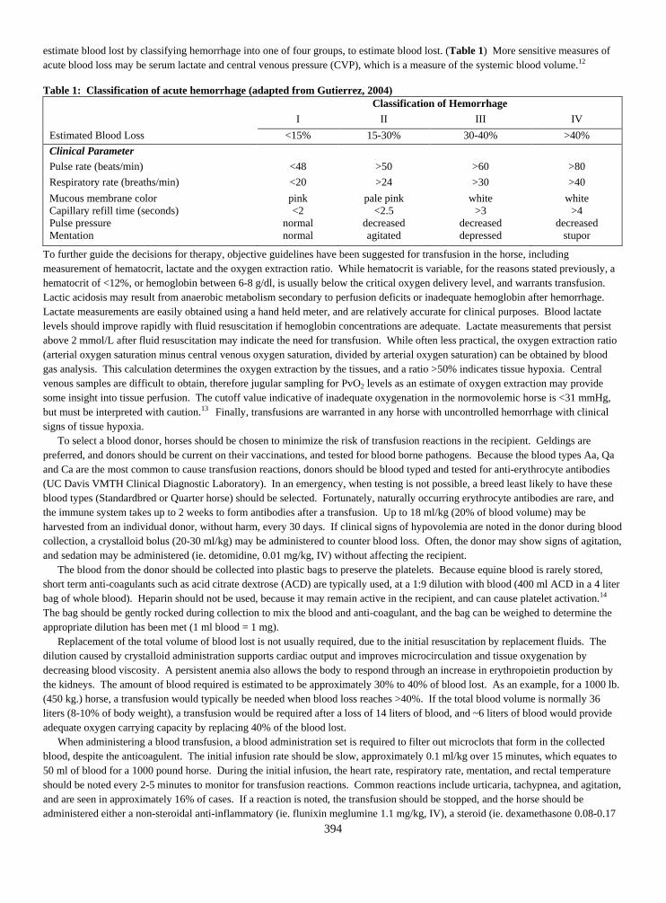
394
estimate blood lost by classifying hemorrhage into one of four groups, to estimate blood lost. (Table 1) More sensitive measures of acute blood loss may be serum lactate and central venous pressure (CVP), which is a measure of the systemic blood volume.12 Table 1: Classification of acute hemorrhage (adapted from Gutierrez, 2004)
Classification of Hemorrhage I II III IV Estimated Blood Loss <15% 15-30% 30-40% >40% Clinical Parameter Pulse rate (beats/min) <48 >50 >60 >80 Respiratory rate (breaths/min) <20 >24 >30 >40 Mucous membrane color Capillary refill time (seconds) Pulse pressure
pink <2
normal
pale pink <2.5
decreased
white >3
decreased
white >4
decreased Mentation normal agitated depressed stupor
To further guide the decisions for therapy, objective guidelines have been suggested for transfusion in the horse, including measurement of hematocrit, lactate and the oxygen extraction ratio. While hematocrit is variable, for the reasons stated previously, a hematocrit of <12%, or hemoglobin between 6-8 g/dl, is usually below the critical oxygen delivery level, and warrants transfusion. Lactic acidosis may result from anaerobic metabolism secondary to perfusion deficits or inadequate hemoglobin after hemorrhage. Lactate measurements are easily obtained using a hand held meter, and are relatively accurate for clinical purposes. Blood lactate levels should improve rapidly with fluid resuscitation if hemoglobin concentrations are adequate. Lactate measurements that persist above 2 mmol/L after fluid resuscitation may indicate the need for transfusion. While often less practical, the oxygen extraction ratio (arterial oxygen saturation minus central venous oxygen saturation, divided by arterial oxygen saturation) can be obtained by blood gas analysis. This calculation determines the oxygen extraction by the tissues, and a ratio >50% indicates tissue hypoxia. Central venous samples are difficult to obtain, therefore jugular sampling for PvO2 levels as an estimate of oxygen extraction may provide some insight into tissue perfusion. The cutoff value indicative of inadequate oxygenation in the normovolemic horse is <31 mmHg, but must be interpreted with caution.13 Finally, transfusions are warranted in any horse with uncontrolled hemorrhage with clinical signs of tissue hypoxia.
To select a blood donor, horses should be chosen to minimize the risk of transfusion reactions in the recipient. Geldings are preferred, and donors should be current on their vaccinations, and tested for blood borne pathogens. Because the blood types Aa, Qa and Ca are the most common to cause transfusion reactions, donors should be blood typed and tested for anti-erythrocyte antibodies (UC Davis VMTH Clinical Diagnostic Laboratory). In an emergency, when testing is not possible, a breed least likely to have these blood types (Standardbred or Quarter horse) should be selected. Fortunately, naturally occurring erythrocyte antibodies are rare, and the immune system takes up to 2 weeks to form antibodies after a transfusion. Up to 18 ml/kg (20% of blood volume) may be harvested from an individual donor, without harm, every 30 days. If clinical signs of hypovolemia are noted in the donor during blood collection, a crystalloid bolus (20-30 ml/kg) may be administered to counter blood loss. Often, the donor may show signs of agitation, and sedation may be administered (ie. detomidine, 0.01 mg/kg, IV) without affecting the recipient.
The blood from the donor should be collected into plastic bags to preserve the platelets. Because equine blood is rarely stored, short term anti-coagulants such as acid citrate dextrose (ACD) are typically used, at a 1:9 dilution with blood (400 ml ACD in a 4 liter bag of whole blood). Heparin should not be used, because it may remain active in the recipient, and can cause platelet activation.14 The bag should be gently rocked during collection to mix the blood and anti-coagulant, and the bag can be weighed to determine the appropriate dilution has been met (1 ml blood = 1 mg).
Replacement of the total volume of blood lost is not usually required, due to the initial resuscitation by replacement fluids. The dilution caused by crystalloid administration supports cardiac output and improves microcirculation and tissue oxygenation by decreasing blood viscosity. A persistent anemia also allows the body to respond through an increase in erythropoietin production by the kidneys. The amount of blood required is estimated to be approximately 30% to 40% of blood lost. As an example, for a 1000 lb. (450 kg.) horse, a transfusion would typically be needed when blood loss reaches >40%. If the total blood volume is normally 36 liters (8-10% of body weight), a transfusion would be required after a loss of 14 liters of blood, and ~6 liters of blood would provide adequate oxygen carrying capacity by replacing 40% of the blood lost.
When administering a blood transfusion, a blood administration set is required to filter out microclots that form in the collected blood, despite the anticoagulent. The initial infusion rate should be slow, approximately 0.1 ml/kg over 15 minutes, which equates to 50 ml of blood for a 1000 pound horse. During the initial infusion, the heart rate, respiratory rate, mentation, and rectal temperature should be noted every 2-5 minutes to monitor for transfusion reactions. Common reactions include urticaria, tachypnea, and agitation, and are seen in approximately 16% of cases. If a reaction is noted, the transfusion should be stopped, and the horse should be administered either a non-steroidal anti-inflammatory (ie. flunixin meglumine 1.1 mg/kg, IV), a steroid (ie. dexamethasone 0.08-0.17
395
mg/kg, IV) or an antihistamine (ie. hydroxazine 0.5-0.1 mg/kg, PO). If anaphylaxis is noted, epinephrine (0.01-0.02 mg/kg, IV) and fluid bolus is immediately required to restore blood pressure. The transfusion may be restarted in horses with minor reactions after the reaction has resolved, but if it reoccurs, or anaphylaxis developed, a new donor will be required. If no adverse reaction is noted after the initial slow infusion, the blood may be bolused in controlled hemorrhage (up to 20 ml/kg/hr),13 or increased to up to 3.75 ml/kg/hr for horses with uncontrolled bleeding. If large volumes of blood are administered, or if you are unsure of your dilution of the anticoagulant, serum ionized calcium should be monitored, due to the risk of citrate toxicity. In addition, crystalloids containing calcium must be stopped during the transfusion. The transfused red blood cells can survive from 4 to 20 days.15
Post-transfusion considerations Broad spectrum antibiotics are indicated after severe hemorrhage, due to the risk of sepsis from the translocation of bacteria across the compromised gastrointestinal mucosa. Erosions of the gastrointestinal tract (the stomach specifically) due to hypoxia, can be addressed by the administration of gastroprotectants, including omeprazole.
Vital signs, mental status, and urine production should be reassessed throughout fluid resuscitation and after restoration of the circulating volume. Venous access would be indicated for horse with ongoing losses due to uncontrolled hemorrhage, or if the response to therapy was less than expected. Serum chemistries would ideally be performed daily to assess electrolytes, organ function and evidence of metabolic changes. Finally, tissue oxygenation can be estimated by the oxygen extraction ratio and serum lactate, to assess the response to therapy and if tissue hypoxia is persistent. References 1. Gutierrez G, Reines HD, Wulf-Gutierrez ME. Clinical review: Hemorrhagic shock. Crit Care. 2004;8:373-381. 2. Martel MJ, MacKinnon CJ, Arsenault MY, et al. Hemorrhagic Shock. J Obstet Gynaecol Can. 2002;24(6):504-511. 3. Heidmann P, Tornquist SJ, Qu A, et al. Laboratory measures of hemostasis and fibrinolysis after intravenous administration of epsilon-aminocaproic acid in clinically normal horses and ponies. Am J Vet Res. 2005;66(2):313-318. 4. Ross J, Dallap BL, Dolente BA, et al. Pharmacokinetics and pharmacodynamics of epsilon-aminocaproic acid in horses. Am J Vet Res. 2001;68(9):1016-1021. 5. Fletcher, DJ, Brainard, BM, Epstein, K, et al. Therapeutic plasma concentrations of epsilon aminocaproic acid and tranexamic acid in horses. J Vet Int Med 2013;27: 1589–1595. 6. Ilium Vasolamin S 100; Troy Laboratories, Glendenning, NSW, Australia. http://www.troylab.com.au/products.php?tid=1&iid=24&pid=129 7. Taylor EL, Sellon DC, Wardrop KJ, et al. Effects of intravenous administration of formaldehyde on platelet and coagulation variables in healthy horses. Am J Vet Res. 2000;61(10):1191-1196. 8. Graham L, Farnsworth K, Cary J. The effect of yunnan baiyao on the template bleeding time and activated clotting time in healthy halothane anesthetized ponies. J Vet Em Crit Care. 2002;12(4):279. 9. LeBlanc MM. Common peripartum problems in the mare. J Eq Vet Sci. 2008;11:709-715. 10. Magdesian KG. Therapeutics in practice: Acute blood loss. Compendium Equine. 2008;3:80-90. 11. Cherkas, D. Traumatic hemorrhagic shock: Advances in fluid management. Emerg Med Pract. 2011;13(11):1-20. 12. Magdesian KG, Fielding CL, Rhodes DM, et al. Changes in central venous pressure and blood lactate concentration in response to acute blood loss in horses. J Am Vet Med Assoc. 2006;229:1458-1462. 13. Divers TJ. Monitoring tissue oxygenation in the ICU patient. Clin Tech in Equine Prac. 2003;2(2):138-144.\ 14. http://www.merckmanuals.com/vet/circulatory_system/blood_groups_and_blood_transfusions/blood_transfusions.html 15. Mudge MC, Walker NJ, Borjesson DL, et al. Post-transfusion survival of biotin-labeled allogeneic RBCs in adult horses. Vet Clin Pathol. 2012;41(1):56-62.
396
Lefts and Rights: Medical Management of Colonic Displacements
Amelia Munsterman, DVM, MS, DACVS, DACVECC Auburn University
Auburn, AL
Pathophysiology of large intestinal displacements Displacement of the large, or ascending, colon is defined by the abnormal position of the large bowel in the abdominal cavity. A predisposing factor for this displacement is the normal anatomy of the equine gastrointestinal organs. The large colon of the horse is only affixed to the body wall at the junction of the right dorsal and transverse colon, and to the cecum by the cecocolic fold. Therefore the majority of this section of intestine can move freely about the abdomen. A displaced colon may be found in a number of different positions at surgery, and four typical presentations for a displacement have been described: 1) retroflexion of the pelvic flexure towards the diaphragm, 2) left dorsal displacement where the colon is positioned lateral to the spleen, or trapped over the nephrosplenic ligament (nephrosplenic entrapment), 3) right dorsal displacement where the colon lies between the cecum and body wall, and 4) a non-strangulating volvulus of <180 degrees.1 Although displacements are not technically described as a strangulating lesion, they may cause intestinal compromise if significant bowel distention occludes venous outflow, or if the colon continues to twist into volvulus of >180 degrees.
While the lack of anatomic attachment to the abdominal wall allows the colon to displace, the cause of this type of colic is still unknown. One theory is that gas distention may cause the colon to become lighter than the surrounding viscera and alter gastrointestinal motility, resulting in movement of the colon out of its normal position. One cause of gas production includes the fermentation of carbohydrates in the colon that are allowed to pass through the small intestine undigested. Carbohydrate loads that reach the large intestine may be secondary to a sudden change in diet or a large meal of carbohydrates (>0.4% of body weight) that does not allow for adequate production of pancreatic amylase.2 The result is fermentation by lactic acid-producing bacteria, luminal acidosis and gas production. A second cause of displacement may be due to changes in microbial flora of the colon secondary to oral antibiotics, resulting in a similar pathologic overgrowth of acid-producing bacteria. Gas distention may also result secondary to obstruction caused by impactions or foreign bodies, such as enteroliths or sand, both of which may complicate the treatment of a simple colon displacement. For right dorsal displacements, specifically, it has been proposed that aberrant gastrointestinal motility patterns at the pelvic flexure are involved.1 Finally, medications or disease that directly impact gastrointestinal motility, including anticholinesterase medications (atropine), can alter the normal flow of ingesta, and predispose to displacement. Clinical findings and diagnosis Large colon displacement can present with variable signs of abdominal pain, depending on the degree of obstruction of the intestinal lumen that the displacement causes. While it is typically an acute disease, the owners in some cases may have noted intermittent discomfort in the days prior to presentation. Abdominal distention is commonly observed, and fecal production is reduced to absent. Horses with displacements may produce net nasogastric reflux, which may confound the diagnosis; the cause of reflux is suspected to be secondary to ileus, compression of the duodenal outflow tract, or tension on the duodenocolic ligament.
On rectal palpation, a tight band is often noted if the colon is within reach, and gas distention or a concurrent impaction may be palpated within the colon. If the band courses over the nephrosplenic ligament, between the caudal pole of the left kidney and the head of the spleen, the diagnosis of a nephrosplenic entrapment can be made. If the band traverses horizontally, between the cecum and body wall, a right dorsal displacement should be suspected. Characteristic rectal exam findings may not be present if the pelvic flexure is retroflexed, and horses with this type of displacement will be described as feeling “empty” when palpating in the caudal abdomen. However, it is important to note that false positives are common when diagnosing displacements by rectal examination; about 20% horses are incorrectly diagnosed with a displacement.3 Differentials to consider for rectal findings consistent with displacement include cecal impaction, cecal or colon tympany, simple large colon impaction, enteroliths, or large colon volvulus.
Advanced diagnostics to consider should include transabdominal ultrasound for both right and left dorsal displacement. Left dorsal displacement has been described as a gas shadowing of the dorsal border of the spleen, ventral displacement of the spleen, presence of the colon lateral to the spleen, or failure to identify the left kidney medial to the spleen when imaged through the flank in a lateral plane.4,5 Sensitivity of ultrasonography for identification of left dorsal displacement is around 89%.5 Right dorsal displacement may also be identified on ultrasound, but has a lower sensitivity, between 56% and 67.7%.6,7 A tentative diagnosis of right dorsal displacement may be made by identification of the vessels that normally lie on the medial surface of the colon incorrectly positioned against the right lateral body wall. If the colon is malpositioned, these vessels can be located by ultrasound between intercostal spaces 10 to 16, and dorsal to the costochondral junction.6 The vessels of the colon should be differentiated from the cecal vessels, that are normally located by ultrasound in the right flank.
397
Bloodwork is not necessarily required for diagnosis, but may be helpful for identification of electrolyte abnormalities and hypovolemia that may affect choice of treatment. On serum chemistries, right dorsal displacements have commonly been described to have a distinctive increase in gamma glutamyltransferase activity (GGT), due to compression and obstruction of the bile duct.8 These changes in serum chemistry have not been associated with an incidence of hepatic insufficiency or failure, and do not require additional therapy. However, there have been cases of left dorsal displacement with elevated serum GGT, which cautions its use as the definitive diagnosis for a right dorsal displacement.8,9
Treatment of colon displacements Large intestinal displacements are treated similarly, regardless of the position of the colon in the abdomen. If the horse is hemodynamically stable, it is held off feed, and administered fluids to both correct deficits and to hydrate the ingesta if an impaction is suspected. Fluid therapy may be provided orally or intravenously. Oral fluids are preferred, due to the fact that they can directly rehydrate an impaction, when present. They are also cost effective compared to intravenous administration. Contraindications to oral fluids would be if the horse is significantly dehydrated, or if the horse is producing net reflux (>2 liters every 2 hours). In addition to fluids, therapy should include analgesics (non-steroidal anti-inflammatory medications), antispasmodics, and low doses of sedation. Providing light exercise (jogging for 10-20 minutes x times a day) may be helpful to improve gastrointestinal motility and return the colon the correct position.3
In addition to these treatments, horses with a left dorsal displacement are often treated with phenylephrine and exercised on a lunge line for 15 to 25 minutes to displace the colon from the nephrosplenic space. Phenylephrine is an alpha-1 adrenergic sympathomimetic that causes vasoconstriction, resulting in splenic contraction by up to 28%.10 The dosage for phenylephrine is 0.04 mg/kg, or 10-20 mg per 1000 lb (450 kg) horse, administered as an intravenous infusion over 15 minutes. Once administration is complete, phenylephrine has a relatively short duration of action, with splenic contraction lasting only 25 minutes.10 It is currently unknown if phenylephrine actually improves the odds of resolving a left dorsal displacement. A recent publication noted a 96.5% survival rate in horses treated conservatively with only fluids, anti-spasmodic medications, and analgesics.11 Of these horses, only 9 out of the 114 were administered phenylephrine, which calls into question the effects, if any, of this medication for therapy of a nephrosplenic entrapment. Treatment with phenylephrine is not benign, and side effects include increased pulmonary artery and right atrial pressures, second degree heart block, hypertension, bradycardia, bradyarrythmias, and decreased cardiac output.10 Phenylephrine should also be used with caution in older horses (>15 years), where severe and fatal hemorrhage into the thorax or abdomen has been reported.12
An alternative medical therapy to consider for nephrosplenic entrapment would include rolling under general anesthesia. This technique involves anesthetizing the horse in right lateral recumbency, rolling the horse over its back into left lateral, and continuing on into right lateral again to evaluate progress with either rectal palpation or ultrasound. If possible, it is preferred to lift the horse into the air by the hind limbs while in dorsal recumbency to allow for agitation of the abdomen before rolling into left lateral recumbency, to help dislodge the colon from the nephrosplenic space.13 While rolling is considerably cheaper than surgery, it has similar anesthetic risks, and should be approached with caution if surgical facilities are not readily available. Reasons for treatment failure can include a second problem or the wrong diagnosis. The rolling procedure was initially described in conjunction with administration of phenylephrine, however, a recent publication noted no improvement in success rates in horses administered this drug.4,14
If the colon or cecum is moderately distended, trocarization may allow for medical management in cases where surgery is not an option. Decompression will improve blood flow to the abdominal organs and can provide the colon space within the abdomen to return back to its normal position. Trocarization is typically performed high in the right flank to decompress the cecum, however, decompression can also be performed on the left if a significant gas cap is present in the displaced colon. Ultrasound should be used to identify the position of the gas filled viscous subcutaneously, and to avoid large vessels or the spleen that may be between the skin and the colon. After routine sterile preparation, a 14 gauge, 5 inch (12 cm) catheter is placed percutaneously perpendicular to the skin, and advanced until gas is noted exiting an attached extension set. If the end of the extension set is placed in a cup of water, it is easier to determine if gas is still exiting the lumen of the bowel. Redirection of the catheter should be avoided to prevent laceration of the intestine, but the catheter can be slowly advanced if needed. Successful trocarization should take 30-45 minutes to significantly reduce gas distention if gas is evacuated passively. As the catheter is removed, an antibiotic may be injected to prevent subcutaneous abscess. Complications of this procedure include hemorrhage and peritonitis, and the horse should be monitored closely after this procedure for pyrexia, that could indicate a developing infection.
Indications for surgery in horses diagnosed with a large colon displacement include the presence of significant abdominal pain or distention on initial exam that is unresponsive to sedation and analgesics, or increasing distention and discomfort despite medical therapy. Horses with significant gastric reflux are also often taken to surgery.11 Horse that will respond to medical therapy should require only low doses of sedation, and repeated administration may indicate the need for surgery or reevaluation of the diagnosis. Clinical signs consistent with endotoxemia including hyperemic mucous membranes, tachycardia, increasing hematocrit with a concurrent hypoproteinemia, and cold extremities are also often indicative of a surgical lesion. In addition, an abnormal
398
abdominocentesis or gas distention that severely limits rectal examination of the abdomen may suggest that medical management will not be successful.
Horses suspected to have a right dorsal displacement, 180 degree volvulus, or retroflexion of the pelvic flexure require a ventral midline or paramedian laparotomy to allow for correction of the displacement and concurrent enterotomy for lavage of an impaction may be needed. While ventral midline is preferred by the author to allow for full exploration of the abdomen, it is possible to perform a flank procedure to remove the colon from the nephrosplenic space, as well as simultaneously allow for ablation in horses with a left dorsal displacement.15 However, standing procedures should not be attempted in horses exhibiting intractable pain, significant feed impactions or if there is a suspicion of a second intestinal abnormality. Prognosis after colon displacement The success of medical and surgical therapy for left dorsal displacement or nephrosplenic entrapment is excellent, between 90% and 100% for all treatments combined.4,13 When the success of individual treatments are examined individually, the rolling procedure has a success rate between 33% and 90%,4,13 exercise has a success rate between 33-100%,13 and surgical management is from 80% to 95.9%.11 As mentioned previously, palliative therapy including fluids, anti-inflammatory medications and antispasmodics has a success rate of 96.5%.11 For right dorsal displacements, success rates overall are around 94%. Success of medical management is 64% whereas surgical success rates between 80 and 93%.3 Prevention of displacements Client education is important in horses that have been diagnosed with a large colon displacement. Because of the link with management strategies, a thorough assessment of the feeding and exercise protocols of these horses should be performed. Of the 4 types of displacement, horses with right dorsal displacement have been noted to be more likely to present with a second episode of colic. In one publication, approximately 42% of horses with a right dorsal displacement experienced an additional episode of abdominal pain within 6 months of surgery, with 10% of those requiring relaparotomy.16 Retroflection of the pelvic flexure was also noted to have a high incidence of recurrent colic in this study (46%), but these horses were unlikely to need a second surgery. Horses with left dorsal displacement and non-strangulating volvulus had a recurrence rate of 8% and 21%, respectively.16 It is important to warn owners that horses with this diagnosis may be at a higher risk for colic in the future.
Recurrence of left dorsal displacement and nephrosplenic entrapment is much less common (about 8%), but some reports note a recurrence rate of up to 21%.16,17 The risk of left dorsal displacement can be reduced by surgical ablation of the nephrosplenic space, but this procedure does not prevent other types of colic, likely due to the management issues surrounding displacements or an underlying motility disorder.17,18 In addition, body type may predispose a horse to this type of colic, which appears to be more common in large, barrel-chested breeds.11 The incidence of colic after this procedure is around 11-21%.17,18 References 1. Hardy J. Specific diseases of the large colon. In: White NA, Moore JN, Mair TS, eds. The Equine Acute Abdomen. Eds: N.A. White, J.N. Moore and T.S. Mair, Jackson: Teton New Media; 2008:628-644. 2. Potter GD, Arnold FF. Householder, DD, et al. Digestion of starch in the small or large intestine of the equine. Pferdeheilkunde 1992;1:107–111. 3. McGovern KF, Bladon BM, Fraser BSL, Boston RC. Attempted medical management of suspected ascending colon displacement in horses. Vet Surg, 2012;41:399-403. 4. Baker WT, Frederick J, Giguere S, et al. Reevaluation fo the effect of phenylephrine bon resolution of nephrosplenic entrapment by the rolling procedure in 87 horses. Vet Surg. 2011;40:825-829. 5. Santschi EM, Slone DE, Frank WM. Use of ultrasound in horses for diagnosis of left dorsal displacement of the large colon and monitoring its nonsurgical correction. Vet Surg. 1993;22:281–284. 6. Grenager NS, Durham MG. Ultrasonographic evidence of colonic mesenteric vessels as an indicator of right dorsal displacement of the large colon in 13 horses. Eq Vet J. 2011;43(Suppl. 39):153-155. 7. Ness SL, Bain FT, Zantingh AJ, et al. Ultrasonographic visualization of colonic mesenteric vasculature as an indicator of large colon right dorsal displacement or 180° volvulus (or both) in horses. Can Vet J. 2012;53(4):378–382. 8. Gardner RB, Nydam DV, Mohammed HO, et al. Serum gamma glutamyl transferase activity in horse with right or left dorsal displacements of the large colon. J Vet Intern Med. 2005;19:761-764. 9. Banse HE, Tennent-Brown BS, Mueller POE. Left dorsal displacement of the large colon manifested as increased γ-glutamyl transferase activity in a horse. Compend Contin Educ Vet. 2012;34(2):E3. 10. Hardy J, Bednarski RM, Biller DS. Effect of phenylephrine on hemodynamics and splenic dimensions in horses. Am J Vet Res. 1994;55:1570-1578. 11. Lindegaard C, Ekstrom CT, Wulf SB, et al. Nephrosplenic entrapment of the large colon in 142 horses (2000–2009): Analysis of factors associated with decision of treatment and short-term survival. Eq Vet J. 2011;43(suppl. 39):63-68. 12. Frederick J, Giguere S, Butterworth K, et al. Severe phenylephrine-associated hemorrhage in five aged horses. J Am Vet Med Ass. 2010;237:830-834. 13. Fultz LE, Peloso JG, Giguere S, Adams AR. Comparison of phenylephrine administration and exercise versus phenylephrine administration and a rolling procedure for the correction of nephrosplenic entrapment of the large colon in horses: 88 cases (2004-2010). J Am Vet Med Assoc. 2013;242:1146-1151.

399
14. Hardy J, Minton M, Robertson JT, et al. Nephrosplenic entrapment in the horse: a retrospective study of 174 cases. Equine Vet J Suppl. 2000;32:95–97. 15. Muňoz J, Bussy C. Standing hand-assisted laparoscopic treatment of left dorsal displacement of the large colon and closure of the nephrosplenic space. Vet Surg. 2013;42(5):595-599. 16. Smith LJ, Mair TS. Are horses that undergo an exploratory laparotomy for correction of a right dorsal displacement of the large colon predisposed to post operative colic, compared to other forms of large colon displacement? Eq Vet J. 2013;42(1):44-46. 17. Rocken M, Schubert C, Mosel G, Litzke LF. Indications, surgical technique, and long-term experience with laparoscopic closure of the nephrosplenic space in standing horses. Vet Surg. 2005;34(6):637-641. 18. Farstvedt E, Hendrickson D. Laparoscopic closure of the nephrosplenic space for prevention of recurrent nephrosplenic entrapment of the ascending colon. Vet Surg. 2005; 34(6):642-645.
400
Equine Wounds: Triage to Treament
Amelia Munsterman, DVM, MS, DACVS, DACVECC Auburn University
Auburn, AL
Exposed bone Exposed or denuded bone is a common complication of wounds of the distal aspect of the limb. Exposed cortical bone in which the periosteum has been removed, is prone to desiccation of the superficial layers of the cortex, which may result in infectious superficial osteitis and sequestrum formation. Exposed bone within a wound can delay wound healing directly if the bone becomes infected, or indirectly because its rigid structure can delay the formation of granulation tissue and wound contraction.
Distal limb avulsion wounds with exposed bone increase in wound size for 14 to 21 days. Wound expansion is due predominantly to the distraction forces applied across the wound during the inflammatory and debridement stages of wound healing, and the lack of a granulation tissue bed in the center of the wound to neutralize the tensile forces exerted on the wound margins from the surrounding skin. Wounds with a small amount of exposed bone, or wounds without exposed bone, expand for a shorter period because less time is required for granulation tissue to seal the wound. Larger wounds with exposed bone take longer to form a granulation bed and subsequently wound contraction is postponed.
Periosteal insults from blunt trauma, tendon/joint capsule strain, surgical manipulation, or laceration/degloving injuries may result in extensive periosteal exostosis. Injuries involving bones in horses stimulate more periosteal new bone growth than similar wounds in other species and ponies. More extensive periosteal reaction in young compared to adult horses has been attributed to a more active osteoblastic activity of the periosteum in young horses. The extensive periosteal new bone growth seen in adult horses is poorly understood. Deferred collagen lysis compared to other species may be a contributing factor. The more extensive periosteal new bone formation in horses compared to ponies is alleged to be the result of a slower onset and longer duration of the periosteal response and prolonged extensive limb swelling in horses, as compared to ponies.
Despite the common occurrence of exposed bone associated with trauma to the distal aspect of the limb, there has been little investigation into methods of stimulating coverage of granulation tissue over exposed bone in horses. Granulation tissue development is a very important role in second-intention healing because it provides a barrier to infection and mechanical trauma for the underlying tissues. Healthy granulation tissue is resistant to infection and provides a moist surface for epithelialization. The delay in wound healing caused by exposed bone has prompted the search for different methods to promote granulation tissue coverage of bone in other species.
Head trauma, thermal injury, and surgical oncology often results in exposed bone of the cranium in humans. In these cases the outer cortex of the uncovered portion of the cranium is fenestrated with drill holes, burrs, or lasers to expose the medullary cavity from which granulation tissue grows to cover the exposed bone. Similarly, exposed cortices of long bones in humans have been fenestrated with drill holes to promote granulation tissue formation. It has been suggested that the drill holes promote healing by allowing osteogenic factors from the medullary cavity access to the wound, or by the enhancement of healing of bone and soft tissue by a nonspecific response known as “the regional acceleratory phenomenon”. Cortical fenestration combined with drugs that promote topical granulation tissue may accelerate granulation tissue coverage compared to control wounds, but further investigation is needed.
Cortical fenestration of 1.6 mm drill holes in the cortex of the second metacarpal bone in experimentally created wounds in dogs resulted in clot formation over the bone that promoted granulation tissue formation and may have protected the bone’s outer layers from desiccation. The effects of cortical fenestration with 3.2 mm drill holes were evaluated in experimentally created wounds of the distal aspect of the limb of horses. Cortical fenestrated wounds became covered with granulation tissue earlier than control wounds, and fenestration had no significant effect on sequestrum formation. The granulation tissue growing directly from the bone surface also contributed to granulation tissue formation. If the wounds are not large (< 6 x 6 cm) it may be difficult to realize a significant contribution from the granulation tissue growing from the cortical fenestration sites alone. Cortical fenestration may also be advantageous if it is used with other methods of promoting granulation tissue. Splinting of the limb is usually not necessary for the recovery from general anesthesia unless there are associated traumatic injuries to the limb that would suggest instability. Degloving injuries Degloving or avulsion injuries are not uncommon in equine practice, and their management can be challenging because of prolonged treatment, cost, and sometimes unknown outcome. The body that becomes entrapped in hazards or a limb that becomes intertwined in fencing or can quickly sustain tissue damage. The most common sites for this type of trauma are the hemi thorax, dorsal aspect of the metacarpus and/or metatarsus and the cranial aspect of the tarsus. Vascular, soft tissue and bone damage is directly proportional to the length of time and effort the horse uses to free itself. Some injuries that seem to be superficial and innocuous on the surface may involve vital structures surrounding the wound and/or later develop cutaneous and internal abscesses and/or ulcerative cellulitis. Local
401
wound care should be an integral part of the initial treatment. The severity and duration and location of the laceration determines the best approach to the treatment of degloving injuries as healing of wounds involving the distal limb is often delayed when compared with other areas of the body, further complicating the healing process.
Primary repair of the wound is the preferred treatment for wounds that involve detachment of skin with maintenance of an intact blood supply. Complications such as sequestrum formation are lessened and healing is improved when the exposed bone and tendons are covered with skin and soft tissue in the immediate post trauma period. Closing as much of the wound as possible improves the cosmetic and functional outcome and lessens the amount of healing having to occur by second intention.
Delayed closure of a degloving injury is preferred when there is significant contamination, swelling and trauma of the wound without loss of skin. Initial treatment for the first 2-3 days after injury include debridement and lavage of the wound followed by wet to dry bandages to facilitate further debridement. Pressure bandaging is indicated to remove edema associated with the injury. Debridement of the wound edges and appropriately applied tension sutures facilitate closure of the wound as skin retraction is a complication of delayed closure.
Second intention healing is indicated for degloving injuries in which there is a considerable loss of skin immediately at the time of injury or in which a closed degloving injury has developed avascular necrosis of the skin with subsequent sloughage. The wound is sharply debrided until only healthy tissue remains. A hydrogel Carradress®, Carrington, Irving, TX) dressing is applied to the region of the wound that remains open. These dressings are able to contribute moisture to dehydrated tissue, augment autolytic debridement and absorb some moisture from an exudating wound. The dressing is applied to the wound bed followed by application of a conformable absorptive dressing (Kerlix®, Kendall, Mansfield, MA). A firm cotton bandage is used to provide warmth, support and to minimize excessive movement of the limb and associated wound area. Depending on the size and location of the wound, skin grafting may be indicated to facilitate complete healing. Grafting should be delayed to permit maximum wound contraction which, depending on the location and size of the wound, may be 4-8 weeks after injury.
Dorsal knuckling of the fetlock and an inability to extend the digit is a common complication of distal limb wounds that is usually associated the loss of the extensor tendon of the distal limb. Supporting the dorsal aspect of the limb to counteract the pull of the flexor tendons on the palmar and/or plantar aspect of the limb is the premise for management of extensor tendon disruption. The wound and extensor tendon laceration is managed by second intention healing without suturing the extensor tendon. A rigid polyvinyl chloride (PVC) splint is applied to the dorsal or palmar and/or plantar aspect of the distal limb after wound bandaging. The bandage and splint, which maintains the limb in extension and prevents dorsal knuckling of the fetlock, are retained until normal limb function returns which may vary from 7 days to 6 weeks. Excessive skin tension Skin sutured with excessive tension is likely to have complications of healing due to local ischemia with pressure necrosis of the surrounding skin and the pull through of sutures at the skin edge with subsequent wound disruption. Undermining the surrounding skin, relief incisions, and appropriately applied tension sutures are the most common methods that can be used to lessen tension along the skin margins.
The surrounding skin can be undermined up to 4 cm from the wound edge without associated complications. Relief incisions can be closed after the primary incision is closed or left to heal by second intention.
In order not to interrupt the blood supply to the primary suture line, tension sutures are positioned well away from the wound margin. Once the tension suture is in place, the primary incision line is sutured to close the wound edges. Tension suture patterns include vertical mattress, horizontal mattress, far-far-near-near, and far-near-near-far patterns. Vertical mattress sutures with or without skin support to prevent laceration of the wound edges such as polyethylene or rubber tubing, are useful in reducing tension on the primary suture line. This tension suture support method is used in areas that cannot be bandaged well such as the upper limb, body and neck region. It is contraindicated to use tension suture supports under a limb cast or heavy bandage as these supports may cause tissue necrosis and suture line failure. Tension sutures are not effective after 7 to 10 days and should be removed in a staggered fashion with one-half removed initially followed by the remaining sutures later. Movement The extent of movement of the skin relative to the underlying bed of granulation tissue is usually much higher in the limb regions than in the trunk. This is possibly exacerbated by the relative lack of skin elasticity as well as the obvious proximity of the limb skin to structures with a high degree of motion such as joints and tendons. Trunk wounds have a better available reparative blood supply than those of the distal limb.
An injury to the distal limb metacarpal or metatarsal region of a horse which involves the flexor tendons and/or their sheaths requires healing by the ingress of blood vessels from adjacent structures. However, as healing attempts to progress, repeated tendon contraction and limb movement moves the injury away from the site of the skin wound leaving the damaged tissues with no effective mechanism for healing.

402
Rigid limb casting of a distal limb wound is very effective in facilitating wound contraction and epithelialization if the tissues are initially sharply debrided and lavaged. The mechanisms for this may be more complex than merely controlling movement. Although movement of the limb and wound is limited, added surrounding pressure applied to the wound may also facilitate the healing process. Warmth, restriction of movement and the presence of a moist healing environment in conjunction with a cast are probably significant factors that contribute to wound healing. Which aspects of the exudate are desirable and enhancing of wound healing and which are inhibitory is not known in the horse. Heat, pain, swelling, or lameness created by the cast indicate attentive reevaluation of the wound and the consideration of cast removal and or cast change. Self-mutilation Significant self-mutilation of wounds through rubbing, biting, and pawing can occur if the horse is not adequately restrained or medicated. Usually the most intense pruritic episodes occur in the first weeks of wound healing during the inflammatory phase of repair and during eschar sloughing but can be a later complication associated with burn wounds. To prevent extreme self-mutilation, the horse should be cross tied and/or sedated at this time and use of a neck collar may be considered. Delayed healing, poor epithelialization, and complications of second intention healing may limit return of the animal to their previous use. Skin grafting Skin grafting decreases healing time and is one of the best techniques for covering a wound that has been chronically affected by exuberant granulation tissue. Skin grafting of lower limb wounds should be considered to cover the granulating wound bed if contraction has ceased and the wound bed is large. Frequently, however, wounds in horses are treated for several weeks before skin grafting is initiated. At this point granulation tissue is mature, fibrous and has less of a blood supply than newly formed granulation tissue. Other complications of graft acceptance and healing are wound infection and sequestra formation.
Chronic inflammation, inherently present during second intention healing of wounds on the distal portion of limbs of horses may be at least as important as infection because it reduces the quality of the granulation bed and results in the production of a moderate amount of purulent exudate, both of which negatively influence acceptance of grafts. As a result the ability of a wound bed to accept a graft is lessened. It is therefore imperative that chronic granulating wounds be debrided to a level below the skin surface down to a level of healthy granulation tissue prior to graft application.
To increase the success of graft acceptance wound bacteria must be minimized. Beta hemolytic Streptococcus spp., Proteus spp., and Pseudomonas spp. are capable of producing destructive proteolytic enzymes and excessive purulent discharge which breakdown fibrinous attachments between the graft and recipient bed. Topical antiseptics have better efficacy than antibiotics in reducing bacterial wound load as the latter increase the risk of patient sensitization and the development of resistant organisms especially when used routinely over prolonged periods in uninfected wounds. Infected wounds, however, should be treated with broad-spectrum antibiotics while awaiting culture results. The bone underlying the wound should be radiographed for evidence of sequestra and excessive pericortical dystrophic mineralization. Large wounds often develop healthy granulating tissue around the perimeter before a sequestrum completely defines itself.
Donor site is influenced by the method of grafting, color, and texture of the donor hair, cosmesis of the donor site, and ease of obtaining skin. Common sites for obtaining donor skin include pectoral, dorsal neck region, perineum, ventral midline, ventral lateral abdomen and sternal region caudal to the girth area.
Pinch grafts Pinch grafts are distinct pieces of skin (3 mm in diameter) produced by excising an elevated cone of skin. Graft acceptance is as high as 75% using pinch grafts partially due to the fact that the pockets of granulation tissue hold the graft in contact with the wound. Complications include necrosis of the graft, slower wound healing, improper orientation of hair, and thin skin coverage of the wound.
Necrotic spots along the top of the granulation pockets normally occur during healing, after which the graft epithelializes circumferentially. Because pinch grafts are small, complete epithelialization of the wound often requires greater than 70 days. Improper orientation of hair growth is a complication of pinch graft application despite repeated efforts to properly align the hair to match that of the recipient area. A cobblestone appearance with thin subcutaneous tissue is sequelae of pinch graft applications that may not be cosmetically acceptable for show horses.
Punch grafts Punch grafts are circular pieces of skin that are directly removed from the locally anesthetized donor site or by obtaining biopsies from an excised piece of donor skin. Common complications of punch graft failure are incomplete removal of the underlying subcutaneous tissue from the graft, recipient site hemorrhage, and motion.
As punch grafts are full thickness they must have the subcutaneous tissue and fascia removed from the dermis with a surgical blade before implanting as these layers will prevent revascularization and subsequent graft failure. Placing grafts in saline soaked sponge gauze for a short period of time minimizes graft desiccation while recipient beds are created. Accumulation of blood and serum beneath the graft displaces the grafts from the recipient site. Hemorrhage can be avoided by ensuring that it is controlled before grafting. Displacement of the grafts can also be minimized by using a biopsy punch a size smaller than used to obtain donor graft to

403
ensure a snug fit in the recipient bed. Displacement of the graft by motion can be minimized by securing the wound under a heavy bandage. Displacement of grafted tissue at wrap changes can be reduced by soaking the primary bandage prior to removal. Casting is not indicated for punch graft techniques as punch grafts are not indicated for grafting over moveable areas of the body.
Tunnel grafts Tunnel grafts are useful for healing of wounds that are hard to immobilize or bandage as on the dorsal surface of the hock or fetlock. Graft survival rates of 80% have been reported with excellent cosmetic results. Complications of tunnel grafting include the placement of tunnel grafts too close to one another, failure of the graft to become exposed and accidental removal of the tunnel graft when removing the overlying granulation tissue.
This technique requires harvesting of full-thickness or spit-thickness strips of skin 2 to 5 mm wide and slightly longer than the length of the wound’s edges. These grafts are placed in granulation tissue that has been allowed to develop 4 to 8 mm above skin level. These tunnels can be created using a cutting needle, flattened K-wire with a trocar point, or malleable alligator forceps. The graft is then tunneled approximately 6 mm below the surface of the granulation tissue at the recipient site ensuring that the epidermal side of the graft faces the surface of the wound. Tunnel grafts should not be placed closer than 2 cm apart to prevent excessive necrosis of granulation tissue. The cut ends of the skin strips are sutured to the skin on either side of the granulation bed. A tourniquet may be useful to control hemorrhage and improve visualization of the strips for procedures that involve grafting on a limb. If placed the correct depth, the granulation tissue overlying the graft should slough in 7 to 10 days. If this does not occur, it should be excised at this time. Adjacent granulation tissue that is raised should be excised at this time. Most tunnel graft failures are attributable to accidental removal of the graft during removal of the overlying granulation tissue or failure of the graft to become exposed. Exposure of the graft if necessary may be facilitated by placing malleable probes or wires through the tunnels to cut through the overlying granulation tissue.
Full thickness sheet graft Full thickness or split thickness grafts can be applied as a sheet or expanded before transplantation. The full thickness sheet graft is the most cosmetic type of free sheet graft as it contains all the properties of the surrounding skin, provides maximum piliation, and can withstand pressure and friction. Full thickness grafts are not as readily accepted because there are less exposed blood vessels available for imbibition of plasma and for inosculation.
No specialized equipment is needed for harvesting, and the procedure can often be performed in the standing sedated horse using local anesthesia. Donor sites of full thickness grafts should be sutured. The graft should be cut slightly larger than the recipient bed to allow for shrinkage after the graft is excised because of recoil of elastic fibers in the deep dermal layers of the of the graft. The full thickness graft should be sutured to the donor site with some tension to prevent occlusion of the dermal vessels that may occur if the graft is allowed to fully contract.
A high oxygen gradient between the wound and the graft is essential for neovascularization of the graft and graft acceptance. Full-thickness grafts treated with hyperbaric oxygen therapy developed less granulation tissue, edema, and neovascularization, but more inflammation. The superficial portion of these full-thickness grafts was also less viable than the superficial portion of those not treated with hyperbaric oxygen therapy.
Full thickness sheet grafts are often considered compromised because they often require more nourishment than can be supplied by the granulating recipient wound. As a result full thickness grafts are usually reserved for fresh uncontaminated wounds. The upper layers of a full thickness graft are more likely to slough because full thickness grafts require more nourishment and have fewer exposed vessels for this purpose. Because of the lack of abundant donor skin in the horse, the graft often must be meshed and expanded to achieve coverage of the wound larger than the donor area.
Split thickness grafts Split thickness grafts are more readily accepted than full thickness grafts, and may be used to cover granulation beds that are less than ideal. Since blood vessels branch as they become more superficial in the dermis more vessels are cut and exposed with split thickness grafts. The greater the number of exposed vessels the better the absorption of nutrients will be from the granulation bed. A split thickness sheet graft is more cosmetic than a pinch or punch graft because the thickness of the graft and orientation of the hair are uniform and coverage by the graft is more complete.
A mechanical dermatome or a free hand knife (Watson Skin graft knife, Down’s Surgical, Sheffield, England) is used to split the dermis. The latter is preferred as it is easy to use and economical to employ. General anesthesia is necessary to obtain the graft as split thickness donor sites are very painful to the horse, since many nerve endings are exposed. Grafts less than 0.5 mm thickness in the horse lack strength, durability, and have sparse or no hair follicles or exocrine glands which results in less sebaceous secretion. Grafts harvested between 0.63 mm and 0.75 mm have good coverage of hair and greater durability than do thinner grafts. Unlike full thickness grafts suturing of the donor site is not required and primary graft contraction is minimal since a portion of the dermis remains intact and heals with a scarred appearance.
The grafts can be applied to the wound after the horse has recovered from general anesthesia. This reduces anesthesia time and the possibility of damage to the graft during the recovery process. The graft can then be affixed to the wound with the horse standing

404
without using local anesthesia by overlapping and gluing the graft with cyanoacrylate to the skin surrounding the wound. To increase graft success in an area that is difficult to immobilize, such as the fetlock or hock, the graft can be further secured by suturing the graft to its recipient bed with simple interrupted absorbable sutures. Meshing grafts greatly enhances graft acceptance by preventing mechanical disruption of the graft from its vascular supply by exudate. Fenestration of the graft also enables topically applied antimicrobial agents to contact the graft bed and allow for the escape of fluid produced by the wound.
Although proper graft bed preparation and grafting techniques are important for successful graft application, successful graft acceptance depends greatly on attention to postoperative care. During the initial 4-10 days the graft may become edematous and pale. These changes are from a loss of blood supply due to vessel constriction and the expulsion of erythrocytes while the graft is nourished by passive imbibing nutrients onto its open vessels from the granulating bed via plasmatic imbibition. By day 10 the graft typically has a complete union to the graft bed. The epidermis might necrose and slough in some regions of the graft. Generally only the superficial areas of the graft have been lost and small areas of dermis surrounded by granulation tissue are present. The epidermis will regenerate from migration of epithelial cells present in the remaining sebaceous glands, sweat glands and hair follicles.
Periodic bandage changes allow for a clean environment and recognition of graft failure. For many horses frequent bandage changes aid in comfort. Soaking the inner bandage with sterile saline for 5 minutes and the carefully removing the bandage prevents destruction of many grafts. The presence of purulent material on the initial bandage change does not have a detrimental effect on acceptance of individual grafts. Silver sulfadiazine in a 1.0% water-miscible cream is effective against most Gram-positive and Gram-negative organisms and may enhance wound epithelialization. Additional immobilization gained with a cast is usually unnecessary to facilitate acceptance of grafts after 10-14 days. Immobilization may, however, lessen edema and decrease the possibility of self-mutilation. Persistence in re-grafting on horses that self-mutilate wounds has resulted in satisfactory wound healing in the majority of cases. References Hendrix SM, Baxter GM. Management of complicated wounds. Vet Clin North Am Equine Pract 2005;21(1):217–230. Clem MF, Debowes RM, Yovich JV. Osseous sequestration in horses, a review of 68 cases. Vet Surg 1988;11:2–5. Stashak TS. Wound management and reconstructive surgery of problems associated with the distal limbs. In: Stashak TS, editor. Equine Wound Management. Philadelphia: Lea & Febiger; 1991, pp. 163–217. Latenser J, Snow SN, Mohs FE. Power drills to fenestrate exposed bone to stimulate wound healing. J Dermatol Surg Oncol 1991;17:265–270. Specht TE, Colahan PT. Osteostixis for incomplete cortical fracture of the third metacarpal bone: results in 11 horses. Vet Surg 1990;19:34–40. Lee AH, Swaim SF, Newton JC. Wound healing over denuded bone. J Am Anim Hosp Assoc 1987;23:75–84. Johnson RJ. The effects of cortical fenestration on second intention healing of wounds over exposed bone of the distal aspect of the limb of horses. Master’s Thesis, Auburn University July 11, 2000. Adam EN, Southwood LL. Surgical and traumatic wound infections, cellulitis, and myositis in horses. Vet Clin North Am Equine Pract 2006;12:335–361. Farstvedt EG, Hendrickson DA, Dickenson CE. Treatment of suppurative facial cellulitis and panniculitis caused by Corynebacterium pseudotuberculosis in two horses. J Am Vet Med Assoc 2004;224:1139–1142. Bertone AL. Tendon lacerations. Vet Clin North Am Equine Pract 1995;11(2):293–314.
405
Thoracic Trauma: Tubes and Trachs
Amelia Munsterman, DVM, MS, DACVS, DACVECC Auburn University
Auburn, AL
Thoracic trauma in horses can results from either a blunt force injury or a penetrating wound.1 A thorough physical exam is the first step in evaluation to ensure a patent airway, adequate ventilation and to stabilize the cardiovascular system. The respiratory cycle should be assessed for evidence of splinting, paradoxical chest wall movement, and/or obvious boney deformation of the ribcage. Palpation may sometimes identify fractures and subcutaneous emphysema, and auscultation and percussion can suggest the presence of pleural fluid or a pneumothorax. Additional diagnostics include ultrasound examination of the chest and abdomen, which can easily be performed in the field. If needed, the horse can be referred for radiographs, to further delineate pulmonary damage, fluid or air accumulations which could be consistent with hemothorax, pneumothorax or pneumomediastinum. More extensive diagnostics to consider include an arterial blood gas, blood lactate, central venous oxygen tension and central venous pressure in horses with abnormal physical exam findings.
Appropriate volume resuscitation should be included in initial management of horses in shock. Clinical signs of circulatory shock would include cold extremities, muddy mucous membranes with a delayed capillary refill time, poor peripheral pulses and depression. The “shock dose” of fluids is 60 ml/kg, however, the calculated volume is the divided into 4 equal boluses. Each dose is administered rapidly, and the patient reassessed for a response before administering the next. Successful resolution of circulatory shock can be identified clinically by improved mentation, peripheral pulses and production of urine. Alternatives to this large volume of crystalloid solutions include hypertonic saline (2-4 ml/kg) or colloids (5-10 ml/kg, 6% Hetastarch) followed by crystalloids to replace deficits, losses and maintenance fluid needs. In adults, as well as foals, additional supportive care including supplemental oxygen, nutritional support and adequate analgesia will assist in the recovery from thoracic trauma. Lacerations and penetrating wounds Lacerations to the thorax may occur due to sharp or blunt force trauma. Despite the fact that these wounds are often sizable, most lacerations to this area heal well, due to the extensive intrinsic blood supply in this region. On initial exam, the injury should be clipped, aseptically prepared, and explored to determine the involvement of deeper structures including the parietal space and musculoskeletal structures such as the cubital or shoulder joints. It would be advised to avoid cleansing the exposed tissues with anything other than normal saline, to prevent further tissue injury. Investigation of the wound should also proceed with caution, to avoid extending the laceration further into the thorax, or damaging blood vessels or nerves. Foreign objects are often noted within thoracic wounds, and advanced diagnostics to consider include ultrasound and contrast fistulograms. Surgical exploration using thoracoscopy and thoracotomy may be required for definitive identification and treatment, however, in the field, an endoscope is a useful substitute to identify debris.2, 3
Primary closure of most thoracic wounds is rarely possible, due to the extensive degree of tissue damage, skin loss, and contamination with debris. Second intention healing will also be required in areas of significant degree of motion or skin tension. Therefore, closed suction drains, stent bandages and/or sterile packing can be used until infection has been brought under control.2 Debridement to remove foreign contaminants can be performed first using low pressure lavage to prevent dissemination of debris into deeper structures. A 60 cc syringe with an 18 g needle or a saline bottle with holes punctured in the lid are sterile options; for severely contaminated wounds, gentle rinsing with a hose and tap water are adequate. As with any wound, broad spectrum antibiotics and tetanus prophylaxis should be administered.
Complications of thoracic wounds include chronic draining tracts if foreign material is missed, and boney sequestration of the rib or sternum secondary to a fracture and bacterial infection. These fistulae will require injection of contrast (radiopaque for radiographs or new methylene blue at surgery) or ultrasound to help to identify the extent of the tract, followed by aggressive surgical debridement for resolution.
Subcutaneous emphysema Subcutaneous emphysema is a common complication secondary to deep axillary or pectoral wounds, which result from collision with objects, impalement or kicks by other horses. The pathogenesis is secondary to the movement of air into the subcutaneous tissues due to advancement of the forelimb opening the wound, followed by trapping and forceful compression of the air into the subcutaneous tissues, as the horse advances through the stride. Emphysema can also occur secondary to tracheal or esophageal rupture.4 Diagnosis of subcutaneous emphysema is by palpation of crepitus in the skin, and severe cases may show changes in contour of the body. While mild emphysema is benign, in severe cases the air may dissect through fascial planes to the head, disrupting airflow through the nasal
406
passages, and causing dyspnea and eventually asphyxia if untreated. Subcutaneous emphysema may also track into the mediastinum and pleural cavities, resulting in tachypnea or hypoxemia.
Prevention is imperative, and is accomplished by packing and sealing the axillary wound with occlusive dressings, and limiting movement of the horse by strict stall rest or cross ties. In cases where subcutaneous emphysema cannot be controlled or has already developed, it is important to monitor for edema of the nasal passages, which may require a tracheostomy. In addition, swelling of the facial musculature may prevent normal intake of food and water, requiring an indwelling feeding tube. The corneas should also be monitored to ensure that ulceration has not developed due to interference by the emphysema with blinking. Serial monitoring should be performed in all cases of subcutaneous emphysema to identify the development of a secondary pneumothorax or pneumomediastinum, which may occur days or weeks after the injury.3 Pneumothorax and pneumomediastinum Pneumothorax is defined as the communication of the pleural space with the environment from an open wound (open pneumothorax), or internal defect in the bronchial tree or lungs (closed pneumothorax). A tension pneumothorax is defined by a flap over this defect, allowing air to gain access on inspiration that is then unable to escape on expiration. This valve can cause a rapid increase in intra-thoracic pressure, resulting in collapse of the lungs, compression of the thoracic tissues and a reduction in venous return to the heart. Most cases of pneumothorax are bilateral, but if the normal fenestrations between the pleural cavities are absent or occluded due to pleural effusion, the pneumothorax may be unilateral.5
Similarly, pneumomediastinum may occur secondary to an existing pneumothorax, subcutaneous emphysema, rupture of pulmonary bullae, perforation of the thoracic esophagus or trachea, or from direct penetration into the mediastinum by a foreign object.2, 6-7 Pneumomediastinum is often a complication of axillary wounds, and air that enters the mediastinum may eventually progress to cause a pneumothorax.3, 6 Any axillary wound should be monitored for progression to a pneumothorax until the wound has fully healed.
On examination, horses with pneumothorax and pneumomediastinum may display dyspnea, cyanosis, tachycardia, depression or anxiety.5, 8 With tension pneumothorax, respiratory distress, tachycardia and hypotension may be observed. On auscultation, breath sounds may be dull or absent dorsally in pneumothorax, consistent with compression and collapse of the affected lung. Diagnosis of pneumothorax and pneumomediastinum can be assisted by thoracic radiographs, however ultrasound is often more sensitive in identifying free air in a pneumothorax. For pneumomediastinum, diagnosis is difficult; visualization of the outlines of the aorta, trachea and esophagus on radiographs is pathognomonic for this condition.
Treatment should be pursued for horses exhibiting clinical signs, including dyspnea or hypoxia. For all external chest wounds, an airtight packing should be applied immediately to prevent further air movement into the thorax. If vital parameters are within normal limits, the pneumothorax often will resolve without further treatment. However, clinical signs of respiratory distress or hypoxia require thoracic drainage and oxygen supplementation. A 24-36 French chest tube or a 14 gauge intravenous catheter placed high in the 13th-14th rib space will allow for both diagnosis and removal of air in a pneumothorax.2 Occlusion of the site of chest tube entry with a finger-trap suture and air-tight bandage is recommended, and a Heimlich valve or continuous mechanical suction may be applied. While barotrauma has not been reported in the adult horse, it would be prudent to use low pressure suction in small horses, foals, and in horses with chronic pneumothorax that may have pulmonary fibrosis. Broad spectrum antibiotics should be provided, and the horse monitored closely for recurrence or decompensation. Nasal oxygen insufflation at 15 L/min would be indicated for sign of tachypnea, hypoventilation and hypoxemia (PaO2 <80 mmHg).
Hemothorax Hemorrhage into the chest may occur after blunt force trauma or penetrating wounds, resulting in intercostal artery disruption or lacerations to the heart, great vessels or pulmonary parenchyma. With severe hemorrhage, physical examination may reveal pale mucous membranes, cool extremities, signs of colic, or altered mentation. Horses in the late phases of hypovolemic shock may exhibit tachycardia, renal insufficiency and alterations in PCV. Mild bleeding, however, may only be recognized by thoracic ultrasound, where free fluid will appear hypoechoic.
Thoracic hemorrhage should be addressed by fluid therapy (plus or minus transfusion based on clinical signs of hypoxemia); but conservative fluid therapy is recommended for active and uncontrolled bleeding to prevent an increase in hemorrhage due to disruption of clots and further dilution of clotting factors. In cases of uncontrollable hemorrhage, crystalloid fluids are titrated to a maintenance rate, and the acute resuscitation is complete when the horse either urinates, or has a mean blood pressure of 60 mmHg. Additional medications to promote clot formation or stasis are aminocaproic acid (an anti-fibrinolytic) or a 0.37% formalin solution IV. If active hemorrhage is controlled (by ligating the hemorrhaging vessel or packing off the wound), fluid therapy can be administered at shock rates (20 ml/kg boluses up to 60 ml/kg) as indicated. If hypoxia or dyspnea is noted on clinical exam, drainage of the thorax using a chest tube at the 6-8th intercostal space will improve ventilation. In addition, if the hemothorax was caused by a penetrating wound, drainage and thoracic lavage will allow treatment of infection to prevent secondary pleuritis and constrictive
407
fibrothorax due to fibrinous adhesions.3 Risks of thoracocentesis include recurrence of hemorrhage due to disruption of the thrombus, or pleuritic caused by introduction of bacteria into the thoracic cavity. Because of these side effects, drainage of a hemothorax is only indicated if the horse is showing clinical signs of hypoxia suspected to be due to the fluid accumulation. Rib fractures Rib fractures may occur subsequent to both penetrating and blunt force trauma, and are commonly caused by dystocia in foals.9 On physical exam, splinting and tachypnea may be noted, as well as pain on palpation, obvious boney abnormalities and crepitus. Identification of rib fractures in the horse is more difficult than in smaller animals, due to the insensitive nature of both radiographs and external palpation. Ultrasound has been recognized as the most sensitive diagnostic in both adults and foals for positive identification of the fractures, as well as diagnosis of complications including pneumothorax, hemothorax and pleuropneumonia.9-10
Treatment of rib fractures includes wound management, broad spectrum antibiotics, analgesia, supplemental oxygen to improve saturation to >90%, as well as addressing concurrent pneumothorax or hemothorax. The discomfort caused by rib fractures must be alleviated to ensure normal chest excursion and expectoration to reduce the risk of pleuropneumonia. Recommendations for pain management horses include non-steroidal anti-inflammatory medications, and local nerve blocks along the caudal surfaces of the affected ribs. Opioids are advantageous, but should be used with caution at higher doses due to the risk of respiratory depression and colic.
In adults, primary repair of rib fractures is typically not required, due to the stability resulting from the non-compliant nature of the chest wall.3 However, in neonates fractures are typically at the costrochondral junction of ribs numbered 3-8. Fractures in this area are directly over the heart and great vessels, increasing the risk of hemothorax, pneumothorax, hemopericardium, diaphragmatic herniation, and hemoabdomen.11 Surgical stabilization can be accomplished in neonates by internal plating, sutures, plastic zip ties or external fixation with a splint.12
Flail chest A flail chest is a specific type of rib fracture defined as 2 or more ribs fractured in 3 or more places, creating a free floating segment of the thoracic wall. This creates a paradoxical respiratory pattern, where the flail segment moves in on inspiration, and out with expiration, opposite the normal respiratory cycle. Diagnosis is based on palpation and observation of the paradoxical movement of the flail segment. Morbidity and mortality with flail chest typically results from the pulmonary contusions caused by the injury, rather than directly from the fractures themselves. Although therapy is similar to that of a simple rib fracture, the impact that causes a flail segment in the adult horse is usually fatal due to concurrent cardiac and pulmonary contusions, and additional injuries to the limbs or gastrointestinal organs. Diaphragmatic hernia Diaphragmatic hernias may result from any condition that results in increased intra-abdominal or intra-thoracic pressures, including falls, dystocia, or blunt force trauma.13 Clinical signs include acute or chronic intermittent episodes of colic, exercise intolerance, or tachypnea, but may be variable or even asymptomatic depending on the degree of visceral herniation.14-18 Diagnosis may be suggestive of a hernia based on rectal palpation (noting an empty caudal abdomen), or imaging including ultrasound or radiographs showing abdominal contents in the lung field. However, diagnostics are often equivocal and surgery is required for a definitive answer.18-19 Less invasive methods may include thoracoscopy or laparoscopy, and may facilitate the repair of dorsal tears.20-22 Treatment involves either direct suturing or a mesh repair of the defect, and this can be difficult due to limited access by either surgical approach.16, 18, 22 While immediate recovery from surgery is guarded due to suture breakdown or anesthetic complications from reperfusion injury and barotrauma, survival after recovery from anesthesia is good, with reports of horses returning to high level performance or breeding programs.18, 23-25 Conclusions Thoracic injuries in the horse can have a successful outcome, provided the clinician promptly addresses the complications of penetrating injuries and blunt force trauma. For most thoracic injuries, long term wound management and follow-up is required. Tetanus prophylaxis and appropriate antibiotics are indicated with both internal and external wounds, and serial evaluations will permit early recognition of complications. A common sequela of thoracic trauma is pleuropneumonia, which may require diagnostic cultures and long-term antibiotics. All horses with thoracic trauma should be provided adequate analgesia, and multimodal pain management is preferred. References 1. Holcombe SJ, Laverty S. Thoracic trauma. In: Auer JA, Stick JA, eds. Equine Surgery. Philadelphia: WB Saunders; 1999:382-385. 2. Hassel DM. Thoracic trauma in horses. Vet Clin Equine. 2007;23:67-80. 3. Laverty S, Lavoie JP, Pascoe JR, Ducharme NG. Penetrating wounds of the thorax in 15 horses. Equine Vet J. 1996;28(3):220-224.

408
4. Fubini SL, Todhunter RJ, Vivrette SL, et al. Tracheal rupture in two horses. J Am Vet Med Assoc. 1985;187(1):69-70. 5. Boy MG, Sweeney CR. Pneumothorax in horses: 40 cases (1980-1997). J Am Vet Med Assoc. 2000;216(12):1955-1959. 6. Hance SR, Robertson JT. Subcutaneous emphysema from an axillary wound that resulted in pneumomediastinum and bilateral pneumothorax in a horse. J Am Vet Med Assoc. 1992; 200(8):1107–1110. 7. Marble SL, Edens LM, Shiroma JT, et al. Subcutaneous emphysema in a neonatal foal. J Am Vet Med Assoc. 1996;208(1):97–99. 8. Mochal CA, Brinkman EL, Linford RL, et al. Vet Med Today: What is your diagnosis? J Am Vet Med Assoc. 2009;234(12):1533-1534. 9. Jean D, Picandet V, Macieira S, et al. Detection of rib trauma in newborn foals in an equine critical care unit: a comparison of ultrasonography, radiography and physical examination. Equine Vet J. 2007;39(2):158-163. 10. Sprayberry KA, Bain FT, Seahorn TL, et al. Fifty-six cases of rib fractures in neonatal foals hospitalized in a referral center intensive care unit from 1997–2001. Presented at the Annual Convention of the American Association of Equine Practitioners; November 2001; San Diego, CA. 11. Schambourg MA, Laverty S, Mullim S, et al. Thoracic trauma in foals: post mortem findings. Equine Vet J. 2003;35(1):78–81. 12. Bellezzo F, Hunt RJ, Provost R, et al. Surgical repair of rib fractures in 14 neonatal foals: case selection, surgical technique and results. Equine Vet J. 2004;36(7):557–562. 13. Bristol D. Diaphragmatic hernias in horses and cattle. Comp Cont Ed Pract Vet. 1986;8:S407–411. 14. Everett KA, Chaffin MK, Brinsko SP. Diaphragmatic herniation as a cause of lethargy and exercise intolerance in a mare. Cornell Vet. 1992;82(3):217–223. 15. Perdrizet JA, Dill SG, Hackett RP. Diaphragmatic hernia as a cause of dyspnoea in a draft horse. Equine Vet J. 1989;21(4):302–304. 16. Dabareiner RM, White NA. Surgical repair of a diaphragmatic hernia in a racehorse. J Am Vet Med Assoc. 1999;214(10):1517–1518. 17. Goehring LS, Goodrich LR, Murray MJ. Tachypnoea associated with a diaphragmatic tear in a horse. Equine Vet J. 1999;31(5):443–445. 18. Romero AE, Rodgerson DH. Diaphragmatic herniation in the horse: 31 cases from 2001-2006. Can Vet J. 2010;51:1247-1250. 19. Bryant JE, Sanchez LC, Rameriz S, et al. What is your diagnosis? Herniation of the intestines into the caudal region of the thorax. J Am Vet Med Assoc. 2002;220(10):1461–1462. 20. Vachon AM, Fischer AT. Thoracoscopy in the horse: diagnostic and therapeutic indications in 28 cases. Equine Vet J. 1998;30(6):467-475. 21. Malone ED, Farnsworth K, Lennox T, et al. Thoracoscopic-assisted diaphragmatic hernia repair using a thoracic rib resection. Vet Surg. 2001;30(2):175-178. 22. Fischer AT. Diaphragmatic hernias in the horse. Presented at the American College of Veterinary Surgeons Symposium; October 2005; San Diego, CA. 23. Speirs VC, Reynolds WT. Successful repair of a diaphragmatic hernia in a foal. Equine Vet J. 1976;8(4):170–172. 24. Proudman CJ, Edwards GB. Diaphragmatic diverticulum (hernia) in a horse. Equine Vet J. 1992;24(3):244–246. 25. Santschi EM, Juzwiak JS, Moll HD, et al. Diaphragmatic hernia repair in three young horses. Vet Surg. 1997;26(3):242–245.
409
All Choked Up: Esophageal Obstructions
Amelia Munsterman, DVM, MS, DACVS, DACVECC Auburn University
Auburn, AL
Esophageal obstruction is the most common abnormality of the equine esophagus.1 Causes can include ingestion of bedding, such as straw or wood shavings, poorly masticated feeds including apples, carrots or hay, as well as processed dry feeds such as beet pulp, pellets or hay cubes. Predisposing factors may include poor dentition, improperly moistened feeds (ie beet pulp), dehydration, or sedation, among others. Choke can also occur in horses that are inclined to bolting their feed, as well as in horses that have had esophageal impaction previously which caused an esophageal stricture or diverticulum. While the diagnosis of an intraluminal obstruction is relatively straightforward, resolution can be frustrating, and complications are often devastating for the future function of the horse. Clinical findings in esophageal obstruction While the classic signalment of a horse with an esophageal obstruction is the older horse with dental disease, the average age is actually quite lower in the literature, around 10.5 years on average.1 There is no one breed that is predisposed to choke, however, Friesians have been noted to have esophageal pathology caused by a distal hypertrophy that may lead to similar clinical findings.2 On examination, horses with an obstruction will often have saliva or feed-tinged mucous exiting both nostrils, and sometimes the mouth. Tachypnea and tachycardia may be present of the obstruction is chronic, due to dehydration, electrolyte abnormalities, and pulmonary inflammation from aspiration of feedstuffs. Horses may appear anxious, and may repeatedly stretch their neck, gulp, cough or retch. Identification of esophageal obstructions The first step in treatment of esophageal obstructions is to sedate the horse to lower the head for both diagnosis and treatment of the obstruction. Diagnosis is often confirmed by passing a nasotracheal tube down the esophagus to the obstruction, which will prevent the tube from passing into the stomach. If the obstruction is in the cervical esophagus, it may be palpable externally. If an endoscope is available, it would be preferable to pass it before passing a nasogastric tube, to assess the degree of trachea contamination prior to lavage, and to determine where the food is lodged and what type of feed stuff it appears to be. An endoscope will also allow the clinician to identify mucosal damage or lacerations that may complicate therapy. Obstructions are most commonly found in the proximal esophagus or at the thoracic inlet.1
Advanced diagnostics, such as radiographs, are not necessary to diagnose a simple esophageal obstruction, and barium should not be administered to horses with esophageal obstruction due to the risk of aspiration and the development of barium pneumonitis. However, horses with recurrent episodes of choke should be evaluated thoroughly for morphologic or functional disturbances, including megaesophagus, esophageal stricture or diverticulum. Endoscopy can be cheaper and more informative than other diagnostics for the identification of esophageal strictures, and it can also be helpful in evaluating the severity of the damage after the obstruction has resolved. Treatment of esophageal obstructions Simple esophageal obstructions may pass into the stomach due to normal peristaltic waves on their own after a single dose of sedation, or may easily pass into the stomach with gentle pressure from the nasogastric tube. However, if the choke has not resolved on its own in 15-20 minutes, or with manual pressure from the nasogastric tube, esophageal lavage should be used to break down the impaction. The typical large bore nasogastric tube may be used for retrograde lavage. Alternatively, a cuffed endotracheal tube may be passed first into the esophagus, followed by the nasogastric tube inside its lumen, to reduce the aspiration of water and feed. Lavage should be performed only in a well-sedated animal to encourage fluid to drain from the nose, rather than down the trachea. Alternative, the horse may be anesthetized to allow for better control of the airway. If the obstruction is a solid foreign body (ie. apples, carrots), use of the endoscope and a small biopsy instrument has allowed for piece-meal dissection of the obstruction where lavage alone was not successful. Finally, if progress is slow or difficult, the horse may be allowed time to rehydrate with intravenous fluids prior to a second attempt, to allow for softening of the impaction.
Sedation is used not only to make the horse more tractable for treatment, but also to promote relaxation of the esophageal musculature. Depending on the site of obstruction, sedation can be tailored to help dilate the musculature at the site of the impaction. Acepromazine (0.07 mg/kg, IV) is a phenothiazine tranquilizer that causes sedation by acting as a dopamine antagonist, as well as smooth muscle relaxation through antagonism of the alpha-1 receptor. It has been shown to work on the musculature of the distal esophagus, causing esophageal dilation and decreasing spontaneous swallowing reflex.3 While it could be useful to treat a distal esophageal obstruction, acepromazine should be used with caution in hypovolemic animals, due to the side effect of peripheral vasodilation. Alpha-2 adrenergic receptor agonists, alternatively, work on the skeletal muscle of the proximal esophagus by reducing
410
normal peristalsis, presumed to be caused by effects on the central nervous system. These effects have been demonstrated with both xylazine (0.5 mg/kg, IV) and detomidine (0.04 mg/kg), while detomidine may cause distention of the esophagus at the thoracic inlet as well. 3 When combined with butorphanol (0.02 mg/kg), xylazine was effective in reducing the number of swallowing events, which could also reduce peristaltic waves. Guaifenesin (25 mg/kg IV) can decrease spontaneous swallowing, but may cause significant ataxia in standing horses, even at this low dose. Of these medications, the alpha-2 agonists alone, and combined with butorphanol, produced the most dramatic effects on the esophagus.3 While oxytocin was once reported to possibly reduce esophageal tone, it has no effect on esophageal pressures in vivo.3 Smooth muscle relaxants, such as n-butylscopolammonium bromide (0.3 mg/kg, IV), have been purported to improve smooth muscle relaxation, and have been used anecdotally for esophageal obstruction in the lower esophagus. In experimental studies, this medication has been shown to eliminate the swallowing reflex in the distal third of the esophagus.4
Due to the accompanying dysphagia, all horses with esophageal obstruction aspirate feed and saliva to some degree, and the costs to a horse from aspiration pneumonia far outweigh the costs of prophylactic use of antimicrobials and their side effects. In all horses with choke, broad spectrum antibiotics (ie. potassium penicillin 22,000 U/kg, IV, QID and gentamicin 6.6 mg/kg, IV, SID, or trimethoprim sulfadiazine 22 mg/kg, PO, BID), including metronidazole for anaerobic bacteria (15 mg/kg, PO, TID) should be prescribed for a minimum of 5 days, and up to 2-8 weeks for confirmed aspiration pneumonia. While it would be tempting to associate the degree of feed contamination in the trachea noted on endoscopy with the risk of aspiration pneumonia, it has shown no diagnostic sensitivity for this complication.1 However, the duration of the obstruction was more likely to correlate with an increase the risk of aspiration pneumonia. Non-steroidal anti-inflammatory medications should be provided, while monitoring hydration status, and clenbuterol (0.08 µg/kg, IV, BID) may be administered to improve mucociliary function and bronchodilation. Sucralfate (20 mg/kg, PO, QID) can also be used to treat esophagitis and minor mucosal irritation.
Horses should remain off feed for at least 24 hours for minor chokes, and up to 3-4 days for horse with severe injury to the esophageal mucosa. The horse may need to be muzzled during this time, and bedding should be removed from the stall to prevent ingestion. Fluids can be provided PO, or IV if severely dehydrated. Feed should be introduced gradually, and softened mashes of pelleted feed supplemented with mineral oil are recommended for 1-3 weeks based on the duration of the obstruction and the appearance of the esophagus on endoscopy. After this time, the normal diet can be gradually re-introduced. If the cause of the obstruction can be determined, dietary and management changes may help prevent re-obstruction. Prognosis is best for horses presented for the first time with an esophageal obstruction and for horses that resolve quickly with lavage.1
Complications of esophageal obstruction
Esophageal stricture Esophageal strictures can be a consequence of circumferential damage or extensive linear tears of the esophagus. The damage can be assessed by endoscopy after relieving the obstruction, and horses that may be predisposed should be reevaluated in 2-4 weeks to determine the duration of feed restriction, and need for further treatments including antibiotic and anti-inflammatory medications. If a stricture develops, the contracture that results from normal wound remodeling will cause the lumen to constrict to a minimal diameter at 30 days after the injury.5 However, remodeling of this scar tissue will continue for up to 60 days. Therefore, medical and dietary management of a stricture can be recommended, and can be successful, if extended for at least 2 months after the choke. If the horse has not resolved the stricture at this time, the modified diet may be maintained indefinitely, or surgical management, including bougienage, esophagomyotomy or esophagopexy may be considered for horses that continue to re-obstruct.6
Esophageal diverticula There are two types of esophageal diverticula, traction and pulsion, and both are caused by esophageal trauma. Traction diverticula typically have a shallow body and wide neck. These characteristics, and the fact that peristalsis is not affected, mean that a traction diverticulum rarely causes clinical problems. Pulsion diverticula, however, appear as a protrusion of esophageal mucosa through a defect in the muscular wall. The narrow neck through the musculature predisposes it to impaction of feed, and can lead to esophageal rupture. Clinical signs of a diverticula will be similar to esophageal obstruction, in addition to the presence of diffuse swelling and possibly an external wound communicating with the diverticula. Most diverticula occur in the cervical neck, but pulsion diverticula have been reported in the thoracic esophagus.7 Diagnosis of esophageal diverticula is similar to that for esophageal obstruction, with the use of plain and contrast radiographs and endoscopy. Surgical repair should be recommended, especially for a pulsion diverticula.
Esophageal rupture Esophageal rupture may occur secondary to pressure necrosis caused by the obstruction itself, hypovolemia, and the peristaltic waves of the esophagus attempting to move the obstruction aborally. It may also occur as a result of attempts to relieve the obstruction with the nasogastric tube and lavage. Clinical signs would be consistent with cellulitis, noted by a painful swelling and crepitus in the ventral neck, along with signs of endotoxemia. If the feed and air dissect caudally or if the rupture is intrathoracic, a pneumomediastinum or pneumothorax may result.8 Treatment involves establishing ventral drainage and bypassing the rent with an indwelling feeding tube placed through the defect or below it.
411
Idiopathic megaesophagus Megaesophagus is a recently recognized disorder noted mainly in Friesian horses, with a possible X linked mechanism of transmission.2,9 Although it has been documented in Dutch Warmbloods and Welsh ponies,10 the incidence in Friesians in one report was 70 times that of other breeds, making it an important disorder to consider for a Friesian presenting with esophageal obstruction. Affected horses may show clinical signs of esophageal dysfunction and chronic dysphagia, including coughing, gagging, ptyalism, nasal discharge, weight loss, anorexia, fever, pleural effusion, and recurrent bouts of esophageal obstruction. However, muscular hypertrophy of the esophagus may be silent and only identified on necropsy as an incidental finding.10
Diagnosis can be made with endoscopy, where a dilation of the esophagus will be visible, along with retrograde reflux of feed, and an abnormal appearance to the mucosal lining.9 Both plain and contrast radiography may also be helpful. On histopathology, megaesophagus can be confirmed by identification of gross hypertrophy of the tunica muscularis of the caudal esophagus.10 Many horses were also diagnosed with mucosa ulceration and fibrinous pleuritis consistent with chronic aspiration pneumonia. Esophageal tears can be concurrent with megaesophagus. While the cause of equine megaesophagus has not been determined, it was morphologically similar to esophageal leiomyomatosis (Alport Syndrome) seen in humans.2 This condition is associated with a mutation in the gene encoding type IV collagen, resulting in hypertrophy of smooth muscle cells of the distal esophagus. Unlike neoplastic lesions, there is low cellularity, and no evidence of mitosis or cellular atypia. There is no treatment in humans, and in horses supportive care is recommended. To reduce the risk of obstruction, soft feeds, such as a mash or grazing, are recommended, as well as offering feed at chest height.11 References 1. Feige K, Schwartzwald C, Furst A, Kaser-Hotz B. Esophageal obstruction in horses: a retrospective study of 34 cases. Can Vet J. 2000;41:207-210. 2. Komine M, Langohr, IMJ, Kiupel M. Megaesophagus in Friesian horses associated with muscular hypertrophy of the caudal esophagus. Vet Pathol. 2014;51:979-985. 3. Wooldridge AA, Eades S, Hosgood GL, Moore RM. Effects of treatment with oxytocin, xyazine, butorphanol, guaifenesin, acepromazine, and detomidine on esophageal manometric pressure in conscious horses. Am J Vet Res. 2002;63:1738-1744. 4. Bertone JJ, Hoover AM, Greene HM, Wickler SJ. N-butylscopolammonium bromide relaxes the distal equine esophagus: a mechanism for is purported usefulness in esophageal obstruction. In Proceedings of the 57th Annual Convention of the American Association of Equine Practitioner. Nov. 18-22, 2011, San Antonio, TX. p. 193-194. 5. Todhunter RJ, Stick JA, Trotter GW, Boles C. Medical management of esophageal stricture in seven horses. J Am Vet Med Assoc. 1984;185(7):784-787. 6. Lillich JD, Frees KE, Warrington K, et al. Esophagomyotomy and esophagopexy to create a diverticulum for treatment of chronic esophageal stricture in 2 horses. Vet Surg. 2001;30(56):449-453. 7. Yamout SZ, Magdesian KG, Tokarz DA, le Jeune SS. Intrathoracic pulsion diverticulum in a horse. Can Vet J. 2012;53:408-411. 8. Hassel DM. Thoracic trauma in horses. Vet Clin Equine. 2007;23:67-80. 9. Boerma S, Sloet Vnan Oldruitenborgh-Oosterbaan M. Megaesophagus in the Fresian horse: a hereditary problem? In Proceedings of the 10th International Congress of World Equine Veterinary Association. Jan. 28-Feb. 1, 2008, Moscow, Russia. p. 483-484. 10. Benders NA, Veldhuis Kroeze EJ, van der Kolk JH. Idiopathic muscular hypertrophy of the oesophagus in the horse: a retrospective study of 31 cases. Equine Vet J. 2004;36(1):46-50. 11. Broekman LEM, Kuiper DS. Megaesophagus in the horse. A short review of the literature and 19 own cases. Vet Quart. 24;4:199-202.
412
Equine Castration: Techniques and Managing Complications
Philip van Harreveld, DVM, MS, DACVS Vermont Large Animal Clinic and Equine Hospital
Milton, VT
The veterinarian’s objective when castrating a horse is to make what may be the only surgical procedure a horse experiences in its life as safe and successful as possible.
Horses are castrated for various reasons, but for the most part to create geldings. Geldings are safer, nicer, and easier to handle than stallions. Castrations create horses that can safely be used and enjoyed for a lifetime.
Because castration is so common, because we all do it, because we equine veterinarians are creative – there are a lot of ways to ‘cut’ a horse. It is good to have a favorite technique and get proficient at it. It is also good to know your options and be flexible given different situations. It can be done standing, in lateral recumbency, in dorsal recumbency, open, closed, semi-open.
In reality, castration can take place at any age as long as both testicles are descended within the scrotum. Some colts are castrated around weaning because of early onset of unwanted male behavior (mounting or aggression). Others are left until older in order to allow the development of masculine physical characteristics. Most castrations take place between 6 months and 2 years of age.
When the client calls to schedule, is a great time for you or your staff to get some basic but important information
• Confirm in current good health. Consider a pre-operative exam and blood work if any concerns. • Age and size estimate. • Level of training or handling. • Vaccination history, especially tetanus prophylaxis. • Deworming history. • Do they know if both the testicles are descended? • Any history of or suspicion of hernia (scrotal or inguinal)? • Has the client ever witnessed a castration and do they have any questions about the procedure?
Know the pertinent anatomy
• Take care to avoid the penis. Take care to avoid the external pudendal vessels. • Scrotum, median raphe. • Spermatic fascia. • Vaginal tunic. • Scrotal ligament, ligament of the tail of the epididymis. • Testicle and epididymis. • Spermatic cord; vasculature and vas deferens. • Cremaster muscle.
Equipment / supplies
• Halter and lead rope. • Restraint ropes and towels for under the head and to cover the eyes. • Gloves, bucket, water, soft roll-cotton, scrub (betadine or chlorhexidine). • General anesthetic agents and local anesthetic. • Sterile gloves and surgery pack: scalpel, mayo scissors, towel clamp, hemostats, and gauze sponges. • Emasculators: Modified White’s, Reimer, Serra.
Pick a spot that is flat, with good footing, and free of obstacles for the procedure. Take time to ensure that all who are helping know their roles. If possible use assistants rather than the client. Explain the plan and what the client can expect to witness and instruct them to stand back, away from the action. Physical exam
• Confirm proper vaccination program. • Assess general health and body condition. • Temperature, pulse, respiration; auscultation. • Estimate weight (tape) and figure doses of anesthetic agents. • Confirm that there are two descended testicles and assess for herniation. Sedate if needed. In some cases it is necessary
to anesthetize the patient to completely palpate for testicles. If both testicles are not evident the surgery should be
413
postponed for a cryptorchidectomy (at the least, the one not evident should be removed first and the surgery cancelled if it cannot be found).
Drug administration
• Anti-inflammatory medication: phenylbutazone or flunixin meglumine. • Peri-operative antibiotic (+/-). • Sedative / analgesic: alpha-2 agonist, xylazine or detomidine. At this time I usually tie a loop of rope around the
patient’s neck. • Additional analgesic: butorphanol. • Muscle relaxant (+/-): diazepam. • Induction agent: Ketamine. • Consider having additional doses of sedative and Ketamine ready if needed (typically ½ of the original induction doses).
Procedure
• Guide the patient down into the desired recumbency and place the towels to protect the patient’s eyes. Usually left lateral for a right-handed surgeon (reverses for left-handed). Hay bales or shavings bags are helpful to position in dorsal recumbency.
• Use a soft cotton rope to carefully tie the elevated hindlimb cranially to the previously placed loop of rope around the neck (quick release knot). Do not tie to the halter.
• The surgeon or assistant scrubs the scrotum and inguinal region to prepare for surgery. • After an initial cleaning, local anesthetic (10 to 20 mls of lidocaine) is injected into each testicle and allowed to diffuse
into the spermatic cords while the rest of the prep is performed. Local anesthetic can also be deposited subcutaneously along the proposed incision lines in the scrotum.
• The surgeon stands behind the horse and reaches over to the operative field. If in dorsal recumbency, the surgeon is between the hind limbs or to the side.
• Approach: parallel scrotal incisions on either side of the median raphe, tent the scrotum and remove a portion, or use a large carmalt to isolate (and provide hemorrhage control) a portion of the scrotum to remove.
• The dependent testicle is removed first. Closed, open, or modified closed technique. o Closed technique: The testicle is grasped from below and elevated. Careful sharp dissection with the blade or
blunt dissection with a hemostat is performed to expose the testicle within the parietal tunic. The spermatic fascia is stripped to isolate the spermatic cord for emasculation. It can be helpful to secure the testicle with a towel clamp and use gauze sponges to facilitate the stripping. The Cremaster can then be digitally isolated from the spermatic cord and bluntly separated. This reduces the “tug” on the cord and allows for more discrete ligation and emasculation of the cord. A hemostat is used to secure a small piece of the vaginal tunic proximal to the emasculation site and any ligatures. This serves as an “insurance policy” – allowing you to retrieve the cord stump if hemorrhage is present.
• Ligatures may be placed in the spermatic cord proximal to the proposed emasculation site. • The emasculator is then placed on the entire cord as far proximal as is practical, using a hand underneath to push the
body wall away and ensure scrotum is not included in the emasculator. Remember to place the emasculator “nut-to-nut” (crushing side to the horse, cutting side to the testicle). Apply the emasculator and leave in place for around 2 minutes. During emasculation I expose the removed testicle from the tunic and show the client that I have successfully removed all the needed tissue: testicle (sperm factory), epididymis (sperm maturation tank), and the vas deferens (sperm delivery system).
• Gently remove the emasculator and examine the cord stump for proper hemostasis. This is facilitated by the previously placed hemostat on the proximal cord. Let go of the hemostat and release tension on the cord and check once again for hemorrhage. If no worries, then remove the hemostat and allow the cord to retract.
• Remove the opposite testicle. Show the client that the horse is officially gelded. • Any protruding tissue should be spread out to avoid large vessels and then removed. If not previously performed, the
medial raphe can be removed between the incisions. • The incisions should be stretched to help with hemostasis and provide for adequate drainage. They are left open to heal
by second intention. • Untie the elevated hind limb and allow the horse to recover. It is usually prudent to hold the patient’s head and neck
down to prevent early and unsuccessful attempts at standing.
414
• Once the horse is recovered and standing, examine the scrotal region for any dangling tissue or excessive hemorrhage. Now is the time to address it before the horse is fully aware.
Aftercare Some bleeding and swelling is expected. Bleeding should initially be no more than dripping (if you can see between drops you are generally OK) and should not last long. Some scrotal swelling is inevitable and will probably be greatest at day 3 to 5 post surgery. Aftercare is aimed at keeping these from becoming problematic. The incisions should be mostly healed within 3 weeks.
• Stall rest for 24 hours to allow proper clot formation. • Then daily forced exercise (trotting) twice a day for 2 weeks. This helps to ensure that swelling is kept to a minimum
and drainage is effective. Turnout in a pasture does not ensure proper exercise as some individuals will choose to stand rather than move around due to post-operative discomfort.
• Phenylbutazone: administer 2.2 to 4.4 mg/kg orally every 12 hours for 1-3 days after surgery. This will help control swelling and provide comfort which supports proper movement and exercise.
• Tetanus toxoid booster if indicated (6-12 months since the last vaccination). Tetanus toxoid and tetanus antitoxin if not properly immunized prior to the procedure.
• Systemic antibiotics may be given for an appropriate regimen if excessive contamination of the surgical field occurred, or in the case of ligature placement.
• Isolate the newly gelded horse from females for at least 2 days (potential for residual viable sperm in the reproductive tract). I suggest 2 weeks – I don’t like the gelding to be tempted to mount anything during the healing period.
• Congratulate your client on creating a better companion.
Potential complications Castration is a common procedure, so complications will take place.
• Excessive bleeding. Normal dripping should stop within a couple hours of castration. Any report of ongoing or voluminous bleeding must be investigated and addressed.
• Excessive swelling. These horses should be examined and the incisions re-opened if necessary. In most cases, administration of NSAIDs and increased exercise will be adequate. While hydrotherapy can be helpful, it also can lead to ascending contamination – avoid if possible.
• Infection. If the problem is confined to the superficial tissues then drainage and systemic antibiotics should be adequate. Involvement of deeper tissues requires surgical isolation and removal of the affected tissues.
• Others include: hydrocele, intestinal eventration or omental prolapse, peritonitis, colic, penile damage, and ongoing stallion-like behavior.
415
Field Colic Management: When Referral is Not an Option
Philip van Harreveld, DVM, MS, DACVS Vermont Large Animal Clinic and Equine Hospital
Milton, VT
Care of the colic begins on the phone. How you handle the first contact from the client has impact. Are you available to examine the horse and fully assess the situation, or is it “give a dose of Banamine and call me in the morning”? We should be encouraging our clients to call us at the first abnormal signs, and visiting each horse showing signs of colic. Signalment Age, breed, sex History General, recent, disease-related
• Housing and husbandry • Feeding • Deworming and vaccinations • Previous medical or surgical issues • Medications • Any change in management practices • Any exposure to toxic substances • Current colic signs: intensity, duration, response to medications
Physical examination Veterinarians should develop a systematic approach to the physical exam as most decisions regarding treatment, prognosis, and need for advance care are based upon the results of a thorough physical exam.
• Observe the horse o Body condition o Magnitude of pain: presence of abrasions or wounds signifies severe pain o Mentation / attitude o Abdominal distension
• Vital parameters – if possible, these should be attained before the administration of any drugs or medications. In some cases sedation and analgesics must be given in order to perform a safe exam – but the affects of these drugs must then be taken into account.
o Temperature Elevation can signify infection: enteritis, colitis, peritonitis. Lowered temperature can signify hypovolemia, dehydration, cardiovascular compromise.
o Heart rate and pulse rate One of the most important parameters of the exam. Heart rate is an excellent marker of pain,
response (or failure to respond) to treatment, progression of the disease, and prognosis. Elevated rates over 60 beats per minute are associated with poor prognosis for survival.
o Respiratory rate • Assess hydration / body fluid content
o Oral mucous membranes (gums) Color, moisture and capillary refill time all provide information regarding hydration and tissue
perfusion. • Pink, moist, CRT < 2 seconds are normal. • Pale, dry, dark red and purple gums with refill times over 2 seconds are markers for
perfusion, hydration, and endotoxic complications. o Pinch the skin of the eyelids or over the shoulder to see how quickly it rebounds. If it stays tented this is a
gross sign of dehydration, as is a sunken appearance of the eyes. • Complete auscultation of the thoracic and abdominal regions (with a stethoscope) to assess heart sounds / rhythm, lung
sounds, and gastrointestinal sounds. • Palpation of limbs to determine digital pulse amplitude and temperature of the extremities.
416
• Rectal examination: should be performed on all horses with colic. o Important to arrive at a definitive diagnosis or to help guide the treatment plan (including the need to refer for
advanced care) and prognosis. o Proper restraint is important: stocks, twitch, sedation. o Hyoscine N-butylbromide: 3cc dose to relax rectal wall for examination. o Structures in the caudal 1/3 of the abdomen can be palpated.
Reasons for alarm are palpation of displaced contents, impactions, gas or fluid distended bowel, twists / torsions, or the inability to access normally palpable structures.
o If no abnormalities are found, yet pain persists, serial rectal exams should be performed to monitor progression of the condition.
o Fecal sample can be collected for parasite egg counts (FEC). • Oral examination
o Any dentition abnormalities or the need for dental equilibration (floating) should be noted and plans for correction made.
• Nasogastric Intubation: passing a tube to the stomach via the nasal passage and esophagus. o Provides diagnostic information, decompresses gas and ingesta to prevent stomach rupture, and allows for
administration of water, electrolytes, and laxatives. o Normal equine stomach should have no more than 1-2 liters of fluid present.
These procedures comprise the basic colic exam that is essential in order to determine the cause of the colic or to make educated decisions on the course of treatment including the need for advanced care. While the majority of colic cases will respond successfully to basic care (whether or not a definitive diagnosis or cause has been identified), it is difficult to impossible to successfully identify those that require advanced care without a thorough and systematic examination. When a systematic approach is followed this examination can be completed in a short period of time (30-45 minutes), and repeated as needed given the condition of the horse.
What follows are ancillary diagnostic tests that can be added to the basic exam when more information is needed depending on the horse’s condition and response or lack of response to normal care. These tests can be performed on site (residence, farm, stable) and completed with laboratory equipment found at most veterinary practices or delivered to commercial laboratories for completion. If the lack of response to therapy dictates that these tests must be performed and the primary veterinarian is unable to provide these services – referral to an equipped practice should be offered. Ancillary diagnostic tests Can help to arrive at a diagnosis, direct treatment, and establish a prognosis.
• Routine blood work o Packed cell volume and total plasma protein to gauge the degree of dehydration. o Complete blood count, serum chemistry, fibrinogen and venous blood gas will provide information on:
Inflammation / infection Dehydration or hypovolemia, toxemia Electrolyte, acid/base, and metabolic disorders
• Lactate levels • Transabdominal ultrasound • Abdominal fluid analysis • Radiography (X-rays) Use may be limited to foals or minis given the power of the equipment and the size of the horse.
Management of the horse with colic can vary significantly based on the cause of the colic and what was found on the various diagnostic tests. In cases where referral is not an option, the practitioner has the freedom to perform more aggressive treatment options and use higher doses of medications, without the fear of treating or masking a horse’s condition past a good surgical outcome.
Basic colic care with no major diagnostic findings • Initial pain control with xylazine, detomidine, butorphanol • Full dose of Flunixine Meglumine every 12 hours IV or PO • Spasmolytic: hyoscine N-butylbromide • Nasogastric intubation with fluids and electrolytes for re-hydration • IV fluids administration • Gastroguard if history warrants it • Monitoring and follow up examinations
417
Gas colic • Walking horse to facilitate passage of gas • Tubing the horse with water and mineral oil • Loading a horse in a trailer and driving around • Monitor lactate levels • Enterocentesis – decompress gas from colon or cecum • Intermittent administration of pain medication
Impaction colic • Nasogastric intubation with stool softeners
o Epson salts o Mineral oil o DSS
• Continuous or intermittent administration of gastric fluids • Walking the horse to facilitate manure passage • IV fluids administration • Intermittent administration of pain medication
Displacement / torsion of the large colon • Can carry a poor prognosis • Can be challenging to manage without surgery • Intermittent administration of pain medication • IV Fluids • Walking the horse to facilitate displaced viscus to move • Euthanasia if pain control becomes ineffective or Lactate levels are rising
Left dorsal displacement of the large colon (Nephrosplenic entrapment) • Rectal and ultrasound findings to confirm diagnosis • Administration of Phenylephrine followed by lunging the horse • Administration of Phenylephrine followed by triple drip anesthesia and rolling the horse • Follow up rectal and ultrasound to confirm reduction of the displacement
Small intestine conditions • Presence of reflux followed by frequent decompression • Distention easily determined with rectal and ultrasound findings • Enteritis (fever?) vs entrapment / incarceration / impaction • Lactate levels on blood and belly tap helpful • Enteritis requires medical managements with fluids and possibly antibiotics • Surgical lesion unlikely to resolve medically
Cecal impaction • Similar management to large colon impactions • Spontaneous rupture always a concern • Frustrating condition to treat medically
Gastric impaction • IV Fluids • Attempt to get nasogastric tube at least partially in the stomach to administer fluids • Diet Coke?? • Can take several days to resolve • Endoscopic monitoring of the condition / resolution • Ulcer medication administration
418
Penile Cancer and Dysfunction: Treatments for a Hidden Problem
Philip van Harreveld, DVM, MS, DACVS Vermont Large Animal Clinic and Equine Hospital
Milton, VT
The horse’s prepuce or sheath, is a folded sleeve of integument covering the mobile portion of the penis. The equine prepuce is different than most species as it’s made of a double fold of preputial skin. Most preputial abnormalities are easily diagnosed from the horse’s history and a physical exam.
The equine penis is the male organ of copulation. It’s divided into 3 parts: the root, the body or shaft and the glans penis. Two erectile bodies are present in the penis, the dorsally located corpus cavernosum penis (CCP) which is responsible for erection and the ventrally located corpus spongiosum penis (CSP). Clinical examination of the penis and the prepuce include
• Urination: It’s important to see a horse urinate as part of the examination process. If a horse struggles or is painful during urination, some form of urethral obstruction should be suspected. In that case, the bladder should be palpated to determine if it’s distended.
• Erection and Ejaculation: This is an important component of a complete examination of a stallion with servicing problems. If pain, failure to achieve erection, or failure to ejaculate is noticed, further investigation, including a semen sample is recommended.
• Palpation: This is best done with the horse sedated and complete palpation of the penis and sheath should be performed. Make sure the penis can easily be exteriorized from the sheath. The urethral recess should also be inspected for the presence of a bean.
• Visual inspection: Look for any abnormalities, which include lesions, lacerations, scar tissue, cancerous masses, and habronemiasis. Based on the findings of the visual inspection, further diagnostic tests can be performed.
• Endoscopy: Great diagnostic to determine the presence of a urethral stone, obstruction, and source for possible bleeding origination from the urethra. Discharge coming from the opening of the accessory sex glands can also be identified.
• Ultrasonography: Helpful in assessing carvenosal tissue problems, urethral problems, and the location or urethral calculi.
• Cavernosography: Contrast medium can be injected into the CCP followed by radiographs to determine the presence of shunts, and rupture or lacerations of the tunica albuginea.
• Miscellaneous: urethral catheterization, cytology, histopathology and cultures.
Conditions • Open wounds: Horses can lacerate the penis while jumping barriers, while breeding a mare, or by falling on a sharp
object. During breeding a loosely placed breeding stitch can also cause injury. It’s important to determine if lacerations are superficial, or if they include deeper tissues, such as the cavernosal cavities or the urethra. If possible, it’s preferable to debride and suture fresh wounds.
• Hematomas: This constitutes a true emergency, and treatment aiming at decreasing hemorrhage should be instituted as soon as possible. Compressing the penis with a tight bandage to relieve edema and minimize further hemorrhage. An elastic bandage can be used to decrease penile swelling as much as possible, and the penis can then be either supported against the abdomen, or replaced within the sheath with a purse string suture. It’s important to allow enough of an opening in the purse string so that horses urine can evacuate the sheath.
• Paraphimosis: This condition is defined by the inability of a horse to retract the penis back into the prepuce. It commonly occurs secondary to trauma, which leads to edema or a hematoma. It can also be seen in horses that are severely debilitated. Other causes include nerve damage, spinal disease, herpesvirus 1, rabies, and following administration of phenothiazine-derivative tranquilizers. Treatment is aimed at controlling edema and preventing further trauma. If possible, the penis should be retracted back into the prepuce and retained there with towels clamps or a purse string suture. If the penis is too enlarged to be replaced into the prepuce, it should be bandaged against the abdomen. The penis should be kept lubricated with either glycerin or a topical antibiotic ointment. Systemic anti-inflammatory drugs should be administered to reduce inflammation and provide analgesia. If medical management fails to resolve the condition, surgical intervention may be required.
• Phimosis: This condition is defined by the horse’s inability to exteriorize the penis from the prepuce; this is likely caused by a congenital or acquired stricture of the preputial opening or ring. It can also be caused due to cancerous or
419
cicatrizing lesions. If phimosis is caused by constriction of the preputial orifice, surgical removal of a wedge of the external preputial lamina is removed.
• Priapism: This condition is defined by persistent erection without sexual excitement. In horses, the condition is usually caused by the use of phenothiazine-derivative tranquilizers, but can occasionally also by caused be general anesthesia, parasitism of the spinal cord, and neoplasia of the pelvic canal. Medical treatment of this condition includes massaging the penis with an emollient dressing and compressing the penis against the body wall with a sling. Benztropine mesylate can be administered soon after onset of the condition to re-establish impaired venous drainage. The usual dose is 8mg (per horse) IV slowly. Other treatments include instillation of phenylephrine in saline injected into the CCP, surgical lavage of the CCP, and as a last resort, creation of a surgical shunt between the CCP and the CSP to facilitate drainage of blood. Partial phallectomy has also been reported as a treatment option for priapism.
• Neoplasia: This is a common condition in the horse, as external genitalia is the second most common location for neoplasia in the horse. Neoplasms include squamous papillomas, squamous cell carcinoma (most common), sarcoids, melanomas, mastocytomas, and hemangiomas. Diagnosis is usually made based on history, palpation and visual inspection of the penis and prepuce. Treatment for this condition includes surgical excision, cryotherapy, chemotherapy and hyperthermia. Early detection and treatment is usually associated with a favorable prognosis.
• Habronemiasis: Also know as “summer sores”, is caused by the larvae of the stomach worm Habronema. The Parasite is usually deposited on the horse by flies. Wounds are usually present during the spring and summer. Most common locations for the lesions are on the preputial ring and the urethral process. Diagnosis is usually made by appearance, but cytology of the exudate can confirm the diagnosis. Treatment of smaller lesions usually entails systemic Ivermectin and topical application of steroid ointment. If lesionsare excessively large, surgical debulking of the lesions may be required.
Surgical procedures
• Segmental Posthectomy: This surgical procedure consists of the removal of a circumferential segment of the internal preputial lamina. Indications for this procedure include removal of neoplasms, granulomas or scars.
• Bolz Technique of Phallopexy: Surgical procedure aimed at permanently retracting a paralyzed penis into the preputial cavity thus avoiding a partial phallectomy.
• Amputation of the urethral process: Intended for removal of granulomatous or neoplastic lesions of the urethral process. • Partial phallectomy: Removal of the distal segment of the penis where permanent penile paralysis is present in addition
to irreparable trauma, or if significant neoplasia of distal penis is present. Various techniques have been described including the Vinsot’s technique, Williams Technique, and the Scott Technique.
• En-bloc penile resection: Surgical option when there is significant neoplasia present on the free portion of the penis and the sheath. This surgical option removes the free portion of the penis as well as the sheath and regional lymphnodes. A perineal urethrostomy is performed just distal to the anus as the new means for urination for the horse.
• Partial perineal urethrostomy: Surgical procedure aimed at healing “blow out” type lesions from the CSP into the urethral lumen causing hemospermia and post urination bleeding.
• Perineal urethrostomy: Surgical approach to remove a urethral or bladder calculus. Usually left open to heal by second intention.
420
Oxytet Deficiencies: Practical Management of Potomac Horse Fever,
Lyme Disease, and Anaplasmosis Philip van Harreveld, DVM, MS, DACVS
Vermont Large Animal Clinic and Equine Hospital Milton, VT
Oxytetracycline is a commonly used antibiotic in Veterinary Medicine. It was the second broad-spectrum antibiotic to be discovered. It works by interfering with the ability of bacteria to produce essential proteins, which categorizes this antibiotic as bacteriostatic. The use of oxytetracycline in Equine Medicine has increased over the past years with the increased prevalence of Potomac Horse Fever, Lyme disease and Anaplasmosis in different parts of the country.
Potomac Horse Fever (PHF), also known as Equine Monocytic Ehrlichiosis is caused by the organism Neorickettsia risticii. Clinical signs of the disease include fever, mild to severe diarrhea, laminitis, mild colic, and decrease abdominal gut sounds. When PHF is confirmed on a farm or certain geographic area, future cases are likely to occur. Diagnosis of PHF still remains problematic. Testing can be done by IFA, Elisa and a PCR test. Paired titer samples can be helpful in confirming infection, but can take a long time and treatment decisions need to be made prior to results. The main treatment for PHF is oxytetracycline at 6.6 mg/kg a day for 3-5 days IV, which is given in addition to supportive care that includes fluids, NSAID’s and gastro protectants. Outcome prognosis is very dependent on how soon treatment is initiated after onset of clinical signs.
Lyme disease is caused by a bacterial spirochete named Borrelia burgdorferi. The bacteria is carried by black-legged deer ticks. Infections occur when an infected tick has bitten a horse and remains attached for at least 24-36 hours. Clinical signs of the disease include generalized stiffness, hyperesthesia, grumpiness, lameness, low-grade fever, and poor performance. In rare cases, there is also a neurologic form of the disease referred to as Neuroborreliosis. Diagnosis of the condition remains difficult because testing relies on antibody levels in the blood. Many horses can carry a titer and differentiating presence of antibodies and active infections is very difficult. Treatment usually involves the use of Doxycycline and oxytetracycline or a combination of both. Milder cases are usually treated with 6 weeks of BID oral doxycycline, and more severe cases can be treated with 7-14 days of IV Oxytetracycline followed by 6 weeks of oral doxycycline. There are no approved vaccines for Lyme disease in horses, so the best prevention for this disease remains active tick control.
Anaplasmosis, also known as Equine Granulocytic Ehrlichiosis is caused by the organism Anaplasma phagocytophilum, a gram-negative bacterial organism. The organism is spread by deer tick bites. Clinical sings usually develop 1-12 days post infection and include fever, depression, lethargy, partial anorexia, and limb edema. In more severe cases the presence of ataxia and reluctance to move can also be present. Clinical diagnosis is based on presence of some clinical signs described above in endemic areas, in addition to leukopenia and thrombocytopenia. Laboratory testing to confirm diagnosis include IFA, paired-titer testing and PCR. Cases are usually treated based on clinical signs, as response to treatment is usually evident prior to results being available. Treatment usually involves the use 3-5 days of Oxytetracycline at 6.6 mg/kg a day, in addition to supportive care with Flunixin meglumine and IV or PO fluids in more severe cases. Prognosis for recovery is excellent, especially in horses treated early, and resolution of clinical signs can usually be seen within 24 hours.
Horses presenting with lethargy and fever are very common in daily equine practice. Rule outs include various conditions ranging from bacterial, viral and Rickettsial organisms. Since so many of cases of equine fever include conditions that respond to Oxytetracycline, we recommend instituting treatment as soon as clinical signs are present. Once laboratory testing confirms a diagnosis the use of oxytetracycline can be discontinued if necessary. We have found in clinical practice that the use of flunixin meglumine and oxytetracycline can be nephrotoxic in horses, and concurrent administration of either IV or PO fluids is recommended. Monitoring creatinine levels closely is also indicated, especially on horses with more severe clinical sings, as they tend to dehydrate more easily.
421
Equine Field Anesthesia: Practical Guide
Philip van Harreveld, DVM, MS, DACVS Vermont Large Animal Clinic and Equine Hospital
Milton, VT
Standing equine sedation Xylazine
• Reaches maximum effect in 5-8 minutes IV • Can be given IV or IM (IM dose can be close to double) • Avoid giving to horses with a fever (hyperventilation) • Duration 15-20 minutes • Causes decreased GI motility, 2nd degree block • Very good for short term analgesia in equine colic • Deeply sedated horses can still respond to stimulation, especially touch
Detomidine • 10 times the strength of Xylazine • Can be given IV or IM (IM dose can be close to double) • Avoid giving to horses with a fever (hyperventilation) • Duration 40-50 minutes • Good for longer term analgesia in equine colic • Good sedative for dental procedures
Butorphanol • Rarely used alone for sedation, other than foals • Can be given IV or IM • Can be used for analgesia to break pain cycle • Can cause head shakes – affects x-rays and dental procedures
Xylazine or detomidine with butorphanol • Very good combination for standing procedures • Duration up to 60 minutes • Can produce obvious ataxia • Marked decrease in GI motility • Often combination of choice for joint injections
Acepromazine • Produces a calming effect on the horse • Not a potent tranquilizer • Causes vasodilation • Useful in agitated post partum mares to allow foal to nurse • Useful in laminitis and tying up cases • Be aware in males and stallions: can lead to priapism
Intravenous general anesthesia For recumbent procedures up to 1 hour
Xylazine / ketamine • 10-20 minutes of surgical anesthesia • Good induction and recovery • Xylazine administered first, followed 3 minutes later by Ketamine • Often requires additional dose (1/3 original dose) • Popular combination for castrations • Increase induction dose by 20% in more agitated animals
Xylazine / ketamine / diazepam • Less movement • Added muscle relaxation • Smoother recovery
422
Xylazine / ketamine / guaifenesin drip • Induce with xylazine / diazepam / ketamine • Continuous infusion at 2 ml/kg/hr • 1 Liter 5% GG + 500 mg xylazine + 1000 mg ketamine • Excellent form of anesthesia • GREAT muscle relaxation • MUST use a catheter / not to be given off the needle • Ideal for field settings • Do not exceed 1 hour of anesthesia • Recommended for castration of stallions
Epidural anesthesia – caudal
• Recommended for standing procedures • Different choices:
o Lidocaine o Carbocaine o Xylazine o Detomidine
• 19-g 1.5 inch needle • First movable space after the sacrum • Decrease systemic sedation to avoid excessive ataxia • Indications:
o Tail procedures – mass removals o Perineal surgery (urethrotomy) o Recto vaginal tears o Rectal tears (repair and / or stabilizing a patient)
423
Equine Hernias: Overview of Diagnosis and Treatments
Philip van Harreveld, DVM, MS, DACVS Vermont Large Animal Clinic and Equine Hospital
Milton, VT
Classifications of hernias By opening or defect
• Direct: Through an abnormal defect in a normal anatomical structure o Diaphragmatic o Body wall
• Indirect: Through a normal or potential defect in another anatomical structure o Inguinal o Umbilical
Epidemiology • Congenital: hernia defect that is present at birth (protrusion might not be evident till later) • Acquired: defect occurs after birth
o Blunt trauma o Surgical trauma o Degeneration (pre pubic tendon) o Increased diameter of normal body opening (inguinal)
Presentation • Reducible • Non reducible (incarcerated) • Strangulated (vascular compromise of herniated tissue)
Components of a hernia
• The ring: actual defect in the limiting wall • The hernia sac: made up of tissues that cover the contents • The contents: intestine, omentum
Types
Umbilical • At the umbilical remnant • Females > males • Quarter Horses? • Acquired vs. heritable • Reducible vs non reducible • Treatment: medical vs surgical
Richter’s hernia • Entrapped anti-mesenteric wall of intestine
Direct hernias • Usually traumatic • Open abdomen vs. closed • Decisions for emergency treatment: entrapped viscera? Open body cavity? • Delay repair if possible • Treat similar to RV tears • Diagnosis: exam, ultrasonography • Treatment:
o Intact skin and SQ : wrap or herniorraphy (delay for fibrosis – weeks to months) o Mesh vs sutured repairs
• Open body cavity: emergency, goal is to get cavity closed Prepubic tendon rupture
• Prepartum mares • Predisposed by:
o Hydrops allantois
424
o Hydrops amnios o Trauma o Twins
Inguinal hernia • Congenital and Acquired • Indirect vs Direct • Inguinal rupture • Scrotal • Foals:
o Diagnosis: exam, ultrasound o Usually no colic o Observe for peritoneal rupture: fluid in SQ, discolored skin o Treatment: conservative vs surgical o Surgical: castration when possible, close external inguinal ring
• Adults: o Severe colic o Enlarged scrotum: firm, cool o History of work or breeding o Treatment: address colic, surgical emergency
Internal hernias • Colic:
o Mesenteric rent o Gastrosplenic rent o Epiploic foramen entrapment o Diaphragmatic hernia
425
Ultraviolet B Radiation for Exotic Pets: The Good, the Bad, and the Photokeratitis
Mark Mitchell, DVM, PhD, DECZM University of Illinois
Urbana, IL
Because the majority of exotic pets are being housed indoors, it is important that they are provided lighting that mimics natural light. In addition to the provision of light, the amount of light provided in captivity should also mimic natural patterns. Photoperiods in the wild are generally between 12-15 hours a day, depending on season. To have success with exotic pets in captivity, it is important that we make recommendations to our clients that can ensure their long term success with their pets/breeding animals. The purpose of this presentation is to provide attendees an overview of the different types of lighting available for exotic pets held indoors, and how we can best use these lighting systems to provide the best captive environment for our patients.
Artificial lighting is provided in two different forms: incandescent and fluorescent lighting. Many of us are familiar with the standard forms of these lighting types, although there are some exceptions we may be less familiar with. One of the confusing aspects of lighting comes when manufacturers make claims about their light bulbs that are not true. The following review is meant to help clarify any misconceptions regarding the different types of lights.
Incandescent lighting is represented by the standard screw-in light bulb. This type of light has dominated the lighting scene for the provision of light in standard lighting fixtures in human domiciles. This type of light can generate a great deal of heat, especially at higher wattages, and requires a large amount of energy to run. There is a current movement to replace these bulbs for the more energy conserving fluorescent coli bulbs. The primary benefits associated with the incandescent bulbs are that they are inexpensive, can be used to generate heat, and can be made in different colors (e.g., red, black green, clear) and lighting spectrums (e.g., black light). To the author, incandescent lighting remains the best method for providing and regulating the environmental temperature within an exotic pet’s enclosure. Incandescent lighting, with few exceptions, functions to provide visible light and infrared light (or heat). Although many manufacturers make a claim that their infrared lights are “full-spectrum” and can provide ultraviolet B radiation, it is not true. Two exceptions are the black lights and mercury vapor bulbs. Black lights do produce ultraviolet radiation, but it is not in the spectrum considered important for the photochemical stimulation of vitamin D. Some mercury vapor bulbs do provide ultraviolet B radiation within this spectrum, as well as heat. Actually, many of the mercury vapor bulbs can produce a significant amount of heat, making them only ideal for large vivariums.
Fluorescent light bulbs are sold in two forms, the original tube style and the more recent coiled screw-in type. Historically, when people discussed “full spectrum” light bulbs they were talking about the fluorescent tube light bulbs. The first to be sold as “full-spectrum”, the Vita-light, was popular among hobbyists. It wasn’t until later that research showed that this bulb did not produce an appreciable amount of ultraviolet B radiation in the appropriate range. This is an important point to consider, as there are a number of different manufacturers offering these bulbs and making claims regarding their value. It is important to research the bulbs prior to making the recommendations. The more recent coiled fluorescent bulbs appear to have the potential to produce even higher amounts of ultraviolet B radiation (in the appropriate range) than the tube bulbs. Again, the bulbs that can do this are specifically manufactured to do so. A fluorescent coli bulb from the local hardware store is not the same bulb as one produced specifically for reptile enclosures. The primary advantages associated with these bulbs is that they can provide ultraviolet B radiation in the appropriate range (290-310 nanometers) and provide high quality visible light. The primary disadvantages are that these bulbs produce little heat, requiring an additional bulb to generate infrared light heat, and can be expensive.
Ultraviolet light is produced by electromagnetic radiation. The wavelengths for ultraviolet radiation are shorter than those for visible and infrared light. Ultraviolet radiation is generally discussed in relation to those categories important to vertebrates: Ultraviolet A, B, and C. Ultraviolet C radiation represents the shortest wavelengths of the three classes (<280 nanometers). This range of ultraviolet radiation is germicidal, and is commonly used to control pathogens in aquatic systems. Ultraviolet B radiation provides the medium range ultraviolet radiation (280-315 nanometers). Ultraviolet A radiation represents the longest rays of the group and is characterized as “black light” (> 315-380 nanometers). Ultraviolet B radiation represents the range considered important in the synthesis of vitamin D3. Vitamin D3 is an essential hormone that plays many different important physiologic roles. Its role in calcium metabolism is probably its most recognized function, where it helps to ensure the development and maintenance of healthy bones. In some exotic pets, maintaining appropriate levels of vitamin D3 has also been found to be associated with increased reproductive success. Ultraviolet C is not generally discussed at any great extent, although it is considered important in regulating behavior in vertebrates.
There are two primary methods for obtaining vitamin D3: synthesizing it from exposure to ultraviolet B radiation or consuming a vertebrate that has synthesized the hormone through exposure to the sun. The production of vitamin D occurs as a result of the photosynthetic conversion of 7-dehydrocholesterol to pre-vitamin D3. Pre-vitamin D3 is converted to vitamin D3 via a temperature
426
dependent process. At this stage the hormone is transported to the liver where it is hydroxylated to 25-hydroxyvitamin D3. The kidneys serve as the site for the final conversion of the hormone to 1, 25-hydroxyvitamin D3, which represents the active form.
Vitamin D is considered important in vertebrates because it plays many different roles in the body. Because captive exotic pets are generally maintained indoors and derive no unobstructed sunlight, the use of “full spectrum” lighting has become an important consideration for ensuring that captive, non-carnivorous species can obtain vitamin D3. Until recently, studies evaluating the importance of full spectrum lighting in exotic pets have been limited to species of lizards. However, recently published original research from the author’s laboratory has shown that 25-hydroxyvitamin D levels in a snake, Elaphe guttata, and chelonian, Trachemys scripta elegans, could be significantly increased after exposure to appropriate full spectrum lighting. Similarly, research evaluating these lights in rabbits and rodents has shown similar results. It has generally been accepted that these animals obtain their vitamin D through their diet; however, the results of these studies suggest that in these species, they can generate endogenous vitamin D, like humans, from direct stimulation to appropriate artificial lighting. Coiled fluorescent screw-in light bulbs were used for the study. The bulbs were placed within 6-9 inches of the study animal’s basking spot. The findings of these studies confirm the importance of using full spectrum lighting for captive exotic pets.
When making recommendations regarding lighting that provides good quality ultraviolet B radiation it is important to recognize that not all bulbs are created equal. Although “full-spectrum” lights may appear similar, they can produce vastly different quantities of ultraviolet B radiation. To confirm the quantity of ultraviolet B radiation being produced by a bulb, it is important to measure the intensity of the radiation using an appropriate radiometer/photometer. The distance the bulb is placed to a basking reptile can also have an effect on the quantity and intensity of light reaching an animal. “Full-spectrum” lights should not be shown through glass, as it can defract the ultraviolet B radiation away from the pet. Historically, only fluorescent tube light bulbs produced any significant quantity of ultraviolet B radiation; however, some coiled fluorescent bulbs and mercury vapor bulbs can also produce appropriate to high levels of ultraviolet B radiation.
Visible light Visible light is provided in the mid-light spectrum. The quality of visible light provided by different bulbs can vary. Some light bulbs provide poor-quality visible light across the color spectrum. In these cases, the light within the enclosure may have a “yellow” quality and the vibrant colors of the pet won’t be apparent. Many exotic pets require high-quality visible light to identify the colors of foods, predators, and potential mates, among other things. Color rendering index is an important parameter to evaluate in the light bulbs. Fluorescent bulbs generally provide the best visible light. Most of the high quality “full spectrum” fluorescent tube and coil bulbs available through the pet trade provide good quality visible light. Infrared light Infrared radiation is in the upper end of the light spectrum, and the area in which heat is generated. Although there are a variety of different heating elements for exotic pet enclosures, the author prefers to use radiant heat sources in the form of light. This is the most natural method of providing heat to exotic pets, and mimics the primary method they absorb heat in the wild. It is possible to use variable wattage incandescent bulbs to provide a gradient of temperature for a pet’s enclosure. The wattage for the bulbs will vary depending on the size and depth of the enclosure. Conclusions Artificial light is an important consideration for captive exotic pets being held indoors. It is important to use high quality light bulbs that meet the animal’s needs across all three forms of the light spectrum, including ultraviolet, visible and infrared radiation. The provision of high quality light will help to ensure our client’s success with their pet.
427
Opening Pandora’s Shell: Medical and Surgical Considerations for Chelonians
Mark Mitchell, DVM, PhD, DECZM University of Illinois
Urbana, IL
Chelonians are commonly presented to veterinarians for a variety of health concerns. The purpose of this presentation is to provide a review of important biologic, husbandry, and disease information as it relates to these animals
Chelonians are long-lived reptiles that have always been of interest to humans, originally as a source of food, and more recently as pets. Chelonians are found on all of the inhabited continents. Since the 1980’s the popularity of chelonians has increased dramatically. The primary reason for this has been the successful reproduction of these animals in captivity. As the popularity of these reptiles continues to rise, veterinarians can expect to encounter them more frequently in their practices.
Chelonians represent a diverse group of animals that can be found in different ecological niches, including aquatic, temperate, semi-arid and desert habitats. Characterizing the specific habitat required by a chelonian can be useful when designing a vivarium. These diurnal species prefer to bask in the morning and late afternoon hours in to avoid the excessive heat of the day. Because chelonians are ectotherms, it is important to provide them an appropriate environmental temperature range. In general, a diurnal range from 80-90oF is appropriate; while a nighttime drop to 70-80oF will suffice. Chelonians not provided an appropriate environmental temperature may have a decreased metabolic rate and immune response, resulting in limited growth and chronic infections.
For years there has been very little research focused at identifying the specific nutritional requirements of chelonians. Chelonians are generally classified as herbivorous, omnivorous or carnivorous. Herbivorous tortoises generally feed on a high degree of succulents and grasses within their native environments. The grasses are important sources of fiber, and provide essential cellulose for microbes in the colon of these reptiles. These microbes utilize these plant sources to generate volatile fatty acids (e.g., energy) for the tortoise. Captive tortoises should be provided a diverse diet comprised of vegetables, fruits, and grasses. The author prefers to use timothy or Bermuda grass hay, mustard and collard greens, and romaine lettuce as the basis for the diet. Fruits generally comprise 10-15% of the diet. Other green leafy vegetables, beans, and squash can be used to round out the diet. When offered a diverse diet, nutritional supplements are not generally required.
Omnivorous chelonians should be provided a diet comprised of both animal and plant materials. As juveniles, omnivorous chelonians tend to prefer animal proteins, while adult animals tend to consume more plant protein in their diet. Omnivorous chelonians should be provided the same plant based diet as described previously for herbivorous reptiles. In the United States, there are six invertebrates sold commercially, including the commercial cricket (Acheta domesticus), mealworm (Tenebrio molitor), superworm (Zoophobias morio), waxworm larva (Galleria mellonella), fruit fly (Drosophila spp.), and earthworm (Lumbricus terrestris). The primary advantage to using these invertebrates is that they are readily available through most pet distributors year round. Unfortunately, these prey items do not provide a complete and balanced diet for an omnivorous chelonian. Most of these invertebrates are deficient in calcium, the exception being earthworms maintained in high calcium soils. Feeding or “gut-loading” commercial invertebrates prior to offering them to a chelonian can help to increase the mineral content of the prey items. Dusting the prey item with a calcium carbonate powder may also help to increase the calcium content of the prey items.
Some pet owners elect to capture wild invertebrates to feed their chelonians. It is important to only collect invertebrates from areas that are free of insecticides. There are a number of invertebrates that produce toxins that can prove fatal to a reptile. The same considerations should be followed when allowing tortoises to free-graze in a yard. Pesticides or insecticides used to treat grass can also be toxic to tortoises.
Chelonians not provided a balanced diet might develop hypovitaminosis A. Hypovitaminosis A is a common finding in tortoises that are offered a vitamin A deficient diet. Affected tortoises may present with blepharoedema, nasal and ocular discharge, dermatitis, diarrhea, and pneumonia. In severe cases, affected animals can die from hypovitaminosis A. Fast-growing juveniles and reproductively active females are most commonly affected. Affected chelonians develop squamous metaplasia, which results in the loss of tight cell junctions and increases the risk of opportunistic infections. Diagnosis is generally made based on history, physical examination, and measuring vitamin A levels. Hematologic samples and radiographs should also be performed to determine the extent of the disease. Treatment should include correcting dietary and environmental deficiencies. Parenteral vitamin A (1,500-2,500 IU/kg) can be used to initiate treatment. Over dosing an affected chelonian with vitamin A can cause an iatrogenic hypervitaminosis A, which can lead to the sloughing of the integument. Special care should be taken to only use the parenteral vitamin A in cases where the veterinarian is confident in their diagnosis.
Obesity is a common problem identified in captive chelonians that are offered ad lib food and not provided any exercise. Obesity can lead to other health issues, including dystocia and hepatic disease, and clients should be provided dietary recommendations to reduce the weight of their chelonians.
428
In the past decade, there has been a rise in the number of “new” or emerging infectious diseases reported in reptiles. Emerging infectious diseases include both newly identified pathogens and those pathogens that may have been previously characterized and are being reported with increased frequency. Veterinarians play an important role in the diagnosis of infectious diseases in herpetological collections and should closely monitor the literature to keep abreast of new findings and current research.
The rise in emerging infectious diseases in reptiles may be attributed to several factors, including the increased number of reptiles being imported into the United States and Europe, poor quarantine and sanitation programs, and improved diagnostic assays. The popularity of reptiles in the United States remains high, with millions of reptiles being imported annually. The popularity of reptiles has led to the growth of reptile swap meets, where herpetoculturists have the opportunity to select from a large number of different reptile species. At these swap meets large numbers of reptiles are maintained in relatively small areas with minimal/no biosecurity. Herpetoculturists routinely handle different specimens without washing their hands, possibly introducing and disseminating pathogens through the reptiles. The sanitation methods used to control or eliminate pathogens in reptile collections may also be suspect. Inappropriate use of disinfectants may lead to the development of resistant strains of microbes.
The number of diagnostic tests available to the clinician treating reptiles has increased dramatically over the past ten years. Historically, clinicians treated all “infections” in reptiles as bacterial diseases. However, over the past ten years, there have been an increased number of reports of viruses and fungi being isolated from diseased reptiles. The advent of molecular diagnostic testing has led to the development of highly sensitive and specific enzyme-linked immunosorbent assays, polymerase chain reaction (PCR), and reverse-transcriptase PCR.
The incidence of herpesvirus infections in chelonians has been on the rise since originally being isolated from sea turtles in 1975. Herpesvirus infections have been identified in freshwater, marine, and terrestrial species of chelonians. Transmission of the herpesvirus is believed to be via the horizontal route, although it has been suggested that a vertical route of transmission is also possible. Affected animals may present with rhinitis, conjunctivitis, necrotizing stomatitis, enteritis, pneumonia, and neurological disease. Molecular diagnostics, electron microscopy, and viral isolation have been used to diagnose herpes infections in chelonians. Affected animals should be provided appropriate supportive care (e.g., fluids, enterals, and antibiotics) to control clinical signs. Acyclovir has been used with some success by reducing viral replication. However, there is no effective treatment for this virus. Affected animals should not be released into the wild to prevent translocation of the virus to naïve chelonians.
Mycoplasmosis is a bacterial infection that has been associated with severe disease in chelonians. Affected animals may present with nasal and ocular discharge, conjunctivitis, palpebral edema and pneumonia. There are several diagnostic tests available to confirm mycoplasmosis in reptiles, including culture, an ELISA and a PCR assay. Microbiologic culture can be used to confirm an infection, but it is difficult to isolate this bacteria and time consuming. Currently, parallel testing using both the ELISA and PCR assays provides the highest degree of sensitivity. Treatment may be attempted using tetracyclines and flouroquinolones. Mycoplasmosis has been associated with declines in native tortoise populations in the United States and treatment of wild specimens is not recommended.
Chelonians are routinely presented to veterinarians for traumatic injuries. The majority of these injuries generally result in the fracture of the shell. Shell fractures should be managed as an emergency. Fractures to the shell can result in the loss of body heat, fluids, and the natural barrier against pathogens. A thorough examination is performed to assess the extent of the animal’s injuries, with shell fragments stabilized to minimize pain. Analgesics should be given prior to reducing the shell fractures. To determine the chelonian’s general health condition, diagnostic tests including a packed cell volume, complete blood count, and plasma biochemistries analysis are needed. Survey radiographs should be taken to assess the extent of skeletal and soft tissue injuries. Shell fractures greater than six hours old are managed as a contaminated injury, and samples from within the wound collected for microbial culture. The author has isolated both Gram-positive and Gram-negative bacteria from these injuries and broad-spectrum systemic antimicrobials are warranted in these cases depending on the antimicrobial sensitivity pattern.
The first step is to managing a shell fracture is to remove any debris by liberally flushing the injury with sterile warm physiologic saline. Care should be taken not to introduce excessive amounts of saline into the coelomic cavity. Wet-to-dry bandages can be applied to the shell surface to facilitate removal of debris. I generally use physiologic saline or dilute chlorhexidine for the wet bandage. Wet-to-dry bandages should only be used until the exudate associated with the wound is under control, as long-term use of these bandages can result in the desiccation of the viable tissues.
There are a number of opinions on the best method to correct a shell fracture. The author generally uses surgical hardware to reduce the fractures or manage the injury as an open wound and allow it to heal completely by second intention. Surgical correction is necessary for shell fractures that are not stable or involve greater than 20% of the shell surface area. Cerclage wire, plates or metal braces have all been used to reduce shell fractures. These devices are generally not removed from the shell fracture unless the animal remains in captivity until the shell fracture is completely healed. Once the fractures are reduced, the injury can be allowed to heal by secondary intention healing or covered with an acrylic polymer. Wounds that are not covered should be irrigated daily and kept free of debris until a protective epithelial barrier is observed. Commercial epoxy resins are also routinely used to repair shell injuries. However, these compounds are exothermic, and leakage into an injury could cause osteomyelitis or coelomitis. If the acrylic polymer

429
is used to protect the fracture site, than the epoxy can be used to cover the acrylic and form a watertight seal for aquatic chelonians. The convalescence period for a chelonian shell fracture can range from 6-30 months, depending on environmental and physical variables (e.g., environmental temperature and age).
430
Understanding the Bowel Wrapped in Fur: Rabbit and Rodent GI Disease
Mark Mitchell, DVM, PhD, DECZM University of Illinois
Urbana, IL
The gastrointestinal tract of rabbits and rodents is unique in comparison to other domestic mammals. Veterinarians should become familiar with the anatomic and physiologic differences of the gastrointestinal tract of these animals in order to improve their management of diseases associated with this organ system. Diseases of the gastrointestinal system are a common finding in captive rodents and lagomorphs and have been associated with infectious diseases, parasites, toxins, and neoplasia. The purpose of this presentation is to provide attendees with a review of important anatomical features of the gastrointestinal system of rabbits and rodents and to discuss common diseases associated with the gastrointestinal system. History and physical examination A thorough history is essential to identifying any potential etiology(ies) responsible for gastrointestinal disease in rabbits and rodents. In many cases, there will be deficiencies in the animal’s husbandry. Inappropriate diet is a common problem encountered in the author’s practice. The physical examination should be thorough and complete. The ears, nares and eyes should be clear and free of discharge. The oral cavity should be examined closely. Because incisor and molar malocclusions are common in these animals, it is imperative that the teeth be closely inspected. The incisors can be evaluated by lifting the upper and lower lips, while examining the molars may require a more invasive approach, such as an oral speculum. The integument and furs should be evaluated for the presence of ectoparasites and injuries. The lungs and heart should be ausculted to determine in there are any problems with the cardiorespiratory systems. The extremities should be palpated. The plantar surfaces of rabbits should be closely inspected. Pododermatitis is a common problem in rabbits housed on wire bottom cages. The abdomen should be palpated. The kidneys, urinary bladder, stomach, and large intestine can generally be palpated during a routine examination. The anus and urogenital area should be examined, and these areas free of discharge. A rectal temperature should be taken. Rabbit body temperature is generally between 99-102oF. The appearance of the droppings produced during the examination should be evaluated. Rabbit and rodent pellets should be well formed and moist. If the fecal component of the dropping is loose or watery, it is suggestive of a diarrhea. Changes in fecal color can also suggest a gastrointestinal abnormality.
Diagnostic testing A complete blood count and plasma chemistry analysis should be done to assess the physiologic status of the rabbit or rodent patient. Inflammatory leukograms are frequent findings in animals with gastrointestinal disease, and are characterized by a heterophilia/neutrophilia and monocytosis. Anemia is also a frequent finding in chronic cases of gastrointestinal disease. Alterations in the enzymes, electrolytes, and proteins may be observed in animals with gastrointestinal disease. Survey radiographs can be used to assess the gastrointestinal tract. When the gastrointestinal tract of these animals becomes static, ileus will become evident. Microbiological culture should be done to isolate a specific pathogen, and an antimicrobial sensitivity assay performed to determine the most appropriate antibiotic for the case. A fecal examination should be done to rule-out parasitism and bacterial infections. Endoscopy can also be used to evaluate the gastrointestinal tract.
Bacterial diseases Bacterial diseases are one of the most common causes of gastrointestinal disease in rabbits and rodents. The majority of the isolates recovered from animals with diarrhea are opportunistic Gram-negative bacteria, although certain Gram positive bacteria (Clostridium spp.) can also cause issues. Many of these isolates are typically found in the animal’s environment. An antimicrobial sensitivity assay should be performed on the isolate to determine the most appropriate antibiotic. A fluroquinolone or potentiated sulfa may be used as a first order antibiotic while the sensitivity assay is pending. Penicillins and cephalosporins should never be given orally to rodents and rabbits.
Gastric stasis Gastric stasis is a common finding in captive rodents and rabbits. Animals that develop gastric stasis may do so as a result of ingesting fur or another obstructive material (e.g., carpet) or as a result of some other medical gastrointestinal slow down. Fur ingestion may be accidental, which is thought to occur as a method to increase dietary fiber, or purposeful, as a result of nest building or barbering. Rabbits and rodents that present with trichobezoars may be anorectic, depressed and lethargic. Often these animals have a “doughy” abdomen. A firm mass can often be palpated in the stomach. Survey radiographs can be used to confirm the presence of hair in the stomach. In most cases the history will be that the animal has been anorectic, but their will be apparent ingesta (the fur) in the stomach. In many cases, ileus occurs secondarily to the trichobezoar. These cases can be treated medically or surgically. Medical management should consist of re-hydrating the animal and re-stimulating the gastrointestinal tract. Any fluid imbalances should be corrected first. Motility enhancers should not be used if an obstructive trichobezoar is suspected. Antimicrobials should be used if

431
enteritis develops. Mineral oil can also be used to assist in the passage of the trichobezoar. Surgical removal of a trichobezoar should be attempted if medical management is unsuccessful.
Parasites Protozoal parasites (e.g., coccidian) are the most common endoparasites encountered in rodents and rabbits in the author’s practice. Although coccidians are generally considered self-limiting in mammals, they do not appear to be in rabbits. Eimeria is the most common genera encountered. Diagnosis can be made from direct saline smears. Treatment can generally be accomplished using appropriate anti-coccidiocides such as ponazuril. The most common nematodes encountered in captive rabbits and rodents are pinworms. These parasites are considered by many to be commensals. The author generally recommends treating animals with pinworms when burdens appear heavy or it is a breeding operation.
Neoplastic diseases Gastrointestinal neoplasia is an infrequent finding in rabbits and rodents. Neoplasia should always be considered in a differential diagnosis when an undetermined mass is associated with the gastrointestinal tract. Diagnosis is generally made using hematology, radiography, and biopsy/histopathology. Management of neoplasia in rabbits and rodents is dependent on the type of neoplasia.
432
Captive Reptile Diseases: What’s Lurking under those Scales?
Mark Mitchell, DVM, PhD, DECZM University of Illinois
Urbana, IL
In the past two decades, there has been a rise in the number of emerging and re-emerging infectious diseases reported in reptiles. Emerging infectious diseases include newly identified pathogens, while those characterized as re-emerging include those that may have been previously characterized but are being reported with increased frequency. Veterinarians play an important role in the diagnosis of infectious diseases in herpetological collections and should closely monitor the literature to keep abreast of new findings and current research.
The rise in emerging infectious diseases in reptiles may be attributed to several factors, including the increased number of reptiles being imported into the United States and Europe, poor quarantine and sanitation programs, and improved diagnostic assays. The popularity of reptiles in the United States remains high, with millions of reptiles being imported annually. The popularity of reptiles has led to the growth of reptile swap meets, where herpetoculturists have the opportunity to select from a large number of different reptile species. At these swap meets large numbers of reptiles are maintained in relatively small areas with minimal/no biosecurity. Herpetoculturists routinely handle different specimens without washing their hands, possibly introducing and disseminating pathogens through the reptiles. The sanitation methods used to control or eliminate pathogens in reptile collections may also be suspect. Inappropriate use of disinfectants may lead to the development of resistant strains of microbes.
The number of diagnostic tests available to the clinician treating reptiles has increased dramatically over the past ten years. Historically, clinicians treated all “infections” in reptiles as bacterial diseases. However, over the past ten years, there have been an increased number of reports of viruses and fungi being isolated from diseased reptiles. The advent of molecular diagnostic testing has led to the development of highly sensitive and specific enzyme-linked immunosorbent assays, polymerase chain reaction (PCR), and reverse-transcriptase PCR.
The incidence of herpesvirus infections in chelonians has been on the rise since originally being isolated from sea turtles in 1975. Herpesvirus infections have been identified in freshwater, marine, and terrestrial species of chelonians. Transmission of the herpesvirus is believed to be via the horizontal route, although it has been suggested that a vertical route of transmission is also possible. Affected animals may present with rhinitis, conjunctivitis, necrotizing stomatitis, enteritis, pneumonia, and neurological disease. Molecular diagnostics, electron microscopy, and viral isolation have been used to diagnose herpes infections in chelonians. Affected animals should be provided appropriate supportive care (e.g., fluids, enterals, and antibiotics) to control clinical signs. Acyclovir has been used with some success by reducing viral replication. However, there is no effective treatment for this virus. Affected animals should not be released into the wild to prevent translocation of the virus to naïve chelonians.
Mycoplasmosis is a bacterial infection that has been associated with severe disease in chelonians. Affected animals may present with nasal and ocular discharge, conjunctivitis, palpebral edema and pneumonia. Mycoplasmosis has also been identified in squamates and crocodilians. There are several diagnostic tests available to confirm mycoplasmosis in reptiles, including culture, an ELISA and a PCR assay. Microbiologic culture can be used to confirm an infection, but it is difficult to isolate this bacteria and time consuming. Currently, parallel testing using both the ELISA and PCR assays provides the highest degree of sensitivity. Treatment may be attempted using tetracyclines and flouroquinolones. Mycoplasmosis has been associated with declines in native tortoise populations in the United States and treatment of wild specimens is not recommended.
Cryptosporidium serpentis is considered a “plague” of captive snake collections. This apicomplexan parasite has been associated with both high morbidity and mortality in captive collections. Affected snakes commonly regurgitate their meals, have a mid-body swelling, and are dehydrated. A variety of methods may be used to diagnose cryptosporidiosis in snakes. Acid-fast cytology of a regurgitated meal or fecal sample is often diagnostic. Because there is currently no effective treatment, affected animals should be culled. Cryptosporidium saurophilum is a more recently diagnosed species associated with lizards. Whereas C. serpentis is associated with the stomach, C. saurophilum is associated with the intestine. Currently, no consistent treatment is available for C. saurophilum or C. serpentis.
Bearded dragon adenovirus was first reported in Australia in the early 1980’s. The virus was not characterized in the United States until more than a decade later. Since that time, the virus has spread through the bearded dragon population in the USA and should be considered endemic. Transmission of the virus is primarily by the direct route (fecal-oral), although vertical transmission may also be possible. Affected animals may present with anorexia, weight loss, limb paresis, diarrhea and opisthotonous. Concurrent dependovirus and coccidial infections have also been observed in neonatal bearded dragons. Biopsies of the liver, stomach, esophagus, and kidney may be collected to confirm diagnosis (ante-mortem). On histopathology, basophilic intranuclear inclusion bodies are strongly suggestive of adenoviral infection. Currently, there is no non-invasive ante mortem diagnostic test to confirm adenovirus in the reptile; however, the author is currently working on a polymerase chain reaction (PCR) assay to detect adenovirus in the feces of affected
433
animals. There is no effective treatment for adenoviral infections, although supportive care (e.g., fluids, enterals, antibiotics) may be useful in stemming the secondary effects of the disease. Again, very little is known regarding the epidemiology of this virus; therefore, special precautions should be taken when working with affected animals. Because there is no effective treatment, affected bearded dragons should be culled from breeding populations.
Coccidiosis is a major cause of morbidity and mortality in reptiles. A species of special concern, Isospora amphiboluri, is found in bearded dragons. These endoparasites are especially problematic in neonatal dragons, often resulting in stunting, diarrhea, and death. Whereas most coccidial infections in higher vertebrates are self-limiting, these infections often persist in bearded dragon colonies. Historically, eliminating coccidia from bearded dragons was difficult because most of the therapeutics used to eliminate the parasites were coccidiostatic. Penazoril (30 mg/kg per os once with a second treatment 48 hours later) is coccidiocidal and has excellent therapeutic value against I. amphiboluri. Quarantine and environmental disinfection/sanitation should also be done to eliminate coccidia from dragon colonies.
Microsporidians are obligate intracellular parasites. The life cycle of these parasites includes both merogenic and sporogenic phases. These parasites are common in lower vertebrates (e.g., fish), but have also been implicated as a concern in humans with acquired immunodeficiency virus. Bearded dragons infected with these parasites can present with a similar clinical picture as adenovirus or coccidiosis. Affected dragons are anorectic, unthrifty, cachectic, and may die acutely. Diagnosis is generally made at post-mortem. Hepatic and renal necrosis is common, although other organ systems (e.g., intestine and gonads) may also be affected. There is no effective treatment. To limit the likelihood of introducing this parasite into a collection, herpetoculturists should only acquire animals from reputable breeders and quarantine any new arrivals for a minimum of 60-90 days.
Ranavirus is an emerging disease of chelonians. This virus has a high morbidity and mortality. It has been isolated from both captive and wild chelonians. Affected animals typically develop upper respiratory signs (e.g., palpebral edema, conjunctivitis), lower respiratory signs, oral ulcers, cervical edema, and gastrointestinal signs. Diagnosis can be done using PCR. There is currently no effective treatment for affected animals.
434
Ornamental Fish 101: Managing Pet Fish without Getting All Wet
Mark Mitchell, DVM, PhD, DECZM University of Illinois
Urbana, IL
There are two ways to approach a disease issue in fish: 1) ante-mortem tests and 2) post-mortem tests. Ante-mortem tests, or those done on live fish, are done when the aquarist is interested in saving a particular fish. The aquarist may pursue this route because of either personal (yes, the human-animal bond does occur with non-furry animals!) or financial (e.g., valuable breeding animal) reasons. Post-mortem tests, or those done on dead animals, are pursued when the aquarist is interested in saving a group of fish. A necropsy (animal form of an autopsy) can provide a great deal of insight into the disease condition of a particular fish, and therefore the population of animals that it originates from. The purpose of this presentation is to review the common diagnostic tests used to assess the disease status of a fish.
There are a number of different reasons that fish develop disease, including poor water quality, inappropriate husbandry, nutritional deficiencies, infectious disease (e.g., bacteria, viral, fungal), and parasitic disease. To determine which of these etiologies is responsible for disease in a particular fish (or fishes), diagnostic testing is required. Although the concept of performing these tests may appear overwhelming, with practice, diagnosing disease can become second nature.
The most common ante-mortem tests performed on fish are gill biopsies, skin scrapes, fin biopsies, complete blood counts, cultures, and fecal direct smears. Selecting which test to perform should be based on the clinical signs of the fish. Dyspnea (rapid breathing) in fish is suggestive of gill disease, and a gill biopsy would be appropriate. Lesions found on the skin (e.g., excessive mucous production) or fins (e.g., erosions) may be suggestive of infectious or parasitic disease, and a skin scrape or fin biopsy would be appropriate. Fish that are depressed, anorectic (not eating), or thin (muscle wasting) may have an internal disease (e.g., infectious or parasitic disease). A bacterial culture can be done to identify a specific bacterial pathogen. An antibiotic sensitivity profile can also be done to determine which antibiotic is best suited for eliminating the infection. A complete blood count can be used to interpret the animal’s overall well-being or a fecal exam can be used to assess the potential for internal parasites. All of these tests can be done on alert or anesthetized animals, although the author prefers to anesthetize animals for the procedures. Tricaine methane sulfonate (MS-222; Argent Laboratories, Redmond, WA 98052)(100-200 mg/L) is the preferred anesthetic for anesthetizing fish.
Gill biopsies (clips) are an excellent method for assessing the quality of the gills. Teleosts, or bony fish, have 4 pairs of gills. The gills reside in the protective buccopharyngeal chamber under the operculum (gill cover). At the microscopic level, the gills can be divided into the primary and secondary lamellae. The primary lamellae represent the individual gill filaments that can be observed with the naked eye, while the secondary lamellae are comprised of a single layer of epithelial and endothelial cells and line the primary lamellae. The secondary lamellae are the site for gas exchange (e.g., oxygen absorption and carbon dioxide off-loading) and the excretion of wastes (e.g., ammonia). The surface area of the gills is vast, and allows for the rapid movement of water across the gill surface. Any damage to the gills can decrease the surface area associated with the secondary lamellae, and lead to dyspnea and death. Elevated levels of chlorine, ammonia, and nitrite, along with infectious and parasitic diseases, are the most common causes of gill disease in ornamental fish. To confirm which of these problems is associated with a specific case, diagnostic tests, such as a gill biopsy, should be done. If ammonia, nitrite or chlorine toxicity is suspected, than a water test should be done too. Elevated levels of any of these toxins, in combination with microscopic changes in the gills (e.g., excessive mucous production and a loss of respiratory surface area), are diagnostic. The presence of infectious (e.g., bacterial or fungal) or parasitic diseases with abnormal gills is also diagnostic. Once a diagnosis is made, an appropriate treatment plan can be devised. For example, water changes can be made to reduce the toxicity associated with ammonia or nitrite, sodium thiosulfate used to dechlorinate water, or an appropriate antibiotic or anti-parasitic given to treat infectious or parasitic agents.
A gill biopsy can be collected from an anesthetized or alert fish; however, the author performs this procedure on anesthetized patients. When handling fish it is best to wear latex exam gloves to minimize the likelihood of traumatizing the skin of the fish. The integument of fish is an important component of their innate (natural) immune system. Any damage to the skin can lead to an increased likelihood of opportunistic pathogens invading a fish. The gloves should also be moistened with the water from the animal’s aquarium. The fish should be netted and removed from the aquarium. The thumb of your non-dominant hand should be inserted under the operculum, and the operculum raised slightly. Once elevated, a fine pair of scissors can be inserted under the operculum to collect the gill biopsy. A small cutting (4) of primary lamellae should be collected. A small amount of bleeding may occur, but generally ceases within seconds. The gill sample should be placed onto a glass microscope slide, a drop of water from the animal’s aquarium placed on the sample, and a coverslip added to protect the sample. Water from the aquarium is preferred because it is isotonic (balanced) for any pathogens found on the gill. Adding water from another source that is not balanced can lead to the death of the organism and an inability to make a diagnosis. The sample should be reviewed immediately after collection to ensure best results.
435
A skin scrape should be done in cases where a fish has lesions on the skin. The skin scrape can be used to identify infectious or parasitic organisms. A glass microscope slide can be used to collect the sample. The slide should be held at a 45o angle and drawn in a cranial to caudal direction (e.g., from head to tail). The sample should be placed on a second microscope slide, mixed with a drop of water from the aquarium, and covered with a coverslip. Again, the sample should be read immediately for best results. If bacteria are a concern, than a Gram stain or Diff-quik stain can be done to evaluate the types of bacteria present. To prepare these slides, the sample and drop of water are mixed, the sample heat fixed using a match or lighter, and the sample stained according to the manufacturer’s recommendation.
A fin biopsy should be considered in cases where lesions are found on the fins. Many times these lesions are associated with a bacterial, fungal or parasitic infection. A fine pair of scissors should be used to collect the sample. If the sample can be collected between fin rays, that is preferred; however, this is not always possible, and the fin will regenerate. The sample should be handled in a similar fashion to the skin scrape, and either be placed on a slide with a coverslip or stained.
Fecal exams for parasites can be done on free-catch samples (e.g., found in the tank) or via enema. The samples should be placed on a slide with a drop of water and a coverslip and reviewed.
Post-mortem examinations should always be performed immediately after the fish has expired. Autolysis, or tissue disintegration, can occur rapidly in fish, and can severely limit the value of a necropsy. Fish that have been dead in the water for even a couple hours, depending on the water conditions and temperature, may have limited value. Therefore, it is important to perform the procedure as soon as possible after death. In cases where this is not possible, the animal should be stored in a refrigerator in an air tight bag. Freezing a fish can lead to tissue crystallization and eventual autolysis with thawing and is not recommended. Storing a fish in water is also not recommended, again, because of the potential for autolysis.
A fish post-mortem can be divided into two major parts: the gross examination and the microscopic examination. The gross examination will provide a significant amount of information; however, this is not generally diagnostic. The microscopic examination requires a review of the tissues under a light microscope. This aspect of the post-mortem examination generally requires the assistance of a veterinary pathologist. Veterinarians interested in submitting samples can find individuals capable of reviewing a case by searching the internet or local/state diagnostic laboratory. The author sends his samples to Dr. Michael Garner at Northwest ZooPath (www.zoopath.com).
When performing a necropsy on a fish, it is important to protect yourself against potential zoonotic diseases (e.g., those diseases that can be transmitted from animals to humans). The author highly recommends wearing latex exam gloves (or nitrile gloves for those with allergies to latex) when performing a necropsy. There are a number of bacterial and fungal fish diseases that can cause localized or even systemic diseases in humans. The cuts and scrapes we have on our hands can serve as excellent sites of entry for these pathogens, and thus the reason gloves are important.
The gross post-mortem examination will be the primary focus of this article, as the histologic examination is beyond the scope of this article. The post-mortem examination should start with an external examination of the fish. The general appearance of the fish should be closely inspected. How is the muscling? Is the animal thin? This can usually be determined by evaluating the large (epaxial) muscles along the spine. Animals with chronic disease typically lose muscle in an attempt to generate energy to defend against an infectious disease (e.g., mycobacteriosis). Are there erosions or ulcers on the skin? How large are they? Are they full thickness (e.g., can you see the underlying muscles)? These types of lesions may be indicative of aggressive bacterial infections that may be contagious to other fishes (e.g., Aeromonas spp.). A close external examination can provide a significant amount of insight into the health status of the animal. Not fully evaluating the fish can result in misdiagnosis. Once the external examination is completed, a thorough internal examination should be done.
Prior to opening the coelomic cavity (abdomen), it is important to evaluate the oral cavity and gills. The operculum should be removed and the gross appearance of the gills recorded. If the fish is only recently expired, they should remain moist and red. If the fish has been expired for an extended period of time, then they may appear deteriorated. Excessive mucous production or a loss of color is suggestive of disease. A clip of the gills can be taken and reviewed (unstained) under a light microscope to identify potential pathogens.
The author prefers to open the fish on the left side for the internal examination, as it provides better access to the spleen. The initial incision should be made on the ventral surface of the fish, cranial (in front of) to the anus. The incision should then be extended cranially to the level of the operculum. The incision should then be extended dorsally towards the spine. At this point, the incision can be extended caudally towards the tail, parallel to the spine. Finally, the incision can be extended ventrally back to the level of the initial incision. Once the incision is completed, the entire lateral aspect of the body wall can be removed. With the body wall removed, it will be possible to visualize the internal organs. With over 20,000 different teleosts, it is impossible to describe the variation in organ position, size, color, and texture in a single article. For the most part, these things are similar, but you can expect to be stumped on occasion. provide a review of the general locations of these organs in two different species of cichlids. For a more complete review of fish anatomy, the readers are directed to Michael Stosskopf’s Fish Medicine (1992, W. B. Saunders Publishing). With time and practice, a veterinarian can become quite adept at identifying organs and knowing what looks normal and what looks abnormal.

436
The gross examination of the organs can certainly provide some insight into the health status of the animal, but is generally limited without histopathology (microscopic review of the tissues). Again, this is when submitting samples to a pathologist can prove invaluable. For example, the gills of a fish may appear grossly abnormal, but it would require histopathology to confirm the presence of a mycobacteriosis.
To truly characterize a specific cause of disease in a fish or a group of fish, diagnostic tests must be performed. For many veterinarians, the idea of performing these tests may be daunting; however, with practice any veterinarian can become proficient at performing and interpreting these tests.
437
Husbandry and Nutrition for Backyard Poultry: Making Lean, Mean, Egg-Producing Machines
Mark Mitchell, DVM, PhD, DECZM University of Illinois
Urbana, IL
Backyard poultry are gaining in popularity. Much of this is related to people wanting to have a better understanding of where their food comes from, as well as developing a strong bond with the animals providing this food. For most veterinarians, poultry medicine in veterinary school is a distant memory. This is primarily because it was based on population medicine versus individual patient medicine, which is more common with backyard poultry. The good news is that veterinarians working with domestic pets already have a strong understanding of managing individual patients and can learn to do the same with poultry by gaining some knowledge regarding the specific husbandry and nutritional needs of these animals. Housing When considering housing for backyard poultry it is important to provide the birds protection against predators and inclement weather, as well as sufficient space for feed, water, laying, and exercise. Fortunately, chickens are highly adaptable to a variety of environments and conditions. A typical chicken coop should provide 1.5 to 2 ft2 of floor space per adult chicken. There are many different designs available on the internet for designing chicken coops (http://pubs.ext.vt.edu/). Ultimately, the structure itself should be easy to disinfect. Many clients like to design coops that are architecturally unique; however, it is important that is meets the needs of the birds first.
Ventilation is an important consideration for poultry. Poor ventilation can lead to stress which can affect performance (egg-laying) and predispose birds to disease (e.g., respiratory infections). Because warm air rises and cool air settles, it is important for clients to use fans or ventilation methods that ensure the air circulates rather than stagnates. Air circulation may also need to be varied based on season, depending on where the birds are housed. During the summer, it is important for warm air to be removed, while in the winter, it is important to limit the amount of cold air entering the coop to maintain a warm environment for the birds.
Perches can be provided in the roost to limit the time the birds are on the ground. This will help limit fecal contamination on the feet and the potential for pododermatitis. Perches should typically be 12-24 inches off the ground; it is important to leave room to clean under them. Perches can be constructed out of wood; however, they should be replaced if they become difficult to disinfect.
Nesting boxes should also be provided for hens in the coop. These houses provide a specific site for the hens to lay their eggs. The author recommends one nest box for every 3-4 birds. Nest boxes can be purchased from commercial sources or constructed from available building supplies. A good size for a nest box is 1 ft (tall) x 1.5 ft (wide) x 1 ft (deep); however, this may vary some depending on breed being chosen.
Nutrition and nutritional diseases Chickens are omnivores and will gladly consume both animal (invertebrates such red worms) and plant products. Fortunately, there are commercial diets available to accommodate chickens from birth through laying. It is important for clients to utilize these special diets to provide the essential levels of macro- and micronutrients for their birds. This is especially important during the rapid growth of juvenile birds and egg producing years of hens. Nutritional diseases typically only occur in poultry fed unbalanced, non-commercial diets, such as table scraps. The most common nutritional diseases the author sees are related to low calcium diets (secondary nutritional hyperparathyroidism), especially in reproductively active hens, and hypovitaminosis A. Chickens presenting with secondary nutritional hyperparathyroidism typically have soft-shelled eggs, muscle fasiculations, and general weakness. If chicks are fed a poor diet, abnormal bone growth (e.g., pathologic fractures, splay legs) may also be common. Affected animals should be placed on an appropriate diet and supplements with oral calcium (calcium glubionate) until stabilized. Chickens presenting with hypovitaminosis A typically suffer from the effects of squamous metaplasia. Affected animals may have poor skin and feathers and abscesses within the oral cavity. Again, correcting the diet and supplementing the chicken with oral or parenteral vitamin A should correct the deficiency.
438
Examinations and Diagnostics for Backyard Poultry: You Can Perform Antemortem Workups on Chickens?
Mark Mitchell, DVM, PhD, DECZM University of Illinois
Urbana, IL
Backyard poultry are gaining in popularity. Much of this is related to people wanting to have a better understanding of where their food comes from, as well as developing a strong bond with the animals providing this food. For most veterinarians, poultry medicine in veterinary school is a distant memory. This is primarily because it was based on population medicine versus individual patient medicine, which is more common with backyard poultry. The good news is that veterinarians working with domestic pets already have a strong understanding of managing individual patients and can learn to do the same with poultry by gaining some knowledge regarding the methods used to examine and perform work-ups in these animals. Restraint Chickens can often be restrained simply by holding their wings up against their body. A towel can also be wrapped around the body of the bird, like a burrito, to settle the animal and allow the examination of the head and neck. Some birds may peck, and in these cases, gently grasping the head at the level of the quadrate/mandible with one hand and supporting the animal’s body with your second hand will suffice. Remember, chickens are a prey species and many predators kill them by wringing their neck; therefore, it is best to limit the amount of time you restrain their head. It is also important to avoid placing any excessive pressure over the keel, as these animals do not have a diaphragm and must move the keel to ventilate. Physical examination Always observe a chicken patient from a distance prior to restraining it for a physical examination. Special attention should be given to mentation, respiration, and locomotion. The physical examination should be performed in a thorough and consistent manner. The eyes should be clear with no discharge. A baseline ophthalmic exam should be performed to evaluate the conjunctiva, cornea, anterior chamber, and posterior chamber. Birds suffering from a traumatic injury may have hemorrhage in the eye or other significant lesions. The nares should be clear and free of discharge. The oral cavity should be inspected closely. The mucous membranes should be pale to pink and free of thick ropey mucous. The tongue should be identified and evaluated for function. The glottis should be free of discharge. The choanae should be free of discharge and have well-developed choanal papillae. The integument should be closely inspected for traumatic injuries and inflammatory responses (dermatitis, parasites). Feather parasites are a common problem with pet chickens. The muscling over the keel bone, spine and ribs should be palpated. The muscles over these structures should be well developed. The keel and spine will be prominent in birds with muscle wasting. The wings and limbs should be palpated for any traumatic injuries and to assess feather condition. Evaluating the muscles covering the limbs can also be helpful with assessing body condition. The coelomic cavity should be palpated for any abnormalities. A thumb or finger can be used to gently palpate the viscera. Any abnormal masses should be further evaluated using appropriate diagnostic tests. A stethoscope may be used to measure the heart and respiratory rates and listen for abnormalities in these systems. Diagnostic sampling Chickens are stoic animals and have evolved to mask their illness. A thorough physical examination may be helpful in characterizing certain disease problems, but its overall value may be limited. A veterinarian must use available diagnostic tests, such as hematology, plasma chemistry analysis, microbiological culture, cytologic examination and parasitologic examination, to aid in the diagnosis of disease.
Hematology Hematologic samples may be collected and submitted to evaluate the red and white blood cells (complete blood count), plasma enzymes, minerals, proteins, and electrolytes. When collecting a blood sample, considering the sample volume is important. Approximately 1.0-ml blood / 100gr body weight can be safely collected from birds, although it is important to consider the animal’s overall health status when estimating how much blood to collect.
Blood samples are routinely collected from the jugular vein, medial metatarsal vein, and basilic vein of birds. The jugular vein is located on the lateral aspect of the neck. Wiping alcohol over the lateral cervical region will cause the feathers to part over the apterium, enabling the venipuncturist to visualize the jugular vein. The author prefers to use a 25-26 gauge needle on a 3-ml syringe to collect the sample. Using a larger gauge needle may result in a hematoma forming. The medial metatarsal vein is another appropriate site for venipuncture in chickens. Wiping alcohol over the venipuncture site may help facilitate direct visualization of the vessel. Again, the author prefers to use a 25-26 gauge needle with a 3-ml syringe for sample collection. The basilic vein is located on the ventral surface of the wing at the level of the elbow. Care should be taken when sampling this site, as hematoma formation is
439
common. This is a site where the author always prefers a 26-gauge needle. Because the vessel is highly moveable, it is often helpful to stabilize the vessel against your thumb.
Blood samples should be immediately placed into an appropriate collection tube. Blood samples that are going to be used to perform a complete blood count (CBC) should be placed into an ethylenediaminetetraacetic acid (EDTA, purple-top) microtainer tube and samples for plasma chemistry analysis should be placed into a lithium heparin (green) microtainer tube.
Microbiological culture Both Gram-positive and Gram-negative bacteria have been associated with infections in birds, although Gram-negative bacteria (e.g., Salmonella, E coli) are considered to be more important pathogens of chickens. Bacteria may be differentiated not only by their staining characteristics (Gram-positive or negative), but a battery of additional biochemical tests: aerobic or anaerobic, lactose fermenting or non-lactose-fermenting, etc. When a microbiologic sample is collected, it should be streaked onto appropriate agar plates and incubated. Most commercial microbiologic incubators are set at 98.6oF because they were developed to grow pathogenic bacteria from humans. Because avian core body temperatures may vary from this standard (>104oF), it has been suggested that different incubator temperatures may be needed to isolate bacteria from birds. However, research suggests that the pathogens are capable at growing at a range of temperatures.
Parasitology Parasites are a common finding in backyard chickens. Birds may be infested with both internal (endoparasites) and external (ectoparasites) parasites. Therefore, a thorough parasitological examination should be performed. Birds may be infested with the same groups of endoparasites commonly identified in domestic species, including protozoa and nematodes. Although chickens and parasites have evolved to exist together, parasites can overburden their host in captivity. An avian endoparasite examination should include both a fecal direct saline smear and a fecal floatation. A direct saline smear will facilitate the examination of bacteria and some protozoa (ciliated and flagellated protozoa), while the fecal float is used to identify larger parasites (e.g., nematodes, flukes, cestodes).
Diagnostic imaging Radiographs and ultrasound are important diagnostics for backyard poultry. Because cancer and foreign bodies are common in these birds, the author frequently uses radiographs to confirm the presence of a mass and ultrasound to identify a specific mass for a fine needle aspirate or biopsy.
440
Common Diseases of Backyard Poultry: Creepy-Crawlies to Cancer
Mark Mitchell, DVM, PhD, DECZM University of Illinois
Urbana, IL
Backyard poultry are gaining in popularity. Much of this is related to people wanting to have a better understanding of where their food comes from, as well as developing a strong bond with the animals providing this food. For most veterinarians, poultry medicine in veterinary school is a distant memory. This is primarily because it was based on population medicine versus individual patient medicine, which is more common with backyard poultry. The good news is that veterinarians working with domestic pets already have a strong understanding of managing individual patients and can learn to do the same with poultry by gaining some knowledge regarding the specific diseases of these animals. Reproductive diseases Chickens have been bred for commercial purposes to produce eggs for 1-2 laying seasons. With backyard poultry, there is a desire to increase the laying season and longevity of the birds; however, this is typically because these animals are considered as pets rather than as a source of a product (eggs). With this increased longevity, it is not uncommon to have birds presented for reproductive diseases. In the author’s experience, reproductive diseases are the most common reason backyard poultry are presented to his practice. Egg-yolk coelomitis commonly occurs in chickens greater than 2 years of age. Cases can be acute or chronic in nature. In many cases, birds present because of a swollen “abdomen” and decreased egg production. In mild cases, medical treatment including antibiotics, anti-inflammatories, analgesics, and supportive care can prove successful. In severe cases, surgery is required. Another common reproductive presentation is associated with ovarian cancer. Because of the similarities in cancer between chickens and humans, these animals are commonly used as translational models for human cancer. Again, the most common reason the birds are presented is for decreased egg production. In severe metastatic cases, which are common, birds may also show signs of general weakness, anorexia, and weight loss. Surgery is the only option for these birds; however, many cases have already metastasized by the time the birds are presented. Infectious and parasitic diseases The infectious diseases that tend to create the most problems for backyard poultry are Gram negative bacteria, including Salmonella spp., E. coli, and Campylobacter spp. Common reasons birds are presented are for infections of the reproductive tract (e.g., egg-yolk coelomitis), digestive tract (e.g., diarrhea), and respiratory tract (e.g., pneumonia). Gram stains may be used to guide the clinician, but microbiological culture and antimicrobial sensitivity testing are necessary to confirm the pathogen and antibiotic needed to treat the animal, respectively. It is important to remember that many of the antibiotics used for domestic pets are off limits for production species, such as backyard poultry. The following link provides a list of antibiotics that can be used for backyard poultry: http://www.extension.org/pages/66983/drugs-approved-for-use-in-conventional-poultry-production#.VYquibfbLcs
Marek’s disease is associated with a herpesvirus. This virus is commonly spread between birds by dust and feather dander. This is typically a disease of very young birds, but can also show up in older flocks too. This virus can lead to tumor development, which can affect the bird’s ability to ambulate. Birds being presented with Marek’s disease are often anorexic, weak, and have delayed ambulation. There is a vaccine against this virus that can be given to young animals.
Ectoparasites and endoparasites are common in poultry kept in poor husbandry conditions. Mites, lice and ticks can be found on backyard poultry. The type of parasite can be determined by reviewing the organism under light microscopy. Fecal floats and direct saline smears should be used to screen chickens for endoparasites. Nematodes, cestodes, and protozoa may be found during these examinations. In most cases, birds with low burdens of parasites will show no signs of disease. In cases where birds are confined and husbandry is poor, burdens can rise and animals may show signs of decreased egg production, diarrhea, pneumonia, and general weakness. The following link provides a list of antiparasitics that can be used for backyard poultry: http://www.extension.org/pages/66983/drugs-approved-for-use-in-conventional-poultry-production#.VYquibfbLcs
441
Feline Urinalysis Update Dennis Chew, DVM, DACVIM
The Ohio State University Columbus, OH
Urinalysis – the body fluid of choice for disorders of the urinary tract and more Collection of urine without contamination (non-urinary chemicals, cells, environmental elements) and without trauma to the urinary tract (which introduces cells and protein into the urine) is critical to the proper interpretation of results. The method by which urine is collected influences the cell and chemical content that will be reported, and should be clearly noted on the urinalysis form. Urine may be collected by voiding, catheterization, or cystocentesis; each method has its own advantages and disadvantages. The single most important kidney function test from the urinalysis is the degree of urine concentration as evaluated by urinary specific gravity (USG). Less than maximal urine concentration may provide clues to underlying renal and endocrine disorders. A complete urinalysis should be submitted whenever serum biochemistry and CBC are submitted in order to allow a clearer analysis of the patient’s condition. Two handbooks/manuals of veterinary urinalysis are available as references.1,2
Voided urine Voided samples are acceptable for evaluation of urinary specific gravity (USG). It is almost never possible to collect mid-stream voided samples from cats. Urine should NOT be expressed from the bladder of cats as trauma from this procedure often adds blood and protein to the sample. Wide fluctuations in USG do not occur throughout the day in cats as occurs in dogs, so timing of sample collection is usually not important. Non-absorbable kitty litter (e.g., Nosorb ®) placed in a cleaned and rinsed litter box may allow the collection of a voided sample from cats. Make certain there is no bleach contamination to the sample as this can give an artificially positive reaction for blood on dipstrip chemical analysis. Contamination from the distal urethra, genital tract, skin, and environment can make interpretation of results from voided urine samples difficult. Voided samples are not acceptable for bacterial culture due to the potential for heavy bacterial contamination of the sample from the distal urethra and genital tracts, although the degree of this type of contamination is far less in cats than in dogs. Analysis of a voided urine sample is often needed to determine whether blood observed from a previous sample collected by cystocenteisis was caused by the cystocentesis needle.
Catheterized urine It is rarely justified to obtain routine urinalysis by catheter, since the possibility of introducing bacteria is always a threat to create iatrogenic urinary tract infection (UTI). If a urinary catheter is being placed for other reasons, collection of urine through the catheter may be acceptable, but some changes in the urinalysis may be the result of trauma from passing of the catheter. Routine catheterization of male cats should be avoided due to the possibility of causing urethritis and urethral obstruction following the procedure. Culture of catheterized samples may help document urinary infection. Results of urinalysis taken from animals with indwelling urinary catheters are more likely to have blood and protein present, secondary to the presence of the catheter. The initial 1-3 mL of urine from the catheter should be discarded (called a mid-stream catheterized sample), since the first few mL are most likely to be contaminated from the urethra and genital tracts.
Cystocentesis samples In general, it is best to evaluate urine collected by cystocentesis (vesicopuncture), since this method bypasses potential contamination of the specimen with cells, protein, or bacteria from the urethra, vagina, prepuce, and perineum. This is unquestionably the method of choice for urine culture and microscopic evaluation of bacteria in sediment, since normal urine directly from the bladder should not contain any bacteria. Some problems with interpretation of results can occur when the tip of the needle has traumatized the bladder or if the bladder wall has inadvertently been aspirated into the needle during sampling (adding RBC or epithelial cells). Cystocentesis should also be avoided if there has been recent major caudal abdominal trauma due to the possibility of bladder wall devitalization from the trauma.
Cystocentesis is readily performed when the urinary bladder is palpable in cats. If the bladder is not palpable, cystocentesis should not be attempted with blind techniques as used with some success in dogs. Urinary urgency and pollakiuria can make it difficult to keep enough urine in the bladder to obtain a sample from a palpable bladder. It may be necessary to give the cat an analgesic and mild tranquilizer to decrease urgency so that the bladder will fill over the next few hours. Removing the litter tray the night before a first morning appointment increases the chances to be able to palpate the bladder and obtain a cystocentesis sample. This method is useful for cats scheduled to be examined for wellness visits or elective pre-operative procedures.
Sudden collapse following/during cystocentesis has been very uncommonly encountered in cats, probably a result of a vagal-vagal response. Though sometimes dramatic, this effect is quite transient. We have observed this in some male cats with urethral obstruction in which decompressive cystocentesis was very rapidly accomplished. A 22 gauge needle or smaller should be used for puncture of a palpable bladder using dorsal or lateral recumbency. A one-inch needle should be used for thin animals; up to a two inch needle can be used for large or obese cats. The needle should be pointed toward the pelvic inlet to allow collection of a sample as the bladder collapses without needle trauma during aspiration. Although cystocentesis can be performed in cats using dorsal recumbency, it is safer and easier in most cases to perform the procedure with the cat restrained in lateral recumbency. The bladder can be palpated and
442
isolated using one hand to position the bladder away from the bowel. With four fingers under the cat pull up lightly on the abdomen, using the thumb to isolate the bladder within the abdomen in the ideal position. With the other hand, direct the syringe and needle perpendicular to the body wall, through the abdomen, and into the bladder. Ultrasound (ULS) guidance usually allows cystocentesis of enough urine from a small bladder that could not be sampled during bladder palpation. Even with ULS the bladder may be too small to successfully obtain a sample. In these instances, waiting for the bladder to fill with more urine is advised. In some practices, all urine samples are obtained with ULS guidance whether the bladder is palpable or not. The advantage to this method is that it allows a brief structural evaluation of the bladder to exclude the presence of cystic calculi or bladder masses. Performing the urinalysis A complete urinalysis that includes evaluation of physical properties, chemical properties, and urinary sediment microscopy should always be performed when possible, otherwise potentially meaningful clinical information will not be evaluated. Acquisition of a very small urine sample volume may not allow the performance of all 3 components of the complete urinalysis, but there is almost always enough volume to analyze the chemical dipstrip and the USG. In some instances all of the small volume will be prioritized to submit for urine culture instead of components of the UA.
Should the UA be performed in-house or shipped to a veterinary referral laboratory? One answer does not fit all practice situations especially depending on technical personnel available and their level of expertise with urinalysis. UA results from fresh urine can differ from those following storage and shipping depending upon time before analysis and temperature conditions of the sample. Samples that sit overnight in the refrigerator before analysis may suffer loss of cells, loss of cellular detail, degradation of casts, and precipitation of crystals that were not there at the time of collection. To lessen the impact of this, an unstained dry mount of urine sediment may be sent along with the urine specimen allowing cellular detail to be preserved (Dr. Maxey Wellman personal communication) but this will not preserve casts or crystals for observation.
A standard quantity of urine should be centrifuged to allow semiquantitative comparison of any abnormal findings between animals or from the same animal over time. Usually 6 to10 mL is recommended for routine urinalysis, but smaller volumes are often analyzed. The volume of urine subjected to analysis should be specifically noted as used in your practice or sent to a referral laboratory. Comparison of urinary sediment results between large and small urinary volumes that were centrifuged at either high or low speed suggested minimal differences in a recent veterinary abstract but differences in the number of reported of casts were found.3
Urinalysis should be performed as quickly as possible following collection of the sample (within 15 to 30 minutes). Prolonged exposure of urine to room temperature before analysis can result in dissolution or degradation of delicate casts, change in pH, growth of bacterial contaminants, and loss of cellular detail due to intracellular degeneration. Refrigeration of the specimen is necessary if examination within 15 to 30 minutes after collection is not possible. The diagnostic value of the urinalysis is greatly enhanced when the urine sample is obtained prior to initiation of diuretic or intravenous fluid therapy that may alter urine concentration. Fresh urine sediment evaluation is likely to be most valuable/revealing in cats that are systemically ill or in the hospital receiving treatment.
USG is the weight of urine compared to that of distilled water. Highly concentrated urine is expected in the urine of healthy cats. USG is the only indicator of renal function in the urinalysis and consequently is very important. USG is estimated by refractometric methods that depend on the bending of light in proportion to the number of molecules dissolved in solution. Refractometers designed for analysis of human urine are often used in veterinary practices, but these have a limited range for the upper scale (1.001 to 1.035). Refractometers designed for veterinary use are more appropriate to use since the scale is calibrated from 1.001 to 1.060. USG most often exceeds 1.035 in cats with normal renal tubular function.4 It is not acceptable to report USG values as "Greater than 1.035" or "Off the Scale," as potentially valuable quantitative information is lost regarding renal function and risk for idiopathic cystitis or urolithiasis. The refractive index for urine differs between dogs, cats, and humans, so it is best to use a veterinary refractometer that displays different scales to record the refractive index (estimate of USG) for dogs and cats.5 Both digital and optical refractometry correlate well to urine osmolality, but digital methods remove the variability of subjective interpretation.6
Dipstrip reactions for urine chemistry are graded on a subjective scale from 0 to 4 plus, with 1 plus being a trace reaction and 4 plus being the most intense reaction possible. It is important that urine be at room temperature for dipstrip testing as some color reactions are temperature-dependent. Urine should be well-mixed prior to exposure to the dipstrip to ensure that all constituents of the urine will contact the reagent pads. Color reactions should be read in good light, as some of the reactions have subtle color changes, particularly notable for protein content. Highly pigmented urine (obviously bloody or dark with bilirubin) can make it difficult or impossible to accurately determine the degree of color reaction in some instances. Human dipstrip testing for WBC is very unreliable in urine from cats (many false positives).7 Similarly, dipstrip testing should not be used to determine USG.8 Automated devices to read the colorimetric reactions from dipstrips are becoming increasingly available in private practice and can remove some of the inherent subjectivity to reading the color reactions with the naked eye.9,10
443
Evaluation of urinary sediment The goal of centrifugation is to concentrate otherwise undetectable abnormal urinary elements for microscopic evaluation. A pellet at the bottom may or may not be macroscopically visible following centrifugation. Sedi-Stain® may be added to the sediment to enhance contrast of cellular elements; although this is optional, it is recommended. Sedi-stain sometimes causes mucus strands to look like casts or precipitates to look like bacteria. The microscopic slide is first examined under low power to count casts and to detect areas of interest that need examination under high power. At least 10 high-dry microscopic fields are then evaluated to quantitate white blood cells, red blood cells, epithelial cells, and bacteria, and to examine crystals that might be present. Casts are counted per low-dry power field. It is a good idea to bias the examination to include the coverslip margins as elements often accumulate there. It is now easy to capture digital images of urinary sediment using a smart phone and an inexpensive adapter to the microscope eyepiece.11 This allows a more permanent record to be captured and stored for part of the patient’s medical record and also provides a means to send images to specialists for further identification of abnormal elements.
Urinary sediment from healthy animals contains very few cells or casts and no bacteria, but can contain certain crystals. The ability to properly identify red blood cells, white blood cells, and bacteria is most important. Do not expect cells in urine to look like they do on a blood film due to the widely varying effects of urinary osmolaitiy on the cells as well as that from urinary pH and urinary toxins. Highly concentrated urine will cause cells to shrink and very dilute urine will cause cells to swell. The presence of up to 5 red and 5 white blood cells per high-dry microscopic field is considered normal when the sample is obtained atraumatically by catheterization or cystocentesis. Some labs include up to 10 RBC per HPF to be “normal”. Slightly higher numbers of cells (up to 8 red or white cells per HPF) may still be considered normal when a voided sample is examined. The presence of clumps of white blood cells increases the probability that an organism is the cause of pyuria, and clumps should be so noted on the form. Lipiduria is normal in cats – lipid droplets are highly refractile and vary greatly in size. Lipid droplets are often confused with RBC (and sometimes with crystals) but can be differentiated with more certainty following staining with Sudan stain.
Epithelial cells Zero to occasional transitional epithelial cells should be present in urine from healthy cats. Transitional epithelial cells vary widely in size, and are usually rounded, but only small ones (approximately 1.5 to 2 times the size of white cells) are derived from the kidney. Unfortunately, small transitional epithelial cells can also originate from the lower urinary tract. Small transitional epithelial cells with a tail-like configuration (caudate cells) are thought to arise from the renal pelvis and consequently their presence may suggest upper urinary tract localization of disease. The presence of sheets or clumps (rafts) of transitional epithelial cells strongly suggests neoplasia, but may also occur with severe inflammation. A dry mount cytological preparation of urine should be examined for morphology of these epithelial cells if rafts are consistently identified in the urinary sediment. Squamous epithelial cells can be observed in voided specimens. These cells are of no particular significance in urine as they arise from non-urinary tract tissue.
Bacteria When urine samples from healthy animals are properly collected and examined in a timely manner, none or very few bacteria should be seen. Particles of debris, stain precipitates, and very tiny crystals may look like cocci when subjected to Brownian motion in urine sediment, resulting in a false positive for bacteria to be reported by the laboratory. It is easier to be confident that bacteria are present when rod-shaped organisms are seen. Specimens which are reported positive for bacteria should be Gram stained or stained with Diff-Quick® for confirmation,12-14 and a quantitative urine culture should be performed. The absence of microscopically visible bacteria does not ensure that bacteria are absent; at least 10,000 rods/mL or 100,000 cocci/mL of urine must be present to be visible during wet-mount microscopy.
Casts Casts are molds of proteins and cells that form within the lumen of the distal tubule and should be rarely encountered in urine from healthy animals. Cellular casts in urine are always considered pathologic regardless of their quantity. Cellular casts are easily disrupted and can undergo rapid cellular degeneration. So it is essential to examine fresh urinary sediment if cellular casts are to be identified. The presence of cellular casts localizes a pathological process to the kidneys.
Cellular casts may consist of red blood cells, white blood cells, or renal tubular epithelial cells. Red blood cell casts are occasionally observed in acute glomerulitis and following severe renal trauma or renal biopsy. Acute glomerular disease is not common in cats. White blood cell casts (pus casts) are indicative of renal inflammation and are often thought to be caused by bacterial infection. Epithelial cell casts result as the lining of the renal tubule sloughs following a variety of injuries to the kidney – indicating severe tubular injury.
It is easy to identify the type of cellular cast when the morphology of the cells within the cast is well preserved. When cellular degeneration has occurred it can be difficult to tell the difference between white blood cell and epithelial cell casts. Where cell type cannot be accurately determined, the cast is referred to as a degenerating cellular cast. Since even a single cellular cast is of great diagnostic significance, it is important to note their presence. Cellular casts are especially fragile and their presence is easily missed if urine is stored too long prior to examination.
444
Granular casts are more commonly encountered in animals with renal disease than cellular casts. According to the classic theory of Addis, granular casts develop from degenerating renal epithelial cells, white cells, and red cells that have remained within the renal tubular lumen. Granules can also originate from precipitation of filtered serum proteins into tubular fluid.
Waxy casts consequently require the longest intrarenal time for their development. Waxy casts are translucent and sometimes take up stain intensely. They tend to be brittle, often with visible fractures and sharp, broken off ends. They are not fragile casts, and are stable for some time in alkaline or acid urine. Since it takes more intrarenal time to form this cast, their presence implies local nephron obstruction and often indicates advanced renal disease.
Hyaline casts are pure precipitates of matrix (Tamm-Horsfall) mucoprotein. Hyaline casts are transparent and have low optical density. They can be missed during brightfield microscopy if lighting intensity is not reduced. The presence of persistent hyaline casts usually indicates increased filtration of serum proteins which does not happen in healthy animals. Increased filtered proteins can occur from glomerular disease, passive congestion, and fever. Increased concentration of THP favors its precipitation – this can occur in highly concentrated urine and from increased tubular secretion. Decreased tubular flow rate and the presence of myoglbin or hemoglobin in the tubular fluid favor precipitation of THP.
Crystals The presence of crystals in urine is often more confusing than helpful in providing meaningful information. Many amorphous crystals cannot be definitively identified based on morphology alone. Urinary pH can suggest which types of crystals are more like to precipitate out of solution at a particular pH. Crystals can be identified in those without stones, in those with stones, and sometimes in those with stones of another crystal composition, so their clinical significance is questionable in many instances. It is VERY IMPORTANT to remember that crystals can come out of solution after collection of the sample, especially during storage and even more so during refrigeration. Crystals that are reported may not have been there at the time the sample was collected.15,16
Struvite crystals are common in both normal and abnormal small animals and their presence in urinary sediment does not mean by this finding alone that the animal has urolithiasis due to struvite. Struvite crystals are the most common type encountered in small animals. The presence of struvite crystals is commonly encountered in urinalysis from normal dogs and cats. Struvite is easily identified when they assume the “coffin-lid” appearance but they can also assume amorphous forms. Struvite crystals form more often in alkaline urine and are commonly encountered as an artifact following storage and refrigeration.
Calcium oxalate crystals can be helpful in establishing a diagnosis of ethylene glycol (radiator fluid) poisoning in the appropriate clinical setting, but they can also be seen in the urine of healthy animals. So-called "hippurate" crystals also help to support a diagnosis of ethylene glycol poisoning, but they are really not hippurates as was once thought.17,18 There are many different morphological appearances for calcium oxalate crystalluria, some of which are not easy to identify. These crystals are more often found in acid urine. The dihydrate form of calcium oxalate is relatively easy to recognize due to its rhomboid shape with internal Maltese cross pattern. Oxalate crystals may be an artifact of storage and refrigeration or may be associated with urolithiasis, hypercalcemia, or ethylene glycol ingestion.
The presence of cystine crystals is abnormal and in animals with urolithiasis does help to confirm their chemical composition. They are usually noted in acid urine. These hexagonal crystals are never normal and are associated with cystinuria or cystine urolithiais. These crystals may be confused with struvite crystals, but cystine crystals are flat and display little internal architecture.
Urate crystalluria is never normal in the cat. In the presence of confirmed urolithiasis their presenece suggests the chemical composition of the urinary stone. The presence of ammonium biurate, leucine, or tyrosine crystals can be seen in animals with liver disease, but are not commonly observed.
Bilirbuin crystalluria is never normal in the cat and should prompt further evaluation of liver function. Pseudocasts/artifacts
Sometimes elements within urinary sediment will resemble casts when they are really artifacts, called pseudocasts. The presence of mucus in urine can trap debris in such a way that the resulting structure appears very similar to a cast. The pseudocast can be quite long and its diameter quite variable. Sometimes packing of crystals or many bacteria during centrifugation can produce structures that resemble casts. In these instances, examine a fresh drop of unspun urine for comparison. Squamous epithelial cells have a tendency to roll on themselves and can look like casts, but they are much larger than casts. Degenerated lower urinary tract epithelial cells can produce pseudocasts that resemble granular casts; however, usually these pseudocasts, unlike true casts, have rounded ends and walls which are not parallel.
Vegetative matter such as straw and fiber is observed frequently in specimens collected by voiding. Ova of Capillaria plica can occasionally be encountered in urine sediment of cats with and without signs of lower urinary tract disease. Special tips - urinalysis
• Evaluate fresh sediment- everything is easier to identify • Crystals from refrigerated urine may be artifacts– note if refrigerated • Describe if WBC are clumped
445
• Look closely at clumped WBC for possible organisms • Describe “bacteria” as cocci or rods • Don’t rely on dipstrip pads for WBC in dogs or cats • Don’t rely on dipstrip pads for USG • If you see things that look like fungal elements, make sure they are not elongate bacteria. • If fungal elements are seen, make sure they are not in the stain • Consider Gram-stain of urine when “bacteria” are noted in the urinary sediment. • Get pH by meter if it is important to know precise values • Make sure you have the “real” specific gravity – not “off scale” • Perform dispsticks on urine that has been warmed to room temperature if samples have been stored in the refrigerator • Be careful to distinguish lipid droplets from RBC in urine from cats • Quantitate the number of crystals, note if they are aggregating or not, and make sure to report if they were discovered in
refrigerated urine References 1. Osborne CA, Stevens JB. Urinalysis: A Clinical Guide to Compassionate Patient Care. Shawnee Mission, KS: Bayer; 1999. 2. Chew DJ, DiBartola SP. Handbook of canine and feline urinalysis. St Louis: Ralston Purina Co.The Gloyd Group 1998. 3. Layssol C, Geffr A, Braun JP, et al. Comparison of three different methods of urine canine sediment preparation for microscpic analysis In: European Society of Veterinary Clinical Pathology (ESVCP) and European College of Veterinary Clinical Pathology (ECVCP) 11th Annual Congress. Thessaloniki, Greece – October 7–9, 2009: 2009:E-40. 4. Rishniw M, Bicalho R. Prevalence and causes of urine specific gravity ≤ 1.035 in apparently healthy cats presenting to first opinion practice. J Vet Intern Med 2013;27:741(NU-738). 5. George JW. The usefulness and limitations of hand-held refractometers in veterinary laboratory medicine: an historical and technical review. Vet Clin Pathol 2001;30:201-210. 6. Bennett AD, McKnight GE, Dodkin SJ, et al. Comparison of digital and optical hand-held refractometers for the measurement of feline urine specific gravity. J Feline Med Surg 2011;13:152-154. 7. Holan KM, Kruger JM, Gibbons SN, et al. Clinical evaluation of a leukocyte esterase test-strip for detection of feline pyuria. Vet Clin Pathol 1997;26:126-131. 8. Vonderen IKv, Kooistra HS, Bruijne JJd. Evaluation of a test strip for estimating the specific gravity of canine urine. Tijdschrift voor Diergeneeskunde 1995;120:400-402. 9. Defontis M, Bauer N, Failing K, et al. Automated and visual analysis of commercial urinary dipsticks in dogs, cats and cattle. Res Vet Sci 2013;94:440-445. 10. Bauer N, Rettig S, Moritz A. Evaluation the Clinitek status automated dipstick analysis device for semiquantitative testing of canine urine. Res Vet Sci 2008;85:467-472. 11. Mutter WP, Brown RS. Point-of-care photomicroscopy of urine. N Engl J Med 2011;364:1880-1881. 12. Way LI, Sullivan LA, Johnson V, et al. Comparison of routine urinalysis and urine Gram stain for detection of bacteriuria in dogs. J Vet Emerg Crit Care (San Antonio) 2013;23:23-28. 13. O'Neil E, Horney B, Burton S, et al. Comparison of wet-mount, Wright-Giemsa and Gram-stained urine sediment for predicting bacteriuria in dogs and cats. The Canadian veterinary journal La revue veterinaire canadienne 2013;54:1061-1066. 14. Swenson CL, Boisvert AM, Kruger JM, et al. Evaluation of modified Wright-staining of urine sediment as a method for accurate detection of bacteriuria in dogs. Journal of the American Veterinary Medical Association 2004;224:1282-1289. 15. Albasan H, Lulich JP, Osborne CA, et al. Effects of storage time and temperature on pH, specific gravity, and crystal formation in urine samples from dogs and cats. J Am Vet Med Assoc 2003;222:176-179. 16. Sturgess CP, Hesford A, Owen H, et al. An investigation into the effects of storage on the diagnosis of crystalluria in cats. J Feline Med Surg 2001;3:81-85. 17. Kramer JW, Bistline D, Sheridan P, et al. Identification of hippuric acid crystals in the urine of ethylene glycol-intoxicated dogs and cats. J Am Vet Med Assoc 1984;184:584. 18. Thrall MA, Dial SM, Winder DR. Identification of calcium oxalate monohydrate crystals by X-ray diffraction in urine of ethylene glycol-intoxicated dogs. Vet Pathol 1985;22:625-628.
446
Diagnosing and Treating Urinary Tract Infection in Cats Dennis Chew, DVM, DACVIM
The Ohio State University Columbus, OH
Urinary tract infection (UTI) exists when bacteria colonize portions of the urinary tract that are normally sterile (i.e., kidney, ureter, bladder, proximal urethra). UTI most commonly affects the bladder. Bacterial colonization may be superficial along the mucosa, or deeper within the mucosa or submucosa. Bacterial UTI is far less commonly diagnosed in cats compared to dogs and is estimated to affect 1-3% of cats in their lifetime. Dogs with no identifiable anatomical, metabolic, or urinary functional problems of the urethra or bladder can acquire UTI, which is quite different for UTI that develops in most cats. Cats that develop UTI are by definition considered “complicated” since healthy cats have exquisite urinary tract defense systems that simply do not allow a “casual” development of UTI. Cats with bacterial UTI will most often be discovered to have anatomical, metabolic, or functional problems of the bladder or urethra, or have undergone urinary tract instrumentation (e.g. urinary catheterization) that facilitate bacterial ascent and colonization of the urinary tract. Diagnosis Various combinations of hematuria, pyuria, and bacteriuria are observed in urinary sediment from cats with LUT signs associated with a positive quantitative urine culture (clinical UTI). In cats without LUT signs evaluated for other reasons, a positive urine culture in substantial quantity can be documented (occult or asymptomatic UTI – discussed later). The isolation of bacteria in large quantities does not determine whether the UTI is located in the upper or lower urinary tract, if the UTI is chronic or acute, or if the infection is deep within tissue or superficial along the mucosa.
It is important to remember that many particles in urinary sediment from cats, more so than dogs, resemble bacteria – lipid droplets, small crystals, cellular fragments, mucus, stain precipitates. Dry-mount examination of urinary sediment following either Wright’s-Giemsa or Gram stain to further identify bacteria in urinary sediment from cats increases the certainty that UTI really exists or it does not.1 Urinalysis and aerobic quantitative urine culture reported in colony-forming units per milliliter (cfu/mL) should be conducted in all cats suspected of having a UTI. Isolation of organisms in large quantitative growth (cfu/mL) from a properly collected and handled sample is the gold standard for definitive diagnosis. The number of cfu/mL needed to definitively confirm the existence of UTI varies depending on how the urine is collected and whether clinical signs are present. Lower cfu/ml are often considered clinically significant in patients with increased voiding frequency in which organisms may be eliminated from the bladder before they have time to replicate to higher numbers.
Do not submit sterile swabs soaked or dipped in urine since quantitative culture methods cannot be performed on this type of sample. Culture of urine following cystocentesis is the method of choice to most easily establish a definitive diagnosis of UTI as this bypasses potential contamination with organisms from the distal urethra or genital tract.2,3 Far less contamination with bacterial organisms occurs during collection of voided or catheterized urine samples from cats compared to dogs. In one study, 24 samples from healthy cats of both sexes, no growth occurred when urine was collected by cystocentesis. Minimal cfu/ml of bacterial growth occurred from samples collected by urinary catheter. In 9 of 12 samples from male cats no growth occurred; 3 samples grew between 10 and 100 cfu/mL. No growth occurred in 11 of 12 samples from female cats in samples collected by catheter; in 1 sample between 100 and 1,000 cfu/mL growth occurred. Quantitative growth (cfu/mL) was much greater in both male and female cats from urine samples collected by voiding. Organisms grew from all 11 urine samples collected by voiding from male cats. Quantitative growth ranged from 100 to > 100,000 cfu/mL in these samples; in 6 of 11 samples, growth exceeded 1,000 cfu/mL (> 10,000 cfu/mL in 2 samples). No growth occurred in 5 of 12 samples collected by voiding from female cats; in 4 of 7 positive cultures, growth was 1,000 to 10,000 cfu/mL and in 1 > 100,000 cfu/mL. In samples with positive growth, more than one organism was frequently isolated. Escherichia coli, Staphylococcus spp, Streptococcus spp, Corynebacterium spp, Pasteurella spp, and Flavobacterium spp were the organisms isolated in decreasing frequency from the urine of these normal cats.4
True bacterial UTI is likely in cats when ≥ 1,000 cfu/ml of organisms are isolated from urine collected by cystocentesis; < 1,000 cfu/ml is more likely to be from contamination during the collection process. Low-level growth from cystocentesis samples is possible in cats with true UTI when antibacterial treatment has been given recently. UTI is likely to exist when ≥ 1,000 cfu/ml are isolated from urine collected by urinary catheterization from either male or female cats; < 1,000 cfu/ml is most likely associated with contamination. Some criteria state that UTI is likely in cats isolating ≥ 10,000 cfu/ml from voided urine 5, but this may not always be true since high level contamination occasionally occurs in both male and female cats using this method of collection.4 Culture of voided urine is not recommended since high level growth can occur from contamination rather than indicating true UTI, though no growth on voided urine samples does provide meaningful information.
The Uricult® Vet dip paddle system (LifeSign, Skillman, NJ) can be a useful in-house screening tool for identification of bacterial growth.6 Quantitative results (cfu/mL) determined by comparing growth on the paddles with a standard illustration of organism
447
density provided by the manufacturer were not always accurate. Inaccuracy in identification of isolated organisms sometimes occurred when paddles were used, particularly when multiple uropathogens were present. This paddle system provides no method for susceptibility testing of isolated organisms, although the bacteria can be categorized into gram-positive or gram-negative status. When growth occurs, paddles or a fresh urine sample should be submitted to a commercial microbiology laboratory for identification and antimicrobial susceptibility testing. Veterinary hospitals should determine whether their referral microbiology laboratory will accept organisms already growing on paddles for definitive identification and minimum inhibitory concentration (MIC) testing. This paddle system for organism isolation appears most clinically useful as an in-house method to identify urine samples that are sterile or samples with low quantitative growth compatible with contamination during the sample collection.6
The Accutest Uriscreen® is an in-house color reaction based test designed to rapidly detect catalase from bacteria and from cells in the urine sample from dogs and cats. A negative test supports that UTI does not exist but there are false positives for UTI, so a positive test necessitates a follow-up quantitative urine culture.7
Organisms isolated from cats with UTI Twenty-five percent of urine cultures from cats not biased toward those diagnosed with urinary disease were positive for bacterial growth considered indicative of a UTI in one report from a teaching hospital.8 The criteria to establish a UTI included any growth in a cystocentesis sample, ≥ 1,000 cfu/ml in catheterized samples, and ≥ 10,000 cfu/ml in voided urine. The number of cats with true UTI is likely overestimated in this study due to the entry criteria. Eighteen bacterial species were isolated in this study. E. coli accounted for 47% of the isolates, Staphyolococcus spp for 18%, and Streptococcus spp for 13%. A single bacterial isolate occurred in 85%; > 1 isolate occurred in 15% of the positive cultures. The USG of cats infected with E. coli tended to be < 1.025 whereas those infected with Staph or Strep were usually > 1.025. Older female cats were over represented, as were Siamese cats.8 E.coli and Gram-positive cocci were also the most commonly isolated organisms from Australian cats with UTI in other reports. Older female cats were also more likely to have a positive urine culture as in the previously mentioned study. E. coli was isolated in 37% of the positive cultures, Enterococcus species in 29%, Staphylococcus felis in 20% and Proteus species in 5%. Enterococcus fecalis accounted for 95% of enterococci spp with the remainder by enterococcus faecium.9,10 Enterocccus accounted for 19% of positive urine culture from cats evaluated at the OSU CVM.11 Staphylococcus felis is a coagulase-negative organism that has traditionally been considered a normal commensal organism from healthy cats present on the skin, eyelid margins, conjunctival sac, and in saliva, but appears that this organism can be a uropathogen for the cat.9
Occult UTI was documented in 38 of 132 urine specimens (44 isolates) collected by cystocentesis from cats without LUT signs, inappropriate urination, or previous UTI – these samples were submitted as part of other diagnostic workups for a variety of conditions including CKD, hypethyroidism, and diabetes mellitus. Hematuria and pyuria were common in the urinalyses from urine culture-positive cats and culture-positive urine specimens were more likely to come from older female cats. Enterococcus faecalis was the most common isolate (19 of 44 total isolates) followed by E. coli (17 of 44 isolates). A few isolates of Proteus mirabilis, Staphylococcus felis, and Streptococcus bovis were also documented in this group of cats. Heavy growth of bacteria at ≥ 100,000 cfu/mL was documented in 39 of 44 isolates and moderate growth at 10,000 to 100,000 cfu/mL was found in 5 of 44 isolates.12 Occult bacteriuria that is either persistent or transient has been described in apparently healthy dogs or those presented for elective surgical procedures13,14 but this has not been reported in healthy cats. Urine was collected by cystocentesis from 108 healthy cats (53 males and 55 females) with a median age of 4.0 years without previous or current LUT signs. Both urine and urine sediment underwent quantitative culture resulting in no growth in 107 of 108 samples. In the remaining sample >100,000 cfu/mL of 2 organisms was isolated, likely the result of contamination. 15
A unique form of relapsing UTI is caused by Corynebacterium urealyticum16,17 or Corynebacterium jeikeium 18 in which encrustations of urinary tissue and struvite (so-called “encrusting cystitis”) prevent eradication of the organism with medical treatment alone. These organisms are rarely isolated as a cause for UTI in cats but may be under-diagnosed. These organisms are often reported as “diptheroids” thought to be contaminants that are not further characterized. These organisms are often slow growing and require special media to facilitate their growth and identification. These organisms are highly resistant to commonly prescribed urinary antibacterials and the prognosis for cure is generally poor even with surgery and long-term antibiotics.
Conditions associated with UTI in cats UTI occurs with increased frequency in special populations of cats that include those with metabolic disease (CKD, hyperthyroidism, diabetes mellitus), prior instrumentation of the urinary tract with urinary catheterization, urinary incontinence, acquired anatomical abnormalities (stones, tumors, perineal urethrostomy), and congenital anomalies. Chronic kidney disease (CKD), hyperthyroidism, and/or diabetes mellitus all increase the risk for cats to acquire a true bacterial UTI, 19 though clinical signs of UTI may not be present (asymptomatic bacteriuria). In one study 10–15% of cats with hyperthyroidism, diabetes mellitus or chronic renal disease had a bacterial UTI,12 similar to findings of other studies.19-21
448
In a report comparing 155 cats with UTI to 186 cats without UTI, significant risk factors to acquire UTI were identified for cats with urinary incontinence, transurethral procedures, gastrointestinal diseases, decreased body weight, and decreased urine specific gravity. In this study, 35.5% of cats had no clinical signs associated with their UTI (asymptomatic bacteriuria). UTI in this study was defined as any growth from samples collected by cystocentesis and > 103 cfu/mL from samples collected by urethral catheterization22. Decreased urinary specific gravity was not identified as a risk for UTI in cats of another study.19
An early report drew attention to the apparently high rate of UTI in cats with azotemic CKD. Five of 15 CKD cat urine samples without obvious bacteriuria in urinary sediment grew organisms and 12 of 19 CKD cats with bacteriuria grew organisms. Whether or not these CKD cats had LUT signs associated with a positive urine culture was not addressed.23 The finding of a positive urine culture in cats with CKD could be associated with infection within the kidneys but often this cannot be proven to exist. In a study of 42 female and 44 male cats with CKD undergoing routine urine culture surveillance, positive urine cultures in samples collected by cystocentesis were identified 31 times from 25 cats over a period up to 3 year of their CKD. Eighteen of the 25 cats (72%) were classified as having occult UTI. Eighty-seven percent of cats with positive urine cultures were found to have active urinary sediment. Increasing age was a significant risk factor to acquire occult UTI in female CKD cats. The presence of UTI was not associated with the severity of azotemia or survival in these cats24.
The frequency of UTI in reports of young cats with non-obstructive LUT signs is quite low (often reported at less than 2%) in most studies in North America, the UK and Europe. 25-31. Idiopathic/interstitial cystitis accounts for 60 to 70% of diagnoses in cats presenting for some form of urinary urgency. In cats older than 10 years, UTI appeared to be quite common (>50%) in those evaluated for signs of urinary urgency; idiopathic cystitis accounted for only 5% of cases in this group of cats.32,33
A study in 2007 of cats from Norway with a variety of obstructive and non-obstructive causes of LUT signs34 found a surprisingly high number of cats with positive urine culture in large quantitative growth, far more so than in other reports. Findings from this study are difficult to interpret since many of the cultures were from voided midstream (46%) or catheterized urine samples (21%) rather than from the gold standard of cystocentesis (21%); in 10% the method of urine collection was not recorded. 44 of 118 samples cultured on the same day isolated bacteria > 103 cfu/ml. In 33 of these 44 samples, growth was > 104 cfu/ml and in 20 growth was > 105 cfu/ml. Quantitative growth from midstream voided samples from healthy cats is sometimes substantial as was shown in 55% of males and 40% of females that grew > 103 cfu/ml in another study 4.
Congenital anomalies of the urinary tract are occasionally the cause of UTI in young cats. Any condition associated with non-urge related incontinence can be expected to be associated with UTI. A common urogenital sinus malformation was found as the underlying cause for UTI and incontinence in 3 young female cats that were evaluated for recurrent lower urinary tract infections and incontinence (Ohio State University CVM 2014 – publication in preparation). Fusion of the vagina to the proximal urethra created a single vaginourethra. No vestibule existed as the vulva and urethra appeared as a continuous structure that allowed easy fecal contamination. Cystoscopy was the diagnostic tool used in these cases to confirm the abnormal anatomical status. Partial invagination of the urinary bladder was diagnosed in one cat with clinical signs of hematuria, stranguria, and inappropriate urination associated with UTI. This diagnosis may be made on the basis of detection of invaginated tissue in the bladder apex during abdominal ultrasonography.35 Treatment Antibacterial susceptibility testing on isolated organisms is recommended to guide the best treatment selection. Results can reveal the presence of resistance organisms that can predict treatment failure and the need for greater surveillance following treatment. A change in urinary antimicrobial may be needed based on the results of susceptibility testing after the initial treatment was started at the time of submission of the culture.
The Working Group of the International Society for Companion Animal Infectious Diseases (ISCAID) recommends treatment with urinary antibacterial drugs that are likely to be effective against more than 90% of the urinary isolates when this information is available. In general, ISCAID recommends initial therapy for uncomplicated UTI with amoxicillin (11–15 mg/kg PO q8h) or trimethoprim–sulfonamide (TMP-sulfa; 15 mg/kg PO q12h); the group does not recommend amoxicillin–clavulanate for initial treatment in these cases because of lack of evidence for the need for clavulanate in addition to amoxicillin.36 Additional detail and a free PDF download of this work published by Veterinary Medicine International is available at http://www.hindawi.com/journals/vmi/2011/263768/.
Amoxicillin/clavulanic acid was recommended for Gram-negative infections and amoxicillin for Gram-positive infections in one review of cats with UTI. Variation in bacterial prevalence and susceptibility patterns should also be taken into account when prescribing antibacterial treatment10 Most isolates of E.coli in one study showed susceptibility to the 14 antimicrobials tested. Staphylococcus felis was susceptible to all antimicrobial agents tested. Enterococcus was universally sensitive to amoxicillin/clavulante and penicillin/amoxicillin in 2 studies of UTI in cats by the same group.9,12 Enterococcus faecalis can vary greatly in its susceptibility pattern to antimicrobial agents and so may require higher dosage, longer duration or a combination of therapeutic agents in some patients with overt LUT signs. A high proportion of Enterococcus isolates were resistant to clindamycin
449
(97.3%) and cephalothin (72.3%). Enterococcus had intermediate susceptibility to enrofloxacin, ( 61.1%) and marbofloxacin (80.5%).9 All cephalosporins, potentiated sulfas, and aminoglycosides are notoriously ineffective against Enterococcus even when the susceptibility test results return as sensitive for those drugs. Enterococcus is usually susceptible to imipenem and meropenem BUT use of these drugs should be restricted to those cases that have LUT signs and have failed treatment with amoxicillin or amoxicillin-clavulante. Current recommendations are to NOT treat asymptomatic UTI associated with enterococcus since this infection can come and go without treatment. Aggressive treatment for asymptomatic UTI runs the risk that the original enterococcus will become more resistant and then become symptomatic when it was not before. There is also the possibility that the enterococcus will be eradicated, but UTI with a more virulent and symptomatic organism will take its place.
Resistance patterns were reported for isolates of E. coli mostly from urine of dogs (301)and cats (75) in various regions of the United States. Resistance to amoxicillin was 46%, amoxicillin-clavulanate 37%, cefpodoxime 22%, doxycycline 22%, enrofloxacin 21%, trimethoprim-sufla 19%, and gentamicin at 12%. This pattern for E. coli resistance suggests that empirical treatment for UTI may have limited success in this geographic location. Treatment of E. coli with amoxicillin or with amoxicillin-clavulanate may be less likely to be effective than commonly believed.37
An early report documented the effectiveness of enrofloxacin treatment of UTI in cats. In this study all isolates were considered susceptible to enrofloxacin and post treatment sterility was documented in 21 of 23 cats.38 As noted above, there are concerns for increasing resistance patterns for E. coli in the United States; there are no recent reports of UTI in cats treated with enrofloxacin. The total daily dose of enrofloxacin in cats should be limited to 5 mg/kg either once daily, or divided in order to limit retinal toxicity. Retinal toxicity is a fluroquinolone class risk, especially for those that achieve the highest retinal concentrations and can result in mydriasis and blindness.39,40 It appears that cats as a species have developed a limited efflux mechanism to remove fluoroquinolones from the retina compared to other species.41 High-dose short-duration protocols prescribing enrofloxacin to treat UTI have been developed for use in dogs with uncomplicated UTI42 but these protocols should NEVER be used in cats due to retinotoxicity that predictably develops at high doses. Administration of the 3rd generationi fluroquinolone pradofloxacin at 6 to 10 times the recommended dose was shown to have no retinal toxic effects in cats based on rod and cone function evaluated with ERG. Retinal histopathology was unaltered during high dose pradofloxacin treatment. Cats treated with high doses of enrofloxacin showed diffuse retinal degeneration and poor rod and cone function.40
Cefovecin is an extended spectrum semi-synthetic 3rd generation cephalosporin approved in Europe for use in cats with UTI caused by E. coli, but not approved for this indication in the United States. As noted in the ISCAID guidelines, routine use of a 3rd generation cephalosporin as a first-choice treatment is controversial. It is designed to have a 14-day dosing interval after a single subcutaneous injection. Post treatment urine cultures revealed sterile urine in 75.9% of all cats treated with a single injection of cefovencin. Escherichia coli was eliminated in 76.7 per cent of cefovecin-treated cats compared with 62.5 per cent of cephalexin-treated cats. Cefovecin demonstrated statistical non-inferiority compared with cephalexin for bacterial elimination in this study. 43 Efficacy of cefovecin to sterilize the urine in cats with UTI was less than that reported by the same group in dogs with UTI.44
Client-owned cats with bacteriologically confirmed UTI were treated with either pradofloxacin, doxycycline, or amoxicillin-clavulante45 Urine culture revealed growth following treatment in 0 of 27 cats treated with pradofloxacin, 3 of 23 cats treated with doxycycline, and in 3 of 28 cats treated with amoxicillin-clavulante.45 Pradofloxacin undergoes more hepatic excretion than does enrofloxacin but still achieves urinary concentrations that can be highly effective in the eradication of uropathogens. Pradofloxacin may be the preferred fluoroquinolone to prescribe for use in cats with UTI and impaired renal function due to the hepatic pathway for its excretion and its retinal safety profile should high concentrations of pradofloxacin accumulate in cats with decreased renal function. Pradofloxacin is FDA approved for soft tissue infections in cats; it can be considered for off-label treatment of UTI in cats.
Study of canine and feline E.coli isolates that were considered highly resistant to standard antimicrobial agents showed susceptibility to fosfomycin at concentrations well below the susceptible breakpoint. This finding makes it attractive to consider fosfomycin as a treatment for resistant E. coli.46 Fosfomycin is considered a nephroprotectant in some species but in cats this drug can be highly nephrotoxic. When given to experimental cats for as little as 3 days, severe tubular lesions were evident and renal function declilned as BUN and serum creatinine increased.47
The recommendation of 7 to 14 days of an appropriate antimicrobial for treatment of an uncomplicated lower UTI has been based on conventional experience over the years, but surprisingly little data exist to support or refute these protocols. Ultimately, antimicrobials should be given for as long as is necessary to effect a bacteriologically sterile urine during administration of the medication and for a protracted time following discontinuation of treatment.
450
References 1. O'Neil E, Horney B, Burton S, Lewis PJ, MacKenzie A, Stryhn H. Comparison of wet-mount, Wright-Giemsa and Gram-stained urine sediment for predicting bacteriuria in dogs and cats. Can Vet J 2013;54:1061-1066. 2. Duijkeren Ev, Laar Pv, Houwers DJ. Cystocentesis is essential for reliable diagnosis of urinary tract infections in cats. Tijdschrift voor Diergeneeskunde 2004;129:394-396. 3. Ling GV. Antepubic cystocentesis in the dog: an aseptic technique for routine collection of urine. California Veterinarian 1976;30:50-52. 4. Lees GE, Simpson RB, Green RA. Results of analyses and bacterial cultures of urine specimens obtained from clinically normal cats by three methods. J Am Vet Med Assoc 1984;184:449-454. 5. Lees GE. Bacterial urinary tract infections. Vet Clin North Am Small Anim Pract 1996;26:297-304. 6. Ybarra WL, Sykes JE, Wang Y, Byrne BA, Westropp JL. Performance of a veterinary urine dipstick paddle system for diagnosis and identification of urinary tract infections in dogs and cats. J Am Vet Med Assoc 2014;244:814-819. 7. Kvitko-White HL, Cook AK, Nabity MB, Zhang S, Lawhon SD. Evaluation of a catalase-based urine test for the detection of urinary tract infection in dogs and cats. J Vet Intern Med 2013;27:1379-1384. 8. Davidson AP, Ling GV, Stevens F, Franti CE, Johnson DL, Lang SS. Urinary tract infection in cats: a retrospective study 1977-1989. California Veterinarian 1992;46:32-34. 9. Litster A, Moss SM, Honnery M, Rees B, Trott DJ. Prevalence of bacterial species in cats with clinical signs of lower urinary tract disease: recognition of Staphylococcus felis as a possible feline urinary tract pathogen. Veterinary Microbiology 2007;121:182-188. 10. Litster A, Thompson M, Moss S, Trott D. Feline bacterial urinary tract infections: An update on an evolving clinical problem. Vet J 2011;187:18-22. 11. Chew DJ, DiBartola SP, Schenck PA. Cystitis and urethritis: urinary tract Iinfection In: Chew DJ, DiBartola SP,Schenck PA, eds. Canine and feline nephrology and urolopgy, second edition: Elsevier Saunders, 2011;240-280. 12. Litster A, Moss S, Platell J, Trott DJ. Occult bacterial lower urinary tract infections in cats - urinalysis and culture findings. Veterinary Microbiology 2009;136:130-134. 13. Wan SY, Hartmann FA, Jooss MK, Viviano KR. Prevalence and clinical outcome of subclinical bacteriuria in female dogs. J Am Vet Med Assoc 2014;245:106-112. 14. McGhie JA, Stayt J, Hosgood GL. Prevalence of bacteriuria in dogs without clinical signs of urinary tract infection presenting for elective surgical procedures. Aust Vet J 2014;92:33-37. 15. Eggertsdottir AV, Saevik BK, Halvorsen I, Sorum H. Occurrence of occult bacteriuria in healthy cats. Journal of feline medicine and surgery 2011;13:800-803. 16. Bailiff NL, Westropp JL, Jang SS, Ling GV. Corynebacterium urealyticum urinary tract infection in dogs and cats: 7 cases (1996-2003). J Am Vet Med Assoc 2005;226:1676-1680. 17. Cavana P, Zanatta R, Nebbia P, Miniscalco B, Vittone V, Zanoni MG, Serra R, Farca AM. Corynebacterium urealyticum urinary tract infection in a cat with urethral obstruction. J Feline Med Surg 2008;10:269-273. 18. Puskar M, Lemons C, Papich MG, Vaden SL, Birkenheuer A. Antibiotic-resistant Corynebacterium jeikeium urinary tract infection in a cat. J Am Anim Hosp Assoc 2007;43:61-64. 19. Bailiff NL, Westropp JL, Nelson RW, Sykes JE, Owens SD, Kass PH. Evaluation of urine specific gravity and urine sediment as risk factors for urinary tract infections in cats. Veterinary clinical pathology / American Society for Veterinary Clinical Pathology 2008;37:317-322. 20. Mayer-Roenne B, Goldstein RE, Erb HN. Urinary tract infections in cats with hyperthyroidism, diabetes mellitus and chronic kidney disease. J Feline Med Surg 2007;9:124-132. 21. Bailiff NL, Nelson RW, Feldman EC, Westropp JL, Ling GV, Jang SS, Kass PH. Frequency and risk factors for urinary tract infection in cats with diabetes mellitus. Journal of veterinary internal medicine / American College of Veterinary Internal Medicine 2006;20:850-855. 22. Martinez-Ruzafa I, Kruger JM, Miller R, Swenson CL, Bolin CA, Kaneene JB. Clinical features and risk factors for development of urinary tract infections in cats. Journal of feline medicine and surgery 2012;14:729-740. 23. Lulich JP, O'Brien TD, Osborne CA, Polzin DJ. Feline renal failure: questions, answers, questions. Compendium on Continuing Education for the Practicing Veterinarian 1992;14:127...152. 24. White JD, Stevenson M, Malik R, Snow D, Norris JM. Urinary tract infections in cats with chronic kidney disease. Journal of feline medicine and surgery 2013;15:459-465. 25. Buffington CA, Chew DJ, Kendall MS, Scrivani PV, Thompson SB, Blaisdell JL, Woodworth BE. Clinical evaluation of cats with nonobstructive urinary tract diseases. J Am Vet Med Assoc 1997;210:46-50. 26. Kruger JM, Osborne CA, Goyal SM, Wickstrom SL, Johnston GR, Fletcher TF, Brown PA. Clinical evaluation of cats with lower urinary tract disease. J Am Vet Med Assoc 1991;199:211-216. 27. Defauw PA, Van de Maele I, Duchateau L, Polis IE, Saunders JH, Daminet S. Risk factors and clinical presentation of cats with feline idiopathic cystitis. J Feline Med Surg 2011;13:967-975. 28. Gerber B, Boretti FS, Kley S, Laluha P, Muller C, Sieber N, Unterer S, Wenger M, Fluckiger M, Glaus T, Reusch CE. Evaluation of clinical signs and causes of lower urinary tract disease in European cats. J Small Anim Pract 2005;46:571-577. 29. Barsanti JA, Brown J, Marks A, Reece L, Greene CE, Finco DR. Relationship of lower urinary tract signs to seropositivity for feline immunodeficiency virus in cats. J Vet Intern Med 1996;10:34-38. 30. Gunn-Moore DA, Cameron ME. A pilot study using synthetic feline facial pheromone for the management of feline idiopathic cystitis. J Feline Med Surg 2004;6:133-138. 31. Gunn-Moore DA, Shenoy CM. Oral glucosamine and the management of feline idiopathic cystitis. J Feline Med Surg 2004;6:219-225. 32. Bartges J. Lower urinary tract diseases in geriatric cats. American College of Veterinary Internal Medicine Forum 1997: 322–324.
451
33. Bartges JW, Barsanti JA. Bacterial urinary tract infection in cats In: Bonagura JD, ed. Kirk's current veterinary therapy XIII: small animal practice. Philadelphia: W.B. Saunders, 2000;880-882. 34. Eggertsdottir AV, Lund HS, Krontveit R, Sorum H. Bacteriuria in cats with feline lower urinary tract disease: a clinical study of 134 cases in Norway. Journal of feline medicine and surgery 2007;9:458-465. 35. Adin CA, Chew DJ, Heng HG, Townsend KL, Karnik K. Bladder inversion and secondary hematuria in a 6-month-old domestic shorthair cat. Journal of the American Veterinary Medical Association 2011;239:370-373. 36. Weese JS, J.M. B, Boothe D, Breitschwerdt EB, Guardabassi L, Hillier A, Lloyd DH, Papich MG, Rankin SC, Turnidge JD, Sykes JE. Antimicrobial use guidelines for treatment of urinary tract disease in dogs and cats: antimicrobial guidelines Working Group of the International Society for Companion Animal Infectious Diseases. Veterinary Medicine International 2011;2011 Article ID 263768. 37. Boothe D, Smaha T, Carpenter DM, Shaheen B, Hatchcock T. Antimicrobial resistance and pharmacodynamics of canine and feline pathogenic E. coli in the United States. Journal of the American Animal Hospital Association 2012;48:379-389. 38. Murasugi E, Amamori T, Ishii S, Matsou K, Watabe S, Hatakeyama H, Sekiguchi N, Fukumoto K. Clinical efficacy of enrofloxacin in dogs and cats with urinary tract infection. Journal of Veterinary Medicine, Japan 1995;48:801-806. 39. Gelatt KN, van der Woerdt A, Ketring KL, Andrew SE, Brooks DE, Biros DJ, Denis HM, Cutler TJ. Enrofloxacin-associated retinal degeneration in cats. Veterinary ophthalmology 2001;4:99-106. 40. Messias A, Gekeler F, Wegener A, Dietz K, Kohler K, Zrenner E. Retinal safety of a new fluoroquinolone, pradofloxacin, in cats: assessment with electroretinography. Documenta ophthalmologica Advances in ophthalmology 2008;116:177-191. 41. Ramirez CJ, Minch JD, Gay JM, Lahmers SM, Guerra DJ, Haldorson GJ, Schneider T, Mealey KL. Molecular genetic basis for fluoroquinolone-induced retinal degeneration in cats. Pharmacogenetics and genomics 2011;21:66-75. 42. Westropp JL, Sykes JE, Irom S, Daniels JB, Smith A, Keil D, Settje T, Wang Y, Chew DJ. Evaluation of the efficacy and safety of high dose short duration enrofloxacin treatment regimen for uncomplicated urinary tract infections in dogs. J Vet Intern Med 2012;26:506-512. 43. Passmore CA, Sherington J, Stegemann MR. Efficacy and safety of cefovecin for the treatment of urinary tract infections in cats. J Small Anim Pract 2008;49:295-301. 44. Passmore CA, Sherington J, Stegemann MR. Efficacy and safety of cefovecin (Convenia) for the treatment of urinary tract infections in dogs. J Small Anim Pract 2007;48:139-144. 45. Litster A, Moss S, Honnery M, Rees B, Edingloh M, Trott D. Clinical efficacy and palatability of pradofloxacin 2.5% oral suspension for the treatment of bacterial lower urinary tract infections in cats. Journal of Veterinary Internal Medicine 2007;21:990-995. 46. Hubka P, Boothe DM. In vitro susceptibility of canine and feline Escherichia coli to fosfomycin. Veterinary Microbiology 2011;149:277-282. 47. Fukata T, Imai N, Shibata S. Acute renal insufficiency in cats after fosfomycin administration. Vet Rec 2008;163:337-338.
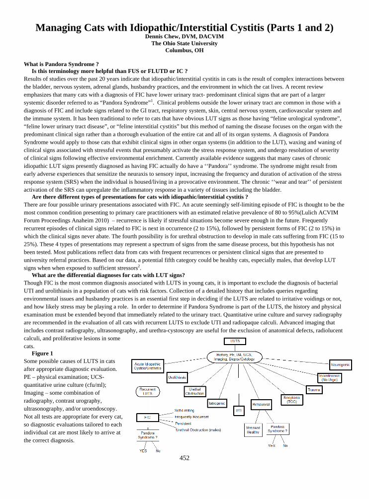
452
Managing Cats with Idiopathic/Interstitial Cystitis (Parts 1 and 2) Dennis Chew, DVM, DACVIM
The Ohio State University Columbus, OH
What is Pandora Syndrome ?
Is this terminology more helpful than FUS or FLUTD or IC ? Results of studies over the past 20 years indicate that idiopathic/interstitial cystitis in cats is the result of complex interactions between the bladder, nervous system, adrenal glands, husbandry practices, and the environment in which the cat lives. A recent review emphasizes that many cats with a diagnosis of FIC have lower urinary tract- predominant clinical signs that are part of a larger systemic disorder referred to as “Pandora Syndrome”1. Clinical problems outside the lower urinary tract are common in those with a diagnosis of FIC and include signs related to the GI tract, respiratory system, skin, central nervous system, cardiovascular system and the immune system. It has been traditional to refer to cats that have obvious LUT signs as those having “feline urological syndrome”, “feline lower urinary tract disease”, or “feline interstitial cystitis” but this method of naming the disease focuses on the organ with the predominant clinical sign rather than a thorough evaluation of the entire cat and all of its organ systems. A diagnosis of Pandora Syndrome would apply to those cats that exhibit clinical signs in other organ systems (in addition to the LUT), waxing and waning of clinical signs associated with stressful events that presumably activate the stress response system, and undergo resolution of severity of clinical signs following effective environmental enrichment. Currently available evidence suggests that many cases of chronic idiopathic LUT signs presently diagnosed as having FIC actually do have a ‘‘Pandora’’ syndrome. The syndrome might result from early adverse experiences that sensitize the neuraxis to sensory input, increasing the frequency and duration of activation of the stress response system (SRS) when the individual is housed/living in a provocative environment. The chronic ‘‘wear and tear’’ of persistent activation of the SRS can upregulate the inflammatory response in a variety of tissues including the bladder.
Are there different types of presentations for cats with idiopathic/interstitial cystitis ? There are four possible urinary presentations associated with FIC. An acute seemingly self-limiting episode of FIC is thought to be the most common condition presenting to primary care practitioners with an estimated relative prevalence of 80 to 95%(Lulich ACVIM Forum Proceedings Anaheim 2010) – recurrence is likely if stressful situations become severe enough in the future. Frequently recurrent episodes of clinical signs related to FIC is next in occurrence (2 to 15%), followed by persistent forms of FIC (2 to 15%) in which the clinical signs never abate. The fourth possibility is for urethral obstruction to develop in male cats suffering from FIC (15 to 25%). These 4 types of presentations may represent a spectrum of signs from the same disease process, but this hypothesis has not been tested. Most publications reflect data from cats with frequent recurrences or persistent clinical signs that are presented to university referral practices. Based on our data, a potential fifth category could be healthy cats, especially males, that develop LUT signs when when exposed to sufficient stressors2.
What are the differential diagnoses for cats with LUT signs? Though FIC is the most common diagnosis associated with LUTS in young cats, it is important to exclude the diagnosis of bacterial UTI and urolithiasis in a population of cats with risk factors. Collection of a detailed history that includes queries regarding environmental issues and husbandry practices is an essential first step in deciding if the LUTS are related to irritative voidings or not, and how likely stress may be playing a role. In order to determine if Pandora Syndrome is part of the LUTS, the history and physical examination must be extended beyond that immediately related to the urinary tract. Quantitative urine culture and survey radiography are recommended in the evaluation of all cats with recurrent LUTS to exclude UTI and radiopaque calculi. Advanced imaging that includes contrast radiography, ultrasonography, and urethra-cystoscopy are useful for the exclusion of anatomical defects, radiolucent calculi, and proliferative lesions in some cats.
Figure 1 Some possible causes of LUTS in cats after appropriate diagnostic evaluation. PE – physical examination; UCS- quantitative urine culture (cfu/ml); Imaging – some combination of radiography, contrast urography, ultrasonography, and/or uroendoscopy. Not all tests are appropriate for every cat, so diagnostic evaluations tailored to each individual cat are most likely to arrive at the correct diagnosis.
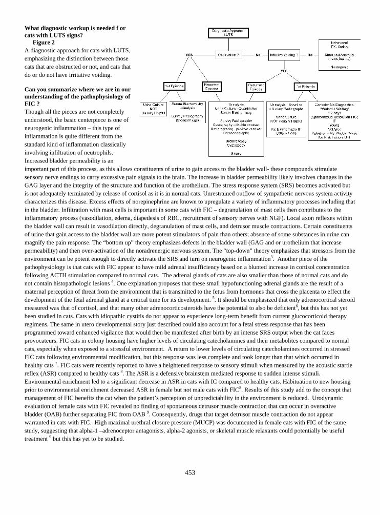
453
What diagnostic workup is needed f or cats with LUTS signs?
Figure 2 A diagnostic approach for cats with LUTS, emphasizing the distinction between those cats that are obstructed or not, and cats that do or do not have irritative voiding. Can you summarize where we are in our understanding of the pathophysiology of FIC ? Though all the pieces are not completely understood, the basic centerpiece is one of neurogenic inflammation – this type of inflammation is quite different from the standard kind of inflammation classically involving infiltration of neutrophils. Increased bladder permeability is an important part of this process, as this allows constituents of urine to gain access to the bladder wall- these compounds stimulate sensory nerve endings to carry excessive pain signals to the brain. The increase in bladder permeability likely involves changes in the GAG layer and the integrity of the structure and function of the urothelium. The stress response system (SRS) becomes activated but is not adequately terminated by release of cortisol as it is in normal cats. Unrestrained outflow of sympathetic nervous system activity characterizes this disease. Excess effects of norepinephrine are known to upregulate a variety of inflammatory processes including that in the bladder. Infiltration with mast cells is important in some cats with FIC – degranulation of mast cells then contributes to the inflammatory process (vasodilation, edema, diapedesis of RBC, recruitment of sensory nerves with NGF). Local axon reflexes within the bladder wall can result in vasodilation directly, degranulation of mast cells, and detrusor muscle contractions. Certain constituents of urine that gain access to the bladder wall are more potent stimulators of pain than others; absence of some substances in urine can magnify the pain response. The “bottom up” theory emphasizes defects in the bladder wall (GAG and or urothelium that increase permeability) and then over-activation of the noradrenergic nervous system. The “top-down” theory emphasizes that stressors from the environment can be potent enough to directly activate the SRS and turn on neurogenic inflammation3. Another piece of the pathophysiology is that cats with FIC appear to have mild adrenal insufficiency based on a blunted increase in cortisol concentration following ACTH stimulation compared to normal cats. The adrenal glands of cats are also smaller than those of normal cats and do not contain histopathologic lesions 4. One explanation proposes that these small hypofunctioning adrenal glands are the result of a maternal perception of threat from the environment that is transmitted to the fetus from hormones that cross the placenta to effect the development of the fetal adrenal gland at a critical time for its development. 5. It should be emphasized that only adrenocortical steroid measured was that of cortisol, and that many other adrenocorticosteroids have the potential to also be deficient6, but this has not yet been studied in cats. Cats with idiopathic cystitis do not appear to experience long-term benefit from current glucocorticoid therapy regimens. The same in utero developmental story just described could also account for a fetal stress response that has been programmed toward enhanced vigilance that would then be manifested after birth by an intense SRS output when the cat faces provocateurs. FIC cats in colony housing have higher levels of circulating catecholamines and their metabolites compared to normal cats, especially when exposed to a stressful environment. A return to lower levels of circulating catecholamines occurred in stressed FIC cats following environmental modification, but this response was less complete and took longer than that which occurred in healthy cats 7. FIC cats were recently reported to have a heightened response to sensory stimuli when measured by the acoustic startle reflex (ASR) compared to healthy cats 8. The ASR is a defensive brainstem mediated response to sudden intense stimuli. Environmental enrichment led to a significant decrease in ASR in cats with IC compared to healthy cats. Habituation to new housing prior to environmental enrichment decreased ASR in female but not male cats with FIC8. Results of this study add to the concept that management of FIC benefits the cat when the patient’s perception of unpredictability in the environment is reduced. Urodynamic evaluation of female cats with FIC revealed no finding of spontaneous detrusor muscle contraction that can occur in overactive bladder (OAB) further separating FIC from OAB 9. Consequently, drugs that target detrusor muscle contraction do not appear warranted in cats with FIC. High maximal urethral closure pressure (MUCP) was documented in female cats with FIC of the same study, suggesting that alpha-1 –adrenoceptor antagonists, alpha-2 agonists, or skeletal muscle relaxants could potentially be useful treatment 9 but this has yet to be studied.
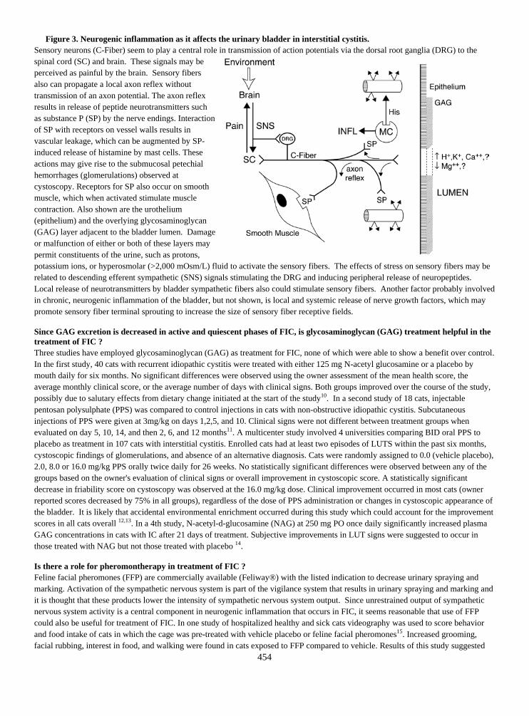
454
Figure 3. Neurogenic inflammation as it affects the urinary bladder in interstitial cystitis. Sensory neurons (C-Fiber) seem to play a central role in transmission of action potentials via the dorsal root ganglia (DRG) to the spinal cord (SC) and brain. These signals may be perceived as painful by the brain. Sensory fibers also can propagate a local axon reflex without transmission of an axon potential. The axon reflex results in release of peptide neurotransmitters such as substance P (SP) by the nerve endings. Interaction of SP with receptors on vessel walls results in vascular leakage, which can be augmented by SP-induced release of histamine by mast cells. These actions may give rise to the submucosal petechial hemorrhages (glomerulations) observed at cystoscopy. Receptors for SP also occur on smooth muscle, which when activated stimulate muscle contraction. Also shown are the urothelium (epithelium) and the overlying glycosaminoglycan (GAG) layer adjacent to the bladder lumen. Damage or malfunction of either or both of these layers may permit constituents of the urine, such as protons, potassium ions, or hyperosmolar (>2,000 mOsm/L) fluid to activate the sensory fibers. The effects of stress on sensory fibers may be related to descending efferent sympathetic (SNS) signals stimulating the DRG and inducing peripheral release of neuropeptides. Local release of neurotransmitters by bladder sympathetic fibers also could stimulate sensory fibers. Another factor probably involved in chronic, neurogenic inflammation of the bladder, but not shown, is local and systemic release of nerve growth factors, which may promote sensory fiber terminal sprouting to increase the size of sensory fiber receptive fields. Since GAG excretion is decreased in active and quiescent phases of FIC, is glycosaminoglycan (GAG) treatment helpful in the treatment of FIC ? Three studies have employed glycosaminoglycan (GAG) as treatment for FIC, none of which were able to show a benefit over control. In the first study, 40 cats with recurrent idiopathic cystitis were treated with either 125 mg N-acetyl glucosamine or a placebo by mouth daily for six months. No significant differences were observed using the owner assessment of the mean health score, the average monthly clinical score, or the average number of days with clinical signs. Both groups improved over the course of the study, possibly due to salutary effects from dietary change initiated at the start of the study10. In a second study of 18 cats, injectable pentosan polysulphate (PPS) was compared to control injections in cats with non-obstructive idiopathic cystitis. Subcutaneous injections of PPS were given at 3mg/kg on days 1,2,5, and 10. Clinical signs were not different between treatment groups when evaluated on day 5, 10, 14, and then 2, 6, and 12 months11. A multicenter study involved 4 universities comparing BID oral PPS to placebo as treatment in 107 cats with interstitial cystitis. Enrolled cats had at least two episodes of LUTS within the past six months, cystoscopic findings of glomerulations, and absence of an alternative diagnosis. Cats were randomly assigned to 0.0 (vehicle placebo), 2.0, 8.0 or 16.0 mg/kg PPS orally twice daily for 26 weeks. No statistically significant differences were observed between any of the groups based on the owner's evaluation of clinical signs or overall improvement in cystoscopic score. A statistically significant decrease in friability score on cystoscopy was observed at the 16.0 mg/kg dose. Clinical improvement occurred in most cats (owner reported scores decreased by 75% in all groups), regardless of the dose of PPS administration or changes in cystoscopic appearance of the bladder. It is likely that accidental environmental enrichment occurred during this study which could account for the improvement scores in all cats overall 12,13. In a 4th study, N-acetyl-d-glucosamine (NAG) at 250 mg PO once daily significantly increased plasma GAG concentrations in cats with IC after 21 days of treatment. Subjective improvements in LUT signs were suggested to occur in those treated with NAG but not those treated with placebo 14. Is there a role for pheromontherapy in treatment of FIC ? Feline facial pheromones (FFP) are commercially available (Feliway®) with the listed indication to decrease urinary spraying and marking. Activation of the sympathetic nervous system is part of the vigilance system that results in urinary spraying and marking and it is thought that these products lower the intensity of sympathetic nervous system output. Since unrestrained output of sympathetic nervous system activity is a central component in neurogenic inflammation that occurs in FIC, it seems reasonable that use of FFP could also be useful for treatment of FIC. In one study of hospitalized healthy and sick cats videography was used to score behavior and food intake of cats in which the cage was pre-treated with vehicle placebo or feline facial pheromones15. Increased grooming, facial rubbing, interest in food, and walking were found in cats exposed to FFP compared to vehicle. Results of this study suggested
455
that hospitalized cats exposed to FFP were calmer and more comfortable in their cages than cats exposed to vehicle. It has been our observation that some cats are very affected by FFP while in others the effect is minimal to nil. A randomized, double-blinded, placebo-controlled, crossover study was performed in 12 cats (9 of 12 completed the full study) with recurrent FIC, comparing once daily environmental treatment with FFP (Feliway®) or placebo; treatment was for 2 months and then switched to the other treatment for the next 2 months 16. This small number of cats exposed to FFP had fewer mean days displaying signs of cystitis, a reduced number of episodes of cystitis, and fewer negative behavioral traits, but this data did not achieve statistical significance for a difference over placebo treatment of the environment. Is there a role for amitriptyline or other tricyclic anti-depressant (or analgesic) TCA for the treatment of FIC ? In some cases YES. The need for this kind of therapy has dramatically lessened since we as a profession have become much more successful at implementing environmental modification, which usually works well without need for chronic drug therapy. We do prescribe amitriptyline for its beneficial effects for cats with FIC that have frequent recurrences or persistent LUT signs AFTER the client’s best efforts to implement environmental enrichment have failed to improve the cat’s clinical signs. This type of therapy should NOT be undertaken for an initial episode of FIC or a “flare” of signs that occur infrequently. We sometimes prescribe amitriptyline for cats owned by clients that are considering euthanasia for their cat with FIC – this can sometimes allow the client to see early benefits while implementing environmental enrichment. Maximal beneficial effects of TCA, if any, often require weeks to months to be observed and in general should not be abruptly discontinued (so called “abrupt withdrawal syndrome”). Treatment series of FIC with amitriptyline has been reported 3 times, 1 study of chronic FIC (frequently recurrent or persistent signs) and 2 of acute bouts of FIC. In the chronic study, 15 cats were enrolled with FIC that failed to respond to other treatments; no placebo group was treated. Amitriptyline treatment (10 mg PO every 24 hours in the evening) successfully decreased clinical signs of severe recurrent FIC in 9 of 15 cats treated for 12 months (11 of 15 cats for the first 6 months). Somnolence, weight gain, decreased grooming, and transient cystic calculi were observed during treatment in some cats. Despite clinical improvement, cystoscopic abnormalities persisted in all cats at the 6- and 12-month evaluations 17. In one short term study, 31 untreated male and female cats with acute (<14 days signs), nonobstructive, idiopathic lower urinary tract disease were enrolled in a placebo controlled study. Cats were hospitalized and treated with 5mg amitriptyline or a placebo daily for 7 days and then treatment discontinued. Clinical signs and hematuria resolved similarly in both groups of treated cats by day 8. Cats were evaluated in the clinic 1 month later and by questions over the telephone 6, 12, and 24 months after treatment. Clinical signs recurred faster and more frequently (10.5 vs. 2.4 events/1,000 days) in the amitriptyline treated cats, a finding likely attributable to the abrupt withdrawal of amitriptyline treatments after 7 days- there was no difference in recurrence when the first 21 days were excluded from the analysis 18. In another short-term study of FIC, amitriptyline at 10 mg once daily per os (11) or placebo (13) was given for 7 days by owners at home. All cats were also treated with amoxicillin BID for 7 days. The severity of clinical signs was assessed at days 0, 7, and 14 – no significant difference was found between amitriptyline and placebo treated cats of this study19. How do we treat an acute episode of LUT signs for either its first time, or an infrequently recurrent event ? We treat nearly all FIC cats of this type with a combination of buprenorphine and acepromazine PO for 5 to 7 days. The combination of an analgesic and a tranquilizer with properties that also decrease urethral tone seem like a compassionate and appropriate choice of treatment. It is likely that the tranquilizer reduces the activity of the autonomic nervous system which is useful in the initial treatment of FIC. We believe that this helps to acutely decrease clinical signs in cats with acute episodes of FIC or flares of chronic FIC, though this has not been specifically studied. Whether this regimen reduces future episodes of FIC has also not been tested. We take the opportunity at the first visit to discuss with the owners that even a first event of FIC may be associated with recurrence and that there may be steps that can be taken to reduce this likelihood (not yet studied in a prospective way) when environmental enrichment and modification are successfully implemented. What analgesic treatments should I consider? The best approach to analgesia for bladder pain (visceral) has yet to be determined. Butorphanol has been used, but its effects are less long-lived or potent than those of buprenorphine 20,21 Sustained release formulations of buprenorphine have recently become available that can provide up to 72 hours of therapeutic drug levels for pain relief following a single injection. Fentanyl patches have been used in rare cases in which bladder pain was assessed as severe. Should I consider NSAID treatment to provide anti-inflammatory and analgesic effects? Anecdotal reports of the usefulness of non-steroidal anti-inflammatory drugs (NSAID)s, especially meloxicam and ketoprofen, abound, but no studies of safety or effectiveness are available for review. Some specialists have prescribed piroxicam for use on alternate days, but there are no controlled clinical trials of its effectiveness or safety. NSAIDs are not commonly used for treatment of interstitial cystitis in humans. NSAIDs that are licensed for use in cats list indications for pre-emptive pain management, usually as a single treatment before anesthesia and surgery. Chronic use of NSAIDs in cats can be dangerous due to the possibility for
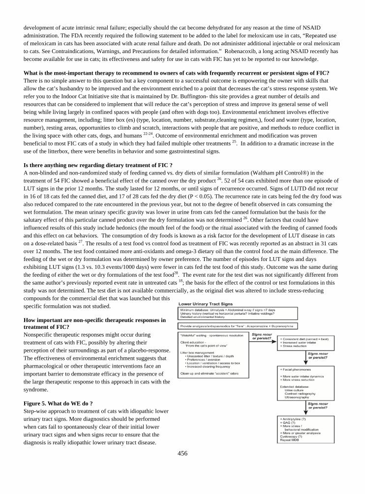
456
development of acute intrinsic renal failure; especially should the cat become dehydrated for any reason at the time of NSAID administration. The FDA recently required the following statement to be added to the label for meloxicam use in cats, “Repeated use of meloxicam in cats has been associated with acute renal failure and death. Do not administer additional injectable or oral meloxicam to cats. See Contraindications, Warnings, and Precautions for detailed information.” Robenacoxib, a long acting NSAID recently has become available for use in cats; its effectiveness and safety for use in cats with FIC has yet to be reported to our knowledge. What is the most-important therapy to recommend to owners of cats with frequently recurrent or persistent signs of FIC? There is no simple answer to this question but a key component to a successful outcome is empowering the owner with skills that allow the cat’s husbandry to be improved and the environment enriched to a point that decreases the cat’s stress response system. We refer you to the Indoor Cat Initiative site that is maintained by Dr. Buffington- this site provides a great number of details and resources that can be considered to implement that will reduce the cat’s perception of stress and improve its general sense of well being while living largely in confined spaces with people (and often with dogs too). Environmental enrichment involves effective resource management, including; litter box (es) (type, location, number, substrate,cleaning regimen,), food and water (type, location, number), resting areas, opportunities to climb and scratch, interactions with people that are positive, and methods to reduce conflict in the living space with other cats, dogs, and humans 22-24. Outcome of environmental enrichment and modification was proven beneficial to most FIC cats of a study in which they had failed multiple other treatments 25. In addition to a dramatic increase in the use of the litterbox, there were benefits in behavior and some gastrointestinal signs. Is there anything new regarding dietary treatment of FIC ? A non-blinded and non-randomized study of feeding canned vs. dry diets of similar formulation (Waltham pH Control®) in the treatment of 54 FIC showed a beneficial effect of the canned over the dry product 26. 52 of 54 cats exhibited more than one episode of LUT signs in the prior 12 months. The study lasted for 12 months, or until signs of recurrence occurred. Signs of LUTD did not recur in 16 of 18 cats fed the canned diet, and 17 of 28 cats fed the dry diet (P < 0.05). The recurrence rate in cats being fed the dry food was also reduced compared to the rate encountered in the previous year, but not to the degree of benefit observed in cats consuming the wet formulation. The mean urinary specific gravity was lower in urine from cats fed the canned formulation but the basis for the salutary effect of this particular canned product over the dry formulation was not determined 26. Other factors that could have influenced results of this study include hedonics (the mouth feel of the food) or the ritual associated with the feeding of canned foods and this effect on cat behaviors. The consumption of dry foods is known as a risk factor for the development of LUT disease in cats on a dose-related basis 27. The results of a test food vs control food as treatment of FIC was recently reported as an abstract in 31 cats over 12 months. The test food contained more anti-oxidants and omega-3 dietary oil than the control food as the main difference. The feeding of the wet or dry formulation was determined by owner preference. The number of episodes for LUT signs and days exhibiting LUT signs (1.3 vs. 10.3 events/1000 days) were fewer in cats fed the test food of this study. Outcome was the same during the feeding of either the wet or dry formulations of the test food28. The event rate for the test diet was not significantly different from the same author’s previously reported event rate in untreated cats 18; the basis for the effect of the control or test formulations in this study was not determined. The test diet is not available commercially, as the original diet was altered to include stress-reducing compounds for the commercial diet that was launched but this specific formulation was not studied. How important are non-specific therapeutic responses in treatment of FIC? Nonspecific therapeutic responses might occur during treatment of cats with FIC, possibly by altering their perception of their surroundings as part of a placebo-response. The effectiveness of environmental enrichment suggests that pharmacological or other therapeutic interventions face an important barrier to demonstrate efficacy in the presence of the large therapeutic response to this approach in cats with the syndrome. Figure 5. What do WE do ? Step-wise approach to treatment of cats with idiopathic lower urinary tract signs. More diagnostics should be performed when cats fail to spontaneously clear of their initial lower urinary tract signs and when signs recur to ensure that the diagnosis is really idiopathic lower urinary tract disease.
457
Properly controlled clinical trials may provide better approaches to treatment in the future, but this is what we do in the interim. “Pearls” Pandora Syndrome – aka feline interstitial/idiopathic cystitis (FIC)
1. Signs of urinary urgency during FIC may be expressions of a systemic disease created by a highly active outflow (unrestrained) from the sympathetic nervous system in response to stressors (provocateurs) .
2. When multi-modal environmental modification (including environmental enrichment) is effectively implemented, treatment with drugs is RARELY NEEDED.
3. Stress up-regulates the inflammatory potential of several organs, including the bladder. 4. Bacterial urinary infections (UTI) are rarely identified in cats with signs of lower urinary tract disease, unless they have
specific risk factors (U-cath within last 6 months, perineal urethrostomy, dilute urine – CKD, diabetes mellitus, hyperthyroidism)
5. The term “Pandora Syndrome” should help to remind the clinician that LUT signs may be part of a bigger picture that involves other organ systems.
6. We advocate the use of analgesia (buprenorphine) during acute episodes of FIC. 7. We use tranquilization with acepromazine in combination with buprenorphine in most of our cases of non-obstructive
episodes. 8. On occasion, the use of amitriptyline can be useful in the treatment of FIC. 9. The use of GAG (glycosaminoglycan) supplementation has failed to show an effect superior to placebo in several studies of
FIC treatment. 10. The use of feline facial phaeromones has not been shown to be superior to placebo in the treatment of FIC. 11. The feeding of as much wet food as possible in the diet is advocated by some for its protective effect on the recurrence of
the signs of FIC, and may be helpful as long as it does not result in additional threat to the cat. 12. There is no indication for surgery in non-obstructive FIC. 13. When surgery is performed in patients with FIC, obtain a full thickness bladder biopsy to allow evaluation of mast cells with
special stains (toluidine blue). 14. Sometimes a so-called “placebo” treatment actually can have a positive effect between the cat, the owner, and the
environment such that a positive outcome is achieved. 15. In most cases, antibiotic treatment does not have a role in the treatment of FIC. 16. Treatment of FIC with glucocorticosteroids has not shown an effect greater than that of placebo in limited study. 17. Chronic treatment of FIC with NSAIDs is NOT ADVOCATED due to the high sensitivity of the cat to sustain renal injury
with this class of drugs, especially if there is any tendency toward dehydration. References 1. Buffington CA. Idiopathic cystitis in domestic cats--beyond the lower urinary tract. Journal of veterinary internal medicine / American College of Veterinary Internal Medicine 2011;25:784-96. 2. Stella JL, Lord LK, Buffington CA. Sickness behaviors in response to unusual external events in healthy cats and cats with feline interstitial cystitis. Journal of the American Veterinary Medical Association 2011;238:67-73. 3. Westropp JL, Buffington CA. In vivo models of interstitial cystitis. The Journal of urology 2002;167:694-702. 4. Westropp JL, Welk KA, Buffington CA. Small adrenal glands in cats with feline interstitial cystitis. The Journal of urology 2003;170:2494-7. 5. Buffington CA. Developmental influences on medically unexplained symptoms. Psychother Psychosom 2009;78:139-44. 6. Dimitrakov J, Joffe HV, Soldin SJ, Bolus R, Buffington CA, Nickel JC. Adrenocortical hormone abnormalities in men with chronic prostatitis/chronic pelvic pain syndrome. Urology 2008;71:261-6. 7. Westropp JL, Kass PH, Buffington CA. Evaluation of the effects of stress in cats with idiopathic cystitis. American journal of veterinary research 2006;67:731-6. 8. Hague DW, Stella JL, Buffington CA. Effects of interstitial cystitis on the acoustic startle reflex in cats. American journal of veterinary research 2013;74:144-7. 9. Wu CH, Buffington CA, Fraser MO, Westropp JL. Urodynamic evaluation of female cats with idiopathic cystitis. American journal of veterinary research 2011;72:578-82. 10. Gunn-Moore DA, Shenoy CM. Oral glucosamine and the management of feline idiopathic cystitis. Journal of feline medicine and surgery 2004;6:219-25. 11. Wallius BM, Tidholm AE. Use of pentosan polysulphate in cats with idiopathic, non-obstructive lower urinary tract disease: a double-blind, randomised, placebo-controlled trial. Journal of feline medicine and surgery 2009;11:409-12. 12. Chew DJ, Bartges JW, Adams LG, Kruger JM, Buffington CAT. Evaluation of Pentosan Polysulfate Sodium in the Treatment of Feline Interstitial Cystitis: A Randomized, Placebo-Controlled Clinical Trial. J Urology 2011;185:e382 (abstract 952). 13. Chew DJ, Bartges JW, Adams LG, Kruger JM, Buffington CT. Randomized trial of pentosan polysulfate sodium for reatment of feline interstitial (Idiopathic) cystitis Journal of veterinary internal medicine / American College of Veterinary Internal Medicine 2009;23.
458
14. Panchaphanpong J, Asawakarn T, Pusoonthornthum R. Effects of oral administration of N-acetyl-d-glucosamine on plasma and urine concentrations of glycosaminoglycans in cats with idiopathic cystitis. American journal of veterinary research 2011;72:843-50. 15. Griffith CA, Steigerwald ES, Buffington CA. Effects of a synthetic facial pheromone on behavior of cats. Journal of the American Veterinary Medical Association 2000;217:1154-6. 16. Gunn-Moore DA, Cameron ME. A pilot study using synthetic feline facial pheromone for the management of feline idiopathic cystitis. Journal of feline medicine and surgery 2004;6:133-8. 17. Chew DJ, Buffington CA, Kendall MS, DiBartola SP, Woodworth BE. Amitriptyline treatment for severe recurrent idiopathic cystitis in cats. Journal of the American Veterinary Medical Association 1998;213:1282-6. 18. Kruger JM, Conway TS, Kaneene JB, et al. Randomized controlled trial of the efficacy of short-term amitriptyline administration for treatment of acute, nonobstructive, idiopathic lower urinary tract disease in cats. Journal of the American Veterinary Medical Association 2003;222:749-58. 19. Kraijer M, Fink-Gremmels J, Nickel RF. The short-term clinical efficacy of amitriptyline in the management of idiopathic feline lower urinary tract disease: a controlled clinical study. Journal of feline medicine and surgery 2003;5:191-6. 20. Warne LN, Beths T, Holm M, Carter JE, Bauquier SH. Evaluation of the perioperative analgesic efficacy of buprenorphine, compared with butorphanol, in cats. Journal of the American Veterinary Medical Association 2014;245:195-202. 21. Steagall PV, Monteiro-Steagall BP, Taylor PM. A review of the studies using buprenorphine in cats. Journal of veterinary internal medicine / American College of Veterinary Internal Medicine 2014;28:762-70. 22. Herron ME. Advances in understanding and treatment of feline inappropriate elimination. Top Companion Anim Med 2010;25:195-202. 23. Herron ME, Buffington CA. Feline focus-environmental enrichment for indoor cats. Compend Contin Educ Vet 2010;32:E1-5. 24. Herron ME, Buffington CA. Environmental enrichment for indoor cats: implementing enrichment. Compend Contin Educ Vet 2012;34:E3. 25. Buffington CA, Westropp JL, Chew DJ, Bolus RR. Clinical evaluation of multimodal environmental modification (MEMO) in the management of cats with idiopathic cystitis. Journal of feline medicine and surgery 2006;8:261-8. 26. Markwell PJ, Buffington CA, Chew DJ, Kendall MS, Harte JG, DiBartola SP. Clinical evaluation of commercially available urinary acidification diets in the management of idiopathic cystitis in cats. Journal of the American Veterinary Medical Association 1999;214:361-5. 27. Buffington CA. External and internal influences on disease risk in cats. Journal of the American Veterinary Medical Association 2002;220:994-1002. 28. Kruger J, Lulich J, Merrills J, et al. A year-long prospective, randomized, double-masked study of nutrition on feline idopathic cystitis(FIC). American College of Veterinary Internal Medicine Forum 2013; Seattle
459
Updates on Managing Male Cats with Urethral Obstruction Dennis Chew, DVM, DACVIM
The Ohio State University Columbus, OH
Pathophysiology of urethral obstruction (UO) Thrity-nine % to 67% of male cats evaluated with lower urinary tract signs have been reported to have urethral obstruction.1-4 Male cats with urethral obstruction (UO) were described to have urethral plugs as the most common cause in early reports,3 but recent reports emphasize idiopathic causes.1,2,5 In one study the cause of obstruction was considered to be idiopathic in all 82 cats studied,6 but other studies report plugs, uroltihiasis and UTI in decreasing order behind idiopathic causes for UO.1,2,5 When plugs do form, it is likely that they are extensions of the process leading to feline idiopathic/intersititial cystitis (FIC). This is consistent with findings from an unpublished study at The Ohio State University using urethroscopy at the time of initial evaluation in which plugs were rarely identified. Urethral plugs have minimal intrinsic cohesive structure but often are cylinder-shaped after extrusion from the urethra. Urethral plugs are fundamentally different from calculi that lodge within the urethra (i.e., urethroliths). Uroliths have an organized internal structure with much less matrix, and are not easily compressed or distorted. Urethral plugs consist largely of matrix mucoprotein with embedded minerals. The predominant mineral composition in most plugs is magnesium ammonium phosphate hexahydrate (i.e., struvite). This is true despite the fact that cats form calcium oxalate and struvite uroliths with nearly equal frequency. Most plugs are assumed to lodge within the penile urethra, but obstructions also can occur at more proximal sites. Definitive diagnosis of a urethral plug requires retrieval of the plug. Supportive evidence for the presence of a urethral plug can be seen on radiographs in some cats with UO. Previously, the crystalline-matrix hypothesis proposed that plugs formed secondary to precipitation of struvite crystals in the urine that then became embedded in a matrix. According to this hypothesis, plugs created UO and urethritis. It is now hypothesized that plugs form as a consequence of underlying idiopathic urethritis and cystitis (i.e., inflammation occurs first, followed by plug formation).
Some cats have signs of non-obstructive idiopathic/interstitial cystitis before UO, while many cats have lower urinary tract signs after relief of UO. Obstruction can be secondary to functional urethral spasm in addition to swelling of the urethra due to edema and hemorrhage. Pathologic or neurogenic processes cause contraction of the circular smooth or skeletal muscle of the urethra or both. Stimulation of adrenoreceptors (particularly α-1) within the urethra increases urethral tone in normal cats. Pain and stress after UO increase sympathetic outflow from the central nervous system which can lead to additional urethral spasm.
Bacterial urinary tract infection (UTI) is very uncommon before urethral catheterization.3,7 UTI deserves more consideration in cats with recurrent UO that have undergone urinary instrumentation. Urethral stricture may occur, especially in cats that have had previous indwelling urinary catheters and for those with severe recurrent episodes of non-obstructive idiopathic/interstitial cystitis. Neoplasia of the urethra or bladder neck is rare. Urinary catheter fragment foreign body in urethra or bladder is rare, as is phimosis as a cause for UO.
Signalment, history, physical examination Approximately 75% of cats presented with UO are experiencing their first episode.6,8 Median duration of clinical signs before initial presentation was 3 days in a study of 223 cats. Signs include those of cystitis and partial obstruction before development of complete obstruction. The majority of cats with UO are relatively stable however, approximately 10% are critically ill.
Severe bradycardia (< 100 bpm) from the effects of hyperkalemia has been reported in 5% of cases, moderate bradycardia (100-140 bpm) in 6% of cases and mild bradycardia (140-160 bpm) in 12% of cases; arrhythmias were detected in 11% of cases. Fifty % of cats can be expected to have normal body temperature, hypothermia in about 40% and hyperthermia in 10%. Rectal temperature < 95-96.6°F or heart rate < 120 bpm was the most the accurate predictor of severe hyperkalemia. A combination of hypothermia and bradycardia was 98 to 100% predictive for severe hyperkalemia (> 8.0 mEq/L).9 Twitching or seizures is very uncommon (0.5%) and related to ionized hypocalcemia. Systemic blood pressure most often is normal.10 Mean arterial pressure correlated inversely with serum potassium and directly with total serum calcium concentrations. Major abnormalities on physical examination and serum biochemistry were encountered despite normal blood pressure in this study.
Diagnostics A recent report noted that darker red urine observed at the time of urinary catheter placement was associated with azotemia, hyperkalemia, and lower USG. Color of the urine was not associated with the presence or absence of urinary stones.11
Hyperkalemia does not occur in isolation and often is accompanied by acidosis and low serum ionized calcium concentration. Serum potassium concentrations ranged from 3.4 to 10.5 mEq/L in 199 cats. Six % were below the reference range; 41% were above the reference range, and 53% in the reference range. Serum potassium concentration was < 6.0 mEq/L in 66% of cases, > 6.0 but < 8.0 mEq/L in 12% of cases, > 8.0 but < 10.0 mEq/L in 12% of cases, and > 10.0 mEq/L in < 1% of cases. Hyperkalemia most often was encountered with acidosis (pH < 7.2 in 74% of cases) and low serum ionized calcium concentration (< 1.0 mmol/L in 75% of cases).
Approximately 33% of cats with UO are expected to have clinically relevant hypocalcemia based on serum ionized calcium concentration. Serum ionized calcium concentration was below the reference range in 34%, above the reference range in 19%, and in
460
the reference range in 47%. Serum ionized calcium concentration was > 1.2 mmol/L (> 4.8 mg/dL) in 23%, > 1.0 but < 1.2 mmol/L (> 4.0 but < 4.7 mg/dL) in 57%, > 0.8 but < 1.0 mmol/L (> 3.2 but < 4.0 mg/dL) in 14%, ≤ 0.8 mmol/L (≤ 3.2 mg/dL) in 6%. Serum total calcium concentration in 51 cats was below the reference range in 39%, above the reference range in 0%, and within the reference range in 61%. Cats with low serum total calcium concentrations had moderate to severely decreased serum ionized calcium concentrations.8,12 In one study, more cats were found to have hypocalcemia when defined by measurement of serum ionized calcium concentration (75%) than when defined by serum total calcium concentration (27%).12 Survival of cats with UO was influenced by ionized calcium status in another study. The median concentration of ionized calcium in survivors was 1.08 mmol/l (range 0.65 to 1.28 mmol/L) and in non-survivors was 0.88 mmol/l (0.66 to 1.11 mmol/L); P = 0.037). Hypocalcemia was detected in 51% of survivors vs 100% of non-survivors; P = 0.024.6
Struvite crystals may be observed at the time of obstruction, especially if urine pH is alkaline. The presence and amount of struvite crystalluria preceding UO has not been reported. Struvite crystalluria can be expected from any condition associated with urinary pH increased above 6.7. Crystals are more likely to be secondary to urine stasis or alkaline urine pH (secondary to sterile inflammation with extravasation of plasma proteins into urine) than a primary cause of obstruction. Struvite crystalluria was greater in male cats with obstruction than in male cats without obstruction (P 0.051), though cause or effect of the crystalluria was not established in one study. Struvite crystalluria was not associated with hematuria, proteinuria, or pyuria but was associated with urinary pH in this same study.5
Nearly all cats with UO have sterile urine on original presentation for obstruction. Zero of 18 cats with UO in one study7 and in 0/36 cats in another study soon to be published out of The Ohio State University (Dr. Ed Cooper OSU - personal communication 2014) had bacterial growth. Bacteria were isolated from urine collected through the urinary catheter at initial presentation in 14% of cats (14/192) in one study, but quantitative methods as to cfu/mL were not used. Many of these cats were referred with an indwelling urinary catheter already in place.13 Only 1 of 32 cats in another study had a positive urine culture from a cystocentesis sample at the time of UO relief.14 Bacterial culture at the time of urinary catheter removal is more likely to identify pathogenic bacteria. Isolation of bacteria from cats with a previous history of UO is more likely than isolation from cats suffering an initial episode. Imaging of cats during/after UO All cats with UO should have radiography to determine if urolithiasis is contributing to obstruction. Attention is usually centered to determine the presence of urinary stones in the bladder and/or urethra. It is very important to include the perineal region in the radiographs to identify urethral calculi. Evaluation of the kidneys and ureters is important to be sure nephroliths or ureteroliths are not part of the overall process, because upper urinary tract involvement can markedly affect the overall prognosis. Free fluid resulting in a loss of abdominal detail can be seen in some cats with severely distended and highly permeable (“leaky”) urinary bladders. A small amount of free abdominal fluid may be identified at initial presentation that is more easily detected on ultrasonography. In cats with recurrent UO, contrast radiography and ultrasonography may be informative as to the underlying diagnosis. Positive contrast urethrography is especially useful to disclose urethral trauma, urethral perforation, or urethral stricture, especially after recent instrumentation of the urethra. Radiography is the gold standard imaging method for the detection of urethral stones as ultrasonography only examines the most proximal portion of the urethra. If only ultrasonograpy is available to image the urinary tract (limitations of equipment, personnel, or cost), then it is advisable to perform the sonogram before AND after reverse flushing of the urethra in order to detect the presence of small stones that may now appear in the bladder after hydropulsion that were not initially visible. This however does not exclude the presence of stones still within the urethra.
Caudal abdominal effusion was detected in 10 of 34 cats on radiographs after placement of a urethral catheter without associated cystocentsis.15 Nineteen of 34 cats with UO that underwent abdominal radiography had signs of abdominal effusion before or after cystocentesis and passage of a urinary catheter. Prior to cystocentesis, 11 of 20 cats had abdominal effusion in the same study.14In another study in which therapeutic cystocentesis was used as the sole treatment to relieve bladder pressure, 8 of 15 had evidence for abdominal effusion after bladder pressure was first relieved.16 In yet another study, 87 cats underwent abdominal ultrasonography within 24 hours of the relief of UO by passage of a urethral catheter and no use of cystocentesis.17 Hyperechogenic pericystic fat and pericystic effusion were each observed in 60% of these cats. Ninety % of evaluated cats had bladder thickening, 20% had suspended linear strands, and over 50% of cats had either moderate or severe increases in urinary sediment or hyperechogenicity. Cystolithiasis was documented in 47% of these cats. This frequency is much higher than that in another report in which only 2 of 35 cats were found to have stones (radiography in 34 cats and ultrasonography in 3 cats).14 The reason for this disparity between ULS and radiography in detection in cystolithiasis is not obvious. ULS could be more sensitive in the detection of uroliths, but ultrasonography and radiography has not been compared in the same cats with UO at the same time of their clinical presentation, before or after instrumentation. It is also possible that more stones were detected in the study using ultrasonography since these images were acquired after urethral flushing which could have retropulsed urethral stones into the bladder. Eight cats with pseudomembranous cystitis associated with UO have been described in two reports.17,18Thick echogenic septa were described traversing the bladder lumen. These
461
bands could represent sloughing of necrotic areas of the bladder into the lumen and they were associated with fibrinous exudate, blood clots, and necrotic debris.
It has long been taught that acute UO in male cats adversely affects renal function but does not create structural changes in the kidneys. It has been known for decades that palpably enlarged kidneys are detected during physical examination in some cats before relief of UO. In cats with UO undergoing ultrasonography, either unilateral or bilateral renomegaly was detected in 42 %, pyelectasia in 60 % (10% > 3.4 mm), and perirenal effusion (retroperitoneal) in 35% of the cases. Ureteral dilatation was detected in 24%. How rapidly these changes resolve has not yet been reported.17 Relief of obstruction due to plugs or idiopathic causes Decompressive (therapeutic) cystocentesis is the next step recommended to perform after sedation and IV catheter placement. The benefits of decompressive cystocentesis outweigh potential adverse effects. Decompressive cystocentesis has been considered controversial by some clinicians who fear that bladder rupture will occur or that urine will continue to leak from the bladder. No adverse effects were observed in a recent report of 47 UO male cats that underwent decompressive cystocentsis.14 Cystocentesis to empty the bladder should be performed as soon as possible in cats with very large bladders to prevent rupture of the bladder and to allow renal excretory function to resume. Cystocentesis allows for rapid reduction of urinary tract pressure and resumption of GFR compared to catheterization, which can take considerable time. Decompressive cystocentesis may stabilize the cat before anesthesia for urinary catheter placement. Relief of bladder pressure before urethral catheterization also may facilitate efforts to dislodge urethral plugs, and allows collection of a superior urine sample for analysis before manipulation of the urinary tract and contamination by irrigation solutions.
Some leakage of urine immediately after decompressive cystocentesis may occur, especially if the bladder is not adequately emptied. The use of a 22-gauge needle on an extension set or use of a butterfly needle can minimize trauma and urine leakage during the procedure. In one study, the median volume of urine removed by urinary catheter at the time initial obstruction was relieved in 28 cats was 85 mL (range, 35 to 280 mL).10 Plain abdominal radiographs (including the perineal region) should be obtained after decompressive cystocentesis to identify mineralized plugs, urethral calculi, or cystic calculi. Some clinicians obtain radiographs after catheter passage, but the presence of an indwelling urinary catheter can obscure the presence of urethral calculi.
Standard epidural techniques require special expertise and training but a new simplified method using sacro-coccygeal placement of local anesthetic to allow urethral catheterization and pain management appears promising.19 This technique produces anesthesia to the perineum, penis, urethra, colon, and anus within 5 minutes of preservative-free lidocaine injection and lasts up to 60 minutes. The authors of this study concluded that relief of urethral obstruction was easier and quicker during placement of the urethral catheter, presumably associated with urethral relaxation. Cats of this study received pre-medication protocols but not full anesthesia. Cats did not appear to struggle during catheterization, flushing, or suturing after the lidocaine infusion and appeared to be less painful after catheter placement.
Studies in cats have shown that indwelling polyvinyl catheters create less urethral trauma and inflammation than do indwelling polypropylene catheters. Silicone urinary catheters have not been specifically studied in cats. Do not administer glucocorticoids to a cat while an indwelling urinary catheter is in place. The risk for bacterial pyelonephritis is great in this setting and glucocorticoids are unlikely to control urethritis in this setting (i.e. continuous trauma from an indwelling catheter).20 The use of antibiotics does not prevent the development of UTI in patients with indwelling urinary catheters. Do not prescribe antibiotics while a urinary catheter is in place (unless you have documented by bacterial culture that a UTI already is present). Antibiotic use may promote development of resistant isolates when UTI does develop. Consider culturing the urine when the urinary catheter is removed. This recommendation is supported by the finding that 6 of 18 cats developed significant bacteriuria (3/6 at 24 hours and another 3/6 at 48 hours) within 48 hours while the indwelling urinary catheter was in place.7 Recurrent UO at day 30 was significantly less common when the indwelling urinary catheter was left in place for more hours, though the median times were similar between those with recurrence and those that did not recur.15
The chronic prognosis for recurrence of LUTD signs following relief of UO is guarded. Eight of 22 (36%) cats with idiopathic UO re-obstructed after a median of 17 days in one study whereas 3 of 7 (43%) cats with UO associated with urethral plugs re-obstructed within 7 months. Recurrent obstruction was the cause for euthanasia in 21% of cats in this study.2 The recurrence rate was 22% at 6 months and 24% at 2 years.6 Ten of 68 cats were reported to developed recurrent UO within 30 days of release from the hospital in another study.15 In an older study, the recurrence rate was 35% within 6 months.21 No studies on recurrence rates for UO have been reported prospectively after implementation of aggressive environmental modification. Recurrence rates may be lower in cats for which environmental modification can be adequately implemented. A small number of cats develop urethral strictures. This is a complication that occurred in 11% of affected cats in one study.22 Some cats develop bacterial UTI after instrumentation of their urinary tract (i.e. catheterization) and we have observed positive urine culture in some cats as late as 6 months after relief of UO. Signs of ongoing idiopathic cystitis are expected in 30-50% of cats that have had an episode of UO. In one study, 50% of cats with idiopathic UO developed lower urinary tract signs after relief of obstruction.2 In a study of 68 cats treated for UO, 50 cats had lower
462
urinary tract signs following release from the hospital. Pollakiuria (50%), stranguria (46%) and periuria (40%) were the most common clinical signs. Clinical signs lasted ≥ 7 days in 29 of 68 cats.15
Non-conventional treatment for urethral obstruction in male cats
Non-invasive non-instrumentation treatment protocol A report describing a method for relief of urethral obstruction in male cats without the use of urethral catheterization was recently described.16 The reported treatment protocol was proposed for use only as an alternative to euthanasia due to financial constraints of owners unable to afford conventional treatment costs. Conventional treatment with passage of a urinary catheter and IV fluid infusion in the hospital was offered as the first choice. This non-invasive approach is not meant for cats with urethral calculi or those with severe metabolic derangements. The severity of azotemia does not determine use of this protocol. A plain lateral abdominal radiograph is taken to exclude calculi. Decompressive cystocentesis is performed initially and then as needed up to every 8 hours. The urethra is not irrigated or catheterized, though the distal penis is gently massaged. No IV catheter is placed and IV fluids are not administered. Drug treatments include: acepromazine (0.25 mg IM or 2.5 mg PO q8h), buprenorphine (0.075 mg PO q8h), medetomidine (0.1mg IM q24h if no urinations are noted in the first 24 hours). The cat is placed in a quiet, low stress environment. Some fluids may be given subcutaneously as needed, but the goal is to avoid excessive urine production from full hydration. Treatment success was defined as spontaneous urination within 72 hours and subsequent discharge from the hospital. Successful discharge from the hospital occurred in 11/15 cats (73%). Treatment failure occurred in 4/15 (27%) cats due to uroabdomen (3) or hemoabdomen (1). Cats that experienced treatment failure had significantly higher serum creatinine concentrations. At necropsy, severe bladder inflammation was found, but there was no evidence of bladder rupture.
Atracurium The intraurethral installation of atracurium besylate was compared to that of physiological saline prior to retrograde flushing of the urethra. Atracurium besylate is a curare derivative that provides neuromuscular blockade of striated muscles by antagonizing acetylcholine at the nicotinic receptor in the neuromuscular junction. Atracurium besylate is rapidly inactivated by plasma esterases or by spontaneous degradation and does not depend on the liver or kidneys for excretion. Atracuium was first diluted from 10 mg/dl to 0.5 mg/dl and then injected under steady gentle pressure for 5 minutes while the external urethral orifice was occluded. Sixty-four percent of cats treated with atracurium were unobstructed during the first hydropulsion attempt compared to 15% of cats receiving the saline installation prior to flushing. The mean time to relieve obstruction was 21 seconds in those receiving atracuriurm compared to 235 seconds for those receiving the saline control.23
Lidocaine The recurrence rate and clinical signs for UO in 26 cats were determined at 2 weeks, 1 month, and 2 months following intravesical installation of lidocaine vs placebo once daily for 3 days through the indwelling urinary catheter. The recurrence rate for obstruction (58% [7/12] in the lidocaine group and 57% [8/14]) in the control group and magnitude of clinical signs were not different between treatment groups.24
Prazosin vs phenoxybenzamine In a recent report of UO cats, overall recurrent obstruction at 24 hours occurred in 21/192 cats(10.9%) and at 30 days in 37/157 (23.6% )cats.13 The recurrence rate in cats treated with prazosin was 10/140 (7.1%) and 20/110 (18.8%) at 24 hours and 30 days following urinary catheter removal compared to 10/46 (21.74%) at 24 hours and 16/41 (39.02%) at 30 days in cats treated with phenoxybenzamine, which was different statistically. Recurrent urethral obstruction is most likely to occur within the first 7 days following urinary catheter removal in most studies. Recurrent urethral obstruction occurred within the first 4 days of urinary catheter removal in 32 of 37 (86.49%) male cats in this study. The use of a 3.5 Fr indwelling urethral catheter was associated with less recurrent obstruction at 24 hours following removal of the urethral catheter compared to the use of a 5.0 Fr indwelling urinary catheter.13 The logic for the use of prazosin in the treatment of male cats with UO was challenged by one group on the basis that this drug blocks alpha receptors of urethral smooth muscle and that the obstruction usually involves the penile urethra which is surrounded by striated muscle.25 We seemingly have also had success using drugs that are designed to block peripheral alpha adrenoceptors – there could be central nervous system effects that have yet to be studied in cats. It is also possible that there is “cross-talk” between the autonomic nerves and those controlling somatic tone to the urethra. Another possibility for a salutary effect could be some “down-stream” effect on the striated muscle after tone in the smooth muscle is reduced.
Intravesical GAG treatment A proprietary GAG formulation designed for intravesical administration has recently been manufactured by Arthrodynamics and marketed as A-CYST® from Dechra Veterinary Products. This formulation consists of 5 mg/mL of hyaluronic acid and 100 mg/mL of chondroitin sulfates (C4 and C6) in a 10 % solution of n-acetyl-d-glucosamine [NAG].26 The commercial preparation designed for intravesical installation was studied for its safety when administered IM (0.1 mL/lb) to 8 healthy cats every 4 days for a total of 5 treatments. No systemic toxicity was observed and decreased oxidative stress was suggested based on one measured marker.26 Sixteen male cats with acute urethral obstruction were enrolled in a randomized placebo controlled study comparing this GAG treatment to that of placebo installations.27 After relief of urethral obstruction, the bladder was flushed to remove debris. After residual urine was
463
removed, either the GAG preparation or saline placebo was instilled (2.5 mL) through the indwelling urethral catheter at times 0, 12, and 24 hours after placement of the indwelling urethral catheter. Saline or GAG solution was kept in contact with the bladder for 30 minutes prior to allowing urine to flow through the collection system again. All cats were followed for 7 days following removal of the urethral catheter the time of which varied to the individual cat’s needs. Acute repeat obstruction occurred in 0/9 cats treated with the GAG preparation and in 3/7 cats treated with the saline placebo (P= 0.06). Two of the 3 cats that failed placebo treatment were crossed-over to enter the GAG treatment group to contribute to the final 9 cats in this group that did not reobstruct. No adverse effects were identified following intravesical infusion of either the GAG or saline solutions.27 Though the GAG treatment group did not achieve statistical significance, zero cats treated with the GAG solution had recurrence of UO during the 7 days of this study. Further study is warranted to see how the data emerges in a larger series of cats with UO that are treated with this treatment protocol.
Amitriptyline A report from Brazil suggests that oral amitriptyline may be useful in relief of UO in male cats caused by urethral plugs.28 Obstructed cats had serum creatinine concentrations of > 4.0 mg/dL and BUN concentrations of >120 mg/dL before treatment. Treatment details were not provided in this publication but were obtained by me from the author with the help of a Portuguese-speaking translator (2009). Some cats had decompressive cystocentesis performed and all were given IV 0.9% NaCl. No cats had urethral flushing or placement of an indwelling urinary catheter. No other drugs or anesthetic agents were administered besides ampicillin for prevention of UTI. This protocol has been used in Dr. Achar’s practice as the standard of care for many years. Amitriptyline (1 mg/kg) was given orally for 30 days. This time period was arbitrarily chosen to decrease the likelihood of recurrence of UO. Amitriptyline should never be abruptly discontinued because of possible development of “abrupt withdrawal syndrome.” Urethral plugs were spontaneously eliminated and urinary flow was restored in all cats within 72 hours. Urethral plugs were analyzed and found to contain varying proportions of struvite, calcium oxalate, and ammonium urates. Transient somnolence was attributed to the use of amitriptyline, an effect that lessened as azotemia resolved. This effect has been described when amitriptyline is used in cats without azotemia. All cats had normal BUN and serum creatinine concentrations when measured 30 days later. No cats experienced recurrent UO during the 30 days of treatment. The beneficial effects of amitriptyline in cats with UO appear to be mediated by relaxation of urinary tract smooth muscle through mechanisms that involve voltage-dependent potassium channels. References 1. Gerber B, Boretti FS, Kley S, Laluha P, Muller C, Sieber N, Unterer S, Wenger M, Fluckiger M, Glaus T, Reusch CE. Evaluation of clinical signs and causes of lower urinary tract disease in European cats. Journal of Small Animal Practice 2005;46:571-577. 2. Gerber B, Eichenberger S, Reusch CE. Guarded long-term prognosis in male cats with urethral obstruction. Journal of Feline Medicine and Surgery 2008;10:16-23. 3. Kruger JM, Osborne CA, Goyal SM, Wickstrom SL, Johnston GR, Fletcher TF, Brown PA. Clinical evaluation of cats with lower urinary tract disease. J Am Vet Med Assoc 1991;199:211-216. 4. Saevik BK, Trangerud C, Ottesen N, Sorum H, Eggertsdottir AV. Causes of lower urinary tract disease in Norwegian cats. J Feline Med Surg 2011;13:410-417. 5. Defauw PA, Van de Maele I, Duchateau L, Polis IE, Saunders JH, Daminet S. Risk factors and clinical presentation of cats with feline idiopathic cystitis. J Feline Med Surg 2011;13:967-975. 6. Segev G, Livne H, Ranen E, Lavy E. Urethral obstruction in cats: predisposing factors, clinical, clinicopathological characteristics and prognosis. J Feline Med Surg 2011;13:101-108. 7. Hugonnard M, Chalvet-Monfray K, Dernis J, Pouzot-Nevoret C, Barthelemy A, Vialard J, Goy-Thollot I. Occurrence of bacteriuria in 18 catheterised cats with obstructive lower urinary tract disease: a pilot study. Journal of feline medicine and surgery 2013. 8. Lee JA, Drobatz KJ. Characterization of the clinical characteristics, electrolytes, acid-base, and renal parameters in male cats with urethral obstruction. Journal of Veterinary Emergency and Critical Care 2003;13:227-233. 9. Lee JA, Drobatz KJ. Historical and physical parameters as predictors of severe hyperkalemia in male cats with urethral obstruction. Journal of veterinary emergency and critical care 2006;16:104-111. 10. Malouin A, Milligan JA, Drobatz KJ. Assessment of blood pressure in cats presented with urethral obstruction. Journal of Veterinary Emergency and Critical Care 2007;17:15-21. 11. Brabson TL, Bloch CP, Johnson JA. Correlation of gross urine color with diagnostic findings in male cats with naturally occurring urethral obstruction. J Feline Med Surg 2014. 12. Drobatz KJ, Hughes D. Concentration of ionized calcium in plasma from cats with urethral obstruction. Journal of the American Veterinary Medical Association 1997;211:1392-1395. 13. Hetrick PF, Davidow EB. Initial treatment factors associated with feline urethral obstruction recurrence rate: 192 cases (2004-2010). Journal of the American Veterinary Medical Association 2013;243:512-519. 14. Hall J, Hall K, Powell LL, Lulich J. Outcome of male cats managed for urethral obstruction with decompressive cystocentesis and urinary catheterization: 47 cats (2009-2012). J Vet Emerg Crit Care (San Antonio) 2015;25:256-262. 15. Eisenberg BW, Waldrop JE, Allen SE, Brisson JO, Aloisio KM, Horton NJ. Evaluation of risk factors associated with recurrent obstruction in cats treated medically for urethral obstruction. J Am Vet Med Assoc 2013;243:1140-1146. 16. Cooper ES, Owens TJ, Chew DJ, Buffington CA. A protocol for managing urethral obstruction in male cats without urethral catheterization. Journal of the American Veterinary Medical Association 2010;237:1261-1266.
464
17. Nevins JR, Mai W, Thomas E. Associations between Ultrasound and Clinical Findings in 87 Cats with Urethral Obstruction. Vet Radiol Ultrasound 2015. 18. Le Boedec K, Pastor ML, Lavoue R, Reynolds BS. Pseudomembranous cystitis, an unusual condition associated with feline urine outflow obstruction: Four cases. J Feline Med Surg 2011;13:588-593. 19. O'Hearn AK, Wright BD. Coccygeal epidural with local anesthetic for catheterization and pain management in the treatment of feline urethral obstruction. J Vet Emerg Crit Care (San Antonio) 2011;21:50-52. 20. Barsanti JA, Shotts EB, Crowell WA, Finco DR, Brown J. Effect of therapy on susceptibility to urinary tract infection in male cats with indwelling urethral catheters. Journal of Veterinary Internal Medicine 1992;6:64-70. 21. Bovee KC, Reif JS, Maguire TG, Gaskell CJ, Batt RM. Recurrence of feline urethral obstruction. Journal of the American Veterinary Medical Association 1979;174:93-96. 22. Barsanti JA, Brown J, Marks A, Reece L, Greene CE, Finco DR. Relationship of lower urinary tract signs to seropositivity for feline immunodeficiency virus in cats. J Vet Intern Med 1996;10:34-38. 23. Galluzzi F, De Rensis F, Menozzi A, Spattini G. Effect of intraurethral administration of atracurium besylate in male cats with urethral plugs. J Small Anim Pract 2012;53:411-415. 24. Zezza L, Reusch CE, Gerber B. Intravesical application of lidocaine and sodium bicarbonate in the treatment of obstructive idiopathic lower urinary tract disease in cats. J Vet Intern Med 2012;26:526-531. 25. Lulich J, Osborne C. Prazosin in cats with urethral obstruction. J Am Vet Med Assoc 2013;243:1240. 26. Veir JK, Webb CB, Lappin MR. Systemic effects of a commercial preparation of chondroitin sulfate, hyaluronic acid and nacetyl-D-glucosamine when administered parenteral to healthy cats. International Journal of Applied Research in Veterinary Medicine 2013;11:91-95. 27. Bradley AM, Lappin MR. Intravesical glycosaminoglycans for obstructive feline idiopathic cystitis: a pilot study. J Feline Med Surg 2013. 28. Achar E, Achar RA, Paiva TB, Campos AH, Schor N. Amitriptyline eliminates calculi through urinary tract smooth muscle relaxation. Kidney Int 2003;64:1356-1364.
465
Acute Kidney Disease in Cats: Diagnosis, Management, and Prevention
Dennis Chew, DVM, DACVIM The Ohio State University
Columbus, OH
Acute kidney injury (AKI) is the term used to describe a spectrum of acute alterations in kidney function and structure that range from mild (clinically inapparent) to overt acute renal failure (varying degrees of azotemia). Portions of the nephron may be temporarily injured or they may sustain lethal injury resulting in permanent loss of nephron mass depending on the severity of the insult. Recovery of full renal function and histopathological structure is possible in some cases. Partial recovery with substantial nephron loss will result in recovery as a CKD patient in some. In other patients, severe injury results in substantial loss of nephron mass and renal function that will not allow a reasonable quality of life without dialysis. Severely azotemic AKI patients often require dialysis to be managed adequately.
The details of a new grading system for categorization of acute kidney injury (AKI) developed by IRIS (International Renal Interest Society) are available for further review at http://www.iris-kidney.com/guidelines/grading.shtml. Much like the IRIS staging system for CKD, this grading system is designed to detect AKI at early stages when it is more likely that therapeutic interventions can avert further injury and allow recovery of renal function and tissue repair. The clinical prognosis is likely to align with the AKI grade that develops. Historically, attention was mostly directed to patients with serum creatinine that exceeded the reference range. In the IRIS AKI scheme, even a small increase in serum creatinine within the reference range is considered an important marker for potential acute renal injury. The IRIS AKI grading system involves evaluation of fasting serum creatinine concentration as the first step and then the staging is refined based on urine output if it is known (see Table 1). The same cutoffs for creatinine and urine output have been chosen for use in the dog and the cat. Oliguria, normal urine production, or polyuria can all occur depending on the specific cause and severity of renal injury sustained during AKI. History and physical examination parameters also enter into assignment of the grade. AKI typically focuses on those with acute injury to kidneys that were intrinsically normal prior to the acute injury. Pre-renal and post-renal disorders can occur in the absence of primary renal injury but they can also occur on top of a primary renal injury. Patients with CKD often have an “acute-on-chronic” presentation with changes in level of azotemia that falls into the AKI grading scheme. An inability to regulate solute and water balance is often present and renal synthetic and degratory functions are impaired to varying degrees during AKI. It should be noted that this AKI staging scheme is dynamic in that the grade may increase or decrease in severity over time and treatment. Extensive diagnostic evaluation may be needed to determine the specific cause(s)/diagnosis underlying the AKI; specific diagnosis is not specified by the AKI grading status.
Differential diagnosis and frequency of AKI – See Table 2. Causes of AKI in cats The frequency of underlying conditions associated with AKI varies with the nature of the veterinary practice. Nephrotoxicity is the leading cause for AKI at The Ohio State University Veterinary Hospital, followed by ischemia. The aggressive use of potentially nephrotoxic antibiotics, particularly the aminoglycosides, can contribute to nephrotoxic AKI. The exposure to cholecalciferol rodenticides, use of non-steroidal anti-inflammatory drugs (NSAID), and exposure of veterinary patients to extensive surgical procedures and aggressive post-traumatic resuscitative maneuvers as emergency patients can result in AKI. Ischemic and nephrotoxic AKI occur more readily in patients that have underlying chronic renal disease or renal failure. Diagnosis of AKI Rapid increases of BUN, serum creatinine, and serum phosphorus may be observed during severe AKI. This is particularly helpful to document AKI in the absence of recent serum biochemistry values for comparison. For example, a patient’s serum creatinine of 4.3 mg/dl, 6.0 mg/dl, and 7.5 mg/dl sequentially over three consecutive days supports a diagnosis of azotemic AKI. Serum creatinine and BUN do not increase over this short a time period in hydrated patients with CKD. Hyperphosphatemia may be out of proportion to the degree of increase in BUN or serum creatinine in those with AKI compared to CKD. The magnitude of elevation in BUN or serum creatinine concentrations is not helpful in the diagnosis of azotemic AKI vs CKD or in the differentiation of pre-renal, intrinsic renal, or post-renal azotemia. See Table 1 AKI grading for how to detect AKI at earlier levels of increasing serum creatinine. Urinalysis reveals a low specific gravity (USG) during the maintenance phase of azotemic AKI (SG less than 1.030, but most-often in the 1.007 to 1.015 range). Decreased maximal USG may be detected before an increase in serum creatinine is detected. Dipstrips may show proteinuria, hematuria or glucosuria on occasion. UPC can be increased due to increase in protein excretion normally handled by renal tubules. Urinary sediment is typically “active” at early stages of the maintenance phase of severe AKI exhibiting increased numbers of casts (particularly cellular casts) and small epithelial cells compatible with renal tubular epithelium. Animals with AKI as the sole problem should have smooth kidneys with normal or increased kidney size whereas those with chronic renal failure may show small and or irregular kidneys both on palpation and abdominal radiographs. Renal ultrasonography can provide additional anatomic
466
information to confirm intrarenal lesions, but cannot be relied on to distinguish acute from chronic renal failure or to suggest a specific microscopic lesion. Failure to document ultrasonographic renal changes does not exclude a diagnosis of AKI. Kidneys may enlarge during AKI but this may not be detected if they are still within the normal range for kidney size; kidneys tend to become “plump” before they measure elongated. Peri-renal effusion was described in 6 cats with azotemic AKI.1 Renal biopsy may be helpful to determine that an azotemia is due to primary renal lesions and to characterize the changes as acute or chronic. A positive urine culture in the face of AKI is of concern for upper urinary tract infection, but this finding alone is not definitive to establish a diagnosis of pyelonepohritis.
It is imperative to exclude acute post-renal azotemia due to ureteral stones or stricture in cats presenting with azotemia that appears to have developed suddenly. In some cats ureteral stones cause complete obstruction of one or both ureters resulting in varying degree of oliguria or anuria and rapidly escalating magnitude of azotemia. Due to the frequency of this syndrome associated with calcium oxalate urolithiasis, survey radiographs need to be evaluated in all cats suspected to have AKI. If renal or ureteral stones are noted, ultrasonography to determine the degree of any hydronephrosis and or hydroureter is the next step. Many of these cats have pre-existing chronic kidney disease that makes it relatively easy for azotemia to develop even when only one ureter is obstructed. In many instances, there is the presence of “big-kidney little-kidney” syndrome likely reflecting previous chronic kidney injury reducing the size of one kidney and hydronephrosis increasing the size of the second kidney.2 Though the azotemia can be quite striking and rapid in development, these cases represent acute post-renal azotemia on top of chronic primary kidney disease. Medical therapy is not often successful in management of these cats and relief of the ureteral obstruction by minimally invasive stenting or traditional surgery will be needed in order to sustain life without dialysis. The prognosis following relief of the obstruction is often guarded due to the underlying chronic kidney disease. Prognosis of AKI The attending veterinarian and client often have greater expectations for immediate improvement following treatment than is realistic, remembering that the maintenance phase of azotemic AKI can last weeks in some cases before adequate renal repair and function can occur. The most likely causes for death during the initial management of the azotemic AKI patient in the maintenance phase are from the effects of hyperkalemia, metabolic acidosis, and severe azotemia. Overhydration and resulting pulmonary edema are the next major causes of death during vigorous fluid therapy.
There is no magnitude of increased serum creatinine concentration measured at one time point that determines prognosis. Serial serum creatinine measurements over time are much more informative. Acute changes in the concentration of serum creatinine were associated with prognosis in one study of 209 cats with an initial serum creatinine of < 1.6 mg/dl and at least 2 serum creatinine measurements within 7 days. A poorer prognosis was found in cats that increased their highest serum creatinine to > 1.6 mg/dl with at least an increase of 0.3 mg/dl. If this increase in serum creatinine were achieved within 3 or 7 days, cats were about 3 times more likely to die at 30 days and 4 times more likely to die within 7 days. When this increase in serum creatinine occurred within 2 or 3 days, death within 90 days was 3 times more likely.3 Azotemic AKI was diagnosed in 32 cats of an earlier study (serum creatinine >2.5 mg/dl); 18 cats were oliguric at the time of diagnosis. About half of these AKI cats survived (53%) with complete resolution of azotemia in 25% and persistent azotemia (CKD recovery) in 28%. The initial BUN or serum creatinine concentration did not predict survival nor did oliguria. Serum potassium increases seemed to be the most important predictor of survival; a 57% decreased chance in survival occurred for each mEq/L increase over the initial serum potassium concentration. Low initial serum albumin and bicarbonate were also associated with less survival.4
A grave prognosis is warranted for cats that develop anuric AKI after IV fluid treatment, a situation most-likely to develop in ethylene glycol intoxication but may also be encountered in cats following ingestion of Easter or day lilies. It should be noted that dogs and cats with severe oliguric AKI have recently been shown to survive with return of renal function and urine production following several months of hemodialysis. The presence of non-oliguria does not guarantee survival either. Due to the poor to grave prognosis for many cases with severely azotemic AKI, prevention is far preferred to treatment. General goals for treatment of azotemic AKI during the maintenance phase Placement of an indwelling intravenous catheter is necessary to adequately administer fluids and drugs in the management of azotemic AKI. Rapid correction of dehydration is indicated and can be individually calculated (estimated % dehydration x body weight in kg = Liters of dehydration) or given as 2 to 3 times maintenance fluid needs (60 to 90 ml / pound per day). Further fluids are given to match sensible (urinary volume), insensible (respiratory losses at about 10 ml/lb/day),and contemporary (an estimated volume from vomiting and diarrhea) fluid losses. Since urine output is widely variable in AKI, it is advisable to place an indwelling urinary catheter to monitor urine output to facilitate fluid therapy decisions for the initial 24 to 48 hours. The recognition of oliguria is important initially as it dictates the volume of IV fluid therapy that can be safely given. Urine production less than 1.0 ml/kg/hour (24 ml/kg/day) qualifies for oliguria in our hospital prior to rehydration and volume expansion. Relative oliguria exists if urine production is form 1.0 to 2.0 ml/kg/hour while on IV fluids. Urine output should be from 2.0 to 5.0 ml/kg/hour during vigorous administration of
467
IV fluids if the kidneys are healthy. It is essential to curtail the fluid prescription for volume to be further infused once hydration has been established especially when urine output does not increase. It is the author’s impression that it is easier for cats with AKI to develop overhydration compared to dogs with AKI even with careful monitoring. Newer thinking about the dangers of IV fluid therapy in the critically ill If insufficient fluids are given to the AKI patient, the kidneys are not optimally perfused and sustain further ischemic injury. If too much fluid is given, then overt overhydration with pulmonary edema, congestive heart failure, and death follow. A new paradigm suggests that too many fluids and subclinical development of overhydration also result in further renal injury from visceral overhydration and reductions in renal blood flow and GFR as renal interstitial edema develops.5-9 Renal edema can be an early development following some forms of renal injury. It appears that renal edema can also develop as a consequence of too aggressive fluid therapy. Conventional wisdom has been that it is better to have a little over-hydration than to have the damaged kidneys endure any chance for underperfusion and ischemic injury. It now appears that contrary to popular opinion, it is better to be a little on the “dry” side following rehydration and moderate resuscitation rather than to risk the development of over-hydration. It is possible that declining renal functions in the face of aggressive fluid therapy (reflected by rising BUN, creatinine, and phosphorus) may actually be caused by this treatment and resulting renal edema. Interstitial edema decreases renal blood flow by compression of renal vessels, and opposes GFR by compression of Bowman’s capsule and compression of renal tubules. This concept needs to be further evaluated in both human and veterinary medicine. For now, caution is advised so that minimal fluids following correction of hypotension and rehydration are administered. The concept that “less is more” has been advocated in a veterinary review of AKI in cats.10 Conversion from oliguria to non-oliguria Mannitol, furosemide, dopamine, or combinations of these are the diuretics most often employed in attempts to convert oliguria to non-oliguria or to increase renal function (RBF, GFR) Rehydration prior to use of diuretics should occur first to allow greater delivery of the diuretic to its site of action. There are no reports that detail the response of cats or dogs with clinical AKI to these treatments. The so-called “renal-dose” of dopamine (below the vasopressor dose, often from 2 to 5 micrograms/kg/minute) has surprisingly little clinical documentation to support its use in either human or veterinary medicine.11,12 A combined infusion of dopamine and furosemide to awake normal cats increased urine output but did not increase GFR.13 Fenoldopam as a selective DA-1 receptor agonist has the potential to cause renal vasodilatation with increased RBF, GFR, and natriuresis without activation of alpha and beta adrenergic receptor effects that occur with dopamine at higher doses.14 Ethylene glycol nephrotoxicity The gold standard to prove the presence of ethylene glycol or its toxic metabolites following bioconversion remains testing with HPLC on serum or plasma samples. This type of testing is not commonly available, though it can be performed at local human hospital laboratories. The EG Test Kit (Allelic Biosystems, Kearnesville WV) is supposed to be able to detect 50 mg/dl of ethylene glycol in a serum/plasma sample but this has not been studied in cats. Test strips designed to detect ethylene glycol (Kacey ethylene glycol test, Kacey Inc, Asheville, NC.) were found to have too many false positives and false negatives to be useful for clinical work in cats.15 The Catachem test kit (Catachem Inc., Oxford, Connecticut) detected the presence of EG when added to serum or plasma of dogs and cats but did have a positive bias in slightly overestimating actual EG concentrations.16 This company provides both a quantitative and qualitative test to detect EG. The utility of the osmole gap has been ignored by many in the critical care community. A large osmole gap is proportional to the amount of unmetabolized ethylene glycol in many cases. A large osmole gap is most commonly created by ethylene glycol ingestion in small animals, but a large osmole gap could also result in animals that have consumed propylene glycol as an alternate and less toxic formulation of antifreeze. The presence of calcium oxalate crystalluria is supportive for the diagnosis of ethylene glycol intoxication in the appropriate setting – cat that is sick, possible history or observation of ingestion, and sub-maximally concentrated urine. Calcium oxalate crystalluria is observed in fewer cats than in dogs with ethylene glycol intoxication.17,18 Calcium oxalate monohydrate crystalluria is more commonly detected than calcium oxalate dihydrate crystal following EG ingestion. Calcium oxalate monohydrate has several different morphologic appearances that can be difficult to identify whereas calcium oxalate dehydrate is more easily recognized.19 An extremely hyperechogenic renal cortex and medulla may be observed soon after ingestion of lethal quantities of EG in the cat as in the dog.20,21
Fomepizole at high doses is the antidote of choice to treat cats following EG ingestion. Fomepizole is administered in higher doses than needed in dogs in order to effectively inhibit alcohol dehydrogenase22, which otherwise is the first step in the bioactivation of EG to its toxic intermediary metabolites. Fomepizole is given to cats with an initial dose of 125 mg/kg IV followed by 31.25 mg/kg at 12, 24, and 36 hours. Use of this treatment protocol was effective in prevention of azotemic AKI in experimental cats treated within 3 hours of exposure to an otherwise lethal dose of EG. Fomepizole was a more effective treatment than ethyl alcohol and provided less CNS depression (some sedation was observed).23 This fomepizole protocol was successfully used to treat 3 cats with naturally occurring EG poisoning that were not azotemic at presentation.24 If fomepazole is not available and it is within 3 hours of EG ingestion, 20% ethanol at 5mL/kg IV initially, followed by the same dose every 6 hours for 5 treatments and then every 8 hours for 4
468
treatments could be a life-saving alternative antidote. Ethyl alcohol should ALWAYS BE DILUTED prior to administration, otherwise IV administration can cause cardiac arrest. Lily nephrotoxicty 25-32 The cat is exquisitely and perhaps uniquely sensitive to the nephrotoxic effects following lily ingestion. The specific toxic principle is unknown but all parts of the lily are toxic to cats. Nephrotoxicity has been observed in cats that have chewed only a small portion of a single lily leaf. The Lilium genus contains nearly 100 species and hundreds of hybrids that are thought to be toxic too. Aqueous extracts of the flower and leaf from the Easter lily contain the toxic principle, with the flower being more potent. Calla lily and peace lily are not real lilies and are not associated with AKI in cats. Lily of the valley does not contain a nephrotoxin, but does contain a digitalis-like toxin. Pancreatic histopathology is observed in some cats.
A history that the cat was observed chewing on lily plants or the finding of fragments of the plant observed in the cat’s vomitus provides pivotal clues to the diagnosis. Hypersalivation and vomiting may occur soon after ingestion of lilies due to local irritant effects on the GI tract. Vomiting and lethargy are commonly described 1 to 5 days after plant ingestion in those suffering AKI. Renomegaly and abdominal pain may be detected on physical examination. Varying degrees of azotemia may be documented in cats presenting days after lily ingestion. On urinalysis, isosthenuria, proteinuria, glucosuria, cylindruria, and occasionally ketonuria are present in those with severe AKI but crystalluria is notably absent. Oliguria or anuria may persist despite intravenous fluid therapy in those with severe AKI.
Decontamination combined with fluid diuresis for 48 hours prevents development of azotemic AKI for up to 6 hours after ingestion of lilies. Decontamination 18 hours or more after lily ingestion does not prevent development of azotemic AKI. Induction of vomiting followed by administration of activated charcoal and a cathartic is recommended by the Animal Poison Control Center. Vomiting should not be induced in cats that already are vomiting as a consequence of lily ingestion. No antidote is available to counteract effects of the absorbed nephrotoxin. Nearly all cats presented early with GI signs alone survive after decontamination and induction of diuresis.
As many as 33% to 50% of cats that ingest lilies will develop azotemic AKI if not treated within a few hours following lily ingestion. Anuric AKI can occur 18 to 24 hours after ingestion. Prognosis for recovery is poor after lily-induced development of severely azotemic AKI. The magnitude of azotemia that develops during AKI does not predict survival, but urine output does. Cats with aoztemic AKI that are polyuric are more likely to survive. Cats with azotemic AKI and persistent oliguria or anuria are unlikely to survive. Cats that survive severe azotemic AKI after lily ingestion tend to have substantial permanent loss of renal mass and go on to develop various stages of CKD.
In a recent abstract, 30 cats were treated for lily ingestion associated AKI and 22 cats survived. Eighteen of the 30 cats were managed with aggressive medical treatment in which 89% survived. Twelve of the 30 cats were treated with intermittent hemodialysis with a 50% survival rate. Urine output and hydration status at time of diagnosis were not related to survival. Cats with a serum creatinine > 2.0 mg/dl at the time of diagnosis were more likely to die.33 NSAID AKI NSAIDs are not directly nephrotoxic, but rather work as nephrotoxicants that cause their damaging effect through intense vasoconstriction that develops under special circumstances. NSAID cause AKI only if systemic vasoconstrictor signals have been activated following hemodynamic insult (sodium depletion, volume contraction, hypotension, shock, anesthesia). Normal renal vascular resistance and renal blood flow are relatively well maintained during times of vasoconstriction if synthesis of renal vasodilator substances is normal. Renal vasoconstriction however proceeds unopposed if the synthesis of renal vasodilatory prostaglandins has been blocked by NSAID administration. In these instances, progression to acute azotemic AKI and papillary necrosis may occur. An increased frequency of azotemic AKI was reported in 16 young cats given NSAID at the time of routine desexing without IV fluid administration. Four of these cats were euthanized due to failure of severe azotemia to resolve, 4 cats survived with azotemic CKD, and 8 cats recovered with complete resolution of azotemia34 In 21 cats with NSAID AKI of another study, the mortality rate was 25% mostly in cats associated with papillary necrosis. Supportive therapy for up to 4 weeks was required for some survivors.35 The FDA recently required the following statement to be added to the label for meloxicam use in cats, “Repeated use of meloxicam in cats has been associated with acute renal failure and death. Do not administer additional injectable or oral meloxicam to cats…” Robenacoxib, a long acting NSAID, recently has become available for use in cats in North America. Whether the incidence of NSAID-associated AKI is less during treatment with newer generation NSAIDs touted to have less GI side effects remains to be determined.
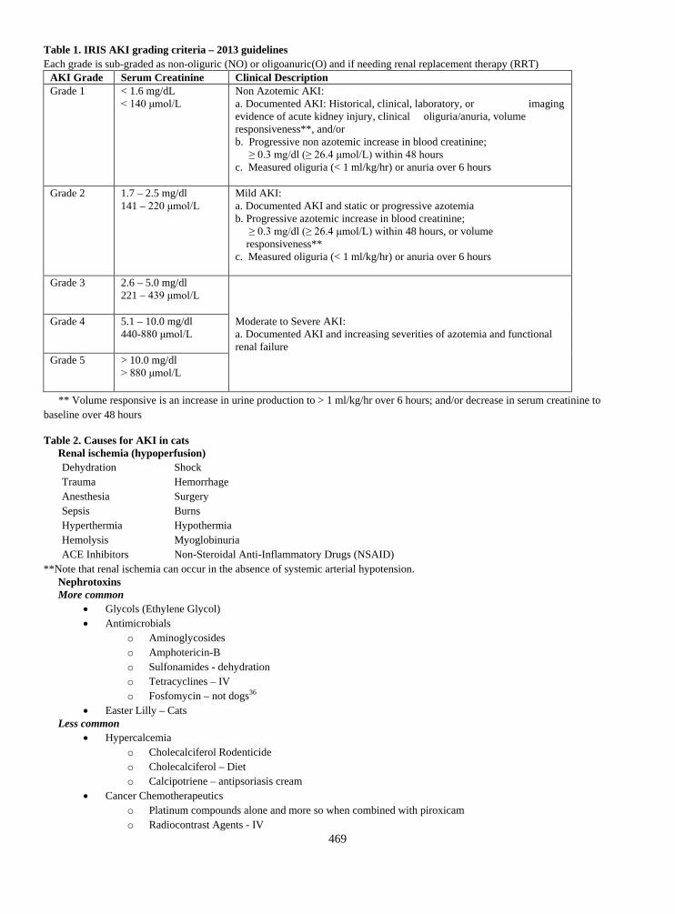
469
Table 1. IRIS AKI grading criteria – 2013 guidelines Each grade is sub-graded as non-oliguric (NO) or oligoanuric(O) and if needing renal replacement therapy (RRT)
AKI Grade Serum Creatinine Clinical Description Grade 1 < 1.6 mg/dL
< 140 μmol/L
Non Azotemic AKI: a. Documented AKI: Historical, clinical, laboratory, or imaging evidence of acute kidney injury, clinical oliguria/anuria, volume responsiveness**, and/or b. Progressive non azotemic increase in blood creatinine; ≥ 0.3 mg/dl (≥ 26.4 μmol/L) within 48 hours c. Measured oliguria (< 1 ml/kg/hr) or anuria over 6 hours
Grade 2 1.7 – 2.5 mg/dl 141 – 220 μmol/L
Mild AKI: a. Documented AKI and static or progressive azotemia b. Progressive azotemic increase in blood creatinine; ≥ 0.3 mg/dl (≥ 26.4 μmol/L) within 48 hours, or volume responsiveness** c. Measured oliguria (< 1 ml/kg/hr) or anuria over 6 hours
Grade 3 2.6 – 5.0 mg/dl 221 – 439 μmol/L
Grade 4 5.1 – 10.0 mg/dl 440-880 μmol/L
Moderate to Severe AKI: a. Documented AKI and increasing severities of azotemia and functional renal failure
Grade 5 > 10.0 mg/dl > 880 μmol/L
** Volume responsive is an increase in urine production to > 1 ml/kg/hr over 6 hours; and/or decrease in serum creatinine to baseline over 48 hours Table 2. Causes for AKI in cats
Renal ischemia (hypoperfusion) Dehydration Shock Trauma Hemorrhage Anesthesia Surgery Sepsis Burns Hyperthermia Hypothermia Hemolysis Myoglobinuria ACE Inhibitors Non-Steroidal Anti-Inflammatory Drugs (NSAID)
**Note that renal ischemia can occur in the absence of systemic arterial hypotension. Nephrotoxins More common
• Glycols (Ethylene Glycol) • Antimicrobials
o Aminoglycosides o Amphotericin-B o Sulfonamides - dehydration o Tetracyclines – IV o Fosfomycin – not dogs36
• Easter Lilly – Cats Less common
• Hypercalcemia o Cholecalciferol Rodenticide o Cholecalciferol – Diet o Calcipotriene – antipsoriasis cream
• Cancer Chemotherapeutics o Platinum compounds alone and more so when combined with piroxicam o Radiocontrast Agents - IV
470
o Heavy Metals Miscellaneous causes of AKI
• Renal thromboembolism – renal infarction • Acute-on-chronic renal failure • Renal amyloidosis with acute papillary necrosis
Acute hyperphosphatemia • Tumor lysis syndrome
o Phosphate enema o Phosphate acidifier o Massive soft tissue trauma
• Pancreatitis • Food-associated renal failure – FARF • (melamine with cyanuric acid tainting)
References 1. Holloway A, O'Brien R. Perirenal effusion in dogs and cats with acute renal failure. Vet Radiol Ultrasound 2007;48:574-579. 2. Dalby AM, Adams LG, Salisbury SK, Blevins WE. Spontaneous retrograde movement of ureteroliths in two dogs and five cats. Journal of the American Veterinary Medical Association 2006;229:1118-1121. 3. Harison E, Langston C, Palma D, Lamb K. Acute azotemia as a predictor of mortality in dogs and cats. J Vet Intern Med 2012;26:1093-1098. 4. Worwag S, Langston CE. Acute intrinsic renal failure in cats: 32 cases (1997-2004). J Am Vet Med Assoc 2008;232:728-732. 5. Prowle JR, Kirwan CJ, Bellomo R. Fluid management for the prevention and attenuation of acute kidney injury. Nat Rev Nephrol 2014;10:37-47. 6. Prowle JR. Acute kidney injury: an intensivist's perspective. Pediatr Nephrol 2014;29:13-21. 7. Prowle JR, Liu YL, Licari E, Bagshaw SM, Egi M, Haase M, Haase-Fielitz A, Kellum JA, Cruz D, Ronco C, Tsutsui K, Uchino S, Bellomo R. Oliguria as predictive biomarker of acute kidney injury in critically ill patients. Crit Care 2011;15:R172. 8. Prowle JR, Echeverri JE, Ligabo EV, Ronco C, Bellomo R. Fluid balance and acute kidney injury. Nat Rev Nephrol 2010;6:107-115. 9. Liu YL, Prowle J, Licari E, Uchino S, Bellomo R. Changes in blood pressure before the development of nosocomial acute kidney injury. Nephrol Dial Transplant 2009;24:504-511. 10. Langston CE, Eatroff A. Less is more - fluid therapy for kidney disease. J Fel Med Surg 2012;14:773. 11. Sigrist NE. Use of dopamine in acute renal failure. Journal of Veterinary Emergency and Critical Care 2007;17:117-126. 12. Wohl JS, Schwartz DD, Flournoy WS, Clark TP, Wright JC. Renal hemodynamic and diuretic effects of low-dosage dopamine in anesthetized cats. Journal of Veterinary Emergency and Critical Care 2007;17:45-52. 13. McClellan JM, Goldstein RE, Erb HN, Dykes NL, Cowgill LD. Effects of administration of fluids and diuretics on glomerular filtration rate, renal blood flow, and urine output in healthy awake cats. Am J Vet Res 2006;67:715-722. 14. Simmons JP, Wohl JS, Schwartz DD, Edwards HG, Wright JC. Diuretic effects of fenoldopam in healthy cats. Journal of Veterinary Emergency and Critical Care 2006;16:96-103. 15. Acierno MJ, Serra VF, Johnson ME, Mitchell MA. Preliminary validation of a point-of-care ethylene glycol test for cats. Journal of Veterinary Emergency and Critical Care 2008;18:477-479. 16. Scherk JR, Brainard BM, Collicutt NB, Bush SE, Almy FS, Koenig A. Preliminary evaluation of a quantitative ethylene glycol test in dogs and cats. J Vet Diagn Invest 2013;25:219-225. 17. Thrall MA, Grauer GF, Mero KN. Clinicopathologic findings in dogs and cats with ethylene glycol intoxication. J Am Vet Med Assoc 1984;184:37-41. 18. Grauer GF, Thrall MA. Ethylene glycol (antifreeze) poisoning in the dog and cat. Journal of the American Animal Hospital Association 1982;18:492-497. 19. Thrall MA, Dial SM, Winder DR. Identification of calcium oxalate monohydrate crystals by X-ray diffraction in urine of ethylene glycol-intoxicated dogs. Vet Pathol 1985;22:625-628. 20. Adams WH, Toal RL, Brieder MA. Ultrasonographic findings in dogs and cats with oxalate nephrosis attributed to ethylene glycol intoxication: 15 cases (1984-1988). Journal of the American Veterinary Medical Association 1991;199:492-496. 21. Adams WH, Toal RL, Breider MA. Ultrasonographic findings in ethylene glycol (antifreeze) poisoning in a pregnant queen and 4 fetal kittens. Veterinary Radiology 1991;32:60-62. 22. Dial SM, Thrall MA, Hamar DW. Comparison of ethanol and 4-methylpyrazole as treatments for ethylene glycol intoxication in cats. Am J Vet Res 1994;55:1771-1782. 23. Connally HE, Thrall MA, Hamar DW. Safety and efficacy of high-dose fomepizole compared with ethanol as therapy for ethylene glycol intoxication in cats. J Vet Emerg Crit Care (San Antonio) 2010;20:191-206. 24. Tart KM, Powell LL. 4-Methylpyrazole as a treatment in naturally occurring ethylene glycol intoxication in cats. J Vet Emerg Crit Care (San Antonio) 2011;21:268-272. 25. Bennett AJ, Reineke EL. Outcome following gastrointestinal tract decontamination and intravenous fluid diuresis in cats with known lily ingestion: 25 cases (2001-2010). J Am Vet Med Assoc 2013;242:1110-1116. 26. Fitzgerald KT. Lily toxicity in the cat. Topics in Companion Animal Medicine 2010;25:213-217.

471
27. Hadley RM, Richardson JA, Gwaltney-Brant SM. A retrospective study of daylily toxicosis in cats. Veterinary and Human Toxicology 2003;45:38-39. 28. Hall JO. Nephrotoxicity of Easter lily (Lilium longiflorum) when ingested by the cat. Journal of Veterinary Internal Medicine 1992;6:121. 29. Langston CE. Acute renal failure caused by lily ingestion in six cats. J Am Vet Med Assoc 2002;220:49-52, 36. 30. Rumbeiha WK, Francis JA, Fitzgerald SD, Nair MG, Holan K, Bugyei KA, Simmons H. A comprehensive study of Easter lily poisoning in cats. J Vet Diagn Invest 2004;16:527-541. 31. Slater MR, Gwaltney-Brant S. Exposure circumstances and outcomes of 48 households with 57 cats exposed to toxic lily species. J Am Anim Hosp Assoc 2011;47:386-390. 32. Tefft KM. Lily nephrotoxicity in cats. Compendium on Continuing Education for the Practicing Veterinarian 2004;26:149-157. 33. Lippi I, Ross S, Cowgill L. Acute kidney injury secondary to lily intoxication in 30 cats. . J Vet Internal Med 2013;27:738. 34. Robson M, Chew D, van Aalst S. Intrinsic acute renal failure (ARF) associated with non-steroidal anti-ionflammatory drugs (NSAID) use in juvenile cats undergoing routine desexing- 16 cases 1998-2005 J Vet Intern Med 2006;20:740. 35. Pagès JP. Nephropathies associated with NSAIDs in cats: 21 cases (1993-2001). Pratique Médicale & Chirurgicale de l'Animal de Compagnie 2005;40:177-181. 36. Fukata T, Imai N, Shibata S. Acute renal insufficiency in cats after fosfomycin administration. Vet Rec 2008;163:337-338.
472
Special Aspects of Diagnosing and Managing Chronic Kidney Disease Dennis Chew, DVM, DACVIM
The Ohio State University Columbus, OH
The incidence of the diagnosis of CKD in cats is made 2 to 3 times as frequently compared to dogs and is especially common in geriatric cats.1 CKD is clinically characterized by the development of variably progressive irreversible intrarenal lesions and loss of renal functions. Compensatory increases (so called adaptations) in glomerular hemodynamics and glomerular volume may actually be maladaptive in the long term as they cause increased protein trafficking across the glomerulus.
The initial diagnosis of CKD is made on some combination of findings from clinical signs, physical examination (especially large or small kidneys, irregular kidneys), renal imaging, urinalysis, and serum biochemistry. A surprising number of cats with CKD have upper urinary tract uroliths at the time of initial diagnosis.2-4 Abdominal radiographs should be routinely obtained to determine the presence or absence of radiopaque stones. Renal and ureteral ultrasonography should be performed in all cats in which renal or ureteral stones were found on radiography in order to tell whether or not there is an obstructive component to the CKD. T4 should be measured in all cats with suspected CKD since hyperthyroidism can mask the detection of azotemia by its effects that increase GFR and RBF; hyperthyroidism may also contribute to progression of CKD through a variety of mechanisms including intraglomerular and systemic hypertension. 5 Conventional wisdom and experience suggests that client owned cats with healthy kidneys elaborate urine with a specific gravity of >1.035. This concept was recently validated in a study of cats evaluated at first opinion clinics.6 Cats with USG < 1.035 should undergo further diagnostic investigation to determine if they have an endocrine or renal disorder with or without associated clinical signs. A surprising number of experimental 7 and clinical cats with CKD continue to be able to elaborate urine with a USG > 1.035, so the presence of “concentrated” urine and mild to moderate azotemia does NOT exclude the presence of primary kidney disease in cats as it often does in dogs. Cats that have thin body condition, prior periodontal disease or cystitis, anesthesia or documented dehydration in the preceding year, or being a neutered male (vs spayed female) were reported to be at increased risk for the diagnosis of CKD.8
A staging system initially based on the level of serum creatinine concentration has been developed by IRIS (International Renal Interest Society) for use in cats that are hydrated and stable. Serum creatinine is measured again on at least 2 occasions 2 weeks apart by the same lab. Sub-staging is then based on the degree of proteinuria as measured by UPC and also the magnitude of blood pressure. Staging using this system is designed to detect CKD much earlier than with traditional methods and also to potentially match treatments by stage. Normal and stage 1 CKD cats have serum creatinine concentrations < 1.6 mg/dl (< 140 μmol/L). Normal cats usually have a UPC < 0.2, with 0.2-0.4 considered borderline increased, and > 0.4 overtly proteinuric. Details of this staging system can be found online at http://www.iris-kidney.com. This staging system does not indicate the underlying cause for the CKD which requires other diagnostic workup to determine. It is important to remember that nearly all studies on the effect of diet or drugs have studied overtly azotemic cats (serum creatinine > 2.0 mg/dl). It has not been determined whether or not the salutary effects of treatment in azotemic cats confer the same benefits to CKD cats at earlier stages.
Tubulo-interstitial nephritis of unknown origin is the most common cause of azotemic CKD in the cat, as in the dog. However, cats have several renal diseases that deserve additional consideration as compared to dogs including breed related predilection for renal amyloidosis (Abyssinian, Oriental Short Hair) and polycystic kidney disease (Persian, Himalayan). Cats have greater frequency of CKD associated with renal LSA than dogs. Peri-nephric pseudocyst can be associated with CKD in cats and should be considered as a differential diagnosis for apparent renal enlargement in addition to renal LSA and hydronephrosis.
A variety of interventions (diet and drugs) can slow the progression of the renal disease, improve the quality of life for the patient, and/or extend the quantity of life. Dennis-I just moved this here as it opens your discussion re treatment.
Dietary interventions for CKD Dietary therapy remains the cornerstone of management of CKD. Diet modifications include phosphorus restriction (most important), providing reduced quantity but high quality protein, adequate non protein calories from fat and CHOs, modifying sodium content (not the degree of restriction once recommended by some), supplementing potassium, B vitamins, alkali as needed and providing omega three fatty acids. In one 2-year study, cats with a serum creatinine > 2 mg/dl fed a renal diet had a median survival time that was 2.4 times longer than cats fed a maintenance diet (633 days vs 264 days). 9 In another study, IRIS stage 2 & 3 cats were followed for 24 months. Cats fed the maintenance diet had more uremic episodes and more renal-related deaths compared with cats fed the renal diet. 3 In a study of 175 CKD cats fed 1 of 7 different renal diets, the median survival time was 16 months (12 to 23 months) compared to a median survival time of 7 months for cats eating their maintenance diet. Interestingly, the longest survival period was found in cats eating a renal diet with the highest eicosapentaenoic acid (diet not available in North America), otherwise the renal diets were similar in composition. 10 Patients are more likely to accept a new renal diet if offered before uremia develops and a gradual transition may be needed.
473
The number one reason to restrict dietary protein is to provide an adequate degree of restricted intake of phosphorus, especially those associated with animal tissues in the diet. Decreased production of nitrogenous wastes can occur in those with large increases in BUN, and consequently improve the clinical well-being of the pet even though renal function remains unchanged. If proteinuria is present, dietary protein restriction may lower the magnitude of proteinuria through obscure mechanisms. Reduced dietary protein intake may also lessen inflammatory, fibrogenic and oxidative stress pathway.11 The amount to restrict dietary protein is not known, so it is currently recommended to provide at least maintenance levels. For cats with CKD, the minimum dietary protein requirement suggested is 20% of calories, which equates to 24% protein on a dry-matter basis.11-14Others suggest 28–35% (DMB).15It is emphasized that less total dietary protein can be fed if high biologic value proteins, such as egg, are fed.13 Lowering animal-derived protein (source of phosphates) in the diet may be essential to lower dietary phosphorus intake needed to achieve target levels of serum phosphorus.16 Too much dietary protein restriction can and often does result in protein: calorie malnutrition. Protein malnutrition from any cause is strongly correlated with morbidity and mortality. If protein malnutrition becomes evident in a patient (hypoalbuminemia, anemia, weight loss or loss of lean muscle mass), then the amount of protein should be increased until signs are no longer evident. Cats with sarcopenia, regardless of the stage of renal disease, may require more protein than a renal diet can provide-careful monitoring and adjustment will be needed in these cats.
Pets with CKD often suffer from poor appetite that can contribute to poor body condition. This is often associated with decreased prognosis as the owner’s often euthanize when quality of life is perceived as unacceptable. Mirtazapine (Remeron) helps not only with appetite but with uremic-associated nausea. Recent work in cats indicates mirtazapine can be administered at a low dose (1.88 mg) every 48 hours to cats with CKD, but was only studied for its effects for 3 weeks.17,18 Remember that mirtazapine and cyproheptadine cannot not be administered concurrently. Cyproheptadine is in fact used as an antidote for serotonin effects of mirtazapine overdose. Maropitant (Cerenia): NK-1 receptors are in the chemoreceptor trigger zone, in the emetic center itself, as well as peripherally. Consequently, Cerenia is a great choice to treat vomiting/nausea in renal cats. Despite the label recommendation, many specialists are recommending Cerenia for longer than 5 days (personal communication with specialists and with Zoeitis scientists). Dose: 1 mg/kg PO once daily. Refrigerate to help alleviate the sting associated with injectable cerenia.19 Omeprazole (Losec): Studies in cats have also shown Omeprazole to be more effective than H2 blockers such as famotidine and ranitidine in decreasing gastric acidity.20 Dosage: 0.5-1 mg/kg once a day. If H2 blockers are used, dosages recommended are Famotidine (Pepcid®) 0.5 mg/kg IM, SQ, PO q 12 hours or Ranitidine (Zantac®) 1-2 mg/kg q 12 hours (cat). Studies have shown most cats with uremia do have elevated gastrin levels (and likely corresponding hyperacidity) but no GI ulcers. 20,21 Consequently, sucralfate is not usually indicated. The GI bleed with uremia could be from dysregulation of the vasculature and platelet dysfunction associated with uremia.20,21 If used, a dose of 0.25 -0.5 g/cat q 12 hours is recommended. In some countries sucralfate is used as an intestinal phosphate binder due to its aluminum content. Ondansetron at the time of this writing is not highly recommended. The bioavailability is not high (maybe 30% at best in cats) and the half-life is very short (it would be best to give this drug 4 times/day).22
Phosphorus Higher concentrations of serum phosphorus predicted an increase in serum creatinine > 25% above baseline over 12 months in 47% of CKD cats. 23 Serum phosphorus was the only clinicopathologic variable predictive of survival in one study of CKD cats. There was an increase in risk of death of nearly 12% for each mg/dl increase in phosphorus in the same study.24 Higher phosphorus concentration was associated with a higher risk of death within1 month in another study.25 Even when serum phosphorus was within the reference range, cats with CKD of one study that had phosphorus concentration > 4.7 to ≤ 6.8 mg/dl serum phosphorus had a higher risk of death compared to CKD cats in which circulating phosphorus concentration was ≤ 4.7 mg/dl. 26
Dietary phosphorus restriction is critical at least from Stage 2 onwards; there is no data to evaluate any potential benefit of Pi restriction in Stage 1. Compared to the average grocery or pet store foods, the renal friendly veterinary diets are restricted in phosphorus by 70 to 80%. Serum phosphorus concentration may increase in CKD pets that increase their food intake following other supportive CKD treatments. Renal diets may provide sufficient dietary phosphate restriction during early stages of CKD but often the addition of dietary phosphate binders will be needed to reach targeted control of serum phosphorus. Early phosphorus restriction in CRF has been shown in dogs and cats to blunt or reverse renal secondary hyperparathyroidism.27
Intestinal phosphate binders Aluminum salts are the most widely used phosphate binders in cats. Aluminum based phosphate binding agents (aluminum hydroxide, aluminum carbonate) are highly effective in lowering serum phosphate levels, forming insoluble and nonabsorbable aluminum phosphate precipitates in the intestinal lumen. THERE IS NO KNOWN SAFE DOSE OF ALUMINUM SALTS FOR HUMANS WITH CKD. Detrimental effects of aluminum based phosphate binders as described in humans seen in humans have not been systematically evaluated in small animal patients and are rarely clinically appreciated. As cats with CKD can live for years on treatment, concerns for aluminum accumulation deserve more study as to long-term safety. Calcium-based binders are not as effective as aluminum salts, having a lower affinity for phosphorous, thus effective binding of dietary phosphorous requires large doses of calcium, often enough to induce hypercalcemia in humans. The most commonly used calcium based phosphate binders are calcium carbonate and calcium acetate. Animals should be monitored for development of hypercalcemia whenever calcium-containing
474
phosphorus binders are used. Sevelamer hydrochloride (Renagel®, Genzyme Corporation) and the more recently FDA approved Sevelamer carbonate (Renvela®, Genzyme Corporation) are organic polymers that do not contain aluminum or calcium and are not absorbed from the gastrointestinal tract (excreted entirely in feces). Their effects on dogs and cats with clinical CRF have not been reported. Epakitin® (Vetoquinol Inc.) is marketed as a complementary feed on the veterinary market. It contains the adsorbent chitosan (8% crab and shrimp shell extract), 10% calcium carbonate, and 82% lactose and is designed to reduce GI phosphorus absorption and to lower urea nitrogen due to effects of reduced protein digestibility. The results of two studies 28,29 suggest that this supplement could be an alternative to prescription of renal veterinary diets thereby allowing some cats to continue on their regular diets while still reducing the risks for progression of CKD associated with total body phosphorus burden. We have, however, observed the development of hypercalcemia in a few CKD cats with the use of this product probably as a consequence of the calcium carbonate. Lanthanum carbonate (Fosrenol®, Shire Pharmaceuticals) is a non-aluminum and non-calcium containing intestinal phosphate binder and is indicated for use in human patients with end-stage renal failure to reduce serum phosphorous. Very little lanthanum is absorbed across GI tract and lanthanum accumulates to a far less degree following absorption compared to aluminum since lanthanum undergoes extensive hepatic excretion whereas aluminum is excreted mostly by the kidneys. Lanthanum appears to have minimal toxicity in humans. A recent abstract in a small number of CKD cats administered lanthanum carbonate in food at 95 mg/kg/day to achieve very modest serum phosphate control.30 Several reports of the efficacy and safety of lanthanum carbonate treatment in cats have been published. 31 Lanthanum carbonate octahydrate (Lantharenol® Bayer HealthCare AG) is marketed as a feed additive for adult cats in order to decrease intestinal phosphate absorption. Renalzin® (Bayer HealthCare AG) is the proprietary name for the delivery system of Lantharenol® and comes as a pump system that delivers lanthanum carbonate along with kaolin and vitamin E at appropriate doses to food for cats. This system is widely available in the UK and Europe, but not in the USA or Canada. The proprietary formulation of human lanthanum carbonate is soon to become available as a generic product.
Pronefra® recently has been launched (Virbac, France) as a dietary supplement for cats with CKD. This product provides a combination of calcium and magnesium carbonate as the intestinal phosphate binders, chitosan for “uremic toxin” binding, vasoactive peptides (designed to maintain normal blood pressure) and an extract of Astragalus membranaceus (Chinese herb for anti-inflammatory and anti-fibrotic effects). Safety of this product was reported in 10 normal cats in which Pronefra was added to the food once daily for 12 weeks32,33 No changes in circulating calcium or magnesium were noted at during this study. Presently there are no reported studies of safety or efficacy in clinical cats with CKD treated with this supplement.
Novartis has developed a new oral phosphate binder for cats called Lenziaren ® (SBR759). Iron oxide with starch and sucrose exist in this preparation as an insoluble complex. A dose of 0.5 to 1.0 Gm/cat/day is recommended when added to standard diets.34 A dose of 0.25 Gm/cat/day to 1.0 Gm/cat/day is recommended when adding this phosphate binder to a renal diet. 35 Safety and efficacy of Lenziaren® in cats with CKD are not yet reported. Lenziaren is touted by the authors as a phosphate binder that does not contain aluminum, calcium, or lanthanum that could be problematic in cats with CKD. That is true for the aluminum and calcium as a factor in favor of its use, but there is no known toxicity of lanthanum yet reported.
Control of proteinuria Cats with azotemic CKD increased their risk for death or euthanasia when the UPC was 0.2 to 0.4 compared to <0.2 and was further increased in cats with UPC of >0.4.36 The prognosis for survival is influenced by the UPC despite what has traditionally been thought to be low-level proteinuria. The effect of treatments that lower proteinuria on survival have not been specifically studied. Since even low-level proteinuria is a risk factor for cats to not survive, it is prudent to consider treatments that lower the amount of proteinuria in those with CKD. See discussions about the potential benefits of dietary protein restriction (above) and RAAS inactivation (below) to reduce the magnitude of proteinuria.
RAAS inactivation RAAS inactivation results in decreased generation of angiotensin-2 and aldosterone that can exert benefits to reduce progression of CKD. These beneficial effects can occur through variable combinations of reduction in systolic blood pressure, decreased intra-glomerular hypertension, decreased glomerular proteinuria, and less generation of pro-inflammatory and pro-fibrotic cytokines in patients with CKD.
Benazepril is labeled for treatment of azotemic CKD in cats in the UK, Europe, and Canada (Fortekor®), but not in the USA. The ACE-inhibitor benazepril consistently reduces proteinuria in various stages of CKD in cats even when the base line level of proteinuria is seemingly trivial. Benazepril has been shown in two clinical studies to reduce the UPC in cats with azotemic CKD.37,38 Despite reduction in proteinuria in CKD cats with initial UPC > 1.0 that were treated with benazepril in one study, increased survival time was not found over placebo.37 The average survival time of all benazepril treated cats in this study was 501 days vs. 391 days for placebo treated cats but this effect did achieve statistical significance. 37 In another study of 61 cats with CKD, benazepril treatment for 189 days appeared to stabilize those in IRIS stage 2 or 3 with less transition to stage 4 compared to treatment with placebo, though this effect did not achieve statistical significance (low number of cats and short duration of study.38
The angiotensin receptor blocker (ARB) telmisartin (Semintra® Boehringer Ingelheim) was approved by the European Commission in 2013 for use in the European Union as a drug for use in cats with CKD and is available for use in Canada but not yet
475
in the USA. Semintra was found to be at least as effective as benazepril in reducing proteinuria in cats with CKD and was well tolerated.39,40 A US Patent application was filed in July 2013 by Boehringer Ingelheim. It is not clear when or if an ARB should be chosen to reduce RAAS activity instead of an ACE-Inhibitor for treatment of CKD in veterinary patients to reduce proteinuria, systemic blood pressure, or intra-renal inflammation. A veterinary review of the RAAS system, ACE-Inhibitors and ARB’s provides more detail for the interested reader.41
Activated vitamin-D metabolites: calcitriol Calcitriol treatments help to decrease PTH or prevent its increase in those with renal secondary hyperparathyroidism. This occurs largely through genomic effects to block PTH synthesis in addition to a mild calcemic effect, and anti-proliferative effect that prevents parathyroid gland hyperplasia. It has become increasingly apparent that calcitriol has major beneficial anti-inflammatory and anti-fibrotic intrarenal effects that are independent of effects on PTH.27During treatment of CRF patients with calcitriol, simultaneous monitoring of serum ionized calcium, serum phosphorus and PTH concentrations is the ideal way to document successful and safe control of renal secondary hyperparathyroidism. Calcitriol should not be administered until hyperphosphatemia has been controlled. If the Ca X P solubility product exceeds 60-70, calcitriol should be avoided because of the risk of soft-tissue mineralization.
In a recent study of dogs with azotemic CKD that were treated with calcitriol a median of 365 days survival was observed compared to 250 days in dogs treated with placebo (renal diet in both groups).42 Similar studies were performed in cats by the same investigators who concluded that there is no advantage to calcitriol treatments in cats with CRF but the study followed cats for just one year. In order to show a difference in treatment effect, if one exists, studies in cats with CKD must be conducted for at least 2 and possibly 3 years due to the inherently slow nature of the progression of chronic renal disease in this species. The authors believe that beneficial effects of calcitriol treatment are likely to occur in cats with CKD.
A compounding pharmacy will be needed to reformulate calcitriol from the human parent drug to a concentration suitable for the dosing of cats. We recommend intermittent rather than daily dosing treatment protocols as the standard of care since less hypercalcemia occurs using this protocol. The equivalent dose given at 2.5 ng/kg daily is given instead every 3.5 days. This works out to a dose of 9 ng/kg (8.75 ng/kg rounded to 9 ng/kg). It is important to give the dose every 3.5 days, rather than on day 1 & 4. For example if a dose is given Tuesday PM the next dose should be given Saturday AM. This is the longest time in between dosing that will still suppress the parathyroid gland. This method of dosing is especially attractive for cat owners since medication will only be given twice weekly. Systemic hypertension Systemic hypertension is common in cats with CKD with 13-28% of cats presenting with hypertension when CKD is first diagnosed and up to 65% of cats developing hypertension at some point during the progression of their renal disease.43-51 Cats that have systemic hypertension from a variety of causes have been shown to survive longest when their blood pressure is well controlled.
Enalapril or benazepril as monotherapy has not been very effective for treatment of hypertensive cats or dogs. The calcium channel blocker, amlodipine has been used successfully in cats at a dosage 0.625 to 1.25 mg per cat given orally once per day. Follow-up evaluations should be scheduled for one week after beginning treatment with amlodipine. Adverse effects (including hypotension) are very uncommon with the use of amlodipine in cats.43,46,47 References 1. Lulich JP, O'Brien TD, Osborne CA, Polzin DJ. Feline renal failure: questions, answers, questions. Compendium on Continuing Education for the Practicing Veterinarian 1992;14:127-152. 2. Pimenta MM, Reche-Júnior A, Freitas MF, Wang L. Nephrolithiasis and ureterolithiasis prevalence in cats with chronic kidney disease presented at the Univeristyof Sao Paul Veteirnary Hospital. NEPHROLITHIASIS AND. JFMS 2013;15 823. 3. Ross SJ, Osborne CA, Kirk CA, Lowry SR, Koehler LA, Polzin DJ. Clinical evaluation of dietary modification for treatment of spontaneous chronic kidney disease in cats. Journal of the American Veterinary Medical Association 2006;229:949-957. 4. Ross SJ, Osborne CA, Lekcharoensuk C, Koehler LA, Polzin DJ. A case-control study of the effects of nephrolithiasis in cats with chronic kidney disease. J Am Vet Med Assoc 2007;230:1854-1859. 5. van Hoek I, Daminet S. Interactions between thyroid and kidney function in pathological conditions of these organ systems: a review. Gen Comp Endocrinol 2009;160:205-215. 6. Rishniw M, Bicalho R. Prevalence and cuases of urine specifc gravity ≤ 1.035 in apparently healthy cats presenting to first opinion practice. J Vet Intern Med 2013;27:741(NU-738). 7. Adams LG, Polzin DJ, Osborne CA, O'Brien TD. Effects of dietary protein and calorie restriction in clinically normal cats and in cats with surgically induced chronic renal failure. Am J Vet Res 1993;54:1653-1662. 8. Greene JP, Lefebvre SL, Wang M, Yang M, Lund EM, Polzin DJ. Risk factors associated with the development of chronic kidney disease in cats evaluated at primary care veterinary hospitals. J Am Vet Med Assoc 2014;244:320-327. 9. Elliott J, Rawlings JM, Markwell PJ, Barber PJ. Survival of cats with naturally occurring chronic renal failure: effect of dietary management. J Small Anim Pract 2000;41:235-242. 10. Plantinga EA, Everts H, Kastelein AM, Beynen AC. Retrospective study of the survival of cats with acquired chronic renal insufficiency offered different commercial diets. Vet Rec 2005;157:185-187.
476
11. Bartges JW. Chronic kidney disease in dogs and cats. Vet Clin North Am Small Anim Pract 2012;42:669-692, vi. 12. Bartges JW. Dietary protein and chronic kidney disease: how much is enough? ACVIM Forum 2014. 13. Polzin DJ. Chronic kidney disease in small animals. Vet Clin North Am Small Anim Pract 2011;41:15-30. 14. Baldwin K, Bartges J, Buffington T, Freeman LM, Grabow M, Legred J, Ostwald D, Jr. AAHA nutritional assessment guidelines for dogs and cats. J Am Anim Hosp Assoc 2010;46:285-296. 15. Kirk CA, Hickman MA. Dietary protein requirements of cats with spontaneous renal disease. J Vet Intern Med 2000;14:351. 16. Burkholder WJ. Dietary considerations for dogs and cats with renal disease. J Am Vet Med Assoc 2000;216:1730-1734. 17. Quimby JM, Gustafson DL, Lunn KF. The pharmacokinetics of mirtazapine in cats with chronic kidney disease and in age-matched control cats. J Vet Intern Med 2011;25:985-989. 18. Quimby JM, Lunn KF. Mirtazapine as an appetite stimulant and anti-emetic in cats with chronic kidney disease: a masked placebo-controlled crossover clinical trial. Vet J 2013;197:651-655. 19. Narishetty ST, Galvan B, Coscarelli E, Aleo M, Fleck T, Humphrey W, McCall RB. Effect of refrigeration of the antiemetic Cerenia (maropitant) on pain on injection. Veterinary Therapeutics 2009;10:93-102. 20. Goldstein RE, Marks SL, Kass PH, Cowgill LD. Gastrin concentrations in plasma of cats with chronic renal failure. J Am Vet Med Assoc 1998;213:826-828. 21. McLeland SM, Lunn KF, Duncan CG, Refsal KR, Quimby JM. Relationship among serum creatinine, serum gastrin, calcium-phosphorus product, and uremic gastropathy in cats with chronic kidney disease. Journal of Veterinary Internal Medicine 2014;28:827-837. 22. Quimby JM, Lake RC, Hansen RJ, Lunghofer PJ, Gustafson DL. Oral, subcutaneous, and intravenous pharmacokinetics of ondansetron in healthy cats. J Vet Pharmacol Ther 2014;37:348-353. 23. Chakrabarti S, Syme HM, Elliott J. Clinicopathological Variables Predicting Progression of Azotemia in Cats with Chronic Kidney Disease. J Vet Intern Med 2012. 24. Boyd LM, Langston C, Thompson K, Zivin K, Imanishi M. Survival in cats with naturally occurring chronic kidney disease (2000-2002). J Vet Intern Med 2008;22:1111-1117. 25. Kuwahara Y, Ohba Y, Kitoh K, Kuwahara N, Kitagawa H. Association of laboratory data and death within one month in cats with chronic renal failure. J Small Anim Pract 2006;47:446-450. 26. King JN, Tasker S, Gunn-Moore DA, Strehlau G. Prognostic factors in cats with chronic kidney disease. J Vet Intern Med 2007;21:906-916. 27. de Brito Galvao JF, Nagode LA, Schenck PA, Chew DJ. Calcitriol, calcidiol, parathyroid hormone, and fibroblast growth factor-23 interactions in chronic kidney disease. J Vet Emerg Crit Care (San Antonio) 2013;23:134-162. 28. Wagner E, Schwendenwein I, Zentek J. Effects of a dietary chitosan and calcium supplement on Ca and P metabolism in cats. Berl Munch Tierarztl Wochenschr 2004;117:310-315. 29. Brown SA, Rickertsen M, Sheldon S. Effects of an intestinal phosphorus binder on serum phosphorus and parathyroid hormone concentration in cats with reduced renal function. International Journal of Applied Research in Veterinary Medicine 2008;6:155-160. 30. Hall MJ, Marino CL, Eatroff AE, Ross SJ, Pressler BM. Efficacy amd adverse effects pf ;amtjami, carbonate in cats. J Vet Intern Med 2013;27:737-738. 31. Schmidt BH, Dribusch U, Delport PC, Gropp JM, van der Staay FJ. Tolerability and efficacy of the intestinal phosphate binder Lantharenol(R) in cats. BMC Vet Res 2012;8:14. 32. Bernachon N, Fournel S, Gatto H, Monginoux P, McGahie D. Comparative palatability of five supplements designed for cats suffering from chronic renal disease. Irish Veterinary Journal 2014;67:(19 May 2014). 33. Bernachon N, Fournel S, Gatto H, Monginoux P, McGahie D. Effect of a product containing the dietary phosphate binders calcium and magnesium carbonate associated with other reno-protectant substances (Pronefra®) on blood parameters and mineral balance in adult cats. International Journal of Applied Research in Veterinary Medicine 2014;12:8-17. 34. King JN, Erasmus HL, Delport PC, Bester I, Seewald W. Efficacy, acceptability and tolerability of the new oral phosphate binder Lenziaren(R) in healthy cats fed a standard diet. BMC Vet Res 2014;10:258. 35. King JN, Delport PC, Luus HG, Erasmus HL, Barnes PM, Speranza C. Efficacy and acceptability of the new oral phosphate binder Lenziaren in healthy cats fed a renal diet. J Vet Pharmacol Ther 2014. 36. Syme HM, Markwell PJ, Pfeiffer D, Elliott J. Survival of cats with naturally occurring chronic renal failure is related to severity of proteinuria. J Vet Intern Med 2006;20:528-535. 37. King JN, Gunn-Moore DA, Tasker S, Gleadhill A, Strehlau G. Tolerability and efficacy of benazepril in cats with chronic kidney disease. J Vet Intern Med 2006;20:1054-1064. 38. Mizutani H, Koyama H, Watanabe T, Kitagawa H, Nakano M, Kajiwara K, King JN. Evaluation of the clinical efficacy of benazepril in the treatment of chronic renal insufficiency in cats. J Vet Intern Med 2006;20:1074-1079. 39. Sent U, Goessl R, Lang I, Stark M. Efficacy of long-term treatment with telmisartan oral solution on quality of life and disease progression in cats wioth chronic kidney disease. JFMS 2013;15:820-821. 40. Sent U, Lang I, Moore G. Characterisation of telmisartan in cats. JFMS 2013;15:824-825. 41. Pressler B. A practical guide to antiproteinuric drugs in dogs. Veterinary Medicine 2013;108:392-397. 42. Polzin D, Ross S, Osborne C, Lulich J, Swanson L. Clinical benefit of calcitriol in canine chronic kidney disease. Journal of veterinary internal medicine / American College of Veterinary Internal Medicine 2005;19:433. 43. Jepson RE, Elliott J, Brodbelt D, Syme HM. Effect of control of systolic blood pressure on survival in cats with systemic hypertension. J Vet Intern Med 2007;21:402-409. 44. Jepson RE. Feline systemic hypertension: Classification and pathogenesis. J Feline Med Surg 2011;13:25-34. 45. Syme HM, Barber PJ, Markwell PJ, Elliott J. Prevalence of systolic hypertension in cats with chronic renal failure at initial evaluation. J Am Vet Med Assoc 2002;220:1799-1804.

477
46. Brown S, Atkins C, Bagley R, Carr A, Cowgill L, Davidson M, Egner B, Elliott J, Henik R, Labato M, Littman M, Polzin D, Ross L, Snyder P, Stepien R, American College of Veterinary Internal M. Guidelines for the identification, evaluation, and management of systemic hypertension in dogs and cats. J Vet Intern Med 2007;21:542-558. 47. Elliott J, Barber PJ, Syme HM, Rawlings JM, Markwell PJ. Feline hypertension: clinical findings and response to antihypertensive treatment in 30 cases. J Small Anim Pract 2001;42:122-129. 48. Cowgill LD. The role of fluid volume in hypertension of kidney disease in dogs and cats. ECVIM-CA Congress 2012. 49. Henik RA, Snyder PS. Treatment of systemic hypertension in cats with amlodipine besylate. Journal of the American Animal Hospital Association 1997;33:226-234. 50. Kobayashi DL, Peterson ME, Graves TK, Lesser M, Nichols CE. Hypertension in cats with chronic renal failure or hyperthyroidism. J Vet Intern Med 1990;4:58-62. 51. Mishina M, Watanabe T, Fujii K, Maeda H, Wakao Y, Takahashi M. Non-invasive blood pressure measurements in cats: clinical significance of hypertension associated with chronic renal failure. J Vet Med Sci 1998;60:805-808.
478
Treatment of Idiopathic Hypercalcemia in Cats: Case Studies- Diets or Drugs?
Dennis Chew, DVM, DACVIM The Ohio State University
Columbus, OH
How common is hypercalcemia in cats? The frequency of the detection of hypercalcemia in cats has dramatically increased in many regions of the world over the past 20 years mostly due to the diagnosis of idiopathic hypercalcemia (IHC).1-7 Hypercalcemia is most often initially defined in primary care practice by the finding of increased serum total calcium on routine serum biochemistry. Mild hypercalcemia based on serum total calcium is often overlooked during analysis of serum biochemical profiles, so the frequency of hypercalcemia is likely to be more common than generally recognized. Mild serum total hypercalcemia is frequently attributed to hemoconcentration from dehydration.
Total serum calcium cannot be reliably used to predict the metabolically active ionized calcium fraction in cats.8 There was an overall diagnostic discordance of 40% during evaluation of 434 feline serum samples using total calcium to predict ionized calcium in cats of one study. Ionized hypercalcemia and normocalcemia were underestimated and ionized hypocalcemia was overestimated.
Characterization of hypercalcemia Once ionized hypercalcemia has been identified, the next step is to determine if the process is PTH-dependent (high PTH from failure to suppress abnormal parathyroid glands) or PTH-independent (PTH is appropriately suppressed as the response of normal parathyroid glands). In a study of 322 cats, ionized hypercalcemia was parathyroid independent in 82%, equivocal in 10%, and parathyroid-dependent in 8% of these cats.9 In cats with parathyroid-independent hypercalcemia, malignancy-associated hypercalcemia (MAH) needs to be excluded. MAH most often results from humoral mechanisms as the tumor secretes calcemic substances such as PTHrP into the circulation; local osteolytic hypercalcemia is far less common. When PTHrP is reported to be high, the presence of malignancy is likely. A low or undetectable PTHrP does not exclude malignancy as the cause for hypercalcemia since other cytokines that cause calcemia can be elaborated by the tumor instead of PTHrP on occasion.
If the diagnostic evaluation does not reveal malignancy as the cause for parathyroid-independent hypercalcemia (PTHrP and body cavity imaging), evaluation of circulating vitamin D metabolites may be useful in determining the underlying cause or mechanism for the hypercalcemia. Hypervitaminosis D is classically characterized by increased concentrations of circulating 25(OH)-vitamin D (calcidiol) following excess ergo/cholecalfierol exposure from food10,11 or from cholecalciferol-containing rat-bait.12,13 Increased circulating calcitriol has been reported in cats with granulomatous disease and hypercalcemia, likely the result of unregulated conversion of calcidiol to calcitriol by activated macrophages.14-16 What are the causes of hypercalcemia in cats? The frequency for the occurrence of total serum hypercalcemia from biochemical panels from sick or well cats is not known. The only large survey of the causes of hypercalcemia in cats was reported from a veterinary teaching hospital based on the measurement of serum total calcium in 2000.17 Ionized hypercalcemia concentration has been sporadically reported in cats with specific diseases, but not in a series of cats with varying causes of hypercalcemia. Idiopathic hypercalcemia, CKD, and neoplasia are the most common and important differential diagnoses to exclude as the cause for parathyroid independent hypercalcemia. Overt hypervitaminosis D, granulomatous disease, and hypoadrenocorticism are other far less common causes of hypercalcemia in cats. Calcium oxalate urolithiasis was reported to be associated with hypercalcemia in cats; however, it is likely that hypercalcemia preceded the formation of stones rather than the urolithiasis acting as a stimulus for the formation of hypercalcemia.17,18 IHC was not considered as a diagnostic category in one large study of cats with hypercalcemia,17 but in another study the occurrence of IHC in 20 cats was published that same year.18 Primary hyperparathyroidism was infrequently diagnosed as the cause of the hypercalcemia at a teaching hospital (4 of 71 cats),17 but this diagnosis is far more frequently made by veterinary endocrine referral laboratories.1,9 Based on the number of consultations by veterinary internists and endocrinologists, as well as sample submissions to endocrine laboratories, idiopathic hypercalcemia (IHC) is currently the most-common cause of hypercalcemia in cats in North America and likely so in other parts of the world.1,2,5-7
While MAH is the number one cause of pathological hypercalcemia in the dog,19 it occurs far less frequently in the cat. Based on serum total calcium and how the data is parsed, MAH is 3rd in frequency behind IHC and CKD in cats with hypercalcemia.17 In dogs, the overwhelming cause of MAH is lymphoma with occasional carcinoma as the diagnosis,19 whereas in cats lymphoma and carcinomas each account for about 1/3 of the cases.17 Patients with MAH are usually “sick” as it takes a reasonably large tumor burden to synthesize the messengers that result in hypercalcemia. Signalment and clinical signs of IHC cats In a report from 427 cats with IHC evaluated at an endocrinology laboratory, the age at diagnosis ranged from 0.5 to 20 years (mean 9.8 ± 4.6 yr). Males and females were equally represented in this study. Long-haired cats were noted to be overrepresented at 27% of
479
the cases in this report,20 but not in a recent case-control epidemiological study (data analyzed post Todd Green Master’s Ohio State University 2008).
No clinical signs were noted in 46% of IHC cats. Other clinical signs were largely related to gastrointestinal signs, including mild weight loss (18%), chronic constipation (5%), vomiting and decreased appetite. IBD was diagnosed in 6% of the IHC cats of this study. Lower urinary tract signs may be observed, especially if urolithiasis is present. Uroliths or renoliths were observed in 15%, and calcium oxalate stones were specifically noted in 10% of cases. Polyuria/polydipsia has not been frequently reported in cats with IHC.20
In many instances, hypercalcemia based on measurement of total serum calcium is fortuitously discovered following submission of serum samples from wellness examinations, pre-anestheic evaluation of seemingly healthy individuals, those with routine medical conditions, and those from cats forming calcium-oxalate stones. Hypercalcemia is also sometimes discovered following submission of samples from cats with seemingly trivial clinical complaints like intermittent vomiting of hairballs. Though many cats with IHC do not have obvious clinical signs at first look, a more careful review of the history and physical examination often discloses some abnormality that could be explained by persistence of chronic ionized hypercalcemia. This includes low-grade weight loss, loss of muscle mass, and lethargy. Intermittent vomiting and constipation are also possibly due to adverse effects of ionized hypercalcemia on gut motility. Chronic ionized hypercalcemia is a risk factor for the genesis of calcium oxalate urolithiasis and for the development of chronic renal injury resulting in CKD that may take months to years to develop. How is the diagnosis of IHC established ? The diagnosis of IHC is one of exclusion after initially confirming that the ionized calcium is increased. All the known causes of hypercalcemia should ideally be eliminated – this kind of workup can be exhaustive and expensive. The increase in circulating ionized calcium in IHC can be mild, moderate, or severe, as it can also be with other causes of hypercalcemia. Often mild increases in total or ionized calcium that are discovered fortuitously tend to increase over time, but to a varying magnitude. We have observed the ionized calcium concentration to fluctuate into and above the reference range, especially when the hypercalcemia is marginal in magnitude. We have observed large fluctuations in total and ionized calcium concentrations on occasion in some cats with IHC and those with primary hyperparathyroidism.
In order to exclude other causes of hypercalcemia, a minimum database including a CBC, biochemistry profile and urinalysis, should be performed. Additionally, analysis of PTH and 25-hydroxyvitamin D are necessary to rule out hyperparathyroidism and hypervitaminosis D as the cause of the hypercalcemia. The typical pattern for calcium regulatory hormones in IHC would be for the PTH concentration to be within the reference range (often lower end), the PTHrP concentration to be undetectable, and to have a normal serum ionized magnesium concentration.20 Most 25-hydroxyvitamin D and calcitriol concentrations are usually within the reference range, but a few cats with IHC have been noted to have values increased above the reference range.18,20
Chest radiographs are useful to rule out metastatic pulmonary nodules and mediastinal lymphoma that may be associated with hypercalcemia. Unlike in dogs, mediastinal lymphoma is not common in cats. A combination of abdominal radiographs and ultrasonography can be useful to determine the presence of urolithasis (kidney, ureter, bladder, urethra), obstructive nephropathy from the stones, or the presence of inflammatory/infiltratitve masses that could be associated with the genesis of the hypercalcemia. Treatment recommendations and prognosis may change with the presence of stones and their location. Should all cats with IHC receive treatment? Cats with minimal increases in circulating calcium concentrations are often ignored in clinical practice since many of these cats have mild or no apparent clinical signs. Even though obvious clinical signs are often not apparent, subtle clinical signs often exist. Excess calcium can be toxic to cells, exerting either physiological or structural effects particularly in the central nervous system, gastrointestinal tract, heart, and kidneys. Mineralization of soft tissues is an important potential complication related to the presence of ionized hypercalcemia that is in part determined by the concomitant concentration of serum phosphorus, but this does not develop in all IHC cats. The clinical outcome for cats with IHC that have not been treated has not been established following the initial diagnosis. An argument can be made to withhold treatment when an IHC cat has no recognizable signs, no identified risk factors for urolithiasis or CKD, and the increase in ionized calcium is minimal. A stronger argument can be made to treat IHC cats in which the ionized calcium concentration continues to escalate. The strongest argument to start treatment exists for cats that have ongoing weight loss, depression, vomiting, constipation, urinary stones, emergence of CKD and or development of sub-maximally concentrated urine. Treatment of IHC – diet Management of IHC usually begins with a dietary recommendation to attempt to restore normocalcemia. Reports of treatment outcome following dietary change are quite limited, so diet recommendations are largely based on expert opinion and uncontrolled case studies in small numbers of cats. We have observed decreased circulating ionized calcium in some cats following dietary change, but the magnitude and duration of this decrement can be quite variable. Future studies comparing test and control diets are needed to
480
determine the effects, if any, of altering intake of nutrient(s) on concentrations of the calcium regulatory hormones PTH, calcidiol, calcitriol, and 24,25(OH)2-vitamin D in addition to that for ionized calcium.
Is there one specific dietary nutrient on which we should focus that will consistently decrease circulating ionized calcium? Regulation of the circulating calcium concentration is dynamic and complex. It has not been determined how much of the hypercalcemia in IHC cats results from too much dietary calcium intestinal absorption, increased bone resorption, reduced renal excretion of calcium, or combinations of these processes. Many of the nutrients in the diet interact with each in ways that affect dietary calcium absorption and not all calcium in the diet is biologically available for absorption.21 Vitamin D is one obvious dietary nutrient that can affect intestinal absorption of calcium and it also has effects on osteoclastic bone resorption that can contribute to the degree of calcemia.22 Vitamin A has effects on the osteoclast that can work in concert with vitamin D to increase bone resorption.23
What do we know about dietary calcium content in the management of IHC? Some veterinary nutritionists recommend diets to treat IHC based on a decreased calcium content on a g calcium/1000 kcal (Mcal) energy basis.24 Minimal and maximal nutrient recommendations for cat food are provided by the Association of American Feed Control Officials (AAFCO) and the National Research Council (NRC). Most diets sold over-the-counter should meet AAFCO requirements; however, veterinary therapeutic diets may be specifically modified in order to provide certain nutrients at concentrations less than AAFCO minimums. The average calcium content of grocery store foods in the USA is approximately 2.0 to 3.0 g calcium per Mcal (200-300 mg/100 kcal), though some contain up to 6.0 g calcium per Mcal (600 mg per 100 kcal).25 Some of the highest calcium diets are “high-fiber” diets; thus one must carefully weigh the pros and cons of recommending a high-fiber diet for dietary management of IHC when there is some evidence that reducing dietary calcium may be effective in restoring normocalcemia. Nutrient concentrations of diets can be found either in product guides or by contacting the diet manufacturer, but this information is not readily available from the routine diet label. Nutrient profiles are constantly evolving and this information may change up to every 6-12 months. For feline adult maintenance, the NRC recommended allowance (RA) is 0.72 g calcium per Mcal 26 and the AAFCO minimum is 1.5 g calcium per Mcal.27
Feeding of a high protein and low carbohydrate food similar to what cats would eat in the wild (i.e., 40-60% of calories from protein; 30-50% of calories from fat, and <15% of calories from carbohydrates) has been recommended to effectively lower serum calcium concentration in some cats with IHC, especially those with low magnitude hypercalcemia.4,28 This nutrient profile is what would be expected from veterinary therapeutic diets designed for cats with diabetes mellitus and also many over-the-counter canned feline diets. In reviewing these types of diets however, it should be noted that calcium content varies from about 1.5 to 5.5 g per Mcal.
What do we know about dietary vitamin D content in the management of IHC? IHC is not the result of obvious excess dietary vitamin D intake since serum concentrations of 25(OH)-vitamin D have been within the reference range in most cats with IHC. However, the minimal requirement for vitamin D in cats is debatable since reference ranges have been established in cats fed vitamin D–supplemented diets. Normal concentrations of 25(OH)-vitamin D could still potentially be associated with IHC in cats if there are up-regulating mutations in the VDR (vitamin D receptor). These possibilities have not yet been investigated.
For adult cats, the NRC-RA for dietary vitamin D3 (cholecalciferol) is 70 IU per Mcal. The safe upper limit (SUL) is listed as 7,520 IU per Mcal.26 AAFCO minimum and maximum recommendations for feline adult maintenance are 125 and 2,500 IU per Mcal, respectively.27 Clearly, there is a wide range of acceptable dietary vitamin D in commercial cat foods. Feeding a diet formulated to be low in vitamin D content at < 200 IU per Mcal has been recommended in dietary treatment of cats with IHC.4,28
How helpful are high fiber diets in restoration of normocalcemia in cats with IHC? Higher fiber diets were associated with the restoration of normocalcemia in 5 of 5 cats with calcium oxalate stones and a likely diagnosis of IHC (high ionized calcium concentration) in one report.29 The effects of fiber on intestinal absorption of calcium are complex and depend on the type and amount of fiber in the diet and the interactions with other nutrients in the diet. It has been theorized that supplemental fiber may lead to increased binding of intestinal calcium, preventing its absorption, and also to decreased intestinal transit time through the small intestine, reducing calcium absorption.29,30 The salutary effect of a higher fiber diet, if any, is not simply due to the binding of calcium to fiber. It appears to be common practice for most manufacturers to increase the concentration of calcium in high-fiber diets to offset the potential for decreased absorption.
How helpful are higher salt diets in management of IHC? Treatment with higher salt content diets has not been studied in IHC cats, with or without calcium oxalate stones. Higher salt intake potentially could promote increased water intake, volume expansion, and a dilution effect that would decrease circulating ionized calcium to some degree. Increased water turnover would then create more dilute urine that should help prevent calcium oxalate stone growth by reducing RSS. Increasing salt intake up to 3.7 g per Mcal has been reported to be safe without detection of deleterious effects on renal function, cardiovascular function, and systemic blood pressure when studied in normal cats, geriatric cats, and cats with surgically reduced renal mass.31-35 Future studies of higher dietary salt intake for treatment of cats with IHC are warranted.
481
Treatment of IHC- glucocorticosteroids and oral alendronate We do not recommend starting drug therapy immediately after the diagnosis of IHC since dietary treatment is effective in restoration of normocalcemia in some cats. Treatment with glucocorticoids restores normocalcemia or dramatically reduces the ionized calcium concentration in most cats with IHC, at least initially. A maximal decline in calcium to within the reference range often requires dose escalation and the beneficial effect may be transient. Approximately 80% of cats with IHC become normocalcemic with 1.5 to 2.0 mg/kg/day prednisone per day, but some may require increasing doses to remain normocalcemic over time.36 It is important to not prescribe glucocorticosteriods before the diagnosis of the hypercalcemia has been established with some certainty, otherwise cytolytic effects in LSA and myeloproliferative disorders will make definitive diagnosis difficult or impossible. A mild calcium-lowering effect can be exerted by use of glucocortocosteroids in other forms of malignancy-associated hypercalcemia and in those with primary hyperparathyroidism. It is also preferred to have biopsy-proven IBD before the start of glucocorticosteroids. Oral prednisolone achieves greater maximal concentration in the circulation than does oral prednisone in the cat, possibly due to greater GI absorption of prednisolone or less hepatic conversion of prednisone to prednisolone.37 Prednisolone is given orally at 5 – 10 mg/cat/day for 1 month before reevaluation. Though prednisolone can be effective in restoration of normocalcemia in IHC cats, we now usually consider prednisolone as treatment after oral bisphosphonate treatment has failed to restore normocalcemia. In these instances, prednisolone is prescribed in addition to the oral bisphosphonate, but much lower doses of prednisolone may now be effective during combination drug therapy. Long-term treatment with prednisolone contributes to muscle wasting4-6 and possible induction of diabetes mellitus in some cats.
Bisphosphonate treatment for IHC cats Historically, oral bisphosphonates have been recommended to treat IHC cats when dietary modification and prednisolone treatment have been unsuccessful in restoration of normocalcemia. Oral alendronate has become our preferred option to treat IHC cats after dietary modification has failed to restore normocalcemia.28 Even though not extensively reported, we now consider bisphosphonate therapy a safer alternative to glucocorticosteroid use in cats that failed dietary intervention. Treatment with bisphosphonates may be useful to decrease the magnitude of hypercalcemia in cats with IHC by altering osteoclastic bone resorption. IV treatment with bisphosphonates is almost never needed in IHC since the hypercalcemia is chronic and the cats are usually not in an acute crisis.
The long-term safety and efficacy of oral alendronate therapy has not been reported in cats. The safety and efficacy of oral alendronate treatment given once weekly for 6 months was reported in 12 cats with IHC.38 Two of the 12 cats developed mild ionized hypocalcemia at 6 months of treatment. We have followed some IHC cats undergoing alendronate treatment for over 2 years without reported clinical side effects.36 The safety of oral alendronate treatment for cats with IHC and CKD has not been specifically studied, but we have not observed any documented decreases in renal function that we could attribute directly to the alendronate. Drug-induced esophageal damage (erosive esophagitis and esophageal stricture) and gastritis are of concern in humans taking oral bisphosphonates.39-42 We have not observed the development of these lesions, nor have they been reported by others, following oral alendronate treatment in IHC cats.
An increased risk for bone fracture has been reported in humans on long-term bisphosphonate treatment presumably because of the increased brittleness of bone due to bisphosphonate therapy.43 Bisphosphonate treatment in humans generally does not exceed 3 years due to concerns that acquired bone pathology outweighs previous benefits.44 We have become aware of two cats that developed pathologic fractures following 9 and 5 years of treatment with weekly oral alendronate.
Any food in the stomach can drastically reduce the absorption of alendronate to near zero – bisphosphonates are poorly absorbed at best under optimal conditions. To maximize intestinal absorption of alendronate, we recommend fasting cats overnight for 12 hours prior to the administration of medication, giving the pills in nothing other than tap water, and then feeding the cat two hours later. Though not specifically studied, an 18-hour fast prior and 4-hour fast post-pill might be a better protocol to achieve the highest possible intestinal absorption.45 We do not recommend the administration of alendronate in pill pockets due to concern about decreased intestinal absorption that could occur. For the same reason, we do not recommend alendronate that has been formulated by compounding pharmacies in flavored solution or suspension.
Given the risk of esophagitis and stricture associated with oral bisphosphonate treatment in humans, we advise extra caution to prevent esophageal tissue damage following oral alendronate administration in cats. The starting dose is usually 10 mg/cat (NOT per kg) per week initially. We recommend administration of whole tablets only, as cut tablets may increase exposure of the esophagus and stomach to adverse effects. We recommend “buttering” the cat’s lips/nose as this has been shown to increase salivation and swallowing which contributes to decreased transit time and less time for mucosal contact from the pill.46 The effect of butter on intestinal absorption of alendronate has not been specifically studied, but use of butter as part of our treatment protocol has effectively restored normocalcemia in many cats. Five to 6 ml of tap water is administered via syringe to provide an additional measure to prevent the pills from getting caught in the esophagus.47 Using these preventative measures, we have not yet observed any signs of esophagitis in cats treated with alendronate.
Some cats return to normocalcemia on 10 mg oral alendronate per week, whereas other cats require dose escalation to do so. If the ionized calcium remains above the reference range at the 4 to 6 week visit, increase the dose to 20 mg once each week, or alternate
482
giving 10 mg one week followed by 20 mg the next week to provide an average of 15 mg per week. Once the ionized calcium enters the reference range, we recommend reevaluation in 1, 3, and 4 to 6 months if the ionized calcium remains stable within the reference range. Many IHC cats return to normocalcemia following a 10 mg once weekly dose of oral alendronate, whereas some IHC cats will require 20 mg weekly to achieve normocalcemia. Rarely, 30 or 40 mg/cat/week oral alendronate will be needed to restore normocalcemia. Alendronate dose reduction should be prescribed for cats that achieve very low reference range ionized calcium in order to prevent the development of overt hypocalcemia. For cats that develop overt hypocalcemia, alendronate treatment should be discontinued, at least temporarily.
When should bisphosphonate treatment be stopped for IHC cats? Alendronate treatment should be stopped in IHC cats that fail to regain normocalcemia despite 30 to 40 mg weekly doses after ascertaining strict adherence to the pre-pill fasting protocol. Alternatively, prednisolone can be added on top of alendronate to see if a beneficial effect can be gained to lower circulating calcium during combination therapy.
It is not known how long oral alendronate treatment should be continued in those IHC cats that have regained normocalcemia for long periods of time. It is possible that the salutary effects to keep circulating calcium concentrations within the reference range may last long after alendronate is discontinued due to its long half-life in bone, but this has not been specifically studied.
Though bisphosphonate treatment is very often effective in restoration of normocalcemia in IHC cats, it would be far preferable to find the underlying cause(s) of IHC so that drug therapy would no longer be needed. Guidelines as to how long bisphosphonate treatment can safely be given to cats with any disease have yet to be established. We are concerned that some cats are now receiving bisphosphonate therapy for years that may be detrimental to the cat’s long-term bone health (based on emerging reports of pathological fractures in some cats). It may not be enough to just monitor calcium and renal function status in IHC cats during treatment interventions. The measurement of calcium regulatory hormones (PTH, calcitonin, calcidiol, calcitriol, 24,25(OH)2-vitamin D, FGF-23, Klotho) before and after treatment interventions will likely reveal important components for the pathophysiology of IHC in cats and may provide targets to be altered during therapy, and also information to ensure long-term safety. Our new recommendation is to include baseline long bone radiographs for all IHC cats being treated with oral bisphosphonates for more than one year, and then yearly thereafter to more readily detect early bone injury that may be developing. Long-term safety studies in cats treated with oral alendronate are needed. References 1. Schenck PA, Chew DJ, Nagode LA, et al. Disorders of calcium: hypercalcemia and hypocalcemia. In: DiBartola SP, ed. Fluid, electrolyte, and acid-base disorders in small animal practice, 4 ed. St. Louis: London: Saunders Elsevier; 2011:120-194. 2. Schenck PA, Chew DJ. Hypercalcemia: a quick reference. Vet Clin North Am Small Anim Pract 2008;38:449-453, viii. 3. Refsal KR, Provencher-Bolliger AL, Graham PA, et al. Update on the diagnosis and treatment of disorders of calcium regulation. Vet Clin North Am Small Anim Pract 2001;31:1043-1062. 4. Peterson M. Feeding the Cat with Endocrine Disease. In: ACVIM Forum 2013. 5. Peterson ME. Nutritional management of idiopathic hypercalcemia in cats. In: Peterson ME, ed. Insights into Veterinary Endocrinology2012 November 3. 6. Peterson ME. What's causing idiopathic hypercaclemia in cats? In: Peterson ME, ed. Insights into Veterinary Endocrinology2012 October 28. 7. de Brito Galvao JF, Schenck PA, Chew DJ. Hypercalcemia: Diagnosis and treatment options in dogs and cats. Veterinary Focus 2011;21:27-34. 8. Schenck PA, Chew DJ. Prediction of serum ionized calcium concentration by serum total calcium measurement in cats. Can J Vet Res 2010;74:209-213. 9. Bolliger AP, Graham PA, Richard V, et al. Detection of parathyroid hormone-related protein in cats with humoral hypercalcemia of malignancy. Vet Clin Pathol 2002;31:3-8. 10. Wehner A, Katzenberger J, Groth A, et al. Vitamin D intoxication caused by ingestion of commercial cat food in three kittens. J Feline Med Surg 2013;15:730-736. 11. Morris JG, Earle KE, Anderson PA. Plasma 25-hydroxyvitamin D in growing kittens is related to dietary intake of cholecalciferol. Journal of Nutrition 1999;129:909-912. 12. Peterson EN, Kirby R, Sommer M, et al. Cholecalciferol rodenticide intoxication in a cat. J Am Vet Med Assoc 1991;199:904-906. 13. Moore FM, Kudisch M, Richter K, et al. Hypercalcemia associated with rodenticide poisoning in three cats. J Am Vet Med Assoc 1988;193:1099-1100. 14. Mealey KL, Willard MD, Nagode LA, et al. Hypercalcemia associated with granulomatous disease in a cat. J Am Vet Med Assoc 1999;215:959-962, 946. 15. Sharma OP. Hypercalcemia in granulomatous disorders: a clinical review. Current opinion in pulmonary medicine 2000;6:442-447. 16. Stern JA, Chew DJ, Schissler JR, et al. Cutaneous and systemic blastomycosis, hypercalcemia, and excess synthesis of calcitriol in a domestic shorthair cat. J Am Anim Hosp Assoc 2011;47:e116-120. 17. Savary KC, Price GS, Vaden SL. Hypercalcemia in cats: a retrospective study of 71 cases (1991-1997). J Vet Intern Med 2000;14:184-189. 18. Midkiff AM, Chew DJ, Randolph JF, et al. Idiopathic hypercalcemia in cats. J Vet Intern Med 2000;14:619-626. 19. Messinger JS, Windham WR, Ward CR. Ionized hypercalcemia in dogs: a retrospective study of 109 cases (1998-2003). J Vet Intern Med 2009;23:514-519. 20. Schenck PA, Chew DJ, Refsal K, et al. Calcium metabolic hormones in feline idiopathic hypercalcemia. In: J Vet Intern Med (Abstract) 2004;442.
483
21. Camara-Martos F, Amaro-Lopez MA. Influence of dietary factors on calcium bioavailability: a brief review. Biological trace element research 2002;89:43-52. 22. Cline J. Calcium and vitamin d metabolism, deficiency, and excess. Top Companion Anim Med 2012;27:159-164. 23. Johansson S, Melhus H. Vitamin A antagonizes calcium response to vitamin D in man. J Bone Miner Res 2001;16:1899-1905. 24. Fascetti AJ, Delaney SJ. Nutritional management of endocrine disease. In: Fascetti AJ, Delaney SJ, eds. Applied Veterinary Clinical Nutrition. Chickester, West Sussex: Wiley-Blackwell,; 2012:289-300. 25. Parker V. Calcium content in commercial cat diets - Personal communication. In: 2013. 26. Nutrient Requirements for Adult Dog Minimum Requirements and Recommended Allowances. National Research Council. In: Nutrient requirements of dogs and cats. Washington, DC: Ad Hoc Committee on Dog and Cat Nutrition; 2006:152. 27. . Oxford: Offical Publication; 2012. 28. de Brito Galvao JF, Chew DJ, Parker V. Management of idiopathic hypercalcemia. In: August J, ed. August’s Consultations in Feline Internal Medicine, 7th ed. St. Louis: Saunders Elsevier; 2015. 29. McClain HM, Barsanti JA, Bartges JW. Hypercalcemia and calcium oxalate urolithiasis in cats: a report of five cases. J Am Anim Hosp Assoc 1999;35:297-301. 30. Parivar F, Low RK, Stoller ML. The influence of diet on urinary stone disease. J Urol 1996;155:432-440. 31. Chetboul V, Reynolds BS, Trehiou-Sechi E, et al. Cardiovascular effects of dietary salt intake in aged healthy cats: a 2-year prospective randomized, blinded, and controlled study. PloS one 2014;9:e97862. 32. Reynolds BS, Chetboul V, Nguyen P, et al. Effects of dietary salt intake on renal function: a 2-year study in healthy aged cats. J Vet Intern Med 2013;27:507-515. 33. Luckschander N, Iben C, Hosgood G, et al. Dietary NaCl does not affect blood pressure in healthy cats. J Vet Intern Med 2004;18:463-467. 34. Cowgill LD, Segev G, Bandt C, et al. Effects of Dietary Salt Intake on Body Fluid Volume and Renal Function in Healthy Cats. J Vet Intern Med 2004;18:415. 35. Biourge VC, Devois C, Morice G, et al. Increased dietary NaCl significantly increases urine volume but does not increase urinary calcium oxalate relative supersaturation in healthy cats. J Vet Intern Med 2001;15:301. 36. de Brito Galvao JF. Treatment of idiopathic hypercalcemia in 29 cats (1999-2010) (unpublished observations). In: The Ohio State University; 2013. 37. Graham-Mize CA, Rosser EJ, Hauptman J. Absorption, bioavailability and activity of prednisone and prednisolone in cats. Advances in Veterinary Dermatology 2005;5:152-158. 38. Hardy BT, de Brito Galvao JF, Green TA, et al. Treatment of Ionized Hypercalcemia in 12 Cats (2006-2008) Using PO-Administered Alendronate. J Vet Intern Med 2015;29:200-206. 39. Abraham SC, Cruz-Correa M, Lee LA, et al. Alendronate-associated esophageal injury: pathologic and endoscopic features. Mod Pathol 1999;12:1152-1157. 40. Aibar Arregui MA, de Escalante Yanguela B, Munoz Villalengua M, et al. Esophageal stenosis caused by alendronate. Rev Esp Enferm Dig 2011;103:338-339. 41. Peter CP, Handt LK, Smith SM. Esophageal irritation due to alendronate sodium tablets: possible mechanisms. Dig Dis Sci 1998;43:1998-2002. 42. Ueda K, Muto M, Chiba T. A case of esophageal ulcer caused by alendronate sodium tablets. Gastrointest Endosc 2011;73:1037-1038. 43. Isaacs JD, Shidiak L, Harris IA, et al. Femoral insufficiency fractures associated with prolonged bisphosphonate therapy. Clin Orthop Relat Res 2010;468:3384-3392. 44. Papapetrou PD. Bisphosphonate-associated adverse events. Hormones 2009;8:96-110. 45. Mohn KL, Jacks TM, Schleim KD, et al. Alendronate binds to tooth root surfaces and inhibits progression of feline tooth resorption: a pilot proof-of-concept study. J Vet Dent 2009;26:74-81. 46. Griffin B, Beard DM, Klopfenstein KA. Use of butter to facilitate the passage of tablets through the esophagus in cats. J Vet Intern Med 2003;17:445. 47. Westfall DS, Twedt DC, Steyn PF, et al. Evaluation of esophageal transit of tablets and capsules in 30 cats. J Vet Intern Med 2001;15:467-470.
484
Feline Pain Management: Recognizing, Preventing, and Treating
Ilona Rodan, DVM, DABVP Cat Care Clinic
Madison, WI
Pain recognition and treatment in cats has lagged behind that of dogs because of the challenge to recognize sings of pain and the concern for adverse effects of drugs. However, pain management is essential to patient welfare, successful case outcomes, and client satisfaction.1 Fortunately, pain can be recognized by identifying changes in an individual cat’s behavior, making owner history a critical factor in the recognition of non-surgical pain. Multi-modal pain management with multiple drugs often used at lower doses can relieve feline pain without the negative effects of medication. Advances in feline pain recognition Recognizing pain through behavior
Clients recognize pain more accurately in their cat Changes in an individual cat’s behavior are the best method to identify pain in the cat,1Because the cat owner knows their cat and its normal behaviors better than anyone, it is important to include them as an integral part of the healthcare team when it comes to recognizing pain.2 Changes can be either changes in normal behavior(s) or a start of a new, but abnormal behavior for an individual cat.2,3,4,5,6
Studies indicate that clients can often identify pain in their own pets more accurately than veterinarians can.3,4,5 Unfortunately, they often consider the changes to be associated with “old age” rather than pain or illness.
Veterinary teams and pain recognition and assessment A comprehensive approach to identifying pain includes every member of the healthcare team, in addition to the client. All team members should be educated to recognize pain, and client education for early pain recognition is critical as well. Changes in behavior and behavior problems are the most important signs (See Tables 1 and 2); although the signs may also be associated with other conditions, pain must be assessed when they are noted.
Body posture Other signs include changes in body or facial posturing. The body may be hunched in pain. A common facial posturing in acute pain is squinted eyes.8
Mobility Changes in movement may be the easiest signs to notice. However, most cat owners consider these to be normal aging changes instead of signs of degenerative joint disease. These include stiffness upon wakening, legs that tremble or shake, being “down” in hocks or carpi, or a decrease in overall mobility. A common sign seen is the cat who wants to jump but hesitates, standing in position as if it is readying itself to jump but is thinking about whether it is worth the discomfort or effort.
Pain scoring should occur in all patients. There is now a validated acute pain scale for cats, and pictures and videos are available to go with that scale.7,8 A score that is more
readily usable is also available,9 and Dr. Lascalles is developing a feline chronic pain scale. Preventing pain
Handling feline patients to prevent pain Because it is difficult to recognize pain, even before it is diagnosed it is important to handle each and every cat regardless of age as potentially having pain. Since anxiety can exacerbate pain, allow the cat to hide in the bottom half of the carrier or a cat bed brought from home. Use gentle and respectful handling techniques. Non-skid surfaces prevent slipping. Allow the cat to be where it wants to be, and as comfortable as possible throughout the examination.
Start the examination from a distance to assess body posture, stance, and gait. If possible, entice the cat to walk but do not force it to do so. Usually the best way to assess gait is at the end of the appointment by placing the cat at the opposite end of the room from the carrier and watching the cat go to its carrier.
Examination should start with the least painful parts of the examination, and obtaining heart and respiratory rates as well as blood pressure prior to joint palpation improves accuracy of these results. If pain is noted at any time before or during the physical examination, stop and give analgesia, and examine the non-painful areas and collect lab samples prior to further assessing the painful areas. Transmucosal or intramuscular buprenorphine is an excellent analgesic in this situation.
Weight optimization and prevention of dental disease Preventive veterinary care can help prevent pain in the majority of our feline patients. Preventing dental disease, the most common condition seen in cats, prevents oral pain. Client education for home care and medical treatment to prevent dental disease is an excellent and cost-effective plan.
485
Obesity, the second most common condition in owned cats, exacerbate discomfort to joints. We know that weight optimization alone helps reduce pain in people and dogs with DJD,9 and it is likely that this is true in cats as well.
Peri-operative and “peri-procedure” analgesia Systemic and local analgesics, including opioids, local and topical analgesics are part of analgesic protocols in feline surgical and dental patients. There are also many procedures that deserve analgesia prior procedure, which include anal gland expression, manual extraction of stool, ear cleaning, and radiographs. A complete list can be found in the 2007 AAHA-AAFP Pain Management Guidelines.
Home environment Many cats have degenerative joint disease, and other cats may have difficulty getting to favored locations because of other medical problems. Providing ramps or steps to get to favored places, placing food, water, and litter in easily accessible places will allow cats to continue to perform their normal behaviors. Treating pain
Favorite feline drugs Opioids are commonly used for prevention and management of acute pain, as well for flare-ups of chronic pain, and palliative care. They are often used pre-operatively in conjunction with other medications. Buprenorphine is commonly used in cats, and should be given either transmucosally, intramuscularly, or intravenously.10
Non-steroidal anti-inflammatory drugs (NSAIDs) are the mainstay for management of chronic pain. Many studies have indicated its chronic use in cats despite NSAIDs not being approved for long-term use in cats in the United States.
Local anesthetics should be used with surgical and dental procedures as one of the modalities to prevent pain. Gabapentin is routinely used in people with neuropathic or maladaptive pain. The author routinely uses gabapentin in cats with
diabetic neuropathy and amputations, and frequently for degenerative joint disease. Studies indicate that used with caution – examination, diagnostics, instructions to stop the medication if anorexia or vomiting, and regular follow-up – increased the comfort and activity of the feline patients. Its use in lower doses in cats with chronic kidney disease did not reduce lifespan.11
Degenerative joint disease Degenerative joint disease (DJD) is a very common condition in cats that impacts quality of life and the relationship owners have with their cats. However, it is frequently unrecognized and under-diagnosed. In one random study of cats in different age groups, 91% of 100 cats had radiographic evidence of arthritis, occurring as early as 6 months of age, and with equal frequency in all age groups.12 Signs appear to worsen with age.13
Patient history and owner awareness are critical steps to recognize DJD and to help assess response to treatment. Since changes in behavior are the most common sign, and owners know their cats better than anyone, owner input is integral to recognizing whether their cat is jumping as high, climbing steps as previously, or hesitates to jump. Since most cat owners think their cat is just “getting old”, our task therefore is to educate owners that behavior changes, even subtle, can indicate pain or illness.
The signs of DJD pain in cats are subtle because of the cats’ tendency to hide pain as a protective mechanism. Additionally, as opposed to the dog, most cats with DJD don’t limp because the disease is bilaterally impacting the same joints.13 Concurrent conditions occur frequently, and were found in 44% of cats affected with DJD in one study.14 Cat owners think their cats are “just getting old”, and the common signs that dogs have don’t occur in cats.
Behavioral signs of pain are either loss of normal behaviors, development of new or different behaviors for that individual cat, or abnormal behaviors.13 Decrease or loss of normal behaviors are the most concerning for owners, and include decreased mobility and a decline in grooming due to stiffness and pain. Toileting outside the litter box can occur because of the challenges to get to the box that is often in the basement, or hidden or raised so that a dog won’t dine on its “tasty treats”. Changes in behavior or abnormal behavior can occur with many other conditions, making it difficult to identify the underlying cause.
DJD includes joint degeneration of either synovial (appendicular) or cartilaginous (intervertebral disc) joints.15 Feline DJ occurs in both the spine and the appendages. Spinal or axial DJD is more frequently found between thoracic vertebrae T7-T10, but the lumbar vertebrate are affected more severely. Axial DJD increases with age.12 The more commonly affected appendicular joints are the hips, elbows, knees, and hocks. As opposed to axial JD, appendicular occurs equally through the ages.12
Although many cats have radiographic evidence on DJD, changes on radiographs do not equate with pain. Additionally, cats that have early DJD without obvious radiographic changes consistent with DJD can also be painful. This makes owner input even more important.
Changes in jumping, going up and down stairs, and hesitation to jump or climb are signs that owners should watch for in addition to all other behavior changes noted in Table 1. Letting owners know that purring is often used to comfort self, and can occur in painful cats.
The mainstay of DJD treatment in cats is NSAID’s. Other medications are also used in cats. Environmental management is an important supportive measure, providing easy access to litter boxes, resting areas, and other favored spots.
486
Table 1. Changes in normal behaviors associated with pain • Appetite
o Decrease or increase • Eliminations
o Increase or decrease in volume o Changes in ability to get in and out of the box
• Grooming o Overgrooming in one or more areas o Not grooming +/- matting
• Sleep o Sleeping more o Sleeping less because cannot get comfortable (restless)
• Activity o Decrease or increase
• Vocalizing o Yowling during the night or at any time o Not meowing for treats or food as usual o Increase or decrease in purring - o Purring can occur in cats trying to comfort themselves \
• Play o Decreased
• Interactions with people or other pets o Intercat aggression o Human directed o Withdrawn or hiding o “Clingy” o More “cranky”
Table 2. Abnormal behaviors associated with pain
• House soiling o Urine and/or feces outside the litter box o May be over the litter box edge or in an area away from the box
• Irritable or cranky • Aggression
o Human directed o Directed toward another pet or pets
References Pain management in cats: past, present and future. Part 1. The cat is unique, P.M. Taylor, S.A. Robertson, Journal of Feline Medicine and Surgery (2004) 6:313-320. Sparkes AH, et al., ISFM and AAFP Consensus Guidelines: Long-term Use of NSAIDs in Cats, J Fel Med & Surg, 2010 (12)521-538. Robertson SA, Lascelles BDX, Long-Term Pain in Cats: How Much Do We Know about This Important Welfare Issue? J Fel Med & Surg, 2010 (12) 188-189. Benito J, Gruen ME, et al., Owner-assessed indices of quality of life in cats and the relationship to the presence of degenerative joint disease, J Fel Med & Surg, 2012 (14) 863-870. Lascelles BDX, et al. Evaluation of a digitally integrated accelerometer-based activity monitor for the measurement of activity in cats, Vet Anaesth Analg, 2008 (35) 173-183. Bennett D, Osteoarthritis in the Cat: 1. How common is it and how easy to recognize, J Fel Med & Surg, 2012, (14) 65-75. Brondani JT, et al., Refinement and initial validation of a multidimensional composite scale for use in assessing acute postoperative pain in cats, Am J Vet Research, 72:2, 2011, 174-183. Hellyer P, Rodan I, Brunt J, Downing R, Hagedorn JE, Robertson SA. AAHA/AAFP pain management guidelines for dogs and cats. J Am Anim Hosp Assoc 2007; 43:235-248 and J Feline Med Surg 2007; 9: 466–80. Available at www.aahanet.org and www.catvets.com. Marshall WG, Hazewinkel HA, Mullen D, et al. The effect of weight loss on lameness in obese dogs with osteoarthritis. Vet Res Commun 2010;34(3):241-253. Robertson SA, Lascelles BD, Taylor PM, et al. PK-PD modeling of buprenorphine in cats: intravenous and oral transmucosal administration. J Vet Pharmacol Ther 2005;28(5):453-460. Gowan RA, Baral RM, Lingard AE, et al. A retrospective analysis of the effects of meloxicam on the longevity of aged cats with and without overt chronic kidney disease. J Feline Med Surg 2012;14(12):876-881. Lascelles D, Robertson S, DJD-Associated Pain in Cats: What can we do to promote patient comfort? J Fel Med & Surg, (2010) 12, 200-212. Bennett D, Morton C, A study of owner observed and behavioural lifestyle changes in cats with musculoskeletal disease before and after analgesic therapy, J Fel Med & Surg, 2009, 11:997-1003.

487
Klinck MP, Frank, D, et al, Owner-perceived signs and veterinary diagnosis in 50 cases of feline osteoarthritis, Can Vet J. Nov 2012; 53(11): 1181–1186. Lascelles BDX, Henry JB, et al, Cross-sectional study evaluating the presence of radiographic degenerative joint disease in domesticated cats. Vet Surg, 2010: 39 (5): 535-544.
488
Multi-Cat Household: Introducing a New Cat and Intercat Aggression
Ilona Rodan, DVM, DABVP Cat Care Clinic
Madison, WI
Many cat owners think that the more cats, the merrier. Although many cats may be content together, cats routinely don’t accept a newly adopted cat into the household, which results in many of the recently adopted cats being returned to shelters. Intercat aggression in cats that live in the same home is often subtle, but it commonly occurs leading to feline stress and behavior problems. The behavior problems often lead to surrender or euthanasia of a once beloved pet. Even if the cat remains in the home, a decline in the cat’s physical and emotional welfare is likely.
Is ownership of multiple cats or introducing a new cat to the home a veterinary responsibility? We joined this profession to help animals, but we have been educated primarily to in the treatment of medical and surgical issues and behavioral concerns are a prominent issue to owners. Fortunately, animal welfare has recently been incorporated into our oaths, and all major veterinary organizations have developed welfare statements to help understand the needs of a species different from our own. Animal welfare means how an animal is coping with the conditions in which it lives. Welfare includes health, comfort, safety, and prevention from unpleasant states such as pain, fear, and distress. It also includes the ability for a species to express innate behaviors. Protecting an animal's welfare means providing for its physical, social, and emotional needs.
We must understand that species to be able to provide for its’ welfare. Understanding the cat is essential to help clients make educated decisions about adoption and introduction of a new cat to a household, and as well as the essential needs of each cat in a multi-cat household to prevent stress and resolve many negative welfare issues.
Understanding the cat
Cats as solitary hunters As solitary hunters of multiple small prey each day, cats have protective mechanisms to maintain their physical health and avoid danger. Cats do so by maintaining their familiar territory in which they have a sense control over their physical and social environment.1 Having a sense of control - even if it is not exerted – reduces stress and increases a sense of safety.2 It is only if there are sufficient resources that cats may choose to either be with other cats or not.
A change in the household such as the addition of a new cat or an existing cat becoming socially mature can lead to a lack of sense of control and increased fear. Some cats are so frightened that they refuse to pass or come near a “bully” cat, and may avoid litter boxes, eat rapidly, and do other abnormal behaviors because they don’t have easy access to the resources they need. This often results in sickness or behavior problems. By understanding the cat’s needs and educating clients about them, we can reduce feline stress.
To protect selves, cats possess heightened fear responses in response to fear.3 If a suspected threat enters their territory (e.g., a new cat), they respond to the confrontation primarily by avoiding or hiding, with fighting occurring only as a last resort. 4 Fear responses are normal feline protective behaviors.
Cats as communicators The primary goal of cat communication is to prevent altercations and active fighting with other cats over food and territory.5 This is done primarily through scent marking of territory and posturing. Fighting only occurs when other means of communication have failed.
Olfactory communication Olfactory communication plays an important role in social behavior. It enables hunting cats to communicate remotely by marking a territory as their own with a long-lasting signal.3 The sebaceous glands located around the lips and chin deposit the cat’s scent on objects, other cats, and/or people. The interdigital sebaceous glands leave olfactory signals through scratching, and the perianal area most commonly leaves the scent through spraying, but can also occur with urination and middening (fecal marking). Spraying is usually a normal olfactory communication among cats, although inter-cat conflict in the household can induce spraying.
Unfamiliar scents can frighten and arouse cats. Providing familiar scents such as that of a favorite person can help a cat adapt to new situations. The synthetic feline facial pheromone analog, Feliway®, mimics the natural pheromone that is deposited when a cat rubs its face on objects, and has been shown to provide a calming effect in unfamiliar or stressful environments or situations.6,7,8 The new product, Feliway Multicat, is now available in the US only, and is supposed to be helpful for introducing a new cat and reducing intercat conflict in multicat households.
Vocal communication Feline vocalizations are a medium-range communication, and can also protect cats without physical conflict. Most vocalizations bring cats together; the trill and meow are friendly greeting calls. Cats hiss, growl, or shriek as a threat to others to protect themselves and their territory when olfactory communication has not worked to keep cats at a long-range distance from others.
489
Visual communication Cats communicate with a range of subtle body postures, facial expressions, and tail positions to diffuse tension and avoid physical contact with unfamiliar cats. Body postures help us identify a fearful cat from a short-range distance. Facial signals change more quickly than body postures, and provide more immediate indications of a cat’s fear and aggression level.5
Tactile communication Affiliate cats engage in tactile communication (see The Social Cat).
The social cat Cats are social animals, but their social structure differs significantly from that of people and dogs. The feline social system is flexible, meaning that cats can live alone or in groups called colonies if there are sufficient resources.9,10 Females, usually related, live in colonies and collaboratively rear and nurse kittens. Males often have a larger home range or territory and hunt solitarily.
Within the colony, cats will choose preferred associates or affiliates. These cats demonstrate affection towards each other by allorubbing (rubbing against each other) and allogrooming (grooming each other) to maintain the colony odor.5 Allogrooming occurs preferably on the head and neck. Affiliates also engage in other behaviors that help us recognize that they like each other; these include nose-touching, and sleeping together or partially on top of another. Cats are more likely to allogroom a related cat rather than one that is not related.9,11 Adopting an already socially bonded pair, such as siblings, is preferable to adopting cats from different social groupings. If it is not possible to adopt related cats, adult cats are more likely to accept kittens than mature cats.11
The sensitive period for socialization to humans and other animals is the time during which particular events will most likely have long-term effects on development12; for kittens, this is between 2 and 7 weeks of age (much earlier than it is for puppies, which is between 7-14 weeks of age). If kittens have positive experiences with other kittens and cats during this period, they are more likely to accept other cats later in life.
Colony members do not welcome unfamiliar cats into their colony, and usually show aggression toward these strangers. If these unfamiliar cats continue to come around the colony and become familiar, they may
gradually be integrated into the colony. This gradual process of increasing familiarity should occur when we introduce a new kitten or cat into a household with already existing cat(s). Educate clients about cats needing to feel safe and with a sense of control in the environment, and that the cats may never become affiliates.
The territorial cat and needed resources In the wild, cats reduce potential fights by dispersing or avoiding each other.1 This is often not possible in the multi-cat household. Inter-cat conflict and behavior problems often occur because household cats don’t have multiple resources in multiple places, and therefore cannot avoid the other cat(s).
Hiding is a coping behavior that cats may display in response to stimuli or changes in their environment. It is commonly seen in stressful situations and when cats want to avoid interactions with other cats or people.13 In a study of 60 pairs of neutered, indoor-only cats, cats spent approximately 48-50% of their time out of each other’s sight.1
Just because cats come together for feeding or to sleep on the same bed, it doesn’t mean that they like each other or that stress isn’t occurring in the feline household; in many households, cats come together because the primary resources are placed in one location. Cats are more likely to rest or sleep alone 5; multiple comfortable resting areas should be provided.
Multiple resources with easy access, and out of view of other resources must occur. This includes hiding places and use of vertical space to allow cats to be apart if they so choose. Vertical space increases overall space and provides for the cat to oversee the environment. Litter boxes, food, and water stations that are placed in different locations so that individual cats don’t need to see each other reduces competition for resources, bullying, and stress.12 Serious consideration should be taken before adopting a new cat if cats already exist in the home. Clients should be educated to let cats choose their own affiliates, and be made aware that the greater number of cats in a household, the greater the chance of behavior problems.
Introducing a new cat Most owners introduce cats by putting them together right away. Although some cats adapt quite readily, the majority have a more difficult time. How cats are introduced can make a tremendous difference in the stress of all the household cats, and making the new cat feel comfortable.
There are several different suggested methods for how to introduce a cat, but the most important principles are the following: • The owner(s) must have patience and make introductions very gradually • Increase familiarity • Provide a sense of control • Multiple resources, and each resource in multiple places • Reward the positive • If a problem occurs at any time, start over
490
The speaker’s preferred method: Prior to bringing home the new cat, set up a separate room so that the new cat can have its own safe space with all resources. Add synthetic feline pheromone analog to all cat areas, including both the new cat’s space and that of the other cats. Ensure that the rest of the home has all the resources needed and that each of these resources is found in multiple places.
Confine the new cat into the separate room so that the other cats can first become familiar with its scents and sounds. Spend quality time with the newcomer, but also with all the other cats. Provide safe hiding places for the new cat in its space so that it can hide if desired; this is an important coping strategy for a cat.
Once cats are comfortable with the scents and sounds – usually days to weeks – start to play and feed the most enticing food on each side of the door. Calm and curious behavior should be rewarded with special treats. Encourage them to paw at toys under the door.
Bedding or a towel with the scent can be swapped to see how the cats react. If things are going well, open the door a crack so that cats can see each other. This can easily be done with rubber door wedges on each side of the door or a hook and eye. It is important to remind clients that patience and time are our friends with introductions. Weeks to months can make a lifetime of difference.
Multi-cat households There may be many social groupings in a multi-cat household; in fact, each cat may be its own social group. Many cats do not get along well in multiple cat households, but people often don’t recognize the problem because the cats don’t fight. The more obvious behavior problems or signs of stress-associated sickness are also often misinterpreted.
People also often misinterpret cats liking each other when they come together to eat or sleep; the behavior may also occur when the primary resources are all in one location. Many cats in multiple cat households learn to avoid, and even “time share”, using same resting and other areas, but at different times. By understanding the cat’s communications and body postures, we can recognize the subtle signs of the aggression. Providing multiple resources with easy access, and in multiple locations that are out of view of other resources, gives the cat choice and a sense of control. Resources include food, water, toileting, resting, and elevated areas. Vertical space increases overall space and allows the cat to monitor its environment. Litter boxes, food, and water stations that are placed in different locations so that individual cats don’t need to see each other reduces competition for resources, bullying, and stress.12
Conclusion Veterinary professionals who educate cat owners about the social nature of the cat and its need for sufficient resources and space reduce potential behavior problems and feline stress. Cats are fascinating and allowing them to be cats greatly enhances their quality of life and welfare. References Rochlitz I, Housing and Welfare, in The Welfare of Cats, Ed., Rochlitz, 2007, pp. 177-203. Rand JS, Kinnaird E, Baglioni A, et al: Acute stress hyperglycemia in cats is associated with struggling and increased concentrations of lactate and norepinephrine. J Vet Intern Med 16:123-132, 2002. Griffin B, Hume KR: Recognition and management of stress in housed cats, in August J (ed): Consultations in Feline Internal Medicine, vol 5. St. Louis, Elsevier, pp 717-734, 2006. Karagiannis C, Heath S, Understanding Emotions, In: Rodan I, Heath S, ed. Feline Behavioral Health and Welfare. St Louis: Elsevier; 2015: 223-229. Bowen J, Heath S: An overview of feline social behaviour and communication, in Behaviour Problems in Small Animals: Practical Advice for the Veterinary Team, ed 1. Saunders Ltd., p 29, 2005. Kronen PW, Ludders JW, Erb HN, et al: A synthetic fraction of feline facial pheromones calms but does not reduce strugglingin cats before venous catheterization. Vet Anaesth Analg 33:258-265, 2006. Griffith CA, Steigerwald ES, Buffington CA: Effects of a synthetic facial pheromone on behavior of cats. J Am Vet Med Assoc 217:1154-1156, 2000. Gaultier E: Current research in canine and feline pheromones, Vet Clin North Am Small Anim Pract, 33:187, 2003. Crowell-Davis SL, Curtis TM, Knowles RJ: Social organization in the cat: a modern understanding. J Feline Med Surg, 6:19-28, 2004. Bradshaw JWS, Casey RA, Brown SL, The Social Behaviour, in The Behaviour of the Cat, 2nd Ed, Cabi, 2012. Neilson JC, Top 10 Cat Behavior Tips, Vet Med. October 2005;100(10):743-749. Overall KL: Normal Feline Behavior: Clinical Behavioral Medicine for Small Animals, ed 1. St. Louis, Mosby, pp 45-76, 1997. Carlstead, K., Brown, J. L. & Strawn W. (1993) Behavioral and physiological correlates of stress in laboratory cats. Applied Animal Behaviour Science, 38(2), 143-158.
491
Understanding the Cat and Owner: Getting them Back into Your Practice
Ilona Rodan, DVM, DABVP Cat Care Clinic
Madison, WI
Cats are the most popular pet in the United States and several other countries. However, many owners don’t understand their healthcare needs, and veterinary practices often don’t cater to what both owners and cats need. You can change that in your practice with steps that will demonstrate your knowledge of and compassion for cats and their people, and help your practice grow in one of the largest niches. Understanding cat owners Cat owners love their cats but they often don’t understand them, their healthcare needs, or how to recognize when they are sick. The good news is that they want to do the best for their cats. As veterinary professions, we need to not only understand what is best for the cat, but also for cat owners, because it is both the behavior of the cat and the fallacies people have about them that lead to the lack of veterinary care. We must also understand the stress associated with the veterinary visit – for both them and the cat.
If we listen to clients, we can develop opportunities to resolve their concerns and transform them into aware clients who recognize the value of veterinary care. Bayer Veterinary Care Usage Studies have identified the following based on interviews of thousands of cat owners.
Here is what they say: • My cat hates going to the vet: 58.2% • Just thinking of taking my cat to the vet is stressful: 37.6% • I don’t take my cat to the vet except for shots: 40.9% • I would take my cat to the vet if I knew I could prevent problems and expensive treatment later: 66% • I would take my cat to the vet if I was convinced it would help my cat liver longer: 53%
This information provides us with opportunities to increase veterinary visits through client education and awareness. Carrier training and making the veterinary practice and patient handling less stressful will reduce that barrier to care. Educating cat owners about the value of preventive care and early detection are critical to increase their awareness and to make educated decisions about their pet’s veterinary care. Understanding the cat We must understand the cat to understand respectful and successful handling techniques that prevent injury and stress for all involved.
Feline protective mechanisms Cats are solitary hunters, who have retained many of the behaviors of their wild ancestors, Felis sylvestris lybica.1,2 They must maintain their physical health and avoid danger, so that they are strong enough to hunt each and every day. They do so by maintaining their familiar territory in which they have a sense control over their environment.3 Having a sense of control, even if it is not exerted, makes the cat more comfortable and reduces stress.4
Fear is a normal protective response in unfamiliar situations. As solitary hunters, cats must protect themselves to survive, and therefore possess a heightened fear response.5 If a suspected threat enters their territory (e.g., a new cat), they respond to the confrontation by avoiding or hiding, with fighting occurring only as a last resort.6
Feline communication acts to prevent altercations and to avoid the risks of active fighting.11 Fighting only occurs when other means of communication have failed. Cats use olfactory, visual, auditory, and tactile communication. Olfactory communication plays an important role in social behavior and marking of territory as their own with a long-lasting signal.3 The sebaceous glands located around the lips and chin deposit the cat’s scent by rubbing or marking on others or objects.
Cats communicate with a range of subtle body postures, facial expressions, and tail positions to diffuse tension and avoid physical contact with unfamiliar cats. Body postures help us identify a fearful cat from a medium-ranged distance. Facial signals, especially changes in pupils and ear position, change more rapidly than body postures and provide more immediate indications of a cat’s fear and aggression level.3,11
The cat perceives staring (especially by an unfamiliar person) as a threat. As visual people, we may stand in front of and look directly at a cat and induce fear. Standing to the side and not directly looking at a cat that considers us unfamiliar or threatening will reduce fear. Additionally, blinking signals that the cat is seeking reassurance in a tense environment; we can help comfort the cat if we blink slowly or make “winky-eyes” in the direction of the cat. Cats are often calmer if they can hide, eliminating the visual cues
Tactile communication is common in affiliate cats (cats that like each other). Affiliative behavior includes allorubbing (rubbing against another); they will do this with people too to mark us with their scent in the veterinary practice. Massaging or petting on the head and neck from the side or behind is comparable to affiliative behavior. It is safer than scruffing in addition to reducing fear.
492
Using this information to make veterinary visits less stressful We can alleviate many of the stressors of feline veterinary visits if we incorporate the information about who the cat is with what we need to do. This starts with making the carrier and other aspects of the veterinary visit familiar. Recognizing that scent is tremendously important in the cat’s world, and using synthetic feline pheromones and eliminating strong scents that are offensive to the cat. Recognizing fear and handling cats to prevent fear and aggression is critical and will be covered in the next lecture. Handling principles based on understanding the cat
1. Give the cat a sense of control 2. The fewer the handlers, the better. 3. Stay calm and speak in a soft voice. 4. Move slowly to obtain quicker results. 5. Do not stare at the cat, but rather look from the side or “wink”. 6. Cats like the familiar – have owner bring what’s familiar. 7. Cats prefer to be massaged or petted around the face – under the chin, in the cheek area, and between the ears – always
try to do this instead of scruffing or “clipping”, which don’t allow the cat a sense of control. 8. Cats want places to hide (boxes, towels or blankets, tall-sided cat beds) 9. When cats feel more secure, they also like places to perch to oversee their environment (kitty condominiums, the top of
a box, shelf, etc. 10. Punishment usually backfires – cats learn from rewarding desired behavior 11. Stand to the side instead of looming over the cat – we are big and scary! – when getting out of a carrier or cage.
Handling through an appointment
History If the cat is not highly aroused when placed in the exam room, allow the cat a sense of control by obtaining the history with the carrier on the floor, and with the door open so that the cat can come out and inspect the environment on its own. It the cat is highly aroused, cover the carrier with a large towel – either one from home or one sprayed with feline pheromone analog spray – over the carrier to block the cat’s vision of us.
If the owner indicates that they think the cat is painful or if you notice the cat acting painfully while collecting the history, a cursory exam can be done and then buprenorphine given to prevent pain during the rest of examination and sample collection.
Examination Examine the cat where it chooses to be – on a lap, on the floor, a bench or in the bottom half of the carrier.
Many cats prefer to remain in the bottom half of the carrier for as much of the examination as possible. Some cats do well also on our lap or the lap of the client’s as long as the cat is calm in their lap. When we sit on a stool near the client with the cat in our lap, we are now on the same physical level as the client (as most clients tend to sit on the chairs/benches in the exam rooms), which creates the sense of being an equal partner with the client in the care of their cat. This increases value and respect by the client for what we do and how we do it. When we are standing and the client is sitting, the height difference is huge, conveying different levels which can create a barrier to engaging the client. Also, without a physical barrier, such as an exam table between us, the communication is more open.
To prevent both fear and pain, it is best to take the cat out of the carrier only once; for example, the exam can be done in the bottom half of the carrier, ending with the weight. After weighing, collect lab samples if indicated. It is much less stressful for the feline patient if lab samples are collected in the examination room instead of the treatment area. Once a cat has acclimated to one room, the stress of moving to another alerts the cat once again to potential danger, increasing blood pressure and other parameters.
If the client brings in more than one cat for an examination, and the cats are not getting along well in the unfamiliar environment, or if one cat is very stressed, separate the cats into different examination rooms, and work with each individually. Discuss the potential problems and how to deal with them if the cats still don’t get along well when they return home.
Lab sample collection Collect samples with the least amount of people and minimal handling. Usually only one holder is needed. Speak softly or distract with food, treats, or toys. Allow the cat to remain in a natural position, and without stretching or holding legs tightly; this prevents both pain and fear. Have a blanket or something soft for them to lie on, preferably one that smells like home. Older, arthritic, and underweight cats are especially uncomfortable on cold and hard surfaces, and need thick padding or fleece underneath them. Gently wrapping the cat in a towel can increase security.
Senior cats and cats of any age with chronic kidney disease or hyperthyroidism should have blood pressures measurements taken. Blood pressure should be measured before other diagnostic tests, while keeping the patient as relaxed and calm as possible to avoid white coat hypertension. The environment should be quiet, away from other animals and generally have the owner present.1 Measuring blood pressure is usually best conducted in the exam room, rather than in the treatment area, because it takes 5-10 minutes
493
for the cat to acclimate to a new room; obtaining the history and performing the examination prior to blood pressure measurement will take approximately that time, allowing the cat to adapt to the exam room.1,2
It is best to collect all
The above applies as well if fine needle aspirates or samples for a dermatologic workup are taken. Pain relief should be given if these conditions are painful.
lab samples in the examination room to prevent additional fear for the cat. Many clients prefer to watch blood pressure evaluation, venipuncture, and cystocentesis instead of worrying about what’s happening to their cat “in the back”. It is great client education and increases perception of value. If the client prefers not to watch, they can wait in the reception area while samples are collected in the exam room. When all procedures are completed, allow the cat to return to the carrier if it wishes to while the client is educated about necessary treatments and next veterinary visits.
Analgesia and chemical restraint Pain relief should be given to cats with painful conditions, regardless of whether that is what the cat presents for.
Chemical restraint may increase safety and reduce stress for the cat, client and veterinary team. It is always better to use restraint pre-emptively because, once the cat is agitated, chemical restraint is less effective or reliable.6 Low-dose dexmedetomidine (which is reversible), combined with an opioid is an excellent option for sedation; if more sedation is needed, ketamine can be added.
Preventing anxiety at future visits Alprazolam and gabapentin are both medications that are helpful to prevent anxiety at future veterinary visits. Gabapentin at 100mg per cat given 90 minutes prior to the veterinary visit is helpful in many cases where cats were still anxious or fear-aggressive with alprazolam. Helpful resources AAFP Cat Friendly Practice AAFP and ISFM Feline-Friendly Handling Guidelines CATalyst Council Handling Videos The Best Place to Examine a Cat: http://www.youtube.com/watch?v=izUsUH5SRUM&feature=relmfu Massage to calm an anxious cat: http://www.youtube.com/watch?v=6-IPmWTa_0o&feature=relmfu Tips for handling a fearful cat: http://www.youtube.com/watch?v=dZDSoYyMs9Y&feature=channel&list=UL Handling a Cat for Lab Sample Collection: http://www.youtube.com/watch?v=C8iAexzg710&feature=relmfu Getting a cat out of a cage: http://www.youtube.com/watch?v=Xr5W91nFK4M&feature=relmfu Cats and Carriers: Friends not Foes:http://www.youtube.com/watch?v=9RGY5oSKVfo&feature=channel&list=UL Cat Clicker Training into Carrier with Dr. Jacqui Neilson and Bug: http://www.youtube.com/watch?v=JRGKJ8FCH94&feature=channel&list=UL and http://www.youtube.com/watch?v=b6Bz6K6HqXg&feature=channel&list=UL Tips for taking your cat to the veterinarian: http://www.youtube.com/watch?v=VAaGJTcX0zI&feature=channel&list=UL Rodan I, Understanding the Cat and Feline-friendly Handling, in The Cat: Clinical Medicine and Management, ed., Little S, Pub. Elsevier, pp. 2-18. Yin S. Low stress handling, restraint and behavior modification of dogs & cats (book and DVD). CattleDog Publishing, 2009. References 1. Driscoll CA, Menotti-Raymond M, RocaAL et a l.: The Near Eastern origin of cat domestication, Science 317:519, 2007. 2. Bradshaw JWS, The Behaviour of the Domestic Cat, CABI Publ, 1992. 3. Rochlitz I, Housing and Welfare, in The Welfare of Cats, Ed., Rochlitz, 2007, pp. 177-203. 4. Rand JS, Kinnaird E, Baglioni A, et al: Acute stress hyperglycemia in cats is associated with struggling and increased concentrations of lactate and norepinephrine. J Vet Intern Med 16:123-132, 2002. 5. Griffin B, Hume KR: Recognition and management of stress in housed cats, in August J (ed): Consultations in Feline Internal Medicine, vol 5. St. Louis, Elsevier, pp 717-734, 2006. 6. Karagiannis C, Heath S, Understanding Emotions, In: Rodan I, Heath S, ed. Feline Behavioral Health and Welfare. St Louis: Elsevier; 2015: 223-229. 7. Neville PF, An ethical viewpoint: The role of veterinarians and behaviourists in ensuring good husbandry for cats, J Feline Medicine and Surgery 2004 6: 43 DOI: 10.1016/j.jfms.2003.09.012. 8. Kronen PW, Ludders JW, Erb HN, et al: A synthetic fraction of feline facial pheromones calms but does not reduce strugglingin cats before venous catheterization. Vet Anaesth Analg 33:258-265, 2006. 9. Griffith CA, Steigerwald ES, Buffington CA: Effects of a synthetic facial pheromone on behavior of cats. J Am Vet Med Assoc 217:1154-1156, 2000. 10. Gaultier E: Current research in canine and feline pheromones, Vet Clin North Am Small Anim Pract, 33:187, 2003. 11. Bowen J, Heath S: An overview of feline social behaviour and communication, in Behaviour Problems in Small Animals: Practical Advice for the Veterinary Team, ed 1. Saunders Ltd., p 29, 2005.
494
Recognizing and Treating Feline Arthritis Ilona Rodan, DVM, DABVP
Cat Care Clinic Madison, WI
Arthritis, or more appropriately termed, degenerative joint disease (DJD) is a common cause of chronic feline pain. Although DJD has been recognized to occur commonly since the early 2000’s, numerous journal articles have been written since then to help us recognize the signs of DJD pain and to assess treatment options.
The challenge to diagnose is that cat owners think their cat is just slowing down due to aging. Feline DJD is also difficult to diagnose in the veterinary practice because cats don’t demonstrate the more obvious signs seen in dogs and gait assessment is usually challenging. Feline DJD is also untreated or inadequately treated in cats concern for adverse events from drugs and owner difficulty to administer medication. Feline DJD impacts the cat’s quality of life and the relationship that owners have with their cats.
Is it arthritis or DJD? Arthritis is inflammation of the joint and DJD consists of both inflammatory and non-inflammatory disease processes that lead to the degeneration or destruction of synovial (appendicular) or cartilaginous (intervertebral disc) joints.1 It is osteo-productive leading to the development of osteophytes. Incidence of DJD Degenerative joint disease is a common cause of chronic pain in cats.1,2,3,4 In one random study of cats in different age groups, 91% of 100 cats had radiographic evidence of DJD, occurring as early as 6 months
Concurrent conditions occur frequently, and were found in 44% of cats affected with DJD in one study.6 Although concurrent disease is common in older cats, chronic kidney disease and DJD occur concurrently in all age groups.6
of age, and with equal frequency in all age groups.5 Signs appear to worsen with age.4
Where does DJD occur? Feline DJ occurs in both the spine and the appendages. Spinal or axial DJD is more frequently found between thoracic vertebrae T7-T10, but the lumbar vertebrate are affected more severely. Axial DJD increases with age.7
The more commonly affected appendicular joints are the hips, elbows, knees, and hocks. As opposed to axial DJD, appendicular occurs equally through the ages.7
The challenge to diagnose Feline DJD is difficult to detect because of the cat’s tendency to hide pain as a protective mechanism. Cat owners think their cats are slowing down or “just getting old”. Additionally, as opposed to the dog, most cats with DJD don’t limp because the disease is bilaterally impacting the same joints.8
Although many cats have radiographic evidence on DJD, radiographic signs do not equate with pain. Additionally, cats that have early DJD without obvious radiographic changes consistent with DJD can be painful. This makes owner input even more important.
Changes in behavior are the most common signs of DJD, but they also occur with other physical pain, either acute or chronic, non-painful illness, as well as with emotional pain, such as stress. Recognizing pain through behavior changes The signs of DJD are often subtle changes in behavior. These signs are so subtle that they frequently are unrecognized both by owners and veterinary professionals. The signs can be either changes in normal behavior(s) or the start of a new, but abnormal behavior for an individual cat, which can include behavior problems such as house soiling or aggression (see Table 1).1,2,3,5,9,11 A cat may present with one or multiple changes in behaviors.
Since changes in behavior are the most common signs of pain, the client is an important member of their cat’s health care team because of their familiarity with their cat’s behaviors and the ability to detect the earliest changes to those behaviors. Owner education is critical for them to recognize that even subtle changes are significant and to contact the veterinary practice if they notice deviations from their cat’s normal behavior(s). However, signs of DJD are frequently only appreciated during the veterinary visit.
The team approach to diagnosing DJD
The history: Owner input is critical Studies indicate that clients often recognize the pain of DJD in their own pets more accurately than veterinarians because they know their cat’s normal behaviors and often recognize changes to the behaviors more readily.1,2,5,9,10 Unfortunately, clients frequently think the changes are associated with “old age” rather than pain. Owner involvement is also important to recognize response to treatment of pain.3,8 Interestingly, a study demonstrated that cat owners placed more importance on non-physical outcomes (60%) such as
495
grooming and comfort during resting, in contrast to the hypothesis that physical activity (mobility) would be more significant to owners.5
Changes in jumping, going up and down stairs, and hesitation to jump or climb are signs that owners should watch for in addition to all other behavior changes noted in Table 1. Letting owners know that purring is often used to comfort self, and can occur in painful cats.
History should include open-ended questions about changes in behavior.12 For example: “What changes have you noted in Fluffy’s behavior since the last visit?” A good second question is: “What else?” If the owner has not mentioned the cat’s jumping and step climbing ability, these are good questions to follow with. Sometimes, the signs of DJD are so subtle that they may appear as a hesitation to jump up to or down from a favorite spot, or moving more slowly going up and down steps. House soiling can also be seen with DJD, either because the cat cannot jump over the high sides of the box or because they cannot climb the steps to the basement, the preferred litter box location for many owners.
Examination from a distance While observing the cat prior to handling it, assess for stiffness and muscle atrophy over back and limbs. If the cat chooses to stay in the carrier, it is best to assess the gait at the end of the appointment, often following both examination and diagnostic testing. The easiest method is to place the cat on the floor on the opposite end of the room from the carrier because most cats will immediately head towards the carrier, providing the veterinarian the opportunity to assess the gait. If there is not enough space in the exam room or if the cat slinks while walking at the hospital, home videos are recommended, which are now readily obtained with use of smart phones or other equipment. Cats should not be walked in a hallway as one would a dog due to fear, probable freezing or fleeing, and possible fear-associated aggression if the fleeing cat is chased.
Comparison with previous examinations can be very helpful. In addition to medical records, many hospitals have the capability to add patient pictures to the veterinary software. Use of this technology provides the opportunity to monitor changes such as the previously well-fleshed cat that has become muscle wasted either due to lack of usage with DJD or another underlying problem.
Hands-on examination A painful cat may be tense and resist examination in an attempt to protect self. Some cats that become aggressive with handling are painful cats. Gentle handling and providing analgesia will facilitate the exam and keep the patient as comfortable as possible.
Letting the cat remain in its preferred location and position, and tailor the order of examination, postponing the potentially painful areas until the end. Palpation of back and limbs should be performed to identify painful axial and appendicular DJD respectively. Spinal pain is most commonly located over the lumbar and lumbosacral regions. Palpation of thickening of the elbow or knee joints is not uncommon with DJD of these joints. Other signs are crepitus, effusion within the joint capsule, and decreased range of motion.
Handling DJD patients to prevent pain Because it is difficult to recognize pain, even before it is diagnosed it is important to handle each and every cat regardless of age as potentially having arthritis. Additionally, soft bedding should be provided because hard surfaces can increase the pain of an arthritic cat. Non-skid surfaces prevent slipping. Allow the cat to be where it wants to be, and as comfortable as possible throughout the examination.
Start the examination from a distance as we do to identify respiratory distress. Most cats will not limp, but often one will see stiffness, a subtle change in their gait, and/or hesitancy with jumping. If possible, entice the cat to walk, jump, or at least stand or sit. If you suspect potential pain, a trial dose of buprenorphine is helpful to differentiate between pain and fear.
Examination should start with the least painful parts of the examination, and obtaining heart and respiratory rates as well as blood pressure prior to joint palpation improves accuracy of these results. Palpation and manipulation of joints can be performed gently to assess muscle condition, range of motion, +/- joint discomfort. Analgesia may be needed prior to assessment. Often joints will be thickened or have reduced range of motion. The patient often will tense of palpation of painful joints, but fearful patients are often tense throughout the examination.
There is potential for exacerbation of pain or further injury if cats with spinal pain, regardless of etiology, are held or picked up by the scruff, or other painful manipulations occur.
Many cats are uncomfortable due to DJD when legs are handled during examination or diagnostic testing. It is important that the cat be allowed to remain in positions it prefers, which are often more comfortable to them. Legs should not be stretched out tightly, but instead held in a comfortable position. Analgesia or anesthesia may be required prior to evaluation.
Treatment of feline DJD DJD treatment includes the need for both medical and environmental modifications to allow the cat to perform its normal behaviors and maintain comfort.
Non-pharmacological treatment The most important non-pharmacologic approach to treatment of feline DJD is modification of the home environment to allow easy access to favored places. Pet steps or ramps can provide easy access to preferred resting area for cats with DJD. Providing food, water, and litter in easily accessible areas where there is no competition for these resources improves feline welfare.

496
Pharmacologic treatment NSAIDs are the mainstay of pharmacologic treatment for DJD in cats as well as other species.3,13,,14 Meloxicam is the only NSAID approved for long-term use at this time. In Canada, Europe, and many other countries, meloxicam is approved for long-term use in cats at a dose of 0.05 mg/kg q 24 hr. It is not approved for long-term use in the United States US), and it is recommended that owners sign a waiver when prescribed for long-term in the US. Dosing should be by lean body weight. Owners should be warned to stop medication and call the veterinary practice if the cat is not eating, is vomiting, or any other changes. The patient should be reassessed for comfort as well as for diagnostic monitoring. The author does taper meloxicam to every other or every third day when possible.
Although veterinarians are often concerned about NSAID use in cats with concurrent chronic kidney disease (CKD), some studies have indicated safety at lower doses in cats with stable CKD. One study indicated safety with 0.01–0.03 mg/kg q 24 hr).14 References Robertson SA, Lascelles BDX, Long-Term Pain in Cats: How Much Do We Know about This Important Welfare Issue? J Fel Med & Surg, 2010 (12) 188-189. Bennett D, et al. Osteoarthritis in the Cat: 1. How common is it and how easy to recognize, J Fel Med & Surg, 2012, (14) 65-75. Sparkes AH, Reidun H, et al, ISFM and AAFP Consensus Guidelines: Long-term use of NSAIDs in cats, J Fel Med & Surg (2010) 12, 521-538. Lascelles BDX, Henry JB, et al, Cross-sectional study evaluating the presence of radiographic degenerative joint disease in domesticated cats. Vet Surg, 2010: 39 (5): 535-544. Benito J, Gruen ME, et al., Owner-assessed indices of quality of life in cats and the relationship to the presence of degenerative joint disease, J Fel Med & Surg, 2012 (14) 863-870. Marino CL, Lascelles BDX, Vaden SL, Gruen ME, Marks SL, Prevalence and classification of chronic kidney disease in cats randomly selected from four age groups and in cats recruited for degenerative joint disease studies. J Fel Med & Surg 2014, 16:465-472. Lascelles D, Robertson S, DJD-Associated Pain in Cats: What can we do to promote patient comfort? J Fel Med & Surg, (2010) 12, 200-212. Bennett D, Morton C, A study of owner observed and behavioural lifestyle changes in cats with musculoskeletal disease before and after analgesic therapy, J Fel Med & Surg, 2009, 11:997-1003. Lascelles BDX, et al. Evaluation of a digitally integrated accelerometer-based activity monitor for the measurement of activity in cats, Vet Anaesth Analg, 2008 (35) 173-183. Zamprogno H, Hansen BD, et al, Item generation and design testing of a questionnaire to assess degenerative joint disease-associated pain in cats. Am J Vet Res, Dec 2010, 71(12):1417-1424. Taylor PM, Robertson SA, Pain management in cats: past, present and future. Part 1. The cat is unique, J Fel Med & Surg (2004) 6:313-320. McArthur ML, Fitzgerald JR, Companion animal veterinarians' use of clinical communication skills. Aust Vet J. September 2013;91(9):374-80. Bennett D, Zainal Ariffin SM, Johnston P. Osteoarthritis in the cat: 2. How should it be managed and treated? J Fel Med Surg 2012; 14:76-84. Gowan RA, Lingard AE, Johnston L, et al, Retrospective case control study of the effects of long-term dosing with Meloxicam on renal function in aged cats with degenerative joint disease. J Fel Med Surg 2011; 13:752-761.
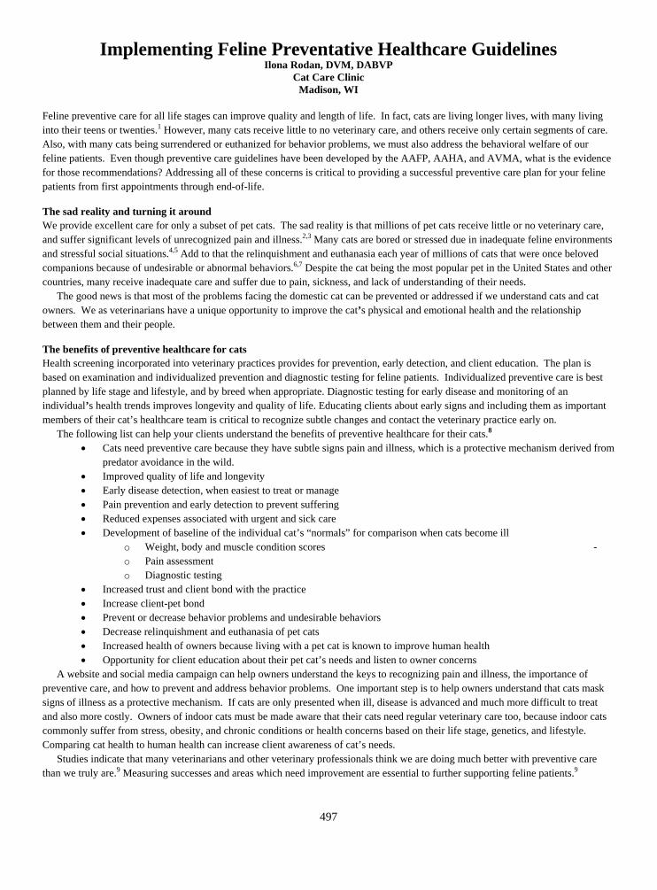
497
Implementing Feline Preventative Healthcare Guidelines Ilona Rodan, DVM, DABVP
Cat Care Clinic Madison, WI
Feline preventive care for all life stages can improve quality and length of life. In fact, cats are living longer lives, with many living into their teens or twenties.1 However, many cats receive little to no veterinary care, and others receive only certain segments of care. Also, with many cats being surrendered or euthanized for behavior problems, we must also address the behavioral welfare of our feline patients. Even though preventive care guidelines have been developed by the AAFP, AAHA, and AVMA, what is the evidence for those recommendations? Addressing all of these concerns is critical to providing a successful preventive care plan for your feline patients from first appointments through end-of-life. The sad reality and turning it around We provide excellent care for only a subset of pet cats. The sad reality is that millions of pet cats receive little or no veterinary care, and suffer significant levels of unrecognized pain and illness.2,3 Many cats are bored or stressed due in inadequate feline environments and stressful social situations.4,5 Add to that the relinquishment and euthanasia each year of millions of cats that were once beloved companions because of undesirable or abnormal behaviors.6,7 Despite the cat being the most popular pet in the United States and other countries, many receive inadequate care and suffer due to pain, sickness, and lack of understanding of their needs.
The good news is that most of the problems facing the domestic cat can be prevented or addressed if we understand cats and cat owners. We as veterinarians have a unique opportunity to improve the cat’s physical and emotional health and the relationship between them and their people. The benefits of preventive healthcare for cats Health screening incorporated into veterinary practices provides for prevention, early detection, and client education. The plan is based on examination and individualized prevention and diagnostic testing for feline patients. Individualized preventive care is best planned by life stage and lifestyle, and by breed when appropriate. Diagnostic testing for early disease and monitoring of an individual’s health trends improves longevity and quality of life. Educating clients about early signs and including them as important members of their cat’s healthcare team is critical to recognize subtle changes and contact the veterinary practice early on.
The following list can help your clients understand the benefits of preventive healthcare for their cats.8 • Cats need preventive care because they have subtle signs pain and illness, which is a protective mechanism derived from
predator avoidance in the wild. • Improved quality of life and longevity • Early disease detection, when easiest to treat or manage • Pain prevention and early detection to prevent suffering • Reduced expenses associated with urgent and sick care • Development of baseline of the individual cat’s “normals” for comparison when cats become ill
o Weight, body and muscle condition scores - o Pain assessment o Diagnostic testing
• Increased trust and client bond with the practice • Increase client-pet bond • Prevent or decrease behavior problems and undesirable behaviors • Decrease relinquishment and euthanasia of pet cats • Increased health of owners because living with a pet cat is known to improve human health • Opportunity for client education about their pet cat’s needs and listen to owner concerns
A website and social media campaign can help owners understand the keys to recognizing pain and illness, the importance of preventive care, and how to prevent and address behavior problems. One important step is to help owners understand that cats mask signs of illness as a protective mechanism. If cats are only presented when ill, disease is advanced and much more difficult to treat and also more costly. Owners of indoor cats must be made aware that their cats need regular veterinary care too, because indoor cats commonly suffer from stress, obesity, and chronic conditions or health concerns based on their life stage, genetics, and lifestyle. Comparing cat health to human health can increase client awareness of cat’s needs.
Studies indicate that many veterinarians and other veterinary professionals think we are doing much better with preventive care than we truly are.9 Measuring successes and areas which need improvement are essential to further supporting feline patients.9
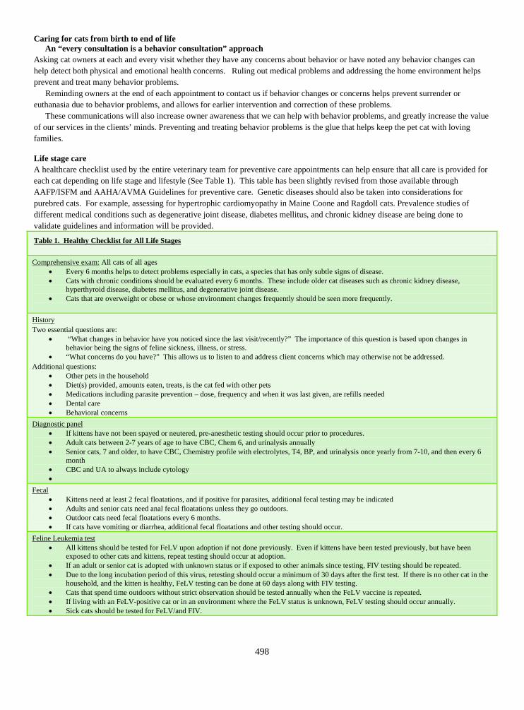
498
Caring for cats from birth to end of life An “every consultation is a behavior consultation” approach
Asking cat owners at each and every visit whether they have any concerns about behavior or have noted any behavior changes can help detect both physical and emotional health concerns. Ruling out medical problems and addressing the home environment helps prevent and treat many behavior problems.
Reminding owners at the end of each appointment to contact us if behavior changes or concerns helps prevent surrender or euthanasia due to behavior problems, and allows for earlier intervention and correction of these problems.
These communications will also increase owner awareness that we can help with behavior problems, and greatly increase the value of our services in the clients’ minds. Preventing and treating behavior problems is the glue that helps keep the pet cat with loving families. Life stage care A healthcare checklist used by the entire veterinary team for preventive care appointments can help ensure that all care is provided for each cat depending on life stage and lifestyle (See Table 1). This table has been slightly revised from those available through AAFP/ISFM and AAHA/AVMA Guidelines for preventive care. Genetic diseases should also be taken into considerations for purebred cats. For example, assessing for hypertrophic cardiomyopathy in Maine Coone and Ragdoll cats. Prevalence studies of different medical conditions such as degenerative joint disease, diabetes mellitus, and chronic kidney disease are being done to validate guidelines and information will be provided.
Table 1. Healthy Checklist for All Life Stages
Comprehensive exam:• Every 6 months helps to detect problems especially in cats, a species that has only subtle signs of disease.
All cats of all ages
• Cats with chronic conditions should be evaluated every 6 months. These include older cat diseases such as chronic kidney disease, hyperthyroid disease, diabetes mellitus, and degenerative joint disease.
• Cats that are overweight or obese or whose environment changes frequently should be seen more frequently.
Two essential questions are: History
• “What changes in behavior have you noticed since the last visit/recently?” The importance of this question is based upon changes in behavior being the signs of feline sickness, illness, or stress.
• “What concerns do you have?” This allows us to listen to and address client concerns which may otherwise not be addressed. Additional questions:
• Other pets in the household • Diet(s) provided, amounts eaten, treats, is the cat fed with other pets • Medications including parasite prevention – dose, frequency and when it was last given, are refills needed • Dental care • Behavioral concerns
• If kittens have not been spayed or neutered, pre-anesthetic testing should occur prior to procedures. Diagnostic panel
• Adult cats between 2-7 years of age to have CBC, Chem 6, and urinalysis annually • Senior cats, 7 and older, to have CBC, Chemistry profile with electrolytes, T4, BP, and urinalysis once yearly from 7-10, and then every 6
month • CBC and UA to always include cytology •
• Kittens need at least 2 fecal floatations, and if positive for parasites, additional fecal testing may be indicated Fecal
• Adults and senior cats need anal fecal floatations unless they go outdoors. • Outdoor cats need fecal floatations every 6 months. • If cats have vomiting or diarrhea, additional fecal floatations and other testing should occur.
• All kittens should be tested for FeLV upon adoption if not done previously. Even if kittens have been tested previously, but have been exposed to other cats and kittens, repeat testing should occur at adoption.
Feline Leukemia test
• If an adult or senior cat is adopted with unknown status or if exposed to other animals since testing, FIV testing should be repeated. • Due to the long incubation period of this virus, retesting should occur a minimum of 30 days after the first test. If there is no other cat in the
household, and the kitten is healthy, FeLV testing can be done at 60 days along with FIV testing. • Cats that spend time outdoors without strict observation should be tested annually when the FeLV vaccine is repeated. • If living with an FeLV-positive cat or in an environment where the FeLV status is unknown, FeLV testing should occur annually. • Sick cats should be tested for FeLV/and FIV.
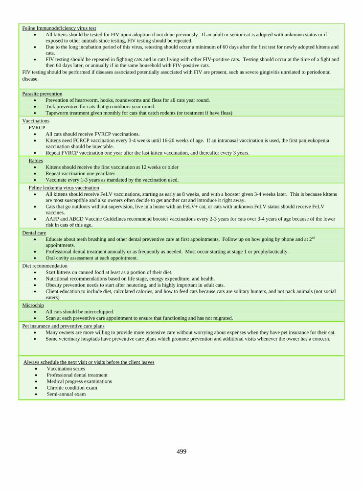
499
Feline Immunodeficiency virus test• All kittens should be tested for FIV upon adoption if not done previously. If an adult or senior cat is adopted with unknown status or if
exposed to other animals since testing, FIV testing should be repeated.
• Due to the long incubation period of this virus, retesting should occur a minimum of 60 days after the first test for newly adopted kittens and cats.
• FIV testing should be repeated in fighting cats and in cats living with other FIV-positive cats. Testing should occur at the time of a fight and then 60 days later, or annually if in the same household with FIV-positive cats.
FIV testing should be performed if diseases associated potentially associated with FIV are present, such as severe gingivitis unrelated to periodontal disease.
• Prevention of heartworm, hooks, roundworms and fleas for all cats year round. Parasite prevention
• Tick preventive for cats that go outdoors year round. • Tapeworm treatment given monthly for cats that catch rodents (or treatment if have fleas)
Vaccinations
• All cats should receive FVRCP vaccinations. FVRCP
• Kittens need FCRCP vaccination every 3-4 weeks until 16-20 weeks of age. If an intranasal vaccination is used, the first panleukopenia vaccination should be injectable.
• Repeat FVRCP vaccination one year after the last kitten vaccination, and thereafter every 3 years.
• Kittens should receive the first vaccination at 12 weeks or older Rabies
• Repeat vaccination one year later • Vaccinate every 1-3 years as mandated by the vaccination used.
• All kittens should receive FeLV vaccinations, starting as early as 8 weeks, and with a booster given 3-4 weeks later. This is because kittens
are most susceptible and also owners often decide to get another cat and introduce it right away.
Feline leukemia virus vaccination
• Cats that go outdoors without supervision, live in a home with an FeLV+ cat, or cats with unknown FeLV status should receive FeLV vaccines.
• AAFP and ABCD Vaccine Guidelines recommend booster vaccinations every 2-3 years for cats over 3-4 years of age because of the lower risk in cats of this age.
• Educate about teeth brushing and other dental preventive care at first appointments. Follow up on how going by phone and at 2nd appointments.
Dental care
• Professional dental treatment annually or as frequently as needed. Must occur starting at stage 1 or prophylactically. • Oral cavity assessment at each appointment.
• Start kittens on canned food at least as a portion of their diet. Diet recommendation
• Nutritional recommendations based on life stage, energy expenditure, and health. • Obesity prevention needs to start after neutering, and is highly important in adult cats. • Client education to include diet, calculated calories, and how to feed cats because cats are solitary hunters, and not pack animals (not social
eaters)
• All cats should be microchipped. Microchip
• Scan at each preventive care appointment to ensure that functioning and has not migrated.
• Many owners are more willing to provide more extensive care without worrying about expenses when they have pet insurance for their cat. Pet insurance and preventive care plans
• Some veterinary hospitals have preventive care plans which promote prevention and additional visits whenever the owner has a concern.
• Vaccination series Always schedule the next visit or visits before the client leaves
• Professional dental treatment • Medical progress examinations • Chronic condition exam • Semi-annual exam

500
Conclusion Preventive care improves and extends quality of life for feline patients. Incorporating preventive care for all life stages prevents or detects common conditions of those life stages.
References Gunn-Moore D. Considering older cats. J Sm Anim Pract Age. 2006;47:430–431. Lue TW, Pantenburg DP, Crawford PM. Impact of the owner-pet and client-veterinarian bond on the care that pets receive. J Am Vet Med Assoc. 2008;232:531–540. Bayer HealthCare. Veterinary care usage study III: Feline findings; 2012. http://www.bayerdvm.com/show.aspx/news-release-bvcus-iii-feline-findings. Heath SE. Behaviour problems and welfare. In: Rochlitz I, ed. The welfare of cats. Dordrecht: Springer; 2005:91–118. Ellis SH, Rodan I, et al. AAFP and ISFM Feline Environmental Needs Guidelines. J Feline Med Surg. 2013;15: 219–230. Kass PH, New Jr JC, Scarlett JM, Salman MD. Understanding animal companion surplus in the United States: relinquishment of nonadoptables to animal shelters for euthanasia. J Applied Anim Welfare Sci. 2001;4:237–248. Salman MD, Hutchison J, Ruch-Gallie R. Behavioral reasons for relinquishment of dogs and cats to 12 shelters. J Applied Anim Welfare Sci. 2000;3:93–106. Rodan I, Sparkes A, Preventive Healthcare for Cats, in The Cat: Clinical Management and Medicine, Ed. Little S, 2012, pp. 151-181. Compliance: Taking Quality Care to the Next Level. Lakewood, CO: American Animal Hospital Association, 2009.
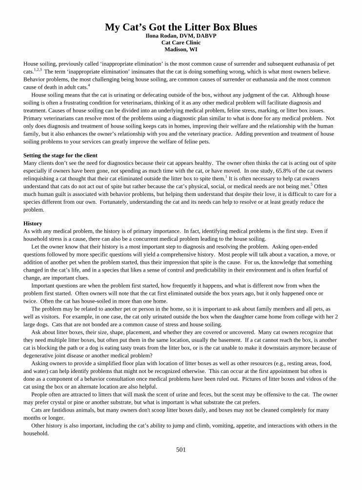
501
My Cat’s Got the Litter Box Blues Ilona Rodan, DVM, DABVP
Cat Care Clinic Madison, WI
House soiling, previously called ‘inappropriate elimination’ is the most common cause of surrender and subsequent euthanasia of pet cats.1,2,3 The term ‘inappropriate elimination’ insinuates that the cat is doing something wrong, which is what most owners believe. Behavior problems, the most challenging being house soiling, are common causes of surrender or euthanasia and the most common cause of death in adult cats.4
House soiling means that the cat is urinating or defecating outside of the box, without any judgment of the cat. Although house soiling is often a frustrating condition for veterinarians, thinking of it as any other medical problem will facilitate diagnosis and treatment. Causes of house soiling can be divided into an underlying medical problem, feline stress, marking, or litter box issues. Primary veterinarians can resolve most of the problems using a diagnostic plan similar to what is done for any medical problem. Not only does diagnosis and treatment of house soiling keeps cats in homes, improving their welfare and the relationship with the human family, but it also enhances the owner’s relationship with you and the veterinary practice. Adding prevention and treatment of house soiling problems to your services can greatly improve the welfare of feline pets. Setting the stage for the client Many clients don’t see the need for diagnostics because their cat appears healthy. The owner often thinks the cat is acting out of spite especially if owners have been gone, not spending as much time with the cat, or have moved. In one study, 65.8% of the cat owners relinquishing a cat thought that their cat eliminated outside the litter box to spite them.1 It is often necessary to help cat owners understand that cats do not act out of spite but rather because the cat’s physical, social, or medical needs are not being met.5 Often much human guilt is associated with behavior problems, but helping them understand that despite their love, it is difficult to care for a species different from our own. Fortunately, understanding the cat and its needs can help to resolve or at least greatly reduce the problem. History As with any medical problem, the history is of primary importance. In fact, identifying medical problems is the first step. Even if household stress is a cause, there can also be a concurrent medical problem leading to the house soiling.
Let the owner know that their history is a most important step to diagnosis and resolving the problem. Asking open-ended questions followed by more specific questions will yield a comprehensive history. Most people will talk about a vacation, a move, or addition of another pet when the problem started, thus their impression that spite is the cause. For us, the knowledge that something changed in the cat’s life, and in a species that likes a sense of control and predictability in their environment and is often fearful of change, are important clues.
Important questions are when the problem first started, how frequently it happens, and what is different now from when the problem first started. Often owners will note that the cat first eliminated outside the box years ago, but it only happened once or twice. Often the cat has house-soiled in more than one home.
The problem may be related to another pet or person in the home, so it is important to ask about family members and all pets, as well as visitors. For example, in one case, the cat only urinated outside the box when the daughter came home from college with her 2 large dogs. Cats that are not bonded are a common cause of stress and house soiling.
Ask about litter boxes, their size, shape, placement, and whether they are covered or uncovered. Many cat owners recognize that they need multiple litter boxes, but often put them in the same location, usually the basement. If a cat cannot reach the box, is another cat is blocking the path or a dog is eating tasty treats from the litter box, or is the cat unable to make it downstairs anymore because of degenerative joint disease or another medical problem?
Asking owners to provide a simplified floor plan with location of litter boxes as well as other resources (e.g., resting areas, food, and water) can help identify problems that might not be recognized otherwise. This can occur at the first appointment but often is done as a component of a behavior consultation once medical problems have been ruled out. Pictures of litter boxes and videos of the cat using the box or an alternate location are also helpful.
People often are attracted to litters that will mask the scent of urine and feces, but the scent may be offensive to the cat. The owner may prefer crystal or pine or another substrate, but what is important is what substrate the cat prefers.
Cats are fastidious animals, but many owners don't scoop litter boxes daily, and boxes may not be cleaned completely for many months or longer.
Other history is also important, including the cat’s ability to jump and climb, vomiting, appetite, and interactions with others in the household.
502
Physical examination, differential diagnosis, and diagnostics A comprehensive examination will identify changes in weight, assess body and muscle condition scores, and include an orthopedic evaluation. With an increase in older cats due to advances in medical and home care, many of our patients have degenerative joint disease or other causes of difficult mobility (e.g., hypokalemia or other causes of weakness). Hyperthyroidism is a common cause of fecal soiling, but may lead to urine soiling as well. Any urinary tract or gastrointestinal disease may lead to house soiling.
Diagnostic testing should always include a urinalysis, complete blood count, and chemistry profile for urine soiling. In addition to these tests, fecal tests should be done for cats that are house soiling. Thyroid testing should be performed in cats 7 years and older, or if they have other signs consistent with thyroid disease.
Radiographs and abdominal ultrasound are also needed in many cases if obvious answers are not found with baseline testing.
Medical etiology Treatment of specific medical conditions should occur in conjunction with making litter boxes easily accessible and more appealing to cats, in addition to providing them space where they don’t need to interact with other animals to reach the boxes. Even if the medical problem is the primary cause, the cat may have found a preferable area to eliminate while ill, and this must be addressed as well. Feline idiopathic cystitis There is a strong link between feline stress and the chronic pain syndrome, feline idiopathic cystitis (FIC).6,7,8 Also called feline interstitial cystitis, it is the most common cause of feline lower urinary tract disease, with 54-64% of cats presenting with lower urinary tract signs having idiopathic disease.9 FIC was initially considered a disease of the bladder alone, but it is now recognized that the response is activated in the brain by the hypothalamic stress response system.7
Stressors include unfamiliar environments and individuals, and a lack of predictability and sense of control, either in the home or the veterinary practice. For example, a hospitalized cat may have a perception of poor predictability and a lack of sense of control if there are inconsistencies in caretakers, feeding and cleaning routines or periods of light and dark.7
A significant decrease in the frequency of FIC signs has been seen with environmental enrichment, familiarity, and a sense of control.6,9 Based on this information, veterinarians can help cat owners recognize environmental stressors and how to improve the environment and sense of control for the cat.
Marking behavior Marking behavior is common in unneutered cats, but may also occur in neutered cats. Marking is an important means of feline communication, and includes urine marking (spraying), fecal marking (middening), rub or cheek marking, and scratch marking. Cats communicate through scent marking and body posturing is used to avoid conflict and protect self. Marking in neutered cats usually indicates a stressful environment.10 Providing a safe environment with place to hide, and easy and safe access to litter boxes without needing to see another cat and feline synthetic pheromone analog diffusers are important to reduce stress. Medication may also be needed temporarily to resolve the problem. Environmental problems
Litter boxes Most commercial litter boxes are too small for cats, since cats need to be able to turn around and scratch. Preferable are large storage containers and dog litter boxes. An opening can be cut out of the front of a high-sided plastic container to allow easier access for cats having difficulty jumping over the edge. High sides are needed for cats that are “high risers” or spray in the litter box.
Location, location, location What may be more important that the number of boxes is the location of the litter boxes. Most people keep litter boxes in the basement and all right next to each other. They think they have 3 boxes, but the cat sees only one box if they are all in the same location. Having at least one litter box on each floor is also needed, especially for cats that have more difficulty going up and down the stairs.
Multiple litter boxes to a cat means that they need to be in different locations, and out of view of other litter boxes. If a cat is fearful of something blocking its pathway –such as another cat it doesn’t like staring at him or her from the staircase or hallway to the litter boxes - the cat is likely to find a safer place to eliminate.
Cats are not the only ones who may block access to a box. If there is also a dog in the home, try to place litter boxes in places that are still easy access for the cat but difficult for the dog to get to. Children may also accidentally frighten a cat, and if a person tries to medicate or do something else to a cat while it is in the box, they will also not want to use the box. Fortunately, with boxes n different locations, the cat can choose which path to take and remain safe.
The fastidious feline Cats are extremely clean animals, and they do not want to eliminate in dirty boxes. Litter boxes should be scooped at least once daily. They also need to be cleaned out at least once weekly with non-scoopable litter and every 2-4 weeks with scoopable litter. Before
503
replacing litter, use mild detergent or just hot water to clean the box. Having additional boxes allows one to rotate in a new box while one is being cleaned and dried.
Diabetic cats and those on subcutaneous fluids will need more frequent scooping and cleaning, as will cats with diarrhea. Litter types
The pet stores have numerous types of litters marketed for humans. Many contain deodorizers, and may be made of pebbles, crystals, pine, corn, or paper. In the wild, cats use sand or dirt, and most cats prefer unscented sand litter. Cleaning areas of house soiling Many enzyme breakdown products are available on the market, but efficacy is not all the same. Anti-Icky Poo and Urine Off are excellent products to eliminate the smell of urine outside the box so that cats are not attracted back to that area. Preventing house soiling It is always easier to prevent house soiling than to treat it. Unfortunately, assumptions are often made that owners know how to purchase litter boxes and litter, and where to put the boxes. Nothing can be further from the truth. Providing cat owners with information on litter box size, types of litter, frequency of box cleaning and how to clean the box will help prevent problems. Educating owners to contact the veterinarian even if the cat misses the box just once will help them recognize that we know how to deal with behavior as well as medical problems. Conclusion Behavior problems, and especially house soiling, are a major concern to owners. With interest in helping owners resolve these problems, we become better veterinarians and better advocates for feline patients. References Kass PH, New,JC Jr.,Scarlett JM, Salman, MD, Understanding Animal Companion Surplus in the United States: Relinquishment of Nonadoptables to Animal Shelters for Euthanasia, J Applied An Welfare Sci, 4(4), 2001:237-248. National Council on Pet Population Study and Policy. The top ten reasons for pet relinquishment to shelters in the United States. Salman MD, Hutchison J, Ruch-Gallie R, Behavioral Reasons for Relinquishment of Dogs and Cats to 12 Shelters, J Applied An Welfare Sci, 2000, 3(2), 93-106. Patronek GJ, Dodman NH, Attitudes, procedures, and delivery of behavior services by veterinarians in small animal practice. J Am Vet Med Assoc, 1999 Dec 1;215(11):1606-1611. Carney HC, Sadek TP, Curtis TM, Halls V, Heath S, Hutchison P, Mundschenk K, Westropp JL, AAFP and ISFM Guidelines for Diagnosing and Solving House-Soiling Behavior in Cats, J Fel Med & Surg, 2014, 16:579–598. Buffington CA, Westropp JL, Chew DJ, Bolus RR (2006) Clinical evaluation of multimodal environmental modification (MEMO) in the management of cats with idiopathic cystitis. J Feline Med Surg 8(4):261-268. Stella JL, Lord LK, Buffington CAT, Sickness behaviors in response to unusual external events in healthy cats and cats with feline interstitial cystitis. J Am Vet Med Assoc 2011; 238(1):67-73. Stella J, Croney C, Buffington CAT, Effects of stressors on the behavior and physiology of domestic cats Appl Anim Behav Sci. January 2013;143(2-4):157-163. Defauw PAM, et al, Risk Factors and Clinical Presentation of Cats with Feline Idiopathic Cystitis, J Feline Med Surg, December 2011;13(12):967-975. Hague DW, Stella JL, Buffington CA, Effects of interstitial cystitis on the acoustic startle reflex in cats. Am J Vet Res, 2013, 74: 144-147.

504
Housing Cats at your Practice: Preventing Problems for Staff and Cats
Ilona Rodan, DVM, DABVP Cat Care Clinic
Madison, WI
Most cats will need to be hospitalized at some point in their life, but the stressors of being in an unfamiliar environment and away from their people can negatively impact their welfare and recovery. We want a hygienic environment and the ability to check patients regularly; many cats however want a stable and quiet environment where they are not bothered, and just want to go back home. Our goal is to bring them back to health, but when the environment is stressful, cats may not eat. Add the stress of the cat owner into the picture, and we have a lot to address. Fortunately, understanding the stressors and when it is best for the cat to remain in the home environment makes all the difference in improving patient care. Challenges associated with caging cats The challenges to housing cats in a veterinary practice are the same as seen in shelters and boarding facilities. We are working with a patient who has strong fear responses associated with lack of familiarity and predictability, and their needs not being met. 1,2,3 Cats are social animals, and when they are separated from their owners for hospitalization or boarding, the disruption of the social bond also leads to the cat’s stress.4,5,6 Ensuring provision of familiar bedding, toys, and food is helpful, and cats often do better if owners have regular visiting privileges when their cat needs hospitalization.
The unfamiliar smells, sounds, sites, and handling also lead to stress. Stress is also caused with an inconsistent schedule. The cat may acclimate, but often it will take from 2 days up to 5 weeks for acclimation to an unfamiliar environment.7,8,9 Stress is a serious factor in the return to health of the feline patient. There is increased risk of a break in upper respiratory infections10 and stress-associated medical conditions such as feline idiopathic cystitis (FIC) can occur in hospitalized or even previously healthy boarding cats.3,11 Lab abnormalities can also occur secondary to stress, making diagnosis and followup difficult.12
Recognizing stress in hospitalized cats How can we tell if stress is occurring in a hospitalized cat? The difficulty is that the signs of stress associated with lack of familiarity and reduced sense of control during caging are usually inhibited normal behaviors rather than more overt abnormal behaviors.1,2 They include decreased activity, appetite, eliminations, grooming, play, and sleep, and the problem is even more complicated because these signs can also be seen in sick cats.1,2,7 Cats that demonstrate these more subtle signs of fear and stress may suffer more than cats that demonstrate blatant signs of upset.2 Although inhibited behaviors are more common, some caged cats may become vigilant, watching every movement that occurs around them, and be alert to every sound. These cats cannot rest, because they must monitor the unfamiliar environment to protect themselves. It is also important to recognize that caged cats may become fearfully aggressive as a protective mechanism and to take them out of the cage within their carrier or cat bed.
The old adage of waiting until a hospitalized patient starts eating before sending them home should not be followed for cats. If all other signs are good, let the owner know why you are sending the cat home, and that they must contact the practice and bring the cat back if the cat does not start eating at home. Another excellent option is to let them take the cat home for the night, with a recheck appointment scheduled for the next morning. Setting up hospital and boarding wards for success Cat-only wards that prevent visibility of dogs and other species is important to reduce feline fear. Cats also become fearful if they see unfamiliar cats or those that they do not like. Back-to-back or side-by-side cages help prevent visibility of other caged cats.
Cats may also have increased stress if they observe other cats being examined.13 If examination must occur in the ward, place a towel over half of the cage door so that the cat choose to see what is going on or not.
Regardless of the length of stay - a few hours to several days or even weeks – the cat must be provided with its essential needs, which include resting or hiding place, perch, litter box, food, water, and play.6 Cats that like attention should also receive human attention. Both the size and complexity of the cage are important to meeting the needs of the caged cat.6 Cages can be enlarged by adding vertical space or combining 2 smaller cages. Cages should be warm, non-slippery, and without the cat seeing its reflection, and laminate cages are preferred over stainless steel.If only stainless steel cages are available, tall cat beds and toweling should be used in the cage. Getting the cat out of the cage Removing a cat from a cage is one of the most threatening experiences for the cat, with increased potential injury for veterinary team members. Therefore, it is important to approach the cage from the side and not make eye contact with the cat. Calmly open the door and allow the cat to come towards you. If the cat chooses not too, remove the cat within the bedding or other hiding area the cat is in.
505
Cage cleaning The old rules of cleaning the cage completely once or twice are no longer the best way. Cats mark their territory with facial pheromones, which makes them more comfortable in the environment, and it is important to avoid cleaning these marked areas until the patient or boarding cat goes home. It is best for the cat to remain in the cage for the duration unless it is soiled to the point that it cannot be cleaned without moving the cat to another cage. The preferred method is to spot clean the cage if it is not soiled.14,15 Spot clean or clean around the cage with minimal disruption of the patient. Do not spray areas clean, but wipe them. Avoid wiping areas that are not soiled. Additionally, the towels or blankets in the cage should not be changed unless soiled, because doing so takes away the familiar scents and introduces unfamiliar scents. The same goes for litter boxes: Try to keep the same litter box with the cat during the cat’s stay, scooping two or more times daily, but not washing out unless necessary. Instructions for spot cleaning are given in Boarding cats in the veterinary practice Many veterinary practices board cats or at least to provide medical boarding for those that need medication while owners are away from home. Some cats do better with cat sitters coming into the home environment, but many owners prefer to board their cats. Cats that are boarded for several days or longer need larger spaces. “Kitty condos” are excellent options for boarding cats. If these are unavailable, providing out of cage time in a safe place and during quiet times can be helpful for those cats that feel safe doing so.
When cats from multicat households are boarded, they should be placed together only if they show affiliative (bonded) behaviors, such as licking or grooming or sleeping while touching. Even if bonded, they need their own cat beds since even bonded cats like to spend at least 50% of the time by themselves. 16 It is also important to assess multicat interactions because those cats that are socially incompatible with their housemates may be able to manage the social tension between them when they have a whole house to occupy, but they may find being confined in a small boarding cage for 1 week or more highly stressful. Owners often want their cats to be housed together, but the decision has to be made with the best interests of the cats in mind, and, in some cases, being separated from each other is preferable from a feline perspective. Decreasing owner stress It is well recognized that owners are concerned about leaving their cat at a veterinary practice or an emergency facility. Offering to let them see where their cat will stay and how the cage is set up can relieve some of this stress, and allowing visiting privileges increases their comfort and the value of the experience. Letting owners know that their cat will be most comfortable with familiar bedding, toys, treats, etc. allows them to help as part of the team for their cat’s care.
Clients are anxious for updates, and sending e-mails, pictures, and setting up times to call will help reduce staff and client stress. Conclusion Because of the challenges of hospitalization for feline patients and their owners, it is advisable to minimize hospitalization and to admit cats into veterinary practices only when absolutely necessary. When the cat must be hospitalized, it is important to provide an environment that respects natural feline behavior and aims to meet the behavioral needs of feline patients. Simple steps based on an understanding of the cat’s needs can be taken to help reduce the stress of hospitalization or boarding, which not only benefits the welfare of the cat but also improves the practice’s ability to treat and evaluate the cat and will result in an increase in safety and job satisfaction among staff members. References 1. Griffin B, Hume KR: Recognition and management of stress in housed cats. In August JR, editor: Consultations in feline internal medicine, ed 5, St Louis, 2006, Saunders Elsevier, pp 717–734. 2. Patronek GJ, Sperry E: Quality of life in long-term confinement. In August JR, editor: Consultations in feline internal medicine, ed 4, St Louis, 2001, Saunders Elsevier, pp 621–633. 3. Stella JL, Lord LK, Buffington CAT: Sickness behaviors in response to unusual external events in healthy cats and cats with feline interstitial cystitis. J Am Vet Med Assoc 238:67–73, 2011. 4. Gourkow N, Fraser D: The effect of housing and handling practices on the welfare, behaviour and selection of domestic cats (Felis sylvestris catus) by adopters in an animal shelter. Anim Welf 15:371–377, 2006. 5. Kry, K, Casey R: The effect of hiding enrichment on stress levels and behaviour of domestic cats Felis sylvestris catus in a shelter setting and the implications for adoption potential. Anim Welf 16(3): 375–383, 2007. 6. Rochlitz I: Recommendations for the housing of cats in the home, in catteries and animal shelters, in laboratories and in veterinary surgeries. J Feline Med Surg 1:181–191, 1999. 7. Zeiler GE, Fosgate GT, van Vollenhoven E, Rioja E: Assessment of behavioural changes in domestic cats during short-term hospitalisation. J Feline Med Surg 16:499–503, 2014. 8. McCobb EC, Patronek GJ, Marder A, et al: Assessment of stress levels among cats in four animal shelters. J Am Vet Med Assoc 226:548–555, 2005. 9. Rochlitz I, Podberscek AL, Broom DM: Welfare of cats in a quarantine cattery. Vet Rec 143:35–39, 1998.

506
10. Tanaka A, Wagner DC, Kass PH, Hurley KF: Associations among weight loss, stress, and upper respiratory tract infection in shelter cats. J Am Vet Med Assoc 240:570–576, 2012. 11. Stella J, Croney C, Buffington T: Effects of stressors on the behavior and physiology of domestic cats. Appl Anim Behav Sci 143:157–163, 2013. 12. Quimby JM, Smith ML, Lunn KF: Evaluation of the effects of hospital visit stress on physiologic parameters in the cat. J Feline Med Surg 13:733–737, 2011. 13. Wallinder E, et al: Are Hospitalised Cats Stressed by Observing Another Cat Undergoing Routine Clinical Examination? International Society of Feline Medicine 2012 Proceedings. 14. Newbury S, Blinn MK, Bushby PA, et al: Guidelines for standards of care in animal shelters, Corning, NY, 2010, Association of Shelter Veterinarians. Available at http://oacu.od.nih.gov/disaster/ShelterGuide.pdf. Accessed January 20, 2015. 15. UC Davis Koret Shelter Medicine Program: Instructions for Spot Cleaning, http://www.sheltermedicine.com/node/339. 16. Rochlitz I. Recommendations for the housing and care of domestic cats in laboratories. Lab Anim. 2000;34:1–9. 14. Ray CC, Callahan-Clark J, Beckel NF, Walters PC: The prevalence and significance of hyperglycemia in hospitalized cats. J Vet Emerg Crit Care 19:347–351, 2009. 15. Buffington CA, Westropp JL, Chew DJ, Bolus RR: Clinical evaluation of multimodal environmental modification (MEMO) in the management of cats with idiopathic cystitis. J Feline Med Surg 8:261–268, 2006. 16. Wallinder E, et al: Are Hospitalised Cats Stressed by Observing Another Cat Undergoing Routine Clinical Examination? International Society of Feline Medicine 2012 Proceedings. 17. Rochlitz I: Basic requirements for good behavioural health and welfare in cats. In Horowitz DF, Mills DS, editors: BSAVA manual of canine and feline behavioural medicine, ed 2, Gloucester, UK, 2009, British Small Animal Veterinary Association (BSAVA), pp 35–48. 18. Newbury S, Blinn MK, Bushby PA, et al: Guidelines for standards of care in animal shelters, Corning, NY, 2010, Association of Shelter Veterinarians. Available at http://oacu.od.nih.gov/disaster/ShelterGuide.pdf. Accessed January 20, 2015. 19. UC Davis Koret Shelter Medicine Program: Instructions for Spot Cleaning, http://www.sheltermedicine.com/node/339.
507
The Cat Friendly Practice: A Win-Win for You, Cats, and Clients
Ilona Rodan, DVM, DABVP Cat Care Clinic
Madison, WI
The Cat Friendly Practice Program (CFP) was developed to help veterinarians provide the best of health care, regardless of whether the practice is companion animal, feline only, or a mixed animal practice. CFP was launched by the American Association of Feline Practitioners in early 2012 and has grown rapidly with the primary goal being increased feline veterinary visits to increase health and welfare. Although the cat is the most popular pet in the US, millions of pet cats receive little to no needed veterinary care, and suffer unrecognized pain and illness.1,2
With the popularity of the cat as well as advances in feline health care, one would expect cats to have increased veterinary care. However, there has been a decline in patient visits since 2001. Active and new client numbers are down significantly in most practices, as are the number of patients seen weekly at most veterinary practices.2 And the numbers continue to decline despite major efforts by all major veterinary organizations that see cats – AAFP, AAHA, and AVMA. Comparing 2011 to 2006, the number of cat-owning households that did not take their cat to the veterinarian increased by a staggering 24%.3 Correlating with the decline in visits is a significant increase in cats with preventable diseases, including a 10% increase in dental disease, 13% increase in internal parasites, and 16% increase in diabetes mellitus.4
There are 2 major reasons the lack of veterinary care - the stress associated with getting the cat to the veterinary practice and owners not recognizing the value of preventive care. Veterinary organizations started recommending that cats be kept indoors around 2001 to improve their safety and longevity as well as to protect wildlife, and feline visits started their decline at that time. Owners erroneously think that indoor cats don’t need care; of course, nothing is further from the truth and we must increase their awareness of medical conditions that occur in cats. The stress to a cat that is always in their home is also more significant than the cat exposed outdoor life. At the heart of the Cat Friendly Practice is the potential for building productive relationships with cat owners, which will result in improved care of the feline patient.
Participating practices that have become cat friendly have noticed a considerable difference, with increased feline visits and client value, decreased patient fear and owner stress, and a subsequent increase in income. When a practice takes the steps to become a Cat Friendly Practice, it creates a practice environment that values the feline patient’s needs.
The first steps are to better understand the stress surrounding veterinary visits. With this information, we can recognize how to improve the hospital environment, the experience of getting the cat to the practice, and how the cat is handled in our hospitals. Becoming a Cat Friendly Practice can help turn around these problems and increase feline patients in your practice.
First impressions Walk into your practice or the practice you work at with the mindset of a cat owner who loves their cat and is stressed about the experience. Do you see primarily dog pictures and brochures, and dogs in the waiting area? Think about what that loving cat owner feels when he or she walks in.
Regardless of practice type, let your clients know that you cater to cats with equal information about cats on your website, blogs, and Facebook. If you are “cat friendly”, promote that. CFP practices are provided with large amounts of information for staff and client education, and this information can be freely used by CFP member practices to educate clients and promote their services. It starts at home When scheduling the first appointment, ask clients if they have a carrier, and if they have difficulties getting their cat into the carrier. Provide information to help them make the visit less stressful for their cat and them. If they don’t already have a carrier, provide recommendations.
Choice of carrier Hard sided carriers that can be taken apart in the middle are preferred. The cat can then remain in the bottom half of the carrier if it doesn’t want to come out at the veterinary hospital. Even better is to have a hard sided carrier that has the option to open from both the top as well as the front. If the cat won’t go into the carrier on its own, the cat can then be lowered gently through the top instead of shoved through the front opening.
Some clients prefer soft-sided carriers, either because they are lighter to carry or more comfortable for the cat. If used, care must be taken to prevent the carrier from collapsing or being pushed in on the cat. Also, the cat may be jostled more in a soft carrier, and increased care to keep the carrier steady is important by carrying gingerly. The biggest issue is getting the cat out of this type of carrier if it chooses to remain inside. Looming over the carrier or pulling the cat out often increases feline fear and potential fear-associated aggression. If a client prefers a soft-sided carrier, recommend one that has 3 openings, with a large opening that comes down in a U-shape in the middle.
508
Training the cat to the carrier The first step in using carriers is to make the carrier a comfortable, secure place for the cat.
Educate clients to keep the carrier in a familiar place in the home to increase familiarity and reduce fear. Help them recognize how to make the carrier a safe haven, by leaving it out instead of only bringing it out for veterinary visits. If clients refuse to have it out all the time, minimally bring the carrier out a few days before the veterinary visit. The carrier should be cozy within, with a fleece of fluffy blanket. Place or toss treats or preferred kibbles daily into the carrier to entice the cat to enter. Reward the cat for use of the carrier.
Clients should also be taught to reward or reinforce desired behavior, and to never
For the cat that is still stressed and wary of the carrier, and will not enter on its own, there are still options. First, the cat should never be yelled at or chased in an attempt to get it into the carrier. Place the cat into a small room where the cat cannot hide. Teach the client to calmly get the carrier, and bring it into the room, closing the door. Calmly put the cat into the carrier. Hard-sided carriers with an opening at the top are ideal for these cats because the owner can then gently place the cat through the top, instead of shoving the cat through the door.
punish the cat– either verbally or physically. Encourage the client to bring favorite treats, toys, or grooming utensils as well, so that they can be used to entice the cat to come out of it’s carrier and/or to reward or reinforce desired behaviors.
There will always be patients that still remain anxious whether it is due to previous fearful or painful experiences, or how the owner reacts. Because anxiety can inhibit learning, cats with a history of being anxious on car rides and/or veterinary visits may require anxiolytic (anti-anxiety) medication. Alprazolam and gabapentin are anxiolytics, but acepromazine is not; it should not be used because dissociated aggression can occur. Some cats will become more reactive with alprazolam, but this does not occur with gabapentin.
Gabapentin is the speaker’s drug of choice. Although gabapentin is an analgesic medication, it also works as an anxiolytic. Dose gabapentin at 10 mg/kg or 100 mg/cat 90 minutes – 3 hours prior to the veterinary visit. Some cats may need up to 150 mg/cat. It is tasteless and can be mixed into a small amount of canned food. The lecturer and many others use 100 mg per cat as their dose rather than the 10 mg/kg calculation, regardless of size of the cat. The speaker has used 50mg in geriatric cats that are cachectic. It can be sedating and it is best to let the owner know that the cat may sleep more after the visit.
Travel is best on an empty stomach to prevent motion sickness and so that the cat is more interested in treats at the veterinary practice. Both provide for a more positive experience. If needed, a small amount of canned food with gabapentin can be given. A synthetic feline facial pheromone (FFP) analog sprayed into the carrier at least 30 minutes before travel, has a calming effect on the cat.5 Draping a blanket over the carrier can also help prevent fear and motion sickness. If the cat is still nauseous – lip-licking, drooling, or vomiting during transportation - maropitant (Cerenia) is recommended to prevent motion-sickness.6 The cat friendly hospital environment Provide a quiet environment, with no offensive smells (dog odors, strong cleaning solutions, or perfumes). Use a synthetic feline pheromone analog to help calm feline patients. If at all possible, have a separate waiting area for cats or divide the waiting area into two, with back-to-back benches and taller shelves or plants between them to provide a visual barrier and separate waiting areas.
Regardless of how “cat-friendly” the waiting area is, it is best to take the cat and client directly into an exam room. In hospitals that also see non-feline patients, designating one or two exam rooms for cats helps reduce stress, and provides an environment that is for cats only. Other options are to dedicate one-half day to cat-only appointments or surgeries.
Performing all examinations and diagnostic testing in the examination room instead of the treatment area will help keep the cat less fearful and facilitate the procedures.
Unfamiliar scents can frighten and arouse cats. Providing familiar scents such as that of a favorite person can help a cat adapt to new situations. The synthetic feline facial pheromone analog, Feliway®, mimics the natural pheromone that is deposited when a cat rubs its face on objects, and has been shown to provide a calming effect in unfamiliar or stressful environments or situations.7,8,9 The new product, Feliway Multicat, is now available in the US only, and is supposed to be helpful for introducing a new cat and reducing intercat conflict in multicat households.
Cats that require hospitalization often become inactive and inhibit normal behavior such as feeding, grooming, exploring, and playing.10 Cats need both places to hide and places up high. Hiding places can be as simple as a sturdy cardboard box, or a cat bed with high edges to hide in. Vertical space provides vantage points to monitor the cat’s surroundings and the approach of people and other animals.11 Caging should always include comfortable bedding, preferably with the familiar scent from a favorite person. Whenever possible, separate cat wards from dog wards. Also, cages should not face each other so that cats cannot see other cats or any other animals. Feline synthetic pheromone analog sprayed into cages and boarding suites at least 30 minutes prior to use can calm feline patients and increase normal behaviors while caged.12 Cats that are very anxious or fearfully aggressive are best isolated into an
509
empty isolation area; if not possible, place a towel on the front of the cage to screen out activity that may add to the cat’s anxiety. Scheduled feeding and cleaning times are less stressful for feline patients. Conclusion Developing a Cat Friendly Practice can reduce stress for all involved, and increase cat visits to improve feline healthcare. References Lue TW, Pantenburg DP, Crawford, PM, Impact of the owner-pet and client-veterinarian bond on the care that pets receive, JAVMA, Vol 232, No. 4, February 15, 2008, pp. 531-540. Bayer Veterinary Care Usage Study III: Feline Findings (2012) U.S. Pet Ownership & Demographics Sourcebook (2012). Banfield Pet Hospital. State of pet health 2011 report. Volume 1.,Portland, Ore: Banfield Pet Hospital, 2011. Available at: www.banfield.com/Banfield/files/bd/bd826667-067d-41e4-994d-5ea0bd7db86d.pdf., Accessed 1.6.2013. Pageat P, Gaultier E: Current research in canine and feline pheromones, Vet Clin North Am Small Anim Pract 33:187, 2003. Hickman MA, Cox SR, Mahabir S et al: Safety, pharmacokinetics and use of the novel NK-1 receptor antagonist maropitant (Cerenia) for the prevention of emesis and motion sickness in cats, J Vet Pharmacol Ther 31:220, 2008. Kronen PW, Ludders JW, Erb HN, et al: A synthetic fraction of feline facial pheromones calms but does not reduce strugglingin cats before venous catheterization. Vet Anaesth Analg 33:258-265, 2006. Griffith CA, Steigerwald ES, Buffington CA: Effects of a synthetic facial pheromone on behavior of cats. J Am Vet Med Assoc 217:1154-1156, 2000. Gaultier E: Current research in canine and feline pheromones, Vet Clin North Am Small Anim Pract, 33:187, 2003. Rochlitz I, Recommendations for the housing of cats in the home, in catteries and animal shelters, in laboratories and in veterinary surgeries, J Feline Med & Surg (1999) 1, 181-191. Patronek G, Sperry E: Quality of life in long-term confinement. In August J (ed): Consultations in Feline Internal Medicine, ed 4, Philadelphia, 2001, W.B. Saunders, p 621. Griffith C, Steigerwald E, Buffington C: Effects of a synthetic facial pheromone on behavior of cats, J Am Vet Med Assoc 217:14, 2000.
510
Meeting the Environmental Needs of Indoor Cats Ilona Rodan, DVM, DABVP
Cat Care Clinic Madison, WI
The veterinary profession is responsible for both the physical and emotional health of our patients. Advances in feline medicine have increased the pet cat’s physical health and longevity1, but emotional and environmental health needs often go unrecognized. Inadequate feline environments frequently lead to boredom, stress, and obesity.2,3 Behavior problems and normal feline behaviors that people consider undesirable can also occur when cats’ needs are not met. Environmental stressors can even lead to physical health problems, such as feline idiopathic cystitis. These problems occur due to the disparity between who cats really are and the impression that many owners have, which is that they are low maintenance and easy to care for pets.
The cat is a paradox – although fairly adaptable and social animals under the right conditions, cats have retained many of the behaviors of their wild ancestor, Felis lybica.4,5 In fact, pet cats are still more similar to their wild ancestors than to other species and require an environment that provides for their needs. Understanding the cat, its normal behaviors, and its needs can often prevent or resolve stress, boredom, and behavior problems. Regardless of the age and physical health of the cat, and regardless of whether the cat is indoors only, indoor/outdoor, at home or at the veterinary practice, providing for the cat’s environmental needs is not optional but rather essential for its welfare.3
Feline welfare and the veterinary responsibility Within the past decade, a large number of worldwide veterinary organizations have rewritten their veterinary oaths to emphasize welfare. Animal welfare is defined by the AVMA as: “…how an animal is coping with the conditions in which it lives. An animal is in a good state of welfare if (as indicated by scientific evidence) it is healthy, comfortable, well nourished, safe, able to express innate behavior, and if it is not suffering from unpleasant states such as pain, fear, and distress. Protecting an animal's welfare means providing for its physical and mental needs.” Many feline patients are not allowed to express innate behaviors, often leading to fear and stress - to poor welfare. Understanding the cat and its needs
Cats as solitary hunters Because cats are solitary hunters of several small prey daily, they must maintain their physical health and avoid danger. They do so through two major protective mechanisms – territoriality to maintain safe space and having a heightened fear response. Familiar territory provides cats with a sense of control over their physical and social environment.6 Having a sense of control - even if it is not exerted - makes the cat more comfortable and reduces stress.7
A primary goal of communication between cats is to protect territory and avoid physical altercations.8 They communicate through body and facial posturing, as well as via their senses. Scent marking is most important for cats, with scent and pheromonal signals used as distance communication to keep other cats away without the need for physical contact. Scent marking occurs via facial and body rub marking, scratching, urine spraying, or middening (fecal marking). Spraying in neutered cats is usually secondary to stress in the environment.
Cats possess heightened a heightened fear response as a protective response to fear.9 If cats are forced to leave their familiar territory or a threat enters their territory, they respond to this confrontation by avoidance or hiding, with fighting only occurring if there are not other options to protect self. Fear commonly occurs when a cat is taken outside its environment and brought to the veterinary hospital. Providing a place to hide for both inpatients and out-patients can prevent fear-associated aggression.
Providing choice in the environment through multiple resources - hiding, perching, feeding, water, and toileting areas - in multiple locations in a multi-cat household will reduce fear and provide cats with a sense of control and more secure environment.3 This is important regardless of whether it is the home environment, veterinary practice, cattery, or shelter. Feline environmental needs
Safe space Hiding is a coping behavior that cats often display in response to changes in their environment.10 In the home, this could be an unfamiliar person or pet. Problems often occur with a newly adopted cat being introduced to already existing household cats without gradual introduction. Even if it is not a newly introduced cat, it is not unusual for cats that live in the same household not to like each other and choose to rest in a safe place away from others. Even affiliate cats – cats that like each other – prefer to sleep alone and out of sight of others approximately half the time.6 Appropriate sleeping areas are also good hiding places, such as a box, a cat bed with high sides, or a carrier with soft bedding such as fleece.
In the veterinary hospital, a safe place is necessary for both in-patients and out-patients. The carrier – especially if the cat has positive experiences and familiarity with it in the home environment – is an excellent safe place. Allowing the cat to rest in the
511
bottom half of the carrier during examinations and providing either the carrier or another hiding place during hospitalization or boarding will increase feline safety and security, and decrease fear-associated aggression.
Elevated resting areas Increasing overall space by providing cat trees, perches, shelves, or other vertical space helps prevent conflict between cats.3 Cats can also monitor or oversee the environment from a vertical space.
Scratching Scratching is a normal feline behavior that marks territory with both scent and visible markings. It also is done to sharpen claws, remove old sheaths, and to stretch muscles. Providing scratching posts with preferred texture, such as sisal rope or natural wood, as well as in multiple locations helps prevent furniture destruction. Posts should be placed in locations where cats prefer to scratch – usually next to a most prominent piece of furniture, but sometimes also where new scents occur (e.g., the front or back door).
Normal feeding behavior of the solitary hunter Cats are not pack hunters, but rather solitary hunters, eating 10-20 small meals per day, with repeated cycles of hunting to catch their small prey. Not all attempts to catch prey are successful (some suggest that up to 50% of the hunt cycles are not successful).11 Think about how much time and energy the cat utilizes just to survive!
Compare that to what happens with many owned cats. People usually control the feedings, often providing 1-2 meals daily of highly palatable food. The inability to control access to food is associated with feline stress. 12 The sedentary house cat expends very little energy and time hunting, and more time eating. In some countries, including the US, many cats are kept indoors. Whether to protect the cat itself or wildlife, failure to provide opportunities for predatory behavior may deprive cats of mental and physical activity, and may contribute to development of obesity and other health problems.11,13
Because people are social eaters usually enjoying meals together, they often provide multiple cats with food either in one bowl or in bowls placed side-by-side, not recognizing that this causes competition for food resources and stress for the cat. One can understand why some cats may eat large volumes very rapidly, often overeating, and perhaps regurgitating. Stress is usually the short-term result, and obesity and obesity-associated diseases are more long-term outcomes.
Regardless of how much cats are fed, the hunting instinct still exists; cats often bring in these unwanted “presents” to their people. Cats are also crepuscular animals, hunting primarily at dawn and dusk, when their prey is usually present. This sometimes leads to waking owners during the wee hours of the morning, which can be quite annoying for humans. Often owners inadvertently reinforce this behavior in their attempt to quiet the cat so that they can go back to sleep, leading to a long-term and frustrating problem for owners. Client education can prevent this problem as long as we welcome clients to discuss their frustrations or concerns about their cats with us.
As veterinarians, we have the opportunity and responsibility to educate clients about normal feeding behavior of the cat as part of the nutritional advice we provide. This will help prevent both medical and behavioral problems, obesity, and stress in the home environment. This can be done by simulating “hunting” through the use of food toys or puzzles, tossing kibbles, or hiding them around the house. This more normal feeding behavior will increase exercise, reduce boredom, and help prevent obesity.15 Providing feeding areas in multiple locations which are out of sight of each other will prevent competition for food resources.
Drinking behavior: Cats in the wild drink water in locations separate from food. Some cats prefer running water, and some still water. Provide water dishes in multiple locations and away from food.
Play behavior Queens teach kittens to hunt through play behavior. The rough tussle and tumble of kittens help them hone their hunting skills. Kittens and even adult cats, especially if housed singly, may want to play with their owner’s hands and feet in the same way. When young, people often think this is cute, and unknowingly reward the behavior. Play aggression can lead to human injury and zoonotic disease. Playing with an interactive toy minimally once to twice daily can prevent this problem. Cats learn to anticipate and prefer the routine of playing daily at a certain time, even if toys are rotated. Cats playing together also provides an outlet for this behavior. Self play is also important, and can be provided through puzzle feeder toys or other favored toys (e.g., hair scrunchy, foil or paper ball).
Toileting areas Litter boxes should be placed in multiple locations around the home, but away from food, water, and sleeping areas. It is not uncommon for cat owners to prefer to place 2 or more litter boxes in the basement next to each other. This poses multiple problems – usually there are noisy appliances and equipment in the basement, the boxes next to each other don’t provide easy access to a box if a more confident cat is blocking a timid cat, and a cat with degenerative joint disease or another condition making it difficult to get downstairs – that can lead to a cat soiling outside the litter box.
Many litter boxes are also too small for cats. Cats prefer larger boxes so that they can turn around, dig, and eliminate. Boxes should be 1.5 times the size of the cat from the tip of the nose to the base of the tail. Dog litter boxes and plastic storage containers with an opening make excellent cat boxes.
512
Some cats will eliminate in any type of litter. Others prefer a soft consistency, such as sand over pebbles. Deodorizers or scented litter since as pine are developed to attract consumers and not the cat. Some cats find them offensive. Most cats do well with unscented clumping sand litter.
Scooping boxes a minimum of once daily and changing boxes completely when needed (weekly for clay or non-clumping litter and once every 2 or more weeks for clumping litter) will also help to prevent house soiling problems. Single vs. multiple cat households Cats are social animals, but their social system is flexible, meaning that cats can live alone or in groups called colonies if there are sufficient resources.5,11 Cats that like each other demonstrate affection towards each other by rubbing against or grooming each other, or sleeping in close physical contact.
Cats usually do not readily welcome unfamiliar cats. In a study of 1,286 relinquished cats, relinquishment was associated with the number of pets in the household, as well as new cats being added into the home environment.14 Introductions must be gradual, and with all resources available in multiple locations to prevent the need to pass an unfamiliar cat.
Just because cats come together for feeding or to sleep on the same bed, it doesn’t mean that they like each other or that stress isn’t occurring in the feline household; in many households, cats come together because the primary resources are placed in one location. Since cats are more likely to rest or sleep alone, multiple comfortable resting areas should be provided. Inter-cat conflict and behavior problems often occur because household cats don’t have multiple resources in multiple places, and therefore cannot avoid the other cat(s).
Multiple resources with easy access, and out of view of other resources must occur. This includes hiding places and use of vertical space to allow cats to be apart if they so choose. Vertical space increases overall space and provides for the cat to oversee the environment. Litter boxes, food, and water stations that are placed in different locations so that individual cats don’t need to see each other reduces competition for resources, bullying, and stress.12 Serious consideration should be taken before adopting a new cat if cats already exist in the home. Clients should be educated to let cats choose their own affiliates, and be made aware that the greater number of cats in a household, the greater the chance of behavior problems. Conclusion The dilemma of what cat owners want and what cats need can both be met when behavioral health is incorporated into veterinary practice. When we understand cats and treat them as the species they are, we can enrich their lives and increase their welfare, further enhancing the bond we share with them. References Gunn-Moore, D, Considering Older Cats, J Sm An Pract Age, 2006. Aug;47(8)430-431 Heath SE, Behaviour problems and welfare. In: Rochlitz I (ed). The welfare of cats. Dordrecht, The Netherlands: Springer 2005, pp. 91-118. Ellis SH, Rodan I, et al., AAFP and ISFM Feline Environmental Needs Guidelines, J Fel Med & Surg, 2013, 15:219-230. Driscoll CA, Menotti-Raymond M, Roca AL et al.: The Near Eastern origin of cat domestication, Science 317:519, 2007. Bradshaw JWS, Casey RA, and Brown SL, The Behaviour of the Domestic Cat, 2nd Edition, CABI Publ, 2012. Rochlitz I, Housing and Welfare, in The Welfare of Cats, Ed., Rochlitz, 2007, pp. 177-203. Rand JS, Kinnaird E, Baglioni A, et al: Acute stress hyperglycemia in cats is associated with struggling and increased concentrations of lactate and norepinephrine. J Vet Intern Med 16:123-132, 2002. Bowen J, Heath S: An overview of feline social behaviour and communication, in Behaviour Problems in Small Animals: Practical Advice for the Veterinary Team, ed 1. Saunders Ltd., p 29, 2005. Griffin B, Hume KR: Recognition and management of stress in housed cats, in August J (ed): Consultations in Feline Internal Medicine, vol 5. St. Louis, Elsevier, pp 717-734, 2006. Carlstead, K., Brown, J. L. & Strawn W. (1993) Behavioral and physiological correlates of stress in laboratory cats. Applied Animal Behaviour Science, 38(2), 143-158. Rochlitz I, Basic requirements for good behavioural health and welfare of cats, In Horwitz D, Mills D (eds). BSAVA manual of canine and feline behavioural medicine. 2nd edn. Gloucester: British Small Animal Veterinary Association, 2009, 35-48. Bissot T, Servet E, Vidal S, et al, Novel dietary strategies can improve the outcome of weight loss programmes in obese client-owned cats. J Feline Med Surg. February 2010;12(2):104-12. Herron MAE, Buffington CAT, Feline Focus: Environmental Enrichment for Indoor Cats, Compendium: Continuing Education for Veterinarians December 2010. Salman MD, Hutchison J, Ruch-Gallie R, Behavioral Reasons for Relinquishment of Dogs and Cats to 12 Shelters, J Applied An Welfare Sci, 2000, 3(2), 93-106.
513
Getting Nutrition into a Cat Craig Webb, DVM, DACVIM
Colorado State University Fort Collins, CO
Our understanding of feline nutrition has advanced significantly from the day when we simply considered them small dogs, and the number of options we now have for dietary intervention in this species has expanded exponentially. But neither the knowledge of feline metabolism nor the number of available diets helps us, or the cat, one bit, if we can’t get the stuff into them. When a Labrador retriever refuses to eat we know the prognosis is grave: when a cat refuses to eat it may well be that they have decided that the presentation of their latest meal was not up to standards. Unlike Labrador retrievers, cats are one-trial learners, so make the mistake of trying sneak a medication into the one particular flavor of food the cat will tolerate, and that may well be the last time you get anything into that cat’s mouth. Try to switch diets on a Labrador and you might get a brief pause as the dog considers the phrase “fool me once, please!”. Try to switch diets on a cat, for its own good mind you, and suffer an expression of disdain and an attitude of incredulous indignation. So of course, what is perhaps the single most common clinical expression of almost anything wrong with a cat? A decreased-to-absent appetite. And what are the consequences of anorexia in a cat compared to a Labrador? Well cats have their own specific condition for just that – hepatic lipidosis. This presentation should be considered a “group effort” as collectively the veterinary profession aspires to be more clever than a single cat and we explore a plethora of possible strategies for getting nutrition into a cat. Terminology and differentials Hyporexia is the term for a reduced appetite and Anorexia is complete inappetence. Large category differentials for Anorexia are: Primary anorexia, Secondary Anorexia, and Pseudo-Anorexia. Primary anorexia is most often associated with brain disease, trauma, or tumor, including “anosmia” or the inability to perceive odor – a particularly important sensation for cats. Pseudo-anorexia is any condition where the cat actually wants to eat but is extremely reluctant to do so because of other pressing problems. Examples of pseudo-anorexia would include dental disease, musculoskeletal disorders, pain (of the some component of the oral cavity and GI tract, or an unrelated but uncomfortable condition), stress, anxiety, depression, even environmental ques. Secondary anorexia is everything else, and the category that is most commonly the reason for presentation of a cat that’s not eating. Consequences Anorexia leads to malnutrition, and malnutrition has significant consequences for cats. The cat’s unique metabolic make-up is not particularly well suited for adapting to different nutritional planes, their metabolic rate and preferential metabolic pathways do not alter their activity level to a great extent in response to changes in nutrient content. When their diet or lack of dietary intake fails to provide their preferred nutrients they may turn to their endogenous supply (muscle protein leading to cachexia) or over-produce harmful metabolic by-products (ketoacidosis). A poor nutritional plane leads to immunosuppression, proteolysis, hepatic lipidosis, and an increase in mortality. Importantly, Reynolds et al. (2010) showed that for cat owners the quality of life is more important than longevity, and appetite ranked as one of the key components in a cat owner’s perception of their cat’s quality of life. Cats are also particularly prone to stress, whether obvious to us or only perceived by the cat, and Stella et al. (2011) demonstrated that one of the consequences of stress in cats is anorexia. Veterinary clinics and hospitalized cats The advent of Feline Friendly Practice standards (www.catvets.com) from the American Association of Feline Practitioners goes a long way towards helping clinicians design environments and interactions that can reduce stress and decrease the incidence of hospital acquired anorexia in our feline patients. Unfortunately there are still too many scenarios where cats who are already feeling ill are stuffed into a carrier and exposed to a room full of predators just before being introduced to the healing hands of the veterinary staff, potentially for restraint, target practice, and indignity before being transferred to a metal box with none of the comforts of home. But an active appreciation and the motivation to change can go a long way towards relatively simple (a hiding box, an elevated perch, the appropriate temperature, the line-of-sight) or complex (feline-only reception area, cat-only exam rooms, specially trained and qualified “crazy cat” nurses) measures to reduce the stress of your feline patients and the anxiety of their owners. While hospitalized it should be routine for cats to be weighed daily and assessed in terms of body condition score, muscle score, amount of food and water consumed, evidence of urination and defecation, including fecal characteristics. Consideration of who is feeding the cat, what and in what, when and where, texture (it is often mistakenly assumed that every cat would prefer wet food to dry…not so) and social setting (some will only eat when witnessed, others only when alone). Remember that cats are “neophobic”, they are very suspicious of new things and most everything in a veterinary clinic is new to a cat. Ironically, the veterinary hospital is probably the worst place (and time) to attempt to introduce a cat to a new “prescription diet”, and as mentioned, hiding medication in what a cat will eat is, unfortunately, often a quick way to add that to the list of what a cat will not eat. Consider a “sacrificial” diet first.
514
Guideline for how much to feed a cat Resting Energy Requirement (RER) = 30 x weight (kg) + 70 Kcal for cats < 5 kg = 70 x weight (kg)0.75 Kcal for cats > 5 kg 5 kg cat: 220 kcal/day, with > 5 gm protein/kg/day KEY = Monitoring! Non-pharmaceutical intervention in anorectic cats Dr. Google, the Popular Press, Cat Fancy magazine, YouTube, and the Crazy Cat Lady next door, the number of reliable sources for ideas on how to convince a cat to eat are almost boundless! Much of it common sense, some of it is cat sense, some of it is Oscar Myer Bologna. At first do no harm, but as long as you observe that Golden Rule, Kitty-bar-the-door because it is open to most whatever your imagination can come up with. From obvious (palatability, smell, temperature, texture, freshness, presentation) to open-mindedness (acupuncture, slippery elm, B vitamin complex) to absurd (wafting wonderous odors under their noses while blowing CatNip bubbles into their faces…), all are worthy of consideration. Pharmacology Several recent pharmaceutical advances are of tremendous benefit to the cat with some form of gastrointestinal disease as a reason for anorexia, and the clinician attempting to care for that patient. Metoclopramide still may have a place as a pro-motility agent in the cat, but it has largely been replaced by cisapride (5mg per cat, two to three times daily) for that function. The pharmacology of the cat’s emetic center is simply not amenable to metoclopramide as an effective feline anti-emetic. Fortunately, ondansetron (0.5 mg/kg IV or PO once daily) and maropitant (1mg/kg daily, subcutaneously or orally – 1/4th of a 16mg tablet) appear to be very effective anti-emetics in the cat. So if needed, we can stop the cat with acute gastritis from vomiting. What about getting them to eat? Cyproheptadine (2-4mg per cat, once or twice daily) has long been used as an appetite stimulant in cats, with variable success. More recently, mirtazapine (1/8th of a 15mg tablet once daily, reduce the dose to every other day in cats with chronic kidney disease to ) has been shown to be an effective appetite stimulant in many cats, and may have some anti-emetic properties as well. Contrary to the original dosing information (every 3 days), research by Dr. Quimby at Colorado State University has shown that the pharmacokinetics of mirtazapine in cats would require daily administration of the drug for full effect. It appears safe to mix and match the various anti-emetics and appetite stimulants, and the most effective combination will likely differ for different patients.
Finally, if a feline patient at CSU is approaching 48 hours without having been convinced to take on nutrition voluntarily (or with the help of pharmaceutical intervention), we will move relatively quickly towards “assisted feeding” through either a nasoesophageal feeding tube (liquid diet such as CliniCare at 1 kcal/ml, or the human product Ensure, also 1 kcal/ml), or quite frequently, an esophageal feeding tube (E-tube) with a blenderized diet, particularly if we are trying to get the cat out of the hospital.
E-tube placement: (a number of excellent demonstrations available as YouTube videos) • 20-24 French Argyle Catheter • Surgical scrub & alcohol; sterile gloves; 3cc syringe; 2-0 or 3-0 Nylon suture • Clippers & blades, Sterile instrument pack (towels, drape, clamps, scalpel #10 blade, 4x4s • Curved blunt-tipped forceps • General anesthesia, right lateral recumbency for left-sided placement • Aseptic prep of lateral mid-cervical area, extend neck, place mouth speculum • Premeasure feeding tube from mid-cervical to 7th-8th intercostal space (place mark on tube) • Cut end of feeding tube at an angle to enlarge diameter of delivery • Large curved forceps through oral cavity, into esophagus, push outward to id entry point • 1-2 cm incision over Forceps bulge in neck, avoid vessels • Blunt dissection of subQ, esophagus & esophageal mucosa to visualize forceps • Forceps grasp end of E tube and pull that portion out of the mouth to pre-marked length • Reinsert tip of tube through mouth, into esophagus, and feed distally • Outer portion of E-tube will “flip”, showing that inner portion is running in aboral direction • Chinese finger trap and bandage
Summary
• Anorexia leads to malnutrition relatively quickly in the feline and should be addressed • Abundant numbers of unproven strategies exist for encouraging cats to eat, use common sense. • Pharmacological intervention should be temporary while underlying conditions are addressed • The Esophageal Feeding Tube is an excellent way to empower an owner to help their cat.
References Reynolds CA et al. Perceptions of quality of life and priorities of owners of cats with heart disease. J Vet Intern Med 2010; 24:1421-1426. Stella J, et al. Effects of stressors on the behavior and physiology of domestic cats. Appl Anim Behav Sci 2013; 143:157-163.
515
Feline Nutrition Basics from a Junk Food Junkie Craig Webb, DVM, DACVIM
Colorado State University Fort Collins, CO
The veterinary profession is undergoing a subtle but important shift in terminology. Whereas previously a diagnosis of “Inflammatory Bowel Disease” or IBD was often given to any cat with diarrhea when time, patience, or finances precluded an actual diagnosis, we now tend to start with the term “Chronic Enteropathy”. From there we move forward diagnostically and therapeutically in an organized manner that allows us to add important qualifiers, such as Chronic Enteropathy – Food Responsive Diarrhea. Only when we have exhausted the differential list and procured histopathology do we settle for a diagnosis of IBD (NOTE: the real name is idiopathic inflammatory bowel disease and it is a histopathologic diagnosis). One consequence of this shift in vocabulary is a shift in our choice of trial therapies, moving away from early intervention with glucocorticoids (IBD) to a renewed appreciation for the power and importance of dietary therapy. History “Pharmaceutical agents are often given inappropriate precedence in the treatment of gastrointestinal tract diseases. Nutrients have marked influences on the gastrointestinal tract and manipulation of the diet provides clinicians with a powerful therapeutic strategy to be used alone or concurrently with drug therapy”
W. Grant Guilford, J Nutrition, 1994 As early as 1994 Dr. Guilford recognized that different diseases of the GI tract were likely to respond to different dietary manipulations. Simply characterizing the clinical condition was an important first step towards deciding on the best fit amongst diet choices. For example, for chronic small bowel diarrhea Dr. Guilford recommended a “highly digestible, gluten-free, hypoallergenic, isosmolar, low in fat and low in lactose” diet. That should just about cover it!
We have long recognized the cat as an obligate carnivore but we continue to debate just exactly what impact that status should have on what we actually feed this species. Bear in mind that if left to their own devices, and assuming they more closely resembled a contestant on Hunger Games as opposed to the Couch Potato so many of us are accustomed to dealing with, cats would consume a diet high in protein, with low to moderate amounts of fat and minimal carbohydrate. A cat’s obligate daily protein requirement (30% DMB) is over twice that of a dog (12%) and cats have specific requirements for particular proteins (ex. taurine) as well as a number of vitamins, arachidonic acid, carnitine, and vitamin D. Acute gastroenteritis Historically the first principle in the nutritional management of acute gastroenteritis has been no nutrition at all – “rest” the GI tract with a 24-48 hour fast. In addition to diarrhea, nausea and inappetence, the patient was often vomiting upon presentation, adding to the argument against putting anything (ie. food) down the pet’s throat. The potential contribution of acute pancreatic inflammation and the concern over stimulating the pancreas with food also fuels the fasting paradigm. Following the period of fasting, small quantities of a “bland” diet are gradually introduced as we hold our breath hoping the offending etiology has passed. A somewhat more scientific justification for a period of fasting would be the concern over antigen exposure in the gut during a period of inflammation, potentially creating a “food allergy” where previously there had been none. With cats this approach can be problematic. For one thing, a high protein/low carbohydrate diet does not fit the usual definition of a “bland” diet. The canine bland diet contains a small amount of highly digestible protein, a low fat content, and moderate to large amounts of highly digestible carbohydrate (ie. white rice). In addition, cats frequently can be anorectic for several days before their owner’s realize what’s (not) happening and present them to the veterinarian, and anorexia in a cat can have much more severe consequences than anorexia in a Labrador retriever. Not feeding a cat for 24 hours is still considered a viable way to “rest” the GI tract in cases of acute gastroenteritis, but the clinician must be aware of the likelihood that the clock on that 24-hour window may well have already run out by the time the patient is in your office.
Several recent pharmaceutical advances are of tremendous benefit to the cat with acute gastroenteritis, and the clinician attempting to care for that patient. Metoclopramide still may have a place as a pro-motility agent in the cat, but it has largely been replaced by cisapride (5mg per cat, two to three times daily) for that function. The pharmacology of the cat’s emetic center is simply not amenable to metoclopramide as an effective feline anti-emetic. Fortunately, ondansetron (0.5 mg/kg IV or PO once daily) and maropitant (1mg/kg daily, subcutaneously or orally – 1/4th of a 16mg tablet) appear to be very effective anti-emetics in the cat. So if needed, we can stop the cat with acute gastritis from vomiting. What about getting them to eat? Cyproheptadine (2-4mg per cat, once or twice daily) has long been used as an appetite stimulant in cats, with variable success. More recently, mirtazapine (1/4th of a 16mg tablet once daily, reduce the dose in cats with chronic kidney disease) has been shown to be an effective appetite stimulant in many cats, and may have some anti-emetic properties as well. Contrary to the original dosing information (every 3 days), research by Dr. Quimby at Colorado State University has shown that the pharmacokinetics of mirtazapine in cats would require daily administration of the drug
516
for full effect. It appears safe to mix and match the various anti-emetics and appetite stimulants, and the most effective combination will likely differ for different patients.
Finally, if a feline patient at CSU is approaching 48 hours without having been convinced to take on nutrition voluntarily (or with the help of pharmaceutical intervention), we will move relatively quickly towards “assisted feeding” through either a nasoesophageal feeding tube (liquid diet such as CliniCare at 1 kcal/ml, or the human product Ensure, also 1 kcal/ml), or quite frequently, an esophageal feeding tube (E-tube) with a blenderized diet, particularly if we are trying to get the cat out of the hospital.
Dietary intervention for acute gastritis in cats: • High quality protein • Highly digestible diet (>90%), single ingredients, no additives or flavorings • Moderate energy density, small amounts of highly digestible carbohydrate • High moisture content • Fat for palatability • 3-4 meals/day
The quality of the protein source in the diet is perhaps the single key ingredient for the successful passage and placation of an inflamed feline GI tract. Any poor-quality, undigested protein enters the colon as food for the bacterial microbiota that residue there. This may result in a change in the quantity and quality of the colonic bacterial population (“there goes the neighborhood”), stimulates the secretion of water into the GI lumen, and increases the amount of ammonia produced and thereby further damages an already diseased GI mucosa. In short, exacerbates both the feel (softer) and smell (bad) of the problem (diarrhea). Food responsive diarrhea, a chronic enteropathy of cats The veterinary profession (with the persistent prodding of pet food companies) is expanding the clinical definition (a bit faster than our basic understanding) of the impact diet has on gastrointestinal disease. Even the language is evolving to acknowledge the fact that diet plays a role in GI health well beyond the simple classification of allergy or intolerance. Cataloging dietary components as a cause or contributor to GI disease has evolved from “It’s the beef” to looking at the potential role of grains, gluten, preservatives and preparation. Prescribing dietary intervention as a contributor to the cure for GI disease has evolved from single-source Lamb & Rice to diets incorporating most any creature on the planet, exotic vegetables, prebiotics, probiotics, a spectrum of digestibility, combinations of fibers and various volumes of fat, essential ingredients as well as essentially eliminated ingredients.
Dr. Guilford and many others have continued to contribute strong research evidence for the impact of diet as both the cause and potential cure for GI conditions. Several key take-home points from this effort are:
• A significant percentage of cats with GI disease will respond favorably, if not completely, to dietary intervention • A diet trial for a gastroenterologist lasts about 2-weeks, compared to the 8-12 week effort for a dermatologist • The standard dietary intervention remains the hypoallergenic/hydrolyzed diet • A much more diverse array of dietary options should be considered • Sometimes it is a matter of matching a specific diet with a particular patient, especially with cats
Fiber
• Non-digestible plant carbohydrate Soluble, fermentable fiber (ex. beet pulp) is easily broken down by GI bacteria into short-chain fatty acids (SCFA), an essential nutrient for repairing and maintaining a healthy GI mucosa. Soluble fiber will also slow down digestion, delay gastric emptying, inhibit absorption of nutrients and cholesterol, slow GI transit time, increase fecal water content, and shift the microbial balance towards “healthy” bacterial species (Lactobacilli and Bifidobacter) from unhealthy species (Clostridium and E coli).
• Oatmeal, oat cereal, lentils, apples, oranges, pears, oat bran, strawberries, nuts, flaxseeds, beans, psyllium, carrots • Metamucil: psyllium, 1/8th – 1/4th teaspoon twice a day
Insoluble, poorly fermentable fiber (ex. cellulose) adds bulk to the stool, and may help normalize motility and act as a laxative. Colitis is the GI condition that appears to be most responsive to this action, hence the proliferation of “fiber-responsive” diets. Fiber-responsive diets high in insoluble fiber should be avoided in cats prone to constipation (chronic kidney disease) or obstipation (megacolon).
• Whole wheat, whole grains, wheat bran, seeds, nuts, barley, brown rice, zucchini, broccoli, carrots, green beans, root vegetable skins
• Canned pumpkin: 90% water, 3% fiber, 1-2 teaspoons per meal Summary
• Dietary intervention may not be the only therapy, but it must be a part of an effective plan • It takes 3 strikes before a cat is out; even a different version of a diet-type may hit the mark • 2 weeks, not 12, or “Thank Heaven I’m not a Dermatologist!”, for a GI diet-trial

517
• Expand the definition of Dietary Intervention beyond Diets References Zoran DL. Nutritional management of feline gastrointestinal diseases. Top Companion Anim Med 23:200-206, 2008. Guilford WG, Jones BR, Markwell PJ, et al. Food sensitivity in cats with chronic idiopathic gastrointestinal problems. J Vet Int Med 15:7-13, 2001.
518
Crappy Cat Cases: A Look at Cat Crap
Craig Webb, DVM, DACVIM Colorado State University
Fort Collins, CO
Following a complete History and Physical Examination, often the first step in the diagnostic work-up of feline diarrhea in an otherwise seemingly healthy cat is to stop looking at the cat for a moment and start looking at what is coming out of the cat. With technological advances, the “fecal exam” has evolved from a quick smear on a glass slide +/- a drop of saline and a high school chemistry microscope, to entire laboratories devoted to the PCR detection of a single organism. One result of this technology appears to be that you can find almost any infectious organism you want to find in the feces of almost any cat. Ironically, this explosion of technical diagnostic ability has actually highlighted the importance of our non-technical skill set, the History and Physical Examination, when deciding just which infectious organism we should be most interested in setting out to find in the feces of a particular patient. Feline panleukopenia Feline “Panleuk” is a viral infection of non-vaccinated cats (and therefore kittens are particularly susceptible) caused by feline parvovirus that results in an acute presentation of predominantly gastrointestinal signs: vomiting, diarrhea, anorexia, dehydration and lethargy. This condition is often terminal. Like Parvo in puppies, the virus attacks the rapidly dividing cells of the GI mucosa, destroying the normal architecture and function of the villi and crypts. The CNS and retina can be affected, and the disease derives its name from the panleukopenia seen on CBC.
The virus is very stable in the environment and extremely contagious, although the disease is extremely unlikely in well-vaccinated cats. Hence the classic “at-risk” population is young kittens (< 6 months of age) in a shelter environment, often with an unknown or inadequate vaccination history.
The panleuk posture is one of severe dehydration, weakness and lethargy, with the head flat on the floor or hanging into a water bowl, similar to the hypokalemic cat. Abdominal palpation may elicit discomfort and reveal intestines that are too firm or too soft, but either way, just not right. As with many acute and severely ill cats, body temperature is more likely to drop than register as a febrile as the condition progresses. It the kitten was infected very early on the presentation may include cerebellar signs such as hypermetria and a wide-based stance. A fundic exam should be performed to look for retinal dysplasia. The viral destruction of leukocytes makes these kittens susceptible to secondary infection and some number of them are likely to present with concurrent respiratory signs – the triad of a distemper dog: GI, respiratory, and CNS.
Severe panleukopenia is present on the CBC of these kittens and the biochemical profile may reflect the patient’s dehydration and GI signs. The fecal CPV antigen immunoassay, a canine parvovirus assay, detects the feline panleukopenia virus in feces. Paired serum samples would demonstrate a rising antibody titer over a 2-3 week period. Viral isolation and electron microscopy are used less frequently.
Treatment is largely supportive: • Hydration – often severely dehydrated • Electrolytes – abnormal secondary to diarrhea and vomiting • Nutrition – patients are often anorectic, consider nasogastric tube for support • Transfusion – sometimes used for oncotic support • Secondary bacterial infections – broad spectrum antibiotic
The persistence of the virus in the environment is problematic and requires significant attention to all areas of potential shedding, using a 1:32 dilution of household bleach. The KEY to control is vaccination of all cats (those that survive the infection appear to be protected for life). There are a variety of products available, and the last “kitten” vaccine should be administered at 16-20 weeks of age (see AAFP Guidelines and Disease Information Fact Sheet (Dr. Margie Scherk), www.catvets.com). FeLV Feline leukemia virus (FeLV) is a retrovirus that infects the intestinal crypt epithelial cells, although most famous its effect on cells of the bone marrow and immune system. Young cats and kittens appear particularly susceptible, especially if they spend time outdoors or are from a multi-cat household. Grooming, biting, and sharing life (or more specifically, saliva) with other cats increases the likelihood of transmission. FeLV is a differential for persistent diarrhea in a young cat, particularly if accompanied by concurrent infections. Lymphoma is also common in young FeLV-positive cats.
The CBC often reflects the hemolymphatic aspects of infection, resulting in anemia, abnormal lymphocyte counts, neutropenia, and thrombocytopenia. Biochemical changes are non-specific. The IFA assay will identify an FeLV antigen, although may not detect the virus for up to 12 weeks post-infection. The ELISA assay may detect the same viral antigen earlier in the disease progression.
519
Treatment is again largely supportive (as above) although these patients are more likely to require blood transfusions for their anemia. More specific drugs to consider for the treatment of these kittens include:
Zidovudine 5-15 mg/kg PO BID alpha-interferon 30 U/day PO for 7 days every other week Propionibacterium acnes 0.5 mL/cat IV once or twice weekly acemannan 100mg/cat/day Oxytetracycline (Mycoplasma haemofelis) 15 mg/kg PO TID Doxycycline (Mycoplasma haemofelis) 5 mg/kg PO BID Erythropoietin 35-100 IU/kg SC q48 hours rhG-CSF 5 ug/kg SQ q24 hours
For FeLV vaccination at CSU we use the canarypox-FeLV recombinant vaccine, always testing the cat for FeLV status prior to vaccination. Decreasing exposure to other cats is another strategy. (see AAFP feline retrovirus management guidelines). FIP Another viral disease of high mortality in young cats, FIP, a mutated coronavirus, does most of its damage through an immune-mediated process where one of the body’s defenses, macrophages, actually help spread and perpetuate the disease, which is classically pyogranulomatous in nature. Anorexia, weight-loss, and diarrhea are clinical signs associated with GI involvement, but usually the other effected tissues result in the clinical signs that are most suggestive of the diagnosis. Panleukopenia is actually another differential to consider in these young cats and kittens as they may have neurological and ocular signs, but FIP cats also often have swollen bellies full of fluid and granulomatous masses, and are frequently icteric. Elevated globulins and a viscous, straw-colored abdominal fluid are highly suggestive of the disease, although histopathology (intestines, liver, kidney) is the gold standard.
Treatment is supportive, but FIP is almost invariably a terminal disease. Vaccination is not generally recommended by the American Association of Feline Practitioners, and prevention is best done through the reduction of possible exposure.
Tritrichomonas “Good news Mrs. Smith, your cat’s diarrhea is likely to resolve all on its own in just 6 months to 2 years!”
Tritrichomonas foetus is a motile flagellated protozoal cause of diarrhea, predominantly in young cats, but reported in older cats as well. One of its best friends appears to be Giardia, as they are commonly found hanging out together. Both are most often transmitted by the fecal-oral route. The clinical presentation is usually one of persistent or recurrent large bowel diarrhea with few other problems besides a very sore bum, maybe even rectal prolapse. Although rare, some cats can present in much worse shape, with anorexia, weight-loss, fever, fecal incontinence and abdominal pain. Another common aspect of the presentation is that a battery of dewormers has failed to have the desired effect. The classic history includes an environment of exposure; shelters, cat shows, breeders, or catteries.
Diagnosis starts with an index of suspicion based on the clinical presentation. A wet mount of fresh feces (the fresher the better, no refrigeration, use 40x magnification) may reveal the organism – the classic distinction between Tritrich and Giardia is based on motion: Tritrichomonas appears to have a jerky movement and spindle-shaped undulating membranes, Giardia with a spiral motion and a concave ventral disc. Direct examination is, however, low yield. Fecal culture for protozoal organisms is available (InPouch TF), one advantage being that Giardia organisms do not grow in the pouches (incubated at 37oC for 48 hours or room temperature for 12 days, examine daily) while at CSU we frequently use PCR to identify the presence of the organism (the Colorado Diagnostic Laboratory offers a duel PCR assay for both Tritrich and Giardia, again emphasizing that the 2 are often found in the same cat). Again, we start with a clinical diagnosis, since we have found adult cats PCR positive for Tritrichomonas where the organism very likely has nothing to do with the cat’s diarrhea. Testing all cats in a household does help to identify carriers.
There are a lot of drugs that won’t treat Tritrichomonas; the one that seems to have the most success is Ronidazole (30 mg/kg/day for 14 days), although recent work by Dr. Jody Gookin (who first identified this organism as a cause of diarrhea in young cats) suggests that some degree of resistance to Ronidazole is emerging in this population of bugs, much as there is an increase in resistance of Giardia to metronidazole. Like metronidazole, adverse side-effects of ronidazole include neurotoxicity (ataxia and seizures). T. foetus is easily killed in the environment with most disinfectants, so regular cleaning of “infected” households is important – asymptomatic carriers are common. This is one reason for therapeutic failure, in addition to inappropriate dosing of ronidazole or emerging resistance of the organism to this treatment. Some practitioners will use a high fiber diet in these patients because of the large bowel aspect of the disease. Probiotics are frequently recommended, and a recent prospective, double-blinded, placebo-controlled study found that adding probiotics (Enterococcus faecium) to Ronidazole for the treatment of Tritrichomonas significantly reduced the likelihood of relapse following treatment, compared to treatment with Ronidazole alone (Lalor & Gunn-Moore, International Society of Feline Medicine abstract, 2012). There is some question regarding the safety of Ronidazole in kittens and it is generally not recommended for use in kittens less than 12 weeks of age, but in this abstract the range of the treated patients started at 2 months of age and no adverse side-effects were reported. Capsules are stored in the freezer and owners should use precautions when handling the drug (use gloves, do not open or crush capsules) as it is considered a carcinogen.
520
Giardia Giardia is a flagellate protozoan parasite that most often causes acute, small bowel diarrhea, but should also be a differential for acute or chronic large, small, or mixed bowel diarrhea – in other words, diarrhea (oh, and occasionally pets will vomit). Young cats are more likely to be clinically effected than older cats, and the condition may be severe in kittens.
As with Tritrichomonas, kittens from population-dense environments (kennels, catteries, and cat shows) are at increased risk, a number of dewormers appear ineffective against Giardia, and resistance to metronidazole also appears to be an emerging problem.
Trophozoites can be found in fresh feces where the “falling leaf” motility and concave surface distinguishes them from the “herky-jerky” movement of Tritrichomonas. Cysts are shed intermittently but can be revealed through zinc sulfate and centrifugal flotation of fresh feces (3 samples, 2 grams of feces mixed with 15 ml of a 33% zinc sulfate soluition, strained, filled with additional zinc sulfate, and centrifuged for 3-5 minutes at 1500 rpm with the tube coverd with a coverslip; Lugol’s iodine may be added to the centrifuge tube to make identification easier). Various fecal ELISA assays are available, and at CSU we often employ PCR.
Therapy can be attempted with: Fenbendazole 50 mg/kg PO q24 hours for 5 days Pyrantel, praziquantel, febantel 56 mg/kg (febantel) q24 for 5 days Furazolidone suspension 4 mg/kg BID for 7-10 days Metronidazole benzoate 10-25 mg/kg PO BID for 5-7 days Tinidazole 30 mg/kg PO q24 hours for 3 days Quinacrine 11 mg/kg PO, q 24 hours, 12 days
Therapeutic failure and relapse is common, suggesting misdiagnosis, inappropriate dosing, lack of client compliance, reinfection, or concurrent disease.
Cryptosporidium parvum Cryptosporidium is a coccidian parasite that can cause anything from nothing to transient to life-threatening disease. Diagnosis is made with a fecal ELISA or IFA, although be aware that like Giardia, Cryptosporidium is shed intermittently. There are very few treatment options available in cats; azithromycin (5-10 mg/kg PO q 24 hours for 14 days) has been successful in anecdotal reports. Summary
• A number of viral causes of kitten diarrhea have not been covered (calicivirus, rotavirus, astrovirus, enteric coronavirus; definitive diagnosis is difficult and treatment is largely supportive care
• A number of bacterial causes of kitten diarrhea have not been covered (salmonella, campylobacter, clostridium, colibacillosis; definitive diagnosis is difficult and treatment is largely supportive care
• Kittens are particularly susceptible to the effects of dehydration, lack of nutrition, and thermoregulation; once again highlighting the importance of all aspects of supportive in these small patients.
References Truyen U, Addie D, Belák S, et al. Feline panleukopenia: ABCD guidelines on prevention and management. J Feline Med Surg 11:538-546, 2009. Levy J, Crawford C, Hartmann K, et al. AAFP feline retrovirus management guidelines. J Feline Med Surg 10:300-316, 2008. Pederson MC. A review of feline infectious peritonitis virus infection: 1963-2008. J Feline Med Surg 11:225-258, 2009. Marks SL, Willard MD. Diarrhea in kittens. In, August, JR (Ed), Consultations in Feline Internal Medicine, vol 5. Elsevier, St. Louis, MO., p 133, 2006. Tritrichomonas information found at http://www.cvm.ncsu.edu/docs/personnel/gookin_jody.html
521
Novel Therapies for Feline Chronic Enteropathy Craig Webb, DVM, DACVIM
Colorado State University Fort Collins, CO
Whether it is from internet research, social media, TV advertising, or personal experience, our clients are becoming increasingly aware of therapies beyond those traditionally utilized in our treatment of gastrointestinal disease. Hence, we must make every effort to stay at least one step ahead of our clientele, and take that step using as much evidence-based medicine as possible to support our commentary and our recommendations. This is not an easy task, but it is an important one if we hope to maximize the effectiveness, both therapeutic- and cost-, of our treatment regimens. Probiotics Our knowledge of the GI microbiome is still incomplete. We know there are normal inhabitants, such as Firmicutes, Bacteroidetes, Fusobacteria, etc.; we know there are pathogens including various Clostridium, Campylobacter, Salmonella, and Escherichia spp; and we know that “dysbiosis” is a common finding in dogs with chronic diarrhea as a result of GI disease.
What we know about the use of probiotics in GI disease is even more incomplete. We know that to have any chance of being beneficial, the probiotic supplement must 1) contain a lot of organisms – current human trials are often using orders of magnitude higher doses than those found in veterinary studies; 2) those organisms must be alive; 3) probiotic effects are likely to be rapid onset with minimal staying power once discontinued; 4) probiotics are assumed to work by changing the make-up of the intestinal microbiota, but may in fact exert effects in other ways; 5) fortunately, like cobalamin, probiotics seem to belong on the same “do no harm” poster, with very few and very extra-ordinary exceptions.
Probiotics in the Feline Veterinary Literature Citation Population (n) Key Reported Effect
Marshall et al AJVR 2006 Healthy adult cats (12) 2 ↑ Lactobacillus, ↓ Clostridium & Enterococcus
Veir et al Vet Ther 2007 Kittens (9) 1 ↑ CD4+ cells Lappin et al JFMS 2009 Chronic FHV-1 cats (12) 1 lessened morbidity Rishniw et al JFMS 2011 CKD cats (10) 3 Failed to reduce azotemia sprinkled on food Garcia et al FEMS 2011 Healthy cats & dogs (12/12) 4 ↑ abundance of probiotic bacteria in feces Bybee et al JVIM 2011 Shelter dogs & cats (> 100) 1 Cats sig fewer episodes ≥ 2 days Hart et al JFMS 2012 Feline chronic diarrhea (53) 4 70% perceived improvement in diarrhea Lalor & Gunn-Moore ISFM 2012 Tritrichomonas (Abstract) 1 Concurrent Rx reduces recurrence
1 Enterococcus faecium SF68 (FortiFlora); 2 Lactobacillus acidophilus; 3 Azodyl; 4 Proviable-DC (Nutramax) Cobalamin As we race to give the injection of our current “do no harm” poster child, vitamin B12, do not lose sight of the fact that cobalamin levels can be used as a diagnostic tool. Many cats with chronic gastrointestinal signs receive cobalamin supplementation regardless of their endogenous level, and so that level is often left unmeasured. But research suggests that the lowest cobalamin levels are frequently found in cats with GI lymphoma, and gastroenterologist are forever struggling with the important distinction between IBD and GI lymphoma. Of course it is not that easy – cats with IBD can have very low cobalamin levels, and cats with GI lymphoma can have normal cobalamin levels, but we start with a clinical diagnosis and test to support or refute that diagnosis. In that capacity, the initial cobalamin concentration could be an important clue.
It was the Ruaux et al. study of 2005 (JVIM) that alerted the profession to the importance and impact of cobalamin supplementation (250 micrograms SQ once weekly) in cats with GI disease and marked hypocobalaminemia (≤ 100 ng/L). Since that seminal study cobalamin levels are being measured in cats with a wide variety of non-GI diseases and hypocobalaminemia may be a significant contributor to a number of conditions. In 2007 Allenspach et al. (JVIM) identified hypocobalaminemia (≤ 200 ng/L) as a significant risk factor for a negative outcome for dogs with chronic enteropathies, and a cobalamin less than 150 ng/L is suggestive of GI lymphoma in cats. Stem cells The most commonly diagnosed Feline Chronic Enteropathy is inflammatory bowel disease. IBD in cats is not subdivided into ulcerative colitis and Crohn’s disease, as IBD is in human patients. The cytokine profile in cats with IBD compared to cats with non-IBD GI disease shows an increase in both immunomodulatory cytokines IL-10 and TNF-β as well as the proinflammatory cytokines IL-6, IL-18, TNF-α, and Il-12p40. In a separate study the proinflammatory cytokines IL-1, IL-8, and IL-12 were increased in cats with IBD. Clearly there is significant immune dysregulation in feline IBD, and although the cytokine profile is complex and incompletely understood, it appears consistent with a Th1 response, as seen in humans with Crohn’s disease. The trophic properties along with the
522
anti-inflammatory and immunomodulatory effects of MSC administration make it a theoretically beneficial therapeutic modality for the treatment of feline IBD. The early success reported in animal models and clinical trials with human patients suffering from Crohn’s disease further suggest that the use of MSC therapy in feline IBD warrants further investigation. Our laboratory has shown that feline adipose-derived MSC (fMSC) can be generated in large quantities to allow for clinical use, and that these fMSC are plastic-adherent, spindle-shaped cells that possess tri-lineage differentiation capabilities and suppress T-cell proliferation in vitro. Allogeneic fMSC have been safely and repeatedly administered to healthy and diseased cats with no notable side effects. We are currently conducting a blinded placebo control study to evaluate the safety, feasibility, and clinical effect of allogeneic fMSC as a treatment for feline IBD. What’s all this have to do with my clients and their cats? Adipose-derived feline mesenchymal stem cells ARE NOT embryonic stem cells, and so a significant barrier to their use (those based on philosophical, religious, and ethical beliefs) has been removed. Any client with a keyboard can quickly immerse themselves in the internet enthusiasm for the “silver bullet” potential of stem cell therapy – and then they come to see you! As summed up by Dr. Dori Borjesson, (Cyranoski 2013), many veterinarians offer stem cell therapies to satisfy demanding customers, so “Clinicians are sucked into giving treatment” even in the absence of research to support such treatment.
It appears that currently there are 2 veterinary companies vying for your stem cell business; Vet-Stem (www.vet-stem.com) which offers Vet-Stem® Regenerative Cell Therapy® and MediVet America, LLC, (www.medivet-america.com) which offers an in-house kit. In either case, the majority of these commercial treatments involve patients with orthopedic and musculoskeletal problems: chronic osteoarthritis, soft tissue injuries of the joints, tendons and ligaments, and fractures, although feline gingivitis, kidney disease, IBD, and pulmonary fibrosis are also reported as targets. Neither website provides any references or cites any research on the use of their product in cats with chronic enteropathies, including IBD.
In both cases the process begins with the harvesting of adipose tissue from the patient to be treated (autologous treatment). Vet-Stem has you ship that adipose tissue to their facility for processing, the company returns the injection-ready product (Vet-Stem® Regernerative Cell Therapy®) within 24 hours, at a cost of approximately $2,000 - $3,500, and with the requirement that the veterinarian has completed the company’s accreditation course. MediVet America provides a kit for the in-house processing of adipose tissue, producing an injection-ready product in approximately 4 hours, at cost of about $1,800. Both companies claim to have serviced thousands of pets, although neither provides a specific number for the cats that have received treatment.
MediVet America states that “Adult stem cells are highly concentrated in the fat tissue. At this concentration, it is no longer necessary to culture the stem cells to acquire the necessary cell numbers to make a healing impact. The stem cells are contained within a pool of cells in the fat termed the Stromal Vascular Fraction (SVF). The SVF may impart anti-inflammatory effects, add bioactive peptides, and contribute to reformation and architectural organization. These are benefits lost once stem cells are cultured.” The company provides a enzyme system to break down the adipose tissue and a filter and antibiotic wash for sterility of the resultant stromal vascular fraction. A key step appears to be the LED light activation of proliferation, differentiation, and induction prior to the reintroduction into the patient. MediVet claims that “we have seen positive clinical improvement in 95% of the arthritic cases performed nationwide.”
Vet-Stem processes the adipose tissue within their own facility and returns injection-ready Vet-Stem Regerative Cells (VSRC™) within 24 hours, “a functionally diverse cell population able to communicate with other cells in their local environment.” Bob Harman, Vet-Stem, Inc. CEO is quoted as saying there is “an 80% success rate in imp;rovement of quality of life.” (Smith 2013). Again, there are no references or cited research on the use of this therapy in cats with chronic enteropathies, including IBD. The website states that Vet-Stem is currently evaluating the use of stem cells for the treatment of IBD, feline CKD, liver disease, immune-mediated diseases, and heart disease. Their website states that cancer, systemic infection, neurologic disorders (including spinal cord injuries), uncontrolled diabetes mellitus, and any organ disease disqualifies a pet for Vet-Stem therapy. Stem cells - conclusion
• Stem cell therapy is not currently regulated by the FDA. • “Stem cell therapy” is actually the injection of a heterogenous population of cells, including mesenchymal stem cells,
endothelial progenitor cells, fibroblasts, haematopoietic and immune cells, and others. • A search of PubMed for studies on MSC therapy in clinical cases of feline diseases produces a single pilot study looking
at their use in cats with CKD (Quimby 2011). • Stem cells have become the latest in a long line of therapies in veterinary medicine where our use is fast and far out-
pacing our understanding. • Proceed with optimism and hope, but significant contemplation and caution.
523
Summary • Dietary intervention may not be the only therapy, but it must be a part of an effective plan • It takes 3 strikes before a cat is out; even a different version of a diet-type may hit the mark • 2 weeks, not 12, or “Thank Heaven I’m not a Dermatologist!”, for a GI diet-trial • Expand the definition of Dietary Intervention beyond Diets
Acknowledgments Our current fMSC study is supported by a generous grant from the The Winn Feline Foundation (George and Phyllis Miller Trust, The San Francisco Foundation) References W. Grant Guilford. Nutritional management of gastrointestinal tract diseases of dogs and cats. J Nutr 124:2663S-2669S, 1994. Zoran DL. Nutritional management of feline gastrointestinal diseases. Top Companion Anim Med 23:200-206, 2008. Guilford WG, Jones BR, Markwell PJ, et al. Food sensitivity in cats with chronic idiopathic gastrointestinal problems. J Vet Int Med 15:7-13, 2001. Burns K. Stem cells in theory & practice. J Am Vet Assoc 2011: 238:396-399. Cyranoski, D. Stem cells boom in vet clinics. Nature 2013: 496:148-149. Lopez, MJ. Stem cell therapies: reality in the making. Clinician’s Brief June 2013: 69-71. Smith, K. Silver bullet or flash in the pan? Trends (trends.aahanet.org) Aug 2013: 20-25. Trepanier L. Idiopathic inflammatory bowel disease in cats. Rational treatment selection. J Fel Med Surg 2009: 11:32-38.
524
The Philosophy Behind Feline Triaditis Craig Webb, DVM, DACVIM
Colorado State University Fort Collins, CO
Occam’s Razor has been at the foundation of medical education since the dawn of time. Dr. Hickam first challenged Occam’s Razor while on staff at Indiana University Medical School. Although at one level this debate is one of philosophy, for those of us working with cats, the distinction between these two philosophies has a potentially major impact on the clinic floor. Feline “triaditis” serves as an excellent example. Definitions Occam’s Razor, expressed in Latin as the lex parsimoniae (law of parsimony), is a principle that generally recommends selecting the competing hypothesis that makes the fewest new assumptions, when the hypotheses are equal in other respects. When discussing Occam's razor in contemporary medicine, physicians speak of diagnostic parsimony. Diagnostic parsimony advocates that when diagnosing a given injury, ailment, illness, or disease a doctor should strive to look for the fewest possible causes that will account for all the symptoms. Hickam’s dictum states that it is often statistically more likely that a patient has several common diseases, rather than having a single rarer disease which explains their myriad symptoms. Also, independently of statistical likelihood, some patients do in fact turn out to have multiple diseases, which by common sense nullifies the approach of trying to explain any given collection of symptoms with one disease. The classic examples in Feline Medicine are Chronic Kidney Disease (Occam’s Razor) and Diabetic Ketoacidosis (Hickam’s Dictum). Feline triaditis – Hicam’s Dictum or Occam’s Razor applied to the cat The origin of the term “triaditis” in feline medicine appears to have been the publication by Weiss DJ et al. (JAVMA 1996) “Relationship between inflammatory hepatic disease and inflammatory bowel disease, pancreatitis, and nephritis in cats.” In that report we find the following statement:
“The prevalence of IBD (83%) and pancreatitis (50%) was greater for cats with cholangiohepatitis, compared with cats without inflammatory hepatic disease. Thirty-nine percent of cats with cholangiohepatitis had IBD and pancreatitis. Evidence of IBD in association with cholangiohepatitis was characterized by infiltration of lymphocytes and plasma cells into the lamina propria; however, neutrophilic infiltrates also were found in 40% of cats with cholangiohepatitis.” For the authors the clinical implication of this finding was that cats with a diagnosis of cholangiohepatitis should be evaluated for IBD and pancreatitis.
Unfortunately, our understanding of the term 15 years later remains rudimentary and speculative, as highlighted in the publication by Clark JEC et al. (JFMS 2011) “Feline cholangitis: a necropsy study of 44 cats (1986-2008).”
“… it is clear that concurrent pancreatitis and IBD occurs in cats with all forms of cholangitis (30%) and that some cats with cholangitis do not have pancreatitis or IBD. It is unknown whether a single pathogenesis relating inflammatory disease of these three organs occurs in cats with all forms of cholangitis. Bacterial and immune-mediated etiologies have been proposed for the various forms of cholangitis. Information regarding etiology of, and predisposing factors for, concurrent cholangitis, pancreatitis and IBD could not be determined in this study. Further investigation is required to better understand the etiopathogenesis of this condition.”
Triaditis can be broken down into the component parts; in the original research it was felt that the predominant signs of triaditis were the result of the cholangitis, with pancreatitis and IBD being secondary complications. More recently, with an increased awareness of pancreatitis in cats, and the long-standing popularity of the diagnosis of IBD, rank-ordering the importance of the individual diseases or determining the actual prevalence of the various possible combinations has become problematic.
Feline cholangitis Cholangitis is the most common primary hepatic disease of cats (hepatic lipidosis is more common, but secondary to another concurrent condition and anorexia in the vast majority of cases). There are 3 distinct forms of cholangitis in cats: Neutrophilic (bacterial), Lymphocytic, and Chronic cholangitis associated with liver fluke infection.
Although clinical signs can be non-specific (anorexia, weight loss, lethargy, vomiting, diarrhea, fever), variable, and overlap extensively, Table 1 attempts to summarize the nomenclature and clinical characteristics of Neutrophilic and Lymphocytic cholangitis.
Neutrophilic (N) acute and chronic Lymphocytic (L) Younger males Older, chronic, progressive (European breeds) Acute, febrile, icteric, lethargic, abd pain Variable appetite, vomiting, weight loss +/- Vomiting or Diarrhea Icteric, ascites Extra-hepatic biliary obstruction, lipidosis ↑Globulins ↑ALT (although can be normal) Total bilirubin, ALT, ALP, GGT are all variable total bilirubin, ALP, GGT are all variable Bile duct distention, hepatomegaly, mixed echogenicity
525
CBC shows left shift w/toxic neutrophils Bile cytology (toxoplasmosis, Helicobacter)* US reveals thickened GB wall Bile culture (E.coli, other enterics) Bile cytology (toxoplasmosis, Helicobacter)* Liver touch-prep cytology for bacteria Bile culture (E.coli, other enterics) Histopathology for definitive diagnosis
Abd = abdominal; GB = gallbladder; CBC = complete blood count; US = ultrasound * 22 gauge 1.5 inch spinal needle in a trans-hepatic approach (decreased leakage) Table 2 summarizes the treatment options for feline cholangitis.
Treatment Information Dose Fluids & Electrolytes Oral (voluntary), IV, subQ 40-60 Kcal/kg/day Nutrition Oral (voluntary), E-tube 40-60 Kcal/kg/day Cobalamin (vit B12) Taper after 6 weeks 250–500 µg SC once per week Pain management As covered under Pancreatitis (N) Antibiotics Ampicillin, Cephalexin, Clavimox* 3-6 months (L&N) Metronidazole Immunomodulatory & Antibiotic 7.5 mg/kg BID (L) Prednisolone Immunomodulation 1-4 mg/kg/day, taper q2wks (L) Chlorambucil Ursodiol Choleretic, “silver bullet” 10-15 mg/kg q24hr, long term SAMe Liver protectant, antioxidant 200 mg q24hr vit K1 Dose prior to E-tube placement 5 mg/cat q1-2 days SQ Lactulose HE, ptyalism 0.5-1.0 ml/kg PO TID Neomycin HE, act within GI tract 20 mg/kg q8–12hr PO Methotrexate Confirmed cases of bringing fibrosis 0.4 mg/day divided, q7-10 days
E-tube = esophagostomy feeding tube; cobalamin = DOSE; BID = twice daily; TID = 3 times daily; HE = hepatic encephalopathy *May combine with baytril; Avoid chloramphenicol, clindamycin, erythromycin, lincomycin, streptomycin, sulfonamides, trimethoprim- sulfas, tetracyclines Pancreatitis Feline pancreatitis may occur as one of two forms, or an overlap of the two: Acute Necrotizing (ANP) is the more rare presentation, with acute or chronic Lymphoplasmacytic appearing to be more common. There is no age, sex, or breed predisposition, although some reports find Siamese to be over-represented. The clinical signs can be indistinguishable and include lethargy, anorexia, and dehydration, with icterus, abdominal pain, and hypothermia appearing in the more severe ANP form. Abnormalities on the biochemical profile can include elevations in liver enzyme activity, total bilirubin, and blood glucose. The cats are often azotemic with electrolyte abnormalities, including hypokalemia. Low ionized calcium is a poor prognostic indicator. CBC can reveal a nonregenerative anemia and a leukocytosis is more common than leukopenia. The feline PLI (Texas AM GI Lab) or the SpecfPL (IDEXX), run on a serum sample from a fasted cat, are excellent blood tests for the ANP form (100% sensitivity), while they perform with a bit less sensitivity in cases of mild or chronic feline pancreatitis (60-85% sensitivity). At CSU we have removed amylase and lipase from our biochemical profiles entirely. Abdominal radiographs could be normal or show a loss of serosal detail, a mass effect, or dilated fluid or gas-filled duodenum. Abdominal ultrasound could also be normal, or reveal a hypoechoic pancreas, hyperechoic surrounding mesentery, a mass effect, or dilated common bile duct. Definitive diagnosis is histopathology, obtained either through laparotomy or laparoscopy, but with the caveat that pancreatic disease can be focal and non-uniform.
The cause of either form of pancreatitis in cats is unknown or undetermined in the majority of cases. Differentials to consider include parasites (Toxoplasmosis, Amphimerus pseudofelineus), viruses (Herpes and FIP), trauma, hypoperfusion and ischemia, and concurrent disease. It seems unlikely that glucocorticoids, obesity, or high fat intake are causes of pancreatitis in cats. Table 3 summarizes treatment options for the various forms of pancreatitis seen in the cat.
Acute Necrotizing Pancreatitis (ANP) Fluids Crystalloids & Colloids Consider Hetastarch, Dextran Nutrition NE-tube, E-tube Crucial for the Cat Antiemetics Maropitant 1.0 mg/kg q24 hours Ondansetron 0.1-1.0 mg/kg q12-24 hours Pain management Buprenorphine 0.005–0.01 mg/kg lingual q 4–8 hours Meperidine 1–2 mg/kg IM q 2–4 hours Butorphanol 0.2–0.4 mg/kg IM q2–4 hours Ketamine or Lidocaine CRI Acidity H2 Blockers, Pantoprazole 0.5–1 mg/kg IV over 15 minutes q24h Antibiotics Controversial, Cefotaxime 50 mg/kg IM q8 hours
526
Plasma Controversial 20 ml/kg IV Chronic Pancreatitis
Fluid support Oral, subQ, E-tube Hydration Nutrition Highly digestible Feed the Beast Cobalamin (vit B12) Taper after 6 weeks 250–500 µg SC once per week Pain management Buprenorphine 0.005–0.01 mg/kg lingual q4–8 hours Antiemetic Maropitant 1.0 mg/kg q24 hours Choloretic Ursodiol 10 - 15 mg/kg q24hours Antioxidant SAMe 200 mg/day Probiotic Proviable, FortiFlora As directed by the package insert Omega-3 FA Various formulas 2000 mg/day Steroids Human Autoimmune disease 5 mg/cat/day Antibiotics Broad spectrum Cover E. coli
Feline inflammatory bowel disease Feline Inflammatory Bowel disease (IBD) is a histopathologic diagnosis for a chronic enteropathy that occurs most commonly in middle-age to older cats. Clinical signs include chronic diarrhea, vomiting, variable appetite, and weight-loss. These signs may be intermittent or persistent, and of variable severity. In addition to histopathology of the small intestine and colon, additional information can be obtained from more recently available advanced diagnostics, such as immunohistochemistry, flow cytometry, and PCR. Although it is suspected that the luminal bacteria (normal microbiota and/or pathogens) play an important role our lack of understanding of the pathogenesis of IBD often renders current treatments non-specific and unsatisfactory. It is especially important to rule-out as many clinically plausible differentials as possible before settling for a diagnosis of IBD, which, after all, is more correctly termed idiopathic IBD. Research has repeatedly demonstrated that an number of cats with chronic enteropathies respond very well, in not completely, to dietary intervention alone, an option which may not be considered if the initial diagnosis is IBD. Table 4 summarizes the treatments used most frequently in cats with IBD.
Treatment Information Dose Dietary – decrease antigens Hypoallergenic, hydrolyzed Various Brands available Dietary – increase fiber Large bowel, Fiber-responsive Dietary – avoid high fat Osmotic diarrhea Prednisolone Anti-inflammatory, immunomodulation 1-2mg/kg BID, taper Budesonide Local activity, High first-pass metabolism 1 mg/cat/day, taper Chlorambucil Low grade lymphoma Various schedules Probiotics Intestinal microbiota Beware of “false” advertising
Summary
• Feline patients frequently carry more than one significant disease • Concurrent diseases may be distinct entities or share a common etiology • Failure to recognize and address concurrent disease often precludes therapeutic success • Feline cholangitis, pancreatitis, and IBD may be housed within the same cat • Histopathology remains the gold standard for diagnosis; gallbladder aspiration is an important adjunct
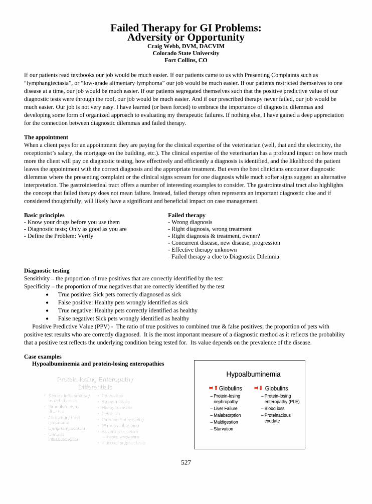
527
Failed Therapy for GI Problems: Adversity or Opportunity
Craig Webb, DVM, DACVIM Colorado State University
Fort Collins, CO
If our patients read textbooks our job would be much easier. If our patients came to us with Presenting Complaints such as “lymphangiectasia”, or “low-grade alimentary lymphoma” our job would be much easier. If our patients restricted themselves to one disease at a time, our job would be much easier. If our patients segregated themselves such that the positive predictive value of our diagnostic tests were through the roof, our job would be much easier. And if our prescribed therapy never failed, our job would be much easier. Our job is not very easy. I have learned (or been forced) to embrace the importance of diagnostic dilemmas and developing some form of organized approach to evaluating my therapeutic failures. If nothing else, I have gained a deep appreciation for the connection between diagnostic dilemmas and failed therapy. The appointment When a client pays for an appointment they are paying for the clinical expertise of the veterinarian (well, that and the electricity, the receptionist’s salary, the mortgage on the building, etc.). The clinical expertise of the veterinarian has a profound impact on how much more the client will pay on diagnostic testing, how effectively and efficiently a diagnosis is identified, and the likelihood the patient leaves the appointment with the correct diagnosis and the appropriate treatment. But even the best clinicians encounter diagnostic dilemmas where the presenting complaint or the clinical signs scream for one diagnosis while much softer signs suggest an alternative interpretation. The gastrointestinal tract offers a number of interesting examples to consider. The gastrointestinal tract also highlights the concept that failed therapy does not mean failure. Instead, failed therapy often represents an important diagnostic clue and if considered thoughtfully, will likely have a significant and beneficial impact on case management. Basic principles Failed therapy - Know your drugs before you use them - Wrong diagnosis - Diagnostic tests; Only as good as you are - Right diagnosis, wrong treatment - Define the Problem: Verify - Right diagnosis & treatment, owner? - Concurrent disease, new disease, progression - Effective therapy unknown - Failed therapy a clue to Diagnostic Dilemma Diagnostic testing Sensitivity – the proportion of true positives that are correctly identified by the test Specificity – the proportion of true negatives that are correctly identified by the test
• True positive: Sick pets correctly diagnosed as sick • False positive: Healthy pets wrongly identified as sick • True negative: Healthy pets correctly identified as healthy • False negative: Sick pets wrongly identified as healthy
Positive Predictive Value (PPV) - The ratio of true positives to combined true & false positives; the proportion of pets with positive test results who are correctly diagnosed. It is the most important measure of a diagnostic method as it reflects the probability that a positive test reflects the underlying condition being tested for. Its value depends on the prevalence of the disease. Case examples
Hypoalbuminemia and protein-losing enteropathies
HypoalbuminemiaHypoalbuminemia
GlobulinsGlobulins–– ProteinProtein--losing losing
nephropathynephropathy–– Liver FailureLiver Failure–– MalabsorptionMalabsorption–– MaldigestionMaldigestion–– StarvationStarvation
GlobulinsGlobulins–– ProteinProtein--losing losing
enteropathyenteropathy (PLE)(PLE)–– Blood lossBlood loss–– ProteinaciousProteinacious
exudateexudate
528
Although the combination of hypoalbuminemia and hypoglobulinemia is “textbook” for a protein-losing enteropathy (PLE) – assuming the dog is not bleeding out in front of you or has presented with 3rd degree burns over most of its body – it should be noted that a normal, or even elevated globulin level should never be the only reason to take PLE off a list of differentials that was generated by a History and Physical Examination. It should also be noted that PLE is not really a diagnosis at all, but simply a description of the consequences of the severity or chronicity of the actual underlying problem. The diagnostic work-up can begin by adjusting the above list of PLE differentials based on the breed and age of the dog, with IBD being more common in older animals, lymphosarcoma being less particular, and lymphangiectasia preferring certain breeds. Parvovirus and intussusception often occur together in young dogs as does heavy GI parasitism, while histoplasmosis has a geographical distribution.
Once hypoalbuminemia is noted on the biochemical profile and globulins are assessed the clinician needs to confirm that there is no significant loss of protein in the urine (urine protein:creatinine ratio) and the liver is cable of producing adequate protein (other biochemical indicators of liver function followed by a bile acids test if necessary). Other common laboratory abnormalities consistent with PLE include lymphopenia, hypocholesterolemia, hypomagnesemia, and hypocalcemia. It would be unusual for an animal with a PLE not to present with diarrhea, and a thorough fecal examination is an essential part of that diagnostic work-up. In addition, feces can be submitted (Texas A&M GI Laboratory) for measurement of fecal α1-protease inhibitor enzyme quantification, a molecule that is of similar size to albumen but is not degraded in its journey through the GI tract, and hence, and indirect marker of albumen loss and a relatively sensitive (more sensitive than serum albumen?) marker of protein-loss through the GI tract. Be aware of possible significant and clinically relevant electrolyte abnormalities in Ca++ and/or Mg++, as well as either Secondary Hyper- or Hypoparathyroidism.
The most direct route to a diagnosis in cases of PLE is histopathology. The advantages and pitfalls of the various techniques available (endoscopy, laparoscopy, and exploratory surgery) is beyond the scope of these notes. It is common for lesions suggestive of lymphangiectasia AND inflammatory bowel disease to be present within the same biopsy sample – the bursting of lacteals and release of their contents is likely to set up an inflammatory response, and the crowding of the interstitial space with cells of the immune system is likely to impede normal lymphatic flow. This makes the site (duodenum versus ileum), depth (mucosal versus full-thickness), and quality of the biopsy of significant importance in final interpretation. The potential diagnostic yield provided by abdominal ultrasound prior to any of the above-listed techniques is also beyond the scope of this discussion, as the literature continues to debate the significance and sensitivity of certain ultrasonographic abnormalities, such as a loss of normal intestinal wall architecture, intra-mural “speckles”, or enlarged mesenteric lymph nodes.
Lymphangiectasia Breed predisposition: Yorkshire terriers and Soft-Coated Wheaten terriers.
NOTE: If endoscopy is planned and lymphangiectasia is a primary differential, administration of corn oil one hour prior to the biopsy procedure may increase the likelihood of documenting dilated lacteals (Drs. Willard and Zoran, Texas A&M).
Treatment Options for Lymphangiectasia TPN or PPN Critical Care setting Low-fat Diet Royal Canin, Purina, Iams, Science Diet, etc. Medium chain triglyceride oil (MCT) Controversial Corticosteroids 1-2 mg/kg SID (caution: catabolic hormone) Oncotic Pressure Colloids, Plasma, Human Albumen Rutin 50 mg/kg TID (anecdotal)
Feline hypereosinophilic syndrome A syndrome that appears to be unique to the Feline, eosinophilic infiltrates are found in the intestinal tract, and the diagnosis may stop there, being deemed Eosinophilic Gastroenteritis. But in this syndrome the GI tract is just part of the pathologic picture, and eosinophils are found to be invading a number of other parencymal organs, particularly the spleen. Cats with this syndrome are usually > 7 years old and most frequently present with diarrhea (often bloody) and weight-loss. Physical examination reveals thickened small intestines, again leading the clinician to conclude that this is only a disease of the tube itself, not the rest of the animal. Peripheral eosinophilia can be seen, sometimes to a minor degree (2,000 cells/μl) and sometimes to an astonishing degree (60,000 cells/μl). The biggest dilemma is the difference in prognosis: garden-variety eosinophilic gastroenteritis should usually respond quite favorably to standard IBD treatment, while Hypereosinophilic Syndrome responds poorly to a similar protocol, and in fact, to most any protocols attempted, ie. the prognosis is quite poor for these patients. Motility disorders Motility disorders may be famous enough to warrant their own name, as in Feline Megacolon, but otherwise are often a secondary complication of the more standard enteropathies, or even non-GI systemic disease. Barium and BIPS are messy and variable, leaving us with few diagnostic options when trying to identify motility disorders. Our therapeutic options are also limited, often non-specific,
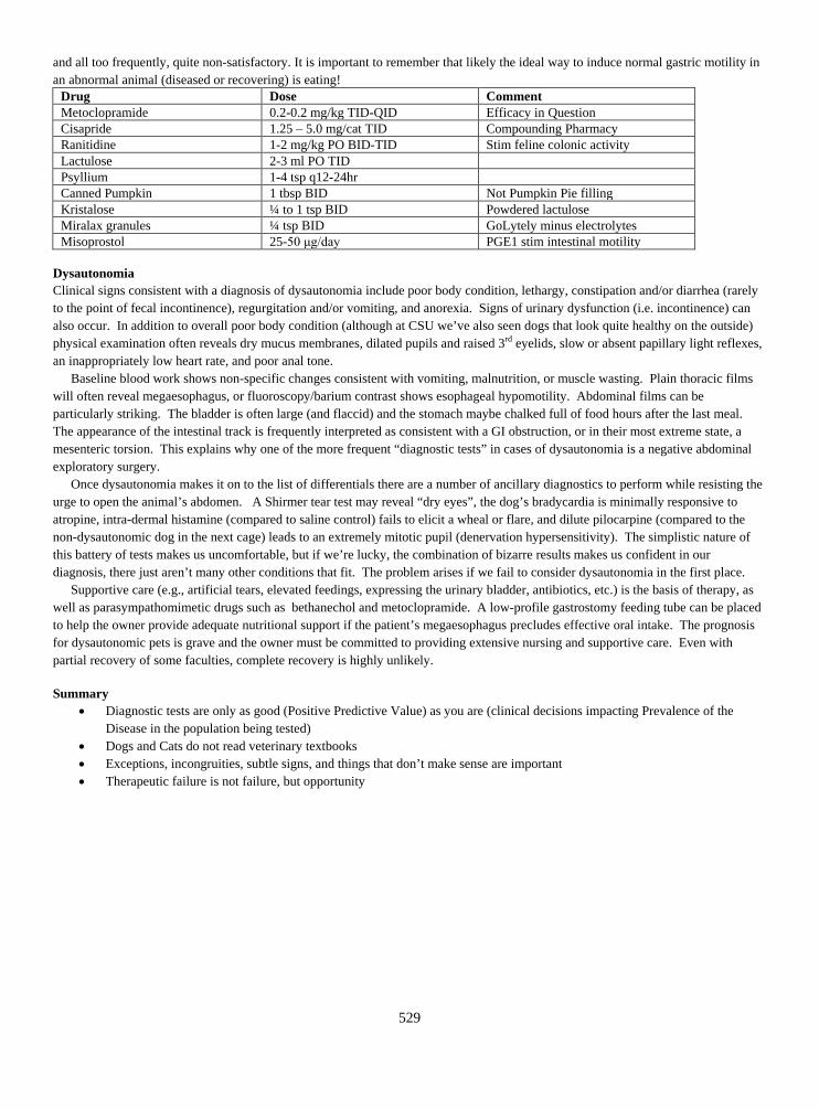
529
and all too frequently, quite non-satisfactory. It is important to remember that likely the ideal way to induce normal gastric motility in an abnormal animal (diseased or recovering) is eating!
Drug Dose Comment Metoclopramide 0.2-0.2 mg/kg TID-QID Efficacy in Question Cisapride 1.25 – 5.0 mg/cat TID Compounding Pharmacy Ranitidine 1-2 mg/kg PO BID-TID Stim feline colonic activity Lactulose 2-3 ml PO TID Psyllium 1-4 tsp q12-24hr Canned Pumpkin 1 tbsp BID Not Pumpkin Pie filling Kristalose ¼ to 1 tsp BID Powdered lactulose Miralax granules ¼ tsp BID GoLytely minus electrolytes Misoprostol 25-50 μg/day PGE1 stim intestinal motility
Dysautonomia Clinical signs consistent with a diagnosis of dysautonomia include poor body condition, lethargy, constipation and/or diarrhea (rarely to the point of fecal incontinence), regurgitation and/or vomiting, and anorexia. Signs of urinary dysfunction (i.e. incontinence) can also occur. In addition to overall poor body condition (although at CSU we’ve also seen dogs that look quite healthy on the outside) physical examination often reveals dry mucus membranes, dilated pupils and raised 3rd eyelids, slow or absent papillary light reflexes, an inappropriately low heart rate, and poor anal tone.
Baseline blood work shows non-specific changes consistent with vomiting, malnutrition, or muscle wasting. Plain thoracic films will often reveal megaesophagus, or fluoroscopy/barium contrast shows esophageal hypomotility. Abdominal films can be particularly striking. The bladder is often large (and flaccid) and the stomach maybe chalked full of food hours after the last meal. The appearance of the intestinal track is frequently interpreted as consistent with a GI obstruction, or in their most extreme state, a mesenteric torsion. This explains why one of the more frequent “diagnostic tests” in cases of dysautonomia is a negative abdominal exploratory surgery.
Once dysautonomia makes it on to the list of differentials there are a number of ancillary diagnostics to perform while resisting the urge to open the animal’s abdomen. A Shirmer tear test may reveal “dry eyes”, the dog’s bradycardia is minimally responsive to atropine, intra-dermal histamine (compared to saline control) fails to elicit a wheal or flare, and dilute pilocarpine (compared to the non-dysautonomic dog in the next cage) leads to an extremely mitotic pupil (denervation hypersensitivity). The simplistic nature of this battery of tests makes us uncomfortable, but if we’re lucky, the combination of bizarre results makes us confident in our diagnosis, there just aren’t many other conditions that fit. The problem arises if we fail to consider dysautonomia in the first place.
Supportive care (e.g., artificial tears, elevated feedings, expressing the urinary bladder, antibiotics, etc.) is the basis of therapy, as well as parasympathomimetic drugs such as bethanechol and metoclopramide. A low-profile gastrostomy feeding tube can be placed to help the owner provide adequate nutritional support if the patient’s megaesophagus precludes effective oral intake. The prognosis for dysautonomic pets is grave and the owner must be committed to providing extensive nursing and supportive care. Even with partial recovery of some faculties, complete recovery is highly unlikely.
Summary
• Diagnostic tests are only as good (Positive Predictive Value) as you are (clinical decisions impacting Prevalence of the Disease in the population being tested)
• Dogs and Cats do not read veterinary textbooks • Exceptions, incongruities, subtle signs, and things that don’t make sense are important • Therapeutic failure is not failure, but opportunity
530
Medicine’s Cutting Edge: What’s New in Gastroenterology (Parts 1 and 2)
Anthony Carr, Dr. med. vet., DACVIM Western College of Veterinary Medicine
Saskatoon, SK
Fecal transplants for refractory diarrhea There is a controversy as to how much of a pathogen C. perfringens actually represents, however it does appear to cause issues in some dogs. In the study presented 8 dogs with antibiotic refractory diarrhea and persistent C. perfringens PCR tests were examined. All dogs had failed treatment with metronidazole and amoxicillin/clavulanic acid. The dogs were given an enema that contained feces from a healthy donor dog that had been blenderized with sterile saline. This was repeated between one and three times in various dogs. All eight dogs responded extemely well with complete resolution of diarrhea. In 6 dogs the C. perfringens alpha toxin gene expression could no longer be detected.
These results are certainly of interest. Although this study focused on the ability to suppress C. perfringens it is important to remember that all dogs improved, even though 2 still had the organism. It may be better to consider this as a treatment in dogs with non-antibiotic responsive diarrhea. There certainly is a lot of interest in this area in humans where it has been found to be very helpful in the treatment of C. difficile associated diarrhea (resolution of around 90%). The technique is old with some mention of it in the 1940's and 50's, however as the arsenal of antibiotics increased, these treatments lost favor. There has however been the development of antibiotic resistant strains that once again has led to fecal transplant research. Oral cobalamin supplementation in dogs with chronic enteropathy and hypocobalaminemia Low cobalamin concentration can be found in dogs and cats with a variety of disorders. Almost all dogs and cats diagnosed with EPI have low cobalamin concentration as do many dogs with chronic enteropathies. The low cobalamin has been associated with continued signs of enteropathy unless corrected. This generally has been done using weekly injections which is of course not necessarily a desirable way to treat a patient. Researchers from Sweden carried out a retrospective study in dogs diagnosed with a low cobalmin and signs of a chronic enteropathy that were treated with oral cobalamin. Supplementation was somewhat empiric with dogs <20 kg getting 1/4 of a 1 mg tablet per 10 kg of body weight and dogs >20 kg getting 1 mg daily. Serum cobalamin concentration had to be less than 200 pmol/L to be included in the study.
A total of 39 dogs were included in the study. At onset median cobalamin concentration was 178 (range 117-199 pmol/L).The median follow up increase in cobalamin concentration (20-195 days after starting supplementation) was 534 pmol/L (range 54 to 1305 pmol/L). This difference was statistically significant.
This study is quite interesting as it does suggest that oral supplementation with cobalamin may be a viable alternative to injections to normalize cobalmin concentration. The statistics do show that response is quite variable however and it would certainly be indicated to monitor cobalmin concentrations to see if there is a significant increase in cobalamin concentration if oral supplementation is used. In those patients that fail to have a significant rise in concentration it may be advisable to use injections. What is unanswered is if there is a difference in the clinical response to oral vs. injection for cobalmin supplementation. It would also be interesting to see if this helps with EPI. References ORAL COBALAMIN SUPPLEMENTATION IN DOGS WITH CHRONIC ENTEROPATHIES AND HYPOCOBALAMINEMIA. L. Toresson, J.M. Steiner, J.S. Suchodolski, T. Spillmann. J Vet Int Med 2014:28;1044 USE OF FECAL TRANSPLANT IN EIGHT DOGS WITH REFRACTORY CLOSTRIDIUM PERFRINGENS ASSOCIATED DIARRHEA. T. Murphy, J. Chaitman, E. Han. J Vet Int Med 2014:28;1047
531
Pancreatitis in Cats Anthony Carr, Dr. med. vet., DACVIM Western College of Veterinary Medicine
Saskatoon, SK
Pancreatitis is a common problem in cats and as with most things, cats are not small dogs. Clinical signs and outcomes tend to be different between the two species. As with dogs, pancreatitis needs to be on the differential list for most cats with signs of gastroenteritis. Diagnosis is not always straightforward, though this generally does not have a major impact on therapy as therapy for gastroenteritis and pancreatitis are very similar in cats. Signalment and risk factors for pancreatitis Most cats with pancreatitis are middle age or older. There is an association between GI disease, hepatic disease and pancreatic disease that is termed "triaditis" by some authors. This is based on a research paper that found an association with these diseases, however it was very specific forms of disease. Making a global association between these various diseases is not supported by scientific evidence. Clinical signs Clinical signs are variable, though generally less severe than in dogs. Anorexia is common as is lethargy. Pancreatitis is a common cause of hepatic lipidosis, so that the signs of HL may predominate. Pain on abdominal palpation is also much rarer. With chronicity, weight loss can occur. In cats necrotizing pancreatitis can also have a very rapidly fatal course, though clinical signs also tend to not be very specific. Diagnosis There is no test that says a patient does or does not have pancreatitis. This is especially true since many other diseases can have pancreatic involvement without pancreatitis being the major issue (i.e. foreign body).
Imaging Radiographs can show non-specific changes. Ultrasound is better, but sensitivity and specificity can be quite variable (usually severe cases can be diagnosed, but with mild cases common degenerative changes to the pancreas can mimic pancreatitis).
Laboratory testing The results of routine blood and urine analysis are not diagnostic, though at times they can help to assess severity (especially the CBC) and if concurrent issues such as diabetes are present. There may also be indications of cholestasis present. In cats hypocalcemia can be a clue to pancreatitis.
Amylase and lipase Sensitivity and specificity are very poor in cats and as such these analytes are of no value in the diagnosis of pancreatitis.
TLI The TLI assay is not sufficiently useful to rely upon to rule pancreatitis in or out.
fPLI/fPL This test can be useful in diagnosing pancreatic disease, however the test is not specific nor necessarily sensitive (depending on cut off around 60 to 80% for sensitivity and specificity). It is also important that a positive test not be interpreted in a way that stops the clinician from looking for other reasons for the clinical signs present (i.e GI foreign body) as this could result in major issues not being diagnosed. Treatment There is no specific therapy for pancreatitis, the therapy that is needed is extremely good supportive care. Fluid therapy is a major part of therapy for pancreatitis. By providing fluids, acidosis and hypoperfusion are limited which can contribute to progression of disease. Buffered crystalloids are generally preferred over other crystalloids. A fluid therapy plan should of course address any dehydration present to minimize the impact of hypoperfusion, especially to organs sensitive to this such as the kidney and GI tract. In those patients where peripheral edema becomes an issue or where blood pressure cannot be maintained with crystalloids, colloids such as Pentaspan or Hetastarch can be used.
Anti-emetic therapy is important in those cases where ongoing vomiting or nausea are major issues, though this is rarer in cats. Analgesia is an important part of treating pancreatitis in cats that show signs of pain. Pain severity will be variable and as such
analgesic protocols should be adjusted. Buprenorphine is a good choice in many cases. The use of prophylactic antibiotics in humans and dogs with pancreatitis is controversial. In cats there is little indication for their
use unless an infection is diagnosed (i.e. aspiration pneumomia). Nutrition is vital in cats with pancreatitis as they can not infrequently develop hepatic lipidosis if anorexic. Appetite stimulants
rarely are efficacious enough to consider them as a viable option. Feeding tubes are generally the preferred way to provide nutrition. Given that cats can be quite unstable initially, placement of a naso-esophageal feeding tube is often the first step to meeting the

532
nutritional needs of cats with pancreatitis. This allows feeding of a liquid diet. CRI administration is better than bolus administration as it seems that tolerance to feeding is bettter (less nausea and vomiting). In many cases this is adequate to allow the cats to stabilize and start to eat on their own. If signs are persistent, placement of an esophageal or gastrotomy tube can be considered for longer term therapy, especially if the patient is to be discharged from the hospital. References; Available on request from the author
533
Pancreatitis in Dogs Anthony Carr, Dr. med. vet., DACVIM Western College of Veterinary Medicine
Saskatoon, SK
Pancreatitis is a common problem in dogs and needs to be on the differential list for most adult dogs with signs of gastroenteritis. Diagnosis is not always straightforward, though this generally does not have a major impact on therapy. Signalment and risk factors for pancreatitis Most dogs are middle age or older. Miniature Schnauzers, Poodles and Yorkshire Terriers appear to be predisposed, though the evidence for this is limited.
Other concurrent disease processes may predispose patients to developing pancreatitis. Diabetes mellitus, hyperadrenocorticism and hypothyroidism may all increase the risk of pancreatitis as may the history of prior GI disease.
Medications have been associated with an increased risk of pancreatitis. Agents implicated include, potassium bromide, phenobarbital, azathioprine and furosemide to name a few.
A risk factor that is often discussed is dietary indiscretion. Dogs that consume unusual foods (i.e. get into table scraps or the garbage) have a higher incidence of pancreatitis.
Acute pancreatitis may develop from decreased perfusion to the pancreatitis. This can be seen with shock, general anesthesia and other causes of hypotension and hypoperfusion.
Clinical signs Clinical signs are variable, from mild anorexia and mild GI signs to rapidly progressive shock and death. No changes are present that are specific enough to rule pancreatitis in or out. Common clinical signs are anorexia, vomiting and cranial abdominal pain. Depending on how severe the pancreatitis is, other signs such as fever, dehydration, hypothermia, or hypotension can be present. If the pancreatic inflammation has been present for several days, icterus may become visible, generally secondary to obstruction of the common bile duct. Diagnosis There is no test that says this patient does or does not have pancreatitis. This is especially true since many other diseases can have pancreatic involvement without pancreatitis being the major issue (i.e. foreign body).
Imaging Radiographs can show non-specific changes. Ultrasound is better, but sensitivity and specificity can be quite variable (usually severe cases can be diagnosed, but with mild cases common degenerative changes to the pancreas can mimic pancreatitis).
Laboratory testing The results of routine blood and urine analysis are not diagnostic, though at times they can help to assess severity (especially the CBC) and if concurrent issues such as diabetes are present. There may also be indications of cholestasis present.
Amylase and lipase Sensitivity and specificity are poor. Concurrent gastrointestinal disease or poor renal excretion can increase amylase and lipase.
TLI The TLI assay is not sufficiently useful to rely upon to rule pancreatitis in or out.
cPLI/spec cPL This test can be useful in diagnosing pancreatic disease, however the test is not specific nor necessarily sensitive. Prevailing opinion was that it was sensitive (good to rule out pancreatitis) but not very specific (too many false positives). One study that looked at histopathology as the gold standard however showed that specificity was good at the >400 ug/L (90%) but sensitivity was poor (33% at the same cut off). Another major concern was that although it was specific, few of the dogs with pancreatitis had this as the major reason for their clinical signs. This lab test is also commonly elevated with Cushings disease and hyperlipidemia without signs of pancreatitis. It is also important that a positive test not be interpreted in a way that stops the clinician from looking for other reasons for the clinical signs present (i.e GI foreign body) as this could result in major issues not being diagnosed. Treatment There is no specific therapy for pancreatitis, the therapy that is needed is extremely good supportive care. Fluid therapy is a major part of therapy for pancreatitis. By providing fluids, acidosis and hypoperfusion are limited which can contribute to progression of disease. Buffered crystalloids are generally preferred over other crystalloids. A fluid therapy plan should of course address any dehydration present to minimize the impact of hypoperfusion, especially to organs sensitive to this such as the kidney and GI tract. In those patients where peripheral edema becomes an issue or where blood pressure cannot be maintained with crystalloids, colloids such as Pentaspan or Hetastarch can be used. Plasma has been anecdotally suggested to be useful for pancreatitis, however studies in dogs and
534
humans have not shown any clinical benefit. The beneficial effect was attributed to providing substances such as coagulation factors, alpha 2 macroglobulin, and others that are depleted in the course of pancreatitis. Of course, plasma is an excellent colloid. They can be considered in those patients with DIC, though even in these cases it is unclear if there is any benefit.
Anti-emetic therapy is important in those cases where ongoing vomiting or nausea are major issues. A wide variety of products can be used and response will be variable. Maropitant (Cerinia©), metoclopramide, ondansetron/dolasetron and chlorpromazine are all options. Generally I avoid use of metoclopramide as it may in theory limit pancreatic perfusion and in my hands is at best a poor anti-emetic.
Analgesia is an important part of treating pancreatitis in dogs. Pain severity will be variable and as such analgesic protocols should be adjusted. In most patients opiods will be needs. The more potent of these preparations can however also lead to clinical issues such as vomting, regurgitation and anorexia. This can be minimized by using CRIs over bolus administration. Buprenorphine is a good choice in mild cases, in cases with more signs of pain use of hydromorphone, ketamine and possibly lidocaine need to be considered.
The use of prophylactic antibiotics in humans with pancreatitis is controversial. In dogs there is little evidence that bacteria play a role with acute pancreatitis. As such their routine use is not advisable. They can be considered in those patients where there is a suspicion of bacterial infection (i.e. aspiration pneumonia) or where the CBC and other clinical data suggest that sepsis, GI necrosis or pancreatic necrosis are present.
References; Available on request from the author
535
GI Formulary: More is not Better
Anthony Carr, Dr. med. vet., DACVIM Western College of Veterinary Medicine
Saskatoon, SK
Gastric ulceration Prevention is always preferable to treatment. The same applies for gastric ulcers/erosions. In most cases this is of course not possible. One exception is ulceration associated with NSAID administration. The prostaglandin analogue misoprostol (3 to 5 µg/kg q 8h) has been shown to prevent ulcers in humans treated with NSAIDs. Proton pump inhibitors also show promise with this, whereas H2 blockers are not considered efficacious.
A variety of medications are available for treating erosions/ulcers, the majority work by suppressing stomach acid secretion. Both H2 receptor antagonists and proton pump inhibitors have been used in dogs and cats. It is not uncommon to see gastric acid suppressing agents being combined with sucralfate for the treatment of erosions/ulcers. This does not appear justified since in humans the combination is no more effective than using a single agent.
The H2 blockers bind to receptors on the acid producing parietal cells. This renders the cells less likely to respond to histamine, gastrin or acetylcholine. Commonly used H2 blockers used include cimetidine, famotidine and ranitidine. Ranitidine also has prokinetic effects in the GI tract that make it an antiemetic as well. Studies in dogs suggest that these agents do not suppress stomach acid production adequately. Proton pump inhibitors are more effective at significantly decreasing gastric acid secretion since the binding of the drug to the parietal cell is irreversible and inhibits stomach acid secretion. Omeprazole (0.2 to 0.7 mg/kg daily) has been used in dogs and is highly effective.
Sucralfate is a mucosal protectant that is often used with GI erosions/ulceration. It binds to defects in the mucosa, protecting the damaged area. Production of prostaglandins is also increased which results in increased mucous and bicarbonate production. Many additional effects have been documented as well. Promotility agents A variety of purported prokinetic medications are available, though evidence that they work in a clinical patient are limited. Metoclopramide (0.2-0.4 mg/kg q 8h) can be used and has been shown to be a prokinetic in healthy dogs. It has central antiemetic effects as well as speeding gastric emptying. A more potent effect with regard to gastric emptying can be achieved with cisapride (0.1 to 1.0 mg/kg q 8-12h). Erythromycin at low dosages will also promote gastric emptying by stimulating motilin receptors in the GI tract (0.5 to 1.0 mg/kg q 8h). Oral ranitidine and nizatidine also have prokinetic effects because they inhibit acetylcholinesterase activity thereby increasing parasympathetic tone. Antiemetics Antiemetics are commonly used in veterinary practice. By reducing nausea they improve the condition of the patient. They also reduce loss of fluids and electrolytes caused by persistent vomiting. Antiemetics can act at various receptors. Some are specific to individual receptors whereas others may influence multiple receptors. In patients response to a specific antiemetic can be variable so that various agents or combinations may be needed to achieve the desired therapeutic benefit.
Metoclopramide is a commonly used antiemetic. It can be given via intermittent subcutaneous injections (0.2 –0.4 mg/kg q 6 h, SQ, IM) or via a constant rate intravenous infusion (1-2 mg/kg/day). The latter appears to be more efficacious. This medication predominantly affects the D2 dopaminergic receptors in the CRTZ and gut. It also affects the 5-HT3 serotonergic receptors in the CRTZ.
Phenothiazines such as chlorpromazine (0.2 – 0.4 mg/kg q 8h SQ) or prochlorperazine (0.5 mg/kg q 8h SQ or IM) are broad spectrum antiemetics with activity at the α2-adrenergic, D2-dopaminergic, histaminergic, and cholinergic receptors. These medications are a good choice in those patients that fail to respond to metoclopramide, it is possible to use both agents concurrently. They can cause hypotension and this should be monitored for. Sedation is also usually quite pronounced.
A limited number of medications are specific to the 5-HT3 serotonergic receptors. Ondansetron (0.5 – 1.0 mg/kg q 12 to 24h PO) can be helpful in some cases of vomiting associated with stimulation of the CRTZ.
H1-histaminergic receptor antagonists include diphenhydramine (2 – 4 mg/kg q 8h PO) and dimenhydrinate (4 – 8 mg/kg q 8h PO). These can be used for the treatment of motion sickness or vestibular disease.
Erythromycin at low dosages (0.5 – 1.0 mg/kg q 8h) can also be an antiemetic by stimulation of the motilin receptors that increase GI motility and promote gastric emptying.
Maropitant is a veterinary specific antiemetic that is widely used. It is a NK 1 receptor antagonist. It is approved for use in dogs older than 8 weeks of age and for 5 consecutive days. Efficacy is good, probably as good as most other antiemetics. References; available upon request from the author
536
Constipation in Cats Anthony Carr, Dr. med. vet., DACVIM Western College of Veterinary Medicine
Saskatoon, SK
Constipation is a frequent complaint middle aged to older cats. In some cases the disease becomes refractory enough to treatment that either subtotal colectomy or euthanasia have to be considered. The problem is thought to be caused by underlying metabolic problems in some patients such as kidney disease or other issues that generally result in dehydration. These are however relatively rare and do not generally cause clinical signs. Most clinical cases of recurrent constipation/obstipation are idiopathic in nature. Pelvic abnormalities and strictures represent some of the occasional causes of this problem that can be identified with work up as is nerve trauma to the sacral region. Megacolon represents the extreme manifestation of obstipation/constipation. In cats with megacolon abnormal smooth muscle cell function of the colon has been detected though this was in cats with advanced disease so it is uncertain if this was truly the cause or a manifestation of chronic constipation.
The consequences of constipation/obstipation usually are metabolic derangements. With prolonged problems endotoxemia and even death can occur. Long term this can also lead to megacolon, though in many cases megacolon can occur without a clear history of constipation/obstipation. Treatment A variety of treatments have been recommended for the constipated/obstipated cat as well as the cat with megacolon.
Initial management When initially presented relieving the constipation is indicated. This can be done with a variety of ways, whereby manual disimpaction is the least “nice” of the options and should be reserved for refractory cases. Enemas can often be helpful to help to moisten dried out feces. In general 5 to 10 ml/kg of warm water can be given as an enema. Alternatively smaller volumes of DSS (5 to 10 ml total dose) can be given, though this is more irritating. This can be supplemented with oral lactulose and fluid therapy to maximize efficacy.
Recently we have adapted the use of PEG solutions administered via NE tube to help relieve obstipated/constipated cats. This is similar to methods used in humans. We give PEG solution as a slow trickle via NE tube (4 to 18 hours). This generally results in defecation within 6 to 12 hours. Obviously before embarking on this therapy it is wise to rule out obstructions of the GI tract that would make passing feces difficult or impossible. To date we have not had any significant adverse side effects and have not had to resort to manual disimpaction. In some cases enemas were given concurrently, thought this does not appear to be necessary.
Long term management Ultimately in those cats where the problem constantly recurs, surgical intervention may be needed. Medical therapy (life-time) can in many cases avert the need for surgery or significantly delay the need for surgery.
Diet is an important part of management and it is difficult to be sure which diet is best in each individual case. Increased fiber and low residue diets are the most popular.
Fiber has been recommended for many years. This can be a psyllium product (Metamucil 1-4 tsp per meal), pumpkin pie filling or wheat bran. Fiber has been shown in humans to be only moderately effective as a laxative.
Lactulose is also a very good option for maintaining soft stools. The dosage is 0.5 ml/kg two to three times daily. Dosage is adjusted to obtain the stool quality desired. In humans this product is known to cause flatulence and GI cramping. A powder form is available that may be better tolerated in cats. Recently PEG containing laxatives (Miralax) have been recommended for use in cats. In humans PEG laxatives have been shown to be safe and effective with few adverse side effects. These products have been recommended in cats though there is no published data showing efficacy or safety. We did carry out a study in 6 cats and did not see any clinically significant adverse effects. Dosage was quite variable, so individual dose titration is recommended. Starting at ½ to ¼ of a teaspoon twice daily is a good starting point.
Prokinetic medications are vital to managing the chronically impacted cat. Cisapride (2.5 to 5 mg/cat q8 to 12 hours) has helped many cats avoid surgery for megacolon. It can be obtained from many compounding pharmacies. Other medications are available as well, however in most cases Cisapride is adequate. References Candy DCA, Edwards D, Geraint M. Treatment of faecal impaction with polyethylene glycol plus electrolytes (PGE + E) followed by double-blind comparison of PEG + E versus lactulose as maintenance therapy. Journal Pediatric Gastroenterology and Nutrition 2001; 43:65. Ramkumar D, Rao SSC. Efficacy and safety of traditional medical therapies for chronic constipation: systematic review. Am J Gastrorenterol 2005;100:936. Washabau RJ, Holt DE. Diseases of the large intestine. In Ettinger SJ, Feldman EC (eds): Textbook of Veterinary Internal Medicine, 6th ed. St. Louis, Elsevier, 2005.
537
EPI: Not Such a Zebra
Anthony Carr, Dr. med. vet., DACVIM Western College of Veterinary Medicine
Saskatoon, SK
EPI in dogs What is exocrine pancreatic insufficiency
Exocrine pancreatic insufficiency (EPI) is a condition where the pancreas no longer produces adequate amounts of digestive enzymes. Clinical signs usually will not occur until over 90% of the exocrine pancreas has become non-functional. The majority of cases in dogs are caused by pancreatic acinar atrophy, though some may also develop because of chronic pancreatitis. Clinical signs develop because digestion no longer occurs properly leading to malabsorption with weight loss and diarrhea. Maldigestion and malabsorption does not just result from the enzyme deficiency. The disease also leads to changes in small intestinal digestive processes. Small intestinal bacterial overgrowth (SIBO) is also frequently seen and can contribute to the diarrhea seen. Low cobalamin levels are also seen which may play a role with small intestinal disease in these dogs.
What is pancreatic acinar atrophy Pancreatic acinar atrophy (PAA) is a condition where a normal pancreas undergoes atrophy. A familial predisposition in German Shepherds and rough-coated collies has been established. It can however occur in any breed with large breed dogs being more commonly affected.
What is a typical signalment for EPI Since PAA is the most common cause of EPI many of the patients are German Shepherds. They tend to be young animals, usually under 2 years of age.
What are the clinical signs of EPI Signs of EPI are relatively typical, though they can be caused by other diseases as well. Dogs with EPI can have profuse diarrhea. The bowel movements are often malodorous. Borborygmus and flatulence are common. These patients will have a ravenous appetite yet continue to lose weight. Poor hair coat is common because of the malnourishment present. On occasion other signs such as vomiting, coprophagia, pica and anorexia can be seen.
Routine clinical pathologic tests rarely document abnormalities. Folate levels may be elevated and cobalamin concentration decreased in dogs with EPI. This may be from SIBO, however the cobalamin deficiency may also occur because the pancreas produces a factor needed for cobalamin absorption that can be lacking in dogs with EPI.
How is EPI diagnosed EPI is generally diagnosed with pancreatic function tests. Canine TLI is currently the preferred test for EPI. The animal should be fasted 12 hours to collecting serum for analysis. Low TLI concentration in patients with typical clinical signs are considered highly diagnostic. Low TLI values can be found in dogs without clinical signs. Many other tests have been tried but none are superior to TLI.
How is EPI treated Treatment of EPI hinges on enzyme replacement and nutritional intervention. Powdered pancreatic enzymes work best and are used at 1 to 2 teaspoons per meal. There is no need to pre-incubate the food. Alternatively fresh pancreas (50-100g/meal) can be used. The majority of the enzymes are destroyed in the stomach so some dose titration may be needed. Although raw pancreas results in higher GI lipase activity, response rates between powdered enzymes and raw pancreas were comparable.
In these patients it is vital to manage their diet carefully. Initial management can be with a high quality maintenance diet. Fat restriction should not be attempted initially, in fact fat supplemented diets may be a consideration if body condition cannot be improved. If this results in persistent diarrhea, a fat restricted, highly digestible diet should be tried. Cobalamin injections can be given (250-500 μg, repeat as indicated by serum levels), especially to those patients that are responding poorly to therapy. Cobalamin concentration can be determined via a blood test. Vitamin absorption may generally be inadequate in these patients so that use of vitamin supplements is prudent. Antibiotics are indicated if SIBO is suspected to be contributing to poor therapeutic response. Metronidazole, tylosin and tetracyclines are all good options for this. Tylosin may be preferable as it seems to be very good at treating chronic diarrhea in dogs.
What is the prognosis for EPI Generally the prognosis is good, especially if the initial response to therapy is good. Around 60% of dogs will do very well, 10% get better and 20% do poorly. With a poor initial response overall survival seems shortened. Cobalamin deficiency has been the only factor that seemed to correlate with how well the initial response was. Factors that did not have an effect were the type of supplement used, additional drugs used or diet used.
538
EPI in cats The diagnosis of EPI in dogs and cats has become more common as the assay for feline trypsin-like immunoreactivity (fTLI) became available. The test is considered diagnostic for EPI if a concentration of < 8 μg/L is found. Previous to this diagnosis was complicated based upon clinical signs and a variety of fecal digestion assays. There is relatively little information on EPI in cats that has been published. There have been individual case reports as well as 3 case series that included 41 cases.1-3 These case series showed that weight loss was the most common clinical sign of EPI in cats. Diarrhea was common, but not present in all cats and often not like the "typical" feces noted in dogs (voluminous, malodorous, steatorrhea). Polyphagia was uncommon. Age range was quite broad from 3 months to 16 years with the majority being middle aged. Although these case series are interesting they only provide information on a small number of patients, considering that in 2010 there 775 samples submitted to the Gastrointestinal Laboratory at Texas A&M University with fTLI concentrations consistent with a diagnosis of feline EPI.4
At the 2011 ACVIM Forum in New Orleans a research abstract was presented by researchers from the GI Laboratory and Department of Clinical Sciences at Texas A&M.5 The researchers searched their database for cats with a TLI concentration below 8 μg/L found over a 15 month period. Questionnaires were sent to the veterinarians that had submitted the samples, with 150 surveys being returned. There were many breeds involved and it did not seem that there was a clear breed predilection, though this could not be definitively determined with this type of study. Mean age of the cats was 8.1 years ± 4 with 41% being female and 59% male. Body condition was poor with a median of
3/9. Of the cats that had cobalamin determined, 77% were deficient with many of them having undetectable concentrations. Folate was increased in 47% of the cats where it was determined.
The most common clinical sign in this study was weight loss which was seen in 91% of the cats. The amount of weight loss varied widely from 40 grams to 6.82 kg with a median of 1.4 kg. Loose stools were seen in only 62% of the affected cats. Other clinical signs included poor haircoat (50%), anorexia (45%), increased appetite (42%), depression (40%), watery diarrhea (28%), and vomiting (19%). Concurrent diseases were also quite commonly reported (58%). The most common ones were inflammatory bowel disease (IBD, 21%), diabetes (14%), pancreatitis (11%) and hepatic lipidosis (6%). Of the affected cats 68% were treated with pancreatic enzyme supplementation with a good response in 66%, partial response in 24% and poor response in 10%.
This study is interesting on many levels. It is the largest study to date so the clinical information is very valuable. There are of course potential methodological issues with survey studies that introduce bias depending on high the response rate, though in this case they do not detract from the clinical relevance of the data. EPI in cats is certainly not as rare as we once thought it was. The clinical presentation deviates significantly from the typical presentation in a dog. Weight loss is certainly common in both dogs and cats and can be quite pronounced, however in cats diarrhea is clearly not a consistent finding. The diarrhea also appears to be much less severe when it is seen and often does not have the characteristics we expect to see such as large volume, highly fluid and greasy appearing stools. Polyphagia does occur in some cats, however almost half the cats were reported to have a decreased appetite.
A variety of concurrent diseases were found, most of them are not surprising. The etiology of EPI in cats in most cases probably relates to chronic pancreatitis and easily explains the 11% of cats that were thought to have pancreatitis in this study.1-4 In dogs it usually is related to pancreatic acinar atrophy which may also be the cause of EPI in very young cats.4 With chronic pancreatitis you can also see the development of endocrine pancreatic insufficiency, e.g. diabetes. Hepatic lipidosis has also been linked with pancreatitis. IBD was reported in 21% of the cats with EPI which may well have to do with what is termed triad disease or triaditis. In cats it has been shown that there is a statistical association between IBD, pancreatitis and cholangiohepatitis.
Treatment for EPI with pancreatic enzyme supplementation was successful in a significant percentage of the cases. The cats with EPI also had other issues that might have impacted management. Most of the cats had low cobalamin concentrations. It has been previously shown that in cats with hypocobalaminemia supplementation of parenteral cobalamin is often needed to resolve GI signs (250 μg subcutaneously weekly for 6 weeks, then once monthly with periodic checks of serum concentration). In addition the presence of EPI may be associated with small intestinal dysbiosis (formerly termed small intestinal bacterial overgrowth). The low cobalamin and high folate would certainly point toward this diagnosis. If pancreatic enzyme supplementation and cobalamine injection do not resolve signs, consideration should be given to antibiotic therapy. Dietary therapy is also very important, though to date there are no studies that tell us which diets to preferentially use.
This study does point out that EPI is not as rare as we thought and it would seem prudent to test for fTLI concentration in cats with unexplained weight loss or chronic diarrhea, even if they are very young. The presence of hypocobalaminemia should also increase the suspicion that EPI is present. It should also be considered in those cats with diabetes that appear to have good glycemic control but consistent weight loss or diarrhea. In the unexplained weight loss cases where blood work, T4, thoracic radiographs and abdominal ultrasound did not establish a diagnosis, I would often perform endoscopy to determine if occult neoplasia or some form of IBD was present. Based on this study, I will certainly have fTLI, cobalamin and folate determined prior to this more expensive and invasive diagnostic procedure.

539
References 1. Steiner JM, Williams DA. Serum feline trypsin-like immunoreactivity in cats with exocrine pancreatic insufficiency. J Vet Intern Med 2000;14:627-629 2. Kook PH, Zerbe P, Reusch CE. Exokrine Pankreasinsuffizienz bei der Katze. Schweiz Arch Tierheilk 2011;153:19-25. 3. Thompson KA, Parnell NK, et al. Feline exocrine pancreatic insufficiency: 16 cases (1992-2007). J Feline Med Surg 2009;11:935-940. 4. Steiner JM. Exocrine pancreatic insufficiency in the cat. Topics in Compan An Med 2012;27:113-116. 5. Xenoulis PG, Wooff P, et al. Feline exocrine pancreatic insufficiency: 150 cases. J Vet Intern Med 2012;26:765.
540
Zoonotic GI Diseases: What You Don’t Know Can Hurt You
Anthony Carr, Dr. med. vet., DACVIM Western College of Veterinary Medicine
Saskatoon, SK
Zoonotic disease has always been an area where veterinarians have been involved. Zoonotic infections can be viral, bacterial, fungal and parasitic in origin. The veterinary profession has helped protect people from potentially lethal zoonotic infections, for example by widespread vaccination of domestic animals against rabies. Complacency over infectious diseases had increased in industrialized countries with the advent of antibiotics and vaccinations to the point where infectious disease was an issue apparently of little importance. Recent times have shown this assessment to be incorrect with new players such as HIV and more recently SARS. There are however still plenty of old players around that have not went away just because they were ignored, some of them are zoonotic. It is our duty as veterinarians to protect the general public from these threats as best as possible. It is also vital to have an understanding of zoonotic disease to prevent infection of those people routinely involved with large numbers of animals, namely the veterinary health care team including veterinarians. Fortunately many of the zoonotic diseases of concern are preventable or the chances of transmission to humans can be significantly reduced by proper education and management techniques. This article will review some of the diseases to be concerned about, though obviously it is not an all-inclusive list of all zoonotic diseases. Bacterial zoonoses Many zoonotic diseases are bacterial in origin. Many of these bacteria are enteric organisms, as a result feces is a major way to spread these infections. Others can be transmitted via bites or scratches such as Bartonella. Especially with enteric bacteria it is important to remember that clinically healthy animals can still harbor pathogenic bacteria. Elimination of these bacteria is often not possible so it is very important that measures are put into place in practices to minimize the risk to other patients, staff and the general public. It is also very important to educate clients to the potential risks, especially if the household has members that are not fully immunocompetent.
Salmonellosis Salmonellosis has received public attention on occasion. Most cases of this disease are acquired as a result of ingestion of contaminated food. Raw chicken and uncooked eggs are generally recognized as common sources of salmonella infections in man. The risks of exotic pets, especially in regard to turtles have also been widely publicized. Another area that has received some media attention is rawhide chews which can be contaminated with salmonella. Depending upon the source, a large percentage of rawhides and pig ears can harbor salmonella. In one outbreak it was shown that contact with the treats or pets that consumed them was responsible for human salmonellosis.1 Of 94 pig ear samples from retail outlets 51% were harboring Salmonella. Salmonella was also found in other treats including beef hoof, braided chews and similar products. Of great concern were outbreaks of multidrug resistant Salmonella typhimurium in small animal facilities including an animal shelter and 2 small animal clinics in 1999.2 In one veterinary clinic the likely source of the infection was a kitten with diarrhea, 10 of 20 employees developed clinical signs. In another instance one affected person was an employee and 2 were clients that had brought their cats to the clinic for treatment. After discharge the cats developed diarrhea and the owners subsequently became ill. This obviously raises the specter of liability for the pet and owner’s illness.
It certainly is not surprising that dogs can also harbor Salmonella species. Most recent studies have shown a prevalence of around 1 to 2 % in normal dogs and cats.3,4 Percentages may be higher in animals with diarrhea. Very high prevalence had been found in racing sled dogs, where 69% of dogs without diarrhea were shedding salmonella.5 In Greyhounds with diarrhea 61% were positive for Salmonella, in non-diarrheic dogs the percentage was 11%.6 The increased proportion of Salmonella positive animals in these dogs may relate to the stress of athletic performance or to their diets.
Raw meat can be a source of salmonella infection in dogs. This has been shown in a variety of studies looking at athletic dogs such as Greyhounds and sled dogs that routinely receive uncooked meat as part of their diet.6 Recently there has been considerable interest in raw diets for pet dogs, the most popular called BARF (biologically appropriate raw food). The internet is replete with sites that popularize this type of diet and it’s supposed health benefits. It does however mean that owners are routinely contaminating their environment with potentially infectious materials such as raw chicken. Dogs are not known to be especially clean eaters and it is highly likely that infectious organisms are disseminated throughout the home. In a recent study on a small number of dogs, 30% of dogs fed a BARF diet were shedding Salmonella, 80% of the food samples were positive.7 This has also been my personal experience where dogs fed BARF diets are positive for Salmonella (2 of 3 tested) even without clinical signs of diarrhea.
Campylobacter The prevalence of campylobacter closely parallels that of Salmonella in cats, with approximately 1% harboring this infection.4 The prevalence in dogs is considerably higher in some studies where up to 28% of dogs are infected.3 Other studies however show the prevalence to also be around 1%.8 The majority of human cases are acquired by ingestion of contaminated food. The percentage of
541
poultry with campylobacter is higher than the percentage with salmonella. There is the possibility of spread from dog or cat to man. The majority of dogs will not show clinical signs if infected.
Prevention Some very simple management techniques can prevent bacterial infections from being transmitted. Hand washing is vital; it should be done between each patient and certainly before eating. Food should also remain out of the area where animals are handled. In those cases with diarrhea or proven infections gloves should be worn when handling the patient and hands washed after removing the gloves. Patients with diarrhea, confirmed or suspected zoonotic infections should also be isolated from other animals, especially those that are very susceptible to infections such as those with major trauma, surgeries or on immune suppressive therapy. Animals fed raw meat diets should be considered carriers of pathogens until proven otherwise. Antibiotics should obviously be used wisely to limit the emergence of resistance strains. Dogs diagnosed with renal failure that do not have an obvious cause, i.e. ethylene glycol intoxication, should be tested for leptospirosis. Even after exposure a short course of antibiotics can prevent clinical disease in humans. Parasitic zoonoses Unlike bacterial infections, parasites are much easier to prevent. There are a wide variety of effective deworming medications that can eliminate or at least significantly decrease the chances of contracting parasites. Unfortunately, the indications are that we as veterinarians do a relatively poor job of this. In 1991 a survey of veterinarians showed that recommendations in regard to parasite control were inadequate.9 Only approximately 1/3 of veterinarians routinely discussed the zoonotic risk of parasites with owners. Almost 2/3 of the veterinarians incorrectly recommended treating intestinal parasites beginning at an age over 4 weeks. Less than half gave preventive anthelmintics to pups and dogs and about 1/3 did not recommend routine testing and treatment of nursing dogs. Approximately three fourths of the veterinarians tested for helminths in pubs. Unfortunately, over half of the respondents did not initiate therapy unless there was a positive fecal test. This is not appropriate since fecals can be negative even when the dogs are harboring adult parasites. Even more shocking was that almost half of the veterinarians surveyed considered roundworms or hookworms of little or no concern as a zoonosis. The majority of veterinarians in this survey did not even come close to properly addressing parasite burdens. More recently a survey was carried out of Connecticut pediatricians and veterinarians.10 The only comfort we can get from this study is that veterinarians seem to do a better job of discussing zoonosis than physicians. Interestingly veterinarians thought physicians or public health departments were responsible for public education whereas physicians thought it should be public health departments or veterinarians, which means both groups did not feel they had ultimate responsibility in this regard. Considering that ten years had elapsed between studies there was little indication that prevention strategies had improved. Only about 12% of veterinarians began deworming puppies at 2 to 3 weeks of age. The greatest majority (78%) didn’t begin treatment till the puppies were over 6 to 7 weeks of age. Slightly more than 50% of veterinarians carry out prophylactic deworming of puppies and kittens, unfortunately almost half of them use an interval greater than 2 weeks as recommended by the Centers for Disease Control. Parasite control is an area where small animal practitioners need to do a much better job in the interest of the pets and the general public.
Roundworms Roundworms are still of major zoonotic concern. Prevalences vary from 3%8 to 33%11. Prevalences tend to be higher in kittens and puppies, though this may be misleading in that shedding is more intermittent in adults. Zoonotic transmission to humans does occur, with children being especially susceptible to the negative affects. Visceral larva migrans can be a devastating disease. In Connecticut 10.2 to 27.9% of children were seropositive for exposure to roundworms.10 Thousands of cases are diagnosed each year. Pups acquire Toxocara canis in utero, transmammary or fecal-oral. The adults can become patent and begin shedding eggs at 3 weeks of age. In cats in utero infection does not appear to occur, infection is transmammary or fecal-oral. The eggs are very resistant and can persist for years in the environment.
Prevention In dogs and cats it is inappropriate to test for intestinal parasites and then not treat because of a negative fecal. Shedding is intermittent and can be low grade. Routine prophylactic deworming is ideal in all patients; the current dewormers available are highly efficacious with a low incidence of side effects. Puppies should be started at 2 weeks of age and should be dewormed every 2 weeks thereafter until 8 weeks old. Kittens can be started at 6 weeks and repeated at 8 and 10 weeks. Four to 6 weeks after anthelminthic therapy is completed it would be advisable to perform a fecal exam to make sure the parasites have been eradicated. Treating the nursing dam is also indicated, as they will often begin to shed heavily around the time of parturition. Many heartworm preventives can also aid in reducing parasite burdens in adult dogs, although the 4 week interval will allow shedding and may not be adequate for heavily exposed dogs. If one of these heartworm preventives is not used then at least annual deworming is recommended, more frequently if indicated by exposure risk (puppies, kennel, etc.). Extensive recommendations for prevention of zoonotic transmission of intestinal parasites from pets to humans can be found on the CDC website at http://www.cdc.gov/ncidod/dpd/parasites/ascaris/prevention.htm. Another part of the CDC website that is well worth surfing is http://www.cdc.gov/ncidod/dpd/parasiticpathways/animals.htm as there are great handouts available that explain about the parasites that can be very informative for clients.
542
Hookworms Hookworms can be found in many areas of the country though the southeast appears to have a greater prevalence of Ancylostoma than other regions. Hookworm larvae can penetrate the skin of humans leading to cutaneous larva migrans. More severe manifestations are also possible with skeletal muscle involvement, visceral migrans and human intestinal involvement. Prevention programs are the same as for roundworms.
Tapeworms The most common tapeworm in dogs and cats, Dipylidium caninum, is not zoonotic. There is increasing concern however about Echinococcus multilocularis. Originally limited to Alaska it is now enzootic to northcentral US and southcentral Canada, though it has been identified in Wyoming, Nebraska, Iowa, Ohio, Indiana and Illinois.12 The main reservoirs are foxes and coyotes. Ingestion of eggs from this parasite by a human lead to alveolar hydatid disease, a potentially fatal occurrence. The liver and other organs are usually affected, surgery is often not curative. To date only a few people have been diagnosed with this disease, given its spread it is likely this will increase. Since this is a highly fatal disease preventive measures should be instituted. In areas where E. multilocularis is known to be dogs and cats that are predatory should routinely be dewormed with a medication that eliminates tapeworms as well as the other common intestinal parasites.
Given the central role veterinarians play in the healthcare of pets we have a vital role in minimizing the risk of zoonotic disease. We also need to be especially vigilant with these diseases as they can spread to other animals we are caring for or potentially to us or the people that work for us. The surveys that have been done to date do not suggest that we are doing the best that we could. Recognizing the importance of zoonotic disease and providing educational information to pet owners will increase the quality of medicine we practice and good medicine is always good for a practice. References Clark C, Cunningham J, et al. Characterization of Salmonella associated with pig ear dog treats in Canada. J Clin Microbiol 2001;39:3962-3968. Anonymous. Outbreaks of multidrug-resistant Salmonella typhimurium associated with veterinary facilities – Idaho, Minnesota, and Washington, 1999. Morbidity and Mortality Weekly 2001; 50:701-704. Cave NJ, Marks SL, et al. Evaluation of a routine diagnostic fecal panel for dogs with diarrhea. JAVMA 2002; 221:52-59. Hill SL, Cheney JM, et al. Prevalence of enteric zoonotic organisms in cats. JAVMA 2000; 216:687-692. Cantor GH, Nelson S, et al. Salmonella shedding in racing sled dogs. J Vet Diagn Invest 1997; 9:447-448. Stone GG, Chengappa MM, et al. Application of polymerase chain reaction for the correlation of Salmonella serovars recovered from greyhound feces with their diet. J Vet Diagn Invest 1993; 5:378-385. Joffe DJ, Schlesinger DP. Preliminary assessment of the risk of Salmonella infection in dogs fed raw chicken diets. Can Vet J 2002; 43:441-442. Hackett T, Lappin MR. Prevalence of enteric pathogens in dogs of North-Central Colorado. JAAHA 2003;39:52-56. Harvey JB, Roberts JM, Schantz PM. Survey of veterinarians’ recommendations for treatment and control of intestinal parasites in dogs: public health implications. JAVMA 1991; 199:702-707. Gauthier JL, Richardson DJ. Knowledge and attitudes about zoonotic helminths : a survey of Connecticut pediatricians and veterinarians. Compend Contin Educ Pract Vet 2002; 24 (supplement):4-9. Spain CV, Scalett JM, et al. Prevalence of enteric zoonotic agents in cats less than 1 year old in central New York state. J Vet Intern Med 2001; 15:33-38. Hildreth MB, Johnson MD, Kazacos KR. Echinococcus multilocularis: a zoonosis of increasing concern in the United States. Compend Contin Educ Pract Vet 1991; 13:727-740.
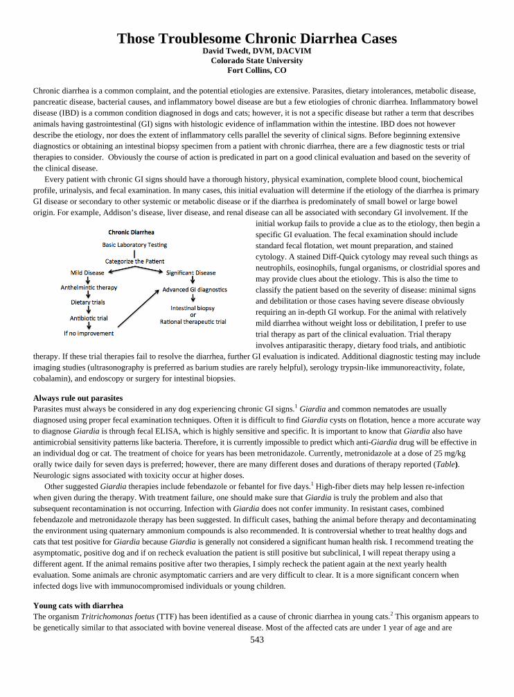
543
Those Troublesome Chronic Diarrhea Cases David Twedt, DVM, DACVIM
Colorado State University Fort Collins, CO
Chronic diarrhea is a common complaint, and the potential etiologies are extensive. Parasites, dietary intolerances, metabolic disease, pancreatic disease, bacterial causes, and inflammatory bowel disease are but a few etiologies of chronic diarrhea. Inflammatory bowel disease (IBD) is a common condition diagnosed in dogs and cats; however, it is not a specific disease but rather a term that describes animals having gastrointestinal (GI) signs with histologic evidence of inflammation within the intestine. IBD does not however describe the etiology, nor does the extent of inflammatory cells parallel the severity of clinical signs. Before beginning extensive diagnostics or obtaining an intestinal biopsy specimen from a patient with chronic diarrhea, there are a few diagnostic tests or trial therapies to consider. Obviously the course of action is predicated in part on a good clinical evaluation and based on the severity of the clinical disease.
Every patient with chronic GI signs should have a thorough history, physical examination, complete blood count, biochemical profile, urinalysis, and fecal examination. In many cases, this initial evaluation will determine if the etiology of the diarrhea is primary GI disease or secondary to other systemic or metabolic disease or if the diarrhea is predominately of small bowel or large bowel origin. For example, Addison’s disease, liver disease, and renal disease can all be associated with secondary GI involvement. If the
initial workup fails to provide a clue as to the etiology, then begin a specific GI evaluation. The fecal examination should include standard fecal flotation, wet mount preparation, and stained cytology. A stained Diff-Quick cytology may reveal such things as neutrophils, eosinophils, fungal organisms, or clostridial spores and may provide clues about the etiology. This is also the time to classify the patient based on the severity of disease: minimal signs and debilitation or those cases having severe disease obviously requiring an in-depth GI workup. For the animal with relatively mild diarrhea without weight loss or debilitation, I prefer to use trial therapy as part of the clinical evaluation. Trial therapy involves antiparasitic therapy, dietary food trials, and antibiotic
therapy. If these trial therapies fail to resolve the diarrhea, further GI evaluation is indicated. Additional diagnostic testing may include imaging studies (ultrasonography is preferred as barium studies are rarely helpful), serology trypsin-like immunoreactivity, folate, cobalamin), and endoscopy or surgery for intestinal biopsies. Always rule out parasites Parasites must always be considered in any dog experiencing chronic GI signs.1 Giardia and common nematodes are usually diagnosed using proper fecal examination techniques. Often it is difficult to find Giardia cysts on flotation, hence a more accurate way to diagnose Giardia is through fecal ELISA, which is highly sensitive and specific. It is important to know that Giardia also have antimicrobial sensitivity patterns like bacteria. Therefore, it is currently impossible to predict which anti-Giardia drug will be effective in an individual dog or cat. The treatment of choice for years has been metronidazole. Currently, metronidazole at a dose of 25 mg/kg orally twice daily for seven days is preferred; however, there are many different doses and durations of therapy reported (Table). Neurologic signs associated with toxicity occur at higher doses.
Other suggested Giardia therapies include febendazole or febantel for five days.1 High-fiber diets may help lessen re-infection when given during the therapy. With treatment failure, one should make sure that Giardia is truly the problem and also that subsequent recontamination is not occurring. Infection with Giardia does not confer immunity. In resistant cases, combined febendazole and metronidazole therapy has been suggested. In difficult cases, bathing the animal before therapy and decontaminating the environment using quaternary ammonium compounds is also recommended. It is controversial whether to treat healthy dogs and cats that test positive for Giardia because Giardia is generally not considered a significant human health risk. I recommend treating the asymptomatic, positive dog and if on recheck evaluation the patient is still positive but subclinical, I will repeat therapy using a different agent. If the animal remains positive after two therapies, I simply recheck the patient again at the next yearly health evaluation. Some animals are chronic asymptomatic carriers and are very difficult to clear. It is a more significant concern when infected dogs live with immunocompromised individuals or young children. Young cats with diarrhea The organism Tritrichomonas foetus (TTF) has been identified as a cause of chronic diarrhea in young cats.2 This organism appears to be genetically similar to that associated with bovine venereal disease. Most of the affected cats are under 1 year of age and are
544
reported to have a watery to sometimes mucoid diarrhea. It is most often observed in cats from humane shelters or catteries, and Abyssinians and Bengal cats appear to be over-represented or to have a more resistant disease. There are several ways to diagnose TTF. In some cases, a diagnosis can be made by performing a wet mount fecal prep and identifying the organism. A small amount of stool is thinned with warm saline solution, a coverslip applied, and the feces examined at 40X. It is important that the stool is fresh for examination. A colonic flush of saline can also be used to obtain fecal material for cytology and culture. TTF is identified by its progressive forward motion. (In contrast, Giardia has a falling leaf motion.) Feces can also be cultured in your practice using the bovine TTF culture technique employing an In Pouch TF™ culture method (Biomed Diagnostic Labs) (Figure 3). With these pouches, a very small amount of stool is placed in the broth and cultured at room temperature. The bag is then examined under a microscope 24 to 72 hours later for evidence of motile organisms. Fecal PCR for TTF is offered by many commercial labs and is considered the test of choice for confirming the infection. Ronidazole is the only antimicrobial shown to have efficacy in treating TTF infection.3 Ronidazole is given at 30 mg/kg q24h PO for up to 14 days. Ronidazole has a very narrow therapeutic range; higher doses or a longer duration can result in neurotoxicity. Ronidazole is not approved for use in the United States and must be obtained through a reliable compounding pharmacy. It is very bitter and therefore should be given via capsule; liquid solutions are not recommended. Treatment failure can occur, and a fecal PCR should be performed if a cat fails to respond to therapy because a negative PCR result means TTF is a less likely cause of the diarrhea. When left untreated many cats eventually become normal, especially young cats under 1 year of age. In one study, 88% cats with TTF infection were reported to undergo spontaneous resolution of diarrhea within two years of a diagnosis; however, most remained infected based on PCR results when retested as long as two to five years after the initial diagnosis.4 The role of these asymptomatic carriers in disease transmission remains unclear. When the diet works Over the years, I have become more and more impressed to see GI signs resolve simply by changing a patient’s diet. It is my impression, which is supported by a number of clinical studies, that possibly 30% to 50% of dogs and cats with nonspecific GI disease may respond to diet alone.5-7 A positive response to a diet trial is referred to as a food-responsive diarrhea (FRD). FRDs include both true dietary allergies and dietary intolerances. Allergies result from a reaction with a protein antigen, whereas intolerances occur in response to some substance in the diet, such as a preservative or food coloring. Dietary trials using a test diet generally require two weeks or less to appreciate a response; the GI signs seem to respond much faster than dermatologic signs, which that may take eight weeks or more to improve. There is no ideal diet that will consistently resolve diarrhea. My personal favorite is the use of a hydrolyzed diet, such as Purina HA®. Hydrolyzed diets are single-protein sources (usually soy-, rice-, or potato-based) and have undergone digestion, producing low-molecular-weight protein derivatives that are thought to be highly digestible with low antigenic potential. Their benefit might actually be because they are pure and contain little else that might contribute to a dietary intolerance. These diets have now become the ideal initial trial diet. If a positive response is observed, then the patient’s GI signs can be controlled with a diet. The patient can either continue on the test diet or you can attempt to find another long-term diet that works well for both the client and patient. Some clinicians recommend if there is a diet response that the patient to be fed that diet exclusively for at least three months, at which time the diet can be changed or even the original diet reintroduced. Only a small percentage of dogs with GI signs (~8%) relapse on challenge and are thus truly food allergic.7 Feeding novel-protein diets with a single protein antigen would be an alternative approach. If using the novel antigen diets, one should prescribe only veterinary diets because many over-the-counter novel-protein diets are not all that novel and have been shown to contain many other antigens not listed on the label.8 Highly digestible gastrointestinal diets such as Purina EN® may improve assimilation, promote gastrointestinal health, and modify the microbiota. Diets containing highly fermentable fibers such as those containing fructooligosaccharides (also referred to as prebiotics diets) are often useful for colonic disease because fermentation products are shown to benefit mucosal function and modify enteric microbiota, promoting "good" bacteria and inhibiting certain pathogenic bacteria.9 If a diet trial is unsuccessful, with no improvement in clinical signs after 10 to 14 days, the next step is to institute an antibiotic trial. GI drugs and bugs There are many dogs with chronic large or small bowel disease that have an antibiotic-responsive diarrhea (ARD). An old term for ARD is small intestinal bacterial overgrowth (SIBO). However, SIBO is a poorly defined syndrome in dogs, and we currently have no way to adequately and convincingly diagnose bacterial overgrowth or to know in which cases antibiotics would be beneficial short of a therapeutic trial. More recently the term gastrointestinal dysbiosis has been given to conditions associated with an abnormal GI bacterial ecosystem.10 In simple terms, GI dysbiosis refers to an imbalance in GI bacteria with the loss of the “good bacteria” coupled with an increase in the so-called “bad bacteria.” For chronic diarrhea cases that do respond to antibiotic therapy, it is likely the antibiotics are not eliminating a specific pathogen but rather changing the overall bacterial ecosystem, promoting a more normal bacterial makeup. Some cats and dogs with gastrointestinal dysbiosis have decreased serum cobalamin (vitamin B12) concentrations.11 The cobalamin deficiency can be due to lack of intrinsic factor production, abnormal increased intestinal bacterial utilization, or ileal disease causing inadequate cobalamin absorption. Serum folate concentrations are usually variable in cases having dysbiosis.
545
Metronidazole is frequently used in GI cases but long-term administration and potential side effects make it less desirable than other options. Metronidazole has been shown to cause DNA damage to feline lymphocytes in vitro. There is also evidence in laboratory animals that it has some carcinogenic potential.12 A suggested GI dosage for metronidazole in cats and dogs is 7.5 to 10 mg/kg given orally twice daily. A commonly used alternative, and my first choice, is tylosin. Tylosin was first reported to be useful for chronic diarrhea in the early 1970s and there has been a recent resurgence in interest and use of the antibiotic. Tylosin is a macrolide, bacteriostatic antibiotic that is currently marketed over the counter for the treatment of respiratory disease in chickens. Tylosin has activity against most gram-positive and gram-negative cocci, gram-positive rods, and Mycoplasma; however, the gram-negative bacteria Escherichia coli and Salmonella species are intrinsically tylosin-resistant.13 Tylosin works by transiently changing the GI enteric bacterial population, probably by promoting the growth of beneficial commensal bacteria while suppressing deleterious bacteria. Once tylosin is discontinued, the original bacterial population often returns to its pretreatment state. There is also a suggestion that tylosin may have anti-inflammatory properties.13 Tylosin appears to have almost no systemic or toxic side effects. The initial dose recommendation for tylosin in both dogs and cats is 15 mg/kg orally, twice a day, mixed with the food (has a bitter taste) or given via gelatin capsule. (Note: it comes as a powder and a #3 gelatin capsule holds 130 mg, a #1 capsule holds 240 mg, a #0 capsule holds 345 mg, and a #00 capsules hold 430 mg.) For cases that respond, the long-term dose can be reduced to as low as 5 mg/kg/day.13 Tylosin is effective for most Clostridium perfringes and is considered by many to be the treatment of choice for suspected clostridial diarrhea.14 Probiotics To date, there have been very few controlled clinical studies evaluating probiotic success. However, a large double-blinded placebo control study of shelter dogs and cats developing diarrhea found significantly fewer cats that received Enterococcus faecium (FortiFlora®, 2.1 x 10⁹ cfu/day) developed diarrhea for greater than a two-day duration.16 Probiotics exert their effects as long as they are being given but once stopped the GI flora generally returns to the pretreatment state. It may seem counterintuitive to give antibiotics with probiotics, but clinical improvement is often seen when they are given in combination. Probiotics are considered a safe adjunctive therapy and are commonly used for both acute and chronic diarrhea in dogs and cats as well as for the prevention of stress induced diarrhea.15-17 Recommendations for the ideal probiotic, containing an adequate type and number of viable organisms for specific GI disorders, become difficult to make. Some over-the-counter preparations have been found not to contain the label claims.18 My recommendation is to use a product produced by a reputable veterinary company that has done research on their product. German shepherds with chronic diarrhea A clinical syndrome frequently encountered in German shepherd dogs is chronic GI signs and weight loss. Exocrine pancreatic insufficiency is common in the breed, requiring pancreatic enzyme supplementation, and it must first be ruled out. The diagnosis is made by documenting a subnormal trypsin-like immunoreactivity (TLI) concentration followed by improvement with pancreatic enzyme replacement. A second group of German shepherd dogs with similar clinical signs have normal TLI concentrations. Many of these dogs turn out to have an antibiotic-responsive diarrhea due to GI dysbiosis. Testing should include measurement of folate and cobalamin (serum B12) concentrations. Low cobalamin and high folate levels are characteristic of both exocrine pancreatic insufficiency and GI dysbiosis. Dogs with subnormal cobalamin concentrations will require parenteral supplementation (initially, about 500 µcg subcutaneously weekly) as part of the therapy. The cause of the GI dysbiosis in German shepherds is unknown. Researchers have investigated IgA concentrations, suggesting the possibility of an inherent deficiency leading to altered GI immunity. More recently researchers have measured toll-like receptors (TLR) in the GI tract of these dogs with a documented abnormal expression of the receptors. Using candidate gene analysis, polymorphisms in TLR4 and TLR5 were recently shown to be significantly associated with IBD in German shepherds.19 Furthermore, the same polymorphisms in TLR5 were also associated with IBD in a heterogeneous population of dogs consisting of 38 different breeds.19 These mutations could well play an important role in the pathogenesis of IBD in dogs, as a mutated receptor will lead to misrepresentation of commensal bacteria as pathogens, therefore signaling “danger” to the host and initiating the characteristic inflammatory response seen in this disease. Management of affected German shepherds involves diet, antibiotics, and cobalamin supplementation. Prebiotics and probiotics are also often given as additional adjunctive therapy. This condition tends to require life-long management. When is it inflammatory bowel disease? A diagnosis of IBD requires a complete laboratory evaluation to rule out other diseases. A complete blood count, biochemical profile, urinalysis, fecal cytology, and parasite evaluation are required in all cases. An eosinophilia or hypoproteinemia may provide clues to IBD. Abdominal radiographs or ultrasonography may be helpful. However, ultrasound images showing increased wall thickness are neither specific nor sensitive for the diagnosis of IBD.20 Specific testing may include measurement of serum folate and cobalamin concentrations. Cobalamin deficiency is a common complication of feline GI disorders, and complete improvement in GI function is not possible until cobalamin deficiency is corrected.11
546
An overall impression is that most cases of IBD can be managed; however, unless the underlying etiology can be identified and removed, it can become a long-term proposition. A retrospective study demonstrated that only 26% of canine IBD cases progressed to complete remission, with intermittent clinical signs remaining in about half of the cases, 4% being completely uncontrolled, and 13% resulting in euthanasia because of poor response to treatment.21 Another study found 18% of the dogs were euthanized because of their disease.6 Poor prognostic indicators are hypoalbuminemia and hypocobalaminemia.6 Treatment of IBD Patients that do not respond to a diet or an antibiotic trial are usually administered glucocorticoids. It is estimated that about 30% of the dogs that fail to respond to a change of diet and antibiotics will respond to corticosteroids. Generally oral prednisolone is given to dogs and cats once daily at a starting dose of 1 to 2 mg/kg, and then the dose is tapered over an eight-week period. However, the side effects of glucocorticoids can be marked, and I try never to exceed a total of 40 mg per day in large-breed dogs. Budesonide is a novel glucocorticoid that is reported to have high first-pass hepatic metabolism and exerts a “local effect” on the intestine with minimal systemic effects. An enteric-coated formulation is used for people with IBD but a non-enteric coated formulation made by a compounding pharmacy should be used. Despite apparent efficacy of budesonide in dogs and cats, the systemic steroid effects are present and consequently, its use may have no benefit over traditional corticosteroid therapy in most cases. The recommended dose is 1 mg once daily in cats and toy breeds and up to 2 mg once daily for large-breed dogs.
If there is poor response to glucocorticoids in dogs after the first three to four weeks or if the side effects are severe, then I recommend oral cyclosporine at 5 to 10 mg/kg once daily for at least two months. Many dogs with IBD that are steroid refractive are reported to respond to cyclosporine.22 In cats, the use of chlorambucil (2 to 6 mg/m2, q24h, PO, or 2 mg/cat three times a week) with prednisolone is preferable, if there is inadequate response to glucocorticoid treatment alone. If chlorambucil is used, hematologic parameters should be monitored regularly. Cyclosporine blood concentrations do not need to be monitored regularly, unless side effects induced by the cyclosporine treatment are suspected or an inadequate response to treatment is observed. If measuring cyclosporine serum concentrations, it is recommended to take blood samples one to two hours after giving the medication to ensure that peak concentrations are measured. If the cyclosporine serum concentration is above 700 ng/ml at peak level, then halving the dosage for the first two weeks can reduce the side effects.22 If the patient responds to cyclosporine, then the medication can either be tapered slowly or stopped after 10 weeks. Sulfasalazine (20 to 50 mg/kg orally three times daily for three to six weeks) and related drugs are often used in dogs when IBD is limited to the large intestine. However, side effects include keratoconjunctivitis sicca, so tear production should be monitored regularly when using these drugs. I rarely prescribe sulfasalazine for large bowel disease because, in my experience, most patients get better with diet and antibiotics. Other novel or adjunctive therapies could include omega 3 fatty acids for anti-inflammatory effects and various antioxidants. Probiotics have also been suggested to be beneficial for treating IBD due to the multiple mechanisms described above. References upon request
547
Abnormal Liver Enzymes: A Clinical Approach
David Twedt, DVM, DACVIM Colorado State University
Fort Collins, CO
The identification of abnormal liver enzymes usually indicates liver damage but rarely provides a diagnosis or etiology. Abnormal liver enzymes are common and in a study of 1,022 blood samples taken from both healthy and sick dogs and cats, one diagnostic laboratory found 39% had ALP increases and 17% had ALT increases. When presented with a patient having abnormal liver enzymes it is important to recognize that the patient could have primary liver disease but more likely the patient has other primary non-hepatic condition resulting in secondary liver involvement. It is therefore important to perform a complete review of all other body systems.
It is also important to understand the reason for increased liver enzyme activity and the following sections will deal with liver specific tests. Tests of heptocellular necrosis or degeneration Increases in either alanine aminotransferase (ALT) or aspartate aminotransferase activity (AST) indicate hepatocellular membrane damage and leakage of the enzymes. This could be due to death of the hepatocyte or from hepatocyte degeneration where the membrane just has increased permeability. Conceptually ALT and AST should be thought of as hepatocellular “leakage” enzymes. Subsequent to an acute and diffuse injury, the magnitude of increase crudely reflects the number of affected hepatocytes. The plasma half-life of ALT activity is about 2.5 days (60 hours) in dogs however concentrations may take days to weeks to decrease following an acute insult based on models of acute hepatic injury. Persistent increased ALT and AST enzyme activity over weeks is characteristic of chronic hepatitis in the dog. As a general rule, ALT increases should be investigated when they are greater than twice normal or persistently abnormal over weeks to months. Hepatic AST is located predominately in hepatocyte mitochondria (80%) but is also soluble in the cytoplasm. Because of the mitochondrial location, AST elevations are more sensitive for liver disease than ALT and reflect more significant cell damage. On the other hand, AST is less specific than ALT because of the presence in other tissues (i.e., muscle so always check CK). Following an acute injury resulting in a moderate to marked increase in the serum ALT and AST concentrations, due to their difference in plasma half-life, the serum AST will return to normal more rapidly (hours to days) than the serum ALT (days). Tests of cholestasis and drug-induction Alkaline phosphatase (ALP) and gamma-glutamyltransferase (GGT) show minimal activity in normal hepatic tissue but can become increased in the serum subsequent to increased enzyme production stimulated by either impaired bile flow or drug-induction. These enzymes have a membrane bound location at the canalicular surface; ALP associated more with the canalicular membrane and GGT associated more with epithelial cells comprising the bile ductular system. With cholestasis, surface tension in the canuliculi and bile ductules increases and production of these surface enzymes is then up-regulated. An increase in the serum ALP and GGT activity can be the result of induction by endogenous, topical or systemic glucocorticoids, anticonvulsant medications (ALP only) and possibly other drugs or herbs. The plasma half-life for hepatic ALP in the dog is 66 hours in contrast to 6 hours for the cat and the magnitude of enzyme increase (presumably a reflection of the synthetic capacity) is greater for the dog than the cat. Bone source arises from osteoblastic activity and is elevated in young growing dogs before their epiphysial plates close or in some dogs with bone tumors or lytic lesions. One study identified that increased ALP concentrations in some dogs with osteogenic bone tumors tended to indicate a poorer prognosis, probably from diffuse bone metastasis. In the adult without bone disease, an increased serum ALP activity is usually of hepatobiliary origin. Hepatic GGT is located predominately on the canalicular membrane and bile ducts. Chronic elevations in GGT tend to better reflect hepatobiliary tract disease, with he most marked elevations resulting from diseases of the biliary epithelium such as bile duct obstruction, cholangiohepatitis, cholecystitis or neoplasia. In dogs, GGT has a lower sensitivity (50%) but higher specificity (87%) for hepatobiliary disease than total ALP. If ALP is elevated with a concurrent increase in serum GGT, specificity for liver disease increases to 94%. Bone does not contain GGT and the administration of anticonvulsant medications to dogs does not cause an increase in the serum GGT activity. Evaluation of liver function On a routine biochemical profile it is important to note the liver function tests (or tests that involve liver function) including bilirubin, albumin, glucose, BUN, and cholesterol. Bilirubin elevations can occur from hemolysis, hepatic dysfunction or extrahepatic cholestasis. Measuring the percent conjugated to unconjugated bilirubin to determine location is not useful in the dog. Albumin is exclusively made in the liver and if albumin is not lost, sequestered or diluted, a low concentration would suggest significant hepatic dysfunction. It may take greater than 60% hepatic dysfunction for albumin concentrations to decline. Cholesterol can be variable and
548
increased in cholestatic conditions and decreased in portosystemic shunts. When glucose and BUN activity is low from liver dysfunction suggests significant hepatic disease and a guarded prognosis.
Bile acids Measurement of serum bile acids is thought to be the most sensitive function test that is readily available in small animal practice. Bile acids are synthesized from cholesterol in the liver and then conjugated and excreted into the bile. Bile acids are transported to the gallbladder and following a meal are excreted into the intestine where they emulsify fat for absorption. In the distal small intestine bile acids are actively resorbed and return to the liver where they are efficiently extracted by the hepatocytes and then re-circulated back into the bile. Only a small fraction of the total bile acid pool ever escapes into the systemic circulation. Thus, the enterohepatic circulation of bile acids occurs with a 95-98% rate of efficiency. The current suggestion for performing bile acid levels is to differentiate between congenital portal vascular anomalies and liver insufficiency, prior to the development of jaundice. The determination of total bile acids can contribute to the decision to obtain histological support for a definitive diagnosis. The fasting total serum bile acid concentration (FSBA) is a reflection of the efficiency and integrity of enterohepatic circulation. Pathology of the hepatobiliary system or the portal circulation results in an increased FSBA prior to the development of hyperbilirubinemia, therefore, bile acid measurement is not useful in the icteric patient. An increase is not specific for a particular type of pathologic process but is associated with a variety of hepatic insults or abnormalities of the portal circulation. Bile acids should be used to screen patients with persistently abnormal liver enzymes, to determine if there could be loss of hepatic function, which adds further diagnostic support during investigation of the case. It is also helpful to measure bile acids to determine level of hepatic dysfunction in animals with PSS or portal vein hypoplasia (PVH), also known as microvascular dysplasa. When the fasting value is greater than 25 µmol/L for the dog and cat, there is a high probability that the histology findings will define a lesion.
When the total fasted bile acid concentration is normal or in the “gray zone” the FSBA should be followed by a 2-hour postprandial serum total bile acid (PPSBA) looking for an increase of greater than 25 µmol/L. The diagnostic value of determining PPSBA concentration is increased sensitivity for the detection of hepatic disease and congenital portal vascular anomalies. In dogs, the specificity of fasting and postprandial bile acids for hepatobiliary disease is 95% and 100% when cutoff values greater than 15 µmol/L and 25 µmol/L are used, respectively. When using these guidelines it is prudent to recognize that a small number of apparently healthy dogs have been reported with PPSBA values above 25 umol/L or these may actually represent dogs that have PVH. Occasionally the FSBA value is greater than the PPSBA value. The reason for this non sequitur is probably multifactorial. It has been shown that (1) the peak PPSBA concentration for individual dogs is variable, (2) fasted dogs store about 40% of the newly produced bile in the gallbladder and (3) a meal stimulates the release of only between 5 to 65% gallbladder bile. Undoubtedly these physiologic variables in addition to physiological variation in intestinal transit time and concurrent underlying intestinal disease contribute to the dichotomy.
Recently, urinary bile acids have become available as a diagnostic tool. Identifying increased urinary bile acids provides similar information to what is obtained from serum bile acids and neither test appears to be better than the other. The advantage of urinary bile acid measurements would be for the screening of litters of young puppies for suspected inherited vascular anomalies where urine collection is simpler than paired serum samples.
Coagulation panels Major clotting factors are synthesized in the liver (except factor 8) and therefore prolonged clotting time may suggest significant hepatic dysfunction or factor consumption. Because coagulation abilities may not be normal in patients with liver disease, it is advisable to check clotting times prior to performing liver biopsy.
Ammonia High ammonia levels reflects abnormal hepatic portal shunting (acquired or congenital shunts) or significant hepatocellular dysfunction of greater than 70%. The liver detoxifies ammonia that primarily arises from the gastrointestinal tract by conversion to urea. Elevated fasting blood ammonia levels have been shown to be a sensitive (98%) and specific (89%) test for the detection of congenital or acquired portosystemic shunting in dogs. Due to problematic requirements for sample handling and submission, blood ammonia or the ammonia tolerance test is infrequently performed by some clinical practices. However, recent availability of blood ammonia for in-clinic analyzers, has helped make the test more feasible. Diagnostic strategies. In the asymptomatic patient with an increased liver biochemical test(s) the increased value should be confirmed. If no likely explanation for the laboratory abnormalities can be found there are two courses of action that one can take; either begin a diagnostic evaluation of the patient starting with bile acid determinations, or re-evaluate the patient’s liver enzymes at a later date. The diagram below depicts a general algorithm for the work-up of dogs that have abnormal liver enzymes. The identification of abnormal liver enzymes may occur when the sick patient is presented for evaluation or during a routine health screen in the healthy patient. Abnormal liver enzymes in the sick patient could either be the result of primary liver disease/damage or secondary due to a multitude of other non-hepatic disorders. The most common cause of abnormal liver enzymes is in fact, not primary liver disease at all but rather the result of reactive hepatic changes occurring secondary to other non-hepatic causes. Generally, secondary hepatic changes are
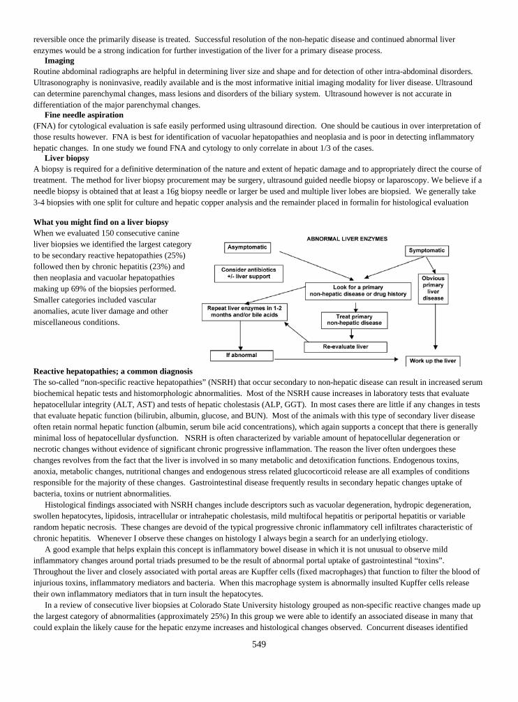
549
reversible once the primarily disease is treated. Successful resolution of the non-hepatic disease and continued abnormal liver enzymes would be a strong indication for further investigation of the liver for a primary disease process.
Imaging Routine abdominal radiographs are helpful in determining liver size and shape and for detection of other intra-abdominal disorders. Ultrasonography is noninvasive, readily available and is the most informative initial imaging modality for liver disease. Ultrasound can determine parenchymal changes, mass lesions and disorders of the biliary system. Ultrasound however is not accurate in differentiation of the major parenchymal changes.
Fine needle aspiration (FNA) for cytological evaluation is safe easily performed using ultrasound direction. One should be cautious in over interpretation of those results however. FNA is best for identification of vacuolar hepatopathies and neoplasia and is poor in detecting inflammatory hepatic changes. In one study we found FNA and cytology to only correlate in about 1/3 of the cases.
Liver biopsy A biopsy is required for a definitive determination of the nature and extent of hepatic damage and to appropriately direct the course of treatment. The method for liver biopsy procurement may be surgery, ultrasound guided needle biopsy or laparoscopy. We believe if a needle biopsy is obtained that at least a 16g biopsy needle or larger be used and multiple liver lobes are biopsied. We generally take 3-4 biopsies with one split for culture and hepatic copper analysis and the remainder placed in formalin for histological evaluation What you might find on a liver biopsy When we evaluated 150 consecutive canine liver biopsies we identified the largest category to be secondary reactive hepatopathies (25%) followed then by chronic hepatitis (23%) and then neoplasia and vacuolar hepatopathies making up 69% of the biopsies performed. Smaller categories included vascular anomalies, acute liver damage and other miscellaneous conditions.
Reactive hepatopathies; a common diagnosis The so-called “non-specific reactive hepatopathies” (NSRH) that occur secondary to non-hepatic disease can result in increased serum biochemical hepatic tests and histomorphologic abnormalities. Most of the NSRH cause increases in laboratory tests that evaluate hepatocellular integrity (ALT, AST) and tests of hepatic cholestasis (ALP, GGT). In most cases there are little if any changes in tests that evaluate hepatic function (bilirubin, albumin, glucose, and BUN). Most of the animals with this type of secondary liver disease often retain normal hepatic function (albumin, serum bile acid concentrations), which again supports a concept that there is generally minimal loss of hepatocellular dysfunction. NSRH is often characterized by variable amount of hepatocellular degeneration or necrotic changes without evidence of significant chronic progressive inflammation. The reason the liver often undergoes these changes revolves from the fact that the liver is involved in so many metabolic and detoxification functions. Endogenous toxins, anoxia, metabolic changes, nutritional changes and endogenous stress related glucocorticoid release are all examples of conditions responsible for the majority of these changes. Gastrointestinal disease frequently results in secondary hepatic changes uptake of bacteria, toxins or nutrient abnormalities.
Histological findings associated with NSRH changes include descriptors such as vacuolar degeneration, hydropic degeneration, swollen hepatocytes, lipidosis, intracellular or intrahepatic cholestasis, mild multifocal hepatitis or periportal hepatitis or variable random hepatic necrosis. These changes are devoid of the typical progressive chronic inflammatory cell infiltrates characteristic of chronic hepatitis. Whenever I observe these changes on histology I always begin a search for an underlying etiology.
A good example that helps explain this concept is inflammatory bowel disease in which it is not unusual to observe mild inflammatory changes around portal triads presumed to be the result of abnormal portal uptake of gastrointestinal “toxins”. Throughout the liver and closely associated with portal areas are Kupffer cells (fixed macrophages) that function to filter the blood of injurious toxins, inflammatory mediators and bacteria. When this macrophage system is abnormally insulted Kupffer cells release their own inflammatory mediators that in turn insult the hepatocytes.
In a review of consecutive liver biopsies at Colorado State University histology grouped as non-specific reactive changes made up the largest category of abnormalities (approximately 25%) In this group we were able to identify an associated disease in many that could explain the likely cause for the hepatic enzyme increases and histological changes observed. Concurrent diseases identified

550
included neoplasia, gastrointestinal, renal, autoimmune, dermatologic, dental, infectious and cardiac disease as a few examples. In some cases an underlying disease is not identified. The ALT values on the average are 1-2 X normal and the ALP values 1-3 X normal. It is interesting to note that in a series of 32 dogs having reactive hepatopathies, 8/8 cases in which serum bile acids were run, all were within the normal reference range again suggesting hepatic function tends to remain intact.
This category appears to be the most common histological change to occur in dogs and is by far the most common cause of elevated liver enzymes. Based on this fact, dogs presented with elevations in ALT and ALP should always have primary non-hepatic disease ruled out first. These changes are usually very reversible and no specific hepatic therapy is required short of treating the primary disease. The liver changes resolve once the primary etiology is successfully treated. Therapy providing good liver support such as antioxidants may be warranted. Summary Abnormal liver enzymes should not be ignored and should be investigated in a systematic manner as previously discussed. Asymptomatic animals with no evidence of significant or treatable disease or in situations where financial constraints limit further work up the patient should be fed a quality maintenance diet for the patient’s stage of life and the possibility of instituting specific liver support therapy should be explored.
551
Acute Liver Disease David Twedt, DVM, DACVIM
Colorado State University Fort Collins, CO
Acute liver failure (ALF) is an uncommon condition that results in rapid deterioration of liver function occurring in a previously healthy animal. It is generally characterized by severe hepatocyte death due to aptoposis or cytolytic necrosis. The extent and location of the hepatocyte death depends on the etiology. Etiology Drugs are the most common known cause of ALF in dogs and cats. Drugs can affect the liver in one of two ways. First, they may have a direct toxicity to hepatocytes or becomes metabolized to a toxic compound that then causes damage. This first classification is referred to as a direct hepatotoxin and is dose related and reproducible. An example would be acetaminophen poisoning and CCNU therapy. More common however are drugs associated with an idiosyncratic drug reaction. Idiosyncratic drug reactions are unpredictable and not dose related but most often associated with abnormal or aberrant metabolism of the drug to a toxic compound. Listed below are some of the more common drug associated hepatotoxicities. It should be noted however any drug metabolized by the liver has the potential to be a hepatotoxin. The common incriminators causing an idiosyncratic reaction include the NSAIDs, trimethoprim-sulfa, lysodren, ketoconazole (and other antifungals), and diazepam (in cats) to name but a few. At Colorado State University we reported on a series of dogs developing acute liver toxicity associated with carprofen. The toxicity was idiosyncratic occurring in possibly 1 out of 10,000 dogs. We have also more recently identified toxicity associated with azathioprine. See table of common drug associated with liver disease. Some herding breed dogs lack p-glycoprotein that plays an important role in metabolism of many drugs. Thus it is not surprising that lack of P-glycoprotein, which occurs in many herding-breed dogs leads to increased susceptibility to drug toxicosis.
Other causes of acute liver failure include infectious agents such as Leptospirosis. Environmental toxins such as industrial solvents, plants, insects, chemicals, envenomation, sago palm seeds, heavy metals, Amanita phalloides (mushroom) and aflatoxin have been incriminated to cause liver disease. Several years ago there was a large outbreak of liver failure in many dogs in the Eastern part of the United States due to contaminated dog food with aflatoxin. In most cases aflotoxin is an isolated event. Xylitol an artificial sweetener found in chewing gum can result in a sudden drop in glucose due to increase insulin release and in some cases also causes acute liver disease as well.
Damage to the liver may range from mild to moderate hepatic necrosis resulting in minimal clinical signs. Signs may be associated with vomiting, lethargy and anorexia. Massive hepatic necrosis will result in ALF and produce significant clinical signs of liver failure and possibly death. The signs of ALF are variable but usually will always include anorexia, depression, lethargy and vomiting. Neurological signs from hepatic encephalopathy may progress to coma or seizures. Jaundice is invariably present. ALF can also result in evidence of hemorrhage either from lack of coagulation factors or from DIC. GI ulceration is common. Septicemia may occur from uptake of enteric bacteria. Hepatic pain may be observed on abdominal palpation. Some drugs associated with liver toxicity Acetaminophen Arsenicals Ketoconazole Sulfonamides Halothane Carprofen (NSAIDs) Griseofulvin Itraconazole/ketoconazole Mitotane (lysodren) Trimethoprim-sulfa Diazepam Anabolic steroids Anticonvulsant drugs Antineoplastic drugs Azathioprine Tetracycline (doxycycline) Amiodarone CCNU Clinical findings Focal or mild to moderate hepatic necrosis is generally associated with clinical signs that are suggestive of liver disease. Acute massive necrosis however will produce clinical signs. The signs of acute hepatic necrosis are nonspecific but usually will include anorexia, depression, lethargy and vomiting. In hepatic failure hemorrhage and hepatic encephalopathy ensues this is then often followed by coma or seizures. Hepatic pain may be observed on abdominal palpation.
The clinicopathologic changes reflect the extent of necrosis and loss of hepatocyte numbers. The hepatic transaminases (ALT and AST) are released when the cell membrane is damaged and the cytosol enzymes leak out. A marked increase in AST to ALT ratio suggests more severe hepatocellular damage. Generally ALP and GGT increases are associated with hepatic necrosis and are only mild to moderately elevated. Hyperbilirubinemia is common when significant hepatic necrosis is present and frequently very high
552
when massive necrosis occurs. Changes in the liver function test will reflect the magnitude of hepatic damage. When the necrosis is massive and liver function is compromised these function changes will occur. Clotting factors decline and may contribute to hemorrhage. Hypoglycemia, low BUN, hypoalbuminemia, and increase in ammonia all reflect hepatic failure. It is important to note that with acute severe necrosis albumin concentrations may remain normal early in the disease due to the longer half-life of albumin (2 weeks) as compared to clotting factors being only (hours to days). Frequently platelet numbers and function are also compromised in massive liver failure and DIC is a common complication. With recovery the AST will decline prior to ALT and could be prognostically helpful.
Cerebral edema and associated increased intracranial pressure occurs with liver failure. The pathogenesis of the edema is unclear. It is thought that it is a combination of vasogenic and cytotoxic edema. Coagulopathy occurs from failure to produce clotting factors and platelet dysfunction. With secondary organ and vascular damage DIC is common in end stage liver failure. Hypotension from decreased peripheral resistance and cardiac dysfunction occurs. Hypokalemia, hyponatremia and respiratory alkalosis are reported in humans having hepatic failure. A metabolic (lactic) acidosis may also occur. Another serious complication of liver failure is renal dysfunction. The mechanism is complex and may be associated with acute tubular necrosis but also associated with vascular changes referred to as hepato-renal syndrome. Oliguric renal failure denotes a grave prognosis. Because of the failure to produce BUN in many cases it is prudent to follow serum creatinine that is not influenced by hepatic function.
The patient is also susceptible to infection and septicemia due to the transmigration of bacteria through the GI tract entering into the portal system. With hepatic dysfunction filtering of portal blood is impaired and systemic bacteremia results.
Management When there is acute ingestion of a hepatotoxin toxin vomiting should be induced followed by administration of activated charcoal to prevent absorption of the toxin or drug absorption. In most cases the animal is clinical and too advanced for gastric lavage an charcoal to have any benefit.
The next step is to prevent further hepatocyte damage by providing an environment for optimal hepatic function. There is considerable evidence showing that free radicals are generated in acute liver damage and participate in the pathogenesis of liver injury. Free radicals are molecules with an unpaired electron that form by the injurious effects of certain drugs or various other toxic agents or events. Free radicals, if not inactivated, damage cellular macromolecules via lipid peroxidation and thus participate in cellular injury when produced in excess. Depletion of antioxidants primarily glutathione parallels hepatic damage. N-acetylcysteine (NAC, Mucomyst™) is thiol (SH) donor and promotes the production of glutathione. Glutathione is the most important detoxifier of toxic cellular xenobiotics. There is also evidence that NAC protects against hepatic ischemia-reperfusion damage possibly by inhibiting Kupffer cell function. Further NAC has beneficial effects on liver blood flow, oxygen extraction, and the formation of non-glutathione products that protect against cell injury. Experimentally NAC has protective effects against aflatoxin damage as well. The suggested dose for NAC is 140 mg/kg IV followed by 70 mg/kg IV bid or tid for one to three days. The injectable NAC should be diluted 1:4 in 5% dextrose and water and given slowly over 30 minutes to 1 hour. When vomiting has resolved NAC therapy can be switched to oral medications. Oral S-Adensosylmethionine (SAMe) also protects against liver damage in dogs and cats by increasing hepatocyte glutathione concentrations being a SH donor. It also acts as a methyl donor and enzyme activator for key reactions that maintain membrane structure and function. Reports show protection against acetaminophen toxicity in the dog and cat. SAMe is given orally at the dose of 20 mg/kg bid or daily. SAMe in combination with milk thistle products is commercially available and would be of benefit as well.
The use of other antioxidants is warranted in management of the liver disease including vitamin E and milk thistle or its by-products. Vitamin E, d-alpha tocopherol, functions a major membrane bound intracellular antioxidant, protecting membrane phopspholipids from peroxidative damage when free radicals are formed. Vitamin E is shown to protect against the effects of copper, bile acids and other hepatotoxins. A suggested vitamin E dose is 50 to 400 IU a day. Other antioxidants that have been investigated but lack clinical experience in dogs and cats include allopurinol and desferoxamine.
Milk thistle has been used for centuries as a natural remedy for diseases of the liver and biliary tract. Silymarin the active extract consists of bioflavonoligans that have been reported to work as antioxidants, scavenging free radicals and inhibiting lipid peroxidation. In a number of human clinical studies on patients having either acute or chronic liver disease has provided mounting evidence of the benefit of milk thistle. These studies must be interpreted with care because of the variable experimental design and limited number of cases. One canine study showed that dogs poisoned with amanita mushrooms that were treated with milk thistle had less clinical signs and complete survival while one-third of dogs in the untreated group died. Due to the lack of standardization of milk thistle preparations it is difficult to provide an appropriate dosage. Suggestions have included 50-250 mg/kg bid. Milk thistle is reported to have an extremely low toxicity in humans and animals and has been used extensively in clinical patients with little concern for side effects. It appears to have a synergistic effect with vitamin E.
Other therapy includes fluid therapy giving balanced electrolytes generally with potassium and glucose supplementation. Fresh whole blood or frozen plasma may be required to provide clotting factors. Stored blood should be avoided because ammonia
553
concentrations increase over time prom production of red blood cells. The use of hetastarch or dextrans may be required to maintain blood pressure. (Note low MW dextrans may worsen DIC). Generally an indwelling urinary catheter is placed to monitor urinary output and to detect oliguria. Fruosemide and dopamine may be used to induce a diuresis. Broad-spectrum intravenous antibiotic therapy is generally indicated to manage and possibly prevent septicemia. Antibiotic choice should be ones that are not hepatotoxic or require hepatic metabolism. Common antibiotics to avoid include trimethoprim-sulfa, tetracycline and metronidazole. Gastrointestinal ulceration is managed using the H2 blocker such as ranitidine (2-5 mg/kg BID/TID) and oral sucralfate (Carafate 1 mg tab/25 kg TID given 1 hour before ranitidine). Cimetidine is to be avoided in liver disease because it must be metabolized by the liver and alters hepatic metabolism (via cytochrome P450 system) of other drugs. The dose of H2 blockers should be reduced with renal dysfunction.
Conventional therapy for HE includes the use of enemas to clean the colon of both bacteria and protein substrates for ammonia production. Slightly acidic enemas will lower the pH of the colon thus ionizing ammonia and reducing its absorption. Povidone iodine (betadine) can safely be given by enema as a 10% solution (weak tea color) that will both acidify the colon and have an antiseptic action reducing bacterial numbers. Nonabsorbable intestinal antibiotics are used to alter bowel flora and suppress urease-producing organisms important in formation of factors causing hepatic encephalopathy. Antibiotic suggestions include oral ampicillin or aminoglycosides (neomycin, kanamycin or gentamicin). A nondigestible disaccharide lactulose (Cephulac or Chronulac) given orally acidifies the colon converting ammonia to ammonium which is poorly absorbable thus trapping ammonia in the colon. The fermentation products of lactulose will also act as an osmotic laxative reducing colonic bacteria and protein substrates. A dose of 1-10 ml orally TID is generally effective. Lactulose is not absorbed systemically and thus considered safe. The dose should be adjusted to cause 3 or 4 soft stools a day. If diarrhea develops the dose should be reduced. Lactulose can also be given by enema in treating the severe case of hepatic encephalopathy. Treatment of seizures with sedatives or barbiturates is generally contraindicated. One should make sure that the patient is not hypoglycemic and take measures to treat HE. In some cases worsening of the neurological status is associated with development of cerebral edema. Corticosteroids have however not been definitively shown to be effective in hepatic failure and edema. The combination of the synergistic effects of mannitol and furosemide is shown to be of some benefit in cerebral edema.
Prognosis The prognosis of hepatic necrosis depends on the amount of damage and the secondary complications that occur. The prognosis for acute hepatic necrosis and hepatic failure depends on the extent of hepatic damage, metabolic complications and the ability to maintain the patient until hepatic regeneration is possible. Aggressive management and anticipation of potential complications will improve survival. With biochemical and clinical evidence of loss of hepatic function the prognosis becomes guarded. In humans artificial livers or liver transplant are used in severe cases. It has been stated that a prothrombin time greater than 100 seconds indicates a grave prognosis. It also appears the more peracute the liver failure the better the prognosis for recovery; the failures that are delayed in onset have a poorer prognosis. Also following acute hepatic necrosis either complete recovery or progression to cirrhosis or chronic hepatitis may occur. In general terms supportive care and management of metabolic complications is provided until hepatocyte regeneration returns the liver to normal function.
554
Copper-Associated Liver Disease: More Common than You Think
David Twedt, DVM, DACVIM Colorado State University
Fort Collins, CO
There is increasing recognition that inflammatory liver disease in dogs is associated with abnormal hepatic copper (Cu) concentrations. Abnormal hepatic Cu accumulation results as either a primary metabolic defect in Cu metabolism unique to some breeds or as a secondary event associated with chronic hepatic cholestasis resulting in a decrease in biliary excretion of hepatic Cu. Abnormal hepatic copper can also accumulate in the liver secondary to increased dietary intake. Regardless of the cause at a certain concentration Cu contributes to hepatocellular damage. Normal copper metabolim Copper is an essential trace element required as a redox co-factor for many different enzymes. Copper enters the body through the diet and approximately 30% is absorbed by the upper small intestine with unabsorbed copper passing through the feces. Although the exact details of intestinal Cu absorption is not completely delineated it is clear that copper is taken up in the intestine through an active transport mechanism shared with zinc. Intestinal copper is quickly bound to the cytosolic protein metallothionein. Intestinal Cu is subsequently transported to the liver bound to albumin and transcuprein. The liver is responsible for the uptake and storage of copper, as well as the regulation of excretion of this metal into the bile. Hepatic copper is either complexed to ceruloplasmin, an acute phase reactant protein, and transported to peripheral tissues for utilization, or Cu is redistributed among the various metallothioneins in the liver. Metallothioneins are cysteine-rich, cytosolic proteins capable of binding several metal ions, including copper. Metallothioneins functions are to protect the hepatocyte against the toxicity from free Cu catalyzing oxygen free radicals and also to mediate Cu transport into the bile for removal from the body. The normal hepatic copper concentrations in dogs are maintained at approximately 200-400 µg/g dry weight liver.
Recently there has been characterization of the genetic regulation of copper excretion by the liver. A specific gene in humans ATP7b is a copper-transporting ATPase expressed within the secretory pathway of hepatocytes and plays a critical role in copper excretion and ceruloplasmin production. A second gene encoding COMMD1 (MURR1) is expressed in the liver suggesting that this protein also plays a role in hepatic copper transport and biliary copper excretion.1 Wilson disease in humans is an inherited mutation in the gene encoding human ATP7b and results in hepatic copper overload and decreased Cu-ceruloplasmin production. Bedlington Terriers also have an inherited disorder of copper homeostasis. These animals have impaired copper excretion into bile but no abnormality in copper incorporation into ceruloplasmin suggesting that the defect occurs distal to the function of ATP7b in intracellular copper transport.2 This disorder has recently been shown to result from deletion of a gene on dog chromosome 10 encoding a small cytosolic protein termed COMMD1 (MURR1).3 Copper hepatotoxicities in dogs
Bedlington terriers Hepatic copper toxicity was first identified in Bedlington Terriers in 1975. It was subsequently shown that affected Bedlington Terriers have an inherited autosomal recessive defect, which results in reduced biliary excretion of copper with hepatic metallothionein sequestration of Cu in hepatic lysosomes. The pathogenesis of hepatic damage is thought to occur when the metallothionein sequestration ability for Cu becomes exceeded and free copper is released. The mitochondria appear to be the first organelle to become damaged resulting in mitochondrial electron leak initiating lipid membrane peroxidation and eventual cellular death.4
The excess hepatic copper is sequestered in lysosomes bound to metallothionein proteins. Routine stained histological sections may show abundant golden-brown refractile hepatocellular lysosomal granules that contain the sequestered Cu. These granules are nonspecific for copper, but may indicate abnormal copper accumulation. A more reliable semi-quantitative estimation involves histochemical staining for hepatic Cu. Reliable tissue bound copper stains include rhodanine and rubeanic acid. The copper tends to accumulate in a centrilobular location. A grading system of 1-4 estimating the quantity of Cu granules correlates roughly with quantitative determination of hepatic Cu when the values approach >750 µg/g dry liver weight.
Definitive determination of the amount of hepatic Cu requires a quantitative analysis of tissue Cu. Hepatic copper content is measured using atomic absorption spectroscopy and can be determined on a full 16 g needle biopsy sample, although larger samples provide better accuracy. Samples for analysis should be placed in a Cu free container (such as a serum blood tube) for analysis. Normal canine hepatic Cu concentrations are less than 400 µg/g dry weight liver. The concentration at which abnormal hepatic Cu contributes to hepatic damage is unknown. It is possible at least at my Diagnostic Laboratory, to take adequate size biopsy sample embedded in paraffin for histology and de-paraffinize the sample to obtain a quantitation of copper.
555
Morphologic evidence of inflammatory hepatic injury in Bedlington Terriers begins when concentrations reach approximately 2,000 µg/g dry weight although sub-cellular morphologic changes are found with lower Cu concentrations. Homozygous affected dogs have increased copper concentrations but should be older than one year of age prior to having a biopsy. This is because the heterozygous carrier dogs normally increase in copper concentrations out of the normal range until around 6-9 months of age before concentrations fall back into the normal range. Genetic testing (VetGen.com) is also available for Bedlington Terriers to determine if they are free of disease.5 A liver biopsy is required to completely confirm if the dog is phenotypic affected.
Doberman pinscher Doberman hepatitis is a form of chronic hepatitis. The incidence is unknown but may occur in as high as 4 to 6% of dogs. The high percent suggests a genetic predisposition.6 Females seem to be over-represented. The disease begins in young dogs (1-3 years) with increased ALT concentrations and having sub-clinical hepatitis. Clinical evidence of liver disease usually begins around 4-7 years of age with chronic hepatitis and cirrhosis. Copper appears to be associated with the disease and recent studies suggest that copper is often increased prior to development of clinical hepatitis. Cu64 isotope studies demonstrate affected dogs have an impaired biliary excretion of copper.7 Copper chelator therapy in sub-clinical dogs normalized copper concentrations with improvement in the grade of histological damage. In affected dogs the copper concentrations generally range from 1000-2000 µg/g DW liver. At this point no specific gene has been identified for this disease to determine the mode of genetic transmission. The above evidence suggests a primary defect in copper metabolism in the breed but awaits further conformation. An autoimmune mechanism is also suggested but this too requires further investigation. It appears that the hepatocyte may have abnormal MHC class II complex expression stimulating activation of CD4 T cells and an immune disease.
Dalmatians A retrospective study summarizes 10 Dalmatians suspected of having hepatic copper toxicosis.8 Two of the dogs were related and all presented for gastrointestinal clinical signs, had elevated liver enzymes and necroinflammatory hepatic changes associated with copper-laden hepatocytes most prominent in a centrilobular location. The mean hepatic copper concentration was 3,197µg/d dry weight liver. In 5 of these 9 dogs, hepatic copper concentrations exceeded 2,000 µg/d DW liver with several dogs having copper levels as high as those observed in Bedlington Terriers. These findings support the hypothesis that a primary metabolic defect in hepatic copper metabolism occurs in the Dalmatian breed. Some of these dogs also have renal glycosuria suggesting a Fanconi- like effect. The mechanism and genetic basis of this condition is under further study.
West Highland white terriers The “Westie” breed has been associated with liver disease and hepatic copper accumulation. The clinical findings appear to be different than other breeds associated with copper accumulation. Dogs reported showed evidence of hepatitis or cirrhosis and had increased hepatic copper ranging from 1000-3000 µg/g dry weight liver. Twenty-four dogs described ranged from 3-7 years of age. Some dogs in this report had high copper concentrations but no evidence of liver disease while others did.9 While the Bedlington Terrier tends to accumulate Cu with age it was not apparent in this group of dogs. Affected dogs that were bred produced offspring with elevated copper concentrations supporting a genetic defect. Several dogs were treated with zinc therapy and showed reduction in hepatic copper concentrations.
Labrador retrievers Chronic hepatitis is reported to be common in this breed and there is evidence that copper accumulation is associated with some, but not all the cases. There has been extensive work on this syndrome in the Netherlands and they document it to be inherited and in fact, asymptomatic relatives of affected dogs also contain copper in their livers. We find females are more commonly affected and the diagnosis is generally made between 2 to 7 years of age. Hepatic copper concentrations generally range between 750 to 2000 µg/g dry weight liver. The histological location of the Cu being centrilobular suggests that Cu elevation is probably not secondary to cholestasis. It appears that copper chelation is beneficial in some dogs with hepatitis and copper accumulation.
Researchers demonstrated that the copper accumulation in these dogs is controlled using short-term penicillamine therapy followed by feeding a low copper diet. They further found dietary copper in commercially available dog food can influence hepatic copper concentrations and can be a risk factor for the development of copper-associated hepatitis in Labrador retrievers with a genetic susceptibility to copper.
Other breeds and cats The Skye Terrier, Anatolian Shepherd, and possibly the Keeshond as well as other breeds have also been reported with liver disease and increased copper accumulation. The exact mechanism or extensive description in specific breeds is lacking. We will occasionally identify cats with increased copper and evidence of hepatitis and thus cats with evidence of inflammatory liver disease should be investigated for increase copper concentrations. Treatment considerations Regardless of the cause of hepatic Cu accumulation it has been shown that unbound Cu plays a role in hepatocellular damage. The treatment possibilities are threefold: (1) to decrease further absorption of copper from the gastrointestinal tract by feeding a Cu deficient diet or blocking dietary Cu uptake, (2) to enhance hepatic Cu removal using specific chelator therapy and/or (3) to protect the
556
liver from copper catalyzed oxidative damage using antioxidant agents. A specific therapeutic plan requires careful case evaluation and individual formulation.
We now speculate that a number of other dogs may have the inability to handle dietary copper resulting in hepatic copper accumulation. This theory comes about because the normal hepatic copper concentration for dogs has been increasing over the years and the fact that canine commercial diets are over supplementation with copper (if you compare that to copper requirements for humans). Further, in a study investigating feral dogs that were unlikely to have ever eaten commercial dog food were found to have significantly lower hepatic copper concentrations compared with normal control dogs eating a commercial diet. Consequently, we believe some dogs taking in excessive copper may have the inability to handle the high copper will develop copper associated hepatitis.
Diets low in copper are recommended for the dogs that have copper associated liver disease based on liver biopsy. However the restriction of dietary copper may do little to lower hepatic copper concentrations in diseased dogs having already large amounts of hepatic copper but diet will lessen further absorption of the metal. It is difficult to limit dietary copper because most commercial dog foods contain supplemental copper that likely exceeds the dog’s actual dietary requirements. Most formulated “liver diets” have lower copper concentrations and are recommended. Homemade diets can also be prepared so that they do not to contain excess copper. These diets should exclude liver, shellfish, organ meats and cereals that are all high in copper content. Vitamins or mineral supplements should not contain copper or iron. The company www.BalanceIt.com makes a copper free dietary vitamin mineral supplement that can be used with homemade diets. They also have formulations for a homemade diet.
If the liver biopsy of a dog with chronic hepatitis indicates significant abnormal hepatic copper accumulation, a low copper diet and copper chelation or zinc therapy should be started. I believe hepatic copper levels of greater than 750 µcg/g dry weight (dw) liver (normal <400 µg/g dw) requires therapy to reduce copper concentrations. Animals having greater than 1,500 µcg/g dw should all have chelator therapy because that is a concentration considered to definitely be toxic to hepatocytes.
Zinc given orally as the acetate, sulfate, gluconate or other salt has been shown to be effective in preventing hepatic copper absorption from the GI tract in Wilson’s disease patients that have been previously decoppered with penicillamine. Oral zinc therapy works by causing an induction of the intestinal copper-binding protein metallothionein. Dietary copper binds to the metallothionein with a high affinity that prevents transfer from the intestine into the blood. When the intestinal cell dies and is sloughed, the metallothionein bound copper becomes excreted through the stool. I will sometimes use zinc after a course of chelation therapy or as a primary therapy in a dog having modest hepatic copper accumulation or when the client can not afford penicillamine therapy. An initial induction dose of 5-10 mg/kg body weight divided BID of elemental zinc. Following one to 3 months of induction period the dose can be reduced in approximately half. The goal is to get serum zinc concentrations greater than 200µg/dl but less than 500 so I will often check serum zinc concentrations several times during a course of therapy. The zinc must be administered on an empty stomach and has the frequent side effect of vomiting. Zinc also has anti-fibrotic and hepatoprotective properties as well.
Chelator treatment using penicillamine is the primary therapy for copper associated liver disease. Penicillamine binds with copper and then promotes copper removal through the kidneys. Penicillamine is the most frequent copper chelator recommended for use in dogs. The dose is 10-15 mg/kg bid given on an empty stomach. Side effects include anorexia and vomiting but can be managed by starting at a lower dose and then increasing the dose over time or by giving a small amount of food with the drug. Therapy using penicillamine is a slow and prolonged process taking months to cause a substantial reduction in hepatic copper concentrations. Penicillamine also has been shown to have a protective effect in the liver beyond chelation therapy. It is believed penicillamine induces a hepatic copper binding protein, metallothionein, thus binding and sequestering copper in a nontoxic form in the liver. The length of chelation therapy is variable but based on past experience some general recommendations can be made. Ideally repeat liver biopsies should be obtained to determine success of the chelation and to direct duration of therapy. The following is only a general recommendation; if copper is less than 1000 I will generally treat for 3-4 months, if 1000-2000 I treat for 4-6 months and if greater than 2000 6-8 months. I monitor ALT levels and if they become normal I often discontinue therapy, maintain on a low copper diet and will in some cases consider zinc supplementation as well. Ideally repeat liver biopsies with copper quantitation is the gold standard to direct therapy. Recently Cupramine has gone up in significant cost and therefore compounding formulations or DePen out of Canada is an option.
Antioxidant therapy is also indicated. Cu accumulation is thought to catalyze the formation of reactive free radicals causing membrane peroxidation. In another in vivo study using laboratory animals it was demonstrated that vitamin E therapy (d-alpha tocopherol) had protective properties against Cu hepatotoxicity. Vitamin E, a major membrane bound intracellular antioxidant, functions to quench membrane lipid peroxidation damage when free radicals are formed. Based on these preliminary studies it is suggested that vitamin E therapy may have a protective benefit in affected dogs with abnormal hepatic Cu concentrations and oxidative damage. A dose range of 100 to 400 IU of d-alpha tocopherol given daily is suggested. The supplement appears to be safe and free of side effects in this dose range. Other antioxidants or glutathione may also be beneficial however vitamin C has been shown to act as a pro-oxidant in the presence of increased concentrations of copper and should not be supplemented.

557
Summary The abnormal accumulation of hepatic Cu can occur as a primary disease or secondary to cholestatic liver disease. Because of Cu’s both direct and indirect effects on hepatocellular morphology and function, attempts should be directed at depleting abnormal hepatic Cu by either blocking Cu absorption or through chelation therapy. References 1Sluis van de BJ, et.al. Hum Mol Genet 1999, 8:501. 2Wijmenga C, et.al. Proc Nutr Soc 2004; 63:31. 3van De Sluis B, et.al. Hum Mol Genet 2002; 11(2): 165. 4Haywood S, et.al. Anal Cell Pathol 1996;10 (3):229. 5Yuzbasiyan-Gurkan V, et.al. Am J Vet Res 1997; 58 (1): 23. 6Speeti M, et.al. Vet Pathol 1998; 35:361. 7Mandigers PJJ, et.al. J Vet Intern Med 2004; 18 (5): 647. 8Webb CB, et. al. J Vet Intern Med 2002; 16 (6):665. 9Thornburg LP, et.al. Vet Rec 1986;118(4);110. 10Brewer GJ, et. al. J Am Vet Assoc. 1992; 201(4):564. 11Twedt DC, et.al. J Am Vet Med Assoc 1988;192(1):52. 12Spee B, et.al. Comp Hepatol 2005; 1:3. 13Sokol RJ, et.al. Gastroenterology 1994; 107(6):1788.
558
Update on Liver Disease in Cats David Twedt, DVM, DACVIM
Colorado State University Fort Collins, CO
Laboratory testing A sick cat may become icteric (jaundice) without having primary liver disease. This is because of the complexities of bilirubin metabolism combined with cat’s weak ability to conjugate compounds. Normal hepatic bilirubin metabolism must go through several steps in the hepatocyte before excretion into the bile.1 This metabolism can be affected by inflammatory cytokines or endotoxins and from nutritional alterations due to mobilization of free fatty acids delivered to the liver or from protein deficiencies resulting from catabolic conditions. Cats also have inherent low concentrations of glucuronyl transferase, an enzyme required to convert bilirubin to water-soluble form prior to hepatic excretion.1 It is this complex pathway that can result in icterus without evidence of significant structural liver disease. We recently reviewed 180 cats having elevated bilirubin concentrations and cases were grouped them into those clinically icteric (bilirubin>3.0 mg/dl) or those with biochemical icterus (having only icteric serum with bilirubin ranging from 0.5 to 2.9 mg/dl). Cats with clinically icteric (bilirubin > 3.0 mg/dl) most often have primary hepatobiliary disease when hemolytic disease is ruled out. Cats having biochemical icterus (bilirubin < 3.0 mg/dl) do not always have primary hepatobiliary disease and many have other non-hepatic disorders with the liver being secondarily affected with what I refer to as a reactive hepatopathy.
A study evaluating the utility of liver biochemistries in the diagnosis of feline liver disease found the best predictive tests for primary liver disease includes ALP, GGT, total bilirubin and bile acids.3 ALP increases with hepatic cholestatsis. ALP is unique in cats in that the half-life of the enzyme is short (6 hours compared to 72 hours in the dog) and the feline liver is reported to contain only one-third the concentrations found in dogs.3 Consequently, increases in serum ALP with cholestasis are not expected to increase with the same magnitude as observed in dogs with similar diseases. ALP is also not induced by corticosteroids nor do they cause a steroid hepatopathy. Gamma-glutamyl transpeptidase (GGT) is a similar enzyme to ALP that increases with cholestasis and is more sensitive for feline inflammatory liver disease than ALP. Presumably this is because GGT is found in higher concentrations in the bile ducts than the hepatocyte where ALP predominates. Uniquely cats with idiopathic hepatic lipidosis usually have marked increases in ALP while GGT concentrations show only mild increases.4 Cats with cholangitis usually have higher elevations in GGT than ALP. Bile acids in the cat are most useful in screening for portosystemic shunts. Liver disease in cats In an unpublished review of 175 consecutive liver biopsies performed on cats at Colorado State University several large categories were observed. Making up 87% of the liver biopsies were 4 groups: Lipidosis (both idiopathic and secondary, 26%), Cholangitis (25%), Neoplasia (20%) and Reactive hepatopathies (16%). Hepatic cysts are also an occasional finding in some cats but rarely cause problems. Lipidosis and cholangitis were the most common conditions and will be discussed below. Reactive hepatopathies refer to changes in the liver that occur secondary to a primary non-hepatic disorder such as inflammatory bowel disease, hyperthyroidism and cardiac disease as a few examples. Hepatic lipidosis Hepatic lipidosis can occur as either a primary idiopathic disease syndrome or secondary to a number of other primary disease conditions. Lipid accumulation in the liver is simply the result of nutritional, metabolic or toxic insults to the liver and the degree of lipid accumulation can be quite variable and the process is reversible.5 For example, a common secondary disease associated with significant hepatic triglyceride accumulation is diabetes mellitus. This diagnosis is generally obvious (hyperglycemia and glycosuria) and the lipidosis resolves with appropriate therapy. Hepatic lipid accumulation can also result secondary to a number of other disease syndromes associated with anorexia and weight loss such as pancreatitis, inflammatory bowel disease or other major organ dysfunction. These secondary conditions generally have less severe lipidosis than the clinical syndrome associated with idiopathic hepatic lipidosis in which there is no identifiable etiologic factor. Lipid accumulation is more unique to cats than dogs, in other words cats get lipidosis easily from many conditions.
The etiology of idiopathic hepatic lipidosis
In the idiopathic form affected animals generally are older and obese cats that have undergone a stressful episode in the recent history followed by a period of complete anorexia. There does not appear to be a breed or sex predisposition. Cats will present with
is unknown and many theories have been put forward without substantial documentation. One proposal is that there is a defect in hepatic lipid mobilization and decreased ability for hepatic fat oxidation, decreased synthesis of apoproteins and decreased lipoprotein removal from the liver.5 The cause for the rapid mobilization of peripheral fat however is as yet unknown. A second novel theory speculated by some is that the disease is a primary central anorexia disorder with resultant lipidosis. In any event it is important to investigate all possible secondary conditions leading to anorexia and initiating the typical cascade of hepatic lipidosis. One study reported on a number of cats with acute pancreatitis resembling the idiopathic form of hepatic lipidosis.
559
an acute history of rapid weight loss (up to 40-60% body weight over 1-2 weeks), depression and icterus. The weight loss is significant with loss of muscle mass while abdominal and inguinal fat stores are often spared. Typical neurological signs commonly associated with hepatic encephalopathy in the dog are uncommon. Complete anorexia, lethargy and depression may however be in part the result of hepatic encephalopathy. These cats generally have a total aversion to any type of food.
The diagnosis of idiopathic hepatic lipidosis is supported by the clinical history and laboratory findings. Icterus and marked elevations in ALP are consistent findings. ALT (SGPT) levels are generally abnormal and quite variable in magnitude of elevation. GGT concentrations are only moderately increased in these cats. Icterus with a very high ALP and normal GGT should be a clue to probable idiopathic lipidosis given with appropriate clinical features. Hypercholesterolemia, hyperammoniemia and abnormal bile acid levels are characteristic. About 1/3 of the cats have a nonregenerative anemia, hypokalemia and clotting abnormalities and about 1/2 the cats demonstrate poikilocytes in the RBC's. Finding severe hypokalemia, anemia or other concurrent disease (ie pancreatitis) in lipidosis cats has a poor survival rate.4
The liver size may be normal or enlarged on palpation or radiographically. A definitive diagnosis requires a liver biopsy or hepatic cytology. A fine needle aspirate of the liver with cytological evidence of many vacuolated hepatocytes helps support a diagnosis. Be aware that cytological diagnosis does not always correlate with histology. A needle aspirate can be performed with the cat in dorsal recumbency and a 22 g needle on a syringe directed slightly cranial and lateral to the left from the left xyphoid space. The aspirate can be stained with Diff-quick or Sudan stain. A hepatic tissue biopsy confirms the diagnosis of lipidosis. Care should be taken when obtaining a liver biopsy as some cats may have coagulation abnormalities.
The therapy for idiopathic hepatic lipidosis requires aggressive management.6 I believe up to an 80% or higher survival rate should be expected in cats given appropriate therapy and no underlying disease is present. Initial therapy requires rehydration with balanced electrolyte solutions. Replacement of potassium deficits is imperative as normokalemia improves survival.4 Some cats may also require magnesium supplementation as well.6 Administration of glucose containing solutions may actually cause marked hyperglycemia in these patients and result in a refeeding syndrome (see below). Cats also have a tendency to develop lactic acidosis and therefore lactate-containing fluids (i.e. Lactated Ringers) should be avoided. The practice of adding B-vitamins to the fluids should also be avoided because their prolonged exposure to light in the fluid bag will inactivate them. Parenteral administration is a better option.
Adequate nutrition then becomes the most important part of the therapy for hepatic lipidosis. Force-feeding or appetite stimulation is generally not adequate to meet caloric needs and tube feeding is the best way to administer adequate calories.7 Nasogastric tubes can be used but due to the small size feeding is limited to liquid diets and they are less tolerated than larger tubes. I suggests placement of either an esophageal or gastrostomy feeding tube. In our practice we find that esophageal tubes to be well tolerated and having less complications than gastric tubes. One should refer to specific articles on tube placement techniques. We find the 20 French red rubber feeding tubes ideal for the esophagus.
The nutritional recommendations for idiopathic hepatic lipidosis are completely empirical and poorly documented. There is some evidence that L-carnitine supplementation in cats may protect against hepatic lipid accumulation (at least in weight reduction studies in cats) and consequently may be an appropriate dietary adjunct for cats with lipidosis.8 Carnitine is required for transport of long chain fatty acids into the mitochondria for subsequent oxidation and energy production. A deficiency of carnitine may lead to impaired mitochondrial function. It appears that carnitine deficiency could result in chronic liver disease and that supplementation may help protect against encephalopathy, hypoglycemia, and subcellular damage. Studies have however have failed to show carnitine deficiency in cats with hepatic lipidosis.9 Suggested dose is 250-300 mg/day. Supplementation is reported to be associated with better survival rates, however this is not well documented.
There is also new evidence to suggest many cats with hepatic lipidosis have or will develop cobalamin deficiency. Experimental cobalamin deficiency results in lethargy, anorexia and weight loss – the signs observed with lipidosis. Anecdotal reports suggest cats improve faster with high doses of cobalamin given 250 µg SQ weekly. Serum cobalamin levels should first be determined to document the presence of a deficiency.
Other therapies suggested include S-adensosylmethionine (SAMe) a nutraceutical that is a naturally occurring molecule found in all living organisms and is involved in the metabolism of glutathione (GSH). GSH participates in many metabolic processes and plays a critical role in detoxification mechanisms of the cell.6 SAMe is also important in hepatocyte membrane integrity and function. The suggested dose is 100 mg/day. Another antioxidant hepatoprotectant is milk thistle or its extract silybin (available as a silybin-phosphatidylcholine combination, Marin™), is a safe hepatic support therapy.
The prognosis must be guarded however with aggressive nutritional therapy many if not most cats recover. Several complications that can occur with therapy include a re-feeding syndrome and vomiting. The re-feeding syndrome is associated with the development of an often life-threatening electrolyte disturbances that occurs within 24 to 48 hours of enteral feeding.10 If vomiting occurs I will sometimes use maropitant (Cerenea™) or other antiemetics. Maropitant is metabolized by the liver and the dose I use in cats with hepatic lipidosis is lower (0.25- 0.5 mg/kg SO q 24 h) with my normal cat dose being 1.0 mg/kg SQ q 24 h. We have also used
560
mirtazapine (Remaron™) a tetracyclic antidepressant that has both antiemetic and appetite stimulant effects (approximate dose is 1/8 of a 15 mg tablet every 3 days) with encouraging preliminary success.
When the cat is consuming adequate calories without the need for tube supplementation the feeding tube can be removed. Tube feeding may extend for up to 4-6 weeks. A failure to respond to traditional hepatic lipidosis therapy should signal the need to investigate the likelihood of an underlying condition in the patient. Inflammatory liver disease Cholangitis is an inflammatory disorder of the hepatobiliary system. It is a disease complex that may be concurrently associated with duodenitis, pancreatitis, cholecystitis and/or cholelithiasis. The terminology is somewhat confusing and pathologists describe the condition differently. Based on the histological classification of the WSAVA Liver Standardization Group this complex has been separated into three histological groups; neutrophilic cholangitis, lymphocytic cholangitis and cholangitis associated with liver flukes.
Neutrophilic cholangitis This classification has previously been referred to as suppurative or exudative cholangitis /cholangiohepatitis and is the most common type of biliary tract disease observed in cats in North America. Neutrophilic cholangitis is thought to be the result of biliary tract infection ascending from the gastrointestinal tract. In the acute neutrophilic form (ANF), the lesions are exclusively neutrophilic or suppurative but over time it is thought that cases may progress to a chronic neutrophilic form (CNF) having a mixed inflammatory pattern containing variable numbers of neutrophils, lymphocytes and plasma cells.
The ANF is thought to be the result of an ascending bacterial infection. Usually coliforms (E. coli) are cultured from the liver or bile. Inflammation can also extend into the hepatic parenchyma causing a cholangiohepatitis. Cats with this syndrome are usually young (~3-5 years) and present with acute illness usually a week or less in duration. They may have evidence of a fever, anorexia, vomiting or lethargy. A leukocytosis is generally identified on the CBC. The ALT and ALP are increased but variable and these cats are frequently icteric. Ultrasound should be performed to rule out pancreatitis and biliary obstruction. In some cases we will perform an ultrasound-guided cholecystocentesis for cytology and culture. An elevated feline PLI would support concurrent pancreatitis. A liver biopsy is required for histology and will confirm the diagnosis. The liver should always be cultured because of the relationship of bacteria and cholangitis. If obstruction is identified surgery becomes indicated to decompress and flush the biliary system. However, I always try to avoid surgical diversion surgery of the biliary system unless it becomes the last resort.
Therapy for these cats first includes fluid and electrolyte therapy if needed. Antibiotics are a critical part of the therapy as well. Ampicillin, ampicillin-clavulanic acid, cephalosporins and metronidazole have been suggested as effective antibiotics. Unless a culture and sensitivity says otherwise ampicillin or ampicillin-clavulanic acid are my choice because of the likelihood of E. coli and the fact that both are concentrated in the bile. It is recommended that cats be treated for at least 1 month or even longer with antibiotics. Short duration of therapy may result in reoccurrence of clinical signs. Ursodeoxycholic acid (Actigall 10-15 mg/kg/day) should be used as well. Abdominal discomfort and vomiting may be associated with hepatobiliary pain and buprenorphine (Buprenex™) should be administered.
There is also a direct relationship between chronic cholangitis and inflammatory bowel disease and chronic pancreatitis. One study found 83% of affected cats had inflammatory bowel disease and 50% had concurrent chronic pancreatitis. The association of the three together has been referred to as “feline triaditis”. Possibly the common channel theory where the pancreatic ducts and bile ducts join before entering the duodenum explain this triad of clinical signs. Ascending bacteria initiate the acute disease and then over time it becomes chronic. In a yet published study we have identified over 50% of affected cats to have evidence of bacteria in and around bile ducts of these cats suggesting that resident bacteria may be responsible for the chronic inflammation.
Affected cats are usually middle aged or older and have a long duration of signs being weeks to months. Presenting complaints are often vomiting, lethargy and anorexia. Signs may wax and wan and weight loss may be present. Physical findings identify jaundice in most, possibly hepatomegaly and rarely abdominal effusion.
The laboratory findings are variable. Most cats are icteric and there are variable increases in ALP/GGT or ALT/AST. Hyperglobulinemia is observed in over 50% if the cases. Ultrasound may reveal pancreatic, bile duct or gallbladder changes. The liver generally has a mixed echoegnicity pattern with prominent portal areas. Cats with concurrent pancreatitis may have increases feline pancreatic lipase immunoreactivity (fPLI). A liver biopsy confirms the diagnosis.
The primary treatment involves immunosuppressive therapy using prednisolone at 2-4 mg/kg daily and then slowly tappering over 6 to 8 weeks to 0.5-1 mg/kg given once or every other day. This therapy does not appear to resolve this chronic disease but generally slows the progression and may minimizes the clinical signs. A course of antibiotic therapy for several weeks is administered for the possibility of a bacterial component and in light of our yet unpublished study more aggressive antibiotic therapy may be indicated. Ursodeoxycholic acid is a nontoxic hydrophilic bile acid that when administered changes the bile acid milieu. Ursodeoxycholic acid (10-15 mg/kg/day) is nontoxic and suggested for these cats and in fact may be even more beneficial than corticosteroids. This drug is reported to increase bile flow (choleresis), change bile acid concentrations to less toxic concentrations, reduce inflammation and
561
fibrosis and improve liver enzymes. Liver support therapy such as SAMe, Silybin or other antioxidants may be of benefit in the long term management.
The disease is slow and progressive often scattered with periodic flair ups. Approximately 50% of the cases will have a prolonged survival. The final stage of this disease complex is biliary cirrhosis having extensive fibrosis and bile duct proliferation that may end with liver failure associated with ascites and hepatic encephalopathy.
Lymphocytic cholangitis This is a condition (severe lymphocytic portal hepatitis, progressive lymphocytic cholangitis or nonsuppurative cholangitis) is described as a very chronic inflammatory biliary tract condition that is progressive over months and years. Some describe it as being acute or chronic in nature. This disorder appears to be more common in European cats than in cats in North America. The pathology of the liver is characterized by a consistent moderate to marked infiltration of small lymphocytes predominately restricted to the portal areas, often associated with variable portal fibrosis and biliary proliferation. The later stages result in considerable distortion of liver architecture. The bile ducts can also become irregular with dilation and fibrosis. In some cases lymphocytic infiltrates in the portal areas may be confused with well-differentiated lymphocytic lymphoma. It is postulated that lymphocytic cholangitis could be the result of immune mediated mechanisms based on preliminary immunologic studies while others have found DNA fragments of Helicobacter pylori in the bile of some cats suggesting bacterial involvement in the pathogenesis of the disease.6 We have found bacteria to be less commonly associated with this condition using special fluorescent stains for enteric bacteria.
This syndrome as a slowly progressive chronic disease continuing over months and years. It is often first identified in cats under 4 years of age and Persian cats appear to be over-represented, suggesting a possible genetic predisposition.5 The most common clinical features observed late in the disease include ascites, jaundice, and hypergammaglobulinemia (in almost all cases). In advanced cases, ultrasonographic examination often demonstrates dramatic changes intra and extra-hepatic bile ducts with marked segmental dilations and areas of stenosis that may lead the operator to believe there is an obstruction. Ascites and hepatic encephalopathy occur late in the disease as a result of acquired portal hypertension and hepatic dysfunction.
The treatment for the chronic lymphocytic cholangitis involves using anti-inflammatory or immunosuppressive therapy in addition to supportive therapy as described with neutrophilic cholangitis. Some report lymphocytic cholangitis had a better response when treated with ursodeoxycholic acid than with corticosteroids.6 This finding may not be completely unexpected because ursodeoxycholic acid has been shown to have a positive treatment effect in humans having chronic primary biliary cirrhosis having a very similar histologic pattern to these chronic cases. References 1. van den Ingh TSGAM, Cullen JM, Twedt DC, et al: Morphological classification of biliary disorders of the canine and feline liver. In: Rothuizen J, Bunch SE, Cullen JM, et al, eds. WSAVA standards for clinical and histological diagnosis of canine and feline liver diseases. Saunders/Elsevier: Edinburgh, 2006, p61. 2. Twedt DC, Armstrong PJ: Feline Inflammatory Liver Disease. In Bonagura JD, Twedt DC eds: Kirk’s Current Veterinary Therapy XIV, St Louis, 2008, Saunders/Elsevier, p 576. 3. Gagne JM, Weiss DJ, Armstrong PJ: Histopathologic evaluation of feline inflammatory liver disease. Vet Pathol 33:521, 1996. 4. Weiss DJ, Armstrong PJ, Gagne J: Inflammatory liver disease. Semin Vet Med Surg (Small Anim) 12:22, 1997. 5. Lucke VM, Davies JD: Progressive lymphocytic cholangitis in the cat. J Small Anim Pract 25:249, 1984. 6. Rothuizen J: Cholangitis in cats-a review, Proceedings of the 31st World Small Animal Congress, Prague, Czech Republic, 2006, p 47.
562
Common Liver Diseases in Dogs David Twedt, DVM, DACVIM
Colorado State University Fort Collins, CO
Several hepatobiliary disorders have in the last few years come under increased recognition and interest in dogs. Understanding theses specific conditions is essential in the diagnosis and management of canine liver disease. Vacuolar hepatopathies Hepatic vacuolar change is a common histological diagnosis in dogs but not cats. When we reviewed 150 consecutive liver biopsies performed at Colorado State University approximately 12% of the cases had predominately a vacuolar hepatopathy (VH) as the major histological finding. By definition according to the WSAVA Liver Standardization Group VH refers to a reversible parenchymal change that is characterized by swollen hepatocytes with clear cytoplasm due to glycogen without displacement of the nucleus from the center. The distributionand the extent of the lesion can vary being either diffuse, zonal, or involve individual cells. VH is a relatively easy histological diagnosis to make however Periodic acid Schiff (PAS) staining with or without diastase can be used to demonstrate glycogen accumulation. Vacuolated hepatocytes can also result from fat accumulation secondary to abnormal fat metabolism and is referred to as hepatic steatosis or lipidosis. Hepatic steatosis is a distinct histological vacuolar classification associated with abnormal fat metabolism and will not be discussed in this chapter.
VH in dogs is most often associated with hyperadrenocorticism (HAC). The dog is particularly sensitive the effects of glucocorticoids that both induce serum alkaline phosphatase (ALP) steroid isoenzyme activity and causes hepatic glycogen accumulation. (see chapter Evaluation of Elevated Alkaline Phosphatase in Evolve). Congenital glycogen storage disorders, breed specific disorders, hepatic nodular hyperplasia and a variety of stress-associated secondary diseases are conditions that can cause this typical hepatic vacuolar changes. In a large study of 336 histological liver specimens having VH (defined as making up greater than 25% of the hepatocytes) were retrospectively reviewed for an underlying etiology (Hill et al., 2006). The authors report 55% of the cases were associated with either endogenous or exogenous glucocorticoids with the remaining 45% having no known glucocorticoid exposure. Most all of the dogs with no glucocorticoid exposure had other identifiable concurrent illness. Conditions such as renal, immune-mediated, cardiac, hepatic, gastrointestinal disease, or neoplasia accounted for many cases. The author’s hypothesis was that stress-induced hypercortisolemia associated with acute or chronic illness likely contributed to the development of the VH. A second in vivo study showed that by experimentally inducing a chronic four to five-fold elevations in plasma cortisol concentrations to simulate a stress-like state in normal dogs inhibited non-hepatic glucose utilization and increased hepatic gluconeogenesis and glycogen formation through enhanced substrate delivery to the liver.
Idiopathic vacuolar hepatopathy There is a subset of dogs having elevations in serum alkaline phosphatase and excessive hepatic glycogen accumulation that do not have evidence of either a stress induced illness, evidence of HAC based on cortisol testing, a history of recent glucocorticoid administration or have a specific hepatic disease. These dogs are referred to as having an idiopathic vacuolar hepatopathy (IVH). They generally have no clinical signs and are usually identified during investigation of unexplained elevations in serum alkaline phosphatase (ALP) found on a routine health screen. Several theories have been put forward as to the cause of IVH. Some believe adrenal progestagens; most likely increases in 17-hydroxyprogesterone and progesterone are responsible as these changes as they are frequently identified to be abnormal when a commercial adrenal steroid panel is performed. However, critical evaluation and validation of the adrenal steroid panel (17-hydroxyprogesterone, progesterone, estradiol, testosterone and androstenedione) is as yet still lacking and a direct association has not be made. Because the VH changes are typical of glucocorticoid excess it is entirely possible that a yet to be identified adrenal steroid could be responsible for the VH. Obviously future research is necessary to delineate this syndrome and the relationship to adrenal steroids.
Scottish terriers are also reported to have a breed-specific syndrome associated with a VH and elevated serum ALP. These affected dogs generally have no clinical signs. The authors found that the elevated ALP was predominately the corticosteroid isoform and following ACTH stimulation test in conjunction with an adrenal steroid panel found increases in one or more non-cortisol steroid hormones. The authors conclude that affected Scottish terriers have a type of hyperadrenocorticism on the basis of exaggerated adrenal hormone response. We have also observed similar non-cortisol steroid hormone increases in Scottish terriers but also in Scottish terriers without VH or increases in ALP adding more confusion to this syndrome. The reader should refer to Chapter 51, Occult hyperadrenocorticism: Is It Real? for further information concerning adrenal steroids.
Dogs with IVH generally have no clinical signs. They are usually identified serendipitously on a biochemical profile identifying elevations in serum ALP concentrations that subsequently initiates a diagnostic work-up. Most affected dogs are middle-aged or older at the time of diagnosis. There does not appear to be a breed or sex predisposition other than the syndrome described above in the Scottish terrier. A small percent of dogs may have reported polyuria and polydipsia (PU/PD) but the other signs typical of HAC are generally absent. The work up of the asymptomatic dog having an IVH usually begins after the identification of an elevation in serum
563
ALP. The ALP increases are often 5 to 10 times normal concentrations; the other liver enzymes are usually normal or there are occasional mild elevations in alanine aminotransferase (ALT) and gamma glutamyl transferase (GGT). Marked elevations in liver enzymes other than ALP is not typical of this syndrome and if present other types of liver disease should be investigated. The work-up should first rule out common causes for an elevated ALP such as drug administration (including topical or systemic steroids, phenobarbital, or herbal medications), cholestatic liver disease, or bone disorders. Next adrenal testing (ACTH stimulation or low dose dexamethasone suppression) would be prudent to perform to eliminate possibility of HAC. Determining the percent of ALP steroid isoenzyme is generally not helpful. Dogs with IVH will have predominately a steroid-induced ALP isoenzyme but this is neither specific for HAC or IVH and other non-adrenal illness may also have similar increases in the steroid-induced ALP isoenzyme. Basic tests of liver function tend to be normal however the author has seen a few cases having very mild elevations in serum bile acids. Abdominal ultrasound of the liver is helpful to rule out hepatic nodular hyperplasia, occult hepatic neoplasia or cholestatic disorders that all could be differentials for an elevated ALP. Affected IVH dogs generally have an enlarged uniformly hyperechoic liver with rounded borders. Adrenal glands are generally normal. Fine needle aspiration of the liver with cytology supports a diffuse vacuolar change. A PAS stain of the cytology sample can help confirm the presence of hepatic glycogen. A liver biopsy confirms diffuse vacuolar change but is rarely necessary. I generally make the diagnosis of IVH based on the above diagnostic findings and after exclusion of HAC, drugs, hepatic nodular hyperplasia, hepatic neoplasia or cholestatic liver disease.
At this time I believe adrenal sex steroid panel testing for most cases is not necessary for two reasons; first, our inability to adequately interpret the tests results and second, most all IVH dogs are generally asymptomatic and information obtained from the testing offers little important diagnostic or therapeutic information. Several labs offer adrenal hormone analysis and currently the most extensive adrenal steroid hormone profile is offered by the Clinical Endocrinology Laboratory at the University of Tennessee. The protocol for running the test is identical to that for a standard ACTH stimulation test.
Both proteinuria or hypertension are occasionally identified in cases of IVH and the affected dos should be periodically monitored for these complications and if identified, managed appropriately. Dogs with IVH are also thought to have an increased risk for developing biliary mucoceles and there is also some anecdotal evidence to suggest that some Scottish terriers with VH are at an increased risk of development of hepatic neoplasia (hepatocellular adenoma or carcinoma). Consequently it would be prudent to monitor IVH dogs from time with an ultrasound of the liver and biliary system.
The management of IVH is controversial at best and there are no studies critically evaluating therapy for this syndrome. I believe that specific therapy is unnecessary unless complicating factors such as hypertension, proteinuria or significant PU/PD exist. Problem associated with therapy arise from the fact we do not know what the endpoint of therapy should be; is it normalization of adrenal hormones, return of ALP into the normal range or histological resolution of the VH? There are anecdotal reports of dogs with IVH being successfully treated using low doses of mitotane and monitoring clinical parameters and measuring adrenal steroid concentrations including cortisol to assure hypoadrenocorticism does not result. Trilostane often shows a similar clinical response however concentrations of 17-hydroxyprogesterone and progesterone are frequently higher following this therapy. Ancedotal reports of clinical improvement in dogs having IVH using either oftherapy does suggesting abnormal adrenal steroid production may be involved in the pathogenesis of this syndrome. However these treatments beg the question if therapy is warranted due to the expense of medication and monitoring and the potential complications associated with the therapy alone. Until more is known about this syndrome this author can’t recommend specific adrenal therapy unless significant clinical findings would warrant a trial therapy.
Alternative therapies suggested include melatonin and flax seed products. Melatonin has been shown to decrease sex hormone concentrations in normal dogs. It is reported to be beneficial in some dogs with alopecia X syndrome, and has also been suggested for IVH. Doses of 3 mg/15 kg q 24h PO has been recommended however here is no published data showing effectiveness for dogs with IVH. Flaxseed hull products with lignans have also been suggested because they compete with estridiol production but again there is no reported evidence of benefit for IVH syndrome.
Liver support therapy using products such as s-adenosylmethionine (SAMe), the milk thistle products, or other antioxidants may have some beneficial effects. One study showed dogs given glucocorticoids and treated with SAMe failed to show a decrease in serum ALP or amount of VH but did have improvement in hepatocyte oxidative status through increased glutathione concentrations. The above products are generally safe for liver support but will unlikely have any effect in the resolution of IVH. Hepatic nodular hyperplasia This is a benign process causing an increase in hepatic values and histomorphologic changes that include macroscopic or microscopic hepatic nodules containing vacuolated hepatocytes. Liver function remains unchanged. Grossly, the appearance may be suggestive of chronic hepatitis or neoplasia. The etiology is unknown but appears to be an aging change in dogs; most of those affected are greater than 10 years of age. Laboratory findings include an ALP increase (mean ALP ~ 600 IU/L), but some may have mild increases in ALT and AST concentrations as well. Ultrasound may be normal or may demonstrate larger nodules (many can be only microscopic and not observed on ultrasound). Biopsy confirms the diagnosis, however a wedge section is preferred. A needle aspirate or needle
564
biopsy may only demonstrate show a vacuolar hepatopathy There is no specific therapy and it does not progress to a neoplastic process. Gallbladder mucocele To date greater than 130 cases of gallbladder mucocele have been documented in the literature. A gallbladder mucocele is a condition that is described as an enlarged gallbladder with immobile stellate or finely striated patterns of mucoid material within the gallbladder lumen detected with ultrasound. The changes described can result in biliary obstruction or gallbladder perforation and peritonitis. Smaller breeds and older dogs are overrepresented. Shetland sheepdogs and Cocker Spaniels are most commonly affected. Most dogs are presented for nonspecific clinical signs such as vomiting, anorexia and lethargy. Abdominal pain, icterus and hyperthermia are common findings on physical examination in advanced cases. Most have serum elevations in bilirubin, ALP, GGT and variable ALT although some dogs are asymptomatic and a mucocele is diagnosed as an incidental finding on abdominal ultrasound. The Shetland sheepdogs tend to have hyperlipidemia and were first thought to have a genetic defect in the ABCB4 hepatobiliary transporter gene involved phosphocholine transport into the bile. That theory is now questioned in a reported second larger study. Risk factors identified in mucocele cases include endocrine disease (hypothyroidism, Cushing’s disease) and idiopathic vacuolar hepatopathy, hyperlipidemia and dogs on high fat diets. Gallbladder mucoceles appear ultrasonographically as an immobile accumulation of anechoic-to-hypoechoic material characterized by the appearance of stellate or finely striated bile patterns (wagon wheel or kiwi fruit appearance). This should be differentiated from biliary sludge (bile sludge can be found in normal animals), by the absence of gravity dependent bile movement while the mucocele is non-movable. The gallbladder wall thickness and wall appearance are variable and nonspecific. The cystic, hepatic or common bile duct may be normal size or dilated suggesting biliary obstruction. Gallbladder wall discontinuity on ultrasound indicates rupture whereas neither of the bile patterns predicted the likelihood of gallbladder rupture.
Cholecystectomy is the treatment of choice for biliary mucoceles. Following cholecystectomy and recovery of postoperative period the prognosis is excellent especially when the liver enzymes are normal. Mortality rates have been reported to be in the 20% range and some may persist in having liver disease with elevated liver enzymes. There are reports of resolution of some mucoceles using ursodeoxycholic acid (ursodiol) and a low fat diet but this should only be attempted in the healthy patient and with careful monitoring. Ursodeoxycholic acid is thought to up-regulate biliary excretion of phospholipids and increase bile salt dependent flow.
On histopathology the gallbladder demonstrates cystic mucinous hyperplasia. The pathophysiology of this condition is unknown. It is possible biliary stasis and abnormal bile composition or lack of solubility results in gallbladder mucosal irritation and subsequent mucinous hyperplasia. Infection does not appear to be a factor in this condition. A mucocele is reported the most common cause of a gallbladder perforation. Portal vein hypoplasia Portal vein hypoplasia (PVH), also referred to as microvascular dysplasia (MVD), is a common syndrome in the dog associated with abnormal microscopic hepatic portal circulation. It is thought that PVH is 15 to 30 times more common that a congenital portosystemic shunt (PSS). Hepatic PVH has been suggested as the terminology by the WSAVA Liver Standardization Group that may better reflect the etiology of this condition although MVD is ingrained in the veterinary liturature. It is believed that the primary defect in affected dogs is the result of hypoplastic small intrahepatic portal veins. This condition is thought to be a defect in embryologic development of the portal veins. With paucity in size or number of portal veins there is a resultant increased arterial blood flow in attempt to maintain hepatic sinusoidal blood flow. The hepatic arteries become torturous and abundant in the triad. Sinusoidal hypertension occurs under this high pressure system. Lymphatic dilation results and it is thought that this opens up of embryologic sinusoidal vessels to reduce pressure and thus acquired shunts develop to transport some (but not all) of the blood to the central vein thus by-passing the sinusoidal hepatocytes. This results in abnormal hepatic parenchymal perfusion and lack of normal trophic factors bathing the sinusoids causing hepatic atrophy. With portal shunting of blood increased iron uptake also occurs that results in hepatic iron granuloma formation. Ascites or portal hypertension generally do not occur in this condition.
Because similar histological changes occur in dogs having PVH and PSS (i.e., hepatic hypoperfusion) the diagnosis can be confusing. If an intrahepatic or extrahepatic macroscopic shunt is not observed then PVH becomes the probable diagnosis. Angiography or transcolonic portal scintigraphy fails to demonstrate macroscopic shunting in this condition. Often a needle biopsy is not sufficient to provide enough portal areas to make the diagnosis, and consequently a wedge or laparoscopic biopsy may be necessary. The condition that was first described in Cairn terriers and now is felt to occur in other breeds of dogs. Yorkshire Terriers and Maltese may be over represented. Animals show no outward clinical signs and are usually identified because of elevated liver enzymes (ALT). All patients have abnormal serum bile acid concentrations (usually moderate elevations) but generally they are less than 100 µmole/L. It is reported PVH dogs have normal protein c concentrations while PSS dogs have concentrations less than 70% normal. There is no specific therapy. Some suggest antioxidants (i.e., SAMe, milk thistle etc.). The long-term prognosis is uncertain because of lack of experience with this relative new disease. There may be a small number of dogs developing portal hypertension over time.
565
Update on Chronic Hepatitis in Dogs David Twedt, DVM, DACVIM
Colorado State University Fort Collins, CO
The most important and most common primary liver disease in the dog is chronic hepatitis. Chronic hepatitis is not a single disease but rather the inflammatory changes can be due to many of etiologies. The therapy should be directed first at the cause of the inflammation. In most all cases a liver biopsy is required to confirm the diagnosis before effective therapy can be begun.
Chronic hepatitis is an etiologic diverse and morphologically variable condition associated by mixed inflammatory cell infiltrates. It is characterized by hepatocellular apoptosis or necrosis, a variable mononuclear or mixed inflammatory infiltrate, regeneration and fibrosis. The proportion and distribution of these components vary widely. Plasma cells, lymphocytes and macrophages predominate with a lesser number of neutrophils. Because we see non-specific mild portal inflammation as a common non-specific reactive change frequently secondary to intra-abdominal disorders like IBD I need the pathologist to tell me the severity of inflammation and chronicity of the disease. The presence of fibrosis in the hepatic biopsy usually denotes to me more serious consequences. As damage progresses cirrhosis can result with diffuse fibrosis, alteration in hepatic lobular architecture with the formation of regenerative nodules and abnormal vascular anastomoses. Cirrhosis, a sequel of some chronic hepatitis cases, is often associated with portal hypertension, ascites and multiple portosystemic collateral veins. Some may show manifestations of liver failure, e.g., hyperbilirubinemia, coagulopathies, edema due to hypoalbuminemia, ascites and hepatoencephalopathy. This type of chronic inflammation is uncommon in the cat as their inflammatory disease is directed at bile ducts causing cholangitis. Etiology The etiology of this chronic inflammatory condition is generally never determined. To date the best-described etiology of chronic hepatitis is the copper associated hepatitis of the Bedlington terrier (see below copper associated hepatitis). This breed and others are thought to have an inherited copper associated chronic hepatitis. Copper accumulates in hepatocyte from abnormal metabolism to a level that then becomes toxic causing hepatocyte death. There are also likely breeds that have difficulty in handling copper if taken in orally in excess amounts.
Infectious chronic hepatitis in man is most often associated with viral etiologies. The search for a viral etiology of hepatitis in the dog however has been unrewarding. The canine adenovirus type 1 given experimentally to partially immune dogs did caused hepatitis and fibrosis. Others identified a suspected acidophil cell hepatitis virus in dogs that were vaccinated with liver homogenates from dogs dying from chronic hepatitis. The vaccinated dogs developed fibrosis and inflammation in their livers. Subsequent further research or publications into viral etiologies are lacking. Chronic hepatitis has also been associated with leptospirosis with the authors describing "atypical leptopsires" in a colony of dogs having hepatitis. However we have examined over 50 dogs livers havng hepatitis using PCR for Leptospirosis and did not identify a single positive case. Other infectious agents suggested as a possible etiology include Helicobacter sp, Bartonella, and Leishmaniasis.
Chronic liver injury has also been reported in dogs with aflatoxicosis as well as various drug-induced hepatitis. Some dogs treated with anticonvulsant drugs primidone, phenytoin and phenobarbital will develop chronic hepatitis. We have also observed some dogs treated with NSAIDs to also have hepatitis which asks the question of NSAIDs being related to hepatitis. In man alpha-1-antitrypsin (AAT- also referred to as alpha one protease inhibitor) deficiency is known to cause chronic hepatitis and cirrhosis. Investigation by researchers in Sweden using immunostaining for AAT in hepatocytes found some dogs with chronic hepatitis to be positive for ATT in the hepatocytes but the dogs differ from man in that serum AAT remained in the normal range while humans have low concentrations. It is not known if the AAT accumulation is the cause or the result hepatocyte damage. The breed most often associated with AAT accumulation is thought to be the cocker spaniel.
Finally immune associated hepatitis may also occur in the dog. Autoimmune liver disease in humans is an important cause of chronic hepatitis and is associated with diagnostic circulating autoantibodies. It appears that autoantibodies (ANA, antimitochrondial antibodies [AMA], smooth muscle antibodies [SMA], liver membrane autoantibodies [LMA]) are markers of autoimmune hepatitis in humans. A number of studies have been performed in dogs looking for liver associated antibodies and cell-mediated responses to support autoimmune disease as an etiology. Findings so far suggest autoimmune liver disease exists but studies fail to conclusively prove its existence. The pathogenesis proposed is that an insulting agent damages the hepatocytes thus releasing liver antigens that initiate a secondary immune response perpetuating chronic hepatitis. Nonetheless, immune-mediated mechanisms are thought to occur in some cases of chronic hepatitis and this is further supported by the fact that some dogs respond favorably to immunosuppressive therapy.
There are also a number of breeds that have an increased incidence of chronic hepatitis and are thought to be inherited. Some of these breeds have copper associated chronic hepatitis and are discussed below. Other breeds not yet associated with copper include the standard poodle, Cocker spaniel, Springer spaniel and Scottish terrier. The pathogenesis of the hepatitis is yet unknown. Cocker
566
spaniels both English and American tend to be more commonly males and ATT accumulation may play a role in their disease. More recently in Europe English Springer Spaniels have been reported to have a breed associated hepatitis. Standard poodles are more commonly females and tend to have prolonged survival with immunosuppressive therapy. We are currently studying the standard poodle at Colorado State University. Clinical findings The incidence of chronic hepatitis makes up approximately one fourth of the cases having liver biopsies at Colorado State University (based on a review of 150 consecutive liver biopsies). Chronic hepatitis is more common in female dogs. The average of presentation ranges from 4 to 10 years. It is interesting to note that in both our series and in studies by others it is uncommon to observe chronic hepatitis/cirrhosis in dogs older than 10 years of age. As a general rule old dogs (> 11 years of age) don’t generally present with chronic hepatitis/cirrhosis or if they do they are at or near end stage disease.
The clinical signs parallel the extent of hepatic damage. Early in the disease there are usually no or minimal clinical signs. Only after the disease progresses do the clinical signs specific for liver disease becomes evident. Frequent early signs are gastrointestinal associated with vomiting, diarrhea and poor appetite or anorexia. Ascites, jaundice and hepatic encephalopathy may then occur as the disease progresses. With development of these late signs the long-term prognosis is generally poor.
The laboratory findings include consistently elevated ALT and ALP. The magnitude of rise need not be marked however. One report found 75% of the cases with abnormal bilirubin elevation (mean elevation of 2.6 mg/dl). Serum proteins are variable. As the lesions become more severe albumin levels decline. Serum bile acids are abnormal in most cases having significant chronic hepatitis and measurement of bile acids appear to be a good screening test for the patient with unexplained elevations in ALT and ALP. In our study all dogs evaluated with chronic hepatitis had abnormal bile acid concentrations. In a second study only 8/26 dogs with chronic hepatitis had normal fasting bile acids. However, postprandial samples were not determined in these cases. Determining postprandial bile acids has been shown to increase the sensitivity of this test.
A presumptive diagnosis is made based on the clinical features and persistent increases of ALP and ALT values. A definitive diagnosis requires a hepatic biopsy showing characteristic morphological patterns. Needle aspirates are not helpful in making the diagnosis of chronic hepatitis because it is important to see the architecture of the liver and location and extent of the inflammation. One must work with the pathologist when making the diagnosis of chronic hepatitis and to be certain that characteristic abnormalities found in chronic hepatitis are present. Prognosis There is little information of the prognosis with and without therapy. The prognosis in dogs with advanced chronic hepatitis and cirrhosis is guarded. In a study by Strombeck found mean survivals ranging from 6 to 16 months with therapy. This study also identified that dogs with hypoalbuminemia, hypoglycemia and coagulopathies have very guarded prognostic factors and many died within 1 week of diagnosis. A second study of 79 dogs found that dogs with cirrhosis had a survival of less than one month and dogs with chronic hepatitis had a mean survival in the range of about 20 to 30 months. Most of these dogs were not advanced in their disease and had concurrent corticosteroid treatment. Treatment I have four general goals in therapy: 1) remove the etiology, 2) provide an adequate diet, 3) give specific therapy and 4) providing general liver support. First step in the therapy for chronic hepatitis and other liver diseases involves removing the primary etiology if it can be identified. Short of treating the primary etiology all other therapies suggested are unproven in the management of liver disease in dogs. Much of the therapy is directed at providing adequate liver support. This often involves the use of multiple therapies.
Diet Adjusting diet therapy should be considered in all cases however only general guidelines should be given. First, palatability is important to assure adequate energy requirements are met. Next, there is a misconception about diet and liver disease that liver patients should be placed on a protein restricted diet. Protein restriction should only be instituted in the patient that has clinical evidence of protein intolerance (i.e. hepatic encephalopathy). Diets low in copper are recommended for the dogs that have copper associated liver disease based on biopsy. Most formulated “liver diets” have lower copper concentrations and are often supplemented with additional zinc. Homemade diets can also be prepared that do not to contain excess copper. These diets should exclude liver, shellfish, organ meats and cereals that are all high in copper content. Vitamins or mineral supplements should not contain copper or iron.
Antiinflammatory therapy Decreasing inflammation as a specific therapy for chronic hepatitis in the dog or cholangitis in the cat is unproven although the author’s clinical impression suggests anti-inflammatory therapy is beneficial in some cases. The treatment of chronic hepatitis is quite controversial and there are as yet no good controlled studies in animals to support corticosteroids use in every case. Antiinflammatory therapy is indicated in suspected immune mediated chronic hepatitis. A suggested dose of 1 to 2 mg/kg/day using prednisolone
567
(prednisone requires hepatic biotransformation) should be instituted. When clinical improvement is suspected or after several weeks the dose is then gradually tapered eventually to a dose of 0.5 mg/kg/day or every other day. The only accurate way to evaluate a response to any therapy is to re-biopsy the patient in 6 months to 1 year because the patient will develop a concurrent steroid hepatopathy with increased liver enzymes making laboratory determination of any improvement impossible. Alternatively one could stop steroids and recheck enzymes in 1 to 2 months. We have more recently been using cyclosporine in many cases with a good clinical response. Our experience using 5 mg/kg bid or q 24 hrs (without steroids) has been very encouraging in dogs that are thought to have immune mediated chronic hepatitis. The veterinary formulation Atopica™ is a microemulsified preparation with the identical properties to Neoral™ (also sold as modified generic cyclosporine) that ensures more consistent bioavailability. With evidence of clinical response at 5 mg/kg bid I will often decrease to once a day therapy. Using cyclosporine alone one can follow the liver enzymes making the need for a liver biopsy less frequently required.
Antifibrotic drugs Corticosteroids, zinc and penicillamine all have anti-fibrotic effects. Colchicine is a drug that has limited success I chronic hepatitis. Recently it was found that angiotensin II inhibitor Losartan (Zestril™, 0.25-0.5 mg/kg/Day) has effects in reducing or preventing fibrosis in humans by effecting function of stellate (fibrosis producing) cells.
Choleretic drugs Decreasing cholestasis has been shown to be of benefit in humans and animals having cholestatic hepatobiliary disease. As serum bile concentrations increase (these are predominately cytotoxic bile acids) they can cause cell membrane permeability changes and fibrogenesis. Ursodeoxycholic acid (Ursodiol -Actigall™- 300 mg caps) is a choleretic agent developed to dissolve gallstones but later fond to have positive effects in patients with chronic hepatitis. This drug is a synthetic hydrophilic bile acid that essentially changes the bile acid pool from the more toxic hydrophobic bile acids to less toxic hydrophylic bile acids. Ursodeoxycholic acid has been shown to increase bile acid dependent flow, reduce hepatocellular inflammatory changes, fibrosis and possibly some immunomodulating effects. The hepatoprotective characteristics makes one believe ursodeoxycholic acts as an antioxidant. The dose for ursodeoxycholic acid is 15 mg/kg daily. No toxicity has been observed in dogs and cats at this dose. There has been a concern raised by some that it should not be used if there is any possibility of a bile duct obstruction for fear of biliary rupture. Although with obstruction surgery is indicated ursodeoxycholic acid is not a prokinetic and will not cause a rupture. In fact in experimental bile duct obstructions there was less secondary "toxic" changes in the liver in rats given ursodiol than placebo.
Antibiotics Antibiotics are indicated for primary hepatic infections. There however may be evidence that bacterial colonization may take place in a diseased liver. Kupffer cell dysfunction could be a reason for secondary bacterial infections. It may be prudent for antibiotic therapy or trial for several weeks in patients having significant hepatic disease (i.e. chronic hepatitis). Amoxicillin, cephalosporin, or metronidazole are suggested. Metronidazole may have some immunosuppressive properties as well as antibacterial mechanisms. For liver disease I would use 7.5-10 mg/kg bid a much lower dose used for other bacterial infections because of hepatic metabolism of the drug.
Antioxidants There has been recent interest in the management of certain types of liver disease using antioxidants. Antioxidants in general provide liver support to promote optimal hepatic function. Considerable evidence shows that free radicals are generated in chronic hepatitis and participate in the pathogenesis of oxidative liver injury in dogs and cats.
Vitamin E, d-alpha tocopherol, functions a major membrane bound intracellular antioxidant, protecting membrane phopspholipids from peroxidative damage when free radicals are formed. Vitamin E is shown to protect against the effects of copper, bile acids and other hepatotoxins. In a small study of dogs having chronic hepatitis we found all dogs had evidence of oxidative damage. In a three-month placebo controlled study treating only with vitamin E there was evidence improvement in the oxidant status of the treated dogs however we did not identify changes in clinical, laboratory or histology during this short treatment period. A suggested vitamin E dose is 50 to 400 IU a day. The d-alpha tocopheryl formulation is much more potent than the most common commercial form (dL-alpha tocopheryl). Since bile acids are required for fat-soluble vitamin E absorption and may be reduced in cholestatic liver disease, a water-soluble formulation is suggested. For a water soluble form I use Twin labs Liqui-E. The vitamin E is derived from TPGS (d-alpha tocopheryl polyethylene glycol 1000 succinate) and has a rapid absorption. Because of the potential benefits of vitamin E, the lack of side effects and since the drug is inexpensive I place most all my liver patients on E therapy.
S-Adensosylmethionine (SAMe) is a naturally occurring molecule found in all living organisms and is involved in a number of metabolic pathways that appear to be beneficial to the liver as well as other tissues. SAMe is involved in three major biochemical pathways. It is involved in cell replication and protein synthesis, has a modulating influence on inflammation and plays a role as a precursor of the antioxidant glutathione in the hepatocyte. Research has demonstrated that the exogenous administration of SAMe to have potential beneficial effects for a number of types of liver damage. In one study giving acetaminophen to cats at a sub-lethal dose we observed protective effects of SAMe when measuring markers of hepatic oxidative damage and RBC fragility. Studies investigating naturally occurring liver disease in animals are required to determine the benefit of SAMe administration in liver disease.

568
I will routinely prescribe SAMe (Denosyl™) in patients having acute liver toxicity and in many cases having chronic liver disease or other liver disorders. A recommended dose range is 20 mg/kg/day. It should be given on an empty stomach and the tablets not broken. There are numerous commercial sources of SAMe each having variable concentration or purity of the compound. Foil wrapped tablets produced by a company that provides reliable purity and potency is recommended.
Milk thistle
has been used for centuries as a natural remedy for diseases of the liver and biliary tract. Silymarin the active extract consists of bioflavonoligans that have been reported to work as antioxidants, scavenging free radicals and inhibiting lipid peroxidation. Several recent human clinical trials have assessed the efficacy of silymarin in the treatment of liver disease. The data is somewhat difficult to interpret because of the limited number of patients, poor study design, variable etiologies, lack of standardization of silymarin preparations with different dosing protocols. There is however compelling evidence to suggest silymarin has a therapeutic effect in acute viral hepatitis, alcoholic liver disease, patients with cirrhosis, and in toxin or drug-induced hepatitis. Unfortunately, the purity of commercial products, and therapeutic dosage is unknown. Clinical trials are limited in small animals and reported success is only anecdotal. Dosage of milk thistle ranges from 50 to 250 mg bid. Milk thistle is reported to have an extremely low toxicity in humans and animals and has been used extensively in clinical patients with little concern for side effects. To date there is only one published clinical study evaluating the efficacy of silymarin in the treatment of liver disease in dogs. In this placebo controlled experimental study dogs were poisoned with the Amanita phalloides mushroom. Researchers showed silymarin to have a significant effect on liver enzymes, the extent of histological liver damage and survival outcome. Based on this canine study and several clinical reports in humans poisoned with Amanita and treated with silymarin having a favorable outcome many physicians in Europe now accept silymarin as part of the standard protocol for mushroom poisoning.
General support therapy The remainder of the therapy for chronic hepatitis involves treatment of secondary complications. These occur as the disease becomes advanced. Hepatic encephalopathy, GI ulceration and ascites are common clinical occurrences in advanced hepatitis or cirrhosis.

569
Diagnostic Laparoscopy: Overview of What You Can Do and
How to Charge for It David Twedt, DVM, DACVIM
Colorado State University Fort Collins, CO
Karen Felsted, CPA, MS, DVM, CVPM
PantheraT Veterinary Management Consulting Dallas, TX
Laparoscopy is a minimally invasive procedure for examination, biopsy or performing surgical techniques within the abdominal cavity. The technique involves distention of the abdominal cavity with gas followed by placement of a rigid telescope through a portal in the abdominal wall to examine the contents of the peritoneal cavity. Biopsy forceps or other instruments are then passed into the abdomen through adjacent portals to perform various diagnostic or surgical procedures. The limited degree of invasiveness, diagnostic accuracy, and rapid patient recovery make laparoscopy an ideal technique for tissue biopsy or to perform selected surgical procedures. Laparoscopy is easy to perform once the basic indications and the technique is learned. Thoracoscopy is the examination of the chest cavity and can be performed with the same instrumentation as used with laparoscopy but requires specific anesthesia considerations.
Should laparoscopy be incorporated in your practice? The answer is yes, if you have a busy practice and want to include cutting edge minimally invasive diagnostics and surgical techniques to your patients. The capital investment of the laparoscopic equipment should easily pay for it’s self if basic indications are applied to your clinical cases. One should always ask “Can I do this with the laparoscope?”. Laparoscopy has an easy learning curve when compared to that of flexible GI endoscopy or ultrasonography. A routine diagnostic laparoscopic procedure can often be performed within 15-20 minutes and many of the diagnostic procedures I perform are done on an outpatient basis. Because of the minimal invasiveness of laparoscopy there is considerable client acceptance and willingness to have laparoscopy as an option. Indications Common indications for diagnostic laparoscopy includes examination and biopsy the abdominal organs or masses. Laparoscopy is frequently used as a method for obtaining liver, pancreas, kidney, splenic, and intestinal biopsies. Laparoscopy is also used to diagnose and to stage the extent of neoplastic conditions of the abdominal cavity or to determine the cause of an unexplained abdominal effusion. Other ancillary diagnostic techniques using laparoscopic guidance include gallbladder aspiration (choleocystocentesis), and splenoportography. Surgical laparoscopy in small animals is still in its infancy and techniques and procedures are being developed. One’s imagination and available surgical instruments limit surgical laparoscopy. Surgical procedures that have been performed on small animal clinical cases include: gastrostomy and jejunostomy feeding tube placement, adrenalectomy, gastropexy, ovariohysterectomy, cryptorchid removal, transabdominal cystoscopy with cystic calculi removal to name but only a few techniques performed. See table 1 for common diagnostic and surgical procedures performed with laparoscopy.
The advantages of laparoscopy over a conventional surgical laparotomy include improved patient recovery because of smaller surgical sites, lower postoperative morbidity, and decreased infection rate, postoperative pain, and hospitalization time. Other less obvious benefits of laparoscopy are related to fewer stress mediated factors than do occur with surgery.
Due to the limited degree of invasiveness of this procedure there are few contraindications of laparoscopy. Often, the high-risk patients become good candidates for the less invasive laparoscopic procedure than a full surgical exploratory. Abdominal effusion, abnormal clotting times, and poor patient condition are only relative contraindications. Fluid can be removed prior to or during a laparoscopic procedure and has little influence over the success rate of the procedure. Abnormal clotting times may also not definitively preclude the use of laparoscopy. Abnormal coagulation from liver disease does not always correlate with excessive bleeding at the biopsy site. Laparoscopy further makes it possible to visually select areas that appear to be less vascular and to monitor the extent of bleeding following the collection of a biopsy. If bleeding is considered excessive various laparoscopic techniques can be used to control the hemorrhage. Absolute contraindications for laparoscopy include septic peritonitis or conditions for which surgical intervention is clearly indicated. Relative contraindications include the patient condition, small body size, or obesity. The procedure becomes difficult in extremely small (<2 kg body weight) or obese patients.
Basic equipment The basic equipment required for diagnostic laparoscopy includes the telescope, corresponding trocar-cannula, light source, gas insufflator, veress needle (for insufflation), and various forceps and ancillary instruments. Telescopes most frequently used in small animal laparoscopy generally range in diameters from 2.7 to 10 mm. The author recommends and uses a 5-mm diameter 0-degree field of view telescope for routine diagnostic laparoscopy. The 0-degree designation means that the telescope views the visual field directly

570
in front of the telescope in a 180-degree circumference. Angled viewing scopes enable the operator to look over the top of organs and see into small areas but the angulation also makes the orientation more difficult for the inexperienced operator.
The telescope is connected to a light source using a light guide cable. It is generally recommended that a high-intensity light source such as a xenon light source be used for laparoscopy. Light sources used for gastrointestinal endoscopy are generally sufficient for laparoscopy. A video camera attached to the telescope allows the image to be viewed on a video screen. Video-assisted laparoscopy is imperative when performing surgical procedures.
A veress needle is used for initial insufflation of the abdominal cavity. The needle consists of an outer cutting tip and, contained within the needle, a spring-loaded obturator that retracts into the needle shaft as it traverses the abdominal wall. Once in the abdominal cavity the obturator is once again advanced beyond the sharp tip and prevents needle injury to internal abdominal organs. The needle is then connected to the automatic gas insufflator. Most automatic insufflators are similar and function to dispense gas at a prescribed rate while maintaining a predetermined intra-abdominal pressure. Carbon dioxide is the gas most often used in order to prevent air emboli and spark ignition during cauterization.
The trocar cannula units are required to enter the abdominal cavity and are of a corresponding size to receive either the telescope or the biopsy instruments. It consists of a sharp trocar housed in an outer cannula. Together they are used to penetrate the abdominal wall. Once in the abdomen the trocar is removed while the cannula remains in place traversing the abdominal wall and becomes a portal for introduction of the telescope or instruments into the abdominal cavity while maintaining the pneumoperitoneum.
Common accessory instruments include a palpation probe used to move and palpate abdominal organs and biopsy forceps. The author prefers a 5 mm diameter biopsy forceps with oval biopsy cups to obtain liver, spleen, abdominal mass, and lymph node biopsies. A variety of other biopsy forceps, tissue graspers, and aspiration needles are also available for diagnostic laparoscopy. A “true-cut” type or similar biopsy needle is required for both kidney and deep tissue biopsies. This type of biopsy needle is passed directly through the abdominal wall and guided to the area to be sampled without the need for a cannula. Procedural considerations The patient should be fasted for at least 12 hours before the procedure and the urinary bladder should be evacuated. Laparoscopy is commonly performed using general gas anesthesia and most patients tolerate the anesthesia and laparoscopy well. In some situations the author will perform diagnostic laparoscopy using only heavy sedation in conjunction with local anesthesia at the entry sites. In order to select the appropriate cannula portal placement sites one must first determine the objectives of the laparoscopic procedure. The two most common approaches are a right lateral and a midline approach. The right lateral approach is recommended for diagnostic evaluation of the liver, gallbladder, right limb of the pancreas, duodenum, right kidney, and the right adrenal gland. A ventral approach is useful for many operative procedures, and offers good visualization of the liver, gallbladder, pancreas, stomach, intestines, reproductive system, urinary bladder, and spleen. With the ventral approach visualization is sometimes hindered by the location of the falciform ligament. A complete description of a step-by-step technique of the laparoscopy procedure is beyond the scope of this paper and has been previously described. Liver biopsy A major indication for diagnostic laparoscopy is for visualization and biopsy of the liver. I generally use a right lateral approach however a ventral or left lateral entry site can also be used. The entry sight is determined based on what one desires to view. For a liver biopsy I believe that a 5 mm oval cup shaped forceps provides excellent biopsy samples. The forceps are visually directed to the area of the liver to be samplaed. A 3x5 mm biopsy sample is obtained using this technique. Either the edge or flat surface of the liver can be sampled using this method. Once the liver tissue is grasp the forceps are held closed for 15-30 seconds and then the sample is pulled away from the liver. Generally multiple liver samples are taken. The size of the sample is adequate for most all liver evaluations including quantitative hepatic metal analysis. Following liver biopsy the site is examined to assure adequate clotting. Normally only several milliliters of blood is lost from the biopsy site; however due to the magnification it often seems like a larger volume of blood. A palpation probe should be used to examine the site for excessive bleeding. The probe can also be used to apply local pressure over the bleeding area if necessary. Although infrequently required, excessive bleeding can be managed by placing a small piece of Gel Foam™ over the bleeding area using endoscopic grasping forceps. Electrocoagulation can also be performed at the bleeding area however this is rarely necessary. A recent report found that laparoscopic directed forceps liver biopsies had better diagnostic yield than two 18-gauge biopsy needle samples. The major difference being the sample size obtained with the two techniques. Pancreatic biopsy The pancreas is best evaluated with a right or ventral abdominal approach. Often the diagnosis of acute or chronic pancreatitis can be made based on visual inspection alone. Viewing the pancreas in acute pancreatitis is sometimes difficult when there is considerable inflammation and adhesions around the organ. Pancreatic samples are generally always taken using a punch type biopsy forceps. The samples should be obtained from an edge of the organ away from the pancreatic ducts that traverse the center of the gland.
571
Complications from laparoscopic pancreatic biopsies are rare and the incidence of postoperative pancreatitis in our experience and in one experimental study was non-existent. We have also used laparoscopy to locally lavage the pancreatic area. Renal biopsy Renal biopsies are generally obtained using a standard biopsy needle. The right kidney is preferred for renal biopsies, because it is less movable than the left kidney. A right lateral approach is most often used. The abdominal entry site for the biopsy needle is determined during laparoscopy. A small skin incision is made at the needle entry site and the biopsy needle is passed directly through the abdominal wall and advanced toward the kidney. The biopsy needle is visually directed to obtain renal cortex, avoiding the large vessels at the cortico-medullary junction. Following the kidney biopsy there are usually several milliliters of blood lost at the biopsy site. If bleeding from the kidney biopsy is excessive, the palpation probe can be directed to the area and pressure applied at the site until the bleeding has stopped. Intestinal biopsy The small intestine can also be biopsied using laparoscopy by a technique of exteriorizing a piece of intestine through the abdominal wall using the accessory trochar cannula entry site. A 5 mm grasping forceps with multiple teeth is used to grasp the intestine. The antimesenteric boarder of the intestine is firmly grasped and the intestine is then pulled to the cannula. Once the forceps with intestine are firmly approximated to the cannula, the cannula wall incision is elongated to exteriorize a small loop of the bowel. Stay sutures are placed in the intestine to prevent it from falling back into the abdomen. A small full thickness piece of intestine is obtained using the same technique as one would use for an open surgical procedure. The intestine is closed and returned to the abdominal cavity. If further diagnostics or more biopsies are to be obtained a pneumoperitoneum must be established and the trochar cannula reintroduced. A similar technique can be used for exteriorizing the jejunum or stomach for surgical placement of a jejunostomy or gastrostomy feeding tube. Laparoscopic tube placement requires a pexy of the bowel to the abdominal wall. Surgical procedures There are also a number of surgical procedures that can also be performed which are beyond the scope of this lecture but include gastropexy, jejunostomy tube placement, laparoscopic assisted cystotomy, adrenalectomy, cholecystectomy are but a few of the procedures Complications The complication rate of laparoscopy is low. In a review by the author of a series of cases involving diagnostic laparoscopy the complication rate was less than 1%. Serious complications include anesthetic or cardiovascular related death, bleeding, or air embolism. Minor complications are generally operative and are associated with inexperience or failure to understand the limitations and potential complications.
572
Congenital Heart Disease: What Can I Tell from Radiographs?
Laura Armbrust, DVM, DACVR Kansas State University
Manhattan, KS
General principles of cardiac radiography Radiographs are essential for the evaluation of cardiac disease. They are easy to perform and useful for evaluation of the cardiac silhouette, pulmonary vessels, and pulmonary parenchyma. Proper positioning and technique are essential for evaluation of the cardiac silhouette and pulmonary vessels. This includes at least two orthogonal views (lateral and ventrodorsal or dorsoventral). Often the dorsoventral is preferred for cardiac evaluation and better visualization of the caudal lobar vasculature. The underlying cause of the cardiac disease may not always be evident on radiographs; therefore, echocardiography may be necessary to determine the etiology or further define congenital cardiac diseases. Cardiac anatomy There is a wide range of normal size and shape of the cardiac silhouette in dogs. In breeds with a deep chest the trachea will diverge from the spine acutely, while in brachycephalic breeds the trachea may parallel the spine. A line drawn from the carina to the apex of the heart on the lateral view should divide the heart into approximately 3/5 on the cranial side (right heart) and 2/5 on the caudal side (left heart). On the lateral view the cardiac silhouette should be 2.5-3.5 intercostal spaces at the widest dimension. These are not absolute values and are highly variable between breeds. In a brachycephalic dog 3.5 intercostal spaces would be normal, while in a very deep-chested dog 3.0 intercostal spaces may signify enlargement.
A cranial and caudal cardiac waist may be (but is not always) visible in normal dogs on the lateral view. The degree of cardiosternal and cardiophrenic contact is highly variable and subjective. On the ventrodorsal view the widest width of the cardiac silhouette should be < 50-66% of the widest width of the thorax. The vertebral heart score (VHS) provides a useful objective measurement of cardiac size. In the dog the VHS is < 10.5, with slight variations based on breed.
The cat heart should be very petite on both views. There is usually 1 intercostal space between the apex of the heart and diaphragm. The normal VHS is < 7.8 in the cat.
The clock face anatomy applied to the cardiac silhouette is very helpful in evaluating specific chamber and vascular anatomy abnormalities. Don’t forget to evaluate the diameter of the caudal vena cava (CVC), which should be less than the diameter of the aorta. Evaluation of the pulmonary arteries and veins is an essential part of the overall assessment. A general rule of thumb for dogs is the cranial lobar vessels (evaluated on the lateral view) should be less in diameter, where they cross the 4th rib, than the proximal third of that same rib. On the ventrodorsal view the caudal lobar vessels are evaluated at the level of the 9th rib and should be less in width than the rib width. Radiographic abnormalities associated with patent ductus arteriosis (PDA) The abnormal communication between the proximal descending aorta and pulmonary artery leads to left to right shunting and radiographic changes depend on the severity/size of the shunt and patient age. A focal bulge in the descending aorta is considered pathopneumonic for PDA. Additional changes that are often seen are left atrial/auricular and left ventricular enlargement, main pulmonary artery enlargement, and over circulation (both arteries and veins enlarged). In severe cases pulmonary cardiogenic edema is present. Left ventricular enlargement results in elevation of the carina due to increased height of the cardiac silhouette on the lateral view. Increased convexity of the left border and rounding of the cardiac apex with elongation of the heart is seen on the VD/DV view. Left atrial enlargement results in elevation of the trachea and left mainstem bronchus due to increased height of the cardiac silhouette and loss of the caudal cardiac waist on the lateral view. Bowing of the mainstem bronchi and an enlarged left auricle (at the 3 o’clock position) is seen on the DV/VD view. Increased opacity in the perihilar region can be present on all views.
In rare cases increased pulmonary vascular resistance will result in elevated right ventricular pressure (Eisenmenger’s syndrome) in which case the PDA will shunt from right to left. In these cases there will be right ventricular enlargement and dilation of the main pulmonary artery. Radiographic abnormalities associated with pulmonic stenosis (PS) Typically pulmonic stenosis is caused by an abnormality at the level of the pulmonic valve, which leads to restriction of flow from the right ventricle into the main pulmonary artery. Occasionally the abnormality may be subvalvular, but radiographic findings are similar in both cases. The three main radiographic findings are a prominent main pulmonary artery, right ventricular enlargement, and normal to under circulation (decreased size of arteries and veins).
Right ventricular enlargement is identified by increased sternal contact, increased intercostal space width (increased craniocaudal dimension), and elevation of the apex of the heart from the sternum all seen on the lateral view. On the ventrodorsal view there is

573
rounding and prominence of the right ventricle resulting in a reverse D shape of the cardiac silhouette. With severe RV enlargement the height of the heart may also become elevated, although this is more commonly seen with left-sided heart enlargement.
Main pulmonary artery enlargement is identified by at bulge or prominence at the 1-2 o’clock position on the VD/DV view. On the lateral view there may be loss of the cranial cardiac waist. Radiographic abnormalities associated with aortic stenosis Aortic stenosis is most commonly subvalvular, caused by a fibrous ring below the valve. The hallmark radiographic features are enlargement of the aortic arch with or without left ventricular enlargement. An enlargement of the aortic arch will result in an elongate heart on the ventrodorsal view and loss of the cranial cardiac waist/bulge at the craniodorsal heart margin on the lateral view. There is potential for left atrial enlargement if mitral insufficiency occurs. Radiographic abnormalities associated with ventricular septal defects Ventricular septal defects are generally located in the dorsal part of the septum. Because the pressure in the left ventricle is greater than the right ventricle during systole blood shunts from left to right. Depending on the age and severity of the shunting the cardiac silhouette may be normal or range to severe generalized (both right and left side) cardiac enlargement. Variability from normal size to over circulation of pulmonary vessels can be seen. Radiographic abnormalities associated with atrioventricular dysplasia Both mitral and tricuspid dysplasia will result in atrial overload. If the mitral valve is affected left atrial enlargement and pulmonary venous congestion can be seen. With tricuspid valve dysplasia the right atrium will enlarge with concurrent enlargement of the caudal vena cava and ascites as common radiographic findings. Right atrial enlargement is seen as focal elevation of the trachea cranial to the carina and loss of the cranial cardiac waist on the lateral view. There is rounding and enlargement of the right atrium (9-11 o’clock) on the VD/DV view. Radiographic abnormalities associated with Tetralogy of Fallot The four cardiac abnormalities seen with tetralogy are PS, VSD, overriding aorta, and RV hypertrophy (secondary to the PS). These will result in a mildly enlarged right ventricle as described above in the PS section, or a boot-shaped heart on the VD view. As with PS pulmonary vessels are generally decreased in size. Radiographic abnormalities associated with peritoneopericardial diaphragmatic hernia Communication between the abdomen and pericardial space will result in a globoid or abnormal shape to the cardiac silhouette. Because the liver, falciform fat, spleen, and gastrointestinal tract can all be present within the pericardial sac there is often a mixed opacity to cardiac silhouette. Lack of visualization of an intact diaphragm (cardiac silhouette and cupula of diaphragm blend together) is a key finding. A dorsal peritoneal pericardial remnant extending from the diaphragm to the caudodorsal pericardium is sometimes recognized.
574
Is that Intestinal Tract Normal? Ultrasound of the GI Tract
Laura Armbrust, DVM, DACVR Kansas State University
Manhattan, KS
Normal gastrointestinal ultrasound Ideally the patient should be fasted prior to ultrasonography to decrease the amount of gas and ingesta, particularly in the stomach. A complete exam of the abdomen is recommended to assess for concurrent disease such as mesenteric lymphadenopathy, pancreatic disease, carcinomatosis, etc. A high frequency transducer is important (> 10 MHz is ideal) to maximize resolution and completely evaluate wall layering.
Abdominal radiographs are complimentary to abdominal ultrasound and my preference particularly for patients with gastrointestinal disease is to obtain the radiographs first. Radiographs will help determine if there is abnormal distention of bowel and help evaluate the gastrointestinal content, which can be obscured by gas on ultrasound. Other additional important findings, such as pulmonary metastases or osseous lesions, may be detected with radiographs.
Complete ultrasonographic examination of the gastrointestinal tract includes evaluation of wall thickness (from inner mucosal margin to outer serosal margin) and layering, evaluation of luminal contents and determination of peristaltic function. The gastrointestinal tract should be scanned in multiple planes. For the stomach and duodenum this can be accomplished by scanning the cranial and right abdomen. For the small intestines it is best to examine the entire central abdomen in a zigzag pattern. The colon can be scanned based on anatomic location. The appearance of the gastrointestinal tract will vary greatly depending on the degree of distention and the luminal contents.
The stomach is rarely empty in the dog and often contains gas, even when fasted. When the stomach is empty it will look like a “flower” especially in the cat. In the normal dog the gastric wall is less than 5 mm in thickness and in the cat less than 3.5 mm. These thickness measurements are taken in between rugal folds. Gastric rugae can be recognized in the fundus and body of the stomach with the visibility and thickness dependent on the degree of gastric distension.
The normal thickness of the jejunum in dogs is less than 5 mm and in cats less than 2.5 mm. The duodenum tends to be the thickest area in dogs measuring up to 6 mm. In cats the ileum can measure up to 3.2 mm and has a prominent bright submucosa. The cat ileum has a distinctive “spoke-wheel” appearance. The large intestine is the thinnest part of the gastrointestinal tract and is usually less than 1.5 mm, but can be up to 3 mm in the dog and 2.5 mm in the cat if non-distended.
Ultrasonography allows for differentiation of the layers of the gastrointestinal tract, which alternate in echogenicity. Under optimal conditions, five separate layers can be identified. These include the luminal-mucosal interface (hyperechoic), mucosa (hypoechoic), submucosa (hyperechoic), muscularis (hypoechoic), and subserosa-serosa (hyperechoic). The submucosa and subserosa-serosa are hyperechoic because of the presence of relatively more fibrous connective tissue. The mean number of peristaltic contractions in the gastrointestinal tract is 4-5 per minute. Abnormal luminal findings
Foreign bodies Ultrasonography can be useful for the identification of a number of different types of gastrointestinal foreign bodies. The ability to detect objects depends on the location, gastrointestinal contents and distention, and the object composition.
One of the most common abnormalities of the gastrointestinal tract is distention. The two broad categories that should be distinguished are mechanical (obstructive) versus functional (paralytic) disease. Determining the degree and extent of dilation of the bowel will usually help with this distinction. Typically mechanical disease has a focal or segmental distribution, where functional disease is more diffuse. Mechanical obstructions usually cause bowel distention that is moderate to severe (this will be duration dependent) where functional disease usually causes mild to moderate distention. There are exceptions to these general rules. Dysautonomia often has a pattern more consistent with mechanical rather than functional disease. Very proximal duodenal obstructions may have very little distention if vomiting occurs. The identification of segmental bowel distension with fluid or gas may signify obstruction and should prompt a careful search for foreign material or mural disease that may be causing the obstruction. Because gas can surround foreign material these can be missed with ultrasound.
Objects that transmit sound are more accurately represented than are objects that attenuate sound. All but the near margin of strongly attenuating objects are obscured by the acoustic shadow that they produce. Although this shadow prevents full visualization of the object, its presence can be an indicator that foreign material is present. Objects that attenuate sound produce a highly echogenic linear interface at their near surface, followed by an acoustic shadow that may have either a “clean” or “dirty” appearance. The shape of the echogenic line may help to identify the type of foreign material present. Food/ingesta can shadow to various degrees and should not be mistaken for foreign material. Similarly feces within the colon can have a similar appearance to foreign material within
575
the small intestine. It is very important to recognize and distinguish large intestine from small intestine. The large intestine can often be traced cranially starting at the distal descending colon dorsal to the urinary bladder.
With ultrasound linear foreign objects are often associated with bowel wall thickening and plication. The foreign material is often hyperechoic with variable degrees of shadowing. Gastrointestinal parasites can sometimes mimic linear foreign material.
Intussusception The sonographic diagnosis of an intussusception is generally straightforward. An intussusception in the transverse plane is that of concentric layers of bowel wall within the intussuscepted segment (target or bulls eye). On the longitudinal scan an intussusception has the appearance of a thickened segment of bowel with an excessive number of layers that alternate in echogenicity. Hyperechoic mesenteric fat is generally seen associated within the intussusceptum. Abnormal mucosal findings
Neoplasia Lymphosarcoma is the most common type of feline gastrointestinal neoplasm and occurs in the dog as well. The most common ultrasonographic features of lymphosarcoma are thickening of the stomach or bowel wall, loss of its normal layered appearance with reduced echogenicity of the wall, decreased motility, and lymphadenopathy. Diffuse disease can also occur with lymphoma.
Carcinomas are the most common gastric neoplasia in the dog. These usually originate in the pylorus, but may occur in any location in the stomach and also within the intestine. If the mass lesion is in the outflow region (pylorus) the stomach may appear severely distended with fluid, fluid and gas or empty post vomiting. Focal changes are often seen with carcinoma. Wall thickening is more often asymmetric, but it can be symmetric. The loss of the normal layered appearance of the gastrointestinal wall reflects infiltration of neoplastic and inflammatory cells, necrosis, edema, and hemorrhage. Carcinomas can have associated ulceration that is sometimes visible with ultrasound.
Fungal disease Fungal diseases can cause both focal and diffuse lesions of the intestinal tract. Similar to neoplasia fungal disease can cause bowel wall thickening and loss of layering with a variable echogenicity. Inflammatory disease Inflammatory bowel disease has a broad spectrum of changes, which are relatively non-specific (animals with disease may have normal ultrasound examinations). Changes that have been reported are focal to diffuse thickening, altered echogenicity of the wall, poor intestinal wall layer definition and enlargement of adjacent lymph nodes. The most common small intestinal finding would be mild, diffuse wall thickening with intact wall layering. In comparison neoplasia is more often focal, with greater thickness of the wall and loss of the normal layering. These categories can overlap; therefore, cytology or histopathology is required for definitive diagnosis.
Lymphocytic-plasmacytic enteritis is highly variable in appearance. In cats increased thickness of the muscularis layer can be seen with chronic disease.
Gastritis can be diffuse or focal wall thickening and is often associated with decreased motility. Gastric wall edema is generally diffuse with thickening of the wall and altered appearance of the layering. Mineralization of the mucosa (hyperechoic, shadowing area) is seen occasionally with chronic uremia.
Lymphangiectasia The ultrasound appearance of lymphangiectasia includes thin linear bands, oriented perpendicular to the lumen or an overall increase in echogenicity within the normally hypoechoic mucosa. These changes are thought to occur secondary to dilation of lacteals. Concurrent anechoic peritoneal effusion may be present.
Miscellaneous Corrugation of the intestinal tract has been described with many disease processes. These include inflammation (enteritis, pancreatitis, focal peritonitis), neoplasia, and ischemia. Regional extension from pancreatitis often affects the stomach, duodenum, or colon.
Methods of diagnosis When considering methods of diagnosis the two main considerations are the location and size of the lesion. With a mural lesion of the stomach or proximal duodenum, endoscopy is the preferred method of sampling. If the lesion is located in the small intestine or involves the entire stomach wall and is greater than 1cm in thickness then ultrasound guided fine needle aspirates can be obtained. For fine needle aspirates a 1 to 1 ½ inch 22-gauge needle should be used. If the lesion is thicker than the throw of the biopsy gun (usually 1 ½ to 2 ½ cm) a biopsy can be safely performed. It is important to avoid the lumen during aspiration and biopsy. Commonly, adjacent enlarged lymph nodes may be easier to sample than the affected bowel wall and if possible it is recommended that both areas be aspirated to increase the likelihood of an accurate diagnosis. Diffuse, mild wall thickening (especially if the thickened layer is the muscularis) is best diagnosed with full-thickness surgical biopsies.
576
When is Thoracic CT Indicated? Correlations with Thoracic Radiographs
Laura Armbrust, DVM, DACVR Kansas State University
Manhattan, KS
How does CT work? Computed tomography (CT) provides cross-sectional imaging, which allows for visualization of structures without the superimposition that is present with radiographs. This is especially useful for the thorax. Consider the structures you see at the 3rd intercostal space on a lateral radiograph: both sides of the thoracic wall, pleural space, mediastinal structures and right and left cranial lung lobes. Another advantage of CT over radiographs is the superior contrast resolution CT provides.
CT units produce a very thin fan of x-rays which are directed through the patient and strike a row of radiation detectors. The amount of radiation going through a specific part of the patient, and therefore reaching the detector is related to the density of the body part. This data is then manipulated by the computer to form a grey scale image for viewing.
As with radiographs, when looking at a CT bone=white, air=black, fat=dark grey, and soft tissues/fluid=various shades of gray. Metal is also white, but creates streak artifacts that sometimes severely degrade image quality. Once the image has been acquired, post-processing parameters can be adjusted to best visualize bone, soft tissue, or lung. For example when viewing a thoracic CT three windows would be used: lung window, soft tissue window, and bone window.
CT studies of the thorax are normally performed with slices in the transverse (axial) plane. This information can then be used to reformat the area of interest into a different plane (sagittal, coronal, oblique), therefore maximizing interpretation. The patient is scanned in ventral recumbency to allow maximum aeration of the dorsal lung fields. This sometimes results in atelectasis of the ventral lung. Patients are normally under general anesthesia for the CT. For proper evaluation of the lung it is important to provide positive pressure ventilation to maximize lung aeration (thus maximizing contrast). General indications for thoracic CT Thoracic CT has many indications including: detection of pulmonary metastatic disease, characterizing pulmonary parenchymal disease, determining the cause of pneumothorax, defining the margins of mediastinal and thoracic wall masses, evaluating the severity of thoracic trauma, and evaluation of the pulmonary vasculature. Specifically, CT of the thorax is useful for determining the extent of involvement of disease when conventional radiographic studies are inconclusive, staging neoplasia (anatomic relationships, surgical planning, prognostic indicators), and monitoring response to therapy. CT of the thorax Multiple cases examples will be presented showing the significance of thoracic CT for the following diseases.
Pulmonary metastasis Detection of pulmonary metastasis is greater with CT than radiographs. If detection of pulmonary metastasis will alter therapy or prognosis then CT may be indicated. In general a soft tissue nodule has to be at least 0.5 cm (often larger in a big dog or dependent on the location of the metastases) for detection on radiographs. CT can detect metastasis in any location in the lung as small as 2 mm.
Pulmonary masses CT is useful for pulmonary masses for 3 reasons. First, detection of pulmonary metastases may preclude surgery. Secondly, for determination if surgical resection of the mass is possible. Often solitary pulmonary masses are closely associated with the hilus, which may make surgical removal difficult or impossible. Lastly, for detection of enlarged tracheobronchial lymph nodes that also need to be surgically excised.
Pulmonary parenchymal disease CT is useful for defining the distribution of the disease and the portion of the lung most affected (interstitial/peribronchial, bronchial, alveolar). This information is useful for determining the best test for diagnosis and also defining the appropriate treatment.
Pneumothorax and thoracic trauma Pneumothorax can be traumatic or spontaneous in origin. CT is useful for determining the cause of pneumothorax in animals who have persistent or recurrent disease. CT is especially useful for defining the location of small blebs or bulla in these cases. CT can readily define thoracic trauma and is particularly useful for suspected thoracic spine trauma.
Mediastinal disease Most commonly US would be used for mediastinal masses, however, CT can also determine if mediastinal widening is due to fat or fluid accumulation. The location of a mediastinal mass and proximity to the major vessels within the mediastinum is important when determining whether surgical resection is feasible.

577
Thoracic wall and pleural disease CT is indicated for determination of the extent of thoracic wall masses if surgery is being considered. Iodinated contrast material can be used to help define the extent of the mass. CT is also used for radiation therapy planning in certain cases of skin or subcutaneous malignant masses.
Pulmonary vasculature Although not as commonly used in animals as people, CT can be used for detection of pulmonary thromboembolic disease.
CT guided aspirates and biopsies When abnormalities are detected CT can be used as a guide for fine needle aspiration or biopsy. Peripheral lung masses, mediastinal masses, and spinal lesions are the most common locations for aspirates. The most common complications are pneumothorax and hemorrhage.
578
To Cut or Not to Cut: Answers from Your Abdominal Radiographs
Laura Armbrust, DVM, DACVR Kansas State University
Manhattan, KS
Preparation for abdominal radiographs Fast for 12-24 hours prior to radiography if possible. Often this is not feasible, especially when the animals present acutely. Medications should be discontinued or avoided prior to radiography when feasible (acepromazine and butorphanol are ok in the dog or cat or ketamine with acepromazine in cats if sedation is required to perform radiographs). For gastrointestinal disease I prefer the right and left lateral views as well as the ventrodorsal view. Taking both lateral views allows redistribution of gas not only within the stomach, but also the small intestine and colon. If you place the patient in left lateral prior to taking the VD view you will often have a better chance of visualizing gas in the duodenum. The gas provides negative contrast, which is often helpful in distinguishing luminal or mural disease. Recall in general you are not able to define stomach or intestinal wall thickness on survey radiographs. Mechanical vs. functional disease of the stomach With both mechanical and functional stomach abnormalities there is variability in the degree of stomach distention. This will depend on the duration of the obstruction/disease and the frequency of vomiting. With either obstructive or functional disease it is common to see moderate to severe distension of the stomach with a fluid gas mixture. In chronic obstructive diseases mineral opacities are sometimes seen within the pylorus (gravel sign).
Animals that have a distended stomach that is only gas filled most likely have aerophagia. The position of the stomach is important to consider from a surgical standpoint. Gastric distention and volvulus is best identified on the right lateral radiograph with the hallmark features of dorsal displacement of the pylorus with a soft tissue band (compartmentalization) between the pylorus and fundus. Unless you can identify the cause of stomach distention on radiographs, it can be difficult to distinguish between mechanical and functional disease.
Radiolucent foreign bodies can be difficult to define on radiographs. The simplest technique to visualize these structures is variation in patient position during radiography, using the principle of shifting the gas to the non-dependent portion of the stomach in an attempt to outline the foreign material. This works best for pyloric foreign bodies that can be outlined by gas when the patient is in left lateral recumbency. This is particularly useful for trying to determine if a foreign body can be removed with endoscopy or if there is extension into the duodenum and surgery is more appropriate.
Negative or positive contrast may be helpful to distinguish the cause of gastric distention. If you are planning to ultrasound the patient do not use either of these techniques or it will make US much more difficult. Air for negative contrast (pneumogastrography) may help if there is not much air within the stomach. This can be done with an orogastric tube and 5ml/lb of air.
Positive contrast may be useful for identifying abnormalities. A full dose of barium may obscure material within the stomach leading to a false-negative result. If the foreign material is absorbent, such as cloth, it may not be seen initially with a positive gastrogram; however, retention of contrast within the material after the stomach empties will provide a diagnosis.
Pyloric obstructions can be caused by infiltrative mural disease such as neoplasia or granulomatous disease. Survey radiographs often show distention of the stomach with a fluid-gas pattern. The left lateral view can be useful in these cases as well. Positive contrast upper gastrointestinal series The stomach should be free of ingesta prior to the administration of contrast. The standard dose of 30% w/v barium is 6-12 ml/kg given as a bolus via an orogastric tube. Iohexol (a non-ionic iodinated contrast medium) can also be used in a 1:1 dilution with water at the same dose. Non-contrast radiographs and radiographs taken immediately after administration of contrast should be included. In cats radiographs are taken at 5 minutes then every 15-20 minutes until contrast reaches the colon and emptied from the stomach. In dogs radiographs are usually made at 15 and 30 minutes then every hour until contrast reaches the colon and has emptied from the stomach. Normal radiographic anatomy of the intestines The small intestine should be evaluated for serosal margin definition. The margin should be smooth. Serosal margins are normally visible due to fat in the peritoneum. Loss of serosal detail occurs when the animal is young (< 6 months), emaciated, or if abdominal fluid or cellular infiltrates are present. The normal diameter of the small intestine in the dog is < 2-3 rib widths or less than 1.6 times the dorsoventral dimension of the 5th lumbar vertebral body at the narrowest area. The normal diameter in the cat is up to 12 mm. The small bowel should be evenly distributed throughout the abdomen. In the obese cat it is common for the intestines to be localized in the ventral abdomen to the right of midline. The small bowel should have a smooth, continuous, curved appearance. The radiopacity of the bowel loop is dependent upon the contents (fluid filled, gas filled, or filled with a combination of fluid and gas). A
579
small amount of gas above fluid will give the false appearance of bowel wall thickening; therefore, bowel wall thickness should never be evaluated on survey films. Thickness is best evaluated with the use of contrast or abdominal ultrasound. In the fasted cat there should be minimal intestinal gas. In the normal dog 30-60% of the intestine can be gas-filled. Positive contrast radiography In the duodenum of young dogs it is common to see “pseudoulcers” along the antimesenteric border during an upper gastrointestinal series. These are a result of indentations in the mucosa at the site of lymphoid follicles. In cats the “string of pearls” is a common finding in the duodenum and is due to normal peristalsis. Radiographic distention of the intestine The two broad categories of intestinal distention are recognized on radiographs. These are mechanical (obstructive) versus functional (paralytic) disease. Determining the degree and extent of dilation of the bowel will usually help with this distinction. Typically mechanical disease has a focal or segmental distribution, where functional disease is more diffuse. Mechanical obstructions usually cause bowel distention that is moderate to severe (this will be duration and location dependent) where functional disease usually causes only mild to moderate distention. There are exceptions to these general rules. Dysautonomia often has a pattern more consistent with mechanical rather than functional disease. Very proximal duodenal obstructions may have very little distention if vomiting occurs.
As with the stomach taking the opposite lateral radiograph may be beneficial in diagnosing intestinal abnormalities, as the redistribution of gas may highlight an area that otherwise may be overlooked. The opposite lateral radiograph may help further define the location of the abnormality (i.e. often gas enters the duodenum on a left lateral radiograph).
Positive contrast radiography can be used to distinguish functional from mechanical ileus, as intraluminal and mural causes of obstruction can be defined. Intraluminal foreign bodies will present as filling defects within the contrast column. If the foreign body is causing a complete obstruction, minimal contrast will be seen aboral to the obstruction and the intestine will be distended orally. Contrast material may continue if a partial obstruction is present. Contrast radiography may be needed to diagnose high or proximal obstructions (ex. duodenal) as typical radiographic findings of mechanical obstruction are often not present.
Linear foreign bodies have a characteristic radiographic pattern that includes plication of the bowel and an abnormal gas pattern consisting of crescent or comma-shaped bubbles. The plication of the small bowel is more readily identified with positive contrast radiography.
Intussusceptions can sometimes be seen with radiographs if there is enough gas to outline the intussusceptum; however, more commonly signs are consistent with a mechanical obstruction. The ultrasound diagnosis of an intussusception is pathognomonic.
Enteritis is a non-specific term, but is often used to describe acute inflammation commonly seen with gastroenteritis or parvovirus. Radiographs may be normal or be consistent with functional ileus. With positive contrast the intestines may have abnormal motility and irregularity of the mucosal surface. Corrugation of the intestine may be seen as well.
Peri-intestinal inflammation usually appears as a focal loss of serosal detail on radiographs. This is seen most commonly associated with the duodenum secondary to pancreatitis. The duodenum may contain gas and appear corrugated.
Intestinal neoplasia may result in radiographic findings of a mass effect and/or mechanical ileus. Compression radiography is an excellent technique for evaluation of suspected masses in the central abdomen. Compression
radiography utilizes a radiolucent spoon (wood or Lucite) to decrease superimposition of structures. The abdomen is compressed in the region of interest and a radiograph repeated (the exposure should be decreased by 10% because the thickness is decreased). This will often define the origin of a mass.
Positive contrast radiography is helpful in determining if mural disease is present. Mural lesions range in radiographic appearance from intraluminal protrusions to infiltrative thickening of the bowel wall. Wall lesions may be asymmetric or circumferential. It is common for the lumen to be narrow at the region of infiltration, resulting in mechanical obstruction. Pneumocolon and partial barium enema Differentiation of the small and large intestinal tract can sometimes be difficult. Pneumocolon- administration of air via a lubricated red rubber tube in the colon; up to 5 mL air/pound or a partial barium enema- administration of barium contrast via a red rubber tube; up to 5 mL/pound, can be performed to help distinguish the small intestine from colon.
580
The Tricky Cat Thorax: How Helpful are Radiographs?
Laura Armbrust, DVM, DACVR Kansas State University
Manhattan, KS
General principles of thoracic radiography in cats As with thoracic radiographs of the dog it is important to obtain radiographs in maximum inspiration. In the cat the diaphragm will generally be at the level of T13 to L1 on an inspiratory view. A minimum of two orthogonal views should be obtained (three views often recommended). Accurate positioning is important as obliquity can mimic disease, particularly when evaluating the cardiac silhouette.
Variations that are specific to the cat include a change in the position of the cardiac silhouette on the lateral radiograph as the cat ages. In most cats the cardiac silhouette is vertically positioned on the lateral radiograph. In some older cats the heart will “lie down” and become more horizontal in position, which results in an increase in sternal contact. Additionally, the aorta may be redundant or tortuous in appearance on the lateral view. This will result in a fairly discreet round bulge at the 1 to 2 o’clock position on the VD view often referred to as the aortic knob.
Obese cats present a challenge for evaluation of the thoracic cavity, particularly the cardiac silhouette. Typically these obese cats will have a large amount of pericardial fat, which will result in indistinct margins of the cardiac silhouette, particularly on the VD or DV views. On the lateral view it is sometimes possible to distinguish between the fat opacity and the true cardiac margin.
The cat heart should be very petite on both views. There is usually 1 intercostal space between the apex of the heart and diaphragm (unless the cat is obese). The normal VHS (vertebral heart score) is < 8 in the cat. Feline pulmonary pathology Allergic airway disease in cats has a bronchial pattern. Due to the small airway size in cats bronchial disease will sometimes mimic a nodular pattern as seen with fungal disease. Particularly close evaluation of the caudodorsal lung fields for the classic linear parallel lines (tram tracks) and donuts (bronchi in cross section) is important. Cats with allergic airway disease may have concurrent right middle lung lobe syndrome (alveolar increased soft tissue opacity throughout the lung lobe) and air trapping (caudal displacement and flattening of the diaphragm with a barrel-shaped appearance).
Pulmonary metastatic disease and primary pulmonary neoplasia in cats is similar to that seen in dogs. Recall in cats you may see lung-digit syndrome with the cat presenting for clinical signs related to digit disease. Always take radiographs of these cats to rule out concurrent pulmonary pathology (generally bronchogenic adenocarcinoma). A solitary mass is most common, but a multitude of appearances have been described. Primary pulmonary neoplasia often will metastasize to the pleural cavity; therefore, concurrent pleural effusion is not uncommon. Pulmonary lymphoma is not very common in the cat, but has a highly variable radiographic appearance.
Granulomatous disease generally has a diffuse structured nodular pattern that is miliary (very small nodules giving a snowstorm appearance). Concurrent lymphadenopathy in the tracheobronchial region may be present.
Cats with parasitic infections, such as heartworm disease and Toxocara cati, will have an interstitial to bronchial/peribronchial pattern with some cats also having concurrent pulmonary artery enlargement. Feline mediastinal pathology Mediastinal lymph node enlargement is the most common mediastinal pathology in cats. Lymph node enlargement, depending on severity, may be seen as an increased soft tissue mass cranial to the cardiac silhouette and ventral to the trachea. A mass effect is seen with severe enlargement with dorsal displacement of the trachea and caudal displacement of the carina, which is typically located at the 5-6th intercostal space. The cranial mediastinum will be wide on the ventrodorsal view and there can be concurrent caudal displacement and reduction in size of the cranial lung lobes. Thymic enlargement can mimic lymphadenopathy; however, thymic disease will generally be more left sided on the ventrodorsal view rather than midline as the lymph nodes are. Concurrent pleural effusion can be seen.
Mediastinal cysts are immediately cranial to the cardiac silhouette and ventral in location. Confirmation of a anechoic thin-walled structure with thoracic ultrasound is useful as these cysts are typically incidental findings. Feline cardiac pathology Enlargement of the cardiac silhouette in cats can be due to numerous causes including fat as described above, cardiac chamber enlargement, pericardial disease (such as effusion and peritoneopericardial hernia), and masses (uncommon).
Cats with hypertrophic cardiomyopathy (HCM), restrictive, and unclassified cardiomyopathies can have normal radiographs; therefore, if cardiac disease is suspected and the cardiac silhouette appears normal echocardiography is recommended. In general cats

581
with HCM will have varying degrees of left atrial enlargement. Cats with restrictive and unclassified disease tend to have biatrial enlargement. A valentine shaped heart on the ventrodorsal view is primarily due to left atrial enlargement in cats. Right atrial enlargement generally only changes the heart shape on the VD view if there is concurrent, severe left atrial enlargement.
In cats with dyspnea if the VHS can be performed (cardiac silhouette well visualized) and is < 8 cardiac disease is unlikely as the cause of dyspnea. A VHS between 8-9.3 is equivocal and echocardiography is recommended. If the VHS is > 9.3 cardiac disease is likely the cause of dyspnea. Echocardiography would still be recommended to determine the underlying cause of disease. Feline pleural pathology There are numerous causes of pleural effusion in cats including chylothorax, transudates (often secondary to heart disease), exudates (including infectious and neoplastic causes), and hemorrhage. All effusions have a similar opacity on radiographs; however, differentials may be prioritized based on concurrent disease. Ultimately thoracocentesis with cytologic evaluation is recommended for confirmation. In cases of chronic pleural effusion the lung lobe margins may be rounded. This is a poor prognostic indicator as it suggests some degree of fibrosing pleuritis.
582
Musculoskeletal Disease Imaging: Is that Neoplasia, Infection, or Degenerative Disease?
Laura Armbrust, DVM, DACVR Kansas State University
Manhattan, KS
Determining that a bone or joint is abnormal can be difficult. If an abnormality is suspected an interpretation should be made as to the aggressiveness of the lesion. If an aggressive lesion is suspected then often invasive procedures are indicated for cytologic or histopathologic diagnosis. Additionally, thoracic radiographs are indicated for aggressive disease. Neoplasia and infectious disease both result in aggressive bone and joint lesions. Generally these can be prioritized based on the singalment, history, physical exam, and radiographic findings. Characterization of the location within the bone (epiphyseal, physeal, metaphyseal, or diaphyseal) and defining if the disease is monostotic vs. polyostotic (or monoarticular vs. polyarticular) are also important factors. Aggressive versus non-aggressive bone lesions Most cases of aggressive bone lesions are easily determined if the basic principles of radiologic assessment are followed. Evaluate the following: soft-tissues, periosteal reaction, bone lysis/bone disruption particularly of the cortex, and the zone of transition (between normal and abnormal bone). Non-aggressive lesions have minimal soft-tissue swelling, minimal lysis, a short zone of transition and smooth, distinct periosteal reaction. Aggressive lesions have moderate to severe soft-tissue swelling with bone lysis and aggressive types of periosteal reaction (spiculated to amorphous) with a long zone of transition.
Patterns of lysis ranging from the least to the most aggressive include geographic, moth eaten, and permeative. Geographic lysis is actually the easiest to visualize, as it is a focal area that has a well-defined margin. Geographic lysis is most commonly seen with bone cysts or abscess. Moth eaten lysis is smaller, multifocal regions of lysis. Permeative lysis is generally the hardest to define, as it is pinpoint areas of lysis. Both moth eaten and permeative lysis are seen with infectious and neoplastic disease.
Patterns of periosteal reaction ranging from the least to most aggressive include smooth/solid, multilayered (lamellar), spiculated/columnar, sunburst, and amorphous new bone. The smooth/solid and lamellar reactions will have well defined margins, while the others tend to be less distinct with very irregular margins. Amorphous new bone refers to mineralization that is not in contact with the bone, rather it is within the surrounding soft tissues.
Often there is more than one pattern of lysis and periosteal reaction present. The lesion should be defined by the most aggressive pattern present. If the margin of the lesion is discreet and easy to define this is referred to as a short zone of transition, which is less aggressive. If it is difficult to define the exact margins of the lesion then a long zone of transition is present, which is more aggressive. Aggressive versus non-aggressive joint disease Animals that have severe degenerative joint disease can sometimes be mistaken for more aggressive lesions (infectious or neoplastic diseases; rheumatoid arthritis). The three main components to evaluate are the soft-tissue changes (intracapsular, extracapsular, or both), bone production, and bone lysis. Degenerative changes will have a mild to moderate degree of intracapsular soft tissue changes, while more aggressive disease will have moderate to severe swelling (this can be located both intra- or extracapsular).
In general, degenerative disease is predominately a productive process while aggressive disease is more lytic. In severe, chronic cases of degenerative disease there may be subchondral bone cystic changes that are inappropriately characterized as aggressive lysis.
In small animals bacterial septic arthritis is more commonly a result of a direct wound rather than hematogenous spread. The most common neoplastic disease of the joints is synovial sarcoma. Infectious arthritis, joint neoplasia, and chronic/severe erosive arthritis can all have a similar radiographic appearance; however, erosive arthritis is polyarthritic. Stress radiographs for joint instability Joint instability may be seen with trauma or severe infectious and neoplastic diseases. The integrity of the multiple ligaments and tendons present at many joints can be assessed using stress radiography. Stress radiography applies forces against the suspected area that is of concern. An example we see very commonly is the cranial drawer in dogs with cranial cruciate rupture (we are often unknowingly applying that stress when the radiographs are made). Although stress radiography can be performed almost anywhere it is most useful at the carpus and tarsus due to the complexity of the joint. Biopsy or wait and see? If you are confident or have a high suspicion that a lesion is aggressive then fine needle aspiration or biopsy is indicated. A three-view thoracic metastases check should be performed as this may also help define aggressiveness. If you are less confident or suspect the disease is non-aggressive or degenerative then it is appropriate to take the wait and see approach and recommend repeat radiographs at a later time. Aggressive lesions will change quickly with in 2-3 weeks, while degenerative or benign changes have a much slower progression.
583
Strategies for Minimizing Hospital-Acquired Infections Kate KuKanich, DVM, PhD, DACVIM
Kansas State University Manhattan, KS
The goal is to review the concepts of endemic vs epidemic hospital acquired infections, to examine relevant research available in the veterinary literature, and to discuss strategies for minimizing these infections in our veterinary hospitals. Types of hospital-acquired infections Also known as nosocomial infections or healthcare-associated infections, hospital-acquired infections (HAIs) are by definition infections that develop in a hospital or clinic but were neither present nor incubating at the time of admission. Most HAIs in human healthcare (>80%) are from IV-catheters, pneumonia, surgical site infections, and urinary tract infections (UTIs), and these are the most common HAIs in veterinary patients as well. Endemic infections result from a patient’s endogenous flora contaminating a normally sterile site to cause infection. These are likely a result of underlying immunosuppression or breaches in normal anatomic barriers (IV or urinary catheters, endotracheal intubation, surgical incisions, implants). Endemic infections occur in a single patient at a time; however, this type of infection is likely to occur more frequently than epidemic infections. On the other hand, epidemic infections result from a point source of contamination, originating from the environment or a contaminated individual other than the patient. In a typical veterinary hospital or clinic, numerous veterinary patients are housed in the same room (in ICU or a hospital ward), allowing veterinary patients to be at risk of patient-to-patient transmission of infectious agents, either through direct or indirect contact. An example of indirect contact is transmission of a virus or bacterium on a fomite, such as a thermometer, between patients. Since epidemic infections can occur from a point source, these infections may occur as an outbreak, with multiple patients affected. Although we do not know the prevalence of epidemic infections in veterinary private practices, one study found that eighty-two percent of teaching hospitals reported an outbreak of HAI over a 5 year span; 45% reported ≥1 outbreak, and 32% reported having to close part of the hospital to control disease spread. Risk factors in veterinary medicine
Being able to recognize veterinary patients that are at increased risk for HAI is the first step in protecting individual patients. Veterinary patients can have specific intrinsic risk factors including primary (rare) or secondary immunodeficiencies that can increase their risk of developing endemic HAI. Underlying diseases that cause secondary immunodeficiencies are often overlooked as contributing to a patient acquiring a HAI. These include infections (distemper, parvovirus, panleukopenia, ehrlichiosis, demodicosis, FeLV, FIV, and sepsis), age-related concerns (failure to ingest colostrum), metabolic disturbances (diabetes mellitus, renal failure, hyperadrenocorticism), nutritional deficiencies, drug therapies (glucocorticoids, cytotoxic drugs, cyclosporine), and miscellaneous causes of neutropenia (increased use, drug therapy, infectious agents, bone marrow disease, immune-mediated destruction). Extrinsic risk factors for HAI include exposure to invasive medical and surgical devices, being in close contact with other patients who may be harboring and shedding resistant bacteria, suboptimal hygiene practices, and antimicrobial use allowing selection for resistant opportunistic bacteria. Intravenous catheter related infections Prevalence of catheter-tip contamination has been reported to be about 25% in a veterinary ICU setting and 19% in a non-ICU clinical veterinary setting. The most likely time for contamination of intravenous catheters to occur is during catheter placement, originating from the patient’s skin or the technician’s hands. It is therefore important to maintain careful attention to hygiene during intravenous catheter placement. Other possible sources of bacteria include introduction of a contaminated medication or contaminated skin-prep solution. Numerous species of bacteria including Staph spp., Enterobacter, Streptococcus, Klebsiella, Serratia, E. coli, Acinetobacter, and Citrobacter, as well as Candida spp., have been isolated from intravenous catheters from dogs and cats. Risk factors for catheter associated infections have included: duration of catheter, infusion of non-sterile fluid and dextrose, and underlying immunosuppression; but studies have found mixed results when investigating risks in this area. One veterinary study found that IV catheters can be left in place up to 10 days if aseptic technique is used during placement and good catheter care is performed; while it is recommended to remove catheters as soon as possible, routinely changing them is not recommended. Clinical signs suggestive of IV-catheter infections include warmth, swelling, and redness at the catheter site, pain on injection, and fever. These signs could indicate local phlebitis but a catheter-related infection could also progress to sepsis, especially in an immunosuppressed patient. Definitive diagnosis of a catheter associated HAI is made by removing and culturing the catheter; blood cultures can also be performed. Infections should be treated based on culture and susceptibility results. Recommendations to minimize IV catheter infections include: washing hands before placement and wearing gloves while placing IV catheters, shaving hair circumferentially around the limb, sterile preparation of catheter site, securing the bandage well to minimize catheter movement, keeping the bandage clean and dry, minimizing manipulation of the port, and only inserting sterile fluids/medications into the catheter. Maintaining multi-
584
use tubs of preparation supplies (such as alcohol and chlorhexidine) is not recommended, as these can be contaminated with bacteria and consequently infect multiple patients. Hospital acquired pneumonia Unlike in human medicine, where most hospital-acquired pneumonias are a consequence of ventilator use, the majority of veterinary cases of hospital-acquired pneumonia occur from aspiration of gastrointestinal contents in a patient who is vomiting, regurgitating, undergoing anesthesia or sedation, or has neurologic or laryngeal deficits. These infections typically involve bacteria from the oropharynx and GI tract. Guidelines for minimizing risk of hospital-acquired pneumonia in our patients could include: appropriate fasting prior to anesthesia, properly inflating endotracheal cuffs during all intubation procedures, suctioning excess fluid from the airways, elevating the head of sedated or anesthetized patients to prevent reflux and aspiration, using sterile endotracheal tubes for ventilator patients, keeping circuit tubing clean, using sterile nebulization units, and avoiding unnecessary antacid therapy in the perioperative period. Surgical site infections Surgical site infections are those infections diagnosed within 30 days of surgery, or within 1 year of surgery if an implant was surgically placed. Traumatized tissue, seromas, hematomas, and dead space all decrease the body’s ability to defend itself against bacterial colonization. The incidence of surgical site infections in small animals is about 5% in clean or clean-contaminated wounds, and about 10% in contaminated or dirty wounds. Risk factors for veterinary patients include: duration of anesthesia, degree of wound contamination, surgical technique, number of people in the operating room, infections at sites other than the surgical site, immune competency, and prolonged use of antimicrobial agents after surgery. There is also increased risk in dogs who have a concurrent endocrinopathy (especially diabetes), due to immunosuppression. Prophylactic antimicrobial agents are indicated for certain surgeries: clean surgeries involving implants, clean surgeries lasting longer than 90 minutes, clean-contaminated surgeries, and dirty surgeries. Antimicrobial agents should be administered so that they peak in the patient’s blood at the first incision (current recommendations are to give 30 minutes prior to first incision, and every 90 minutes during surgery). Cefazolin is a good choice, because it reaches high tissue levels and has good activity against common veterinary wound isolates. Strategies to minimize surgical site infections include: clipping after induction, not before induction; following aseptic technique for patient preparation and during surgery; minimizing anesthesia and surgery times; remembering the adage “antibiotics cannot replace the performance of a skilled surgeon,” thus striving to minimize tissue trauma during surgery; for contaminated surgeries using copious lavage and monofilament nonabsorbable suture; and keeping the incision as clean as possible postoperatively. Hospital acquired UTIs UTIs are the most common HAIs in both animals and human beings, accounting for about 40% of nosocomial infections in people, and consequences can include pyelonephritis, sepsis, and death. Changes in the patient’s immune system due to illness or local changes such as an indwelling urinary catheter immunity can allow easier access for GI flora to enter, colonize, and adhere within the urinary tract, leading to infection. This emphasizes the importance of keeping our patients clean, especially when they have diarrhea (wrapping their tails, frequent baths). The presence of a urinary catheter is a well-known risk factor for developing a nosocomial UTI. Bacteria can travel either on the interior or exterior surface of the urinary catheter. Biofilm formation can occur on the surface of catheters with E. coli, Enterococcus, Proteus, Klebsiella, and Pseudomonas, making antimicrobial penetration more challenging. In one study, 10% of dogs with indwelling catheters developed a UTI. For catheterized dogs, the odds of UTI increased 27% for each day of catheterization, and 454% with catheterization and concurrent antimicrobial administration. Strategies to minimize catheter-associated UTIs include: only using indwelling catheters when absolutely necessary (acute renal failure, post-obstruction, recumbent patients) rather than for convenience, placing catheters sterilely and with minimal trauma, washing hands and wear gloves for catheter placement, using closed systems and preventing retrograde flow of urine, washing hands or wear gloves to empty urine, avoiding concurrent antimicrobial therapy if possible, removing the urinary catheter as soon as possible, and culturing urine at the time of catheter removal if a UTI is suspected. Antimicrobial therapy can be reserved for documented UTIs based on culture and susceptibility of urine at the time of catheter removal. Additional strategies for minimizing hospital acquired infections Handwashing may be the single most effective control measure to prevent HAI. Soap and water are still the best way to thoroughly wash your hands, mainly because hand sanitizers are not effective against some important bacteria and viruses (Clostridium difficile spores, calicivirus). Proper handwashing should involve 20 seconds of mechanical scrubbing with soap and water (water doesn’t have to be hot) covering all aspects of the hands, followed by a thorough rinse, and completely drying with a paper towel. Hand air dryers, whether they are warm wide jets of air (traditional dryers) or very high velocity unheated air intended to be faster and save electricity (jet air dryers) have been shown to disseminate bacteria and are not recommended. Alcohol-based hand sanitizers should also be available, because these products may be preferred by staff and increase overall hand hygiene compliance. The CDC recommends
585
soap and water when hands are visibly dirty and accepts alcohol-based sanitizer for other routine decontamination. CDC recommendations include performing hand hygiene before and after direct contact with each patient, before donning gloves, after removing gloves (sterile or unsterile), after contact with any bodily fluid, after using the restroom, and before eating or drinking.
Keeping the hospital clean is also an important way to minimize HAIs. Developing and following a standard operating procedure for cleaning is a good place to start. A basic quaternary ammonium compound is effective for most infectious agents and can be used routinely, but bleach and/or potassium peroxymonosulfate should also be stocked, as these are required to kill others such as parvovirus or calicivirus. Manually removing organic debris, followed by a ten-minute contact time with disinfectants on surfaces is recommended for optimal cleaning.
As always, patients with highly infectious diseases (parvovirus, Bordetella) should be kept in a fully pre-stocked isolation ward, with minimal foot traffic, to protect the rest of the hospitalized patients from acquiring those infections. For highly immunosuppressed patients, such as puppies with parvoviral enteritis, staff should be reminded to wash their hands well prior to contact, to minimize spread of infections to these pets. It’s also important to isolate certain patients within ICU, to the best of our ability. Patients who are immunosuppressed or those with urinary catheters, jugular catheters, on ventilators, receiving total parenteral nutrition, or any potential MRSA patients should be placed in end cages away from the rest of the population, if possible, to minimize potential acquisition or spread of infection.
Proactive and frequent continuing education of hospital personnel regarding the importance of infection control is an important way to keep these issues a priority in each hospital and maximize the safety for all patients, staff, and clients. Using passive surveillance to maintain records of suspected and confirmed HAIs is also valuable so that trends can be recognized and addressed in a timely fashion. References Benedict KM, Morley PS, Van Metre DC. Characteristics of biosecurity and infection control programs at veterinary teaching hospitals. JAVMA 2008;233:767-773 Mathews KA, Brooks MJ, Valliant AE. A prospective study of intravenous catheter contamination. JVECC. 1996;6:33-43. Seguela J, Pages JP. Bacterial and fungal colonization of peripheral intravenous catheters in dogs and cats. JSAP 2011;52:531-535. Marsh-Ng ML, Burney DP, Garcia J. Surveillance of infections associated with intravenous catheters in dogs and cats in an intensive care unit. JAAHA 2007;43:13-20. Brown DC, Conzemius MG, Shofer F, et al. Epidemiologic evaluation of postoperative wound infections in dogs and cats. JAVMA. 1997;210(9):1302-6. Eugster S, Schawalder P, Gaschen F, et al. A prospective study of postoperative surgical site infections in dogs and cats. Vet Surg. 2004;33:542-50. Nicholson M, Beal M, Shofer F, et al. Epidemiologic evaluation of postoperative wound infection in clean-contaminated wounds: A retrospective study of 239 dogs and cats. Vet Surg 2002;31:577-81. Whittem TL, Johnson AL, Smith CW, Schaeffer DJ, Coolman BR, Averill SM, Cooper TK, Merkin GR. Effect of perioperative prophylactic antimicrobial treatment in dogs undergoing elective orthopedic surgery. JAVMA 1999;15:212-216. Smarick SD, Haskins SC, Aldrich J, et al. Incidence of catheter-associated UTI among dogs in a small animal ICU. JAVMA 2004;224:1936-40. Bubenik LJ, Hosgood GL, Waldron DR, et al. Frequency of UTI in catheterized dogs and comparison of bacterial culture and susceptibility testing results for catheterized and noncatheterized dogs with UTIs. JAVMA 2007;231:893-99. Best EL, Redway K. Comparison of different hand-drying methods: the potential for airborne microbe dispersal and contamination. J Hosp Infection 2015;89:215-217. Boyce JM, Pittet D. Guideline for hand hygiene in health-care settings. Recommendations of the Healthcare Infection Control Practices Advisory Committee and the HICPA/SHEA/APIC/IDSA Hand Hygiene Task Force. MMWR Recomm Rep, 2002;51:1-44.
586
Feline Upper Respiratory Infections: Sneezes, Snuffles, and Snorts…Oh My!
Kate KuKanich, DVM, PhD, DACVIM Kansas State University
Manhattan, KS
Upper respiratory signs are a common presenting complaint for cats visiting small animal veterinary practices. Typical signs include sneezing, nasal and ocular discharge, congestion, and altered appetite, with some cats having additional coughing, conjunctivitis, keratitis, and oral ulceration. There is a lot of overlap in clinical signs and presentation among the various differential diagnoses, and determining the true underlying cause can be challenging. With certain diagnoses, successful management can be frustrating for both owners and veterinarians and requires client education and establishment of realistic expectations. Diagnostic evaluation A thorough physical examination is critical for each cat presenting with upper respiratory signs. This exam should include assessment of airflow, symmetry, and pain during palpation of nasal cavities, retropulsion of eyes, otoscopic exam, dental exam, and lymph node evaluation. Together with signalment and history, exam findings allow the clinician to rank differential diagnoses and create a logical diagnostic and treatment plan. For example, young cats with decreased airflow will have an inflammatory polyp higher on their differential list, whereas an old cat may have cancer highest. Similarly, a cat with mild serosanguinous nasal discharge and sneezing 1 week after July 4th is likely to be having a herpes flare-up, whereas a cat with an ulcerated nasal lesion is more likely to be ill from Cryptococcus. If the cat’s clinical presentation fits with a simple viral infection, further diagnostic testing may not be warranted, and the clinician may be best to educate the owner about feline viruses, supportive care options, and reinforce vaccination importance. In other cases where bacterial or fungal infection are suspected, diagnostic testing is warranted to determine exact etiology and optimal therapy. Although there are many infectious causes of upper respiratory disease in cats, part of the thorough workup is to investigate the possibility of non-infectious causes as well, such as foreign body, lymphoplasmacytic rhinitis, dental disease, polyps, and neoplasia. These diagnostic tests may include CBC, FeLV/FIV testing, cytology, Cryptococcus antigen testing, sedated dental exam and polyp check, skull radiographs or CT scan, rhinoscopy, bacterial culture, PCR, and virus identification. Feline herpevirus-1 The most common viral cause of upper respiratory disease in cats is feline Herpesvirus-1. Clinical signs can be severe, including nasal and ocular discharge (typically serous), sneezing, stertorous breathing, depression, anorexia, gingivostomatitis, fever, conjunctivitis, and keratitis, and these signs are the most severe in young, unvaccinated, or immunosuppressed cats. Both virus isolation and PCR can be used to confirm presence of herpes in cats, but their diagnostic utility is limited because viral detection does not prove illness from the virus; thus rarely do we confirm the presence of herpes in clinical cases. Feline herpesvirus-1 is a self-limiting infection, and clinical signs resolve in most cats in about a week. Supportive care to keep their nares clean and nutritional support can be very helpful. All cats remain latent carriers with intermittent shedding; flare-ups can occur a week after a stressful event and last for 1-2 weeks. Flare-ups are self-limiting but frustrating and may be confused with secondary bacterial infections. Antimicrobial therapy will not help the primary herpes infection and should be prescribed judiciously to minimize development of resistance. L-lysine can be used in attempt to suppress viral replication during outbreaks or as maintenance therapy long-term and may help some cats. Good hygiene and isolation protocols decrease exposure to kittens, and routine vaccination will help minimize severity of clinical signs. Client education is beneficial regarding the recurrent nature of the disease, importance of minimizing stressful events, and providing supportive care during recurring episodes. Feline calicivirus Calicivirus is seen much less commonly in cats than herpesvirus, but can show similar clinical signs, including nasal and ocular discharge (typically serous), sneezing, fever, depression, oral ulceration, gingivostomatitis, conjunctivitis, and lameness. Calicivirus is a single-stranded non-enveloped RNA virus with a high mutation rate. Calicivirus is shed in upper respiratory secretions and transmitted by fomites, surviving several weeks in the environment. Diagnosis can be made with virus isolation and PCR. Like herpes, calicivirus is self-limiting and thus treatment is supportive only. Infected cats should be isolated, and the environment should be cleaned with bleach (diluted ½ cup 5% bleach in a gallon of water) or potassium peroxymonosulfate to minimize spread of this virus. Bordetella bronchiseptica Bordetella bronchiseptica, one causative agent of canine kennel cough, is a Gram-negative aerobic bacterium that can be either a primary or secondary pathogen in feline upper respiratory tract infections. Cats at increased risk are those from rescue organizations or multi-cat households, cats with exposure to dogs with kennel cough, and cats with concurrent respiratory infections. Bordetella
587
colonizes respiratory mucosa, adheres to cilia and causes ciliostasis and destruction of cilia, ultimately leading to mucociliary clearance failure. Clinical signs include sneezing, nasal and ocular discharge (often mucopurulent), dyspnea, and sometimes cough that can progress to bronchopneumonia. Diagnosis is made by bacterial culture and susceptibility (notify the lab if you suspect Bordetella so that proper media can be used) or PCR. Most cases are susceptible to doxycycline 5mg/kg PO BID for 3+ weeks (use liquid formulation or follow with water bolus to minimize risk of esophageal damage). Bordetella bronchiseptica is potentially zoonotic, especially to immunosuppressed children. Mycoplasma spp. Mycoplasma spp. are bacterial organisms lacking a cell wall that are part of the normal flora in the cat's upper respiratory tract. Although their role is still not fully understood, it is believed they can also be either a primary or secondary pathogen causing or contributing to respiratory disease including conjunctivitis, chronic rhinitis, and pneumonia. Transmission occurs via direct contact, although aerosol droplets and fomites likely play a minor role. Diagnosis is by culture or more commonly PCR. Without a cell wall, they are not susceptible to Beta-Lactams, but instead most often respond to doxycycline, azithromycin, or fluoroquinolones. Secondary bacterial infections Upper respiratory infections in cats can be frustrating because of the influence of underlying viral disease as well as secondary bacterial infections, and it can be challenging to differentiate between these etiologies and other concerning conditions. Factors that contribute to secondary bacterial infections include immunosuppression from viral infection and stress, permanent turbinate damage from viral infection, and alteration of normal protective flora from antimicrobial therapy. Although empirical antimicrobial therapy such as doxycycline or amoxicillin-clavulanic acid can be helpful in some circumstances, empirical antimicrobial agents should be used sparingly to minimize resistance and adverse effects. Culture and susceptibility are recommended to identify inciting bacteria and most appropriate therapy, but optimal method of sample collection is still debated, including submitting biopsies for culture, nasal flushes, or nasal swabs.
Cryptococcus neoformans Cryptococcus neoformans is the most common fungal organism causing upper respiratory disease in cats. It is a dimorphic fungus whose mycelia are found in the soil, thus outdoor cats are at increased risk of infection, but we also see infections in indoor-only cats. Infection occurs via inhalation of spores that can remain in their nose (approximately 80%) or infect their lungs and spread hematogenously. Nasal cryptococcosis is typically very invasive, destroying turbinates and causing skin erosions, ulcerations, and facial asymmetry. Infection can involve the CNS by direct extension through the cribriform plate causing various neurologic signs. Ocular involvement can include chorioretinitis, retinal hemorrhages or detachment, and blindness. Diagnosis is with either cytologic identification of organisms from a nasal lesion or latex agglutination test for capsular antigen (on serum or CSF). This antigen test is a highly sensitive and specific test. CT can be used to evaluate the extent of disease if needed. Fluconazole is considered the first choice for antifungal therapy because of good penetration into the CSF and ocular tissue. Selected references Johnson LR, Kass PH. Effect of sample collection methodology on nasal culture results in cats. J Fel Med Surg 2009;11:645-649. Litster AL, Wu CC, Constable PD. Comparison of the efficacy of amoxicillin-clavulanic acid, cefovecin, and doxycycline in the treatment of upper respiratory tract disease in cats housed in an animal shelter. J Am Vet Med Assoc 2012;241:218-226. Ner Z, Ross LA, Horn MV, et al. Bordetella bronchiseptica infection in pediatric lung transplant recipients. Pediatr Transplant 2003;7:413-417. Sykes JE. Pediatric feline upper respiratory disease. Vet Clin Small Anim 2014;44:331-342. Vier JK, et al. Prevalence of selected infectious organisms and comparison of two anatomic sampling sites in shelter cats with upper respiratory tract disease. J Fel Med Surg 2008;10:551-7.
588
Zoonotic Concerns for Small Animal Veterinary Clinics Kate KuKanich, DVM, PhD, DACVIM
Kansas State University Manhattan, KS
Zoonoses are infectious agents able to be transmitted directly from animals to humans. Due to occupational exposure, small animal practitioners and staff are likely exposed to zoonotic agents with some regularity although incidence of illness stemming from this risk is unknown, as there is a paucity of literature in this area. Although there are too numerous to count zoonoses that small animal practitioners may face, a recent survey of small animal practitioners in the U.S. found that the zoonoses most veterinarians were concerned about included ringworm (71%), GI bacteria (39%), GI parasites (37%), leptospires (34%), rabies (22%), Toxoplasma gondii (21%), and unknown or emerging pathogens (21%). Despite recognized concern of zoonotic exposure, there appears to be a disconnect with veterinary personnel failing to take proper measures to protect themselves from acquiring these diseases. Specific zoonotic diseases of concern Dermatophytosis, caused by Microsporum canis, Trichophyton mentagrophytes, and Microsporum gypseum, is one of the most common zoonotic agents that veterinarians encounter. In a recent study surveying practicing veterinarians about their personal history with zoonotic disease, ringworm infection was reported most commonly, with 54% of veterinarians reporting acquiring this infection, mostly from exposure to cats. The highly infectious spores can be spread through direct contact, fomites, or fleas. With its high level of contagiousness, ringworm is also a nosocomial concern; in one study 15/50 veterinary clinics had M. canis isolated from the floor of the waiting room, exam room, or wards. Prevention of ringworm transmission relies on wearing gloves when examining pets with skin lesions (especially those with any known contact), treating all pets in a home, quarantining or preventing contact in the home until three negative cultures have been obtained, and proper cleaning of the hospital and home environment.
With the emergence of antibiotic resistant Staphylococcus spp and the high prevalence of Staph pyodermas seen in dogs and cats, zoonotic and anthroponotic transmission of Staph spp. has become a concern. While dogs and cats can carry antibiotic sensitive S. aureus and methicillin resistant S. aureus (MRSA), it continues to be more common for dogs and cats to carry S. pseudintermedius (previously known as S. intermedius). S. pseudintermedius remains the most common cause of pyodermas in veterinary patients, and is usually sensitive to many antibiotics, including cephalosporins. For veterinary patients with pyodermas that do not respond to first-line antibiotics, a culture to identify the infecting organism (including the species of Staphylococcus) and antibiotic susceptibility is recommended; this is important to guide therapy for the veterinary patient as well as to allow optimal recommendations for the household. Both S. pseudintermedius and S. aureus can spread between species (canine and human) in both directions. Immunosuppressed people may be at increased risk for acquiring resistant S. pseudintermedius from a pet, and these patients should be isolated from immunosuppressed people during treatment if possible. We also worry that MRSA can spread anthroponotically (from humans to pets); dogs and cats may be transient carriers or subclinical reservoirs (colonized) with MRSA or could develop clinical illness (post-operative wound infections, catheter-associated infections, urinary tract infections). Many pets colonized with MRSA will clear the MRSA on their own if isolated from the infected owner while the owner is treated, while pets with clinical illness require topical or systemic antibiotic therapy. One study found 18% of veterinary staff, 9% of canine patients, and 10% of environmental samples within a teaching hospital in the UK to be contaminated with MRSA, often the same strain. Sources of contamination in the environment can include stethoscopes, cell phones, door handles, cage doors, and water bowls. Prevention of Staphylococcus transmission between species relies on preventing direct contact (wearing gloves), keeping the environment clean, and prompt treatment of ill individuals (pets or owners).
Bartonella spp are Gram-negative bacteria that are highly adapted to one or more mammalian hosts. They have evolved to survive intracellularly, allowing some protection from the immune system, persistent infections, and challenging diagnoses. Bartonella henselae is transmitted among young cats via flea vectors, and transmission to humans can occur through the bite or scratch of an infected cat or by infected flea feces contaminating an open wound. Disease in humans is often (>75%) self-limiting, mild, and flu-like with lymphadenomegaly; however up to 25% of infected people can have complications including endocarditis, encephalitis, ocular abnormalities, and renal disease among others. Most infected cats are subclinical, although the full extent of Bartonella’s pathogenesis in cats is still being investigated. Prevention centers on flea control in cats. Risk of bartonellosis is increased in veterinary personnel (due to increased exposure), and a recent survey found this to be the second most commonly reported zoonoses among practicing veterinarians. Immunosuppressed people are also at increased risk. Precautions for minimizing exposure for all people includes using caution during feline restraint and discouraging rough play with young cats (to prevent scratches and bites), strict flea control, maintaining good hygiene (wash hands frequently), and specifically it is recommended that households with immunosuppressed people own adult cats rather than kittens.
Kennel cough, frequently caused by Bordetella bronchiseptica, is a commonly diagnosed condition in pet dogs and occasionally in pet cats. There are reports of Bordetella bronchiseptica being diagnosed in pediatric lung transplant patients and HIV patients who had
589
contact with ill dogs, suggesting potential zoonotic transmission. Further research is needed to confirm this route of transmission and zoonotic risk. Until further research is available, immunosuppressed people should avoid contact with dogs suspected or confirmed to be infected with Bordetella as well exposure to live Bordetella vaccines which can rarely revert to virulence.
Leptospirosis, caused the Gram-negative spirochete Leptospira interrogans, is a zoonotic agent of worldwide importance. Transmission occurs from direct contact with infected urine or from water, food, or soil that is contaminated with infected urine. In small animal practice, dogs with clinical signs consistent with leptospirosis (acute renal failure, liver disease) should be tested promptly to confirm infection and precautions taken until results are available. Within veterinary hospitals, dogs suspected or confirmed to have leptospirosis should either have an indwelling urinary catheter or urinate in an isolated area that is easily bleached. Personnel should wear protective clothing (gloves, mask, booties, and gown) when handling these patients and when cleaning their cages to avoid exposure through direct contact or aerosolization of bacteria. All waste should be disposed of as biohazardous material. Caution should also be taken with pet rats, as exposure to their urine is another reported route of zoonotic transfer of leptospirosis to veterinary personnel. Although data are not available regarding the prevalence of leptospirosis in pet rodents, a high proportion of inner city rats carry and shed leptospires. Therefore wearing gloves is advised when handling pet rats, and thorough hand hygiene is recommended if any contact with urine is experienced.
Although exposure to Salmonella spp. is a known risk whenever handling reptiles and amphibians, small animal practitioners and staff may also be at risk of exposure when examining dogs and cats who eat raw diets. Research shows that pets eating raw diets shed increased Salmonella in their feces (compared to those eating cooked foods) but may not show symptoms of diarrhea. With grooming and licking behaviors typical in most pets, Salmonella from these pets’ feces could spread throughout the pets’ hair and then to the hands of their owners and veterinary personnel, as well as other surfaces in the clinic. Ingestion of raw diets pose risk of additional infections for the pet, resulting in additional exposures for clients and veterinary personnel, including: Campylobacter, E. coli, Toxoplasma, Cryptosporidium, and others. For this reason, educating veterinary personnel about the risks of handling reptiles and pets who eat raw foods (in addition to educating clients who may not understand these risks themselves) is important. Immunosuppressed individuals should avoid exposure all together, and others should take care to wear gloves during contact and wash hands thoroughly after exposure.
Veterinary personnel and cat owners have long been considered at increased risk for exposure and seropositivity to Toxoplasma gondii; however, newer studies have challenged this risk. A study by Jones et al found that seropositivity in Americans over the age of 12 years was 22.5%, while a study by Shuhaiber et al performed at the Ontario Veterinary Medical Association Conference found that healthy veterinarians and technicians who expected positive results based on exposure had a 14.2% seropositivity rate. Cat ownership, contact with cats, including those who hunt or eat raw meat, and cleaning litter boxes were not found to be risk factors for seropositivity in recent studies. While these studies help to clarify risk of toxoplasmosis, education about its zoonotic potential remains paramount to prevent human illness. Continued focus on hygiene around cats and their feces is important, as well as education about transmission through undercooked meats and exposure to contaminated soil (including gardening and handling fresh produce). Legal implications A practice owner could be held liable for a staff member (employee or volunteer) becoming ill from a zoonotic disease acquired during employment at that small animal practice. Owners of veterinary clinics should be proactive about ensuring a safe working environment for all veterinary staff. This includes creating an infection control plan (if not already in place) and education of new staff and frequent updates for existing staff about zoonotic concerns and personal protection. It is also important to be sure staff know how to implement proper infection control measures to minimize this risk and maintain a safe working environment. Involvement of staff in development and revisions of the infection control plan as well as in continuing education (i.e. rotating who presents brief topic rounds regarding an infection control topic during a staff meeting) is a great way to keep infection control at the forefront and to establish an environment of awareness and safety in your hospital. Veterinarians should also make it clear to staff and volunteers that they should seek medical attention from their physicians if they have any concerns of acquiring a zoonotic disease. For legal reasons, it is recommended to keep records of all training of staff, just as you should record discussions of zoonotic risk with a client in their patient’s medical record. Selected references Wright JG, Jung S, Holman RC, Marano NN, McQuiston JH. Infection control practices and zoonotic disease risks among veterinarians in the United States. JAVMA 2008;232:1863-1872. Marsh AE, Babcock S. Legal implications of zoonotic disease transmission for veterinary practices. Vet Clin Small Animal 2015;45:393-408. Jackson J, Villarroel A. A survey of the risk of zoonoses for veterinarians. Zoonoses and Public Health 2011;59:193-201. Mancianti F, Papini R. Isolation of keratinophilic fungi from the floors of private veterinary clinics in Italy. Vet Res Commun 1996;20:161-166. Weese JS. Methicillin-resistant Staphylococcus aureus: an emerging pathogen in small animals. 2005;41:150-157.
590
Loeffler A, Boag AK, Sung J, et al. Prevalence of MRSA among staff and pets in a small animal referral hospital in the UK. J Antimicrob Chemo 2005;56:692-697. Brunt J, Guptill L, Kordick DL, et al. American Association of Feline Practitioners 2006 Panel report on diagnosis, treatment, and prevention of Bartonella spp. Infections. J Fel Med Surg 2006;8:213-226. Ner Z, Ross LA, Horn MV, et al. Bordetella bronchiseptica infection in pediatric lung transplant recipients. Pediatr Transplantation 2003;7:413-417. Dworkin MS, Sullivan PS, Buskin SE, et al. Bordetella bronchiseptica infection in human immunodeficiency virus-infected patients. Clin Inf Dis 1999;28:1095-9. Barmettler R, Schweighauser A, Bigler S, et al. Assessment of exposure to Leptospira serovars in veterinary staff and dog owners in contact with infected dogs. JAVMA 2011;238:183-188. Baer R, Turnberg W, Yu D, et al. Leptospirosis in a small animal veterinarian: reminder to follow standardized infection control procedures. Zoonoses and Public Health 2010;57:281-284. Joffe DJ, Schlesinger DP. Preliminary assessment of the risk of Salmonella infection in dogs fed raw chicken diets. Can Vet J 2002;43:441-442. KuKanich KS. Update on Salmonella contamination of pet food, treats, and nutritional products and safe feeding recommendations. J Amer Vet Med Assoc 2011; 238:1430-4. LeJeune JT, Hancock DD. Public health concerns associated with feeding raw meat diets to dogs. JAVMA 2001;219:1222-1225. Jones JL, Kruszon-Moran D, Wilson M, et al. Toxoplasma gondii infection in the U.S.: seroprevalence and risk factors. Amer J of Epi 2001;154:357-365. Shuhaiber S, Koren G, Boskovic R, et al. Seroprevalence of Toxoplasma gondii infection among veterinary staff in Ontario, Canada (2002): Implications for teratogenic risk. BMC Inf Dis 2003:3:8. Cook AJC, Gilbert RE, Buffolano W, et al. Sources of toxoplasma infection in pregnant women: European multicenter case-control study. BMJ 2000;321:142-147.
591
Parvo Puppy Management: Is there Anything New?
Kate KuKanich, DVM, PhD, DACVIM Kansas State University
Manhattan, KS
Diagnosis of parvovirus Quick recognition of clinical signs and confirmation of parvovirus infection is the first step towards successful management of this disease. Classic parvovirus enteritis is characterized by severe vomiting, followed by hemorrhagic diarrhea, anorexia, dehydration, and lethargy. In-house fecal enzyme-linked immunosorbent assay (ELISA) antigen tests are the most commonly used diagnostic tests for parvovirus. All young dogs with consistent clinical signs should be tested for parvovirus. Although sensitivity of fecal ELISAs has varied, specificity is consistently high. If testing is delayed 5 days after onset of clinical signs, false negative test results may occur due to decreased viral shedding. A false negative may rarely occur if a viral mutation prevents identification by the ELISA test. If a false negative is suspected, PCR or virus isolation from a fecal sample can be submitted for further analysis. PCR and DNA sequencing are available to distinguish between variants of CPV (2b and 2c); however this information may not be clinically relevant for clinicians. Additional diagnostic testing should also be performed including minimally a PCV/TS, blood glucose, blood smear (to evaluate number of neutrophils), and fecal flotation. Ideally, a CBC and chemistry would also be performed to assess the patient’s status and assist in developing an optimal treatment plan. Isolation of puppies with parvovirus Once a diagnosis of parvovirus has been confirmed, it is important to quarantine parvovirus puppies in an isolation ward to minimize potential transmission to other dogs within the hospital population. An isolation ward should be cleaned thoroughly between patients and fully stocked with basic equipment (stethoscope, thermometer, fluid pump, IV catheter kit, etc.) to prevent cross-contamination between isolation and traditional wards. Limited personnel should be permitted to enter isolation, to decrease exposure and risk of transmission via fomites to other patients. To minimize spread of disease to neutropenic parvovirus puppies, all personnel entering isolation should wash their hands and use a footbath prior to entry, and should wear a cap, gown, booties, and gloves throughout the visit. Intravenous catheters should be placed as sterilely as possible and well maintained, as bacterial colonization of IV catheters is a reported and potential complication for this population of dogs. Personnel should use the footbath again on exit, and wash hands thoroughly with soap and water (ideally) or gel sanitizer. Staff should be reminded that dogs with parvovirus are extremely immunosuppressed, and there should be equal concern for what microbes we bring into isolation as what we bring out of isolation. Treatment strategies for parvovirus puppies Therapy for dogs with parvovirus is supportive, with the goals of rehydration, correcting and maintaining electrolyte and glucose abnormalities, and providing antimicrobial and antiemetic support. Fluid rates should be calculated based on percent dehydration (8% dehydrated equates to 0.08xbody weight in kilograms to determine the number of liters of fluid to replace) plus maintenance needs (60ml/kg/day). A replacement isotonic fluid such as LRS or 0.9% saline is typically appropriate, although many good options exist. Most puppies require dextrose supplementation (2.5-5%) as well as potassium chloride, and inclusion of these supplements should be based on initial lab work findings. Fresh frozen plasma can be beneficial as oncotic support for dogs who are losing excessive proteins through their gastrointestinal tracts and for dogs who develop DIC.
Antimicrobial therapy is typically provided because these dogs are considered to be highly immunosuppressed with severe neutropenia and at risk of gastrointestinal translocation and secondary bacterial infections, such as sepsis, UTI, and pneumonia. Either ampicillin-sulbactam (30mg/kg IV TID) or ampicillin (22mg/kg IV TID) is a good choice for broad-spectrum coverage. Traditionally many clinicians have used additional antimicrobial therapy for enhanced Gram-negative spectrum, such as an aminoglycoside or fluoroquinolone, but these agents are often not warranted and should be used with caution due to the potential for adverse effects.
Antiemetic therapy is usually needed and can include metoclopramide (1-2mg/kg/day continuous rate infusion), maropitant (1mg/kg SQ q 24hrs), or ondansetron (0.2mg/kg IV BID); the ideal anti-emetic should be chosen based on the individual patient recognizing that only maropitant is labeled for use in dogs and each has potential adverse effects. Dogs should be encouraged to eat as soon as possible, as early enteral nutrition may result in further clinical improvement and improved outcome.
Many treatments have been tried in the past but have NOT been shown to be helpful and are therefore not currently recommended in the management of parvovirus, including steroids, antiendotoxin, and flunixin meglumide. Other treatments have had minimal research performed to date, and while not enough data yet exist to recommend adding these medications to our treatment protocols, the results are interesting and some may prove useful in the future pending further studies.
The antiviral medication oseltamivir (Tamiflu) was tested in a randomized prospective trial on 35 dogs with parvovirus but no clear benefit was found of this therapy.
592
Immune-plasma taken from dogs surviving parvovirus enteritis was used in a prospective randomized double-blinded placebo-controlled clinical trial with 7 treated dogs and 7 control dogs (receiving 0.9% saline). No significant differences were identified between groups among neutrophil counts, magnitude of viremia, body weight change, length of hospitalization, or cost of treatment.
Interferon-omega has been studied in beagles with clinical parvovirus and control beagles in a double-blinded placebo-controlled study, and in this study 4/5 treated dogs survived and 5/5 placebo dogs had progressive disease and passed away in 10 days. Unfortunately the placebo control group was treated with only subcutaneous fluids without antimicrobial or antiemetic therapy, likely explaining the low survival rate, which is considerably lower than expected and reported for dogs treated in most ICUs. Thus the results of this study and benefits of this medication warrant further investigation.
Recombinant human granulocyte-colony stimulating factor (G-CSF) has been evaluated in attempt to address the severe neutropenia that many puppies develop. In a randomized controlled clinical trial 23 puppies with parvovirus and <1000 neutrophils were enrolled and 11 of these puppies received the G-CSF daily until their neutrophils were >1500 while 12 control puppies did not receive the therapy; all puppies received standard supportive care. No significant differences were seen with regards to time of hospitalization or neutrophil counts between the treatment and control groups. A second G-CSF study enrolled 62 dogs with parvovirus and neutropenia (28 received recombinant canine G-CSF, 34 controls, and all 62 received supportive care). In this study the treated dogs had improved neutrophil counts and shorter hospitalization stays, but also shorter survival times with 4 treated dogs being euthanized or dying in the first week compared with no deaths in the control group within the first week. With these limited data, it cannot yet be recommended to administer G-CSF to clinical patients with parvovirus, but further research regarding efficacy, safety, and optimal dosing may be fruitful. Monitoring and complications during hospitalization for parvovirus Thorough physical exam including body weight should be performed at least twice daily (ideally more often) to assess hydration, so that changes in fluid administration can be made as needed. An estimate of fluid losses should be made including production of vomitus and diarrhea, to aid in calculation of fluid replacement requirements. Additional things to look for in the physical exam include crackles on thoracic auscultation suggestive of aspiration pneumonia, acute abdominal pain or a palpable mass suspicious for an intussusception, and evidence of intravenous catheter-site irritation/infection. Secondary infections including urinary tract infections and pneumonia are common because of the leukopenia and possibility of GI translocation of bacteria causing sepsis. Urinary tract infections may remain subclinical or silent, and are seen in up to 25% of parvovirus puppies; all attempts to keep these puppies clean should be made to minimize fecal contamination of their distal urethras. If concern exists for these infections, a urinalysis, urine culture, and/or chest radiographs should be performed as indicated. Blood glucose and blood smear to assess neutrophil count should be monitored at least daily throughout hospitalization as well, and if puppies are not improving as expected, a full CBC and chemistry (+/- coagulation profile) are recommended to reevaluate their status (white cell differential, proteins, electrolytes, glucose, organ function, etc.). Rarely, infected dogs may develop neurologic signs from the virus itself, or more likely from electrolyte imbalances, hypoglycemia, sepsis, or from disseminated intravascular coagulopathy causing hemorrhage into the central nervous system, underscoring the need for close monitoring with physical exams and laboratory work. Prevention of parvovirus No discussion regarding management of parvovirus would be complete without mention of the importance of vaccinating our canine patients. Vaccination is critical for the prevention of parvovirus. Developing a hospital protocol for vaccination and educating clients about the benefit of vaccination versus the severity of parvoviral enteritis is advised. Clients should be taught about the window of susceptibility for their puppy to develop parvovirus, and the importance of keeping them isolated from potential exposure during this vulnerable time. Although puppies are at highest risk, clients should be educated that unvaccinated adult dogs too can become infected and ill from parvovirus, and that vaccination is important in these dogs as well. Currently available vaccines have been demonstrated in prospective studies to offer cross-protection against both CPV-2b and CPV-2c variants. In order to provide full protection and to avoid perceived vaccine breaks, vaccines should be administered by veterinarians, so that proper schedules, storage, and administration can be strictly followed. Selected references Lobetti RG, Joubert KE, Picard J, et al. Bacterial colonization of intravenous catheters in young dogs suspected to have parvoviral enteritis. J Amer Vet Med Assoc 2002;220:1321-4. Kalli I, Leontides LS, Mylonakis ME, et al. Factors affecting the occurrence, duration of hospitalization and final outcome in canine parvovirus infection. Res Vet Sci 2010;89:174-178. Mohr AJ, Leisewitz AL, Jacobson LS, et al. Effect of early enteral nutrition on intestinal permeability, intestinal protein loss, and outcome in dogs with severe parvoviral enteritis. J Vet Intern Med 2003;17:791-798. Bragg RF, Duffy AL, DeCecco FA, et al. Clinical evaluation of a single dose of immune plasma for treatment of canine parvovirus infection. J Amer Vet Med Assoc 2012;240:700-704.

593
Mantione NL, Otto CM. Characterization of the use of antiemetic agents in dogs with parvoviral enteritis treated at a veterinary teaching hospital: 77 cases (1997-2000). J Amer Vet Med Assoc 2005;227:1787-1793. Savigny MR, Macintire DK. Use of oseltamivir in the treatment of canine parvoviral enteritis. J Vet Emerg Crit Care 2010;20:132-142. Martin V, Najbar W, Gueguen S, et al. Treatment of canine parvoviral enteritis with interferon-omega in a placebo-controlled challenge trial. Vet Microbiol 2002;89:115-127. Rewerts JM, McCaw DL, Cohn LA, et al. Recombinant human G-CSF for treatment of puppies with neutropenia secondary to canine parvovirus infection. J Amer Vet Med Assoc 1998;213:991-992. Duffy A, Dow S, Ogilvie G, et al. Hematologic improvement in dogs with parvovirus infection treated with recombinant canine G-CSF. J Vet Pharmacol Therap 2010;33:352-356. Otto CM, Jackson CB, Rogell EJ, et al. Recombinant bactericidal/permeability-increasing protein (rBPI21) for treatment of parvovirus enteritis: a randomized double blinded placebo-controlled trial. J Vet Intern Med 2001;15:355-360. Goddard A, Leisewitz AL, Christopher MM, et al. Prognostic usefulness of blood leukocyte changes in canine parvoviral enteritis. J Vet Intern Med 2008;22:309-316.
594
Tips for Antibiotic Treatment Success Kate KuKanich, DVM, PhD, DACVIM
Kansas State University Manhattan, KS
Which organism are we treating? We often have high index of suspicion for a bacterial infection based on clinical signs and examination, and even if we submit a bacterial culture, clinicians often prescribe empirical antimicrobial therapy while the culture results are pending. In some situations, empirical therapy is considered acceptable, such as for an uncomplicated UTI in a young female dog or a superficial pyoderma. During this time, choice of empirical therapy is important, as it could either be successful to treat the clinical signs and infection or fail to do so. Rather than taking an approach of using a “broad spectrum” antimicrobial agent, a better strategy may be to consider where the suspected infection is (which organ) and which bacterial species are most likely to cause infections at that site (i.e., E. coli is the most common bacteria to cause UTIs but we also see Staph, Enterococcus, etc.). If possible, use any preliminary information available, such as an urinalysis or skin cytology to gain further information about the bacterial culprit, such as Gram stain status (negative or positive) and shape of bacteria (rod or cocci), as this can narrow down your list of likely bacterial species. On occasion this cytology will surprise us and suggest that the infection is not bacterial at all but instead fungal or may not even be infectious but instead be cancerous or of other etiology entirely. Once you have a narrow list of possible bacterial species, prevalence data about susceptibility can help you decide which antimicrobial agent may be the best choice for empirical therapy. This type of prevalence data can come from the veterinary literature or may be available through your local bacteriology laboratory. If initial empirical therapy fails or if a culture identifies a different bacterial species, further therapy can then be based on specific information gained from culture and susceptibility. A sample for culture and susceptibility should be submitted for any infection which has already been a challenge to treat as well as for any new infection in which it is difficult to predict which empirical antimicrobial agent is likely to be successful. Interpreting the culture and susceptibility Bacterial cultures are best performed in a laboratory that adheres to the standards set by the CLSI (Clinical Laboratory for Standards Institute) which provides recommendations for best methods of isolating bacteria and determining susceptibility. The CLSI provides breakpoints for correlating susceptibility results to likelihood of therapeutic success or failure based on veterinary research whenever available, and based on human research when veterinary information is not yet available. A breakpoint is the MIC cutoff that the CLSI concludes is most appropriate for determining if an isolate is susceptible to the antimicrobial (greater likelihood of clinical cure than resistant) or resistant (isolate not inhibited by the antimicrobial at the achievable blood concentration based on typical dosing, or therapeutic success unlikely based on previous treatment studies). Ideally breakpoints should be specific to a single drug (cephalexin), single host (dog), a single bacterial species (Staph. pseudintermedius), and single site (skin), as well as dose/route/frequency/duration of antimicrobial therapy, but realistically this detailed information is rarely available and many breakpoints available to us from CLSI are based on drug classes (beta lactams), human studies, bacterial classes (Enterobacteriaceae), and concentration of drug reached in the bloodstream. These generalities may make interpretation and application of susceptibility results to an individual case less than straight-forward. For this reason, seeing an “R” on a susceptibility report does not necessarily mean that therapeutic success is not achievable with that particular drug, and similarly it is certainly possible to have therapeutic failure with a drug that appears “S” in vitro. Managing resistant infections It can be frustrating to receive a culture and susceptibility report documenting an infection that appears to be resistant to nearly every antimicrobial agent available. In these situations, before prescribing an injectable antimicrobial that needs to be given every 6 hours, one with known worrisome adverse effects, or one that is of vital importance for treating human infections, it is worth revisiting why the infection is there to begin with. What about this patient is allowing these bacteria to gain access, survive, and multiply in the site of infection? Is there a nidus of infection that can be removed, such as a urolith or bone plate? Is there systemic or local immunosuppression that can be medically or surgically managed? Can changes in husbandry be made that will improve the patient’s ability to fight the infection? Thorough investigation into these areas should be made and all attempts to correct any identified abnormalities made prior to prescription of further antimicrobial therapy. For patients with comorbidities or niduses of infection that cannot be controlled or eliminated, successful treatment of resistant infections can be nearly impossible, and prescription of additional antimicrobial therapy in accordance with results of a susceptibility report is likely to quickly result in resistance to that antimicrobial agent as well. In these situations, recurrent and resistant infections are likely, and difficult decisions need to be made regarding when treatment is indicated. Factors to consider when making these decisions include site of infection, which organism is identified, how many bacteria are present (i.e., what is the CFU count/ml, was the bacteria grown on enrichment only?), is the patient showing clinical signs consistent with infection in the site (i.e., trying to determine if true infection is present vs. asymptomatic bacteriuria), and what is
595
the likelihood that not treating this infection would result in worsening disease (i.e., pyelonephritis, sepsis). In these cases it can be beneficial to consult with an internal medicine specialist to be certain no additional comorbidities can be identified and corrected that may make the infection easier to control. Similarly, it may be warranted to consult with a veterinary clinical pharmacologist to discuss the culture report and creative options for antimicrobial therapy. Considerations for treatment of specific infections
Resistant urinary tract infections If a seemingly simple UTI does not resolve with empirical antimicrobial therapy or the veterinary patient has a recognized reason for having a complicated UTI, a culture and susceptibility is essential for appropriate management. While in-house culturing is an option, several studies have documented that bacterial identification and antimicrobial susceptibility results may not be accurate, leading to inappropriate treatment and therapeutic failure. In-house testing may be utilized to “rule-out” infection, but any growth should prompt submission of a new urine sample to an outside diagnostic laboratory that follows CLSI guidelines. Submitting a fresh urine sample is better than submitting an isolate from an in-house plate because it allows for determination of CFU/ml urine, which is an important factor in determining whether treatment with antimicrobial therapy is warranted at all. In general, a UTI is consider worthy of treatment if the patient has clinical signs consistent with UTI, a urine sediment showing inflammation, and a urine culture (collected by cystocentesis) with >1000 CFU/ml bacteria, although there are always exceptions to this rule. Most simple UTIs are easy to treat once the inciting bacteria is identified and susceptibility report is available. While these infections may have several susceptible therapeutic options that could lead to treatment success, clinicians should consider the potential for adverse effects, evidence for efficacy, cost, compliance issues, and stewardship when choosing any antimicrobial protocol. For example, trimethoprim sulfa may be very effective at treating many UTIs, but its potential for causing devastating adverse effects keeps it off the first tier of antimicrobial options. Ciprofloxacin can also be effective for treating UTIs in some dogs, but its wide variability in absorption make it less reliable for achieving UTI therapeutic success than fluoroquinolones that are labeled for use in dogs. For complicated infections with resistant isolates much fewer options may exist on a susceptibility report. In these cases, it is always prudent to ensure that any underlying comorbidity is controlled as well as possible prior to proceeding with a change in antimicrobial therapy. If an oral antimicrobial option does not appear to be available on the susceptibility report, a phone call to the diagnostic laboratory or a clinical veterinary pharmacologist may help to determine further options for an extended spectrum of susceptibility testing or alternative treatment strategy. In some cases, despite seeing an R on the report, therapeutic success can be achieved with an antimicrobial due to its high concentration in the urine, and a veterinary pharmacologist can help evaluate the individual case to determine if that may be an option. This type of consult should be performed early in the resistant UTI case, to optimize the chance of finding a successful therapeutic option before resorting to antimicrobials that may require hospitalization for frequent injectable administration, those which come with undesired adverse effects, or those which are of critical importance in human medicine. Urinary nutraceuticals, such as cranberry extract, D-mannose, and forskolin, have also been used in conjunction with appropriate antimicrobial therapy to assist in control of patients with recurrent UTIs (mainly for prevention, not treatment); at this time evidence is lacking to suggest that these are effective in clinical veterinary patients, and information about safety is not available.
Resistant pyodermas Bacterial pyodermas continue to be one of the more common reasons why dogs are presented to general practicing veterinarians. An important fact about pyodermas is that they are almost always secondary, and identifying and treating the underlying cause can be as important as addressing the pyoderma itself. Underlying causes can include anatomical (skin fold), allergic (atopy), parasitic infections, and endocrine disorders (hyperadrenocorticism). Cytology should be performed to evaluate the affected area and look for presence of inflammation and bacteria, as well as to rule in/out other potential causes for skin disease, such as other fungal or parasitic infections, immune disease, or neoplasia. Good empirical choices for bacterial pyodermas continue to be cephalosporins, clindamycin, or erythromycin; on the other hand, fluoroquinolones should not be on the first tier of drugs for empirical treatment of a pyoderma. Bacterial culture of pustules or of a punch biopsy should be performed in cases that do not respond to empirical antimicrobial therapy, if cytology shows a mixed infection, or if the infection recurs. This has become more important recently as the prevalence of methicillin and multidrug resistant Staph spp has been increasing and previous antibiotic usage has been associated with development of methicillin resistance among Staph isolates. Treatment for any pyoderma should include addressing the underlying cause as well as possible, which in some cases can be a challenge but is of vital importance. Basic pyoderma management tools remain unchanged, such as clipping and cleaning hotspots, using an E-collar to prevent further trauma, and using topical shampoos and medications in conjunction with systemic medication. Chlorhexidine baths (usually twice weekly) can be an excellent complement to antimicrobial therapy and help to minimize pyoderma recurrence. Topical mupirocin ointment can effectively treat some focal pyodermas (even MRSPs) without concurrent systemic antibiotic therapy. Various wipes with chlorhexidine and/or antifungal medication can also be quite useful for skin fold pyodermas and other focal infections. For resistant infections, it is best to use topical medications instead of systemic antimicrobials whenever possible, and if systemic antimicrobial therapy is required that

596
therapy should be based on culture and susceptibility. Close follow-up is necessary to ensure progression towards therapeutic success, and consultation with a dermatologist is recommended if improvement is not seen. References Bemis D, et al. Evaluation of susceptibility test breakpoints used to predict mecA-mediated resistance in Staphylococcus pseudintermedius isolated from dogs. J Vet Diag Invest. 2009;21:53-58. Bryan J, Frank L, Rohrbach BW, et al. Treatment outcome of dogs with MRSP and MSSP pyoderma. Vet Derm 2012;23:361-368. Clinical Laboratory Standards Institute (CLSI). Performance standards for antimicrobial disk and dilution susceptibility tests for bacteria isolated from animals. Approved standard – 4th edition. Wayne (PA): Clinical and Laboratory Standards Institute; 2013. CLSI documents VET01-A4 and Second Informational Supplement VET01-S2. Papich MG. Antibiotic treatment of resistant infections in small animals. Vet Clin Small Animals 2013;43:1091-1107. Papich MG. Antimicrobials, susceptibility testing, and minimum inhibitory concentrations (MIC) in veterinary infection treatment. Vet Clin Small Animals 2013;43:1079-1089. Papich MG. Ciprofloxacin pharmacokinetics and oral absorption of generic ciprofloxacin tablets in dogs. AJVR. 2012;73:1085-91. Trepanier, L. A. Idiosyncratic toxicity associated with potentiated sulfonamides in the dog. J Vet Pharmacol Ther 2004;27:129–138. Weese S, Morley P, Giguere S, et al. ACVIM consensus statement on therapeutic antimicrobial use in animals and antimicrobial resistance. JVIM 2015;29:487-498.
597
Histoplasmosis: Recognition, Diagnosis, and Treatment
Kate KuKanich, DVM, PhD, DACVIM Kansas State University
Manhattan, KS
Epidemiology and pathogenesis Histoplasma capsulatum is a saprophytic dimorphic fungus commonly found in the Missouri, Mississippi, and Ohio River valleys. In soil, it exists in a mycelial form; however when exposed to mammalian body temperatures, usually by inhalation, the fungus converts into a yeast. Yeasts survive intracellularly within monocytes and travel from the lungs to other organs of the body leads to dissemination of the organism and systemic histoplasmosis. Clinical presentation Histoplasmosis affects mostly young to middle-aged dogs who have outdoor exposure. Histoplasmosis can affect any age cat. Cats who spend their time strictly indoors can also become infected; in one study approximately one-third of affected cats were considered by their owners to live strictly indoors. Presenting complaints most commonly include inappetence, weight loss, vomiting, fever, respiratory problems, large bowel diarrhea, and ocular disease, although other signs can also occur. On physical examination, pale or icteric mucous membranes, lymphadenomegaly, ocular changes (uveitis, retinal granulomas, etc.), increased respiratory rate and effort, hepatomegaly, splenomegaly, thickened intestines, and fever may all be appreciated. Diagnosis The most definitive method for diagnosing histoplasmosis in dogs and cats is by identification of the fungal organism by cytology or histopathology. The organism appears as a small (2-4μm diameter) yeast with a thin clear rim. Organisms are usually found intracellularly in multiples but occasionally exist as singlets and extracellularly. In some cases with bone marrow involvement, Histoplasma organisms can be seen in circulating monocytes on a CBC. Otherwise, a CBC may show a nonregenerative anemia, variable white count, and variable platelet count (may be low). Patients with histoplasmosis may have hypoalbuminemia, hyperglobulinemia, hypercalcemia, elevated liver enzyme activities, and hyperbilirubinemia. Chest radiographs frequently document a military or unstructured nodule interstitial pattern. As mentioned above, the most accurate diagnostic test is an aspirate or biopsy to document presence of the organisms. Affected organs with lower risk for sample collection, such as lymph nodes, skin lesions, and rectal scrape, should be sampled first. If a definitive diagnosis is not obtained with these tests, samples can be collected by aspiration from other affected organs such as liver, spleen, lung, and marrow, followed by more invasive tests as needed, such as intestinal biopsy.
For patients where cytology/histopathology are not able to provide a diagnosis or collection of these samples is considered unsafe, other diagnostic options exist. Serologic testing is available by various methods, but little research is available regarding the utility of this test for clinical veterinary patients with histoplasmosis and there has been concern regarding false negative results. Antigen testing has received much research attention recently and has become a helpful addition to our diagnostic options for patients with suspected histoplasmosis. A preliminary study using a third generation enzyme-linked immunosorbent assay (ELISA) for detection of Histoplasma antigen (MiraVista; Indianapolis, IN) identified Histoplasma antigen in 17/18 cats with histoplasmosis confirmed by either cytology or histopathology, resulting in a sensitivity of 94.4%. The same test was found to have 100% specificity based on 20 cats who had diagnostic workups including cytology, histopathology, or necropsy to confirm non-histoplasmosis illness. Research is ongoing to investigate the utility of this test with dogs, as well as the utility for using this test to guide decisions about length of treatment and relapses of fungal disease. Fungal culture is also an available diagnostic tool, but prolonged turnaround times to obtain results have made its use less practical for clinical veterinary cases. Treatment Little research exists regarding the optimal treatment drug choice and dosing protocols for histoplasmosis in dogs and cats. Traditionally, itraconazole has been considered the oral treatment of choice for histoplasmosis in dogs and cats, with dosing recommendations ranging from 5mg/kg PO BID for cats (Hodges) to 10mg/kg PO q 12-24h for dogs or cats (Bromel). Generic itraconazole is considered to be an acceptable option to brand name (Sporonox©), but compounded itraconazole should be avoided. Fluconazole is also commonly used and is especially helpful in cases with ocular or CNS involvement. Further research is warranted to determine the optimal dose for efficacy and safety of fluconazole in dogs and cats; current dose recommendations range from 2.5-5mg/kg PO q 12-24h (Bromel) to 10mg/kg PO BID (Reinhart), the latter being currently used at the author’s institution. Treatment should extend at least 2 months past resolution of all clinical signs, laboratory abnormalities, and radiographic changes, with most patients requiring therapy for at least 6 months. Relapses are somewhat common and owners should be educated to watch for recurrent signs. During therapy, patients are monitored closely (usually monthly) for adverse effects to the azole therapy (hepatotoxicity, altered
598
appetite) as well as improvement or worsening of disease. Amphotericin B can be used in patients with fulminant disease or those who cannot absorb oral medications due to severe intestinal histoplasmosis. Additional medications including terbinafine, voriconazole, and others can also be considered for those patients not responding or having adverse effects to the above medications. Select references Atiee G, Kvitko-White H, Spaulding K, Johnson M. Ultrasonographic apprearance of histoplasmosis identified in the spleen in 15 cats. Vet Radiol Ultrasound 2014;55:310-314. Greene CE. Chapter 60: Histoplasmosis. Infectious diseases of the dog and cat 2006. English K, Cowell RL, Tyler RD, Meinkoth LH. Chapter 16: Transtracheal and Bronchoalveolar Washes. Diagnostic cytology and hematology of the dog and cat 2008. Bromel C, Sykes JE. Histoplasmosis in dogs and cats. Clin Tech Small Anim Pract 2005; 20: 227-32. Cook AK, Cunningham LY, Cowell AK, Wheat LJ. Clinical evaluation of urine Histoplasma capsulatum antigen measurement in cats with suspected disseminated histoplasmosis. J Fel Med Surg 2012;14:512-515. Hodges RD, Legendre AM, Adams LA, Willard MD, Pitts RP, Monce K, Needels CC, Ward H. Itraconazole for the treatment of histoplasmosis in cats. J Vet Intern Med 1994;8:409-413. Mawby DI, Whittemore JC, Genger S, Papich MG. Bioequivalence of orally administered generic, compounded, and innovator-formulated itraconazole in healthy dogs. J Vet Intern Med. 2014;28:72-77. Reinhart JM, KuKanich KS, Jackson T, Harkin KR. Feline histoplasmosis: fluconazole therapy and identification of potential sources of Histoplasma species exposure. J Fel Med Surg 2012;14:841-848. McMillan CJ, Taylor SM. Transtracheal aspiration in the diagnosis of pulmonary blastomycosis (17 cases: 2000-2005). Can Vet J 2008;49:53-55.
599
Enterococcal Infections: To Treat or Not to Treat?
Kate KuKanich, DVM, PhD, DACVIM Kansas State University
Manhattan, KS
Although the role of enterococci in veterinary medicine is not yet fully clear, we do know that enterococci are not simply benign flora within the gastrointestinal tract. Enterococci may possess genes for virulence and antimicrobial resistance that can be transferred among enterococcal strains to other GI flora, allowing them to cause illness and making treatment complicated. Enterococcal infections have become a leading cause of nosocomial disease in human healthcare, and clinical cases have been emerging in veterinary medicine in the past decade. Ongoing research and improved knowledge regarding the epidemiology, pathogenesis, and antimicrobial resistance of these infections will allow better control and management of enterococcal cases in veterinary medicine. Enterococcus species characteristics, virulence, and resistance Enterococci are Gram-positive facultative anaerobic bacteria that are commensal organisms in the GI tract of companion animals and human beings but can be opportunistic pathogens in these individuals as well. There are 37 species in this genus, with E. faecalis, and E. faecium most commonly associated with clinical disease. Enterococci are hearty bacteria that can tolerate a wide range of extreme environmental conditions, including temperature (5-65°C), pH (4.5-10), and high NaCl and bile concentrations, and this helps them to survive stressors in the body and in the environment and contribute to hospital-acquired infections. Certain virulence genes (such as esp, asa1, GelE, cylA) are believed to help enterococci to survive in the environment, colonize patients, and contribute to specific infections. Research is ongoing to investigate the relationship between specific virulence genes and clinical disease in veterinary medicine. It is also hypothesized that transfer of virulence genes between bacteria via conjugation could be linked to transfer of antimicrobial resistance genes.
Antimicrobial resistance among enterococci is a well-known and well researched problem with both inherent and acquired resistance occurring. Enterococci are inherently resistant to clindamycin, sulfas, aminoglycosides, and have some inherent resistance to penicillins. However, many enterococcal isolates are susceptible to and DO clinically respond well to treatment with amoxicillin, ampicillin or amoxicillin-clavulanic acid. Another clinically relevant resistance is that enterococci may appear to be susceptible to cephalosporins in vitro (on a culture report), but cephalosporins are a NOT clinically effective treatment for enterococcal infections and should not be used. Furthermore, use of extended-spectrum cephalosporins in people has been labeled a risk factor for development of MDR and vancomycin-resistant enterococcal infections. Due to these known resistance issues, the CLSI Guidelines recommend that diagnostic laboratories do not report susceptibility results for enterococcal isolates to cephalosporins, clindamycin, sulfas, and aminoglycosides (at a typical low level). For resistant isolates some laboratories will do additional testing to determine if that isolate is susceptible to higher level of aminoglycosides, as this can be a therapeutic option especially in combination with ampicillin for synergistic effect. Fluoroquinolone resistance is acquired and somewhat common among veterinary isolates, but when susceptible fluoroquinolones can be quite useful to achieve clinical success for resistant urinary tract infections. Acquired resistance among enterococcal isolates has also been documented for aminoglycosides, tetracyclines, erythromycin, and vancomycin. Gastrointestinal flora, disease, and probiotics Enterococci can be isolated from the feces of about 53% of healthy dogs and 44% of cats, as a component of their normal gastrointestinal flora, with E. faecalis being found with highest prevalence in both dogs (60%) and cats (45%), followed by E. hirae and E. faecium. As part of the normal flora, enterococci rarely contribute to gastrointestinal disease. However, sporadic case reports suggest that enterococci can cause diarrhea in young animals, including puppies and kittens. In recent years, marketing of probiotics has increased dramatically and been directed towards promoting overall digestive health, providing healthy flora to compete with pathogens for nutrients and binding in the intestine, and improving immune function. Although rarely found in human probiotics due to concerns of safety, enterococci are commonly found in veterinary probiotic formulations. Research has been done to select strains that are able to survive and adhere within the gastrointestinal tract and are considered safe, meaning that they do not carry and cannot receive genes for antimicrobial resistance and certain virulence traits. While E. faecium SF68 is generally considered to be safe, it may be wise to use caution with probiotics containing other strains of enterococci, as it may be possible for some strains to receive and carry the vanA gene cluster (carrying resistance for vancomycin) as well as other genes for antimicrobial resistance and/or virulence. Managing enterococcal infections As recognized in human healthcare, in veterinary medicine enterococci have now been documented to cause infections in many organs outside of the intestinal tract, including pyodermas and wound infections, otitis, respiratory infections, biliary infections, and urinary tract infections. The most important question to ask when a culture taken from a dog or cat reports enterococci, whether it be in the urinary tract or elsewhere, is why is this organism able to thrive in this organ system? What is the underlying problem allowing these
600
opportunistic bacteria to survive and multiply? In most situations the clinician can identify a primary problem altering the patient’s systemic or local immunity that contributes to the success of the enterococci. Finding that underlying problem and addressing it to reverse the local or systemic immunosuppression can be the most effective way to address the enterococcal infection. In some instances, antimicrobial therapy directed towards the enterococci may not be required. If an underlying problem cannot be addressed, enterococci may continue to thrive despite seemingly appropriate antimicrobial therapy (based on susceptibility testing), which commonly results in development of resistance to each antimicrobial agent used. This emphasizes the need for a thorough diagnostic evaluation to determine any underlying predisposing comorbidity and the importance of doing everything possible surgically, medically, and with husbandry efforts to address these underlying problems. Consultation with an internal medicine specialist to help identify comorbidities is recommended for persistent or frustrating enterococcal infections. Enterococcal urinary tract infections While significant enterococcal infections outside of the urinary tract of dogs and cats are still relatively uncommon, enterococcal UTIs have become a clinical frustration for veterinary practitioners. A retrospective study performed of all canine urine samples submitted for culture from the Western College of Veterinary Medicine Veterinary Teaching Hospital in Saskatchewan found 10.2% of all positive cultures to have Enterococcus spp. Enterococci were commonly (26.7%) found in mixed-bacterial urine cultures, and they were responsible for 5.5% of recurrent UTIs. A study performed at Michigan State University found that canine enterococcal UTIs were most commonly caused by E. faecium (37%), followed by E. gallinarum (31%), and E. faecalis (20%), but this species distribution has varied by location. In that study, the majority of enterococcal isolates were resistant to 3+ antibiotics, and one E. faecium isolate was resistant to vancomycin. The bottom line is that enterococcal UTIs are seen routinely now, and frequently they come with MDR susceptibility panels.
Are these truly UTIs or are some of these cases of asymptomatic bacteriuria? We likely see both true enterococcal UTIs and cases of asymptomatic bacteriuria with enterococci. Unfortunately, there is not yet enough evidence based research to accurately distinguish between these, but labeling them as UTI/bacteriuria may be less important than remembering the opportunistic nature of these bacteria. As mentioned above, identifying and fixing the underlying comorbidity may be all that is needed to allow the body to clear the enterococci on its own. If the CFU/ml is low and there is a fixable underlying comorbidity, treatment with antimicrobial therapy may not be necessarily warranted. Specific comorbidities to consider for these cases are the same that we would think about for any complicated UTI, including: systemic immunosuppression (corticosteroid therapy, chemotherapy, hyperadrenocorticism, etc.) and local conditions creating a nidus of infection or otherwise altering the patient’s defense against a UTI (urinary catheter, uroliths, anatomic defect, prostatitis, etc.).
When antimicrobial treatment is deemed necessary, antimicrobial therapy for enterococcal UTI should be based on culture and susceptibility, and both intrinsic and acquired resistance in these species should be considered. Antimicrobial agents that are often effective for human and companion animal enterococcal UTIs include amoxicillin, amoxicillin-clavulanic acid, fluoroquinolones, and nitrofurantoin. Anecdotally, for mixed-bacterial UTIs, therapeutic success has been found by tailoring therapy towards the non-enterococcal species (i.e. choosing an antibiotic to which the E. coli is susceptible, even if the enterococci appear resistant) and monitoring cultures closely. For more challenging and resistant infections, exploiting the synergism between beta-lactams and aminoglycosides can also be used with caution; in these cases clinicians should ask their diagnostic lab for susceptibility testing to high level aminoglycoside if not already available. While the majority of enterococcal infections are susceptible to vancomycin, linezolid, and quinopristin-dalfopristin (E. faecium only), these antibiotics are best reserved for use in human infections; if a canine or feline UTI becomes resistant to all other antimicrobials, consultation with an internal medicine specialist and clinical veterinary pharmacologist is recommended to be certain all agree that we are not missing alternate comorbidities and therapeutic possibilities, respectfully. Selected references Went, C., B. Wiesenthal, E. Dietz, and H. Ruden. (1998). Survival of vancomycin resistant and vancomycin susceptible enterococci on dry surfaces. Journal of Clinical Microbiology. 36:3734-3736. Kuhn, I., A. Viersen, L. G. Burman, B. Olsson-Liljequist, A. Franklin, M. Finn, F. Aarestrup, A. M. Seyfarth, A. R. Blanch, H. Taylor, J. Caplin, M. A. Morena, L. Dominguez, and R. Mollby. (2000). Epidemiology and ecology of enterococci, with special reference to antibiotic resistant strains, in animals, humans and the environment. Example of an ongoing project within the European research programme. Int. J. Antimicrob. Agents 14:337-342. Upadhyaya GPM, Lingadevaru UB, Lingegowda RK. Comparative study among clinical and commensal isolates of Enterococcus faecalis for presence of esp gene and biofilm production. J Infect Dev Ctries 2011;5:365-369. Rice LB. Emergence of vancomycin-resistant enterococci. Emerg Inf Dis 2001;7:183-7. Rice EW, et al. Detection of intrinsic VRE in animal and human feces. Diag Micro Inf Dis 2003;46:155-158. Marothi, Y. A., H. Agnihotri, and D. Dubey. (2005). Enterococcal resistance-An overview. Indian. J. Med. Microbiol. 23:214-219. Murray BE, Singh KV, Markowitz SM, Lopardo HA, Patterson JE, et al. Evidence for clonal spread of a single strain of beta-lactamase producing Enterococcus faecalis to 6 hospitals in 5 states. J Infect Dis 1991;163:780-785.

601
Damborg P, Top J, Hendrickx AP, et al. Dogs are a reservoir of ampicillin-resistant E. faecium lineages associated with human infections. Appl Environ Microbiol 2009;75:2360-5. Jackson, C. R., P. J. Fedorka-Cray, J. A. Davis, J. B. Barrett, and J. G. Frye. (2009). Prevalence, species distribution and antimicrobial resistance of enterococci isolated from dogs and cats in United States. Journal of Applied Microbiology. 107: 1269-1278. Collins JE, et al. Enterococcus (Streptococcus) durans in the small intestines of a diarrheic pup. Vet Path 1988;25:396. Nicklas JL, et al. In situ molecular diagnosis and histopathologic characterization of enteroadherent Enterococcus hirae infection in preweaning-age kittens. J Clin Microbiol 2010;48:2814-20. Lund B, Edlund C. Probiotic Enterococcus faecium strain is a possible recipient of the vanA gene cluster. CID 2001;32:1384-5. Rinkinen M, Jalava K, Westermarck E, Salminen S, Ouwehand AC. Interaction between probiotic lactic acid bacteria and canine enteric pathogens: a risk factor for intestinal E. faecium colonization? Vet Microbiol 2003;92:111-119. Ball KR, Rubin JE, et al. Antimicrobial resistance and prevalence of uropathogens at the Western College of Veterinary Medicine Teaching Hospital, 2002-2007. Can Vet J 2008;49:985-990. Simjee S, White DG, et al. Characterization of TN1546 VRE isolated from canine UTIs. J Clin Microbiol 2002;40:4659-65.
602
Canine RMSF and its Zoonotic Potential Kate KuKanich, DVM, PhD, DACVIM
Kansas State University Manhattan, KS
Rocky Mountain spotted fever (RMSF) is caused by a Gram-negative obligate intracellular bacteria, Rickettsia rickettsii, and it is transmitted to dogs and human beings by ticks. Dogs in the U.S. can also be infected with R. Montana, R. rhipicephali, R. belli, and R. akari; however, R. rickettsii is the only Rickettsia known to be pathogenic in dogs. While ticks are the vectors, small mammals (mostly rodents) act as the reservoir host for R. rickettsii. RMSF is seen throughout the continental U.S., reflecting the host range of susceptible tick populations: the American dog tick (Dermacentor variabilis) in the eastern United States, and either the Rocky Mountain wood tick (Dermacentor andersonii) in the western United States and Canada, or the brown dog tick (Rhipicephalus sanguineus) in the southwestern United States. Despite its name, the highest prevalence of RMSF is in the Southeastern and south central United States. Most cases are diagnosed from April until October, coinciding with peak tick activity. Ticks must be attached and feeding for 6-20 hours before Rickettsia are transmitted. Transmission may also occur by ingestion of Dermacentor ticks or from contamination of a wound with tick feces or secretions.
Transmission does not occur directly between dogs and humans; however infected dogs are considered a sentinel for potential concurrent tick exposure and disease transmission to humans. In people, the overall incidence of rickettsial infections is increasing, with male Caucasian children less than 15 years old having the highest incidence of infection. People who live near wooded areas or areas with tall grass are also at increased risk. The main risk factor for dogs is spending time outdoors; however, young purebred dogs are overrepresented in some reports. Pathophysiology After infection, the incubation period for RMSF is approximately 1 week (range 2-14 days), as rickettsiae disseminate via the circulation and multiply within vascular endothelium and smooth muscle. Three main mechanisms of pathogenesis exist: 1) endothelial response to injury leading to promotion of a pro-inflammatory state and pro-thrombotic state, 2) microvascular thrombosis and endothelial injury causing oxidative stress leading to cell death, and 3) immune-mediated platelet destruction. A diffuse vasculopathy, perivascular inflammation, and microvascular thrombosis occur, especially within the brain, skin, gastrointestinal organs, heart, lungs, kidneys, and skeletal muscle. Vasculitis leads to increased vascular permeability and edema. Clinical manifestations of RMSF The earliest and most common signs of RMSF seen in dogs are nonspecific and include lethargy, anorexia, and fever. Lymphadenomegaly, rapid weight loss, and edema may also be noted on physical exam. Evidence of bleeding can also be seen as petechiae, melena, epistaxis, or hematuria. Recent history of tick exposure is suggestive, but not necessary, and was only reported in 17% of dogs in one study.
Ocular signs occur in up to 80% of RMSF cases in dogs, secondary to vasculitis, and can include: discharge, scleral injection, conjunctival injection, scleral/conjunctival/iridal/retinal hemorrhage, conjunctivitis, scleral petechiae, anterior uveitis, hyphema, and retinitis. Ocular signs are mostly bilateral, generally mild, and with proper treatment most ocular signs resolve in 2-5 days.
Dyspnea and other respiratory signs such as epistaxis are reported in up to 20% of canine RMSF cases. Dyspnea can be attributed to vasculitis causing pulmonary edema, hemorrhage, or could be secondary to anemia or neurologic dysfunction in some cases. A small study reviewing thoracic radiographs of dogs with RMSF found that a mild unstructured interstitial pattern was the most common abnormality.
Arthralgia and myalgia are less commonly seen in canine RMSF than in other tick-borne diseases but may be seen in up to 20% of cases.
Cutaneous lesions can occur on the face, ears, oral cavity, extremities, ventrum, vulva, scrotum, or prepuce, and include: edema, hyperemia, necrosis, petechiae, and ecchymosis. Discrete vesicles and erythematous macules can also occur on the buccal mucosa. Cutaneous necrosis is uncommon but can be severe and can occur weeks after proper therapy has been initiated and improvement in other signs has been seen. Necrosis occurs in the extremities and previously edematous regions. Necrosis may occur more in dogs that have a delay in treatment, and may be more severe in German shepherds and Springer spaniels. Histopathology has shown focal coagulative dermal necrosis, suggestive of ischemia from vascular occlusion, but direct immunofluorescence for R. rickettsii in the same biopsy tissue has been negative.
Neurologic dysfunction may occur in up to 43% of dogs with RMSF, and can be focal or generalized, including hyperesthesia, tremors, ataxia, paresis, vestibular disease, altered mental status, stupor, seizures, or coma. The most common neurologic manifestation is vestibular disease, usually central, and often with strabismus. Dogs with RMSF can also have meningitis progressing to encephalomyelitis. CSF analysis may show increased cell and protein counts, mostly neutrophils or pleomorphic, and CSF can be
603
submitted for RMSF titer. Higher mortality rate has been reported for dogs with RMSF and neurologic dysfunction, but prognosis depends on type of neurologic disease manifested (many vestibular dogs recover fully).
Hepatopathies are reported in human cases of RMSF, but information regarding liver involvement is scarce in the veterinary literature. In human cases of RMSF, hepatic lesions are described as having inflammation and vasculitis of the portal triad with large mononuclear cells, neutrophils, and rickettsial organisms. There is no hepatocellular necrosis. Hyperbilirubinemia is likely from a combination of mild cholestasis from inflammation, edema, and hemolysis.
Intact dogs can develop orchitis, scrotal edema, hyperemia, and epididymal pain related to RMSF. Orchitis induced by RMSF can mimic testicular neoplasia and torsion and can be differentiated using ultrasound. Diagnostic testing The most typical laboratory abnormalities in dogs with RMSF include thrombocytopenia, moderate leukocytosis, hypoalbuminemia, and elevated ALP activity. This is similar in human beings with RMSF who commonly have thrombocytopenia and mild liver enzyme activity elevations. Dogs with pulmonary signs can have radiographic evidence of a mild unstructured interstitial pattern. Diagnosis is based on tick exposure, consistent clinical presentation, and serology results, and treatment should be started while serology is pending. The indirect immunofluorescence assay (IFA) is used in dogs and human beings to detect IgM and IgG antibodies. IgM antibodies can be detected 1 week after clinical signs appear and decline after 2 months. An IgM single titer of 1:64 or greater with consistent exposure and clinical signs is diagnostic for RMSF. However, the serologic gold standard for diagnosis involves a 4-fold increase in IgG titer from the acute initial phase to the convalescent phase 3 weeks later. IgG antibodies can be detected 2-3 weeks after infection but titers can remain elevated long-term; thus a single IgG titer is only indicative of exposure. Cerebrospinal fluid analysis can identify antibodies in patients with CNS signs. PCR may be useful during acute infections before dogs have seroconverted and to confirm active infection in seropositive dogs. Treatment There are 3 documented effective antibiotics for canine RMSF: doxycycline, enrofloxacin, and chloramphenicol. Doxycycline (5mg/kg PO BID for 7-14 days for dogs) is the treatment of choice, due to its efficacy and coverage against other tick-borne infections with which the dog may be co-infected. Co-infection cases may require 3-4wks of treatment. With the challenges of acquiring doxycycline in the current market, minocycline can also be used (7.5mg/kg PO BID), although research to confirm efficacy for RMSF in dogs is lacking. Enrofloxacin (5mg/kg PO BID for 7-14 days for dogs) works well if dogs cannot tolerate doxycycline, or if parenteral antibiotics are needed and you do not have access to injectable doxycycline. Enrofloxacin should be avoided in growing animals and clinicians should use caution or ideally avoid enrofloxacin in CNS cases (especially seizuring dogs). Enrofloxacin is not effective against E. canis and other tick-borne diseases. Steroid therapy is not typically required (or recommended) for successfully treatment RMSF in dogs, but it may be needed in rare cases with a severe immune-mediated component (ITP) or strong inflammatory component (meningitis or ocular cases). Prednisone has been shown to have no detrimental effects when given in combination with doxycycline to dogs with RMSF. Most patients show rapid response to appropriate antimicrobial therapy within 48 hours if treated early in the course of disease; however this disease can be fatal in both human beings and in dogs if not recognized and treated promptly. Public health considerations RMSF has been considered the most prevalent and severe human rickettsial disease in the United States. Manifestations of human RMSF include: fever, rash, headache, fatigue, joint and muscle pain, nausea, and decreased appetite. Rash is the most common clinical finding, occurring in 85-90% of infected human beings, and beginning on the extremities, especially the palms and soles. Signs can rapidly progress if undiagnosed, and multi-systemic vasculitis can lead to gangrene, shock, acute respiratory distress, thrombotic stroke, and death. Human mortality is reported to be 2-10%. Dogs bring ticks in closer proximity to their owners, and dogs can be sentinels for disease, often becoming infected and sick before their owners. Therefore, client education about how RMSF is acquired and prevented is important to protect the family members as well. Prevention of RMSF includes limiting exposure to and removal of ticks after each exposure, as well as using tick repellants, sprays, and veterinary approved products. Veterinarians can also inform clients of clinical signs that are seen with RMSF infection in people, and vets should always suggest that clients contact their physician with any concerns about human illness or infection. Reporting cases of RMSF to local or state health authorities is recommended but not required. Selected references Warner RD, Marsh WW. Zoonosis Update: Rocky Mountain spotted fever. J Am Vet Med Assoc 2002;221:1413-1417. Low RM, and Holm JL. Canine Rocky Mountain spotted fever. Compend Contin Educ Pract Vet 2005;July:530-538. McQuiston H, et al. Evidence of exposure to spotted fever group rickettsiae among Arizona dogs outside a previously documented outbreak area. Zoonosis and Public Health 2009 epub doi: 10.1111/j.1863-2378.2009.01300.x

604
Gasser AM, Birkenheuer AJ, and Breitschwerdt EB. Canine Rocky Mountain Spotted Fever: A retrospective study of 30 cases. J Am Anim Hosp Assoc 2001;37:41-48. Davidson MG. Ocular manifestations of RMSF in dogs. JAVMA 1989;194:777-781. Drost WT, et al. Thoracic radiographic findings in dogs infected with R. rickettsii. Vet Rad and US 1997;38:260-266. Weiser IB and Green CE. Dermal necrosis associated with RMSF in four dogs. JAVMA 1989;195:1756-8. Mikszewski JS, et al. CNS dysfunction associated with RMSF infection in 5 dogs. JAAHA 2005;41:259-266. Jackson MD, et al. RMSF: Hepatic lesions in childhood cases. Pediatric Pathology 1986;5:379-388. Adams JS and Walker DH. The liver in RMSF. Am J Clin Pathol 1981;75:156-61. Ober CP, et al. Orchitis in 2 dogs with RMSF. Vet Rad and US 2004;45:458-65. Grindem CB, et al. Platelet-associated immunoglobulin in canine RMSF and erhlichiosis. JAAHA 1999;35:56-61. Breitschwerdt EB, et al. Efficacy of chloramphenicol, enrofloxacin, and tetracycline for treatment of experimental RMSF in dogs. Antimicrobial Agents and Chemo 1991;35:2375-2381. Breitschwerdt EB, et al. Prednisone at anti-inflammatory or immunosuppressive doses in conjunction with doxycycline does not potentiate the severity of R. rickettsii infection in dogs. Anti Agents and Chemo 1997;41:141-7. Elchos BN and Goddard J. Implications of presumptive fatal Rocky Mountain spotted fever in two dogs and their owner. J Am Vet Med Assoc 2003;223:14501452. Paddock CD, et al. Short report: concurrent RMSF in a dog and its owner. Am J Trop Med 2002;66:197-199.
605
Diagnosing Seizure-Like Events: Startling Results of EEG Video Case Studies
William Bush, VMD, DACVIM Bush Veterinary Neurology Service
Frederick, MD
The International League Against Epilepsy (ILAE) defines seizure as the transient occurrence of signs, symptoms or both due to abnormal excessive or synchronous neuronal activity in brain. Although this seems straight forward, there are many events that cause a transient occurrence of signs, symptoms or both that are NOT from abnormal excessive or synchronous neuronal activity in the brain. These events are seizure-like and masquerade as a true epileptic seizure. Without confirming with Electroencephalography (EEG) this events would be falsely classified as seizure. Conversely, abnormal excessive or synchronous neuronal activity in the brain does not always have a detectable transient occurrence of signs, symptoms or both. Thus using observation alone, without EEG, would lead to events being falsely classified as not seizure. The goal of this talk is to provide video and EEG from cases that are seizure-like such in order to improve your ability to detect true epileptic seizure. Role of EEG In human medicine EEG is used to discriminate movement disorder, psychogenic events, metabolic disease from true epileptic seizure. EEG is also used to detect subtle or non-convulsive seizure and then evaluate the effectiveness of anti-epileptic drug (AED) therapy. We have EEG in all 4 of our clinics as well as an intramural EEG certification program for our technical staff. The EEG is read with the assistance of an extra-mural physician expert in the interpretation of both human and animal EEG. We perform EEG for the exact same reasons it is performed in human medicine. Intracranial and extra-cranial activity during seizure The first step in seizure generation is that a group of neurons synchronizes and depolarizes / repolarizes autonomously and spreads within that hemisphere of the brain due to failure of spatial containment. This hypersynchronous electrical activity then crosses to the other hemisphere capturing the entire brain before eventually being contained, usually within 2 minutes. During the seizure there is excess glutamate release that can lead to temporary neuronal dysfunction.
In the pre-ictal state, as the focus is developing and spreading the patient may experience abnormal visual, auditory, physical, or autonomic nervous system abnormalities manifested as staring off into space, searching a room, restlessness, clingy behavior, fly biting, circling, odd vocalization, a limb becoming stiff or rhythmically moving, elevated heart rate, dilated pupils, salivation, vomiting.
In the ictus or seizure, the focus has captured both cerebral hemispheres and the patient may experience loss of consciousness, collapses, rigidly extend the neck and all 4 limbs. The hypersynchronous or rhythmic nature of the electrical focus can be noted as paddling or all 4 limbs. A failure to control and regulate the breathing can manifest as apnea and paradoxical breathing where the diaphragm and intercostal muscles are not working together. Perturbations in the autonomic nervous system can lead to bradycardia or tachycardia, profuse salivation, urination, defecation, miosis or mydriasis, and piloerection. The post-ictal period results from excitotoxicity and typically lasts 30-30 minutes where the patient emerges from being confused, blind, weak, and side-stepping. If the seizure is not recognized (non-convulsive) or difficult to treat neuronal necrosis and death can result from seizure.
Practical criteria for distinguishing seizure In a recent paper the inclusion criteria for seizure was when 3 of the 4 of the following were noted.
1. Salivation, urination or defecation 2. Tonic or tonic-clonic posture or movements or rhythmic contractions of facial or appendicular muscles 3. Decreased responsiveness intra-ictally 4. Postictal phase in which abnormal behavior or mental state was noted
Generally episodes that do not have 3 of the 4 criteria are classified as seizure-like and may or may not be manifestation of abnormal electrical activity or true epileptic seizure. There are many episodic expressions of disease or events that are seizure-like but not seizure (Table 1). Therefore using observation alone to detect seizure in these cases would cause a false positive assertion that these event were seizure, delayed diagnosis and treatment of the underlying cause and the needless and potentially harmful application of AED. Electrical seizure and electrical status epilepticus As mentioned above a seizure is fundamentally from abnormal excessive or synchronous neuronal activity in the brain. Using EEG, electrographic seizures are defined as ictal discharges consisting of a rhythmic pattern with definitive evolution in frequency,
606
amplitude and/or morphology persisting for at least 10 seconds. When ictal discharges are present for more than 30 minutes then patient is suffering from electrical status epilepticus. Convulsive vs. non-convulsive seizure Electrical seizure can occasionally manifest as convulsions (generalized tonic-clonic) with patient flailing on its side, paddling all 4 limbs or holding the limbs, head and neck in rigid extension. A non-convulsive seizure (NCS) is defined as a seizure where there is no overt convulsive movements. Another term used for non-convulsive seizure is complex partial seizure where there is only an acute alteration in consciousness. NCS is more common than convulsive seizure in people and cats, and potentially dogs as well. When an electrical seizure lasts for more than 30 minutes it is almost always non-convulsive and termed non-convulsive status epilepticus (NCSE).
Using observation alone to detect seizure non-convulsive seizure will be missed or falsely negative, AED would not be applied and the patient would be at higher risk for mortality and permanent disability or poor seizure control. Retrospective study at Bush Veterinary Neurology Service In a recent retrospective of 73 dogs and 13 cats where EEG was performed to evaluate seizure-like events there were many cases of metabolic disease, movement disorders and psychogenic events that appeared as seizure. Many of these cases will be presented during the talk. In this study, 15/86 (17%) had electrographic seizure and 9/86 (10%) had electrographic status epilepticus. Non-convulsive seizure was noted in 13/15 (87%) of these cases and manifested as coma, twitching of the ears or facial muscles, pupil dilation, paroxysmal elevations of temperature or respiratory rate. The incidence of electrical seizure, non-convulsive seizure and non-convulsive status epilepticus in this study mirrored findings in pediatric and adult studies of hospitalized human patients.
Although not statistically significant then in hospital mortality was 40% in the electrical seizure group as compared to 21% in the group without electrical seizure. A higher mortality and disability rate has also been found in humans and independent of the patient’s age or underlying diagnosis. This suggests that electrical seizure detection and treatment can reduce mortality in veterinary patients. Predictors of electrical seizure Because EEG is rarely available to the veterinary practitioner we examined factors that might predict a patient is experiencing electrical seizure. Cluster seizure, seizure within 8 hours, twitching and a structural brain problem had a trend towards predicting electrical seizure our patient population. Although these factors might raise suspicion for electrical seizure we had many patients in the non-seizure group that looked like they were having seizure. This highlights the necessity of EEG for diagnosing seizure.
One important finding in this study was that cats and younger animals are significantly more likely to have electrical status epilepticus and electrical seizure, respectively. Many video examples will be provided within the talk. Treating suspect electrical seizure In recent human study of 164 patients presenting for convulsive status epilepticus (defined as a seizure lasting more than 5 minutes, 2 seizure without becoming normal between or seizing at presentation) patients were treated with a standard protocol for status epilepticus. Once the convulsive seizure resolved, patients had an EEG and about 50% were found to still have electrical seizure and 14% were in electrical status epilepticus. The mortality rate in the electrical seizure group was 50% an independent of age and underlying diagnosis. This paper also notes that once the EEG was used as the end point for treatment and not observation, outcomes were thought to improve. In the only current veterinary study of a similar population all 10 of the convulsive status epileptic patients were thought to have electrical seizure. This begs the question for veterinary medicine of how and when should we treat without the benefit of EEG.
A general recommendation can be to treat a convulsive seizure patient with AED until there are no signs of twitching or autonomic changes. It is advised to give Leveteriacetam 60 mg/kg intravenously and then 10 mg/kg boluses of Phenobarbital every 20 minutes provided the systolic blood pressure is greater or equal to 90 mmHG. A total dose of 50 mg/kg can safely be administered to both dogs and cats. Conclusion When a patient presents for seizure the veterinarian must ask themselves if the event was a seizure. Falsely identifying events as seizure or failing to recognize non-convulsive seizure will lead to worse outcomes. Although EEG is needed to allow best diagnosis of seizure-like events, a thorough history and examination, considering the list of seizure-like diseases, and being cognizant of varied presentation for non-convulsive seizure can assist in better diagnosis and treatment of seizure-like diseases.
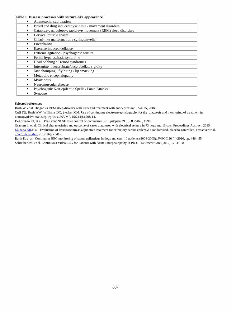
607
Table 1. Disease processes with seizure-like appearance Atlantoaxial subluxation Breed and drug induced dyskinesia / movement disorders Cataplexy, narcolepsy, rapid eye movement (REM) sleep disorders Cervical muscle spasm Chiari-like malformation / syringomyelia Encephalitis Exercise induced collapse Extreme agitation / psychogenic seizure Feline hyperesthesia syndrome Head bobbing / Tremor syndromes Intermittent decerebrate/decerebellate rigidity Jaw chomping / fly biting / lip smacking Metabolic encephalopathy Myoclonus Neuromuscular disease Psychogenic Non-epileptic Spells / Panic Attacks Syncope
Selected references Bush W, et al. Diagnosis REM sleep disorder with EEG and treatment with antidepressant, JAAHA, 2004 Cuff DE, Bush WW, Williams DC, Stecker MM. Use of continuous electroencephalography for the diagnosis and monitoring of treatment in nonconvulsive status epilepticus. JAVMA 15;244(6):708-14. DeLorenzo RJ, et al. Persistent NCSE after control of convulsive SE. Epilepsia 39 (8): 833-840, 1998 Granum L, et al. Clinical characteristics and outcome of cases diagnosed with electrical seizure in 73 dogs and 13 cats. Proceedings Abstract, 2015 Muñana KR,et al. Evaluation of levetiracetam as adjunctive treatment for refractory canine epilepsy: a randomized, placebo-controlled, crossover trial. J Vet Intern Med. 2012;26(2):341-8 Raith K, et al. Continuous EEG monitoring of status epilepticus in dogs and cats: 10 patients (2004-2005). JVECC 20 (4) 2010, pp. 446-455 Schreiber JM, et el. Continuous Video EEG for Patients with Acute Encephalopathy in PICU. Neurocrit Care (2012) 17: 31-38
608
Nonconvulsive Seizure: Clandestine, Common, and Important
William Bush, VMD, DACVIM Bush Veterinary Neurology Service
Frederick, MD
A seizure is defined as a transient occurrence of signs, symptoms or both due to abnormal excessive or synchronous neuronal activity. In veterinary medicine seizure is diagnosed via observations about the patient’s level of awareness, spontaneous muscle movements in the limbs and head as well as autonomic changes. However, a seizure is fundamentally an aberrant electrical event in the brain and therefore best diagnosed using electroencephalography (EEG). In human medicine the primary use of EEG is to distinguish epileptic seizure from psychogenic seizure, metabolic events or movement disorders. Equally important, EEG is used in seizure patients with altered mentation to detect electrical seizure and assess response to pharmacologic therapy. At our clinic, we have worked with a physician expert in the interpretation of human and animal EEG for over a decade and use EEG for these same purposes. The talk will discuss the characteristics, diagnosis, treatment and prognosis with electrical seizure and non-convulsive status epilepticus in both human and veterinary medicine. Seizure terminology An electrical seizure is defined as ictal discharges consisting of a rhythmic pattern with definitive evolution in frequency, amplitude and/or morphology persisting for at least 10 seconds. Electrical seizure can occasionally manifest as convulsions (generalized tonic-clonic) with patient flailing on its side, paddling all 4 limbs or holding the limbs, head and neck in rigid extension. A non-convulsive seizure (NCS) is defined as a seizure where there is no overt convulsive movements. Another term used for non-convulsive seizure is complex partial seizure where there is only an acute alteration in consciousness. NCS is more common than convulsive seizure in people and cats, and potentially dogs as well. When an electrical seizure lasts for more than 30 minutes it is almost always non-convulsive and termed non-convulsive status epilepticus (NCSE). Seizure frequency and status epilepticus Terms to describe seizure frequency from best to worst prognosis include: sporadic seizure, cluster seizure, acute repetitive seizure, convulsive status epilepticus (CSE), non-convulsive status epilepticus (NCSE), refractory status epilepticus (RSE), super-refractory status epilepticus, or malignant status epilepticus. A cluster seizure is 2 or more seizure within 24 hours and acute repetitive seizure is 2 or more seizure within 5-12 hours. When seizures are prolonged and without a return to baseline, they are referred to as status epilepticus (SE) with the best studied form being CSE. The original definition of SE was a 30 minute or longer continuous seizure because animal models show neuronal damage and seizure become self-sustaining after 30 minutes. However the current definition of a SE is a seizure lasting 5 minutes or longer, 2 seizures where the patient is unable to respond to commands or walk between seizures, or when patient is still seizing. This definition has evolved because clinicians no longer want to delay a patient’s entry into a protocol for SE since prompt therapy is required for a good outcome. In human medicine, about 150,000 people are thought to develop CSE per year in the USA. In dogs with idiopathic epilepsy (genetic or unknown cause) the rate of cluster seizure is reported at 41-94% and for CSE 53-69%. These conditions are common and important problems in both veterinary and human medicine. Progression from convulsive to non-convulsive status epilepticus During CSE treatment with benzodiazepines (BDZ) can be ineffective due to endocytosis and changes to the GABA receptor. In this same period of time the convulsions often stop and the electrical seizure can persist despite no obvious convulsions, which is called non-convulsive status epilepticus (NCSE) (Foreman). Refractory status epilepticus (RSE) is diagnosed when electrical seizure activity persists despite treatment with 2 anti-epileptic drugs (AEDs) at appropriate doses – refractory SE is typically non-convulsive. When RSE persists for greater than 24 hours it is super-refractory SE and when SE returns within 5 days of tapering anesthetic medications used to treat SE, then it is called malignant SE. NCSE is clandestine As our video cases will demonstrate many of our patients with NCSE appeared to be asleep or comatose. This is common too in people. A paper concludes that many people with NCSE appeared to be sleeping and that “clinical detection of NCSE would not have been possible with routine neurologic evaluations without use of EEG monitoring”. The signs of NCSE can range from mild confusion or disorientation to coma. NCSE should be suspected following a convulsive seizure when there is no improvement in 20 minutes or failure to return to baseline in 60 minutes. Distinguishing NCSE from conditions that mimic seizure can be difficult and about 25% of human patients can have subtle movements that are not from seizure. In human medicine studies have demonstrated some positive symptoms indicative of NCS/NCSE and negative symptoms indicating clinical signs are not from seizure. No such

609
published criteria exist in veterinary medicine. However, in our population of patients with NCSE we noted subtle twitching of ears or facial muscles, confusion, transient hyperthermia, episodic and unexplained changes in respiratory rates, mydriasis, or coma. NCSE frequency in seizure patients In veterinary medicine there are a few reports describing electrical seizure or NCSE following treatment of convulsive SE with diazepam and phenobarbital. In one report of ten patients (7 dogs, 3 cats) treated for CSE with anesthetic doses phenobarbital or propofol anesthesia, 100% of the patients had electrical seizure. In 164 human patients at Virginia Commonwealth University treated with their hospital’s typical protocol for CSE, EEG recording after the CSE stopped showed that 48% of the patients had persistent electrical seizure and 14% had NCSE. Another human study showed that about 33% of patients in convulsive status continue to have electrographic seizure after the convulsive seizure had stopped Generally in human medicine, NCSE is thought to be underdiagnosed and to account for about 30-40% of all SE cases. In an effort to diagnose electrical seizure and NCSE in seizure patient with seizure and an altered mentation we record the routine EEG for about 20-30 minutes, whereas in humans recordings are often done for 1-2 days via continuous monitoring. Using routine EEG we noted 4/11 (36%) of cats and 5/55 (9%) dogs were diagnosed with NCSE and 4/55 (7%) had electrical seizure. Pathophysiology with NCSE Pathology with CSE is due to both systemic effects from the muscle movement and hyperthermia and the intracranial effects of continuous neuronal firing in the brain with mortality rates of 25- 40% reported in canine patients. NCSE could be thought of as more benign than CSE since there are no obvious muscle movements or systemic effects but, on the other hand, NCSE is typically noted after CSE and represents prolonged and continuous neuronal firing in the brain. Human studies have shown that NCSE E is a statistically significant independent predictor of mortality and reduced functional independence separate from age and underlying diagnosis. In other words, when comparing age and diagnoses matched cases, those with NCSE did far worse indicating that NCSE is a pathologic and lethal process.
NCSE cause brain necrosis and facilitates more seizure. The excessive neuronal firing in NCSE generates excess glutamate mediated stimulation of the NMDA receptor, calcium influx and then neuronal cell death. A reduced seizure threshold from the kindling and mirroring phenomenon, structural and cellular reorganization of the hippocampus, selective neurodegeneration and altered cellular expression, and distribution of neurotransmitter and receptor channels can self-sustain the seizure and promote future seizure. This latter phenomenon in people is well known as one study showed 33% of refractory SE will have recurrent seizure within 5 days of tapering an anesthetic medication, a condition referred to as malignant SE (Foreman). In this talk the speaker will describe a 7 year-old Swiss Mountain dog with seizure of unknown cause that had two episodes of NCSE, 30 days apart, where necropsy showed neuronal necrosis. This case demonstrates that NCSE can cause neuronal necrosis and reduce the seizure threshold. Mortality with NCSE Human studies show that NCSE is an independent predictor of mortality, especially when there is a delay in diagnosis, or longer episodes of NCSE. Mortality rates with CSE in humans is about 30% whereas 50% is a stated mortality rate for NCSE. There are no published veterinary reports regarding the incidence or mortality rates with NCSE, however in our study 2/4 cats and 3/5 dogs or 5/9 (55%) had died within 3 months of their NCSE. Treatment of status epilepticus The endpoint for treating CSE is cessation of all motor activity, but as noted above, many of these cases will continue to have electrical seizure or NCSE. In human medicine, refractory CSE is treated with anesthetic doses of midazolam, propofol or pentobarbital. There is no clear advantage to any of these agents but surveys of physicians show a preference for barbiturate. EEG defines the endpoint of treatment which includes the reduction or elimination of epileptiform discharges (ED) and/or to establish a burst suppression pattern. Burst suppression is thought to be neuroprotective in NCSE because it hyperpolarizes at least 95% of cortical neurons and conserves on ATP. The necessary duration of burst suppression or whether it is as good or better of an endpoint that eliminating ED is debated in human medicine.
After a benzodiazepine, second line seizure treatment in humans can include Levetiracetam, Valproate and Phenytoin. Levetiracetam had variable response rates (30-79%) as a second line treatment in human NCSE. Levetiracetam has been studied for acute repetitive seizure and SE in veterinary medicine and demonstrated to be more effective than placebo. Furthermore, Levetiracetam is thought to be synergistic with benzodiazepines. Fosphenytoin has also been studied in veterinary medicine in a similar patient population also shown to be much more effective than placebo, however this drug is currently not available and also cost prohibitive. A clinical trial is being proposed in veterinary medicine to study injectable valproate compared to placebo in CSE.
610
Recommended seizure treatment In our clinic we give 1 mg/kg to 2 mg/kg of valium, IV and 60 mg/kg of Levetiracetam, IV and if patient does not improve in 10-15 minutes then a routine EEG is performed. If epileptiform discharges are noted then boluses of 10 mg/kg of phenobarbital are given until ED abate or burst suppression pattern is noted. We have noted that between 45 mg/kg and 100 mg/kg of phenobarbital is typically required to achieve these endpoints. In a veterinary report of 10 patients with electrical seizure following CSE, burst suppression and/or elimination of ED was a goal and the report concluded this treatment strategy was safe as mechanical ventilation was not required and only treatable hypotension noted as side effects. Without the benefit of EEG, the end point of therapy should be the absence of any subtle twitching of ears or facial muscles, confusion, transient hyperthermia, episodic and unexplained changes in respiratory rates, or mydriasis. Conclusion Patients presenting after a convulsive seizure or status epilepticus that also exhibit behavioral changes or altered mentation are candidates for having continued electrical seizure and NCSE. EEG is required to prove the diagnosis and ideally to guide therapy. Practically speaking, in seizure patients that remains twitchy or confused following seizure and can’t be referred for EEG, administer 60 mg/kg Levetiracetam and then phenobarbital 8-10 mg/kg boluses every 20-30 minutes to a total dose of 40 mg/kg and blood pressure support if needed. Hopefully the use of veterinary EEG will become more common so patients with CSE can be referred for prompt detection and treatment of NCSE. References DeLorenzo RJ, et al. Persistent NCSE after control of CSE. Epilepsia 1998; 39(8): 833-840 Foreman B, et al. Epilepsy Emergencies: Diagnosis and Management. Neurol Clin 2012; 30: 11-41 Patterson N. New and old drugs for CSE. ACVIM Proceedings 2014 Raith K, et al. Continuous EEG monitoring of SE in dogs and cats: 10 patients (2004 -2005). J Vet Emerg Crit Care. 2010; 20(4): 446 -455 Rueegg SJ, et al. Diagnosis and treatment of NCSE in ICU setting. Current Treatment Options in Neurology 2003; 5: 93-110 Schreiber JM, et al. Continuous Video EEG for patients with acute encephalopathy in pediatric ICU. Neuro Crit Care 2012; 17: 31-38
611
Idiopathic vs Structural Epilepsy: Clinical Guidelines for Making this Vital Distinction
William Bush, VMD, DACVIM Bush Veterinary Neurology Service
Frederick, MD
Some studies show more than 1 in 20 dogs will suffer from recurrent seizure in their lifetime. When a client presents a recent onset seizure patient they are keenly interested in the diagnosis and prognosis along with best course of action. Some cases will be of unknown or genetic cause (idiopathic) and others will have a specific (structural) cause for the seizure. The diagnostic plan, prognosis and treatment plan can be very different between dogs with an unknown cause for their seizure and dogs with a structural problem (brain tumor, encephalitis, stroke, malformation). Considering the age of onset, breed, weight, historical and neurological exam findings are crucial in estimating the likelihood that there is a structural cause for the seizure. This talk will discuss the current terminology and rational for grouping seizure by their underlying cause and frequency and then discuss how to make the distinction between structural versus idiopathic epilepsy. Epilepsy vs. reactive seizure Epilepsy generally means recurrent seizure, however in humans after just one seizure you can be considered epileptic if the seizure is associated with an enduring alteration of the brain that increases the likelihood of seizure. Reactive seizures occur when the brain is normal but reacting to an extra-cranial toxic or metabolic insult. Epilepsy terminology In 1989 the International League Against Epilepsy (ILAE) distinguished 3 etiologies of epilepsy which were then adopted in veterinary medicine. Idiopathic or primary epilepsy is diagnosed if no underlying cause can be determined other than a possible hereditary predisposition. Symptomatic epilepsy is a consequence of an identifiable brain disorder. Cryptogenic (probable symptomatic) epilepsy a heritable cause is not likely and an underlying pathologic change in the brain suspected but not proven. In 2005 these terms for epilepsy were changed by the ILAE to genetic, structural and unknown cause and now these are the terms used in published veterinary literature.
Genetic epilepsy can be diagnosed when the prevalence in a breed exceeds that of the general population. Making this distinction is important because certain breeds may have a particularly severe form of genetic epilepsy. For example in the Border Collie survival from seizure onset is 2 years with a 94% rate of cluster seizure and 53% rate of status epilepticus. Conversely genetic epilepsy in the Lagotto Ramagnolo starts at 6 weeks of age and resolves by 16 weeks of age. Structural epilepsy is diagnosed when there is a physical disruption of the brain from a malformation, infection, inflammation, stroke or brain tumor. Epilepsy of unknown cause is diagnosed when a cause for the seizure has not been determined.
Classification by seizure frequency Progression of disease and a worse prognosis is often indicated when seizure becomes more frequent. Therefore applying other terms for more frequent or longer seizures is valuable. A cluster seizure is noted there are 2 or more seizure within 24 hours and acute repetitive seizure is 2 or more seizure within 5-12 hours. Status epilepticus (SE) is present when the seizure lasts 5 minutes or longer, 2 seizures where the patient is unable to respond to commands or walk between seizure, or patient having a seizure at presentation. SE may not respond to initial treatment with Benzodiazepine, Phenobarbital and/or Levetiracetam at which time it is called refractory status epilepticus (RSE). In these cases electroencephalography (EEG) often shows continued seizure activity despite few to no physical manifestation of the seizure, a condition called non-convulsive status epilepticus (NCSE). SE and NCSE have an associated 25 and 50% mortality rate in human and veterinary medicine. Age of onset A recent study of dogs 7 or older at time of first seizure that had a MRI determined that 79% of dogs had structural epilepsy and 21% had cryptogenic epilepsy (now called seizure of unknown origin). Furthermore when the dogs were 10 or older at seizure onset there was an 87% chance of an abnormal MRI showing a structural cause for the seizure. In the dogs with structural epilepsy, 72% had a brain tumor with stroke and encephalitis being the next most common causes of seizure. At other end of spectrum, dogs younger than 6 months of age are very likely to have a genetic or seizure of unknown cause. Breed Genetic epilepsy and epilepsy of unknown cause is the most prevalent diagnosis in dogs between 6 months and 7 years of age. However, within this age group encephalitis in young dogs and prevalent in many small breeds (Pug, Chihuahua, Yorkshire terrier, Maltese, Westie, Dachshund, Minature poodle, Shih Tzu, others). Therefore in young, small breed dogs encephalitis should be highly
612
suspected as the cause of seizure, especially when seizure are clustered, progressive over a few weeks to a few months or there are examination or behavioral changes. A recent study showed a statistically higher incidence of brain tumors in the breeds Golden Retriever, Boxers, French Bulldog, Rat Terrier and Boston Terriers. Increasing age and weight were also correlated with higher rates of brain tumor. Therefore in these breeds and dogs > 15 kg, a recent onset seizure when 5 or older should raise a high suspicion for brain tumor. Behavior In dogs with seizure from structural brain disease the seizure can be the only symptom, however there are often subtle behavioral changes. When these behavioral changes are noted in a seizure patient then this should raise suspicion for a structural brain problem. These include inappropriate defecation, inappropriate urination, not greeting the owners, restless at night, sleeping more in the day, irritability, not playing, and aggression. Exam findings Seizure is generated from lesions in the forebrain or thalamus. Lesions in this area can cause patients to circle towards the side of the lesion and have contralateral menace and postural deficits. Since strength and gait are generated from the brainstem, a focal forebrain lesion would not be expected to cause weakness or ataxia. If a patient has a unilateral menace deficit with normal pupillary light responses and normal palpebral response then a contralateral forebrain mass lesion should be suspected. Similarly if the gait is normal but there is a unilateral postural deficit (paw flip test, tactile placing, hopping) then a contralateral forebrain lesion should be suspected. Lastly, while in the exam room if a patient circles to only one side then a forebrain lesion is very likely and will be located on the side towards which they are circling. In a recent study of dogs and cats where only neck pain was noted almost 10% had only a focal brain tumor. The presence of neck pain in a seizure patient should suggest there is a structural cause of the seizure. However an abnormal exam is not always noted and about 30% of patients with a mass lesion will have a normal neurological exam. Conclusion Your client expects a sense of the diagnosis, treatment plan and prognosis when they present with a pet with recent onset seizure. Prior to starting AED or/and referral for MRI and neurological consultation, you can make an accurate guess as to the diagnosis by considering age, breed, weight, historical findings and then performing a 5 minute neurological examination. Selected references De Lahunta AD, Glass E, eds. Veterinary Neuroanatomy and Clinical Neurology. 3rdedit, Saunders Elseveir, St. Loius, 2009 Monteiro R, et al. Canine idiopathic epilepsy: prevalence, risk factors and outcome associated with CS and SE. Journ Small Anim Pract 2012; 53: 526-530 Schwartz M, et al. Assessment of the prevalence and clinical features of cryptogenic epilepsy in dogs: 45 cases (2003-2011). JAVMA 2013; 242 (5): 651 Song RB, et al. Postmortem evaluation of 435 cases of intracranial neoplasia in dogs and relationship with breed, age and body weight. J Vet Intern Med 2013; 27: 1143-1152
613
What Would You Do? Valuable and Humorous Lessons from a
Veterinary Specialist and Owner Operator William Bush, VMD, DACVIM
Bush Veterinary Neurology Service Frederick, MD
In the last 10 years my core practice has grown from 1 employee to over a hundred across several locations – our core business is neurology / neurosurgery and advanced imaging (MRI, CT). Additionally, there is an Emergency and Internal Medicine practice in one location and I am part owner in the real estate in several of these locations. We have a centralized, in-house marketing, human resources, accounting, and operational management headed by a CEO that oversees the businesses. The transition from veterinarian to veterinary owner operator (VOO) to being a successful, hands-off owner has fulfilled my business vision but has also been very difficult. The goals of this talk are to share the lessons learned along the way from the innate adversity of veterinary business management and ownership.
One important realization is that the veterinary owner operator (VOO) wears many hats but is always viewed as the owner in every interaction. The VOO is under an intense, invisible spotlight and can compel and inspire their staff to be their best or implode their business. This talk will be divided into the different relationships a VOO has in a given day- VOO – manager, VOO- staff veterinarian, VOO- staff member. Although several “what would you do scenarios” will be presented – it is hoped the participants will bring their own current struggles for us to discuss as a group. Culture Defining the vision, mission, and core principles of your practice and then making all decisions centered around these core principles will set the culture. The business goal will be to have a group of like-minded people who understand and are working toward the same goal. Management’s role is to lead by example and correct and coach people when they stray from core principles. One thing I have learned is that when the culture strays at a particular location, everything gets harder and it is expensive. This can take the form of fewer office visits, inadequate follow-up with clients and primary care veterinarians, employee dissatisfaction, and employee turn-over. I have found that investing in management pays tremendous dividends and that good management is expensive but bad management is very expensive. The VOO – manager relationship Ironically, the biggest hurdle for veterinary business growth is that the VOO is very likely to be the first to stray from the stated core principles and when they do it is devastating to management. This most often takes the form of an owner changing their minds, deviating from a policy, discounting, squeezing in appointments when staff is beyond capacity, or simply not trusting that the manager has good instincts and good ideas. Because the VOO is under an invisible spotlight, any action they take that contradicts a policy or procedure will be felt by the staff 10 fold and devastating to management. This is the key moment in the VOO-manager relationship – the VOO can argue or defend their behavior or simply have the humility to apologize and genuinely try to change their behavior. One way to retain and inspire your manager is to have regularly scheduled meetings where the manager is free to speak of any recent transgressions allowing you to be more self-aware and improve. The absolute key to success as a VOO is to be self-aware and humble – otherwise you will never realize what could have been your team’s full potential. For me personally physical fitness and meditation have allowed me to be more self-aware and find opportunities daily to respond correctly instead of reacting incorrectly.
Recently we gave an award to one of our assistants for up-holding our core principle of integrity because she had the courage to tell the VOO that their behavior in the exam room was inappropriate. I believe I may have contradicted or inadvertently demeaned the assistant – my first reaction could have been to be angry, instead I responded by thinking for a moment and then correctly my behavior. The VOO – staff veterinarian relationship Most VOO have obtained their role not because they wanted to be a business owner but because they were exceptional veterinarians that ‘needed help’ to meet the demand in their area. The VOO then hires an associate and takes on the roles of owner, manager and often mentor. In this role, humility is again important and also the need to realize that a successful owner and mentor revels in the success of their employee and not their owner ability. Although counter intuitive this should take the form of refusing to see cases on their day off, working more holidays, seeing the more difficult and less lucrative cases, and genuinely wanting their associate to be successful. If you put the associate through the same trials that you remember going through in your career this will likely foster resentment or send the staff veterinarian a mixed message.
614
Similarly the VOO ability to perform a procedure efficiently, diagnose quickly and accurately, or generate genuine client satisfaction does not matter and holding this as a standard or even as a goal for the associate is unhealthy. The goal for the VOO is to downplay or minimize their ability while striving to develop the employees and celebrate their successes along the way.
One likely limitation of the staff veterinarian as compared to the VOO is the ability to connect with most clients. Often VOO understand that clients are people with animal problems meaning that clients desire a connection to their veterinarian – they want their pet’s problem resolved but they also want to be involved in the plan and feel better after the visit. Veterinary training is very systematic and often associates are generating problem lists and rule-outs plus a plan of attack instead of listening to the client. Veterinarians by nature are very systematic and this can on occasion leave some clients feeling cold or less connected. Since this ability to connect is somewhat innate and can take time to develop, the VOO will need to be careful not to expect too much from an associate. Generally recognizing and optimizing strengths of an associate while not asking them to work solely on their weaknesses will lead to the most success. VOO – staff member relationships Whether you like it or not, as a veterinarian and especially as a VOO, you are a manager. Successful management where you optimize an employee’s potential demands a good manager-employee relationship. The responsibility for maintaining and cultivating this relationship is not equally split between the employee and manager, in fact in veterinary medicine about 80% of the responsibility falls on the manager. The veterinarian will need to understand that a disapproving look or comment can be very destructive to this relationship and taking the time to teach, develop the employee skill set, and invest in the employee will inspire the employee to reach their full potential. Once this culture is present in your practice you will no longer have trouble “ finding good help”. There is a word of caution however, it is in fact “lonely at the top” and having a manager employee relationship that extends into friendship and beyond the scope of just work will dramatically limit the effectiveness of the manager and will in fact be destructive to that relationship. Therefore, if you want your employee to be successful be friendly and considerate but do not be their friend.
The last point is that the actions of the VOO are always under a magnifying glass, although this is rarely recognized by the VOO. Therefore taking time to teach, investing in an employee and giving them the benefit of the doubt will inspire your team but an expression of frustration or disappointment will devastate them and the team. On bad days this can be difficult but a successful VOO will be always mindful of this relationship and disciplined enough to not to undermine this relationship. Conclusion A successful VOO will be mindful of their various relationships and simultaneous roles or hats that they wear within the practice. Although they might think of themselves as simply “seeing a case” they are in fact setting the culture, supporting management, and being a caring owner of the practice at the same time. Being self-aware and humble will be the key to success in this relationship. References Watzman CW, Katz D. The Invisible Spotlight – Why Manager Can’t Hide. 2011 Create Space Publisher Godin S. Linchpin – Are you indispensable? 2010 Penguin Group Publishing, New York, NY
615
Upper vs Lower Motor Neuron: Never Miss Again after this Videocase Presentation
William Bush, VMD, DACVIM Bush Veterinary Neurology Service
Frederick, MD
The ability to examine a patient and determine where in the body the disease is located is critical to determining the cause, best therapy and prognosis. Weakness is a common presenting complaint and can result from spinal cord (upper motor neuron - UMN) or nerve and muscle disease (lower motor neuron-LMN). Understanding the function of the UMN and LMN system will enhance the accuracy of lesion localization and therefore determination of the diagnostic testing and prognosis. This talk will discuss the function of the UMN and LMN system and then how to assess these systems via examination of a patient’s gait, postural responses, and reflex testing. Secondly we will discuss assessing the cutaneous trunci reflex and for focal pain to assist in lesion localization and honing the list of possible the spinal cord diseases. Lastly we will discuss evaluating the palpebral response and laryngeal / pharyngeal / esophageal function in LMN disease. Tonic gamma loop mechanism Muscle tone refers to the intrinsic tension of the muscle when supporting the body against gravity while standing, shifting weight from limb to limb, jumping or performing stairs – but how does this occur? The tonic gamma loop mechanism (TGLM) is intrinsic to the LMN system and understanding TGLM physiology offers insight into how gait is generated and why UMN or LMN disease cause alterations in muscle tone and reflex. When we jump down from a height and our knees start to bend or buckle there is a sudden stretch of the quadriceps muscles and stimulation of stretch receptors (neuromuscular spindle) within these muscles. These receptors then stimulate the sensory portion of the femoral nerve which then directly synapse and stimulates the motor portion (alpha motor neuron) of the femoral nerve. This causes the quadriceps muscle (which is innervated by the femoral nerve) to contract and prevent your knees from buckling. When the patella tendon is artificially simulated with a reflex hammer it fools the body into thinking there is a sudden heavy load on the quadriceps muscle (like jumping down), the TGLM is stimulated and the knee jerks. Gait generation As mentioned above, when information from the neuromuscular spindle of the TGLM returns to the spinal cord it directly synapses on the alpha motor neuron or motor portion of the same muscle in which the spindle is located. However, the information also stimulates inhibitory interneurons that then reduce activity or tone in the antagonistic muscle group. Therefore when there is contraction of the quadriceps there will be reduced tone in the hamstring or flexor group. The modulation of flexion is performed by the phasic gamma loop or flexor gammas loop. The UMN system acts on the tonic and then phasic gamma loop mechanisms to generate extension and then flexion of the leg by activating this intrinsic reflex mechanisms and therefore generate gait. UMN lesions influence the TGLM In our example above, gravity lengthen the quadriceps muscle and stretches the neuromuscular spindle, which then via a direct synaptic connection, stimulates the femoral nerve. This causes contraction of the quadriceps muscle which then causes more stretching of the neuromuscular spindle and more contraction of the quadriceps muscle. This system, if not modulated would lead to dramatic increases in muscle tone and reflex. The UMN system modulates or controls the TGLM and therefore controls muscle tone and reflex. Disease of the UMN lesion can cause increased tone and reflex. Examination of dogs with UMN spinal cord disease often reveals increased tone because there is resistance to flexion of the stifle. This stiffness can also manifest in the protraction phase of the gait and appear as swinging out of the limb (circumduction) or a long-strided gait. Furthermore, brainstem lesion (where the UMN tracts start) can lead to opisthotonus also known as decerebrate rigidity where the head, neck and limbs are held in rigid extension. Reflex testing A reflex is something that occurs automatically or spontaneously without influence from the cerebrum whereas a reaction or response requires the unconscious participation of the cerebrum. In reflex testing there is a sensory stimulus that runs into the spinal cord or brainstem and then an immediate spinal cord or brainstem mediated response. For example, stretching the patella tendon with a pleximeter (reflex hammer) causes a sudden, intense stimulation of the stretch receptors within the femoral nerve, in essence simulating what would happen if we jumped down from a large height. Immediately the muscles innervated by the femoral nerve contact and the knee jerks. An absence of reflex often means there is a lesion of the motor or sensory portion of the femoral nerve or severe disease of the quadriceps muscle. If there is an increase in reflex (exaggerated, clonus) then there is a failure of UMN system to control this reflex.
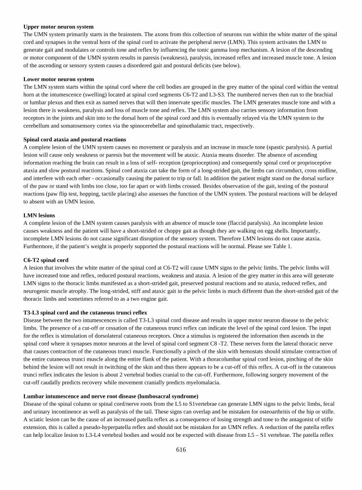
616
Upper motor neuron system The UMN system primarily starts in the brainstem. The axons from this collection of neurons run within the white matter of the spinal cord and synapses in the ventral horn of the spinal cord to activate the peripheral nerve (LMN). This system activates the LMN to generate gait and modulates or controls tone and reflex by influencing the tonic gamma loop mechanism. A lesion of the descending or motor component of the UMN system results in paresis (weakness), paralysis, increased reflex and increased muscle tone. A lesion of the ascending or sensory system causes a disordered gait and postural deficits (see below). Lower motor neuron system The LMN system starts within the spinal cord where the cell bodies are grouped in the grey matter of the spinal cord within the ventral horn at the intumescence (swelling) located at spinal cord segments C6-T2 and L3-S3. The numbered nerves then run to the brachial or lumbar plexus and then exit as named nerves that will then innervate specific muscles. The LMN generates muscle tone and with a lesion there is weakness, paralysis and loss of muscle tone and reflex. The LMN system also carries sensory information from receptors in the joints and skin into to the dorsal horn of the spinal cord and this is eventually relayed via the UMN system to the cerebellum and somatosensory cortex via the spinocerebellar and spinothalamic tract, respectively. Spinal cord ataxia and postural reactions A complete lesion of the UMN system causes no movement or paralysis and an increase in muscle tone (spastic paralysis). A partial lesion will cause only weakness or paresis but the movement will be ataxic. Ataxia means disorder. The absence of ascending information reaching the brain can result in a loss of self- reception (proprioception) and consequently spinal cord or proprioceptive ataxia and slow postural reactions. Spinal cord ataxia can take the form of a long-strided gait, the limbs can circumduct, cross midline, and interfere with each other - occasionally causing the patient to trip or fall. In addition the patient might stand on the dorsal surface of the paw or stand with limbs too close, too far apart or with limbs crossed. Besides observation of the gait, testing of the postural reactions (paw flip test, hopping, tactile placing) also assesses the function of the UMN system. The postural reactions will be delayed to absent with an UMN lesion. LMN lesions A complete lesion of the LMN system causes paralysis with an absence of muscle tone (flaccid paralysis). An incomplete lesion causes weakness and the patient will have a short-strided or choppy gait as though they are walking on egg shells. Importantly, incomplete LMN lesions do not cause significant disruption of the sensory system. Therefore LMN lesions do not cause ataxia. Furthermore, if the patient’s weight is properly supported the postural reactions will be normal. Please see Table 1. C6-T2 spinal cord A lesion that involves the white matter of the spinal cord at C6-T2 will cause UMN signs to the pelvic limbs. The pelvic limbs will have increased tone and reflex, reduced postural reactions, weakness and ataxia. A lesion of the grey matter in this area will generate LMN signs to the thoracic limbs manifested as a short-strided gait, preserved postural reactions and no ataxia, reduced reflex, and neurogenic muscle atrophy. The long-strided, stiff and ataxic gait in the pelvic limbs is much different than the short-strided gait of the thoracic limbs and sometimes referred to as a two engine gait. T3-L3 spinal cord and the cutaneous trunci reflex Disease between the two intumescences is called T3-L3 spinal cord disease and results in upper motor neuron disease to the pelvic limbs. The presence of a cut-off or cessation of the cutaneous trunci reflex can indicate the level of the spinal cord lesion. The input for the reflex is stimulation of dorsolateral cutaneous receptors. Once a stimulus is registered the information then ascends in the spinal cord where it synapses motor neurons at the level of spinal cord segment C8 -T2. These nerves form the lateral thoracic nerve that causes contraction of the cutaneous trunci muscle. Functionally a pinch of the skin with hemostats should stimulate contraction of the entire cutaneous trunci muscle along the entire flank of the patient. With a thoracolumbar spinal cord lesion, pinching of the skin behind the lesion will not result in twitching of the skin and thus there appears to be a cut-off of this reflex. A cut-off in the cutaneous trunci reflex indicates the lesion is about 2 vertebral bodies cranial to the cut-off. Furthermore, following surgery movement of the cut-off caudally predicts recovery while movement cranially predicts myelomalacia. Lumbar intumescence and nerve root disease (lumbosacral syndrome) Disease of the spinal column or spinal cord/nerve roots from the L5 to S1vertebrae can generate LMN signs to the pelvic limbs, fecal and urinary incontinence as well as paralysis of the tail. These signs can overlap and be mistaken for osteoarthritis of the hip or stifle. A sciatic lesion can be the cause of an increased patella reflex as a consequence of losing strength and tone to the antagonist of stifle extension, this is called a pseudo-hyperpatella reflex and should not be mistaken for an UMN reflex. A reduction of the patella reflex can help localize lesion to L3-L4 vertebral bodies and would not be expected with disease from L5 – S1 vertebrae. The patella reflex
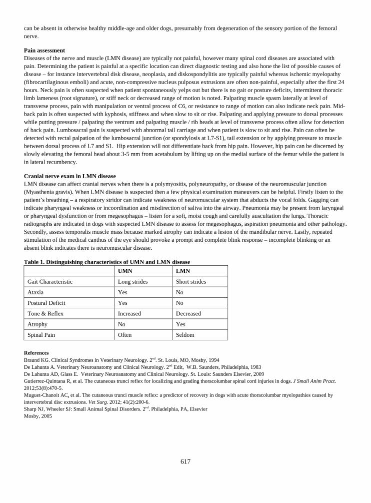
617
can be absent in otherwise healthy middle-age and older dogs, presumably from degeneration of the sensory portion of the femoral nerve. Pain assessment Diseases of the nerve and muscle (LMN disease) are typically not painful, however many spinal cord diseases are associated with pain. Determining the patient is painful at a specific location can direct diagnostic testing and also hone the list of possible causes of disease – for instance intervertebral disk disease, neoplasia, and diskospondylitis are typically painful whereas ischemic myelopathy (fibrocartilaginous emboli) and acute, non-compressive nucleus pulposus extrusions are often non-painful, especially after the first 24 hours. Neck pain is often suspected when patient spontaneously yelps out but there is no gait or posture deficits, intermittent thoracic limb lameness (root signature), or stiff neck or decreased range of motion is noted. Palpating muscle spasm laterally at level of transverse process, pain with manipulation or ventral process of C6, or resistance to range of motion can also indicate neck pain. Mid-back pain is often suspected with kyphosis, stiffness and when slow to sit or rise. Palpating and applying pressure to dorsal processes while putting pressure / palpating the ventrum and palpating muscle / rib heads at level of transverse process often allow for detection of back pain. Lumbosacral pain is suspected with abnormal tail carriage and when patient is slow to sit and rise. Pain can often be detected with rectal palpation of the lumbosacral junction (or spondylosis at L7-S1), tail extension or by applying pressure to muscle between dorsal process of L7 and S1. Hip extension will not differentiate back from hip pain. However, hip pain can be discerned by slowly elevating the femoral head about 3-5 mm from acetabulum by lifting up on the medial surface of the femur while the patient is in lateral recumbency. Cranial nerve exam in LMN disease LMN disease can affect cranial nerves when there is a polymyositis, polyneuropathy, or disease of the neuromuscular junction (Myasthenia gravis). When LMN disease is suspected then a few physical examination maneuvers can be helpful. Firstly listen to the patient’s breathing – a respiratory stridor can indicate weakness of neuromuscular system that abducts the vocal folds. Gagging can indicate pharyngeal weakness or incoordination and misdirection of saliva into the airway. Pneumonia may be present from laryngeal or pharyngeal dysfunction or from megesophagus – listen for a soft, moist cough and carefully auscultation the lungs. Thoracic radiographs are indicated in dogs with suspected LMN disease to assess for megesophagus, aspiration pneumonia and other pathology. Secondly, assess temporalis muscle mass because marked atrophy can indicate a lesion of the mandibular nerve. Lastly, repeated stimulation of the medical canthus of the eye should provoke a prompt and complete blink response – incomplete blinking or an absent blink indicates there is neuromuscular disease. Table 1. Distinguishing characteristics of UMN and LMN disease
UMN LMN
Gait Characteristic Long strides Short strides
Ataxia Yes No
Postural Deficit Yes No
Tone & Reflex Increased Decreased
Atrophy No Yes
Spinal Pain Often Seldom References Braund KG. Clinical Syndromes in Veterinary Neurology. 2ed. St. Louis, MO, Mosby, 1994 De Lahunta A. Veterinary Neuroanatomy and Clinical Neurology. 2nd Edit, W.B. Saunders, Philadelphia, 1983 De Lahunta AD, Glass E. Veterinary Neuroanatomy and Clinical Neurology. St. Louis: Saunders Elsevier, 2009 Gutierrez-Quintana R, et al. The cutaneous trunci reflex for localizing and grading thoracolumbar spinal cord injuries in dogs. J Small Anim Pract. 2012;53(8):470-5. Muguet-Chanoit AC, et al. The cutaneous trunci muscle reflex: a predictor of recovery in dogs with acute thoracolumbar myelopathies caused by intervertebral disc extrusions. Vet Surg. 2012; 41(2):200-6. Sharp NJ, Wheeler SJ: Small Animal Spinal Disorders. 2ed. Philadelphia, PA, Elsevier Mosby, 2005
618
Diagnosing and Treating the Five Disk Diseases: Why is MRI so Important?
William Bush, VMD, DACVIM Bush Veterinary Neurology Service
Frederick, MD
The spinal cord connects the brain to the neuromuscular system which is responsible for locomotion and respiration. The spinal cord is protected by a long bony box that is segmented to allow for improved axial movements. The segments of the bony box (vertebrae) are cushioned by the intervertebral disk. The intervertebral disk (IVD) contains a jello-like substance called nucleus pulposus (embryonic notochord) that is wrapped by concentric layers of ligamentous material (annulus fibrosus). The IVD is located below and adjacent to the spinal cord and pathology of the IVD puts the spinal cord at risk for infection, infarction, and compression all of which can lead weakness, paralysis, ataxia, pain, and incontinence. Role of magnetic resonance imaging (MRI) MRI characteristics are based on the proton or water content of different tissue allowing for detection of even mild pathology in the soft tissue and to a lesser extent bone. Because MRI directly images the spinal cord it is superior to X-ray based technology like radiography, myelography and CT scanning. MRI consistently demonstrates superior accuracy in the diagnosis of spinal cord disease. Additionally, MRI is better at predicting outcome than assessment of neurological function at presentation. MRI is currently essential to the practice of high quality veterinary neurology. Hansen type I disk disease Hansen Type I disease occurs when the degenerated annulus fibrosus loses tensile strength and tears allowing the degenerated, dehydrated nucleus pulposus to extrude or herniate and co-occupy the spinal canal with the spinal cord. The stretching of the meninges, nerve root and annulus fibrosus tear generating pain and the spinal cord compression can cause weakness, paralysis and incontinence. Prognosis and the choice of medical vs. surgical management is made by combining information about the neurological grade and MRI findings. This disease is prevalent in 3-6 year-old dachshunds and many other small breed dogs and 6-9 year-old large breed dogs like the German Shepherd, Rottweiler, Dalmatians and mixed-breeds. Type I disk disease has a very good to excellent prognosis but surgery is often required when there is persistent pain and/or weakness with significant spinal cord compression. Remarkably, in paraplegic dogs with Type I disease that have hemilaminectomy, MRI is a greater predictor of outcome than nociceptive or deep pain status. Hansen type II disease Hansen Type II disease is present when there are micro-tears and bulging of the annulus fibrosus and compression of the spinal cord, meninges and nerve roots. The disease is most common in larger breed dogs in the low cervical spine and lumbosacral junction. In the low cervical spine, Type II disk extrusion is an important factor in cervical spondylomyelopathy and is also known by disk associated wobbler’s syndrome (DAWS). In DAWS the outcome with surgery is better than with medical management although there is no significant difference in life expectancy with these 2 treatments. Lumbosacral disk disease can mimic hip or stifle disease but unlike these other conditions, it can lead to urinary or fecal incontinence. Success rates with surgery are generally with LS surgery unless incontinence is already present. Therefore the distinction between lumbosacral Type II disk extrusion and orthopedic disease is an important. Nerve pain (and not orthopedic disease) can be distinguished from orthopedic disease via palpation of lateral muscles just cranial and lateral to the wings of the ilium, between the L7 and S1 dorsal spinous process, ventral surface of L7 and S1 via rectal, or with elevation of the tail. Pain with hip extension can indicate nerve compression or joint pain. However, hip pain can be discerned by slowly elevating the femoral head about 3-5 mm from acetabulum by lifting up on the medial surface of the femur while the patient is laterally recumbent Acute non-compressive nucleus pulposus extrusion (ANNPE) ANNPE is sometimes referred to as Type III disk disease or low volume high velocity disk extrusion. In this disease a small amount of nucleus pulposus (NP) ruptures at a high velocity through a small tear in the dorsal annulus fibrosus leading to edema, malacia, and/or hemorrhage of the spinal cord and epidural fat but minimal to no compression of the spinal cord. ANNPE has a peracute onset and associated with activity or a traumatic event and seen more commonly in medium to large breed, male dogs, especially Labrador retrievers and mixed breeds. About 2/3 of the patients in one study returned to walking and when the T2 weighted cross sectional MRI showed less than 90% of the spinal cord to be affected, 93% of the dogs regained function. Similar to Type I disk disease, the prognosis is more strongly correlated to MRI findings than admission neurological grade or nociceptive status. Many patients have difficulty urinating over the short-term and are treated with phenoxybenzamine or prazosin and diazepam to reduce smooth and skeletal muscle tone, respectively. Urinary catheterization can be a useful way of managing these patients in the first 2-3 days while
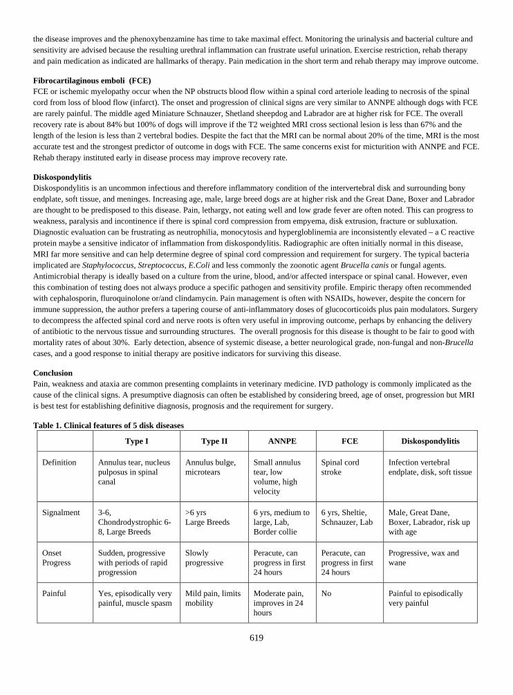
619
the disease improves and the phenoxybenzamine has time to take maximal effect. Monitoring the urinalysis and bacterial culture and sensitivity are advised because the resulting urethral inflammation can frustrate useful urination. Exercise restriction, rehab therapy and pain medication as indicated are hallmarks of therapy. Pain medication in the short term and rehab therapy may improve outcome. Fibrocartilaginous emboli (FCE) FCE or ischemic myelopathy occur when the NP obstructs blood flow within a spinal cord arteriole leading to necrosis of the spinal cord from loss of blood flow (infarct). The onset and progression of clinical signs are very similar to ANNPE although dogs with FCE are rarely painful. The middle aged Miniature Schnauzer, Shetland sheepdog and Labrador are at higher risk for FCE. The overall recovery rate is about 84% but 100% of dogs will improve if the T2 weighted MRI cross sectional lesion is less than 67% and the length of the lesion is less than 2 vertebral bodies. Despite the fact that the MRI can be normal about 20% of the time, MRI is the most accurate test and the strongest predictor of outcome in dogs with FCE. The same concerns exist for micturition with ANNPE and FCE. Rehab therapy instituted early in disease process may improve recovery rate. Diskospondylitis Diskospondylitis is an uncommon infectious and therefore inflammatory condition of the intervertebral disk and surrounding bony endplate, soft tissue, and meninges. Increasing age, male, large breed dogs are at higher risk and the Great Dane, Boxer and Labrador are thought to be predisposed to this disease. Pain, lethargy, not eating well and low grade fever are often noted. This can progress to weakness, paralysis and incontinence if there is spinal cord compression from empyema, disk extrusion, fracture or subluxation. Diagnostic evaluation can be frustrating as neutrophilia, monocytosis and hypergloblinemia are inconsistently elevated – a C reactive protein maybe a sensitive indicator of inflammation from diskospondylitis. Radiographic are often initially normal in this disease, MRI far more sensitive and can help determine degree of spinal cord compression and requirement for surgery. The typical bacteria implicated are Staphylococcus, Streptococcus, E.Coli and less commonly the zoonotic agent Brucella canis or fungal agents. Antimicrobial therapy is ideally based on a culture from the urine, blood, and/or affected interspace or spinal canal. However, even this combination of testing does not always produce a specific pathogen and sensitivity profile. Empiric therapy often recommended with cephalosporin, fluroquinolone or/and clindamycin. Pain management is often with NSAIDs, however, despite the concern for immune suppression, the author prefers a tapering course of anti-inflammatory doses of glucocorticoids plus pain modulators. Surgery to decompress the affected spinal cord and nerve roots is often very useful in improving outcome, perhaps by enhancing the delivery of antibiotic to the nervous tissue and surrounding structures. The overall prognosis for this disease is thought to be fair to good with mortality rates of about 30%. Early detection, absence of systemic disease, a better neurological grade, non-fungal and non-Brucella cases, and a good response to initial therapy are positive indicators for surviving this disease. Conclusion Pain, weakness and ataxia are common presenting complaints in veterinary medicine. IVD pathology is commonly implicated as the cause of the clinical signs. A presumptive diagnosis can often be established by considering breed, age of onset, progression but MRI is best test for establishing definitive diagnosis, prognosis and the requirement for surgery. Table 1. Clinical features of 5 disk diseases
Type I Type II ANNPE FCE Diskospondylitis
Definition Annulus tear, nucleus pulposus in spinal canal
Annulus bulge, microtears
Small annulus tear, low volume, high velocity
Spinal cord stroke
Infection vertebral endplate, disk, soft tissue
Signalment
3-6, Chondrodystrophic 6-8, Large Breeds
>6 yrs Large Breeds
6 yrs, medium to large, Lab, Border collie
6 yrs, Sheltie, Schnauzer, Lab
Male, Great Dane, Boxer, Labrador, risk up with age
Onset Progress
Sudden, progressive with periods of rapid progression
Slowly progressive
Peracute, can progress in first 24 hours
Peracute, can progress in first 24 hours
Progressive, wax and wane
Painful Yes, episodically very painful, muscle spasm
Mild pain, limits mobility
Moderate pain, improves in 24 hours
No Painful to episodically very painful
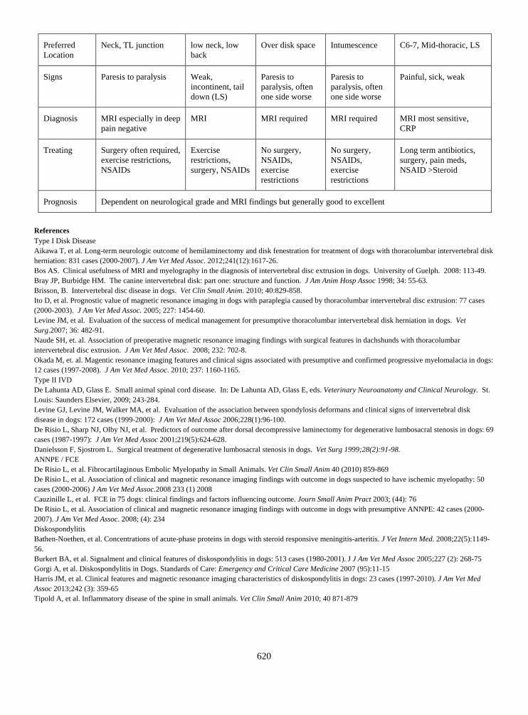
620
Preferred Location
Neck, TL junction low neck, low back
Over disk space Intumescence C6-7, Mid-thoracic, LS
Signs Paresis to paralysis Weak, incontinent, tail down (LS)
Paresis to paralysis, often one side worse
Paresis to paralysis, often one side worse
Painful, sick, weak
Diagnosis MRI especially in deep pain negative
MRI MRI required MRI required MRI most sensitive, CRP
Treating Surgery often required, exercise restrictions, NSAIDs
Exercise restrictions, surgery, NSAIDs
No surgery, NSAIDs, exercise restrictions
No surgery, NSAIDs, exercise restrictions
Long term antibiotics, surgery, pain meds, NSAID >Steroid
Prognosis Dependent on neurological grade and MRI findings but generally good to excellent
References Type I Disk Disease Aikawa T, et al. Long-term neurologic outcome of hemilaminectomy and disk fenestration for treatment of dogs with thoracolumbar intervertebral disk herniation: 831 cases (2000-2007). J Am Vet Med Assoc. 2012;241(12):1617-26. Bos AS. Clinical usefulness of MRI and myelography in the diagnosis of intervertebral disc extrusion in dogs. University of Guelph. 2008: 113-49. Bray JP, Burbidge HM. The canine intervertebral disk: part one: structure and function. J Am Anim Hosp Assoc 1998; 34: 55-63. Brisson, B. Intervertebral disc disease in dogs. Vet Clin Small Anim. 2010; 40:829-858. Ito D, et al. Prognostic value of magnetic resonance imaging in dogs with paraplegia caused by thoracolumbar intervertebral disc extrusion: 77 cases (2000-2003). J Am Vet Med Assoc. 2005; 227: 1454-60. Levine JM, et al. Evaluation of the success of medical management for presumptive thoracolumbar intervertebral disk herniation in dogs. Vet Surg.2007; 36: 482-91. Naude SH, et. al. Association of preoperative magnetic resonance imaging findings with surgical features in dachshunds with thoracolumbar intervertebral disc extrusion. J Am Vet Med Assoc. 2008; 232: 702-8. Okada M, et. al. Magentic resonance imaging features and clinical signs associated with presumptive and confirmed progressive myelomalacia in dogs: 12 cases (1997-2008). J Am Vet Med Assoc. 2010; 237: 1160-1165. Type II IVD De Lahunta AD, Glass E. Small animal spinal cord disease. In: De Lahunta AD, Glass E, eds. Veterinary Neuroanatomy and Clinical Neurology. St. Louis: Saunders Elsevier, 2009; 243-284. Levine GJ, Levine JM, Walker MA, et al. Evaluation of the association between spondylosis deformans and clinical signs of intervertebral disk disease in dogs: 172 cases (1999-2000): J Am Vet Med Assoc 2006;228(1):96-100. De Risio L, Sharp NJ, Olby NJ, et al. Predictors of outcome after dorsal decompressive laminectomy for degenerative lumbosacral stenosis in dogs: 69 cases (1987-1997): J Am Vet Med Assoc 2001;219(5):624-628. Danielsson F, Sjostrom L. Surgical treatment of degenerative lumbosacral stenosis in dogs. Vet Surg 1999;28(2):91-98. ANNPE / FCE De Risio L, et al. Fibrocartilaginous Embolic Myelopathy in Small Animals. Vet Clin Small Anim 40 (2010) 859-869 De Risio L, et al. Association of clinical and magnetic resonance imaging findings with outcome in dogs suspected to have ischemic myelopathy: 50 cases (2000-2006) J Am Vet Med Assoc.2008 233 (1) 2008 Cauzinille L, et al. FCE in 75 dogs: clinical findings and factors influencing outcome. Journ Small Anim Pract 2003; (44): 76 De Risio L, et al. Association of clinical and magnetic resonance imaging findings with outcome in dogs with presumptive ANNPE: 42 cases (2000-2007). J Am Vet Med Assoc. 2008; (4): 234 Diskospondylitis Bathen-Noethen, et al. Concentrations of acute-phase proteins in dogs with steroid responsive meningitis-arteritis. J Vet Intern Med. 2008;22(5):1149-56. Burkert BA, et al. Signalment and clinical features of diskospondylitis in dogs: 513 cases (1980-2001). J J Am Vet Med Assoc 2005;227 (2): 268-75 Gorgi A, et al. Diskospondylitis in Dogs. Standards of Care: Emergency and Critical Care Medicine 2007 (95):11-15 Harris JM, et al. Clinical features and magnetic resonance imaging characteristics of diskospondylitis in dogs: 23 cases (1997-2010). J Am Vet Med Assoc 2013;242 (3): 359-65 Tipold A, et al. Inflammatory disease of the spine in small animals. Vet Clin Small Anim 2010; 40 871-879
621
Common and Deadly: Recognizing and Treating Inflammatory Disease of the
Brain, Spinal Cord, and Meninges William Bush, VMD, DACVIM
Bush Veterinary Neurology Service Frederick, MD
When there are inflammatory cells within the brain, spinal cord or meninges then the terms encephalitis, myelitis, and meningitis are used, respectively. When inflammation is in more than one area the terms are combined like with meningoencephalomyelitis. The inflammation in these cases if most often from a non-infectious, unknown etiology and therefore called meningoencephalomyelitis of unknown etiology (MUE). The signs of the disease are specific to the location of the inflammation and most cases respond well to aggressive immune suppression. This talk discusses the terminology, definition / histopathology, common presentations, treatment and prognosis for different manifestations of non-infectious inflammatory disease within the nervous system Terminology MUE is an umbrella term for necrotizing encephalitis (NE) and granulomatous meningoencephalomyelitis (GME). Necrotizing encephalitis implies death of neurons within the brain from inflammation and is further subdivided into Pug dog encephalitis or necrotizing meningoencephalitis (NME) and Yorkshire terrier encephalitis or necrotizing leukoencephalitis (NLE). MUE has replaced these terms since multiple breeds have been identified with these disease and the prognosis, testing and treatment protocols are similar. Steroid responsive meningitis-arteritis (SRMA) is another non-infectious inflammatory disease that typically involves only the meninges – this disease will be discussed separately at end of this talk. Definitions and signalments NLE was first described in the Yorkshire terrier. NLE is a progressive disease with an acute or chronic onset where there is necrosis of the white matter that with time, can coalesce into cavities or holes in the brain. The grey matter and spinal cord are spared in this disease. The other form of NE, NME first described in the Pug dog has since been noted in many other small breeds like the Maltese, Brussels Griffon, Colon du Tulear, Shih Tzu, and Papillion. NME is typically an acute onset and rapidly progressive disease of the both the grey and white matter of the cerebrum, with only 25 % of cases showing any multifocal or brainstem signs. Because the cerebrum is so commonly affected, seizure is common clinical sign - 94% of Pugs with this disease have seizure. GME is common and may account for up to 25% of canine CNS disease – it is an acute onset, progressive and potentially fatal disease. Unlike NE, the disease can manifest in the cerebrum, brainstem and spinal cord – 8 % of all cases present with only spinal cord signs. Histopathologically GME is noted most often in the white matter as perivascular infiltrates of rounds cells (plasma cells, lymphocytes and occasionally lymphoblasts) - these can coalesce to form tumors (unlike NE where the lesions coalesce to form cavities within the brain) (SJSR). Female, small breed dogs like the Miniature poodle, Maltese, Dachshund, Westie, and Chihuahua are commonly affected. Most dogs with GME are 4-8 years of age, whereas with NE most dogs are under 4 years of age. The take home point is that MUE should be suspected in small breed under 8 years of age with acute onset of brain and less commonly spinal cord signs. Signs of disease with MUE The signs of disease are specific to the region of the brain that is involved. Most cases of MUE presents with multifocal clinical representing a mixture of forebrain and brainstem signs which can include altered mentation, visual deficits, central vestibular signs, proprioceptive placing and hopping deficits and seizures. In one report, 8% of cases has only spinal cord signs (weakness, paralysis, ataxia). What is the cause of MUE? The causes of MUE is thought to be from a genetic predisposition coupled with environmental exposures leading to a pathologic immune response. For instance, the histopathological differences in NE may result from minor differences among breeds, modifying genes, or variations in antigenic exposure. Breed predispositions indicate there is a heritable component to development of MUE. In the Pug, heritability has been proven and a strong association demonstrated between affected dogs with single nucleotide polymorphism within the dog’s leucocyte antigen (DLA) complex II region located on chromosome 12. The authors point out that this same association is made in human multiple sclerosis (MS) patients and that NME in the Pug dog maybe a good model for the less common acute variant forms of MS. Recent work in Maltese with NME show risk loci on chromosome 4 and 15.
MUE has been associated with viral diseases like Borna virus, West Nile, Canine parainfluenza, and Encephalomyocarditis virus, Canine herpes virus-1, Parvovirus, Porcine herpes virus-1, Bunya- and Polyomaviruses. Additionally, DNA from E. Coli, Mycoplasma canis. and Bartonella vinsonii subsp berkhoffii have been identified in sporadic cases of MUE and a recent report shows DNA from Anaplama phagocytephilum in 4/23 cases SRMA. These pathogens are not thought to be direct cause of the disease but according to
622
the “Hit-and-Run Hypothesis” work in tandem with genetic and other environmental factors (vaccination?) to generate an autoimmune response, perhaps through molecular mimicry.
Autoimmune disease is likely in MUE because the CSF and serum of dogs with MUE contain an anti-astrocyte autoantibodies against glial fibrillary acidic protein (GFAP) which is an intermediate filament protein important in astrocyte function. Recent work has shown that the active cellular proliferation is thought to occur within the CNS lesion (and not from a migration from outside the CNS) and is assisted by matrix metalloprotineases (MMPs). MMPs are enzymes necessary for migration of leukocytes into the CNS or CSF and MMP-9 is elevated in some dogs with MUE. Other work by Dr. Mariani has also shown elevations in many interleukins necessary for lymphocyte proliferation and trafficking into tissue. However, to date there is no useful serum or CSF biomarker to assist in the diagnosis or treatment of MUE.
Lastly, since some cases of MUE lesions contain small amount of lymphoblasts and some are truly shown to be lymphoma at the time of histopathology, it is theorized that MUE is a lymphoproliferative disorder with features of both inflammation and neoplasia. Further support for this claim is the marked clinical responses of certain cases to chemotherapy. MUE diagnosis An MUE diagnosis is based on clinical suspicion from the signalment and disease progression, and then MRI, CSF and infectious disease testing. It can be difficult ruling-out infection because of inaccurate test results and the fact that there are not tests for all known pathogens. For example, we had a suspected MUE whose necropsy revealed a high burden of an unknown protozoal agent. Complicating things further is not all cases will have an abnormal MRI and between 12-25% of MUE cases will have normal CSF analysis. In cervical spinal cord MUE, MRI of the paraspinal cervical muscles with STIR sequence in MUE is often abnormal (78% sensitivity) and rarely abnormal in normal controls (92% specificity) – because CSF results can be normal in cases of spinal cord MUE about 10% of the time, this sequence is important in suspected cases of cervical and maybe intracranial MUE. Pursing infectious etiology When the CSF is abnormal in a MUE cases, less than 10% of cases will have a predominantly neutrophilic CSF analysis. Therefore a neutrophilic pleocytosis should alert clinician to a possible infection rather than MUE. Typical testing when searching for infection could include PCR, serology and rarely cultures for protozoal, rickettsial, fungal, bacterial, and viral diseases. In the Mid-Atlantic region of the USA, we typically test the CSF via PCR for distemper virus, serology for Toxoplasmosis gondi, Neospora caninum and potentially Sarcocystis neurona, and antigen testing for Cryptoccocus sp. as well as whole blood PCR testing for vector borne disease. Failure to improve while on antibiotics or a relapse of signs when prednisone is reduced while on antibiotic therapy is often the last step in ruling-out infection and committing to multimodal immune suppressive therapy (see below). Brain biopsy has been reported and occasionally performed in our clinic however, the procedure has risks, costs, may yield false negative or positive results and may not change the course of treatment. A recent paper describing needle guided brain biopsy had 82% of cases achieved a specific diagnosis with a 6% indirect mortality rate and 29% incidence of transient side effects (stupor, seizure, weakness and loss of proprioception). MUE treatment Initial testing often reveals inflammation but does not clearly delineate between non-infectious and infectious inflammation. To address a possible infection antimicrobial therapy (clindamycin 15 mg/kg, BID, minocycline 10 mg/kg, BID +/- Fluconazole 10 mg/kg, BID) if often started while waiting for infectious disease test results. Prednisone 0.5 mg/kg, BID is also started and if signs are progressive and severe additional immune suppression could be considered with chemotherapy (Cytosine arabinoside, Lomustine, Procarbazine) and/or immune modulation with (Cyclosporine and less commonly Leflunomide, Azathioprine, or Mycophenolate). Radiation therapy has also been reported to have a positive influence of the disease course with MUE. There are many important and unanswered clinical questions revolving around what is best immunosuppressive protocol and when it is advised to stop therapy. Steroid alone are insufficient In a meta-analysis of MUE cases the median survival for dogs treated with corticosteroid plus any other immune suppressant protocol ranged from 240 to 590 days (n=96) compared with corticosteroid alone where range of median survival was 28 to 357 days ( n= 43). A recent retrospective study evaluating different glucocorticoid protocols (no other immune therapy) showed survival times ranging from 2 to 2065 days – and the authors concluded that an 18 week schedule of sole prednisone therapy can be used to treat MUE. However, multiple other authors conclude that treating with immune suppressants other than prednisone will improve control of the immune condition, improve survival times, and improve quality of life for the patient by reducing steroid associated side-effects (polydipsia, polyuria, polyphagia, muscle loss, urinary tract infection, hepatotoxicity, etc.). However, which immune suppressive protocol is best in not known and there is a desperate need for randomized, blinded, controlled, prospective study of MUE to assess current and future therapies that could include (intravenous immunoglobulin, plasma exchange and even anti-viral therapy).
623
Once remission or improvement is achieved it can be difficult to know when to taper steroid and other immune suppressive therapy. In our experience, tapering medication can lead to relapses with poor outcomes in dogs that had a normal neurological exam. A recent paper showed that follow-up CSF analysis at 3 months can predict relapse and that tapering medication in dogs with an abnormal MRI always lead to relapse. Repeating these tests when they were previously abnormal is advised prior to the tapering or elimination of immune suppressive therapy. Prognosis Comparing studies is difficult due to different inclusion criteria, therapy, treatment endpoints, and lack of a prospective, controlled study. There is a recent prospective study of 39 MUE dogs treated with prednisone and then Cytosine arabinoside that provides insight into the prognosis with MUE. 13/39 (33%) died in the first 72 hours and 22/39 (56%) died within the first 52 days and the study had an overall mean survival time of 26 days (range 0-2250 days). The remaining 17 dogs that lived beyond 52 days had survivals that ranged from 562 to 2241 days (median 1616 days). Overall 12/39 (31%) dogs returned to normal and 7/39 (18%) were normal without treatment. These results can best be summarized by saying MUE can have an acute and fatal presentation up to 33% of the time and if alive at 8 weeks then survival time jumps to a median of 4 and ½ years. Among the dogs that survive more than 8 weeks, most return to normal and some can be off medication altogether. Prognostic indicators One paper demonstrated that signs of high intracranial pressure (foramen magnum herniation, loss of cerebral sulci) was associated with a higher mortality. Multifocal disease and seizure have been inconsistently reported as negative prognostic indicators in MUE. A recent abstract suggested that focal brainstem disease carried best prognosis in MUE. Seizure and the role of electroencephalography (EEG) in MUE Non-convulsive seizure and non-convulsive status epilepticus (NCSE) can be present and only detectable by using EEG. In pediatrics, continuous EEG is used in patients with encephalitis, seizure and altered mentation to identify non-convulsive seizure and non-convulsive status epilepticus. Children with non-convulsive seizure have a poor outcome compared to those with the same diseases without non-convulsive seizure. We have also documented NCSE in MUE and believe that identifying and treating NCSE would improve outcome in MUE cases. NCSE should be highly suspected in MUE patients with seizure as part of the presenting complaint plus altered mentation, twitching of the ears or eyelids, sudden changes in temperature or respiratory rate, or unexplained coma. If referral for EEG is not possible, I recommend treating with Levetiracetam 60 mg/kg, IV and then potentially phenobarbital at doses of 20-40 mg/kg, divided into 6-8 mg/kg boluses until there are no abnormal movements or paroxysmal changes in vital parameters. Steroid responsive meningitis-arteritis (SRMA) SRMA is a systemic immune disorder characterized by inflammatory lesions of the meninges and associated arteries. This disease can occur in any breed but the Bernese Mountain Dog, Boxer, Beagle, German Short and Wire Haired pointers, Weimaraner are over-represented. Clinical signs typically start at 10 months of age with a range of 6-18 months, however it has been reported in dogs as old as 7 years of age. Although histopathological changes have been noted in the heart, mediastinum, thyroid and there is an association with immune mediated polyarthritis - the clinical signs are from the meningitis (Webb). Clinical sign include neck pain and lethargy, not eating, and fever. Typical exam findings include stiff neck, short- strided gait, neck and back pain on palpation and spontaneous yelping-out or with movement. Misdiagnosis can occur because this is a sporadic disease, with non-specific, waving and waning signs that are often initially responsive to antibiotics and NSAID therapy. The diagnosis is made when inflammation is noted on CSF analysis, with most cases having a severe neurotrophilic pleocytosis. The major differential diagnosis for these clinical signs is diskospondylitis which would be best identified with MRI.
A peripheral neutrophilia and elevated globulin count are inconsistently findings whereas serum C reactive protein (CRP) is elevated in all cases. CRP is an excellent biomarker for this disease because it drops to normal with resolution of disease and increases with relapse. Serum and CSF IgA concentration are increased indicating this is an immune disease, however IgA is a poor biomarker because it remains elevated, even in remission. Treatment involves high dose steroid therapy and when there is an incomplete response, relapse, or intolerable corticosteroid side effects then other immune modulators can be added-on (Azathioprine, Cyclosporine, Mycophenolate). A chronic form of the disease has been reported where there is weakness and ataxia in all 4 limbs and a mononuclear CSF analysis - it has been suggested this develops from late recognition of the disease or inadequate treatment (premature taper of therapy, too little immune suppression). Most cases return to normal. Relapse can occur in 20-60% of the cases – a higher CRP at 4 weeks is associated with multiple relapses. Although treatment durations of up to 20 months have been reported – most cases require about 6-10 months of therapy.
624
Conclusion MUE is common and should be expected in any small breed dog with acute and/or progressive brain or spinal cord signs. Once diagnosed with MUE the prognosis is guarded but might be able to be improved by treating with multiple immune suppressive medications as well as anti-epileptic drugs. A return to normal is certainly possible as well as clinical remission. More study is desperately needed to determine the benefit of different treatment protocols, especially ones where multiple immune suppressive medication are given early in the course of the disease. SRMA is another example of immune mediated disease observed in medium to large breeds at about 10 months of age – if recognized early in the disease course the prognosis is excellent. References Cizinauskas S, et al. Long-term treatment with SRMA: clinical, lab, and therapeutic results. Journ Small Anim Pract 2000; 41: 295-301 Flegel T, et al. Diagnostic yield and adverse effects of MRI-guided free-hand brain biopsy through a mini-burr hole in dogs with encephalitis. J Vet Intern Med 2012; 26: 969 Granger N, et al. Clinical findings and treatment in non-infectous meningoencephalomyelitis in dogs: a systemic review of 457 published cases from 1962-2008. The Veterinary Journal 2010; 184: 290-297 Higginbotham MJ, et al. Noninfectious CNS disease in dogs. Compendium 2007 : 488 Lowrie M, et al. MUO investigations of prognostic factors and outcome using a standard treatment protocol. Veterinary Record 2013 Lowrie M, et al. SRMA: a prospective study of potential disease markers, prednisone treatment and long-term outcome in 20 dogs (2006-2008). J Vet Intern Med 2009; 23: 862-870 Mariani C. Canine meningoencephalitis: what do we know?. ACVIM Proceedings 2014 Schatzberg SJ. Idiopathic and granulomatous and necrotizing inflammatory disorders of the CNS. Vet Clin Small Anim 2010; 40: 101-120 Tipold A, et al. An update on steroid responsive meningitis-arteritis. Journ Small Anim Pract 2010; 51: 150-154
625
Central vs Peripheral Vestibular Disease: A Matter of Life or Death
William Bush, VMD, DACVIM Bush Veterinary Neurology Service
Frederick, MD
The vestibular system provides information to the brainstem and somatosensory cortex regarding head position, acceleration, and deceleration and provides us with our sense of balance. Clinical signs of dysfunction include side-stepping as though drunk, abnormal head or eye position and spontaneous eye movement. Examination of the patient will allow an assessment of whether the dysfunction is from the nerve and therefore peripheral to the brain or from the brainstem or central. This distinction is critical because central diseases are often life-threatening unless identified and treated, whereas peripheral disease often improves on its own or with minor intervention. There are many causes of peripheral and central vestibular disease but special attention should be given to meningoencephalitis of unknown etiology (MUE) because it is common and often lethal if not treated promptly. This talk will discuss common and distinguishing features of central and peripheral vestibular disease, common causes for diseases in each location and available treatments and prognosis for MUE. Vestibular anatomy and function Movement of endolymph over the hair cells of the receptors of the inner ear (semicircular canal, saccule, and utriculus) provides input to the vestibular nerve. The cell bodies for the vestibular nerve are located in 4 paired nuclei located within the brainstem nestled around the fourth ventricle and choroid plexus. The receptor apparatus the detects acceleration, deceleration as well as the static position of the head. There are many outputs from the vestibular nuclei:
1. Vestibular system controls eye position and coordinated movement by synapsing on cranial nerve 3, 4, and 6 via the medial longitudinal fasiculus (MLF). The generation of physiological nystagmus by moving the head left and right is called the vestibulo-ocular reflex. This reflex relies on structures deep within the brainstem and when abnormal and not related to drug therapy, there is an indication of severe brainstem dysfunction.
2. The vestibulospinal tract connects the vestibular nuclei with the nerve and muscle and will increase extensor tone to support the body against gravity during movement
3. Vestibular system has projections via the caudal cerebellar peduncle to the cerebellum which functions to coordinate movement of the eyes, neck, trunk, and limbs in relation to movement of the head as well as static head position.
4. Vestibular influences on the vomiting center in the reticular formation of the brainstem account for the motion sickness often noted in people and possibly in dogs with vestibular dysfunction.
5. There is a conscious awareness or perception of balance and equilibrium and although the pathway is not currently well defined, there is a thalamic relay of information to the somatosensory cortex.
Besides the receptors of the inner ear there are visual and proprioceptive inputs into the vestibular system. Blindfolding a vestibular patient and then lifting them off the floor often increase the sense of poor balance. Also, congenitally blind patients often have spontaneous nystagmus. Central vs. peripheral vestibular disease Peripheral vestibular disease has a fairly consistent clinical presentation. A useful tool to think about central disease is that dogs whose clinical signs do not look like they peripheral likely have central disease. Please see Table 1. Peripheral vestibular disease Peripheral vestibular disease typically has a sudden onset and can be associated with vomiting at its onset. Patients have rotary or horizontal nystagmus at a rate of 60 beats per minute or greater and a head tilt of about 20 degrees from midline. The nystagmus can change from rotary to horizontal but its fast phase should remain opposite the direction of the head tilt. Persistent weakness and postural deficit are not noted and after a few hours of acclimating these dogs are bright and responsive and able to ambulate. These patients may lean, side-step or rarely roll in the same direction as the head tilt.
The three most common causes of peripheral vestibular disease are infection of the middle ear extending into the inner ear’s bony labyrinth that contains the vestibular receptors (OTMI), the old dog peripheral vestibular or idiopathic vestibular syndrome (dogs typically older than 5, cats of any age), and the low thyroid state, especially when the cholesterol is elevated. Central vestibular disease One specific example of central disease is called paradoxical vestibular disease because the signs are different or opposite of what would be expected for peripheral disease. In this syndrome, the lesion is within the brain in the caudal cerebellar peduncle or floculonodular lobe of the cerebellum and the head tilt is opposite the side of the lesion. Some clinical signs of non-peripheral or
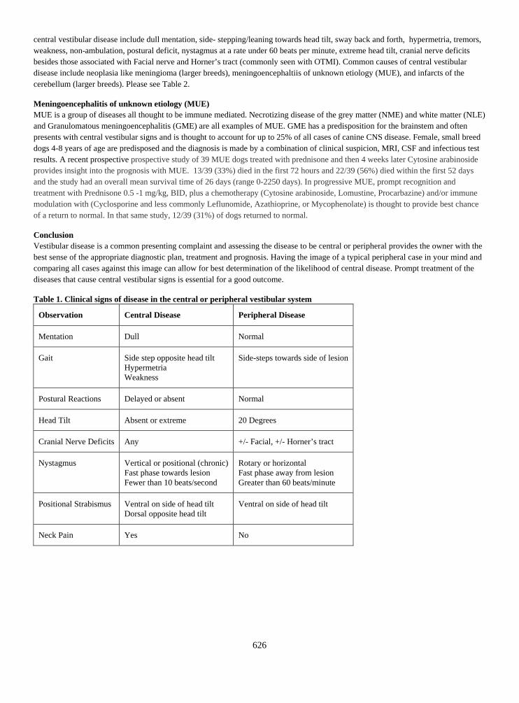
626
central vestibular disease include dull mentation, side- stepping/leaning towards head tilt, sway back and forth, hypermetria, tremors, weakness, non-ambulation, postural deficit, nystagmus at a rate under 60 beats per minute, extreme head tilt, cranial nerve deficits besides those associated with Facial nerve and Horner’s tract (commonly seen with OTMI). Common causes of central vestibular disease include neoplasia like meningioma (larger breeds), meningoencephaltiis of unknown etiology (MUE), and infarcts of the cerebellum (larger breeds). Please see Table 2. Meningoencephalitis of unknown etiology (MUE) MUE is a group of diseases all thought to be immune mediated. Necrotizing disease of the grey matter (NME) and white matter (NLE) and Granulomatous meningoencephalitis (GME) are all examples of MUE. GME has a predisposition for the brainstem and often presents with central vestibular signs and is thought to account for up to 25% of all cases of canine CNS disease. Female, small breed dogs 4-8 years of age are predisposed and the diagnosis is made by a combination of clinical suspicion, MRI, CSF and infectious test results. A recent prospective prospective study of 39 MUE dogs treated with prednisone and then 4 weeks later Cytosine arabinoside provides insight into the prognosis with MUE. 13/39 (33%) died in the first 72 hours and 22/39 (56%) died within the first 52 days and the study had an overall mean survival time of 26 days (range 0-2250 days). In progressive MUE, prompt recognition and treatment with Prednisone 0.5 -1 mg/kg, BID, plus a chemotherapy (Cytosine arabinoside, Lomustine, Procarbazine) and/or immune modulation with (Cyclosporine and less commonly Leflunomide, Azathioprine, or Mycophenolate) is thought to provide best chance of a return to normal. In that same study, 12/39 (31%) of dogs returned to normal. Conclusion Vestibular disease is a common presenting complaint and assessing the disease to be central or peripheral provides the owner with the best sense of the appropriate diagnostic plan, treatment and prognosis. Having the image of a typical peripheral case in your mind and comparing all cases against this image can allow for best determination of the likelihood of central disease. Prompt treatment of the diseases that cause central vestibular signs is essential for a good outcome. Table 1. Clinical signs of disease in the central or peripheral vestibular system
Observation Central Disease Peripheral Disease
Mentation Dull Normal
Gait Side step opposite head tilt Hypermetria Weakness
Side-steps towards side of lesion
Postural Reactions Delayed or absent Normal
Head Tilt Absent or extreme 20 Degrees
Cranial Nerve Deficits Any +/- Facial, +/- Horner’s tract
Nystagmus Vertical or positional (chronic) Fast phase towards lesion Fewer than 10 beats/second
Rotary or horizontal Fast phase away from lesion Greater than 60 beats/minute
Positional Strabismus Ventral on side of head tilt Dorsal opposite head tilt
Ventral on side of head tilt
Neck Pain Yes No
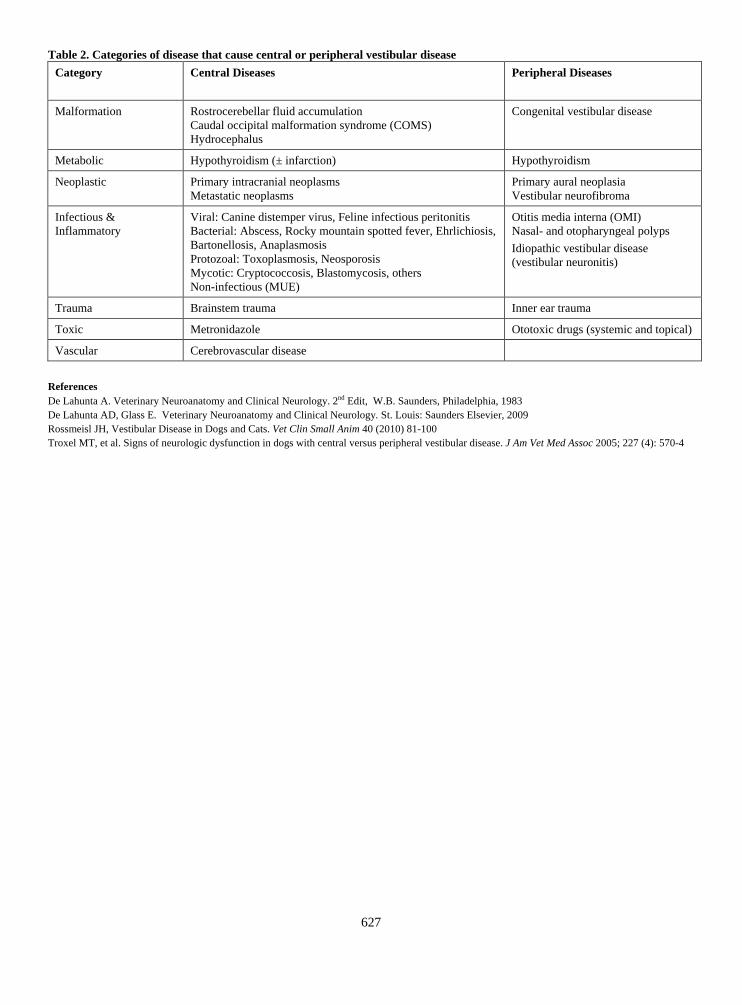
627
Table 2. Categories of disease that cause central or peripheral vestibular disease Category Central Diseases Peripheral Diseases
Malformation Rostrocerebellar fluid accumulation Caudal occipital malformation syndrome (COMS) Hydrocephalus
Congenital vestibular disease
Metabolic Hypothyroidism (± infarction) Hypothyroidism
Neoplastic Primary intracranial neoplasms Metastatic neoplasms
Primary aural neoplasia Vestibular neurofibroma
Infectious & Inflammatory
Viral: Canine distemper virus, Feline infectious peritonitis Bacterial: Abscess, Rocky mountain spotted fever, Ehrlichiosis, Bartonellosis, Anaplasmosis Protozoal: Toxoplasmosis, Neosporosis Mycotic: Cryptococcosis, Blastomycosis, others Non-infectious (MUE)
Otitis media interna (OMI) Nasal- and otopharyngeal polyps Idiopathic vestibular disease (vestibular neuronitis)
Trauma Brainstem trauma Inner ear trauma
Toxic Metronidazole Ototoxic drugs (systemic and topical)
Vascular Cerebrovascular disease References De Lahunta A. Veterinary Neuroanatomy and Clinical Neurology. 2nd Edit, W.B. Saunders, Philadelphia, 1983 De Lahunta AD, Glass E. Veterinary Neuroanatomy and Clinical Neurology. St. Louis: Saunders Elsevier, 2009 Rossmeisl JH, Vestibular Disease in Dogs and Cats. Vet Clin Small Anim 40 (2010) 81-100 Troxel MT, et al. Signs of neurologic dysfunction in dogs with central versus peripheral vestibular disease. J Am Vet Med Assoc 2005; 227 (4): 570-4
628
Ouch! Recognizing and Treating Neck or Back Pain William Bush, VMD, DACVIM
Bush Veterinary Neurology Service Frederick, MD
Recognition of pain is perhaps the biggest limitation to treating pain in veterinary practice. The degree and location of the pain can be determined with careful consideration of owner observations in conjunction with the physical examination. Once pain is localized a diagnostic efforts should be made to determine the cause of the pain such that it can be treated as specifically. Acute pain is generally easier to manage than chronic pain and in particular neuropathic pain. This talk will discuss the behavioral observations and physical exam techniques to assess the location and degree of pain. Differential diagnoses for spinal pain will be discussed along with tips on diagnoses and treatment. Next we will discuss inflammatory pain (physiologic, nociceptive or acute) and neuropathic pain and implications for pharmacologic and non-pharmacologic treatments. Pain categories Pain is an unpleasant sensory and emotional experience associated with actual or potential tissue damage. Acute pain (physiologic, nociceptive, and inflammatory) has a sudden onset, dissipates with healing, and is localized and transient – it serves as a protective mechanism to spare an individual tissue damage. Chronic pain however is unrelenting, intense, purposeless and persists long after the recovery from the inciting injury and if often refractory to common analgesic agents. The most severe form of chronic pain is neuropathic pain which occurs when the primary lesion is within the nervous system. Neuropathic pain results from plasticity in the nervous system and is associated with lower quality of life scores and is described in people as burning, lancinating, shooting, tingling, crawling or electrical sensations. Breakthrough pain is an abrupt, short-lived, and intense pain that “breaks through” the around-the-clock analgesics used to controls persistent pain. It is common in people and subdivided into incident, idiopathic and incident related. Pain assessment Lord Kelvin in 1883 stated the ability to effectively treat pain is dependent on how well pain can be detected and measured. Dogs and cats hide their pain as a protective mechanism such that a dog may still experience pain and wag its tail. Despite this challenge, pain can be localized, characterized and diagnosed with a physical exam and behavioral history. Many of the current pain assessment tools (PATs) in veterinary medicine are adapted from humans and involve questionnaires that determine client-specific outcome measures (CSOM). These have been shown to be a sensitive method for use in the pain management of dogs. Looking at these questionnaires offers insight into what things dogs or cats might do when they are in pain. Please see Table 1. Physical exam techniques to detect and localize pain Pain from the cervical vertebral column or neck pain is often suspected when patient spontaneously yelps out and there is no gait or posture deficits, intermittent thoracic limb lameness (root signature), or a stiff neck with decreased range of motion. Palpating muscle spasm laterally at level of transverse process, pain with manipulation or ventral process of C6, or resistance to range of motion can also indicate neck pain. Dog with Caudal Occipital Malformation Syndrome (see below) will often vocalize with firm palpation of dorsal or lateral muscle at level of dorsal process of C2. Pain from thoracic, ribs and lumbar spinal column or mid-back is often suspected with kyphosis, stiffness or when patient is slow to sit or rise. Detection of mid-back pain involves applying pressure to dorsal processes while putting pressure / palpating the ventrum and/or palpating muscle / rib heads at level of transverse process. Lumbosacral or low back pain is suspected with abnormal tail carriage, fecal or urinary incontinence, and when patient is slow to sit and rise. Low back pain can be detected with rectal palpation of the lumbosacral junction (or spondylosis at L7-S1), tail extension and lateral movement, or by applying pressure to muscle between dorsal process of L7 and S1 or muscle just cranial and lateral to ilium. Hip extension will not differentiate back from hip pain. However, hip pain can be discerned by slowly elevating the femoral head about 3-5 mm from acetabulum by lifting up on the medial surface of the femur while the patient is in laterally recumbent. In a patient with mid-back pain testing the cutaneous trunci cut-off can be very useful in determining the level of the lesion (see notes from UMN vs LMN talk). Neck pain localization and differential diagnoses Neck pain can come from another location about 25% of the time. When a patient’s pain localizes to the neck it is important to consider the cause of the pain could be only within the head or even the thorax. A recent retrospective of 169 dogs and 9 cats presenting with neck pain showed pathology in only the neck 73% of the time, only the head 9% of the time, head and neck in 17%, and thorax in 1%. We have also diagnosed neck pain with involvement of only the high thoracic spine as well. In this same neck pain retrospective about 50% of the cases suffered from intervertebral disk disease, 20% from neoplasia, 15% from inflammatory disease, 9% from trauma, 3% vascular and 3% malformations. Important points are that if you assume spinal pain is from disk disease then you will be incorrect 50% of the time and that about 10% of the cases of neck pain in this study had a brain tumor.
629
Differential diagnoses for spinal pain in small dogs The following diseases are diagnosed in mostly but not exclusively small breed dogs. The intervertebral disk (IVD) is located between the vertebral endplates and serves to cushion the vertebrae. In health it consists of a well hydrated nucleus pulposus which is surrounded by concentric rings of collagen called the annulus fibrosis (AF). Type I IVD occurs when the AF completely tears and the dehydrated and often calcified NP extrudes into the spinal canal or intervertebral foramen causing compression and inflammation of the meninges, spinal cord and nerve root. Type II IVD occurs when there is a bulging with microtears of the AF without NP extrusion. Together they are no doubt the most common causes of spinal pain in companion animals. When intractable pain is the only sign of disk disease success rates with surgery are about 98%. Management with pain medication, restricted activity and rehab therapy can also be highly successful. However, as mentioned above, disk disease only accounts for about 50% of the cases of spinal pain. Meningoencephalomyelitis of Unknown Etiology (MUE) is a non-infectious inflammatory condition of the brain, spinal cord and meninges – this disease is common and can present with just spinal cord signs about 10% of the time. MUE is most common in small breed dogs under 8 years of age and responsive to glucocorticoid therapy. The diagnosis is easily confused with IVD without advanced diagnostic testing. MUE can progress to life-threatening intracranial disease especially when not treated with immune suppressive therapy – therefore the veterinarian should be vigilant for progression of signs to include head tilt, depression, and seizure. Glucocorticoids should be avoided for treating spinal pain because it can mask MUE and when compared to NSAIDs is associated with more side-effects, lower quality of life scores and higher recurrence rates of IVD. Caudal Occipital Malformation Syndrome (COMS) or Chiari-Like Malformation occurs when a malformed or hypoplastic occipital bone allows the cerebellum to protrude into the brainstem and high cervical spinal cord often causing fluid build-up within the spinal cord (syringomyelia). The disease is often progressive and best treated with pain modulators +/- surgery. A specific characteristic of this disease is scratching at the side of the head without making contact (phantom scratching). Atlanto-Axial subluxation secondary to an absent or hypoplastic dens and/or associated ligaments can also be seen in young small breed dogs, present as intermittent neck pain and progress to paresis, paralysis, head tilt and death. In our clinic it is not uncommon to find more than of these abnormalities in patients with neck or back pain. Differential diagnosis for spinal pain in large breed dogs The following diseases are diagnosed in mostly but not exclusively large to medium breed dogs. Diskospondylitis is an infection of the intervertebral disk, vertebral endplates and adjacent soft tissues which can enter the spinal canal causing empyema and meningitis. The pain can be from inflammation as well as disk extrusion and pathologic fracture. Male dogs, older dogs and being a Boxer, Great Dane, and Labrador are risk factors for this disease. An intermittent fever, neutrophilia, elevated globulin and radiographic changes are inconsistently present with this disease. MRI and potentially a C reactive protein are most valuable diagnostic aids. Some dogs require anti-microbial therapy for up to a year. The prognosis is fair but improved with early diagnosis. Steroid Responsive Meningitis – Arteritis is an immune disease seen in mostly large dogs (Bernese Mountain Dog, Boxer, Pointer but also the beagle) at about 10 months of age. The signs and biochemical profile closely overlap with diskospondylitis and can include not eating, lethargy, intermittent pain, neck stiffness, and a short-strided gait. Acute Non-Compressive Nucleus Pulposus Extrusion (ANNPE) occurs when during exercise or activity a small amount of NP extrudes at a high velocity through a tear in the AF. Fibrocartilaginous Embolism (FCE) occurs when NP ruptures into the vascular supply and arterial blood flow to the spinal cord. These two disease present very similar clinically in that patients suddenly yelp out and suffer a sudden onset, non-progressive and often asymmetric weakness along with dysuria. Nerve sheath tumors can present as pain alone with muscle atrophy and lameness being noted later in the course of the disease. Vertebral fracture can occur in any size dog and are often suspect based on the history. Neuropathic pain Neuropathic pain (NP) develops when there is a lesion within the somatosensory system. NP develops when there is physical disruption of the pain pathway which starts with the receptors in the peripheral nerve and ends with the somatosensory cortex. NP results from persistent and exuberant firing of the peripheral pain fibers which then leads to recruitment of silent nociceptors in the periphery (peripheral sensitization), enhanced reactivity or disruption of neurons in the CNS (central sensitization) or imbalance of the endogenous facilitator systems and descending inhibitory systems. Over activity of the spinal cord NMDA receptor is thought to be the key process in generating NP.
In humans, NP results from inadequate recognition and treatment of pain. Furthermore, in humans with NP, the lesion within the nervous system is not always observed (despite having NP) and clinical signs can take more than a year to develop. The abnormal sensations are described as dysaesthesia (spontaneous or evoked unpleasant abnormal sensation like burning or pins and needles), allodynia (pain from stimulus that is normally not painful – light touch might be experienced as an electrical shock), or hyperaesthesia (increased pain response from a normally painful). Companion animal examples of neuropathic pain One model of NP is thought to occur in the Cavalier King Charles Spaniel where syringomyelia physically disrupts the dorsal horn of the spinal cord leading to increased levels of Substance P and abnormal sensory processing or NP. Affected dogs might be
630
hypersensitive to touch, scratch an area on the shoulder, ear or neck without making physical contact (phantom scratching), rub their face or head and spontaneously yelp out. Like in people, recently NP in this breed has been shown to correlate with an anxiety behaviors, increased fear and reduced owner-perceived quality of life.
Feline hyperaesthesia syndrome is likely another good example of NP. These cats often suffer severe pain despite no cause being determined for their clinical signs. Affected cats stare at their flank and attack or excessively groom these areas. They can have episodes of mydriasis, hallucinate and run around the house frantically, vocalize and act irritable. Cats often respond to treatments recommended in people for NP like gabapentin, amitriptyline and anti-epileptics drugs. Therapy for pain in companion animals with neurological lesions A subset of human or veterinary patients with acute pain from a neurological lesion will go on to develop chronic, severe pain called NP. NP pain occur from excessive neuronal firing and is thought to occur from inadequate pain treatment. Therefore when there is pain from a lesion within the nervous system it is important to diagnose and eliminate the painful stimuli (treat the cause) and promptly start aggressive, multimodal pain medications.
Anti-inflammatory therapy NSAIDs are the typical first choice pain medication based on perception of efficacy and side-effect profile. For dogs, meloxicam available as a 7.5 mg generic tablet and is dosed at 0.1 mg/kg, once a day. Carprofen at 2 mg/kg, Q12 or 4 mg/kg, Q24 is also used about as commonly as Meloxicam at 0.1 mg/kg, Q24. There are many other NSAID choices and if one NSAID is not effective changing NSAID is often recommended. NSAIDS work by blocking inflammatory prostaglandins by inhibiting cyclooxygenase isoform 2 (COX-2). Importantly, NSAIDS also combat central sensitization by blocking the hyperalgesia induced by activation of spinal glutamate and substance P receptors. Lastly, COX2 inhibition has been shown to benefit recovery in the brain and spinal cord of laboratory animals. A urine specific gravity, chemistry and CBC are advised prior to anti-inflammatory therapy as the rare dog will develop renal tubule disease, hepatotoxicity and dysfunction, and gastrointestinal bleeding.
Prednisone is not typically used for pain control unless inflammation or brain swelling is suspected as a primary cause of the pain, 0.5 mg/kg of prednisone or 0.1-0.2 mg/kg of dexamethasone, twice a day will be the highest dose used to try to achieve pain control.
Ketamine & amantidine Ketamine binds the NMDA receptor which prevents central sensitization or wind-up. In humans and veterinary medicine ketamine has been demonstrated to improve analgesia and outcome and reduce requirement for opiates. Ketamine does NOT increase intracranial pressure and has no effect on cerebral blood vessels size when CO2 is controlled. In our neurology clinic all surgical patients are managed peri-operatively with ketamine to improve analgesia and prevent wind-up. A loading dose of 0.5 mg/kg and then a CRI achieved by placing 60 mg of ketamine in a 1 liter bag in a 10 kg dog – this will achieve a dose of about 10 ug/kg/min when the fluids are run at 10 ml/kg/hr. Some authors suggest doses should be 2-3 times higher than this dose and would likely be very safe. Amantadine is also a NMDA receptor antagonist and has been shown to be of benefit in a randomized, blind, placebo controlled study as an adjunct to Meloxicam in osteoarthritis. The dose is 3-5 mg/kg once a day and the medication comes in a 10 mg/ml oral syrup or 100 mg tablet.
Lidocaine Systemically administered lidocaine is a sodium channel blocker effective for treating neuropathic pain at doses that do not produce anesthesia or slow cardiac conduction. Lidocaine blocks ectopic afferent neural activity at the NMDA receptor within the dorsal horn and several veterinary studies have shown benefit to lidocaine infusions during anesthesia. A loading dose of 2 mg/kg and then a CRI of 4 mg/kg/hr can be achieved in a 10 kg patient by placing 400 mg in a 1 liter bag 10 ml/kg/hr (surgical fluid rate).
Amitriptyline Tricyclic antidepressants are a recommended first line treatment of neuropathic pain in people. The descending inhibitory system is activated by incoming nociceptive firing and serves to reduce the perception of pain. This system is deficient in neuropathic pain. By blocking the reuptake of serotonin and catecholamine they enhance the activity of the descending inhibitory system. Furthermore, amitriptyline is an NMDA receptor antagonist. The recommended dose of amitriptyline is 1-2 mg/kg, once to twice a day in the dog and 2.5 to 12.5 mg per cat, once a day. The medication is bitter.
Tramadol Although structurally similar to codeine, tramadol is a weak mu opiate agonist and works by inhibiting the reuptake of serotonin and norepinephrine. Therefore it works similar to amitriptyline in that it increases the descending inhibition of pain. Bioavailability in the dog is variable and one study showed that 5 mg/kg, every 6 hours was needed to provide similar serum concentrations to those in people associated with analgesia. However, we use a dose of tramadol of 2-5 mg/kg, 2 to 3 times a day. There is a theoretical concern for serotonin sickness (vomiting, diarrhea, seizures, hyperthermia, hyperesthesia, depression) when tramadol is combined with other therapies that increase serotonin, however we have yet to recognize this condition in our clinic. We have noted mild to moderate sedation with this medication.
631
Gabapentin Although an antiepileptic drug, Gabapentin works via several mechanisms to provide pain control and is cornerstone of therapy in our neurology clinic. Gabapentin blocks calcium channels which is important because central sensitization or wind-up is facilitated by the influx of calcium. Gabapentin activates descending noradrenergic systems facilitating the release of NE in the dorsal horn which then binds the alpha-2 receptor and provides analgesia. Gabapentin causes a mild sedation and rarely severe sedation which resolves when drug is stopped. We use 10 mg/kg, 3 times a day in a dag and if sedation is absent and pain present then the dose can be increased at 10 mg/kg intervals up to 50 mg/kg per dose. In cats we usually start with 5 mg/kg, twice a day. Gabapentin should be tapered because of the concern for rebound hyperalgesia. When possible, Gabapentin is used prior to spinal surgery and continued beyond when other treatments have been eliminated.
Opiates Opiates bind receptors both centrally and peripherally to provide analgesia and control anxiety. Peripherally they prevent nociceptive sensitization and prevent neurotransmitter release. Centrally they act in the dorsal horn to modulate input from C- fibers (which mediate secondary pain or the throbbing you feel with ongoing tissue damage) and in the cerebral cortex they blunt the perception of pain. In our clinic we use the mu agonists – fentanyl 2-5 ug/kg/hr as CRI and hydromorphone 0.1 mg/kg, IV or IM. Butorphanol and burenoprhine are partial opioid receptor agonist and used only when there are intolerable side-effects from fentanyl or hydromorphone. We will also use the opiate receptor antagonist, naloxone, to reverse the effects of a mu agonist. For chronic or oral therapy we prescribe codeine, which get metabolized to morphine or much less commonly the fentanyl patch.
Opiates are useful for controlling pain but have several downsides to consider. One, side effects include dysphoria, panting, nausea, not eating, sedation, weakness, and dysuria. This can confound examination and if the clinician assumes changes in exam are from opiate and not from progression of spinal cord or brain disease, then important interventions like surgery and drugs for brain swelling might not be applied. Alternatively a patient might be delivered an inappropriately poor prognosis if the signs of disease are not from the neurological lesion but from the opiate side effects. Secondly, opiates may not be effective for all forms of neuropathic pain because they do not modulate incoming signals from the tactile / proprioceptive fibers that mediate tactile allodynia and opioid receptors in the descending pathway are down-regulated in neuropathic pain. Lastly, although uncommon opiates can generate a hyperalgesic or paradoxical response, physical dependence can develop with chronic use, and individual receptor type difference might mean more than one opiate may need to be used to achieve desired response.
In our clinic we are frequently left to wonder if a patient on opiates is still painful or dysphoric. If we consider it likely that the patient is still painful then we treat dysphoria by adding a CRI of acepromazine 0.01-.01 mg/kg/hr, or dexmedetomidine 1-3 ug/kg/hr . If we suspect dysphoria then we partially or completely reverse opiate with butorphanol 0.3 mg/kg, IV or low dose naloxone 0.01 mg/kg. Another strategy would be to switch opiates – in the patient recovering from surgery we often substitute Tylenol #4 (acetaminophen 325 mg / codeine 60 mg) at a dose of 15 mg/kg acetaminophen and 1-2 mg/kg codeine, 2 to 3 times a day.
Dexmedetomidine Alpha-2 agonists work in the brainstem to activate the descending inhibitory system by binding a brainstem nucleus called the locus ceruleus. In the spinal cord alpha-2 agonists inhibit incoming peripheral pain signals. Spinally administered alpha-2 agents reverse allodynic and dysesthetic pain in peripheral nerve injury in both rats and people. Dexmedetomidine is use commonly in the control of perioperative pain at doses of 1-3 ug/kg/hr. Pain control is synergistic when used with opiates. Dexmedetomidine is excellent in perioperative setting where a patient on opiate may be either dysphoric from opiate or painful – it has an anxiolytic effect and also reduces or eliminates the dose of opiate needed to control pain.
Anxiolytics Pain is a conscious or cerebrally recognized phenomenon. The cerebral anticipation of pain causing anxiety or anxiety itself appears to amplify the recognition or experience of pain. Sedation and/or anxiety medication can have a synergistic or useful role in controlling pain. Acepromazine at 1 mg/kg, PO, up to every 6 hours or at bedtime is advised in addition to multimodal pain protocols, especially in beagles with severe neck pain secondary to disk disease. Acepromazine can also be used as a CRI at 0.01 to 0.1 mg/kg/hr for anxiety and dysphoria in the hospital. Trazodone is very popular in our clinic for sedation and anxiety associated with cage confinement or hospitalization and is used at 2.5-5 mg/kg, 2-3 times a day. We have combined trazodone with tramadol without any evidence of serotonin sickness. At these doses seizures have not been noted with either medication. Alprazolam 0.1 mg/kg, PO up to 2 mg, twice a day can also be used for anxiety, however it seems to be less effective than trazodone or acepromazine.
Muscle relaxants Muscle spasm can be painful and is often associated with processes of the spinal column which are painful, treatment with methocarbamol at 30 mg/kg, 3 times a day and or valium 0.3 -0.5 mg/kg, 3 times a day can be useful for attenuating muscle spasm.
Tylenol and codeine Acetaminophen is safe and effective when combined with an NSAID or corticosteroid at doses of about 10 mg/kg, twice a day. When needed, as an add-on medication, acetaminophen 300 -325 mg plus codeine 30 mg or codeine 60 mg are administered as Tylenol #3 and Tylenol #4, respectively. The dose of codeine typically starts at 1 mg/kg and can be increased to 2-3 mg/kg. In dogs that are restless at night and potentially painful, the Tylenol #3 or #4 and acepromazine 1 mg/kg combination is effective.
632
Acupuncture Acupuncture is the stimulating of specific anatomic points in the body to provide a therapeutic or analgesic effect. Placement of needles releases endorphins, serotonin, and NE which can affect the processing of sensory input and amplify the descending inhibitory system. Acupuncture is highly recommended for both acute pain and the treatment of neuropathic pain.
Rehab therapy / nursing care Passive range of motion, hydrotherapy and then controlled exercise plus bladder management are important for optimizing the comfort of neurological patients. These therapies reduce pain from contracture, anxiety, and the discomfort of bladder distension and infection. Recommendations Whenever a patient presents with pain from a neurological lesion then I advise an anti-inflammatory, gabapentin, and tramadol. If this is insufficient then change the NSAID, add on an opiate and/or acetaminophen. If the patient is suspected of having neuropathic pain or another chronic painful condition then owner is counseled that break through pain is likely and can be treated with hospitalization and a CRI of an opiate plus dexmedetomidine in addition to oral maintenance medications. Prior to surgery these same medications (gabapentin, NSAID, tramadol) are recommended plus ketamine and lidocaine are added to all fluid bags. A fentanyl CRI at 5-10 ug/kg/hr is used for anesthesia and then tapered over the next 2-24 hours post-surgery. Dexmedetomidine is commonly used to reduce the need and side-effects noted with opiates. Conclusion Veterinarian’s primary responsibility is to recognize and treat pain. Therefore, in every patient and similar to humans, pain should be considered the fifth sign. It should be remembered that nervous system lesions cause severe pain and can transform into a chronic pain syndrome called neuropathic pain. Neuropathic pain is an especially debilitating condition in people and thought to result from inadequate treatment of the initial pain focus. Recognizing, diagnosing, and specifically treating a painful condition plus the use of multi-modal pain protocols are essential to a achieving a good outcome. Table 1. Behaviors associated with pain in companion animals
Unwilling to jump, slow to more, rise or sit, exercise intolerant, lame, stiff, arched back Lethargic, dull, irritable, anxious, fearful, aggressive, aloof, confused, not greeting owner Not eating well Requires being hand fed to eat well (neck pain) Yelping out with movement or spontaneously, crying, groan, scream, quiet Squinting Panting Shaking or trembling, Chewing, licking, looking, rubbing at specific location or surgical incision Crying, flinch, snap, growl, guard or bite in response to touch / being pet
References Grubb T. Chronic neuropathic pain in veterinary patients. Topics in Companion Animal Medicine 2010: 45-51 Mathews K. Neuropathic pain in dogs and cats: if only they could tell us if they hurt. Vet Clin Small Anim 2008; 38:1365-1414 Rutherford L, et al. Questionnaire-based behavior analysis of the CKCS with neuropathic pain due to Chiari-like malformation and syringomyelia. The Veterinary Journal 2012; 194: 294-298 Sharkey M. The challenge of assessing osteoarthritic and postoperative pain in dogs. AAPS Journal 2013; 15 (2): 503
633
Why Dog is Man’s Best Friend: Exciting Results of Canine Neurology Clinical Trials
William Bush, VMD, DACVIM Bush Veterinary Neurology Service
Frederick, MD
Despite many significant bench-top findings in laboratory animals there has been little progress in improving clinical outcomes for brain tumors and Status Epilepticus (SE). Veterinary clinical researchers working in tandem with the human research community are making significant progress in fostering a mechanistic understanding of human disease - this effort is called translational medicine. Due to the contributions of dog and their owners we are likely on the brink of significantly better clinical outcomes in both humans and dogs. Brain tumor The two most common brain tumor types in canines and people are meningioma and glioma. Each are a serious problem for each species and very similar. Brain tumors in canine have disease courses, clinical manifestations, imaging characteristics, molecular and histopathology and genetic changes that are very similar. The translational value of canine brain tumor research is well recognized resulting in some impressive results from NIH funded clinical trials in the dog.
Glioma Glioma is a brain tumor arising from the support cells of the brain or glia. Malignant glioma and glioblastoma multiforme in particular represent some of the most biologically aggressive and treatment refractory malignancies in humans and dogs. For humans treated with surgery, radiation and chemotherapy (temozolamide) average survival is 14 months, 2 year survival is seen in 26% and 5 year survival in just 4 %. These abysmal results have not really changed since 1959 despite intensive research. For canine intraxial tumors (all grades of glioma and other tumors) the median survival following radiation, corticosteroids +/- surgery is about 10 months. Dogs are about 3 more times as likely as a human to develop a brain tumor and in each species glioma make up about 1/3 of all brain tumors. Impediments to treating glioma in humans remain failure of the therapy to cross the blood brain barrier, infiltration of disease into the normal brain such that it is hard to know true borders of the tumor, and cellular resistance and heterogeneity.
Meningioma Meningioma is a brain tumor arising from the arachnoid lining of the brain. They are the most common brain tumor of humans and canine and comprise about 45% of primary canine brain tumors. In people, meningioma are predominantly of a lower grade and surgery is the primary treatment. However about 6000 people per year will have recurrence often due to invasive or malignant (higher grade) disease. Reoperation is often performed but even in low grade disease there is a recurrence rate of 50% by 3 years. Radiation therapy can be effective in lower grade meningioma but has been associated with cognitive deficits, secondary malignancy, and transformation of the tumor to a higher grade neoplasm. More than 40% of canine meningioma are atypical or malignant and tumors in dogs in general 5 to 7 times faster than in people. Surgery for canine meningioma is reported to have a median survival of about 10 months for canine meningioma. Convection enhanced delivery (CED) CED is a low-pressure continuous infusion process that occurs over hours to days that can achieve substantial tissue concentrations of macromolecules over a large area of the brain without producing serious injury, increasing intracranial pressure or decreasing blood flow. This technique can be followed with real-time MRI and gadolinium loaded liposomes. Dr. Peter Dickinson at UC Davis’ pioneering research showed CED to be effective and suggested that the canine spontaneous glioma can be a model system for the validation and development of novel therapeutic strategies for human brain tumors. Dr. Simon Platt at UGA is enrolling patients into a funded, canine glioma trial studying the CED of the epidermal growth factor inhibitor Cetuximab. Another proposed clinical trial involves the CED of an Pseudomonal exotoxin coupled to the IL-13 and agonist for the tyrosine kinase receptor Eph2A. Each receptor is overexpressed in high grade glioma in humans and canines but not normal canine brain tissue. A funded study using CED with carboplatin is being performed at UMINN. Electroporation Electroporation is performed using electrical pulses in order to reversibly or irreversibly permeabilize the cell membrane. Reversible electroporation safely delivers things like plasmid DNA and viruses through nanopores while irreversible electroporation (IRP) simply leads to cell death. Electroporation can generate heat leading to cell death over a relatively large area, so non-thermal methods are used in the brain. Electrochemotherapy (ECT) occurs when electroporation is combined with chemotherapy to promote delivery across the blood brain barrier. In the rodent glioma model, ECT with bleomycin doubled survival time AND in vitro investigations show that IRP alone has cytotoxic effects on canine, rodent and human glioma cell lines. Dr. John Rossmeisl at Virginia Tech has run a funded clinical trial where canine glioma patients received either stereotactic IRP and placebo or sterotactic IRP and bleomycin. One
634
patient from our clinic was enrolled and remains disease free two years later. Dr. Rossmeisl has also reported non-thermal irreversible electroporation (N-TIRE) by itself to have reduced the tumor volume in a boxer with a malignant glioma by 75% in 48 hours. Brain tumor vaccine Surgical biopsies from canine brain tumors can be shipped to the Ohlfest laboratory at UMINN for the production of a vaccine that is then administered intradermally to the patient every 2 weeks for 12 weeks. Funded trials have been performed for both glioma and meningioma. Interim results for surgery plus vaccine +/- IFN gamma for various glioma have shown mean survival for about 8 months. Meningioma vaccine results show a mean survival well over 2 years compared to controls of about 8 months. We have used the UMINN laboratory to prepare vaccine for meningioma and nasal carcinoma with favorable results. Human status epilepticus Convulsive human status epilepticus (HSE) affects an estimated 152,000 people causing death in 42,000 and leaving the survivors with a lower IQ, lower quality of life and socioeconomic status. Lorazepam and phenytoin have marginal success rates in treating HSE, 67% and 44% respectively, but remain the mainstay of therapy in people. These treatments have potential for respiratory depression and severe sedation (benzodiazepines) and cardiac complications (phenytoin). These drugs are intended for the treatment of chronic epilepsy and are based on clinical studies that are at least 20 years old that were based on research 30 to 80 years old or older. The mechanisms driving HSE are different than chronic epilepsy and therefore adapting drugs designed for chronic epilepsy simply because there is an intravenous formulation seems inadequate. There are many drugs demonstrated in rodents to be highly effective against a broad spectrum of different induced seizures. These drugs are not being developed for HSE because of the relatively smaller market (the drugs will only be used for a short time) and there is not enough safety and preclinical data to warrant a human clinical trial. If there was a more relevant clinical model of SE in the dog then drug companies might develop some of the very promising, ‘rodent proven’ drugs for treatment of the HSE. Canine status epilepticus Canine status epilepticus (CSE) has many striking similarities to that of HSE. The different seizure types noted in people (simple partial, complex partial, and tonic-clonic) are recognized in dogs. Electroencephalographic (EEG) studies in dogs, including those from our own clinic, have identified interictal and ictal patterns are similar in dogs and people. CSE is a common emergency condition treated in veterinary hospital with 53-59% of dogs with idiopathic or genetic epilepsy experiencing at least one episode of CSE and some experiencing more than 10 episodes. The mean life span of dogs with SE is shorter many veterinary studies and there is a mortality rate of 25% despite being treated with valium and phenobarbital infusions. Advantages of canine for the study of HSE The similarities between HSE and CSE have provoked researchers to develop the canine as a platform for study of novel drugs for HSE and translating this information into valuable information for researchers and drug companies. Unlike induced disease in rodents, epilepsy in dogs is a natural model and the same mechanism of HSE and drug resistance likely exist in the dog. Separate from generating important preclinical information for people, the other valuable aspect of the canine model is their body size. A dog is about 25% the size of a human but a rodent is 0.025%, this makes it much easier to generate an accurate formula for picking the correct human dose. Furthermore, results of safety and efficacy in the dog are more easily assessed than in a rodent. In particular rodent models fail to predict cognitive, behavioral and neurological side effects like irritability, insomnia, poor balance and cardiac effects like arrhythmia and hypotension. Being able to assess drug side-effect, safety, and efficacy in a clinical trial will make it much easier to generate appropriate approval and interest in human clinical trials. CSE clinical trials One of the goals of CSE trials was to develop the canine as a platform for study of HSE and to generate a network similar to the Neurological Emergency Treatment Trails (NETT) program in people. The NETT program is funded by NIH and recently reported it’s results “Rapid Anticonvulsant Medications Prior to Arrival Trail (RAMPART) Using Midazolam” ahead of schedule and under budget. Canine trials with Levetiracetam (LEV) and then Fosphenytoin (FOS) have showed that the canine is a useful platform for the study of CSE. This then allowed for the formation of the Canine Neurology Treatment Trail (CNETT) consortium whose focus is to study novel drugs for SE.
In the first trial LEV was used to treat CSE in a randomized, placebo-controlled, double masked study. Dogs were enrolled in the study after having CSE and then if there was further seizure they were given 0.5 mg/kg diazepam and a placebo or LEV (30 mg/kg or 60 mg/kg over 5 minutes). The responder rate defined as dogs with no additional seizure after the study drug was 56% in the LEV group compared to 10% in the placebo group (p=0.057). Furthermore dogs in the placebo group required significantly more boluses of diazepam than the LEV group.
635
The second study examined FOS in 31 clinical patients (22 in FOS group, 9 in placebo) were enrolled from both university and private specialty practice. There was a significant difference in the 12 hour responder rate with 63% in the FOS group versus 22% in the placebo group having no further seizures after receiving study treatment. This response rate is nearly identical to that seen in HSE with FOS and the authors conclude that this is proof that naturally occurring CSE can be utilized as a translational platform for future studies of novel SE compounds that could economically bridge the laboratory studies in rodent models with human SE trials. Conclusion Thus far clinical trials in people have failed to produce meaningful progress for the most serious neurological conditions. Utilizing the canine as a clinical model for studying brain tumors and SE should provide vital insight and meaningful advancements in therapy for humans, proving the adage that the dog is man’s best friend. Brain tumors references Andersen BM, et al. Vaccination for invasive canine meningioma induces in situ production of antibodies capable of antibody-dependent cell-mediated cytotoxicity. Cancer Res. 2013 May 15;73(10):2987-97 Dickinson PJ, et al.Canine spontaneous glioma: a translational model system for convection-enhanced delivery. Neuro Oncol. 2010;9:928-40 Freeman AC, et al. CED of Cetuximab conjugated with Iron Oxide Nanoparticles for Canine Intracranial Gliomas: A pilot Study, ACVIM Proceedings, 2014 Garcia PA, et la. Non-thermal irreversible electroporation (N-TIRE) and adjuvant fractionated radiotherapeutic multimodal therapy for intracranial malignant glioma in a canine patient. Technol Cancer Res Treat. 2011;10(1):73-83. Rossmeisl JR. Molecularily Targeted Cytotoxic and Elecroporation-Based Therapies for Canine Intracranial glioma. ACVIM Proceedings, 2011 Pluhar GE. Canine Brain Tumor Clinical Trials at the University of Minnesota. ACVIM Proceedings, 2011 Status epilepticus references Hardy BT, et al Double-masked, placebo-controlled study of intravenous levetiracetam for the treatment of status epilepticus and acute repetitive seizures in dogs. J Vet Intern Med. 2012 Mar-Apr;26(2):334-40. Leppik IE, et al. Canine status epilepticus: a translational platform for human therapeutic trials. Epilepsia. 2011 Oct;52 Suppl 8:31-4. Patterson, E. et al. The canine translational platform: proof of concept study of fosphenytoin for status epilepticus in canine clinical patients. Abstract, American Epilepsy Meeting, Washington, D.C. 2013 Potschka H, et al. Canine epilepsy as a translational model? Epilepsia. 2013 Apr;54(4):571-9.
636
New Tricks for Feeding Old Dogs Julie Churchill, DVM, PhD, DACVN
University of Minnesota St. Paul, MN
Maturity is not a disease Senior dogs commonly present to veterinarians for primary care and represent approximately one third of the pet dog population1. Lifespans are increasing and thus both the percentage and the age of elder dogs may be increasing.2 Pet owners perceive that most pets, including senior dogs are healthy and do not require a therapeutic food, 3 but they are still left with hundreds of pet foods to choose from. Advice and information recommending the best food is available almost anywhere; from trainers to pet food retailers, from magazines, internet sources and social media. However it is important to remember that there is no established AAFCO nutrient profile for a "senior" life stage, thus the nutrient content of products marketed for senior pets varies widely. This makes it even more critical for the veterinary health care team to play an active role in providing credible nutritional advice, especially for senior dogs that have unique nutritional concerns. What is old? The point at which a dog progresses from adult to a senior or geriatric life stage is variable and subjective. Dogs’ life expectancies vary widely depending on breed and body size and aging changes are also variable. They may include loss of senses (hearing or vision), reduced energy requirements and lean body mass as well as a decline in various organ functions. The American Animal Hospital Association Senior Care Guidelines suggest that, with exception of large-breed dogs, most dog breeds reach middle age by 7-8 years of age and should be considered seniors when they reach the last 25% of the predicted life span for their breed.4 Despite this arbitrary categorization, physiologic changes that occur in middle-aged and senior dogs make them less tolerant of nutritional deficiencies or excesses. Middle aged dogs are “at risk” or more vulnerable to age-related health problems. Middle age may bring an increasing incidence of chronic diseases, many of which can be influenced by nutritional management.5 Thus a vital component of preventive medical care should include a “senior” screen or health risk assessment for early detection of health problems and adjustments to care to preventing or slow onset of age-related diseases. Every senior health screen should include a thorough nutritional assessment followed by an individualized nutritional recommendation. Performing a nutritional assessment Before any diet changes are recommended, a nutritional evaluation should be performed. Each nutritional assessment and recommendation should include 3 components: the patient, the diet and feeding management factors6. An accurate diet history is invaluable when assessing of the nutritional health of the patient and will be vital to formulating an individualized diet plan. Understanding the nutritional changes that occur with aging and identifying any changes in the individual patient can help the clinician better match the appropriate food with the patient’s unique needs. The patient, the food and the pet owner’s feeding practices are interrelated and require reassessment. Health and nutritional status are not static especially in senior pets, but rather a dynamic process worthy of continued re-evaluation and treatment modifications to match changing needs of the pet.
Patient assessment An initial assessment of the patient can be done quickly and utilizes information collected as part of a health assessment: a complete medical and diet history and a thorough physical examination and appropriate lab work (ex, CBC, serum biochemical profile, urinalyses). The nutritional screening process (Table1) can quickly identify patients with “nutritional’ risks. Healthy seniors, ( those without identified risks), who are eating a nutritionally balanced diet, have a healthy body weight, body and muscle conditions (BCS, MCS) and are free of significant physical or laboratory abnormalities need no further evaluation at this time. A pet-specific nutrition assessment and recommendation for healthy seniors can be done quickly. Nutritional recommendations should include: the specific name of food that matches the pet’s current nutritional needs, the amount and frequency for feeding and a monitoring plan. In many of these patients, the feeding recommendation involves little or potentially no change, but should include a verification and validation for the owner that the current food and feeding plan meets the pet’s needs, and a documentation of the current feeding plan in the medical record.
If nutritional risk factors or age-related problems are identified, an extended evaluation and management plan is indicated. This in-depth evaluation should address some common age-related diseases that may be influenced by nutritional management [Table 2]:
• Weight management-achieve or maintain a healthy body weight • Osteoarthritis • Cognitive dysfunction
Diet assessment A complete diet history is important for evaluating the pet’s current nutritional status. Ideally you would like enough information that you can reproduce the animal’s exact diet (brand and amounts eaten). The diet history should identify all snacks, treats and nutritional
637
supplements by type and amount. The drug/supplement history should include questions about the use of food to administer medication, as it may comprise to a significant portion of the dog’s intake. Diet history information combined with the patient assessment provides information about the patient’s daily caloric requirements and specific nutrient intake. This nutrient intake should be compared to the patient’s individual needs. For example, and overweight pet with a robust appetite, should not be fed a calorie dense product. Reducing the amount of a high calorie product as a way to limit calorie intake could lead to deficiencies of other essential nutrients and increase hunger or undesirable food-seeking behaviors.
Feeding management assessment Feeding practices and preferences influence a pet’s intake. Determine whether other pets present competition or limit access to food. Determine whether food is accurately measured, how much/ how often food is offered and how much is eaten. Determine if there have been recent changes to the feeding plan and why, as well how the pet accepted those changes. This information will allow the veterinary team to determine the nutritional adequacy of the current diet, as well as help identify factors that could contribute potential success or problems with adherence to a new recommendation.
Reassessment and modification of treatment plan Nutritional assessment of geriatric pets is an ongoing process. Dogs experience a variable and wide variety of metabolic changes as they age. It is important to communicate and engage pet owners to create the expectation of continued reassessment and treatment modifications that accommodate the specific changes observed in each individual dog rather than adopting a “geriatric’ protocol. A vigilant monitoring plan allows early detection of problems if they arise and a better opportunity to intervene or modify the pet’s individualized nutritional plan to improve its health. Partner with clients to help ensure success and maintain adherence to the feeding and monitoring goals. Effects of aging on nutritional needs
Energy Aging results in changes to both structure and functional of the GI tract. However no studies report clinically relevant differences in nutrient absorption between young adult and geriatric dogs7. Maintenance energy requirement (MER) is defined as the energy required to keep an animal in a “maintenance state”, or maintaining a normal activity. MER caries depending on factors such as breed, health, neuter status and age. As dogs age, MER decreases ~25%, with the greatest decrease at middle age (7 years)8. Loss of lean body muscle (LBM) appears to be the primary factor influencing the reduction in energy requirements9. LBM accounts for about 96% of an animal’s basal energy expenditure 10. Aging dogs are less active which also contributes to reduced LBM and MER. If no adjustments are made to the pet’s energy intake to account for the reduction in LBM, activity and MER, then the senior pet will gain weight and increase the risk for obesity. BCS should be closely monitored in elder dogs to prevent obesity. Unhealthy weight gain exacerbates many age-related conditions. A higher protein to calorie ratio diet would be beneficial to promote ideal weight maintenance in senior pets identified at risk for obesity11. Results from a lifetime study performed in dogs revealed lower disease incidence, later onset of disease and increased life span in calorically restricted animals. Dogs fed a 25% reduction compared to controls lived an average of 13.0 years compared with 11.2 years in the control group12. Thus maintaining energy balance and avoiding unhealthy weight gain should be one of the most important health goals for senior dogs.
Water Elder humans exhibit decreased thirst and drinking when challenged by fluid deprivation. Although unknown in dogs, a similar response is expected5. Thus water intake should be monitored or ensured when elder dogs are exercising or exposed to hot environments. Senior dogs may also be at risk dehydration if they have subclinical renal insufficiency. When a senior pet’s appetite is good but water intake is suspect, add water to the food to ensure adequate intake and hydration.
Protein Protein requirements increase with age due to increased protein turnover and reduced protein synthesis13, 14 Healthy senior dogs do not benefit from protein restriction15 and may be harmed by limiting dietary protein 16. Protein restriction of seniors could be more detrimental than protein deficiency in younger animals17. A general guideline for estimating daily protein needs is to provide 2.55 gms protein /kg body weight (BW) or ~ 1 gm protein/lb BW. 13, 17-19. This level of protein intake should minimize risk of protein deficiency. Senior dogs may need up to 1.5-2 times more than this13. Older dogs also require fewer calories, or less food than younger dogs. Diets for older dogs should not only contain lower calories but a higher percentage of protein or a higher protein:calorie ratio in order to meet the dog’s age-related nutritional needs. Based on the diet history, assure the patient is meeting daily protein needs; ~ 1 gm protein/lb BW minimum. Food with 25 % of the calories from quality protein should meet the needs of most healthy aged dogs and minimize loss of LBM. Assess MCS to monitor LBM.
638
Nutritional intervention of selected age-related diseases Although the most common age-related conditions are best managed with a multimodal approach combining nutritional strategies, exercise or environmental enrichment and possible medical management, the discussion will focus on nutritional management. Overweight/obesity Hyperadiposity, the most prevalent form of malnutrition, contributes to many of the diseases linked to obesity21-23. Still pets that are overweight go unrecognized or may not have this health concern addressed. Based on the canine lifespan study12 which demonstrated
many negative health consequences of overweightedness and benefits of being lean, weight management should remain a top priority for senior pet health. Yet it remains one of the most significant health problems among middle aged and elder dogs. Monitor the pet’s diet, BW, BCS and MCS at each visit . Once excess weight is diagnosed, action should be taken to achieve healthy BW and BCS. Creating a negative energy balance promotes weight loss. This is best achieved by feeding foods with low calorie density, increased protein content and increased nutrient calorie:ratio to assure adequate intake of essential nutrients while restricting calories. Degenerative joint disease Osteoarthritis (OA), the most prevalent joint disorder in dogs, affects as many as 20%, and obesity is recognized as a primary risk factor24. Nutritional strategies for OA include the following:
Weight and muscle management Loss of excess body weight/fat can improve clinical signs of lameness in arthritic dogs25. Strategies to maintain healthy BW, BCS and LBM and prevent sarcopenia should be prioritized for senior dogs. The can be achieved by selecting a complete and balanced diet that meets protein and other nutrients when providing the amount of calories to prevent excess body fat gain. The nutritional goal is to delay onset and prevent progression of OA.
Long chain omeg-3 fatty acids (n-3) Shows the greatest evidence for synovial anti-inflammatory effects26,27 compared to other nutraceuticals. Marine oils (EPA> DHA)28 are preferred with more effective anti-inflammatory effects compared to shorter chain flax or other plant source n-3 oils. There is no standard accepted dose. Cognitive dysfunction As many as 20-68% of middle age to elderly dogs experience cognitive dysfunction or behavioral changes which can manifest in varying degrees of mental decline29. (Table 2). Nutraceuticals may have potential use both in prevention and treatment, but are best when combined with environmental enrichment30-32.
Antioxidants The brain is especially susceptible to free radical damage and cognitive dysfunction. Multiple studies have shown improved clinical signs of age-related cognitive changes in dogs fed antioxidant-enriched diets or supplements30-32.
Medium chain triglycerides Supplementation with MCT improved cognitive performance and preserved brain structure of elder dogs. MCT provides an alternate cerebral energy source by way of ketones without restricting dietary carbohydrate or proteins34, 35.
Supplements versus enriched diets One caveat for the use of nutraceutical supplementation is that that they have not been adequately assessed for efficacy, optimal doses or nutrient interactions. When considering whether to select a diet containing the supplement or to prescribe a supplement, consider the nutrient composition of the ‘base diet.” Assure the base diet meets the macronutrient needs of the patient and determine if it will provide an adequate dose of the intended supplement when fed to meet the energy needs of the pet. If not, it would be prudent to select a more appropriate diet and give the intended dose of supplement. Table 1. Intial screen: assessing for nutritional risk factors
Nutritional Screen for Risk Factors Require extended evaluation if () HISTORY OF:
Treats/snacks/human foods > 10% intake Inadequate information/inappropriate feeding/food Consuming unconventional diets Previous/ongoing medical problems Gastrointestinal signs
PHYSICAL EXAMINATION: Any abnormal BCS (≠5/9 or 3/5) Any MCS <3 Unintentional weight loss OR gain
639
New medical condition Poor skin hair coat Dental disease
Adapted from Table 2 , AAHA Nutrition Assessment Guidelines Table 2. Extended screening: assessing senior dogs for nutritionally relevant age-related factors
Extended evaluation: Age-related diseases to evaluate in senior dogs
Abnormal Body Condition- Is this pet overweight or Underweight? Diet- Is the pet eating appropriate amounts of balanced diet?
• Assess appetite and intake • Assess ability to eat; prehension, mastication swallowing for those underweight &/or poor intake • Assess sensory input; smell, vision, palatability of food. Consider palatability enhancer if necessary
Mobility and access to food and water • Is the pet able to walk, access food provided? Able to stand to eat? • Other pets or physical limitations impairing access? • Mobility and exercise- is the pet’s MCS normal (3/3)? • Presence of osteoarthritis, lameness, pain?- play a role in maintenance of comfort, fitness and healthy BCS • Activity minimizes sarcopenia • Exercise and activity provide mental stimulation and environmental enrichment
Assess cognitive function • Disorientation/confusion-becomes lost or confused, fails to recognize familiar people? • Changed interactions with family members? Isolates or seeks attention less often? • Change in sleep/activity cycles? Wander or pace, sleep more in day, less at night? • Loss of house training (non-medical reasons)
References available upon request Banfield Pet Hospital State of Pet Health Report (2013). Accessed September 1, 2014 from http://www.stateofpethealth.com/Content/pdf/Banfield-State-of-Pet-Health-Report_2013.pdf Total pet ownership and pet population. American Veterinary Medical Association. In US pet ownership & demographics sourcebook. AVMA Membership & Field Services; Schamburg: 2012. Pp 1-49. Pet feeding practices among dog and cat owners in the United States and Australia. Laflamme DP, Abood SK, Fascetti AJ, et al. JAVMA 232:687–94, 2008. AAHA Senior Care Guidelines for dogs and cats. Epstein M, Kuehn NF, Landsberg G, et al. JAAHA 41:81-91, 2005. Age-related changes in nutrient utilization by companion animals. Fahey GC, Barry KA, Swanson KS. Annu Rev Nutr. 28:425-445, 2008. AAHA Nutritional assessment guidelines for dogs and cats. Baldwin K, Bartges J, Buffington T, et al. JAAHA 46:285–96, 2010. Nutrition of Aging Dogs. Larsen JA, Farcas A. Vet Clin Small Anim 44:741-759, 2014. Effect of age on maintenance energy requirements and apparent digestibility of canine diets. Laflamme DP, Martineau B, Jones W, et al. Comp COnt Educ Small Anima Pract 22(suppl 9A);113, 2000. Factors influencing lean body mass in aging dogs. Kealy RD. Comp Cont Educ Pract Vet 21:34–7, 1998. The inter-organ flux of substrates in fed and fasted man, as indicated by arterio-venous balance studies. Elia M. Nutr Res Rev 4:3–31, 1991. AAHA Weight Management Guidelines for Dogs and Cats. Brooks D, Churchill J, Fein K, et al. JAAHA 50:1-10, 2014. Effects of diet restriction on life span and age-related changes in dogs. Kealy RD, Lawler DF, Ballam JM, et al. JAVMA 220:1315–20, 2002. Determination of optimal dietary protein requirements of young and old dogs. Wannemacher RW Jr, McCoy JR. J Nutr 88:66–74, 1966. Age-related changes in protein synthesis. Richardson A, Birchenall-Sparks MC. Rev Biol Res Aging 1:255–73,1983. The influence of dietary protein, lipid, phosphorus and sodium on renal structure and function in geriatric dogs. Churchill J, in PhD thesis. Department of Veterinary Clinical Sciences, College of Veterinary Medicine, University of Minnesota, St Paul, MN 2001. Effect of moderate protein deficiency on immune function. McMurray DN. Compend Contin Educ Vet21:21–4, 1999. Nutrition for aging cats and dogs and the importance of body condition. Laflamme DP. Vet Clin North Am Small Anim Pract 35:713–42, 2005. Survey of opinions about nutritional requirements of senior dogs and analysis of nutrient profiles of commercially available diets for senior dogs. Hutchinson D, Freeman L, Schreiner K et al. Intern J Appl Res Vet Med. 9 (1): 68-70, 2011. Nutrient requirements of dogs and cats. National Research Council. The National Academies Press. Washington DC, 119, 2006. Beneficial effects of dietary mineral restriction in dogs with marked reduction of functional renal mass. B r o w n S A , C r o w e l l W A , B a r s a n t i J A , e t a l . J Am Soc Nephrol 1:1169–79, 1991. The effects of obesity-associated insulin resistance on mRNA expression of peroxisome proliferator-activated receptor-I' target genes, in dogs. Gayet C, Leray V, Saito M, et al. Br J Nutr 98:497–503, 2007. Comparison of adipokine concen- trations and markers of inflammation in obese versus lean dogs. Eirmann LA, Freeman LM, Laflamme DP, et al. Intern J Appl Res Vet Med 7:196 –205, 2009. Improvement in insulin resistance and reduction in plasma inflammatory adipokines after weight loss in obese dogs. Dom Anim Endocrinol 37:214 –26, 2009. German AJ, Hervera M, Hunter L, et al.

640
Osteoarthritis and body weight. Foye PM, Stitik TP, Chen B, et al. Nutr Res 20:899–903, 2000. Effect of weight reduction on clinical signs of lameness in dogs with hip osteoarthritis. Impellizeri JA, Tetrick MA, Muir P. JAVMA216:1089–91, 2000. Effects of different n6:n3 fatty acid ratio diets on canine stifle osteoarthritis. Bartges JW, Budsberg SC, Pazak HE. Orthopedic Research Society 47th Annual Meeting. February 25-28, San Francisco, CA, 2001. Effects of feeding a high omega-3 fatty acids diet in dogs with naturally occurring osteoarthritis. Moreau M, Troncy E, Del Castillo JR, et al. J Anim Physiol Anim Nutr (Berl) 97:830–7, 201. Dietary eicosapentaenoic acid and docosahexaenoic acid equally incorporate as decosahexaenoic acid but differ in inflammatory effects. Sierra S, Lara-Villoslada F, Comalada M, et al. Nutrition 24:245–54, 2008. Prevalence and risk factors for behavioural changes associated with age-related cognitive impairment in geriatric dogs. Zakona G, Garcia-Belenguer S, Chacon G, et al. J Sm Anim Pract 50:87–91, 2009. BDNF increases with behavioral enrich- ment and an antioxidant diet in aged dog. Fahnestock M, Marchese M, Head E, et al. Neurobiol Aging 33(3):546 –54, 2012. Synergistic effects of long-term antioxidant diet and behavioral enrichment on beta-amyloid load and non-amyloidogenic processing in aged canines. Pop V, Head E, Hill MA, et al. J Neurosci 30:9831–9, 2010. Nutritional management of brain ag- ing in dogs. Roudebush P, Zicker SC, Cotman CW, et al. JAVMA 227:722–8, 2005. Enhancing brain functions in senior dogs: a new nutritional approach. Pan Y. Topics in Comp Anim Med. 26:1, 10-16, 2011. Dietary enrichment with medium chain triglycerides (AC-1203) elevates polyunsaturated fatty acids in the parietal cortex of aged dogs: implications for treating age-related cognitive decline. Taha AY, Henderson ST, Burnham WM. Neurochem Res 34:1619–25, 2009. Dietary supplementation with medium-chain TAG has long-lasting cognition-enhancing effects in aged dogs. Pan Y, Larson B, Araujo JA, et al. Br J Nutr 103: 1746–54, 2010.
641
Pet Food Ratings, Rankings, and Recalls: How to Make Sense of it All
Julie Churchill, DVM, PhD, DACVN University of Minnesota
St. Paul, MN
How to evaluate a pet food and make a diet recommendation Clients can bombard the veterinarian and veterinary staff with questions about pet food. With almost 5000 different product labels on the market it is inevitable veterinarians will be asked about a product they are not familiar with. Advice and information recommending the best food is readily available almost anywhere; from trainers to pet food retailers, from magazines, internet sources and social media. However these voices can be strongly biased and may compete with the veterinarian healthcare team’s advice. Therefore veterinary professionals need to be competent and confident in evaluating new or less familiar products in order to make nutritional recommendations for their patients and help owners make sound nutritional decisions for their pet.
When evaluating a pet food, the label is a good place to start. All pet food labels must include the following: a guaranteed analysis (% nutrient content as fed), a nutritional adequacy statement according to standards required by the Association of American Feed Control Officials (AAFCO), an ingredient list, food name and type, feeding guideline and manufacturer contact information (Zicker, 2008). A systematic approach to evaluating labels is a useful first step in assessing a product for your patient.
Many of the rating systems and pet food reviews are based on judgments about ingredients. With the exception of patients with adverse reactions or primary food allergies, or a traditional Chinese medicine approach, this is often the least useful information provided. Clients also want to prioritize ingredients and much of their information is based on misconceptions. The veterinary team must be careful not to discount client concerns, yet use the opportunity to educate and guide owners in their decision-making about pet foods.
What follows is a suggested approach to assessing labels and pet food products for indicators of a product’s nutritional value and potential impact on pet health.
Suggested in descending order of importance: Manufacturer contact information
Contact the manufacturer whenever you have questions about a product. This can provide you with valuable information as well as an indication of how willing a company is to work with the veterinary profession. The American Animal Hospital Association (AAHA) (Baldwin 2010) and the World Small Animal Veterinary Association (WSAVA, 2011) Nutritional Assessment Guidelines lists an excellent set of potential questions to ask of manufacturers:
• Do you have a veterinary nutritionist or equivalent on staff and available to answer questions? • Who formulates the diets and what are their nutritional credentials? • Which diets are tested using AAFCO feeding trials vs formulation? • What is the caloric value (per can/cup)? • Will you provide nutrient analysis and digestibility of your products? • Describe your quality control measures to assure consistency and quality • Where are your diets produced and manufactured? • What kinds of research have been conducted on your products? Where is it published?
Nutritional adequacy A statement of nutritional adequacy developed by AAFCO is required on all pet food packages. Nutritional adequacy can be met either 1) through animal feeding trials or 2) through formulation tests. Feeding trials are conducted with animals to ensure that nutrients in a given food or line of foods are present in sufficient quantities to promote good health and are bio-available to the animal ensuring the nutrients are digested properly. Formulated products have had nutrient content confirmed by mathematical calculations (adding nutrient content listed in a database of ingredients) or by laboratory testing. The formulation method does not include testing involving animals. Feeding trials allow for an in vivo product evaluation and are preferable to formulations.
Life stage claim AAFCO requires that foods meet and disclose one of two nutrient profiles based on the pet’s life stage. The “maintenance” life stage nutrient profile is designed to meet the nutritional needs for adult dogs or cats. The “growth and reproduction” nutrient profile is designed to meet the nutritional needs for puppies or kittens as well as pregnant or lactating adults. A product with a nutritional adequacy statement designated to meet ‘all life-stages’ must meet standards for both. Pet foods designed for a single life-stage (i.e. ‘maintenance’ or ‘growth and reproduction’) better match the nutrient profiles for pets of that life stage. Products formulated for “all life stages” may contain excessive amounts of some nutrients, which can result in overfeeding. It is better to feed pets with food designed to match their life stage. It is important to remember that there is no AAFCO defined nutrient profile for senior/geriatric life stage and the nutrient content of products marketed for senior pets can vary widely.
642
Caloric content disclosure Because of the prevalence of obesity in pets, caloric disclosure and labeling is essential for veterinarians to assess and counsel clients about purchasing pet foods that meet the energy needs of their pets. Unfortunately, caloric disclosure is not yet required by law and remains optional for manufacturers to include on packaging. Without information on caloric content, pet owners run the risk of over feeding their pets, resulting in obesity and related health problems. Companies that choose to report the caloric content are preferred. Making calorie content readily accessible greatly helps the veterinary team determine a proper food dose when making a nutritional recommendation, and allows consumers to make comparisons between foods and select more appropriate feeding portions.
Ingredients Evaluation of the ingredient list is often the most controversial aspect when interpreting a label. Evaluating ingredients presents challenges for clients because they are barraged with marketing claims, misinformation and even scare tactics. Evaluation of ingredient lists remains challenging for many veterinary professionals because transparency about ingredients, ingredient sources, and processing methods beyond the minimum of what is legally required is generally difficult to come by in the pet food industry. In addition, the nutrient-based scientific literature is not comprehensive, especially when compared to the research base for human nutrition. Although there is widespread misunderstanding about pet food ingredients, the major ingredients commonly used in pet food (beef, poultry by product, lamb meal etc.) are fairly well regulated and defined by AAFCO. Many fruits, vegetables, and other seemingly healthy ingredients have no AAFCO definition for the ingredient. If an ingredient definition does not exist, AAFCO regulations state that it “shall be identified by the common or usual name.” For example, ‘Apples’ or other fruits may contain seeds, stems, leaves, skins, or pulp. While pulp may contribute nutrients to the food, the generic definition does not clearly exclude any other parts that may not be beneficial to the animal’s health. For all of these reasons, reliance on pet food ingredients as the primary way to assess a pet food product would be a poor indicator of a product’s overall health impact for a pet. As part of the initiative to consider nutrition the 5th vital assessment, a Nutrition reference manual (http://www.everypeteverytime.com/docs/en-us/Pet_Nutrition_Ref_Manual.pdf) provides an excellent description of pet food label requirements and clarifications about ingredient definitions. Nutrition recommendation, a pet-specific process The final steps of making a nutritional recommendation for a pet food are to use your judgment in evaluating a product and match it closely with life stage, life style and health of the pet. To complete the process, you would continue to monitor the pet’s response to make sure you see the expected results, that the patient maintains optimal health. Recalls Sadly, pet food safety issues remain a growing concern. We have become more aware of pet food safety issues, most dramatically evident in 2007 with melamine adulteration of wheat gluten which affected many products and led to renal failure in a number of pets. Most recently, at the time of this writing, at least 3 products have been recalled for aflatoxin contamination, and several more foods or treats with potential salmonella contamination. Pet food safety is now more closely monitored by the FDA, and there are more professional “watch-dogs” sharing information and updates about pet food recall; Veterinary Information Network (VIN), American Veterinary Medical Association(AVMA), and State Veterinary Medical Association etc. There is now a central Food and Drug Administration (FDA) online safety reporting portal for veterinarians and owners to submit reports of concern about pet foods and treats. These can be submitted electronically: http://www.fda.gov/AnimalVeterinary/SafetyHealth/ReportaProblem/default.htm
If the veterinarian has suspicions about the safety of a food, this warrants a thorough diet history. Check the FDA website and contact the pet food manufacturer to alert and confirm your concerns. You will need information from the product label, so advise client to keep the label with the food until the bag is completely consumed. References Baldwin K, Bartges J, Buffington T, et al. AAHA Nutritional Guidelines for dogs and cats. J Am Anim Hosp Assoc. 2010; 46:285-296. FDA Animal and veterinary safety reporting portal (2010), http://www.fda.gov/AnimalVeterinary/SafetyHealth/ReportaProblem/default.htm Nutrition reference manual (nd), Hill’s Pet Nutrition, http://www.everypeteverytime.com/docs/en-us/Pet_Nutrition_Ref_Manual.pdf Pet Nutrition Alliance. AAHA Tips for Implementing Nutrition as a Vital Assessment in Your Practice (2012) http://petnutritionalliance.org/PDFS/PNA_TipsGuide_AAHAproof2.pdf WSASA, Nutritional assessment guidelines (2011) http://www.wsava.org/PDF/Misc/WSAVA_GlobalNutritionalAssessmentGuidelines_2011.pdf Zicker, S. Evaluating pet poods: how confident are you when you recommend a commercial pet food?” Topics in Companion Animal Medicine. 23 (3).121-126, 2008.
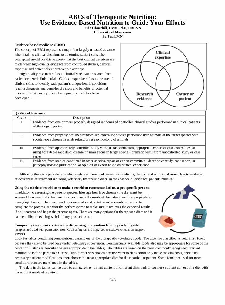
643
ABCs of Therapeutic Nutrition: Use Evidence-Based Nutrition to Guide Your Efforts
Julie Churchill, DVM, PhD, DACVN University of Minnesota
St. Paul, MN
Evidence based medicine (EBM) The concept of EBM represents a major but largely untested advance when making clinical decisions to determine patient care. The conceptual model for this suggests that the best clinical decisions are made when high quality evidence from controlled studies, clinical expertise and patient/client preferences overlap:.
High quality research refers to clinically relevant research from patient centered clinical trials. Clinical expertise refers to the use of clinical skills to identify each patient’s unique health condition, reach a diagnosis and consider the risks and benefits of potential intervention. A quality of evidence grading scale has been developed:
Quality of Evidence Grade Description
I Evidence from one or more properly designed randomized controlled clinical studies performed in clinical patients of the target species
II Evidence from properly designed randomized controlled studies performed usin animals of the target species with spontaneous disease in a lab setting or research colony of animals
III Evidence from appropriately controlled study without randomization, appropriate cohort or case control design using acceptable models of disease or simulations in target species; dramatic result from uncontrolled study or case series
IV Evidence from studies conducted in other species, report of expert committee, descriptive study, case report, or pathophysiologic justification or opinion of expert based on clinical experience
Although there is a paucity of grade I evidence in much of veterinary medicine, the focus of nutritional research is to evaluate
effectiveness of treatment including veterinary therapeutic diets. In the absence of evidence, patients must eat. Using the circle of nutrition to make a nutrition recommendation, a pet-specific process In addition to assessing the patient (species, lifestage health or disease) the diet must be assessed to assure that it first and foremost meets the needs of the patient and is appropriate for managing disease. The owner and environment must be taken into consideration and to complete the process, monitor the pet’s response to make sure it achieves the expected results. If not, reassess and begin the process again. There are many options for therapeutic diets and it can be difficult deciding which, if any product to use. Comparing therapeutic veterinary diets-using information from a product guide (adapted and used with permission from CA Buffington and http://vet.osu.edu/vmc/nutrition-support-service) Look for tables containing some nutrient parameters of the therapeutic veterinary foods. The diets are classified as veterinary foods because they are to be used only under veterinary supervision. Commercially available foods also may be appropriate for some of the conditions listed (as described where appropriate in the tables). The tables are based on the most commonly recognized nutrient modifications for a particular disease. This format was chosen because veterinarians commonly make the diagnosis, decide on necessary nutrient modifications, then choose the most appropriate diet for their particular patient. Some foods are used for more conditions than are mentioned in the tables.
The data in the tables can be used to compare the nutrient content of different diets and, to compare nutrient content of a diet with the nutrient needs of a patient:
644
To compare diets a. of similar moisture content and energy density, one can use the amount of nutrient per unit as fed - AAFCO regulations
require that minimum percentages of protein and fat, and maximums for moisture and fiber, be reported on all pet foods. b. of differing moisture content (e.g., dry vs. canned) and similar energy density, one can use the amount of nutrient per
unit dry matter. For example, a dry diet containing 20% protein and 9% water (=91% dry matter) on an as fed basis contains 20/91 * 100 = 22% protein on a dry matter basis, whereas a canned diet containing 5% protein and 77% water (=23% dry matter) on an as fed basis contains 5/23 * 100 = 22% protein on a dry matter basis.
c. of differing energy density (e.g., high vs. low fat), one can use the amount of nutrient per 100 kcal - For example, a diet containing 25% protein and 7% fat on a dry matter basis contains 8 grams of protein per 100 kcal, whereas a diet containing 25% protein and 21% fat on a dry matter basis contains only 5 grams of protein per 100 kcal. The therapeutic (prescription) diets report information this way.
To compare nutrient content of a diet with the nutrient needs of a patient, use the amount per unit body weight per day - because many veterinary foods contain restricted amounts of some nutrients, one must compare the number of grams of nutrient in the amount of food consumed with the needs of the animal to ensure that deficiencies are avoided. This is of practical concern for protein and sodium. For example, the minimum protein intake to sustain protein reserves in dogs is approximately 2.55 gms protein /kg body weight (BW) or ~ 1 gm protein/pound BW per day. If a dog with advanced renal failure consumes 20 kcal per pound body weight per day, the diet would need to contain at least 5 grams per 100 kcal to provide enough protein to meet the dog’s needs. If the dog consumed 30 kcal per pound body weight per day, only 3.3 grams protein per 100 kcal diet would be necessary.
Because diet therapy for a number of diseases consists of restriction of nutrient intake, and because many patients with nutrient-sensitive diseases are older and don’t eat much, the risk of nutrient deficiencies must be considered. This is particularly true when the therapy is anticipated to continue for months or years. For these reasons, estimates of daily minimum intakes of some essential nutrients (amount per pound body weight) for adult, average-sized pets are presented below: Nutrient Dog Cat
Energy 10 kcal Water 10 ml Protein 1 gm (2.55gm/kg BW) 2 gm (~ 5 gm/kg BW) Sodium 10 mg Phosphorus 20 mg
Veterinary foods often are sold as containing "high" or "low" levels of some nutrients. Currently, no generally accepted definition of these terms exists. My own definitions, many extrapolated from humans, follow:
Definition of "high" and "low" nutrient densities Nutrient Dog Cat
Low calorie < 3 kcal/gm dry matter < 3 kcal/gm dry matter High calorie >4.5 kcal/gm dry matter >4.5 kcal/gm dry matter Low protein <5 gm/100 kcal <7 gm/100 kcal High protein >8 gm/100 kcal >10 gm/100 kcal Low fat <2.5 gm/100 kcal <3 gm/100 kcal High fat >5 gm/100 kcal >5 gm/100 kcal Low fiber <0.25 gm/100 kcal <0.25 gm/100 kcal High fiber >1.5 gm/100 kcal >1.5 gm/100 kcal Low sodium <100 mg/100 kcal <100 mg/100 kcal
General feeding suggestions: Remember, It is may be better for a patient to eat some of the "wrong" diet than none of the "right" diet!
1. Introduce diet gradually, once the patient’s condition is improving, to avoid creating a learned aversion, which is the association of an adverse stimulus with a novel diet. If one intends to feed a particular diet long-term, it should be introduced when the patient is feeling better so it is associated with feelings of improving health.
2. Amount- use the "Energy needs of sedentary dogs and cats" graph for initial guidelines, or offer ~20 kcal per pound body weight per day to cats and most dogs (~10 kcal/pound if > ~100 pounds), adjusting intake as necessary to maintain a moderate body condition.
3. Follow instructions in the section entitled "treating inappetence" when patient food intake falls below the above intake estimates.

645
The remainder of this discussion will use cases of commonly used veterinary therapeutic diets to evaluate evidence in the context of patients. References Roudebush P, Allen T, Dodd C et al. Application of evidence-based medicine to veterinary clinical nutrition. JAVMA 224:11:1766-1771. Buffington CA. The Ohio State University Veterinary Medical Center Nutrition Suppor t Service website. Accessed March 2014: http://vet.osu.edu/vmc/nutrition-support-service Hand M, et al, eds. Small animal Clinical Nutrition: making pet foods at home, 5th ed. Topeka, Mark Morris Institute, 2010. Fascetti A, Delaney S, eds. Applied Veterinary Clinical Nutrition. West Sussex UK, Wiley Blackwell, 2012..
646
Integrate Nutrition in Preventative Care: The 5th Vital Assessment
Julie Churchill, DVM, PhD, DACVN University of Minnesota
St. Paul, MN
Food matters The idea that great or even ‘optimal’ nutrition positively influences an animal’s health is fairly well accepted by pet owners and veterinary professionals alike. Increasingly, the public is asking the veterinarian to serve as a resource for nutritional advice for their pets, and nutrition is playing a bigger role in clinical practice. Several recent publications have helped to include nutrition as a standard of care; the American Animal Hospital Association-American Veterinary Medical Association jointly introduced the first (AAHA-AVMA) Preventative Healthcare Guidelines (2011), and the AAHA Nutritional Assessment Guidelines (Baldwin, 2010). The AAHA Nutritional Assessment Guidelines outline the goals of providing better:
• Awareness of the importance of nutritional assessment of dogs and cats. • Guidelines for nutritional evaluation of animals to promote optimal health and response to disease. • Evidence and tools to support recommendations.
In 2011 the World Small Animal Veterinary Association (WSAVA) followed suit with a similar document of Guidelines for nutritional assessment. WSAVA promoted a global initiative to standardize the process to include nutritional assessments as the “fifth” vital sign (temperature, pulse, respiration, pain assessment, and nutritional assessment) to be included as a standard of care when performing physical examinations and histories for all small animals. Together these documents endorse the incorporation of nutritional assessments into regular patient care as critical for maintaining animal health and optimizing response to disease. Incorporating nutrition standards in the care of small animal patients will help develop a partnership between owner, veterinary healthcare team and lead to healthier pets.
Every pet, every time Incorporating the AAHA/WSAVA Nutritional Assessment Guidelines doesn’t need to be time consuming or complex for most patients, but it does need to be specific to that individual pet. The assessment process can be quickly incorporated into the routine history and physical examination portion of every appointment. In the course of obtaining the history and performing the exam, each patient is screened for nutritional risk factors, established by the pet’s breed, life stage and life style, body weight and condition, health history, and underlying comorbidities, medications and diet.
The nutritional screening process can be used to quickly eliminate or identify patients with “nutritional’ risks. Patients who are healthy have a normal body weight, normal body and muscle condition (BCS, MCS) and are free of any other abnormal historical or physical exam findings. Making a pet-specific nutrition recommendation for these healthy patients can be done quickly, by recommending the amount and type of a high quality food that matches the pet’s nutritional life stage requirements. In many of these patients, it involves little or no change, but a verification and documentation of the current feeding plan.
Pets should be considered to have nutritional risk factors when any of the following are present; • Abnormal BCS ( or MCS) • Snacks, treats or table foods > 10% of total Calories • Specific life stage considerations; especially at the time of spay or neuter • Unconventional diet • Systemic or dental disease • Gastrointestinal signs • Poor skin condition or hair coat • Inadequate or inappropriate housing
When any of these findings are discovered by the nutritional screening process, it raises the index of suspicion for a nutrition-related problem. Discovery of nutritional risk factors also identifies an opportunity for further evaluation and the potential for nutrition to play an increased role in improving the pet’s health. When any of the above risk factors are identified, an extended nutritional evaluation is likely indicated. The importance of the extended evaluation increases as the number or severity of risk factors increases. The extended nutrition evaluation would include a more in-depth diet history and assessment of the pet’s diet.
Practicing great nutrition The Pet Nutrition Alliance (PNA; www.petnutritionalliance.org) was created to help raise awareness about the importance of proper pet nutrition, and the value of nutritional assessments for every pet, every time. The PNA is developing a set of practical tools for the entire veterinary healthcare team to assist them in implementing these nutritional guidelines for every pet. One of the first tools, AAHA Tips for Implementing Nutrition as a Vital Assessment in Your Practice can be downloaded from the Pet Nutrition Alliance
647
website resource section. Recent evidence suggests this process is most successful when the veterinary team develops easy protocols for each team member to follow.
Now is the time for the veterinary healthcare team to reclaim the role of nutrition counselor. Matching a diet with the proper species and life stage nutritional requirements can help prevent diet-associated diseases. Modifying a food to better meet an animal’s needs or intolerances allows diet to play an important role in the therapeutic management of the disease. Clients are seeking information about how diet can improve the health of their pets. When veterinary professionals don’t meet this need, clients seek less reliable sources for dietary information. Veterinarians may lose the opportunity to improve patient health and provide high quality preventative care. When the veterinary team makes individualized nutrition part of the every pets visit, they not only meet the needs of the patient but strengthen the relationship with their clients. References AAHA Nutrition: the first step in preventative care. (2012) https://www.aahanet.org/library/Preventive_Healthcare_Resources.aspx AVMA-AAHA,Canine and feline preventative healthcare guidelines. J Am Anim Hosp Assoc. 2011; 47:306-311. Baldwin K, Bartges J, Buffington T, et al. AAHA Nutritional Guidelines for dogs and cats. J Am Anim Hosp Assoc. 2010; 46:285-296. Nutrition: The 5th vital assessment., nd, Hill’s Pet Nutrition, http://www.everypeteverytime.com/nutrition-assessment-support.html Pet Nutrition Alliance. AAHA Tips for Implementing Nutrition as a Vital Assessment in Your Practice (2012) http://petnutritionalliance.org/PDFS/PNA_TipsGuide_AAHAproof2.pdf WSASA, Nutritional assessment guidelines (2011) http://www.wsava.org/PDF/Misc/WSAVA_GlobalNutritionalAssessmentGuidelines_2011.pdf
648
To Cook or not to Cook! Quagmires and Controversies of “Natural Diets”
Julie Churchill, DVM, PhD, DACVN University of Minnesota
St. Paul, MN
People have passion about their pets and feeding is an essential component of and caring for them. Clients want the very best for their pets and nutrition plays a big role in keeping pets healthy. Nutrition is also an important part of the human-animal bond, because there is no better way to nurture pets than by feeding them well. As we become more aware of the role great nutrition can impact human health, owners consider many of the same factors when choosing foods for their dog or cat. In the past 15 years, there has been increasing interest in finding more “natural’ alternatives to feeding pets. This increased even more since 2007 with the recall involving melamine adulteration of pet foods. In an effort to choose more “natural,” potentially safer foods for their dogs, many owners are feeding or considering feeding unconventional diets such as home cooked or raw food diets
Home preparation of less conventional diets either raw or cooked, remains a controversial subject among veterinarians, nutritionists, trainers, breeders and pet owners. Why does this controversy persist when everyone shares the same goal; to feed their pet best diet? There are pros and cons to consider with every feeding recommendation. This discussion will examine the evidence behind the recommendations, including nutritional goals, evaluation of resources, indications or contraindications for considering an alternative to conventional commercial pet food. The nutrition reference manual offers discussion points to have with clients about unconventional diets (
http://www.everypeteverytime.com/docs/en-us/Pet_Nutrition_Ref_Manual.pdf).
Home cooked diets When a client wants to prepare food for their pet it is important to understand their reasons. Once you understand the goals of the client you can partner with them to find the best fit for them and their pet which may or may not be a home cooked diet. Often clients are seeking control in selecting the ingredients in order to provide high quality or organic, additive or preservative-free. Other reasons for wanting to prepare foods at home include diagnostic or elimination diets to detect food allergies, management of multiple diseases for which there is no commercially available therapeutic diet, or end stage care for inappetant patients with terminal disease.
Because most nutritional problems do not cause noticeable physical or laboratory abnormalities until the nutritional imbalance has been present for months or years, the link to diet can be hard to identify. The appearance of a glossy coat and a happy dog can mislead people into thinking all is well. Truthfully, many pets can tolerate nutritional imbalance quite well and even flourish, for a while. This is in part because dogs are very resilient and can do well as long as they get basic nutrients. However, a nutritional imbalance that is tolerable is not the optimal care that pet owners are striving for when they choose to feed homemade or raw diets. Although some were close, not a single homemade diet evaluated by the Nutrition Support Service at the University of Minnesota Veterinary Medical Center in the past 10 years was truly complete and balanced. Formulating complete and balanced diets is not simple. Verify the nutrition credentials and training of people advocating unconventional diets and select a recipe that has been formulated by a veterinary nutritionist. Guidelines have been published to help practitioners make a quick assessment of an existing typical home made recipe for clients (Remillard and Crane 2010, Appendix 1). Homemade diets for patients with medical problems should always be formulated by a veterinary nutritionist specifically for the individual patient after careful nutritional assessment of the patient. Raw diets The lines are blurring regarding raw food diets. In the 1990’s and early 2000 several books were published (Billinghurst 1993, Volhard 2000, Schultze 1999) advocating the feeding of raw and meaty bones. More recently there has been an increase in the number of commercially available raw meat diets. There are abundant health and nutritional claims that raw foods offer superior nutrition, and maintain the enzymes needed for healthy digestion. There is no nutritional advantage gained by feeding raw meat, bones and/ or eggs. A healthy animal has sufficient enzymes within its own digestive tract to fully utilize the nutrients from their food. The cooking process can alter digestibility and some vitamin levels to a slight degree. However cooking will not cause significant nutrient loss that would make a complete and balanced diet nutritionally deficient. There has been a growing body of literature reporting health risks associated with bacterial contamination (salmonella and e. coli spp) of raw meat based diets, both home prepared and commercially prepared raw foods. To date there is no evidence for health benefits of raw foods, and public health ( AAHA position statement 2012).
There are newer commercial raw pet food products that process the food by freezing, freeze drying or high pressure pasteurization (HPP). Each of these processes may reduce the risk of bacterial and parasitic contamination to some degree; however it does not eliminate it (Weese et al, 2005). As yet, there are no regulations specifically defining ‘raw’. Should a food undergoing HPP still be considered raw. Several of these newer products do not comply with label regulations and some do not describe safe handling practices on the label (Mehlenbacher, 2012).
649
The goal behind feeding homemade or raw foods is certainly laudable, the desire to do the very best for pets. Excellent communication skills will be paramount when discussing this issue with owners so that you can focus on shared goals-; the optimal health of the animal and minimizing health risks. It is important to partner with the client to find the best approach for feeding a healthy choice for their pets. Helpful tools to use in your discussion may be the raw protein diet policy by the Delta Society, an organization promoting the human–animal connection with the mission to improve health
Tips for discussing unconventional diets with pet owners http://www.deltasociety.org/Page.aspx?pid=638, and the AAHA raw protein position statement .
a. Obtain a thorough diet history to include treats, snacks and human foods. Also include any foods used to administer medication. If 90% or more of the intake is composed of a good quality complete and balanced product it is unlikely to adversely affect the nutritional balance for healthy adult (non-reproducing) pets. Pets are higher risk is treats or additional food exceed 10% of the diet and should not contain potentially harmful foods such as chocolate, grapes, raisins, or onions.
b. Identify and discuss the reason for the owner’s selection of unconventional foods and their concerns about changing the diet. This can focus your discussion to address their concerns.
c. Use empathy and care when discussing foods, presenting evidence based facts and be aware of determining an owners “readiness to change”.
References AAHA.Raw protein diet position statement. (October 2012), https://www.aahanet.org/Library/Raw_Food_Diet.aspx Billinghurst I.The barf diet; raw feeding for dogs and cats using evolutionary principles. Bathurst Australia: Billinghurst, 2001. Delta Society Raw Protein Diet Policy: http://www.deltasociety.org/Page.aspx?pid=638 Hand M, et al, eds. Small animal Clinical Nutrition: making pet foods at home, 5th ed. Topeka, Mark Morris Institute, 2010. Mehlenbacher S et al, .Availability, brands, labeling and salmonella contamination of raw pet food in minneapolis/st paul area. Zoonosis and public health, 2012 59(7) 513–520. Nutrition reference manual (nd), Hill’s Pet Nutrition, http://www.everypeteverytime.com/docs/en-us/Pet_Nutrition_Ref_Manual.pdf Schultze K, The ultimate diet; Natural nutrition for dogs and cats. Carlsbad: Hay House 1999 Weese, 2005. CVJ: Bacterial evaluation of commercial canine and feline raw diets. Can Vet J. 2005 June; 46(6): 513–516 WSASA, Nutritional assessment guidelines (2011) http://www.wsava.org/PDF/Misc/WSAVA_GlobalNutritionalAssessmentGuidelines_2011.pdf Churchill J. Increase the Success of Weight Loss Programs by Creating an Environment for Change. Compendium: Continuing Education for Veterinarians December 2010 E1-E4. Chandler, M. Unconventional Diets. Clinician’s Brief. February 2014:15-17. Appendix 1. (Hand 2010)
Guidelines* to assess a homemade recipe: Do five food groups appear in the recipe?
• a carbohydrate/fiber source • a protein source, preferably animal origin • a source of macro minerals, particularly calcium • a multi-vitamin and trace mineral source
Is the carbohydrate source a cooked cereal and present in equal or higher quantity than meat? • Carbohydrate :protein ratio 1:1 in cats and 2:1 to 3:1 in dogs
What is the type and quantity of the primary protein source? • Protein content of various mammalian and avian skeletal muscle tissues is generally equivalent • For amino acid balance, provide some liver as part of the meat portion once a week
Is the animal protein source lean or fatty? • If the protein source is “lean”, an additional fat source is required, at least 2% of the formula for dogs and 5% of the
formula for cats Is a source of calcium and other minerals provided******* (most often neglected item)
• Most home made formulas require a specific calcium supplement • Use calcium carbonate (0.5 g. per 5 kg BW) when the protein portion is equal to or is greater than the carbohydrate
portion • Use calcium and phosphorus supplement (dicalcium phosphate or bone meal) when the protein fraction is less than the
carbohydrate fraction Is a source of vitamins and other micronutrients provided?
• Supplements providing vitamins, trace minerals, fatty acids and taurine should always be provided • these guidelines for healthy adult (“maintenance” life stage) animals.

650
There are many references for home cooked diets for pets. They all have pros and cons. Check the nutrition credentials of the author, and use some of the above guidelines to make an assessment of the recipes.
Other resources for complete and balanced homemade diets are :acvn.org for a list of nutritionists available for consultation and www.BalanceIT.com and www.petdiets.com for formulated recipes by veterinary nutritionists.
651
Client Conversations: Keeping Clients through Nutritional Questions
Julie Churchill, DVM, PhD, DACVN University of Minnesota
St. Paul, MN
We are all familiar with the ‘good news’ about preventive care: it saves lives, improves health and quality of life. Nutrition is an integral and essential component of providing optimal preventive health care for pets. Yet the bad news is that many veterinary patients are not receiving the preventive care they deserve. Recent studies document a decline in preventive veterinary care. Veterinary professionals tend to view following their recommendations or “compliance”, especially when it comes to nutrition, as the responsibility of the client. A common misconception is that clients heedlessly ignore the veterinary team’s recommendations, but in reality there are many factors contributing to non-compliance. Employing great communication skills can be a key way in which the veterinary team can take and active and important role in helping clients care for their pets. This will support clients’ understanding, trust and remembering the nutritional care plan, thus improving compliance, adherence and ultimately the pet’s care.
Veterinarian professionals may lack concrete tools to assess the clients’ understanding, receptiveness or readiness for change. Through carefully worded questions when taking a medical and diet history, the veterinary professional can identify the client’s receptiveness and readiness for change. Based on this knowledge, they can better identify the appropriate way and time to implement a nutritional plan. If a client is not yet ready, they can instead help clients explore obstacles and overcome barriers to sustainable change. For example, the best predictors of adherence to a weight loss program are the veterinary professional's interviewing skills and qualities of the veterinary-client interaction. In order to improve adherence, it is essential to establish an atmosphere of trust and demonstrate concern for both the patient and the client's well-being.
The American Animal Hospital Association (AAHA) has developed a practical acronym for veterinary staff to use to remember techniques to increase client compliance. The CRAFT formula: Compliance = Recommendation, Acceptance, and Follow Through. Compliance is thought to be optimal once the veterinary team member has made a specific recommendation to the client; the client accepts the recommendation and follows through with the care plan. CRAFT depends on the abilities of the veterinary staff to communicate effectively with the client using an intentional approach to communication known as ‘relationship-centered’ care. This style of communication and care has been associated with improved adherence. Using relationship centered communication, the veterinary professional shares information with the client in ways that they understand what is happening with their pet and share in decision-making. Recommendation The Nutritional recommendation should provide the client with specific, concise recommendation and a rationale describing the benefits for the pet. The selection should provide complete and balanced nutrition, be acceptable to the owner, enjoyed by the pet and maintain stable and healthy ideal body weight to prevent many health risks.
The way the recommendation is delivered is also important. Below are some tips to making a clear recommendation: • Avoid distractions from the conversation. Discuss in the exam room, rather than a noisy lobby. • Organize the recommendation: specific brand, amount, frequency, monitoring plan. • Give information is short pieces if information and check for understanding by the client. • Use lay person language. • Use ‘props’ such as pet food labels, portion sizes, measuring cups etc. • Summarize the discussion to repeat important points. • Provide written instructions to go home.
Definitions
Compliance The extent to which a pet receives a treatment, screening or procedure in accordance with accepted veterinary health care practices. Compliance involves both veterinary staff performing and recommending this treatment, screening or care, as well as client follow through.
Adherence The extent to which patients take medications prescribed, involving the pet owner in: filling/refilling prescriptions; administering correct dose; timing and use and completing the prescribed course. Adherence is generally a term applied to medication rather than recommendations for wellness checks or diagnostic screenings.
AAHA 2009
652
Acceptance When the client receives the recommendation and understands and agrees to the plan, this is ‘acceptance’. Although this step seems dependent only on the client, the veterinary team can significantly influence client acceptance by:
• Observing the client for verbal/nonverbal cues of non-acceptance. • Acknowledging these signs and invite concerns or worries they might have. “it seems like you may be worried about
changing foods, can you tell me about your thoughts?” • Active listening through verbal and nonverbal cues (maintain eye contact, allow pauses for clients to speak, encourage
them to ‘go on”). Follow through Non-compliance most often happens due to a failure of follow through. It is very important even after the client has left the veterinary clinic to receive support from the veterinary team to follow through with the agreed upon plan. Veterinary clients may find it difficult to remember all that is said during the consultation. There are a variety of ways to support clients and follow through after the client and pet return home:
• Provide written instructions that include o trouble shooting ideas or adverse events to watch for o ways to contact you if they have questions.
• Schedule recheck appointments before the client leaves the clinic • Verify contact information and ask for preferred choice of reminder contact • Call/email client to check in 2-3 days after the visit to see if there are challenges to following the treatment plan • Use a reminder system to assist with follow up appointments
Team approach The veterinary support staff plays a vital role in all parts of CRAFT and it is important that all members share a consistent message. Review the clinic protocol at staff meetings, and periodically evaluate/refine the protocol. There are helpful resources available for team training. Suggestions and resources for incorporating nutrition in the care of each patient is available at: www.everypeteverytime.com.
The partnership for Healthy Pets Initiative (http://www.partnersforhealthypets.org) has the goal of helping veterinary professionals gain the tools needed to insure a healthy, long life for the pet and to communicate the importance of preventive care for the lifelong health of the patient. There are tools and resources available specifically aimed at improving communication skills; ”words that work”: (http://www.partnersforhealthypets.org/communications.aspx ) Check-in
• Client fills out nutrition history/update form • Weigh in • Front desk staff places nutrition history/update form in patient’s records
Exam room • Veterinary technician records history using nutrition history form to help ask questions and initiate discussion • Veterinarian performs a physical exam and nutritional assessment, makes a specific nutrition recommendation and
documents the recommendation in patient’s records • Veterinary technician asks client if there are any questions and reinforces the recommendation, dispenses tools
(measuring cups, literature about nutrition, information packet) and sends patient home with an information packet even if they don’t purchase recommended food
Check out • Front desk staff asks client again if there are any questions, dispenses food and reinforces recommendation, schedules
a callback in 2-7 days if recommendation is for a new food and enters reminder code for re-purchase of food two weeks before food will run out
Techniques of using relationship-centered communication to develop a health care plan.
Explore the clients’ perspective Check in with the client to get their specific concerns, beliefs, goals and expectations, to better understand the client’s perceived value of each treatment option. For example: “Ms Gray, there are several great food choices that will meet Spike’s needs. What are your concerns about what he is eating now? What are your biggest goals for feeding Spike?
653
Share veterinary thinking When appropriate, discuss your findings and reasoning out loud. This shows the client your ‘work’ and allows them to follow the thought process or problems you face and can encourage their input. For example: “we wanted a food Spike really enjoyed, but he is only eating a small amount yet gained almost 15% in the last month”……”yes, it’s such a little amount and he’s still always hungry and started stealing food from the kids. Is there anything we can do?”
Encourage the client to participate Let the client help with suggestions or solutions that work for them. “Maybe a different food would help, and I can have the kids put food away when they finish snacks”.
Incorporate the client’s individual concerns. Partner in care Relate the treatment choice to a concern or comment the client made so they make the connection between care and benefit.
“You mention you want Spike to enjoy his meals, but unhealthy weight gain is related to many health risks and a shorter lifespan. We can try some foods that are really tasty, higher in protein and lower in calories so he can eat more food and feel more satisfied.” Recommended reading 1. American Animal Hospital Association. The path to high quality care: practical tips for improving compliance. In. Lakewood, CO; 2003. 2. Baldwin K, Bartges J, Buffington T, et al. AAHA Nutritional Guidelines for dogs and cats. J Am Anim Hosp Assoc. 2010; 46:285-296. 3. Nutrition: The 5th vital assessment., nd, Hill’s Pet Nutrition, http://www.everypeteverytime.com/nutrition-assessment-support.html 4. Volk JO, Felsted KE, Thomas JG, Siren CW: Executive summary of the Bayer veterinary care usage study. J Am Vet Med Assoc 2011, 238(10):1275-1282. 5. Woolf SH, Grol R, Hutchinson A, Eccles M, Grimshaw J: Clinical guidelines: potential benefits, limitations, and harms of clinical guidelines. BMJ 1999, 318(7182):527-530. 6. Development of new canine and feline preventive healthcare guidelines designed to improve pet health. J Am Vet Med Assoc 2011, 239(5):625-629. 7. Volk JO, Felsted KE, Thomas JG, Siren CW: Executive summary of phase 2 of the Bayer veterinary care usage study. J Am Vet Med Assoc 2011, 239(10):1311-1316. 8. Morrisey JK, Voiland B. Difficult interactions with veterinary clients: working in the challenge zone. Vet Clin Small Anim. 2007;37:65-77. 9. Abood SK. Increasing adherence in practice: making your clients partners in care. Vet Clin Small Anim. 2007;37:151-164.
654
Weight Management Programs: If Only they Could Lose, Everyone Wins
Julie Churchill, DVM, PhD, DACVN University of Minnesota
St. Paul, MN
Be a change agent- move your clients from thinking to doing to successfully manage the weight of their pet Over the past decade there have been many efforts in the veterinary profession to increase awareness about pet obesity. Yet obesity continues be one of the most common diseases affecting dogs and cats (Association for pet obesity prevention 2012). In spite of increased awareness of obesity by pet owners, the discussion about weight loss remains a common and frustrating aspect of small animal practice. This conversation can be sensitive and recommendations frequently go unheeded. Before this lack of success in helping pets achieve and maintain healthy weight leads you to ignore this disease, consider it an opportunity to try another way to help these patients. In spite of the evidence that obesity negatively influences health, wellbeing and even life span(Kealy et al. 2002, Pibot 2006), veterinary professionals still struggle to influence clients to begin or adhere to a weight loss program for their pets. One reason for this may be that the veterinary team lacks concrete tools to assess the clients’ readiness for change. Through carefully worded questions when taking a medical and diet history, the veterinary professional can identify their client’s receptiveness and readiness for change. Based on the client’s readiness, the health professional can better identify the appropriate time to implement a nutritional plan. If a client is not yet ready, they can instead help clients explore obstacles and overcome barriers to sustainable change.
Three essential elements should be present to make a weight loss program successful 1) owner commitment 2) individualized weight loss plan 3) regular reassessment. Careful attention to assure each component is present not only contributes to successful weight loss of the patient but also increases satisfaction of both the client and veterinary team.
Establish owner commitment Assess the client’s readiness for change. If they are ready to act, proceed with your nutritional plan. If not, employ ways to move them from thinking (contemplation) to doing (action).
Individualize the weight loss plan Partner with clients to make the weight loss plan patient-specific; a plan that works for the client and meets the nutritional needs of the pet. A careful and complete diet history (food and treat types, amounts, schedule etc) reveals important information about how the family relates to the pet through food and often provides insight about potential challenges the client will face (Michel.2009). The diet history also reveals information about the pet’s nutritional status which is often imbalanced from additional treats and human foods added to commercial products. Because of individual variation in energy needs of pets, the diet history serves as an invaluable diagnostic tool. An accurate diet history provides information about current caloric intake which then serves as a more precise starting point for the food dose calculation (start at 75-80% of current intake,) (Perea 2010).
3. Regularly reassess Initially biweekly follow-up will help support clients, assure a healthy rate of loss (0.5-1.5% body weight per week) and provide early detection of potential relapses so the weight loss plan can be adjusted or the clients redirected before excessive weight gain occurs and frustration becomes another barrier to success.
Great communication skills are as essential as clinical skills (physical examination and technical skills) for achieving success in helping clients with weight loss programs for their pets. A useful resource for communicating about this and other nutrition topics is available on line through partners for healthy pets, http://www.partnersforhealthypets.org/communications.aspx. Often the most frustrating cases are those where there is a mismatch of expectations between the client and the veterinary professionals. An appreciation of the client’s level of motivation for weight loss allows us to tailor our interventions and can help reduce our frustration with clients that do not adhere to the weight loss program.
Key challenges to consider include: • Do busy veterinary professionals have time to discuss feeding and lifestyle issues with clients in a manner that will be
effective? • When recommending changes in feeding and lifestyle, how can adherence to a weight loss program be increased?
The best predictors of adherence to a weight loss program are the veterinary professional's interviewing skills and qualities of the veterinary-client interaction. (Morrisey JK and Voiland B 2007) In order to improve adherence, it is essential to establish an atmosphere of trust and demonstrate concern for both the patient and the client's well-being. It is also important to understand how behavior change takes place (Abood 2007).
Psychologists have developed several models that help guide our understanding of how humans make changes in behavior to improve health. The “stages of change” model, also known as the transtheoretical model (TTM), developed by James Prochaska (Shumaker, et al 2009) and colleagues, can be used to assess the client’s readiness to change their behavior (Buffington 2004). Using this model can help the veterinary professional better understand the change process and provide useful strategies to customize their recommendations to the client. TTM helps us better partner with our client and patient to provide an individualized plan to best suit
655
their needs. Implementing a weight loss plan when the client is ready to act on this advice will improve the success and be a more efficient use of time. Step 1: Identify the stage of change The 5 stages of change and characteristic attributes of clients:
1. Precontemplation—the person has no intention of taking action in the next 6 months. These clients might commonly be referred to as resistant, unmotivated or unaware, but clearly, they are not ready to change. In reality, it is often our intervention programs that have not been ready for them.
2. Contemplation—the person is aware of pros and cons of changing and intends to change in next 6 months. They may be stuck “thinking about it”, intending to change “soon”.
3. Preparation— the person plans to take action in the next month. Clients may have recognized the problem and sought advice already from books or online or by talking to a pet store employee, trainer or veterinary professional. Recruit these people for action-oriented programs.
4. Action—the person has taken action that is significant enough to result in a reduction of risks for disease. For example, the client may have reduced treats or selected a different pet food. However, the change would not be considered a significant action unless it reduced calories by at least 10% and provided complete and balanced nutrition. Veterinary professionals can help refine the plan to achieve healthy weight loss.
5. Maintenance—the individual continues action to prevent relapse. Step 2: Understand the change process By understanding the stages of change the health professional can adapt their communication to meet the stage of the client. If a client is in one of the early stages, it isn’t the time to try and implement a weight loss plan for this pet. It’s equally important that we don’t ignore this patient’s obesity. Don’t give up. These patients warrant a monitoring plan. It may take time and several visits to establish rapport and build the trust necessary to move the client along to the next stage, hopefully closer to being ready to “take action” and implement a weight loss program for their pet and ultimately take steps to improve their health (see table). Step 3: Select a stage-appropriate intervention The failure of many weight loss programs are often because of a mismatch between the type of intervention and the client's readiness to change. Many traditional programs are action-oriented while the majority of clients are not in the action stage. See the table for examples of communication tools to identify the client’s stage of change and how to communicate best to match the client’s readiness. When a partnership is formed with the client you create an environment that supports change. By understanding the stages of change the veterinarian can help move the client from thinking to doing. Selecting the right intervention at the right time for the right client can tremendously improve the clinical outcome. Successfully managing obesity can change a frustrating problem to a rewarding one. The pet achieves greater health, an improved quality of life and pet owners become loyal clients because they have been active partners in the healthcare plan. References Abood SK. Increasing adherence in practice: making your clients partners in care. Vet Clin Small Anim. 2007;37:151-164. Association for pet obesity prevention(2012), http://www.petobesityprevention.com/ Buffington CA, et al., Manual of Veterinary Dietetics. St Louis: Saunders. 2004. Kealy RD, et al. Effect of diet restriction on life span and age related changes in dogs. JAVMA 2002; 220:1315-1320. Michel KE, Using a diet history to improve adherence to dietary recommendations. Compendium 2009;31:1;22-24. Partners for healthy pets, Words that work series: nutrition, 2012.AVMA, http://www.partnersforhealthypets.org/communications.aspx Perea S. What’s the take-home? Nutrition. NAVC Clinician’s Brief 2010; 8:3: 63-65. Pibot P et al., eds. Encyclopedia of Canine Clinical Nutrition. Diffo Print Italia , 2006. Morrisey JK, Voiland B. Difficult interactions with veterinary clients: working in the challenge zone. Vet Clin Small Anim. 2007;37:65-77. Shumaker, et al., eds. The Handbook of Health Behavior Change: The transtheoretical model of behavior change., 3rd ed. New York:Springer; 2009. Churchill J. Increase the Success of Weight Loss Programs by Creating an Environment for Change. Compendium: Continuing Education for Veterinarians December 2010 E1-E4. Brooks D, Churchill J, Fein K, et al. 2014 AAHA Weight Management Guidelines for Dogs and Cats. JAAHA Jan/Feb. or: https://www.aahanet.org/Library/WeightManagement.aspx
656
Canine Lymphoma: What’s on the Horizon
Alison Book, DVM, MS, DACVIM Veterinary Cancer and Surgery Specialists
Milwaukie, OR
Lymphoma is one of the most commonly diagnosed cancers in dogs, cats and people. Canine lymphoma bears similarity to the non-Hodgkin’s lymphomas (NHL) in humans and both exhibit similar responses to treatment with chemotherapy. Lymphoma is very difficult to cure and a leading cause of cancer death in dogs and people. Despite many efforts over the last 20-30 years, outcomes in canine patients have not significantly improved over those achieved with CHOP-based chemotherapy protocols (cyclophosphamide, doxorubicin, vincristine, and prednisone). These chemotherapy protocols have extended both the longevity and quality of life in dogs with lymphoma but novel strategies are needed to increase survival times. This presentation will cover a general review of lymphoma and recent advances that hold promise for the future. Diagnosis and diagnostic advances Lymphomas are a diverse group of cancers arising from lymphoid cells. There are greater than 30 types of canine lymphoma described that differ in anatomic, histologic and immunophenotypic (T vs. B cell) classification. These different types of lymphoma can vary in their biologic behavior and prognosis; however, further studies are currently needed to correlate the various categories of disease with clinical outcome. The majority of canine lymphomas are intermediate or high grade and are generally characterized as being biologically aggressive and rapidly progressing. Indolent lymphomas may progress more slowly and dogs may experience long-term survival with limited or no therapy; however, indolent lymphomas represent a small percentage of lymphoma in dogs. Diagnosis of lymphoma is achieved via cytology or biopsy. While not performed in every case, the following diagnostics may be helpful to establish a diagnosis of lymphoma or to further characterize the tumor.
Immunophenotyping (cytology, histopathology, or flow cytometry) Using antibodies against specific cell surface markers (ex. B cell CD 79a/CD20, T cell CD3/CD4/CD8), this test is primarily used to determine if the lymphoma is B or T cell in origin. However, it can also be helpful to support a diagnosis of lymphoma by documenting a homogenous population of the same immunophenoytpe within a tissue.
Flow cytometry This test allows immunophenotyping of cells in suspension (blood, effusions, and aspirates of LNs or organs). Flow cytometry can also provide information regarding cell size and expression of other CD molecules that may correlate with prognostic information.
PARR (PCR for antigen receptor rearrangement) Theoretically, a malignant cell population should be derived from expansion of a single clone. PARR amplifies the variable regions of the T cell receptor or Immunoglobulin (Ig) receptor gene to detect the presence of clonal lymphocyte populations. When it is not possible to differentiate between malignant and benign lymphocytes based on cytology or histopathology alone, PARR may be helpful to confirm a diagnosis (especially useful when the lymphocyte population is heterogeneous). PARR can be used to detect minimal residual disease but investigations are ongoing to determine if this is a useful clinical marker of early recurrence.
Proteomics (ex. PetScreen) Proteomics analyzes the protein components of a cell, which may be used to identify cancer specific markers. Preliminary studies have been performed in canine lymphoma but clinical application is limited at this time. Staging Lymphoma is considered a systemic disease and most dogs are presented in advanced stages (III to IV). Ideally, the extent of disease is determined after diagnosis as a baseline for treatment monitoring. However, the degree of staging necessary is controversial. The completeness of staging in any given case is often dictated by 1) how a diagnostic test affects treatment plan, 2) how it affects client’s decision making and 3) how it affects patient prognosis. A thorough physical exam, CBC, serum chemistry profile, and urinalysis are indicated for every patient to obtain vital information regarding organ and bone marrow function before starting treatment with chemotherapy. Additionally, information regarding prognostic factors (hypercalcemia, anemia) may be obtained. Further diagnostics to consider include thoracic radiographs and abdominal radiographs/ ultrasound. These imaging studies are non-invasive and may provide information regarding areas of significant disease burden (such as mediastinal or sublumbar lymph nodes). This can be important information when monitoring for lymphoma relapse. In the author’s practice, abdominal ultrasound is also highly recommended for any dog with clinical signs attributable to the GI tract in order to rule out involvement, and thoracic radiographs/ echocardiogram are recommended for any dog predisposed to heart disease. The value of a bone marrow aspirate in the face of a normal CBC is questionable and rarely pursued in the author’s practice.
657
PET/CT (positron emission tomography/ computed tomography) PET/CT combines functional and anatomical imaging to allow detection of metabolic or proliferative activity throughout the body. PET/CT is currently the standard of care for monitoring and predicting response to therapy in people with lymphoma. PET/CT has also shown promise for evaluating response to chemotherapy and predicting relapse in dogs with lymphoma. Standard treatment options Multiagent chemotherapy is the mainstay of treatment for lymphoma. For intermediate to high grade lymphomas, CHOP-based protocols are typically advised as first line therapy and provide the best response rates (80-95%) and treatment outcomes. At this time, long term maintenance chemotherapy does not appear to improve remission times. Additionally, dogs that do not receive maintenance therapy appear to be more likely to achieve a second remission following relapse. Several studies suggest that inclusion of L-asparaginase in the protocol does not significantly improve outcome (remission rates or duration of remission). In the author’s practice, the decision to use L-asparaginase is made on a case-by-case basis and typically reserved for particular situations (ex. sick patient, cytopenic, rescue, etc.). Individual response and remission durations vary depending on prognostic factors. Overall median survival times are 12-14 months with approximately 20-25% of dogs alive at 2 years. Alternative protocols are offered if clients need less costly or more convenient options.
Rescue chemotherapy is associated with lower response rates and shorter remission times. Chemotherapy agents that are commonly used in the rescue setting include lomustine (CCNU), doxorubicin, mitoxantrone, MOPP (mustargen, vincristine, procarbazine and prednisone), actinomycin-D, and dacarbazine (DTIC). Novel treatment options Monoclonal Antibodies (Mab): Outcome improvements in people with non-Hodgkin’s lymphoma have been due in large part to Mab therapies such as rituximab (anti-CD20 antibody used to treat B-cell lymphomas). However, rituximab is not effective in dogs. Currently, clinical studies are ongoing to evaluate two conditionally approved monoclonal antibodies (Aratana Therapeutics) for use in the treatment of canine lymphoma. These promising canine-specific antibodies are directed against CD20 (AT-004) for B-cell lymphoma and CD52 (AT-005) for T-cell lymphoma.
Bone marrow/ stem cell transplantation Ablative total body irradiation and/or chemotherapy combined with bone marrow or stem cell transplantation is available for dogs with lymphoma. However, these treatments are not widely accessible, are costly, and are associated with increased morbidity in dogs undergoing treatment. While these treatments present a potential for increased cure rates, results of a large number of treated cases have yet to be reported.
Adoptive T cell therapy Expanded autologous T cells infused after CHOP chemotherapy has been shown to significantly improve overall and disease free survival in a small number of dogs with B cell lymphoma. While quite promising, this therapy is currently available to client-owned dogs only through clinical trials. Prognosis Widely accepted negative prognostic factors include T cell immunophenotype (for multicentric lymphoma), substage b (sick), prior treatment with prednisone, and certain anatomic sites (cranial mediastinal involvement, primary diffuse cutaneous, GI, hepatosplenic, and primary CNS). Recently, it has been shown that B-cell lymphomas expressing low levels of class II MHC or lower than normal levels of B5 antigen also had a poorer prognosis. Presence of anemia is also associated with a worse prognosis. Alternatively, it appears that dogs with indolent lymphoma experience prolonged survival times.
658
Mast Cell Tumors: The Good, the Bad, and the Ugly
Alison Book, DVM, MS, DACVIM Veterinary Cancer and Surgery Specialists
Milwaukie, OR
Timothy Fan, DVM, PhD, DACVIM University of Illinois
Urbana, IL
This presentation will cover a general review of canine mast cell tumors (MCTs) and the range of biologic and clinical behavior seen with this tumor. Using case examples, we will concentrate on best practice for diagnosis, staging, and treatment as well as a practical, patient-focused approach to considering options and choosing a treatment plan.
Mast cell tumors are one of the most common cutaneous tumors in the dog. Biologic behavior is variable and clinical outcome is best predicted by histologic grade. Grade I tumors are usually well differentiated, rarely metastasize and are associated with an excellent outcome. Grade II tumors are locally invasive, may spread to local lymph nodes, and uncommonly spread throughout the body. A population of intermediate grade (Patnaik grade II) MCTs appear to follow a more malignant course, spreading locally and to distant sites. Additional factors are often considered in attempt to predict the behavior of grade II tumors. Grade III tumors are usually anaplastic and locally aggressive, with a high rate of metastasis. These tumors are not cured typically, but many dogs can have extended remissions if tumors are caught early and treated aggressively. In practice, deciphering which MCTs will behave aggressively can be difficult, making prognosis and optimal treatment challenging to predict. Consideration of a number of clinical (tumor size, clinical signs, etc.) and histologic factors (mitotic index, c-kit, etc.) can be used to help the clinician best present to and guide clients through a wide range of diagnostic and treatment options. Diagnosis and staging In most cases, MCTs can be easily diagnosed via fine needle aspirate and cytology with the rapid hematologic-type stains used in most practices. A small percentage of MCTs may have poorly staining granules, in which case a Wright-Giemsa or toluidine blue stain may be necessary. If histopathology is required for diagnosis, careful consideration of tumor location, size, and clinical factors is needed to plan for biopsy. When possible, wide excisional biopsy is preferred and incisional biopsy is uncommonly pursued in the author’s practice.
Staging is important in the clinical evaluation of canine MCT patients; however, what constitutes adequate staging is controversial. In select cases, an extensive work-up may not be necessary. Generally speaking, a minimum database (complete blood count serum biochemistry profile) and regional lymph node cytology are recommended for all dogs with MCT. These diagnostics are typically inexpensive and quick to perform and are likely sufficient for cases where the tumor is amenable to wide surgical excision and no negative prognostic factors are present. Histologic assessment of a regional lymph node may be required for definitive diagnosis of regional metastasis if cytology is suspicious but not definitive for metastatic disease. If the tumor is in an undesirable surgical location or if negative prognostic factors exist, further staging with abdominal ultrasound is advised. Abdominal ultrasound is non-invasive and allows evaluation of spleen, liver, and intra-abdominal lymph nodes for metastatic disease. Fine needle aspirate of the spleen and liver are always advised if the organs look abnormal. Several studies have suggested that FNA of the spleen and liver is warranted in the case of clinically or histopathologically aggressive disease even if they appear normal on ultrasound. In the author’s practice, splenic aspirate is strongly advised for any high grade II or III tumor or in the case of concerning clinical behavior (see prognostic factors). Thoracic radiographs rarely reveal metastasis. However, it is reasonable to pursue this as a pre-anesthetic screening and to rule out other unrelated disease processes prior to a surgical procedure. Bone marrow aspirate is rarely indicated. Prognostic factors
Grade Histologic grade is considered the most consistent prognostic factor available for canine MCT but should be interpreted in light of other prognostic factors when making treatment decisions. Histopathologic grading is complicated by interobserver variation among pathologists. Currently, two forms of grading are reported in clinical practice. The most commonly utilized grading system is the Patnaik grading system (low- grade I, intermediate- grade II, high- grade III). More recently, a 2-tier histologic grading system (low, high) has been introduced for canine MCTs. The second system was developed in an attempt to compensate for some of the weaknesses of the Patnaik system. However, further validation is needed to determine if this is truly better at predicting behavior and clinical outcome.
659
Proliferation indices Mitotic index (MI) is a strong predictor of overall survival in dogs. Using a cutoff of 5/10 high powered fields, dogs with a low MI (<5) had a median survival time of 80 months compared to 3 months for dogs with a high MI (>5). It is advisable that any tumor with a high MI is staged and treated as an aggressive MCT in practice.
Other markers of proliferation that have been evaluated include Ki67 (a protein in the nucleus that correlates with cell proliferation), AGNORs (argyrophilic nucleolar organizer regions), and PCNA. These require the use of special stains and are often included in the MCT prognostic panel. Interpretation of this panel can often be confusing for clinicians. At this point, it appears that Ki67 is most useful clinically as a prognostic factor for intermediate grade tumors to help predict expected survival times when the clinical picture remains confusing based on other factors.
C-Kit KIT (a receptor tyrosine kinase) dysregulation has been implicated in the pathogenesis of MCT development and evaluated as a prognostic factor. While KIT staining patterns (cytoplasmic localization) may be associated with dysregulation and prognosis, clinical application of this as a prognostic factor remains challenging. Alternatively, the presence of c-kit activating mutations is strongly associated with a higher rate of local recurrence, metastasis, and death from disease and should be considered a poor prognostic indicator.
Tumor location Some tumor locations may differ in behavior and prognosis. Subcutaneous tumors may have a better prognosis. Mucous membrane sites, subungual, and visceral tumors are associated with a worse prognosis. Conjunctival tumors and those of the eyelid margin may be an exception with studies showing prolonged survival after surgery alone. Perioral and muzzle MCTs have an increased risk of locoregional metastasis yet prolonged median survival times despite the higher rate of lymph node metastasis. Scrotal and preputial tumors may be associated with a worse prognosis but this remains controversial.
Clinical stage Higher stage disease is associated with a worse prognosis. The effect of lymph node metastasis on outcome may be dependent on grade of the primary tumor and how the lymph node is treated. Thus, clinical judgment is important. Multiple tumors may not negatively affect prognosis.
Other factors Local recurrence, systemic and local clinical signs, growth rate, and tumor size have all been correlated with prognosis and should be considered in the overall evaluation of a patient’s tumor. Treatment options
Primary therapy Wide surgical excision is the primary treatment of choice for tumors localized to the skin and subcutaneous tissues. Adequate tissue margins may be related to grade; however, grade is often unknown prior to therapy. At least 2-3 cm lateral margins and one tissue plane deep is generally recommended; 2 cm margins are likely adequate for grade I and II tumors. One study found no local recurrence at 2 years for primarily low to intermediate grade tumors removed with a lateral histologic margin of >10 mm and a deep histologic margin of >4 mm. However, histologic margin size may not accurately reflect margin size at surgery. Histopathology is advised for every tumor to determine grade and evaluate margins. The majority of low and intermediate grade tumors are cured with adequate surgical excision. Occasionally, external beam radiation therapy (RT) may be used as a primary treatment in cases of non-resectable tumors. Approaching the dog with multiple mast cell tumors can be challenging and primary therapy should be considered on a case by case basis.
Adjuvant local therapy Adjuvant local therapy should be discussed with pet owners when adequate margins cannot be achieved due to location or histologic assessment reveals incomplete or narrow excision. Unfortunately, confusion exists regarding which tumors require additional treatment due to varied reports of local recurrence rate in incompletely and narrowly resected tumors (ranging anywhere from about 12-60%). When local therapy is being considered, grade, proliferation indices and c-kit status may be helpful in determining which cases would benefit. The implication of regrowth based on location may also play a factor in discussion with owners regarding the importance of adjuvant therapy. Standard of care options include primary re-excision and radiation therapy, both of which have been found to reduce local recurrence rates and increase survival times. MCTs are radiosensitive and 75-96% of dogs will have a local cure with adjuvant radiation therapy. An alternative option is electrochemotherapy (when available) which shows initial promise in improving local control for incompletely removed tumors.
Systemic therapy Chemotherapy or tyrosine kinase inhibitors (TKIs) should be offered following excision of tumors in dogs with poor prognostic indicators (grade III, high mitotic index, metastasis, poor location, etc.). High grade and metastatic mast cell tumors are unlikely to be cured, but adjuvant therapy may improve disease free intervals and survival times. Vinblastine and lomustine are commonly used traditional chemotherapy agents. Response rates range from 11-64% when used against bulky disease; however, chemotherapy is more
660
successful against microscopic disease. A variety of chemotherapy protocols exist. A combination vinblastine/prednisone protocol is preferred as a first-line protocol for adjuvant therapy in the author’s practice (weekly therapy for 4 treatments and then biweekly therapy for 4 treatments). If the initial vinblastine dose is well tolerated (2 mg/m2), dose escalations (increases of 0.25 mg/m2 at a time up to 3.5 mg/m2) should be considered in an attempt to improve efficacy. Lomustine (CCNU) is typically dosed every 2-3 weeks and requires close monitoring due to potential for myelosuppression and hepatotoxicity. Denamarin is recommended as supportive therapy for any dog treated with lomustine. Paclitaxel (Paccal Vet) has also recently been evaluated and appears to be safe and clinically effective for gross disease (complete or partial response 59%). However, the role of this agent in the adjuvant setting has not yet been defined. Metronomic chlorambucil may also be a consideration in cases where dogs have failed other therapies or a lower cost alternative is desired.
Toceranib phospate (Palladia) and masitinib (Kinavet) are orally administered TKIs that have efficacy against gross disease. While these drugs can be considered as adjuvant treatment, there is no data currently to define the efficacy of TKIs alone in the adjuvant setting. In the author’s practice, toceranib is discussed as an option for primary adjuvant therapy in cases when an owner declines intravenous treatment for their pet or subsequent to traditional chemotherapy when the presence of a c-kit mutation is known. In the treatment of bulky disease, Toceranib has a response rate of about 40% (~60% if stable disease is included). While dogs with KIT mutations were more likely to have a response than those without (69% vs. 37%), routine testing prior to toceranib therapy is probably not indicated for bulky disease as tumor response will guide therapy. Adverse effects include GI toxicity, mild to moderate leukopenia, and occasional muscle pain or mild PLN. Tolerability of toceranib improves when doses lower than the label dosage are used (2.5-2.75 mg/kg EOD or M,W,F). Combination of toceranib with vinblastine chemotherapy and palliative radiation therapy has also been studied.
Masitinib is conditionally approved for the treatment of nonresectable grade II or III cutaneous MCTs as a first-line therapy. Treatment with masitinib (12.5 mg/kg daily) has been shown to improve time to progression and survival rates at 12 and 24 months for dogs harboring activating c-kit mutations. Thus, this drug can provide the potential for long-term disease stabilization in some dogs. Adverse effects include mild GI toxicity, mild myelosupression, occasionally PLN, and rarely hemolytic anemia. An appropriate monitoring schedule is important when treatment with oral TKIs is employed. When significant adverse effects are noted, treatment is typically discontinued for a period of time. In the author’s experience, it can often be restarted at a lower dose.
Ancillary therapy Histamine blockers (H1 and H2) are indicated for cases when gross disease is present, either preoperatively or in the palliative setting for nonresectable masses/ metastatic disease. Diphenhydramine (2 mg/kg BID-TID) and famotidine (0.5 mg/kg QD-BID) are common choices.
Clinical management of mast cell tumors can be challenging due to the wide range of biologic behavior. Although many cases are cured with adequate local therapy, the use of prognostic indicators discussed can help guide the clinician in determining which tumors are more likely to behave aggressively, and thus, become life-threatening for the dog. When clear poor prognostic factors exist, complete staging and adjuvant therapy is strongly advised. However, uncertainty regarding prognosis may remain in some cases despite our best efforts to define tumor behavior. This highlights the importance of owner education and clinical judgment in selecting appropriate diagnostic and therapeutic options.
661
Transitional Cell and Prostrate Carcinomas: Best Therapeutic Options
Alison Book, DVM, MS, DACVIM Veterinary Cancer and Surgery Specialists
Milwaukie, OR
Transitional cell and prostate carcinoma continue to be problematic diseases in our canine patients. Tumors are often locally advanced at diagnosis and the location of disease frequently limits surgical options and results in dysuria or obstruction of the urinary tract. Additionally, as advancements in primary tumor control are made, the rate and impact of distant metastases becomes greater. Despite these challenges, treatment options are available that may afford dogs improved quality of life and extended survival time.
Transitional cell carcinoma (TCC) is the most common tumor of the urinary bladder and affects tens of thousands of dogs each year. Risk factors for development of TCC include both heritable genetic factors and environmental exposures. Breeds at an increased risk of developing TCC include Scottish Terriers Eskimo dogs, Shetland Sheepdogs, West Highland White Terriers, Keeshonds, Samoyeds, and Beagles. Owners of such breeds should be educated on the risk of TCC and informed of concerning clinical signs related to the urinary tract. Prostatic cancer may be either TCC or prostatic carcinoma (PC). Prostatic carcinomas are less common, representing less than 1% of canine tumors. The etiology of prostatic carcinoma is unknown although high grade prostatic intraepithelial neoplasia (PIN) has been detected in dogs with and without prostatic carcinoma. Breeds at an increased risk include Bouvier des Flandres, Doberman pinscher, Shetland sheepdog, Scottish terrier, beagle, miniature poodle, German shorthaired pointer, Airedale, and Norwegian elkhound. The risk of both TCC and prostatic adenocarcinoma may be increased in neutered dogs. Both TCC and prostatic carcinoma are of particular interest due to similarities between dogs and humans and the potential for translation of research between species. Presentation and diagnosis Dogs with both TCC and prostate carcinoma commonly present with hematuria, stranguria and pollakyuria. In addition, tenesmus and dyschezia may occur secondary to prostate tumors or enlarged regional lymph nodes. Since these tumors predispose dogs to bacterial infections of the urinary tract, temporary improvement or resolution of clinical signs may occur with antibiotic therapy. When evaluating dogs with signs related to the urinary tract, neoplasia should be considered and further investigation pursued if no bacterial infection is present, response to therapy is transient or incomplete, or if the breed is at high risk for TCC or prostatic carcinoma. Clinical signs of local invasion and distant metastatic disease may also be present.
Evaluation of dogs with suspected TCC or prostate carcinoma often begins with a thorough physical examination including a rectal exam, urinalysis, and imaging of the abdomen.Thickening and/or a mass of the bladder wall or urinary tract or an enlarged, irregular prostate increases suspicion for TCC or prostatic carcinoma, respectively. Finding abnormal epithelial cells in urine also increases suspicion. Cytology may be able to provide a diagnosis of carcinoma. However, histopathology is ultimately needed for definitive diagnosis. Samples may be obtained via surgical routes, cystoscopy, traumatic catheterization, FNA or prostatic wash depending on tumor type. Tumor seeding is a risk of percutaneous biopsy/FNA. Samples may be obtained from the primary tumor or metastatic lesions. The value of urine antigen testing for TCC has limited value due to false-positive results. Staging Canine TCC is most commonly located in the trigone region of the urinary bladder. Urethral inolvement occurs in 56% of dogs and prostatic involvment occurs in 29% of male dogs. Almost 80% are invading the bladder wall (T2) and 20% invade nearby organs (T3). Metastasis is present in about 20% of patients at diagnosis but more than half of dogs at death. Canine prostatic tumors are both locally invasive and have a high rate of regional and distant metastasis (~80%). Lymph node and lungs are the most common sites but skeletal metastasis (especially lumbar vertebrae and pelvis) occurs in 22-42% of patients. Staging should include CBC, serum chemistry profile, urinalysis and culture, thoracic radiographs, abdominal radiographs and/or abdominal ultrasound, +/- urinary tract imaging. Abdominal ultrasound is most often employed to monitor tumor response; however, a standardized protocol is often necessary for this to be accurate. Treatment
Systemic medical therapy The mainstay of TCC and prostate carcinoma treatment is sytemic medical therapy with chemotherapy, COX inhibitors, and a combination of these. The goal of therapy is remission or disease stabilization and improvement in clinical signs. The typical chemotherapy drugs employed are generally well tolerated and include mitoxantrone, vinblastine, gemcitabine, and platinum agents. Doxorubicin and metronomic chlorambucil have also been investigated for TCC. The best outcomes are seen when COX inhibitors (such as piroxicam) are combined with chemotherapy agents. Mitoxantrone is most commonly used as a first line agent in the author’s practice; vinblastine is also commonly used for TCC. However, many drugs are often employed sequentially throughout the disease
662
course guided by tumor and clinical response as well as tolerability of therapy. With combination therapy for TCC, survival times can extend beyond a year with good quality of life. When chemotherapy is declined piroxicam used as a sole agent can provide palliation of clinical signs and a median survival time of about 6-8 months. In cases where piroxicam is not well tolerated, evidence supports deracoxib as a reasonable alternative. For prostate carcinoma, evidence supports a survival benefit with piroxicam or carprofen (~7 vs. 1 month) whereas the benefit of systemic chemotherapy is less clear.
Treatment of secondary urinary tract infections should be guided by culture and sensitivity results to minimize antibacterial resistance.
Surgery Curative intent surgery has a limited role in dogs with TCC due to the typical trigonal location, extensive bladder wall invasion, multifocal lesions, or the presence of metastatic disease. It may be indicated for cytoreduction when small tumors are located away from the trigone; however, it is unclear if cytoreductive surgery augments the benefit of adjuvant therapy. Transurethral approaches (tumor removal via cystoscopy) including laser ablation are possible but less beneficial in canine patients compared to humans since disease is rarely superficial. The benefit of including this type of therapy in a multi-modal approach is unknown but may be considered in select cases when owners are highly motivated. Surgery is also generally palliative for prostatic carcinoma and prostatectomy or electrosurgical transurethral resection is generally recommended only for dogs with early stage disease. Importantly, complications are common and survival benefit is limited; careful case selection is necessary.
Palliative surgical procedures to maintain urine flow are possible for both tumors and include prepubic cystotomy catheters and placement of urethral stents. Placement of urethral stents is preferred since there are no external components or owner maintenance. Complications can occur and the median survival time after stent placement is limited (about 1-2 months) but owners are generally satisfied with the outcome.
Radiation therapy The use of radiation therapy (RT) to treat TCC and prostate carcinoma is challenging due to change in bladder location and shape. Because of this, large fields are needed and complications in surrounding normal tissues are common. Advances in RT technology (IM-IGRT/ SRT) may allow more targeted and controlled delivery to local disease and preliminary information shows promise for increased survival times when combined with chemotherapy and NSAIDs. Currently, the benefit of adding coarse-fraction external beam RT to systemic therapy is questionable but there may be a place for palliation of urinary tract obstruction or clinical signs relating to local disease or skeletal metastases in select cases.
Intravesicular therapy for TCC Partial remission and stable disease have been documented in dogs treated with chemotherapy delivered directly into the bladder. Significant systemic absorption occurred in some dogs and response was not superior to systemic therapy. However, this treatment may be considered for select cases or dogs that have failed other therapies.
Emerging therapies New strategies currently under investigation include folate targeted therapy, a bladder cancer specific peptide (PLZ4) targeted therapy, and demethylating agents.
663
Nuts and Bolts of Canine Osteosarcoma Timothy Fan, DVM, PhD, DACVIM
University of Illinois Urbana, IL
Osteosarcoma (OS) is the most common primary bone tumor of dogs, accounting for approximately 85% of malignancies arising in the skeleton. It is a high grade, biologically aggressive neoplasm of mesenchymal origin that closely parallels human OS. It is estimated that 10,000 dogs per year develop OS in the United States. The peak incidence of canine OS occurs primarily in middle-aged to older animals, with a median age of 7 years; although, a bimodal age distribution is reported with a second small peak at 18 to 24 months. Approximately 75% of OS occurs in the appendicular skeleton. Analogous to humans, the metaphysis of long bones is the most common primary location, with the forelimbs affected twice as commonly as the rear limbs. The most frequent anatomical sites are the distal radius (35%) and proximal humerus (18%) followed by the distal femur, proximal tibia, and distal tibia. Osteosarcoma is typically a cancer of large and giant breed dogs with only 5% of tumors occurring in dogs weighing less than 15 kilograms, the majority of which originate in the axial skeleton. The precise etiology of canine OS is unknown; however, likely include genetic predispositions, exposure to ionizing radiation and sustained microtrauma (ex. repetitive weight bearing stresses, metallic implants) as possible risk factors in dogs for OS development. Current local therapies The local effects of OS which result in excessive and pathologic bone resorption have a significant impact on patient mobility and quality of life, and thus, addressing the primary tumor is one of the major goals of OS therapy. Effective local therapy for canine OS necessitates the removal or killing of malignant osteoblasts and various treatment modalities have been employed to this end. The following discussion will focus on the benefits and limitations of current local therapies for canine OS.
Surgery Surgical resection of the primary tumor followed by either a platinum- or doxorubicin-based chemotherapy protocol generally results in the longest median survival times, with a median survival time approximately 275-300 days. For appendicular OS, surgical options include amputation or limb-sparing procedures. High amputation of the affected limb is the standard local treatment, and most dogs function well after this procedure, retaining good mobility and quality of life. An advantage of amputation is that it usually ensures complete local tumor removal. However, in cases where severe preexisting conditions exist, such as obesity, orthopedic or neurological disease, limb amputation may not be a viable option.
In select cases, a limb-sparing surgery may be an alternative to amputation, in which the affected bone is resected and replaced by a normal bone allograft, metal endoprosthesis, or other less common methods. Overall, outcome has been acceptable following limb salvage, with approximately 80% of dogs experiencing good to excellent limb function; however, even in the hands of the most experienced surgeons, there remains a risk for relatively high rates of local complications including recurrent disease, construct failure, and post-operative infection.
Local palliative strategies Standard-of-care therapy, defined as the treatment option that results in the longest median survival times, is surgical resection of the primary tumor followed by 3 to 6 cycles of either a platinum- or doxorubicin-based chemotherapy protocol. Unfortunately, not all dogs with OS are considered good candidates for amputation, and alternative palliative treatment options for controlling bone pain should be considered. Reported survival times for canine patients treated with palliative intent therapy ranges from 3 to 10 months.
With the commercial boom of pharmacologic pain medications approved for use in dogs and cats, the general practitioner is now offered a plethora of novel analgesics that may provide some moderate relief for chronic osteolytic pain associated with appendicular OS. In addition to the administration of conventional nonsteroidal anti-inflammatory drugs (NSAIDs) or the application of transdermal opioids, newer analgesics such as tramadol and gabapentin may also alleviate cancer-related pain.
Palliative radiation therapy (RT) Palliative RT is effective for the management of malignant bone pain, and typically involves administering coarse fractions of 8 to 10 Gy of megavoltage irradiation, in 3 treatments at 0, 7 and 21 days. Palliative RT reportedly improves limb function and quality of life in about 75% of patients, and for a median of 2-3 months duration. The concurrent administration of systemic chemotherapy along with palliative RT appears to enhance analgesic response rates and durations, and should be highly recommended.
Radiopharmaceuticals The use of a therapeutic radionuclide called 153Samarium-EDTMP has been described for both appendicular and axial OS in dogs, and provided pain relief in many treated patients. By means of delivery concentrated radiation doses to the site of active bone remodeling, 153Samarium-EDTMP administration is capable of providing significant and meaningful palliation of bone pain in dogs suffering from appendicular OS. 153Samarium-EDTMP therapy is well tolerated and alleviates osteolytic bone pain in the majority of dogs treated. Side effects associated with treatment include transient decreases in platelet and white blood cell counts.
664
Stereotactic radiosurgery (SRS) Radiosurgery involves the precise delivery of a single large dose of radiation to a designated tumor target, and has been used for the treatment of brain tumors, as well as, appendicular OS. The use of SRS in dogs with OS can provide pain alleviation, long-term local tumor control and improvement in limb function. Similar to palliative radiation therapy, combining systemic chemotherapy with SRS appears to enhance response rates and durations.
Aminobisphosphonates The pharmaceutical use of aminobisphosphonates is accepted for the treatment of neoplastic bone disorders in human cancer patients. At low concentrations aminobisphosphonates inhibit bone resorption without inhibiting the process of bone mineralization. This results in stabilization and even enhancement of bone mineral density. Bisphosphonates directly inhibit bone resorption by binding to hydroxyapatite crystals, as well as inducing osteoclast apoptosis. In part, pain associated with bone cancers is a direct consequence of malignant bone resorption. Therefore, inhibiting pathologic bone resorption with aminobisphosphonates would theoretically decrease the likelihood of pathologic fracture, as well as alleviate intense bone pain.
Aminobisphosphonates are synthetic analogs of inorganic pyrophosphate (PPi) that were initially utilized in the detergent industry as demineralizing agents, and then for diagnostic purposes in bone scanning, based on their ability to adsorb to bone mineral. The pharmaceutical use of aminobisphosphonates has now gained wide acceptance in human non-neoplastic bone disorders such as osteoporosis and Paget’s disease. In the last decade, aminobisphosphonates have been intensely investigated as novel antineoplastic agents. Currently, several aminobisphosphonates have demonstrated efficacy for treatment of tumor-induced hypercalcemia, multiple myeloma, and metastatic bone diseases.
The effective treatment of bone disorders by aminobisphosphonates is attributed to their differential effect on bone resorption and bone mineralization. At low concentrations aminobisphosphonates inhibit bone resorption without inhibiting the process of bone mineralization. This results in stabilization and even enhancement of bone mineral density. Aminobisphosphonates directly inhibit bone resorption by binding to hydroxyapatite crystals. Once incorporated into the hydroxyapatite matrix of bone, aminobisphosphonates inhibit further calcium and phosphorous mineral dissolution. Perhaps more importantly, aminobisphosphonates impede osteoclast activity and induce osteoclast apoptosis; both mechanisms result in inhibition of bone resorption. Systemic therapies Chemotherapy agents that have demonstrated efficacy in the treatment of OS include the platinum agents and doxorubicin. While chemotherapy is primarily used in the management of canine OS for the purpose of delaying onset of metastasis, it may also be employed in local therapy as a pretreatment to amputation or limb salvage. In veterinary medicine, studies that evaluated dogs receiving intra-arterial (IA) cisplatin prior to limb spare surgery found that cisplatin IA with or without radiation therapy induced a significantly greater percent tumor necrosis when compared with dogs receiving no pretreatment, and that percent tumor necrosis was strongly predictive of local tumor control.
665
Canine Hemangiosarcoma: Rational Future Targets
Timothy Fan, DVM, PhD, DACVIM University of Illinois
Urbana, IL
Hemangiosarcoma (HSA) is a malignant neoplasm which originates from vascular endothelium and accounts for 0.3-2% of all canine cancers. Large breed dogs such as German Shepherd Dogs, Golden Retrievers, and Labrador Retrievers are over represented with a median age at diagnosis of 9-10 years. Most frequently affected primary sites of HSA in these patients include the spleen, skin, and heart (right atrium and auricle). Other less common sites include the liver, lungs, kidney, muscle, oral cavity, bone, and the urinary bladder. Clinical signs can be nonspecific or consist of acute weakness or collapse with corresponding abdominal distension, tachycardia, tachypnea, pale mucus membranes, and weak pulses. These clinical signs are often secondary to acute blood loss into the peritoneal or pericardial cavity.
Standard of care treatment for HSA depends primarily on tumor location but in large part consists of surgery followed by chemotherapy. The chemotherapeutic agent of choice for HSA is Doxorubicin. For strictly dermal HSA, chemotherapy is not necessary following complete surgical removal with adequate margins. However, for the remaining HSA locations surgery alone affords the patient with a median survival time of less than 2 months. Even with the addition of chemotherapy, the majority of patients will succumb to their disease within 4-8 months. Death is usually secondary to metastatic disease via hematogenous spread to the pulmonary parenchyma and intraabdominal dissemination primarily, but also to the skin, bones, and brain. Pathology and natural behavior Malignant endothelium serves as the underlying pathology of HSA, and hence HSA can involve any organ requiring nutrition and oxygen via blood circulation. Often dogs presenting for visceral organ HSA will present with signs associated with acute tumor rupture and resultant hemorrhage and hypovolemic shock. Symptomology reflects the hemodynamic instability of these acutely bleeding patients and include lethargy, weakness, collapse, anorexia, mucous membrane pallor, delayed capillary refill time, tachycardia, tachypnea, cardiac arrhythmias, and poor pulse quality. In circumstances where the patient does not experience a life-threatening hemorrhage event, clinical symptoms might recur and take on an episodic pattern. With primary splenic or hepatic HSA, tumor rupture results in abdominal distention and a noticeable fluid wave secondary to hemorrhagic effusion. With primary cardiac HSA, muffled heart sounds, venous congestion, and signs compatible with cardiac tamponade may be noted. Primary subcutaneous and intramuscular HSA, typically occur as large, firm or fluctuant masses. Overlying skin may be ecchymotic and ulcerated. Diagnosis and staging Presumptive diagnosis of HSA can be made based upon multiple clinical and physical findings, as well as patient signalment. However, baseline diagnostics which should be considered in any patient with presumed HSA might include the following:
• Complete blood count o Anemia: secondary to hemorrhage o Schistocytes: red blood cell morphology o Thrombocytopenia: immune mediated destruction, splenic sequestration, severe hemorrhage, and/or
disseminated intravascular coagulopathy (DIC) o Neutrophilic leukocytosis
• Serum chemistry panel o Hypoproteinemia: secondary to blood loss o Liver enzyme elevations: involvement of hepatic parenchyma o Hypoglycemia: rare paraneoplastic syndrome
• Coagulation panel o Elevations in clotting times: disseminated intravascular coagulation o Defects in both primary and secondary coagulation cascades
• Thoracic radiography o Evaluation of overt lung metastases o Cardiac involvement with globoid cardiac silhouette
Pericardial effusion • Echocardiography
o Evaluation of right auricle or atrial mass effects o ECG might demonstrate ventricular arrhythmias and electrical alternans
• Abdominal ultrasound
666
o Evaluate primary abdominal tumor involvement, as well as regional metastases within the visceral organs residing within the peritoneal cavity
• Cytology o Considered insensitive for diagnosis given poorly exfoliative nature of sarcomas
• Biopsy o Required for definitive diagnosis o Diagnostic and therapeutic
Canine hemangiosarcoma treatment options Due to the devastating prognosis for HSA, multiple new therapies outside the realm of surgery and standard doxorubicin administration have been devised and evaluated. These include various alternative chemotherapeutic protocols, intracavitary chemotherapy administration, immune modulation, matrix metalloproteinase inhibitors, antiangiogenic therapy, and tumor vaccines.
Combination chemotherapy protocols with doxorubicin, cyclophosphamide and vincristine (VAC) or doxorubicin and cyclophosphamide (AC) have been evaluated. Unfortunately, the addition of these chemotherapeutic agents to standard treatment with doxorubicin afforded no significant increase in survival times with median survival times of 172 and 179 days respectively. A dose intensified doxorubicin protocol has also been evaluated with doxorubicin being administered every 2 weeks instead of every 3 weeks, however median survival time was not statistically different from that of standard treatment methods. Intraperiotoneal administration of liposome encapsulated doxorubicin has been evaluated as the abdomen is a main site of progression of disease and thus it is logical to treat them with a drug that due to its liposome encapsulation and pegylated nature should have a longer half-life in the plasma. Unfortunately, again survival times did not vary significantly from those previously reported.
Tumors require angiogenesis for growth and thus anti-angiogenic drugs have been and are currently being heavily investigated for the use in a multitude of tumors. Minocycline, an antiangiogenic metalloproteinase agent with anticollagenase activity, was evaluated in combination with doxorubicin and cyclophosphamide for treatment of dogs with hemangiosarcoma. Regrettably, the addition of this drug revealed no significant survival advantage with an all too familiar median survival time of 170 days. Additionally continuous low dose chemotherapy with the combination of etoposide, cyclophosphamide, and piroxicam was evaluated in 9 dogs diagnosed with stage II splenic HSA. The goal of this study was to see if this combination of drugs, which targets the tumor neovasculature itself, would improve survival times in contrast to traditional therapy. Survival times of the dogs in this study were comparable to other previously established studies and known survival times.
Immune modulation via administration of a liposome-encapsulated muramyl tripeptide-phosphatidylethanolamine (L-MTP-PE), a synthetic derivative of a component of bacterial cell walls, in combination with chemotherapy afforded the longest survival times of all above novel treatment options. L-MTP-PE activates macrophages and monocytes leading to increased tumoricidal activity. While the survival time of dogs treated with this therapy (277 days) is the longest seen in the literature, there were an equivalent number of dogs with stage I as compared with stage II and this likely biased the results. Further study with a larger sample size of stage II HSA would be interesting but, studies have not been pursued further due to the lack of availability of this product to the veterinary community at this time, due to high cost and limited supply.
As immune modulation seems to be one of limited treatment options which may improve overall survival times in dogs with hemangiosarcoma, a vaccine prepared from lysates of allogenic canine HSA cell lines was evaluated in 28 dogs. Vaccines were given intraperitoneally once per week for 5 weeks then once monthly for three additional treatments. The vaccine was often given in combination with standard doxorubicin doses. Of the 6 dogs evaluated for antibody production, all 6 mounted a strong response to the vaccine and side effects were minimal. No statistically significant improval in survival time was seen.
667
Nasal Tumors: Differentials and Treatment Options
Timothy Fan, DVM, PhD, DACVIM University of Illinois
Urbana, IL
The nasal cavity is comprised of various cell types which provide secretory and structural functions. As such, the malignant transformation of cells within the nasal passage often gives rise to tumors of epithelial or mesenchymal origin. Primary tumors of the nasal cavity account for approximately 1-2% of all neoplasms in dogs. In the majority of cases, nasal neoplasms are histologically malignant and are capable of regionally invasive and expansive growth patterns which invade into the nasal passages, frontal sinuses, and cranial vault cavity. With lower frequency, nasal tumors can eventually spread to regional and distant sites, which include the draining lymph nodes and lungs, respectively. Histologically, carcinomas are more common than sarcomas, and account for 60% to 78% of all nasal tumors. In the majority of descriptive studies, adenocarcinoma was most common (45%) histologic subtype, followed by squamous cell carcinoma (20%), chondrosarcoma (14%), undifferentiated or anaplastic carcinoma (11%), and unspecified carcinoma (10%). Nasal tumors are less commonly diagnosed in felines than dogs, but nonetheless are malignant in greater than 90% of affected cats. Lymphoma and carcinoma are the most common types of nasal tumor diagnosed in cats. Pathology and clinical symptoms Nasal tumors are characterized by rapid and progressive local tissue invasion, but a low metastatic rate. Humane euthanasia of dogs diagnosed with nasal tumors is the result of local tumor progression rather than development of metastatic disease. Although the incidence of regional and distant metastases for nasal tumors is relatively low (less than 30%), the histologic subtype may influence both localized and metastatic behaviors. Carcinomas may be subcategorized as being less or more aggressive. In general, highly undifferentiated and anaplastic carcinomas, as well as squamous cell carcinomas, prove more difficult to treat in dogs. Consequently, dogs suffering from anaplastic carcinoma or squamous cell carcinoma generally survive for shorter periods of time in comparison with dogs diagnosed with nasal adenocarcinoma. Median survival time of dogs with aggressive carcinomas and less aggressive carcinomas has been reported to be 7.2 and 11.9 months, respectively. Nasal tumors arising from mesenchymal origin, in particular chondrosarcoma appear to be less aggressive, with dogs achieving median survival durations approaching 2 years.
Given their growth within the nasal passage, many dogs remain asymptomatic for many months until tumor burden is substantial and occludes airflow or erodes through bone and blood vessels. The most common clinical signs seen in animals with nasal tumors include epistaxis, facial asymmetry, non-hemorrhagic nasal discharge, and sneezing. Physical examination findings may include stertorous breathing, enlarged mandibular lymph nodes, neurologic signs, decreased retropulsion of the eye(s), exophthalmus, ocular discharge resulting from nasolacrimal duct obstruction, and overt facial bone deformation. Although the presence of facial deformity is highly suggestive of a cancerous process, other differential diagnoses should include fungal or bacterial rhinitis, foreign body, trauma, developmental abnormalities, and dental pathology. Epistaxis is a common clinical sign in dogs and cats diagnosed with nasal tumors. The majority of dogs (~85%) with nasal neoplasia will manifest with frank hemorrhagic or serosanguinous nasal discharge, which correlates with a poorer prognosis. Diagnosis and staging Presumptive diagnosis of nasal passage cancer can be made based upon multiple clinical and physical findings, as well as patient signalment. However, baseline diagnostics which should be considered in any patient with presumed nasal tumor might include the following:
• Complete blood count o Anemia: secondary to hemorrhage
Uncommon to have severe blood loss • Serum chemistry panel
o Usually unremarkable • Coagulation panel and buccal mucosal bleeding time
o Rule out systemic coagulopathy for epistaxis • Systemic blood pressure and fundoscopic examination
o Rule out systemic hypertension for epistaxis • Regional lymph node aspiration and cytology
o Determine if malignant population of cells have regionally spread to dependent lymph node (uncommon) • Thoracic radiography
o Determine if malignant population of cells have distantly metastases to the pulmonary parenchyma (uncommon)
668
• Skull radiography o Evaluate for asymmetry
Filling defect on affected side, contrary to findings with fungal rhinitis (lysis) Insensitive measure for identifying nasal pathology
• Computed tomography o Identification of mass effect o Identification of associated bony lysis and proliferation o Highly sensitive imaging modality for detecting nasal pathology
• Cytology o Feasibility is dependent upon location of primary tumor and ability to sample with needle
• Biopsy o Preferred method of definitive diagnosis o Several different methodologies for sample retrieval
Blind intranasal sample collection with forceps or curette Rhinoscopic assisted biopsy (space and visual constraints) Otoscopic transilluminator guided biopsy for rostral lesions Open rhinotomy biopsy (not generally performed, high morbidity) Hydropulsion with nasal flushing and dislodgment of tissue fragments
Nasal tumor treatment options
Radiation therapy The delivery of ionizing radiation with megavoltage therapy machines have been used for curative intent and palliative therapy for nasal tumors. Radiation therapy has the advantage of treating the entire nasal cavity, including bone, and its use has been associated with the greatest improvement in survival when compared to non-radiation treatment options. Despite the inability to cure the majority of dogs treated with radiation therapy, many patients enjoy relatively long durations of local disease control, improved clinical symptoms, and increased quality of life scores.
Definitive treatment Radiation therapy with curative intent has been previously described as a sole treatment option of nasal tumors in dogs. Conventional protocols require the administration of small fractions (3-4.2 Gy) repeatedly (10-19 treatments) on a daily or every other day basis for a total radiation dosage of 40 to 57 Gy. With the advancement in radiation technologies, it has become possible to “sculp” the radiation field to the contours of tumors within the nasal passages, thereby minimizing adverse effects to surrounding normal tissues. Advanced radiation units which allow for conformal targeting of tumor tissues include stereotactic radiosurgery and intensity-modulated radiation therapy. The use of stereotactic radiosurgery and intensity-modulated radiation therapy have not definitively proven improvements in survival time for treated patients, however, their remarkable precision with depositing radiation lessens undesirable acute and late radiation side effects, thereby attenuating unnecessary patient treatment-related morbidity.
Radiation therapy with surgery Some debate exists over the utility of combining radiation therapy with surgical resection for the management of canine nasal tumors. For the majority of patients diagnosed with nasal cancer, cytoreductive surgery is not deemed possible or favorable for improved outcome, given the highly invasive properties of nasal tumors and the confined anatomic region of involvement. The vast majority of studies do not demonstrate any added benefit when surgery is combined with radiation therapy for the localized management of nasal tumors. However, in patients with small and ventrally confined nasal tumors which can be surgically approached through the soft palate, the combination of radiation therapy with surgery might be an option which improves overall disease control durations without and unacceptable increase in patient morbidity.
Radiation therapy with chemotherapy Systemic chemotherapy has been classically indicated for the treatment of disseminated metastatic disease. However, the achievement of high local concentrations within the primary tumor microenvironment may allow for systemic chemotherapy to exert direct anticancer activities, which may contribute to the localized control of various cancers, including nasal tumors. However, given the paramount role of ionizing radiation for the management of nasal tumors, the inclusion of systemic chemotherapy for the treatment of nasal cancer has been as a radiosensitizer, rather than a direct cytotoxic agent. Various small descriptive studies have been conducted in veterinary medicine to support the potential benefit of combining radiation therapy with a radiosensitizing chemotherapeutic agent such as cisplatin or carboplatin. Collectively, the anecdotal evidence would suggest the feasibility of combining platinum agents with radiation therapy, without unacceptable toxicity; however, historical studies have been inadequately designed to determine if any therapeutic benefit is achieved with this rational combination approach.

669
Palliative radiation therapy The goal of palliative radiation therapy is to reduce tumor burden and improve quality of life. Most commonly, palliative radiation protocols deliver large fractions of radiation (6-8 Gy fractions) once to twice weekly for a total of 4-6 treatments. This palliative dosing strategy typically ameliorates clinical symptoms associated with disease, however is insufficient to dramatically reduce tumor burden for prolonged periods of time.
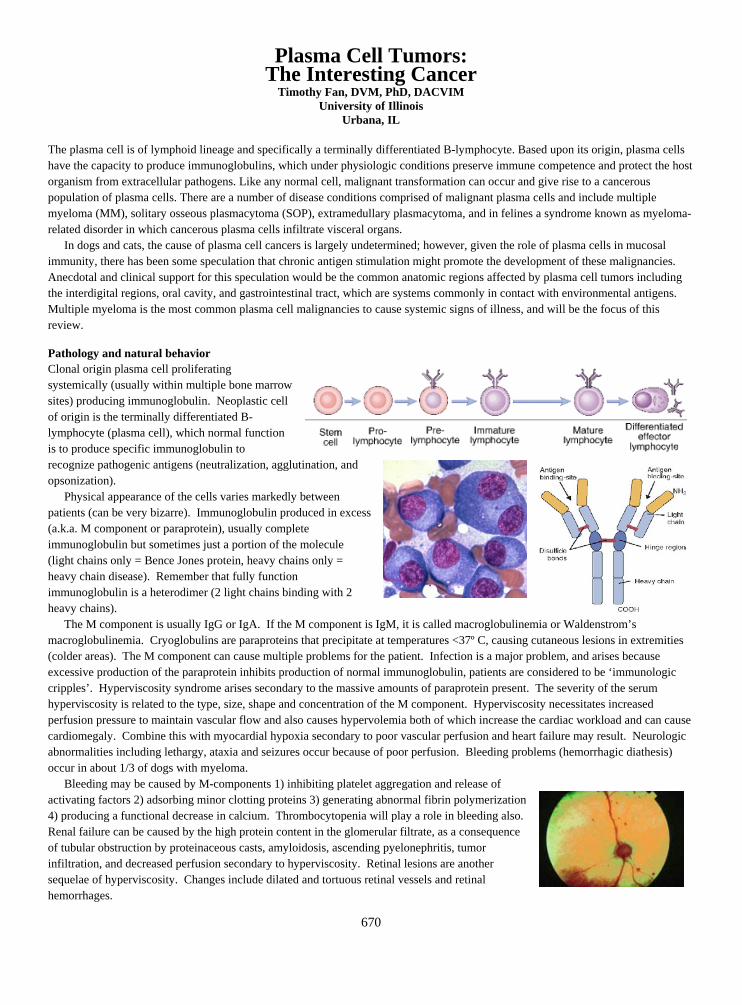
670
Plasma Cell Tumors: The Interesting Cancer
Timothy Fan, DVM, PhD, DACVIM University of Illinois
Urbana, IL
The plasma cell is of lymphoid lineage and specifically a terminally differentiated B-lymphocyte. Based upon its origin, plasma cells have the capacity to produce immunoglobulins, which under physiologic conditions preserve immune competence and protect the host organism from extracellular pathogens. Like any normal cell, malignant transformation can occur and give rise to a cancerous population of plasma cells. There are a number of disease conditions comprised of malignant plasma cells and include multiple myeloma (MM), solitary osseous plasmacytoma (SOP), extramedullary plasmacytoma, and in felines a syndrome known as myeloma-related disorder in which cancerous plasma cells infiltrate visceral organs.
In dogs and cats, the cause of plasma cell cancers is largely undetermined; however, given the role of plasma cells in mucosal immunity, there has been some speculation that chronic antigen stimulation might promote the development of these malignancies. Anecdotal and clinical support for this speculation would be the common anatomic regions affected by plasma cell tumors including the interdigital regions, oral cavity, and gastrointestinal tract, which are systems commonly in contact with environmental antigens. Multiple myeloma is the most common plasma cell malignancies to cause systemic signs of illness, and will be the focus of this review. Pathology and natural behavior Clonal origin plasma cell proliferating systemically (usually within multiple bone marrow sites) producing immunoglobulin. Neoplastic cell of origin is the terminally differentiated B-lymphocyte (plasma cell), which normal function is to produce specific immunoglobulin to recognize pathogenic antigens (neutralization, agglutination, and opsonization).
Physical appearance of the cells varies markedly between patients (can be very bizarre). Immunoglobulin produced in excess (a.k.a. M component or paraprotein), usually complete immunoglobulin but sometimes just a portion of the molecule (light chains only = Bence Jones protein, heavy chains only = heavy chain disease). Remember that fully function immunoglobulin is a heterodimer (2 light chains binding with 2 heavy chains).
The M component is usually IgG or IgA. If the M component is IgM, it is called macroglobulinemia or Waldenstrom’s macroglobulinemia. Cryoglobulins are paraproteins that precipitate at temperatures <37º C, causing cutaneous lesions in extremities (colder areas). The M component can cause multiple problems for the patient. Infection is a major problem, and arises because excessive production of the paraprotein inhibits production of normal immunoglobulin, patients are considered to be ‘immunologic cripples’. Hyperviscosity syndrome arises secondary to the massive amounts of paraprotein present. The severity of the serum hyperviscosity is related to the type, size, shape and concentration of the M component. Hyperviscosity necessitates increased perfusion pressure to maintain vascular flow and also causes hypervolemia both of which increase the cardiac workload and can cause cardiomegaly. Combine this with myocardial hypoxia secondary to poor vascular perfusion and heart failure may result. Neurologic abnormalities including lethargy, ataxia and seizures occur because of poor perfusion. Bleeding problems (hemorrhagic diathesis) occur in about 1/3 of dogs with myeloma.
Bleeding may be caused by M-components 1) inhibiting platelet aggregation and release of activating factors 2) adsorbing minor clotting proteins 3) generating abnormal fibrin polymerization 4) producing a functional decrease in calcium. Thrombocytopenia will play a role in bleeding also. Renal failure can be caused by the high protein content in the glomerular filtrate, as a consequence of tubular obstruction by proteinaceous casts, amyloidosis, ascending pyelonephritis, tumor infiltration, and decreased perfusion secondary to hyperviscosity. Retinal lesions are another sequelae of hyperviscosity. Changes include dilated and tortuous retinal vessels and retinal hemorrhages.
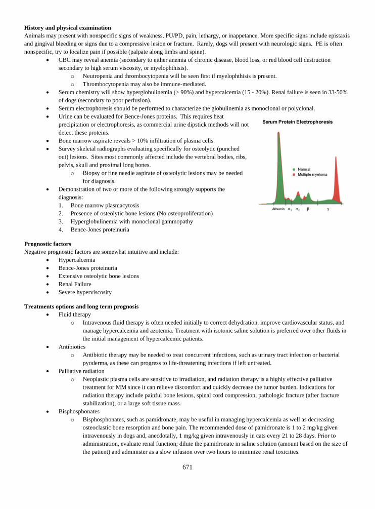
671
History and physical examination Animals may present with nonspecific signs of weakness, PU/PD, pain, lethargy, or inappetance. More specific signs include epistaxis and gingival bleeding or signs due to a compressive lesion or fracture. Rarely, dogs will present with neurologic signs. PE is often nonspecific, try to localize pain if possible (palpate along limbs and spine).
• CBC may reveal anemia (secondary to either anemia of chronic disease, blood loss, or red blood cell destruction secondary to high serum viscosity, or myelophthisis).
o Neutropenia and thrombocytopenia will be seen first if myelophthisis is present. o Thrombocytopenia may also be immune-mediated.
• Serum chemistry will show hyperglobulinemia (> 90%) and hypercalcemia (15 - 20%). Renal failure is seen in 33-50% of dogs (secondary to poor perfusion).
• Serum electrophoresis should be performed to characterize the globulinemia as monoclonal or polyclonal. • Urine can be evaluated for Bence-Jones proteins. This requires heat
precipitation or electrophoresis, as commercial urine dipstick methods will not detect these proteins.
• Bone marrow aspirate reveals > 10% infiltration of plasma cells. • Survey skeletal radiographs evaluating specifically for osteolytic (punched
out) lesions. Sites most commonly affected include the vertebral bodies, ribs, pelvis, skull and proximal long bones.
o Biopsy or fine needle aspirate of osteolytic lesions may be needed for diagnosis.
• Demonstration of two or more of the following strongly supports the diagnosis: 1. Bone marrow plasmacytosis 2. Presence of osteolytic bone lesions (No osteoproliferation) 3. Hyperglobulinemia with monoclonal gammopathy 4. Bence-Jones proteinuria
Prognostic factors Negative prognostic factors are somewhat intuitive and include:
• Hypercalcemia • Bence-Jones proteinuria • Extensive osteolytic bone lesions • Renal Failure • Severe hyperviscosity
Treatments options and long term prognosis
• Fluid therapy o Intravenous fluid therapy is often needed initially to correct dehydration, improve cardiovascular status, and
manage hypercalcemia and azotemia. Treatment with isotonic saline solution is preferred over other fluids in the initial management of hypercalcemic patients.
• Antibiotics o Antibiotic therapy may be needed to treat concurrent infections, such as urinary tract infection or bacterial
pyoderma, as these can progress to life-threatening infections if left untreated. • Palliative radiation
o Neoplastic plasma cells are sensitive to irradiation, and radiation therapy is a highly effective palliative treatment for MM since it can relieve discomfort and quickly decrease the tumor burden. Indications for radiation therapy include painful bone lesions, spinal cord compression, pathologic fracture (after fracture stabilization), or a large soft tissue mass.
• Bisphosphonates o Bisphosphonates, such as pamidronate, may be useful in managing hypercalcemia as well as decreasing
osteoclastic bone resorption and bone pain. The recommended dose of pamidronate is 1 to 2 mg/kg given intravenously in dogs and, anecdotally, 1 mg/kg given intravenously in cats every 21 to 28 days. Prior to administration, evaluate renal function; dilute the pamidronate in saline solution (amount based on the size of the patient) and administer as a slow infusion over two hours to minimize renal toxicities.

672
Aminobisphosphonates are an essential component of therapy for MM in people, and their use is associated with significantly reduced skeletal-related events and improved survival in some studies.
• Analgesics o Dogs and cats with MM may experience moderate to severe pain; treating for this pain is a priority. Pain may
be relieved by treating the underlying cancer and providing various analgesic therapies and supportive care. • Chemotherapy
o Although a cure is unlikely, MM can be a rewarding disease to treat since chemotherapy can greatly extend the quality and duration of life. The chemotherapy drugs most often used are alkylating agents, usually melphalan, combined with corticosteroids. However, eventual relapse during therapy is anticipated.
o The overall response rate for dogs treated with melphalan and prednisone chemotherapy is 92%, with 43% of dogs achieving a complete response and 49% achieving a partial response. The median survival time of dogs treated with this drug combination is 540 days, which is significantly longer than the survival time of 220 days in dogs treated with prednisone alone.
673
Glaucoma Micki Armour, VMD, DACVO
Eye Care for Animals Leesburg, VA
Glaucoma is a complicated and often frustrating cause of vision loss in small animals. The pathogenesis of glaucoma is only partially understood, however the end result is loss of retinal ganglion cell function, axonal destruction in the optic nerve, and vision loss. Because clinical signs of glaucoma have been described in humans without overt increases in intraocular pressure (IOP), and because optic nerve microcirculation and retinal ganglion cell function impairment have been observed before elevations in IOP in Beagle dogs with hereditary glaucoma, elevated IOP is now considered a ‘risk factor’ for glaucoma, not the primary cause. Moreover, glaucoma is considered to be a group of many diseases (rather than one single disease) with a common outcome.
Normal aqueous humor dynamics involves the production of aqueous humor by the nonpigmented epithelial cells of the ciliary body (via active transport, passive diffusion and ultrafiltration), with concurrent drainage from the globe via multiple mechanisms. In the dog and cat, most aqueous humor exits the eye through the iridocorneal angle and trabecular meshwork (‘conventional outflow’), with a smaller volume exiting the globe through uveoscleral vasculature (‘unconventional outflow’). To maintain a stable intraocular pressure, the rate of drainage must match the rate of aqueous humor formation. Diurnal variations in intraocular pressure have been observed in most species studied, and in the dog, intraocular pressure tends to decrease mildly with age. A ‘normal’ intraocular pressure in any given patient depends on multiple variables but intraocular pressures in excess of 25-30mmHg in the dog and cat are generally concerning.
Accurate evaluation of intraocular pressure can be difficult due to patient noncompliance or a multitude of other factors. Patient positioning, increased jugular pressure, tonometer used, excessive eyelid manipulation during measurements, corneal thickness and cleanliness of the tonometer have all been identified as factors contributing to erroneous IOP estimation.
In the dog, glaucoma is most commonly diagnosed as a primary disease. Abnormalities in iridocorneal anatomy can be observed on biomicroscopic evaluation (gonioscopy). These visible abnormalities are considered to be linked to microscopic abnormalities in the conventional drainage system as a whole. Dogs with abnormally appearing iridocorneal angles (excessively narrow or closed) are considered ‘goniodysgenic’, and are at risk of bilateral glaucoma in their lifetimes. Primary glaucomas have been classified as either ‘open angle’ or ‘closed angle’ in both human and veterinary medicine. Open angle glaucoma is most common in humans while in the dog the lion’s share of cases are closed angle. The difference is clinically significant, as open angle glaucomas are generally chronic, milder (increases in IOP of a few points are considered significant), and more responsive to medical therapy, while closed angle glaucoma as observed in the dog is generally associated with an acute, marked increase in IOP that is accompanied by pain and acute vision loss. It is also significant in that due to availability of funding, the majority of pharmacologic studies evaluating anti-glaucoma drugs are based on treatment of open angle glaucomas (including veterinary studies, in which a rare colony of Beagles with open angle glaucoma is the target of most pharmacologic medical research). In the cat, a rare form of glaucoma known as Feline Aqueous Humor Misdirection Syndrome (FAHMS) has been described in which changes in the anterior vitreous face result in aqueous humor accumulation in the vitreal chamber (rather than the anterior chamber), resulting in progressive anterior chamber shallowing and elevations in intraocular pressure. Most feline glaucomas, however, are secondary to chronic intraocular inflammation and can be especially difficult to treat given the dearth of effective anti-glaucoma medications in this species.
Common causes of secondary glaucoma include uveitis, intraocular hemorrhage, intraocular surgery, lens instability, retinal detachment, and intraocular neoplasia. The pathogenesis of the secondary glaucomas usually involves either pre-iridal fibrovascular membrane (PIFVM) development (in the cases of uveitis, intraocular hemorrhage, intraocular surgery, lens instability, retinal detachment and occasionally neoplasia), direct obstruction of the conventional outflow system (also in the case of lens instability and neoplasia), or both. PIFVMs develop over the anterior or posterior surface of the iris and grow anteriorly to occlude the iridocorneal angle. There is to date no known treatment available to prevent the development of these membranes in eyes at risk.
Commonly applied medical therapies for glaucoma are outlined below. Osmotic agents Osmotic agents are commonly used in emergency management of glaucoma due to rapid efficacy. They are administered systemically and distributed to extracellular fluids (ie plasma), increasing plasma osmolality. When plasma osmolality exceeds that of the intraocular fluid, water diffuses from the aqueous and vitreous humor down-gradient to plasma, essentially dehydrating the vitreal and aqueous chambers. Mannitol is administered IV at dosages ranging from 1-2 g/kg over 30 minutes. The reduction in IOP generally begins within 30 minutes-1 hour with effects lasting from 6-10 hours. Mannitol is not metabolized and therefore can be administered to diabetic patients. It should be administered through a filter given its propensity to form crystals. Glycerin is easy to administer, inexpensive and does not require intravenous access or special storage. It is administered orally at a dosage of 1-2 g/kg. A reduction in IOP should be observed within an hour of administration and can last as long as 10 hours. Administration may result in vomiting.
674
Glycerin should NOT be administered to diabetics, as it is metabolized to glucose and will result in hyperglycemia. Isosorbide can be administered orally like glycerin but unlike glycerin, will not result in hyperglycemia. The recommended dosage in dogs is 1-1.5 g/kg; efficacy in one report was similar to that of glycerin. Use of hyperosmotic agents is contraindicated in uveitic eyes due to the increased permeability of inflamed eyes. They should not be administered with fluids (and water should be withheld for ~2 hours post-administration). Due to the expected increase in intravascular volume associated with these agents, hyperosmotics should not be administered in patients with significant cardiovascular disease. Carbonic anhydrase inhibitors (CAIs) Carbonic anhydrase inhibitors inhibit formation of bicarbonate in the ciliary body that is necessary for production of aqueous humor. Commonly used topical agents include dorzolamide (Trusopt®) and brinzolamide (Azopt®). Oral CAIs include methazolamide and acetazolamide. Dorzolamide is available as a generic and is fairly cost effective. Topical CAIs can be administered 2-3 times daily. Maximum efficacy may take 4-5 days to achieve but decreased aqueous humor production occurs within 30 minutes – a few hours of dosing. The topical CAIs can be used in dogs and cats and are effective in both species. They can be used in all types of glaucoma, have no effect on pupil size, and do not contribute to intraocular inflammation. Dorzolamide is available as a combination drug with timolol (Cosopt®), which is now available as a generic. The efficacy of topical CAIs has been shown to be of equal efficacy to that of systemic CAIs. Methazolamide is dosed at 2-5mg/kg PO q12h and is available as a 50mg tablet. Acetazolamide is available as 125mg and 500mg tablets. The recommended dosage is 4-8mg/kg PO q8-12 hours. Commonly reported side effects of systemic CAIs include PU/PD, GI upset and panting (to compensate for metabolic acidosis). Systemic CAIs should not be used in patients with respiratory compromise. Cats appear to be more susceptible to metabolic acidosis and therefore systemic CAIs should be used with extreme caution in this species. Beta-blockers Beta blockers are very effective for reduction of IOP in humans and are the most commonly prescribed class of drugs for the treatment of glaucoma in people. Timolol is the most widely used of these medications in both human and veterinary medicine, but other topical beta blockers are available, including levobunolol, betaxolol, metipranolol and carteolol. Beta blockers reduce IOP by decreasing aqueous humor production but the exact mechanism of this effect is not completely understood. In the dog and cat their efficacy is considered relatively poor and bradycardia and mild (bilateral) miosis associated with their use has been documented. Their use in equine glaucoma appears more promising. In veterinary medicine, timolol 0.5% is administered twice daily. Use with caution in patients with cardiovascular disease.
Prostaglandin analogs The prostaglandin analogs appear to be the most effective drugs in the treatment of canine glaucoma. These drugs increase aqueous ouflow (with no effect on aqueous production). The mechanism of action is mediated through binding to prostanoid FP receptors. In the dog and human, activation of the prostanoid FP receptor results in increased uveoscleral outflow (ie unconventional outflow) through remodeling of the ciliary body musculature. Increased conventional outflow also occurs through morphological changes in the trabecular meshwork. The most commonly prescribed prostaglandin analog in veterinary medicine is latanoprost (Xalatan®), which is now available as a generic. Other available PG analogs include bimatoprost and travaprost. In the cat, latanoprost and other PG analogs are ineffective because activation of prostanoid EP receptors is required for similar effects in this species. The prostaglandin analogs are generally administered q12h in dogs (once daily in humans). In the dog, PG analogs result in maked miosis and because they work through activation of inflammatory mediators, should not be used in cases of secondary glaucoma caused by anterior lens luxation or chronic uveitis.
Other classes Other, less commonly utilized anti-glaucoma drug classes include the cholinergic agonists (pilocarpine, carbachol, demecarium bromide, echothiophate iodide), adrenergic agonists (dipivefrin), and alpha2-adrenergic agonists (apraclonidine, brimonidine). Due to associated local side effects, lack of availability and/or systemic side effects, they are seldom used. Surgical therapy The recommended surgical options often depend on whether vision is considered salvageable. Surgical options for the visual eye consist of laser ciliary body destruction and gonioimplants.
Endolaser cyclophotocoagulation (ECP) ECP is a relatively new surgical option. A diode laser is used to target the pigmented tissue of the ciliary body, thereby destroying the adjacent nonpigmented epithelium. The advantage of this procedure is that the laser is built into an endoscopic probe, allowing for localization and direct treatment of the ciliary processes with little bystander damage. Because the laser energy is likely to induce cataract development the process is often combined with phacoemulsification. No studies evaluating the long term efficacy of this surgery have been published, however preliminary results are promising.
Transscleral cyclophotocoagulation (TSCPC)
675
TSCPC involves laser ablation of the ciliary body, similarly to ECP. Both Nd:YAG and diode lasers have been utilized. The main difference between TSCPC and ECP is that TSCPC does not involve direct visualization of the ciliary body. Rather, external landmarks are used to position the laser probe over the external surface of the eye and the laser energy is directed through the sclera. Possible post-operative complications include excessive intraocular inflammation (with the potential to induce retinal detachment), cataract development, hyphema, and, as with any glaucoma surgery short of enucleation or evisceration, uncontrolled intraocular pressure. One retrospective study quoted a ~50% success rate in terms of IOP control with a ~20% success rate in terms of vision 1 year post-treatment. Immediate post-operative spikes in IOP are common and can result in post-operative vision loss.
Gonioimplants/valves Gonioimplants facilitate drainage of aqueous humor through a tubing system implanted directly into the anterior chamber. Both valved and nonvalved implants exist. Valved systems allow for egress of aqueous humor at intraocular pressures >12mmHg, while nonvalved systems are limited only by resistance of the tubing itself. Although these shunt systems are generally efficacious in the short term, post-operative uveitis with fibrin development and valve occlusion, requiring intracameral tissue plasminogen activator administration, is relatively common. The long term efficacy of shunts is limited by avascular bleb development, in which an avascular fibrotic capsule develops around the base of the device. This fibrosis is observed so commonly that gonioimplant failure is generally considered inevitable, and implant placement is often combined with a cyclodestructive procedure to provide more long term control of IOP.
Eyes that have lost vision are most commonly treated with one of the following surgeries to improve quality of life by controlling pain related to elevated IOP.
Enucleation Enucleation is probably the most commonly performed ‘surgery of comfort’ for glaucoma. Possible post-operative complications include hemorrhage, post-operative fistulas or mucocele development caused by incomplete removal of conjunctival, caruncular or third eyelid glandular tissue, and, rarely, orbital emphysema. In the cat, the optic nerve is relatively short and excessive traction on the enucleated globe must be avoided during surgery to prevent damage to the optic chiasm and contralateral blindness.
Cyclocryothermy Cyclocryothermy allows for reduction in IOP without loss of the globe. This procedure is generally reserved for the canine and has a reported ~80% success rate. Either nitrous gas or liquid nitrogen can be used. Using external landmarks to estimate the site of probe placement, the cryotherapy is applied externally. The ‘cryodose’ applied depends on the pre-operative intraocular pressure. The biggest advantage is that the surgery is noninvasive; post-operative therapy generally involves anti-inflammatories (often continued for life). Disadvantages include the potential for persistently elevated IOP, cataract development, hyphema, retinal detachment and globe phthisis.
Chemical ciliary body ablation Pharmacologic ablation of the ciliary body can be performed with gentamicin, or, as more recently described, cidofovir. Gentamicin is cytotoxic to both the ciliary body and retina and therefore should never be used in a visual eye. Even in blind eyes, however, this procedure should be used with caution as retrospective pathologic studies have reported an increased incidence of intraocular tumor development post-operatively. The outcome of this procedure is also the least predictable of the cyclodestructive procedures, with published reports citing a ~65% success rate. Other possible post-operative complications include cataract development, retinal detachment, hyphema, chronic uveitis and phthisis. Intravitreal gentamicin should NOT be administered to patients with renal compromise, as the drug is detectable in plasma post-operatively.
Evisceration and Intrascleral prosthesis (ISP) ISP surgery involves creating a 180 degree limbal incision at the corneoscleral junction, removal of all intraocular contents, and replacement of these contents with an silicone intraocular prosthetic. The advantages of ISP include complete resolution of glaucoma and maintenance of a ‘cosmetic’ globe. Disadvantages include the potential for recurrent corneal ulcerations due to decreased corneal sensitivity (caused both by historical glaucoma and transection of corneal nerves intraoperatively) and the potential for KCS. ISPs should generally be avoided in eyes with underlying intraocular neoplasia, pre-operative keratitis or KCS.
Unfortunately, glaucoma remains a disease with no effective ‘cure’, and although primary glaucoma often presents initially as a unilateral process, most ‘at-risk’ dogs will develop glaucoma in the contralateral eye within 1 year of diagnosis. Initiating prophylactic anti-glaucoma treatment in the normotensive eye has been shown to delay the onset of glaucoma in a dog at risk by as much as 18 months. Client education and early intervention can delay vision loss and improve quality of life for your patients. References Plummer, C.E., Regnier, A. & Gelatt, K.N. (2013) The Canine Glaucomas. In: Veterinary Ophthalmology (ed. Gelatt, K.N.), 5th ed., Vol.2, pp.1050-1145. Ames: Wiley-Blackwell.
676
How to Approach the Squinty Cat Micki Armour, VMD, DACVO
Eye Care for Animals Leesburg, VA
There are many potential pathogens that can lead to ocular irritation in cats; of note are Chlamydophila felis, Calicivirus, Mycoplasma, and Feline herpesvirus 1 (FHV1). Of these pathogens, FHV1 is by far the most incriminated when it comes to chronic and recrudescent conjunctivitis, blepharitis, and keratitis. Chlamydophila felis The most common ocular sign associated with Chlamydophila felis is chemosis. There are 2 morphologic forms of C. felis: the elementary body, which is an extracellular infectious form, insensitive to antibiotics, and the reticulate body, which is the intracellular form that leads to cellular damage. The reticulate form is sensitive to antibiotics. Transmission of C. felis is generally thought to be via aerosolization, direct contact, or contracted via fomites. Incubation is 3-5 days, and C. felis is shed for approximately 60 days after inoculation. Clinical signs include chemosis, hyperemia, serous discharge and blepharospasm. Cats less than 1 year are most likely to become infected; cats greater than 5 are fairly resistant. Chalmydophila infections tend to follow a chronic course, that typically begins with unilateral clinical signs that then can include the contralateral eye. The clinical signs typically resolve in a few weeks, although mild conjunctivitis can persist for months.
In a study (Vet Record, 1994) comparing 6 otherwise healthy to 6 FIV positive cats experimentally infected with C. Felis, clinical signs in control cats resolved within a mean of 109 days post-infection, while in FIV infected cats clinical sings persisted beyond 200 days.
Cytology of the intracytoplasmic reticulate bodies days 3-14 post-inoculation has poor sensitivity and specificity; false positives are common. PCR and serology after day 32 of inoculation are possible diagnostic tools. However, cell culture is considered the gold standard for the identification of C. felis. Cats vaccinated prior to conjunctival and nasal infections developed milder ocular and URI signs than unvaccinated cats, with a shorter shedding period. However, the vaccine doesn’t prevent infection or clinical signs, and can be associated with fever, lethargy, anorexia and lameness following vaccination.
Therapeutic options for the treatment of C. felis include doxycycline, azithromycin and clavamoc. Doxycycline appears to be the most efficacious for C. felis with clinical improvement in as few as 2 days. Calicivirus Calicivirus is a single stranded non-enveloped RNA virus, transmitted via aerosols or fomites. The virus typically incubates for 2-3 days, and results in acute respitatry tract signs, including glossal ulceration. It also results in polyarthritis and conjunctivitis in cats. It is generally considered a self-limiting disease process, which appears to resolve in 7-10 days. Topical antivirals do not appear to be effective as all of our topical antivirals target DNA, not RNA. Calicivirus also does not result in latent infections.
Calicivirus is diagnosed via cell culture, scrapings of respiratory of conjucntival secretions, immunohistochemical stainin, and electron biomicroscopy. Prevention is via the modified live or killed vaccine. Mycoplasma Mycoplasma felis and M. gatae are two isolates that are also incriminated in ocular irritation. Mycoplasma spp. Have questionable clinical significance because they have been isolated from clinically normal cats and have resulted in viariable conjunctivitis when administered experimentally. In addition, the disease process has not been replicated when cats were experimentally infected.
Diagnosis is via culure, cytology (noting the epithelial cytoplasmic basophilic inclusion bodies), and PCR (which has the highest sensitivity and specificity). Topical antibiotics have resulted in resoliution of ocular clinical signs. Feline herpesvirus 1 (FHV1) Feline herpesvirus 1 is an enveloped alphaherpesvirus known for its notorious chronic tissue latency. It is environmentally very fragile, and only lives 12-18 hours outside of a host. It is transmitted via direct contact or aerosolization, and infects the oral, nasal, and conjucntival surfaces. Like all herpesviruses, FHV1 is highly species specific. Its seroprevalence within the feline population has been reported as high as 97%.
Feline herpesvirus 1 infects the epithelial cells lining the respiratory tract, conjunctiva and corneal epithelium. The replicating virus results in cytolysis and causes inflammatory-mediated cell damage 4-6 days following exposure. Of note, it also established latency in the trigeminal ganglia, and can chronically and repeatedly travel to the eye via anterograde axonal transport, spontaneously shedding virus particles.
Maternal antibody protection wanes 8-12 weeks following birth. Although the course of the disease process can be self-limiting (10-20 days), it can result in severe ocular signs, including hyperemia, blepharospasm, chemosis, serous to purulent ocular discharge,
677
corneal ulceration and keratitis, and fibrinocellular exudate. Dendritic corneal ulcers are pathognominic for this disease process. It is often associated with a concurrent upper respiratory tract infection.
Cytology is often unrewarding, as it is likely only useful in primary infections and inclusion bodies are rare. Histopathology reveals epithelial necrosis 4 days following infection with a neutrophilic infiltrate, fibrosis and scarring. Viral detection is best performed via PCR using a dry cotton swab and performing a standard volume PCR assay. Virus isolation, fluorescent antibody, serum neutralizing antibody and ELISA testing have also been documented to detect FHV1. Prevention is difficult due to the high seroprevalence of FHV1, although vaccination may help protect against future disease episodes.
The contraindication of corticosteroids and FHV1 has been documented in the literature. Steroids have been associated with persistent and progressive corneal ulceration, neovascularization, prolonged viral shedding, corneal sequesta formation, calcific band keratopathy, and decreased tear production. Also, secondary bacterial infections may complicate viral flare-ups, and warrant the use of concurrent antibiotic therapy.
Topical and systemic antiviral therapy is often warranted. Potential antiviral therapeutic options include trifluridine, idoxuridine, vidarabine, cidofovir, acyclovir and famciclovir. Other therapies include the utilization of interferon, lysine and mucinomimetics.
Feline herpesvirus 1 may be associated with other disease processes, including eosinophilic conjunctivitis and keratitis, corneal sequestrum formation and dendritic dermatitis. References Veterinary Ophthalmology (ed Gelatt, KN) 5th Edition;, Vol2 Ames: Wiley-Blackwell
678
Managing the Bug-Eyed Dog Susan Keil, DVM, MS, DACVO Keil Veterinary Ophthalmology
Lenexa, KS
A significant percentage of an ophthalmologist’s weekly caseload is devoted to this subset of dogs. The goal of this lecture is to review factors to help you manage the ocular health of the brachycephalic individuals (Pug, Shih Tzu, Lhasa Apso, Boston, Japanese Chin, Pekingese). Educate your clients
• Special needs – use caution around dogs (including play), do not allow to hang out of car windows, recommend harness over collar (tugging increases ocular pressure), monitor if they sleep with their eyes (think about lubricating every night throughout life), no cage drying (hand dry)
• Grooming – start grooming very early (as a puppy, getting use to the noises, the table and routine of a groomer), allowing handling of face, utilize both reward and reprimand, need REGULAR grooming appointments
• Annual exams – take extra time on the eyes, always do a Schirmer tear test, look at lid apposition, evaluate corneal clarity, check nasal hair status
• Teach client when to reach out for help – o If the eye is red, but not painful o If the eye is red and painful o If the eye is red, painful, and getting worse o Meaning – call with any concern lasting over 24-28 hours!
**Great questions to help you determine severity over the phone = is the eye open or squinting/closed? Goals for the primary care veterinarian
• Reduce chronic pain • Recheck frequently • Identify and appropriately treat all ulcers, especially infected situations • Identify and refer descemetoceles and full-thickness corneal ruptures • Reduce the numbers of visually impaired and blind eyes • Reduce the numbers of enucleations • Extra attention for monocular bug-eyed patients – evaluate more than once annually if needed
Anatomy & physiology
Skull anatomy • dolichocephlic • mesaticephalic • brachycephalic
Eyelids • folds of thin skin continuous with facial skin with the free edges forming lateral / medial canthi • palpebral fissure is the opening between the lids • provide obvious physical protection • contain meibomian glands which produce meibum (outer tear film (TF) layer) to reduce evaporation • contain conjunctival goblet cells which produce mucin (inner TF layer) to attach TF to the cornea • distributes tear film around cornea • removes debris • these dogs have a shallow orbit combined with both a large globe and a large palpebral fissure
Blinking • upper lid provides more movement and protection than the lower lid • 10-20 per minute with 85% being yoked blinks and up to 66% being incomplete in brachycephalics closure
o CN VII – orbicularis oculi o CN VII – corrugator supercilii o CN III – levator palpebral superioris
Cornea • nerve sensitivity a MAJOR protector – often lost with years, microtraumas, chronic disease
679
• rapid blink and globe retraction with nictitans prolapse is a basic protective reflex which is very challenged in these dogs
• Cochet-Bonnet esthesiometer is an instrument which detects corneal sensitivity; brachycephalics • have the least corneal sensitivity of all breeds; additionally, the central zone is the least sensitive which is also the most
anatomically exposed region Reflexes
• corneal o afferent arm – ophthalmic and maxillary of the trigeminal (V) o efferent arm – facial (VII)
• palpebral o afferent arm – ophthalmic of trigeminal (V) o efferent arm – facial (VII)
• menace o afferent arm - retinal rods / cones and optic (II) o efferent arm – facial (VII)
The annual exam
Evaluation of the blink response: Is it complete? • Touch all four canthi – scrutinize! If not blinking completely, need to be lubricating life long!
o viscous, before bed, in am, more often? o need a permanent canthoplasty?
Quantitative assessment of the tear film: Measure aqueous layer with the Schirmer Tear Test (STT) • Do for a full 60 seconds (often 15 mm / 30 sec and 17 mm / 60 secs) • Do every year! May need to do more often! • Very under utilized for the red-eye exam!!
o 15 mm / 60 seconds is not adequate for a 1.5 year old Pug that cannot blink o Start immunomodulators if not adequate!
Cyclosporine (Optimmune (0.2%), compounded 1 or 2% in aqueous or oils) Tacrolimus (0.03% compounded in aqueous or oils, 0.5%)
Qualitative assessment of the tear film • Tear film breakup time (TFBUT) – evaluates the integrity of the mucin layer
o use rose Bengal dye o normal is a dry spot on the cornea @ 20 seconds o can be very challenging cases / very uncommon o need to lubricate constantly / not a specific therapy
Trichiasis: normal eyelid hair from normal follicle that deviates to contact the cornea/conjunctiva • Nasal • Caruncular – small, pink, globe-like nodule at medial canthus
o Shih Tzu, Lhasa Apso o wick tear, but do not poke
• Treatment: +/-resection, cryoepilation, manual epilation (temporary, will grow in more stiff) • May not require treatment
Trichomegaly: abnormally long normal eyelash • Clinically not a problem – does not require treatment, can complicate ocular hygiene • Cocker, Shih Tzu, Lhasa
Nasal fold trichiasis • Pug, Pekingese • Treatment: surgical resection if causing chronic irritation
o evaluate from the front and sides with eyes looking forward and moving right/left to determine if bothering the corneas
o closely look at corneal pigment and fibroses patterns to see if they match Distichia / districhiasis: normal cilia from an abnormal spot
• Grow from the meibomian gland duct o distichia are modified hair follicles following the path of least resistance out the duct
• May cause no problems (Cockers!)
680
• May be very problematic o Location, numbers, and combination of other surface disease are all factors
• May cause problems in the future o if in doubt, take it out! o if going under anesthesia for something else, remove if possible even if non-clinical
• Treatment: cryoepilation (liquid nitrogen, nitrous oxide) o tedious, boring, and may be very time consuming o lid splitting and electrolysis not recommended o small individual cryo units possible for very small numbers of hairs
Ectopic cilia: normal cilia from the meibomian gland but erupting through the palpebral conjunctiva • Not following the path of least resistance down the meibomian gland • Very painful! • Typically located from 10 to 2 o’clock on the superior lid (had one inferior case) • Can be very challenging to locate sometimes (need magnification–slit lamp, operating scope) • Think young dog with superficial ulcer that keeps coming back
o these are NOT refractory • Treatment: en bloc excision with magnification and cryo treatment
o Eyelid swollen but instantly dog’s comfort much improved Entropion: inward rolling of part or all of the eyelid margin causing hair-bearing skin to chronically rub on the cornea /
conjunctiva • Always clinically significant • A function of lid laxity • Mild (little inversion and loose contact with ocular surface) cases – epiphora, low-grade
o discomfort, excessive lacrimation • Moderate to severe cases cause constant trigeminal irritation, resulting in constant pain,
o secondary inflammation and ulceration/descemetoceles, self-trauma; o with chronicity pigmentation, scarring, vascularization, blindness result.
• Treatment: surgery o very undertreated condition o consistently hear how more active, energetic, engaging the dogs are after corrected
Ectropion: outward rolling of the eyelid margin causing conjunctival tissue to be exposed and lagophthalmos may occur • Lower eyelid typically involved • A function of lid laxity • May see cicatricial eversion of the upper lid • Typically not clinically significant – especially in brachycepahlics
o exposure of the conjunctiva may lead to low-grade conjunctivitis and predispose to epiphora and pre-corneal tear film deficiency
Euryblepharon = Macroblepharon = abnormally large palpebral fissure • Required feature in many breeds • Two different clinical presentations
o brachycephalics obtain exophthalmos through a shortened maxilla and shallow orbit with Euryblepharon resulting from protrusion of the globe forward through the fissure
o other types have an excessive eyelid length and the palpebral fissure is poorly supported by the globe o both types have a disturbance of the tear film dynamics and possible surface disease
• Treatment: may benefit medial / lateral canthoplasty Lagophthalmos: inability for a complete blink to occur
• Treatment: may benefit from medial / lateral canthoplasty Corneal clarity: pigment, vessels, edema, fibrosis
• For all these corneal changes, identifying the underlying disease important o KCS, trichiasis, Lagophthalmos, entropion, multi-factorial situation
The management plan
1. Encourage regular grooming with a groomer that can trim around the face safely and thoroughly 2. Practice / acclimate dog to getting drops and ointments 3. Annual, semi-annual, quarterly exams
681
4. Patient age and owner goals factor into both the short and long term plans 5. Require more aggressive integration of surgical treatments than for non-brachycephalic dogs 6. Be less tolerant of “mild” situations than for non-brachycephalic dogs 7. Life long medical requirements: lubricants before bed, constant corneal needs 8. No set protocol – modify for each patient 9. Often under treated for our convenience 10. More clients willing / wanting to do more
Medical management
Topical non-steroidals (NSAIDs) • Flurbiprofen • Ketorolac • Diclofenac
Topical steroid • Prednisolone acetate 1%: penetrates better • Dexamethasone 0.1% • NeoPolyDexamethasone 0.1%: do you need this combined with an antibiotic?
Immunomodulators • Cyclosporine
o Optimmune 0.2% ointment o 1%, 2% compounded solution
aqueous oils (coconut, sunflower, safflower, vegetable, other)
• The OILS may irritate – not the drug – switch to aqueous • Tacrolimus
o 0.03%, 0.5% aqueous oils (coconut, sunflower, safflower, vegetable, other)
• determine if patient annoyed or actually irritated by oil • oils have a longer contact time (providing lubrication)
Artificial tear products • For aqueous deficiencies
o 0.3% hydroxypropyl methylcellulose: Genteal, Tears Naturale, Bion Tears o 1% carboxymethylcellulose: Refresh Liquigel, Celluvisc o hydroxypropyl gel forming matrix: Systane o 0.3% hyaluronate: I-drop
• For lipid deficiencies o Caster oil, glycerin, poysorbate-80: Refresh endura o Mineral oil, polysorbate -80: Soothe emollient drops
• For mucin deficiency o 1% carboxymethylcelluose: Refresh Liquigel / celluvisc o hydroxylpropyl gel forming matrix: Systane ultra
• Gels o 0.3% hydroxypropyl methylcellulose: Genteal Gel o 1.5% carboxymethylcellulose: Tears again Night and Day gels
• Ointments o Petrolatum, mineral oil: Refresh PM, Tears Naturale PM, Lacrilube
682
Disorders of the Lens Susan Keil, DVM, MS, DACVO Keil Veterinary Ophthalmology
Lenexa, KS
A. Crystalline anatomy: The lens is transparent, avascular, and biconvex to support the jobs of refraction and image focusing on the retina. Because the lens is avascular, the aqueous humor provides nourishment and waste removal. Uveitis alters lens metabolism. The hyaloid and pupillary membranes provide the vascular network during embryogenesis. A. Lens regions
• Nuclear: central 80% • Cortical: surrounds nucleus – anterior, posterior, equatorial • Capsular: epithelial cell membrane semipermeable for nutrition & metabolic waste • Y-sutures: capsule epithelial cells under the anterior lens capsule
o replicate and migrate anterior & posterior to form new fibers o these tips join at the Y-sutures.
• Regular arrangement of fibers is responsible for the lens transparency • A cataract is a change in the fiber arrangement. • New fibers result in increased lens density as the lens is restricted in size (~ 6 yrs)
B. Congenital anomalies
• May be solitary or multiple ocular disorders • Variations in shape, location, size, and transparency occur • Etiologies during embryogenesis: toxic, inherited, nutritional, traumatic, infectious.
o microphakia, posterior lenticonus, coloboma, cataract, retinal dysplasia, microphthalmia, PPMs, persistent tunica vasculosa lentis
• Suspicious of congenital problems: o perform a complete bilateral exam after dilation o remove individuals from breeding programs o follow affected animals, monitoring for cataract & glaucoma o cataract surgery may be an options +/- IOL. o do have an increased risk of glaucoma and retinal detachments.
C. Cataracts
• Opacity of the lens/capsule regardless of size • Two common etiologies are inherited (congenital or acquired) and diabetes mellitus • 50% of diabetics will develop cataracts in 6 months; 80% in 1.3 years. • trauma, uveitis, electric shock, radiation, nutritional deficiencies less common. • classifications:
o age (congenital vs. developmental) o cause o location (capsular, cortical, nuclear, equatorial vs axial) o severity
incipient does not affect vision significantly (<10%) immature (vision compromise but not complete) mature (obscures image completely) hypermature (volume reduces to lens protein liquefaction) Morgagnian (hypermature cataract undergoing substantial resorption where the cortex liquefies and
the nuclear drops (uncommon, Cocker / Bichon) • feline: not by diabetes, commonly occur secondary to uveitis, inherited possible • diabetic intumescent cataract:
o aldose reductase enzyme catalyzes the reduction of glucose to intracellular sorbitol o sorbitol causes an osmotic shift, leading to significant swelling and fiber disruption o equatorial vacuoles (bubbles) progress to fiber swelling and rupture, o complete opacification, and Y-suture clefting.
683
• diabetic cataracts may develop in just days. o spontaneous lens capsule ruptures may occur due to the rapid osmotic lens swelling o (rapid forming genetic cataracts may also do this) o this situation is both a medical and surgical emergency.
A referral is recommended for ALL patients, even if the client is not interested in surgery. Many of these cases have lens-induced uveitis (LIU). LIU needs therapy. The goals are to prevent tertiary glaucoma and avoid shutting the door of surgical opportunity. D. Lens induced uveitis: up to 71% of mature, intumescent, hypermature cataracts have LIU
1. Phacolytic: uveal inflammatory response occurring secondary to lens proteins leaking through an intact lens capsule, resulting in lens shrinkage/capsule wrinkling.
2. Phacoclastic: spillage of lens proteins into the eye after the capsule ruptures E. Cataract management and surgery Should surgery be performed? This depends on numerous factors, such as the degree of visual impairment and the goals and finances of the client. Age is not a disease, but often a factor in client decision as is patient general health. Diabetics are over 50% of my surgery cases. They do not need to be completely regulated prior to surgery. Often, surgery often needs to be performed quickly to help eliminate LIU. The patients are significantly more active and engaged after surgery, I believe helping to maintain weight and regulation. Diabetics do well under anesthesia. All surgery patients, but particularly the diabetics, look and act 3-4 years younger.
There is currently no medical therapy for cataracts that has any clinical benefit for vision restoration. Do not fall prey to various dietary and holistic “cures” such as NuVet, Can-CTM, N-Acetyl-Carnosine. Proper nutrition may play a role in prevention, but once a cataract, always a cataract. Mature, intumescent, and hypermature cataracts increases risk of LIU, secondary glaucoma, instability, retinal detachment, capsule opacification.
Prompt referral is imperative to ensure the best ocular management and postoperative results. Some clients are unwilling or unable to visit an ophthalmologist. Recommendation in these cases is to monitor for both uveitis and glaucoma, use a topical anti-inflammatory (+/- atropine to control LIU), and monitor tear production.
Cataract surgery has improved dramatically over the past decades due to many improvements: phacoemulsification techniques, viscoelastic agents, intraocular lenses, anti-inflammatory drugs.
Successful outcomes also dictated by surgeon skill / experience, appropriate patient selection, meticulous attention to detail, and diligent post op treatment and monitoring. The surgery is very rewarding, a significant financial investment and a large time investment. Pre-op requirements: complete general and ocular exam, electroretinogram, ocular ultrasound, gonioscopy, appropriate pre-op blood work, +/- dental, +/- urine culture
If surgery is not performed, continue to monitor indefinitely. Complications w/out surgery include uveitis, glaucoma, luxation, and retinal detachment. Lim, et all 2011 found 20% of cases without medical or surgical therapy had a poor outcome (pain, aforementioned complications, enucleation, evisceration).
Unoperated eyes have a higher percentage of eventual glaucoma (20%) then operated (8%). Uncommonly younger dogs may have spontaneous resorption and regain aphakic vision (Cocker, Bichon). Sooner is always better
than later when referring / performing surgery. Topical NSAIDS / steroid may be required until no longer leaking protein. Exams recommended every 6 to 8 months for mature, hypermature, intumescent cataracts once stable.
Phacoemulsification and intraocular lens (IOL) implantation is the standard of care. Extensive training and equipment required. Surgical success recorded as 85-92%. It is important to get LIU controlled prior to
surgery. Bilateral typical at one surgery due to reduced cost, one anesthesia, one post op recovery, and one two week post surgery requirement for owner. Typically, 10-14 days of harness walk only, Elizabethan collars, and many meds. Rechecks at least at 1 day, 1 week, 1 month, 3 months, 6 months, annual.
The post-op management by a primary care veterinarian would include menace, PLR, STT measurement, IOP (with definite caution immediately post op, especially with Schiotz and even Tonopen) as well as examine anterior and posterior segment. Annual exams are recommend with an ophthalmologist to look for early complications. Some patients require low dose life long topical medications. F. Lens instability Primary (genetic) luxations - think all terrier breed, Shar Pei, cattle dog, Brittany, Border Collie, mini bull. There is of ten asymmetric bilateral disease. There are some genetic tests available for this autosomal recessive disorder.
Secondary luxations – glaucoma, hypermature cataracts, chronic LIU • Clinical signs: aphakic crescent, phacodenesis (lens tremble), iridodenesis (iris tremble), vitreous in anterior chamber,
corneal edema, lens in the wrong position (anterior / posterior / subluxated) • Case evaluation: measure IOP, assess function (dazzle, menace, PLR), and determine lens location.

684
Anterior luxations are considered emergencies if the eye is functioning (perform intracapsular cataract extraction (ICCE) but not an ER case if negative function (enucleation, evisceration). Contraindicated are the topical miotic agents (Latanoprost, pilocarpine, demecarium bromide). Utilize carbonic anhydrase inhibitors (CAI), beta blocker, and osmotic agents.
Posterior luxations are not emergency situations but a referral is recommended to assess both eyes and discuss long term goals, options, and medications. Topical miotics may be used to trap lens/lower IOP. Removing a subluxated lens is debatable. The conservative approach is to use a topical miotic. The surgical approach is an ICCE. Increased complications include retinal detachments and glaucoma. G. Trauma Both blunt and sharp trauma can result in significant damage, blindness, and globe loss. Penetrating sharp items (barb wire, cat claws, metal objects) may penetrated the anterior lens capsule (ALC), causing a cataract and phacoclastic uveitis. This requires a prompt referral for corneal repair and cataract surgery. Blunt damage (ball, kick), may also result in cataract, uveitis, luxation, glaucoma and retinal detachment (sometimes years later). Utilize topical NSAIDS and antibiotics (and often atropine) until an ophthalmology examination.
685
Equine Recurrent Uveitis Susan Keil, DVM, MS, DACVO Keil Veterinary Ophthalmology
Lenexa, KS
General information • ERU is the most common cause of equine blindness, affecting 8-25% of US horses. • An Essay on the Disease of the Eye of the Horse (1818) described a “specific inflammation” • aka – periodic ophthalmia, moon blindness (Vegetius / 4th century AD – cyclic nature) • other causes of uveitis include trauma, surgery, corneal disease • ERU has an initial acute uveitis separated by recurrences at variable time intervals • Etiology is not understood – genetic, infectious, environment, multi-factorial?
o is understood to be a dysregulated immune response responsive to steroids b. unresponsive to antibiotics c. inflammatory recurrences
• Of the affected horses, 1-2% has clinical disease considered vision threatening • High cost to the equine community • Still cannot be cured and may be difficult to manage • Peak age of presentation 4-6 years with no gender preference • May be either unilateral or bilateral • If a patient goes two years without a flare-up, the risk of future flare-ups is greatly reduced
Uveal tract anatomy
• Anterior uvea: iris and ciliary body (pars plicata and pars plana) • Posterior uvea: choroid • Blood ocular barrier (BOB) = blood aqueous barrier (BAB) and the blood retinal barrier (BRB)
Uveitis pathophysiology
• Uveal tract inflammation >> vascular congestion >> increased vascular permeability >> o BOB breakdown >> leakage of blood / cells, fibrin, protein into the aqueous humor >> visible as aqueous
flare, hyphema, hypopyon Histology
In the acute phase, the iris and ciliary body (CB) are first infiltrated by the neutrophil (hypopyon), then lymphocytes, plasma cells, and macrophages invade resulting in exudates of protein and fibrin in the anterior chamber (retina typically spared initially)
In the chronic phase, lymphocytes continue to infiltrate the iris and CB resulting in nodules. T lymphocytes predominate with a high % of CD4+; blood vessel walls and the ciliary processes thicken; with continued disease, all ocular structures are damaged histologically. ERU clinical classifications
Disease stage • acute: actively painful, obvious intraocular inflammation • quiescent: inactive; comfortable without visible active intraocular inflammation, but research has shown that even in
stages of quiescence there remains active inflammation • end stage: clinical signs include chorioretinal scarring ((butterfly lesion = wing-shaped hypopigmentation nasal and
temporal to optic nerve head) and small, circular focal depigmentation with central hyperpigmentation in the non-tapetal region), blindness, phthisis bulbi, mature / hypermature cataracts, synechia, retinal detachments
Inflammation location • anterior segment: clinical signs include pain, epiphora, conjunctival hyperemia, corneal edema, aqueous flare, miosis,
limbal neovascularization (often deep / short), corneal ulcers, keratic precipitates, hyphema, hypopyon, iris color change (rubeiosis or dullness caused by inflammation / edema), hypotony (5-15 mmHg), anterior / posterior synechia, can see calcific band keratopathy with chronicity
• posterior segment: involves choroid, vitreous, retina with anterior uveitis signs absent to very mild; clinical signs include vitreal cloudiness & opacities, retinal detachments, peripapillary chorioretinal scarring, difficulty in assessing fundus; affects warm bloods, European horses, draft breeds; least common type
• both segments (panuveitis): see a combination of anterior & posterior disease changes
686
Type of recurrence • classic: typical presentation of acute followed by quiescent phase, shorter periods between flares, increasing severity
with flare-ups; affects many breeds; sequelae include glaucoma, synechia, lens luxation, cataract, retinal detachment, optic nerve atrophy, blindness, phthisis bulbi
• insidious: chronic, persistent low-grade inflammation; acute stages often not noticed and not painful; gradual cataract formation, retinal degeneration, blindness; affects mainly Appaloosa and draft breeds; difficult for the owner to note a problem until progression is too late; thought to be a distinct disease from classic ERU
ERU pathogenesis hypotheses
Infectious antigen or agent in the uveal tract or vitreous after initial uveitis • molecular mimicry
Persistence of immune-competent sensitized T-cells in the uveal tract • autoaggressive T cell reaction against retinal proteins which leads to continueddestruction of intraocular tissues
Deposition of immune complexes ERU immunology
T cells (CD4+ with Th1-phenotype) are the main infiltrating cells • invade the iris and retina • form characteristic iris follicles • goal is to adjust treatment approach to specifically target T cell inflammation rather than general inflammation
The T cells target retinal autoantigens • S-Ag (S-antigen), IRBP (interphotoreceptor-retinoid binding protein), MDH (malate dehydrogenase), CRALBP
(cellular retinaldehyde binding protein) Horses carrying the gene coding for the MHC class I haplotype ELA-A9 are more susceptible in developing ERU
• 41% of warmblood horses with ERU carried this gene ERU diagnostics
• Complete ophthalmic exam • Complete general physical • CBC/Profile +/- Lepto serology
ERU in the Appaloosa breed
8x more likely to develop uveitis and 4x more likely to develop blindness vs all other breeds Insidious form! Duration and age of onset are challenging to document Two specific genetic markers have been identified in Appaloosa uveitis
• -MHC Class I and II markers (Kaese et al, 2008 o many human diseases linked to MHC alleles o MHC microsatellite association with Appaloosa ERU
Ume011 and EqMHC1 • does melanin play a role in immune dysfunction
o is melanin linked to the predisposition of immune system dysfunction? o is the lack of melanin a primary factor in development of the disease?
Coat color / pattern IS a factor with the disease • affected horses have an overall light coat pattern with focal darker spots • affected horses show annual coat color changes, often tending to lighten with age • non-affected horses tend to have a dark coat with a blanket
IEOC 2009 conference lecture by AE Dwyer • New York study of 160 horses over 11 years with uveitis
o 26% (42 horses) were Appaloosa o 74% (118 horses) were other breeds
• Seroreactivity to L. Pomona o 62% were seropositive o study population divided into four groups
Appaloosa that were seropositive (14) Appaloosa that were seronegative (28) other breeds that were seropositive (86)
687
other breeds that were seronegative (32) Horses presenting with bilateral disease
Leptospirosis
• Smallest spirochete bacteria that is motile and able to enter hosts (wounds, mucous membranes) • Aquatic unicellular organism in rivers, lakes, sewage • Principle reservoirs: deer, swine, rats, cattle, possum • Organism multiplies in the kidneys and shed in the urine • Horses infected by drinking contaminated water with cases most commonly seen in rainy seasons • Clinical signs: fever, icterus, anemia, anorexia, transient depression • Organisms localize in the renal tubules, genital tracts, and anterior / posterior ocular segments • Two most common serovars incriminated in disease are L. interrogans pomona and grippotyphosa • First reported in Ithaca, NY,1947, with horses developing ERU 18-24 months later • Lepto may have helped initiate ERU in some cases, but did not factor into the recurrences • To date, the exact mechanism of interaction between the horse’s immune system and the organism is not understood
ERU treatment
Goals • Control pain • Preserve vision • Reduce inflammation
Decrease inflammation • Systemic NSAID / corticosteroid
o flunixin meglumine*** use this first!!! dose: 1 mg / kg PO, IV every 12 to 36 hours (depends on disease stage) reducing to 0.5 mg/kg PO
every 12 to 36 hours risk: long term concern of gastric and renal toxicity Gastrogard recommended at preventative dosing (1/4 tube daily)
o phenylbutazone at consult often stopping this drug and starting flunixin* dose: 4.4 mg/kg PO, IV every 12 to 36 hours (depends on disease stage) risk: long term concern of gastric and renal toxicity
o aspirin o dexamethasone (Azium)
potent anti-inflammatory dose: 5-20 mg/day PO or 2.5 mg/day IM risk: frequent side effects including laminitis and GI concerns; use with caution and as a last resort;
taper off dose, alter management (decrease stresses / confinement, increase forage, decrease starches)
o prednisone potent anti-inflammatory dose: 100-300 mg/day PO, IM risk: see dexamethasone
• Topical NSAID / corticosteroid o pred acetate 1%***, dexamethasone 0.1%**
potent, excellent penetration, dose frequency depends on stage of disease hourly to weekly risk: predisposes to corneal fungal disease
o diclofenac 0.1%, flurbiprofen 0.03%, ketorolac good penetration, dose frequency depends on stage of disease hourly to weekly risk: decreases corneal epithelialization
• remember = NSAIDs are not 100% safe • Ocular injections
o subconjunctival triamcinolone potent anti-inflammatory
688
dose: 1-2 mg providing 7-10 day duration risk: cannot remove!; severe predisposition for bacterial or fungal keratitis
o intravitreal gentocin being utilized if patient demonstrating seropositivity to Leptospira dose 4 mg efficacy not proven, but clinical improvement noted in certain cases
o intravitreal • Surgical cyclosporine suprachoroidal implants
o suppresses T-cells o provides a slow release of CsA over 2-3 years o essential to control uveitis BEFORE surgery** o cases need to be referred earlier in general to have this option more accessible o surgery for visual eyes only (expense / general anesthesia)
Reduce discomfort and synechia • Mydriatic cycloplegic
o atropine 1% minimize synechia and provide pain relief dose depends on stage of disease (TID to every 72 hours) predispose to colic / decrease gut motility (overstressed)
ERU management
• Optimize systemic health o maintain good nutrition, regular activity, and regular fecal egg counts / deworming schedule
• Decrease risk of injury o keep enclosures safe
• Minimize inflammatory stimuli o wear a fly mask! (uv light flies, wind, other antigens) o routine hoof and dental care o control rodents, environmental insects o bedding type
• Keep up to date with vaccinations Blind ERU horses
• Review expectations – be realistic, acknowledging each situation different • Encourage the horse owner to start training and acclimation for blindness early, before vision is lost • Most horses will eventually loose monocular or binocular vision • Most horses will adapt, but this period can vary between weeks to months • Euthanasia may be elected for individual situation (expense, horse not adapting, safety, etc) • Find a calm companion (horse, mini, goat) and decrease stressors (wind, noise) • Keep the horse’s daily routine very regular • Really utilize other senses – sound, tactile • Teach verbal cues • ”Proof” the pasture • BlindHorses.org is a good source of advice • Realize that being blind +/- phthisis bulbi does not mean the patient is pain-free!
o really assess this situation if you have chronic epiphora • Enucleation may be needed to eliminate all discomfort
689
Equine Fungal Keratitis Susan Keil, DVM, MS, DACVO Keil Veterinary Ophthalmology
Lenexa, KS
Anatomy • Dimensions: 30 – 34 mm horizontal and 23 – 26.5 mm vertical limbus to limbus
o 780 micrometers thick centrally; thicker at the periphery • Histologically three layers with a pre-corneal tear film • Lipophilic superficial epithelium: 8-12 layers of nonkeratinized squam, wing, and basal cells • Hydrophilic stroma: 90% of thickness being 75 – 80% water • Lipophilic deep endothelium: single layer, basement membrane of which is Descemet’s
Physiology
• Nutrition: o Oxygen – tear film, atmosphere, slight from aqueous humor and limbal blood vessels o Glucose, amino acids, vitamins from the aqueous humor, slight from tears and blood vessels
• Microflora: o Primarily gram positive and fungal organisms with some gram negative
• Innervation: the cornea is highly sensitive, with innervation via the ophthalmic branch of CN V
Prevalence • Regionally frequent (humid climates, warm months) • Regional species variability
Clinical appearance
• Superficial keratomycosis o Microerosion: multifocal subepithelial whitish opacities, pain / secondary reflex uveitis o Superficial ulceration: more severe, defined epithelial loss, pain / secondary uveitis o Plaque formation (“cake-frosting” look): white-yellow necrotic stromal plaque with ulceration
• Stromal ulcerative keratomycosis*: most common, 50-80% of cases, severe pain / anterior uveitis o Corneal furrowing: rapidly occurs at any time, curvilinear region of stromal loss at ulcer edge o Melting o Perforation
• Stromal abscess Differential diagnoses
• Cytology, cultures (bacterial / fungal), PCR o Be prepared to repeat cytology +/- culture at second, third, fourth exams!
• Trauma / foreign bodies • Infiltrative ulcerative disease • Mixed fungal / bacterial infection (20-35% of infectious cases) • Calcific band keratopathy • Corneal degeneration • Corneal neoplasia
Pathogenesis
• Loss of the epithelium allows ocular surface and environmental fungal organisms to adhere, invade, colonize, and infect the stroma
• Protease production by epithelial cells, fungi, stromal fibroblasts, and inflammatory cells triggers extensive keratomalacia
• Fungal organisms known to inhibit in vitro angiogenesis • Deep stromal disease is perpetuated by the fungal affinity for the glycosaminoglycans near the Descemet’s membrane
Predisposing factors • Topical antibiotics
690
o Healthy gram positive microflora which produce balanced antifungal and antibacterial substances shift to gram negative in corneal ulcers
o Antibiotic use can exacerbate this use • Topical steroids • Alteration in corneal biofilm
Medical treatment The multiple goals are to attack the fungus and secondary bacterial infection, as well as manage corneal destruction, ocular pain, and secondary anterior uveitis
• Placement of a subpalpebral lavage tube o Think about formulation of medications you are dispensing – ointments do not go through a tube o Must be placed properly
Antifungal medication Most drugs are fungistatic because of the inability to achieve adequate concentrations in the presence of an intact corneal epithelium
• If the epithelium is lost, the drug concentration increases • High frequency administration is required to increase the drug concentration • Clinical experience – too much too fast increases risk of a melt down??
o this possibly results in clinical deterioration intense inflammatory reaction
o weigh this risk against stage of primary disease Therapeutic debridement
• debulks organism and increases drug penetration • Treat two weeks past the resolution of clinical signs***
Antifungal classes (three) Polyenes
• preferentially bind to ergosterol (fungi cell membrane sterol) • increases membrane permeability, leakage, oxidative damage • broad spectrum • good efficacy against filamentous and yeast fungi • best for ulcerative disease • start at every 2-4 hours, then taper • Natamycin 5% suspension
o only commercially available ophthalmic antifungal • Amphotericin B: increased epithelial toxicity vs Natamycin
Azoles • preferentially binds to fungal enzyme in cytochrome P450 system • inhibits ergosterol synthesis, increases membrane permeability, alters fungi cell enzyme systems • start at every 2-4 hours, then taper • Imidazoles
o ^Miconazole topical 1% good penetration through intact epithelium subconjunctival 1% - 5 to 10 mg q 24 to 48 hours
o ^Ketaconazole topical 1%: best for ulcers, poorest efficacy of azoles Triazoles
• ^Voriconazole topical 1% o good penetration through intact epithelium o refrigerate o systemic 3 mg / kg PO q 12 hours o good intraocular penetration with or without inflammation o more limited spectrum than other azoles
• ^Itraconazole 1% in 30% DMSO suspension o good penetration through intact epithelium o systemic 1.5 mg / kg IV q 24 hrs; 5.0 mg / kg PO q 24 hrs o poor intraocular penetration o if using PO, suggested to use IV solution as capsules have highly variable absorption
691
• ^Fluconazole topical 0.2% o poorer efficacy vs other azoles against filamentous sp. & yeast o systemic 14 mg / kg PO once, then 5 mg / kg PO q 24 hrs o good intraocular penetration with or without inflammation o more limited spectrum than other azoles
• Nucleoside analogs = Flucytosine: enzymatically altered within the fungal cell to the cytotoxic principal fluorouracil • Other antifungal treatments
o Silver sulfadiazine 1% ointment topical q 8 to 12 to 24 hrs metal ions bind microbial DNA and inhibit synthesis
• DILUTE Povidone – iodine solution o topical q 8 to 12 to 24 hours o germicide
Topical antibiotics • Targeted therapy is ideal • Prophylactic if primary organism is not identified • May require more than one antibiotic
o space frequency appropriately • Gram negative: Ofloxacin, ciprofloxacin, aminoglycosides, chloramphenicol (not Pseudomonas) • Gram positive: chloramphenicol, mofloxacin, Cefazolin / AT, neomycin, bacitracin • Topical mydriatic / cycloplegic: 1% atropine
o Reduces risk of posterior synechia and secondary glaucoma o Degree of mydriasis correlates with severity of cycloplegia
more severe uveitis will require more frequent treatments • Systemic NSAIDs
o Flunixin meglumine (1.1 mg / kg) PO or IV q 12 hours o Taper dose with control of disease
-typically tapered too soon • Topical antiproteases
o Autologous serum: serine protease entrapment o EDTA 0.2%: Ca / Zn chelation of MMPs o Tetracyclines: Ca / Zn chelation of MMPs (may also be administered oral) o N-Acetylcysteine 5-10%: Ca / Zn chelation of MMPs
• Systemic steroids o Not recommended due to increase systemic and ocular risks
• Omeprazole (Gastrogard) o Preventative dose: 2 mg / kg PO daily o Treatment dose: 4 mg / kg PO daily
• Oral / parenteral antibiotics o Penetration of drug into tear film, cornea, and anterior chamber unknown o Corneal inflammation and neovascularization may increase drug delivery o TMS, Potassium Penicillin, Gentocin o Doxycycline
antibiotic anti-inflammatory anticollagenolytic
• Subconjunctival medications: simply not ideal o Limited area o Unpredictable drug bioavailability; often need BID – TID to achieve therapeutic levels o Incited inflammatory response
Surgical treatment Utilize this option when medical therapy was delayed or is ineffective
692
Goals Remove infectious organisms and inflammatory cells Bring in structural support Bring in an immediate blood supply
o protease inhibitors o growth factors o fibroblasts
Indications Ulcers with >/ 50% stromal loss Ulcers with </ 50% stromal loss but associated with progressive malacia Descemetocele Corneal perforation / iris prolapse Monitor degree of uveitis
Procedures Conjunctival grafts (pedicle, bipedicle, free island, hood, bridge, 360 degree) Lamellar corneal grafts (corneoscleral or corneoconjunctival transposition; posterior lamellar (PLK) or deep lamellar endothelial keratoplasty (DLEK), penetrating keratoplasty) +/- conjunctival grafting Natural (autologous cornea, amnion) or synthetic / biosynthetic tissue (A-Cell, Biosys) graft
Medical treatment as indicated above after surgery Prognosis
• Infection, corneal digestion, and associated inflammation must be controlled • These cases are a huge financial, time, and emotional commitment • Client education and constant client support by the veterinary team is key • The use of the subpalpebral lavage system cannot be overemphasized • Treatment is often very prolonged (weeks (3) to months (4)) and costly • Vision: 50-90% of cases • Globe retention: 70-95% of cases
693
Evidence-Based Approach to Cranial Cruciate Repair Surgery Jennifer Wardlaw, DVM, MS, DACVS
Gateway Veterinary Surgery St. Louis, MO
Current research and literature will be reviewed to encourage the audience to update and make their own decisions regarding this multi-million dollar problem in our small animal patients. This lecture gets to the heart of which procedures are the best in skilled surgeons’ hands for our canine patients.
Cranial cruciate ligament rupture is a common cause of hindlimb lameness in dogs and is seen in cats as well. Patients can be managed without surgery with exercise restrictions, body weight management and pain medications. However, a better prognosis is achieved when the patients are less than 15 kg. Also, the presence of a meniscal tear or concurrent patellar luxation makes medical management less successful.
When surgical stabilization is opted for, the veterinarian is faced with a plethora of options. The key is to find the balance of what the surgeon is comfortable with and what the best option is for the patient. If the best possible option is chosen by the veterinarian but they do not have the training or experience to perform the procedure correctly, the potential complications can be disastrous. The fact that several options are available to address the same surgical problem indicates that no one procedure is perfect for all cases and all situations. Being current on the options and the data published is necessary to make the most educated decisions for your patients.
There are innumerable intraarticular repair methods in the literature and the theory behind these is the basis for human ACL repair. However, due to the degenerative nature of CCLR in dogs, these techniques have fallen out of favor. Intracapsular techniques are degraded by the inflammatory mediators seen in stifles with osteoarthritis (OA). The result is an unstable surgical repair and a lower level of function due to lameness with progressive OA. Long term outcomes with intracapsular repair are not as good as extracapsular techniques. However, if this is the procedure you are most comfortable with, and the owner will not accept referral to a surgeon, than this may be the “best” option for that patient.
The original extracapsular prosthetic stabilization has gone through many revisions and adjustments since its inception in 1966. The current technique is usually a lateral circumfabellar-tibial suture. Bone anchors can be used on the femur instead of around the fabella if preferred. The tibial suture is typically passed through a tibial bone tunnel located at the level of the long digital extensor tendon groove. Sutures can be tied or crimped. Nylon leader, monofilament or braided sutures are currently used, while stainless steel is no longer recommended due to cycling failure. The type of knot thrown can affect structural strength of some suture materials. For instance a surgeon’s throw may weaken knot security, but a square knot where the first throw is clamped to maintain tension while the rest of the knot is tied has not shown to weaken a number of suture materials. Crimps are available for use with specific prosthetic materials but are not interchangeable with sizes or types of sutures. Crimp placement requires addition equipment and slippage is found to occur in 8% of cases. However crimp placement has less elongation and more stiffness than a clamped square knot. The loop configuration of the prosthetic material has also been shown to influence performance. But in most cases, the tension of the suture is not conserved for longer than six to eight weeks after surgery. Most commonly the strength is lost through elongation or rupture. Despite positive clinical results, these techniques do not achieve normalization of stifle biomechanics to the cruciate deficient stifle and may not be the best option especially for large or overweight dogs.
Isometry and a stiffer prosthesis are the potential benefits of the TightRope CCL®. The FiberTape (Arthrex Vet Systems) used in the system has shown significantly greater stiffness and ultimate load to failure forces. However this puts the joint at risk if the prosthesis is over-tightened or if poor isometry is created with inaccurate bone tunnels. In a recent study the TightRope CCL® resulted in outcomes similar to that of the TPLO (Tibial Plateau Leveling Osteotomy). A multicenter study has shown 94% of dogs having good to excellent outcomes with a 9% major complication rate including implant failure, infection, and meniscal tear.
The TPLO surgery has historically been promoted for use in active large breed dogs or dogs with excessive tibial plateau slope. Several studies have found similar results six months postoperatively when comparing the extracapsular suture and the TPLO. However, the extracapsular dogs tended to be lighter and begin physical rehabilitation earlier than the TPLO group. It is possible that larger dogs treated with a lateral suture may have had a worse outcome. Clinically the TPLO dogs are believed to bear more weight sooner while the extracapsular dogs hold the leg up for 1-2 weeks. The TPLO surgery involves specialized equipment and is described as having a steep learning curve. Utilizing arthroscopy or a mini-arthrotomy is proposed to minimize patient discomfort over the arthrotomy used with the lateral suture technique. Complication rates with the TPLO are lower with unilateral or staged procedures ranging from 12-21%. A less specialized version of the TPLO is the Cranial Closing Wedge (CCW) which also lessons the tibial slope to negate tibial thrust, but also alters the mechanical axis of the tibia with a forward shift. This changes the biomechanics of the tibia and may change weight distribution on the menisci. The technique utilizes a saw but does not require a specialized bone plate. It can be combined with the TPLO in cases with excessive (greater than 30°) tibial slope.
The Tibial Tuberosity Advancement (TTA) is a newer procedure that eliminates cranial tibial thrust. The mechanics place the patellar tendon force perpendicular to the weight-bearing force through the stifle. A bone graft appears to be beneficial for speeding
694
the healing of the boney defect created. Specialized equipment is required but the procedure is technically less challenging and perhaps faster than the TPLO. Long term studies show similarities between the TPLO and TTA, although the TTA appears to take longer to heal the osteotomy and cannot be used in cases with excessive tibial slope. Implant designs are still changing with regards to fork design and available cage sizes for advancement. The overall complication rate for TTA ranges from 25-59%, including minor complications.
All of the osteotomy techniques require strict confinement while the bone heals. This may be a deciding factor between techniques in ill mannered dogs or outdoor-only animals. While physical rehabilitation is started early in all dogs, the postoperative care for the osteotomy dogs can be weeks to month longer than the lateral suture technique. However, early return to function is vital for joint health, and to rebuild muscle mass and regain lost bone density. Service or therapy dogs who are kept in a controlled manner will likely benefit from the quick return to weight bearing of the osteotomy procedures, with their daily activities being as controlled and calm as most rehabilitation programs.
The existence of so many variations on the same surgical problem has shown no concrete superior method for treating our veterinary patients exists to date. Research is ongoing to illustrate the pros and cons of the newer techniques to determine the best options. Kinematic and objective controlled multi-center prospective trials are needed. But patient needs and variation in fibrosis, activity level, meniscal damage and age along with owner financial constraints will all play into the decision of the “right” treatment modality.
695
Surgical Options for Repairing Luxating Patellas Jennifer Wardlaw, DVM, MS, DACVS
Gateway Veterinary Surgery St. Louis, MO
Soft tissue and orthopedic procedures are both needed to correct luxating patellas. The intricate details of both will be discussed with focus on anatomy and accuracy. Postoperative care and physical rehabilitation will also be discussed.
Patellar luxation can be congenital or traumatic. Congenital is typically the presentation for small and toy breed dogs. Although some large breed dogs can have it as well. It is typically bilateral, with medial being the most common side of the luxation. Often times one side may be more severe than the other side. Traumatic patellar luxation is typically unilateral in nature and tends to be laterally luxated.
The patella is a sesamoid bone in quadriceps mechanism and uses the straight patellar tendon to insert on the tibial tuberosity. Although the true pathogenesis is unknown, it may results from abnormal hip conformation, angle of inclination, or coxa vara. Any of these components that cause a malalignment of the quadriceps mechanism, can lead to patella luxation. After the patella has been luxated is leads to secondary changes to the limb including medial displacement of the quadriceps, lateral bowing of the femur, torsion of the distal femur, shallow trochlear groove, stifle instability and medial displacement of the tibial tuberosity.
Clinical signs are very suggestive of the disease with an intermittent lameness or “hopping”. Animals may also have a crouched stance or “bowlegged” appearance. Lameness often increases as degenerative joint disease develops. Diagnosis is based on clinical signs and physical examination. However, radiographs are needed of the entre hindlimb to assess for torsional deformity, hip conformation and other orthopedic issues as well. Radiographs will show the displacement of the patella on the craniocaudal view. Shyline views can also be used to assess the trochlear groove. Documentation of secondary arthritis is also important for prognostic goals following surgery.
The patella luxation grading system is currently used to help elicit when surgery is needed and judge the level of surgery needed to correct the limb abnormality. Grade 1
• Intermittent luxation • Patella can be manually luxated, but reduces spontaneously • Rarely lame, occasionally skip • Minimal medial tibial rotation
Grade 2
• Frequent luxation • Patella luxates with stifle manipulation, reduces spontaneously with rotation of the tibia • Lameness varies- occasional skip to weight bearing lameness • Medial tibial rotation- up to 30°
Grade 3
• Patella is luxated but it can be reduced, reluxates • Chronic lameness of varying severity • Medial tibial rotation of 30° to 60° • Moderate angular and torsional deformities
Grade 4
• Patella is luxated continually and cannot be manually reduced • Limb is carried or the animal moves in a crouched stance • Medial tibial rotation of 60° to 90° • Marked angular and torsional deformities
Surgical repair goals are to realign the extensor apparatus, normalize the forces acting on the physes/cartilage and stabilize the patella in the trochlear groove. Soft tissue reconstruction is helpful, but not a surgical solution to this orthopedic condition by itself. More commonly used soft tissue procedures include; Imbrication of the lateral retinacular fascia, Patellar and tibial anti-rotational sutures, Medial release (desmotomy). Bone reconstruction is far more successful and should be the cornerstone of any patellar luxation surgery. Orthopedic corrections can include; Tibial tuberosity transposition, Trochleoplasty techniques, Corrective osteotomies of femur / tibia. The goal os surgery is to improve limb function in dogs with lameness. Surgery does not prevent the progression of OA.

696
Reluxation is common with up to 48% reluxation in one study. However relaxation is usually mild (grade 1), and clinical signs may be minimal, negating the need for further surgery.
697
Managing Hip Dysplasia in Young Dogs Jennifer Wardlaw, DVM, MS, DACVS
Gateway Veterinary Surgery St. Louis, MO
At birth most hips are normal. The femoral head and neck are cartilaginous and begin forming bone by endochondral ossification. Joint congruence and stability are dependent on periarticular soft tissues. This congruency and stability is critical for normal joint development. Disparity in the development can happen with any boney part of the joint or soft tissue including muscles, ligaments and the joint capsule. The skeleton develops rapidly and small problems can rapidly lead to a chain reaction of disease. If the hip joint is lax or unstable it leads to poor joint congruence which causes subluxation and further abnormal hip development. A dog may have normal hips at birth but through genetics, nutritional or environmental factors, develops hip dysplasia (HD).
Nutritional influences such as a high plane of nutrition or imbalance can lead to HD. A high plane of nutrition affects growth rate and can lead to rapid bone growth and weight gain. This can over-load the soft tissue support and has been shown to increase the frequency and severity of HD. Studies have shown a faster than average weight gain may lead to HD, even with exercise restriction. A dietary electrolyte imbalance may affect the synovial fluid. A low dietary anion gap (sodium + potassium –chloride) results in less subluxation while excess may increase synovial fluid amount and joint laxity. This may be due to surface tension and hydrostatic pressure.
Paying special attention to “at risk” puppies during initial examination is a key component to managing these patients. Asking pertinent questions about their normal activity, keeping them lean and on a balanced diet to avoid adverse nutritional influences are key. In puppies that are large or giant breeds, or have known familial histories perform an Ortolani and Barden exam. Also consider switching off puppy formulas at 6 months to slow the rate of growth. You may also want to consider prophylactic management.
A radiographic diagnosis of HD is more difficult in younger dogs but can be performed with various techniques. The hip extended view is used by the Orthopedic Foundation for Animals (OFA), and the Norberg Angle. Distraction radiography is used in the Penn HIP Program and Dorsolateral subluxation techniques. The OFA scale does not require special equipment but identifies OA and is not a sensitive method to detect early or mild laxity. You can also not certify with OFA until they are 2 years of age making it a difficult screening test for puppies unless they are severely affected. PennHIP requires certification to submit films as well as sedation or anesthesia of the patients. You need three mandatory radiographs. The distraction index is calculated off the percent of the femoral head that is luxated out of the acetabulum. A distraction index of greater than 0.3 is considered disease susceptible, but breed variation of measurements exist. This modality has been shown to be statistically predictable at 16 weeks of age.
Once you have a diagnosis or have decided for early prevention, time is one your side since you caught it early. Medical management is 80% successful and is clinically more helpful the earlier you begin. Weight control or reduction is the cornerstone to minimize the stress of the growing active joints. A regulated exercise program should be utilized but not overdone. OA disease modifying agents or nutraceuticals can be started early. Physical rehabilitation can be tailored for a puppy and includes homework for owners that promote not only joint health but obedience and training. NSAIDs can safely be used in puppies after 2 months if pain is an issue. The key to conservative treatment or prevention of HD is the multimodal approach. Controlled exercise programs should be designed for the active playful puppy. Consider postponing strict training until they are at least 6 months of age. Excessive force even on normal joints can cause OA. Agility, flyball, sporting and rescue training should be “walk through” training to get the idea and the motions without the force. Exercise is good in moderation and will help reduce obesity as well as maintain a good range of motion. Low impact exercise can be used liberally including swimming, walking, obedience class and leash training. Studies have shown that even with radiographic evidence at a young age of HD, weight control and leash walking can dramatically increase the range of motion, exercise tolerance and long-term function for years.
Nutraceuticals have been shown to be the most beneficial in offsetting OA when given before the inflammation starts, meaning preemptively when we suspect disease. Since they have minimal if any side effects and the potential for a large impact, it is easy to prescribe them to owners who are willing. Nutraceuticals have been called disease modifying agents, disease modifying osteoarthritic drugs, supplements, additive and vitamins. The key to understanding the options are to realize the FDA does not regulate these products for efficacy or quality. It is vital you find a company you like, believe in and has research to support their products and claims. If you are using a product and not seeing results, then try a new source. Some options work better for certain cases, but generally speaking when added to a well balanced multimodal approach can make a big difference with regards to patient comfort and cartilage health. Most contain glucosamine and chondroitin sulfate in various forms. It is reported that they are absorbed by the GI tract, become incorporated into joint tissues, and provide the necessary precursors to maintain cartilage health and decrease inflammation. Anecdotal reports, in vitro studies, and published clinical trials indicate that these agents are effective in treating OA.
Glucosamine is an amino-monosaccharide nutrient that has exhibited no toxicity even at high oral doses. It is a precursor to the disaccharide unit of glycosaminoglycans, which comprise the proteoglycan ground substance of articular cartilage. Studies using radiolabeled compounds in man and animals have shown that 87% of orally administered glucosamine is absorbed. Glucosamine acts
698
by providing the regulatory stimulus and raw materials for synthesis of glycosaminoglycans. Since chondrocytes obtain preformed glucosamine from the circulation (or synthesize it from glucose and amino acids), adequate glucosamine levels in the body are essential for synthesis of glycosaminoglycans in cartilage. Glucosamine is also used directly for the production of hyaluronic acid by synoviocytes.
In vitro biochemical and pharmacological studies indicate that the administration of glucosamine normalizes cartilage metabolism and stimulates the synthesis of proteoglycans. In one study, glucosamine stimulated synthesis of glycosaminoglycans, proteoglycan and collagen, suggesting it not only provides raw material for their production, but may actually up-regulate synthesis. The effects of glucosamine sulfate on human chondrocyte gene expression was also evaluated, assessing its effects on type II collagen, fibronectin and proteoglycans in normal adult chondrocytes. Glucosamine modulated the expression of cartilage proteoglycans, decreased stromelysin mRNA levels in osteoarthritic chondrocytes, and preserved the constitutive expression of type II collagen and fibronectin in both normal and osteoarthritic chondrocytes.
Chondroitin Sulfate (CS) is a long chain polymer of a repeating disaccharide unit. It is the predominant glycosaminoglycan found in articular cartilage and can be purified from bovine, whale, and shark cartilage sources. Bioavailability studies in rats, dogs and humans have shown 70% absorption of CS following oral administration. Studies in rats and humans using radiolabeled CS have shown that CS does reach synovial fluid and articular cartilage.
When human articular chondrocytes were cultivated in clusters in the presence of CS, proteoglycan levels were significantly increased and collagenolytic activity was decreased. A similar study indicated that CS competitively inhibited degradative enzymes of proteoglycans in cartilage and synovium. In a study of rabbits with chymopapain-induced stifle arthritis, proteoglycan depletion was reduced by the administration of CS.
Clinical trials in humans have also found CS to be effective in reducing the symptoms of OA. In a placebo-controlled, double-blinded study of 120 patients with OA of the knees and hips, treatment with CS resulted in significant improvements in pain-scale scores and pain-function index. In another study of 42 patients with knee OA, CS treatment significantly reduced pain and increased joint mobility. Bone and joint metabolism (as assessed by various biochemical markers) also stabilized in the patients treated with CS while remaining abnormal in patients receiving a placebo. Hyaluronate concentrations and viscosity were increased, and collagenolytic activity was decreased, in the synovial fluid of OA patients treated with CS for 10 days. These clinical trials indicate that CS has a positive effect in controlling the symptoms associated with OA. Combinations of glucosamine and chondroitin sulfate are commonly used and it has been reported that these agents work synergistically.
Dasuquin ® (Nutramax Laboratories, Inc.) is a joint nutraceutical marketed for management of OA in dogs and cats. It is a combination of glucosamine, chondroitin sulfate, decaffeinated tea polyphenols, and avocado/soybean unsaponifiables (ASU). Tea polyphenols may have a positive effect on cartilage health and provide oxidative balance in the body. ASU, which are biologically active lipids, have been shown to be more effective than chondroitin sulfate in inhibiting the expression of certain OA mediators responsible for cartilage breakdown. In in vitro studies, ASU has been shown to decrease the expression of COX-2 enzyme, TNF-α, IL-1β, and PGE2 in chondrocytes. It was also shown to stimulate synthesis of cartilage matrix by increasing levels of TGF-ß. A 2007 study found that dogs given ASU for 3 months had elevated levels of TGF-ß in their synovial fluid compared to control dogs. The combination of ASU with glucosamine and chondroitin sulfate decreased the expression of numerous pro-inflammatory mediators, including TNF-α, IL-1β, and iNOS. This decrease in pro-inflammatory mediators seen with Dasuquin® (Cosequin® with ASU) is greater than that seen with Cosequin® alone. In an in vivo study of the effects of Cosequin® on cartilage metabolism in dogs, serum samples were collected after treatment with Cosequin® and tested for circulating glycosaminoglycan content. Median serum glycosaminoglycan levels were significantly increased in treated dogs. When normal calf cartilage segments were exposed to the serum from the treated dogs, the biosynthetic activity of chondrocytes was significantly increased and proteolytic degradation of the cartilage segments cultured in serum was reduced. In vitro studies at the Nutramax laboratories also demonstrated the beneficial effect of Dasuquin® on chondrocytes from different species incluing equine, camelid, canine, feline and bovine. Dasuquin®I inhibited the production of inflammatory mediators and signaling molecules in the inflammatory cascade.
Omega acid supplementation was discovered when dermatologic patients were experiencing relief from their OA. Maintaining a high content of the long chain omega-3 fatty acids EPA, and DHA is the key with this nutraceutical. Short chain omega-3s compete with omega-6s for conversion to long chain fatty acids and then for uptake into cell membranes. Omega-3s and omega-6s have different effects on the inflammatory response. Omega-6 arachidonic acid is the precursor to more pro-inflammatory mediators. While omega-3 EPA is a precursor to less potent inflammatory mediators. Omega-3s are readily available from several companies for veterinary as well as human products. Pet foods that contain them must be kept in a sealed bag for less than 30 days or they dry out. Fish oils will also help lubricate the skin and shine the coat. For large breed dogs I follow the human label recommendation for full grown dogs or half the dose for puppies. If you overdose the oils, they can have soft stool or diarrhea and should decrease the dose.
Some other options that are developing for easy oral administration include green-lipped mussel, methyl-sulfonyl-methane, duralactin and S-adenyl-L-methionine. Less research or anecdotal evidence exists for these but is continually being developed.
699
The use of joint nutraceuticals in dogs prior to the development of OA is controversial. No controlled studies have been reported that document the efficacy of nutraceuticals in preventing the development of OA. However, because of their reported effects on improving cartilage matrix and reducing levels of inflammatory mediators within the joint, many clinicians have advocated the prophylactic use of joint nutraceuticals, particularly in athletic and large dogs that might be susceptible to joint injury. Additional research is needed to confirm the value of prophylactic use of joint nutraceuticals.
There are also surgical options to diminish the signs of OA in puppies that have HD. The two surgical options are Juvenile Pubic Symphysiodesis (JPS) and Triple Pelvic Osteotomy (TPO). JPS is a simple procedure performed on puppies 12 to 20 weeks of age. But the optimal results are achieved on puppies less than 16 weeks old. Please note that this age is before the PennHIP certification age. The procedure fuses the pubic symphysis with electrocautery via a ventral midline incision. There are no implants and, with proper protection of the urethra and depth to avoid the colon, very few potential side effects. Electrocautery is used every 2-3 mm along the symphysis to cause thermal necrosis and premature closure. The pelvis continues to grow in all other planes while being static at the pubis, resulting in ventroversion of the acetabulum. This procedure is not readily detectable on OFA and PennHIP films and should therefore only be performed on animals that will be sterilized to avoid certifying or breeding falsely represented hip conformation. The TPO is typically performed on dogs less than 10-12 months of age without radiographic signs of OA. It is used to correct hip laxity. Three osteotomies are made on the pubis, ischium and ilium to allow reorientation of the acetabulum. Then an angled plate is placed on the ilium to secure the weight bearing axis for boney healing. The forced manual ventroversion increases dorsal coverage of the femoral head and reduces the formation of OA by improving joint stability and congruence. However bilateral surgery is not performed due to high complication rates and surgeries should be staged at least 4 weeks apart. Potential complications include a narrowed pelvic canal, sciatic neuropraxia, implant failure and an abnormal gait. Lameness improves in 92% of dogs and the progression of OA appears to be slowed with this procedure. The JPS and TPO procedures have similar effects on hip conformation, although neither eliminate laxity or completely cure HD. They can arrest or limit the progression of HD in mild to moderate cases. Both of these preventative surgeries require early puppy screening and counseling of owners about potential benefits and expected outcomes.
700
Managing Hip Dysplasia in Old Dogs Jennifer Wardlaw, DVM, MS, DACVS
Gateway Veterinary Surgery St. Louis, MO
At birth the hips are normal. The femoral head and neck are cartilaginous and begin forming bone by endochondral ossification. Joint congruence and stability are dependent on periarticular soft tissues. This congruence and stability is critical for normal joint development. Disparity in the development can happen with any boney part of the joint or soft tissue including muscles, ligaments and the joint capsule. The skeleton develops rapidly and small problems can rapidly lead to a chain reaction of disease. If the hip joint is lax or unstable it leads to poor joint congruence which causes subluxation and further abnormal hip development. A dog may have normal hips at birth but through genetics, nutritional or environmental factors, develops hip dysplasia (HD).
A presumptive diagnosis of HD can often be made based on clinical signs or physical examination. Palpating for crepitus, a luxated hip or performing the Ortolani and Barden maneuvers can all help make a correct diagnosis of HD. A radiographic diagnosis of HD is more easily made in older dogs. The hip extended view is used by the Orthopedic Foundation for Animals (OFA), and the Norberg Angle. Distraction radiography is used in the Penn HIP Program and Dorsolateral subluxation techniques. The OFA scale does not require special equipment but identifies OA yet is not a sensitive method to detect early or mild laxity.
Medical management is 80% successful and is clinically more helpful the earlier you begin. Weight control or reduction is the cornerstone to minimize the stress diseased joints. A regulated exercise program should be utilized but not overdone. OA disease modifying agents or nutraceuticals can be started early. The key to conservative treatment of HD is the multimodal approach. Excessive force even on normal joints can cause OA. Exercise is good in moderation and will help reduce obesity as well as maintain a good range of motion and comfort.
Weight loss is the easiest and perhaps most beneficial part of a multimodal approach to OA. Minimizing the work the diseased joints have to contend with should be paramount to any regime. Dogs with OA should be kept on the thin side of normal. With proper weight management, many dogs are able to stop taking pain medications until much later in the disease process. Commercially available diets are geared towards weight loss as well as joint comfort. Diets should be low calorie and low in protein while providing an otherwise balanced nutritional plane. Having truthful conversations about treats and table scraps should be geared to reveal honest habits. Caloric responsibility should be encouraged and adjustments made to account for the dogs’ favorite treats or foods. Exercise is also important to maintain a good range of motion and weight level. Minimizing concussive forces like stairs, jumping, climbing, running, and horse play should be minimized while still maintaining a good quality of life. Encourage leash walks, swimming and pay close attention to what activities make them sorer afterwards. While we don’t want to lock our patients in a box or take away their quality of life, easing their burden is important for their joints. If they love to play fetch on the weekends, make their owners aware that that will be a painful time and they should premedicate or otherwise adjust the protocol for their pet. Having thick warm bedding should also be encouraged to help aching joints. If an overweight animal prefers the hard, cold floor, suggest placing a fan near the orthopedic bed to encourage usage.
NSAIDs are readily available and widely used for OA in dogs. The important thing is to find a drug that works well for each patient and to make the owners aware of potential side effects. If one stops working for a patient, try switching to a different one. When switching NSAIDs, a wash out period of at least two half-lives is recommended. NSAIDs can be used for painful flair ups, around times of increased activity, or later in the disease, for daily maintenance pain relief. For patients with NSAID sensitivities or for patients needing additional pain medication there are other options as well. Tramadol is a synthetic mu opiod with a wide safety margin. It can be given several times a day which make it ideal for use around exercise or physical rehabilitation periods. I typically use 5 mg/kg up to 4 times daily. Gabapentin is a GABA analogue design to treat epilepsy but is widely used for neuropathic pain and OA in people. The most common side effect appears to be sedation. An accepted canine dose is 5-10 mg/kg 2-3 times daily. Acetaminophen with codeine is an additional option for OA management. Due to the limited pill size it is often times easier to dose than tramadol in larger patients. Since it is not considered a COX1 or COX2 drug, side effects should be minimal when used concurrently with NSAIDs, but should still be considered. This drug is dosed off the codeine at 1-2 mg/kg three times daily.
Nutraceuticals have been shown to be the most beneficial in offsetting OA when given before the inflammation starts, meaning preemptively when we suspect disease. Since they have minimal if any side effects and the potential for a large impact, it is easy to prescribe them to owners who are willing. Nutraceuticals have been called disease modifying agents, disease modifying osteoarthritic drugs, supplements, additives and vitamins. The key to understanding the options are to realize the FDA does not regulate these products for efficacy or quality. It is vital you find a company you like, believe in and has research to support their products and claims. If you are using a product and not seeing results, then try a new source. Some options work better for certain cases, but generally speaking when added to a well balanced multimodal approach can make a big difference with regards to patient comfort and cartilage health. Most contain glucosamine and chondroitin sulfate in various forms. It is reported that they are absorbed by the GI
701
tract, become incorporated into joint tissues, and provide the necessary precursors to maintain cartilage health and decrease inflammation. Anecdotal reports, in vitro studies, and published clinical trials indicate that these agents are effective in treating OA.
Physical rehabilitation for muscle mass, range of motion and comfort are a huge component to managing an older dog with arthritis. Passive range of motion with stretching and massage can help aid in comfort while bathing the articular cartilage with nutrients from the synovial fluid. Increasing awareness with bedding, stairs, and household routines will help minimize concussive activities. While implementing therapeutic exercises during regular walks can increase muscle mass and range of motion, especially extension. Focus on increasing comfort while optimizing rear weight distribution through regular, motivated exercise.
If medical management is not an option or is not working for your patient, there are two salvage procedures; Total Hip Arthroplasty (THA) and Femoral head and neck ostectomy (FHO). THA is indicated for large and giant breed dogs but is available in sizes for small dogs and cats. Unilateral replacement is adequate for 80% of dogs. The procedure is technically challenging and expensive. There are cemented and cementless systems with templates and modular designs for a custom fit. The prognosis for a pain-free function is 95% having a good to excellent outcome. Potential complications include infection, luxation, fracture, sciatic neuropraxia or implant loosening. FHO is used to preserve limb function in severe OA when medical management is ineffective or when a THA has unrepairable complications. It is typically performed in small dogs and cats but can be used for larger dogs when THA is not feasible. It is less expensive and easier to perform than a THA. The prognosis is good in smaller patients but much better if muscle atrophy is not severe. Postoperative physical therapy is important to achieve a flexible pseudoarthrosis.
702
Evidence-Based Approach to Hip Luxation Jennifer Wardlaw, DVM, MS, DACVS
Gateway Veterinary Surgery St. Louis, MO
Coxofemoral luxation is the most commonly luxated joint in dogs, accounting for 90% of all luxations. It is usually the result of trauma or severe hip dysplasia with 78% being craniodorsally luxated. The primary stabilizers of the hip joint are the joint capsule and the ligament of the head of the femur also known as the round ligament tor the teres ligament. The secondary stabilizers are the periarticular muscles, such as the gluteals and the hydrostatic pressure. In immature dogs, capital physeal fracture may result from hip trauma and dogs less than 11 months are twice as likely to fracture as they are to luxate their hips.
Diagnosis of hip luxation can be made with a history of trauma, physical examination, radiographs, but should also be focused on identifying other traumatic injuries. Physical examination of an acute luxation may show non-weight bearing lameness with external rotation of the femur and adduction of the limb. More chronic luxations may show some weight bearing. Palpation may reveal swelling in the hip region. Coxofemoral luxation may be identified palpably by placing a thumb in the ischiatic notch (between the greater trochanter and the ischiatic tuberosity) and externally rotating the femur. If the femoral head is normally seated within the acetabulum, the thumb will be displaced from the ischiatic notch with external rotation of the femur. However, if the femoral head is luxated, the clinician’s thumb is not displaced when the femur is rotated. The integrity of the coxofemoral joint may also be evaluated by palpation of the craniodorsal border of the ilium, the greater trochanter, and the ischiatic tuberosity. When the hip is reduced, the greater trochanter is positioned distal to the axis of a line drawn between the craniodorsal border of the ilium and the ischiatic tuberosity, and it is positioned markedly closer to the ischiatic tuberosity than the craniodorsal border of the ilium. With a craniodorsal luxation, the greater trochanter is equidistant from both points. With ventral and caudoventral luxations, the greater trochanter is displaced medially, and hip adduction and internal rotation may be constrained by entrapment of the femoral head within the obturator foramen.
Reduction and stabilization of the coxofemoral joint is preferred in most cases and may be accomplished using closed or open techniques. Conservative treatment can be tolerated in some cats in which, without reduction, a pseudoarthrosis may develop between the luxated femoral head and the caudal portion of the ilium, allowing limited, pain-free function. However, reduction and stabilization of the joint is recommended in cats that fail to bear weight on the limb within 4 to 5 days of injury, and in all dogs with coxofemoral luxation. In most cases, closed reduction of the coxofemoral luxation is attempted first. Although closed reduction is unsuccessful in many cases, attempted closed reduction before open surgical reduction does not appear to alter the long-term prognosis. However, initial open (surgical) reduction is indicated if acetabular or femoral head fractures are present, the joint reluxates after radiographically confirmed closed reduction, concurrent injuries require immediate return of hip function, or the luxation is chronic and visual evaluation of the cartilage is necessary. After closed reduction of a craniodorsal hip luxation, a non–weight bearing sling (Ehmer sling or figure of eight bandage) is typically applied to the hindlimb to maintain reduction. The Ehmer sling flexes the hip joint and abducts and internally rotates the femur to position the femoral head within the acetabulum. After closed reduction of a ventral luxation, hobbles may be placed on the hindlimbs (at the hocks or stifles) to prevent limb abduction and maintain joint reduction. However, many ventral luxations are managed successfully without hobbles. In a report of 14 dogs with caudoventral hip luxation, 80% returned to normal gait and function after closed reduction alone.
Insertion of an ischioilial pin (DeVita pin) has been described to stabilize the hip joint after closed reduction. A Steinmann pin is placed through a stab incision ventral to the ischium, is passed cranially over the femoral head, and is embedded into the wing of the ilium. Typically, the pin is allowed to remain in place for 2 to 4 weeks, with exercise restricted for an additional 2 to 4 weeks following removal. A study of 21 dogs with craniodorsal coxofemoral luxation demonstrated that reduction was maintained in 73% of dogs treated with ischioilial pinning; however, the complication rate was 32%. Complications reported with ischioilial pinning include pin migration, reluxation, sciatic nerve injury, damage to the femoral head, and joint sepsis. External fixation (both rigid and flexible) has been described for maintaining joint stability after closed reduction. No published studies have described the long-term outcome associated with the use of external fixation for hip joint stabilization.
Open reduction of coxofemoral luxations allows exploration of the joint, removal of hematoma and soft tissues entrapped within the acetabulum, and application of internal stabilization. The success rate after open reduction and stabilization (≈85%) is significantly greater than after closed reduction. Various techniques are used alone or in combination to stabilize the joint while the joint capsule and periarticular soft tissues heal. Techniques described for open reduction and stabilization are numerous and include capsulorrhaphy, ischioilial pinning, prosthetic capsule technique, transposition of the greater trochanter, transarticular pinning, toggle rod stabilization, fascia lata loop stabilization, extra-articular iliofemoral suture placement, transposition of the sacrotuberous ligament, femoral head and neck excision arthroplasty, triple pelvic osteotomy, and total hip arthroplasty. Success rates of 83% to 90% have been reported with use of the capsulorrhaphy technique. However, in many cases, the joint capsule is too severely damaged to permit adequate closure.
703
The prosthetic capsule technique is performed if the joint capsule is damaged or avulsed from the acetabulum, two bone screws or bone anchors are placed in the dorsal acetabular rim to serve as anchor points for suture attachment. The prosthetic capsule technique is reported to prevent reluxation in 66% to 100% of cases. Excellent or good outcomes were noted in 65% to 67% of dogs; 18% had mild lameness and 18% had severe lameness.
Osteotomy of the greater trochanter can improve exposure to the joint for other open reduction techniques. The greater trochanter is reattached with a tension band wire fixation or a lag screw. Trochanteric transposition alone prevented reluxation in 84% of patients in one report. Toggle rod stabilization allows early use of the limb after surgery, which may be necessary if injury to the opposite hindlimb or forelimbs are present. Although toggle rod fixation is often used as a single technique, this method of repair may be augmented by capsulorrhaphy if a midsubstance tear of the capsule occurs, or by the prosthetic capsule technique if the capsule is avulsed from the acetabular rim or femoral neck. If a dorsal open approach was used, the greater trochanter is transposed or reattached to its original position using tension band wire fixation. Toggle rod relaxation rate is less than 11%, with 81% of dogs exhibiting little or no long-term lameness, when commercial toggles are used.
An extra-articular iliofemoral suturing technique has been described for stabilization of hip luxation in dogs and cats. A hole is drilled from lateral to medial in the ilium just cranial to the acetabulum. A second hole is drilled from caudal to cranial through the femur just distal to the insertion of the gluteal muscles at the base of the greater trochanter. The suture is then passed from cranial to caudal through the hole in the femur, and from caudal to cranial beneath the insertion of the gluteal muscles on the greater trochanter and tied. An alternative method of placing the suture, which avoids the need to drill holes in the ilium and femur, has been described, in which the suture is anchored cranially in the tendon of origin of the psoas minor muscle and caudally to the tendon of insertion of the middle gluteal muscle. A report of 14 dogs described no reluxations and nor complications with dogs bearing weight 1 to 10 days postoperatively. Lameness persisted for an average of 20 days.
Surgical removal of the femoral head and neck is indicated for the treatment of recurrent hip luxation, concurrent severe fractures of the acetabulum or femoral head and neck, and coxofemoral osteoarthritis. Femoral head and neck excision should be considered if closed and open techniques are unsuccessful, if severe femoral head damage is present and total hip replacement is not an option for the patient or client, or if the client wishes to avoid the possibility of reluxation and additional surgery. Excision arthroplasty is also used by some surgeons as a primary treatment for hip luxation in cats because the procedure typically leads to full return of function.
Triple pelvic osteotomy has been described for management of craniodorsal coxofemoral luxation; it provides stability by rotating the acetabulum laterally to provide greater coverage of the femoral head. This technique has been used primarily for recurrent luxations, for dogs with hip dysplasia, and for coxofemoral luxation after total hip replacement. Hip replacement arthroplasty may be used in cases of chronic reluxation, severe osteoarthritis, or damage to the femoral head.
Postoperative care for most patients with coxofemoral luxation should include confinement and restricted activity for 4 to 6 weeks to allow soft tissue healing. After closed reduction of a coxofemoral luxation, the limb is typically placed in a non–weight-bearing sling (Ehmer or figure of eight) for 7 to 14 days, and the patient is monitored closely for complications. However, many internal stabilization techniques can be used without external coaptation, allowing early postoperative use of the surgically treated limb if necessary. Patients should be monitored closely for clinical signs of reluxation, including lameness, hip pain, and reduced function. Serial radiographic evaluation of the joint is warranted to confirm reduction, monitor for implant complications, and assess the development of osteoarthritis. Medical management to preserve healthy articular cartilage should be instituted because development of arthritis is a common complication after hip luxation. Initiating OA management should be early to achieve the most benefit. Nutraceuticals, anti-inflammatory drugs and physical rehabilitation should be considered to promote an early return to function and maintenance of joint health when an arthroplasty is not performed. Hip arthroplasty, either hip replacement or femoral head ostectomy, require specific postoperative care and exercise regimes.
The prognosis after coxofemoral luxation is fair to good if reduction and stability are achieved soon after injury. A long-term study (8 to 156 months’ follow-up) of 64 dogs treated for coxofemoral luxation using various techniques, including closed reduction and bandaging, extracapsular suture stabilization, toggle rod stabilization, DeVita pinning, and femoral head and neck excision arthroplasty, found that 62% showed no lameness and 20% were severely lame. Palpation of the hip joint revealed crepitus in 32% of dogs and pain in 48% of dogs. In all, 92% of dogs were found to have normal range of motion in the affected hip joint. Investigators also reported that the presence of concomitant injuries and treatment delayed for longer than 3 days did not result in a worse prognosis. Osteoarthritis of the hip joint progresses in 55% to 62% of patients after coxofemoral luxation and was more pronounced in heavier dogs.
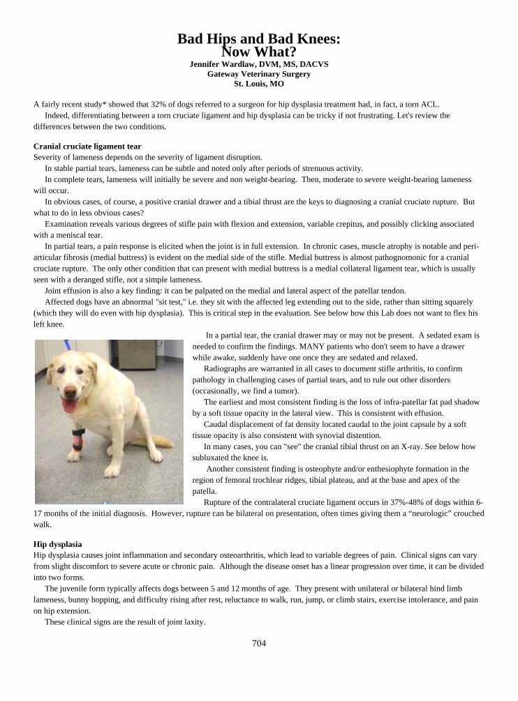
704
Bad Hips and Bad Knees: Now What?
Jennifer Wardlaw, DVM, MS, DACVS Gateway Veterinary Surgery
St. Louis, MO
A fairly recent study* showed that 32% of dogs referred to a surgeon for hip dysplasia treatment had, in fact, a torn ACL. Indeed, differentiating between a torn cruciate ligament and hip dysplasia can be tricky if not frustrating. Let's review the
differences between the two conditions. Cranial cruciate ligament tear Severity of lameness depends on the severity of ligament disruption.
In stable partial tears, lameness can be subtle and noted only after periods of strenuous activity. In complete tears, lameness will initially be severe and non weight-bearing. Then, moderate to severe weight-bearing lameness
will occur. In obvious cases, of course, a positive cranial drawer and a tibial thrust are the keys to diagnosing a cranial cruciate rupture. But
what to do in less obvious cases? Examination reveals various degrees of stifle pain with flexion and extension, variable crepitus, and possibly clicking associated
with a meniscal tear. In partial tears, a pain response is elicited when the joint is in full extension. In chronic cases, muscle atrophy is notable and peri-
articular fibrosis (medial buttress) is evident on the medial side of the stifle. Medial buttress is almost pathognomonic for a cranial cruciate rupture. The only other condition that can present with medial buttress is a medial collateral ligament tear, which is usually seen with a deranged stifle, not a simple lameness.
Joint effusion is also a key finding: it can be palpated on the medial and lateral aspect of the patellar tendon. Affected dogs have an abnormal "sit test," i.e. they sit with the affected leg extending out to the side, rather than sitting squarely
(which they will do even with hip dysplasia). This is critical step in the evaluation. See below how this Lab does not want to flex his left knee.
In a partial tear, the cranial drawer may or may not be present. A sedated exam is needed to confirm the findings. MANY patients who don't seem to have a drawer while awake, suddenly have one once they are sedated and relaxed.
Radiographs are warranted in all cases to document stifle arthritis, to confirm pathology in challenging cases of partial tears, and to rule out other disorders (occasionally, we find a tumor).
The earliest and most consistent finding is the loss of infra-patellar fat pad shadow by a soft tissue opacity in the lateral view. This is consistent with effusion.
Caudal displacement of fat density located caudal to the joint capsule by a soft tissue opacity is also consistent with synovial distention.
In many cases, you can "see" the cranial tibial thrust on an X-ray. See below how subluxated the knee is.
Another consistent finding is osteophyte and/or enthesiophyte formation in the region of femoral trochlear ridges, tibial plateau, and at the base and apex of the patella.
Rupture of the contralateral cruciate ligament occurs in 37%-48% of dogs within 6-17 months of the initial diagnosis. However, rupture can be bilateral on presentation, often times giving them a “neurologic” crouched walk. Hip dysplasia Hip dysplasia causes joint inflammation and secondary osteoarthritis, which lead to variable degrees of pain. Clinical signs can vary from slight discomfort to severe acute or chronic pain. Although the disease onset has a linear progression over time, it can be divided into two forms.
The juvenile form typically affects dogs between 5 and 12 months of age. They present with unilateral or bilateral hind limb lameness, bunny hopping, and difficulty rising after rest, reluctance to walk, run, jump, or climb stairs, exercise intolerance, and pain on hip extension.
These clinical signs are the result of joint laxity.
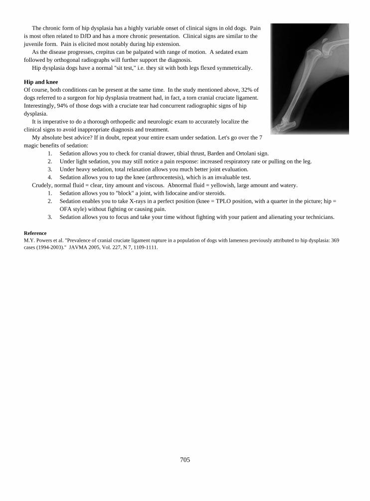
705
The chronic form of hip dysplasia has a highly variable onset of clinical signs in old dogs. Pain is most often related to DJD and has a more chronic presentation. Clinical signs are similar to the juvenile form. Pain is elicited most notably during hip extension.
As the disease progresses, crepitus can be palpated with range of motion. A sedated exam followed by orthogonal radiographs will further support the diagnosis.
Hip dysplasia dogs have a normal "sit test," i.e. they sit with both legs flexed symmetrically. Hip and knee Of course, both conditions can be present at the same time. In the study mentioned above, 32% of dogs referred to a surgeon for hip dysplasia treatment had, in fact, a torn cranial cruciate ligament. Interestingly, 94% of those dogs with a cruciate tear had concurrent radiographic signs of hip dysplasia.
It is imperative to do a thorough orthopedic and neurologic exam to accurately localize the clinical signs to avoid inappropriate diagnosis and treatment.
My absolute best advice? If in doubt, repeat your entire exam under sedation. Let's go over the 7 magic benefits of sedation:
1. Sedation allows you to check for cranial drawer, tibial thrust, Barden and Ortolani sign. 2. Under light sedation, you may still notice a pain response: increased respiratory rate or pulling on the leg. 3. Under heavy sedation, total relaxation allows you much better joint evaluation. 4. Sedation allows you to tap the knee (arthrocentesis), which is an invaluable test.
Crudely, normal fluid = clear, tiny amount and viscous. Abnormal fluid = yellowish, large amount and watery. 1. Sedation allows you to "block" a joint, with lidocaine and/or steroids. 2. Sedation enables you to take X-rays in a perfect position (knee = TPLO position, with a quarter in the picture; hip =
OFA style) without fighting or causing pain. 3. Sedation allows you to focus and take your time without fighting with your patient and alienating your technicians.
Reference M.Y. Powers et al. "Prevalence of cranial cruciate ligament rupture in a population of dogs with lameness previously attributed to hip dysplasia: 369 cases (1994-2003)." JAVMA 2005, Vol. 227, N 7, 1109-1111.
706
How to Lower Infection Rates in Orthopedic Surgeries Jennifer Wardlaw, DVM, MS, DACVS
Gateway Veterinary Surgery St. Louis, MO
The normal incidence of clean surgical wound infections is 0-4.4%. Keeping your hospital and staff under good surveillance will help minimize nosicomial infections. Washing hands between patients, using soap for at least 20 seconds and using care when taking rectal temperatures to clean equipment and hand will help. The main sources of surgical infection come from the OR, the surgical team, instruments, but mostly the patient’s endogenous flora.
Antibiotics should be considered for surgeries over two hours, ones with implants, or if there is a break in asepsis. If the animal has been on an appropriate antibiotic for a few days prior to surgery, perioperative antibiotics are not needed. For orthopedic procedures indicating the need for antibiotics, Staphylococci spp. are the most common skin flora we encounter. Therefore, most orthopedic surgeons choose cefazolin or similar antibiotics. However, good surgical technique is the best prevention of infection and when a break in aspesis occurs it should be repaired appropriately. Not every patient and every surgery needs antibiotics and the potential downsides or complications should be weighed against the risk with need for perioperative drugs. Typically perioperative antibiotics are given at induction or 30 minutes before the skin incision, then every 90 minutes. The first dose should not be given until cultures are obtained, if they are indicated.
Anesthesia reduces a patient’s resistance to infection. Propofol has been associated with higher postoperative infections, and sepsis. It should be used with aseptic technique and promptly disposed of to avoid contamination. Maintaining a patient’s blood pressure and body temperature will help their immune system fight infection. Warming tables, circulating water blankets and Bair Huggers should be considered. Bair Hugger’s have been shown to grow bacteria in their hoses and may increase wound colonies, although data is conflicting. Most surgeons prefer to turn on the Bair Hugger after the patient is completely draped to avoid this potential issue.
Controlling the surgical environment will limit bacteria significantly. Disinfection of equipment should be routine and frequent. Traffic should be minimized, the door closed and laminar flow should create 15 air exchanges per hour. Laminar flow decreased room bacteria by 61% and wound bacteria by 92%. Proper surgical attire should be fresh scrubs, caps that cover all hair and masks that are soft with pleated fabric. Shoe covers may help with sanitation but are not necessary.
Patient preparation should include getting rid of fleas if present, bathing if dirty (dry completely afterwards), walking outside to eliminate or expressing the bladder. Shaving should be performed while surgical scrubs are protected with a lab coat, in the prep area. The surgical clip should be performed with a 40 blade, against the grain, at least two clipper blades from the proposed incision. Hair should be removed with a vacuum. Care must be taken not to cause razor burn by keeping clipper blades clean, sharp and well lubricated. Clipper cleaning should be performed after every surgical prep case. A rectal purse string should also be considered for pelvic limb procedures.
The surgical site is prepared with a rough prep in the preparation area, and then a final sterile prep in the surgical suite. The skin cannot be sterilized, but the number of bacteria can be significantly reduced. The “ideal” method is elusive but several options exist. Povidone-iodine (PI) is cheap, has a broad spectrum, but will stain fur, and is inactivated by organic debris. Chlorhexidine (Chlorhex) has good activity in organic debris, good residual activity, broad spectrum, low tissue toxicity except mucous membranes and does not stain clothing. Rinsing agents are commonly used but not necessary. 70% Isopropol alcohol is antibacterial, dries quickly but is flammable and may cool the patient. Sterile saline is non-flammable but has not antimicrobial activity.
A 3 minute chlorhex scrub was shown to be equivalent to a 10 minute PI scrub in one study with no difference in infection rates. Another study agreed that both scrubs work, but the Chlorhex with saline had a better residual activity than Chlorhex and alcohol. Alcohol rinse after iodine helps release the iodine activity, but decreases the residual activity as well. If an alcohol rinse is used, the surgical site should then be painted or sprayed with betadine.
The surgical site prep is performed next in the surgical suite. It is ideally performed with sterile gloves, with sterile gauze and prep basin with ‘new’ surgical scrub. If a rinsing agent is not used, dry gauze can be used to remove the scrub detergent. Good technique should be used for the sterile scrub to not contaminate the surgical site during the scrubbing. It should be noted that one step 70% PI solution was as efficacious as alternating with alcohol and is much simpler. Additionally paint-only or spray-only seems efficacious as well. One study indicated that applying antiseptic with gauze versus sterile gloves did not make a difference as long as the proper agent and contact time were used.
Surgeon prep with surgical scrub offers the same multitude of scrub solutions. PI and chlorhex have the same pros and cons for the surgeon’s hands as they did for the patient’s leg. However, the World Health Organization and scientific evidence support the use of hydro-alcoholic rubs for presurgical hand scrubs. If your hands are free of gross contamination, these methods are less abrasive and less likely to cause bacterial proliferation on microabrasions from the scrub sponge. Two such solutions are Avagard (1% chlorhax,
707
61% alcohol) and Sterilium (80% alcohol). Numerous studies compare the two solutions, but both work very well and the choice is surgeon preference.
Strict aseptic technique should be followed once in surgery. Surgeons should stand properly to avoid contamination, surgical supplies should be handled properly, opened properly and their sterility confirmed. Sterile saline for lavage should be poured from a fresh unopened bottle with care to avoid splashing on the instrument tray. Traffic around the room should be minimized. It is everyone’s responsibility in the room to monitor for a break in asepsis and make the surgeon aware if such a break occurs. When in doubt, change it out.
Surgical drapes should be resistant to penetration, strike-through and movement. Four quarter drapes are used at the primary layer and may be adhesive or towel clamps applied. Once the foot is captured with sterile vetwrap, a full length patient drape should be used to cover the entire patient. Clamps on the patient drape should not penetrate both layers of drapes. A stockinette or ioban on the incision may be used but are not necessary. However, some sort of sterile water impermeable layer must be applied over the sterile vetwrap due to its porosity. All cables, hoses etc. should be secured with a non-penetrating clamp as well.
Disposable gowns decrease contamination rate. Hemostasis, minimizing tissue trauma, good aseptic technique and speed are all important for the surgeon to be mindful of. Infection rate doubles with every hour of surgery. Braided suture should be kept off the skin during handling. Gloves should be checked regularly for holes and remain tight on the hands and fingertips. Wearing double gloves or orthopedic gloves will decrease the chance of exposed hands. 84% of glove defects occur in procedures over an hour long and individuals were not able to accurately predict a defect. Overall glove defects occurred in 23% of cases and were more likely to occur in orthopedic cases.
Surgical closure can be performed with staples for speed or suture for cost savings. A couple of studies contradict whether staples versus sutures are better for closure on TPLOs and extracapsular surgeries. But notably, one retrospective study showed no increased benefit of antibiotic impregnated suture for preventing infection with TPLOs. This may indicate there is not a need for these more expensive sutures in clean procedures.
After surgery patients should have their bladder expressed or emptied to avoid eliminating on the surgical site, body temperature should be brought back to normal. Clean bedding with spill resistant water bowls should be maintained throughout the hospital stay. Postsurgical site infections in dogs and cats can be minimized with these proactive precautionary steps.

708
Laser Use in Physical Rehabilitation and Adding it to Your Practice
Jennifer Wardlaw, DVM, MS, DACVS Gateway Veterinary Surgery
St. Louis, MO
Karen Felsted, CPA, MS, DVM, CVPM PantheraT Veterinary Management Consulting
Dallas, TX
Laser therapy is Light Amplification by the Stimulated Emission of Radiation (LASER). Lasers are classified into four levels depending on their potential to harm tissue. Class 1 is a laser pointer used in lectures or at a grocery store while an example of a Class 4 laser would be a surgical cutting laser. Class 3 and 4s are used for low level laser therapy or physical rehabilitation. They are advocated for many things but used mostly for wound healing and pain relief.
Laser therapy cause cellular oxygen production by photons being absorbed into the mitochondria. This in turn causes a proton gradient across the cell and mitochondrial membrane. The gradients result in increased cell permeability. Laser therapy also stimulates the production of ATP, thereby stimulating DNA production. Also laser therapy increases cellular metabolism and growth. This accelerates tissue repair and cell growth in tendons, ligaments and muscles.
There are also indications in human and rodent models that laser therapy may block pain transmission through conduction latencies and selectively inhibit nociceptive neuronal activities. It may also increase endorphins. For this reason laser therapy is being used for muscle trigger points and acupuncture, called acupressure.
Laser therapy is advocated in wound healing due to its ability to stimulate fibroblasts and speed collagen production to repair tissues. It appears to accelerate angiogenesis and neovascularization. Laser is used on edema because it causes vasodilation and improves lymphatic drainage. It appears laser therapy may help with surgical incisions, open wounds and burns. The goal of wound laser therapy is to increase blood circulation, stimulate the reduction of hemoglobin, then stimulate both the reduction and immediate re-oxygenation of cytochrome c oxidase. This is the normal metabolic, wound healing process, just trying to speed it up with laser therapy.
Lasers emit energy, or joules, at a certain wavelength. This wavelength determines how deep the laser will penetrate into the tissue. The power, or watts, of a laser is the rate or speed at which it can deliver the desired energy to the tissues. There are many different lasers with different penetrating wavelengths, but the energy density or dose for square of centimeter of tissue is the critical data point. Not only does the laser light need to fully penetrate the area we want, but it needs to bring the right level of energy to the tissue. Based on the size of the tissue or area we are treating (cm2) is how we determine the total dosage (J/cm2). The power of your laser will determine if that takes you 10 seconds or 10 minutes to accomplish that treatment dose. Research is still ongoing for determining whether continuous wave, or pulsed wave lasers are better, if daily or every other day protocols are superior and what the ideal dosage is for a condition. So given all the variables in laser company styles, format and protocols, it is of paramount importance that we discuss energy density and dosages in the same concise language so we can communicate appropriately; Joules per centimeter squared.
We do know that the minimum dosage in humans to achieve a photochemical response to laser therapy is 5 J/cm2. We also know there are contraindications to laser therapy; active hemorrhage, local steroids, pregnancy, cancer, heart disease, photosensitive medications.
There are limited studies looking at laser therapy, but many are in progress. Once human study should an improvement in pain relief for 2 months and up to 1 year after a two week protocol. A canine study showed similar results with weekly sessions for four to six weeks showing 70% of patients showing some improvement in arthritic pain and gait abnormalities.
The difference between commercially available laser unties lie solely in the wavelength, power density, pulse modulation and aesthetics. The goal is to stimulate the cell, and ultimately the body, to perform its natural functions, but at an enhanced rate.
709
How to Introduce and Incorporate Physical Rehabilitation into Your Practice
Douglas Stramel, DVM, CVPP, DVMA Advanced Care Veterinary Services
Carrollton, TX
Indications for rehabilitation Post-operative recovery
Speed recovery; Improve quality of recovery; Prevent overload of other limbs; Prevent adhesions, disuse atrophy; Regain normal range of motion; Return to performance; Indications for Rehabilitation
Patients predisposed to permanent loss of joint motion: Age; Trauma; Repeated surgeries; Immobilization; Limb disuse Dogs undergoing intensive rehab programs after CCLR surgery regained normal limb function at 6 months. Cases managed at
home had significantly less function than the normal leg; Marsolais et al, JAVMA 2002 220;9 Early intensive rehabilitation of post-op CCLR dogs prevented atrophy, built muscle mass and strength and increased range of
motion in both flexion and extension when compared with at home gentle therapy; Monk et al. AJVR 2006 67;3 Continuous passive range of motion showed an anti-inflammatory effect on meniscal fibrocartilage. Immobilized knees had an
increase in pro-inflammatory COX-2 and MMP-1 resulting in GAG degeneration; Journal of Orthopedic Research 2005 Daily controlled therapy increases survival time in dogs with degenerative myelopathy. Intensive therapy had the better outcome
Kathman et al. J Vet Int Med 2000 Intensive therapy (not at home) increased mobility, decreased pain and facilitated weight loss in dogs with osteoarthritis Mlacnick
et al. JAVMA 2006 229;11 Quadriceps strengthening resulted in a 23% decrease in pain scores of humans with knee arthritis Kee 2000 Owner expectations
Environmental conditions; Athletic function;Have to be realistic – chronic vs acute; Owner expectations – ideal vs. practical Treatment plan Define a goal; Determine prognosis; Design treatment schedule; Reassess and modify as needed. The rehabilitation plan There is no set formula, even for specific conditions; The treatment or exercise plan is modified to the individual dog; Things change at each therapy session, and progress is not always smooth; Frequent reassessments vital; Pain management also vital. Patient assessment A complete musculoskeletal exam consists of observing the animal at rest, in motion and palpation of the animal at rest and in lateral recumbency.
Gait analysis Consistent with same observer Vet Comp Orthop Traumatol. 2007;20(4):299-304. Am J Vet Res. 2004 Dec;65(12):1634-43. Quantitative
Computer assisted gait analysis Kinematics: describes motion; Force plate analysis: measure ground reaction force; Pressure mats: pressure patterns under a foot over time
“Head bob” = Forelimb lameness; Can see more of footpads from rear = Unloading that foot during stride; “Sway” in the rear (hula dancer) = Bilateral Rear limb lameness (Hip Dysplasia); “Bunny Hopping” = Bilateral rear limb lameness (Hip Dysplasia); “Hip hike” or swings tail = Rear limb lameness; Short stride = May be protecting contra-lateral limb; Hear dragging of nails = Neurologic disease, sometimes orthopedic pain; Swings forelimb out or “paddles” = Elbow or shoulder pain or infraspinatus contracture
Joint range of motion Goniometry; Reliable (Jaegger et al AJVR 2002 63;7); Quantitative measure of progress and outcome; Measure flexion and extension in each joint
For example medial shoulder instability is likely if valgus deviation at the shoulder is more than 32 degrees, diagnostic if more than 50 degrees Vet Surg. 2008 Feb;37(2):132-41.
Muscle girth Gulick tape measure; Weighted spring for accuracy and repeatability
710
Lesion Motor function Thoracic limb reflexes/muscle tone Pelvic limb reflexes/muscle tone C1-5 Tetraparetic/plegic Normal to increased , Normal to increased, C6-T2 Tetraparetic/plegic , Decreased to absent, Normal to increased, Thoracic limb gait may be short and stilted, T3-L3 Paraparetic/plegic , Normal , Normal to increased, L4-S3 Paraparetic/plegic Normal, Decreased to absent
For each joint, the examiner should record: crepitus; range of motion; presence of effusion; pain response; instability. Palpation for pain Myofascial trigger points Disturbed motor function, Muscle weakness without loss of mass, Muscle stiffness, Restricted Range of Motion, Vasoconstriction, Local Tenderness Restore range of motion PROM – passive range of motion; AROM – active range of motion; Active assisted range of motion. Each joint should be undergo comfortable range of motion every 8 -12 hours.
Effects of immobilization Occurs rapidly; Loss of muscle mass; Shortening of ligaments, tendons; Stiffening of joint; Thinning of articular cartilage; Some effects irreversible Am J Vet Res. 1982 Mar;43(3):454-60. Range of motion effects on tissues Muscle stretching and strengthening; Cartilage health – imbibitions; Joints, ligaments, tendons loosen; Blood vessels and sensory nerves adapt to allow more motion; Muscle memory – re-learns normal activity
Proprioceptive re-training Even after minor injury, joint mechanoreceptor firing is disrupted; Information exchange between the body and brain is disrupted, fine motor control is decreased; stimulate firing of mechanoreceptors by unbalancing the patient; challenge increases as patient improves
Motor control and gait pattern are practiced over cavaletti rails, on ramps, small stairs etc. Cryotherapy Decreases nerve signal speed; Decreases inflammation; Shrinks blood vessels; Can decrease bleeding Aust N Z J Obstet Gynaecol. 2009 Oct;49(5):517-24. Thermotherapy Increases collagen stretch; Increases blood flow; Direct application – superficial. Moist heat is best; Deep heat – therapeutic ultrasound Deep tissue heating to control pain, break down scar tissue and increase mobility .
Increases enzyme action to speed healing Blood Coagul Fibrinolysis. 2006 Mar;17(2):105-12. Both infrared and ultrasound heat increased range of motion over stretching alone after joint contracture in dogs Non Thermal:
Vibration and compression of molecules (acoustic streaming), alters membrane permeability to ions, increases fibroblast proliferation and releases growth factors J Athl Train 2002:37(3):293-9
Laser:photobiostimulation – laser produces monochromatic, coherent light. Effects are not thermal Photon absorption by photoreceptive molecules in the mitochondria (chromophores) and at the cell membrane – energy is
converted to ATP Karu et al. J Photochemistry & photobiology 1999 (49):1-17 Alters specific cell function as measured by microarray leading to wound healing, pain relief and tissue repair. Zhang et al. J
Invest. Derm 2003:120(5);849-57 Anti-inflammatory effects, inhibitory to nocciceptors Chow et al. J Periph Nerv. System. 2007. Low level laser applied to spinal cord can improve recovery of nerves. Lasers in Surgery and Medicine 2001 28
Electrical stimulation Stimulates nerves and muscles for pain control, strength-building & adaptation; NMES for muscle strength, reduce edema; TENS for pain control = Stimulates large diameter nerve fibers (gate theory of pain) to block nociceptor signals. Some cumulative effect Levine et al. Proc 2nd Int. Symp. Rehab Phys Ther in Vet Med. 2002: 199 Hydrotherapy in a whirlpool tub Pain control and muscle relaxation. Excellent for older patients. Water pressure can reduce edema Massage Increases mobility, relaxes muscles, controls pain. Stimulates large diameter nerve fibers Connect Tissue Res. 2009;50(5):323-9.
711
Therapeutic exercise Hydrotherapy
Helps to reduced edema, manage pain, decrease load on joints Rheumatology (Oxford). 2009 Sep;48(9):1155-9. Epub 2009 Jul 16. Allows rehabilitative exercises that are not possible on land due to pain, imbalance, fragility etc. J Aging Phys Act. 2008 Oct;16(4):381-92. Improves outcomes in people with osteoarthritis BMC Musculoskelet Disord. 2009 Aug 19;10:104.
Underwater treadmill The treadmill can be used in the very early stages of recovery; the higher the water level, the more weight that is supported. As strength and correct movement improves, the water level is lowered each session to further increase strength. Lean and muscled animals tend to sink; weights can be used to increase effort; life jackets to decrease effort. Patient has to strengthen trunk to oppose rotational forces. Hydrostatic Pressure: affects lung volumes, reduction of edema, pressure increases with increased depth. Works muscles 3-5 times harder then on land Northrip et al. Intro Biomech Anal Sport 1979:96-119. Use turbulence to provide resistance in strength training. Drag can be used to assist motion e.g. hip extension. Allows time for the unbalanced animal to correct before falling vs. on land. Paralyzed patients can walk in water before land
Swimming Allows exercise when no weight bearing is possible, great for core strength as patient needs to overcome rotational forces. If a patient will not use a front leg. Contraindications: Open wounds – at least day 5 after incision (granulation tissue); Fever/infection; Gastrointestinal disease; Severe cardiac or respiratory disease; Contagious disease.
Outcomes Re-evaluate every 1- 2 weeks;extensive re-evaluation usually around 6 weeks into therapy, measurements etc. Patient ‘graduates’ when able to fully perform activities of daily living (varies each patient) and muscle mass is even, range of motion maximized. Athletes take longer to get back to performance. Geriatric patients and those with chronic conditions that cannot be managed at home may continue maintenance visits with us at least once a week. Muscular injury Muscle shortens in response to injury; Protective mechanism; Disuse further decreases circulation; Build up of lactate etc.; Pain exacerbated, further stimulates spasm; Neighboring muscles overloaded
Treatment of muscle injuries Decrease inflammation in acute stage; Restore mobility and circulation; Prevent scar formation or break down scar tissue; Restore adaptive length; Strengthen; Rehab of a muscle injury; Ice in acute stages; Prevent/treat scar tissue = Therapeutic ultrasound, Laser, Massage, Range of motion exercises
Collateral ligament sprain Diagnosis can be based on goniometry and pain; Stressed radiograph. Prevention of adhesions. Encourage fibers of tendon to heal in correct orientation with gentle loading.
Adequan (Oryan et al. Conn Tiss Res. 2008;49(5):351-60) After adhesions/scar formed: 3 months = can still re-model. 6 months = some permanent change Patellar luxation Grade I-II can be conservatively managed; Rehabilitation to strengthen quadriceps. Always check for concurrent CCLR. After surgery, rehab used to strengthen and for pain relief Hip dysplasia Radiographs – arthroscopic study showed cartilage degeneration preceded radiographic changes months to yrs Holsworth et al. JAVMA 2005 227;7. Go by clinical signs of pain and limited extension. The femoral head is ‘loose’ in the socket but extension is limited by pain and capsular contraction Fractures Prevent disuse atrophy! Increases new bone deposition through early weight bearing. Non-union or delayed union: Shock Wave Therapy. High energy shock wave therapy accelerates bone healing. Duerr F, Palmer R, et al CSU 2014 World Veterinary Orthopaedic Congress, Humans on bed rest with 10 min standing daily had a 25% decrease on the impact of immobilization
Carpus – reluctance to jump down; Elbow – reluctance to go downstairs; Shoulder – reluctance to jump down; Tarsus – reluctance to climb stairs; Stifle – unable to sit straight; Hip – reluctance to jump in car; L-S spine – reluctance to squat
Decreased muscle mass and function due to disuse from pain; Increased strain on already painful joints; Worsening of cartilage changes from disuse; Abnormal gait can lead to myofascial pain

712
Management of osteoarthritis Optimize weight; Improve range of motion; Relieve Myalgia; Increase muscle mass, strength and fitness; Increase function at home; Assistive devices; Decrease dependence on medication
More and more OA diagnosed in cats. Rehabilitation therapy is possible. Can use activity monitors to detect effects of pain relief in cats with OA Vet Anaesth Analg. 2008 Mar;35(2):173-83. Epub 2007 Oct 10. Neurologic diseases Pain management; Assisted standing – ball, slings, water; Assisted ambulation; Strengthening, balance and proprioception; Proprioceptive feedback very important; Gentle stimulation without overload of the nervous system; Neurologic Rehabilitation
UMN disease – massage and range of motion to decrease spasticity LMN disease – Electrical stimulation to decrease neurogenic atrophy, PROM Neuromuscular facilitation; Plasticity of brain and neural tissue; Sensory feedback aids continued motor learning; Body weight
supported or partial weight bearing training
713
Playing Outside the Box: Shock Wave, Regenerative Cell, and
Pulsed Electromagnetic Field Therapies Douglas Stramel, DVM, CVPP, DVMA
Advanced Care Veterinary Services Carrollton, TX
Shockwave technology High-energy sound waves in any elastic medium (in our case water) create a rapid increase in pressure, similar to an explosion, or thunder during a lightning storm. These waves are sound energy. Around 1,000 times the magnitude of ultrasound. Rapid increase in pressure and slight negative pressure dip causing cavitation (mini-shock waves). Large focal area for deep tissue repair. Not radial pressure waves or laser therapy
Electrohydraulic Peak Pressure 70MPa; Rise time: nanoseconds; Produces true shockwave at all settings; Up to110 mm penetration (50 mm in dogs)
Release of cytokines Re-initiates healing by making chronic inflammation into acute inflammation. Release of growth factors (VEGF): increase in capillary density and soft tissue regeneration. Release of BMPs: bone regeneration and healing.
Recommended for various musculoskeletal disorders; Noninvasive regenerative therapy; Potential surgical alternative; post-surgery recovery; part of rehabilitation program; 1-3 treatments (˜3 weeks apart) generally heal soft tissue injuries or delayed fractures and/or provide symptom relief for 6-12 months.
Indications Chronic wounds, Lick granulomas, Delayed or non-union fractures, Nonsurgical management of digital, metacarpal, and metatarsal fractures, Osteoarthritis: hip, elbow, stifle, hock, shoulder, Supraspinatus tendinitis, Glenohumeral ligament tears (medial shoulder instability), Osteochondrosis of the shoulder, Triceps muscle tear, Fragmented coronoid process, Biceps tendinitis, Spinal column issues (intervertebral disc disease, lumbosacral disease, spondylosis deformans, cauda equine syndrome), Iliopsoas muscle injuries, Patellar tendinitis (especially post-operative anterior cruciate repair), Achilles tendinitis, Calcaneal tendon tears Research Clinical success in human and equine medicine has led to research and gleaned new opportunities in canines. Studied and used at top clinics and universities throughout the country.
PEMF Uses electrical energy to direct a series of magnetic pulses through tissue. Dr. Oz promotes it so it has to be valid. Although the effects are know this does not “prove” their medical value. i.e. the effect may not always translate into a viable treatment.
PEMF – Proposed Mechanism: creates a low frequency electrical current in the damaged tissue binding of Calcium with Calmodulin = production of the beneficial form of Nitric Oxide = improved circulation; diminished swelling; reduction of pain; re-normalizing disturbed cell membrane potential; production of several growth factors; down regulate the production of cytokines
PEMF magnetic fields are created from the electrical flow of current through coils of metal wire; The more turns of the coil, the stronger the field; On/off frequency of electricity determines the rate of pulsation
Improves function to brain and peripheral nerve sites that were lost. Clinical evidence of treating urinary incontinence in humans. Treatment of tendonitis. Improved wound healing. Treatment of osteoarthritis
World Health Organization says available evidence suggests an absence of adverse effects in humans. Avoid pregnant animals: No studies done! May interfere with medical devices: pacemaker, neurostimulator and insulin pump = This is a concern if you are sending these home with owners who have one of these devices. Drug patches. Vasodilation can occur
Efficacy Decided lack of manufacturing regulations. Variance in label claims. Variance in strength of magnet. Buyer beware! Regenerative therapies Injection of small volumes of biologically active substances into joints and soft tissues. Promote tissue repair and growth. Reduce pain. Augment natural healing process.
PRP Autologous biologic therapy prepared from whole blood; Concentration of platelets in plasma fraction >200,000 plt/ul (100-400K); 2-8x concentration of whole blood; Variable amounts of other blood components = WBC, RBC, Plasma proteins. “Milieu of bioactive factors”
714
Platelets First to the injury site. Most often associated with hemostasis. Important role in tissue healing, inflammation. Recognition and repair of damaged tissues. Contain a variety of bioactive factors housed in cytoplasmic granules that are released upon activation or tissue injury, stimulate chemotaxis cell migration, proliferation, angiogenesis and matrix synthesis.
Variability Between systems, Whole blood platelet count, HCT, Hydration status of patient, Anticoagulant. PRP should be evaluated for each patient?
The good essential to normal tissue healing. Phagocytosis. Antimicrobial. The bad: may result in pro-inflammatory signaling and increased catabolism
Optimal Platelet Count = Unknown; Platelet concentration positively correlated with GF concentration (dogs); Tissue dependent?; Increasing platelet concentration resulted in decreased collagen synthesis (equine SDFT model); Reducing WBC more important than increasing plt; Maximum biologic threshold (>5X no benefit)
Platelet activation 1) Prior to injection – formation of fibrin “clot”; Calcium chloride or gluconate; Thrombin. Immediate release of GF. 2) After injection – liquid; Exposure to tissue factors (collagen). Sustained release of GF. Advantages of activated PRP: Greater release of GF. Resulting clot serves as provisional matrix, contains high concentration of GF. Disadvantages of activated PRP: “Clot” within joint. Rapid release of GF which may be diluted or removed from joint. Method of activation may influence signaling.
Anabolic effect Anabolic effects of PRP are attributed to effects of growth factors on articular cartilage, synovium and MSC. Improved healing of cartilage defects.
Anti-catabolic effect Chondroprotective effect through regulation of catabolic factors. Increased HA secretion. Anti-inflammatory effects In health, platelets modulate magnitude & duration of inflammatory response. PRP has a complex role in inflammation, with both pro and anti-inflammatory activities. Reduced NF-κβ activation.
Analgesic Down regulation of inflammation. Augmentation of cannabinoid receptors (CB1/CB2). Stimulation of thrombin receptors (PAR-1)
Antimicrobial At least 7 antimicrobial proteins have been isolated from human platelets. Leukocyte concentration
Indication for use Osteoarthritis, Perioperative: Articular fracture repair, Post arthroscopy, OCD, Postoperative: Slow return to function (TPLO, MPL, etc) Safety: autologous No significant side effects reported in human clinical trials; Adverse events are rare; Infection – low risk due to WBC/GFs; Bleeding/bruising; Exacerbation of stiffness/soreness (2-7 days)
Vivregen (VIVR8) A proprietary non-cellular fluid that contains all the mediators secreted by the stem cells from a natural young, developing tissue with regenerative ability. Manufactured in an FDA cGMP compliant manufacturing facility. Available as an injection for non-food animals such as equine and canine. Observed effects Soft tissue injuries and post-op healing; Osteoarthritis; Microvascular injuries, muscle strain and sprain. No Clinical Research.
Massage Pain and relaxation in cancer patients: Sui-whi, Jane et al. Effects of massage on pain, mood status relaxation and sleep in Taiwanese patients with metastatic bone pain: A randomized clinical trial. IASP June 2011
Muscle damage Crane, Justin et al. Massage Therapy Attenuates inflammatory Signaling After Exercise-Induced Muscle Damage Sci Transl Med 2012
Degenerative myelopathy Kathmann, S. et al. Daily Controlled Physiotherapy Increases Survival Time in Dogs with Suspected Degenerative Myelopathy J Vet Intern Med 2006:20:927-932
715
Perioperative Use of Alpha-2s and Dissociatives Douglas Stramel, DVM, CVPP, DVMA
Advanced Care Veterinary Services Carrollton, TX
Dexodomitor has flexible indications FDA-approved for sedation and analgesia in dogs and cats. FDA-approved as a preanesthetic prior to general anesthesia in dogs and cats. Dexmedetomidine can be used in cats as young as 12 weeks of age and dogs as young as 16 weeks of age
DEXDOMITOR is a full agonist of the α-2 adrenoceptor. Greater degree of α-2 selectivity provides more predictable sedation, analgesia, and muscle relaxation
Hemodynamic effects of α-2 agonists Peripheral vasoconstriction > initial hypertension followed by normo- or slight hypotension > compensatory reduction in heart rate > decreases in body temperature and respiratory rate. Bradycardia - May Be Advantageous During Anesthesia, Provided That Tissue Perfusion of the Vital Organs Meets Oxygen Demand .
Atropine = Inc HR; Possible Inc in BP; Minimal improvement of Cardiac Output; if atropine is used, administered at least 10 minutes prior to administration of Dexmedetomidine
Pt benefits Less Physiological Stress; Less Risk of Physical Harm; Better Experience
Staff benefits Decreased Risk of Injury and Radiation Exposure; Less Stress; Less Time and Personnel to Complete Procedure.
Pet owner benefits Decrease their own anxiety about vet visit; Improve perception regarding their pet's levels of comfort and anxiety; Demonstrate compassionate treatment of pet .
Uses Grooming, nail clipping, bathing; Ear exam and treatment; Ophthalmologic exams; Abscess or wound treatment; Cast or bandage application/ removal; Diagnostic imaging; Skin biopsies/scrapings; Suture removal; etc
Product features Reduces Induction agent by 30 – 61%; Reduces Inhalant requirements by 40 – 60% . The Percent in Reduction is Dose Dependent. Antisedan (atipamezole) reverses dexdomitor effects Administered as an IM injection at a dose volume equal to Dexdomitor dose volume. Reversal effects are noted approximately 5–10 minutes after administration of Antisedan is only approved for use in the dog. Antisedan reverses both the sedative and analgesic effects of Dexdomitor. Antisedan is an α-2 antagonist that competitively inhibits α-2 adrenergic receptors
Dexdomitor micro-dosing Provides short duration of action ~15 minutes; Micro dose: 1 ug/kg (Sedative dose: 5-20 ug/kg); Spare propofol induction dose; “Smooth out” effect before recovery; Rapid resolve of delirium during recovery. Very Small Volume = Dexdomitor 0.1 (NEW)
Telazol Uses
Restraint for diagnostic procedures; Induction and maintenance of anesthesia; Restraint and sedation of various exotic species Precautions
Respiratory and cardiovascular depression may be seen; Transient tachycardia may occur in dogs (30 min); Avoid in patients with pancreatic disease, preexisting renal pathology, severe cardiac dysfunction and/or respiratory dysfunction; Not to be used in pregnant bitches or queens; Contraindicated in caesarian sections.
Advantages Rapid induction of dissociative anesthesia; two active ingredients in one product; low volume; Analgesia and muscle relaxation
Disadvantages Fixed ratio of tiletamine/zolazepam = In cats duration of activity of zolazepam is longer than tiletamine (tranquilization longer than anesthesia during recovery). In dogs the duration of activity of tiletamine is longer than zolazepam (anesthesia longer than tranquilization during recovery). Salivation, bradycardia, hypotension noted in cats. Salivation, tachycardia noted in dogs. Pupilliary dilation; Respiratory depression; Hypothermia; Low pH (2.2 – 2.8) associated with pain on IM injection; Prolonged recovery (vocalization, muscle twitching, vomiting)
Telazol-torbugesic-dexmedetomidine (TTDex) Reconstitute tiletamine-zolazepam (Telazol) with 2.5 mL of butorphanol (10mg/ml) and 2.5 mL of dexmedetomidine (0.5 mg/ml). Each ml TTDex contains: 100 mg of tiletamine-zolazepam; 5 mg of butorphanol; 250 ug of Dexmedetomidine. Based on clinical experience, the TTDex can be stored at room temperature for up to 3 months after reconstitution. (Not Studied).
716
Induces various depths of sedation and a surgical plane of anesthesia within 3 to 5 minutes after a single intramuscular injection. Can be used both in dogs and cats with the same combination and dose rates. Visceral and somatic analgesia both intraoperatively and postoperatively. Small injection volume to facilitate drug administration. Potency of this drug combination makes it economical. PROVIDE OXYGEN via ET; Nasal Cannula or Face Mask.
Dosing TTDex • Mild sedation = 0.005 ml/kg: Tiletamine-zolazepam 0.5 mg/kg; Butorphanol 0.025 mg/kg;
Dexmedetomidine 1.25 ug/kg • Moderate sedation = 0.01 mL/kg: Tiletamine-zolazepam 1 mg/kg; Butorphanol 0.05 mg/kg;
Dexmedetomidine 2.5 ug/kg • Profound sedation = 0.02 mL/kg: Tiletamine-zolazepam 2 mg/kg; Butorphanol 0.1 mg/kg; Dexmedetomidine 5 ug/kg • Surgical plane of anesthesia = 0.03 mL/kg: Tiletamine-zolazepam 3 mg/kg; Butorphanol 0.15 mg/kg; Dexmedetomidine
7.5 ug/kg Intramuscular injections
Onset of action: 3- 4 minutes; Duration of effect: 30 – 40 minutes. Induction →recovery →walking = 2 hours Intravenous injection
Halve the recommended intramuscular doses for both dogs and cats. Onset of action: immediate. Duration of effect: approximately two thirds the length of intramuscular administration
Reversal of TTDex Dexmedetomidine: reversed with atipamezole; Butorphanol: antagonized with naloxone or naltrexone; Zolazepam of the Telazol: antagonized with flumazenil. If using Antisedan: If TTDex < 0.02 ml/kg wait 30 minutes; If TTDex > 0.025 ml/kg wait 50 minutes.
TTDex side effects Hypoxia – oxygen responsive usually seen in the first 5 minutes of injection. ALWAYS supply O2. Pain on injection. Apneustic breathing pattern inspiratory breath holding followed by a few rapid breaths No treatment needed.
Ketamine HCl Uses
Induction and maintenance of anesthesia Precautions
Poor muscle relaxant and should not be used alone for major surgery; Can cause increases in CSF pressure and should not be used in patients with head trauma or elevated CSF pressure; Potentially epileptogenic; Avoid in patients with hepatic or renal insufficiency; Doses > 50 mg/kg cats or primates > 40 mg/kg should not be used
Advantages Inexpensive; Maintains cardiorespiratory function; Rapid onset, long duration of action
Disadvantages Hypersalivation; Increased muscle tone; Slight hypothermia (cats); Eyes remain open, must apply ophthalmic lubricant; Slow recovery when used alone; Possible vocalization and tremors on recovery; Acidic pH may lead to pain on injection; Respiratory depression at high doses.
Dexdomitor-ketamine Overall healthy and exercise tolerance CV wise; Single IM injection; Rapid onset of lateral recumbency (5-8 min); Heart rate higher than Dexdomitor alone; Excellent muscle relaxation and analgesia; Reversible with Antisedan ~ 40 min after Ketamine.
Kitty magic The original kitty magic IM formula for 4 KG cat: 0.1 mL Ketamine; 0.1 mL butorphanol 0.1 mL dexmedetomidine = 12.5 mcg/kg Dex, 2.5 mg/kg K, and 0.25 mg/kg T Notes on dosing ALWAYS double check dosing when transcribing from lectures! I have had some weird dosing strategies reported back to me. Plumbs and North American Companion Animal Formulary are great sources.
717
“Wind-Up” Pain: Central Sensitization
Douglas Stramel, DVM, CVPP, DVMA Advanced Care Veterinary Services
Carrollton, TX
Peripheral sensitization • Release of compounds by noxious stimuli can lead to lowering of nociceptor thresholdtissue inflammation =
“sensitizing soup”: Histamine, HT-5, BK, K+ions, PG, Cytokines, etc = reduced nociceptor thresholds = primary hyperalgesia
Hyperalgesia
Central sensitization • nociceptive neurons in the dorsal horn of the spinal cord become sensitized by peripheral tissue damage or inflammation • Dorsal horn neurons become hyper-responsive; sending more signals to the brain • cells that fire together, wire together • activation of NMDA receptors; reduced endogenous opioids levels; reduced serotonin level;expanded receptor fields
(pain in neighboring areas not subjected to injury, or secondary hyperalgesia) Maladaptive pain
• Pain that serves no useful purpose, and significantly impairs the quality of life of the patient. • Consequences of Chronic Pain • Hindrances to Chronic Pain Management • Pain Scales: Helsinki Chronic Pain Index; Canine Brief Pain Index; Cincinnati Orthopedic Disability Index
o Glasgow Composite Pain Scale; AAHA • NSAID’s -- remain the mainstay of therapy for chronically painful patients. • NSAID Precautions • Use only 1 NSAID at a time • Never combine NSAIDS with glucocorticoids
o Gastric Ulceration o Change to alternative NSAID if initial response is not favorable
Robenacoxib J Vet Pharmacol Ther. 2012 Jun;35(3):290-300. Safety of oral robenacoxib in the cat. King JN, Hotz R, et al Steroids
• Decrease pain by decreasing inflammation • Intra-articular: Triamcinolone < 25 KG = 5 mg> 25 KG = 10 mg • Stop NSAIDs for 4 days pre & post injection.
Protective effects of corticosteroids on cartilage lesions and osteophyte formation in the Pond-Nuki dog model of osteoarthritis. Arthritis Rheum. February 1989;32(2):181-93. J P Pelletier1; J Martel-Pelletier
In vivo protective effects of prophylactic treatment with tiaprofenic acid or intraarticular corticosteroids on osteoarthritic lesions in the experimental dog model. J Rheumatol Suppl. February 1991;27(0):127-30. J P Pelletier1; J Martel-Pelletier
The in vivo effects of intraarticular corticosteroid injections on cartilage lesions, stromelysin, interleukin-1, and oncogene protein synthesis in experimental osteoarthritis. Lab Invest. May 1995;72(5):578-86. J P Pelletier1; J A DiBattista; J P Raynauld; S Wilhelm; J Martel-Pelletier
• What do we do with those patients that stop becoming responsive to our NSAID therapy? • Do we switch to a different NSAID? NO! • Re-evaluate the patient and the treatment strategy. Start a multi-modal approach • Central Sensitization → Allodynia • NMDA receptor: N-methyl-D-aspartate
o NMDA receptors are “silent” until persistent or large scale release of glutamate o by blocking the activation of these receptors, a reduction in CNS hyperresponsiveness can be achieved,
allowing other analgesics to function more effectively o adjunctive drug to improve the control of pain
718
o act to increase opioid receptor sensitivity • Constant Rate Infusion : Opioid: Morphine or Hydromorphone or Fentanyl with Lidocaine/Ketamine • Use for 48 – 72 hours in severe chronic pain patients.
Opioid
• The most powerful analgesics available, with actions at peripheral, spinal and supraspinal levels. • Main advantage of giving opioids as a CRI is the avoidance of peaks and valleys seen with intermittent injections. • A lower dose of opioid can be used, which can reduce the unwanted side effects.
Morphine Loading dose 0.25 mg/kg IV followed by 2 – 6 ug/kg/minute
Hydromorphone Loading dose 0.05 mg/kg IV followed by 0.4 - 1.2 ug/kg/minute
Fentanyl Loading dose 0.002 mg/kg IV followed by 0.02 – 0.08 ug/kg/minute
Lidocaine Local anesthetic that provides excellent systemic analgesia.
• Dosage: Loading dose 2 mg/kg IV followed by 20–50 µg/kg/minute. • IV lidocaine is extremely short acting and can be discontinued without residual effect. • Typically lidocaine is not recommended for use in cats due to potential for cardiotoxic effects.
Ketamine Dissociative anesthetic and an N-methyl-D-aspartate (NMDA) antagonist.
• Dosage: Loading dose 0.5 mg/kg IV followed by 2 - 10 ug/kg/min • Always be given in combination with an opioid.
Amantadine Gabapentin Amitriptyline Acetaminophen
• 10 – 15 mg/kg q 8 – 12 hours • CAUTION in hepatic or renal disease. • Break through pain • Can use with corticosteroids or NSAIDs • Do not use in cats • Helpful to bridge from one NSAID to another
Tramadol • Dual mode of action: monoamine reuptake inhibition and mu agonist – NOT DOGS! • controlled substance august 18, 2014 • Combining tramadol with other analgesics (NSAID’s, mu agonists) further enhances tramadol’s efficacy, producing a
multimodal pain relieving action. Non-pharmacologic therapies
• Rehabilitation Therapy:Joints need to stay in motion • Acupuncture: Pain management – endogenous opioids “homeostasis” • Electrical nerve stimulation procedures :Gate Theory: inhibit pain signals in dorsal horn
Laser therapy • cartilage stimulation • fibroblast production • enhancement of immune cells • increases the vascularity of healing tissue • may help with Myofascial Pain
Pulsed magnetic field therapy • Enhanced cartilage repair • Increased collagen synthesis of tendon fibroblasts • Cell proliferation • Synthesis of glycosaminoglycans by growth plate chondrocytes • Decrease production of inflammatory cytokines
Superficial heat • Heat the skin and subcutaneous tissues to a depth of 1-2 cm.

719
• The tissue is usually heated to 40 to 45C for 15 to 20 minutes. • Increases blood flow to the area, promotes tissue extensibility, decreases pain, muscle spasm and joint stiffness, and
causes general relaxation. • Heat is contraindicated if swelling or edema are present.
720
What’s New in Treating Chronic Pain and Osteoarthritis Douglas Stramel, DVM, CVPP, DVMA
Advanced Care Veterinary Services Carrollton, TX
Osteoarthritis is the #1 cause of chronic pain in dogs Chronic pain; Decreased activity; An overall negative impact on the patient; Interferes with human-animal bond; Decreased food intake; Euthanasia for dogs that become non-responsive to treatment.
Cartilage Damage Initiates Osteoarthritis: Chondrocytes are damaged and release matrix metalloproteinases (MMPs) and Nitric oxide and inflammatory cytokines. Nitric oxide causes further release of MMPs and release of inflammatory mediators. MMPS cause further damage to the cartilage.
Other Changes in OA: Thickening of Joint Capsule; Remodeling of subchondral bone sclerosis; Osteophyte formation; Joint is less able to bear stress and forces:
Joint Inflammation: Mechanoreceptors – stretch and pressure, which allows the brain to know where the limb is in space; Neuroreceptors – noxious stimuli. Cartilage is anueral; cartilage damage alone is not painful; Neurogenic Inflammation is Also a Source of Pain in Osteoarthritis
A Comprehensive Approach to Treatment is Required to Break the Cycle: NSAIDs are a cornerstone for OA management, which also includes weight control and exercise to both treat the pain and inflammation of OA as well as the dysfunction of the joint.
Diagnosis and Treatment of Osteoarthritis Can Be Challenging: Pain is a hallmark of osteoarthritis; signs may be subtle; often go unnoticed; Dog owners often do not mention as a problem; Dogs mask pain; pain not evident during physical exam; Often requires a thorough orthopedic examination; When a dog is diagnosed with OA, they are often experiencing chronic pain. Do the risks outweigh the benefits? The answer to that question is patient dependent and depends upon a number of factors. “Lowest Effective Dose” FDA=Lowest effective dose for the shortest duration consistent with individual response. For idiosyncratic reactions, reducing the dose does not decrease the risk.
Minimize the risk. Dogs at greatest risk: Dehydrated or on concomitant diuretic therapy; Dogs with renal failure, cardiovascular and or hepatic dysfunction. Pet Owner Communication, Always provide a Client Information Sheet with prescription
NSAIDs NOT RECOMMENDED if: Pre-existing hyperbilirubinemia; Elevated ALT, AST and GGT; Albumin decreased; Elevated ALP with clinical signs of liver or Cushing’s disease
NSAIDs are safe & effective for vast majority of canines Safety and Efficacy of Using NSAIDs Long-term in Dogs with Osteoarthritis: the current evidence suggests that there is a clinical
benefit of longer-term NSAID use for dogs with chronic osteoarthritis and that is associated with a low risk of serious adverse events” — Innes et al. “Review of the safety and efficacy of long-term NSAIDs use in the treatment of canine osteoarthritis.” Vet Rec 2010
Rehabilitation (Physical Therapy): The goal of rehab is to : maintain muscle mass, build muscle support around joint, reduce pain, weight loss
Adequan: Polysulfated Glycosaminoglycan: FDA approved, disease modifying osteoarthritis drugs; for dogs and horses; water-based, for intramuscular Injection. MOA in vitro studies show: Inhibit serine proteinases; PGE2 synthesis; metalloproteases, hyaluronidases and others. Stimulate synthesis of protein, collagen, proteoglycans, and hyaluronic acid
Summary of ASU, glucosamine, and chondroitin sulfate’s effects: Combination inhibits expression & production of many mediators. Effects extend across species (including canine, feline, equine, and human cells) and to multiple cells in the joint – cells which may all contribute to cartilage breakdown by secreting the mediators responsible
Amantadine – studied to treat canine osteoarthritis: In dogs with osteoarthritic pain refractory to an NSAIDs, addition of amantadine improved physical activity. Might be a useful adjunct therapy for the clinical management of canine osteoarthritic pain. 1. Lascelles BDX, et al. J Vet Intern Med. 22(1):53-9, 2008.
NMDA receptor antagonists. Amantadine is excreted, primarily unchanged, in the urine. Consider reduced doses, if used at all, for patients with impaired renal function. Dogs & Cats: 3 to 5 mg/kg SID PO Typically used for 21 days to “re-set” pain pathways
Gabapentin: An oral prescription medication capable of helping reduce neuropathic and other chronic pain states. Although its mechanism is unknown, it has been shown to affect central sensitization. Excreted unchanged in the urine reduce dose or discontinue in patients with renal dysfunction. Dogs: 10 +++ mg/kg BID to QID PO; Cats: 5.0 +++ mg/kg BID PO
Amitriptyline : Tricyclic antidepressants (TCA’s) have been used in humans and animals as adjuncts to other analgesics (especially opioids) for chronic pain. They act to inhibit serotonin and norepinephrine reuptake, though they may have other analgesic effects as well (including possible actions at opioid receptors and on nerve transmission). 1 – 3 mg/kg BID: typically start at 1mg/kg and increase after 1 week if needed.
721
Acetaminophen: 10 – 15 mg/kg q 8 – 12 hours. CAUTION in hepatic or renal disease. Break through pain. Can use with corticosteroids or NSAIDs. Do not use in cats. Helpful to bridge from one NSAID to another
Tramadol: Dogs make negligible amounts of the o-desmethyltramadol (M1) metabolite, which is the metabolite known to have mu-agonist activity in humans and rodents; what little is produced has a very short T ½: 2.2 Hr. No clinical studies on oral tramadol, nor clinical studies on use in osteoarthritis
Characterisation of tramadol, morphine and tapentadol in an acute pain model in Beagle dogs. Kogel B, et al Vet Anaesth Analg. 2014 May;41(3):297-304. Results: tramadol did not induce antinociception at any dose tested. Measurements of the serum levels of tramadol and the M1 metabolite revealed only marginal amounts of the M1 metabolite, which explains the absence of the antinociceptive effect of tramadol in this experimental pain model in dogs.
CereKin from Kindred Biosciences: Antagonist being developed to directly inhibit IL-1β synthesis and release in vitro and down modulates IL-1β induced activities.
Grapiprant - Aratana: a new piprant chemical class drug. Instead of inhibiting the cyclooxygenase enzymes, grapiprant has a specific target: the EP4 receptor. Prostaglandin E2 normally binds this receptor, and this binding results in pain and inflammation. When grapiprant blocks the EP4 receptor, Prostaglandin E2 cannot bind, and this results in the blocking of the pain response.
Monoclonal Antibody – NV-01 NexVet: a monoclonal antibody (mAb) that targets nerve growth factor, a potent mediator of pain in the body. Is classed as a ‘Biological’ agent; completely caninised, or “100% dog”, meaning the canine immune system does not regard it as foreign. NV-02: 100% feline monoclonal antibody to the same target.
ACVIM Abstract: Efficacy of Canine Anti-nerve Growth Factor Antibody for the Alleviation of Degenerative Joint Disease-Associate Pain in Dogs. BDX Lascelles, et al
Canine Brief Pain Inventory scores for dogs with osteoarthritis before and after administration of a monoclonal antibody against nerve growth factor. Ralph P. Webster, et al. AJVR, Vol 75, No. 6, June 2014.
A fully caninised anti-NGF monoclonal antibody for pain relief in dogs. Gearing DP, et al. BMC Vet Res. 2013; 9:226
Transient receptor potential channels Involved in transduction and transmission; A mixed Na+/Ca++ ion channel; Mediates moderate heat pain and inflammatory pain; Vanilloid agonist binding opens the calcium ion channel.
Resiniferatoxin (RTX): Sorrento Therapeutics / Ark Animal Therapeutics: RTX targets TRPV-1, specific afferent sensory neurons (A-delta and C fiber). Scoville units = 16 BILLION. Highly specific and does not bind to large myelinated nerves that are responsible for transmitting normal pain sensations, controlling muscle function and impact cognition. High potency combined with the specific targeting of TRPV-1 allows for selective destruction of the pain-triggering nerves.
Deletion of vanilloid receptor 1-expressing primary afferent neurons for pain control. Karai L, Brown DC, et al J Clin Invest 2004 May;113(9):1344-52
Physiologic and antinociceptive effects of intrathecal resiniferatoxin in a canine bone cancer model. Anesthesiology 2005 Nov;103(5):1052-9 Brown, DC, Iadarola MJ, et al
Resiniferatoxin for Pain Treatment: An Interventional Approach to Personalized Pain Medicine. The Open Pain Journal, 6:95-107, 2013.
Capsaicin – Centrexion: synthetic version of capsaicin, which works on a site-specific basis; reduce osteoarthritic pain on average by approximately 70%; provide pain relief for up to six months for a single injection.
Osteoclasts: Bisphosphonate used as a preventative before chemically induced arthritis, completely prevented onset of the arthritic changes. In fact, cartilage was not different from controls. Needs to be used very early in disease to have positive effects. Other agents include calcitonin (need to use early) and strontium ranelate which shows more clinical promise in human studies
Targeting cartilage defects - severe oa Shock Wave Therapy: EFFECT OF SHOCK WAVE ON ELBOW OA IN DOGS. DL Millis, DVM, DACVS, CCRP,
DACVSMR et al, University of Tennessee, College of Veterinary Medicine . Conclusion “Shock Wave appears to be an efficacious addition to the multimodal approach to OA of the elbow”.
Pulsed magnetic field therapy: Enhanced cartilage repair; Increased collagen synthesis of tendon fibroblasts; Cell proliferation; Synthesis of glycosaminoglycans by growth plate chondrocytes; Decrease production of inflammatory cytokines
Superficial Heat (moist heat) The tissue is usually heated to 40 to 45C for 15 to 20 minutes. Increases blood flow to the area, promotes tissue extensibility, decreases pain, muscle spasm and joint stiffness, and causes general relaxation. Heat is contraindicated if swelling or edema are present.
Intra-articular hyaluronic acid: In vitro evaluation shows clear superiority of high molecular weight HA but in vivo more complicated. High molecular weight (>3.5x10⁵ Kda) has been shown to be more effective than low molecular weight hyaluronic acid in several studies (human, rabbit, horse). Low molecular weight in ankle study shown not superior to saline injection. Look at concentration of injection - Hycoat is 5mg/ml and is low mw, Legend and Hyalartin-V are 10mg/ml and high molecular weight

722
Intra-articular injections – Steroids: Much information from equine and human medicine. Methylprednisolonone forms deposits (hydroxyapatite) in synovial fluid and on joint surface, Triamcinolone does not. With triamcinolone, only repeated injections suppress proteoglycan production, not single injection – this was shown to be chondroprotective (synovitis). Adding triamcinolone improves viscosupplementation (hyaluronic acid injections)
Always double check dosing when transcribing from lectures. Plumbs or North American Companion Animal Formulary.
Managing chronic osteoarthritis can be challenging Diagnosis may occur later in the disease; Progressive nature of disease; Chronic pain aspects
Multimodal, comprehensive approach is recommended: Rehabilitation, Pharmacologics and Weight Loss NSAIDs are currently the cornerstone of therapy: Benefits/Risks are assessed patient-by-patient; Appropriate patient selection and
monitoring maximize benefits and minimize risks; Long-term therapy can provide progressive benefits for some. More research is needed to assess adjunctive therapies
723
Basics of Locoregional Anesthetic Blocks Douglas Stramel, DVM, CVPP, DVMA
Advanced Care Veterinary Services Carrollton, TX
Locoregional analgesia and anesthesia A local anesthetic is an agent that interrupts pain impulses in a specific region of the body without a loss of patient consciousness. Normally, the process is completely reversible-
History -the agent does not produce any residual effect on the nerve fiber.
• Cocaine which was isolated from coca leaves by Albert Niemann in Germany in the 1860s. • First clinical use of Cocaine was in 1884 by Sigmund Freud who used it to wean a patient from morphine addiction. • 1884 Dr. Karl Koller: topical ocular anesthetic • 1884 Dr. William Stewart Halsted was the first to use injection of cocaine into a sensory nerve trunk to create surgical
anesthesia. All local anesthetics are weak bases, classified as tertiary amines.
Esters: cocaine, procaine, tetracaine, chloroprocaine Hydrolyzed in plasma by pseudo-cholinesterase. (Paraaminobenzoic acid: by-product of metabolism; cause of allergic reaction seen with these agents)
Amides: Lidocaine, ropivacaine, bupivacaine, mepivicaine, prilocaine, etidocaine • They are metabolized in the liver to inactive agents. (Liver Failure – CAUTION) • True allergic reactions are rare.
Mechanism of action • Slow rate of depolarization. • Reduce height of action potential. • Slow axonal conduction. • Ultimately prevent propagation of action potential. • Increase threshold potential.
Lidocaine
• One of the most frequently used local anesthetics. • Considered to be the prototype of the aminoamide family of drugs. • Quick onset of action. • Short duration of action.
It is commonly used for local anesthesia of • peripheral nerves • neuraxial/epidural anesthesia • local infiltration • intravenous regional anesthesia (IVRA) • topical desensitization of mucosa or skin.
Systemically as an intravenous agent for its analgesic, anti-inflammatory, and anti-arrhythmic effects. Mepivacaine
• Amide-type of local anesthetic. • Clinical profile similar to that of lidocaine. • Although the onset of mepivacaine is similar to lidocaine, its duration of action is longer. • Mepivacaine is commonly used for:
o peripheral nerve blocks o epidural anesthesia o local infiltration
Bupivacaine
• One of the most commonly used local anesthetic agents. • Relatively slow onset of action. • Duration of action is significantly longer time than those of lidocaine or mepivacaine. • Significantly more cardiotoxic than other local anesthetic agents.
724
• Bupivacaine is frequently used for: o nerve blocks o neuraxial anesthesia
• wide range of concentrations (0.125–0.75%), allows for the development of differential blockade (sensory without motor block).
Ropivacaine
• 0.25 – 0.75 % • relatively slow onset similar to bupivacaine. • lower potential for cardiovascular and CNS toxicity. • At 0.5% there is less motor blockade than bupivacaine.
CNS toxicity
• Excitement: Nystagmus ; Tremors; Shivering; Seizures • Depression: Unconsciousness; Coma; Respiratory depression
o If enough drug is absorbed, respiratory arrest o DEATH
• Cardiovascular toxicity: Hypotension Bradycardia Ventricular dysrthymia o Cardiovascular Collapse (IV Bupivacaine)
Maximum dosages
Lidocaine 10mg/kg Mepivacaine 6mg/kg Bupivacaine 4mg/kg Ropivacaine 3mg/kg Cats – Use ½ - ¾ the above doses.
Distal paw three point block
1. 1: Dorsal aspect at the base of 1st digit 2. 2: Proximal and medial to the accessory carpal pad 3. 3: Proximal and lateral to the accessory carpal pad
Distal paw ring block Inject subcutaneously from lateral to medial just proximal to the radiocarpal joint on the dorsal and palmar surface. Itra-testicular block
• 2% Lidocaine: 1 mg/kg & 0.5% Bupivacine 1 mg/kg • Insert needle from the caudal pole towards the spermatic cord; • ASPIRATE -- at least 3 seconds • Inject ~ 1/3 to ½ drug volume into each testicle – turgid • Any left over drug can be used as an incisional block
Intra-peritoneal block
• OHE • Exploratory Laprotomy • 2% Lidocaine 4 - 8 mg/kg & 0.5% Bupivacaine 2 mg/kg • Inject into intra-peritoneal space, prior to surgical incision.
Intra-articular block
• Blockade of intra-articular structures only. • DOES NOT block skin, peri-articular soft tissue or the subchondral bone. • Indications:
o Joint surgery – peri-operative o Chronic Pain – advanced osteoarthritis o Diagnostic testing for lameness
• ALL local anesthetic agents are CYTOTOXIC to chondrocytes • Lidocaine>>Bupivacaine>>Ropivacaine
725
Elbow Shoulder Stifle Coxofemoral
RUMM block • Injection site for the radial nerve block:
o lateral side of the thoracic limb, caudal to the humerus o between the lateral head of the triceps and the brachialis muscle o at the level of the middle and distal thirds of the humerus
• Injection site for the median and ulnar nerves: o medial aspect of the thoracic limb, midhumerus o between the biceps and the medial head of the triceps o caudal to the brachial artery
• Injection site for musculocutaneous o cranial to the brachial artery o Indications: o Surgery or trauma to the distal thoracic limb o Including the elbow
726
Giardiasis: A Stinky, Smelly Mess
Michael Dryden, DVM, MS, PhD Kansas State University
Manhattan, KS
Several morphologically distinct Giardia species are recognized. However, Giardia isolated from humans and other mammals are morphologically identical and therefore the taxonomic status and zoonotic potential are not clear. Many investigators prefer GIARDIA INTESTINALIS as the official name for the species infecting humans and other mammals. However, the name G. lamblia has historically been the more widely used in the medical literature.
• World wide dist. in all classes of vertebrates • In humans Giardia infection common • In dogs incidence 5 to 22%
Table 1. Genotypes and host ranges of giardia intestinalis isolates Assemblage Host range AI - AIV xx Humans (AI, AII), dogs (rarely AI), livestock, beavers, guinea pigs and some other animals (AIII-AIV) B Humans, chinchillas, beavers, rats, muskrat, rabbit, and rarely dogs, coyotes, cats C & D Dogs F Cats E Alpaca, cattle, goats, pigs, sheep G Domestic rats
Host list not complete. Classification is undergoing continuous change. The lifecycle of Giardia can seem rather simple, but is actually fairly complex. It has a trophozoite (9-21 x 5-15µm) stage (motile,
teardrop or pear shaped, flagella, with one side shaped into a sucking disc for attachment to epithelium). Trophozoite contains two nuclei each with a large endosome. Trophozoites are bilaterally symmetrical and dorsoventrally flattened. As trophozoites travel down the gastrointestinal tract with the ingesta and it is in the lower jejunum that the complex process of encystation begins. The presence of bile salts and a decrease in cholesterol signal the trophozoites to initiate the encystation process. The trophozoites’ endoplasmic reticulum produces the building blocks which form the outer cyst wall. The material is packaged into encystation vesicles. The vesicles transport the material to the cell surface where it is released. After the cell wall components are released the vesicle can reseal and may be endocytosed by the parasite or remain empty. The completed cyst wall consists of a thick fibrillar outer cover and two inner cell membranes adjacent to the plasma membrane of the parasite. D-galactosamine polymer, cyst wall protein 1, cyst wall protein 2, and cyst wall protein 3 have been identified as structural components of the cyst wall. The membrane components are deposited first at the lateral flange area. This causes cell rounding and a depression in the central ventral region of the parasite. Flagella are modified and are internalized by the elongating ventral membrane. The caudal flagella remain external until the last steps of encystation, and then they are retracted into a vacuole within the cysts. These flagella can be observed to be beating within the cyst. This cyst wall is what makes the cysts environmentally resistant. As cyst wall production is occurring, the trophozoite partially divides. Trophozoites undergo nuclear replication but not cytokinesis. These nuclei are genetically separate from each other. Daughter cells inherit one copy of each nucleus. The nuclei can be asymmetrical in their karyotypes and DNA content. It has been proposed that there is at least one copy of each chromosome in each nucleus. To what extent they are different has yet to be discovered. When the encystation process is completed, the cyst (8-12 x 7 - 10µm) contains four nuclei, two bi-lobed adhesive disks and doubled flagella which are encapsulated by the cyst wall. Further division of the mitotically arrested trophozoite will occur during excystation. The newly formed cysts are passed into the environment with the feces. Ingestion of cysts by a new host starts the life cycle over again. The prepatent period, the time from ingestion of cysts to shedding cysts in the feces has been shown to be 5 – 16 days. Cysts may survive for weeks in water or moist environment.
Pathology is likely multifactorial. Mechanical interference due to massive numbers of trophozoites blanketing intestinal epithelium and thus blocking absorption. Damage of the brush border of epithelial cells resulting in a deficiency of several disaccharidases especially lactase. Toxins produced by the parasite seem to interfere with enzyme activity at level of villus. Excessive mucous secretion due to irritation by parasites.
Clinical Signs in dogs and cats is highly variable, from no clinical signs to severe diarrhea. Animals less than 1 year of age most susceptible. Most prominent sign is diarrhea, stool is soft, greasy and mucoid. Weight loss is not uncommon. Some animals may pass large number of cysts and still have formed feces. Dry skin and poor hair coat due to deficiency of fat and soluble vitamins
727
absorption. Growth retardation in young animals secondary to malabsorption can also occur. Infected animals may also have flatulence. Diagnosis
• Use ZnSO4 flotation for cysts (Centrifugation) o Cysts shrivel within minutes to hours o Want sp. gr. 1.18 & Use Lugol’s iodine to stain cyst o Most sugar and salt solutions distort cysts
• Fecal ELISA o Canine fecal ELISA (Idexx Giardia snap test - detects Giardia cyst wall antigen)
• Wet mounts (direct smear) of a fresh sample is the only way trophozoites can be observed – however, the diagnostic sensitivity of a direct smear is considered poor.
• Excretion of Giardia sp. cysts is intermittent, so check more than one stool o Well conducted fecal flotation exam using zinc sulfate or Giardia Snap test are only positive approximately
80% of the time in known positive puppies. o If fecal exams or Snap tests conducted on 3 consecutive days on these puppies then approximately 95%
chance of finding cysts or cyst antigen. • Detection of cyst wall antigen in chronically infected asymptomatic adult dogs can be problematic. • May be one of the most commonly over-diagnosed, under-diagnosed and misdiagnosed parasitic diseases.
Treatment
Dogs • Fenbendazole; 50mg/kg PO SID for 3 days • Drontal Plus (Praziquantel, Pyrantel pamoate, Febantel); 5.4 to 7mg/kg SID for 3 days • Metronidazole (FlagylR) - 25 mg/kg P.O. BID x 5 – 10 days
o Side effects (neurological toxicity; ataxia, seizures, etc.) & resistance common o Not approved for any use in animals o Metronidazole is effective against anaerobic infections, has antinflammatory properties, and is effective for
diarrhea associated with colitis. Therefore, metronidazole often firms up loose stools regardless of the cause of the diarrhea.
o Currently not recommended as primary drug for treatment • In diarrheic puppies not uncommon to administer either FBZ or Drontal plus along with metronidazole. • Bath animals on day of last treatment. (Removal of cysts from hair and skin) • Control also involves prevention of fecal contamination of feed and water and disinfection (1% chlorine bleach).
Cats • Fenbendazole as for dogs • Drontal Plus as for dogs • Metronidazole - 25 mg/kg b.i.d. x 7 days or 50mg/kg s.i.d x 5 days
Recurrence of infection is not uncommon • Possible failure to clear dogs with initial treatment • Reinfection from cyst contaminated hair if animals not bathed • Reinfection in cats from contaminated litter box • Reinfection from asymptomatic carrier animals • Reinfection from cyst contaminated water (lakes, streams, wells, ponds, puddles etc..) • Remember that the PPP can be as short as 6 to 8 days, so a positive test 1 to 2 weeks after treatment can be due to either
failure to initially clear an infection or reinfection. Asymptomatic adult dogs and cats
• It may at times be necessary to treat an asymptomatic adult dog or cat in a household that has a recurrently symptomatic puppy or kitten.
• However, it is debatable whether it is necessary to treat an asymptomatic adult dog or cat in a home where there are no symptomatic animals. Zoonotic potential is unknown, animals are often difficult to clear and reinfection is common.
Epidemiology and public health
• Humans infected with Assemblages A and B, dogs primarily with C and D, and cats with F. • Assemblage A has been subclassified into A-I to A-IV
728
• A-II in humans • A-III and A-IV exclusively in animals. • Recently, small numbers of dogs and cats have been found to have Assemblages A-I or B. • Studies suggest limited potential transmission between dogs and humans using various molecular analyses. • Four genetic loci: the ITS1-5.8 S-ITS2 ( ITS), the glutamate dehydrogenase ( gdh), the triosephosphate isomerase ( tpi),
and the beta-giardin ( bg) are used in these studies. Interestingly they do not always result in agreement of genetic assemblages.
• Therefore, the extent that dogs and cats serve as a source of human Giardiasis remains unresolved. • While it appears that most Giardia sp. may not be capable of infecting humans and the risk is minimal, until the issue is
conclusively resolved it must still be considered a possible zoonosis. Suggested references Ballweber LR, Xiao L, Bowman DD, Kahn G, Cama VA. Giardiasis in dogs and cats: update on epidemiology and public health significance. Trends Parasitol. 2010 Apr;26(4):180-189. Beck, R.; Sprong, H.; Pozio, E.; et al. Genotyping Giardia duodenalis isolates from dogs: lessons from a multilocus sequence typing study. Vector Borne and Zoonotic Diseases 12( 3) 206-213 ,2012 Carlin EP, Bowman DD, Scarlett JM, Garrett J, Lorentzen L. 2006. Prevalence of Giardia in symptomatic dogs and cats throughout the United States as determined by the IDEXX SNAP Giardia test. Vet Therap 7(3): 199-206. Fayer R, Santin M, Trout JM, Dubey JP. Detection of Cryptosporidium felis and Giardia duodenalis Assemblage F in a cat colony. Vet Parasitol. 2006 Aug 31;140(1-2):44-53. Epub 2006 Apr 18. Garcia, L.S. and Shimizu, R.Y. (1997) Evaluation of nine immunoassay kits (enzyme immunoassay and direct fluorescence) for detection of Giardia lamblia and Cryptosporidium parvum in human fecal specimens. J.Clin.Microbiol. 35, 1526. Keith CL, Radecki SV, Lappin MR. Evaluation of fenbendazole for treatment of Giardia infection in cats concurrently infected with Cryptosporidium parvum. Am J Vet Res. 2003 Aug;64(8):1027-9. Kirkpatrick CE, Green GAIV. Susceptibility of domestic cats to infectious with Giardia lamblia cysts and trophozoites from human sources. Jour Clin Microbiol 1985; 21:678-680. Kirkpatrick CE, Farrell JP. Feline giardiasis: Observations on natural and induced infections. AJVR 1984; 45:2182-2188. McGlade TR, Robertson ID, Elliot AD, Thompson RC. High prevalence of Giardia detected in cats by PCR. Vet Parasitol. 2003 Jan 2;110(3-4):197-205 Olson, M.E., Morck, D.W. and Ceri, H. (1996) The efficacy of a Giardia lamblia vaccine in kittens. Can.J.Vet.Res. 60, 249. Payne P, Dryden M, Ridley R, Bathgate C, Milliken G, Stewart P. Evaluation of the Efficacy of Drontal Plus and GiardiaVax to Eliminate Cyst Shedding in Dogs Naturally Infected with Giardia sp. J. Am. Vet. Med. Assoc. 220(3): 330-333, 2002. Scorza AV, Ballweber LR, Tangtrongsup S, Panuska C, Lappin MR. Comparisons of mammalian Giardia duodenalis assemblages based on the β-giardin, glutamate dehydrogenase and triose phosphate isomerase genes.Vet Parasitol. 2012 May 8. [Epub ahead of print] Scorza AV, Lappin MR. Metronidazole for the treatment of feline giardiasis. J Feline Med Surg. 2004 Jun;6(3):157-60 Stokol, T., Randolph, J.F., Nachbar, S. and Barr, S.C. (1997) Development of bone marrow toxicosis after albendazole administration in a dog and cat. J.Am.Vet.Med.Assoc. 210, 1753. Stein JE, Radecki SV, Lappin MR. Efficacy of Giardia vaccination in the treatment of giardiasis in cats. J Am Vet Med Assoc. 2003 Jun 1;222(11):1548-51. Sulaiman IM, Fayer R, Bern C, Gilman RH, Trout JM, Schantz PM, et al. Triosephosphate isomerase gene characterization and potential zoonotic transmission of Giardia duodenalis. Emerg Infect Dis 9( 11): 1444-1452, 2003. Upcroft, J., Mitchell, R., Chen, N. and Upcroft, P. (1996) Albendazole resistance in Giardia is correlated with cytoskeletal changes but not with a mutation at amino acid 200 in 0-tubulin. Microbial Drug Resistance 2, 303.
729
More Ticks, More Diseases, in More Places Michael Dryden, DVM, MS, PhD
Kansas State University Manhattan, KS
There are two primary tick families, Argasidae (soft ticks) and Ixodidae (hard ticks). The ticks of most importance to dogs and cats and their owners in North America are the Ixodidae or hard ticks. Hard ticks are characterized by a hardened dorsal shield (scutum) and a head (capitulum) that extends in front of the body. Many species also have eye spots on the scutum and posterior indentations called festoons that can be used to aid in identification. Additionally, the Ixodidae that are most commonly found on dogs and cats in North America are all 3-host ticks, with each motile stage (larva, nymph and adult) feeding once on a different host after molting.1-3
Most motile tick life stages that infest dogs and cats use an ambush technique called questing, while Ixodes sp. may employ both ambush and hunter tactics.3 Ticks do not jump onto hosts or drop out of trees. Ticks that utilize the ambush strategy climb onto weeds, grasses, bushes, or other leafy vegetation, extend their forelegs that contain a sensory apparatus called the Haller's organ and wait for passing hosts to brush against the vegetation. When the host brushes against the plant the tick immediately releases from the vegetation and crawls onto the host.
Amblyomma americanum (Lone Star Tick) is named for the characteristic and easily recognizable single white spot that occurs on the dorsal shield of the female. The range of Amblyomma americanum appears to be increasing across the southern, plains, midwestern and eastern states. Once considered occurring primarily in the south, with southern New Jersey being its most northern range, its geographic range has expanded.4-6 Several factors have contributed to the increased range of A. americanum including increased habitat and its wide host range that includes deer, small mammals, birds and man. This tick occurs most commonly in woodland habitats with dense underbrush. Substantial reforestation over the last century in urban and rural habitats has provided increased areas of habitat for White-Tailed Deer and for survival and expansion A. americanum. The White-Tailed Deer is considered a preferred host for A. americanum, and all life stages will feed upon White-Tailed Deer.4-6
It is well recognized that prior to and in the early to mid 19th century White-Tailed Deer were numerous and widespread throughout N.A.. But throughout the 19th century unregulated hunting, loss of natural predators, and extensive loss of habitat decimated deer populations.5. By the beginning of the 20th century only an estimated 300,000 – 500,000 deer remained in North America.5 Then during the early and mid part of 20th century restrictions were placed on deer hunting, a number of states began restocking efforts and combined with an increase in natural habitat there was a marked resurgence in deer populations to an estimated 30 million by 2012 (http://www.qdma.com/). As deer expanded their range and increased their numbers, there was a corresponding increase in ticks species closely associated with deer.
Another host that utilizes similar habitats and is an excellent host for larvae and nymphs is the wild turkey. Areas with a deciduous forest canopy, high White-Tailed Deer and wild turkey populations can have remarkably large populations of A. americanum. Many other animals can be parasitized by this aggressive tick. Immature stages can be found on a variety of ground dwelling birds and numerous mammals such as red fox, rabbits, squirrels, raccoons, dogs, cats, coyotes, deer and humans.1, 36 Adult A. americanum also feed on a variety of hosts including cats, cattle, coyotes, deer, dogs, horses, sheep, raccoons, and humans.
A. americanum is considered a major vector of animals and human pathogens including Ehrlichia chaffensis (Human Monocytic Ehrlichiosis) and E. ewingii.4 It has also been implicated in the transmission of Francella tularensis (Tularemia). The lone-star tick is also now believed to be the primary vector of Cytauxzoon felis, the highly pathogenic and usually fatal protozoan parasite of cats.7,8 This tick also transmits the Heartland Virus and can cause red meat allergy.9,10
Another Amblyomma species that parasitizes dogs is the Gulf Coast Tick, Amblyomma maculatum. Amblyomma maculatum is a 3-host tick with larvae and nymphs feeding on small rodents and ground dwelling birds such as quail, meadow larks and cattle egrets. Adults primarily parasitize the ears of large mammals such as cattle but will also feed on horses, pig, goats, dogs, bear, birds, bobcats, coyotes, rabbits, raccoons, deer, and humans.11 Once consider restricted within a 100 mile strip along the Gulf and Atlantic Coasts, it is now recognized to extend further in-land particularly in the central U.S. with expansions into Oklahoma and eastern Kansas.11. A. maculatum carries Hepatozoon americanum, the etiologic agent of American Canine Hepatozoonosis. The transmission of this disease is unique in that dogs need to ingest the tick to become infected.11
Dermacentor sp ticks are one of the most widespread and common ticks infesting dogs and cats in North America. Dermacentor variabilis (American Dog Tick) occurs in the Eastern US from Florida to southern New England and from the Atlantic coast to the eastern sections of the plains states.60 Populations also occur along the Pacific coast. This tick commonly occurs in grassy meadows, young forests, along road ways and trails.1,12
Larvae of D. variabilis, feed on small rodents such as voles and mice. In the southern U.S. larvae hatching from eggs laid during the early summer can undergo two distinct periods of host seeking. Some larvae may host seek in late summer, but others will enter diapause in the fall and will not seek hosts until early February and will continue this activity for 2 – 3 months.1,12 Once attached, larvae can take from 3 to 12 days to engorge, but typically average 4 days.1,12 Questing activity of nymphs quickly follows larval
730
activity during the spring and early summer as soil temperatures warm. Common hosts for nymphs include cats, dogs, opossums, rabbits, raccoons, and other medium to small sized mammals. Similar to larvae, nymphs feed for only a few days, taking from 3 to 11 days to engorge.1,12
Adults may seek hosts that same summer after molting but often overwinter and begin questing the following spring.1,12 Common hosts for adult D. variabilis include cats, dogs, cattle, horses, and other large mammals, including humans. Similar to males in the genera Amblyomma and Rhipicephalus, males in the genus Dermacentor feed sparingly and do not engorge. Female D. variabilis are typical of many Ixodid ticks in that they engorge markedly on blood and often increase over 100x in size. Fully engorged female D. variabilis drop from their hosts within 4 to 10 days and will deposit between 4,000 – 6,500 eggs.1,12 The entire life cycle can be completed in a little as 3 months in the southern US, but may take up to 2 years in more northern climates.1,12 Similar to other Ixodid ticks unfed adults can survive for protracted periods without feeding. Adult D. variabilis can live more than 2 years without feeding if hosts are not available. Given the fact that the various stages can survive for prolonged periods waiting for appropriate hosts on which to feed and that it often takes 2 years to complete development from egg to adult it must be recognized that an adult tick found on a dog may have originated from eggs laid 2 to 4 years previously.
Dermacentor sp. ticks are important vectors of disease.1,12 Dermacentor variabilis has been implicated in the transmission of Cytauxzoonosis (Cytauxzoon felis). D. variabilis and D. andersoni are the primary vectors of Rocky Mountain Spotted Fever (Rickettsia rickettsii) to dogs and humans. Both species are also most commonly associated with tick paralysis in North America. They can also transmit Francella tularensis.
Ixodes scapularis, the Blacklegged Tick, (Deer Tick or Lyme Disease Tick) is an Inornate tick. Larvae are small and often difficult to see being about 0.5mm long, flat, six-legged, and nearly translucent.12,13 Nymphs are approximately 1mm long and darker in color. Unfed males are approximately 2mm long and unfed females about 2.5mm.13 There is considerable morphologic differences between male and female Ixodes. Males are dark brown, almost black, with shorter palps than females. Females have longer mouthparts and appear two-toned. In the non-nengorged female the inornate dorsal shield covers the anterior 1/3rd of the body leaving the orange-brown posterior area of the body exposed.
Ixodes scapularis is widely distributed in the Eastern and Central U.S. being found in at least 35 states.12 The area of distribution is from Florida to Maine, west into far eastern South Dakota and south through eastern Kansas into central Texas.13 Ixodes scapularis is also located in central and eastern Canada.14
Similar to A. americanum the distribution of I. scapularis is linked to the distribution and abundance of the White-Tailed Deer.12 While White-Tailed Deer are widely distributed across the central and eastern U.S. the abundance of I. scapularis is not always directly related to the abundance of the deer populations. Tick populations can vary markedly across a region due to a variety of factors including soil type, moisture and forest cover. In the north central U.S. I. scapularis were more numerous in areas with a deciduous forest canopy and soil textures classified as sandy or loam-sand.
Seasonal activity varies by geographic region but larval activity is generally highest in August and September, when larvae attach and feed on a wide variety of small mammals such as mice, chipmunks and shrews, and on birds and lizards.12,13 The immature ticks typically engorge for 2 to 4 days before dropping off to molt in moist protected areas such as under leaf litter in forested habitats. Larvae overwinter and then molt to nymphs in the spring. Nymphs will feed for 3 – 4 days on a variety of hosts including mice, squirrels, chipmunks, raccoons, opossums, skunks, shrews, cats, birds, and humans. Nymphs occur primarily from May through July in Northern regions and January through September in the Southern U.S. Adults occur most commonly from October through December with those not finding a host questing again the next spring, typically from March to May.71 Adults feed for 5 – 7 days primarily on white tail deer but are also found on such hosts as bobcats, cattle, coyotes, dogs, foxes, horses, humans, white tailed deer, opossums, raccoons and other mammals.
Ixodes scapularis is the vector of Borrelia burgdorferi (Lyme Disease) in the central, upper midwest and northeast U.S., it is also the vector of Anaplasma phagocytophilum (Human Granulocytic Ehrlichiosis), and Babesia microti (Human Babesiosis).12 Ixodes scapularis may also cause tick paralysis.
Rhipicephalus sp ticks are reddish brown in color and are inornate. The basis capitulum is hexagonal, and eyes and festoons are present. Most tick species are often restricted in their distribution due to evolutionary adaptation to specific hosts and ecological factors. However since dogs are the primary host for Rhipicephalus sanguineus, they widely distributed in tropical and temperate regions throughout the world wherever dogs are found. R. sanguineus appears to be well adapted to dogs as their natural host.12,15 Most Ixodid ticks undergo develop naturally outdoors, but R. sanguineus is commonly found indoors. It is the only species of tick that infests human dwellings and kennels in North America and all motile life stages prefer to feed on dogs. It appears to be cold intolerant, can withstand areas of low humidity, and persists in temperate regions by inhabiting kennels and homes. These ticks often crawl up walls in homes and kennels and can be found in false ceilings.
Adult ticks can be found throughout the haircoat but are most commonly located in the ears or between the toes of dogs and feed for 5 – 21 days.12,15 Once females have completed engorgement they drop off and deposit up to 4000 eggs. The eggs are often deposited in cracks and crevices along floors, behind dog cages or even in ceilings.12,15 Eggs are often deposited near host resting or

731
sleeping sites.84 In kennels eggs can hatch within 20 to 30 days with larvae rapidly finding and feeding upon puppies or mature dogs. While preferring to feed on dogs, immatures will also feed on rodents and rabbits.32 Larvae and nymphs which are commonly distributed along the back and neck of dogs feed over a period of 3 – 11 days. As with many hard ticks unfed stages can survive for prolonged periods in the environment. Unfed larvae, nymphs and adults can survive for up to 8, 6 and 19 months, respectively. The lifecycle may be completed in as little as 63 to 91 days which allows for a rapid increase in tick populations making infestations of homes or kennels extremely difficult to eradicate.
Rhipicephalus sanguineus is the vector of a number of important pathogens such as Ehrlichia canis (Canine Monocytic Ehrlichiosis) and Babesia canis (Canine Babesiosis). It is also may be a vector of Anaplasma platys (formerly Ehrlichia platys) and Babesia gibsoni. Recently R. sanguineus has been shown to be involved in the transmission of Rickettsia rickettsi, the etiological agent of Rocky Mountain spotted fever in the southwestern U.S.16,17
References Dryden MW, Payne PA. Biology and Control of ticks infesting dogs and cats in North America Vet Ther 2004;26:2-16. Kettle DS. Ixodida-Ixodidae (Hard Ticks), in Medical and Veterinary Entomology. Wallingford, UK: CAB International; 1990. p. 423-448. Sonenshine DE, Lane RS, Nicholson WL. Ticks (Ixodida).In:Mullen G. & Durden L. (eds) Medical and Veterinary Entomology. Amsterdam:Academic Press Elsevier Science; 2002. p. 517-558. Childs JE, Paddock CD. The ascendancy of Amblyomma americanum as a vector of pathogens affecting humans in the United States. Annu Rev Entomol 2003;48:307-337. Paddock CD, Yabsley MJ. Ecological havoc, the rise of white-tailed deer, and the emergence of Amblyomma americanum-associated zoonoses in the United States. Curr Top Microbiol Immunol 2007;315:289-324. Springer et al. Spatial distribution of counties in which A. americanum is known to be established or reported, cumulative from the 1890s to present time. J Med Entomol 2014, 51(2):342-351. Miller J, Davis CD. Increasing frequency of feline cytauxzoonosis cases diagnosed in western Kentucky from 2001 to 2011. Veterinary Parasitology 198 (2013), 205-208. Meinkoth JH, Kocan AA. Feline cytauxzoonosis. Vet Clin Small Anim 35(2005), 89-101. Savage, Harry M.; Godsey, Marvin S., Jr.; Lambert, Amy; et al. First Detection of Heartland Virus (Bunyaviridae: Phlebovirus) from Field Collected Arthropods. AMERICAN JOURNAL OF TROPICAL MEDICINE AND HYGIENE 2013, 89:445-452. Wuerdeman MF, Harrison JM. A case of tick-bite-induced red meat allergy. Mil Med. 2014,179(4):e473-5. Paddock CD & Goddard J. The Evolving Medical and Veterinary Importance of the Gulf Coast Tick (Acari: Ixodidae). J Med Entomol 2015, 52(2):230-252. Blagburn BL, Dryden MW. Biology, treatment and control of flea and tick infestations. Vet. Clin. N. Am. 2009, 39(6):1173-1200. Keirans JE, Hutcheson HJ, Durden LA, Klompen JS. Ixodes (Ixodes) scapularis (Acari:Ixodidae): redescription of all active stages, distribution, hosts, geographical variation, and medical and veterinary importance. J Med Entomol 1996;33:297-318. Ogden NH., Artsob, PH., Lindsay, R. et. al. Lyme disease: A zoonotic disease of increasing importance to Canadians. Can. Fam. Phys. 2008;54:1381-1384. Dantas-Torres F. The brown dog tick, Rhipicephalus sanguineus (Latreille, 1806) (Acari: Ixodidae): from taxonomy to control. Vet Parasitol 2008;152(3-4):173-85. Demma LJ, Eremeeva M, Nicholson WL, Traeger M, Blau D, Paddock C, Levin M, Dasch G, Cheek J, Swerdlow D, McQuiston J. An outbreak of Rocky Mountain Spotted Fever associated with a novel tick vector, Rhipicephalus sanguineus, in Arizona, 2004: preliminary report. Ann N Y Acad Sci 2006;1078:342-3. Nicholson WL, Paddock CD, Demma L, Traeger M, Johnson B, Dickson J, McQuiston J, Swerdlow D. Rocky Mountain spotted fever in Arizona: documentation of heavy environmental infestations of Rhipicephalus sanguineus at an endemic site. Ann N Y Acad Sci 2006;1078:338-41.

732
What are We Going to Do About All These Ticks? Michael Dryden, DVM, MS, PhD
Kansas State University Manhattan, KS
While recent pharmaceutical advances have been made in the control of flea reproduction, such advances in the area of tick control are lacking. With the exception of the brown dog tick Rhipicephalus sanguineus, our ability to manage tick reproduction is limited, if not almost non-existent. As discussed previously in most flea infestations we have the opportunity to control flea reproduction by either killing fleas before they can reproduce or killing flea eggs. However, it is not just because we have effective residual insecticides, insect growth regulators or insect development inhibitors that we are successful, it is also due in large part to the fact we can often target the primary reproductive host, the flea infested dog or cat. And interestingly, failures in flea control often occur when flea infested feral pets or flea infested urban wildlife invade the owners’ yards.
But when dealing with most 3-host ticks the problem is that the majority of the reproducing ticks are not on the dogs or cats, but on their native wildlife hosts. Since we are limited in our ability to manage ticks on wildlife, reinfestations are a common occurrence and protracted use of acaracides is routine in many areas. As mentioned earlier one tick species that is an exception is R. sanguineus. While this tick is a 3-host tick, practically it can be said to function as a 1-host tick, since all life stages prefer to feed upon dogs. Therefore, we have the opportunity through rigorous application of acaracides to kill female ticks before they reach repletion.
Since tick control can be extremely difficult and because they are vectors of a variety of bacterial and protozoal diseases, veterinarians should have an understanding of the ecology of the tick(s) encountered in the area in which they practice. Veterinarians need to be educated on the various aspects of tick ecology, disease transmission and control methodologies so that they can then educate their staff and pet owners.
Numerous studies demonstrate the high level of efficacy of the various acaracides but the residual activity is rarely 100% and the efficacy of products varies between and as well as within species, even in the same laboratory.1-8 Evaluations of acaracides under natural or field conditions further illustrates that while efficacy is good it is not 100%.
In a field efficacy trial conducted in Kansas U.S.A, an imidacloprid (8.8% w/w)-permethrin (44.0% w/w) formulation was evaluated on dogs against naturally occurring populations of Amblyomma americanum. When dogs were walked in a naturally tick infested environment the 48-hour post-exposure efficacy of imidacloprid-permethrin formulation was 93.5%, 98.9%, 94.6%, 94.1% and 96.6% on days 3, 7, 14, 21 and 28 respectively, post-treatment.5
Variation in product efficacy occurs. In two studies conducted at K-State, different results were found when evaluating the efficacy of acaricides against Dermacentor variabilis infestations in dogs from two different regions of the USA.1,4 In the first study, the efficacy of imidacloprid–permethrin and fipronil–(s)-methoprene formulations were evaluated against a D. variabilis isolate from California. The 48-h post-infestation efficacy on day 30 post-treatment was 92.0% and 83.2%, respectively, for the imidacloprid–permethrin and fipronil–(s)-methoprene formulations. In the second study, the 48-h post-infestation efficacy on day 30 for the imidacloprid–permethrin and fipronil–(s)-methoprene formulations against a D. variabilis isolate from Oklahoma was 17.5% and 75.7% respectively. New isoxazoline molecules (afoxolaner and fluralaner) are now offering the first ever oral approach to tick control. These compounds have demonstrated excellent efficacy against a variety of tick species.6-7
While product efficacy is often excellent in most studies, significant variation in efficacy can occur and 100% control is rarely achieved. Therefore it can be expected that under natural conditions in areas where dogs are being frequently exposed to ticks pet owners will see ticks on treated dogs. We might also expect that efficacy in real world situations might be lower due to such factors as bathing and swimming, differences between dog breeds and haircoat types and frequency and correctness of product application.
Since 100% tick kill is not always achievable, perceived efficacy of acaracides may be directly related to the numbers of ticks to which dogs are exposed. If a dog is treated with one of these highly efficacious acaracides and encounters just a few ticks it is likely all those ticks will be killed. However, if tick exposure is considerably larger, we can expect a few ticks to be observed on these dogs and pet owners may perceive a lack of efficacy. Therefore in areas where tick populations are increasing the perception may be that the products are not as effective as they once were.
Pet owners often view tick infestations of their pets differently than flea infestations. Whether this is due to concerns about tick transmitted diseases or simply a phobia, the presence of a couple of ticks on the pet often elicits a more pronounced negative reaction than the presence of a couple of fleas. A 95% effective flea product may provide great client satisfaction while a similarly effective tick product may be perceived as a failure. Therefore it is not uncommon that label recommended application of a product does not appear to control the problem. This may be real or perceived, based upon pet owner expectations of product performance. Given pet owner concerns, the fact that ticks transmit serious diseases and lack of 100% efficacy; occasionally additional control measures are needed. If additional control measures are deemed necessary, pet owners need to be educated as to why additional control measures are necessary and notations made in the pet’s record.
733
One of the most common practical attempted solutions to this problem in dogs is to increase the frequency of application. Here increased residual efficacy is the expected outcome, since you are increasing the residual acaracide levels with the shorter application intervals. EPA registered acaracides should be used according to their product labels. Additionally, with many 3-host ticks, destruction of tick habitat can reduce exposure pressure. Areas that serve as refuge for ticks and wild mammals such as grass, weeds, and brush piles, between runs and along buildings, can be eliminated or treated with an approved acaracide.
In some situations, especially in tropical and subtropical regions and in climate controlled kennels brown dog ticks may infest buildings with ticks crawling up walls, curtains and throughout the home or kennel.9 In these situations acaracides may need to be sprayed indoors into cracks and crevices, behind and under furniture or cages and along walls and the ceiling. Following application, make sure the acaracide is dry before you allow animals or humans back into the premises to minimize toxicity problems. Finally, restricting pet access from tick-infested environments may be necessary.
It is apparent that the range and local density of certain tick species has increased in many areas. Whatever the factors it must be recognized that tick infestation pressure may be much higher and associated tick transmitted diseases may be more prevalent in some locations today than in the past. The increase in tick populations means that pets are encountering ticks more frequently, are exposed to more ticks per encounter and clients may be seeing more ticks on their pets than in the past. Since tick products do not kill or repel all ticks instantly, clients may get the false impression that the products are not performing as well as in the past. These situations necessitate that veterinarians set client expectations, before clients set their own unrealistic expectations of control.
References Dryden MW, Payne PA, Smith V, Hostetler J. Evaluation of an imidacloprid (8.8% w/w)--permethrin (44.0% w/w) topical spot-on and a fipronil (9.8% w/w)--(S)-methoprene (8.8% w/w) topical spot-on to repel, prevent attachment, and kill adult Rhipicephalus sanguineus and Dermacentor variabilis ticks on dogs. Vet Ther 2006; 7(3):187-198. Dryden MW, Payne PA, Smith V, Hostetler J. Evaluation of an imidacloprid (8.8% w/w)--permethrin (44.0% w/w) topical spot-on and a fipronil (9.8% w/w)--(S)-methoprene (8.8% w/w) topical spot-on to repel, prevent attachment, and kill adult Ixodes scapularis and Amblyomma americanum ticks on dogs. Vet Ther 2006; 7(3):173-86. Rugg D, Hair JA. Dose determination of a novel formulation of metaflumizone plus amitraz for control of cat fleas (Ctenocephalides felis felis) and brown dog ticks (Rhipicephalus sanguineus) on dogs. Vet Parasitol 2007; 150:203-208. Dryden M, Payne P, McBride A, Mailen S, Smith V, Carithers D. Efficacy of Fipronil (9.8% w/w) + (S)-Methoprene (8.8% w/w) and Imidacloprid (8.8% w/w) + Permethrin (44% w/w) against Dermacentor variabilis (American Dog Tick) on Dogs. Vet Ther 2008; 9(1):15-25. Dryden MW, Payne PA, Smith V, Hostetler J. Efficacy of imidacloprid (8.8% w/w) plus permethrin (44% w/w) spot-on topical solution against Amblyomma americanum infesting dogs using a natural tick exposure model. Vet Ther 2006; 7: 99-106. Mitchell EB, Dorr P, Everett WR, Chester TS, Larsen D. Efficacy of afoxolaner against Dermacentor variabilis ticks in dogs. Vet Parasitol 2014, 201(3-4):220-222. Wengenmayer C, Williams H, Zschiesche E, Moritz A, Langenstein J, Roepke R, Heckeroth AR. The speed of kill of fluralaner (Bravecto) against Ixodes ricinus ticks on dogs. Parasit Vectors. 2014, 18;7(1):525. Doyle V, Beugnet F, Carithers D. Comparative efficacy of the combination fipronil-(S)-methoprene and the combination permethrin-imidacloprid against Dermacentor reticulatus, the European dog tick, applied topically to dogs. Vet Ther 2005; 6:303-310. Dantas-Torres F. The brown dog tick, Rhipicephalus sanguineus (Latreille, 1806) (Acari: Ixodidae): from taxonomy to control. Vet Parasitol 2008; 152(3–4):173–85.
734
Clinical Diagnostic Parasitology Michael Dryden, DVM, MS, PhD
Kansas State University Manhattan, KS
To ensure the health and well-being of pet dogs and cats, examination of feces for parasite eggs, oocysts, and cysts are an important part of the daily routine for most veterinary practices. Many different procedures and techniques are used, each with its own advantages and limitations. Direct fecal smears are useful for detecting motile protozoa, and sedimentation examinations are useful for recovering heavy (e.g., Physaloptera spp) or operculated (e.g., fluke) eggs that do not float well because of the hypertonic effects exerted by the flotation solution. The methods most frequently used to recover parasite eggs, oocysts, and cysts are flotation techniques that rely on the differences in the specific gravity (SG) of the egg(s), fecal debris, and flotation solution.
The SG of most parasite eggs is between 1.05 and 1.23.1 For parasite eggs to float, the SG of the flotation solution must be greater than that of the eggs. Ideally, all helminth eggs and protozoan cysts and oocysts would float and still maintain their morphologic integrity while fecal debris would sink in the chosen flotation solution. Flotation solutions are made by adding a measured amount of salt or sugar to a specific amount of water to produce a solution with the desired SG. Common flotation solutions include saturated sodium chloride (NaCl; SG 1.18), sugar (Sheather’s solution; SG 1.27 to 1.33), sodium nitrate (NaNO3; SG 1.18 to 1.2), magnesium sulfate (MgSO4; SG 1.2), and zinc sulfate (ZnSO4; SG 1.2). These solutions are effective, easy to make or commercially available, and relatively inexpensive.
Flotation procedures vary from the simple to the complex. The simplest procedure involves mixing a small amount of feces with flotation solution in a cylinder (shell vial or centrifuge tube) and adding solution until the cylinder is nearly full. The preparation is then allowed to stand until the eggs float to the top, and a sample from the top is removed to a microscope slide using a tool such as a wire loop, straw, needle hub, or glass rod. A refinement of this method involves filling the cylinder until a slight positive meniscus is formed and placing a glass coverslip over it. Again, the cylinder is allowed to stand until the eggs have had time to float to the top, and the coverslip is then removed to a microscope slide and examined. Several commercial apparatuses that use a screen to retain debris from floating to the top are variations of the simple shell vial technique.
A further refinement of the flotation technique involves centrifugation to spin down the debris and allow the eggs to float to the surface of the solution where they can be recovered. If a fixed-angle centrifuge head is used, the centrifuge tubes cannot be filled completely and thus should be removed from the centrifuge after spinning and placed vertically in a test tube rack. If a swing-head centrifuge is used, the tubes can be filled to a slight positive meniscus and covered with 18- or 22-mm2 coverslips before centrifuging. When tubes are spun with coverslips in place, care should be taken not to open the centrifuge before it stops spinning, or the coverslips can shift and ruin the preparation. Veterinary hospitals usually use one or more of these methods based on cost, ease of use, availability of hardware, or simply tradition.
The Ovassay method with 1.1-SG ZnSO4 solution readily floats, hookworm (A. caninum) eggs (SG 1.05591); however, ascarid (T. canis) eggs (SG 1.0900) may not be recovered and whipworm (T. vulpis) eggs (SG 1.1453) are virtually impossible to float with such a solution.2 This points out the necessity for using care in weighing the salts and measuring water when preparing flotation solutions and for assuring proper SG by testing the solution with an SG hydrometer. When the SG of the salt solution (ZnSO4) is raised to 1.2, T. vulpis, and T. canis eggs are recovered in the Ovassay but in fewer numbers than with a centrifugation method using either ZnSO4 or sugar. A centrifugation method will recover significantly higher fecal counts compared with the Ovassay method.
For A. caninum, a centrifugation method using 1.2-SG NaNO3 solution results in significantly higher fecal egg counts than the simple flotation method, which is allowed to stand for 5 or 10 minutes.2 A 15- or 20-minute simple flotation method recovers significantly similar fecal counts as compared with the centrifugation method. With low numbers of T. vulpis eggs the 5’ and 10’ simple floats can miss eggs in 2 out of 3 samples.
Over the past decade a number of studies have been conducted to evaluate and compare the performance of various fecal diagnostic techniques.2-9
From 2000 to 2004, students at KSU evaluated 206 fecal samples known to contain hookworm (A. caninum) eggs.2 When all hookworm data were combined, the direct smear technique failed to detect hookworm eggs 72.82% of the time. The Ovassay and centrifugation techniques yielded false-negative results 4.85% and 0.97% of the time, respectively, and recovered more than 50 eggs/slide 36.41% and 74.76% of the time, respectively.2
Students evaluated 171 fecal samples known to contain ascarid (T. canis or T. cati) eggs. When all ascarid data were combined, the direct smear technique failed to detect ascarid eggs 85.38% of the time. The Ovassay and centrifugation techniques yielded false-negative results 25.88% and 10.53% of the time, respectively, and recovered more than 50 eggs/slide 1.18% and 42.69% of the time, respectively.2
Students evaluated 203 fecal samples known to contain whipworm (T. vulpis) eggs. When all whipworm data was combined, the direct smear technique failed to detect whipworm eggs 92.61% of the time. The Ovassay and centrifugation techniques yielded false-
735
negative results 32.02% and 4.93% of the time, respectively, and recovered more than 50 eggs/slide 2.96% and 23.65% of the time, respectively.2
Students also evaluated 53 fecal samples known to contain tapeworm (Taenia sp) oocysts and 26 samples known to contain coccidia (Isospora sp) oocysts. The direct smear technique failed to detect tapeworm eggs 96.15% of the time. The Ovassay and centrifugation techniques yielded false-negative results 76.92% and 11.54% of the time, respectively. When the two sets of coccidia data were combined, the direct smear technique failed to detect coccidia oocysts 94.34% of the time. The Ovassay and centrifugation techniques yielded false-negative results 50.94% and 5.66% of the time, respectively.2
Evaluations of centrifugation fecal techniques and IDEXX SNAP® Giardia fecal antigen test kits of puppy fecal samples by 2nd year veterinary students showed that almost half (56/116) of the fecal samples were recorded as positive for Giardia. The direct smear technique detected the fewest number of positives with students recording only 4 positive samples. This data may be artificially low since the fecals were collected several hours prior to laboratory and trophozoites may have been dead at time of examination. Students recorded that the SNAP® Giardia fecal antigen test identified 55 of 116 samples as Giardia positive and ZnSO4 centrifugation technique recorded 45 of 116 samples as positive.
At a wet lab conducted at the Central Veterinary Conference in 2005 twenty-seven (27) participants returned completed fecal data forms. When a centrifugation fecal flotation technique was compared to passive flotation technique the data demonstrated that centrifugation with either 1.18 sp. gr. ZNSO4 or 1.27 sp. gr. Sheather’s sugar solution routinely recovers more eggs and oocysts than the passive Ovassay technique. Not only did the centrifugation technique recover more eggs and oocysts in addition the participants recorded many more samples as positive with the centrifugation technique. Strikingly only once (T. canis – Ovassay - ZNSO4) did the Ovassay technique recover all parasites in all samples, while only once did the centrifugation technique fail to recover all parasites in all samples. In the group that used 1.18 sp. gr. ZNSO4 solution only 2 of 14 participants recovered Taenia sp. eggs. While in the group using 1.27 sp. gr. Sheather’s sugar solution all 13 participants recovered Taenia sp. eggs using.
Even though the participants knew the samples were positive for Giardia recovery and identification of Giardia sp. oocysts was problematic for the 27 participants irregardless of technique. Only 6 of the 27 participants were able to recover and identify Giardia sp. oocysts from a known positive sample. One participant each using the Centrifugation with ZNSO4, Ovassay with ZNSO4 and Ovassay with Sugar was able to recover and identify Giardia sp. cysts. Three participants using the Centrifugation with Sugar were able to recover and identify Giardia sp. cysts. All 27 participants had a positive SNAP® Giardia fecal antigen test on the mixed sample.
As part of a weeklong clinical Parasitology training program, veterinarians participated in a wet-lab evaluating fecal examination techniques.9 Three classes were offered during 2010, 2011 and 2012, for a total of 9 classes that included 56 participants. Fecal samples were collected from dogs at the local animal shelter, verified as positive for various parasite diagnostic stages and mixed to form composite samples. While species of parasites in fecal samples varied, all 9 classes evaluated samples that contained A. caninum, T. canis and T. vulpis eggs. Each participant conducted a direct smear, an Ovassay using a 1.18 sp. gr. ZnSO4 solution, a centrifugation procedure using 1.18 sp. gr. ZnSO4 solution and a centrifugation procedure using 1.24 sp. gr. sugar solution. Using the direct smear technique, participants recovered T. canis, T. vulpis and A. caninum eggs 30.4% (17/56), 26.8% (15/56) and 30.4% (17/56) of the time, respectively. The Ovassay recovered T. canis, T. vulpis and A. caninum eggs 57.1% (32/56), 41.1% (23/56) and 87.5% (49/56) of the time, respectively. The centrifugation procedure with ZnSO4 recovered T. canis, T. vulpis and A. caninum eggs 94.6% (53/56), 85.7% (48/56) and 100% (56/56) of the time, respectively. The centrifugation procedure with the sugar solution recovered T. canis, T. vulpis and A. caninum eggs 96.4% (54/56), 100% (56/56) and 100% (56/56) of the time, respectively. When the Ovassay technique was used, only 33.3%, 11.1% and 44.4% of the time did every participant recover T. canis, T. vulpis and A. caninum eggs, respectively. When the participants used the centrifugation procedure with sugar solution, every participant in every class recovered eggs of T. vulpis and A. caninum and 77.8% of the time every participant recovered eggs of T. canis.
Conclusions In today’s litigious society, failure to detect a light infection in a pet, regardless of whether treatment was initiated, could be significant from a legal standpoint. Although lawsuits resulting from OLM have usually revolved around failure to initiate appropriate deworming procedures, inappropriate diagnostic methodology could be an issue. Practitioners have told me that the reasons they use commercial fecal kits or a simple flotation method instead of centrifugation are that the former cost less to run and take less time. However, these data show that centrifugation consistently recovered more eggs than either of the other techniques, even when comparing a 5-minute centrifugation with a 20-minute simple floatation. Also, examining the coverslip before allowing the sample to stand for 15 minutes when using the simple flotation technique and a solution with an appropriate SG could result in a missed diagnosis of T. vulpis.
Failure to ensure that a prepared flotation solution has the proper SG could result in a missed diagnosis of either T. vulpis or T. canis, both of which are pathogenic parasites in dogs. Solutions should be properly prepared following standard formulas when using
736
bulk sugar or salts or specific label directions when hydrating commercial salt solutions. After the solution has been prepared, it is recommended that the SG be checked with a hydrometer.
While the sugar solution was very effective in the centrifugation method, it consistently recovered fewer parasite eggs than did NaNO3 when the simple flotation method was used. The increased viscosity of the sugar solution might impede egg recovery in a simple flotation. Examining the coverslip before all the eggs in the sample have had a chance to rise to the surface might result in a missed diagnosis or alter a clinical impression if far fewer eggs are recovered. Veterinarians might be well advised to reevaluate their fecal examination protocols or, at the very least test, to be sure their flotation solutions are formulated to attain a SG heavy enough to allow T. vulpis eggs to float. Spirurid (e.g. Physaloptera sp; SG 1.23761) and tapeworm (e.g., Taenia sp; SG 1.2251) eggs are even heavier and require a SG of 1.22 or greater to effectively recover eggs from fecal samples.
ZnSO4 has been shown to be the most efficient flotation solution for recovery of Giardia cysts and is often used in veterinary practices. The wet lab conducted at the CVC highlighted a potential problem in using 1.18 sp. gr. ZnSO4 even in a centrifugation procedure. Only 2 of 14 (14.29%) participants that used the 1.18 sp. gr. ZnSO4 centrifugation procedure correctly recorded that sample as positive for Taenia sp. eggs. While 100% of the participants using the 1.27 sp. gr. sugar solution recovered Taenia sp. eggs from the same sample. This result was not completely unexpected since Taenia sp. eggs have an average sp. gr. of 1.2251. This indicates that veterinary practices using 1.18 sp. gr. ZnSO4 as their flotation solution are likely failing to identify some dogs infected with Taenia sp. tapeworms and possibly other parasites that shed heavy eggs such as Physaloptera sp. which has eggs with an average sp. gr. of 1.2376.
If Giardiasis is on the differential list of a dog (or cat) with diarrhea the data suggests that conducting both ZnSO4 centrifugation fecal examination and a SNAP Giardia sp. fecal antigen test may increase the chances of recording a positive finding. However, it must also be remembered that a single negative examination, even if both tests are conducted simultaneously, does not necessarily rule out Giardiasis.
The major question is what procedure or procedures should be conducted for routine fecal examinations. Data from this current study and another study previously published by this author would suggest that swing-head centrifugation technique using 1.27 sp. gr. Sheather’s sugar solution is the most efficient in recovering many commonly encountered parasite eggs and oocysts. While the sugar solution is effective for many eggs and oocysts it will distort and/or destroy most Giardia sp. cysts making them often unrecognizable to most veterinarians and technicians. Many practices therefore use ZnSO4 as their flotation solution. However, as demonstrated in this investigation a ZnSO4 flotation solution may not be able to float some heavy parasite eggs. In addition, there are the previously mentioned problems in many practices of correctly identifying Giardia sp. cysts even using ZnSO4. In this investigation registered veterinary technicians and veterinarians had great difficulty in identifying cysts even when informed the samples were positive. While proper training of veterinarians and clinical staff in correctly identifying Giardia sp. cysts is important and would likely greatly improve correct diagnoses, it may be difficult on a large scale basis.
Due to the inability of 1.18 sp gr. ZnSO4 flotation solution to consistently recover heavier parasite eggs these authors recommend that for routine fecal examinations 1.27 sp. gr. Sheather’s sugar solution should be used in a swing-head centrifugation technique. In addition, if Giardiasis is encountered in your practice area then the fecal examination should be accompanied by an efficient Giardia sp. fecal antigen snap test. Due to the difficulty of identifying Giardia sp. cysts, the in-clinic soluble Giardia spp. fecal antigen snap test likely will improve a clinic’s ability to arrive at a correct diagnosis.
References David ED, Lindquist WD: Determination of the specific gravity of certain helminth eggs using sucrose density gradient centrifugation. J Parasitol 68:916–919, 1982. Dryden MW, Payne PA, Ridley R, Smith V. Comparison of common fecal flotation techniques for the recovery of parasite eggs and oocysts. Vet Therapeutics 6(1), 14 – 28, 2005. Blagburn B. The elusive whipworm, Trichuris vulpis. NAVC Clinician’s Brief September(Suppl):2-4, 2008. Dryden MW, Payne PA, Smith V. Accurate diagnosis of Giardia spp. and proper fecal examination procedures. Vet Therapeutics 7(1), 4 – 14, 2006. Dryden MW, Payne PA, Ridley R, Smith V. Gastrointestinal Parasites: the practice guide to accurate diagnosis and treatment. Supplement to Compendium: Continuing Education for Veterinarians. 28 (8A): 3 -13, 2006. Gates MC, Nolan TJ. Comparison of Passive Fecal Flotation Run by Veterinary Students to Zinc-Sulfate Centrifugation Flotation Run in a Diagnostic Parasitology Laboratory. J. Parasitol., 95(5):1213–1214, 2009. O’Horo M, et al. A Comparison of Fecal Examination Techniques for the Recovery of Parasite Ova in Large Animals. Vet Tech July:442-443, 2007. Zajac A, Johnson J, King S: Evaluation of the Importance of Centrifugation as a Component of Zinc Fecal Flotation Examinations. J Am An Hosp Assoc 38(3):221-224, 2002. Dryden MW, Payne PA. Further evaluation of fecal examination techniques for the recovery of Toxocara canis, Trichuris vulpis and Ancylostoma caninum eggs. Am. Assoc. Vet. Parasitol. 57th Annual Meeting, 4-7 August 2012, San Diego, CA. P.74.
737
Heartworm in Dogs: What You Thought You Knew, What You Need to Know,
and What We Actually Know Michael Dryden, DVM, MS, PhD
Kansas State University Manhattan, KS
Hosts and geographic distribution
• Dogs, coyotes, cats, ferrets, fox, etc. • U.S. reported in all 50 states (endemic at least regionally in all states but Alaska) • In a national survey veterinarians in the United States reported over 255,000 dogs diagnosed with heartworm infection
in 2004. In the aftermath of the 2005 hurricane season >11,000 rescued animals were collected at animal shelters, these animals were transported to at least 37 states. In one study of over 3,000 dogs and cats, 48.8% of dogs and 4.0% of cats relocated were heartworm positive.1
Life cycle Adults occur naturally in pulmonary arteries and occasionally the right heart; occasionally aberrant migrations to other locations in the body. (Showing adult worms in right ventricle of a dog’s heart to a client may be an effective tool but it is typically a post-mortem finding)
1. Female D. immitis in pulmonary arteries and right heart produce microfilariae that enter the circulation. 2. Microfilariae may survive up to 3.5 years in the vascular system. 3. Mosquitoes become infected by feeding on infected dogs and ingesting microfilariae.2 4. 25 species of mosquitoes have been found naturally infected in the United States. 5. Microfilariae exit midgut and migrate to the malpighian tubules & undergo morphologic change to L1,then develop
from the L1 – L2.3 6. L2 migrate into hemocel and move towards the head of the mosquito. 7. L2 molt to L3 infective larvae in the salivary glands of the mosquito. 8. L3 exhibit positive thermotaxis and when the mosquito (I.H.) bites a dog (or other host) the infective L3 exit the labium
in a drop of hemolymph and fall around the bite wound.3 9. Typically have 2 to 3 L3 , but numbers as high as 30 have been recorded. Great variability.3 10. L3 migrate into bite wound and then reside in Sub Q and molt to the L4 in subcutaneous tissues within 3 – 12 days. 11. L4 migrate to Sub Q or muscle in thorax and molt to the immature adult within 50 – 70 days.4 12. Immature adults begin migration to pulmonary arteries (rarely heart) by 70 – 90 days P.I. , by day 120 virtually all
worms are in pulmonary vasculature. 13. 2-4 cm long 14. D. immitis mature and then male and female D. immitis mate and females begin depositing microfilariae (L1) within 6
– 7 months P.I. 15. In dogs adult D. immitis may live 5 – 7 years 16. Despite popular misconception, adult heartworms rarely inhabit the heart. Rather they inhabit the pulmonary arteries. In
heavy infections, if the cardiac output falls, or death occurs, they may drift (“migrate retrograde”) to the heart. Diagnosis
Historically diagnosis was made by recovering and identifying the microfilariae. However, diagnostic procedures have changed due to increased use of macrolide preventives which may suppress microfilariae populations, the presence of occult (amicrofilaremic dogs) infections and the improved sensitivity and specificity of immunodiagnostic tests. Most antigen tests are highly sensitive (1+ female worms) and highly specific (rare cross reactions with other antigens).5-7 Antigen tests ELISA, immunochromatographic and hemagglutination test systems are available for detecting circulating heartworm antigen produced by mature female worms (uterine antigen). Approximately 10% of dogs are antigen positive 5 months post-infection, but it is not until 7 months that >95% are antigen positive. Depending on the sensitivity of the particular heartworm antigen test, antigenemia may proceed, but sometimes lags the appearance of microfilariae, by a few weeks. In addition, antigen tests may remain positive 4 – 6 months following death of adult D. immitis. In general the antigen tests will detect 1 female worm 62 – 64% of the time or two female worms 82 – 88% of the time (sensitivity).5,6 Infections with 1 - 4 female worms 93 -100% depending upon the specific test.5-7 Tests for microfilariae are used to recover D. immitis microfilariae from blood. Blood should be drawn in the afternoon due to nocturnal periodicity of microfilariae.
Recently several articles have demonstrated that antigen tests may not test positive in some dogs and cats.8-11 It appears that antigen-antibody complexes are formed that bind the heartworm antigen so that it is “unavailable” to react on the available antigen
738
tests.8-10 These complexes can be overcome by heat-treatment of serum samples to denature the complex. Serum samples are heated to 103C for 10 minutes in a dry heat-block and then spun in an ultracentrifuge to separate serum from coagulum left after heating. This heated serum is then used in the antigen tests. This is not a diagnostic approach easily achieved in many veterinary practices. Heating of serum is not approved for any current heartworm antigen test and heating of serum will destroy any antibodies in the sample if testing for tick transmitted diseases is warranted. Heating of samples may be considered if 1) a dog with clinical signs of heartworm diseases tests both antigen and microfilariae negative, 2) if discrepant results are found on different antigen tests, 3) at 6 months post-adulticide therapy and 4) if monitoring dogs on “slow-kill” therapy. It has been found that upwards of 50% of dogs on slow-kill therapy that have become antigen negative may in fact still have adult heartworms.11
Treatment Treatment (Only adulticide currently on the market is melarsomine (Immiticide®; Merial)
• Deep intramuscular injection into the epaxial musculature with a fresh 1.5" needle, restrict activities (cage rest) for 4 – 6 weeks after treatment. Activity against mature D. immitis. Adult males more susceptible than female D. immitis. Melarsomine (3 dosage regimens are FDA approved; 2 are commonly used). One 2.5 mg/kg IM (deep muscular injection) dose followed 30 days later by two IM doses over 24 hr appears to be the safest dosage regimen and is very effective. AHS recommends this treatment approach. (Efficacy 98.7%; 100% males 98% females)
• Dead worms being swept to the lungs can be a consequence of successful adulticide therapy and reactions may be severe or even life threatening if infection is heavy or pulmonary arterial disease is extensive. Reaction in the lungs is often an intensive immune response to the dead worms. If signs of embolism (low grade fever, cough, hemoptysis, exacerbation of right heart failure) develop, they are usually evident within 7 to 10 days but occasionally as late as four weeks, after completion of adulticide administration. A pivotal factor in reducing the risk of embolic complications is exercise restriction during the critical month following treatment.
• No exercise or stress should be allowed for 4 – 6 weeks post treatment. The relationship of heartworm, wolbachia and doxycycline
Wolbachia pipientis a symbiotic, gram negative intracellular bacteria that is closely related to rickettisa. Identified in human and animal filarial nematodes. All D. immitis parasites harbor Wolbachia. Wolbachia organisms are maternally transferred from one filarial generation to the next. Bacteria are present in all life stages of the parasite. Wolbachia are released in large numbers at death of parasite and during production and release of microfilariae.12
Generally filariae free of Wolbachia after treatment with tetracyclines show inhibition of maturation, survival and reproduction. It has been shown that a combination of doxycycline/macrocyclic lactones are microfilaricidal and adulticidal.12-16 American Heartworm Society treatment guidelines 2014 (extracted from http://www.heartwormsociety.org/veterinary-resources/canine-guidelines.html#9
Treatment Day 0 Dog diagnosed and verified as heartworm positive.
Positive antigen (Ag) test verified with 2nd Ag or microfilaria (MF) test Clinical signs with one positive Ag test and MF test (for status) Begin exercise restriction. The more pronounced the symptoms, the stricter the exercise restriction. If the dog is symptomatic: Stabilize with appropriate therapy and nursing care. Prednisone prescribed at 0.5 mg/kg BID 1st week, 0.5 mg/kg SID 2nd week, 0.5 mg/kg
EOD 3rd and 4th week Day 1 Administer heartworm preventive.
If microfilariae are present, pretreat with antihistamine and glucocorticosteroid to reduce risk of anaphylaxis.
Observe for at least 8 hours for signs of reaction. Day 1-28 Doxycycline 10 mg/kg BID for 4 weeks
Reduces pathology associated with dead heartworms Disrupts heartworm transmission
Day 30 Administer heartworm preventive. Day 60 Administer heartworm preventive.
1st melarsomine injection 2.5 mg/kg intramuscularly (IM) Rx prednisone 0.5 mg/kg BID 1st week, 0.5 mg/kg SID 2nd week, 0.5 mg/kg EOD 3rd
and 4th week Cage restriction/on leash when using yard
Day 90 Administer heartworm preventive. 2nd melarsomine injection2.5 mg/kg IM
739
Day 91 3rd melarsomine injection2.5 mg/kg IM Rx prednisone 0.5 mg/kg BID 1st week, 0.5 mg/kg SID 2nd week, 0.5 mg/kg EOD 3rd
and 4th week Continue exercise restriction for 6 to 8 weeks
Day 120 Test for presence of microfilariae. Establish year-round heartworm prevention.
Day 271 Antigen test 6 months after completion. Prevention (chemoprophylaxis) - dogs
Heartworm preventives are the macrocyclic lactones (ivermectin, milbemycin oxime, moxidectin and selamectin). These drugs have exceptionally anthelmintic activity against L3 and L4. The “preventive” effect of the monthly formulations is achieved by killing L3 and L4 once a month when the products are administered. With monthly products dogs are essentially “dewormed once a month” for subcutaneous nematode heartworm larvae. Monthly administration essentially “reaches back” in time 30 days killing all deposited L3 and molted L4 with the short term “pulse” (effective blood levels only last a few days) of the macrocyclic lactones anthelmintic.
Reports of lack of efficacy of preventives (resistance?) Reports of lack-of-efficacy have occurred at an increased rate from the lower Mississippi delta region. Reasons are multi-factorial and not clearly understood. Recent data generated in studies using a D. immitis strain called MP3 (collected in Georgia) have indicated that the MP3 strain is more susceptible to moxidectin than to other preventives as it was 100% at a single dose.17 Ivermectin, milbemycin oxime and selamectin preventives were 95% - 99% effective against the MP3 strain following a single dose.17,18 However, milbemycin oxime was 100% against MP3 strain when 3 consecutive monthly doses were given.18 Additionally a bioassay conducted at Auburn has identified microfilariae from dogs that appear to be less susceptible to macrocyclic lactones.19 A bioassay conducted at the University of Georgia on L3 from dogs with suspected preventive failure did not provide evidence of reduced susceptibility.19 Genetic analysis of microfilariae from dogs that had experienced unexpected preventative failure had significant genetic changes (loss of polymorphism) as compared to microfilariae with known susceptibility.20,21
Every compound currently marketed in every form of administration (oral, topical, and parenteral) has been shown to be less than perfect in at least one study (American Heartworm Society Jan 2014). References Levy JK. Edinboro CH, Glotfelty C-S, Dingman PA, et al. Seroprevalence of Dirofilaria immitis, feline leukemia virus, and feline immunodeficiency virus infection among dogs and cats exported from the 2005 Gulf Coast hurricane disaster area. JAVMA 231:218-225, 2007. Ledesma N, Harrington L. Mosquito Vectors of Dog Heartworm in the United States: Vector Status and Factors Influencing Transmission Efficiency. Topics in Companion Animal Medicine 26(4):178-185, 2011 Otto GF, Jachowski JR LA. Mosquitoes and canine heartworm disease. Proceedings of the Heartworm Symposium '80, Dallas, Texas, 23-24 February 1980. Pages: 17-32, 1981. Kotani & Powers. Developmental stages of Dirofilaria immitis in the dog. AJVS. 43; 2199-2206, 1982. Courtney CH, Zeng QiYun. Comparison of heartworm antigen test kit performance in dogs having low heartworm burdens. Vet Parasitol 96(4) 317-322, 2001. Atkins CE. Comparison of results of three commercial heartworm antigen test kits in dogs with low heartworm burdens. JAVMA 222(9): 1221-1223, 2003. Lee ACY, Bowman DD, Lucio-Forster A, Beall MJ, Liotta JL, Dillon R. Evaluation of a new in-clinic method for the detection of canine heartworm antigen. Vet Parasitol 177(3/4); 387-391, 2011. Velasquez L, Blagburn BL, Duncan-Decoq R, Johnson EM, Allen KE, Meinkoth J, Gruntmeir J, Little SE. Increased prevalence of Dirofilaria immitis antigen in canine samples after heat treatment. Vet Parasitol 206(1-2):67-70, 2014. Little SE, Munzing C, Heise SR, Allen KE, Starkey LA, Johnson EM, Meinkoth J, Reichard MV. Pre-treatment with heat facilitates detection of antigen of Dirofilaria immitis in canine samples. Vet Parasitol. 203(1-2):250-252, 2014. Little SE, Raymond MR, Thomas JE, Gruntmeir J, Hostetler JA, Meinkoth JH, Blagburn BL. Heat treatment prior to testing allows detection of antigen of Dirofilaria immitis in feline serum. Parasit Vectors. 13;7:1. 2014. Little S, Drake J, Gruntmeir J, Merritt H, allen L. False negative antigen tests in dogs infected with heartworm and placed on macrocyclic lactones preventivews. AAVP 59th annual meeting 26-29 July, 2014. Denver Co. abstract 54 McCall JW, Genchi C, Kramer L, et. Al. Heartworm and Wolbachia: Therapeutic implications. Vet. Parasitol. 158, 204-214, 2008. Bazzocchi C et al. Combined ivermectin and doxycycline treatment has microfilaricidal and adulticidal activity against Dirofilaria immitis in experimentally infected dogs. Int J Parasitol. 38(12):1401-1410, 2008. Grandi G, Quintavalla C, Mavropoulou A, et. al. A combination of doxycycline and ivermectin is adulticidal in dogs with naturally acquired heartworm disease (Dirofilaria immitis). Vet. Parasitol. 169: 347-351, 2010. McCall JW, Kramer L, Genchi C, Guerrero J, Dzimianski MT, Supakorndej P, Mansour A, McCall SD, Supakorndej N, Grandi G, Carson B. Effects of doxycycline on early infections of Dirofilaria immitis in dogs. Vet. Parasitol. 176, 361-367, 2011. Kramer L, Grandi G, Passeri B, et. Al. Evaluation of lung pathology in Dirofilaria immitis-experimentally infected dogs treated with doxycycline or a combination of doxycycline and ivermectin before administration of melarsomine dihydrochloride. Vet. Parasitol. 176:357-360, 2011. Blagburn B L, Dillon AR, Arther RG, et. Al.. Comparative efficacy of four commercially available heartworm preventive products against the MP3 laboratory strain of Dirofilaria immitis. Vet Parasitol 176:189-194, 2011.

740
Snyder DE, Wiseman S, Bowman DD, McCall JW, Reinemeyer CR. Assessment of the effectiveness of a combination product of spinosad and milbemycin oxime on the prophylaxis of canine heartworm infection. Vet Parasitol 180:262-266, 2011. Bowman DD. Heartworms, macrocyclic lactones, and the specter of resistance to prevention in the United States. Parasites & Vectors 5:138, 2012. Geary TG, Bourguinat C, Prichard RK. Evidence for macrocyclic lactone anthelmintic resistance in Dirofilaria immitis. Top Companion Anim Med. 26(4):186-192, 2011. Bourguinat C, Keller K, Bhan A, Peregrine A, Geary T, Prichard R. Macrocyclic lactone resistance in Dirofilaria immitis. Vet Parasitol 181(2-4):388-392, 2011.
741
A Case-Based Approach to Flea Control Michael Dryden, DVM, MS, PhD
Kansas State University Manhattan, KS
Flea control has always been difficult, and the advent of modern flea control products has made the job easier. Flea control products with excellent residual activity against fleas and/or their eggs have had a tremendous impact on flea control, but in certain cases flea control still seems problematic for some pet owners. The series of cases presented here illustrate real examples of challenging flea control situations, in which owners continued to see fleas on treated pets.
Before examining these cases, there are some facts about flea biology and flea control that are essential if difficult flea cases are to be understood.
• Ctenocephalides felis felis, the “cat flea,, is as an adult an obligate parasite that is metabolically and reproductively bound to its host. 1-3 This revelation dramatically changed how we attempt to manage fleas; by applying products to and on our pets in an attempt to control reproduction and break the life-cycle, rather than focusing on the environment.4-6
• Once on a preferred host, cat fleas begin feeding within minutes, and soon thereafter begin breeding. A female flea can begin laying eggs within 24-48 hours of jumping onto a host. In a few days a female flea can lay 40-50 eggs per day.2
• Flea eggs roll off the host, and larvae typically hatch in 3-5 days. Larvae are the most sensitive stage of the flea, requiring flea feces for nutrition, protection from direct sunlight, temperature in the range of 45 F to 90 F, and relative humidity in the range of 50% to 85%.7,8
o Most larvae do not survive to become adult fleas. o The rate of flea development is temperature dependent. Development from eggs to fleas can occur in less than
three weeks at 85 F, and can take 7 to 12 weeks at 65 F7,8. o New fleas will develop and emerge where pets or other flea hosts spend most of their time, because this is where
most eggs are deposited and larvae require flea feces for nutrition. • Common hosts for C. felis include cats, dogs, opossums, raccoons, domestic rabbits, and hedgehogs.7,8 Squirrels and birds
are not hosts for cat fleas. o Feral cats, opossums, and raccoons move throughout neighborhoods, leaving flea eggs behind. o These “urban wildlife” hosts for the cat flea often seek shelter under covered protected areas, and where they rest
they leave behind flea eggs and flea feces. With some indoor flea infestations, the number of fleas emerging into the home will increase substantially in the month after
treatment of all pets with flea control product.5,6 These are called “redline” homes, and in these cases pet owners will see more fleas on their pets, and possibly themselves, than before treatment. When confronted with increasing numbers of fleas, pet owners will inevitably conclude that the flea control product is not working at all. In fact, the fleas emerging into the home after the pets were treated came from eggs laid weeks before treatment. Those eggs will continue to develop into larvae, pupae, and adult fleas and the pet owners will continue to see new fleas on their pets until the source of these fleas is exhausted. Depending on the temperature, it can take three weeks to several months for the infestation to run its course.9-12 Case #1: Hitch-hiker fleas and the indoors-only cats
Signalment Three permanently indoor cats, aged 3 years, 5 years, and 18 years.
History All three cats had fleas at the initial examination. Flea counts on the two younger cats declined after treatment with fipronil (s)-methoprene, but flea counts on the older Persian cat continued to increase during the first three weeks after treatment . Flea traps were placed on a throw rug in the dining room and on a rug at the foot of a couch on which the older cat liked to lie. Flea trap counts spiked one week after the cats were treated, and then abruptly declined to 1 or 0 for the remainder of the study. The source of fleas on the cats after day 7 was unknown and prompted further investigation.
Examination of the pets At an examination on study day 39 the Persian cat had two fleas. This cat had poor dentition, as expected for her age and a bit of a rough hair coat.
Examination of the premises Inside
The home had all hardwood floors with a few throw rugs and was kept very clean. The white sofa in the living room, where the Persian cat enjoyed resting, had cat hair and flea dirt on a corner of a cushion. Under the cushion more flea dirt was visible.
742
Outside The home had an attached screened lanai. Walking into the yard, the Flea Team immediately noticed two members of the “pack of cats” that frequented the yard.
Diagnosis The opossums, raccoons, and the pack of cats were a source of flea eggs in the yard, where conditions were ideal for flea development. It was likely that fleas were jumping onto the owner and were subsequently carried into the home, where they found their preferred hosts, the cats.
Conclusions and lessons learned The older Persian cat had the most fleas, because with her poor dentition and age she was an inefficient groomer of fleas. Because she spent much of her time lying on the sofa, she shed flea eggs and flea dirt (a crucial food source for flea larvae7,8) onto and into the sofa cushions. Flea larvae were able to develop in the darkness under the cushion, and with the high relative humidity inherent to Tampa, FL. Vacuuming under the cushion likely contributed to the rapid decline in emerging fleas from the sofa, but vacuuming did not remove all flea eggs, as found by observing the large numbers of flea egg casings under the sofa cushion several weeks later.
This case illustrates that permanently indoor cats can and do get fleas, often from their owners. Cat fleas will jump onto people, and will bite people.7,8 When a person carries fleas into the home, they will eventually jump off the person, and if the indoor cat walks by, the flea will jump onto the cat, on which it will feed, reproduce, and remain until it is groomed off.2 Case #2: Where are all these fleas coming from?
Signalment A one year-old, spayed female, Cocker Spaniel and a five year-old, spayed female domestic shorthair cat.
History At the initial examination, there were no fleas on the cat and the dog had 40 fleas. Twenty fleas were collected in the flea traps. The cat was not included in the study, but it was treated with fipronil (s)-methoprene at the same time the dog was treated. In this home the flea trap counts initially increased (“red-line” home), but then decreased to zero as the indoor flea infestation ran its course. The flea counts on the dog, after initially declining, steadily increased, even as the indoor emerging flea counts (trap flea counts) declined. This prompted a thorough on-site investigation.
Examination of the pets The dog was examined at 8 o’clock in the morning by the Flea Team. This was the time the Flea Team performed all of its examinations at this home, because of the homeowners work schedules. On initial inspection many fleas (33) were found crawling on the dog’s abdomen.
Examination of the premises The homeowner showed the Flea Team a large tree hanging over a shed in the back yard, which, he explained, was home to a raccoon. Wooden lattice covered the sides of most of the deck, but one section of lattice had been removed to allow the dog access under the deck, where the dog could rest in the shade. The soil under the deck was sandy, which is typical for Tampa. Dr. Dryden explored under the deck and behind the shed, where the owner had discarded cut limbs from the tree. A few minutes later he found several fleas on his socks!
Diagnosis The source of the dog’s fleas was the backyard. Fleas were developing under the deck, where the shaded sandy soil under the deck provided ideal conditions for flea development. The source of flea eggs was suspected to be the raccoon (s).
Follow-up examination On day 60 of the study the Flea Team conducted the final examination. There were no fleas in the two traps placed in the home overnight, but the dog had 60 fleas on it!
Conclusions and lessons learned The dog picked up a large number of fleas every time it went out to the backyard. The Flea Team concluded that the raccoon (probably raccoons; because is there really ever just one), used the area under the deck as shelter, so this became a flea infestation “hot-spot”. Treatment with fipronil (s)-methoprene stopped flea reproduction on the pets, and the indoor flea infestation ran its course. Fipronil (s)-methoprene continued to kill new fleas that the dog picked up when it went out to the yard. Case #3: You just can’t stop
Signalment 3 pets in household. Winsor an 11 year old male Labrador Retriever, 90 lbs
Enrolled in study 9/21/2011. Additional Pets: 2 cats History
Dog was last administered a flea product (nitenpyram) 3/11/2011 and one cat was also administered nitenpyram in March. The other cat (2 years old) had never been administered a flea product.

743
Examination of the pets Whole body flea comb counts revealed that the dog had 1090 fleas while the cats each had 94 and 95 fleas each. Dog was administered oral spinosad on days 0, 30 and 60. Cats were administered topical spinosad on days 0, 30 and 60. Flea numbers on the pets rapidly disappeared and all pets were flea free by day 60. However on the final recheck on day 90 (30 days since last treatment) one cat was found to have one flea.
Conclusions and lessons learned A typical flea control history was observed where a number of months had passed since the pets had adequate flea control. Once administered the spinosad based products produced rapid elimination of the fleas. However, due to the large number of flea reservoirs in the area animals are constantly exposed to fleas as evidenced by the single flea found on one cat on day 90. While on day 90 it was only one flea, it is likely that the indoor-outdoor cat would acquire additional fleas. As commonly occurs most pet owners stop flea control once the original infestation is perceived to have been eliminated. If flea control is not maintained another infestation is just a few weeks away.
Summary It can be very difficult for veterinarians and clinic staff to solve many of the flea cases they are faced with, because they often do not, or cannot, have the information necessary to find the solution. But flea control cases are really like any other diagnostic challenge, and clinics can develop diagnostic skills that can be used to respond to the flea problems of their clients. References Dryden MW. Evaluation of certain parameters in the bionomics of Ctenocephalides felis felis (Bouché 1835). M.S. Thesis, Purdue University, W. Lafayette, IN: 1988. p. 115. Dryden MW: Host association, on-host longevity and egg production of Ctenocephalides felis felis. Vet Parasitol 1989;34:117-122. Dryden M, Gaafar S. Blood Consumption by the Cat Flea, Ctenocephalides felis felis (Siphonaptera: Pulicidae). J Med Entomol 1991; 28(3):394-400. Dryden MW, Broce AB: Integrated flea control for the 21st century. Comp Cont Ed Prac Vet 24:(1 suppl):36–39, 2002. Chin A, Lunn P, Dryden M: Persistent flea infestations in dogs and cats controlled with monthly topical applications of fipronil and methoprene. Aust Vet Pract 35(3):89–96, 2005. Dryden MW. How you and your clients can win the flea control battle. Vet. Med. Supplement March: 17-26, 2009. Dryden M, Rust M. The Cat Flea - Biology, Ecology and Control. Vet Parasitol 1994;52:1-19. Rust M, Dryden M. The biology, ecology and management of the cat flea. Ann Rev Entomol 1997;42:451-473.3 Dryden MW, Perez HR, Ulitchny DM. Control of fleas on pets and in homes by use of imidacloprid or lufenuron and a pyrethrin spray. J Am Vet Med Assoc 1999;215(1):36-39. Dryden MW, Denenberg TM, Bunch S. Control of fleas on naturally infested dogs and cats and in private residences with topical spot applications of fipronil or imidacloprid. Vet Parasitol 2000;93(1):69-75. Dryden M, Denenberg TM, Bunch S, et al. Control of fleas on dogs and cats and in private residences with the combination of oral lufenuron and nitenpyram. Vet Ther 2001;2:208-214. Dryden MW, Burkindine T, Lewis L, et al. Efficacy of selamectin in controlling natural flea infestations on pets and in private residences in comparison with imidacloprid and fipronil, in Proceedings. Am Assoc Vet Parasitol Annu Mtg, 2001; P34. Dryden MW, Broce AB. Development of a trap for collecting newly emerged Ctenocephalides felis (Siphonaptera: Pulicidae) in homes. J Med Entomol 1993;30(5):901-906.
744
Small Ruminant Practice for Small Animal Veterinarians David Pugh, DVM, MS, MAg, DACT, DACVN, DACVM
Alabama Veterinary Diagnostic Laboratory Auburn, AL
The purpose of a herd health program is the obvious, simply to maintain optimal health and wellbeing, maximize productivity, and minimized any disease. The purpose of this paper and the seminar that accompanies it is to briefly cover some of basics vaccination, dietary management, parasite control, and reproductive management protocols used to enhance production. Vaccinations and management practices Although all parts of a herd health program for meat goats are important, perhaps the first part implemented, and the ‘center piece’ for maintaining flock health is bio-security. No new animals should be brought to the farm an introduced into the general population of the farm unless they first undergone a complete examination, been quarantined for a minimum of 30 days, with no contact with the remainder of the flock. Strict measures to minimize the introduction of diseases foreign to a farm must be adhered to.
The second part of a program to be instituted should be the dietary- nutrition program. The body condition should be assessed on all far animals on a bi-yearly to monthly basis. Scores of 2.5 to 3.5 (on a 5-point system) are desirable. Body condition scoring is the best tool for assessing long term energy adequacy. Animals with optimal body condition score will more likely become pregnant, and be less likely to succumb to pregnancy toxemia Prevent over-conditioning and provide plenty of exercise for parturient does. Obesity should be avoided as it is associated with both a greater incidence of dystocia, some forms of pregnancy toxemia, and increased production costs.
Whenever the clinician elects to implement a vaccination program for meat goats, one should be cognizant that extra label use of pharmaceuticals and vaccines in food animals are ‘frond upon’ by the FDA and USDA, respectively. Goats, whether used for meat, milk, as pets, fiber are all still considered food animals. Yearly all animals on the farm should be given vaccinations and boosters for toxoids for Clostridium perfringens type C and D and C. tetani. Pregnant animals should be vaccinated 30 days before kidding. This will help protect the doe and ensures high levels of antibodies to protect the newborn kid. If the herd has a history of abortion and the causative agent has been confirmed, and if vaccines for those diseases exist, then pregnant animals should be vaccinated for those particular forms of abortion causing diseases 1 month before the start of breeding. Bucks, yearlings, and other adults should receive annual boosters at the same time to streamline animal handling. Kids from non-immunized dams should be vaccinated at 1 to 3 weeks and given a booster 3 to 4 weeks later. Kids from immunized dams should be vaccinated at 1 to 2 months and given a booster 3 to 4 weeks later.
Some multivalent clostridial vaccines, including those against blackleg, malignant edema, and bacillary hemoglobinuria, rabies and leptospirosis are occasionally used, but are un-common diseases in goats. Vaccination to prevent such diseases may not economically justifiable When needed due to such diseases being endemic on certain farms the extra label use of cow vaccines is commonly employed. Vaccination site should be chosen in order to minimize for blemishes, lameness, and site reactions that require trimming at slaughter or poor performance in the show ring. Subcutaneous injection, behind the elbow or in the caudolateral neck region are preferred, with a third choice injection over the ribs.
At least 1-2 months prior to the breeding season, designated breeding bucks should be identified, examined to ensure good overall health, and a breeding soundness examination performed. All suspect bucks should be culled or removed from the farm, and only those found healthy, free of congenital defects, and which pass the breeding soundness examination used. Perform pregnancy diagnosis on all bred does 45 to 60 days after breeding after breeding will also enhance overall productivity. Animals not found to be pregnant should be culled or removed from the herd/flock. Animals confirmed pregnant should not be co-mingled with new or open animals. Parasite control Internal parasitism of meat goats is the most significant health risk affecting production and can result in serious economic losses for producers. Financial losses are a result of decreases in growth, milk and fiber production, as well as increasing treatment and prophylaxis costs. Parasitic infections can also result in death and/or loss of function. Although a major concern in production animals, this can be quiet devastating for pet animal owners.
Feeding practices, which increase stocking rates but also increase pasture contamination with nematode parasite eggs, magnify internal parasite infection in grazing goats. Care should be taken to minimize intake of infective nematode parasite larvae when feeding goats. Animals with limited nutrient intake or those offered diets deficient in one or more nutrients are more likely to suffer losses from internal parasitism than those fed a balanced diet.
Meat goats infected with internal nematode parasites quickly contaminate pastures through their manure. Others then become infected as they graze on the same pasture. Small ruminant or camelid production systems that use grazing without pasture rotation,
745
particularly in areas of high rainfall and ground moisture, are more likely to have infected animals. Droughts, inclement weather or lack of adequate forage (or pasture) may result in increased animal concentration, which can also equate to an increase in parasitism.
Because of the increase in parasite egg production and fecal contamination around late pregnancy/early post-birth, breeding herds usually experience more gastrointestinal parasite problems than mature non-breeding animals. This periparturient rise in nematode egg output in goats feces can readily lead to pasture contamination and an increased risk of infection in kids living, or weaned, on a heavily contaminated pasture. Routine deworm 30 days prior to kidding and continue deworming through the birthing season for meat goats will help reduce the pasture contamination associated with this periparturient rise in EPG. Fecal egg count should be monitored during this period.
GI parasites appear to have a negative effect on protein and to a lesser extent, on energy metabolism, and increase requirements of these nutrients in goats. Increasing dietary protein (particularly proteins rich in sulfur containing amino acids) intake will aid in overcoming some of the clinical signs seen with parasitism. Protein quality appears to have a more significant affect than the quantity. Supplemental soybean meal and energy can improve resilience. Dietary supplementation appears is more effective when targeted (ie. specific nutrients are deficient and the goat’s requirements for those nutrients are greatest).
Given the complicated nature of parasite control and recent documentation of widespread resistance to deworming compounds in North America, meat goat producers take a multi-pronged approach to parasite control: (1) Employ husbandry and feeding practices that minimize parasite infection; (2) Use novel deworming practices that maximize endectocide efficacy and minimize parasite resistance; (3) Consult with your local veterinarian and/or a state agricultural extension specialist.
Strategic deworming Strategic deworming is an effective control regimen if reinfection from pasture grazing is minimal. Using a strategic program, producers should deworm animals: (1) Just before they are placed on dry lot feeding prior to winter management; (2) During winter when freezing conditions kill infective parasite larvae, lowering the incidence of reinfection from pasture grazing; and/or (3) Prior to the peripaturient rise in parasite eggs (one month pre-birthing).
After deworming, meat goats should be moved, if possible, to a pasture with a low level of infective parasite larvae to help minimize the potential of reinfection. Those pastures are typically used for small grain or hay production, and are grazed by horses or cattle, llamas and alpacas. Treating young animals at weaning and moving them to a “safe pasture” is also a form of strategic deworming.
Tactical deworming Using a tactical program, a de-wormer is administered during the period of greatest pasture contamination. For example, animals are treated 10 to 14 days following a rain, especially if it follows a drought. Tactical deworming can also be used in response to an increase in fecal egg counts. Systems such as the McMasters fecal flotation quantify the number of nematode parasite eggs per gram of feces, allowing producers to identify and address parasitic infections before they become serious.
Strategic and tactical regimens may also be combined, but doing so will result in a greater chance of recruiting anthelmintic resistance.
Suppressive deworming While suppressive deworming programs may be appropriate in certain situations and on some farms, many meat goat operations face other challenges as a result of using this regimen. Suppressive programs call for deworming at regular intervals, usually two weeks to three months apart, which is expensive, labor intensive, and fails to use or take into account animals’ natural resistance to parasites. Using these programs reduces the farm/flocks natural refugia and enhances onset of anthelmintic resistance. If all animals are dewormed routinely, without regard to weather or production status, meat goats with some natural resistance to internal parasites cannot be identified, and parasite resistance to deworming compounds is magnified. With the exception of strategic and tactical deworming, routine deworming should be used cautiously, as it may result in animals building resistance to anthelmintics more rapidly. Other management Other management tools useful in controlling parasite associated diseases include: (1) Identify and remove animals from pasture that do not appear to be resistant to internal parasites. Culling these animals, while keeping and breeding meat goats that appear to perform better in the face of parasites, will enhance a herds overall production on a farm where parasites are a major problem. This will rarely be used in pet animal herds;(2) Ensure proper nutrition to help meat goat’s naturally ward off internal parasites and recover from parasite-induced damage after deworming; (3) Providing good forages and meeting protein, energy, mineral and vitamin requirements are critical to maximizing flock or individual meat goat’s production and reducing dependence on deworming agents; (4) Inclusion of higher quality and higher concentrations of protein in the diet (emphasis on legumes), including browse for foraging animals; (5)
746
Insuring adequate macro and trace minerals (with emphasis on Ca, P, Zn, Cu, and Se); (6) Allowing access to condensed tannin containing forages all will aid in the reduction of internal parasite burdens; (7) Feeding to meet nutritional requirements for optimal growth/production.
These animals fed in such a manner as to improve health are usually more prone to have better resistance and resilience. Providing supplemental protein improves resistance & resilience, particularly with protected protein. Feeding forages with condensed tannins may help prevent parasitic disease.
Some dietary/herbal modifications of the diet include: Feeding goats sericea lespedeza hay or pelleting sericea lespedeza. Feeding birdsfoot trefoil and sainfoin for ~ 2 wks appears to reduce the FEC.Feeding 600mg/kg bw of Orange oil emulsion can reduce the FEC by over 90%, Feeding Albizia anthelmintica bark has some effect by reducing the FEC by nearly 80% in some studies. Lippia sioides essential oil may also reduce the FEC by over 50% in some studies. Ethanolic extracts of Iris hookeriana rhizome may reduce the FEC by about 45%, whereas feeding Azadirachta indica (neem) leaves does not affect FEC when studied. Feeding Azadirachta indica seed may reduce FEC by up to 40%.
Rotate pastures, allowing meat goat’s access to tannin and/or protein rich forages (legumes); use proper pasture fertilization techniques, and administer dewormers to maximize herd production while minimizing the harmful effects of internal parasites.
Minimizing resistance to endectocides Ensure adequate intake of all deworming products by weighing each animal; avoid administering an “average body weight” dose, and properly use administration equipment.Dosing or administering a dewormer based on average body weight will result in underdosing some goat’s (ineffective control of parasites), while others are overdosed (increased costs, possible toxicity).
Identify goat’s that need deworming and only treat them to help diminish resistance to dewormers and slow the onset of dewormer-resistant parasite populations on farms.
Management tool that identify individual that need de-worming and those that have greater resistance such as .FAMACHA©), which identifies individual members of the flock that are anemic because of heavy parasite burdens and would benefit from deworming. FAMACHA may also be effective in reducing the long-term use of de-wormers, increasing flock performance and lowering the incidence of parasite resistance to deworming agents. FAMACHA appears to be an effective tool for decision analysis of determining which goat needs to be effectively dewormed, and when it should be dewormed.
Additional reading Sheep and Goat Medicine, 2nd Edition, Elsevier, 2011 Goat Medicine, 2nd Edition, Wiley-Blackwell, 2009 Sheep and Goat Medicine, Elsevier, 2002
747
Feeding and Parasite Control in Small Ruminants David Pugh, DVM, MS, MAg, DACT, DACVN, DACVM
Alabama Veterinary Diagnostic Laboratory Auburn, AL
Internal parasitism is the most significant health risk affecting production and economic losses for small ruminant (sheep, goats, llama/alpaca, cervid) producers. Parasitic infections can cause death, loss of function, poor reproductive performance, depressed weaning weights, decreased milk production, more poor fiber-wool-mohair production, etc.
GI parasites appear to have a negative effect on protein and to a lesser extent, on energy metabolism, and increase requirements of these nutrients in goats. Increasing dietary protein (particularly proteins rich in sulfur containing amino acids) intake will aid in overcoming some of the clinical signs seen with parasitism. Protein quality appears to have a more significant affect than the quantity. Supplemental soybean meal and energy can improve resilience. Dietary supplementation appears is more effective when targeted (ie. specific nutrients are deficient and the goat’s requirements for those nutrients are greatest).
Given the complicated nature of parasite control and recent documentation of widespread resistance to deworming compounds in North America, meat goat producers take a multi-pronged approach to parasite control: (1) Employ husbandry and feeding practices that minimize parasite infection; (2) Use novel deworming practices that maximize endectocide efficacy and minimize parasite resistance; (3) Consult with your local veterinarian and/or a state agricultural extension specialist.
Other management tools useful in controlling parasite associated diseases include: (1) Identify and remove animals from pasture that do not appear to be resistant to internal parasites. Culling these animals, while keeping and breeding meat goats that appear to perform better in the face of parasites, will enhance a herds overall production on a farm where parasites are a major problem. This will rarely be used in pet animal herds; (2) Ensure proper nutrition to help meat goat’s naturally ward off internal parasites and recover from parasite-induced damage after deworming;
Rotate pastures, allowing meat goat’s access to tannin and/or protein rich forages (legumes); use proper pasture fertilization techniques, and administer dewormers to maximize herd production while minimizing the harmful effects of internal parasites. Minimizing Resistance to Endectocides Ensure adequate intake of all deworming products by weighing each animal; avoid administering an “average body weight” dose, and properly use administration equipment. Dosing or administering a dewormer based on average body weight will result in underdosing some goat’s (ineffective control of parasites), while others are overdosed (increased costs, possible toxicity).Identify goat’s that need deworming and only treat them to help diminish resistance to dewormers and slow the onset of dewormer-resistant parasite populations on farms.
Management tool that identify individual that need de-worming and those that have greater resistance such as FAMACHA©), which identifies individual members of the flock that are anemic because of heavy parasite burdens and would benefit from deworming. FAMACHA may also be effective in reducing the long-term use of de-wormers, increasing flock performance and lowering the incidence of parasite resistance to deworming agents. FAMACHA appears to be an effective tool for decision analysis of determining which goat needs to be effectively dewormed, and when it should be dewormed.
Due to the difficulty in parasite control and the widespread resistance to de-worming compounds, the clinician should focus their efforts on management, and away from simply ‘ de-worming’. The clinician should attempt to utilize some of the following management practices: (1) Employ husbandry and feeding practices that minimize parasite infection. Increasing the intake of dietary protein, rich in sulfur containing amino acids, will help prevent clinical signs seen with parasitism; particularly in those animals in highest need. Supplemental soybean meal and energy can improve resilience once dewormed. Providing good forages and meeting protein, energy, mineral and vitamin requirements are critical to maximizing flock or individual goat production. insuring adequate macro and trace minerals (with emphasis on Ca, P, Zn, Cu, and Se), and allowing access to condensed tannin containing forages all will aid in the reduction of internal parasite burdens.
Animals fed to improve health are usually more prone to have better resistance and resilience. Providing supplemental protein improves resistance & resilience, particularly with protected protein. Feeding forages with condensed tannins may help prevent parasitic disease.
Some dietary/herbal modifications of the diet include: Feeding small ruminants s sericea lespedeza hay or pelleting sericea lespedeza. Feeding birdsfoot trefoil and sainfoin for ~ 2 wks appears to reduce the FEC.Feeding 600mg/kg bw of Orange oil emulsion can reduce the FEC by over 90%, Feeding Albizia anthelmintica bark has some effect by reducing the FEC by nearly 80% in some studies. Lippia sioides essential oil may also reduce the FEC by over 50% in some studies. Ethanolic extracts of Iris hookeriana rhizome may reduce the FEC by about 45%, whereas feeding Azadirachta indica (neem) leaves does not affect FEC when studied. Feeding Azadirachta indica seed may reduce FEC by up to 40%.
Rotate pastures, allowing meat small ruminants ’s access to tannin and/or protein rich forages (legumes); use proper pasture fertilization techniques, and administer dewormers to maximize herd production while minimizing the harmful effects of internal parasites.

748
Feeding to meet nutritional requirements for optimal growth/production are more prone to have better resistance and resilience. : (2) Identify the goat, sheep, or camelids that need de-worming, and only treat only those animals. This will help maintain refugia, thus diminishing or slowing the onset of anthelmintic resistance in parasite populations In that flock or herd. (Note: FAMACHA is the most popular of these targeted de-worming systems); (3) Ensure adequate intake of all deworming products by weighing each animal; avoid administering an “average body weight” dose, and properly use administration equipment. Dosing or administering a anthelmintic based on average body weight will result inappropriate drug use, by under-dosing some animals over-dosing others. : (4) Check the efficacy of deworming agents yearly via fecal egg reduction testing. When a class on anthelmintic is no longer efficacious, use to another class. Other reading Sheep and Goat Medicine, 2nd Edition, Elsevier, 2011 Goat Medicine, 2nd Edition, Wiley-Blackwell, 2009 Sheep and Goat Medicine, Elsevier, 2002 Nutrient Requirements of Small Ruminants: sheep, goats, cervids, and new world camelids: Animal Nutrition Series, NRC of the National Academies, 2007
749
Feeding and Nutrition in Small Ruminants David Pugh, DVM, MS, MAg, DACT, DACVN, DACVM
Alabama Veterinary Diagnostic Laboratory Auburn, AL
This discussion will include general nutrition and nutrition and disease prevention for the practicing veterinarian. The authors experience is limited to the 34 yrs of work in southern tier states, and for the past 25 years working with producers in Alabama. Although all aspects of meat goat flock maintenance are important, proper nutrition is paramount in order to maximize health and productivity, and minimize disease, costs and poor overall herd performance.
Body condition scoring is the best tool for assessing long term energy adequacy. Body condition should be assessed on all members of the herd or flock on a bi-yearly to monthly basis. Body condition scores should be assessed in a 1-5 scale where 1 is extremely thin, 5 being morbidly obese. Scores of 2.5 to 3.5 are optimal. Animals with these optimal body condition scores will more likely become pregnant, lactate proper, more likely produce adequate, good quality colostrum, and be less likely to succumb to pregnancy toxemia Obesity should be avoided as it is associated with a greater incidence of dystocia, possibly poor milk production, some forms of pregnancy toxemia, and increased production costs. Whenever designing a feeding protocol for meat goats, Wikse’s “Four-Step Supplementation” system designed for beef cattle operations is very applicable. After fresh, clean water requirements are met, then the nutritionist, veterinarian, and/or care giver should shift their focus on meeting the requirements for: 1) Energy; 2) Protein (maintain ~ 10% CP for entire diet for maintenance, and increasing this nutrient density in the diet based on changing requirements); 3) Vitamins; and 4) Minerals (free choice mineral salt w/ 8-12% Calcium and Phosphorus, and adequate trace minerals). Feeding for energy demands Energy deficiency may occur because of inadequate feed intake, poor quality feedstuffs being offered, the inability to consume provided feedstuffs (eg. hoof or other orthopedic problem resulting in an inability to walk to graze, dental disease, etc). As previously described, body condition scoring systems are the most efficient method of assessing dietary energy adequacy. Energy requirements are affected by the goat’s age, body size, growth rate, pregnancy status, number of fetuses, stage or trimester of gestation , lactation, and stage lactation. Environmental effects on dietary requirements include: ambient temperature, humidity, hours of daylight, and wind velocity.
The most common sources of feedstuffs used for energy supplementation are carbohydrates, fats and some sources of protein. Forages should supply the largest amount of required energy. Goats can obtain almost everything they need from well-maintained pastures. Health issues may be associated with poor quality hay and the increased intake of grains. Feeding hay is at least three times as expensive as grazing, thus emphasis should be made on providing a good quality forage and maximizing and maintaining a good productive pasture. From an animal and plant nutritional standpoint, pastures should not be grazed too short:1-2” for bermudagrass and bahiagrass, 2-3” for tall fescue, 2-4” for orchardgrass and Timothy. Good practices for pasture management include: 1) allow time for plants to recover post grazing; 2) have more than one pasture and practice pasture rotation; 3) fertilize based on soil test; 4) maintain multi species of grasses and a good balance of legumes and grasses.
Legumes (alfalfa, clover, kudzu, etc) should be part of any forage feeding program. Legumes have a higher concentration of protein, energy, Calcium, Vitamin A, and have a calcium to phosphorus ratio of 6:1. The optimum Calcium to phosphorus ratio should be between 1 and 2 to 1. A mineral with extra phosphorus may be needed to help balance the calcium to phosphorus ratio when legumes are the predominant forage fed. This author prefers to feed legumes at no more than one third of the entire diet, or use them as protein supplements.
Grasses, when compared to legumes, tend to be lower in crude protein, energy, higher in fiber, with a more appropriate calcium to phosphorus ratio. Goats can obtain almost everything they need from well-maintained pastures, and grass only forages may meet adults nutrient requirements, if offered at a rate of 2-2.5% of BW/day. However, young growing animals, animals lactating or being shown, or animals with debilitating diseases may need grass legume combinations, or concentrate supplementation.
Goats have prehensile lips and small mouths, are active foragers. Goats can use browse that has woody stems or thorns, and tend to select highly digestible portions of forages. Goats select grass over legumes and browse over grass, and prefer to graze along fence lines, rough or rocky pastures, and in areas commonly not conducive for cattle grazing. Goats flourish in areas featuring browse, but perform poorly when compared with sheep or cattle on improved pastures. If given a choice, meat goats prefer a diet of 80% to 85% browse. Goats selectively consume forages using their mobile lips and tongue. Goats are particular about their diet and refuse to consume soiled feeds. Goats are effectively used for weed and brush control in many production systems, particularly when co-grazing with sheep or cows.
750
Herd Health Programs in Small Ruminants David Pugh, DVM, MS, MAg, DACT, DACVN, DACVM
Alabama Veterinary Diagnostic Laboratory Auburn, AL
Although all parts of a herd health program for meat goats are important, perhaps the first part implemented, and the ‘center piece’ for maintaining flock health is bio-security. No new animals should be brought to the farm an introduced into the general population of the farm unless they first undergone a complete examination, been quarantined for a minimum of 30 days, with no contact with the remainder of the flock. Strict measures to minimize the introduction of diseases foreign to a farm must be adhered to.
The second part of a program to be instituted should be the dietary- nutrition program. The body condition should be assessed on all far animals on a bi-yearly to monthly basis. Scores of 2.5 to 3.5 (on a 5-point system) are desirable. Body condition scoring is the best tool for assessing long term energy adequacy. Animals with optimal body condition score will more likely become pregnant, and be less likely to succumb to pregnancy toxemia Prevent over-conditioning and provide plenty of exercise for parturient does. Obesity should be avoided as it is associated with both a greater incidence of dystocia, some forms of pregnancy toxemia, and increased production costs.
Whenever the clinician elects to implement a vaccination program for meat goats, one should be cognizant that extra label use of pharmaceuticals and vaccines in food animals are ‘frond upon’ by the FDA and USDA, respectively. Goats, whether used for meat, milk, as pets, fiber are all still considered food animals. Yearly all animals on the farm should be given vaccinations and boosters for toxoids for Clostridium perfringens type C and D and C. tetani. Pregnant animals should be vaccinated 30 days before kidding. This will help protect the doe and ensures high levels of antibodies to protect the newborn kid. If the herd has a history of abortion and the causative agent has been confirmed, and if vaccines for those diseases exist, then pregnant animals should be vaccinated for those particular forms of abortion causing diseases 1 month before the start of breeding. Bucks, yearlings, and other adults should receive annual boosters at the same time to streamline animal handling. Kids from non-immunized dams should be vaccinated at 1 to 3 weeks and given a booster 3 to 4 weeks later. Kids from immunized dams should be vaccinated at 1 to 2 months and given a booster 3 to 4 weeks later.
Some multivalent clostridial vaccines, including those against blackleg, malignant edema, and bacillary hemoglobinuria, rabies and leptospirosis are occasionally used, but are un-common diseases in goats. Vaccination to prevent such diseases may not economically justifiable When needed due to such diseases being endemic on certain farms the extra label use of cow vaccines is commonly employed. Vaccination site should be chosen in order to minimize for blemishes, lameness, and site reactions that require trimming at slaughter or poor performance in the show ring. Subcutaneous injection, behind the elbow or in the caudolateral neck region are preferred, with a third choice injection over the ribs.
At least 1-2 months prior to the breeding season, designated breeding bucks should be identified, examined to ensure good overall health, and a breeding soundness examination performed. All suspect bucks should be culled or removed from the farm, and only those found healthy, free of congenital defects, and which pass the breeding soundness examination used. Perform pregnancy diagnosis on all bred does 45 to 60 days after breeding after breeding will also enhance overall productivity. Animals not found to be pregnant should be culled or removed from the herd/flock. Animals confirmed pregnant should not be co-mingled with new or open animals.
Internal parasitism of sheep, goats, and llamas is the most significant health risk affecting production and can result in serious economic losses for producers. Financial losses are a result of decreases in growth, milk and fiber production, as well as increasing treatment and prophylaxis costs. Parasitic infections can also result in death and/or loss of function. Although a major concern in production animals, this can be quiet devastating for pet animal owners.
Feeding practices, which increase stocking rates but also increase pasture contamination with nematode parasite eggs, magnify internal parasite infection in grazing sheep. Care should be taken to minimize intake of infective nematode parasite larvae when feeding small ruminants and camelids. Animals with limited nutrient intake or those offered diets deficient in one or more nutrients are more likely to suffer losses from internal parasitism than those fed a balanced diet.
Small ruminants and camelids infected with internal nematode parasites quickly contaminate pastures through their manure. Others then become infected as they graze on the same pasture. Small ruminant or camelid production systems that use grazing without pasture rotation, particularly in areas of high rainfall and ground moisture, are more likely to have infected animals. Droughts, inclement weather or lack of adequate forage (or pasture) may result in increased animal concentration, which can also equate to an increase in parasitism.
Because of the increase in parasite egg production and fecal contamination around late pregnancy/early post-birth, breeding flocks usually experience more gastrointestinal parasite problems than mature nonbreeding animals. This periparturient rise in nematode egg output in ewes’ feces can readily lead to pasture contamination and an increased risk of infection in lambs, kids, or crias living, or weaned, on a heavily contaminated pasture.
751
Given the complicated nature of parasite control and recent documentation of widespread resistance to deworming compounds in North America, Fort Dodge Animal Health recommends small ruminant and camelid producers take a multi-pronged approach to parasite control:
Employ husbandry and feeding practices that minimize parasite infection, Use novel deworming practices that maximize endectocide efficacy and minimize parasite resistance, and Consult with your local veterinarian and/or a state agricultural extension specialist.
Strategic deworming Strategic deworming is an effective control regimen if reinfection from pasture grazing is minimal. Using a strategic program, producers should deworm animals:
Just before they are placed on dry lot feeding prior to winter or feedlot anagement,During winter when freezing conditions kill infective parasite larvae, lowering the incidence of reinfection from pasture grazing, and/or Prior to the peripaturient rise in parasite eggs (one month pre-lambing).
After deworming, sheep should be moved, if possible, to a pasture with a low level of infective parasite larvae to help minimize the potential of reinfection. Those pastures are typically used for small grain or hay production, and are grazed by horses, cattle, or animals other than goats, llamas and alpacas. Treating young animals at weaning and moving them to a “safe pasture” is also a form of strategic deworming.
Tactical deworming Using a tactical program, a dewormer is administered during the period of greatest pasture contamination. For example, animals are treated 10 to 14 days following a rain, especially if it follows a drought.
Tactical deworming can also be used in response to an increase in fecal egg counts. Systems such as the McMasters fecal flotation quantify the number of nematode parasite eggs per gram of feces, allowing producers to identify and address parasitic infections before they become serious.
Strategic and tactical regimens may also be combined. Suppressive deworming While suppressive deworming programs may be appropriate in certain situations, many sheep operations face other challenges as a result of using this regimen.
• Suppressive programs call for deworming at regular intervals, usually two weeks to three months apart, which is expensive, labor intensive, and fails to use or take into account animals’ natural resistance to parasites.
• If all animals are dewormed routinely, without regard to weather or production status, sheep with some natural resistance to internal parasites cannot be identified, and parasite resistance to deworming compounds is magnified.
• With the exception of strategic and tactical deworming, routine deworming should be used cautiously, as it may result in animals building resistance to anthelmintics more rapidly.
Other management tools
• Salvage deworming programs are used solely to save animals that are badly parasitized. It is important to note, however, if animals are dewormed only after signs of parasitism (e.g., botttlejaw, anemia, woolbreak, weight loss, etc.), flock productivity will suffer.
• Identify and remove animals from pasture that do not appear to be resistant to internal parasites. Culling these animals, while keeping and breeding sheep that appear to perform better in the face of parasites, will enhance a flocks’ overall production on a farm where parasites are a major problem.
• Ensure proper nutrition to help sheep naturally ward off internal parasites and recover from parasite-induced damage after deworming. Providing good forages and meeting protein, energy, mineral and vitamin requirements are critical to maximizing flock or individual sheep production and reducing dependence on deworming agents.
• Rotate pastures, allowing sheep access to tannin and/or protein rich forages; use proper pasture fertilization techniques, and administer CYDECTIN Oral Drench for Sheep to maximize flock production while minimizing the harmful effects of internal parasites.
Minimizing resistance to endectocides
• Ensure adequate intake of all deworming products by weighing each animal; avoid administering an “average body weight” dose, and properly use administration equipment.
• Dosing or administering a dewormer based on average body weight will result in underdosing some sheep (ineffective control of parasites), while others are overdosed (increased costs, possible toxicity).
752
• Identify sheep that need deworming and only treat them to help diminish resistance to dewormers and slow the onset of dewormer-resistant parasite populations on farms.
A new management tool is the use of FAMACHA©, which identifies individual members of the flock that are anemic because of heavy parasite burdens and would benefit from deworming. FAMACHA may also be effective in reducing the long-term use of dewormers, increasing flock performance and lowering the incidence of parasite resistance to deworming agents. Feeding white-tailed deer Adult white-tailed deer (WTD) are ruminant animals that are classified as browsers/concentrate selectors. Browsers have a relatively small and simplified rumen which is has less able of digesting large quantities of feed high in cellulose, passing feed through the digestive tract more rapidly than grazers, like cattle. Browsers, like WTD, extract fewer nutrients from feed compared to grazing animals. Naturally, WTD feed mostly on forbs and the leaves, twigs, and bark of trees and shrubs, and will usually eat more frequently than cattle, averaging 10-12 feedings each day.
Body Condition of the doe or fat covering, which is a clear indicator of energy intake is associated with reproductive ability. Does with good body condition will usually have better reproductive ability, and are more likely to respond to hormonal stimulation. Does entering winter in good condition are able to lose 10-15 per cent body weight without compromising reproduction. Feeding ~2 lbs daily of high energy feedstuff (eg cracked corn) to Does in moderate to poor body condition 4-5 wks pre breeding and 4-5 wks post breeding will usually enhance pregnancy rates. Obese does are more prone to ‘birthing’ problems, therefore obesity should be avoided.
WTD can function as true grazers if good quality mixed legume/grasses are offered. When evaluating hay the purchaser should choose green, leafy, fine stemmed, soft, and harvested in early bloom (legumes) or pre seed (grass hay); with a high leaf to stem ratio, and have a fresh smell and appearance.
Over cured, sun bleached, moldy, dirty, trash, dusty, heavy, wet, warm, seed heads, should be avoided. Hay’s should always be analyzed.
Legumes can make up a major portion of their diet. WTD are more resistant to bloat on legume pastures than other ruminants. Legumes (alfalfa, clover, kudzu, etc) have higher protein, energy. Ca, Vitamin A, Ca:P ratio of 6:1. Alfalfa is the first choice for legumes, as it can supply the WTD’s protein and calcium requirements for growth, late gestation, lactation, and antler growth. When feeding legume hays, such as alfalfa, a mineral with added phosphorus should be offered.
Grass hays, when compared to legumes, have lowered concentrations of crude protein, energy, higher in fiber, and have a better Ca:P ratio than do legumes.
When feeding high legume diets, the caregiver should remember that diets with crude protein intakes greater than 14.5% might be associated with decreased fertility.
If the forages are analyzed and the analysis properly evaluated, and then compared to the above charts, it may be possible to meet
nutritional requirements by offering WTD good quality legume based forages.
Feeding sheep and goats energy Energy limitations may result from inadequate feed intake or from low quality of the diet. Energy requirements are affected by age, body size, growth, pregnancy, and lactation. Increased temperature, humidity, sunshine, and wind velocity may decrease energy requirements. Shearing mohair from Angora and Cashmere goats decreases insulation and results in increased energy needs (at least in colder environments).
The best assessment of energy intake adequacy in the goat is proper body condition or fat covering the loin, brisket, inner thigh, and ribs. If animals are parasite- and disease-free, yet underconditioned, then they are usually being fed an energy-deficient diet. Body condition scores should be assessed in a 1-5 scale where 1 is extremely thin, 5 being morbidly obese, and 2.5-3 a well conditioned animal. Pregnancy toxemia Pregnancy toxemia (ketosis, hepatic lipidosis) is most commonly encountered in late term gestation of sheep or goats carrying multiple fetuses, exacerbated by an inability to consume adequate energy to match metabolic demands. Environmental conditions that increase energy requirements decrease energy intake predispose to this condition. Obese or extremely thin sheep and goats may be more prone to developing the condition. Ewes carrying twins require more energy than those carrying singletons, and those carrying triplets require more than ewes carrying twins. Some animals may not be capable of consuming enough feedstuffs to meet these demands required for late gestation. Environmental stresses or chronic illnesses that result in weight loss, depressed appetite, and a negative energy balance all lead to alterations in the insulin-glucagon ratio. Fatty acids and glycerol are mobilized from body fat and used for energy production. If energy supply does not keep pace with demands, the liver may become overwhelmed with free fatty acids, and the liver may release toxic levels of ketones.
753
The symptoms of pregnancy toxemia are depression, incoordination, circling, teeth grinding, recumbency, tremors, and stargazing. The diagnosis is confirmed by detecting an increase in urine and blood ketone concentrations. Treatment should include oral propylene glycol, sodium propionate, or intravenous glucose solutions, and be initiated immediately. Strategies aimed at prevention include meeting energy, protein, mineral and vitamin requirements, decreasing stress in late gestation (e.g., parasite control, predator control), dietary supplementation of niacin and/or ionophores, and shearing of pregnant sheep in late gestation. Providing adequate bunk space, identifying those animals carrying twins or triplets (e.g., transabdominal ultrasonography) and feeding them accordingly, maintaining proper body condition throughout gestation, and decreasing the incidence of chronic disease all may help prevent this condition. Protein Feed intake and digestibility is reduced if dietary crude protein is 6%. Most forages will contain adequate amounts of dietary protein for maintenance, but lactating, growing, sick, debilitated animals may require diets fortified with legumes, or protein supplements. Feeding adequate amounts of protein aids in the control to internal nematode parasites.
Goats are browsers and are useful in the control of unwanted shrub and other non toxic plants. Browse (leaves and twigs of trees and shrubs) generally contain higher levels of crude protein and phosphorus during their growing season than do grasses. Grazing or browing tannin containing plants may aid in the control of many species of internal nematode parasites. Other reading Sheep and Goat Medicine, 2nd edition D.G. Pugh & A.N Baird (eds), Elsevier, Kansas City (in Press) Pugh DG. Hand feeding orphaned small ruminants. Compend Contin Educ Pract Vet 33(4): E1, 2011. Nutrient Requirements of Small Ruminants: sheep, goats, cervids, and new world camelids: Huston JE, RG White, B Bequette, H Dove, A Goetsch, B Hess, M Bravo, S Solaiman, and DG Pugh: Animal Nutrition Series, NRC of the National Academies, 2007 Sheep and Goat Medicine, D.G. Pugh (ed), Saunders 2002, Kansas City
754
Reproduction in Small Ruminants David Pugh, DVM, MS, MAg, DACT, DACVN, DACVM
Alabama Veterinary Diagnostic Laboratory Auburn, AL
Regardless of the species of small ruminant the clinician is treating, there are several herd/flock health practices that are useful in enhancing productivity and improving reproductive health. Included in these practices are: (1) Perform pregnancy diagnosis on all bred females 45 to 60 days after breeding ( Note: a linear array ultrasound machine with a 3.5-5 mHz transducer can be used in most small ruminat females. Proper restraint, use of a ‘coupling agent or material’, and the knowledge of the reproductive tract of each specie lends itself to excellent results); (2) Maintain a separate are for confirmed pregnant females and do not mix any new animals with pregnant small ruminants; (3) If the herd has a history of disease associated pregnancy wastage, and efficacious vaccines are available, then the breeding females should be vaccinated for those specific abortion causing diseases, 1 month before the start of breeding. In herds with a history of infectious abortion, consider adding ionophores and/or tetracyclines to feeds or mineral mixtures. (Note: The clinician should always be mindful of USDA regulations when concerning feed additives); (4) Monitor and record all herd members body condition score. Prevent over-conditioning and provide plenty of exercise for parturient does. Scores of 2.5 to 3.5 (on a 5-point system) for goats are desirable. Periodically check pregnant does’ for ketones with urine sticks in herds with a history of pregnancy toxemia. If animals examined have higher than normal ketone values, take steps to correct the problem (e.g., feed niacin and/or ionophores, feed more grain, force more exercise). ; (5) Trim feet) as needed, and maintain good hoof care. Avoid hoof disease late in gestation. ; (6) Provide a clean, dry, draft-free area for maternity pens, or a well-drained, clean pasture with shelter. (7) Give yearly vaccination boosters of toxoids for Clostridium perfringens type C and D and C. tetani and other required vaccinations for that herd, 30 days before kidding. (8) Insure a well designed parasite control program is instituted. Preference should be given to programs that minimize the need to deworm, and enhance refugia , by selective or targeted deworming. (FAMACHA). Life cycle feeding Goats have greater nutritional requirements ona body weight basis, than cows. Good-quality grass forage or browse, with free access to fresh water and a trace mineral - salt supplement is will meet the demands of most goats. Range or pastured goats, does in late gestation or early lactation, and bucks used for breeding may require supplemental concentrates. A complete, loose, trace mineralized salt containing selenium (in deficient areas) should be offered free choice year-round to all goats. Salt and/or mineral blocks designed for cattle, are commonly used, may not be suitable for goats and may associated with broken teeth or injured mouths. The care giver should avoid the over supplementation of concentrate feeding. When feeding does of poor to moderate body condition ( <3), increasing the energy intake of does 3 to 4 weeks prior to breeding increases the ovulation rate. This increased feeding, or flushing, can be done by feeding good quality forages free choice, allowing creep grazing, feeding concentrates, or increasing the amount of concentrate the animals are receiving. Pregnant does should receive supplemental feed 4 weeks before and after kidding; however over-conditioning should be avoided. Obesity is associated with dystocia, pregnancy toxemia, and raises production cost. Bucks can be fed a ration that is used as maintenance rations for does. The clinician should be cognizant of the potential for urinary calculi, and feed males rations to minimize the incidence of this disease. A creep feeding starter ration and good-quality hay should be provided to kids from the first week of life to promote ru
Loss of the embryo, fetus, conceptus during gestation is referred to as fetal loss or wastage, or abortion (Note: these terms will be used interchangeably during this discussion). Loss rates of 5% are commonly encountered in goat flocks. Infectious agents, stress, some drugs, malnutrition, and toxic materials (plants, chemicals, mycotoxins, etc) may all be causes of this condition. Non infectious causes of fetal wastage Stress (malnutrition, predator attack, weather changes, etc) can be a common cause of pregnancy loss, in the doe due to her dependency on the CL. Angora goats may be more prone to stress related abortions than other breeds. Periods of excessive heat can also cause pregnancy wastage, particularly during the early embryonic period. Malnutrition (energy/protein/vitamin/mineral deficiencies, excesses, or inappropriate nutrient ratios) can in some instances result in fetal wastage. Maintaining an optimal body condition score for the doe(s) while insuring adequate dietary intake will help maximize normal pregnancy, fetal development, and parturition. Some drugs have been associated with fetal losses (phenothiazine, levamisole, xylazine, acepromazine, glucocorticosteriods, estrogenic compounds, prostaglandins, etc) and thus should be either avoided or used in a very judicious fashion during gestation. Plants that accumulate nitrate plants (Jimsonweed, Johnson grass, lamb’s quarter, oat hay, pigweed, sorghum, sweet clover, sunflower, and many others) and can cause nitrate-nitrite toxicosis, resulting in fetal losses.
755
Some common causes of infectious fetal loss Chlamydiosis (chlamydophila abortus)
C. psittaci (gram-negative, intra-cellular, organism) is one of the most common causes of infectious fetal loss in goats in North America. When introduced into naive goat flocks, fetal loss rates as high as 25% to 60% may be encountered. Chlamydial organisms can cause buck epididymitis, keratoconjunctivitis, pneumonia, and polyarthritis. Most commonly the transmission of C. abortus is via oro-nasal contact of aborted tissues, vaginal discharges or contaminated neonates. Aborting does shed the organism in the uterine discharge, fetus, and placenta, particularly during the first 3 weeks after abortion. However, some birds (pigeons, sparrow) serve as reservoir hosts, while ticks and other arthropods may be vectors for disease transmission. Signs in the infected doe may include: late term abortions (100d to near term), anorexia, febrileness, bloody vaginal discharge, a fresh, autolyzed fetus, and/or weak kids, and possibly retained fetal membranes. The placenta should be examined for thickened, white, gray, yellow, or red cotyledons. Cytologic or histologic evaluation of cotyledonary impression smears (Gimenez or modified Ziehl-Neelsen stain) for elementary bodies, or necrotizing vasculitis of placental vessels will aid in the diagnosis. Aborted kids may have ‘white spots’ on the liver, grossly. A definitive diagnosis can be made by culturing the organism or with PCR from fresh placenta, stomach content or fetal tissue. Serological testing may also be of value, particularly if using paired serum samples of aborting does (2 to 3 weeks apart), or by finding antibodies in blood/serum of the aborted kid. The prophylactic use of tetracycline in all pregnant does may be of value in controlling, preventing, or arresting the continued spread of abortions in a herd. Sheep vaccine may be used in extra label goats, but the efficacy is not completely known. Quarantine of aborting does and proper disposal of aborted feti and tissues is critical in order to minimize the spread.
Toxoplasmosis Toxoplasma gondii can cause abortion, fetal mummification, stillbirth, and the birth of weak lambs and kids. Domestic cats develop a transplacental infection after ingesting infected rodents or birds. Kittens that become infected in utero, can shed T. gondii oocytes in feces. Infected kittens/young/immunosuppressed cats may bury feces in hay and feed bins. Does become infected when ingesting oocyst contaminated feedstuffs Toxoplasma can invade and multiply in the doe’s placenta then infect the fetus, causing necrosis of the placenta, particularly the cotyledons, with resultant abortion, stillbirth, or the birth of weak kids. Does infected prior to breeding usually do not abort, while those infected between 1 and 3 mo of gestation undergo fetal wastage or mummification. Does which are infected from 3-5mo of gestation abort. Most non immunosuppressed, does show no overt signs (other than the occasional increased rectal temperature) at the time of abortion. Does with concurrent immunosuppression can develop neurologic disease. On gross examination of the placenta, the cotyledons are grey-white to yellow, with focal areas of calcification and necrosis. T gondii antibodies in aborted fetal fluids or presuckling blood indicates transplacental infection, and can confirm the diagnosis. The absence of T. gondii antibodies by 1 wk of abortion can usually rule out Toxoplasma as the cause. High antibody titers in a doe is not diagnostic of recent infection, but the absence of antibodies can usually rule out toxoplasmosis. A positive diagnosis of toxoplasmosis requires isolation/culture of the T gondii from the placenta or fetal tissues. All samples for culture should be transported the laboratory on ice. Toxoplasmosis is best controlled by preventing pregnant doe exposure to infective oocytes. Management protocols useful for prevention should include: (1) fetal membranes and aborted materials not used in diagnostics should be incinerated; (2) kittens and pregnant queens should be kept from pasture and feedstuffs used for feeding and care of the pregnant does; (3) Spayed queens kept in barns may prevent feral pregnant queens from nesting; (4) keep feedstuffs in areas or containers that minimize cats defecating in the feedstuffs, feeders, and/or feed handling equipment. The inclusion decoquinate, monensin, lasalocid may be useful in toxoplasmosis control, but the clinician should always be mindful to avoid the use of pharmaceuticals in an extra label fashion. The clinician should also be mindful of the zoonotic potential for toxoplasmosis. People should only consume pasteurized goat milk.
Listeriosis Listeriosis monocytogenes (gram-positive, non–acid-fast facultative microaerophilic organism) can cause meningoencephalitis, abortion, and septicemia in goats. The causal organism can be found in feces, plant material, silage, soil, and water. Ingestion by the doe of contaminated feedstuffs in early pregnancy may result in abortion, and stillbirth or weak neonates when ingested late gestation. Both abortion and neurologic conditions may occur simultaneously in goat herds. Fetal wastage will usually occur in late gestation. The abortion may be associated with fever, in-appetence, septicemia, and the expulsion of an autolysed fetal tissues. A confirmative diagnosis can be made by culturing L monocytogenes from the placenta, fetal tissues, or uterine discharge. The disease may be minimized or prevented by: (1) avoiding susceptible animals grazing contaminated pastures or feeding spoiled silage; (2) inclusion of chlortetracycline in feed supplements; (3) administration of long-acting oxytetracycline preparations during abortions storms; (4) use of a vaccine where available. As Listeria may be zoonotic, care should be taken to minimize transmission to people by exposure to humans of aborted tissues.
Mycoplasmosis Mycoplasma organisms can cause arthritis, keratoconjunctivitis, mastitis, and vulvovaginitis and fetal wastage. Fetal wastage occurs in the final trimester of gestation, and aborting does will shed the organism in amniotic fluid, milk, and placenta. Confirmative diagnosis can be made by culture and serotyping of the organism from placenta or fetal tissues. Some antibiotics (tetracyclines and tylosin) may be of benefit to prevent the continued spread if used during abortion outbreaks.
756
Campylobacter (vibrosis) Campylobacter fetus and/or C jejuni (gram-negative, microaerophilic rods) are rarely documented in cases of fetal loss of goat in North America. Infection may occur when pregnant does ingest water of feedstuffs contaminated by the organism; usually after it has been shed via the GI tract of sheep, dogs, and some birds. Fetal loss is characterized by stillbirths, weak kids, late gestational expulsion of fresh feti, and the doe may present with diarrhea. Findings of placental edema, with necrosis and/or swollen cotyledons, fetal subcutaneous edema, pleuritis, hepatic disease (gray targets on liver surface), and peritonitis would be indicative of Campylobacter. Isolation of the organism from placenta, fetal abomasal contents, and maternal vaginal discharge will provide a definitive diagnosis. Antibiotic therapy (penicillin, tetracycline, streptomycin) during late gestation, early kidding season, or during a outbreak, may decrease the incidence of fetal loss. A vaccine is available for sheep, and may be of use (in an extra label fashion) for goats. On endemic premises an autogenous bacterin may be of value. C. jejuni is associated with mild gastroenteritis in people. Other reading Mobini S, A.M. Heath, D.G. Pugh : Chapter 6 – Theriogenology, in Sheep and Goat Medicine, 2nd Edition, Elsevier, 2011 Pugh DG: Fetal loss in goats. Clinical Theriogenology 5(4):489-492, 2013. Sheep and Goat Medicine, D.G. Pugh (ed), Saunders, Kansas City: 2002, pp 129-186. Nutrient Requirements of Small Ruminants: sheep, goats, cervids, and new world camelids: Animal Nutrition Series, NRC of the National Academies, 2007
757
To Cut or Not to Cut? Identifying Surgical GI Disease and Timing Your Intervention
Emily Miller, DVM, DACVS University of Missouri
Columbia, MO
Acute abdomen refers to a sudden onset of abdominal pain and is a common clinical presentation in small animal practice. Other clinical signs such as shock and vomiting and/or diarrhea commonly accompany the abdominal pain. If left untreated, acute abdomen can result in death of the patient. However, the acute abdomen patient presents a diagnostic and therapeutic challenge to the small animal practitioner.
Abdominal pain is a non-specific finding as it can be a clinical manifestation of a wide variety of underlying pathologies. Timely identification of the source of abdominal pain is important as some underlying causes of acute abdomen are best managed medically, while treatment of other conditions requires surgical intervention. This lecture will focus on gastrointestinal (GI) causes of acute abdomen; it is not intended to provide an exhaustive review of acute abdomen. Sources of abdominal pain Nociceptive nerve fibers are located within the wall of hollow organs, the capsule of solid viscera, the parietal peritoneum, and mesenteries. These fibers may be stimulated by stretching, ischemia, inflammation, or direct trauma. Abdominal pain is elicited when hollow organs such as the GI tract are subjected to tension (e.g. dilatation, torsion or volvulus), hypersegmentation, or traction (e.g. adhesions). An abdominal pain response will also be generated by capsular stretch of solid organs such as the pancreas (e.g. due to a mass effect within the organ or visceral inflammation and edema).
Apparent abdominal pain can originate from extra-abdominal locations when these share a common central nociceptive pathway with those mechanisms of abdominal pain listed above. The most common source of referred abdominal pain in small animals is spinal pain, although pleural disease may also occasionally result in referred abdominal pain.
GI differential diagnoses for acute abdomen are listed below. It is important to remember that any intra-abdominal structure can be the source of acute abdominal pain and differential diagnosis lists should be generated accordingly.
• Infectious enteritis o Canine parvovirus o Feline panleukopenia o Parasitic infestation
• Dietary indiscretion • Hemorrhagic gastroenteritis • Ileus • GI ulceration • GI obstruction
o Foreign body o Neoplasia o Intussusception o Incarceration
• Gastric dilatation-volvulus (GDV) • Mesenteric volvulus • Mesenteric thrombosis • GI perforation
Obstipation diagnostic evaluation In order to identify the source of abdominal pain in a timely fashion, determine whether or not surgical intervention is required, and establish the urgency of surgical intervention, the small animal practitioner must follow a systematic approach in the evaluation and treatment of the patient presented with an acute abdomen.
History and physical examination Diagnostic evaluation of the acute abdomen patient begins with obtaining the patient signalment, medical history, and physical examination as for any other patient. However, these investigations are truncated during the initial evaluation of the acute abdomen patient in accordance with emergency and critical care principles.
A primary survey focused on evaluation of the respiratory, cardiovascular, neurologic, and renal systems is performed. Patients with abnormalities identified on the primary survey should receive immediate stabilizing interventions. Such interventions generally involve treatment of shock, hypoxemia, and nervous system dysfunction and are aimed at correction of life-threatening abnormalities.
758
The primary survey includes both subjective and objective parameters. In addition to a focused physical examination, intravenous access should be obtained and the following urgent care assessments obtained:
• Packed cell volume (PCV) and total protein concentration (TP) • Venous blood gas • Lactate • SpO2 • Non-invasive blood pressure (NIBP) • Electrocardiogram (ECG) • Abdominal and thoracic focused assessment with sonography for trauma (aFAST and tFAST)
Examples of primary survey abnormalities that should prompt immediate stabilizing interventions are listed below. Only those abnormalities commonly encountered in patients with an acute abdomen of GI origin are listed.
• Respiratory system: Abnormalities indicate the potential for hypoxemia; supplemental oxygen should be administered. o Tachypnea (> 40 breaths/minute) with increased respiratory effort o Open-mouth breathing in cats (these patients should be placed in an oxygen cage before attempting the primary
survey as physical examination may precipitate respiratory arrest) o Abnormal lung sounds ± increased respiratory effort o SpO2 < 95%
• Cardiovascular system: Abnormalities indicate the potential for inadequate tissue perfusion; intravenous (IV) fluid therapy is indicated.
o Depressed mentation o Pale pink, white, injected mucus membranes o Capillary refill time (CRT) > 2 seconds o Tachycardia (heart rate > 220 beats/minute in cats, > 160 beats/minute in small breed dogs, > 100 beats/minute in
large breed dogs) o Bradycardia in cats (heart rate < 160 beats/minute) o Ventricular arrhythmias o Systolic blood pressure < 90 mm Hg o Lactate > 2.5 mmol/L
After the primary survey has been completed and appropriate stabilizing interventions initiated, a secondary survey consisting of a complete physical examination is performed. During the secondary survey, the respiratory, cardiovascular, neurologic, and renal systems are re-evaluated to determine adequate response to stabilizing interventions.
At this time, a complete medical history should also be obtained. The client should be specifically questioned as to the possibility of exposure to potential foreign bodies, toxins, or medications as well as the potential for dietary indiscretion or trauma. The patient’s vaccination status should be confirmed and the progression of clinical signs should be determined. The date of the last known heat cycle should be determined for all intact female patients.
On secondary survey of the acute abdomen patient, the abdomen should be thoroughly evaluated. This includes visual inspection, abdominal auscultation and percussion, rectal examination, and abdominal palpation. Ideally, the abdomen should be palpated last to avoid overlooking significant extra-abdominal findings or eliciting a pain response that precludes further examination of the patient. Physical examination of the abdomen alone rarely provides a specific diagnosis.
Clinical pathology Ideally, samples for complete blood count, serum chemistry panel, urinalysis, and coagulation panel should be collected following patient stabilization but prior to volume replacement. If access to a full laboratory is not immediately available, the minimum database should include:
• PCV and TP • Peripheral blood smear • Blood glucose level • Dipstick blood urea nitrogen (BUN) level • Venous blood gas • Serum electrolyte levels • Urine specific gravity (USG)
If peritoneal fluid is present, a sample should be submitted for cytology and clinical pathologic analysis. Findings consistent with septic peritonitis are listed below.
• Intracellular bacteria • Organic fibers
759
• Glucose gradient > 20 mg/dL of peripheral blood to peritoneal fluid • Lactate gradient ≥ 2 mmol/L of peripheral blood to peritoneal fluid
Diagnostic imaging Diagnostic imaging should be performed only once the patient has been stabilized. Survey abdominal radiographs are indicated in all patients presenting with acute abdominal pain. In general, two orthogonal radiographic views are preferred; however, in patients suspected of having gastric GDV, a single right lateral radiograph is usually diagnostic.
Survey abdominal radiographs commonly allow for diagnosis of GDV, organomegaly or mass effects, intestinal obstruction, pneumoperitoneum, and peritoneal effusion. The following criteria, measured on a lateral abdominal radiograph, are consistent with small intestinal (SI) obstruction:
• Dogs: Ratio of maximal SI diameter to narrowest width of L5 vertebra > 1.6 • Cats: Ratio of maximal SI diameter to height of the cranial endplate of L2 vertebra > 2.0
In the event that survey abdominal radiographs do not provide a definitive diagnosis, abdominal ultrasonography is commonly employed. Abdominal ultrasonography has the advantage of providing information regarding tissue architecture and can be used to guide aspiration of peritoneal effusion or intra-abdominal organs. In dogs, identification of a jejunal luminal diameter > 1.5 cm with normal wall layering should raise suspicion for SI obstruction.
Contrast studies of the GI tract, blind abdominocentesis, and diagnostic peritoneal lavage are being used less commonly as the availability of abdominal ultrasound has increased and will not be discussed here. Surgical versus medical conditions GI causes of acute abdomen are divided below into those that are generally considered medical emergencies, those that urgently require surgical treatment, and those that generally require non-urgent surgical treatment.
• Medical emergencies o Infectious enteritis o Hemorrhagic gastroenteritis o Non-perforating gastric ulceration o Obstipation
• Urgent surgical emergencies o Foreign bodies causing pain or GI obstruction o Other causes of GI obstruction o GDV o Mesenteric volvulus o GI perforation (Commonly associated with septic peritonitis and/or pneumoperitoneum in the absence of recent
abdominal surgery. These conditions can also be causes by other non-GI causes of acute abdomen). • Non-urgent surgical conditions
o GI foreign bodies not causing pain or GI obstruction (most commonly includes gastric and distal SI foreign bodies).
The distinction between medical and surgical emergencies is not always clear and some medical emergencies may develop complications requiring surgical intervention during the course of treatment. It is therefore important that the practitioner frequently re-evaluate the clinical condition and response to treatment of all patients presented with an acute abdomen. Timing of surgical intervention Once the acute abdomen patient has been identified as having a surgical condition, the question of when to perform the necessary surgery arises. Unfortunately, a single set of objective criteria does not exist; the clinician must evaluate each case on an individual basis.
Even those patients with an urgent surgical condition should receive medical stabilization prior to induction of general anesthesia for surgical intervention. The need for stabilizing interventions should be identified during the primary survey, which allows the practitioner to initiate therapy before a definitive diagnosis is made. Only once medical stabilization has been initiated should the practitioner proceed with further diagnostics, including the secondary survey.
Goals of medical stabilization include restoration of normal fluid and electrolyte balance, restoration of tissue perfusion, minimization of peritoneal contamination, and provision of analgesia. Although medical stabilization is always indicated in patients presented with an acute abdomen, it is important to keep in mind that complete hemodynamic stabilization is not always possible. Patients that fail to respond positively to aggressive stabilizing efforts should be operated without delay.
Shock doses of IV fluids should be administered to effect to resolve hypovolemia and restore tissue perfusion. Supplemental oxygen should be administered during resuscitation. If hypotension persists despite adequate volume expansion, vasopressor therapy may be required. If sepsis is suspected, empiric broad-spectrum parenteral antibiotic therapy should be instituted. A second generation
760
cephalosporin such as cefoxitin is appropriate; combination therapy involving ampicillin or cefazolin and enrofloxacin is also suitable. Supplementation of glucose and electrolytes should be provided as indicated. Analgesia should be provided after physical examination; parenterally-administered opioids are the mainstay of analgesic therapy. Summary Determination of the appropriate timing of surgical intervention for the acute abdomen patient is based on clinical judgment. There are no objective parameters that can be uniformly applied to determine the exact best time for surgical intervention. As such, the practitioner should consider all acute abdomen patients to have the potential to require urgent surgery until proven otherwise. In order to maximize patient outcome, early, aggressive resuscitation strategies should be instituted while the practitioner efficiently performs a systematic diagnostic plan aimed at ruling out potential surgical emergencies. The patient’s response to resuscitation must be continually re-evaluated in order to identify those requiring surgical intervention more urgently than initially estimated. References Drobatz KJ. Acute abdominal pain. In: Silverstein DC, Hopper K, editors. Small animal critical care medicine. 2nd ed. Saint Louis (MO): Saunders; 2015. p. 597-600. Dye T. The acute abdomen: A surgeon’s approach to diagnosis and treatment. Clin Tech small Anim Pract. 2003;18(1):53-65. Heeren V, Edwards L, Mazzaferro EM. Acute abdomen: diagnosis. Compendium. 2004;26(5):350-363. Heeren V, Edwards L, Mazzaferro EM. Acute abdomen: treatment. Compendium. 2004;26(5):366-373. Mazzaferro EM. Triage and approach to the acute abdomen. Clin Tech small Anim Pract. 2003;18(1):1-6. Reineke EL. Evaluation and triage of the critically ill patient. In: Silverstein DC, Hopper K, editors. Small animal critical care medicine. 2nd ed. Saint Louis (MO): Saunders; 2015. p. 1-5. Volk SW. Peritonitis. In: Silverstein DC, Hopper K, editors. Small animal critical care medicine. 2nd ed. Saint Louis (MO): Saunders; 2015. p. 643-648.
761
Managing Plant Intoxications Camille DeClementi, VMD, DABT, DABVT
ASPCA Animal Poison Control Center Urbana, IL
In general, even plants considered “non-toxic” may cause mild GI upset if ingested therefore the owner should be instructed to watch for GI effects. When faced with an exposure to a plant, it is important to obtain the genus and species names whenever possible since many plants share similar sounding common names but the toxic potential may be quite different. Animals ingesting water in the reservoir of a houseplant may also ingest the toxic principle of plant (e.g. lilies or cardiac glycosides). Lily Easter lilies (Lilium longiflorum), tiger lilies (Lilium tigrinum), rubrum or Japanese showy lilies (Lilium speciosum and Lilium lancifolium), and various day lilies (Hemerocallis species) have been incriminated in causing acute renal failure and death in cats. The toxic principle is unknown. Even minor exposures (a few bites on a leaf, ingestion of pollen, etc.) may result in toxicosis, so all feline exposures to lilies should be considered potentially life-threatening and merit aggressive clinical intervention. It should be noted that not all plants with “lily” in the name are members of Liliaceae, e.g. calla lily (Zantedeschia spp. which are oxalate-containing plants) or lily of the valley (Convallaria spp., which contain cardiac glycosides).
Affected cats often vomit within a few hours of exposure to lilies, but the vomiting usually subsides after a few hours, during which time the cats may appear normal or may be mildly depressed and anorexic. Within 24 to 72 hours of ingestion, oliguric to anuric renal failure develops, accompanied by vomiting, depression, anorexia, dehydration, and hypothermia; additionally, disorientation, ataxia, facial and paw edema, dyspnea, and seizures have been less commonly reported.
Elevations in blood urea nitrogen (BUN), creatinine, phosphorus and potassium are detectable as early as 12 hours post ingestion. Creatinine elevations may be especially striking, with levels as high as 44 mg/dl reported. In some cases, hypoglycemia and mild liver enzyme elevations may occur. Pancreatitis may also develop. Abundant casts, proteinuria, glucosuria, and isosthenuria are usually detectable on urinalysis within 24 hours of ingestion, reflecting lily-induced necrosis of promixal renal tubular epithelial cells. In severe cases, death or euthanasia due to acute renal failure generally occurs within 3 to 6 days of ingestion.
When initiated within 18 hours of ingestion, decontamination (emesis, oral activated charcoal, and cathartic) and fluid diuresis at twice maintenance infusion rate for 48 hours have been effective in preventing lily-induced acute renal failure. Conversely, delaying treatment beyond 18 hours frequently results in death or euthanasia due to severe renal failure. Baseline renal values should be obtained upon presentation and then repeated at 12, 24, and potentially 48 hours.
Because the tubular injury from lily ingestion spares the renal tubular basement membrane in many cases, regeneration of damaged tubules may be possible. In severe cases, peritoneal dialysis may aid in managing renal failure until tubular regeneration occurs (10-14 days or longer). Cycads Cycads are commonly known as Sago palms, Coontie palms and Cardboard palms (Zamia floridana, Cycas revolute, Macrozamia spp.). These large, attractive evergreen plants are commonly found in yards in warmer climates like in Florida, North Carolina and Georgia. Recently a smaller (bonsai-like) version of the plant is being sold as a houseplant all over the country. All parts of the plant are toxic but the seeds (nuts) contain the highest concentration of the toxic principle, cycasin. GI, hepatic and CNS effects are possible. All species are at risk.
Since intoxication is often life-threatening, all exposures to cycads should be considered serious and merit aggressive clinical intervention. Gastrointestinal upset usually develops within 24 hours whereas laboratory abnormalities may not develop for 24 – 48 hours. Vomiting, depression, diarrhea and anorexia are the most common signs in dogs following ingestion of a cycad. Elevations in bilirubin, serum alanine aminotransferase and alkaline phosphatase are also common due to liver necrosis. Hematemesis, seizures and coagulopathies are possible. Signs usually continue for 24 – 48 hours but can be prolonged for longer than one week.
Induction of emesis is appropriate for asymptomatic patients that have recently ingested the cycad. Once the vomiting is controlled, give one dosage of activated charcoal. Repeated doses of activated charcoal may be beneficial in large ingestions. A baseline chemistry panel and CBC should be done and liver values monitored daily for 48 hours. In patients that have GI effects, control vomiting with emetics and begin GI protective medications such as a proton-pump inhibitor and sucralfate. Fluid therapy with 5% dextrose in water, since this may be liver-protective, should be started. Begin liver-protective medications such as SAMe, Marin or Denamarin and manage any seizures that develop with diazepam. Treat any coagulopathies as needed.
The prognosis is good if treatment is instituted soon after ingestion before clinical signs develop. Symptomatic patients have a guarded prognosis and the reported mortality rate in dogs with clinical signs is greater than 30%.
762
Grayanotoxin-containing plants Azalea, rhododendron (Rhododendron spp.), Laurel (Kamia spp.), Japanese pieris (Pieris japonica) and Labrador tea (Ledum glandulosum) contain grayanotoxins which are found in all parts of plants. These plants have a wide distribution throughout the United States and most species are susceptible to poisoning by these plants.
Grayanotoxins cause GI, neurologic and cardiovascular dysfunction and act by modulating the gating kinetics of sodium channels in cell membranes, resulting in prolonged depolarization of nervous tissue. The most common sign seen in pets is gastrointestinal upset including vomiting, diarrhea, drooling, abdominal pain and anorexia. More serious signs including cardiac arrhythmias or arrest, hypotension, weakness, pulmonary edema, dyspnea, CNS depression, limb paralysis, seizures and coma can occur but are not typical. Clinical signs can develop very soon after ingestion up to 12 hrs post ingestion and may persist 1 – 2 days.
For asymptomatic patients, consider emesis. Very large ingestions may necessitate gastric lavage or a gastrotomy. Activated charcoal can be used in addition to or instead of emesis or lavage. The patient should be monitored for neurologic and cardiovascular effects. GI upset should be managed with standard anti-emetics. IV fluids should be given as needed and cardiac arrhythmias should be managed as appropriate. Diazepam can be used for seizures.
Cardiac glycoside plants Plants containing cardiac glycosides are widely distributed throughout the United States and include Foxglove (Digitalis purpurea), Oleander (Nerium oleander), Lily-of-the-valley (Convallaria majalis), Milkweeds (Aesclepias spp), Kalanchoe (Kalanchoe spp.), and Squill (Urginea maritima). Cardiac glycosides interfere with the sodium/potassium ATPase enzymes throughout the body resulting in an elevation of the intracellular sodium. The elevated sodium is exchanged for calcium, elevating the intracellular free calcium. This leads to increased calcium-dependent contraction of the cardiac muscle cells. They also decrease the intracellular potassium concentrations which affects the resting membrane potential.
All parts of plant (green or dry) are toxic and most species are susceptible to poisoning by these plants. Signs develop within 6-12 hours of ingestion and commonly include vomiting, diarrhea and abdominal pain. Weakness, cardiac
arrhythmias, hypotension, dyspnea, tremors, ataxia, coma and death are possible. Serum potassium may increase or decrease. Treatment should include decontamination by emesis or lavage and/or activated charcoal. Monitor for GI, CV and CNS effects
and begin IV fluid therapy. Manage GI signs and arrhythmias if they develop. Bradyarrhythmias can be treated with atropine and tachyarrhythmias with lidocaine or phenytoin. Treatment should continue until all signs resolve and the prognosis depends on the severity of the signs that develop. Digibind® can be used as an antidote to bind to and remove cardiac glycosides from the systemic circulations but it can be expensive and difficult to find.
Ornamental bulbs Most ornamental bulbs are in the family Amaryllidaceae and include daffodils, jonquils, Clivia lilys, Snowdrop, Barbadoes lily and amaryllis. Iris and tulips are also ornamental bulbs and cause similar clinical signs. These plants contain a number of alkaloids like lycorine and galanthamine. Lycorine is the most important toxic alkaloid. In most cases, bulbs cause GI upset, lethargy and anorexia. In some cases, hypotension with resultant weakness and ataxia may develop. Large quantities of foliage or bulb ingestion can cause muscle tremors and seizures. Signs usually develop within a few hours and can last 2-3 days. If a bulb is swallowed whole, an obstruction may occur. If the bulbs are moldy, there is the potential for that tremorgenic mycotoxins are present and can cause clinical effects.
Autumn crocus (Colchicum autumnale) is another ornamental bulb that is much more dangerous. It contains colchicine which is used as an anti-metabolite and can cause severe clinical signs including shock, seizures and death. Castor bean The Castor Bean plant (Ricinus communis) grows in the temperate areas of the United States. It is an attractive plant and is used for landscaping. Castor oil is obtained from the plant and doesn’t contain the toxic principle which is found in the seeds. Castor oil is used in the manufacture of soaps, lubricants, brake fluids, paints and dyes. The seeds, which are also attractive and are used in folk jewelry, contain one of most deadly toxins known – a lectin called ricin. Ricin was used in the 1978 KGB assassination of Bulgarian defector and BBC World Service commentator, Georgi Markov. In a fashion befitting a Hollywood spy movie, a poison pellet containing ricin was stabbed into his thigh with the tip of an umbrella. Three days later he died of cardiac failure, having been afflicted with a host of other ailments.
All species are susceptible to ricin poisoning. Ricin degrades rRNA, preventing protein synthesis and leading to cell death. Clinical signs include vomiting and diarrhea (+/- blood), depression, abdominal pain, anorexia and liver +/- renal injury. Treatment should include decontamination by emesis or lavage and/or activated charcoal. Begin gastrointestinal protectant medications and pull a baseline CBC and serum biochemistry. Begin fluid therapy. Seizures can be controlled with diazepam. Laboratory alterations may take 12-24 hrs to develop. Monitor the patient until levels return to normal and clinical signs resolve. Manage hepatic injury or renal
763
failure by standard means. The prognosis good with prompt and aggressive care: the mortality rate is approximately 9%. These patients may require short- to long-term management of hepatic and/or renal compromise.
Precatory bean (Abrus precatorius) also contains lectins. These grow in the warm regions of the Americas. The toxic principle is abrin. Abrin is thought to be even more toxic than ricin. Yew Japanese yew (Taxus cuspidate), English yew (Taxus baccata) and Chinese yew (Taxus chinensis) are some of the most toxic plants in the United States and are commonly found in household landscapes. Pacific or western yew (Taxus brevifolia) is less toxic then the others. All parts of the plant, except the aril (the fleshy red seed covering) contain the toxic taxine alkaloids. Dried plant material is also very toxic. A dog playing with a stick could get exposed to enough of the toxic principle to lead to death.
The taxine alkaloids are cardiotoxic and act by blocking myocyte calcium channels. GI upset may also occur from the volatile irritants in the plant and CNS excitation has also been reported. The mechanism of the CNS effects is unknown.
Sudden death is often the only clinical sign associated with yew ingestion since the plant is highly toxic and signs can occur rapidly. Trembling, tremors, dyspnea, nausea, hypotension, vomiting and diarrhea may also occur. ECG findings in intoxicated patents may include bradycardia, absent p waves and an increase in QRS duration.
Treatment should include decontamination by emesis or lavage and/or activated charcoal and monitoring for GI, CV and CNS effects. If signs develop, treatment is symptomatic and supportive and may include atropine for bradycardia, fluid therapy to manage hypotension, GI protectants and antiemetics and diazepam to control tremors. Prevention and education of owners is critical since yew poisoning has a very guarded prognosis. Yew branches should never be used as play sticks for dogs and owners should dispose of yew trimmings in an area where their pets do not have access to them. Insoluble calcium oxalate-containing plants This group of plants is large and most are in the Araceae family. They are common household plants. Some of the common members are Elephant’s Ear (Colocasia spp.), Flamingo Plant (Anthurium spp.), Jack-in-the-pulpit (Arisaema triphyllum), Cuckoo-pint (Arum spp.), Caladium spp., Dumb cane (Dieffenbachia spp.), pothos (Epipremnum spp.) and Philodendron spp.
These plants contain sharp, needle-shaped calcium oxalate crystals called raphides which reside in idoblast cells in the leaves. When the leaves are chewed, bruised or otherwise damaged, the idoblasts swell then break open and shoot the raphides out. The raphides lodge into the oral mucosa causing oral irritation, drooling, gagging and vomiting. These signs are usually mild and self-limiting but in significant ingestions can cause stomatitis, pharyngeal edema, gastritis and enteritis.
Milk or yogurt may aid in easing discomfort since the calcium in those products will bind to the crystals and help pull them from the mucosal tissues. GI protectants and antiemetics may also be needed. Saponin-containing plants Dracaena spp., including corn plant, dragon tree, money tree and lucky bamboo, contain steroidal saponins and glycosides which are soapy and bitter-tasting and irritating to the GI tract. All parts of these plants are toxic. The most common clinical signs following ingestion are vomiting (occasionally with blood), hypersalivation, anorexia, depression and ataxia. Cats may also develop mydriasis, dyspnea and tachycardia. Most animals recover without treatment.
Sansevieria spp. (common names: mother-in-law’s tongue and snake plant) also contain saponins and can cause similar signs. Aloe spp. (aloe vera) contains saponins in their white sap so can lead to GI upset. The gel from this plant is non-toxic and is used medicinally for burns. English ivy (Hedera helix) is used as a ground cover or house plant. It also contains a saponin called hederagenin which can lead to GI upset. All parts of the plant are toxic.
Pokeweed (Phytolacca americana) also contains saponins. Ingestion can cause severe gastroenteritis. The roots are the most toxic portion. Young shoots and leaves can be eaten after 2-3 changes of boiled water (poke salad). The ripe berries (with the seeds removed) can be used to make jams and jellies. The berries contain a red dye that was used by Native Americans to decorate their horses and the United States Constitution is written in pokeberry ink. Hops Hops (Humulus lupulus)cause a Malignant Hyperthermia-like syndrome in dogs. Plant material, and hops used for brewing either unused or spent can cause clinical signs. The signs develop within 3 hours and include tachypnea, severe hyperthermia (108°F +) and death within 6 hours.
The toxic principle is unknown. Decontamination by emesis is indicated within 1 hour of exposure if patient is asymptomatic. Activated charcoal can be useful. Dantrolene (direct-acting skeletal muscle relaxant) can be used to control the hyperthermia. Cyproheptadine may also be helpful. Treatment is otherwise symptomatic and supportive including IV fluids and diazepam for tremors. The prognosis is guarded to poor.
764
Soluble oxalate-containing plants The shamrock plant (Oxalis spp.), a common household plant sold around St. Patrick’s Day and rhubarb (Rheum rhaponticum) contain soluble oxalates. The entire shamrock plant is toxic whereas only the leaves and roots of rhubarb are toxic whereas the stems are edible.
The soluble oxalates are absorbed into the systemic circulation from the GI tract and combine with the calcium in blood leading to severe hypocalcemia. The calcium oxalate crystals that are formed then concentrate in the renal tubules leading to renal failure.
Emesis is indicated for recent exposures. Administration of yogurt or milk immediately after the ingestion of soluble oxalate containing plants has shown to help decrease the amount of oxalates that are absorbed by changing them into insoluble calcium oxalate crystals. Monitor for hypocalcemia and correct with IV calcium gluconate as needed. IV fluid diuresis is indicated to prevent acute renal failure. Hypocalcemia is expected within 2 – 12 hours and renal effects within 24 – 48 hours. Brunfelsia spp. This group includes Brunfelsia americana (Lady of the Night), Brunfelsia australis (Paraguay jasmine, Morning-Noon-and-Night plant, Yesterday-Today-and-Tomorrow Plant), Brunfelsia grandiflora, Brunfelsia lantifolia (Kiss-Me-Quick), Brunfelsia pauciflora (Yesterday-Today-and-Tomorrow Plant, Morning-Noon-and-Night, Yesterday-and-Today). These are outdoor evergreen shrubs found in temperate areas including California, coastal Texas, and Florida. These can also be grown in planters in more northerly areas.
All parts of the plant are toxic but the fruit is the most toxic part. The plants contain multiple toxins including brunfelsamidine, hopeanine which cause tremors and seizures and scopoletin, a smooth muscle relaxant. Clinical signs include seizures, extensor rigidity similar to strychnine, tremors, diarrhea, ataxia and vomiting. The onset of action is minutes to hours and the prognosis is guarded once neurologic signs appear. The tremors may last for weeks after exposure.
Emesis is not recommended following ingestion due to rapid onset of clinical signs. Activated charcoal can be used in an attempt to prevent absorption of the toxins. Control tremors with methocarbamol and seizures with valium or propofol. Minimizing external stimuli (sound and light) may reduce the incidence and severity of seizures. IV fluids to maintain urine output and prevent damage to renal tubules from myoglobinuria or acidosis. Thermoregulation is important due to the risk of hyperthermia from tremors and seizures.
765
Managing Garage and Basement Hazards Camille DeClementi, VMD, DABT, DABVT
ASPCA Animal Poison Control Center Urbana, IL
Lead Lead is a heavy metal that is used in a large variety of products including lead weights, lead-based paints, lead solders, wire shielding, auto batteries, caulks, lead-containing toys and lead shot. Although household paints manufactured after 1977 can only contain 0.06% lead, it may still be present in large amounts in agricultural or industrial paints. Lead absorption is high in the acidic environment of the stomach and lower in the intestine therefore the biggest risk is when lead is in the stomach.
Lead poisoning can be either acute or chronic. Acute signs in dogs and cats include anorexia, agitation, behavior changes, ataxia, muscle tremor and intermittent seizures. Chronic effects include vomiting, diarrhea, abdominal discomfort, anorexia, personality changes, lethargy, ataxia, intermittent seizures, weight loss, anemia and megaesophagus which occurs primarily in cats. Lead can also cause a variety of hematological abnormalities including anemia, basophilic stippling, nucleated red blood cells and elevated liver and kidney values.
Lead levels can be determined on whole blood (heparinized or EDTA), not serum since >90% of circulating lead is bound to RBCs. Normal background levels in animals are usually less than 0.1 ppm (10 µg/dl). Levels exceeding 0.35 ppm (35 µg/dl) are suggestive of lead toxicity and are usually associated with clinical signs. In the presence of appropriate clinical signs, levels greater than or equal to 0.35 ppm are diagnostic of lead toxicity. Levels between 0.1 and 0.35 ppm suggest significant lead exposure, and, in the presence of consistent clinical signs, are suggestive of lead toxicity. In chronic lead toxicity, blood lead levels may not be appreciably high, due to distribution of lead from blood to tissues. Blood lead levels are not reflective of total body burden nor do they necessarily correlate to severity of signs.
Management of lead intoxication consists of first stabilizing the patient and addressing any severe clinical signs such as controlling seizures. Lead should then be removed from the GI tract (by bulking the diet or surgery). Once the lead is removed from the GI tract, chelation can be carried out. General symptomatic and supportive care should also be carried out as needed.
Batteries Flashlights, remote controls, battery-operated toys, watches, calculators, hearing aids, etc. all provide the opportunity for pets, especially dogs, to be exposed to alkaline or disc batteries. Most alkaline dry cell batteries use potassium hydroxide or sodium hydroxide to generate current, and disc, nickel-cadmium, and silver batteries are generally of the alkaline type. The alkaline gel within a battery can cause liquefactive necrosis of tissue, resulting in burns that can penetrate deeply. Lithium disc batteries may lodge in the esophagus, increasing the risk of esophageal ulceration. In addition, batteries casings may result in respiratory or gastrointestinal obstruction if inhaled or swallowed.
When batteries are chewed and the contents released, alkaline burns result. Signs of foreign body obstruction (vomiting, anorexia, tenesmus, etc.) may occur when casings are swallowed; disc batteries may be inhaled, resulting in acute dyspnea and cyanosis.
Treatment of battery exposures is as for exposure to any alkaline product. Complete evaluation of the oral cavity and pharynx for ulceration or irritation should be performed upon presentation of the animal to the veterinarian, although with very recent exposures the oral cavity may appear normal. Evidence of oral discomfort and inflammation generally develop within 2 to 4 hours, although the full extent of injury may not be evident until 12 hours post exposure.
It is important to remember that the absence of oral burns does not preclude the development of esophageal burns. Endoscopy may be elected for cases in which esophageal damage is a concern, although delaying endoscopy for 12 hours will allow the full extent of the burns to develop. If mucosal burns develop, treatment should include antibiotics, pain medication as needed, gastrointestinal protectants (e.g. sucralfate slurries) and general supportive care. Corticosteroid use is controversial. In cases with severe oral burns or esophageal burns, placement of a gastrostomy tube will facilitate nutritional support while allowing for mucosal healing. Esophageal lesions may take weeks to heal and there may be risk of stricture formation, leading to impairment of esophageal function.
In the case of lithium batteries, administration of tap water in 20 ml boluses every 15 minutes has been shown to decrease the severity and delay the development of current-induced tissue injury in dogs. Radiography to determine the location of the battery casing should be performed in cases where the casing is missing.
The decision to remove a battery present in the stomach depends on the size of the animal, battery size, and evidence of battery puncture. Batteries that are small relative to the size of the animal will often pass uneventfully through the GI tract and into the stools. Bulky diets may assist in the passage of the battery. If the battery is not seen in the stools within 3 days of ingestion, radiography is recommended to verify the location of the battery. Batteries that have not passed through the pylorus within 48 hours are unlikely to do so and may require endoscopic or surgical removal, although endoscopic removal is not recommended in cases where there is suspicion that the battery has been punctured.
766
Ethylene glycol Ethylene glycol is most commonly thought of as an automotive radiator antifreeze, but it is also present in high concentrations in many brake fluids and aircraft deicers. In addition, ethylene glycol is often used in condensers, heat exchangers, home solar units and portable basketball goal post bases. Ethylene glycol may also be used to winterize toilets in recreational vehicles and summer homes in colder climates. Ethylene glycol is commonly present as a component in household paints, but it is rarely present in concentrations above 10% and significant ethylene glycol exposure is unlikely unless very large quantities of paint are ingested. Inks, ink pad, polishes, finger moistening compounds (e.g. Tacky Finger®), and other stationery supplies may contain ethylene glycol. Some ink pens contain relatively high levels of ethylene glycol, but the total volume of ink is only a few milliliters, so ink pens would only pose an ethylene glycol risk to very small animals such as birds, pocket pets, or dogs/cats less than 2-3 pounds.
Unfortunately, reliable toxic doses of ethylene glycol have not been established for most animals, including dogs and cats. Much of the acute toxicity data available is based on doses that cause early deaths from acidosis and inebriation and do not take into account the fact that many animals may survive the initial stages of toxicosis only to succumb to kidney failure days later. Because of this, any suspected oral exposure of an animal to radiator antifreeze should be considered a potential toxicosis, and steps should be taken, through historical and diagnostic information, to attempt to determine the extent of the exposure. When doubt still exists, the only prudent recourse is to treat as if the ingestion was potentially toxic.
It is important to remember that ethylene glycol is a very potent alcohol; for that reason, many of the signs of ethylene glycol toxicosis will relate to severe alcohol intoxication. In addition, ethylene glycol is broken down to metabolites (e.g. oxalic acid) that cause damage to the kidney tubules, resulting in renal failure. Because of the different mechanisms involved in ethylene glycol toxicosis, clinical signs frequently change throughout the course of the toxicosis. The clinical signs can be broken down into 3 different stages, although considerable overlap between these stages may be seen and some animals will not experience each stage. Death can occur at any stage. The stages are 1) neurologic, the initial inebriation due to the effects of alcohol on the CNS, 2) cardiopulmonary, due to severe acidosis and electrolyte disturbances, and 3) renal, due to renal tubular injury from calcium oxalate crystals.
Stage 1: Neurologic. Generally begins within 30 minutes of exposure and lasts up to 12 hours. In mild to moderate cases, this stage may pass quickly and may not be noted by the pet owner or veterinary staff. Animals are initially ataxic, disoriented, “drunk,” stuporous, hypothermic (especially cats), polyuric, and polydipsic (PD/PU more pronounced in dogs). Coma and death may occur during this stage, or the animal may appear to partially or fully recover over 3-6 hours. By 6-12 hours, the neurologic status of the animal may again deteriorate due to development of severe metabolic acidosis from ethylene glycol metabolites. You may see marked CNS depression, stupor or coma and seizures are possible.
Stage 2: Cardiopulmonary. This stage generally occurs 12 to 24 hours following exposure. Signs may be more recognizable in dogs than cats. Tachypnea, tachycardia, depression, +/- seizures, and pulmonary edema may occur. At this time, a high anion gap and severe metabolic acidosis are generally present.
Stage 3: Oliguric renal failure. This stage can be seen as early as 12 hours, especially in cats, but generally within 24-72 hours following exposure. Clinical signs may include azotemia, depression, anorexia, vomiting, abdominal pain, oral ulcers, and oliguria progressing to anuria. Laboratory findings may include low urine specific gravity, glucosuria and calcium oxalate crystals may be visualized in the urine (absence of crystalluria does NOT rule out the possibility of EG toxicosis). Seizures are possible.
Clinical pathologic abnormalities include increased osmolal gap and anion gap, hyperglycemia, hyperkalemia, decreased blood pH, and hypocalcemia. BUN and creatinine become elevated but usually not before 12 hours post exposure; therefore BUN and creatinine are of minimal benefit in diagnosing early exposures.
Diagnosis is based on history, clinical signs, and confirmatory laboratory testing. There are two in-house ethylene glycol tests presently on the market. Both tests have benefits and disadvantages which will be discussed below. The PRN ethylene glycol and REACT tests are no longer sold. There is a new test on the market called VetSpec™ which is made by Catachem, Inc. It is very similar to the old PRN ethylene glycol test with the additional benefits of having both a feline and canine control. Cats are much more sensitive to ethylene glycol toxicosis than dogs. The level requiring treatment in cats is > 20 mg/dL while the level for treatment in dogs is > 50 mg/dL. This test will give a false positive with propylene glycol but not methanol, ethanol or isopropyl alcohol and it can be used to determine the length of treatment. Links to information about this test: http://catacheminc.com/tools/data.php?id=134 http://www.catacheminc.com/brochures/Ethylene-Glycol-Flyer.pdf
Another ethylene glycol test, produced by Kacey, Inc. uses a test strip onto which a drop of plasma is deposited and a color change indicates whether the sample contains ethylene glycol. Advantages to this test include ease of use, short time to finish (8 minutes), and separate indicator pads for cats (measuring > 20 mg/dL) and dogs (measuring >50 mg/dL). Disadvantages of the test as it currently exists include it gives a false positive with any alcohol (glycerol, sorbitol, mannitol, formaldehyde, methanol, ethanol, etc) and since the color change involves green dyes, people with red-green color blindness might not be able to distinguish color changes.
Other means of diagnosing ethylene glycol exposure in pets include having levels run at a human hospital on a STAT basis. Many human hospitals are willing to do this, although sometimes it takes talking to the laboratory technician rather than a receptionist.
767
Levels of 50 mg/dl (or 5 μg/ml, be sure to check the units reported) or greater in dogs would be considered significant. In cats, any level above zero should be considered significant. Measuring anion gap (>25 mEq/L) or serum osmolality (> 20 mOsm/kg) may assist in diagnosing ethylene glycol toxicosis. Observation, via Wood’s lamp, of fluorescence in urine, stomach contents or on paws/muzzle may suggest exposure (fluorescein dye is added to automotive antifreeze to help in detecting radiator leaks).
Treatment of ethylene glycol toxicosis must be timely and aggressive. Failure to institute appropriate therapy within the first several hours may result in irreversible renal damage or death of the animal. For recent (within 45 minutes) exposures and asymptomatic animals, induce vomiting or perform gastric lavage; because food in the stomach may slow absorption, emesis or lavage may be of benefit up to 1 hour in animals that have recently eaten. The use of activated charcoal is not likely to be useful since aliphatic alcohols are not well adsorbed by charcoal. Additionally, activated charcoal products can interfere with in-house tests and the animals may have significant CNS depression and nausea putting them at risk for aspiration. Based on exposure history and/or diagnostic test results, the use of either fomepizole or ethanol infusion (see below) may be indicated.
Symptomatic animals should be stabilized as needed. Seizures can be controlled with diazepam or barbiturates, but care must be taken to minimize any further CNS depression. Intravenous fluids are the cornerstone of treatment, especially in symptomatic animals. High infusion rates of crystalloids are necessary to correct dehydration and hypoperfusion; fluid ins and outs should be monitored to avoid fluid overload and possibly pulmonary edema. Treatment of acidosis and renal failure may be required. Oliguric or anuric animals may require peritoneal dialysis.
Intravenous ethanol and, more recently fomepizole (4-MP, 4-methylpyrazole, Antizol-Vet™), have been used successfully in the management of ethylene glycol toxicosis in animals and man. The primary goal of using these compounds is to delay the breakdown of ethylene glycol to its more toxic metabolites, allowing the parent compound to be excreted in the urine unchanged. Best results with either of these treatments require initiation of treatment as soon as possible following ingestion, preferably within the first 6-8 hours for dogs and first 3 hours for cats.
Ethanol has the advantages of being inexpensive and readily available, but it has some serious drawbacks, including worsening of metabolic acidosis and CNS depression, making evaluation of the degree of ethylene glycol toxicosis difficult. Additionally, ethanol treatments are time-intensive and require constant patient monitoring because of the severe side effects. Ethanol can be used in both cats and dogs. The preferred treatment regime is to administer 8.6 ml/kg (600 mg/kg) of a 7% (70 mg/ml) ethanol solution and then maintain at 1.43 ml/kg/hr (100 mg/kg/hour), up to 200 mg/kg/hr as a constant rate infusion. The animal must be constantly monitored and the dosage adjusted to prevent severe respiratory depression and acidosis. The other method of ethylene glycol treatment would be to make a 20% ethanol solution. Dogs are given 5.5 ml/kg every 4 hours for 5 treatments then every 6 hours for 4 treatments. Cats are given 5.0 ml/kg every 6 hours for 5 treatments, then every 8 hours for 4 treatments.
Fomepizole will not cause hyperosmolality or metabolic acidosis. In contrast to ethanol, which is administered every 4 hours or as a constant-rate infusion, fomepizole is administered every 12 hours for 36 hours. The initial dosage in dogs is 20 mg/kg (slow IV over 15-30 minutes), then 15mg/kg (slow IV) at 12 and 24 hours, and then 5mg/kg is given at 36 hours. Fomepizole is not expected to cause sedation in dogs. The main drawbacks with fomepizole are the cost of the medication and the fact that fomepizole is only useful in cats if given within 3 hours of exposure. In cats, the dosage is 125 mg/kg initially, followed by 31.25 mg/kg at 12, 24 and 36 hours. Sedation is expected with this protocol in cats but it is expected to produce better results than ethanol treatment in cats. In one study, fomepizole used within first 3 hrs of administration of a lethal ethylene glycol dose resulted in a 75% recovery rate (versus a 25% recovery rate with ethanol). At 4 hrs post ethylene glycol dosing, there was 100% mortality with fomepizole and ethanol.
Treatment should be continued until the patient is clinically normal and has had at least 24 hours with normal renal function and acid base parameters. The VetSpec™ test can be used to determine when all of the parent ethylene glycol compound has been excreted and fomepizole or ethanol can be discontinued. The prognosis for recovery depends on degree of exposure, length of time between exposure and treatment, and aggressiveness of treatment. Surviving animals may fully recover or may have residual renal insufficiency requiring lifetime maintenance. The presence of oliguria/anuria indicates a grave prognosis.
Mothballs Mothballs may be composed of either 100% naphthalene or 99% paradichlorobenzene. Naphthalene-based mothballs are approximately twice as toxic as paradichlorobenzene, and cats are especially sensitive to naphthalene. Naphthalene causes Heinz bodies, hemolysis, and, occasionally, methemoglobinemia in dogs at doses > 400 mg/kg. Mothballs generally weigh ~ 5 g. One 5 g mothball contains 5000 mg of naphthalene. Therefore in a 20-lb dog, 1 naphthalene moth ball is enough to cause toxicosis. Paradichlorobenzene primarily affects the liver and CNS, although methemoglobinemia and hemolysis have been reported in humans.
Signs of ingestion of naphthalene mothballs may include vomiting, weakness, icterus, lethargy, icterus, brown-colored mucous membranes, and collapse. Rarely, hepatitis has been reported 3-5 days post-ingestion. Paradichlorobenzene mothballs may cause GI upset, ataxia, disorientation, and depression.
Elevations in liver serum biochemical values may occur within 72 hours of ingestion.
768
Treatment of mothball ingestion includes early emesis, activated charcoal, and cathartic. Treatment for hemolysis or methemoglobinemia (blood replacement therapy, N-acetylcysteine, etc.) may be necessary. Intravenous fluid diuresis should be maintained in cases with hemolysis in order to minimize the risk of hemoglobin-induced renal nephrosis. Evidence of hepatic damage, based on biochemical values, would indicate that symptomatic therapy for general liver failure (SAMe, dietary management, etc.) should be instituted. Paintballs Paintballs are used in recreation “war” games. They contain different types and colors of paint inside a gelatin capsule. Ingestion of large numbers of paintballs has been associated with acid/base imbalances, electrolyte disorders (most frequently hypernatremia), neurologic signs (ataxia, seizures), and occasional death in dogs. The mechanism of action is thought to be an alteration of the body's water balance. Ingredients found in paintballs, such as polyethylene glycol, glycerol, and sorbitol are osmotically active agents capable of causing fluid shifts from the vasculature into the bowel lumen with a resultant increase in plasma osmolality and hypernatremia.
Management should include emesis, in asymptomatic patients, if large numbers of paintballs are ingested. Activated charcoal is contraindicated as it will pull more fluids into the GI tract. Monitor electrolytes carefully and correct any imbalances. Warm water enemas may help stimulate movement of paintballs through the GI tract and will help correct hypernatremia. In symptomatic animals, monitor electrolytes and acid-base values every 2-4 hours until clinical signs resolve and values normalize. In hypernatremic patients, administer low sodium IV fluids (D5W, 2.5% dextrose + 0.45% NaCl) and repeat enemas until serum sodium levels return to normal. Fluid rates may need to be quite high due to compensate for loss of fluids into the GI tract. Ice melters Ice melters can contain a variety of substances and they can contain multiple different ingredients. Many contain one or more of the flowing salts: sodium chloride, potassium chloride, magnesium chloride or calcium chloride. Some products can contain calcium carbonate, calcium magnesium acetate or urea. The type and severity of the clinical signs that can develop will depend on the ingredients and the amount ingested.
In most cases, ingestion of small amounts of most ice melts (e.g. licking paws after walking through product) will cause mild gastrointestinal upset that is usually self-limiting. Management of low-level ice melt exposure involves dilution in asymptomatic animals and management of vomiting (NPO, antiemetics) in symptomatic animals.
Sodium chloride-based ice ingested in large amounts may cause hypernatremia which can lead to vomiting, polyuria/polydipsia, fasciculations, tremors, seizures, tachycardia, and acidosis. In acute exposures to sodium chloride, hypernatremia develops quickly so it is safe to decrease the serum sodium rapidly. The fluid of choice is 5% dextrose in water. Anticonvulsants can be used to help control CNS signs. In cases of acute hypernatremia of short duration (less than 12 hours), warm water enemas may aid in correction of the electrolyte abnormality.
Ingestion of large amounts of potassium chloride-based ice melts may cause severe gastrointestinal irritation, potentially resulting in GI hemorrhage. Hyperkalemia may also occur, primarily in patients with decreased renal efficiency. Signs associated with hyperkalemia are vomiting/diarrhea, weakness, hypotension, and abnormal cardiac conduction. Fluids (LRS or saline) and furosemide or hydrochlorothiazide are used to treat the hyperkalemia. Other recommended treatments include electrolyte, glucose and renal function monitoring.
Ingestion of ice melts containing magnesium chloride may cause hypermagnesemia in patients with compromised renal function, in addition to gastrointestinal irritation. More severe signs would be weakness, respiratory depression, hypotension, cardiac abnormalities and impaired neuromuscular transmission. Treatment is mainly symptomatic and supportive.
Ingestion of calcium-based ice melts may cause transient elevated serum calcium levels, but they usually do not cause serious hypercalcemia following acute ingestion. Calcium carbonate and calcium-magnesium acetate ice melts are irritants that can cause mild gastritis while calcium chloride ice melts are capable of causing severe irritation, including oral/esophageal ulceration and gastrointestinal hemorrhage. Because of the potential for oral mucosal injury from calcium chloride ice melts, induction of emesis is contraindicated following ingestion. Treatment is symptomatic and supportive including treatment for severe mucosal irritation with exposure to the calcium chloride form including GI protectants such as sucralfate slurries and an H2 blocker or proton-pump inhibitor. The patient should also be started on broad-spectrum antibiotics and pain control provided.
Ingestion of large amounts of urea-based ice melters can cause GI upset but life-threatening signs are not expected. Management of the GI signs is important to prevent dehydration.
Hydrocarbons Hydrocarbons which are found in gasoline, kerosene and a variety of other products have the potential to affect 5 body systems: CNS, gastrointestinal, ocular, respiratory and skin. Ingestion and dermal exposure can cause GI and skin irritation. CNS depression is also possible. Aspiration of hydrocarbons causes the largest concern. The more volatile a hydrocarbon is, the greater potential for adverse
769
clinical effects. If an animal is exposed and remains asymptomatic for 6 - 12 hours, they are unlikely to develop problems. Treatment is symptomatic and supportive in animals that are showing clinical signs. Bleach Bleaches are irritants at low concentrations. Exposure usually involves dermal, ocular, oral, respiratory, or gastrointestinal irritation. Bleaches with alkalinity property (pH>11) could potentially be caustic. The severity of injury depends on the concentration of the product and the duration of contact. When bleach is combined with an acid or ammonia, chloramine gas can be produced which if inhaled can cause mucous membrane and respiratory irritation. Fabric softener Fabric Softeners contain cationic detergents. Cationic detergents are more toxic than non-ionic/anionic detergents and can cause extensive systemic and local effects at levels as low as 2% or less. Cats are much more sensitive to the effects of cationic detergents than dogs. Local tissue injury caused by cationic detergents resembles that seen with exposure to alkaline products. In addition, cationic detergents can cause systemic toxicity including CNS depression, coma, seizures, hypotension, muscular weakness and fasciculations, collapse, pulmonary edema, and metabolic acidosis. The mechanism of these signs is not known.
Dryer sheets contain cationic detergents so ingestion can cause localized corrosive injury and potentially foreign body obstruction. Dryer sheets that have already been used contain less of the detergents and pose less danger. Treatment would include monitoring for signs of corrosive injury in the mouth and GI tract, bulking the diet to help pass the sheet, and managing and CNS effects, hypotension and pulmonary edema. In general the remainder of the treatment would be as discussed for batteries above.
770
Managing Kitchen and Bathroom Hazards Camille DeClementi, VMD, DABT, DABVT
ASPCA Animal Poison Control Center Urbana, IL
Ant and roach traps Ant and Roach Traps contain multiple active ingredients including chlorpyrifos, sulfluramid, fipronil, propoxur, boric acid, and hydramethylnon at very low concentrations. These are not likely to cause any significant clinical effects. Bait containing avermectins and arsenic are potentially more serious (avermectins in Collie breeds or dogs with microfilaria and arsenic in cats). Baits also contain inert ingredients such as peanut butter, breadcrumbs, sugar and vegetable or animal fats to attract the insects. These may cause GI upset and the plastic or metal may pose a foreign body hazard. Pennies Ingestion of coins by pets, especially dogs, is not uncommon. Of the existing US coins currently in circulation, only pennies pose a significant toxicity hazard. Pennies minted starting in 1982 contain 97.5% zinc and 2.5% copper, making ingested pennies a rich source of zinc. Other potential sources of zinc include hardware such as screws, bolts, nuts, etc., all of which may contain varying amounts of zinc. In the stomach, gastric acids leach the zinc from its source, and the ionized zinc is readily absorbed into the circulation, where it causes intravascular hemolysis.
The most common clinical signs of penny ingestion are vomiting, depression, anorexia, hemoglobinuria, diarrhea, weakness, collapse and icterus. Secondarily, acute renal failure may develop. Clinical laboratory abnormalities will be suggestive of hemolysis (elevated bilirubin, hemoglobinemia, hemoglobinuria, regenerative anemia) and may also indicate the development of kidney failure. Serum zinc levels may be obtained—blood should be collected in all plastic syringes (no rubber grommets) and shipped in Royal blue top vaccutainers to minimize contamination with exogenous zinc. Radiography of the abdomen may reveal the presence of coins or other “hardware” within the stomach. In some cases, bismuth subsalicylate tablets (such as Pepto Bismol®) are radio-opaque and can be mistaken for coins on radiographs.
Treatment for recently ingested pennies may include induction of vomiting. Activated charcoal is not indicated, as it is of little benefit in binding metals. Removal of zinc-containing foreign bodies via endoscopy or gastrotomy/enterotomy may be required. The patient should be radiographed following removal of the coins to ensure that coins hidden in gastric ruggae were not missed. Treatment for symptomatic animals should include blood replacement therapy as needed, intravenous fluids, and other supportive care. The use of chelators is generally not required as zinc blood levels drop rapidly as soon as the source is removed. Grapes and raisins The ingestion of raisins or grapes by dogs may result in acute renal failure. APCC also has a few reported cases in cats and one ferret. All cases of grape and raisin exposure should be considered as potentially serious since the minimum toxic dose in dogs for grapes and raisins isn’t known. There is a case in the APCC database of 2 Maltese dogs (10 and 15 pounds) that were each fed 3 grapes. 48 hours post exposure, the dogs were asymptomatic but referred for lab work. Mild azotemia was documented but urine specific gravity was not obtained. 48 hours of IV fluids corrected the azotemia in both dogs and no other signs developed. To date the toxic principle is unknown. Analysis of grapes or raisins involved in some of these cases has been negative for heavy metals, pesticides, and known mycotoxins.
In most cases, dogs have shown vomiting, usually within 6 hours of ingestion. Grapes/raisins are often seen in the vomitus. Other signs reported in the first 24-36 hours were diarrhea (+/- blood), anorexia, lethargy, and abdominal pain. Most dogs have elevated serum creatinine and BUN upon presentation to the veterinarian. Some dogs also have elevations in serum calcium, phosphorus, glucose, liver enzymes, amylase or lipase. Many of the dogs will develop anuric or oliguric renal failure within 36-72 hours of ingestion of grapes or raisins. In one study, 47% of symptomatic dogs either died or were euthanized due to poor response to treatment for renal failure. One dog with anuric renal failure recovered following peritoneal dialysis.
Dogs, cats, or ferrets who have ingested grapes or raisins, especially in large quantities, should be managed aggressively. Early decontamination via emesis or lavage followed by activated charcoal is recommended. Fluid diuresis (two times maintenance) for 48 hours should be instituted, and serial serum chemistries should monitored for at least 72 hours post ingestion. Use of diuretics to maintain adequate urine flow is essential in cases of oliguria or anuria. If available, peritoneal dialysis or hemodialysis may be considered in cases of refractory anuria/oliguria. Symptomatic care for vomiting, diarrhea, or other signs may be required. Animals who develop severe oliguria or anuria have a poor prognosis. Xylitol Xylitol is a sugar alcohol which is approximately as sweet as sucrose. It is obtained by acid hydrolysis of birch bark and occurs naturally in low levels in fruits. Xylitol is used in low-carbohydrate products, in gums and other candies, in toothpaste, mouthwashes,
771
and some sun screens. It was used regularly in Scandinavian countries and Japan and over the past few years has become very popular in the United States.
The popularity of xylitol is due to the following characteristics: it is a naturally occurring substance so consumers may believe it is safe, it has a low glycemic index so it is a good sugar substitute for diabetics, it has fewer calories than sugar and it is anti-bacterial so it prevents tooth decay. In humans, xylitol does not significantly raise glucose in blood and it does not significantly stimulate insulin release.
In dogs, xylitol can cause significant hypoglycemia and liver failure. The most common signs reported in dogs following ingestion include vomiting, depression, lethargy, weakness, ataxia and seizures. Hypoglycemia and hypokalemia can develop. Signs can be seen within 15-30 minutes. The APCC uses 50 mg/kg for as a trigger dose for hypoglycemia and 500 mg/kg for liver effects.
Treatment may include inducing emesis if the patient is asymptomatic. Activated charcoal is not expected to bind to xylitol since it is a small alcohol. In asymptomatic patients, monitor blood glucose and provide frequent small meals. Symptomatic dogs should be treated with first a bolus of dextrose then a CRI. Small frequent meals should be provided. Potassium supplementation should be instituted if serum potassium falls below 2.5 mEq/L. Monitor for liver effects. Increases in ALT, AST, ALKP usually occur within 24 hours. Most dogs appear to recover but aggressive liver support is appropriate (Fluids, SAMe, Marin, Milk Thistle, Acetlycysteine). Macadamia nuts Macadamia nuts are cultivated from Macadamia integrifolia trees commonly found in Hawaii and Australia. Macadamias are a popular nut for snacking, and are used in baking. Clinical signs are reported at ingestions as low as 2.4 g/kg body weight in dogs. No deaths have been reported at this time. Doses of 1 g/kg, or higher, require decontamination. Clinical signs include weakness, depression, vomiting, ataxia, tremors, transient paresis, and hyperthermia. Mild elevations in serum triglycerides, lipase and alkaline phosphatase may be seen, and should return to normal in 48 hours. Treatment of clinical signs includes fluids and thermoregulation. A cold water enema can speed recovery. Prognosis of macadamia nut ingestion is good. Only dogs appear to be at risk. Bread dough Bread dough ingestion can cause the following problems: foreign body obstruction, gastric dilation and/or volvulus, ethanol toxicosis and metabolic acidosis. When rising bread dough is ingested, the internal body temperature causes the dough to rise rapidly and the yeast to produce ethanol which is rapidly absorbed from the stomach. Clinical signs may include vomiting, retching, abdominal distension, behavioral changes, vocalization, ataxia, and CNS depression.
Treatment is symptomatic and supportive. If the ingestion was recent and no clinical signs have developed, inducing emesis may be effective. If the patient has developed clinical signs, gastric lavage can be performed to release the gas and remove a portion of the dough. The use of cool water for the lavage may slow the fermentation process by the yeast. In extreme cases, surgical removal of the dough may be necessary. Acidosis should be managed and IV fluids started. The prognosis is good if signs are managed. Liquid potpourri Liquid potpourri may contain essential oils and cationic detergents; because product labels may not list ingredients, it is wise to assume that any given liquid potpourri contains both ingredients. The majority of significant exposures to liquid potpourri in cats occur when the product is spilled, and cats get the product on their coats and groom it off.
Essential oils in liquid potpourris can cause mucous membrane and gastrointestinal irritation, central nervous system depression, and dermal hypersensitivity and irritation. Severe clinical signs can be seen with potpourri products that contain cationic detergents. Dermal exposure to cationic detergents can result in erythema, edema, intense pain, and ulceration. Clinical signs from ingestion of cationic detergents may not develop immediately, and it may require up to 12 hours for the full extent of tissue damage to become apparent. Signs resulting from ingestion of cationic detergents include depression, hypersalivation, anorexia, oral inflammation or ulceration, smacking of lips, tongue flicking, dysphagia, vomiting (+/- blood), abdominal pain, and melena. Significant hyperthermia (>104° F) may accompany oral inflammation. Esophageal and/or pharyngeal ulceration may occur. Inhalation of corrosive material may result in coughing, dyspnea, and moist lung sounds. Sequelae can include esophageal perforations or strictures and pleuritis or peritonitis from leakage of ingesta through perforated mucosa.
As with ingestion of any potentially corrosive agent, emesis should NOT be induced nor should activated charcoal be given. Complete evaluation of the oral cavity and pharynx for ulceration or irritation should be performed upon presentation of the cat to the veterinarian, although with very recent exposures the oral cavity may appear normal. Evidence of oral discomfort and inflammation generally develop within 2 to 4 hours, although the full extent of injury may not be evident until 12 hours post exposure.
It is important to remember that the absence of oral burns does not preclude the development of esophageal burns. Endoscopy may be elected for cases in which esophageal damage is a concern, although delaying endoscopy for 12 hours will allow the full extent of the burns to develop. If mucosal burns develop, treatment should include antibiotics, pain medication as needed, gastrointestinal protectants (e.g. sucralfate slurries) and general supportive care. Corticosteroid use is controversial. In cases with severe oral burns
772
or esophageal burns, placement of a gastrostomy tube will facilitate nutritional support while allowing for mucosal healing. Esophageal lesions may take weeks to heal and there may be risk of stricture formation, leading to impairment of esophageal function. Alcoholic beverages Ethanol is found in these beverages. All alcohols are rapidly absorbed orally; dermal absorption can also occur. Inhalation, particularly of concentrated fumes in a confined area, can also cause systemic signs. Signs develop rapidly, often within 30-60 minutes, and include vomiting, ataxia, tremors, hypothermia, hypoglycemia, acidosis, aspiration pneumonia, respiratory depression, and coma. Due to rapid onset of signs, decontamination should be performed only within the first 30 minutes following ingestion. Other treatment is symptomatic and supportive and includes fluid diuresis, thermoregulation, and correction of acidosis and hypoglycemia. Chocolate Methylxanthines are alkaloids that occur naturally in plants and are found in tea, coffee beans, cola beans, and cocoa beans. The methylxanthines in chocolate include caffeine and theobromine. Some methylxanthines are used therapeutically as bronchodilators including theophylline and aminophylline. Chocolate toxicity is a very common, year round problem with peak times or “Chocolate Season” beginning with Halloween then Christmas through Valentine’s Day and ending with Easter.
Methylxanthines are rapidly absorbed by mouth and metabolized in the liver. Elimination is via the bile and/or urine and varies with the age and species of the patient. Methylxanthines undergo enterohepatic recirculation and can be reabsorbed via the bladder wall. These characteristics will guide decontamination recommendations (see below).
The elimination half-life of methylxanthines varies widely by species. For theophylline, the elimination half-life is 5.7 h in dogs, 7.8 h in cats and 20-36 h in humans. For theobromine, the elimination half-life is 17.5 h in dogs and 6-10 h in humans. For caffeine, the elimination half-life is 4.5 h in dogs, 3-6 h in humans (longer in infants and for overdoses in adults) and 1.5 - 2 h in mice and rats. The toxic doses for dogs are theobromine LD50 250-500mg/kg, caffeine MLD 140-150 mg/kg and theophylline LD50 250 mg/kg. For cats, theobromine LD50 200 mg/kg, caffeine MLD 100-150 mg/kg and theophylline LD50 800 mg/kg.
Methylxanthines work by blocking adenosine receptors (adenosine is a bronchoconstrictor, anticonvulsant, and regulates heart rhythm), by increased calcium movement into cells and by increasing cAMP via inhibition of phosphodiesterase. These actions show clinically as stimulation of the CNS and cardiovascular systems.
In dogs, the listed chocolate dosages are expected to show the following clinical effects. 20 mg/kg: mild signs (vomiting, hyperactivity, PD). 40-50 mg/kg: moderate to severe signs (cardiotoxicity). 60 mg/kg: seizures possible. And 100 mg/kg is the minimum lethal dosage.
Clinical signs usually develop within 6-12 hours of ingestion and can include polydipsia, vomiting, diarrhea, bloating, restlessness, hyperactivity (progressing to ataxia, tremors and seizures), cardiac effects (PVCs, tachycardia), tachypnea, hypertension, hyperthermia and coma. Pancreatitis is also possible since chocolates have high fat and sugar content and may contain nuts.
Calculating the dosage of methylxanthines exposure is an important step in managing these cases. It is often difficult to identify the amounts and types of chocolate used with products from a bakery or a prepackaged mix and the same problem can arise with assorted filled candies and chocolate-covered nuts. It is therefore best to assume the worst case scenario and calculate the dosage as if the product was solid chocolate. First determine the amount of methylxanthines in the type of chocolate using the table below then calculate the dosage of total methylxanthines per kg of patient body weight.
Type of Chocolate Theobromine Content per oz Caffeine Content per oz
Total Methylxanthines per oz
White 0.25 0.85 1.1 Milk 58 6 64 Dark 130 20 150 Semi-Sweet 138 22 160 Unsweetened Baking 393 47 440 Cocoa Powder 737 70 807
An example dosage calculation follows: A 9 # dog eats 5 oz dark chocolate. Dark chocolate contains approximately 130 mg theobromine and 20 mg caffeine per ounce. To calculate dose based on total methylxanthines: 5 oz X 150 mg/oz = 750 mg and 750 mg divided by 9 kg equals 83.3 mg/kg as the total methylxanthine exposure in the dog.
Calculating the dosage for the newer gourmet chocolate bars on the market is a little different. To calculate the dosage in those cases follow this example. The % cocoa actually refers to the % of chocolate liquor in the bar. Chocolate liquor is cooled into blocks and used as unsweetened baking chocolate. So the % cocoa listed on the label should be multiplied by the value for unsweetened baking chocolate (~400 mg/oz). A 70% bar would have (0.7 X 400) = 280 mg/oz and a 9 kg dog would only have to eat 2 oz = 60 mg/kg.
773
Management of chocolate exposures includes stabilizing the patient then deciding on decontamination. Emesis may be effective even hours after ingestion since chocolate can form a bezoar in the stomach. Lavage can be considered in cases where emesis is contraindicated, not possible or has been unsuccessful. For example, lavage is an option if the patient is agitated, seizing or recumbent or has other health concerns. Lavage is not likely to be as effective as emesis and is associated with potential risks so should not be decided on haphazardly. Activated charcoal is also used frequently in the management of chocolate exposures. Repeated doses may be used due to the extensive enterohepatic recirculation (q 4-6 hr X up to 3 treatments). It is important to monitor serum sodium levels when using activated charcoal due to the risk of hypernatremia developing.
The patient should be monitored including heart rate and rhythm, blood gases and baseline pancreatic enzymes and repeat as needed. Supportive care may include thermoregulation, diazepam, barbiturate, methocarbamol for tremors/seizures and oxygen as needed for cardiopulmonary compromise. Treat arrhythmias as needed (propranolol). IV fluids are used to enhance excretion and provide CV support. A urinary catheter can be placed to decrease reabsorption across bladder wall or the patient should be walked frequently.
Onions, garlic, leeks, chives (allium apecies) Onions and garlic contains a variety of sulfur-containing compounds especially alk(en)cystein sulfoxides. Ingestion of these compounds can cause oxidative injury to hemoglobin resulting in the precipitation of hemoglobin and the formation of Heinz bodies in the RBC and/or methemoglobinemia. Heinz-body containing RBC’s are subject to lysis or removal by the spleen. The result is an anemia that may develop a few days after ingestion of the agent.
Cats are more sensitive to the effects of Allium toxicity than dogs. In dogs, the Japanese breeds (such as Shiba Inus and Akitas) may be at higher risk due to an inherited RBC defect. Consumption of as little as 5 g/kg of onions in dogs may result in anemia. Effects may be seen with chronic ingestion of the agents.
Treatment is supportive and symptomatic. Following a large ingestion of the agent, emesis and activated charcoal may be useful. If anemia develops, RBC transfusions should be performed particularly if the animal is showing signs of hypoxia due to anemia. Avocados Species sensitivity varies. In dogs, avocados are likely of low toxicity leading to GI upset or obstruction if the pit was ingested. At least one report of cardiac failure in dogs exists in the literature although the reliability of the report cannot be guaranteed. In rabbits, inflammation of the mammary glands may be seen. In some bird species, avocado ingestion can lead to myocardial necrosis and failure with respiratory distress, pericardial effusion, and death. Treatment is symptomatic but the prognosis is poor. Teflon pans Polytetrafluoroethylene (PTFE) is a synthetic polymer used to make Teflon® and Silverstone® non-stick cookware. Over-heating (above 280° C or 530°F) PTFE cookware releases toxic pyrolysis products. While these fumes are toxic to all species, birds are the most susceptible. In birds, the condition is generally a rapidly fatal one. Signs include acute pulmonary distress with noisy respiration and dyspnea.
These signs are shortly followed by "rocking movements," eyelid blinking sometimes described as somnolence and agonal convulsions (in some cases), followed by death. On necropsy, extensive pulmonary hemorrhage and congestion are found and particles may be seen in some lung sections. Unfortunately in most cases the birds are found dead and there is no time for intervention. If a bird is still alive, it should be removed from the area to prevent further exposure. Symptomatic care including steroids, a warm humid environment, fluids and antibiotics is indicated. Non-ionic and anionic detergents Non-ionic and anionic detergents are found in a wide variety of household products, including body and hand soaps, shampoos, dishwashing detergents, various household cleaners, etc. These products are gastrointestinal and ocular irritants with few to no systemic effects.
Clinical signs consist of hypersalivation, vomiting, and diarrhea, and are generally mild and self limiting, although ingestion of large quantities may result in more severe vomiting (+/- blood) requiring veterinary intervention. Protracted vomiting may also cause dehydration and electrolyte abnormalities necessitating parenteral fluid therapy. Aspiration of liquid detergent can cause dyspnea and a chemical pneumonitis.
Management includes symptomatic treatment for gastric upset and parenteral fluid therapy, if indicated. Treat ocular exposures by flushing eyes with room temperature water or sterile saline solution for 5 minutes. While corneal injury is unlikely, if persistent photophobia, blepharospasm, or lacrimation should occur, fluorescein stain of the eye should be performed to rule out corneal erosions or ulcers. For respiratory signs, oxygen and other supportive care may be needed.
774
Managing Yard and Garden Hazards Camille DeClementi, VMD, DABT, DABVT
ASPCA Animal Poison Control Center Urbana, IL
Blue-green algae Blue-green algae (BGA) or (cyanobacteria) are ubiquitous microscopic organisms that form colonies visible to the naked eye, rely on photosynthesis for energy, and have a cell wall similar to gram-negative bacteria. Accumulation of large amounts of blue-green algae, termed "waterblooms" or simply "blooms," can be found in many lakes, ponds and rivers throughout the world. The blooms usually develop during the warm weather of late summer and early fall. The growth of the bloom is enhanced by increased nitrogen and phosphorous levels in the water (run-off from heavily fertilized fields or industrial waste). Increased wind activity may concentrate the cyanobacteria along the shoreline increasing the risk of ingestion and intoxication by animals. The color of cyanobacterial blooms can be light to dark green, and sometimes reddish brown, and become blue-green as the algae deteriorates. There are 3 main toxins produced by BGA: microcystins, anatoxin-a and anatoxin-a(s). Ingestion of water containing BGA and its toxins can result in rapid, acute death with few other clinical signs. The clinical signs are usually divided into 2 syndromes: hepatotoxic syndrome and neurotoxic syndrome.
The hepatotoxic syndrome is due to ingestion of microcystins. These are extremely potent hepatotoxins and animals poisoned with these toxins usually present within 1 – 4 hours of exposure with weakness, reduced responsiveness, reluctance to move, mental derangement, prolonged capillary refill time, pallor of mucous membranes, bloody diarrhea, and cardiovascular collapse. Death may occur within a few hours to a few days, depending upon the amount ingested and the toxicity of the bloom. Death often is preceded by coma, muscle tremors, paddling, and periods of forced expiration. Death usually results from intrahepatic hemorrhage and hypovolemic shock and/or acute liver failure. Animals, especially cattle that survive acute hepatotoxicoses, may develop photosensitization.
The neurotoxic syndrome can be caused by anatoxin-a or anatoxin-a (s). Anatoxin-a is an alkaloid and potent postsynaptic depolarizing neuromuscular blocking agent that affects nicotinic and muscarinic acetylcholine receptors. Animals that ingest anatoxin-a may present acutely with muscle tremors, rigidity, lethargy, respiratory distress and seizures. Death from respiratory paralysis can occur within 30 minutes. Anatoxin-a(s) is the only known naturally occurring organophosphorus cholinesterase inhibitor. Following ingestion animals may present with increased salivation, urination, lacrimation and defecation in addition to tremors, dyspnea and seizures. Death from respiratory paralysis can occur within 60 minutes.
For the neurotoxic syndrome, atropine or 2-PAM can be used however they are rarely administered in time. For the hepatotoxic, combat shock and hemorrhage with fluids and blood products and monitor glucose and potassium levels. Hepatocytes can regenerate if less than lethal level ingested. Otherwise, treatment is symptomatic and supportive and since the onset is so rapid and prognosis is grave, prevention of exposure is the best course of action.
Grill cleaners/pool hazards Many grill cleaners contain alkaline corrosive ingredients including potassium hydroxide and sodium hydroxide. Pool products can also contain alkaline ingredients but more commonly contain acid corrosive ingredients. Acids produce pain on contact with sensitive tissues because of coagulative necrosis. This pain usually causes the animal to stop chewing on an acid. Alkaline corrosives result in liquefaction necrosis and don’t cause immediate pain so the exposure may be more serious. In both cases, monitor for 12 – 24 hours for oral and GI ulceration. Attempts to chemically neutralize an acid or alkali agent with a weak alkali or acid, respectively, are contraindicated, as this may stimulate an exothermic reaction that will exacerbate tissue injury. Treatment of oral exposure includes immediate dilution with water or milk. Induction of emesis is contraindicated due to the risk of increasing corrosive injury. Activated charcoal is ineffective for caustic agents and should not be used. Feeding soft food for a few days post exposure may prevent worsening of the irritation. Treatment of oral lesions is symptomatic, and should include antibiotics to prevent infection; pain management (opioids), sucralfate slurries to treat oral, esophageal or gastric ulcers; intravenous fluids to maintain hydration; and provision for nutritional support (e.g. gastrostomy tube). The use of corticosteroids to decrease inflammation and esophageal stricture formation is controversial, as steroids will delay wound healing and may increase susceptibility to infection. Fertilizers and herbicides Fertilizers contain salts of nitrogen, phosphorus and potassium (N-P-K). In most exposures, they are GI irritants. Be sure to check for added iron, insecticides or pesticides which could increase the toxic potential. If the iron level >5%, more significant effects may occur. “Organic” Fertilizer/Bone Meal/Blood Meal are very attractive to dogs and cats. If the product is moldy or rancid, bacterial gastroenteritis or tremorgenic mycotoxins (see below) are a concern. There is also a risk of possible impaction if a large amount is ingested. Herbicides in general, do not cause severe systemic signs when the cat or dog has access to an appropriately treated yard. May note mild GI upset which is less likely after the product has dried.
775
Fireflies Fireflies contain cardiac glycosides which are structurally similar to the toxins in certain plants (Foxglove, Oleander, squill) and in Bufo toads. Lizards fed fireflies have developed head shaking, oral gaping, dyspnea, color changes and sudden death. No reported patient has survived long enough to reach a veterinarian for treatment. Reptile owners should be informed of the dangers of feeding fireflies. Imidacloprid Imidacloprid is a chloronicotinyl nitroguanide insecticide which is used in many lawn and garden products and in spot-on flea control products for dogs and cats. In the spot-on formulations it can be found in combination with permethrin, moxidectin and the insect growth regulator, pyriproxyfen.
It has a wide margin of safety in mammals because there are higher concentrations of the target nicotinic acetylcholine receptors in insects than in mammals. In addition, imidacloprid has a higher affinity for insect than vertebrate receptors. Dogs given 41 mg/kg/d for one year had no effects and twenty times the therapeutic dose used topically in puppies had no adverse effects. If imidacloprid is ingested, mild gastrointestinal upset or hypersalivation can develop. Fipronil Fipronil is a phenylpyrazole insecticide that is found in ant and roach baits and premise sprays used by pest control officers. It is also found in spot-on flea control products for dogs and cats (Frontline® and Frontline® Plus). The fipronil patent recently expired so many new spot-on products are being introduced into the market. In the spot-on products, fipronil can be found in combination with insect growth regulators, amitraz and cyphenothrin.
Fipronil acts through excitation of insect neurons by reversing their GABA inhibition. It has a wide margin of safety in mammals since their GABA receptors are different. Oral dosing at 87 times the therapeutic dose in dogs and 20 times in cats only caused GI signs of drooling and vomiting. It is possible to see dermal hypersensitivity reactions after topical application and there have been anecdotal reports of seizures in rabbits. Anticholinesterase insecticides Carbamates and Organophosphates (OP) are both anticholinesterase insecticides. They cause their effects by binding to the enzyme acetylcholinesterase and competitively inhibiting it. The neurotransmitter acetylcholine is normally catabolized by acetylcholinesterase. When the enzyme is inhibited, acetylcholine accumulates and overstimulates muscarinic and nicotinic receptors in the nervous system. Organophosphosphates and carbamates differ in their affinity for acetylcholinesterase. OPs bind to acetylcholinesterase irreversibly in a process called aging whereas carbamates bind reversibly. There are many different insecticides in this group. Some common OPs are chlorpyrifos, diazinon, and disulfoton. Some common carbamates are methomyl, propoxur and carbaryl.
Clinical signs of intoxication are grouped by which receptors are involved. Overstimulation of muscarinic receptors leads to the SLUDDE signs: salivation, lacrimation, urination, defecation, dyspnea and emesis. Miosis and bradycardia are also common. The dyspnea is due to increased respiratory secretions and bronchoconstriction. Overstimulation of nicotinic receptors leads to tremors, paresis, seizures, coma and death in some cases.
The toxicity of these agents varies widely with some causing death in seconds while others rarely cause problems. In cases of suspected anticholinesterase poisoning, an atropine test dose is used to confirm the diagnosis. A preanesthetic dose (0.02 mg/kg IV) of atropine is given. If the muscarinic signs improve then the patient was not
Consider emesis (only if asymptomatic) and activated charcoal in patients with a large oral exposure. Bathe with liquid dishwashing detergent if the exposure was topical. If significant muscarinic signs are present, including bradycardia or copious respiratory secretions, give atropine (0.1 - 0.2 mg/kg for dogs and cats). If the patient is only drooling, do not give atropine. Valium or barbiturates can be given for seizures and methocarbamol for tremors. Provide ventilatory support and oxygen when needed. Pralidoxime (2-PAM) is antidotal since it frees the OP from the enzyme acetylcholinesterase. 2-PAM should be used with OPs only since carbamate binding is reversible.
poisoned by an OP or carbamate since it takes a very high dose of atropine (about 10X) to compete with the excess acetylcholine at the receptors.
Mulches The biggest concern with ingestion of most mulch is mechanical GI irritation and possible obstruction. Mulch that is moldy can contain tremorgenic mycotoxins (see below). Cocoa mulch can cause more serious effects due to the methylxanthines caffeine and theobromine in the mulch. The total methylxanthine content in cocoa mulch is estimated to be 0.25% - 3.87%. Methylxanthines are alkaloids that occur naturally in plants and are also found in tea, coffee beans and cola beans.
776
Methylxanthines are rapidly absorbed by mouth and metabolized in the liver. Elimination is via the bile and/or urine and varies with the age and species of the patient. Methylxanthines undergo enterohepatic recirculation and can be reabsorbed via the bladder wall. These characteristics will guide decontamination recommendations (see below).
Methylxanthines work by blocking adenosine receptors (adenosine is a bronchoconstrictor, anticonvulsant, and regulates heart rhythm), by increased calcium movement into cells and by increasing cAMP via inhibition of phosphodiesterase. These actions show clinically as stimulation of the CNS and cardiovascular systems.
In dogs, the following methylxanthine dosages are expected to show the following clinical effects. 20 mg/kg: mild signs (vomiting, hyperactivity, PD). 40-50 mg/kg: moderate to severe signs (cardiotoxicity). 60 mg/kg: seizures possible. And 100 mg/kg is the minimum lethal dosage.
Clinical signs usually develop within 6-12 hours of ingestion and can include polydipsia, vomiting, diarrhea, bloating, restlessness, hyperactivity (progressing to ataxia, tremors and seizures), cardiac effects (PVCs, tachycardia), tachypnea, hypertension, hyperthermia and coma.
Management of cocoa mulch exposures includes stabilizing the patient then deciding on decontamination. Lavage can be considered in cases where emesis is contraindicated, not possible or has been unsuccessful. For example, lavage is an option if the patient is agitated, seizing or recumbent or has other health concerns. Lavage is not likely to be as effective as emesis and is associated with potential risks so should not be decided on haphazardly. Activated charcoal is also used frequently in the management of cocoa mulch exposures. Repeated doses may be used due to the extensive enterohepatic recirculation (q 4-6 hr X up to 3 treatments). It is important to monitor serum sodium levels when using activated charcoal due to the risk of hypernatremia developing.
The patient should be monitored including heart rate and rhythm, blood gases and baseline pancreatic enzymes and repeat as needed. Supportive care may include thermoregulation, diazepam, barbiturate, methocarbamol for tremors/seizures and oxygen as needed for cardiopulmonary compromise. Treat arrhythmias as needed (propranolol). IV fluids are used to enhance excretion and provide CV support. A urinary catheter can be placed to decrease reabsorption of the methylxanthines across bladder wall or the patient should be walked frequently. Pressure treated lumber A copper-chromium-arsenic (CCA) mixture was used in the past in the United States to pressure treat lumber. Even though use of CCA-treated wood was restricted in residential settings in 2004, it is still found in many older decks, fences and outdoor structures. Current pressure treated wood sold for residential use contains non-arsenic containing compounds. This older CCA wood does not present a risk of arsenic intoxication if the wood is chewed by an animal because the arsenic is bound up in the CCA matrix, making it unavailable for absorption. However, ingestion of the ashes of burnt CCA wood does pose a hazard since the fire would have released the arsenic.
Arsenic causes massive endothelial damage and bleeding in the GI tract. In severe cases, rapid onset of hypovolemic shock and death can occur following ingestion. The initial clinical signs might include vomiting (+/- blood), weakness, pallor, and collapse. Animals surviving more than a few hours may develop renal failure within 2-5 days. Treatment is symptomatic and supportive. Chelation can be attempted, but animals showing more than mild to moderate GI upset have very guarded prognosis. Mushrooms Generally, toxic mushrooms are divided into 8 groups, based on toxin type. Six of these groups are of potential veterinary significance and representative members are common throughout North America. Species containing hepatoxic cyclopeptides (Amanita, Galerina, and Lepoita), especially Aminita phalloides, the death cap are the most commonly documented cases of fatal mushroom poisoning in dogs. A large range of signs have been seen depending on the species of mushroom ingested, including vomiting, diarrhea, liver failure, acute renal failure, arrythmias (bradycardia, tachycardia), hallucinations, methemoglobinemia, visual disturbances, dyspnea, tremors, seizures, and death. There is a useful table in the peer reviewed Toxicology Brief entitled Mushroom Poisoning in Dogs (Cope, RB) which describes these groups by toxin type, relevant species, and methods of toxicity.
To be safe, decontaminate (emesis +/- activated charcoal) all mushroom ingestions and monitor closely for 48 hours. Pull baseline CBC and chemistry panel and repeat at 24 hours or before if any clinical signs of liver or renal failure present. This is a good website to help identify a mushroom: http://www.mycokey.com/newMycoKeySite/MycoKeyIdentQuick.html .
Metaldehyde Many snail and slug baits contain metaldehyde. These products also contain ingredients such as molasses or bran to attract snails and slugs. Dogs are attracted to these ingredients and readily ingest the bait. Metaldehyde is highly toxic: a 10-pound dog only needs to ingest 1 teaspoon of 2% metaldehyde bait to be at risk for significant toxicosis.
Metaldehyde’s mechanism of action is not fully understood, but may involve depletion of neurotransmitters such as GABA and serotonin in the CNS. Signs can occur within several minutes to a few hours following ingestion. In dogs, clinical signs can include
777
anxiety, agitation, drooling, ataxia, stiffness and rigidity, tremors, seizures, hyperthermia and potentially death. Liver failure may develop within 2-3 days of exposure.
Treatment of metaldehyde exposures includes decontamination in asymptomatic animals, and symptomatic management of clinical signs. Methocarbamol is preferred for managing tremors. Electrolyte and acid/base should be monitored and abnormalities corrected as needed. Hyperthermia frequently resolves once tremors and seizures are under control, but external cooling measures may be required. Liver and renal values should be monitored for up to 72 hours following exposure. Glow jewelry Glow-in-the-dark items are popular novelties that are sold at fairs, carnivals, novelty stores and skating arenas; they are most popular around the 4th of July and Halloween holidays. These items include glo-sticks and glow-in-the-dark jewelry (necklaces, bracelets, etc). The primary luminescent agent in these types of products is dibutyl phthalate (n-butyl phthalate), an oily liquid that is also used as a plasticizer and insect repellent. Dibutyl phthalate is of relatively low toxicity (LD50 >8000 mg/kg in rats). Pet exposures to glow-in-the-dark items are unlikely to cause serious problems due to the low toxicity, the extremely unpleasant taste and the small amounts of dibutyl phthalate in these types of items.
Exposures generally occur when cats bite into glow-sticks or jewelry. The extremely unpleasant taste of dibutyl phthalate is thought to be responsible for the clinical signs seen and to limit exposure to these items. Signs generally occur within seconds of the cat biting into the item, and cats will often have a much exaggerated reaction to the taste of dibutyl phthalate. They may display profuse salivation and foaming, with occasional retching and/or vomiting. More dramatic are the behavioral effects in cats from exposure to glow items, with neurological signs such as hyperactivity, aggression, head shaking, hiding, and agitation being reported. Rarely, transient panting, dyspnea, tremors and urinary incontinence have been reported in cats.
In spite of their initial intensity, signs from these items are generally self-limiting and should resolve once the cat gets the taste of the product out of its mouth. The goal of managing an exposure to glow items is to dilute the taste using milk or highly palatable food (e.g. canned tuna). Any chemical that has gotten on skin or fur should be bathed off to prevent re-exposure should the cat groom itself; taking the pet into a darkened room will aid in identifying the luminescent chemical on the skin or coat. For ocular exposure, copious flushing of the eyes is recommended. In most cases, once the disagreeable taste is dealt with, cats will return to normal with no further treatment needed. Tremorgenic mycotoxins Tremorgenic mycotoxins produced by molds on foods are a relatively common, and possibly under-diagnosed, cause of tremors and seizures in pet animals. Because of their relatively indiscriminate appetites, dogs tend to be most commonly exposed to tremorgens. These toxins are produced from a variety of fungi, however tremorgens produced by Penicillium spp. are the most commonly encountered. These molds grow on practically any food, including dairy products, grains, nuts, and legumes; compost piles may also provide a source of tremorgens. Tremorgens have a several different mechanisms of actions: some alter nerve action potentials, some alter neurotransmitter action, and while others alter neurotransmitter levels. The overall affect is the development of muscle tremors and seizures.
Clinical signs include fine muscle tremors that may rapidly progress to more severe tremors and seizures. Death generally occurs in the first 2 to 4 hours and is usually secondary to respiratory compromise, metabolic acidosis or hyperthermia. Other signs that may be seen include vomiting (common), hyperactivity, depression, coma, behavior alterations, tachycardia, and pulmonary edema.
Asymptomatic animals exposed to moldy foods should be decontaminated via emesis or lavage followed by activated charcoal and cathartic. In symptomatic animals, control of severe tremors or seizures has priority over decontamination. Seizures may respond to diazepam, however others have had better success with methocarbamol (Robaxin; 55-220 mg/kg IV to effect), especially in seizuring animals. Barbiturates may be used in animals that are unresponsive to other anticonvulsants. Supportive care should include intravenous fluids, thermoregulation, and correction of electrolyte and acid-base abnormalities. In severe cases, signs may persist for several days, and residual fine muscle tremors may take a week or more to fully resolve. Testing of stomach content, suspect foods, or vomitus for tremorgens is available through the Animal Health Diagnostic Laboratory, Michigan State University (517-355-0281).
778
Managing Human Medication Intoxications Camille DeClementi, VMD, DABT, DABVT
ASPCA Animal Poison Control Center Urbana, IL
Acetaminophen Acetaminophen (Tylenol®, non-aspirin pain reliever, APAP) is a synthetic non-opiate derivative of p-aminophenol. Acetaminophen's exact mechanism of action is unknown but it is believed to block production of prostaglandins from arachidonic acid by inhibiting COX-3. Acetaminophen acts primarily in the CNS to increase the pain threshold and may also inhibit chemical mediators that sensitize the pain receptors to mechanical or chemical stimulation. The antipyretic activity of acetaminophen is achieved by blocking the effects of endogenous pyrogens by inhibiting prostaglandin synthesis.
Acetaminophen is rapidly and almost completely absorbed from the GI tract. Peak plasma levels are seen at 10-60 minutes for regular products and at 60-120 minutes for extended release forms. Two major conjugation pathways are used to metabolize acetaminophen by most species (P-450 metabolism followed by glucuronidation or sulfation). Acetaminophen-induced hepatoxicity and nephrotoxicity is due to the formation of the metabolite, N-acetyl-para-benzoquinoneimine (NAPQI), in the liver and to a lesser degree in the kidney. NAPQI binds covalently to sulfhydryl groups on tissue macromolecules leading to cell necrosis. Glutathione can conjugate and neutralize NAPQI, but when glutathione stores are depleted, NAPQI binds to the hepatic cell membrane and damages the lipid layer. Large doses of APAP can cause nephrotoxicity characterized by proximal tubule necrosis. Another metabolite, para-aminophenol (PAP), has been show to damage the RBCs leading to methemoglobinemia and Heinz body formation.
Methemoglobin values increase within 2-4 hours, followed by Heinz body formation. Clinical signs seen with acetaminophen toxicity include depression, weakness, hyperventilation, icterus, vomiting, methemoglobinemia, hypothermia, facial or paw edema, death, cyanosis, dyspnea, and hepatic necrosis. Other possible clinical signs include metabolic acidosis, renal insufficiency/damage, myocardial damage, coma, thrombocytopenia, and vomiting. Liver necrosis is less common with cats than with dogs. Clinical signs of methemoglobinemia may last 3-4 days. Hepatic injury may not resolve for several weeks. Hepatotoxicity has been reported in dogs at 100 mg/kg and 200 mg/kg caused clinical methemoglobinemia in 3 out of 4 dogs. Doses of 40 mg/kg have resulted in KCS 72 hours after ingestion. Cats develop clinical signs at doses >40 mg/kg in one study however no dose is safe in cats since they are deficient in glucuronyl transferase. Ferrets are considered to be as sensitive as cats.
Early decontamination is most beneficial. Emesis can be performed in the asymptomatic animal, unless contraindicated. Activated charcoal adsorbs acetaminophen and may need to be repeated, due to enterohepatic recirculation. A cathartic should also be used, unless the animal is dehydrated or has diarrhea. Monitor liver values and for the presence of methemoglobinemia. ALT, AST and bilirubin may rise within 24 hours after ingestion and peak within 48 to 72 hours. Serum albumin concentrations decrease significantly after 36 hours and continue to decrease during liver failure, providing a true index of liver function.
Symptomatic patients need initial stabilization, including oxygen if dyspneic. Treatment involves replenishing the glutathione stores and converting methemoglobin back to hemoglobin. N-acetylcysteine (Mucomyst®, NAC) is hydrolyzed to cysteine and becomes a precursor in the synthesis of glutathione or can also be oxidized to organic sulfate needed for the sulfation pathway. This provides sulfhydryl groups which bind with acetaminophen metabolites to enhance elimination. NAC is available in 10% and 20% solutions. An initial oral loading dose for NAC is 140 mg/kg of a 5% concentration (can be diluted in 5% Dextrose or sterile water). This is followed with 70 mg/kg PO QID for generally 7 treatments. With ingestion of massive quantities some authors suggest using 280 mg/kg for a loading dose and continuing treatment for 12 to 17 doses. Adverse effects of the oral route of NAC include nausea and vomiting. Not all NAC is labeled for IV use, however the loading dose (diluted to 5%) could be given slow IV over a period of 15 to 20 minutes with use of a bacteriostatic filter (0.2 micron) in life-threatening cases. A two-to-three hour wait between activated charcoal administration and PO administration of NAC is recommended, since activated charcoal could adsorb NAC as well as acetaminophen. Fluid therapy is used to correct dehydration and for maintenance needs, not for diuresis. Whole blood transfusions or oxyglobin may be necessary to increase oxygen carrying capacity.
For hepatic injury, a new therapy that shows potential is s-adenosylmethionine (SAMe, Denosyl-SD4®) at 20 mg/kg/day. Early studies and anecdotal reports show a positive effect for treatment of acetaminophen toxicosis. OTC formulations of SAMe have variable potency; use a prescription quality, enteric coated product (round the dose to nearest whole pill and do not break pill). Steroids and antihistamines are contraindicated.
Prognosis is good if the animal is treated promptly. Animals with severe signs of methemoglobinemia or with hepatic damage have poor to guarded prognosis. Treatment may continue for weeks.
Albuterol Albuterol (Proventil®, Ventolin®) is a synthetic sympathomimetic amine with primarily beta-2 receptor agonist properties. It is used most commonly for the treatment of asthma. Albuterol binds to beta-2 receptors on the surface of the smooth muscle cells in many
779
different tissues in addition to skeletal muscle, liver and cardiac tissue. Binding to the receptor initiates the conversion of ATP to cyclic AMP, which mediates a variety of intercellular responses resulting in smooth muscle relaxation, increased skeletal muscle contractility and an intracellular shift of potassium. Overdoses of albuterol may lead to effects of beta-1 stimulation, including increased inotropic and chronotropic effects on the heart.
Dogs are usually exposed by chewing on inhalers but there are also solutions, syrups, powders, tablets, and extended release tablets available. When inhalers are punctured, dogs get an inhalation plus an oral exposure. This leads to a quick onset of signs and prolonged duration signs. When inhaled, signs can begin in five minutes. Ingestions usually have a lag time of 30 minutes before clinical signs start. In dogs, signs generally resolve within 12 hours except for certain individuals who may experience signs for up to 48 hours. The most common clinical effects are tachycardia, vomiting, depression, tachypnea, hyperactivity, muscle tremors, hypokalemia, and weakness. Rarely, death has been reported to the APCC.
Oral decontamination is not advised for inhaler, solution or syrup exposure due to rapid absorption and onset of actions. Emesis (if within minutes of ingestion) and activated charcoal is advised with tablet ingestion only (especially extended relief tablets). Vital signs, heart rate and rhythm, and serum potassium levels should be monitored closely for at least the first 12 hours post-exposure and longer if clinical signs persist.
Valium may be given for hyperactivity, agitation, muscle tremors or seizures. Propranolol or other non-selective beta blockers should be administered if heart rates greater than 160 to 180 bpm are observed. Propranolol slows the heart rate, has direct myocardial depressant effects and helps normalize serum potassium levels. Potassium may be supplemented as needed and should be considered if serum potassium levels fall below 2.5 mEq/l. Animals with known or underlying cardiac disease may be at risk for decompensation and sudden death. Prognosis in most cases is very good.
Baclofen Baclofen is a centrally acting skeletal muscle relaxant which has a narrow margin of safety in dogs. Signs develop at doses < 1mg/kg and death has been observed at 4.4mg/kg and above. Common signs include hypersalivation, depression, vocalization, ataxia, respiratory depression and tremors. Other possible signs include bradycardia, seizure activity, recumbency and coma. The onset of clinical signs can be as early as 15 minutes post exposure, and as late as 7 hours post exposure. Clinical signs may last for several days in large overdoses.
Treatment includes emesis if the patient is asymptomatic and/or activated charcoal if it is safe to give. Symptomatic and supportive care includes IV fluid therapy at twice maintenance using a balanced electrolyte solution since fluid diuresis may enhance elimination. Pull a baseline chemistry panel with electrolytes and repeat as needed. Vocalization has been successfully managed with cyproheptadine.
Provided adequate ventilatory support is available, the prognosis is generally good but seizing animals have a guarded prognosis. No long term sequelae are expected. Pseudoephedrine Pseudoephedrine is a sympathomimetic drug which occurs naturally in plants of the genus Ephedra. It is used as a decongestant and is frequently found in combination medications with antihistamines, pain relievers and cough suppressants. It is used therapeutically in dogs for urethral sphincter hypotonus and is dosed at 0.2 - 0.4 mg/kg. Doses of 5-6 mg/kg in dogs can cause clinical signs of agitation, nervousness, hyperactivity, restlessness, panting, tachycardia, hypertension, and some other behavior changes. Doses of 10-12 mg/kg can be lethal in dogs. Signs may last 24 – 72 hours.
Early decontamination is most beneficial. Emesis can be performed in the asymptomatic animal, unless contraindicated. Activated charcoal with a cathartic can be used instead or in addition to emesis. A warm water enema may be considered for extended release products. Treatment is symptomatic and supportive. Phenothiazines (e.g., acepromazine, chlorpromazine) can be used to control agitation; beta-blockers can help control tachycardia; external cooling may be used for hyperthermia; and cyproheptadine may help prevent serotonin syndrome. Patients should also be kept in a quiet, dark area to minimize sensory stimulation. Use of diazepam for the CNS effects may worsen the signs.
Thyroid hormones Acute overdoses of thyroid hormone supplements are well tolerated by dogs due to poor intestinal absorption, alternative metabolic pathways, buffering organs and protein binding. Don’t be fooled by human supplements with strengths in “mgs” – they convert to only “mcgs” of levothyroxine. For example: Armour brand thyroid: 1 grain (65 mg) = 100 mcg of levothyroxine. Severe signs are not expected at dosages below 1 mg/kg (levothyroxine) in dogs. Higher dosages can result in GI upset, hyperactivity, hypertension, lethargy, tachycardia, and tachypnea.
780
Amphetamines Amphetamines stimulate the CNS and cardiovascular systems. They are used as appetite suppressants and to treat narcolepsy, ADHD, Parkinson ’s disease and behavior disorders. With amphetamines, mild signs can occur at dosages < 1 mg/kg. With doses >1 mg/kg, significant signs are expected, such as agitation, disorientation, hallucinations, hypertension, hyperthermia, arrhythmias, tremors/seizures and serotonin syndrome.
Treatment includes decontamination (emesis or activated charcoal) when appropriate. Monitor ECG, body temperature and blood pressure. Acepromazine dosed at 0.05 - 1.0 mg/kg IV or IM provides good control of the clinical signs. Start at the low end of the dosage range (0.05 mg/kg) and increase incrementally. Acepromazine often controls both CNS excitation, hypertension and tachycardia. If the patient remains tachycardic following acepromazine administration, propranolol can be given (0.02-0.06 mg/kg, given IV slowly). Other treatments include giving IV fluids at a cautious rate (monitor for hypertension), controlling hyperthermia and minimizing sensory stimuli. Venlafaxine Venlafaxine (Effexor®) is a bicyclic antidepressant. It has the potential to cause lethargy, mydriasis, vocalization, hyperactivity, agitation, ataxia, hypotension, tachycardia, tremors, seizures, vomiting, hypertension and serotonin syndrome.
Treatment includes stabilizing the patient then deciding on decontamination. Emesis is indicated if the patient is asymptomatic and no other contraindications. Activated charcoal may also be used. Supportive care will include managing seizures, tremors or rigidity, hyperthermia and GI signs, providing CV support, monitoring acid/base, electrolytes, EKG and blood pressure. There is a significant risk for DIC, rhabdomyolysis, or multi-organ failure if prolonged seizures or hyperthermia. Cyproheptadine is a non-selective serotonin antagonist that aids with hyperthermia, vocalization and disorientation. The dose is 1.1 mg/kg PO or rectally (cats 2-4 mg/cat) and can be repeated q 6-8 hr prn. Opioids There are many opioids and opiates used in human and veterinary medicine. Opioids and opiates are synthetic or natural compounds derived from the opium poppy, Papaver somniferum, and are generally classified (agonist or partial agonist) by their ability to exert effects at the different opioid receptors (mu, kappa, delta, sigma). Partial agonists are agonists at one (or more receptors) and antagonists at others. Opioids act centrally to elevate the pain threshold and to alter the psychological response to pain. Most of the clinically used opioids exert effect at the mu receptor (mu1 subtype mediates analgesic effects, mu2 mediates respiratory depression).
Opioids are well absorbed from the GI tract. Bioavailability is variable as some opioids have a large first pass effect (i.e. fentanyl). These opioids are administered in other manners (CRI, buccal, transdermal) to reach therapeutic blood levels. Metabolism varies, but opioids generally undergo hepatic metabolism with some form of conjugation, hydrolysis, oxidation, glucuronidation, or dealkylation. This glucuronidation may account for the sensitivity of cats (who are deficient in glucuronyl-S-transferase) to opioids.
In dogs, CNS signs include depression, ataxia, and seizures. Respiratory depression, vomiting, bradycardia, and hypotension may be seen. Cats may show excitatory behavior and urinary retention. Detection of opioids can be made from urine or serum samples.
Treatment in an asymptomatic animal may include emesis if the ingestion is recent. Activated charcoal with cathartic should be administered and the patient monitored for up to 12 hours. If the animal becomes symptomatic, respiratory support may need to be given. If severe respiratory depression is seen, naloxone (a pure competitive antagonist with activity at the mu receptors) at a dose of 0.1-0.2 mg/kg IV, IM or SQ can be administered to reverse respiratory effects. As the duration of action of naloxone is much shorter than that of the opioids, repeat dosages may be necessary. Naloxone may not result in the animal regaining full consciousness. Partial agonists/antagonists (i.e. butorphanol) may be used to partially reverse pure agonists if no naloxone is available. Monitor temperature, cardiac function and blood gases. Treatment times will vary with the half-life of the opioid. If respiratory and cardiovascular function can be maintained then prognosis is good. For those cases that are seizuring, prognosis is guarded.
Alprazolam Alprazolam (Xanax) is a benzodiazepine. The mechanism of action is uncertain but it may antagonize serotonin or act as GABA agonist. Alprazolam has a fairly wide margin of safety unless mixed with other CNS depressants. The duration of action is short. CNS and respiratory depression are most common following exposure. Many patients will be ataxic, drooling and depressed. Some patients will instead be disoriented, agitated, vocalizing. Some patient may show paradoxical dysphoria (very young, very old, or very high doses).
Treatment includes emesis if the patient is asymptomatic and/or activated charcoal if it is safe to give. Flumazenil is used as a reversal agent for severe CNS depression. For agitation, a low dose of acepromazine can be used. Provide symptomatic and supportive care including thermoregulation and confine and keep away from obstacles for 24 hrs.
781
Decontamination Essentials Including Emetics and Activated Charcoal Camille DeClementi, VMD, DABT, DABVT
ASPCA Animal Poison Control Center Urbana, IL
All patients should be stabilized prior to attempts at decontamination. Once stabilization has been accomplished, decontamination should be considered to prevent systemic absorption of the toxicant. The specific method of decontamination chosen in each case must be guided by the species exposed and the exposure circumstances. When a patient has ingested a potentially toxic dose of a substance, the clinician has many options for decontamination including dilution, induction of emesis, gastric lavage, the use of adsorbents, cathartics, and administration of enemas. In many cases, the best treatment plan will include more than one of these methods.
Dilution using a small amount of milk or water is recommended in cases where irritant or corrosive materials have been ingested. A dose of 2-6 ml/kg is suggested, which for an average-sized cat, would be approximately only 1 – 2 teaspoons. Using only a small amount is important since using excessive amounts could lead to vomiting and re-exposure of the esophagus to the damaging material. Juicy fruits and vegetables can be fed to accomplish dilution in some patients, especially birds and reptiles. Dilution is not appropriate in patients who are at an increased risk for aspiration, including those who are actively seizing or obtunded. Dilution with milk, yogurt and cottage cheese has been useful in cases of oral irritation following ingestion of plants containing insoluble calcium oxalate crystals (Philodendron species, for example.
Emetics are usually most effective if used within 2-3 hours after the ingestion but in some cases, emesis may be effective even after that time frame. If the substance ingested could coalesce to form a bezoar in the stomach or a timed-released medication was ingested, emesis may be effective later than 3 hours after the ingestion. Chocolate and chewable medications are examples of products which may form bezoars. Emetics generally empty 40-60% of the stomach contents. Feeding a small moist meal before inducing vomiting can increase the chances of an adequate emesis.
Animals which are able to vomit safely include dogs, cats, ferrets, and potbelly pigs. Emetics should not be used in birds, rodents, rabbits, horses or ruminants. Rodents are unable to vomit. Rabbits have a thin-walled stomach putting them at risk for gastric rupture if they vomit.
Induction of emesis is contraindicated with ingestion of corrosive agents including alkalis and acids. The protective epithelial lining of the esophagus may be damaged initially when one of these products is swallowed. The muscular layer of the esophagus may be exposed and at risk for ulceration, perforation and scarring if vomiting does occur. Emesis is also not recommended after petroleum distillate ingestion due to the risk of aspiration. The clinician must also take into account when deciding whether to induce emesis, any pre-existing conditions of the patient that can cause vomiting to be hazardous including severe cardiac disease or seizure disorder. In all instances the attending veterinarian must carefully weigh the benefits of emesis against the risks. Emesis may not be needed if the animal has already vomited and is not appropriate if the animal is already exhibiting clinical signs such as coma, seizures or recumbency, which make emesis hazardous. Additionally, if the patient has ingested a CNS stimulant and is already agitated, the additional stimulation of vomiting could lead to seizures.
Hydrogen peroxide, apomorphine hydrochloride and xylazine hydrochloride are commonly used emetics in the veterinary clinical setting. Preliminary data obtained from the ASPCA Animal Poison Control’s toxicology database indicate that hydrogen peroxide and apomorphine are effective emetics in dogs. Emesis was successful in ninety-two percent of dogs when administered either 3% hydrogen peroxide or apomorphine. No significant adverse effects were reported in dogs after emetic use. Apomorphine was poorly effective as an emetic in cats and using it in cats is controversial. Xylazine was an effective emetic in only fifty-seven percent of cats. When emesis was successfully induced, sixty-eight percent of patients vomited some portion of the ingested toxicant.
Adsorbents may be utilized instead of or in addition to using an emetic or performing gastric lavage to prevent further systemic absorption of a toxicant. These agents act by adsorbing to a chemical or toxicant in the gastrointestinal tract and facilitating its excretion in the feces. Activated charcoal is the most commonly used adsorbent.
Activated charcoal is composed of large porous particles that adsorb to and therefore trap a wide range of organic compounds within the gastrointestinal tract. It is created from materials such as coal, wood, rye starch and coconut shells through a process using acid and steam treatments. Charcoal tablets and capsules available over the counter which are used to control flatulence and bloating are not likely to be as effective adsorbents as the commercially prepared products since the concentration of charcoal in the capsules is often low and the binding area much smaller.
Repeated doses of activated charcoal should be considered in some instances where toxicants are known to undergo enterohepatic recirculation. In enterohepatic recirculation, the toxicant is first carried to the liver by either the portal vein after absorption from the gastrointestinal tract or via the systemic circulation. Once in the liver the toxicant then enters the bile and is excreted into the gastrointestinal tract where it is again available for absorption. Examples of toxicants known to undergo this type of recycling include
782
most NSAIDs, marijuana and digoxin. When repeated doses are indicated, half the original dose should be given at 4 to 8 hour intervals.
Administration of activated charcoal does carry some risks and it does not bind all compounds equally. Some chemicals that are not bound effectively include: ethanol, methanol, fertilizer, fluoride, petroleum distillates, most heavy metals, iodides, nitrates, nitrites, sodium chloride, and chlorate. Activated charcoal should not be given to animals that have ingested caustic materials. It is unlikely to bind them and it can be additionally irritating to the mucosal surfaces and make visualization of oral and esophageal burns difficult. Activated charcoal can cause a false positive on an ethylene glycol test since propylene glycol is found in many formulations. Additionally, the timing of the activated charcoal administration should be taken into account when deciding on dosing of other oral medications since the charcoal can also bind them.
Activated charcoal administration carries a significant risk of aspiration. If a patient does aspirate the charcoal, the prognosis is poor hence proper placement of the stomach tube and a protected airway is a must in symptomatic patients. Constipation and black bowel movements are possible making it difficult to determine if melena is present. If the activated charcoal sits within the gastrointestinal tract for a significant period of time, it may release the compound it has adsorbed. It is for this reason that activated charcoal is frequently administered with a cathartic. Many commercially available preparations do contain a cathartic such as sorbitol.
Hypernatremia is another possible adverse effect of activated charcoal administration. In humans, hypernatremia has been reported primarily in children when multiple doses of a charcoal-sorbitol mixture were administered. The mechanism of hypernatremia is attributed to a water shift from the intracellular and extracellular spaces into the gastrointestinal tract as a result of the osmotic pull of the sorbitol cathartic. The ASPCA Animal Poison Control Center (APCC) has also received reports of elevated serum sodium following activated charcoal administration in dogs. Hypernatremia appears to be reported more often in small dogs receiving multiple doses of activated charcoal, but it has also been reported in large dogs and in cases receiving only a single dose. Furthermore, unlike the human reports, elevated serum sodium has also been noted in cases where no cathartic was present in the charcoal. In hypernatremia cases, the APCC has found that administration of a warm water enema is effective at lowering the serum sodium and controlling the resultant central nervous system effects.
Cathartics enhance elimination of substances, including administered activated charcoal, by promoting their movement through the gastrointestinal tract. Activated charcoal only binds to toxicants by weak chemical forces, so without cathartics the bound toxicant can eventually be released and reabsorbed. When used with activated charcoal, the cathartic is given immediately following or mixed with the charcoal. Cathartics are contraindicated if the animal is dehydrated, has diarrhea, if ileus is present, or if intestinal obstruction or perforation are possible. Sorbitol is the most commonly used cathartic; it is the cathartic of choice and is frequently combined with activated charcoal in commercially prepared charcoal products.
Enemas may be indicated when elimination of toxicants from the lower gastrointestinal tract is desired. Medications formulated as extended-release or controlled-release are absorbed from the entire gastrointestinal tract, including the colon. An enema can be used to move those medications through the colon quickly and lessen additional systemic effects. The general technique is to use plain warm water or warm soapy water.
783
Wildlife as Sentinels for Environmental Contamination Camille DeClementi, VMD, DABT, DABVT
ASPCA Animal Poison Control Center Urbana, IL
The terms sentinel, monitor and indicator are often used interchangeably with regard to assessing pollutant contamination within an environment. For the purposes of this lecture, I will use these terms as defined by O’Brien et al. (1993). An indicator is an organism which is used to point out the presence of an environmental condition. An example of using an animal as an indicator is a study in which researchers analyzed ear wax from an endangered blue whale, killed by a ship, to provide insight about the toxicants the whale was exposed to during its life. Ear wax in whales is laid down in bands, somewhat like the rings of a tree. The earliest bands, which were laid down while the whale was in the womb and nursing, contained multiple persistent organic pollutants and were likely transferred from his mother. Other bands contained additional environmental toxicants including mercury, flame retardants, and pesticides (Heffernan 2013).
For clarity, monitors and sentinels provide more information than indicators. A monitor is an organism which is used to point out the presence of an environmental condition, and shows a measurable change in a known characteristic that allows conclusions to be drawn about the health implications for other species, or the environment as a whole. An example of using an animal as a monitor is a study in which researchers measured levels of anticoagulant rodenticide (AR) in deceased birds of prey, regardless of the reason for their death. This researcher was not trying to prove that exposure to an AR was the cause of death; instead she was evaluating how wide-spread exposure to an AR was, in these non-target species, in a certain area. Since the level that will cause illness is known, the levels in the deceased birds could be used to judge the likely health impacts for other birds of prey, and other animals that prey on rodents, living in the same area (Murray 2011).
A sentinel is an organism which is used to point out the presence of an environmental condition, and shows a measurable change in a known characteristic that allows conclusions to be drawn about health implications for humans, and provides an early warning of those implications (O’Brien et al. 1993). An example of using an animal as a sentinel is the well known canary in the coal mine. The canary was used for this purpose because it is more sensitive, due to its small size and avian respiratory anatomy, to the toxic effects of carbon monoxide. If this poisonous gas was present within the mine, the canary would become ill or die more quickly than the miners alerting them to the danger and allowing them the opportunity to move to safety.
Literature cited Heffernan O. Whale earwax a time capsule for stress and toxins. Retrieved 05/02/14, 2013 from http://www.nature.com/news/whale-earwax-a-time-capsule-for-stress-and-toxins-1.13750 Murray M. Anticoagulant rodenticide exposure and toxicosis in four species of birds of prey presented to a wildlife clinic in Massachusetts, 2006-2010. J Zoo Wildl Med. 2011; 42(1): 88 – 97. O’Brien DJ, Kaneene JB, Poppenga RH. The use of mammals as sentinels for human exposure to toxic contaminants in the environment. Environmental Health Perspectives. 1993; 99:351 – 368.
784
Domestic Animals as Sentinels for Environmental Contamination Camille DeClementi, VMD, DABT, DABVT
ASPCA Animal Poison Control Center Urbana, IL
The terms sentinel, monitor and indicator are often used interchangeably with regard to assessing pollutant contamination within an environment. For the purposes of this lecture, I will use these terms as defined by O’Brien et al. (1993). An indicator is an organism which is used to point out the presence of an environmental condition. For clarity, monitors and sentinels provide more information than indicators. A monitor is an organism which is used to point out the presence of an environmental condition, and shows a measurable change in a known characteristic that allows conclusions to be drawn about the health implications for other species, or the environment as a whole. A sentinel is an organism which is used to point out the presence of an environmental condition, and shows a measurable change in a known characteristic that allows conclusions to be drawn about health implications for humans, and provides an early warning of those implications.
Animals make good research subjects for environmental contamination for multiple reasons. Animals share the same environment as man, and yet they don’t share human behaviors which confound research results. These behaviors include smoking, leaving the site of exposure, receiving additional exposures at work, bathing, and choosing alternative food and water sources. Animals are continuously exposed to the environment whereas the humans living there after aren’t continuously exposed.
An ideal sentinel would be similar enough to man physiologically to show similar biological and pathological effects to the environmental condition. It should accumulate toxicant at levels that reflect environmental concentrations. It should have a long enough life span to demonstrate the effects of exposure over time, and should be large enough to provide adequate samples but small enough to make it easy to capture. Normal values for the parameters being tested should be known. The species should have abundant enough numbers that statistically significant sampling is possible, and sampling won’t affect the health of the population. The ideal animal should also be sensitive enough to the environmental condition being studied to be predictive of human exposure, and their reaction to it should be specific to a particular condition in question. This would allow the animal to give early warning.
Historically, dogs have been used as sentinels for environmental-related neoplasia (bladder cancer and industrialization, mesothelioma and asbestos), for PCB contamination, and for lead-exposure in children. Cats have acted as sentinels for methylmercury poisoning from ingesting contaminated fish. Horses exposed to TCDD in a riding arena were used to indicate risk for the humans that also used the arena. And cattle, swine, and sheep acted as sentinels when the fire retardant PBB was mistakenly mixed in feed.
Literature cited O’Brien DJ, Kaneene JB, Poppenga RH. The use of mammals as sentinels for human exposure to toxic contaminants in the environment. Environmental Health Perspectives. 1993; 99:351 – 368.
785
Acute Kidney Injury- An Update Mark Acierno, DVM, DACVIM
Louisiana State University Baton Rouge, LA
Acute Kidney Injury (AKI) is characterized by a rapid and progressive decline in the kidney’s ability to regulate electrolytes, maintain fluid balance, and filter metabolic waste. AKI can occur as a consequence of ischemia, toxins, infectious agents or systemic disease (Figure 1); however, most cases of small animal AKI appear to be the result of toxins (e.g., ethylene glycol) and infectious agents (e.g., Leptospirosis spp.). Diagnostics The standard diagnostic plan for a patient with AKI should include a comprehensive history, a complete blood count, serum chemistry analysis, urine analysis, urine culture, and diagnostic imaging. Depending on the geographic area, region specific infectious disease serology may be indicated.
A urine analysis can quickly differentiate between pre-renal and renal azotemia. A loss in the ability to concentrate urine is the hallmark of primary renal disease while a urine specific gravity of greater than 1.025 in the dog and greater than 1.035 in the cat is normally associated with pre-renal azotemia (e.g., dehydration). The urine should be checked for the presence of calcium oxalate monohydrate crystals, as these are intimately associated with ethylene glycol ingestion, a common cause of AKI. Proteinuria, in the absence of bacteria, WBCs or casts may suggest the presence of glomerular disease. The effects of urine concentration on protein measurements make spot values nearly impossible to interpret; therefore, a urine protein: creatinine ratio (UP:C) is essential. In the absence of white cells, bacteria casts or significant
An elevated white blood cell count may be seen with pyelonephritis, systemic infectious and inflammatory diseases; however, the CBC is often unremarkable regardless of the cause. Failure of the patient to have a stress leukogram (neutrophilia, lymphopenia, monocytosis, and eosinopenia) should raise the suspicion that the patient has hypoadrenocorticism (Addison’s disease) and not AKI.
blood contamination, a normal UP:C should be <0.5 in the dog and <0.4 in the cat. Bacteria may be found when examining urine by light microscopy; however, pyelonephritis can exist in the absence of an active urine sediment; therefore, a urine culture should always be performed.
AKI is always accompanied by elevations in serum blood urea nitrogen (BUN) and creatinine. Elevations in phosphate usually exceed that of creatinine. Calcium is often mildly elevated; however, decreases are associated with ethylene glycol exposure. The anion gap should always be calculated ((Na+ + K+) - (Cl- + HCO3
-)), as ethylene glycol toxicity is usually associated with significant increases (normal canine 12-24 mEq/L; feline 13-27 mEq/L). Since hypoadrenocorticism can present with signs and hematologic findings consistent with AKI, a Na+/K+ ratio should always be calculated.
Ultrasound is the preferred imaging modality as it provides information about the kidney architecture, outflow tract, and renal perfusion. Ultrasound can also be used to obtain renal biopsies in a minimally invasive fashion. Radiographs are more limited but can be useful in evaluating kidney size and for finding nephroliths and ureteroliths.
Leptospirosis titers should be submitted in all canine patients unless the cause of the AKI is immediately identified. Generally, a microscopic agglutination titer of >1:100 in a non-vaccinated animal suggests exposure, while a four (4) fold increase in paired titers 14 days apart is diagnostic for an active infection. Alternatively, a single titer of 1:800 with compatible clinical signs is suggestive of an active infection. Nevertheless, we often find that titers can be low or normal in the acute phase of the disease. Other serologic tests (e.g., Borreliosis, Leishmaniasis, Babesiosis, etc.) should be considered based on geographic location or relevant travel history.
Any patient with a severe metabolic acidosis, increased anion gap, calcium oxalate crystalluria, hypocalcemia or any combination of these signs should be tested for ethylene glycol exposure regardless of history. Owners rarely realize the potential for exposure, so history can be of limited usefulness. Since the toxic dose of ethylene glycol for cats is significantly less than that for dogs, test kits can give false negative results. A newer test with increased sensitivity for use in cats has been introduced. Specific ethylene glycol treatment guidelines can be found in Figure 3.
Management Successful treatment of AKI is dependant on identifying and stopping the on-going insult, establishing a brisk diuresis and correcting life threatening electrolyte imbalances. Due to inappetence, vomiting and an impaired ability to concentrate urine, dehydration is common in AKI patients Initial fluid therapy must account for three factors: patient dehydration, maintenance fluids and insensible losses. The patients level of dehydration must be estimated (Figure 2) and then the total volume of fluids needed to correct dehydration can be calculated using the equation: Deficit in L = % dehydration * Kg body weight. So that fluids have time to equilibrate between the vascular, interstitial, and cellular compartments, this deficit must be divided and administered over 8 - 12 hours. A balanced electrolyte solution can be used (LRS, Normosol); however, if hyperkalemia exists, 0.9% saline should be substituted.
786
Commonly available formulas for calculating “maintenance” fluids assume the kidneys are functioning normally and are of little help in formulating fluid therapy plans for patients with AKI; rather, an indwelling urinary catheter should be aseptically placed to measure urine output. Hourly urine output is then used as our surrogate “maintenance” fluid rate. Finally, insensible losses can be estimated as 20ml/kg/day plus any fluids lost to vomiting or diarrhea.
Example 1 A 10kg dog presenting for AKI is found to be 10% dehydrated and is producing 5 ml/h urine. Total dehydration is determined to be 1L (L = 10 * 0.10) and this will be given over 8 -12 hours. Thus the dehydration component of the hourly fluid therapy is 83 ml/hr (1,000ml / 12 hours). The patient is producing 5 ml/hr and this would represent our maintenance fluids. There has been no vomiting or diarrhea reported so the insensible losses would be 8 ml/hr [(20ml/kg *10)/24]. Therefore, our hourly fluid rate for the next 12 hours should be 96 ml/h (83 ml/h + 5 ml/h + 8 ml/h). Urine output per hour is monitored regularly and the maintenance fluid component of the formula adjusted.
Due to the reduced ability of the kidneys to regulate fluids, AKI patients are at a high risk for overload and death; therefore, whenever possible, central venous pressure should be monitored. A CVP increase of more than 5 cmH2O above baseline or an absolute value of > 9 cmH2O suggests an excessive fluid administration.
Once dehydration has been corrected, the goal should be a brisk diuresis. The indwelling urinary catheter facilitates the monitoring of urine production. Urine should be collected and quantified at a regular interval (i.e., 4 hours) and the rate of fluid administration adjusted so that administered fluids roughly equal urine produced per hour plus insensible (0.8 ml/kg/hour) and estimated losses from vomiting and diarrhea.
Example 2 Our 10kg dog is now well hydrated. The patient is producing 9 ml/hr and this would represent our maintenance fluids. There has been no vomiting or diarrhea reported so the insensible losses would be 8 ml/hr [(20ml/kg *10)/24]. Therefore, our hourly fluid rate should be 17 ml/h (9 ml/h + 8 ml/h). Urine output is monitored regularly (e.g. every 4 hours) and the maintenance component of the formula adjusted.
Urine production of less than 0.25ml/kg/hr in a well-hydrated animal is indicative of oliguric renal failure while the failure to produce any urine is called anuric renal failure. These conditions necessitate aggressive treatment. Oliguric & anuric AKI In animals that are not fluid overloaded, mannitol is considered by many to be the treatment of choice. In experimentally induced cases of AKI, it has been shown to increase renal blood flow, shrink swollen tubular cells, dislodge casts and scavenge free radicals. It is given as a 0.25–1.0 g/kg IV bolus over 20 minutes. If urine production improves, a CRI 1–2 mg/kg/minute should be started. Mannitol may further complicate the hyperosmolar state seen with ethylene glycol toxicity and should be avoided.
Furosemide a loop diuretic, has experimentally been shown to increase urine production, dislodge tubular obstructions and induce vasodilatation. Often, it is given as a bolus (2-4 mg/kg) and if urine production increases, a CRI is started (0.25-1.0mg/kg/hr). Some believe that efficacy can be improved by administering dopamine as a CRI (0.5-3 ug/kg/min); however, we have not found this to be helpful. Furosemide can increase the toxic effects of aminoglycosides and should be avoided in these cases. Historically, furosemide was given with dopamine for its alleged synergistic effects; however, the use of dopamine is not indicated.
Electrolyte imbalances Life threatening electrolyte imbalances often accompany oliguria and anuria. Hyperkalemia is the most common life threatening electrolyte imbalance. Restoring urine production is critical for long-term survival. Short-term medical management strategies are shown in Figure 4.
Metabolic acidosis is also common in AKI. Generally, it is not treated unless serum bicarbonate is less than 14mEq/L or pH <7.2. A conservative bicarbonate dose can be calculated: (body weight in Kg x 0.3) x (16-measured bicarbonate mEq/L). One third this dose is given over thirty minutes while another one third of the dose can be given with intravenous fluids over four to six hours.
Quite commonly, renal failure patients experience significant vomiting that can complicate fluid treatment, worsen electrolyte imbalances and are unpleasant for the patient. Therefore, it is important that vomiting be controlled. The exact cause of the vomiting is unknown, but is believed to be due to a combination of direct stimulation of the CRTZ by uremic toxins as well as gastric irritation. Appropriate antiemetic protocols can be found in Figure 5.
It is important to remember that many drugs are cleared through the kidneys. Because of this, blood levels of these drugs can rapidly increase in patients with decreased renal clearance. Drugs with a narrow therapeutic index or nephrotoxic (aminoglycosides are both) must be avoided. Others can be either dose reduced or time interval increased. Doses can be reduced by multiplying the normal dose by 1/creatinine.
787
CRRT/intermittent dialysis Veterinary CRRT/dialysis centers are becoming more widely available throughout the country. Intermittent hemodialysis is the traditional diffusion-based treatment modality in which a patient with renal disease is treated several times a week for a specific time interval. Continuous renal replacement therapy (CRRT) is a collection of blood purification modalities that utilizes the principles of diffusion and convection to treat the complications of acute kidney injury, and remove toxins. In contrast to intermittent dialysis, once a patient begins CRRT treatment, therapy continues until renal function recovers, or the patient is transitioned to intermittent hemodialysis. Indications for CRRT and intermittent hemodialysis include anuria, oliguria, severe uremia, fluid overload, hyperkalemia, diuretic resistant congestive heart failure and toxicosis. An up to date listing of CRRT and intermittent dialysis centers can be found at www.queenofthenephron.com and www.vetcrrt.net. Figure 1 Causes of AKI
• Infectious agents: Leptospirosis, Rocky Mountain spotted fever, Ehrlichiosis, Babesiosis, • pyelonephritis • nephrotoxins: ethylene glycol, toxic plants, grape products • Drugs: NSAIDs, aminoglycosides, amphotericin B, cisplatin, cyclosporine • Ischemia: anesthetic hypotension • Systemic disease: heat stroke
Figure 2 Estimation of dehydration
• <5% - Not detectable • 5%-6% - Subtle loss of skin elasticity • 6%-8% - Delay in return of skin; slight increase in CRT, dry mucous membranes • 10%-12% Skin stands in place, increase in CRT, eyes sunken, dry mucous membranes, • 12% - 15% - Death imminent
Figure 3 Ethylene glycol exposure If detected in the first 1-2 hours (rare)
• Induce vomiting, gastric lavage, activated charcoal Canine cases
• Methylpyrazole (4-MP) 20mg/kg IV first dose; 15mg/kg @12 hours; 15 mg/kg @24 hours; 5 mg/kg @ 36 hours Cats
In one study, Methylpyrazole was superior to ethanol in treatment of ethylene glycol exposure: • Methylpyrazole (4-MP) 126 mg/kg slow IV initial dose; 31.25 mg/kg IV @ 12 hours; 31.25 mg/kg IV @ 24 hours;
31.25 mg/kg IV @ 36 hours Alternative treatment
• 0.5ml/kg 20 % ethanol is diluted in fluids and given every 4 hours for 5 treatments then: • 5.5 ml/kg 20% ethanol diluted in fluids every 6 hours for 4 more treatments. • Can lead to significant CNS depression and worsen vomiting • In all cases, prognosis is significantly improved if hemodialysis is available. In dogs, greatest benefit is seen in the first
5-8 hours and cats within 3 hours of ingestion. Figure 4 Management of hyperkalemia
• Calcium gluconate (10% solution) 0.5-1.0 ml/kg given IV over 10-15 minutes. o Protects against cardiac toxicity for approximately 30 minutes.
• Regular insulin 0.1– 0.25 u/kg & glucose 1-2 g/unit of insulin. o K+ transported into the intercellular space with glucose. Provides short term correction.
• Sodium Bicarbonate 1-2 mEq/kg over 20 minutes. o Increases pH of blood allowing K+ back into the intercellular space. Effects can last 1-2 hours. o May lead to CNS acidosis and death and should be used with caution.
Figure 5 Antiemetic protocols
• Metoclopramide (Reglan) dopamine inhibition 1.0-2.0 mg/kg q24 CRI • Dolasetron (Anzemet) 5HT inhibitor 0.4-0.6 mg/kg IV q24h • Maropitant (Cerenia) NK1 antagonist 1.0 mg/kg SQ (Dog) • Ondanstron (Zofran) 5HT inhibitor 0.1-0.2 mg/kg IV q6-12h

788
Treatment for gastric irritation/ulceration • Omeprazole (Prilosec) Proton pump inhibitor 0.7 mg/kg PO q24 • Famotidine (Pepcid) H2 receptor antagonist 0.5 mg/kg PO, SQ, IV q24 • Sucralfate (Carafate) Gastric coating 0.5-1.0 mg/kg PO (Dog); 0.25 mg/kg PO (Cat)
789
How Do We Turn Off This Spigot? Diagnosing and Treating Incontinence in Dogs
Mark Acierno, DVM, DACVIM Louisiana State University
Baton Rouge, LA
At one time, dogs lived in the backyard and incontinence was not a serious issue. However, as dogs have moved into the home, the ability to control the timing of urination has become essential, and incontinence has become a major source of frustration for both owners and veterinarians.
Incontinence is defined as the involuntary escape of urine during the storage phase of the urinary cycle. The most common presentation is an intermittent or continuous dribbling of urine combined with episodes of normal voiding. Causes of incontinence include urethral sphincter incompetence (spay related incontinence), ectopic ureters, an inability of the bladder to dilate, spasms of the bladder, and damage to the nerves controlling micturition; however, urethral incompetence and ectopic ureters account for approximately 85% of all cases of canine incontinence. Pathophysiology The urinary cycle is divided into two phases: the filling phase, and the emptying phase. During the filling phase, the body of the bladder acts as a reservoir, while the trigone and the urethra act as a closed valve. The hypogastric nerve (L1 to L4) provides sympathetic stimulation of β-receptors in the body of the bladder, resulting in relaxation and stretching, while stimulation of α-receptors in the bladder’s trigone region and proximal urethra causes constriction. Therefore, the sympathetic nervous system regulates the filling phase of micturition.
Sensory receptors embedded in the wall of the bladder become stimulated as the bladder fills. Information from these receptors travels in the pelvic nerve to the spinal cord, where the signal is relayed to the brainstem. Sensations from the bladder are integrated with conscious information from the forebrain and the impulse to empty the bladder is carried down the spinal cord. The pelvic nerve provides parasympathetic innervation to detrusor muscle fibers, causing relaxation of the bladder neck and simultaneous contraction the bladder body. Thus, the parasympathetic nervous system regulates the emptying phase of the urinary cycle. Diagnostics A detailed history is essential. The timing, volume, events surrounding the leakage of urine (e.g., sleeping, excitement) and the quality and quantity of purposeful urinations can all provide important clues as to the cause of the incontinence. The genitals should be carefully examined for moisture, staining of the fur and skin scalding, as an animal that is truly incontinent should have these signs.
Laboratory data should include a complete blood count, serum chemistry and urine analysis with aerobic culture. The blood work helps rule-out systemic disease while the urine analysis may implicate cystitis as a cause of the incontinence. Urine culture results can be difficult to interpret as urinary tract infections can cause urge incontinence, but UTIs can also result from various anatomic abnormalities such as ectopic ureters.
The neurological examination is a critical, and often overlooked part of the incontinence exam. Disorders affecting the spinal cord above the fifth lumbar vertebra (upper motor neuron lesions) produce an involuntary, erratic, reflexive emptying of the bladder with increased resistance of the external urethral sphincter. These animals usually present with significant neurologic abnormalities including: paresis, paralysis, hyperreflexia, decreased proprioception, and decreased pain perception. Lesions of the sacral spinal cord (lower motor neuron lesions) can prevent bladder sensation from traveling up the spinal cord. Sacral lesions also affect the pudendal nerve resulting in the loss of external sphincter resistance. Patients with sacral lesions typically dribble urine and have an easily expressed, over-distended bladder. In addition, because of the loss of bladder sensation, they rarely attempt to void. These lower motor neuron lesions can be more difficult to detect, as signs of neurologic dysfunction are usually subtle. Squeezing the distal portion of the penis or edge of the vulva and observing the anus for a reflexive contraction can test the afferent and efferent function of the pudendal nerve.
Causes: Urethral sphincter incompetence is the most common form of canine incontinence and affects almost 10% of all spayed dogs and as many as 12% of large breed dogs. Incontinence can occur any time after an uneventful ovariohysterectomy. Typically, owners describe a dribbling of urine that is most noticeable when the animal is asleep. The mechanism by which the ovariohysterectomy leads to incontinence is not clear.
Due to the prevalence of urethral incompetence, the typical clinical presentation, and the relative safety of the drugs used to treat this condition, diagnosis is often made empirically. Definitive diagnosis requires a urethral pressure profile (UPP). The UPP can be especially helpful in diagnosing atypical patients or cases that do not respond to medical therapy. For some time, α-adrenergic agonists have been the treatment of choice. Phenylpropanolamine (Proin), a nonselective adrenergic agonist is commonly used. Resolution of clinical signs can be expected in approximately 85% of cases. Potential side effects include hypertension, restlessness, irritability, tachycardia, increased intraocular pressure, and hepatic glycogenolysis. Therefore, the use of this drug should be avoided in patients
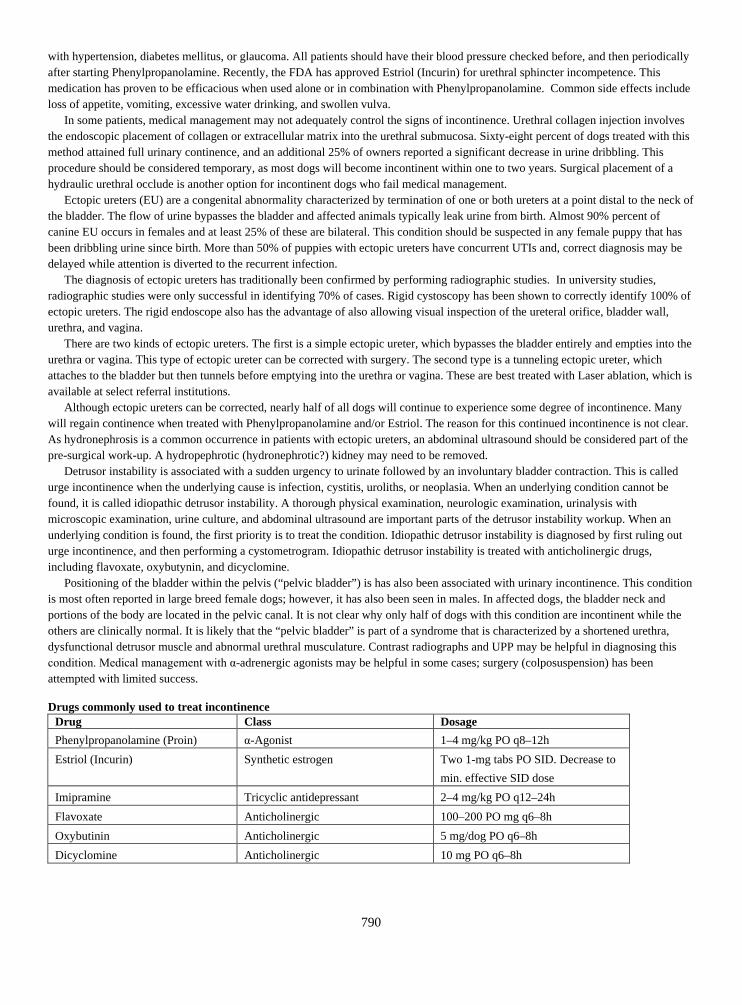
790
with hypertension, diabetes mellitus, or glaucoma. All patients should have their blood pressure checked before, and then periodically after starting Phenylpropanolamine. Recently, the FDA has approved Estriol (Incurin) for urethral sphincter incompetence. This medication has proven to be efficacious when used alone or in combination with Phenylpropanolamine. Common side effects include loss of appetite, vomiting, excessive water drinking, and swollen vulva.
In some patients, medical management may not adequately control the signs of incontinence. Urethral collagen injection involves the endoscopic placement of collagen or extracellular matrix into the urethral submucosa. Sixty-eight percent of dogs treated with this method attained full urinary continence, and an additional 25% of owners reported a significant decrease in urine dribbling. This procedure should be considered temporary, as most dogs will become incontinent within one to two years. Surgical placement of a hydraulic urethral occlude is another option for incontinent dogs who fail medical management.
Ectopic ureters (EU) are a congenital abnormality characterized by termination of one or both ureters at a point distal to the neck of the bladder. The flow of urine bypasses the bladder and affected animals typically leak urine from birth. Almost 90% percent of canine EU occurs in females and at least 25% of these are bilateral. This condition should be suspected in any female puppy that has been dribbling urine since birth. More than 50% of puppies with ectopic ureters have concurrent UTIs and, correct diagnosis may be delayed while attention is diverted to the recurrent infection.
The diagnosis of ectopic ureters has traditionally been confirmed by performing radiographic studies. In university studies, radiographic studies were only successful in identifying 70% of cases. Rigid cystoscopy has been shown to correctly identify 100% of ectopic ureters. The rigid endoscope also has the advantage of also allowing visual inspection of the ureteral orifice, bladder wall, urethra, and vagina.
There are two kinds of ectopic ureters. The first is a simple ectopic ureter, which bypasses the bladder entirely and empties into the urethra or vagina. This type of ectopic ureter can be corrected with surgery. The second type is a tunneling ectopic ureter, which attaches to the bladder but then tunnels before emptying into the urethra or vagina. These are best treated with Laser ablation, which is available at select referral institutions.
Although ectopic ureters can be corrected, nearly half of all dogs will continue to experience some degree of incontinence. Many will regain continence when treated with Phenylpropanolamine and/or Estriol. The reason for this continued incontinence is not clear. As hydronephrosis is a common occurrence in patients with ectopic ureters, an abdominal ultrasound should be considered part of the pre-surgical work-up. A hydropephrotic (hydronephrotic?) kidney may need to be removed.
Detrusor instability is associated with a sudden urgency to urinate followed by an involuntary bladder contraction. This is called urge incontinence when the underlying cause is infection, cystitis, uroliths, or neoplasia. When an underlying condition cannot be found, it is called idiopathic detrusor instability. A thorough physical examination, neurologic examination, urinalysis with microscopic examination, urine culture, and abdominal ultrasound are important parts of the detrusor instability workup. When an underlying condition is found, the first priority is to treat the condition. Idiopathic detrusor instability is diagnosed by first ruling out urge incontinence, and then performing a cystometrogram. Idiopathic detrusor instability is treated with anticholinergic drugs, including flavoxate, oxybutynin, and dicyclomine.
Positioning of the bladder within the pelvis (“pelvic bladder”) is has also been associated with urinary incontinence. This condition is most often reported in large breed female dogs; however, it has also been seen in males. In affected dogs, the bladder neck and portions of the body are located in the pelvic canal. It is not clear why only half of dogs with this condition are incontinent while the others are clinically normal. It is likely that the “pelvic bladder” is part of a syndrome that is characterized by a shortened urethra, dysfunctional detrusor muscle and abnormal urethral musculature. Contrast radiographs and UPP may be helpful in diagnosing this condition. Medical management with α-adrenergic agonists may be helpful in some cases; surgery (colposuspension) has been attempted with limited success. Drugs commonly used to treat incontinence
Drug Class Dosage Phenylpropanolamine (Proin) α-Agonist 1–4 mg/kg PO q8–12h Estriol (Incurin) Synthetic estrogen Two 1-mg tabs PO SID. Decrease to
min. effective SID dose Imipramine Tricyclic antidepressant 2–4 mg/kg PO q12–24h Flavoxate Anticholinergic 100–200 PO mg q6–8h Oxybutinin Anticholinergic 5 mg/dog PO q6–8h Dicyclomine Anticholinergic 10 mg PO q6–8h
791
Help! Help! I Can’t Pee! Feline Lower Urinary Tract Disease
Mark Acierno, DVM, DACVIM Louisiana State University
Baton Rouge, LA
Feline lower urinary tract disease (FLUTD) is a term given to cats that present exhibiting clinical signs including: straining to urinate, hematuria, pollakiuria, and periuria. The condition can be divided into two groups: cats that present with a urethra obstruction, and cats that are unobstructed but displaying clinical signs. Regardless, of whether they are obstructed or not, the most common cause of all FLUTD is idiopathic cystitis. This is a diagnosis of exclusion and accounts for about 70% of affected cats. Only about 15% of cats with lower urinary tract signs have uroliths. Urinary tract infections are extremely uncommon and make up less than 2% of these patients. Tumors are occasionally seen, but very uncommon.
The first step in assessing a cat that presents with FLUTD is to quickly recognize patients that are obstructed. These cats, which are almost always male, can be identified by the palpation of an enlarged, firm bladder. Affected cats may azotemic, hyperkalemic, acidotic, bradycardic and have ECG arrhythmias associated with atrial standstill (e.g. absence of P waves, prolonged QRS, spiked T waves). Obstructed patients should have an IV catheter placed and, unless they are obtunded, be sedated (the author uses ketamine and valium). One algorithm
• Monitor ECG and treat hyperkalemia as needed • If patient is stable, radiograph of entire urinary tract • Draw blood for possible serum chemistry and electrolytes • Place IV catheter and begin fluids • Attempt to catheterize with a sterile 3.5 French open-end tomcat catheter
o May also use “slippery sam” • If obstruction cannot be relieved, attempt to dislodge obstruction by flushing urethra with sterile saline or LRS • If obstruction cannot be relieved and bladder is extremely firm, remove urine by cystocentesis
o This decreases back-pressure and allow for aseptic collection of urine for analysis & culture (if needed) • If obstruction will not dislodge, retropulsion with the urethral orifice pinched off. This may dilate the urethra around the
obstruction and allow the obstruction to dislodge • A SMALL amount of lidocaine gel may be infused into the catheter to decrease urethral spasm. • Once urethral patency is established: • A slippery sam canther can be inserted and sutured into place or: • Measure distance from prepuce to neck of bladder • Note distance on a sterile 5-French soft red rubber (polyvinyl) and insert into the urethra up to that point • Place tape on external part of tube (as close to prepuce as possible) and suture tape to prepuce with two sutures • Tape free end of tube to tail • Attach extension set to urinary catheter and place sterile collection bag on the other end of the extension set • Place e-collar before the patient awakens!
Pathophysiology of obstructions Most urethral obstructions are the result of spasms or urethral plugs. The origin of the “urethral plug” is unknown although they are often a mix of a proteinaceous matrix and struvite crystals. It is thought that inflammation of the urinary tract leads to leakage of serum, including albumin and bicarbonate, into the urine. This provides the protein needed for matrix formation and the increased pH of the urine, promoting the formation of struvite crystals. The protein matrix forms a web like structure in the urethra that traps the struvite crystals leading to the plug. Management of obstructed patients Patient should be rehydrated aggressively with intravenous fluids; however, care should be taken not to fluid overload these patients. Electrolyte and acid/base disturbances will correct quickly once the bladder is emptied and IV fluids have started. Patient with cardiac arrhythmias due to severe hyperkalemia can be treated with calcium gluconate, insulin/dextrose, or sodium bicarbonate. Post-obstructive diuresis may last 2-5 days and predisposes cats to dehydration and further electrolyte disturbances. The cause of post-obstructive diuresis is unknown; however, significant amounts of dilute urine can be produced leading to hypovolemia. Recommendations are to measure urine output every 2-4 hours and be used to set IV fluid rates so that IV fluids match those lost to excessive urination. Once the patient is able to eat and drink, the rate of intravenous fluid administration should be reduced gradually
792
over 12–24 hours with urine output and body weight monitored. If normal hydration is maintained, intravenous fluid administration can be stopped when the urinary catheter is removed.
The author usually leaves the indwelling urinary catheter in place until urine is clear of gross hematuria. If the initial obstruction is functional or secondary to plug formation, urethral spasms after catheter removal will often lead to re-obstruction. Medications to control urethral spasm include: Amitriptyline (Elavil) 1 mg/kg PO q24h; Prazosin (Minipress) 0.25 – 0.5 mg/cat PO q24h. Do not begin administration of these medications until elctrolyte derangements are corrected and patients are normotensive. Antibiotics should not be started unless the urine culture clearly indicates an active infection. Urinary catheterization may increase likelihood of urinary tract infection; however, this can only be determined by re-culturing urine after the catheter has been removed. There is no indication to culture urinary catheters. All idiopathic cystitis patients should be treated as indicated below. Pathophysiology of FIC Cats with FIC have abnormalities in the mucus covering of the uroendothelium, histological changes in the endothelium of the bladder wall, and abnormalities of the stress response mechanism. How these findings fit together to produce the constellation of clinical signs seen with FIC is not entirely clear. Glycosaminoglycans (GAG’s) are an important part of the protective mucus layer that covers the urinary tract. Cats with FIC produce decreased amounts of GAG’s (particularly GAG GP-51) than normal felines. Without this protective layer, irritants (e.g., hydrogen, calcium, potassium, etc.) found in urine come in contact with the bladder wall. Sensory neurons (C fibers) relay information through the spinal cord, causing the brain to sense pain. Locally, the noxious stimuli cause neurogenic inflammation (substance P release resulting in vascular leakage and histamine release).
When normal cats are subjected to stressful events, (e.g., change in the environment) there is an initial catecholamine release followed by period of acclimation and a return to baseline levels. Cats with FIC seem to have an exaggerated and extended catecholamine response that may be similar to anxiety disorders in seen in some people. This suggests that cats with FIC have an inappropriate response to stress. Besides generalized anxiety, this catecholamine release can up-regulate sensory neurons in the bladder wall and perpetuate inflammation.
Diagnostics Fifteen percent of cats with signs consistent with lower urinary tract disease have cystic calculi; therefore, a radiograph that includes the entire urinary tract is essential. Urinalysis findings are often unhelpful because hematuria, pyuria, proteinuria, and crystaluria are often seen and are not specific for any particular condition. For example, pyuria can be seen with both urinary tract infections and sterile FIC. Urine cultures are likely to be negative in young cats with normally concentrated urine; nevertheless, a culture should probably be submitted. Complete blood counts and serum chemistry analysis are unlikely to reveal a specific underlying condition.
Treatment While the exact cause of FLUTD is unknown, a multifaceted approach combining environmental enrichment, diet pheromones and pharmacology appears to give many patients relief.
Environment Feline idiopathic cystitis may actually be a clinical manifestation of a physiologically inappropriate response to stress. As such, we must look for possible stressors in the cat’s environment and ways to reduce them. FLUTD surveys that require the owner to answer questions about the home environment have been developed. These surveys are helpful in extracting information from the owner in a systematic and concise way that may be difficult in a time limited clinic interaction. In multi-cat households, the “1+1” rule should be applied to all shared resources. If there are two cats in the household, then there should be three litter boxes, three food bowls, three scratching posts, etc. Cats seem to prefer surveying the environment from elevated vantage points. Cat hammocks and window perches can provide pets with the necessary comfort. When there is conflict between pets, steps must be taken to reduce adverse interactions. Toys that encourage interaction between owners and the pets should be encouraged.
Diet The role of diet in controlling FIC is somewhat controversial. Although most feline diets have been acidified and, in some cases, magnesium restricted “for urinary tract health”, there is no known benefit of this practice. Struvite crystals do not damage normal urinary tract epithelium and their presence is neither the cause nor result of FIC. Recently, a small (25 cats) unpublished study suggested that a particular diet could decrease FLUTD by 89%; however, despite extensive marketing, this information has not yet been peer reviewed. The feeding of canned moist diet has been associated with lower incidences and recurrence of FIC; however, it is important not to “switch” diets, as abrupt changes have the potential trigger episodes of FIC. Rather, the new diet should be offered alongside the cat’s usual food, and the pet should be allowed to choose between them. If the cat will only eat the dry food, then a change should not be instituted. It is not clear if the benefit of a canned diet is entirely attributable the increased water content and associated decrease in urine concentration, or if environmental enrichment is provided by providing a moist food.
The feeding environment should be analyzed for potential stress. Most cats prefer to eat alone where they will not be disturbed by other animals, sudden movement or unexpected noises. Other cats seem to be more social and may enjoy human companionship during their meals. The goal is to minimize stress.
793
Pheromones Pheromones are chemical messengers that convey information between members of the same species. Cats release several pheromones from facial scent glands, and they deposit these pheromones by rubbing their heads on objects (including human companions) when they feel safe. A synthetic analogue (Feliway, Ceva Sante Animale, France) is available as both a room diffuser and spray. The proposed effect of this product is to decrease anxiety-related behavior. While studies examining the use of this product in cats with FIC are lacking, there is evidence that this product can decrease marking and scratching behavior in multi-cat households, as well as increase grooming and eating in hospitalized patients. It has been suggested that the area in which the cat is inappropriately urinating be treated with either a room diffuser or by application of the spray.
Drug therapy: drug therapy Amitriptyline (Elavil), a tricycle antidepressant/ antianxiety drug with anti-anxiety, analgesic and anti-inflammatory properties. A study in 2003 found a significant benefit from its use. Not only does amitriptyline decrease anxiety, provides analgesia both peripherally and in CNS, but it has been shown to be a potent relaxer of urethral tissue. Recommended dosage is 1 mg/kg Q24. Prozosin (Minipress) is an alpha-blocker that may decrease urethral spasms. It is dosed at 0.5 mg/cat (NOT kg). At this time there is no data to support or refute its usage.
While the exact cause of FLUTD is unknown, a multifaceted approach combining environmental enrichment, diet pheromones and pharmacology appears to give many patients relief. In cases in which symptoms cannot be controlled, a penile urethrostomy may be required. Because of the risk of incontinence, the required anatomic modification, and high life long risk of infection, this should only be considered when other possible alternatives have been exhausted.
794
Protein-Losing Nephropathy: Latest Treatment Recommendations
Mark Acierno, DVM, DACVIM Louisiana State University
Baton Rouge, LA
A normally functioning glomerulus provides an effective barrier against the passing of albumin and other proteins into the urine. In patients with a protein losing nephropathy, the barrier is disrupted and proteins are carried into the proximal tubule. Although PLNs start out as a condition that affects the glomerulus, eventually, the entire nephron becomes affected by inflammation and fibrosis.
There are two disease processes associated with disruption of the normal filtration barrier of the glomerulus and the development of protein losing nephropathy (PLN): glomerulonephritis (GN) and amyloidosis. These cannot be distinguished from each other on the basis of clinical signs or laboratory data. Only histology can differentiate the two conditions.
Amyloidosis is caused by an inappropriate deposition of fibrils into the glomerulus that act to disrupt normal function. These fibrils are formed by the polymerization of protein subunits (e.g. serum amyloid A), which are produced in the liver and are one of a number of acute-phase reactant proteins produced in response to inflammation. These fibrils have a specific structure called a beta-pleated sheet, and once polymerized, they cannot be degraded.
Glomerulonephritis results from antigen-antibody complexes that become trapped in the glomerular wall. GN can be divided into categories based on histologic appearance: membranoproliferative gomerulonephritis, membranous nephropathy, and proliferative gomerulonephritis. Membranoproliferative gomerulonephritis (MPGN) is the most common type of GN seen in the dog and accounts for up to 60% of all cases. Membranous nephropathy (MN) is the second most common type of GN lesions seen in the dog and is the most common in the cat. Proliferative gomerulonephritis is thought to account for as little as 2% of GN cases.
Clinical features of PLNs are dependent on the underlying cause and degree of azotemia. Lethargy, anorexia, vomiting and weight loss are among the more common clinical signs; although, polyuria, polydipsia and ataxia can be seen. PLN is generally a disease of middle aged to older dogs. Diagnosis
Urine analysis The hallmark of PLN is the finding of inappropriate amounts of urine protein. It is impossible to determine the significance of proteinuria without performing a urine protein creatinine ratio (UP:C). Healthy dogs should have a UP:C of less than 0.5 while cats should have less than 0.4 . White blood cells, bacteria, sperm and casts can artificially increase the UP:C; therefore, the UP:C should only be analyzed in tandem with a full urinalysis and sediment examination. Studies suggest that as the UP:C increases, so does day-to-day measurement variability. Submitting pooled samples that have been collected over three days and stored at 40of may decrease this variability.
Hematology Anemia is a common finding in PLN patients. The cause of anemia is probably multi-factorial: decreased RBC life span, decreased erythropoietin production and the anemia of chronic disease. Changes in the white blood count are often non-specific. Serum chemistry findings are similar to other renal diseases and include elevations in BUN, creatinine and phosphate. Serum bicarbonate may decrease as renal dysfunction progresses.
Infectious diseases PLNs can be secondary to an identifiable underlying disease; therefore, a thorough search for an inciting cause is essential. Infectious, neoplastic, endocrine, inflammatory and drug-related causes must be considered. Serologic tests should be selected on the basis of the patient’s geographic location and the relevant travel history. Infectious diseases associated with PLN include: Rocky Mountain Spotted fever, Ehrlichiosis, Borreliosis, Leishmaniasis, Babesiosis, and heartworm disease.
Diagnostic imaging Protein losing nephropathy can be secondary to neoplastic processes, and ultrasound and radiographic studies are an essential part of the diagnostic plan. Ultrasound can also provide information about the renal architecture and outflow tract.
Amyloidosis vs. glomerulonephritis The only way to differentiate amyloidosis from glomerulonephritis is by renal biopsy. At one time, the degree of proteinuria was thought to differentiate between the diseases; however, there is no meaningful difference between the protein loss experienced in these two conditions. In addition, several forms of GN are now recognized, and their treatment can be quite different. These cases can only be differentiated on the basis of a renal biopsy sample examined by light, electron and immunofluorescence microscopy.
795
Management General treatment
The majority of dogs with PLN develop hypertension during the course of their disease. Blood pressure should be checked periodically and antihypertensive therapy administered as needed.
Thromboembolism is the cause of death for at least 13% of dogs with glomerular disease. Much of the blame has been placed on anti-thrombin, which is lost through the damaged glomerulus. Risk of thromboembolism is greatest when anti-thrombin is less than 75% of normal. This correlates with a serum albumin of approximately 2.0 g/dl.
A low dose of aspirin may inhibit platelet aggregation and clot formation. Although previous recommendation suggested a dose of 0.5mg/kg, 1.0 – 5.0 mg/kg q24h is now recommended. An added benefit of this therapy may be to reduce platelet aggregation in the glomerulus. Another option is clopidogrel 1.1 mg/kg Q24. Clopidogrel is a platelet aggregation inhibitor, which may be used with or in place of aspirin.
Although initially counter-intuitive, PLN patients should be fed a moderately protein-restricted diet. Studies in people with PLN have shown that feeding a high protein diet is associated with increased mortality. Patients fed a low protein diet lived longer and suffered lower morbidity. The reason for this paradox is unclear, but is likely due to the increased loss of protein into the tubule.
Amyloid-specific Colchicine, has been shown to inhibit release of amyloid from hepatocytes and has been used in humans with Mediterranean fever to prevent onset of renal amyloidosis. The efficacy of this drug in treatment of renal amyloidosis in animals has not been documented. Colchicine has significant side-effects including vomiting, diarrhea and nausea. In addition, there is no known benefit to starting treatment after azotemia develops. The currently recommended canine dose is 0.02–0.04 mg/kg PO q24.
Dimethyl sulfoxide (DMSO) has been advocated for the treatment of amyloidosis. There are no controlled clinical studies to determine if DMSO is beneficial for small animals; however, an individual case report suggest a positive response. The canine dose is 80 mg/kg diluted 1:4 with sterile saline given subcutaneously three times a week. Methyl-sulfonyl-methane (MSM) is a derivative of DMSO that can be given orally. Theoretical benefits are the same as for DMSO but it is considered more convenient since it can be given at home. There is no clinical data in people or animals to support the use of MSM. The currently recommended canine dose is 88 mg/kg q8h.
GN Angiotensin converting enzyme inhibiters have been shown to decrease UP:C, lower blood pressure and improve clinical outcome in patients with GN. Some of the effects may be due to decreased glomerular hypertension secondary to dilation of the efferent arteriole. Enalapril is cleared exclusively by the kidneys, while Benazepril is cleared by the kidney and liver; therefore, Benazepril (0.25–0.5 mg/kg PO Q12-Q24h) is preferred in kidney injury patients. The goal should be to decrease the UP:C into the normal range or at least 50% of baseline. In patients that do not respond to ACEI, angiotensin receptor blockers (ARBs) may be helpful. Irbesartan 1-3 mg/kg PO q24 may be helpful.
It is becoming increasingly clear that some dogs suffer from a sub-type of GN that is responsive to immunosuppressive agents. A biopsy is required to determine which patients would benefit from immunosuppressive therapy. Histopathologic examination must include electron microscopy and immunofluorescent microscopy. More importantly, only the International Veterinary Renal Pathology Service (979.845.2351) is equipped to perform such studies. Tissue samples sent to national and university laboratories universally result in disappointing results. Cases of immunosuppressive responsive GN are treated with steroids and mycophenolate (12–17 mg/kg PO Q24 or divided).
When PLN is progressive and histopathology is unavailable, the use of immunosuppressive drugs could be considered; however, the owner must be aware of potential risks. In cases of rapidly progressive GN, mycophenolate (10 mg/kg PO q12) has been recommended with or without cyclophosphamide (200-250 mg/m2 Q 3 weeks). A complete response defined as a UPC of < 0.5, a partial response defined as a reduction of the UPC by 50%. If after 3-4 months there is no clinically significant response, therapy should be discontinued. In patients that do have a response to therapy, immunosuppressive therapy should continue for 12-16 weeks, after which the dose may be tapered while monitoring UPC and azotemia.

796
Canine Geology: Uroliths
Mark Acierno, DVM, DACVIM Louisiana State University
Baton Rouge, LA
Urine is a solution made up of dissolved substances some of which have a tendency to precipitate out of solution, while others actively inhibit precipitation. Under normal circumstances, there is a balance between the two; however, when there is an excess of solute or a shortage of inhibitors, crystals and uroliths can form. In some cases, urine pH can also directly impact the solubility of these solutes.
Presence, number, size, and location of uroliths are usually determined by radiographic and/or ultrasound examination. As size and radiolucency can vary substantially, ultrasound has proven to be more diagnostically useful than radiographs.
Likely mineral composition of uroliths can be predicted on the basis of clinical data including the age, breed, and sex, the radiographic density and shape of the urolith, the mineral composition of crystals in the urine sediment, urine pH, and bacterial culture of the urine; however, actual mineral composition can only be determined by quantitative analysis of recovered uroliths. Struvite In dogs, the most common urolith is struvite (magnesium, ammonium, phosphate). These stones, once called triple phosphate, are associated with urease-producing bacteria (Staphylococcus intermedius, Proteus mirabilis, Pseudomonas spp., Klebsiella spp.). Urea is plentifully in the urine and urease splits it to form ammonia and bicarbonate. Ammonium combines with magnesium and phosphate while the bicarbonate raises the urine pH. The change in pH decreases the solubility of the ammonium, magnesium, phosphate complex leading to the formation of struvite crystals and stones. The anatomic configuration of the female makes urease-producing infections more common than in males.
Struvite uroliths can be dissolved over time; however, in males, the long narrow urethra and the potential for stones to become lodged in the area of the os penis, should be considered. A discussion with the owner about the relative risks and benefits of medical and surgical management should occur before deciding on a therapy.
Dissolution of struvite uroliths involves a combination of antimicrobial and dietary therapy. Appropriate antimicrobial selection can only be made on the basis of a urine culture and sensitivity. The urine must be obtained by cystocentesis and examined in a timely fashion. Since bacteria are often sequestered and protected in the layers of the stone, antibiotic therapy must be continued throughout the dissolution process. There are several commercially available struvite dissolving diets. These diets tend to be low in protein and acidify the urine; however, these diets may not be nutritionally balanced and are not intended for long-term use. Every month, the patient’s progress should be evaluated by performing abdominal radiographs and a urine analysis. If the pH is greater than 7.0, a resistant urinary tract infection should be suspected and a urine sample, obtained by cystocentesis, should be submitted for culture.
Prevention of recurrence Inhibition of struvite formation requires prevention of alkaline urine; therefore, the urinary tract must be free of infections. At home monitoring can be performed by measuring urine pH on a regular basis. This can be performed using urine dip-sticks or with an inexpensive pH meter. The presence of alkaline urine indicates that urine should be collected by cystocentesis and submitted for culture. Dogs with recurrent UTIs despite the resolution of uroliths should be evaluated for structural abnormalities that may predispose the patient to UTIs. We have found that most of these cases are related to an involuted vulva, which is amenable to surgical correction. Other patients with recurrent UTIs may benefit from long term, low-dose antibiotic therapy: ampicillin (10 mg/kg PO q24h) or amoxicillin (5 mg/kg PO q24h) administered at bedtime. Calcium oxalate The second most common canine urolith is calcium oxalate. The formation of these stones can be the result of excessive calcium / oxalate excretion or an absence of urolith inhibitors.
Pathogenesis Increased calcium excretion can be the result of excessive intestinal absorption, bone demineralization, or renal leakage. Although excessive loss of oxalate into the urine (hyperoxaluria) plays a role in urolith formation in people, it is not thought to be important in dogs. In addition, although it was once thought that calcium oxalate urolith formation was promoted by acidic urine, it is now generally accepted that formation of these stones is not greatly affected by pH.
Patients with increased intestinal calcium absorption have mildly increased serum calcium, with suppressed serum PTH levels. In the kidney, increased amounts of calcium are passed through the glomerulus; however, the absence of PTH inhibits tubular resorption. This results in hypercalciuria and stone formation.
Bone demineralization calcium oxalate uroliths have been reported in dogs secondary to hyperparathyroidism. Although uroliths have been reported secondary to paraneoplastic hypercalcemia and granulomatous diseases in people, it is not clear if this occurs in dogs.
797
Leakage hypercalciuria is a failure of the renal tubules to resorb filtered calcium despite the presence of adequate parathyroid hormone (PTH). As calcium is lost, PTH is released from the parathyroid, mobilizing calcium from bone and stimulating the conversion of calcidiol (25-hydroxycholecalciferol) to calcitriol (25-hydroxyvitamin D). Calcitriol increases intestinal absorption of calcium. Although large amounts of calcium are lost into the urine, serum levels are normal due to the actions of PTH and calcitriol. For reasons that are not entirely clear, hyperadrenocorticism (cushing’s disease) also promotes tubular calcium loss.
Calcium oxalate uroliths cannot be dissolved and must be removed by surgery or lithotripsy. One of the most important steps in preventing calcium oxalate crystals and uroliths is to increase water intake, which can be accomplished by feeding a canned diet. The optimum diet composition to prevent calcium oxalate urolith formation is unknown. Previously, it had been recommended that diets be low in protein and sodium restricted; however, recent epidemiologic studies have challenged this approach. Currently recommended diets include Royal Canin S/O, Hill’s u/d and Hills w/d. Despite appropriate diet therapy, as many as 50% of dogs will have recurrence within 3 years.
Thiazide diuretics have been shown to decrease urinary calcium excretion and should be considered in dogs with persistent calcium oxalate crystalluria or recurrence of uroliths despite appropriate diet therapy (hydrochlorothiazide 2 mg/kg PO q12h). Urinary citrate is a urolith inhibitor that is thought to form complexes with calcium and increases its solubility. In people, potassium citrate supplementation increases urine pH and increases the amount of citrate excreted into the urine. Citrate complexes with calcium and increases its solubility; however, supplementation in dogs does not result in a significant change in urine citrate concentration. Therefore, the benefit of potassium citrate supplementation is not clear. Nevertheless, some experts recommend potassium citrate supplementation (25–50 mg/kg PO q12h). Urate Urate stones are the third most common urolith in dogs and account for 8% of canine urinary calculi. A genetic predisposition (autosomal recessive) has been documented in Dalmatians and is suspected in English bulldogs. Urate stone formation has also been associated with hepatic dysfunction, especially hepatic portal shunts. Interestingly, a significant number of dogs that are not Dalmatians or English bulldogs, and have no evidence of liver disease, develop urate uroliths. While the mechanism of urate stone formation in these dogs is unclear, Yorkshire terrier, shih tzu and miniature schnauzer appear to be over represented.
Pathophysiology In most dogs, dietary purines are metabolized in a series of steps to become the highly soluble allantoin (Figure 1). This process, which takes place in the liver, involves converting purines to hypoxanthine. Through the action of xanthine oxidase, hypoxanthine is converted to xanthine and then uric acid. Uric acid, which is poorly soluble, is then converted to the highly soluble allantoin through the enzymatic action of uricase.
Although Dalmatians possess all the necessary enzymes to convert purines to allantoin, it appears that a defective cell membrane transport system decreases hepatic metabolism of uric acid and prevents its resorption in the kidney. The resulting hyperuricosuria may not completely explain the urolith formation, as not all Dalmatians with increased urate excretion are affected. It is likely that other factors, including urine pH and a decrease in the production of crystallization inhibitors, play a role. The fact that most Dalmatians that present for urate stones are male is probably due to anatomic differences (e.g. decreased urethral diameter) between the sexes.
Treatment Although urate uroliths in non-liver disease dogs can be medically dissolved, there is a risk of urethral obstruction. A discussion with the owner about the relative risks and benefits of medical and surgical management should occur before selecting a therapy. Dissolution should not be attempted in male dogs.
The dissolution of urate uroliths involves dietary modification, medical intervention and urine modification. Reduction of urate excretion can be achieved by restricting total protein and by substituting low purine content proteins (e.g., eggs) for high purine content proteins (e.g., fish). Feeding a protein-restricted diet can dilute urine by decreasing the urea available for the medullary concentration gradient of the kidney. Feeding a canned diet in order to increase water intake can further encourage diluting urate.
Allopurinol, a xanthine oxidase inhibitor, prevents the conversion of xanthine to uric acid. Although a variety of adverse reactions have been reported in humans, the most commonly reported adverse effect in dogs is the formation of xanthine uroliths. This is usually seen when allopurinol is used with a non-protein restricted diet.
Prevention Prevention of urate uroliths recurrence is best accomplished through a combination of low protein diets and urine alkalinization. Prophylactic treatment with allopurinol is not recommended, as it may promote the formation of xanthine uroliths. Urate urolith formation is inhibited in an alkaline urine as the concentration of ammonium ions, needed for stone formation, decreases. The target pH range is 7.0 -7.4. Low protein diets favor an alkaline pH; however, sometimes further urine alkalinization is needed. Potassium citrate (40-90 mg/kg PO q12h) can be utilized. A urine pH of 7.5 or higher should be avoided as this may favor the formation of calcium phosphate and struvite uroliths. Response to medical therapy can be evaluated by checking urine pH (7.0 – 7.4) urine specific gravity (<1.015) and BUN (<10 mg/dl).
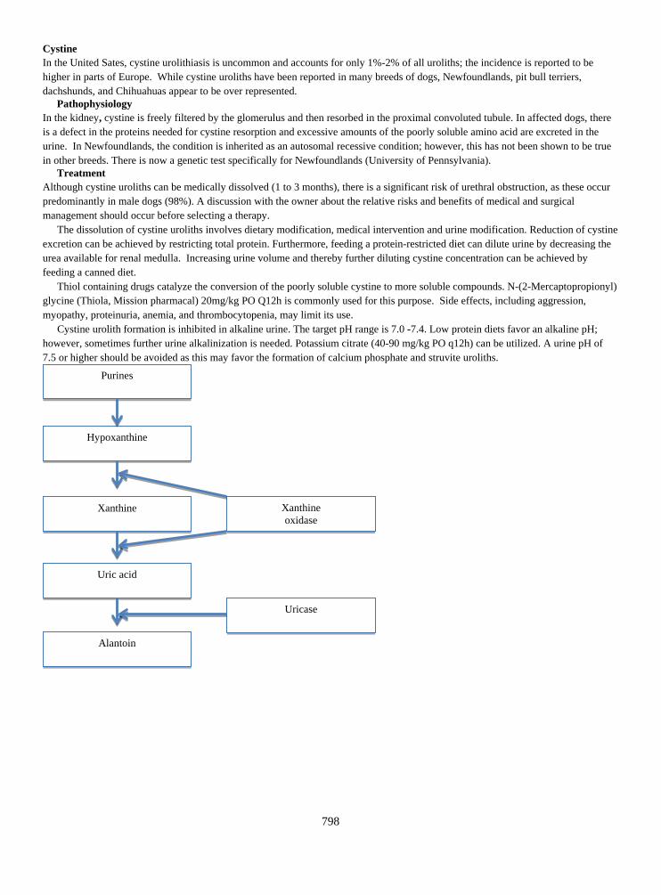
798
Cystine In the United Sates, cystine urolithiasis is uncommon and accounts for only 1%-2% of all uroliths; the incidence is reported to be higher in parts of Europe. While cystine uroliths have been reported in many breeds of dogs, Newfoundlands, pit bull terriers, dachshunds, and Chihuahuas appear to be over represented.
Pathophysiology In the kidney, cystine is freely filtered by the glomerulus and then resorbed in the proximal convoluted tubule. In affected dogs, there is a defect in the proteins needed for cystine resorption and excessive amounts of the poorly soluble amino acid are excreted in the urine. In Newfoundlands, the condition is inherited as an autosomal recessive condition; however, this has not been shown to be true in other breeds. There is now a genetic test specifically for Newfoundlands (University of Pennsylvania).
Treatment Although cystine uroliths can be medically dissolved (1 to 3 months), there is a significant risk of urethral obstruction, as these occur predominantly in male dogs (98%). A discussion with the owner about the relative risks and benefits of medical and surgical management should occur before selecting a therapy.
The dissolution of cystine uroliths involves dietary modification, medical intervention and urine modification. Reduction of cystine excretion can be achieved by restricting total protein. Furthermore, feeding a protein-restricted diet can dilute urine by decreasing the urea available for renal medulla. Increasing urine volume and thereby further diluting cystine concentration can be achieved by feeding a canned diet.
Thiol containing drugs catalyze the conversion of the poorly soluble cystine to more soluble compounds. N-(2-Mercaptopropionyl) glycine (Thiola, Mission pharmacal) 20mg/kg PO Q12h is commonly used for this purpose. Side effects, including aggression, myopathy, proteinuria, anemia, and thrombocytopenia, may limit its use.
Cystine urolith formation is inhibited in alkaline urine. The target pH range is 7.0 -7.4. Low protein diets favor an alkaline pH; however, sometimes further urine alkalinization is needed. Potassium citrate (40-90 mg/kg PO q12h) can be utilized. A urine pH of 7.5 or higher should be avoided as this may favor the formation of calcium phosphate and struvite uroliths.
Alantoin
Uric acid
Hypoxanthine
Xanthine
Purines
Xanthine oxidase
Uricase
799
Improving Quality of Life for Cats with Chronic Kidney Disease Mark Acierno, DVM, DACVIM
Louisiana State University Baton Rouge, LA
Chronic kidney disease (CKD) is the most common renal disorder affecting companion animals, and a leading cause of morbidity and mortality in older cats. The clinical presentation of CKD is the result of a progressive decrease in the kidney’s ability to concentrate urine, excrete nitrogenous waste, produce hormones and maintain electrolyte homeostasis. Although CKD is sometimes the result of congenital disorders (polycystic kidney disease, amyloidosis), infectious diseases (FIP, pyelonephritis) or neoplastic causes, older cats are usually diagnosed with idiopathic disease. Pathophysiology The inciting cause of CKD is unknown. One theory is that an initial insult reduces the number of functioning nephrons. Remaining nephrons compensate by hypertrophy and increasing clearance of nitrogenous wastes; however, this results in inflammation, tubulointerstitial fibrosis, and further loss of renal function. Clinical presentation Clinical features of clinical CKD include polyuria/polydipsia, weight loss, lethargy, anorexia, vomiting, dehydration, oral ulcers and uremic breath. Early signs are subtle and many owners miss them. Geriatric screening, which includes a CBC, chemistry, T4 and urine analysis, should be performed on all senior cats (>9 years old) to aid in identifying “at risk” patients. Diagnosis The diagnosis of CKD is made on the basis of history, physical examination and laboratory findings. Once the presence of azotemia is confirmed, other possible differential diagnosis (prerenal azotemia, post renal azotemia, and AKI) must be eliminated from consideration. A CBC, serum chemistry, urine analysis, aerobic urine culture, and abdominal ultrasound should be performed on all suspected CKD patients.
Urine analysis Although isosthenuria (Sp Grav. 1.007–1.015) is common in dogs with CKD, cats with significant renal disease can still produce urine with a specific gravity >1.020. A urine protein:creatinine ratio (UPC) may be warranted in all CKD cats. In one study, CKD cats which had a UPC <0.43 lived approximately two years while those with a protein:creatinine ratio of >0.43 lived only about nine months. Even if no bacteria are microscopically detected in the urine, an aerobic urine culture should always be performed.
Serum chemistry Overt kidney disease is always accompanied by an elevation in BUN and creatinine; however, CKD patients are often severely muscle wasted and creatinine may not be as significantly elevated as BUN. Elevations in phosphate and calcium are common while decreases in bicarbonate and potassium (in cats) are less commonly seen.
Complete blood count A normochromic normocytic non-regenerative anemia is the most common hematologic finding in patients with CKD. The WBC is usually unremarkable although elevations in the white blood cell count can be associated with chronic pyelonephritis or a systemic infectious disease.
Radiographs and ultrasound When available, ultrasound is the preferred imaging modality as it provides an unparalleled view of the renal architecture, outflow tract and blood flow. Typically, CKD is characterized as bilateral loss of renal mass; however, certain disease processes such as polycystic kidney disease have a characteristic ultrasound appearance. Enlarged kidneys suggest an underlying disease process such as: lymphoma, FIP, hydronephrosis, primary renal neoplasia or AKI. Mild dilatation of the renal pelvis (pyelectasia) is often seen with renal infections. Management
Fluid therapy Patients with CKD often have difficulty maintaining proper hydration. This complicates the management of CKD as dehydration leads to pre-renal azotemia and promotes anorexia. Initially, the patient can be encouraged to drink more fluids. Pet water fountains and flavoring tap water with chicken broth or tuna juice, or feeding a canned diet can be helpful. Administration of subcutaneous fluids at home can help prevent dehydration, minimize pre-renal azotemia and improve the patient’s quality of life. Later in the disease process, an esophagostomy tube can be placed to help provide both nutrition as well as fluids. In addition, medications can be administered through this route.
800
Control of acidosis Maintenance of acid-base balance is one of the kidney’s primary functions. Even when mild, acidosis complicates CKD by increasing protein and muscle catabolism, stimulating bone demineralization and contributing to the clinical signs of renal disease. Most feline diets are acidified. Changing CKD patients to an appropriately buffered renal failure diet is perhaps the most important step in controlling metabolic acidosis. Occasionally additional alkalization therapy may be required. When Total CO2 <18 mEq/L or serum bicarbonate is <17 mEq/L, sodium bicarbonate at a dose of 5-10 mg/kg PO q8h–q12h or potassium citrate at 40-60 mg/kg PO q8h–q12h can be given. The exact dose needs to be tailored to the individual patient.
Potassium At one time, hypokalemia was a common finding in cats with CKD. This condition has been associated with muscle weakness, ileus, and fatal cardiac arrhythmias; however, the most common clinical sign is anorexia. The mechanism by which hypokalemia develops is believed to be a combination of anorexia and increased renal loss. Renal failure diets are supplemented with potassium and in most cases a diet change will prevent this condition. Oral potassium supplementation can be given to patients that do not respond to diet. Potassium citrate 20-30 mg/kg PO q24h or Potassium gluconate 2–6 mEq/cat PO q24h can be given.
Hyperphosphatemia Because of its role in muscle and nerve conduction, the body tightly regulates serum calcium. The parathyroid glands and kidneys play the central role in this process. Hyperphosphatemia, causing secondary hyperparathyroidism and hypercalcemia, is common in CKD patients. This is problematic as there is a risk of soft tissue mineralization when the serum calcium and phosphorus product approaches 70. Since the primary source of serum phosphorus is diet, all CKD patients should be fed a low phosphorus, renal failure diet. If, after 14–21, days the hyperphosphatemia has not resolved, a phosphate-binding agent should be added to the diet. Aluminum hydroxide (30–80 mg/kg PO q24h divided and given with food) compounded into capsules and given with meals is the product that is often recommended. Commercially available liquid phosphate binders are especially unpalatable to cats. lanthanum (Fosrenol) 30 mg/kg divided and given with meals is an excellent alternative. It should be crushed and mixed well with food to prevent vomiting. Calcium based phosphate binders have the potential to complicate the disease process.
Secondary hyperparathyroidism can also be treated with calcitriol supplementation. Theoretical benefits include control of hyperphosphatemia as well as suppression of the renin – angiotensin system, systemic activation of Vit D receptors and protection of podocytes in glomerular hypertrophy. One study demonstrated that in Dogs with stage 3-4 CKD, progression of disease slowed and length of life increased when they were treated with calcitriol; however, similar benefits have not been exhibited in cats. The treatment protocol is to first normalize serum phosphous & ionized calcium using standard medical therapy. Calcitriol can then be given (2.0 – 2.5 ng/kg Q24). To minimize calcium absorption, calcitriol should not be given with meals. Serum ionized calcium and PTH need to be monitored regularly. The goal of therapy is to minimize PTH with out inducing hypercalcemia.
Treatment of anemia The most important cause of anemia in the CKD patient is the loss of erythropoietin production by renal interstitial cells. Historically, endogenous erythropoietin production in CKD cats has be supplemented with recombinant human erythropoietin (Epoetin alfa). This is a human recombinant protein and, in cats, it can stimulate an immune response that can inactivate both the exogenous and endogenous hormone; Therefore, use of this product was reserved for patients who demonstrate significant anemia (PCV<15). The usual starting dose is 44–120 units/kg SQ three times a week. The goal of treatment should be to raise PCV to approximately 25%. Once this is attained, a maintenance dose of 44–88 units/kg SQ one to two times a week is often sufficient. Failure of the PCV to rise despite increasing dosages of erythropoietin is strongly suggestive of an immune response. The drug should be discontinued at once, and its use can be re-attempted in 3-4 weeks. The use of erythropoietin has been associated with hypertension, and therefore, blood pressure should be checked at every visit. Darbepoetin Alfa (Aranesp) is an alternative to Epogen that is thought to be less immunogenic than epoetin. It is dosed 0.45 μg/kg SC once weekly and then tapered to once every 2-3 weeks. Because it appears to be less immunogenic, PCV can be increased to the lower end of “normal range” (approx. 35%). Iron stores should be supplemented (10 mg/kg IM every 3–4 weeks). Although it is more expensive than Epoetin alfa, Darbepoetin should be used when ever possible.
Diet Cats with CKD, who are fed an appropriately formulated renal failure diet, live longer than those fed a maintenance formulation. These diets help by controlling acidosis, hyperphosphotemia, and hypokalemia. It is now thought that the ratio of omega-3 to omega-6 fatty acids may also play an important role in slowing the progression of renal disease. Most renal failure diets are supplemented with omega-3 fatty acids.
Anorexia The cause of anorexia in CKD patients is probably due to irritation/ulceration of the GI tract combined with the hematogenous build up of uremic toxins. Because of their formulation, renal failure diets can compound the problem by being unpalatable. Owners should try several different brands and formulations in order to find one that their pet will eat. Renal failure diets should never be started in hospital or a food aversion may develop. Many CKD pets will eat frequent small meals rather than one or two large ones. Being certain to provide small amounts of fresh food several times a day can be helpful. Warming of wet food can make it more appetizing.

801
Because hypergastrinemia can occur with CKD, some patients respond to H2-histamine receptor blockers, such as famotidine (0.5mg/kg PO q24h). For patients that will not eat a renal failure diets, consultation with a boarded nutritionist should be considered.
Advanced renal therapies Renal transplantation for cats with CKD is now being performed at several referral centers. In order to be a candidate, a patient must be free of infectious diseases (FeLV, FIV, toxoplasmosis), have recent thoracic radiographs, an abdominal ultrasound, and an echocardiogram. Six-month survival approaches 60 %, while three-year survival is greater than 40 %.
802
Measure Twice, Cut Once: Calculate Marketing Return on Investment
Jed Schaible, VMD, CVPM The Vet Marketing Firm
St. Petersburg, FL
For a private practice, advertising is crucial to maintain healthy new client growth numbers that achieve sustainability and hopefully growth. There is little argument that measuring the ROI with digital marketing is much easier than with traditional advertising, such as print, PR, Yellow Pages, mailers, etc. Too many traditional campaigns fall into the trap of being unmeasured or under-measured even though there are affordable solutions to track the impact of most advertising campaign dollars.
This lecture will present actionable measuring techniques to assess advertising impact and ROI through free or affordable services that, although advanced in their efficacy, are quite attainable and easy to implement. The following measurement techniques will be discussed:
1. Landing Pages and Google Analytics 2. Call Tracking and Monitoring 3. Heat Mapping 4. Calculating CPA and Cutting The Fat
Landing pages for conversions There are many forms of advertising modalities that occur online, in print, on signage and on the practice website. If you are running an ad in a magazine or advertising in a newspaper, how can you tell the amount of traffic those advertisements are generating?
If you only do one thing to drastically improve your marketing ROI of advertising, adding landing pages to your website is a savvy investment. Landing pages are single web pages that appear in response to clicking on an ad or a search engine result that are a logical extension of the ad or search term. For example, if you type “golf clubs” into Google, Golfsmith.com has the number one ranking with a landing page that is not their homepage, but rather a dedicated page discussing its selection of clubs. If your practice is like most other practices pushing advertising, email marketing, and social media traffic to their website homepage, you are missing out on a sure way to increase the odds of converting that traffic into customers. Google’s “Quality Score” was discussed in the lecture (http://en.wikipedia.org/wiki/Quality_Score).
Having Google Analytics on your website will allow you to determine landing page traffic and if you have a dedicated landing page for each ad, you will be able to directly measure the direct traffic from each of the ads. Also, did you know that there is a social section in Google Analytics? Using the “Network Referrals” tab in the social section will show you the traffic that is coming from Twitter, Facebook, and Google+. Want to know who is calling your practice from your Facebook page? No problem; simply assign a call-tracking number to your Facebook page.
Example Dr. Baker owns a two-doctor practice and just built an addition in order to provide enough space for boarding. She decides that she is going to run an ad in her local newspaper. Dr. Baker is paying a rate based on “Cost Per Mile” – a fee per thousand views/impressions on the newspaper website as well. She has Google Analytics set up on her website but she can’t tell if the print ad is driving visits to her website.
In this example, if a person is interested in boarding their pet, they type in the website and be brought to the homepage of a veterinary hospital. The content at the destination is not targeted to fill the needs of that potential customer and there is no landing page offer. If a landing page is provided that has a great pitch about the boarding services, it will not only increase the likelihood of a sale, but Google Analytics can be used to measure the traffic being generated from the advertisement and shed light on of the ROI of you’re the ad campaign. If Dr. Baker had run an ad the newspaper with a boarding promotion landing page (i.e. www.yourwebsite.com/boarding-coupon) she would have been able to track the ROI of the campaign and would have increased the conversion to call. If Dr. Baker saw little or no traffic being generated by that ad in Google Analytics, Dr. Baker would know to stop paying for that ad and try something else. Call tracking to assess advertising ROI and customer service Call tracking and recording is a fantastic way to determine if your advertising, premium listing or Google/Bing ads are performing well or should be cut. Call tracking is a technology that enables a practice to track phone calls associated with performance-based advertising and provides actual recordings of inbound calls generated by ads and accessible via a cloud-based dashboard.
There are countless providers… simply type “call tracking” into Google. The cost is pennies per minute and the practice manager is able to access a dashboard to listen to calls from different phone numbers that are tracked for separate ads. Many practice managers worry about having another number out there that is different from their real number. That is why I recommend holding on to the phone number indefinitely, which has a negligible cost compared to the power of call tracking and recording.
803
Example Dr. Nash has a practice near a high-end gated community with a favorable demographic. He has a competitor in the shopping center right next to the gate at the community entrance and he suspects that this is providing them a huge competitive advantage. Dr. Nash offers more comprehensive services, such as acupuncture, laser therapy, and rehabilitation, but it has been difficult to get the word out to that community. He decides to advertise in the community’s publication and the local newspaper. He has no idea if these advertisements are driving business to his practice.
Dr. Nash didn’t know that he could use a call tracking service to create virtual phone numbers that redirect to the reception desk. By assigning a different phone number to each of his ads (Yellow Pages, Newspaper, Pay Per Click Landing Pages, etc.), he could assess whether or not his ads are driving business and cut the ads that are ineffective (which would more than pay for the call tracking).
Call tracking can even go to another level in providing analytics around how effective team members are at customer service. Customer service performance metrics can be attained and used to identify problems at the reception desk by having incoming calls recorded and graded on specific criteria by the call tracking service or by the practice manager. Long hold times can be identified, over- or under-quoting services can be discovered, and customer service issues can be addressed with the aid of actual recordings used in team meetings. Nothing is worse than paying for a “click” and having the receptionist fail to close the appointment.
Calculating cost-per-acquisition (CPA) for advertising campaigns – it is time to cut the fat Now that you are able to estimate the revenue from your print ads and your digital campaigns through landing page analytics and call tracking, you will want to calculate the CPA for those campaigns. Let’s pretend you are evaluating your advertising dollars spent on the Yellow Pages.
A Yellow Pages ad for your practice costs $2000 per year and the call tracking data shows that it produced 50 calls. Because you have a call-tracking dashboard, the office manager can cross-reference the inbound phone numbers on your practice management software to see what percentage of calls led to new clients. As it turns out, ten of those became new clients.
You can then do a ballpark estimate of the client lifetime value (LTV) by using this equation; (Average Value of a Sale) X (Number of Repeat Transactions) X (Average Retention Time in Years for a Typical Customer). As it turns out, the gross LTV for your practice is $850 and since your margins are 20%, the net profit LTV is $170. The value of those 10 clients is therefore $1700 in net profit (10 x $170).
Cost-per-acquisition calculations allow you to focus primarily on how your advertising costs compare to the number of client acquisitions those costs yield. Using this example, the ad cost $2000 and resulted in 10 clients, therefore the CPA for that ad is $200. The CPA shouldn’t exceed the profit made from each client acquired. In this example, the CPA is 15% higher than the net profit that the ad created and therefore, you should second-guess renewing your Yellow Page ad in the next sales cycle.
Content is king… But which content is king? Using heat mapping to shed light on content value and placement Heat Mapping is the process of recording website user behaviors. Google Analytics is fantastic at determining what is clicked on and how long pages are read, bounce rate, etc., but what your clients are actually interested in on your pages was previously unknown… until now.
Heat mapping software can be easily installed on your website to track mouse movements, scrolling, clicks and referrals. During the lecture, examples were shown of heatmapping results on an actual practice website. In the example it was shown how a practice had been dedicating resources to build out a resource center that almost no one viewed or clicked on. In another example, it was shown that very few users scrolled down below the fold. “Above the fold” refers to the portion of the webpage that is visible without scrolling on a screen. The recommendation was to move some important links to “above the fold”. To view a;; these images, you can head over to http://bit.ly/jedslides.
Caption These are Heat Mapping results. Notice the resources link on the left has negligible interest, where as the services and staff bio pages are of more interest to clients. Putting it all together If you create unique landing pages with relevant content based off of heat mapping data for each advertisement and assign a unique phone number to each ad through call tracking, assessing the ROI of your advertising dollars becomes much more scientific than the “where did you find us” box on your new client sign-up form. These marketing analytics will also solve the problem of a client writing “newspaper” and then you having ads in multiple newspapers or

804
writing “online”, which could mean your website, Google Maps, Adwords, Yellow Pages, etc. You will be left with one very welcome dilemma, and that is what are you going to do with the freed-up funds in your new advertising budget?
805
No Search Engine Optimizer, No Clients: Your Disappearing Website
Jed Schaible, VMD, CVPM The Vet Marketing Firm
St. Petersburg, FL
Search Engine Optimization (SEO) is relatively new, constantly changing concept for practice marketers. There are two types of SEO. One is national SEO that a brand uses across the country to improve traffic. This involves on-page factors, links, indexing, social sharing, etc. A veterinary practice, however, is most concerned with local SEO.
Local SEO has some unique challenges. Although the factors mentioned above in national SEO also are included in Local SEO, local SEO comes with some additional challenges. The first and arguably the most important local factor that you can control is your Google+ profile. Your Google+ My Business listing is critical to optimize with appropriate categories. Examples were given on miscategorized Google+ My Business listings and how it affected the practice revenues. One example was given where a practice was categorized as a “boarding house” instead of a “veterinary hospital” and they were completely removed from local search engine result pages. The use of multiple categories for the practice profit centers and the simplicity of the system was demonstrated.
Here is what you will need to optimize the Google My Business Page: 1. Minimum of 6 high quality photos 2. 1 hi res logo (250 x 250 px) 3. 1 optimized cover photo (Up to 2120 x 1192 px) or 5 images (from the 6) 4. Practice information 5. Hours of operation 6. Categories (Veterinary Hospital, Boarding, Grooming, Pet Store, etc.) 7. Pick your primary Category as the hospital profit center 8. Reviews (hopefully positive)
The Google My Business page is an example of a local citation. There are hundreds of local citations. A partial list can be found at the following link: http://bit.ly/jedcitations.
Here is the definition of a local citation from an authoritative SEO publisher: “Citations are defined as mentions of your business name and address on other webpages—even if there is no link to your website.
An example of a citation might be an online yellow pages directory where your business is listed, but not linked to. Citations can also be found on local chamber of commerce pages, or on a local business association page that includes your business information, even if they are not linking at all to your website. Citations are a key component of the ranking algorithms in Google and Bing. Other factors being equal, businesses with a greater number of citations will probably rank higher than businesses with fewer citations (1).”
The local search ecosystem diagram is needed to understand how the local search data on your practice is propagated within the Internet. In the USA, there are four primary sources of data for the search engines to pull from: Acxiom, Localeze, Factual, and Infogroup. Other local citations are players in sending “fresh” information to search engines as well, however, only the premium listings, such as Superpages or Citysearch, have a critical role in data provision. It can take several months for changes in these data aggregators to populate in search engine databases thereby impacting search engine rankings (2). You don’t have to understand this completely, you just need to know to check your major listings for errors.
If your practice has incorrect information (NAP) on any of these major citations, such as Superpages or Yelp, it may confuse the search engine algorithms and therefore the data sent into Google, Bing, Yahoo, etc. This negatively impacts the ability for potential new clients to find your practice. If you aren’t showing up in local search engine result pages (SERPs), until you can get the data fixed, you may consider running Google Adwords paid search ads so that your practice shows up in the meantime.
Several tools were demonstrated that allow a practice to check their data set online and find where there are inconsistent, incomplete, or missing data sets on various local citation sources. A real practice example was give on how a private practice can out-compete a corporate chain simply by manually optimizing their local citations with a large data set and by making sure that they add bios, pictures, hours, categories, email, services, hospital summary and more.
Live case studies were presented on practices in the lecture. An example was given of a practice named “Blue Cross Animal Hospital”. It was shown that the practice was being confused by data providers with Blue Cross & Blue Shield, an insurance company. This practice was not ranking well in local SERPs due to the inconsistent data across their local citations. The most important local citations affected were Yahoo local, YellowPageCity.com, Mapquest and Citysearch. Using these citations and The Local Search Ecosystem diagram, it was demonstrated that the data errors were due to problems with the Infogroup data aggregator. In this example, a $50 submission to Infogroup would have corrected the problem within several months.
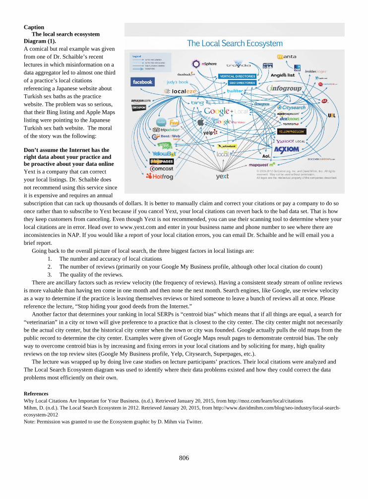
806
Caption The local search ecosystem
Diagram (1). A comical but real example was given from one of Dr. Schaible’s recent lectures in which misinformation on a data aggregator led to almost one third of a practice’s local citations referencing a Japanese website about Turkish sex baths as the practice website. The problem was so serious, that their Bing listing and Apple Maps listing were pointing to the Japanese Turkish sex bath website. The moral of the story was the following: Don’t assume the Internet has the right data about your practice and be proactive about your data online Yext is a company that can correct your local listings. Dr. Schaible does not recommend using this service since it is expensive and requires an annual subscription that can rack up thousands of dollars. It is better to manually claim and correct your citations or pay a company to do so once rather than to subscribe to Yext because if you cancel Yext, your local citations can revert back to the bad data set. That is how they keep customers from canceling. Even though Yext is not recommended, you can use their scanning tool to determine where your local citations are in error. Head over to www.yext.com and enter in your business name and phone number to see where there are inconsistencies in NAP. If you would like a report of your local citation errors, you can email Dr. Schaible and he will email you a brief report.
Going back to the overall picture of local search, the three biggest factors in local listings are: 1. The number and accuracy of local citations 2. The number of reviews (primarily on your Google My Business profile, although other local citation do count) 3. The quality of the reviews.
There are ancillary factors such as review velocity (the frequency of reviews). Having a consistent steady stream of online reviews is more valuable than having ten come in one month and then none the next month. Search engines, like Google, use review velocity as a way to determine if the practice is leaving themselves reviews or hired someone to leave a bunch of reviews all at once. Please reference the lecture, “Stop hiding your good deeds from the Internet.”
Another factor that determines your ranking in local SERPs is “centroid bias” which means that if all things are equal, a search for “veterinarian” in a city or town will give preference to a practice that is closest to the city center. The city center might not necessarily be the actual city center, but the historical city center when the town or city was founded. Google actually pulls the old maps from the public record to determine the city center. Examples were given of Google Maps result pages to demonstrate centroid bias. The only way to overcome centroid bias is by increasing and fixing errors in your local citations and by soliciting for many, high quality reviews on the top review sites (Google My Business profile, Yelp, Citysearch, Superpages, etc.).
The lecture was wrapped up by doing live case studies on lecture participants’ practices. Their local citations were analyzed and The Local Search Ecosystem diagram was used to identify where their data problems existed and how they could correct the data problems most efficiently on their own. References Why Local Citations Are Important for Your Business. (n.d.). Retrieved January 20, 2015, from http://moz.com/learn/local/citations Mihm, D. (n.d.). The Local Search Ecosystem in 2012. Retrieved January 20, 2015, from http://www.davidmihm.com/blog/seo-industry/local-search-ecosystem-2012 Note: Permission was granted to use the Ecosystem graphic by D. Mihm via Twitter.
807
Stop Hiding Your Good Deeds from the Internet Jed Schaible, VMD, CVPM
The Vet Marketing Firm St. Petersburg, FL
Benjamin Franklin once wrote, "Either write something worth reading or do something worth writing about." Public Relations is critical to establishing your practice in a market, both through word of mouth and through local SEO. In this lecture, practice marketers will learn how to write an effective and optimized press release, how to leverage their clients to score media pickups, and learn about PR campaigns that have had high success rates for other practices. Practice marketers will learn how to get their daily good deeds published on the Internet through online brand reputation campaigns that increase star ratings on important sites such as Google+, which favorably influence local search engine rankings. Press release optimization for SEO All too often I come across practices that have been fortunate enough to have their good deeds covered by the news, however, more often than not, they gain no SEO link equity from the news coverage. By SEO link equity, I mean a link with favorable “anchor text” linking to your website or a landing page on your website.
The anchor text are the words that you click on in a hyperlink to go to a website URL. Search engines like Google use a practice’s link profile in part to determine the practice ranking in local search engine result pages (SERPs). If a lot of publishers on the web are linking to your practice with the keywords, “New York City Veterinarian”, your website will start to rank more authoritatively for those keywords than other websites/practices. This is how writing for external publishers or being covered by the news can help you generate new customers through search engines. You just have to be sure to include your anchor text in your press releases and in articles you publish on external websites. Rotating anchor text for keyword diversity You will want to publish press releases and/or articles with anchor text for your most desirable keywords. Once your rankings improve for a particular keyword phrase, you can switch out the keywords in your anchor text in press releases moving forward. This is also applicable if you write for an external publisher, such as a local news affiliate or a local website. If your practice is in, for example, Advance, NC, you would want to focus on anchor text with “Advance, NC Veterinarian.” Once you rank well organically for those keywords, you can switch your anchor text to “Advance, NC Animal Hospital” so that you can start to rank for “animal hospital” searches in your geographic area. Here is a list of your keywords you want to focus on:
• Veterinarian / Vet (Google treats these as synonyms) • Animal Hospital • Pet Hospital • Animal Clinic • Pet Clinic
Establishing a media list You will need to create a list of media contacts in your local area and reach out to them. Consider local websites like Patch.com or local news organization websites. Remember, if the publication is in print it will not help with SEO; it will be an earned media spot that is read by people, but not search engines. Contact the editors (their email addresses are published on their websites) and express your interest in writing a weekly online column or discuss with them the process of submitting a press release. PR has a lot to do with relationships.
Your clients are very diverse. You probably have accountants, lawyers, plumbers, teachers, truck drivers, etc. as clients. You also likely have publishers and editors as clients. Reach out to your staff during a team meeting and ask if anyone knows of any clients in the media. More often than not, when I consult with practices, they have several media contacts already as clients. Have a cameraman at the local fox affiliate as a client? Network with that client to get an introduction to local editors. After all, with today’s 24/7 news cycle, local media publishers are starved for content. Make it newsworthy I have never had a problem getting news coverage for a charity fundraising campaign for first responders. For some reason, if a local veterinary hospital raises money to pay for police dog bullet proof vests or oxygen masks for first responding working dogs, the media seems obligated to publish those stories. Medical research press releases are also picked up easily. If you are doing a clinical trial in your practice or have launched some advanced therapeutic modality, such as stem cell therapy, that is newsworthy for local media outlets. If you are doing any research that has a human health component (i.e. the research in dogs may help children), that story will have a better chance of being picked up and potentially syndicated regionally or nationally.
808
Press release format There is a fairly standard format for creating press releases. Your credibility is contingent on your presentation, therefore pitching your story in the standard format is critical. The press release should be approximately 300-400 words. If you are publishing the press release to a PR distribution site such as PRweb.com, it is vital that the first 100 words of the press release body include the top keywords that you are interested in promoting since search engines weight this text more heavily and it is also read more by readers (1). It is also very important to copy and paste the press release into the body of the email to the publisher. Publishers get so many press releases that they usually don’t take the time to download an attachment, so make it easy on them and hopefully you can evade the publisher’s delete button. The following chart outlines the best practices for press release creation:
Press release formatting and optimization guide FOR IMMEDIATE RELEASE:
These words should be at the top of the email in upper case. If you don't want the story to be immediately covered, you can write, "HOLD FOR RELEASE UNTIL [Date]
Headline If your headline isn't catchy, your story will never have a chance City, State - Month Day, Year -
The location and the date begin the preclude the body of the release and allows the publisher to quickly identify if the story is locally and temporally relevant
Body Paragraphs Avoid long paragraphs - limit paragraphs to four lines. Unlike the headline that you write with the editors and publishers in mind, you are writing the body for readers. Include relevant human-interest quotes and mention people and their point of view to create more interest in your practice/service.
Last Body Paragraph Here you want to include the call to action (i.e. a phone number to call, a website to go to). You will want to include a link with favorable keyword anchor text.
Contact Information Include the contact's name, title, email, phone number, website address and physical address.
END ### This indicates the end of the press release. It is optional to add the word count below this.
Example press release and media outcome FOR IMMEDIATE RELEASE
Tarpon Springs Veterinarian is Making Incredible Advances in Stem Cell Research in Dogs with Allergies Tarpon Springs, Florida – September 17th, 2014 - Dr. Amsberry, owner of St. Francis Pet Care Center has just initiated a pilot
study in association with Animal Cell Therapies of San Diego. To their knowledge, this is the first study ever conducted in North America, investigating the treatment of allergic dermatitis in dogs utilizing allogeneic stem cells. Pilot studies such as this, play a vital role in the FDA approval process and they are excited to be involved in such cutting-edge biotechnology.
Allergic dermatitis can be a severe, life-long, debilitating condition that is very common in the canine population, especially here in Florida. It is hoped that stem cell therapy will decrease the severity of symptoms for dogs who suffer from allergic dermatitis.
Dr. Amsberry is treating five patients who have displayed severe chronic allergic dermatitis as verified with positive allergy tests results.
Allogeneic stem cells are harvested from a dog’s umbilical cord, subjected to a barrage of tests, and replicated in the lab. Extensive testing is necessary in order to verify that these cells are pure and will not create any rejection by the host. Dr. Amsberry claims, “To date, there have been neither rejection reactions nor side effects from the use of Animal Cell Therapies allogeneic stem cells in dogs, only benefits.”
Each patient was administered an IV infusion of millions of allogeneic stems cells based upon the dog’s weight. Physical examination, photographic and client surveys were performed prior to infusion and will be repeated at 1, 3, and 6 months post infusion.
Dr. Amsberry is looking for additional patients with immune mediated diseases in the next few months. Please contact St. Francis Pet Care Center at (727) 937-7600 for more information and visit the Tarpon Springs Veterinary Hospital website. Contact: [CONTACT NAME] [CONTACT TITLE] [CONTACT EMAIL] [CONTACT PHONE NUMBER] [CONTACT WEBSITE URL]
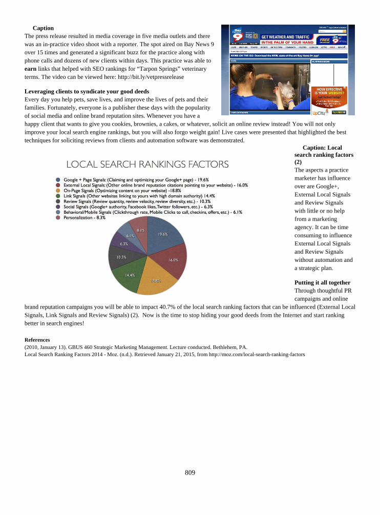
809
Caption The press release resulted in media coverage in five media outlets and there was an in-practice video shoot with a reporter. The spot aired on Bay News 9 over 15 times and generated a significant buzz for the practice along with phone calls and dozens of new clients within days. This practice was able to earn links that helped with SEO rankings for “Tarpon Springs” veterinary terms. The video can be viewed here: http://bit.ly/vetpressrelease Leveraging clients to syndicate your good deeds Every day you help pets, save lives, and improve the lives of pets and their families. Fortunately, everyone is a publisher these days with the popularity of social media and online brand reputation sites. Whenever you have a happy client that wants to give you cookies, brownies, a cakes, or whatever, solicit an online review instead! You will not only improve your local search engine rankings, but you will also forgo weight gain! Live cases were presented that highlighted the best techniques for soliciting reviews from clients and automation software was demonstrated.
Caption: Local search ranking factors (2) The aspects a practice marketer has influence over are Google+, External Local Signals and Review Signals with little or no help from a marketing agency. It can be time consuming to influence External Local Signals and Review Signals without automation and a strategic plan. Putting it all together Through thoughtful PR campaigns and online
brand reputation campaigns you will be able to impact 40.7% of the local search ranking factors that can be influenced (External Local Signals, Link Signals and Review Signals) (2). Now is the time to stop hiding your good deeds from the Internet and start ranking better in search engines! References (2010, January 13). GBUS 460 Strategic Marketing Management. Lecture conducted. Bethlehem, PA. Local Search Ranking Factors 2014 - Moz. (n.d.). Retrieved January 21, 2015, from http://moz.com/local-search-ranking-factors
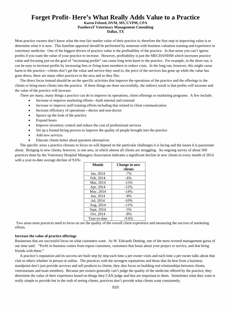
810
Forget Profit- Here’s What Really Adds Value to a Practice Karen Felsted, DVM, MS, CVPM, CPA
PantheraT Veterinary Management Consulting Dallas, TX
Most practice owners don’t know what the true fair market value of their practice is; therefore the first step in improving value is to determine what it is now. This baseline appraisal should be performed by someone with business valuation training and experience in veterinary medicine. One of the biggest drivers of practice value is the profitability of the practice. In that sense you can’t ignore profits if you want the value of your practice to increase. However, profitability is just the MECHANISM which increases practice value and focusing just on the goal of “increasing profits” can cause long term harm to the practice. For example, in the short run, it can be easy to increase profits by increasing fees or firing team members to reduce costs. In the long run, however, this might cause harm to the practice—clients don’t get the value and service they used to, the price of the services has gone up while the value has gone down, there are many other practices in the area and so they flee.
The direct focus instead should be on the specific activities that improve the operations of the practice and the offerings to the clients or bring more clients into the practice. If these things are done successfully, the indirect result is that profits will increase and the value of the practice will increase.
There are many, many things a practice can do to improve its operations, client offerings or marketing programs: A few include: • Increase or improve marketing efforts—both internal and external • Increase or improve staff training efforts including that related to client communication • Increase efficiency of operations—doctor and non-doctor • Spruce up the look of the practice • Expand hours • Improve inventory control and reduce the cost of professional services • Set up a formal hiring process to improve the quality of people brought into the practice • Add new services • Educate clients better about payment alternatives
The specific areas a practice chooses to focus on will depend on the particular challenges it is facing and the issues it is passionate about. Bringing in new clients, however, is one area, in which almost all clients are struggling. An ongoing survey of about 500 practices done by the Veterinary Hospital Managers Association indicates a significant decline in new clients in every month of 2014 with a year-to-date average decline of 9.6%:
Month Change in new clients
Jan, 2014 -7% Feb, 2014 -6% Mar, 2014 -13% Apr, 2014 -12% May, 2014 -14% Jun, 2014 -8% Jul, 2014 -10%
Aug, 2014 -11% Sept, 2014 -5% Oct, 2014 -8%
Year-to-date -9.6% Two areas most practices need to focus on are the quality of the overall client experience and measuring the success of marketing efforts. Increase the value of practice offerings Businesses that are successful focus on what customers want. As W. Edwards Deming, one of the most revered management gurus of our time said: “Profit in business comes from repeat customers, customers that boast about your project or service, and that bring friends with them.”
A practice’s reputation and its success are built step by step each time a pet owner visits and each time a pet owner talks about that visit to others whether in person or online. The practices with the strongest reputations and those that do best from a business standpoint don’t just provide services and sell products to clients; they also focus on building real relationships between clients, veterinarians and team members. Because pet owners generally can’t judge the quality of the medicine offered by the practice, they determine the value of their experience based on things they CAN judge and that are important to them. Sometimes what they want is really simple to provide but in the rush of seeing clients, practices don’t provide what clients want consistently.
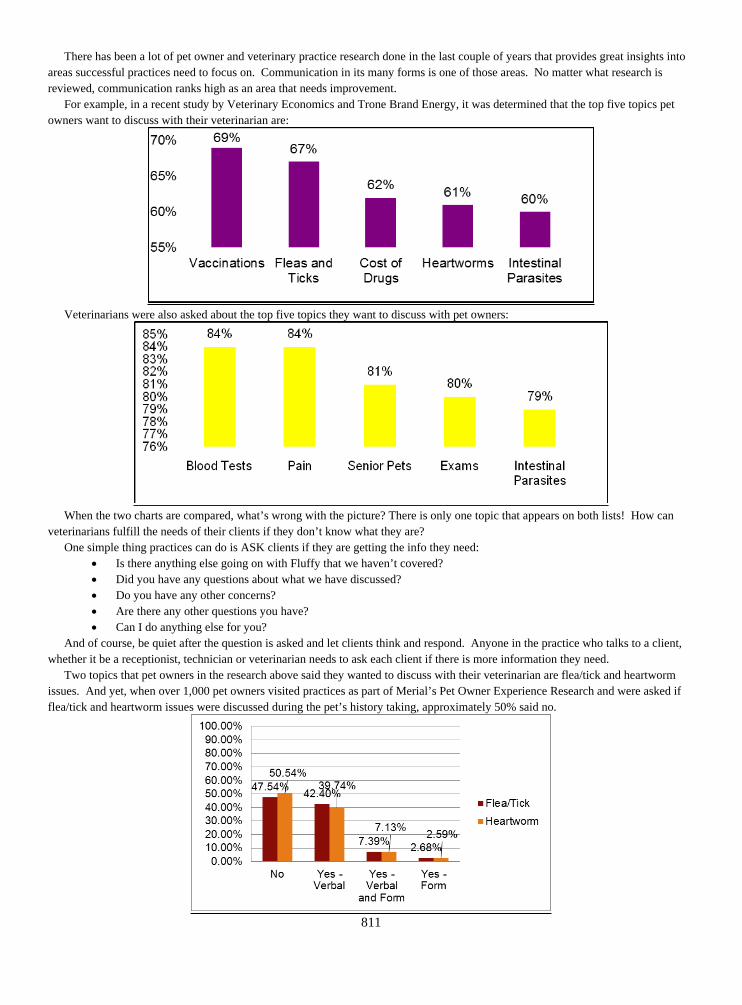
811
There has been a lot of pet owner and veterinary practice research done in the last couple of years that provides great insights into areas successful practices need to focus on. Communication in its many forms is one of those areas. No matter what research is reviewed, communication ranks high as an area that needs improvement.
For example, in a recent study by Veterinary Economics and Trone Brand Energy, it was determined that the top five topics pet owners want to discuss with their veterinarian are:
Veterinarians were also asked about the top five topics they want to discuss with pet owners:
When the two charts are compared, what’s wrong with the picture? There is only one topic that appears on both lists! How can
veterinarians fulfill the needs of their clients if they don’t know what they are? One simple thing practices can do is ASK clients if they are getting the info they need:
• Is there anything else going on with Fluffy that we haven’t covered? • Did you have any questions about what we have discussed? • Do you have any other concerns? • Are there any other questions you have? • Can I do anything else for you?
And of course, be quiet after the question is asked and let clients think and respond. Anyone in the practice who talks to a client, whether it be a receptionist, technician or veterinarian needs to ask each client if there is more information they need.
Two topics that pet owners in the research above said they wanted to discuss with their veterinarian are flea/tick and heartworm issues. And yet, when over 1,000 pet owners visited practices as part of Merial’s Pet Owner Experience Research and were asked if flea/tick and heartworm issues were discussed during the pet’s history taking, approximately 50% said no.
812
How can pet owners understand the importance of pet health care and the practice’s recommendations if veterinarians and their teams don’t talk to clients about these topics? Implementing changes to workflow, team responsibilities and communication tools can vastly improve the frequency and success of these conversations. Bringing clients into the practice Most practices are seeing a decline in new client numbers; data from about 500 practices in a year-long survey by the Veterinary Hospital Managers Association showed about a 10% decline in new clients during 2014. Attracting new clients to a practice is more important than ever and, of course, bringing clients into the practice is about marketing. In veterinary medicine, marketing is often broken down into “external marketing” and “internal marketing” with different team members focusing on each. Veterinary teams tend to think of external marketing as being the promotion of the practice done via Facebook, the practice’s website, advertising in local publications, participation in community events, or other efforts. External marketing will reach both current clients and potential new clients so the goal is to both attract new clients and solidify the relationship with current clients. Internal marketing is generally thought of as being the act of “selling” veterinary services to pet owners who are current clients of the practice via education about the need for care, the value of the services offered and providing a good client service experience.
In addition to the increased pressure to attract clients to a practice, there are also many more ways to acquire new clients than ever before. In addition to designing and implementing marketing programs, it is also even more important to determine what works in actually bringing those new clients in. The steps involved in this practice include:
• Identify where clients heard about the practice • Review client acquisition metrics • Determine the cost of acquisition • Calculate how much revenue the new clients are estimated to bring in while at the practice • Estimate the life-time profits expected to be generated from these clients
The practice must first track where new clients come from. Often this task falls to the front desk team. How the client heard about the practice should be a question asked on every new client worksheet but it is also critical that the reception team also ask every client and make sure the info was actually recorded for future analysis.
New client numbers are readily available from the practice information management system and these figures should be compared from month to month and from this month in the current year to the same month last year. New client figures can also be compared to published benchmarks. These must be compared as a ratio; the number of full-time-equivalent doctors is the most commonly used basis. Ratio analysis is better because:
• Trends over months or years can be more easily analyzed as the practice grows • Businesses of different sizes can be more easily compared
In addition to determining where the new clients are coming from and what percentage of total new clients comes from each source; the cost of acquisition must also be calculated as shown in the following example. The ABC Animal Hospital decides to advertise in the Lakeshore Condominium monthly online newsletter as well as write an “Ask the Vet” column in the same issue. The cost per month is $450.00 or $375.00 per month with a 3 month contract. The practice chooses the 3 month contract because they feel it will take that long not only for pet owners to see the promo but to have a need to visit.
The practice obtained 2 new clients in month 1, 6 in month 2 and 7 in month 3 for a total of 15 new clients from a $1,125 investment plus the time to write the “Ask the Vet” columns. They estimate each pet will bring in about $2,250 in revenue over the years it comes to the practice and that a $75 per pet acquisition cost is reasonable.
Perhaps the most important thing to remember about marketing is that you can’t sell what you don’t have. There is no point in launching into a comprehensive campaign to bring clients into the practice if they aren’t going to have a good experience once they get there. You may get them in once, but they won’t come back. Every practice needs to review the client service experience from beginning to end and correct any deficiencies. Do clients have to wait 20-30 minutes before they see a doctor? Are doctors and staff able to communicate with pet owners in a way that is friendly, professional and conveys real information? Do clients get callbacks within the promised time? Whatever is wrong with the practice needs to be put right before marketing efforts can be expected to succeed.
As discussed above, profitability is just the MECHANISM which increases practice value and focusing just on the goal of “increasing profits” can cause long term harm to the practice. The direct focus instead should be on the specific activities that improve the operations of the practice and the offerings to the clients or bring more clients into the practice. If these things are done successfully, the indirect result is that profits will increase and the value of the practice will increase.
813
Smart Ways to Help Clients Pay Karen Felsted, DVM, MS, CVPM, CPA
PantheraT Veterinary Management Consulting Dallas, TX
One of the many unhappy lessons veterinarians and their teams have had to learn from the 2007 recession and the challenging after-years is that clients don’t have bottomless wallets filled with cash to spend on veterinary care. Data from the Bayer Veterinary Care Usage Study indicates clients want more payment options; unfortunately many practices don’t offer them at all or don’t do a good job of telling clients about them. In addition to the changes in the economy, clients are dealing with the increasing costs of veterinary care resulting from the availability of more sophisticated medical options, the extended life span of pets which results in more routine care spending as well as an increased likelihood of the pet developing a serious and/or chronic disease and fee increases well above the rate of inflation. Even clients who are fully committed to providing quality care are looking at payment alternatives. Almost all practices accept cash, credit cards and checks; other payment options include third party veterinary credit cards, pet insurance, wellness plans and in-clinic billing options.
It’s not reasonable to think that practices can just give veterinary care away; running a veterinary hospital isn’t cheap and veterinarians and their staff have the same need to earn a good living as anyone else. But there are things that can be done. If clients don’t think they will need future veterinary care, why will they be interested in payment alternatives? This is one of the most important points to be made on this topic. If users of any product or service don’t think they will need it in the future, they aren’t going to care about finding a way to pay for it. Many pet owners don’t really understand the kinds of preventive care their pet will need in the future nor the cost of that care. And they don’t really think through the likelihood that the pet will get sick or injured. Veterinarians and their teams have this information and need to incorporate this into the conversations regularly had with clients. Start talking to pet owners about future care and payment alternatives from the first time you see them and don’t stop after one conversation. This topic should be regularly revisited. Money is hard to talk about sometimes and the veterinary team doesn’t always know how to get started; consider this as a sample script:
“One thing we haven’t talked about yet is Michelle’s future medical care. We have her scheduled for the rest of her kitten vaccinations so that’s all taken care of; but to keep her healthy after that, you will want to have her examined by a veterinarian at least once a year. This will ensure she gets the vaccinations and other preventive care she needs and that we can make sure there aren’t any underlying medical things going on.
Just like people, pets get sick too, no matter how well taken care of they are. For instance, some of the most common conditions we see are ear infections, stomach upsets and skin allergies. The problem is, these are never planned events and I always like to let new puppy parents know so they can prepare for the unexpected. If you’re not expecting the cost of these things, it can be a shock to the budget. Some people put money aside in a special savings account in case something happens; others like to have a more formal option in place. Here at our practice, we offer encourage pet owners to consider special veterinary credit cards or pet insurance. Would you like me to have Susan come in and talk to you a little more about these options?”
Billing options For years, many practices used in-house delayed billing plans to help clients who couldn’t come up with the cash necessary for their pet’s care at the time of service. These generally took the form of held checks or statements sent post treatment with the idea that clients would pay when they received the statement, either in full or in installments. Practices had varying degrees of success in actually collecting these amounts and the trend has been away from in-house options and towards third party payment plans, pet insurance and wellness plans.
While practice owners don’t want either themselves or their staff to function as insurance sales people or credit card vendors, those who work in practices already regularly recommend to clients products and services not carried in the practice. Examples include obedience training, pet day care centers, groomers, pet sitters and a wide variety of dietary and other products. Doctors and staff take the time to understand those products enough to be comfortable with the recommendations and help clients understand the options because they think they are of value to the client in taking better care of their pets.
Why is it any different with financial products that allow clients to provide more comprehensive care? Not only do pets benefit from the improved care; veterinary practices benefit because clients who have the financial ability to pay for better care help us practice the kind of veterinary medicine we want to practice and improve the profitability of the practice. A sophisticated study conducted by Veterinary Pet Insurance, one of the largest pet health insurance companies, showed that the company’s clients with pet health insurance on average had 41% higher stop-treatment levels, scheduled 40% more veterinary visits, and spent twice as much on veterinary care over the life of their pet. Studies by CareCredit, one of the leading third-party medical financing companies, revealed that 71% of cardholders said that having this financing option affected their decision as to the level of treatment they can provide their pet and that pet owners with veterinary credit cards and that cardholders spent almost 50% more on their pets than non-cardholders.
814
In order to effectively recommend these payment options, veterinarians and their staff must first of all understand the products themselves. Recommendations to clients are most helpful when they include not only a general recommendation for a kind of product but a recommendation for a specific brand along with the reasons why the practice thinks this product is the best one and a company the practice has had a good experience with. This is no different from medical products; clients don’t just want to know that their pets should be on heartworm preventative; they want to know which brand your practice recommends and why. Practice owners and managers need to make sure that team members have the training to then talk knowledgably and enthusiastically to clients about these options. Information should also be available in printed form as well as on the practice’s website. Additional client resources will be discussed below. Remember that clients don’t just want to be handed a couple of brochures; they want to know what you recommend. If the plans are presented as a last-ditch option instead of a powerful way for the client to provide better care, then clients will likely pick up on this lack of enthusiasm. Third party veterinary payment cards Third party veterinary payment plans aren’t all the same but in general, their financing arrangements function like a credit card that can be used for veterinary care. Clients can apply for the cards while at the veterinary practice and receive immediate approval. The practice receives its money soon after they provide the care and is not responsible for collecting from the client.
As with regular credit cards, the practice pays a fee to the financing company. Sometimes these fees are higher than those charged with regular credit cards; however, some advantages exist with these dedicated medical credit cards that counterbalance the fee. First of all, the ability to be approved for credit while at the practice means that pet owners can easily make an immediate decision to accept the practice’s recommendations for their pets that they might not have been able to do had a source of payment not been available. The higher fee also allows the financing company to offer plans which are attractive to clients and, again, encourage them to provide more comprehensive care for their pets. As with regular credit cards, not all clients will be approved. Not all clients deserve to be approved! Even if some of your clients do not get approved, going through the application process with clients you are considering granting in-house credit to is a good exercise; if the client is denied by a third party financing company you now know that there is a good chance they may not be able to pay off any amounts you let them charge in-house. Several companies offer third party payment plans in the veterinary world. In order to be comfortable recommending both the concept and a particular company to your clients, compare plans and check out the company. Are they helpful? Is it easy to get your questions answered? Is information about their plans readily available? Do any of your colleagues use them? What has been their experience? Pet health insurance Another financial option for clients is pet health insurance. As with third party payment plans, understanding the general types of plan options and the companies providing them will help you make intelligent and useful recommendations to clients. When veterinarians, their staff or their clients become unhappy with pet insurance, it generally arises from a lack of understanding of what is reasonable to expect from pet insurance in general or of the specific terms of a particular policy. A couple of points that will help both practice employees and clients understand their options better include:
• Pet insurance isn’t right for all pet owners. Several factors for clients to consider in making the decision to insure their pet include their bond with the pet, their philosophical position about how much they would be willing to pay for a pet’s care, their level of risk tolerance and the nature of their financial situation. Pet owners need to think about their ability to cover not only basic wellness care (annual exams, vaccines, heartworm tests and preventative, etc.) but also non-routine accidents and illnesses. Some clients can cover the costs of this kind of care with some planning, a savings account, a credit card and access to medical financing as discussed above. But what happens if their pet needs care that is really complicated and expensive or even catastrophic? These are the kinds of events that even the most financially responsible pet owner may have trouble finding the cash for. Pet insurance offers not just claims reimbursement but it also offers piece of mind that when something of an expensive and catastrophic nature happens, care can be provided.
• All companies limit coverage in some way; if they didn’t they would pay out more in claims than they took in from premiums and would be bankrupt in months. These limitations come in several forms including deductibles, co-pay %s, annual or lifetime limits, the use of benefit schedules, and coverage exclusions. Practice team members and clients need to understand the coverage of the policy as a whole; you can’t just say “well, this policy has a 20% co-pay so it’s not as good” or “this policy uses a benefit schedule instead of a % pay so it’s not as good”—look at the coverage as a whole compared to the premium. Pet owners also need to be aware of any breed specific conditions that apply to their pets or any particular types of procedures they might want covered (for example, dentistry or acupuncture) and see if their policy includes those items.
• For pet owners or veterinarians to expect that all owners will receive claims payments that equal or exceed what they pay in premiums isn’t even a realistic expectation. That doesn’t happen with any kind of insurance—how many people have gotten reimbursed for homeowner or automobile insurance claims that have exceeded the cost of the premium? Do
815
you even expect to? Some percentage of pet owners will pay more in premiums than they receive back in benefits; you could say they were unlucky with their pet insurance or you could say they were lucky with their animal’s health. Another group will pay much less in premiums than they get in benefits—these individuals owned pets who were unlucky health wise but they were fortunate enough to have insured their pets. And most pet owners (or owners of any insurance) are going to be somewhere in the middle.
• Companies offer information about their policies and other useful tools/resources on their websites or via phone. Pet owners need to read the information and understand it before recommending or signing up for a policy; if it’s not readily available or it’s confusing, they should find another company.
Once a pet owner has decided that pet insurance is for them, they need to pick a company and a plan. There are many options out there; it can be a bit daunting to sort through them all. You can help your clients by recommending a couple of companies you are comfortable with. You can also provide them with a list of questions they should ask:
http://veterinarybusiness.dvm360.com/handout-20-questions-veterinary-pet-insurance-companies Pay by the month preventive care plans The last alternative payment option to be discussed is pay-by-the-month wellness plans. Pet owners in the Bayer Study loved the idea of wellness plans—i.e. a group of bundled services they could pay for on a monthly basis instead of all at once. The ability to spread payments out gives clients the ability to provide better health care for their pets. The programs must be designed effectively, however, in order to provide a positive return for both pet owners and practices.
So how do these plans work? They are essentially annual preventive care plans that include the specific services the practice feels a pet owner should provide to each pet during a year in order to keep it healthy. These are wellness or preventive services such as vaccines, physical exams, heartworm testing and other diagnostic blood work, and deworming. The plans aren’t generally meant to provide care for sick pets although of course there is a little overlap. For example, the physical exam may diagnose an illness and the pets that get dewormed may already have intestinal parasites. But generally, they are focused on keeping pets from getting sick, rather than treating an illness.
What works about these plans, first of all, is that they bring people into the practice. It is much easier to pay $30/month for pet care than to have to write a check all at once for $200. The initial cost of an office visit and physical exam is a barrier for many clients and these plans encourage them to bring their pet to the practice. Veterinarians can’t diagnose disease and can’t demonstrate to a client how wonderful the practice is if clients don’t walk in the door.
Practices also make money from these plans because, just as with gym memberships and gift cards, many people don’t use all the services. The increased visits also allow veterinarians to diagnose other conditions that require treatment outside of the care included in the plan. Finally, it’s also important to remember that many clients on wellness programs spend more than they normally would have if they weren’t on the program; paying monthly is what makes this possible.
There are, of course, some design and operational issues that must be addressed in order for these plans to operate smoothly. These include:
• Payment processing • Level of integration with the PIMS • Accounting for the payments • How to allocate payments to veterinarians • Dealing with defaults • Appropriate communication • Information security requirements
As noted above, the power in programs like these is that they offer clients an incentive to visit the practice. However, this alone won’t necessarily keep pet owners visiting regularly; the clients have to have a good experience and find value in the care they get once they get to your practice. In addition to marketing the program effectively, the use of the program must be tracked over time to see if it actually increases revenue, profits, visits and the amount of care provided to patients.
Clients have made it clear they want to provide good care to their pets but they need payment alternatives to do so. Not every new strategy will work for every practice but it would be foolish to ignore the feedback we have gotten from pet owners.
816
Financial Horror Stories and How to Avoid Them Karen Felsted, DVM, MS, CVPM, CPA
PantheraT Veterinary Management Consulting Dallas, TX
The world of veterinary medicine doesn’t look like it used to 10 years ago – excess capacity, a difficult economy and changes in what clients want all impact what’s going on in practice. Thriving financially is harder than it used to be and some practices are running into real trouble. Most of it is avoidable if you know what to look for and the steps to take to forestall problems. Case #1 ABC Animal Hospital is a large companion animal general practice located in a major city in the Midwest. The practice culture is unique with a focus on client service, medical care and employee satisfaction not seen in many hospitals. They have operated out of a very small space since they opened and the dream was always to build a beautiful new hospital that would allow them to continue growing and practice the exceptional medicine they want to practice. A few financial highlights:
• Revenue grew from about $700,000 in the year the practice started to about $5,000,000 seven years later—annual growth rates ranged from 10% to 38% depending on the year.
• Revenue growth flattened out and actually declined in some months in the year before they started their building project • Profitability in the practice has been average but not great. Expense cuts would have significantly changed the culture
of the practice and the management team hasn’t wanted to do that • The practice has little in the way of an emergency fund to weather financial surprises • After moving into the new building, the practice found it couldn’t make its loan payments
What could/should the practice have done to avoid this situation? Case #2 Dr. Smith is 70 years old and the sole owner of a companion animal practice located in a small city in the southeast. The practice is located in an attractive but not extravagant building in a middle-class part of town. Dr. Smith has owned this practice for 39 years, has strong relationships with his clients and team, is active in the local VMA and is well thought of in his community. He is dealing with some very challenging health issues, is ready to retire and sell the practice and looking forward to getting a good price for it. He needs the money. A few financial highlights:
• Revenue increased 7% in the most recent year but had suffered about 10% decline in the two prior years • Transactions declining 1-3% each year • COGS generally 28% of gross revenue in each year • Support staff costs about 25% of gross revenue in each year • Rent 4% of gross revenue in each year • Revenue per doctor, ATC and new client numbers are strong • Operating profit margin ranges from 1-3% depending on the year o What’s wrong with the practice financially? o What can be done to fix it? o What could have been done earlier?
817
Diabetes Mellitus in Dogs: Acute Care and Long-Term Management and
Helping Clients Pay for it David Bruyette, DVM, DACVIM
VCA West Los Angeles Animal Hospital Los Angeles, CA
Karen Felsted, CPA, MS, DVM, CVPM
PantheraT Veterinary Management Consulting Dallas, TX
Diabetes mellitus is a common endocrine disorder in dogs and cats. Recent data has shed light on the pathogenesis of the disorder in dogs and cats and has highlighted the role of diet, insulin and novel hypoglycemic therapies. In the majority of cases, the most appropriate therapy in both dog and cats includes the administration of insulin.
The key to successful management of the diabetic patient lies in close communication with the pet owner and prompt recognition and treatment of concurrent disorders. Key facts
1. Insulin is still the mainstay of therapy in the majority of dogs and cats with diabetes mellitus. 2. Diet is an important part of diabetic management especially in obese patients and cats. 3. Auto-immune disease, pancreatitis and amyloidosis are the most common causes of diabetes in dogs and cats.
Successful management of the diabetic patient involves many factors. An understanding of dietary therapy, insulin preparations, oral and novel hypoglycemic agents and management of concurrent illness, are all required to optimize glycemic control. The goals of therapy are to control clinical signs, prevent or slow the progression of cataracts, avoid hypoglycemia and maintain ideal body weight. An additional goal in cats is to obtain remission. The challenge is to address these concerns while attempting to help the owners deal with what they may consider a time consuming, expensive and chronic medical condition.
Diabetes Mellitus in dogs and cats results from a decrease in insulin secretion from the beta cells of the pancreas and/or a decrease in insulin action. There are three classifications of diabetes:
Type I diabetes is comparable to insulin dependent diabetes mellitus (IDDM) in humans. It results in low basal insulin concentrations with impaired insulin secretion following a glucose load. Treatment requires insulin injections. It is the most common form of diabetes in dogs.
Type II diabetes is similar to non-insulin dependent diabetes (NIDDM) in humans and is managed with dietary therapy and oral hypoglycemics. It causes normal to increased basal insulin concentrations with decreased secretion following a glucose load. Insulin may or may not be required for animals with Type II diabetes.
Type III diabetes is seen most commonly in hormonally-induced diabetes in dogs and cats and is similar to impaired glucose tolerance (IGT) in humans. Diabetogenic hormones (epinephrine, cortisol, glucagon and growth hormone) or medications interfere with insulin action and cause glucose intolerance, which can lead to diabetes.
Etiology and signalment
Canine There are some distinct differences in the etiology of canine and feline diabetes. In dogs, it is generally thought to be an immune mediated disease with gradual destruction of beta cells. The progression from normal, to glucose intolerant, to diabetes, is generally slow so that most islets (over 90%) are lost before diabetes occurs. Other causes of diabetes in dogs include genetic predisposition, chronic pancreatitis and medication-induced diabetes (glucocorticoids and megestrol acetate).
Genetic predisposition to diabetes is most common in the following breeds: German Shepherd dogs, Schnauzers, Beagles, and Poodles. Golden Retrievers and Keeshonds are more prone to juvenile diabetes.
Gender is a factor in dogs with females being three times more likely to develop diabetes than males. Generally, diabetes occurs in dogs in middle age (6-9 years) but can also present earlier for specific breeds, particularly the Golden Retriever and Keeshond.
Feline The most common causes of diabetes in cats are obesity, pancreatitis and most commonly, amyloidosis of the pancreatic beta cells. There appears to be very little gender predisposition to this disease in cats, although it is slightly more common in males than females. As with dogs, the onset of diabetes in cats occurs most often in middle age.
818
Clinical signs The clinical signs of diabetes include PU/PD (polyuria and polydipsia) from hyperglycemia, resulting in glycosuria and a resultant osmotic diuresis. Polyphagia and weight loss is common although many animals will still be obese upon presentation. In addition to the polyphagia, there may be variable degrees of dehydration especially in the cat. Cataract formation is very common in dogs with diabetes, but rare in cats. Cats often present with icterus as a result of concurrent hepatic lipidosis and/or pancreatitis. Icterus is not common in dogs unless they have pancreatitis. Cats may also exhibit a plantigrade stance (peripheral neuropathy) that is directly related to the severity and duration of hyperglycemia. Clinical neuropathies do occur in dogs, but are extremely rare.
Differential diagnoses include: hyperthyroidism (in cats), gastrointestinal lymphoma, hepatic disease, renal disease, pancreatitis, hyperadrenocorticism, and acromegaly. Diagnosis Diagnosis involves testing for persistent fasting hyperglycemia, with fasting blood glucoses greater than 200mg/dl. Clinicians also will need to rule out transient hyperglycemia that may be due to: post-prandial hyperglycemia; diabetogenic hormones (endogenous or exogenous); and stress hyperglycemia. Stress hyperglycemia can be a problem in cats due to the release of epinephrine when stressed or handled.
Laboratory abnormalities include: • Hemogram
o non-specific o signs of dehydration
• Biochemistry profile o hyperglycemia o increases in SAP and ALT o increases in bilirubin (usually in cats)
hepatic lipidosis pancreatitis
• Urinalysis o glycosuria
renal threshold for glucose • canine 180-220mg/dl • feline 240-300 mg/dl
o ketonuria o up to 40% of patients will have positive urine cultures in the absence of an active urine sediment.
Treatment The number one cause of death in diabetic dogs and cats is not the disease itself, rather, it is the owner's frustration with the disease. This is an extremely important point to remember when treating diabetic animals. Good communication with the pet owner is perhaps the most important component of managing the disease.
It is recommended that clinicians schedule a 30-minute appointment with the client at the time of discharge before sending the diabetic patient home for the first time. During this appointment, clinicians should thoroughly discuss the care required for the patient. Include the following instructions in that discussion: how to give the animal injections; how to store insulin, what types of food to feed and how often; how to recognize the signs of hypoglycemia and how t react to this condition. Also include information on what clinical signs to look for in terms of monitoring water intake and urine production. The client should be give written instructions for use as a reference once they are caring for the patient at home. It is essential that the clinician and veterinary staff strive to educate the caregiver and motivate them to get involved in the care of their diabetic pet.
The goals of treatment include elimination of the clinical signs of diabetes, prevention or slowing of cataract formation and resulting blindness, prevention of potentially dangerous hypoglycemia, and prevention and/or treatment of concurrent illness.
Therapy for diabetes centers on three main areas: Treatment of concurrent illness (i.e., urinary tract infections, pydermas, etc.), insulin therapy, and dietary management.
Concurrent illness Monitoring for concurrent illness is very important in effectively managing diabetic dogs and cats. Clinicians must effectively recognize and treat the other disorders because the concurrent illness will impact the diabetic regulation and many common diseases have similar clinical signs to diabetes mellitus. Even simple problems such as UTI’s and pyodermas can result in activation of stress hormones and result in insulin resistance.

819
Insulin therapy There has been a considerable amount of confusion over the various insulin preparations that are available. In general, animal origin insulins are being discontinued as the desire and ability to treat people with human derived insulin preparations has progressed.
There is concern that animals receiving human insulin will develop antibodies resulting in decreased insulin activity and/or effectiveness. Dogs receiving any insulin product that is not derived from pork may make antibodies. However, studies have shown that those antibodies do not interfere with the glucose control. In fact, dogs that made antibodies against insulin had a longer duration of insulin action, which actually enhanced the effect of the insulin rather than decreased its efficacy. A recent study in cats should that 13% developed anti-insulin antibodies. None of the cats should signs of insulin resistance.
The options with human insulin include ultra short acting, short acting, intermediate acting, and long-acting insulins. The short acting insulins are primarily used for ketoacidosis, and therefore, are not covered in this article. The intermediate acting insulins are classified as either NPH or Lente. It is important to note however, that even though they are classified as intermediate, they do not behave the same way in the dog or cat. Lente is actually a mixture of two different insulin preparations, which results in a bimodal onset of actions. This is helpful in some patients because is helps block post- prandial hyperglycemia. Conversely, a lente insulin is not recommended for use in an animal that does not develop post prandial hyperglycemia. It is recommended that NPH be used in the majority of dogs and cats with diabetes and it is also understood that most patients will require two injections a day to achieve glycemic control. Canine patients
Newly diagnosed patients 1. Vetsulin (porcine origin lente): A zinc, porcine, intermediate acting insulin. Canine and porcine insulin have an identical
amino acid sequence thereby eliminating the theoretical complication of anti-insulin antibodies and their effect on glycemic control. The suggested, initial starting dose is 0.5 units/kg BID. This insulin is only available at a concentration of 40 iu/ml (U-40) so please make sure that proper insulin syringes are provided to the owner. Re-assessment of clinical signs and a serial blood glucose curve should be performed 1 week after starting therapy. This insulin must be thoroughly shaken before administration. For additional information see: www.vetsulin.com.
2. Humulin N or Novolin N; These are both intermediate acting, human origin insulins. Suggested starting doses are 0.5 units/kg BID. Re-assessment of clinical signs and a serial blood glucose curve should be performed 1 week after starting therapy. I would avoid NPH insulins from Wal Mart due to product inconsistencies.
3. Glargine: 4. Detemir: 5. PZI:
Transitioning canine patients If you have canine patients currently taking Humulin L lente insulin, I would switch them to either Vetsulin or Humulin N. The initial dose of Vetsulin or Humulin N will remain the same with re-assessment of clinical signs and a serial blood glucose curve performed 1 week after changing insulin preparations.
With the recent introduction of the AlphaTrak Blood Glucose Monitoring System (Abbott) we have the ability to very accurately measure blood glucose concentrations in both dogs and cats using very small quantities of blood. This will allow both veterinarians and pet owners to obtain very reliable results in both the hospital and home setting. This information can then be used to make informed decisions regarding the management of diabetic patients. These decisions impact the type and dose of insulin selected, the frequency of insulin administration, aid in the assessment of glycemic control, help in preventing hypoglycemic episodes and monitor for remission of diabetes especially in feline patients.
Glycemic control can be evaluated in a numbers of ways. Owner assessment of clinical signs (polyuria, polydipsia, weight gain or loss), progression of diabetic cataracts (dogs), presence of peripheral neuropathy (cats), and episodes of hypoglycemia are often the best indicators of glycemic control. Changes in insulin dosage or documenting remission of diabetes, is best determined by blood glucose measurement. Recognizing that the measurement of blood glucose concentrations can be problematic in the hospital setting (especially in cats as a result of stress induced hyperglycemia) recent work has evaluated the practicality and value of at home blood glucose monitoring in dogs and cats. At home blood glucose monitoring is essential in the management of human patients with diabetes given that a number of the complications associated with long term diabetes are directly related to persistent hyperglycemia. While diabetic retinopathy, nephropathy, painful neuropathies and cardiovascular disease are rare in our veterinary patients, adequate glycemic control is required to eliminate clinical signs and decrease morbidity and mortality in dogs and cats. Control of clinical signs does not require the restoration of euglycemia but rather involves keeping the blood glucose levels below renal threshold for the majority of the day. Renal threshold for glucose is 180 mg/dl in the dog and approximately 280 mg/dl in the cat. It is very important that we remember the owners of diabetic dogs and cats are being asked to do a great deal to help in the management of their pet’s chronic illness and we need to do whatever we can to make the clients job easier while at the same time taking steps to assure maximal diabetic control.
820
Using the information derived using at home or in hospital glucose monitoring Dogs
• Dogs on NPH or Lente Insulins o If the preinsulin blood glucose concentration is > 360 mg/dl and/or the nadir blood glucose concentration is >
180 mg/dl the dose of insulin is increased by 25%.. o If the preinsulin blood glucose concentration is 270 to 360 mg/dl and/or the nadir blood glucose concentration
is 90 - 180 mg/dl the dose of insulin is maintained. o If the preinsulin blood glucose concentration is 190 - 270 mg/dl and/or the nadir blood glucose concentration
is 54 - 90 mg/dl use the nadir, clinical signs and the next preinsulin glucose concentration to determine if the dose is decreased (50%) or maintained.
o If the preinsulin blood glucose concentration is < 180 mg/dl and/or the nadir blood glucose concentration is < 54 mg/dl the dose of insulin is decreased by 50%.
The use of the AlphaTrak Blood Glucose Monitoring System both in the clinic and at home will greatly improve our ability to assess glyemic control and improve insulin therapy. In conjunction with close observation of clinical signs, at home glucose monitoring should go a long way towards improving the quality of life of diabetic pets and their owners.
Diet There is a considerable amount of reliable research data showing that diets high in carbohydrates, low in fat and high in fiber are helpful in regulating diabetic dogs. These types of diets lower the average insulin dose, the average blood sugar, the amount of urine being produced and glycosolated hemoglobins and fructosamine levels.
The carbohydrates in these diets are complex carbohydrates. It is important to avoid diets high in simple sugars, which includes any commercial semi-moist food, primarily those packaged in foil packets. Diets high in simple sugars are absorbed very rapidly before the insulin has time to work. The goal with diet is to balance the absorption of sugar with the onset of action of the insulin. A high carbohydrate/low fat diets also decreases plasma free fatty acid and cholesterol concentrations, and increases the number and activity of insulin receptors.
High fiber diets reduce insulin resistance. The fiber acts to decrease post prandial hyperglycemia, primarily because it delays gastric emptying. A high fiber diet also decreases absorption of glucose and increases insulin action at the receptor.
It has recently been suggested that diabetic cats be fed a high protein/low carbohydrate diet. This can be accomplished with several commercially available canned diets (Hill’s M/D, IVD Development, Purina DM, many other canned kitten diets). These diets may result in remission of the diabetes and elimination of the need for exogenous insulin and/or oral hypoglycemic agents. High protein/low carbohydrate diets more closely resemble the diet of felines in the wild and may help reduce glucose intolerance, insulin resistance and obesity.
Feeding Ideally, the feeding schedule should be coordinated with the onset of action of the insulin. With dogs, this is fairly easy to regulate, but with cats, it is nearly impossible due to their "grazing" style of eating. For cat owners who may not be able to follow a strict feeding schedule or those with multiple pet households, insulin therapy will have to be adjusted to meet the owner's needs. The most important component of the dietary plan is to stress consistency in the diet. The following feeding schedule can be used for dogs and some cats. With insulin given once a day, feed three meals a day (of equal calories) at six-hour internals. Give the first meal at the time of the insulin injection. For animals receiving insulin twice a day, feed four meals a day. Schedule them to coincide with the insulin injections and feed mid-afternoon and late evening.
If the owner is unable to follow this schedule, advise them to feed twice a day, at the time of injection and 8-10 hours later (for once a day insulin patients); or at the times of insulin injections (for twice a day insulin patients). Home management
1. Instruct owner on proper injection techniques, injection locations, storage and handling of insulin. 2. Instruct owner on how to monitor clinical signs. 3. Continue feeding schedule and dietary therapy. 4. Instruct owners to initially monitor urine glucose/ketone levels daily, usually in the morning or evening prior to
feeding. If persistent glycosuria or ketonuria is observed, ask owner to contact the veterinary hospital. 5. Advise owners of the signs of and treatment for hypoglycemia. Have owners keep a bottle of Karo syrup on hand if
signs occur (i.e., weakness, ataxia, seizures) so they can rub syrup on the gums immediately. Instruct them to call the veterinary hospital.
6. Home monitoring of a diabetic cat is frequently based on observance of clinical signs only. 7. Serial sugars after the first week of home management.
821
Re-check evaluations 1. Obtain owner assessment of clinical signs. 2. Serial blood sugars are helpful due to:
a. Variability of insulin action in a given patient. b. Inaccuracy of random blood or urine sugars in monitoring the degree of glycemic control. c. Not particularly helpful as a routine procedure in animals that are well controlled clinically.
3. Body weight 4. Physical examination/ophthalmic exam 5. Discuss urine log book with owner 6. Laboratory work as clinically indicated
a. Role of glycosylated hemoglobin and frustosamine: b. Fructosamine may be helpful in distinguishing stress-induced hyperglycemia from diabetes in cats. These
tests can be used every 3 – 4 months as an indicator of long term (2-3 weeks fructosamine; 4-6 weeks glycosylated hemoglobin) glucose control. Rising values indicate the need for further evaluation.
Problems with insulin therapy
• Insulin induced hyperglycemia (Somogyi phenomenon) o Hypoglycemia (<65mg/dl) followed by hyperglycemia (>300mg/dl) within 24 hours of insulin injection. o Suspect when insulin requirements exceed 2 U/kg and clinical signs persist. o Suspect when animal has signs of hypoglycemia in afternoon. o Diagnosis with serial sugars. o Treat by decreasing insulin dose 25-50% and review insulin administration with the owner to rule out
management problems. o Re-check serial sugars in one week.
• Rapid insulin metabolism o Duration of insulin less than 18 hours. o Signs return in the evening. o Diagnosis is with serial sugars. Hyperglycemia (>250) within 18 hours of insulin injection without previous
hypoglycemia. o Treatment:
Review management with owner Switch to twice daily insulin administration. Most dogs and cats require insulin twice a day to
achieve adequate glycemic control. Consider switching to PZI in cats. • Insulin Resistance
o Hyperglycemia (>300) throughout the day, despite insulin dosages > 2 U/kg. o Diagnosis based on serial sugars. o Potential causes of insulin resistance:
Management problems Hyperadrenocoticism Steroid or Ovaban administration Diestrus or pregnancy Acromegaly Concurrent illness, infection Anti-insulin antibodies Hypothyroidism (dogs), hyperthyroidism (cats)
o If insulin dose exceeds 2U/kg, the animal should be evaluated for one of these causes of resistance. • Hypoglycemia
o Insulin overdosage o Suspect if animal shows weakness, shaking, ataxia, seizures at time of insulin's peak effect. o Therapy (instructions for owners)
Mild signs - give food and call veterinarian Moderate signs - apply Karo syrup to the mouth, offer food when alert and then notify veterinarian. Comatose - apply Karo syrup to mouth and take animal to hospital.
o When hypoglycemia occurs, serial sugars should be performed to re-assess insulin dose
822
Diagnostic Laparoscopy: Overview of What You Can Do and
How to Charge for It David Twedt, DVM, DACVIM
Colorado State University Fort Collins, CO
Karen Felsted, CPA, MS, DVM, CVPM
PantheraT Veterinary Management Consulting Dallas, TX
Laparoscopy is a minimally invasive procedure for examination, biopsy or performing surgical techniques within the abdominal cavity. The technique involves distention of the abdominal cavity with gas followed by placement of a rigid telescope through a portal in the abdominal wall to examine the contents of the peritoneal cavity. Biopsy forceps or other instruments are then passed into the abdomen through adjacent portals to perform various diagnostic or surgical procedures. The limited degree of invasiveness, diagnostic accuracy, and rapid patient recovery make laparoscopy an ideal technique for tissue biopsy or to perform selected surgical procedures. Laparoscopy is easy to perform once the basic indications and the technique is learned. Thoracoscopy is the examination of the chest cavity and can be performed with the same instrumentation as used with laparoscopy but requires specific anesthesia considerations.
Should laparoscopy be incorporated in your practice? The answer is yes, if you have a busy practice and want to include cutting edge minimally invasive diagnostics and surgical techniques to your patients. The capital investment of the laparoscopic equipment should easily pay for it’s self if basic indications are applied to your clinical cases. One should always ask “Can I do this with the laparoscope?”. Laparoscopy has an easy learning curve when compared to that of flexible GI endoscopy or ultrasonography. A routine diagnostic laparoscopic procedure can often be performed within 15-20 minutes and many of the diagnostic procedures I perform are done on an outpatient basis. Because of the minimal invasiveness of laparoscopy there is considerable client acceptance and willingness to have laparoscopy as an option. Indications Common indications for diagnostic laparoscopy includes examination and biopsy the abdominal organs or masses. Laparoscopy is frequently used as a method for obtaining liver, pancreas, kidney, splenic, and intestinal biopsies. Laparoscopy is also used to diagnose and to stage the extent of neoplastic conditions of the abdominal cavity or to determine the cause of an unexplained abdominal effusion. Other ancillary diagnostic techniques using laparoscopic guidance include gallbladder aspiration (choleocystocentesis), and splenoportography. Surgical laparoscopy in small animals is still in its infancy and techniques and procedures are being developed. One’s imagination and available surgical instruments limit surgical laparoscopy. Surgical procedures that have been performed on small animal clinical cases include: gastrostomy and jejunostomy feeding tube placement, adrenalectomy, gastropexy, ovariohysterectomy, cryptorchid removal, transabdominal cystoscopy with cystic calculi removal to name but only a few techniques performed. See table 1 for common diagnostic and surgical procedures performed with laparoscopy.
The advantages of laparoscopy over a conventional surgical laparotomy include improved patient recovery because of smaller surgical sites, lower postoperative morbidity, and decreased infection rate, postoperative pain, and hospitalization time. Other less obvious benefits of laparoscopy are related to fewer stress mediated factors than do occur with surgery.
Due to the limited degree of invasiveness of this procedure there are few contraindications of laparoscopy. Often, the high-risk patients become good candidates for the less invasive laparoscopic procedure than a full surgical exploratory. Abdominal effusion, abnormal clotting times, and poor patient condition are only relative contraindications. Fluid can be removed prior to or during a laparoscopic procedure and has little influence over the success rate of the procedure. Abnormal clotting times may also not definitively preclude the use of laparoscopy. Abnormal coagulation from liver disease does not always correlate with excessive bleeding at the biopsy site. Laparoscopy further makes it possible to visually select areas that appear to be less vascular and to monitor the extent of bleeding following the collection of a biopsy. If bleeding is considered excessive various laparoscopic techniques can be used to control the hemorrhage. Absolute contraindications for laparoscopy include septic peritonitis or conditions for which surgical intervention is clearly indicated. Relative contraindications include the patient condition, small body size, or obesity. The procedure becomes difficult in extremely small (<2 kg body weight) or obese patients.
Basic equipment The basic equipment required for diagnostic laparoscopy includes the telescope, corresponding trocar-cannula, light source, gas insufflator, veress needle (for insufflation), and various forceps and ancillary instruments. Telescopes most frequently used in small animal laparoscopy generally range in diameters from 2.7 to 10 mm. The author recommends and uses a 5-mm diameter 0-degree field of view telescope for routine diagnostic laparoscopy. The 0-degree designation means that the telescope views the visual field directly
823
in front of the telescope in a 180-degree circumference. Angled viewing scopes enable the operator to look over the top of organs and see into small areas but the angulation also makes the orientation more difficult for the inexperienced operator.
The telescope is connected to a light source using a light guide cable. It is generally recommended that a high-intensity light source such as a xenon light source be used for laparoscopy. Light sources used for gastrointestinal endoscopy are generally sufficient for laparoscopy. A video camera attached to the telescope allows the image to be viewed on a video screen. Video-assisted laparoscopy is imperative when performing surgical procedures.
A veress needle is used for initial insufflation of the abdominal cavity. The needle consists of an outer cutting tip and, contained within the needle, a spring-loaded obturator that retracts into the needle shaft as it traverses the abdominal wall. Once in the abdominal cavity the obturator is once again advanced beyond the sharp tip and prevents needle injury to internal abdominal organs. The needle is then connected to the automatic gas insufflator. Most automatic insufflators are similar and function to dispense gas at a prescribed rate while maintaining a predetermined intra-abdominal pressure. Carbon dioxide is the gas most often used in order to prevent air emboli and spark ignition during cauterization.
The trocar cannula units are required to enter the abdominal cavity and are of a corresponding size to receive either the telescope or the biopsy instruments. It consists of a sharp trocar housed in an outer cannula. Together they are used to penetrate the abdominal wall. Once in the abdomen the trocar is removed while the cannula remains in place traversing the abdominal wall and becomes a portal for introduction of the telescope or instruments into the abdominal cavity while maintaining the pneumoperitoneum.
Common accessory instruments include a palpation probe used to move and palpate abdominal organs and biopsy forceps. The author prefers a 5 mm diameter biopsy forceps with oval biopsy cups to obtain liver, spleen, abdominal mass, and lymph node biopsies. A variety of other biopsy forceps, tissue graspers, and aspiration needles are also available for diagnostic laparoscopy. A “true-cut” type or similar biopsy needle is required for both kidney and deep tissue biopsies. This type of biopsy needle is passed directly through the abdominal wall and guided to the area to be sampled without the need for a cannula. Procedural considerations The patient should be fasted for at least 12 hours before the procedure and the urinary bladder should be evacuated. Laparoscopy is commonly performed using general gas anesthesia and most patients tolerate the anesthesia and laparoscopy well. In some situations the author will perform diagnostic laparoscopy using only heavy sedation in conjunction with local anesthesia at the entry sites. In order to select the appropriate cannula portal placement sites one must first determine the objectives of the laparoscopic procedure. The two most common approaches are a right lateral and a midline approach. The right lateral approach is recommended for diagnostic evaluation of the liver, gallbladder, right limb of the pancreas, duodenum, right kidney, and the right adrenal gland. A ventral approach is useful for many operative procedures, and offers good visualization of the liver, gallbladder, pancreas, stomach, intestines, reproductive system, urinary bladder, and spleen. With the ventral approach visualization is sometimes hindered by the location of the falciform ligament. A complete description of a step-by-step technique of the laparoscopy procedure is beyond the scope of this paper and has been previously described. Liver biopsy A major indication for diagnostic laparoscopy is for visualization and biopsy of the liver. I generally use a right lateral approach however a ventral or left lateral entry site can also be used. The entry sight is determined based on what one desires to view. For a liver biopsy I believe that a 5 mm oval cup shaped forceps provides excellent biopsy samples. The forceps are visually directed to the area of the liver to be samplaed. A 3x5 mm biopsy sample is obtained using this technique. Either the edge or flat surface of the liver can be sampled using this method. Once the liver tissue is grasp the forceps are held closed for 15-30 seconds and then the sample is pulled away from the liver. Generally multiple liver samples are taken. The size of the sample is adequate for most all liver evaluations including quantitative hepatic metal analysis. Following liver biopsy the site is examined to assure adequate clotting. Normally only several milliliters of blood is lost from the biopsy site; however due to the magnification it often seems like a larger volume of blood. A palpation probe should be used to examine the site for excessive bleeding. The probe can also be used to apply local pressure over the bleeding area if necessary. Although infrequently required, excessive bleeding can be managed by placing a small piece of Gel Foam™ over the bleeding area using endoscopic grasping forceps. Electrocoagulation can also be performed at the bleeding area however this is rarely necessary. A recent report found that laparoscopic directed forceps liver biopsies had better diagnostic yield than two 18-gauge biopsy needle samples. The major difference being the sample size obtained with the two techniques. Pancreatic biopsy The pancreas is best evaluated with a right or ventral abdominal approach. Often the diagnosis of acute or chronic pancreatitis can be made based on visual inspection alone. Viewing the pancreas in acute pancreatitis is sometimes difficult when there is considerable inflammation and adhesions around the organ. Pancreatic samples are generally always taken using a punch type biopsy forceps. The samples should be obtained from an edge of the organ away from the pancreatic ducts that traverse the center of the gland.
824
Complications from laparoscopic pancreatic biopsies are rare and the incidence of postoperative pancreatitis in our experience and in one experimental study was non-existent. We have also used laparoscopy to locally lavage the pancreatic area. Renal biopsy Renal biopsies are generally obtained using a standard biopsy needle. The right kidney is preferred for renal biopsies, because it is less movable than the left kidney. A right lateral approach is most often used. The abdominal entry site for the biopsy needle is determined during laparoscopy. A small skin incision is made at the needle entry site and the biopsy needle is passed directly through the abdominal wall and advanced toward the kidney. The biopsy needle is visually directed to obtain renal cortex, avoiding the large vessels at the cortico-medullary junction. Following the kidney biopsy there are usually several milliliters of blood lost at the biopsy site. If bleeding from the kidney biopsy is excessive, the palpation probe can be directed to the area and pressure applied at the site until the bleeding has stopped. Intestinal biopsy The small intestine can also be biopsied using laparoscopy by a technique of exteriorizing a piece of intestine through the abdominal wall using the accessory trochar cannula entry site. A 5 mm grasping forceps with multiple teeth is used to grasp the intestine. The antimesenteric boarder of the intestine is firmly grasped and the intestine is then pulled to the cannula. Once the forceps with intestine are firmly approximated to the cannula, the cannula wall incision is elongated to exteriorize a small loop of the bowel. Stay sutures are placed in the intestine to prevent it from falling back into the abdomen. A small full thickness piece of intestine is obtained using the same technique as one would use for an open surgical procedure. The intestine is closed and returned to the abdominal cavity. If further diagnostics or more biopsies are to be obtained a pneumoperitoneum must be established and the trochar cannula reintroduced. A similar technique can be used for exteriorizing the jejunum or stomach for surgical placement of a jejunostomy or gastrostomy feeding tube. Laparoscopic tube placement requires a pexy of the bowel to the abdominal wall. Surgical procedures There are also a number of surgical procedures that can also be performed which are beyond the scope of this lecture but include gastropexy, jejunostomy tube placement, laparoscopic assisted cystotomy, adrenalectomy, cholecystectomy are but a few of the procedures Complications The complication rate of laparoscopy is low. In a review by the author of a series of cases involving diagnostic laparoscopy the complication rate was less than 1%. Serious complications include anesthetic or cardiovascular related death, bleeding, or air embolism. Minor complications are generally operative and are associated with inexperience or failure to understand the limitations and potential complications.
825
Laser Use in Physical Rehabilitation and Adding it to Your Practice
Jennifer Wardlaw, DVM, MS, DACVS Gateway Veterinary Surgery
St. Louis, MO
Karen Felsted, CPA, MS, DVM, CVPM PantheraT Veterinary Management Consulting
Dallas, TX
Laser therapy is Light Amplification by the Stimulated Emission of Radiation (LASER). Lasers are classified into four levels depending on their potential to harm tissue. Class 1 is a laser pointer used in lectures or at a grocery store while an example of a Class 4 laser would be a surgical cutting laser. Class 3 and 4s are used for low level laser therapy or physical rehabilitation. They are advocated for many things but used mostly for wound healing and pain relief.
Laser therapy cause cellular oxygen production by photons being absorbed into the mitochondria. This in turn causes a proton gradient across the cell and mitochondrial membrane. The gradients result in increased cell permeability. Laser therapy also stimulates the production of ATP, thereby stimulating DNA production. Also laser therapy increases cellular metabolism and growth. This accelerates tissue repair and cell growth in tendons, ligaments and muscles.
There are also indications in human and rodent models that laser therapy may block pain transmission through conduction latencies and selectively inhibit nociceptive neuronal activities. It may also increase endorphins. For this reason laser therapy is being used for muscle trigger points and acupuncture, called acupressure.
Laser therapy is advocated in wound healing due to its ability to stimulate fibroblasts and speed collagen production to repair tissues. It appears to accelerate angiogenesis and neovascularization. Laser is used on edema because it causes vasodilation and improves lymphatic drainage. It appears laser therapy may help with surgical incisions, open wounds and burns. The goal of wound laser therapy is to increase blood circulation, stimulate the reduction of hemoglobin, then stimulate both the reduction and immediate re-oxygenation of cytochrome c oxidase. This is the normal metabolic, wound healing process, just trying to speed it up with laser therapy.
Lasers emit energy, or joules, at a certain wavelength. This wavelength determines how deep the laser will penetrate into the tissue. The power, or watts, of a laser is the rate or speed at which it can deliver the desired energy to the tissues. There are many different lasers with different penetrating wavelengths, but the energy density or dose for square of centimeter of tissue is the critical data point. Not only does the laser light need to fully penetrate the area we want, but it needs to bring the right level of energy to the tissue. Based on the size of the tissue or area we are treating (cm2) is how we determine the total dosage (J/cm2). The power of your laser will determine if that takes you 10 seconds or 10 minutes to accomplish that treatment dose. Research is still ongoing for determining whether continuous wave, or pulsed wave lasers are better, if daily or every other day protocols are superior and what the ideal dosage is for a condition. So given all the variables in laser company styles, format and protocols, it is of paramount importance that we discuss energy density and dosages in the same concise language so we can communicate appropriately; Joules per centimeter squared.
We do know that the minimum dosage in humans to achieve a photochemical response to laser therapy is 5 J/cm2. We also know there are contraindications to laser therapy; active hemorrhage, local steroids, pregnancy, cancer, heart disease, photosensitive medications.
There are limited studies looking at laser therapy, but many are in progress. Once human study should an improvement in pain relief for 2 months and up to 1 year after a two week protocol. A canine study showed similar results with weekly sessions for four to six weeks showing 70% of patients showing some improvement in arthritic pain and gait abnormalities.
The difference between commercially available laser unties lie solely in the wavelength, power density, pulse modulation and aesthetics. The goal is to stimulate the cell, and ultimately the body, to perform its natural functions, but at an enhanced rate.

826
Intraoral Radiography: Not Just a Fancy Coat Rack
Heidi Lobprise, DVM, DAVDC Main Street Veterinary Hospital and Dental Clinic
Flower Mound, TX
Karen Felsted, CPA, MS, DVM, CVPM PantheraT Veterinary Management Consulting
Dallas, TX
Radiology is a vital tool in veterinary dentistry assisting in diagnosis, treatment planning, and monitoring of oral disease. Diagnostically, being able to assess normal anatomy helps to determine if abnormalities exist, including variations in development (missing or aberrant teeth) or acquired diseases that may affect the bone and tooth structure (CMO, hyperparathyroidism, neoplasia). When determining the possible treatment for problems such as feline cervical line lesions, endodontically compromised teeth and periodontal disease, radiology can help the practitioner make a more accurate assessment. Preoperative radiographs can help monitor extractions by revealing abnormal root structures, impacted teeth, tooth resorptionand ankylosed roots. Post-operative films check treatment success. Endodontics requires several films during the procedure to evaluate routine treatment and reveal complications. Basics of equipment
Radiographic unit The most commonly used x-ray generator is a standard dental model, which is either wall-mounted or supported by a mobile stand. There are also hand-held units available for greater ease in transportation or use in multiple sites. Staff should minimize their exposure by standing at least 6 feet from the tube head and always at an angle of 90 to 135 degrees from the path of the primary beam.
Films Intraoral films provide isolation of a specific tooth with excellent detail, with a non-screen, double-coated emulsion film. The No.2 periapical film is the most commonly used, and is similar in size to most digital sensors. Occlusal films (No.4,) are 2 1/4 X 3 inches in size and often used for imaging the incisal areas, to include the canines of larger dogs, and can be useful for nasal imaging. A raised dot imprinted on the film and packet indicates the side that should face the X-ray tube, placing the concave "dimple" away from the tube. Once developed, this dot helps determine the orientation and identification of the teeth. The film is encased in an inner black paper sheet with a layer of lead foil on the backside that reduces backscatter from deeper oral tissues, all in a plastic or paper cover. These films can be hand developed in small containers in a dark room, using a chairside developer, or an automatic developer.
Direct digital For convenience, increased use and decreased patient anesthetic time, investing in a digital dental system often pays for itself in a matter of months, and greatly increases the learning curve for new users. While the sensors are not inexpensive, being able to immediately see the image on the computer screen is of great benefit for both diagnostic purposes and to be able to adjust the angulation or technique to get a reasonable image. A downside to direct digital is the single size (No.2) of the sensor.
Indirect digital As a compromise between standard films and direct digital, indirect digital radiography may be accomplished using phosphor plates that are photostimulatable. The phosphorus sensor uses an image plate that can be reused (the outer sleeve is replaced), then the plate is placed in a scanner, so the image can be transferred to a computer. There are more steps with the indirect method and it takes longer than the direct method, but varying sizes of plates can be utilized.
Technique
There are many ways to teach and take dental radiographs; the author’s preference is to have the patient in lateral recumbency and slightly adjust the head position using towels, depending on the image needed. Others prefer dorsal and ventral recumbency for taking radiographs - determine what works best for you and your staff
Parallel While a parallel technique (film and object parallel with x-ray beam perpendicular) would be ideal to minimize distortion, most areas of the oral cavity do not lend themselves easily to this positioning. The only region where the film can be placed parallel to the teeth is that of the mandibular premolars and molars, with a corner of the film pressing into the intermandibular space. The most mesial (rostral) roots and teeth may not be visible on this view, as the film may be limited by the mandibular symphysis, but aiming the radiographic beam from a slightly rostral oblique position may allow these roots to be imaged.
Bisecting angle technique For the rest of the teeth in the oral cavity, a parallel positioning is not possible, so, a film is placed as close to a parallel plane to the object (root or tooth) as possible. Remember to place the film so the roots will be imaged, not necessarily the crown. One option is to use a bisecting angle technique for these films by aiming the beam at a line that bisects the angle formed by the long axis of the object (tooth) and the film.

827
Modified technique Another way of determining beam position is to first line up the beam (or similar object such as a 2-inch roll of tape) perpendicular to the film. This would result in an image that is too short (shadow of a tree at noon). Next, line up the beam perpendicular to the root (tooth); this image would be too long (shadow of a tree at daybreak). Then, split the difference between these two positions, and the resulting image will be approximately the same size as the object, thus minimizing the distortion (and the beam will be perpendicular to that bisecting line mentioned earlier). Helpful devices, such as connecting two tongue depressors with a pushpin, and using a roll of tape to invisualize where the beam will travel , can help you determine the two positions (perpendicular to film; perpendicular to tooth), so you can aim the beam halfway between the two. This perspective will also help you make appropriate adjustments to an image; if you want to make the image shorter, move the beam to a position more perpendicular to the film. Challenging radiographs – the cat quick 6
• With the cat in lateral recumbency (e.g. – left side down), take the first image of the mandibular premolars and molar with a parallel technique.
o If the mesial (rostral) root of the mandibular third premolar does not show, adjust the xray head further ventral and forward
• Take an image of the lower canines and incisors: roll the tongue back into the pharyngeal area to keep the sensor in place better; use the modified technique
• Take an image of the upper canine and incisors with the sensor ‘wide’ across the palate o If you need to isolate the right canine tooth apex better, come slightly off midline o Take an image of the maxillary premolars o Place the sensor up against the palate o Using a tape roll, visualize where the beam would be, if aimed directly perpendicular to the teeth: you will
not be coming directly laterally to the maxilla, but slightly from in front o Then visualize where the beam would be perpendicular to the film o Split the difference o The zygomatic arch will always be in the way – if you elongate the image by moving the xray beam more
perpendicular to the teeth, the arch ‘moves’ a little more out of the way. • Using a clear feline mouth gag (cut part of a tuberculin syringe); place the sensor under the head on the left side
(extraoral); the left maxillary premolars will be placed nearly flat on the sensor in this position. o Using the tape roll, and angled from the back of the head, look across the arch at an oblique/angle, until you
see the palatal surfaces of the left maxillary premolars without the right premolars superimposed over them Make sure the sensor is placed far enough forward and dorsal that the angled beam will go through
the teeth and hit the plate. • 5 of the 6 films are done!
o Adjust the cat to left lateral recumbency and take the left mandibular premolars Challenging dog radiographs
• Maxillary incisors – in most dogs with a normal head shape, then ventral portion of the nares will be lined up with the base of the xray cone when positioned
• Maxillary canine apex – palpate where the apex is positioned by running your finger up the buccal jugae to the tip (it is usually somewhere over the second premolar
o Place the sensor centered at the maxillary second premolar o Adjust the xray beam from midline to a slight oblique so the canine is not superimposed over the premolars in
the image; make sure it is centered on the spot where you palpated the canine apex • Maxillary molars – with a skull or model, observe how the molars are in a different ‘line’ than the premolars
o Place the sensor in the mouth lined up with the two molars (usually angled in a palatal direction) o Aim the beam almost directly onto the sensor (just a slight adjustment)
• Mandibular canines o If you place the sensor across both lower second premolars and aim the beam perpendicular to the sensor, you
will have both canine apices for good comparison • Mandibular premolars
o Since the symphysis restricts the sensor from going far enough forward to get a true parallel image of the first and second premolars, adjust the beam to come from in front of and below the teeth to ‘push’ them onto the image (or take it extraorally)
• Brachcephalic dogs

828
o Use extraoral shots as is done for cats
Troubleshooting radiographs • Teeth are too long, or the apex is not on the film
o Place the sensor deeper into the palate – you want to see the roots, not the crown o Adjust the beam to be more perpendicular to the film – ‘shortens’ the teeth
• Teeth are too short o Adjust the beam to be more perpendicular to the tooth – ‘enlongates’ the teeth
• Image shows unexpected bone loss (and crowns are burnt out) o Decrease time of exposure; if at lowest time, move xray cone an inch or two away from object
829
Pain Medication: A Win, Win Situation for You,
Your Patients, and Your Clients Andrew Claude, DVM, DACVAA
Mississippi State University Mississippi State, MS
• Historically, it was believed animals did not feel pain or perceived pain differently than did humans. An example of a
misconception regarding post-operative pain in animal patients was that pain following surgery benefited animals because it limited movement thus preventing further injury.
• Animals and humans share similar anatomical and physiological nociceptive structures for the production, conduction, and modulation of pain.
• Pain assessment in animals is based on anthropomorphic comparisons, subjective, and objective criteria. Ethical principles of pain management in veterinary medicine
• The Veterinarian’s Oath states, “...the protection of animal health and welfare, the prevention and relief of animal suffering...” Does the Veterinarian’s Oath still apply today?
• Since recorded history humans have consistently demonstrated a keenness toward domesticating and caring for animals. Unfortunately, the historical relationship between humans and animals is tainted with various forms of animal cruelty.
• Modern biology presented similarities between humans and animals, thus proving animals were not distinct from humans.
• Charles Darwin’s theory of evolution transformed the perception of the relationship between animals and humans. • In United States, the 1966 Animal Welfare Act and The National Institutes of Health Reauthorization Acts set the stage
for social, economic, and legislative actions leading to the modernization of the concept of animal welfare. • As modern medicine became more scientifically based, pain, although always recognized as an entity of pathology, was
difficult to accept because it never completely had a scientific explanation. • Veterinary medicine was founded originally to benefit the animal agricultural industry and military use of horses.
Anesthesia and analgesia were primarily means to help control large animals, protect personnel, and the value of the patient.
• Although human medicine has made tremendous advancements in pain management veterinary medicine still lags behind.
• Society’s views of animal pain and welfare have changed dramatically since the Animal Welfare Act was passed in 1966. Today, society no longer tolerates unnecessary animal suffering. The ease of information from the world-wide internet allows people to self-educate on subjects in pet health and welfare. Clients no longer consider pain management options as a luxury for their pet but instead as a mandatory part of an overall procedure.
• Two primary factors that will contribute to the veterinary industry losing significance in society are refusal to change and refusal to charge. Each one of us, as a representative of the veterinary industry, has an obligation to remain educated regarding pet health issues (including pain management), and be the primary source of information about pet welfare for clients, and clients have an obligation to realize financially the importance of veterinarians’ expertise in the health and welfare of their pets.
References ACVAA website: http://www.acvaa.org, American College of Veterinary Anesthesiologists' position paper on the treatment of pain in animals, 2006 http://www.acvaa.org/docs/Pain_Treatment Carroll G, Analgesics and pain, Vet Clin North Am Small Anim Pract. 1999 May;29(3):701-17 Fajt V, Wagner S, Norby B, Analgesic drug administration and attitudes about analgesia in cattle among bovine practitioners in the United States, JAVMA, 2011, March, 238(6):755-67 Hellyer P, Rodan I, Downing R, AAHA/AAFP Pain Management Guidelines for Dogs and Cats, JAAHA, 2007, Sept/Oct, 43:235-48 Hewson C, Dohoo I, Lemke K, Perioperative use of analgesics in dogs and cats by Canadian veterinarians in 2001, Can Vet J, 2006, Apr;47(4):352-9.
830
Ideal Outpatient Visits- From Check-in to Check-out
Sheila Grosdidier, BS, RVT and Mark Opperman, BS, CVPM Veterinary Management Consultants
Evergreen, CO
This is an extraordinary occasion for veterinary practice. The rapid changes in technology, medicine, and client expectations have all melded together to yield very unique, rewarding opportunities for those veterinary hospitals who are willing to embrace those changes and fully engage the potential of this challenging business climate. What’s a forward thinking practice to do? Here are the points to consider
1. Client service is dead – client experience is everything Mrs. Expect comes into the clinic and waits until 3:30PM to see the doctor for her 3:00PM appointment. Dr. Wonderful does an excellent comprehensive physical examination, runs an in-house laboratory panel and prescribes the correct medication. While there an assortment of variables that come into play, how do you think Mrs. Expect discusses this time at the practice with her family this evening at the dinner table? Do you think the words bad service or bad experience was used? Clients no longer assess the service alone in determining their satisfaction, it is the total experience; and they measure not only against other veterinary practices, but other service/professional businesses. Every detail of the entire visit now must be considered. It’s no longer about how your practice compares to other practices, it’s how your practice compare to other service businesses. Here is how your practice can engage this change
2. Your team comes first – not your client Yes, this sounds counterintuitive, but it’s true because without the commitment of your team in the belief that they are respected, understood and appreciated, you’re your training, marketing and hiring will be of limited value. Treat your team members as well as you want them to treat your clients. What does it truly take to have your receptionist Sarah tell Ms. Delay to come in 20 minutes before the clinic closes instead of telling her that she will have to come in tomorrow? Creating an environment where there is a genuine sense of compassion and interest in the client means that the culture within the clinic itself possesses those qualities. Here is how your practice can engage this change
3. It’s courtesy, not efficiency that builds loyalty Gather up all those wonderful letters and emails about why you are the best practice and how much your clients love you. Pick out the words that clients use to describe you – peruse some of these words appear – compassionate, caring, empathetic, friendly, loving, and amazing. How many times do you see words punctual, efficient, competent, or my personal favorite, discounted? (Sorry, I just had do put that in there) While you need to be mindful of time, it is courtesy, kindness, compassion and true understanding that create the connection with clients. The receptionist who is more concerned with efficiency instead of making eye contact and using a genuine smile to build rapport with a client has missed a crucial opportunity. And, in this case, who failed? The receptionist? Management? Here is how your practice can engage this change
4. Expectation is a moving target Rapidly accelerated change is the constant; only those practices that can reliably evolve along with the increasing and diverse expectations of clients will assure their achievement. The other element in this equation of success is that clients will not always know what they want for their pet. Veterinary practices will need to intrigue clients, invite their curiosity, improve their knowledge and engage their delight to create a loyalty that will yield a desire to make their pet a lifelong patient of the practice. Here is how your practice can engage this change
831
Salary, Production, and ProSal: What’s Best for Your Practice?
Sheila Grosdidier, BS, RVT and Mark Opperman, BS, CVPM Veterinary Management Consultants
Evergreen, CO
Without question the most common question I am asked as a consultant, is “how do I pay my associates fairly?” This is not a concern only for practice owners, but naturally for associates as well. Practice owners wish to pay associates fairly, but also give them an incentive to produce income for the practice. Associates wish to get paid fairly and make as much money as they can. What could be a more natural relationship?
There are basically three ways an associate can be paid. These are salary, production, or a combination of both. Let’s review the
pros and cons of each of these methods of compensation. Salary – If an associate is paid strictly on salary, compensation is usually negotiated at the beginning of the year. The amount
decided upon can be strictly arbitrary, or it could be based on that veterinarian’s previous year’s production. The pros of this type of salary arrangement are that both the owner and associate know how much they have to pay or will be paid. There is comfort in the fact of knowing what your expense or income will be. The downside of a salary agreement is that there is no motivation to increase ones production. A veterinarian who does the minimum expected of him or her, will get the exact same compensation as a veterinarian who spends the time to work up cases, educate clients and provides a full service approach to there clients. Is this method of compensation fair to both parties?
Several years ago I lectured and consulted in Sweden. At that time all but one of the veterinary hospitals were run and controlled by the government. I visited several practices owned by the government. Clients would bring their pets into the hospital and ask for a Rabies vaccination and indeed that was all they received. Even if the pet had never received a distemper vaccination, or never had a fecal exam performed, the client would not be asked if they desired any additional services. They only got what they asked for. In addition there were long waiting periods and the facilities themselves were not very impressive. I then visited the one veterinary practice that was privately owned. Talk about night and day difference. The hospital itself was modern and clean. Clients were greeted when they entered the practice, the doctors spent time with the client and their pet. The veterinarians in this privately owned hospital educated clients about there pets needs and provided a full service approach. There was indeed no comparison. In the government owned practices the veterinarians were paid a salary, in the private practice the veterinarians were paid on a percentage of their production. The government wanted to know why the one private hospital was the only one that was profitable! Could you guess some of the reasons why?
Percentage based compensation – Another method of compensating associates would be to pay them strictly on their production. Most experts agree that if an associate is to be paid based on a percentage of their production, they should be paid between 18 to 25% depending upon other costs of employment. Costs of employment include anything that indeed costs the practice money. Therefore any direct cost the practice is incurring due to the employment of the associate must be calculated. As an example if a practice if paying for health insurance, continuing education, dues, licenses, liability insurance, disability insurance and even the cost of matching FICA, all these costs must be determined and included into the calculation to determine the true costs of employment. It is these costs that should not exceed 25% of an associate’s production.
The pros of percentage based compensation is that employed veterinarians can be much more in control of there compensation. If it is a real busy month and the associate’s production is high, there check will reflect this. This method of compensation also gives an associate a much greater incentive to be productive and help the practice grow. The downside of production based compensation is that there is no guarantee of compensation. If an associate does not produce, they will not get paid. Without prior knowledge of what an associate has produced, this can be a very scary proposition for an associated to enter into. ProSal formula Thus the advent of the ProSal formula of compensation for associate Veterinarian’s (January 1997 Veterinary Economics). The ProSal formula is without question the best of both worlds. The ProSal formula is a combination of a guarantee base of compensation; however the associate is paid on a percentage of their production.
The way the ProSal formula works is as follows: An associate will be guaranteed a base salary for the year. As an example we may guarantee our associate $45,000 a year. We will then take the guarantee base and divide it by 24 (since the associate will be paid twice a month). This amount will be paid on or about the 20th of each month. At the end of the month we will determine the associate’s production and take a percentage of it that was pre-determined and agreed upon (18-25%) and figure out what the associate should have been paid for that month. From that amount we will subtract the prior payment and issue a check for the balance.
832
An example of this would be as follows • Guaranteed base of $45,000.00 a year • Associate will be paid 21% of production • During the month the associate produced $29,000 of income • Payment on the 20th of the month = $1,875.00 • (1/24 of $45,000.00) • Payment of the 10th of the following month = $4,215.00 • (21% of $29,000 = $6,090.00 less $1.875.00)
At the end of the year we would total all the compensation received by the associate. If that total did not exceed the guarantee base of $45,000.00 we would owe the associate the difference. Therefore the guarantee base comes in at the end of the year and in figuring out the fixed payment each month.
This is indeed the best of both worlds. The associate is guaranteed to earn, no less than the guarantee base, but has the potential to earn whatever they wish, within reason. They can’t earn less, but they can earn a whole lot more. If they do earn more, than they are of course more productive for the practice and thus a win for the practice as well. In the past eight years that we have been using the ProSal formula there has only been one occasion in which an associate has not earned their guaranteed base. There are hundreds of associates presently being paid under this method of compensation. Indeed, associates themselves love the ProSal formula once they get over their initial fear of it. Owners are always amazed at how much more productive an associate becomes once they are on the ProSal formula. It is truly the best method of compensation for associates that I have seen.
It is important to note, that I do not feel money is the end all. I certainly know that most veterinarians have not gotten into this profession to get rich. Indeed, I feel that quality of medicine and surgery always come first. This however, does not mean that we should not make more money, or provide an associate with an incentive to do so.
Now, let’s take a few minutes to ask and answer some of the more commonly asked questions in regards to the ProSal formula: How do I define production?
Production is defined as fees generated and collected for services the doctor was formally involved in the delivery of. Therefor the doctor must have “hands on” in order to receive credit for service. As an example we might consider and out patient office visit where a doctor has done a comprehensive physical exam, vaccination and sold a heartworm preventative and bottle of shampoo. The doctor in this case would get full credit for all these products and services because they were done during the course of an office visit.
If the client came back a month or two later to purchase more shampoo and if this was done over the counter, the doctor would not get credit for it. The exceptions to this rule are x-rays, laboratory procedures and dentistry, assuming a technician provides these services. The doctor who ordered the procedure or over saw it would receive credit for it.
Even with a good definition of production there will be some grey areas and some overlap between doctors. These should be expected and there needs to be a give and take attitude and one of teamwork established within the practice.
My computer credits the doctor when the service is charged for weather I get paid or not. How do I keep track of this? Most veterinary software programs do indeed credit the doctor when the service is rendered weather the practice is paid or not. No, it is not fair to the practice to pay an associate their percentage of production when the hospital has not been paid. This is another advantage of the ProSal formula since it hopefully brings the associate into the reality of a client’s ability to pay for services rendered.
It is my suggestion that if your software credits associates when the service is rendered the associate should indeed receive credit at that time. At 90 or 120 days, if the account still remains uncollected the amount that was paid to the associate should be deducted from their next “production” check. Therefor we will deal with this problem at the back end instead of the front. If we do get payment the associate will receive their percentage of production in their next “production” check
How do I determine total costs of employment? As previously stated total costs of employment refer to all costs incurred by the practice to employ an associate. These can vary substantially from practice to practice. The worksheet provided (see figure 1) should help to figure out what the actual costs are. This worksheet should be filled out annually on each associate and given to them. This will help the associate understand how there percentage is figure out and why. The total cost of employing an associate should not exceed 25% of their production. If production does exceed this number the practice is over compensating their associate.
How do I figure vacation and personal leave into the formula? Under ProSal if an associate does not produce, they do not get paid. True, there is a guaranteed base, but that comes in at the end of the year. The practice should specify in the associate’s contract the amount of vacation days and personal leave days they are providing. If an employed veterinarian takes a vacation in a given month, there second check might be less, depending upon their production for the month. The first check is always guaranteed.
This should not be interpreted as the associate not getting paid vacation or paid personal leave days. Instead the associate is getting paid more for 50 weeks of work instead of getting less for 52 weeks of work. Compensation is the same it is only being paid over a different time span
833
How can my associate be assured that they have received proper credit for services they have rendered? The associate is entitled to receive a copy of the end of day report which shows what has been credited to his or her account. This may be the itemized audit trail or a specific doctor production report. If there is a mistake is should be corrected as soon as possible and the correction should show up in the next report presented to the associate.
Does the ProSal formula work with part time employed veterinarians? Yes! The guarantee base will of course be less, but the same benefits of ProSal apply. The associate will be provided an incentive
to offer a full service approach and educate clients. The associate will also be rewarded for doing so. Some practices will just pay a part time associate on production, which is fine if the associate is comfortable with this. If not the ProSal formula may be just the ticket.
I am worried about placing my associates on a production basis of compensation because I do not want to affect the harmony of the practice and don’t want my doctors more concerned about money, than the patient.
I have heard this comment a lot, but truly have not found it to be a problem. First of all most if not all veterinarians truly care about the animal and if anything we have to constantly remind them, that we are also running a business. Money is by no means the end all, but it is nice to be paid for what we do.
There was one situation where a doctor reviewed all the out patient charts before she decided which one she was going to see. She was trying to figure out which one would generate her more income. When reviewing this doctor it was quite obvious that this was a symptom of the problem, and not the problem itself. This person was quite immature and indeed had a lot of other problems. She was replaced within the practice and all was fine. The bottom line here is that this is used may times as an excuse, but in reality has little basis in fact. Many associates who voice this concern know that if they are placed on production they will find out they are getting paid more than they deserve.
My associate is board certified, or has been with me a lot of years, so should I pay them more than other associates? No! If an associate is board certified or if a veterinarian has been with the practice for a long period of time they should have increased production and therefore will get paid more, not as a function of there percentage, but rather their ability to produce income. Shouldn’t a board certified veterinarian be able to generate more income than one that is not? And if not, why not? A board certified surgeon should certainly be charging more an hour for his or her time, than a veterinarian that is not.
Therefore a board certified veterinarian, or one who has been with the practice for a long time may indeed generate a greater pay check, but it will not be because of there title or length of employment, but instead their ability to produce income.
Do I have to adjust the percentage each year? It depends if the total cost of employment is close to 25% then you should not adjust the percentage. If the total cost of employment is 21 or 22% you may wish to. Many practices will start an associate off at one percentage and over a three to five year period graduate an associate up. This is a point that can and should be negotiated with the associate. However, one great advantage of the ProSal formula is that there does not have to be re negotiations each year. Therefore a practice may wish to start an associate at a certain percentage and keep them there. The increase in income will come from the associate’s enhanced ability to produce income along with fee schedule increases.
There is no question in my mind that the ProSal formula is by far the best method of associate doctor compensation. This formula allows an associate to have some control over her or her income and provides an incentive to be productive. From the practice owner’s point of view the ProSal formula provides for a fair and just method of compensation. Most if not all veterinary employers wish to compensate fairly, the only problem was how to do it and the ProSal formula certainly solves that problem.
834
Build a New Business Model for the 21st Century Bash Halow, CVT, CVPM Halow Tassava Consulting
Wyalusing, PA
The financially healthy and growing veterinary practice of the future will have responded successfully to five widening challenges in our industry. These are: the changing demographics of our veterinary workforce, our clients’ expectations for fast and convenient service, the push for online visibility, changes in the way we communicate and reach our clients, and the loss of traditional profit centers. We will take a closer look at each of these challenges and a few of the solutions likely to work for veterinarian-owners. Importance of strategic planning Before we begin, it’s important to note that a major difference between the successful practices of the future and those that will continue to struggle or worse, go out of business, is the former’s proactive approach to the challenges outlined above. This is done in the context of a strategic plan, which is a long-range outline of goals designed to keep the practice operating optimally in the face of foreseen challenges. It’s unlikely that practices that do not take time to plan for future challenges before they unfold will successfully accommodate for them at when they arrive, since we are living in a world that has the ability to take swift advantage of any weakness or opportunity in the marketplace. So, it’s important to note that while the solutions offered in this paper are good ones, they must be taken in the context of a business’s long term plans for growth and scheduled as part of an ongoing process of change and improvement. Changing demographics The young veterinarians of today and tomorrow differ from their counterparts of thirty years ago. These veterinarians are more likely to be female, carry a significant school debt, and have a different outlook on work-life balance. According to the 2013 AVMA Workforce Study, the women in our industry work 300 less hours annually than their male counterparts. It is unclear whether this statistic represents a work-life balance choice or because women are more likely to take time off for parenthood.
Though a discussion on the over-supply of veterinary services might well come under the heading ‘increased competition’, we will discuss it here as it underscores another major difference between the veterinary pool of today (and what will be in the future) and the one of yesteryear. According to the 2013 AVMA Workforce Study, excess capacity of veterinary services is pervasive throughout the US and predicted to remain so well into the foreseeable future. In the simplest of terms, this means that there are more veterinarians than there is veterinary work. Because many of the veterinarians in this country are self-employed, this doesn’t mean that veterinarians are unemployed, simply underemployed. Still this statistic has an impact on annual wages and drives prices (read profit) for veterinary services down.
It remains to be seen what the full implications of this note-worthy shift in demographics signifies. Already there are buying groups offering partnership opportunities to veterinarians who are interested in working fewer hours and who see the benefit of sharing ownership responsibilities with others. At many of the practices we work with, full-time schedules of veterinary services are regularly cobbled together using the efforts of several part time veterinarians causing unevenness in the practice’s culture and consistency. It will be interesting to see how part time veterinarians and veterinarians who are interested in working from home will be linked to their place of employment by technology, perhaps used as part of a plan to offer telemedicine or to field real-time queries from prospective clients and drive business to their practice doors.
Increased demand for service and convenience After the recession of 2008, companies saved money by using fewer people to do more work. The result is a workforce with long and tough hours. These consumers have less time available to them to shop and are more likely to look for fast, convenient solutions for their product and service needs. According to the Cambridge-based research company, Forrester, e-commerce will increase by 13% this year and by 2017 account for 10% of all retail sales in the United States. It will outpace brick-and-mortal retail store sales for the next 5 years if not longer. An array of companies, with considerable marketing and retailing power, are aware of this trend and are advertising to consumers that they sell online veterinary products, and in some cases, services, that were traditionally only provided by veterinarians. As of 2008, Wal-Mart was estimated to hold an eye-opening 25% of the pet product market with their goals set on 30% by 2010. Because Wal-Mart does not release information on which areas of their business generate what revenue, it is unclear if that they achieved their goal, but their interest in capturing a larger portion of pet supply sales both in their physical store and online is not in doubt. In 2012, the alternative to veterinary-supplied flea medication, Pet Armor, made Information Resources Incorporated’s top 10 list of Most Successful New Product Pace-Setters with 126.4 million dollars in sales. Successful veterinary practices of the future will have invested in acquiring and marketing an online store of their own that offers competitive shipping rates and which can be easily navigated and used by their clients.
835
Communication changes Like everything else, communication has shifted into the digital world. 1.4 billion people use Facebook worldwide and Twitter hosts more than 400 million ‘tweets’ of communication each day. But our communication hasn’t just shifted to a different forum; it’s shifted in form as well. Grammarians and essayists of old would cringe at the corners cut in today’s most widely read stories (typically in the form of blogs) and indeed many of them would have difficulty even finding the written word at all, since so much of communication today is videotaped. YouTube is one of the most widely used communication forums in the world and as of March 2014 had one billion users. The successful practice of the future will not have written a white paper on intestinal parasites, but will have set their pen aside, slipped off their lab coat, put their feet up on their desk, and videotaped their thoughts on the matter. They will employ people who are neither afraid of filming or of being filmed. They will have Cell Phones Must Be Carried With You At All Times policy because they will be aware that our most competitive asset, our services and our team, must be digitally shared with the world if we are going to successfully make a case for our unique relevance. Online visibility Each day, Americans rely more heavily on the Internet as a way to explore and shop for goods and services. This means that companies are willing to pay more to appear at the top of any search that is related to their business. This is bad news for the small business owner since larger companies have more financial and human resources to apply to the job of improving search engine rankings. In a typical Google search, results appear in a series of pages, the first half of the first page of which is considered to be most valuable since it is unlikely that consumers will spend the time (or need) to scroll further down and look at the remaining search engine results. In this ‘top of the fold’ as it is called, ad space is sold along the top and right margins of the page leaving only a small space for one or two businesses to appear organically (that is to say, genuinely relevant to the search). While is it likely that even this small strip of Internet real estate will erode, successful veterinary practices of the future will have found a way to digitally transcribe the loving work they do with pets and the connections that they make with their clients using social media. While economics may drive search engines to sell more ad space, they too are aware that their clients want more than the yellow pages of yore where all information was paid for. Search engines will find a profitable way to bring both paid advertiser and organic search results to their clients and the successful practices of the future will work hard to make sure they appear number one in these organic searches. Loss of profit centers Flea, tick and heartworm products, our pharmacy in general, spays and neuters, and vaccines are services that have traditionally made up 30% of our revenue or more. In the practices with which we regularly work, spay and neuter sales have seen the steepest decline followed by flea tick and heartworm sales, with a drop in vaccine and pharmacy sales coming up a more distant third and fourth. Successful practices of the future will offset this drop in sales by increasing compliance to preventative care and making the services we provide more appealing to pet owners. They will shift more of their inventory onto their online store and recognize the value of focusing their work time on the services that provide the greatest return on investment. Conclusion The competition challenges we are facing because of a surplus of veterinarians, the Internet, big box store intrusion into the market, and the shifting shopping and spending habits of Americans are not going to go away. Like any successful business, the veterinary practice of the 21st century is not a static entity, but one that bends and adapts with the needs of its clients and the market. While each of these future practices may have a unique spin on how it responds to any or all of the challenges listed above, they will share a common practice of reviewing the status quo and making plans for a long term strategy for change in response to it.
836
Case Study: Veterinary Phone Call Disasters
Bash Halow, CVT, CVPM Halow Tassava Consulting
Wyalusing, PA
You’ve labored over your Facebook page, fretted over two thousand dollars you spent in pet-related community-event sponsorships, agonized over a decision to cancel your yellow page ad: most of us have a blind marketing strategy. You close your eyes, throw a marketing barb out there, and hope that you hit your mark. A substantial amount of time, money, effort and concern is invested in marketing our practice with all efforts bent towards a single end outcome: get the phone to ring.
And yet…take a second to review how much time you’ve spent managing how that phone should be answered, the kind of person that should answer it, and what should be said at the moment it is picked up.
All that money invested in getting that phone to ring and we leave it to one or two, not-very-well-trained (though probably very well-intended) individuals. Then we add insult to injury by asking them to answer the phone in conjunction with these other responsibilities: personal secretary, switchboard operator, faxer, medical-records-coordinator, food-order-taker, international-health- certificate-signer-offer, greeter, teacher, accountant, cashier, rabies-tag-organizer, etc.
Effectively communicating to our clients is not impossible, in fact, it’s not even hard, provided we take a moment and consider what we are trying to accomplish and have the right tools at hand to succeed.
A great first impression When is the last time you called a business and the phone rang and rang? What was your perception? You weren’t thinking, ‘oh those poor employees, I’ll bet they are really working hard’. Quite the opposite, you imagined a disorganized workplace or worse, apathetic employees who didn’t care about you. Regardless if it’s true, people are making immediate presumptions about who you are and the kind of business you are running if the phone rings too long; a presumption that is completely antithetical to your Mission and goals. A greeting that matters What’s in a greeting? A ‘this is Mary’ or announcing that they have called ABC Animal Hospital is so basic, that I would argue it's not worth paying a human to do. If that’s all you want to accomplish with a phone greeting…the introduction to a switchboard… then allow a robotic switchboard operator to handle it. The first sounds of a phone call should communicate how much your practice cares. It doesn’t matter if you are busy with three or four other projects, answering the phone in a rush of words ‘abcanimalhospitalthisisMaryhowcanIhelpyou’ is completely antithetical to your goals. Everyone on the team must be aware of this and be willing to assist with the sometimes over-whelming workload of phone calls. Prioritizing phone calls and client care is something the entire team (owners, doctors, technicians and assistants) must understand. In fact, during the first week of training, all team members should be taught to prioritize responsibilities in this order: employee safety, patient safety, and client wellbeing. In other words, provided you are not going to injury yourself, others or patients, you should look out for opportunities to directly assist clients whether they are on the phone or in the practice. A tone of voice that conveys understanding and concern The people who are calling up your veterinary practice are probably a lot like you: well-intended folks who love animals. In this case, they’re calling up because they would like some help in caring for those animals. They may be under some stress about the well-being of their pet or how much the services are going to cost, so they might not be as pleasant sounding as you would like them to be. Still, underneath this ‘noise’, their heart is as full as yours. Letting them know that you care by investing in how you sound will go a long way in defusing any negative emotionality they may be feeling on their end. Additionally, you will make your practice stick out from the roster of practices they may have decided to ‘shop’ before yours. I would argue that the level of customer service any of us experience on a day-to-day basis is so rudimentary that the smallest efforts you can make on your end will help your practice stand out. . Showing that your practice’s services and products address the client’s needs How many times have I called practices, told them that my pet had diarrhea, and was provided general guidelines for what I should feed my pet over the next 24 hours? How many times have I told a client care representative or technician that I was concerned about fleas and been given a lunch-and-learn lecture on the flea life cycle? We are more than information booths at veterinary practices. Our job is to listen to clients’ needs, show them that we care, and teach them how our services and products satisfy their concerns. Forget about training client care representatives how to be mini-veterinarians. Skip your interests in being non-committal about a veterinary visit. We are in the business of providing medical care. When clients call up for our services, invite them to take part of them. Period. If you are too concerned that a veterinary visit is not worth it given your belief that the problem is too small, then discuss changing
837
your practice’s fee structure with the practice owner or seek out additional education with a veterinary professional so that you understand the value of a visit. Instead of making a pact that you will turn away cases you feel are too minor to address, make a pact that you will make every veterinary visit worthwhile to your clients. No one benefits from keeping your clients at arm's length. Your practice owner has invested hundreds of thousands of dollars in her quest to provide veterinary medical care, love, and concern to the animals in the community. Now invite the community into your practice so that all of you can make good on that commitment. Catalyze an appointment Ending a conversation with ‘well if you’d like to come in, it’s up to you’, underlines your own belief that the veterinary visit lacks value. People don’t make phone calls to veterinary hospitals because they have nothing better to do with their day. They call because they are concerned. Teach them that they can allay their concerns by making a veterinary office visit and that you and your team will do everything you can to make the patient comfortable, answer the client’s questions, and mitigate whatever problem is going on. Instead of fretting about what should or should not be seen, work as a group to evaluate the client and patient experience and do everything you can to improve its take-away value. Remember that all the hours you spend improving your own knowledge base, the services that the practice offers, the improvements to the facility itself, and the commitment you have all made in one another as a team, is wasted if no one gets a chance to experience it first hand. Challenge the status quo Today’s veterinary offices provide as wide of a range of services as do human hospitals, yet the work systems we have in place for handling the additional volume and complexity of services remain the same: someone up front who answers the phone and two people in the back who treat the patients. Communication has changed so dramatically and increased in volume so much, that we simply must rethink the way we handle this new burden if we are going to be successful. Practices today are experimenting with Live Chat, Facebook, Twitter, texting, YouTube, Pet Portals, and email as an alternative to the telephone when communicating with clients. It is likely that these last few years of rapid changes are only the beginning of many more years to come of additional changes and increased complexity to the way we communicate. Stop putting someone out front and someone in the back because ‘that’s what you’re supposed to do’. Stop relegating the responsibility all of us have to greet, care and welcome our clients to our practice to one or two over-taxed individuals. Sit down as a group and think about the process by which these clients in need come to us and how we tell them ‘you’re in good hands’. The discussion will be the first step in a journey all of you take to help these animal-loving folks feel more secure and to create a nurturing care facility for patients in need.
838
Leaders, You are Not Alone: Top Tips from the Nation’s Top Veterinary Leaders
Bash Halow, CVT, CVPM Halow Tassava Consulting
Wyalusing, PA
Leadership is one of those words that is so frequently used that many of us come into the role of leadership believing we already understand what it means. In truth, successful leadership is an extremely personal journey. Grounded in a handful of principals, the lion’s share of leadership is self awareness: awareness of how leadership flows forth from us, how we stand in the way of our employee’s success, and how we stand in the way of our own. But it’s also about vision. People who make employee schedules, who order inventory, arbitrate client complaints, hire employees, fire employees and who earn more money to do all of the above are not necessarily leaders. Leadership is not defined by the amount of power you have or by the exclusiveness of your work responsibilities; it is defined by your ability to see a distant, purposeful objective, to lock onto those coordinates, and to move your team towards it.
It’s no coincidence that leadership is regularly compared to sports and defined using sport terminology. Words like coaching, team player, team, goal, huddle, and interface are regularly used to define how we should interact with employees and how we should achieve our objectives. The analogy is also meant to flavor our approach to leadership; to give us the sense that leadership is as much about ‘calling the shots’ as it is about working with other members of the team to execute the directives. What turns us on, both as spectators and participants, is a winning group of individuals. While all of us are likely to have a mental poster or two of our favorite quarterbacks, CEOs, presidents, priests, or war generals, at the core of our esteem is our admiration for these individuals to lead the group.
It’s practically cathartic. Humans have been coordinating their efforts to win since the days when we stalked the Wooly Mammoth. At some point in our lives, each of us has been galvanized by the thrill of achieving a goal in the company of others. When we raise our collective voices in the worship of God; when we put on a show for an applauding audience; when we march through the smoldering, hard-won battlefield; we thrill. Great leaders are not respected for what they do; they are respected for what we do under their leadership.
Your practice’s future success is predicated on your willingness to reevaluate your understanding of leadership, to understand your leadership shortcomings, and your commitment to the persistent effort that it takes to change oneself.
But it’s wholly possible that you do not change or improve as a leader because you don’t know in which direction to move! One of the biggest hurdles leaders struggle with is the isolation of leadership. Despite training and experience, we regularly encounter problems that are completely new to us; that we must solve in isolation and without the benefits of knowing anyone we can turn to for help. It makes for lonely workdays, and means that many decisions are framed in doubt. Is it really lonely at the top? It may be a matter of perspective For assistance in exploring the isolation of leadership, I turned to a number of esteemed veterinary practice managers and owners. The owners and managers I interviewed agreed that their roles were lonely, but interestingly they didn’t perceive their loneliness as negative, merely as a product of their unique position and perspective. As Daniel Stobie of Northstar Vets in Robbinsville, NJ commented, “It’s hard to know all the stresses and responsibilities that go along with being an owner, so others can’t really relate to all the balls you are juggling. You are in a unique situation.” Jill Renfrew, industry management consultant and practice administrator, had a similar look on the matter. “Leaders ‘get’ how the pieces fit together…this global view makes them different and that difference predisposes them to loneliness.” Hakim Franklin, who rose from the ranks of Kennel Assistant to his current position as Hospital Administrator at Mt Airy Animal Hospital had this to say, “There are simply less people in the organization who share in your day-to-day work experiences and challenges…once I took on a managerial role, I had to learn to keep a professional distance from those I supervised. I also noticed the employees did the same because I became,‘The Boss’.” Leadership traits that cut against the grain Part of our unique perspective may have something to do with our training as medical professionals. Anthony DeCarlo, both veterinarian and practice owner of Red Bank Veterinary, one of our industry’s largest practices, sees how his role as leader separates him from his veterinarian peers. “As a manager we’re trained to bring ideas, however imperfect, to the table and make them better through the process of collaboration. As doctors we are trained to be analytical, to take things apart, to find out what’s wrong with a thing. One process puts things together; the other takes them apart. There’s nothing wrong with either, each have their merits in the right setting, but certainly someone who walks into the boardroom with an intent on being right and interested only in finding flaws is not helpful in moving ideas forward.”
839
Lean on me While the leaders admitted that aspects of leadership could be lonely, all underlined the importance of ameliorating that loneliness with the input of an objective and caring sounding board; an ear you could rely on to share thoughts and get some feedback. For Anthony Pardo, founder of Pittsburgh Veterinary Specialty and Emergency Center, this person is his wife. “She has been invaluable in providing support for me. The best thing is her ability to listen to the issues as a relatively unbiased observer and give me her opinion on the issues from a more objective perspective.” Jill Renfrew agreed. “I need to have a ‘safe’ zone, a community of friends, where I can work through the trials and tribulations of leadership…In that zone, I am free to work through a problem without fear of reprisal or recrimination.” Anthony DeCarlo leans on those at his practice who are more visionary. “It’s the thinkers, I lean on the most. The ones that are not short sighted and have the long-term, best interest of the practice in mind.” It’s true. The position comes with drawbacks and risks “It’s aged me,” admits Jill Renfrew. Anthony DeCarlo, typically a bounding optimist on the topic of leadership, concedes too in part, “Leadership can destroy you if you don’t go into it for the right reasons.” He explained, “If you want to own a practice because you want to have control and make money, you’re going about it all wrong. The leader destined for happiness (and health) is one who passionately wants others to succeed and creates an environment in which they can do that.” You’ll be a better person for it But these negative comments by our panel on leadership are misleading. A reprise that each manager returned to was the deep opportunities for growth that leadership provides. Dr. Pardo: “I’m aware that our hospital has an impact on people’s lives, on their welfare, on their livelihood. I take that responsibility seriously and am better for it.” Dr. DeCarlo extolled leadership as a chance to “learn how to make people work productively together. A skill that lasts you forever.” He then added, “Leadership affords you the chance to learn how and why people behave as they do and to take stock of your own actions.” Leadership taught Daniel Stobie to be more patient and open-minded. “ I solicit feedback from multiple people before making a decision, so therefore my decisions are better…I have also learned to take a few days before giving an answer or making a decision, rather than making it on the fly…<this> has proven to serve me and the practice better.” Additional advice for the lonely leaders out there If you’re still feeling isolated in your role, here are some parting words of encouragement.
Dr. DeCarlo: “If you’re lonely, it may be a sign that you are going at leadership all wrong. Leaders who try to create opportunities for others to succeed (and for clients to be served well) are not lonely. They are fulfilled; they’re happy; and they have fun. Of course you shouldn’t confuse your responsibility for making tough decisions with loneliness. The former is the task given to a woman or a man of good intention, of vision, and of strong mind. The latter is an effect of holding onto too much control.”
Jill Renfrew: “Recognize that you do not know it all. Excel in aspects of ownership for which you show interest and promise. Surround yourself with others who compliment your strengths and weaknesses and approach life with a spirit of curiosity and adventure.”
Dr. Pardo: “Be fair and honest in everything you do…the rewards will be gratifying if you build a hospital that you are proud of.” Hakim Franklin: “Leadership will change your life. It’s up to you whether that will be a positive or negative change.” Dr. Daniel Stobie: “Don’t sweat the small stuff. Focus on the big picture, set goals and stick to them. Plan to re-plan, but never
give up.”
840
Stop the Day-to-Day Time-Suckers and Start Managing for the Future Bash Halow, CVT, CVPM Halow Tassava Consulting
Wyalusing, PA
Practice Management leadership obligations can be organized into two piles. One pile contains all the regular responsibilities: scheduling, ordering, communicating with vendors, payroll, analysis, human resources, etc. The other pile, often less high, but much more critical, includes the more exclusive responsibilities of leadership: long-range planning, growing practice value, and market strategy. Without an expeditious way of handling the first pile, the second is left to molder and with it the long term success of your business. It is therefore essential to turn to technology and good management practices to make sure that the first pile is completed regularly, accurately and most importantly, expediently, so that practice managers have time to do what’s most important: manage. The schedule Minimize the time invested in scheduling your employees by first trying to delegate the job. Employees are most capable of working together to devise their own schedule. Educate the team on your expectations for coverage and professionalism when working together on the project, and then follow through with all involved to make sure that the process is fair and positive. Alternatively, turn to online programs like When To Work, which keep the schedule virtual, available to anyone at any time with access to the Cloud, and provide you time saving shortcuts. Hiring Reading a full inbox of applications, reaching out to the ones you like by email, struggling to set up times to talk, and blocking out time in your day to meet the many candidates you want to interview can be so overwhelming that we sometimes hire quickly just to end the process and move on with the rest of our tasks. Big mistake. Clearly, great hires are essential to a great practice. Build appealing ads that reach for the very best in the candidate pool, but ads that also include specific directions that make applicants work for the position. Asking the candidate to write a cover letter original to your job for example is a great way of eliminating all lazy applicants from the onset. Of those applicants that land in your inbox, scrap any that haven’t followed your directions. Invite the remaining applicants to fill out a more extensive online application preloaded onto your website (more specifics of this will be covered in the lecture). As these secondary applications come in, select only the best candidates and schedule phone interviews. Make it clear at the outset of the call that you’ll only be spending a short period of time getting to know one another. Of the applicants that succeed at impressing you on the first call, schedule a second more extensive call.
Seem like a lot? It’s actually not. The screening tips described above will cull at least 95% of all applicants before you pick up a phone, leaving you with only the very best to spend time interviewing. Inventory: The most time saving approach to inventory is a team of individuals who can be leaned on to count, order, unpack, and document inventory purchases. Begin streamlining your inventory process by reaching out to one of your suppliers for an inventory management course. Merely understanding best inventory management processes is a great start at streamlining your efforts at managing your own practice’s supplies. Next, supply your inventory merchant with a list of your accounting software’s ‘chart of accounts’, a list of each expense category in your software and to which each inventory item belongs. Your merchant will be able to break your inventory order down into your chart of account’s specific expenses, making the documentation of inventory purchases easy and efficient. Thirdly, collaborate with your practice management software support team to use your software to track additions and subtractions to your inventory count. With the right amount of fine tuning, you’ll soon be able to rely upon your inventory software to provide you an accurate count of what you have purchased, how much you have spent, how much revenue you have generated, and what needs to be ordered anew. Communication There are as many communication timesavings devices as there are communication time suckers in today’s expansive technological world. Today’s marketplace provides us ways to streamline the way we keep clients engaged, introduce ourselves to new clients, market our services, follow through with client requests, and on an on. We will cover a number of these in the CVC lecture, but one communication time saving device is so important and so impactful, it deserves mention here, Google Apps. Google Apps is a suite of communication tools that allow small businesses to affordably offer team members company email addresses, a way to share documents, a way to access important team communication pieces, and a chance to explore the dozens of third party apps designed specifically to streamline the work and communication efforts of small businesses. Begin your introduction to Google Apps by watching the product videos they have loaded onto YouTube or simply call the Google support team and take advantage of their swift, helpful advice.
841
Meetings Tackle this time sucker with a clear understanding of what you are trying to accomplish. Team meetings should serve your small business in this way: a way to keep your employees oriented towards long term goals and Mission Values, and a way to hone a team effort to service and care. Meetings for the sake of meetings are a big no no. Meetings should begin with a clear objective(s), agenda, and thoughts about ROI. Remember time is money and turning the practice ‘off’ so that everyone can sip soda and eat pizza is expensive. Depending on content, spending time meeting can be valuable or wasteful. Also, stop thinking of meetings in hour-long blocks of time. Why not 30 or even 10 minutes? Lastly, don’t bore team members and suck time out of your day by spending meeting time reading direction to employees. My rule? If you can read it, don’t meet it. Team meetings should be huddles during which we discuss specific efforts to date, look forward to what’s around the corner, and to make plans for how the group will take on the new challenges. Online presence Online presence is what I call a reverse time sucker. We actually spend less time than we should on this all-important aspect of communicating with our existing clients and reaching new ones. Companies that have purchased the optimizing efforts of a third party are most likely spending a lot of money on a very third rate result. The expenditure has saved time, but the overall return, considering how poor the result, is a net negative. Save time and improve online presence both by treating yourself to a crash course in how search engines work. Merely ‘google’ Google to get started or review the material we have loaded onto our resources page at halowtassava.com. Just know this: the lion’s share of online visibility can be achieved by writing original, engaging content (which won’t be nearly as hard or as time consuming as you believe it will be). The particulars of this process will be discussed at CVC. For the time being know that saving time with online presence begins with 1) understanding how search engines rank content 2) devising a list of the areas of your business you believe would be worthwhile to promote and 3) spreading out the writing responsibility for these topics to a number of members of your practice. Drama Interpersonal relationships, good and bad, waste a significant portion of time for everyone involved. While no manager will ever be excused from talking with employees and helping them sort through problems, it’s important that boundaries be drawn. Make sure that your employees live up to a hospital-wide expectation for respectful and emotionally mature interaction. As a supervisor, never participate in gossip or gripe sessions, as you are sure to ignite any number of copycat sessions throughout the building. Work with your team to develop a communication policy that explicitly addresses the deleterious, time-wasting effects of gossip, infighting, negativity, inappropriate communication, and so forth. Write out action plans for how everyone should behave in such circumstances and then hold your team members accountable. Conclusion The business of veterinary medicine is too often passed over in the interest of getting routine management responsibilities ‘off our desk’. Employee time saving practices and tools to streamline your day-to-day efforts so that you can focus on the more important and valuable parts of management: competitiveness and growth
842
5 Sure-Fire Ways to Improve Your Bottom Line Bash Halow, CVT, CVPM Halow Tassava Consulting
Wyalusing, PA
The title of this presentation is a bit misleading, because it suggests that fast capital can be injected into your practice with a quick slight of hands. In fact, if you are already in a financial bind, it is probably due to some chronic leadership missteps and in this context, any of the recommendations below are likely to worsen things rather than make them better.
At the end of the day, you are a service company that rides on the trust of clients. Any attempt to treat these trusting individuals as rubes who can be fleeced, may be the final nail in your financial crisis and your business. There’s probably no greater turnoff to clients or team members than actions by veterinary owners or practice managers that smack of dishonesty.
While the suggestions below are all likely to work in the short term, true lasting growth will only occur when they are under taken as part of a bigger scheme to improve the practice’s leadership and the group’s teamwork. Raise prices One of the first things we do when meeting with new clients is to review all of the services and inventory items in their software. It’s typically an eye taxing process. Most hospitals have 80 or more pages of pills, pill pockets, prescription diets and pet products. What we find are a lot of mistakes. Years of service and product changes and additions have resulted in a database that is not properly categorized or sensibly priced. Print out a usage report for everything sold last year and take the not-so-pleasant, but necessary four hours of time it takes to do two things:
1. Review the list for mistakes in the way items are categorized and priced 2. Highlight high volume, non-shopped services and products.
For information on how to accurately categorize your services in a way that will allow you to use industry benchmarks and manage your return on investment better in the future, go to halowtassava.com resources
Before we go on, I want to say a word about pricing in general. In many practices, vaccines, routines surgeries and exams are priced low. Perhaps it’s left over from an age when expenses were so minimal, that the low prices for these shopped services were still adequately profitable. Perhaps the pricing reflects a time when folks wouldn’t consider paying anything more than 25 dollars for a ‘dog’, perhaps it was a loss leader strategy (the process of marking down popular services or products below the break even point as a way to entice clients to come to the store and purchase higher-margin products); whatever the reason this much is true: modern veterinarians are too often left holding the bag on a suite of services and products that aren’t priced high enough to be profitable.
Veterinary teams that fail to understand that our most popular services are priced too low exacerbate the problem when they look at the higher prices for all other non-shopped items and call them unfair. Indeed, comparatively, they are priced higher, but they have to be in order to make up for the financial hits on the chin we are taking with every routine visit that we see.
I’m about to recommend a review of your non-shopped services and a sizeable price increase on anything that sees a lot of traffic, but before you do this, you have to sit down with your team members and explain what you are doing and why. I recommend that you share with them some pricing basics. Use the formula of pricing shown on the resources page of the halowtassava.com website to illustrate the logic behind what you are doing. Have a discussion with everyone about the energy, emotion, care and consideration that all of you put into the services you provide. If the team members can’t see the value of the money you spend on delivering a product, perhaps they will understand the value of what they bring to the product, every day, every week, every year. In my experience, the number one reason why pricing fails is not because clients believe it to be too high, it’s because the team members think it so.
So, review your usage report and single out non -shopped items whose sales volume is high. Ask yourself how a one, two, five or ten dollar increase in price would affect your annual gross sales based on last year’s volume. Write the answer in the margin of the page and continue your way through the report. In the end, you may find that by raising prices on only some items, you’ve found a way to generate more than 30K worth of revenue.
Again, these sorts of tactics are guerrilla warfare. They’re designed to give your company a fast infusion of cash; they’re a response to a pressing need. In the long term, you want your pricing to have more meaning than ‘because we can get away with it’. You want your team members to understand the value of everything that they do, to be able to confidently state a price to a client and believe in it, but in the interim, its okay to raise your surgery fluid charge by 10 dollars. It’s a short term solution to what will hopefully be a short term crisis and buy you time until you rally your whole team around a pricing strategy this has credibility and captures everyone’s buy in.
But while we’re on the topic of services and pricing, I should talk about what I call ‘boutique’ charges. These are fees that have been employed by some in our industry to raise prices through the back door. I’m talking about charges like ‘medical waste’, ‘venipuncture charge’, ‘monitoring fee’ and so forth. There’s nothing wrong with these. They are simply another way of expressing the fact that the charges we have for routine procedures are not sufficient. Instead of raising the prices on shopped items, these
843
managers have chosen to break out a portion of the expenses that these services generate and put a price tag on them. In my experience, they work. Critical to their success is the belief by your team that they are merited. Woe to the practice manager who invokes a rule that every invoice should have a medical waste fee attached without discussing it with the team first. If team members believe your actions to be nothing more than a trick to shake down clients for more money, your credibility as a ‘caring’ professional is out the window and so too will be countless other opportunities to distinguish yourself to your clients as truly vested in their interests. If you want to make these charges work, first run them by your team, explain how they allow you to raise prices on shopped services and still keep your practice competitive, explain that you yourself will personally guarantee the satisfaction of any client that complains about them, and that they are not dishonest, but merely stating the price of a service in a different, potentially more positive way. Agree on a standard of care Standard of Care has been discussed so frequently, at so many conferences, that we’ve become somewhat inured against its potential transformational power on a practice. Without question, implementing a hospital wide standard of care is the best thing I have ever done for the practices I have managed and with whom I consult. It rallies the group’s effort, reduces errors, identifies more significant patient health problems, improves the image of the practice as expert and caring, and lastly, provides an immediate and significant bump in revenue often in the tens-of-thousands of dollars.
Begin with a look at your clients’ compliance with widely accepted recommendations like parasite testing. Review this for the practice and by associate. Then find a benchmark for your practice by running the same report for a rabies vaccine by species (typically the service for which there is the most compliance by our clients). Determine the dollar value and more importantly, the increase in patient wellbeing, if you were to increase your practice’s compliance by 10, 20 or even 50% (use the benchmark of rabies vaccine compliance as a realistic top end goal). Many veterinary software applications have compliance reportage options already built in that will make this process easier. Additionally, by using your external laboratory reportage on parasite incidence/samples-submitted ratio, you can work backwards to figure out the number of additional pets that will leave your practice positively identified for parasite infection that would have otherwise gone undiagnosed. What a great message to send to your team: that you are not only being more productive financially, but that you are being better health professionals who are providing each client with more value for their pet’s annual visit.
As with all of these recommendations for increased revenue, the most successful standard of care campaign begins with a discussion and the involvement of the whole practice.
Third time reminder calls If you are using a communication service like Vetstreet, you can simply log into your portal and view your practice’s reminder compliance. Otherwise, use your practice’s software to determine the number of services that were reminded for and the percentage of those that were satisfied. In most practices this number is less than 75%. Granted, the reason the numbers look so low is because many practices fail to carefully manage their reminders. Clients are sent reminders for services that have been previously declined or for ‘lone wolf’ services…services that are just far enough outside the time span of other more recognizable services (rabies vaccines for example) to seem too trivial to bother with. Still, capturing a small percentage of these overdue reminder patients is worth a great deal of money. At one practice, we tracked nearly 3000 dollars a month in additional revenue from appointments that we made by calling our overdue reminder list.
In some practices, team members squirm away from the responsibility of calling third time reminders because they believe they are being too pushy. Managers should be empathetic to their concerns and help these folks select the right kind of language to use on the phone. Help them understand that reminding someone isn’t nagging, it’s being helpful! Clients are busy people too and a call, from someone who sounds like they are genuinely concerned and trying to be helpful, that reminds them about an important service for a mutually loved pet, is a good thing! Have your team experiment with scripts to use for these calls, not so much as something that they should read over the phone, but something to stimulate their own thinking about what to say when reminding clients that it is time to come to the practice. Review patient charts before the patient arrives Another excellent revenue-building recommendation that couples nicely with team building is the practice of reviewing patient charts prior to the start of each business day. There are many ways practice teams have devised to accomplish this. In some cases, a client care representative pulls charts the evening before. Other practices use whatever veterinarian is on hand or one of the technicians. However it is accomplished, the through line remains the same. First, confirm that all client and patient data is complete. Review the reminders. Confirm that they are correct and that there aren’t any services missing from the list. As a rule, I like to remind for services that I’m going to recommend, not services that have previously been purchased. This means that practice teams have to rely upon the medical history to confirm whether or not a service has been done, but in long run, it’s a better way of making sure that each client is more compliant with recommendations.
844
The next step is to create a checklist for all services and products that you’re going to recommend at the time the client arrives. This list is not only designed to make sure that we are not forgetting or missing anything with respect to our recommendations, but to make us look like a well-oiled, professional team when the client arrives. Make sure that any medications are already put together and invoiced in the computer. Print out check-in sheets if you are paperless practice to help you remember the names of family members, the gender of the patient, and any other information that helps you speak familiarly with the client and that makes them feel like they are a stand out at your hospital.
Reducing inventory costs But improving the bottom line doesn’t have to be only about improved revenue. Focusing on savings can offer up a cash infusion that’s both sizeable and immediate.
Start with eliminating redundant medications and generics. Yes, generics. Selecting a brand- named product not only helps you secure a positive partnership with a vendor that can provide you many kinds of practice support, but underlines your expertise to the client. Also, generics are more often price shopped, whereas brand names are not. Lastly, brand named drugs typically come with manufacturer’s support in the case of any adverse events.
In the practices we visit, it’s usual to see COGS hovering around 18-20% of gross revenue. This includes laboratory costs, specialists that charge by the case, radiology fees and any other variable costs incurred by the practice. But just because you see that your COGS number is within these margins or lower, doesn’t mean there’s not room for additional savings. As practice advisor Dr. Tom Cat says, “Why strive to be average?” There’s always room for improvement. With 20 cents of every dollar spent on variable costs, it’s worthwhile to review what’s up on the shelves one more time to see what can be eliminated.
Reducing payroll My final advice is this. While every leader bears some responsibility for their business’s financial predicament, in many cases the status quo is also due to the half-hearted investment of those who are employed by the business. In the 15 years that I have been managing and consulting, I have yet to find an owner that regretted terminating a lackluster employee. Indeed, what is there to cry about? These team members wear on the productivity of the whole force, chew up a bunch of your valuable leadership time with pettiness, and generally bring down the morale of the whole practice. You’re reluctant to let them go because of a bunch of what ifs. What if they badmouth me around town? What if I can’t find someone to replace them? What if they’re right, I really am a monster. My advice is to let them go and find out, because whatever fallouts occur because of the move, the benefits of the termination will be ten fold greater, not the least of which is an immediate payroll savings because with the lack luster team member gone, the rest of the team will step up, be more productive and potentially make you realize that refilling the position isn’t required.
Owners are also wise to consider the amount of money they are spending on their managers. Ideally, all practices should employ autonomous, sophisticated practice managers who fully grasp the weightiness of leadership and who have the ability to think and act like owners. In fact, there’s probably nothing better an owner could do to improve their practice’s sale value than to work towards a strong team that is led by a capable and shrewd practice manager.
Unfortunately we too often graduate client care representatives and technicians into the role of practice manager, pay them as practice managers, then fail to train them to do the kind of work we need them to do in order to justify their salary. I have visited lots of practices that curtail managers from the opportunity to genuinely lead because they’ve failed to train them or because they’ve selected someone with tenure, but not the aptitude for leadership. With an annual price tag of 80K or more, this is an expensive oversight. A fast shot of capital may be the decision to reconfigure the leadership at your practice to either match its money outlay in multiples of their pay or rewrite their job description (and their pay scale) to match their performance.
845
Conquering the Cold Shoulder: Fixing Team Communication
Bash Halow, CVT, CVPM Halow Tassava Consulting
Wyalusing, PA
If no one turned round when we entered, answered when we spoke, or minded what we did, but if every person we met 'cut us dead,' and acted as if we were non-existing things, a kind of rage and impotent despair would ere long well up in us, from which the cruellest bodily tortures would be a relief.-William James, The Principles of Psychology
What can be worse than those periods of time when you go days, weeks, months or even years without someone talking to you at work? The icy standoff causes anxiety for those directly and indirectly involved and the wasted time and energy is truly shameful. During our work, we have countless opportunities to improve: to care more, to serve better, to grow, to connect…all squandered because we were too preoccupied with refrigerating the shoulder we’re turning towards a colleague.
Many of us are guilty. At some point, medical directors, managers, associate veterinarians, technicians, receptionists, even clients have all turned up the emotional air conditioning and engaged in the habit of not speaking as a way of demonstrating their disapproval and discontent. It’s a habit that we need to break as managers or face the deleterious effects it causes to our productivity, workplace satisfaction, employee physical health, and employee retention.
In a paper published online in April 2014 entitled, Is Negative Attention Better than No Attention? The Comparative Effects of Ostracism and Harassment at Work, author Jane O’Reilly et al defines ostracism as: ‘When an individual or group fails to take actions that socially engages another, when such actions would be appropriate.’
It’s an expensive and widespread occurrence. In studies, nearly 50% of all employees report that they have experienced some form of ostracism in the past 6 months. Employees who are polled on the topic are twice as likely to cite ostracism as acceptable behavior versus more traditional forms of harassment, and ostracism is a better predictor of costly workplace data points such as poor morale and employee turnover.
The paper cited above is only one of dozens on the topic. Most experts agree that ostracism is prevalent in our businesses because it is believed it to be a more socially acceptable way of demonstrating disapproval than traditional forms of harassment, which employees believed to be illegal, socially unacceptable, and more hurtful. In fact, the opposite is true. People who were harassed at work (and I’m referring to legal acts of harassment against non-protected classes of individuals such as yelling, shaming and so forth) experienced fewer negative side effects with respect to productivity, emotional wellbeing and workplace engagement than their counterparts who were ostracized. As the O’Reilly paper states:
Employees who feel left out of the social fabric at work are likely to have reduced levels of commitment to their workplace and higher turnover intentions, and they are more likely to leave their organization should the opportunity arise.
It goes on to conclude: Ostracism, compared with harassment, has a more negative effect on employees’ work-related attitudes of affective commitment
and psychological withdrawal. It’s ironic that as veterinary professionals most of us are aware that ostracizing any social animal often leads to the death of the
target individual. Though the same is not always true today, at some point in our evolutionary history, human life was directly tied to group acceptance. Our need to be a part or a social group is more than a feeling, it is wired into our makeup. A whopping 65% of all children develop imaginary friends between the ages of 3 and 5, with an additional 33% of that group carrying those relationships through the age of 7. According to Anita Gurian, clinical assistant professor of child and adolescent psychology at the Child Study Center of NYU Medical Center, “Imaginary friends serve several useful purposes. They enable children to try out different relationships at a critical point in their social development. They allow children to explore issues of control, discipline, and power without the anxiety attached to interactions with real authority figures.”
Despite the fact that many of us have experienced enormous pain as targets of ostracism, few of us hold back from perpetrating the dynamic. In some cases, we are aware of the destructive anxiety this causes the target of the ostracism, but in many cases, research shows that perpetrators of ostracism viewed it as the most socially acceptable (and legal) form of dealing with a problem co-worker. The disconnect that we have between how we would feel if we were ostracized versus the lack of feeling we feel for those that we target is called an empathy gap.
Ostracism is not reserved for the weakest and lowest members of the group. Practice owners and managers are often singled out by the staff for the cold shoulder treatment. In research done by Jane O’Reilly et al, 50% of all workers who were targets of ostracism believed that their position of power was the distinguishing attribute that caused them to be ostracized from their coworkers.
With so much emotional pain, turnover and productivity at stake, it would be valuable for all managers to take a look at their hospital communication policies and specifically address workplace ostracism. This portion of the communication manual is sometimes referred to as the practice’s ‘civility policy’ and outlines behaviors that are important to the practice’s collective, emotional
846
well-being. Brenda Tassava, CVPM, of Halow Tassava has a copy of this on the Halow Tassava website entitled Committment of Mutual Respect that can be downloaded as a jumping off point when designing similar communication policies for your practice.
Civility policies should include a description of workplace ostracism, how to avoid being ostracized, signs of ostracism, and what to do about it if you are a target or perpetrator. I’ll briefly talk about some of these sections below: Reasons why we ostracize
It’s accidental Team members shouldn’t be quick to conclude they are the targets of ostracism. In studies completed on the topic, a majority of people who identified themselves as targets for ostracism later discovered that they had been mistaken. The common signals for ostracism can often be unintentional. Before jumping to the conclusion you are a target of ostracism, be sure you’re not a victim of paranoia.
A perception of difference Another cause for the cold shoulder is difference. People who are perceived as looking, being, or acting outside the social mores of the group are often the target of ostracism. Difference is regularly singled out by social groups. If you find yourself a target for ostracism, ask yourself if there is something you are intentionally or unintentionally doing that negatively distinguishes you from the group. While it would be a mistake to quash individuality as a deterrent to ostracism, understanding the sometimes justifiable reasons why ostracism is taking place can reduce the anxiety that the ostracism induces in the target.
Trust Many people who ostracize believe that they have been wronged in someway. Typically, it’s a trust issue. Owners and manager are often oblivious to how they contribute to a breakdown in trust because they fail to understand how powerful employees perceive them to be. As a leader, you have the power to end someone’s employment. The broad reaching impact of that power may not be as obvious to you, as it is to those that work under you. Termination, or risk of termination can cause your employees sleepless nights, concern that they might not be able to provide for their family, and loss of dignity and social standing. Anything that you do as an employer that demonstrates that you are wielding that power disrespectfully, unprofessionally or unjustly is likely to be met with extreme resentment from those that work for you. These employees start to commiserate with their peers about your behavior, band together, in solidarity and… in silence. Additional behavioral components Despite the above mentioned empathy gap, there are many of us that understand the profound power of ostracism to inflict pain. If you are a target of ostracism at your workplace you may well be a target of intended enmity. Whoever is responsible has decided for whatever reason to hurt you and they have resorted to ostracism, not only because most practices allow such behavior to go uncorrected, but because they are aware of the decisive pain it will inflict. Know the signs Several behaviors in the workplace can be construed as ostracizing. Simply listing these behaviors as part of the practice’s communication policy may be sufficient to generate thought and dialogue, and minimize workplace ostracism. Remember to educate your team members that these behaviors still have ostracizing effects whether they are intentional or unintentional.
1. Failure to include someone in a dialogue (it’s important to note this applies to clients who may be within earshot of a dialogue between team members)
2. Failure to greet someone or say goodbye during appropriate times 3. Failure to include individuals in electronic communication when appropriate 4. Failure to invite the individual to social functions 5. Ceasing conversation when the target enters the room 6. Lack of eye contact or appropriate body gestures when socially appropriate 7. Failure to respond emotionally or verbally to signs of distress or discomfort in individuals 8. Failure to communicate obvious tensions between individuals 9. Failure to provide job performance feedback when it is appropriate
How to stop it One of the worst mistakes any supervisor can make is to assume that everyone at the workplace comes with the emotional intelligence, empathy, social skills, and breadth of spirit and mind to understand how to communicate responsibly. Communication manuals should include guidelines for civility and mutual respect that include a discussion on appropriate ways to greet, talk and interact with coworkers in the various points of our workflow interaction.
Ostracism should be defined and directly addressed by articulating specific ostracizing behaviors that are not to be tolerated in the workplace. Examples include recommendations on how to handle all of the above–mentioned incidences. As a rule of thumb,
847
employees can be encouraged to regularly make a mental review of their peers at work and confirm that they are regularly acknowledging them and letting them know that they are aware of their presence and efforts. Additionally teach employees how to resolve conflict and the importance of dealing directly with employees with whom they may have issues.
Managers and owners should be cautioned against a tit-for-tat response with respect to ostracism. Much of the silence directed at supervisors may have more to do with the supervisor’s position than anything else. Employees may simply feel too shy or too timid about initiating conversation with their superiors. As a supervisor, continue to push past silence or awkward treatment directed towards you by your team member by maintaining an even level of social engagement with everyone in your employ. Remember that many managers end up as unwitting perpetrators of workplace ostracism because they innocently fail to engage their team members. Managers and owners can be directed to review the following list as a way to avoid some of the more common forms of ostracism.
1. Regularly greet and say good bye to all employees in the building daily 2. Stay current on employee reviews 3. Confirm that all employees, who pertain to a topic of communication (electronic or otherwise), are included in it. 4. Reward evenly and fairly 5. Evenly distribute your attention and time to all team members 6. Verbally address employee issues as they arise 7. Never use silence as a passive way of displaying disapproval
Each year, it is likely that thousands of veterinary professionals will face the agonizing effects of being pushed away from their health care team into an icy corner of silence. Do what you can as a leader of the practice to increase awareness of ostracism and to decrease the numbers of people who both willingly and unwillingly perpetrate it.
848
“Right Up”: Employee Discipline That Shows Results
Bash Halow, CVT, CVPM Halow Tassava Consulting
Wyalusing, PA
Since my introduction to veterinary practice management I have regularly engaged in a discussion about ‘writing up’ the people that work for me. I have attended conferences on the matter, been given one-on-one coaching by attorneys on the subject, been the one responsible for handing out the written warning, and have seen the flushed, red, shamed, furious faces of those that have been on the receiving end of it. I’m unsure who introduced the workplace-process of formalizing our employee concerns on paper. Perhaps the practice emerged out of our interest in underlining our concern, of proving that we made our concerns known. Perhaps we actually believed the process would pack enough wallop, enough threat, enough gravity to actually change the behavior of a troubled employee. That has not been my experience in either the practices that I have managed or with the ones with whom I consult. I would like to take this opportunity to discuss written warnings, review any labor laws that pertain to them, and postulate on a better alternative. It’s not the law Federal law ensures that employees are paid and worked fairly; that they’re safe; that medical expenses for on-the-job injuries are paid; that whistle blowers, veterans, and union members are protected; that employees aren’t forced to take lie detector tests; and that families have the chance to regroup in times of medical crisis (FMLA). There are a few other laws, but the ones mentioned above are the major ones. Absent from the list are protection for the tardy, the rude, the insubordinate, the mean, the lazy, and the ‘I just don’t like yous’. If you have a member of this group on your team and you live in an At Will employment state, termination of the employee can happen at any time. Forty-nine out of fifty states have such laws in affect, Minnesota being the only exception. At Will Employment is defined by the National Conference of State Legislators as:
An employer can terminate an employee at any time for any reason, except an illegal one, or for no reason without incurring legal liability. Likewise, an employee is free to leave a job at any time for any or no reason with no adverse legal consequences.
Those that believe an employee must first be warned about termination are mistaken. As attorney Aaron Morris of Morris and Stone Law Offices writes:
If there is no agreement to the contrary, an employer does not need any reason to fire you. You can be fired on the complete whim of your employer. This is called “at-will” employment. Just as you are free to leave a job whenever you please, the employer can fire you whenever he, she or it pleases. Illegal reasons for termination In addition to the above protections, federal law looks out for employees who are old, disabled, compensated unequally (with respect to a protected class), genetically handicapped, harassed (with respect to a protected class), not born in the US, pregnant, racially different, religiously different, retaliated against (with respect to whistle blowing), whose gender is disliked, and who feel like they have been sexually harassed. It’s extremely important to note that if an employee has a beef with you and they can make an argument that your conduct was predicated by their inclusion in one of the above groups, they can file a complaint against your business with the EEOC. Mind, that no amount of write-ups will prevent a case from going before the EEOC if the complaining party is included in one of the above-mentioned protected classes and feels as though they have a case. Harrassment and retaliation may not mean what you think that they mean Of the list above, probably no two arguments for discrimination are more misunderstood than harassment and retaliation. Harrassment only exists, in the legal sense, as it applies to a protected class of individuals. For example, getting yelled at or shamed at work by itself is not harassment. Getting yelled at or shamed, because of your inclusion in one of the protected classes of employees listed above, is harassment. In such a case, written documentation of corrective actions against the employee could potentially support your position that your actions as an employer were not discriminatory, but such documentation does not prevent the employee from claiming such a case exists. The exception to this rule is the case of sexual harassment, the grounds for which tend to be exceedingly wide in most states. In my opinion, a thorough understanding of sexual harassment law, as it applies to whatever state in which you are working, is mandatory knowledge for all employees in all capacities at all veterinary practices.
Like harassment, retaliation is likewise misunderstood. Use this case as an example. An employee dislikes her boss and relays this news to a coworker. The coworker, in turn, tells the employer what she has heard. The employer terminates the employee for ‘bad mouthing’ management. That evening, over a glass of wine to commiserate the termination, the coworker confesses to the employee that she is the one who leaked the information to the employer. The employee now believes she has a case for retaliation. She is mistaken.
849
Retaliation, in the legal sense, refers to whistle blowing on the part of the employee against illegal practices on the part of the employer. For example, an employer is not paying overtime properly to employees. A team member notifies the authorities and the employer is fined. In this case, termination of the employee for the ‘whistle blowing’ act would be deemed retaliatory. Employees who are terminated for trying to unionize similarly have a case for retaliation, but employers and employees should understand that retaliation, in the legal sense, is specifically defined by the Federal Government and each individual State. No law requires you to document employee interaction While documentation of all employee interaction can be used productively, it is not necessary that this documentation be formally presented to the employee for their review or their signature. These ‘write ups’ may prove that the employer made their concern known to the employee, but it does not mean that the employee agreed, nor does it excuse the employer of any wrong doing or of breaking any labor laws. Creating a ‘paper trail’ does not prevent you from paying unemployment The decision to pay unemployment benefits is made by the your state’s unemployment office, not by any law. Every case is handled individually and there are a multitude of examples of employers who were forced to pay unemployment benefits to employees irrespective of the amount of ‘write ups’ they had on file OR how the employee left the company’s employment. I can think of one first hand example of an employee telling an owner to ‘<expletive> off’ before walking out on their job. This employee filed for unemployment arguing that the owner was so awful to work for that they had no alternative but to quit. The unemployment board agreed and the employee received their compensation (despite the fact that the employer has been a successful business owner and upstanding member of the community for more than three decades). ‘Write ups’ don’t change people Take a moment and consider the ‘write ups’ you have filed: tardiness, insubordination, workplace errors…in retrospect, do you honestly believe they were successful at changing the offending person’s behavior? Our first priority as employers is to hire people who are devoted to our Mission. With that accomplished, our second priority is to cultivate the strengths and minimize the weaknesses in each of these employees to facilitate their successful participation in our Mission goals. This isn’t accomplished by retribution, but by empathetic discussion, an objective exploration of the employee’s perspective, and a review of the work place systems in which they are asked to participate. Documenting employee behavior responsibly In the absence of a legal requirement to document employee behavior, however; there still can be positive and necessary purpose to the practice. Here is a list of ways to use documentation responsibly and positively: As part of recommended communication Considering the number of workplace relationships that break due to a lack of communication between employers and employees, think of the value of a communication policy that recommends that employers spell out their expectations for employees in writing and then provides the employee an opportunity to respond in kind. Used in conjunction with respectful dialogue, a written description of each party’s thoughts on the whatever matter is at hand, not only serves as proof that the discussion happened, but ensures that each side understands the other. Journal style documentation An employee signature on a ‘write up’ isn’t required in order for the document to be valid or to be viewed as truthful. Simply journaling the employee interaction may be sufficient in any case brought against the employer for the violation of any labor law or for unemployment benefit purposes. In some states, the only documents that employees may have a right to access are the ones that they sign. Announcing in the employee handbook that employee interactions will be regularly documented to ensure compliance with all labor laws may be enough of a deterrent to any employee that seeks to press a case against an employer. Without access to all of the information in their file, how will they (or their attorney) know how strong of a defense the employer can mount? If it’s really supposed to be progressive, change the name Ideally, you have employed individuals in whom you are invested. As we’ve already indicated, you don’t have to ‘write someone up’ as a stepping-stone to termination or even something you need to do in between now and when you finally make up your mind about termination. Ideally, the process of dialogue between employers and employees is supposed to be constructive, not destructive. Instead of calling your employee documentation ‘an employee write up’, why not try something like ‘progress report’? In fact, one could argue that such language is much more likely to portray you as a fair employer to any third party ‘judging’ your actions, than it is if you used language like ‘write up’. Additionally, isn’t it much more likely that both you and the employee will enter a discussion about change more positively when it is put in the context of ‘progress’ as opposed to ‘write up’?

850
Yes, I get it. A labor law dispute is a BIG DEAL I’m not so naïve to dismiss the enormous impact of an employment lawsuit against a manager or an employer. I’ll also admit the plain truth that some employees can be downright jerks and take a perfectly great small business operation and willfully destroy for their own selfish gain. I do however take a stand against employee management whose only underpinnings are cover-yourself, written admonitions and fear. Most employee lawsuits begin as a feeling by the employee that they have been mistreated and misunderstood. If you want to ‘write something up’, try your hand at writing and building proactive hiring protocols; work systems that optimally integrate everyone’s actions; job descriptions that effectively make clear our expectations; and review processes that effect change. This is a much better way to spend your time as a leader than regularly reminding people in writing that you have the power to terminate them. Is it a method that leaves you open to risk? Yes. But that potential is not just for exposure, but success. The amazing business that you will be will not be predicated on your fear of employee retaliation, but by building management systems that help all of your employees be the very best they can ultimately be.
851
Why Your Team is Mad at You- And How to Fix It
Bash Halow, CVT, CVPM Halow Tassava Consulting
Wyalusing, PA
As we discussed in Conquering the Cold Shoulder, nothing can be more debilitating that anger directed towards an individual. In cases where that individual is the manager of the practice, a role that is often performed in isolation becomes only lonelier and more isolating.
Leaders who are currently on the receiving end of an employee’s animosity are better served by reflecting on how they are contributing to the problem rather than doting on the incivility of the employee. While these situations may be driven by any number of faults on the employee’s side, waiting for the employee to wake up to their folly and mend things on their end will likely do little to resolve things in an expeditious manner. By reflecting on how oneself may be contributing to the situation at hand you are at best kick starting a peace accord, and at the very least, improving yourself as a leader and a person. Oblivious to your impact All practice owners, managers and veterinarians should understand that they are figureheads in the practice and that their actions amplified to those over which they lead. Comments, asides, and jokes that would otherwise be innocuous, in settings where the leader is amongst peers, can have a bigger impact when spoken around those in your charge. While all of us like to think that we are part of a work ‘family’ or that ‘at my job, we’re all friends’, leaders would do well to remember that they have the power to terminate jobs, ruin careers, mess with people’s income levels, shame, and impact well being. Be considerate of your employees’ sense of security by minding what you say about who they are and how they perform their jobs. Disrespect The vast majority of your team members are trying to a good job, often in the absence of clear goals, impactful training, functioning workflow systems and equipment, and a positive work culture. Before you critique the actions of your employees, inquire about the decisions that that they have made and their own thoughts on why the performed the way that they did. Lowering the boom, without benefit of knowing which direction that employee was trying to ‘steer the craft’, is a mistake. Acknowledgement These folks are setting aside their lives, their preferences, their comfort levels and their convenience to serve your business and your direction. Try to see their effort from their perspective and acknowledge it! We draw many comparisons between our employees and members of an athletic team, but when do our practice team members win? In basketball, each hoop shot is cheered. Can the same be true at our practice as we achieve service and care goals? Failure to acknowledge and celebrate great team performance is reneging on a fundamental part of the employee/employer relationship and is a rational source of resentment. Betrayal In exchange for a wage, your employees have betrothed their loyalty to you and your company. Not returning the same loyalty to them, to their honor, to their well-being, and their emotional and physical security, is perceived as betrayal. Gossip from peer-to-peer is debilitating enough, but when if the employee finds out that their employer is gossiping about them, the damage is exponentially impactful. Any false act, any act of betrayal on the part of the employer towards the employee, may cripple the employee/employer relationship. Conclusion Your employees are not children, they are functioning, rational, capable adults that possess the same feelings and react to circumstances the same way that you do. Thinking otherwise is hubristic and misguided. Before blaming team members for their reaction to you, first think of what you may have done to spark it. While no one deserves the right to behave inhospitably towards another, understanding how you may be engendering that urge in others is helpful in mitigating the instances during which it occurs.
On a more positive note, recognize that fairness, consideration, respect and even love win out in the long term. If you are an employer that has incurred the wrath of your employee, resolution and forgiveness are not only possible, but likely, provided you demonstrate that you are someone that they can trust and provided you maintain an upstanding approach to your relationship with them. Great relationships aren’t constructed only of high notes, but include the lows as well. Provided that the overall theme of your behavior is one of respect and trust, the healthy productive part of your employee/employer relationship will be recoverable.
852
Building a Shopaholic-Friendly Pharmacy Bash Halow, CVT, CVPM Halow Tassava Consulting
Wyalusing, PA
Veterinary professionals who worked through the early 1990’s probably have a bad taste in their mouths for online pharmacies. These third-party sites that promised to help veterinarians compete with the online-pet-pharmacy movement of the time were in many ways premature. Coupled with internal glitches that confounded veterinarians and pet-owner-clients, as well as a still-weak base of consumers, they delivered up lackluster performances.
Today’s online pharmacies have learned from their predecessors and have emerged as strong allies to veterinarians in a market regularly besieged with competitors. It is a good thing too, since the day when a veterinarian can’t go without an online pharmacy is soon approaching. Here are four major reasons:
• Competitors are numerous and aggressive • Online purchasing habits are on the rise • Competitors are targeting your clients in unprecedented ways • Clients have an increased need for shopping convenience
More competitors Everyone is familiar with the nearly ubiquitous 1-800 companies that offer pet medications, but were you aware that companies like Wal-Mart, Target and K-Mart all have a line of veterinary-pharmacy products that they are heavily marketing? Were you aware that Perrigo, a company that owns the right to the generic fipronil product, PetArmor, along with major retail partners, has been developing an OTC market for pet healthcare products traditionally dispensed only by veterinarians? The company believes the PetArmor franchise brand might include more potential products in the flea and tick and health-and-wellness categories “to bring additional vet technologies to the hands of consumers in the mass market” (source: 2013 plstorebrands.com).
Even grocery stores have discovered the lure of including pet pharmacy products as part of their one-stop-shopping model. As of October 2014, Kroger foods not only had a webpage devoted entirely to pet medications, but a four-dollar, generic, pet-medication promotion that lists the names of all of the generic medications for which there is a brand named counterpart. They also had a downloadable pet card that consumers could use for promotions when purchasing their pet medications at Kroger Foods. Consumer spending habits are changing It’s also important to note that consumers’ online spending habits are rapidly on the rise. As of 2012, consumers spent 3.7 billion dollars online on pet related products, up 76% in 4 years (source: 2014 Watt Pet Food Industry.com). Much of this habit has been driven by the marketing efforts of online store giants like Amazon, E-bay, and so forth, that regularly promote the savings-value of online shopping, but a rising amount of this shopping activity is driven by the pervasive and highly effective targeting efforts of popular apps, websites, and social media venues. Competitors are targeting your clients Our Internet service providers, our browsers, the applications that we use on our phones and tablets, and by the social media sites are closely watching most of our activity online. Though the details of how this is accomplished are fascinating, they’re beyond the scope of this article. None-the-less, most of you are probably aware that this is happening. Any of you who have explored purchasing a product or who have researched a particular topic online, have probably noticed that advertising for this product has started to show up in the sidebars of your online activity and in your browser feed. It’s only a matter of time before the Internet discovers that your clients are pet owners and before those owners start to receive invitations to purchase pet-related products and services on a daily basis, several times a day. The numerous advantages of online pharmacies Online pharmacies don’t have to be just a reaction to the current industry climate; they can also be a proactive strategic response to keep your team members and your company focused on your two biggest strengths: expert care and personal service.
Most veterinary practices spend too much time counting, ordering, unpacking, stocking, tracking, returning, and searching for inventory items that provide them little return on their investment. Most practices are short of team members to begin with. Take a moment to consider the value of employing hard-to-come-by veterinary professionals to labor over low-profit-margin inventory while your valued clients’ needs go unmet, while practice phones go unanswered, while patient care follow through falls by the wayside, and so forth. At a typical veterinary practice, veterinary services are wildly more profitable than inventory items. Deciding on how an online pharmacy can help you be both competitive and more focused on client service is more than just a reactionary move to keep up with the times; it’s a proactive way to increase client satisfaction, minimize mistakes, and maximize service sale’s opportunities.
853
Selecting the right site for your practice When looking for an online pharmacy company with whom you can successfully partner, make sure that you confirm the following:
• Test the site’s ‘user friendliness’. Have the vendor walk you through the process by which client accounts are opened and managed. Make sure that it’s easy for you to upload product recommendations into the client’s ‘cart’. Ensure that it’s easy for you to make changes to the products you have in your pharmacy or to know how much to mark them up. Review the sales reportage available to you. As you move forward, it will be necessary to understand how profitable your store is and how much difference it is making in your practice’s bottom line. Think about the information that you will require as a practice manager to make future decisions regarding your online pharmacy and product promotion in general and make sure your online pharmacy can give it to you.
• Talk to the vendor about their willingness to assist you with marking up products or with providing you references of some of their clients who’ve successfully employed the site to grow their practice. Explore how the company can help with marketing the pharmacy, following up with your clients, training your team members how to use the site, or giving your company more of a competitive edge by offering client promotions. As a veterinary business owner, you have more than enough to do with respect to client education and patient care. Finding an online pharmacy isn’t just about finding a site that functions; it’s about finding a company that can partner with you in sales and growth.
Strategize your pharmacy for maximum benefit In order for any online pharmacy to truly be successful, it must be conceived within an overall strategy to capture increased compliance and opportunity, and to manage your inventory costs, your practice’s physical space and the additional revenue saved by keeping a lower level of inventory in stock.
One of the most immediate benefits a practice manager can obtain with an online pharmacy is the reduction of the physical inventory kept in house. This is an immediate infusion of cash. Use the start of an online pharmacy as a chance to eliminate all generics and all redundancy from your practice’s shelves, a process that can free up thousands of dollars and play to your strengths of expert opinion. Confirm that you are using the POS software to track your inventory effectively and to mark it up appropriately. Lastly, print out a usage report on all pharmacy items. Take a moment and review the prices that you have associated with each. Traditionally, we mark up all pharmacy categories by the same percentage, but one doesn’t have to do this. When pricing your veterinary pharmacy items, don't think about a percent markup, think about the final margin that you believe your clients will tolerate, and then mark up each item accordingly. Depending on the cost for an item, the mark up may vary within a category. If you match this information against the usage report and mark up only the non-shopped items that have the highest volume sales, you can make strategic price increases that will typically go unnoticed by your team and by your clients, but will have the maximum impact on your practice’s gross revenue.
Most veterinarians have understandable concerns that third party pharmacies will cannibalize their pharmacy revenue. These practitioners may be best served by using their online pharmacy firstly as a way of successfully tapping the enormous opportunity of improved parasite control and prescription diet compliance. Online pharmacies can also be used to generate new areas of opportunity heretofore not explored in their practices in the areas of nonprescription diets and pet retail products. Let me explain.
Most practices have year round parasite compliance rates of 35% or so. Were a practice to identify a client that has purchased less-than a year’s worth of parasite control for their pet and to remind them to purchase more through their online pharmacy, they would capture additional untapped business, lower their inventory holding costs, and decrease the time their team spends following up with clients. Were they to assist clients with auto-ship options for their pet’s prescription diets, they would not only boost client compliance rates for prescription-diet sales that typically hover around a dismal 5%, but generate additional revenue in food sales without the annoying hassle of stocking, labeling and selling this low-margin, cumbersome product. Finally, online pharmacies give veterinarians nearly limitless amounts of free warehouse space to store products for sale to their clients that they typically would not have marketed. For example, every practice owner has an opinion on what foods are better than others, what kind of pet carriers are superior, what gentle leaders work best, and so forth. It’s now possible to educate clients on these products and provide them with an opportunity to purchase them through the practice without sending them on a goose chase through the closest, stadium-sized pet retail store.
One final benefit of online stores is their ability to increase a practice’s online visibility. Practices that create pages with product recommendations, use correct SEO coding for these pages, and then hyperlink these pages to their store increase their practice’s online search rankings. Since many clients are searching for pet related products, having pages on one’s website specifically about these pet products not only optimizes your practice’s visibility in a way that heretofore has not been possible, but also boosts sales at no additional expense to the practice.
When implementing an online pharmacy, take a good deal of time to first revisit your practice’s Mission and long term goals. Starting with a clear understanding of what you are trying to accomplish will help you land your online pharmacy efforts in the right

854
context. Additionally, you’ll be armed with an understanding of how to implement the pharmacy into your practice’s workflow, how to roll the idea out to your team, and most importantly how to involve everyone in educating the clients about the site.
855
10 Take-Home Marketing Tactics to Try Today Bash Halow, CVT, CVPM Halow Tassava Consulting
Wyalusing, PA
When I first started in veterinary medicine, there were still many veterinarians who talked about marketing to clients with distain. Anything that smacked of ‘selling’ struck them as what? Beneath them? Unethical? It was a position they could still afford to hold in a world that hadn’t yet been introduced to the Internet on such a wide scale and in a time when competition was still manageable.
But today’s veterinary service consumers are a different client entirely. Unlike their predecessors of 15 or 20 years ago, these individuals receive veterinary marketing material daily. It’s a stealthy campaign. Web browsers capture the personal preferences and buying habits of your clients literally with every computer keystroke that they make, then sell that information to companies interested in selling services and products. If your clients haven’t found alternative online sources for their veterinary products and services showing up in their daily Internet feed, then it’s only a matter of time.
In the presentation at CVC, we’ll cover 10 specific marketing tactics to try when you return to your practice, but in this paper we’re going to cover four aspects of marketing strategy that are essential to making any individual effort successful.
• Market the individual attention, care, and expertise that your practice brings to clients and their pets • Work from a marketing plan that ties the practice image, education, and marketing materials to specific, long-term plans
and goals. • Create a unified, team approach to client communication • Augment your marketing campaign with a strong online presence that invites click-throughs to the practice’s website
and a call to action (appointment) Strong sense of identity In order to distinguish your company from the multitude of other veterinary services visible to potential clients online, you must have some distinguishing characteristic, preferably one that plays to your clients’ strong interest in trusting, competent care.
Identity can be first explored in your Mission Statement, a document that identifies your business as uniquely relevant to the community, to clients, and to employees. Mission Statements that work succeed in articulating the practice’s aspirations in original, captivating language. The message within them should be as inspirational as the forces that first drove the practice owner to open his or her business. Their goal is to describe a light of compassion and dedication in such illuminating terms, that others are drawn to it.
Writing such an evocative document however is only the first step. If a Mission is truly going to assist with an effective marketing campaign, then it’s meaning must be understood as it plays out in the day-to-day actions of the practice. This understanding will assist you with selecting the most effective photos and language for your marketing materials. A marketing plan Many veterinarians believe that an essential element of marketing is a discount. This is a mistake. Discounting is a passive message to your clients and worse, to your employees, that your pricing has no merit. Additionally it plays to your competitor’s strengths: typically low prices and selection. In a world where Wal-Mart overwhelmingly dominates, private business owners that try to compete with special discount pricing will lose.
Rather than rely on a single shot discount campaign to bring in a boost of capital, develop a plan, a series of marketing endeavors that live within a your practice’s larger, long-term goals. Typically every marketing plan attempts to answer the following questions:
• What are the specific goals of this campaign? Make sure that the goals can be articulated in short, clear sentences. • How will our success be measured? When building a campaign, cite how the outcome of the campaign can be discussed
with respect to increased revenue, better client retention, more new clients and so forth. • Are our marketing goals attainable? When building a campaign, make sure that you use internal or external data from
similar campaigns as a benchmark for how well you expect the team to perform. • Are our marketing goals relevant? Is this campaign in keeping with the Mission Statement and the practice’s long-term
goals? Writing clear, sentences on how the campaign fulfills the Mission Statement is helpful in crafting a plan that meshes will with the practice’s real reason for being
• Can our marketing goals be completed in a timely manner? Does this campaign have a clear, realistic beginning, middle and end? Make sure that you map out the campaign on a calendar and consider the time you’ll need to successfully, undertake and complete a campaign.
To see how a practice might use a marketing campaign that has clearly defined parameters and one that underlines value and expertise, let’s review the following example.
Assume that a practice is trying to distinguish itself as a resource for quality, life-long preventative care for its patients. Compared to its competitors, the prices that it charges for its well care are nearly 1/3 higher. Instead of embarking on a campaign that discounts
856
its services to match those of its competitor, the practice decides on a campaign to demonstrate its value and its identity. It sees a litter of adorable, stray kittens that it fosters as an opportunity to both capture the attention of it’s audience and to demonstrate the practice’s uniqueness, care and expertise. On paper, they write how such a marketing campaign helps the practice achieve its long-term growth goals and its goals for client loyalty and retention. Next it drafts a schedule of regular social media posts, hand out materials, and client education scripts that walk clients through the first year of life for these kittens, along with the value of vaccination, parasite control, neutering, oral care and so forth. Throughout, they confirm that the content of the material that they are creating is reflective of their Mission Statement, works synergistically, and is measurably improving their chances of reaching their long-term practice goals. Because the entire campaign is mapped out in advance, the practice is better positioned to consistently brand and integrates the materials for maximum impact. A team approach With a plan completely drafted and an understanding of how the client education can build on itself, it’s easier to assign team members specific responsibility in the client education process. By now, everyone must have heard the axiom, ‘clients must hear a message 5 times before they will act on it’. By having a marketing plan in place, a schedule of posts, and an understanding of what marketing materials will be needed, each team member can be assigned a specific role in client education. Each marketing element can exist as a piece of a larger message and consequently make the overall message more resonant. Augmenting marketing with online visibility One could use a lot of superlatives when discussing the value of using online social media platforms to assist with marketing and could still be in danger of under-expressing the value of these extremely powerful tools.
The Internet’s reach and it’s ability to point consumers towards products that they are likely to be interested in is extremely sophisticated and in fact, unprecedented. Search engines have the ability to sift through content, determine what it is about, and then offer it up to individuals based on the words that these people use in their queries, which sites they visit, where they live, and so forth. The process is not perfect, nor is it straightforward since search engines can’t actually ‘read’ anything. Additionally, because there is so much potentially relevant data, search engines have to have other ways of deciding relevancy.
As of October 2013, Google determines relevance using an algorithm that considers dozens of criteria. Most other search engines employ similar methodologies. Some of the criteria that search engines equate with relevance are:
1. Website popularity…is this a site that see a lot of traffic? 2. Website age…is this a site that’s been around awhile? 3. Website size…is this a site that has a lot of content? 4. ‘Freshness’ of content…search engines like websites with content that is regularly updated 5. Website citation…is this a site that is referenced a lot by others as demonstrated by those that hyperlink their sites to
this one? 6. Social Media interest…is the content of this site often shared? 7. Content…does it appear to the search engine that this content is relevant to the consumer’s interests? 8. Keyword phrases…Does it appear to the search engine that your content is about what you say it about 9. Headings…do subject headings indicate that there is a sizeable amount of information, relevant to the query, contained
in your content? Practices interested in boosting the success of their online marketing can use the above list to their advantage in the following
ways: Website
Since search engines like websites with fresh content, convert your website to a WordPress site. If you haven’t worked on a WordPress site before, you’ll find that they are as easy to update as a Word file. Their intuitive layout and the ease with which pages can be added, subtracted, or changed, means that practices can have immediate, personal control over their content without waiting for their website administrator to get around to things. It also means that they can steadily build on the site’s longevity and size with regular systematic posts (as outlined in your marketing plan). This kind of writing schedule assures that your content will be continually growing and evolving in size, the very things that search engines are looking for. For more information on these sites, visit our blog at Halow Tassava where there are a number of good articles on the topic.
Website citation and Social media sharing Search engines are drawn to content that others appear to find interesting. Consequently, imbue your online marketing with material that is likely to be references and shared. If you write a blog on vaccines, try adding in a helpful table that includes the vaccine name, a description of how it is administered, a description of the disease it prevents, and any risk assessments that determine if a patient should get one. If you’re writing about kittens, perhaps a list of some of the postures they are likely to take if they are in pain, they are sick, or are playing would be interesting to readers. Having charts like these imbedded in your blog entices readers to link to them when writing their own online content, an action that improves your practice’s search rankings.
857
Photograph children and pets at your practice, preferably the two combined. Both subjects are widely shared online. Additionally, giving your blog posts honest, but enticing titles, or associating your posts with captivating, original photos increases the likelihood that people will read and share your online posts.
Lastly, make sure you understand how to cross post all blog content to your social media sites. Many practice owners believe that the secret to increased online visibility consists of regular posts to social media sites. This is only half true. For the best online visibility, practices must both post to social media sites and link back to their website, a tactic that encourages the viewer to click through the posts on social Media to the blog post itself which should live on the practice’s website.
Content As mentioned before, all search engines can’t read content for themselves, instead, they rely on a series of data points that help them determine how online content might be relevant to online users. Fortunately, many search engines give us tools to help convey to them what our online content is about and why it, above other content, might be more relevant. Understanding how to employ these tools is fast becoming essential knowledge to anyone that posts online with the hopes of attracting readership. Articles on how to use these tools abound. For a complete listing of these resources and a more in depth description of how they are used, please go to our website.
Being a successful practice owner in today’s day and age is challenging. Marketing is only one of many new jobs we must undertake in addition to our role as care provider if our business is going to continue to be competitive. However, embracing marketing as an opportunity to both build your business and to more clearly articulate your identity to your clients and team, is a winning combination that not only brings you revenue today, it ensures long term growth for tomorrow.
858
Stop Receptionists from Turning Clients Away Jeremy Keen, DVM
Jackson Animal Clinic Jackson, TN
Crater face, pizza face, Zitty Pimpleson......we all remember the terrible names for acne. Acne is the worst nightmare of every adolescent. This condition can put a strain on, not only your physical looks, but also your social and emotional health. It attacks just about everyone to a point and, goodness forbid, it doesn't happen on prom night. Acne is defined as the occurrence of inflamed or infected sebaceous glands in the skin. It can drive people crazy, make them uncomfortable and self-conscious, and leave life-long scars. Luckily, after our teenage years, this condition is much less common; however, professionally, our practices are always at risk for this inflammatory infestation.
The face of our practice is what matters the most in the client's eyes and if it is not smooth and beautiful, then most clients will not be interested in the heart of the practice. Any small blemish can have a huge impact on how successful your practice is. It's time for you to take a good, long look inside of the face of your practice and decide if you're a Hollywood Hottie or a Bumpy Buster in need of some Clearasil. Avoiding the receptionist roadblock The first team members your clients will meet are the receptionists. Your receptionist team will have one of the greatest impacts on how well your client base grows. Here are a few traits that a receptionist must possess in order to avoid these pesky pimples:
Respect thy client and pet when a client enters your practice, the receptionist should attempt to greet the client and pet by name. The receptionist also needs to take the time to state the reason for the visit.
Great phone personality Every practice needs to strive to have a standard phone greeting in place. This greeting must contain a polite hello, followed by the team member's name, the practice name, and something to the effect of “how may I serve you?” When a potential new client calls, the receptionist needs to schedule a “meet and greet” appointment and also mention the clinic website.
Strategic scheduling When scheduling an appointment, a receptionist needs to take the time to offer many options if at all possible. This allows the client to schedule an appointment with as little stress as possible as well as proves to the client that you respect his or her busy schedule. When a client is checking out, the next follow up appointment needs to be scheduled at that time. When it comes to client compliance, this is where we really fall short in the veterinary industry. We, as veterinarians, complain when a patient does not show up for a follow up that we swear we “told” the client about but most likely did not relay that information onto our receptionist. I recommend that every practice have a scheduling protocol in place and stick to it.
Be hospitable One of the best customer service steps that we can take, next to treating the client and patient with respect, is being hospitable and helping the client and pet feel at home. The receptionist should offer every client a lite snack and water or coffee. One of the best, most inexpensive practice builders that you can implement is having a refreshment area in the waiting area. It needs to be out of reach from pets and small children and needs to remain clean at all times. Some other small gestures that a receptionist can take, that make a world of difference to the client, include holding the door open for them, carrying an umbrella out during a storm, helping them out with their pets, and providing hooks to place the pets leash on while checking out.
Confrontation= calm Unfortunately, there will be times when things don't go so smoothly for clients and it is inevitable that we will have upset clients from time to time. Sometimes receptionists have to play the role of psychologist just as much as customer service representatives. The first rule is to try to always stay calm and not become defensive. Always remember that a lack of communication is what leads to most client complaints. Once again, I recommend that receptionists take the role of a psychologist during confrontations and first take time to listen to the complaints, make a list of each complaint, and then cover them one by one. By doing this, the receptionist is able to prove to the client that the complaint is taken seriously and everything is being done in order to resolve the issue. If the issue cannot be resolved by doing this, then it should be taken to the office manager.
Don't wing it A great receptionist must be aware of and thoroughly know all clinic policies. There is nothing more frustrating for a client than to be told one thing when checking in and then told a completely different policy while checking out or in the exam room. This is not just the full responsibility of the receptionist but they must know these policies. The only way to avoid confusion in this area is for the entire veterinary team to be on the same page on clinic policies.
859
No assumptions One of the most important aspects of a great receptionist is that he or she asks questions when something is not crystal clear. There will always be small mistakes that pop up but the only way to learn from these is to ask how to correct the mistake. This is a crucial trait for a receptionist to portray. The last conversation the client has is with the receptionist and we would hate for the client to leave with conflicting information, or even worse, the wrong medication or instructions.
The golden rule Treat others the way that you wish to be treated. Are your receptionists treating clients like family? Are they complimenting Mrs. Smith on her outfit or Fluffy's new Halloween costume? If not, they need to. All receptionists need to go the extra mile to make clients feel at home. We want our clients to think of us as family. Having said this, receptionists must also remember to manage their time well. I recommend giving each client and pet a specific compliment, then allowing a short reply from the client, and finish up by saying “well let's get Fluffy all checked in so we can get her on her way to continuing to live a healthy life.”
Know your role A knowledgeable receptionist is a great receptionist but it's also important to know when to stop. Most receptionists are very good at handling some of the more common client questions (what are heartworms, what products prevent fleas, etc.) but every now and then, receptionists can go too far and accidentally diagnose. Most receptionists are so knowledgeable that they don't even notice when they do this. This is when knowing too much can be dangerous. I think every clinic owner and manager should set up a list of client questions that receptionists are allowed to answer and which ones need to be passed on to other staff members. We all frown when we see a stack of charts for call-backs, but it's the safest route to go and it's practicing good medicine. We must also remember that the best way to prevent diagnosing over the phone is to schedule the patient for a visit with the veterinarian.
Some great ways to help receptionists obtain these key traits are by utilizing role play and lunch and learns. Using role play during staff meetings allows receptionists to ask questions and point out faults, successes, or mistakes. This also provides a comfortable and fun environment in which to learn from each other.
860
Small Things Technicians Do to Congest Clinics Jeremy Keen, DVM
Jackson Animal Clinic Jackson, TN
Avoiding technician traffic A clinic's technicians can easily cause many bumps on the face of your practice before the veterinarian is ever seen, and many of these bumps are so easy to prevent. Once again, get the Clearasil out and let's go to work. Here are some ways that you can make sure you have tremendous techs and keep the face of your practice a Beautiful Betty!
Get to know the client and pet The first, and most important, step for technicians is to greet the client and pet and introduce themselves. The technician needs to check out the client information sheet prior to entering the exam room. This allows the technician to call the pet and client by name and also become familiar with some of the pet's medical history. I highly recommend that technicians pay close attention to the client and pet's body language and listen for any comments the client may have. These can be positive or negative comments, such as preference in veterinarian, a bad memory in an exam room, or certain precautions to take with the pet. Once these key pieces of information are overheard, the technician can make a note or alert in the computer so that the whole veterinary team will know in the future.
Explain the process During many patient visits, certain diagnostics are required. Clients and pets can become very anxious and stressed about these diagnostics especially if they are unfamiliar with the testing procedure. This is the best time for the technician to shine. The biggest mistake a technician can make is in the initial dialogue. Too often does one walk into an exam room and say “hello Mrs. Smith, I am going to take Fluffy in the “back” for her tests.” The client usually agrees but at the same time is thinking, what the heck is going on and where is the “back.” Is it dark and scary back there with dripping water and a mad scientist doctor holding a scalpel blade and needle? Of course the client will never tell you about this thought but I guarantee you it happens all the time. I will admit that, the majority of the time, clients are fine with their pets being taken to the treatment area for diagnostics, but for goodness sake, work on the dialogue leading up to this. I recommend having a standard exam room dialogue for technicians to use. It should go step by step like the following:
1. Greet the client and patient by name 2. Kneel or sit down to pet the patient, or if the pet is anxious, at least kneel or sit down in order to get to the client and
pet's level 3. Explain the process of collecting samples for diagnostics, heartworm tests, fecal analysis, etc. 4. Finally, ask permission to “borrow” the pet. I recommend saying “Mrs. Smith may I “borrow” Fluffy and take her to
our “playroom” for the diagnostic samples?' By using great body language and choosing words wisely, a technician can easily shine in the exam room and the client and pet's
stress level will be kept to a minimum. Great phone personality
I believe that every practice needs to strive to have a standard phone greeting in place. The phone greeting must contain a polite hello, followed by the practice name and team member's name, and something to the effect of “how may I serve you?”
Interactive If you want to add more craters to that pimply face of your clinic, I recommend hiring a technician that is a depressive dud. However, if you are ready to dive into the Clearasil, you need to hire technicians that are interactive with staff and clients. Interaction is just as much body language as it is verbal. Great technicians have the ability to interact and communicate with clients in an informative and polite manner. Technicians must be able to remain calm while communicating with clients because they will receive many questions that may be redundant, however, it must be remembered that the client may not know the answer. I recommend that technicians treat every client as if they are brand new pet owners no matter how much of an expert they may be. A great technician goes into an exam room with an open mind and the desire to educate.
Enthusiastic There is no greater team member than one who is enthusiastic about his or her job. Enthusiasm can go a long way in any industry and this especially applies to veterinary medicine. When you have an enthusiastic employee, his or her personality and actions will rub off on the entire team.
Parasite 101 Technicians must, must, must know about common parasites and the preventative medications used for each. Most practices feel their clients are well educated on fleas, ticks, and heartworms, but the majority of clients have no clue. A vital part of the technician’s job is to take the time to thoroughly educate clients on the danger of these parasites and what can be done to prevent them. Technicians must remember to talk in normal language when discussing these issues. For example, it is of no value to a client if a technician says, “the
861
Ivermectin in this product is used to prevent heartworms and the Pyrantel is used to prevent Ancylostoma and Toxocara.” What tha!! I don't even recommend calling them intestinal parasites. The best way to describe these parasites is by using common language and being graphic. For example, Tina the tech could say “this preventative is used to treat heartworms and intestinal worms. Heartworms are like worms you see on the ground that get into your pet's heart and cause blockages. Other intestinal worms that need to be prevented include hookworms, roundworms, and whipworms. These worms get into the stomach and intestines and eat away at the intestinal wall and cause your pet to become very sick. Also, some of these worms are contagious to people (for the love of Pete, don't use the word Zoonotic) and by giving this preventative every 30 days, we can keep all of these horrible things from happening.” By using this type of language, we can easily describe to the client what we are preventing as well as be graphic enough so that the point gets across. During my personal discussions with clients, I can tell they understand the importance of heartworm prevention, but once I mention that the clients, or their children, can contract some of these worms, their eyes get big and then they are all about some prevention. Once again, I recommend having a standard dialogue for technicians to go by while explaining parasite prevention. Great technicians are full of animal knowledge and this information needs to be translated on to every client.
Exit strategy No part of the client visit may be more crucial than when it comes time to being discharged. This is where communication is most important among the veterinary team. Each clinic must have a certain strategy or protocol in place for this step. As far as the technician goes, it should be his or her job to gather all medication, treatment information, and remaining pertinent diagnostic information from the veterinarian before allowing a client to be discharged. I recommend the exit strategy to flow as the following:
1. Veterinarian discusses diagnostic results, overall diagnosis, and treatment plan 2. Technician performs an overview of medications, diagnostic results, and treatment with the veterinarian 3. Technician discusses and explains medications with the client as well as makes sure all information on the medication label
is correct. You may call the medications by name but you also need to explain if it is an antibiotic, anti-inflammatory, etc. 4. Ask the client if he or she has any questions or if there is anything that is not clear 5. Walk the client and pet to the reception area. If the client is elderly or disabled, please offer to carry or walk the pet for
them. 6. Once in the reception area, it is the technician's responsibility to reiterate everything to the receptionist. The technician needs
to refer to the client and pet by name, explain what treatments and diagnostics occurred, and what medications are being sent home. The technician also needs to relay any recheck appointment information on to the receptionist.
7. Politely thank the client for visiting the clinic and for allowing you to serve them These are numerous qualities of terrific technicians as well as tips to create a terrific tech. Some of the best ways to help create and
maintain great technicians, include implementing lunch and learns and continuing education opportunities. The educational events can focus on puppy and kitten behavior, pet training, healthy treats for pets, parasite prevention, and numerous other areas. I also recommend applying role play during staff meetings. This allows for a very comfortable setting and often educational and comical atmosphere. Role play also allows staff to point out or realize any areas of concern or success in the everyday operations of the clinic.
862
How DVMs Drive Clients Crazy- and How to Avoid It Jeremy Keen, DVM
Jackson Animal Clinic Jackson, TN
Avoiding the DVM detour We, as veterinarians, do so many great things in practice. We are surgeons, internists, dermatologists, ophthalmologists, and so much more. To most of our clients, I am sure we are seen as perfect; however, there are many areas outside of the medical or surgical aspect of practice that we must remain aware of in order to maintain this “perfection.” We can all be the greatest surgeon or internist but if we don't remain aware of our client's needs, then many clients will be taking a DVM detour and looking for a vet who will. In order to avoid this detour, and for the sake of not adding any more pimples to our pretty practice face, every veterinarian must portray the following:
Communication is key Congratulations, you are a doctor, but please don't talk like a doctor. We must remember to talk in normal, everyday language so that our clients can understand what is going on. Our clients already know that we are smart, we don't have to use big medical terms to prove it. I'm not saying that we don't need to use the correct terms, we just need to state the medical terminology and then explain what it means in client friendly language. It doesn't matter how smart or skilled you are, if you cannot communicate with clients, they will swerve off the main road and take that detour that will be so detrimental to your practice.
Know thy client A veterinarian should always introduce himself by name to the client and call the pet by name upon entering the exam room. If you are seeing a client whom you have already met, you should still shake their hand and welcome them to your hospital. Please do not walk into an exam room and say “so Fluffy is here for itchy skin, let's get started.” You may treat and fix the itchy skin and do a great job medically speaking, but how in the world is the client going to remember which fantastic vet to come back to if you never mentioned your name. Even worse, how is that client going to recommend you to others if he or she does not know your name? Word of mouth is one of your best practice builders, so wouldn't it be good to have a DVM name to go along with your client's success story?
Discuss the physical examination Always be vocal and keep open communication during your examination. Explain what is being examined (and why) and make sure to thoroughly discuss any abnormalities. During the course of my examination, I prefer to explain each step as I go along and then discuss any abnormalities and recommended treatments. If the client presents with a certain health complaint for the patient, you need to perform a thorough physical examination, while being vocal through each step, but always return to the area of concern. By this, you need to discuss all abnormalities or concerns during the examination but always focus on the presenting complaint during the final stages of the examination.
What's the plan Following a thorough physical examination, the veterinarian needs to sit down with the client and personally discuss all diagnostic results and treatment plan. We need to make sure to cover each aspect of the blood work, not just the abnormalities, and discuss what our treatment goals are. At the end of the discussion, we need to make sure the client has an understanding of what the issue is and how it is going to be treated. We need to make sure to give the client a handout that further discusses the health issue and then decide on a recheck evaluation before finishing the conversation. The use of a good, thorough handout discussing the disease will allow the veterinarian to briefly discuss the issue, send the client home to read over the handout, and then follow up with the veterinarian to discuss it in more detail if needed.
Offer the best!! Our job as veterinarians is to keep our client's pets as healthy as possible. Our job is not to be a psychic and try to guess how much money our client is willing to spend. This is where we mess up all the time. We must learn to never pre-judge what our clients are willing to do for their pets. This is the most dangerous thing a DVM can do, not only financially for the practice, but it can also lead to numerous misdiagnosed cases.
Avoid the sticker shock The best way to prevent from pre-judging clients is to provide an estimate for the best diagnostic and treatment plan for each individual pet. Some clients will tell you that cost is no object, but giving estimates is a good habit to get into. When it comes time to pay the bill, some “sticker shock” clients will complain, but most will just pay the bill and then never come back. Having an estimate ahead of time allows the client to make the best financial decisions. If the client cannot afford to take on the best diagnostic and treatment plans, then that is the time to discuss secondary options.
Take time to listen We should all allow ourselves a couple of minutes to give each and every client our undivided attention. By doing this, we have gained the respect of our clients as well as proven to them that we are completely in tune to what is going on with their pet. During
863
these couple of minutes, I recommend holding the pet or playing on the floor with the pet while maintaining eye contact with the client.
Knowledgeable on nutrition We, as veterinarians, all need to be educated on nutrition for our patients. Most of us see a large percentage of overweight pets on a daily basis and often times we try to avoid the nutrition and exercise talk. By doing this, we are helping no one. Nutrition is an area in practice that can become a unique niche. By taking a couple of minutes to discuss individual nutritional plans, offer healthy treat lists, and discuss supplements, we will prove to every client that we care. Many veterinarians hate to embark on the nutrition journey with clients and it can be very difficult at times; however, in the long run, it proves to our clients that we truly care.
Handouts As veterinarians, we can spend all day discussing and explaining to clients the specifics of a disease that their pet may be dealing with (heartworm disease, thyroid disease, kidney insufficiency, diabetes, etc.), but when that client leaves the hospital (unless the client has a medical background), he or she will still be overwhelmed and probably confused by what this disease means in their pet and how it must be treated. This is where a very detailed handout will come into play. Veterinarians, as client educators, should have handouts describing the most common diseases that are seen in our patients, or at least take the time to photocopy a brief, simplistic description of the disease process for the client. I send home many handouts every day, and for my chronic disease patients or long term medication users, I have formulated a handout describing the follow up examination and testing protocols that are needed. This not only provides useful information for the client, but also reduces the amount of confusion the client may feel.
Blame causes hostility There are always times when we, as veterinarians, have tough days. We may be dealing with some very sick patients and mind-boggling cases, performed a number of euthanasias in one day, or just feel as if we are constantly chasing our tail. You have to admit that seeing a patient chase its tail is very humorous, but when we as doctors do it just to keep up, it can become frustrating. During these stressful times, it is so easy to allow our emotions to determine our actions, but we must strive not to do this. It seems that when we have great days, nothing can go wrong, but when we have frustrating and emotional days, the whole veterinary world turns upside down. It never fails that this will be the day when you have Tucker the train wreck coming in for a recheck examination. Tucker is that patient that you know you have diagnosed correctly but the client is not complying with the treatment recommendations and frustrations are at an all-time high and patience is running out. It would be so easy to blame the client for not being compliant and that is why Tucker is tumbling down the quality of life drain. At any time in life, whether it be in veterinary medicine or not, blame causes a feeling of hostility. It is human nature to feel embarrassed or belittled when you are blamed for something. Blame achieves nothing and it only causes negativity for the doctor-client-patient relationship. When this situation occurs we must remember to listen to the client first. Take notes on what the client is doing at home to care for Tucker. We must also obtain a detailed history of Tucker’s condition and any improvements or declines in his status. By doing this, we are taking a step back and making sure that we and the client are on the same page. We need to also review the diagnosis and treatment plan that has already been put into effect so that we can find any misunderstandings. Finally, if the client is still not comfortable with the diagnosis or treatment plan and he or she continues to be non-compliant with your recommendations, don’t be afraid to offer a second opinion. Also, remind the client that you will always offer the best standard of care for Tucker no matter what treatment the client selects. By taking these few easy steps during the situation, you have not blamed the client for lack of care, you have maintained a trusting relationship and an open road of communication, and you have re-assured the client that your main concern is Tucker’s comfort and quality of life.
We, as veterinarians, are in the driver’s seat when it comes to keeping our clients happy. We set the tone when we enter the exam room, during the exam, and before the client and patient are discharged. If we see each client and patient as just another number walking in the clinic door, then we might as well head out to the road and put up the detour sign because you are in need of some serious construction. However, if we see each client and patient as an opportunity to educate, listen to, and offer the best standard of care, we will never lose.
864
Say This, Not That: The Art of a Great Recommendation
Ruth MacPete, DVM MacPete Veterinary Service
San Diego, CA
The quality of the veterinary care we can give depends on our ability to communicate with the client, gain their trust, and help them understand why their compliance is so important. Interacting and communicating with people is a key part of veterinary medicine. Yet despite this fact, the people part of the equation is often overlooked in veterinary schools. Most veterinarians are great with animals but what about people? Whether we like it or not, veterinary medicine is a service business, and communicating with people is paramount to success and ultimately ensuring that animals get the care they need. Why does communication matter so much? Owners want to be treated with respect, kindness, and compassion. They want to understand what’s wrong with their pet and know all treatment and management options. When clients do not understand a recommendation, they often fail to follow it. Proficient veterinary skills and knowledge are key, but if you don’t know how to communicate what you recommend effectively, chances are your clients won’t either. In addition, legal action and complaints against veterinarians are not uncommon and often stem from poor communication between veterinarian and owner. Now days, veterinarians must be expert not only in animal illnesses and the vast array of treatment options and specialist care, but also be in tune with human emotions and needs and be skilled communicators. Veterinarians need to know the basic principles of good communication in order to successfully serve their animal patients. Goals of successful communication The goal of effective communication is simple: to empower our clients with the knowledge required to make an informed decision about their pets’ health care. The veterinary communication cycle As veterinary professionals we interact with not only pets but also their owners each and every day, it is important to understand how the multifaceted communication process works in order to assure that the messages we send clients are received appropriately. The communication process includes multiple components, and each component is critical to effective and thorough communication. The communication cycle begins before a client enters are clinic, while they wait for their exam, during the examination and even continues after the examination concludes and the client leaves the practice. Before the exam Our industry focuses a lot on communication during a veterinary visit BUT it’s also important to communicate with our clients before and after their visits.
How can we communicate important pet health topics with our clients before their pet is examined? Communicate with your clients before their actual visit with reminders, targeted alerts about local issues, and educate them about important healthcare topics via newsletters, social media and mailings. In the waiting room Take advantage of the time our clients spend in the waiting room, or in the exam room by providing educational pamphlets, videos and more. Communicating in room The first step in communicating effectively with your clients is to develop a relationship with your clients and their pets. Next you need to explain what you are doing when examining their animal and why. Exams are a great opportunity to share your knowledge and educate your client about pet healthcare. Talk while you examine your patient; explain why you are looking in their pet’s eyes, ears and mouth. What might you find? Likewise if you recommend diagnostic testing explain why. What does the lab work evaluate and why is that important? If your exam uncovers a problem. Be sure to tell patients the problems precisely and in simple terms. Inform them of how the problems occurred and inform them of the treatment recommendations.
Let them know what may happen if treatment is delayed or ignored. Be sure your recommendations are clear and involve them in the decision making process. Utilize technology One of the goals of effective communication is to get your clients to take action and ultimately ensure that their pets get the treatment and care you recommend. To accomplish this goal, clients need a sense of urgency and ownership. Take advantage of the numerous
865
“high tech” gadgets that allow you to illustrate conditions to your clients. Many programs allow you to use 3D to show a client conditions like torn ACL ligaments or luxating patellas. You no longer have to rely on your drawing or verbal explanation, these programs help you illustrate common medical conditions. Clients are more likely to follow your recommendations is they understand what the problem is and what you recommend. The power of showing clients is further enhanced when you are able to educate them on cause and effect using these patient education software programs. Everyone needs to be on the same page The need for effective communication extends to colleagues and staff members. We are part of a healthcare team, which may include receptionists, veterinary assistants,veterinary technicians, an office manager and professional colleagues.
How many of your staff interact with your clients during a routine visit? And how often do your team members interact with your clients daily? What are your clients hearing from your various team members? Is it a consistent message and theme? Consistency is the first step to effective communication. The fact is many people communicate with clients throughout their visit and they all need to be on the same page. Reinforce your recommendations Be sure to reinforce your recommendations by communicating with your clients year-round. Take every opportunity you have to communicate the importance of preventive care by sending out monthly newsletters and sharing blogs and articles that talk about why regular check-ups are so important. Most veterinary software allows you to conveniently connect electronically with your clients. You can communicate with your clients via e-newsletters, postcards and by sharing articles and blogs on your website and social media sites. In addition be sure to notify your clients about promotional activities like dental month or diabetes month. These not only inform your client about your promotions during these month but they help educate your client about important health topics. Likewise make sure clients know where to get more information. After the visit-be sure they know where to get additional reliable info. Summary It doesn’t matter if you are the best surgeon or diagnostician in the world; if you can’t communicate with your clients, you can’t win. If your clients don’t say yes to your recommended treatment, your patients will not get the care they need. Effective communication requires patience, effort, humility, and a process for improvement.
866
5 Secrets to Lifelong Clients Ruth MacPete, DVM
MacPete Veterinary Service San Diego, CA
Client retention is one of the most important components to a successful practice. As an industry, we devote a lot of time and money seeking new clients, and spend relatively little trying to keep existing clients. Every veterinary practice needs to know not only how to attract new clients, but also how to keep them for life, so they come back year after year, and pet after pet.
So what are the secrets to keeping your clients for life? Actually, a lasting client-doctor relationship is similar to a lasting friendship. So what is it about our long-term friends that make them friends for life? Why is it that even if you haven’t seen or talked with your friends in awhile, when you finally reconnect, it feels like no time has passed and your friendship is the same as it always has been. That's the type of relationship you want with your clients. Whether you see them once a year or more often, every visit should feel the way it feels when you’re with an old friend. So what are the elements of a lasting friendship?
1. Bond The foundation to a good relationship is a strong bond. Take time to get to know your clients and develop a relationship. Find out about your clients. Ask about their human family, their hobbies, where they come from, etc. You might find out you have a lot in common with your clients if you take the time to get to know them.
2. Trust Trust is essential to all good relationships. Whether it's a friendship, marriage, or business relationship, trust is essential. Always be honest and admit any mistakes. It may not be easy to admit our mistakes, but by doing so, you are showing your clients that you care honesty and value it above all else. In the end, most clients will respect and trust you.
3. Reliable Be there for your clients when they need you. Return their calls and emails in a timely fashion. No one likes to be ignored and clients are no different. It’s also important to be there for your clients during hard times. Make sure you reach out to your clients when they are dealing with family tragedies, and of course, when they lose a beloved animal friend. Just like long-term friends, clients will appreciate knowing that you are thinking of them during their time of need.
4. Respect and feeling cared Most good relationships are built on mutual respect. The same is true for doctor-client relationships. It’s important that you and your entire staff treat all your clients with respect. This starts the minute they call the clinic for an appointment, and continues until they leave the clinic. Likewise, make sure your clients know that you care about them. Send thank you notes to new clients, thank clients for their feedback and reviews, and communicate with your clients multiple times throughout the year so they know you are thinking about them and haven't forgotten them.
5. Communication Friends are people you can talk to and get advice from. Clients, like friends, need to feel that you are someone they can talk to and communicate their needs with. Just like people want their friends to be able to listen, they also want them to be able to communicate clearly and effectively. And obviously its imperative you speak the same language in order to be close to someone. The same holds true of good client-doctor relationships. You need to speak in a language you're your clients will understand and skip the detailed medical jargon.
867
How to Deal with Dr. Google Ruth MacPete, DVM
MacPete Veterinary Service San Diego, CA
Put yourself in your client’s shoes. It’s Friday night and after a long day at work, you return home only to discover that your dog’s sick. Of course, your regular vet’s office is closed because it’s after hours. You’re not thrilled about waiting a few hours at the emergency clinic. And to top every, you’re hungry and tired. What do you do? If you’re like most pet owners, you’re going to check-in with Dr. Google.
Whether we like it or not, we live in a world of smartphones and the Internet. We must accept the fact that our clients routinely rely on Google to learn more about their pet’s health and everything else, for that matter. Of course, not everything you find on the Internet is true, so we must be prepared to dispel the misinformation promoted by the web. Many of these myths are only inaccurate, but also downright harmful. Why do people turn to Dr. Google? Clients turn to Dr. Google for different reasons. Sometimes they simply want to learn more about their pet’s condition. Or they might be afraid to ask their vet a particular question. Finally, some want to find out if they need to bring their pet to the vet for their problem. What affect does Dr. Google have on your practice? Unfortunately, the Internet is riddled with misinformation. Anyone can post an article about pet health, whether they have a DVM behind their name or not. Besides being inaccurate, this misinformation can be harmful. At the very least, it can result in a delay of treatment, or at the very worst, expose pets to harmful home remedies. Clients who rely on Dr. Google often skip routine exams and preventive care. Visits are also lengthier since you have to spend extra time dispelling misinformation or convincing clients that what they read online does not apply to their pet. What can you do about Dr. Google? Whether you like it or not, Dr. Google is here to stay. However, our job as veterinarians has not changed: we still must provide the best care for our client’s pets. One new responsibility we face in the 21st century is helping our clients navigate the Internet. What you should do
1. Be sure your clients check with you before changing their pet’s medicine or food, etc. 2. Make sure your clients realize that nothing replaces a hands-on on exam by a trained professional. 3. Create a list of reliable websites for clients. 4. Give educational handouts. 5. Create a library of articles for your clients. 6. Include reliable, resource links on your website.
What you should not do
1. Ignore it and hope it goes away. 2. Refuse to talk about your client’s Internet research. 3. Make the client feel bad that they cheated on you with Dr. Google.
868
How to Build a Great Clinic Team Ruth MacPete, DVM
MacPete Veterinary Service San Diego, CA
Let’s face it; veterinary medicine is a team sport. The final score, client satisfaction, depends upon everyone’s efforts, not just the doctor’s. Don’t believe me? Who do we trust to make a good first, and last impression on our clients? We rely on our staff, of course. The success of a veterinary practice depends upon having a cohesive team that works well together. Without an effective team, your clinic may not run smoothly and could send your clients running for the door. Your team and how well they work together also influences the office environment and the energy they project to your client. It’s easy to see how your team can be your practice’s greatest asset, or greatest liability. So how can you assemble the best veterinary team possible? Recruit strong players You want to build your team from the very best prospects, and hiring a new staff member should be taken as seriously as the NFL draft. In order to attract the most applicants, invest in placing ads that clearly detail what you are looking for and consider pre-interview questionnaires to narrow your focus on players who will fit best with your team. Be sure to conduct face-to-face interviews to get a chance to evaluate your possible team player. Don’t be afraid to ask other team members what they thought of the interviewee. What was their attitude like? Skills? Remember you can teach skills, but you may not be able to change attitudes. Define the player’s roles Expectation management is key to any successful relationship. Expectation management refers to simply letting someone know what’s expected of them upfront. Whenever you hire a new team member, make sure you give clear expectations from the start. You can’t expect people to perform well if they don’t understand their role and position on the team. Make sure you define positions and give prospective employees a full description of duties to avoid the perception that you are adding on jobs later. Ensure everyone understands what their job duties are and what will be expected of them. Make detailed written job descriptions to avoid possible misunderstandings. Policies lay a strong foundation for the practice provided that they support the mission and goals. Furthermore, policies need to include both positive and negative consequences for compliance. Expectation management leads to job satisfaction for team members. Train your new players In addition to defining their role on the team, be sure to give them adequate training. A few weeks of training can really boost a new hire’s confidence level and help them to acclimate to your team. Once you’ve found the right team member, avoid putting them in situations that will overwhelm them before they are trained effectively. When training new hires, why reinvent the wheel? Create training tools and handbooks for everything that your team members will need do know so that each new recruit has accessible references they can use. Set goals Similar to defining player roles, establish goals for the team in order to give the team direction. Make sure these goals are clear, attainable, and ultimately promote your practice’s mission. Help your team achieve these goals by breaking them down into obtainable steps. Give your team members guidance as to how they can contribute and accomplish the team goals. Offer feedback and praise Regular evaluations of your team members are a great way to provide feedback. Without feedback, staff members have no way of knowing how they are doing. Evaluations should praise team members for jobs done well. Praise reinforces the actions and behaviors you wish to promote and also raises the team morale. If goals are not being met, use constructive criticism to engage staff members and encourage accountability. Recognize outstanding staff members with awards to promote an office environment of excellence. Lead Remember that every team needs a leader. As the veterinarian, take it upon yourself to lead by example. Promote the attitudes and behavior you expect from everyone else by setting the standard yourself. Make sure your actions don’t contradict your expectations. Team members are more likely to buy-in if they see their team leader following the practice mission. Don’t be afraid to trade players The fact is most great teams didn’t start off as dynasties. Great teams make adjustments, and even dynasties have rebuilding seasons. Sometimes talented players have to be traded or cut if they don’t help their team. Veterinary teams are no different. It may take time

869
and several changes to obtain the right mix and combination of staff members that work well together. Don’t be afraid to let people go and find new ones they don’t fit well with your existing team. Know how to keep players When you have a winning team, it is important to try to keep the team together. Show your commitment to the team by rewarding excellence with bonuses and other incentives. Besides rewards, promote team retention by making your clinic an enjoyable work environment. Your goal is to make your employees want to come to work because work is fun. Think of companies like Google and Southwest. Apply what these companies do in your veterinary clinic so that your employees want to work and not seek other employment.
Running a veterinary practice takes a lot of hard work and time. Having a skilled team can make all the difference. Effective veterinary teams help you run your clinic more efficiently and will leave a positive impression on your clients.
870
What to do with a Bad Online Review Ruth MacPete, DVM
MacPete Veterinary Service San Diego, CA
There is no doubt online reviews have changed the way businesses are reviewed and rated. When was the last time you used a Zagat guide to look for a restaurant? Nowadays, if you want to find a highly rated restaurant, you grab your smartphone and “Yelp” it. Online review websites like Yelp, Angie’s List, and Google+ have revolutionized the way people find and ultimately choose businesses. Online reviews can be shared with the world from anywhere within seconds. These reviews are becoming more and more important for businesses, including veterinary clinics. New clients who once turned to their neighbors and friends for recommendations now turn to Yelp and Angie’s List to find a new veterinary clinic. BrightLocal Local Consumer Review Survey 2014 reported that approximately 88% of consumers surveyed said that they trust online reviews as much as personal recommendations. Online sites can be great for businesses, helping them increase clientele and grow revenue.
BrightLocal Local Consumer Review Survey 2014 revealed that 88% of consumers regularly or occasionally use online reviews to determine which local business to use. Consumers are becoming more and more familiar with online review sites and surveys show they trust them and use them to pick and choose businesses. The growth of smartphones means it’s even easier for consumers to look at online reviews at any time and anywhere. Therefore it is important veterinary clinics to open business accounts on several review sites, monitor them regularly, and also to routinely seek online reviews.
But what do you do if you get a negative review? Bad reviews can be detrimental to businesses. So how should you deal with a negative online review? Take a deep breath It is important to not immediately respond to a negative online review. Your response can determine whether a negative review hurts or actually helps your business. So relax, calm down and always avoid reacting with anger. Find out the facts Before you respond, be sure to do a little detective work. Find out what happened and who was involved. The goal is to keep an open mind and get the facts from everyone involved. Was this an isolated event? Or is this a recurring problem that requires you to fix a systematic weakness? Keep in mind that a negative review can viewed as a learning opportunity for your hospital and can actually improve your practice’s customer service in the long run. Address the negative review Once you are calm and have collected the facts, it is time to acknowledge your critic and address the negative review. Treat your online reviewer as you would any client that verbalizes a complaint. The first thing you would do is offer a sincere apology. Take responsibility, apologize for their grievance, and then offer to make it up to the client. It’s important to respond to the negative review publically and also privately. Public responses allow other potential customers to see that you care about customer service. Privately you can go into more detail about how you would like to make it up to the client and restore their trust in your clinic. Your response to the negative online review may actually cause the reviewer to edit or remove their negative comments if they feel that the situation was handled well. Whether the negative review is removed or not, a tactful, sincere acknowledgment of the negative review shows you care about customer service and can make all the difference between attracting, or scaring away future clients. Know when to move on Unfortunately, even if you handle the negative review well, you may not get anywhere with this client. Be familiar with what different review sites will remove and what they will not. If you are unable to remove the negative comment, remember it’s not the end of the world. Move on knowing that you tried. The important thing is you stayed professional and other potential clients will see that. Dilute negative reviews with positive reviews One or two negative reviews are not the end of the world. While you may not be able to get the client to take down a negative review, you can take control of online review sites that frequently rank in the top of search engine results pages. Become an active participant in guiding a positive conversation about your brand on those sites. Make your presence on online review sites positive by soliciting positive reviews from your best customers. There's nothing wrong with asking happy customers to write a review. Remember the more positive reviews, you can lessen the effect a few negative comment will have on your overall rating.
871
The Case for Emotional Intelligence in Practice Shawn McVey, MA, MSW
McVey Management Solutions Austin, TX
Ever witness the devastation that you leave in your path when you’re in a bad mood? Great leaders are emotionally aware, of self and of others. In this session, you will learn how to positively influence the group’s emotions, differentiate between common leadership styles, and apply them at the right time. You will also chart your own path to becoming an emotionally intelligent leader. Don’t be that person who leads the team right down the drain!
After attending this session, you will be able to: • Understand that effective leaders move the collective emotions of the group in a positive direction • Recognize the most common ways people demonstrate lack of emotional intelligence • Avoid typical emotional intelligence challenges common to veterinary leaders
Primal leadership Great leaders motivate us and move us to do better than we thought we could. When we try to explain “great leaders,” we think of strategy, vision, and powerful ideas. But the reality is PRIMAL: Great leadership works through emotions. Whatever a leader does, her success is linked to HOW she does it. It’s important to realize how influential emotions are on our ability to lead and the relationships we create with others.
Most people recognize that moods have an effect on the workplace, but perhaps you think of moods as “too personal” to discuss as part of work. Review the “Checklist on the Appropriate Expression of Feelings and Emotions” below to help you understand just how much your moods affect the people at work.
1. Do I feel that it’s all right to express my feelings and emotions and to talk about them? 2. Have I learned that feelings and emotions are dangerous? 3. Do I allow my emotions to flow spontaneously without trying to push them down? 4. Do I feel that I have to manufacture or make up emotions that I don’t really feel in order to please others or because I
think I should feel certain emotions? 5. Are there emotions that I overdo (e.g., anger, depression, self-pity)? 6. Are there emotions I refuse to experience (e.g., hurt, enthusiasm)? 7. Do I let my emotions add life and color to my conversations? 8. Do I control my emotions too much? 9. Do I use my emotions to control others and get them to do what I want them to do (e.g., to leave me alone)? 10. Do I express or talk about my emotions when they come up in the here and now? 11. Do I save my emotions for a later, safer time? 12. Do I feel that I have a right to be emotional, to assert my emotions without stepping on the rights of others? 13. Do I ever use my emotions to step on the rights of others? 14. Am I willing to take reasonable risks in expressing my emotions? That is, do I experiment with emotions that I
ordinarily hide or avoid? For instance, can I, in a reasonable way, express negative emotions in the here and now to another person?
15. When others are expressing emotions, do I get scared or annoyed? 16. Do I try to get them to stop expressing their emotions?
The emotional task of the leader is what relates to “primal.” It is the original and most important act of leadership. In modern-day work culture, this primal task is largely invisible but remains foremost among the many jobs of leaders. You need to drive the collective emotions of the group you are leading in a positive direction and clear the smog associated with toxic emotions. In any human group, the leader has maximal power to sway everyone’s emotions.
If people’s emotions are pushed toward enthusiasm, their performance soars. If people are driven toward rancor and anxiety, they are thrown off stride.
Primal leadership extends beyond getting tasks done. Followers look to leaders for emotional support: EMPATHY. When leaders drive emotions positively, we call this RESONANCE.
The reason a leader’s manner (how s/he does things) matters is because of the design of the human brain. Our limbic system is our emotional center in the brain, and it is an open-loop system. We rely on connections to other people for our own emotional stability! The open-loop limbic system is a winning design because it allows feeling animals to come to one another’s rescue.
872
We transmit emotional signals that literally affect the physiology of those around us. It is a part of daily life, but we are often not aware of it. In conversations, a person’s heart rate will mirror the other’s. When three strangers sit next to one another for three minutes in silence, the most emotionally expressive of the three transmits his mood with NO WORDS.
Moods at work are like the ingredients in a soup. Each person contributes his/her own flavor, but the spiciest one is the leader. Why? EVERYONE watches the boss! The leader talks more. The leader is listened to more carefully. The leader is first to speak on a subject. Others’ comments are more often “parroting” or affirmation of the leader’s comments. When others in a group raise a question, the rest of the group looks to leader for a reaction.
Regardless of who the emotional leader is, s/he has a knack for being a limbic attractor. Not all emotions spread the same way. Cheerfulness and warmth are the best (easiest to spread). Irritability is less contagious. Depression hardly spreads at all. Laughter is the most powerful transmitter in the open loop system. Of ALL emotional signals, smiles are the most contagious. We literally get “emotionally hijacked” by laughter. It is impossible to really fake laughter. In a neurological sense, it is the shortest distance between two brains. Most work-related laughter has nothing to do with jokes or pranks; it’s a response to friendly interaction.
Emotions are highly intense, fleeting, and sometimes disruptive to work. Moods tend to be less intense and longer lasting and typically don’t interfere with work, but the research shows that mild but prolonged anxiety (over three days) can decrease productivity. Good and bad moods perpetuate themselves and skew the employees’ perception of the emotional climate of work.
Ever had a sour relationship with a boss or mentor that came home with you and disrupted sleep or eating habits? Let’s talk about emotional hijacking! Negative emotions are the worst: chronic anger, anxiety, and a sense of futility. The most frequent cause of negative emotions at work is the relationship with the boss! (90% in Yale study). Distress erodes EI and decreases a person’s ability to be empathetic.
The percentage of time people feel positive emotions at work turns out to be one of THE strongest predictors of work satisfaction. It directly correlates to attrition and retention. Put simply, leaders who spread bad moods are bad for business. Common sense holds that upbeat employees are more productive. For every 1% improvement in the service climate, there is a 2 % increase in revenue!
Overall, the climate, or how people feel about working at a company, can account for 20% to 30% of business performance. What drives climate? 50% to 70% of how employees perceive their organization’s climate can be traced to the actions of one person: the leader.
Resonant leadership comes naturally to emotionally intelligent leaders. It is defined as synchronous vibration; to resound; the prolongation of sound by reflection. It is leadership with heart. Without heart, you may manage, but you cannot lead.
Dissonance, on the other hand, is an unpleasant, harsh sound; lack of harmony. The discordant leader produces groups that feel emotionally discordant. People have a sense of being continuously off key. A survey of 1,000 US workers revealed that 42% experienced incidences of yelling and verbal abuse at least once during the year.
The emotional toll of dissonance is toxicity. Toxicity results in emotional hijacking. The fight-or-flight response is triggered and people tune out or stonewall. Dissonance dispirits people and sends them packing. Leaders do not usually intend to create dissonance, but they lack the EI to change. The most important of these competencies is empathy. The great leader must have the intellect to grasp the specifics and challenges at hand (analytical thinking is critical). Intellect gets you in the door, but EMOTIONS ARE MORE POWERFUL THAN INTELLECT.
Why? The thinking brain evolved from the limbic brain, and it continues to take orders from it when we perceive a threat. The amygdala is the trigger point. It has worked for 10,000 years to help us survive. We face complex social realties with a brain that has been designed for physical emergencies. Hijacking occurs when we find ourselves swept away by anxiety or anger better suited for bodily threats. The signal goes from amygdala to the prefrontal area of the brain. It receives ALL information and decides what to do with it.
On the neurological superhighway, are you crashing or safely parked? The dialogue between the neurons in the emotional centers and the prefrontal areas orchestrates thoughts and feelings. Biologically speaking, the art of resonant leadership interweaves our intellect and emotions.
The four domains of emotional intelligence
1. Self-awareness: Can I accurately identify my own emotions and tendencies as they happen? The competencies are emotional self-awareness, accurate self-assessment, and self-confidence.
2. Self-management: Can I manage my emotions and behavior to a positive outcome? The competencies are self-control, transparency, adaptability, achievement orientation, initiative, and optimism.
3. Social awareness: Can I accurately identify your emotions and tendencies as I interact with you? The competencies are empathy, organizational awareness, and service orientation.
4. Relationship management: Can I manage my interactions with others constructively and to a positive outcome? The competencies are inspiration, influence, developing others, change catalyst, conflict management, and teamwork and collaboration.
873
Self-awareness The research shows that the domains are closely intertwined and actually build on one another. A leader cannot manage his emotions if he has little or no awareness of them. If his emotions are out of control, his relationships suffer. Self-awareness facilitates empathy and self-management, and these two, in combination, allow effective relationship management. EI leadership builds up from a foundation of self-awareness.
Self-aware leaders understand their values and goals and dreams. They know where they are headed and why. They are attuned to “what feels right.” More telling (but less visible) is a propensity for self-reflection and thoughtfulness.
Our guiding values are housed in the prefrontal area of the brain. What we like is easier to access, and what we loathe is least accessible. From a neurological standpoint, what keeps us moving toward our goals in life comes down to the mind’s ability to remind us of how satisfied we will feel when we accomplish our goals. This capacity resides in the circuitry between the amygdala and the left prefrontal cortex.
Intuition or “gut” is self-awareness. Intuition works best when used with fact. Much of our learning in life comes from experiences of observation and feelings that we store in the basal ganglia (by the spinal cord), which is the most primitive part of our brains. You may feel like, “I just know it.” YOU DO! Emotional intelligence offers a route to that knowledge.
Self-management Self-management is the leader’s primal challenge. From self-awareness flows self-management, the drive that all leaders need to achieve their goals. A brain scan of someone who is anxious shows high activity in the amygdala and the right side of the prefrontal area in particular. The emotional centers are driving or reverberating.
This high activity in the pre-frontal zone makes us fix our attention and obsess about our source of stress. The left side of the prefrontal area inhibits the neurons from the amygdala and so keeps us from being hijacked or captured by our stress. Self-management is the component of EI that keeps us from being a prisoner of our own feelings. It allows emotional clarity and concentrated energy that leadership requires. When we encounter others, we have “dueling amygdalas” that create either resonance or dissonance. Staying upbeat and enthusiastic is a learned CHOICE!
Empathy Leaders with empathy are able to attune to a wide range of emotional signals, letting them sense the felt, but unspoken, emotions in a person or group. Much of this information is adapted from Primal Leadership by Goleman, Boyatzis & McKee, which we recommend if you’d like to delve into this topic further.
There is a business case to make for empathy. Of all the domains of emotional intelligence, it appears that social awareness may be the easiest to recognize. We have all felt the empathy of a coworker and recognized its impact, yet rarely in business is anyone recognized for his/her empathetic behavior. The word makes people uncomfortable in a business setting.
BUT empathy is critically important! It is the single most important competency to be an effective leader. It’s not, “I’m OK, you’re OK” mushiness. It does not mean feeling others’ feelings and reacting how they want you to. It means taking the employees’ feelings into thoughtful consideration and then making intelligent decisions that work those feelings you’re your response.
When leaders can truly be responsive to others’ feelings, they have another tool to guide the systems and stay on track. Empathy is the key to retaining talent. OF ALL THINGS UNDER CONTROL, climate is the main reason talented people leave a job, and take your knowledge with them!
Relationship management The triad of self-awareness, self-management, and empathy all come together in this final component of emotional intelligence. Relationship management is the art of handling relationships begins with authenticity: acting from one’s genuine feelings.
Friendliness with purpose moves people in the right direction because we know we cannot perform our jobs alone. Socially skilled leaders have a knack for finding common ground. It does NOT mean they socialize continually; it means they work under the assumption that nothing gets done alone. Relationship skills allow leaders to put EI to work, but the well-orchestrated use of leadership styles really sets great leaders apart. Leadership styles in a nutshell To help you further understand how to be a great leader, we’ll talk about the six basic leadership styles: visionary, coaching, affiliative, democratic, pacesetting, and commanding. For each style, we’ll cover how it builds resonance, the impact it has on the work climate, its positive aspects, and when it doesn’t work.
Visionary The visionary builds resonance by moving people toward shared dreams. The impact on the work climate is the most strongly positive out of all the styles. The visionary keeps people focused on the future, not the present. S/he is gifted at retaining employees and allows people to see and feel how they fit into the grand scheme of things and answers the “why” question every employee has.
What makes a visionary? Inspirational leadership is an emotional intelligence competency. You need self-awareness and self-confidence. Transparency is crucial. Empathy is the most crucial competency.
874
It’s appropriate when changes require a new vision, or when a clear direction is needed. It works best to turn around a company or when the business is in need of a fresh start. It doesn’t work when a leader is working with a team of experts, or peers who are more experienced than you are. In that case, you might be viewed as pompous and seen as out of step with the agenda at hand. Caveats aside, it’s still a powerful style of leadership.
Coaching The coach builds resonance by connecting what a person wants with the organization’s goals. The impact on the climate is highly positive. It’s appropriate when you need to help an employee improve performance by building long-term capabilities.
Coaching is the art of one-on-one interaction. Coaches focus on personal development, with the tasks at hand as tools for that development. They communicate general interest in people rather than tools that are needed to get the job done.
The coach helps staff identify unique weaknesses and strengths and connect them to personal and career aspirations. Coaches are great at delegating and developing peoples’ stretch goals. They can tolerate short-term failure. They create a really positive mood in the work environment.
Coaches have well-developed emotional intelligence competencies of developing others, self-awareness, and empathy. This is great for employees who are motivated and want to be developed. It’s not so great with employees who lack motivation or requires excessive feedback. When executed poorly, coaching looks and feels more like micro-managing and creates a lack of confidence, leading to a downward spiral in performance.
Affiliative The affiliative leader builds resonance by creating harmony by connecting people to each other. The impact on the climate is positive. Affiliative leaders are relationship builders, with the ability to openly share their emotions or express how they feel. They place less emphasis on accomplishing tasks and more emphasis on how employees feel. They build loyalty and commitment to the company.
What makes an affiliative leader? They are great at collaborating. They promote harmony and foster interaction. Their key competencies are conflict management and empathy.
This style shouldn’t be used alone. A leader who focuses on praise can allow poor performance to go uncorrected. Employees are left to figure things out on their own. This style is very powerful when combined with the visionary style. It’s appropriate to use this style to heal rifts in a team, motivate during stressful times, strengthen connections, restore harmony, increase morale, improve communication, or repair broken trust.
Democratic The democrat builds resonance by valuing people’s input and getting commitment through participation. The democratic style can best be summed up by, “Let’s talk it over.” The democrat keeps morale high and builds a positive climate. The impact on the climate is positive.
What makes a democratic leader? Teamwork, collaboration, and conflict management are the critical competencies. Empathy is a key competency. These people are superb listeners.
It’s appropriate to use this style to build buy-in or consensus, or to get valuable input from employees. This style works best when the leader is uncertain about what to do. It can help bring to the surface ideas to implement the vision. When overused, there are endless, intolerable meetings. It’s not good when decisions need to be made quickly.
Pacesetting The pacesetter builds resonance by meeting challenging and exciting goals. They tend to be very unclear about guidelines and expect people to “just know what to do.” They can appear as if they don’t care about people’s feelings, resulting in a climate of dissonance. More often than not, this style poisons a climate and forces employees into survival mode, resulting in no innovative thinking and constricted talent. The impact on climate can be highly negative if execution is poor.
Pacesetters base their actions on their drive to achieve. They tend to excel at the technical aspects of their jobs, but feel disdain toward the cooperative bent that leadership demands.
Use this style sparingly. Used incorrectly, it leaves employees feeling pushed too hard. It works well with the passion of visionaries and team-building of affiliative leaders. It’s appropriate when you need to get high-quality results from a motivated and competent team.
Commanding The commander builds resonance by soothing fears by giving clear direction in an emergency. The style can best be summed up by, “Do it because I say so.” Commanders demand immediate compliance without offering explanation. Their performance feedback is often focused ONLY on things that are wrong, which erodes people’s sense of pride in their jobs and their self-confidence. The impact on the climate is highly negative if the style is misused.
This style lacks a critical tool that all leaders need: the ability to give people the sense that they fit into the grand scheme of things. These leaders do have the competencies of influence, drive for achievement, and initiative. Emotional self-control is the most critical competence if this style is to have any lasting, positive impact. The medical community is a breeding ground for this type of leadership. Many medically trained professionals do not know that there are other, more effective ways to get things done.
875
This style should be used only when a company is in crisis and/or employees need to be shocked into a new reality. It’s appropriate to use it to kick-start a turnaround, or with problem employees.
Everyone has read accounts of leaders who have this style and have achieved success, but did they really? Usually people below them protect employees from their deficient emotional intelligence skills. When people have to work for these “SOB” bosses, they just don’t. Talented people leave. Learning how to be a great leader You CAN learn the skills you need to be a great leader. Research shows that leaders can improve and DO improve, if they are willing. It is limbic learning, experiential. Because it happens at this primal level, the learning lasts a lifetime. Self-directed learning is defined as intentionally developing or strengthening an aspect of who you are or who you want to be, or both.
Self-learning involves five discoveries, each representing a discontinuity. The goal is to use each discovery as a tool to making the necessary changes to become more emotionally intelligent. The learning is recursive: The steps do not occur in an orderly way, but rather follow a sequence, with each step demanding different amounts of time and effort. This results in a sustainable transformation.
You have to ask yourself these questions: Who is my ideal self? Who do I want to be? What do I want out of life and work?
The idea self is where change begins. You discover who you would like to be and what you want in your life and work. This development requires a deep reaching into your gut. You know you have touched it when you suddenly begin to feel passionate about the possibilities your life holds. When the gap between your ideal self and your work-imposed self becomes evident, the result is apathy or rebellion.
Who is my real self? How do I act? What are my strengths—where my ideal and real self overlap? What are my gaps—where my ideal and real self differ? Are you a boiling frog? If you drop a frog into a pot of boiling water, it will instantly jump out, but if you place it in cool water and gradually raise the temperature to boil, it will stay in and boil! Leadership analogy: We settle into routine or allow small compromises for convenience. Inertia sets in.
Taking stock requires self-awareness. Take an inventory of your talents and passions: the person you actually are as a leader. Beware of self-delusion and vital lies. Use the 360-degree review to reveal blind spots. The people who do the review must interact with you on a regular basis, and you must reveal yourself to them.
What is my learning agenda? How do I build on my strengths while reducing gaps? When we do see our blind spots, we tend to focus on those rather than strengths. Emphasis on gaps arouses right frontal cortex activity, which produces anxiety and defensiveness. For sustainable change, we need to focus on strengths first while reducing gaps.
Rather than a performance-improvement plan, make yourself a “Learning Agenda.” The focus is on the possibility of change rather than rehabilitation. Goals should build on strengths, not focus on weaknesses. The goals must be your own and not be imposed by others. Learning goals should allow for preparing for future in different ways. They must be feasible, with manageable steps. They must suit your learning style.
How can I experiment with new behaviors, thoughts, and feelings? Experimenting reconfigures the brain. You begin the process of experimentation when your agenda, and the steps leading up to it, have prepared you to focus your attention on what to do.
How can I practice the new behavior, building new neural pathways so that I can master the concepts? You build new neurons or paths from the primal brain to the prefrontal area that shift from right to left. Improving EI takes months rather than days. Repetition and practice are vital.
How can I develop relationships that make change possible? How can I build trusting relationships with people like coaches, who help support and encourage me at each step during the process? Leadership is intrinsically stressful, because we are trained to have the answers and are not keen on trying new approaches. Change leaders develop mentor relationships. The mentor must be more than a coach, and the relationship needs to be one of trust candor and support. Action plan
• This week, I will: • The outcome for me is: • The outcome for the organization is: • This month, I will: • The outcome for me is: • The outcome for the organization is: • The outcome for the organization is:
876
Commando Conversations: Becoming Conflict-Competent
Shawn McVey, MA, MSW McVey Management Solutions
Austin, TX
Conflict brings out the flight-or-fight response in everyone. But once you understand the mechanics of conflict and how you can manage your own emotions before, during, and after conflict, it doesn’t seem so scary. You’ll learn why it’s crucial to be conflict-competent and how to build the skills you need to stay calm, cool, and collected during tough conversations.
After participating in this session, you will be able to: • Appreciate the importance of developing your skills in conflict competence • Define the basic dynamics of conflict • Identify your personal triggers and hot buttons • Understand strategies to more effectively engage in conflict • Avoid destructive methods of engaging in conflict
What is conflict? Conflict is any situation in which people have apparently incompatible interests, goals, principles, or feelings. It is triggered by 1) precipitating events, where someone says or does something that causes us to believe that their interests, goals, principles, or feelings are incompatible with or threatening our own, and 2) hot buttons, which are situations or behaviors in others that tend to frustrate or irritate us enough to cause us to overreact.
There are two type of conflict: cognitive and affective. Cognitive conflict is focused on tasks and problem-solving and based on a seemingly incompatible difference of ideas. Arguments can be spirited, but the emotional tone remains neutral or even positive. It can lead to creativity, energy, higher productivity, and strengthened relationships. Affective conflict is when you are blaming people or proving the other person is wrong. People feel threatened, and it is typically associated with negative emotional tone and ongoing tension. It can lead to poorer morale, bad decision-making, and destroyed relationships.
The costs of conflict include • Stress • Wasted time • Lowered morale • Increased turnover • Higher absenteeism • Grievances • Lawsuits • Poisoned relationships • Aggression • Retaliation • Harmed reputation • Derailed careers • Anger • Fear • Defensiveness • Negativity • Hurt • Embarrassment
The benefits of conflict include • Improved communication • Open information sharing • Vigorous creation of ideas • Higher-quality decision-making • Improved working relationships • Innovative solutions • Less stress, more fun!
877
What is conflict competence? Conflict competence is the ability to develop and use cognitive, emotional, and behavioral skills that enhance productive outcomes of conflict while reducing the likelihood of escalation or harm.
The 10 truths of conflict competence are 1. Conflict is inevitable and can lead to positive or negative results depending on how it is handled. 2. While people generally see conflict as negative and prefer to avoid it, better results can emerge from engaging it
constructively. 3. In order to overcome reluctance to address conflict, people need to believe it is important to do so, thus recognizing the
tremendous value of managing conflict effectively. 4. Individual conflict competence involves developing cognitive, emotional, and behavioral skills that enable one to cool
down, slow down, and engage conflict constructively. 5. Cognitive skills include developing self-awareness about one’s current attitudes and responses to conflict and an
understanding of conflict’s basic dynamics. 6. Emotional skills include understanding one’s emotional responses to conflict, regulating those responses to attain and
maintain emotional balance, understanding and responding to the emotions of one’s conflict partners, and, when necessary, slowing down to allow extra time to cool down.
7. Behavioral skills include engaging constructively by understanding others’ perspectives, emotions, and needs; sharing one’s own thoughts, feelings, and interests; collaborating to develop creative solutions to issues; and reaching out to get communications restarted when they have stalled.
8. Engaging constructively also involves reducing or eliminating the use of destructive behaviors characterized by fight-or-flight responses to conflict.
9. In team settings, conflict competence includes creating the right climate to support the use of the “cool down, slow down, and engage constructively” model among teammates so they can have open and honest discussion of issues. Creating the right climate includes developing trust and safety, promoting collaboration, and enhancing team emotional intelligence.
10. In organizational contexts, conflict competence involves creating a culture that supports the “cool down, slow down, and engage constructively” model. This includes aligning mission, policies, training programs, performance standards, and reward structures to reinforce the conflict competence model. It also includes creating integrated conflict-management systems to support these cultural changes.
How does a leader become conflict competent? A conflict-competent leader must be able to self-diagnose and have a high degree of self-awareness in order to handle personal conflicts effectively. S/he must be an expert observer of others so evidence of conflict can be spotted early. S/he must be able and willing to intervene in the discussions of, coach, and influence those who are in conflict. A conflict-competent leader has the ultimate goal to build organizational conflict competence, where all team members are self-monitoring and conflict is viewed for its strategic value.
On your journey to becoming conflict competent, you will 1) develop your emotional response by building awareness of your own responses to conflict and your hot buttons/triggers, 2) develop your cognitive abilities by learning mental models and basic dynamics, and 3) develop your behavioral response by applying new skills: cool down, slow down, and engage constructively.
How do you currently respond to conflict? What are your hot buttons and triggers? Hot buttons or triggers are situations or behaviors which can hold an emotional charge. Once triggered, the person will attribute negative motives to other person, overreact, and set off the retaliatory cycle. There are several types of people who may push your buttons, create an overreaction, and potentially cause conflict. Some you might recognize are those who are: unreliable, overly analytical, unappreciative, aloof, micro-managing, self-centered, abrasive, untrustworthy, or hostile.
How do you regain emotional balance or “cool down”? Use these helpful tools: • Reframe the situation, otherwise known as cognitive reappraisal. Examine the facts underlying a conflict for
nonthreatening, alternative explanations. • Be mindful by paying attention on purpose, in the present moment, and non-judgmentally to things as they are. Observe
what you are feeling and thinking, rather than being caught up in the thoughts and feelings. • Change your focus. Disrupt negative emotional reactions by breaking the mind’s absorption on thoughts related to the
conflict. • Cultivate positive emotions. What brings you a deep sense of peace, contentment, and happiness? Use humor and
laughter to foster a sense of gratitude. Think of what things inspire you and make you happy. Positive emotions have a cumulative effect, so reflect on these uplifting thoughts daily.
878
• Build your resilience. It takes time to recover from strong negative emotions. Decrease the time it takes you to recover from emotional hijacking by building your capacity to respond effectively. Focus on core concerns.
• Create positive emotions by focusing on the five core relational concerns common to all people: o Appreciation: acknowledge others o Affirmation: build connections o Autonomy: right to make own decisions o Status: acknowledge skills/talents o Role: define importance of each job
• Show respect. Use the Canadian Human Rights Commission Model, VALUED. This stand for Validate, Ask (open-ended questions), Listen (to test assumptions), Uncover interests, Explore options, Decide (on solutions).
• Slow down. When negative emotions are aroused in conflicts, we enter a refractory period, in which emotions hold sway over our rational mind. Take a time out to allow yourself extra time to apply some cooling-down techniques.
• Practice using constructive language. “I’m upset right now and need some time to cool down so I can listen to you with the attention you deserve.” “This is an important issue and deserves our full attention. I need a little while to reflect on this so that I can do it justice.”
What are the constructive responses to conflict?
Reaching out Reaching out is an overt attempt to resume communications with one’s conflict partner once a conflict has arisen. Give an overt invitation. Intend to address emotional damage. Offer to take responsibility and apologize. Express interest in resolving the issue.
Perspective taking Perspective taking means putting yourself in the other person's position and trying to understand that person's point of view. Listen for understanding. Focus only on the substance. Check for understanding and satisfaction. Focus on the other party’s emotions. Demonstrate empathy.
Expressing emotions When you express emotions, talk honestly with the other person and express your thoughts and feelings. Become aware of your own feelings. Try transparency. Own your feelings. Contrary to a common misconception, effective expression of emotions, thoughts, and interests is a sign of strength, forthrightness, and honesty. Hiding thoughts and feelings is the appearance of dishonesty, which leads to mistrust, which prolongs conflict.
Creating solutions In creating solutions, you brainstorm with the other person, ask questions, and try to create solutions to the problem. Be careful not to rush to solutions too quickly, which results in agreements that only superficially address the issue or satisfy only one partner. Use in combination with adapting and reflective thinking in order to generate collaborative outcomes.
Reflective thinking When you think reflectively, you analyze the situation, weigh the pros and cons, and think about the best response. Notice your own reactions and reactions of others during conflict. Be aware of the immediate and ongoing impact of the conflict on oneself and all the other parties involved. Think through alternatives to responding to the conflict.
Before an impending conflict, ask yourself why you think the conflict is imminent. Is there something that can be done now to defuse the conflict? During a conflict, ask yourself if you have to respond now or if it can wait until you have time to reflect on the issues and potential solutions. Are emotions in control enough to continue conversing or do you all need some “cooling-off time”? Are your tone, body language, words, or stance contributing to the conflict? After a conflict, ask yourself what the primary issue was in the conflict and what alternative solutions existed. How well did you communicate during the conflict, and is there anything you wish you had done differently? Is there any follow-up you would like to initiate to reduce the impact of something you wish you hadn’t said? What can you do now to try to resolve the conflict and minimize any further damage?
Adapting Adapting means staying flexible and trying to make the best of the situation. Have an optimistic mindset that views conflict as an inevitable part of the workplace (and life in general). Be willing to entertain a wide variety of alternatives for resolution. Be aware of changes or opportunities that signal the potential for engaging in problem-solving and conflict resolution.
Delay responding When you delay responding, you wait things out, let matters settle down, or take a "time out" when emotions are running high. Cool down to regain emotional balance. Slow down or walk away. You must be accountable and committed to come back and engage with the conflict.
879
What are the destructive responses to conflict? Winning at all costs
If you try as hard as you can to prevail in a conflict and argue vigorously for your own position, you are engaging in “winning at all costs.” People who do this consistently run the risk of losing the opportunity for win-win solutions and tend to alienate the other person by appearing unreasonable and selfish.
Early in the conflict, identify your fundamental goals—what you really want or need. Learn the different between disposable, non-essential, and essential needs. Remind yourself that it is not “me against her,” but “us against the problem.” Look for an acceptable compromise and win-win solutions.
Displaying anger Raising your voice or using harsh, angry, or aggressive words are signs of angry feelings. People who frequently display anger often escalate conflict, even causing fairly minor disagreements to become quite serious, which results in erosion of trust, teamwork, and open communication. People who display anger are also overlooked for promotions and raises because they are perceived as lacking impulse control and the skills needed to manage, motivate, and lead.
Your goal is to acknowledge the anger you feel and yet express it in a non-aggressive manner. Get to know yourself and your hot buttons better. Remind yourself that you are in control, and the anger will pass. Take a private moment with JUST yourself to rant, rave, scream and otherwise express how you are feeling. Begin sentences with “I” statements, practice asking for a time-out, and use the delay responding technique.
Demeaning others This is the most destructive of all responses to conflict because it is hard to ignore when a person indicates contempt or disrespect for us personally. Contempt includes laughing, being sarcastic, rolling your eyes, talking while other people are talking, and directing comments toward someone’s personality rather than their performance. It frequently escalates conflict and almost always leads to feelings of resentment, anger and hopelessness toward the person who acts this way.
Balance criticism with praise. Express appreciation for differing opinions and approaches. Attack the problem, not the person. Don't use sarcasm or cynical remarks. Ask for coaching and feedback from those you respect. Remind yourself: My goal is to inform and encourage, not hurt or demoralize.
Retaliating Trying to get even, lying, one-upping, obstructing the other person, or getting revenge on him or her later is retaliation. This technique prolongs and escalates conflicts and gives the signal that you are not a team player, and that you do not accept the legitimacy of the initial outcome of the conflict.
Reflect on what the cost or impact has been, both personally and professionally, as a result of your past retaliatory behaviors. Remind yourself of your personal values, and try to depersonalize the conflict. Practice openly discussing your hurt, envy, or anger. Respond positively when faced with a conflict. Be the bigger person. Make the first move to stop the conflict cycle. Turn the other cheek. Show respect for, and if necessary, forgive the other person.
Avoiding You are avoiding if you are trying to keep your distance from the other person, or acting distant or aloof, or ignoring the problem all together. This technique prolongs and escalates conflict and shows that you are not a team player or a good leader. It contributes to dysfunctional culture and contributes to good people walking out the door.
To tackle this problem strategy, feel the fear and take action anyway. Envision what you want and remember why taking action is necessary. Start small and then continue your progress by addressing more easily-resolved issues first, then work up to more complicated ones. Make a public commitment to take action.
Yielding Giving in to the other person in order to avoid further conflict, or doing what the other person wants just to make life easier or end an argument is yielding behavior. While the conflict appears to be resolved on the surface, the underlying causes often will not be addressed, and are likely to recur. A person who frequently yields is then less effective on those occasions when it is necessary to work hard to defend one’s position.
Why do you yield? Remember that a productive relationship based on direct communication will benefit all concerned. Prepare for difficult conversations by planning how you can state your needs and wants. Push yourself to be the one to come up with solutions. Remind yourself to be persistent.
Hiding emotions Are you concealing your true emotions about a situation from the other party? While there are times when it is helpful not to express every emotional response you have, frequently hiding your emotions becomes destructive, because it deprives the other person of useful information about how you really feel. Thus it decreases the likelihood that you will reach a truly mutually satisfactory agreement. It leads to mistrust because your verbals and non-verbals are inconsistent.
Avoid one-word answers like, “Fine.” Use “I” statements and describe your emotional state. Be calm, not out of control. Practice feeling your emotions bodily and describing the experience to friends or family members you trust. Practice expressing emotions in

880
the mirror so you can see how your non-verbals change with each emotion. Practice explaining your emotional state in an informative and professional way that casts no blame. Remind yourself that how you feel is important to the conflict-resolution process.
Self-criticizing You are self-criticizing if you are reflecting over and over on the situation and about things you wish you'd done or said and telling yourself you could have handled things better. Honest self-appraisal is good, but overly negative evaluations of self can produce negative emotions and feelings of helplessness that impair your judgment, as well as affect your behavior and health.
It perpetuates conflict by continually attempting to solve it “perfectly.” Seek feedback from trusted co-workers and friends. Compare your self-appraisal with those offered by others. Commit to bring
your self-appraisal more in line with others. Reflect: Why do you choose to beat yourself up? Which situations trigger your self-criticisms? Avoid linking your self-image to the conflict, and quit taking it personally. I'm a work in progress, and that's enough for today.
How conflict competent do you want to be? Action plan
• This week, I will: • The outcome for me is: • The outcome for the organization is: • This month, I will: • The outcome for me is: • The outcome for the organization is: • The outcome for the organization is:
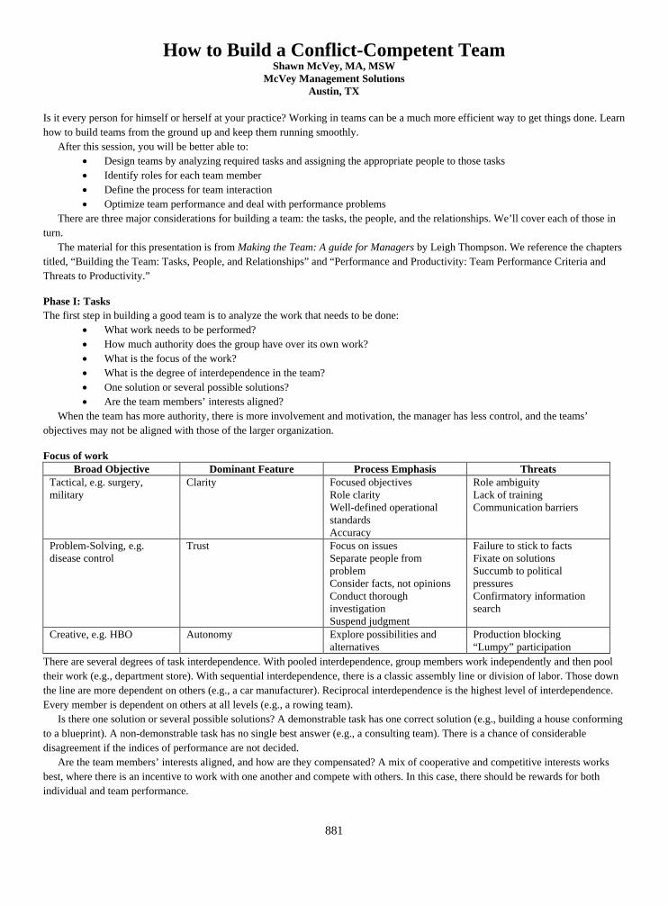
881
How to Build a Conflict-Competent Team Shawn McVey, MA, MSW
McVey Management Solutions Austin, TX
Is it every person for himself or herself at your practice? Working in teams can be a much more efficient way to get things done. Learn how to build teams from the ground up and keep them running smoothly.
After this session, you will be better able to: • Design teams by analyzing required tasks and assigning the appropriate people to those tasks • Identify roles for each team member • Define the process for team interaction • Optimize team performance and deal with performance problems
There are three major considerations for building a team: the tasks, the people, and the relationships. We’ll cover each of those in turn.
The material for this presentation is from Making the Team: A guide for Managers by Leigh Thompson. We reference the chapters titled, “Building the Team: Tasks, People, and Relationships” and “Performance and Productivity: Team Performance Criteria and Threats to Productivity.” Phase I: Tasks The first step in building a good team is to analyze the work that needs to be done:
• What work needs to be performed? • How much authority does the group have over its own work? • What is the focus of the work? • What is the degree of interdependence in the team? • One solution or several possible solutions? • Are the team members’ interests aligned?
When the team has more authority, there is more involvement and motivation, the manager has less control, and the teams’ objectives may not be aligned with those of the larger organization. Focus of work
Broad Objective Dominant Feature Process Emphasis Threats Tactical, e.g. surgery, military
Clarity Focused objectives Role clarity Well-defined operational standards Accuracy
Role ambiguity Lack of training Communication barriers
Problem-Solving, e.g. disease control
Trust Focus on issues Separate people from problem Consider facts, not opinions Conduct thorough investigation Suspend judgment
Failure to stick to facts Fixate on solutions Succumb to political pressures Confirmatory information search
Creative, e.g. HBO Autonomy Explore possibilities and alternatives
Production blocking “Lumpy” participation
There are several degrees of task interdependence. With pooled interdependence, group members work independently and then pool their work (e.g., department store). With sequential interdependence, there is a classic assembly line or division of labor. Those down the line are more dependent on others (e.g., a car manufacturer). Reciprocal interdependence is the highest level of interdependence. Every member is dependent on others at all levels (e.g., a rowing team).
Is there one solution or several possible solutions? A demonstrable task has one correct solution (e.g., building a house conforming to a blueprint). A non-demonstrable task has no single best answer (e.g., a consulting team). There is a chance of considerable disagreement if the indices of performance are not decided.
Are the team members’ interests aligned, and how are they compensated? A mix of cooperative and competitive interests works best, where there is an incentive to work with one another and compete with others. In this case, there should be rewards for both individual and team performance.
882
Phase II: People The second step in building a good team is to analyze the people:
• How many people should be on the team? • Who is ideally suited to do the work? • What skills are required? • What type and level of diversity is optimal in the team?
Pick the right number of people for the team. Managers seem to have an overstaffing bias, and smaller teams are rare. But they tend to work harder on a wider variety of tasks. They assume more responsibility for team performance. Oversize teams, though are less cohesive. Members are more self-conscious and avoid serious topics with frivolous conversation. In addition, the equality of member participation decreases. Managers seriously underestimate how coordination problems multiply when team members are added.
How do you know who is ideally suited to do the work? The more you know about the task, the better you suit people to it. Does it require technical or functional expertise? Task-management skills? Interpersonal skills?A well-rounded team member is a statistical rarity. Rely onelf-reports, past accomplishments, and 360-degree reports to determine how well suited a person is to a particular task.
What type and level of diversity is optimal in the team? Diversity is beyond gender, race, or disabilities. You need diversity in terms of functional skills. The advantages of a diversified team include multiple viewpoints and better decision-making, which gives you a competitive advantage.
What do you diversify based on? Social category (age, sex, race)? This isn’t very constructive. Informational diversity (education, work experience)? These team members debate constructively. Values diversity (work values, goals)? They are likely to engage in destructive conflicts.
How much diversity? If the team is too diverse, it’s difficult to get anything done. We experience some level of interpersonal congruence, which is the degree to which we see ourselves as others see us. Usually, the more diverse the group, the conflict there is. There is task conflict (due to different functional backgrounds) and emotional conflict (due to race, age). A person who is the only member of his/her social category feels isolated and may experience role entrapment.
Phase III: Relationships The third step in building a good team is to analyze the relationships:
• How do team members socialize with each other? • What roles are negotiated among team members? • Which norms are conducive/harmful? • Is cohesion in the team important? • How is trust developed, threatened, and rebuilt among the team members?
When there is a newcomer in the team, everyone completes an evaluation—a cost-benefit analysis, as it were. Teams evaluate the newcomer and vice versa. Is the new person beneficial to the team? Is the team beneficial to the new person? How committed is the individual to the group and the group to the individual?
Everyone goes through a role transition from nonmember to quasi member to full member. Once on the team, each member has a role. Roles and negotiations are not explicitly talked about. Rather, people engage in actions designed to take on that role, which are accepted/rejected by others. Task masters take the role of managing the team in terms of the work to be done. They are the team leaders. Others take on the role of socio-emotional master. This person’s focus is on satisfying the emotional needs of the team. S/he manages the people aspects of a team and restores harmony and cohesion.
Status systems develop within minutes after teams are formed. There is a process by which people acquire authority to be the task master or the socio-emotional master. Team members intuitively take note of one another’s personal qualities that they think are indicative of ability. Real status characteristics are qualities relevant to task at hand, e.g., experience. Pseudo status characteristics are factors like sex, age, ethnicity, and cultural background.
Team norms are not the same as rules. Norms make it easier for people to respond appropriately under new or stressful conditions. Norms add structure to the team. They reduce threats to productivity. Norms are left to natural processes and interaction patterns. The disruptive, least self-conscious people set unfavorable norms. Team Norms
At times, norms may also be in conflict. Something that is right in one department may not be so in another. What happens when someone regularly breaks norms? Try to correct them for a long period, else practices like ostracism persist. Long-term habits of breaking the norms are detrimental to both organization and the individual.
In terms of cohesion, the relationship between team cohesion and performance is primarily correlational rather than causal. Cohesion is also known as solidarity, morale, community, and fellowship. It is a crucial ingredient for team viability. Cohesive behaviors include: showing signs of mutual affection, displaying coordinated patterns of behavior, giving enough credit to team members, and participating in team activities.
883
Trust is confidence one person places in another that the other will honor all commitments. Not everything can be covered in a contract, so we need trust. There are different types of trust, including incentive-based trust (e.g., bonuses), trust based on familiarity, trust based on similarity (e.g., alumni of the same school), and trust based on social networks.
Implicit trust is trust in others in the absence of any rational reason or obvious similarity. We attend to some subtle signals in a social interaction, and we are not aware of their influence. Examples are flattery, instant attitudes (intense likes or dislikes for a novel object), mere exposure (“He grew on me.”), schmoozing (“Let’s have lunch sometime.”), and mirroring, called “social contagion.” This is copying one another’s posture, facial expression, tone of voice, and mannerisms.
Team performance criteria There are essential conditions for team success. Team members must:
• Bring adequate knowledge and skill to bear on the task • Exert sufficient motivation and skill • Coordinate activities and communication
In terms of knowledge and skill, the person must have average cognitive ability, interpersonal skills, and decision-making skills. Interpersonal skills include openness and self-disclosure, knowledge of each other, ability to predict other’s reactions and responses, and capacity for conflict and evaluation.
Coordination is the combined synchronization of all strategies of team members. Coordination problems increase with team size. We recommend single-digit teams. Have an agenda. Train team members together. Practice. Minimize links in communication. Set clear performance standards.
Here’s a team performance equation you can use: • AP = PP + S: T
o AP = Actual Productivity o PP = Potential Productivity o S = Synergy o T = Performance Threats
There are four factors to consider when analyzing the performance of your team: productivity, cohesion, learning, and integration.
Productivity • Does the team have a clear goal? • Which objective performance measures will be used to evaluate results? • Who are the legitimate clients of the team? • Under what conditions should the goal change? • What sources of information should the team consider to assess whether the initial goal should be changed?
Cohesion • Does the team enjoy working together? • What conditions could lead to feelings of resentment? • What conditions could prevent team members from working together in the future? • How are team members expected to accommodate to changes, such as additions to team, growth, and turnover?
Learning • How can team members best learn from one another? • Do the individual team members grow and develop as a result of team experience? • Do team members have a chance to improve their skills or affirm themselves? • What factors and conditions could block personal growth? • Are individuals’ growth needs understood and shared by group members?
Integration • How does the team benefit the larger organization? • Are the team’s goals consistent with those of larger organization? • What other groups, departments, and units are affected by the team? • What steps has the team taken to integrate its activities with those of others?
Action plan
• This week, I will: • The outcome for me is: • The outcome for the organization is:

884
• This month, I will: • The outcome for me is: • The outcome for the organization is: • The outcome for the organization is:
885
Leaders or Managers: What’s the Difference?
Shawn McVey, MA, MSW McVey Management Solutions
Austin, TX
Most employees want to say they have “the greatest boss in the world,” but most do not. Bad bosses are the number-one reason employees leave organizations. There is an abundance of literature and many studies about the qualities of great bosses and the differences between managers and leaders. Veterinary medicine does not create good bosses.
After this session you will be better able to: • Identify key skills needed to be a great manager • Appreciate the need to coach employees according to their unique learning styles and strengths • Handle employees’ failures and coach them up or out of your organization
What do good bosses do? There is one outstanding characteristic: All great managers have the ability to discover what is unique about each person and capitalize on it.
Average managers play checkers; great managers play chess. In checkers: • All the pieces are uniform and move in the same way. • There is a need to plan moves, but all the players move at the same pace and on the same path.
In chess: • Each piece moves in a different way, and you can’t play unless you know how each piece moves. • You won’t win unless you put considerable forethought into how the pieces move IN ADVANCE.
Are you a leader or a manager?
A manager • Turns one person’s talent into performance. • Succeeds only when they can identify and develop the differences in people. • Challenges each person to excel in his/her own way.
A leader • Discovers what is universal and capitalize on it. • Cuts through differences and focus on the needs that we all share.
Great managers are quick to capitalize on the unique strengths of employees, because it’s cost effective and a time saver. He/she will capitalize on strengths because it makes people need one another.
Great managers will introduce a healthy degree of disruption to your practice. They will: • Shuffle existing hierarchies. • Shuffle assumptions about who can do what. • Shuffle existing beliefs about where true expertise lies.
Great managers do these things because they can’t help it! They are astute about managing the needs of people. Perhaps you are experiencing the classic symptoms of “No Team.” After months of training and coaching your team to better
performance, you’re not seeing results. Meetings are a rehash of all the same things. You seem to get new hires that look just like the old hires. To get new results, change the management!
Identify each employee’s unique talents and help them use those qualities to excel. Embrace eccentricities. Emphasize that the company’s culture appreciates differences. Celebrate the uniqueness of each employee.
Use these three tactics • CONTINUOUSLY tweak roles to capitalize on individual strengths. • Pull the triggers that activate employee’s strengths. • Tailor coaching to the individual’s learning style.
The benefits of this management are • You save time. • The team takes ownership for improving their skills. • The team learns to value differences.
Capitalizing on individual strengths How do you identify your employees’ strengths? Ask questions and observe.
• What do they like the best about their jobs? • Which tasks do they look forward to, and which tasks do they avoid?
886
• Ask them, “What was the best day you had at work in the last three months?” • Listen for activities they find intrinsically satisfying.
Weakness doesn’t mean lack of skill. It can be any task that drains your energy or is an activity that when you start it, all you can think about is stopping.
• Ask, “What is the worst day you have had in the last three months?” For the purpose of this management task, we want to downplay discussions of weakness. Offer training to help them overcome
shortcomings stemming from lack of skills or knowledge. Find the employee a mentor/partner with complementary talents. Think about the mechanics of your business and reconfigure work arrangements. Don’t be afraid to be unconventional. Self-awareness vs. self-assurance Do we want an employees armed with the knowledge of their limitations, or do we want them to be confident in their abilities? Focus on strengths. In other words, people get more reward from knowing they are doing a job well than from understanding what parts of the job they are weak at.
Some would argue that employees can get too confident. Perhaps, but It is the manager’s job to simultaneously communicate the importance of employees’ tasks and the real complexity of the obstacles that they will need to overcome to be successful.
Your objective is to create a state of mind in the employee—a realistic assessment of the obstacles and difficulties associated with the goal combined with the confidence to take the goal on (optimism.)
When an employee fails, unless the failure is attributable to factors beyond the employee’s control, they must accept that failure was lack of effort on their part (psychological pressure.)
This obscures self-doubt. It is not that you are not capable, but perhaps it is because you didn’t develop enough skills or you didn’t try hard enough. If there is repeated failure, apply more training if it is lack of knowledge or skill. If the employee does not respond, it is because s/he does not possess the talent or skill to do the job. Manage around their weakness to neutralize it, but don’t keep exposing them to failure.
• Find them a partner/mentor that is strong where they are weak. • Rearrange the employee’s work world to render the weakness irrelevant. • Use “triggers.”
o Time of day? o Night or day strengths? o Time with boss? A little or a lot?
The most powerful trigger is recognition. Activating employee strengths
Source of recognition • Peers • The boss • Others with similar experience • Clients
Type of feedback • Publicly celebrate achievements. • Tell them privately, but vividly, why they are such valuable team members. • Give them professional/ technical awards. • Take photos of them and the “best” clients.
Coach to the learning style
• If their learning style is: o Analyzer: Requires extensive information before accepting a task; hates making mistakes.
• Coach them by: o Spend ample training time, role play, give them time to prepare for challenges.
• If their learning style is: o Doer: Uses trial and error to enhance skills while grappling with tasks.
• Coach them by: o Assign simple tasks, explain desired outcome, then get out of the way. Gradually increase the complexity of
tasks to be assigned. • If their learning style is:
o Watcher: Hones skills by watching other people in action.
887
• Coach them by: o Have them shadow top performers.
The art of success At the heart of a great manager’s success is the ability to appreciate individuality. But great managers need other skills as well:
• Hire well. • Set expectations. • Instinctively interact with others in a productive fashion.
Great managers vs. mediocre managers Mediocre managers hope (or assume) that all of their employees will be motivated by the same things and driven by the same goals. They define behaviors and tell employees to work on skills that don’t come naturally. They encourage sameness and view their job as transformation rather than development.
Great managers “play chess.” They define expectations and outcomes. They encourage individuality and don’t try to change a person’s style. They know their employees will differ in how they think, how they build relationships, how altruistic they are, how patient they can be, how much of an expert they need to be, how prepared they need to be, what challenges them, what drives them, and what their goals are (whew!!)
Differences in traits and talents are like blood types. They cut across superficial variations of race, sex, and age and capture each person’s uniqueness. Like blood types, these traits of people are enduring and resistant to change.
Your most precious resource is time. Why waste it pushing a rock up a hill? Great management is about the RELEASE of talent and skill, not the transformation. It is about constantly tweaking the environment to allow the employees’ unique contributions and styles to develop. Success as a manager depends almost entirely on your ability to do this. Action plan
• This week, I will: • The outcome for me is: • The outcome for the organization is: • This month, I will: • The outcome for me is: • The outcome for the organization is: • The outcome for the organization is:
888
How to Rally Your Associates to be Practice Leaders Shawn McVey, MA, MSW
McVey Management Solutions Austin, TX
Are you prepared to take the step from manager to leader? Learn how to define your daily tasks as leadership or management, recognize what success in both management and leadership looks like, and understand the leadership drivers you’ll need in order to successfully fulfill your role as an administrator.
After attending this session, you will be able to: • Define your daily tasks as leadership or management • Recognize what success in both management and leadership looks like • Understand the leadership drivers required in order to successfully fulfill your role in an administrative position
This material is adapted from the following sources: Peter Stephenson’s Naked Leadership, Executive Coaching, and The Bulletproof Executive; Daniel Goleman’s Emotional Intelligence and Understanding Emotional Intelligence; Muriel Solomon’s Working with Difficult People; and Richard Kock’s Managing without Management. What are the differences between managers and leaders? A manager is more directive and controlling. He measures, assesses, implements, and focuses on the status quo. A leader is more motivating and empowering. He coaches, listens, creates a vision, and focuses on change. When times are tough, leaders prove themselves by taking risks, taking initiative, acting decisively, and communicating effectively. They ARE the good example.
If you recognize that you’re more of a manager and would like to become more of a leader, try these tactics: Share good news. Establish small, achievable goals. Articulate the big picture. Give emotional support. Verify that employees have the right tools. Keep people informed.
People who run veterinary hospitals are usually defined by how well they do four things: create an agenda, develop a human network to achieve the agenda, implement, and achieve outcomes. We’ll talk about those four aspects of leadership in more detail.
When it comes to creating an agenda, managers plan and budget, establish detailed steps and time-saving tables to achieve results, and allocate necessary resources to make it happen. Leaders establish direction, develop a vision for the future, and develop strategies for producing the changes needed to achieve the vision.
When developing a human network, managers organize and staff. They establish a structure to accomplish a plan. They delegate work as necessary, provide policies and procedures to help, and create systems to monitor the plan. Leaders align people, communicate by words and deeds to all whose cooperation is needed, and create teams that understand and accept the vision and strategies.
To implement, managers control and solve problems. They monitor results vs. the plan in detail and identify deviations in the plan and organize to resolve them. Leaders motivate and inspire. They energize people to overcome barriers to change (political, bureaucratic and resource) by satisfying basic but unmet human needs.
To achieve outcomes, managers produce predictability, order, and consistent key results. Leaders initiate change in order to achieve outcomes, which is often dramatic and very useful.
Overall, managerial culture emphasizes control and rationality. A manager is a problem-solver who is usually neither genius nor heroic. She is rather persistent, tough-minded, and analytical. She is willing to work hard, be tolerant, and express goodwill.
Managers adapt impersonal, passive attitudes toward goals. Goals are derived out of necessity and a business plan. Leaders are driven to change attitudes and influence the way that people think about possibilities.
Managers view work as an enabling and educational process. To get people to accept solutions to problems, they must coordinate and balance opposing views. A manager’s need to control and feel safe overrides the desire to be a risk-taker.
Leaders develop fresh approaches to long-standing problems and bring up new options. They project images of success. They help people visualize change. Leaders work from high-risk places, as they are addicted to excitement.
What are the warning signs that the job is too much for you? IQ is necessary. EQ is enhancing. It’s a proven fact that all the attributes of the information age, along with superior EQ, produce superior results. A leader must leverage people and teams to the fullest extent possible and see human beings as human beings rather than as physical assets.
Can you run the practice? • WARNING SIGNS:
o You are in too many meetings and involved in too many tactical/operations decisions. o There are too many days when you feel you have lost control of your time.
889
This problem happens because the sheer volume and intensity of demands catch you by surprise. You may also lack sufficient time-management skills. You also mourn the loss of being close to the patients and clients. And perhaps you weren’t prepared for the paradox of having to let go and let others be in control.
You have to realize that you can’t monitor everything. Learn how to shift your influence from direct to indirect. Articulate and communicate a clear and simple strategy for where the business is headed. Institutionalize rigorous structures and processes to guide, inform, and reward. It’s your job to select and manage key talent.
Are you too involved? • WARNING SIGNS
o You have become the bottleneck. o Staff is overly inclined to consult you before they act. o People start using your name to endorse ideas: “Shawn says…”
You may get stuck in this rut because your direction and vision are unclear. There’s no clearly articulated plan, and the roles are unclear. Regardless, you have to realize that giving orders is very costly.
What happens when you do it? You trigger resentment and defensiveness. Second-guessing demoralizes your staff. In fact, decisions should not come to you until every other path has been exhausted.
Do you know what’s going on? • WARNING SIGNS
o You keep hearing things that surprise you. o You learn about events after the fact. o You hear concern and dissenting views through the grapevine rather than directly.
This happens because information is almost always filtered when it gets to you. Former peers and subordinates are on guard because you influence careers. People naturally protect themselves from consequences.
What to do? Seek outside feedback and counsel. Hold informal meetings with employees. Seek information from front-line employees. Ask a lot of questions!
Are you sending the right message? • WARNING SIGNS
o Employees circulate stories about your behavior that magnify or distort reality. o People around you act in ways that indicate they are trying to anticipate your likes and dislikes.
Everyone in the practice watches and judges your every move. What to do? Avoid speculative discussions with employees. Learn about the signals that you send. Strive for consistent and simple messages.
Are you the boss? • WARNING SIGNS
o You don’t know where you stand with the owners or the Board members. o Roles and responsibilities of the directors/owners and management are not clear. o Management meetings are limited to reporting on results and management decisions.
It sucks to be you. Really, now you have multiple bosses, not just one. You are personally accountable, and your boss is not your friend. You have to move into your new role with grace.
Are you keeping your head? • WARNING SIGNS
o You give interviews about you, rather than the practice. o You have few, if any, activities that are not connected to the business. o Your lifestyle is more lavish or extravagant than that of other leaders in the business.
This problem happens because of the attention and admiration that come with the job, which makes introspection difficult. Everyone said the job was much harder than they thought it was going to be. Personal and family responses change.
What to do? Make an effort to stay humble. Manage organizational context rather than focusing on day-to-day operations. Set the tone and define practice culture through behavior and actions. Remember that position does not guarantee the right to lead. Maintain your moral compass. The five tasks of a leader There are five tasks that an effective leader has down cold:
1. Influencing 2. Synergizing 3. Enabling 4. Energizing 5. Trusting
890
Influencing means attaining results with people outside your direct control. To do this, you have to sell yourself, build networks, create your own goals, hone your time-management skills, and perform like a champ in meetings and during presentations.
Synergizing means attaining superior results with peers. You need the ability to choose effective players and set up effective teams. It also involves maximizing the power of interpersonal relationships, using your conflict-resolution skills, and leading across the organization.
Enabling means attaining superior results with people for whom we are responsible. You manage, coach, motivate, and delegate. You also are a master of systems and processes.
Energizing involves recognizing job fit for all team members and managing talent. You recognize emotional currents and respond to redirect them. You attract and retain employees.
You also adopt and advise about appropriate coping mechanisms when human needs are not met. Trusting is attaining superior results by getting people to believe in the reliability of the organization. You set the example with
consistent behavior and stay in alignment with vision and values. You are the change manager. Trust is the quintessence of leadership.
The myths and truths of leadership You don’t have to be the point person! In fact, it’s a balance of leading and managing. It’s not about leading direct reports. It’s about leading the people around you. You don’t have to use the latest management techniques, because leadership is about you in action. You need emotional intelligence as well as book smarts. And leadership is not about a long list of skills; it’s about five key activities, achieved through competencies.
Influencing myths and truths It’s a myth that the power is all at the top and it’s all about what you know. Power is anywhere, and you better know who has it. It’s about what you know, as well as who you know!
You can’t just wing it at meetings, telling stories and making it all about content. In fact, you have to ask astute questions and be an active listener. You need to place as much emphasis on process as on content.
Don’t hide! If you are not selling your organization externally, you may be viewed as just a cost. It’s easier to spend time with lower-level allies. Get to know high-yield allies. If you can’t run a meeting, remember that God gave you two ears and one mouth. Use them proportionally. If you can’t write well, why would anyone read your work? How you come across is more important than what you know.
Synergizing myths and truths It’s a myth that your direct reports are most important. You have to be a team player with peers as well. You may think you know how you come across because you know yourself. In fact, you come across differently than you think, especially when you are under pressure. It’s a myth that the best teams are teams who get along well. There is a broad range of styles, and there will be some conflict. To synergize, you may think you need to avoid conflict. The truth is that you must manage conflict, and manage it well. Lastly, planning is not about strategy then structure. It’s about leveraging people.
Don’t hide! You must have functional groups and teamwork. The best way to have effective relationships is to communicate in a way that relates to the operating style of the team member. Behavioral flexibility is the key. If the team processes don’t work, neither will your team. You will lose good people, hire good people, and develop good people. It’s all about good people. Continuous improvement requires continuous coaching.
Enabling myths and truths Rather than focusing on quarterly results, you need to focus on the outside environment as well. Otherwise, you will not be around to report on quarterly results. Though it’s important to keep employees informed, you also need to hear from them and actively listen to their concerns. You may think that people can only motivate themselves, but leaders and organizations are the key motivators.
Some leaders come from a strong attitude of, “Never abdicate.” The truth is that abdication and direction are powerful tools. Another myth is that personal accountability is all about taking it and working with it for results. Reporting on results is just as important. Celebrate success and effect remedies. Lastly, a consistent leadership style is not what’s best. Your leadership style needs to flex.
Don’t hide! If your antenna is up, you will get reception. Poor delegation is one of the greatest causes of bottlenecks, low morale, and poor productivity. It is also the greatest opportunity. Don’t hide from personal accountability, and don’t let others hide, either. Vary your coaching approaches, but above all, coach.
Energizing myths and truths It’s a myth that job fit is all about individuals fitting the job. It is equally about the organization fitting the individuals. If you think that self-motivation is a given, think again. The culture can make or break motivation. Good talent does not care of itself. If you don’t manage your talent, it will go elsewhere. Talent management is not all about fast promotion. You need to provide continuous development in a stimulating environment. Career management is not solely the individual’s responsibility. Leave it to them, and they will manage their careers elsewhere.
891
Don’t hide! Seek and deliver strong job fit. If you are overly stressed, is it within your control to do something about it? Confront the career stage you and others are in and develop plans to move each person forward. Avoid slash-and-burn hiring and firing scenarios. Be candid in your review process. Talent management should receive just as much attention as the client base.
Trusting myths and truths Agreeing on and articulating values is the most difficult part, right? Wrong. Making values real and upholding them is the biggest challenge. And it’s not just executives who should lead the business. All employees can lead the business. It’s a myth that change management requires charisma. Change management requires adaptability, compromise, resilience, and two-way communication.
It’s a common misperception that if you restructure, you will cut costs and improve the bottom line. If you restructure and do it well, you MAY improve the bottom line. Staff morale after restructuring is not the biggest problem. Poor leadership at times of change is the biggest problem.
Don’t hide! Employees will give their all only if they trust you. Always walk the talk of the vision and values. In times of change, the leader needs to be more visible. Action plan
• This week, I will: • The outcome for me is: • The outcome for the organization is: • This month, I will: • The outcome for me is: • The outcome for the organization is: • The outcome for the organization is:
892
Stop the Price War and Show Value to Clients (Parts 1 & 2)
Mark Opperman, BS, CVPM and Sheila Grosdidier, BS, RVT Veterinary Management Consultants
Evergreen, CO
What is “perception of value?” 1. The “Three Minute Syndrome” 2. Internet Reviews Online - Is there a review posted about your practice? 3. How is “Perception of Value” Measured? 4. What Effect Does It Have on Your Practice?
How do clients determine a practice’s “perception of value?” Value = Benefit / Price Let’s put ourselves in our clients’ shoes and try to determine our practice’s “perception of value”
The initial contact • Telephone
o Message on hold o Telephone etiquette
• In person The initial impression
• Signage • Outside Physical Environment
o Building appearance Your entrance-way
o Parking lot o Landscaping
• Marriott versus Motel 6 • Entering the Reception Area
o Appearance of reception room o Cleanliness, odor control o Comfort of seating o Distractions - ease of managing one’s pet o Background music o Condition and age of magazines and client education material o Posters and informational wall hangings affixed to the wall
• The Receptionist: The first and most important contact o How was the client greeted?
Smile; Use of pet’s name; Knowledge of what services the client needs • How well do our receptionists present themselves?
o Professional, Knowledgeable & Informative, Uniforms & Name Badges, Organized or Disorganized, Handling of stressful situations
• I can’t find the medical record! o Paper Files vs. Electronic Files o New Client Form o Writing Tablets
• The Wait: constructive or destructive time? o How long is too long? _________________
Communication with the client o Appointments kept on schedule
Ten Minute Flex Scheduling “E” Slots Discharge Appointments
o Effectively utilizing this time
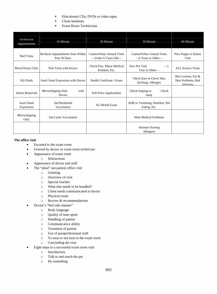
893
Educational CDs, DVDs or video tapes Client handouts Exam Room Technicians
The office visit
• Escorted to the exam room • Greeted by doctor or exam room technician • Appearance of exam room
o Distractions • Appearance of doctor and staff • The “ideal” out-patient office visit
o Greeting o Overview of visit o Special touches o What else needs to be handled? o Client needs communicated to doctor o Physical exam o Review & recommendations
• Doctor’s “bed side manner” o Body language o Quality of time spent o Handling of patient o Communicative ability o Treatment of patient o Use of paraprofessional staff o To treat or not treat in the exam room o Concluding the visit
• Eight steps to a successful exam room visit o Introduction o Talk to and touch the pet o Do something
Technician Appointments
10 Minute 20 Minute 30 Minute 40 Minute
Nail Trims Recheck Appointments Seen Within
Past 30 Days Canine/Feline Annual Visits
—Under 6 Years Old— Canine/Feline Annual Visits
—6 Years or Older— New Puppy or Kitten
Visit
Blood Draws Only Nail Trims with Doctor Check Paw, Minor Medical
Problem, Etc. New Pet Visit —1
Year or Older— ALL Exotics Visits
SQ Fluids Anal Gland Expression with Doctor Health Certificate / Exam Check Ears or Check Skin (Itching), Allergies
Skin Lesions, Ear & Skin Problems, Bad
Allergies
Suture Removals Microchipping Only with
Doctor Soft Paws Applications Check limping or Check
lump
Anal Gland Expression
2nd Bordetella Vaccination Six Month Exam
ADR or Vomitting, Diarrhea, Not Eating, Etc.
Microchipping Only
2nd Lyme Vaccination Most Medical Problems
Animals Starting
Allergens

894
o Say something o Show something o Give them something o Listen o End on a positive note
The exit
1. Processing paper work 2. Filling prescriptions 3. Handling of payment 4. Answering questions
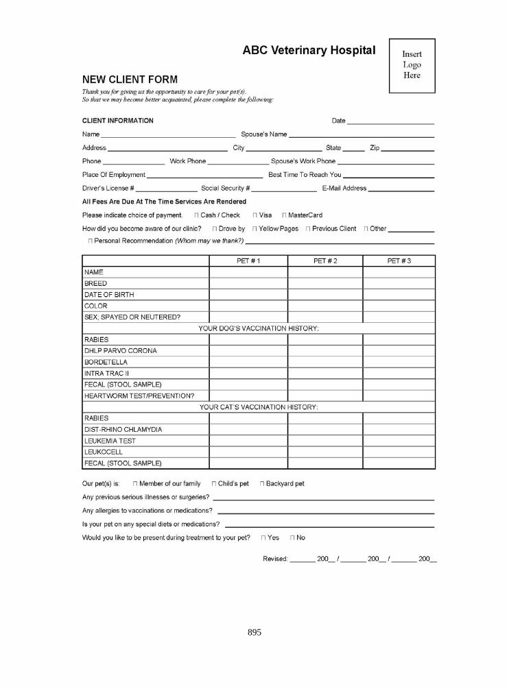
895
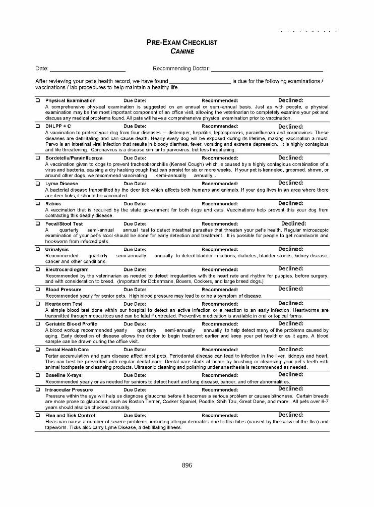
896
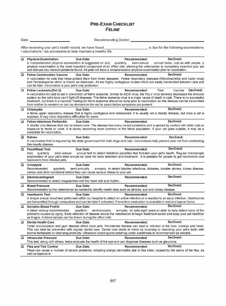
897
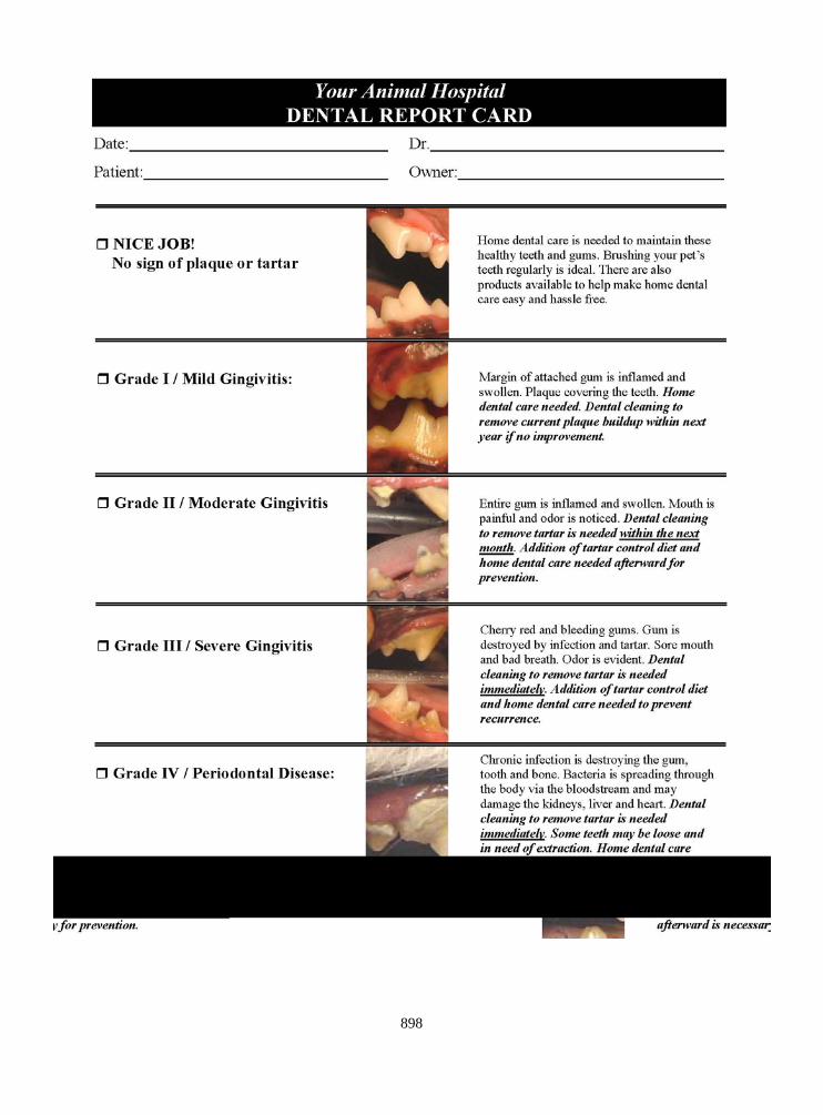
898
899
900
Setting Fees You’re Comfortable With Mark Opperman, BS, CVPM and Sheila Grosdidier, BS, RVT
Veterinary Management Consultants Evergreen, CO
“Shopped” and “exposed” services
Calculate your fees based on
a. Overhead costs per minute b. Direct costs at percent (%) mark-up c. Return on time to the doctor
In hospital service fees - overhead costs per minute
a. Calculate overhead costs/minute/DVM b. Formula: ALL expenses – compensation to doctors (both owners & associates) including related costs – inventory costs
÷ number of hours the doctors are scheduled (both office hours & surgery) ÷ 60 In hospital fees - direct costs
a. Direct costs definition: Inventory costs, costs of materials used in the procedure b. Formula: 2x cost due to costs of ordering
In hospital fees - return on time to the doctor
a. Return on time to the Doctor definition: Per minute cost of the doctor’s time spent on a procedure b. Formula: Cost of DVM’s time/minute x # of minutes spent on procedure c. In hospital procedures d. Surgery
i. General/Soft Tissue ii. Orthopedic
How to determine pharmacy fees
a. Set % mark-up b. Minimum per pill charge c. Pharmacy preparation charge d. Minimum Rx charge
Outside laboratory
a. 2 x cost plus $5.00, or b. 2.5 x cost
901
Associate Skills: Why Leadership and Personality Matters
Heather Prendergast, RVT, CVPM Synergie, LLC
Las Cruces, NM
Owner or associate - regardless of which you are in practice, your leadership ability and personality affect the entire team. Owners and associates will learn how to lead a positive, productive team while learning how to work with other personalities, and honing in on their own strengths and weaknesses.
Every person on the veterinary team has responsibilities, and must be held accountable for their actions. This includes owners and associate veterinarians who often feel their behavior and attitude is justified because of “who they are” on the team. Therefore the key take-away point of this lecture: Team members act as their leadership acts. They work as they are lead. They succeed if given success to follow.
Great leadership starts at the top. Since owners can set the stage for the entire team to work in a positive or negative culture, many associates feel that they do not have “any say so” in the practice. It has been stated that “what they say or think does not matter”, therefore, they simply show up for their job, see the clients and patients, and go home at the end of the day. The fact is, associates have more effect on team members than they believe. Associates are viewed by team members as leaders who make decisions for clients and their pets, on a daily basis. The methods by which associates lead and communicate with fellow team members has a huge affect on how team members deliver the message(s) to clients and provide care to the patients.
Associate veterinarians are leaders that share a mission and a vision; a mission and vision that should shared unanimously by all team members, but is often hidden behind office politics. Every team member, DVM and non-DVM staff entered this profession for the love of animals, with a mission to treat all with respect, dignity and courtesy. When associate veterinarians uphold the mission and vision, they can positively influence other team members to do the same. It starts at the top. Associate veterinarians are leaders that should be driven to inspire and motivate; inspire and motivate all team members and clients.
Associate veterinarians should lead with integrity. Team members wish to follow those with strong morals, values, and ethics. With integrity comes respect. Respect is consideration or esteem given to another person. Each member of the team must respect other team members’ education, skills, and values. Without respect for each other, team members’ morale and self-esteem drop, producing a negative attitude for the entire team. Associates must understand that not all employees have the same thoughts and philosophies, and many people complete tasks differently based on education or previous skill sets. This can be accepted as long as the same ultimate goal is reached in a timely manner. Each member possesses expert skills and credentials that warrant respect, and associates have an uncanny ability to drive this respect among the team.
Empowerment and trust in the team is sometimes difficult for associates. Empowered employees are only as good as their expertise, and expertise comes from training with associates and exceptional leadership. It is imperative to train team members on a continual basis. The strongest employees accept training as a way to improve themselves and the practice. When employees have pride for their workplace and are empowered to improve the daily operations, they will exceed expectations. Emotional ownership of a practice (vs. financial ownership) yields high returns. Team members can never receive enough training. As an associate, step up and offer to train and motivates team members on a continual basis.
Team members receive energy through recognition. Team members should be recognized for a job well done as soon as it is warranted. Many team members only hear of mistakes they have made and the necessary corrections and never hear about the excellent quality of work they produce. Positive situations need to be recognized and brought to the attention of all team members so they can all benefit. Amazing associates know that high performing teams are high producing teams, and they succeed together.
Actions speak louder than words. Behave as you expect others to behave, which will drive the shared values and vision. When a leader is “grumpy” and snaps at fellow team members or talks in a condescending voice, the team moral drops. Not only does this affect the inner dynamics of the team, it affects the communications that will occur with clients throughout the day. When associates and leaders talk in a condescending tone, the authority has just been given to the team to talk to others (including clients) in the same tone of voice. Take ownership for your actions and apologize when appropriate. We are only human. We all make mistakes, and we learn from our mistakes. Admit to your team when you have made a mistake, and they will admit theirs, to you.
The personality of individuals certainly has a dramatic affect on communications within the team, and with clients. With each type of personality come advantages and disadvantages, as well as behavioral predispositions. It is important to learn how to appreciate and leverage personalities for the greatest success. Teams that understand and accept different personalities tend to have higher producing employees, higher client compliance and retention.
Several different personality tests are available, including Myers-Briggs, Keirsey, Merrill Reid, True Colors and Disc, to name a few. The point of learning about individual personalities is not to single one out, but to gain a better understanding of how to communicate better. This same theory can be applied to clients, which will also enhance client communication.
902
Each personality test has slight variation in terms used to describe characteristics. For the purpose of this lecture and its relationship to veterinary medicine, we will use the descriptions provided by the Pawsonality Assessment, courtesy of Patterson Veterinary.
Drivers can be described as being direct, assertive, demanding and seek results. They are similar to the Border Collie breed, in that they are always seeking the quickest way to get from point A to point B. They are efficient, can multi-task, and generally expect the same from others. Because they expect everyone to be the same as them, they come across to others as being intolerant and impatient. Analytics on the other hand, think in a controlled and orderly fashion. Just as the characteristic states, information must be analyzed before a decision can be made. Analytics will have a plan (and sometimes Plan B, in case Plan A does not work out) in place, before any procedure is carried out. Here in lies a clear example of a personality conflict: a driver determines how to get from point A to point B as efficiently as possible, and the analytic is making a plan to get from Point A to Point B as successfully as possible. Therefore, drivers will perceive analytics as slow decision makers; analytics perceive drivers as pushy and making rash decisions.
Expressive personalities are like social butterflies. Imagine a golden retriever: always happy, their tail is always wagging. They are enthusiastic, impulsive, dramatic, animated, and very charming. These social butterflies can charm the pants off a client, and create a warm and hospitable environment. They often have the highest acceptance when making recommendations to clients. The disadvantage to this happy personality: they are easily distracted. Therefore, they are great idea generators and make everyone happy, but they often cannot follow through with what was originally assigned to them. This can make the driver and an analytic personality crazy, until they realize how best to communicate with them, and learn what to expect.
Last but not least, we have the amiables. An amiable is a peacemaking personality; they do not like conflict or confrontation. They are friendly, supportive, patient and loyal. The Labrador breed (in general) is a very loyal breed – they always stay by their owner, and rarely disappoint. Since they do not like conflict, drivers and analytics often discount Labradors, creating a distinct personality clash.
As indicated earlier, people with different personalities have different strengths and weaknesses. Since one person does not possess all the characteristics to be a high achiever, mover and or shaker, it is important to utilize the strengths of all team members and minimize potential weaknesses. This is team diversity that develops respect and rapport with one another, and is driven by leadership – associates included!
903
How to be the Leader Your Team Needs Heather Prendergast, RVT, CVPM
Synergie, LLC Las Cruces, NM
Teams follow their leaders. In fact, what the team does (or does not do) is in direct correlation to their leader (s). However, many veterinary practices lack the leadership needed to produce a positive, effective and efficient culture. Participants will gain an understanding of the importance of great leadership, and will take home tools to produce the dream team.
Every person on our veterinary team should be a leader. Why? Because every team member leads our clients on a daily basis. Team members educate clients and lead them to make the right decision regarding the healthcare for their pet. Through education, positive persuasion and leadership, a bond is established, resulting in an increase in client compliance and retention. However, leadership does not just happen; it starts at the top with owners, administrators and practice managers.
Leadership has been described as the process of social influence in which one person can enlist the aid and support of others in the accomplishment of a common task.1
In general this task or tasks include providing outstanding customer support to our clients and superior medical service to our patients. In fact, this is probably a brief description of you mission statement (or should be!) That being said, if you do not have a mission, vision or values statement in place, then how can management enlist the aid and support of others to accomplish the common task? They do not know what the common task is that must be completed, together.
Create a mission, vision and values statement A mission, the vision and the values (MVVs) of a hospital are core competencies that must be integrated in every practice. MVVs set the structure, creating a positive culture, and goals that help define team member expectations. Without these, team members have no direction; they simply show up to work and complete the tasks assigned to them. Owners make it day to day, with no clear light at the end of the tunnel, and managers struggle to implement successful goals and policies to increase value in the hospital. The mission and vision should be evaluated every couple of years, ensuring that they are still in alignment with the beliefs of the owner. Leadership Leadership, not just management, is vital to the success and growth of a practice. Leadership is influence, as defined above. Leaders motivate team members into action, and inspire them to be the best that they can be. They guide through effective communication, and create an environment that facilitates teamwork.
Leadership is about character, behavior and actions (actions speak louder than words!). Every leader must look in the mirror; are the characteristics and behaviors that one is striving for (within the team) exhibited day in and day out (by oneself)? Through these characteristics, leaders compel individuals to pursue the mission, value and goals of the leader.
In modern society, leaders must strive to achieve short- and long-term goals; create effective, efficient methods to complete tasks; and be proactive instead of reactive to situations. A leader’s personal effectiveness can directly influence a hospital’s success. Leaders must determine the most effective method to manage team members and be able to recognize their own strengths and weaknesses as well as those of others. A motivating leader generates enthusiasm and excitement and an organized leader provides the path to achieve goals; the best type of leader does both. A leader sets the practice’s vision and goals, communicating the vision to the team so they may help accomplish those goals. Leaders must hold themselves to a higher standard of patient care, customer service, performance, and personal behavior. Leading by example has a much more profound effect on team members than leading by directive. Examples show team members what is expected of them when it comes to patient and client care. All team members should be held accountable for providing the best care possible, and leaders can set the stage for this to occur. Leadership that promotes a poor standard of care and professionalism will also affect the team, as team members are only as good as their leaders. Managers of this type should be terminated because they will cause the team to disintegrate and the practice to fail. Owners of this type must re-evaluate themselves and consider self-development if he or she wishes to have a successful practice. Leadership qualities Leaders must possess several qualities to be successful. Self-confidence, sincerity, and enthusiasm for the job are essential. Leaders are effective listeners and accept diverse cultures. To have an effective team, a leader must be a team player and work to solve problems, innovate, and renovate existing policies, procedures, and environments. Leaders explain how to accomplish a task and give a challenge to the team. This allows the team members to think for themselves creatively, which can develop leaders for the future.
1 Wikipedia, http://en.m.wikipedia.org/wiki/Leadership; accessed 11/2014
904
Effective leaders possess self-confidence. They believe they have the ability to complete tasks efficiently and effectively. Leaders accentuate positive personal attributes and do not dwell on negative weaknesses. Self-confident leaders take risks and are able to make recommendations and changes without delay. Genuineness and sincerity come from within and promote trust and communication. Team members know the suggestions and changes made by sincere leaders enhance the skills of all involved. It is important to show appreciation for peoples contributions and create a culture that celebrates values and victories. It is genuine acts of caring that lift people up and creates a desire to exceed expectations.
Enthusiasm shows that leaders are interested in their practices and the steps needed to make it successful. Enthusiasm is contagious to fellow team members and should be a part of practice culture. Enthusiastic team members are excited to come to work, enjoy sharing experiences with others, appreciate humor, and enjoy the team-work environment. Listening effectively has become a lost skill in today’s world; talking has overtaken listening. Listening is the ability to receive, attend to, interpret, and respond to words and body language. Poor listening skills can result in misinterpretation of information, leading to malpractice in the medical field. Poor listening and interpretation can cause a communication breakdown when a leader is trying to manage a practice effectively and lead team members in a positive style. One of the most important skills for effective leadership is to be an effective communicator. Communications must be done in a clear and pleasant manner. Effective communicators think clearly, talk sparingly, and listen intently. It is imperative to think topics through before jumping to a conclusion. All issues must be understood and interpreted before an action can be taken. Rash decisions should not be made, or devastating results may occur. Time should be taken to interpret the facts and prevent immediate judgments. Talking too much can be a problem itself and greatly inhibits listening. Tone is as important as talking; positive, enthusiastic tones are much more effective than negative, authoritarian tones. Exceptional leaders create environments where team members are empowered to communicate openly, voice their concerns, and make changes where necessary to produce an improved service. Inhibiting this environment can be detrimental.
Ethnic cultures have different means of communication, and an effective leader must be able to determine the best method of communication for each. Morals and ethics vary among ethnicities, ultimately affecting the learning and training abilities of different team members. Leaders must be accepting of diverse cultures, welcoming the different qualities each possesses, and work with them to provide the best leadership possible. Veterinary practice is a team business. A team is a simple concept: a group of individuals with different skills and attributes, which contribute the positive culture of the hospital. Effective leaders build teams that allow the business to succeed at all levels, including providing excellent patient and client care and maintaining a friendly and cohesive work environment, all while being able to create and maintain a profit for the practice. Leaders invite creative thinking from team members, and integrate this creative thinking into daily conversations. Creative thinking facilitates productive, problem solving team members that are not afraid to move outside the box.
When building teams, exceptional leaders attract the right people, and place them into the right positions. They also integrate personal growth with practice growth, which encourages career development of each team member. Without personal growth and development, the practice growth is stalled.
Leaders must possess skills to solve problems before they arise—to be proactive instead of reactive. Leaders determine the problem; collect, listen, and interpret the facts; and present a variety of solutions to the team. Team members should be asked how they would resolve a situation; their point of view is critically important in problem resolution. In addition, problem solvers are accountable and productive.
Leadership skills are not developed overnight; they come with patience, education, and trial and error. Managing a practice has both wonderful and terrible days. A leader is sometimes viewed as a “good guy,” sometimes as a “bad guy,” and sometimes as uncaring and lacking compassion. However, the success of a business can depend on the quality of the leader. Someone who is capable of being either a good guy or bad guy when necessary will keep the team focused so they provide excellent quality of care.
905
It Could be You: Protect Yourself from Embezzlement
Heather Prendergast, RVT, CVPM Synergie, LLC
Las Cruces, NM
No one hires a thief; however, fraud and embezzlement occur in small businesses everyday. Participants will gain an understanding of when and how embezzlement occurs, and will leave with tools needed to decrease risk in the practice.
We would never hire a thief to work in our hospitals (intentionally, that is). Shockingly, theft and embezzlement happen everyday in veterinary practices. Often, we don’t know how or where it occurs, or how to stop it when it is occurring. It starts with something little (stealing postage or stamps); the perpetrator then gains confidence in their skills and advances to something a bit more complex (inventory and supplies). Eventually, the dollars add up, which should attract the attention of the owner.
Many practices have fraud opportunities available, yet do not have a checks and balance system in place. Which of the following may be occurring, or has occurred in your practice?
Time: Employees arrive at the practice to begin their shift. They clock in, pour a cup of coffee, eat breakfast, then begin work. If this occurs daily, with an average of 5 employees, your total loss to time card fraud could be 3600 minutes, totaling over $900 per year. Doesn’t seem like much? Wait till you add it to the additional areas outlined below.
Paper goods, inventory and medical supplies: One person orders supplies, receives the goods and places them in their appropriate storage location. Inventory is never tracked and the physical amount on hand is never compared to the computer count. Product can be taken by employees or given away to clients. Some employees intend to charge themselves (but forget); others feel the practice owes them the product, as their pay is not high enough to compensate for all they do for you. Unless you are tracking your inventory, you will never know this product is missing. Take a look at companies on the Internet to see what products and diagnostic testing kits may be selling in your area. Since inventory is rarely tacked in the veterinary practice, a dollar amount cannot be applied here. However, practices spend an average of 20-30% of their gross income on medical supplies. For a million dollar practice, this could be $200,000-$300,000 spent annually, with a hope of at least $400,000 to $600,000 (with a 100$ mark up) received in return. How much of these profits could you be missing?
Controlled Substances and prescription pads: These drugs and pads have a high dollar value on the street, and can enable an employee with a substance abuse problem. Ensure Controlled Substances are locked up according to DEA regulations and balanced on a monthly basis. Investigate any discrepancies immediately.
Cash: Cash is the easiest to steal and cover up. Team members accept cash, end of day reconciliations should be completed, and deposits must be made. End of day reconciliations should be compared to the deposits made to the bank (cash and checks) by someone other than the team members accepting cash, as this is an easy place to alter documents. It should be policy that every invoice is closed out, then a receipt generated when a client makes a payment. This will allow an audit trail to pick up any deletions of transactions made by team members. To add an additional layer of security, password protection should be enabled for any deleted invoices.
Client Credit Card Numbers: NEVER keep client credit card numbers on file. Employees can sell credit card numbers – racking up hundreds of thousands of dollars in charges. Protect your client’s data – by following the Red Flags Rule established by the FTC. This should also be extended to client social security numbers; if any social security numbers are kept in files- shred them immediately.
Unauthorized discounts: Employees like to give friends and family a break, or perhaps give themselves a greater discount than authorized. Review your family and friends discount policy, along with the employee benefits. In addition, consider adding a password to the PMS in order to change the price of items or services. All discounts should be given a code, which will enable the owner/manage to start tracking discounts being given (and ensure they are authorized). According to Well Managed Practices 2011 (Tumblin, Veterinary Economics), 15-25% of gross income is lost, due to missed charges and discounts. If your practice is a million dollar practice, this could be $150,000-$250,000 dollars. Since discounts and missed charges are rarely tracked, practices can’t put a true dollar amount on this factor.
Padding payroll: Does your practice have any oversight on payroll, or is one person responsible? Padding payroll hours and hourly rates can cost the practice thousands of dollars per year.
Padding Production: Yes, padding production. It has been shown that associates have adjusted production amounts in order to increase their production bonus. Enable a password in the practice management software to prevent the unauthorized change of production credit.
Accounting: Creating duplicate invoices, paying a creditor twice (and accepting the refund personally), and payment to a factious creditor has been reported. Paying personal and practice utility bills with the same check has also been reported.
The above are just a few fraud/embezzlement opportunities that exist in veterinary practices. Owners must put a checks and balance system in place to decrease the potential opportunity. Fraud will never be 100% preventable, but actions must be taken to decrease the risk.

906
Your practice action plan 1. Step 1: Return to the practice and review every opportunity in which theft, fraud or embezzlement could occur. 2. Step 2: Create a checks and balance system for each opportunity identified. One person cannot be in charge of the
checks and balances; this is a multi person job. Do not forget to review payment accepting procedures, deposits, end of day reconciliation, inventory ordering, receiving and invoice reconciliation; invoice payments, checking account and credit card reconciliation, payroll and tax payments.
3. Step 3: Monitor the checks and balance system and tweak as needed.
907
Shake Things Up to Grow Your Practice Denise Tumblin, CPA
Wutchiett Tumblin and Associates Columbus, OH
Making your practice more profitable and positioning it for continued success isn’t about luck. You don’t roll the dice and hope to meet your medical and financial goals for the year. Being profitable is about playing the right hand at the right moment and making changes in your practice that maximize your strengths. So use these five critical steps to ensure your practice will be flush with success. Step #1: Enhance patient care Differences in medical philosophies baffle doctors, staff, and clients and can lead to shortcomings in patient care. Say your standard is to perform annual wellness testing on all your senior patients. But only two of four doctors follow the protocol. This puts half your patients at a medical disadvantage. Don’t make people guess what you want. Define your standards of care, get them in writing, and use them as a teaching tool to communicate your expectations to doctors and staff. Your goals are to provide high-quality care, nurture lasting client relationships, cultivate happy, productive doctors and staff, and enjoy a fun, profitable practice.
Every team member at your practice must be knowledgeable about your standards for the sake of consistency and continuity. It’s up to you to teach and mentor your team members and communicate your expectations for upholding those standards of care. An effective training program ensures that new team members start off on the right foot and become successful. Regular internal and external CE encourages team members to grow and will net your practice major rewards in the future. Improving staff expertise frees you to focus on other things – like seeing more patients, taking a lunch break or ending the day on time. Step #2 Create client-centric experiences Yes, it’s easy to go on autopilot in the exam room – especially when this is the sixth time today you’ve explained the importance of heartworm and flea prevention or discussed why it’s not a good idea to delay the dental prophylaxis any longer. These days, however, you simply can’t afford to coast. Every time you’re about to step into the exam room, take a moment to refocus your energy on this particular client and patient. Look at the chart and note a couple of unique characteristics about this pet and owner. Then mention them during the visit.
The bottom line is that you want your clients to receive care that’s relevant to them, custom-built for their pet, and different from what they can get anywhere else. Rather than talking throughout the appointment, try pausing for a few minutes at appropriate moments so you can listen – truly listen – to what your clients are saying. Make the experience about them. Being strongly present with each client and each patient creates inspiration in pet owners. Inspiration creates magnetism. And magnetism gets your clients to “yes”. Step #3: Improve profitability Define your goals for spending and revenue growth. Set revenue and expense goals and then share them with the team members who’ll help you achieve them. And don’t forget to measure and analyze your performance. Create a systematic approach to reviewing your monthly results, and develop an action plan to respond to problems or opportunities you identify during this review. Search “budgeting basics worksheet” at www.dvm360.com for a tool to get you started. See figure 1, WellMP® Revenue Chart. Step #4: Tap into revenue growth Send consistent messages to clients with concise, clear, and specific recommendations. Clients should hear the same message from everyone on your team. If your standard is six-month exams for all senior patients, and the doctor and the technician explain that in the exam room but the receptionist says, “See you next year,” the client will leave confused. Set compliance targets for the services you perform most often and that are most important to your patients and monitor the results (for a detailed plan on how to do this, see “Hit Your Goals” - Figure 2). Provide timely reminders about important services. Proactively take the lead with your clients by offering to schedule the next appointment at the end of the visit instead of waiting for the client to ask about additional care or other services.
Billing clients for all care provided is an opportunity to improve profit without raising fees. Track how often and by how much you’re missing charges by completing a Case Review. Pull a random sample of 10 hospitalized cases and 10 outpatient visits for each doctor. Divide the cases among your healthcare team and ask them to compare the medical record of services provided to the client’s invoice for services billed. Make a copy of the invoice and write a list at the bottom of the care that was provided for free (either intentionally or unintentionally) and the amount of the usual fee. (See “Case Review Summary”, Figure 3 to help you tally the results.) Discuss and identify why the charges were missed. Are the doctors recording charges at the time of treatment – or waiting until later? Are they waiting until discharge to record charges for hospitalized cases? What discounts are doctors giving? Are your
908
team members creating estimates on the fly and underestimating prices? These scenarios lead to missed charges. Once you have an idea of why charges are slipping through the cracks, develop an action plan to improve how your practice charges for care provided. Step #5: Protect your pharmacy They may not know it, but clients receive amazing benefits if they purchase medications like heartworm, flea, and tick products from you rather than a pet specialty store or Internet pharmacy. Your job is to educate them about these benefits. Emphasize your quality control for handling and storing drugs. Share information about manufacturer guarantees. Highlight the convenience of purchasing necessary medications during client visits or ordering from your practice’s on-line home-delivery pharmacy.
Plus – and this is very important – be sure clients know that your products are comparably priced. Many national retailers claim that they’re less expensive, but often it’s simply not true. Clients, however, don’t realize this unless you point it out
The potential impact on revenue from lost product sales is significant, and many Well-Managed Practices are feeling the pinch. Thirty-four percent of Benchmarks 2013 participants report that their volume of medication dispensed dropped 5 percent or less in the last two years, while another 37 percent reported a decline of 6 percent to 10 percent (see Figure 4 for a year-to-year comparison).
In an Elanco-sponsored survey of approximately 1,600 dog owners, four out of five owners purchased flea medication from only one place, and 36 percent purchased exclusively through their veterinary clinic. Building your own online pharmacy may help increase those numbers. Plus, the study found that dog owners who purchased their flea and tick medication through their veterinarian were likely to visit the practice more often in the preceding year than pet owners who purchased from warehouse retailers or online pharmacies. So encouraging clients to buy from you not only protects revenue from that sale but also bonds pet owners to your practice and may even help drive up visit rates. Step #6: Market your practice For some, the word marketing conjures up thoughts of pushy salespeople touting their product or service as the “best”, whether that claim is true or not. Some veterinary practices have been reluctant to advertise because of concerns it would seem unprofessional. If you share similar opinions, it’s time to let go of the negative and embrace the positive side of marketing.
Marketing is really designed to create awareness and educate potential and existing clients about the care their pets need and the services and products you have available. Marketing drives client visits and revenue growth. Patients are more likely to get the care they need because of marketing. Your marketing plan will identify the messages you want to promote, the platforms/formats you’ll use to get your messages out, who’s responsible for each segment of the plan, how much time will be allotted to implement each segment of the plan, the budget you’ve got to work with, the results you want to accomplish, and a plan for monitoring and tracking the results.
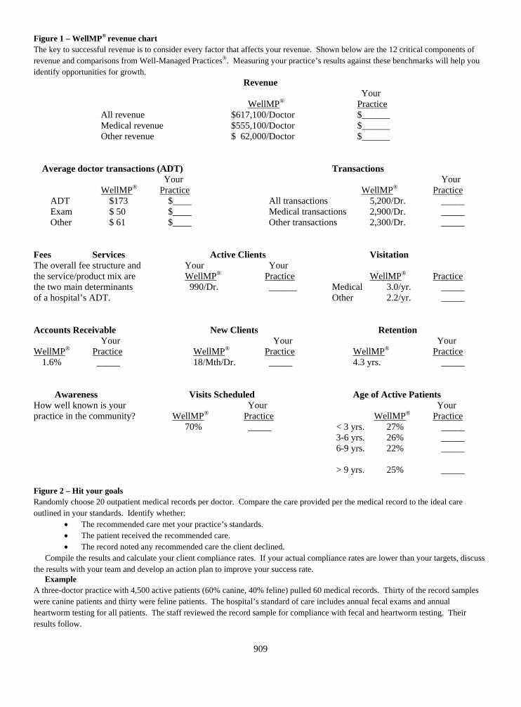
909
Figure 1 – WellMP® revenue chart The key to successful revenue is to consider every factor that affects your revenue. Shown below are the 12 critical components of revenue and comparisons from Well-Managed Practices®. Measuring your practice’s results against these benchmarks will help you identify opportunities for growth.
Revenue Your WellMP® Practice All revenue $617,100/Doctor $______ Medical revenue $555,100/Doctor $______ Other revenue $ 62,000/Doctor $______ Average doctor transactions (ADT) Transactions Your Your WellMP® Practice WellMP® Practice ADT $173 $____ All transactions 5,200/Dr. _____ Exam $ 50 $____ Medical transactions 2,900/Dr. _____ Other $ 61 $____ Other transactions 2,300/Dr. _____ Fees Services Active Clients Visitation The overall fee structure and Your Your the service/product mix are WellMP® Practice WellMP® Practice the two main determinants 990/Dr. ______ Medical 3.0/yr. _____ of a hospital’s ADT. Other 2.2/yr. _____ Accounts Receivable New Clients Retention Your Your Your WellMP® Practice WellMP® Practice WellMP® Practice 1.6% _____ 18/Mth/Dr. _____ 4.3 yrs. _____ Awareness Visits Scheduled Age of Active Patients How well known is your Your Your practice in the community? WellMP® Practice WellMP® Practice 70% _____ < 3 yrs. 27% _____ 3-6 yrs. 26% _____ 6-9 yrs. 22% _____ > 9 yrs. 25% _____ Figure 2 – Hit your goals Randomly choose 20 outpatient medical records per doctor. Compare the care provided per the medical record to the ideal care outlined in your standards. Identify whether:
• The recommended care met your practice’s standards. • The patient received the recommended care. • The record noted any recommended care the client declined.
Compile the results and calculate your client compliance rates. If your actual compliance rates are lower than your targets, discuss the results with your team and develop an action plan to improve your success rate.
Example A three-doctor practice with 4,500 active patients (60% canine, 40% feline) pulled 60 medical records. Thirty of the record samples were canine patients and thirty were feline patients. The hospital’s standard of care includes annual fecal exams and annual heartworm testing for all patients. The staff reviewed the record sample for compliance with fecal and heartworm testing. Their results follow.

910
Services
Active Patients
Target Increase
Tests Needed
Fee
Increased Revenue
Fecal exams: The team reviewed 60 records: 30 of the patients had a fecal exam. So the compliance rate was 50 percent.
4,500 x 20% = 900 x $23 fee
= $20,700
Canine heartworm testing: The team reviewed 30 records; 24 patients had a heartworm test. The compliance rate was 80 percent.
2,700 x 15% = 405 x $40 fee
= $16,200
Feline heartworm testing: The group reviewed 30 records; nine of the patients received a heartworm test. The compliance rate was 30 percent.
1,800 x 15% = 270 x $50 fee
= $13,500
Total potential increase in revenue = $50,400
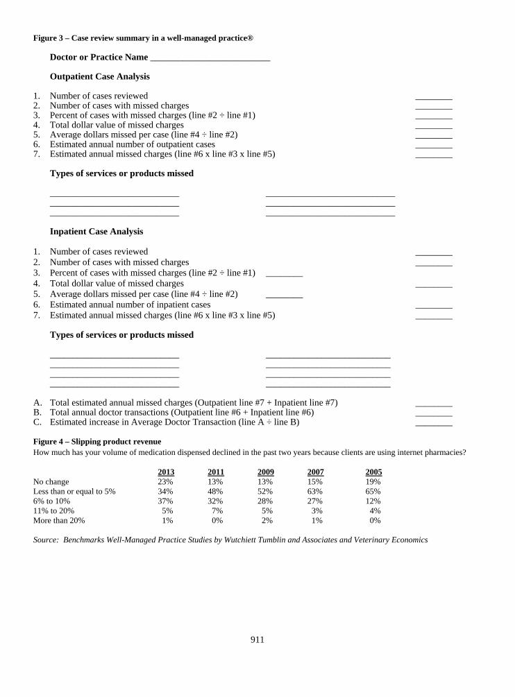
911
Figure 3 – Case review summary in a well-managed practice®
Doctor or Practice Name __________________________
Outpatient Case Analysis
1. Number of cases reviewed ________ 2. Number of cases with missed charges ________ 3. Percent of cases with missed charges (line #2 ÷ line #1) ________ 4. Total dollar value of missed charges ________ 5. Average dollars missed per case (line #4 ÷ line #2) ________ 6. Estimated annual number of outpatient cases ________ 7. Estimated annual missed charges (line #6 x line #3 x line #5) ________ Types of services or products missed ____________________________ ____________________________ ____________________________ ____________________________ ____________________________ ____________________________ Inpatient Case Analysis 1. Number of cases reviewed ________ 2. Number of cases with missed charges ________ 3. Percent of cases with missed charges (line #2 ÷ line #1) ________ 4. Total dollar value of missed charges ________ 5. Average dollars missed per case (line #4 ÷ line #2) ________ 6. Estimated annual number of inpatient cases ________ 7. Estimated annual missed charges (line #6 x line #3 x line #5) ________ Types of services or products missed ____________________________ ___________________________ ____________________________ ___________________________ ____________________________ ___________________________ ____________________________ ___________________________ A. Total estimated annual missed charges (Outpatient line #7 + Inpatient line #7) ________ B. Total annual doctor transactions (Outpatient line #6 + Inpatient line #6) ________ C. Estimated increase in Average Doctor Transaction (line A ÷ line B) ________ Figure 4 – Slipping product revenue How much has your volume of medication dispensed declined in the past two years because clients are using internet pharmacies? 2013 2011 2009 2007 2005 No change 23% 13% 13% 15% 19% Less than or equal to 5% 34% 48% 52% 63% 65% 6% to 10% 37% 32% 28% 27% 12% 11% to 20% 5% 7% 5% 3% 4% More than 20% 1% 0% 2% 1% 0% Source: Benchmarks Well-Managed Practice Studies by Wutchiett Tumblin and Associates and Veterinary Economics
912
It’s Time to Get Serious about the Budget Denise Tumblin, CPA
Wutchiett Tumblin and Associates Columbus, OH
What would you do with an extra $10,000 to $20,000? How about an extra $50,000? Perhaps you’d invest in that new piece of equipment you’ve been eyeing. Perhaps you’d invest in raises for deserving staff members. Perhaps you’d set it aside to build a cushion for a rainy day, or even give yourself an overdue raise. It’s fun to dream about the possibilities of extra cash in your pocket.
Watching what you spend may come naturally in your practice. You work with a practice budget, compare your numbers to the WellMP benchmarks, and adjust your spending when necessary. If so, kudos to you and your staff! But if you’re not quite where you’d like to be when it comes to taking charge of your expenses, now’s the time to put your expenses on a diet.
Rather than adopting the “starvation” approach to accumulate the extra cash, start with these Five Easy Slim Downs and these benchmarks to help you pinpoint where your spending is a little heavy. Then get started with your practice slim down to save that extra $10,000 to $20,000 towards your dream list. Pare down your drug inventory If your shelves are looking a bit bloated, it’s time to eliminate the excess. Veterinarians have many wonderful drugs to choose from to treat patients. But carrying every wonderful medication that’s available ties up a lot of cash and creates confusion for the staff and for clients. Doctors – unite! Create a list of the medications that you believe in the most. Conduct a scientific comparison of the duplicate products you have on your shelf. Consider the pros and cons, safety, and efficacy of each. Make your case scientifically and medically and come to a consensus among the doctors about what’s your best and second choice. Then eliminate any other redundant items from your shelves.
• Tip: Stock $10,000 to $16,000 of drugs and medical supplies per full-time equivalent doctor, or about one month’s supply. This includes heartworm, flea and tick products and excludes diets.
• Tip: Spend 8% to 9% of revenue on drugs and medical supplies. Spend 4% to 6% of revenue on heartworm, flea and tick products.
• Tip: Move infrequently-used medications to your on-line store. Evaluate your labor cost What one or more things could you do differently to increase efficiency and productivity in your hospital? It’s not unusual for different practices to have the same level of staff support, but significantly different levels of doctor production. I’m currently working with two practices, each with a 4 to 1 staff-to-doctor ratio; one generates about $440,000 of medical revenue per FTE doctor and the other generates $670,000 per FTE doctor. What accounts for the $230,000 difference? Explore the following opportunities to rev up your practice’s productivity.
• Do more with less. Bump your pay scale to attract more skilled and efficient employees. We’ve all experienced the employee who seems to get twice as much done in half the time as two other employees combined. You might find that an employee who merits $18 an hour could easily complete the work of two, less productive $12 an hour employees. The result: an annual savings of $10,000 to $12,000 depending on the benefit package.
• Streamline your processes. It’s easy to get into the routine of “that’s the way we’ve always done it.” Take a fresh look at your protocols – are you doing things the easiest, most efficient way, or could you streamline the process? Are staff members duplicating efforts? Eliminate the redundancies. Are you taking extra time to track information that no one is using? Then stop.
o Tip: Hold a contest for your staff. Ask each staff member to submit one or two ideas to improve efficiency throughout the hospital (reception, exam rooms, treatment, surgery, boarding, etc.). Give awards for the top four ideas (first, second, and third place, and honorable mention). Be sure your awards are meaningful and compelling. For example, first prize gets a paid day off; second prize gets a gift certificate for a local spa; third prize a gift certificate for a favorite local restaurant; and honorable mention gets tickets to the movie of their choice. Or, you could let the winners choose which award they would like out of your offerings.
• Get organized. Clutter and untidy work stations add to the chaos of busy days. Spend a day eliminating the mess. Move frequently used items to more accessible parts of the hospital to eliminate wasted steps. Move rarely used items to storage. Get rid of items in storage that you haven’t used for a year or more. Adopt the creed: reduce, reuse, recycle. The hospital will look better, and the doctors and staff will feel better and be more productive!
• Convert under-utilized space to a medical purpose. Some hospitals have idle or under-used space that’s begging for use as a medical area. For example, convert a food storage space to another exam room. Convert an under-utilized retail
913
space to a patient discharge room. Convert an under-utilized storage space adjacent to treatment to a dental suite or a procedures room.
o Tip: Hold a contest for your staff to solicit their ideas about under-utilized areas of the hospital that could be converted to medical use. Give awards for the top ideas (see suggested prizes above).
Bump up your use of technology Update and/or replace hardware to reduce wasted time waiting for the computer to process or recovering from a crash because the system can’t handle the hospital’s current needs. Update your software to the latest version. Replace your software if the company hasn’t provided updates for years or their support is poor. Convert to electronic medical records to eliminate wasted time searching for lost or misplaced records. Technology saves time and reduces frustration when used well.
• Tip: Hire a trainer from your practice management software company to spend a day with your staff teaching them more about your software’s capability. Staff members know the basics. But they may not be aware of all the shortcuts that help streamline their work, or the options that help enhance client service and patient care. The return you’ll receive will be much greater than the cost of the training. Example: One veterinary practice estimated that the knowledge they gained from the training saved three staff members an hour a day, which amounted to an annual labor savings of about $15,000.
Revisit your administrative costs It’s easy for fixed overhead spending to creep up without realizing it. Don’t let the word “fixed” change your mind about giving these expenses another look.
• Use e-mail for reminders, newsletters, educational materials, and other client correspondence instead of the U.S. postal service. Postage adds up and clients may actually prefer to receive information via e-mail.
• Take stock of your office supplies. Organize your inventory in one central location so everyone knows what you have on hand before requesting and ordering more. Change reorder points to minimize the amount of inventory you have on the shelf before placing a new order.
• Evaluate employee health insurance. Talk with your insurance agent about health insurance policies with higher deductibles and co-pays. Sometimes the premium savings is greater than the difference in the deductible, so you can offer to pay part or all of the difference in the deductible and still lower the practice’s cost. Ask your agent to research other policies with lower premiums and similar coverage options. Consider having employees cover part of their health care.
• Assess your Workers’ Compensation Insurance rates. Coverage managed by a private insurance company, if an option in your area, might offer better rates than a fund managed by your state.
• Conduct an energy audit in your practice. A professional energy audit gives you a clear picture of where your practice is losing energy and what you can do to save money. Possible resources to conduct the audit include your state or local government energy or weatherization office or your electric or gas utility company. Per www.energy.gov, you can save 5% to 30% on your energy bill by making the recommended upgrades. Visit www.greenyour.com for an energy audit checklist.
• Investigate the possibility of refinancing your debt. If you’ve got any high-rate loans, act now to see what your options are for getting into a more favorable rate.
Think twice before investing in equipment Do the math to determine if the equipment purchases you’re planning will pay for themselves in a reasonable timeframe. Investing in equipment helps you enhance patient care and client service, and grow your practice. But fabulous equipment rarely used, is a poor investment. Take the time to evaluate how often you’ll use the equipment and the revenue potential before taking the plunge.
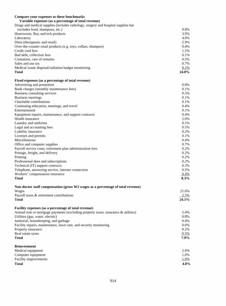
914
Compare your expenses to these benchmarks Variable expenses (as a percentage of total revenue)
Drugs and medical supplies (includes radiology, surgery and hospital supplies but excludes food, shampoos, etc.) 9.8% Heartworm, flea, and tick products 3.9% Laboratory 4.0% Diets (therapeutic and retail) 2.9% Over-the-counter retail products (e.g. toys, collars, shampoo) 0.4% Credit card fees 1.5% Bad debt, collection fees 0.1% Cremation, care of remains 0.5% Sales and use tax 0.7% Medical waste disposal/radiation badget monitoring 0.1% Total 24.0% Fixed expenses (as a percentage of total revenue) Advertising and promotion 0.8% Bank charges (monthly maintenance fees) 0.1% Business consulting services 0.3% Business meetings 0.1% Charitable contributions 0.1% Continuing education, meetings, and travel 0.4% Entertainment 0.1% Equipment repairs, maintenance, and support contracts 0.4% Health insurance 2.0% Laundry and uniforms 0.1% Legal and accounting fees 0.5% Liability insurance 0.2% Licenses and permits 0.1% Miscellaneous 0.4% Office and computer supplies 0.7% Payroll service costs, retirement plan administration fees 0.2% Postage, freight, and delivery 0.2% Printing 0.2% Professional dues and subscriptions 0.2% Technical (IT) support contracts 0.3% Telephone, answering service, internet connection 0.5% Workers’ compensation insurance 0.4% Total 8.3% Non-doctor staff compensation (gross W2 wages as a percentage of total revenue) Wages 21.6% Payroll taxes & retirement contributions 2.5% Total 24.1% Facility expenses (as a percentage of total revenue) Annual rent or mortgage payments (excluding property taxes, insurance & utilities) 5.4% Utilities (gas, water, electric) 0.8% Janitorial, housekeeping, and garbage 0.4% Facility repairs, maintenance, lawn care, and security monitoring 0.6% Property insurance 0.2% Real estate taxes 0.5% Total 7.9% Reinvestment Medical equipment 2.0% Computer equipment 1.0% Facility improvements 1.0% Total 4.0%
915
What Well-Managed Practices Pay: Are You in the Ballpark?
Denise Tumblin, CPA Wutchiett Tumblin and Associates
Columbus, OH
Practices must stay on top of current trends in doctor and staff compensation and benefits to remain competitive and attract efficient, effective and productive employees. Pay scales must also reflect the level of education, experience, skill and personal attributes you want employees to bring to the table. So, what are the latest trends? Benchmarks 2013 – A Study of Well-Managed Practices® sheds light on this hot topic. Employed veterinarians The majority of practices now pay their doctors some form of incentive-based compensation. About 27% pay purely based on production. Another 48% pay doctors a guaranteed base plus a percentage of production over a required minimum. The remaining 25% pay their doctors a fixed salary.
Practices who use a blended rate – i.e., one percentage applies to all medical service and product production – typically pay their doctors between 16% and 21%. Where they fall in the range is dependent upon the practice’s staff-to-doctor ratio. The more staff the practice provides to assist the doctors, the lower the percentage paid to the doctors. The additional staff members allow the doctors to produce at a higher level, which increases doctor compensation. The practice also has an added layer of overhead because of the additional staff members, which the doctors must help support.
Practices who use a split-rate – i.e., one percentage applies to medical service production, and a different percentage applies to medical product production – typically pay their doctors between 22% and 26% for services, and 4% and 10% for products. The service/product split – i.e., how much of medical revenue comes from services and how much from product sales - and the staff-to-doctor ratio will both impact where you end up in the stated ranges.
To make any percentage-based compensation system work, every team member must understand what is and isn’t credited to the doctor’s individual production. Doctors receive credit for all medical service revenue provided during an outpatient appointment, in-hospital treatment, or dental and surgical procedures. Doctors also receive credit for medications and therapeutic foods dispensed during an outpatient appointment, during in-hospital treatment, or at the end of a patient’s hospital stay.
Prescription refills and additional food or product purchases that don’t involve a doctor are credited to a hospital provider. The doctor receives credit for the refill only if it requires his or her time to review the record, assess if the medication or dosage needs to change, and give direction to the staff member who will fill the prescription. Doctors never receive credit for boarding, grooming, or retail purchases.
When multiple doctors collaborate to treat a patient, the doctor who provides each point of care receives credit. For example, if Doctor A examines and admits a patient to the hospital on Day 1, and Doctor B provides or supervises the hospital treatment on Day 2, Doctor A gets credit for everything on Day 1, and Doctor B gets credit for Day 2.
See Figure 1 for the latest on starting salaries for associates based on years of experience. See Figure 2 for other employee benefits.
Non-doctor staff compensation Practices spend between 21% and 25% of revenue on staff compensation, payroll taxes, and retirement contributions. This includes all non-doctor staff positions except groomers – so, hospital administrators, practice managers, receptionists, credentialed technicians, veterinary assistants and kennel/ward attendants are in this number. Where you fall in this range will depend on the cost of living in your area, the skill set of your staff, and your staff-to-doctor ratio. See figure 3 for the latest pay ranges by position.
If your staff costs are high, start by evaluating productivity. Often the issue isn’t over-spending, but rather lower-than-expected productivity. Low productivity has a variety of reasons. Sometimes it’s caused by giving away or significantly discounting care that the practice provides. Sometimes it’s related to a lower-than-warranted fee structure. Sometimes it’s due to a lack of skills or inefficient processes. And, sometimes a practice employs people who are a poor fit and who put a damper on the morale of the other team members. Before you start thinking of ways to cut staff expenses, first determine why your cost is high. Resources to help evaluate your compensation and benefits
• Benchmarks 2013 – A Study of Well-Managed Practices® • Compensation and Benefits by AAHA Press • On-line sources such as www.salary.com or www.payscale.com
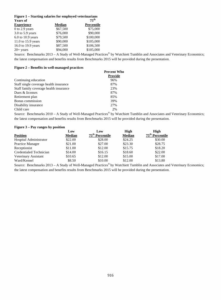
916
Figure 1 – Starting salaries for employed veterinarians Years of 75th Experience Median Percentile 0 to 2.9 years $67,500 $75,000 3.0 to 5.9 years $76,000 $90,000 6.0 to 10.9 years $79,500 $100,000 11.0 to 15.9 years $90,000 $105,000 16.0 to 19.9 years $87,500 $106,500 20+ years $94,000 $105,000 Source: Benchmarks 2013 – A Study of Well-Managed Practices® by Wutchiett Tumblin and Associates and Veterinary Economics; the latest compensation and benefits results from Benchmarks 2015 will be provided during the presentation. Figure 2 – Benefits in well-managed practices Percent Who Provide Continuing education 96% Staff single coverage health insurance 87% Staff family coverage health insurance 23% Dues & licenses 87% Retirement plan 85% Bonus commission 39% Disability insurance 27% Child care 2% Source: Benchmarks 2010 – A Study of Well-Managed Practices® by Wutchiett Tumblin and Associates and Veterinary Economics; the latest compensation and benefits results from Benchmarks 2015 will be provided during the presentation. Figure 3 – Pay ranges by position Low Low High High Position Median 75th Percentile Median 75th Percentile Hospital Administrator $22.00 $28.00 $24.25 $30.00 Practice Manager $21.00 $27.00 $23.30 $28.75 Receptionist $11.00 $12.00 $15.75 $18.20 Credentialed Technician $14.00 $16.15 $18.60 $22.00 Veterinary Assistant $10.65 $12.00 $15.00 $17.00 Ward/Kennel $8.50 $10.00 $12.00 $13.80 Source: Benchmarks 2013 – A Study of Well-Managed Practices® by Wutchiett Tumblin and Associates and Veterinary Economics; the latest compensation and benefits results from Benchmarks 2015 will be provided during the presentation.
917
Write an Rx for Your Success (Parts 1 and 2) Denise Tumblin, CPA
Wutchiett Tumblin and Associates Columbus, OH
Financial security for your practice and your family takes planning and paying attention. Where do you begin? Take these steps today to ensure a strong, stable future. Establish effective documentation systems Use veterinary and accounting software and a Management Statement™ to manage your patient and financial records. Execute partnership/shareholder contracts, a buy/sell agreement, lease agreements for equipment and the facility, employment agreements, etc. Create a foundation for effective practice leadership Identify the leaders, doctors, and advisors required for practice success. Establish and define responsibility for areas of management for the owners. Create an internal communication structure. Establish and define the management positions at the staff level. See Collaborative Teamwork Charts. Envision the ideal healthcare team Determine the key positions and the number of support staff needed to meet patient care and client service goals. Define the parameters for selecting the right people for each position. Establish the pay scale by position and for each level of competency within each position. Identify the training and education required for continued growth. Define the evaluation and promotion protocols for your practice. Determine if the facility configuration and size continues to meet your clients’ and patients’ needs Do you have a well-designed site and floor plan with ample parking, a sufficient number of exam rooms, and ample treatment and surgery areas? Does your location continue to be desirable? Is it visible, accessible, and have room to grow? Pay attention to your physical appearance – the grounds, building, and staff must present a professional, clean, well-maintained image. Stay in tune with your environment to ensure it’s stress-free, warm and welcoming. Develop a practice budget Start with the information you already know – your current year-to-date revenue and expenses. Also consider any additional goals you want to achieve. Lastly, look at benchmarks set by other practices to get an idea of spending levels (see Benchmarks 2015 A Study of Well-Managed Practices® at www.wellmp.com/Benchmarks, or Financial Productivity Pulsepoints by AAHA). Keep this data handy as you plan your revenue and expense changes for the coming year.
• Revenue. How much will revenue grow next year? Consider planned fee changes, compliance initiatives that will result in providing more of existing services, and new services you’re adding to the practice such as laser therapy, ultrasound, behavior consulting, rehabilitative therapy, or acupuncture.
• Expenses. How will your operating expenses change next year? Have you targeted a reduction in some line items like inventory costs? Are you planning increases in other items like staff compensation or equipment purchases? Determine where your spending will remain the same, drop or rise.
Develop a personal budget Tracking your personal household income and expenses and developing a budget provides an organized, systematic approach to efficiently measure and analyze your personal financial position. Begin with the information you already know – your current year-to-date income and expenses. Then consider any changes or new goals for the coming year.
• Income - Is your share of income from the practice or any other businesses you own likely to increase or decrease? Is your spouse/partner expecting any increases or decreases in income? Project how your household income will change for the coming year.
• Expenses – How will your required expenditures change in the coming year? Consider loan/lease repayments on credit cards, autos, your home, or any other debts. Plan for any expected increases in property taxes, insurance, utilities, medical expenses, and auto expenses. Do you have any home repairs on tap? What about alimony or child support? Consider any changes to discretionary expenditures like clothing, travel and vacations, entertainment, club dues, gifts and charitable contributions, etc. And, don’t forget about contributions to savings and retirement accounts or your children’s educational funds.
918
Manage your debt Maintain a Debt Worksheet that tracks all outstanding loans and leases and includes the original debt amount, payoff date, interest rate, monthly payment, and current outstanding balance. When you’ve got extra cash, make additional payments on loans with no pre-payment penalties to reduce the interest paid over the life of the loan. Apply the extra payments towards the highest interest-rate loans first. Have appropriate insurance coverage Talk with your insurance representative to determine your personal and practice needs, including:
• Auto: consider bodily injury, property damage liability, personal injury, uninsured or underinsured motorist, and collision and comprehensive coverage
• Home property insurance: protection for your dwelling, other structures, and personal property, reimbursed living expenses, flood, earthquake, water backup, identity theft restoration, and a personal umbrella policy.
• Practice property insurance: two types of policies - named-perils policy, which only covers losses resulting from particular events named in the policy, and an all-risk policy (also known as special form coverage) which covers all events except those specifically named. The type of business and your location and region of the country are all considered when determining which risks are more likely to affect your business. Property insurance covers loss from fire, theft or vandalism, provides financial assistance to help cover the cost to rebuild or repair business property so that operations can continue with as little disruption as possible, and includes compensation to repair damaged business property or replace what you've lost. Other available coverage includes undamaged stock, data or records, computer virus, intangible coverage, off-premises property, and terrorism.
• Employment Practices Liability Insurance: protects against claims filed by disgruntled employees. • Health Insurance: provides coverage for medical costs. • Liability/Malpractice Insurance: protects professional advice- and service-providing individuals and companies from
bearing the full cost of defending against a negligence claim made by a client, and damages awarded in such a civil lawsuit.
• Workers’ compensation: offers payments to employees who are (usually) temporarily, unable to work because of a job-related injury; compensates for economic loss (past and future), reimbursement or payment of medical and like expenses, general damages for pain and suffering, and benefits payable to the dependents of workers killed during employment.
• Disability Insurance: designed to replace anywhere from 45-60% of your gross income on a tax-free basis should a sickness or illness prevent you from earning an income in your occupation.
• Business overhead expense: reimburses a business for overhead expenses should the owner experience a disability. Eligible benefits include: rent or mortgage payments, utilities, leasing costs, laundry/maintenance, accounting/billing and collection service fees, business insurance premiums, employee salaries, employee benefits, property tax, and other regular monthly expenses.
• Life Insurance: provides cash to your beneficiary(s) in the event of death. Life insurance proceeds are often used to fund the purchase/buy-back of a deceased partner’s share of the practice.
Review contracts and agreements regularly Review Employment Agreements annually, practice Operating Agreements every 3 to 5 years and whenever you’re planning a partial or full practice sale, and Buy Sell Agreements every 3 to 5 years and whenever you’re planning a partial or full practice sale. Clean up your financial records Eliminate the mixing and mingling of personal and practice expenses. Be sure that the practice financial statement reflects only the operating expenses of the practice. Prepare and review the practice’s financial statements on a monthly basis. Prepare separate, cost-center financials if you’re a mixed animal or multiple-location practice. Conduct annual strategic planning meetings Strategic planning is a must for businesses to excel and move forward. To determine where the practice is going, you need to know exactly where it stands, then determine where you want to go and how to get there.
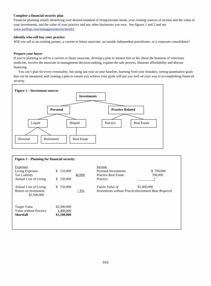
919
Figure 2 – Planning for financial security Expenses Income Living Expenses $ 110,000 Personal Investments $ 700,000 Tax Liability 40,000 Practice Real Estate 700,000 Annual Cost of Living $ 150,000 Practice ? Annual Cost of Living $ 150,000 Future Value of $1,400,000 Return on Investment ÷ 6% Investments without PracticeInvestment Base Required $2,500,000 Target Value $2,500,000 Value without Practice 1,400,000 Shortfall $1,100,000
Personal
Illiquid Liquid
Personal Retirement Real Estate
Investments
Practice Related
Practice Real Estate
Figure 1 – Investment sources
Complete a financial security plan Financial planning entails identifying your desired standard of living/income needs, your existing sources of income and the value of your investments, and the value of your practice and any other businesses you own. See figures 1 and 2 and see www.wellmp.com/managementtools/itemS2.
Identify who will buy your practice Will you sell to an existing partner, a current or future associate, an outside independent practitioner, or a corporate consolidator?
Prepare your buyer If you’re planning to sell to a current or future associate, develop a plan to mentor him or her about the business of veterinary medicine, involve the associate in management decision-making, explain the sale process, illustrate affordability and discuss financing.
You can’t plan for every eventuality, but using last year as your baseline, learning from your mistakes, setting quantitative goals that can be measured, and creating a plan to ensure you achieve your goals will put you well on your way to accomplishing financial security.
920
Collaborative teamwork organizational charts
921
Find Practice Health- Get LEAN! Denise Tumblin, CPA
Wutchiett Tumblin and Associates Columbus, OH
Products made in Japan 50 years ago evoked images of mass-produced, low-quality goods. Today Japanese brand names such as Toyota, Lexus, and Honda signify quality and durability. How were the Japanese automakers able to improve quality and reduce cost? Did they just get lucky? No, they got Lean!
While Lean started in manufacturing, Lean processes translate to service industries too. There are tremendous opportunities for the use of Lean in healthcare. A study of human healthcare facilities concluded that the facilities spent 75% of their time on non-patient tasks related to communicating, coordinating, and documenting care. In addition, a 2003 report by the New England Journal of Medicine reported a 45% defect rate in human healthcare. The overall goal of applying Lean strategies in healthcare is to initiate a process of continuous improvement to improve patient outcomes while lowering costs.
Several human healthcare facilities have used these statistics as a lever for implementing Lean. The results are impressive. ThedaCare, a Wisconsin-based health system, reduced inpatient total cost of care by 25% while improving patient satisfaction to nearly 100%. Seattle Children’s new surgery center reduced nonoperative time by 50% versus the main campus surgery center.
Lean defines waste as any activity clients view as not adding value to their experience and not meeting their needs. By focusing on activities which meet the needs of the client, you will realize benefits such as:
• Improved patient care • Improved client satisfaction • Improved staff satisfaction • Reduced inventory • Improved flow of patient care • Reduced expenses
Waste goes beyond expired medications. Lean identifies seven areas of waste. • Unnecessary services. Do your appointment scheduling and patient protocols include redundant activities? • Mistakes. Does your staff regularly need to redo and correct errors? • Delays. Do equipment failures and wait times for obtaining charts, medications, and other information happen
regularly? • Unnecessary motion. Could you move equipment or supplies to reduce or eliminate wasted effort to increase
efficiency? • Over-processing. Review your protocols and processes on a regular basis to determine if they are still relevant and
provide value. • Excess inventory. This goes beyond the products stocked for retail sale and doctor use. For example, too many files
leads to the need for more cabinets and more floor space. Do you have files and equipment you have not used for months cluttering your work area?
• Excess transport. Do you juggle patients and clients among rooms? In addition to the above areas, are you “wasting” the skills of your employees? Do you offer your staff the ability to use their
creativity and knowledge to the fullest? Lean processes offer staff members an opportunity to add to practice profitability by creating ways to eliminate waste. The traditional top-down management style places stress on the owner to lead initiatives and ensure implementation. Lean processes empower employees to inspect their own work and redesign their work processes to maximize efficiency. The result - more time to practice medicine and staff members have newfound enthusiasm for their work.
One of the strengths of Lean is its focus on action. You can get started right away with a small project, see immediate results that excite you and your staff, and leverage this momentum to take on a larger project. The first step - designate a Change Agent. The leader of your Lean initiative must have an open mind about change and be able to make things happen. He or she can seize upon a frustrating experience and turn it into an opportunity to start a Lean project. For example, a staff member may struggle to find needed medical equipment in a storage area. The Change Agent can use this experience as an opportunity to rearrange the storage area in a way that makes items easy to find and reduces frustration and wasted time.
The Change Agent begins the Lean project by implementing the 5S System for the targeted area of improvement. While originally 5S was used as a tool for maintaining clean work areas, it has evolved into a systematic method for reducing costs, improving work flow, and empowering employees to assist in reducing waste. The staff members closest to the service now have the authority and tools needed to improve work processes or work areas.
Once the first Lean project has been identified, begin by taking photographs or video of the area of focus. Before and after pictures are a powerful tool for showing staff members the benefits derived from Lean initiatives. Next, utilize the 5S steps.
922
1. Sort • Eliminate unneeded items within the target area. Dispose of items that aren’t needed. Fight the urge to hold onto
items because you might need them in the future. 2. Set-In-Order
• Current State. During this step, document the location of each item and the current work flow. Create a map of the area to outline it, identify the large items, and map the flow of patients, clients, employees, and paperwork. Label all significant items, so they are easily identifiable to staff members. This process, called mapping a value stream, creates a one page picture or flow chart of the current process, and helps identify redundant steps and unnecessary motion.
• Future State. With the current state mapped, now create a future state value stream map. How can you eliminate waste identified in the current state value stream map? What is the ideal flow for completing a task? Brainstorm with your staff to create an area which has great flow, is well ordered, and reduces unnecessary movement. Items are now well-labeled and anything can be found within seconds. Everything has a place and there is a place for everything.
3. Shine • Inspect, clean, organize, and de-clutter the area and items within the area. Repair or replace frayed cords, bad
bulbs and batteries, and worn-out parts. 4. Standardize
• Create standards and visual controls such as signs and checklists to improve efficiency and reduce errors. Signboards and color code indicators provide important information at a glance.
5. Sustain • Teach employees your Lean processes and protocols, so everyone understands the benefits. Continue to evaluate
additional Lean opportunities. As with any change, you may encounter staff resistance. People may not understand the need for change, may fear it will lead to
more work, or may not understand that they will be playing a key role in determining the changes. Commit to the program, explain the need for the changes, and address the fears of the resistors. Unfortunately about 10% of the workforce might remain resistant to the change and leave. But those who embrace Lean will enjoy improved safety, work flow, and reduced costs that lead to increased customer satisfaction, employee engagement, and practice profitability. Don’t wait to realize the benefits of Lean. Implement Lean in your practice now!
923
Helping Patients that have Hypertension Mark Acierno, DVM, DACVIM
Louisiana State University Baton Rouge, LA
Hypertension may be the most under-diagnosed systemic illness affecting companion animals.
Blood pressure elevations are commonly divided into primary and secondary hypertension. Primary hypertension is the result of an imbalance in the relationship between cardiac output and systemic vascular resistance. Although more than ninety percent of all human hypertension cases are primary, the exact cause of this imbalance is not well understood. Secondary hypertension is an increase in blood pressure that occurs as a consequence of another systemic disease or drug. While relatively uncommon in humans, secondary hypertension is believed to account for most cases of canine and feline hypertension. This is important since it allows us to focus our screening efforts on a specific group of patients who are at high risk of hypertension. (Table 1) Definition Systemic hypertension is commonly defined as a blood pressure greater than 150/95 mmHg on three separate visits in a patient that demonstrates no clinical signs directly attributable to the blood pressure elevation or a single reading of greater than 150/95 mmHg in a symptomatic patient. Consequences Patients with mild increases in blood pressure only show signs attributable to the underlying disease process. As the hypertension worsens, specific organs are damaged:
• Kidney - Glomerular and tubular damage. • Eye - Retinopathy and choroidopathy (retinal edema, tortuous vessels, hemorrhage and retinal detachment). • Heart - Cardiac hypertrophy (murmur, arrhythmia, and gallop rhythm). • Brain - Hypertensive encephalopathy (ataxia, depression, and seizures).
Measurement The actual measurement of blood pressure requires the catheterization of a suitable artery and the determination of blood pressure with an electronic transducer. Although this is the “gold standard,” it is neither convenient nor practical in the clinical setting. Clinically, we rely on noninvasive indirect estimations of blood pressure using Doppler and oscillometric devices.
Due to its low cost, ease of use, and suitability for use in both cats and dogs, the Doppler flow detector is a commonly used blood pressure measuring device. Doppler units emit ultrasound waves that travel outward until they strike a surface and then they are reflected back to the device. If there is movement, such as blood cells traveling through an artery, the frequency of the returned wave is different than that of the original wave. This change in frequency is amplified by the detector and is heard by the user as a characteristic “swoosh” sound. The only equipment needed is the Doppler flow detector, a sphygmomanometer and a selection of different sized cuffs. Although systolic pressure is easily obtained, determining diastolic pressure can be challenging or impossible.
An oscillometric blood pressure device works by inflating a cuff around an extremity until arterial blood flow is stopped and then, while slowly deflating the cuff, the unit monitors for pulse waves generated by arterial pulsations. Because they are automated and can determine systolic pressure, diastolic pressure, mean arterial pressure and heart rate, these units are quite popular. Despite their convenience, oscillometric units can significantly underestimate blood pressure when used on cats and small dogs (<10 kg) and should not be used on these patients. Recently, a number of oscillometric units that claim to be optimized in cats have been produced; however, recent studies have produced contradictory information as to whether these units can produce accurate measurements. Important concepts Regardless of which blood pressure measuring technology is used, there are four principles that help ensure accurate measurements: Proper cuff selection, consistency of personnel, acclimation of the patient, and repeatability of results. Perhaps the most important factor in attaining accurate and repeatable blood pressure measurements is the selection of a correctly sized cuff. Some veterinary-specific oscillometric units have specially designed cuffs, however for Doppler and most oscillometric units, the width of the cuff should be 30%-40% of the circumference of the extremity. A cuff that is too wide will overestimate blood pressure while a cuff that is too small will artificially decrease values. Stress associated with the office visit can cause profound increases in blood pressure; these effects can be minimized by measuring blood pressure in a quiet area, away from other animals, before other procedures, and only after patients have acclimated to their surroundings. The owner should always be present, and restraint should be kept to a minimum. Pressure measurement should be repeated 4-5 times, and the measurements compared. If there is a significant variation (>10%) the readings should be discarded and the process repeated.
924
TreatmentAngiotensin converting enzyme inhibitors (ACEI) are generally considered to be the initial drug of choice in treating canine hypertension (Table 2). ACEI exert their effect by competitively inhibiting the conversion of angiotensin I to angiotensin II. Since angiotensin II is a potent vasoconstrictor, systemic vasodilatation occurs when its synthesis is inhibited. Angiotensin II also stimulates the release of aldosterone, which leads to sodium and water retention and thus, an increased blood volume. Lastly, angiotensin II directly stimulates the kidney to retain sodium, which results in an increased blood volume. Enalapril is cleared exclusively by the kidneys while benazepril is cleared by the liver as well. For this reason, benazepril is preferable to enalapril in the treatment of hypertension in renal failure patients.
Angiotensin converting enzyme inhibitors are less effective in treating hypertension in cats. Therefore, calcium channel blockers have become the initial drug of choice in controlling feline hypertension. These drugs work by interfering with the influx of calcium needed for smooth muscle contraction and vascular constriction. Due to its once-a-day dosing, gradual rate of effect and relative low cost, amlodipine besylate is the most widely used anti-hypertensive medication for cats (Table 2). Studies in humans and dogs suggest that when calcium channel blockers are used alone for controlling hypertension, they may worsen renal disease despite significantly lowering blood pressure; therefore, these drugs should not be used as a monotherapy antihypertensive agent in dogs. This effect is thought to be due to calcium channel blocker’s preferential dilation of the glomerular afferent arteriole paradoxically resulting in glomerular hypertension. ACEI preferential dilate the efferent arteriole normalizing glomerular pressures and prevent renal damage; therefore, calcium channel blockers should only be used in conjunction with ACEIs when treating canine hypertension. Calcium channel blockers do not appear to cause renal damage in hypertensive cats and are considered safe.
Beta-blockers are useful adjunctive therapy in dogs or cats when the initial anti-hypertensive agent has failed to produce the desired decrease in blood pressure (Table 1). Beta-adrenergic receptors are found in both the heart (B1) and lungs (B2). Blockade of
the B1 receptors will slow the heart and lower blood pressure; blockade of the B2 receptors can cause an undesirable bronchial constriction. Therefore, selection of a Beta-blocker should be limited to a B1-selective antagonist such as atenolol.
Stimulation of alpha-receptors on blood vessels causes vasoconstriction leading to an increase in system vascular resistance and elevations in blood pressure. Alpha-blockers selectively antagonize these receptors resulting in vasodilatation. Prazosin, a potent alpha inhibitor, has successfully been used as an adjunctive treatment for canine hypertension (Table 2). It’s use in cats for this purpose is not recommended.
Drugs that antagonize aldosterone may protect the heart, brain and kidneys from the harmful effects of hypertension. Although inhibition of aldosterone causes only a mild decrease in blood pressure, it may protect against hypertension-induced fibrosis of these target organs. While this information is preliminary, hypertensive patients may benefit from the use of spironolactone as an adjunctive treatment (Table 2). Goals The goal of treating hypertensive patients is to gradually lower systolic pressure to less than 170 mmHg. In the initial stages of treatment, monitor the patient regularly but avoid large adjustments in medications. Changes in dose and drugs should only be made every two weeks unless the patient’s condition deteriorates. Once the patient’s systolic pressure has been regulated, it should be rechecked every three months, and complete blood count and serum chemistry should be checked twice a year. Table 1. Conditions that are commonly associated with hypertension
• Hyperadrenocorticism • Renal disease • Thyroid disease • Diabetes mellitus • Hepatic disease • Drugs: erythropoietin, phenylpropanolamine, others • Uncommon conditions: Polycythemia, pheochromocytoma, hyperaldosteronsism (hyperaldosteronism?), chronic anemia

925
Table 2 Commonly used antihypertensive medications and their dosages Drug Class Canine Dose Feline Dose Enalapril (Enacard, Merck) ACE inhibitor 0.5–1.0 mg/kg q12–24h,
PO 0.25-0.5 mg/kg q12–24h, PO
Benazepril (Lotensin, Novartis) ACE inhibitor 0.25-0.5 mg/kg q12-24h, PO
0.25-0.5 mg/kg q12-24h, PO
Amlodipine (Norvasc, Pfizer) Ca++ channel blocker
0.05–0.2 mg/kg q24h, PO 0.625–1.25 mg/cat q24h, PO
Atenolol (Tenormin, AstraZeneca) Beta Blocker 0.25–1.0 mg/kg q12–24h, PO
6.25-12.5 mg/cat q12-24h, PO
Metoprolol (Lopressor, Novartis) Beta Blocker 0.5-1.0 mg/kg q 8-12h, PO 2.0 – 15.0 mg/cat q8h, PO Prazosin (Minipress, Pfizer) Alpha Blocker 0.5–2.0 mg/dog q12h Not Recommended Spironolactone (Aldactone, Serle) Aldosterone
inhibitors 1.0-2.0 mg/kg q 12h, PO 1.0 mg/kg q 12, PO

926
Dermatology for Technicians Paul Bloom, DVM, DACVD, DABVP Allergy, Skin and Ear Clinic for Pets
Livonia, MI
Protocols are useful in helping to diagnose and treated many different disorders. Part of any good protocol should be a minimum data base (MDB). In addition to signalment, history, etc in veterinary dermatology laboratory testing should be a component of this data base. Just as you may have a standard set of tests for diarrhea you should have a standard set of tests for dermatology cases. In a general practice you should be performing these tests MULTIPLE times daily. In addition to these tests other commonly, easily performed tests are
1. Skin scrapings ** 2. Impressions smears ** 3. Ear cytologies ** if ear disease is present 4. Bacterial culture
** Component of MDB Slide examination When examining a specimen microscopically, once a sample is collected (+/- processed- see cytology) the entire slide should first be examined under a low (scanning) power (4X objective). Use this scan to evaluate the quality and quantity of the sample collected, to find larger mites (eg Sarcoptes, Cheyletiella) and to identify areas that should be examined more closely. After evaluating the slide under 4x power, the slide should be examined using low power (10X objective) for smaller mites (eg Demodex) and then oil immersion (100X) to identify organisms (eg bacteria, yeast) and cells (eg neutrophils, eosinophils, keratinocytes, neoplastic cells etc). With experience you will be able to identify cells w/the 40X objective, thereby saving time. You can improve clarity on 40X by placing immersion oil on the slide and then covering the area w/a cover slip. You then can examine it w/the 40X. When you are looking for cells or organisms raise the condenser but when examining for ectoparasites, if you drop the condenser you increase the contrast making it easier to identify the parasite. Skin scraping Let’s begin with the MBD- before performing skin scrapings you should ask the following questions
1. What technique do I do (broad superficial or deep scrapings or both) 2. Where do I need to skin scrape? 3. What lesions am I looking for to scrape?
The answers to these questions depend on which parasite you suspect. If you suspect a superficial mite (Sarcoptes, Notoedres, Demodex gatoi (cats), Demodex cornei (dogs) Cheyletiella) then broad superficial scrapings should be performed. Deep skin scrapings should be performed when Demodex canis or cati is suspected. (Table 1)
When performing superficial scrapes be sure to: Scrape from appropriate areas. For Sarcoptes you will be more successful if you scrape pinnal edges, the elbows, ventral chest and
hocks. In addition any papular or erythematous lesion should be scraped. For any of the superficial mites, broad scraping should be performed. Remember that mites associated w/hypersensitivity (eg
Sarcoptes, Cheyletiellai) may be difficult to find due to their low numbers so be sure to take multiple (10-15) sites. In contrast to demodex, all scrapes can be placed on 2 or 3 slides because the quantity of mites present is not important, they are either found or not.
When performing a deep skin scrape for demodex (this applies to mostly to dogs) there are a few pitfalls to avoid. By avoiding these errors the diagnosis and your management of demodex will improve.
These include • Failing to squeeze the skin prior to scraping. This helps express the Demodex from the hair follicles • Failing to record location of scrapes; • Failing to record numbers & stages present; • Failing to record whether the mites are alive or dead; • Failing to clip hair at skin scrapings sites (if it is a recheck appointment, the hair may be regrowing preventing proper
sample collection); • Failure to squeeze the skin prior to scraping to try • Failure to recognize that lesions that are granulomatous & fibrotic, especially on the paws may have demodex that are
hard to demonstrate on skin scrapings and a skin biopsy may be necessary to diagnosis; • Failure to sedate dogs if the feet are to be scraped • Failing to scrape hyperpigmented areas even if they are not alopecic;
927
• Failing to scrape areas with comedones even if they are not alopecic • Failing to scrape if a dog only has greasy seborrhea (especially along the dorsum). A long body type of demodex mite
has been identified (Demodex injai). This mite lives in the sebaceous glands of the dog's skin, and thus, is commonly associated with "greasy coats" rather than the moth eaten or pustular appearance that we are used to seeing.
• Failing to take broad superficial skin scrapes even if demodex is the only parasite you suspect. There is a short bodied demodex mite (Demodex cornei), which lives on the surface of the skin layer. Note that there may be a low number of these mites found because of the superficial location of the mites allowing removal by the animal.
Cytology Cytologic examination is another very commonly performed procedure in dermatology that should be performed on any dog or cat presented w/skin or ear disease. Cytology is used to identify the presence (and/or type) of:
• Bacterial or fungal organisms (Malassezia); • Neoplastic cells; • Inflammatory cells; • Abnormal cells (eg acantholytic keratinocytes associated w/pemphigus foliaceus)
Both the direct smear (scrapings, impression, roll smear or tape) and fine needle aspirates (FNA) are valuable techniques that need to be mastered.
When the skin is scaly, a superficial skin scraping can be useful. A very small amount of mineral oil is placed on a #15 scalpel blade to help keep the scale on the blade once it has been collected. The lesion is scraped a few times, and the material collected is placed on a microscope slide, stained (see below about staining samples), and examined microscopically at 40X and 100X.
Direct smears can be collected by a variety of ways. Impression (touch) smears are useful when there is an erosion, ulcer, crust, moist or greasy lesion. To perform an impression
smear, a slide is firmly applied to a lesion and, in most cases, is then gently moved back and forth a few times to increase the yield. Clear acetate tape (“scotch tape”) is also useful for collecting samples. Some people will use slides that are “sticky” on one side. These slides are reported to increase the yield of sample collected but the author finds that a standard slide works quite well. The slide is then processed and examined as previously described
If the lesion is fluid filled (eg pustule, papule) but is too small for a fine needle aspirate, “lance” the lesion with a 25 gauge needle and then do an impression smear.
When sampling crusts, lift the crust and rub both the underside of the crust and the surface of the skin. Roll smears (swabs) are used when it would be difficult to get a slide into the affected area. This could be the face fold, the
interdigital space on cats and small dogs and the ear canals in all dogs and cats. A cotton tipped applicator is gently rubbed back and forth across the lesion and then the material from the applicator stick is rolled back and forth on the slide. If the lesion is scaly, applying a small amount of mineral oil to the swab can help with collection. The sample is rolled onto a microscope slide, stained and examined as previously described.
A fine needle aspirate is performed when a solid or fluid filled mass or lesion is present. A 22-25 gauge needle attached to a 12 cc syringe is placed into the lesion and suction is applied by pulling back the plunger of the syringe (½ to ¾ of the way). The syringe plunger is pulled back and released a few times. Don’t aspirate aggressively enough that you get blood contaminating the sample (you should not see blood in the hub of the needle). After aspirating one spot, stop aspirating and redirect the needle in the mass w/o pulling out and repeat the aspiration. This can be repeated 2 or 3 times on each sampling attempt. The needle is disconnected from the syringe, the syringe is filled w/air and the needle is placed back on the syringe. The material is then ejected from the needle by compressing the plunger. If the lesion is a fluid filled you only have to pull back far enough to get a sample into the syringe. Note- Measuring and noting the location of the masses is valuable for monitoring treatment.
Regardless of the collection technique (except when using the tape prep) historically the author would heat fix the sample, using a cigarette lighter, and then wait a minute or so to allow it to cool. The slide was then stained w/a modified Wright stain (Diff Quik®). There are 3 jars in the Diff Quik® kit. The first jar is a fixative containing methanol, the second contains buffered xanthene dye, which stains the cells and organisms red and the third contains a buffered thiazine dye (methylene blue) which stains the cells and organisms purple. After drying the slide would then be examined.
More recently I have bypassed both the fixative step and the second step (eosin) and directly go to the 3rd step using the methylene blue only. It doesn’t appear to hinder the identification of organisms or inflammatory cells. If using the tape prep I will put a drop on stain on the slide and then place the tape, sticky side down, over the stain and examine.
Ear cytologies are performed to identify mites, infectious agents, ectoparasites and inflammatory cells. A cotton tip applicator is used to collect the samples prior to instituting therapy. Results of the cytology help direct appropriate therapy (presence of infectious agents would indicate the need for antimicrobial therapy). I will also perform ear cytologies during therapy if either the ear(s) are not responding to treatment OR if there were mostly rods on the initial cytology even if the ear(s) look normal at recheck.
928
Regardless of how the sample is collected to examine the slide involves the following steps: 1. Raise the condenser and have the light on the highest level 2. Scan the slide slowly at 10 x to identify areas with the most # of cells 3. Use 100X to ID bacteria (cocci/rods), Malassezia, inflammatory cells and neoplastic cells
a. With practice you can use 40X to ID Malassezia and inflammatory cells 4. Look at several representative fields and record your findings
a. For skin cytologies 1. For bacteria look in 10 fields and record a range (eg 0-5, 5-10, 10-20 etc) – be sure to note if they are
cocci or rods and whether they are intracellular or extracellular 2. For Malassezia look in 20-25 fields (unless they are ID sooner). Report them as negative/+0 if NO
Malassezia is found, +1 if 1 or 2 organisms are found (total #) in all the fields examined and there were never more than 1 in a field, report a +2 if there are more than 1 organism in a field or 1 organism q 3-4 oil fields
b. For ear cytologies i. There is no universal agreement as to what are normal # of cocci or Malassezia from an ear
cytology 1. Depending on the study, cutoff numbers, per oil immersion field (multiple by 2.5 to get
per HPF), between normal and abnormal ears range from >1 Malassezia to >4 Malassezia and from >1 cocci to >10 cocci. It is my opinion that the number of organisms needed to be present to be considered significant is not just a “number”. I don’t perform cytology on normal ears – I only do them if the ears are inflamed or have exudate. In that case ANY organism seen will be treated as part of the therapy regardless of the number present
ii. Inflammatory cells or rod shaped bacteria are never present in a normal ear. Fine tooth combing Combing of the hair with a fine tooth comb (“flea comb”) is a method that can be useful in finding fleas and other ectoparasites (ticks, lice and Cheyletiella). You may also detect miliary lesions on cats that were not appreciated on your physical examination. Trichogram (“hair plucks”) Veterinarians are frequently presented w/animals that have hair loss. In establishing the diagnosis of the hair disease, signalment, history (constitutional signs?) and physical examination (eg pot belly?) are all important components in establishing a diagnosis. There are times that even w/this information the cause of the alopecia has not been established. A trichogram, which is a microscopic evaluation of plucked hairs, may be a useful tool to help identify the underlying cause.
To perform a hair pluck • Grab the hair that is to be examined w/forceps. • Pluck the hairs gently at the base and roll your wrist to gently remove the hair • Place the hairs in mineral oil on a slide w/the hairs running in the same direction. • Apply a coverslip • Examine microscopically
If the alopecia is post traumatic (pruritus) or due to fragile hairs (eg dermatophytosis) the distal end of the hairs will be broken. If the hair loss is spontaneous (eg endocrinopathy) the tips are tapered.
Hair plucks can also be useful in ruling in (but not ruling out) demodicosis. Other ectoparasites may also be identified such as Cheyletiella or lice.
Follicular cast can also be identified w/hair plucks. Follicular casts refers to the accumulation of keratin debris that adheres to the hair shaft as it extends out of the hair follicle. This finding indicates a follicular keratinization disorder which occurs w/vitamin A responsive dermatosis (Cockers), follicular infections (demodex, dermatophyte, bacterial), sebaceous adenitis, endocrinopathy (hyperadrenocorticism, hypothyroidism) or primary seborrhea such as ear margin seborrhea. Bacterial cultures In the past bacterial cultures were not frequently performed in dogs with skin disease since Staphylococcus intermedius was the most common bacterial pathogen and had a predictable susceptibility profile. Unfortunately it isn’t that simple any more. Staphylococcus intermedius , Staphylococcus pseudintermedius, Staphylococcus schleiferi subsp. schleiferi, Staphylococcus schleiferi subsp. coagalens, and Staphylococcus aureus all w/variable susceptibilities (methicillin resistant, multidrug resistant, combination) are now
929
associated w/pyodermas in dogs. Indications for bacterial culture and susceptibility testing in the dog or cat have therefore been modified. This would include the presence of:
• Nodules; • Deep draining tracts; • A bacterial infection of the skin (confirmed by identifying intracellular bacteria and degenerative neutrophils) that fails
to respond to appropriate antibiotic therapy; • A bacterial infection of the skin that is failing to respond to appropriate antibacterial therapy (not recurrent but failure to
respond) especially if there are immunosuppressed individuals in the house hold • Suspicion of an uncommon bacterial infection (atypical mycobacteria, nocardia, actinobacillus); • Suspicion of an anaerobic infection (gas pocket formation);
A few tips • Mini-Tip Culturette (Becton Dickinson Microbiology Systems) - These are culture swabs w/very small tips allowing
insertion into the lesion. Also by limiting the size of the tip, a more precise sample can be collected. • Taking samples from 2 or 3 lesions if possible will increase the likelihood of identifying all pathogens • Do cytology concurrently • When selecting a lesion to culture – from best to worse – • pustule >papule>crust>epidermal collarette • If you are sampling a crust- lift the crust and swab w/the culturettes the underside of the crust and the surface of the skin
under the crusts • For an epidermal collarette lift the edge of the collarette- if you are not able to do this then clip the hair w/scissors to
expose the collarette and take a the culturette swab and gently roll it across the collarette 3 to 4 times. • Have the lab do tube dilution (MIC) rather than disc diffusion (Kirby-Bauer) • Gentle cleansing with an alcohol swab will help remove surface debris. Once the lesion is dry, a sterile 20 gauge needle
can be used to lance the pustule/papule or used to lift the crust. Don’t use alcohol if the lesion you are culturing is a collarette.
930
Adrenal Disease in Dogs and Cats David Bruyette, DVM, DACVIM
VCA West Los Angeles Animal Hospital Los Angeles, CA
Hyperadrenocorticism in cats Hyperadrenocorticism develops most commonly in middle-aged to older cats (mean age = 10.4 years; range 6 - 15 years). Of the reported cases of feline Cushing's syndrome (78%) have been females. This female sex predilection resembles the human syndrome and contrasts with canine hyperadrenocorticism, where no sex predilection occurs.
The most common historical findings and clinical signs associated with feline hyperadrenocorticism are polyuria, polydipsia, and polyphagia. These signs likely correspond to the high incidence of concurrent diabetes mellitus (76%) found in cats with hyperadrenocorticism, and are consistent with the lack of overt signs preceding marked glucose intolerance observed in experimentally-induced disease. The typical "Cushingoid" pot-bellied appearance with hepatomegaly, weight gain, and generalized muscle wasting is common in cats as in dogs. Dermatologic abnormalities frequently recognized include an unkempt hair coat with patchy alopecia, and very thin skin prone to traumatically induced tears and secondary infections.
Hyperglycemia is the most common laboratory abnormality found on serum biochemistries. Cats appear more sensitive to the diabetogenic effects of glucocorticoid excess than dogs. Cats with concurrent diabetes mellitus often exhibit cortisol-induced insulin resistance, requiring high daily doses of insulin to control their hyperglycemia and glucosuria. Hypercholesterolemia is also common, and may relate to insulin resistance and increased lipolysis. Cats lack the steroid-induced isoenzyme of alkaline phosphatase found in the canine, and the half-life of the enzyme appears to be significantly shorter in the cat. Elevation of serum alkaline phosphatase (SAP) is present in only approximately one-third of cats compared to nearly 90% of dogs with hyperadrenocorticism. Increases in SAP and the hepatocellular enzyme ALT appear to correspond with the regulation of the diabetic state, rather than representing direct indicators of glucocorticoid excess. These enzymes frequently normalize with adequate regulation of diabetes, even without therapy directed towards the hyperadrenocorticism. Hematologic findings associated with hypercortisolemia (lymphopenia, eosinopenia, and neutrophilic leukocytosis) occur inconsistently in feline hyperadrenocorticism. Despite clinical polyuria and polydipsia, cats appear to maintain urine specific gravities of greater than 1.020 more frequently than dogs, and only occasionally exhibit dilute urine and decreased blood urea nitrogen concentrations commonly seen in dogs with hyperadrenocorticism.
Endocrinologic evaluation of cats suspected of hyperadrenocorticism involves screening tests to confirm the diagnosis, and differentiating tests to distinguish pituitary-dependent disease (PDH) from adrenal tumors (AT). Adrenocorticotropin (ACTH) stimulation testing in adrenocortical hyperfunction is not as definitive as for hypoadrenocorticism. Fifteen to 30% of cats with confirmed hyperadrenocorticism have had normal cortisol response to ACTH administration (false negatives). In addition, stressed cats and those with non-adrenal illnesses may show an exaggerated response to ACTH in the absence of hyperadrenocorticism (false positives). A normal urine cortisol-to-creatinine ratio (UCCR) can be used to exclude the diagnosis of hyperadrenocorticism in cats as described in dogs. The UCCR is attractive due to the ease of sampling compared to other endocrine function tests, but is non-specific and will be elevated in a variety on non-adrenal illnesses. An exaggerated ACTH stimulation test or an elevated UCCR should be pursued with suppression testing prior to initiating any therapy.
Normal cats are more variable than dogs with respect to the degree and duration of adrenocortical suppression following dexamethasone administration. Intravenous doses of dexamethasone that have been evaluated in the cat range from 0.005 to 1.0 milligrams per kilogram. A dosage of 0.01 mg/kg of dexamethasone, commonly used in low-dose dexamethasone suppression testing in dogs, led to a significant drop in serum cortisol levels in ten normal cats, but 2 of the cats showed a slight escape from suppression by 8 hours after injection. Intravenous dexamethasone sodium phosphate (DSP), 0.01 and 0.1 mg/kg, produced equivalent reductions of plasma cortisol levels, but suppression was sustained below baseline longer with the higher dosage. Cats with various non-adrenal illnesses have also shown inadequate cortisol suppression after a low-dose (0.01 mg/kg) of DSP. The 0.1 mg/kg dosage of dexamethasone seems to more reliably suppress cortisol levels in normal cats and cats with non-adrenal illnesses. Elevated cortisol levels eight hours post-dexamethasone injection, using the 0.1 mg/kg dosage, appears to be a sensitive a diagnostic test for feline hyperadrenocorticism (89%) similar to the low-dose (0.01 mg/kg) screening test in the dog.
The combined dexamethasone suppression/ACTH stimulation test has been used successfully to diagnose hyperadrenocorticism in the cat. Affected cats display inadequate suppression of cortisol 2-4 hours after an injection of 0.1 mg/kg of dexamethasone, and an exaggerated response 1-2 hours after ACTH stimulation. The ability of the combined test to discriminate PDH from AT is unclear. Several cats with confirmed pituitary disease failed to suppress 2-4 hours after dexamethasone. Extending the duration of post-dexamethasone monitoring, or using higher doses of DSP may improve the ability of the combined test to distinguish PDH from AT. Currently, the combined test does not appear to offer more clinical utility than either the ACTH stimulation or dexamethasone suppression test evaluated separately.
931
An ultra-high dose, 1.0 mg/kg, dexamethasone suppression test has been used to distinguish PDH from AT in the cat. Two cats with hyperadrenocorticism diagnosed by the combined high dose dexamethasone suppression/ ACTH stimulation test had exaggerated responses to ACTH with no cortisol suppression 2-4 hours after 0.1 mg/kg DSP. These cats did suppress following the ultra-high dose of dexamethasone, and were later confirmed to have PDH. Cortisol levels should be monitored at several time points following dexamethasone administration to determine if any suppression (a 50% or greater reduction in pre-test values) is occurring. Cats with PDH may show suppression 2, 4, or 6 hours into the test only to escape from the suppressive effects of dexamethasone by 8 hours. One cat with an adrenal adenoma failed to suppress following dexamethasone doses ranging from 0.1 to 1.0 mg/kg. As is the case in dogs, suppression following high doses of dexamethasone is diagnostic for PDH, but failure to suppress requires further testing to distinguish pituitary from adrenal disease.
Determination of plasma ACTH concentrations is an effective way of diagnosing PDH. The normal range of plasma ACTH is lower in cats than in dogs, and many normal cats may have concentrations of ACTH below the lower limits of the sensitivity of the assay. Cats with PDH will have normal to elevated ACTH concentrations while cats with adrenocortical adenomas or carcinomas will have undetectable plasma ACTH levels. Plasma ACTH samples need to be collected and handled carefully. Veterinarians should consult their diagnostic laboratory for specific instructions prior to performing the test. Incorrect sample handling can falsely lower measured values. Normal to elevated plasma ACTH levels support a diagnosis of PDH, whereas low concentrations may require additional diagnostic testing. As in the differentiation of canine hyperadrenocorticism, ACTH levels should only be used to distinguish PDH from AT after hyperadrenocorticism has been confirmed by other screening diagnostics.
Pituitary-adrenal function tests need to be interpreted in conjunction with historical, clinical, and clinicopathologic findings before any conclusions can be drawn. No single diagnostic test is infallible. Equivocal results or discordant findings should be reevaluated. Hyperadrenocorticism is an uncommon disorder in cats. Consequently, false positive test results should be anticipated. Interpretation of endocrinologic testing should incorporate all available information before any therapeutic intervention is attempted.
Diagnostic imaging can facilitate differentiation of PDH from AT when screening tests and clinical findings suggest hyperadrenocorticism. Approximately half of canine adrenal tumors are mineralized and can be recognized radiographically. The frequency of mineralization in feline adrenocortical tumors is unknown, but up to 30% of normal cats may have calcification of their adrenal glands. Abdominal radiographic findings in cats with hyperadrenocorticism included hepatomegaly (69%) and obesity. Ultrasonographic evaluation of adrenal size and morphology has been described for dogs and cats. Nonfunctional adrenal tumors can be incidental findings in humans undergoing abdominal imaging. The incidence of "silent" adrenal masses in the cat is unknown. The presence of unilateral adrenomegaly or distortion of adrenal architecture in a cat suspected of hyperadrenocorticism is strong evidence of AT. Abdominal computerized tomography (CT) and magnetic resonance imaging (MRI) offer improved resolution for the detection of adrenal tumors or hyperplasia. CT and MRI detection of pituitary masses is also now feasible for small animal patients.
Adrenal tumors accounted for 22% of the reported cases of feline hyperadrenocorticism. Half of the adrenocortical tumors were found histologically to be adenomas and half carcinomas. The treatment of choice for adrenal tumors is surgical adrenalectomy. Two cats with adrenocortical adenomas responded well to unilateral adrenalectomy, with clinical signs resolving over 4 to 8 weeks. One cat with an adrenal adenoma removed surgically developed a recurrence of signs 12 months postoperatively. An adenoma of the contralateral adrenal gland was diagnosed. The cat survived a second adrenalectomy and was disease-free for over two years following the second procedure. Surgical therapy and long term follow-up for adrenocortical carcinomas in cats has not been reported.
Treatment options for pituitary dependent hyperadrenocorticism in the cat include both surgical and medical alternatives. Bilateral adrenalectomy followed by mineralocorticoid and glucocorticoid replacement therapy was performed in 11 cats. Nine cats responded well to surgery with cessation of polyuria and polydipsia, regrowth of hair coat, and marked improvement (4) or resolution (5) of diabetes mellitus. One cat developed acute signs of circling, wandering aimlessly, and apparent blindness 2 months post-operatively. An expanding pituitary tumor was suspected, but no necropsy was performed. Two cats died within one week of surgery from sepsis. Survival times for 6 cats with adequate follow-up after bilateral adrenalectomy for PDH ranged from 1 to 12+ months (median 5 months). Two cats are still alive, one year post-operatively. These results suggest that surgical complications of bilateral adrenalectomy may be less frequent in cats than in dogs.
Surgical treatment can also include transsphenoidal hypophysectomy which is performed at WLA for cats with pituitary masses extending above the sella (macroadenoma). Cats with functional tumors have similar success rates to those reported in dogs with PDH.
Four drugs (ketoconazole, mitotane, metyrapone and trilostane ) have been investigated for the medical management of spontaneous feline hyperadrenocorticism. Ketoconazole, an antifungal imidazole derivative, has been shown to inhibit adrenal and gonadal steroidogenesis in humans and dogs. One month of ketoconazole (15mg/kg orally twice daily) administration in 4 cats did not significantly reduce baseline plasma cortisol or ACTH responsiveness at doses 3 times greater than those effective in dogs. Two of 4 cats treated with 10 - 20 mg/kg/day of ketoconazole had adequate control of hypercortisolemia. One of the 4 cats developed severe thrombocytopenia after only one week of therapy and had to discontinue the medication. A cat with adrenocortical adenocarcinoma
932
treated with 30 mg/kg/day for 3½ months showed improved regulation of diabetes and reduction in pu/pd despite no improvement in hyperresponsiveness to ACTH. The cat ultimately was euthanatized subsequent to a non-healing skin laceration, chronic infections, and worsening insulin resistance. No evidence of hepatotoxicity or thrombocytopenia was seen at the 30 mg/kg/day dosage of ketoconazole, but the effectiveness and safety of this therapy remains questionable.
Mitotane, o,p'-DDD, is an adrenal cytotoxic agent and has been used successfully to treat dogs with PDH and AT. Use of mitotane in cats has been discouraged due to the feline sensitivity to chlorinated hydrocarbons. Three of 4 normal cats treated with o,p'-DDD at dosages ranging from 25 - 50 mg/kg, divided twice a day, tolerated the drug well, and remained clinically normal throughout treatment with mitotane. Only 2 of the 4 cats showed a decreased responsiveness to ACTH with mitotane. The cat with the largest reduction in post-ACTH cortisol levels developed vomiting, diarrhea, and partial anorexia lasting 2 weeks after a 50 mg/kg dosage of mitotane. Two cats with PDH treated with o,p'-DDD (25 mg/kg/day x 25 days, and 25 - 50 mg/kg/day x 59 days) tolerated the drug without apparent toxicity, but therapy was ineffective in controlling clinical signs in either cat. A cat with PDH treated with mitotane (50 mg/kg/day x 1 week, then 50 mg/kg/week) developed signs compatible with iatrogenic hypoadrenocorticism after 40 weeks of therapy with o,p'-DDD. At that time the cat was anorectic, lethargic, and exhibiting neurologic signs including mydriasis, pacing, and head pressing. Computerized tomography revealed a large pituitary mass extending above the sella turcica. Mitotane was discontinued, and the cat was treated with 60Co teletherapy. Subsequent CT examinations revealed shrinkage and then disappearance of the mass 10 months post-irradiation. The cat was euthanatized for continued diabetes mellitus and post-irradiation cataracts 2 years after the initial diagnosis of hyperadrenocorticism. We have had 3 other cases where a positive response to mitotane was observed clinically.
Metyrapone, an inhibitor of the 11-b-hydroxylase enzyme that converts 11-deoxycortisol to cortisol, has been used effectively in man to reduce the clinical signs of hypercortisolemia. A reciprocal rise in plasma ACTH levels occurs with falling cortisol concentrations and can eventually override the enzymatic block, allowing a return of clinical signs. In humans, metyrapone is utilized as an adjunctive therapy with pituitary irradiation or surgery. Dosages ranging from 195 - 250 mg/day have been used in cats with hyperadrenocorticism without observed toxicity. In a recent report, a diabetic cat with PDH and severe nonhealing skin wounds was treated with 65 mg of metyrapone orally 3 times a day. After 2 days of therapy the cat developed signs of glucocorticoid deficiency including depression, tremors, and ataxia. The cat improved rapidly following treatment with injectable steroids, and was discharged on twice daily metyrapone therapy. Cortisol response to exogenous ACTH was absent when evaluated on day 7. The cat was re-examined 24 days later after a hypoglycemic episode. The cats skin wounds had resolved and hair regrowth was evident. A follow-up ACTH stimulation test revealed a slightly exaggerated response. The cat underwent successful bilateral adrenalectomy and was euglycemic, with a normal haircoat, 4 months post-operatively. Two of 3 other cats reported in the literature also showed clinical improvement with metyrapone therapy, but follow-up periods were short (less than 6 months). Whether longterm therapy with metyrapone can control hypercortisolemia in cats, or whether rising ACTH levels eventually overwhelm enzymatic blockade has not been determined. Metyrapone appears to permit rapid correction of hyperadrenocorticism in some cats, and may be useful for pre-surgical stabilization prior to adrenalectomy.
We have recently evaluated the safety and efficacy of trilostane therapy (Vetoryl, Dechra Pharmaceuticals) in 15 cats with PDH. Clinical signs (13 of 15 cats) and ACTH stimulation testing results (13 of 15) improved with trilostane therapy. Diabetes mellitus was reported in 9/15 cases. Insulin requirements decreased by 36% within 2 months in 6/9 diabetic cats. Median survival time was 617 days for all cats (range 80-1,278 days). Complications included weight loss, urinary tract infections, chronic kidney disease, seizures, and recurrent pancreatitis. Hypocortisolemia was documented in 1 case. Cause of death occurred as a result of non-adrenal or non-diabetic illnesses (renal failure, seizures [caused by hypoglycemia or unknown]), or lymphoma. Trilostane ameliorates clinical signs of HAC in cats, is tolerated well in the long term, and can lead to improved regulation of diabetes. It should be considered first line therapy for cats undergoing medical management of PDH.
Hyperadrenocorticism in dogs
A. Pituitary-dependent hyperadrenocorticism 1. Surgical management
i. Bilateral adrenalectomy 1. Technically difficult 2. Poor surgical/anesthetic risk 3. Permanently hypoadrenal and require lifelong replacement therapy
B. Hypophysectomy 1. See discussion at the end of this section 2. Lifelong therapy with thyroid hormone and prednisone necessary. 3. Medical therapy
933
Trilostane therapy of canine hyperadrenocorticism The efficacy and safety of trilostane in the treatment of canine PDH were evaluated in a multicentre study at the Royal Veterinary College in London, the Veterinary Teaching Hospital in Dublin and Small Animal Hospital in Glasgow. Seventy-eight dogs with confirmed PDH were treated with trilostane for up to 3 years. The starting dose varied from 1.8 to 20 mg/kg (mean = 5.9 mg/kg).
Trilostane appeared to be well tolerated by almost all dogs with only 2 dogs developing signs and biochemical evidence of hypoadrenocorticism. One of these dogs recovered with appropriate therapy. The other died despite withdrawal of trilostane and administration of appropriate therapy. A further two dogs died within one week of starting trilostane but in neither case could a direct link with the trilostane therapy be established. The low prevalence of side effects compared favourably to those reported with mitotane.
Trilostane was found to be nearly as effective as mitotane in resolving the signs of hyperadrenocorticsm. Polyuria, polydipsia and polyphagia had dissipated in 40 dogs within 3 weeks after starting trilostane. Within 2 months, a further 20 dogs showed decreases in their water and food consumption. These improvements were maintained as long as the dogs remained on adequate doses of trilostane. Skin changes resolved in 24 out of 39 (62%) of dogs that initially presented with dermatological signs. All of these improvements were maintained as long as the dogs remained on adequate doses of trilostane. Only 8 dogs that were treated with trilostane for more than 2 months showed poor control of clinical signs. In contrast, mitotane is effective in about 80% of cases of pituitary dependent hyperadrenocorticism (PDH).
Trilostane caused a significant (p<0.001) reduction in both the mean basal and post-ACTH stimulation cortisol concentrations after 10 days of treatment. The post ACTH cortisol concentration decreased to less than 250 nmol/l (9 µg/dl) in 81% of dogs within one month and in another 15% at some time whilst on treatment. These improvements were also maintained in the study population for the duration of the trial.
Thirty-five dogs had at least one dose adjustment over the treatment period. The dose was increased in 23 dogs up to four times the starting dose. In one dog the dose was increased nine fold over a period of six months. The dose was decreased in nine dogs to as low as a quarter of the starting dose.
The mean survival of all trilostane treated dogs was 661 days. Direct comparison with mitotane was difficult as 65% of the dogs were still alive at the time of censor and therefore the mean survival may still increase.By comparison, the mean survival of mitotane treated dogs has been reported to be 810 to 900 days.
Dosage and administration The current suggested initial starting dose range for dogs with PDH is 1-2 mg/kg once daily. This needs to be adjusted according to clinical signs and serum cortisol values (see below). Doses up to 40-50 mg/kg (divided twice daily) have been given with no unwanted side effects. In some dogs twice daily dosing may be necessary. The drug is given with food. Transsphenoidal hypophysectomy A variety of treatments are available for PDH. Medical treatment options include drugs that chemically destroy the adrenals (lysodren or op-DDD) inhibit enzymes in the adrenal leading to the synthesis of cortisol (ketoconazole, trilostane) or inhibit the release of ACTH from the pituitary gland (Anipryl or selegiline). While these treatments can improve the clinical signs in 40-80% of patients they need to be chronically administered, necessitate frequent monitoring and do not cure or address the primary cause of the disease (the pituitary tumor). In humans, surgery to remove the tumor is the most successful long-term therapy. The most common approach used is the transsphenoidal method, in which a passage way is made in the sphenoid sinus, an air space behind the back of the nose, which is just below the pituitary gland. Surgical cure rates for PDH are reported to be in the range of 65-85%, although more recent long-term follow up data suggest that the recurrence rate is as high as 25 % within 5 years. When no discrete adenoma can be identified, remission of hypercortisolism is observed in only about 40%. Surgery has also been used to treat PDH in dogs. Several groups, most notably in the Netherlands have performed these surgeries with success rates paralleling those reported for humans. However, these surgeries have generally not been performed in the US. Veterinarians at VCAWLAAH, in collaboration with human neurosurgeons that regularly perform transsphenoidal surgery in humans have developed the methods to perform these surgeries in the US and are conducting a research study to determine how effectively these surgeries can be performed.
Hypoadrenocorticism Primary hypoadrenocorticism has been described in cats. Addisonian cats are middle-aged, with a median age of 4 years (mean 5.8 +/- 3.7 years) and range in age from 1.5 to 14 years. No sex or breed predilection is seen. The most common historical problems include lethargy, anorexia, and weight loss. Unlike dogs with adrenal insufficiency, diarrhea is not noted in Addisonian cats. Forty percent of cats have histories of episodic vomiting. Similar to hypoadrenocorticism in the canine, cats often have a waxing and waning clinical course, including temporary "remissions" associated with parenteral fluid and/or corticosteroid administration.
The most common findings on physical examination include depression, weakness, and mild to severe dehydration. Up to 40% present with in severe shock with weak pulses, slow capillary refill times, and extreme weakness or collapse. The duration of clinical signs preceding the diagnosis of hypoadrenocorticism ranges from 5 to 100 days, with a median of 14 days.
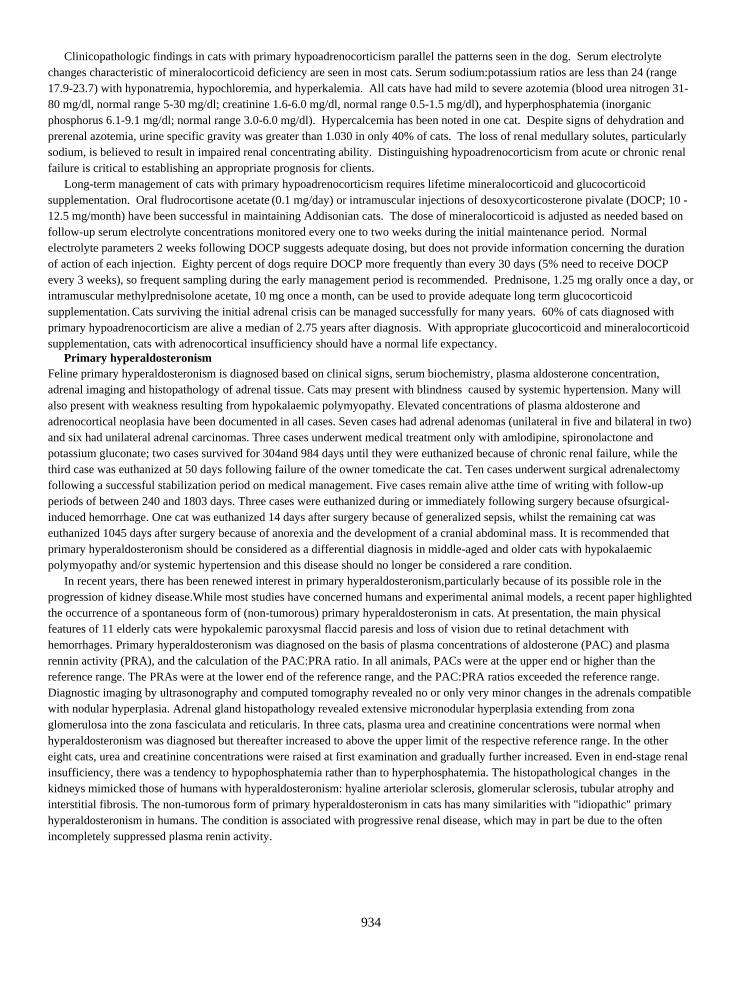
934
Clinicopathologic findings in cats with primary hypoadrenocorticism parallel the patterns seen in the dog. Serum electrolyte changes characteristic of mineralocorticoid deficiency are seen in most cats. Serum sodium:potassium ratios are less than 24 (range 17.9-23.7) with hyponatremia, hypochloremia, and hyperkalemia. All cats have had mild to severe azotemia (blood urea nitrogen 31-80 mg/dl, normal range 5-30 mg/dl; creatinine 1.6-6.0 mg/dl, normal range 0.5-1.5 mg/dl), and hyperphosphatemia (inorganic phosphorus 6.1-9.1 mg/dl; normal range 3.0-6.0 mg/dl). Hypercalcemia has been noted in one cat. Despite signs of dehydration and prerenal azotemia, urine specific gravity was greater than 1.030 in only 40% of cats. The loss of renal medullary solutes, particularly sodium, is believed to result in impaired renal concentrating ability. Distinguishing hypoadrenocorticism from acute or chronic renal failure is critical to establishing an appropriate prognosis for clients.
Long-term management of cats with primary hypoadrenocorticism requires lifetime mineralocorticoid and glucocorticoid supplementation. Oral fludrocortisone acetate (0.1 mg/day) or intramuscular injections of desoxycorticosterone pivalate (DOCP; 10 -12.5 mg/month) have been successful in maintaining Addisonian cats. The dose of mineralocorticoid is adjusted as needed based on follow-up serum electrolyte concentrations monitored every one to two weeks during the initial maintenance period. Normal electrolyte parameters 2 weeks following DOCP suggests adequate dosing, but does not provide information concerning the duration of action of each injection. Eighty percent of dogs require DOCP more frequently than every 30 days (5% need to receive DOCP every 3 weeks), so frequent sampling during the early management period is recommended. Prednisone, 1.25 mg orally once a day, or intramuscular methylprednisolone acetate, 10 mg once a month, can be used to provide adequate long term glucocorticoid supplementation. Cats surviving the initial adrenal crisis can be managed successfully for many years. 60% of cats diagnosed with primary hypoadrenocorticism are alive a median of 2.75 years after diagnosis. With appropriate glucocorticoid and mineralocorticoid supplementation, cats with adrenocortical insufficiency should have a normal life expectancy.
Primary hyperaldosteronism Feline primary hyperaldosteronism is diagnosed based on clinical signs, serum biochemistry, plasma aldosterone concentration, adrenal imaging and histopathology of adrenal tissue. Cats may present with blindness caused by systemic hypertension. Many will also present with weakness resulting from hypokalaemic polymyopathy. Elevated concentrations of plasma aldosterone and adrenocortical neoplasia have been documented in all cases. Seven cases had adrenal adenomas (unilateral in five and bilateral in two) and six had unilateral adrenal carcinomas. Three cases underwent medical treatment only with amlodipine, spironolactone and potassium gluconate; two cases survived for 304and 984 days until they were euthanized because of chronic renal failure, while the third case was euthanized at 50 days following failure of the owner tomedicate the cat. Ten cases underwent surgical adrenalectomy following a successful stabilization period on medical management. Five cases remain alive atthe time of writing with follow-up periods of between 240 and 1803 days. Three cases were euthanized during or immediately following surgery because ofsurgical-induced hemorrhage. One cat was euthanized 14 days after surgery because of generalized sepsis, whilst the remaining cat was euthanized 1045 days after surgery because of anorexia and the development of a cranial abdominal mass. It is recommended that primary hyperaldosteronism should be considered as a differential diagnosis in middle-aged and older cats with hypokalaemic polymyopathy and/or systemic hypertension and this disease should no longer be considered a rare condition.
In recent years, there has been renewed interest in primary hyperaldosteronism,particularly because of its possible role in the progression of kidney disease.While most studies have concerned humans and experimental animal models, a recent paper highlighted the occurrence of a spontaneous form of (non-tumorous) primary hyperaldosteronism in cats. At presentation, the main physical features of 11 elderly cats were hypokalemic paroxysmal flaccid paresis and loss of vision due to retinal detachment with hemorrhages. Primary hyperaldosteronism was diagnosed on the basis of plasma concentrations of aldosterone (PAC) and plasma rennin activity (PRA), and the calculation of the PAC:PRA ratio. In all animals, PACs were at the upper end or higher than the reference range. The PRAs were at the lower end of the reference range, and the PAC:PRA ratios exceeded the reference range. Diagnostic imaging by ultrasonography and computed tomography revealed no or only very minor changes in the adrenals compatible with nodular hyperplasia. Adrenal gland histopathology revealed extensive micronodular hyperplasia extending from zona glomerulosa into the zona fasciculata and reticularis. In three cats, plasma urea and creatinine concentrations were normal when hyperaldosteronism was diagnosed but thereafter increased to above the upper limit of the respective reference range. In the other eight cats, urea and creatinine concentrations were raised at first examination and gradually further increased. Even in end-stage renal insufficiency, there was a tendency to hypophosphatemia rather than to hyperphosphatemia. The histopathological changes in the kidneys mimicked those of humans with hyperaldosteronism: hyaline arteriolar sclerosis, glomerular sclerosis, tubular atrophy and interstitial fibrosis. The non-tumorous form of primary hyperaldosteronism in cats has many similarities with "idiopathic" primary hyperaldosteronism in humans. The condition is associated with progressive renal disease, which may in part be due to the often incompletely suppressed plasma renin activity.

935
References Mellet, AM, Bruyette D, Stanley S: Trilostane therapy for spontaneous hyperadrenocorticism in cats: 15 cases (2004-2013). J Vet Int Med 27: 1471-77, 2013. Bisignano J, Bruyette D: Feline Hyperaldosteronism. J Vet Med 19: 76-87, 2012. Mamelak A, Owen T, Bruyette D: Transsphenoidal surgery for pituitary adenomas using a high definition video telescope in dogs with Cushing’s disease. Vet Surg Jan 27: 1532-1543, 2014.
936
Neurolocalization- Why Does this Dog Walk So Funny?
William Bush, VMD, DACVIM Bush Veterinary Neurology Service
Frederick, MD
When a client presents with a dog or cat the goal of the examination is to determine the location of the disease within the body for the problem. Once the location is known then limiting the list of possible causes to just a few becomes easy when considering the location, age, and breed and disease progression. A useful discussion can then occur regarding the diagnostics, treatments and prognosis for the likely disease (s) that caused the client to present with their pet. In neurology, this is especially important in because problems are often life-threatening, rapidly progressive and diagnostic testing often involves MRI of the diseased part of the nervous system. Simply observing a patient’s mentation / behavior, posture (how they support themselves against gravity) and gait (how they move) will typically allow an observed to determine the location within the nervous system. We will used video case examples to demonstrate lesions within the forebrain, vestibular system, cerebellum, spinal cord and nerve / muscle. Forebrain The forebrain consists of the cerebrum and thalamus and lesions with this area produce seizure and behavior changes like confusion, irritability (headache?), and inappropriate elimination. The forebrain receives sensory information (visual, tactile) from the opposite side of the body. A lesion on the left forebrain can result in an inability to recognize or process incoming information from the right side of the body. This phenomenon is called hemi-inattention or hemi-neglect. Strength, balance and gait are normal because these attributes are controlled by the brainstem. A patient with a left forebrain lesion might bump into things on the right, turn their head or circle only to the left and place the limbs on the right side away from midline. Vestibular system The vestibular system controls head and body position while we are at rest and moving (accelerating, decelerating). The receptors that receive information about head position, acceleration and deceleration are called the semi-circular canals and are located in the bones of the inner ear and the information is processed within the brainstem. Disease of either the nerve or brainstem can generate signs of vestibular disease which include head tilt, side-stepping (drunk appearance), and spontaneous eye movement (nystagmus). If the lesion is within the brainstem then dullness, weakness, and other nerve abnormality are often noted. Cerebellum The word cerebellum means ‘little brain’ and half the neurons of the brain are located within the cerebellum. The cerebellum is located just above the brainstem, behind the osseous tentorium within what is called the cranial caudal fossa. The role of the cerebellum is to smooth out and control movement – the cerebellum does not generate gait or strength. Lesions of the cerebellum produce a characteristic high- stepping gait and patients can have a movement associated (intention) tremor. Cerebellar lesions do not produce behavior changes or weakness although patients may hold their pelvic limbs away from midline or wide-based... A head tilt and spontaneous eye movements can be seen with cerebellum disease but are far more common with disease of the vestibular system. Spinal cord The spinal cord delivers signals from the brainstem to the nerve and muscle to generate gait. It also delivers information from peripheral receptors about limb position to the brain. A severe lesion of the spinal cord will produce paralysis whereas a mild to moderate lesion will produce weakness from failure of delivery of signals to the nerve and muscle. Poor coordination or proprioceptive ataxia of the limbs will also be noted from poor delivery of signal about limbs position to the brain. Weakness and ataxia or a disordered gait are characteristic of spinal cord disease. Spinal cord lesions often cause moderate to severe pain from compression, stretching, or inflammation of the meninges, nerve root or vertebral column structures. Consequently behavioral changes associated with pain (abnormal vocalization, slow to sit and rise, unwilling to move) or abnormal posture (arched back) are often noted. Spinal cord disease is sometimes called upper motor neuron disease. Lesions of the spinal cord cause weakness, ataxia and/or severe pain. Nerve / muscle The nerves start within the spinal cord and carry signals to activate the muscle. An intrinsic, reflexive system of nerves automatically or reflexively produces muscle tone and support against gravity. Disease of the nerve, muscle or their connection (neuromuscular junction) produces the same symptoms and is referred to as lower motor neuron disease or neuromuscular disease. Nerve /muscle disease causes weakness and less commonly paralysis and does not produce incoordination or ataxia. Whereas muscle tone is often increased with upper motor neuron disease, in lower motor disease there is reduced muscle tone. Patients might stand with their hocks
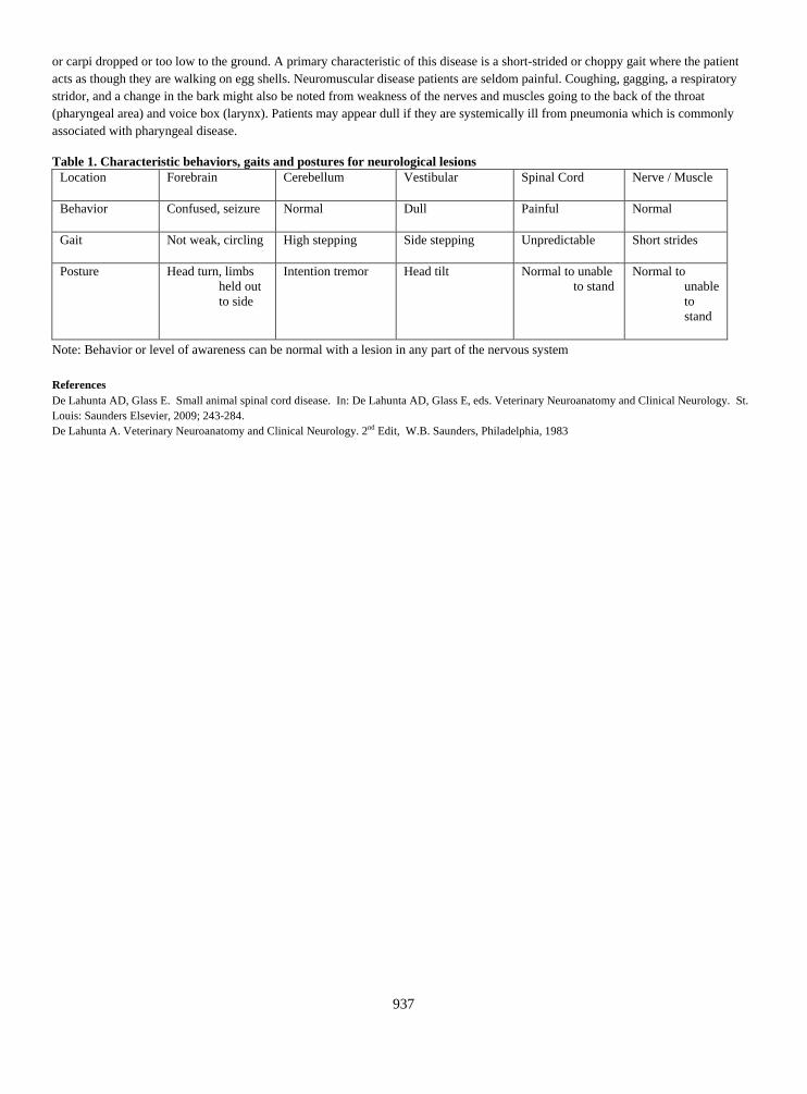
937
or carpi dropped or too low to the ground. A primary characteristic of this disease is a short-strided or choppy gait where the patient acts as though they are walking on egg shells. Neuromuscular disease patients are seldom painful. Coughing, gagging, a respiratory stridor, and a change in the bark might also be noted from weakness of the nerves and muscles going to the back of the throat (pharyngeal area) and voice box (larynx). Patients may appear dull if they are systemically ill from pneumonia which is commonly associated with pharyngeal disease. Table 1. Characteristic behaviors, gaits and postures for neurological lesions
Location Forebrain Cerebellum Vestibular Spinal Cord Nerve / Muscle
Behavior Confused, seizure Normal Dull Painful Normal
Gait Not weak, circling High stepping Side stepping Unpredictable Short strides
Posture Head turn, limbs held out to side
Intention tremor Head tilt Normal to unable to stand
Normal to unable to stand
Note: Behavior or level of awareness can be normal with a lesion in any part of the nervous system References De Lahunta AD, Glass E. Small animal spinal cord disease. In: De Lahunta AD, Glass E, eds. Veterinary Neuroanatomy and Clinical Neurology. St. Louis: Saunders Elsevier, 2009; 243-284. De Lahunta A. Veterinary Neuroanatomy and Clinical Neurology. 2nd Edit, W.B. Saunders, Philadelphia, 1983
938
Nutrition is a Team Sport: Technicians Take Charge!
Julie Churchill, DVM, PhD, DACVN University of Minnesota
St. Paul, MN
Why bother with nutrition? Integrating preventive healthcare into a practice requires commitment and engagement of the entire veterinary team. This commitment and participation in developing a strong preventive program will promote success, optimal pet health, the client-pet relationship and a long term client relationship with the practice. Nutrition is a key component of preventive health care and client interest and belief that great nutrition positively influences their pet’s health is growing. As a result of recent nutrition assessment guidelines from both AAHA and WSAVA the initiative to consider nutrition the 5th vital assessment helped promote nutritionals a standard of care. Clients look to their veterinary team as experts in healthcare as well as nutritional advice. In an AAHA study, 90 % of pet owners want nutritional recommendations yet 15 % perceive they are given one. The AAHA compliance study revealed 7% of the pets that could potentially benefit from therapeutic foods were actually employing that therapy. There is tremendous opportunity for veterinary professionals to take a role otherwise filled with less credentialed. Why is nutrition a team sport? There are many benefits to deploying or adopting a team approach to the practice mission and the veterinary technician is poised to lead the initiative to provide nutritional care for all patients. Group effort increases efficiency by distributing workload and delivering an integrated cohesive message about the importance of nutrition and the value of preventive care. No one can do it all, and when roles are delegated everyone contributes to success and individual motivation increases to accomplish results.
There is great variability in the degree with which nutritional care is incorporated in practices. Some are just getting started, while others may be experiencing some of the predictable pitfalls and challenges. This discussion is intended to be interactive to provide opportunities, strategies resources and tools to consider for improving patient care and client satisfaction by making nutrition a foundation. The intent is to use what would work best in your setting. Elements to develop a practical nutrition program in primary care practice Team based Care-a role for every member (www.everypeteverytime.com)
You can take a self-assessment to see how your practices doing implementing nutrition in patient care. • Define the message • Implement 5th Vital Assessment- every pet every time • Define practice philosophy and training for the entire staff • Tools for the tool kit-
o Develop a written protocol (see example below) o Client education materials o BCS charts o Newsletter, social media
Create the program 1. Develop consensus about nutrition topics and communicate to team members
a. Common client questions b. Team members questions/concerns c. Develop written positions
2. Body weight, Body Condition, Muscle Condition/Body Fat Index a. Choose the scale and score and be consistent
3. Calculate Calories and Food Doses- pet food manufacturers, petobesityprevention.com, usda nutrient database 4. Stock Wellness foods 5. Train team members thoroughly- CE, empower and engage everyone
a. Nutrition training b. Communication training c. Academy of Veterinary Nutrition Technician- nutritiontechs.org
6. Encourage use of foods recommended by practice (offer some not sold) 7. Point person to stay up-to-date on recalls pet food safety current events
a. Update practice website, social media, links to avma, FDA b. Provide list of alternative recommendations
939
8. Provide therapeutic diets Wellness program or package ideas
• Technician managed o Puppy/kitten counseling o Nutrition consultation post every spay/neuter
• Life stage nutrition • Obesity prevention
Nutrition and disease-therapeutic plan • Screened and risks identified- veterinary responsibility • Chronic disease package- follow-up plan • Obesity treatment – include all team members
Nutrition Specialist Referral • Multiple Comorbidities • Severe obesity, aggressive weight loss plan
References/resources www.Everypeteverytime.com. Many tools and documents for assessing a veterinary practice and implementing nutritional care. www.PetNutritionAlliance.org- website with comprehensive nutrition tools for all members of veterinary team, and client education. Baldwin K, Bartges J, Buffington T, et al. AAHA Nutritional Guidelines for dogs and cats. J Am Anim Hosp Assoc. 2010; 46:285-296. WSASA, Nutritional assessment guidelines (2011) http://www.wsava.org/PDF/Misc/WSAVA_GlobalNutritionalAssessmentGuidelines_2011.pdf Nutrition reference manual, Hill’s Pet Nutrition, http://www.everypeteverytime.com/docs/en-us/Pet_Nutrition_Ref_Manual.pdf Pet Nutrition Alliance. AAHA Tips for Implementing Nutrition as a Vital Assessment in Your Practice (2012) http://petnutritionalliance.org/PDFS/PNA_TipsGuide_AAHAproof2.pdf
940
Managing Rodenticide Intoxications Camille DeClementi, VMD, DABT, DABVT
ASPCA Animal Poison Control Center Urbana, IL
While there are several different products that may be used as rodenticides, including strychnine, zinc phosphide, and aluminum phosphide, this presentation will focus on the rodenticides that are most often encountered: anticoagulants, bromethalin, and cholecalciferol. Just as “Kleenex” is commonly used instead of the word tissue, many owners may use the term “D-con” to refer to any rodenticide regardless of the actual brand name. Therefore it is a good idea to ask the owner to bring in the package to verify ingredients. Baits may be formulated as pellets, bars, grains, or meals. Unfortunately rodenticides are not color-coded, so the color of the rodenticide provides no insight into the type of rodenticide. Anticoagulants Warfarin and pindone are short-acting anticoagulants and have short half-lives (<24 hours) compared to the long acting products whose half-lives are up to 6-7 days. Long acting anticoagulants include diphacinone, difethialone, chlorophacinone, brodifacoum, and bromadiolone. First generation anticoagulants include warfarin, diphacinone, chlorphacinone, pindone and valone. Second generation anticoagulants may be longer acting and/or more potent than the first generation anticoagulants and are effective against warfarin-resistant rats. As a general guideline, the minimum toxic dosage of warfarin is >0.5 mg/kg. The minimum toxic dosage of the other anticoagulants is >0.02 mg/kg.
The anticoagulant rodenticides act by competitive inhibition of the enzyme vitamin K1 epoxide reductase thereby preventing the regeneration of inactive Vitamin K1 to its active quinone form. Vitamin K1 deficiency results in depletion of the clotting factors II, VII, IX and X. Because it has the shortest half-life, factor VII it is the first one affected. Depletion of factor VII leads to an elevation of the prothrombin time (PT). PIVKA, the collective term for the precursors of the vitamin K-dependent clotting factors, also becomes increased. The PT may be elevated within 36-72 hours, but the animal is usually still clinically normal. Beyond 72 hours, as other factors become depleted, severe hemorrhage may occur, accompanied by elevations in activated partial thromboplastin time (APTT) and activated clotting time (ACT). In rare instances (e.g. animals with pre-existing bleeding disorders or hepatic disease, etc.) depletion of coagulation factors may occur sooner, resulting in clinical evidence of hemorrhage as early as 24-48 hours following exposure.
In many cases, poisoned animals are not presented to the veterinarian until signs develop. Many patients present with vague clinical signs of lethargy, weakness and anemia without any overt external hemorrhage although some animals may present with frank external hemorrhage from surgical or traumatic wounds, the gastrointestinal tract, or other body orifices (e.g. epistaxis, vulvar bleeding). Hemorrhage into body cavities such as the joints, the peritoneal cavity or the pleural cavity is common. Weakness, pallor, abdominal distention, lameness, swollen joints, dermal bruising, muscular hematomas, dyspnea, labored breathing, or muffled heart sounds are also possible. Bleeding into the brain or spinal cord may result in severe CNS disturbances, paresis, paralysis or acute death. Tracheal constriction due to thymic, peritracheal or laryngeal bleeding may result in severe dyspnea. Clinical pathologic abnormalities may include anemia, thrombocytopenia, hypoproteinemia and decreases in CO2 and pO2.
Diagnosis is based on history, compatible clinical signs and laboratory confirmation of coagulopathy. Differential diagnoses should include congenital and acquired coagulopathies, and other causes of anemia (trauma, etc.). Coagulation panels may aid in the differentiation of anticoagulant rodenticide from other coagulopathies (e.g. disseminated intravascular coagulation, von Willebrand’s disease, Hemophilia A, etc.). Serum chemistry profiles to detect hepatic or other systemic disease that might affect blood clotting are usually indicated. Anticoagulant toxicosis may be worsened in cases of significant hepatic disease due to impaired ability to synthesize coagulation factors and decreased metabolism of ingested rodenticide. Because PT is the first coagulation test to become abnormal in anticoagulant rodenticide toxicosis, it is the best choice for early detection. Elevations in PIVKA may also be used early in anticoagulant rodenticide toxicosis, as normal animals should not have PIVKA present in the circulation. Unfortunately, PIVKA proteins have been shown to be elevated in number of other acquired and congenital coagulopathies
Stabilization of symptomatic animals is a priority. Provide oxygen as needed for dyspnea. Transfusions with whole blood or plasma may be necessary to replace blood and clotting factors. Oxyglobin may also be used.
Decontamination is only effective early (within the first 12 hours). Do not attempt emesis in a bleeding animal. Monitor prothrombin time (PT) between 36-72 hours. Any elevation in the PT warrants full treatment with Vitamin K1. No treatment is indicated if PT remains normal after 72 hours. NOTE: Recent vitamin K1 administration could result in falsely normal PT values because new clotting factor synthesis only requires 6 - 12 hours. Administer Vitamin K1 as needed, or you may opt to begin it prophylactically. Vitamin K1 should not be given intravenously and it is possible to have an anaphylactic reaction when it is given subcutaneously. Oral administration is ideal, because vitamin K1 will be delivered directly to the liver where the clotting factors are activated through the portal circulation. The dose of Vitamin K1 is 3 - 5 mg/kg/day orally. This dose should be divided BID or TID
941
and should be given with a fatty meal to enhance absorption. The average duration of treatment is 14 days for warfarin, 21 days for bromodiolone and 30 days for the others.
Also it is advisable to check a PT at 48- 72 hours following the last dose of Vitamin K1. Vitamin K1 should be continued if the PT is still increased. If possible, avoid the use of other highly protein-bound drugs during the treatment, and instruct the owner to restrict exercise during this time.
Bromethalin Bromethalin uncouples oxidative phosphorylation, resulting in depletion of ATP and loss of energy for sodium-potassium trans-membrane pumps. Intramyelinic edema ensues, characterized by the presence of fluid-filled vacuoles between myelin sheaths. This results in decreased nerve impulse conduction. Baits generally contain 0.01% bromethalin and come in 1.5-ounce packs. Although the literature reports a minimum toxic dose in dogs of 1.67 mg/kg, the ASPCA APCC’s experience indicates that some dogs may show signs at doses as low as 0.9 m/kg. Because of this discrepancy and because treatment once signs have developed is usually unsuccessful, we recommend that decontamination be initiated at doses > 0.1 mg/kg. Cats are considered to be three times more sensitive than dogs, and we recommend decontamination for any cat exposure.
Clinical signs may begin within 24 hours or as long as 2 weeks following ingestion. Earlier onset of signs suggests higher ingested dosage and poorer prognosis. High doses (>2 mg/kg) result in a convulsant syndrome characterized by acute onset of severe tremors, hyperexcitability, seizures, rigidity, opisthotonos, decerebrate posturing, hyperthermia and death within 36 hours of ingestion. Lower doses cause a paralytic syndrome that begins as depression, +/- hyperthermia, progressive paresis originating in the rear and moving cranially and progressive CNS signs. In sub-lethal exposures, signs may arrest at some level of paresis, and the animal may recover gradually over weeks to months or may retain permanent motor impairment. Bromethalin causes spongy degeneration in the white matter of spinal tracts, brainstem, cerebellum, and cerebrum. Electron microscopy demonstrates vacoulation myelin sheaths.
There is no antidote, so aggressive decontamination is critically important. Repeated doses of activated charcoal (every 8 - 12 hours) and cathartic are recommended. Efficacy of activated charcoal in symptomatic animals is poor. Provide supportive care as needed to manage clinical signs including diazepam or barbiturates for seizures. Agents such as mannitol, furosemide, and corticosteroids have been recommended to reduce the cerebral edema, but were of little benefit in reducing the severity of signs in experimental animals. Gingko biloba at 100 mg/kg was shown to reduce the development of cerebral edema and brain lipid peroxidation in rats following a lethal dose of bromethalin. Efficacy of Gingko in other animals has not been studied. Animals exposed at lower doses exhibiting paralysis may recover. The prognosis is extremely poor for animals showing severe signs. Cholecalciferol Cholecalciferol (Vitamin D3) is metabolized in the liver to calcifediol (25-hydroxycholecalciferol). Calcifediol is then metabolized by the kidney to calcitriol (1,25 - dihydroxycholecalciferol). Cholecalciferol increases intestinal absorption of calcium, stimulates bone resorption, and enhances renal tubular reabsorption of calcium. This results in an increase in serum calcium. Prolonged elevation of serum calcium can lead to acute renal failure, cardiovascular abnormalities, and tissue mineralization. The minimum toxic dose of cholecalciferol ranges from 0.5 mg/kg to 3.0 mg/kg. The APCC recommends decontamination at 0.1 mg/kg. One ounce of 0.075% cholecalciferol bait contains 21.28 mg cholecalciferol.
Clinical signs may be delayed in onset and typically occur 18 - 36 hours post ingestion. The most common clincial signs seen with cholecalciferol toxicosis include vomiting, diarrhea, inappetence, depression, polyuria, polydipsia, bradycardia, and cardiac arrhythmias. An initial hyperphosphatemia is often seen within the first 12 hours, followed by hypercalcemia within 24 hours. Hypercalcemic nephropathy develops, resulting in increases in BUN and creatinine. Differential diagnoses for hypercalcemia include juvenile hypercalcemia, hypercalcemia of malignancy, hypoadrenocorticism, hypoadrenocorticism, primary hyperparathyroidism, and calcipotriene toxicosis.
Post mortem lesions seen with cholecalciferol toxicoses include diffuse hemorrhages of the gastrointestinal tract and possible streaking of the renal cortex. Upon cutting, soft tissues of the gastrointestinal tract, heart and kidney lend have a “gritty” feel to the knife. Mineralization and necrosis of gastrointestinal, cardiac, and renal tissues may be seen histologically. Elevated total kidney calcium concentrations may be detected toxicologically.
Aggressive decontamination is recommended for recent exposures. Emesis should be induced, followed by multiple doses (3-4 doses q 6-8 hours) of activated charcoal. Obtain a baseline serum calcium and BUN immediately post-exposure. Monitor serum calcium and BUN every 12-24 hours each day for three days post exposure. If the calcium level remains normal for 96 hrs, no further treatment is be needed. Treat renal effects with supportive care including fluid diuresis 2x maintenance using 0.9% saline or 0.45% saline and 2.5% dextrose. Avoid calcium-containing fluids. Normal saline fluids help to decrease tubular reabsorption of calcium. Pamidronate disodium (Aredia ®) inhibits osteoclastic bone resorption. The dose is 1.3- 2.0 mg/kg as a slow IV infusion over 2 hours. This dose may need to be repeated in 5-7 days. Expect normalization of serum calcium within 48 hours (often within 12-18 hours).

942
The initial expense of pamidronate may be offset by the fact that the animal may be weaned off of IV therapy and sent home once calcium levels normalize. The dose listed is for dogs; one case of pamidronate use in a cat at 1.3 mg/kg had a very favorable outcome.
Oral prednisone at 2-3 mg/kg BID can help lower serum calcium by decreasing its gastrointestinal absorption, lowering bone resorption, and increasing renal excretion. Furosemide at 2.5 to 4.5 mg/kg TID or QID orally, or 5 mg/kg/hr via continuous intravenous infusion has been shown to decrease serum calcium levels. Salmon calcitonin 4-6 IU/kg q 2-3 hours until calcium levels stabilize has also been used. Some dogs become refractory and there is a risk of anaphylaxis. Salmon calcitonin and pamidronate should not be used together, as there is increased risk of soft-tissue mineralization.
Prolonged treatment is often required due to the long half life of Vitamin D3 (> 17 days.) Provide other supportive care as needed during treatment and monitor serum calcium, phosphorous and renal values. A low calcium diet is indicated and the patient should be kept out sunlight as much as possible. Antibiotics may be indicated if prolonged prednisone usage is anticipated.
What should I do if I don’t know what this green stuff is? Endeavor to find the type of bait. Have owner find container. If product was put out by pest control operator, an invoice is required to be left stating which pesticide(s) was/were used. If the rodenticide is still unknown, and the exposure was recent, induce emesis and administer activated charcoal. Repeat doses in case rodenticide is bromethalin or cholecalciferol. Follow decontamination with baseline PT and serum calcium and phosphorus levels. Monitor calcium and phosphorus every 12 hours for 36 hours; if normal, then can discontinue monitoring for cholecalciferol. Monitor PT at 24 and 72 hours; if normal then no treatment for anticoagulants. Educate owner to monitor for neurologic signs (paresis, depression) from bromethalin; signs could be delayed several days.
943
Ticks: What You Don’t Know May Hurt Your Practice
Michael Dryden, DVM, MS, PhD Kansas State University
Manhattan, KS
While often the same products are used to combat ticks as are used to combat fleas, there are substantial differences between flea and tick control. One of the major differences is in the number of species that confront a dog. While there is one predominant flea species that infests dogs in North America, the Cat flea (Ctenocephalides felis), there are at least 10 different tick species that may be encountered. There can be remarkable regional variability in the number and diversity of tick species that infest dogs.1 While practitioners in Hawaii may only deal with one tick species infesting dogs (Brown Dog tick, Rhipicephalus sanguineus), practitioners in New Mexico may encounter three different species, in California six different species and in Nebraska 5 to 6 species.2 This wide diversity in tick species means that ticks occur at different times of the year, are associated with different reservoir hosts and carry and transmit different diseases.
Historically the most common tick recovered from dogs and livestock in the Central U.S. hasbeen Dermacentor variabilis, the American dog tick. However, Amblyomma americanum, the lone star tick has become of increasing importance and over the past few decades there has been a change in the distribution and abundance of certain tick species in North America.1, 3-6 Two of the best documented are Amblyomma americanum and Ixodes scapularis.4-6 Since both these ticks are important vectors of human and animal pathogens these changes in distribution and abundance have had a marked effect upon both human and animal health. Various factors have contributed to tick population movement including: changes in agricultural practices; reforestation, wildlife conservation, relocation and restocking; climate fluctuations and decreased environmental pesticide application.
Specific factors that have contributed to the increased range of A. americanum include increased habitat via reforestation and its wide host range that includes deer, small mammals, birds and man5,6. The White-Tailed Deer is considered a preferred host for A. americanum, and all life stages will feed successfully upon White-Tailed Deer. Another species that utilizes similar habitats and is an excellent host for larvae and nymphs is the wild turkey. Areas with high White-Tailed Deer and wild turkey populations can have remarkably large populations of A. americanum. Similar to A. americanum the distribution of I. scapularis is linked to the distribution and abundance of the white-tailed deer.4
Ixodes scapularis, the Black-legged Tick, (Deer Tick or Lyme Disease Tick) is an inornate tick without eyes or festoons. Larvae are small and often difficult to see. They are about 0.5mm long, flat, six-legged, and nearly translucent. Nymphs are approximately 1mm long and darker in color. Unfed males are approximately 2mm long and unfed females about 2.5mm. There are considerable morphologic differences between male and female Ixodes. Males are dark brown, almost black, with shorter palps than females. Females have longer mouthparts and appear two-toned. In the non-engorged female the inornate dorsal shield covers the anterior 1/3rd of the body, leaving the orange-brown posterior portion of the body exposed. Ixodes scapularis is widely distributed in the Eastern and Central U.S. in at least 35 states.7,8,9 Its distribution is from Florida to Maine, west into far eastern South Dakota, and south through eastern Kansas into central Texas. Ixodes scapularis is also located in central and eastern Canada.
While white-tailed deer are widely distributed across the central and eastern U.S., the abundance of I. scapularis is not always directly related to the abundance of the deer populations. Tick populations can vary markedly across a region due to soil type, moisture and forest cover. In the north central U.S., I. scapularis were more numerous in areas with a deciduous forest canopy, and where soil textures were classified as sandy or loam-sand.10
Seasonal activity varies by geographic region, but larval activity is generally highest in August and September. Larvae attach to and feed on a wide variety of small mammals, including mice, chipmunks and shrews. Larvae also feed on birds and lizards. The white-footed mouse (Peromyscus leucopus) is of particular importance in the tick life cycle and disease transmission, because it serves as a good host for larval I. scapularis and it is a major reservoir of Borrelia burgdorferi.
Immature ticks typically engorge for 2 to 4 days before dropping off to molt in moist protected areas such as under leaf litter in forested habitats. Larvae over-winter and then molt to nymphs in the spring. Nymphs will feed for 3 – 4 days on a variety of hosts including mice, squirrels, chipmunks, raccoons, opossums, skunks, shrews, cats, birds, and humans. Nymphs occur primarily from May through July in the North. Adults occur most commonly from October through December. Adults that do not find a host will quest again, typically from March to May. Adults feed for 5 – 7 days, primarily on white-tailed deer, but also on bobcats, cattle, coyotes, dogs, foxes, horses, humans, opossums, raccoons and other mammals.
Ixodes scapularis is the vector of Borrelia burgdorferi (Lyme Disease) in the central, upper midwest and northeast U.S., it is also the vector of Anaplasma phagocytophilum (Human Granulocytic Ehrlichiosis), and Babesia microti (Human Babesiosis). Ixodes scapularis may also cause tick paralysis.
While recent pharmaceutical advances have been made in the control of flea reproduction, such advances in the area of tick control are lacking. With the exception of the brown dog tick Rhipicephalus sanguineus, our ability to manage tick reproduction is limited, if
944
not almost non-existent. As discussed previously in most flea infestations we have the opportunity to control flea reproduction by either killing fleas before they can reproduce or killing flea eggs. However, it is not just because we have effective residual insecticides, insect growth regulators or insect development inhibitors that we are successful, it is also due in large part to the fact we can often target the primary reproductive host, the flea infested dog or cat. And interestingly, failures in flea control often occur when flea infested feral pets or flea infested urban wildlife invade the owners’ yards.
But when dealing with most 3-host ticks the problem is that the majority of the reproducing ticks are not on the dogs or cats, but on their native wildlife hosts. Since we are limited in our ability to manage ticks on wildlife, reinfestations are a common occurrence and protracted use of acaracides is routine in many areas. As mentioned earlier one tick species that is an exception is R. sanguineus. While this tick is a 3-host tick, practically it can be said to function as a 1-host tick, since all life stages prefer to feed upon dogs. Therefore, we have the opportunity through rigorous application of acaracides to kill female ticks before they reach repletion.
Since tick control can be extremely difficult and because they are vectors of a variety of bacterial and protozoal diseases, veterinarians should have an understanding of the ecology of the tick(s) encountered in the area in which they practice. Veterinarians need to be educated on the various aspects of tick ecology, disease transmission and control methodologies so that they can then educate their staff and pet owners.
Numerous studies demonstrate the high level of efficacy of the various acaracides but the residual activity is rarely 100% and the efficacy of products varies between and as well as within species, even in the same laboratory.11-18 Evaluations of acaracides under natural or field conditions further illustrates that while efficacy is good it is not 100%.
In a field efficacy trial conducted in Kansas U.S.A, an imidacloprid (8.8% w/w)-permethrin (44.0% w/w) formulation was evaluated on dogs against naturally occurring populations of Amblyomma americanum. When dogs were walked in a naturally tick infested environment the 48-hour post-exposure efficacy of imidacloprid-permethrin formulation was 93.5%, 98.9%, 94.6%, 94.1% and 96.6% on days 3, 7, 14, 21 and 28 respectively, post-treatment.15
Variation in product efficacy occurs. In two studies conducted at K-State, different results were found when evaluating the efficacy of acaricides against Dermacentor variabilis infestations in dogs from two different regions of the USA.11,14 In the first study, the efficacy of imidacloprid–permethrin and fipronil–(s)-methoprene formulations were evaluated against a D. variabilis isolate from California. The 48-h post-infestation efficacy on day 30 post-treatment was 92.0% and 83.2%, respectively, for the imidacloprid–permethrin and fipronil–(s)-methoprene formulations. In the second study, the 48-h post-infestation efficacy on day 30 for the imidacloprid–permethrin and fipronil–(s)-methoprene formulations against a D. variabilis isolate from Oklahoma was 17.5% and 75.7% respectively. New isoxazoline molecules (afoxolaner and fluralaner) are now offering the first ever oral approach to tick control. These compounds have demonstrated excellent efficacy against a variety of tick species.16-17
While product efficacy is often excellent in most studies, significant variation in efficacy can occur and 100% control is rarely achieved. Therefore it can be expected that under natural conditions in areas where dogs are being frequently exposed to ticks pet owners will see ticks on treated dogs. We might also expect that efficacy in real world situations might be lower due to such factors as bathing and swimming, differences between dog breeds and haircoat types and frequency and correctness of product application.
Since 100% tick kill is not always achievable, perceived efficacy of acaracides may be directly related to the numbers of ticks to which dogs are exposed. If a dog is treated with one of these highly efficacious acaracides and encounters just a few ticks it is likely all those ticks will be killed. However, if tick exposure is considerably larger, we can expect a few ticks to be observed on these dogs and pet owners may perceive a lack of efficacy. Therefore in areas where tick populations are increasing the perception may be that the products are not as effective as they once were.
Pet owners often view tick infestations of their pets differently than flea infestations. Whether this is due to concerns about tick transmitted diseases or simply a phobia, the presence of a couple of ticks on the pet often elicits a more pronounced negative reaction than the presence of a couple of fleas. A 95% effective flea product may provide great client satisfaction while a similarly effective tick product may be perceived as a failure. Therefore it is not uncommon that label recommended application of a product does not appear to control the problem. This may be real or perceived, based upon pet owner expectations of product performance. Given pet owner concerns, the fact that ticks transmit serious diseases and lack of 100% efficacy; occasionally additional control measures are needed. If additional control measures are deemed necessary, pet owners need to be educated as to why additional control measures are necessary and notations made in the pet’s record.
One of the most common practical attempted solutions to this problem in dogs is to increase the frequency of application. Here increased residual efficacy is the expected outcome, since you are increasing the residual acaracide levels with the shorter application intervals. EPA registered acaracides should be used according to their product labels. Additionally, with many 3-host ticks, destruction of tick habitat can reduce exposure pressure. Areas that serve as refuge for ticks and wild mammals such as grass, weeds, and brush piles, between runs and along buildings, can be eliminated or treated with an approved acaracide.
In some situations, especially in tropical and subtropical regions and in climate controlled kennels brown dog ticks may infest buildings with ticks crawling up walls, curtains and throughout the home or kennel.19 In these situations acaracides may need to be sprayed indoors into cracks and crevices, behind and under furniture or cages and along walls and the ceiling. Following application,
945
make sure the acaracide is dry before you allow animals or humans back into the premises to minimize toxicity problems. Finally, restricting pet access from tick-infested environments may be necessary.
It is apparent that the range and local density of certain tick species has increased in many areas. Whatever the factors it must be recognized that tick infestation pressure may be much higher and associated tick transmitted diseases may be more prevalent in some locations today than in the past. The increase in tick populations means that pets are encountering ticks more frequently, are exposed to more ticks per encounter and clients may be seeing more ticks on their pets than in the past. Since tick products do not kill or repel all ticks instantly, clients may get the false impression that the products are not performing as well as in the past. These situations necessitate that veterinarians set client expectations, before clients set their own unrealistic expectations of control.
References Dryden MW, Payne PA. Biology and Control of ticks infesting dogs and cats in North America. Vet Ther 2004; 26:2-16. Cortinas R, Spomer SM. Occurrence and County-Level Distribution of Ticks (Acari: Ixodoidea) in Nebraska Using Passive Surveillance. J. Med. Entomol. 51(2): 352-359 (2014) Cortinas R, Spomer SM. Lone Star Tick (Acari: Ixodidae) Occurrence in Nebraska: Historical and Current Perspectives. J. Med. Entomol. 50(2): 244Ð251 (2013) Estrada-Pena A. Increasing habitat suitability in the United States for the tick that transmits Lyme disease: A remote sensing approach. Environ Health Perspectives 2002; 110:635-640. Childs, J.E.; Paddock, C.D.: The ascendancy of Amblyomma americanum as a vector of pathogens affecting humans in the United States. Ann Review Entomol 2003;48:307-337. Paddock CD, Yabsley MJ. Ecological havoc, the rise of white-tailed deer and the emergence of Amblyomma americanum-associated zoonoses in the United States. Curr Top Microbiol Immunol 2007;315:289-324. Keirans JE, Hutcheson HJ, Durden LA, Klompen JS. Ixodes (Ixodes) scapularis (Acari:Ixodidae): redescription of all active stages, distribution, hosts, geographical variation, and medical and veterinary importance. J Med Entomol 1996;33:297-318. Dennis DT, Nekomoto TS, Victor JC, Paul WS, Piesman J. Reported distribution of Ixodes scapularis and Ixodes pacificus (Acari: Ixodidae) in the United States. J Med Entomol 1998;35(5):629-38. Rand PW, Lubelczyk C, Holman MS, Lacombe EH, Smith RP Jr. Abundance of Ixodes scapularis (Acari: Ixodidae) after the complete removal of deer from an isolated offshore island, endemic for Lyme Disease. J Med Entomol 2004;41(4):779-84. Guerra M, Walker E, Jones C, Paskewitz S, Cortinas MR, Stancil A, Beck L, Bobo M, Kitron U. Predicting the risk of Lyme disease: habitat suitability for Ixodes scapularis in the north central United States. Emerg Infect Dis 2002;8(3):289-97. Dryden MW, Payne PA, Smith V, Hostetler J. Evaluation of an imidacloprid (8.8% w/w)--permethrin (44.0% w/w) topical spot-on and a fipronil (9.8% w/w)--(S)-methoprene (8.8% w/w) topical spot-on to repel, prevent attachment, and kill adult Rhipicephalus sanguineus and Dermacentor variabilis ticks on dogs. Vet Ther 2006; 7(3):187-198. Dryden MW, Payne PA, Smith V, Hostetler J. Evaluation of an imidacloprid (8.8% w/w)--permethrin (44.0% w/w) topical spot-on and a fipronil (9.8% w/w)--(S)-methoprene (8.8% w/w) topical spot-on to repel, prevent attachment, and kill adult Ixodes scapularis and Amblyomma americanum ticks on dogs. Vet Ther 2006; 7(3):173-86. Rugg D, Hair JA. Dose determination of a novel formulation of metaflumizone plus amitraz for control of cat fleas (Ctenocephalides felis felis) and brown dog ticks (Rhipicephalus sanguineus) on dogs. Vet Parasitol 2007; 150:203-208. Dryden M, Payne P, McBride A, Mailen S, Smith V, Carithers D. Efficacy of Fipronil (9.8% w/w) + (S)-Methoprene (8.8% w/w) and Imidacloprid (8.8% w/w) + Permethrin (44% w/w) against Dermacentor variabilis (American Dog Tick) on Dogs. Vet Ther 2008; 9(1):15-25. Dryden MW, Payne PA, Smith V, Hostetler J. Efficacy of imidacloprid (8.8% w/w) plus permethrin (44% w/w) spot-on topical solution against Amblyomma americanum infesting dogs using a natural tick exposure model. Vet Ther 2006; 7: 99-106. Mitchell EB, Dorr P, Everett WR, Chester TS, Larsen D. Efficacy of afoxolaner against Dermacentor variabilis ticks in dogs. Vet Parasitol 2014, 201(3-4):220-222. Wengenmayer C, Williams H, Zschiesche E, Moritz A, Langenstein J, Roepke R, Heckeroth AR. The speed of kill of fluralaner (Bravecto) against Ixodes ricinus ticks on dogs. Parasit Vectors. 2014, 18;7(1):525. Doyle V, Beugnet F, Carithers D. Comparative efficacy of the combination fipronil-(S)-methoprene and the combination permethrin-imidacloprid against Dermacentor reticulatus, the European dog tick, applied topically to dogs. Vet Ther 2005; 6:303-310. Dantas-Torres F. The brown dog tick, Rhipicephalus sanguineus (Latreille, 1806) (Acari: Ixodidae): from taxonomy to control. Vet Parasitol 2008; 152(3–4):173–85.
946
Video Lessons: Be an Exam Room Hero
Sheila Grosidier, BS, RVT Veterinary Management Consultants
Evergreen, CO
Take a moment and ask yourself these questions in regard to your veterinary practice: • Are you certain that clients are being offered a consistent standard of care? • Do team members have the tools and resources to meet the clients' needs? • What future training do you see your team needing to best grow the business? • How does your team best learn? • What training have you specifically done in the past that has targeted client interaction on an individualized basis?
While there is little argument that costs are rising for training clinic team members, significant debate does exist over what methods of training will ensure consistency, enable the adoption of new skills, and create an environment that allows team members to coach themselves to achieve greater performance. When done correctly, recording employee/client interactions on video for one-on-one review can be one of the most valuable training tools available to change ineffective behavior into consistently productive client communication.
This article will cover a number of essential components that should be considered and implement in order to realise the full potential of video coaching in practice. Communication A 2005 survey of the American Management Association identified that more than half of employers use video surveillance as a way to decrease theft and violence1, while only 16 per cent of those businesses use that information for evaluating employee performance.2
The success of this process will hinge on clear communication with your team members, assuring them that this will not be a covert operation but rather an openly acknowledged training tool for reviewing staff and client interactions and subsequent coaching for future performance. This is not about looking for mistakes; it’s about looking for ways to improve.
While your practice may already be using video technology as a method of monitoring for theft-prevention or safety, video coaching differs in its ultimate aim, and these differences include how the information will be captured, what will be done with the video and how it can change your practice.
Each of us has had those moments when we replay a conversation with a client and wonder, ‘Could I have done something differently?’ Seeing the entire interaction can provide clear answers – after all, a picture is worth a thousand words. So remember that the team will need to be reassured that the main goal of filming is for those images to help them choose just the right words in order to communicate more effectively with clients in the future.
Rules for everyone Because there may be some initial resistance from the team about being recorded on video, it's important to establish clear, steadfast rules that will be outlined and followed so as to reassure the team that this is a training tool to be used in a positive manner. Recording an exam will proceed only after the client has been advised. A consultation could begin, for example, by saying, ‘Ms. Jones, we are videotaping today for training purposes. Is that okay with you?’ Usually clients are glad to give consent, but if the client does not approve, then the camera is to be turned off. Signs must be posted to alert clients and remind team members that the practice is using video cameras, and that it is not a secret activity. While there may be a sense of natural apprehension at the onset of using video, over time, team members will likely have to be reminded that the video is running—it will become part of the landscape. Establish that the video will be used as a tool for training only, not as surveillance. Setting up publicly Implement a clear policy that outlines how the video will be recorded and evaluated to ensure maximum benefit to the employees as well as to the practice. To get the most out of the process, this should not be a one-time activity. Plan to evaluate it monthly and set goals with a timeline for each team member, then evaluate their success. A policy example can be found in Box 2.
1 http://veterinaryteam.dvm360.com/firstline/article/articleDetail.jsp?id=622344 2 http://www.nolo.com/legal-encyclopedia/workplace-cameras-surveillance-employer-rules-35730.html
947
One-on-one coaching Be sure to have your team members assess themselves before their review with their supervisor. It is likely that the team members will readily pick up on how to improve and know what they need to do before the review. This will help them feel more confident ad supportive of your coaching over time. Reinforce this action; point out what they are doing on their own to improve and how that not only directly impacts the well-being of pets, client satisfaction and the health of the practice, but also enhances their skills. This is where an incentive program can accelerate results. The rock star reel Did you just watch Erin give a perfect explanation of why your pet needs to have a senior profile? Did you see how amazing Dr. Sanchez was in her response to an angry client? Videos are amazing tools for training new staff. Ask your team members if they would be comfortable sharing their expertise by showing how to perform at that star level in such special videos. Creating a collection of these interactions is incredibly instructive to new employees. Positive and productive Your team members will probably be concerned that this will be a negative experience that consists of looking for what they are doing wrong. Demonstrate that it’s about building on their current skills and creating positive reinforcement. Start the discussion by asking them to identify what they did well and what they would do differently. Focus on the interaction, not the person. See Table 1 for additional recommendations to make coaching a positive experience. Summary To change ineffective behavior, the first step is to recognize that it is not working and clearly understand what will work. Video can dramatically depict how a team member is communicating with clients at that essential point: when they're in your exam room. Don’t leave it up to chance; leave it up to training.
BOX 1: The benefits of using video for coaching in veterinary practice • Team members can specifically review the actual interaction with each client, not simply go on what they remember
about it. • Video can be viewed and compared "back-to-back"? to see improvement over time. • Team members can review an interaction multiple times focusing on things such as their body language, word choice,
speech volume and cadence and find something different upon each viewing. • See a complete visit from the client’s perspective, not just the team member’s part. Set up the camera to show the entire
exam room. • Find out what clients do in your exam room when you are not present, then think of what you could do to make this a
productive time for them and you. BOX 2: Example policies for team video coaching
• The practice utilizes video as an individual staff coaching tool to evaluate interactions with clients exclusively in the exam room.
• Recording will take place in identified exam rooms on specified dates and times. All exam room assistants or technicians, receptionists, kennel assistants, and veterinarians will be expected to participate, as the entire visit in the exam room will be recorded. The video camera is to be set up correctly so the entire room will be visible.
• All staff who greet clients and guide them into the exam room are to state that the visit is being recorded and request the client's permission (for example, “Mrs. Jones, for training purposes we are recording this on video today—is that okay?”). If the client declines, the video camera is to be turned off.
• Team members will have opportunity to review their video and pick which of the clips they would like to review with their supervisor. Each team member will use an evaluation checklist and submit it to the supervisor prior to the review. (See checklist examples in figures 1 and 2.)
• The supervisor will review the clip one-on-one with the team member and complete the checklist for comparison with the team member’s checklist. An agreed-upon goal will be established for the next videotape session.
• An incentive will be included, for example, all team members who complete four video reviews in a 12-month period will receive a $100 bonus and be entered into an annual drawing for a paid day off at a spa.
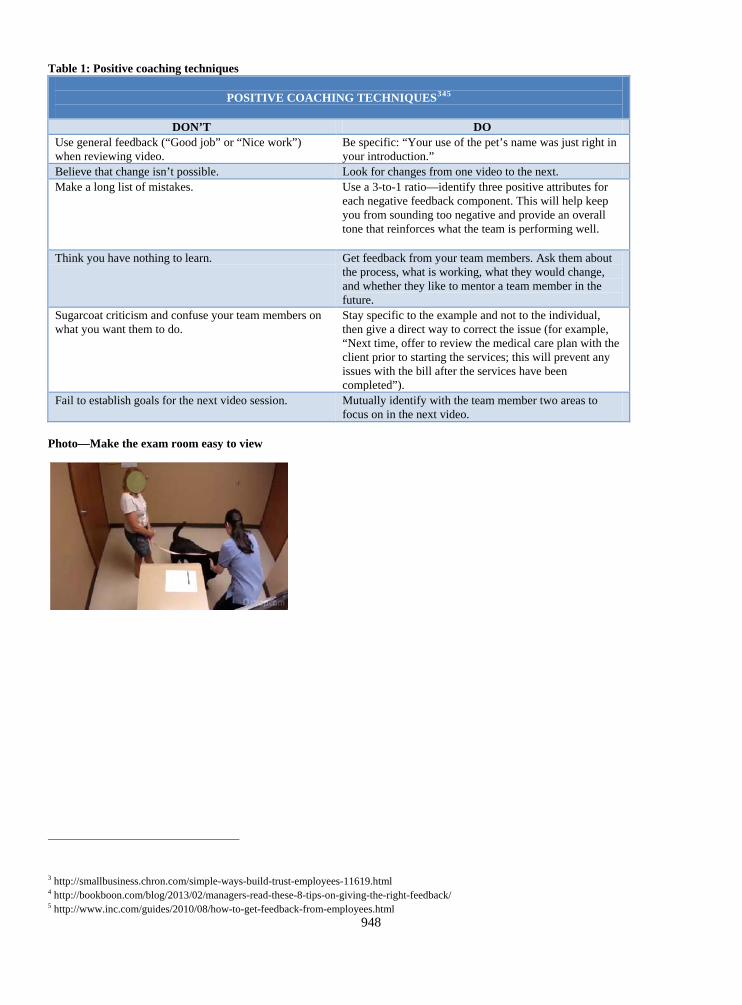
948
Table 1: Positive coaching techniques
POSITIVE COACHING TECHNIQUES345
DON’T DO Use general feedback (“Good job” or “Nice work”) when reviewing video.
Be specific: “Your use of the pet’s name was just right in your introduction.”
Believe that change isn’t possible. Look for changes from one video to the next. Make a long list of mistakes. Use a 3-to-1 ratio—identify three positive attributes for
each negative feedback component. This will help keep you from sounding too negative and provide an overall tone that reinforces what the team is performing well.
Think you have nothing to learn. Get feedback from your team members. Ask them about the process, what is working, what they would change, and whether they like to mentor a team member in the future.
Sugarcoat criticism and confuse your team members on what you want them to do.
Stay specific to the example and not to the individual, then give a direct way to correct the issue (for example, “Next time, offer to review the medical care plan with the client prior to starting the services; this will prevent any issues with the bill after the services have been completed”).
Fail to establish goals for the next video session. Mutually identify with the team member two areas to focus on in the next video.
Photo—Make the exam room easy to view
3 http://smallbusiness.chron.com/simple-ways-build-trust-employees-11619.html 4 http://bookboon.com/blog/2013/02/managers-read-these-8-tips-on-giving-the-right-feedback/ 5 http://www.inc.com/guides/2010/08/how-to-get-feedback-from-employees.html
949
Video Lessons: Technician Tools that Really Work
Sheila Grosidier, BS, RVT Veterinary Management Consultants
Evergreen, CO
Even pets are going electronic these days with an iPod program that will track medical information such as vaccinations, medications and upcoming appointments. So, why aren't you taking advantage of one of the strongest assets a veterinary practice can possess? Clients expect competency. Demonstrating your usage, understanding and ability to embrace technology for the benefit of their pet is vital. Whether you are concerned about team member productivity, implementing effective marketing programs, or placing your practice in an advantageous position online, it's time for you to click your way to success. Here are 8 compelling reasons why the technology superhighway should be your onramp to practice success! Convert from paper files to electronic files
The costs of staying with paper files • How much of your time is wasted in preparing files, filing, retrieving and looking for medical records? • What other wonderful things our receptionists could be doing if they did not have to "play" with our files. • Do you get a headache from trying to read your own (or someone else's) hand-writing in a record? • What the actual cost of your files (paper and space to store the records)? • What does it costs to write the same information over and over (time)?
Automation creates a WIN-WIN! • Recruiting and keeping quality team members has become one of the most time consuming and frustrating challenges
for practices. • There is also frustrating for team members who want to have a balance between challenge and opportunity, and minimal
redundant tasks. • You as an owner want to maximize productivity. Your team members want to be engaged in activities that directly help
pets live longer healthier lives. • Reduce redundant tasks.
o Take advantage of every opportunity to use team members where that personal interaction with the client and pet care cannot be realized any other way but with a team member.
o Ask your team members to identify 10-12 tasks that they do during the day that take up the most amount of time, then consider how you could automate these tasks.
o The savings on time, resources and quality of life are just some of the benefits you will all experience!
Create an online presence First, look at your website
• Do you have a web site? If you do have a web site, are you proud of it? • In today's era, web sites are no longer an option, but a necessity. • What does your web site look like?
o Professional image, o Pictures of your doctors and bios, o Virtual tour of your practice.
To be competitive in today's environment, it is imperative for a practice to have a shop site on their web site • Select you shop site provider carefully. • What products will you offer, and at what prices?
o 3rd party inventory. Make it convenient for clients
• After converting to an electronic medical record system, your veterinary software program should allow your clients to request an appointment via e-mail.
o Available 24-7 o Receptionist can review requests the following day and respond back to the client with a set appointment time.
• If you choose, clients can also access information about their pet -reminders that are due, medications the pet is on and even a "family album" of the pet's pictures.
Get some satisfaction • Want to know what clients really think? Ask them!
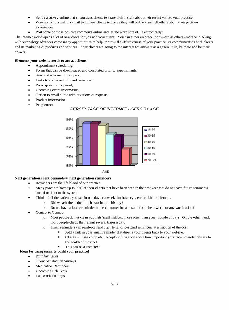
950
• Set up a survey online that encourages clients to share their insight about their recent visit to your practice. • Why not send a link via email to all new clients to assure they will be back and tell others about their positive
experience? • Post some of those positive comments online and let the word spread…electronically!
The internet world opens a lot of new doors for you and your clients. You can either embrace it or watch as others embrace it. Along with technology advances come many opportunities to help improve the effectiveness of your practice, its communication with clients and its marketing of products and services. Your clients are going to the internet for answers as a general rule, be there and be their answer. Elements your website needs to attract clients
• Appointment scheduling, • Forms that can be downloaded and completed prior to appointments, • Seasonal information for pets, • Links to additional info and resources • Prescription order portal, • Upcoming event information, • Option to email clinic with questions or requests, • Product information • Pet pictures
PERCENTAGE OF INTERNET USERS BY AGE Next generation client demands = next generation reminders
• Reminders are the life blood of our practice. • Many practices have up to 30% of their clients that have been seen in the past year that do not have future reminders
linked to them in the system. • Think of all the patients you see in one day or a week that have eye, ear or skin problems…
o Did we ask them about their vaccination history? o Do we have a future reminder in the computer for an exam, fecal, heartworm or any vaccination?
• Contact to Connect o Most people do not clean out their 'snail mailbox' more often than every couple of days. On the other hand,
most people check their email several times a day. o Email reminders can reinforce hard copy letter or postcard reminders at a fraction of the cost.
Add a link in your email reminder that directs your clients back to your website. Clients will see complete, in-depth information about how important your recommendations are to
the health of their pet. This can be automated!
Ideas for using email to build your practice! • Birthday Cards • Client Satisfaction Surveys • Medication Reminders • Upcoming Lab Tests • Lab Work Findings
951
• Seasonal Health Alerts • New Pet Information • Upcoming Education • Target Marketing • Appointment Follow-Up
List of automated reminders • Vaccines • Lab Tests • Diets • Medications • Recommended Testing • Flea/Heartworm Refills • Chronic Medication Refills • New Services • Tests needed due to change in age or condition • Grooming • Boarding
Track your success
• What would you think of a doctor who made a diagnosis without doing a complete physical exam? • Lack of client compliance with recommendations is a common topic of discussion among practice owners. How can you
improve this? • Many practice owners decide they want to put into place a program and process that will increase client visits, improve
client transaction values and assure client service satisfaction. However, they do not know what their current status is in these components of the business.
• Wouldn't it be nice if you could turn on your computer and at a glance see how your practice is doing from a practice management point of view?
o Veterinary Software o Graph and communicate with team!
Key indicators to track • Per Client Transaction (ACT) • Per Doctor Transaction (PCT) • Number of New Clients (& where they originate) • Number of Dentals/Recommendations • Number of Senior Profiles/Recommendations • Inventory Costs/Gross Revenue • Staffing Costs/Gross Revenue
Are you ready to rev up your practice, electrify your clients electronically and leverage your team by reducing redundancy and increasing face to face time with clients? The proficiency of your practice will include embracing digital options throughout the practice. Start with these 8 options and you will see solid and lasting improvements to your practice.
952
Video Lessons: Making the First 90 Days Count- in any Job
Sheila Grosidier, BS, RVT Veterinary Management Consultants
Evergreen, CO 10 ways to make new associates feel welcome
1. R-e-s-p-e-c-t It’s not just a word, it’s what you do. Treat them with respect and model the behavior you expect from them with every employee. Part of respect is praise and feedback; let them know in public when they do well and in private when there’s a concern.
2. Leadership Associates want to know they are on the right path and there is a plan in the practice for moving forward. They want to belong to something bigger than themselves and know that someone they can trust is in charge.
3. Empowerment Allow your Associates to make decisions and to share their ideas with you. While they may not always make the right decisions, they need to know you will support them when needed. Consider what you learned when you made a mistake and encourage them to make decisions.
4. Make it fit Send the new associate out to lunch with different departments in the hospital; one day with the veterinary technicians, one day with the receptionists and then lunch with the kennel and exam room teams. Have them talk about the successes of the practice and how we make a difference in the lives of pets every day.
5. Open it up Have an open house or reception and invite your best clients to meet the new associate. Nothing says welcome like clients who will tell your new employee how wonderful the practice is from the client’s point of view.
6. Expectations Ask them what their expectations are in an employer. Let them know clearly how they will be evaluated and the timeline for performance evaluations.
7. Be proud Put an ad in the paper welcoming the new associate. Create a flyer that you can give to clients in the practice telling them about the wonderful addition to the practice. Post their picture and biography in the exam rooms.
8. Mentor Assign a mentor, someone who can assist them, meet with them regularly and help them to integrate into the practice.
9. Time Set up regular times to meet and talk about cases, comments, and concerns. Encourage the new associate to ask questions when they have them, but also make time so you can demonstrate your commitment to them; and remember, you can’t change the tire at 40 mph, slow down and take the time to talk about it.
10. Make sure there are no misunderstandings Everything should be put in writing, make sure you have an employment contract, job description and policy manual. Go over these with your new associate.
11. Walk your talk Make sure you set the example, not only medically but in your actions. Be to meetings on time, treat other employees with respect, get to work on time. Remember that any successful business starts from the top.
953
Five Everyday Eye Diseases Amy Hunkeler, DVM, DACVO, MBA
Eye Care for Animals Overland Park, KS
Veterinary ophthalmologists rely heavily on the skills of a knowledgeable and well-trained veterinary technician or veterinary assistant. Their role begins with patient care and customer service. It most certainly includes recognizing common ophthalmic conditions, assisting in diagnostic testing, and explaining the recommended treatment plan or surgery to clients.
In this session we will review 5 commonly encountered ophthalmic conditions and help the attendee become more familiar with the clinical signs, definitions, differentials, treatments and pitfalls to medical or surgical management. Entropion Entropion is an inward rolling of the eyelid(s) and eyelashes such that they rub on corneal and conjunctival tissues causing discomfort and potentially corneal ulceration. The diagnosis is made during physical examination and is often hard to overlook because the patient is quite blepahrospastic (painful closure of the eyes, aka squinting) with excess facial wetting. Patients can be affected unilaterally or bilaterally, inferiorly or superiorly, laterally, medially or centrally. Dogs are more likely than cats to show entropion, while puppies and juveniles are more commonly diagnosed than senior dogs. It is important to determine if the condition is primary or secondary to some other condition and then address the root cause. Primary entropion is a surgical disease and correction is undertaken when the animal is at a mature size. Surgical approach varies by the severity and location of the entropion, but for many veterinarians a wedge excision or Hotz-Celsus can be effective. Puppies are managed with temporary eyelid everting sutures while they grow (it may take 2, 3 or 4 procedures to maintain eversion over several months.) It is important to diagnose and treat a patient with entropion so the cornea can be protected from trauma. Prolapsed gland of the nictitans The condition commonly known as “Cherry Eye” describes the pink to red swelling of tissue seen at the medial canthus (middle corner) of one or both eyes. There are more medically appropriate titles, such as, prolapse of the gland of the nictitating membrane, but ‘Cherry Eye’ seems to be used by veterinarians, ophthalmologists and owners on a routine basis.
Prolapse of the gland occurs most commonly in young dogs of any breed or gender. The occasional cat will be affected and typically it will be a Burmese cat. While the cause of a prolapse isn’t fully understood, current science suggests a weakness in the soft-tissue attachments between the nictitating membrane and peri-orbital tissue. Regardless, a nictitans prolapse is non-painful, although both color and swelling become more pronounced the longer the conjunctiva and gland are exposed.
It is best veterinary practice to surgically replace the gland into its normal anatomic position. Excision of the gland is considered by most veterinarians to be a sub-standard practice, bordering on malpractice, based on the glands’ role in producing tears. The nictitans gland is responsible for up to 40% of aqueous portion of the tear film. Excision of the gland places patients at risk of developing keratoconjunctivitis sicca (dry eye.) There are at least a dozen surgical approaches employed to successfully correct a nictitans prolapse; my personal preference is a modified pocket technique. Keratoconjunctivitis sicca (KCS) Keratoconjunctivitis sicca is known most commonly as “Dry Eye” and is perhaps being underdiagnosed in your clinic. In severe cases, diagnosis can be made on the basis of clinical signs, but it is more accurately diagnosed (and diagnosed earlier) when tear production is measured, via a Schirmer Tear Test (STT), during routine annual examinations. A STT reading of 15mm wetting /60 seconds or less qualifies as a diagnosis of KCS. This condition is identified most commonly in dogs, but cats can also be affected. Clinical signs include reduced corneal luster, ocular discharge (often thick, grey-green and tacky), conjunctival hyperemia and blepharospasm. Any age, breed or gender can be affected, but middle-aged and female are slightly over represented and usually there is bilateral disease.
Treatment of dry eye consists of symptom management employing a combination of medications and supportive care products to stimulate tear production, reduce inflammation, and remove ocular debris all while protecting the corneal and conjunctival surfaces. Therapy is always life-long. Failure to treat leads to discomfort, dense corneal scarring and sometimes deep corneal ulcers. We discuss surgical options with motivated clients. This surgery is called a parotid duct transposition (PDT) and the goal is substituting saliva for the missing aqueous portion of tears. While not a prefect replacement saliva can be an alternative if / when medication is not satisfactory. Corneal ulceration Corneal ulceration is diagnosed in both dogs and in cats, however they have different etiologies and treatments. Ulcers are extremely painful due to the density of nerve endings that lay just under the epithelium, so it is important to include pain management in your
954
treatment plan. Ulcers can be divided into many categories, but for the sake of simplicity we will discuss just two: Superficial Ulcers and Deep Ulcers. Superficial corneal ulcers Superficial Corneal Ulcers are also known as indolent erosions, boxer ulcers and non-healing ulcers; it is most appropriate to use the term: Spontaneous Chronic Corneal Epithelial Defect (SCCED.) This is a condition seen in dogs, it is a different condition when identified in cats and should not be managed in the same manner. SCCED’s are most often diagnosed in middle-aged larger breed dogs and they present with blepharospasm, epiphora, discharge, conjunctival hyperemia and retention of fluorescein stain only on the surface of the eye. The hallmark feature is the failure to heal, in spite of appropriate medical management with a topical anti-biotic, an oral anti-inflammatory, and lubrication, plus an e-collar to prevent rubbing. If the lesion fails to heal in a timely manner (<2 weeks), then intervention is needed to facilitate healing. Treatment can include debriding the loose epithelium followed by a keratotomy is performed to disrupt the surface barrier, which is preventing epithelialization. After an appropriately performed procedure and in conjunction with medication, most corneas will heal in less than a week. Post-procedure therapy should include a topical anti-biotic, lubricant, anti-inflammatory and pain relief. We always make a point of educating the owner about this condition recurring in a new location or in the other eye.
Note—Cats are not dogs. Feline superficial ulcers rarely benefit from the same procedure as canine patients (ie debridement + keratotomy.) Corneal ulcers (deep) Deep corneal ulcers are different than superficial ones. They can appear spontaneously and progress rapidly (1-day or less) into the middle and then deeper aspects of the cornea. The symptoms of a deep ulcer are the same as a superficial ulcer, except more severe. The tissues appear more inflamed with purulent discharge, corneal edema, a miotic (small) pupil and clouding in the anterior chamber. The ulcer itself takes on a craterous appearance. Medical management is the first line of defense with a topical antibiotic and other products to reduce pain and inflammation, such as a topical NSAID, oral NSAID, lubricant, oral antibiotic and an e-collar. Experience with corneal ulcer management helps fine-tune therapy decisions and the determination if surgical intervention is needed. In the best cases, medications will work in conjunction with the patient’s immune system and corneal cells to begin repair. In other cases the damage and tissue loss makes waiting a dangerous proposition for the survivability of the globe. In these cases, surgical intervention is advised to fill the defect with a permanent graft and proceed with medical therapy. From the ophthalmology perspective, intervention by a specialist in the early stages of an ulcer can go along way towards returning the globe to good visual health. Cataracts The role of the lens is to focus light on the retina. Any opacity of the lens is labeled as a cataract. Cataracts block light and images on their way to the retina. Small or mild cataracts block small amounts of light and may not impede vision. However a mature (or large) cataract blocks all images from reaching the retina, but do not block a patient’s sense of the light. As the lens undergoes a metaplastic change from clear to opaque, irritating proteins are leaked into the eye causing a form of uveitis called lens-induced-uveitis. While clinical signs range from subtle to hot and painful, treatment requires anti-inflammatory medications topically and perhaps, orally.
Examination of the eye and the lens by a veterinary ophthalmologist can help determine if surgery should be a consideration for the patient. We find a moderate percentage of patients referred for cataracts don’t actually have true lens opacity, but rather have an age-related increased lens density called nuclear sclerosis. Nuclear sclerosis degrades vision and appears ‘grey and hazy’ to the observer, but typically will not progress to blindness.
The goal of cataract removal is to maintain or restore vision by removing the opaque lens and optimally inserting an artificial lens. The good news for the client and the patient: removal of the lens has 90-95% success rate and the lens can’t re-grow. Phacoemulsification is the preferred method of cataract removal and isn’t performed until after a full physical and ophthalmic examination, pre-operative testing and extensive discussion of the surgery, post-op care, successes and complications. Conclusion / closing Ophthalmology in small animal practices can be quite challenging, as the conditions sometimes require quite a bit of time to handle so the client is satisfied and the patient issues managed. The role of the technician is to assist in the diagnostic process and in client education. The information in the proceedings and the lecture should enhance the readers knowledge of a 5 important conditions that may affect one of the animals on your schedule in the coming weeks.
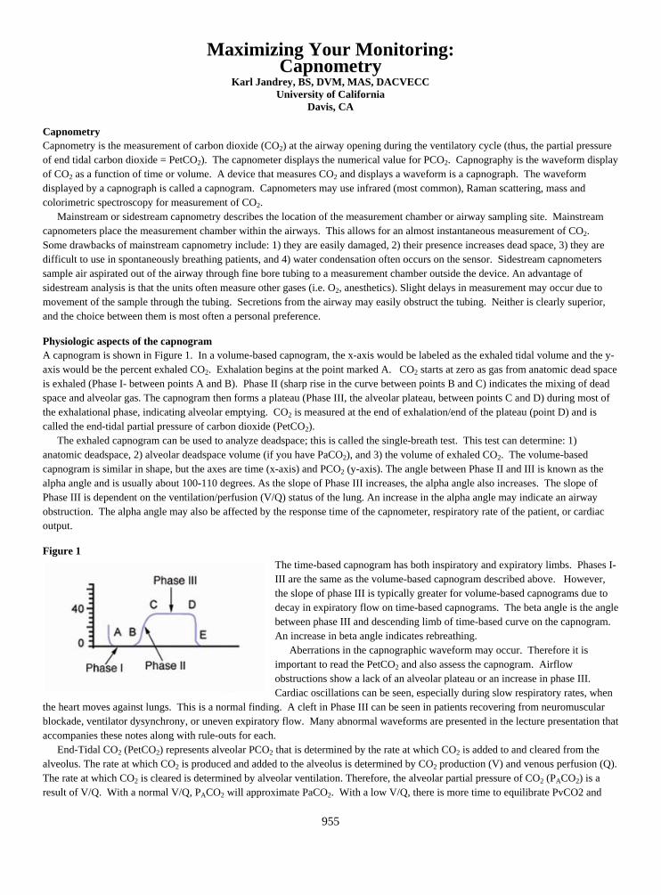
955
Maximizing Your Monitoring: Capnometry
Karl Jandrey, BS, DVM, MAS, DACVECC University of California
Davis, CA
Capnometry Capnometry is the measurement of carbon dioxide (CO2) at the airway opening during the ventilatory cycle (thus, the partial pressure of end tidal carbon dioxide = PetCO2). The capnometer displays the numerical value for PCO2. Capnography is the waveform display of CO2 as a function of time or volume. A device that measures CO2 and displays a waveform is a capnograph. The waveform displayed by a capnograph is called a capnogram. Capnometers may use infrared (most common), Raman scattering, mass and colorimetric spectroscopy for measurement of CO2.
Mainstream or sidestream capnometry describes the location of the measurement chamber or airway sampling site. Mainstream capnometers place the measurement chamber within the airways. This allows for an almost instantaneous measurement of CO2. Some drawbacks of mainstream capnometry include: 1) they are easily damaged, 2) their presence increases dead space, 3) they are difficult to use in spontaneously breathing patients, and 4) water condensation often occurs on the sensor. Sidestream capnometers sample air aspirated out of the airway through fine bore tubing to a measurement chamber outside the device. An advantage of sidestream analysis is that the units often measure other gases (i.e. O2, anesthetics). Slight delays in measurement may occur due to movement of the sample through the tubing. Secretions from the airway may easily obstruct the tubing. Neither is clearly superior, and the choice between them is most often a personal preference. Physiologic aspects of the capnogram A capnogram is shown in Figure 1. In a volume-based capnogram, the x-axis would be labeled as the exhaled tidal volume and the y-axis would be the percent exhaled CO2. Exhalation begins at the point marked A. CO2 starts at zero as gas from anatomic dead space is exhaled (Phase I- between points A and B). Phase II (sharp rise in the curve between points B and C) indicates the mixing of dead space and alveolar gas. The capnogram then forms a plateau (Phase III, the alveolar plateau, between points C and D) during most of the exhalational phase, indicating alveolar emptying. CO2 is measured at the end of exhalation/end of the plateau (point D) and is called the end-tidal partial pressure of carbon dioxide (PetCO2).
The exhaled capnogram can be used to analyze deadspace; this is called the single-breath test. This test can determine: 1) anatomic deadspace, 2) alveolar deadspace volume (if you have PaCO2), and 3) the volume of exhaled CO2. The volume-based capnogram is similar in shape, but the axes are time (x-axis) and PCO2 (y-axis). The angle between Phase II and III is known as the alpha angle and is usually about 100-110 degrees. As the slope of Phase III increases, the alpha angle also increases. The slope of Phase III is dependent on the ventilation/perfusion (V/Q) status of the lung. An increase in the alpha angle may indicate an airway obstruction. The alpha angle may also be affected by the response time of the capnometer, respiratory rate of the patient, or cardiac output. Figure 1
The time-based capnogram has both inspiratory and expiratory limbs. Phases I-III are the same as the volume-based capnogram described above. However, the slope of phase III is typically greater for volume-based capnograms due to decay in expiratory flow on time-based capnograms. The beta angle is the angle between phase III and descending limb of time-based curve on the capnogram. An increase in beta angle indicates rebreathing.
Aberrations in the capnographic waveform may occur. Therefore it is important to read the PetCO2 and also assess the capnogram. Airflow obstructions show a lack of an alveolar plateau or an increase in phase III. Cardiac oscillations can be seen, especially during slow respiratory rates, when
the heart moves against lungs. This is a normal finding. A cleft in Phase III can be seen in patients recovering from neuromuscular blockade, ventilator dysynchrony, or uneven expiratory flow. Many abnormal waveforms are presented in the lecture presentation that accompanies these notes along with rule-outs for each.
End-Tidal CO2 (PetCO2) represents alveolar PCO2 that is determined by the rate at which CO2 is added to and cleared from the alveolus. The rate at which CO2 is produced and added to the alveolus is determined by CO2 production (V) and venous perfusion (Q). The rate at which CO2 is cleared is determined by alveolar ventilation. Therefore, the alveolar partial pressure of CO2 (PACO2) is a result of V/Q. With a normal V/Q, PACO2 will approximate PaCO2. With a low V/Q, there is more time to equilibrate PvCO2 and
956
PACO2; PACO2 will rise towards PvCO2. With a high V/Q, PACO2 will approach the partial pressure of inspired carbon dioxide (PICO2, usually 0). PetCO2 is a representation of millions of alveoli; theoretically, it can be as low as 0 and as high as PvCO2.
There are three major causes of a decreased PetCO2: (1) decreased CO2 production and delivery to the lungs, (2) increased alveolar ventilation, and (3) equipment malfunctions. Decreased CO2 production and delivery to the lungs can be caused by hypothermia, pulmonary hypoperfusion, cardiac arrest, pulmonary embolism, hemorrhage, and hypotension. Increased alveolar ventilation defines hyperventilation. Equipment malfunctions may be any of the following: ventilator disconnect, esophageal intubation, complete airway obstruction, poor sampling, or a leak around the endotracheal tube. Causes of an increase in PetCO2 include: hypoventilation, rebreathing, sepsis, increased metabolism/ malignant hyperthermia, increased skeletal muscle activity, or hyperthermia.
The arterial to end-tidal CO2 gradient [P(a-et)CO2] is normally less than 5mmHg. This difference between PaCO2 and PetCO2 may increase with deadspace-producing diseases (high V/Q, i.e. due to a lower PetCO2). Some clinical applications Non-invasive prediction of PaCO2: The PaCO2 should be predicted only with caution from the PetCO2 due to the fluctuating and unpredictable nature of the P(a-et)CO2 gradient.
Esophageal intubation: Since carbonated beverages are not likely ingested in veterinary patients, there should be no CO2 in gastric contents. If CO2 is not present when the proximal end of the feeding tube is connected to the capnometer, intubation of the gastrointestinal tract must have been successful.
Feeding tube malposition: If CO2 is present, intubation of the airway must have occurred. Cardiac arrest: The measurement of PetCO2 has been used to monitor pulmonary blood flow in humans and animals during
resuscitation. At the onset of arrest, PetCO2 usually drops off to zero. With the restoration of cardiac output, PetCO2 increases. Increased dead-space ventilation: Patients with an increase in the slope of the alveolar plateau have been shown to have increased
alveolar dead-space. Pulmonary thromboembolism usually increases the P(a-et)CO2 gradient. PetCO2 is not PaCO2. It is also not a non-invasive measure of PaCO2. PetCO2 does however differentiate between esophageal and
tracheal intubation. References/suggested reading Jubran, A. (1997). Pulse Oximetry. Principles and Practice of Intensive Care Monitoring. M. Tobin, McGraw-Hill Co.: 261-287. Wright, B. a. H., PW (1996). "Respiratory monitoring during anesthesia: Pulse Oximetry and Capnography." Compendium of Continuing Education for the Veterinary Practitioner (Comp Cont Ed Vet Pract) 18(10): 10831096.
957
CPCR: Current Guidelines and their Applications
Ari Jutkowitz, VMD, DACVECC Michigan State University
East Lansing, MI
Cardiopulmonary cerebral resuscitation (CPCR) refers to the re-establishment of circulation and preservation of neurologic function following an arrest.1 Since its inception in the late 1800’s, CPCR has saved the lives of countless human and veterinary patients. However, low overall survival rates following CPCR indicate that there is still much room for improvement in these practices. This session reviews current practices and updates on CPCR in the veterinary patient with an emphasis on evidence-based guidelines derived from the RECOVER initiative. Basic life support Basic life support refers to the process of establishing an airway, initiating positive pressure ventilation, and performing chest compressions. Because cardiopulmonary arrest (CPA) in veterinary patients is frequently initiated by respiratory arrest, an ABC approach is generally taken as described below. In recent years, there has been a paradigm shift prioritizing chest compressions above all other measures (CAB approach).
Circulation Chest compressions are initiated at a rate of 100-120 per minute, compressing the circumference of the chest by approximately 30-50%. The patient should be in lateral recumbency during compressions. In smaller dogs, where the cardiac pump theory is believed to predominate, hands should be placed over the ventral third of the chest just behind the point of the elbow, corresponding to a position directly over the heart. In larger dogs, the thoracic pump theory is believed to be most important in generating blood flow, and hands should therefore be placed over the widest part of the thorax to create a maximal rise in intrathoracic pressure.
Airway Orotracheal intubation is easily achieved in dogs, as the larynx can be directly visualized by retractig the tongue. The head and neck should be gently extended and a laryngoscope may be used to improve visualization of the larynx. In cases where hemorrhage, saliva, or gastric contents interfere with visualization, suction may be helpful. Alternately, the glottis may be palpated with one finger used to guide tube placement. Once tube placement is verified, the tube should be secured by tying to the nose or around the back of the head. The cuff should be inflated, and assisted ventilation provided. If chest wall excursion is not seen, lung sounds are absent, or abdominal distension is noted, tube placement should be reconfirmed by direct visualization and the cuff should be reinflated. Improper tube placement and tube dislodgement are common causes of CPCR failure.
Breathing Once an endotracheal tube is in place, breathing is initiated at a rate of 10 breaths per minute with 100% oxygen to a tidal volume of approximately 10 ml/kg. An ambu bag with attached oxygen line is ideal for this purpose. If only one person is available to perform CPR, 2 breaths should be given for each 30 chest compressions. If several trained personnel are available, then breaths may be delivered independent of compressions. Chest wall excursion should be seen with each delivered breath. Airway pressures ideally should not exceed 20-30 cm H2O. High airway pressures or inadequate chest wall excursion should prompt a search for pleural space disease, tube malposition, or tube occlusion.
A number of alternative techniques have been investigated that may help to augment blood flow during CPCR. Those that are directly applicable in veterinary patients include circumferential chest compression and interposed abdominal compressions. Circumferential chest compression is most commonly performed in cats and small dogs by encircling the chest with both hands to maximize the rise in intrathoracic pressure during chest compression. In larger animals, interposed abdominal compression may be implemented by having an additional person perform abdominal compressions during the relaxation phase between chest compressions. Interposed abdominal compressions increase venous return to the heart, leading to greater stroke volumes and cardiac output, and have been associated with increased survival to discharge in human patients. Advanced life support Advanced life support consists of drug administration, determination of cardiac electrical activity, and application of electrical defibrillation if indicated. These techniques build upon basic life support to increase the likelihood of successful resuscitation.
Drugs Establishing vascular access is one of the first priorities during advanced life support. While central lines are preferable for rapid distribution of drugs, peripheral catheters are acceptable, and drug delivery may be facilitated by following drug administration with a 10-20 ml IV fluid “chaser”. If vascular access is not immediately obtained, surgical cutdown or intraosseous techniques should be considered. The intratracheal route may also be used initially to deliver drugs. Epinephrine, atropine, vasopressin, lidocaine, and
958
naloxone may all be given in this way by administering twice the normal dose of the drug (or using the “high” dose for epinephrine) and administering several large breaths to disperse the drug.
Drugs administered during CPCR include intravenous fluids, narcotic reversal agents, vasopressors, vagolytics, antiarrhythmics, and potentially sodium bicarbonate. Shock doses of intravenous fluids should be provided in cases where hypovolemia is believed to have played a role in the arrest. Moderate fluid rates should be used in euvolemic patients or patients with underlying heart disease, as rapid administration in these cases may excessively elevate right atrial pressure and consequently decrease myocardial and cerebral perfusion pressure.
Patients who have received narcotic pain relievers or other sedative/anesthetic drugs prior to arrest should immediately be given the reversal agent for that drug. Naloxone may be used to reverse most narcotics at a dose that is isovolumetric to the dose of the original narcotic, or at 0.02-0.04 mg/kg IV if the original dose is unknown. Flumazenil (0.02 mg/kg IV) may be used to reverse benzodiazepines, and yohimbine (0.1 mg/kg) or atipamazole (0.2 mg/kg or isovolumetric) may be used to reverse xylazine and medetomidine respectively. Any anesthetic gases, if still in use, should be discontinued and the anesthetic circuit flushed with fresh oxygen.
Vasopressors are commonly used during CPCR to increase blood pressure and redistribute blood flow to vital organs like the brain and heart. Epinephrine continues to be the vasopressor of choice during CPCR in veterinary patients, though its use is largely extrapolated from clinical studies in human patients. Both low dose and high dose epinephrine protocols are described in human medicine. While high dose epinephrine has been associated with increases in early return of spontaneous circulation, no long-term benefits have been identified. High dose epinephrine has additionally been associated with increased myocardial oxygen demand and worse neurologic outcomes.2 For these reasons, it is recommended that low dose epinephrine initially be administered every 3-5 minutes during CPCR, switching to the high dose only if there is a lack of response to the lower doses. Epinephrine dosing may be rapidly calculated according to the following rule of thumb: 0.1 ml per 20 lb of the 1:1,000 formulation for low dose, or 1 ml per 20 lb for high dose.
Vasopressin is another potent vasoconstrictor that is increasingly used in resuscitation of human patients. Unlike epinephrine, it does not increase myocardial workload, and its effect is not blunted by acidosis. Although clinical data in veterinary patients is currently lacking, animal models and human clinical trials suggest that vasopressin may be as effective as epinephrine.3 Vasopressin (0.8 units/kg IV) may therefore be considered as an alternative to epinephrine in dogs.4
Atropine is another drug frequently administered during CPCR to reverse parasympathetic contribution to the arrest or to treat sinus bradycardia. Atropine is administered at a dose of approximately 1 ml per 20 lb (0.04 mg/kg) for asystole or pulseless electrical activity. When treating sinus bradycardia, only half this dose is needed.
Sodium bicarbonate use in CPCR is controversial, as it has been associated with numerous adverse effects including hypernatremia, paradoxical CNS acidosis, and decreased resuscitation rates in people. However, its use should still be considered during long duration (>10 minutes) arrests, as control of acidosis may improve response to catecholamines as well as post-arrest neurologic outcomes. Bicarbonate is typically given only after 10 minutes of CPCR at a dose of 1 mEq/kg and is repeated every 5 minutes thereafter.
Electrical activity ECG leads should be attached as soon as feasible to assess electrical activity. Connecting the leads to the skin of the lower forelimbs and hindlimbs will help to minimize motion artifact associated with resuscitation efforts. Four rhythms are commonly seen during cardiopulmonary arrest in dogs. Asystole and pulseless electrical activity are the initial arrest rhythms most commonly seen in dogs, followed by ventricular fibrillation and sinus bradycardia.5,6 Accurate ECG diagnosis is vital to a successful code. The presence of sinus bradycardia or suspicion of a vagal arrest should prompt administration of atropine. Asystole should be confirmed in more than one lead, to rule out the possibility of artifact related to poor contact. While some dogs in asystole will convert directly to sinus rhythm following resuscitation, many develop ventricular fibrillation and require electrical shock for conversion. Once ventricular fibrillation is identified, electrical defibrillation should immediately be administered, temporarily bypassing all other resuscitation measures. The greater the time that a dog spends in fibrillation, the lower the likelihood of successful conversion.
Defibrillation Early application of electrical shock is the only effective method for converting VF to sinus rhythm. VF is a form of disorganized electrical activity with various portions of the heart muscle firing at different times. Electrical shock essentially "resets" the cardiac cells so that organized activity can resume. Practically speaking, applied current must pass through at least 30% of cardiac myocytes to effectively convert VF.
To accomplish defibrillation, the dog is flipped into dorsal recumbency immediately preceding defibrillation and handheld paddles are placed on either side of the chest directly over the heart. Ample conducting gel should be applied to the paddles to ensure good contact and prevent dispersion of current. The chest should be compressed between the paddles, minimizing impedance by narrowing the distance between paddles. If using a monophasic defibrillator, the energy for the first shock should be set at 3-5 J/kg. If defibrillation is not successful, CPCR is resumed for 60-90 seconds and a subsequent shock should then be given at the same energy

959
setting. Electrical shock is discontinued once the rhythm converts from VF. Lower energy biphasic shock waveforms have been shown to be as effective as higher energy monophasic waveforms and exclusively used at this time in human patients. If using a biphasic defibrillator, the pediatric settings should be used (2-4 J/kg). 7
For shock-refractory VF, a search should be undertaken to identify problems such as improper paddle position, inadequate contact, insufficient conduction gel, or the presence of pleural space disease that may increase impedance. Drug-shock techniques may then be considered, administering epinephrine or amiodarone (5 mg/kg IV) prior to shock to lower defibrillation threshold. Lidocaine was previously used for this purpose as well, but has been reclassified as a therapy of indeterminate benefit in the most recent ACLS guidelines.7
Open chest CPCR There are a number of absolute indications for open chest CPCR. These include cardiac arrest caused by or associated with pleural space disease (pneumothorax, pleural effusion, diaphragmatic hernia), pericardial effusion, or penetrating injury resulted in cardiac arrest. However, debate exists in veterinary medicine as to other indications for performing open chest CPCR. Some advocate open chest CPCR immediately in large breed dogs because of the limited success of restoring adequate circulation with external compressions while others prefer to perform external CPCR for 5 minutes and then open the chest if there is little or no evidence of effective circulation. Open chest CPCR has the advantage of allowing the clinician to directly compress the heart and improve stroke volume. In addition, opening the chest makes assessment of ventricular filling feasible aiding in the decision of volume delivery.
When opening the chest, it is critical to auscult the chest just prior to the incision to rule out ECG dysfunction as the cause of asystole. The left chest should be crudely clipped of hair at the left 5th-6th intercostals space and a chlorhexidine based antiseptic solution should be briskly applied. An incision should be made through the skin and subcutaneous tissues from just below the spinal musculature to the level of the costochondral junction. Between positive pressure breaths, mayo scissors should be used to poke through the intercostal musculature and the pleura and the chest is opened by sliding the mayo scissors dorsally and ventrally along the cranial border of the rib (to avoid the neurovascular bundle). The pericardium is opened at the pericardio-diaphragmatic ligament and the heart is compressed from the apex to the base. In large dogs, the heart can be compressed against the opposite chest wall.
In the event of return of spontaneous circulation, antibiotics should be instituted immediately, the chest should be lavaged with copious amounts of warm saline, and should be closed using sterile technique over a chest tube. ICU care Following a successful code, a search for underlying causes or complications should be performed and any problems corrected. Blood gases, hematocrit and total solids, blood pressure, and oxygen saturation are carefully monitored and optimized during this time. This tends to be the most challenging phase of arrest management, as complications and recurrence of CPA are common. Neurologic recovery is promoted by maintaining arterial blood pressure and oxygen saturation. Because elevation in carbon dioxide levels leads to cerebral vasodilation and consequently increased intracranial pressure, hypercarbia should be prevented by employing mechanical ventilation if needed. Once cardiovascularly stable, mannitol (0.25-0.5 g/kg IV over 20 minutes) may also be indicated to treat cerebral edema and resultant elevations in intracranial pressure. Corticosteroids are associated with potentially deleterious hyperglycemia in post-arrest patients, and current protocols do not support their use.7
Prognosis Recurrence of CPA in the post-arrest period is common, occurring in up to 70% of successfully resuscitated dogs. Intensive care and monitoring during this time is therefore essential. Survival to discharge following cardiopulmonary arrest has been reported in 4-11% of cases.5,6,8 Transient blindness, seizures, circling, ataxia, and decreased level of consciousness are common for some period of time following CPA, but the majority of survivors have a good prognosis for functional recovery.6
References Otto CM. Cardiopulmonary cerebral resuscitation and ACLS guidelines. In Proceedings of the 8th International Veterinary Emergency and Critical Care Symposium. San Antonio, TX. September 2002. p. 478-481. Gueugniaud PY, Mols P, Goldstein P. et al. A comparison of repeated high doses and repeated standard doses of epinephrine for cardiac arrest outside the hospital. N Engl J Med 1998;339:1595-1601. Wenzel V, Krismer AC, Lindner KH, et al. Comparison of vasopressin and epinephrine for out-of-hospital cardiopulmonary resuscitation. N Engl J Med 2004;350:105-113. Schmittinger CA, Astner S, Astner L, et al. Cardiopulmonary resuscitation with vasopressin in a dog. Vet Anaest Analg 2005;32:112-114. Wingfield WA, Van Pelt DR. Respiratory and cardiopulmonary arrest in dogs and cats:265 cases (1986-1991). J Am Vet Med Assoc 1992;200:1993-1996 Waldrop JE, Rozanski EA, Swanke ED, et al. Causes of cardiopulmonary arrest, resuscitation management, and functional outcome in dogs and cats surviving cardiopulmonary arrest. J Vet Emerg Crit Care 2004;14:22-29. 2005 American Heart Association Guidelines for cardiopulmonary resuscitation and emergency cardiovascular care. Circulation 2005;112(suppl.) Kass PH, Haskins SC. Survival following cardiopulmonary resuscitation in dogs and cats. J Vet Emerg Crit Care 1992;2:57-65.
960
CRI Use for Anesthesia and Analgesia Katrina Lafferty, BFA, CVT, VTS (Anesthesia)
University of Wisconsin Madison, WI
Pain management in veterinary patients has come a long way. There are a myriad of analgesic drugs available and numerous techniques exist to further provide top-notch analgesia and pain management to animals undergoing even the most painful surgeries. Constant rate infusions (CRIs) are increasing in their usage, thanks in large part to their wide versatility. There are pharmacologic agents used as CRIs that can provide analgesia, maintain anesthesia, and provide blood pressure support if needed. Analgesics Delivery of analgesic drugs as a CRI is typically far superior to bolus doses for a number of reasons. By administering a constant, steady infusion of a drug, stable levels of tissue concentration are reached. This prevents the peaks and valleys of comfort versus discomfort that occurs when bolus doses of analgesics are give as a patient begins to seem painful. When giving bolus doses, drugs peak, providing analgesia, then fall to below therapeutic levels allowing for breakthrough pain. CRI administration eliminates that rise and fall. CRIs can be easily adjusted to meet the needs of each individual patient, allowing for lower amounts of the drug to be given. Less money is spent on the actual drug as well as on supplies (syringes, needles, etc). Lower doses also decrease the incidence and severity of side effects.
Analgesic choices Many classes of analgesic drugs can be given as a CRI including opioids, local anesthetics (specifically lidocaine), NMDA receptor antagonists (ketamine), and alpha-2 agonists (dexmedetomidine). Drug dosages are shown in Table 1.
Opioids work by binding with specific receptors in the central nervous system (CNS) and can, depending on the opioid, provide relief for mild to severe pain. Morphine, fentanyl, hydromorphone, and butorphanol are commonly used in opioid CRIs. Morphine, fentanyl, and hydromorphone can be used for moderate to severe pain, while butorphanol is only appropriate for use in mild to moderately painful cases. CRIs tend to reduce the severity of opioid side effects such as vomiting, dysphoria, and respiratory depression, but patients should still be monitored closely for any sign of distress.
Lidocaine works by blocking sodium ion channels and causing membrane stabilization. It can reduce the amount of opioid analgesic and inhalant gases required to maintain anesthesia. Lidocaine is relatively inexpensive and has anti-arrythmic and anti-inflammatory properties. It is may be useful in cases where gastrointestinal pain is involved(GDV, laparotomies, etc). Cats have an increased sensitivity to local anesthetics and it is currently not recommended to use lidocaine infusion on feline cases.
Ketamine works by antagonizing the NMDA receptors which are responsible for central sensitization, hypersensitization, and “wind-up” pain. Ketamine is not capable of providing adequate analgesia in its own right, however, when administered in combination with opioid analgesics, it can lower anesthetic requirements of the patient.
Dexmedetomidine has mild analgesic properties, as well as anxiolytic and sedative properties. It works by simulating alpha-2 receptors in the CNS. Dexmedetomidine CRIs are most commonly used in the postoperative phase, for patients that are anxious and/or vocal despite an appropriate anesthetic regimen.
Anesthetic maintenance Total intravenous anesthesia (TIVA) with propofol may be indicated for short, painless procedures. It may also been needed in situations where a patient cannot be intubated (tracheal procedures), where patients need to remain intubated and unconscious for extended periods (cases requiring long term ventilation assistance), or patients with autosomal disorders where inhalants cannot be used (malignant hyperthermia). In these cases propofol is administered with a bolus dose of 2-5 mg/kg followed by 0.05-0.2mg/kg/min. Adequate oxygenation is always a concern, so if possible it is recommended to intubate patients and maintain them on 100% oxygen. Standard anesthetic monitoring is still needed in TIVA patients. It may be harder to control depth when using TIVA, so good monitoring can act as an early alert system to patients that become too light or too deep, much the same as when anesthetizing a patient with volatile anesthetics.
Pressor support Dopamine and dobutamine are the choices most commonly used to provide blood pressure support in hypotensive patients. Both drugs increase cardiac contractility, and assuming the patient has appropriate vascular volume, these drugs can most boost low blood pressure. The range for both dobutamine and dopamine is 1-10 mcg/kg/min. There can be a wide variation in patient reactions, so it is recommended to begin with low end dosing and adjust as needed.
Equipment One downfall of CRI administration is that it requires specific equipment for the best and safety route of delivery. It is possible to deliver CRIs using either a fluid pump, syringe pump, or standard drip set. It is ideal to use a syringe pump, which can be programmed with all the variables involved in administering the infusion, but pumps can range from hundreds to thousands of dollars. If CRIs are being used on a regular basis, infusion pumps could be worth the investment—less waste, less man-power needed, fewer errors. Fluid
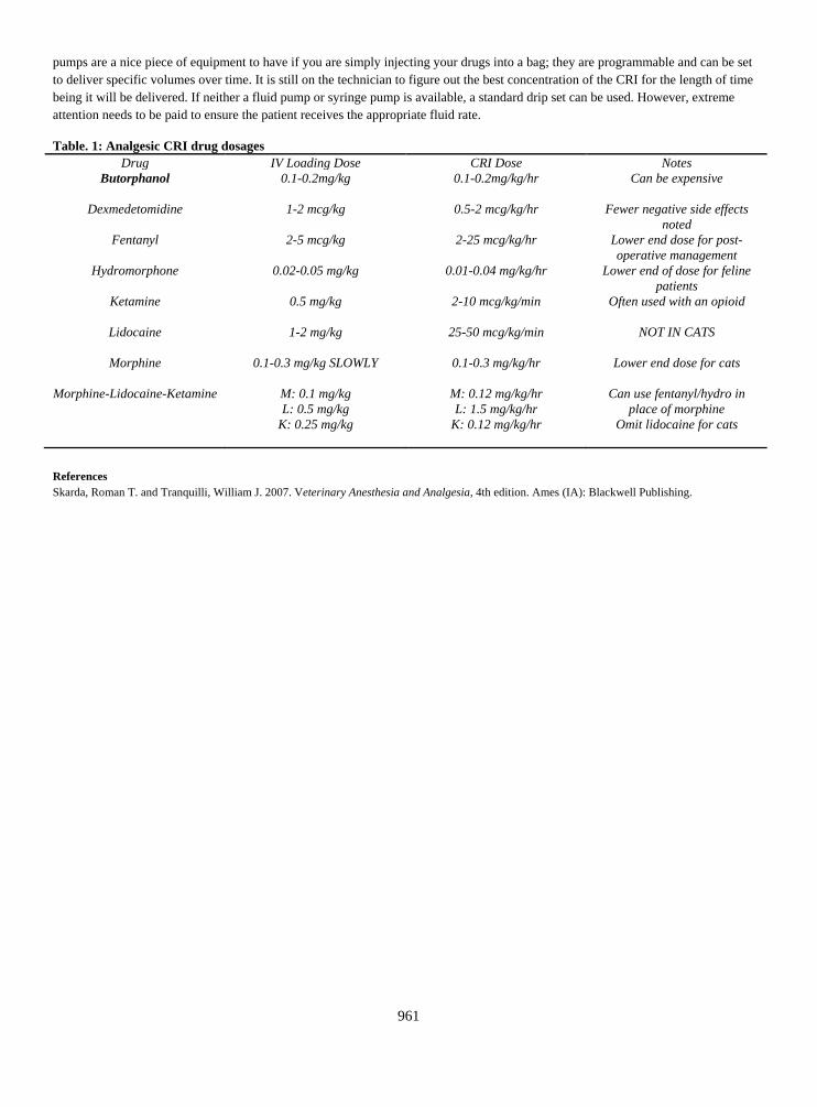
961
pumps are a nice piece of equipment to have if you are simply injecting your drugs into a bag; they are programmable and can be set to deliver specific volumes over time. It is still on the technician to figure out the best concentration of the CRI for the length of time being it will be delivered. If neither a fluid pump or syringe pump is available, a standard drip set can be used. However, extreme attention needs to be paid to ensure the patient receives the appropriate fluid rate. Table. 1: Analgesic CRI drug dosages
Drug IV Loading Dose CRI Dose Notes Butorphanol 0.1-0.2mg/kg 0.1-0.2mg/kg/hr Can be expensive
Dexmedetomidine
1-2 mcg/kg 0.5-2 mcg/kg/hr Fewer negative side effects
noted Fentanyl 2-5 mcg/kg 2-25 mcg/kg/hr Lower end dose for post-
operative management Hydromorphone
0.02-0.05 mg/kg 0.01-0.04 mg/kg/hr Lower end of dose for feline
patients Ketamine
0.5 mg/kg 2-10 mcg/kg/min Often used with an opioid
Lidocaine
1-2 mg/kg 25-50 mcg/kg/min NOT IN CATS
Morphine
0.1-0.3 mg/kg SLOWLY 0.1-0.3 mg/kg/hr Lower end dose for cats
Morphine-Lidocaine-Ketamine M: 0.1 mg/kg L: 0.5 mg/kg
K: 0.25 mg/kg
M: 0.12 mg/kg/hr L: 1.5 mg/kg/hr
K: 0.12 mg/kg/hr
Can use fentanyl/hydro in place of morphine
Omit lidocaine for cats
References Skarda, Roman T. and Tranquilli, William J. 2007. Veterinary Anesthesia and Analgesia, 4th edition. Ames (IA): Blackwell Publishing.
962
To Glasgow and Beyond: Appreciating, Adapting, and Applying Pain Scoring Systems
Katrina Lafferty, BFA, CVT, VTS (Anesthesia) University of Wisconsin
Madison, WI
The 4th vital sign: pain. There are a myriad of negative consequences associated with inappropriately managed pain: decreased healing, infection, heightened reactions to pain, changes to the CNS. Utilizing pain scoring systems reduce pain states and improve patient care.
Poorly managed pain, whether before, during, or after anesthesia, can have a number of harmful consequences. Most publications on the subject of pain management in both “traditional” canine/feline patients and “non-traditional” exotic patients show appropriate analgesia and pain management to be essential to wound healing and patient recovery. Improperly managed pain affects the patient and the owner. Pain is an individual experience and can be somewhat subjective; many factors can affect how pain is interpreted. In humans and animals alike pain is tolerated differently. It can be influenced by genetics (think husky dog versus Staffordshire terrier), learned behaviors, and even culture. In veterinary patients the challenge of adequately recognizing, assessing, and treating pain is further complicated by the non-verbal nature of the patients. Much of the pain assessment techniques used in animals is modeled after what is used in human medicine for infants, toddlers, and other non-verbal patients. In the AAHA pain management guide for cats and dogs it is stated, “It is now well established that animals and humans have similar neural pathways for the development, conduction, and modulation of pain. According to the principle of analogy, because cats and dogs have neural pathways and neurotransmitters that are similar, if not identical, to those of humans, it is highly likely that animals experience pain similarly.” Read this to mean if it would cause discomfort to you, it would cause discomfort to your patient, regardless of species.
To better understand how to manage pain, one should have a fluency in the “language” of pain: • Pain: an unpleasant sensory and emotional experience associated with actual or potential tissue damage; the perception
of nociception • Nociception: the activity produced in the nervous system by noxious stimuli • Analgesia: absence of pain in response to normally painful stimuli • Hyperalgesia: an increased response to a normally painful stimulus • Allodynia: pain due to a stimulus that does not normally produce pain (i.e. touch) • Central Sensitization: “Wind up pain;” changes in the central nervous system that occur as a result of repeated or
chronic painful stimulus • Analgesic: an agent or drug that causes or allows for relief from pain; a “pain killer”
The pain pathway in the body can be broken down into four basic groups: transduction, transmission, modulation, and perception. Analgesics and analgesic techniques work to modify to pain felt and transmitted throughout the circuit. Different analgesic drugs work on different parts of the pathway.
• Transduction: change of cellular chemical information into electrical impulses that travel the spinal cord • Transmission: travel of the pain impulse; transmitting the signal to the brain • Perception: cognizant recognition of painful stimulus • Modulation: Brain and spinal cord communicate and work together to change or modify the painful sensation
There are typically describe 6 types of pain: 1. Somatic Pain: at the level of the skin; also involves muscles, tendons, and joints 2. Visceral Pain: Internal organs, soft tissue 3. Acute Pain: Recently occurring, generally less than a few months 4. Chronic Pain: Longer in scope, lasting more than a few months 5. Neuropathic Pain: Atypical pain, ie: phantom limb pain 6. Referred Pain: Pain perceived in a region disparate from the actual painful site
Pain perception can be broken down into 3 sections: Perception (recognition of discomfort), Threshold (point when painful signal hits the brain), and Tolerance (amount of pain that can be endured). In order to assess pain thoroughly, consider these categories: Incidence, Level, Location, Length. For each patient, each category can have varying levels of intensity. All veterinary patients will be non-verbal, so the difficult task is to use behavioral assessments to answer questions of how painful an animal is. There are species differences in pain behavior, but many pain mannerisms are universal.
963
Universal non-verbal pain indicators • Abnormal posture • Restlessness • Splinting • Vocalizing • Unwillingness to move • Trembling/shivering • Anorexia • Elevated physiologic parameters • Aggression (towards self or others) • Lack of grooming • Self-mutilation
Pain is often characterized and describe as it is relevant to the owner. An adequate and thorough “pain history” can be very useful when creating an appropriate analgesic plan. The history should include:
• Previous/ongoing painful states • Previous methods of controlling pain • Owner’s expectations for pain control • How/why the owner feels a patient is painful
The “FLACC” scale is often used in human medicine for non-verbal patients (infants, toddlers, demented patients, critically ill/unconscious patients) and has applications for veterinary patients. FLACC stands for “Face” (relaxed/grimace/tightened face), “Legs” (normal posture/kicking/restless), “Activity” (normal/restless/agitated), “Cry” (quiet/whimpering/frequent complaints), and “Consolability” (content/distractable/unable to comfort). Each area is ranked 0-2, with elevated numbers requiring treatments.
In veterinary medicine, there are 3 primary scales that have been adapted and utilized for pain assessment. CSU acute pain scales Colorado State University created pain scales for canine and feline patients, based on behavior and physical reactions to stimuli. It is an exceptionally easy to use scale, but does not necessarily have clinical validation to support it. It does have forms for both canine and feline patients. Glasgow composite measure pain score-short form This form is based on specific behavioral markers, assigning numeric points to determine level of pain. This scale is used universally, everywhere from small private practices to veterinary teaching institutes. This scale is designed for canine patients. This scale does have some clinical research to support it. UNESP-botucatu multidimensional composite pain scale This form is the feline companion to the Glasgow Pain Scale. It utilizing 10 categories to assign numbers for painful behaviors.
Using a behavioral checklist is likely the most common way to create a pain assessment chart. Having a chart on hand allows for a standard, equal, reproducible way to evaluate pain in patients.
Behavior based charts should include categories for: • Attitude or Mentation • Activity • Facial expression • Guarding • Posture • Vocalization
Above all, treat each patient as an individual. If pain is suspected, treat. If an animal responds to analgesic intervention, that too is considered a point on the pain assessment scale. References always available upon request.
964
Anesthesia and Analgesia for Avian Patients Katrina Lafferty, BFA, CVT, VTS (Anesthesia)
University of Wisconsin Madison, WI
Many technicians (and veterinarians) do not feel comfortable working with avian patients. There is a decided lack of information on how to appropriately handle, medicate, and anesthetize birds, complicated by the fact that birds are not mammals and have much different anatomy than cats and dogs. However, the number of bird owners is increasing dramatically and more and more of those dedicated pet owners are bringing their family members to veterinary clinics expecting the same kind of care cats and dogs normally receive. Ovariohysterectomies are becoming a common occurrence, as are ceolomic surgeries, fracture repairs, wing amputations, and mass removals. The purpose of this lecture is to provide basic information on how to competently provide quality anesthesia for birds in the hopes that technicians will feel more confident about providing a better standard of care for the exotics species that come through the clinic door. Anatomy Avian patients have a number of anatomical differences from their mammal counterparts. The gastrointestinal tract includes an outcropping of the esophagus known as the crop, 2 chambers in the stomach known as the proventriculus ( a very strong chamber that secretes acid to break down food) and the ventriculus (aka the gizzard; essentially crushes food), and a common outlet for intestinal, genital, and urinary tracts called the cloaca.
The respiratory system in birds is very complicated and can be extremely sensitive. Birds have a very long trachea, slower respiratory rate, and larger tidal volume. They have small, dense lung that cannot handle apnea. They have 9 air sacs that act as a type of bellows and increase respiratory efficiency. Birds have no diaphragm and their entire body cavity acts as a ventilatory force. Normal values
• Temperature: 100-110, Pulse: varies based on species—up to 1000/beats per minute, Respirations: 30-60 • Packed Cell Volume: 38-50% • Total protein: 2.4-5 • Glucose: 185-455 • Blood Volume: 55-70 ml/kg • Remember, these are all VERY species specific!
Premedication
Drug doses for MOST species of bird • Butorphanol: 0.5-2 mg/kg IM/SQ/IV • Buprenorphine: 0.01-0.05 mg/kg IM/SQ/IV • Hydromorphone/Oxymorphone: 0.05-0.2 mg/kg IM/SQ/IV • Midazolam: 0.1-0.5 mg/kg IM/SQ/IV/intranasal • Carprofen: 1-2 mg/kg SQ • Meloxicam: 0.1-0.2 mg/kg SQ/PO • Ketamine: 10-20 mg/kg IM • Propofol: 2-5 mg/kg IV* use with caution
As with canine and feline patients, birds should be premedicated with a sedative and analgesic combination before any anesthetic episode. Premedication helps decrease level of stress in patients, reduces anesthetic requirements, and helps to deal with pain associated with the procedure. While it is not typically acceptable to mask induce patients, exotic species are the exception. They are usually not sedate enough from premedication to allow for easy catheter placement. Even if IV induction is an option, many species are require more time to intubate and an induction with an IV agent, such as propofol, may not allow enough time for intubation before it reaches a crisis. Birds however, are exceptionally easy to intubate, so intravenous induction is an option.
The primary site for intramuscular injections is the pectoral area. It often the largest muscle mass and is easy to locate by palpating the keel and angling the needle laterally. The quadriceps muscles can be used, but the potential hepatotoxic effects of the drug must be considered and weighed.
Subcutaneous injections can be given in the inner thigh or over the keel. The skin in both areas is very thin, which does allow for easy confirmation of placement. The inner thigh is best for larger volume administration although caution should be used as it is near an air sac.
965
Induction and intubation Isoflurane and sevoflurane are both acceptable choices of gas inhalant in exotic anesthesia. When performing a mask induction be extra vigilant in monitoring vital signs. Take care not to over- or under-restrain the patient and use caution not to cause ocular damage with the induction mask. Birds do not have a diaphragm and cannot respire appropriately if their chest is restricted.
Birds should ALWAYS be intubated. Avian patients frequently become apneic under anesthesia and their physiology does not tolerate any episodes of respiratory arrest. Birds are also very prone to regurgitation and intubation lessens the likelihood of aspiration. Tube size for birds usually runs from 2.0mm-5.0mm depending on the size and species of the bird (cockatiels and parakeets can be intubated using 16 gauge or 18 gauge catheters with the stylet removed). Do not inflate the cuff. Birds have complete tracheal rings and any pressure from an inflated cuff can cause tissue necrosis. Birds are technically easy to intubate, the tracheal opening is located just off the base of the tongue and can often be visualized without the aid of a light source. Venipuncture and intravenous catheter placement The right jugular veins in birds is usually easily visualized, well tolerated, and excellent for larger volumes. The ulnar/basilic (wing vein) usually requires anesthesia to attempt. The vessel is very fragile and the technician should expect a hematoma. The vessel runs directly alongside an artery so take care which vessel is being used. The medial metatarsal vein is an excellent choice in larger birds and the first choice in waterfowl. The tools necessary for blood collection include a 1cc syringe or small gauge butterfly set, 23g-27g needles, and small collection tubes. It is important to remember to remove the needle before transferring the sample to the collection tube as the cells being forced through a small gauge needle can be lysed. Bird blood can clot quickly, so if it will not interfere with the required tests, it can be advantageous to preheparinize the syringe.
In birds, catheters can be placed in the ulnar vein, medial metatarsal, or jugular vein. The ulnar vein is easy to see, but is located immediately next to an artery so care should be taken not to catheterize the wrong vessel. The ulnar vein is more challenging to secure and is not practical for maintence beyond the anesthetic and immediate recovery period. The medial metatarsal vein is an excellent choice in long –legged birds and can usually be maintained longer term. The jugular vein is an adequate choice for long term catheters and may be the only choice in very small birds. In birds the right jugular vein is much larger and more prominent.
Equipment needed for intravenous catheters includes 22, 24, or 26 gauge catheters, gentle tape—clear tape or paper tape, and lightweight access ports. Monitoring Similar monitoring techniques are used in canine and feline patients, allowing for a smaller scale. Pulse oximetry can readily be used. Probes can be placed on the feet, tongue, wings, esophagus, or cloaca.
Doppler flow detector crystals can be placed over the dorsal pedal artery or ulnar artery in birds and in larger patients can be used in conjunction with a sphygmomanometer to obtain blood pressure readings. The Doppler probe can also be placed in the femoral area, carotid/thoracic inlet, or on the chest and used for sound and heart rate. Be careful not to restrict respirations if taping the probe on the chest.
Capnometry can be used on any intubated patient. Dead space adapters exist that can be connected to the endotracheal tube in place of the regular tube adapter. The size endotracheal tube used is very small and tube obstruction is a concern. A capnograph can be the first alert to an obstruction or inadvertent extubation. It can confirm proper intubation and can alert the anesthetist to issues with ventilation or cardiac output.
Non-invasive blood pressure monitoring is an option on some animals—good pediatric monitors usually work well on medium to large birds. Small blood pressure cuffs can be placed on the wings or legs.
Electrocardiography can be readily monitored. The ECG gives information about rate and rhythm and can be a helpful tool to use under anesthesia. ECG patches can be placed on both wings and the left leg in birds. It is also possible to use 25 gauge needles pierced through skin with an alligator clip attached to the needle. If using alligator clips on skin (without an ECG patch) either flatten the teeth of the clip or use a gauze square to cushion the area and prevent tissue damage.
Temperature monitoring is one of the most vital monitors in birds. Small patients are at a high risk for hypothermia. It is important to note that for as quickly as small patients can cool down, they can warm up just as fast. Without careful monitoring of temperature, patients can easily become hyperthermic. Temperature can be monitored using either cloacal or esophageal temperature probes.
Monitoring anesthetic depth in birds has some similarities to monitoring anesthesia in other species. Palpebral reflexes are not reliable in birds, however, jaw tone, rectal tone, and pedal reflexes are all accurate indicators of anesthetic depth. As with most species, if an avian patient is too light, heart rate and respiratory rate will increase, if it is too deep, heart rate and respiratory rate will decrease. Recovery Recovery requires almost as much monitoring as during the anesthetic period. Birds can become hypothermic very quickly and temperature should be monitored closely for several hours postoperatively. It can be difficult to maintain catheters in birds and

966
watchful eye must be kept. Respiratory depression can quickly lead to cardiac arrest—vigilance can catch an issue before it becomes a critical problem. Always know the reversal drug and dose needed and have an emergency drug dose chart readily available. Birds should be kept in an area where they can be easily watched but not disturbed. Make sure housing is warm, dark and away from predators. If necessary make modifications to food and water sources so they can be easily accessed by debilitated or recovering patients. References available from the author.
967
Case by Case: Creating the Right Drug Protocols for Canine and Feline Patients
Katrina Lafferty, BFA, CVT, VTS (Anesthesia) University of Wisconsin
Madison, WI
Anesthetic patients should be evaluated on a case-by-case basis and an anesthetic protocol created for the individual situation. That being said, it can be overwhelming and intimidating to pick the right drug combination from the multitude of available options. This lecture will briefly review available drug choices and considerations for premedication and induction as well as intra- and post-procedural analgesia and sedation. Several case studies (both routine and emergency) will be provided for interactive discussion.
The reasons to premedicate are many. Premedication allows for less stressful and safer restraint and catheter placement (for patient and technician), it provides for analgesia during a procedure and by and large decreases the amount of drug needed overall. No one drug possesses every desired quality—good sedation, good analgesia, reversibility, minimal side effects—so it is necessary to choose a combination of drugs to decrease negative effects and lower the total amount of drug needed. An anesthetic protocol should include a drug for sedation and one for analgesia. The specific drugs and amounts used will depend on several factors. The anesthetist creating the plan will have to evaluate the temperament of the patient, the type of procedure being performed, and the co-existing conditions or contraindications each patient may have. Sedatives Generally the available sedatives are benzodiazepines (diazepam, midazolam), phenothiazines (acepromazine), and alpha-2 adrenergic agonists (dexmedetomidine).
Benzodiazepines offer sedation but no analgesia. The sedative qualities are appropriate for patients with calm (or depressed) personalities. They have minimal negative effect on cardiovascular or respiratory systems and are the sedative of choice for compromised patients. Benzodiazepines have many positive qualities; they provide muscle relaxation, have anxiolytic properties, and are commonly used for seizure patients. Benzodiazepines do not work well alone or on excited patients and may even have an opposite excitatory effect, counter to the desired sedation. As with all sedatives, it is best to combine this type of drug with an analgesic opioid for maximum effectiveness. Midazolam is a water soluble benzodiazepine and is safe to administer intravenous (IV), intramuscular (IM), or subcutaneous (SC). Diazepam is lipid soluble and should only be given IV; when given IM or SC routes, diazepam injections are painful and absorption is extremely erratic. Flumazenil is the reversal for benzodiazepines.
Phenothiazines provide good sedation (but again, no analgesia) for patients and are often the best option for aggressive, hyperactive, or extremely anxious animals. Acepromazine is the most commonly available phenothiazine. There are many pros to phenothiazine sedatives, they are good anti-emetics, a benefit when combining with an emetic like opioids, prevent histamine release, and have little effect on the respiratory system. There are a number of cons though, including decreasing the seizure threshold, peripheral vasodilation and resultant hypotension, hypothermia, and inhibition of platelet aggregation. There is evidence that phenothiazines can cause splenic enlargement. This can cause difficultly for surgeons working in the abdominal cavity. Unfortunately, phenothiazines are not reversible. Acepromazine can be given IV, IM, or SC.
Alpha-2 adrenergic agonists provide excellent sedation for most patients and provide very minor analgesia (though not enough for any surgical purpose). Dexmedetomidine is the most commonly used alpha-2. This class of drug can cause respiratory depression, but it varies greatly from patient to patient. This class of drug can cause dark mucous membrane color that is unrelated to oxygenation of tissue. It can cause profound bradycardia and vasoconstriction. It is not advisable to treat the bradycardia with anticholinergics as it increases the workload of the heart. It is best to reverse the drug first, then deal with bradycardia if it continues. This class of drug is only an appropriate choice for relatively healthy patients and cannot be used on any patient with cardiac compromise. Alpha-2s cause vomiting in most cats and many dogs, though this is much less when given intravenously. Alpha-2 adrenergic agonists are reversible with atipamezole. Dexmedetomidine can be given IV, IM, or SC (the dose is dramatically lower when given IV). Analgesics Part two of the premedication composition is an appropriate opioid analgesic. There are a plethora of available choices for any given situation. Opioids fall into three groups: agonist-antagonist, partial agonist, and agonist.
Butorphanol is an agonist-antagonist that provides very minor analgesia and is short acting (30-60 minutes). This drug is best used for minimally painful procedures such as endoscopy, minor biopsies, imaging, or straight-forward dental cleanings. Buprenorphine is a partial agonist. It provides mild to moderate analgesia, lasting about 6-8 hours. It works very well in cats, but not well at all in dogs. Both butorphanol and buprenorphine produce minimal respiratory or cardiovascular depression and usually do not cause vomiting. They both can be reversed with naloxone, however, they bind very tightly to the receptor and can be difficult to reverse. Both drugs also occupy the same receptors used by opioid agonists and thus block the effect if another opioid is given before the butorphanol or buprenorphine have worn off.
968
Opioid agonists such morphine, hydromorphone, oxymorphone, and fentanyl are the best choices for painful or invasive procedures. With the exception of fentanyl, which only lasts 20-30 minutes, opioid agonists last 3-4 hours. Full mu agonists, when given IM, will cause vomiting and pytalism. All opioids can be given IV, IM, or SC, however, morphine must be given slowly IV as it can cause histamine release and should be avoided in patients with known mast cell tumors or history of allergic reactions. Fentanyl should be given IV as a constant rate infusion (CRI) due to its short duration of action.
Opioid agonists can cause depression of the cardiovascular and respiratory systems leading to hypoventilation and hypotension. Negative side effects tend to be greater with higher doses. These drugs allow for sedation and excellent analgesia. All can be given intra-operatively as constant rate infusions. Full reversal (remember, this is reversal of everything, including analgesia) can be achieved quickly with naloxone. If it is desired to only partial reverse the opioid agonist (if a patient is dysphoric or very slow to rouse), butorphanol can be used. This allows for a small amount of analgesia to be maintained. Anticholinergics To give or not to give? That is the question. There is much debate on whether or not to include anticholinergics (atropine and glycopyrrolate typically) in a premedication combination. On one side of the argument are those who opt to include anticholinergics with the thought that it is best to treat opioid-induced bradycardia before it occurs. On the other side of the table are those who believe you should treat the problem only if it actually does occur. Atropine has a rapid onset, but is short acting, where glycopyrrolate is the opposite (long onset, long duration). Both drugs can be given IV, IM, or SC. Induction agents There are typically three choices for anesthetic inductions: propofol, ketamine/diazepam, and etomidate. Propofol is a sedative-hypnotic that is relatively safe for use in most patients. It does cause transient apnea and a dose-dependent cardiovascular depression. In cats it is known to cause Heinz body formation following repeated administration. Propofol can be used alone, or in combination with a benzodiazepine such as diazepam or midazolam. Propofol has on onset of about 30-60 seconds and lasts about 10-15 minutes. Combining propofol with a benzodiazepine allows for a reduction in the amount of propofol needed, thus lessening some of the negative side effects. It is always a good idea to pre-oxygenate (if possible) when giving propofol as an induction agent.
Ketamine/diazepam is readily available and inexpensive. It is safe for use in most patients. Ketamine can be used as a premedicant, intra-operative analgesic and an induction agent. Ketamine is a dissociative agent and causes a trance-like state. Patients induced with ketamine and a benzodiazepine maintain jaw tone and palpebral reflexes. Most patients maintain normal to increased heart rate and blood pressure due to the increase in cardiac contractility caused by the ketamine. Ketamine/benzodiazepine inductions should be avoided in patients with hypertrophic cardiomyopathy or pre-existing tachycardia. Ketamine provides excellent somatic analgesia. It can increase intracranial pressure and should be avoided in neurologic patients. Ketamine can also cause an increase in intraocular pressure and should be used with caution in ophthalamic cases.
Etomidate is an imidazole derivative that has minimal cardiovascular or respiratory effect. It is expensive and tends to be used for extremely critical patients. Etomidate can cause suppression of adrenal functioning and should be avoided in patients with adrenal disease/dysfunction. It can also cause acute hemolysis. Etomidate is given much like propofol—pre-oxygenate, slow, steady administration—but should NEVER be given without appropriate premedication. It often causes nausea, retching and vomiting at induction. This side effect can be avoided if the patient is appropriately sedated and if an adequate amount of the drug is used. Constant rate infusions (CRIs) CRIs are used regularly as a complimentary analgesic. They help maintain adequate anesthetic depth and allow inhalant levels to be kept low. Most analgesic agents can be run as a CRI--ketamine, lidocaine, fentanyl, hydromorphine, butorphanol, morphine, and dexmedetomidine. Many work best when in combinations such as MLK (morphine, lidocaine, ketamine), FLK (fentanyl, lidocaine, ketamine) or HLK (hydromorphone, lidocaine, ketamine). CRIs are beneficial in that they allow for a steady plane of analgesia without the side effects usually associated with bolus dosing. Less of the drug is given overall, thus making this approach more cost effective as well.
One additional drug that should be mentioned as a CRI is propofol. It does not provide any analgesia, but can be used as a form of total intravenous anesthesia (TIVA). This type of general anesthesia is useful in cases where it is not possible to intubate a patient, such as tracheal surgeries. It is also relevant for patients with malignant hyperthermia. General anesthetics Isoflurane and sevoflurane are the most commonly used gas anesthetics. Some specialty practices may have desflurane available, but as that is not common, we will not discuss it here. Both iso and sevo provide a state of unconsciousness but absolutely NO analgesia. It is imperative that an analgesic be used in combination with inhalants. Both gases have dramatic affects on blood pressure and can cause mild to profound hypotension due to vasodilation. Typically the level of hypotension is dose dependant, but not always.

969
The minimum alveolar concentration (MAC) of sevo is 2.4% in dogs and 2.6% in cats; for iso the MAC is 1.3% in dogs and 1.6% in cats. The MAC of an inhalant is defined as the level of anesthetic gas required to prevent reaction or movement in the face of painful stimuli in 50% of patients. The higher the number assigned to a MAC value, the less potent the inhalant anesthetic. Thus sevo, which has a higher MAC is less potent than iso, which is why the level of sevo must be run higher to achieve the same result. Sedatives and opioid analgesics help lower the MAC requirements, allowing the inhalant to be run at a lower level, thus reducing the negative effects of the gas. Post-operative care The responsibility of affective managing pain does not end once the patient has recovered. Post-operative analgesia should be given for any painful procedure, with the kind of drug and duration of administration dependant on the type and severity of the procedure or surgery. All of the drugs listed in the opioid section can be used in the post-operative period—either as single-dose administrations or CRIs, depending on the situation. Non-steroidal anti-inflammatory agents can and should be used as well, where appropriate. References Skarda, Roman T. and Tranquilli, William J. 2007. Veterinary Anesthesia and Analgesia, 4th edition. Ames (IA): Blackwell Publishing.
970
Opioids: So Many Choices, So Little Time
Katrina Lafferty, BFA, CVT, VTS (Anesthesia) University of Wisconsin
Madison, WI
Opioids: Where do they come from? • “Drug of Joy;” one of oldest known drugs in the world • Originally from alkaloids found in poppy resin • Three types now exist : natural, synthetic, semi-synthetic
Opioids: How do they work?
• Bind to specific receptors in the central and peripheral nerve system • Drugs and receptors act like a lock and key system • Not a perfect system though—some opioids “fit” better than others • Receptors: Mu, Kappa, Delta, Sigma
Mu Located throughout CNS and GI tract Responsible for:
Central interpretation of pain PEAR: Physical dependence, Euphoria, Analgesia, Respiratory depression
Kappa Mild to moderate analgesia Minimal respiratory depression, minimal physical dependence, some dysphoric effects SAM: Sedation , Analgesia, Miosis (tiny pupil)
Terminology Efficacy: Maximum response achievable from a drug Pure agonist: Affinity for binding, efficacy Pure antagonist: Affinity for binding, no efficacy, blocks action Mixed agonist-antagonist: Agonist at one site, antagonist at another Partial agonist: Affinity for binding, low efficacy
Opioid agonists
Typically exert effect by acting at mu receptors Full mu agonists are best analgesics for moderate to severe pain Usually produces more analgesic effect with a higher dose
Morphine First opioid Histamine release, vomiting, full agonist at mu/kapp Lasts ~3-4 hours IM/SQ/IV (caution IV); lasts 12-24 hours epidurally (24 hours rare) Dosage ranges in dogs: 0.3-2.0mg/kg IM/SQ; 0.1-0.5mg/kg IV; 0.1-0.2mg/kg epidural Dosage ranges in cats: 0.05-0.2 mg/kg IM/SQ; 0.1-0.2 mg/kg epidural
Codeine Semi-synthetic; Substitution of a methyl group into morphine 1/10th potency of morphine; lower efficacy than morphine Anti-tussive properties Do NOT use in cats if combined with acetaminophen Duration of ~6 hours; maybe longer if combined with NSAIDs Dosage in dogs: 1-2mg/kg PO Dosage in cats: 0.1-1.0mg/kg PO
Meperidine Synthetic 1/8th potency of morphine Agonist at mu; bind to other receptors? Blocks sodium channels/inhibit dorsal horn neurons Duration < 1 hr Potential problems:
Broken down into metabolite that can cause seizures in humans Has atropine-like effects, caution with existing tachycardia Causes histamine release
Dosage in dogs and cats: 3-5mg/kg IM/SQ
971
Hydromorphone Synthetic 8-10x potency of morphine; full agonist at mu Hyperthermia in cats? Duration of 2-4 hours Dosage in dogs: 0.05-0.2mg/kg IM/SQ/IV Dosage in cats: 0.05-0.1mg/kg IM/SQ/IV
Oxymorphone Synthetic Less likely to cause vomiting More expensive Very similar to hydromorphone ; same dosage as hydromorphone
Fentanyl Synthetic Full agonist at mu; 80-100x potency of morphine Short acting <30 min duration; best given as a CRI Opioid induction
Combined with midazolam or diazepam Dosage in dogs: 2-10mcg/kg IV; 2-30mcg/kg/hr CRI; 1-5mcg/kg/hr epidural CRI; 2-5mcg/kg/hr transdermal Dosage in cats: 1-5mcg/kg IV; other dosages same as in dogs
Remifentanyl Synthetic; structural parallel to fentanyl Very short acting--must be on a CRI, like an on/off switch Metabolized to inactive metabolites
Hepatic and renal dysfunction minimally affect drug clearance Dosage in dogs: 4-10mcg/kg IV loading dose; 4-60mcg/kg/hr CRI Dosage in cats: ???
Methadone Synthetic Traditionally used in human medicine to treat opioid addictions Full agonist at mu Affinity for NMDA receptors Duration of ~6hrs Dosage in dogs and cats: 0.05-0.2mg/kg IV/ IM/SQ
Opioid agonist-antagonists
• Occupy mu receptor, but do not engage it • Analgesic actions at kappa receptors • “Ceiling effect” • Takes high doses of full mu agonists or antagonists to override
Butorphanol Synthetic Originally used as an anti-tussive Short lived; duration of 1-3 hours;minimal sedation when given alone Only good for mild to moderate analgesia Dosage in dogs: 0.1-0.4mg/kg IV/SQ/IM Dosage in cats: 0.1-0.8mg/kg IV/SQ/IM
Opioid partial agonists
Binds tightly to mu; slow to leave site Does not reach maximum efficacy For mild to moderate pain management
Buprenorphine Semisynthetic Partial agonist at mu Delayed onset
1 hr to peak effect after IM injection;~25 min to peak effect after IV injection Duration of 6-12 hours Difficult to antagonize Better in cats?

972
Side effects common to most opioids Sedation--Response for most species Euphoria Mania--Particularly in cats and horses Hypothermia—but hyperthermia in cats Nausea/vomiting Dysphoria Respiratory/Cardiovascular depression Urinary retention Defecation Decreased motility/constipation Urticaria/itching Dependence?/Tolerance?
Cautions
Respiratory/Cardiac disease Hypovolemia Hepatic/Renal insufficiency
Opioid antagonists
High affinity for opioid receptors “Bump” opioid agonists from receptors sites Rapidly reverse all opioid effects: INCLUDING ANALGESIA!! Reserved for emergency situations
Naloxone Reverse all opioid effects Short lived ~30 -60 min; May need to redose May produce excitement or anxiety VPC have been noted to occur but are not common Dosage in dogs and cats: 2-20 mcg/kg IV
Naltrexone Lasts approximately twice as long as naloxone Little published information for veterinary use
Signs of an overdose
Coma Depressed respirations: <2-4 breaths a min Cyanosis Hypotension Pinpoint pupils Low body temp Flaccid muscles
How to treat an overdose
Establish an airway FIRST! Dilute naloxone dose and administer SLOWLY IV Closely monitor arousal and respiratory function Watch for renarcotization EVERTYHING reverse—including ANALGESIA What about butorphanol?
References Grimm, Kurt; Thurmon, John; Tranquilli W.J. (2007). Lumb and Jones Veterinary Anesthesia and Analgesia, 4th edn. Blackwell Publishing Inc, Ames, Iowa. Plumb, Donald C. (2011). Plumb’s Veterinary Drug Handbook, 7th edn. Blackwell Publishing Inc, Ames, Iowa. Thurman, John; Tranquilli, William; Benson, G. (1999). Essentials of Small Animal Anesthesia and Analgesia, 1st, edn. Lippencott, Williams, and Wilkins, Philadelphia, Pennsylvania.
973
Avian Restraint and Venipuncture Teresa Lightfoot, DVM, DABVP BluePearl Veterinary Partners
Tampa, FL
Developing a method or methods for restraining uncooperative birds takes time and experience. The techniques for restraint are best demonstrated on real patients in a wet lab or at an lunch seminar. Let us know if your hospital is interested in having a stressful, squawk-filled ‘lunch and learn’ hour and we will schedule it. I’ll bring the Band-Aids..
For this lecture, we will start with how to determine if the stress of restraint is exceeding the bird’s ability to oxygenate. Remember that birds possess complete cartilaginous tracheal rings (one can not “strangle” a bird). Respiration is accomplished by movement of the chest and abdomen. Therefore, over-constriction of the body during restraint can cause oxygen deprivation.
The following is a list of parameters to use in determining if the bird is in danger of oxygen deprivation and death while being restrained. “Put it down” list – when to stop restraint
• If panting and open mouth breathing persist.. • If releasing the head does not cause the bird to attempt to bite.. • If the bird does not bite at a towel placed in its mouth.. • If the bird’s grip with both feet is not strong.. • If the eyes close during the examination.. • To be safe - “Put It Down!’ and observe!
Venipuncture on psittacine birds is usually performed on the right (the larger) jugular vein. The wing vein (basilic vein, more commonly referred to as the brachial or ulnar vein) can also be utilized. Venipuncture of the wing vein tends to be uncomfortable, creates a hematoma, and in birds with calcium deficiency, fractures of the wing may occur. However, many practitioners use this vein successfully. The metatarsal vein in psittacines is quite short, and difficult to isolate in birds under 300 – 400 grams. Also, although hematomas are uncommon due to the lack of a metatarsal subcutaneous space, bleeding from the venipuncture site is common and a pressure wrap will be needed post-venipuncture. Toenail trims should not be used, due to the inaccuracy of samples taken from this site, discomfort to the bird and danger of septicemia if the toenail cut is high enough to allow free flowing blood.
For birds less than 250 grams, it is usually easiest to restrain them yourself. Hemorrhage and hematoma formation are possible sequelae following venipuncture. In small birds this can be a major concern,
and is reason not to take the calculated maximum blood volume for diagnostics. Generally, maximum blood volume for withdrawal is calculated on 1% of the body weight. (i.e. a good sized cockatiel can withstand 1 ml of blood withdrawn. From a 1 kg blue and gold macaw, 10 ml can be removed).
Blood pressure in birds is higher than in mammals, and elevates more markedly with stress. Many people apply pressure to the venipuncture site for a full 30-60 seconds after withdrawal of the needle. While this helps impede seepage of blood from the vein, the restraint needed also causes the blood pressure to stay elevated, increasing the likelihood of continued bleeding. Therefore, some practitioners, (if no obvious venous laceration has occurred), will elect to replace the bird in its cage immediately after venipuncture is completed. Avian emergency and triage - avoiding disaster in the avian patient
Immediate emergency treatment for the moribund bird • Slide bird and cage into warm, humidified, oxygenated environment. That’s it. • Then go talk to the owner – both to get information that may help direct treatment, to ensure that the owner realizes how
critical their bird is, and to permit the bird to restore oxygenation if possible. The moribund/minimally responsive bird If the bird is minimally responsive, palpation of the keel and sterno-pubic area, without moving the bird may be accomplished.
• Emaciation indicates chronicity • Increased sterno-pubic distance (abdominal distention) narrows the differential diagnosis. • In the absence of these findings, more acute disease is likely.
After this brief exam, the bird goes back into oxygen. • Oxygen should be warm and humidified, • Warm, because hypothermia is very common and under diagnosed. The normal body temp of an African Grey, for
instance, is ____??? • Humidified, since dehydration and hypovolemia are common
974
• Oxygenated because, well, duh… If the bird is in the exam room
Exam room entrance • Enter slowly • Sit & allow bird to relax • Discuss history with owner • Observe bird at rest* • Perform cursory PE (Consists of observation of the bird in its cage, noting respiration, mentation, grip, posture) prior to
touching the bird Discuss differentials, diagnostics and treatment plan with the owner Seriously ill but currently stable avian patient
• Fluffed with a weak grip at rest. • Temporarily responds to stimulation by smoothing feathers and looking alert. • Can’t maintain this posture, returns to being sleepy and fluffed. • Hot feet, hot beak often = septicemia
Hospitalization With sick birds that are still standing, especially those that have had blood loss, don’t forget to provide readily accessible food and water!!
Remove perches, since sick bird may sit perched without the energy needed to climb down to their food and water. Offer seed, millet spray, whatever they will eat. Proper diet is important, but diet conversion should be left until the bird has
recovered. Hemorrhage is theoretically addressed first if acute and copious. However, any bleeding severe enough to warrant intervention
will have caused exsanguination within a few minutes. Birds seldom present at that stage. Pressure wraps, bandaging, application of styptic, etc., WILL raise the blood pressure, will increase bleeding, and WILL lead to
increased stress and increased oxygen demand. LESS IS OFTEN MORE Place the bird in a quiet, warm, dark incubator, provide food and water, and leave it alone.
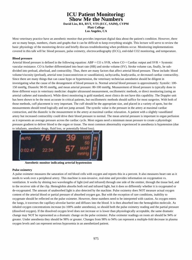
975
ICU Patient Monitoring: Show Me the Numbers
David Liss, BA, RVT, VTS (ECC, SAIM), CVPM Platt College
Los Angeles, CA
Most veterinary practice have an anesthetic monitor that provides important digital data about the patient's condition. However, there are so many beeps, numbers, charts and graphs that it can be difficult to keep everything straight. This lecture will serve to review the basic physiology of the monitoring device and briefly discuss troubleshooting when problems occur. Monitoring implementations covered in this talk will be: blood pressure, pulse oximetry, electrocardiography (ECG), end-tidal CO2 monitoring, and temperature. Blood pressure Arterial blood pressure is defined in the following equation: ABP = CO x SVR, where CO = Cardiac output and SVR = Systemic vascular resistance. CO is further differentiated into heart rate (HR) and stroke volume (SV). Stroke volume can, finally, be sub-divided into preload, afterload, and contractility. Thus, there are many factors that affect arterial blood pressure. These include: blood volume/viscosity (preload), arterial tone (vasoconstricton or vasodilation), tachycardia, bradycardia, or decreased cardiac contractility. Since there are many things that can cause hypo or hypertension, the veterinary technician anesthetist should be diligent in investigating what the cause of the derangement of blood pressure is. Normal arterial blood pressure is approximately: Systolic: 100-150 mmHg, Diastolic 90-95 mmHg, and mean arterial pressure: 80-100 mmHg. Measurement of blood pressure is typically done in three different ways in veterinary medicine: doppler ultrasound measurement, oscillometric methods, or direct monitoring (using an arterial catheter and transducer). While direct monitoring is gold standard, most clinics do not have this capability. The Doppler unit has been shown to be the most accurate in critical patients, but oscillometric methods should suffice for most surgeries. With both of those methods, cuff placement is very important. The cuff should be the appropriate size, and placed in a variety of spots, but the measurements should trend logically and not jump around. The systolic value is the pressure in the artery at maximal cardiac contraction, and the diastolic is the measurement in the artery at maximal cardiac relaxation. A patient with a slightly vasodilated artery but increased contractility could drive their blood pressure to normal. The mean arterial pressure is important in organ perfusion as it represents an average pressure across the cardiac cycle. Most organs need a minimum mean pressure to create a physiologic pressure gradient to deliver blood to the organ or tissue. The most common abnormality experienced in anesthesia is hypotension (due to inhalants, anesthetic drugs, fluid loss, or potentially blood loss).
Anesthetic monitor indicating arterial hypotension Pulse oximetry A pulse oximeter measures the saturation of red blood cells with oxygen and reports this in a percent. It also measures heart rate as it needs to work over a peripheral artery. This machine is non-invasive, real-time and provides information on oxygenation vs. ventilation. It works by shining two wavelengths of light (red and infrared) through one side of the emitter, through the tissue bed, and to the receiver side of the clip. Hemoglobin absorbs both red and infrared light, but it does so differently whether it is oxygenated or de-oxygenated. The amount of unabsorbed light is also detected by the machine. Pulse oximetry does NOT measure actual oxygen content of the arterial blood or partial pressure of absorbed oxygen gas. But with the exception of rare conditions, inability to oxygenate should be reflected on the pulse oximeter. However, these numbers need to be interpreted with caution. As oxygen enters the lungs, it traverses the capillary-alveolar barrier and diffuses into the blood. It is then absorbed into the hemoglobin molecule. As inhaled oxygen concentrations increase (to 100% under anesthesia), so should both the pulse oximetry reading and the partial pressure (dissolved oxygen). If the dissolved oxygen level does not increase or is lower than physiologically acceptable, the same dramatic change may NOT be represented as a dramatic change on the pulse oximeter. Pulse oximeter readings on room air should be 94% or greater. Under anesthesia they should be 98% or greater. Changes from 98% to 94% can represent a multiple-fold decrease in plasma oxygen levels and can represent serious hypoxemia in an anesthetized patient.
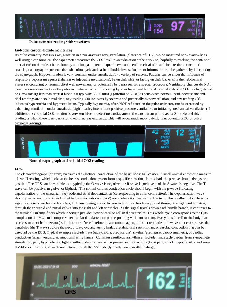
976
Pulse oximeter reading with waveform End-tidal carbon dioxide monitoring As pulse oximetry measures oxygenation in a non-invasive way, ventilation (clearance of CO2) can be measured non-invasively as well using a capnometer. The capnometer measures the CO2 level in an exhalation at the very end, hopfully mimicking the content of arterial carbon dioxide. This is done by attaching a T-piece adapter between the endotracheal tube and the anesthetic circuit. The resulting capnograph represents the exhalation cycle and carbon dioxide levels. Important information can be gathered by interpreting the capnograph. Hypoventilation is very common under anesthesia for a variety of reasons. Patients can be under the influence of respiratory depressant agents (inhalant or injectable medications), be on their side, or laying on their backs with their abdominal viscera encroaching on normal chest wall movement, or potentially be paralyzed for a special procedure. Ventilatory changes do NOT have the same drawbacks as the pulse oximeter in terms of reporting hypo or hyperventilation. A normal end-tidal CO2 reading should be a few mmHg less than arterial blood. So typically 30-35 mmHg (arterial of 35-40) is considered normal. And, because the end-tidal readings are also in real time, any reading <30 indicates hypocarbia and potentially hyperventilation, and any reading >35 indicates hypercarbia and hypoventilation. Typically hypoxemia, often NOT reflected on the pulse oximeter, can be corrected by enhancing ventilation under anesthesia (sigh breaths, intermittent positive pressure ventilation, or initiating mechanical ventilation). In addition, the end-tidal CO2 monitor is very sensitive in detecting cardiac arrest; the capnogram will reveal a 0 mmHg end-tidal reading as when there is no perfusion there is no gas exchange. This will occur much more quickly than potential ECG or pulse oximetry readings.
Normal capnograph and end-tidal CO2 reading ECG The electocardiograph (or gram) measures the electrical conduction of the heart. Most ECG's used in small animal anesthesia measure a Lead II reading, which looks at the heart's conduction system from a specific direction. In this lead, the p-wave should always be positive. The QRS can be variable, but typically the Q-wave is negative, the R wave is positive, and the S-wave is negative. The T-wave can be positive, negative, or biphasic. The normal cardiac conduction cycle should begin with the p-wave indicating depolarization of the sinoatrial (SA) node and atrial depolarization (corresponding to atrial contraction). The depolarization wave should pass across the atria and travel to the atrioventricular (AV) node where it slows and is directed to the bundle of His. Here the signal splits into two bundle branches, both innervating a specific ventricle. Blood has been pushed through the right and left atria, through the tricuspid and mitral valves into the right and left ventricles. As the signal travels down each bundle branch, it continues to the terminal Purkinje fibers which innervate just about every cardiac cell in the ventricles. This whole cycle corresponds to the QRS complex on the ECG and comprises ventricular depolarization (corresponding with contraction). Every muscle cell in the body that receives an electrical (nervous) stimulus, must "reset" before it can contract again, and so a repolarization wave then crosses over the ventricles (the T-wave) before the next p-wave occurs. Arrhythmias are abnormal rate, rhythm, or cardiac conduction that can be detected by the ECG. Typical examples include: rate (tachycardia, bradycardia), rhythm (premature, paroxysmal, etc), or cardiac conduction (atrial, ventricular, junctional arrhythmias). Common anesthetic arrhythmias include: sinus tachycardia (from sympathetic stimulation, pain, hypovolemia, light anesthetic depth), ventricular premature contractions (from pain, shock, hypoxia, etc), and some AV-blocks indicating slowed conduction through the AV node (typically from anesthetic drugs).

977
Temperature Although temperature seems like temperature is simplistic to measure and monitor, not all monitors continuously measure temperature and it is an important anesthetic parameter. Monitors that utilize an esophageal ECG often have a temperature function, but a continuous rectal temperature, or intermittent monitoring (with a digital thermometer) should be done routinely with the patient under anesthesia. Hypothermia is a common occurrence under anesthesia, and derangements in temperature can affect the cardiac cycle, blood pressure, and even coagulation. References available upon request.
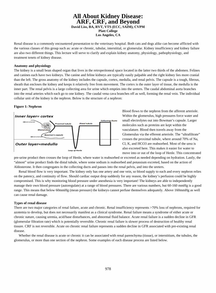
978
All About Kidney Disease: ARF, CRF, and Beyond
David Liss, BA, RVT, VTS (ECC, SAIM), CVPM Platt College
Los Angeles, CA
Renal disease is a commonly encountered presentation to the veterinary hospital. Both cats and dogs alike can become afflicted with the various classes of this group such as: acute or chronic, tubular, interstitial, or glomerular. Kidney insufficiency and kidney failure are also two different things. This lecture will serve to clarify and explain kidney anatomy, physiology, pathophysiology, and treatment tenets of kidney disease. Anatomy and physiology The kidney is a small bean shaped organ that lives in the retroperitoneal space located in the latter two thirds of the abdomen. Felines and canines each have two kidneys. The canine and feline kidneys are typically easily palpable and the right kidney lies more cranial than the left. The gross anatomy of the kidney includes the capsule, cortex, medulla, and renal pelvis. The capsule is a tough, fibrous, sheath that encloses the kidney and keeps it relatively free from movement. The cortex is the outer layer of tissue, the medulla is the inner part. The renal pelvis is a large collecting area for urine which empties into the ureters. The caudal abdominal aorta branches into the renal arteries which each go to one kidney. The caudal vena cava branches off as well, forming the renal vein. The individual cellular unit of the kidney is the nephron. Below is the structure of a nephron: Figure 1: Nephron
Blood flows to the nephron from the afferent arteriole. Within the glomerulus, high pressures force water and small electrolytes out into Bowman’s capsule. Larger molecules such as proteins are kept within the vasculature. Blood then travels away from the Glomerulus via the efferent arteriole. The “ultrafiltrate” crosses the proximal tubule, where around 70% of Na, Cl, K, and HCO3 are reabsorbed. Most of the urea is also excreted here. This makes it easier for water to move into or out of the loop of Henle. This concentrated
pre-urine product then crosses the loop of Henle, where water is reabsorbed or excreted as needed depending on hydration. Lastly, the “almost” urine product finds the distal tubule, where some sodium is reabsorbed and potassium excreted, based on the action of Aldosterone. It then congregates in the collecting ducts and passes into the renal pelvis, and into the ureters.
Renal blood flow is very important. The kidney only has one artery and one vein, so blood supply to each and every nephron relies on the patency, and continuity of flow. Should cardiac output drop suddenly for any reason, the kidney’s perfusion could be highly compromised. This is why monitoring blood pressure under anesthesia is very important! The kidneys are able to independently manage their own blood pressure (autoregulate) at a range of blood pressures. There are various numbers, but 60-160 mmHg is a good range. This means that below 60mmHg (mean pressure) the kidneys cannot perfuse themselves adequately. Above 160mmHg as well can cause renal damage. Types of renal disease There are two major categories of renal failure, acute and chronic. Renal insufficiency represents >70% loss of nephrons, required for azotemia to develop, but does not necessarily manifest as a clinical syndrome. Renal failure means a syndrome of either acute or chronic nature, causing uremia, acid/base disturbances, and abnormal fluid balance. Acute renal failure is a sudden decline in GFR (glomerular filtration rate) which is potentially reversible. Chronic renal failure is slower process of destruction of healthy renal tissure. CRF is not reversible. Acute on chronic renal failure represents a sudden decline in GFR associated with pre-existing renal disease.
Whether the renal disease is acute or chronic it can be associated with renal parenchyma (tissue), or interstitium, the tubules, the glomerulus, or more than one section of the nephron. Some examples of each disease process are listed below.
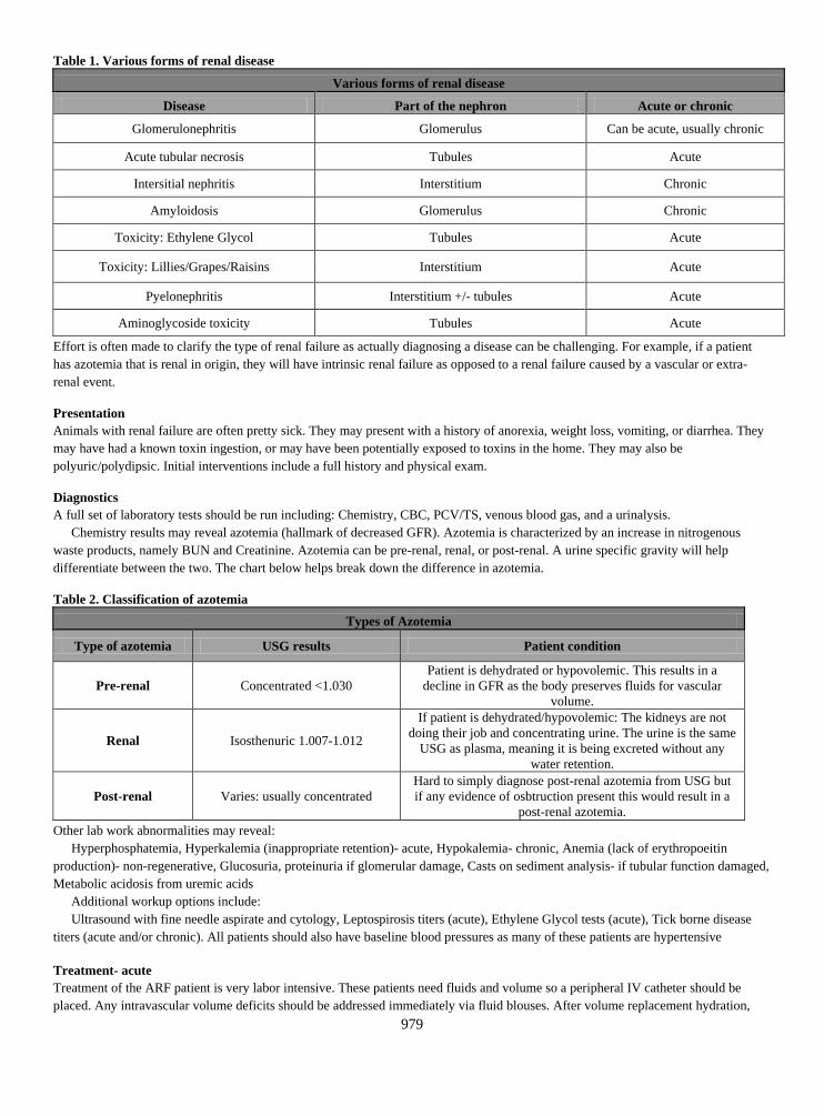
979
Table 1. Various forms of renal disease
Various forms of renal disease
Disease Part of the nephron Acute or chronic
Glomerulonephritis Glomerulus Can be acute, usually chronic
Acute tubular necrosis Tubules Acute
Intersitial nephritis Interstitium Chronic
Amyloidosis Glomerulus Chronic
Toxicity: Ethylene Glycol Tubules Acute
Toxicity: Lillies/Grapes/Raisins Interstitium Acute
Pyelonephritis Interstitium +/- tubules Acute
Aminoglycoside toxicity Tubules Acute
Effort is often made to clarify the type of renal failure as actually diagnosing a disease can be challenging. For example, if a patient has azotemia that is renal in origin, they will have intrinsic renal failure as opposed to a renal failure caused by a vascular or extra-renal event. Presentation Animals with renal failure are often pretty sick. They may present with a history of anorexia, weight loss, vomiting, or diarrhea. They may have had a known toxin ingestion, or may have been potentially exposed to toxins in the home. They may also be polyuric/polydipsic. Initial interventions include a full history and physical exam. Diagnostics A full set of laboratory tests should be run including: Chemistry, CBC, PCV/TS, venous blood gas, and a urinalysis.
Chemistry results may reveal azotemia (hallmark of decreased GFR). Azotemia is characterized by an increase in nitrogenous waste products, namely BUN and Creatinine. Azotemia can be pre-renal, renal, or post-renal. A urine specific gravity will help differentiate between the two. The chart below helps break down the difference in azotemia. Table 2. Classification of azotemia
Types of Azotemia
Type of azotemia USG results Patient condition
Pre-renal Concentrated <1.030 Patient is dehydrated or hypovolemic. This results in a
decline in GFR as the body preserves fluids for vascular volume.
Renal Isosthenuric 1.007-1.012
If patient is dehydrated/hypovolemic: The kidneys are not doing their job and concentrating urine. The urine is the same
USG as plasma, meaning it is being excreted without any water retention.
Post-renal Varies: usually concentrated Hard to simply diagnose post-renal azotemia from USG but if any evidence of osbtruction present this would result in a
post-renal azotemia. Other lab work abnormalities may reveal:
Hyperphosphatemia, Hyperkalemia (inappropriate retention)- acute, Hypokalemia- chronic, Anemia (lack of erythropoeitin production)- non-regenerative, Glucosuria, proteinuria if glomerular damage, Casts on sediment analysis- if tubular function damaged, Metabolic acidosis from uremic acids
Additional workup options include: Ultrasound with fine needle aspirate and cytology, Leptospirosis titers (acute), Ethylene Glycol tests (acute), Tick borne disease
titers (acute and/or chronic). All patients should also have baseline blood pressures as many of these patients are hypertensive
Treatment- acute Treatment of the ARF patient is very labor intensive. These patients need fluids and volume so a peripheral IV catheter should be placed. Any intravascular volume deficits should be addressed immediately via fluid blouses. After volume replacement hydration,
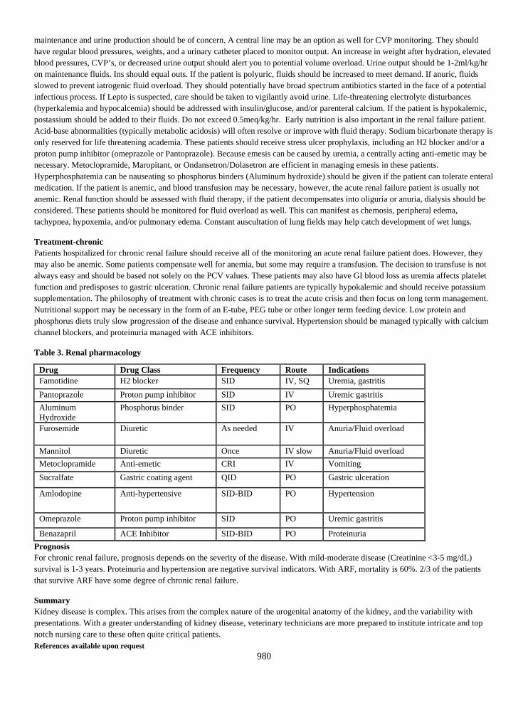
980
maintenance and urine production should be of concern. A central line may be an option as well for CVP monitoring. They should have regular blood pressures, weights, and a urinary catheter placed to monitor output. An increase in weight after hydration, elevated blood pressures, CVP’s, or decreased urine output should alert you to potential volume overload. Urine output should be 1-2ml/kg/hr on maintenance fluids. Ins should equal outs. If the patient is polyuric, fluids should be increased to meet demand. If anuric, fluids slowed to prevent iatrogenic fluid overload. They should potentially have broad spectrum antibiotics started in the face of a potential infectious process. If Lepto is suspected, care should be taken to vigilantly avoid urine. Life-threatening electrolyte disturbances (hyperkalemia and hypocalcemia) should be addressed with insulin/glucose, and/or parenteral calcium. If the patient is hypokalemic, postassium should be added to their fluids. Do not exceed 0.5meq/kg/hr. Early nutrition is also important in the renal failure patient. Acid-base abnormalities (typically metabolic acidosis) will often resolve or improve with fluid therapy. Sodium bicarbonate therapy is only reserved for life threatening academia. These patients should receive stress ulcer prophylaxis, including an H2 blocker and/or a proton pump inhibitor (omeprazole or Pantoprazole). Because emesis can be caused by uremia, a centrally acting anti-emetic may be necessary. Metoclopramide, Maropitant, or Ondansetron/Dolasetron are efficient in managing emesis in these patients. Hyperphosphatemia can be nauseating so phosphorus binders (Aluminum hydroxide) should be given if the patient can tolerate enteral medication. If the patient is anemic, and blood transfusion may be necessary, however, the acute renal failure patient is usually not anemic. Renal function should be assessed with fluid therapy, if the patient decompensates into oliguria or anuria, dialysis should be considered. These patients should be monitored for fluid overload as well. This can manifest as chemosis, peripheral edema, tachypnea, hypoxemia, and/or pulmonary edema. Constant auscultation of lung fields may help catch development of wet lungs. Treatment-chronic Patients hospitalized for chronic renal failure should receive all of the monitoring an acute renal failure patient does. However, they may also be anemic. Some patients compensate well for anemia, but some may require a transfusion. The decision to transfuse is not always easy and should be based not solely on the PCV values. These patients may also have GI blood loss as uremia affects platelet function and predisposes to gastric ulceration. Chronic renal failure patients are typically hypokalemic and should receive potassium supplementation. The philosophy of treatment with chronic cases is to treat the acute crisis and then focus on long term management. Nutritional support may be necessary in the form of an E-tube, PEG tube or other longer term feeding device. Low protein and phosphorus diets truly slow progression of the disease and enhance survival. Hypertension should be managed typically with calcium channel blockers, and proteinuria managed with ACE inhibitors.
Table 3. Renal pharmacology
Prognosis For chronic renal failure, prognosis depends on the severity of the disease. With mild-moderate disease (Creatinine <3-5 mg/dL) survival is 1-3 years. Proteinuria and hypertension are negative survival indicators. With ARF, mortality is 60%. 2/3 of the patients that survive ARF have some degree of chronic renal failure.
Summary Kidney disease is complex. This arises from the complex nature of the urogenital anatomy of the kidney, and the variability with presentations. With a greater understanding of kidney disease, veterinary technicians are more prepared to institute intricate and top notch nursing care to these often quite critical patients. References available upon request
Drug Drug Class Frequency Route Indications Famotidine H2 blocker SID IV, SQ Uremia, gastritis Pantoprazole Proton pump inhibitor SID IV Uremic gastritis Aluminum Hydroxide
Phosphorus binder SID PO Hyperphosphatemia
Furosemide Diuretic As needed IV Anuria/Fluid overload
Mannitol Diuretic Once IV slow Anuria/Fluid overload Metoclopramide Anti-emetic CRI IV Vomiting Sucralfate Gastric coating agent QID PO Gastric ulceration
Amlodopine Anti-hypertensive SID-BID PO Hypertension
Omeprazole Proton pump inhibitor SID PO Uremic gastritis
Benazapril ACE Inhibitor SID-BID PO Proteinuria
981
Pain Management in the ER: The Fifth Vital Sign
David Liss, BA, RVT, VTS (ECC, SAIM), CVPM Platt College
Los Angeles, CA
The recognition and treatment of pain is an incredibly important part of the hospitalized veterinary patient's regimen. Patients that do not have their pain addressed might suffer longer hospitalization times or face increases in morbidity and mortality from pain. Pain is known to elicit a sympathetic response worsening or inciting shock states which promote decreased wound healing and decreased organ perfusion. The veterinary technician is invaluable in assessing and reporting findings to the veterinarian. The veterinary technician, in conjunction with the veterinarian, can discuss and implement a multi-modal approach to managing pain in critically ill patients. Pain physiology and pathophysiology Pain is typically thought of as an adaptive response to prevent injury. If you stick your arm over a fire, it hurts so you can pull away and minimize tissue damage. However, severe injury, or failure to treat pain can cause detrimental physiologic effects, beyond the positive effects of self-preservation. In the periphery, specialized nerves, called nociceptors, exist and transmit pain signals to the spinal cord and to the brain. The free nerve endings, which terminate in soft tissue, have various receptors that can be activated in response to thermal, chemical, or mechanical noxious stimuli. For example, an acid burn will stimulate different fibers than a laceration. The first step in the pain process is transduction, where nerve endings convert stimuli into electrical signals. The two main nerve fibers (each with separate ability to transmit various stimuli) are the A-delta and C fibers. The A-delta fibers tend to fire faster, sending quicker signals to the spinal cord. The C-fibers tend to have a slower ability to reach their threshold. Thus, pain that is felt immediately upon exposure to noxious stimuli (crushing, pinching, tearing) is transmitted through A-delta fibers. Pain that is felt with a bit of a pause, cold temperature, etc is transmitted across C-fibers. Another important point is that there are various A-delta and C nerve fibers contain endings that typically do not transmit pain signals (initially) but can be "woken up" and recruited in severe circumstances. The next step in the pain process is transmission; after the nociceptors have converted the stimulus to energy it is sent to the spinal cord for initial processing. The signal travels through the nerve fibers to the dorsal part of the spinal cord. As discussed earlier, C-fibers are 10x slower than A-delta fibers in their transmitting speed. After pain signals reach the spinal cord, modulation occurs. Here, the spinal cord either dampens or increases the pain signal according to various neurotransmitters or chemicals that are activated/deactivated in the spinal cord. The majority of pain signals that make it to the spinal cord and are sent on are mediated by glutamate, a neurotransmitter. Glutamate acts on the AMPA, KAI, and neurokinin (NK) receptors and stimulates a response by sending the signal up the spinal cord to the brain. The NMDA receptor (upon which ketamine exerts it's effects) is responsible for amplifying pain signals, whether they are incredibly strong or not. The NDMA receptor is thought to be important in prolonged/amplified pain states. A neurotransmitter called Substance P activates the NMDA receptor. Finally, GABA receptors, when activated, tend to inhibit signals from crossing into the spinal cord to be processed. There are other important neurotransmitters involved in modulation of signals in the dorsal horn of the spinal cord. These include: serotonin, norepinephrine, and opioid receptors. Serotonin, norepinephrine and opioid receptors, when activated, inhibit excitation of neurons, thus agonists of these drugs have analgesic properties. The last, and final, step of the pain pathway is perception. Perception occurs in multiple parts of the brain and is then perceived as an unpleasant sensation associated with real or perceived tissue damage. Pain can be categorized in various different ways: disease or anatomy related (pancreatic, etc), location (superficial, visceral, deep), duration (acute, chronic) or intensity (mild, moderate, or severe). These often require some objective input) from the patient, so categorizing these in veterinary patients can be challenging.
A few other important concepts in pain management in the acute patient include: Allodynia, sensititzation, windup, and referred pain. Allodynia refers to an exaggerated reaction to a stimulus that is normally not painful. This can occur due to an exaggerated pain response where the pain threshold of nociceptors is lowered. Sensitization and windup are the result of peripheral and central physiochemical changes that occur during tissue damage and the inflammatory response. Peripherally, inflammatory mediators and cells can reduce the threshold of normally high-threshold nociceptors, and awaken "sleeping" nociceptors causing an exaggerated pain response. Central sensitization (windup) occurs as another mechanism for an exaggerated pain response, and because this occurs in the spinal cord, can result in severe pain that lasts much longer than the initial tissue insult. Repeated signaling to the spinal cord activates excitatory neurotransmitters which activate various receptors (NMDA, notably) and secure open-channels for pain stimuli to pass through. It appears that central sensitization can be responsible for allodynia. Referred pain is pain in a body part that is not affected by tissue damage. This might occur in a limb that was not amputated (phantom limb pain), or pain in limbs where the source of the pain is in the abdomen, for example.
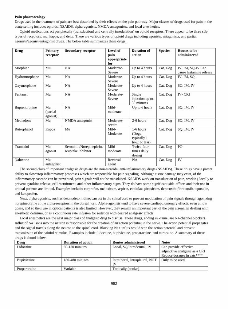
982
Pain pharmacology Drugs used in the treatment of pain are best described by their effects on the pain pathway. Major classes of drugs used for pain in the acute setting include: opioids, NSAIDS, alpha-agonists, NMDA-antagonists, and local anesthetics.
Opioid medications act peripherally (transduction) and centrally (modulation) on opioid receptors. There appear to be three sub-types of receptors: mu, kappa, and delta. There are various types of opioid drugs including agonists, antagonists, and partial agonists/agonist-antagonist drugs. The below table summarizes these drugs. Drug Primary
receptor Secondary receptor Level of
pain appropriate for
Duration of action
Species Routes to be administered
Morphine Mu NA Moderate-Severe
Up to 4 hours Cat, Dog IV, IM, SQ-IV Can cause histamine release
Hydromorphone Mu NA Moderate-Severe
Up to 4 hours Cat, Dog IV, IM, SQ
Oxymorphone Mu NA Moderate-Severe
Up to 4 hours Cat, Dog SQ, IM, IV
Fentanyl Mu NA Moderate-Severe
Single injection up to 30 minutes
Cat, Dog IV- CRI
Buprenorphine Mu (partial agonist)
NA Mild-moderate
Up to 6 hours Cat, Dog SQ, IM, IV
Methadone Mu NMDA antagonist Moderate-severe
2-6 hours Cat, Dog SQ, IM, IV
Butorphanol Kappa Mu Mild-Moderate
1-6 hours (Dogs typically 1 hour or less)
Cat, Dog SQ, IM, IV
Tramadol Mu agonist
Serotonin/Norepinephrine reuptake inhibitor
Mild-moderate
Twice-four times daily dosing
Cat, Dog PO
Naloxone Mu antagonist
Reversal agent
NA Cat, Dog IV
The second class of important analgesic drugs are the non-steroidal anti-inflammatory drugs (NSAIDS). These drugs have a potent ability to slow/stop inflammatory processes which are responsible for pain signaling. Although tissue damage may exist, of the inflammatory cascade can be prevented, pain signals will not be transduced. NSAIDS work on transduction of pain, working locally to prevent cytokine release, cell recruitment, and other inflammatory signs. They do have some significant side-effects and their use in critical patients are limited. Examples include: carprofen, meloxicam, aspirin, etodolac, piroxicam, deracoxib, fibrocoxib, tepoxalin, and ketoprofen.
Next, alpha-agonists, such as dexmedetomidine, can act in the spinal cord to prevent modulation of pain signals through agonizing norepinephrine at the alpha-receptors in the dorsal horn. Alpha-agonists tend to have severe cardiopulomonary effects, even at low doses, and so their use in critical patients is also limited. However, they remain an important part of the pain arsenal in dealing with anesthetic delirium, or as a continuous rate infusion for sedation with desired analgesic effects.
Local anesthetics are the next major class of analgesic drug to discuss. These drugs, ending in -caine, are Na-channel blockers. Influx of Na+ ions into the neuron is responsible for the creation of an action potential in the nerve. The action potential propagates and the signal travels along the neuron to the spinal cord. Blocking Na+ influx would stop the action potential and prevent transmission of the painful stimulus. Examples include: lidocaine, bupivicaine, proparacaine, and tetracaine. A summary of these drugs is found below.
Drug Duration of action Routes administered Notes Lidocaine 60-120 minutes Local, SQ/Intradermal, IV Can provide effective
adjunctive analgesia as a CRI Reduce dosages in cats****
Bupivicaine 180-480 minutes Intrathecal, Intrapleural, NOT IV
Only to be used
Proparacaine Variable Topically (ocular)
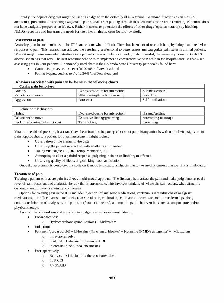
983
Finally, the adjunct drug that might be used in analgesia in the critically ill is ketamine. Ketamine functions as an NMDA-antagonist, preventing or stopping exaggerated pain signals from passing through these channels to the brain (windup). Ketamine does not have analgesic properties on it's own. Rather, it seems to potentiate the effects of other drugs (opioids notably) by blocking NMDA-receptors and lowering the needs for the other analgesic drug (opioid) by itself. Assessment of pain Assessing pain in small animals in the ICU can be somewhat difficult. There has been alot of research into physiologic and behavioral responses to pain. This research has allowed the veterinary professional to better assess and categorize pain states in animal patients. While it might seem somewhat intuitive that a patient who was hit by a car and growls is painful, the veterinary community didn't always see things that way. The best recommendation is to implement a comprehensive pain scale in the hospital and use that when assessing pain in your patients. A commonly used chart is the Colorado State University pain scales found here:
• Canine: ivapm.evetsites.net/refid.20468/refDownload.pml • Feline: ivapm.evetsites.net/refid.20467/refDownload.pml
Behaviors associated with pain can be found in the following charts
Canine pain behaviors Anxiety Decreased desire for interaction Submissiveness Reluctance to move Whimpering/Howling/Growling Guarding Aggression Anorexia Self-mutiliation
Feline pain behaviors Hiding Decreased desire for interaction Hissing/spitting Reluctance to move Excessive licking/grooming Attempting to escape Lack of grooming/unkempt coat Tail flicking Crouching Vitals alone (blood pressure, heart rate) have been found to be poor predictors of pain. Many animals with normal vital signs are in pain. Approaches to a patient for a pain assessment might include:
• Observation of the animal in the cage • Observing the patient interacting with another staff member • Taking vital signs: HR, RR, Temp, Mentation, BP • Attempting to elicit a painful response: palpating incision or limb/organ affected • Observing quality of life: eating/drinking, coat, ambulation
Once the assessment is complete, the decision is made to institute analgesic therapy or modify current therapy, if it is inadequate. Treatment of pain Treating a patient with acute pain involves a multi-modal approach. The first step is to assess the pain and make judgments as to the level of pain, location, and analgesic therapy that is appropriate. This involves thinking of where the pain occurs, what stimuli is causing it, and if there is a windup component.
Options for treating pain in the ICU include: injections of analgesic medications, continuous rate infusions of analgesic medications, use of local anesthetic blocks near site of pain, epidural injection and catheter placement, transdermal patches, continuous infusion of analgesics into pain site ("soaker catheters), and non-allopathic interventions such as acupuncture and/or physical therapy.
An example of a multi-modal approach to analgesia in a thoracotomy patient: • Pre-medication:
o Hydromorphone (pure u opioid) + Midazolam • Induction: • Fentanyl (pure u opioid) + Lidocaine (Na-channel blocker) + Ketamine (NMDA antagonist) + Midazolam
o Intra-operatively: o Fentanyl + Lidocaine + Ketamine CRI o Intercostal block (local anesthesia)
• Post-operatively: o Bupivicaine infusion into thoracostomy tube o FLK CRI o +/- NSAID
984
Garbage Gut: Acute Pancreatitis
David Liss, BA, RVT, VTS (ECC, SAIM), CVPM Platt College
Los Angeles, CA
Pancreatitis is a commonly seen disease presenting to the emergency room. Technicians often can get a hint from the history, (“Oh he got into the trash last night”), and are ready to implement orders and treat these patients. But what about some of the nuances like: nutrition, pathophysiology, and even critical care if these patients go south? This lecture will present a holistic view of acute pancreatitis, focused on the dog, but some information about feline pancreatitis will be presented as well. Pathophysiology The pancreas is a glandular organ in the cranial abdomen. It is attached to the wall of the duodenum and it lies between the duodenum and the stomach on the right side of the body. It has two lobes; the right and left. The right lobe extends down the duodenum and the left lobe angles medially, underneath the body of the stomach.
The major function of the exocrine pancreas (endocrine pancreas secretes insulin) is to store and then release inactive enzymes for digestion of nutrients. The acinar cells produce these and store them in granules. Dogs have two pancreatic ducts, the pancreatic and accessory pancreatic ducts which attach to the duodenum, where the enzymes are released. Cats only have one duct. The majority of the pancreatic cells are consumed in this function.
Pancreatic enzymes digest tissue, and thus are relatively toxic to the body if activated. The pancreas creates and stores the enzymes in an inactive form, called zymogens. The zymogens are sequestered into granules, so they will not come into contact with other pancreatic tissue and cells. Once the inactive zymogens are secreted into the duodenum they are activated by enzymes in the small intestine. Trypsinogen is a major pancreatic enzyme, and is converted into its active form, trypsin. In a cascade-like effect, trypsin activates other pancreatic enzymes.
The pancreas also has inhibitor enzymes, preventing the body from being over-run by these irritating chemicals. Trypsin inhibitor is produced and stored in the pancreas and inhibits trypsin activity. In addition to local pancreatic anti-enzyme chemicals, are the circulating anti-proteases; the primary one is α-macroglobulin among others. However, there is only a certain amount of these anti-enzyme inhibitors and once they are consumed, pancreatic enzymes are free to wreak havoc with bodily tissue.
Acute pancreatitis is seen relatively frequently in the veterinary hospital. It results from an inappropriate activation of the typically inactive zymogens, inside the pancreas, causing autodigestion of the pancreas. Pancreatic inflammation, cellular edema, and necrosis typically occur. Once the pancreatic cells rupture, they leak active pancreatic enzymes in the peritoneum and systemic circulation. This causes widespread systemic inflammation causing a SIRS syndrome. In rare cases, these pancreatic enzymes and systemic inflammation can cause coagulation cascade activation, and thus DIC, and multiple organ dysfunction and death.
Pancreatitis can take the form of severe or non-severe acute pancreatitis. Chronic pancreatitis also exists, but a different approach to treatment is often taken. Severe acute pancreatitis is elicited when systemic signs of pancreatitis occur with multiple organ involvement or dysfunction. Pancreatic necrosis, abscesses, or pseudocysts are typically found in these patients.
History/physical examination Due to the variability of the disease, some patients remain in a subclinical phase, or have mild GI signs. Anecdotally, miniature schnauzers are more prone to the disease; however, this has yet to be proven. In addition, gluttony, and the consumption of a fatty meal, or “garbage gut” is also thought of in conjunction with pancreatitis.
Owners will often report GI signs such as anorexia, and vomiting. Diarrhea may occur. Patients are often lethargic. The signs may mimic an upper GI obstruction, so it is important to carefully evaluate the patient and perform timely diagnostic tests.
Patients will often present with dehydration, fever, icterus (if biliary tree is involved), and abdominal pain. Pancreatitis is reported to be a painful human disease, and generally regarded as a painful animal disease as well. These patients may show signs of shock, such as tachycardia, tachypnea, weak pulses, hypotension, mental dullness.
Diagnostic testing
Radiography and ultrasonography Abdominal radiography is recommended in patients suspected of having acute pancreatitis. It is difficult to diagnose pancreatitis via radiography but it is still a useful modality to help rule out upper GI obstruction. Radiographic findings may include: loss of detail in cranial abdomen, displacement of stomach to the left, displacement of duodenum to the right or ventrally. However, only 24% of patients in a large study on fatal pancreatitis had radiographic changes. Ultrasonography is a relatively useful test in diagnosing acute pancreatitis. However, it certainly relies on the skill of the ultrasonographer. Ultrasonographic findings include an enlarged pancreas, hypoechoic pancreatic center, hyperechoic area surrounding the pancreas (peripancreatic fat necrosis), and effusion surrounding the
985
pancreas. Ultrasonography is more sensitive (less false negatives) than radiography. In human medicine CT is considered gold standard for evaluation of the pancreas. By contrast, in few limited veterinary studies, CT did not hold up to ultrasonography or serologic testing.
Laboratory analysis All patients presenting to the emergency room will benefit from a minimum database (PCV, TS, Blood Glucose, BUN, Electrolytes, Venous blood gas), and a complete blood count and biochemical profile.
Common findings include: inflammatory leukogram (neutrophilia with left shift and toxic changes), anemia and thrombocytopenia (if patient is in DIC), elevated PCV from dehydration, hemoconcentration, elevated amylase and lipase levels, azotemia, hyperglycemia, hypoglycemia, hypercholesterolemia, elevated liver enzymes and hyperbilirubinemia, and hypocalcemia.
Important testing notes Elevated amylase and lipase are not highly correlated as diagnostic of pancreatitis. Firstly, the tests run on typical in-house biochemical analyzers are not pancreas specific. Amylase and lipase are found in the oral cavity, GI tract, pancreas, and liver. And decreased glomerular filtration can greatly affect amylase/lipase levels. The reported sensitivities (amount of accuracy compared to false positives) is 50-70%. Meaning 30-50% of increased amylase/lipase enzymes were not associated with actual diagnosis of pancreatitis. Also, normal amylase/lipase levels do not rule out pancreatitis either. As the pancreas is quite near the bile duct, inflammation and swelling can cause an extrahepatic bile duct obstruction. Thus liver enzymes and makers of biliary tree disease, such as total bilirubin levels, may be eleavated. Hyperglycemia is a common finding and is either a marker of stress or development of diabetes mellitus. 30% of dogs presenting for pancreatitis were found to be hyperglycemic, and 63% of those were found to be diabetic. Hypocalcemia can result from calcium salt formation in the pancreas, related to pancreatic inflammation. In cats, this finding carries a poor prognosis. The prognostic value of this in dogs is unknown.
Pancreatic markers/inflammatory markers C-reactive protein is used in humans to determine severity of pancreatitis. It is an acute-phase protein, (one that is induced in inflammatory states) and may have a prognostic value in dogs. Elevated levels were found in dogs with pancreatic abscess compared to those with just pancreatic inflammation. Elevated C-reactive protein levels were found in dogs with acute pancreatitis vs. normal dogs, making it a marker of pancreatitis. However, although it is sensitive, it is not entirely specific to pancreatitis as elevated levels are found in patients with generalized systemic inflammation. The trypsin-like immunoreactivity (TLI) and pancreatic lipase immuoreactivity (PLI) tests are both pancreas-specific tests and are used in the diagnosis of pancreatitis. While the TLI test is useful in diagnosing exocrine pancreatic insufficiency, its sensitivity and specificity with pancreatic inflammation and dysfunction are low. Therefore, the PLI is the preferred test. It reports pancreas-specific lipase, which is chemically unique from other lipases, and is unaffected by renal failure. The fPLI test will run feline specific pancreatic liplase, and the cPLI test will run canine specific lipase. The spec-CPL test (same as cPLI) can be run through commercial laboratories, and a snap test was introduced into the market a few years ago. It is still recommended to follow up a positive snap test with a spec-CPL measurement. The spec-CPL has greater accuracy for canines, than the fPLI test does for felines. Typically values in the dog >400ug/dL are considered diagnostic with a sensitivity of 93%. Treatment
Fluid therapy Patients with pancreatitis are typically dehydrated, and potentially hypovolemic. If there are any perfusion deficits present, boluses of balanced crystalloid solutions should be used to replace intravascular volume. Once perfusion is restored, rehydration and treatment of ongoing losses can occur. Typically a crystalloid solution that is isotonic and contains some trace electrolytes is used. Potassium supplementation may be used to treat losses.
Anti-emetics Patients that are vomiting are typically treated with anti-emetics to prevent further vomiting and fluid loss. Maropitant (Cerenia®) a NK1 receptor antagonist, can be a very useful anti-nausea and anti-emetic drug. It is labeled for SQ use and can sting when injected. Metoclopramide (Reglan®) is a pro-kinetic and anti-emetic drug. It increases lower esophageal sphincter tone, stimulates peristalsis and acts peripherally and centrally to prevent vomiting. The 5-HT3 serotinin receptor antagonists Ondansetron (Zofran®) and Dolasetron (Anzemet®) can be used to treat vomiting as well. Anti-acid drugs, such as H2 receptor antagonists Famotidine (Pepcid®) and Ranitidine (Zantac®) are effective in lower gastric pH to prevent ulcer formation. The proton-pump inhibitor class of antacid medications are superior to the H2 blocker drugs and include Pantoprazole (Protonix®) and Omeprazole (Prilosec®).
Pain management Analgesia is an important part of treating the pancreatitis patient. Pain assessment and scoring should be used to assess the severity of pain. The first important step is observing the patient. Are they hunched? Do they have a praying stance? Are they crouched at the back of the cage? Are they growling or snarling? Are they attention seeking? These may be signs of pain in addition to physiologic parameters (hyperthermia, hypertension, tachycardia, tachypnea). Pure mu opioids are best at achieving analgesia in these patients. Morphine, Hydromorphone, Oxymorphone or Fentanyl are highly effective. Buprenorphine can be used, but it is a milder analgesic,
986
lasts a long time, and is harder to reverse. I have often found that pure mu opioids need to be given above a Buprenorphine dose for “breakthrough” pain. Fentanyl CRI’s are nice in the ICU because they can be titrated to effect. In addition, Lidocaine and Ketamine adjunctive analgesia can be added for additional pain control.
Nutrition This can be very important in pancreatitis. Historically, patients were not fed at all during the course of pancreatitis. Feeding during vomiting is thought to be controversial, and the addition of food to the stomach induces the pancreas to work, and secrete digestive enzymes. So post-pyloric feeding is often indicated. This is accomplished with a jejunostomy tube. Nasojejunostomy tubes are described but rare. Typically a J-tube is fed through a Gastrostomy tube and used that way. However, these require anesthesia for surgical or percutaneous (endoscopic) placement. Diets fed through a J-tube need to be easily digestible and several elemental diets exist (Clinicare®, Vivonex®, Enteral Care®). These diets are not truly balanced in nutrients and not for the long term. Cats also require different combinations of fat, protein, and carbs so Feline Clinicare® may be the better option for them. In cats, pre-pyloric feeding is often well tolerated, (nasogastric, nasoesophageal, or esophagostomy tubes). In my experience, nasogastric tubes in dogs are typically well tolerated. It is advised to use the enteral route whenever possible. If the patient will not tolerate enteral feedings, the parenteral route may be used. Indications for parenteral nutrition include: intractable vomiting, large volume raging diarrhea, or complications with tube placement. Patients with pancreatitis are typically in a catabolic state and thus partial parenteral nutrition is probably not enough. Total parenteral nutrition, or TPN, is often required. This is an expensive and technically demanding nutritional intervention, requiring 24 hour care and central line placement. There are many complications with TPN but it can be a rewarding treatment if successful. Rare complications If pancreatitis is severe, complications such as DIC and multi-organ failure can result. Careful screening for these problems can help catch them before they have progressed. DIC is a difficult syndrome to treat and is characterized by early hypercoagulability and subsequent hypocoagulability. Fresh frozen plasma and anticoagulants are typically employed in the treatment of DIC. FFP use prior to DIC is not indicated in the literature. Multi-organ failure can include liver failure, renal failure, pulmonary dysfunction (acute respiratory distress syndrome) or GI dysfunction. Gut necrosis can lead to bacterial translocation and sepsis, despite most uncomplicated pancreatitis being considered a non-infectious disease. Careful nursing monitoring of the hematologic, renal, and GI systems can help prevent these problems from occurring. Conclusion Pancreatitis has a favorable outcome if treated early and aggressively. There is no definitive treatment, and most treatments are symptomatic. Early aggressive nutrition can help provide a positive outcome. Careful monitoring of vital signs and organ systems can catch problems early and help guide therapy. References available upon request.
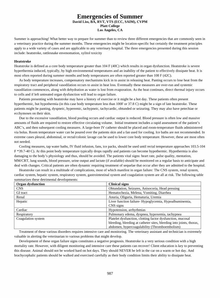
987
Emergencies of Summer David Liss, BA, RVT, VTS (ECC, SAIM), CVPM
Platt College Los Angeles, CA
Summer is approaching! What better way to prepare for summer than to review three different emergencies that are commonly seen in a veterinary practice during the summer months. These emergencies might be location-specific but certainly the treatment principles apply to a wide variety of cases and are applicable to any veterinary hospital. The three emergencies presented during this session include: heatstroke, rattlesnake envenomation, xylitol toxicity Heatstroke Heatstroke is defined as a core body temperature greater than 104 F (40C) which results in organ dysfunction. Heatstroke is severe hyperthermia induced, typically, by high environmental temperatures and an inability of the patient to effectively dissipate heat. It is most often reported during summer months and body temperatures are often reported greater than 108 F (42C).
As body temperature increases, compensatory mechanisms kick in to assist in releasing heat. Panting occurs to lose heat from the respiratory tract and peripheral vasodilation occurs to assist in heat loss. Eventually these measures are over-run and systemic vasodilation commences, along with dehydration as water is lost from evaporation. As the heat continues, direct thermal injury occurs to cells and if left untreated organ dysfunction will lead to organ failure.
Patients presenting with heatstroke may have a history of exercise or it might be a hot day. These patients often present hyperthermic, but hypothermia (in this case body temperature less than 100F or 37.8 C) might be a sign of late heatstroke. These patients might be panting, dyspneic, hyperemic, tachypneic, tachycardic, obtunded or seizuring. They may also have petechiae or ecchymoses on their skin.
Due to the excessive vasodilation, blood pooling occurs and cardiac output is reduced. Blood pressure is often low and massive amounts of fluids are required to restore effective circulating volume. Initial treatment includes a rapid assessment of the patient’s ABC’s, and then subsequent cooling measures. A large-bore IV catheter should be placed and room-temperature fluids administered via bolus. Room temperature water can be poured over the patients skin and a fan used for cooling. Ice baths are not recommended. In extreme cases pleural, abdominal, or rectal/colonic lavage can be used to lower core body temperature. However, these are most often not needed.
Cooling measures, tap water baths, IV fluid infusion, fans, ice packs, should be used until rectal temperature approaches 103.5-104 F *39.7-40 C). At this point body temperature typically drops rapidly and patients can become hypothermic. Hypothermia is also damaging to the body’s physiology and thus, should be avoided. The patients vital signs: heart rate, pulse quality, mentation, MM/CRT, lung sounds, blood pressure, urine output and lactate (if available) should be monitored on a regular basis to anticipate and deal with changes. Critical patients are often dynamic requiring treatment of sequelae that occur after they are admitted to the hospital.
Heatstroke can result in a multitude of complications, most of which manifest in organ failure: The CNS system, renal system, cardiac system, hepatic system, respiratory system, gastrointestinal system and coagulation system are all at risk. The following table summarizes these detrimental developments:
Organ dysfunction Clinical signs CNS Obtundation, Seizures, Anisocoria, Head pressing GI tract Hematochezia, Melena, Vomiting, Diarrhea Renal Anuria, Oliguria, Hematuria, Uremia Hepatic Liver function failure- Hypoglycemia, Hypoalbuminemia,
CNS signs Cardiac Hypotension, arrhythmias Respiratory Pulmonary edema, dyspnea, hypoxemia, tachypnea Coagulation system Platelet dysfunction, clotting factor dysfunction, mucosal
bleeding, bleeding at catheter sites, bleeding into joints, thorax, abdomen, hypercoagulability (Thromboembolism)
Treatment of these various disorders requires intensive care and monitoring. The veterinary assistant and technician is extremely valuable in alerting the veterinarian to various problems that might develop.
Development of these organ failure signs constitutes a negative prognosis. Heatstroke is a very serious condition with a high mortality rate. However, with diligent monitoring and intensive care these patients can recover! Client education is key to preventing this disease. Animal should not be worked hard on hot days. They should NEVER be left in the car on a warm or hot day. And brachycephalic patients should be walked and exercised carefully as their body condition limits their ability to dissipate heat.
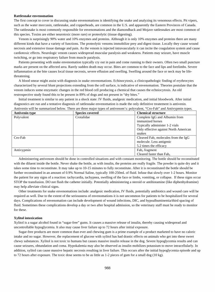
988
Rattlesnake envenomation The first concept to cover in discussing snake envenomations is identifying the snake and analyzing its venomous effects. Pit vipers, such as the water moccasin, rattlesnake, and copperheads, are common in the U.S. and apparently the Eastern Provinces of Canada. The rattlesnake is most commonly responsible for envenomations and the diamondback and Mojave rattlesnakes are most common of this species. Toxins are either neurotoxic (more rare) or proteolytic (tissue digesting).
Venom is surprisingly 90% water and 10% enzymes and proteins. Although it is only 10% enzymes and proteins there are many different kinds that have a variety of functions. The proteolytic venoms immobilize prey and digest tissue. Locally they cause wound necrosis and extensive tissue damage and pain. As the venom is injected intravascularly it can incite the coagulation system and cause cardiotoxic effects. Neurologic venom causes widespread muscular paralysis and weakness. Patients may seizure, have muscle twitching, or go into respiratory failure from muscle paralysis.
Patients presenting with snake envenomation typically cry out in pain and come running to their owners. Often two small puncture marks are present on the affected area. Rarely sudden death may occur. Bites are common to the face and lips and forelimbs. Severe inflammation at the bite causes local tissue necrosis, severe effusion and swelling. Swelling around the face or neck may be life-threatening.
A blood smear might assist with diagnosis in snake envenomations. Echinocytosis, a clinicopathologic finding of erythrocytes characterized by several blunt projections extending from the cell surface, is indicative of envenomation. Theories postulate that the venom induces some enzymatic changes in the red blood cell producing a chemical that causes the echniocytosis. An old restrospective study found this to be present in 89% of dogs and not present in “dry bites.”
Initial treatment is similar to any patient in a shock state: IV fluids, analgesic medication, and initial bloodwork. After initial diagnostics are run and a tentative diagnosis of rattlesnake envenomation is made the only definitive treatement is antivenin. Antivenin will be summarized below. There are three major types of antivenom’s: polyvalent, “Cro-Fab” and Antivypmin types. Antivenin type Species covered Chemical structure Polyvalent Crotalidae Complete IgG and Albumin from
immunized horses Typically administer 1-2 vials Only effective against North American snakes
Cro-Fab Cleaved Fab1 molecules from the IgG molecule. Less antigenic 5.2 times the efficacy
Antivypmin Fab2 fragment Cleared faster than Fab1
Administering antivenom should be done in controlled situations and with constant monitoring. The bottle should be reconstituted with the diluent inside the bottle. Never shake the bottle, as with insulin, the proteins are really fragile. The powder is quite dry and it takes some time to reconstitute. It may take up to 10-15 minutes to fully reconstitute. After it is reconstituted the bottle should be further reconstituted in an amount of 0.9% Normal Saline, typically 100-250mL of fluid. Infuse that slowly over 1-2 hours. Monitor the patient for any signs of a reaction: tachycardia, tachypnea, swelling of the face or limbs, vomiting, or collapse. If these signs occur STOP the transfusion. DO not flush the catheter initially. Potentially administering a steroid or antihistamine (like diphenhydramine) may help alleviate clinical signs.
Other treatments for snake envenomations include: analgesic medication, IV fluids, potentially antibiotics and wound care will be required as well. Due to the extent of the seriousness of envenomations it is not uncommon for patients to be hospitalized for several days. Complications of envenomation can include development of wound infections, DIC, and hypoalbuminemia/third spacing of fluid. Sometimes these complications develop a day or two after hospital admission, so the veterinary staff must be ready to monitor for these. Xylitol intoxication Xylitol is a sugar alcohol found in “sugar-free” gums. It causes a massive release of insulin, thereby causing widespread and uncontrollable hypoglycemia. It also may cause liver failure up to 72 hours after initial exposure.
Sugar-free products are more common than ever and chewing gum is a prime example of a product marketed to have no caloric intake and no sugar. However, the replacement of glucose with xylitol has had drastic effects on animals who get into these sweet chewy substances. Xylitol is not toxic to humans but causes massive insulin release in the dog. Severe hypoglycemia results and can cause seizures, obtundation and coma. Hypokalemia may also be observed as insulin mobilizes potassium to move intracellularly. In addition, xylitol can cause massive hepatic necrosis resulting in liver failure. This occurs after the initial hypoglycemia episode and up to 72 hours after exposure. The toxic dose seems to be as little as 1-2 pieces of gum for a small dog (10 kg).

989
Initial treatment involves decontamination with an emetic such as apomorphine. After administration of apomorphine symptomatic treatment should ensue. Activated charcoal is not needed because it does not effectively bind the xylitol molecule. Hypoglycemia should be treated with boluses of 50% Dextrose and an infusion if necessary. Because of the severe nature of the hypoglycemia, high doses of dextrose are often needed. An infusion >5% should be administered via a central vein due to the hyperosmolar nature of the sugar infusion. Intravenous fluids should be administered with the sugar infusion and blood glucose levels monitored on a regular basis. Cerebral edema may occur secondary to decreased cerebral metabolism and should be treated if encountered. If live enzymes elevate and liver function is affected liver protective medications like SAM-e and silymarin may need to be administered. Nutrition is very important in regulating blood glucose levels and should be administered if possible. Xylitol intoxication is very serious and even if the patient survives the hypoglycemic episode may suffer from liver failure days later.
990
The Technician’s Role in the Dentistry Service Annie Mills, LVT, VTS (Dentistry)
Florida Veterinary Dentistry and Oral Surgery Punta Gorda, FL
The technician is an integral part of the dental service team which will be outlined in this discussion, including the oral exam, admit and discharge of patient, and the intraoperative role. I would encourage you to continue to seek continuing education in the field of dentistry to increase your marketability and subsequently your job security.
A paradigm shift is occurring in veterinary dentistry with many practices increasing the quality of their dental service. As little as 5 years ago, the majority of dental practices did not have dental xray. Now those hospital that don’t are in the minority. Veterinary dental lectures and wetlabs have a much larger presence at national conferences than they did years ago. That being said, this will not be a how-to on extraction for the technician. Our role encompasses much more and oral surgery should be left to the veterinarian, although it is allowed for credentialed technicians to perform extractions in some states. Many of the statutes are somewhat contradictory, allowing technicians to perform extractions but not surgery. AAHA and the dental college recognize extractions as being oral surgery. This is an extremely controversial topic, and though a debate on technician vs. DVM would be exciting, this discussion will cover the role within the scope of AAHA and the dental college standards.
The oral exam is the first step to recognizing pathology and staging the oral cavity. This can be performed by the veterinarian. However, a well trained technician can also perform the oral exam and free up the veterinarian to perform tasks which only he can do. The technician can then continue with educating the client in professional dental services as well as home care.
The technician can also alleviate the pet owner’s fear of anesthesia as well as set expectations as to what will occur during the dental procedure.
Admitting the patient and giving an estimate of charges falls under the technician realm. Very rarely, if ever, should the veterinarian discuss finances with the owner. The most successful practices let the veterinarian remain as the medical authority and leave the finances to office staff or the technician. Home care is also discussed at this point to determine the owner’s willingness to provide regular home care. This will help when making extraction decisions during the procedure, primarily where periodontal patients are concerned.
Technicians wear many hats and radiology technician is absolutely required for the dental service. The dental assessment and subsequent diagnosis is so crucial in order to provide the correct treatment for the dental patient. Many practices have dental xray but do not use it on a regular basis. Client compliance and time management tend to be the 2 main reasons for not taking full mouth dental xrays. Increased compliance can be accomplished by educating the client properly. Using the “prophy and assessment” approach as opposed to “just a cleaning” is very effective in gaining the client’s willingness to allow for dental xray. Time management can be an issue when full mouth radiographs take the technician 45 minutes or more per patient. If 3 or more procedures are scheduled, a significant amount of time is added on to an already full dental surgery day. Continuing education and practice can greatly reduce the amount of time it takes to get diagnostic films on a patient. A reasonable goal to achieve would be 15 minutes on an average to large dog and under 5 minutes for a small dog or cat. The importance of dental xray is beyond the scope of this lecture, however, if extractions are being performed in a general practice, radiographs should be a requirement to perform extraction procedures efficiently and correctly.
Common extraction scenarios for the general practice include fractured teeth, tooth resorption, and advanced periodontal cases. When fractured teeth are noted, it is best to recommend all options to client including referral, root canal and crown. Tooth resorption and periodontal cases can be of varying stages which can dictate different treatment options.
Once a diagnosis is made based on the assessment (radiographs and probing), a treatment plan is created and an estimate for additional procedures is made based on the treatment plan. While the veterinarian is discussing the treatment plan, the technician can be setting up instrumentation, as well as, placing the regional blocks as needed. Again, additional continuing education is recommended with regards to regional blocks, but are an effective component of the pain management protocol for those patients requiring extractions. By blocking the painful stimulus at the site of trauma prevents the impulse from reaching the cerebral cortex. This allows the patient to be kept at a much lighter plane of anesthesia and significantly reducing the risk to the patient. It also will prevent central sensitization or “wind up” postoperatively.
Instrumentation for the procedure itself can be made available and organized to help expedite the extraction process. Knowing the step by step procedure of a surgical extraction is helpful in anticipating the veterinarian’s needs.
The technician can play an important role during the discharge of the patient. This can be an excellent opportunity to impress the client by explaining and showing the dental radiographs. The laptop computer can be brought in the room to discuss with the owner. In addition, a print out of the radiographs can be sent with owner. This is a great marketing tool that shows value as well as helps the client to understand the diagnosis and treatment. Home care protocols should be reiterated as well, especially with the periodontal

991
cases. The importance of plaque prevention should be stressed to prevent further tooth loss. Follow up visits for continued oral care is also discussed at this time.
992
The Dental Chart: The Oral Roadmap Annie Mills, LVT, VTS (Dentistry)
Florida Veterinary Dentistry and Oral Surgery Punta Gorda, FL
Periodontal disease is progressive-the dental chart tracks how a patient is progressing (or not) from cleaning to cleaning. The dental chart is a permanent record of a patient’s dental care and should include: exam of the oral cavity, tooth by tooth exam, pathology found in the oral cavity, radiographic finding, treatment performed, and home care instructions.
The dental chart is the “blueprint” for treatment of the patient and should be a complete record to refer to from one procedure to the next. It should be organized and comprehensive in a simple, easy to read format. Abbreviations should be used to minimize writing and save time. A complete list of approved abbreviations are available from the American Veterinary Dental College and can be found on the website at www.avdc.org.
The complete dental examination starts with the physical exam with the patient before sedation. Any changes in facial structures, ie. swollen face, muzzle, eyes, etc. should be noted. The occlusion should also be evaluated during as well. It is extremely difficult to properly assess a malocclusion in an anesthetized patient due to the endotracheal tube preventing the mouth from being closed completely. A complete pain evaluation should also be performed at this time if possible. If the patient will allow, the pain evaluation is performed by palpating the muzzle, opening and closing the mouth, and digital pressure on any abnormal areas on the gingiva.
A Class 1 Malocclusion is indicated when 1 or more teeth are malpositioned. It is common in brachycephalic patients to have rotated maxillary premolars. This would be an example of a Class 1 malocclusion. Another common example of a Class 1 malocclusion would a “lance tooth” canine that is often seen in Shetland Sheepdogs. The canine tooth points toward the nose instead of its normal position. The AVDC abbreviation is noted by the following: Mal/1/MV.
The next group of classifications pertain to the relationship between the maxilla and mandible. There are several dental issues that can occur when these structures are not aligned or one is shorter than the other. In many cases, these conditions are considered congenital. In some cases, early intervention is recommended and can possibly avoid later dental problems. Interceptive orthodontics is a term used to describe certain treatment plans to correct some orthodontic issues. The mandible and maxilla normally grow independently of each other during a puppy’s development. There is a condition known as dental interlock. This occurs when the mandibular teeth lock with the maxillary teeth and prevent the jaw from growing any further. By extracting the involved teeth, this interlock can be eliminated, allowing the jaw to grow at its normal rate and eventually produce a normal occlusion. However, in more severe cases, the occlusion can’t be corrected, and intervention is required to prevent trauma.
A Class 2 Malocclusion is characterized by the mandible being shorter than the maxilla. The degree of malocclusion can be mild to severe. In more severe cases, the mandibular incisors will make contact with the soft palate and cause trauma. This is when surgical intervention is necessary. Treatment options may include odontoplasty and bonding of the mandibular incisors to eliminate trauma if they are just touching the palate. Extractions are required when the incisors are contacting enough to actually puncture the palate. The abbreviation is noted as Mal/2.
A Class 3 Malocclusion occurs when the maxilla is shorter than the mandible. Typically, the maxillary incisors will cause trauma to the floor of the mouth. Treatment options include odontoplasty or extractions. The abbreviation is noted as Mal/3.
A Crossbite can be classified as rostral or caudal and affects either the incisor alignment or the molar alignment. The charting process itself should be done the same way each time for consistency. This will prevent areas being missed. The
quadrants should be done in the same order. The triadan numbering system is used to speed up the process as opposed to using the anatomical notations. The triadan numbering system can be mastered easily if used consistently. Each quadrant is assigned a “100” number, and each tooth then is assigned a single number. The numbers get larger as the teeth move away from the midline.
The right maxilla are the “100’s”. Starting at the right central incisors, they are numbered as 101, 102, 103. The right maxillary canine is 104, the right maxillary 1st premolar is 105 and so on. Moving in a clockwise direction, the left maxilla is noted as “200’s”, left mandible is noted as “300’s”, and the right mandible as the “400’s”. Charting can be done using a two or four handed technique. A four handed technique is preferred as it reduces the time to chart the oral cavity. This technique is achieved by having one person assessing the oral cavity and a second person making the notations on the chart as the pathology is found and communicated.
Directional terms are used to indicate specific areas of the oral cavity or of individual teeth. Mesial and distal are used when noting individual teeth. Areas that are toward the midline are noted as mesial. Away from the midline is distal. In the maxila there are 2 terms used to indicate direction. Buccal refers to areas toward the cheek, and palatal is toward the palate. In the mandible, lingual is used to indicate any area toward the tongue and labial is toward the lips. Rostral (toward the nose) and caudal (toward the back) are used for indicating direction within the oral cavity itself.
Stage versus index when used in the charting process. Stage is the assessment of the extent of pathological lesions in the course of the disease that is likely to progress. For example, the term “stage” is used when noting periodontal disease. Index is a quantitative
993
expression of predefined diagnostic criteria whereby the presence or severity of pathological conditions are recorded by assessing a numerical value. This term is used when denoting the amount of plaque or gingivitis.
Specific instrumentation and equipment is necessary to perform a comprehensive oral assessment. A periodontal probe/explorer is used to measure periodontal pocket depths, or to detect any enamel defects present. Normal sulcal depth on a canine patient is 1.0-3.0mm and 0.5-1.0mm on a feline patient. The proper lighting and magnification is absolutely required when working in the oral cavity. Oral radiology is also a required component in order to properly assess pathology above the gumline. Changes in bone structure as well as other pathologies can only be found if radiographed.
A comprehensive dental chart is also used and should allow enough space to place diagnostic and treatment abbreviations on individual teeth, as well as, a space for radiographic findings, follow up care, etc. Common pathologies Periodontal changes should be noted including attachment loss, bone loss, furcation exposure, and mobility. Attachment loss is measured from the cementoenamel junction to the apex. Furcation exposure is staged using a periodontal probe. Mobility is measured in millimeters based on the movement of the tooth.
Tooth fractures have several classifications. An uncomplicated crown fracture (UCF)is indicated by a crown fracture that does not involve the pulp chamber. A complicated crown fracture (CCF) is noted when the pulp chamber is open. Other classifications of tooth fracture include uncomplicated crown-root fracture (UCRF), complicated crown-root fracture (CCRF), and root fracture (RF)
Tooth resorption also has several classifications and abbreviations ranging from mild enamel loss (TR1), to complete crown loss (TR5).
Other common pathologies found in the oral cavity include gingival hyperplasia (H), tooth abrasion or wear patterns (AB), oral nasal fistual (ONF), stomatitis (ST), oral masses (OM). Cavities (CA) in dogs aren’t commonly seen, but should always be noted on the dental chart.
Radiographic findings are noted on the dental chart. Horizontal bone loss or vertical bone loss is recorded where applicable, as well as apical lucencies. Other anomalies that can be found include root fractures, impacted teeth (T/I), and abnormally formed teeth.
Common treatment modalities are also noted using the AVDC abbreviations. Open root planing (RPO) and closed root planing (RPC) are techniques used to treat early to advance stages of periodontal disease in order to reduce pocket depths to prevent tooth loss. Simple (x) and surgical (xss) extractions are also noted.
A complete dental chart is essential in making a complete record to diagnose, treat, and monitor the patient’s oral conditions.

994
What Lies Beneath: Dental Radiography Annie Mills, LVT, VTS (Dentistry)
Florida Veterinary Dentistry and Oral Surgery Punta Gorda, FL
Changes in the dentistry service Dentistry within the general practice has always been regarded as a “simple teeth cleaning”. The patient is anesthetized and the crowns are cleaned with an ultrasonic scaler and the patient is recovered. This approach for many years was the acceptable standard of care within the dental service. However, we have learned over the last several years that there is so much more that we could be doing for our patients. The approach has been changing across the country from a “teeth cleaning” to a prophy and assessment, with the emphasis being on the assessment portion. If we assess properly, we can then diagnose and treat accordingly to provide a higher standard of care for the dental patient.
Imagine a patient with clinical signs including polyuria, polydipsia, weight loss, vomiting. The top “rule outs” for this patient would include diabetes or renal disease. In order to make a definitive diagnosis, the best next step would be to perform a full blood panel. Now imagine that there were no blood machines available to run the bloodwork. This makes treating this patient effectively extremely difficult. Blood machines are standard equipment in most practices. Dental radiology needs to be regarded just as important to the dental service. Importance of dental xrays Dental radiology is the single most important diagnostic tool in the dentistry service. Statistically, 70% of cats and 80% of dogs have some degree of Periodontal disease. If periodontal disease is caught early, it can be treated and prevent tooth loss. Most clients would agree that prevention of tooth loss is important to them and their pets, as well as, keeping their pets pain free and comfortable. The standard of care in most practices that have dental radiographs is to take radiographs on an “as needed” basis. For instance, if there is any redness, swelling, oozing or other noticeable lesions seen on an oral exam, then a radiograph is warranted. However, a study at UC Davis showed that 28% of dental lesions in dogs and 42% in cats are missed on oral exam. The AVDC recommended standard of care is full mouth radiographs on every patient to detect pathology. Equipment Digital software, sensor and laptop are standard equipment for making dental radiographs easy to implement into the practice. Film and developing fluids are considered outdated and cumbersome, as well as, time consuming. Digital equipment speeds up the process considerably, making it more efficient and cost effective for the practice. A Dental xray generator is also needed. Any generator can be used with any software and sensor.
The two systems that are most popular are the direct and indirect. A sensor that plugs directly into the computer via USB is a direct system. It produces an image within seconds. Phosphur plates that are fed into a digitizer to produce a digital image is an indirect system. Radiographic techniques There are 2 techniques that are used primarily to image the entire oral cavity. The parallel technique is the simpler of the techniques to master, however, this technique can only be used in the caudal mandible. The sensor is placed parallel to the teeth being imaged and the tube head is placed perpendicular to the sensor.
The bisecting angle technique is more of a challenge to the user. The tube head is placed perpendicular to the angle that bisects the angle that is created by the long axis of the tooth and the sensor. This definition lends itself to a large amount of confusion. It can be difficult to determine the angles and bisecting angles and therefore, difficult in placing the tube head correctly to obtain a diagnostic image. A more simplified version is illustrated when the tube head is thought of as the “sun”, the sensor is the ground or horizon, the tooth is the object, and the shadow that is cast by the “sun” is the xray image. If an object is out in the sun at high noon, the shadow is very short. Conversely, if the object is out at 6 pm, the sun is low in the sky and shadow that is cast is elongated. The ideal angle to place the tube head or “sun” is somewhere between noon (90 degrees off the horizon) and 6 pm (30 degrees off the horizon). A resource for correct positioning is the Radiographic positioning guide by Brett Beckman DVM, FAVD, DAVDC, DAAPM.
Patient positioning is important, as well, to eliminate as many angles as possible to simplify the process even further. When imaging the maxilla, the patient is placed in sternal recumbency with a rolled towel placed under the chin to position the maxilla as parallel to the horizon as possible. To image the mandible, the patient is placed in dorsal recumbency with a rolled towel under the neck to extend the mandible parallel to the horizon.
995
Troubleshooting A significant component to taking dental radiographs is troubleshooting. When an image is taken but is not diagnostic, the key is to know what adjustment to make in order to take a diagnostic film in the very next shot. Effective troubleshooting can decrease the frustration of taking dental radiographs, as well as, decrease the amount of time to complete a full mouth series.
There are typically 2 errors that are made. The first error produces an image that is distorted. The image is either elongated or foreshortened. When this type of error occurs, the tube head will need to be adjusted to eliminate the adjustment. If an image is elongated or “stretched out”, the angle of the tube head should be increased by at least 5 or 10 degrees. For example, if the image was taken at 50 degrees, increase the tube head angle to 60 degrees. If an image is foreshortened or “stubby”, decrease the angle of the tube head. The second error produces an image that is not distorted, but is “cut off” or missing from the image. The adjustment to correct this error involves repositioning the sensor in relation to the tooth or teeth being imaged. The best example of this occurs usually when imaging the apex of the canine. The canine tooth in an average or large size dog is longer than a typical Number 2 sensor. In most cases, the sensor needs to be moved caudally in order to have enough sensor in place to “catch the shadow” of the apex. Evaluation of radiographs When evaluating radiographs, it’s important to be able to recognize normal anatomy versus pathology. The structures that are evaluated include the tooth itself, the periodontal ligament space, the pulp chamber, the apices of the tooth, and the bone around the tooth. Subtle to significant changes in these structures indicate varying degrees of pathology. Common pathology The most common pathology found on dental radiographs include periodontal lesions, endodontic lesions, tooth resporption, neoplasia, impacted and non-vital teeth. Specific changes in bone, periodontal structures and apices can indicate different pathology.
996
Periodontal Disease and Therapies Annie Mills, LVT, VTS (Dentistry)
Florida Veterinary Dentistry and Oral Surgery Punta Gorda, FL
Understanding periodontal disease
• Defined as an inflammation and infection of the tissues surrounding the tooth, collectively called the periodontium._ • Systemic affects from bacteria present can compromise liver, kidneys and heart. • Returning the mouth to a healthier state with a thorough cleaning and periodontal therapy affects the overall health of
the patient. Etiological timeline
• A glycoprotein component of saliva (acquired pellicle) attaches to the tooth surface. • The pellicle takes only 20 minutes to form and helps bacteria attach to the tooth surface. • About 6-8 hours after pellicle formation, bacteria start to colonize the tooth surface-this is what we know as plaque. • The bacteria that have colonized the surface die. The bacteria that are attached to the tooth absorb calcium from saliva
and become calcified-what we know as tartar or calculus._ More fun facts
• The bacteria present in the normal oral flora consist primarily of gram-positive aerobic bacteria. • As periodontal disease progresses, gram-negative bacteria begin to colonize the tooth surface. Specifically, black-
pigmented anaerobic bacteria (BPAB). • The bacteria are arranged in what is called the biofilm-it is the disruption of this biofilm, more than anything else that is
important in the control of periodontal disease._ • Patient Response • As the bacteria infiltrate and colonize the pocket between the free gingiva and tooth, known as the sulcus, the patient
attempts to fight the infection. • White blood cells are sent in to attack the bacteria. • The bacteria often contain endotoxins and enzymes that are toxic to gingival tissues, specifically collagenase, an enzyme
which breaks down tissue. • Gingivitis, loss of tissue (gum recession), furcation exposure, bone loss, and eventually tooth loss can occur without
treatment. Staging periodontal disease
• The worst tooth is used to establish the stage of periodontal disease. • Periodontal staging is based on factors such as plaque, calculus, inflammation and topography. Can be done in the
awake patient if cooperative • The anesthetized patient should have each tooth evaluated and charted for effective treatment. • Healthy Gingiva (Stage 0) has a knifelike margin, smooth topography (the surface features of the gingiva as if flows
from tooth to tooth), and normal sulcus depth 2-3 mm in dogs, 0.5-1 mm in cats. • Early Gingivitis (Stage 1) is limited to gingival tissue and appears reddened, with a mild amount of plaque. • This is the only stage that is reversible. • Early Periodontitis (Stage 3) There is a moderate loss of attachment and moderate pocket formation. • There is also some degree of furcation exposure (the space between tooth roots where the roots join the crown). There is
also a fair amount of gingival bleeding when probed. • Established Periodontitis (Stage 4) A significant amount of bone loss and attachment structures is noted with severe
pocket depths. Significant gum recession and tooth mobility are also present. These patients usually have a strong odor, and pus may be present in the oral cavity
Management of PD begins with prevention
• Client education regarding local and systemic effects can help with client compliance. • Home care for every patient-give owners options to help with client compliance. Home care options can include
dentifrices, CET chews, dental diets, plaque prevention gel, etc. • The goal of periodontal therapy is to keep the teeth as clean as possible, thereby reducing the number of pathogenic
bacteria that destroy periodontal tissues._ • The four main objectives of periodontal therapy include removing the biofilm (plaque, tartar), minimizing attachment
loss, minimizing pocket depth, and maintaining a minimum of attached gingiva.
997
Advanced periodontal therapies The following can and should be performed in general practice to treat most periodontal disease.These therapies can be performed by a technician with the correct training. Charging appropriately for treatment of periodontal disease can significantly increase revenue in the practice.
Ultrasonic Periodontal Debridement removes calculus and debris subgingivally. It also disrupts the cell wall of bacteria thereby destroying it. This procedure is performed with a specific ultrasonic periodontal tip which can be used safely subgingivally.
Subgingival Curettage is the debridement of the periodontal pocket to encourage healing and reattachment of gingiva to reduce pocket depth._ This technique is performed with a curette.
Closed root planing is the debridement of the root, to remove a layer of cementum, soft and hard substrate, and diseased sulcal lining. The purpose of root planing is to remove irregularities and a thin layer of superficial cementum-loaded with bacterial toxins-that will inhibit healing if left in place._ The instrument used is also a curette with the Modified Pen Grasp technique.
Perioceutic placement-doxirobe gel Placement of doxycycline gel is used in periodontal pockets of 4 mm or greater. This acts as a barrier to prevent epithelial tissues from reinhabiting the pocket and allowing the periodontal tissues to grow. It also provides antibacterial properties, immunoregulatory properties to dampen the inflammatory response, and anti-collagenase properties which counteract the tissue destruction process. More advanced periodontal therapies The following therapies can be performed in general practice. They are considered oral surgery and should be performed by a veterinarian with advanced training. Always recommend referral to a dental specialist if treatment needed is beyond your capabilities. Gingival Flaps with open root planing are performed on periodontal pockets 6mm or greater. By creating a gingival flap, it allows for better visualization into the deeper pockets for more effective removal of tartar, bacteria laden cementum and debris. A 12% EDTA solution is then placed to dissolve debris and open the dentinal tubules. A bone graft material is then placed to promote bone growth around the affected tooth. Raising the bar Periodontal therapy techniques improve the dental care of your patients. These therapies allow the practitioner to treat and save teeth as opposed to premature extraction. These techniques are important to master not only for the improved health of the patient, but also to increase revenue for the practice. References
1 S.E. Holmstrom Veterinary Dentistry for the Technician & Office Staff (Saunders 2000) pg. 149_2 S.E. Holmstrom Veterinary Dentistry for the Technician & Office Staff (Saunders 2000) pg. 149_3 S.E. Holmstrom Veterinary Dentistry for the Technician & Office Staff (Saunders 2000) pg. 150_4 H. Lopbrise, R. Wiggs Common Dental Procedures (AAHA Press 2000) pg. 47_5 S.E. Holmstrom, P. Frost, E. Eisner Veterinary Dental Techniques (Saunders 2004) pg. 240_6 S.E. Holmstrom, P. Frost, E. Eisner Veterinary Dental Techniques (Saunders 2004) pg. 237_
998
Regional Nerve Blocks Annie Mills, LVT, VTS (Dentistry)
Florida Veterinary Dentistry and Oral Surgery Punta Gorda, FL
Regional nerve blocks as part of a total multi modal approach to pain management for the dental patient. Regional blocks interrupt impulse transmission along the pain pathway to inhibit the pain response. Placing a regional nerve block can benefit the patient, not only perioperatively, but postoperatively as well. One of the greatest benefits of the regional block is that the anesthetist is able to maintain the patient at a much lighter plane of anesthesia, thereby, significantly reducing the risk of anesthesia. This is extremely important as anesthesia is frightening to the pet owner and can be a major contributing factor in non-compliance with regards to the pet’s professional dental care. Regional blocks also allow for a much smoother recovery. The animal recovers without the initial pain response that can occur in other surgical procedures. For instance, a dog recovering from a spay procedure will have some pain on recovery once the inhalant is cleared and the cerebral cortex recognizes the pain impulse. When a regional block is placed, the pain impulse never reaches the cerebral cortex, so even when the animal is fully awake, there is no recognition of pain. The drugs of choice Lidocaine 2% and bupivicaine 0.5% are used in combination in a 1:4 ratio.
Bupivicaine can be used alone as well. Lidocaine was added to increase the time of onset of the agent as it was thought that bupivicaine could take up to 15 minutes before taking effect. However, it is now thought that the time of onset is closer to 3-5 minutes of that agent. The duration of bupivicaine is approximately 6-8 hours post administration.
An opioid can be added for prolonged or extremely painful procedures. Human studies have shown that adding an opioid to the mix can extend the efficacy of the block up to 24 hours in duration.
The maximum dose for each agent used is 1 mg/kg. The amount used depends on the size of the patient and the number of sites to be blocked with the maximum amount of sites being
4 to block all 4 quadrants of the mouth._ Recommended volume for each site is based on the size of the patient.
• Small up to 12 lbs. 0.1-0.3 ml • Medium 12-50 lbs. 0.3-0.6 ml • Large 50-80 lbs. 0.8-1.2 ml • Patients over 80 lbs. 1.4-1.6 ml_
Materials needed
• A 1, 3 or 6 cc syringe with a 22ga x 1 in needle for patients over 30 lbs. • A 1 cc syringe with a 25 or 27 ga x 5/8” needle for patients under 30 lbs.
The technique The needle is inserted at the appropriate site. The syringe is aspirated to check for “flash” to avoid injecting into a vessel. If flash is present, then the needle is reinserted and aspiration is repeated. If no flash is present, the drug is injected slowly. Digital pressure is applied for 30-60 seconds after injection to “hold” the agent in place in order to bathe the nerve and achieve a more effective block. In the maxilla Caudal Maxillary block-infiltrates the infraorbital nerve at the caudal portion of the canal and provides local anesthesia of the entire ipsilateral side. The foramen is located dorsal to the last molar. The landmark of insertion is located behind the last maxillary molar into the soft tissue. The needle is inserted perpendicular to the maxilla just caudal to the last molar and is advanced no more than halfway. The provides analgesia for the entire ipsilateral side including tooth, bone and soft tissue. It also anesthetizes the pterygopalantine nerve which innervates the palate.
Infraorbital block-infiltrates the infraorbital nerve at the rostral portion of the canal and provides local anesthesia from the 1st premolar to the rostral portion of the maxilla in the mesocephalic patient. This technique provides local anesthesia to the entire ipsilateral side in the cat and brachycephalic patient. The infraorbital canal in these patients is only a few millimeters in length, making this technique the one of choice in these patients. The maxillary technique previously discussed is not necessary to use in the cat and brachycephalic patient. In the mesocephalic patient the foramen lies dorsal to the mesial root of the 3rd maxillary premolar. The lip is retracted dorsally and the foramen is palpated. In the cat, the foramen is much more dorsal to the 1st premolar and is much larger relative to the skull. The syringe is positioned parallel to the maxilla and the needle is inserted through the mucosa in a rostral to caudal direction into the foramen.
999
In the mandible Inferior Alveolar Block-infiltrates the inferior alveolar nerve within the mandibular canal on the lingual aspect of the mandible, provides local anesthesia of the entire ipsilateral side. The foramen lies on the lingual side of the mandible above the mandibular notch. It can also be located by drawing a line from the lateral canthus of the eye to the ventral edge of the mandible or by palpating the mandibular notch by running a finger along the ventral portion of the mandible until the notch is reached. The needle is inserted through the skin at the mandibular notch perpendicular and lingual to the mandible. The needle should be inserted about halfway to the hub.
Middle Mental Block-infiltrates the inferior alveolar nerve in the mandibular through the middle mental foramen and provides local anesthesia from the 3rd premolar to the rostral portion of the mandible. The foramen lies on the lateral aspect of the mandible at the apex of the canine and ventral to the mesial root of the 3rd premolar. The landmark for this foramen is the mandibular frenulum. The syringe is positioned parallel to the mandible and the needle is inserted through the mucosa at the base of the frenulum in a rostral to caudal direction._ The needle should fit “snugly” into the foramen as opposed to loosely if inserted into soft tissue only.
By adding this protocol to the pain management treatment plan, it allows for a smoother recovery for the patient. More importantly, it provides intraoperative pain control and prevents the nociceptive process. The anesthetist can then keep the patient at a much “lighter” plane of anesthesia. Cardiac output, blood pressure and temperature are stabilized at a lower concentration of inhalant which provides a much safer anesthetic event for the patient.
References 1 Beckman, B.W. “Regional Nerve Blocks Key to Delivering Quality Dental Care” September 2007, 1-3_2 Beckman, B.W. “Regional Nerve Blocks Key to Delivering Quality Dental Care” September 2007, 1-3_3 Beckman B.W. “Regional Nerve Blocks Key to Delivering Quality Dental Care” September 2007, 1-3_
1000
Home Care Strategies Annie Mills, LVT, VTS (Dentistry)
Florida Veterinary Dentistry and Oral Surgery Punta Gorda, FL
Periodontal disease is the most prevalent disease among humans as well as our 4 legged patients.
In order to manage this disease, home care must be a large part of the total treatment plan for those patients with early to advanced periodontal disease.
Plaque can form in as little as 8 hours after a professional cleaning. By educating our owners on the process and consequences, we can achieve much higher compliance.
The process starts with saliva or the acquired pellicle, also known as the smear layer. This forms a matrix where plaque begins to organize and eventually mineralizes and becomes calculus or tartar. As this occurs, inflammation begins in response to the bacteria which accumulates along the gingival margin. Pocketing, tissue loss and eventual tooth loss then follows. During this entire process, the patient suffers in silence in most cases. Owners may not notice subtle changes in appetite, activity, etc. Many times, the behavior changes are attributed to “old age” changes. Most owners act when the disease has progressed far enough that the patient is showing clinical signs including halitosis, loss of appetite, severe lethargy, oral pain (ie pawing at the mouth, chewing on one side, dropping food), bleeding, and noticeable loose teeth or even lost teeth. As veterinary professionals, we need to be proactive and institute a dental care strategy that prevents or at the very least, manages periodontal disease.
A healthy mouth through professional dental cleaning, assessment and treatment improves the quality of life for the patient significantly but needs to be maintained between professional care with aggressive home care protocols. Client education prior to and following a dental procedure is paramount to gaining compliance on effective home care.
A thorough client discussion needs to occur to create a protocol that the patient will tolerate and that the owner is able to perform easily and consistently. Ideally, 2 or more modalities of home care will achieve the best results. The ultimate goal of home care is to prevent plaque and, subsequently, inflammation of the gingival tissue.
There are 3 primary modalities of home care and they include mechanical, chemical, and barrier/sealants. Mechanical includes the physical brushing or scraping of plaque from the tooth and includes all veterinary dentifrices. Human toothpaste contains fluoride and can be toxic if swallowed on a regular basis. Brushing, in order to be effective, needs to be done at least 3 times weekly. Daily is best but unfortunately the client does not do that often and compliance is extremely low. The patient’s temperament is a factor as well. If the patient is not cooperative, this method would not be the best choice as a home care strategy.
Hill’s diet t/d is formulated in such a way that it does not crumble like most kibble. It is extremely fibrous and the tooth sinks into it as the patient chews causing it to scrape the tooth and removing plaque. This is a palatable diet and can be fed as a sole diet or given as a treat after eating. This is more effective and tends to get better client compliance, however, this method only affects the carnassial teeth. Another consideration is the patient’s restrictions. If there are any food allergies, this would not be a viable choice. Dental chews and rawhides also act as a “brush” to remove plaque. Greenies and CET chews are included in this group. We can use the patient’s chewing activity to work to our advantage in preventing plaque at home. Chemical home care include those products that are impregnated with chlorhexidine, zinc, or hexametaphosphate. Chlorhexidine when in contact with the tooth surface for an extended period of several minutes can inhibit the accumulation of plaque. This can be offered as a chew or water additive. Healthy mouth is also a water additive which prevents plaque. It is an all natural product which uses plant extracts and has been shown clinically to reduce plaque. Hexametaphosphate prevents the plaque from mineralizing and becoming calculus. All of the Eukanuba diets are coated with hexametaphosphate. Barrier sealants include products like Oravet. This has a vaseline type consistency and is applied in hospital to start and then is used at home by the owner on a once a week basis. This can be of significant benefit to those periodontal patients that will have to forego certain types of home care if extractions and other procedures were performed. The in hospital application can last up to a month and will continue to prevent plaque while the oral cavity heals. Again, client compliance is not as high with this product. Once a week can be forgotton and then may turn into once a month and drop off entirely. An adjunct product that has recently been introduced is 1-TDC which is an emulsified fatty acid. It binds with inflammatory cells and removes them from the body. This adds an extra “punch” against the periodontal process by reduces inflammation. In resisitant periodontal cases we have used Doxycycline as part of a comprehensive home care protocol at a subtherapeutic dose, not for the antibiotic properties, but for the anticollagenase properties inherent to this medication. Because it is prescribed at such a low dose, it can be used long term without the risk of creating resistant strains of bacteria which is always a factor when using antibiotics in these patients. Pulse therapy antibiotics is now considered to be contraindicated due to this risk.
Home care is an important component to the overall treatment and management of the periodontal patient. It’s important to create a partnership with the owner in the dental care of the patient. If the owner is not willing or is unable to provide appropriate aggressive home care, then the treatment of the patient may need to compensate for this. For instance, if there are any teeth that may be saved by performing periodontal therapy including root planing, bone graft, etc., the owner must be made aware that in order for the treatment

1001
to be successful, home care has to be performed consistently. Home care discussions should take place before a dental procedure, as well as, during the discharge of the dental patient. Keeping the owner involved is the best way to manage the disease and improve the quality of life for their pet.
1002
Making the Move from Technician to Lead Technician Heather Prendergast, RVT, CVPM
Synergie, LLC Las Cruces, NM
Many veterinary technicians are elevated into a leadership position based on the exceptional skills they possess on a daily basis. However, fellow team members also wanted that position, and now resist the new role their counterpart is trying to fill. Attendees will learn how to develop RESPECT from fellow team members and overcome the resistance that can be felt for years if not dealt with.
A common scenario that occurs in practices throughout the United States is the advancement of a long time team member into a leadership role. Of course this seems absolutely logical; internal team members are familiar with the practice’s policies and procedures. This person may have some of the skills needed to excel in this leadership position. However, the one skill most lack is the ability to overcome the resistance of fellow team members who also wanted to be elevated into a leadership position. Let us look at a few details to help the suffering leader to overcome obstacles and build respect from the team.
First and foremost, those being elevated, as well as those who were not, must realize that a leader is no one without their team, and a team needs a leader to help provide the vision to achieve goals. No leader is hero without the team; and no team achieves goals without leadership. With that being said, every organization must have a mission, vision and values statement which helps provide guidance to the entire team. If every team member is living and breathing the mission and vision, then the road to a positive environment built with respect for one another is being paved. If a practice does not have a Mission, Vision or Values statement established – no fear! The new leader is here to save the day!
Create a mission, vision and values statement A mission, the vision and the values (MVVs) of a hospital are core competencies that must be integrated in every practice. MVVs set the structure, creating a positive culture, and goals that help define team member expectations. Without these, team members have no direction; they simply show up to work and complete the tasks assigned to them. Owners make it day to day, with no clear light at the end of the tunnel, and managers struggle to implement successful goals and policies to increase value in the hospital. The mission and vision should be evaluated every couple of years, ensuring that they are still in alignment with the beliefs of the owner.
Utilize the team to help establish the MVVs. Of course, the owners opinion is critical in the development of these, but so is the teams. If team members do not feel apart of this, they will not buy into it, they will resist it, and will in fact, will rebel against it. Seek team members opinions and implement it, then they will hold themselves accountable. Leadership Leadership, not just management, is vital to the success and growth of a practice. Leadership is influence. Leaders motivate team members into action, and inspire them to be the best that they can be. They guide through effective communication, and create an environment that facilitates teamwork.
Leadership is about character, behavior and actions (actions speak louder than words!). Every leader must look in the mirror; are the characteristics and behaviors that one is striving for (within the team) exhibited day in and day out (by oneself)? Through these characteristics, leaders compel individuals to pursue the mission, value and goals of the leader.
Exceptional leaders create environments where team members are empowered to communicate openly, voice their concerns, and make changes where necessary to produce an improved service. Inhibiting this environment can be detrimental.
Veterinary practice is a team business. A team is a simple concept: a group of individuals with different skills and attributes, which contribute the positive culture of the hospital. Effective leaders build teams that allow the business to succeed at all levels, including providing excellent patient and client care and maintaining a friendly and cohesive work environment, all while being able to create and maintain a profit for the practice. Leaders invite creative thinking from team members, and integrate this creative thinking into daily conversations. Creative thinking facilitates productive, problem solving team members that are not afraid to move outside the box.
Delegation/empowerment Team members who are elevated into positions of leadership soon realize there are many responsibilities that come with the promotion. Too often, these newly emerging leaders feel it is their responsibility to get all tasks done themselves, and if they delegate, it will how signs of weakness or the inability to handle the job. The fact is: leadership is more than a one-person job; it is a team job. Therefore, effective leadership includes delegations and empowering team members to aid in the completion of tasks. Mangers can oversee tasks, and provide assistance to those in which the tasks have been delegated. When we choose not to delegate, tasks get completed late and often lack in completeness. This breeds disrespect from fellow team members, and stirs the pot for gossip. Develop respect by delegating and empowering the team, and make them feel proud (and accountable) for their contributions.
1003
Training and continuing education Every team member needs training in the veterinary practice. This starts the first day of employment and continues through the last day (which can be 5 years, 10 years, even 15 when we have a well managed team). Phase training must be implement to ensure potholes are not created in the road we are paving. Ask team members to help create this phase training program (cover all position AND lengths of employment) and gain respect by asking them to help implement it. They can train and be accountable for their new team member. In addition, they will understand the need for continuing education, even in seasoned team members. Leveraging Who does what in the hospital? All team members drive passive income (DVMs drive active income), so be sure each team member is completing the tasks that they have the capabilities to do. Does a team member seem disengaged? Ask why and what responsibilities they could take on that would ignite the passion again. Leveraging team members drives income, client retention, and team member respect. Recognition Team members receive energy through recognition. Team members should be recognized for a job well done as soon as it is warranted. Many team members only hear of mistakes they have made and the necessary corrections and never hear about the excellent quality of work they produce. Positive situations need to be recognized and brought to the attention of all team members so they can all benefit.
Don't give up with the first failure. Developing respect comes with overcoming adversity and having resilience. Being an outstanding leader comes with time, patience, and many mistakes. A respected leader accepts criticism well and builds character and moral values based off the criticism. In addition, the leader takes the fall for the team when applicable and gives all recognition to the team for successes.
1004
Technician Accountabilities the Enhance Productivity Heather Prendergast, RVT, CVPM
Synergie, LLC Las Cruces, NM
Veterinary team members can drive approximately 50% of the practice revenues, yet often don’t realize how much they actually contribute, or can contribute. The team drives client compliance, client retention and referrals. Attendees will be exposed to 6 tools and scenarios that help drive practice productivity, which can be taken back into the practice and used immediately.
When practice income is analyzed, it can be broken down into two segments; active and passive income production. Veterinarians that diagnose, prescribe and perform surgery produce active income. Passive income is produced by the remainder of the team (receptionists, kennel assistants, veterinary assistants, veterinary technicians) through appointment booking, client education, treatments of hospitalized patients, generating radiographs and performing dental prophylaxis procedures, just to name a few. A well-managed practice with a strong performing team can produce 50% of the income; however, veterinary technicians must excel at communication, client education, and have leadership abilities (leading clients) in order to drive this income. Communication Communication is essential to the success of the veterinary hospital. Team members must be able to communicate clearly with each other, as well as educate clients with clear and concise information. Team members must communicate clearly regarding treatment plans, ensuring that patient care is a priority. These treatment plans must then be communicated clearly to clients, making sure they understand the importance of the recommended care. Excellent communication will increase team member satisfaction, client compliance and client retention.
A message is composed of three parts: verbal, paraverbal and nonverbal. The verbal component of the message includes the words that are chosen to relay the message, and is responsible for 7% of the message. The enunciation of words and the tone of voice used to relay the message is the paraverbal component and contributes to 38% of the message. The remaining 55% of the message is the nonverbal component, and refers to the body language displayed while talking to another person or persons. Most people think that the words chosen to relay a message play the largest component of a message, when in fact, the body language portrayed dominates. If a listener is confused about the message being sent, they will respond more so to the body language being demonstrated than the words being spoken (actions speak louder than words!). Client education Client education is obviously a piece of communication, as education will not occur without communication. However, the way we educate clients can make or break a relationship that drives production.
There are several learning styles that have been identified, three of which apply to the veterinary setting: visual, verbal and tactile. Visual learning involves the use of pictures, images and spatial learning. Verbal utilizes words, both in speech and writing. Tactile uses hands and a sense of touch.
Since we cannot identify which client learns by which style, it is important to incorporate a small amount of each into our educational plan. Consider the items available to you in the practice to educate clients: manufacture brochures, client education handouts, models and videos. We must always send home information with the client; therefore, we must verbally review written materials. Most of the written materials will have pictures or images included, so we have covered the verbal aspect and a bit of the visual. Now include the use of models (many models are available for free from manufactures, or they can be purchased in the exhibit hall). Let the clients feel and touch as you verbally explain the service or procedure. Third, show a short video (watch it with the client, don't walk out of the room). As a final touch, email the video to the client (you may also ask if they would like the client handout emailed to them; if it is on their smartphone, they can review it anytime!).
Clients must hear a message three times to absorb the information; these methods address the different learning styles and repeat the message(s) subtly. Clients must understand a procedure and the value of it, or they will decline the service. In fact, 8 out 10 clients will decline a service not due to money, but because they are confused or do not understand the need. Positive culture Positive cultures generate a harmonious environment; team members enjoy working with one another, respect flows through the team, and clients build strong and loyal relationships. When a negative culture exists, clients “feel” it. It is cold when they enter the practice, team members snap at each other, and the employee turnover rate is exceptionally high. Due to these 3 simple factors, client trust and bonding rates drop and compliance falls, both having a direct relation to driving referrals and increasing the number of active clients.
Team members must stop gossip, facilitate teamwork and respect all team members to help drive a positive culture.
1005
Leveraging staff The veterinarian’s job description is rather simple: diagnose, prescribe and perform surgery. Therefore, when veterinarians are trimming nails, expressing anal glands, filling medications and taking radiographs, the income production will likely drop. In addition, when team members are unable to perform the functions they have been trained to do, team member satisfaction decreases and the turnover rate increases.
Team members must perform duties that their position specifies. If team members are lacking in the skills needed to complete tasks, continuing education should be sought, whether online or at an event. Often, team members must step up and show their enthusiasm to learn and complete these tasks. Continuing education Veterinary technicians never quit learning. There are always new drugs, equipment, or emerging diseases. In addition to these hard skill enhancers, team members should also learn soft skills that enhance their personal and professional lives. Soft skills include (but are not limited to) communication, customer service, and leadership. Continuing education ignites passion and decreases the risk of burnout. LEADership Every team member is a leader. They may not be a practice or office manager, but they do lead client’s everyday. In addition, the actions that each leader emits can persuade a client to accept recommendations being made, or can make them reject any recommendation being made. Actions speak louder than words, and those with confident, respected actions will drive income through the roof.

1006
Becoming the Indispensable Team Member Heather Prendergast, RVT, CVPM
Synergie, LLC Las Cruces, NM
We can all be replaced…. or can we? If you left the practice, would you be rehired (in an instant)? Yes, technically, we can be replaced, however, creating shoes that are hard to fill is always a desirable trait. What do you bring to the exam room table, or perhaps the better question is, what are you not bringing to the table? Attendees will learn how they can become the superstar of the hospital, and inspire others to do the same. Mission, vision and values of the practice Identify the mission, vision and values of the practice. Live and breathe these statements day in and day out, with every client, every time. If your practice does not have a mission, step up and ask leadership if you can lead the development of one. A mission defines why you come to work everyday. It provides guidance and structure for the practice. What is the vision of the practice? Where will the practice be, and what does the owner want in 2 years, 5 years, and 10 years? If you do not know what the vision is, how can you help obtain these goals? Determine what the vision is, and contribute to achieving those goals everyday. What are the core values of the hospital? Compassion, empathy, accountability, or respect? Team members must exhibit these values 100% of the time, to both clients and team members. Client service orientation Every team member must be customer service oriented. This is a service-oriented industry, and therefore, every client must be satisfied with your performance every time. Do clients know your name? Do they ask for you when they call or come into the practice? Do you provide such stellar service that they compliment you to others? These are great questions you can ask yourself, before asking for a raise. Team members that positively influence clients through education drive client compliance through the roof. Start with wearing a name tag or have your name inscribed on your scrubs. Second, look professional! Never wear stained and wrinkled scrubs. How is a client ever to take you serious, when you look “frumpy”? Next, evaluate your communication platform. Communication Communication is they key to success, and must occur with both team members and clients. Evaluate the words you use on a daily basis. Are they professional, and do they communicate respect? Is your enunciation of words clear or do you include slang in your vocabulary? Does your tone of voice convey respect or does it degrade the listener? Before answering these questions, it is strongly encouraged to record your conversations over a period of days and evaluate them. Knowing you are recording your conversations is going to subconsciously change the communication pattern (until it is forgotten about); once forgotten, then you can truly evaluate yourself.
Pay attention to key phrases you may be saying, but completely unaware of. Consider these three words: “I don't know”. Now consider the negative effect that these 3 simple words can have on a person asking a question. Most commonly, the tone of voice used when uttering these words is negative. It may add to the message that you are lazy and unwilling to find an answer; perhaps you are too busy, or you simply don't care. Regardless, the end result is the same; a negative message has just been conveyed. Rather than stating, “I don’t know”, reply with, “that is a great question, let me find an answer for you!” The tone of voice changes, resulting in a positive message to the person asking the question. This applies to both team members and clients.
Evaluate yourself for the nonverbal language that is exhibited. Nonverbal communication dominates the message being sent, accounting for 55% of the message, while words contribute 7%, and enunciation and tone of voice contribute 38%. Do you stand tall, walk with a peppy step and portray a positive self-image? Facial expressions subconsciously communicate our true internal feelings. Are your facial expressions communicating a positive or negative message? If your practice has security cameras, ask management to view those recordings and evaluate yourself for positive and negative nonverbal cues you may be exhibiting. Respect for one another Every client and team member deserves respect. Each person has different strengths and weaknesses; you may be great with treating patients while another team member is incredible when it comes to client entertainment. There is no need to publicly ridicule a person for a weakness that they posses, however this is often the case in the practice setting. On the flip side of the coin, rarely do team members compliment fellow team members on strengths they exhibit. Be the first in your hospital to compliment others for their strengths, while evaluating your own. What weaknesses can you improve, in order to gain the respect of others?
Respect for one another develops a positive team culture and an incredible team dynamic. Teams that work well together have higher client satisfaction, client retention and client compliance, in addition to increased team moral. Ask yourself if you are breaking down this phenomenal dynamic, or contributing positively, day in and day out.
1007
Respect for one another is not sharing gossip; it is being the person to stop the gossip from flowing through the hospital. Do not talk about other team members unless it is a positive statement; likewise, never talk about clients to other clients. Gossip results in a toxic workplace, and a toxic employee. Learning motivation How motivated are you to learn new concepts and apply them to the practice? Take a good look in the mirror before answering this question. We may say we are the motivated learner, but the answer most often heard when trying to implement new concepts in the practice is “we have done it this way for years, why do we have to change?” Now ask, do you eagerly accept change and apply yourself, or does your ‘nonverbal communication’ suggest otherwise?
Now consider personal education. What steps are you taking to improve your value to the practice? Do you take courses online or at a local community college? Have you considered taking your career to the next step (approved veterinary assistant, a credentialed veterinary technician, a veterinary technician specialist or a CVPM)? It is not necessarily the practices role to push you to improve yourself. Take the first step and become a life long learner, and enhance your value. Influential personality Being an influence on others can have two outcomes: positive or negative. In the veterinary practice, we influence both team members and clients, and if either is negative, it affects the other. If we have a negative effect on team members, clients “see and hear” it. If we have a negative effect on clients, team members “see and hear” it. A positive influential personality is exhibited by a person that is professional, speaks with a positive tone of voice, carries themselves with pride and is willing to take a few extra minutes to educate (both clients and team members). Being a positive influence on others is contagious and leads to a positive team culture. Work ethic If management were to rate you on work ethic, would it be strong, mediocre, or poor? Work ethic, as defined by Wikipedia is a value based on hard work and diligence. It is also a belief in the moral benefit of work and its ability to enhance character. A strong work ethic includes being reliable, having initiative and pursing new skills. Breaking this down further, being reliable includes accountability. Are you on time or early for your shift or required meeting, every time? Are you accountable for your actions, or do you consistently blame others (actions do speak louder than words)? Having a mediocre work ethic is unacceptable, it must be strong, and include motivation, passion, and ambition. Resilience Resilience is the process of adapting well in the face of adversity, trauma, tragedy, threats or significant sources of stress - such as family and relationship problems, serious health problems or workplace and financial stressors. It means "bouncing back" from difficult experiences. It is behaviors, thoughts and actions that can be learned and developed by any team member. Consider how you respond to adversity in the work place, including constructive coaching, difficult clients and the loss of patients. Does your ‘nonverbal communication’ indicate you are angry, in a bad mood or offended? A resilient team member takes the adversity, builds a positive response and implements change to compliment the situation at hand. This resilience builds confidence in character, enhances communication and problem solving skills. Every person experiences adversity in life; it is your choice how you chose to deal with the event. Empowerment Do you possess the soft skills described above, and allow leadership to delegate tasks to you? More often than not, leadership does not delegate as they should due to the lack of soft skill qualities in team members. Increase your value to the practice by accepting skills that can be delegated. Before asking for these tasks, analyze yourself for accountability: can you accept tasks and complete them with 100% by the due date? Resilience: can you accept constructive criticism to improve the task, realign yourself and bring back a new and improved task? Work place ethic: do you give 100% all the time (or do you just “look” busy?) Influence: can you positively affect fellow team members with your newly delegated task, or will you negatively affect others with an “I am better than the” attitude? Learning: are you willing to learn in order to enhance the effectiveness of the task, or will you just complete the task? How can this task help achieve the vision of the practice? All soft skills tie into successful delegation, which results in an empowered employee. Listening skills How well we listen often dictates the outcome of the message being given. In today’s society, we often finish sentences for others, anticipate what the next question is going to be, or have preconceived ideas of what the result of the conversation should be. The result is misunderstood messages, assumptions and sometimes, an intense conflict. Good listeners give their full attention to the person that is speaking (either clients or fellow team members). Eye contact is made, and preconceived thoughts and opinions are placed aside. It is important to listen to not only the words that are being said, but the nonverbal component as well. Identify if the speaker is closed

1008
off or upset (closed, folded arms – protecting the core cavity), happy and engaged (facial expressions), stressed or annoyed (facial expressions and hand gestures). Enhanced listening skills build professionalism, integrity and value. Organization Not all team members have a ‘desk space’ to keep organized, and that is often how others judge the organizational skills of a team member. However, a desk space is not the only thing that can be organized. Consider organized thoughts. Team members that have an organized thought pattern have an increased ability to conceptualize, problem solve and make a sound judgment. They also have an increased ability to positively persuade others and complete tasks, whether delegated or not. Organization is a soft skill that can be built, enhanced and managed on a daily basis.
1009
Cytology of Fluid Aspirates Margi Sirois, EdD, MS, RVT, LAT
Port Richey, FL
Cytology is the examination and interpretation of smears made from tissues and fluids. The purpose of the cytology evaluation is differentiation of inflammation from neoplasia. In general, samples that are inflammatory are characterized by a predominance of neutrophils, macrophages (tissue monocytes), and/or eosinophils. Cytology samples can be collected without specialized equipment and are usually prepared with several different techniques. Sample collection Centesis refers to removal of fluid from a body cavity, such as the thorax, abdomen, joints, urinary bladder. When collecting fluid from the chest or abdomen, syringes of 12 cc and up may be needed, depending on the amount of fluid to be removed. Washes are procedures in which sterile physiological saline is introduced into a space, following which the fluid is aspirated back out of the body and examined. The aspirate may contain cells or other elements that help in diagnosis. Types of washes commonly done are prostatic, tracheal, and nasal. The tracheal wash can be done either under anesthesia, introducing the saline through a catheter put down the lumen of the endotracheal tube, or by introducing a catheter through the skin of the neck at the cricothyroid ligament while the animal is conscious.
Cytologic evaluation of samples obtained from the trachea, bronchi, or bronchioles may assist with diagnosis of pulmonary disease in animals. Tracheal washes may be performed by passage of a catheter through an endotracheal tube in an anesthetized animal (orotracheal approach), via the nasal passages (nasotracheal approach), or through the skin and trachea (percutaneous approach) in a conscious, sedated animal. The transtracheal route minimizes pharyngeal contamination of the specimen, but it is an invasive procedure and requires aseptic technique. The percutaneous method requires the use of an 18 – 20 gauge through-the-needle (jugular) catheter. The laryngeal area is clipped of hair and aseptically prepared. A small amount (usually 0.5 – 1.0ml) of 2% lidocaine is injected into the cricothyroid membrane and surrounding skin. The needle is inserted into the trachea through the cricothyroid membrane. Sterile physiologic saline solution is infused through the catheter at a rate of 0.5 to 1.0 ml/kg of body weight. When the animal coughs, the syringe plunger is retracted several times and the fluid collected placed into a plain sterile tube. Samples should be processed immediately.
The orotracheal technique may be preferred in very small or fractious animals. The patient must be lightly anesthetized and an appropriate size endotracheal tube placed. A urinary or jugular catheter is then placed through the endotracheal tube and saline is infused as described for the percutaneous method. Depending on the level of anesthesia, the animal will often not cough so the saline should be withdrawn within a few seconds and evaluated. Bronchoalveolar lavage (BAL) is an orotracheal technique used to collect samples specifically from the lower respiratory tract. Bronchoscopy is the preferred method for performing a BAL but specialized equipment (e.g. bronchoscope) is required.
With either method, only a small amount of the saline infused will be harvested with the initial collection. Subsequent coughing of the animal may also contain cells of interest so all fluid released during coughing subsequent to the initial collection should also be collected once the animal has been returned to its cage. This fluid should be placed in a sterile tube with a notation containing the site of collection. Such fluids are often contaminated but can sometimes be used for evaluation when the initial collection yields insufficient information.
Cytologic evaluation of samples obtained from the nasal cavity may be useful in investigation of diseases affecting the upper airway. Fluid (normal saline) may be infused into the nasal cavity via the nose, using a syringe and tubing, and then aspirated. This procedure is referred to as a nasal flush. Such specimens are processed as for a tracheal wash. Various abnormalities may be demonstrated with this procedure, such as inflammation secondary to sepsis, fungi and yeasts, and neoplasia. These should not be confused with glove powder, which may be present in some specimens.
Concentration techniques Samples with little mucus (generally corresponding to small numbers of cells) should be centrifuged at low speed, and smears prepared from the sediment. Samples containing much mucus (and usually numerous cells) may not need to be centrifuge-concentrated before a smear is made.
When a cytologic smear is to be made of fluid with a cell count below 500/ml, concentration of cells is mandatory. Such concentration may be helpful even at higher cell counts. For most sample types, the fluid is centrifuged 5 minutes at 165 to 360 G with a centrifuge with a radial arm length of 14.6 cm (the arm length of most urine centrifuges) at 1000 to 1500 rpm. After centrifugation, the supernatant is separated from the sediment and analyzed for total protein concentration. The sediment is resuspended in a few drops of supernatant by gently thumping the side of the tube. A drop of the resuspended sediment is placed on a slide, and a smear is made by the blood smear or compression preparation technique. When possible, several smears should be made by each technique.
1010
Addition of plasma may help cells adhere to the microscope slide. After air drying, the slide may be stained with a Romanowsky stain. Preparation of smears The most common method for preparing cytology samples for microscopic evaluation is the compression smear. This technique compresses the sample between two clean glass slides that are held at right angles to each other. These slides are then pulled apart in a smooth action. A modification of the compression smear procedure involves rotating the slides rather than pulling them at right angles.
Starfish smears and line smears are usually prepared when the compression smear is found to be unsuitable to evaluation. The line smear is particularly useful for samples with very low cellularity. Fluid samples may also be prepared using a wedge film technique.
Staining techniques for cytology samples are similar to that used for blood films. However, the samples normally need increased fixative time and slightly increased staining times as compared to procedures for staining of blood films. Romanowsky stains are the most commonly used type in the veterinary practice.
Terminology Consistent terminology must be used when describing cell types. Specific details on the morphology of each cell type will also assist the clinician in making the diagnosis. Neutrophils and macrophages should be evaluated for presence of vacuoles or phagocytized material. Neoplastic cells should be evaluated for malignant changes, such as mitotic figures and basophilic cytoplasm.
Fluid samples should also be evaluated for cellularity. The total nucleated cell count (TNCC) and total protein values for the sample will allow it to be classified as transudate or exudate. In general, transudate samples are clear or colorless, with TNCC less than 500/microliter and total protein less than 3.0 g/dl. Exudates are characterized by increased cellularity and total protein greater than 3.0 g/dl. This higher cell count and protein value is usually indicative of inflammation. Transudates are more commonly found in ascites and are usually clear and amber or pink in appearance. Fluid aspirates are also described according to the character of the sample. Terminology such as hemorrhagic and chylous are commonly used to describe samples containing blood and lymph fluid, respectively. Inflammation Inflammation is a normal physiologic response to tissue damage or invasion by microorganisms. This damage releases substances that have a chemotactic effect on certain white blood cells. These chemotactic factors, therefore, are involved in attracting white blood cells to the site of inflammation. The first white blood cells to arrive are the neutrophils. Neutrophils phagocytize dead tissue and microorganisms. The process of phagocytosis creates pH changes both within the neutrophils and in the site. As the pH changes, neutrophils become unable to phagocytize any further and the cells quickly die. At this point, macrophages move in to the site and pick up the phagocytic activity. Cytology samples from inflammatory sites are therefore characterized by the presence of white blood cells, particularly neutrophils and/or macrophages. Occasionally, eosinophils or lymphocytes may also be present. In fluid samples, total nucleated cell counts of greater than 5000/ul is a common finding with inflammation. The fluid is often turbid and may be white or pale yellow. Total protein is often greater than 3 gm/dl.
Inflammation can be categorized as suppurative (purulent), granulomatous, pyogranulomatous, or eosinophilic based on the relative numbers of the various cell types present. Suppurative (purulent) inflammation is characterized by the presence of large numbers of neutrophils, usually greater than 85% of the total nucleated cell count. When significant numbers of macrophages are present (greater than 15%) , the sample is classified as granulomatous or pyogranulomatous Fungal and parasitic infections often manifest with this presentation. The presence of greater then 10% of eosinophils along with increased neutrophils indicates an eosinophilic inflammation. This is usually found with parasitic infection but may also be present in some neoplastic disorders. Inflammatory samples must also be evaluated for evidence of degeneration and presence of microorganisms. Nuclear changes that may be found in inflammatory cells (i.e. neutrophils) are nuclear karyolysis (swollen pale nucleus) or karyorrhexis (nuclear fragmentation). Cells should also be evaluated for the presence of bacteria. Inflammatory cells that contain phagocytized microorganisms are referred to as septic. Additional phagocytized material may include erythrocytes or parasites. Cytology of neoplasia Neoplastic specimens normally contain rather homogeneous populations of a single cell type. Although mixed cells populations are sometimes seen, these usually involve a neoplastic area with a concurrent inflammation. Neoplasia is indicated when the cells present are of the same tissue origin. Once identified as neoplastic, the technician should identify the tissue origin and evaluate the cells for presence of malignant characteristics.
Neoplasia must first be differentiated as either benign or malignant. Benign neoplasia is represented by hyperplasia with no criteria of malignancy present in the nucleus of the cells. The cells are of the same type and are relatively uniform in appearance. Cells that display at least three abnormal nuclear configurations are identified as malignant. Nuclear criteria of malignancy can include

1011
anisokaryosis, high or variable N:C ratio, increased mitotic, coarse chromatin pattern, nuclear molding , multinucleation and nucleoli that vary in size, shape and number.
In general, if three or more nuclear criteria of malignancy are present, the specimen is identified as malignant. Exceptions to this general rule would be indicated if inflammation is also present or only a few cells display malignant characteristics. Specimens that have been classified as malignant should be further evaluated to determine the cell type involved. The basic tumor categories seen in mammals include epithelial cell tumors, mesenchymal cell tumors, and discrete round cell tumors. Summary With careful attention to appropriate collection, preparation, and staining technique, a high quality cytology sample can be obtained. Such samples yield valuable results for the clinician and often preclude the need for more invasive procedures to determine diagnosis and prognosis for a patient. References available from the author.
1012
Current Research in Hemostasis and Implications for Diagnostic Testing
Margi Sirois, EdD, MS, RVT, LAT Port Richey, FL
Hemostasis involves a number of complex pathways, platelets and coagulation factors. Any alteration in these parameters can result in a bleeding disorder. Primary hemostasis, can be initiated when a blood vessel is ruptured or torn. The exposed blood vessel endothelium is a charged surface and platelets are attracted to this surface. As platelets congregate at the site, they undergo morphologic and physiologic changes. These changes cause the platelets to adhere to each other as well as the blood vessel. This also causes platelets to release the initiating factor for the chemical phase of hemostasis. Secondary hemostasis (the coagulation cascade) involves a number of coagulation factors. Many of the components of the secondary systems also serve to recruit and activate additional platelets. The end result of the coagulation cascade is the formation of a mesh of fibrin strands that forms the clot. The final phase, tertiary hemostasis, involves degradation of the fibrin clot.
Recent research has focused on characterization of a cell based hemostasis model. These cell based models demonstrate that hemostasis is initiated by interactions of negatively charged phospholipid surfaces of cells and platelets or microparticles. Microparticles are membrane-bound cytoplasmic fragments released from platelets, leukocytes, and endothelial cells. They serve to increase the surface area on which the coagulation complexes in the secondary phase of hemostasis can form.
Tissue factor released from damaged tissues initiates the coagulation reactions. Factors I-XI amplify the cascade. Coagulation pathways do not operate as independent and redundant pathways. Platelets are essential for secondary hemostasis. The thrombin generated by the coagulation cascade recruits and activates platelets and inhibits fibrinolysis. When platelets are activated, phosphatidylserine (PS; formerly referred to as platelet factor 3) is exposed on the outer surface of the membrane. Platelets also release small vesicles from their surface during activation. These microparticles are enriched in PS. PS acts as a binding site for the complex of FVIIIa, FIXa, and FX. and the complex of FVa, FXa, and FII. The FVa,/FXa/FII complexes activate prothrombin (FVII), Coagulation factors are assembled into a complex on the surface of platelet and microvesicles, where they can amplify the coagulation cascade and generate large amounts of thrombin, which converts fibrinogen to fibrin.
In the presence of plasmin and tissue plasminogen activator (tPA), fibrin can be broken down into soluble fibrin degradations products (FDP’s). Insoluble FDP’s as well as d-dimers are formed from the breakdown of insoluble fibrin and result in the dissolution of the clot. Coagulation testing Coagulation tests are designed to evaluate specific portions of the hemostatic mechanisms. Some tests measure just the mechanical phase of hemostasis. Others can measure specific parts of the chemical phase. All patients should be evaluated for coagulation defects prior to undergoing surgery. Most coagulation tests can be completed with minimal time and equipment and are relatively inexpensive.
Platelet counting methods Laboratory evaluation of the thrombocytopenic patient must begin by ruling out sample collection and processing errors. Platelets adhere to charged surfaces such as uncoated glass and this will manifest as a decrease in platelet count. Platelets that are activated during the sample collection and processing procedures will also falsely decrease the platelet count. Common causes of platelet activation during sample collection include prolonged venous stasis and patient excitement. EDTA can trigger platelet activation so samples should be evaluated as soon as possible after collection. Use of automated analyzers for a cell count can also mask abnormalities in both numbers and function of platelets. Lysing reagents used in cell counting do not affect platelet aggregates. The presence of aggregated platelets often results in falsely decreased platelet numbers and falsely increased red or white blood cell counts. Macrothrombocytes in the blood sample can also lead to erroneous results when using automated blood cell counters. A scanning peripheral blood film should be performed to provide an overall indication of platelet size. Some automated analyzers can then be adjusted to count the larger platelets. Since thrombocytopenia can occur in a wide variety of conditions, samples for bacterial culture and immunologic testing may be needed.
A platelet estimate is performed by counting number of platelets seen on differential blood film as averaged over ten oil immersion fields. The presence of an average of 7 to 21 platelets is reported as ‘adequate’. Using this average and multiplying by 20,000 provides an estimate the total platelet count. Another indirect measure of platelet numbers is performed by counting the number of platelets seen per 100 white blood cells on the differential blood film. This number is then used to calculate the platelet estimate using the equation below.
1013
Platelets counts can also be performed with manual methods and with some automated analyzers. If a coagulation disorder is suspected, a manual platelet count should be performed. Always use a freshly collected blood sample to perform manual platelet counts. Although many automated analyzers provide a platelet count, these are often inaccurate due to platelet clumping and platelet/RBC overlap. Morphologic changes in platelets include aggregation and giant platelets. These abnormalities will not be evident with automated analyzers and must therefore be detected using the differential blood film. Platelets counts can be performed using a hemocytometer but require manual dilution of EDTA-anticoagulated whole blood in Ammonium Oxalate.
Platelet histograms Many automated analyzer provide a platelet histogram as part of the CBC results. If a platelet histogram is available, this will aid in the overall evaluation of platelet sufficiency. The platelet histogram can identify the presence of macrothrombocytes, aggregated platelets, and alterations in platelet indices.
Platelet indices Automated analyzers usually provide additional platelet parameters, referred to as platelet indices. These include mean platelet volume (MPV), platelet distribution width (PDW), and Plateletcrit (PCT). Other platelet indices may also be provided depending on the instrument manufacturer. Different analyzer manufacturers vary in the methods used to obtain these results. Clinical usefulness of platelet indices is under investigation. Research is focusing on utilization of platelet indices in differential diagnosis and as prognostic indicators. Current research indicates that the highest values for the various platelet indices may be most likely in patients with immune-mediated thrombocytopenia.
Mean platelet volume (MPV) Even in healthy dogs and cats, individual platelets can vary markedly in size. Increased MPV might be expected in situations where increased loss, destruction, or consumption of platelets is accompanied by megakaryocytic hyperplasia. Accelerated thrombopoiesis tends to result in the release of larger platelets. A. high MPV in dogs indicates adequate bone marrow response; however a normal or low MPV in thrombocytopenic dogs does not predict an inadequate bone marrow response. Healthy cats generally have variably sized platelets and some may be quite large. Some breeds of dogs, e.g. Cavalier King Charles Spaniels, have larger platelets than other breeds. These may be missed with some automated counters because the platelets fall outside the size threshold the instrument uses for counting platelets.
Platelet distribution width (PDW) This measurement is also referred to as platelet size deviation width, platelet component distribution width (PCDW) and platelet dry mass distribution width (PMDW). Increases in PDW are associated with bone marrow hyperplasia.
Plateletcrit (PCT) This measurement is a calculated ratio of the platelet volume to the whole blood volume. Its usefulness in diagnosis and prognosis of hemostatic disorders is not well characterized.
Clot retraction test This procedure provided a crude evaluation of platelet number and function and intrinsic and extrinsic pathways. Blood is drawn into plain sterile tube and incubated at 37 degrees. The tube is examined at 60 minutes and re-examine periodically over a 24-hour period. A clot should be evident in 60 minutes, retracted in about 4 hours, and markedly compact at 24 hours.
Activated clotting time This test can evaluate every clinically significant clotting factor except Factor VII. The test requires a pre-incubated tube that contains a diatomaceous earth material. Venipuncture is performed and 2 ml of blood is collected directly into the tube. A timer is started as soon as the blood enters the tube. The tube is mixed once by gentle inversion and placed in 37-degree incubator or water bath. The tube is observed at 60 seconds and then at 5-second intervals for presence of a clot.
Buccal mucosa bleeding time This is a primary assay for the detection of abnormalities in platelet function. The test requires a Simplate® I or II spring-loaded lancet, blotting paper or #1 Whatman filter paper, stopwatch, and tourniquet. The patient should be anesthetized and placed in lateral recumbency. A strip of gauze is used to tie the upper lip back in order to expose the mucosal surface. A 1 mm deep incision is made using the Simplate® device. Standard blotting paper or #1 Whatman filter paper is used to blot the incision site. This is done by lightly touching the paper to the drop of blood and allowing the blood to be absorbed. Blotting is repeated every five seconds until bleeding has stopped. It is important that the incision not be disturbed in any way until bleeding has stopped. A prolonged bleeding time occurs with most platelet dysfunction syndromes. It will also be prolonged in thrombocytopenia so a platelet count must also be performed.
Activated partial thromboplastin time and partial thromboplastin time These tests are usually performed with automated coagulation analyzers. The tests evaluate the intrinsic pathway. Some methods require multiple reagents. PIVKA The acronym PIVKA refers to “Proteins induced (invoked) by Vitamin K absence.” Vitamin K is required to activate coagulation factors II, VII, IX & X. When vit.K is deficient, precursor proteins of factors II, VII, IX & X (known as PIVKA) build up & can be

1014
detected by the PIVKA, or Thrombotest. The test can differentiate rodenticide toxicity from primary hemophilia when ACT is prolonged. It is a more sensitive test than PT when there is depletion of these factors.
D-dimer and FDP’s Both of these tests are used to evaluate tertiary hemostasis. D-dimers and fibrin degradation products (or fibrin split products) are formed as a clot is degraded. These tests are therefore useful in identifying the presence of DIC and will also provide diagnostic information in cases of liver failure, trauma, and hemangiosarcoma. A canine in-house tests is available for d-Dimer analysis.
Bone marrow biopsy A bone marrow biopsy can be used to determine whether thrombocytopenia is accompanied by a concurrent pancytopenia. Mature megakaryocytes are large cells containing 32 to 64 fused nuclei. Shedding of platelets by the bone marrow megakaryocyte is stimulated by thrombopoietin. Thrombopoietin is produced by the liver. Megakaryocytes are not evenly distributed in a bone marrow aspirate. They are often seen in clusters, particularly at the edges of the slide. Megakaryocytes may number as much as 8-10 per low-power field, although 2-3 per low power field is more common. Dysmegakaryopoiesis (Dysthrombopoiesis) is characterized by the presence of megakaryocytes with separate nuclear lobes or dwarf megakaryocytes.
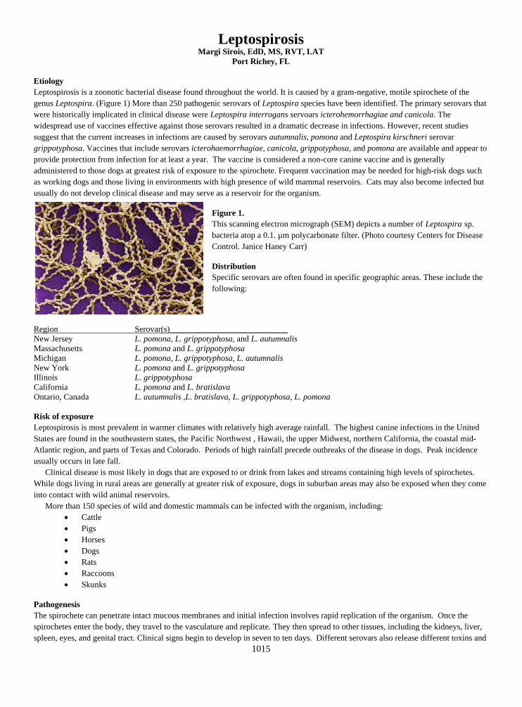
1015
Leptospirosis Margi Sirois, EdD, MS, RVT, LAT
Port Richey, FL
Etiology Leptospirosis is a zoonotic bacterial disease found throughout the world. It is caused by a gram-negative, motile spirochete of the genus Leptospira. (Figure 1) More than 250 pathogenic serovars of Leptospira species have been identified. The primary serovars that were historically implicated in clinical disease were Leptospira interrogans servoars icterohemorrhagiae and canicola. The widespread use of vaccines effective against those serovars resulted in a dramatic decrease in infections. However, recent studies suggest that the current increases in infections are caused by serovars autumnalis, pomona and Leptospira kirschneri serovar grippotyphosa. Vaccines that include serovars icterohaemorrhagiae, canicola, grippotyphosa, and pomona are available and appear to provide protection from infection for at least a year. The vaccine is considered a non-core canine vaccine and is generally administered to those dogs at greatest risk of exposure to the spirochete. Frequent vaccination may be needed for high-risk dogs such as working dogs and those living in environments with high presence of wild mammal reservoirs. Cats may also become infected but usually do not develop clinical disease and may serve as a reservoir for the organism.
Figure 1. This scanning electron micrograph (SEM) depicts a number of Leptospira sp. bacteria atop a 0.1. µm polycarbonate filter. (Photo courtesy Centers for Disease Control. Janice Haney Carr) Distribution Specific serovars are often found in specific geographic areas. These include the following:
Region Serovar(s)____________________________ New Jersey L. pomona, L. grippotyphosa, and L. autumnalis Massachusetts L. pomona and L. grippotyphosa Michigan L. pomona, L. grippotyphosa, L. autumnalis New York L. pomona and L. grippotyphosa Illinois L. grippotyphosa California L. pomona and L. bratislava Ontario, Canada L. autumnalis ,L. bratislava, L. grippotyphosa, L. pomona Risk of exposure Leptospirosis is most prevalent in warmer climates with relatively high average rainfall. The highest canine infections in the United States are found in the southeastern states, the Pacific Northwest , Hawaii, the upper Midwest, northern California, the coastal mid-Atlantic region, and parts of Texas and Colorado. Periods of high rainfall precede outbreaks of the disease in dogs. Peak incidence usually occurs in late fall.
Clinical disease is most likely in dogs that are exposed to or drink from lakes and streams containing high levels of spirochetes. While dogs living in rural areas are generally at greater risk of exposure, dogs in suburban areas may also be exposed when they come into contact with wild animal reservoirs.
More than 150 species of wild and domestic mammals can be infected with the organism, including: • Cattle • Pigs • Horses • Dogs • Rats • Raccoons • Skunks
Pathogenesis The spirochete can penetrate intact mucous membranes and initial infection involves rapid replication of the organism. Once the spirochetes enter the body, they travel to the vasculature and replicate. They then spread to other tissues, including the kidneys, liver, spleen, eyes, and genital tract. Clinical signs begin to develop in seven to ten days. Different serovars also release different toxins and
1016
this may account for variability in severity of infection. Damage to vascular endothelium, vasculitis, hepatitis, and nephritis are common findings. Fatality rates are reportedly as high as 20%. Polyuria and polydipsia can develop in dogs with leptospirosis as a result of a decreased glomerular filtration rate that impairs the renal concentrating ability. Other manifestations of infection can include uveitis, conjunctivitis, tachypnea, and dyspnea.
Assuming the patient mounts an adequate immune response, the spirochetes are generally cleared from most tissues within a few weeks. Organisms tend to persist in the kidneys and can be shed in the urine for many months. Pathologic changes to the kidneys and other organs and tissues often persist even after successful treatment. Clinical signs The organism colonizes the kidney tubules and is shed in the urine. Transmission is by direct contact with urine containing the spirochetes or contact with urine-contaminated soil, water, food, bedding, or other fomites. The incubation period can be as short as one week but infected animals may also be asymptomatic carriers. Disease can also develop months or years later. When symptoms are present in infected dogs, they can include
• Fever • Jaundice • Renal failure • Lethargy • Anorexia • Vomiting • Dehydration • Oliguria • Diarrhea • Hematemesis • Petechiae
Clinical signs in cats are rare but can include renal failure, ascites, and hepatomegaly. Diagnosis Leptospirosis is a historically under-diagnosed disease. The most commonly used diagnostic test is the microscopic agglutination test (MAT), which identifies the presence of serum antibodies to the spirochetes. This test is often interpreted in conjunction with the results of antibody titer testing. Antibody tests may not yield a positive response for the first ten days of illness. Multiple serum samples may be required, especially in the early stages of the disease. The MAT test usually uses from five to eight different serovars and the laboratory may only be testing for certain serovars. The veterinarian may also base a diagnosis on clinical signs and response to treatment. A polymerase chain reaction (PCR) test is also available that may be able to provide earlier diagnosis than the MAT. Culture of the spirochetes is difficult and may require three to six months of incubation.
Increased BUN and serum creatinine concentrations, thrombocytopenia, and leukocytosis with a left shift are present. Urine becomes progressively more dilute as the disease progresses. Disseminated intravascular coagulation (DIC) may occur. Alterations in electrolyte concentrations may also occur. Radiographs may demonstrate enlarged kidneys. Glucosuria, hematuria, and pyuria are often present and urine sediment examination may demonstrate epithelial cell casts or granular casts. Treatment Treatment involves antibiotic therapy (ampicillin, amoxicillin, doxycycline) as well as aggressive supportive care to support renal perfusion. Severely infected patients may require dialysis. Patients generally respond to treatment in ten to fourteen days. Some patients will have residual permanent kidney damage.
Renal function should be monitored at least once every 24 hours during treatment. Fluid therapy and continuous monitoring of urine output via closed collection bags is vital. Closed collection systems minimize staff exposure to infection. Serial evaluations of body weight, respiratory rate, lung sounds, blood pressure, and central venous pressure are important assessments to track the course of disease. Repeat antibody titers can also be used to evaluate response to treatment. Control Infected dogs shed spirochetes in their urine and will likely continue to shed viable organisms for several days after the initiation of appropriate antibiotic therapy. Shedding will be prolonged if antimicrobials therapy is not initiated. Early suspicion with restriction of the infected patient’s movement around the clinic should minimal. Housing in an isolation ward is not generally required as the organisms are not readily transmitted between dogs. Veterinary team members should wear appropriate PPE (i.e. gloves, protective eyewear, facemask, and a disposable gown) when working with infected patients. Routine disinfection is sufficient to decontaminate
1017
runs, cages, bedding, etc. Pressure washing should not be used as it may result in aerosolization of the urine and subsequent dispersal of the organisms. Clients should be advised to take precautions to reduce exposure of their pet to wild animals and standing water. Summary Leptospirosis is an emerging disease in the United States Since and was reinstated as a reportable human disease in 2013. The true incidence of infection is not known. The disease may be fatal if left untreated. Efforts must be made to reduce exposure and increase vaccination of dogs at high risk of exposure. The zoonotic potential is also of great concern and clients must be properly counseled to avoid human infection. References Leptospira species infection in cats: ABCD guidelines on prevention and management. Hartmann K, Egberink H, Pennisi MG, et al. J Feline Med Surg 15:576-581, 2013. Purdue University-Banfield National Companion Animal Surveillance Program for emerging and zoonotic diseases. Vector-Borne Zoonot 6:14-23, 2006. Leptospirosis. Greene CE, Sykes JE, et al. In Greene CE (ed): Infectious Diseases of the Dog and Cat, 4th ed—St. Louis: Saunders Elsevier, 2012, pp 431-447. Emerging Diseases - Leptospirosis - Epidemiology & Diagnostics. Glickman, L. Clinicians Brief. February 2005 Supplement, pg 2 2010 ACVIM Small Animal Consensus Statement on Leptospirosis: Diagnosis, Epidemiology, Treatment, and Prevention. J.E. Sykes, K. Hartmann, K.F. Lunn, G.E. Moore, R.A. Stoddard, and R.E. Goldstein. J Vet Intern Med 2011;25:1–13 Canine Leptospirosis.Harkin, K. Clinicians Brief. June 2005: 15-19
1018
Introduction to Urinalysis Margi Sirois, EdD, MS, RVT, LAT
Port Richey, FL
Formation of urine When blood enters the renal corpuscles, a portion of the plasma, along with its wastes, is filtered through the glomerulus into the next portion of the tubule, the proximal convoluted tubule. Large proteins and cells do not enter the tubule unless there is damage to the glomerulus. The filtered fluid passes slowly through the rest of the nephron and is modified as it moves along. Substances, such as water and glucose, are resorbed back into the blood of the capillary network. The nephron has a specific limit for resorption of specific substances, known as the renal threshold. Should the filtrate contain extremely high concentrations of any of those substances, the excess is not resorbed and is excreted in the urine. By the time the fluid in the nephron reaches the collecting tubules, it has become urine. Collecting tubules of all nephrons drain urine into the renal pelvis to the opening of the ureter. Urine is transported to the urinary bladder by the ureters, muscular tubes that conduct the urine by smooth muscle contractions. The ureters enter the bladder at oblique angles, forming valvelike openings that prevent backflow of urine into the ureters as the bladder fills. The urethra is the tube that carries urine from the urinary bladder to the outside of the body. In females, it is relatively short, straight, and wide, and has a strictly urinary function. In males, it is relatively long, curved, and narrow, and serves both urinary and reproductive functions Sample collection The first step in performing a urinalysis is proper collection of a urine sample, which must be carefully obtained to ensure accurate results. Analysis of urine samples should be performed only on samples taken before administration of therapeutic agents. Urine specimens may be obtained by natural voiding of urine, bladder expression, catheterization, or cystocentesis. The two preferred methods are cystocentesis and catheterization; these methods provide optimal samples for all aspects of urinalysis by avoiding contamination from the distal genital tract and external areas. Collecting samples by voiding or expression of the bladder may be easier, but urine collected in these ways may be of limited diagnostic value Specimen storage and handling Ideally, samples should be analyzed within 30 minutes to 1 hour of collection to avoid postcollection artifacts and degenerative changes. If immediate analysis is not possible, refrigeration preserves most urine constituents for an additional 6 to 12 hours. Refrigeration may have an impact on urine specific gravity, so this test should be done before refrigeration. If a urine sample is going to be refrigerated, it should have a tight-fitting lid to prevent evaporation and contamination. Crystals may form when the urine cools. Decreased glucose and bilirubin concentrations, increased pH resulting from bacterial breakdown of urea to ammonia, crystal formation with increased sample turbidity, breakdown of casts and RBCs (especially in dilute or alkaline urine), and bacterial proliferation may occur in samples allowed to stand for long periods at room temperature. Many crystals may form in refrigerated samples. Refrigerated urine should be warmed to room temperature before evaluation, and crystals that formed during cooling may not dissolve when the sample is brought to room temperature. The urine sample should be mixed by gentle inversion before evaluation so that formed elements are evenly distributed. Cells tend to break down rapidly in urine, so if cytologic evaluation is to be performed, the urine should be centrifuged soon after collection and 1 to 2 drops of the patient’s serum or bovine albumin added to the sediment to preserve cell morphologic characteristics. Physical evaluation Physical properties of urine include all the observations that may be made without the aid of a microscope or chemical reagents. Volume, color, odor, transparency, and specific gravity of urine are evaluated.
Color Evaluate the color and clarity of urine by placing it is a clear container and holding it against a white background. Normal urine color is light yellow to amber as a result of the presence of pigments called urochromes. The magnitude of yellow color in urine varies with the degree of urine concentration or dilution. Colorless urine usually has a low speci fic gravity and is often associated with polyuria. Dark yellow to yellow-brown urine generally has a high specific gravity and may be associated with oliguria. Yellow-brown or green urine that produces a greenish-yellow foam when shaken is likely to contain bile pigments. Red or red-brown urine indicates the presence of RBCs (referred to as hematuria) or hemoglobin (referred to as hemoglobinuria). Urine that is brown when voided may contain myoglobin (referred to as myoglobinuria) excreted during conditions that cause muscle cell lysis. Some drugs may alter the color of urine; red, green, or blue urine may be observed.
Clarity In most species freshly voided urine is transparent or clear. Normal equine urine is cloudy because of a high concentration of calcium carbonate crystals and mucus secreted by glands in the renal pelvis. Normal rabbit urine also has high concentrations of calcium carbonate crystals and appears milky. When observing urine for the degree of transparency, it should be placed against a letter-print
1019
background. Transparency is noted as clear, slightly cloudy, cloudy, or turbid (flocculent) depending on how well the letters can be read through the sample. Clear samples usually do not have much sediment on centrifugation. Cloudy samples usually contain large particles and often yield a significant amount of sediment on centrifugation. Urine may become cloudy while standing because of bacterial multiplication or crystal formation. Substances that cause urine to be cloudy include RBCs, WBCs, epithelial cells, casts, crystals, mucus, fat, and bacteria. Other causes of turbidity can include contaminants from the collection container or surface and contamination with feces. Flocculent samples contain suspended particles that are sometimes large enough to be seen with the naked eye.
Odor The odor of urine is not highly diagnostic but sometimes may be helpful. Normal urine has a distinctive odor that varies among species. The urine of male cats, goats, and pigs has a strong odor. An ammonia odor may occur with cystitis caused by bacteria that produce urease (Proteus spp. or Staphylococcus spp.) that has metabolized urea to ammonia. Samples left standing at room temperature may occasionally develop an ammonia odor as a result of bacterial growth. A characteristic sweet or fruity odor to urine indicates ketones and is most commonly found with diabetes mellitus, ketosis in cows, and pregnancy disease in ewes.
Specific gravity Specific gravity is defined as the weight (density) of a quantity of liquid compared with that of an equal amount of distilled water. The number and molecular weight of dissolved solutes determine the specific gravity of urine. Specific gravity may be determined before or after centrifugation because the particles that settle during centrifugation have little or no effect on speci fic gravity. Whichever method is used to perform the specific gravity in a specific clinic, whether before or after centrifugation, the same method must be consistently performed by all clinic personnel. If the urine is turbid, the sample should be centrifuged and the supernatant used to determine the specific gravity. The specific gravity of urine from polyuric patients tends to be low, and urine from oliguric patients tends to be high. The specific gravity of normal urine depends on eating and drinking habits, environmental temperature, and when the sample was collected. Specific gravity of normal animals is extremely variable and fluctuates throughout the day. In normal dogs the urine specific gravity may range from 1.001 to 1.060 and in normal cats from 1.001 to 1.080.
To determine specific gravity of urine, the refractometer, urinometer, or reagent strips can be used. Reagent strip specific gravity is the least reliable method for determining urine specific gravity in animals. Urine specific gravity is less frequently determined by use of a urinometer. This instrument requires a large amount of urine (approximately 10 ml) and generally provides less-reproducible results than a refractometer. Chemical evaluation Testing for various chemical constituents of urine is usually performed with reagent strips that are impregnated with appropriate chemicals or with reagent tablets. There are some automated analyzers used for serum chemistry that can also be used for urine testing. Modifications of the procedures may be required. The container of reagent strips must be stored at room temperature with the lid tightly closed. The expiration date should also be noted. Some reagent strips simultaneously test for numerous constituents, and other strips exist for individual tests. The reagent strip should be dipped into the sample so that it is fully immersed, then removed and the long edge tilted on a paper towel to allow excess urine to be wicked away. Color changes on each reagent pad are noted at speci fic time intervals. The concentration of various constituents is determined by comparing the colors on the strip with the color chart on the label of the strip container. The manufacturer’s directions must be carefully followed. It is important to note that a large number of conditions, such as medications, dietary factors, and environmental factors, can affect urinalysis test results.
pH The pH expresses the hydrogen ion (H+) concentration. Essentially, pH is a measure of the degree of acidity or alkalinity of urine. A pH above 7.0 is alkaline, whereas a pH below 7.0 is acidic. Proper technique must be used to obtain accurate results. The urine sample must be fresh to obtain accurate results. The pH of samples left standing open at room temperature tends to increase resulting from a loss of carbon dioxide, whereas delays in reading the reaction may lead to color changes and false readings. If samples containing urease-producing bacteria (Proteus spp. or Staphylococcus spp.) are left standing, the pH is usually increased.
The kidneys play a role in acid-base regulation of the body. Kidneys must vary the pH of urine to compensate for diet and products of metabolism. The pH of a healthy animal’s urine depends largely on its diet. Alkaline urine usually is found in animals on plant diets, whereas high-protein cereal diets or diets of animal origin cause acidic urine. Therefore herbivores normally have alkaline urine, carnivores have acidic urine, and omnivores have either acidic or alkaline urine depending on what was ingested. Many dog foods contain substantial amounts of plant material that may cause the urine to be slightly alkaline. Other factors such as stress and excitement, especially in cats, increase the urine pH and may create a transient glucosuria.
Urine pH is usually measured with reagent strips or a pH meter. Factors that may decrease the pH (acidity) include fever, starvation, high-protein diet, acidosis, excessive muscular activity, or administration of certain drugs. Increased pH (alkalinity) may be caused by alkalosis, high-fiber diets (plants), infection of the urinary tract with urease bacteria, use of certain drugs, or urine retention such as occurs with urethral obstruction or bladder paralysis. If the pH of the urine is too acidic or too alkaline speci fic crystals or uroliths can form. The pH can be manipulated with diet to help dissolve the solids or prevent them from forming.
1020
Protein Protein is usually absent or present in only trace amounts in normal urine obtained by catheterization or cystocentesis. In healthy animals, plasma proteins that pass into the glomerular filtrate are resorbed in the renal tubules before the filtrate reaches the renal pelvis. However, voided samples or those obtained by expressing the bladder may contain a small amount of protein from secretions that may contaminate urine during its passage along the urinary tract. Trauma to the urinary tract that results from cystocentesis, catheterization, or bladder expression may occasionally cause sufficient bleeding that results in a trace of protein in the urine. Urine protein measurements are interpreted in light of the collection method, urine specific gravity, rate of urine formation, and contributions from any hemorrhage or inflammation noted by sample analysis. Protein levels in the urine may be measured by several methods, including reagent test strips, sulfosalicylic acid turbidity test, and urine protein/creatinine ratio.
Glucose The presence of glucose in urine is known as glucosuria or glycosuria. Glucose is filtered through the glomerulus and resorbed by the kidney tubules. The amount of glucose in the urine depends on blood glucose levels and the rates of glomerular filtration and tubular resorption. Glucosuria usually does not occur in normal animals unless the blood glucose level exceeds the renal threshold (approximately 170 to 180 mg/dl for dogs). At this concentration tubular resorption cannot keep up with the glomerular filtration of glucose, and glucose passes into the urine.
Glucosuria occurs in diabetes mellitus as a result of a deficiency of insulin or an inability of insulin to function. Insulin is necessary to transport glucose into body cells, and a deficiency causes hyperglycemia and spilling of glucose into the urine. A high-carbohydrate meal may lead to blood glucose levels exceeding the renal threshold and thus glucosuria. Because of this, a period of fasting is recommended before urine glucose concentration is determined. Fear, excitement, or restraint, especially in cats, often causes hyperglycemia and glucosuria as a result of epinephrine release. Glucosuria often occurs after intravenous administration of fluids containing glucose and occasionally after general anesthesia. Rarely glucosuria is found in hyperthyroidism, Cushing’s disease, and chronic liver disease. Glucosuria may occur in some cats with chronic disease, possibly as a result of altered proximal renal tubular function.
Ketones Ketones include acetone, acetoacetic acid, and b-hydroxybutyric acid. Ketone bodies are formed during incomplete catabolism of fatty acids. Normal animals may have small amounts of ketones in the blood. Conditions characterized by altered carbohydrate metabolism may result in excessive amounts of fat catabolism to provide energy. When fatty acid metabolism is not accompanied by sufficient carbohydrate metabolism, excess ketones are present in the urine, a condition called ketonuria.
Ketonuria frequently occurs in animals with diabetes mellitus. Because the animal lacks the insulin necessary for carbohydrate metabolism, fat is broken down to meet the animal’s energy needs and excess ketones are excreted in the urine. Ketones are important sources of energy and are normally produced during fat metabolism. Problems develop, however, when excessive ketones are produced. Ketones are toxic, causing central nervous system depression and acidosis. Acidosis resulting from ketonemia is termed ketoacidosis.
Ketonemia with ketonuria also occurs with high-fat diets, starvation, fasting, long-term anorexia, and impaired liver function. With a high-fat diet, carbohydrates meet a relatively low percentage of energy needs, so a great amount of fat is used to meet energy needs. In the fasting, starved, or anorexic animal, body fat is used to meet energy needs, producing a greater than normal amount of ketones. With liver damage, impaired carbohydrate metabolism leads to fat serving as the main energy source, especially when the damaged liver cannot store adequate amounts of glycogen.
Urinary ketones are detected by using urinary reagent strips with a ketone reagent pad. The color intensity is roughly proportional to the concentration of urine ketones. These methods are most sensitive to acetoacetic acid, less sensitive to acetone and do not detect b-hydroxybutyric acid. b-Hydroxybutyric acid is the first ketone produced by the body in any condition that causes ketosis. Urine reagent test strips may not adequately identify these patients until the ketosis has been present for some time.
Bile pigments Bile pigments commonly detected in urine are bilirubin and urobilinogen. Only conjugated bilirubin (water soluble) is found in urine because unconjugated bilirubin does not pass through the glomerulus into the renal filtrate; it is bound to albumin and is not water soluble. Normal dogs, especially males, occasionally have bilirubin in their urine because of a low renal threshold for conjugated bilirubin and the ability of their kidneys to conjugate bilirubin. Many normal cattle also have small amounts of bilirubin in their urine. Bilirubin is usually not found in the urine of cats, pigs, sheep, or horses. In cats the renal threshold is many times that of dogs, and any amount of bilirubin in the urine is considered abnormal and suggests disease.
Bilirubinuria is seen in a number of diseases, including obstruction of bile flow from the liver to the small intestine and in liver disease. Bilirubinuria results from accumulation in hepatic cells of conjugated bilirubin that is released into the blood and excreted in the urine. Conditions causing biliary obstruction include calculi in the bile duct, tumors in the area of the bile duct, acute enteritis, pancreatitis, and obstruction of the upper intestinal tract. Hemolytic anemia may also cause bilirubinuria, especially in dogs. In hemolytic anemia, the liver’s ability to metabolize the excess bilirubin may be exceeded, resulting in release of conjugated bilirubin
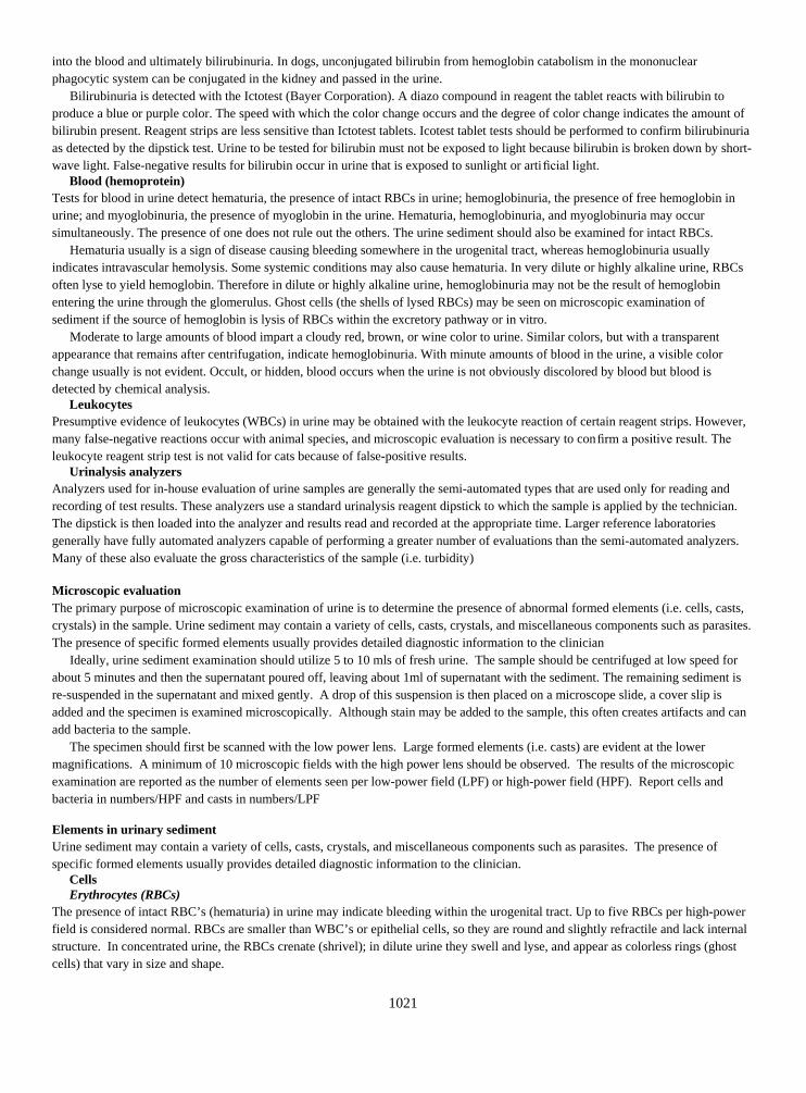
1021
into the blood and ultimately bilirubinuria. In dogs, unconjugated bilirubin from hemoglobin catabolism in the mononuclear phagocytic system can be conjugated in the kidney and passed in the urine.
Bilirubinuria is detected with the Ictotest (Bayer Corporation). A diazo compound in reagent the tablet reacts with bilirubin to produce a blue or purple color. The speed with which the color change occurs and the degree of color change indicates the amount of bilirubin present. Reagent strips are less sensitive than Ictotest tablets. Icotest tablet tests should be performed to confirm bilirubinuria as detected by the dipstick test. Urine to be tested for bilirubin must not be exposed to light because bilirubin is broken down by short-wave light. False-negative results for bilirubin occur in urine that is exposed to sunlight or artificial light.
Blood (hemoprotein) Tests for blood in urine detect hematuria, the presence of intact RBCs in urine; hemoglobinuria, the presence of free hemoglobin in urine; and myoglobinuria, the presence of myoglobin in the urine. Hematuria, hemoglobinuria, and myoglobinuria may occur simultaneously. The presence of one does not rule out the others. The urine sediment should also be examined for intact RBCs.
Hematuria usually is a sign of disease causing bleeding somewhere in the urogenital tract, whereas hemoglobinuria usually indicates intravascular hemolysis. Some systemic conditions may also cause hematuria. In very dilute or highly alkaline urine, RBCs often lyse to yield hemoglobin. Therefore in dilute or highly alkaline urine, hemoglobinuria may not be the result of hemoglobin entering the urine through the glomerulus. Ghost cells (the shells of lysed RBCs) may be seen on microscopic examination of sediment if the source of hemoglobin is lysis of RBCs within the excretory pathway or in vitro.
Moderate to large amounts of blood impart a cloudy red, brown, or wine color to urine. Similar colors, but with a transparent appearance that remains after centrifugation, indicate hemoglobinuria. With minute amounts of blood in the urine, a visible color change usually is not evident. Occult, or hidden, blood occurs when the urine is not obviously discolored by blood but blood is detected by chemical analysis.
Leukocytes Presumptive evidence of leukocytes (WBCs) in urine may be obtained with the leukocyte reaction of certain reagent strips. However, many false-negative reactions occur with animal species, and microscopic evaluation is necessary to confirm a positive result. The leukocyte reagent strip test is not valid for cats because of false-positive results.
Urinalysis analyzers Analyzers used for in-house evaluation of urine samples are generally the semi-automated types that are used only for reading and recording of test results. These analyzers use a standard urinalysis reagent dipstick to which the sample is applied by the technician. The dipstick is then loaded into the analyzer and results read and recorded at the appropriate time. Larger reference laboratories generally have fully automated analyzers capable of performing a greater number of evaluations than the semi-automated analyzers. Many of these also evaluate the gross characteristics of the sample (i.e. turbidity) Microscopic evaluation The primary purpose of microscopic examination of urine is to determine the presence of abnormal formed elements (i.e. cells, casts, crystals) in the sample. Urine sediment may contain a variety of cells, casts, crystals, and miscellaneous components such as parasites. The presence of specific formed elements usually provides detailed diagnostic information to the clinician
Ideally, urine sediment examination should utilize 5 to 10 mls of fresh urine. The sample should be centrifuged at low speed for about 5 minutes and then the supernatant poured off, leaving about 1ml of supernatant with the sediment. The remaining sediment is re-suspended in the supernatant and mixed gently. A drop of this suspension is then placed on a microscope slide, a cover slip is added and the specimen is examined microscopically. Although stain may be added to the sample, this often creates artifacts and can add bacteria to the sample.
The specimen should first be scanned with the low power lens. Large formed elements (i.e. casts) are evident at the lower magnifications. A minimum of 10 microscopic fields with the high power lens should be observed. The results of the microscopic examination are reported as the number of elements seen per low-power field (LPF) or high-power field (HPF). Report cells and bacteria in numbers/HPF and casts in numbers/LPF Elements in urinary sediment Urine sediment may contain a variety of cells, casts, crystals, and miscellaneous components such as parasites. The presence of specific formed elements usually provides detailed diagnostic information to the clinician.
Cells Erythrocytes (RBCs)
The presence of intact RBC’s (hematuria) in urine may indicate bleeding within the urogenital tract. Up to five RBCs per high-power field is considered normal. RBCs are smaller than WBC’s or epithelial cells, so they are round and slightly refractile and lack internal structure. In concentrated urine, the RBCs crenate (shrivel); in dilute urine they swell and lyse, and appear as colorless rings (ghost cells) that vary in size and shape.
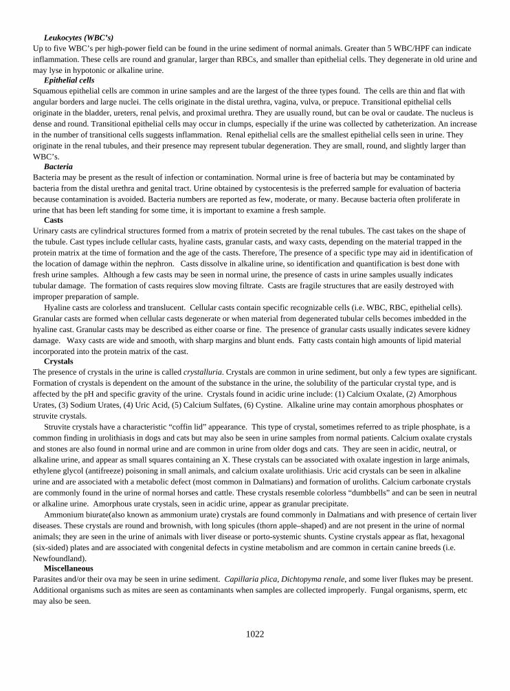
1022
Leukocytes (WBC’s) Up to five WBC’s per high-power field can be found in the urine sediment of normal animals. Greater than 5 WBC/HPF can indicate inflammation. These cells are round and granular, larger than RBCs, and smaller than epithelial cells. They degenerate in old urine and may lyse in hypotonic or alkaline urine.
Epithelial cells Squamous epithelial cells are common in urine samples and are the largest of the three types found. The cells are thin and flat with angular borders and large nuclei. The cells originate in the distal urethra, vagina, vulva, or prepuce. Transitional epithelial cells originate in the bladder, ureters, renal pelvis, and proximal urethra. They are usually round, but can be oval or caudate. The nucleus is dense and round. Transitional epithelial cells may occur in clumps, especially if the urine was collected by catheterization. An increase in the number of transitional cells suggests inflammation. Renal epithelial cells are the smallest epithelial cells seen in urine. They originate in the renal tubules, and their presence may represent tubular degeneration. They are small, round, and slightly larger than WBC’s.
Bacteria Bacteria may be present as the result of infection or contamination. Normal urine is free of bacteria but may be contaminated by bacteria from the distal urethra and genital tract. Urine obtained by cystocentesis is the preferred sample for evaluation of bacteria because contamination is avoided. Bacteria numbers are reported as few, moderate, or many. Because bacteria often proliferate in urine that has been left standing for some time, it is important to examine a fresh sample.
Casts Urinary casts are cylindrical structures formed from a matrix of protein secreted by the renal tubules. The cast takes on the shape of the tubule. Cast types include cellular casts, hyaline casts, granular casts, and waxy casts, depending on the material trapped in the protein matrix at the time of formation and the age of the casts. Therefore, The presence of a specific type may aid in identification of the location of damage within the nephron. Casts dissolve in alkaline urine, so identification and quantification is best done with fresh urine samples. Although a few casts may be seen in normal urine, the presence of casts in urine samples usually indicates tubular damage. The formation of casts requires slow moving filtrate. Casts are fragile structures that are easily destroyed with improper preparation of sample.
Hyaline casts are colorless and translucent. Cellular casts contain specific recognizable cells (i.e. WBC, RBC, epithelial cells). Granular casts are formed when cellular casts degenerate or when material from degenerated tubular cells becomes imbedded in the hyaline cast. Granular casts may be described as either coarse or fine. The presence of granular casts usually indicates severe kidney damage. Waxy casts are wide and smooth, with sharp margins and blunt ends. Fatty casts contain high amounts of lipid material incorporated into the protein matrix of the cast.
Crystals The presence of crystals in the urine is called crystalluria. Crystals are common in urine sediment, but only a few types are significant. Formation of crystals is dependent on the amount of the substance in the urine, the solubility of the particular crystal type, and is affected by the pH and specific gravity of the urine. Crystals found in acidic urine include: (1) Calcium Oxalate, (2) Amorphous Urates, (3) Sodium Urates, (4) Uric Acid, (5) Calcium Sulfates, (6) Cystine. Alkaline urine may contain amorphous phosphates or struvite crystals.
Struvite crystals have a characteristic “coffin lid” appearance. This type of crystal, sometimes referred to as triple phosphate, is a common finding in urolithiasis in dogs and cats but may also be seen in urine samples from normal patients. Calcium oxalate crystals and stones are also found in normal urine and are common in urine from older dogs and cats. They are seen in acidic, neutral, or alkaline urine, and appear as small squares containing an X. These crystals can be associated with oxalate ingestion in large animals, ethylene glycol (antifreeze) poisoning in small animals, and calcium oxalate urolithiasis. Uric acid crystals can be seen in alkaline urine and are associated with a metabolic defect (most common in Dalmatians) and formation of uroliths. Calcium carbonate crystals are commonly found in the urine of normal horses and cattle. These crystals resemble colorless “dumbbells” and can be seen in neutral or alkaline urine. Amorphous urate crystals, seen in acidic urine, appear as granular precipitate.
Ammonium biurate(also known as ammonium urate) crystals are found commonly in Dalmatians and with presence of certain liver diseases. These crystals are round and brownish, with long spicules (thorn apple–shaped) and are not present in the urine of normal animals; they are seen in the urine of animals with liver disease or porto-systemic shunts. Cystine crystals appear as flat, hexagonal (six-sided) plates and are associated with congenital defects in cystine metabolism and are common in certain canine breeds (i.e. Newfoundland).
Miscellaneous Parasites and/or their ova may be seen in urine sediment. Capillaria plica, Dichtopyma renale, and some liver flukes may be present. Additional organisms such as mites are seen as contaminants when samples are collected improperly. Fungal organisms, sperm, etc may also be seen.
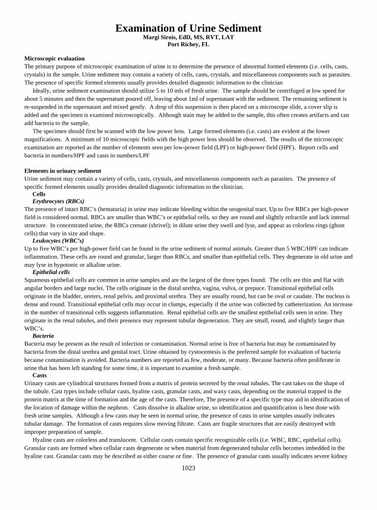
1023
Examination of Urine Sediment Margi Sirois, EdD, MS, RVT, LAT
Port Richey, FL
Microscopic evaluation The primary purpose of microscopic examination of urine is to determine the presence of abnormal formed elements (i.e. cells, casts, crystals) in the sample. Urine sediment may contain a variety of cells, casts, crystals, and miscellaneous components such as parasites. The presence of specific formed elements usually provides detailed diagnostic information to the clinician
Ideally, urine sediment examination should utilize 5 to 10 mls of fresh urine. The sample should be centrifuged at low speed for about 5 minutes and then the supernatant poured off, leaving about 1ml of supernatant with the sediment. The remaining sediment is re-suspended in the supernatant and mixed gently. A drop of this suspension is then placed on a microscope slide, a cover slip is added and the specimen is examined microscopically. Although stain may be added to the sample, this often creates artifacts and can add bacteria to the sample.
The specimen should first be scanned with the low power lens. Large formed elements (i.e. casts) are evident at the lower magnifications. A minimum of 10 microscopic fields with the high power lens should be observed. The results of the microscopic examination are reported as the number of elements seen per low-power field (LPF) or high-power field (HPF). Report cells and bacteria in numbers/HPF and casts in numbers/LPF
Elements in urinary sediment Urine sediment may contain a variety of cells, casts, crystals, and miscellaneous components such as parasites. The presence of specific formed elements usually provides detailed diagnostic information to the clinician.
Cells Erythrocytes (RBCs)
The presence of intact RBC’s (hematuria) in urine may indicate bleeding within the urogenital tract. Up to five RBCs per high-power field is considered normal. RBCs are smaller than WBC’s or epithelial cells, so they are round and slightly refractile and lack internal structure. In concentrated urine, the RBCs crenate (shrivel); in dilute urine they swell and lyse, and appear as colorless rings (ghost cells) that vary in size and shape.
Leukocytes (WBC’s) Up to five WBC’s per high-power field can be found in the urine sediment of normal animals. Greater than 5 WBC/HPF can indicate inflammation. These cells are round and granular, larger than RBCs, and smaller than epithelial cells. They degenerate in old urine and may lyse in hypotonic or alkaline urine.
Epithelial cells Squamous epithelial cells are common in urine samples and are the largest of the three types found. The cells are thin and flat with angular borders and large nuclei. The cells originate in the distal urethra, vagina, vulva, or prepuce. Transitional epithelial cells originate in the bladder, ureters, renal pelvis, and proximal urethra. They are usually round, but can be oval or caudate. The nucleus is dense and round. Transitional epithelial cells may occur in clumps, especially if the urine was collected by catheterization. An increase in the number of transitional cells suggests inflammation. Renal epithelial cells are the smallest epithelial cells seen in urine. They originate in the renal tubules, and their presence may represent tubular degeneration. They are small, round, and slightly larger than WBC’s.
Bacteria Bacteria may be present as the result of infection or contamination. Normal urine is free of bacteria but may be contaminated by bacteria from the distal urethra and genital tract. Urine obtained by cystocentesis is the preferred sample for evaluation of bacteria because contamination is avoided. Bacteria numbers are reported as few, moderate, or many. Because bacteria often proliferate in urine that has been left standing for some time, it is important to examine a fresh sample.
Casts Urinary casts are cylindrical structures formed from a matrix of protein secreted by the renal tubules. The cast takes on the shape of the tubule. Cast types include cellular casts, hyaline casts, granular casts, and waxy casts, depending on the material trapped in the protein matrix at the time of formation and the age of the casts. Therefore, The presence of a specific type may aid in identification of the location of damage within the nephron. Casts dissolve in alkaline urine, so identification and quantification is best done with fresh urine samples. Although a few casts may be seen in normal urine, the presence of casts in urine samples usually indicates tubular damage. The formation of casts requires slow moving filtrate. Casts are fragile structures that are easily destroyed with improper preparation of sample.
Hyaline casts are colorless and translucent. Cellular casts contain specific recognizable cells (i.e. WBC, RBC, epithelial cells). Granular casts are formed when cellular casts degenerate or when material from degenerated tubular cells becomes imbedded in the hyaline cast. Granular casts may be described as either coarse or fine. The presence of granular casts usually indicates severe kidney
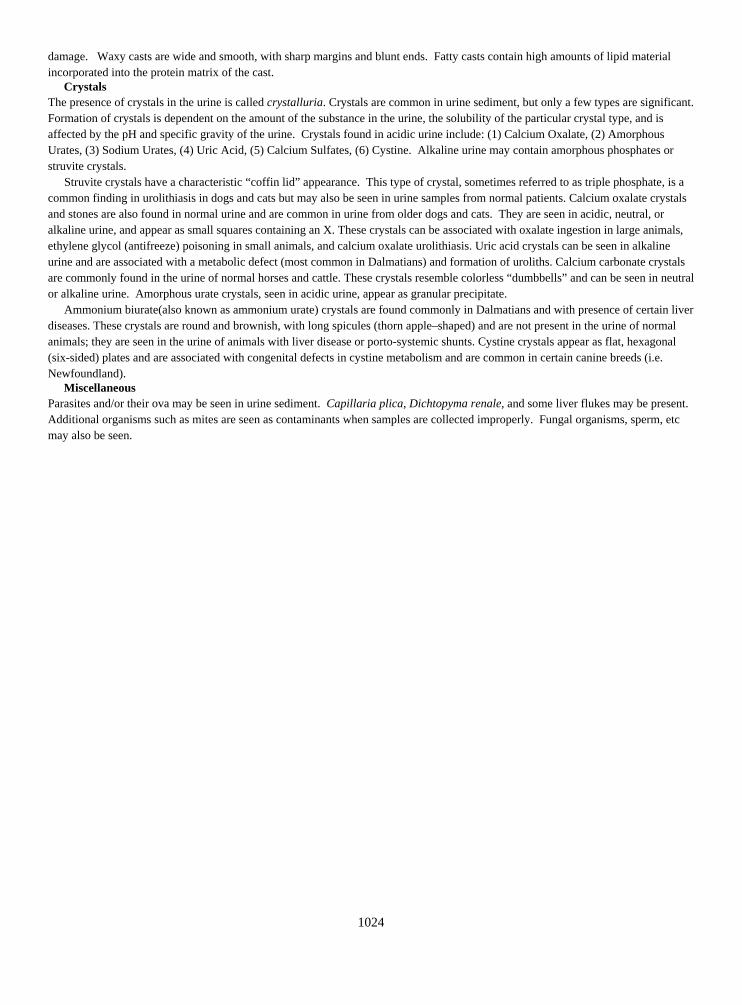
1024
damage. Waxy casts are wide and smooth, with sharp margins and blunt ends. Fatty casts contain high amounts of lipid material incorporated into the protein matrix of the cast.
Crystals The presence of crystals in the urine is called crystalluria. Crystals are common in urine sediment, but only a few types are significant. Formation of crystals is dependent on the amount of the substance in the urine, the solubility of the particular crystal type, and is affected by the pH and specific gravity of the urine. Crystals found in acidic urine include: (1) Calcium Oxalate, (2) Amorphous Urates, (3) Sodium Urates, (4) Uric Acid, (5) Calcium Sulfates, (6) Cystine. Alkaline urine may contain amorphous phosphates or struvite crystals.
Struvite crystals have a characteristic “coffin lid” appearance. This type of crystal, sometimes referred to as triple phosphate, is a common finding in urolithiasis in dogs and cats but may also be seen in urine samples from normal patients. Calcium oxalate crystals and stones are also found in normal urine and are common in urine from older dogs and cats. They are seen in acidic, neutral, or alkaline urine, and appear as small squares containing an X. These crystals can be associated with oxalate ingestion in large animals, ethylene glycol (antifreeze) poisoning in small animals, and calcium oxalate urolithiasis. Uric acid crystals can be seen in alkaline urine and are associated with a metabolic defect (most common in Dalmatians) and formation of uroliths. Calcium carbonate crystals are commonly found in the urine of normal horses and cattle. These crystals resemble colorless “dumbbells” and can be seen in neutral or alkaline urine. Amorphous urate crystals, seen in acidic urine, appear as granular precipitate.
Ammonium biurate(also known as ammonium urate) crystals are found commonly in Dalmatians and with presence of certain liver diseases. These crystals are round and brownish, with long spicules (thorn apple–shaped) and are not present in the urine of normal animals; they are seen in the urine of animals with liver disease or porto-systemic shunts. Cystine crystals appear as flat, hexagonal (six-sided) plates and are associated with congenital defects in cystine metabolism and are common in certain canine breeds (i.e. Newfoundland).
Miscellaneous Parasites and/or their ova may be seen in urine sediment. Capillaria plica, Dichtopyma renale, and some liver flukes may be present. Additional organisms such as mites are seen as contaminants when samples are collected improperly. Fungal organisms, sperm, etc may also be seen.
1025
Ethylene Glycol Toxicosis Margi Sirois, EdD, MS, RVT, LAT
Port Richey, FL
Ethylene glycol is a sweet, odorless, water-soluble chemical with many common household uses. It may be found in automotive anti-freeze, de-icing agents and windshield washer fluids, home solar units, portable chemical toilets, and some household heating components. Ethylene glycol toxicosis most commonly affects dogs, but cats and other animals may also be exposed. While the compound itself is not toxic, it is metabolized into many other components (i.e. glycolic acid, oxalate, and aldehyde) that are extremely toxic and potentially fatal. Animals are attracted to the sweet odor and taste of the chemical and will readily consume it. Several U.S. states have laws that require the addition of bittering agents to some forms of commercially available products containing ethylene glycol in an effort to discourage ingestion Clinical signs may develop within 30 minutes or may be delayed for several days, depending on the total amount consumed. While specific toxic doses have not been established, the lethal dose is relatively small (4.4 to 6.6 ml/kg in dogs; 1.4 ml/kg in cats). Metabolism Ethylene glycol is rapidly absorbed in the gastrointestinal tract and distributed throughout the body. It is primarily metabolized in the liver. The kidneys excrete unaltered ethylene glycol.
Ethylene glycol is initially oxidized to glycoaldehyde by the enzyme alcohol dehydrogenase. Glycoaldehyde is oxidized to glycolic acid by mitochondrial aldehyde oxidase and subsequently metabolized to gyloxide. The glyoxide is then further metabolized to form oxalic acid, glycine, formic acid, hippurate, and several other compounds. Oxalic acid binds to serum calcium and forms calcium oxalate crystals. Clinical signs Symptoms generally progress through three stages, although some patients may skip a stage and the stages may overlap. Death may occur at any stage. The timing for each stage varies with different species. Initial clinical signs include vomiting, diarrhea, and ataxia. This stage usually begins within 30 minutes of ingestion and lasts approximately 12 hours. Owners of dogs allowed to roam may be unaware of these initial signs. After 12 hours, symptoms progress to cardiovascular and pulmonary abnormalities due to the acidosis and electrolyte abnormalities that result from metabolism of the chemical. Tachypnea and tachycardia are the most common clinical signs during this stage, which generally lasts until 24 hours after ingestion. The third stage is characterized by oliguric renal failure and usually occurs within 24 to 72 hours after ingestion, although it may be much earlier in cats. Signs typical of renal failure, such as oliguria progressing to anuria, vomiting, and anorexia, are common Diagnosis and treatment Early diagnosis is essential for effective treatment. Initial diagnosis is generally based on history and physical examination. Some forms of the chemical may fluoresce and remnants of the chemical may be evidenced by fluorescence of the paws, muzzle, urine, and vomitus using a Wood’s lamp. Blood levels of ethylene glycol generally peak about 4 hours after ingestion and diagnostic tests for blood ethylene glycol may be useful up to 12 hours after ingestion. Blood chemistry testing usually indicates azotemia, hyperphosphatemia, hyperkalemia, hypocalcemia, metabolic acidosis, increased anion gap, isosthenuria, and aciduria
Metabolic acidosis results from the accumulation of glycolic acid and other toxic metabolites and can occur within three hours of ingestion. Calcium oxalate crystalluria can occur as soon as three hours after ingestion. The monohydrate form is most common. The crystals can precipitate in the renal tubules and cause mechanical damage and obstruction Treatment It is important to obtain and diagnostic samples needed prior to the initiation of treatment. Patients presented for treatment within 8 hours of ingestion of ethylene glycol have the greatest chance of survival. The veterinarian will usually induce vomiting if ingestion has been known to occur less than an hour prior to presentation. Activated charcoal may be useful if given within three hours of ingestion. Administration of compounds that interfere with or delay formation of the metabolic by-products are also useful. Examples include ethanol and fomepizole. Ethanol is a competitive inhibitor that prevents the conversion of ethylene glycol into glycoaldehyde and other toxic metabolites Fomepizole may be less effective in dogs than cats.
Supportive care aimed is at preventing further kidney damage and maintaining acid-base balance. Fluid therapy is essential to correct electrolyte imbalances and promote diuresis.

1026
Summary Clinical signs vary depending on the amount of ethylene glycol ingested and the time since ingestion. Without early diagnosis and initiation of treatment, prognosis is poor. Patients that survive may have ongoing renal impairment. Prevention of exposure requires aggressive client education. Replacement of ethylene glycol based products with those containing the less toxic compound, propylene glycol, will also help reduce accidental exposures. References available from the author.
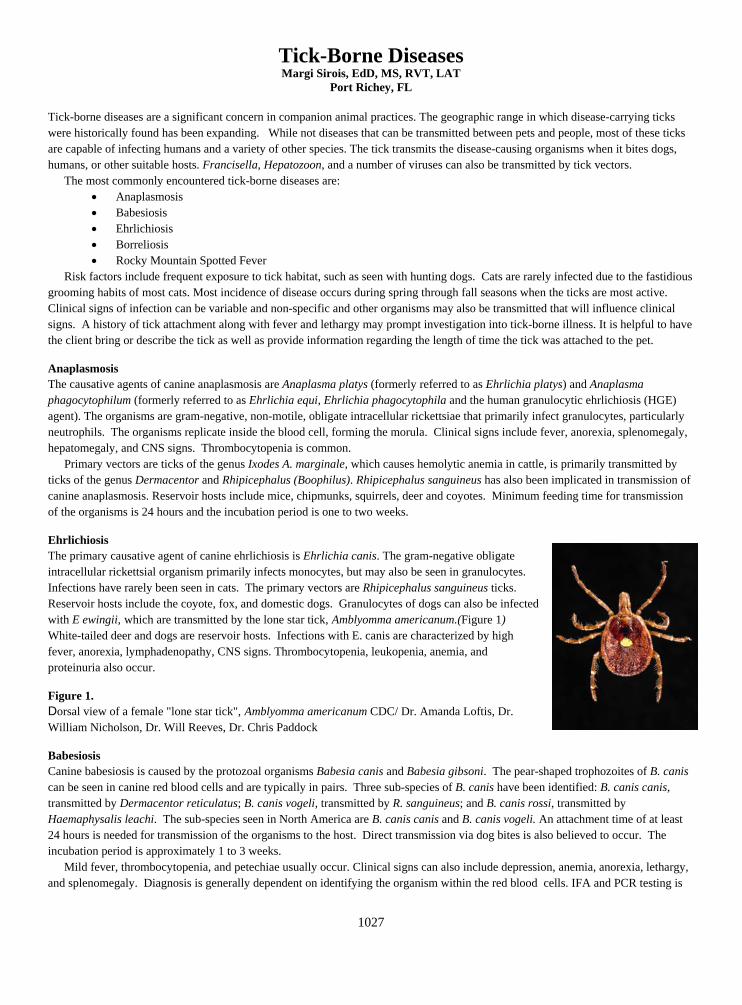
1027
Tick-Borne Diseases Margi Sirois, EdD, MS, RVT, LAT
Port Richey, FL
Tick-borne diseases are a significant concern in companion animal practices. The geographic range in which disease-carrying ticks were historically found has been expanding. While not diseases that can be transmitted between pets and people, most of these ticks are capable of infecting humans and a variety of other species. The tick transmits the disease-causing organisms when it bites dogs, humans, or other suitable hosts. Francisella, Hepatozoon, and a number of viruses can also be transmitted by tick vectors.
The most commonly encountered tick-borne diseases are: • Anaplasmosis • Babesiosis • Ehrlichiosis • Borreliosis • Rocky Mountain Spotted Fever
Risk factors include frequent exposure to tick habitat, such as seen with hunting dogs. Cats are rarely infected due to the fastidious grooming habits of most cats. Most incidence of disease occurs during spring through fall seasons when the ticks are most active. Clinical signs of infection can be variable and non-specific and other organisms may also be transmitted that will influence clinical signs. A history of tick attachment along with fever and lethargy may prompt investigation into tick-borne illness. It is helpful to have the client bring or describe the tick as well as provide information regarding the length of time the tick was attached to the pet. Anaplasmosis The causative agents of canine anaplasmosis are Anaplasma platys (formerly referred to as Ehrlichia platys) and Anaplasma phagocytophilum (formerly referred to as Ehrlichia equi, Ehrlichia phagocytophila and the human granulocytic ehrlichiosis (HGE) agent). The organisms are gram-negative, non-motile, obligate intracellular rickettsiae that primarily infect granulocytes, particularly neutrophils. The organisms replicate inside the blood cell, forming the morula. Clinical signs include fever, anorexia, splenomegaly, hepatomegaly, and CNS signs. Thrombocytopenia is common.
Primary vectors are ticks of the genus Ixodes A. marginale, which causes hemolytic anemia in cattle, is primarily transmitted by ticks of the genus Dermacentor and Rhipicephalus (Boophilus). Rhipicephalus sanguineus has also been implicated in transmission of canine anaplasmosis. Reservoir hosts include mice, chipmunks, squirrels, deer and coyotes. Minimum feeding time for transmission of the organisms is 24 hours and the incubation period is one to two weeks. Ehrlichiosis The primary causative agent of canine ehrlichiosis is Ehrlichia canis. The gram-negative obligate intracellular rickettsial organism primarily infects monocytes, but may also be seen in granulocytes. Infections have rarely been seen in cats. The primary vectors are Rhipicephalus sanguineus ticks. Reservoir hosts include the coyote, fox, and domestic dogs. Granulocytes of dogs can also be infected with E ewingii, which are transmitted by the lone star tick, Amblyomma americanum.(Figure 1) White-tailed deer and dogs are reservoir hosts. Infections with E. canis are characterized by high fever, anorexia, lymphadenopathy, CNS signs. Thrombocytopenia, leukopenia, anemia, and proteinuria also occur. Figure 1. Dorsal view of a female "lone star tick", Amblyomma americanum CDC/ Dr. Amanda Loftis, Dr. William Nicholson, Dr. Will Reeves, Dr. Chris Paddock Babesiosis Canine babesiosis is caused by the protozoal organisms Babesia canis and Babesia gibsoni. The pear-shaped trophozoites of B. canis can be seen in canine red blood cells and are typically in pairs. Three sub-species of B. canis have been identified: B. canis canis, transmitted by Dermacentor reticulatus; B. canis vogeli, transmitted by R. sanguineus; and B. canis rossi, transmitted by Haemaphysalis leachi. The sub-species seen in North America are B. canis canis and B. canis vogeli. An attachment time of at least 24 hours is needed for transmission of the organisms to the host. Direct transmission via dog bites is also believed to occur. The incubation period is approximately 1 to 3 weeks.
Mild fever, thrombocytopenia, and petechiae usually occur. Clinical signs can also include depression, anemia, anorexia, lethargy, and splenomegaly. Diagnosis is generally dependent on identifying the organism within the red blood cells. IFA and PCR testing is
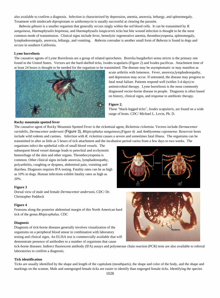
1028
also available to confirm a diagnosis. Infection is characterized by depression, anemia, anorexia, lethargy, and splenomegaly. Treatment with imidocarb dipropionate or azithromycin is usually successful at clearing the parasite.
Babesia gibsoni is a smaller organism that generally occurs singly within the red blood cells. It can be transmitted by R. sanguineus, Haemaphysalis bispinosa, and Haemaphysalis longicornis ticks but bite wound infection is thought to be the most common mode of transmission. Clinical signs include fever, hemolytic regenerative anemia, thrombocytopenia, splenomegaly, lymphadenomegaly, anorexia, lethargy, and vomiting. Babesia conradae is another small form of Babesia is found in dogs and occurs in southern California. Lyme borreliosis The causative agents of Lyme Borreliosis are a group of related spirochetes. Borrelia burgdorferi sensu stricto is the primary one found in the United States. Vectors are the hard-shelled ticks, Ixodes scapularis (Figure 2) and Ixodes pacificus. Attachment time of at least 24 hours is thought to be needed for the organism to be transmitted. The disease may be asymptomatic or may manifest as
acute arthritis with lameness. Fever, anorexia,lymphadenopathy, and depression may occur. If untreated, the disease may progress to fatal renal failure. Patients respond well (within 3-4 days) to antimicrobial therapy. Lyme borreliosis is the most commonly diagnosed vector-borne disease in people. Diagnosis is often based on history, clinical signs, and response to antibiotic therapy. Figure 2. These "black-legged ticks", Ixodes scapularis, are found on a wide range of hosts. CDC/ Michael L. Levin, Ph. D.
Rocky mountain spotted fever The causative agent of Rocky Mountain Spotted Fever is the rickettsial agent, Rickettsia rickettsia. Vectors include Dermacentor variabilis, Dermacentor andersoni (Figure 3), Rhipicephalus sanguineus,(Figure 4) and Amblyomma cajennense. Reservoir hosts include wild rodents and canines. Infection with R. rickettsia causes a severe and sometimes fatal illness. The organisms can be transmitted in after as little as 5 hours of tick attachment and the incubation period varies from a few days to two weeks. The organisms infect the epithelial cells of small blood vessels. The subsequent blood vessel damage leads to petechial and ecchymotic hemorrhage of the skin and other organs. Thrombocytopenia is common. Other clinical signs include anorexia, lymphadenopathy, polyarthritis, coughing or dyspnea, abdominal pain, vomiting and diarrhea. Diagnosis requires IFA testing. Fatality rates can be as high as 10% in dogs. Human infections exhibit fatality rates as high as 20%. Figure 3 Dorsal view of male and female Dermacentor andersoni. CDC/ Dr. Christopher Paddock Figure 4 Festoons along the posterior abdominal margin of this North American hard tick of the genus Rhipicephalus. CDC Diagnosis Diagnosis of tick-borne diseases generally involves visualization of the organisms on a peripheral blood smear in combination with laboratory testing and clinical signs. An ELISA test is commercially available that will demonstrate presence of antibodies to a number of organisms that cause tick-borne diseases. Indirect fluorescent antibody (IFA) assays and polymerase chain reaction (PCR) tests are also available to referral laboratories to confirm a diagnosis. Tick identification Ticks are usually identified by the shape and length of the capitulum (mouthparts), the shape and color of the body, and the shape and markings on the scutum. Male and unengorged female ticks are easier to identify than engorged female ticks. Identifying the species

1029
of larval or nymphal ticks is extremely difficult. The common species can be identified by their size, shape, color, body markings, host, and location on the host. Treatment A variety of antimicrobials are used for treatment of infections with tick-borne diseases, including Doxycycline, Imidocarb, Clindamycin, and Enrofloxacin. Response to therapy varies depending on the presence of concurrent related infections and overall patient physical status. Many dogs will show improvement within 48 hours of the start of antimicrobial therapy. Therapy may not completely eliminate the organisms and infected animals may develop recurrence of clinical signs.
Control of infection requires focus on avoiding exposure to the tick vectors. Attached ticks should be promptly removed and saved for later identification or photographed. Tick collars and topical tick control medications are recommended for those dogs at highest risk of exposure to ticks (i.e. hunting dogs and other outdoor dogs in tick-infested areas). Summary A large number of viral, bacterial, and protozoal agents can be transmitted to both pets and people. Many of these can cause severe disease. Diagnosis is complicated by the fact that the tick vectors can transmit more than one type of infectious organisms simultaneously. This may account for the wide variation in severity of disease. References available from the author.
1030
Diagnostic Testing for Human Drugs of Abuse Margi Sirois, EdD, MS, RVT, LAT
Port Richey, FL
Dogs and cats may be exposed to a variety of illegal and prescription medications in the household. Many times, patients are not presented for treatment until after the onset of clinical signs. Information may also not be forthcoming from the owner due to fear of legal entanglements. In most localities, veterinarians are not required to report exposure of pets to illicit drugs. Making this point known to clients may help increase client comfort in providing accurate and detailed information.
Few studies exist on mechanisms of action of many of these medications in animals. In addition, the mode of exposure often differs from that seen with humans. Drugs that would normally be inhaled or injected by a human user usually result in exposure by ingestion in a pet animal. An additional complication of exposure relates to the potential presence of other substances mixed with the drugs that may not be identifiable or readily apparent. Studies have demonstrated that as much as 50% of illicit drugs may contain none of the drug they are supposed to be or contained other drugs and stimulants Cannabis (cannabis sativa) Marijuana can contain up to 60 different cannibinoids. The primary psychoactive compound of marijuana and its derivatives is delta 9-tetrahydrocannabinol, more commonly called “THC.” Three primary forms exist and the amount of THC present varies in each form. The dried herb contains from 1-8% THC. The resin of the flowering tops is used to create hashish, which contains from 3-6% THC and the concentration in hash oil is 20-50%. Medical uses of marijuana include use in glaucoma patients to decrease intraocular pressure in glaucoma, use in patients undergoing chemotherapy to alleviate nausea. It is also used as an appetite stimulant in anorexic AIDS and cancer patients. A synthetic form (nabilone) is available by prescription and is a Schedule I controlled substance
Pets are usually exposed by ingestion and clinical signs can appear within 30 minutes, depending on the form of the drug and total amount consumed. The drug is absorbed into multiple organs and the highest concentrations are localized in fatty tissue. Metabolism occurs in the liver and metabolites are excreted in both bile and urine. Enterohepatic circulation results in very slow elimination of metabolites. Complete elimination of the drug and its metabolites can take several days.
Clinical signs of marijuana toxicosis include ataxia, disorientation, and bradycardia. Other signs that may also be present include depression, agitation, vocalization, vomiting, diarrhea, hypersalivation, tachycardia, hypothermia, mydriasis, urinary incontinence, seizures, and coma. Fatal exposures are not common. Definitive diagnosis can be aided with the use of urinary TCH tests. The tests can also be performed on other sample types, such as stomach contents. Accuracy of test results depends on the total amount consumed, elapsed time since consumption, and total fluid intake after exposure. No specific serum chemistry abnormalities are usually evident. Cocaine (benzoyl-methylecgonine) Cocaine is an alkaloid compound available either as a pure alkaloid or as cocaine hydrochloride. Either form may be laced with a variety of other substances, including sugars, caffeine, and talc. Other drugs such as heroin and lidocaine may also be present. The medical grade of the compound is a Schedule III controlled substance and is available as a topical anesthetic in either a 1-4% ophthalmic solution or 10-20% solution used in the nasapharynx.
Cocaine is a potent CNS stimulant, local anesthetic and appetite suppressant that acts to inhibit the re-uptake of serotonin as well as norepinephrine and dopamine. Initial clinical signs include hyperactivity, bradycardia and erratic behavior. Hyperthermia, hypersalivation, and vomiting may also occur. Seizures are possible depending on the dose consumed. Later clinical signs can include hypoglycemia, hypoxia, tremors, tachycardia, and hypotension. The intravenous LD100 of cocaine hydrochloride in experimentally exposed dogs and cats has been reported as 12 to 20 mg/kg. Oral exposures are most common and the oral LD50 for dogs is approximately 13 mg/kg).
Detection of cocaine in the plasma, stomach contents, or urine can usually be performed as soon as 30 minutes after administration and detectable levels are often present for as much as a week. Opioids Alkaloid drugs derived partially or wholly from the poppy plant are classified as opioids. These include morphine, codeine, heroin, fentanyl, Percocet, and oxycontin. The prescribed medications are a Schedule II, III or IV controlled substances. These drugs are rapidly absorbed and exhibit CNS effects that include analgesia and sedation. Clinical signs may include vomiting, hypersalivation, lethargy, ataxia which may be followed by respiratory depression, seizures, and coma. Opioids tend to affect cats differently, with cats more likely to exhibit CNS stimulatory effects. Specific urine and hair sample tests are available for a variety of opioids. Because the compounds are similar, some tests are capable of detecting more than one type of opioid.
1031
Amphetamines A variety of prescription and illicit drugs are categorized as amphetamines. These include medications prescribed as appetite suppressants, mood enhancers, and for treatment of attention deficit disorder. The prescribed medications are a Schedule II controlled substances. Illicit drugs in this category are commonly referred to as ‘speed’, or ‘meth’. Pet exposures usually occur by ingestion of medications prescribed to the pet owner. Clinical signs are dose dependent and similar to that seen with cocaine toxicosis, such as restlessness, panting, tachycardia, tachypnea, and hyperthermia. Illicit drug preparations may be laced with other substances such as caffeine, pseudoephedrine, and lidocaine. In addition, some illegal preparations may contain toxic levels of lead. Urine tests are available that either to evaluate exposure to specific amphetamines or provide quick screen results for the presence of metabolites of drugs in this class Barbiturates The barbiturates are a group of Schedule II, III or V controlled substances prescribed as sedatives and anticonvulsants. Clinical signs of exposure include depression, ataxia, hypothermia. Bradycarida or tachycardia may be present. Barbiturates exposure can be confirmed with tests of urine, stomach contents, serum, or fecal contents. Other agents Phenyclcidine (PCP) and ketamine are dissociative anesthetics that are common recreational drugs. Pet exposures may result in hyperactivity, tremors, nystagmus, and seizures. Lysergic acid diethylamide (LSD) is a hallucinogenic agent. The specific mechanisms of metabolism and resulting clinical signs in are not well documented for animals. Many other agents, particularly prescribed medications such as muscle relaxants and antidepressants, can result in toxicities in pet animals. Pet animals may also be exposed to potentially toxic doses of anabolic steroids, nicotine (patches, gum, cigarettes, etc.) and alcohol. Diagnostic concerns Diagnosis of toxic exposures to human drugs of abuse may be complicated by the lack of specific information regarding the potential of exposure. Clients may be unaware that an exposure has occurred or may fear prosecution by admitting to possessing drugs of abuse without a legal prescription. Clinical signs can vary considerably depending on the drug and the total amount ingested.
Routine biochemical analysis of blood and urine rarely demonstrates any abnormality in otherwise healthy patients following an acute exposure. A variety of rapid screening tests are available that can aid diagnosis. The vast majority of these tests utilize a competitive binding immunoassay technique contained within a lateral flow assay. The tests are highly accurate with strict adherence to sample collection, processing and test performance requirements. The test are similar to the type first marketing for use as home pregnancy tests and correlation data indicates a high degree of agreement between laboratory and field test results. The most common types of tests are urine dipstick assays. They are available in numerous configurations that test for a single analyte or for multiple analytes. A few of these tests can be utilized on other body fluids as well. Some manufacturers allow for purchase of very small quantities of tests
The accuracy of the tests depends somewhat on the dose consumed and the time elapsed since exposure. The tests utilize standardized minimum concentration levels established by international regulatory authorities, specifically the National Institute on Drug Abuse, World Health Organization (WHO) and Substance Abuse and Mental Health Services Administration (SAMHSA) of the US Dept of Health and Human Services. Detailed published studies on the validity of these tests for use in veterinary species are not available. Summary While few detailed studies are available on the incidence of exposure to human drugs of abuse, exposures do occur. Exposures may involve illicit drugs or prescribed medications to which the pet has access. Owners may be unwilling to share information on potential exposures. Early intervention minimizes the effects of many toxic exposures to human drugs of abuse. Diagnostic testing used in screening the human populations for drug abuse can be a useful adjunct in diagnosing drugs of abuse toxicities in pets. Such tests are highly accurate with strict adherences to proper sample collect and handling and test procedures. References Brown, Andrew J., Mandell, Deborah C., Illicit Drugs in Silverstein, Deborah. Small Animal Critical Care Medicine. W.B. Saunders Company, 2009. p. 342-345). Donaldson, Caroline W., DVM Marijuana exposure in animals Veterinary Medicine June 2002 437 Kisseberth, W.C.; Trammel, H.L.: Toxicology of selected pesticides,drugs, and chemicals. Illicit and abused drugs. Vet. Clin.North Am. (Small Anim. Pract.) 20 (2):405-418; 1990. � Novotny, Thomas E Tobacco and cigarette butt consumption in humansand animals. Tobacco Control 2011;20(Suppl 1):i17ei20. doi:10.1136/tc.2011.043489 i19 Roder, Joseph D., Pharmaceuticals in Plumlee, Konnie. Clinical Veterinary Toxicology. Mosby, 2004. p.. 282-336).
1032
Blood Typing and Cross Matching Margi Sirois, EdD, MS, RVT, LAT
Port Richey, FL
The increased availability of blood components such as packed red blood cells (pRBCs) and platelet-rich plasma has improved treatment for some patients in emergency and critical care settings. Veterinary blood banks provide blood components, and most also perform blood typing and crossmatching. These procedures can also be performed in the in-house veterinary practice laboratories. Veterinary technicians must understand the concepts of blood component transfusion and the procedures to help ensure that transfusion therapy is safe. Blood groups and immunity Red blood cell (RBC) antigens are structures on RBC surfaces in an animal that may react with antibodies in the plasma of another animal. The specific surface markers in an individual animal are genetically determined and are referred to as blood group antigens. The number of blood groups varies among species. Antigen-antibody reactions can occur with blood transfusions due to variation in blood group antigens between the recipient and the donor.
Erythrocytes (RBCs) of some domestic animals have naturally occurring antibodies (alloantibodies). Once a transfusion has been given to an animal, antibodies against the RBC antigen (immune antibodies) form. Breeding females should always be given properly matched blood to avoid sensitization that results in destruction of the neonate’s RBCs
Blood types Dogs
More than a dozen different canine blood groups have been described. Nomenclature for the blood group systems is designated with the letters DEA (for Dog Erythrocyte Antigen) followed by a number. For DEA systems, the erythrocytes are designated as positive or negative for that specific antigen. The DEA 1 group was once considered to have three subgroups but recent research has documented that these reflected varying degrees of expression of the same gene
DEA-3, DEA-4, DEA-5, and DEA-7 also designate major blood groups. The blood groups considered to be clinically significant are DEA-1 and DEA-7. The DEA1 sub-group elicits the greatest antigen response and causes the most serious transfusion reactions. Approximately 50% of all dogs are positive for the DEA 1 antigen. Transfusion reactions to the other blood groups are less likely to cause clinical signs. An additional canine antigen, designated Dal, has also been described. Because naturally occurring anti-DEA1 antibodies are not known to exist, the first transfusion of DEA1-positive blood into an DEA1-negative recipient may not result in an immediate reaction. However, antibodies can develop and result in a delayed transfusion reaction in as little as a week following the original mismatched transfusion. If a previously immunized DEA1-negative dog receives DEA1-positive blood, severe reactions occur in less than 1 hour.
Cats One blood group system has been identified in the cat, designated the AB system. Blood groups of cats include A, B, and AB. Few cats have group AB. The vast majority of cats have group A, which probably accounts for the low incidence of transfusion reactions in cats. Type B occurs in certain purebred breeds and certain geographic areas. Unlike dogs, cats do possess naturally occurring antibodies to the erythrocyte antigen they are lacking. Type B cats have strong anti-A antibodies while Type A cats have weak anti-B antibodies. Transfusing type B cats with type A blood may result in serious transfusion reactions and death. Thus blood for transfusion of purebred cats should be selected by typing or crossmatching. An additional blood cell antigen, the Mik antigen, has also been described in cats. Neonatal isoerythrolysis has been documented in Type A or Type AB kittens born of Type B queens with naturally occurring anti-A antibodies. Blood typing Methods of identifying some canine and feline blood groups are available for use in veterinary practice. These methods include an immunochromatography assay and a card/slide agglutination assay. The tube method is the gold standard for blood typing but is primarily used in reference laboratories.
The tube method The tube method for determining blood type requires the use of antisera, which consist of antibodies specific for each possible blood type of a given species. Commercial antisera for canine and feline group testing are available for canine and feline blood group. The tube method requires collection of a whole blood sample using EDTA, heparin, or acid-citrate-dextrose anticoagulant. The blood is centrifuged at 1000g for 10 minutes. After removal of the plasma and buffy coat, the erythrocytes are washed three times in a saline solution, centrifuged, and resuspended. The RBC suspension is distributed among as many tubes as required for the number of blood type antisera being tested. A small amount (usually 0.1 mL) of the antisera is added to the appropriately labeled tube. The tubes are incubated for 15 minutes at room temperature and then recentrifuged for 15 seconds at 1000g. Each tube is examined macroscopically and microscopically for evidence of hemolysis or agglutination. Weak positive results may require additional testing.
1033
The card agglutination test Blood samples used to perform the card-based assay must not already show evidence of autoagglutination, usually visible as clumps in the blood sample. Washing the RBCs with phosphate-buffered saline may help salvage a sample that is showing evidence of agglutination. The RapidVet®-H (DEA 1; DMS Laboratories) is a blood-typing test card used to classify dogs as positive or negative for DEA 1. The typing card contains a monoclonal antibody specific to DEA 1. Each card has three visually defined wells labeled “DEA 1–positive control,” “Auto-agglutination Saline Screen” and “patient test.” One drop of EDTA-anticoagulated whole blood and 1 drop of phosphate-buffered saline are mixed onto the lyophilized reagents within each well. In the patient test well, the monoclonal antibody forms an antiserum and is then mixed with whole blood from the patient. DEA 1 positive erythrocytes react with the antiserum, causing agglutination. The antiserum in the patient test well does not react with DEA–1 negative erythrocytes.
RapidVet®-H (Feline) is a similar blood typing test card is available to classify cats as type A, B, or AB. The assay uses test wells that contain lyophilized reagent representing an antibody to the A-antigen and an anti-B antigen component consisting of a lectin. Erythrocytes from type A cats will agglutinate with anti-A monoclonal antibodies (the well labeled A on card) and erythrocytes from type B cats will agglutinate with anti-B solution (the well labeled B on card). Erythrocytes from type AB cats will agglutinate with both anti-A and anti-B reagents. The third well on the card serves as an auto-agglutination saline screen and must be negative for results to be valid. Samples are first screened for auto-agglutination. Should auto-agglutination be present, the red blood cells may be washed with phosphate buffered saline and the auto-agglutination screen repeated. If a negative auto-agglutination result is obtained, the typing test may be performed.
Immunochromatographic assay Two commercial test kits use the immunochromatographic test principle rather than agglutination The control band detects a separate antigen on the red blood cells. The canine test uses a monoclonal anti–DEA 1 antibody strip impregnated onto a paper strip and a second antibody to a universal RBC antigen as a control. An RBC solution diffuses up the strip, and if the cells express DEA 1, they concentrate in the area of antibody impregnation. The cells also concentrate in the area of the control antigen, demonstrating that cells have successfully diffused up the length of the strip. The feline test works the same way; however, it has an area containing an anti-A monoclonal antibody, an area containing an anti-B monoclonal antibody, and a control antibody for a common feline RBC antigen, allowing identification of blood type A, B, or AB.
Crossmatching In the absence of commercial antisera, crossmatching a blood donor and a recipient reduces the possibility of a transfusion reaction. The two-part procedure (major and minor crossmatches) requires a serum sample and a whole blood sample. RBC suspensions, collected as for blood typing, are prepared. In the manual major crossmatch, a few drops of serum from the recipient are added to a few drops of washed packed RBCs from the donor. The mixture is incubated and then centrifuged. Macroscopic or microscopic presence of hemolysis or agglutination indicates a blood-type mismatch. The minor crossmatch is similar except that donor serum and recipient RBCs are used. Both procedures should be performed on all animals that require transfusion but whose blood types are unknown. Two controls are used for the test, which consists of using donor cells with donor serum as well as recipient cells with recipient serum. A commercial test kit for crossmatching is also available.
Agglutination reactions are sometimes graded. Several classification schemes are used for this purpose. The clinician determines whether evidence of agglutination constitutes an unsuitable transfusion. Summary Proper blood typing and crossmatching can provide valuable information to clinicians and help minimize problems in critically ill patients. Ideally, all critically ill patients should undergo blood typing and crossmatching before a transfusion. References available from the author.
1034
How Anthropomorphism, Communication, and Learning Theory Change Patient Perceptions of Common Procedures (Parts 1 and 2)
Melissa Spooner, LVT, VTS (Behavior), BS, KPA-CTP Leader Dogs for the Blind
Bloomfield Hills, MI
Anthropomorphism Anthropomorphism occurs when humanlike characteristics are given to real or imagined nonhuman things. Some of the characteristics may include physical appearance where animals or objects are believed to look humanlike, emotional states where animals or objects are perceived to be uniquely human or have human like motivations. Anthropomorphism does not include behavioral descriptions of observable actions, therefore it is important describe objective actions that occur in a given situation rather than assuming what the motivation may be. Anthropomorphism is also not limited to non-human animals; it can be casts onto an object or a particular situation, for example: “my car loves to go fast, she’s got a real attitude” or “my computer hates cooperating with me.” At times, these assumptions may not always be inaccurate; however more information regarding a particular situation should be gathered in order to be certain.
So why do we anthropomorphize? Because it allows us to better understand, sympathize, control, predict or act on a situation that we can relate to. It may also occur when we are lacking social connection with other humans so we may compensate by creating humans out of non-human things. It is a way of coping with momentary or chronic loneliness. That being said all anthropomorphism isn’t bad. Often it enhances the human-animal bond and strengthens the connection between people and their pets. It can create friends and family during times of depression and may offer a since of security when it is needed most.
Anthropomorphism has both positive and negative qualities when it pertains to better understanding animal behavior because it may have an impact on how we react to a given situation. It could also lead to a behavioral miss-diagnosis and thus inappropriate treatment may be issued. This can be problematic when pet owners inadvertently and/or unknowingly apply inappropriate actions and/or labels to companion pets. While there is no doubt that pet dogs and cats are intelligent we may overestimate that animal’s mental complexity and true motivation for behavior. Therefore anthropomorphism should be avoided when pet owners, trainers, veterinarians and other animal professionals are attempting to better understand why a particular behavior is occurring. For example: many common canine behaviors such as aggression, mounting and pulling on a leash are described as the dogs attempt to become alpha however this assumption could be detrimental when it comes to treating the problem. Instead we as veterinary and animal professionals should rely on scientific facts to make decisions or a formal veterinary diagnosis before proceeding. It is important to assess the environment and context in which the problem behavior occurs in, minimize assumptions of the pet’s emotional state and most importantly watch closely for signs of canine communication. Communication Canine communication eliminates “guessing” and anthropomorphism of assumed emotional states. For many of us it’s easy to detect when a dog is happy and relaxed but what do fearful dogs look like? A relaxed, happy dog will have a soft appearance to their body, similar to puppies. They are more apt to bounce, bow, wiggle and jump. They should be social and highly willing to interact. The tail should be found in a relaxed neutral position or typical position for their breed and possibly wagging. Tail wagging in some cases can be difficult to interpret because it will not always indicate that the dog is happy, as we will discuss later. Other things to note will be the position of the ears, mouth and eyes. All should appear soft, the eyes should be bright not squinting or furrowed eye brows and the pupils should remain small. Happy dogs are generally easy to identify; however the signs that are somewhere between happy and relax to phobic with the possibility of aggression can be a little more difficult to sort. The signs of stress and anxiety that are found in the middle may be referred to as preliminary anxiety signs. There are several excellent articles and pictorials that have been designed to improve ability to read canine body language in a simplified manner (see the Ladder of Aggression by: Kendal Shepherd or Body Language of Fear and Aggression by: Sophia Yin). When assessing body language you will want to continue to watch the eyes, ears, body posture and tail for information. Although some changes can be subtle more often than not, they exist and most animals do their best to display them. The changes in body language may also be referred to as distance increasing signals. That is the animal’s way of saying “no thank you,” “I’ve had enough,” or “I don’t want to be in this situation.” It is our job to read those signals and honor them. Some signs of stress and anxiety may include an overall tensing of the body, and a crouched or ducking position. Remember that dogs are highly dependent on body language to communicate, signs such as turning their head away, turning their body away, backing up and trembling can be displayed by the dog in an anxiety causing situation. Another sign to watch for is the wet dog shake also referred to as the full body shake. This is an anxiety sign that is thought to be offered as the dogs stress level is decreasing. Keep a close watch on the ears and eyes for information too. One clear anxiety sign that we get from the eye is something that is referred to as “whale eye.” Whale eye is where the sclera or white part of the eye becomes more prominent. It is demonstrated by a moon shaped appearance in the lateral or medial aspect of the sclera. Whale eye is most typically observed as the dog turns his head away.
1035
In certain breeds this sign may be difficult to appreciate do to variations in confirmation or the amount of facial hair that covers the eyes. The ears can also offer a lot of information depending on their position. Ears that are low or flattened indicate signs of stress and concern. Again, depending on the confirmation, ear position may vary, so while ears do give us a lot of information they should not be assessed alone. The mouth also says a lot about how a dog is feeling. Yawning, lip licking, whether it s a quick tongue flick or a full mouth swipe, and tightly held lips are good measures of anxiety. Some of these behaviors occur very quickly and can be difficult to catch in action so it is important to watch the dog closely for noticeable changes in behavior. Lastly, take a look at the tail for additional information. As mentioned previously, tail wagging isn’t always happy, in fact this could be one of the most misunderstood body language signs; that’s why it is so important to assess the whole body not just one section. Typically a fast wag, similar to a windmill or a propeller that involves the entire body is indicative of a happy relaxed dog. Wagging that is slow and cautious with the tail held low is a little more concerning and may mean that the dog is unsure about the present situation. Rolling over exposing the belly is often misinterpreted as well. While some dogs do enjoy a good belly rub, for others this is truly a distance increasing signal. If the limbs including the tails are tightly close to the body this may not be the best time to rub the dog’s belly. A spread eagle dog with loosely relaxed limbs is more likely to enjoy the belly rubbing interaction. The events leading up to the dog rolling over can also lend some information as to how the dog is feeling and whether this is a solicitation for a belly rub or way of saying “stay back.” Again, remember to assess the whole dog, not just one aspect. If possible spend 5-10 seconds before approaching the dog to note subtle signs of communication. In some settings it will not be practical for medical staff to avoid interactions with fearful dogs, but learning their signs of stress will give you a different perspective for their present state of mind and allow you to consider a different approach. What can you do to decrease the signs of stress and anxiety? Increase distance between the dog and anxiety causing stimuli, use motivators to create a positive association, prevent, prevent, prevent and follow guidelines of scientific based learning theory. Learning theory Training is the process of modifying the behavior of an animal, either for it to assist in specific activities, undertake in particular tasks, or for it to participate effectively in contemporary domestic life. The basic paradigm of reinforcement and punishment is a breakdown of all training techniques; it gives a scientific explanation of how particular techniques are applied. When discussing learning theory ‘positive’ and negative are used as mathematic terms that imply an addition or a subtraction of a consequence. The terms ‘reinforcement” and ‘punishment’ are often paired with the terms positive and negative to fully define the behavior technique. Reinforcement is used as a consequence to increase behavior; whereas a punishment is used as a consequence decrease a behavior. Positive Reinforcement (+PR) is likely one of the most popular terms; however its definition may not be fully understood. Simply, positive reinforcement can be explained as when a behavior offered results in receiving a desirable reward or added consequence. What is used for reinforcement? Adding something the animal finds motivating to strengthen or increase the frequency of a behavior. Giving the dog a treat for sitting in order to increase the probability that the dog will sit again is a good example of how positive reinforcement works.
Positive reinforcement can be further broken down into two separate techniques; classical conditioning and operant conditioning. Classical conditioning can be used to alter an animal’s emotional response to a stimulus (ex: a response that is independent of voluntary control). Classical conditioning may also be known as Pavlovian conditioning; with this technique we are then able to take something that has no meaning to the animal, pair it with a reward, and teach the animal to have a conditioned emotional response, or a pleasant association with the stimulus. Common examples of classical conditioning are a dog’s response when they see the leash; they assume it means a walk. Another example is a can opener. By itself the can opener has no meaning, however because it routinely opens cans of food, which then results in the animal being fed, thus the can opener becomes the conditioned stimulus. The application of this technique can then be used in behavior modification for everyday pets. A possible application would be to introduce a new puppy to a cage for the first time. If every time the puppy makes contact with the cage on his own, he is then rewarded, he will quickly make the association that the crate is indicative of a positive reward. Counter-conditioning takes classical conditioning to the next level. It is the process whereby an animal is trained to perform a behavior or response that is incompatible with the response that is to be eliminated when presented with the problem-evoking stimulus. In other words when a stimulus is present that the animal already has a negative association with it is then presented with something that is more appealing and desirable then the behavior that is normally offered. For this to be accomplished it will be important to first determine motivators for the animals and rank them from low to high levels. Depending on how negative the association is with the stimulus, the level of the motivator should be increased. Response substitution is considered a branch of counter conditioning; it uses a conditioned response such as a previously taught behavior to control the undesired behavior. The new behavior should be incompatible with the undesirable behavior, easy and enjoyable for the pet to offer. Dogs who are fearful of men are good subjects for the counter conditioning technique. In this application whenever the dog see’s a man who would normally trigger the dog to have a negative response, they are presented with a highly desirable reward (from the trainer or owner and not the man), they may then be asked to complete an easy and well known behavior on cue such as sit. This sequence should continue while the man is present and discontinue when he is gone.
1036
Thus, the presence of the man then becomes the predictor of a reward and a positive association is formed. Desensitization is often used in conjunction with counter conditioning; it is where an animal is gradually exposed to situations or stimuli that would previously bring on an undesirable behavior, but at a level so low that there is no negative response. It can be used to improve emotional perception towards any sound, item or situation. It is a slow process that yields long term positive results; however at intense levels accidental sensitization may occur.
Operant conditioning is the primary method by which training of dogs is achieved. It is dependent on a voluntary behavior being offered, and then it is followed by a reward. There are three primary methods that are used with operant conditioning which include shaping, capturing and luring. Shaping is defined as a force free method of training. It focuses on successive approximation, by deciding what your ideal final behavior should look like then slowly rewarding behaviors along the way until the end result is achieved. A conditioned reinforcer such as clicker can be helpful for shaping behaviors but it not necessary. If a conditioned reinforcer is not used then rewards are given alone to mark the desired behavior. It is Important to increase criteria as the desired behavior is achieved or decrease the criteria to help the learner if they are struggling. Shaping is a great training method to teach increasingly complex tasks however it can at times be a slow process. Capturing is another force free method of training. The focus in this training method is put on behaviors that occur naturally such as sit, down, lying on a side or cocking a head. Rewards can be given alone to mark the desired behavior or a conditioned reinforcer such as clicker can be used to accelerate the training process. Luring is likely the most popular method of positive reinforcement/ operant conditioning; it is the process that most trainers and pet owners gravitate to. Luring uses coaxing, prompting or guiding to achieve the behavior while still being a force free method of training. One downfall of luring is that the learner may catch on quickly but then may become dependent on the lure in this case it is best if the prompts or lure is faded out quickly. Like shaping and capturing a conditioned reinforcer can be used to mark the desired behavior.
Negative Reinforcement (-NR) is the process of removing something unpleasant to increase the frequency of the desired behavior or removing something the animal will work to avoid to strengthen or increase the frequency of a behavior. Heeling is traditionally taught using this method; corrections are given when the dog is in any other place but heel, removal of the unpleasant strengthens the desired behavior. Negative Punishment (-NP) is the process of taking away something the animal will work for to suppress (lessen the frequency of) a behavior. For example if dog jumps on you to get attention, by turning your back or leaving the room, you apply negative punishment by removing the attention they want.
Positive Punishment (+PP) is the process where the behavior that is offered results in receiving an undesirable consequence. Adding something the animal will work to avoid suppressing or lessening the frequency of a behavior is the basis for positive punishment. Common examples of positive punishment include yelling, spanking, shock collar (any kind), and assorted “booby traps.” It can be any action taken to interrupt or discontinue a behavior; it can be verbal, physical or any action that the learner perceives is unpleasant. Positive punishment is the most common type of training used, although it can be difficult for clients and animal professionals to be good at punishment. Punishment must be strong enough to be effective, for some animals this may be hard to achieve. Unintentionally; positive punishment could cause intense fear which may generalize and could lead to aggression. In order to be effective the punishment also must occur while the animal is performing an undesirable behavior and the immediate rewards may outweigh the future possibility of punishment. It can accidentally strengthen the undesired behavior or suppress other behaviors, thus leaving the underlying emotional state of the animal untreated. Punishment may lead to poor association and poor bond with the family or handlers. It teaches an animal what you don’t want it to do but fails to teach it what you expect of it.
Is some punishment ok? Yes, however the timing must be precise, the punishment must be consistent while not being too aversive. Some automatic punishment tools such as a Scat Mat can be used to extinguish an undesirable behavior but this method should only be implemented for non-fear based behaviors. Automatic punishment tools are also known as remote punishment devises or environmental punishment. Most importantly this process does not directly involve human interaction; instead the environment is rigged so that an unpleasant consequence occurs when the undesired behavior occurs.
1037
10 Things Technicians Can do to Improve Animal Behavior Melissa Spooner, LVT, VTS (Behavior), BS, KPA-CTP
Leader Dogs for the Blind Bloomfield Hills, MI
As veterinary professionals it is our duty to prevent or treat illness. In fact many of us chose this profession to better the lives of the animals that we are surrounded by. As the field of veterinary medicine continues to grow we continue develop new diagnostic techniques and treatment methods. With these developments it is only natural that our knowledge for our patient’s mental health and well being expands too. Naturally we see patients that require our attention and we jump into action, whether they are in need of a preventative vaccine or to be prepped for a more in depth procedure. However, no matter how urgent the medical concern is, it is also important to remember the emotional needs of the patient as well. Learning to assess the emotional status of the patient will then allow you to prevent or treat stress as it occurs in a veterinary setting. 1. Know the statistics Behavior problems continue to be the leading cause of relinquishment and euthanasia of pets in the United States; which research shows leads to approximately 224,000 pets being euthanized each year. For this reason it becomes integral for veterinary technicians to have a solid understanding of animal behavior. Bad behavior costs the veterinary hospital time and money due to the amount of staff that needs to care for a single patient and the risk for potential injury for employees. Another study reports that dogs and cats that were fearful, hyperactive, noisy, soiled or damaged the house, or escaped frequently were much more likely to be turned in to shelters. A separate study revealed that most dogs will leave their original home by their second birthday whether these dogs are re-homed or euthanized these occurrence will result in lost revenue for the hospital. A study by AAHA revealed that as many as 90% of dog owners noted one or more behavior problems that they would like to improve. This research suggests that we as veterinary professionals are not doing our part to help decrease these statistics. While so much focus is being put into the potential revenue lost, it may be more important to us these statistics to potentially make behavior profitable by adding services to the hospital that teach clients and their pets to prevent or improve problematic behavior. 2. Be an advocate for low stress handling Veterinary visits are rarely enjoyable, and though there is often a treat made available upon checking check out at the end of the visit, it is typically not enough to make of the other unpleasant events worth being there. As a veterinary professional it is imperative to be constantly mindful of how stressful veterinary appointments and hospitalization can be. Therefore one of the most important low stress handling tools is having compassion and patience for the fearful patient. It is easy to become frustrated with patients that are difficult to handle and require more time. They may be reacting out of fear of previous negative experiences, or they may feel painful, defensive and vulnerable. It is our job as veterinary professionals to protect them, calm them and ultimately ease their pain. While working with any patient, staff should try using minimal restraint, minimal force while adding motivational elements to achieve the desired task. Becoming rough or aggressive with patients will likely not teach them to behave better, especially if the unwanted behavior you are seeing is manifested by fear. Try to stay calm, offer a soothing touch to patients that are frightened, treat all patients as you personally would wanted to be treated as a patient or how you would want another veterinary professional to handle your pet. Remember that actions such as growling and snapping are information and you should use that information to re-asses your approach and make that patient feel more comfortable. Avoid the “I can’t let him get away with that” mentality, pursuing this thought process will likely make the patient more aroused and leave you feeling stressed or frustrated. Be proactive rather than reactive, these types of approach will likely yield better results. If you only implement low stress handling techniques on the worse patients, or wait until they are explosive and impossible to control, your rate of success will be poor. Remind yourself to start when the patient’s anxiety level is minimal, this approach will give you a much higher success rate. 3. Label records and take notes Now that all of the appropriate tools have been gathered to calm the apprehensive patient it is now time prepare the staff and the exam rooms. Start by determining the best way to label patient charts, in a manner that is easy to locate. It should describe in detail what concerns the patient may have in a professional and respectful manor. For example, avoid charting that this patient “hates men” instead it would be more appropriate to log “fearful of men” or “works best with women.” By changing your wording, you can also change the emotional response you or other staff members will have with this patient. Remembering that it is important to have compassion for the fearful patient it will be your job to make the patient’s trip to the veterinary hospital as pleasant as possible and ultimately successful. It will also be imperative to log what techniques or procedures have worked in the past and the ones that have not worked as well. Note, if this dog has someone in particular it works best with or does best in a particular area in the hospital. Demonstrating that your behavior logging system is easily implemented can impress clients and can teach them about having enjoyable experiences at your hospital, which will make them more likely to return. Notes about the animal’s behavior should also be
1038
made in the appointment book when the client is scheduled to come in. It would be a good idea to be prepared for that patient before they enter the building. If the patient is known to be reactive in the lobby, whether it is to other dogs or people you should recommend to the client that the dog wait in their car until their name is called, the patient should be brought in through a back a side door and escorted directly to their exam room. If the client were available to, it would also be ideal if this patient were scheduled at a time of the day when there is less traffic in the hospital. 4. Teach life skills to puppy owners Socialization is a term that often gets over used yet underutilized. It may be term that is mentioned a lot but not completely understood however it plays an integral role in the prevention of behavior problems. Proper socialization could be considered a vaccination for behavioral problems later in pet’s life, just as important as anti-viral vaccinations. One common thought is that puppies should not begin the socialization process (going classes, interacting with adult dogs, being introduced to new environments, etc.) before begin completely vaccinated. Traditionally, puppies complete their initial vaccination series around 16 weeks of age, incidentally, dogs are best able to form new relationships with those of their own species and other species and adapt to stimuli in their environment (habituation) during their socialization period, commonly considered to be between 4-14 weeks of age. So, if puppies are kept isolated during this period they may have a more difficult time being comfortable in new environments and around multiple species in the future. If they have not been properly socialized with people and other pets by the end of this period, they are likely to be fearful, defensive and potentially aggressive when exposed to them at a later age. How to appropriately introduce puppies to new people, places and things is the key to quality socialization. Socialization is more than just introductions, it’s an art and a science. Although exposure is important, having a positive and pleasant experience makes the socialization enjoyable to the puppy. If the puppies are introduced to novel people places and things, and their experience is negative, then there is the possibility that the will learn to develop a negative association. Early and adequate socialization and programs of positive training can go a long way in preventing behavior problems and improving bonding between humans and dogs. While the first three months is the most important socialization period in a puppy’s life, owners of puppies that have passed this milestone are strongly encouraged to continue to socialize their puppies to as many people, pets, and locations as is practical. However, owners of puppies displaying fear should seek veterinary guidance. The fact is more pets are likely to die because of behavior problems than of infectious disease such as parvovirus or distemper: so teaching your clients the importance of proper socialization is critical. Before the socialization process begins, the veterinary team set new families up for success by assisting them when they are selecting a new pet. 5. Teach life skills to kitten owners Preventative behavioral care for puppies is far easier to remember to address than it often is for kittens; however addressing the behavioral needs for kittens is just as important. Teaching clients techniques to make husbandry such as nail trimming, grooming, medication administration, confinement and traveling will last cats a lifetime. These skills can be taught to kittens in a fun low stress manner that is enjoyable for the cat and its family. Administering medication to an adult cat can be a stressful event for both the cat and its owners. There are however several methods that can decrease the stress to make the medication giving process a pleasant experience. The duration that a cat may require medication can vary from just a few doses to life long. Long term medication administration can be especially stressful because of the frequent unpleasant interactions. Physical restraint and manual manipulation may not be unpleasant for all cats; but for many cats this interaction with its owner on a daily basis may deteriorate their bond, causing the cat to want to interact with its owner less or cause stress to manifest into other undesirable behaviors. There are easy solutions to make giving medication a hands-free process or if needed, the physical manipulation low stress. Being proactive and teaching kittens to take medication prior to needing it can set them up for a lifetime of success. Start by offering the kitten a variety of foods that include both hard and soft textures so that if medication needs to be hidden in food later on the cat is already keen to eat a variety of textures. Just as it was important to introduce kittens to novel foods early on in life it is just an important to expose them to physical handling, restraint and manipulation. When any of these techniques are practiced they should be done out of context prior to the cat actually needing to be medicated. Start by gathering a variety of dry cat food kibbles, crunchy and soft cat treats and pieces of real tuna or chicken. Use one hand to cup the kittens head and upper jaw. The index finger on the second hand will be used the open the cats mouth by placing the finger by the opening of the mouth, just under the cats nose but just above the lower incisors. The index finger will be used to open the cat’s mouth while the other hand is holding the cats head in place. Now, drop a piece of the higher value food into that cat’s mouth and release his restraint. The cat may seem unsure after this first interaction but with several more repetitions the kitten will quickly form a positive association with this process. It is recommended that each time the kitten is restrained and falsely medicated a variety of the pre-selected treats are being given. Teaching a new kitten to be comfortably confined while traveling is another skill that can be appreciated more as the cat ages. Cats that are fractious and too frightened to go into a cat carrier may result in missing out on veterinary care because of the owners desire to avoid this stressful interaction. This is not only the case for routine care, but treatment for early onset or less serious illness may be delayed in order to avoid having to place the cat in a carrier. Making the cat carrier a more permanent fixture in the home and a interactive part of the kittens environment will eliminate
1039
the initial alarm and fear when the cat typically is presented and approached with a carrier, simply because the carrier will already be familiar to them. The door of the carrier should remain open so that the kitten has access to the inside. Toys, treats and soft bedding should be placed inside of the carrier so that the kitten is encouraged to seek this location out. Pet safe heated beds can also be placed inside of the carrier to introduce another valuable element. As it becomes more obvious that the kitten has deemed the carrier their sanctuary then it would be appropriate for the owner to begin closing and latching the door, then eventually picking it up and moving short distances. Lastly short travels in the car can be implemented but owners should remain mindful of the kittens comfort level while doing so. It the kitten shows signs of stress then earlier stages of the training process should be revisited before proceeding. 6. When to refer and who to refer to Recommendations for behavior problems are readily available because animal behavior affects every animal owner. Behavior can be a conversation starter, it can be the one thing you have in common with an unfamiliar person, and it can be what builds the bond or breaks down the bond between a pet and their family. For that reason, when families begin to experience behavior problems with their pets it is very important to assist them in finding the appropriate help to remedy the problem. Sending a dog with behavior problems to the wrong person can be as dangerous as not recommending any treatment. Not all trainers and behaviorist are the same. Veterinarians are often the first professionals who are asked for help in improving a undesirable behavior. The veterinary technician can then help advise the owner which professional would be most appropriate to assist the family. Behavior professionals can include trainers, veterinarians, veterinary technicians, and veterinary technicians specialized in behavior, Board Certified Veterinary Behaviorist or a Certified Applied Animal Behaviorist. Clients may need additional help differentiating between each of these professionals, and selecting the right individual for the job. Dog trainers can be a good resource to help owners with their dogs. However, there are no licensing or experience requirements to be a trainer, meaning that there is no oversight of trainers or assurances that a trainer is using appropriate methods with each individual animal. In addition, there is no experience or licensing requirement for someone to call themselves pet behavior counselor or dog behaviorist. Therefore, owners must be careful when choosing trainers for their pets. Trainers can be very helpful in the role of preventing behavior problems. Hosting classes such as puppy socialization, puppy kindergarten and variety of levels of obedience and specialty training classes can assist clients with teaching manners and avoiding nuisance behaviors like jumping, pulling on a leash and basic commands. If the problem is well-defined and does not pose a safety concern, the veterinarian may refer the client to a trainer. If the problem is complicated, they may feel the pet needs a higher level of expertise such as a Board Certified Veterinary behaviorist or a Certified Applied Animal Behaviorist. Often the term “behaviorist” gets used very casually and it may be difficult to discern what the credentials of a behaviorist are. A Board Certified Veterinary Behaviorist is one that has a degree in veterinary medicine; they also have completed a behavior residency under a mentor, and passed board certification. Because they have extensive experience in animal behavior they are able to diagnosis and prescribe treatment for problems including, separation anxiety, aggression, inappropriate elimination, generalized anxieties, stereotypic disorders, cognitive dysfunction and several other problems in a variety of species. The veterinary behaviorist can fully evaluate the pet, both medically and behaviorally, and is trained to recognize where an underlying medical condition may either cause or contribute to a behavior problem. A certified Applied Animal Behaviorist (CAAB) is another professional that can assist pet owners with many of the same behavioral problems. CAAB's have two to five years of formal postgraduate academic education in the field of applied animal behavior resulting in the attainment of master’s degree (MS/MA) or a doctoral degree (PhD). It is important to remember that CAAB’s may not necessarily be veterinarians, so they are unable to prescribe medications for behavioral treatments. Than can however, be very helpful working closely with a veterinarian to obtain solutions to many behavior problems. The veterinary technician can be valued addition to a general practitioner or a Board Certified Veterinary Behaviorist. Technicians can assist the doctors by taking a detailed history and after a diagnosis and treatment plan is made, they can help the family implement and follow through with the techniques. 7. Build a behavior tool box Being prepared for a reactive or aggressive patient will partially influence whether or not your experience and interaction were successful. Building a behavior tool box is like building a crash cart to assist in low stress handling. If there is a patient scheduled with a known history of aggression at the veterinary hospital, or it is determined that the patient you are working with is fractious and has the potential to have a negative experience, stop and prepare tools before continuing with the procedure. The tools used in each situation will vary from patient to patient. Ideally you should have a designated area in the hospital for your “behavior toolbox” this will save time and eliminate staff searching for necessary items when needed. Towels and blankets are one of the easiest tools to use when implementing low stress handling. They can be used to swaddle a patient, cover their head to decrease visual arousal or warmed to add comforting support. Muzzles are the most basic safety tools that should be added to the behavior toolbox. While the standard blue or black nylon veterinary or grooming muzzle can be helpful, it’s not the only muzzle you should reach for. A cage muzzle, also known as a basket muzzle, and an Air Muzzle should be added to your supply. Each of these muzzles are designed to prevent bites but should be implemented at different times and for each unique situation. The nylon muzzle is designed to fit snuggly around the
1040
snout, and extend to or past the tip of the nose. They should be used only for limited portions of time, no more than 5 minutes. Due to the fit of the muzzle, it may restrict airflow and prevent proper ventilation by panting. If your patient is requiring to be muzzled for a longer period of time, then a cage muzzle or Air Muzzle will be a better option. A nylon muzzle should never be left on a patient while unattended or be used to prevent barking. The basket type muzzle can be implemented during procedures that will likely take longer than 15 minutes. This form of muzzle may be ideal for a non-aggressive patient that is being hospitalized and has attempted to chew his bedding, IV line and/ or bandage material. Cage muzzles come in various is sizes. A Calming Cap, by Thundershirt® is a hidden gem and a “must have” in a veterinary hospital. The Calming Cap is made from a sheer nylon material and is designed to cover the eyes of a fearful or fractious dog. The Calming Cap has elastic trim around the mouth opening that can conform to a variety of dogs and can easily be used in conjunction with a standard veterinary muzzle or basket muzzle. The goal of this product is to decrease visual stimuli by essentially filtering the dog’s vision through the sheer fabric panel. The applications for this product are seemingly limitless. They can be used during any standard veterinary procedure such as a physical exam or nail trim. Calming Caps make excellent tools for patients that are being induced for an sedation or anesthetic procedure. Having the cap in place as they are falling asleep encourages their ability to relax; the same applies as they are waking up from anesthesia. The cap will decrease light and stimuli in the surrounding area allowing them to recover calmly and smoothly. The Calming Cap is also a great tool for hospitalized patients that are stressed by their stay, or aroused by other pets passing their kennel. For best results the Calming caps should be implemented proactively. A head halter is a tool that can be used to improve many unwanted canine behaviors. In general a head halter’s abilities extend beyond controlling unwanted pulling or settling of an unruly dog. An experienced staff member can implement it in the veterinary hospital. This tool has been designed to assist in restraining an aroused patient by controlling its head (similar to the rationale of a horse’s head halter). A head halter will not only assist with restraint but it can also calm the patient just by wearing it. If the restrainer has better control, the dog is kept calm, and there is less confrontation. The patient will ultimately have a better experience at the veterinary hospital. The head halter can also provide an opportunity to reinforce desirable behaviors by releasing tension on the mouth and by offering favored food treats if the dog is sufficiently motivated. Peanut butter, squeeze cheese and other soft, highly desirable, easy to administer foods are also “must haves” in the veterinary hospital. Although many of the patients seen in the veterinary practice may be too anxious to eat, they may be more motivated to do so if the food is somewhat rare, novel and highly desirable. Combining the peanut butter with a long handled wooden spoon makes the perfect set of behavior modification tools. The long handle allows the food to be offered to a fearful dog at a safe distance and the spoons can easily be sanitized after each use. By using food during the veterinary exam, dogs can be lured or shape-trained into performing more desirable behaviors. They can be quietly taught to sit, lie down, settle or even offer a paw to perform a procedure, all the while enjoying a favored food, being rewarded for good behavior and steadily decreasing their anxiety. Using food rewards each time a patient is brought to the hospital will teach them to have a positive association with all that is involved. If you find that patients are too anxious to take food during a procedure that’s ok; instead try the sandwich technique. The sandwich technique is where positive pleasant things such as food and toys are offered prior to and directly after a procedure. So while the procedure itself was not enjoyable everything before and after was pleasurable and more likely to stand out in the patients mind. Lastly, canine and feline pheromone products can also be very helpful tools in low stress patient care. CEVA Sante Animale produces Adaptil and Feliway for cats. Any of the products can be used to comfort canine or feline patients in situations that may cause them to be apprehensive or fearful. The spray can be used for situations that include: traveling by car, trips to the veterinary hospital or any other new or potentially challenging situation. It can be applied directly to bedding, inside cages, indoor kennels or in the car. Veterinary staff can spray their clothing to calm the patients they restrain. Wait approximately 15 minutes for the product to become active after spraying 8-10 pumps. It will stay concentrated in the environment for 1-2 hours, although each animal may respond differently. Reapply as needed. The diffuser’s make a great addition to hospital exam rooms or in the hospital kennel area. It can then be diffused at a constant level to comfort patients that are spending long periods of time in your practice. The items kept in the behavior toolbox should be regularly inventoried, checked for damaged and restocked or replaced as needed. 8. Continuing education Much like other areas of veterinary medicine, behavior is ever changing. Behaviorists continue to research new theories, strategies, medications and techniques to improve animal behavior and to prevent behavioral problems from occurring. Continuing education for behavior is becoming more and more available. The American Veterinary Medical Association, the National Association for Veterinary Technicians in America and the Society of Veterinary Behavior Technicians are excellent resources for staying current with behavior topics. Food and drug companies are also good resources; they often make continuing education meetings and programs available either at national conferences or through online resources. Websites such as clickertraining.com offer a wealth of information; however researching behavior on the internet may provide inaccurate information. Caution should be used when reviewing any website to ensure the authors have desirable credentials.
1041
9. Watch your language The words that we choose to express how we feel about a particular patient or in a given situation may have an impact on our behavior. This becomes especially problematic when veterinary professionals inadvertently and/or unknowingly apply inappropriate actions and/or labels to companion pets; these are considered some of the negative results of anthropomorphism. For example; if a canine patient is vocalizing in a kennel and biting at the bars of the cage while he is being hospitalized it is easy to label this dog “annoying” or “a jerk” however if we look at the situation closer and analyze the dogs behavior from a medical stand point we may have a better understanding for the motivation of his behavior. Likely if he is hospitalized he is ill in some way; he may be painful or uncomfortable in some way. The dog may not have a history of being kenneled previously therefore is concerned about the confinement. The patient may have previous hospitalization experiences that were unpleasant or he may be frightened simply because he is surrounded by unfamiliar people. No matter what the reason is it is likely that the dog is not motivated to “annoy” the staff or to make the veterinary staff have a bad day. If you feel like your patient is attempting to annoy you, you may be motivated to react in a different way, such as yelling at the patient or scolding them for their behavior. If you feel that the patient is fearful or painful you are more likely to take appropriate steps to make them more comfortable. If you find yourself feeling frustrated because of a patient’s behavior; remember to assess the environment and context in which behavior is occurring in. You should watch closely for signs of canine communication and most importantly minimize quick assumptions of the pet’s emotional state. 10. Don’t be afraid to talk about behavior It should be our goal as veterinary professionals that we improve the behavioral health and well being of our patients so that the rate of relinquishment and euthanasia due to behavior problems can be decreased. By opening up the lines of communication it will set clients up for success and ultimately allow them to have a better relationship with their pet. Many common behavioral problems can be prevented, avoided or caught and treated at an early stage with proper client education. Although it may not always feel comfortable to make behavioral recommendations to a co-worker or a pet owner the long term benefits will outweigh the short term uncomfortable conversation. Find opportunities to begin sharing your education with individuals that may not have the same set of experiences or education that you have. When a client calls and enquires about a good breeder or postings for available puppies, take the time to discuss pre-selection counseling with them; encourage them to set the up an appointment for this service. If a you see a co-worker struggling with a fractious patient offer to help and demonstrate an alternative technique to better manage their behavior. While seeing a patient for a routine nail trim discuss options with the client on what exercises they can do at home to decrease stress for future nail trims. With each experience the conversations should become easier; clients and coworkers will likely feel more comfortable approaching you with future questions and concerns.
1042
Behavior Therapies: From Natural Supplements to Pharmaceuticals
Melissa Spooner, LVT, VTS (Behavior), BS, KPA-CTP Leader Dogs for the Blind
Bloomfield Hills, MI
An anxiolytic is defined as any drug or supplement used to treat chronic or acute anxiety. Medications or natural supplements used as anxiolytics can be helpful adjuncts for behavior modification if the animal’s fearful or anxious behavior is so intense that it interferes with learning or other normal activities. Though both nutraceuticals and pharmaceuticals play important roles in animal behavior, it is important to understand the different options and how they can most effectively be implemented into a behavior modification plan. The information offered in this presentation should act as a guide, outline or overview of possible applications in a clinical setting.
Over the last five years there has been an increased desire to treat medical issues using natural remedies and alternative medicine. This desire has slowly moved from the human to veterinary world. It is the goal for many pet owners to improve their pet’s medical or behavioral status without altering their pet’s personality negatively. It is also hoped that natural supplements are a healthier choice and will cause less harm to the body. Nutraceutical Medicine is defined as the use of micronutrients, macronutrients, and other nutritional supplements as therapeutic agents. Communication on the potential risks and benefits from the use of these compounds within the context of a valid veterinarian/client/patient relationship is important. Continued research and education on the use of nutraceuticals in veterinary medicine is advised.
Adaptil® by Ceva formerly known as DAP is a synthetic version of a pheromone that is excreted from bitches that are nursing their offspring. Adaptil assists new born puppies to search for, orient and bond to their mother. The pheromone elicits a soothing and calming response to the puppies, however it is thought that the pheromone in the synthetic form can be beneficial for dogs at any age. Adaptil is available as a veterinary Over the Counter (OTC) product. As of today, Adaptil can be found in three different forms; a spray, plug in diffuser and disposable collar. Each works slightly differently. The rate of onset and the longevity varies with each product. Because the rate of onset is generally fairly short (15-30 minutes) depending on the product it can be classified as a situational anxiolytic that can be used on an as needed basis. While the Adaptil collar and diffuser last for four consecutive weeks, the spray can be used as needed in a specific location and should last for several hours before needing to be re-applied.
Rescue Remedy is another natural supplement that has both oral and topical applications. It is often selected as a first form of treatment by many pet owners because of it availability and longevity. The ingredients of Rescue Remedy include Impatiens, Star of Bethlehem, Cherry Plum, Rock Rose and Clematis. This is an alcohol based product and the company suggests that some pets may be sensitive to alcohol, in which case, Rescue Remedy should be diluted before it is administered, or the alcohol-free Rescue Remedy should be selected as an alternative. Similar to many of the other behavior targeted nutritional supplements, Rescue Remedy can be used for pets that experience anxiety during visits to the vet, separation anxiety, noise phobias such as thunderstorms and fireworks, excessive barking, hissing or being kenneled. Shock, trauma, obsessive cleanliness, mistreatment, constant licking, and self-mutilations are listed as aliments that can be treated by Rescue Remedy. The solution can be given to pets orally, in their food or water bowl. Rescue Remedy can also be rubbed directly on an animal’s nose, ear or paw. It can be used for an immediate calming effect in any stressful situation.
Harmonease contains natural extracts of Magnolia officinalis and Phellodendron amurense combined in a chewable tablet. Harmonease helps dogs to overcome stressors such as noises and fear causing situations. The intent of Harmonease is to decrease anxiety without causing lethargy, which is a desirable state for maximum learning. Harmonease has been evaluated in kenneled dogs and shown to stop stereotypical behaviors, such as lick granulomas, spinning and cowering within five days of administration. Dosing directions are recommended once daily as follows; for dogs up to 50 lbs, give ½ tablet and for dogs over 50 lbs give 1 tablet. For noise phobia, administration is recommended 7 days before the anticipated event and throughout the necessary period of time. As with any anxiolytic, Harmonease should be used under the guidance of a veterinarian and in conjunction with a behavior modification plan.
Anxitane is a product from Virbac which sole ingredient is Suntheanine brand L-Theanine in a neurologically active chewable form. Anxitane promotes relaxation in pets exhibiting nervousness, anxiety or response to environmentally induced stress. This nutraceutical targets undesirable behaviors such as fear on walks, people and other animals. It also improves anxiety or stress associated with change in family situation or environment, episodic fear such as; noise phobias grooming, car travel and veterinary visits. The goal of this supplement is to significantly reduce stress related reactions; however Virbac recommends that this product should not be used for patients with severe phobias, separation anxiety or aggression. One reason that this product may be selected over some of the other options is due to its highly palatable form, a poultry flavored tablet, which makes administration to both dogs and cats very easy. There are also no known interactions with other therapeutics. It is recommended that Anxitane is used for no less than 60 days to fully assess the effectiveness and so that maximum results can be observed. Anxitane is available in two different sizes; small (50mg) for dogs and cats under 22lbs which is administered ½ tablet every 12 hours, and medium/large (100mg) for dogs
1043
22.1-55lbs, the dose is ½ every 12 hours and dogs that exceed 55.1 lbs should be given 1 full tablet every 12 hours. The tablets are scored and can be easily divided to achieve the desired dose.
Composure is a nutritional supplement by Vetri-Science, which combines three natural ingredients. Similar to Anxitane, Composure contains Suntheanine brand L-Theanine, which is a naturally occurring amino acid found in green tea and it has been researched extensively for its ability to reduce stress, anxiety and unwanted behavior. It helps the body to produce other calming amino acids such as Dopamine, GABA and Tryptophan and helps bring certain neurotransmitters into better balance. Studies done on L-Theanine showed that dogs reduced anxiety-related behavior without adverse effects or drowsiness; dogs were alert, playful and calmer than expected given their situations, which are the desirable effects when utilizing an anxiolytic. L-Theanine also promotes calming and relaxation and reduces irritability and low mood states. The second ingredient is Colostrum Calming Complex, which is an isolated form of colostrum proteins, which have a calming effect on animals. These bioactive proteins have been found to support cognitive function and support stress reduction. The Colostrum Calming Complex works synergistically with the L-Theanine in this formula to promote relaxation and cognition in dogs. Lastly, Thiamine helps manage stress and reduces irritability. Thiamine (Vitamin B1) has been shown to affect the central nervous system to help calm and soothe anxious animals. A lack of Thiamine can cause mental confusion, muscular weakness, muscle spasms, nervousness, and weight and appetite loss during periods of stress. The Composure formula is available in two forms; a suspension and palatable bite sized chews. Recommended directions for the suspension in cats is ¼ teaspoon twice daily. Dogs under 25 lbs ¼ teaspoon twice daily, 26 - 49 lbs ½ teaspoon twice daily, 50 - 75 lbs ¾ teaspoon twice daily and 76 lbs and over 1 teaspoon twice daily. Recommended directions for the chews are for pets up to 25 lbs 1 chew daily, 26-50 lbs 1 chew daily, 51-100 lbs 2 chews daily, over 100 lbs 3 chews daily. Both forms of Composure can be used either as needed for immediate support or on a daily basis for on-going support. During times of increased stress it is safe to double or triple the directed amount.
Novifit also by Virbac, is targeted towards a slightly different area of behavior. Its active ingredients are S-Adenosyl-L-Methionine-Tosylate Disulfate also known as SAMe. Novifit is designed to support cognitive function of aging dogs and cats. These tablets help to control behavioral disorders related to brain aging such as disorientation, changes in social interactions with people and other pets, changes in sleep-wake cycles and loss of housetraining skills. They are a highly pure and stable form of SAMe (NoviSAMe) presented in an enteric-coated tablet. As a nutraceutical, it is a first line treatment of behavioral problems associated with cognitive decline for both dogs and cats. Novifit is available in three different sizes; small (100mg) for dogs and cats under 22lbs, medium (200mg) for dogs 22.1-44lbs, and large (400mg) for dogs 44.1-88lbs. The tablets can be given once a day in food, but should not be crushed or divided up into smaller pieces.
Not all behavior problems will demonstrate the desired level of improvement by using a natural supplement as therapy. Instead they may require a prescribed short term or long-term pharmaceutical anxiolytic. Some problems such mild noise phobias (thunderstorm & fireworks), activity or location specific (veterinary hospitals or grooming visits) anxieties may need only to be treated on an as needed basis. Using a short-term medication may efficiently treat this diagnosis. Under the supervised care of a veterinarian, a patient’s anxiety status can be thoroughly assessed and the appropriate method of treatment can be determined.
Alprazolam and Diazepam are in the same drug category called Benzodiazepines. Benzodiazepines are anxiolytic medications with rapid onset of action that last for a few to several hours depending on the specific drug and the species. They are frequently used as augmenting agents for SSRIs or SNRIs in the treatment of anxiety disorders but also can be prescribed as a standalone drug. Benzodiazepines are metabolized by the liver and excreted into the urine. They should be used for situational anxiety because of the short duration of onset. Although Alprazolam and Diazepam are in the same family, there are significant differences between the drugs so they may be selected for different reasons. Alprazolam, which is the generic for Xanax is available in both tablet and suspension form. It is often prescribed to improve symptoms related to noise phobia, separation anxiety or generalized anxiety. Alprazolam is most likely to be effective when given 30-60 minutes before the occurrence of the earliest stimuli that elicits a fear response, and may remain in the system for 2-4 hours. Dosing recommendations are 0.02-0.1mg/kg every 6-8 hours for dogs and 0.02-0.05mg/kg for cats as needed for anxiety. If a patient has been receiving Alprazolam daily for several weeks, discontinuation should be gradual, and conducted over a period of at least one month. Diazepam (Valium) has been used in many areas of veterinary medicine. Diazepam has a CNS depressant effect that results in calming, sedative, skeletal muscle relaxation and anticonvulsant effects. Although Diazepam is available in multiple forms, oral administration is the preferred route for improvement of behavior problems. The drug reaches full levels at 30-120 minutes after administration and may last 2-3 hours. Dosing recommendations are 0.5-2mg/ kg every 6 hours for dogs and 0.2-0.5mg/kg every 8-12 hours for cats as needed for anxiety. Previously Diazepam was the drug of choice used to treat cats for spraying, anxiety motivated inappropriate elimination, general anxieties and fear related aggression, however after it was determined that it was more likely to cause hepatopathy, it’s use was decreased. Similar to Alprazolam, patients receiving long-term daily treatment with Diazepam should be gradually weaned before completely discontinued. All of the drugs in the benzodiazepine category are controlled substances with the potential for human abuse. When drugs in this category are prescribed they should be monitored closely. If medications in this class are going to be prescribed over a long period of time only a limited amount of medication should be dispensed as needed, and when additional refills are requested, the previous
1044
amount dispensed, previous date issued and the doctor’s directions should be compared closely. Benziodiazepines are generally safe drugs to use with minimal side effects. The side effects reported most often are lethargy, hyperactivity and ataxia. Benzodiazepines have the possibility to cause disinhibition, or fearful animals could become more aggressive. For this reason it is important to monitor the patient closely while this medication is in use, and appropriate behavior modification should be implemented along with any behavior medication. Fulminant hepatic failure associated with oral administration of Diazepam was reported in some feline patients so blood work should be done on cats to check liver values. Hyperphagia may also be reported in both dogs and cats but in some cases that is a side effect that may be considered desirable at times.
Acepromazine falls under the antipsychotic drug category. It is a tranquilizer that causes decreased motor function and reduced awareness of external stimuli. Patients that are prescribed Acepromazine will appear sedated and possibly lethargic, which can be beneficial in some areas of behavior treatment. Classical antipsychotics can calm anxious patients and may be considered for pets that are destructive to themselves or their surroundings. Acepromazine also has an antiemetic effect so it may be recommended for dogs that experience car ride anxiety characterized by nausea or vomiting. However, there is some concern whether or not Acepromazine makes an appropriate behavior modification drug. Since it causes sedation, it may limit the animal’s ability to learn and repeat desired behaviors in difficult situations. Ataxia, hypotension, decreased seizure threshold, and bradycardia are all possible side effects that may occur after administration of this drug. Any patient that is prescribed Acepromazine should be monitored closely. Recommended doses for Acepromazine is 0.5-2mg/kg orally every 8 hours or as needed for dogs and 1-2mg/kg orally as need in cats.
Situational anti-anxiety supplements or medications may not always be suitable in every case. Patients that experience ongoing anxiety, extreme phobias or when quality of life is severely impacted by their behavior may require long term drug therapy. There are several drug classes to choose from when selecting the appropriate drug for the patient.
SSRIs selectively block the reuptake of serotonin back into the presynaptic neuron. Consequently they increase the levels of serotonin in the synapse. Fluoxetine has been used most commonly in the treatment of behavior problems in companion animals, particularly those with anxiety disorders, such as separation anxiety and aggression, however it may be utilized to decrease reactivity, vigilance and compulsive disorders. Fluoxetine is a Selective Serotonin Reuptake Inhibitor (SSRI) generic for Prozac or Reconcile, a veterinary brand name. Drugs such as Fluoxetine found in the SSRI category are intended as long term anxiolytics, because they may be used over several months, years or lifelong. When Fluoxetine is prescribed it should be explained as a commitment to the client. Improvement from this medication may take 4-6 weeks from the start date to asses, and it is important for clients to have appropriate expectations of the drug. Using a veterinary brand name drug such as Reconcile can have several advantages over choosing the generic version. One important advantage is the palatability of the tablet, which is made from beef flavoring and can ease administration to an already anxious animal. The second advantage is the extensive research that has been completed to fully understand the effects, expectations and benefits of the medication. The company also offers veterinary education and support to prescribers. Despite the many positive aspects of using Fluoxetine it does have several side effects that both prescribers and veterinary staff should be aware of. Lethargy and decreased appetite are most often observed though they are generally transient and reported during the first 1-4 weeks of treatment. Vomiting, diarrhea, changes in urinary frequency, insomnia and sedation are also possible side effects. Dosing the medication every other day for the first 7-10 days can lessen side effects. Food may delay its absorption by 1-2 hours, so it may be most effective if given on an empty stomach. The combination of Fluoxetine and MAOIs can result in serious and sometimes fatal drug interactions. These two medications should never be given together. However, combing an SSRI with a benzodiazepine may enhance the drugs to achieve a better effect. Recommended dosing of Flouxetine is 0.5-2mg/kg once daily. Paroxetine is another SSRI used to treat anxieties involving social interactions; it is very similar to Fluoxetine. Dosing recommendations are 0.5-1mg/kg orally 1-2 times daily. Side effects include sedation, increased anxiety, animals seeming withdrawn, loss of appetite and the possible lowering of seizure threshold. Urine retention and constipation are also possible side effects. It may have the advantage of being more calming and therefore more preferable for some anxiety disorders. It is less likely to lead to agitation and insomnia compared to Fluoxetine. Paroxetine has a much shorter half-life than Fluoxetine. After the desired level of behavioral improvement is achieved the patient should slowly be weaned off of the SSRI, over a 4-6 week period. If medical side effects are observed that suggest interference from the medication, it can be safely stopped abruptly.
Another major class of drugs used for long-term behavior therapy is Tricyclide Anti Depressants, which block the reuptake of norepinephrine, dopamine, and serotonin into the presynaptic terminal. They effectively increase the level of neurotransmitter in the synapse. They are commonly used in dogs to manage behavior problems ranging from aggression to urine marking, repetitive behaviors, and separation anxiety. In cats, TCA’s can be used to treat certain forms of aggression, urine spraying, excessive vocalization and grooming. Clomipramine and Amitriptyline are well known drugs in this category. Clomipramine, also known as Clomicalm, is one of two drugs licensed in the United States used to improve separation anxiety, (the other is Reconcile). Clomipramine is unique from other TCA’s because it is the most selective inhibitor of serotonin reuptake. There is wide dose range offered for Clomipramine. Recommended doses for cats is 0.125-1mg/kg and dogs 0.5-2mg/kg divided every 12 hours depending on the diagnosis. Amitriptyline, (Elavil), may be used for diagnosed separation anxiety and generalized anxiety in both dogs and cats. Because of its sedative, ant-anxiety, anti-histaminic effects as well as its potential improvement for chronic pain it may be useful in
1045
some self-traumatic disorders such as early stages of ALD-type lesions in dogs. Typical dosing for Amitriptyline is 50-150mg per day which is likely divided into 12 hour doses. For both Clomipramine and Amitriptyline, behavioral improvement will take 2-4 weeks to assess and if the desired improvements are not observed by 6-8 weeks, the dose may need to be increased. Potential side effects include cardiac arrthymias and seizures. Urinary retention and dry mouth may also occur during therapy. Amitriptyline is also notoriously known for being very bitter tasting, it can be difficult to mask in food and both dogs and cats may become disagreeable to taking it. Both Clomipramine and Amitriptyline should be slowly weaned before they are discontinued so that the undesired behavior problems do not reoccur.
Most psychoactive medications are used off-label for animals. Only a few medications, such as Fluoxetine (Reconcile, Lilly) and Clomimpramine (Clomicalm, Novartis Animal Health), are indicated for specific use in dogs.
Another class of drugs that is important to be aware of is Azapirones. Azapirones are described as anxioselective instead of anxiolytics because they do not cause sedation, which may occur during treatment with other classes. Similar to some of the TCAs and SSRIs, Buspirone can be used to treat generalized anxiety, inappropriate elimination, (specifically feline urine spraying), separation anxiety and some forms of aggression. A veterinarian may choose to treat with Buspirone when the patient’s anxiety is decreased as it can often make them friendlier and more likely to seek attention. The prescriber should be aware that inhibition could lead to aggression. However, the desired effects may not be achieved by using Buspirone alone. Often Buspirone may be combined with SSRIs or TCAs in order to have an optimal effect. Azapiriones should not be combined with Monoamine Oxidase Inhibitors, (MAOI’s), because of the potential for toxicity. Dosing for Buspirone for dogs is 0.5-2mg/kg every 8-24 hours. The feline dose varies slightly. It can be prescribed as 2.5-7.5mg/ cat every 12 hours or 0.5-1mg/kg every 12 hours. The rate of onset is shorter than other long-term behavior therapy drugs and behavioral improvement can typically be observed in 1-4 weeks.
Prior to starting any new behavior medications it is always recommended that baseline blood work be performed. It is important to understand how the drug that is being prescribed is metabolized and excreted and select appropriate blood work accordingly. An extensive chemistry panel and complete blood count are usually recommended. Depending on the prescribing physician, signalment and history of the patient, additional blood work may be indicated. Age, history and current health status of the patient may also determine how often blood work is repeated. Schedule guidelines for blood work can be as follows: 1-3 years old, once yearly, 4-9 years old, every 6 months, and 10 years and above every 3 months. The prescribing physician should ideally perform a physical exam at regular scheduled intervals. Depending on the reason for the patient being on behavior medications, an observational physical exam may be the only reasonable option. An observational physical exam will allow the physician to assess the patient from a comfortable distance. Gate, body condition score, skin, coat and neurologic status can all be assessed in this manner, however it should be the long term goal of the veterinary staff and client that patient be desensitized for physical exam at future medical visits. Grouping multiple medical procedures together for patients that are fractious during veterinary visits, such as blood draws, vaccinations, nail trims and physical exams, may prove to be easier for the patient to cope with. This will assist in decreasing the amount of “negative” veterinary interactions, although some patients may do better if the procedures are scheduled on separate visits.
Veterinary technicians can play a very important role assuring that multiple medications or other treatments are not prescribed where contraindicated. Patients that are being treated by various doctors run the risk of two or more medications being recommended simultaneously when they should not be paired together. It will be the job of the veterinary technician to work closely with clients on obtaining a complete behavior and medical history and reporting your findings to the veterinarian. The owner should be encouraged to list all medications that the patient may be taking. This list should include the medication strength, frequency, when it was started, last dose given or any medications that have been discontinued recently. Any nutraceutical or over the counter medications that are being administered should be included in this list. This drug list should be updated anytime medications are added or altered and anytime the patient is seen at the hospital. If possible, an alert should be made in the patient’s chart in both digital and hard copies. Fluorescent stickers are an inexpensive yet effective method of alerting all veterinary staff that the patient is taking a medication that may be contraindicated with other medications. As with children, all medications should be stored in tamper proof containers that are out of reach. Due to several behavior medications now being made highly palatable for the patient, they should be kept were family pets do not have access to them.
Concerns arise when SSRIs, MAOs or TCAs are combined with other medications or supplements that may cause excess serotonergic activity at the central nervous system (CNS) and peripheral serotonin receptors. This is a potentially life-threatening adverse drug reaction that known as Serotonin Syndrome, may occur following therapeutic drug use, inadvertent interactions between drugs, or overdose of particular drugs. The most serious cases result when an SSRI are combined with an MAO inhibitor, which decreases serotonin metabolism, and a serotonin receptor agonist, such as Buspirone, a TCA which is a non-selective serotonin reuptake inhibitor, or Meperidine, Tryptophan or Dextromethorphan. There is still much to be learned about how Serotonin Syndrome effects animals, we do know that it has the potential to be fatal and precautions should be taken to avoid this possibility.
1046
Parvovirus: Nursing Considerations
David Twedt, DVM, DACVIM Colorado State University
Fort Collins, CO
Canine parvovirus (CPV) is a family of viruses that attacks rapidly dividing cells including the lymphoid tissue, bone marrow and GI tract causing vomiting, hemorrhagic diarrhea, and leukopenia. CPV infection is acquired by the fecal-oral route. The virus generally infects dogs less than 1 year of age. The diagnosis is made based on clinical signs, signalment of the patient and poor vaccination history. There are currently 2 active variants of the virus CPV-2b and CPV-2c - of which 2c is the most recent recognized and thought by some to be the most virulent. Confirmation of canine parvovirus infection is achieved using in-hospital ELISA SNAP test and detects CPV-2b and CPV-2c variants of the virus. The test requires a small sample of stool from the patient. There is some debate if these tests show false positive results due to recent vaccine but a recent study found that these tests did not produce positive results following a modified live parvo vaccine, suggesting when positive they have parvovirus. False negatives can occur possibly secondary to antigen dilution from diarrhea. Clinical findings Patients suspected of being infected with parvovirus are commonly between 6 weeks to 3 months of any breed that are under-vaccinated. The Doberman and Rotweiller appear to be more seriously affected with the disease. Clinical signs include vomiting, hyperthermia or hypothermia, tachycardia, altered pulse quality, tachypnea, evidence of dehydration and or abdominal discomfort. Early in the disease vomiting may be the only clinical sign but is shortly followed by hemorrhagic diarrhea.
Typical laboratory abnormalities include leukopenia and neutropenia, hypoalbuminemia (from GI losses), hemoconcentration early and anemia later, hypokalemia, and hypoglycemia. Due to decreased perfusion from dehydration, metabolic acidosis and increased serum lactate concentrations are evident on a blood gas analysis.
With aggressive therapy a majority of patients survive the disease however some do die likely secondary to septicemia, hypovolemia and or coagulopathy. It is therefore important to provide aggressive therapy in these patients to improve their outcome. Therapy The typical therapy for the CPV patient will include intravenous fluids, electrolyte and glucose replacement, antibiotics, antiemetics, and analgesics. Other therapies may include antacid or GI protectant therapy. Anti-diarrheals are generally not given.
Intravenous fluid therapy of crystalloid fluids is the primary therapy. Fluids such as Plasma-Lyte™, Normosol-R, or Ringer’s solution are commonly administered. Fluids should replace dehydration, provide maintenance requirements and to replace ongoing loses from vomiting and or diarrhea. With replacement crystalloid fluids, only 20–25% of the infused volume of fluid remains within the intravascular space 1 hour after infusion. Therefore, large volumes of replacement crystalloids need to be administered initially to replace intravascular volume and are continued to replace the ongoing losses from the vomitus and diarrhea.
Colloids are high molecular weight compounds that do not readily leave the intravascular space and exert their effect of expanding intravascular volume by holding and potentially drawing water into the vasculature. Common colloidal solutions used in parvovirus patients include plasma and synthetic compounds such as hydroxyethylstarch (Hetastarch™). There is recent blackbox warnings of Hetastarch causing renal damage in humans and is now being avoided by many veterinarians. The veterinary VetStarch™, is a tetrastarch and may not cause renal damage but this is unknown at this time. Fresh frozen plasma transfusion is occasionally administered to CPV infected patients and has colloidal effects, provides albumin, immunoglobulins, and coagulation factors.
Intravenous fluids are frequently supplemented with dextrose and/or electrolytes based on individual cases. Hypoglycemia is common in young puppies. Concentrations of 2.5–5.0% are common. Hypokalemia is the most common electrolyte abnormality and daily monitoring of the patient's serum potassium is required. Potassium can be added to the crystalloid fluid but potassium should not exceed a rate of 0.5 mEq/kg/hr. We routinely administer 20 mEq KCl/liter of fluids as a starting point.
Because of the immune-compromised nature of these patients and the damage to their GI tract septicemia and bacteremia is common. Consequently antibiotics are routinely prescribed to prevent and help combat infection. In mildly affected patients, single-agent antibiotics are often prescribed. We usually give ampicillin + sulbactam. Enrofloxacin in combination with ampicillin is also used in severe cases but has been shown that enrofloxacin has the potential to damage developing cartilage in growing puppies. Second or third-generation cephalosporins can also be used in some cases.
Antiemetics are indicated to stop vomiting and prevent nausea. Antiemetics will lower the risk of aspiration pneumonia in these debilitated patients and may improve a more rapid return to nutrition. Metoclopramide, dolasetron, ondansetron, and maropitant have all been used to control vomiting in CPV infected patients. Each of these antiemetics has a different mechanism of action and combinations of these medications can be used. Our antiemetic of choice is maropitant (Cerenia). We have shown it is well tolerated,
1047
parvovirus cases had less nausea and had greater nutrition intake during therapy. We have also shown maropitant has effects on blocking visceral pain as well. Additional pain management may include buprenorphine, morphine, hydromorphone, as examples.
Reflux esophagitis is a common finding in patients that have protracted vomiting. The use of gastric cytoprotective medications such as famotidine or pantoprazole are often used.
Parvovirus-infected patients often are not able to tolerate enteral nutrition due to the vomiting, abdominal discomfort, and gastrointestinal pathology. However with aggressive therapy and antiemetics one can begin early nutrition. It has been shown that early nutrition will decrease hospitalization time and improve recovery. Nasoesophageal tube placement and Clinicare™ administration to provide 25% of caloric needs is used. Alternatively we will sometimes just syringe feed a recovery diet such as Hills a/d. Lack of nutrients in the GI tract causes villous atrophy and increased mucosal permeability with bacterial translocation. Other therapies Some veterinarians recommend oseltamivir (Tamaflu) is a neuraminidase inhibitor originally developed for treatment of human influenza virus or the administration of hyperimmune serum. There is yet evidence of the benefit and further investigation regarding the efficacy of these therapies is needed before they may be recommended. Nursing care Appropriate nursing care and monitoring of these patients is essential as they can rapidly decompensate in a matter of a few hours. We monitor twice daily body weight and pain scores in each patient. PCV, TS, K and WBC are our usual monitoring methods. We monitor body temperature and pain scores qid. Keeping the patients clean, hydrated and comfortable will improve outcome. Can parvovirus be treated as an outpatient? Often because of economical reasons it is not possible to treat parvovirus cases in the hospital. In a research study we performed to determine if we could be successful in managing cases on a semi-outpatient basis and found it to be successful in many. In a ramdomized clinical study parvo dogs were treated in the hospital or in a outpatient type situation. These outpatient dogs however were treated in a hospital environment so we could critically evaluate the animal. In general our outpatient protocol involved 2 hours of rapid IV fluid replacement followed by daily SQ fluid administration, daily maropitant, once only Convenia™ injection and force feeding of Hills a/d. In this study the survival rate was similar. Go to the website for more information http://csu-cvmbs.colostate.edu/documents/parvo-outpatient-protocol-faq-companion-animal-studies.pdf. Although many dogs were successfully managed we would recommend hospitalization and monitoring in the moderate to severe cases.
1048
Postoperative Orthopedic Patient Care Jennifer Wardlaw, DVM, MS, DACVS
Gateway Veterinary Surgery St. Louis, MO
The most common orthopedic conditions scene in a small animal private practice are cruciate tears, fractures, luxations, hip dysplasia, arthritis, amputation, tendon repair and bone biopsies. While each of these conditions/procedures have their own variations on what the patient needs, in general terms they are all very similar, especially in the early postoperative period, while in hospital.
Due to the condition of the animal, many are unable to walk before and sometimes after surgery, witout assistance. Also some animals may want to walk, or even run, but are not allowed due to instability or potential harm to themselves. For this reason slings, gurneys and stretchers may be used to transport our patients. Care givers should be mindful of fear as well as pain, which often may indicate the use of a muzzle, especially in trauma cases in the preoperative period.
After surgery, most orthopedic patients will have radiographs taken immediately following surgery as well as during recheck examinations. For postoperative films it is imperative to know this ahead of time so a second machine may be tested and in place to avoid excess time under anesthesia. Preordering films on the digital server, telling other staff prior to moving the anesthetized patient and having a clear order on the views to be taken will limit wasted body temperature, blood pressure and time under anesthesia. Postoperative radiographs are typically taken when implants are put into the patient, when implants are removed and to document the fixation method. Certain cruciate surgeries need specific views taken for postoperative measurements to assess the success of surgery. You should wear lead gowns, gloves, thyroid protectors as well as a radiation badge every time you take a radiograph.
After the patient is awake they may also need help ambulating with a sling for balance, to prevent slippage or to protect the surgical repair. Slings for the patient may be placed to control movement, produce a non-weightbearing gait, or cushion the surgical repair. Depending on the indication, the sling may be in place for weeks to months. When slings are sent home on a patient, proper owner education is vital to successful care. The owners should assess the sling three times daily for slippage, making sure it is clean, dry and not too tight.
Slings are often used for the owner as well. Slings can be purchased to help them carry their pet, assist in walking or make it easier to traverse stairs. Some slings and harnesses are make to remain on the dog all day, others are to be used just when walking the patient. It is important the owners understand the difference and know where to watch for rub sores and irritation. It is a good rule to use a supportive slings on orthopedic patients on all slick floors, including when they return for recheck examinations. These slings are used to prevent them from falling and injuring themselves while they are healing.
In some instances, special braces and prosthetics can be used to replace bulky bandages for more chronic orthopedic patients. Commercial braces should be gradually introduced to assure they use the brace and do not develop pressure sores or chew on it. Custom braces and orthotics are easily made with a cast mold for a perfect fit, but can be quite costly for the temporary orthopedic condition.
Incisional care is vital to preventing an infection. If an infection reaches the depth of an orthopedic implant, those implants will have to be removed with another surgery once the bones have healed. Bacteria form a glycocalyx over the implants that prevents antibiotics from completely clearing the infection. However, if the surgery is stable and has a good blood supply with a properly confined patient, the bone can heal in the face of infection. But the infected implants will have to be removed, cultured and antibiotics continued based on that culture.
Incisions should be kept clean, dry and intact. A healthy incision will have minimal bruising and minimal discharge. A serosanguinous or resolving sangunous discharge can be seen for 1-3 days, especially after an arthroscopic procedure. Swelling should resolve and move ventral/distal with gravity. Clod packs are used for the first 2-3 days after surgery to help minimize swelling and bruising. They should be placed in a thin paper towel and placed on the incision and around the leg for at least 15 minutes. Cold packs can penetrate 3cm. They can be made using commercially available products, a bag of frozen vegetables, or a Ziploc back with 1:3 alcohol:water to create a pliable and moldable pack. Cold packs should not be started until the patients core body temperature has recovered after anesthesia. After 3 days, warm packs should replace the cold packs. Warm packs help with pain relief and healing. Ideally these would be performed 3-5 times a day for 15 minutes. If the patient develops a seroma during the recovery period, strict cage rest and frequent warm packs will resolve it in a few days. Do not aspirate seromas to avoid making them into potential abscesses. Warm packs can be commercial packs, microwaved tube socks filled with lentils or a microwaved wet towel placed in a Ziploc. Again the warm pack should be wrapped in a thing paper towel before applying it to the incision and wrapping it around the leg. You do not want to place a wet thermal pack on a fresh incision to avoid potential nosocomial infections.
External fixators require bandages for the postoperative period to help with swelling. Several days after surgery the bandages are usually changed to a smaller less cumbersome bandage. These bandages are to prevent selftrauma to the patients other legs or household furniture with the fixator pins and clamps. Exfix pins should be checked 2-3 times a day assessing for drainage, swelling, and trauma. Some clinicians like to clean the pin tracts several times a day while others leave them to form a scab or seal over the pin.

1049
Any bandage needs to be protected from the environment when the animal uses the restroom. An IV bag, or commercial product is useful to prevent a bandage becoming prematurely soiled. However, many owners are tempted to leave on the bag and must be instructed to remove it to avoid excessive humidity soaking the bandage from the patient’s own foot.
Since orthopedic patients have so many more things going on while in hospital. Communication and documentation are vital. Every day’s SOAP should comment on the incision, bandage and leg usage. A sudden change in leg usage is cause for concern and should be addressed immediately.
Related Documents