International Journal of Molecular Sciences Review Current Achievements and Applications of Transcriptomics in Personalized Cancer Medicine Stanislaw Supplitt 1, *, Pawel Karpinski 1,2 , Maria Sasiadek 1 and Izabela Laczmanska 1 Citation: Supplitt, S.; Karpinski, P.; Sasiadek, M.; Laczmanska, I. Current Achievements and Applications of Transcriptomics in Personalized Cancer Medicine. Int. J. Mol. Sci. 2021, 22, 1422. https://doi.org/10.3390/ ijms22031422 Received: 20 November 2020 Accepted: 21 January 2021 Published: 31 January 2021 Publisher’s Note: MDPI stays neutral with regard to jurisdictional claims in published maps and institutional affil- iations. Copyright: © 2021 by the authors. Licensee MDPI, Basel, Switzerland. This article is an open access article distributed under the terms and conditions of the Creative Commons Attribution (CC BY) license (https:// creativecommons.org/licenses/by/ 4.0/). 1 Department of Genetics, Wroclaw Medical University, Marcinkowskiego 1, 50-368 Wroclaw, Poland; [email protected] (P.K.); [email protected] (M.S.); [email protected] (I.L.) 2 Laboratory of Genomics and Bioinformatics, Hirszfeld Institute of Immunology and Experimental Therapy, Polish Academy of Sciences, Weigla 12, 53-114 Wroclaw, Poland * Correspondence: [email protected]; Tel.: +48-71-784-1258 Abstract: Over the last decades, transcriptome profiling emerged as one of the most powerful approaches in oncology, providing prognostic and predictive utility for cancer management. The development of novel technologies, such as revolutionary next-generation sequencing, enables the identification of cancer biomarkers, gene signatures, and their aberrant expression affecting oncogenesis, as well as the discovery of molecular targets for anticancer therapies. Transcriptomics contribute to a change in the holistic understanding of cancer, from histopathological and organic to molecular classifications, opening a more personalized perspective for tumor diagnostics and therapy. The further advancement on transcriptome profiling may allow standardization and cost reduction of its analysis, which will be the next step for transcriptomics to become a canon of contemporary cancer medicine. Keywords: transcriptomics; RNA; cancer; oncogenesis; personalized medicine 1. Introduction Cancers are a highly heterogeneous group of diseases whose sensitivity to the therapy varies considerably among patients [1]. The commonly accepted clinical principle attributes the histological type of tumor and its stage, according to the tumor-node-metastasis (TNM) system, as the causes of differences in the treatment response [2,3]. However, the un- derstanding of cancer biology has changed significantly over the last years due to the prominent progress in genomics. Since the completion of the Human Genome Project and development of next generation sequencing (NGS) techniques, the structure and functions of the genome, as well as mechanisms regulating genes’ expression have been extensively explored in the context of oncology [4,5]. The constantly widening knowledge on ge- nomic background of malignant transformation brings a new insight into the molecular justification of pathogenesis of particular tumors and their targeted treatment [6,7]. The branch of molecular genetics with remarkably fast-paced development over the recent years is transcriptomics [4,8]. A transcriptome is a set of all the RNA molecules transcribed from the genome in a given cell, at a particular developmental stage and under certain physiological or pathological conditions [9,10]. It includes protein-coding RNAs (pcRNAs), typically referred to as messenger RNAs (mRNAs), and non-coding RNAs (ncRNAs), from which every particular molecule presents different spectrum of functions in the cell and reacts variously to environmental stimuli [11–14]. The transcriptome profile can be regarded as a snapshot of the temporary cell state and thus, its analysis provides not only information on gene function, but also reveals details on the genome plasticity, gene expression regulation, and modifications of individual transcripts [15]. Therefore, transcriptome analysis is considered as a useful approach for investigation of constantly changing cancer cells at a molecular level. Obtained conclusions contribute directly to the intensive development of precision oncology [16–18]. Int. J. Mol. Sci. 2021, 22, 1422. https://doi.org/10.3390/ijms22031422 https://www.mdpi.com/journal/ijms

Welcome message from author
This document is posted to help you gain knowledge. Please leave a comment to let me know what you think about it! Share it to your friends and learn new things together.
Transcript
International Journal of
Molecular Sciences
Review
Current Achievements and Applications of Transcriptomics inPersonalized Cancer Medicine
Stanislaw Supplitt 1,*, Pawel Karpinski 1,2 , Maria Sasiadek 1 and Izabela Laczmanska 1
�����������������
Citation: Supplitt, S.; Karpinski, P.;
Sasiadek, M.; Laczmanska, I. Current
Achievements and Applications of
Transcriptomics in Personalized
Cancer Medicine. Int. J. Mol. Sci. 2021,
22, 1422. https://doi.org/10.3390/
ijms22031422
Received: 20 November 2020
Accepted: 21 January 2021
Published: 31 January 2021
Publisher’s Note: MDPI stays neutral
with regard to jurisdictional claims in
published maps and institutional affil-
iations.
Copyright: © 2021 by the authors.
Licensee MDPI, Basel, Switzerland.
This article is an open access article
distributed under the terms and
conditions of the Creative Commons
Attribution (CC BY) license (https://
creativecommons.org/licenses/by/
4.0/).
1 Department of Genetics, Wroclaw Medical University, Marcinkowskiego 1, 50-368 Wroclaw, Poland;[email protected] (P.K.); [email protected] (M.S.); [email protected] (I.L.)
2 Laboratory of Genomics and Bioinformatics, Hirszfeld Institute of Immunology and Experimental Therapy,Polish Academy of Sciences, Weigla 12, 53-114 Wroclaw, Poland
* Correspondence: [email protected]; Tel.: +48-71-784-1258
Abstract: Over the last decades, transcriptome profiling emerged as one of the most powerfulapproaches in oncology, providing prognostic and predictive utility for cancer management. Thedevelopment of novel technologies, such as revolutionary next-generation sequencing, enablesthe identification of cancer biomarkers, gene signatures, and their aberrant expression affectingoncogenesis, as well as the discovery of molecular targets for anticancer therapies. Transcriptomicscontribute to a change in the holistic understanding of cancer, from histopathological and organic tomolecular classifications, opening a more personalized perspective for tumor diagnostics and therapy.The further advancement on transcriptome profiling may allow standardization and cost reductionof its analysis, which will be the next step for transcriptomics to become a canon of contemporarycancer medicine.
Keywords: transcriptomics; RNA; cancer; oncogenesis; personalized medicine
1. Introduction
Cancers are a highly heterogeneous group of diseases whose sensitivity to the therapyvaries considerably among patients [1]. The commonly accepted clinical principle attributesthe histological type of tumor and its stage, according to the tumor-node-metastasis (TNM)system, as the causes of differences in the treatment response [2,3]. However, the un-derstanding of cancer biology has changed significantly over the last years due to theprominent progress in genomics. Since the completion of the Human Genome Project anddevelopment of next generation sequencing (NGS) techniques, the structure and functionsof the genome, as well as mechanisms regulating genes’ expression have been extensivelyexplored in the context of oncology [4,5]. The constantly widening knowledge on ge-nomic background of malignant transformation brings a new insight into the molecularjustification of pathogenesis of particular tumors and their targeted treatment [6,7].
The branch of molecular genetics with remarkably fast-paced development over therecent years is transcriptomics [4,8]. A transcriptome is a set of all the RNA moleculestranscribed from the genome in a given cell, at a particular developmental stage and undercertain physiological or pathological conditions [9,10]. It includes protein-coding RNAs(pcRNAs), typically referred to as messenger RNAs (mRNAs), and non-coding RNAs(ncRNAs), from which every particular molecule presents different spectrum of functionsin the cell and reacts variously to environmental stimuli [11–14]. The transcriptome profilecan be regarded as a snapshot of the temporary cell state and thus, its analysis providesnot only information on gene function, but also reveals details on the genome plasticity,gene expression regulation, and modifications of individual transcripts [15]. Therefore,transcriptome analysis is considered as a useful approach for investigation of constantlychanging cancer cells at a molecular level. Obtained conclusions contribute directly to theintensive development of precision oncology [16–18].
Int. J. Mol. Sci. 2021, 22, 1422. https://doi.org/10.3390/ijms22031422 https://www.mdpi.com/journal/ijms
Int. J. Mol. Sci. 2021, 22, 1422 2 of 22
The article summarizes the current achievements of transcriptomics in oncology withan emphasis on its potential in guiding diagnostics and anticancer therapies.
2. Omics: Different Levels of Gene Expression
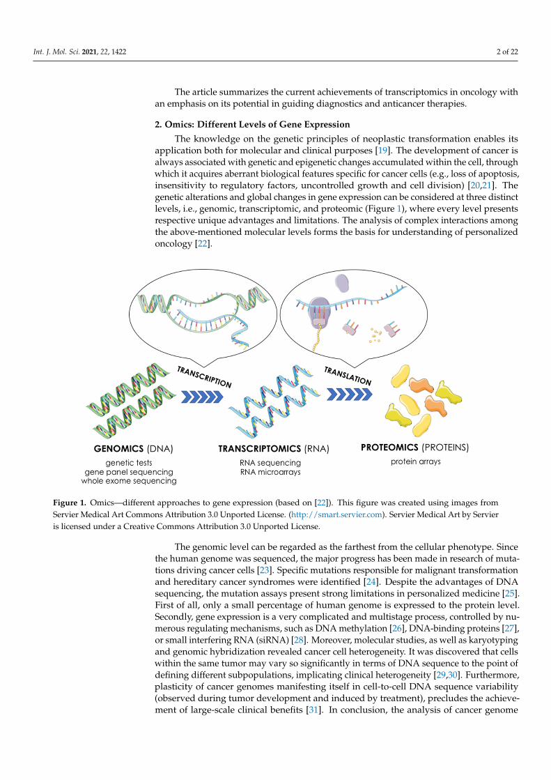
The knowledge on the genetic principles of neoplastic transformation enables itsapplication both for molecular and clinical purposes [19]. The development of cancer isalways associated with genetic and epigenetic changes accumulated within the cell, throughwhich it acquires aberrant biological features specific for cancer cells (e.g., loss of apoptosis,insensitivity to regulatory factors, uncontrolled growth and cell division) [20,21]. Thegenetic alterations and global changes in gene expression can be considered at three distinctlevels, i.e., genomic, transcriptomic, and proteomic (Figure 1), where every level presentsrespective unique advantages and limitations. The analysis of complex interactions amongthe above-mentioned molecular levels forms the basis for understanding of personalizedoncology [22].
Int. J. Mol. Sci. 2021, 22, x FOR PEER REVIEW 2 of 22
[15]. Therefore, transcriptome analysis is considered as a useful approach for investigation of constantly changing cancer cells at a molecular level. Obtained conclusions contribute directly to the intensive development of precision oncology [16–18].
The article summarizes the current achievements of transcriptomics in oncology with an emphasis on its potential in guiding diagnostics and anticancer therapies.
2. Omics: Different Levels of Gene Expression The knowledge on the genetic principles of neoplastic transformation enables its
application both for molecular and clinical purposes [19]. The development of cancer is always associated with genetic and epigenetic changes accumulated within the cell, through which it acquires aberrant biological features specific for cancer cells (e.g., loss of apoptosis, insensitivity to regulatory factors, uncontrolled growth and cell division) [20,21]. The genetic alterations and global changes in gene expression can be considered at three distinct levels, i.e., genomic, transcriptomic, and proteomic (Figure 1), where every level presents respective unique advantages and limitations. The analysis of complex interactions among the above-mentioned molecular levels forms the basis for understanding of personalized oncology [22].
Figure 1. Omics—different approaches to gene expression (based on [22]). This figure was created using images from Servier Medical Art Commons Attribution 3.0 Unported License. (http://smart.servier.com). Servier Medical Art by Servier is licensed under a Creative Commons Attribution 3.0 Unported License.
The genomic level can be regarded as the farthest from the cellular phenotype. Since the human genome was sequenced, the major progress has been made in research of mutations driving cancer cells [23]. Specific mutations responsible for malignant transformation and hereditary cancer syndromes were identified [24]. Despite the advantages of DNA sequencing, the mutation assays present strong limitations in personalized medicine [25]. First of all, only a small percentage of human genome is expressed to the protein level. Secondly, gene expression is a very complicated and multistage process, controlled by numerous regulating mechanisms, such as DNA methylation [26], DNA-binding proteins [27], or small interfering RNA (siRNA) [28]. Moreover, molecular studies, as well as karyotyping and genomic hybridization revealed cancer cell heterogeneity. It was discovered that cells within the same tumor may vary so significantly in terms of DNA sequence to the point of defining different subpopulations, implicating clinical heterogeneity [29,30]. Furthermore, plasticity of cancer genomes
Figure 1. Omics—different approaches to gene expression (based on [22]). This figure was created using images fromServier Medical Art Commons Attribution 3.0 Unported License. (http://smart.servier.com). Servier Medical Art by Servieris licensed under a Creative Commons Attribution 3.0 Unported License.
The genomic level can be regarded as the farthest from the cellular phenotype. Sincethe human genome was sequenced, the major progress has been made in research of muta-tions driving cancer cells [23]. Specific mutations responsible for malignant transformationand hereditary cancer syndromes were identified [24]. Despite the advantages of DNAsequencing, the mutation assays present strong limitations in personalized medicine [25].First of all, only a small percentage of human genome is expressed to the protein level.Secondly, gene expression is a very complicated and multistage process, controlled by nu-merous regulating mechanisms, such as DNA methylation [26], DNA-binding proteins [27],or small interfering RNA (siRNA) [28]. Moreover, molecular studies, as well as karyotypingand genomic hybridization revealed cancer cell heterogeneity. It was discovered that cellswithin the same tumor may vary so significantly in terms of DNA sequence to the point ofdefining different subpopulations, implicating clinical heterogeneity [29,30]. Furthermore,plasticity of cancer genomes manifesting itself in cell-to-cell DNA sequence variability(observed during tumor development and induced by treatment), precludes the achieve-ment of large-scale clinical benefits [31]. In conclusion, the analysis of cancer genome
Int. J. Mol. Sci. 2021, 22, 1422 3 of 22
may provide valuable information on the DNA sequence and its structure, but could beinadequate to describe the actual phenotype of the cell [22]. Therefore other approachesare needed to find proper molecular diagnostic targets and to address specific therapy forcancer patients.
The proteomic approach, which is obviously closer to the molecular mechanisms deter-mining cell’s phenotype than genomics [32], focuses on quantitative protein measurementsto characterize biological processes and deciphers the protein-dependent mechanisms ofgene expression regulation in a living cell [33,34]. It is confirmed that proteins are the keyfactors in all cellular processes, while mutations alter their expressions and/or activities invarious ways [35,36]. The analysis of the cancer proteome enables to obtain a landscape ofpost-translational modifications, interactions between cellular mechanisms and their loca-tions [37]. Among techniques of protein recognition, microarrays are the most frequentlyemployed. This method uses monoclonal antibodies or other binders for identification ofan individual protein. Protein microarrays have gained extensive applications in molecu-lar diagnostics, particularly in cancer biomarker discovery [33]. Nevertheless, technicaldifficulties related to the method, such as different physical and chemical properties ofproteins, as well as the need of usage of highly specific antibody panels, are the significantobstacles for proteomic studies in a wide range [38].
The pivotal link between cellular phenotype and genetic aspects of tumor biologyis transcriptome. It contains all information encoded in RNA transcribed from DNA.In contrast to the genome which is relatively stable, transcriptome reacts actively to thephysiological or pathological conditions. Adjustments in the transcriptome reflect thedifferent cell states, developmental stages, and regulatory mechanisms [15]. Nowadays,transcriptomics have come to the forefront of international scientific attention due to therapid development of RNA-sequencing (RNA-Seq) methods.
3. Transcriptome’s Components and Methods of Its Analysis
Studies show that about 93% of the human DNA is transcribed into RNA, howeveronly 2% into pcRNA [4]. These molecules, in a mature form, specify the sequence of aminoacids in the protein and participate in translation. The remaining part of the human tran-scriptome consists of ncRNA and, out of all discovered until now, include: ribosomal RNA(rRNA), transfer RNA (tRNA), micro RNA (miRNA), small interfering RNA (siRNA), PIWI-interacting RNA (piRNA), small nuclear RNA (snRNA), small nucleoar RNA (snoRNA),extra-cellular RNA (exRNA), guide RNA (gRNA), small Cajal body-specific RNA (scaRNA),circular RNA (circRNA), long non-coding RNA (lncRNA), such as X-inactive specific tran-script (XIST) and HOX Transcript Antisense RNA (HOTAIR) [39]. Their cellular functionssignificantly differ from each other. Some ncRNAs present catalytic roles, e.g., involvedin production of tRNA and rRNA; whereas other participate in controlling mRNA, e.g.,miRNA, snRNA, snoRNA [40].
All the above-mentioned ncRNA types potentially contribute to oncogenesis. More-over, previously unknown types of RNAs are being continuously discovered—mostly dueto modern technologies. Meticulous analysis of RNAs’ functions and the development ofnovel techniques may lead to the improvement of transcriptome cognition.
3.1. History of Gene Expression Measurement Methods
Several technologies have been developed to define the role of all RNAs formingtranscriptome in human diseases. The first approach of studying human transcriptomeascribes to quantification of mRNA with Northern blotting [41,42]. This time-consumingmethod uses gene-specific DNA probes which are hybridized to the RNA and is capable ofanalyzing a maximum a few gene transcripts at the same time [43].
Subsequently newer methods based on complementary probe hybridization wereintroduced to transcriptome analysis [8]. Today, three main directions can be distinguishedin cancer transcriptomics: microarrays, large-scale quantitative reverse-transcription-
Int. J. Mol. Sci. 2021, 22, 1422 4 of 22
polymerase chain reaction (qRT-PCR) (both using probe hybridization), and the newest,next-generation sequencing (NGS) technology, represented by RNA-Seq [8,44].
3.1.1. Microarrays
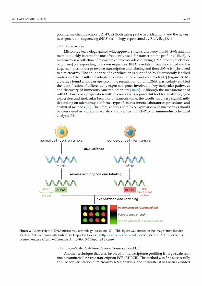
Microarray technology gained wide approval since its discovery in mid-1990s and thismethod quickly became the most frequently used for transcriptome profiling [45,46]. Amicroarray is a collection of microchips of microbeads containing DNA probes (nucleotideoligomers) corresponding to known sequences. RNA is isolated from the control and thetarget samples, undergo reverse transcription and labeling and then cDNA is hybridizedto a microarray. The abundance of hybridization is quantified by fluorescently labelledprobes and the results are adapted to measure the expression levels [47] (Figure 2). Mi-croarrays found a wide usage also in the research of tumor mRNA, particularly enabledthe identification of differentially expressed genes involved in key molecular pathwaysand discovery of numerous cancer biomarkers [48,49]. Although the measurement ofmRNA down- or upregulation with microarrays is a powerful tool for analyzing geneexpression and molecular behavior of transcriptome, the results may vary significantlydepending on microarray platforms, type of laser scanners, laboratories procedures andanalytical methods [50]. Therefore, analysis of mRNA expression with microarrays shouldbe considered as a preliminary step, and verified by RT-PCR or immunohistochemicalanalysis [51].
Int. J. Mol. Sci. 2021, 22, x FOR PEER REVIEW 4 of 22
Subsequently newer methods based on complementary probe hybridization were introduced to transcriptome analysis [8]. Today, three main directions can be distinguished in cancer transcriptomics: microarrays, large-scale quantitative reverse-transcription-polymerase chain reaction (qRT-PCR) (both using probe hybridization), and the newest, next-generation sequencing (NGS) technology, represented by RNA-Seq [8,44].
3.1.1. Microarrays Microarray technology gained wide approval since its discovery in mid-1990s and
this method quickly became the most frequently used for transcriptome profiling [45,46]. A microarray is a collection of microchips of microbeads containing DNA probes (nucleotide oligomers) corresponding to known sequences. RNA is isolated from the control and the target samples, undergo reverse transcription and labeling and then cDNA is hybridized to a microarray. The abundance of hybridization is quantified by fluorescently labelled probes and the results are adapted to measure the expression levels [47] (Figure 2). Microarrays found a wide usage also in the research of tumor mRNA, particularly enabled the identification of differentially expressed genes involved in key molecular pathways and discovery of numerous cancer biomarkers [48,49]. Although the measurement of mRNA down- or upregulation with microarrays is a powerful tool for analyzing gene expression and molecular behavior of transcriptome, the results may vary significantly depending on microarray platforms, type of laser scanners, laboratories procedures and analytical methods [50]. Therefore, analysis of mRNA expression with microarrays should be considered as a preliminary step, and verified by RT-PCR or immunohistochemical analysis [51].
Figure 2. An overview of DNA microarray technology (based on [47]). This figure was created using images from Servier Medical Art Commons Attribution 3.0 Unported License. (http://smart.servier.com). Servier Medical Art by Servier is licensed under a Creative Commons Attribution 3.0 Unported License.
Figure 2. An overview of DNA microarray technology (based on [47]). This figure was created using images from ServierMedical Art Commons Attribution 3.0 Unported License. (http://smart.servier.com). Servier Medical Art by Servier islicensed under a Creative Commons Attribution 3.0 Unported License.
3.1.2. Large-Scale Real-Time Reverse Transcription PCR
Another technique that was involved in transcriptome profiling is large-scale real-time (quantitative) reverse transcription PCR (RT-PCR). The method was first successfullyapplied for verification of microarray RNA analysis, and thereafter it has been extended
Int. J. Mol. Sci. 2021, 22, 1422 5 of 22
to become a self-sufficient technique as it is fast, reliable and enables the accurate andsimultaneous profiling of many genes expression [4,52]. It is noteworthy that RT-PCRfound broad applications in diagnostics and treatment monitoring in oncology [53]. Ithas been used in research of prognostic and predictive tumor biomarkers, e.g., in breastcancer [54], melanoma [55,56], or hepatocellular carcinoma [57,58], as well as for otherpurposes, such as micrometastasis determination [59] or detection of circulating tumorcells [60,61]. In this method complementary DNA (cDNA) is generated from RNA templatein reverse transcription and specific DNA targets are amplified using polymerase chainreaction (PCR), employing large set of primer pairs. Quantitative PCR enables to evaluatethe increase in copies of investigated sequences in real time. It is possible due to labelling ofprimers, oligonucleotides, or amplification products with fluorophores. A positive reactionis detected by the accumulation of a fluorescent signal.
3.1.3. RNA-Sequencing (RNA-Seq)
Above-mentioned hybridization-based methods are limited by the need of priorknowledge on the sequence of the probed nucleic acid [62]. Therefore, transcriptomicsevolved to sequencing-based techniques including consecutively: Sanger sequencing ofexpressed sequence tags (ESTs) and NGS.
Sanger sequencing is a method of controlled termination of DNA synthesis [63]. A sam-ple matrix is usually single-stranded DNA amplified in PCR [64]. The sequencing processrequires the sample matrix, DNA polymerase, one primer, four deoxynucleotidetriphos-phates (dNTPs) and four fluorescent labelled dideoxynucleotidetriphosphate (ddNTPs).DNA polymerase is applied for synthetizing nucleic acid strand using either dNTPs, whichenables continuation of synthesis, or ddNTPs, which terminates further synthesis in aspecific locus [65]. For the purpose of RNA studies, ESTs—short sub-sequences of cDNAcomplementary to a part of mRNA—are used with Sanger sequencing [8]. Before NGS tech-nology was introduced, tag-based sequencing had been commonly used in the research oftumor biomarkers [66–68]. Nevertheless, it presents significant limitations: it can sequenceshort pieces of DNA—up 1000 base pairs, the primer’s template must be defined and therehas to be large amount of copies of the gene in the starting mixture for sequencing [69].
3.1.4. Next Generation Sequencing (NGS)
The new era of transcriptome profiling is set by the NGS. It provides abundant possibil-ities to perform simultaneous investigations of thousands of genes, analyses of complicatedmolecular mechanisms participating in oncogenesis and thus, offers a great contributionto the precision medicine. NGS technology, first introduced in 2004, may be applied invarious areas: whole-genome and exome sequencing, RNA-Seq, which is referred today asa “gold standard” of transcriptome analysis, chromatin immunoprecipitation followed bysequencing (ChIP-Seq) for analysis of interactions between DNA and proteins and copynumber variation-sequencing (CNV-Seq) to analyze DNA copy number variation [70].RNA-Seq enables high-throughput gene expression analysis with remarkable precision.NGS generates data about splicing variants, allelic expression and RNA editing [71]. Itenables the analysis of epigenetic modifications and elements playing role in mechanisms,such as ncRNAs [72]. This technology has significantly increased the efficiency, sensitivity,accuracy and transcript coverage in obtained results in comparison to microarrays [73].
Several NGS platforms are based on the pyrosequencing principle, where bindingof nucleotides during chain synthesis is monitored by luminescence [74]. After the incor-poration of a specific dNTP, a lightning signal is generated when pyrophosphate (PPi)is converted to ATP by ATP sulfurylase in the presence of adenosine 5′ phosphosulfate(APS). Two sequencing platforms directly utilize pyrosequencing principle: 454 Sequencing(454 Life Sciences; now Roche Holding AG, Basel, Switzerland) and PyroMark ID system(Qiagen, Hilden, Germany) [75,76]. Other massive parallel sequencing instruments include:Illumina-Solexa Genome Analyzer family launched in 2006, Ion Torrent, Thermo Fisherand Bionano Technologies [77]. Considering main differences between analyzers, Roche
Int. J. Mol. Sci. 2021, 22, 1422 6 of 22
display higher length reads in comparison to Illumina, but produce more errors withinhomopolymer regions [78,79].
RNA-seq has been widely used for the research on gene expression in various malig-nancies and has been found as an effective tool in exploration of carcinogenesis, identifica-tion of tumor biomarkers and development of new therapeutic strategies. The followingparts of the article provide a clearer image of the achievements of transcriptomics over therecent years.
4. Applications of Transcriptome Analysis in Oncology
The transcriptome-based studies opened a new chapter in molecular understanding ofcancer. Analysis of gene expression allows researchers to explore molecular basis of cancerin a multiway manner and provides information of tumor progression. Technologiesinvolved in transcriptomics have expanded our knowledge on carcinogenesis, tumormicroenvironment (TME), and immuno-oncology, as well as enabled the discovery of new,previously unknown cancer biomarkers.
4.1. Applications in Clinical Classifications of Cancer
Identification of the molecular portrait of the tumor is valuable in cancer classificationand elucidation of biochemical pathways, which allows further selection of selectivetherapeutics and prediction of cancer drug sensitivity [80]. Analysis of the transcriptomehas led to a significant improvement in clinical classification of many tumors [81,82].
So far, numerous studies have been carried out to profile gene expression and thus, toestablish molecular classification and identification of therapeutic/diagnostic targets inbreast cancer [83–86]. One of the first successful attempts of gene expression-based charac-terization was made by Perou et al. (2000). In 65 samples of breast cancer obtained from42 patients, unique patterns of gene expression were observed for every individual [87].Nevertheless, four different molecular clusters of breast cancer, presenting similar expres-sion signatures, were captured: ER+/luminal-like, basal-like, Erb-B2+ (Her-2/neu) andnormal-like breast tumors. The clusters distinguished in this study correspond closely toimmunohistochemistry markers of breast tumors: estrogen receptors, Her-2 and Ki-67 [88],and thus, should be treated as distinct diseases [89,90]. Moreover, similar conclusionswere drawn in the study of Sørlie et al. [91], where luminal type of breast cancer wasdivided into two subtypes: luminal A (with high estrogen receptor expression and lowexpression of proliferative markers, e.g., Ki67) and luminal B (presenting lower estrogenreceptor expression and high level of expression of proliferation-related genes) [92,93]. Theabove-mentioned studies were carried out by using microarray technology, which laid thefoundations of current clinically used molecular classification of breast cancer [94].
Analogous efforts were made in classification of colorectal cancer (CRC). In the re-search conducted by Guinney et al., four consensus molecular subtypes of CRC weredistinguished: CMS1 (microsatellite instability immune-hypermutated, characterized bystrong immune activation, microsatellite unstable), CMS2 (canonical—marked by WNT andMYC signaling pathways activation), CMS3 (metabolic—epithelial with evident metabolicdysregulation), and CMS4 (mesenchymal—with growth factor–β activation, stromal inva-sion and angiogenesis) [95]. Recently, many clinical trials demonstrated the usability ofthis classification, confirming different sensitivities to the treatment, recurrence parametersand tendencies to metastasis of individual CMS subtypes [96–100]. Such an approach aidsin the understanding of CRC etiology for the purposes of precision oncology. However,further population-based trials must be performed to prove the advantages of introducingCMS classification into clinical oncology [84,101].
Another study uncovered differences in origin of ovarian cancer. It is pathologicallydivided into subtypes: epithelial (EOC), sex-cord stromal and germ cell, where EOC issubdivided into: high-grade serous, low-grade serous, mucinous, endometrioid and clearcell carcinoma [102]. According to the classic view on high-grade serous ovarian cancer(HGSOC), it originates from epithelial cells of ovarian surface. However, recent studies
Int. J. Mol. Sci. 2021, 22, 1422 7 of 22
show that tumor may develop from fallopian tube [103,104]. The molecular profiling ofHGSOC has revealed overexpression of PAX8, which is typical for cells within the distalfallopian tube [105,106]. The determination of PAX8 overexpression by using RNA-Seqmay accelerate diagnostics.
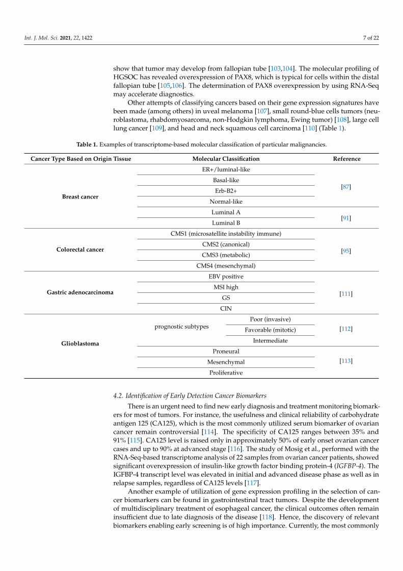
Other attempts of classifying cancers based on their gene expression signatures havebeen made (among others) in uveal melanoma [107], small round-blue cells tumors (neu-roblastoma, rhabdomyosarcoma, non-Hodgkin lymphoma, Ewing tumor) [108], large celllung cancer [109], and head and neck squamous cell carcinoma [110] (Table 1).
Table 1. Examples of transcriptome-based molecular classification of particular malignancies.
Cancer Type Based on Origin Tissue Molecular Classification Reference
Breast cancer
ER+/luminal-like
[87]Basal-like
Erb-B2+
Normal-like
Luminal A[91]
Luminal B
Colorectal cancer
CMS1 (microsatellite instability immune)
[95]CMS2 (canonical)
CMS3 (metabolic)
CMS4 (mesenchymal)
Gastric adenocarcinoma
EBV positive
[111]MSI high
GS
CIN
Glioblastoma
prognostic subtypesPoor (invasive)
[112]Favorable (mitotic)
Intermediate
Proneural
[113]Mesenchymal
Proliferative
4.2. Identification of Early Detection Cancer Biomarkers
There is an urgent need to find new early diagnosis and treatment monitoring biomark-ers for most of tumors. For instance, the usefulness and clinical reliability of carbohydrateantigen 125 (CA125), which is the most commonly utilized serum biomarker of ovariancancer remain controversial [114]. The specificity of CA125 ranges between 35% and91% [115]. CA125 level is raised only in approximately 50% of early onset ovarian cancercases and up to 90% at advanced stage [116]. The study of Mosig et al., performed with theRNA-Seq-based transcriptome analysis of 22 samples from ovarian cancer patients, showedsignificant overexpression of insulin-like growth factor binding protein-4 (IGFBP-4). TheIGFBP-4 transcript level was elevated in initial and advanced disease phase as well as inrelapse samples, regardless of CA125 levels [117].
Another example of utilization of gene expression profiling in the selection of can-cer biomarkers can be found in gastrointestinal tract tumors. Despite the developmentof multidisciplinary treatment of esophageal cancer, the clinical outcomes often remaininsufficient due to late diagnosis of the disease [118]. Hence, the discovery of relevantbiomarkers enabling early screening is of high importance. Currently, the most commonly
Int. J. Mol. Sci. 2021, 22, 1422 8 of 22
used biomarkers in esophageal squamous cell carcinoma (ESCC) diagnosis are: cytokeratin19 fragment antigen 21–1 (Cyfra21–1), carbohydrate antigen 19–9 (CA19–9), carbohydrateantigen 72–4 (CA72–4), carcinoembryonic antigen (CEA), and squamous cell carcinomaantigen (SCC-Ag) [119]. The above-mentioned proteins are characterized by high specificityfor ESCC, but low sensitivity ranging between 10% and 40% [120]. Recent approachescombine mRNA sequencing or proteome sequencing together with computational analy-sis, have identified highly expressed genes or highly secreted proteins, which may serveas early detection biomarkers. Bioinformatics enables parallel analysis of thousands ofvariables in the genome-wide scale, assess their significance, which facilitates selectionof relevant variables and allows for biologically meaningful interpretations [121]. How-ever, chemistry, sequencing, and data preprocessing differ significantly between proteomesequencing and mRNA sequencing, the downstream steps are similar [122]. Statisticalcomparisons of normal tissue, benign neoplasia and tumor tissue by data mining methodsrepresent a common way of searching and prioritization of biomarkers [123]. Regardingstatistical analyses used to compare various stages of neoplasia, there are a huge variety ofdata mining methods including relatively simple feature (gene) selection algorithms (e.g.,t-test, principal component analysis (PCA), nucleoside triphosphate (NTP) classifier andleast absolute shrinkage and selection operator (LASSO)) or more mathematically sophis-ticated approaches (e.g., support vector machine (SVM)-based, Bayesian network-based,neural network-based, or ensemble-based) [124]. It has to be emphasized that difference interms of performance (specificity, sensitivity, accuracy) is comparable between these twoclasses of mining methods (simple and sophisticated). Therefore, in current biomarkerresearch simple solutions such as NTP classifier became popular [125,126]. Moreover, setsof selected markers offer much better performance than single marker, therefore, currentlyin various studies predictive power have benefited from such multi-marker approach [127].For example, in the study of Xing et al., three serum markers were evaluated as potentiallyclinically usable: CHI3L1, MMP13, and SPP1, from more than 4000 differentially expressedgenes in the ESCC transcriptome database. The diagnostic panel using combination ofCHI3L1, MMP13 and SPP1 identified approximately 90% of cases with early-onset ESCC,where the panel’s detectability in non-cancerous tissues ranged between 10% and 15% [128].
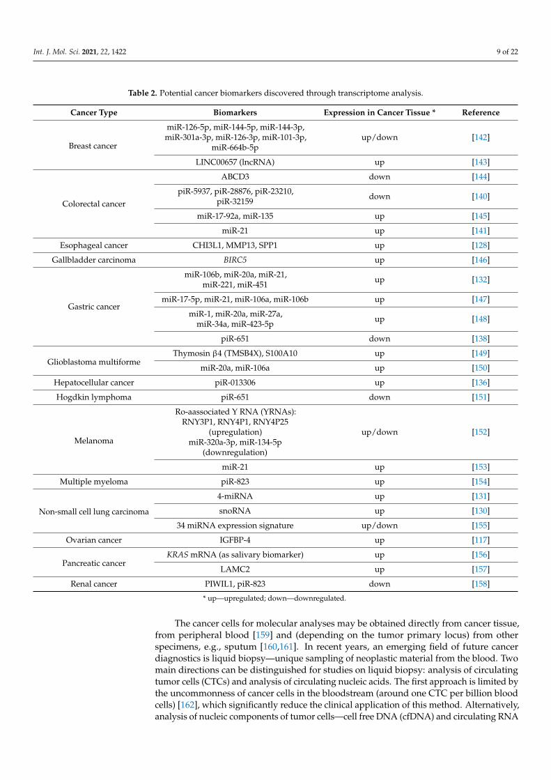
Moreover, various RNA molecules may serve as independent cancer biomarkers [129].Over the recent years, many RNAs were found to be useful in precision oncology (Table 2).For instance, the expression profile of snoRNAs [130] and 4-miRNA expression signa-ture [131] can be used in early diagnosis of non-small-cell lung carcinoma (NSCLC). Inaddition, miR-106b, miR-20a, and miR-221 were confirmed as early detection biomark-ers for gastric cancer [132,133]. The piRNAs, which mediate transcriptional and post-transcriptional gene silencing machineries, may serve as biomarkers for early diagnosis,treatment and prognosis of: renal cancer [134,135], hepatocellular carcinoma [136], glioblas-toma [137] and gastric cancer [138]. Moreover, lncRNAs, such as XIST, present as potentialcandidates for the detection of early gastric cancer [139]. A growing number of studieshave indicated the possibility of applications of ncRNAs in CRC as well [140,141].
Int. J. Mol. Sci. 2021, 22, 1422 9 of 22
Table 2. Potential cancer biomarkers discovered through transcriptome analysis.
Cancer Type Biomarkers Expression in Cancer Tissue * Reference
Breast cancer
miR-126-5p, miR-144-5p, miR-144-3p,miR-301a-3p, miR-126-3p, miR-101-3p,
miR-664b-5pup/down [142]
LINC00657 (lncRNA) up [143]
Colorectal cancer
ABCD3 down [144]
piR-5937, piR-28876, piR-23210,piR-32159 down [140]
miR-17-92a, miR-135 up [145]
miR-21 up [141]
Esophageal cancer CHI3L1, MMP13, SPP1 up [128]
Gallbladder carcinoma BIRC5 up [146]
Gastric cancer
miR-106b, miR-20a, miR-21,miR-221, miR-451 up [132]
miR-17-5p, miR-21, miR-106a, miR-106b up [147]
miR-1, miR-20a, miR-27a,miR-34a, miR-423-5p up [148]
piR-651 down [138]
Glioblastoma multiformeThymosin β4 (TMSB4X), S100A10 up [149]
miR-20a, miR-106a up [150]
Hepatocellular cancer piR-013306 up [136]
Hogdkin lymphoma piR-651 down [151]
Melanoma
Ro-aassociated Y RNA (YRNAs):RNY3P1, RNY4P1, RNY4P25
(upregulation)miR-320a-3p, miR-134-5p
(downregulation)
up/down [152]
miR-21 up [153]
Multiple myeloma piR-823 up [154]
Non-small cell lung carcinoma
4-miRNA up [131]
snoRNA up [130]
34 miRNA expression signature up/down [155]
Ovarian cancer IGFBP-4 up [117]
Pancreatic cancerKRAS mRNA (as salivary biomarker) up [156]
LAMC2 up [157]
Renal cancer PIWIL1, piR-823 down [158]
* up—upregulated; down—downregulated.
The cancer cells for molecular analyses may be obtained directly from cancer tissue,from peripheral blood [159] and (depending on the tumor primary locus) from otherspecimens, e.g., sputum [160,161]. In recent years, an emerging field of future cancerdiagnostics is liquid biopsy—unique sampling of neoplastic material from the blood. Twomain directions can be distinguished for studies on liquid biopsy: analysis of circulatingtumor cells (CTCs) and analysis of circulating nucleic acids. The first approach is limited bythe uncommonness of cancer cells in the bloodstream (around one CTC per billion bloodcells) [162], which significantly reduce the clinical application of this method. Alternatively,analysis of nucleic components of tumor cells—cell free DNA (cfDNA) and circulating RNA
Int. J. Mol. Sci. 2021, 22, 1422 10 of 22
(circRNA, including miRNA)—is gaining an increasing interest, demonstrated in numerousstudies and clinical trials [159,163–165]. Rapid advancement of novel technologies, enablesto detect cancerous DNA/RNA with high accuracy for diagnosis, prognosis and therapeuticmonitoring [166]. RNA monitoring, in comparison to DNA-based biopsy, presents twomain advantages: it is a better tool for identification of gene fusions and functional diseaseprofiling, through detection of ncRNA of biological importance and characterization ofalternative RNA splicing products [167,168]. Consequently, a number of recent studiesinvolving RNA obtained from liquid biopsy is steadily increasing (mostly for miRNAmarkers). Results of large, carefully conducted validation studies (sample sizes above 500)showed promising opportunities of applying miRNA signatures in clinics in the near future.For example, miR-Test published in 2015 is based on signature of 13 serum miRNAs forearly and sensitive detection of lung cancer. MiR-Test displayed the sensitivity of 77.8% andthe specificity of 74.8% to detect lung tumors [169]. There are also several urine-based testsfor detection of prostate cancer including SelectMDx (based on gene expression signature)and Progensa PCA3 (based on lncRNA expression signature) [170,171]. Both tests revealedtheir potential in elimination of unnecessary prostate biopsies [172].
4.3. Creation of Cancer Prognostic and Predictive Panels
Various assays based on gene expression analysis have been developed to guidepersonalized therapeutic pathway in oncology [22]. Multigene tests provide detailedinsight into cancer biology with simultaneous information on expression of relevant,prognostic fundamental genes. Some assays are already used in diagnosis, as prognosticpanels and for the prediction of treatment effects (mostly adjuvant or neoadjuvant systemictherapy) [173].
Among most commonly used gene expression tests are MammaPrint® and OncotypeDX® assessing the risk of relapse and metastasis in breast cancer. MammaPrint® is 70-gene panel, tested in large randomized controlled trial (MINDACT, ClinicalTrials.govidentifier: NCT00433589), where its ability to differentiate low-risk and high-risk patientswith breast cancer was confirmed [174,175]. Oncotype DX® is 21-gene qRT-PCR testassessing the risk of breast cancer recurrence, with particular emphasis on early-stage, ER-positive (estrogen receptor), HER2-negative and axillary node-negative tumors [176]. Theadvantages of Oncotype DX® panel were presented widely in trial TAILORx (ClinicalTrials.gov identifier: NCT00310180) [177]. Other transcriptomic signatures of breast cancerwith clinical usage are: Prosigna®-Assay (PAM50—50-gene panel) [178,179], Endopredict®
(12-gene assay) [180,181].The similar assays are being tested in prognosis of colon cancer. The twin test of Onco-
type DX® used in breast cancer – 12-gene Oncotype DX® Colon Cancer Assay identifies thetumors with high risk of recurrence and helps to determine the necessity of implementationof adjuvant systemic therapy [182–184]. In the study of Aziz et. al, the 19-gene signaturewas evaluated as a predictive classifier in colorectal cancer using microarray profiling [185].It was revealed that the patients assigned into the low-risk group could avoid or receivelower concentrations of adjuvant chemotherapy.
Afirma® gene expression classifier is a microarray assay used for thyroid cancer diag-nosis. It is validated as pre-operative test for differentiating invasive cancer from benignnodules (such as follicular adenoma) in order to avoid unnecessary surgeries [186]. The cy-tological tests used routinely for attaining that purpose are characterized by frequent falseresults, therefore genetic testing is valuable solution for this problem [187]. Other diagnos-tic panels are: ThyroidPrint® (10-gene classifier) [188], ThyroSeq v3 (DNA- and RNA-basedNGS assay analyzing 112 genes) [189], RosettaGX Reveal, and ThyraMIR/ThyGenX™,which are respectively 24-miRNA and 10-miRNA (with 8 DNA mutations) expressiontests [190,191].
Int. J. Mol. Sci. 2021, 22, 1422 11 of 22
4.4. Intratumoral Heterogeneity (ITH) and Tumor Microenvironment (TME)-Related Research
Most of human cells have identical DNA material, despite the differences at thelevel of gene expression. For example, transcriptome and protein landscapes of the livervary significantly from those of the heart. In addition, such cellular differences can bedistinguished within the same organ, e.g., neurons and glial cells of the brain. Thesedifferences in gene expression within particular subpopulations of cells reflect their distinctmorphologies, functions, regenerative capacities, etc. Thus, profiling of transcriptome ofindividual cells—single-cell RNA-Seq (scRNA-Seq)—provides a multidirectional insightinto specific cell’s functions—also in cancer. The scRNA-Seq is valuable but challengingmethod preceded by dissociation of heterogenous tissue and optimized filtering [192].To date, this approach has enabled identifications of novel cell populations and cell-cellinteractions. The characterization of ITH reveals the molecular background for resistanceto the cancer treatment, the dynamic of tumor growth and its potential of relapse [193].Several works show the power of scRNA-Seq to distinguish the ITH. The study of Sharmaet al. revealed the multi-level tumor evolution of lung squamous cell carcinoma based oncell subclones closely related to tumor-immune cell interactions [194]. In the study of Wuet al., the immunological interactions between breast cancer cells and TME were proved,showing the ability of scRNA-Seq for analyzing tumor in a dynamic state [195].
Intra-tumor heterogeneity generates serious complications, including resistance totherapy. In addition to the different subpopulations forming a tumor bulk, the response tothe treatment and whole disease course are dependent, among others on the interactionswithin TME. It consists of complex ecosystem of stromal cells, such as cancer-associatedfibroblasts (CAFs), immune cells, endothelial cells and others of lesser importance. As men-tioned above, TME components actively influence tumor cell proliferation. For example,cytokines secreted by CAFs participate in signal pathways in cancer cells [196]. Moreover,the immune compartment of TME acts immunosuppressively, creating a barrier betweencancer cells and the host’s immune system [197]. The investigation of mechanisms bywhich TME affects cancer biology is possible through leverage of gene expression profiling.
The technology which has been successfully applied in the studies of TME of therecent years is RNA-seq [198]. It has become an integral tool of immunogenomics andserves today for analysis of gene expression patterns associated with the presence of dif-ferent populations of immunological cells in the cancerous tissue [199]. Identification ofdistinct immune and stromal cells subtypes enables not only for understanding of differentmetabolic phenotypes of tumors, but also for the assessment of immune receptor repertoirein purpose of precision cancer immunotherapy [200]. The T-cell receptor (TCR)/B-cell re-ceptor (BCR) sequences on tumor cells provide an information about specificity of immuneresponse in TME. This landscape of TCRs/BCRs may serve as a predictive biomarker ofthe effectiveness of immunotherapy [201,202]. However the validation cohorts of thesegene signatures remains to be seen.
4.5. RNA-Based Therapeutics
Dynamic growth of knowledge on transcriptome has changed a simple understandingof RNA as an intermediary between DNA and protein to a whole variety of moleculeswith numerous functions and a potential of regulation of gene expression. Thus, particularRNAs can be used as therapeutic targets as well. The greatest progress in terms of RNA-based therapies has been made in regard to miRNA.
miRNAs, which selectively induce mRNA degradation and translation inhibition,present an abnormal expression within cancerous tissues. Depending on their functions,miRNAs may act as an oncogenes (also known as oncomiRs—Onc miRNAs) or tumorsuppressors (TS miRNAs) [203]. OncomiRs (e.g., miR155 and miR21) promote cell trans-formation, carcinogenesis, and metastasis by blocking the expression of tumor suppressorgenes, and hence can be used as therapeutic targets in cancer treatment [204–206]. Inturn, TS miRNAs suppress the translation of mRNAs involved in oncogenic pathways.Many studies have confirmed that the expression of TS miRNAs are downregulated in
Int. J. Mol. Sci. 2021, 22, 1422 12 of 22
cancer [207,208]. Therefore, miRNAs can be considered as an early indicator for diagnosticpurposes and patient prognosis, as well as effective target for therapeutic strategies inoncology [203].
There are two main approaches for regulating miRNA activities: suppression of expres-sion of oncogenic miRNAs, and restoration of the loss of TS miRNAs’ expressions [203,209].Inhibition of oncomiRs’ expressions can be achieved through the use of antisense oligonu-cleotides with 2′-O-methyl groups [210–212], 2′-O-methoxyethyl groups [213] and otherlinkages blocking exonuclease degradation, such as N,N-diethyl-4-(4-nitronaphthalen-1-ylazo)-phenylamine [214]. Another strategy is an application of locked nucleic acid (LNA)anti-miRNA oligonucleotides with complementary sequences to target miRNAs [206,215].Currently, two clinical trials, i.e., SOLAR (ClinicalTrials.gov identifier: NCT03713320; miRagenTherapeutics, Inc., Boulder, CO, USA) and PRISM (enrolling; ClinicalTrials.gov identifier:NCT03837457), investigate the efficacy and safety of Cobomarsen (MRG-106)—an LNAantimiR® inhibitor of miR-155 in the treatment of cutaneous T-cell lymphoma (CTCL),adult T-cell lymphoma (ATLL), chronic lymphocytic leukemia (CLL) and diffuse largeB-cell lymphoma (DLBCL) [216–218].
The approach focusing on restoration of down-regulated TS miRNAs is called miRNAreplacement therapy [219]. It is based on delivery of key miRNAs for carcinogenesis usingeither viral or non-viral vectors [220]. Viral platforms have been found to be insufficientdue to triggering of immune response [221]. Non-viral vectors include: inorganic miRNAplatforms (e.g., Fe3O4-based nanoparticles [222], gold or nanodiamonds platforms [223],and silica-based systems [224]), polymer-based miRNA delivery systems (such as polyami-doamine dendrimers—PAMAMs [225,226], or atelocollagen [227,228]) and lipid-basedplatforms [229,230].
Considering the increase in RNA-focused studies over the recent years, the specificknowledge about RNA may result in introduction of clinical, RNA-based techniques [231].Research efforts have focused on overcoming limitations related to RNA therapeutics:low bioavailability, poor cellular uptake or rapid elimination and excessive immune re-sponse [232,233]. Nevertheless, targeting RNAs networks in cancer present strong potentialfor future clinical utility. Therefore, a growth in RNA therapeutics in the next several yearsis highly expected.
5. Conclusions
Transcriptomics undoubtedly deserves to be considered as contemporary “game-changer” in oncology on both molecular and clinical levels. In recent years, the revolutionin the understanding, research, and clinical implementation of RNA-based techniques canbe observed. The constant development of molecular technologies used for transcriptomeanalysis and implementation of NGS platforms considerably changed the landscape ofRNA world research. RNA-seq is continuously gaining larger role in the clinical care ofcancer patients, as well as preclinical research and basic science. It enables an insightinto the molecular mechanisms underlying cancer, which has given rise to numeroustrials related to transcriptome-based personalized oncology. The increasing availabilityof transcriptome profiling enables the introduction of transcriptomics in clinics. To date,several prognostic and predictive gene expression assays, as well as cancer biomarkers, havebeen utilized in clinical oncology. The change of the paradigm of cancer classification—fromhistopathological and clinical to molecular, opens a more personalized perspective fortumor diagnostics and therapy. The further advancement of transcriptomeprofiling willallow for standardization and the cost reduction of its analysis, which will be the next stepfor transcriptomics to become a canon of contemporary cancer medicine.
Author Contributions: S.S., conceptualization, investigation, writing—original draft, correspondingauthor; P.K., writing—review & editing; M.S., writing—review & editing, supervision, fundingacquisition; I.L., writing—review & editing, supervision, validation, project administration. M.S.and I.L. are equal contributors. All authors have read and agreed to the published version of themanuscript.
Int. J. Mol. Sci. 2021, 22, 1422 13 of 22
Funding: This research was funded by Statutory Subsidy Funds of the Genetics Department, WroclawMedical University grant number no. SUB.A.290.19.020.
Institutional Review Board Statement: Not applicable.
Informed Consent Statement: Not applicable.
Data Availability Statement: Not applicable.
Acknowledgments: The study was supported by Statutory Subsidy Funds of the Genetics Depart-ment, Wroclaw Medical University no. SUB.A.290.19.020.
Conflicts of Interest: The authors declare no conflict of interest.
References1. Unger, F.T.; Witte, I.; David, K.A. Prediction of individual response to anticancer therapy: Historical and future perspectives. Cell.
Mol. Life Sci. 2015, 72, 729–757. [CrossRef] [PubMed]2. Bezabeh, T.; Odlum, O.; Nason, R.; Kerr, P.; Sutherland, D.; Patel, R.; Smith, I.C. Prediction of Treatment Response in Head and
Neck Cancer by Magnetic Resonance Spectroscopy. AJNR Am. J. Neuroradiol. 2005, 26, 2108–2113. [PubMed]3. Sobin, L.H. TNM: Evolution and relation to other prognostic factors. Semin. Surg. Oncol. 2003, 21, 3–7. [CrossRef] [PubMed]4. Casamassimi, A.; Federico, A.; Rienzo, M.; Esposito, S.; Ciccodicola, A. Transcriptome profiling in human diseases: New advances
and perspectives. Int. J. Mol. Sci. 2017, 18, 1652. [CrossRef]5. Wheeler, D.A.; Wang, L. From human genome to cancer genome: The first decade. Genome Res. 2013, 23, 1054–1062. [CrossRef]6. López-Lázaro, M. A new view of carcinogenesis and an alternative approach to cancer therapy. Mol. Med. 2010, 16, 144–153.
[CrossRef]7. Janket, S.-J.; Qureshi, M.; Bascones-Martinez, A.; González-Febles, J.; Meurman, J.H. Holistic paradigm in carcinogenesis:
Genetics, epigenetics, immunity, inflammation and oral infections. World J. Immunol. 2017, 7, 11. [CrossRef]8. Lowe, R.; Shirley, N.; Bleackley, M.; Dolan, S.; Shafee, T. Transcriptomics technologies. PLoS Comput. Biol. 2017, 13, e1005457.
[CrossRef]9. Wang, Z.; Gerstein, M.; Snyder, M. RNA-Seq: A revolutionary tool for transcriptomics. Nat. Rev. Genet. 2009, 10, 57–63. [CrossRef]10. Jacquier, A. The complex eukaryotic transcriptome: Unexpected pervasive transcription and novel small RNAs. Nat. Rev. Genet.
2009, 10, 833–844. [CrossRef]11. Popov, X.D.V.; Makhnovskii, P.A.; Shagimardanova, E.I.; Gazizova, G.R.; Lysenko, E.A.; Gusev, O.A.; Vinogradova, O.L.
Contractile activity-specific transcriptome response to acute endurance exercise and training in human skeletal muscle. Am. J.Physiol. Endocrinol. Metab. 2019, 316, E605–E614. [CrossRef]
12. Iwata, M.; Yuan, L.; Zhao, Q.; Tabei, Y.; Berenger, F.; Sawada, R.; Akiyoshi, S.; Hamano, M.; Yamanishi, Y. Predicting drug-inducedtranscriptome responses of a wide range of human cell lines by a novel tensor-train decomposition algorithm. Bioinformatics 2019,35, i191–i199. [CrossRef] [PubMed]
13. Jaeger, P.A.; Doherty, C.; Ideker, T. Modeling transcriptome dynamics in a complex world. Cell 2012, 151, 1161–1162. [CrossRef][PubMed]
14. Mallardo, M.; Poltronieri, P.; D’Urso, O.F. Non-protein coding RNA biomarkers and differential expression in cancers: A review.J. Exp. Clin. Cancer Res. 2008, 27, 19. [CrossRef] [PubMed]
15. Cieslik, M.; Chinnaiyan, A.M. Cancer transcriptome profiling at the juncture of clinical translation. Nat. Rev. Genet. 2018, 19,93–109. [CrossRef]
16. Byron, S.A.; Van Keuren-Jensen, K.R.; Engelthaler, D.M.; Carpten, J.D.; Craig, D.W. Translating RNA sequencing into clinicaldiagnostics: Opportunities and challenges. Nat. Rev. Genet. 2016, 17, 257–271. [CrossRef]
17. Wang, L.; Acharya, L.; Bai, C.; Zhu, D. Transcriptome assembly strategies for precision medicine. Quant. Biol. 2017, 5, 280–290.[CrossRef]
18. Roychowdhury, S.; Chinnaiyan, A.M. Translating cancer genomes and transcriptomes for precision oncology. CA Cancer J. Clin.2016, 66, 75–88. [CrossRef]
19. Patterson, A.D.; Gonzalez, F.J.; Perdew, G.H.; Peters, J.M. Molecular Regulation of Carcinogenesis: Friend and Foe. Toxicol. Sci.2018, 165, 277–283. [CrossRef]
20. You, J.S.; Jones, P.A. Cancer Genetics and Epigenetics: Two Sides of the Same Coin? Cancer Cell 2012, 22, 9–20. [CrossRef]21. Takeshima, H.; Ushijima, T. Accumulation of genetic and epigenetic alterations in normal cells and cancer risk. NPJ Precis. Oncol.
2019, 3, 1–8. [CrossRef] [PubMed]22. Buzdin, A.; Sorokin, M.; Garazha, A.; Glusker, A.; Aleshin, A.; Poddubskaya, E.; Sekacheva, M.; Kim, E.; Gaifullin, N.;
Giese, A.; et al. RNA sequencing for research and diagnostics in clinical oncology. Semin. Cancer Biol. 2019. [CrossRef] [PubMed]23. Nussinov, R.; Jang, H.; Tsai, C.J.; Cheng, F. Review: Precision medicine and driver mutations: Computational methods, functional
assays and conformational principles for interpreting cancer drivers. PLoS Comput. Biol. 2019, 15, e1006658. [CrossRef]24. Thomas, E.; Mohammed, S. Advances in genetic testing for hereditary cancer syndromes. Recent Results Cancer Res. 2016, 205,
1–15. [CrossRef]
Int. J. Mol. Sci. 2021, 22, 1422 14 of 22
25. Esplin, E.D.; Oei, L.; Snyder, M.P. Personalized sequencing and the future of medicine: Discovery, diagnosis and defeat of disease.Pharmacogenomics 2014, 15, 1771–1790. [CrossRef]
26. Jin, Z.; Liu, Y. DNA methylation in human diseases. Genes Dis. 2018, 5, 1–8. [CrossRef]27. Smith, N.C.; Matthews, J.M. Mechanisms of DNA-binding specificity and functional gene regulation by transcription factors.
Curr. Opin. Struct. Biol. 2016, 38, 68–74. [CrossRef]28. Dana, H.; Chalbatani, G.M.; Mahmoodzadeh, H.; Karimloo, R.; Rezaiean, O.; Moradzadeh, A.; Mehmandoost, N.; Moazzen, F.;
Mazraeh, A.; Marmari, V.; et al. Molecular Mechanisms and Biological Functions of siRNA. Int. J. Biomed. Sci. 2017, 13, 48–57.29. Bedard, P.L.; Hansen, A.R.; Ratain, M.J.; Siu, L.L. Tumour heterogeneity in the clinic. Nature 2013, 501, 355–364. [CrossRef]30. Gottlieb, B.; Babrzadeh, F.; Oros, K.K.; Alvarado, C.; Wang, C.; Gharizadeh, B.; Basik, M.; Greenwood, C.M.T.; Beitel, L.K.; Trifiro,
M. New insights into the role of intra-tumor genetic heterogeneity in carcinogenesis: Identification of complex single genevariance within tumors. J. Cancer Metastasis Treat. 2018, 4, 37. [CrossRef]
31. Lee, J.-K.; Choi, Y.-L.; Kwon, M.; Park, P.J. Mechanisms and Consequences of Cancer Genome Instability: Lessons from GenomeSequencing Studies. Annu. Rev. Pathol. Mech. Dis. 2016, 11, 283–312. [CrossRef] [PubMed]
32. Qin, H.; Niu, T.; Zhao, J. Identifying multi-omics causers and causal pathways for complex traits. Front. Genet. 2019, 10. [CrossRef][PubMed]
33. Kiernan, U.A. Biomarker rediscovery in diagnostics. Expert Opin. Med. Diagn. 2008, 2, 1391–1400. [CrossRef] [PubMed]34. Shruthi, B.; Vinodhkumar, P.; Selvamani, M. Proteomics: A new perspective for cancer. Adv. Biomed. Res. 2016, 5, 67. [CrossRef]
[PubMed]35. Engin, H.B.; Kreisberg, J.F.; Carter, H. Structure-Based analysis reveals cancer missense mutations target protein interaction
interfaces. PLoS ONE 2016, 11. [CrossRef]36. Vuong, H.; Cheng, F.; Lin, C.C.; Zhao, Z. Functional consequences of somatic mutations in cancer using protein pocket-based
prioritization approach. Genome Med. 2014, 6. [CrossRef]37. Jimenez, C.R.; Zhang, H.; Kinsinger, C.R.; Nice, E.C. The cancer proteomic landscape and the HUPO Cancer Proteome Project.
Clin. Proteom. 2018, 15, 4. [CrossRef]38. Bhawe, K.M.; Aghi, M.K. Microarray analysis in glioblastomas. Methods Mol. Biol. 2016, 1375, 195–206. [CrossRef]39. Yang, J.X.; Rastetter, R.H.; Wilhelm, D. Non-coding RNAs: An introduction. In Advances in Experimental Medicine and Biology;
Springer: New York, NY, USA, 2016; Volume 886, pp. 13–32.40. Scott, M.S.; Ono, M. From snoRNA to miRNA: Dual function regulatory non-coding RNAs. Biochimie 2011, 93, 1987–1992.
[CrossRef]41. Krumlauf, R. Analysis of gene expression by Northern blot. Mol. Biotechnol. 1994, 2, 227–242. [CrossRef]42. Goldsworthy, S.M.; Goldsworthy, T.L.; Sprankle, C.S.; Butterworth, B.E. Variation in expression of genes used for normalization
of Northern blots after induction of cell proliferation. Cell Prolif. 1993, 26, 511–517. [CrossRef] [PubMed]43. Moustafa, K.; Cross, J.M. Genetic approaches to study plant responses to environmental stresses: An overview. Biology 2016, 5, 20.
[CrossRef] [PubMed]44. Kolodziejczyk, A.A.; Lönnberg, T. Global and targeted approaches to single-cell transcriptome characterization. Brief. Funct.
Genomics 2018, 17, 209–219. [CrossRef] [PubMed]45. Hager, J. [7] Making and Using Spotted DNA Microarrays in an Academic Core Laboratory. Methods Enzymol. 2006, 410, 135–168.
[PubMed]46. Rifkin, R.; Mukherjee, S.; Tamayo, P.; Ramaswamy, S.; Yeang, C.-H.; Angelo, M.; Reich, M.; Poggio, T.; Lander, E.S.; Todd, T.R.;
et al. An analytical method for multiclass molecular Cancer classification. SIAM Rev. 2003, 45, 706–723. [CrossRef]47. Afzal, M.; Manzoor, I.; Kuipers, O.P. A fast and reliable pipeline for bacterial transcriptome analysis case study: Serine-dependent
gene regulation in Streptococcus pneumoniae. J. Vis. Exp. 2015, e52649. [CrossRef] [PubMed]48. Bumgarner, R. Overview of DNA microarrays: Types, applications, and their future. Curr. Protoc. Mol. Biol. 2013, 101,
22.1.1–22.1.11. [CrossRef]49. Hryciuk, B.; Szymanowski, B.; Bienkowski, M.; Perdyan, A.; Korwat, A.; Winnik, K.; Radecka, B.; Zok, J.; Cichowska, N.;
Sosinska-Mielcarek, K.; et al. Consistency in biomarkers expression between matched tissue microarray cores from primarygallblader and ovarian cancers. Oncol. Clin. Pract. 2019, 15, 85–88. [CrossRef]
50. Kothapalli, R.; Yoder, S.J.; Mane, S.; Loughran, T.P. Microarray result: How accurate are they? BMC Bioinform. 2002, 3, 22.[CrossRef]
51. Pfeifer, J.D. Molecular Genetic Testing in Surgical Pathology; Lippincott Williams & Wilkins: Philadelphia, PA, USA, 2006;ISBN 0781747481.
52. Wang, Y.; Barbacioru, C.; Hyland, F.; Xiao, W.; Hunkapiller, K.L.; Blake, J.; Chan, F.; Gonzalez, C.; Zhang, L.; Samaha, R.R. Largescale real-time PCR validation on gene expression measurements from two commercial long-oligonucleotide microarrays. BMCGenom. 2006, 7, 59. [CrossRef]
53. Mocellin, S.; Rossi, C.R.; Pilati, P.; Nitti, D.; Marincola, F.M. Quantitative real-time PCR: A powerful ally in cancer research. TrendsMol. Med. 2003, 9, 189–195. [CrossRef]
54. Monsalve-Lancheros, A.; Ibáñez-Pinilla, M.; Ramírez-Clavijo, S. Detection of mammagloblin by RT-PCR as a biomarker for lymphnode metastasis in breast cancer patients: A systematic review and meta-analysis. PLoS ONE 2019, 14, e0216989. [CrossRef][PubMed]
Int. J. Mol. Sci. 2021, 22, 1422 15 of 22
55. Lim, S.Y.; Lee, J.H.; Diefenbach, R.J.; Kefford, R.F.; Rizos, H. Liquid biomarkers in melanoma: Detection and discovery. Mol.Cancer 2018, 17, 1–14. [CrossRef] [PubMed]
56. Tanaka, R.; Koyanagi, K.; Narita, N.; Kuo, C.; Hoon, D.S.B. Prognostic molecular biomarkers for cutaneous malignant melanoma.J. Surg. Oncol. 2011, 104, 438–446. [CrossRef]
57. Abdel-Hafiz, S.M.; Hamdy, H.E.M.; Khorshed, F.M.; Aboushousha, T.S.; Safwat, G.; Saber, M.A.; Seleem, M.; Soliman, A.H.Evaluation of osteopontin as a biomarker in Hepatocellular carcinomas in Egyptian patients with chronic HCV cirrhosis. AsianPacific J. Cancer Prev. 2018, 19, 1021–1027. [CrossRef]
58. Hass, H.G.; Hass, H.; Jobst, J.; Scheurlen, M.; Vogel, U.; Nehls, O. Gene expression analysis for evaluation of potential biomarkersin hepatocellular carcinoma. Anticancer Res. 2015, 35, 2021–2028.
59. Mitas, M.; Mikhitarian, K.; Walters, C.; Baron, P.L.; Elliott, B.M.; Brothers, T.E.; Robison, J.G.; Metcalf, J.S.; Palesch, Y.Y.;Zhang, Z.; et al. Quantitative real-time RT-PCR detection of breast cancer micrometastasis using a multigene marker panel. Int. J.Cancer 2001, 93, 162–171. [CrossRef]
60. Guo, M.; Li, X.; Zhang, S.; Song, H.; Zhang, W.; Shang, X.; Zheng, Y.; Jiang, H.; Lv, Q.; Jiang, Y.; et al. Real-time quantitativeRT-PCR detection of circulating tumor cells from breast cancer patients. Int. J. Oncol. 2015, 46, 281–289. [CrossRef]
61. Zebisch, M.; Kölbl, A.C.; Schindlbeck, C.; Neugebauer, J.; Heublein, S.; Ilmer, M.; Rack, B.; Friese, K.; Jeschke, U.; Andergassen, U.Quantification of breast cancer cells in peripheral blood samples by real-time rt-PCR. Anticancer Res. 2012, 32, 5387–5391.
62. Kukurba, K.R.; Montgomery, S.B. RNA sequencing and analysis. Cold Spring Harb. Protoc. 2015, 2015, 951–969. [CrossRef]63. Sanger, F.; Coulson, A.R. A rapid method for determining sequences in DNA by primed synthesis with DNA polymerase. J. Mol.
Biol. 1975, 94. [CrossRef]64. Kieleczawa, J. Fundamentals of sequencing of difficult templates-An overview. J. Biomol. Tech. 2006, 17, 207–217. [PubMed]65. Matin, F.; Jeet, V.; Moya, L.; Selth, L.A.; Chambers, S.; Clements, J.A.; Batra, J.; Yeadon, T.; Saunders, P.; Eckert, A.; et al. A plasma
biomarker panel of four microRNAs for the diagnosis of prostate cancer. Sci. Rep. 2018, 8, 6653. [CrossRef] [PubMed]66. Hough, C.D.; Sherman-Baust, C.A.; Pizer, E.S.; Montz, F.J.; Im, D.D.; Rosenshein, N.B.; Cho, K.R.; Riggins, G.J.; Morin, P.J.
Large-scale serial analysis of gene expression reveals genes differentially expressed in ovarian cancer. Cancer Res. 2000, 60,6281–6287. [PubMed]
67. Abba, M.C.; Drake, J.A.; Hawkins, K.A.; Hu, Y.; Sun, H.; Notcovich, C.; Gaddis, S.; Sahin, A.; Baggerly, K.; Aldaz, C.M.Transcriptomic changes in human breast cancer progression as determined by serial analysis of gene expression. Breast CancerRes. 2004, 6, R499–R513. [CrossRef] [PubMed]
68. Campagne, F.; Skrabanek, L. Mining expressed sequence tags identifies cancer markers of clinical interest. BMC Bioinform. 2006,7, 481. [CrossRef] [PubMed]
69. Heather, J.M.; Chain, B. The sequence of sequencers: The history of sequencing DNA. Genomics 2016, 107, 1–8. [CrossRef][PubMed]
70. Xie, C.; Tammi, M.T. CNV-seq, a new method to detect copy number variation using high-throughput sequencing. BMC Bioinform.2009, 10, 80. [CrossRef]
71. Sultan, M.; Schulz, M.H.; Richard, H.; Magen, A.; Klingenhoff, A.; Scherf, M.; Seifert, M.; Borodina, T.; Soldatov, A.;Parkhomchuk, D.; et al. A global view of gene activity and alternative splicing by deep sequencing of the human transcriptome.Science 2008, 321, 956–960. [CrossRef]
72. Lee, C.-Y.; Chiu, Y.-C.; Wang, L.-B.; Kuo, Y.-L.; Chuang, E.Y.; Lai, L.-C.; Tsai, M.-H. Common applications of next-generationsequencing technologies in genomic research. Transl. Cancer Res. 2013, 2, 33–45. [CrossRef]
73. Git, A.; Dvinge, H.; Salmon-Divon, M.; Osborne, M.; Kutter, C.; Hadfield, J.; Bertone, P.; Caldas, C. Systematic comparisonof microarray profiling, real-time PCR, and next-generation sequencing technologies for measuring differential microRNAexpression. RNA 2010, 16, 991–1006. [CrossRef] [PubMed]
74. Mutz, K.O.; Heilkenbrinker, A.; Lönne, M.; Walter, J.G.; Stahl, F. Transcriptome analysis using next-generation sequencing. Curr.Opin. Biotechnol. 2013, 24, 22–30. [CrossRef] [PubMed]
75. Dudley, D.M.; Chin, E.N.; Bimber, B.N.; Sanabani, S.S.; Tarosso, L.F.; Costa, P.R.; Sauer, M.M.; Kallas, E.G.; O’Connor, D.H.Low-cost ultra-wide genotyping using roche/454 pyrosequencing for surveillance of HIV drug resistance. PLoS ONE 2012, 7,e36494. [CrossRef] [PubMed]
76. Chikamatsu, K.; Aono, A.; Hata, H.; Igarashi, Y.; Takaki, A.; Yamada, H.; Sakashita, K.; Mitarai, S. Evaluation of PyroMark Q24pyrosequencing as a method for the identification of mycobacteria. Diagn. Microbiol. Infect. Dis. 2018, 90, 35–39. [CrossRef][PubMed]
77. Del Vecchio, F.; Mastroiaco, V.; Di Marco, A.; Compagnoni, C.; Capece, D.; Zazzeroni, F.; Capalbo, C.; Alesse, E.; Tessitore, A.Next-generation sequencing: Recent applications to the analysis of colorectal cancer. J. Transl. Med. 2017, 15, 246. [CrossRef]
78. Claesson, M.J.; Wang, Q.; O’Sullivan, O.; Greene-Diniz, R.; Cole, J.R.; Ross, R.P.; O’Toole, P.W. Comparison of two next-generationsequencing technologies for resolving highly complex microbiota composition using tandem variable 16S rRNA gene regions.Nucleic Acids Res. 2010, 38, e200. [CrossRef]
79. Loman, N.J.; Misra, R.V.; Dallman, T.J.; Constantinidou, C.; Gharbia, S.E.; Wain, J.; Pallen, M.J. Performance comparison ofbenchtop high-throughput sequencing platforms. Nat. Biotechnol. 2012, 30, 434–439. [CrossRef]
80. Clarke, P.A.; te Poele, R.; Workman, P. Gene expression microarray technologies in the development of new therapeutic agents.Eur. J. Cancer 2004, 40, 2560–2591. [CrossRef]
Int. J. Mol. Sci. 2021, 22, 1422 16 of 22
81. Salem, H.; Attiya, G.; El-Fishawy, N. Classification of human cancer diseases by gene expression profiles. Appl. Soft Comput. J.2017, 50, 124–134. [CrossRef]
82. Golub, T.R.; Slonim, D.K.; Tamayo, P.; Huard, C.; Gaasenbeek, M.; Mesirov, J.P.; Coller, H.; Loh, M.L.; Downing, J.R.; Caligiuri,M.A.; et al. Molecular classification of cancer: Class discovery and class prediction by gene expression monitoring. Science 1999,286, 531–537. [CrossRef]
83. Gruvberger, S.; Ringnér, M.; Chen, Y.; Panavally, S.; Saal, L.H.; Borg, A.; Fernö, M.; Peterson, C.; Meltzer, P.S. Estrogen receptorstatus in breast cancer is associated with remarkably distinct gene expression patterns. Cancer Res. 2001, 61, 5979–5984. [PubMed]
84. Stebbing, J.; Wasan, H.S. Decoding metastatic colorectal cancer to improve clinical decision making. J. Clin. Oncol. 2019, 37,1847–1850. [CrossRef] [PubMed]
85. Sandhu, R.; Parker, J.S.; Jones, W.D.; Livasy, C.A.; Coleman, W.B. Microarray-Based Gene Expression Profiling for MolecularClassification of Breast Cancer and Identification of New Targets for Therapy. Lab. Med. 2010, 41, 364–372. [CrossRef]
86. Reis-Filho, J.S.; Pusztai, L. Gene expression profiling in breast cancer: Classification, prognostication, and prediction. Lancet 2011,378, 1812–1823. [CrossRef]
87. Perou, C.M.; Sørile, T.; Eisen, M.B.; Van De Rijn, M.; Jeffrey, S.S.; Ress, C.A.; Pollack, J.R.; Ross, D.T.; Johnsen, H.; Akslen, L.A.;et al. Molecular portraits of human breast tumours. Nature 2000, 406, 747–752. [CrossRef]
88. Chand, P.; Anubha, G.; Singla, V.; Rani, N. Evaluation of immunohistochemical profile of breast cancer for prognostics andtherapeutic use. Niger. J. Surg. 2018, 24, 100. [CrossRef]
89. Rouzier, R.; Perou, C.M.; Symmans, W.F.; Ibrahim, N.; Cristofanilli, M.; Anderson, K.; Hess, K.R.; Stec, J.; Ayers, M.;Wagner, P.; et al. Breast cancer molecular subtypes respond differently to preoperative chemotherapy. Clin. Cancer Res. 2005, 11,5678–5685. [CrossRef]
90. Lundberg, A.; Lindström, L.S.; Harrell, J.C.; Falato, C.; Carlson, J.W.; Wright, P.K.; Foukakis, T.; Perou, C.M.; Czene, K.; Bergh, J.;et al. Personalized medicine and imaging gene expression signatures and immunohistochemical subtypes add prognostic valueto each other in breast cancer cohorts. Clin. Cancer Res. 2017, 23, 7512–7520. [CrossRef]
91. Sørlie, T.; Perou, C.M.; Tibshirani, R.; Aas, T.; Geisler, S.; Johnsen, H.; Hastie, T.; Eisen, M.B.; Van De Rijn, M.; Jeffrey, S.S.; et al.Gene expression patterns of breast carcinomas distinguish tumor subclasses with clinical implications. Proc. Natl. Acad. Sci. USA2001, 98, 10869–10874. [CrossRef]
92. Kondov, B.; Milenkovikj, Z.; Kondov, G.; Petrushevska, G.; Basheska, N.; Bogdanovska-Todorovska, M.; Tolevska, N.; Ivkovski, L.Presentation of the molecular subtypes of breast cancer detected by immunohistochemistry in surgically treated patients. OpenAccess Maced. J. Med. Sci. 2018, 6, 961–967. [CrossRef]
93. Loi, S.; Haibe-Kains, B.; Desmedt, C.; Lallemand, F.; Tutt, A.M.; Gillet, C.; Ellis, P.; Harris, A.; Bergh, J.; Foekens, J.A.; et al.Definition of clinically distinct molecular subtypes in estrogen receptor-positive breast carcinomas through genomic grade. J.Clin. Oncol. 2007, 25, 1239–1246. [CrossRef] [PubMed]
94. Tsang, J.Y.S.; Tse, G.M. Molecular Classification of Breast Cancer. Adv. Anat. Pathol. 2020, 27, 27–35. [CrossRef] [PubMed]95. Guinney, J.; Dienstmann, R.; Wang, X.; De Reyniès, A.; Schlicker, A.; Soneson, C.; Marisa, L.; Roepman, P.; Nyamundanda, G.;
Angelino, P.; et al. The consensus molecular subtypes of colorectal cancer. Nat. Med. 2015, 21, 1350–1356. [CrossRef] [PubMed]96. Marisa, L.; Ayadi, M.; Balogoun, R.; Pilati, C.; Le Malicot, K.; Lepage, C.; Emile, J.-F.; Salazar, R.; Aust, D.E.; Duval, A.; et al.
Clinical utility of colon cancer molecular subtypes: Validation of two main colorectal molecular classifications on the PETACC-8phase III trial cohort. J. Clin. Oncol. 2017, 35, 3509. [CrossRef]
97. Okita, A.; Takahashi, S.; Ouchi, K.; Inoue, M.; Watanabe, M.; Endo, M.; Honda, H.; Yamada, Y.; Ishioka, C. Consensus molecularsubtypes classification of colorectal cancer as a predictive factor for chemotherapeutic efficacy against metastatic colorectal cancer.Oncotarget 2018, 9, 18698–18711. [CrossRef]
98. Lenz, H.-J.; Ou, F.-S.; Venook, A.P.; Hochster, H.S.; Niedzwiecki, D.; Goldberg, R.M.; Mayer, R.J.; Bertagnolli, M.M.; Blanke, C.D.;Zemla, T.; et al. Impact of consensus molecular subtyping (CMS) on overall survival (OS) and progression free survival (PFS) inpatients (pts) with metastatic colorectal cancer (mCRC): Analysis of CALGB/SWOG 80405 (Alliance). J. Clin. Oncol. 2017, 35,3511. [CrossRef]
99. Stintzing, S.; Wirapati, P.; Lenz, H.-J.; Neureiter, D.; Fischer von Weikersthal, L.; Decker, T.; Kiani, A.; Vehling-Kaiser, U.; Al-Batran,S.-E.; Heintges, T.; et al. Consensus molecular subgroups (CMS) of colorectal cancer (CRC) and first-line efficacy of FOLFIRI pluscetuximab or bevacizumab in the FIRE3 (AIO KRK-0306) trial. J. Clin. Oncol. 2017, 35, 3510. [CrossRef]
100. Mooi, J.K.; Wirapati, P.; Asher, R.; Lee, C.K.; Savas, P.; Price, T.J.; Townsend, A.; Hardingham, J.; Buchanan, D.; Williams, D.; et al.The prognostic impact of consensus molecular subtypes (CMS) and its predictive effects for bevacizumab benefit in metastaticcolorectal cancer: Molecular analysis of the AGITG MAX clinical trial. Ann. Oncol. 2018, 29, 2240–2246. [CrossRef]
101. Fontana, E.; Eason, K.; Cervantes, A.; Salazar, R.; Sadanandam, A. Context matters-consensus molecular subtypes of colorectalcancer as biomarkers for clinical trials. Ann. Oncol. 2019, 30, 520–527. [CrossRef]
102. Peres, L.C.; Cushing-Haugen, K.L.; Anglesio, M.; Wicklund, K.; Bentley, R.; Berchuck, A.; Kelemen, L.E.; Nazeran, T.M.; Gilks,C.B.; Harris, H.R.; et al. Histotype classification of ovarian carcinoma: A comparison of approaches. Gynecol. Oncol. 2018, 151,53–60. [CrossRef]
103. Labidi-Galy, S.I.; Papp, E.; Hallberg, D.; Niknafs, N.; Adleff, V.; Noe, M.; Bhattacharya, R.; Novak, M.; Jones, S.; Phallen, J.; et al.High grade serous ovarian carcinomas originate in the fallopian tube. Nat. Commun. 2017, 8, 1093. [CrossRef] [PubMed]
Int. J. Mol. Sci. 2021, 22, 1422 17 of 22
104. Wang, J.; Dean, D.C.; Hornicek, F.J.; Shi, H.; Duan, Z. RNA sequencing (RNA-Seq) and its application in ovarian cancer. Gynecol.Oncol. 2019, 152, 194–201. [CrossRef] [PubMed]
105. Li, X.; Beihua, K. PAX8 is a novel marker for differentiating between various types of tumor, particularly ovarian epithelialcarcinomas. Oncol. Lett. 2013, 5, 735–738.
106. Rodgers, L.H.; hAinmhire, E.Ó.; Young, A.N.; Burdette, J.E. Loss of PAX8 in high-grade serous ovarian cancer reduces cellsurvival despite unique modes of action in the fallopian tube and ovarian surface epithelium. Oncotarget 2016, 7, 32785–32795.[CrossRef] [PubMed]
107. Tschentscher, F.; Hüsing, J.; Hölter, T.; Kruse, E.; Dresen, I.G.; Jöckel, K.-H.; Anastassiou, G.; Schilling, H.; Bornfeld, N.;Horsthemke, B.; et al. Tumor Classification Based on Gene Expression Profiling Shows That Uveal Melanomas with and withoutMonosomy 3 Represent Two Distinct Entities. Cancer Res. 2003, 63, 2578–2584.
108. Khan, J.; Wei, J.S.; Ringnér, M.; Saal, L.H.; Ladanyi, M.; Westermann, F.; Berthold, F.; Schwab, M.; Antonescu, C.R.;Peterson, C.; et al. Classification and diagnostic prediction of cancers using gene expression profiling and artificial neuralnetworks. Nat. Med. 2001, 7, 673–679. [CrossRef]
109. Karlsson, A.; Brunnström, H.; Micke, P.; Veerla, S.; Mattsson, J.; La Fleur, L.; Botling, J.; Jönsson, M.; Reuterswärd, C.; Planck, M.;et al. Gene Expression Profiling of Large Cell Lung Cancer Links Transcriptional Phenotypes to the New Histological WHO 2015Classification. J. Thorac. Oncol. 2017, 12, 1257–1267. [CrossRef]
110. Zivicova, V.; Gal, P.; Mifkova, A.; Novak, S.; Kaltner, H.; Kolar, M.; Strnad, H.; Sachova, J.; Hradilova, M.; Chovanec, M.; et al.Detection of Distinct Changes in Gene-expression Profiles in Specimens of Tumors and Transition Zones of Tenascin-positive/-negative Head and Neck Squamous Cell Carcinoma. Anticancer Res. 2018, 38, 1279–1290. [CrossRef]
111. Lin, X.; Zhao, Y.; Song, W.M.; Zhang, B. Molecular classification and prediction in gastric cancer. Comput. Struct. Biotechnol. J.2015, 13, 448–458. [CrossRef]
112. Park, J.; Shim, J.K.; Yoon, S.J.; Kim, S.H.; Chang, J.H.; Kang, S.G. Transcriptome profiling-based identification of prognosticsubtypes and multi-omics signatures of glioblastoma. Sci. Rep. 2019, 9, 1–11. [CrossRef]
113. Phillips, H.S.; Kharbanda, S.; Chen, R.; Forrest, W.F.; Soriano, R.H.; Wu, T.D.; Misra, A.; Nigro, J.M.; Colman, H.; Soroceanu, L.;et al. Molecular subclasses of high-grade glioma predict prognosis, delineate a pattern of disease progression, and resemblestages in neurogenesis. Cancer Cell 2006, 9, 157–173. [CrossRef] [PubMed]
114. Elias, K.M.; Guo, J.; Bast, R.C. Early Detection of Ovarian Cancer. Hematol. Oncol. Clin. N. Am. 2018, 32, 903–914. [CrossRef][PubMed]
115. Pepin, K.; Del Carmen, M.; Brown, A.; Dizon, D.S. CA 125 and epithelial ovarian cancer: Role in screening, diagnosis andSurveillance. Am. J. Hematol. Oncol. 2014, 10, 22–29.
116. Moss, E.L.; Hollingworth, J.; Reynolds, T.M. The role of CA125 in clinical practice. J. Clin. Pathol. 2005, 58, 308–312. [CrossRef][PubMed]
117. Mosig, R.A.; Lobl, M.; Senturk, E.; Shah, H.; Cohen, S.; Chudin, E.; Fruscio, R.; Marchini, S.; D’Incalci, M.; Sachidanandam,R.; et al. IGFBP-4 tumor and serum levels are increased across all stages of epithelial ovarian cancer. J. Ovarian Res. 2012, 5, 3.[CrossRef]
118. Nassri, A.; Zhu, H.; Muftah, M.; Ramzan, Z. Epidemiology and Survival of Esophageal Cancer Patients in an American Cohort.Cureus 2018, 10. [CrossRef]
119. Yang, Y.; Huang, X.; Zhou, L.; Deng, T.; Ning, T.; Liu, R.; Zhang, L.; Bai, M.; Zhang, H.; Li, H.; et al. Clinical use of tumorbiomarkers in prediction for prognosis and chemotherapeutic effect in esophageal squamous cell carcinoma. BMC Cancer 2019,19, 526. [CrossRef]
120. Zheng, X.; Xing, S.; Liu, X.M.; Liu, W.; Liu, D.; Chi, P.D.; Chen, H.; Dai, S.Q.; Zhong, Q.; Zeng, M.S.; et al. Establishment of usingserum YKL-40 and SCCA in combination for the diagnosis of patients with esophageal squamous cell carcinoma. BMC Cancer2014, 14, 490. [CrossRef]
121. Shi, L.; Westerhuis, J.A.; Rosén, J.; Landberg, R.; Brunius, C. Variable selection and validation in multivariate modelling.Bioinformatics 2019, 35, 972–980. [CrossRef]
122. Kumar, D.; Bansal, G.; Narang, A.; Basak, T.; Abbas, T.; Dash, D. Integrating transcriptome and proteome profiling: Strategies andapplications. Proteomics 2016, 16, 2533–2544. [CrossRef]
123. Baumgartner, C.; Osl, M.; Netzer, M.; Baumgartner, D. Bioinformatic-driven search for metabolic biomarkers in disease. J. Clin.Bioinform. 2011, 1, 2. [CrossRef] [PubMed]
124. Hira, Z.M.; Gillies, D.F. A review of feature selection and feature extraction methods applied on microarray data. Adv. Bioinform.2015, 2015. [CrossRef] [PubMed]
125. Eide, P.W.; Bruun, J.; Lothe, R.A.; Sveen, A. CMScaller: An R package for consensus molecular subtyping of colorectal cancerpre-clinical models. Sci. Rep. 2017, 7, 16618. [CrossRef] [PubMed]
126. Colombo, C.; Minna, E.; Gargiuli, C.; Muzza, M.; Dugo, M.; De Cecco, L.; Pogliaghi, G.; Tosi, D.; Bulfamante, G.; Greco, A.; et al.The molecular and gene/miRNA expression profiles of radioiodine resistant papillary thyroid cancer. J. Exp. Clin. Cancer Res.2020, 39. [CrossRef]
127. Tsagris, M.; Tsamardinos, I. Feature selection with the R package MXM. F1000Research 2018, 7, 1505. [CrossRef] [PubMed]
Int. J. Mol. Sci. 2021, 22, 1422 18 of 22
128. Xing, S.; Zheng, X.; Wei, L.Q.; Song, S.J.; Liu, D.; Xue, N.; Liu, X.M.; Wu, M.T.; Zhong, Q.; Huang, C.M.; et al. Development andvalidation of a serum biomarker panel for the detection of esophageal squamous cell carcinoma through RNA transcriptomesequencing. J. Cancer 2017, 8, 2346–2355. [CrossRef]
129. Xi, X.; Li, T.; Huang, Y.; Sun, J.; Zhu, Y.; Yang, Y.; Lu, Z.J. RNA biomarkers: Frontier of precision medicine for cancer. Non-codingRNA 2017, 3, 9. [CrossRef]
130. Liao, J.; Yu, L.; Mei, Y.; Guarnera, M.; Shen, J.; Li, R.; Liu, Z.; Jiang, F. Small nucleolar RNA signatures as biomarkers fornon-small-cell lung cancer. Mol. Cancer 2010, 9, 198. [CrossRef]
131. Nadal, E.; Truini, A.; Nakata, A.; Lin, J.; Reddy, R.M.; Chang, A.C.; Ramnath, N.; Gotoh, N.; Beer, D.G.; Chen, G. A novel serum4-microRNA signature for lung cancer detection. Sci. Rep. 2015, 5, 12464. [CrossRef]
132. Cai, H.; Yuan, Y.; Hao, Y.F.; Guo, T.K.; Wei, X.; Zhang, Y.M. Plasma microRNAs serve as novel potential biomarkers for earlydetection of gastric cancer. Med. Oncol. 2013, 30, 452. [CrossRef]
133. Toiyama, Y.; Okugawa, Y.; Goel, A. DNA methylation and microRNA biomarkers for noninvasive detection of gastric andcolorectal cancer. Biochem. Biophys. Res. Commun. 2014, 455, 43–57. [CrossRef] [PubMed]
134. Busch, J.; Ralla, B.; Jung, M.; Wotschofsky, Z.; Trujillo-Arribas, E.; Schwabe, P.; Kilic, E.; Fendler, A.; Jung, K. Piwi-interactingRNAs as novel prognostic markers in clear cell renal cell carcinomas. J. Exp. Clin. Cancer Res. 2015, 34, 61. [CrossRef] [PubMed]
135. Li, Y.; Wu, X.; Gao, H.; Jin, J.M.; Li, A.X.; Kim, Y.S.; Pal, S.K.; Nelson, R.A.; Lau, C.M.; Guo, C.; et al. Piwi-interacting RNAs(piRNAs) are dysregulated in renal cell carcinoma and associated with tumor metastasis and cancer-specific survival. Mol. Med.2015, 21, 381–388. [CrossRef] [PubMed]
136. Rizzo, F.; Rinaldi, A.; Marchese, G.; Coviello, E.; Sellitto, A.; Cordella, A.; Giurato, G.; Nassa, G.; Ravo, M.; Tarallo, R.; et al.Specific patterns of PIWI-interacting small noncoding RNA expression in dysplastic liver nodules and hepatocellular carcinoma.Oncotarget 2016, 7, 54650–54661. [CrossRef]
137. Liu, Y.; Dou, M.; Song, X.; Dong, Y.; Liu, S.; Liu, H.; Tao, J.; Li, W.; Yin, X.; Xu, W. The emerging role of the piRNA/piwi complexin cancer. Mol. Cancer 2019, 18, 1–15. [CrossRef]
138. Cui, L.; Lou, Y.; Zhang, X.; Zhou, H.; Deng, H.; Song, H.; Yu, X.; Xiao, B.; Wang, W.; Guo, J. Detection of circulating tumor cells inperipheral blood from patients with gastric cancer using piRNAs as markers. Clin. Biochem. 2011, 44, 1050–1057. [CrossRef]
139. Lu, Q.; Yu, T.; Ou, X.; Cao, D.; Xie, T.; Chen, X. Potential lncRNA diagnostic biomarkers for early gastric cancer. Mol. Med. Rep.2017, 16, 9545–9552. [CrossRef]
140. Vychytilova-Faltejskova, P.; Stitkovcova, K.; Radova, L.; Sachlova, M.; Kosarova, Z.; Slaba, K.; Kala, Z.; Svoboda, M.; Kiss, I.;Vyzula, R.; et al. Circulating PIWI-interacting RNAs piR-5937 and piR-28876 are promising diagnostic biomarkers of colon cancer.Cancer Epidemiol. Biomark. Prev. 2018, 27, 1019–1028. [CrossRef]
141. Slaby, O. Non-coding RNAs as biomarkers for colorectal cancer screening and early detection. Adv. Exp. Med. Biol. 2016, 937,153–170. [CrossRef]
142. Kahraman, M.; Röske, A.; Laufer, T.; Fehlmann, T.; Backes, C.; Kern, F.; Kohlhaas, J.; Schrörs, H.; Saiz, A.; Zabler, C.; et al.MicroRNA in diagnosis and therapy monitoring of early-stage triple-negative breast cancer. Sci. Rep. 2018, 8, 1–11. [CrossRef]
143. Liu, H.; Li, J.; Koirala, P.; Ding, X.; Chen, B.; Wang, Y.; Wang, Z.; Wang, C.; Zhang, X.; Mo, Y.Y. Long non-coding RNAs asprognostic markers in human breast cancer. Oncotarget 2016, 7, 20584–20596. [CrossRef] [PubMed]
144. Yu, T.; Zhang, H.; Qi, H. Transcriptome profiling analysis reveals biomarkers in colon cancer samples of various differentiation.Oncol. Lett. 2018, 16, 48–54. [CrossRef] [PubMed]
145. Koga, Y.; Yasunaga, M.; Takahashi, A.; Kuroda, J.; Moriya, Y.; Akasu, T.; Fujita, S.; Yamamoto, S.; Baba, H.; Matsumura, Y.MicroRNA expression profiling of exfoliated colonocytes isolated from feces for colorectal cancer screening. Cancer Prev. Res.2010, 3, 1435–1442. [CrossRef] [PubMed]
146. Gu, X.; Li, B.; Jiang, M.; Fang, M.; Ji, J.; Wang, A.; Wang, M.; Jiang, X.; Gao, C. RNA sequencing reveals differentially expressedgenes as potential diagnostic and prognostic indicators of gallbladder carcinoma. Oncotarget 2015, 6, 20661–20671. [CrossRef]
147. Tsujiura, M.; Ichikawa, D.; Komatsu, S.; Shiozaki, A.; Takeshita, H.; Kosuga, T.; Konishi, H.; Morimura, R.; Deguchi, K.; Fujiwara,H.; et al. Circulating microRNAs in plasma of patients with gastric cancers. Br. J. Cancer 2010, 102, 1174–1179. [CrossRef]
148. Liu, R.; Zhang, C.; Hu, Z.; Li, G.; Wang, C.; Yang, C.; Huang, D.; Chen, X.; Zhang, H.; Zhuang, R.; et al. A five-microRNAsignature identified from genome-wide serum microRNA expression profiling serves as a fingerprint for gastric cancer diagnosis.Eur. J. Cancer 2011, 47, 784–791. [CrossRef]
149. Cheng, Q.; Li, J.; Fan, F.; Cao, H.; Dai, Z.Y.; Wang, Z.Y.; Feng, S.S. Identification and Analysis of Glioblastoma Biomarkers Basedon Single Cell Sequencing. Front. Bioeng. Biotechnol. 2020, 8, 167. [CrossRef]
150. Huang, S.W.; Ali, N.-d.; Zhong, L.; Shi, J. MicroRNAs as biomarkers for human glioblastoma: Progress and potential. ActaPharmacol. Sin. 2018, 39, 1405–1413. [CrossRef]
151. Cordeiro, A.; Navarro, A.; Gaya, A.; Díaz-Beyá, M.; Gonzalez-Farré, B.; Castellano, J.J.; Fuster, D.; Martínez, C.; Martínez, A.;Monzó, M. PiwiRNA-651 as marker of treatment response and survival in classical Hodgkin lymphoma. Oncotarget 2016, 7,46002–46013. [CrossRef]
152. Solé, C.; Tramonti, D.; Schramm, M.; Goicoechea, I.; Armesto, M.; Hernandez, L.I.; Manterola, L.; Fernandez-Mercado, M.;Mujika, K.; Tuneu, A.; et al. The circulating transcriptome as a source of biomarkers for melanoma. Cancers 2019, 11, 70. [CrossRef]
Int. J. Mol. Sci. 2021, 22, 1422 19 of 22
153. Saldanha, G.; Potter, L.; Shendge, P.; Osborne, J.; Nicholson, S.; Yii, N.; Varma, S.; Aslam, M.I.; Elshaw, S.; Papadogeorgakis, E.;et al. Plasma microRNA-21 is associated with tumor burden in cutaneous melanoma. J. Invest. Dermatol. 2013, 133, 1381–1384.[CrossRef] [PubMed]
154. Li, B.; Hong, J.; Hong, M.; Wang, Y.; Yu, T.; Zang, S.; Wu, Q. piRNA-823 delivered by multiple myeloma-derived extracellularvesicles promoted tumorigenesis through re-educating endothelial cells in the tumor environment. Oncogene 2019, 38, 5227–5238.[CrossRef] [PubMed]
155. Bianchi, F.; Nicassio, F.; Marzi, M.; Belloni, E.; Dall’Olio, V.; Bernard, L.; Pelosi, G.; Maisonneuve, P.; Veronesi, G.; Di Fiore, P.P. Aserum circulating miRNA diagnostic test to identify asymptomatic high-risk individuals with early stage lung cancer. EMBO Mol.Med. 2011, 3, 495–503. [CrossRef] [PubMed]
156. Zhang, L.; Farrell, J.J.; Zhou, H.; Elashoff, D.; Akin, D.; Park, N.H.; Chia, D.; Wong, D.T. Salivary Transcriptomic Biomarkers forDetection of Resectable Pancreatic Cancer. Gastroenterology 2010, 138, 949. [CrossRef]
157. Kosanam, H.; Prassas, I.; Chrystoja, C.C.; Soleas, I.; Chan, A.; Dimitromanolakis, A.; Blasutig, I.M.; Rückert, F.; Gruetzmann, R.;Pilarsky, C.; et al. Laminin, gamma 2 (LAMC2): A promising new putative pancreatic cancer biomarker identified by proteomicanalysis of pancreatic adenocarcinoma tissues. Mol. Cell. Proteom. 2013, 12, 2820–2832. [CrossRef]
158. Iliev, R.; Stanik, M.; Fedorko, M.; Poprach, A.; Vychytilova-Faltejskova, P.; Slaba, K.; Svoboda, M.; Fabian, P.; Pacik, D.; Dolezel, J.;et al. Decreased expression levels of PIWIL1, PIWIL2, and PIWIL4 are associated with worse survival in renal cell carcinomapatients. Onco. Targets. Ther. 2016, 9, 217–222. [CrossRef]
159. Chian, C.F.; Hwang, Y.T.; Terng, H.J.; Lee, S.C.; Chao, T.Y.; Chang, H.; Ho, C.L.; Wu, Y.Y.; Perng, W.C. Panels of tumor-derivedRNA markers in peripheral blood of patients with non-small cell lung cancer: Their dependence on age, gender and clinicalstages. Oncotarget 2016, 7, 50582–50595. [CrossRef]
160. Yu, L.; Todd, N.W.; Xing, L.; Xie, Y.; Zhang, H.; Liu, Z.; Fang, H.; Zhang, J.; Katz, R.L.; Jiang, F. Early detection of lungadenocarcinoma in sputum by a panel of microRNA markers. Int. J. Cancer 2010, 127, 2870–2878. [CrossRef]
161. Xie, Y.; Todd, N.W.; Liu, Z.; Zhan, M.; Fang, H.B.; Peng, H.; Alattar, M.; Deepak, J.; Stass, S.A.; Jiang, F. Altered miRNA expressionin sputum for diagnosis of non-small cell lung cancer. Lung Cancer 2010, 67, 170–176. [CrossRef]
162. Yu, M.; Stott, S.; Toner, M.; Maheswaran, S.; Haber, D.A. Circulating tumor cells: Approaches to isolation and characterization. J.Cell Biol. 2011, 192, 373–382. [CrossRef]
163. Papadopoulou, E.; Tsoulos, N.; Tsantikidi, K.; Metaxa-Mariatou, V.; Stamou, P.E.; Kladi-Skandali, A.; Kapeni, E.; Tsaousis, G.;Pentheroudakis, G.; Petrakis, D.; et al. Clinical feasibility of NGS liquid biopsy analysis in NSCLC patients. PLoS ONE 2019, 14,e226853. [CrossRef] [PubMed]
164. Johann, D.J.; Steliga, M.; Shin, I.J.; Yoon, D.; Arnaoutakis, K.; Hutchins, L.; Liu, M.; Liem, J.; Walker, K.; Pereira, A.; et al. Liquidbiopsy and its role in an advanced clinical trial for lung cancer. Exp. Biol. Med. 2018, 243, 262–271. [CrossRef] [PubMed]
165. Sorber, L.; Zwaenepoel, K.; Deschoolmeester, V.; Van Schil, P.E.Y.; Van Meerbeeck, J.; Lardon, F.; Rolfo, C.; Pauwels, P. Circulatingcell-free nucleic acids and platelets as a liquid biopsy in the provision of personalized therapy for lung cancer patients. LungCancer 2017, 107, 100–107. [CrossRef] [PubMed]
166. Lu, Y.-T.; Delijani, K.; Mecum, A.; Goldkorn, A. Current status of liquid biopsies for the detection and management of prostatecancer. Cancer Manag. Res. 2019, 11, 5271–5291. [CrossRef] [PubMed]
167. Ozsolak, F.; Milos, P.M. RNA sequencing: Advances, challenges and opportunities. Nat. Rev. Genet. 2011, 12, 87–98. [CrossRef][PubMed]
168. Watson, P.A.; Arora, V.K.; Sawyers, C.L. Emerging mechanisms of resistance to androgen receptor inhibitors in prostate cancer.Nat. Rev. Cancer 2015, 15, 701–711. [CrossRef]
169. Montani, F.; Marzi, M.J.; Dezi, F.; Dama, E.; Carletti, R.M.; Bonizzi, G.; Bertolotti, R.; Bellomi, M.; Rampinelli, C.; Maisonneuve, P.;et al. MiR-test: A blood test for lung cancer early detection. J. Natl. Cancer Inst. 2015, 107. [CrossRef]
170. Sokoll, L.J.; Ellis, W.; Lange, P.; Noteboom, J.; Elliott, D.J.; Deras, I.L.; Blase, A.; Koo, S.; Sarno, M.; Rittenhouse, H.; et al.A multicenter evaluation of the PCA3 molecular urine test: Pre-analytical effects, analytical performance, and diagnostic accuracy.Clin. Chim. Acta 2008, 389, 1–6. [CrossRef]
171. Van Neste, L.; Hendriks, R.J.; Dijkstra, S.; Trooskens, G.; Cornel, E.B.; Jannink, S.A.; de Jong, H.; Hessels, D.; Smit, F.P.; Melchers,W.J.G.; et al. Detection of High-grade Prostate Cancer Using a Urinary Molecular Biomarker–Based Risk Score. Eur. Urol. 2016,70, 740–748. [CrossRef]
172. Fernández-Lázaro, D.; Hernández, J.L.G.; García, A.C.; del Castillo, A.C.; Hueso, M.V.; Cruz-Hernández, J.J. Clinical perspectiveand translational oncology of liquid biopsy. Diagnostics 2020, 10, 443. [CrossRef]
173. Soliman, H.; Shah, V.; Srkalovic, G.; Mahtani, R.; Levine, E.; Mavromatis, B.; Srinivasiah, J.; Kassar, M.; Gabordi, R.; Qamar, R.;et al. MammaPrint guides treatment decisions in breast Cancer: Results of the IMPACt trial. BMC Cancer 2020, 20, 81. [CrossRef][PubMed]
174. Van De Vijver, M.J.; He, Y.D.; Van’T Veer, L.J.; Dai, H.; Hart, A.A.M.; Voskuil, D.W.; Schreiber, G.J.; Peterse, J.L.; Roberts, C.;Marton, M.J.; et al. A gene-expression signature as a predictor of survival in breast cancer. N. Engl. J. Med. 2002, 347, 1999–2009.[CrossRef] [PubMed]
175. Cardoso, F.; Piccart-Gebhart, M.; Van’t Veer, L.; Rutgers, E. The MINDACT trial: The first prospective clinical validation of agenomic tool. Mol. Oncol. 2007, 1, 246–251. [CrossRef] [PubMed]
Int. J. Mol. Sci. 2021, 22, 1422 20 of 22
176. Nicolini, A.; Ferrari, P.; Duffy, M.J. Prognostic and predictive biomarkers in breast cancer: Past, present and future. Semin. CancerBiol. 2018, 52, 56–73. [CrossRef]
177. Sparano, J.A.; Gray, R.J.; Makower, D.F.; Pritchard, K.I.; Albain, K.S.; Hayes, D.F.; Geyer, C.E.; Dees, E.C.; Goetz, M.P.; Olson, J.A.;et al. Adjuvant Chemotherapy Guided by a 21-Gene Expression Assay in Breast Cancer. N. Engl. J. Med. 2018, 379, 111–121.[CrossRef]
178. Wallden, B.; Storhoff, J.; Nielsen, T.; Dowidar, N.; Schaper, C.; Ferree, S.; Liu, S.; Leung, S.; Geiss, G.; Snider, J.; et al. Developmentand verification of the PAM50-based Prosigna breast cancer gene signature assay. BMC Med. Genom. 2015, 8, 54. [CrossRef]
179. Jensen, M.B.; Lænkholm, A.V.; Nielsen, T.O.; Eriksen, J.O.; Wehn, P.; Hood, T.; Ram, N.; Buckingham, W.; Ferree, S.; Ejlertsen, B.The Prosigna gene expression assay and responsiveness to adjuvant cyclophosphamide-based chemotherapy in premenopausalhigh-risk patients with breast cancer. Breast Cancer Res. 2018, 20, 79. [CrossRef]
180. Sestak, I.; Martín, M.; Dubsky, P.; Kronenwett, R.; Rojo, F.; Cuzick, J.; Filipits, M.; Ruiz, A.; Gradishar, W.; Soliman, H.; et al.Prediction of chemotherapy benefit by EndoPredict in patients with breast cancer who received adjuvant endocrine therapy pluschemotherapy or endocrine therapy alone. Breast Cancer Res. Treat. 2019, 176, 377–386. [CrossRef]
181. Mokbel, K.; Wazir, U.; Wazir, A.; Kasem, A.; Mokbel, K. The impact of endopredict clinical score on chemotherapy recom-mendations in women with invasive ER+/HER2-breast cancer stratified as having moderate or poor prognosis by nottinghamprognostic index. Anticancer Res. 2018, 38, 4747–4752. [CrossRef]
182. You, Y.N.; Rustin, R.B.; Sullivan, J.D. Oncotype DX® colon cancer assay for prediction of recurrence risk in patients with stage IIand III colon cancer: A review of the evidence. Surg. Oncol. 2015, 24, 61–66. [CrossRef]
183. Dawod, M.A.I.; Sui, J.S.Y.; Kelly, D.; McSorley, L.M.; Brady, C.; Bambury, R.; O’Reilly, S.; Andrews, E.J.; McCourt, M.; O’Riordain,M.; et al. Clinical utility of Oncotype DX in early stage colon cancer. J. Clin. Oncol. 2017, 35, e15076. [CrossRef]
184. Govindarajan, R.; Posey, J.; Chao, C.Y.; Lu, R.; Jadhav, T.; Javed, A.Y.; Javed, A.; Mahmoud, F.A.; Osarogiagbon, R.U.; Manne, U.A comparison of 12-gene colon cancer assay gene expression in African American and Caucasian patients with stage II coloncancer. BMC Cancer 2016, 16, 368. [CrossRef] [PubMed]
185. Abdul Aziz, N.A.; Mokhtar, N.M.; Harun, R.; Mollah, M.M.H.; Mohamed Rose, I.; Sagap, I.; Mohd Tamil, A.; Wan Ngah, W.Z.;Jamal, R. A 19-Gene expression signature as a predictor of survival in colorectal cancer. BMC Med. Genom. 2016, 9, 58. [CrossRef][PubMed]
186. Kloos, R.T.; Nodülü, T.; Profillemesi, M.; Gen, A.; Sınıflandırıcısının, E.; Karar, K.; Üzerinde, V.; Rolü, G. Molecular Profilingof Thyroid Nodules: Current Role for the Afirma Gene Expression Classifier on Clinical Decision Making. Rev. Mol. ImagingRadionucl Ther 2017, 26, 36–49. [CrossRef] [PubMed]
187. Vargas-Salas, S.; Martínez, J.R.; Urra, S.; Domínguez, J.M.; Mena, N.; Uslar, T.; Lagos, M.; Henríquez, M.; González, H.E. Genetictesting for indeterminate thyroid cytology: Review and meta-analysis. Endocr. Relat. Cancer 2018, 25, R163–R177. [CrossRef][PubMed]
188. González, H.E.; Martínez, J.R.; Vargas-Salas, S.; Solar, A.; Veliz, L.; Cruz, F.; Arias, T.; Loyola, S.; Horvath, E.; Tala, H.; et al.A 10-Gene Classifier for Indeterminate Thyroid Nodules: Development and Multicenter Accuracy Study. Thyroid 2017, 27,1058–1067. [CrossRef] [PubMed]
189. Nikiforova, M.N.; Mercurio, S.; Wald, A.I.; Barbi de Moura, M.; Callenberg, K.; Santana-Santos, L.; Gooding, W.E.; Yip, L.;Ferris, R.L.; Nikiforov, Y.E. Analytical performance of the ThyroSeq v3 genomic classifier for cancer diagnosis in thyroid nodules.Cancer 2018, 124, 1682–1690. [CrossRef]
190. Dos Santos, M.T.; Buzolin, A.L.; Gama, R.R.; Da Silva, E.C.A.; Dufloth, R.M.; Figueiredo, D.L.A.; Carvalho, A.L. MolecularClassification of Thyroid Nodules with Indeterminate Cytology: Development and Validation of a Highly Sensitive and SpecificNew miRNA-Based Classifier Test Using Fine-Needle Aspiration Smear Slides. Thyroid 2018, 28, 1618–1626. [CrossRef]
191. Nishino, M.; Nikiforova, M. Update on molecular testing for cytologically indeterminate thyroid nodules. Arch. Pathol. Lab. Med.2018, 142, 446–457. [CrossRef]
192. Seow, J.J.W.; Wong, R.M.M.; Pai, R.; Sharma, A. Single-Cell RNA Sequencing for Precision Oncology: Current State-of-Art. J.Indian Inst. Sci. 2020, 100, 579–588. [CrossRef]
193. Fan, J.; Lee, H.O.; Lee, S.; Ryu, D.E.; Lee, S.; Xue, C.; Kim, S.J.; Kim, K.; Barkas, N.; Park, P.J.; et al. Linking transcriptional andgenetic tumor heterogeneity through allele analysis of single-cell RNA-seq data. Genome Res. 2018, 28, 1217–1227. [CrossRef][PubMed]
194. Sharma, A.; Merritt, E.; Hu, X.; Cruz, A.; Jiang, C.; Sarkodie, H.; Zhou, Z.; Malhotra, J.; Riedlinger, G.M.; De, S. Non-GeneticIntra-Tumor Heterogeneity Is a Major Predictor of Phenotypic Heterogeneity and Ongoing Evolutionary Dynamics in LungTumors. Cell Rep. 2019, 29, 2164–2174.e5. [CrossRef] [PubMed]
195. Wu, S.; Zhang, H.; Fouladdel, S.; Li, H.; Keller, E.; Wicha, M.S.; Omenn, G.S.; Azizi, E.; Guan, Y. Cellular, transcriptomic andisoform heterogeneity of breast cancer cell line revealed by full-length single-cell RNA sequencing. Comput. Struct. Biotechnol. J.2020, 18, 676–685. [CrossRef] [PubMed]
196. Curtis, M.; Kenny, H.A.; Ashcroft, B.; Mukherjee, A.; Johnson, A.; Zhang, Y.; Helou, Y.; Batlle, R.; Liu, X.; Gutierrez, N.; et al.Fibroblasts Mobilize Tumor Cell Glycogen to Promote Proliferation and Metastasis. Cell Metab. 2019, 29, 141–155.e9. [CrossRef][PubMed]
197. Xiao, Z.; Dai, Z.; Locasale, J.W. Metabolic landscape of the tumor microenvironment at single cell resolution. Nat. Commun. 2019,10, 1–12. [CrossRef]
Int. J. Mol. Sci. 2021, 22, 1422 21 of 22
198. Panichnantakul, P.; Bourgey, M.; Montpetit, A.; Bourque, G.; Riazalhosseini, Y. RNA-seq as a tool to study the tumor microenvi-ronment. Methods Mol. Biol. 2016, 1458, 311–337. [CrossRef]
199. Smith, C.C.; Bixby, L.M.; Miller, K.L.; Selitsky, S.R.; Bortone, D.S.; Hoadley, K.A.; Vincent, B.G.; Serody, J.S. Using RNA Sequencingto Characterize the Tumor Microenvironment. In Methods in Molecular Biology; Humana Press Inc.: Totowa, NJ, USA, 2020; Volume2055, pp. 245–272.
200. Lau, D.; Bobe, A.M.; Khan, A.A. RNA Sequencing of the Tumor Microenvironment in Precision Cancer Immunotherapy. TrendsCancer 2019, 5, 149–156. [CrossRef]
201. Hopkins, A.C.; Yarchoan, M.; Durham, J.N.; Yusko, E.C.; Rytlewski, J.A.; Robins, H.S.; Laheru, D.A.; Le, D.T.; Lutz, E.R.; Jaffee,E.M. T cell receptor repertoire features associated with survival in immunotherapy-treated pancreatic ductal adenocarcinoma.JCI Insight 2018, 3, 973. [CrossRef]
202. Tumeh, P.C.; Harview, C.L.; Yearley, J.H.; Shintaku, I.P.; Taylor, E.J.M.; Robert, L.; Chmielowski, B.; Spasic, M.; Henry, G.; Ciobanu,V.; et al. PD-1 blockade induces responses by inhibiting adaptive immune resistance. Nature 2014, 515, 568–571. [CrossRef]
203. Hosseinahli, N.; Aghapour, M.; Duijf, P.H.G.; Baradaran, B. Treating cancer with microRNA replacement therapy: A literaturereview. J. Cell. Physiol. 2018, 233, 5574–5588. [CrossRef]
204. Hemmatzadeh, M.; Mohammadi, H.; Jadidi-Niaragh, F.; Asghari, F.; Yousefi, M. The role of oncomirs in the pathogenesis andtreatment of breast cancer. Biomed. Pharmacother. 2016, 78, 129–139. [CrossRef] [PubMed]
205. Garzon, R.; Marcucci, G.; Croce, C.M. Targeting microRNAs in cancer: Rationale, strategies and challenges. Nat. Rev. Drug Discov.2010, 9, 775–789. [CrossRef] [PubMed]
206. Nedaeinia, R.; Avan, A.; Ahmadian, M.; Nia, S.N.; Ranjbar, M.; Sharifi, M.; Goli, M.; Piroozmand, A.; Nourmohammadi, E.;Manian, M.; et al. Current Status and Perspectives Regarding LNA-Anti-miR Oligonucleotides and microRNA miR-21 Inhibitorsas a Potential Therapeutic Option in Treatment of Colorectal Cancer. J. Cell. Biochem. 2017, 118, 4129–4140. [CrossRef] [PubMed]
207. Osaki, M.; Takeshita, F.; Sugimoto, Y.; Kosaka, N.; Yamamoto, Y.; Yoshioka, Y.; Kobayashi, E.; Yamada, T.; Kawai, A.; Inoue, T.;et al. MicroRNA-143 regulates human osteosarcoma metastasis by regulating matrix metalloprotease-13 expression. Mol. Ther.2011, 19, 1123–1130. [CrossRef]
208. Wang, R.; Sun, Y.; Yu, W.; Yan, Y.; Qiao, M.; Jiang, R.; Guan, W.; Wang, L. Downregulation of miRNA-214 in cancer-associatedfibroblasts contributes to migration and invasion of gastric cancer cells through targeting FGF9 and inducing EMT. J. Exp. Clin.Cancer Res. 2019, 38, 20. [CrossRef]
209. Mollaei, H.; Safaralizadeh, R.; Rostami, Z. MicroRNA replacement therapy in cancer. J. Cell. Physiol. 2019, 234, 12369–12384.[CrossRef]
210. Weiler, J.; Hunziker, J.; Hall, J. Anti-miRNA oligonucleotides (AMOs): Ammunition to target miRNAs implicated in humandisease? Gene Ther. 2006, 13, 496–502. [CrossRef]
211. Stenvang, J.; Petri, A.; Lindow, M.; Obad, S.; Kauppinen, S. Inhibition of microRNA function by antimiR oligonucleotides. Silence2012, 3, 1. [CrossRef]
212. Yu, B.; Chen, X. Analysis of miRNA Modifications. Methods Mol. Biol. 2010, 592, 137–148. [CrossRef]213. Lima, J.F.; Cerqueira, L.; Figueiredo, C.; Oliveira, C.; Azevedo, N.F. Anti-miRNA oligonucleotides: A comprehensive guide for
design. RNA Biol. 2018, 15, 338–352. [CrossRef]214. Lennox, K.A.; Owczarzy, R.; Thomas, D.M.; Walder, J.A.; Behlke, M.A. Improved performance of anti-miRNA oligonucleotides
using a novel non-nucleotide modifier. Mol. Ther. Nucleic Acids 2013, 2, e117. [CrossRef] [PubMed]215. Chakraborty, C.; Wen, Z.-H.; Agoramoorthy, G.; Lin, C.-S. Therapeutic microRNA Delivery Strategies with Special Emphasis on
Cancer Therapy and Tumorigenesis: Current Trends and Future Challenges. Curr. Drug Metab. 2016, 17, 469–477. [CrossRef][PubMed]
216. PRISM: Efficacy and Safety of Cobomarsen (MRG-106) in Subjects With Mycosis Fungoides Who Have Completed the SOLARStudy—Full Text View—ClinicalTrials.gov. Available online: https://clinicaltrials.gov/ct2/show/NCT03837457?term=miragen&draw=2&rank=1 (accessed on 16 April 2020).
217. SOLAR: Efficacy and Safety of Cobomarsen (MRG-106) vs. Active Comparator in Subjects With Mycosis Fungoides—Full TextView—ClinicalTrials.gov. Available online: https://clinicaltrials.gov/ct2/show/NCT03713320 (accessed on 16 April 2020).
218. Seto, A.G.; Beatty, X.; Lynch, J.M.; Hermreck, M.; Tetzlaff, M.; Duvic, M.; Jackson, A.L. Cobomarsen, an oligonucleotide inhibitorof miR-155, co-ordinately regulates multiple survival pathways to reduce cellular proliferation and survival in cutaneous T-celllymphoma. Br. J. Haematol. 2018, 183, 428–444. [CrossRef] [PubMed]
219. Bader, A.G.; Brown, D.; Winkler, M. The promise of microRNA replacement therapy. Cancer Res. 2010, 70, 7027–7030. [CrossRef][PubMed]
220. Yang, N. An overview of viral and nonviral delivery systems for microRNA. Int. J. Pharm. Investig. 2015, 5, 179–181. [CrossRef]221. Nayak, S.; Herzog, R.W. Progress and prospects: Immune responses to viral vectors. Gene Ther. 2010, 17, 295–304. [CrossRef]222. Schade, A.; Delyagina, E.; Scharfenberg, D.; Skorska, A.; Lux, C.; David, R.; Steinhoff, G. Innovative strategy for microRNA
delivery in human mesenchymal stem cells via magnetic nanoparticles. Int. J. Mol. Sci. 2013, 14, 10710–10726. [CrossRef]223. Cao, M.; Deng, X.; Su, S.; Zhang, F.; Xiao, X.; Hu, Q.; Fu, Y.; Yang, B.B.; Wu, Y.; Sheng, W.; et al. Protamine sulfate-nanodiamond
hybrid nanoparticles as a vector for MiR-203 restoration in esophageal carcinoma cells. Nanoscale 2013, 5, 12120–12125. [CrossRef]
Int. J. Mol. Sci. 2021, 22, 1422 22 of 22
224. Tivnan, A.; Orr, W.S.; Gubala, V.; Nooney, R.; Williams, D.E.; McDonagh, C.; Prenter, S.; Harvey, H.; Domingo-Fernández, R.;Bray, I.M.; et al. Inhibition of neuroblastoma tumor growth by targeted delivery of microRNA-34a using anti-disialogangliosideGD2 coated nanoparticles. PLoS ONE 2012, 7, e38129. [CrossRef]
225. O’Neill, C.P.; Dwyer, R.M. Nanoparticle-Based Delivery of Tumor Suppressor microRNA for Cancer Therapy. Cells 2020, 9, 521.[CrossRef]
226. Ganju, A.; Khan, S.; Hafeez, B.B.; Behrman, S.W.; Yallapu, M.M.; Chauhan, S.C.; Jaggi, M. miRNA nanotherapeutics for cancer.Drug Discov. Today 2017, 22, 424–432. [CrossRef] [PubMed]
227. Hao, Z.; Fan, W.; Hao, J.; Wu, X.; Zeng, G.Q.; Zhang, L.J.; Nie, S.F.; Wang, X.D. Efficient delivery of micro RNA to bone-metastaticprostate tumors by using aptamer-conjugated atelocollagen in vitro and in vivo. Drug Deliv. 2016, 23, 874–881. [CrossRef][PubMed]
228. Ishihara, Y.; Tsuno, S.; Kuwamoto, S.; Yamashita, T.; Endo, Y.; Miura, K.; Miura, Y.; Sato, T.; Hasegawa, J.; Miura, N. Tumor-suppressive effects of atelocollagen-conjugated hsa-miR-520d-5p on un-differentiated cancer cells in a mouse xenograft model.BMC Cancer 2016, 16, 415. [CrossRef]
229. Wang, H.; Liu, S.; Jia, L.; Chu, F.; Zhou, Y.; He, Z.; Guo, M.; Chen, C.; Xu, L. Nanostructured lipid carriers for MicroRNA deliveryin tumor gene therapy. Cancer Cell Int. 2018, 18. [CrossRef] [PubMed]
230. Vickers, K.C.; Remaley, A.T. Lipid-based carriers of microRNAs and intercellular communication. Curr. Opin. Lipidol. 2012, 23,91–97. [CrossRef]
231. Bonneau, E.; Neveu, B.; Kostantin, E.; Tsongalis, G.J.; De Guire, V. How close are miRNAs from clinical practice? A perspectiveon the diagnostic and therapeutic market. Electron. J. Int. Fed. Clin. Chem. Lab. Med. 2019, 30, 114–127.
232. Baumann, V.; Winkler, J. MiRNA-based therapies: Strategies and delivery platforms for oligonucleotide and non-oligonucleotideagents. Future Med. Chem. 2014, 6, 1967–1984. [CrossRef]
233. Hanna, J.; Hossain, G.S.; Kocerha, J. The Potential for microRNA Therapeutics and Clinical Research. Front. Genet. 2019, 10, 478.[CrossRef]
Related Documents