CellBio, 2014, 3, 72-86 Published Online June 2014 in SciRes. http://www.scirp.org/journal/cellbio http://dx.doi.org/10.4236/cellbio.2014.32008 How to cite this paper: Kamarehei, M., Yazdanparast, R. and Aghazadeh, S. (2014) Curcumin Protects SK-N-MC Cells from H 2 O 2 -Induced Cell Death by Modulation of Notch Signaling Pathway. CellBio, 3, 72-86. http://dx.doi.org/10.4236/cellbio.2014.32008 Curcumin Protects SK-N-MC Cells from H 2 O 2 -Induced Cell Death by Modulation of Notch Signaling Pathway Maryam Kamarehei, Razieh Yazdanparast * , Safie Aghazadeh Institute of Biochemistry and Biophysics, University of Tehran, Tehran, Iran Email: * [email protected] Received 23 April 2014; revised 23 May 2014; accepted 30 May 2014 Copyright © 2014 by authors and Scientific Research Publishing Inc. This work is licensed under the Creative Commons Attribution International License (CC BY). http://creativecommons.org/licenses/by/4.0/ Abstract Oxidative stress has been implicated to play a crucial role in the pathogenesis of Alzheimer’s dis- ease (AD). Currently, it is known that numerous signaling pathways involved in neurodegenera- tive disorders are activated in response to oxidative stress. Recent directions on AD treatments have focused on the use of antioxidants including Curcumin, a hydrophobic polyphenol derived from the rhizome of the herb Curcuma longa, to augment the intracellular antioxidant defences. In the present study, hydrogen peroxide (H2O2) was used to evaluate the effects of oxidative stress on apoptotic SK-N-MC cells death with focus on changes in activity of Notch signaling pathway. The extent of lipid peroxidation, protein oxidation and intracellular ROS (Reactive Oxygen Species) levels was investigated as oxidative stress biomarkers. Here, we showed that H2O2 reduced GSH levels and activity of antioxidant enzymes and also influenced Notch signaling activation. The present data concluded that Curcumin protected cells against oxidative stress-induced apoptosis. Keywords Oxidative Stress, Alzheimer’s Disease (AD), Curcumin, Notch Signaling, Reactive Oxygen Species (ROS) 1. Introduction Reactive oxygen species (ROS) such as superoxide anions ( 2 O − ), hydrogen peroxide (H 2 O 2 ) and the highly reac- tive hydroxyl radicals (˚OH) are produced during normal intracellular metabolism, e.g. mitochondrial metabol- ism [1] [2]. Oxidative stress arises due to disturbed equilibrium between pro-oxidant/antioxidant homeostasis * Corresponding author.

Welcome message from author
This document is posted to help you gain knowledge. Please leave a comment to let me know what you think about it! Share it to your friends and learn new things together.
Transcript
CellBio, 2014, 3, 72-86 Published Online June 2014 in SciRes. http://www.scirp.org/journal/cellbio http://dx.doi.org/10.4236/cellbio.2014.32008
How to cite this paper: Kamarehei, M., Yazdanparast, R. and Aghazadeh, S. (2014) Curcumin Protects SK-N-MC Cells from H2O2-Induced Cell Death by Modulation of Notch Signaling Pathway. CellBio, 3, 72-86. http://dx.doi.org/10.4236/cellbio.2014.32008
Curcumin Protects SK-N-MC Cells from H2O2-Induced Cell Death by Modulation of Notch Signaling Pathway Maryam Kamarehei, Razieh Yazdanparast*, Safie Aghazadeh Institute of Biochemistry and Biophysics, University of Tehran, Tehran, Iran Email: *[email protected] Received 23 April 2014; revised 23 May 2014; accepted 30 May 2014
Copyright © 2014 by authors and Scientific Research Publishing Inc. This work is licensed under the Creative Commons Attribution International License (CC BY). http://creativecommons.org/licenses/by/4.0/
Abstract Oxidative stress has been implicated to play a crucial role in the pathogenesis of Alzheimer’s dis-ease (AD). Currently, it is known that numerous signaling pathways involved in neurodegenera-tive disorders are activated in response to oxidative stress. Recent directions on AD treatments have focused on the use of antioxidants including Curcumin, a hydrophobic polyphenol derived from the rhizome of the herb Curcuma longa, to augment the intracellular antioxidant defences. In the present study, hydrogen peroxide (H2O2) was used to evaluate the effects of oxidative stress on apoptotic SK-N-MC cells death with focus on changes in activity of Notch signaling pathway. The extent of lipid peroxidation, protein oxidation and intracellular ROS (Reactive Oxygen Species) levels was investigated as oxidative stress biomarkers. Here, we showed that H2O2 reduced GSH levels and activity of antioxidant enzymes and also influenced Notch signaling activation. The present data concluded that Curcumin protected cells against oxidative stress-induced apoptosis.
Keywords Oxidative Stress, Alzheimer’s Disease (AD), Curcumin, Notch Signaling, Reactive Oxygen Species (ROS)
1. Introduction Reactive oxygen species (ROS) such as superoxide anions ( 2O− ), hydrogen peroxide (H2O2) and the highly reac-tive hydroxyl radicals (˚OH) are produced during normal intracellular metabolism, e.g. mitochondrial metabol-ism [1] [2]. Oxidative stress arises due to disturbed equilibrium between pro-oxidant/antioxidant homeostasis
*Corresponding author.
M. Kamarehei et al.
73
that further takes part in generation of ROS and free radicals which are potentially toxic for neuronal cells [3]. Hydrogen peroxide (H2O2), one of the main reactive oxygen species, is produced during the redox process and is known to cause lipids peroxidation, proteins oxidation and DNA injuries with subsequent cellular apoptosis in various cell types [4] [5]. This has led to evaluating the impact of ROS on intracellular signaling cascades [6]. Notch receptors transmit signals between adjacent cells which are critical at multiple stages of development. These receptors are subjected to a series of proteolytic processing events by the presenilin-dependent γ-secretase that releases the Notch intracellular domain (NICD) [7]. NICD enters the nucleus and promotes transcription of target genes [8]. It has been demonstrated that Notch1 activity selectively influences apoptotic cell death in early neural progenitor cells as well as a p53-dependent pathway is activated in these cells in association with the ex-tensive apoptosis induced by Notch activation [9]. Taken together, these studies suggest a role of Notch on apoptosis. One key regulator of Notch signaling is Numb, an evolutionarily conserved adapter protein discov-ered originally as an intrinsic cell fate determinant in Drosophila [10].
Previous findings support the idea that oxidative stress might indeed impair memory function. It would there-fore seem that antioxidant molecules should be beneficial for treating Alzheimer’s disease (AD). Naturally oc-curring phenolic compounds possess antioxidant properties which combat neurodegenerative diseases like AD [11]. Curcumin (diferuloylmethane), a hydrophobic polyphenol derived from the rhizome of the herb Curcuma longa, has been found to possess many interesting pharmacological and physiological activities [12], such as an-tioxidant, anti-inflammatory [13] [14], antimicrobial, and anticarcinogenic activities [15] [16]. Additionally, it has shown to be bothhepato- and nephro-protective [17] [18], thrombosis suppressing [19], myocardial infarc-tion protective [20]. Recent evidence indicated that curcumin is a potent natural product to treat AD [21] [22] by scavenging free radicals [23] [24] and suppressing Aβ aggregation [25] [26]. The aim of the present study was to examine the effect of H2O2-induced oxidative stress on Notch signaling pathway and also the role of curcumin as a therapeutic agent on preventing or delaying ROS-induced apoptosis through its influence on this pathway.
2. Materials and Methods 2.1. Materials The cell culture medium (RPMI-1640), fetal bovine serum (FBS) and penicillin–streptomycin were purchased from Gibco BRL (Life technology, Paisley, Scotland). Hydrogen peroxide (H2O2) and dimethyl sulfoxide (DMSO) were obtained from Merck (Darmstadt, Germany). The SK-N-MC cell line was obtained from Pasteur Institute of Iran (Tehran, Iran). Curcumin, MTT[3-(4,5-dimethylthiazol-2-yl)-2,5-diphenyl tetrazolium bromide], phenylmethylsulphonyl fluoride(PMSF), leupeptin, pepstatin, aprotinin and dithionitrobenzoic acid (DTNB), GSH were purchased from Sigma Chem. Co. (Germany). 2’,7’-Dichlorofluorescein diacetate (DCFH2-DA) from Molecular Probe (Eugene, OR, USA). Ethidium bromide (EtBr) and acridine orange (Ao) were obtained from Pharmacia LKB Biotechnology AB Uppsala (Sweden). Anti-bcl-2, anti-Bax, anti-cleaved caspase-9, anti-p53, anti-p21 and anti-tubulin antibodies were purchased from Biosource (Nivelles, Belgium). Anti-NICD was ob-tained from cell signaling (MA, USA). Anti-HES1, anti-MDM2 and anti-Numb were purchased from Santa Cruz Biotechnology (Germany). Chemiluminescence detection system was purchased from Amersham-Pharma- cia (Piscataway, NJ, USA).
2.2. Cell Culture Human SK-N-MC cell line was cultured in RPMI-1640 medium supplemented with FBS (10%, v/v), streptomy-cin (100 μg/ml) and penicillin (100 μ/ml). The cells were incubated in a 5% CO2 humidified atmosphere at 37˚C.
2.3. Cell Viability Determination Cell viability was evaluated using the MTT assay. This method is dependent on the reduction of MTT to forma-zan by mitochondrial succinate dehydrogenase of the viable cells. The cells were seeded in 96-well plates at a concentration of 5 × 10⁴/well for 24 h, and then pretreated with different concentrations of curcumin. Two hours later, H2O2 (150 µM) was added to the plate and incubated at 37˚C. After 24 h, the medium was discarded and 10 μl MTT was added to each well from the MTT stock solution (5 mg/ml). After 4 h of incubation, the super-natants were removed. The formazan crystals in each well were dissolved in 100 μl of DMSO, and the absor-
M. Kamarehei et al.
74
bance was measured via ELISA reader (Exert 96, Asys Hitch, Ec Austria) at a wavelength of 570 nm.
2.4. Morphological Study of the Cells The cells (1 × 10⁵ cells/well) were seeded in 12-well plates. They were pretreated with curcumin (0.1 µM) and then were treated with H2O2 (150 µΜ) for 24 h. Apoptosis was determined morphologically after staining the cells with acridine orange/ethidium bromide followed by fluorescence microscopy inspection. Cells were washed with cold PBS and adjusted to a cell density of 1 × 10⁴ cells/ml of phosphate solution (1:1 v/v) then acridine orange/ethidium bromide solution (1:1 v/v) was added to the cell suspension in a final concentration of 100 μg/ml. The cellular morphology was evaluated by an Axoscope2 plus fluorescence microscope from Zeiss (Germany). All experiments were repeated three times and the number of stained cells was counted in 10 ran-domly selected fields. The extent of apoptosis was expressed as a percentage of the total cell count.
2.5. DCF Method for Detection of Intracellular ROS ROS levels were measured by flow cytometry using 2’,7’-dichlorodihydro-fluorescein diacetate (DCFH2-DA), which is converted into a non-fluorescent derivative (DCFH2) by intracellular esterases. The oxidation of DCFH2 by intracellular ROS mainly H2O2, results in fluorescent DCF which stains the cells [27]. Thus, the flu-orescence intensity is proportional to the amount of peroxide produced by the cells [28]. In this experiment, cells were pretreated with curcumin (0.1 µM). After 2 h, H2O2 (150 µM) was added to the cells. Then, cell samples were incubated in the presence of 10 µM DCFH2-DA in phosphate buffered saline (PBS) at 37˚C for 30 min then washed two times with PBS to remove the extracellular DCFH2-DA and the fluorescence intensity was evaluated using flow cytometery technique (Partec PAS, Germany).
2.6. Antioxidant Assays 2.6.1. Catalase Activity Assay CAT activity was measured by the method of Aebi, 1984 [29]. To determine enzyme activity, 200 µl of each cell lysate was added to a cuvette containing 1.995 mL of 50 mM phosphate buffer (pH 7.0). Reaction was started by addition of 1.0 mL of freshly prepared 30 mM H2O2. The rate of decomposition of H2O2 was meas-ured spectrophotometrically at 240 nm. Activity of CAT was expressed as ×10−1 k·mg−1 protein, where k represents the rate constant of the first order reaction of CAT. Protein concentration was determined by the me-thod of Lowry [30].
2.6.2. Glutathione Peroxidase Activity Assay Glutathione peroxidase activity was assayed in a 1mL cuvette containing 0.890 mL of 100 mM potassium phosphate buffer (pH 7.0), 1 mM EDTA, 1 mM NaN3, 0.2 mM NADPH, 1 U/ml GSH reductase and 1 mM GSH. Cell lysate was added to make a total volume of 0.9 ml. The reaction was initiated by the addition of 100 µL of 2.5 mM H2O2, and the conversion of NADPH to NADP+ was monitored with a spectrophotometer at 340 nm for 3 min. GPx activity was expressed as nmoles of NADPH oxidized to NADP+/min/mg protein, using a molar ex-tinction coefficient of 6.22 × 10⁶ (cm−1·M−1) for NADPH [31].
2.7. Determination of Lipid Peroxidation To determine lipid peroxidation, the level of thiobarbituric acid reactive substance (TBARS), mainly malony-dialdehyde (MDA), was assayed spectrophotometrically. The extent of MDA in the presence and absence of H2O2 was evaluated based on the amount of TBARS according to the double heating method [32]. MDA mea-surement was based on absorbance of the purple malondialdehyde (MDA)-TBA complex at 532 nm. Briefly, the cells cultured in 12-well plates were pretreated with curcumin (0.1 µM). After drug treatment, cells were ex-posed to 150 µM H2O2 for 24 h. Then, the cell lysates were mixed with 0.5 mL of 10% trichloroacetic acid and heated at 95˚C for 15 min. After cooling to room tempreture, the samples were centrifuged at 3000 rpm for 10 min and 2 ml of each sample supernatant was mixed with TBA solution (0.67% w/v). Each tube was then placed in boiling water bath for 15 min. After cooling to room temperature, the absorbance was read at 532 nm with respect to the blank solution. The concentration of MDA was calculated based on the extinction coefficient of the TBA-MDA complex (ε = 1.56 × 105 cm−1·M−1) and it was expressed as nmol/mg of protein.
M. Kamarehei et al.
75
2.8. Determination of Protein Oxidation Widely used methods for determination of protein carbonyl (PCO) content, as a marker of oxidative stress, util-ize the reaction of carbonyl groups with 2,4-dinitrophenylhydrazine (DNPH) to form protein-bound 2,4-dini- trophenylhydrazones. For determination of PCO, 1 ml of 10 mM DNPH in 2 M HCl was added to cell lysates. Samples were incubated for 1 h at room temperature. Then, 1 ml of trichloroacetic acid (TCA 10% w/v) was added to the mixture and centrifuged at 3000 g for 10 min. The protein pellets were washed three times with 2 ml of ethanol/ethyl acetate (1:1, v/v) and dissolved in 1 ml of guanidine hydrochloride (6 M, pH 2.3). The car-bonyl content was calculated based on the molar extinction coefficient of DNPH (ε = 2.2 × 104 cm−1·M−1) and was expressed as nmol/mg protein.
2.9. Intracellular Glutathione Evaluation Intracellular glutathione level was determined using dithionitrobenzoic acid (DTNB) method. In the present study, cells were pretreated with curcumin (0.1 and 0.2 µM). Two hours later, H2O2 (150 µM) was added to the cells followed by incubation at 37˚C for an additional 24 h. The concentration of GSH was determined spectro-photometrically at 412 nm in the whole cell lysate using DTNB and the reduced glutathione was expressed as µg/mg of protein [33].
2.10. Immunoblot Analysis After pretreatment of SK-N-MC cells with curcumin (0.1 µM), H2O2 (150 µM) was added to the cells and incu-bated at 37˚C for 24 h. The cells were harvested and lysed using lysis buffer containing 1% Triton X-100, 1% SDS, 10 mM Tris (pH 7.4), 100 mM NaCl, 1 mM EGTA, 1 mM EDTA, 20 mM sodium pyrophosphate, 2 mM Na3VO4, 1 mM NaF, 0.5% sodium deoxycholate, 10% glycerol, 1 mM phenylmethylsulphonyl fluoride (PMSF), 10 µg/ml leupeptin, 1 µg/ml pepstatin and 60 µg/ml aprotinin. Protein concentration of each sample was deter-mined using Lowry’s method. Equal quantities of protein (50 µg) were subjected to SDS-polyacrylamide gel electrophoresis (PAGE) and electrically transferred to nitrocellulose membranes. Transfer of proteins was as-sessed by ponceau-red staining. The filter membranes were blocked in Tris-buffered saline pH 7.4 containing 0.1% Tween-20 and 5% BSA (bovine serum albumin) overnight at 4˚C. The blocked blots were incubated with primary antibodies for 1 h at room temperature using antibody dilutions as recommended by the manufacturer in Tris-buffered saline pH 7.4, 0.1% Tween-20 Following 1 h incubation with anti-rabbit or anti-mouse horseradish peroxidase-conjugated secondary antibodies (Biosource, Belgium), the proteins were detected by an enhanced chemiluminescence (ECL) detection system (Amersham-Pharmacia, Piscataway, NJ) according to the manufac-turer’s instructions.
2.11. Statistical Analysis Data were expressed as percent of values in untreated control cells and each value represents the mean ± S.D. (n = 3). The significant differences between the means of the treated and untreated groups were calculated by un-paired Student’s t-test and p-values less than 0.05 were considered significant.
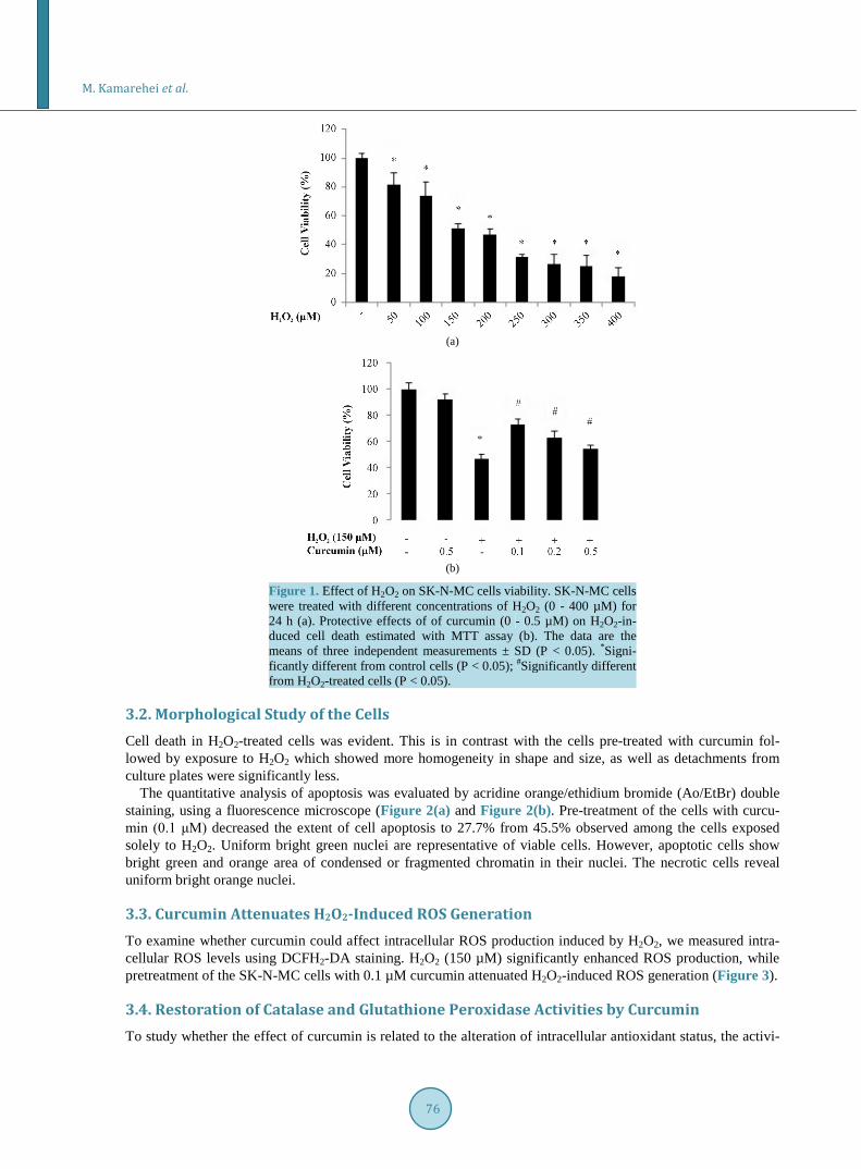
3. Results 3.1. Curcumin Improves the Viability of H2O2-Treated Cells To find the concentration of H2O2 where 50% of cells are viable, SK-N-MC cells were treated with a range of concentrations of H2O2 between 0 and 400 µM followed by further incubation for 24 h. At the end of incubation period, cell viability was established by the MTT assay. Based on Figure 1(a), exposure of the cells to H2O2 has caused reduction in viability by almost 18.1%, 25.8%, 52.9%, 73.3% and 81.9% at concentrations of 50, 100, 150, 300 and 400 μM, respectively. Regarding these data, the rest of experiments have been carried out at a H2O2 concentration of 150 μM. Pretreatment of the cells with curcumin for 2 h (0.1 - 0.5 μM) and subsequent treatment with 150 µM H2O2 for 24 h enhanced the extent of viability in a concentration-dependent manner. As it is evident from Figure 1(b), curcumin at concentrations of 0.1, 0.2 and 0.5 μM restored the cell viability by 25.9%, 15.9% and 7.0%, respectively. However, higher concentrations of curcumin were cytotoxic. Thus, 0.1 µM was selected as the best effective dose of curcumin with maximum antioxidant activity.
M. Kamarehei et al.
76
(a)
(b)
Figure 1. Effect of H2O2 on SK-N-MC cells viability. SK-N-MC cells were treated with different concentrations of H2O2 (0 - 400 µM) for 24 h (a). Protective effects of of curcumin (0 - 0.5 µM) on H2O2-in- duced cell death estimated with MTT assay (b). The data are the means of three independent measurements ± SD (P < 0.05). *Signi- ficantly different from control cells (P < 0.05); #Significantly different from H2O2-treated cells (P < 0.05).
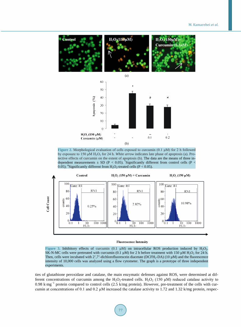
3.2. Morphological Study of the Cells Cell death in H2O2-treated cells was evident. This is in contrast with the cells pre-treated with curcumin fol-lowed by exposure to H2O2 which showed more homogeneity in shape and size, as well as detachments from culture plates were significantly less.
The quantitative analysis of apoptosis was evaluated by acridine orange/ethidium bromide (Ao/EtBr) double staining, using a fluorescence microscope (Figure 2(a) and Figure 2(b). Pre-treatment of the cells with curcu-min (0.1 μM) decreased the extent of cell apoptosis to 27.7% from 45.5% observed among the cells exposed solely to H2O2. Uniform bright green nuclei are representative of viable cells. However, apoptotic cells show bright green and orange area of condensed or fragmented chromatin in their nuclei. The necrotic cells reveal uniform bright orange nuclei.
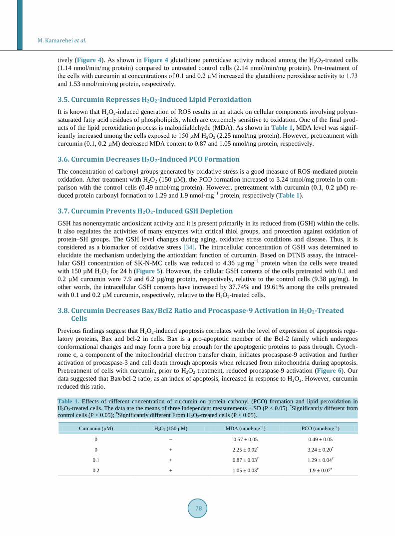
3.3. Curcumin Attenuates H2O2-Induced ROS Generation To examine whether curcumin could affect intracellular ROS production induced by H2O2, we measured intra-cellular ROS levels using DCFH2-DA staining. H2O2 (150 µM) significantly enhanced ROS production, while pretreatment of the SK-N-MC cells with 0.1 µM curcumin attenuated H2O2-induced ROS generation (Figure 3).
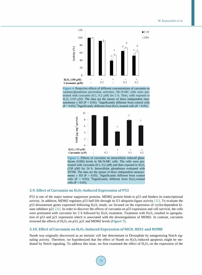
3.4. Restoration of Catalase and Glutathione Peroxidase Activities by Curcumin To study whether the effect of curcumin is related to the alteration of intracellular antioxidant status, the activi-
M. Kamarehei et al.
77
(a)
(b)
Figure 2. Morphological evaluation of cells exposed to curcumin (0.1 μM) for 2 h followed by exposure to 150 μM H2O2 for 24 h. White arrow indicates late phase of apoptosis (a). Pro-tective effects of curcumin on the extent of apoptosis (b). The data are the means of three in-dependent measurements ± SD (P < 0.05). *Significantly different from control cells (P < 0.05); #Significantly different from H2O2-treated cells (P < 0.05).
Figure 3. Inhibitory effects of curcumin (0.1 µM) on intracellular ROS production induced by H2O2. SK-N-MC cells were pretreated with curcumin (0.1 µM) for 2 h before treatment with 150 µM H2O2 for 24 h. Then, cells were incubated with 2’,7’-dichlorofluorescein diacetate (DCFH2-DA) (10 µM) and the fluorescence intensity of 10,000 cells was analyzed using a flow cytometer. The graph is a prototype of three independent experiments.
ties of glutathione peroxidase and catalase, the main enzymatic defenses against ROS, were determined at dif-ferent concentrations of curcumin among the H2O2-treated cells. H2O2 (150 μM) reduced catalase activity to 0.98 k∙mg−1 protein compared to control cells (2.5 k/mg protein). However, pre-treatment of the cells with cur-cumin at concentrations of 0.1 and 0.2 µΜ increased the catalase activity to 1.72 and 1.32 k/mg protein, respec-
M. Kamarehei et al.
78
tively (Figure 4). As shown in Figure 4 glutathione peroxidase activity reduced among the H2O2-treated cells (1.14 nmol/min/mg protein) compared to untreated control cells (2.14 nmol/min/mg protein). Pre-treatment of the cells with curcumin at concentrations of 0.1 and 0.2 µΜ increased the glutathione peroxidase activity to 1.73 and 1.53 nmol/min/mg protein, respectively.
3.5. Curcumin Represses H2O2-Induced Lipid Peroxidation It is known that H2O2-induced generation of ROS results in an attack on cellular components involving polyun-saturated fatty acid residues of phospholipids, which are extremely sensitive to oxidation. One of the final prod-ucts of the lipid peroxidation process is malondialdehyde (MDA). As shown in Table 1, MDA level was signif-icantly increased among the cells exposed to 150 μM H2O2 (2.25 nmol/mg protein). However, pretreatment with curcumin (0.1, 0.2 µM) decreased MDA content to 0.87 and 1.05 nmol/mg protein, respectively.
3.6. Curcumin Decreases H2O2-Induced PCO Formation The concentration of carbonyl groups generated by oxidative stress is a good measure of ROS-mediated protein oxidation. After treatment with H2O2 (150 µM), the PCO formation increased to 3.24 nmol/mg protein in com-parison with the control cells (0.49 nmol/mg protein). However, pretreatment with curcumin (0.1, 0.2 µM) re-duced protein carbonyl formation to 1.29 and 1.9 nmol·mg−1 protein, respectively (Table 1).
3.7. Curcumin Prevents H2O2-Induced GSH Depletion GSH has nonenzymatic antioxidant activity and it is present primarily in its reduced from (GSH) within the cells. It also regulates the activities of many enzymes with critical thiol groups, and protection against oxidation of protein–SH groups. The GSH level changes during aging, oxidative stress conditions and disease. Thus, it is considered as a biomarker of oxidative stress [34]. The intracellular concentration of GSH was determined to elucidate the mechanism underlying the antioxidant function of curcumin. Based on DTNB assay, the intracel-lular GSH concentration of SK-N-MC cells was reduced to 4.36 µg∙mg−1 protein when the cells were treated with 150 µM H2O2 for 24 h (Figure 5). However, the cellular GSH contents of the cells pretreated with 0.1 and 0.2 µM curcumin were 7.9 and 6.2 µg/mg protein, respectively, relative to the control cells (9.38 µg/mg). In other words, the intracellular GSH contents have increased by 37.74% and 19.61% among the cells pretreated with 0.1 and 0.2 µM curcumin, respectively, relative to the H2O2-treated cells.
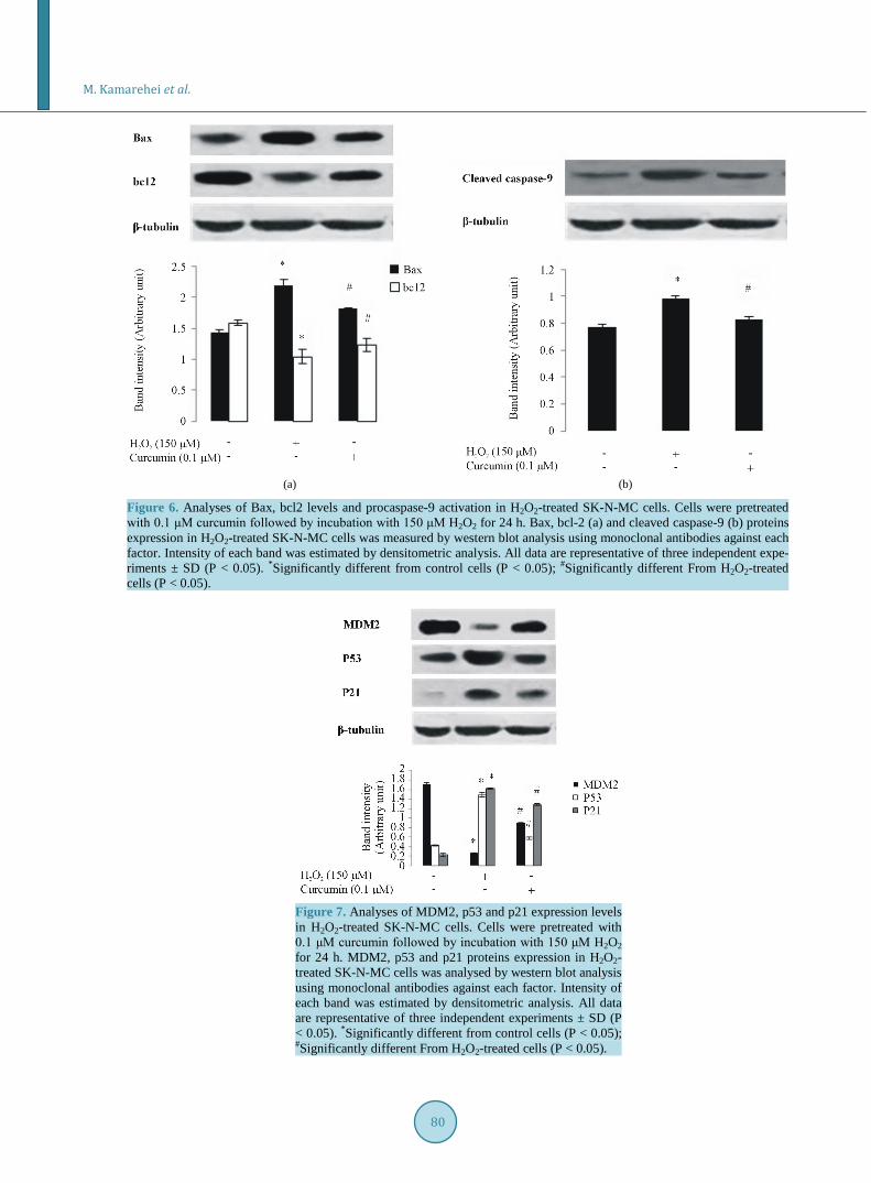
3.8. Curcumin Decreases Bax/Bcl2 Ratio and Procaspase-9 Activation in H2O2-Treated Cells
Previous findings suggest that H2O2-induced apoptosis correlates with the level of expression of apoptosis regu-latory proteins, Bax and bcl-2 in cells. Bax is a pro-apoptotic member of the Bcl-2 family which undergoes conformational changes and may form a pore big enough for the apoptogenic proteins to pass through. Cytoch-rome c, a component of the mitochondrial electron transfer chain, initiates procaspase-9 activation and further activation of procaspase-3 and cell death through apoptosis when released from mitochondria during apoptosis. Pretreatment of cells with curcumin, prior to H2O2 treatment, reduced procaspase-9 activation (Figure 6). Our data suggested that Bax/bcl-2 ratio, as an index of apoptosis, increased in response to H2O2. However, curcumin reduced this ratio. Table 1. Effects of different concentration of curcumin on protein carbonyl (PCO) formation and lipid peroxidation in H2O2-treated cells. The data are the means of three independent measurements ± SD (P < 0.05). *Significantly different from control cells (P < 0.05); #Significantly different From H2O2-treated cells (P < 0.05).
Curcumin (µM) H2O2 (150 µM) MDA (nmol∙mg−1) PCO (nmol∙mg−1)
0 − 0.57 ± 0.05 0.49 ± 0.05
0 + 2.25 ± 0.02* 3.24 ± 0.20*
0.1 + 0.87 ± 0.03# 1.29 ± 0.04#
0.2 + 1.05 ± 0.03# 1.9 ± 0.07#
M. Kamarehei et al.
79
Figure 4. Protective effects of different concentrations of curcumin on catalase/glutathione peroxidase activities. SK-N-MC cells were pre-treated with curcumin (0.1, 0.2 µM) for 2 h. Then, cells exposed to H2O2 (150 µM). The data are the means of three independent mea-surements ± SD (P < 0.05). *Significantly different from control cells (P < 0.05); #Significantly different from H2O2-treated cells (P < 0.05).
Figure 5. Effects of curcumin on intracellular reduced gluta-thione (GSH) levels in SK-N-MC cells. The cells were pre-treated with curcumin (0.1, 0.2 µM) and then exposed to H2O2 (150 µM) for 24 h. Intracellular glutathione evaluated with DTNB. The data are the means of three independent measure-ments ± SD (P < 0.05). *Significantly different from control cells (P < 0.05); #Significantly different from H2O2-treated cells (P < 0.05).
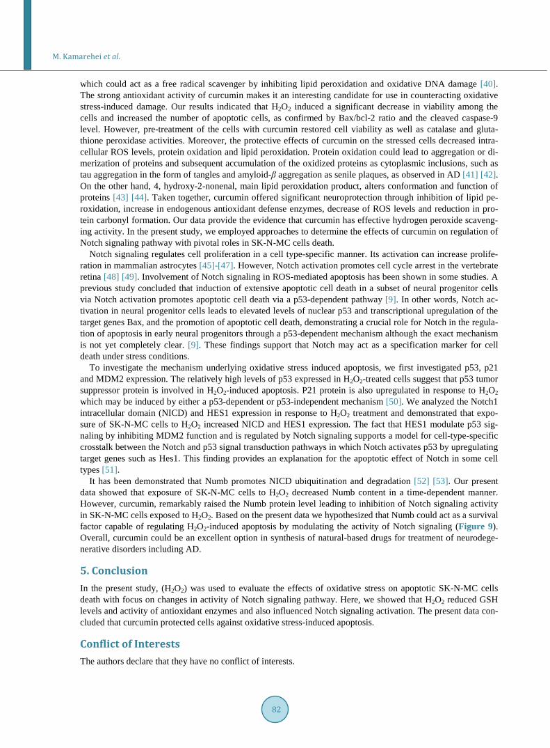
3.9. Effect of Curcumin on H2O2-Induced Expression of P53 P53 is one of the major tumour suppressor proteins. MDM2 protein binds to p53 and hinders its transcriptional activity. In addition, MDM2 regulates p53 half-life through its E3 ubiquitin-ligase activity [35]. To evaluate the p53 downstream genes expressed following H2O2 insult, we focused on the expression of cyclin-dependent ki-nase inhibitor p21 [36]. In order to discover the effects of curcumin on p53 expression and cell survival, the cells were pretreated with curcumin for 2 h followed by H2O2 treatment. Treatment with H2O2 resulted in upregula-tion of p53 and p21 expression which is associated with the downregulation of MDM2. In contrast, curcumin reversed the effects of H2O2 on p53, p21 and MDM2 levels (Figure 7).
3.10. Effect of Curcumin on H2O2-Induced Expression of NICD, HES1 and NUMB Numb was originally discovered as an intrinsic cell fate determinant in Drosophila by antagonizing Notch sig-naling activity. Therefore, we hypothesized that the effect of Numb on H2O2-induced apoptosis might be me-diated by Notch signaling. To address this issue, we first examined the effect of H2O2 on the expression of the
M. Kamarehei et al.
80
(a) (b)
Figure 6. Analyses of Bax, bcl2 levels and procaspase-9 activation in H2O2-treated SK-N-MC cells. Cells were pretreated with 0.1 μM curcumin followed by incubation with 150 μM H2O2 for 24 h. Bax, bcl-2 (a) and cleaved caspase-9 (b) proteins expression in H2O2-treated SK-N-MC cells was measured by western blot analysis using monoclonal antibodies against each factor. Intensity of each band was estimated by densitometric analysis. All data are representative of three independent expe-riments ± SD (P < 0.05). *Significantly different from control cells (P < 0.05); #Significantly different From H2O2-treated cells (P < 0.05).
Figure 7. Analyses of MDM2, p53 and p21 expression levels in H2O2-treated SK-N-MC cells. Cells were pretreated with 0.1 μM curcumin followed by incubation with 150 μM H2O2 for 24 h. MDM2, p53 and p21 proteins expression in H2O2- treated SK-N-MC cells was analysed by western blot analysis using monoclonal antibodies against each factor. Intensity of each band was estimated by densitometric analysis. All data are representative of three independent experiments ± SD (P < 0.05). *Significantly different from control cells (P < 0.05); #Significantly different From H2O2-treated cells (P < 0.05).

M. Kamarehei et al.
81
Notch intracellular domain (NICD) and HES1, the downstream target gene of Notch signaling. As shown in Figure 8, H2O2 treatment significantly increased the NICD and HES1 expression, suggesting that H2O2 could activate Notch signaling. On the other hand, H2O2 treatment resulted in down-regulation of numb expression in a time-dependent manner as well as curcumin decreased NICD and HES1 levels. Taken together, these data pro-vided evidence that curcumin could protect SK-N-MC cells from H2O2-induced apoptosis by Numb up-regula- tion which antagonize Notch signaling activity.
4. Discussion The AD brain is characterized by extensive oxidative stress [37] [38]. Currently, it is known that numerous sig-naling pathways which are involved in neurodegenerative disorders including AD are activated in response to oxidative stress. Thus, removal of excess reactive oxygen species or suppression of their generation may be ef-fective in preventing oxidative cell death. Regarding the previous findings, future directions on AD treatments will focus on the use of antioxidants to contribute to the neuroprotection and potential enhancement of the intra-cellular antioxidant mechanisms.
Curcumin with its proven antioxidant properties has been shown to have several therapeutic effects [39]
(a) (b)
Figure 8. Analyses of NICD, HES1 and Numb expression levels in H2O2-treated SK-N-MC cells. Cells were pretreated with 0.1 μM curcumin followed by incubation with 150 μM H2O2 for 24 h. NICD and HES1 proteins expression in H2O2-treated SK-N-MC cells was measured by western blot analysis using monoclonal antibodies against each factor. The intensity of each band was estimated by densitometric analysis. (a). Cells were pretreated with 0.1 μM curcumin followed by incubation with 150 μM H2O2 for 24 h and Numb protein level in H2O2-treated SK-N-MC cells analysed by western blot (b). All data are representative of three independent experiments ± SD (P < 0.05). *Significantly different from control cells (P < 0.05); #Significantly different From H2O2-treated cells (P < 0.05).
M. Kamarehei et al.
82
which could act as a free radical scavenger by inhibiting lipid peroxidation and oxidative DNA damage [40]. The strong antioxidant activity of curcumin makes it an interesting candidate for use in counteracting oxidative stress-induced damage. Our results indicated that H2O2 induced a significant decrease in viability among the cells and increased the number of apoptotic cells, as confirmed by Bax/bcl-2 ratio and the cleaved caspase-9 level. However, pre-treatment of the cells with curcumin restored cell viability as well as catalase and gluta-thione peroxidase activities. Moreover, the protective effects of curcumin on the stressed cells decreased intra-cellular ROS levels, protein oxidation and lipid peroxidation. Protein oxidation could lead to aggregation or di-merization of proteins and subsequent accumulation of the oxidized proteins as cytoplasmic inclusions, such as tau aggregation in the form of tangles and amyloid-β aggregation as senile plaques, as observed in AD [41] [42]. On the other hand, 4, hydroxy-2-nonenal, main lipid peroxidation product, alters conformation and function of proteins [43] [44]. Taken together, curcumin offered significant neuroprotection through inhibition of lipid pe-roxidation, increase in endogenous antioxidant defense enzymes, decrease of ROS levels and reduction in pro-tein carbonyl formation. Our data provide the evidence that curcumin has effective hydrogen peroxide scaveng-ing activity. In the present study, we employed approaches to determine the effects of curcumin on regulation of Notch signaling pathway with pivotal roles in SK-N-MC cells death.
Notch signaling regulates cell proliferation in a cell type-specific manner. Its activation can increase prolife-ration in mammalian astrocytes [45]-[47]. However, Notch activation promotes cell cycle arrest in the vertebrate retina [48] [49]. Involvement of Notch signaling in ROS-mediated apoptosis has been shown in some studies. A previous study concluded that induction of extensive apoptotic cell death in a subset of neural progenitor cells via Notch activation promotes apoptotic cell death via a p53-dependent pathway [9]. In other words, Notch ac-tivation in neural progenitor cells leads to elevated levels of nuclear p53 and transcriptional upregulation of the target genes Bax, and the promotion of apoptotic cell death, demonstrating a crucial role for Notch in the regula-tion of apoptosis in early neural progenitors through a p53-dependent mechanism although the exact mechanism is not yet completely clear. [9]. These findings support that Notch may act as a specification marker for cell death under stress conditions.
To investigate the mechanism underlying oxidative stress induced apoptosis, we first investigated p53, p21 and MDM2 expression. The relatively high levels of p53 expressed in H2O2-treated cells suggest that p53 tumor suppressor protein is involved in H2O2-induced apoptosis. P21 protein is also upregulated in response to H2O2 which may be induced by either a p53-dependent or p53-independent mechanism [50]. We analyzed the Notch1 intracellular domain (NICD) and HES1 expression in response to H2O2 treatment and demonstrated that expo-sure of SK-N-MC cells to H2O2 increased NICD and HES1 expression. The fact that HES1 modulate p53 sig-naling by inhibiting MDM2 function and is regulated by Notch signaling supports a model for cell-type-specific crosstalk between the Notch and p53 signal transduction pathways in which Notch activates p53 by upregulating target genes such as Hes1. This finding provides an explanation for the apoptotic effect of Notch in some cell types [51].
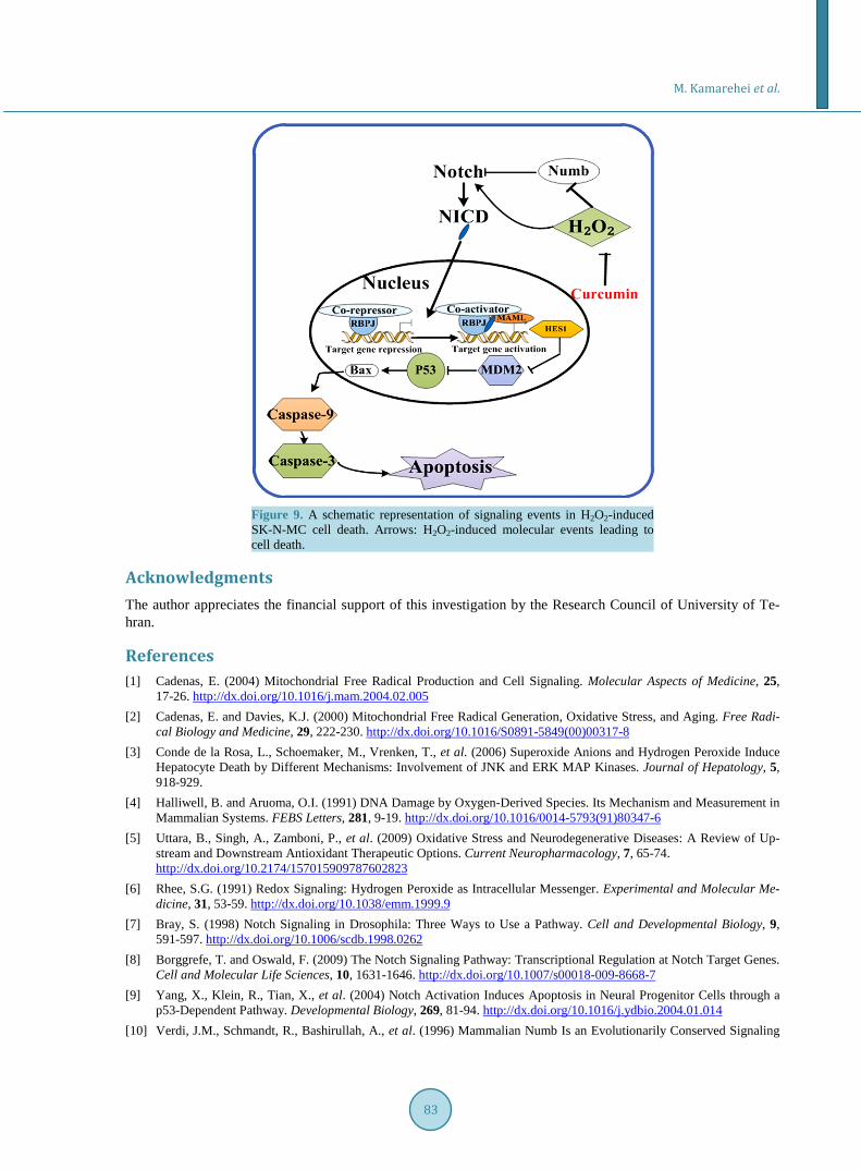
It has been demonstrated that Numb promotes NICD ubiquitination and degradation [52] [53]. Our present data showed that exposure of SK-N-MC cells to H2O2 decreased Numb content in a time-dependent manner. However, curcumin, remarkably raised the Numb protein level leading to inhibition of Notch signaling activity in SK-N-MC cells exposed to H2O2. Based on the present data we hypothesized that Numb could act as a survival factor capable of regulating H2O2-induced apoptosis by modulating the activity of Notch signaling (Figure 9). Overall, curcumin could be an excellent option in synthesis of natural-based drugs for treatment of neurodege-nerative disorders including AD.
5. Conclusion In the present study, (H2O2) was used to evaluate the effects of oxidative stress on apoptotic SK-N-MC cells death with focus on changes in activity of Notch signaling pathway. Here, we showed that H2O2 reduced GSH levels and activity of antioxidant enzymes and also influenced Notch signaling activation. The present data con-cluded that curcumin protected cells against oxidative stress-induced apoptosis.
Conflict of Interests The authors declare that they have no conflict of interests.
M. Kamarehei et al.
83
Figure 9. A schematic representation of signaling events in H2O2-induced SK-N-MC cell death. Arrows: H2O2-induced molecular events leading to cell death.
Acknowledgments The author appreciates the financial support of this investigation by the Research Council of University of Te-hran.
References [1] Cadenas, E. (2004) Mitochondrial Free Radical Production and Cell Signaling. Molecular Aspects of Medicine, 25,
17-26. http://dx.doi.org/10.1016/j.mam.2004.02.005 [2] Cadenas, E. and Davies, K.J. (2000) Mitochondrial Free Radical Generation, Oxidative Stress, and Aging. Free Radi-
cal Biology and Medicine, 29, 222-230. http://dx.doi.org/10.1016/S0891-5849(00)00317-8 [3] Conde de la Rosa, L., Schoemaker, M., Vrenken, T., et al. (2006) Superoxide Anions and Hydrogen Peroxide Induce
Hepatocyte Death by Different Mechanisms: Involvement of JNK and ERK MAP Kinases. Journal of Hepatology, 5, 918-929.
[4] Halliwell, B. and Aruoma, O.I. (1991) DNA Damage by Oxygen-Derived Species. Its Mechanism and Measurement in Mammalian Systems. FEBS Letters, 281, 9-19. http://dx.doi.org/10.1016/0014-5793(91)80347-6
[5] Uttara, B., Singh, A., Zamboni, P., et al. (2009) Oxidative Stress and Neurodegenerative Diseases: A Review of Up- stream and Downstream Antioxidant Therapeutic Options. Current Neuropharmacology, 7, 65-74. http://dx.doi.org/10.2174/157015909787602823
[6] Rhee, S.G. (1991) Redox Signaling: Hydrogen Peroxide as Intracellular Messenger. Experimental and Molecular Me- dicine, 31, 53-59. http://dx.doi.org/10.1038/emm.1999.9
[7] Bray, S. (1998) Notch Signaling in Drosophila: Three Ways to Use a Pathway. Cell and Developmental Biology, 9, 591-597. http://dx.doi.org/10.1006/scdb.1998.0262
[8] Borggrefe, T. and Oswald, F. (2009) The Notch Signaling Pathway: Transcriptional Regulation at Notch Target Genes. Cell and Molecular Life Sciences, 10, 1631-1646. http://dx.doi.org/10.1007/s00018-009-8668-7
[9] Yang, X., Klein, R., Tian, X., et al. (2004) Notch Activation Induces Apoptosis in Neural Progenitor Cells through a p53-Dependent Pathway. Developmental Biology, 269, 81-94. http://dx.doi.org/10.1016/j.ydbio.2004.01.014
[10] Verdi, J.M., Schmandt, R., Bashirullah, A., et al. (1996) Mammalian Numb Is an Evolutionarily Conserved Signaling
M. Kamarehei et al.
84
Adapter Protein That Specifies Cell Fate. Current Biology, 69, 1134-1145. http://dx.doi.org/10.1016/S0960-9822(02)70680-5
[11] Prior, R.L. and Cao, G. (2000) Flavonoids: Diet and Health Relationships. Nutrition in Clinical Care, 2, 279-288. http://dx.doi.org/10.1046/j.1523-5408.2000.00074.x
[12] Aggarwal, B.B., Kumar, A. and Bharti, A.C. (2003) Anticancer Potential of Curcumin: Preclinical and Clinical Studies. Anticancer Research, 23, 363-398.
[13] Sharma, O.P. (1976) Antioxidant Activity of Curcumin and Related Compounds. Biochemical Pharmacology, 25, 1811-1812. http://dx.doi.org/10.1016/0006-2952(76)90421-4
[14] Srimal, R.C. and Dhawan, B.N. (1973) Pharmacology of Diferuloyl Methane (Curcumin), a Non-Steroidal Anti In- flammatory Agent. Journal of Pharmacy and Pharmacology, 25, 447-452. http://dx.doi.org/10.1111/j.2042-7158.1973.tb09131.x
[15] Jordan, W.C. and Drew, C.R. (1996) Curcumin—A Natural Herb with Anti-HIV Activity. Journal of the National Medical Association, 88, 333.
[16] Kuttan, R., Bhanumathy, P., Nirmala, K. and George, M.C. (1985) Potential Anticancer Activity of Turmeric (Curcu- ma longa). Cancer Letters, 29, 197-202. http://dx.doi.org/10.1016/0304-3835(85)90159-4
[17] Kiso, Y., Suzuki, Y., Watanabe, N., Oshima, Y. and Hikino, H. (1983) Antihepatotoxic Principles of Curcuma longa Rhizomes. Planta Medica, 49, 185-187. http://dx.doi.org/10.1055/s-2007-969845
[18] Venkatesan, N., Punithavathi, D. and Arumugam, V. (2000) Curcumin Prevents Adriamycin Nephrotoxicity in Rats. British Journal of Pharmacology, 129, 231-234. http://dx.doi.org/10.1038/sj.bjp.0703067
[19] Srivastava, R., Dikshit, M., Srimal, R.C., et al. (1985) Anti-Thrombotic Effect of Curcumin. Thrombosis Research, 40, 413-417. http://dx.doi.org/10.1016/0049-3848(85)90276-2
[20] Dikshit, M., Rastogi, L., Shukla, R. and Srimal, R.C. (1995) Prevention of Ischaemia-Induced Biochemical Changes by Curcumin and Quinidine in the Cat Heart. Indian Journal of Medical Research, 101, 31-35.
[21] Chandra, V., Ganguli, M., Pandav, R., et al. (1998) Prevalence of Alzheimer’s Disease and Other Dementias in Rural India: The Indo-US Study. Neurology, 51, 1000-1008. http://dx.doi.org/10.1212/WNL.51.4.1000
[22] Lim, G.P., Chu, T., Yang, F., Beech, W., Frautschy, S.A. and Cole, G.M. (2001) The Curry Spice Curcumin Reduces Oxidative Damage and Amyloid Pathology in an Alzheimer Transgenic Mouse. Journal of Neuroscience, 21, 8370- 8377.
[23] Litwinienko, G. and Ingold, K.U. (2004) Abnormal Solvent Effects on Hydrogen Atom Abstraction. 2. Resolution of the Curcumin Antioxidant Controversy. The Role of Sequential Proton Loss Electron Transfer. The Journal of Organic Chemistry, 69, 5888-5896. http://dx.doi.org/10.1021/jo049254j
[24] Shen, L., Zhang, H.Y. and Ji, H.F. (2005) Successful Application of TD-DFT in Transient Absorption Spectra As- signment. Organic Letters, 7, 243-246. http://dx.doi.org/10.1021/ol047766e
[25] Ono, K., Hasegawa, K., Naiki, H. and Yamada, M. (2004) Curcumin Has Potent Anti-Amyloidogenic Effects for Alz- heimer’s Beta Fibrils in Vitro. Journal of Neuroscience Research, 75, 742-750. http://dx.doi.org/10.1002/jnr.20025
[26] Yang, F., Lim, G.P., Begum, A.N., Ubeda, O.J., Simmons, M.R., Ambegaokar, S.S., et al. (2005) Curcumin Inhibits Formation of Amyloid Beta Oligomers and Fibrils, Binds Plaques, and Reduces Amyloid in Vivo. Journal of Biologi- cal Chemistry, 280, 5892-5901. http://dx.doi.org/10.1074/jbc.M404751200
[27] Gomes, A., Fernandes, E. and Lima, J.L.F.C. (2005) Fluorescence Probes Used for Detection of Reactive Oxygen Spe- cies. Journal of Biochemical and Biophysical Methods, 65, 45-80. http://dx.doi.org/10.1016/j.jbbm.2005.10.003
[28] LeBel, C.P., Ischiropoulos, H. and Bondy, S.C. (1992) Evaluation of the Probe 2’,7’-Dichlorofluorecein as an Indicator of Reactive Oxygen Species Formation and Oxidative Stress. Chemical Research in Toxicology, 5, 227-231. http://dx.doi.org/10.1021/tx00026a012
[29] Aebi, H. (1984) Catalase in Vitro. Methods in Enzymology, 105, 121-126. http://dx.doi.org/10.1016/S0076-6879(84)05016-3
[30] Lowry, O.H., Rosebrough, N.J., Farr, A.L. and Randall, R.J. (1951) Protein Measurement with the Folin Phenol Rea- gent. Journal of Biological Chemistry, 193, 265-275.
[31] Flohe, L. and Gunzler, W.A. (1984) Assays of Glutathione Peroxidase. Methods in Enzymology, 105, 114-121. http://dx.doi.org/10.1016/S0076-6879(84)05015-1
[32] Drapper, H.H. and Hadley, M. (1990) Malondialdehyde Determination as Index of Lipid Peroxidation. Methods in En- zymology, 186, 421-431. http://dx.doi.org/10.1016/0076-6879(90)86135-I
[33] Jollow, D., Mitchell, L., Zampaglione, N. and Gillete, J. (1974) Bromobenzene Induced Liver Necrosis. Protective Role of Glutathione and Evidence for 3, 4-Bromobenzenoxide as the Hepatotoxic Intermediate. Pharmacology, 11,
M. Kamarehei et al.
85
151-169. http://dx.doi.org/10.1159/000136485 [34] Hermes-Lima, M. (2004) Oxygen in Biology and Biochemistry: Role of Free Radicals. In: Storey, K.B., Ed., Func-
tional Metabolism: Regulation and Adaptation, John Wiley & Sons, Inc., Hoboken, 319-368. [35] Kubbutat, M.H., Jones, S.N. and Vousden, K.H. (1997) Regulation of p53 Stability by Mdm2. Nature, 387, 299-303.
http://dx.doi.org/10.1038/387299a0 [36] El-Deiry, W.S., Tokino, T., Velculescu, V.E., Levy, D.B., Parsons, R., et al. (1993) WAF1, a Potential Mediator of
p53 Tumor Suppression. Cell, 75, 817-825. http://dx.doi.org/10.1016/0092-8674(93)90500-P [37] Dumont, M. and Beal, M.F. (2011) Neuroprotective Strategies Involving ROS in Alzheimer Disease. Free Radical Bi-
ology and Medicine, 51, 1014-1026. http://dx.doi.org/10.1016/j.freeradbiomed.2010.11.026 [38] Lynch, T., Cherny, R.A. and Bush, A.I. (2000) Oxidative Processes in Alzheimer’s Disease: The Role of Aβ-Metal In-
teractions. Experimental Gerontology, 35, 445-451. http://dx.doi.org/10.1016/S0531-5565(00)00112-1 [39] Reddy, A.C. and Lokesh, B.R. (1994) Studies on the Inhibitory Effects of Curcumin and Eugenol on the Formation of
Reactive Oxygen Species and the Oxidation of Ferrous Iron. Molecular and Cellular Biochemistry, 137, 1-8. http://dx.doi.org/10.1007/BF00926033
[40] Ortiz-Ortiz, M.A., Morán, J.M., Bravosanpedro, J.M., González-Polo, R.A., Niso-Santano, M., Anantharam, V., Kan- thasamy, A.G., Soler, G. and Fuentes, J.M. (2009) Curcumin Enhances Paraquat-Induced Apoptosis of N27 Mesence- phalic Cells via the Generation of Reactive Oxygen Species. NeuroToxicolgy, 30, 1008-1018. http://dx.doi.org/10.1016/j.neuro.2009.07.016
[41] Butterfield, D.A., Drake, J., Pocernich, C. and Castegna, A. (2001) Evidence of Oxidative Damage in Alzheimer’s Disease Brain: Central Role of Amyloid β-Peptide. Trends in Molecular Medicine, 7, 548-554. http://dx.doi.org/10.1016/S1471-4914(01)02173-6
[42] Stadtman, E.R. and Berlett, B.S. (1997) Reactive Oxygen-Mediated Protein Oxidation in Aging and Disease. Chemical Research in Toxicology, 10, 485-494. http://dx.doi.org/10.1021/tx960133r
[43] Lauderback, C.M., Hackett, J.M., Huang, F.F., Keller, J.N., Szweda, L.I., Markesbery, W.R. and Butterfield, D.A. (2001) The Glial Glutamate Transporter, GLT-1, Is Oxidatively Modified by 4-Hydroxy-2-Nonenal in the Alzheimer’s Disease Brain: The Role of Abeta1-42. Journal of Neurochemistry, 78, 413-416.
[44] Subramaniam, R., Roediger, F., Jordan, B., Mattson, M.P., Keller, J.N., Waeg, G. and Butterfield, D.A. (1997) The Li-pid Peroxidation Product, 4-Hydroxy-2-Trans-Nonenal, Alters the Conformation of Cortical Synaptosomal Membrane Proteins. Journal of Neurochemistry, 69, 1161-1169. http://dx.doi.org/10.1046/j.1471-4159.1997.69031161.x
[45] Chambers, C.B., Peng, Y., Nguyen, H., Gaiano, N., Fishell, G. and Nye, J.S. (2001) Spatiotemporal Selectivity of Re-sponse to Notch1 Signals in Mammalian Forebrain Precursors. Development, 128, 689-702.
[46] Furukawa, T., Mukherjee, S., Bao, Z., Morrow, E.M. and Cepko, C.L. (2000) Rax, Hes1, and Notch1 Promote the Formation of Muller Glia by Postnatal Retinal Progenitor Cells. Neuron, 26, 383-394. http://dx.doi.org/10.1016/S0896-6273(00)81171-X
[47] Tanigaki, K., Nogaki, F., Takahashi, J., Tashiro, K., Kurooka, H. and Honjo, T. (2001) Notch1 and Notch3 Instruc-tively Restrict bFGF-Responsive Multi-Potent Neural Progenitor Cells to an Astroglial Fate. Neuron, 29, 45-55. http://dx.doi.org/10.1016/S0896-6273(01)00179-9
[48] Dorsky, R.I., Rapaport, D.H. and Harris, W.A. (1995) Xotch Inhibits Cell Differentiation in the Xenopus Retina. Neu-ron, 14, 487-496. http://dx.doi.org/10.1016/0896-6273(95)90305-4
[49] Scheer, N., Groth, A., Hans, S. and Campos-Ortega, J.A. (2001) An Instructive Function for Notch in Promoting Gli-ogenes Is in the Zebra Fish Retina. Development, 128, 1099-1107.
[50] Chin, Y.E., Kitagawa, M., Su, W.C.S., You, Z.H., Iwamoto, Y. and Fu, X.Y. (1996) Cell Growth Arrest and Induction of Cyclin-Dependent Kinase Inhibitor p21WAF1/CIP1 Mediated by STAT1. Science, 272, 719-722. http://dx.doi.org/10.1126/science.272.5262.719
[51] Huang, Q., Raya, A., DeJesus, P., Chao, S.H., Quon, K.C., Caldwell, J.S., et al. (2004) Identification of p53 Regulators by Genome-Wide Functional Analysis. Proceedings of the National Academy of Sciences of the United States of America, 101, 3456-3461.
[52] McGill, M.A. and McGlade, C.J. (2003) Mammalian Numb Proteins Promote Notch1 Receptor Ubiquitination and Degradation of the Notch1 Intracellular Domain. The Journal of Biological Chemistry, 278, 23196-23203. http://dx.doi.org/10.1074/jbc.M302827200
[53] Wakamatsu, Y., Maynard, T.M., Jones, S.U. and Weston, J.A. (1999) NUMB Localizes in the Basal Cortex of Mitotic Avian Neuroepithelial Cells and Modulates Neuronal Differentiation by Binding to NOTCH-1. Neuron, 23, 71-81. http://dx.doi.org/10.1016/S0896-6273(00)80754-0
M. Kamarehei et al.
86
Abbreviations MTT, 3-(4,5-dimethylthiazol-2-yl)-2,5-diphenyltetrazolium bromide; GSH, Glutathione; DTNB, Dithionitrobenzoic acid; MDA, Malondialdehyde; PBS, Phosphate buffer saline; PCO, Protein carbonyl.
Related Documents