Curcumin limits the fibrogenic evolution of experimental steatohepatitis Francesco Vizzutti 1,5 , Angela Provenzano 1,5 , Sara Galastri 1 , Stefano Milani 2,3 , Wanda Delogu 1 , Erica Novo 4 , Alessandra Caligiuri 1 , Elena Zamara 4 , Umberto Arena 1 , Giacomo Laffi 1 , Maurizio Parola 4 , Massimo Pinzani 1,3 and Fabio Marra 1,3 Nonalcoholic steatohepatitis is characterized by the association of steatosis with hepatic cell injury, lobular inflammation and fibrosis. Curcumin is known for its antioxidant, anti-inflammatory and antifibrotic properties. The aim of this study was to test whether the administration of curcumin limits fibrogenic evolution in a murine model of nonalcoholic steatohepatitis. Male C57BL/6 mice were divided into four groups and fed a diet deficient in methionine and choline (MCD) or the same diet supplemented with methionine and choline for as long as 10 weeks. Curcumin (25 mg per mouse) or its vehicle (DMSO) was administered intraperitoneally every other day. Fibrosis was assessed by Sirius red staining and histomorphometry. Intrahepatic gene expression was measured by quantitative PCR. Hepatic oxidative stress was evaluated by staining for 8-OH deoxyguanosine. Myofibroblastic hepatic stellate cells (HSCs) were isolated from normal human liver tissue. The increase in serum ALT caused by the MCD diet was significantly reduced by curcumin after 4 weeks. Administration of the MCD diet was associated with histological steatosis and necro-inflammation, and this latter was significantly reduced in mice receiving curcumin. Curcumin also inhibited the generation of hepatic oxidative stress. Fibrosis was evident after 8 or 10 weeks of MCD diet and was also significantly reduced by curcumin. Curcumin decreased the intrahepatic gene expression of monocyte chemoattractant protein-1, CD11b, procollagen type I and tissue inhibitor of metalloprotease (TIMP)-1, together with protein levels of a-smooth muscle-actin, a marker of fibrogenic cells. In addition, curcumin reduced the generation of reactive oxygen species in cultured HSCs and inhibited the secretion of TIMP-1 both in basal conditions and after the induction of oxidative stress. In conclusion, curcumin administration effectively limits the development and progression of fibrosis in mice with experimental steatohepatitis, and reduces TIMP-1 secretion and oxidative stress in cultured stellate cells. Laboratory Investigation (2010) 90, 104–115; doi:10.1038/labinvest.2009.112; published online 9 November 2009 KEYWORDS: curcumin; fibrosis; hepatic stellate cells; nonalcoholic steatohepatitis; tissue inhibitor of metalloprotease-1 Nonalcoholic fatty liver disease (NAFLD) is one of the most frequent causes of liver dysfunction in Western countries. 1 Obesity and insulin resistance are major risk factors for the development of NAFLD, which is considered the hepatic manifestation of metabolic syndrome. Accordingly, the pre- valence of NAFLD has increased markedly, along with the acquisition of a sedentary lifestyle and changes in dietary habits. NAFLD comprises ‘simple’ fatty liver, a condition believed to bear little risk of progression, and nonalcoholic steatohepatitis (NASH), that may lead to progressive liver disease, including cirrhosis and its complications, and he- patocellular carcinoma. 2–4 In comparison with bland stea- tosis, NASH is histologically similar to alcoholic steatohepatitis, and is characterized by the presence of signs of hepatocellular damage and inflammation, including bal- looning degeneration and hepatocyte death, formation of Mallory-Denk bodies, and infiltration with inflammatory cells. 5 These changes are associated with a variable degree of fibrosis or with the presence of cirrhosis. 5,6 Although several aspects in the pathogenesis of NASH remain unclear, it is now well established that accumulation of excess lipids, especially in the form of free fatty acids, results in toxic damage to the hepatocytes, or lipotoxicity, which triggers inflammation and tissue repair in the form of fibrosis. 7,8 In Received 7 November 2008; revised 28 July 2009; accepted 31 July 2009 1 Dipartimento di Medicina Interna, University of Florence, Florence, Italy; 2 Dipartimento di Fisiopatologia Clinica, University of Florence, Florence, Italy; 3 Center for Research, High Education and Transfer DENOThe, University of Florence, Florence, Italy; 4 Dipartimento di Medicina e Oncologia Sperimentali, University of Turin, Turin, Italy Correspondence: Professor F Marra, Dipartimento di Medicina Interna, University of Florence, Viale Morgagni 85, Florence, I-50134, Italy. E-mail: [email protected] 5 These two authors contributed equally to this work. Laboratory Investigation (2010) 90, 104–115 & 2010 USCAP, Inc All rights reserved 0023-6837/10 $32.00 104 Laboratory Investigation | Volume 90 January 2010 | www.laboratoryinvestigation.org

Welcome message from author
This document is posted to help you gain knowledge. Please leave a comment to let me know what you think about it! Share it to your friends and learn new things together.
Transcript

Curcumin limits the fibrogenic evolution ofexperimental steatohepatitisFrancesco Vizzutti1,5, Angela Provenzano1,5, Sara Galastri1, Stefano Milani2,3, Wanda Delogu1, Erica Novo4,Alessandra Caligiuri1, Elena Zamara4, Umberto Arena1, Giacomo Laffi1, Maurizio Parola4, Massimo Pinzani1,3
and Fabio Marra1,3
Nonalcoholic steatohepatitis is characterized by the association of steatosis with hepatic cell injury, lobular inflammationand fibrosis. Curcumin is known for its antioxidant, anti-inflammatory and antifibrotic properties. The aim of this studywas to test whether the administration of curcumin limits fibrogenic evolution in a murine model of nonalcoholicsteatohepatitis. Male C57BL/6 mice were divided into four groups and fed a diet deficient in methionine and choline(MCD) or the same diet supplemented with methionine and choline for as long as 10 weeks. Curcumin (25 mg per mouse)or its vehicle (DMSO) was administered intraperitoneally every other day. Fibrosis was assessed by Sirius red staining andhistomorphometry. Intrahepatic gene expression was measured by quantitative PCR. Hepatic oxidative stress wasevaluated by staining for 8-OH deoxyguanosine. Myofibroblastic hepatic stellate cells (HSCs) were isolated from normalhuman liver tissue. The increase in serum ALT caused by the MCD diet was significantly reduced by curcumin after 4weeks. Administration of the MCD diet was associated with histological steatosis and necro-inflammation, and this latterwas significantly reduced in mice receiving curcumin. Curcumin also inhibited the generation of hepatic oxidative stress.Fibrosis was evident after 8 or 10 weeks of MCD diet and was also significantly reduced by curcumin. Curcumin decreasedthe intrahepatic gene expression of monocyte chemoattractant protein-1, CD11b, procollagen type I and tissue inhibitorof metalloprotease (TIMP)-1, together with protein levels of a-smooth muscle-actin, a marker of fibrogenic cells. Inaddition, curcumin reduced the generation of reactive oxygen species in cultured HSCs and inhibited the secretion ofTIMP-1 both in basal conditions and after the induction of oxidative stress. In conclusion, curcumin administrationeffectively limits the development and progression of fibrosis in mice with experimental steatohepatitis, and reducesTIMP-1 secretion and oxidative stress in cultured stellate cells.Laboratory Investigation (2010) 90, 104–115; doi:10.1038/labinvest.2009.112; published online 9 November 2009
KEYWORDS: curcumin; fibrosis; hepatic stellate cells; nonalcoholic steatohepatitis; tissue inhibitor of metalloprotease-1
Nonalcoholic fatty liver disease (NAFLD) is one of the mostfrequent causes of liver dysfunction in Western countries.1
Obesity and insulin resistance are major risk factors for thedevelopment of NAFLD, which is considered the hepaticmanifestation of metabolic syndrome. Accordingly, the pre-valence of NAFLD has increased markedly, along with theacquisition of a sedentary lifestyle and changes in dietaryhabits. NAFLD comprises ‘simple’ fatty liver, a conditionbelieved to bear little risk of progression, and nonalcoholicsteatohepatitis (NASH), that may lead to progressive liverdisease, including cirrhosis and its complications, and he-patocellular carcinoma.2–4 In comparison with bland stea-
tosis, NASH is histologically similar to alcoholicsteatohepatitis, and is characterized by the presence of signsof hepatocellular damage and inflammation, including bal-looning degeneration and hepatocyte death, formation ofMallory-Denk bodies, and infiltration with inflammatorycells.5 These changes are associated with a variable degree offibrosis or with the presence of cirrhosis.5,6 Although severalaspects in the pathogenesis of NASH remain unclear, it isnow well established that accumulation of excess lipids,especially in the form of free fatty acids, results in toxicdamage to the hepatocytes, or lipotoxicity, which triggersinflammation and tissue repair in the form of fibrosis.7,8 In
Received 7 November 2008; revised 28 July 2009; accepted 31 July 2009
1Dipartimento di Medicina Interna, University of Florence, Florence, Italy; 2Dipartimento di Fisiopatologia Clinica, University of Florence, Florence, Italy; 3Center for Research,High Education and Transfer DENOThe, University of Florence, Florence, Italy; 4Dipartimento di Medicina e Oncologia Sperimentali, University of Turin, Turin, ItalyCorrespondence: Professor F Marra, Dipartimento di Medicina Interna, University of Florence, Viale Morgagni 85, Florence, I-50134, Italy.E-mail: [email protected]
5These two authors contributed equally to this work.
Laboratory Investigation (2010) 90, 104–115
& 2010 USCAP, Inc All rights reserved 0023-6837/10 $32.00
104 Laboratory Investigation | Volume 90 January 2010 | www.laboratoryinvestigation.org
this context, the generation of reactive oxygen intermediateshas a pivotal role in the induction of damage to the cellularmembranes and DNA.9,10
No animal models of NASH fully reproduce the patho-genetic mechanisms and histological features observed inhumans. However, administration of a methionine- andcholine-deficient (MCD) diet to rodents has been shown torecapitulate most of the features of this disease.11,12 TheMCD diet is high in sucrose and fat but lacks methionine andcholine, two essential components for hepatic b-oxidationand very low-density lipoprotein production. As a result,animals develop severe pericentral steatosis and necroin-flammation, mainly characterized by infiltration withmononuclear cells and polymorphonuclear neutrophils. Inaddition, mice fed an MCD diet show unequivocal evidenceof increased oxidative stress, which is likely to be importantin the progression from steatosis to steatohepatitis and theeventual appearance of liver fibrosis.13,14 Thus, the rodentMCD model is widely accepted to test agents that reduceinflammatory changes or oxidative stress in the liver, andtheir potential ability to prevent the progression of steato-hepatitis to fibrosis.
Curcumin, the yellow pigment of the plant Curcuma longa(turmeric), is extensively used for food preparation and is apotent antioxidant and chemopreventive agent in vivo.15
Curcumin has been used in different models of acute andchronic liver injury associated with high levels of oxidativestress and inflammation, including alcohol-related or carbontetrachloride-induced liver injury.16,17 However, only limitedinformation is available on the possible effects of curcuminon the development and progression of nonalcoholic stea-tohepatitis. Recent studies have indicated that curcuminameliorates the early stages of damage in experimental stea-tohepatitis,13 but the effects of curcumin on the fibrogenicprogression and long-term outcome of this condition havenot been tested so far. In this study, we provide evidence thatadministration of curcumin to mice fed an MCD diet reducesfibrotic scar formation and intrahepatic expression of fibro-genic genes. In addition, curcumin limits secretion of tissueinhibitor of metalloprotease (TIMP)-1 and generation ofreactive oxygen species in cultured human hepatic stellatecells (HSCs).
MATERIALS AND METHODSAnimals and Experimental ProtocolMale C57BL/6 mice weighing between 20 and 25 g werepurchased from Charles River Laboratories (Calco, Italy). Allanimals, 8 weeks of age at the beginning of the study, werehoused six or seven per cage and kept under a controlledtemperature of 22±21C, 50–60% relative humidity and 12 hlight/dark cycles. Mice had free access to food and waterad libitum and were weighed at weekly intervals throughoutthe experiment. Mice were fed either a diet deficient inmethionine and choline (MCD diet) or the same dietsupplemented with methionine and choline (control diet,
CD). Diets were prepared by Dottori Piccioni Laboratories(Milan, Italy) and stored at 41C until used. Experimentalanimals receiving the different diets were further subdividedto receive intraperitoneal administration of either curcuminor its vehicle (dimethylsulfoxide, DMSO), both purchasedfrom Sigma Chemical (Saint Louis, MO, USA). Curcuminwas dissolved in DMSO at a concentration of 0.1 mg/ml, and250 ml of this solution (equal to 25 mg of curcumin), or anequal volume of vehicle (DMSO), was administered in-traperitoneally to the animals every other day. Solutions weremade fresh before each administration. Six to eight animalsin each of the four experimental groups (CD–DMSO, CD–Curcumin, MCD–DMSO, MCD–Curcumin) were eu-thanized at 4, 8 or 10 weeks by exsanguination underanesthesia with an i.p. injection of 80 mg/kg 50% tiletaminehydrochloride and 50% zolazepam hydrochloride (Zoletil,Virbac, France). Body weight was recorded and blood wascollected from the inferior vena cava, centrifuged, and serumstored for further analysis. Livers were rapidly dissected out,weighed, snap-frozen in liquid nitrogen and kept at �801Cfor RNA and protein extraction. A portion of the liver wasimmediately fixed in formalin for histological analyses. Allanimals received humane care, and experimental protocolswere conducted according to established internationalguidelines (Guide for the Care and Use of LaboratoryAnimals, NIH publication No. 86-23) after approval by theNational Regulatory Authorities.
Serum Aminotransferase LevelsBlood samples were centrifuged at 3000 r.p.m. for 10 min at41C to obtain serum, that was kept at �201C until analyzed.Serum alanine-amino transferase activity was determinedusing a commercially available kit (DiaSys DiagnosticSystems GmbH, Germany).
Liver HistologyA portion of liver tissue was fixed by immersion in 10%buffered formalin (pH 7.4) for 24 h. The fixed tissue wasdehydrated in graded ethanol, paraffin-embedded and sec-tioned at a thickness of 4 mm. Hematoxylin-eosin and SiriusRed stainings were performed as previously described,18 andliver histology was evaluated by an experienced hepato-pa-thologist blinded to the type of treatment received by theanimals. Necro-inflammation was graded focusing on zone 3as 0 (absent), 1 (sparse or mild, focal), 2 (noticeable) and 3(severe). Quantification of fibrosis and steatosis was per-formed by histomorphometric analysis using a digital camera(DFC 320, Leica, Germany) coupled to a bright field mi-croscope (DM4000, Leica, Germany). Three randomlyselected fields (� 10 magnification) of hematoxylin–eosin orpicrosirius red-stained liver slides (5-mm sections) werescored in three sections of each animal at a final magnifica-tion of � 100. Images were analyzed using the Image J 1.42fsoftware (National Institute of Health, USA). Using the‘Image’ tool window, images were prepared for analysis using
Curcumin and fibrosis in steatohepatitis
F Vizzutti et al
www.laboratoryinvestigation.org | Laboratory Investigation | Volume 90 January 2010 105

the ‘Adjust contrast’ tool to optimize contrast and the‘Threshold’ function to select specific signal. Results weremeasured in terms of % area occupied by specific staining.
RNA Isolation and Quantitative PCRThese experiments were carried out as previously described.19
The list of FAM-labeled probes and primers for specific genesor for the housekeeping gene, GAPDH, is indicated inTable 1. All reagents were purchased from Applied Biosys-tems (Foster City, CA, USA). Relative gene expression wascalculated as 2–DCt (DCt¼Ct of the target gene minus Ct ofGAPDH).
Western Blot AnalysisLiver tissue was homogenized in an Ultra-Turrax homo-genizer in a lysis buffer containing Protease Inhibitor Cock-tail (Sigma Chemical Co). Insoluble proteins were discardedby high-speed centrifugation at 41C at 12 000 r.p.m. for10 min. Protein concentration was determined using the BCAProtein Assay kit according to the protocol provided by themanufacturer (Pierce Chemical, Rockford, IL, USA). Fiftymicrograms of protein was separated by 10% SDS-PAGE andelectroblotted on a polyvinylidene difluoride membrane(Millipore Corporation, Bedford, Massachussets, USA). Aftertransfer, the membrane was stained with a Ponceau redsolution (Sigma Chemical) to ensure equal protein loading.The staining was then removed by extensive washing in PBS-Tween. The membranes were then blocked with 2% BSA in0.1% PBS-Tween and sequentially incubated with mono-clonal anti-a smooth muscle actin antibodies (1:500 dilution,Sigma) in PBS-Tween containing 5% nonfat milk. Afterextensive washing, the membranes were stained with horse-radish peroxidase-conjugated antibodies against a-SMA, andimmunoreactive bands were detected using enhanced che-miluminescence (Amersham, Arlington Heights, IL, USA).
Determination of Oxidative Stress in Liver TissueIntrahepatic levels of 8-hydroxy-2-deoxyguanosine (8-OHdG),a well-established marker of oxidative DNA damage,20 weredetermined by immunohistochemistry as described previouslyby Toyokuni et al.21 Morphometric assessment was performedusing an optic microscope (Eclipse E600; Nikon, Tokyo, Japan)connected to a high-resolution camera (CC12 Soft-ImagingSystem, Munster, Germany).
Measurement of Secretion of Tissue Inhibitor ofMetalloproteinases-1 by Cultured Stellate CellsIsolation, characterization and culture of human HSCs havebeen described in detail elsewhere.22 HSCs were used in theirfully activated, myofibroblast-like phenotype. After overnightserum deprivation, the medium was changed to fresh serum-free medium and the cells were incubated in the presence orabsence of curcumin for 24 h. At the end of the incubation,cell-conditioned medium was collected and the levelsof TIMP-1 measured by ELISA (Bender GmbH, Vienna,Austria).
Detection of Intracellular Generation of ReactiveOxygen SpeciesHuman hepatic stellate cells were seeded in 12-well cultureplates (105 cells per well). After 24 h of incubation in serum-free medium, cells were preincubated for 30 min with 20 mMcurcumin and then left untreated or exposed for 15 min tothe following pro-oxidant conditions: (a) 0.1 mM 2-Methyl-1,4-naphthoquinone (menadione); (b) 0.1 mM 2,3-di-methoxy-1,4-naphthoquinone (DMNQ), to generate in-tracellularly superoxide anion or hydrogen peroxide; (c)treatment with the xanthine/xanthine oxidase system (hy-poxanthine 0.4 mM—xanthine oxidase 2 mU), generatingsuperoxide anion extracellularly. Intracellular generation ofreactive oxygen species (ROS) was detected measuring theconversion of 20,70-dichlorodihydrofluorescein diacetate(DCF-DA, used at 5 mM concentration), as recently de-scribed.23 Cells were observed and photographed under aZeiss fluorescence microscope.
Statistical AnalysisUnless stated otherwise, data are expressed as mean±s.d.Comparisons of animals treated with or without curcuminwere performed by Student’s t-test. Histological scores ofinflammation were compared using the Wilcoxon nonpara-metric test. P-values less than 0.05 were consideredsignificant.
RESULTSAdministration of an MCD diet to mice, irrespective ofsupplementation with curcumin, caused a marked and pro-gressive decrease in body weight and liver weight (Table 2).As a result, no differences in the liver/body weight ratio wererecorded. Administration of curcumin did not result in majorchanges in body or liver weight at all time points tested (4, 8
Table 1 List of the sets of FAM-labeled probes and primers(Applied Biosystems) for specific genes or for the house-keeping gene, GAPDH, used in real-time PCR assays
CD11b (Itgam) Mm00434455-m1
Monocyte chemoattractant protein-1 (MCP-1) Mm00441242-m1
Procollagen Ia1 Mm00801666-g1
TNF Mm00443258-m1
TGF-b1 Mm00441724-m1
Tissue inhibitor of metalloproteinase-1 (TIMP-1) Mm00441818-m1
Glyceraldehyde phosphate dehydrogenase (GAPDH) Mm99999915-g1
Human tissue inhibitor of metalloproteinase-1
(TIMP-1)
Hs00171558-m1
Human glyceraldehyde phosphate dehydrogenase
(GAPDH)
4326317E
All assays were developed for murine genes, unless otherwise indicated.
Curcumin and fibrosis in steatohepatitis
F Vizzutti et al
106 Laboratory Investigation | Volume 90 January 2010 | www.laboratoryinvestigation.org

or 10 weeks), except for a significant reduction in bodyweight of mice receiving the MCD diet and curcumin for 8weeks (Table 2). Liver injury was first assessed by measuringplasma ALT levels (Figure 1). On administration of CD, nosignificant changes in ALT were observed comparing micereceiving curcumin with those receiving DMSO. In contrast,administration of the MCD diet induced a 5-fold increase inALT levels after 4 weeks of treatment. ALT remained elevatedat 8 weeks, declining at 10 weeks of treatment (2.5-foldincrease). In mice receiving curcumin, ALT levels weremarkedly and significantly reduced at the 4-week time pointin comparison with mice receiving DMSO (Figure 1).However, at later time points, the differences between micereceiving MCD–curcumin and MCD–DMSO were nolonger evident.
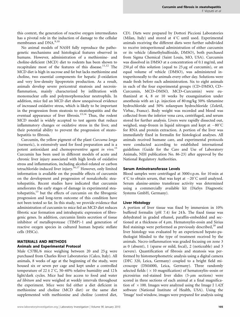
To explore whether curcumin exerted beneficial effects onliver histopathology, paraffin-embedded specimens werefirst analyzed after staining with hematoxylin and eosin(Figure 2). MCD diet caused a rapid and marked accumu-lation of fat in hepatocytes, with a predominant macro-vesicular pattern. As previously reported in this model, MCDdiet also induced an evident infiltration with inflammatorycells, mostly represented by mononuclear cells, but with acontribution of polymorphonuclear leukocytes. Inflamma-tion was detected in foci or in surrounding groups ofhepatocytes with microvesicular steatosis, forming lipo-granulomas (Figure 2). Steatosis and inflammation were
already present after 4 weeks of treatment and persistedthroughout the study, with a moderate decline at later timepoints. Treatment with curcumin resulted in a generalimprovement of inflammation associated with the MCD diet.The effects of curcumin were more marked at later timepoints (8 and 10 weeks) when a significant decrease in thehistological scores for necro-inflammation was observed
Table 2 Body and liver weight during the experimental protocol
Body weight T0 (g) Body weight (g) Liver weight (g) Liver/body weight (%)
4 weeks
CD–DMSO 25.3±0.6 27.3±1.2 0.923±0.264 3.35±0.85
CD–Curc 27.0±1.6 28.3±1.6 1.083±0.342 3.80±1.03
MCD–DMSO 24.2±1.2 17.9±0.8 0.742±0.106 4.27±0.29
MCD–Curc 24.0±0.9 17.2±0.7 0.815±0.074 4.76±0.60
8 weeks
CD–DMSO 23.3±1.7 30.1±1.0 1.080±0.139 3.60±0.53
CD–Curc 24.8±0.5 29.3±2.4 0.863±0.113 2.98±0.21
MCD–DMSO 23.9±1.0 15.0±1.3* 0.428±0.227 2.79±1.27
MCD–Curc 23.2±1.0 13.8±1.2 0.326±0.126 2.32±0.75
10 weeks
CD–DMSO 23.3±0.5 27.2±1.5 1.133±0.104 4.16±0.28
CD–Curc 23.8±0.5 28.8±2.3 1.108±0.098 3.85±0.28
MCD–DMSO 23.7±1.8 14.1±1.2 0.727±0.222 5.19±1.68
MCD–Curc 23.5±1.0 13.8±0.5 0.624±0.032 4.51±0.28
Mice were administered a methionine and choline-deficient diet (MCD) or an identical diet supplemented with methionine and choline (CD) and treated withcurcumin (Curc.) or its vehicle (DMSO) for the indicated time points. At the beginning of the protocol (T0), and at the time of killing, body and liver weight wererecorded. Data are mean±s.d. *, Po0.05 vs MCD–Curc.
Figure 1 Curcumin reduces aminotransferase elevation at early time points
during experimental steatohepatitis. Male C57/Bl6 mice were placed on a
control diet (CD) or on a methionine and choline-deficient diet (MCD) for 4,
8 or 10 weeks, as indicated, and treated with DMSO (black columns) or with
curcumin (gray columns). Serum alanine aminotransferase levels were
assayed as described in Materials and Methods. Data are expressed as
mean±s.d. *Po0.05 vs DMSO.
Curcumin and fibrosis in steatohepatitis
F Vizzutti et al
www.laboratoryinvestigation.org | Laboratory Investigation | Volume 90 January 2010 107
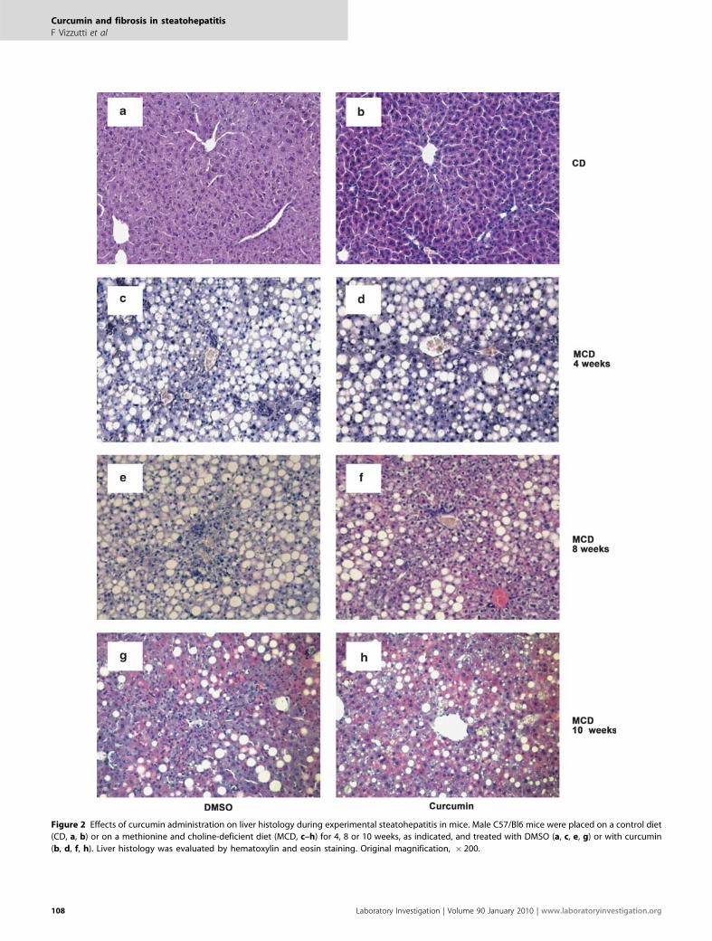
Figure 2 Effects of curcumin administration on liver histology during experimental steatohepatitis in mice. Male C57/Bl6 mice were placed on a control diet
(CD, a, b) or on a methionine and choline-deficient diet (MCD, c–h) for 4, 8 or 10 weeks, as indicated, and treated with DMSO (a, c, e, g) or with curcumin
(b, d, f, h). Liver histology was evaluated by hematoxylin and eosin staining. Original magnification, � 200.
Curcumin and fibrosis in steatohepatitis
F Vizzutti et al
108 Laboratory Investigation | Volume 90 January 2010 | www.laboratoryinvestigation.org

(Figure 3). Curcumin resulted in a slight improvement in thegrade of steatosis associated with the MCD diet, which,however, was not statistically significant when assessed byhistomorphometry (data not shown). These data indicatethat curcumin treatment induces a reduction of necro-in-flammation in mice administered the MCD diet.
Although steatosis and inflammation appear early duringthe course of experimental steatohepatitis, fibrosis is a lateevent that has a deep impact on prognosis, causing hepato-cellular dysfunction and the emergence of portal hyperten-sion. We analyzed the appearance of fibrosis in mice receivingan MCD diet by staining liver specimens with Sirius red,which clearly detects collagen fibers (Figure 4). Fibrosisappeared after 8 weeks of MCD diet, surrounding the cen-trilobular vein and creating a fine network of fiberssurrounding either individual cells or groups of hepatocytesin the perivenular area, in a chicken-wire manner. Thesefeatures were more marked at 10 weeks of the study protocol.In contrast, little, if any, periportal fibrosis was observed(data not shown). After 10 weeks of diet, treatment withcurcumin was accompanied by a marked and significantdecrease in the accumulation of fibrillar extracellular matrixaround the sinusoids and in the perivenular areas, as assessedby computerized histomorphometry (Figure 5).
Generation of oxidative stress-related molecules is a criticalmechanism underlying the development and progression ofnonalcoholic steatohepatitis, and is well reproducible in theMCD model. We used immunostaining to detect 8-OH-dG, areliable marker of oxidative stress-mediated DNA damage.Although nuclei positive for 8-OH-dG were virtually absentin the livers of animals consuming the control diet, theirabundance was markedly upregulated in mice administeredthe MCD diet (Figure 6). Curcumin, which also possesses
antioxidant properties, led to a significant reduction in thepercentage of nuclei positive for 8-OH-dG, suggesting aninhibitory action on the generation of oxidative stress-relatedmolecules.
We next investigated the molecular mechanisms implicatedin the anti-inflammatory and anti-fibrogenic effects of cur-cumin, evaluating the intrahepatic expression of genes in-volved in the response to liver injury. Recruitment ofinflammatory cells is critically dependent on the increasedexpression of chemokines, which attract different classes ofleukocytes. As most of the inflammatory infiltrates in thismodel are represented by mononuclear cells, we analyzed theexpression of monocyte chemoattractant protein-1 (MCP-1or CCL2), which targets monocytes and T lymphocytes andis highly relevant in the response to hepatic injury.24 A 3-foldincrease in MCP-1 expression was observed in mice ad-ministered the MCD diet, whereas a significant reduction waspresent in animals treated with curcumin (Figure 7a). Simi-larly, expression of CD11b, a marker of leukocyte activation,was significantly reduced by treatment with curcumin (Fig-ure 7b). We next analyzed the expression of genes related tofibrogenesis. Expression of type I procollagen, one of themost abundant components of the fibrillar extracellularmatrix in fibrotic livers, was significantly reduced in miceadministered curcumin while receiving an MCD diet (Figure7c). As matrix accumulation depends not only on increasedproduction but also on decreased removal, we analyzed thegene expression levels of TIMP-1, which inhibits the action ofseveral matrix-degrading enzymes and has been tightlyassociated with the development of liver fibrosis. It is notedthat the expression of TIMP-1 was more than 90-foldincreased in mice administered the MCD diet, comparedwith CD (Figure 7d). Curcumin treatment reduced TIMP-1expression by approximately 70% in comparison withuntreated animals.
To provide further evidence for the antifibrogenic action ofcurcumin in this model, we analyzed the intrahepatic levelsof a-smooth muscle actin, a well-established marker ofmatrix-producing myofibroblasts, resulting mostly from theactivation of HSCs. Expression of a-smooth muscle actin wasnegligible in mice treated with the control diet, in keepingwith the normal histological appearance of the tissue. After10 weeks of treatment with the MCD diet, a marked upre-gulation of a-smooth muscle actin expression was observed,in agreement with the increased deposition of fibrotic tissueand the related activation of HSCs (Figure 8). When analyzedby densitometry scanning, the expression of this marker wasincreased 20-fold in comparison with animals treated withthe control diet. In contrast, animals receiving curcuminwhile eating an MCD diet showed a marked and significantreduction in a-smooth muscle actin expression, which wasfound to be 80% lower than that in mice treated with DMSO.These data confirm that curcumin induces anti-fibrogenicactions, related at least in part to a lower accumulation ofHSCs during the course of the fibrogenic process.
Figure 3 Effects of curcumin on the inflammatory scores. Histological
analyses were conducted using light microscopy at � 100 magnification.
Slides as presented in Figure 2 were blindly evaluated by an expert in liver
pathology, and a semiquantitative score was assigned as described in
Materials and Methods for necro-inflammation. Scores in mice treated with
DMSO (black columns) were compared with those in mice treated with
curcumin (gray columns). Data are expressed as mean±s.d. *Po0.05 vs
DMSO.
Curcumin and fibrosis in steatohepatitis
F Vizzutti et al
www.laboratoryinvestigation.org | Laboratory Investigation | Volume 90 January 2010 109

Figure 4 Effects of curcumin administration on hepatic fibrosis during experimental steatohepatitis in mice. Male C57/Bl6 mice were placed on a control
diet (CD, a, b) or on a methionine and choline-deficient diet (MCD, c–h) for 4, 8 or 10 weeks, as indicated, and treated with DMSO (a, c, e, g) or with
curcumin (b, d, f, h). Hepatic fibrosis was evaluated by sirius red staining. Original magnification, � 200.
Curcumin and fibrosis in steatohepatitis
F Vizzutti et al
110 Laboratory Investigation | Volume 90 January 2010 | www.laboratoryinvestigation.org

Curcumin has been indicated as a powerful modulator ofthe biology of activated HSCs. On the basis of the observa-tion that treatment with curcumin exerts a potent inhibitoryaction on the expression of TIMP-1, we next investigatedwhether curcumin could affect TIMP-1 in an in vitro system(Figure 9). Cultured human HSCs were exposed to differentconcentrations of curcumin for 24 h, and accumulation ofTIMP-1 in cell-conditioned medium was assayed by ELISA.Concentrations of curcumin previously shown to modulatethe biology of different cell types25,26 resulted in a dose-de-pendent reduction of TIMP-1 secretion. Further increases incurcumin concentration did not result in more evident ef-fects. Curcumin also reduced the mRNA levels for TIMP-1,measured by real-time PCR, although this effect did notreach statistical significance (data not shown). We next
evaluated the effects of curcumin as a direct antioxidant forHSCs. Cells were loaded with the fluorescent dye, DCF-DA,which allows the detection of any significant increases inintracellular levels of ROS with high sensitivity.23 Pre-incubation with curcumin significantly blocked the genera-tion of ROS induced by three different protocols (Figure 10).In addition, when HSCs were incubated with hypox-anthine—xanthine oxidase, a significant upregulation ofTIMP-1 secretion was observed (Figure 11), indicating that apro-oxidant environment regulates the release of TIMP-1.When HSCs were pre-incubated with curcumin, at a con-centration effective in reducing oxidative stress (see Figure10), TIMP-1 secretion was significantly decreased (Figure 11).Thus, curcumin reduces the ability to secrete an inhibitor ofmatrix degradation by HSCs, and limits the production andeffects of ROS in these fibrogenic cells.
DISCUSSIONConsiderable attention is being directed to the identificationof novel therapeutic strategies for nonalcoholic steatohepa-titis, considering the extremely high prevalence of thisdisease. In particular, therapies that limit hepatic injury andthe related occurrence of inflammation and fibrosis wouldbe particularly appealing for this condition. Curcumin, ayellow coloring ingredient of the spice turmeric, obtainedfrom the rhizome of Curcuma longa, is extensively usedfor food preparation, and has been used in indigenousherbal medicine for the treatment of inflammatory andliver disorders. Curcumin has potent antioxidant, anti-inflammatory, antimutagenic and anticarcinogenic proper-ties.15 In addition, curcumin has been shown to delay theonset of lipid peroxidation, to ameliorate chemically inducedoxidative stress, and to increase the expression of xenobioticdetoxifying enzymes.15 Previous studies have also highlightedthe protective effects of curcumin toward several types ofchemically induced hepatotoxicity, including damageinduced by carbon tetrachloride and by experimentalalcoholic liver disease,16,17 providing the rationale for itspossible use in the context of nonalcoholic steatohepatitis. Inthis study, we provide evidence for the first time thatadministration of curcumin limits the long-term evolution ofexperimental steatohepatitis, improving fibrosis and down-regulating different genes involved in the fibrogenic process.Occurrence and progression of steatohepatitis are criticallydependent on the presence of hepatocellular damage, in-flammation and oxidative stress.8 Remarkably, curcumin wasable to beneficially affect all these components, although theeffects showed some variability. Animals treated with cur-cumin had lower ALT levels, at least during the early stages ofthe disease, together with reduced inflammation, histologicalevidence of injury, and signs of oxidative stress. It is im-portant to underline that these events are tightly inter-connected during the response of the liver to injury.Recruitment of inflammatory cells, mainly orchestratedby proteins of the chemokine family,24 contributes to the
Figure 5 Effects of curcumin on histological fibrosis. Slides stained with
Sirius red were evaluated by quantitative histomorphometry at 10 weeks, as
described in Materials and Methods. Data of mice treated with DMSO
(white and black columns) were compared with those of mice treated with
curcumin (gray and cross-hatched columns). Data are expressed as
mean±s.d. *Po0.001 vs MCD DMSO.
Figure 6 Curcumin reduces hepatic oxidative stress during experimental
steatohepatitis. Mice (6–8 for each group) were placed on a control diet
(CD) or on a methionine and choline-deficient diet (MCD) for 10 weeks.
Data of mice treated with DMSO (white and black columns) were compared
with those of mice treated with curcumin (gray and cross-hatched
columns). Nuclei positive for 8 hydroxy-deoxyguanidine were
immunostained and counted. Data are expressed as mean±s.d. *Po0.05 vs
DMSO.
Curcumin and fibrosis in steatohepatitis
F Vizzutti et al
www.laboratoryinvestigation.org | Laboratory Investigation | Volume 90 January 2010 111

exacerbation of hepatocyte damage and to the generation ofoxidative stress-related products. These latter mediators,including reactive oxygen species and reactive aldehydes, can
trigger further inflammation and hepatocellular injury. Alongthese lines, this series of events may be interrupted, at leastpartially, in animals lacking inflammatory chemokines suchas MCP-1,19 and curcumin was indeed effective in limitingthe expression of this chemokine in animals exposed to theMCD diet. This effect was associated with a reduction in theamount of oxidative stress, measured as oxidative changes inhepatocyte DNA, and in the prevention of necro-inflamma-tion. All these findings are in line with known activitiesof curcumin in the liver and in other tissues, where it hasbeen previously shown to limit activation of the master
Figure 7 Effects of curcumin on hepatic gene expression during experimental steatohepatitis. Mice (6–8 for each group) were placed on a control diet (CD) or
on a methionine and choline-deficient diet (MCD). At the time of the killing, liver tissue was collected for RNA extraction, and gene expression of monocyte
chemoattractant protein-1 (4 weeks, panel a), CD11b (4 weeks, panel b), procollagen type I (8 weeks, panel c) and tissue inhibitor of metalloproteinase-1 (8
weeks, panel d) was measured by real-time PCR. Expression in mice treated with DMSO (white and black columns) was compared with that in mice treated with
curcumin (gray and cross-hatched columns). Data are expressed as mean±s.d. *Po0.05 vs DMSO.
Figure 8 Reduced expression of a-smooth muscle actin in mice treated with
curcumin. Mice were placed on a control diet (CD) or on a methionine and
choline-deficient diet (MCD) for 10 weeks. Liver tissue was homogenized and
extracted for total protein. a-SMA expression was analyzed by western blotting
(panel a) as detailed in Materials and Methods. (b) Signal for a-SMA was
quantified by densitometry and expressed in barograms as mean±s.d.
*Po0.05 vs DMSO. Data of mice treated with DMSO (white and black columns)
were compared with those of mice treated with curcumin (gray and cross-
hatched columns).
Figure 9 Curcumin inhibits the expression of TIMP-1 in cultured HSCs.
Cultured human HSCs were deprived of serum and exposed to the
indicated concentrations of curcumin for 24 h. At the end of incubation,
TIMP-1 concentrations were assayed by ELISA in cell-conditioned media.
Mean±s.d. of three experiments. *Po0.05 vs control.
Curcumin and fibrosis in steatohepatitis
F Vizzutti et al
112 Laboratory Investigation | Volume 90 January 2010 | www.laboratoryinvestigation.org

inflammatory transcription factor, nuclear factor (NF)-kBand to block oxidative injury.15 Our data are in clear agree-ment with the study of Leclercq et al, in which curcuminlimited activation of NF-kB and expression of MCP-1 and
intercellular adhesion molecule-1, whereas steatosis was un-affected.13 NF-kB activation has been previously shown tohave a relevant role in the hepatic fibrogenic response.27
Recently, it has been reported that interruption of NF-kB
Figure 10 Reduced oxidative stress in HSCs treated with curcumin. Human HSCs were deprived of serum and either left untreated or preincubated with
20 mM curcumin for 30 min. At the end of incubation, cells were exposed for 15 min to 0.1 mM 2-Methyl-1,4-naphthoquinone (Menadione), or 0.1 mM 2,3—
dimethoxy—1,4-naphthoquinone (DMNQ), or 0.4 mM hypoxanthine—2 mU xanthine oxidase (X/XO). Intracellular generation of ROS was assessed by
measuring the fluorescence of 20 ,70-dichlorodihydrofluorescein diacetate (DCF-DA, used at 5 mM concentration) as described in Materials and Methods. Panel
a: phase contrast photomicrographs (left row); fluorescence microscopy (central row); overlay (right row). Panel b: the percentage of cells positive for DCF-
DA fluorescence was counted in the different experimental conditions described above (mean±s.d. of three experiments). 1Po0.05 vs control; *Po0.05 vs
the respective condition in the absence of curcumin.
Curcumin and fibrosis in steatohepatitis
F Vizzutti et al
www.laboratoryinvestigation.org | Laboratory Investigation | Volume 90 January 2010 113

signaling by curcumin suppresses the expression of the pro-fibrogenic cytokine, connective tissue growth factor, inHSCs.28 Therefore, further studies are needed to establishwhether inhibition of NF-kB activation also has a role in theobserved limitation of fibrosis in the MCD model. It is notedthat Leclercq et al. did not report any effects on oxidativestress,13 whereas we did observe a protective effect of cur-cumin on DNA oxidation. The reason for this discrepancy islikely related to the longer time points investigated in thestudy reported herein. It should also be mentioned that wedecided to administer curcumin through the intraperitonealroute, as in previously reported studies,29–31 to minimizepossible differences in curcumin administration linked to thefact that the sickest animals on the MCD diet would havebeen likely to limit food intake.
The extended experimental protocol used in this study hasallowed us to provide definitive evidence on the inhibitoryeffects of curcumin administration on the development ofhepatic fibrosis in experimental steatohepatitis. Fibrogenesisis the common evolution of most forms of chronic liverdisease, including nonalcoholic steatohepatitis, and is strictlylinked to the eventual appearance of hepatic decompensationand death. The mechanisms of hepatic fibrogenesis have beenthe focus of extensive investigation over the last 15 years,32
and during steatohepatitis, inflammatory cell infiltration andgeneration of oxidative stress have been indicated as pivotalfactors.8 Thus, reduced fibrogenesis observed in this study inanimals treated with curcumin is dependent, at least in part,on the modulation of the pro-fibrogenic signals generated byinflammation and oxidative stress. However, several studieshave indicated that curcumin has direct anti-fibrogeniceffects through modulation of different aspects of the biology
of HSCs, which on a process known as ‘activation’ acquire aphenotype of fibrogenic myofibroblasts and are criticallyinvolved in hepatic scarring.32 In particular, curcumin hasbeen shown to induce expression of the transcription factorPPAR-g, leading to inhibition of mitogen-stimulatedproliferation of HSCs.33 In addition, curcumin blocked theactivation of critical pro-fibrogenic pathways in rodent HSCs,including the activation of NF-kB and extracellular signal-regulated kinase, propagation of signal transduction down-stream of TGF-b receptors, and expression and signaling ofPDGF-b receptor and EGF receptor.33–35 These data indicatethat curcumin may be a general inhibitor of fibrogenesisindependent of the setting in which it develops. In thisstudy, a direct effect on HSCs is suggested by the fact thatexpression of a-smooth muscle actin, a classical marker offibrogenic cells derived from the activation of HSCs, wassignificantly reduced in mice receiving curcumin, and wasassociated with reduced expression of type I procollagen.We also detected a striking decrease in the expression ofTIMP-1 in the liver of mice administered curcumintogether with an MCD diet, and the extent of reduction ofTIMP-1 expression was more marked than that of type Iprocollagen. To provide further evidence for a direct action ofcurcumin on fibrogenic cells, we found that curcumin dose-dependently inhibits the secretion of TIMP-1 by culturedHSCs, thus identifying an additional mechanism of reducedfibrogenesis. Further studies will be required to clarify themolecular mechanisms responsible for this effect, consideringthat we did not observe a significant reduction in the steady-state mRNA levels of TIMP-1 in HSCs exposed to curcumin.The modulatory effect of curcumin on inhibitors of me-talloproteinases is in agreement with data recently reportedin myofibroblasts from other tissues.36 On the other hand,curcumin has been shown to increase TIMP-1 expression inbreast cancer cells, in which it provides anti-metastatic sig-nals.37 It is noted that curcumin was also effective in reducingROS generation by HSCs, and inhibited the increase inTIMP-1 secretion caused by oxidative stress, identifying ad-ditional mechanisms underlying the anti-fibrogenic action ofthis compound.10 Taken together, these data indicate thatcurcumin exerts its anti-fibrogenic action at multiple levels,including direct actions on HSCs.
It should be acknowledged that the use of the MCD diet asa model of steatohepatitis represents a limitation ofour study, because it does not reproduce those metabolicderangements typical of patients with nonalcoholic steato-hepatitis, including obesity and dyslipidemia.38 However, theprincipal aim of this study was to investigate the possiblemodulation of the progressive aspects of steatohepatitis,specifically fibrogenesis, that are very modestly induced inany animal model of the metabolic syndrome.12
In conclusion, we have shown that curcumin administra-tion limits fibrosis development and progression in mice withexperimental steatohepatitis. These data have particularrelevance in a time where global demand for more affordable
Figure 11 Upregulation of TIMP-1 secretion by oxidative stress is
prevented by curcumin in HSCs. Human HSCs were deprived of serum and
either left untreated or preincubated with 20 mM curcumin for 30 min,
followed by exposure for 24 h to 0.4 mM hypoxanthine—2 mU xanthine
oxidase (X/XO). At the end of incubation, TIMP-1 concentrations were
assayed by ELISA in cell-conditioned media (mean±s.d. of three
experiments). 1Po0.05 vs control; *Po0.05 vs X/XO in the absence of
curcumin.
Curcumin and fibrosis in steatohepatitis
F Vizzutti et al
114 Laboratory Investigation | Volume 90 January 2010 | www.laboratoryinvestigation.org

therapeutics and concerns about commonly used drugs arerefocusing interest on traditional medicines,39 especially forthe treatment of very prevalent diseases that are likely torequire lengthy or lifelong treatments, such as nonalcoholicsteatohepatitis.
ACKNOWLEDGEMENT
This study was supported by grants from the Italian Ministry of Research (to
FM and M Par), the University of Florence (to FM), Regione Piemonte (to M
Par.), and the Italian Liver Foundation (to FM and M Pinz.).
DISCLOSURE/CONFLICT OF INTEREST
The authors declare no conflict of interest
1. Lazo M, Clark JM. The epidemiology of nonalcoholic Fatty liver disease:a global perspective. Semin Liver Dis 2008;28:339–350.
2. Ekstedt M, Franzen LE, Mathiesen UL, et al. Long-term follow-up ofpatients with NAFLD and elevated liver enzymes. Hepatology2006;44:865–873.
3. Matteoni CA, Younossi ZM, Gramlich T, et al. Nonalcoholic fatty liverdisease: a spectrum of clinical and pathological severity.Gastroenterology 1999;116:1413–1419.
4. Teli MR, James OF, Burt AD, et al. The natural history of nonalcoholicfatty liver: a follow-up study. Hepatology 1995;22:1714–1719.
5. Yeh MM, Brunt EM. Pathology of nonalcoholic fatty liver disease. Am JClin Pathol 2007;128:837–847.
6. Ludwig J, Viggiano TR, McGill DB, et al. Nonalcoholic steatohepatitis:Mayo Clinic experiences with a hitherto unnamed disease. Mayo ClinProc 1980;55:434–438.
7. Choi CS, Savage DB, Kulkarni A, et al.. Suppression of diacylglycerolacyltransferase-2 (DGAT2), but not DGAT1, with antisense oligo-nucleotides reverses diet-induced hepatic steatosis and insulinresistance. J Biol Chem 27 May 2007.
8. Marra F, Gastaldelli A, Svegliati Baroni G, et al. Molecular basis andmechanisms of progression of non-alcoholic steatohepatitis. TrendsMol Med 2008;14:72–81.
9. Edmison J, McCullough AJ. Pathogenesis of non-alcoholicsteatohepatitis: human data. Clin Liver Dis 2007;11:75–104 ix.
10. Parola M, Robino G. Oxidative stress-related molecules and liverfibrosis. J Hepatol 2001;35:297–306.
11. Koteish A, Diehl A. Animal models of steatosis. Sem Liv Dis 2001;21:89–104.
12. London RM, George J. Pathogenesis of NASH: animal models. Clin LiverDis 2007;11:55–74 viii.
13. Leclercq IA, Farrell GC, Sempoux C, dela Pena A, Horsmans Y. Curcumininhibits NF-kappaB activation and reduces the severity of experimentalsteatohepatitis in mice. J Hepatol 2004;41:926–934.
14. Leclercq IA, Farrell GC, Field J, et al. CYP2E1 and CYP4A as microsomalcatalysts of lipid peroxides in murine nonalcoholic steatohepatitis.J Clin Invest 2000;105:1067–1075.
15. Anand P, Thomas SG, Kunnumakkara AB, et al. Biological activities ofcurcumin and its analogues (Congeners) made by man and MotherNature. Biochem Pharmacol 2008; 76:1590–1611.
16. Fu Y, Zheng S, Lin J, et al. Curcumin protects the rat liver from CCl4-caused injury and fibrogenesis by attenuating oxidative stress andsuppressing inflammation. Mol Pharmacol 2008;73:399–409.
17. Nanji AA, Jokelainen K, Tipoe GL, et al. Curcumin prevents alcohol-induced liver disease in rats by inhibiting the expression of NF-kappaB-dependent genes. Am J Physiol Gastrointest Liver Physiol2003;284:G321–G327.
18. Bataller R, Schwabe RF, Choi YH, et al. NADPH oxidase signaltransduces angiotensin II in hepatic stellate cells and is critical inhepatic fibrosis. J Clin Invest 2003;112:1383–1394.
19. Zamara E, Galastri S, Aleffi S, et al. Prevention of severe toxic liverinjury and oxidative stress in MCP-1-deficient mice. J Hepatol2007;46:230–238.
20. Halliwell B, Whiteman M. Measuring reactive species and oxidativedamage in vivo and in cell culture: how should you do it and what dothe results mean? Br J Pharmacol 2004;142:231–255.
21. Toyokuni S, Tanaka T, Hattori Y, et al. Quantitativeimmunohistochemical determination of 8-hydroxy-20-deoxyguanosineby a monoclonal antibody N45.1: its application to ferricnitrilotriacetate-induced renal carcinogenesis model. Lab Invest1997;76:365–374.
22. Caligiuri A, Bertolani C, Guerra CT, et al. Adenosine monophosphate-activated protein kinase modulates the activated phenotype ofhepatic stellate cells. Hepatology 2008;47:668–676.
23. Cannito S, Novo E, Compagnone A, et al. Redox mechanisms switch onhypoxia-dependent epithelial-mesenchymal transition in cancer cells.Carcinogenesis 2008;29:2267–2278.
24. Marra F. Chemokines in liver inflammation and fibrosis. Front Biosci2002;7:d1899–d1914.
25. Chen HW, Huang HC. Effect of curcumin on cell cycle progression andapoptosis in vascular smooth muscle cells. Br J Pharmacol1998;124:1029–1040.
26. Cohly HH, Taylor A, Angel MF, et al. Effect of turmeric, turmerin andcurcumin on H2O2-induced renal epithelial (LLC-PK1) cell injury. FreeRadic Biol Med 1998;24:49–54.
27. Elsharkawy AM, Mann DA. Nuclear factor-kappaB and the hepaticinflammation-fibrosis-cancer axis. Hepatology 2007;46:590–597.
28. Chen A, Zheng S. Curcumin inhibits connective tissue growthfactor gene expression in activated hepatic stellate cells in vitroby blocking NF-kappaB and ERK signalling. Br J Pharmacol2008;153:557–567.
29. Gaedeke J, Noble NA, Border WA. Curcumin blocks fibrosis in anti-Thy1 glomerulonephritis through up-regulation of heme oxygenase 1.Kidney Int 2005;68:2042–2049.
30. Poylin V, Fareed MU, O’Neal P, et al. The NF-kappaB inhibitor curcuminblocks sepsis-induced muscle proteolysis. Mediators Inflamm2008;2008:317851.
31. Swarnakar S, Ganguly K, Kundu P, et al. Curcumin regulates expressionand activity of matrix metalloproteinases 9 and 2 during preventionand healing of indomethacin-induced gastric ulcer. J Biol Chem2005;280:9409–9415.
32. Friedman SL. Mechanisms of hepatic fibrogenesis. Gastroenterology2008;134:1655–1669.
33. Lin J, Chen A. Activation of peroxisome proliferator-activated receptor-gamma by curcumin blocks the signaling pathways for PDGF and EGFin hepatic stellate cells. Lab Invest 2008;88:529–540.
34. Zheng S, Chen A. Disruption of transforming growth factor-betasignaling by curcumin induces gene expression of peroxisomeproliferator-activated receptor-gamma in rat hepatic stellatecells. Am J Physiol Gastrointest Liver Physiol 2007;292:G113–G123.
35. Zhou Y, Zheng S, Lin J, et al. The interruption of the PDGF and EGFsignaling pathways by curcumin stimulates gene expression ofPPARgamma in rat activated hepatic stellate cell in vitro. Lab Invest2007;87:488–498.
36. Yasui H, Andoh A, Bamba S, et al. Role of fibroblast growth factor-2 inthe expression of matrix metalloproteinases and tissue inhibitors ofmetalloproteinases in human intestinal myofibroblasts. Digestion2004;69:34–44.
37. Shao ZM, Shen ZZ, Liu CH, et al. Curcumin exerts multiple suppressiveeffects on human breast carcinoma cells. Int J Cancer 2002;98:234–240.
38. Torres DM, Harrison SA. Diagnosis and therapy of nonalcoholicsteatohepatitis. Gastroenterology 2008;134:1682–1698.
39. Singh S. From exotic spice to modern drug? Cell 2007;130:765–768.
Curcumin and fibrosis in steatohepatitis
F Vizzutti et al
www.laboratoryinvestigation.org | Laboratory Investigation | Volume 90 January 2010 115
Related Documents