"Science Stays True Here" Biological and Chemical Research, Volume 2014, 1-15 | Science Signpost Publishing Curcumin / BSA: New Approach for Hepatocellular Carcinoma Treatment Faten Zahran 1 , Essam Mady 2 , Osama Yasein 3 , Akaber T. Keshta 1 1. Biochemistry Department, Faculty of Science, Zagazig University, Egypt. 2. Biochemistry Department, Faculty of Science, Ain-shams University, Egypt. 3. Histology Department, Faculty of Medicine, Zagazig University, Egypt. Received: September 15, 2014 / Accepted: October 10, 2014 / Published: November 25, 2014 Abstract: The systemic availability of curcumin is very low after oral administration; this limits their therapeutic potential. This study aims to increase the bioavailability of curcumin; the highest reproducible solubility modality will be applied on an experimental carcinogenesis models in order to evaluate its chemo-preventive, chemotherapeutic effects and antitumor potential. We found that administrating of curcumin (200 mg/kg I.P. bound to 5% BSA in PBS, pH 7.4) results in a significant inhibitory effect on tumor in vivo. An anti-oxidant effect and anti-tumor effect of curcumin in vivo was observed. A significant reduction in anti-oxidants and tumor markers levels in tumor treated animals when compared with untreated ones. As well as Bcl2 expression was reduced. Conclusion: curcumin bound BSA has a strong inhibitory activity against tumors. The anti-tumor mechanism may be mediated by preventing oxidative damage and induction of apoptosis improved animals’ chances of survival and they become healthier. Keywords: Curcumin, Bovine Serum albumin, Apoptosis, Hepatocellular Carcinoma. 1. Introduction Hepatocellular carcinoma (HCC), the most common type of liver cancer, is the third leading cause of cancer deaths worldwide [1]. It is one of the five most common cancers worldwide. HCC is a high-grade malignancy showing a rapid infiltrating growth, early stage metastasis, poor therapeutic response and disappointing prognosis even after successful curative resection surgery [2]. Surgery, including transplantation, remains the only potentially curative modality for HCC, yet the recurrence rate for this particular cancer is high and long-term survival rate is rather poor [3]. Cancer chemoprevention is a rapidly growing area of oncology which can make a significant progress in the prevention and treatment of carcinogenesis by administration of various drugs with chemical or natural entities depending on their anti-mutagenic properties [4]. Cancer chemoprevention is a rapidly growing area of oncology which can make a significant progress in the prevention and treatment of carcinogenesis by administration of various drugs with chemical or natural entities depending on their anti-mutagenic properties. It is, therefore, essential that new therapeutic options are needed for cancer therapy with attention to toxicity and side effects, besides the major treatment modalities including surgery, immunotherapy and radiotherapy [5, 6]. Curcumin (diferuloylmethane) is a major constituent of the yellow spice turmeric derived from the rhizomes of Curcuma longa. It is safe and nontoxic and has demonstrable antitumor, anti-inflammatory, apoptotic, and antioxidant properties. It was shown previously that curcumin inhibits tumor metastasis, invasion, and angiogenesis [7-9]. Therefore, it is regarded as a high potential to develop into modern drug. Curcumin’s chemo-preventive activity in Corresponding author: Faten Zahran, Biochemistry Department, Faculty of Science, Zagazig University, Egypt. E-mail: [email protected].

Welcome message from author
This document is posted to help you gain knowledge. Please leave a comment to let me know what you think about it! Share it to your friends and learn new things together.
Transcript
"Science Stays True Here" Biological and Chemical Research, Volume 2014, 1-15 | Science Signpost Publishing
Curcumin / BSA: New Approach for Hepatocellular
Carcinoma Treatment
Faten Zahran1, Essam Mady2, Osama Yasein3, Akaber T. Keshta1 1. Biochemistry Department, Faculty of Science, Zagazig University, Egypt.
2. Biochemistry Department, Faculty of Science, Ain-shams University, Egypt.
3. Histology Department, Faculty of Medicine, Zagazig University, Egypt.
Received: September 15, 2014 / Accepted: October 10, 2014 / Published: November 25, 2014
Abstract: The systemic availability of curcumin is very low after oral administration; this limits their therapeutic potential. This study aims to increase the bioavailability of curcumin; the highest reproducible solubility modality will be applied on an experimental carcinogenesis models in order to evaluate its chemo-preventive, chemotherapeutic effects and antitumor potential. We found that administrating of curcumin (200 mg/kg I.P. bound to 5% BSA in PBS, pH 7.4) results in a significant inhibitory effect on tumor in vivo. An anti-oxidant effect and anti-tumor effect of curcumin in vivo was observed. A significant reduction in anti-oxidants and tumor markers levels in tumor treated animals when compared with untreated ones. As well as Bcl2 expression was reduced. Conclusion: curcumin bound BSA has a strong inhibitory activity against tumors. The anti-tumor mechanism may be mediated by preventing oxidative damage and induction of apoptosis improved animals’ chances of survival and they become healthier. Keywords: Curcumin, Bovine Serum albumin, Apoptosis, Hepatocellular Carcinoma.
1. Introduction
Hepatocellular carcinoma (HCC), the most common type of liver cancer, is the third leading cause of cancer deaths worldwide [1]. It is one of the five most common cancers worldwide. HCC is a high-grade malignancy showing a rapid infiltrating growth, early stage metastasis, poor therapeutic response and disappointing prognosis even after successful curative resection surgery [2]. Surgery, including transplantation, remains the only potentially curative modality for HCC, yet the recurrence rate for this particular cancer is high and long-term survival rate is rather poor [3]. Cancer chemoprevention is a rapidly growing area of oncology which can make a significant progress in the prevention and treatment of carcinogenesis by administration of various drugs with chemical or natural entities depending on their anti-mutagenic properties [4]. Cancer chemoprevention is a rapidly growing area of oncology which can make a significant progress in the prevention and treatment of carcinogenesis by administration of various drugs with chemical or natural entities depending on their anti-mutagenic properties. It is, therefore, essential that new therapeutic options are needed for cancer therapy with attention to toxicity and side effects, besides the major treatment modalities including surgery, immunotherapy and radiotherapy [5, 6].
Curcumin (diferuloylmethane) is a major constituent of the yellow spice turmeric derived from the rhizomes of Curcuma longa. It is safe and nontoxic and has demonstrable antitumor, anti-inflammatory, apoptotic, and antioxidant properties. It was shown previously that curcumin inhibits tumor metastasis, invasion, and angiogenesis [7-9]. Therefore, it is regarded as a high potential to develop into modern drug. Curcumin’s chemo-preventive activity in
Corresponding author:
Faten Zahran, Biochemistry Department, Faculty of Science, Zagazig University, Egypt. E-mail: [email protected].

Curcumin / BSA: New Approach for Hepatocellular Carcinoma Treatment 2
animal model systems has led investigators to study its potential impact upon tumor cell growth and apoptosis. Several reports document an anti-proliferative effect on cultured cells such as on colon cancer and breast cancer cells. This may, in part, be because of programmed cell death because at high concentrations curcumin can induce apoptosis such as in human leukemia cells [10]. Curcumin acts as a scavenger of oxygen species, such as hydroxyl radical, superoxide anion, and singlet oxygen, and it interferes with lipid per-oxidation. Curcumin possibly used in the prevention and treatment of cancer [11-12]. Curcumin suppresses a number of key elements in cellular signal transduction pathways pertinent to growth, differentiation, and malignant transformation [13]. Unfortunately, the solubility of curcuminoids in aqueous solutions is exceedingly low. This restricts their systemic availability in orally administered formulations and limits their therapeutic potential [14] because most of the curcumin is metabolized in the intestine.
Several studies recently confirm the high affinity nature of curcumin binding to Bovine Serum Albumin (BSA) [15]. Curcuminoids were highly soluble in solutions of purified albumin, a major component of serum. Solid curcuminoids would thus allow only limited surface access of the solvent to disrupt the defined structures. At saturating BSA concentrations (14–20%), the amount of DMSO-dissolved curcuminoids converted into BSA solubilized curcuminoids was about 92–96%. At pH ≈ 7.0, the protein molecule acquired more negative charges, which in turn help expand the protein a bit further allowing the binding of more dye molecule (curcumin) without altering the affinity [16]. Hence, the concentrations at saturation obtained by adding either solid or DMSO dissolved curcuminoids to 5% BSA were about two-fold higher. So, these promising novel formulations, which appear to provide longer circulation, better permeability, and resistance to metabolic processes [17]. Curcumin bio-conjugates containing albumin improve medicinal properties over curcumin, suggesting increased cellular uptake.
2. Materials and Methods
2.1. Tumors Hepatocellular carcinoma (HCC) was induced by an intraperitoneal injection of N-diethylnitrosamine “DENA” (200
mg/kg; Sigma Chemical Company) [18] dissolved in saline. 2.2. Curcumin
Crude curcumin was obtained from Fluka, Buchs, Switzerland, was dissolved in dimethylsulphoxide (DMSO), and then dissolved in 5% (Bovine Serum Albumin (BSA) in Phosphate Buffer Saline (PBS); pH 7.4 solution used during the treatment to increase the bioavailability of curcumin. 2.3. Experimental Design
Albino male Wister rats` 190-200 g body weights were raised at the experimental animal house of the faculty of Science, Zagazig University. The animals were maintained in controlled environment of temperature, humidity and light. They were fed on a commercial standard diet and tap water. Rats were divided according to their weight into four groups, each one includes 8 rats. Group I, were received a saline solution by I.P. injection represented as a negative control group; Group II, were received DENA (200 mg/kg I.P.) served as a positive control group. Group III, were injected I.P with curcumin one week before DENA (tumor induction), represented as preventive group, Group IV, were injected I.P. DENA, represented a therapeutic group, then curcumin was injected every week for five weeks to the last groups. The rats of four groups were maintained under the same conditions and were observed carefully everyday to the end of experiment. At the end of the experiment, the blood samples and liver tissues were collected from animals for biochemical and histological assays. 2.4. Biochemical Analyses
a) Estimation of α-L- Fucosidase in Serum
Curcumin / BSA: New Approach for Hepatocellular Carcinoma Treatment 3
Serum α-L- Fucosidase (AFU) acivity was determined by using Biodiagnostic kit method (Biodiagnostic Company, Dokki, Giza, Egypt). The AFU assay is based on the enzymatic cleavage of the synthetic substrate p-notrophenol and L-Fucose. The yellow color of p-nitrophenol in an alkaline medium can be measured at 405 nm [19].
b) Anti-oxidants Assayes (i) Estimation of Nitric Oxide in Serum In acid medium and in the presence of nitrite the formed nitrous acid diazotise sulphanilamide and the product are
coupled with N-(1–naphthyl) ethylenediamine. The resulting azo dye has a bright reddish –purple color which can be measured at 540 nm in a spectrophotometer [20].
(ii) Estimation of Malondialdehyde in Serum The lipid peroxidation products were estimated by the formation of thiobarbituric acid (TBA) and quantified in term
of MDA where, thiobarbituric acid (TBA) reacts with MDA in acidic medium at temperature of 95°C for 30 min to form thiobarbituric acid reactive product, the absorbance of the resultant pink product can be measured at 534 nm in a spectrophotometer [21].
(iii) Estimation of Glutathione reduced in erythrocyte lysate Glutathione reduced (GSH) level was determined by using Biodiagnostic kit method. This method based on the
reduction of 5, 5`-dithiobis (2-nitrobenzoic acid) (DTNB) with glutathione (GSH) to produce a yellow compound can be measured at 405 nm. The reduced chromogen is directly proportional to GSH concentration [22].
(iv) Estimation of Glutathione-S-Transferase in Plasma Glutathione –S-Transferase (GST) level was determined by using Biodiagnostic kit method. This bio-diagnostic GST
assay measure total GST activity (cytosolic and microsomal) by measuring the conjugation of 1-chloro-2,4- dinitrobenzene (CDNB) with reduced glutathione. The conjugation is accompanied by an increase the absorbance at 340 nm (UV method). The rate of increase the conjugation is directly proportional to GST activity in the sample [23].
(v) Estimation of Total Antioxidant capacity in Plasma Total Antioxidant Capacity (TAC) level was determined by using Biodiagnostic kit method. The determination of the
antioxidative capacity is performed by the reaction of antioxidants in the sample with a defined amount of exogenously provide hydrogen peroxide (H2O2). The antioxidants in the sample eliminate a certain amount of provided H2O2. The residual H2O2 is determined colorimetrically by an enzymatic reaction which involves the conversion of 3, 5-dichloro-2-hydroxybenzensulphate to a colored product can be measured at 505 nm [24]. 2.5. Histological study
Liver specimen was fixed in 10% neutral buffered formalin, embedded in paraffin, and sectioned. After de-paraffinization and dehydration, the paraffin blokes were stained with hematoxylin and eosin for microscopic examination. Light microscopy was used to evaluate the pathological changes in the liver tissues. 2.6. Chromosomal aberration
After treatment, the animals of all groups were sacrificed at the sampling time of 24 h by cervical dislocation (colchicine was given at a dose 4 mg/kg of the body weight, 2 hrs prior to killing to arrest the metaphase stage). Cytogenetic analysis was performed as per protocol of Preston et al., [25]. The bone marrow was flushed out from both femurs using Hank`s balanced salt solution (pH 7.2). The cells were centrifuged at 1000 rpm for 5 min. and the pellet was re-dispersed in hypotonic solution of 0.56% (w/v) KCl for 30 min. at 37◦C to permit osmotic swelling of cells. Swollen cells were fixed in ice- cold Carnoy`s fluid, dropped to slides and stained with phosphate-buffered 5% Giemsa solution. A total of 75 well- spread metaphase plates per animal in each group were analyzed for chromosomal aberrations at a magnification of 100×.
Curcumin / BSA: New Approach for Hepatocellular Carcinoma Treatment 4
2.7. Caspase-3 Colorimetric Assay The activity of caspase-3 was determined by the colorimetric caspase-3 kit according to the manufacturer’s
instructions (R& D system, Inc.) [26]. 2.8. Determination of Bcl-2 percentage in cells by flowcytometry
The apoptosis rate was measured by flowcytometry assay (Santa Cruz Biotechnology, Inc.) through determination of Bcl-2 expression in cells [27]. Immunohistochemical staining was adopted to test the protein expression of bcl-2 in the hepatic tissues of the rats. After bcl-2 protein expression of hepatic tissues were observed under light microscope, the comprehensive judgment was carried out based on the percentage of positive cells. 2.9. Immunohistochemical evaluation of Bcl-2 expression in the hepatic tissues of the rats
Bcl-2 was detected in immersion fixed paraffin-embedded sections of cancer tissue using Human/Mouse Bcl-2 Antigen Affinity-purified Polyclonal Antibody at 15 µg/mL overnight at 4 °C (R & D Systems, Inc.) as described by Bankfalvi et al., [28]. 2.10. Statistical Analysis
All results were analyzed by SPSS software (version 14). Data were expressed as mean ± SD. The student’s t test was used for statistical analysis of differences between each two groups. Comparison of mean values of studied variables among different groups was done using ANOVA test. Pearson’s correlation coefficient was used to quantify the relationship between the studied parameters. P<0.01 was considered to be significant [29].
3. Results
Data obtained from Table (I) summarize the anti-tumor and anti-oxidant effects of Curcumin as a chemo-preventive natural product. 3.1. Effect of Curcumin on AFU activity
A marked significant increase in AFU activity and oxidative stress in DENA induced HCC "group III" compared to negative control group "I". Anti-tumor effect of Curcumin showed a significant decrease in the activity of AFU enzyme by 35.3% and 38.7% in therapeutic and preventive groups, respectively; compared to HCC group (p<0.05) as represent in Fig. (1). 3.2. Effect of Curcumin on oxidative stress
MDA was significantly decreased by 41.6% and 44.8% in therapeutic and preventive groups, respectively. NO was significantly reduced by 36.1% and 37.7% in therapeutic and preventive groups, respectively; (p<0.05), show Fig. (2).
Also, there were a significant decrease in the GST, GSH, and TAC levels in both therapeutic and preventive groups (43.4%, 33.3%; 18.6%, 14.9%; 27.03%, and 27.9%, respectively) compared to HCC group (p<0.05), Fig. (2). 3.3. Effect of Curcumin on liver sections
Histopathological examination of liver sections from control animals revealed normal architecture. While, DENA group showed appearance of Hepatocellular carcinoma with enlarged hyper-chromatic nuclei and scattered mitosis, distorted architecture with moderate to dense inflammation represented by the presence of inflammatory cells and increase number of leukocyte cells and ballooning degeneration of hepatocytes. This results in irregular arrangement of hepatocytes with dilated central blood sinusoids. Nevertheless, Curcumin administration as a treatment, induce mild liver affection either before or after treatment; i.e. Preventive IV and Therapeutic III Groups"; the hepatocytes and sinusoid showed normal morphology, reflecting a well preserved liver parenchyma (H & E, ×200). Curcumin alleviated the DENA induced alterations seen in the above group, reduction of inflammation and re-arrangement of hepatocytes as normal case (Fig. 3 A, B, C and D).
Curcumin / BSA: New Approach for Hepatocellular Carcinoma Treatment 5
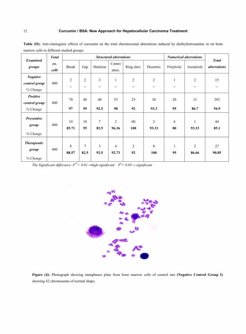
3.4. Effect of Curcumin on Chromosomal aberrations Table (II) shows the effect of Curcumin on the chromosomal aberrations. The bone marrow cells of the control
groups showed normal metaphase spread having 42 chromosomes (Fig. 4). While structural and numerical aberrations were found in HCC groups (rats treated with DENA alone). The data revealed that, the endoreduplication break, gap, deletion, ring chromosomes, and dicentric chromosomes were the most frequent types of structural chromosomal aberrations (97%, 95%, 92%, and 93%, respectively) (Fig. 5). But the numerical aberrations showed up in the form of aneuploidy and polyploidy, 87% and 95%, respectively. Moreover, the number of aberrations per cell reduced by Curcumin administration, significantly by 85.1% and 90.8% in therapeutic (III) and preventive (IV) groups, respectively (Fig. 6). 3.5. Effect of curcumin on caspase- 3 activation
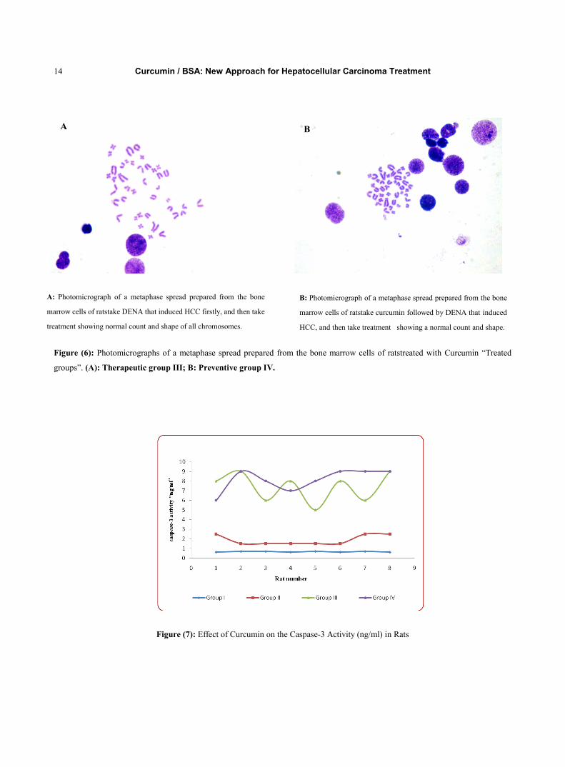
The mean values of Caspase-3 activity in HCC group rats were significantly increased from 0.66 ± 0.04 to 1.87±0.51 (ng/ml) by 183.3.0%, (p<0.001) compared to that of negative control group rats. Furthermore, the treatments with curcumin showed significantly increase in Caspase-3 activity by 249.65% and 334.76%, (p<0.001) in the therapeutic and preventive groups, respectively; compared to the positive control group, Fig. (7). 3.6. Percentage of Bcl-2 in liver tissues by flowcytometry
Flowcytometric studies show that, curcumin treatments with curcumin (200 mg/kg, 5% BSA, pH 7.4, I.P.) results in a highly significant reduction of Bcl-2 expression by 28.95% and 42.51 %, (p<0.001) in therapeutic and preventive groups, respectively compared to the positive control group, Fig. (8). 3.7. Immunohistochemistry study with Bcl-2 antibodies
To confirm our results, Immunohistochemical studied of Bcl-2 expression were done in the liver tissues in all rat studied groups. In the normal group, immunostaining of Bcl-2 protein (faint brown color), was identified in basal keratinocytes and dendritic cells adjacent to the basement membrane, with nuclear and cytoplasmic staining, Fig. (9a). While, a positive strong staining for Bcl-2 was observed in the tumor liver tissues “HCC” (Positive Control group II), compared to the normal tissues Fig. (9b). Moreover, both treatments of rats with curcumin showed a highly significant decrease in Bcl-2 expression, in the therapeutic and preventive groups, compared to HCC ones, as shown in Fig. (9c & d). 3.8. Correlations between different Studied Parameters among different Groups
Table (III) summarizes the correlations between different parameters. In studied groups, there were positive significant correlations between serum AFU and MDA, NO, GST, GSH, and TAC; respectively (r=0.915; r=0.914; r=0.838; r=0.921; r=0.930, and r= 0.648, p<0.01; respectively). Also, there were positive significant correlations between serum MDA and NO, GST, GSH, and TAC (r=0.938; r=0.862, r=0.939, r=0.960, p<0.01; respectively) in he studied groups. There were positive significant correlations between serum NO and GST, GSH, and TAC (r=0.908; r=0.940, r=0.976, p<0.01; respectively). Our data illustrated positive correlations between GST and GSH, TAC (r=0.874, r=0.938, p<0.01; respectively).
4. Discussion
Curcumin (diferuloylmethane) a low molecular weight polyphenol derived from the rhizomes of Curcuma spp., has been shown to prevent cancer in the skin, fore-stomach, duodenum, and colon of mice and in the tongue, colon, mammary glands, and sebaceous glands of rats [30]. It is well known that the systemic availability of Curcumin is very low after oral administration, because most of Curcumin is metabolized in the intestine [31]. The anti-tumor effect of Curcumin has been attributed in part to the suppression of cell proliferation, reduction of tumor load and induction of
Curcumin / BSA: New Approach for Hepatocellular Carcinoma Treatment 6
apoptosis in various cancer models both in vitro and in vivo [32]. Curcumin inhibits multiple levels within transcriptional network to restrict cell proliferation. We found a marked significant increase in AFU activity in DENA induced HCC "group III" compared to negative control group "I". Anti-tumor effect of Curcumin/BSA showed a significant decrease in the activity of AFU enzyme by 35.3% and 38.7% in therapeutic and preventive groups, respectively; compared to HCC group (p<0.05) as shown in Fig. (1). In addition, our results revealed that, Curcumin administration (200 mg/kg b.w; 5%BSA, I.P.) decrease the oxidative stress induced by DENA in HCC group; as illustrated in Table (1). Levels of MDA, NO, GST, GSH, and TAC were significantly decrease in therapeutic and preventive groups compared to HCC group (41.6%, 44.8%; 36.1%, 37.7%; 43.4%, 33.3%; 18.6%, 14.9%, and 27.0%, 27.9%, p<0.01; respectively) as shown in Fig. (2). Diethylnitrosamine (DENA) has been suggested to cause oxidative stress and cellular injury due to enhanced formation of free radicals [33]. It is metabolized to its reactive ethyl radical metabolites hat interact with DNA causing mutation, which lead to carcinogenesis [34]. In hepato-carcinogenesis, fucosylation of some sugar proteins increase and this increase may lead to the elevation of AFU activity [35]. As Curcumin has potential in the prevention and treatment of cancer [36], protects from liver injury [37].Curcumin is an effective scavenger of free radicals such as superoxide anion radicals, hydroxyl radicals and nitrogen dioxide radicals [38]. So, Curcumin may reduce the toxicity of heterocyclic aromatic amines; and inhibit the ability of nitrosamines to cause liver cancer. Our results are in agreement with Monica et.al. [39], who concluded that Curcumin exerted cell growth and apoptotic effects to free radicals generation in hepatic cancer cell line. Also, we are in accordance to Priyadarsini et al., [40] who found the antioxidant activity of Curcumin that inhibit lipid peroxidation in rat liver. Chuang et al., [41] results` indicate that Curcumin effectively inhibits DENA induced hepatocarcinogenesis; as a series of intermediate biological markers showed a remarkable increase in HCC but, eating Curcumin containing diet reversed the levels to normal values. Our biochemical findings were further supported by the histopathological examination of liver sections, which illustrated that liver tissue of DENA treated rats showed damage, resulting in malignant cell formation. On the contrary, liver tissues of Curcumin treated rats showed more or less normal hepatic lobular architecture in both therapeutic and preventive groups, as illustrated in Fig. (3 A, B, C, and D). Administration of DENA produce high significantly chromosomal aberrations 94.9% compared to control group "I" as shown in Fig.(4). The date revealed that, the numerical aberrations showed up in the form of aneuploidy and polyploidy (86.7%, 95%, respectively). The centromeric attenuation, endoreduplication break, gap, and deletion were the most frequent types of structural chromosomal aberrations (98%, 97%, 95%, and 92%, respectively), while other types of damage came next in descending values, ring and dicentric chromosomes (92% and 93.3%, respectively) as shown in Fig (5). But Curcumin administration weekly before and after HCC induction was significantly reduced the chromosomal aberrations (numerical and structural) by 85.1% and 90.8% in both therapeutic and preventive groups; respectively, as shown in Fig.(6). This reduction effect of Curcumin may be due to the anti-mutagenic potential that is related to anti-oxidant and anti-carcinogenic activity of Curcumin [42]. We agreed with Irulappan and Natarajain, [43] who found the total number of aberrations was significantly reduced in root tip cells pretreated with Curcumin. Curcumin can induce apoptosis by different mechanisms, such as, by inhibiting the expression of the anti-apoptotic genes bcl-2 and bcl-xL, by inhibiting AP-1 and NF-қB transcription factors [44]. Physiologically bcl-2 protein blocks the apoptotic process by inhibiting the release of cytochrome C from mitochondria whereas it locates at the cytoplasmic surface of the mitochondrial membrane [45]. Recently, ROS has been shown to down-regulate bcl-2 expression, thereby sensitizing the cells to apoptotic death [46]. Apoptotic signals provoke cytochrome c release from the mitochondria into the cytoplasm where it associates with Apaf-1 (apoptosis activating factor 1) that recognizes the inactive pro-caspase 9 and forms the apoptosome, which triggers autocatalytic processing of pro-caspase 9. In turn, active caspase 9 activates downstream
Curcumin / BSA: New Approach for Hepatocellular Carcinoma Treatment 7
executioner caspases. Caspase 3 is the ultimate executioner caspase that is essential for the nuclear changes associated withapoptosis,includingchromatincondensation[47]. Confirming our results, flowcytometricand Immunohistochemical studies of the expression of Bcl-2 were being done in the liver tissues in all rat studied groups. It was cleared that, curcumin down-regulated the expression of bcl-2 in hepatic tissues. Our findings were agreed with Shankar et al., [48] who studied the immunohistochemistry in tumor tissues and obtained that, the treatment of mice with curcumin inhibited the expression of anti-apoptotic Bcl-2 proteins.
5. Conclusion
It could be concluded that our in vivo studies provide a support for the hypothesis of the anti-apoptosis and strong anti-oxidative property of curcumin. The ability of curcumin to induce apoptosis in cancer cells without cytotoxic effects on healthy cells.
References
[1] Thorgeirsson S.S. & Grisham J.W., Molecular pathogenesis of human hepatocellular carcinoma. Nat. Genet. 31, 339-346;
(2002).
[2] Shi M., Zhang C.Q., Zhang Y.Q., Liang X.M., Li J.Q., Micrometastasis of solitary hepatocellular carcinoma and appropriate
resection margin. W.J. Gastroenterol, 28:376 – 381, (2004).
[3] Chintana P., Role of Curcumin on tumor Angiogensis in Hepatocelluar Carcinoma. Naresuan University Journal, 16(3):
239-245; (2008).
[4] Hong-Fang J., Xue-Juan L., Hong-Yu Z., Natural products and drug discovery. EMBO Rep. 10: 194–200; (2009).
[5] Jang J., Kay C., You C., Kim C., Bae S., Choi J., Yoon S., Han C., Jung H., Choi I., Simultaneous multitarget irradiation using
helical tomotherapy for advanced hepatocellular carcinoma with multiple extrahepatic metastases. Int. J. Radiat. Oncol. Biol.
Phys. 74: 412–418; (2009).
[6] Kane A., Yang I., (2010), Interferon-gamma in brain tumor immunotherapy. Neurosurg. Clin. N. Am. 21: 77-86.
[7] Kunnumakkara A.B., Anand P., and Aggarwal B.B., Curcumin inhibits proliferation, invasion, angiogenesis and metastasis of
different cancers through interaction with multiple cell signaling proteins. Cancer Lett; 269:199–225, (2008).
[8] Kunnumakkara A.B., Diagaradjane P., and Guha S., et al., Curcumin sensitizes human colorectal cancer xenografts in nude
mice to γ-radiation by targeting nuclear factor-κB-regulated gene products. Clin Cancer Res; 14:2128–36, (2008).
[9] Kunnumakkara A.B., Guha S., Krishnan S., Diagaradjane P., Gelovani J., Aggarwal B.B., Curcumin potentiates antitumor
activity of gemcitabine in an orthotopic model of pancreatic cancer through suppression of proliferation, angiogenesis, and
inhibition of nuclear factor-κB-regulated gene products. Cancer Res; 67:3853–61, (2007).
[10] Sivagurunathan S., Natalie A. E., Dominic T. M., George W. S., Yue Y. S., and Robert Z. O., Dietary Curcumin Inhibits
Chemotherapy-induced Apoptosis in Models of Human Breast Cancer: cancer research 62, 3868–3875, (2002).
[11] Aggarwal B.B., Kumar A., Bharti A.C., Anticancer potential of curcumin: preclinical and clinical studies. Anticancer Res.; 23:
363-398, (2003).
[12] Cheng A.L., Hsu C.H., Lin J.K., et al., Phase I clinical trial of curcumin, a chemopreventive agent, in patients with high-risk or
pre-malignant lesions. Anticancer Res.; 21: 2895 – 2900, (2001).
[13] Christopher R. I., Donald J. L. J., Samantha O., Michael W. H. C., David J. B., Marion L. W., Peter B. F., William P. S., and
Andreas J. G., Metabolism of the Cancer Chemopreventive Agent Curcumin in Human and Rat Intestine. Cancer Epidemiology,
Biomarkers & Prevention. Vol. 11, 105–111, (2002).
Curcumin / BSA: New Approach for Hepatocellular Carcinoma Treatment 8
[14] Wolfgang W.Q., Differential solubility of curcuminoids in serum and albumin solutions: implications for analytical and
therapeutic applications. BMC Biotechnology, 8:84; (2008).
[15] Barik A., Priyadarsini K.I., Mohan H., Photophysical studies on binding of curcumin to bovine serum albumins. Photochem.
Photobiol. 77:597-603; (2003).
[16] Mitra S.P., Binding and stability of Curcumin in Presence of Bovine Serum Albumin. J. Surface Sci. Technol., 23(3-4): 91-110;
(2007).
[17] Anand P., Kunnumakkara, A. B., Robert A. N., and Aggarwal, B.B., Bioavailability of Curcumin: Problems and Promises.
MOLECULAR PHARMACEUTICS VOL. 4, NO. 6, 807–818; (2007).
[18] Dezhong J. L., Agneta B., Peter E., Jan-Åke G., and Inger P. H., Diethylnitrosamine Causes Pituitary Damage, Disturbs
Hormone Levels, and Reduces Sexual Dimorphism of Certain Liver Functions in the Rat. Environmental Health Perspectives.
Vol. (109), 9: 943-947, (2001).
[19] Zielke K., Okada S., and O′Brien J.S., Fucosidosis: diagnosis by serum assay of alpha-L-fucosidase. J Lab Clin Med 79:
164-169; (1972).
[20] Montgomery, H.A.C. and Dymock J.F., The determination of nitrite in water. Analyst, 86: 414- 416, (1961).
[21] Satoh, K., Serum Lipid Peroxide in cerebrovascular disorders determined by a new colorimetric method. Clinica Chimica Acta,
90:37-43, (1978).
[22] Beutler, E., Duron, O. and Kelly, B., Improved method for the determination of blood glutathione. J.Lab.Clin.Med. 61:882-890,
(1963).
[23] Habig, W.H.; Pabst, M.J. and Jakoby, W.B., glutathione S-transferases: the first enzymatic step in mercapturic acid formation. J.
Biol. Chem., 249: 7130-7139, (1974).
[24] Koracevic, D., Koracevic, G., Djordjevic, V., Andrejevic, S., Cosic, V., Method for the measurement of antioxidant activity in
human fluids. J. Clin. Pathol. ; 54:356-361, (2001).
[25] Preston, R.J., Deen B.J., Galloway S., Holden S.H., Mc Fee A.F. and Shelby M., Mammalian in vivo-cytogenetic assays:
analysis of chromosome aberrations in bone marrow cells. Mut. Res., 21: 187–8, (1987).
[26] Casciola-Rosen L., Nicholson D.W., Chong T., Rowan K.R., Thornberry N.A., Miller D.K., Rosen A., Apopain/CPP32 cleaves
proteins that are essential for cellular repair: a fundamental principle of apoptotic death. J. Exp. Med. 183: 1957-1964, (1996).
[27] Huiglsoot, M., et al., Differential regulation of doxorubicin-induced mitochondrial dysfunction and apoptosis by Bcl2 in
mammary adenocarcinoma (MTLn3) cells. J.Biol.Chem.2777:35869-35879, (2002).
[28] Bankfalvi A., Navabi H., Bier B., Bocker W., Jasani B. and Schmid K.W., Wet autoclave pretreatment for antigen retrieval in
diagnostic immunohistochemistry. J. Pathol., 174, 223–228, (1994).
[29] Levesque, R. SPSS, Programming and Data Management: A Guide for SPSS and SAS Users, Fourth Edition, SPSS Inc.,
Chicago Ill. (2007).
[30] Mahmoud N.N., Carothers A.M., Grunberger D., Bilinski R.T., Churchill M.R., Martucci C., Newmark H.L., and Bertagnolli
M.M.,Plant phenolics decrease intestinal tumors in an animal model of familial adenomatous polyposis. Carcinogenesis (Lond.),
21: 921–927; (2000).
[31] Ireson, C.R., Jones, D.J.L., Orr, S., Coughtrie, M.W.H., Boocock, D., Williams, M.L., Farmer, P.B., Steward, W.P., Gescher,
A.J., Metabolism of the cancer chemopreventive agent curcumin in human and rat intestine. Cancer Epidemiol Biomarkers
Prev. 11: 97–104, (2002).
[32] Dhillon N., Aggarwal B.B., Newman R.A., Wolff R.A., Kunnumakkara A.B., Abbruzzese J.L., Ng C.S., Badmaev V.,
Kurzrock R.,Phase II trial of curcumin in patients with advanced pancreatic cancer. Clin Cancer Res, 14:4491-4499; (2008).

Curcumin / BSA: New Approach for Hepatocellular Carcinoma Treatment 9
[33] Ramakrishnan, G., Raghavendran, H.R., Vinodhkumar, R., Devaki, T., Suppression of N-nitrosodiethylamine induced
hepatocarcinogenesis by silymarin in rats. Chem. Biol. Interact. 161(2):104-114; (2006).
[34] Chakraborty, T., Chatterjee A., Rana A., Dhachinamoorthi D., Kumar P.A., Chatterjee, M., Carcinogen-induced early
molecular events and its implication in the initiation of chemical hepatocarcinogenesis in rats: chemo-preventive role of
vanadium on this process. Biochim. Biophys. Acta. 1772(1):48-59; (2007).
[35] Kenta M., Katsuhisa N., Takatoshi N., Michio A., Harumasa Y., Naoyuki T., Norio H., and Eiji M., A high expression of
GDP-fucose transporter in hepatocellular carcinoma is a key factor for increases in fucosylation. Glycobiology, 17(12): pp.
1311–1320; (2007).
[36] Devasent, K.N., Rajasekaran, V.P.M., Bis-1, 7-(2-hydroxyphenyl)-hepta-1, 6-diene-3, 5-dione (a curcumin analog)ameliorates
DMH-inducedhepatic oxidatives stress during colon carcinogenesis. Pharmacol.Res.46: 39– 45; (2002).
[37] Morikawa T., Matsuda H., Ninomiya K., and Yoshikawa M., Medicinal foodstuffs. XXIX .Potent protective effects of
sesquiterpenes and curcumin from Zedoariae Rhizoma on liver injury induced byD-galactosamine/lipopolysaccharide or tumor
necrosis factor-alpha. Biol Pharm Bull 25 :627-631; (2002).
[38] Motterlini, R., Foresti, R., Bassi, R., Green, C.J., Curcumin, an antioxidant and anti-inflammatory agent, induces heme
oxygenase-1 Andprotects endothelial cells against oxidative stress. Free Radical Biology and Medicine. 28 (8), 1303–1312,
(2000).
[39] Monica N., Paola P., Daniela P., Luisa D., Melchiorre C. and Natale D., Antitumor effects of curcumin, alone or in
combination with cisplatin or doxorubicin, on human hepatic cancer cells: Analysis of their possible relationship to changes in
NF-kB activation levels and in IAP gene expression. Cancer Letters224(1): Pages 53-65; (2005).
[40] Priyadarsini K.I., Maity D.L., Naik G.H., Kumar S.M., Unnkrishnnan M.K., Satava J.G., and Mohan H., Role of phenolic O-H
and methylene hydrogen on the free radical reactions and antioxidant activity of curcumin. Free. Rad. Biol. Med., 35(5):
475-484; (2003).
[41] Chuang S.E., Kuo M.L., C H Hsu, Chen C.R., Lin J.K., Lai G.M., Hsieh C.Y., Cheng A.L., Curcumin-containing diet inhibits
diethylnitrosamine-induced murine hepatocarcinogenesis. Carcinogenesis. 21 (2):331-5; (2000).
[42] Yogeshwer S., Annu A., Pankaj T., Anti-mutagenic potential of curcumin on chromosomal aberrations in Wistar rats. Mutation
Research, 515:197–202; (2002).
[43] Irulappan R., Natarajan P., Antimutagenic potential of curcumin on chromosomal aberrations in Allium cepa. J Zhejiang Univ
Sci B 8(7):470-475; (2007).
[44] Jutooru I., Chadalapaka G., Lei P., Safe S., Inhibition of NF ĸB and pancreatic cancer cell and tumor growth by curcumin is
dependent on specifi city protein down-regulation. J Biol Chem. 285: 25332-25344; (2010).
[45] Osman H.G., Gabr O.M., Lotfy S., and Gabr S., serum levels of Bcl-2 and cellular oxidative stress in patients with viral
hepatitis. Indian Journal of Medical Microbiology. 25(4):323-9, (2007).
[46] Christine S., Leela A.K., and Ashok K., Effect of curcumin on normal and tumor cells: Role of glutathione and bcl-2. Mol
Cancer Ther; 3(9):1101-1108; (2004).
[47] Andrej Crِ., Jože P., Nina G., The expression of bcl-2 and pro-caspase 3 in head and neck squamous cell carcinoma. zdrav vestn;
71: III-39–43, (2002).
[48] Shankar S., Ganapathy S., Chen Q. and Srivastava R.K., Curcumin sensitizes TRAIL-resistant xenografts, molecular
mechanisms of apoptosis, metastasis and angiogenesis. Molecular Cancer, 7:16, (2008).

Curcumin / BSA: New Approach for Hepatocellular Carcinoma Treatment 10
0
2
4
6
8
10
12
14
16
I II III IV
AFU
Act
ivity
Groups
Table (I): Anti-tumor and Anti-oxidant activities of curcumin in different studied groups
Parameter Group I Group II Group III Group IV
AFU (U/L) M ± S.D 7.55 ± 0.28 14.17±1.64 8.68±0.64** 9.17±0.72**
% Change ------ ------ 35.29 38.74
MDA (nmol/ml) M ± S.D. 5.62±0.05 12.35±0.48 6.81±1.04** 7.21±0.58**
% Change ------- ------ 41.62 44.86
NO (µmol/l) M ± S.D. 47.78±0.63 74.63±1.29 46.48±1.25** 47.67±1.31**
% Change ------- ------- 36.12 47.67±1.31**
GSH (mg/dl) M ± S.D. 14.69±0.24 19.42±0.14 16.51±0.12** 15.79±0.09**
% Change ------ ------- 18.69 14.98
GST (U/L) M ± S.D. 26.07±0.72 39.67±2.45 26.44±1.56** 22.44±1.01**
% Change ------- ------ 43.43 33.35
TAC (mM/L) M ± S.D. 0.78±0.01 1.11±0.04 0.80±0.02** 0.81±0.02**
% Change ------ ------ 27.03 27.93
The Significant difference: P**< 0.01→high significant P*< 0.05→ significant
Figure (1): Mean Difference of AFU activity in the studied groups.

Curcumin / BSA: New Approach for Hepatocellular Carcinoma Treatment 11
Figure (2): Mean Difference of Anti-Oxidants levels in the studied groups.
Figure (3): Light photomicrograph of rat liver in the studied groups Hx & E X200. (A): photomicrograph of Negative Control
“Group I” showing classical hepatic lobule consisting of hepatocytes with light stained nucleus and prominent nucleolus (arrow)
and liver sinusoids (s) radiated from the central vein (C). (B): photomicrograph of Positive Control “Group II” showing classical
hepatic lobule with many leukocyte cells (arrow head). Many hepatocytes show vacuolated cytoplasm with peripheral condensed
nuclei (arrow) as signs of apoptosis. Note irregular arrangement of hepatocytes and dilated blood sinusoids (s). (C)&(D):
photomicrograph of Treated Groups (Group III and Group IV) showing classical hepatic lobule small central vein (c) surrounded
with many inflammatory cells (arrow). Note Hepatocytes were re-arranged again as normal.
0
10
20
30
40
50
60
70
80
MDA NO GSH GST TAC
Ant
i-oxd
ant L
evel
Anti-oxidantGroup I Group II Group III Group IV
A
B
C D
Curcumin / BSA: New Approach for Hepatocellular Carcinoma Treatment 12
Table (II): Anti-clastogenic effects of curcumin on the total chromosomal aberrations induced by diethylnitrosamine in rat bone
marrow cells in different studied groups:
Examined
groups
Total
ex.
cells
Structural aberrations Numerical aberrations Total
aberrations Break Gap Deletion Centro
atten. Ring chro. Dicentric Polyploidy Aneuploidy
Negative
control group
% Change
400 2
--
2
--
3
--
1
--
2
--
2
--
1
--
2
--
15
--
Positive
control group
% Change
400 70
97
40
95
40
92.5
55
98
25
92
30
93.3
20
95
15
86.7
295
94.9
Preventive
group
% Change
400 10
85.71
18
55
7
82.5
2
96.36
00
100
2
93.33
4
80
1
93.33
44
85.1
Therapeutic
group
% Change
400 8
88.57
7
82.5
3
92.5
4
92.73
2
92
0
100
1
95
2
86.66
27
90.85
The Significant difference: P**< 0.01→high significant P*< 0.05→ significant
Figure (4): Photograph showing metaphases plate from bone marrow cells of control rats (Negative Control Group I)
showing 42 chromosome of normal shape.

Curcumin / BSA: New Approach for Hepatocellular Carcinoma Treatment 13
Figure (5): Photomicrographs of a metaphase spread prepared from the bone marrow cells of ratstreated with DENA “Positive
control group II” showing numerical (A) and structural (B, C, D, E) chromosomal aberrations.
B: Photomicrograph of a metaphase spread prepared from
the bone marrow cells of ratstreated with DENA showing
chromatid deletion (arrows).
A: Photomicrograph of a metaphase spread prepared
from the bone marrow cells of ratstreated with DENA
showing a polyploidy.
C: Photomicrograph of a metaphase spread prepared
from the bone marrow cells of ratstreated with DENA
showing endoreduplication (arrows).
D: Photomicrograph of a metaphase spread prepared
from the bone marrow cells of ratstreated with DENA
showing ring chromosome (arrows).
A B
C D
E: Photomicrograph of a metaphase spread prepared from the
bone marrow cells of ratstreated with DENA showing chromatid
gap (arrows).
E
Curcumin / BSA: New Approach for Hepatocellular Carcinoma Treatment 14
Figure (6): Photomicrographs of a metaphase spread prepared from the bone marrow cells of ratstreated with Curcumin “Treated
groups”. (A): Therapeutic group III; B: Preventive group IV.
Figure (7): Effect of Curcumin on the Caspase-3 Activity (ng/ml) in Rats
B: Photomicrograph of a metaphase spread prepared from the bone
marrow cells of ratstake curcumin followed by DENA that induced
HCC, and then take treatment showing a normal count and shape.
A B
A: Photomicrograph of a metaphase spread prepared from the bone
marrow cells of ratstake DENA that induced HCC firstly, and then take
treatment showing normal count and shape of all chromosomes.

Curcumin / BSA: New Approach for Hepatocellular Carcinoma Treatment 15
Figure (7): Dot plot display Effect of Curcumin on the BCl2 expression in mice in tumor and treated groups.
Fig. (8): (A) Negative Control Group I showing
negative reaction for Immunohistochemical reaction
for Bcl-2 protein normal expression; (Bcl-2 and Hx
X100). (B) Positive Control Group II demonstrating
strong positive immune-reactivity for Bcl-2 protein
in the hepatic lobules (highly intensity of brown
color); over expression in HCC group. (C)
Therapeutic group demonstrating moderately
positive immune-reactivity for Bcl-2 protein
(down-regulation) in the hepatic lobules (arrows). (D)
Preventive group demonstrating moderately positive
immune-reactivity for Bcl-2 protein
(down-regulation) in the hepatic lobules.
A C
B D
Negative Control Group
Positive Control Group
Therapeutic group
Preventive Group (II)
Related Documents