- 65- Cultured adult muscle fibers in the microgravity environment. The MYO experiment in the Foton-M3 space flight mission Susanna Franzoso (1), Dorianna Sandonà (1), Anne Picard (1), Sandra Furlan (2), Valerio Gobbo (2), Sergio Salvatori (1), Nicola Elvassore (3), Elisa Cimetta (3), Romeo Betto (2), Stefano Schiaffino (1,2,4) (1) Department of Biomedical Sciences, University of Padova, (2) C.N.R. Institute of Neurosciences, Padova, (3) Department of Chemical Engineering, University of Padova, and (4) Venetian Institute of Molecular Medicine (VIMM), Italy Abstract Loss of skeletal muscle mass and force occurs in situations of altered use such as denervation, immobilization, aging and microgravity. The atrophy program induced under these conditions leads to an enhanced muscle protein degradation mainly via the ubiquitin- proteasome and the autophagy-lysosome systems. The aim of our work is the definition of signaling pathways involved in muscle atrophy during space flights, that were never explored so far. As cellular model, we used cultured single skeletal muscle fibers isolated from adult mice. On September 2007, we had the unique opportunity to expose skeletal muscle fibers to microgravity condition during the unmanned Russian Foton-M3 mission, sponsored by Italian Space Agency (ASI) LIFE program. We utilized six pre-existing STROMA bioreactors (produced by Kayser Italia), four of which were for molecular analyses and two for morphology. Here we report the results of this space flight experience whose specific objective was to determine the effects of launch mechanical solicitations and four days space flight on single muscle fibers. Electron microscopy and gene expression analyses showed that space flight caused only minor modifications to cultured muscle fibers. Importantly, Foton-M3 mission provided useful indications on the requirements necessary to develop a novel bioreactor to be used in microgravity environment as well as in on ground experiments. Key Words: space flight, single muscle fibers, muscle atrophy, ubiquitin-proteasome, autophagy, bioreactors Basic Applied Myology 19 (2&3): 65-76, 2009 Skeletal muscle disuse in the microgravity environment is associated to a rapid muscle wasting process [3]. Generation of active and passive tensions is important to maintain muscles mass whereas reduction of both tensions contributes to the loss of muscle mass and strength. Regulation of Akt and its downstream signaling pathways GSK-3β, mTOR and FoxO are associated with muscle hypertrophy and atrophy [1;12]. Independently of the primary cause, skeletal muscle atrophy occurs through the activation of common programs, leading to progressive protein degradation [8]. Three major proteolytic pathways (Ca2+-dependent, lysosomal, and ubiquitin- proteasomal pathways) are active in skeletal muscle. In atrophying muscles, the ubiquitin-proteasome and the autophagic/lysosomal pathways regulate the accelerated degradation of myofibrillar proteins and muscle organelles. Both pathways are controlled by FoxO transcription factors and comprise the muscle- specific ligases atrogin-1 and MURF-1, and the autophagy-lysosome members cathepsin L and LC3β [9;18;22]. Early evidence showed that space flights cause muscle atrophy and loss of muscle force [2;3], primarily from accelerated breakdown of muscle proteins [6]. However, the activity of the FoxO pathway during space flights has never been explored. On September 2007, our research group had the opportunity to expose skeletal muscle fibers to microgravity condition during the unmanned Russian Foton-M3 mission, under the LIFE program sponsored by the Italian Space Agency (ASI). This unique experiment, named MYO, utilized six pre-existing bioreactors (STROMA, Kayser Italia) and was aimed to determine the effects of launch and landing mechanical solicitations and of space flight on single adult muscle fibers. The ultimate goal of our research was to investigate the atrophy signaling pathways under microgravity conditions at the single muscle

Welcome message from author
This document is posted to help you gain knowledge. Please leave a comment to let me know what you think about it! Share it to your friends and learn new things together.
Transcript

- 65-
Cultured adult muscle fibers in the microgravity environment. The MYO experiment in the Foton-M3 space flight mission
Susanna Franzoso (1), Dorianna Sandonà (1), Anne Picard (1), Sandra Furlan (2), Valerio Gobbo (2), Sergio Salvatori (1), Nicola Elvassore (3), Elisa Cimetta (3), Romeo Betto (2), Stefano Schiaffino (1,2,4)
(1) Department of Biomedical Sciences, University of Padova, (2) C.N.R. Institute of Neurosciences, Padova, (3) Department of Chemical Engineering, University of Padova, and (4) Venetian Institute of Molecular Medicine (VIMM), Italy
Abstract Loss of skeletal muscle mass and force occurs in situations of altered use such as denervation, immobilization, aging and microgravity. The atrophy program induced under these conditions leads to an enhanced muscle protein degradation mainly via the ubiquitin-proteasome and the autophagy-lysosome systems. The aim of our work is the definition of signaling pathways involved in muscle atrophy during space flights, that were never explored so far. As cellular model, we used cultured single skeletal muscle fibers isolated from adult mice. On September 2007, we had the unique opportunity to expose skeletal muscle fibers to microgravity condition during the unmanned Russian Foton-M3 mission, sponsored by Italian Space Agency (ASI) LIFE program. We utilized six pre-existing STROMA bioreactors (produced by Kayser Italia), four of which were for molecular analyses and two for morphology. Here we report the results of this space flight experience whose specific objective was to determine the effects of launch mechanical solicitations and four days space flight on single muscle fibers. Electron microscopy and gene expression analyses showed that space flight caused only minor modifications to cultured muscle fibers. Importantly, Foton-M3 mission provided useful indications on the requirements necessary to develop a novel bioreactor to be used in microgravity environment as well as in on ground experiments.
Key Words: space flight, single muscle fibers, muscle atrophy, ubiquitin-proteasome, autophagy, bioreactors
Basic Applied Myology 19 (2&3): 65-76, 2009
Skeletal muscle disuse in the microgravity environment is associated to a rapid muscle wasting process [3]. Generation of active and passive tensions is important to maintain muscles mass whereas reduction of both tensions contributes to the loss of muscle mass and strength. Regulation of Akt and its downstream signaling pathways GSK-3β, mTOR and FoxO are associated with muscle hypertrophy and atrophy [1;12]. Independently of the primary cause, skeletal muscle atrophy occurs through the activation of common programs, leading to progressive protein degradation [8]. Three major proteolytic pathways (Ca2+-dependent, lysosomal, and ubiquitin-proteasomal pathways) are active in skeletal muscle. In atrophying muscles, the ubiquitin-proteasome and the autophagic/lysosomal pathways regulate the accelerated degradation of myofibrillar proteins and muscle organelles. Both pathways are controlled by FoxO transcription factors and comprise the muscle-
specific ligases atrogin-1 and MURF-1, and the autophagy-lysosome members cathepsin L and LC3β [9;18;22]. Early evidence showed that space flights cause muscle atrophy and loss of muscle force [2;3], primarily from accelerated breakdown of muscle proteins [6]. However, the activity of the FoxO pathway during space flights has never been explored.
On September 2007, our research group had the opportunity to expose skeletal muscle fibers to microgravity condition during the unmanned Russian Foton-M3 mission, under the LIFE program sponsored by the Italian Space Agency (ASI). This unique experiment, named MYO, utilized six pre-existing bioreactors (STROMA, Kayser Italia) and was aimed to determine the effects of launch and landing mechanical solicitations and of space flight on single adult muscle fibers. The ultimate goal of our research was to investigate the atrophy signaling pathways under microgravity conditions at the single muscle
MYO experiment in the Foton-M3 space flight mission Basic Applied Myology 19 (2&3): 65-76, 2009
66
fiber level and identify possible molecular targets to prevent muscle atrophy during space flights.
As cellular model, we used enzymatically dissociated mouse flexor digitorum brevis (FDB) muscle fibers. The expression of developmentally mature proteins and the absence of fetal proteins, in addition to the maintenance of normal calcium handling, highlights the FDB culture system as a more mature and perhaps more relevant culture system for the study of adult skeletal muscle function, as compared to widely used myogenic cell lines. Avian muscle myotubes, differentiated from embryonic cells, were previously tested under microgravity conditions [20]. Space flight caused atrophy of the spontaneously-contracting avian myotubes, by producing down-regulation of protein synthesis without affecting the rate of total muscle protein degradation. This result was interpreted as a direct consequence of space flight to muscle cytoskeleton and metabolism [20]. Avian muscle myotubes are however immature muscle cells and do not represent an appropriate model of mature myofibers. In contrast, cultured adult muscle fibers are a suitable model in medium-term experiments, including microgravity, as calcium handling and expression of mature proteins is maintained throughout the culture period of at least 8 days [13].
Last but not least, MYO experiment was also designed to identify the requirements for the development of a novel bioreactor to culture adult skeletal muscle fibers both in the microgravity environment and, more generally, in on ground experiments. A novel bioreactor, planned by ASI, named MYOBOX, would permit the use of FDB myofiber culture for drug screening under highly controlled conditions.
The preliminary results obtained with STROMA bioreactors show that cultured muscle fibers apparently resist to the intense solicitations of launch and landing. Moreover, the molecular analyses performed in very small size specimens demonstrated the dysregulation of diverse genes involved in muscle atrophy that, however, was not dissimilar to that noted in the on ground control.
Materials and Methods
Culture of adult muscle fibers
The flexor digitorum brevis (FDB) muscle was isolated from 12-13 week old C57BL/6J male mice as previously described [18]. Briefly, the FDB muscle was excised and immersed for 10 min in a Tyrode solution (135 mM NaCl, 4 mM KCl, 1 mM CaCl2, 1 mM MgCl2, 0.33 mM KH2PO4, 10 mM glucose and 10 mM HEPES, pH 7.3) containing 3 mM 2,3-butanedione monoxime. The muscle was transferred to a petri dish containing Tyrode solution, to remove plantaris tendons and residual connective tissue, and then into an Eppendorf tube containing Tyrode solution
supplemented with 10% FCS (Gibco) and 0.25 % type I collagenase (Sigma) and incubated first for 1 h at 4°C and then for 2 h at 37°C. After two washings with Tyrode solution and a third one with Tyrode supplemented with 10% FCS, the muscle was passed repeatedly through a wide-mouth Pasteur pipette to disperse single fibers into a 50-ml Falcon tube containing 2 ml of Tyrode solution. Fibers were sedimented (500 rpm, 4 min at room temperature), the supernatant removed and fresh Tyrode solution was added to obtain about 200-400 fibers per 100 µl. One hundred µl were then placed on 9-mm Termanox cover slips (Nunc, VWR, Milan, Italy) coated with Matrigel (BD Biosciences). After 4 hours, 900 µl of Tyrode solution was added and the muscle fibers were incubated for 20 h at 37°C. Adhesion of muscle fibers to the Termanox cover slips was inspected by light microscopy, then the coverslips were integrated into the cell chamber of STROMA bioreactors bathed by Tyrode solution.
STROMA Bioreactor
In the ASI MYO mission, six STROMA bioreactors developed by Kayser Italia (Livorno, Italy) were used. Four bioreactors were used for gene expression analyses and two for morphology. The STROMA container is composed by a block of biological compatible plastic comprising five cylinders filled with necessary solutions and a cell culture chamber. The whole STROMA architecture and, in particular, the cell culture chamber were originally set to host osteoclasts seeded in a three-dimensional culture inside a Skelite disk (9.0-mm OD x 1.2-mm high). The pistons inside the cylinders are individually activated by an electronic programmable card, while one-way valves determine the correct flow of liquids. After being sealed, each STROMA is accommodated into a “Type I/E” container and four-to-six bioreactors are then integrated into a Biokon container. A temperature sensor is included into each STROMA Type I/E container to record temperature changes occurring during the experiments.
RNA extraction, amplification and reverse transcription
At the end of microgravity exposition (and of equivalent on ground controls), the culture medium of fibers in the four STROMA bioreactors devoted to molecular analyses was changed with RNA Later solution (Ambion) to preserve RNA. At the end of MYO mission, the four Termanox coverslips and the cognate RNA Later solutions were recuperated. The FDB fibers were scraped out from the coverslip, resuspended in small volume of PBS and sedimented. Similarly, the RNA later preserving solution was gently centrifuged to collect possible detached fibers. The two sediments were resuspended into lysis buffer and homogenized through a 25-G needle. Total RNA
MYO experiment in the Foton-M3 space flight mission Basic Applied Myology 19 (2&3): 65-76, 2009
67
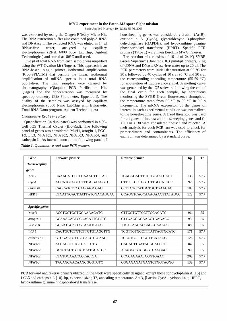
Table 1. Quantitative real-time PCR primers
Gene Forward primer Reverse primer bp T°
Housekeeping
genes
ActB CAAACATCCCCCAAAGTTCTAC TGAGGGACTTCCTGTAACCACT 135 57.7
CycA AGCATGTGGTCTTTGGGAAGGTG CTTCTTGCTGGTCTTGCCATTCC 92 57.7
GAPDH CACCATCTTCCAGGAGCGAG CCTTCTCCATGGTGGTGAAGAC 183 57.7
HPRT CTCATGGACTGATTATGGACAGGAC GCAGGTCAGCAAAGAACTTATAGCC 123 57.7
Specific genes
Murf1 ACCTGCTGGTGGAAAACATC CTTCGTGTTCCTTGCACATC 96 55
atrogin-1 GCAAACACTGCCACATTCTCTC CTTGAGGGGAAAGTGAGACG 93 55
PGC-1α GGAATGCACCGTAAATCTGC TTCTCAAGAGCAGCGAAAGC 88 55
LC3β CACTGCTCTGTCTTGTGTAGGTTG TCGTTGTGCCTTTATTAGTGCATC 171 57.7
cathepsin L GTGGACTGTTCTCACGTCCAAG TCCGTCCTTCGCTTCATAGG 128 57.7
NFATc1 ACCAGCTCTGCCATTGTG GAGACTTGATAGGGACCCC 84 55
NFATc2 GCTCTGCTGTTCTCATGGATGC ACAGGCGTCGGGTCAGGAC 99 55
NFATc2 CTGTGCAAACCCCACCTC GCCCAGAAATCGGTGAAC 209 57.7
NFATc4 TACAGCAACAAGCGGGTGTC CGGAGAGATGAGTCTGGTAGGG 139 57.7
PCR forward and reverse primers utilized in the work were specifically designed, except those for cyclophilin A [16] and LC3β and cathepsin L [10]. bp, expected size ; T°, annealing temperature. ActB, β-actin; CycA, cyclophilin a; HPRT, hypoxanthine guanine phosphoribosyl transferase.
was extracted by using the Qiagen RNeasy Micro Kit. The RNA extraction buffer also contained poly-A RNA and DNAase I. The extracted RNA was eluted in 14 µl RNase-free water, analyzed by capillary electrophoresis (RNA 6000 Pico LabChip, Agilent Technologies) and stored at -80°C until used.
Five µl of total RNA from each sample was amplified using the WT-Ovation kit (Nugen). This approach is an RNA-based, single primer isothermal amplification (Ribo-SPIATM) that permits the linear, isothermal amplification of mRNA species in a total RNA population. The final samples were cleaned by chromatography (Qiaquick PCR Purification Kit, Qiagen) and the concentration was measured by spectrophotometry (Bio Photometer, Eppendorf). The quality of the samples was assayed by capillary electrophoresis (6000 Nano LabChip with Eukaryotic Total RNA Nano program, Agilent Technologies)
Quantitative Real Time PCR
Quantification (in duplicates) was performed in a 96-well IQ5 Thermal Cycler (Bio-Rad). The following panel of genes was considered: Murf1, atrogin-1, PGC-1α, LC3, NFATc1, NFATc2, NFATc3, NFATc4, and cathepsin L. As internal control, the following panel of
housekeeping genes was considered : β-actin (ActB), cyclophilin A (CycA), glyceraldehyde 3-phosphate dehydrogenase (GAPDH), and hypoxanthine guanine phosphoribosyl transferase (HPRT). Specific PCR primers (Table 1) were from Eurofins MWG Operon.
The reaction mix consists of 10 µl of 2x iQ SYBR Green Supermix (Bio-Rad), 0.3 pmol/µl primers, 2 ng of cDNA and DNase/RNase-free water up to 20 µl. The PCR parameters were initial denaturation at 95 °C for 30 s followed by 40 cycles of 10 s at 95 °C and 30 s at the corresponding annealing temperature (55-59 °C) for acquisition of fluorescence signal. A melting curve was generated by the iQ5 software following the end of the final cycle for each sample, by continuous monitoring the SYBR Green fluorescence throughout the temperature ramp from 65 °C to 99 °C in 0.5 s increments. The mRNA expression of the genes of interest in each experimental condition was normalized to the housekeeping genes. A fixed threshold was used for all genes of interest and housekeeping genes and Ct < 10 or > 30 were considered “noise” and rejected. A melt analysis for each PCR run was used to check for primer-dimers and contaminants. The efficiency of each run was determined by a standard curve.
MYO experiment in the Foton-M3 space flight mission Basic Applied Myology 19 (2&3): 65-76, 2009
68
Electron microscopy
During the Foton-M3 mission, at the scheduled time, the fibers into two cell chambers devoted to morphological analyses, were treated with two subsequent additions of fixing solution (4% glutaraldehyde, 0.1 M sodium-cacodylate, pH 7.4). The concentration of glutaraldehyde was at least double that normally used in ultrastructural studies because of the known incomplete solution replacement of STROMA cell chambers (see Results section). A second consecutive treatment with the fixative (after 1 min) was expected to guarantee an almost complete fixation and preservation of fibers. After sample recovery, the fixing solution was washed out and muscle fibers were post fixed for 2 hour in 1% osmium tetroxide. The specimens were rapidly dehydrated in graded ethanol and acetone, infiltrated with EPON-acetone (1:1) mixture for 2 hours, and embedded in EPON. Ultrathin sections (40 nm) were cut with an LKB Ultrotome (LKB Produkter AB Stockholm Sweden) and a Drukker diamond knife (Drukker; Element Six The Netherlands), stained with 2% uranyl acetate and lead citrate. All sections were examined on a FEI Tecnai 12 electron microscope (FEI Company; USA) at 100 kV equipped with TVIPS digital camera (Tietz Video and Image Processing System).
Results Our research group was offered by the Italian Space
Agency (ASI), within the ASI LIFE program, the opportunity of exposing skeletal muscle fibers to microgravity conditions during the unmanned Russian Foton-M3 mission, scheduled on September 2007. This experiment, named MYO, utilized six pre-existing bioreactors (STROMA, Kayser Italia) and was aimed to determine the effects on skeletal muscle fibers of launch and landing mechanical solicitations and of 11 days space flight.
Preflight experiments and modifications of STROMA bioreactor
A number of preliminary experiments were carried out to set the ideal conditions to perform the experiments. We first considered how to adapt the STROMA bioreactor to the use of cultured adult muscle fibers. In fact, STROMA was originally designed to be used for bone cell studies and the cell chamber was designed to host a solid Skelite disk for the tridimensional culture of osteoclasts. In that experiment the hydrodynamic settings of the bioreactor was not a major issue. In contrast, in the MYO mission, high hydrostatic pressure is very critical for cultured muscle fibers. Thus a preliminary study was carried out to determine the actual operative conditions of the bioreactor.
In STROMA, the change of culture media is driven by a spring hosted on the rear of each cylinder that, when activated, transmits the motion to the piston to
push the fluid in and out of the culture chamber. We calculated that the force generated at the beginning of the injection was approximately of 2.5 atmospheres, a value expected to generate so high shear stresses on the cell chamber to cause the detachment of adherent muscle fibers. Moreover, by using tracer-marked fluids, we noted that the replacement of exhausted culture media was not complete (56% of the liquid only was renewed in a first change and 79% after the second change). Finally, by computational simulations, we calculated the linear velocity of the fluid in the conduit leading from the culture medium reservoir to the culture chamber as high as 0.4 ml/s, a velocity generating in the cell chamber high turbulence, not uniform flow profiles, and stagnant volumes of liquid, especially at the bottom of the chamber. To keep down the velocity of fluid exchanges, in order to reduce the shear stress over the cell adhesion surface, we interposed a nitrocellulose filter, supported by a highly porous plastic disc (to facilitate diffusion of fluids), just above the cell chamber in front to the inlet nozzles. This maneuver was without major effects on the amount of media exchange but significantly reduced the shear stress over muscle fibers.
Culture conditions of muscle fibers
Because of the expected intense mechanical stresses expected to be encountered by the bioreactors during Foton-M3 mission, particularly the intense vibrations during launch and landing of Foton-M3 capsule, we performed a set of experiments to determine the optimal adhesion substrate for the fibers into the bioreactor cell chamber. Among the diverse substrates tested, the Matrigel “cell basement mixture” (Becton Dickson), which is composed of laminin, collagen IV, heparan sulfate protoglycans, and entactin, resulted the more adequate to guarantee the full attachment of fibers to the incubation surface.
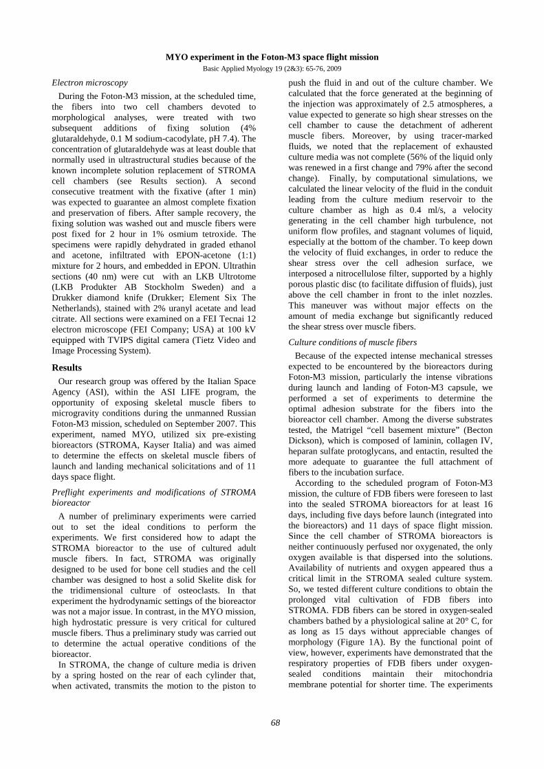
According to the scheduled program of Foton-M3 mission, the culture of FDB fibers were foreseen to last into the sealed STROMA bioreactors for at least 16 days, including five days before launch (integrated into the bioreactors) and 11 days of space flight mission. Since the cell chamber of STROMA bioreactors is neither continuously perfused nor oxygenated, the only oxygen available is that dispersed into the solutions. Availability of nutrients and oxygen appeared thus a critical limit in the STROMA sealed culture system. So, we tested different culture conditions to obtain the prolonged vital cultivation of FDB fibers into STROMA. FDB fibers can be stored in oxygen-sealed chambers bathed by a physiological saline at 20° C, for as long as 15 days without appreciable changes of morphology (Figure 1A). By the functional point of view, however, experiments have demonstrated that the respiratory properties of FDB fibers under oxygen-sealed conditions maintain their mitochondria membrane potential for shorter time. The experiments
MYO experiment in the Foton-M3 space flight mission Basic Applied Myology 19 (2&3): 65-76, 2009
69
Fig. 1 Prolonged culture of FDB fibers. A, FDB fibers were cultured in sealed-cells for at least two weeks.
Images were taken at the indicated time. An enlarged view after 11 days shows the preserved striations of the fiber. Note that apparently the fibers do not experience atrophy, as fiber diameter does not change during the incubation. B, FDB fibers were cultured for seven days in sealed wells were incubated with the membrane potential-sensitive dye tetramethyl rhodamine methyl ester (TMRM) that, after 30 minutes, localized to the mitochondria, indicating that mitochondria are vital. The depolarizing agent FCCP (carbonylcyanide-4-(trifluoromethoxy)-phenylhydrazone) dissipates the mitochondrial potential and the TMRM flows out the organelles with the concomitant reduction of the fluorescence. The inset shows the TMRM fiber staining just before depolarization with FCCP.
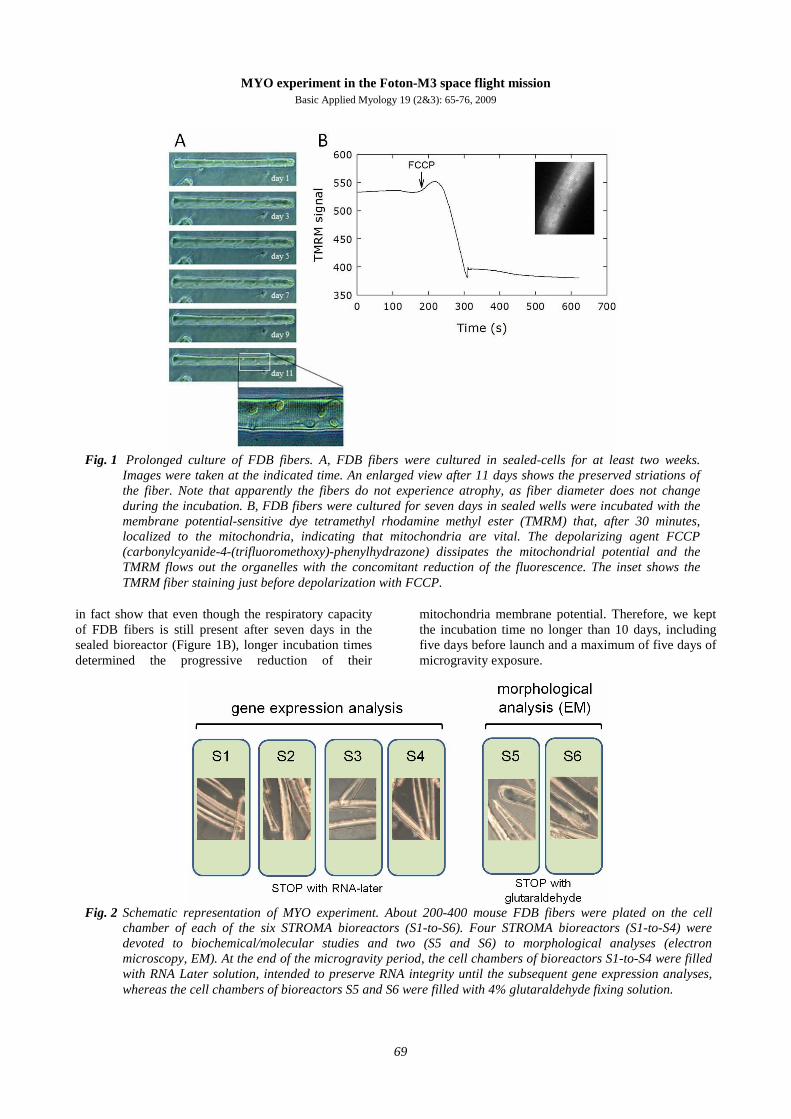
Fig. 2 Schematic representation of MYO experiment. About 200-400 mouse FDB fibers were plated on the cell
chamber of each of the six STROMA bioreactors (S1-to-S6). Four STROMA bioreactors (S1-to-S4) were devoted to biochemical/molecular studies and two (S5 and S6) to morphological analyses (electron microscopy, EM). At the end of the microgravity period, the cell chambers of bioreactors S1-to-S4 were filled with RNA Later solution, intended to preserve RNA integrity until the subsequent gene expression analyses, whereas the cell chambers of bioreactors S5 and S6 were filled with 4% glutaraldehyde fixing solution.
in fact show that even though the respiratory capacity of FDB fibers is still present after seven days in the sealed bioreactor (Figure 1B), longer incubation times determined the progressive reduction of their
mitochondria membrane potential. Therefore, we kept the incubation time no longer than 10 days, including five days before launch and a maximum of five days of microgravity exposure.

MYO experiment in the Foton-M3 space flight mission Basic Applied Myology 19 (2&3): 65-76, 2009
70
Fig. 3 Flow chart of MYO mission. FDB fibers were isolated at the Physiology laboratory of Vrije University of
Amsterdam and seeded in plastic disks (L-6d). Twenty-four hours after isolation (L-5d), the disks were integrated into STROMA bioreactors and in Biokon containers. Next day, the Biokon container was transported by an European Space Agency special flight to Baikonur Cosmodrome. The launch of Foton-M3 capsule, by means of a Soyuz rocket, occurred on September 14th. Four days after launch (L+4) the experiment was stopped by releasing appropriate solutions in the cell chambers (see Figure 2). The Foton-M3 capsule landed on September 26th (L+12), and the Biokon container was recovered the same day and transported back to Baikonur. The six bioreactors were then transported to Padova and inspected on October 1st. On the plot below is reported the temperature changes during the MYO experiments, as recorded by an internal STROMA bioreactors device
Fig. 4 Electron microscopy of FDB fibers flown on Foton-M3 capsule. A, on ground control; B, Foton-M3 fiber. G,
glycogen granules; T, triads; Mt, mitochondria
Foton-M3 mission
The MYO experiment of Foton-M3 mission used six STROMA bioreactors. Two main experiments were envisaged: 1) the analysis of genes related to atrophy
signaling pathways and protein degradation and 2) the analysis of morphological changes of muscle fibers caused by mechanical solicitations and exposure to microgravity. Four STROMA bioreactors were utilized

MYO experiment in the Foton-M3 space flight mission Basic Applied Myology 19 (2&3): 65-76, 2009
71
Fig. 5. Gene expression in FDB fibers flown on Foton-M3 capsule. Quantitative RT-PCR analysis of gene expression
was performed on RNA isolated from FDB muscle fibers immediately after being isolated (freshly isolated, three experiments), in FDB fibers at the end of on ground experiments (on ground, two experiments) carried out by reproducing all of the conditions of fibers that flown on board the Foton-M3 capsule (space flight). The results were normalized to the expression of the two housekeeping genes HPRT and CycA and to the expression values of FDB muscle (see Table 2). A, Expression of muscle-specific ligases atrogin-1 and MURF-1, members of the ubiquitin-proteasome system; B, cathepsin L and of LC3, members of the autophagy-lysosome pathway; C, NFATc1 and PGC-1α, activity-dependent genes.
for the molecular analyses and two for electron microscopy (Figure 2). To preserve RNA integrity in the cell chambers we utilized RNA Later, a high-salt solution able to preserve RNA for at least two weeks.
The MYO experiment started on September 8th, 2007, six days before launch (L-6) of Foton-M3 capsule (Figure 3). FDB fibers were isolated from C57BL/6J male mice at the muscle physiology laboratory of the Vrije University of Amsterdam. About 400 fibers were seeded on six Termanox coverslips that, after 24 hours, were inserted into the cell chamber of the bioreactors. These were then integrated into the Biokon container (#4 of ASI mission), transported to Baikonur Cosmodrome, and integrated into Foton-M3 capsule. The launch occurred 5 days after fiber isolation (September 18th, 2007) and the MYO experiment was stopped after 4 days of flight, by activating the appropriate STROMA cylinders. Landing of Foton-M3 capsule occurred on September 26, the capsule was recovered few hours later and delivered to Baikonur. Biokon containers were then transported to Amsterdam, the six STROMA bioreactors recovered and finally transported to our laboratory in Padova.
Morphological analyses
The morphological analysis was possible in few fibers only, most probably because fixation was not
complete and many fibers detached and were lost during the double exchange of the fixing solutions. It is possible that the mechanical solicitations of landing contributed to the detachment of partly fixed, and probably less adherent, FDB fibers. Electron microscopy shows that FDB fibers from the Foton-M3 mission preserved an apparently normal ultrastructural architecture, with regular myofibrils and aligned sarcomeres, mitochondria, and sarcoplasmic reticulum membranes (Figure 4). Granules of glycogen were still evident after 10 days permanence into the sealed bioreactors. This first observation indicate that the fibers that remained adherent in STROMA cell chamber, well tolerate the mechanical solicitations due to launch and landing and, apparently, microgravity per se does not cause evident morphological alterations.
Gene expression analyses
As a control for molecular analyses of space flight fibers (FDB-SF), we utilized both RNA from the FDB muscle (FDB-M), freshly-isolated FDB fibers (FDB-F) and from fibers that experienced on ground the same conditions of Foton-M3 mission (FDB-OG). Fibers in the FDB-OG group were isolated and included into six STROMA bioreactors and were incubated at temperatures that varied according to the changes recorded during the entire MYO experiment, from the first integration into the bioreactor until the final
MYO experiment in the Foton-M3 space flight mission Basic Applied Myology 19 (2&3): 65-76, 2009
72
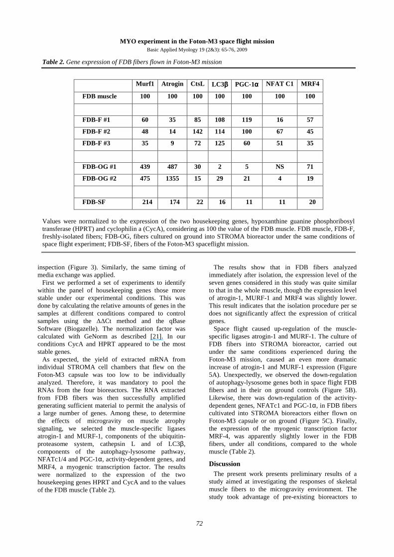
Table 2. Gene expression of FDB fibers flown in Foton-M3 mission
Murf1 Atrogin CtsL LC3ββββ PGC-1αααα NFAT C1 MRF4
FDB muscle 100 100 100 100 100 100 100
FDB-F #1 60 35 85 108 119 16 57
FDB-F #2 48 14 142 114 100 67 45
FDB-F #3 35 9 72 125 60 51 35
FDB-OG #1 439 487 30 2 5 NS 71
FDB-OG #2 475 1355 15 29 21 4 19
FDB-SF 214 174 22 16 11 11 20
Values were normalized to the expression of the two housekeeping genes, hypoxanthine guanine phosphoribosyl transferase (HPRT) and cyclophilin a (CycA), considering as 100 the value of the FDB muscle. FDB muscle, FDB-F, freshly-isolated fibers; FDB-OG, fibers cultured on ground into STROMA bioreactor under the same conditions of space flight experiment; FDB-SF, fibers of the Foton-M3 spaceflight mission.
inspection (Figure 3). Similarly, the same timing of media exchange was applied.
First we performed a set of experiments to identify within the panel of housekeeping genes those more stable under our experimental conditions. This was done by calculating the relative amounts of genes in the samples at different conditions compared to control samples using the ∆∆Ct method and the qBase Software (Biogazelle). The normalization factor was calculated with GeNorm as described [21]. In our conditions CycA and HPRT appeared to be the most stable genes.
As expected, the yield of extracted mRNA from individual STROMA cell chambers that flew on the Foton-M3 capsule was too low to be individually analyzed. Therefore, it was mandatory to pool the RNAs from the four bioreactors. The RNA extracted from FDB fibers was then successfully amplified generating sufficient material to permit the analysis of a large number of genes. Among these, to determine the effects of microgravity on muscle atrophy signaling, we selected the muscle-specific ligases atrogin-1 and MURF-1, components of the ubiquitin-proteasome system, cathepsin L and of LC3β, components of the autophagy-lysosome pathway, NFATc1/4 and PGC-1α, activity-dependent genes, and MRF4, a myogenic transcription factor. The results were normalized to the expression of the two housekeeping genes HPRT and CycA and to the values of the FDB muscle (Table 2).
The results show that in FDB fibers analyzed immediately after isolation, the expression level of the seven genes considered in this study was quite similar to that in the whole muscle, though the expression level of atrogin-1, MURF-1 and MRF4 was slightly lower. This result indicates that the isolation procedure per se does not significantly affect the expression of critical genes.
Space flight caused up-regulation of the muscle-specific ligases atrogin-1 and MURF-1. The culture of FDB fibers into STROMA bioreactor, carried out under the same conditions experienced during the Foton-M3 mission, caused an even more dramatic increase of atrogin-1 and MURF-1 expression (Figure 5A). Unexpectedly, we observed the down-regulation of autophagy-lysosome genes both in space flight FDB fibers and in their on ground controls (Figure 5B). Likewise, there was down-regulation of the activity-dependent genes, NFATc1 and PGC-1α, in FDB fibers cultivated into STROMA bioreactors either flown on Foton-M3 capsule or on ground (Figure 5C). Finally, the expression of the myogenic transcription factor MRF-4, was apparently slightly lower in the FDB fibers, under all conditions, compared to the whole muscle (Table 2).
Discussion The present work presents preliminary results of a
study aimed at investigating the responses of skeletal muscle fibers to the microgravity environment. The study took advantage of pre-existing bioreactors to
MYO experiment in the Foton-M3 space flight mission Basic Applied Myology 19 (2&3): 65-76, 2009
73
evaluate in cultured adult muscle fibers the direct effects of microgravity both on morphology and gene expression. In particular, we investigated, for the first time, possible changes of genes of the ubiquitin-proteasome system and autophagy/lysosomal pathway. In addition, activity-dependent and myogenic genes were also considered. The results show that the direct exposition to microgravity and space flight mechanical solicitations did not cause major modifications of single muscle fibers ultrastructure. By contrast, exposition to microgravity caused up-regulation of muscle-specific ligases atrogin-1 and MURF-1, and down regulation of the autophagy-lysosome members cathepsin L and LC3β, and of the activity-dependent genes NFATc1/4 and PGC-1α. On-ground control of the space flight experiment showed an even greater up-regulation of atrogin-1 and MURF-1, though on the whole the changes were similar to those of space flight fibers.
The reduced gravitational force is a major contributor to muscle atrophy in space. Reduced active and passive forces onto the muscle both activate the atrophy-related pathways. The balance between anabolism and catabolism determines the trophic state of a muscle and Akt is a key kinase associated with both skeletal muscle hypertrophy and atrophy [1;13]. In the diverse forms of skeletal muscle atrophy there is the activation of common programs, that downstream Akt comprise GSK-3β, mTOR and Foxo, and the prevalence of degradative processes [8]. Three major proteolytic pathways (Ca2+-dependent, lysosomal, and ubiquitin-proteasomal pathways) are active in skeletal muscle. In atrophying muscles, the ubiquitin-proteasome and the autophagic/lysosomal pathways regulate the accelerated degradation of myofibrillar proteins and muscle organelles. Both pathways are controlled by FoxO transcription factor and comprise the muscle-specific ligases atrogin-1 and MURF-1, and the autophagy-lysosome members cathepsin L and LC3β [8;18;22].
Early evidence demonstrated that spaceflight causes the reduction of muscle fiber cross sectional area and slow-to-fast transformation [2-5;7;11;15]. The expo-sure to microgravity causes, as in other forms of muscle atrophy, the accelerated breakdown of muscle proteins [6] and up-regulation of the ubiquitin ligase gene, MuRF-1 [10]. Spaceflight enhances degradation of rat myosin heavy chain in association with activation of ubiquitin-proteasome pathway. Moreover, spaceflight increased mRNA levels of cathepsin L, however, the cathepsin L inhibitor E64 was not able to prevent atrophy [6]. The exposure to microgravity also causes down-regulation of genes involved in regulation of muscle satellite cell replication [19] and imbalanced expression of mitochondrial genes [4]. A comprehen-sive study of the effects of microgravity on FoxO signaling pathways is however still missing.
As cellular model, we used enzymatically dissociated mouse flexor digitorum brevis (FDB) muscle fibers. The culture of adult fibers was carried out into pre-existing bioreactors, STROMA, originally designed for bone cell studies. In particular, since the cell chamber was designed to host a solid Skelite disk for the tridimensional culture of osteoclasts, the hydrodynamic characteristics of the bioreactor had to be adapted to the culture of muscle fibers. Importantly, all adaptive modifications of STROMA bioreactor and the overall experience represent the basis for the design of a novel bioreactor. Nevertheless, the use of isolated adult muscle fibers was expected to minimize the effects of orthostatic forces over the tissue and evaluate more direct microgravity effects on the fiber. In a previous study, cultures of avian muscle myotubes, differentiated from embryonic cells, were first tested under microgravity conditions [20]. Space flight caused atrophy to the spontaneously-contracting avian myotubes without affecting the rate of total muscle protein degradation, though with down-regulation of protein synthesis. This result was interpreted as a direct consequence of space flight to muscle cytoskeleton and metabolism [20]. Avian muscle myotubes are however immature muscle cells and cannot be considered mature myofibers. In contrast, cultured adult muscle fibers are a suitable model in medium-term experiments, including microgravity, as calcium handling and expression of mature proteins is maintained throughout a culture period of at least 8 days [13].
Our ultrastructural analysis shows that apparently there are no appreciable alterations in the muscle fiber organization, neither of myofibrils nor intracellular organelles (mitochondria and sarcotubular membra-nes). Since the FDB fibers flown on Foton-M3 capsule were subjected to the sole direct effects of microgravity and were not affected by the stresses of reentry and gravity reloading, our results seem to confirm a previous observation suggesting that reloading-induced-alterations represent the primary cause of the observed ultrastructural damage of the atrophic muscle exposed to microgravity [14].
The analysis of gene expression of space flight fibers shows a several-fold up-regulation of atrogin-1 and MURF-1, two muscle ligases regulating the ubiquitin-proteasome system [18]. MURF-1 was previously reported to be up-regulated under microgravity [10]. Surprisingly, we also found down-regulation of cathepsin L and LC3β expression in FDB fibers exposed to microgravity. These two genes of the autophagy/lysosomal signaling are usually up-regula-ted during muscle atrophy [11;22], and, consistently, up-regulation of cathepsin L was demonstrated after muscle exposure to microgravity [6]. This result suggests that the ubiquitin-proteasome system is the main degradation system activated in cultured
MYO experiment in the Foton-M3 space flight mission Basic Applied Myology 19 (2&3): 65-76, 2009
74
spaceflight fibers. Indeed, the cathepsin L inhibitor E64 was not able to prevent muscle atrophy of mice exposed to microgravity [6]. The spaceflight fibers also demonstrated down-regulation of activity-dependent genes, such as PGC-1α and NFATc1. On ground control fibers showed similar modification of all the genes considered. The only difference is related to the expression of atrogin-1 and MURF-1, more pronounced in the on ground control. However, exposition to microgravity of a large number of specimens will be necessary to confirm this finding. As reported in the Results section, the Foton-M3 mission has permitted to identify the more adequate measures and technical characteristics to design a novel bioreactor (Myobox). This should contain a large number of muscle fiber cultures on which it will be possible to test diverse experimental conditions and to obtain data of statistical relevance.
The main goal of our work was to explore for the first time the atrophy signaling pathway under reduced gravitational force. The final end is to determine whether treatment with proteasome inhibitors is able to mitigate the progression of muscle atrophy induced by microgravity. The culture of adult fibers into bioreactors could represent an ideal mean to screen suitable compounds capable to modulate muscle atrophy and prevent loss of muscle force during space flights. Our work has an obvious major fall-out on ground, since disclosing the signaling pathways involved in muscle atrophy can lead to the identification of potential therapeutic targets and to the development of new strategies to prevent muscle wasting and loss of muscle power resulting from aging, disuse and neuromuscular disorders.
Acknowledgements This work was supported by a grant to S.S. from ASI
(project OSMA) and by C.N.R. institutional funds. We thank V. Zollesi, P.L. Ganga, A. Donati and L. Petracchi of Kayser Italia for the skillful assistance with STROMA bioreactors during all phases of MYO mission. We thank Prof. G.J.M. Stienen for offering space and facilities in his laboratory at Vrije University, Amsterdam, for the isolation of the muscle fibers flown on Foton-M3 mission.
Address Correspondence to: Romeo Betto, CNR Institute of Neurosciences, c/o
Department of Biomedical Sciences, University of Padova. Viale Giuseppe Colombo 3, I-35121 Padova, Italy. Phone +39-049-8276027; fax +39-049-8276040 E-mail: [email protected]
References [1] Bodine SC, Stitt TN, Gonzalez M, Kline WO,
Stover GL, Bauerlein R, Zlotchenko E, Scrimgeour A, Lawrence JC, Glass DJ, Yancopoulos GD. Akt/mTOR pathway is a crucial regulator of skeletal muscle hypertrophy and can prevent muscle atrophy in vivo. Nat Cell Biol 2001; 3: 1014-1019.
[2] Falempin M, Mounier Y. Muscle atrophy associated with microgravity in rat: basic data for countermeasures. Acta Astronaut 1998; 42: 489-502
[3] Fitts RH, Riley DR, Widrick JJ. Functional and structural adaptations of skeletal muscle to microgravity. J Exp Biol 2001; 204: 3201-3208.
[4] Hansen G, Martinuk KJ, Bell GJ, MacLean IM, Martin TP, Putman CT. Effects of spaceflight on myosin heavy-chain content, fibre morphology and succinate dehydrogenase activity in rat diaphragm. Pflugers Arch 2004; 448: 239-247
[5] Harrison BC, Allen DL, Girten B, Stodieck LS, Kostenuik PJ, Bateman TA, Morony S, Lacey D, Leinwand LA. Skeletal muscle adaptations to microgravity exposure in the mouse. J Appl Physiol 2003; 95: 2462-2470.
[6] Ikemoto M, Nikawa T, Takeda S, Watanabe C, Kitano T, Baldwin KM, Izumi R, Nonaka I, Towatari T, Teshima S, Rokutan K, Kishi K. Space shuttle flight (STS-90) enhances degradation of rat myosin heavy chain in association with activation of ubiquitin-proteasome pathway. FASEB J 2001; 15: 1279-1281.
[7] Kraemer WJ, Staron RS, Gordon SE, Volek JS, Koziris LP, Duncan ND, Nindl BC, Gómez AL, Marx JO, Fry AC, Murray JD. The effects of 10 days of spaceflight on the shuttle Endeavor on predominantly fast-twitch muscles in the rat. Histochem Cell Biol 2000; 114: 349-355.
[8] Lecker SH, Jagoe RT, Gilbert A, Gomes M, Baracos V, Bailey J, Price SR, Mitch WE, Goldberg AL. Multiple types of skeletal muscle atrophy involve a common program of changes in gene expression. FASEB J 2004; 18: 39-51
[9] Mammucari C, Milan G, Romanello V, Masiero E, Rudolf R, Del Piccolo P, Burden SJ, Di Lisi R, Sandri C, Zhao J, Goldberg AL, Schiaffino S, Sandri M. FoxO3 controls autophagy in skeletal muscle in vivo. Cell Metab 2007; 6:458-471
[10] Nikawa T, Ishidoh K, Hirasaka K, Ishihara I, Ikemoto M, Kano M, Kominami E, Nonaka I, Ogawa T, Adams GR, Baldwin KM, Yasui N,
MYO experiment in the Foton-M3 space flight mission Basic Applied Myology 19 (2&3): 65-76, 2009
75
Kishi K, Takeda S. Skeletal muscle gene expression in space-flown rats. FASEB J 2004; 18: 522-524.
[11] Ohira Y, Yoshinaga T, Nomura T, Kawano F, Ishihara A, Nonaka I, Roy RR, Edgerton VR. Gravitational unloading effects on muscle fiber size, phenotype and myonuclear number. Adv Space Res 2002; 30: 777-781.
[12] Pallafacchina G, Calabria E, Serrano AL, Kalhovde JM, Schiaffino S. A protein kinase B-dependent and rapamycin-sensitive pathway controls skeletal muscle growth but not fiber type specification. Proc Nat Acad Sci USA 2002; 99: 9213-9218
[13] Ravenscroft G, Nowak KJ, Jackaman C, Clément S, Lyons MA, Gallagher S, Bakker AJ, Laing NG. Dissociated flexor digitorum brevis myofiber culture system - a more mature muscle culture system. Cell Motil Cytoskeleton 2007; 64: 727-738.
[14] Riley DA. Review of primary spaceflight-induced and secondary reloading-induced changes in slow antigravity muscles of rats. Adv Space Res 1998; 21: 1073-1075.
[15] Roy RR, Bodine SC, Pierotti DJ, Kim JA, Talmadge RJ, Barkhoudarian G, Fanton JW, Koslovskaya I, Edgerton VR. Fiber size and myosin phenotypes of selected Rhesus hindlimb muscles after a 14-day spaceflight. J Gravit Physiol 1999; 6: 55-62.
[16] Sanchez H, Chapot R, Banzet S, Koulmann N, Birot O, Bigard AX, Peinnequin A. Quantification by real-time PCR of developmental and adult myosin mRNA in rat
muscles. Biochim Biophys Res Commun 2006, 340: 165 -174
[17] Sandonà D, Danieli-Betto D, Germinario E, Biral D, Martinello T, Lioy A, Tarricone E, Betto R. The T-tubule membrane ATP-operated P2X4 receptor contractility of skeletal muscle. FASEB J 2005; 19: 1184-1186.
[18] Sandri M, Sandri C, Gilbert A, Skurk C, Calabria E, Picard A, Walsh K, Schiaffino S, Lecker SH, Goldberg AL. Foxo transcription factors induce the atrophy-related ubiquitin ligase atrogin-1 and cause skeletal muscle atrophy. Cell 2004; 117: 399-412.
[19] Taylor WE, Bhasin S, Lalani R, Datta A, Gonzalez-Cadavid NF. Alteration of gene expression profiles in skeletal muscle of rats exposed to microgravity during a spaceflight. J Gravit Physiol 2002; 9: 61-70.
[20] Vandenburgh H, Chromiak J, Shansky J, Del Tatto M, Lemaire J. Space travel directly induces skeletal muscle atrophy. FASEB J 1999; 13: 1031-1038.
[21] Vandesompele J, De Preter K, Pattyn F, Poppe B, Van Roy N, De Paepe A, Speleman F. Accurate normalization of real-time quantitative RT-PCR data by geometric averaging of multiple internal control genes. Genome Biol 2002; 3(r34): 1-11.
[22] Zhao J, Brault JJ, Schild A, Cao P, Sandri M, Schiaffino S, Lecker SH, Goldberg AL. FoxO3 coordinately activates protein degradation by the autophagic/lysosomal and proteasomal pathways in atrophying muscle cells. Cell Metab 2007; 6: 472-483.
Related Documents


![FOTON brochure I [v5] - ESAesamultimedia.esa.int/docs/foton/FOTON-M3_brochure.pdf · The Foton-M3 spacecraft will be carrying a European payload of 400 kg covering experiments in](https://static.cupdf.com/doc/110x72/5bcd298909d3f2c65e8c75ec/foton-brochure-i-v5-the-foton-m3-spacecraft-will-be-carrying-a-european.jpg)