Mark S. Pearce, PhD CT scan studies – present results and the future

Welcome message from author
This document is posted to help you gain knowledge. Please leave a comment to let me know what you think about it! Share it to your friends and learn new things together.
Transcript

Mark S. Pearce, PhD
CT scan studies – present results and
the future

CT scanning
A very useful, sometimes
lifesaving, tool
7 years from theory to first clinical use (1971)
8 further years to a Nobel prize (1979 to Allan
Cormack and Godfrey Hounsfield)

CT scan usage
Available worldwide at over 30,000 centres (and
continuing to increase)
11% of all medical imaging examinations in the
UK
68% of total collective dose to UK population from
medical x-ray examinations

Frequency of CT scans per year
1980 1985 1990 1995 2000 2005
V15
0.0
0.5
1.0
1.5
2.0
2.5
3.0
Year
CT
sc
an
s p
er
ye
ar
in t
he
UK
(m
illio
ns)
0
0.01
0.02
0.03
0.04
0.05UK
Nu
mb
er
of
CT
sc
an
s p
er
pers
on
/ y
ea
r
1980 1985 1990 1995 2000 2005 2010
Year
0
10
20
30
40
50
60
70
80
0.00
0.05
0.10
0.15
0.20
0.25
CT
sc
an
s p
er
ye
ar
in U
S (
millio
ns
)
Nu
mb
er
of
CT
scan
s p
er
/ p
ers
on
/ y
ear
Trends in CT usage

Early Fears
Two risk projection studies lead to much media interest
Brenner et al estimated that of the 1.6 million children in the
US who get CT scans to the head and abdomen each year,
about 1,500 will eventually die from a cancer induced by the
radiation of those scans.
Donnelly et al showed that too many CT scanners were
giving children adult-sized doses, often several times higher
than necessary.

Further risk projection studies
•Mostly extrapolated ‘expected’ doses and
‘expected’ cancer risks
•i.e. no empirical data
•Projections were often limited to certain scans,
mortality outcomes only and made assumptions
regarding modern protocol adjustments that may
not have been possible historically

Miglioretti et al (2013)
• Modelled the risks with childhood CT in seven US
healthcare systems
•Estimated both effective and organ doses
•Projected that with 4million CTs done in children in
the US per year, this would lead to 4870 excess
cancers.
•Reducing the doses to the highest 25% exposed
patients would prevent 43% of these cancers
Moving forward from predictions
Models using existing risk estimates are very
useful for publicising the need for radiation
protection and empirical research, but….
It is much better if we complement these studies by
direct observations of the relevant health effects in
populations that we want to protect.

The UK CT Scan Study
Long-term sequelae of radiation
exposure due to computed tomography
in childhood and early adulthood
Funders:
• US National Cancer Institute
• UK Department of Health

Cohort Study
Patients having one or more CT scans between
1985-2002
• First scanned aged <22 years
• Free from cancer at first CT
Radiology departments with available electronic RIS
data of sufficient quality
• Film / paper records from small number of Trusts
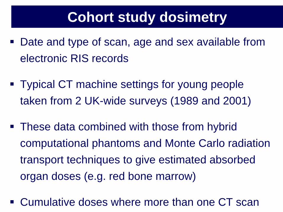
Cohort study dosimetry
Date and type of scan, age and sex available from
electronic RIS records
Typical CT machine settings for young people
taken from 2 UK-wide surveys (1989 and 2001)
These data combined with those from hybrid
computational phantoms and Monte Carlo radiation
transport techniques to give estimated absorbed
organ doses (e.g. red bone marrow)
Cumulative doses where more than one CT scan

Outcome data
RIS data linked with the NHSCR (1985-2008)
• Cancer incidence
• Mortality
• Loss-to-follow-up (e.g. notified emigrations)
Excluded patients with existing cancer and those diagnosed with leukaemia within 2 years of first CT scan (5 years for brain tumours)
• Sensitivity analyses with greater years of exclusion

Leukaemia dose-response

Brain dose-response

Sensitivity analyses
Excluding all scans in the 10 years prior to a
brain tumour diagnosis gave a higher dose-
response than in the original analysis
• i.e. the opposite to that expected if bias from CT
related to diagnosis was driving the findings
Little evidence of non-linearity of the dose-
response for either leukaemia or brain tumours

Main findings of the UK study
Significant associations between the estimated
radiation doses and subsequent incidence of
leukaemia and brain tumours
Assuming typical doses:
• 5-10 head CTs (≈50mGy to RBM) give an
estimated tripling of risk of leukaemia
• 2-3 head CTs (≈60mGy to the brain) give an
estimated tripling of risk of brain tumour

Strengths and weaknesses
We used empirical data
Cohort approach avoided recall bias (exposure
data from medical records)
Nationwide cancer registration (97%
ascertainment)
Used a careful approach to avoid those with
existing cancers

Strengths and weaknesses
Dosimetry was improved on previous estimates
• Provided organ doses
Uncertainties still exist
• Not expected to bias the findings
Unable to obtain individual-level parameter data
for such a large and historical cohort

The Australian CT Study
Cohort study of 10.9 million people identified
through Medicare
Patients aged under 20 years
Scans between 1985 and 2005
Exposed cohort: 680,211
Less detailed dosimetry than in the UK study
(and primarily based on effective doses)

The Australian CT Study
IRRs for all cancers fell with increasing lag times
1 year: IRR 1.24 (95% CI 1.20, 1.29)
5 years: IRR 1.21 (95% CI 1.16, 1.26)
10 years: IRR 1.18 (95% CI 1.11, 1.24)

The Australian CT Study
IRRs for specific cancers
• Raised IRRs for nearly all cancer types
• Including Hodgkin’s Lymphoma and melanoma
• Not including breast or lymphoid leukaemia

The Australian CT Study
Additional considerations
• Missing exposures from tertiary hospitals
• Leukaemia risks increased with age at exposure
• Brain and other solid tumours had high excess
rates within 5 years of first CT
• But, brain tumour incidence was still increased at 15
years from the first exposure

International collaboration
Similar studies were underway in:
• Canada, Sweden, Israel and France
EU-funded collaborative study (EPI-CT) began in
2011
New study underway in Brazil
Most studies are using a similar study design
and collaborations are underway re dosimetry

EPI-CT Objectives
Establish a large multinational European cohort of paediatric and young adult patients who received CT scans
Describe patterns of use of CTs over time and between countries
Develop individual estimates of organ-specific doses from paediatric CT scans using a unified improved method for dose estimation for paediatric and young adult patients
Evaluate the radiation-related risk of cancer in this cohort
Test biological markers of CT-irradiation effects (pilot study)
Develop methods to characterize quality of CT images in relation to the corresponding examination dose
Provide recommendations for a “harmonised” approach to CT dose optimisation for paediatric patients in Europe

EPI-CT: Estimated cohort size per country

CT scan epidemiology – the future
Further risk-based analyses of all cohorts,
including pooling of cohorts
Uncertainties analyses
Long-term follow-up of all the cohorts, and more
cohorts to be added
More national cancer registries throughout
Europe – covering all ages

CT scan epidemiology – the future
Need to establish registries of non-cancer
conditions, e.g. cataracts
Continued improvements in dosimetry and better
availability of indication data
More harmonised ethical approval systems

CT scan epidemiology – the future
Better and easier data linkage throughout
Europe
• Including links with other disease registries, e.g.
congenital anomalies
Do we need better guidelines?
• Certainly need to make sure that justification
guidelines are followed

Interpretation of the evidence so far
•The immediate benefits outweigh the (small) risks in
most settings when CT is used appropriately
•Of utmost importance is that, where CT is used, it should
only be used where fully justified from a clinical
perspective
Related Documents