CT Findings in Small CT Findings in Small Bowel Obstruction Bowel Obstruction Faisal Budhani Faisal Budhani Diagnostic Radiology Diagnostic Radiology PGY-3 Resident PGY-3 Resident

CT Findings in Small Bowel Obstruction Faisal Budhani Diagnostic Radiology PGY-3 Resident.
Dec 24, 2015
Welcome message from author
This document is posted to help you gain knowledge. Please leave a comment to let me know what you think about it! Share it to your friends and learn new things together.
Transcript

CT Findings in Small Bowel CT Findings in Small Bowel ObstructionObstruction
Faisal BudhaniFaisal Budhani
Diagnostic RadiologyDiagnostic Radiology
PGY-3 ResidentPGY-3 Resident

What is Important On-Call ?What is Important On-Call ?
Must answer the following questions:Must answer the following questions:1.1. Bowel obstruction: Y or NBowel obstruction: Y or N
2.2. Complete vs Partial ObstructionComplete vs Partial Obstruction
3.3. Location of Transition PointLocation of Transition Point
4.4. Closed Loop or Internal HerniaClosed Loop or Internal Hernia
5.5. Complications: perforation, strangulation and Complications: perforation, strangulation and ischemia ischemia
OutlineOutlineIntroductionIntroductionRole of CT in SBORole of CT in SBODiagnosis of SBODiagnosis of SBOLevel of ObstructionLevel of ObstructionDegree of ObstructionDegree of ObstructionCauses of SBOCauses of SBO– IntrinsicIntrinsic– Extrinsic Extrinsic – IntussusceptionIntussusception– Intraluminal Intraluminal – Closed-loopClosed-loop
Strangulation following SBOStrangulation following SBOManagement of SBOManagement of SBOWhat is Important On-Call ?What is Important On-Call ?

Introduction Introduction
Relatively common accounting for 20% of Relatively common accounting for 20% of all acute surgical admissionsall acute surgical admissionsDiagnosis based on history, physical signs Diagnosis based on history, physical signs and radiographic findingsand radiographic findingsSite and cause of SBO and presence of Site and cause of SBO and presence of strangulation must be determined to strangulation must be determined to ensure appropriate treatmentensure appropriate treatmentConventional radiology is first imaging Conventional radiology is first imaging modality with an accuracy of diagnosing modality with an accuracy of diagnosing presence of SBO 46-80%presence of SBO 46-80%
Role of CT in SBORole of CT in SBO
CT able to determine presence, level, CT able to determine presence, level, degree and cause of SBO and identify degree and cause of SBO and identify associated strangulationassociated strangulationCT able to depict pathology in bowel wall, CT able to depict pathology in bowel wall, mesentery, mesenteric vessels and mesentery, mesenteric vessels and peritoneal cavityperitoneal cavitySensitivity of CT in detecting high grade Sensitivity of CT in detecting high grade SBO is 78-100%SBO is 78-100%
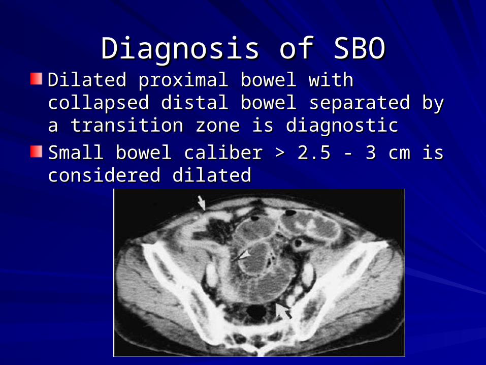
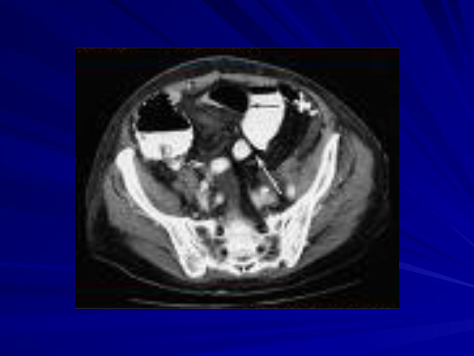
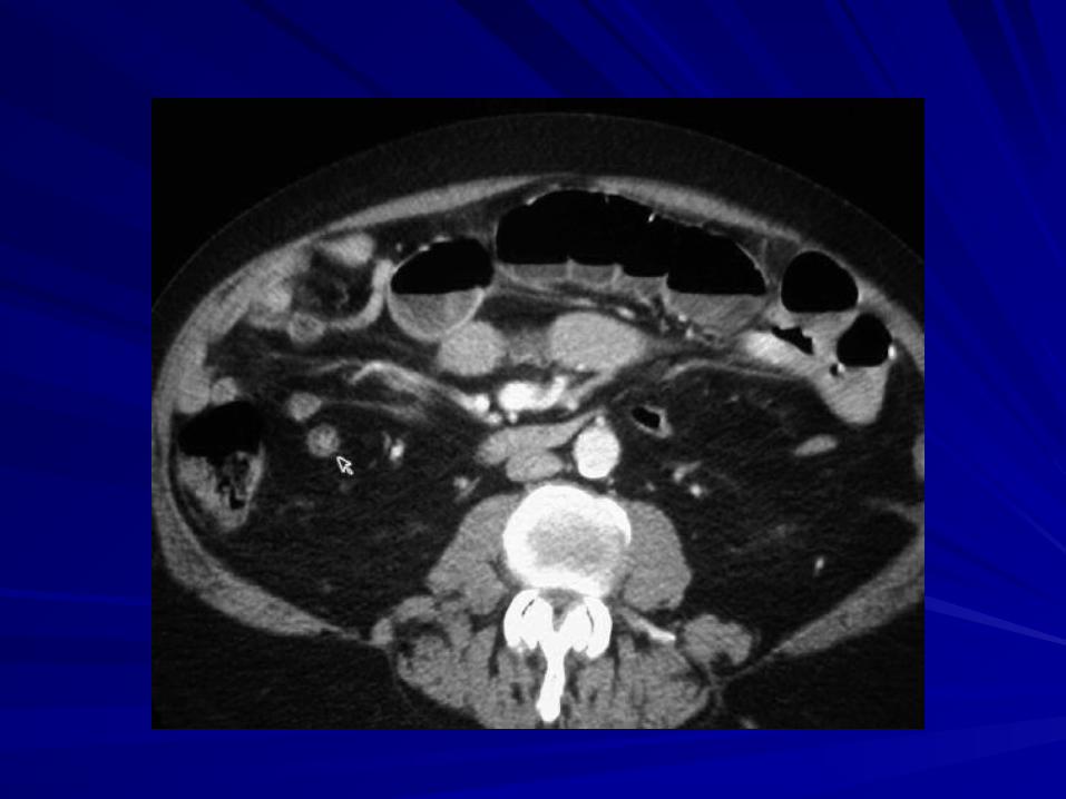
Diagnosis of SBODiagnosis of SBODilated proximal bowel with collapsed distal Dilated proximal bowel with collapsed distal bowel separated by a transition zone is bowel separated by a transition zone is diagnostic diagnostic
Small bowel caliber > 2.5 - 3 cm is considered Small bowel caliber > 2.5 - 3 cm is considered dilateddilated
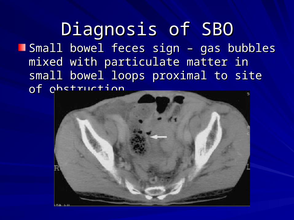
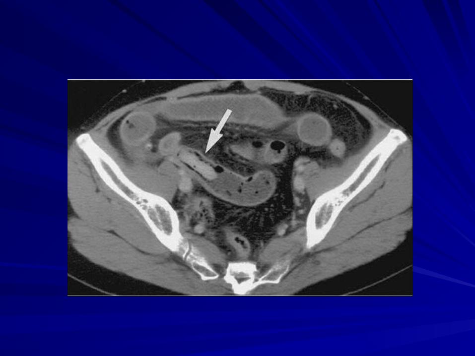
Diagnosis of SBODiagnosis of SBOSmall bowel feces sign – gas bubbles mixed Small bowel feces sign – gas bubbles mixed with particulate matter in small bowel loops with particulate matter in small bowel loops proximal to site of obstructionproximal to site of obstruction

Level of ObstructionLevel of Obstruction
Cannot be determined by intra-abdominal Cannot be determined by intra-abdominal location of transition zonelocation of transition zone
Dilated bowel loops migrate from their Dilated bowel loops migrate from their expected anatomic positionsexpected anatomic positions
Relative length of dilated versus collapsed Relative length of dilated versus collapsed bowel must be considered bowel must be considered

Degree of ObstructionDegree of Obstruction
Complete vs. Partial SBO based on Complete vs. Partial SBO based on degree of collapse and amount of residual degree of collapse and amount of residual contents distal to obstructioncontents distal to obstruction
Passage of oral contrast distal to the Passage of oral contrast distal to the transition zone always indicates partial transition zone always indicates partial obstructionobstruction
AdhesionsAdhesions
Responsible for more than half of all SBOResponsible for more than half of all SBOEtiology:Etiology:
Surgery Surgery 80% 80%Peritonitis Peritonitis 15% 15%Other (congenital, idiopathic) Other (congenital, idiopathic) 5% 5%
Adhesions may be single, multiple or Adhesions may be single, multiple or extensive extensive Not seen on CTNot seen on CT other causes of bowel other causes of bowel obstruction must be ruled out obstruction must be ruled out

HerniaHernia
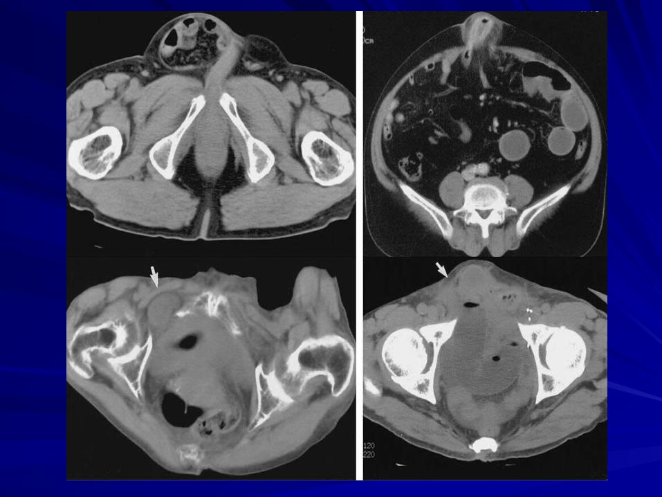
22ndnd most common cause of SBO (10%) most common cause of SBO (10%)External HerniaExternal Hernia– Prolapsed of viscera through defect in Prolapsed of viscera through defect in
abdominal/pelvic wallabdominal/pelvic wall– CT useful in detecting hernias in unsuspecting sites CT useful in detecting hernias in unsuspecting sites
and obese patients and obese patients
Internal HerniaInternal Hernia– herniation of bowel loops through developmentally or herniation of bowel loops through developmentally or
surgically created defect in peritoneum, omentum or surgically created defect in peritoneum, omentum or mesenterymesentery
– Less common than external herniasLess common than external hernias
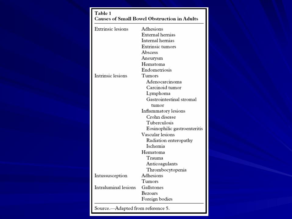
Other Extrinsic CausesOther Extrinsic Causes
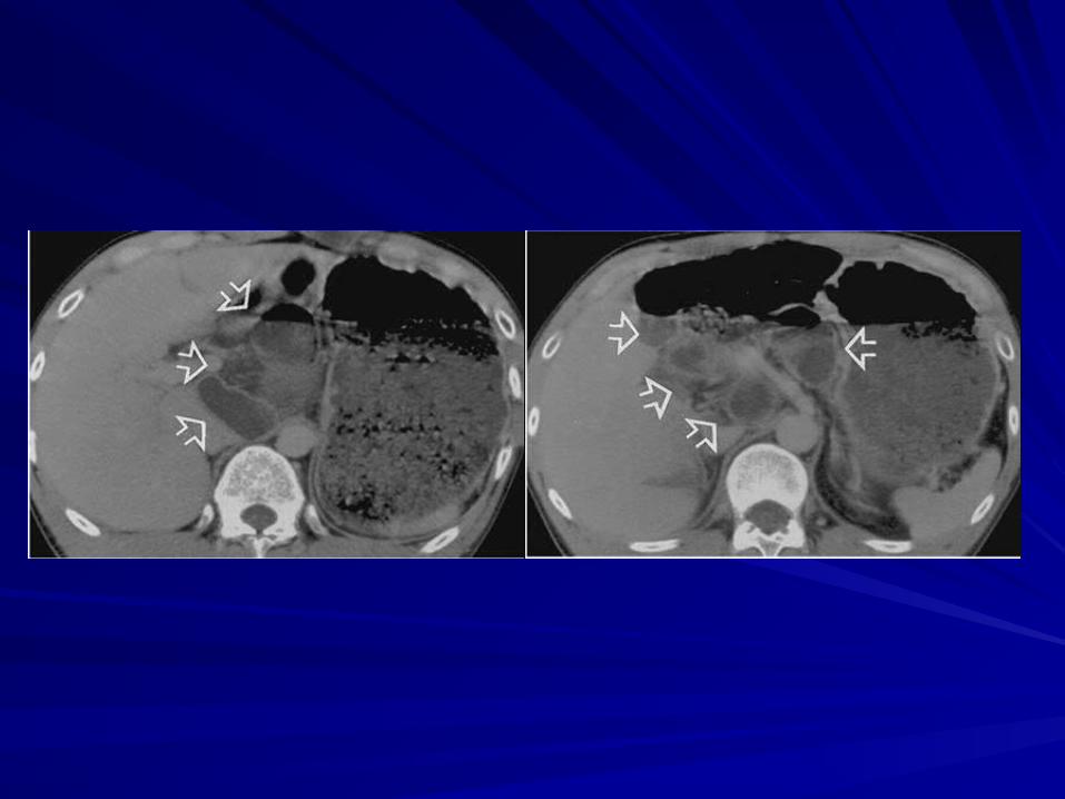
Variety of neoplastic, inflammatory or Variety of neoplastic, inflammatory or vascular lesions can cause SBO through vascular lesions can cause SBO through direct compression or desmoplastic direct compression or desmoplastic reactionreactionMost common extrinsic is peritoneal Most common extrinsic is peritoneal carcinomatosiscarcinomatosis
Multiple transitions zones of nodular wall Multiple transitions zones of nodular wall thickening thickening Mycobacterial infections, carcinoid and desmoid Mycobacterial infections, carcinoid and desmoid tumors have similar imaging findingstumors have similar imaging findings
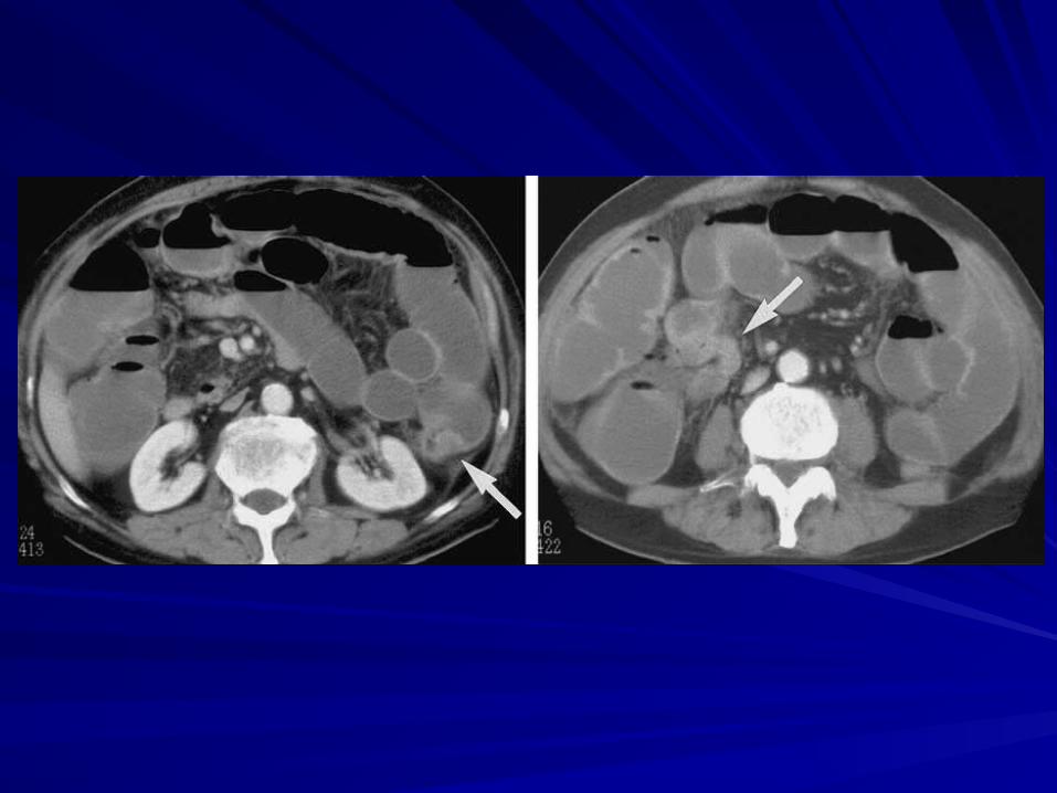
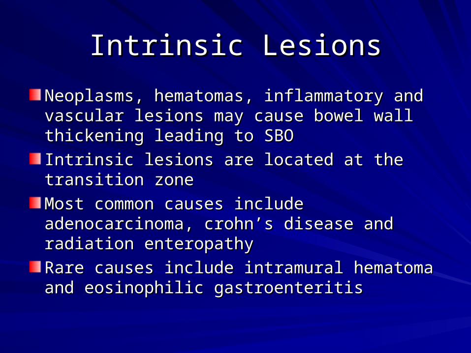
Intrinsic LesionsIntrinsic Lesions
Neoplasms, hematomas, inflammatory and Neoplasms, hematomas, inflammatory and vascular lesions may cause bowel wall vascular lesions may cause bowel wall thickening leading to SBOthickening leading to SBO
Intrinsic lesions are located at the transition Intrinsic lesions are located at the transition zone zone
Most common causes include adenocarcinoma, Most common causes include adenocarcinoma, crohn’s disease and radiation enteropathycrohn’s disease and radiation enteropathy
Rare causes include intramural hematoma and Rare causes include intramural hematoma and eosinophilic gastroenteritis eosinophilic gastroenteritis

IntussusceptionIntussusception
Relatively rare cause of adult SBO (5%)Relatively rare cause of adult SBO (5%)
Unlike infants, 80% of cases caused by Unlike infants, 80% of cases caused by underlying neoplasm, adhesion, inverted underlying neoplasm, adhesion, inverted Meckel’s, foreign body or previous surgery Meckel’s, foreign body or previous surgery serves as lead point serves as lead point
Collapsed proximal segment (intussusceptum) Collapsed proximal segment (intussusceptum) with its mesenteric fat and vessels within the with its mesenteric fat and vessels within the wall of the distal bowel (intussuscipiens) wall of the distal bowel (intussuscipiens) characteristic target sign on axial imagescharacteristic target sign on axial images

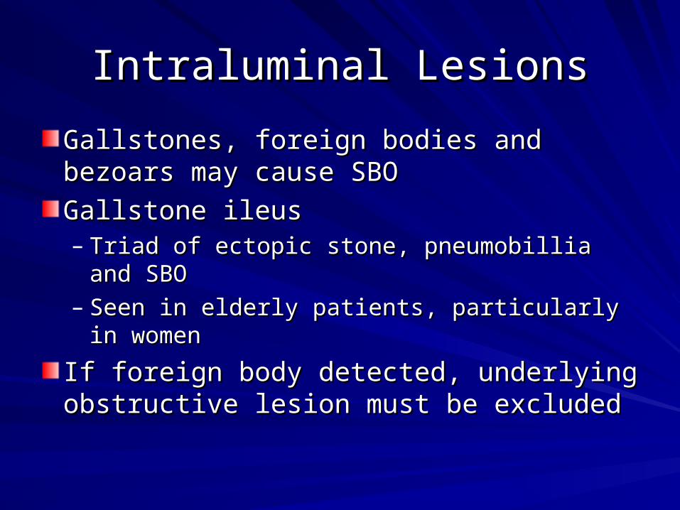
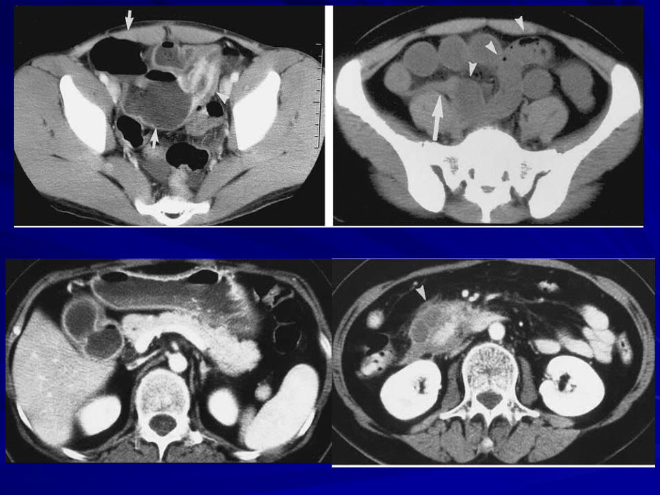
Intraluminal LesionsIntraluminal Lesions
Gallstones, foreign bodies and bezoars Gallstones, foreign bodies and bezoars may cause SBOmay cause SBO
Gallstone ileusGallstone ileus– Triad of ectopic stone, pneumobillia and SBOTriad of ectopic stone, pneumobillia and SBO– Seen in elderly patients, particularly in womenSeen in elderly patients, particularly in women
If foreign body detected, underlying If foreign body detected, underlying obstructive lesion must be excludedobstructive lesion must be excluded

Closed Loop Obstruction Closed Loop Obstruction
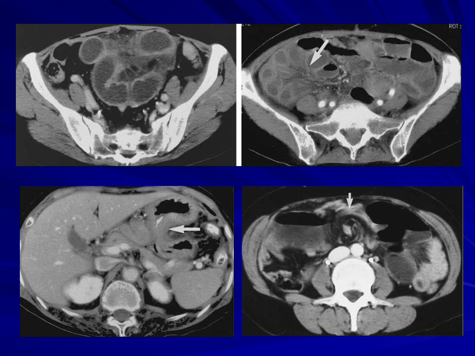
2 points of bowel obstructed at a single site2 points of bowel obstructed at a single site
Most often caused by adhesive bands; external Most often caused by adhesive bands; external and internal hernias less commonand internal hernias less common
Tends to involve the mesentery Tends to involve the mesentery prone to prone to volvulus volvulus
C-shaped/U-shaped loop of bowel with vessels C-shaped/U-shaped loop of bowel with vessels converging towards site of torsionconverging towards site of torsion
2 adjacent collapsed loops with interposed 2 adjacent collapsed loops with interposed dilated fluid filled boweldilated fluid filled bowel

StrangulationStrangulation
Mechanical obstruction associated with bowel Mechanical obstruction associated with bowel ischemiaischemiaMajority of cases associated with closed-loop Majority of cases associated with closed-loop obstructionobstructionWall thickening (“halo sign”), mesenteric Wall thickening (“halo sign”), mesenteric hazziness, pneumatosis and portal venous gashazziness, pneumatosis and portal venous gasWith IV contrast: lack of enhancement, With IV contrast: lack of enhancement, asymmetric enhancement or delayed asymmetric enhancement or delayed enhancement of bowel wallenhancement of bowel wallCT detection rate of strangulation is 63-100%CT detection rate of strangulation is 63-100%

Management Management
Acute complete SBO Acute complete SBO surgical surgical Partial SBO Partial SBO conservative conservativeFollow up imaging recommended (CT or small bowel Follow up imaging recommended (CT or small bowel enteroclysis) in indeterminate cases) in indeterminate casesClosed loop obstruction in absence of ischemia is a Closed loop obstruction in absence of ischemia is a surgical emergency as it can progress to strangulation surgical emergency as it can progress to strangulation Risk of strangulation in compete SBO increases with Risk of strangulation in compete SBO increases with timetime
Surgery within 36 hrs Surgery within 36 hrs mortality rate = 8% mortality rate = 8%Surgery after 36 hrs Surgery after 36 hrs mortality rate = 25% mortality rate = 25%
Exploratory laparotomy recommended in all patients with Exploratory laparotomy recommended in all patients with closed loop or signs of ischemiaclosed loop or signs of ischemia– If CT findings not in keep with clinical presentation, patients must If CT findings not in keep with clinical presentation, patients must
undergo laparotomy undergo laparotomy

What is Important On-Call ?What is Important On-Call ?
Must answer the following questions:Must answer the following questions:1.1. Bowel obstruction: Y or NBowel obstruction: Y or N
2.2. Complete vs Partial ObstructionComplete vs Partial Obstruction
3.3. Location of Transition PointLocation of Transition Point
4.4. Closed Loop or Internal HerniaClosed Loop or Internal Hernia
5.5. Complications: perforation, strangulation and Complications: perforation, strangulation and ischemia ischemia
Related Documents


![Welcome [weillcornellbrainandspine.org] · Maricruz Rivera, MD, PhD PGY-3. Neurological Surgery Residents. Evan Bander, MD PGY-5 Alexander D. Ramos, MD, PhD PGY-5 Joseph Carnevale,](https://static.cupdf.com/doc/110x72/5f7167444c714e55d46f024a/welcome-weill-maricruz-rivera-md-phd-pgy-3-neurological-surgery-residents.jpg)