HAL Id: hal-01945281 https://hal.archives-ouvertes.fr/hal-01945281 Submitted on 12 Feb 2019 HAL is a multi-disciplinary open access archive for the deposit and dissemination of sci- entific research documents, whether they are pub- lished or not. The documents may come from teaching and research institutions in France or abroad, or from public or private research centers. L’archive ouverte pluridisciplinaire HAL, est destinée au dépôt et à la diffusion de documents scientifiques de niveau recherche, publiés ou non, émanant des établissements d’enseignement et de recherche français ou étrangers, des laboratoires publics ou privés. csrB Gene Duplication Drives the Evolution of Redundant Regulatory Pathways Controlling Expression of the Major Toxic Secreted Metalloproteases in Vibrio tasmaniensis LGP32 an Ngoc Nguyen, Elena Disconzi, Guillaume Charrière, Delphine Destoumieux-Garzón, Philippe Bouloc, Frédérique Le Roux, Annick Jacq To cite this version: an Ngoc Nguyen, Elena Disconzi, Guillaume Charrière, Delphine Destoumieux-Garzón, Philippe Bouloc, et al.. csrB Gene Duplication Drives the Evolution of Redundant Regulatory Pathways Controlling Expression of the Major Toxic Secreted Metalloproteases in Vibrio tasmaniensis LGP32. MSphere, American Society for Microbiology., 2018, 3 (6), pp.e00582-18. 10.1128/mSphere.00582-18. hal-01945281

Welcome message from author
This document is posted to help you gain knowledge. Please leave a comment to let me know what you think about it! Share it to your friends and learn new things together.
Transcript
HAL Id: hal-01945281https://hal.archives-ouvertes.fr/hal-01945281
Submitted on 12 Feb 2019
HAL is a multi-disciplinary open accessarchive for the deposit and dissemination of sci-entific research documents, whether they are pub-lished or not. The documents may come fromteaching and research institutions in France orabroad, or from public or private research centers.
L’archive ouverte pluridisciplinaire HAL, estdestinée au dépôt et à la diffusion de documentsscientifiques de niveau recherche, publiés ou non,émanant des établissements d’enseignement et derecherche français ou étrangers, des laboratoirespublics ou privés.
csrB Gene Duplication Drives the Evolution ofRedundant Regulatory Pathways Controlling Expressionof the Major Toxic Secreted Metalloproteases in Vibrio
tasmaniensis LGP32an Ngoc Nguyen, Elena Disconzi, Guillaume Charrière, Delphine
Destoumieux-Garzón, Philippe Bouloc, Frédérique Le Roux, Annick Jacq
To cite this version:an Ngoc Nguyen, Elena Disconzi, Guillaume Charrière, Delphine Destoumieux-Garzón, PhilippeBouloc, et al.. csrB Gene Duplication Drives the Evolution of Redundant Regulatory PathwaysControlling Expression of the Major Toxic Secreted Metalloproteases in Vibrio tasmaniensis LGP32.MSphere, American Society for Microbiology., 2018, 3 (6), pp.e00582-18. �10.1128/mSphere.00582-18�.�hal-01945281�
csrB Gene Duplication Drives the Evolution of RedundantRegulatory Pathways Controlling Expression of the MajorToxic Secreted Metalloproteases in Vibrio tasmaniensis LGP32
An Ngoc Nguyen,a,b Elena Disconzi,a Guillaume M. Charrière,c Delphine Destoumieux-Garzón,c Philippe Bouloc,a
Frédérique Le Roux,d,e Annick Jacqa
aInstitute for Integrative Biology of the Cell (I2BC), CEA, CNRS, Univ. Paris‐Sud, Université Paris‐Saclay,Gif‐sur‐Yvette, France
bBiotechnology Department, Institute of Biotechnology and Food Technology, Industrial University of Ho ChiMinh City, Ho Chi Minh City, Viet Nam
cInteractions Hôtes-Pathogènes-Environnements, Université de Montpellier, CNRS, Ifremer, Université dePerpignan Via Domitia, Montpellier, France
dIfremer, Unité Physiologie Fonctionnelle des Organismes Marins, Plouzané, FranceeSorbonne Universités, UPMC Paris 06, CNRS, UMR 8227, Integrative Biology of Marine Models, StationBiologique de Roscoff, Roscoff, France
ABSTRACT CsrBs are bacterial highly conserved and multiple-copy noncoding smallRNAs (sRNAs) that play major roles in cell physiology and virulence. In the Vibrio ge-nus, they are known to be regulated by the two-component system VarS/VarA. Theymodulate the well-characterized quorum sensing pathway controlling virulence andluminescence in Vibrio cholerae and Vibrio harveyi, respectively. Remarkably, Vibriotasmaniensis LGP32, an oyster pathogen that belongs to the Splendidus clade, wasfound to have four copies of csrB, named csrB1-4, compared to two to three copiesin other Vibrio species. Here, we show that the extra csrB4 copy results from acsrB3 gene duplication, a characteristic of the Splendidus clade. Interestingly, csrBgenes are regulated in different ways in V. tasmaniensis, with csrB1 expressionbeing independent of the VarS/VarA system. We found that a complex regulatorynetwork involving CsrBs, quorum sensing, and the stationary-phase sigma factor�S redundantly but differentially controls the production of two secreted metal-loproteases, Vsm and PrtV, the former being a major determinant of the V. tas-maniensis extracellular product toxicity. In particular, we identified a novel VarS/VarA-dependent but CsrB-independent pathway that controls positively bothVsm production and PrtV production as well as rpoS expression. Altogether, ourdata show that a csrB gene duplication event in V. tasmaniensis supported theevolution of the regulatory network controlling the expression of major toxic se-creted metalloproteases, thereby increasing redundancy and enabling the inte-gration of additional input signals.
IMPORTANCE The conserved CsrB sRNAs are an example of sibling sRNAs, i.e.,sRNAs which are present in multiple copies in genomes. This report illustrates hownew copies arise through gene duplication events and highlights two evolutionaryadvantages of having such multiple copies: differential regulation of the multiplecopies allows integration of different input signals into the regulatory network ofwhich they are parts, and the high redundancy that they provide confers a strongrobustness to the system.
KEYWORDS bacterial gene regulation, bacterial sRNAs, transcriptomics, Vibriopathogenic to oysters, host-pathogen interactions
Received 24 October 2018 Accepted 5November 2018 Published 28 November2018
Citation Nguyen AN, Disconzi E, Charrière GM,Destoumieux-Garzón D, Bouloc P, Le Roux F,Jacq A. 2018. csrB gene duplication drives theevolution of redundant regulatory pathwayscontrolling expression of the major toxicsecreted metalloproteases in Vibriotasmaniensis LGP32. mSphere 3:e00582-18.https://doi.org/10.1128/mSphere.00582-18.
Editor Craig D. Ellermeier, University of Iowa
Copyright © 2018 Nguyen et al. This is anopen-access article distributed under the termsof the Creative Commons Attribution 4.0International license.
Address correspondence to Annick Jacq,[email protected].
A.N.N. and E.D. contributed equally to thisarticle.
RESEARCH ARTICLEMolecular Biology and Physiology
crossm
November/December 2018 Volume 3 Issue 6 e00582-18 msphere.asm.org 1
on Decem
ber 5, 2018 by guesthttp://m
sphere.asm.org/
Dow
nloaded from
Bacterial regulatory small RNAs (sRNAs), often noncoding, are now recognized ascrucial regulators in adaptation to environment and hosts (reviewed in reference 1).
trans-Encoded sRNAs can be classified into two main categories. The first categorycomprises regulatory RNAs that target mRNAs, and the second category encompassessRNAs that target proteins (reviewed in reference 2).
Within the second category, sRNAs from the CsrB family in gammaproteobacteriaare 100 to more than 400 nucleotides (nt) in length. CsrBs bind to the posttranscrip-tional regulator CsrA and titrate its activity (3). CsrA, in most cases, inhibits translationand induces degradation of various mRNA targets involved in carbon metabolism,motility, biofilm formation, secondary metabolite production, quorum sensing (QS),and virulence, depending on the species (see references 4, 5, and 6 for reviews). As atranslational inhibitor, CsrA binds to Shine-Dalgarno (SD) sequences, thus preventingribosome loading (7, 8). However, in a few cases, CsrA works as an activator of geneexpression (9–11). CsrB secondary structures contain stem-loop structures with severalCsrA binding motifs (AGGA/ARGGA [where “R” stands for T/C/G]) exposed in the loops.As each CsrB has numerous motifs, one CsrB molecule binds and titrates many CsrAmolecules, therefore competing with CsrA targets (3). CsrB expression in Escherichia coliis activated by the two-component system (TCS) BarA/UvrY (homologous to GacS/GacAin Pseudomonas aeruginosa and Vibrio fischeri and to VarS/VarA in Vibrio cholerae). In V.cholerae, three copies of CsrB sRNAs (named CsrB, CsrC, and CsrD) have been shown tomodulate QS through the indirect action of CsrA on the regulatory cascade controllingthe production of the master regulator HapR (12). CsrA is essential for virulence in V.cholerae (13). In V. fisheri, CsrA modulates luminescence and indirectly controls squidcolonization (14–16).
CsrBs are generally expressed from genes in multicopies in bacterial genomes. SuchsRNAs have been named “sibling sRNAs” (17). Other examples include RyhB, a sRNAregulated by iron deficiency, whose gene is present in two copies in Yersina species; OmrAand OmrB in E. coli, which control iron acquisition, curli formation, and motility; and csRNAspresent in two to six copies in Streptococcus sp. (17). The csrB (also known as rsm) geneshave been identified in most Gram-negative bacteria, usually in two to three copies, and arethe most widely distributed multiple-copy sRNA genes. In addition, most sequencedVibrionaceae spp. express Qrr sRNAs from multiple-copy (four to five) genes. Qrr sRNAsinhibit expression of major QS transcription factor HapR in V. cholerae and of its homologueLuxR in V. harveyi and therefore are essential QS mediators (18).
Vibrio tasmaniensis LGP32 (formerly V. splendidus LGP32) is a member of theSplendidus clade that has been isolated from oyster suffering from summer mortalityevents threatening the sustainability of the French oyster-growing industry (19, 20).Although important progress has been made in our understanding of the mechanismsunderlying virulence in this emerging pathogen (21–26), still, little is known about theirregulation. LGP32 expresses two secreted metalloproteases (Vsm and PrtV/InhA), Vsmbeing the major determinant of LGP32 extracellular products (ECPs) when they are injectedinto oysters (21, 24). Vsm is a zinc-containing metalloprotease exhibiting 67.7% identitywith V. cholerae hemagglutinin/protease HapA, and V. tasmaniensis PrtV/InhA (VS_II1062)shares 72.5% identity with V. cholerae PrtV. Regulatory pathways controlling their expres-sion are still unknown, whereas their homologs in V. cholerae were previously shown to beunder the control of the HapR master regulator (27, 28). Exploring more-diverse models isa prerequisite for addressing the issue of regulatory network remodeling during Vibrioevolution and its possible involvement in virulence emergence.
Unlike other Vibrio species, which have two to three csrB gene copies, LGP32 hasfour copies (29). So far, among Vibrionaceae, only Photobacterium profundum was foundto have four putative csrB gene copies (30). As expected for csrB genes, all four csrBgenes in V. tasmaniensis were upregulated at high cell density. They were also found tobe among the most highly expressed genes in the genome, suggesting an importantrole in cell physiology (29).
One issue that arises is that of why there are so many copies of CsrB genes (17). Toaddress this central issue, we analyzed the properties of the regulatory network
Nguyen et al.
November/December 2018 Volume 3 Issue 6 e00582-18 msphere.asm.org 2
on Decem
ber 5, 2018 by guesthttp://m
sphere.asm.org/
Dow
nloaded from

involving CsrBs in V. tasmaniensis. Specifically, we asked how new csrB copies arose,what advantages they confer to the cell, and whether they are functionally redundantand regulated in the same way. Our data show that a csrB gene duplication event in theSplendidus clade led to the integration of new input signals and to additional layers ofredundancy in the regulation of vsm and prtV expression.
RESULTSThe evolution of CsrBs within vibrios. In V. tasmaniensis LGP32, we identified four
copies of csrB (29). This prompted us to investigate the evolution of csrB copies in theVibrionaceae. Differences in copy numbers in different species can result from gains orlosses of csrB genes. In the case of a gain, we could envisage two scenarios to generateadditional copies: they could have arisen from gene duplication events (17), or theycould have resulted from horizontal transfers.
To understand how multiple Vibrio csrB copies have evolved, we selected from theNCBI database 13 Vibrio and Aliivibrio sp. fully assembled genomes. By successive BLASTsearches starting from the four copies of csrB identified in V. tasmaniensis, we estab-lished a catalogue of the csrB genes present in these genomes. The results confirmedthat, in general, Vibrio spp. have three copies of the csrB gene. The list of 38 csrB geneswas complemented by a copy from P. profundum, which was used as an outgroup inour phylogeny.
Since homologous gene copies in different species are more likely to have con-served synteny than copies resulting from gene duplication (i.e. paralogs) or horizontaltransfer (xenologs), we first examined synteny for each csrB gene to determine theorthologs. We thus identified 13 conserved synteny groups (described in Tables S1 toS4 in the supplemental material) corresponding to the gene clusters labeled a to m inFig. 1. We also observed some partial changes of synteny, with rearrangement ofsynteny blocks leading to the formation of composite groups in the vicinity of somecsrB genes (noted by a combination of two letters). Each csrB gene copy was thenassigned to one or two synteny groups according to its genomic environment (TablesS1 to S4). Neither V. tasmaniensis csrB3 nor the fourth copy, csrB4, had conservedsynteny with the other species’ csrB genes. In addition, csrB4 appeared to be presentonly in the Splendidus clade (i.e., Vibrio cyclitrophicus and Vibrio crassostreae strains).Overall, we identified five groups of csrB genes; within each group, each csrB com-pletely or partially shared synteny with at least one other member of the group (Fig. 1;see also Tables S1 to S4). In the case of V. cholerae, we used the names attributed byLenz et al, i.e., csrB, csrC, and csrD (12), whereas other copies were named according totheir synteny groups. Members of the first group, comprising V. tasmaniensis csrB1 andV. cholerae csrC, are all in the vicinity of gene cluster a. A second synteny group,characterized by gene cluster g, comprises V. tasmaniensis csrB2 and V. cholerae csrB.The third synteny group is more composite in nature and consists of a combination ofgene clusters i, j, k, and l. It includes V. tasmaniensis csrB3 and V. cholerae csrD. Thesethree groups are specific to the Vibrio strains. Aliivibrio csrB copies were assigned to twosynteny groups, the ef group (csrB1) and the m group (csrB2). Members of the ef grouphave partial synteny with V. nigripulchritudo csrB1, sharing cluster e. In Aeromonassalmonicida, a third copy, csrB3, is located next to csrB2 and seems to result from a geneduplication of the latter (see below).
To establish the evolutionary relationship between those csrB genes, we constructedtheir phylogenetic tree using the PhyML maximum likelihood method (31) (Fig. 1) aswell as the multilocus sequence analysis (MLSA) tree of the corresponding Vibrio andAliivibrio strains, using three housekeeping genes (see Materials and Methods). Wefound that Vibrio csrB genes are in general phylogenetically closer to their orthologs inother species (i.e., to those having conserved synteny) than to their paralogs in thesame species. Two exceptions were A. salmonicida csrB3, which is closer to A. salmoni-cida csrB2 than to the other csrB genes, suggesting that it corresponds to a recentduplication of csrB2 in this species, and V. nigripulchritudo csrB2, which clusters with V.nigripulchritudo csrB1. However, V. nigripulchritudo csrB2 shares synteny with csrB2 of
V. tasmaniensis CsrB sRNAs Control Production of Vsm
November/December 2018 Volume 3 Issue 6 e00582-18 msphere.asm.org 3
on Decem
ber 5, 2018 by guesthttp://m
sphere.asm.org/
Dow
nloaded from
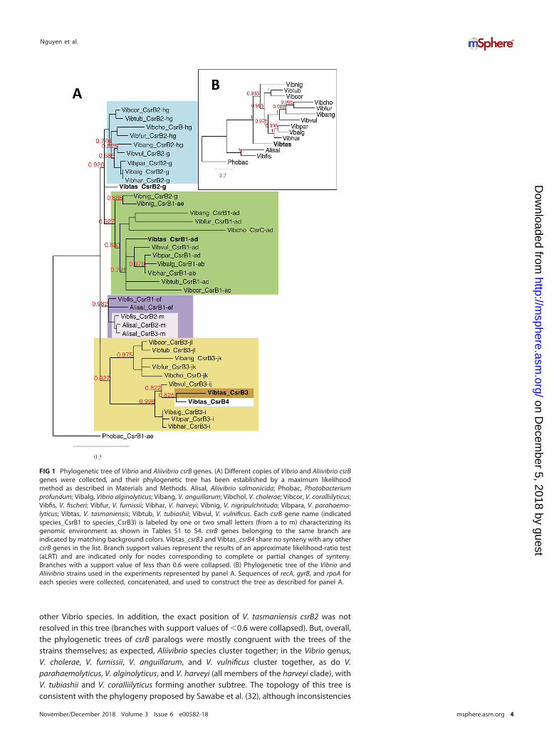
other Vibrio species. In addition, the exact position of V. tasmaniensis csrB2 was notresolved in this tree (branches with support values of �0.6 were collapsed). But, overall,the phylogenetic trees of csrB paralogs were mostly congruent with the trees of thestrains themselves; as expected, Aliivibrio species cluster together; in the Vibrio genus,V. cholerae, V. furnissii, V. anguillarum, and V. vulnificus cluster together, as do V.parahaemolyticus, V. alginolyticus, and V. harveyi (all members of the harveyi clade), withV. tubiashii and V. coralliilyticus forming another subtree. The topology of this tree isconsistent with the phylogeny proposed by Sawabe et al. (32), although inconsistencies
FIG 1 Phylogenetic tree of Vibrio and Aliivibrio csrB genes. (A) Different copies of Vibrio and Aliivibrio csrBgenes were collected, and their phylogenetic tree has been established by a maximum likelihoodmethod as described in Materials and Methods. Alisal, Aliivibrio salmonicida; Phobac, Photobacteriumprofundum; Vibalg, Vibrio alginolyticus; Vibang, V. anguillarum; Vibchol, V. cholerae; Vibcor, V. coralliilyticus;Vibfis, V. fischeri; Vibfur, V. furnissii; Vibhar, V. harveyi; Vibnig, V. nigripulchritudo; Vibpara, V. parahaemo-lyticus; Vibtas, V. tasmaniensis; Vibtub, V. tubiashii; Vibvul, V. vulnificus. Each csrB gene name (indicatedspecies_CsrB1 to species_CsrB3) is labeled by one or two small letters (from a to m) characterizing itsgenomic environment as shown in Tables S1 to S4. csrB genes belonging to the same branch areindicated by matching background colors. Vibtas_csrB3 and Vibtas_csrB4 share no synteny with any othercsrB genes in the list. Branch support values represent the results of an approximate likelihood-ratio test(aLRT) and are indicated only for nodes corresponding to complete or partial changes of synteny.Branches with a support value of less than 0.6 were collapsed. (B) Phylogenetic tree of the Vibrio andAliivibrio strains used in the experiments represented by panel A. Sequences of recA, gyrB, and rpoA foreach species were collected, concatenated, and used to construct the tree as described for panel A.
Nguyen et al.
November/December 2018 Volume 3 Issue 6 e00582-18 msphere.asm.org 4
on Decem
ber 5, 2018 by guesthttp://m
sphere.asm.org/
Dow
nloaded from
were observed for V. vulnificus csrB1 and csrB3 that placed V. vulnificus closer to V.harveyi than to V. cholerae, in contrast to what would have been expected from theMLSA tree (compare Fig. 1A and B). V. tasmaniensis and V. nigripulchritudo are moredivergent, and their positions in the tree differ according to the csrB paralogs. Overall,the clustering pattern of csrB genes in the phylogenetic tree does not support a modelwhere csrB gene copies would be the result of horizontal transfer, at least in our smallset of species. Rather, the different csrB copies appear to be mostly out-paralogs, havingdiverged after a duplication event in a common ancestor followed by genomic relo-cation. Aliivibrio salmonicida csrB3 provides a good example of such duplication event.In the case of V. tasmaniensis csrB3, the absence of conserved synteny can be bestexplained by a recent duplication event, followed by a loss of the original copy. Finally,csrB4 in V. tasmaniensis likely resulted from a duplication of csrB3, in the embranchmentleading to the Splendidus clade.
In summary, the synteny conservation pattern, together with the phylogenetic treeof csrB genes, represents evidence of several duplication events occurring duringevolution leading to a change in the synteny of new csrB copies.
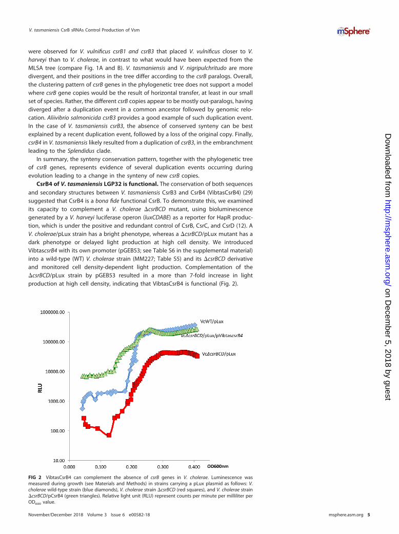
CsrB4 of V. tasmaniensis LGP32 is functional. The conservation of both sequencesand secondary structures between V. tasmaniensis CsrB3 and CsrB4 (VibtasCsrB4) (29)suggested that CsrB4 is a bona fide functional CsrB. To demonstrate this, we examinedits capacity to complement a V. cholerae �csrBCD mutant, using bioluminescencegenerated by a V. harveyi luciferase operon (luxCDABE) as a reporter for HapR produc-tion, which is under the positive and redundant control of CsrB, CsrC, and CsrD (12). AV. cholerae/pLux strain has a bright phenotype, whereas a �csrBCD/pLux mutant has adark phenotype or delayed light production at high cell density. We introducedVibtascsrB4 with its own promoter (pGEB53; see Table S6 in the supplemental material)into a wild-type (WT) V. cholerae strain (MM227; Table S5) and its �csrBCD derivativeand monitored cell density-dependent light production. Complementation of the�csrBCD/pLux strain by pGEB53 resulted in a more than 7-fold increase in lightproduction at high cell density, indicating that VibtasCsrB4 is functional (Fig. 2).
FIG 2 VibtasCsrB4 can complement the absence of csrB genes in V. cholerae. Luminescence wasmeasured during growth (see Materials and Methods) in strains carrying a pLux plasmid as follows: V.cholerae wild-type strain (blue diamonds), V. cholerae strain �csrBCD (red squares), and V. cholerae strain�csrBCD/pCsrB4 (green triangles). Relative light unit (RLU) represent counts per minute per milliliter perOD600 value.
V. tasmaniensis CsrB sRNAs Control Production of Vsm
November/December 2018 Volume 3 Issue 6 e00582-18 msphere.asm.org 5
on Decem
ber 5, 2018 by guesthttp://m
sphere.asm.org/
Dow
nloaded from
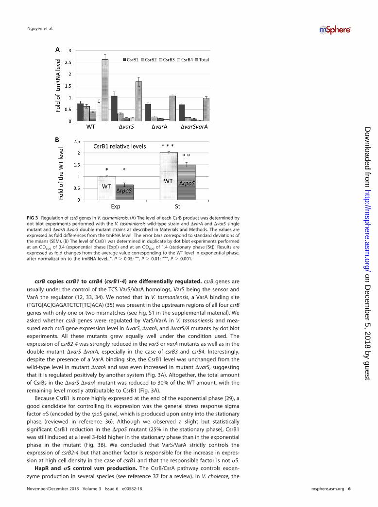
csrB copies csrB1 to csrB4 (csrB1-4) are differentially regulated. csrB genes areusually under the control of the TCS VarS/VarA homologs, VarS being the sensor andVarA the regulator (12, 33, 34). We noted that in V. tasmaniensis, a VarA binding site(TGTG[AC]GAGATCTCT[TC]ACA) (35) was present in the upstream regions of all four csrBgenes with only one or two mismatches (see Fig. S1 in the supplemental material). Weasked whether csrB genes were regulated by VarS/VarA in V. tasmaniensis and mea-sured each csrB gene expression level in �varS, �varA, and �varS/A mutants by dot blotexperiments. All these mutants grew equally well under the condition used. Theexpression of csrB2-4 was strongly reduced in the varS or varA mutants as well as in thedouble mutant �varS �varA, especially in the case of csrB3 and csrB4. Interestingly,despite the presence of a VarA binding site, the CsrB1 level was unchanged from thewild-type level in mutant �varA and was even increased in mutant �varS, suggestingthat it is regulated positively by another system (Fig. 3A). Altogether, the total amountof CsrBs in the �varS �varA mutant was reduced to 30% of the WT amount, with theremaining level mostly attributable to CsrB1 (Fig. 3A).
Because CsrB1 is more highly expressed at the end of the exponential phase (29), agood candidate for controlling its expression was the general stress response sigmafactor �S (encoded by the rpoS gene), which is produced upon entry into the stationaryphase (reviewed in reference 36). Although we observed a slight but statisticallysignificant CsrB1 reduction in the �rpoS mutant (25% in the stationary phase), CsrB1was still induced at a level 3-fold higher in the stationary phase than in the exponentialphase in the mutant (Fig. 3B). We concluded that VarS/VarA strictly controls theexpression of csrB2-4 but that another factor is responsible for the increase in expres-sion at high cell density in the case of csrB1 and that the responsible factor is not �S.
HapR and �S control vsm production. The CsrB/CsrA pathway controls exoen-zyme production in several species (see reference 37 for a review). In V. cholerae, the
FIG 3 Regulation of csrB genes in V. tasmaniensis. (A) The level of each CsrB product was determined bydot blot experiments performed with the V. tasmaniensis wild-type strain and �varA and �varS singlemutant and �varA �varS double mutant strains as described in Materials and Methods. The values areexpressed as fold differences from the tmRNA level. The error bars correspond to standard deviations ofthe means (SEM). (B) The level of CsrB1 was determined in duplicate by dot blot experiments performedat an OD600 of 0.4 (exponential phase [Exp]) and at an OD600 of 1.4 (stationary phase [St]). Results areexpressed as fold changes from the average value corresponding to the WT level in exponential phase,after normalization to the tmRNA level. *, P � 0.05; **, P � 0.01; ***, P � 0.001.
Nguyen et al.
November/December 2018 Volume 3 Issue 6 e00582-18 msphere.asm.org 6
on Decem
ber 5, 2018 by guesthttp://m
sphere.asm.org/
Dow
nloaded from
CsrB/CsrA pathway controls the hapR transcript level by modulating the QS pathway(12). Since HapR controls the production of secreted hemagglutinin/protease HapA andmetalloprotease PrtV in V. cholerae (12, 27, 28), we examined whether secreted proteaseproduction in V. tasmaniensis could be used to monitor HapR activity in V. tasmaniensisand hence the CsrB pathway.
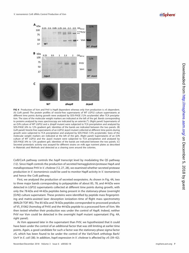
First, we analyzed the production of secreted exoproteins. As shown in Fig. 4A, twoto three major bands corresponding to polypeptides of about 85, 78, and 44 kDa weredetected in LGP32 supernatants collected at different time points during growth, withonly the 78-kDa and 44-kDa peptides being present in the stationary-phase (overnight[O/N]) culture supernatant. These proteins were identified by peptide mass fingerprint-ing and matrix-assisted laser desorption ionization–time of flight mass spectrometry(MALDI-TOF MS). The 85-kDa and 78-kDa peptides corresponded to processed productsof VS_II1062 (homolog of PrtV) and the 44-kDa peptide to a processed form of Vsm. Wethen tested whether their production was under the control of HapR. Indeed, neitherPrtV nor Vsm could be detected in the overnight hapR mutant supernatant (Fig. 4A,right panel).
As Vsm appeared later in the supernatant than PrtV, we hypothesized that it couldhave been under the control of an additional factor that was still limiting at earlier timepoints. Again, a good candidate for such a factor was the stationary-phase sigma factor�S, which has been found to be under the control of the VarS/VarA orthologs BarA/UvrY in E. coli (38). In addition, hapA expression in V. cholerae is affected by �S (39–42).
FIG 4 Production of Vsm and PrtV is HapR dependent whereas only Vsm production is �S dependent.(A) (Left panel) The protein profiles of vesicle-free supernatants of WT LGP32 culture supernatants atdifferent time points during growth were analyzed by SDS-PAGE (12% acrylamide) after TCA precipita-tion. The sizes of the molecular weight markers are indicated at the left of the gel. Bands correspondingto proteins analyzed by mass spectroscopy are indicated by an asterisk (*). (Right panel) Supernatants ofan O/N culture of WT LGP32 and a �hapR mutant were subjected to TCA precipitation and analyzed bySDS-PAGE (4% to 12% gradient gel). Identities of the bands are indicated between the two panels. (B)(Left panel) Vesicle-free supernatants of an LGP32 �rpoS mutant collected at different time points duringgrowth were subjected to TCA precipitation and analyzed by SDS-PAGE (12% acrylamide). Sizes of themolecular weight markers are indicated at the left of the gels. (Right panel) Supernatants of an O/Nculture of WT LGP32 and the �rpoS mutant were subjected to TCA precipitation and analyzed bySDS-PAGE (4% to 12% gradient gel). Identities of the bands are indicated between the two panels. (C)Secreted proteolytic activity was assayed for different strains on milk agar nutrient plates as describedin Materials and Methods and detected as a clearing zone around the colonies.
V. tasmaniensis CsrB sRNAs Control Production of Vsm
November/December 2018 Volume 3 Issue 6 e00582-18 msphere.asm.org 7
on Decem
ber 5, 2018 by guesthttp://m
sphere.asm.org/
Dow
nloaded from
Accordingly, we examined the kinetics of the occurrence of PrtV and Vsm in the culturesupernatant of an rpoS mutant. In a �rpoS strain, Vsm was not detected even uponentry into the stationary phase or in an overnight culture (Fig. 4B), whereas the kineticsof production of PrtV in the mutant was not significantly different from that in the WTstrain (Fig. 4A and B). We concluded from these results that vsm expression requires �Sin addition to HapR. When protease production was assayed on milk agar plate, noclearing zone could be detected around either the hapR colonies or the rpoS colonies(Fig. 4C). Binesse et al. (21) have shown previously that Vsm is the main contributor tothe proteolytic activity of the supernatant. This was confirmed by the results of ourplate assay, since the �rpoS was still able to produce PrtV but did not displayproteolytic activity. In conclusion, Vsm requires both HapR and �S for its production,whereas PrtV requires only HapR.
The VarS/VarA/CsrB system controls secreted protease production in a LuxO-dependent manner. In order to decipher the regulatory pathway linking QS tosecreted protease production, the quantity of Vsm and PrtV was determined in variousmutants. As expected, a �vsm mutant supernatant showed a total absence of the44-kDa band corresponding to the Vsm processed product. Surprisingly, the level ofthis band was also strongly decreased in the prtV mutant (Fig. 5A), suggesting someinteraction between the two proteases. Deleting varS and varA had no or only a slighteffect on protease production, whereas the absence of all four csrB genes resulted in areduction in the levels of both the Vsm and PrtV bands, with a stronger effect on Vsmthan on PrtV. We reasoned that in the varS varA mutant, the presence of csrB1 could besufficient to ensure some production of proteases. Indeed, in the varS varA csrB1 triplemutant, we did not detect any production/secretion of PrtV nor Vsm in the supernatant(Fig. 5A, left panel).
CsrBs act by binding to and titration of the regulatory protein CsrA, counteractingCsrA action (reviewed in reference 43). In V. cholerae, it was shown that CsrA canactivate indirectly the response regulator LuxO, which negatively regulates the QSresponse by controlling positively the production of Qrr sRNAs at low cell density (12,18). Qrrs are themselves inhibitors of HapR production (18). Inactivation of luxO wasable to suppress the effect of a strong reduction of CsrB levels due to a varA mutation(12). We repeatedly failed to construct a csrA null mutant(s) in V. tasmaniensis LGP32and thus could not examine if inactivation of CsrA can suppress the varS varA csrB1triple mutant phenotype. Instead, we asked whether the VarS/VarA/CsrB/CsrA pathwayacted on protease production by counteracting the inhibitory action of LuxO. Weintroduced a null luxO mutation in the different backgrounds and analyzed the contentof an overnight culture supernatant of each mutant (Fig. 5A, right panel). We observedthat the luxO mutation could clearly restore protease production, although not to theWT level, in the varS varA csrB1 triple mutant as seen by SDS gel analysis of thesupernatant (Fig. 5A).
Neither a �hapR nor a �rpoS mutant, neither of which produced any Vsm in theirsupernatant (Fig. 4B), displayed proteolytic activity in a milk agar plate assay. Wewanted to determine more quantitatively how much proteolytic activity we coulddetect in our different strains (Fig. 5C). As expected, a �vsm mutant did not show anyactivity, confirming that Vsm is essential for secreted proteolytic activity. The �prtVmutant showed a strong reduction of proteolytic activity, in keeping with the importantreduction in the level of the Vsm band seen on gel. Nonetheless, we could observesome discrepancies between the apparent level of the Vsm band in gel and the levelof proteolytic activity displayed by different mutants with partial effects (compareFig. 5B and A). But, overall, the proteolytic tests confirmed our main conclusions: thecomplete absence of both the varS and varA genes and of all four csrB genes is neededto totally abolish production of secreted proteases, and the inactivation of luxO in sucha strain is sufficient to restore some secreted protease production and proteolyticactivity in the supernatant.
To confirm that the presence of either VarA or any CsrB is sufficient for theproduction of Vsm and PrtV, we reintroduced each in the triple mutant varS varA csrB1,
Nguyen et al.
November/December 2018 Volume 3 Issue 6 e00582-18 msphere.asm.org 8
on Decem
ber 5, 2018 by guesthttp://m
sphere.asm.org/
Dow
nloaded from
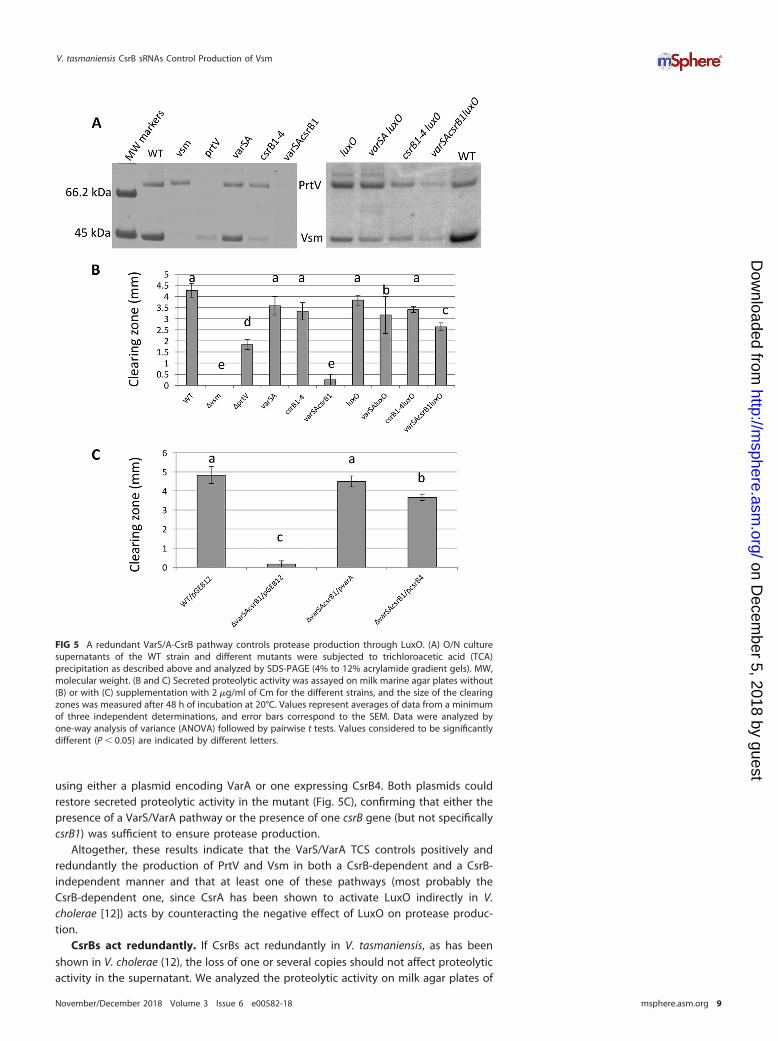
using either a plasmid encoding VarA or one expressing CsrB4. Both plasmids couldrestore secreted proteolytic activity in the mutant (Fig. 5C), confirming that either thepresence of a VarS/VarA pathway or the presence of one csrB gene (but not specificallycsrB1) was sufficient to ensure protease production.
Altogether, these results indicate that the VarS/VarA TCS controls positively andredundantly the production of PrtV and Vsm in both a CsrB-dependent and a CsrB-independent manner and that at least one of these pathways (most probably theCsrB-dependent one, since CsrA has been shown to activate LuxO indirectly in V.cholerae [12]) acts by counteracting the negative effect of LuxO on protease produc-tion.
CsrBs act redundantly. If CsrBs act redundantly in V. tasmaniensis, as has beenshown in V. cholerae (12), the loss of one or several copies should not affect proteolyticactivity in the supernatant. We analyzed the proteolytic activity on milk agar plates of
FIG 5 A redundant VarS/A-CsrB pathway controls protease production through LuxO. (A) O/N culturesupernatants of the WT strain and different mutants were subjected to trichloroacetic acid (TCA)precipitation as described above and analyzed by SDS-PAGE (4% to 12% acrylamide gradient gels). MW,molecular weight. (B and C) Secreted proteolytic activity was assayed on milk marine agar plates without(B) or with (C) supplementation with 2 �g/ml of Cm for the different strains, and the size of the clearingzones was measured after 48 h of incubation at 20°C. Values represent averages of data from a minimumof three independent determinations, and error bars correspond to the SEM. Data were analyzed byone-way analysis of variance (ANOVA) followed by pairwise t tests. Values considered to be significantlydifferent (P � 0.05) are indicated by different letters.
V. tasmaniensis CsrB sRNAs Control Production of Vsm
November/December 2018 Volume 3 Issue 6 e00582-18 msphere.asm.org 9
on Decem
ber 5, 2018 by guesthttp://m
sphere.asm.org/
Dow
nloaded from
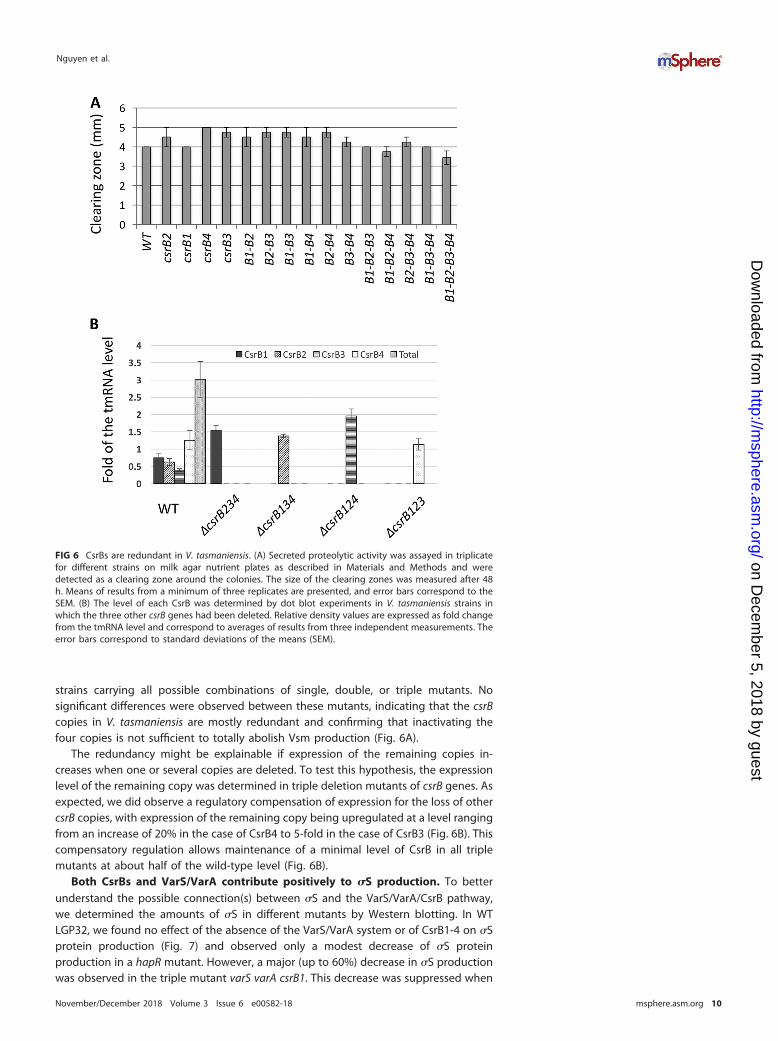
strains carrying all possible combinations of single, double, or triple mutants. Nosignificant differences were observed between these mutants, indicating that the csrBcopies in V. tasmaniensis are mostly redundant and confirming that inactivating thefour copies is not sufficient to totally abolish Vsm production (Fig. 6A).
The redundancy might be explainable if expression of the remaining copies in-creases when one or several copies are deleted. To test this hypothesis, the expressionlevel of the remaining copy was determined in triple deletion mutants of csrB genes. Asexpected, we did observe a regulatory compensation of expression for the loss of othercsrB copies, with expression of the remaining copy being upregulated at a level rangingfrom an increase of 20% in the case of CsrB4 to 5-fold in the case of CsrB3 (Fig. 6B). Thiscompensatory regulation allows maintenance of a minimal level of CsrB in all triplemutants at about half of the wild-type level (Fig. 6B).
Both CsrBs and VarS/VarA contribute positively to �S production. To betterunderstand the possible connection(s) between �S and the VarS/VarA/CsrB pathway,we determined the amounts of �S in different mutants by Western blotting. In WTLGP32, we found no effect of the absence of the VarS/VarA system or of CsrB1-4 on �Sprotein production (Fig. 7) and observed only a modest decrease of �S proteinproduction in a hapR mutant. However, a major (up to 60%) decrease in �S productionwas observed in the triple mutant varS varA csrB1. This decrease was suppressed when
FIG 6 CsrBs are redundant in V. tasmaniensis. (A) Secreted proteolytic activity was assayed in triplicatefor different strains on milk agar nutrient plates as described in Materials and Methods and weredetected as a clearing zone around the colonies. The size of the clearing zones was measured after 48h. Means of results from a minimum of three replicates are presented, and error bars correspond to theSEM. (B) The level of each CsrB was determined by dot blot experiments in V. tasmaniensis strains inwhich the three other csrB genes had been deleted. Relative density values are expressed as fold changefrom the tmRNA level and correspond to averages of results from three independent measurements. Theerror bars correspond to standard deviations of the means (SEM).
Nguyen et al.
November/December 2018 Volume 3 Issue 6 e00582-18 msphere.asm.org 10
on Decem
ber 5, 2018 by guesthttp://m
sphere.asm.org/
Dow
nloaded from
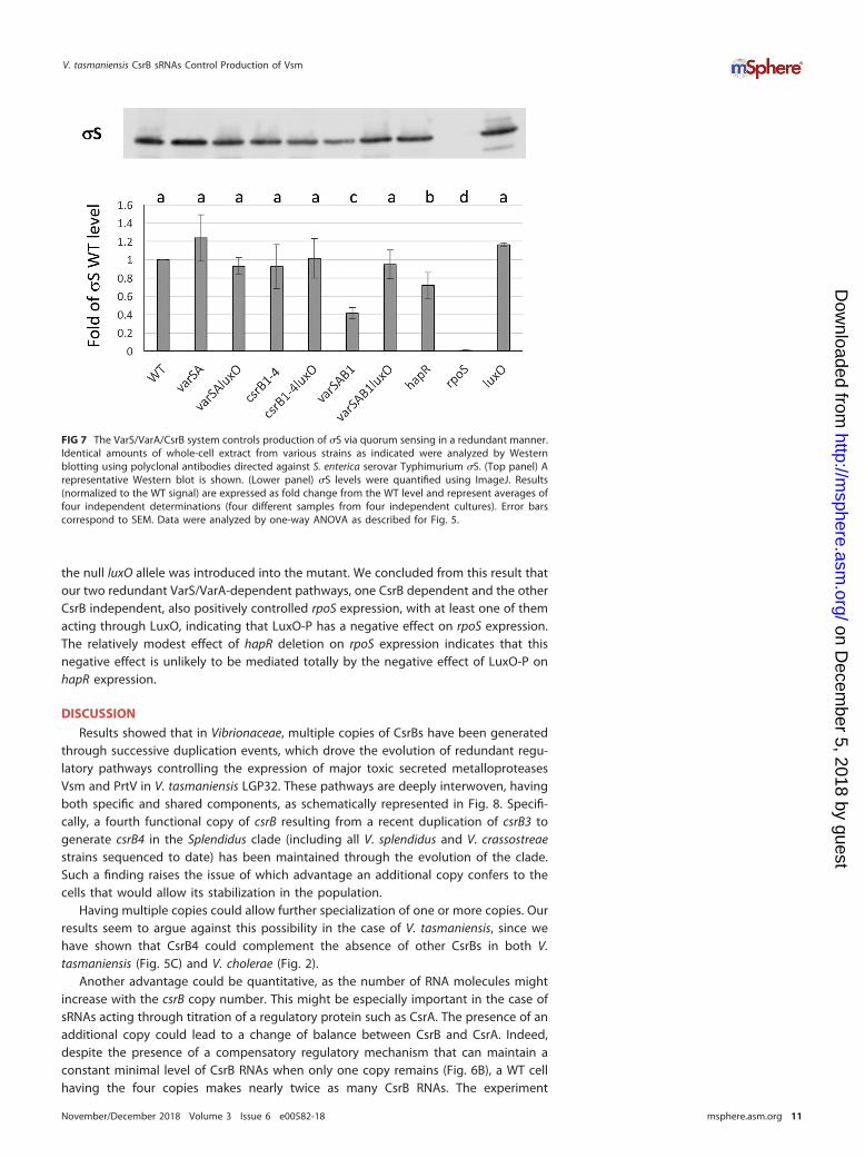
the null luxO allele was introduced into the mutant. We concluded from this result thatour two redundant VarS/VarA-dependent pathways, one CsrB dependent and the otherCsrB independent, also positively controlled rpoS expression, with at least one of themacting through LuxO, indicating that LuxO-P has a negative effect on rpoS expression.The relatively modest effect of hapR deletion on rpoS expression indicates that thisnegative effect is unlikely to be mediated totally by the negative effect of LuxO-P onhapR expression.
DISCUSSION
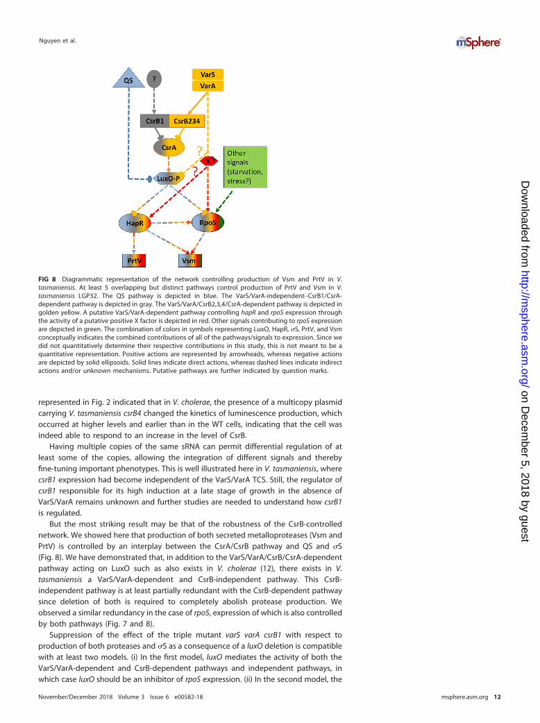
Results showed that in Vibrionaceae, multiple copies of CsrBs have been generatedthrough successive duplication events, which drove the evolution of redundant regu-latory pathways controlling the expression of major toxic secreted metalloproteasesVsm and PrtV in V. tasmaniensis LGP32. These pathways are deeply interwoven, havingboth specific and shared components, as schematically represented in Fig. 8. Specifi-cally, a fourth functional copy of csrB resulting from a recent duplication of csrB3 togenerate csrB4 in the Splendidus clade (including all V. splendidus and V. crassostreaestrains sequenced to date) has been maintained through the evolution of the clade.Such a finding raises the issue of which advantage an additional copy confers to thecells that would allow its stabilization in the population.
Having multiple copies could allow further specialization of one or more copies. Ourresults seem to argue against this possibility in the case of V. tasmaniensis, since wehave shown that CsrB4 could complement the absence of other CsrBs in both V.tasmaniensis (Fig. 5C) and V. cholerae (Fig. 2).
Another advantage could be quantitative, as the number of RNA molecules mightincrease with the csrB copy number. This might be especially important in the case ofsRNAs acting through titration of a regulatory protein such as CsrA. The presence of anadditional copy could lead to a change of balance between CsrB and CsrA. Indeed,despite the presence of a compensatory regulatory mechanism that can maintain aconstant minimal level of CsrB RNAs when only one copy remains (Fig. 6B), a WT cellhaving the four copies makes nearly twice as many CsrB RNAs. The experiment
FIG 7 The VarS/VarA/CsrB system controls production of �S via quorum sensing in a redundant manner.Identical amounts of whole-cell extract from various strains as indicated were analyzed by Westernblotting using polyclonal antibodies directed against S. enterica serovar Typhimurium �S. (Top panel) Arepresentative Western blot is shown. (Lower panel) �S levels were quantified using ImageJ. Results(normalized to the WT signal) are expressed as fold change from the WT level and represent averages offour independent determinations (four different samples from four independent cultures). Error barscorrespond to SEM. Data were analyzed by one-way ANOVA as described for Fig. 5.
V. tasmaniensis CsrB sRNAs Control Production of Vsm
November/December 2018 Volume 3 Issue 6 e00582-18 msphere.asm.org 11
on Decem
ber 5, 2018 by guesthttp://m
sphere.asm.org/
Dow
nloaded from
represented in Fig. 2 indicated that in V. cholerae, the presence of a multicopy plasmidcarrying V. tasmaniensis csrB4 changed the kinetics of luminescence production, whichoccurred at higher levels and earlier than in the WT cells, indicating that the cell wasindeed able to respond to an increase in the level of CsrB.
Having multiple copies of the same sRNA can permit differential regulation of atleast some of the copies, allowing the integration of different signals and therebyfine-tuning important phenotypes. This is well illustrated here in V. tasmaniensis, wherecsrB1 expression had become independent of the VarS/VarA TCS. Still, the regulator ofcsrB1 responsible for its high induction at a late stage of growth in the absence ofVarS/VarA remains unknown and further studies are needed to understand how csrB1is regulated.
But the most striking result may be that of the robustness of the CsrB-controllednetwork. We showed here that production of both secreted metalloproteases (Vsm andPrtV) is controlled by an interplay between the CsrA/CsrB pathway and QS and �S(Fig. 8). We have demonstrated that, in addition to the VarS/VarA/CsrB/CsrA-dependentpathway acting on LuxO such as also exists in V. cholerae (12), there exists in V.tasmaniensis a VarS/VarA-dependent and CsrB-independent pathway. This CsrB-independent pathway is at least partially redundant with the CsrB-dependent pathwaysince deletion of both is required to completely abolish protease production. Weobserved a similar redundancy in the case of rpoS, expression of which is also controlledby both pathways (Fig. 7 and 8).
Suppression of the effect of the triple mutant varS varA csrB1 with respect toproduction of both proteases and �S as a consequence of a luxO deletion is compatiblewith at least two models. (i) In the first model, luxO mediates the activity of both theVarS/VarA-dependent and CsrB-dependent pathways and independent pathways, inwhich case luxO should be an inhibitor of rpoS expression. (ii) In the second model, the
FIG 8 Diagrammatic representation of the network controlling production of Vsm and PrtV in V.tasmaniensis. At least 5 overlapping but distinct pathways control production of PrtV and Vsm in V.tasmaniensis LGP32. The QS pathway is depicted in blue. The VarS/VarA-independent–CsrB1/CsrA-dependent pathway is depicted in gray. The VarS/VarA/CsrB2,3,4/CsrA-dependent pathway is depicted ingolden yellow. A putative VarS/VarA-dependent pathway controlling hapR and rpoS expression throughthe activity of a putative positive X factor is depicted in red. Other signals contributing to rpoS expressionare depicted in green. The combination of colors in symbols representing LuxO, HapR, �S, PrtV, and Vsmconceptually indicates the combined contributions of all of the pathways/signals to expression. Since wedid not quantitatively determine their respective contributions in this study, this is not meant to be aquantitative representation. Positive actions are represented by arrowheads, whereas negative actionsare depicted by solid ellipsoids. Solid lines indicate direct actions, whereas dashed lines indicate indirectactions and/or unknown mechanisms. Putative pathways are further indicated by question marks.
Nguyen et al.
November/December 2018 Volume 3 Issue 6 e00582-18 msphere.asm.org 12
on Decem
ber 5, 2018 by guesthttp://m
sphere.asm.org/
Dow
nloaded from
VarS/VarA-dependent, CsrB-independent pathway controls the expression of an Xfactor that acts positively on rpoS expression.
In total, in V. tasmaniensis, there are at least four highly redundant but distinctpathways, forming a complex network which positively controls secreted metallopro-teases and �S production. The first one is a QS-regulated pathway that leads to thedephosphorylation of LuxO at high cell density, preventing the production of Qrrs andthus leading to production of the master regulator HapR and of the stationary-phasesigma factor �S (indicated in blue on Fig. 8). The second is a VarS/VarA-independent,CsrB1-dependent pathway leading to titration of CsrA, thus decreasing the amount ofphosphorylated LuxO in response to an unknown signal (indicated in gray). The thirdis a VarS/VarA/CsrB234-dependent pathway that also leads to titration of CsrA (indi-cated in golden yellow). The fourth is also VarS/VarA dependent but is also CsrBindependent and controls positively the levels of HapR and �S. This pathway mightconverge with the previous one on LuxO or might act through an unknown X factor(indicated in red). In addition, Vsm production but not PrtV production is indirectlyregulated by other signals controlling rpoS expression, such as starvation (indicated ingreen). Finally, HapR itself can positively control the production of �S, although to alesser extent. The resulting network can integrate different input signals and controldifferentially the timing of production of the two metalloproteases, with each pathwaycontributing at different levels to PrtV and Vsm expression. Identification of as-yet-unknown components in this circuit and precise determination of the respectivecontributions of the pathways to the final outcome will be required to fully understandthe behavior of this complex network in response to varying conditions.
What could be the significance of our finding for the virulence of V. tasmaniensis?The VarS/VarA homologues have been shown to be important for virulence in manyGram-negative pathogens, including in Vibrio species (13, 44). In V. aestuarianus,another oyster pathogen, it was recently shown that a frameshift in the varS gene wassufficient to induce loss of pathogenicity toward oysters, together with the loss ofproduction of the Vam metalloprotease, the homolog of Vsm (45). However, we did notobserve loss of virulence in the varS varA mutant or in the varS varA csrB1 mutant in V.tasmaniensis when the bacteria were injected into the adductor muscles of the oysters(see Fig. S2 in the supplemental material), which is consistent with Vsm being essentialfor the cytotoxicity of LGP32 supernatant to oysters but not for in vivo virulence in suchan oyster infection model (24). This does not preclude the possibility of a role of themetalloproteases at earlier stages of infection, for instance, during colonization.
To summarize, we have shown that in V. tasmaniensis LGP32, different copies ofCsrBs are regulated differentially, allowing the sensing of different signals. Multiplecopies of csrB genes contribute significantly to a highly redundant regulatory networkthat integrates QS, starvation, the signal(s) sensed by VarS, and an unknown signalcontrolling csrB1 expression, to control positively the timing of production of Vsm andPrtV. In addition, we have identified a novel VarS/VarA-dependent and CsrB-independent pathway that controls positively both Vsm and PrtV production and rpoSexpression.
MATERIALS AND METHODSBacterial strains and media. Bacterial strains used in this study are listed in Table S5 in the
supplemental material. V. tasmaniensis LGP32 (20) and derivatives were grown at 20°C with agitation inZobell medium (4 g/liter peptone, 1 g/liter yeast extract, 0.1 g/liter ferric phosphate, 30 g/liter sea salt[Sigma] per liter) or in marine broth (Difco-BRL) as specified, except for conjugation, where the mediumused was LBS (10 g Bacto tryptone, 5 g yeast extract, 30 g NaCl [per liter]). E. coli strains ß2163 (46) andMFDpir (47) (Table S5) and plasmid pAM34recA (48) (Table S6) were gifts from Didier Mazel. V. choleraewild-type and �csrBCD strains and pBB1 (pLux) were gifts from Bonnie L. Bassler (49). E. coli and V.cholerae were grown in LB medium at 37°C and 30°C, respectively. Antibiotics were used at the followingconcentrations: chloramphenicol (Cm) at 2 �g/ml and tetracycline (Tc) at 5 �g/ml for V. tasmaniensis andV. cholerae and Cm at 20 �g/ml and Tc at 10 �g/ml for E. coli. When necessary, thymidine anddiaminopimelate (DAP) were added to growth media at a final concentration of 0.3 mM.
Mutant and plasmid construction. Derivatives of suicide plasmid pSW7848 (which can replicateonly in a Pir-positive [Pir�) strain [50]) containing flanking regions of the genes to be deleted and thelow-copy-number replicative plasmid pGEB12 (51) containing VibtascsrB4 (pGEB53) were constructed by
V. tasmaniensis CsrB sRNAs Control Production of Vsm
November/December 2018 Volume 3 Issue 6 e00582-18 msphere.asm.org 13
on Decem
ber 5, 2018 by guesthttp://m
sphere.asm.org/
Dow
nloaded from
one-step isothermal assembly (Gibson assembly) (52). Briefly, amplified linearized plasmid DNA andpurified PCR products corresponding to 600 bp upstream and downstream of the target gene weremixed (ratio of plasmid/insertion � 1:5) in a 15-�l assembly mixture containing 5= T5 exonuclease (NewEngland Biolabs), Phusion DNA polymerase (Thermo Scientific), and Taq DNA ligase (NEB) and incubatedfor 1 h at 50°C. Assembly reaction mixtures were used immediately for transformation or were stored at�20°C until use. Plasmids and primers used for amplification are listed in Tables S6 and S7, respectively.
For conjugation between E. coli and V. tasmaniensis, strain MFDpir (47) (where the RP4 conjugationoperon is more stable than in the original ß2163 strain [46]) was made gyrA462. gyrA462 is an allele ofgyrA conferring resistance to toxin CcdB, whose gene is present on pSW7848 and its derivatives. Aftertransformation using the pAM34recA plasmid (48), the gyrA462 allele was introduced by P1 transduction,selecting for tetracycline resistance conferred by a tightly linked Tn10 transposon. Since replication ofpAM34 is dependent upon the presence of IPTG (isopropyl-�-D-thiogalactopyranoside), a resultingtransductant clone carrying the allele was further grown without IPTG in order to get rid of the plasmid.The resulting strain was named GEB883 (Table S5).
RP4-based conjugations and selection of mutants were carried out as follows. The donor strain(GEB883 with the mobilizable plasmid) and the recipient strain (LGP32 and derivatives) were grown tothe stationary phase in LB plus DAP plus Cm (E. coli) and in LBS (LGP32) at 37°C and 20°C, respectively.Overnight cultures of the donor and recipient strains were diluted 100-fold in broth and grown to anoptical density at 600 nm (OD600) of 0.3. A 1-ml volume of donor cells was centrifuged and washed twicewith LB and then resuspended in 100 �l LBS, whereas recipient cells were centrifuged and resuspendedin 250 �l of LBS. For conjugation, 10 �l of donor cells and 50 �l of concentrated recipient cells werespotted on a 0.45-�m-pore-size nitrocellulose filter (Whatman) and the filter was incubated on a LBS agarplate overnight (O/N) at 20°C. For controls, donor cells or recipient cells alone were spotted on filters.Cells were then resuspended from the filter in 1 ml of LBS, centrifuged, resuspended in 100 �l of LBSmedium, and spread for selection of the exconjugants on a LBS-plus-Cm agar plate in the presence of0.2% glucose before incubation at 20°C until colonies appeared. Potential exconjugants were purifiedtwice on the same selective medium at 20°C and then restreaked on LBS– 0.2% arabinose to induce theccdB gene and to select for bacteria that had lost the inserted plasmid. Colonies were repurified twiceon the same medium, and PCR was used on colonies to check for the presence or absence of the targetgenes.
Bioluminescence assays. Overnight culture of V. cholerae and derivatives were diluted at 1:1,000 in200 �l LB medium or LB plus Tc and/or Cm to maintain pLux and/or pGEB53. Direct light counts andOD600 were measured every 15 min during 40 h at 28°C using a luminometer plate reader (ChameleonV; Hidex). Relative light unit (RLU) values represent counts per minute per milliliter per OD600
�1 value.Dot blot analyses. An overnight culture of V. tasmaniensis was diluted at 1:100 in 100 ml of Zobell
medium and grown at 20°C until an OD600 of approximately 1 was reached. No differences in the growthrates of the different mutants analyzed were observed. After centrifugation at 4°C, cell pellets were keptat �80°C until RNA preparation. Total RNA was obtained as described before (29). A 0.5-�g volume oftotal RNA from each sample was mixed with the same volume of RNA loading dye (Thermo Scientific) andheat denatured, and an equal volume of ice-cold SSC (1� SSC is 0.15 M NaCl plus 0.015 M sodium citrate)(20�) was added to obtain the final RNA solutions. The RNA solutions were spotted under a vacuum onan Amersham Hybond-N� membrane prewashed with SSC (10�), using a Schleicher & Schuell Minifold-Idot blot system. The wells were then rinsed with 150 �l of SSC (10�). The membrane was air-dried,cross-linked, and hybridized with probes specific to the sRNA to be assayed (Table S7) as describedpreviously (29). Signal intensity was quantified by the use of ImageJ 1.48v (Wayne Rasband [http://imagej.nih.gov/ij/download.html]). Relative densities were normalized to the signal of transfer-messenger RNA (tmRNA). Experiments were done in triplicate, unless otherwise specified, and all valueswere expressed as fold change from the level of the tmRNA measured under the same conditions.
SDS-PAGE analysis and detection of protease activity. A 1-ml volume of overnight bacterialculture was centrifuged at 14,000 rpm for 5 min at 4°C. The collected supernatant was again centrifugedat 65,000 � g for 30 min at 4°C in a Beckman TM-100 ultracentrifuge to remove vesicles. The vesicle-freefraction was subjected to trichloroacetic acid (TCA) precipitation (10% final concentration) at 4°C andwashed twice with ice-cold acetone before being resolubilized in 60 �l of SDS sample buffer per 1 OD600
equivalent and analyzed by SDS-PAGE using a 12% polyacrylamide gel or a 4% to 12% gradient gel asspecified.
In order to identify proteins in the extracellular fractions, SDS-PAGE was done as described above,and selected bands were identified by peptide mass fingerprinting after trypsinolysis, using matrix-assisted laser desorption ionization–time of flight mass spectrometry (MALDI-TOF MS) at the PlatformSICaPS (CNRS, Gif sur Yvette, France).
Alternatively, in subsequent experiments, the bacterial culture was centrifuged twice at 20,000 � gfor 20 min at 4°C and the supernatant was collected and subjected to TCA precipitation. Pellets werewashed with ice-cold acetone and resuspended in SDS sample buffer at a concentration equivalent toan OD600 of 20.
Secreted protease activity of whole cells was detected by spotting 5 �l of O/N bacterial cultures ona marine agar plate supplemented with 10% sterile skimmed milk. The clearing zone was measured after48 h of incubation at 20°C.
For Western blotting of �S, a concentration equivalent to an OD600 of 0.02 of centrifuged bacterialcells lysed in SDS sample buffer was loaded on a 4% to 12% gradient SDS/polyacrylamide gel. Afterelectrophoresis, separated proteins were transferred onto a polyvinylidene difluoride (PVDF) membraneusing an iBLOt2 device (Life Technologies) following the manufacturer’s instructions. Completion of
Nguyen et al.
November/December 2018 Volume 3 Issue 6 e00582-18 msphere.asm.org 14
on Decem
ber 5, 2018 by guesthttp://m
sphere.asm.org/
Dow
nloaded from
transfer was monitored using prestained molecular weight markers (Life Technologies). The membranewas then incubated successively with a 1:4,000 dilution of a rabbit anti-Salmonella enterica serovarTyphimurium �S antiserum (a gift from Françoise Norel-Bouzoukian) and a 1:1,600 dilution of ahorseradish peroxidase (HRP)-coupled anti-rabbit goat IgG monoclonal antibody (ImmunoReagents, Inc.)in iBind Flex solution (Invitrogen) using a iBind Flex device, following the manufacturer;s instructions.Analysis of the bands was carried out using ECL spray reagent (Advansta). Imaging was done with a GEHealthcare system charge-coupled-device (CCD) camera and quantification with ImageJ 1.48v. Resultsare expressed as fold change from the level of the �S WT signal in the same blot.
Phylogeny of csrB genes. (i) A total of 38 csrB sequences from 14 species whose fully assembledgenomes were present in public databases (11 Vibrio species corresponding to 7 different clades of theVibrio genus, 2 Aliivibrio species, and 1 P. profundum species) were collected from the NCBI genomedatabase after detection performed using BLAST repeatedly and visual inspection of the correspondinggenomic regions to determine the most probable transcription start site and the Rho-independentterminator. For each copy, flanking coding DNA sequences (CDS) were examined to search for syntenyconservation, with the help of Absynte (Archaeal and Bacterial Synteny Explorer; http://archaea.u-psud.fr/absynte/Default.aspx). To build the phylogenetic tree of csrB copies, csrB sequences (not including thepredicted terminator) were aligned using MUSCLE (http://www.phylogeny.fr), with a maximum of 25iterations. The resulting alignment was curated using G-block in the less stringent mode. A total of 352(75%) of 464 original positions were retained in the resulting alignment that was used to generate aphylogeny using the maximum likelihood method implemented in PhyML (31). The tree was drawn usingTreeDyn. Statistical tests for branch support were computed using the approximate likelihood ratio test(aLRT) (53).
(ii) To determine the phylogeny of the 14 strains, a concatenation of recA, gyrB, and rpoA sequenceswas used to construct the tree by PhyML (http://www.phylogeny.fr), using the default mode.
SUPPLEMENTAL MATERIALSupplemental material for this article may be found at https://doi.org/10.1128/
mSphere.00582-18.FIG S1, PDF file, 0.02 MB.FIG S2, TIFF file, 13 MB.TABLE S1, PDF file, 0.1 MB.TABLE S2, PDF file, 0.05 MB.TABLE S3, PDF file, 0.05 MB.TABLE S4, PDF file, 0.03 MB.TABLE S5, PDF file, 0.04 MB.TABLE S6, PDF file, 0.03 MB.TABLE S7, PDF file, 0.03 MB.
ACKNOWLEDGMENTSWe thank Bonnie L. Bassler and Didier Mazel for kindly providing plasmids and
strains and Stephanie Henry for constructing some of the mutants used in this study.We are indebted to Françoise Norel for generously providing anti-�S antibodies. Thiswork has benefited from mass spectroscopy results obtained thanks to the excellentexpertise of the SICaPS facility of the I2BC, supported by the Région Ile de France andthe Fondation pour la Recherche Médicale (http://www.i2bc.paris-saclay.fr/spip.php?article201&lang�en).
This work was supported by Program Blanc Biodiversité, écologie et agronomie(ANR 11 BSV7 023 02 “Vibriogen” to A.J., F.L.R., and D.D.-G.). The funders had no role instudy design, data collection and interpretation, or the decision to submit the work forpublication.
REFERENCES1. Wagner EG, Romby P. 2015. Small RNAs in bacteria and archaea: who
they are, what they do, and how they do it. Adv Genet 90:133–208.https://doi.org/10.1016/bs.adgen.2015.05.001.
2. Gottesman S, Storz G. 27 October 2010. Bacterial small RNA regulators:versatile roles and rapidly evolving variations. Cold Spring Harb PerspectBiol https://doi.org/10.1101/cshperspect.a003798.
3. Liu MY, Gui G, Wei B, Preston JF, III, Oakford L, Yuksel U, Giedroc DP,Romeo T. 1997. The RNA molecule CsrB binds to the global regulatoryprotein CsrA and antagonizes its activity in Escherichia coli. J Biol Chem272:17502–17510. https://doi.org/10.1074/jbc.272.28.17502.
4. Heroven AK, Bohme K, Dersch P. 2012. The Csr/Rsm system of Yersinia
and related pathogens: a post-transcriptional strategy for managingvirulence. RNA Biol 9:379 –391. https://doi.org/10.4161/rna.19333.
5. Romeo T, Vakulskas CA, Babitzke P. 2013. Post-transcriptional regu-lation on a global scale: form and function of Csr/Rsm systems.Environ Microbiol 15:313–324. https://doi.org/10.1111/j.1462-2920.2012.02794.x.
6. Vakulskas CA, Potts AH, Babitzke P, Ahmer BM, Romeo T. 2015. Regula-tion of bacterial virulence by Csr (Rsm) systems. Microbiol Mol Biol Rev79:193–224. https://doi.org/10.1128/MMBR.00052-14.
7. Baker CS, Eory LA, Yakhnin H, Mercante J, Romeo T, Babitzke P. 2007.CsrA inhibits translation initiation of Escherichia coli hfq by binding to a
V. tasmaniensis CsrB sRNAs Control Production of Vsm
November/December 2018 Volume 3 Issue 6 e00582-18 msphere.asm.org 15
on Decem
ber 5, 2018 by guesthttp://m
sphere.asm.org/
Dow
nloaded from
single site overlapping the Shine-Dalgarno sequence. J Bacteriol 189:5472–5481. https://doi.org/10.1128/JB.00529-07.
8. Dubey AK, Baker CS, Romeo T, Babitzke P. 2005. RNA sequence andsecondary structure participate in high-affinity CsrA-RNA interaction.RNA 11:1579 –1587. https://doi.org/10.1261/rna.2990205.
9. Wei BL, Brun-Zinkernagel AM, Simecka JW, Pruss BM, Babitzke P, RomeoT. 2001. Positive regulation of motility and flhDC expression by theRNA-binding protein CsrA of Escherichia coli. Mol Microbiol 40:245–256.https://doi.org/10.1046/j.1365-2958.2001.02380.x.
10. Yakhnin AV, Baker CS, Vakulskas CA, Yakhnin H, Berezin I, Romeo T,Babitzke P. 2013. CsrA activates flhDC expression by protecting flhDCmRNA from RNase E-mediated cleavage. Mol Microbiol 87:851– 866.https://doi.org/10.1111/mmi.12136.
11. Jonas K, Edwards AN, Ahmad I, Romeo T, Romling U, Melefors O. 2010.Complex regulatory network encompassing the Csr, c-di-GMP and mo-tility systems of Salmonella typhimurium. Environ Microbiol 12:524 –540.https://doi.org/10.1111/j.1462-2920.2009.02097.x.
12. Lenz DH, Miller MB, Zhu J, Kulkarni RV, Bassler BL. 2005. CsrA andthree redundant small RNAs regulate quorum sensing in Vibrio chol-erae. Mol Microbiol 58:1186 –1202. https://doi.org/10.1111/j.1365-2958.2005.04902.x.
13. Mey AR, Butz HA, Payne SM. 2015. Vibrio cholerae Csra regulates Toxrlevels in response to amino acids and is essential for virulence. mBio6:e01064-15. https://doi.org/10.1128/mBio.01064-15.
14. Williams JW, Ritter AL, Stevens AM. 2012. CsrA modulates luxR transcriptlevels in Vibrio fischeri. FEMS Microbiol Lett 329:28 –35. https://doi.org/10.1111/j.1574-6968.2012.02499.x.
15. Foxall RL, Ballok AE, Avitabile A, Whistler CA. 2015. Spontaneous phe-notypic suppression of GacA-defective Vibrio fischeri is achieved viamutation of csrA and ihfA. BMC Microbiol 15:180. https://doi.org/10.1186/s12866-015-0509-2.
16. Whistler CA, Ruby EG. 2003. GacA regulates symbiotic colonization traitsof Vibrio fischeri and facilitates a beneficial association with an animalhost. J Bacteriol 185:7202–7212. https://doi.org/10.1128/JB.185.24.7202-7212.2003.
17. Caswell CC, Oglesby-Sherrouse AG, Murphy ER. 2014. Sibling rivalry:related bacterial small RNAs and their redundant and non-redundantroles. Front Cell Infect Microbiol 4:151. https://doi.org/10.3389/fcimb.2014.00151.
18. Lenz DH, Mok KC, Lilley BN, Kulkarni RV, Wingreen NS, Bassler BL. 2004.The small RNA chaperone Hfq and multiple small RNAs control quorumsensing in Vibrio harveyi and Vibrio cholerae. Cell 118:69 – 82. https://doi.org/10.1016/j.cell.2004.06.009.
19. Gay M, Renault T, Pons AM, Le Roux F. 2004. Two Vibrio splendidusrelated strains collaborate to kill Crassostrea gigas: taxonomy andhost alterations. Dis Aquat Organ 62:65–74. https://doi.org/10.3354/dao062065.
20. Le Roux F, Zouine M, Chakroun N, Binesse J, Saulnier D, Bouchier C,Zidane N, Ma L, Rusniok C, Lajus A, Buchrieser C, Medigue C, PolzMF, Mazel D. 2009. Genome sequence of Vibrio splendidus: an abun-dant planctonic marine species with a large genotypic diversity.Environ Microbiol 11:1959 –1970. https://doi.org/10.1111/j.1462-2920.2009.01918.x.
21. Binesse J, Delsert C, Saulnier D, Champomier-Verges MC, Zagorec M,Munier-Lehmann H, Mazel D, Le Roux F. 2008. Metalloprotease Vsm isthe major determinant of toxicity for extracellular products of Vibriosplendidus. Appl Environ Microbiol 74:7108 –7117. https://doi.org/10.1128/AEM.01261-08.
22. Duperthuy M, Binesse J, Le Roux F, Romestand B, Caro A, Got P,Givaudan A, Mazel D, Bachere E, Destoumieux GD. 2010. The major outermembrane protein OmpU of Vibrio splendidus contributes to host anti-microbial peptide resistance and is required for virulence in the oysterCrassostrea gigas. Environ Microbiol 12:951–963. https://doi.org/10.1111/j.1462-2920.2009.02138.x.
23. Duperthuy M, Schmitt P, Garzón E, Caro A, Rosa RD, Le Roux F,Lautrédou-Audouy N, Got P, Romestand B, de Lorgeril J, Kieffer-JaquinodS, Bachère E, Destoumieux-Garzón D. 2011. Use of OmpU porins forattachment and invasion of Crassostrea gigas immune cells by the oysterpathogen Vibrio splendidus. Proc Natl Acad Sci U S A 108:2993–2998.https://doi.org/10.1073/pnas.1015326108.
24. Le Roux F, Binesse J, Saulnier D, Mazel D. 2007. Construction of a Vibriosplendidus mutant lacking the metalloprotease gene vsm by use of anovel counterselectable suicide vector. Appl Environ Microbiol 73:777–784. https://doi.org/10.1128/AEM.02147-06.
25. Vanhove AS, Duperthuy M, Charrière GM, Le Roux F, Goudenège D,Gourbal B, Kieffer-Jaquinod S, Couté Y, Wai SN, Destoumieux-Garzón D.2015. Outer membrane vesicles are vehicles for the delivery of Vibriotasmaniensis virulence factors to oyster immune cells. Environ Microbiol17:1152–1165. https://doi.org/10.1111/1462-2920.12535.
26. Vanhove AS, Rubio TP, Nguyen AN, Lemire A, Roche D, Nicod J, VergnesA, Poirier AC, Disconzi E, Bachere E, Le Roux F, Jacq A, Charriere GM,Destoumieux GD. 2016. Copper homeostasis at the host vibrio interface:lessons from intracellular Vibrio transcriptomics. Environ Microbiol 18:875– 888. https://doi.org/10.1111/1462-2920.13083.
27. Kovacikova G, Skorupski K. 2002. Regulation of virulence gene expres-sion in Vibrio cholerae by quorum sensing: HapR functions at the aphApromoter. Mol Microbiol 46:1135–1147. https://doi.org/10.1046/j.1365-2958.2002.03229.x.
28. Vaitkevicius K, Rompikuntal PK, Lindmark B, Vaitkevicius R, Song T, WaiSN. 2008. The metalloprotease PrtV from Vibrio cholerae. FEBS J 275:3167–3177. https://doi.org/10.1111/j.1742-4658.2008.06470.x.
29. Toffano-Nioche C, Nguyen AN, Kuchly C, Ott A, Gautheret D, Bouloc P,Jacq A. 2012. Transcriptomic profiling of the oyster pathogen Vibriosplendidus opens a window on the evolutionary dynamics of the smallRNA repertoire in the Vibrio genus. RNA 18:2201–2219. https://doi.org/10.1261/rna.033324.112.
30. Kulkarni PR, Cui X, Williams JW, Stevens AM, Kulkarni RV. 2006. Predictionof CsrA-regulating small RNAs in bacteria and their experimental verifi-cation in Vibrio fischeri. Nucleic Acids Res 34:3361–3369. https://doi.org/10.1093/nar/gkl439.
31. Dereeper A, Guignon V, Blanc G, Audic S, Buffet S, Chevenet F, DufayardJF, Guindon S, Lefort V, Lescot M, Claverie JM, Gascuel O. 2008.Phylogeny.fr: robust phylogenetic analysis for the non-specialist. NucleicAcids Res 36:W465–W469. https://doi.org/10.1093/nar/gkn180.
32. Sawabe T, Ogura Y, Matsumura Y, Feng G, Amin AR, Mino S, NakagawaS, Sawabe T, Kumar R, Fukui Y, Satomi M, Matsushima R, Thompson FL,Gomez-Gil B, Christen R, Maruyama F, Kurokawa K, Hayashi T. 2013.Updating the Vibrio clades defined by multilocus sequence phylogeny:proposal of eight new clades, and the description of Vibrio tritonius sp.nov. Front Microbiol 4:414. https://doi.org/10.3389/fmicb.2013.00414.
33. Lapouge K, Schubert M, Allain FH, Haas D. 2008. Gac/Rsm signal trans-duction pathway of gamma-proteobacteria: from RNA recognition toregulation of social behaviour. Mol Microbiol 67:241–253. https://doi.org/10.1111/j.1365-2958.2007.06042.x.
34. Brencic A, McFarland KA, McManus HR, Castang S, Mogno I, Dove SL,Lory S. 2009. The GacS/GacA signal transduction system of Pseudomonasaeruginosa acts exclusively through its control over the transcription ofthe RsmY and RsmZ regulatory small RNAs. Mol Microbiol 73:434 – 445.https://doi.org/10.1111/j.1365-2958.2009.06782.x.
35. Zere TR, Vakulskas CA, Leng Y, Pannuri A, Potts AH, Dias R, Tang D,Kolaczkowski B, Georgellis D, Ahmer BM, Romeo T. 2015. Genomictargets and features of BarA-UvrY (-SirA) signal transduction systems.PLoS One 10:e0145035. https://doi.org/10.1371/journal.pone.0145035.
36. Navarro Llorens JM, Tormo A, Martínez-García E. 2010. Stationary phasein gram-negative bacteria. FEMS Microbiol Rev 34:476 – 495. https://doi.org/10.1111/j.1574-6976.2010.00213.x.
37. Babitzke P, Romeo T. 2007. CsrB sRNA family: sequestration of RNA-binding regulatory proteins. Curr Opin Microbiol 10:156 –163. https://doi.org/10.1016/j.mib.2007.03.007.
38. Mukhopadhyay S, Audia JP, Roy RN, Schellhorn HE. 2000. Transcriptionalinduction of the conserved alternative sigma factor RpoS in Escherichiacoli is dependent on BarA, a probable two-component regulator. MolMicrobiol 37:371–381. https://doi.org/10.1046/j.1365-2958.2000.01999.x.
39. Silva AJ, Benitez JA. 2004. Transcriptional regulation of Vibrio choleraehemagglutinin/protease by the cyclic AMP receptor protein and RpoS. JBacteriol 186:6374 – 6382. https://doi.org/10.1128/JB.186.19.6374-6382.2004.
40. Wang H, Ayala JC, Benitez JA, Silva AJ. 2012. Interaction of the histone-like nucleoid structuring protein and the general stress response regu-lator RpoS at Vibrio cholerae promoters that regulate motility andhemagglutinin/protease expression. J Bacteriol 194:1205–1215. https://doi.org/10.1128/JB.05900-11.
41. Wang H, Wu JH, Ayala JC, Benitez JA, Silva AJ. 2011. Interplay amongcyclic diguanylate, HapR, and the general stress response regulator(RpoS) in the regulation of Vibrio cholerae hemagglutinin/protease. JBacteriol 193:6529 – 6538. https://doi.org/10.1128/JB.05166-11.
42. Yildiz FH, Schoolnik GK. 1998. Role of rpoS in stress survival and virulenceof Vibrio cholerae. J Bacteriol 180:773–784.
Nguyen et al.
November/December 2018 Volume 3 Issue 6 e00582-18 msphere.asm.org 16
on Decem
ber 5, 2018 by guesthttp://m
sphere.asm.org/
Dow
nloaded from
43. Timmermans J, Van Melderen L. 2010. Post-transcriptional global regu-lation by CsrA in bacteria. Cell Mol Life Sci 67:2897–2908. https://doi.org/10.1007/s00018-010-0381-z.
44. Gauthier JD, Jones MK, Thiaville P, Joseph JL, Swain RA, Krediet CJ, GuligPA, Teplitski M, Wright AC. 2010. Role of GacA in virulence of Vibriovulnificus. Microbiology 156:3722–3733. https://doi.org/10.1099/mic.0.043422-0.
45. Goudenege D, Travers MA, Lemire A, Petton B, Haffner P, Labreuche Y,Tourbiez D, Mangenot S, Calteau A, Mazel D, Nicolas JL, Jacq A, Le RouxF. 2015. A single regulatory gene is sufficient to alter Vibrio aestuarianuspathogenicity in oysters. Environ Microbiol 17:4189 – 4199. https://doi.org/10.1111/1462-2920.12699.
46. Demarre G, Guérout A-M, Matsumoto-Mashimo C, Rowe-Magnus DA,Marlière P, Mazel D. 2005. A new family of mobilizable suicide plasmidsbased on broad host range R388 plasmid (IncW) and RP4 plasmid(IncPalpha) conjugative machineries and their cognate Escherichia colihost strains. Res Microbiol 156:245–255. https://doi.org/10.1016/j.resmic.2004.09.007.
47. Ferrieres L, Hemery G, Nham T, Guerout AM, Mazel D, Beloin C, Ghigo JM.2010. Silent mischief: bacteriophage Mu insertions contaminate prod-ucts of Escherichia coli random mutagenesis performed using suicidaltransposon delivery plasmids mobilized by broad-host-range RP4 con-jugative machinery. J Bacteriol 192:6418 – 6427. https://doi.org/10.1128/JB.00621-10.
48. Cronan JE. 2003. Cosmid-based system for transient expression and
absolute off-to-on transcriptional control of Escherichia coli genes. JBacteriol 185:6522– 6529. https://doi.org/10.1128/JB.185.22.6522-6529.2003.
49. Bassler BL, Wright M, Showalter RE, Silverman MR. 1993. Intercellularsignalling in Vibrio harveyi: sequence and function of genes regulatingexpression of luminescence. Mol Microbiol 9:773–786. https://doi.org/10.1111/j.1365-2958.1993.tb01737.x.
50. Val ME, Skovgaard O, Ducos-Galand M, Bland MJ, Mazel D. 2012. Ge-nome engineering in Vibrio cholerae: a feasible approach to addressbiological issues. PLoS Genet 8:e1002472. https://doi.org/10.1371/journal.pgen.1002472.
51. Lakhal F, Bury-Moné S, Nomane Y, Le Goïc N, Paillard C, Jacq A. 2008.DjlA, a membrane-anchored DnaJ-like protein, is required for cytotoxic-ity of clam pathogen Vibrio tapetis to hemocytes. Appl Environ Microbiol74:5750 –5758. https://doi.org/10.1128/AEM.01043-08.
52. Gibson DG. 2011. Enzymatic assembly of overlapping DNA fragments.Methods Enzymol 498:349 –361. https://doi.org/10.1016/B978-0-12-385120-8.00015-2.
53. Anisimova M, Gascuel O. 2006. Approximate likelihood-ratio test forbranches: a fast, accurate, and powerful alternative. Syst Biol 55:539 –552. https://doi.org/10.1080/10635150600755453.
54. Lemire A, Goudenege D, Versigny T, Petton B, Calteau A, Labreuche Y, LeRoux F. 2015. Populations, not clones, are the unit of Vibrio pathogenesisin naturally infected oysters. ISME J 9:1523–1531. https://doi.org/10.1038/ismej.2014.233.
V. tasmaniensis CsrB sRNAs Control Production of Vsm
November/December 2018 Volume 3 Issue 6 e00582-18 msphere.asm.org 17
on Decem
ber 5, 2018 by guesthttp://m
sphere.asm.org/
Dow
nloaded from
Related Documents