Published: March 21, 2011 r2011 American Chemical Society 6367 dx.doi.org/10.1021/jp200301g | J. Phys. Chem. C 2011, 115, 6367–6374 ARTICLE pubs.acs.org/JPCC Crystallization-Induced Top-Down Wormlike Hierarchical Porous r-Fe 2 O 3 Self-Assembly Ramakrishnan Amutha, †,‡ Manickavachagam Muruganandham,* ,‡,§ Marappan Sathish, ^ Sambandam Akilandeswari, † Rominder P. S. Suri, § Eveliina Repo, ‡ and Mika Sillanp € a € a ‡ † Department of Physics, Annamalai University, Annamalainagar 602 008, India ‡ Laboratory of Green Chemistry, Faculty of Technology, Lappeenranta University of Technology, Patteristonkatu 1, FI-50100 Mikkeli, Finland § Water & Environmental Technology (WET) Center, Department of Civil and Environmental Engineering, Temple University, Philadelphia, Pennsylvania 19122, United States ^ Institute of Multidisciplinary Research for Advanced Materials, Tohoku University, Sendai 980-8577, Japan b S Supporting Information 1. INTRODUCTION Hematite (R-Fe 2 O 3 ), an n-type semiconductor (E g = 2.1 eV), is an attractive multifunctional material due to its low cost, good stability, high resistance to corrosion, nontoxicity, and environ- mentally friendly properties. 1 Its potential applications are various such as catalysis, gas sensors, magnetic devices, photoelec- trodes, pigments, and adsorbents. 24 Scientists are continuously exploring new methodologies to fabricate desired morphology and surface properties for these applications. The conventional simple metalorganic complex is a signifi- cant asset for material fabrication. Thus, oxalic acid has been successfully used as a coordination reagent (ligand) to prepare various nano/micromaterials. 1,5,6 Du et al. reported oxalic acid- assisted fabrication of Fe 2 O 3 hollow urchins by hydrothermal treatment of a Fe(NO 3 ) 3 oxalic acid coordination complex without using any other reagents. 1 Most of these synthetic methods are based on hydrothermal methods, which limit large- scale and industrial production applications. One of the attractive methods for iron oxide preparation is by solid-state thermal decomposition of the iron oxalate complex at suitable temperature. Most studies were, however, conducted using commercially avail- able iron(II) and iron(III) oxalates under different experimental conditions, but none have prepared nano/microstructured iron oxide synthesis. 710 The wormlike morphological surfactants, micelles, and poly- mers were successfully used as templates or morphology directing agents to fabricate materials with wormlike morphology. 1113 The wormlike R-Fe 2 O 3 film has previously been successfully synthesized using pluronic copolymer as a structure directing agent. 14 Such synthesis requires template removal, however, which is an additional step. On the other hand, oxalic acid is considered the most effective iron oxide contaminant dissolving agent in minerals. 15,16 Chen et al. successfully prepared porous R-Fe 2 O 3 nanodisks via oxalic acid etching. 17 Therefore, oxalic acid could possibly be used as both a complexing and an etching agent for iron oxide fabrication. Devel- opment of simple, reliable synthetic methods for hierarchically self- assembled architectures with controlled morphologies is still a major challenge. Generally, crystallization of amorphous materials results in a more stable crystalline state. Such crystallization-induced nano/ microstructured fabrication is a novel approach, however, and has not been studied. We recently reported the synthesis of stable amorphous mesoporous iron oxides (AMIOs) using ferric oxalate complex under open atmospheric conditions at low Received: January 11, 2011 Revised: February 26, 2011 ABSTRACT: In the first large-scale investigation we found a novel crystallization-induced fabrication of top-down wormlike hierarchical porous hematite self-assembly (WHHS). WHHS results in the crystallization of stable amorphous iron oxide microbundles at higher temperatures (400 °C). Oxalic acid was successfully used as both a complexing and an etching agent for the preparation; etching induced a morphological transition from microbundles to microsheets. The preparation method of the ferric oxalate complex is a crucial step for the formation of WHHS. The synthesized ferric oxalate and WHHS was characterized using X-ray diffraction (XRD), field emission scanning electron microscopy (FE-SEM), high-resolution transmission electron microscopy (HR-TEM), Raman spectra, FTIR, and nitrogen adsorption analysis. We investigated the influence of decomposition temperatures and concentration of both oxalate and iron precursors on the formation of WHHS and its surface properties as well as the magnetic and electrochemical properties of the synthesized WHHS. The WHHS showed weak ferromagnetic properties. We also examined a plausible WHHS formation mechanism.

Welcome message from author
This document is posted to help you gain knowledge. Please leave a comment to let me know what you think about it! Share it to your friends and learn new things together.
Transcript
Published: March 21, 2011
r 2011 American Chemical Society 6367 dx.doi.org/10.1021/jp200301g | J. Phys. Chem. C 2011, 115, 6367–6374
ARTICLE
pubs.acs.org/JPCC
Crystallization-Induced Top-Down Wormlike Hierarchical Porousr-Fe2O3 Self-AssemblyRamakrishnan Amutha,†,‡ Manickavachagam Muruganandham,*,‡,§ Marappan Sathish,^
Sambandam Akilandeswari,† Rominder P. S. Suri,§ Eveliina Repo,‡ and Mika Sillanp€a€a‡
†Department of Physics, Annamalai University, Annamalainagar 602 008, India‡Laboratory of Green Chemistry, Faculty of Technology, Lappeenranta University of Technology, Patteristonkatu 1, FI-50100 Mikkeli,Finland§Water & Environmental Technology (WET) Center, Department of Civil and Environmental Engineering, Temple University,Philadelphia, Pennsylvania 19122, United States
^Institute of Multidisciplinary Research for Advanced Materials, Tohoku University, Sendai 980-8577, Japan
bS Supporting Information
1. INTRODUCTION
Hematite (R-Fe2O3), an n-type semiconductor (Eg = 2.1 eV),is an attractive multifunctional material due to its low cost, goodstability, high resistance to corrosion, nontoxicity, and environ-mentally friendly properties.1 Its potential applications arevarious such as catalysis, gas sensors, magnetic devices, photoelec-trodes, pigments, and adsorbents.2�4 Scientists are continuouslyexploring newmethodologies to fabricate desiredmorphology andsurface properties for these applications.
The conventional simple metal�organic complex is a signifi-cant asset for material fabrication. Thus, oxalic acid has beensuccessfully used as a coordination reagent (ligand) to preparevarious nano/micromaterials.1,5,6 Du et al. reported oxalic acid-assisted fabrication of Fe2O3 hollow urchins by hydrothermaltreatment of a Fe(NO3)3�oxalic acid coordination complexwithout using any other reagents.1 Most of these syntheticmethods are based on hydrothermal methods, which limit large-scale and industrial production applications. One of the attractivemethods for iron oxide preparation is by solid-state thermaldecomposition of the iron oxalate complex at suitable temperature.Most studies were, however, conducted using commercially avail-able iron(II) and iron(III) oxalates under different experimentalconditions, but none have prepared nano/microstructured ironoxide synthesis.7�10
The wormlike morphological surfactants, micelles, and poly-mers were successfully used as templates or morphology directingagents to fabricatematerials with wormlikemorphology.11�13 ThewormlikeR-Fe2O3 film has previously been successfully synthesizedusing pluronic copolymer as a structure directing agent.14 Suchsynthesis requires template removal, however, which is an additionalstep. On the other hand, oxalic acid is considered the most effectiveiron oxide contaminant dissolving agent in minerals.15,16 Chen et al.successfully prepared porous R-Fe2O3 nanodisks via oxalic acidetching.17 Therefore, oxalic acid could possibly be used as both acomplexing and an etching agent for iron oxide fabrication. Devel-opment of simple, reliable synthetic methods for hierarchically self-assembled architectureswith controlledmorphologies is still amajorchallenge.
Generally, crystallization of amorphous materials results in amore stable crystalline state. Such crystallization-induced nano/microstructured fabrication is a novel approach, however, andhas not been studied.We recently reported the synthesis of stableamorphous mesoporous iron oxides (AMIOs) using ferricoxalate complex under open atmospheric conditions at low
Received: January 11, 2011Revised: February 26, 2011
ABSTRACT: In the first large-scale investigation we found anovel crystallization-induced fabrication of top-down wormlikehierarchical porous hematite self-assembly (WHHS). WHHSresults in the crystallization of stable amorphous iron oxidemicrobundles at higher temperatures (400 �C). Oxalic acid wassuccessfully used as both a complexing and an etching agent for the preparation; etching induced a morphological transition frommicrobundles to microsheets. The preparation method of the ferric oxalate complex is a crucial step for the formation of WHHS.The synthesized ferric oxalate and WHHS was characterized using X-ray diffraction (XRD), field emission scanning electronmicroscopy (FE-SEM), high-resolution transmission electron microscopy (HR-TEM), Raman spectra, FTIR, and nitrogenadsorption analysis. We investigated the influence of decomposition temperatures and concentration of both oxalate and ironprecursors on the formation of WHHS and its surface properties as well as the magnetic and electrochemical properties of thesynthesized WHHS. The WHHS showed weak ferromagnetic properties. We also examined a plausible WHHS formationmechanism.
6368 dx.doi.org/10.1021/jp200301g |J. Phys. Chem. C 2011, 115, 6367–6374
The Journal of Physical Chemistry C ARTICLE
temperature.18 We now report crystallization-induced oxalicacid-assisted (complexing and etching agent) fabrication ofhematite microbundles and microsheets without using anytemplates and solvents. This hierarchical porous hematite micro-sheets and microbundles morphology has not been previouslyreported; we thereby introduce a new morphology in the ironoxide family. We also discuss the influence of various experi-mental parameters on the formation of WHHS and its mechan-isms as well as the magnetic, electrochemical properties, andadsorption of the synthesized WHHS.
2. EXPERIMENTAL SECTION
Ferric nitrate nonahydrate, anhydrous ferric chloride, iron(III)oxalate hexahydrate, and oxalic acid were purchased from AldrichChemical Co. Ltd. (Helsinki, Finland). All chemicals were ofanalytical grade and used without further purification. For allexperimental workMilli Q-Plus water (resistance = 18.2MΩ) wasused. The synthesis of iron oxide involves two steps. First, thepreparation of a ferric nitrate�oxalic acid complex (ferric oxalate)by mixing an equal volume (100 mL) of 0.1 M ferric nitratenonahydrate with the required concentration of oxalic acid. Weprepared three types of ferric oxalate solids using 0.17, 0.24, and0.4 M oxalic acid resulting in 2.5, 3.25, and 5 g of solids, hereafterdenoted as Feox-1, Feox-2, and Feox-3. During magnetic stirring,conducted in the dark, oxalic acid was added drop by drop (30�45min) into a ferric nitrate solution. The solution was stirred forseveral hours and then evaporated on the hot plate until becominga dry greenish solid. Care was taken at the final stages of solventevaporation allowing performance of nitric acid evaporation in thefume cupboard. In the second step, prepared ferric oxalatecomplex is decomposed at a desired temperature and time underopen atmospheric conditions. After decomposition, the oven wascooled to room temperature, and the samples were washed withplenty of water and ethanol and dried at 120 �C for 2 h.
The X-ray diffraction (XRD) patterns were recorded using anX’Pert PRO PAN analytical diffractometer, scanning angles of 10�to 100�. High-resolution transmission electronmicroscope imageswere recorded using FE-TEM (Philips CM-200 FEG - (S) TEM -Super Twin). Samples for HR-TEM were prepared by ultrasoni-cally dispersing the catalyst into ethanol and then placing a drop ofthis suspension onto a carbon-coated copper grid and air-drying.The working voltage of TEM was 80 kV. The morphology of thecatalyst was examined using a Hitachi S-4100 scanning electronmicroscope (SEM). Prior to SEM measurements, the sampleswere mounted on a carbon platform, which was then coated byplatinum using a magnetron sputter for 10 min. The platecontaining the sample was placed in the SEM for analysis atdesired magnifications. The surface properties (surface area, poresize, and pore volume) of the iron oxides were measured with aAutosorb-1-C surface area and pore size analyzer (QuantachromeUK). All the samples were degassed overnight at 120 �C and thenanalyzed for nitrogen adsorption. The metal concentrations in thefiltrates were analyzed by an inductively coupled plasma opticalatomic emission spectrometry (ICP-OES) model iCAP 6300(Thermo Electron Corp.).
3. RESULTS AND DISCUSSION
We used a new synthetic methodology for WHHS fabricationthat utilized thermal decomposition of ferric oxalate salt. We alsoused a new synthetic method for the ferric oxalate preparationand utilized the excess oxalate ion as an etching agent during the
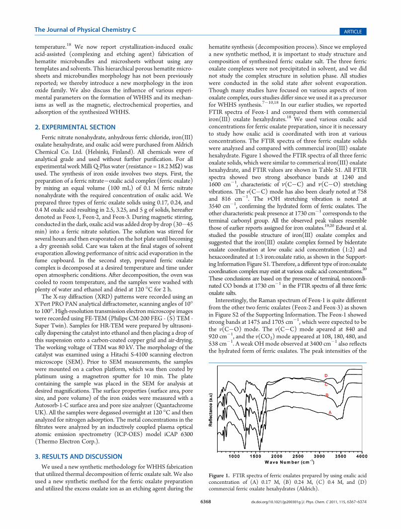
hematite synthesis (decomposition process). Since we employeda new synthetic method, it is important to study structure andcomposition of synthesized ferric oxalate salt. The three ferricoxalate complexes were not precipitated in solvent, and we didnot study the complex structure in solution phase. All studieswere conducted in the solid state after solvent evaporation.Though many studies have focused on various aspects of ironoxalate complex, ours studies differ since we used it as a precursorfor WHHS synthesis.7�10,18 In our earlier studies, we reportedFTIR spectra of Feox-1 and compared them with commercialiron(III) oxalate hexahydrates.18 We used various oxalic acidconcentrations for ferric oxalate preparation, since it is necessaryto study how oxalic acid is coordinated with iron at variousconcentrations. The FTIR spectra of three ferric oxalate solidswere analyzed and compared with commercial iron(III) oxalatehexahydrate. Figure 1 showed the FTIR spectra of all three ferricoxalate solids, which were similar to commerical iron(III) oxalatehexahydrate, and FTIR values are shown in Table S1. All FTIRspectra showed two strong absorbance bands at 1240 and1600 cm�1, characteristic of ν(C�C) and ν(C�O) stretchingvibrations. The v(C�C) mode has also been clearly noted at 758and 816 cm�1. The νOH stretching vibration is noted at3540 cm�1, confirming the hydrated form of ferric oxalates. Theother characteristic peak presence at 1730 cm�1 corresponds to theterminal carbonyl group. All the observed peak values resemblethose of earlier reports assigned for iron oxalates.19,20 Edward et al.studied the possible structure of iron(III) oxalate complex andsuggested that the iron(III) oxalate complex formed by bidentateoxalate coordination at low oxalic acid concentration (1:2) andhexacoordinated at 1:3 iron:oxalate ratio, as shown in the Support-ing Information Figure S1. Therefore, a different type of iron:oxalatecoordination complex may exist at various oxalic acid concentrations.20
These conclusions are based on the presence of terminal, noncoordi-nated CO bonds at 1730 cm�1 in the FTIR spectra of all three ferricoxalate salts.
Interestingly, the Raman spectrum of Feox-1 is quite differentfrom the other two ferric oxalates (Feox-2 and Feox-3) as shownin Figure S2 of the Supporting Information. The Feox-1 showedstrong bands at 1475 and 1705 cm�1, which were expected to bethe v(C�O) mode. The v(C�C) mode apeared at 840 and920 cm�1, and the v(CO2) mode appeared at 108, 180, 480, and538 cm�1. A weak OHmode observed at 3400 cm�1 also reflectsthe hydrated form of ferric oxalates. The peak intensities of the
Figure 1. FTIR spectra of ferric oxalates prepared by using oxalic acidconcentration of (A) 0.17 M, (B) 0.24 M, (C) 0.4 M, and (D)commercial ferric oxalate hexahydrates (Aldrich).
6369 dx.doi.org/10.1021/jp200301g |J. Phys. Chem. C 2011, 115, 6367–6374
The Journal of Physical Chemistry C ARTICLE
synthesized iron oxalates are different from the commercialiron(III) oxalate hexahydrates. The Raman spectra of ferricoxalate, however, correlate well with the FTIR spectra and earlierreports assigned for iron oxalates.19 The Feox-2 and Feox-3showed very similar Raman spectra, indicating a similar complexstructure. Thus, all the intense peaks are noted at a lowerwavenumber, and no charecteristic peaks are noted at a longerwavenumber, except one weak mode at 1300 cm�1. Generally,v(FeO) and δ(FeO2) bands appeared at lower wavenumbers,and peaks appeared at 222 cm�1. The strong peak appearing at292, 400, and 485 cm�1 is assigned for the δ(CO2) mode. Theaforementioned discussion clearly indicated that the structure ofFeox-1 differs from the other two ferric oxalates.
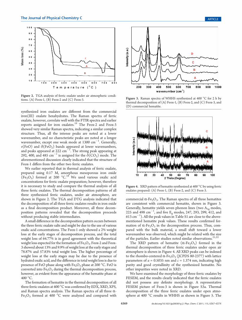
We earlier reported that in thermal analysis of ferric oxalate,prepared using 0.17 M, amorphous mesoporous iron oxide(Fe2O3) formed at 200 �C.18 We used various oxalic acidconcentrations for ferric oxalate preparation; however, thereforeit is necessary to study and compare the thermal analysis of allthree ferric oxalates. The thermal decomposition patterns of allthree synthesized ferric oxalates, under air atmosphere, areshown in Figure 2. The TGA and DTG analysis indicated thatthe decomposition of all three ferric oxalates results in iron oxideas a final decomposition product. Moreover, all three decom-position patterns revealed that the decomposition proceedswithout producing stable intermediates.
A small difference in the decomposition pattern occurs betweenthe three ferric oxalate salts and might be due to the use of variousoxalic acid concentrations. The Feox-1 only showed a 2% weightloss at the early stages of decomposition process, and the totalweight loss of 64.77% is in good agreement with the theoreticalweight loss expected for the formation of Fe2O3. Feox-2 and Feox-3 showed about 13% and 9.9%of weight loss at the early stages and78.87% and 57.83% total weight loss. The higher percentage ofweight loss at the early stages may be due to the presence ofhydrated oxalic acid, and the difference in total weight loss is due topresence of FeO phase along with Fe2O3 phase at 200 �C. FeO isconverted into Fe2O3 during the thermal decomposition process,however, as evident from the appearance of the hematite phase at400 �C.
The formation of hematite in the thermal decomposition of allthree ferric oxalates at 400 �Cwas confirmed by EDX, XRD, XPS,and Raman spectra analysis. The Raman spectra of all three R-Fe2O3 formed at 400 �C were analyzed and compared with
commercial R-Fe2O3. The Raman spectra of all three hematitesare consistent with commercial hematite, shown in Figure 3.Generally, hematite yields seven phonon lines (two A1g modes,225 and 498 cm�1, and five Eg modes, 247, 293, 299, 412, and613 cm�1). All the peak values in Table S1 are close to the above-mentioned hematite peak values. These results confirmed for-mation of R-Fe2O3 in the decomposition process. Thus, com-pared with the bulk material, a small shift toward a lowerwavenumber was observed, which might be related with the sizeof the particles. Earlier studies noted similar observations.21,22
The XRD pattern of hematite (R-Fe2O3) formed in thethermal decomposition of three ferric oxalates under open airatmosphere is shown in Figure 4. All XRD peaks can be indexedto the rhombo-centered R-Fe2O3 (JCPDS 80-2377) with latticeparameters of a = 0.5035 nm and c = 1.374 nm, indicating highpurity and good crystallinity of the synthesized hematite. Noother impurities were noted in XRD.
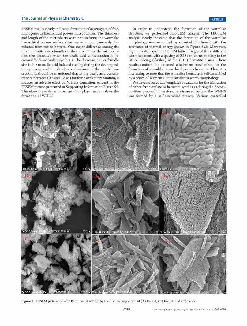
We have examined the morphology of three ferric oxalates byFESEM, and the results clearly indicated that the ferric oxalatesdid not possess any definite morphology. A representativeFESEM picture of Feox-3 is shown in Figure S3a. Thermaldecomposition of the three ferric oxalates under open atmo-sphere at 400 �C results in WHHS as shown in Figure 5. The
Figure 2. TGA analysis of ferric oxalate under air atmospheric condi-tions. (A) Feox-1, (B) Feox-2 and (C) Feox-3. Figure 3. Raman spectra of WHHS synthesized at 400 �C for 2 h by
thermal decomposition of (A) Feox-1, (B) Feox-2, and (C) Feox-3, and(D) commercial hematite.
Figure 4. XRD pattern of hematite synthesized at 400 �Cby using ferricoxalates prepared: (A) Feox-1, (B) Feox-2, and (C) Feox-3.
6370 dx.doi.org/10.1021/jp200301g |J. Phys. Chem. C 2011, 115, 6367–6374
The Journal of Physical Chemistry C ARTICLE
FESEM results clearly indicated formation of aggeragates of free,homogeneous hierarchical porous microbundles. The thicknessand length of the microsheets were not uniform; the wormlikehierarchical porous surface structure was homogeneously dis-tributed from top to bottom. One major difference among thethree hematite microbundles is their size. Thus, the microbun-dles size decreased when the oxalic acid concentration is in-creased for ferric oxalate synthesis. The decrease in microbundlesize is due to oxalic acid induced etching during the decomposi-tion process, and the details are discussed in the mechanismsection. It should be mentioned that as the oxalic acid concen-tration increases (0.5 and 0.8 M) for ferric oxalate preparation, itinduces an adverse effect on WHHS formation, evident in theFESEM picture presented in Supporting Information Figure S5.Therefore, the oxalic acid concentration plays a major role on theformation of WHHS.
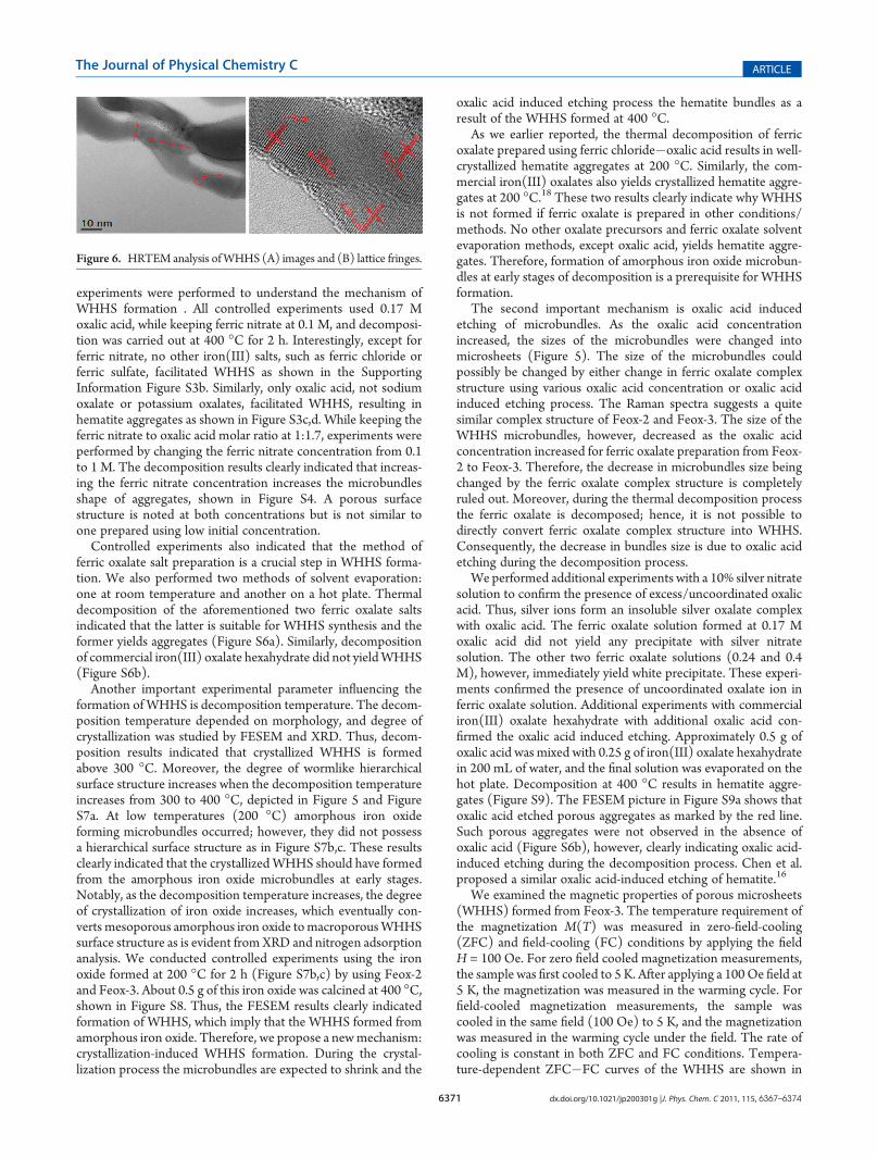
In order to understand the formation of the wormlikestructure, we performed HR-TEM analysis. The HR-TEManalysis clearly indicated that the formation of the wormlikemorphology was assembled by oriented attachment with theassistance of thermal energy shown in Figure 6a,b. Moreover,Figure 6a displays the HRTEM lattice fringes of three differentworm segments with a spacing of 0.25 nm, corresponding to thelattice spacing (d-value) of the (110) hematite planes. Theseresults confirm the oriented attachment mechanism for theformation of wormlike hierarchical porous hematite. Thus, it isinteresting to note that the wormlike hematite is self-assembledby a series of segments, quite similar to worm morphology.
We have not used any templates or catalysts for the fabricationof either ferric oxalate or hematite synthesis (during the decom-position process). Therefore, as discussed before, the WHHSwas formed by a self-assembled process. Various controlled
Figure 5. FESEM pictures of WHHS formed at 400 �C by thermal decomposition of (A) Feox-1, (B) Feox-2, and (C) Feox-3.
6371 dx.doi.org/10.1021/jp200301g |J. Phys. Chem. C 2011, 115, 6367–6374
The Journal of Physical Chemistry C ARTICLE
experiments were performed to understand the mechanism ofWHHS formation . All controlled experiments used 0.17 Moxalic acid, while keeping ferric nitrate at 0.1 M, and decomposi-tion was carried out at 400 �C for 2 h. Interestingly, except forferric nitrate, no other iron(III) salts, such as ferric chloride orferric sulfate, facilitated WHHS as shown in the SupportingInformation Figure S3b. Similarly, only oxalic acid, not sodiumoxalate or potassium oxalates, facilitated WHHS, resulting inhematite aggregates as shown in Figure S3c,d. While keeping theferric nitrate to oxalic acid molar ratio at 1:1.7, experiments wereperformed by changing the ferric nitrate concentration from 0.1to 1 M. The decomposition results clearly indicated that increas-ing the ferric nitrate concentration increases the microbundlesshape of aggregates, shown in Figure S4. A porous surfacestructure is noted at both concentrations but is not similar toone prepared using low initial concentration.
Controlled experiments also indicated that the method offerric oxalate salt preparation is a crucial step in WHHS forma-tion. We also performed two methods of solvent evaporation:one at room temperature and another on a hot plate. Thermaldecomposition of the aforementioned two ferric oxalate saltsindicated that the latter is suitable for WHHS synthesis and theformer yields aggregates (Figure S6a). Similarly, decompositionof commercial iron(III) oxalate hexahydrate did not yieldWHHS(Figure S6b).
Another important experimental parameter influencing theformation of WHHS is decomposition temperature. The decom-position temperature depended on morphology, and degree ofcrystallization was studied by FESEM and XRD. Thus, decom-position results indicated that crystallized WHHS is formedabove 300 �C. Moreover, the degree of wormlike hierarchicalsurface structure increases when the decomposition temperatureincreases from 300 to 400 �C, depicted in Figure 5 and FigureS7a. At low temperatures (200 �C) amorphous iron oxideforming microbundles occurred; however, they did not possessa hierarchical surface structure as in Figure S7b,c. These resultsclearly indicated that the crystallizedWHHS should have formedfrom the amorphous iron oxide microbundles at early stages.Notably, as the decomposition temperature increases, the degreeof crystallization of iron oxide increases, which eventually con-verts mesoporous amorphous iron oxide tomacroporousWHHSsurface structure as is evident fromXRD and nitrogen adsorptionanalysis. We conducted controlled experiments using the ironoxide formed at 200 �C for 2 h (Figure S7b,c) by using Feox-2and Feox-3. About 0.5 g of this iron oxide was calcined at 400 �C,shown in Figure S8. Thus, the FESEM results clearly indicatedformation of WHHS, which imply that the WHHS formed fromamorphous iron oxide. Therefore, we propose a newmechanism:crystallization-induced WHHS formation. During the crystal-lization process the microbundles are expected to shrink and the
oxalic acid induced etching process the hematite bundles as aresult of the WHHS formed at 400 �C.
As we earlier reported, the thermal decomposition of ferricoxalate prepared using ferric chloride�oxalic acid results in well-crystallized hematite aggregates at 200 �C. Similarly, the com-mercial iron(III) oxalates also yields crystallized hematite aggre-gates at 200 �C.18 These two results clearly indicate why WHHSis not formed if ferric oxalate is prepared in other conditions/methods. No other oxalate precursors and ferric oxalate solventevaporation methods, except oxalic acid, yields hematite aggre-gates. Therefore, formation of amorphous iron oxide microbun-dles at early stages of decomposition is a prerequisite for WHHSformation.
The second important mechanism is oxalic acid inducedetching of microbundles. As the oxalic acid concentrationincreased, the sizes of the microbundles were changed intomicrosheets (Figure 5). The size of the microbundles couldpossibly be changed by either change in ferric oxalate complexstructure using various oxalic acid concentration or oxalic acidinduced etching process. The Raman spectra suggests a quitesimilar complex structure of Feox-2 and Feox-3. The size of theWHHS microbundles, however, decreased as the oxalic acidconcentration increased for ferric oxalate preparation from Feox-2 to Feox-3. Therefore, the decrease in microbundles size beingchanged by the ferric oxalate complex structure is completelyruled out. Moreover, during the thermal decomposition processthe ferric oxalate is decomposed; hence, it is not possible todirectly convert ferric oxalate complex structure into WHHS.Consequently, the decrease in bundles size is due to oxalic acidetching during the decomposition process.
We performed additional experiments with a 10% silver nitratesolution to confirm the presence of excess/uncoordinated oxalicacid. Thus, silver ions form an insoluble silver oxalate complexwith oxalic acid. The ferric oxalate solution formed at 0.17 Moxalic acid did not yield any precipitate with silver nitratesolution. The other two ferric oxalate solutions (0.24 and 0.4M), however, immediately yield white precipitate. These experi-ments confirmed the presence of uncoordinated oxalate ion inferric oxalate solution. Additional experiments with commercialiron(III) oxalate hexahydrate with additional oxalic acid con-firmed the oxalic acid induced etching. Approximately 0.5 g ofoxalic acid was mixed with 0.25 g of iron(III) oxalate hexahydratein 200 mL of water, and the final solution was evaporated on thehot plate. Decomposition at 400 �C results in hematite aggre-gates (Figure S9). The FESEM picture in Figure S9a shows thatoxalic acid etched porous aggregates as marked by the red line.Such porous aggregates were not observed in the absence ofoxalic acid (Figure S6b), however, clearly indicating oxalic acid-induced etching during the decomposition process. Chen et al.proposed a similar oxalic acid-induced etching of hematite.16
We examined the magnetic properties of porous microsheets(WHHS) formed from Feox-3. The temperature requirement ofthe magnetization M(T) was measured in zero-field-cooling(ZFC) and field-cooling (FC) conditions by applying the fieldH = 100 Oe. For zero field cooled magnetization measurements,the sample was first cooled to 5 K. After applying a 100Oe field at5 K, the magnetization was measured in the warming cycle. Forfield-cooled magnetization measurements, the sample wascooled in the same field (100 Oe) to 5 K, and the magnetizationwas measured in the warming cycle under the field. The rate ofcooling is constant in both ZFC and FC conditions. Tempera-ture-dependent ZFC�FC curves of the WHHS are shown in
Figure 6. HRTEM analysis ofWHHS (A) images and (B) lattice fringes.
6372 dx.doi.org/10.1021/jp200301g |J. Phys. Chem. C 2011, 115, 6367–6374
The Journal of Physical Chemistry C ARTICLE
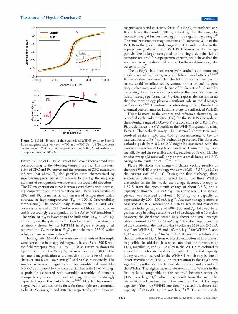
Figure 7b. The ZFC�FC curves of the Feox-3 show a broad cuspcorresponding to the blocking temperature TB. The irreversi-bility of ZFC and FC curves and the presence of ZFC maximumindicate that above TB the particles were characterized bysuperparamagnetic behavior, whereas below TB, the magneticmoment of each particle was frozen in the local field direction.23
The FC magnetization curve increases very slowly with decreas-ing temperature and tends to flatten out. There is no overlap ofZFC and FC branches at any measured temperature whichbifurcate at high temperatures, Tirr ≈ 300 K (irreversibilitytemperature). The second sharp feature in the FC and ZFCcurves is observed at 221 K—the so-called Morin transition—and is accordingly accompanied by the AF to WF transition.24
The value of TM is lower than the bulk value (TM ∼ 260 K),indicating a well-crystallized condition for the wormlike structureas already shown by the HRTEM in Figure 6. Meng et al.reported the TM value in R-Fe2O3 nanochains at 237 K, whichis higher than our observation.25
The magnetic (M�H) hysteresis measurements of the samplewere carried out in an applied magnetic field at 5 and 300 K withthe field sweeping from �10 to þ10 kOe. Figure 7a shows thehysteresis loops of the R-Fe2O3 microsheets at 5 and 300 K. Theremanent magnetization and coercivity of the R-Fe2O3 micro-sheets at 300 K are 0.089 emu g�1 and 51 Oe, respectively. Thesmaller remanent magnetization for as-obtained wormlikeR-Fe2O3 compared to the commercial hematite (0.61 emu/g)is probably associated with wormlike assembly of hematitenanoparticles, since the remanent magnetization is stronglydependent upon the particle shape.26,27 At 5 K, the remnantmagnetization and coercivity force for the sample are determinedto be 0.325 emu g�1 and 408 Oe, respectively. The remanent
magnetization and coercivity force of R-Fe2O3 microsheets at 5K are larger than under 300 K, indicating that the magneticmoment may get further freezing and the region may change.28
The smaller remanent magnetization and coercivity value of theWHHS in the present study suggest that it could be due to thesuperparamagnetic nature of WHHS. However, as the averageparticle size is larger compared to the single domain size ofhematite required for superparamagnetism, we believe that thesmaller coercivity value could account for the weak ferromagneticbehavior only.29
The R-Fe2O3 has been intensively studied as a promisinganode material for next-generation lithium ion batteries,6,17,30
Earlier studies confirmed that the lithium intercalation perfor-mance could be influenced by various properties such as poresize, surface area, and particle size of the hematite.31 Generally,increasing the surface area or porosity of the hematite increaseslithium storage performance. Previous reports also demonstratethat the morphology plays a significant role in the dischargeperformance.32,33 Therefore, it is interesting to study the electro-chemical performance for lithium storage of synthesizedWHHS.
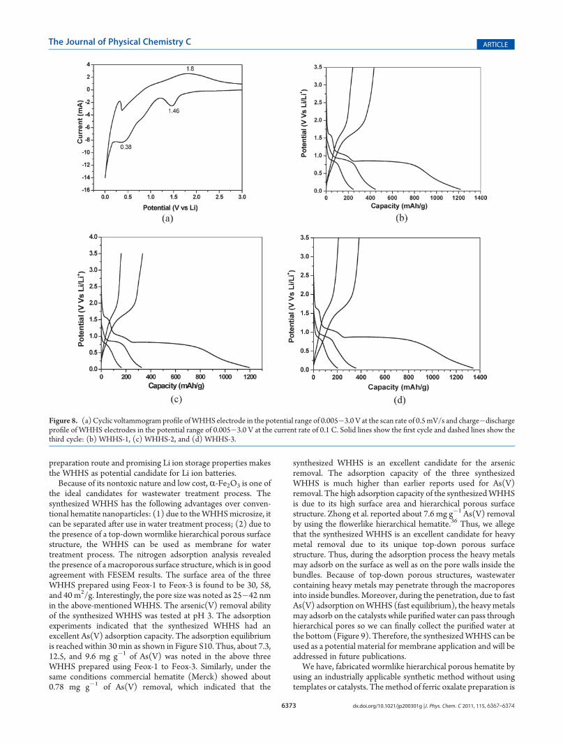
Using Li metal as the counter and reference electrodes, werecorded cyclic voltammetry (CV) for the WHHS electrode inthe potential range of 0.005�3 V at a slow scan rate of 0.5 mV/s.Figure 8a shows the CV profile of the WHHS prepared by usingFeox-3. The cathode sweep (Li insertion) shows two well-resolved peaks at 1.46 and 0.38 V corresponding to the Li-intercalation and Fe3þ to Fe0 reduction processes. The observedcathodic peak from 0.2 to 0 V might be associated with theirreversible reaction of Fe2O3 with metallic lithium into Li2O andmetallic Fe and the reversible alloying reaction of Fe with Li. Theanodic sweep (Li removal) only shows a small hump at 1.8 V,owing to the oxidation of Fe0 to Fe3þ.
Figure 8b shows the charge�discharge cycling profiles ofthe three WHHS in the voltage window of 0.01�3.5 V (vs Li) atthe current rate of 0.1 C. During the first discharge, threesuccessive plateaus were observed for all the three WHHSelectrodes. In the first cycle, the voltage decreased steeply to1.65 V from the open-circuit voltage of about 3.2 V, and acapacity of about 60�90 mA h g�1 was conquered. The secondplateau was observed at about 1.10 V up to a capacity ofapproximately 200�250 mA h g�1. Another voltage plateau isobserved at 0.8 V, whereupon a plateau sets in and maintainsuntil a discharge capacity of 800�900 mAh/g, followed by agradual drop in voltage until the end of discharge. After 10 cycles,however, the discharge profile only shows one small voltageplateau around 0.9 V. For 68 mA h g�1, the discharge capacitiesof the electrode in the first and third cycles are 1216 and 247 mAh g�1 for WHHS-1, 1198 and 162 mA h g�1 for WHHS-2, and1343 and 203 mA h g�1 for WHHS-3. It could be attributed tothe formation of Li2O, from which the extraction of Li is almostimpossible. In addition, it is speculated that the formation ofLi2O, metallic Fe, and Li�Fe alloy in the WHHS microbundlesalters the bundles size and its porosity. Thus, a fast capacityfading rate was observed for the WHHS-1, which may be due tolarger microbundles. The Li ion intercalation in the Fe2O3 wassignificantly influenced by the microbundles size and porosity ofthe WHHS. The higher capacity observed for the WHHS in thefirst cycle is comparable to the reported hematite nanorods(1151 mA h g�1),34 which may result from the wormlikehierarchical surface structures of the hematite. The first dischargecapacity of the threeWHHS considerably exceeds the theoreticalcapacity of R-Fe2O3 (1007 mA h g�1).35 Thus, the simple
Figure 7. (a) M�H loop of the synthesized WHHS by using Feox-3.Inset: magnetization between �700 and þ700 Oe. (b) Temperaturedependence of ZFC and FC magnetization of R-Fe2O3 microsheets atthe applied field of 100 Oe.
6373 dx.doi.org/10.1021/jp200301g |J. Phys. Chem. C 2011, 115, 6367–6374
The Journal of Physical Chemistry C ARTICLE
preparation route and promising Li ion storage properties makesthe WHHS as potential candidate for Li ion batteries.
Because of its nontoxic nature and low cost, R-Fe2O3 is one ofthe ideal candidates for wastewater treatment process. Thesynthesized WHHS has the following advantages over conven-tional hematite nanoparticles: (1) due to theWHHSmicrosize, itcan be separated after use in water treatment process; (2) due tothe presence of a top-down wormlike hierarchical porous surfacestructure, the WHHS can be used as membrane for watertreatment process. The nitrogen adsorption analysis revealedthe presence of a macroporous surface structure, which is in goodagreement with FESEM results. The surface area of the threeWHHS prepared using Feox-1 to Feox-3 is found to be 30, 58,and 40 m2/g. Interestingly, the pore size was noted as 25�42 nmin the above-mentioned WHHS. The arsenic(V) removal abilityof the synthesized WHHS was tested at pH 3. The adsorptionexperiments indicated that the synthesized WHHS had anexcellent As(V) adsorption capacity. The adsorption equilibriumis reached within 30min as shown in Figure S10. Thus, about 7.3,12.5, and 9.6 mg g�1 of As(V) was noted in the above threeWHHS prepared using Feox-1 to Feox-3. Similarly, under thesame conditions commercial hematite (Merck) showed about0.78 mg g�1 of As(V) removal, which indicated that the
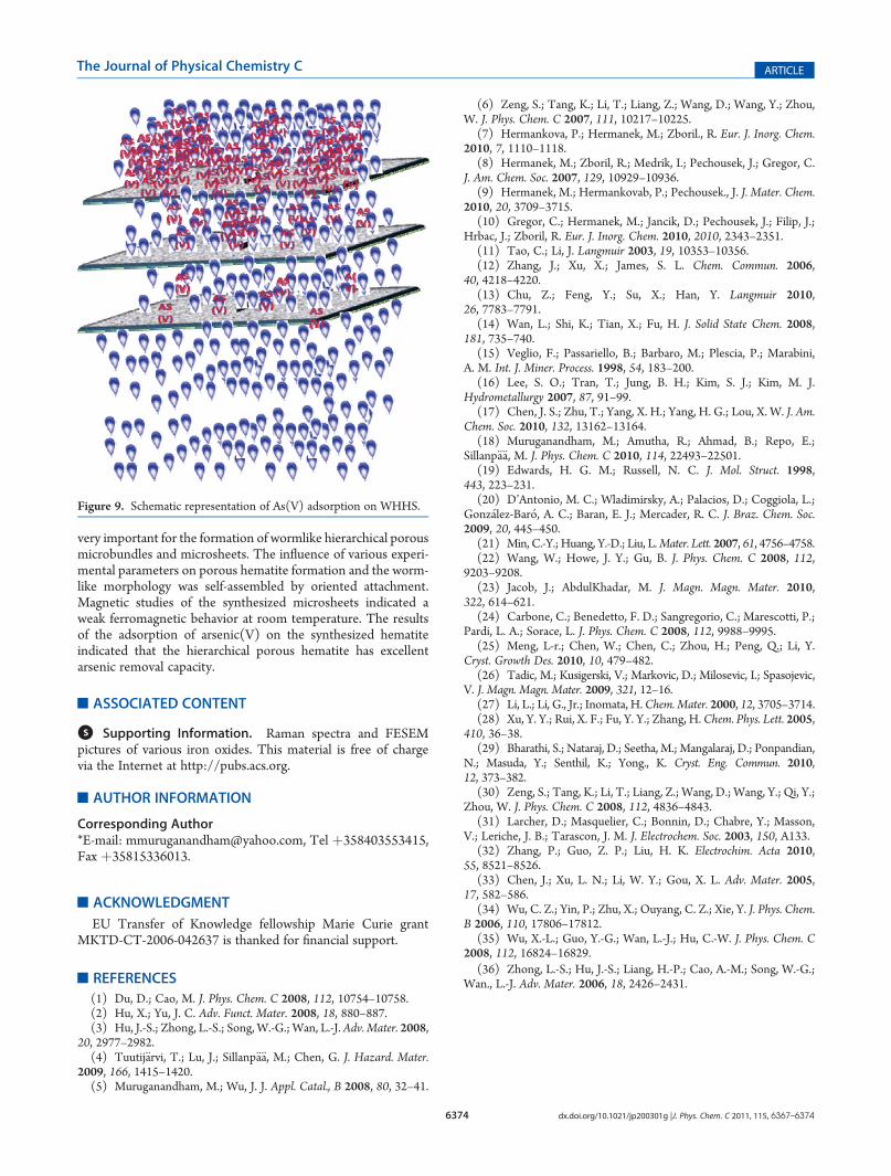
synthesized WHHS is an excellent candidate for the arsenicremoval. The adsorption capacity of the three synthesizedWHHS is much higher than earlier reports used for As(V)removal. The high adsorption capacity of the synthesizedWHHSis due to its high surface area and hierarchical porous surfacestructure. Zhong et al. reported about 7.6 mg g�1 As(V) removalby using the flowerlike hierarchical hematite.36 Thus, we allegethat the synthesized WHHS is an excellent candidate for heavymetal removal due to its unique top-down porous surfacestructure. Thus, during the adsorption process the heavy metalsmay adsorb on the surface as well as on the pore walls inside thebundles. Because of top-down porous structures, wastewatercontaining heavy metals may penetrate through the macroporesinto inside bundles. Moreover, during the penetration, due to fastAs(V) adsorption onWHHS (fast equilibrium), the heavy metalsmay adsorb on the catalysts while purified water can pass throughhierarchical pores so we can finally collect the purified water atthe bottom (Figure 9). Therefore, the synthesizedWHHS can beused as a potential material for membrane application and will beaddressed in future publications.
We have, fabricated wormlike hierarchical porous hematite byusing an industrially applicable synthetic method without usingtemplates or catalysts. Themethod of ferric oxalate preparation is
Figure 8. (a) Cyclic voltammogram profile ofWHHS electrode in the potential range of 0.005�3.0 V at the scan rate of 0.5mV/s and charge�dischargeprofile of WHHS electrodes in the potential range of 0.005�3.0 V at the current rate of 0.1 C. Solid lines show the first cycle and dashed lines show thethird cycle: (b) WHHS-1, (c) WHHS-2, and (d) WHHS-3.
6374 dx.doi.org/10.1021/jp200301g |J. Phys. Chem. C 2011, 115, 6367–6374
The Journal of Physical Chemistry C ARTICLE
very important for the formation of wormlike hierarchical porousmicrobundles and microsheets. The influence of various experi-mental parameters on porous hematite formation and the worm-like morphology was self-assembled by oriented attachment.Magnetic studies of the synthesized microsheets indicated aweak ferromagnetic behavior at room temperature. The resultsof the adsorption of arsenic(V) on the synthesized hematiteindicated that the hierarchical porous hematite has excellentarsenic removal capacity.
’ASSOCIATED CONTENT
bS Supporting Information. Raman spectra and FESEMpictures of various iron oxides. This material is free of chargevia the Internet at http://pubs.acs.org.
’AUTHOR INFORMATION
Corresponding Author*E-mail: [email protected], Tel þ358403553415,Fax þ35815336013.
’ACKNOWLEDGMENT
EU Transfer of Knowledge fellowship Marie Curie grantMKTD-CT-2006-042637 is thanked for financial support.
’REFERENCES
(1) Du, D.; Cao, M. J. Phys. Chem. C 2008, 112, 10754–10758.(2) Hu, X.; Yu, J. C. Adv. Funct. Mater. 2008, 18, 880–887.(3) Hu, J.-S.; Zhong, L.-S.; Song, W.-G.; Wan, L.-J. Adv. Mater. 2008,
20, 2977–2982.(4) Tuutij€arvi, T.; Lu, J.; Sillanp€a€a, M.; Chen, G. J. Hazard. Mater.
2009, 166, 1415–1420.(5) Muruganandham, M.; Wu, J. J. Appl. Catal., B 2008, 80, 32–41.
(6) Zeng, S.; Tang, K.; Li, T.; Liang, Z.; Wang, D.; Wang, Y.; Zhou,W. J. Phys. Chem. C 2007, 111, 10217–10225.
(7) Hermankova, P.; Hermanek, M.; Zboril., R. Eur. J. Inorg. Chem.2010, 7, 1110–1118.
(8) Hermanek, M.; Zboril, R.; Medrik, I.; Pechousek, J.; Gregor, C.J. Am. Chem. Soc. 2007, 129, 10929–10936.
(9) Hermanek, M.; Hermankovab, P.; Pechousek., J. J. Mater. Chem.2010, 20, 3709–3715.
(10) Gregor, C.; Hermanek, M.; Jancik, D.; Pechousek, J.; Filip, J.;Hrbac, J.; Zboril, R. Eur. J. Inorg. Chem. 2010, 2010, 2343–2351.
(11) Tao, C.; Li, J. Langmuir 2003, 19, 10353–10356.(12) Zhang, J.; Xu, X.; James, S. L. Chem. Commun. 2006,
40, 4218–4220.(13) Chu, Z.; Feng, Y.; Su, X.; Han, Y. Langmuir 2010,
26, 7783–7791.(14) Wan, L.; Shi, K.; Tian, X.; Fu, H. J. Solid State Chem. 2008,
181, 735–740.(15) Veglio, F.; Passariello, B.; Barbaro, M.; Plescia, P.; Marabini,
A. M. Int. J. Miner. Process. 1998, 54, 183–200.(16) Lee, S. O.; Tran, T.; Jung, B. H.; Kim, S. J.; Kim, M. J.
Hydrometallurgy 2007, 87, 91–99.(17) Chen, J. S.; Zhu, T.; Yang, X. H.; Yang, H. G.; Lou, X. W. J. Am.
Chem. Soc. 2010, 132, 13162–13164.(18) Muruganandham, M.; Amutha, R.; Ahmad, B.; Repo, E.;
Sillanp€a€a, M. J. Phys. Chem. C 2010, 114, 22493–22501.(19) Edwards, H. G. M.; Russell, N. C. J. Mol. Struct. 1998,
443, 223–231.(20) D’Antonio, M. C.; Wladimirsky, A.; Palacios, D.; Coggiola, L.;
Gonz�alez-Bar�o, A. C.; Baran, E. J.; Mercader, R. C. J. Braz. Chem. Soc.2009, 20, 445–450.
(21) Min, C.-Y.; Huang, Y.-D.; Liu, L.Mater. Lett. 2007, 61, 4756–4758.(22) Wang, W.; Howe, J. Y.; Gu, B. J. Phys. Chem. C 2008, 112,
9203–9208.(23) Jacob, J.; AbdulKhadar, M. J. Magn. Magn. Mater. 2010,
322, 614–621.(24) Carbone, C.; Benedetto, F. D.; Sangregorio, C.; Marescotti, P.;
Pardi, L. A.; Sorace, L. J. Phys. Chem. C 2008, 112, 9988–9995.(25) Meng, L-r.; Chen, W.; Chen, C.; Zhou, H.; Peng, Q.; Li, Y.
Cryst. Growth Des. 2010, 10, 479–482.(26) Tadic, M.; Kusigerski, V.; Markovic, D.; Milosevic, I.; Spasojevic,
V. J. Magn. Magn. Mater. 2009, 321, 12–16.(27) Li, L.; Li, G., Jr.; Inomata, H.Chem. Mater. 2000, 12, 3705–3714.(28) Xu, Y. Y.; Rui, X. F.; Fu, Y. Y.; Zhang, H.Chem. Phys. Lett. 2005,
410, 36–38.(29) Bharathi, S.; Nataraj, D.; Seetha, M.; Mangalaraj, D.; Ponpandian,
N.; Masuda, Y.; Senthil, K.; Yong., K. Cryst. Eng. Commun. 2010,12, 373–382.
(30) Zeng, S.; Tang, K.; Li, T.; Liang, Z.; Wang, D.; Wang, Y.; Qi, Y.;Zhou, W. J. Phys. Chem. C 2008, 112, 4836–4843.
(31) Larcher, D.; Masquelier, C.; Bonnin, D.; Chabre, Y.; Masson,V.; Leriche, J. B.; Tarascon, J. M. J. Electrochem. Soc. 2003, 150, A133.
(32) Zhang, P.; Guo, Z. P.; Liu, H. K. Electrochim. Acta 2010,55, 8521–8526.
(33) Chen, J.; Xu, L. N.; Li, W. Y.; Gou, X. L. Adv. Mater. 2005,17, 582–586.
(34) Wu, C. Z.; Yin, P.; Zhu, X.; Ouyang, C. Z.; Xie, Y. J. Phys. Chem.B 2006, 110, 17806–17812.
(35) Wu, X.-L.; Guo, Y.-G.; Wan, L.-J.; Hu, C.-W. J. Phys. Chem. C2008, 112, 16824–16829.
(36) Zhong, L.-S.; Hu, J.-S.; Liang, H.-P.; Cao, A.-M.; Song, W.-G.;Wan., L.-J. Adv. Mater. 2006, 18, 2426–2431.
Figure 9. Schematic representation of As(V) adsorption on WHHS.
Related Documents