Crystal structure of the SARS coronavirus nucleocapsid protein dimerization domain I-Mei Yu 1,3 , Michael L. Oldham 1,3 , Jingqiang Zhang 2 , and Jue Chen 1,* 1 Department of Biological Sciences and the Cancer Center, Purdue University, West Lafayette, IN 47907 2 State Key Lab for Biocontrol, Zhongshan University, Guangzhou, P.R.China 3 These authors contributed equally to this work * To whom correspondence should be addressed: Department of Biological Sciences Purdue University West Lafayette, IN 47907-1393 Phone: 765-496-3113 FAX: 765-496-1189 Email: [email protected] The coordinates and the structure factors have been deposited in the Protein Data Bank under ID code XXX.

Welcome message from author
This document is posted to help you gain knowledge. Please leave a comment to let me know what you think about it! Share it to your friends and learn new things together.
Transcript

Crystal structure of the SARS coronavirus nucleocapsid protein
dimerization domain
I-Mei Yu 1,3, Michael L. Oldham1,3, Jingqiang Zhang2, and Jue Chen1,*
1Department of Biological Sciences and the Cancer Center, Purdue University, West
Lafayette, IN 47907
2State Key Lab for Biocontrol, Zhongshan University, Guangzhou, P.R.China
3These authors contributed equally to this work
*To whom correspondence should be addressed:
Department of Biological Sciences
Purdue University
West Lafayette, IN 47907-1393
Phone: 765-496-3113
FAX: 765-496-1189
Email: [email protected]
The coordinates and the structure factors have been deposited in the Protein Data Bank under ID code XXX.

Summary
The causative agent of severe acute respiratory syndrome (SARS) is the SARS-associated
coronavirus, SARS-CoV. The nucleocapsid (N) protein plays an essential role in SARS-
CoV genome packaging and virion assembly. We have previously shown that SARS-CoV N
protein forms a dimer in solution through its C-terminal domain. In this study, the crystal
structure of the dimerization domain, consisting of residues 270 to 370, is determined to
1.75 Å resolution. The structure shows a dimer with extensive interactions between the two
subunits, suggesting that the dimeric form of the N protein is the functional unit in vivo.
Although lacking any sequence similarity, the dimerization domain of SARS-CoV N protein
has a fold similar to that of the nucleocapsid protein of porcine reproductive and respiratory
syndrome virus (PRRSV). This finding provides structural evidence of the evolutionary link
between Coronaviridae and Arteriviridae, suggesting that the N proteins of both viruses
have a common origin.
2

Introduction
Coronaviruses are enveloped, single-stranded, positive-sense RNA viruses that infect
a variety of mammals and birds. Although previously identified human coronaviruses cause
only mild respiratory infections, in the 2003 outbreak of severe acute respiratory syndrome
(SARS), a disease caused by a new type of coronaviruses (SARS-CoV), there were more
than 8,000 cases resulting in 774 deaths (approximately 10 percent mortality). Phylogenetic
analysis suggests that SARS-CoV diverged early from group 2 coronaviruses and has
evolved independently for a long period of time (Stadler et al., 2003).
The coronavirus genome, containing approximately 30,000 bases, is the largest
among positive-sense RNA viruses (Lai and Cavanagh, 1997; Lai and Holmes, 2001). It
encodes non-structural proteins including the RNA polymerase and helicase, as well as the
spike (S), envelope (E), membrane (M), and nucleocapsid (N) structural proteins. The
coronavirus virion is about 120 nm in diameter and consists of a lipid envelope containing
three or four anchored glycoproteins and a helical ribonucleoprotein core (Sturman et al.,
1980). The surface projections forming the crown-like structure observed under the electron
microscope are made up of the S protein, which is responsible for receptor recognition and
membrane fusion (Babcock et al., 2004; Bos et al., 1995; Bosch et al., 2003; Krueger et al.,
2001; Kubo et al., 1994). The integral membrane proteins M and E are essential for virus
budding. When co-expressed in animal cells, the M and E proteins are sufficient to form
virus-like particles (Vennema et al., 1996). The N protein interacts with the viral genome to
form the ribonucleoprotein core; and has been shown to be involved in viral RNA synthesis,
transcriptional regulation of genomic RNA, translation of viral proteins, and budding (He et
al., 2003; Lai and Cavanagh, 1997; Tahara et al., 1998).
3

Coronaviruses are related to arteriviruses by their similar genome organizations and
viral replication mechanisms (Lai and Holmes, 2001; Snijder and Meulenberg, 2001).
Recently, Coronaviridae and Arteriviridae were united to form the new order Nidovirales.
The name of the order comes from a property common to both viruses, a nested set of
subgenomic mRNAs for structural protein expression (L. nidus, meaning nest). The
replicase genes of arteriviruses and coronaviruses are thought to have a common origin since
they both have conserved domains that are present at the same relative positions (Cavanagh,
1997; Lai and Cavanagh, 1997; Snijder and Meulenberg, 1998). The structural proteins,
however, are thought to be unrelated due to differences in protein size and lack of sequence
identity (Cavanagh, 1997).
Coronavirus N proteins are composed of three distinct domains. High resolution
structures of the N-terminal domain (~ 130 residues) were determined by NMR (Huang et
al., 2004) and crystallography (Fan et al., 2005). This region folds similarly to the U1A
RNA-binding protein and is suggested to bind RNA (Fan et al., 2005; Huang et al., 2004).
The central region of the N protein has also been shown by several laboratories to be an
RNA-binding domain (Masters, 1992; Nelson and Stohlman, 1993; Nelson et al., 2000;
Peng et al., 1995). No structural information is available for the central domain, possibly due
to its highly positively charged and flexible nature. Recently, secondary structure elements
of the C-terminal residues 248-365 of SARS-CoV N protein have been reported from NMR
studies; however, the tertiary structure was not resolved (Chang et al., 2005). Here we report
the crystal structure of the C-terminal domain of SARS-CoV N protein consisting of
residues 270-370. Consistent with biochemical studies showing that this domain mediates
dimer formation of the N protein (Fan et al., 2005; Yu et al., 2005), the structure consists of
4

a dimer with extensive interactions between subunits, suggesting that the N protein is not
stable in the monomeric form and that the dimeric conformation represents the functional
unit of the N protein. Furthermore, the C-terminal structure of SARS-CoV N protein shows
similarity to that of the N protein of porcine reproductive and respiratory syndrome virus
(PRRSV), a member of Arteriviridae. The structural similarity between these two N proteins,
in the absence of sequence identity, adds further evidence that Coronaviridae and
Arteriviridae have evolved from a common ancestor.
Results
Crystallization and Structure Determination
The full length SARS-CoV N protein (SARS N) and 18 truncated constructs were
expressed in bacteria and purified for crystallization. Whereas most constructs yielded either
no crystals or merely clusters of small needles, a C-terminal construct containing residues
270-370 (cSARS-N) was readily crystallized to give well diffracting crystals. The
diffraction pattern indicated that the crystals were unusually twinned resulting in two
overlapping monoclinic lattices of similar unit cell dimensions. The two lattices are related
by a 180° rotation around the y-axis followed by a 50° rotation around the z-axis. As a result,
nearly half of the observable reflections overlapped to some extent. The twinning
phenomenon was reproducible regardless of the changes in temperature, precipitant, pH, and
salt concentration. Because of the large number of partially overlapped reflections, only the
central four pixels of each reflection were measured, these being proportional to the
integrated intensity presuming the profile was fairly constant (details of the data processing
5

procedure will be published elsewhere). The inflated I/sigma values (Table I) were caused
by the incorporation of overlapping reflections during the intensity measurements.
The structure of cSARS-N was determined by MAD using a SeMet derivative to
1.75 Å resolution. The crystal belongs to space group C2 and the asymmetric unit contains a
dimer of two subunits related by a 180° rotation. The final model, containing residues 270-
366 of one subunit and 274-369 of the other, was refined to a Rwork/Rfree of 18.7/23.5 (Table
I). A representative 2Fo - Fc electron density map is shown in Figure 1. The stereochemical
quality of the model was analyzed with PROCHECK, and 97.5% of nonglycine residues are
in the most favored regions with no disallowed residues.
Overall Structure
Sequence comparison shows that the dimerization domain of the nucleocapsid
protein is conserved among the three groups of coronaviruses, suggesting a common
structural and functional role of this domain (Fig 1A). The monomer of cSARS-N contains
five short α- helices, one 310 helix, and two β-strands. The overall shape of the monomer
resembles the letter C, with one edge formed by a β-hairpin extending away from the rest of
the molecule (Fig 1C). In contrast to many small proteins that are remarkably compact, the
monomeric cSARS-N domain folds into an extended conformation with a large cavity in its
center (Fig 1C). It is therefore likely that the N protein is not stable in the monomeric form
and higher oligomerization of the polypeptide is necessary to produce a stable conformation.
Consistent with previous biochemical studies suggesting that the full length SARS N protein
forms a dimer in solution through its C-terminal domain (Fan et al., 2005; Yu et al., 2005), a
compact dimeric form of cSARS-N was observed in the crystal (Fig 1D). The dimer has a
6

flat structure of approximate dimensions 48 x 42 x 25 Å. The two subunits in the dimer are
almost identical, with an rms deviation of 0.36 Å over 92 Cα atoms out of 101 residues and
the largest displacement of 0.56 Å. The dimer interface is largely formed by insertion of the
β-hairpin of one subunit into the cavity of the opposite subunit (Fig 1D). As a result, the 4 β-
strands of the two subunits form an anti-parallel β-sheet, with 10 hydrogen bonds formed
across the dimer interface by the main chain atoms of residues 330-340. Two large,
pyramidal hydrophobic cores form a bow-tie shaped pocket which further stabilizes the
dimer. Each core consists of base residues F287, L292, W302, I305, F308, Y334, I338,
L340, L354, I358 from one subunit and apex residues F315, F316, I321, and L332 from the
other subunit. The buried area in the dimer interface is 2093 Å2 per subunit, which accounts
for 28% of the total solvent accessible surface area of each subunit. The extended structure
of the monomeric subunit and the extensive interactions between the subunits within a dimer
suggest that the dimeric arrangement observed crystallographically represents the biological
architecture of the coronavirus N protein. The structure of cSARS-N is consistent with
biochemical studies showing that residues 1-284 of the SARS N protein are dispensable for
dimer formation (Yu et al., 2005). The strong interactions between the two subunits explain
the previous observation that a significant amount of denaturant (4 M urea) was necessary to
disrupt the dimer in solution (Yu et al., 2005).
A recent NMR study reported secondary structural assignments of a SARS N protein
construct that is similar to cSARS-N (Chang et al., 2005). Despite the difficulty in resolving
the 3D structure, a dimeric interface consisting of a 4-stranded anti-parallel β-sheet and two
α-helices was proposed (Chang et al., 2005). Comparing the crystal structure with the
proposed NMR structure, one of the most notable differences is the location of the longest
7

helix, helix E (residues 346-357). In the crystal structure, helices E of both subunits are
located at the edge of the dimer molecule, away from each other (Fig 1D); while in the
structure proposed by NMR, they are placed at the interface to stabilize the dimer (Chang et
al., 2005).
Structural Similarity to the Nucleocapsid Protein of Arterivirus A DALI search of the protein data bank did not identify any other proteins with
similar folds to that of cSARS-N. However, common structural features can be found
between cSARS-N and the nucleocapsid protein of PRRSV (Fig 2). PRRSV belongs to the
recently recognized family Arteriviridae, which is in the same order as Coronaviridae. The
crystal structure of the C-terminal 65 residues of PRRSV N protein (Doan and Dokland,
2003) is a homodimer. Each subunit consists of two central anti-parallel β-strands flanked
by three α-helices. While the primary sequences of PRRSV N and cSARS-N have no
significant similarity (Fig 2A), superposition of the monomeric N proteins of PRRSV and
SARS-CoV shows that the two N proteins have similar folds in a core region of ~50
residues (Fig 2B and 2C). The central β-strands are superimposable and the helices
following the β-strands are of similar length and position (Fig 2B). In addition, as proposed
in the recent NMR study (Chang et al., 2005), the dimer interface of the two N proteins are
very similar, noted by the intertwining of the central β-strands to form a 4-stranded sheet
(Fig 2C). This is evident by an rms deviation of 2.24 Å for the Cα positions among 38
residues at the dimer interface.
8

Discussion
Specific packaging of the viral genome into the virion is a critical step in the life
cycle of an infectious virus. The N protein plays an essential role in this process through
self-association and interactions with viral RNA and other viral proteins. In an effort to
understand the mechanism of how SARS N protein functions, we determined the crystal
structure of its C-terminal dimerization domain. Extensive hydrogen-bonding and
hydrophobic interactions are observed between the two subunits within a crystallographic
dimer, suggesting that the functional unit of the N protein is dimeric. A model of the full-
length SARS N protein is proposed in Figure 3, where the two known SARS N protein
domains (residues 49-178 and 270-370) are connected by a flexible central region. In this
model, the N-terminal domain is proposed as monomeric, as indicated by its NMR structure
(Huang et al., 2004). Strong protein-protein interactions at the dimerization region may be
essential to hold the highly charged RNA-binding domains in close proximity, thereby
facilitating the formation of a large helical nucleocapsid core. Association of the N protein
dimers is necessary for further assembly of the core. The full-length dimeric N protein has a
propensity to form tetramers and higher molecular weight oligomers in vitro (Yu et al.,
2005). A serine/arginine-rich motif (residues 184-196) was shown to be important for N
protein oligomerization (He et al., 2004). Since constructs containing residues 211-422 or
285-422 of SARS N do not form oligomers larger than dimers (Yu et al., 2005), it is
possible that the serine/arginine-rich motif located outside the dimerization domain is
necessary to mediate further association of N protein dimers. Recently, the C-terminal 45
residues of the mouse hepatitis virus N protein were shown to be the major determinant for
9

interaction with the M protein (Hurst et al., 2005). Association of the N protein with the M
protein may also play a role in the assembly of the nucleocapsid core into a progeny virion.
RNA viruses have high rates of sequence divergence and genome recombination,
and thus it is often a challenge to study the evolutionary relationships among viruses.
Structural studies have become an important method for revealing distant relationships
among viruses. Among all lipid-enveloped RNA viruses, high resolution structures of the
proteins that package the viral genome into virions, often termed the nucleocapsid or capsid
proteins, are currently available from five virus families (Fig 4) (Retroviridae are not
included in this discussion because their replication pathway is through a DNA intermediate
(Goff, 2001)). Although functionally equivalent, the size and structure of nucleocapsid
proteins are remarkably diverse among these five virus families. The nucleoprotein of borna
disease virus (BDV), a single-stranded (ss) RNA virus in the family Bornaviridae, contains
370 residues and folds into an S-shaped molecule consisting of 16 helices and 2 short β-
strands (Rudolph et al., 2003). In the crystal lattice, four subunits of BDV N interact
extensively to form a homotetramer, suggesting that the functional unit of BDV N is a
tetramer (Fig 4A). Both Semliki Forest virus (genus Alphavirus, family Togaviridae) and
dengue virus (genus Flavivirus, family Flaviviridae) are small ssRNA viruses with
icosahedral symmetry. It has been suggested that they have a common ancestor because the
structures of one of their surface glycoproteins are remarkably similar (Lescar et al., 2001;
Pletnev et al., 2001; Rey et al., 1995). Inside the lipid bilayer, however, the nucleocapsid
cores of these two viruses are quite different. An alphavirus core consists of 240 copies of
the capsid protein arranged in a T=4 icosahedral lattice (Zhang et al., 2002). Each capsid
protein has ~270 residues, of which the N-terminal ~120 residues are flexible. The
10

remaining 150 residues adopt a chymotrypsin-like fold and form homodimers in the crystal
(Choi et al., 1997; Choi et al., 1991) (Fig 4C). In contrast, cryoelectron microscopy
reconstructions of flaviviruses did not show a defined structure of the capsid core (Kuhn et
al., 2002; Zhang et al., 2003), suggesting that the core is either disordered or has a different
symmetry from the surface icosahedral lattice. In addition, the exact copy number of the
capsid protein in the core is unclear. The C-terminal 100 residues of flavivirus capsid
proteins fold into a four-helical structure with no similarity to that of an alphavirus capsid
protein (Dokland et al., 2004; Ma et al., 2004) (Fig 4).
It is striking that, within the group of enveloped RNA viruses, no two nucleocapsid
proteins are known to be structurally similar except those of Arteriviridae and
Coronaviridae (Fig 4). The structural similarity between the N proteins of SARS-CoV and
PRRSV provides valuable information for understanding the evolutionary links between
corona- and arteriviruses. Although grouped in the same order, Nidovirales, the structural
proteins of these viruses were previously thought to be unrelated due to marked difference in
their size and lack of sequence identity (Cavanagh, 1997). In this study, a common fold of
the C-terminal domain Nidovirales N proteins is observed, suggesting a possible common
origin of these two proteins. The amino acid sequence and gene size diversity may have
resulted from extensive mutation and RNA recombination during evolution. It is also
important to note that the assembled cores of corona- and arteriviruses have completely
different structures. PRRSV N protein interacts with the RNA genome to form a spherical,
possibly icosahedral core (Brinton-Darnell and Plagemann, 1975) while coronaviruses are
known to have helical nucleocapsids (Macneughton and Davies, 1978). It was previously
suggested that the ancestral virus had an icosahedral nucleocapsid core, and the larger N
11

protein of coronavirus, freed from its icosahedral package constraints, allowed the
coronavirus genome to become larger during evolution (Godeny et al., 1993).
Experimental Procedures
Cloning, expression, and purification
DNA encoding various fragments of SARS-CoV (strain GD01) nucleocapsid protein,
SARS N, was cloned into a pMCSG7 plasmid with an N-terminal 6x His tag and a tobacco
etch virus (TEV) protease cleavage site. BL21(DE3)Star E. coli cells (Invitrogen),
transformed with the expression plasmid, were grown to log phase at 37°C, cooled to 16°C,
and protein expression was induced by the addition of 0.1 mM IPTG. Cells were harvested
by centrifugation at 4000 x g for 20 min. Cell pellets were resuspended in lysis buffer (20
mM Tris pH 8.5, 200 mM NaCl) and lysed by sonication. Cell lysate was centrifuged at
90,000 x g for 45 min at 4°C; and the supernatant was loaded onto a cobalt column
(Clontech laboratories; Talon metal affinity resin). The column was then washed with lysis
buffer, followed by lysis buffer plus 5 mM imidazole. The protein was eluted with lysis
buffer plus 100 mM imidazole. Fractions containing SARS N protein were pooled and
dialyzed against 20 mM Tris pH 8.5, 150 mM NaCl to remove the imidazole. TEV protease
was added to 10% (w/w) and the reaction mixture was incubated at 30 °C for 10 hours.
Complete removal of the affinity His tag was monitored by SDS-PAGE. Further purification
was achieved by size exclusion chromatography (Amersham Biosciences; Superdex 75) in
lysis buffer. The Seleno-methionine (SeMet) derivatized protein was expressed in the
methionine auxotroph strain B834(DE3) in minimal medium supplemented with SeMet
(Sigma). SeMet protein was purified as described above for the native protein except that 10
12
mM DTT was added prior to size exclusion chromatography to keep all SeMet in the
reduced form. Incorporation of SeMet was confirmed by mass spectrometry and
fluorescence scans around the Se absorption edge.
Crystallization and Structure Determination
The protein sample was dialyzed against 10 mM Tris at pH 8.5 and 70 mM NaCl and
concentrated to 45 mg/ml. Crystals were grown by mixing protein solution with the
reservoir solution containing 30-33 % pentaerythritol ethoxylate 15/4 EO/OH, 50 mM
(NH4)2SO4, and 50 mM Bis-Tris pH 6.5 at 1:1 ratio in sitting drops by vapor diffusion at
4°C. Crystals were looped out of the drop directly and flash-frozen in liquid nitrogen. Data
were collected at 100K with a Quantum-Q315 CCD (ADSC) detector on beamline 19-ID at
the Advanced Photon Source (Structural Biology Center, Argonne National Laboratory). X-
ray diffraction data for the initial structure determination were collected to 2.2 Å resolution
from crystals containing SeMet incorporated protein. Phases were obtained from the four Se
sites found by multiwavelength anomalous dispersion (MAD) using the program SOLVE
(Terwilliger, 2003) and improved by solvent-flattening using CNS (Brunger et al., 1998).
Initial model was built automatically using Arp/Warp in CCP4 (Collaborative
Computational Project, 1994). A higher resolution data set (1.75 Å) from a SeMet
incorporated protein crystal was used for refinement. The resolution was extended over
several cycles of model building in O (Jones et al., 1991) and refinement in REFMAC5 in
CCP4 (Collaborative Computational Project, 1994).
13

Acknowledgements
While this work was in progress, the structure of the nucleocapsid protein of another
coronavirus, the avian infectious bronchitis virus (IBV), was determined by Drs. H. Jayaram
and B. V. V. Prasad at Baylor College of Medicine. The coordinates of IBV N protein were
kindly provided to us by them for phasing via molecular replacement. However, they were
unnecessary since the experimental MAD phasing was sufficient to build the initial model.
We thank Dr M. G. Rossmann for many helpful discussions and Drs. R. J. Kuhn, M. G.
Rossmann, E.G. Strauss, and J.H. Strauss for critical reading of the manuscript. We also
thank the beamline staff at SBC 19-ID at the Advanced Photon Source for assistance with
data collection, and the Purdue Cancer Center for X-ray and DNA sequencing facilities. This
work was supported by NIH grant (P01 AI055672 to R. J. K.) and the Pew Scholars Program
in the Biomedical Sciences (to J.C.).
14

References:
Babcock, G. J., Esshaki, D. J., Thomas, W. D., Jr., and Ambrosino, D. M. (2004). Amino acids 270 to 510 of the severe acute respiratory syndrome coronavirus spike protein are required for interaction with receptor. J Virol 78, 4552-4560. Barton, G. J. (1993). ALSCRIPT: a tool to format multiple sequence alignments. Protein Eng 6, 37-40. Bos, E. C., Heijnen, L., and Spaan, W. J. (1995). Site directed mutagenesis of the murine coronavirus spike protein. Effects on fusion. Adv Exp Med Biol 380, 283-286. Bosch, B. J., van der Zee, R., de Haan, C. A., and Rottier, P. J. (2003). The coronavirus spike protein is a class I virus fusion protein: structural and functional characterization of the fusion core complex. J Virol 77, 8801-8811. Brinton-Darnell, M., and Plagemann, P. G. (1975). Structure and chemical-physical characteristics of lactate dehydrogenase-elevating virus and its RNA. J Virol 16, 420-433. Brunger, A. T., Adams, P. D., Clore, G. M., DeLano, W. L., Gros, P., Grosse-Kunstleve, R. W., Jiang, J. S., Kuszewski, J., Nilges, M., Pannu, N. S., et al. (1998). Crystallography & NMR system: A new software suite for macromolecular structure determination. Acta Crystallogr D Biol Crystallogr 54 ( Pt 5), 905-921. Cavanagh, D. (1997). Nidovirales: a new order comprising Coronaviridae and Arteriviridae. Arch Virol 142, 629-633. Chang, C. K., Sue, S. C., Yu, T. H., Hsieh, C. M., Tsai, C. K., Chiang, Y. C., Lee, S. J., Hsiao, H. H., Wu, W. J., Chang, C. F., and Huang, T. H. (2005). The dimer interface of the SARS coronavirus nucleocapsid protein adapts a porcine respiratory and reproductive syndrome virus-like structure. FEBS Lett 579, 5663-5668. Choi, H. K., Lu, G., Lee, S., Wengler, G., and Rossmann, M. G. (1997). Structure of Semliki Forest virus core protein. Proteins 27, 345-359. Choi, H. K., Tong, L., Minor, W., Dumas, P., Boege, U., Rossmann, M. G., and Wengler, G. (1991). Structure of Sindbis virus core protein reveals a chymotrypsin-like serine proteinase and the organization of the virion. Nature 354, 37-43. Collaborative Computational Project, N. (1994). Acta Crystallogr D, 760-763. Doan, D. N., and Dokland, T. (2003). Structure of the nucleocapsid protein of porcine reproductive and respiratory syndrome virus. Structure (Camb) 11, 1445-1451.
15

Dokland, T., Walsh, M., Mackenzie, J. M., Khromykh, A. A., Ee, K. H., and Wang, S. (2004). West Nile virus core protein; tetramer structure and ribbon formation. Structure 12, 1157-1163. Fan, H., Ooi, A., Tan, Y. W., Wang, S., Fang, S., Liu, D. X., and Lescar, J. (2005). The nucleocapsid protein of coronavirus infectious bronchitis virus: crystal structure of its N-terminal domain and multimerization properties. Structure 13, 1859-1868. Godeny, E. K., Chen, L., Kumar, S. N., Methven, S. L., Koonin, E. V., and Brinton, M. A. (1993). Complete genomic sequence and phylogenetic analysis of the lactate dehydrogenase-elevating virus (LDV). Virology 194, 585-596. Goff, S. P. (2001). Retroviridae: The Retroviruses and Their Replication. In Retroviridae: the viruses and their replication, B. N. Fields, D. M. Knipe, and P. M. Howley, eds. (Philadelphia & New York), pp. 1871-1940. Gouet, P., Robert, X., and Courcelle, E. (2003). ESPript/ENDscript: Extracting and rendering sequence and 3D information from atomic structures of proteins. Nucleic Acids Res 31, 3320-3323. He, R., Dobie, F., Ballantine, M., Leeson, A., Li, Y., Bastien, N., Cutts, T., Andonov, A., Cao, J., Booth, T. F., et al. (2004). Analysis of multimerization of the SARS coronavirus nucleocapsid protein. Biochem Biophys Res Commun 316, 476-483. He, R., Leeson, A., Andonov, A., Li, Y., Bastien, N., Cao, J., Osiowy, C., Dobie, F., Cutts, T., Ballantine, M., and Li, X. (2003). Activation of AP-1 signal transduction pathway by SARS coronavirus nucleocapsid protein. Biochem Biophys Res Commun 311, 870-876. Huang, Q., Yu, L., Petros, A. M., Gunasekera, A., Liu, Z., Xu, N., Hajduk, P., Mack, J., Fesik, S. W., and Olejniczak, E. T. (2004). Structure of the N-Terminal RNA-Binding Domain of the SARS CoV Nucleocapsid Protein. Biochemistry 43, 6059-6063. Hurst, K. R., Kuo, L., Koetzner, C. A., Ye, R., Hsue, B., and Masters, P. S. (2005). A major determinant for membrane protein interaction localizes to the carboxy-terminal domain of the mouse coronavirus nucleocapsid protein. J Virol 79, 13285-13297. Jones, T. A., Zou, J. Y., Cowan, S. W., and Kjeldgaard (1991). Improved methods for building protein models in electron density maps and the location of errors in these models. Acta Crystallogr A 47 ( Pt 2), 110-119. Krueger, D. K., Kelly, S. M., Lewicki, D. N., Ruffolo, R., and Gallagher, T. M. (2001). Variations in disparate regions of the murine coronavirus spike protein impact the initiation of membrane fusion. J Virol 75, 2792-2802.
16

Kubo, H., Yamada, Y. K., and Taguchi, F. (1994). Localization of neutralizing epitopes and the receptor-binding site within the amino-terminal 330 amino acids of the murine coronavirus spike protein. J Virol 68, 5403-5410. Kuhn, R. J., Zhang, W., Rossmann, M. G., Pletnev, S. V., Corver, J., Lenches, E., Jones, C. T., Mukhopadhyay, S., Chipman, P. R., Strauss, E. G., et al. (2002). Structure of dengue virus: implications for flavivirus organization, maturation, and fusion. Cell 108, 717-725. Lai, M. M., and Cavanagh, D. (1997). The molecular biology of coronaviruses. Adv Virus Res 48, 1-100. Lai, M. M. C., and Holmes, K. V. (2001). Coronaviridae: The Viruses and Their Replication. In Fields Virology, B. N. Fields, D. M. Knipe, and P. M. Howley, eds. (Philadelphia & New York, Lippincott-Raven Publishers), pp. 1163-1203. Lescar, J., Roussel, A., Wien, M. W., Navaza, J., Fuller, S. D., Wengler, G., and Rey, F. A. (2001). The fusion glycoprotein shell of Semliki Forest virus: an icosahedral assembly primed for fusogenic activation at endosomal pH. Cell 105, 137-148. Ma, L., Jones, C. T., Groesch, T. D., Kuhn, R. J., and Post, C. B. (2004). Solution structure of dengue virus capsid protein reveals another fold. Proc Natl Acad Sci U S A 101, 3414-3419. Macneughton, M. R., and Davies, H. A. (1978). Ribonucleoprotein-like structures from coronavirus particles. J Gen Virol 39, 545-549. Masters, P. S. (1992). Localization of an RNA-binding domain in the nucleocapsid protein of the coronavirus mouse hepatitis virus. Arch Virol 125, 141-160. Nelson, G. W., and Stohlman, S. A. (1993). Localization of the RNA-binding domain of mouse hepatitis virus nucleocapsid protein. J Gen Virol 74 ( Pt 9), 1975-1979. Nelson, G. W., Stohlman, S. A., and Tahara, S. M. (2000). High affinity interaction between nucleocapsid protein and leader/intergenic sequence of mouse hepatitis virus RNA. J Gen Virol 81, 181-188. Peng, D., Koetzner, C. A., McMahon, T., Zhu, Y., and Masters, P. S. (1995). Construction of murine coronavirus mutants containing interspecies chimeric nucleocapsid proteins. J Virol 69, 5475-5484. Pletnev, S. V., Zhang, W., Mukhopadhyay, S., Fisher, B. R., Hernandez, R., Brown, D. T., Baker, T. S., Rossmann, M. G., and Kuhn, R. J. (2001). Locations of carbohydrate sites on alphavirus glycoproteins show that E1 forms an icosahedral scaffold. Cell 105, 127-136. Rey, F. A., Heinz, F. X., Mandl, C., Kunz, C., and Harrison, S. C. (1995). The envelope glycoprotein from tick-borne encephalitis virus at 2 A resolution. Nature 375, 291-298.
17
Rudolph, M. G., Kraus, I., Dickmanns, A., Eickmann, M., Garten, W., and Ficner, R. (2003). Crystal structure of the borna disease virus nucleoprotein. Structure 11, 1219-1226. Snijder, E. J., and Meulenberg, J. J. (1998). The molecular biology of arteriviruses. J Gen Virol 79 ( Pt 5), 961-979. Snijder, E. J., and Meulenberg, J. J. M. (2001). Arteriviruses. In Fields Virology, B. N. Fields, D. M. Knipe, and P. M. Howley, eds. (Philadelphia & New York, Lippincott-Raven Publishers), pp. 1205-1220. Stadler, K., Masignani, V., Eickmann, M., Becker, S., Abrignani, S., Klenk, H. D., and Rappuoli, R. (2003). SARS--beginning to understand a new virus. Nat Rev Microbiol 1, 209-218. Sturman, L. S., Holmes, K. V., and Behnke, J. (1980). Isolation of coronavirus envelope glycoproteins and interaction with the viral nucleocapsid. J Virol 33, 449-462. Tahara, S. M., Dietlin, T. A., Nelson, G. W., Stohlman, S. A., and Manno, D. J. (1998). Mouse hepatitis virus nucleocapsid protein as a translational effector of viral mRNAs. Adv Exp Med Biol 440, 313-318. Terwilliger, T. C. (2003). SOLVE and RESOLVE: automated structure solution and density modification. Methods Enzymol 374, 22-37. Vennema, H., Godeke, G. J., Rossen, J. W., Voorhout, W. F., Horzinek, M. C., Opstelten, D. J., and Rottier, P. J. (1996). Nucleocapsid-independent assembly of coronavirus-like particles by co-expression of viral envelope protein genes. EMBO J 15, 2020-2028. Yu, I. M., Gustafson, C. L., Diao, J., Burgner, J. W., 2nd, Li, Z., Zhang, J., and Chen, J. (2005). Recombinant severe acute respiratory syndrome (SARS) coronavirus nucleocapsid protein forms a dimer through its C-terminal domain. J Biol Chem 280, 23280-23286. Zhang, W., Mukhopadhyay, S., Pletnev, S. V., Baker, T. S., Kuhn, R. J., and Rossmann, M. G. (2002). Placement of the structural proteins in Sindbis virus. J Virol 76, 11645-11658. Zhang, Y., Corver, J., Chipman, P. R., Zhang, W., Pletnev, S. V., Sedlak, D., Baker, T. S., Strauss, J. H., Kuhn, R. J., and Rossmann, M. G. (2003). Structures of immature flavivirus particles. EMBO J 22, 2604-2613.
18
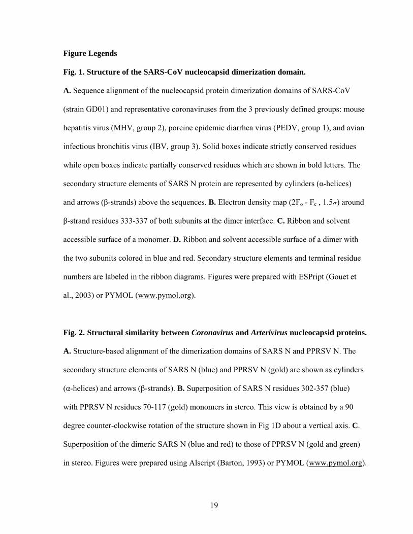
Figure Legends
Fig. 1. Structure of the SARS-CoV nucleocapsid dimerization domain.
A. Sequence alignment of the nucleocapsid protein dimerization domains of SARS-CoV
(strain GD01) and representative coronaviruses from the 3 previously defined groups: mouse
hepatitis virus (MHV, group 2), porcine epidemic diarrhea virus (PEDV, group 1), and avian
infectious bronchitis virus (IBV, group 3). Solid boxes indicate strictly conserved residues
while open boxes indicate partially conserved residues which are shown in bold letters. The
secondary structure elements of SARS N protein are represented by cylinders (α-helices)
and arrows (β-strands) above the sequences. B. Electron density map (2Fo - Fc , 1.5 ) around
β-strand residues 333-337 of both subunits at the dimer interface. C. Ribbon and solvent
accessible surface of a monomer. D. Ribbon and solvent accessible surface of a dimer with
the two subunits colored in blue and red. Secondary structure elements and terminal residue
numbers are labeled in the ribbon diagrams. Figures were prepared with ESPript (Gouet et
al., 2003) or PYMOL (www.pymol.org).
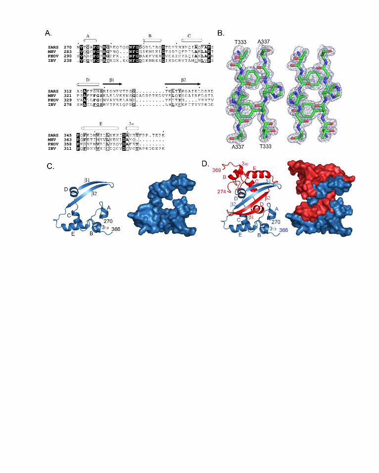
Fig. 2. Structural similarity between Coronavirus and Arterivirus nucleocapsid proteins.
A. Structure-based alignment of the dimerization domains of SARS N and PPRSV N. The
secondary structure elements of SARS N (blue) and PPRSV N (gold) are shown as cylinders
(α-helices) and arrows (β-strands). B. Superposition of SARS N residues 302-357 (blue)
with PPRSV N residues 70-117 (gold) monomers in stereo. This view is obtained by a 90
degree counter-clockwise rotation of the structure shown in Fig 1D about a vertical axis. C.
Superposition of the dimeric SARS N (blue and red) to those of PPRSV N (gold and green)
in stereo. Figures were prepared using Alscript (Barton, 1993) or PYMOL (www.pymol.org).
19

Fig. 3. A proposed model for the full-length SARS N protein dimer and the function of
each domain. The N-terminal domains (residues 49-178) are placed at arbitrary
orientations relative to the dimerization domains (residues 270-370). Regions where the
structure is unknown are shown as dashed lines. The residue numbers of each domain and
the serine/arginine-rich motif are labeled. The two subunits in a dimer are colored in red and
blue.
Fig. 4. Structural diversity of enveloped RNA virus nucleocapsid proteins.
Representative known structural domains of nucleocapsid proteins of Bornaviridae (BDV),
Togaviridae (SFV), Flaviviridae (dengue), Arteriviridae (PRRSV), and Coronaviridae
(SARS-CoV) are shown. One subunit in the oligomer is colored based on its secondary
structure elements: blue (α-helices), yellow (β-strands), and green (coils); the other subunits
are colored in gray. A. The crystallographic tetramer of borna disease virus (BDV)
nucleocapsid protein (Rudolph et al., 2003). B. Semliki Forest virus (SFV) nucleocapsid
protein. Only one of the two crystallographic dimers is shown (Choi et al., 1997). C. NMR
structure of dimeric dengue capsid protein (Ma et al., 2004). D. Porcine reproductive and
respiratory syndrome virus (PRRSV) nucleocapsid protein crystallographic dimer (Doan and
Dokland, 2003). E. The C-terminal dimerization domain, residues 270-370, of SARS-CoV
nucleocapsid protein determined in this study. F. NMR structure of the N-terminal domain
of SARS-CoV nucleocapsid protein, residues 49-178 (Huang et al., 2004). Figures were
prepared with PYMOL (www.pymol.org).
20

Table I. Data Collection and Refinement Statistics Data Collection Se-Met 1 Se-Met 2 peak peak inflection remote Space group C2 a, Å 124.2 124.3 b, Å 50.5 50.6 c, Å 41.5 41.6 β, ° 108.9 108.8 Resolution range, Å 30-1.75 50-1.9 50-2.2 50-2.2 Wavelength, Å 0.97929 0.97929 0.97940 0.94285 Unique reflections 24479 27506 24045 24131 Completeness1 99.2(99.6) 100.0(100.0) 99.2(100.0) 99.2(100.0) Redundancy 2.9 8.2 9.4 9.4 I/sigma 11.8(7.5) 15.6(14.3) 15.6(17.2) 14.8(11.2) Rsym
2,% 7.4(15.0) 10.6(16.5) 12.8(15.5) 15.1(21.2)
Refinement Rwork
3, % 18.7 Rfree, % 23.5No. of nonhydrogen atoms Protein 1511 Water 212 Sulphate 1Rmsd from ideality Bond lengths, Å 0.02 Bond angles, ° 1.67Estimated coordinate error (Luzzati), Å 0.16Average B-factor 18.0Disallowed, % 0.0 1 Values in parentheses are for the highest resolution shell. 2 Rsym = ∑|Ii - <I> |/ ∑Ii, where Ii is the intensity of the ith observation and <I> is the mean intensity of the reflection 3 R = ∑|| Fo | - | Fc ||/∑|Fo|, where Fo and Fc are the observed and calculated structure factors amplitudes. Rfree is calculated using 5% of the total reflections
21


Related Documents