research papers Acta Cryst. (2020). C76, 741–745 https://doi.org/10.1107/S2053229620008153 741 Received 24 March 2020 Accepted 19 June 2020 Edited by H. Uekusa, Tokyo Institute of Tech- nology, Japan Keywords: magnesium; carbonate; chloride; hydrate; synchrotron; twinning; crystal structure. CCDC reference: 2010753 Supporting information: this article has supporting information at journals.iucr.org/c Crystal structure and characterization of magnesium carbonate chloride heptahydrate Christine Rincke, a * Horst Schmidt, a Gernot Buth b and Wolfgang Voigt a a Institute of Inorganic Chemistry, TU Bergakademie Freiberg, Leipziger Strasse 29, D-09599 Freiberg, Germany, and b Institute for Photon Science and Synchrotron Radiation (IPS), Karlsruhe Institute of Technology (KIT), Hermann-von- Helmholtz-Platz 1, D-76344 Eggenstein-Leopoldshafen, Germany. *Correspondence e-mail: [email protected] MgCO 3 MgCl 2 7H 2 O is the only known neutral magnesium carbonate con- taining chloride ions at ambient conditions. According to the literature, only small and twinned crystals of this double salt could be synthesised in a concentrated solution of MgCl 2 . For the crystal structure solution, single-crystal diffraction was carried out at a synchrotron radiation source. The monoclinic crystal structure (space group Cc) exhibits double chains of MgO octahedra linked by corners, connected by carbonate units and water molecules. The chloride ions are positioned between these double chains parallel to the (100) plane. Dry MgCO 3 MgCl 2 7H 2 O decomposes in the air to chlorartinite, Mg 2 (OH)Cl(CO 3 )nH 2 O (n = 2 or 3). This work includes an extensive characterization of the title compound by powder X-ray diffraction, thermal analysis, SEM and vibrational spectroscopy. 1. Introduction In the context of CO 2 research, the interactions of CO 2 with salts and brine solutions are of great interest. Therefore, the system MgCl 2 –MgCO 3 –H 2 O–CO 2 has been investigated. The only nonbasic salt containing carbonate and chloride ions is MgCO 3 MgCl 2 7H 2 O (Rincke, 2018). The formation conditions of MgCO 3 MgCl 2 7H 2 O were described for the first time by Gloss (1937) and Walter-Levy (1937). It can be synthesized at room temperature by adding MgCO 3 3H 2 O to a highly concentrated solution of magnesium chloride saturated with CO 2 (Gloss, 1937; Schmidt, 1960). Within the scope of outbursts of CO 2 in potash mines, MgCO 3 MgCl 2 7H 2 O was discussed as a storage compound for CO 2 in the 1960s (Schmidt, 1960; Serowy, 1963; Serowy & Liebmann, 1964; Schmittler, 1964; D’Ans, 1967). This salt forms needle-like crystals, which are only stable in concen- trated MgCl 2 solution (Moshkina & Yaroslavtseva, 1970). It decomposes immediately when it is washed with water. When it was stored in air, basic carbonate was formed (Gloss, 1937). Schmittler (1964) concluded from a powder X-ray diffrac- tion (PXRD) pattern of MgCO 3 MgCl 2 7H 2 O that its crystal structure exhibits a C-centred monoclinic lattice with para- meters a = 13.27 (0), b = 11.30 (8), c = 9.22 (7) A ˚ and = 118.2 (6) . Due to the low scattering power and the small size of the crystals, a crystal structure analysis of single crystals was not possible until now. Our own investigations should provide a better comprehension of the synthesis of MgCO 3 MgCl 2 7H 2 O and provide a more detailed characterization, including a crystal structure analysis. ISSN 2053-2296

Welcome message from author
This document is posted to help you gain knowledge. Please leave a comment to let me know what you think about it! Share it to your friends and learn new things together.
Transcript

research papers
Acta Cryst. (2020). C76, 741–745 https://doi.org/10.1107/S2053229620008153 741
Received 24 March 2020
Accepted 19 June 2020
Edited by H. Uekusa, Tokyo Institute of Tech-
nology, Japan
Keywords: magnesium; carbonate; chloride;
hydrate; synchrotron; twinning; crystal structure.
CCDC reference: 2010753
Supporting information: this article has
supporting information at journals.iucr.org/c
Crystal structure and characterization ofmagnesium carbonate chloride heptahydrate
Christine Rincke,a* Horst Schmidt,a Gernot Buthb and Wolfgang Voigta
aInstitute of Inorganic Chemistry, TU Bergakademie Freiberg, Leipziger Strasse 29, D-09599 Freiberg, Germany, andbInstitute for Photon Science and Synchrotron Radiation (IPS), Karlsruhe Institute of Technology (KIT), Hermann-von-
Helmholtz-Platz 1, D-76344 Eggenstein-Leopoldshafen, Germany. *Correspondence e-mail:
MgCO3�MgCl2�7H2O is the only known neutral magnesium carbonate con-
taining chloride ions at ambient conditions. According to the literature, only
small and twinned crystals of this double salt could be synthesised in a
concentrated solution of MgCl2. For the crystal structure solution, single-crystal
diffraction was carried out at a synchrotron radiation source. The monoclinic
crystal structure (space group Cc) exhibits double chains of MgO octahedra
linked by corners, connected by carbonate units and water molecules. The
chloride ions are positioned between these double chains parallel to the (100)
plane. Dry MgCO3�MgCl2�7H2O decomposes in the air to chlorartinite,
Mg2(OH)Cl(CO3)�nH2O (n = 2 or 3). This work includes an extensive
characterization of the title compound by powder X-ray diffraction, thermal
analysis, SEM and vibrational spectroscopy.
1. Introduction
In the context of CO2 research, the interactions of CO2 with
salts and brine solutions are of great interest. Therefore, the
system MgCl2–MgCO3–H2O–CO2 has been investigated. The
only nonbasic salt containing carbonate and chloride ions is
MgCO3�MgCl2�7H2O (Rincke, 2018).
The formation conditions of MgCO3�MgCl2�7H2O were
described for the first time by Gloss (1937) and Walter-Levy
(1937). It can be synthesized at room temperature by
adding MgCO3�3H2O to a highly concentrated solution of
magnesium chloride saturated with CO2 (Gloss, 1937;
Schmidt, 1960).
Within the scope of outbursts of CO2 in potash mines,
MgCO3�MgCl2�7H2O was discussed as a storage compound for
CO2 in the 1960s (Schmidt, 1960; Serowy, 1963; Serowy &
Liebmann, 1964; Schmittler, 1964; D’Ans, 1967). This salt
forms needle-like crystals, which are only stable in concen-
trated MgCl2 solution (Moshkina & Yaroslavtseva, 1970). It
decomposes immediately when it is washed with water. When
it was stored in air, basic carbonate was formed (Gloss,
1937).
Schmittler (1964) concluded from a powder X-ray diffrac-
tion (PXRD) pattern of MgCO3�MgCl2�7H2O that its crystal
structure exhibits a C-centred monoclinic lattice with para-
meters a = 13.27 (0), b = 11.30 (8), c = 9.22 (7) A and � =
118.2 (6)�. Due to the low scattering power and the small size
of the crystals, a crystal structure analysis of single crystals was
not possible until now. Our own investigations should provide
a better comprehension of the synthesis of MgCO3�MgCl2�
7H2O and provide a more detailed characterization, including
a crystal structure analysis.
ISSN 2053-2296

2. Experimental
2.1. Synthesis and crystallization
The synthesis of MgCO3�MgCl2�7H2O is based on the
information of Schmidt (1960). MgO (1 g, Magnesia M2329,
p.a.) was added to 200 g of an aqueous solution of MgCl2 (5.5
molal, Fluka, �98%). The suspension was stirred for 30 min.
Afterwards, the undissolved MgO was filtered off. CO2 was
bubbled through the stirred solution for 24 h at room tem-
perature. The product was filtered off for further character-
ization.
2.2. Single-crystal diffraction
Data were collected on beamline SCD at the KIT
Synchrotron Radiation Source using a Stoe IPDS diffrac-
tometer with monochromated radiation of � = 0.8000 A. A
crystal of MgCO3�MgCl2�7H2O was recovered from a droplet
of its mother liquor and mounted rapidly in the cold (150 K)
stream of nitrogen gas of the diffractometer.
2.3. Powder X-ray diffraction (PXRD)
PXRD patterns were taken for phase identification with a
laboratory Bruker D8 Discover powder diffractometer in
Bragg–Brentano set up (Cu K�1 radiation, Vantec 1 detector).
The samples were prepared as flat plates and measured at
room temperature.
2.4. Thermal analysis
The thermal analysis was performed with a TG/DTA 220
instrument from Seiko Instruments (reference substance:
Al2O3, open platinum crucible; argon flow: 300 ml min�1;
heating rate: 5 K min�1, prior period 30 min at 298.15 K in an
argon flow).
research papers
742 Rincke et al. � Magnesium carbonate chloride heptahydrate Acta Cryst. (2020). C76, 741–745
Table 1Experimental details.
Crystal dataChemical formula MgCO3�MgCl2�7H2OMr 305.64Crystal system, space group Monoclinic, CcTemperature (K) 150a, b, c (A) 13.368 (5), 11.262 (5), 9.266 (4)� (�) 118.83 (3)V (A3) 1222.0 (9)Z 4Radiation type Synchrotron, � = 0.8000 A� (mm�1) 0.93Crystal size (mm) 0.13 � 0.07 � 0.01 � 0.02 (radius)
Data collectionDiffractometer Stoe IPDS IIAbsorption correction For a sphere (Coppens, 1970)No. of measured, independent and
observed [I > 2�(I)] reflections8746, 6975, 5476
Rint 0.0613�max (�) 26.7(sin �/�)max (A�1) 0.561
RefinementR[F 2 > 2�(F 2)], wR(F 2), S 0.053, 0.161, 1.12No. of reflections 4791No. of parameters 179No. of restraints 22H-atom treatment Only H-atom coordinates refined��max, ��min (e A�3) 0.36, �0.43Absolute structure Flack x determined using 647
quotients [(I+) � (I�)]/[(I+) + (I�)] (Parsons et al., 2013)
Absolute structure parameter 0.43 (13)
Computer programs: X-AREA (Stoe & Cie, 2015), X-RED (Stoe & Cie, 2015),SHELXS97 (Sheldrick, 2008), SHELXL2016 (Sheldrick, 2015), DIAMOND (Branden-burg, 2017) and publCIF (Westrip, 2010).
Figure 1Powder XRD patterns of MgCO3�MgCl2�7H2O under ambient conditions (Cu K�1 radiation) for (a) the unwashed product immediately after thesynthesis, (b) the unwashed product stored in the air after 19 months, (c) the product washed with ethanol after storage in the air for 10 d and (d) theproduct washed with ethanol after storage in the air for 19 months. Reference data: MgCO3�MgCl2�7H2O (PDF 21-1254) and Mg2(OH)Cl(CO3)�3H2O(PDF 07-0278).

2.5. Scanning electron microscopy (SEM)
The SEM images were recorded with a TESCAN Vega 5130
SB instrument (20 kV accelerating voltage). The sample was
coated with gold.
2.6. Vibrational spectroscopy
For the FT–IR spectrum, a Thermo Scientific Nicolet 380
FTIR spectrometer (spectral resolution: 6 cm�1, 256 scans per
measurement) with KBr blanks was used.
The Raman spectrum was recorded shortly after synthesis
with a Bruker RFS100/S FT spectrometer at room tempera-
ture (Nd/YAG-laser, wavelength of the laser: 1064 nm).
2.7. Refinement
Crystal data, data collection and structure refinement
details are given in Table 1. Due to the small crystals and their
low scattering power, the crystal structure solution was carried
out by single-crystal diffraction at a synchrotron radiation
source. The quality of the crystals affected the measured data
set with the effect that only reflections to sin �max/� = 0.56 A�1
could be considered for the structure refinement. The crystal
structure was solved by direct methods. The resulting structure
solution exhibits a chemically reasonable atomic arrangement,
distances, angles and displacement parameters.
H atoms were placed in the positions indexed by difference
Fourier maps and their Uiso values were set at 1.2Ueq(O) using
a riding-model approximation.
The crystal exhibits nonmerohedral twinning. The matrix
that relates the individual diffraction pattern was determined
as (1 0 1.38, 0 �1 0, 0 0 �1). The reflections of both domains
were integrated (number of reflections in domain 1: 2829;
domain 2: 3505; overlaid: 641; major twin component fraction:
56.45%).
3. Results and discussion
3.1. Characterization of magnesium carbonate chlorideheptahydrate
The characterization of the unwashed product with PXRD
is in accordance with the reference pattern PDF 21-1254 for
MgCO3�MgCl2�7H2O (Schmittler, 1964). The filtered product
was stored in a sealed vessel. After 19 months, the powder
pattern remained constant, i.e. the product did not alter. If the
product was washed with ethanol and stored in the air,
decomposition to chlorartinite [Mg2(OH)Cl(CO3)�3H2O]
begins within a few days (Fig. 1). This observation confirms the
information of Gloss (1937).
The thermal decomposition of MgCO3�MgCl2�7H2O starts
as early as the heating begins and shows two main steps
(Fig. 2). H2O, CO2 and HCl are evaporated off. This is in
accordance with the observation of Serowy & Liebmann
(1964). A precise assignment of the stepwise mass loss is not
possible. The characterization of the residue with PXRD at
573 K exhibits the presence of a mixture of basic magnesium
carbonates, i.e. hydromagnesite [Mg5(CO3)4(OH)2�4H2O] and
amorphous phases. At 803 K the decomposition is complete
and only MgO remains in the residue. The observed mass loss
of 74.3 (1)% confirms the theoretical mass loss of 73.6%.
The SEM images of MgCO3�MgCl2�7H2O show thin needles
(50 � 5 mm), which are twinned or even more intergrown
(Fig. 3). Numerous crystallization experiments with the aim of
obtaining larger crystals were not successful.
The FT–IR (Fig. 4) and Raman spectra (Fig. 5) of
MgCO3�MgCl2�7H2O confirm the absence of hydroxide ions in
the crystal structure, because there are no bands above
3500 cm�1 as in chlorartinite, Mg2(OH)Cl(CO3)�3H2O (Ver-
gasova et al., 1998). The assignment of the bands was con-
cluded from a comparison with the vibrational spectra of other
neutral magnesium carbonates and chlorartinite (Coleyshaw
et al., 2003; Vergasova et al., 1998) (Table 2).
research papers
Acta Cryst. (2020). C76, 741–745 Rincke et al. � Magnesium carbonate chloride heptahydrate 743
Figure 3SEM images of MgCO3�MgCl2�7H2O, with the crystals exhibitingtwinning or even further intergrowth.
Table 2Assignment of the IR and Raman bands of MgCO3�MgCl2�7H2O.
IR RamanAssignment (Coleyshawet al., 2003)
3407, 3240 3386, 3250 (OH)W
1635 1660 (OH)W
1550, 1449, 1401 1544 as(CO)1114 1111 s(CO)845 794 �(CO)620 599 as(CO)457 403, 227, 181, 154, 124 lattice vibrations
Notes: = valence vibration, = deformation vibration (in the plane), � = deformationvibration out of the plane, W = water, s = symmetric and as = asymmetric.
Figure 2Thermal analysis of MgCO3�MgCl2�7H2O.

3.2. Crystal structure of magnesium carbonate chlorideheptahydrate
The monoclinic crystal structure of MgCO3�MgCl2�7H2O
with the space group Cc and the lattice parameters published
by Schmittler (1964) were confirmed. There are two distin-
guishable magnesium ions. Mg1 is coordinated by three water
molecules and two carbonate anions. One carbonate acts as a
monodentate ligand via atom O9 and the other as a bidentate
ligand via atoms O2 and O6. The octahedra of Mg2 are formed
by four water molecules and two carbonate units which are
connected to the magnesium ion in a monodentate manner via
atoms O2 and O6 (Fig. 6). The corner-linked Mg–O octahedra
are arranged in a zigzag manner and together with the car-
bonate units form double chains parallel to the (100) plane
(Fig. 7).
All the carbonate units are crystallographically equivalent
and exhibit a Cs geometry, because they are planar, but the
C—O bonds have different lengths. Each carbonate unit is
coordinated by three magnesium ions: monodentate to Mg1,
bidentate to Mg1i and monodentate to Mg2ii (see Fig. 6 for
symmetry codes). In addition, the carbonate units stabilize the
double chains (Fig. 7).
Between the double chains, which are arranged in a zigzag-
like stacking order parallel to the (001) plane, are located the
chloride ions Cl1 and Cl2 (Fig. 8). The positions of atoms H1A
and H3B are fixed by short hydrogen bonds to atoms O9iv and
O4vi, and the other H atoms by interactions with the chloride
ions (Table 3 and Fig. 9). As a consequence, a three-dimen-
sional network is formed.
research papers
744 Rincke et al. � Magnesium carbonate chloride heptahydrate Acta Cryst. (2020). C76, 741–745
Figure 4IR spectrum of MgCO3�MgCl2�7H2O under ambient conditions.
Figure 5Raman spectrum of MgCO3�MgCl2�7H2O under ambient conditions.
Figure 7The characteristic structural motif in MgCO3�MgCl2�7H2O, showing thedouble chain of MgO octahedra linked by corners and carbonate unitsparallel to the (100) plane.
Figure 6The asymmetric unit and coordination units of MgCO3�MgCl2�7H2O[symmetry codes: (i) x, �y, z � 1
2; (ii) x, y, z � 1; (iii) x, �y, z + 12; (iv) x, y,
z + 1].

The structural motifs of such double chains are similar in
MgCO3�MgCl2�7H2O and MgCO3�3H2O (Giester et al., 2000),
but in contrast to MgCO3�3H2O in MgCO3�MgCl2�7H2O, only
two of three carbonate units and three and four water mol-
ecules instead of two water molecules are linked to each Mg
atom. Furthermore, no free water molecules are positioned
between these double chains in MgCO3�MgCl2�7H2O. The
crystal structures of other neutral magnesium carbonates, e.g.
MgCO3�5H2O, MgCO3�6H2O and the chloride-containing
magnesium carbonates Mg2(OH)Cl(CO3)�2H2O (chlor-
artinite) and Mg2(OH)Cl(CO3)�H2O (dehydrated clorarti-
nite), do not exhibit such double chains (Liu et al., 1990;
Rincke et al., 2020; Sugimoto et al., 2006, 2007). Therefore, the
crystal structure of MgCO3�MgCl2�7H2O is unique.
Acknowledgements
The award of synchrotron beamtime at KIT Synchrotron
Radiation Source, Karlsruhe, Germany, is gratefully
acknowledged.
References
Brandenburg, K. (2017). DIAMOND. Crystal Impact GbR, Bonn,Germany.
Coleyshaw, E. E., Crump, G. & Griffith, W. P. (2003). Spectrochim.Acta A Mol. Biomol. Spectrosc. 59, 2231–2239.
Coppens, P. (1970). Crystallographic Computing, edited by F. R.Ahmed, S. R. Hall & C. P. Huber, pp. 255–270. Copenhagen:Munksgaard.
D’Ans, J. (1967). Kali und Steinsalz, 4, 396–401.Giester, G., Lengauer, C. L. & Rieck, B. (2000). Mineral. Petrol. 70,
153–163.Gloss, G. (1937). Dissertation. Friedrich-Wilhelms-University of
Berlin, Germany.Liu, B., Zhou, X., Cui, X. & Tang, J. (1990). Sci. China Ser. B, 33,
1350–1356.Moshkina, I. A. & Yaroslavtseva, L. M. (1970). Zh. Neorg. Khim. 15,
3345–3350.Parsons, S., Flack, H. D. & Wagner, T. (2013). Acta Cryst. B69, 249–
259.Rincke, C. (2018). Dissertation. TU Bergakademie Freiberg, Ger-
many.
Rincke, C., Schmidt, H. & Voigt, W. (2020). Acta Cryst. C76, 244–249.Schmidt, E. (1960). Bergakademie, 12, 693–697.Schmittler, H. (1964). Deut. Akad. Wiss. 6, 644–648.Serowy, F. (1963). Freiberger Forschungshefte A, 267, 405–419.Serowy, F. & Liebmann, G. (1964). Wissenschaftl. Zeitschrift der
Technischen Hochschule fur Chemie ‘Carl Schorlemmer’ Leuna-Merseburg, 6, 338–342.
Sheldrick, G. M. (2008). Acta Cryst. A64, 112–122.Sheldrick, G. M. (2015). Acta Cryst. C71, 3–8.Stoe & Cie (2015). X-AREA and X-RED32. Stoe & Cie, Darmstadt,
Germany.Sugimoto, K., Dinnebier, R. E. & Schlecht, T. (2006). J. Appl. Cryst.
39, 739–744.Sugimoto, K., Dinnebier, R. E. & Schlecht, T. (2007). Powder Diff.
22(1), 739–744.Vergasova, L. P., Filation, S. K., Serafimova, E. K. & Sergeeva, S. V.
(1998). Zapiski Vserossiiskogo Mineralogicheskogo Obshchestva,127, 55–59.
Walter-Levy, L. (1937). Compt. Rend. 205, 1405–1407.Westrip, S. P. (2010). J. Appl. Cryst. 43, 920–925.
research papers
Acta Cryst. (2020). C76, 741–745 Rincke et al. � Magnesium carbonate chloride heptahydrate 745
Table 3Hydrogen-bond geometry (A, �).
D—H� � �A D—H H� � �A D� � �A D—H� � �A
O1—H1A� � �O9iv 0.82 (3) 1.94 (6) 2.688 (14) 153 (13)O1—H1B� � �Cl2iv 0.82 (3) 2.38 (4) 3.186 (11) 167 (12)O3—H3A� � �Cl2v 0.82 (3) 2.32 (3) 3.135 (11) 174 (17)O3—H3B� � �O4vi 0.82 (3) 2.10 (11) 2.796 (13) 143 (17)O4—H4A� � �Cl1vii 0.82 (3) 2.36 (3) 3.176 (10) 171 (14)O4—H4B� � �Cl1viii 0.82 (3) 2.49 (7) 3.251 (10) 155 (13)O5—H5A� � �Cl2viii 0.82 (3) 2.45 (7) 3.222 (11) 157 (16)O5—H5B� � �Cl1ix 0.81 (3) 2.54 (6) 3.327 (11) 164 (17)O7—H7A� � �Cl2viii 0.82 (3) 2.42 (6) 3.212 (11) 163 (16)O7—H7B� � �Cl1x 0.82 (3) 2.30 (4) 3.111 (11) 169 (16)O8—H8A� � �Cl2vi 0.82 (3) 2.46 (5) 3.254 (10) 163 (11)O8—H8B� � �Cl2v 0.82 (3) 2.41 (3) 3.233 (10) 178 (11)O10—H10A� � �Cl1vii 0.82 (3) 2.34 (5) 3.146 (10) 166 (15)O10—H10B� � �Cl1xi 0.82 (3) 2.67 (7) 3.424 (10) 154 (12)
Symmetry codes: (iv) x, y, z + 1; (v) x + 12, y� 1
2, z + 1; (vi) x + 12,�y + 1
2, z + 12; (vii) x, y� 1,
z; (viii) x, �y + 1, z + 12; (ix) x + 1
2, �y + 32, z + 1
2; (x) x + 12, y � 1
2, z; (xi) x, �y + 1, z � 12.
Figure 9Excerpt of the crystal structure of MgCO3�MgCl2�7H2O, showing thehydrogen-bond interactions of the H atoms with chloride ions (dashedlines) [symmetry codes: (iv) x, y, z + 1; (v) x + 1
2, y � 12, z + 1; (vi) x + 1
2,�y + 1
2, z + 12; (vii) x, y � 1, z; (viii) x, �y + 1, z + 1
2; (ix) x + 12, �y + 3
2, z + 12;
(x) x + 12, y � 1
2, z; (xi) x, �y + 1, z � 12].
Figure 8Excerpt of the crystal structure of MgCO3�MgCl2�7H2O, showing thezigzag-like stacking order of the double chains and the chloride ionsbetween them.

supporting information
sup-1Acta Cryst. (2020). C76, 741-745
supporting information
Acta Cryst. (2020). C76, 741-745 [https://doi.org/10.1107/S2053229620008153]
Crystal structure and characterization of magnesium carbonate chloride
heptahydrate
Christine Rincke, Horst Schmidt, Gernot Buth and Wolfgang Voigt
Computing details
Data collection: X-AREA (Stoe & Cie, 2015); cell refinement: X-AREA (Stoe & Cie, 2015); data reduction: X-RED (Stoe
& Cie, 2015); program(s) used to solve structure: SHELXS97 (Sheldrick, 2008); program(s) used to refine structure:
SHELXL2016 (Sheldrick, 2015); molecular graphics: DIAMOND (Brandenburg, 2017); software used to prepare material
for publication: publCIF (Westrip, 2010).
Magnesium carbonate chloride heptahydrate
Crystal data
MgCO3·MgCl2·7H2OMr = 305.64Monoclinic, Cca = 13.368 (5) Åb = 11.262 (5) Åc = 9.266 (4) Åβ = 118.83 (3)°V = 1222.0 (9) Å3
Z = 4
F(000) = 632Dx = 1.661 Mg m−3
Synchrotron radiation, λ = 0.8000 ÅCell parameters from 4192 reflectionsθ = 2.7–29.5°µ = 0.93 mm−1
T = 150 KNeedle, colourless0.13 × 0.07 × 0.01 × 0.02 (radius) mm
Data collection
Stoe IPDS II diffractometer
Radiation source: synchrotronrotation method scansAbsorption correction: for a sphere
(Coppens, 1970)
8746 measured reflections
6975 independent reflections5476 reflections with I > 2σ(I)Rint = 0.061θmax = 26.7°, θmin = 3.3°h = −14→14k = −12→12l = −10→10
Refinement
Refinement on F2
Least-squares matrix: fullR[F2 > 2σ(F2)] = 0.053wR(F2) = 0.161S = 1.124791 reflections179 parameters22 restraintsPrimary atom site location: structure-invariant
direct methodsHydrogen site location: difference Fourier map
Only H-atom coordinates refinedw = 1/[σ2(Fo
2) + (0.094P)2] where P = (Fo
2 + 2Fc2)/3
(Δ/σ)max < 0.001Δρmax = 0.36 e Å−3
Δρmin = −0.43 e Å−3
Absolute structure: Flack x determined using 647 quotients [(I+)-(I-)]/[(I+)+(I-)] (Parsons et al., 2013)
Absolute structure parameter: 0.43 (13)
supporting information
sup-2Acta Cryst. (2020). C76, 741-745
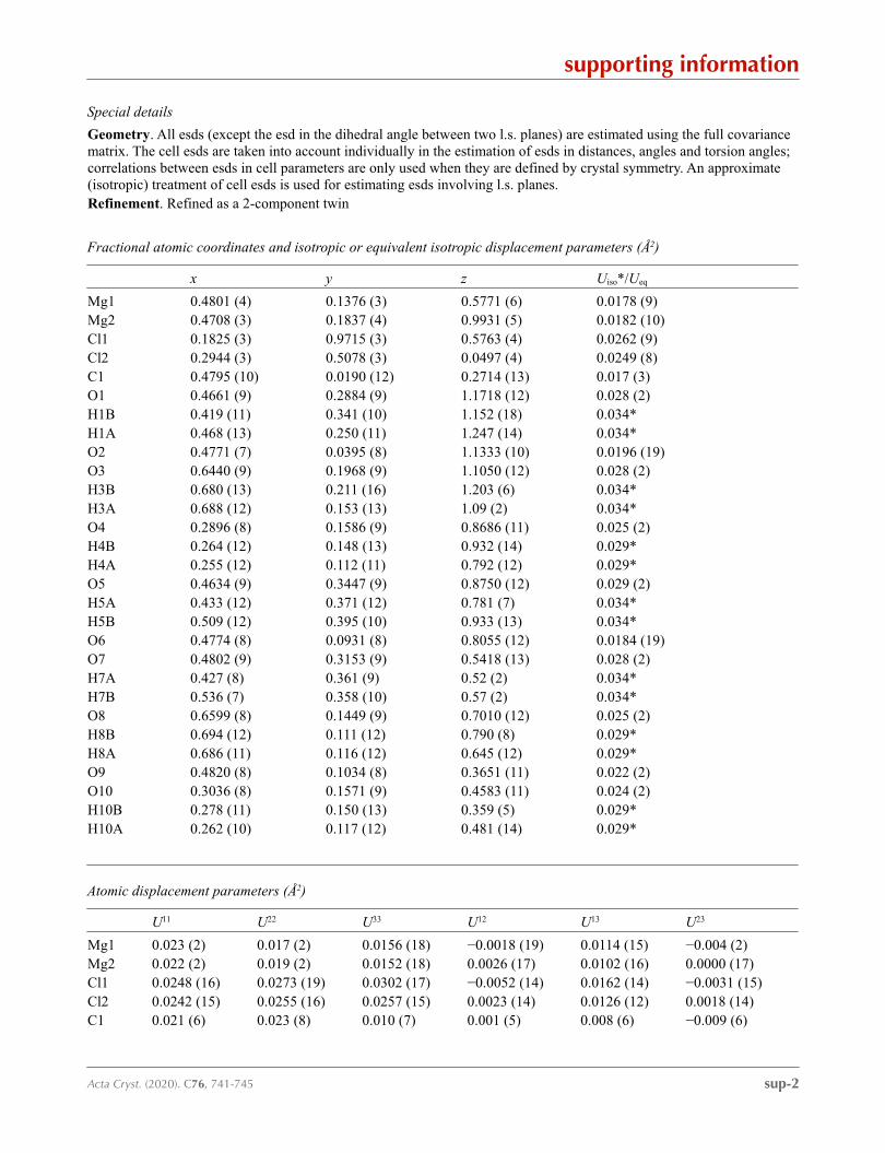
Special details
Geometry. All esds (except the esd in the dihedral angle between two l.s. planes) are estimated using the full covariance matrix. The cell esds are taken into account individually in the estimation of esds in distances, angles and torsion angles; correlations between esds in cell parameters are only used when they are defined by crystal symmetry. An approximate (isotropic) treatment of cell esds is used for estimating esds involving l.s. planes.Refinement. Refined as a 2-component twin
Fractional atomic coordinates and isotropic or equivalent isotropic displacement parameters (Å2)
x y z Uiso*/Ueq
Mg1 0.4801 (4) 0.1376 (3) 0.5771 (6) 0.0178 (9)Mg2 0.4708 (3) 0.1837 (4) 0.9931 (5) 0.0182 (10)Cl1 0.1825 (3) 0.9715 (3) 0.5763 (4) 0.0262 (9)Cl2 0.2944 (3) 0.5078 (3) 0.0497 (4) 0.0249 (8)C1 0.4795 (10) 0.0190 (12) 0.2714 (13) 0.017 (3)O1 0.4661 (9) 0.2884 (9) 1.1718 (12) 0.028 (2)H1B 0.419 (11) 0.341 (10) 1.152 (18) 0.034*H1A 0.468 (13) 0.250 (11) 1.247 (14) 0.034*O2 0.4771 (7) 0.0395 (8) 1.1333 (10) 0.0196 (19)O3 0.6440 (9) 0.1968 (9) 1.1050 (12) 0.028 (2)H3B 0.680 (13) 0.211 (16) 1.203 (6) 0.034*H3A 0.688 (12) 0.153 (13) 1.09 (2) 0.034*O4 0.2896 (8) 0.1586 (9) 0.8686 (11) 0.025 (2)H4B 0.264 (12) 0.148 (13) 0.932 (14) 0.029*H4A 0.255 (12) 0.112 (11) 0.792 (12) 0.029*O5 0.4634 (9) 0.3447 (9) 0.8750 (12) 0.029 (2)H5A 0.433 (12) 0.371 (12) 0.781 (7) 0.034*H5B 0.509 (12) 0.395 (10) 0.933 (13) 0.034*O6 0.4774 (8) 0.0931 (8) 0.8055 (12) 0.0184 (19)O7 0.4802 (9) 0.3153 (9) 0.5418 (13) 0.028 (2)H7A 0.427 (8) 0.361 (9) 0.52 (2) 0.034*H7B 0.536 (7) 0.358 (10) 0.57 (2) 0.034*O8 0.6599 (8) 0.1449 (9) 0.7010 (12) 0.025 (2)H8B 0.694 (12) 0.111 (12) 0.790 (8) 0.029*H8A 0.686 (11) 0.116 (12) 0.645 (12) 0.029*O9 0.4820 (8) 0.1034 (8) 0.3651 (11) 0.022 (2)O10 0.3036 (8) 0.1571 (9) 0.4583 (11) 0.024 (2)H10B 0.278 (11) 0.150 (13) 0.359 (5) 0.029*H10A 0.262 (10) 0.117 (12) 0.481 (14) 0.029*
Atomic displacement parameters (Å2)
U11 U22 U33 U12 U13 U23
Mg1 0.023 (2) 0.017 (2) 0.0156 (18) −0.0018 (19) 0.0114 (15) −0.004 (2)Mg2 0.022 (2) 0.019 (2) 0.0152 (18) 0.0026 (17) 0.0102 (16) 0.0000 (17)Cl1 0.0248 (16) 0.0273 (19) 0.0302 (17) −0.0052 (14) 0.0162 (14) −0.0031 (15)Cl2 0.0242 (15) 0.0255 (16) 0.0257 (15) 0.0023 (14) 0.0126 (12) 0.0018 (14)C1 0.021 (6) 0.023 (8) 0.010 (7) 0.001 (5) 0.008 (6) −0.009 (6)

supporting information
sup-3Acta Cryst. (2020). C76, 741-745
O1 0.048 (6) 0.021 (5) 0.024 (5) 0.008 (4) 0.023 (5) 0.009 (4)O2 0.027 (5) 0.017 (5) 0.017 (4) 0.005 (4) 0.013 (4) −0.003 (4)O3 0.030 (6) 0.032 (6) 0.023 (5) 0.005 (4) 0.013 (5) −0.001 (4)O4 0.028 (5) 0.030 (5) 0.017 (5) −0.002 (4) 0.012 (4) 0.001 (4)O5 0.040 (6) 0.023 (5) 0.020 (5) −0.006 (4) 0.013 (4) −0.001 (4)O6 0.025 (5) 0.016 (5) 0.017 (4) 0.000 (4) 0.012 (4) 0.002 (4)O7 0.030 (5) 0.022 (5) 0.038 (7) 0.006 (4) 0.021 (5) 0.004 (4)O8 0.024 (5) 0.029 (5) 0.020 (5) 0.003 (4) 0.010 (4) −0.003 (4)O9 0.034 (6) 0.018 (5) 0.020 (5) 0.002 (4) 0.018 (4) 0.005 (4)O10 0.025 (5) 0.029 (6) 0.019 (5) 0.001 (4) 0.012 (4) 0.002 (4)
Geometric parameters (Å, º)
Mg1—O9 2.014 (10) Mg2—O2 2.055 (10)Mg1—O7 2.028 (11) Mg2—O1 2.058 (11)Mg1—O2i 2.066 (9) Mg2—O5 2.095 (11)Mg1—O10 2.079 (11) Mg2—O4 2.140 (11)Mg1—O8 2.107 (11) C1—O9 1.277 (15)Mg1—O6 2.192 (10) C1—O2ii 1.285 (15)Mg2—O3 2.036 (11) C1—O6i 1.305 (17)Mg2—O6 2.054 (11)
O9—Mg1—O7 91.7 (4) O3—Mg2—O1 91.0 (5)O9—Mg1—O2i 94.2 (4) O6—Mg2—O1 174.8 (5)O7—Mg1—O2i 174.1 (5) O2—Mg2—O1 87.3 (4)O9—Mg1—O10 92.8 (4) O3—Mg2—O5 87.6 (4)O7—Mg1—O10 84.2 (4) O6—Mg2—O5 89.9 (4)O2i—Mg1—O10 94.6 (4) O2—Mg2—O5 172.2 (4)O9—Mg1—O8 89.5 (4) O1—Mg2—O5 85.0 (4)O7—Mg1—O8 87.7 (4) O3—Mg2—O4 176.1 (5)O2i—Mg1—O8 93.2 (4) O6—Mg2—O4 88.7 (4)O10—Mg1—O8 171.6 (4) O2—Mg2—O4 85.8 (4)O9—Mg1—O6 155.8 (4) O1—Mg2—O4 92.5 (5)O7—Mg1—O6 112.5 (4) O5—Mg2—O4 94.4 (4)O2i—Mg1—O6 61.6 (4) O3—Mg2—Mg1iii 92.8 (3)O10—Mg1—O6 89.6 (4) O6—Mg2—Mg1iii 71.4 (3)O8—Mg1—O6 91.6 (4) O2—Mg2—Mg1iii 26.5 (3)O9—Mg1—C1iii 124.7 (4) O1—Mg2—Mg1iii 113.8 (3)O7—Mg1—C1iii 143.6 (5) O5—Mg2—Mg1iii 161.2 (3)O2i—Mg1—C1iii 30.5 (4) O4—Mg2—Mg1iii 84.2 (3)O10—Mg1—C1iii 93.4 (4) O9—C1—O2ii 121.5 (12)O8—Mg1—C1iii 92.0 (4) O9—C1—O6i 123.5 (10)O6—Mg1—C1iii 31.1 (4) O2ii—C1—O6i 115.0 (10)O9—Mg1—Mg2i 67.8 (3) O9—C1—Mg1i 175.9 (10)O7—Mg1—Mg2i 159.4 (3) O2ii—C1—Mg1i 54.7 (6)O2i—Mg1—Mg2i 26.4 (2) O6i—C1—Mg1i 60.3 (6)O10—Mg1—Mg2i 94.5 (3) C1iv—O2—Mg2 138.1 (8)O8—Mg1—Mg2i 93.9 (3) C1iv—O2—Mg1iii 94.7 (8)

supporting information
sup-4Acta Cryst. (2020). C76, 741-745
O6—Mg1—Mg2i 88.0 (3) Mg2—O2—Mg1iii 127.1 (4)C1iii—Mg1—Mg2i 56.9 (3) C1iii—O6—Mg2 134.5 (8)O3—Mg2—O6 87.9 (4) C1iii—O6—Mg1 88.5 (7)O3—Mg2—O2 92.6 (4) Mg2—O6—Mg1 137.0 (5)O6—Mg2—O2 97.9 (4) C1—O9—Mg1 142.8 (8)
Symmetry codes: (i) x, −y, z−1/2; (ii) x, y, z−1; (iii) x, −y, z+1/2; (iv) x, y, z+1.
Hydrogen-bond geometry (Å, º)
D—H···A D—H H···A D···A D—H···A
O1—H1A···O9iv 0.82 (3) 1.94 (6) 2.688 (14) 153 (13)O1—H1B···Cl2iv 0.82 (3) 2.38 (4) 3.186 (11) 167 (12)O3—H3A···Cl2v 0.82 (3) 2.32 (3) 3.135 (11) 174 (17)O3—H3B···O4vi 0.82 (3) 2.10 (11) 2.796 (13) 143 (17)O4—H4A···Cl1vii 0.82 (3) 2.36 (3) 3.176 (10) 171 (14)O4—H4B···Cl1viii 0.82 (3) 2.49 (7) 3.251 (10) 155 (13)O5—H5A···Cl2viii 0.82 (3) 2.45 (7) 3.222 (11) 157 (16)O5—H5B···Cl1ix 0.81 (3) 2.54 (6) 3.327 (11) 164 (17)O7—H7A···Cl2viii 0.82 (3) 2.42 (6) 3.212 (11) 163 (16)O7—H7B···Cl1x 0.82 (3) 2.30 (4) 3.111 (11) 169 (16)O8—H8A···Cl2vi 0.82 (3) 2.46 (5) 3.254 (10) 163 (11)O8—H8B···Cl2v 0.82 (3) 2.41 (3) 3.233 (10) 178 (11)O10—H10A···Cl1vii 0.82 (3) 2.34 (5) 3.146 (10) 166 (15)O10—H10B···Cl1xi 0.82 (3) 2.67 (7) 3.424 (10) 154 (12)
Symmetry codes: (iv) x, y, z+1; (v) x+1/2, y−1/2, z+1; (vi) x+1/2, −y+1/2, z+1/2; (vii) x, y−1, z; (viii) x, −y+1, z+1/2; (ix) x+1/2, −y+3/2, z+1/2; (x) x+1/2, y−1/2, z; (xi) x, −y+1, z−1/2.
Related Documents