Crystal Nucleation, Growth, and Morphology of the Synthetic Malaria Pigment -Hematin and the Effect Thereon by Quinoline Additives: The Malaria Pigment as a Target of Various Antimalarial Drugs Inna Solomonov, † Maria Osipova, † Yishay Feldman, ‡ Carsten Baehtz, § Kristian Kjaer, | Ian K. Robinson, ! Grant T. Webster, # Don McNaughton, # Bayden R. Wood, # Isabelle Weissbuch,* ,† and Leslie Leiserowitz* ,† Contribution from the Department of Materials and Interfaces, The Weizmann Institute of Science, 76100-RehoVot, Israel, Chemical Research Support, The Weizmann Institute of Science, 76100-RehoVot, Israel, Hasylab at DESY, Notkestrasse 85, D-22607 Hamburg, Germany, Niels Bohr Institute, UniVersity of Copenhagen, UniVersitetsparken 5, DK-2100 Copenhagen, Denmark, Department of Physics and London Centre for Nanotechnology, UniVersity College, Gower Street, London WC1E 6BT, England, and Centre for Biospectroscopy and School of Chemistry, Monash UniVersity, 3800 Victoria, Australia Received October 17, 2006; E-mail: [email protected]; [email protected] Abstract: The morphology of micrometer-sized -hematin crystals (synthetic malaria pigment) was determined by TEM images and diffraction, and by grazing incidence synchrotron X-ray diffraction at the air-water interface. The needle-like crystals are bounded by sharp {100} and {010} side faces, and capped by {011} and, to a lesser extent, by {001} end faces, in agreement with hemozoin (malaria pigment) crystals. The -hematin crystals grown in the presence of 10% chloroquine or quinine took appreciably longer to precipitate and tended to be symmetrically tapered toward both ends of the needle, due to stereoselective additive binding to {001} or {011} ledges. Evidence, but marginal, is presented that additives reduce crystal mosaic domain size along the needle axis, based on X-ray powder diffraction data. Coherent grazing exit X-ray diffraction suggests that the mosaic domains are smaller and less structurally stable than in pure crystals. IR-ATR and Raman spectra indicate molecular based differences due to a modification of surface and bulk propionic acid groups, following additive binding and a molecular rearrangement in the environment of the bulk sites poisoned by occluded quinoline. These results provided incentive to examine computationally whether hemozoin may be a target of antimalarial drugs diethylamino-alkoxyxanthones and artemisinin. A variation in activity of the former as a function of the alkoxy chain length is correlated with computed binding energy to {001} and {011} faces of -hematin. A model is proposed for artemisinin activity involving hemozoin nucleation inhibition via artemisinin--hematin adducts bound to the principal crystal faces. Regarding nucleation of hemozoin inside the digestive vacuole of the malaria parasite, nucleation via the vacuole’s membranous surface is proposed, based on a reported hemozoin alignment. As a test, a dibehenoyl-phosphatidylcholine monolayer transferred onto OTS-Si wafer nucleated far more -hematin crystals, albeit randomly oriented, than a reference OTS-Si. Introduction Malaria, a tropical disease caused by protozoan parasites of the genus Plasmodium, has been a primary concern to humanity for centuries and is now extended to more than 40% of the world’s population. Plasmodium falciparum, the most prevalent species across the globe, is often fatal to humans. During its life stages within the red blood cell, the malaria parasite degrades host hemoglobin as a major source of amino acids. During this proteolysis in an acidic digestive vacuole, free heme, ferri- protoporphyrin IX (Figure 1a), which is toxic to the parasite, is released. But the heme is converted into a crystalline compound called malaria pigment or hemozoin that is harmless to the parasite. This crystalline product is isostructural 1 with the synthetic phase -hematin, the crystal structure of which has been solved from its synchrotron X-ray diffraction powder pattern by Bohle and co-workers. 2 The crystal structure, which had previously been assumed to be built of polymeric chains, 3 was shown to be centrosymmetric, space group P1 h, consisting of propionate- † Department of Materials and Interfaces, The Weizmann Institute of Science. ‡ Chemical Research Support, The Weizmann Institute of Science. § Hasylab at DESY. | Niels Bohr Institute, University of Copenhagen. ! Department of Physics and London Centre for Nanotechnology, University College. # Centre for Biospectroscopy and School of Chemistry, Monash Uni- versity. (1) Oliveira, M. F.; Kycia, S. W.; Gomez, A.; Kosar, A. J.; Bohle, D. S.; Hempelmann, E.; Menezes, D.; Vannier-Santos, M. A.; Oliveira, P. L.; Ferreira, S. T. FEBS Lett 2005, 579, 6010. Published on Web 02/10/2007 10.1021/ja0674183 CCC: $37.00 © 2007 American Chemical Society J. AM. CHEM. SOC. 2007, 129, 2615-2627 9 2615

Welcome message from author
This document is posted to help you gain knowledge. Please leave a comment to let me know what you think about it! Share it to your friends and learn new things together.
Transcript
Crystal Nucleation, Growth, and Morphology of the Synthetic
Malaria Pigment !-Hematin and the Effect Thereon byQuinoline Additives: The Malaria Pigment as a Target of
Various Antimalarial Drugs
Inna Solomonov,† Maria Osipova,† Yishay Feldman,‡ Carsten Baehtz,§
Kristian Kjaer,| Ian K. Robinson,! Grant T. Webster,# Don McNaughton,#
Bayden R. Wood,# Isabelle Weissbuch,*,† and Leslie Leiserowitz*,†
Contribution from the Department of Materials and Interfaces, The Weizmann Institute ofScience, 76100-RehoVot, Israel, Chemical Research Support, The Weizmann Institute of Science,76100-RehoVot, Israel, Hasylab at DESY, Notkestrasse 85, D-22607 Hamburg, Germany, Niels
Bohr Institute, UniVersity of Copenhagen, UniVersitetsparken 5, DK-2100 Copenhagen,Denmark, Department of Physics and London Centre for Nanotechnology, UniVersity College,Gower Street, London WC1E 6BT, England, and Centre for Biospectroscopy and School of
Chemistry, Monash UniVersity, 3800 Victoria, Australia
Received October 17, 2006; E-mail: [email protected]; [email protected]
Abstract: The morphology of micrometer-sized !-hematin crystals (synthetic malaria pigment) wasdetermined by TEM images and diffraction, and by grazing incidence synchrotron X-ray diffraction at theair-water interface. The needle-like crystals are bounded by sharp {100} and {010} side faces, and cappedby {011} and, to a lesser extent, by {001} end faces, in agreement with hemozoin (malaria pigment) crystals.The !-hematin crystals grown in the presence of 10% chloroquine or quinine took appreciably longer toprecipitate and tended to be symmetrically tapered toward both ends of the needle, due to stereoselectiveadditive binding to {001} or {011} ledges. Evidence, but marginal, is presented that additives reducecrystal mosaic domain size along the needle axis, based on X-ray powder diffraction data. Coherent grazingexit X-ray diffraction suggests that the mosaic domains are smaller and less structurally stable than inpure crystals. IR-ATR and Raman spectra indicate molecular based differences due to a modification ofsurface and bulk propionic acid groups, following additive binding and a molecular rearrangement in theenvironment of the bulk sites poisoned by occluded quinoline. These results provided incentive to examinecomputationally whether hemozoin may be a target of antimalarial drugs diethylamino-alkoxyxanthonesand artemisinin. A variation in activity of the former as a function of the alkoxy chain length is correlatedwith computed binding energy to {001} and {011} faces of !-hematin. A model is proposed for artemisininactivity involving hemozoin nucleation inhibition via artemisinin-!-hematin adducts bound to the principalcrystal faces. Regarding nucleation of hemozoin inside the digestive vacuole of the malaria parasite,nucleation via the vacuole’s membranous surface is proposed, based on a reported hemozoin alignment.As a test, a dibehenoyl-phosphatidylcholine monolayer transferred onto OTS-Si wafer nucleated far more!-hematin crystals, albeit randomly oriented, than a reference OTS-Si.
Introduction
Malaria, a tropical disease caused by protozoan parasites ofthe genus Plasmodium, has been a primary concern to humanityfor centuries and is now extended to more than 40% of theworld’s population. Plasmodium falciparum, the most prevalentspecies across the globe, is often fatal to humans. During itslife stages within the red blood cell, the malaria parasite degrades
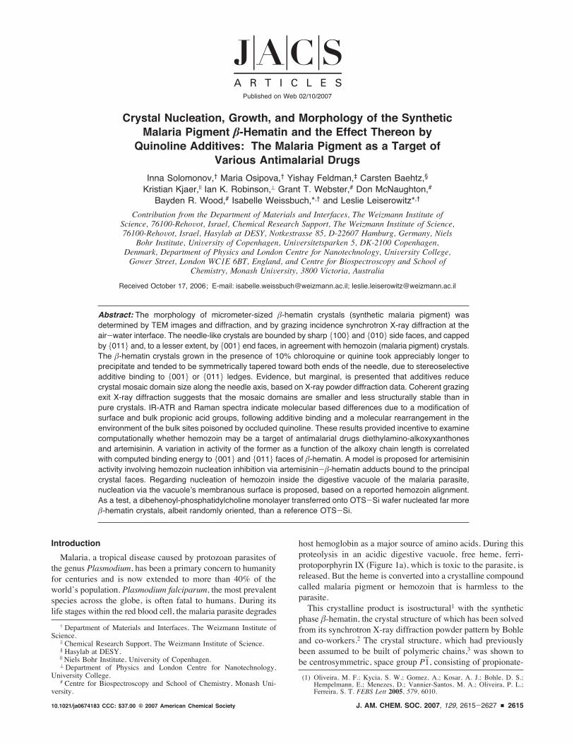
host hemoglobin as a major source of amino acids. During thisproteolysis in an acidic digestive vacuole, free heme, ferri-protoporphyrin IX (Figure 1a), which is toxic to the parasite, isreleased. But the heme is converted into a crystalline compoundcalled malaria pigment or hemozoin that is harmless to theparasite.This crystalline product is isostructural1 with the synthetic
phase !-hematin, the crystal structure of which has been solvedfrom its synchrotron X-ray diffraction powder pattern by Bohleand co-workers.2 The crystal structure, which had previouslybeen assumed to be built of polymeric chains,3 was shown tobe centrosymmetric, space group P1h, consisting of propionate-
† Department of Materials and Interfaces, The Weizmann Institute ofScience.
‡ Chemical Research Support, The Weizmann Institute of Science.§ Hasylab at DESY.| Niels Bohr Institute, University of Copenhagen.! Department of Physics and London Centre for Nanotechnology,
University College.# Centre for Biospectroscopy and School of Chemistry, Monash Uni-
versity.
(1) Oliveira, M. F.; Kycia, S. W.; Gomez, A.; Kosar, A. J.; Bohle, D. S.;Hempelmann, E.; Menezes, D.; Vannier-Santos, M. A.; Oliveira, P. L.;Ferreira, S. T. FEBS Lett 2005, 579, 6010.
Published on Web 02/10/2007
10.1021/ja0674183 CCC: $37.00 © 2007 American Chemical Society J. AM. CHEM. SOC. 2007, 129, 2615-2627 9 2615
linked (Fe-O-C(dO)CH2CH2-) reciprocal head-to-tail cyclicheme dimers interlinked via O-H‚‚‚OdC hydrogen-bondedcyclic pairs forming chains aligned parallel to the axial directiona-c (Figure 1b). This crystal structure led Bohle et al.2 topropose, in keeping with previous similar suggestions,4,5 thatantimalarial quinoline-type drugs act by binding to hemozoincrystal faces, which would inhibit their growth and result in abuildup of the toxic heme and thus lead to the death of theparasite. However, no information on the crystal surface bindingsite was provided, nor details on the habit and {h,k,l} faces ofthe micrometer-sized hemozoin or !-hematin crystals, in termsof the crystal structure.By making use of the crystal structure of !-hematin, we have
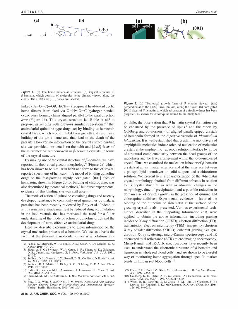
reported its theoretical growth morphology6 (Figure 2a) whichhas been shown to be similar in habit and form to that of severalreported specimens of hemozoin.7 A model of binding quinolinedrugs to the fast-growing highly corrugated {001} face ofhemozoin, shown in Figure 2b for binding of chloroquine, wasalso determined by theoretical methods,6 but direct experimentalevidence of this binding site was still absent.The mode of action of quinoline-containing drugs and recently
developed resistance to commonly used quinolines by malariaparasites has been recently reviewed by Bray et al.8 Indeed, itis this resistance, made manifest by reduced drug accumulationin the food vacuole that has motivated the need for a fullerunderstanding of the mode of action of quinoline drugs and thedevelopment of new, effective antimalarial drugs.Here we describe experiments to glean information on the
crystal nucleation process of !-hematin. We use as a basis thefact that the !-hematin molecular dimer is a bolaform am-
phiphile, the observation that !-hematin crystal formation canbe enhanced by the presence of lipids,9 and the report byGoldberg and co-workers10 of aligned parallelepiped crystalsof hemozoin formed in the digestive vacuole of Plasmodiumfalciparum. It is well-established that crystalline monolayers ofamphiphilic molecules induce oriented nucleation of molecularcrystals at the amphiphilic-aqueous solution interface by virtueof structural complementarity between the head groups of themonolayer and the layer arrangement within the to-be-nucleatedcrystal. Thus, we examined the nucleation behavior of !-hematincrystals at an air-water interface and at the interface betweena phospholipid monolayer on solid support and a chloroformsolution. We present here a characterization of the !-hematincrystal morphology obtained from different solvents in relationto its crystal structure, as well as observed changes in themorphology, time of precipitation, and a possible reduction indomain size of crystals grown in the presence of quinine andchloroquine additives. Experimental evidence in favor of thebinding of the quinoline to !-hematin at the surface of thegrowing crystal is also presented. Various experimental tech-niques, described in the Supporting Information (SI), wereapplied to obtain the above information, including grazingincidence X-ray diffraction (GIXD), electron diffraction (ED),transmission electron microscopy (TEM) images, synchrotronX-ray powder diffraction (XRPD), coherent grazing exit syn-chrotron X-ray scattering, micro-Raman spectroscopy, and IRattenuated total reflectance (ATR) micro-imaging spectroscopy.Micro-Raman and IR-ATR spectroscopies have recently beenused to understand the electronic structure of !-hematin andhemozoin in whole red blood cells11 and are shown to be a usefulway of monitoring heme aggregation through specific markerbands in human red blood cells.12
(2) Pagola, S.; Stephens, W. P.; Bohle, D. S.; Kosar, A. D.; Madsen, S. K.Nature 2000, 404, 307.
(3) Slater, A. F. G.; Swiggart, W. J.; Orton, B. R.; Flitter, W. D.; Goldberg,D. E.; Cerami, A.; Henderson, G. B. Proc. Natl. Acad. Sci. U.S.A. 1991,88, 325.
(4) Sullivan, D. J.; Gluzman, I. Y.; Russell, D. G.; Goldberg, D. E. Natl. Acad.Sci. U.S.A. 1996, 93, 11865.
(5) Sullivan, D. J.; Matile, H.; Ridley, R. G.; Goldberg, D. E. J. Biol. Chem.1998, 273, 31103.
(6) Buller, R.; Peterson, M. L.; Almarsson, O.; Leiserowitz, L. Cryst. GrowthDes. 2002, 2, 553-562.
(7) Chen, M. M.; Shi, L.; Sullivan, D. J. Mol. Biochem. Parasitol. 2001, 113,1.
(8) Bray, P. G.; Ward, S. A.; O’Neill, P. M. Drugs, Disease and Post-genomicBiology, Current Topics in Microbiology and Immunology; Springer-Verlag: Berlin, Heidelberg, 2005; Vol. 295.
(9) Fitch, C. D.; Cai, G. Z.; Shen, Y. F.; Shoemaker, J. D. Biochim. Biophys.Acta 1999, 1454, 31.
(10) Goldberg, D. E.; Slater, A. F. G.; Cerami, A.; Henderson, G. B. Proc.Natl. Acad. Sci. U.S.A. 1990, 87, 2931-2935.
(11) Wood, B. R.; Langford, S. J.; Cooke, B. M.; Lim, J.; Glenister, F. K.;Duriska, M.; Unthank, J. K.; McNaughton, D. J. Am.. Chem. Soc. 2004,126, 9233-9239.
Figure 1. (a) The heme molecular structure. (b) Crystal structure of!-hematin, which consists of molecular heme dimers, viewed along thec-axis. The (100) and (010) faces are labeled.
Figure 2. (a) Theoretical growth form of !-hematin viewed: (top)perpendicular to the {100} face, (bottom) along the c-axis; (b) corrugated{001} faces of !-hematin, at which adsorption of quinoline drugs has beenproposed, as shown for chloroquine bound to the (001) face.6
A R T I C L E S Solomonov et al.
2616 J. AM. CHEM. SOC. 9 VOL. 129, NO. 9, 2007
Next we examine by computational means the possibility ofhemozoin nucleation/growth inhibition as an important factorin the antimalarial activity of drugs other than quinolines,focusing on diethylamino-alkoxyxanthones and artemisinin. Thebinding properties of 3,6-bis-"-diethylamino-alkoxyxanthones(Chart 1) to the surface crystal structure of !-hematin areexamined in terms of the antimalarial activity of the drug, whichbelongs to a novel class of antimalarial compounds with activityagainst multidrug-resistant Plasmodium parasites, reported byRiscoe and co-workers.13,14
These compounds, labeled XNn, where n is the number ofCH2 groups per chain ranging from 2 to 8 (except n )7), formsoluble complexes with heme and prevent the precipitation ofheme in aqueous solution under the mildly acidic conditions ofthe parasitic digestive vacuole at pH ) 5.2. A strong correlationwas found between the heme affinity (Ka) measured in aqueoussolution, the alkoxy chain length, and antimalarial potency,13
shown in Figure S1of the SI. The two most active compoundsin the series, n ) 5 and 6, exhibited low nanomolar 50%inhibitory concentration (IC50) values against strains of chloro-quine-susceptible and multidrug-resistant Plasmodium falci-
parum in vitro.Finally, we consider inhibition of crystal nucleation/growth
of hemozoin as a possible mode of action to account for thewidely used antimalarial drug, artemisinin, extracted from anancient Chinese herbal remedy, Artemisia annu (sweet worm-wood or “qinghao”). Artemisinin and its derivatives (Chart 2a),being highly active against multidrug-resistant strains of malariaparasite, have attracted much attention; the mechanisms ofaction, resistance, and toxicity have been recently reviewed byMeshnick15 and by Bray et al.8 The reactivity of the peroxidefunction of artemisinin has been considered as the primary factorof the pharmacological activity. A key finding is that artemisinincannot be cyclically oxidized and reduced; only one free radicalcan result from one drug molecule, which has a selective toxiceffect at very low concentrations. The selective toxicity ofartemisinin may arise from alkylation of the heme leading toheme-artemisinin-derived covalent adducts. Heme-artemisininadducts have been demonstrated in parasite cultures treated withtherapeutic concentrations of artemisinin derivatives.16 Arte-misinin forms covalent bonds with heme when incubated in acell-free solution, and these same artemisinin-heme adductsappear to form in artemisinin-treated parasites. At micromolarconcentrations, artemisinin inhibits hemoglobin digestion bymalaria parasites and inhibits hemozoin formation, but this hasonly been demonstrated in cell-free conditions.17 The structure
of an artemisinin-porphyrin adduct has been characterized byMeunier and co-workers,18 following activation of the peroxidefunction of artemisinin by iron(II)-heme generated in situ fromiron(III)-protoporphyrin-IX and glutathione, a biologicallyrelevant reductant. It was found that under mild conditions, sucha reaction produced a high yield (85%) of heme derivativesalkylated at R, !, #, and $ positions by a C4-centered radicalderived from artemisinin (Chart 2b), where the regioselectivityat the R, ! and $ positions were about the same at 28% onaverage. The relatively low yield of 13% at the #-position ispresumably due to molecular overcrowding. We shall make useof this structural information to develop a scenario involvinghemozoin nucleation inhibition to account for the antimalarialbehavior of artemisinin and, as an extension, rationalize theincreased antimalarial efficacy when a cocktail of artemisininand quinoline-type drugs are used in conjunction. We stress,however, that there are various hypotheses on the mechanismof artemisinin-based drug action; besides heme alkylation, anumber of other biological targets have been proposed includingspecific parasite proteins.8,15
Results
!-Hematin Crystals at the Air-Water Interface. Little isknown on the crystal nucleation of the malaria pigmenthemozoin in the digestive vacuole of the malaria parasite. Inorder to glean some knowledge on the process, albeit indirect,we tried to monitor, by grazing incidence X-ray diffraction(GIXD) using synchrotron radiation, the crystal nucleation of!-hematin (i.e., synthetic hemozoin) in a manner akin to the
(12) Wood, B. R.; Hammer, L.; Davis, L.; McNaughton, D. J. Biomed. Opt.2005, 10, 014005/1-01400.5/13.
(13) Kelly, J. X.; Winters, R.; Peyton, D. H.; Hinrichs, D. H.; Riscoe, M.Antimicrob. Agents Chemother. 2002, 46, 144-150.
(14) Riscoe, M.; Kelly, J. X.; Winter, R. Curr. Med. Chem. 2005, 12, 2539.(15) Meshnick, S. H. Int. J. Parasitol. 2002, 32, 1655-1660.(16) Hong, Y. L.; Yang, Y. Z.; Meshnick, S. R.Mol. Biochem. Parasitol. 1994,
63, 121-128.
(17) Pandey, A. V.; Tekwani, B. L.; Singh, R. L.; Chauhan, V. S. J. Biol. Chem.1999, 274, 19383.
(18) Robert, A.; Coppel, Y.; Meunier, B. Chem. Comm. 2002, 414-415.
Chart 1. Generalized Structure of3,6-Bis-"-N,N-diethylamino-alkoxyxanthones, Labeled XNn, wheren is the Number of CH2 Groups
Chart 2. (a) Molecular Structure of Artemisinin, and TwoDerivatives; (b) alkylation of Iron(II)-Heme by Artemisinin; in theseHeme-Artemisinin Adducts the Substitution May Occur at R, ! (asshown), #, or $ Positions, Yielding Products ARH, A!H, A#H, andA$H, Respectively, where A Represents an Artemisinin MoietyCovalently Bound to a Heme H via Sites R, !, #, $
!-Hematin as Target of Antimalarial Drugs A R T I C L E S
J. AM. CHEM. SOC. 9 VOL. 129, NO. 9, 2007 2617
way adopted in following the crystal nucleation of the am-phiphile cholesterol from one to three bilayers at the air-waterinterface.19 The idea was to take advantage of the fact that themolecular dimeric structure of !-hematin (Figure 1b), the basicbuilding block of the crystal, may be regarded as a bolaformamphiphile and so might form crystalline multilayers at the air-water interface.20
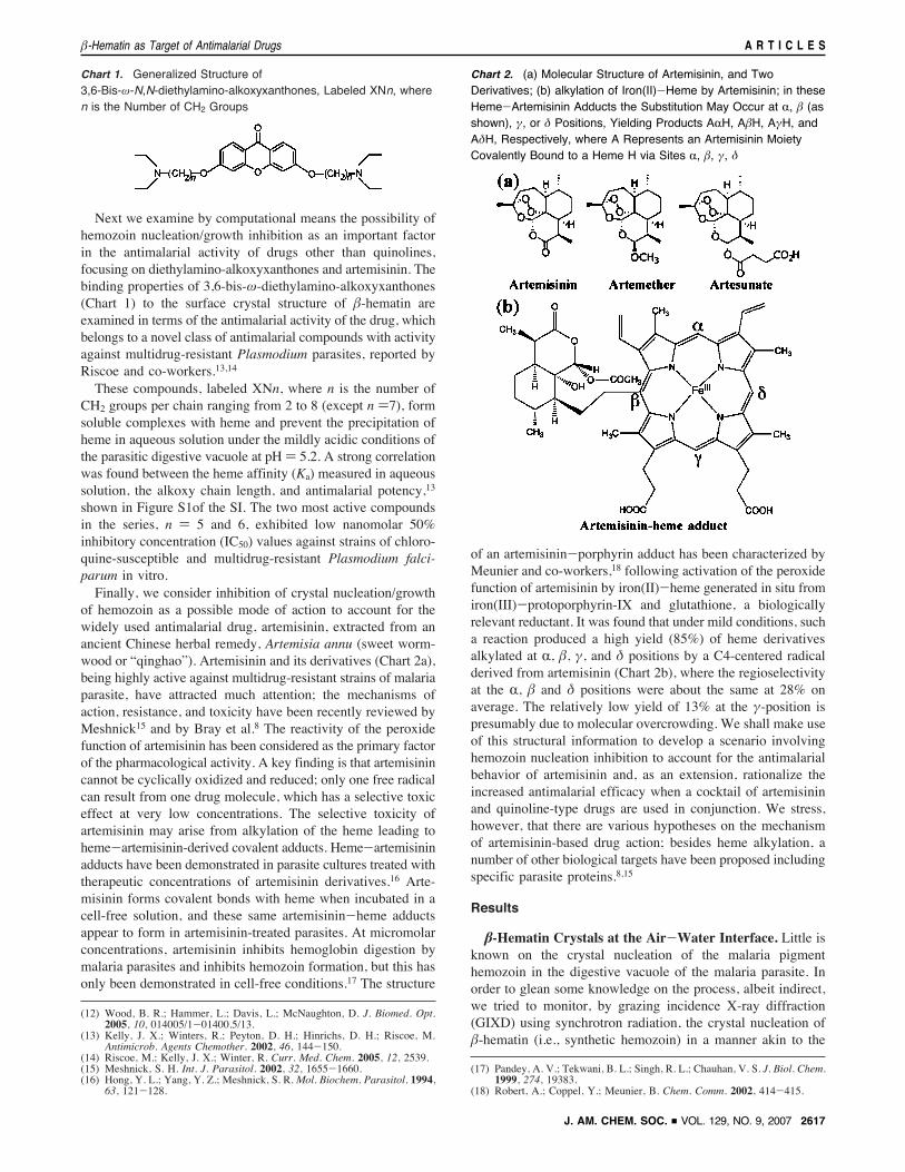
When a fresh solution, which contained hemin (chloro-(protoporphyrinato)iron(III)) and 2,6-lutidine in chloroform (seeexperimental details in SI), was spread on water, formation of!-hematin crystals was not detected on examination of the filmat the liquid surface by GIXD even after 24 h, nor by FTIRafter transfer to solid support. The result remained unchangedwhen the phospholipid dipalmitoylphosphatidyl-choline or-ethanolamine (DPPC or DPPE), sufficient for about 15% of afull monolayer coverage, was added to the chloroform solution.By contrast, the bulk solutions, yielded !-hematin nanocrystalsonly after overnight aging, as determined by FTIR and powderX-ray diffraction measurements. For the nanocrystal character-ization, samples were prepared by spreading the aged solutionon the water surface of a Langmuir trough followed bytransferring the insoluble floating material onto a gold-coveredsilicon wafer. The FTIR spectra obtained were very similar tothose of published data,21 which showed the characteristic strongand narrow bands at 1206 and 1660 cm-1, indicative of theiron-coordinated C-O and CdO stretching vibrations whichare absent in hemin. The X-ray powder diffraction patternmatched that of published data.2 An aged bulk solution, whenspread on the water surface, yielded !-hematin crystals floatingmainly on their {100} face, according to GIXD and X-rayreflectivity measurements20,22-24 of the film measured on water(Figure 3) performed on the liquid surface diffractometer atHasylab. Along the a*-direction, namely perpendicular to theplane of the {100} face, the domain size of these floating crystalswas found to be in the range between 200 and 450 Å, determinedfrom the widths of several {100} Bragg peaks by applicationof the Scherrer formula.25 The {100} Bragg reflection of onesample preparation is shown in Figure 3b.Induced Nucleation of !-Hematin Crystals by a Di-
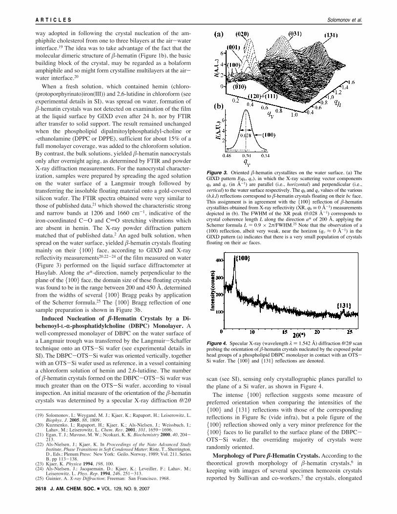
behenoyl-L-r-phosphatidylcholine (DBPC) Monolayer. Awell-compressed monolayer of DBPC on the water surface ofa Langmuir trough was transferred by the Langmuir-Schaffertechnique onto an OTS-Si wafer (see experimental details inSI). The DBPC-OTS-Si wafer was oriented vertically, togetherwith an OTS-Si wafer used as reference, in a vessel containinga chloroform solution of hemin and 2,6-lutidine. The numberof !-hematin crystals formed on the DBPC-OTS-Si wafer wasmuch greater than on the OTS-Si wafer, according to visualinspection. An initial measure of the orientation of the !-hematincrystals was determined by a specular X-ray diffraction %/2%
scan (see SI), sensing only crystallographic planes parallel tothe plane of a Si wafer, as shown in Figure 4.
The intense {100} reflection suggests some measure ofpreferred orientation when comparing the intensities of the{100} and {131} reflections with those of the correspondingreflections in Figure 8c (vide infra), but a pole figure of the{100} reflection showed only a very minor preference for the{100} faces to lie parallel to the surface plane of the DBPC-OTS-Si wafer, the overriding majority of crystals wererandomly oriented.
Morphology of Pure !-Hematin Crystals. According to thetheoretical growth morphology of !-hematin crystals,6 inkeeping with images of several specimen hemozoin crystalsreported by Sullivan and co-workers,7 the crystals, elongated
(19) Solomonov, I.; Weygand, M. J.; Kjaer, K.; Rapaport, H.; Leiserowitz, L.Biophys. J. 2005, 88, 1809.
(20) Kuzmenko, I.; Rapaport, H.; Kjaer, K.; Als-Nielsen, J.; Weissbuch, I.;Lahav, M.; Leiserowitz, L. Chem. ReV. 2001, 101, 1659-1696.
(21) Egan, T. J.; Mavuso, M. W.; Ncokazi, K. K. Biochemistry 2000, 40, 204-213.
(22) Als-Nielsen, J.; Kjaer, K. In Proceedings of the Nato AdVanced StudyInstitute, Phase Transitions in Soft Condensed Matter; Riste, T., Sherrington,D., Eds.; Plenum Press: New York: Geilo, Norway, 1989; Vol. 211, SeriesB, pp 113-138.
(23) Kjaer, K. Physica 1994, 198, 100.(24) Als-Nielsen, J.; Jacquemain, D.; Kjaer, K.; Leveiller, F.; Lahav, M.;
Leiserowitz, L. Phys. Rep. 1994, 246, 251-313.(25) Guinier, A. X-ray Diffraction; Freeman: San Francisco, 1968.
Figure 3. Oriented !-hematin crystallites on the water surface. (a) TheGIXD pattern I(qh, qV), in which the X-ray scattering vector componentsqh and qV (in Å-1) are parallel (i.e., horizontal) and perpendicular (i.e.,Vertical) to the water surface respectively. The qh and qV values of the various(h,k,l) reflections correspond to !-hematin crystals floating on their bc face.This assignment is in agreement with the {100} reflection of !-hematincrystallites obtained from X-ray reflectivity (XR, qh ! 0 Å-1) measurementsdepicted in (b). The FWHM of the XR peak (0.028 Å-1) corresponds tocrystal coherence length L along the direction a* of 200 Å, applying theScherrer formula L ) 0.9 " 2&/FWHM.25 Note that the observation of a(100) reflection, albeit very weak, near the horizon (qV # 0 Å-1) in theGIXD pattern (a) indicates that there is a very small population of crystalsfloating on their ac faces.
Figure 4. Specular X-ray (wavelength ' ) 1.542 Å) diffraction %/2% scanprobing the orientation of !-hematin crystals nucleated by the exposed polarhead groups of a phospholipid DBPC monolayer in contact with an OTS-Si wafer. The {100} and {131} reflections are denoted.
A R T I C L E S Solomonov et al.
2618 J. AM. CHEM. SOC. 9 VOL. 129, NO. 9, 2007
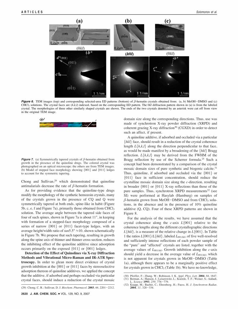
in the c-direction, are delineated by well-developed {100} and{010} side faces and smaller {011} end faces (Figure 2a). Inorder to experimentally establish this morphology, use was madeof TEM images and ED patterns. Here advantage was taken ofthe small angles between the real (a,b,c) and reciprocal(a*,b*,c*) crystal axes (10°, 8°, 7° respectively) of !-hematin26in order to obtain {0kl} or {h0l} diffraction patterns assumingthat the crystals would lie on their {100} or {010} facesrespectively.The !-hematin crystals, (grown in MeOH-DMSO27 or
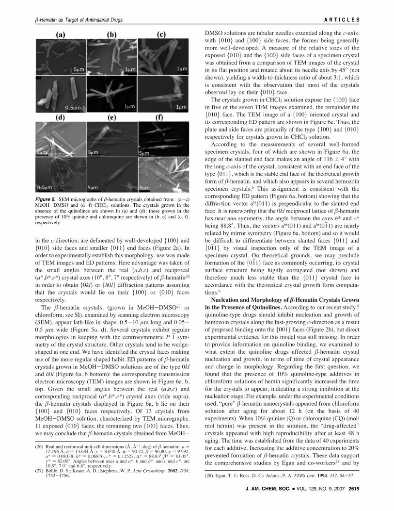
chloroform, see SI), examined by scanning electron microscopy(SEM), appear lath-like in shape, 0.5-10 µm long and 0.05-0.5 µm wide (Figure 5a, d). Several crystals exhibit regularmorphologies in keeping with the centrosymmetric P 1h sym-metry of the crystal structure. Other crystals tend to be wedge-shaped at one end. We have identified the crystal faces makinguse of the more regular shaped habit. ED patterns of !-hematincrystals grown in MeOH-DMSO solutions are of the type 0kland h0l (Figure 6a, b bottom); the corresponding transmissionelectron microscopy (TEM) images are shown in Figure 6a, b,top. Given the small angles between the real (a,b,c) andcorresponding reciprocal (a*,b*,c*) crystal axes (vide supra),the !-hematin crystals displayed in Figure 6a, b lie on their{100} and {010} faces respectively. Of 13 crystals fromMeOH-DMSO solution, characterized by TEM micrographs,11 exposed {010} faces, the remaining two {100} faces. Thus,we may conclude that !-hematin crystals obtained fromMeOH-
DMSO solutions are tabular needles extended along the c-axis,with {010} and {100} side faces, the former being generallymore well-developed. A measure of the relative sizes of theexposed {010} and the {100} side faces of a specimen crystalwas obtained from a comparison of TEM images of the crystalin its flat position and rotated about its needle axis by 45° (notshown), yielding a width-to-thickness ratio of about 3:1, whichis consistent with the observation that most of the crystalsobserved lay on their {010} face.The crystals grown in CHCl3 solution expose the {100} face
in five of the seven TEM images examined, the remainder the{010} face. The TEM image of a {100} oriented crystal andits corresponding ED pattern are shown in Figure 6c. Thus, theplate and side faces are primarily of the type {100} and {010}respectively for crystals grown in CHCl3 solution.According to the measurements of several well-formed
specimen crystals, four of which are shown in Figure 6a, theedge of the slanted end face makes an angle of 116 ( 4° withthe long c-axis of the crystal, consistent with an end face of thetype {011}, which is the stable end face of the theoretical growthform of !-hematin, and which also appears in several hemozoinspecimen crystals.6 This assignment is consistent with thecorresponding ED pattern (Figure 6a, bottom) showing that thediffraction vector d*(011) is perpendicular to the slanted endface. It is noteworthy that the 0kl reciprocal lattice of !-hematinhas near mm symmetry, the angle between the axes b* and c*being 88.8°. Thus, the vectors d*(011) and d*(01h1) are nearlyrelated by mirror symmetry (Figure 6a, bottom) and so it wouldbe difficult to differentiate between slanted faces {011} and{01h1} by visual inspection only of the TEM image of aspecimen crystal. On theoretical grounds, we may precludeformation of the {01h1} face as commonly occurring, its crystalsurface structure being highly corrugated (not shown) andtherefore much less stable than the {011} crystal face inaccordance with the theoretical crystal growth form computa-tions.6
Nucleation andMorphology of !-Hematin Crystals Grownin the Presence of Quinolines. According to our recent study,6
quinoline-type drugs should inhibit nucleation and growth ofhemozoin crystals along the fast-growing c-direction as a resultof proposed binding onto the {001} faces (Figure 2b), but directexperimental evidence for this model was still missing. In orderto provide information on quinoline binding, we examined towhat extent the quinoline drugs affected !-hematin crystalnucleation and growth, in terms of time of crystal appearanceand change in morphology. Regarding the first question, wefound that the presence of 10% quinoline-type additives inchloroform solutions of hemin significantly increased the timefor the crystals to appear, indicating a strong inhibition at thenucleation stage. For example, under the experimental conditionsused, “pure” !-hematin nanocrystals appeared from chloroformsolution after aging for about 12 h (on the basis of 40experiments). When 10% quinine (Q) or chloroquine (CQ) (mol/mol hemin) was present in the solution, the “drug-affected”crystals appeared with high reproducibility after at least 48 haging. The time was established from the data of 40 experimentsfor each additive. Increasing the additive concentration to 20%prevented formation of !-hematin crystals. These data supportthe comprehensive studies by Egan and co-workers28 and by
(26) Real and reciprocal unit cell dimensions (Å, Å-1, deg) of !-hematin: a )12.196 Å, b ) 14.684 Å, c ) 8.040 Å, R ) 90.22, ! ) 96.80, # ) 97.92,a* ) 0.08338, b* ) 0.06876, c* ) 0.12527, R* ) 88.83° !* ) 83.05°,#* ) 82.00°. Angles between axes a and a*, b and b*, and c and c*, are10.5°, 7.9° and 6.8°, respectively.
(27) Bohle, D. S.; Kosar, A. D.; Stephens, W. P. Acta Crystallogr. 2002, D58,1752-1756. (28) Egan, T. J.; Ross, D. C.; Adams, P. A. FEBS Lett. 1994, 352, 54-57.
Figure 5. SEM micrographs of !-hematin crystals obtained from: (a-c)MeOH-DMSO and (d-f) CHCl3 solutions. The crystals grown in theabsence of the quinolines are shown in (a) and (d); those grown in thepresence of 10% quinine and chloroquine are shown in (b, e) and (c, f),respectively.
!-Hematin as Target of Antimalarial Drugs A R T I C L E S
J. AM. CHEM. SOC. 9 VOL. 129, NO. 9, 2007 2619
Chong and Sullivan,29 which demonstrated that quinolineantimalarials decrease the rate of !-hematin formation.As for providing evidence that the quinoline-type drugs
modify the morphology of the synthetic hemozoin crystals, manyof the crystals grown in the presence of CQ and Q weresymmetrically tapered at both ends, spine-like in habit (Figure5b, c, e, f and Figure 7a), primarily those obtained from CHCl3solution. The average angle between the tapered side faces offour of such spines, shown in Figure 7a is about 11°, in keepingwith formation of a stepped-face morphology composed of aseries of narrow {001} or {011} facet-type ledges, with anaverage height/width ratio of tan5.5° #10, shown schematicallyin Figure 7b. We propose that such tapering, resulting in growthalong the spine axis of thinner and thinner cross-section, reducesthe inhibiting effect of the quinoline additive since adsorptionoccurs primarily on the exposed {011} or {001} ledges.Detection of the Effect of Quinolines via X-ray Diffraction
Methods and Vibrational Micro-Raman and IR-ATR Spec-
troscopy. In order to glean more direct evidence of crystalgrowth inhibition at the {001} or {011} faces by stereoselectiveadsorption thereon of quinoline additives, we applied the conceptthat the additive, if adsorbed and perhaps occluded via particularcrystal faces, should induce a reduction of the crystal mosaic
domain size along the corresponding directions. Thus, use wasmade of synchrotron X-ray powder diffraction (XRPD) andcoherent grazing X-ray diffraction30 (CGXD) in order to detectsuch an affect, if present.A quinoline additive, if adsorbed and occluded via a particular
{hkl} face, should result in a reduction of the crystal coherencelength L{h,k,l} along the direction perpendicular to that face,as would be made manifest by a broadening of the {hkl} Braggreflection. L{h,k,l} may be derived from the FWHM of theBragg reflection by use of the Scherrer formula.25 Such aconcept had been demonstrated by a comparison of the crystalmosaic domain sizes of pure synthetic and biogenic calcite.31
Thus, quinoline, if adsorbed and occluded via the {001} or{011} face in sufficient concentration, should reduce thecrystalline mosaic domain size along the c-direction, resultingin broader {001} or {011} X-ray reflections than those of thepure samples. Thus, synchrotron XRPD measurements32 (seeSI) were performed at Hasylab (Hamburg) on samples of!-hematin grown from MeOH-DMSO and from CHCl3 solu-tions, in the absence and in the presence of 10% quinolineadditive (Q, CQ). Four of these XRPD patterns are shown inFigure 8.For the analysis of the results, we have assumed that the
crystal coherence along the c-axis L{001} relative to thecoherence lengths along the different crystallographic directionsL{hkl}, is a measure of the relative change in L{001}. In Table1 the ratios L{001}/L{hkl}, labeled L001/hkl, of five well-resolvedand sufficiently intense reflections of each powder sample ofthe “pure” and “affected” crystals are listed, together with theaverage values of L001/hkl. Growth inhibition along the c-axisshould yield a decrease in the average value of L001/hkl, whichis not apparent for crystals grown in MeOH-DMSO (Table1a), although there appears to be a marginally positive effectfor crystals grown in CHCl3 (Table 1b). We have no knowledge,
(29) Chong, C. R.; Sullivan, D. J. Biochem. Pharmacol. 2003, 66, 2201-2212.
(30) Pfeiffer, F.; Zhang, W.; Robinson, I. K. Appl. Phys. Lett. 2004, 84, 1847.(31) Berman, A.; Hanson, J.; Leiserowitz, L.; Koetzle, T. F.; Weiner, S.; Addadi,
L. Science 1993, 259, 776-779.(32) Knapp, M.; Baehtz, C.; Ehrenberg, H.; Fuess, H. J. Synchrotron Radiat.
2004, 11, 328-334.
Figure 6. TEM images (top) and corresponding selected-area ED patterns (bottom) of !-hematin crystals obtained from: (a, b) MeOH-DMSO and (c)CHCl3 solutions. The crystal faces are (h,k,l) indexed, based on the corresponding ED pattern. The 0kl diffraction pattern shown in (a) is from the labeledcrystal. The morphologies of three other similarly shaped crystals are shown. The ends of the two crystals denoted by an asterisk were cut off from viewin the original TEM image.
Figure 7. (a) Symmetrically tapered crystals of !-hematin obtained fromgrowth in the presence of the quinoline drugs. The colored crystal wasphotographed on an optical microscope; the others are from TEM images.(b) Model of stepped face morphology showing {001} and {011} ledgesto account for the symmetric tapering.
A R T I C L E S Solomonov et al.
2620 J. AM. CHEM. SOC. 9 VOL. 129, NO. 9, 2007
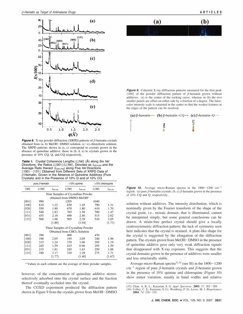
however, of the concentration of quinoline additive stereo-selectively adsorbed onto the crystal surface and the fractionthereof eventually occluded into the crystal.The CGXD experiment produced the diffraction pattern
shown in Figure 9 from the crystals grown from MeOH-DMSO
solution without additives. The intensity distribution, which isnominally given by the Fourier transform of the shape of thecrystal grain, i.e., mosaic domain, that is illuminated, cannotbe interpreted simply, but some general conclusions can bedrawn. A strain-free perfect crystal should give a locallycentrosymmetric diffraction pattern; the lack of symmetry seenhere indicates that the crystal is strained. A plate-like shape forthe crystal is suggested by the elongation of the diffractionpattern. The crystals grown fromMeOH-DMSO in the presenceof quinoline additive gave only very weak diffraction signalsthat disappeared with X-ray exposure. This suggests that thecrystal domains grown in the presence of additives were smallerand less structurally stable.
Average micro-Raman spectra33,34 (see SI) in the 1800-1200cm-1 region of pure !-hematin crystals and !-hematin grownin the presence of 10% quinine and chloroquine (Figure 10)show minor variation, mainly in band widths and relative
(33) Chan, A. K. L.; Kazarian, S. G. Appl. Spectrosc. 2003, 57, 381-389.(34) Colley, C. S.; Kazarian, S. G.; Weinberg, P. D.; Lever, M. J. Biopolymers
2004, 74, 328-335.
Figure 8. X-ray powder diffraction (XRPD) patterns of !-hematin crystalsobtained from (a, b) MeOH-DMSO solution, (c-e) chloroform solution.The XRPD patterns shown in (a, c) correspond to crystals grown in theabsence of quinoline additive, those in (b, d, e) to crystals grown in thepresence of 10% CQ, Q, and CQ respectively.
Table 1. Crystal Coherence Lengths L{hkl} (Å) along Six hklDirections, the Ratios L{001}/L{hkl}, Denoted as L001/hkl and theAverage Ratio thereof "L001/hkl# along Five hkl Directions{100}-{131} Obtained from Different Sets of XRPD Data of!-Hematin, Grown in the Absence of Quinoline Additives (PureCrystals) and in the Presence of 10% Q and of 10% CQ
pure !-hematin +10% quinine +10% chloroquine
{hkl} L{hkl} L001/hkl L{hkl} L001/hkl L{hkl} L001/hkl
Nine Samples of Crystalline Powderobtained from DMSO-MeOHa
{001} 990 1205 1040{100} 810 1.22 830 1.45 790 1.31{020} 550 1.80 670 1.80 610 1.70{111} 540 1.83 765 1.58 700 1.47{031} 455 2.18 490 2.46 515 2.02{131} 500 1.98 505 2.39 510 2.05
"1.80# "1.94# "1.71#
Three Samples of Crystalline PowderObtained from CHCl3 Solution
{001} 390 400 470{100} 190 2.05 195 2.05 240 1.96{020} 315 1.24 370 1.08 395 1.19{111} 245 1.59 415 0.96 295 1.59{031} 215 1.81 245 1.63 250 1.88{131} 180 2.17 310 1.29 275 1.71
"1.77# "1.40# "1.67#
a Values in each column are the average of three powder samples.
Figure 9. Coherent X-ray diffraction patterns measured for the first peak{100} of the powder diffraction pattern of !-hematin grown withoutadditives: (a) is the center of the rocking curve, whereas in (b) the twosmaller panels are offset on either side by a fraction of a degree. The false-color intensity scale is saturated in the center so that the weaker features atthe edges of the pattern can be resolved.
Figure 10. Average micro-Raman spectra in the 1800-1200 cm-1
region: (a) pure !-hematin crystals; (b, c) !-hematin grown in the presenceof 10% CQ and Q, respectively.
!-Hematin as Target of Antimalarial Drugs A R T I C L E S
J. AM. CHEM. SOC. 9 VOL. 129, NO. 9, 2007 2621
intensity. Band assignments, local symmetry coordinates, sym-metry terms, and the spectroscopic notation for !-hematin arebased on earlier studies.11,35
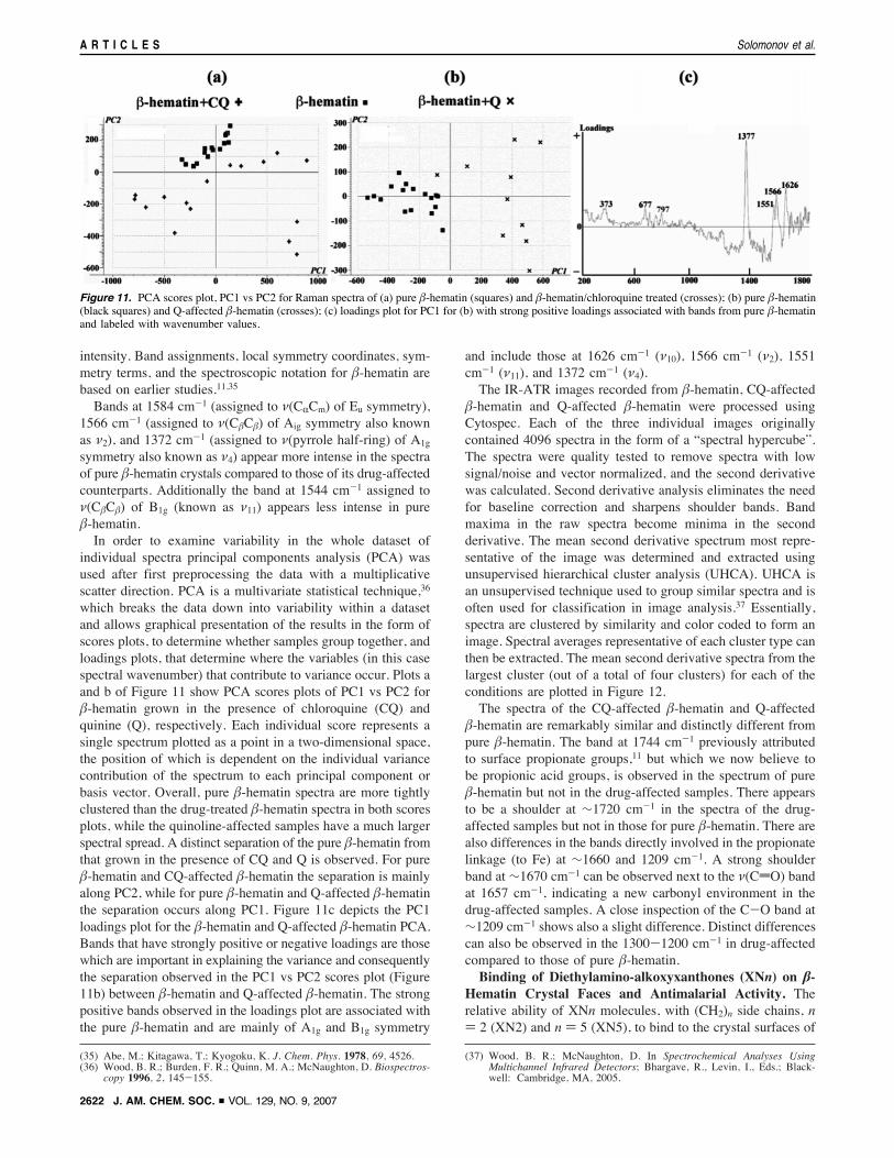
Bands at 1584 cm-1 (assigned to ((CRCm) of Eu symmetry),1566 cm-1 (assigned to ((C!C!) of Aig symmetry also knownas (2), and 1372 cm-1 (assigned to ((pyrrole half-ring) of A1gsymmetry also known as (4) appear more intense in the spectraof pure !-hematin crystals compared to those of its drug-affectedcounterparts. Additionally the band at 1544 cm-1 assigned to((C!C!) of B1g (known as (11) appears less intense in pure!-hematin.In order to examine variability in the whole dataset of
individual spectra principal components analysis (PCA) wasused after first preprocessing the data with a multiplicativescatter direction. PCA is a multivariate statistical technique,36
which breaks the data down into variability within a datasetand allows graphical presentation of the results in the form ofscores plots, to determine whether samples group together, andloadings plots, that determine where the variables (in this casespectral wavenumber) that contribute to variance occur. Plots aand b of Figure 11 show PCA scores plots of PC1 vs PC2 for!-hematin grown in the presence of chloroquine (CQ) andquinine (Q), respectively. Each individual score represents asingle spectrum plotted as a point in a two-dimensional space,the position of which is dependent on the individual variancecontribution of the spectrum to each principal component orbasis vector. Overall, pure !-hematin spectra are more tightlyclustered than the drug-treated !-hematin spectra in both scoresplots, while the quinoline-affected samples have a much largerspectral spread. A distinct separation of the pure !-hematin fromthat grown in the presence of CQ and Q is observed. For pure!-hematin and CQ-affected !-hematin the separation is mainlyalong PC2, while for pure !-hematin and Q-affected !-hematinthe separation occurs along PC1. Figure 11c depicts the PC1loadings plot for the !-hematin and Q-affected !-hematin PCA.Bands that have strongly positive or negative loadings are thosewhich are important in explaining the variance and consequentlythe separation observed in the PC1 vs PC2 scores plot (Figure11b) between !-hematin and Q-affected !-hematin. The strongpositive bands observed in the loadings plot are associated withthe pure !-hematin and are mainly of A1g and B1g symmetry
and include those at 1626 cm-1 ((10), 1566 cm-1 ((2), 1551cm-1 ((11), and 1372 cm-1 ((4).The IR-ATR images recorded from !-hematin, CQ-affected
!-hematin and Q-affected !-hematin were processed usingCytospec. Each of the three individual images originallycontained 4096 spectra in the form of a “spectral hypercube”.The spectra were quality tested to remove spectra with lowsignal/noise and vector normalized, and the second derivativewas calculated. Second derivative analysis eliminates the needfor baseline correction and sharpens shoulder bands. Bandmaxima in the raw spectra become minima in the secondderivative. The mean second derivative spectrum most repre-sentative of the image was determined and extracted usingunsupervised hierarchical cluster analysis (UHCA). UHCA isan unsupervised technique used to group similar spectra and isoften used for classification in image analysis.37 Essentially,spectra are clustered by similarity and color coded to form animage. Spectral averages representative of each cluster type canthen be extracted. The mean second derivative spectra from thelargest cluster (out of a total of four clusters) for each of theconditions are plotted in Figure 12.The spectra of the CQ-affected !-hematin and Q-affected
!-hematin are remarkably similar and distinctly different frompure !-hematin. The band at 1744 cm-1 previously attributedto surface propionate groups,11 but which we now believe tobe propionic acid groups, is observed in the spectrum of pure!-hematin but not in the drug-affected samples. There appearsto be a shoulder at $1720 cm-1 in the spectra of the drug-affected samples but not in those for pure !-hematin. There arealso differences in the bands directly involved in the propionatelinkage (to Fe) at $1660 and 1209 cm-1. A strong shoulderband at $1670 cm-1 can be observed next to the ((CdO) bandat 1657 cm-1, indicating a new carbonyl environment in thedrug-affected samples. A close inspection of the C-O band at$1209 cm-1 shows also a slight difference. Distinct differencescan also be observed in the 1300-1200 cm-1 in drug-affectedcompared to those of pure !-hematin.Binding of Diethylamino-alkoxyxanthones (XNn) on !-
Hematin Crystal Faces and Antimalarial Activity. Therelative ability of XNn molecules, with (CH2)n side chains, n) 2 (XN2) and n ) 5 (XN5), to bind to the crystal surfaces of
(35) Abe, M.; Kitagawa, T.; Kyogoku, K. J. Chem. Phys. 1978, 69, 4526.(36) Wood, B. R.; Burden, F. R.; Quinn, M. A.; McNaughton, D. Biospectros-
copy 1996, 2, 145-155.
(37) Wood, B. R.; McNaughton, D. In Spectrochemical Analyses UsingMultichannel Infrared Detectors; Bhargave, R., Levin, I., Eds.; Black-well: Cambridge, MA, 2005.
Figure 11. PCA scores plot, PC1 vs PC2 for Raman spectra of (a) pure !-hematin (squares) and !-hematin/chloroquine treated (crosses); (b) pure !-hematin(black squares) and Q-affected !-hematin (crosses); (c) loadings plot for PC1 for (b) with strong positive loadings associated with bands from pure !-hematinand labeled with wavenumber values.
A R T I C L E S Solomonov et al.
2622 J. AM. CHEM. SOC. 9 VOL. 129, NO. 9, 2007
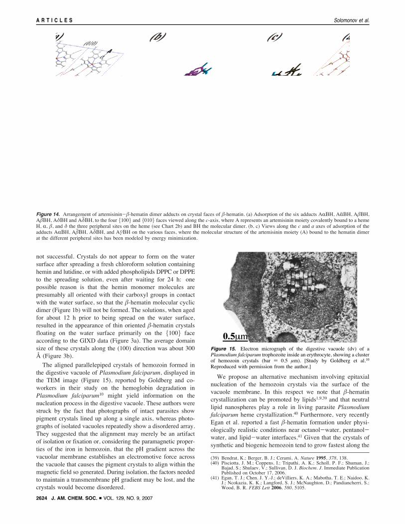
!-hematin was studied by modeling (see SI) the docking of thedrug onto the {001} and {011} crystal surfaces. The resultsare presented in Table 2 and Figure 13.Artemisinin-Hematin Molecular Dimer Adduct as In-
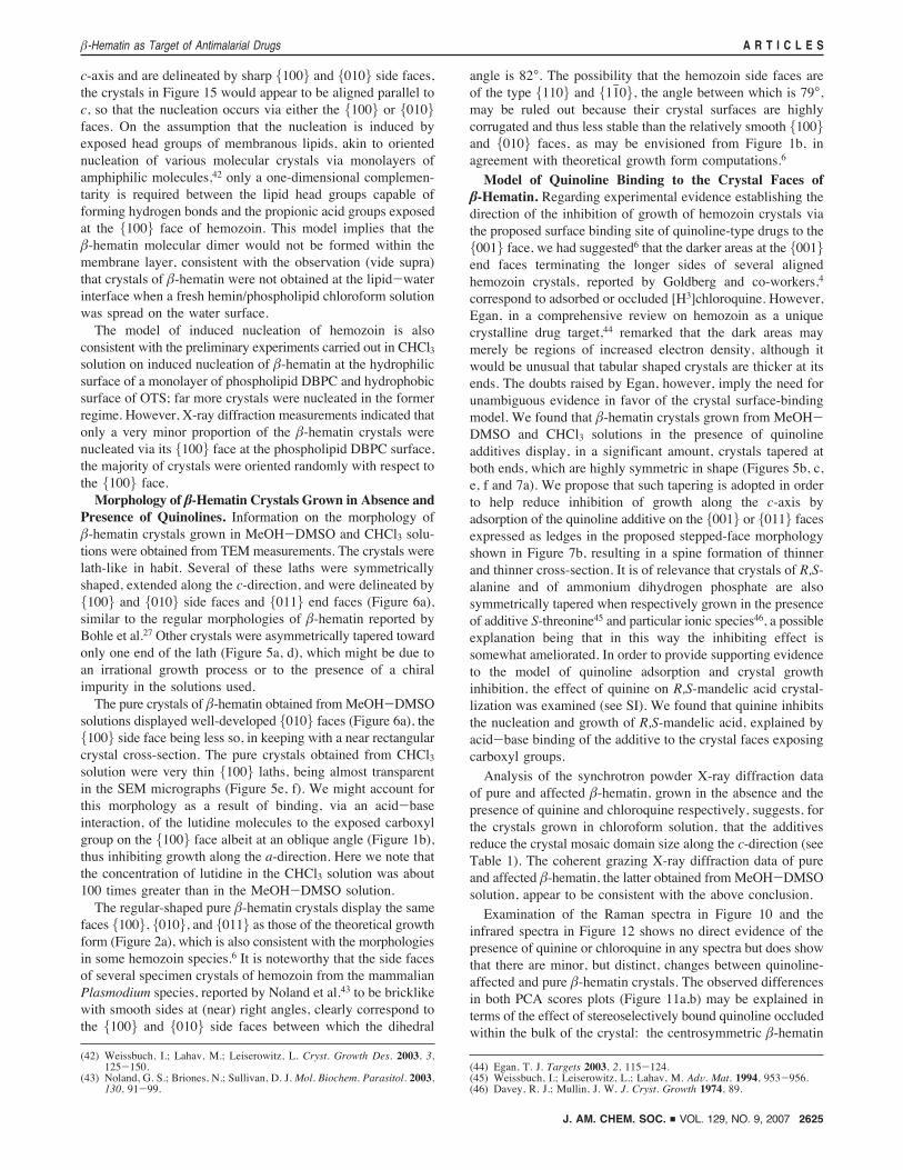
hibitor of Hemozoin Formation. Here we show that theantimalarial drug artemisinin may, in principle, act as ahemozoin growth inhibitor. Meunier and co-workers18 havedemonstrated that artemisinin covalently reacts with the freeheme monomer at the three different sites (R, !, $) on themolecular periphery with about the same regioselectivity of 29%,yielding artemisinin-heme adducts labeled ARH, A!H, andA$H, as depicted in Chart 2b for site !. Reaction at site # iscomparatively low at 13%. We invoke the possibility that eachof the four adducts might link to a pure heme monomer in thedigestive vacuole of the malaria parasite to form a dimer witha basic skeleton akin to the !-hematin cyclic dimer (Figure 1b),the exact structure thereof depending upon that of the adductand its relative orientation to the heme monomer prior to dimerformation. Such molecules, if present in the digestive vacuoleof the parasite, would act in a manner akin to a classic “tailor-made” additive in solution designed to inhibit crystal nucleationand growth.38 In principle, four diastereomeric pairs of dimers(labeled AR!H, ARj!H; A!!H, A!h!H; A$!H, A$h!H;A#!H, A#j!H) may be formed and adsorbed on the six {100},{010}, and {001} faces of !-hematin.For the most part, each additive type may be adsorbed at
only one of the six faces, as shown somewhat schematically in
Figure 14a for binding of the R, Rj , !, !h, and $, $h derivatives tothe four {100} and {010} crystal side faces. The moleculeswithin each pair are diastereoisomers since artemisinin is chiraland the !-hematin cyclic dimer molecule is centrosymmetric.Using CERIUS2 software we computed low-energy models ofsome of the diastereoisomeric dimer structures, which are shownadsorbed onto the different crystal faces in Figure 14b,c.Therefore, on the assumption that the heme-artemisinin
reaction products are formed in the digestive vacuole of theparasite in sufficient concentration, an efficient inhibition ofgrowth of hemozoin should occur since the additives may actalong all three principal growth directions, a, b, and c. Inprinciple, only a fraction of the additives can be adsorbed ontothe small, but fast-growing {001} faces. Therefore, we mayexpect a stronger inhibition of crystal growth along this directionto occur were quinoline drugs, such as chloroquine or quinine,used in conjunction with artemisinin, given the proposedmechanism of action of artemisinin. Thus, we examined, viathe use of a model crystal system of S-alanine as described inthe SI, whether a combination of tailor-made crystal nucleationretardants, which inhibit crystal growth along different direc-tions, acts more effectively than each alone. The results suggestthat simultaneous use of two types of additives, which bind todifferent crystal faces, has a greater inhibiting effect onnucleation than each of the additives used alone.
Discussion
Nucleation of Hemozoin Crystals. Little is known at themolecular level on the nucleation process of hemozoin in thedigestive vacuole of the parasite, including the critical size ofthe nucleus, and the role played by various molecules in thevacuole to promote the nucleation process. Attempts to gleaninformation on the critical size of !-hematin nuclei by monitor-ing the early stages of crystallization of !-hematin at the air-water interface using grazing incidence X-ray diffraction were
(38) Weissbuch, I.; Addadi, L.; Lahav, M.; Leiserowitz, L. Science 1991, 253,637-645.
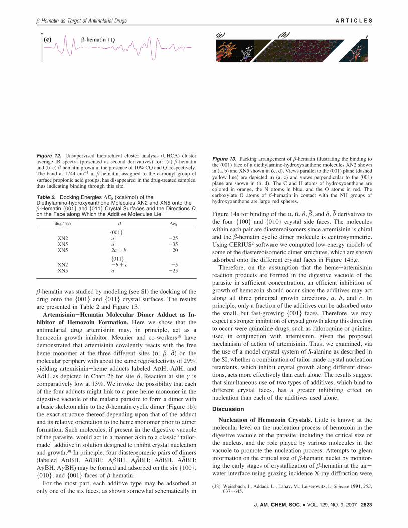
Figure 12. Unsupervised hierarchical cluster analysis (UHCA) clusteraverage IR spectra (presented as second derivatives) for: (a) !-hematinand (b, c) !-hematin grown in the presence of 10% CQ and Q, respectively.The band at 1744 cm-1 in !-hematin, assigned to the carbonyl group ofsurface propionic acid groups, has disappeared in the drug-treated samples,thus indicating binding through this site.
Table 2. Docking Energies "Eb (kcal/mol) of theDiethylamino-hydroxyxanthone Molecules XN2 and XN5 onto the!-Hematin {001} and {011} Crystal Surfaces and the Directions Don the Face along Which the Additive Molecules Lie
drug/face D "Eb
{001}XN2 a -25XN5 a -35XN5 2a + b -20
{011}XN2 -b + c -5XN5 a -25
Figure 13. Packing arrangement of !-hematin illustrating the binding tothe (001) face of a diethylamino-hydroxyxanthone molecules XN2 shownin (a, b) and XN5 shown in (c, d). Views parallel to the (001) plane (dashedyellow line) are depicted in (a, c) and views perpendicular to the (001)plane are shown in (b, d). The C and H atoms of hydroxyxanthone arecolored in orange, the N atoms in blue, and the O atoms in red. Thecarboxylate O atoms of !-hematin in contact with the NH groups ofhydroxyxanthone are large red spheres.
!-Hematin as Target of Antimalarial Drugs A R T I C L E S
J. AM. CHEM. SOC. 9 VOL. 129, NO. 9, 2007 2623
not successful. Crystals do not appear to form on the watersurface after spreading a fresh chloroform solution containinghemin and lutidine, or with added phospholipids DPPC or DPPEto the spreading solution, even after waiting for 24 h: onepossible reason is that the hemin monomer molecules arepresumably all oriented with their carboxyl groups in contactwith the water surface, so that the !-hematin molecular cyclicdimer (Figure 1b) will not be formed. The solutions, when agedfor about 12 h prior to being spread on the water surface,resulted in the appearance of thin oriented !-hematin crystalsfloating on the water surface primarily on the {100} faceaccording to the GIXD data (Figure 3a). The average domainsize of these crystals along the (100) direction was about 300Å (Figure 3b).
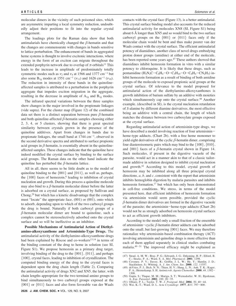
The aligned parallelepiped crystals of hemozoin formed inthe digestive vacuole of Plasmodium falciparum, displayed inthe TEM image (Figure 15), reported by Goldberg and co-workers in their study on the hemoglobin degradation inPlasmodium falciparum10 might yield information on thenucleation process in the digestive vacuole. These authors werestruck by the fact that photographs of intact parasites showpigment crystals lined up along a single axis, whereas photo-graphs of isolated vacuoles repeatedly show a disordered array.They suggested that the alignment may merely be an artifactof isolation or fixation or, considering the paramagnetic proper-ties of the iron in hemozoin, that the pH gradient across thevacuolar membrane establishes an electromotive force acrossthe vacuole that causes the pigment crystals to align within themagnetic field so generated. During isolation, the factors neededto maintain a transmembrane pH gradient may be lost, and thecrystals would become disordered.
We propose an alternative mechanism involving epitaxialnucleation of the hemozoin crystals via the surface of thevacuole membrane. In this respect we note that !-hematincrystallization can be promoted by lipids1,9,39 and that neutrallipid nanospheres play a role in living parasite Plasmodiumfalciparum heme crystallization.40 Furthermore, very recentlyEgan et al. reported a fast !-hematin formation under physi-ologically realistic conditions near octanol-water, pentanol-water, and lipid-water interfaces.41 Given that the crystals ofsynthetic and biogenic hemozoin tend to grow fastest along the
(39) Bendrat, K.; Berger, B. J.; Cerami, A. Nature 1995, 378, 138.(40) Pisciotta, J. M.; Coppens, I.; Tripathi, A. K.; Scholl, P. F.; Shuman, J.;
Bajad, S.; Shulaev, V.; Sullivan, D. J. Biochem. J. Immediate PublicationPublished on October 17, 2006.
(41) Egan, T. J.; Chen, J. Y.-J.; deVilliers, K. A.; Mabotha, T. E.; Naidoo, K.J.; Ncokazia, K. K.; Langford, S. J.; McNaughton, D.; Pandiancherri, S.;Wood, B. R. FEBS Lett 2006, 580, 5105.
Figure 14. Arrangement of artemisinin-!-hematin dimer adducts on crystal faces of !-hematin. (a) Adsorption of the six adducts AR!H, ARj!H, A!!H,A!h!H, A$!H and A$h!H, to the four {100} and {010} faces viewed along the c-axis, where A represents an artemisinin moiety covalently bound to a hemeH, R, !, and $ the three peripheral sites on the heme (see Chart 2b) and BH the molecular dimer. (b, c) Views along the c and a axes of adsorption of theadducts AR!H, A!h!H, A$h!H, and A#j!H on the various faces, where the molecular structure of the artemisinin moiety (A) bound to the hematin dimerat the different peripheral sites has been modeled by energy minimization.
Figure 15. Electron micrograph of the digestive vacuole (dv) of aPlasmodium falciparum trophozoite inside an erythrocyte, showing a clusterof hemozoin crystals (bar ) 0.5 µm). [Study by Goldberg et al.10
Reproduced with permission from the author.]
A R T I C L E S Solomonov et al.
2624 J. AM. CHEM. SOC. 9 VOL. 129, NO. 9, 2007
c-axis and are delineated by sharp {100} and {010} side faces,the crystals in Figure 15 would appear to be aligned parallel toc, so that the nucleation occurs via either the {100} or {010}faces. On the assumption that the nucleation is induced byexposed head groups of membranous lipids, akin to orientednucleation of various molecular crystals via monolayers ofamphiphilic molecules,42 only a one-dimensional complemen-tarity is required between the lipid head groups capable offorming hydrogen bonds and the propionic acid groups exposedat the {100} face of hemozoin. This model implies that the!-hematin molecular dimer would not be formed within themembrane layer, consistent with the observation (vide supra)that crystals of !-hematin were not obtained at the lipid-waterinterface when a fresh hemin/phospholipid chloroform solutionwas spread on the water surface.The model of induced nucleation of hemozoin is also
consistent with the preliminary experiments carried out in CHCl3solution on induced nucleation of !-hematin at the hydrophilicsurface of a monolayer of phospholipid DBPC and hydrophobicsurface of OTS; far more crystals were nucleated in the formerregime. However, X-ray diffraction measurements indicated thatonly a very minor proportion of the !-hematin crystals werenucleated via its {100} face at the phospholipid DBPC surface,the majority of crystals were oriented randomly with respect tothe {100} face.Morphology of !-Hematin Crystals Grown in Absence and
Presence of Quinolines. Information on the morphology of!-hematin crystals grown in MeOH-DMSO and CHCl3 solu-tions were obtained from TEMmeasurements. The crystals werelath-like in habit. Several of these laths were symmetricallyshaped, extended along the c-direction, and were delineated by{100} and {010} side faces and {011} end faces (Figure 6a),similar to the regular morphologies of !-hematin reported byBohle et al.27 Other crystals were asymmetrically tapered towardonly one end of the lath (Figure 5a, d), which might be due toan irrational growth process or to the presence of a chiralimpurity in the solutions used.The pure crystals of !-hematin obtained from MeOH-DMSO
solutions displayed well-developed {010} faces (Figure 6a), the{100} side face being less so, in keeping with a near rectangularcrystal cross-section. The pure crystals obtained from CHCl3solution were very thin {100} laths, being almost transparentin the SEM micrographs (Figure 5e, f). We might account forthis morphology as a result of binding, via an acid-baseinteraction, of the lutidine molecules to the exposed carboxylgroup on the {100} face albeit at an oblique angle (Figure 1b),thus inhibiting growth along the a-direction. Here we note thatthe concentration of lutidine in the CHCl3 solution was about100 times greater than in the MeOH-DMSO solution.The regular-shaped pure !-hematin crystals display the same
faces {100}, {010}, and {011} as those of the theoretical growthform (Figure 2a), which is also consistent with the morphologiesin some hemozoin species.6 It is noteworthy that the side facesof several specimen crystals of hemozoin from the mammalianPlasmodium species, reported by Noland et al.43 to be bricklikewith smooth sides at (near) right angles, clearly correspond tothe {100} and {010} side faces between which the dihedral
angle is 82°. The possibility that the hemozoin side faces areof the type {110} and {11h0}, the angle between which is 79°,may be ruled out because their crystal surfaces are highlycorrugated and thus less stable than the relatively smooth {100}and {010} faces, as may be envisioned from Figure 1b, inagreement with theoretical growth form computations.6
Model of Quinoline Binding to the Crystal Faces of
!-Hematin. Regarding experimental evidence establishing thedirection of the inhibition of growth of hemozoin crystals viathe proposed surface binding site of quinoline-type drugs to the{001} face, we had suggested6 that the darker areas at the {001}end faces terminating the longer sides of several alignedhemozoin crystals, reported by Goldberg and co-workers,4
correspond to adsorbed or occluded [H3]chloroquine. However,Egan, in a comprehensive review on hemozoin as a uniquecrystalline drug target,44 remarked that the dark areas maymerely be regions of increased electron density, although itwould be unusual that tabular shaped crystals are thicker at itsends. The doubts raised by Egan, however, imply the need forunambiguous evidence in favor of the crystal surface-bindingmodel. We found that !-hematin crystals grown from MeOH-DMSO and CHCl3 solutions in the presence of quinolineadditives display, in a significant amount, crystals tapered atboth ends, which are highly symmetric in shape (Figures 5b, c,e, f and 7a). We propose that such tapering is adopted in orderto help reduce inhibition of growth along the c-axis byadsorption of the quinoline additive on the {001} or {011} facesexpressed as ledges in the proposed stepped-face morphologyshown in Figure 7b, resulting in a spine formation of thinnerand thinner cross-section. It is of relevance that crystals of R,S-alanine and of ammonium dihydrogen phosphate are alsosymmetrically tapered when respectively grown in the presenceof additive S-threonine45 and particular ionic species46, a possibleexplanation being that in this way the inhibiting effect issomewhat ameliorated. In order to provide supporting evidenceto the model of quinoline adsorption and crystal growthinhibition, the effect of quinine on R,S-mandelic acid crystal-lization was examined (see SI). We found that quinine inhibitsthe nucleation and growth of R,S-mandelic acid, explained byacid-base binding of the additive to the crystal faces exposingcarboxyl groups.Analysis of the synchrotron powder X-ray diffraction data
of pure and affected !-hematin, grown in the absence and thepresence of quinine and chloroquine respectively, suggests, forthe crystals grown in chloroform solution, that the additivesreduce the crystal mosaic domain size along the c-direction (seeTable 1). The coherent grazing X-ray diffraction data of pureand affected !-hematin, the latter obtained from MeOH-DMSOsolution, appear to be consistent with the above conclusion.Examination of the Raman spectra in Figure 10 and the
infrared spectra in Figure 12 shows no direct evidence of thepresence of quinine or chloroquine in any spectra but does showthat there are minor, but distinct, changes between quinoline-affected and pure !-hematin crystals. The observed differencesin both PCA scores plots (Figure 11a,b) may be explained interms of the effect of stereoselectively bound quinoline occludedwithin the bulk of the crystal: the centrosymmetric !-hematin
(42) Weissbuch, I.; Lahav, M.; Leiserowitz, L. Cryst. Growth Des. 2003, 3,125-150.
(43) Noland, G. S.; Briones, N.; Sullivan, D. J. Mol. Biochem. Parasitol. 2003,130, 91-99.
(44) Egan, T. J. Targets 2003, 2, 115-124.(45) Weissbuch, I.; Leiserowitz, L.; Lahav, M. AdV. Mat. 1994, 953-956.(46) Davey, R. J.; Mullin, J. W. J. Cryst. Growth 1974, 89.
!-Hematin as Target of Antimalarial Drugs A R T I C L E S
J. AM. CHEM. SOC. 9 VOL. 129, NO. 9, 2007 2625
molecular dimers in the vicinity of such poisoned sites, whichare asymmetric imparting a local symmetry reduction, undoubt-edly adjust their positions to fit into the regular crystalarrangement.
The loadings plots for the Raman data show that bothantimalarials have chemically changed the !-hematin and thatthe changes are commensurate with changes in bands sensitiveto lattice perturbation. The enhancement of bands in aggregatedheme systems is thought to involve excitonic interactions, whereenergy in the form of an exciton can migrate throughout theextended porphyrin network due to overlap of &-orbitals11 Thisleads to the increase in amplitude of predominately totallysymmetric modes such as (2 and (4 at 1566 and 1377 cm-1 butalso some B1g modes at 1551 cm-1 ((11) and 1626 cm-1 ((10).The reduction in intensity of these bands in the quinoline-affected samples is attributed to a perturbation in the porphyrinaggregate that impedes exciton migration in the aggregate,resulting in the decrease in amplitude of A1g and B1g modes.
The infrared spectral variations between the three samplesshow changes in the major involved in the propionate linkages(vide supra). For the cluster map (Figure 12) of the combineddata set there is a distinct separation between pure !-hematinand both quinoline-affected !-hematin samples choosing either2, 3, 4, or 5 clusters, showing that there is great spectralsimilarity between crystals grown in the presence of thequinoline additives. Apart from changes in bands due topropionate linkages, the small band at 1744 cm-1, observed inthe second derivative spectra and attributed to surface propionicacid groups in !-hematin, is essentially absent in the quinoline-affected samples. These changes indicate that the quinoline haveindeed modified the crystal surface by binding to the surfaceacid groups. The Raman data on the other hand indicate thequinoline has perturbed the !-hematin lattice.All in all, there seems to be little doubt as to the model of
quinoline binding to the {001} and {011}, as well as, perhaps,the {100} faces of hemozoin,6 leading to inhibition of crystalnucleation and growth. During this process a quinoline moleculemay also bind to a !-hematin molecular dimer before the latteris adsorbed on a crystal surface, as proposed by Sullivan andChong,29 but which has a kinetic disadvantage that the complexmust “locate” the appropriate face, (001) or (001h), onto whichto adsorb, depending upon to which of the two carboxyl groupsthe drug is bound. Naturally if both carboxyl groups of a!-hematin molecular dimer are bound to quinoline, such acomplex cannot be stereoselectively adsorbed onto the crystalsurface and so will be ineffective as an inhibitor.
Possible Mechanisms of Antimalarial Action of Diethyl-
amino-alkoxyxanthone and Artemisinin-Type Drugs. Theantimalarial activity of the diethylamino-alkoxyxanthone drugshad been explained by Riscoe and co-workers13,14 in terms ofthe binding constant of the drug to heme in solution (see SI,Figure S1). We propose hemozoin as a primary drug target,involving binding of the drug to the {001}, {011}, and perhaps{100}, crystal faces, leading to inhibition of crystallization. Thecomputed binding energy of the drug to the crystal faces isdependent upon the drug chain length (Table 2). Comparingthe antimalarial activity of drugs XN2 and XN5, the latter, withchain lengths appropriate for the two terminal amino groups tobind simultaneously to two carboxyl groups exposed at the{001} or {011} faces and also form favorable van der Waals
contacts with the crystal face (Figure 13), is a better antimalarial.This crystal surface binding model also accounts for the reducedantimalarial activity for molecules XN8 (SI, Figure S1) beingabout 6 Å longer than XN5 and so would bind to the two surfacecarboxyl groups on the {001} or {011} faces only if themolecular chain would be bent and thus make poorer van derWaals contact with the crystal surface. The efficient antimalarialpotency of diamidines, another class of novel drugs embodyingproton donor groups (amidine) at either end of the molecule,has been reported some years ago.47 These authors showed thatdiamidines inhibit hemozoin formation in vitro with a similarpotency to chloroquine. It is likely that these drugs, such aspentamidine (H3N2C-C6H4-O-C5H10-O-C6H4-CN2H3) in-hibit hemozoin formation as a result of binding of both amidinegroups of the molecule to exposed propionic acid groups at thecrystal surface. Of relevance to the model proposed forantimalarial action of the diethylamino-alkoxyxanthones isgrowth inhibition of barium sulfate by an additive with moietieswhich simultaneously cap onto the crystal surface.48 Anotherexample, (described in SI), is the crystal nucleation retardationof S-alanine by different diamine derivatives, the most effectivebeing an additive with a central chain, the length of whichmatches the distance between two carboxylate groups exposedat the crystal surface.Regarding antimalarial action of artemisinin-type drugs, we
have described a model involving reaction of four artemisinin-heme-type adducts, (Chart 2b), with a free heme monomer toyield eight derivatives of the cyclic !-hematin dimer, comprisingfour diasteroisomeric pairs which may bind to the {100}, {010},and {001} faces of a !-hematin crystal shown in Figure 14.Such molecules, if present in the digestive vacuole of theparasite, would act in a manner akin to that of a classic tailor-made additive in solution designed to inhibit crystal nucleationand growth.42 According to this model, crystal growth ofhemozoin may be inhibited along all three principal crystaldirections, a, b, and c, consistent with the report that artemisinininhibits hemoglobin digestion by malaria parasites and inhibitshemozoin formation,17 but which has only been demonstratedin cell-free conditions. We stress, in terms of the modelpresented here, that efficient inhibition of hemozoin formationvia artemisinin would seem possible, provided the cyclic!-hematin dimer derivatives are formed in the digestive vacuoleof the parasite; the artemisinin-heme-type adducts (Chart 2b)would not be as strongly adsorbed on hemozoin crystal surfacesto act as efficient growth inhibitors.According to the model only a small fraction of the ensemble
of artemisinin-cyclic !-hematin dimer adducts can be adsorbedonto the small, but fast-growing {001} faces. We may thereforerationalize why artemisinin-based combination therapy (ACT)involving artemisinin and quinoline drugs is more effective thaneach of them applied separately in clinical studies combatingmalaria.49-51 The improved efficacy might be explained as
(47) Stead, A. M. W.; Bray, P. G.; Edwards, I. G.; Dekoning, H. P.; Elford, B.C.; Stocks, P. A.; Ward, S. A. Mol. Pharmacol. 2001, 1298.
(48) Coveney, P. V.; Davey, R.; Griffin, J. L. W.; He, Y.; Hamlin, J. D.;Stackhouse, S.; Whiting, A. J. Am. Chem. Soc. 2000, 122, 11557.
(49) deVries, P. J.; Bich, N. N.; Thien, H. V.; Hung, L. N.; Anh, T. K.; Kager,P. A.; Heisterkamp, S. H. Antimicrob. Agents Chemother. 2000, 44, 1302-1308.
(50) Gupta, S.; Thapar, M. M.; Mariga, S. T.; Wernsdorfer, W. H.; Bjorkman,A. Exp. Parasitol. 2002, 100, 28-35.
(51) Olliaro, P. L.; Taylor, T. W. J. Postgrad. Med. 2004, 50, 40-44.(52) Wei, K.-T.; Ward, D. L. Acta Crystallogr. 1977, B33, 797-800.
A R T I C L E S Solomonov et al.
2626 J. AM. CHEM. SOC. 9 VOL. 129, NO. 9, 2007
follows: quinoline drugs are expected to inhibit growthprimarily along the fast-growing needle c-axis, and the arte-misinin-type drugs have been hypothesized to retard growthalong all three principal crystal directions (a, b, c). Thus, acombination of these two types of drugs would be effectiveinhibitors of overall crystal formation of hemozoin because ofa relatively high concentration of drugs available for bindingonto the small, yet fast-growing, opposite faces terminating theneedle. This hypothesis is in agreement with the experimentson the model crystal system S-alanine, suggesting that acombination of tailor-made additives that bind to different crystalsurfaces has a greater inhibiting effect on nucleation than eachof the additives used alone.
Concluding Remarks
The most dominant crystal faces of !-hematin have beenexperimentally characterized, in agreement with the shape ofsome reported hemozoin crystals and the theoretical growth formof !-hematin.6 An effect of quinine and chloroquine on thegrowth morphology of !-hematin has been observed andcorrelated with adsorption of the drugs onto the {001} or {011}crystal faces. This interpretation is in agreement with evidence,although marginal, from synchrotron X-ray diffraction data ofpowder samples that the quinoline additives reduce the crystaldomain size along the c-axis and with coherent grazing X-raydiffraction experiments, which suggest that the !-hematincrystals grown in the presence of the additives were smallerand less structurally stable than the pure form. The IR-ATRand Raman spectral results show that !-hematin grown in thepresence of the quinoline drugs embodies molecular baseddifferences from pure !-hematin. The results suggest modifica-tion of surface and bulk propionic acid linkages and aggregationperturbation within the affected crystal, albeit minor, presumablyarising from host molecular rearrangement in the environmentof the bulk sites poisoned by occluded quinoline. These proposedchanges are consistent with the interpretation of morphologicalmodification, the X-ray diffraction results, and the modelcomputations of surface binding of quinoline antimalarials tothe {001} face of !-hematin.6Computational studies carried out on the antimalarial diethyl-
amino-alkoxyxanthanones suggest that the hemozoin crystal isa primary drug target, via stereoselective binding of the twoamine groups of the molecule to acid groups exposed at thecrystal surface, resulting in inhibition of hemozoin nucleationor growth. A model is proposed for the antimalarial action ofartemisinin-type drugs involving efficient inhibition of crystal
nucleation of hemozoin by binding onto all principal crystalfaces, thus acting akin to classic tailor-made inhibitors of crystalnucleation. This model might also explain the effective anti-malarial properties of a combination of artemisinin-type drugsand quinolines, assuming that the latter will inhibit growth alongthe fast-growing but small faces at the opposite ends of theneedle-shaped hemozoin crystal.
A model has been presented of induced epitaxial nucleationof hemozoin via a lipid membrane in the digestive vacuole ofa malaria parasite, based on a published electron micrographof the digestive vacuole of a Plasmodium falciparum trophozoiteinside a red blood cell, showing a cluster of aligned hemozoincrystals (Figure 15).10 Studies on oriented nucleation of !-hematin crystals at the interface between lipid monolayers andwater, or other liquids, are currently being undertaken.
Acknowledgment. This work was supported in part by theKimmelmann Center at the Weizmann Institute of Science, theDanSync program of the Danish Natural Science ResearchCouncil, the Carlsberg Foundation and the European Communityunder TMR-Contract ERBFMGECT950059. Thanks are due toHASYLAB for synchrotron beamtime and to Professor JochenSchneider, Director of HASYLAB. Regarding use of beamtimeat the Advanced Photon Source, the UNICAT facility issupported by the Materials Research Laboratory, University ofIllinois funded by U.S. DOE award DEFG02-91ER45439, theOak Ridge National Laboratory, the National Institute ofStandards and Technology and UOP LLC. Part of this work isfunded by an Australian Research Council Discovery Grant.B.R.W.’s work is supported by an Australian SynchrotronProgram Fellowship Grant and a Monash University Synchro-tron Fellowship Grant.
Supporting Information Available: Experimental Section;Figure S1 showing the reported13 effect of carbon chain lengthon the antimalarial activity (IC50) and heme affinity (Ka) ofdiethylamino-hydroxyxanthone molecules; section describinginhibition of crystal nucleation of R,S-mandelic acid withquinine; section describing a computational study on bindingof diethylamino-alkoxyxanthones to !-hematin crystal faces incorrelation with antimalarial activity; section describing a modelstudy on inhibition of crystal nucleation of S-alanine via additivecombinations. This material is available free of charge via theInternet at http://pubs.acs.org.
JA0674183
!-Hematin as Target of Antimalarial Drugs A R T I C L E S
J. AM. CHEM. SOC. 9 VOL. 129, NO. 9, 2007 2627
Related Documents