CRYOPRESERVATION STRATEGY FOR ADIPOSE TISSUE AND ADIPOSE TISSUE DERIVED STEM CELLS BY DEVELOPING NON- TOXIC FREEZING SOLUTIONS Dissertation submitted in partial fulfillment of the degree of Doctor of Philosophy in Biotechnology and Medical Engineering by Sirsendu Sekhar Ray (Roll Number: 510BM408) based on research carried out under the supervision of Prof. (Mrs.) Krishna Pramanik And Prof. Sunil Kumar Sarangi June, 2016 Department of Biotechnology and Medical Engineering National Institute of Technology Rourkela

Welcome message from author
This document is posted to help you gain knowledge. Please leave a comment to let me know what you think about it! Share it to your friends and learn new things together.
Transcript

CRYOPRESERVATION STRATEGY FOR ADIPOSE TISSUE AND
ADIPOSE TISSUE DERIVED STEM CELLS BY DEVELOPING NON-
TOXIC FREEZING SOLUTIONS
Dissertation submitted
in partial fulfillment of the degree of
Doctor of Philosophy
in
Biotechnology and Medical Engineering
by
Sirsendu Sekhar Ray
(Roll Number: 510BM408)
based on research carried out
under the supervision of
Prof. (Mrs.) Krishna Pramanik
And
Prof. Sunil Kumar Sarangi
June, 2016
Department of Biotechnology and Medical Engineering National Institute of Technology Rourkela


Dedicated to
My Parents

Declaration of Originality
I, Sirsendu Sekhar Ray, Roll Number -510BM408 hereby declare that this dissertation entitled
''Cryopreservation strategy for adipose tissue and adipose tissue-derived stem cells by developing
non-toxic freezing solutions” represents my original work carried out as a doctoral student of NIT
Rourkela.To the best of my knowledge, it contains no material previously published or written by
another person, nor any material presented for the award of any other degree or diploma of NIT
Rourkela or any other institution. Any contribution made to this research by others, with whom I
have worked at NIT Rourkela or elsewhere, is explicitly acknowledged in the dissertation. Works
of other authors cited in this dissertation have been duly acknowledged under the section
''Bibliography''. I have also submitted my original research records to the scrutiny committee for
evaluation of my dissertation.
I am fully aware that in the case of any non-compliance detected in future, the Senate of NIT
Rourkela may withdraw the degree awarded to me based on the present dissertation.
June 30, 2016 Sirsendu Sekhar Ray
NIT Rourkela

Acknowledgements
I avail this opportunity to express my indebtedness, deep gratitude and sincere thanks to my
advisors Prof. Krishna Pramanik and Prof. Sunil Kumar Sarangi. I appreciate all their
contributions of guidance, ideas, and funding to make my Ph.D. experience productive and
stimulating. Under their expert guidance, I successfully overcame many difficulties and learned a
lot. Without them, this thesis would not have been materialized. I can only say proper thanks to
them through my future work.
I would like to extend a special thanks to Dr. Ajit Samal, Dr. Nirved Jain and Dr. Manoj Khanna
for their constant support and help to complete this work. I express my sincere thanks to Prof. M.
K. Gupta, Head, Biotechnology & Medical Engineering Department and members of Doctoral
Scrutiny Committee (DSC) Prof. R.K.Sahoo, Prof. S. Das , Prof. A. Biswas, Prof. S. Bhutia and
all the faculty member of Biotechnology & Medical Engineering Department for their suggestions
and constructive criticism during the preparation of the thesis.
My special thanks to my collegues Prof.Kunal Pal , Prof.Indranil Bannerjee and Prof. Suprotim
Giri for their motivation and support. I take this opportunity to thank Rahman, Shahensha, Sagar,
Nimal, Krishan, Somaraju, Joseph, Narendra, Priyanka, Rik, Sweta, Abinaya, Bhism and
Akalabya for their help and constant support towards the completion of the thesis work. I would
like to express my gratitude to my research group and students, whom I guided for their B.Tech
and M.Tech thesis completion. I also would like to thank Department of Biotechnology and
Department of Science and Technology, Govt.of India for funding the projects on
cryopreservation of stem cells.
At last, words are not enough to say thank you to my Baba, Ma, Dada, Didi, Boudi, and
Dabababu for their patience, support and endurance towards the completion of the thesis. My
special thanks also to my Father in laws and Mother in laws for their motivation. I greatly
indebted to my wife, Sonali for her everlasting support.

Abstract The present research focuses on the development of a Me2SO and serum-free non-toxic freezing
solution from natural cryoprotective agents (CPAs) in a suitable carrier media for preserving
adipose tissue and adipose tissue-derived stem cells (hADSCs) with long shelve-life. The
efficiency of the various hydrocolloids and organic osmolytes as CPAs and individual PBS
components such as NaCl, Na2HPO4, KCl and KH2PO4 were evaluated to select the potential
CPAs and carrier media. Among these, trehalose and NaCl were found to be the most efficient
extracellular CPAs and carrier media. The freezing solution comprising of 160mM NaCl/90mM
trehalose achieved adipose tissue viability of 84%. The efficiency of the freezing solution (89%
viability) was increased by the addition of curcumin as antioxidant. The cryopreservation
efficiency was further improved by optimizing the control freezing parameters, which resulted in
93% cell viability. Thus, the formulated freezing solution comprising of 160mM NaCl/90mM
trehalose/1mg/ml curcumin has been proven to have the ability in maintaining the viability for a
long time and provides the isolated hADSCs from cryopreserved adipose tissue with desired
proliferation and differentiation potential.
An effort has also been given to isolate hADSCs from adipose tissue and develop a
cryopreservation strategy for their preservation by formulating an effective freezing solution
similar to adipose tissue. A cell viability of 81% was achieved with 10%PVP/0.9%NaCl/60mM
ectoin. The cryopreservation efficacy of the formulated freezing solution was optimized by
following Taguchi orthogonal design method thereby optimal composition of the freezing solution
was obtained as 160 mM NaCl/10% PVP/90mM ectoin/100µg/ml catalase, providing the post-
thaw viability of 85%. The viability was further enhanced to 89% in control rate freezing with the
optimized condition at -1°C/min. The freezing solution has the ability to maintain cell viability,
proliferation and differentiation capability of hADSCs for long storage time.
Thus, it has been demonstrated that the formulated serum-free and non-toxic freezing solutions
comprising of 160mM NaCl/90mM trehalose/1mg/ml curcumin and 160 mM NaCl/10%
PVP/90mM ectoin/100µg/ml catalase are effective for long-term preservation of adipose tissue
and hADSCs respectively. These solutions may provide effective cryopreservation strategy for the
supply of these products for clinical application in future.
Keywords: Cryopreservation; Adipose tissue; Adipose tissue derived stem cells; Non-toxic
freezing solution.

List of Contents
Certificate of Examination I
Supervisor’s Certificate II
Declaration of Originality IV
Acknowledgement V
Abstract VI
Contents VIII
List of figures XII
List of tables’ XVII
List of abbreviations XVII
Chapter 1 Introduction 1-10
1.1 Background and significance of study 2
1.2 Cryopreservation of cells and tissues 3
1.2.1 Principle of cryopreservation 3
1.2.2 History of cryopreservation 3
1.3 Important factors involved in cryopreservation 4
1.3.1 Ice formation in a cell suspension 4
1.3.2 Rate of cooling 4
1.3.3 Rate of thawing 5
1.3.4 Ion transport 5
1.3.5 Generation of free radicals 5
1.3.6 Cytoskeleton and cell membrane changes 5
1.3.7 Apoptosis and necrosis 6
1.4 Storage systems for cryopreservation 6
1.4.1 Mechanical freezers 6
1.4.2 Cryogenic freezers 7
1.5 Strategy of cryopreservation 7
1.5.1 Optimization of freezing solution 7
1.5.2 Optimization of thermodynamics 8
1.6 Cryoprotectant and toxicity 9
1.7 Adipose tissue 9
1.8 Importance and application of adipose tissue 9
1.8.1 Lipofilling 9
1.8.2 Production of stem cells 9
1.9 Cryopreservation of adipose tissue 9
1.10 Cryopreservation of adipose tissue derived stem cells 9
Chapter 2 Literature review 11-24
2.1 Cryopreservation of adipose tissue 12
2.1.1 Viability analysis of cryopreserved adipose tissue 14
2.1.2 Isolation of mesenchymal stem cells from cryopreserved
adipose tissue
15
2.2 Cryopreservation of mesenchymal stem cells 16
2.2.1 Effect of freezing on functionality of adipose-derived stem cells 23
Chapter 3 Scope and objectives 25-29
Chapter 4 Materials and methods 30-38
4.1 Chemicals and culture media 31
4.1.1 Cryoprotective agents 31
4.1.2 Carrier media 31
4.1.3 Antioxidants and inhibitors 31
4.1.4 Cell culture and differential media 31
4.1.5 Viability assessment 32
4.2 Cryopreservation of adipose Tissue 32
4.2.1 Collection and processing of adipose tissue 32
4.2.2 Preparation of freezing solution 32
4.2.3 Cryopreservation experiment 33
4.2.4 Adipose tissue viability assessment 33
4.2.5 Morphological characterization 35
4.2.6 Isolation of hADSCs from cryopreserved adipose tissue 35
4.3 Cryopreservation of adipose-derived stem cells 35
4.2.1 Isolation and culture of hADSCs 35
4.3.2 Immunophenotypic characterization of hADSCs 36
4.3.3 Preparation of freezing solution 36
4.3.4 Cryopreservation experiment 36
4.3.5 hADSCs viability assessment 37
4.3.6 Cytoskeleton analysis 37
4.3.7 Differentiation potential assessment 37
4.3.8 Proliferation kinetics 38
4.4 Statistical analysis 38
Chapter 4 &5 Results and discussion 39-129
Chapter 5 Cryopreservation of adipose tissue 40-97
5.1 Screening of potential cryoprotectants and carrier media towards
the formulation of freezing solution for cryopreservation of
adipose tissue
41
5.1.1 Screening of extracellular CPAs 42
5.1.2 Screening of intracellular CPAs 47
5.1.3 Screening of ionic compounds 52
5.1.4 Formulation and evaluation of freezing solution for adipose
tissue cryopreservation
58
5.2 Improvement of Adipose tissue viability by the addition of anti-
oxidants in freezing solution
63
5.2.1 Screening of antioxidants 63
5.2.2 Improvement of 160mM NaCl/90mM trehalose freezing
solution supplemented with antioxidants
68

5.3 Effect of signaling pathway inhibitors 74
5.4 Control rate freezing of adipose tissue using formulated freezing
solution
78
5.4.1 Effect of cooling rate 78
5.4.2 Effect of Seeding 82
5.5 Long-term viability of adipose tissue by controlled rate freezing
using formulated freezing solution
87
5.5.1 Morphological assessment of cryopreserved adipose tissue 90
5.5.2 Survival of stem cells in cryopreserved adipose tissue 94
Chapter 6 Cryopreservation of adipose tissue derived stem cells 98-129
6.1 Screening of potential cryoprotectants and carrier media towards
the formulation of freezing solution for cryopreservation of
adipose tissue-derived stem cells
99
6.1.1 Morphological and immunophenotypic characterization of
hADSCs
99
6.1.2 Evaluation of hydrocolloids 100
6.1.3 Evaluation of PVP in combination with PBS components as
carrier media
102
6.1.4 Improvement of the developed freezing solution by addition
of organic osmolytes
104
6.1.5 Cytoskeletal analysis 105
6.1.6 Differentiation Potential 106
6.2 Optimization of freezing solution composition to improve
cryopreservation outcome of adipose tissue-derived mesenchymal
stem cells
108
6.2.1 Formulation of Freezing solution 108
6.2.2 hADSCs viability assessment by Trypan blue dye exclusion
assay
109
6.2.3 Taguchi statistical analysis 110
6.2.4 Flow cytometry 113
6.2.5 MTT Assay 115
6.2.6 Validation of Taguchi results 116
6.3 Evaluation of signaling pathway inhibitors for cryopreservation of
hADSCs
117
6.3.1 Trypan blue dye exclusion 118
6.3.2 MTT assay 118
6.4 Optimization of controlled rate freezing parameters for
cryopreservation of hADSCs using the developed freezing solution
120
6.4.1 Effect of cooling rate 120
6.4.2 Effect of seeding temperature 122
6.5 Effect of long-term storage on cryopreserved hADSCs using the
developed freezing solution
125
6.5.1 Trypan blue dye exclusion assay 125

6.5.2 MTT assay 126
6.5.3 Morphology of cryopreserved hADSCs 126
6.5.4 Cytoskeleton distribution of cryopreserved hADSCs 127
6.5.5 Proliferation kinetics 128
6.5.6 Differentiation ability 128
Chapter 7 Summary & Conclusion 130-36
Bibliography 137
List of publications 156
CV 157

List of figures
Figure 5.1 Oil release indicating the efficiency of extracellular cryoprotectants in maintaining
viability of the cryopreserved adipose tissue. 43
Figure 5.2 Metabolic activity of the cryopreserved adipose tissue treated with trehalose
and sucrose in varying concentrations measured by XTT assay. 44
Figure 5.3 Evaluation of the efficiency of trehalose and sucrose as CPAs for the
cryopreservation of adipose tissue by measuring extracellular activity of
glycerol-3-phosphate-dehydrogeage (G3PDH) enzyme. 45
Figure 5.4 Cellular viability in terms of malondialdehyde production of cryopreserved
adipose tissues using trehalose and sucrose as CPAs was assessed by
TBARS assay. 47
Figure 5.5 Screening of intracellular cryoprotectants based on oil ratio analysis. The
higher efficiency of ectoin is evident from its lower oil release than other
CPAs. 49
Figure 5.6 Evaluation of organic osmolytes in various concentrations for
cryopreservation of adipose tissue by XTT assay. 50
Figure 5.7 Effect of ectoin and hydroxyectoin as intracellular CPAs on the
cryopreservation of adipose tissue by the assessment of G3PDH enzyme
activity. 51
Figure 5.8 Viability assessment of post thaw adipose tissues using ectoin and
hydroxyectoin as CPAs by TBARS assay. 52
Figure 5.9 Screening of ionic compounds for cryopreservation of adipose tissue by oil
ratio analysis. 54
Figure 5.10 Evaluation of selected ionic compounds for cryopreservation of adipose
tissue by XTT assay. 55
Figure 5.11 Evaluation of selected ionic compounds for cryopreservation of adipose
tissue by the extracellular G3PDH activity assay. 56
Figure 5.12 Viability of cryopreserved adipose tissue in presence of different ionic
compounds in freezing solution as carrier media was measured by TBARS
assay. 57
Figure 5.13 Post-thaw viability of cryopreserved adipose tissue in freezing solutions
formulated from trehalose and ectoin in NaCl as carrier media by oil ratio
analysis 59
Figure 5.14 Cellular viability of cryopreserved adipose tissue in freezing solutions
prepared from trehalose and ectoin in NaCl carrier media by XTT assay 60
Figure 5.15 Viability of cryopreserved adipose tissue in freezing solutions prepared from

trehalose and ectoin in NaCl carrier media assessed by G3PDH activity 61
Figure 5.16 Viability of cryopreserved adipose tissue in freezing solutions prepared from
trehalose and ectoin in NaCl carrier media measured by TBARS assay. 62
Figure 5.17 Screening of antioxidants for cryopreservation of adipose tissue measured by
oil ratio. 64
Figure 5.18 Evaluation of curcumin and carnitine for cryopreservation of adipose tissue
measured by XTT assay. 65
Figure 5.19 Performance evaluation of curcumin and carnitine for cryopreservation of
adipose tissue measured by G3PDH assay 66
Figure 5.20 Evaluation of curcumin and carnitine for cryopreservation of adipose tissue
measured by TBARS assay. 67
Figure 5.21 Oil ratio analysis indicate the viability of cryopreserved adipose tissue using
curcumin and carnitine in freezing solution. 69
Figure 5.22 The viability assessment of cryopreserved adipose tissue using curcumin and
carnitine as antioxidants in freezing solution. 70
Figure 5.23 G3PDH assay indicate the viability of cryopreserved adipose tissue using
curcumin and carnitine in freezing solution. 71
Figure 5.24 TBARS assay indicate the viability of cryopreserved adipose tissue using
curcumin and carnitine in freezing solution. 72
Figure 5.25 Effect of Rho kinase and caspases inhibitors supplemented with the most
effective 160mM NaCl/90mM trehalose/1mg/ml curcumin freezing solution
on the viability of cryopreserved adipose tissue by oil ratio. 75
Figure 5.26 XTT assay indicates the viability of cryopreserved adipose tissue using Rho
kinase and caspase inhibitors supplemented with the formulated freezing
solution. 76
Figure 5.27 G3PDH assay indicates the viability of cryopreserved adipose tissue using
Rho kinase and caspases inhibitors supplemented with freezing solution. 77
Figure 5.28 TBARS assay indicates the effect of Rho kinase and caspases inhibitors on
the viability of cryopreserved adipose tissue using
160mMNaCl/90mMtrehalose/1mg/ml curcumin freezing solution. 77
Figure 5.29 Oil ratio analyses indicates the effect of cooling rate on the cryopreservation
of adipose tissue using 160mM NaCl/90mM trehalose/1mg/ml curcumin
freezing solution 79
Figure 5.30 XTT assay showing the effect of cooling rate on the viability pattern of
cryopreserved adipose tissue using 160mM NaCl/90mM trehalose/1mg/ml
curcumin freezing solution. 80

Figure 5.31 G3PDH assay indicates the effect of cooling rate on the cryopreservation of
adipose tissue using 160mM NaCl/ 90mM trehalose/1mg/ml curcumin
freezing solution. 81
Figure 5.32 Effect of cooling rates on the post thaw viability adipose tissue
cryopreserved in 160mM NaCl/90mM trehalose/1mg/ml curcumin freezing
solution assessed by TBARS assay. 82
Figure 5.33 Measured oil ratio showing the effect of seeding temperature on the
cryopreservation of adipose tissue using 160mM NaCl/90mM
trehalose/1mg/ml curcumin freezing solution. 83
Figure 5.34 Effect of seeding temperature on the cryopreservation of adipose tissue using
160mM NaCl/90mM trehalose/1mg/ml curcumin freezing solution evaluated
by XTT measurement. 84
Figure 5.35 G3PDH assay showing the effect of seeding temperature on the post thaw
viability of adipose tissue cryopreserved in 160mM NaCl/90mM
trehalose/1mg/ml curcumin freezing solution. 85
Figure 5.36 The viability pattern for post thaw adipose tissue evaluated by TBARS assay
indicating -7°C as the seeding temperature that provided maximum tissue
viability with producing minimum lipid peroxidation products 86
Figure 5.37 Measured oil ratio showing the effect of 160mM NaCl/90mM
trehalose/1mg/ml curcumin freezing solution on long-term viability of
cryopreserved adipose tissue 88
Figure 5.38 Assessment of long-term viability of cryopreserved adipose tissue measured
by XTT assay. 88
Figure 5.39 G3PDH assay shows the efficiency of 160mM NaCl/90mM
trehalose/1mg/ml curcumin freezing solution on long-term viability of
cryopreserved adipose tissue. 89
Figure 5.40 TBARS assay shows the viability pattern of adipose tissue cryopreserved in
the developed 160mM NaCl/90mM trehalose/1mg/ml curcumin freezing
solution during the 90 days period of storage 90
Figure 5.41 CLSM images of fresh and post thaw cryopreserved (90 days) adipose tissue
90
Figure 5.42 CLSM images showing the structural integrity of fresh (a) and cryopreserved
adipose tissue of 90 days storage (b) using PI as fluorescence dye. 91
Figure 5.43 CLSM of fresh (a) and cryopreserved (90 days) adipose tissue (b) using PI
indicating dead endothelial cells. 92
Figure 5.44 CLSM images of adipose tissue using curcumin in PBS (a) and curcumin in
2.5% Me2SO. 93

Figure 5.45 Shows the structural and functional integrity of SDs treated (a), fresh (b) and
cryopreserved (90 days) adipose tissue (c) using 0.1% curcumin/ 2.5%
Me2SO. 94
Figure 5.46 Phase contrast microscopic images shows gradual change of round to
fibroblastic cell morphology of hADSCs isolated from fresh (a) and
cryopreserved (90 days) adipose tissue (b) observed upon culture in DMEM
containing 10% FBS. 95
Figure 5.47 Proliferation kinetics of hADSCs isolated from fresh and cryopreserved
adipose tissue of 3months storage. 96
Figure 4.48 Assessment of differentiation potential of hADSCs isolated from fresh (a, b)
and cryopreserved (90 days storage) adipose tissue (c, d). 97
Figure 6.1 Flowcytometric analysis showing the expression of positive CD90 (99%),
CD73 (89%), and CD105 (98%) markers and negative HLA-DR (0.5%),
CD34 (1.2%) and CD45 (2%) markers representing the cells are of
mesenchymal stem cells in characteristics 100
Figure 6.2 Effect of types of hydrocolloids and their concentrations on the viability of
cryopreserved hADSCs assessed by Trypan blue assay. 101
Figure 6.3 Effect of selected hydrocolloids on the viability of cryopreserved hADSCs
assessed by MTT assay. 102
Figure 6.4 Effect of PBS and its individual ionic components as carrier media on the
viability of cryopreserved hADSCs in 10% PVP assessed by Trypan blue
assay (a) and MTT assay (b). 103
Figure 6.5 Effect of organic osmolytes on the viability of cryopreserved hADSCs. The
Trypan blue assay (a) and MTT assay (b) results revealed that
10%PVP/0.9%NaCl freezing supplemented with organic osmolytes improve
cell viability. 105
Figure 6.6 CLSM images of frozen hADSCs in 10%PVP/0.9%NaCl/60mM ectoin
freezing solution. 106
Figure 6.7 Osteogenic and adipogenic differentiation potentiality of hADSCs
cryopreserved in 10%PVP/0.9%NaCl/60mM ectoin freezing solution (a) and
(b). 107
Figure 6.8 Illustrates Main effect plot for mean of S/N ratios. S/N ratio increases with
increase in the concentration of NaCl (A) and ectoin (C). PVP (B) and
catalase (D) showed the highest S/N ratio at a concentration of 10% (w/v)
and 100µg/ml respectively. 111
Figure 6.9 Flow cytometry analysis of cryopreserved hADSCs using PI 114

List of tables
Table 2.1 Literature review of adipose tissue cryopreservation 15
Figure 6.10 MTT assay of cryopreserved hADSCs. 115
Figure 6.11 Trypan blue assay results showing the effect of signaling pathway inhibitors
supplemented with the formulated freezing solution 118
Figure 6.12 MTT assay showing the post thaw viability of cryopreserved hADSCs using
signaling pathway inhibitor supplemented with the developed freezing
solution. 119
Figure 6.13 Trypan blue assays showing the effect of cooling rate on post thaw viability
of hADSCs cryopreserved in 160mM NaCl/ 10%PVP/90mM
ectoin/100µg/ml catalase freezing solution. 121
Figure 6.14 Effect of cooling rate on post thaw viability of hADSCs cryopreserved in
160mM NaCl/ 10%PVP/90mM ectoin/100µg/ml catalase freezing solution
assessed by MTT assay. 122
Figure 6.15 Trypan blue assay showing the effect of seeding temperature on post thaw
viability of hADSCs cryopreserved in the formulated 160mM
NaCl/10%PVP/90mM ectoin/100µg/ml catalase freezing solution. 123
Figure 6.16 Effect of seeding temperature on post thaw viability of hADSCs
cryopreserved in 160mM NaCl/ 10%PVP/90mM ectoin/100µg/ml catalase
freezing solution assessed by MTT assay. 124
Figure 6.17 Long-term viability study on cryopreserved hADSCs using 160mM
NaCl/10% PVP/90mM ectoin/100µg/ml catalase freezing solution assessed
by Trypan blue assay. 125
Figure 6.18 Long-term viability study on cryopreserved hADSCs using 160mM
NaCl/10% PVP/90mM ectoin/100µg/ml catalase freezing solution assessed
by MTT assay. 126
Figure 6.19 Morphological changes of cryopreserved (90 days storage) hADSCs in
160mM NaCl/10% PVP/90mM ectoin/100µg/ml catalase freezing solution
as observed under phase contrast microscope. (Scale bar 100µm) 127
Figure 6.20 Cytoskeleton distribution of cryopreserved (90 days storage) hADSCs in
160mMNaCl/10% PVP/90mMectoin/100 µg/mlcatalase freezing solution.
127
Figure 6.21 Proliferation kinetics of cryopreserved (90 days storage) hADSCs in 160mM
NaCl/10% PVP/90mM ectoin/100 µg/ml catalase freezing solution. 128
Figure 6.22 Differential ability of frozen (90 days storage) hADSCs in 160mM
NaCl/10% PVP/90mM ectoin/100µg/ml catalase freezing solution.(Scale bar
100µm) 129

Table 2.2 Literature review isolation of stem cells from cryopreserved
adipose tissue
17
Table 2.3 Literature review on cryopreservation of adipose tissue derived
stem cells
18
Table 5.1 Comparison of viability using different freezing solution 86
Table 5.2 Comparison of viability between fresh and cryopreserved adipose
tissue (3months)
97
Table 6.1 Four control factors and three levels of concentration 109
Table 6.2 Formulation of freezing solutions based on Taguchi L9 (3(4))
array
109
Table 6.3 Trypan blue dye exclusion assay of post-thawed hADSCs 110
Table 6.4 Response table for mean viability of cryopreserved hADSCs 112
Table 6.5 ANOVA analysis of the four factors 112
Table 6.6 Table 6.6 Comparison of hADSCs viability cryopreserved in
various freezing solution
129
List of abbreviations

hADSCs Human adipose tissue derived stem cells
MSCs Mesenchymal stem cells
PVP Polyvinylpyrrolidone
MC Methylcellulose
TG Tragacanth gum
AG Acacia gum
DMEM Dulbecco’s Modified Eagle’s Medium
FBS Fetal bovine serum
HS Human serum
PBS Phosphate Buffered Saline
XG Xanthum gum
IN Inulin
CG Carrageenan
CMC Carboxymethylcellulose
Me2SO Dimethyl sulphoxide
FACS Flow activated cell sorter
LN2 Liquid nitrogen
DNA Deoxyribonucleic acid
E Ectoin
HE Hydroxyectoin
T Trehalose
ROS Reactive oxygen specifies
CFU Colony forming unit
Caspase Cysteinyl aspartic acid-protease
ROCK Rho kinase
Da Dalton
PE Phycoerythrin
FITC Fluorescein Isothiocyanate
RNA Ribonucleic acids
RBC Red blood cells
XTT 2,3-Bis-(2-Methoxy-4-Nitro-5-Sulfophenyl)-2H-Tetrazolium-5-
Carboxanilide
MTT 3-(4,5-Dimethylthiazol-2-yl)-2,5-Diphenyltetrazolium Bromide
G3PDH Glycerol 3 Phosphate dehydrogenase
PI Propidium iodide
CD Cluster differentiation
PVA Polyvinyl alcohol
EGTA ethylene glycol-bis(β-aminoethyl ether)-N,N,N',N'-tetraacetic acid
IGF Insulin growth factor
BMP Bone morphogenic protein
HES Hydroxyethyl starch
CLSM Confocal laser scanning microscope
IBMX Isobutylmethylxanthine
OD Optical density

PC Pectin

1 | P a g e
Chapter 1
Introduction
2 | P a g e
1.1 Background and Significance of study
Lipofilling is an important surgical procedure in reconstructive and aesthetic surgery that
is used to fill up the depressed region or scar of a patient body. The procedure involves the
surgical removal of excess adipose tissue from one region and injecting or placing it back
to the desired location. Because of the resorption of the adipose tissue with time, the
procedure requires repetition for good contour build up at the desired location, which
increases the patient morbidity and decreases the comfort of both doctor and patient [1].
One of the solutions to this problem is to cryopreserve the adipose tissue at the time of the
first harvest and inject the tissue as per the requirement over the time.
Similar to the need of adipose tissue in reconstructive procedures, there is also a lot of
demand for mesenchymal stem cells (MSCs) for tissue engineering and stem cell therapy
applications. Tissue engineering and stem cells based therapy have the potential to
alleviate the suffering of millions of people worldwide who are affected by various tissue
and blood-related diseases such as diabetes, Parkinson diseases, osteoarthritis, bone,
cartilage, tendon, muscle, neuronal, etc. Besides lipofilling, adipose tissue can also cater
the demand of adipose tissue-derived mesenchymal stem cells (hADSCs) for the above
purpose [2]. hADSCs are used for tissue regeneration because they differentiate into
different cell types including osteoblasts, adipocytes, chondrocytes, myoblasts, etc.
Therefore, to reduce patient morbidity during collection and for advancements towards
clinical applications, developing an effective cryopreservation strategy is of utmost
importance for preserving both adipose tissue and hADSCs with long shelve-life.
Cryopreservation is a complex process, which often leads to cryoinjury mediated cell
death, and therefore, the successful cryopreservation requires suitable freezing solution for
specific cells and tissues [3]. Cryopreservation using a conventional freezing solution
containing dimethyl sulfoxide (Me2SO) and fetal bovine serum (FBS) has several
detrimental effects including, the acute and chronic toxicity to patients and genotoxicity to
preserved cells [4]. Furthermore, the most commonly used DMEM carrier media having
multicomponent constituents creates a more complex and unpredictable cell environment
at sub-zero temperature because of ice crystallization [5]. Hence, the development of a
Me2SO and serum-free freezing solution in a suitable carrier media for cryopreserving

3 | P a g e
both adipose tissue and hADSCs is inevitable to meet the growing demand of successful
lipofilling operation and hADSCs for therapeutic and tissue regeneration applications.
1.2 Cryopreservation of cells and tissues
1.2.1 Principle of cryopreservation
Cryopreservation is defined as the maintenance of cells and tissues at temperatures below
–80°C and in freezing condition whereas hypothermic storage includes maintenance at
temperatures above 0°C but below 32°C [6]. Both these preservation methods are based
on the principle of low-temperature effect on the metabolism of a living system.
Metabolism which involves both anabolism and catabolism is essentially controlled by
thermal energy governed molecular mobility and activity. At lower thermal energy,
molecular motion and activity slowed down. In fact, a decrease of 10°C equals a 3%
decrease in metabolic activities [7]. By slowing down the biophysical processes and
reducing the metabolic activity, the living systems can be preserved for a very long time.
Hence, cryopreservation can preserve cells and tissues for a longer duration of time than
hypothermic preservation without alteration of their characteristics.
1.2.2 History of cryopreservation
In 1776, Spallanzani reported the maintenance of sperm motility even after low-
temperature exposure [8]. Subsequently, Mantegazza in 1866 suggested the need of sperm
cryobanks [9]. However, the real works on cryopreservation started after the discovery of
glycerol as cryoprotective agents by Polge and colleagues in 1949 [10]. James Lovelock
proposed that damage to cells occur because of osmotic stress [11]. In 1960, Peter Mazur
demonstrated the significance of solute concentration effect in cryopreservation and
proposed that slow freezing could allow sufficient time to permit water to leave the cells
and thus could avoid intracellular ice formation [12]. In 1972, the strategy for the
maintenance of embryos in freezing condition was developed and in-vitro fertilization was
adopted [13]. Routine cryopreservation of various cells and tissues are common in
laboratory setting and numbers of both profit and non-profit banking systems exist
worldwide.

4 | P a g e
1.3 Important factors involved in cryopreservation
1.3.1 Ice formation in a cell suspension
During freezing, aqueous cell suspension tends to undercool or supercool until the
crystallization of water molecules starts because of heterogeneous nucleating agents and
then ice crystals form [14]. The supercooling effect is because of reduction of freezing
point by the solutes necessary for the osmotic equilibrium of cells. Furthermore, the
intracellular solute concentration is higher than the extracellular solute concentration and
thermal conduction through lipid bilayer hinders the intracellular ice formation. Thus,
extracellular freezing usually precedes the intracellular freezing, as the intracellular
solution tends to supercool more than the extracellular solution. The ice formation process
involves the progressive growth of the ice crystals extracellularly initially and separation
of remaining unfrozen solution from frozen water. This progressive ice formation process
increases the concentration of solutes in unfrozen solution progressively resulting in
osmosis out of water from intracellular to the extracellular environment [15]. The loss of
water and consequent shrinkage induces stress to the structure and components of the cells
resulting loss of viability of cells. Furthermore, both intracellular and extracellular
hexagonal strong ice crystals also damage the structure of cells mechanically resulting
lysis of the cells [16].
1.3.2 Rate of cooling
A rate of cooling is a very important factor that affects cell survival during freezing. At the
slow cooling rate, extracellular ice formation proceeds much earlier than intracellular ice
formation and hence more solute concentration related injury occurs to cells whereas, at a
rapid cooling rate, a higher amount of intracellular ice formation occurs causing damage to
cells. Hence, an optimal cooling rate is necessary during cryopreservation [17]. Each cell
type has its characteristic optimum cooling rate, which is determined by the water
permeability of the cell and the freezing solution composition. The optimal cooling rate
usually lies between 0.3°C and 10°C per min.
Another important factor during cooling is latent heat of fusion [18]. The phase change
from liquid water to crystalline ice is an exothermic process, thus increases the localized
temperature and deregulates the ice nucleation process. Dysregulated ice nucleation
process causes injury to cells and tissues. To minimize the damage controlled nucleation
in the form of seeding is required.

5 | P a g e
When the rate of cooling is sufficiently high, liquid water, rather than crystallizes into ice,
changes into the highly viscous glassy state. The process is called vitrification [19].
Vitrification techniques were successfully applied to a variety of complex biological
materials such as gametes, kidney and liver for effective cryopreservation.
1.3.3 Rate of thawing
Though less critical than the rate of cooling, the rate of warming also has a significant
impact on the viability of cells. During thawing, the reverse of cooling occurs in which the
extracellular ice melts leads to hypotonic solution formation [20]. Hypotonicity results in
swelling of cells and loss of viability. Although rapid warming is better for most types of
cells and tissues, slow warming was reported to increase the viability of embryos and
RBCs.
1.3.4 Ion transport
The intracellular and the extracellular ionic distribution are different. Sodium and chloride
are the most common extracellular and potassium and phosphates are most common
intracellular ions [21]. This distribution of ions is maintained by membrane pumps and
principally by Na+K+ATPase pump [22]. At low temperature, when the pump activity
decreases, cells swell because of the inward drive of extracellular water resulted in
colloid-osmotic lysis of the cells. Furthermore, the functionality of ions pumps is affected
by temperature changes leading to ionic imbalances in low temperature. Moreover, ionic
imbalances of hydrogen ions cause pH to fall intracellularly resulted in alteration of
molecular structure [23].
1.3.5 Generation of free radicals
Metabolic activity in the respiratory chain of mitochondria is one of the last events to
cease at cryotemperature. Furthermore, ionic imbalances resulted in an increase in
intracellular calcium concentration, which activates the free radical generation [24]. The
generated free radicals remained unchecked because of cold denaturation of free radical
scavenging enzymes such as superoxide dismutase and catalase [25]. This unrestricted
formation of free radicals causes damage to a variety of cellular components such as lipid
membranes, proteins, DNA and RNA.
1.3.6 Cytoskeleton and cell membrane changes
Solute concentration dependent shrinkage during cooling and hypotonicity-induced
swelling of cells impart stress to the skeleton of cells maintained by microtubular systems.

6 | P a g e
Furthermore, cold denaturation is also known to depolymerize the microtubules causing
additional damage to the cytoskeleton [26]. The normal cytoskeleton is required for
maintaining the structural and functional integrity of cells.
During cooling, phospholipids of cell membrane undergo an abrupt change from a
disordered fluid to a highly ordered hexagonal lattice [27]. However, as cholesterol,
another lipid of the cell membrane is still in fluid phase, phase separation of the membrane
occurs, with redistribution of membrane proteins from the organized membrane to the
disorganized liquid phase of the membrane. The consequence of the resulting packing
fault induces an alteration in membrane permeability resulting in imbalances in ionic
distribution and damage to cells.
1.3.7 Apoptosis and necrosis
Necrosis and apoptosis are two distinct ways in which cells may die. Necrosis is caused by
the general failure of cellular homeostatic regulation following injury induced by a variety
of deleterious stimuli whereas apoptosis is programmed cell death, which is a regulated
process distinguishable from necrosis by numerous morphological biochemical and
physiological features. Cryoinjury can lead to both necrosis and apoptosis-mediated cell
death [28].
1.4 Storage systems for cryopreservation
Storage below -120°C is advisable to stop the metabolic activity completely and avoid the
recrystallization-induced damage in the temperature range of -80°C to -100°C. For this
purpose, two types of storage containers are usually used: Mechanical freezers and
cryogenic freezers.
1.4.1 Mechanical freezers
Mechanical freezers use a recirculating refrigerant via heat exchanger coils that exchanges
heat from air circulating within the freezer to reduce the temperature [29]. Mechanical
freezers usually operate at a temperature range of -20°C to -80°C.Although such storage
containers can be used for a short period, it has few advantages. Mechanical freezers
provide more uniform temperature distribution from top-to-bottom; pose fewer
contamination risks to samples and less risk to workers.

7 | P a g e
1.4.2 Cryogenic freezers
Cryogenic freezers usually cool the temperature of storage container through direct
application of LN2, which has a temperature of -196°C [30]. At this temperature, samples
can be preserved for years as it provides a wider safety zone of -120°C. However, because
of the risk of contamination, most of the biobanks prefer storing the samples at vapor
phase of LN2, which provide a temperature of -150°C, although the margin of safety is
compromised.
1.5 Strategy of cryopreservation
1.5.1 Optimization of freezing solution
Carrier media
Carrier media is usually composed of various ionic solutes and buffers to provide optimal
osmotic equilibrium of cells and tissues for cryopreservation. The role of carrier media
becomes important in sub-zero temperature [31]. The improper composition may lead to
ionic disequilibrium and loss of viability of cryopreserved cells and tissues.
Cryoprotectants
Cryoprotectants are chemically diverse additive compounds to carrier media that are able
to protect cells against the stresses of freezing and thawing. They are usually highly
soluble in water and have low or no toxicity to the tissues and cells. Usually,
cryoprotectants protect the frozen cells by one or more of the following mechanisms a)
suppressing high salt concentrations b) reducing cell shrinkage at a given temperature d)
reducing the fraction of the solution frozen at a given temperature e) minimizing
intracellular ice formation [32]. Cryoprotectants are divided broadly into two types based
on whether they permeate cells; membrane-permeating or low molecular weight such as
glycerol, dimethyl sulfoxide (Me2SO) and membrane non-permeating such as sucrose and
polyvinylpyrrolidone (PVP). Cell membrane permeating or intracellular cryoprotectants
are low molecular weight compounds that are more effective in minimizing cell damage in
slowly frozen biological systems whereas non-membrane permeating, or extracellular
cryoprotectants are high molecular weight compounds that are usually more effective at
protecting biological systems cooled at rapid rates [33]. Some of these non-permeating
cryoprotective agents such as trehalose have direct protective effects on the cell
membrane. Combinations of cryoprotectants may result in additive or synergistic
enhancement of cell survival.

8 | P a g e
Ice blockers: Antifreeze proteins act by preferential adsorption to the prism face of
internal planes of ice in such a manner, that ice crystal growth perpendicular to the prism
face is inhibited. By synthesizing this antifreeze protein certain animals and plants survive
in extremely cold climates [16].
Organic osmolytes
Organic Osmolytes are small solutes that are usually synthesized by cells to counteract the
stresses such as hyperosmolarity, anhydrobiosis, thermal and pressure stresses. They
protect the cells by acting as antioxidants (e.g. polyols, taurine), providing redox balance
(e.g. glycerol) and stabilizing the macromolecules such as proteins, DNA and RNA ( e.g.
ectoin and hydroxyectoin) [34]. Osmolytes act as a protectant of low temperature-induced
stress and increase the viability of cryopreserved cells and tissues.
Antioxidants
As already discussed, the free radicles that generate during cryoprocess may damage the
cryopreserved cells and tissues. Antioxidants counteract the free radicle mediated damage
by chelating or neutralizing those free radicles resulted in increased viability of
cryopreserved cells and tissues. Antioxidants such as Vit C, Vit E, carnitine and curcumin
have already been used as additive to freezing solutions in cryopreservation of various
cells and tissues [35].
Caspase and Rho kinase inhibitors
Cryopreservation-induced apoptosis is usually mediated by a caspase-dependent pathway.
Thus, the inhibitor of caspases such as Z-VAD-FMK is known to decrease the apoptosis
mediated loss of viability in post-thawed cells and tissues [36]. Rho kinase is mainly
involved in regulation of shape and movement of cells by acting on the cytoskeleton.
Thus, Rho kinase inhibitor such as Y-27632 improved the post-thaw viability of cells by
stabilizing the cytoskeleton [37].
1.5.2 Optimization of thermodynamics
Controlled rate freezer
Controlled rate freezer offers the widest control options for a freezing protocol. As the
controlled-rate freezer allows complex, fully controlled temperature versus time profiles to
be created, protocols can be designed that are appropriate to the cell type and
cryoprotectant concentration [38]. Slow (<0.1°C/min) to rapid (>50°C/min) cooling rate
can be achieved and controlled by the controlled rate freezer. Additional steps such as for

9 | P a g e
manual seeding can be added to the profile. With control rate freezer, it is possible to
optimize cryoprocess for various cell types.
1.6 Cryoprotectant and toxicity
Cryoprotectants are reported to have adverse effects on cells due to chemical toxicity, or
they could be a result of osmotic stress during the addition of the cryoprotectant before
freezing and during dilution of the cryoprotectant after thawing. Cryopreservation using
conventional 10% Me2SO supplemented with FBS as freezing solution is warranted with
several detrimental effects including genotoxicity of preserved cells, the acute and chronic
toxicity of patients. Hence, development of an effective freezing solution free from
Me2SOand serum is inevitable.
1.7 Adipose tissue
Adipose tissue or adipose organ is derived from the mesodermal germ layer. The main
cellular component of adipose tissue is adipocytes, which is lipid-filled. Also, other cells
like stem cells, endothelial cells, smooth muscle cells, immune cells also exist in the
tissue. White adipose tissue is the dominant form of adipose tissue in body compared to
brown adipose tissue and constitutes 15 - 20% of the mass of males and 25 - 30% in
females. Adipocytes, filled with lipid droplets, store energy for the body. Also, adipose
tissue secretes many hormones and signaling molecules and thus acts as an endocrine
gland [39].
1.8 Importance and application of adipose tissue
1.8.1 Lipofilling
Liposuction has become one of the most common procedures in plastic surgery. Most of
the time the goal is to remove excessive fat to improve the body contour. In such cases,
the aspirated fat can be transferred to various acceptor sites, e.g., to the thorax after a
breast segmentectomy or mastectomy. Furthermore, multiple reports show that autologous
fat grafting can enhance healing and improve scar quality in mature burn wounds, chronic
ulcers and skin areas affected by radiation therapy. Lipofilling can also be used for soft-
tissue augmentation in hand or facial rejuvenation when injected in volume lacking areas
[40]. However, the main drawback of this lipofilling technique remains the
unpredictability of the outcome, mainly due to resorption of injected tissue. This increases
the need for more grafting operation after initial procedures, which increases the morbidity
and expenditure of patients [41].

10 | P a g e
1.8.2 Production of stem cells
Stem cells are a population of cells possessing self-renewal, long-term viability and
multilineage potential [42]. Albeit embryonic stem cells have potent ability to differentiate
into various cells lineages, their practical use is limited due potential problems of cell
regulation and ethical considerations [43]. In contrast, MSCs are derived from the
autologous origin and neither have ethical nor immunological problems [44]. The MSCs
can be isolated from various organs such as fetal liver, umbilical cord blood, adipose
tissue, and bone marrow.
Isolation of mesenchymal stromal cells from adipose tissue has become prominent in
recent years as it is abundantly available and the frequency of obtaining MSCs is relatively
high i.e. 1 fibroblastoid like colony (CFU-F) in every 100 colonies formed. This ratio is
very high when compared to MSCs obtained from bone marrow i.e. 1 in 100000 CFU
[45]. Furthermore, the MSCs from adipose tissue can differentiate into adipogenic,
osteogenic, myogenic, neurogenic and chondrogenic lineages and these cells are
genetically stable when compared to bone marrow-derived mesenchymal stem cells [46].
Moreover, they are immunocompatible and immunosuppressant, which increases their
potential as therapeutics for a number diseases and disorders [47].
1.9 Cryopreservation of adipose tissue
A possible solution in addressing the problem of resorption after lipofilling is storage of
the remaining adipose aspirates after the initial liposuction. Studies have been done to
determine whether cryopreservation is a valid option to store adipocytes. Theoretically, if
resorption occurs, the previously aspirated adipose tissue could be thawed and injected.
This procedure could be performed on an outpatient basis and under local anesthesia. The
main advantage of this method would be that it eliminates the need for more liposuction
procedures under general anesthesia and, consequently, that it reduces hospitalization
time, costs and operative or anesthetic risks.
1.10 Cryopreservation of adipose tissue-derived stem cells
The advancement in mesenchymal stem cell-based therapeutics calls for an effective
cryopreservation strategy for preservation of human adipose-derived stem cells with long
selve-life. Cryopreserved hADSCs will not only cater the emergent and elective
requirement of hADSCs but also will provide clinically relevant cell numbers for
therapeutic purposes.

11 | P a g e
Chapter 2
Literature review

12 | P a g e
2.1 Cryopreservation of adipose tissue
With increased awareness of aesthetic appeal, lipofilling has grown in popularity as
dermal fillers and increasingly being used in reconstructive and aesthetic surgery.
However, because of lipoatrophy in lipofilling, the procedure often needs to be repeated
increasing the morbidity of the patient. Cryopreservation of left over adipose tissue at the
time of first lipofilling surgery emerged as a possible solution to improve the patient
compliance [48]. However, cryopreservation of cells and tissues often requires
optimization of cryoprocess parameters in the presence of suitable freezing solution to
prevent low temperature induced damage to cells and tissues [49].
Among the cryoprocess parameters, storage temperature, cooling and thawing rate are
important factors that influence the viability of cryopreserved adipose tissue. To observe
the significance of storage temperature on the viability of cryopreserved adipose tissue,
Lidagoster MI et al., in 2000 implanted the adipose tissue in mice model after preservation
at -16°C and 1°C. They observed the signs of inflammation and higher loss of tissue
viability in the grafted cryopreserved adipose tissue compared to fresh adipose tissue that
indicates that storage temperature has a significant influence on the cryopreservation of
adipose tissue [50]. Subsequently, Wolter TP et al. compared the viability of
cryopreserved fats stored at -20°C and -80°C for different duration. They concluded that -
80°C is a preferable temperature than -20°C [51]. However, Erdim M et al. demonstrated
that 4°C is enough to maintain the viability of stored fats for 2 weeks without any need of
freezing solution [52]. In contrasts, Son D et al. in 2010 showed that lipoaspirates retain
inadequate viability, if stored at -15°C and -70°C [53]. Therefore, the consensus is that
storage temperature has significance influence on the viability of cryopreserved adipose
tissue and to prevent ice-related damage at sub-zero temperature, it is preferable to store
the tissue at less than -70°C.
To find out the suitable cooling rate for adipose tissue cryopreservation, Pu LLQ et al. in
2005 demonstrated that slow cooling is preferable than fast cooling. They first cooled
down the samples to -30°C with a constant decline of 1°C/min and then they held it at that
temperature for 10min and transferred it to -196°C (Liquid nitrogen) [54]. Therefore,
keeping in view that the not much work has been done to find out optimal cooling rate and
the importance of rate of cooling in maintaining the viability of cryopreserved tissue,
further experiments need to be performed to find out the ideal rate of cooling and optimize
the cryopreservation process for adipose tissue. Similar to cooling rate, the thawing rate of

13 | P a g e
cryopreserved adipose tissue is another important factor that controls viability of post-
thawed tissue. Hwang SM et al. compared the viability of adipose tissue in different
thawing condition such as natural thawing at 25℃ for 15min; natural thawing at 25C for
5min, followed by rapid thawing at 37℃ in a water bath for 5min; and rapid thawing at
37℃ for 10min in a water bath [55]. They demonstrated that rapid thawing at 37°C for
10min in a water bath was ideal for the thawing the cryopreserved adipose tissue.
A freezing solution is usually composed of cryoprotectants that protect the cell membrane
and macromolecules of cells and tissues from ice-induced damage. To find out the
significance of cryoprotectant in the cryopreservation of adipose tissue, in 2004, MacRae
JW et al. preserved adipose tissue with and without cryoprotectants. They concluded that
at -196°C, the samples preserved with cryoprotectants showed increased viability of
adipose tissue than the samples preserved without the cryoprotectants proving the
necessity of cryoprotectant in the cryopreservation of adipose tissue [56]. Subsequently,
Wolter TP et al. evaluated various cryoprotectants such as polyvinylpyrrolidone, glycerol,
dextran and hydroxyethyl starch and stored the tissue at -20°C and -80°C. They revealed
that storing at -20°C lead to the death of adipose tissue with or without cryoprotectants;
however cryoprotectants provided slight protection of adipose tissue at -80°C resulting in
improved viability outcome [51]. To find out most effective cryoprotectant for adipose
tissue cryopreservation, Moscatello DK et al. compared the effect of Me2SO with PVP and
glycerol and demonstrated that Me2SO is a better cryoprotectant for adipose tissue than
other cryoprotectants [57]. Later on, Pu LLQ et al. in a series of experiments showed that
among the polyols, trehalose is a better cryoprotectant than other polyols and upon
combination with Me2SO showed the improved viability of cryopreserved adipose tissue
[58-65]. The adipose tissue cryopreserved with Me2SO (0.5M), and trehalose (0.2M)
maintained volume, weight and fatty tissue structure of injected free grafts more than the
tissues cryopreserved without the cryoprotectants. However, another experimental study
by the same group suggested that 0.35M trehalose is the optimal concentration for
cryopreservation of adipose tissue. 0.35M trehalose provided similar viability outcome of
free fat grafts injected in nude mice compared to free fat grafts preserved with
Me2SO(0.5M) and trehalose (0.2M). Thus, 0.35M trehalose can replace the toxic Me2SO
containing the freezing solution for cryopreservation of adipose tissue. To correlate the
effect of harvesting technique with effects of cryoprotectants, they harvested fats with
Coleman technique and cryopreserved that with Me2SO (0.5M) and trehalose (0.2M).

14 | P a g e
They observed that the fat retained normal histology albeit with a lower concentration of
glycerol 3 phosphate dehydrogenase enzyme. However, Li BW et al. when compared the
fat preserved with normal saline and preserved with hydroxyethyl starch at -20, -80 and -
196 °C, found no differences in cell viability once injected into nude mice [66].
Thus, it can be concluded that cryoprotectants protect the adipose tissue from the ice
related injury during cryopreservation. However, there are studies, which pointed
otherwise. Most of the studies establish Me2SO and trehalose as preferred cryoprotectants.
However, ideal concentrations are not conclusive with reports of different concentrations
of trehalose as optimal concentration, and further research and investigation would be
preferable. Furthermore, the role of normal saline and other buffers as carrier medium is
also controversial needing more research on that topic.
2.1.1 Viability analysis of cryopreserved adipose tissue
Because of the complexity of adipose tissue, viability measurement becomes complex.
There is no consensus on the reliable and accurate methods to measure the viability of
adipocytes in adipose tissue and different methods were used in different experimental
studies to measure the viability of adipose tissues. In 2005, Lei H et al. demonstrated that
glucose transportation test could be used to measure the viability of adipose tissue along
with histology examination [67]. Suga H et al. suggested that single tests might not be
sufficient to measure the viability of adipose tissue rather a combination of glycerol 3
phosphate dehydrogenase, XTT and adipocyte staining using fluorescent dyes such as Nile
Red, Hoescht 33342 and propidium iodide (PI) may be required to predict the viability of
adipose tissue reliably [68]. Fluorescent staining, XTT and Glycerol 3 Phosphate
Dehydrogenase (G3PDH) assay provided good correlations between the number of viable
adipocytes and resulting values and G3PDH assay is a specific test for the viability of
adipocytes in adipose tissue. In addition, many other tests such as oil ratio analysis, MTT,
Trypan blue assay were also used to measure the viability of adipose tissue. Therefore, it
can be concluded that the combination of tests is required to assess the viability of adipose
tissue reliably.
15 | P a g e
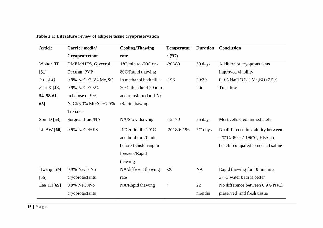
Table 2.1: Literature review of adipose tissue cryopreservation
Article Carrier media/
Cryoprotectant
Cooling/Thawing
rate
Temperatur
e (°C)
Duration Conclusion
Wolter TP
[51]
DMEM/HES, Glycerol,
Dextran, PVP
1°C/min to -20C or -
80C/Rapid thawing
-20/-80 30 days Addition of cryoprotectants
improved viability
Pu LLQ
/Cui X [48,
54, 58-61,
65]
0.9% NaCl/3.3% Me2SO
0.9% NaCl/7.5%
trehalose or.9%
NaCl/3.3% Me2SO+7.5%
Trehalose
In methanol bath till -
30°C then hold 20 min
and transferred to LN2
/Rapid thawing
-196 20/30
min
0.9% NaCl/3.3% Me2SO+7.5%
Trehalose
Son D [53] Surgical fluid/NA NA/Slow thawing -15/-70 56 days Most cells died immediately
Li BW [66] 0.9% NaCl/HES -1°C/min till -20°C
and hold for 20 min
before transferring to
freezers/Rapid
thawing
-20/-80/-196 2/7 days No difference in viability between
-20°C/-80°C/-196°C; HES no
benefit compared to normal saline
Hwang SM
[55]
0.9% NaCl/ No
cryoprotectants
NA/different thawing
rate
-20 NA Rapid thawing for 10 min in a
37°C water bath is better
Lee HJ[69] 0.9% NaCl/No
cryoprotectants
NA/Rapid thawing 4 22
months
No difference between 0.9% NaCl
preserved and fresh tissue

16 | P a g e
2.1.2 Isolation of mesenchymal stem cells from cryopreserved adipose tissue
Successful strategy for cryobank of adipose tissue is not only important for the supply of
adipose tissue but also important for the supply of adipose tissue-derived mesenchymal
stem cells when the need arises. Thus, in 2006, Matsumoto D et al. evaluated the effect of
storage temperature on the retrievability of adipose- derived stem cells from cryopreserved
adipose tissue. They concluded that adipose-derived stem cell yield was significantly
reduced by preservation at room temperature for 24h and by preservation at 4°C for 2 or 3
days [70]. Furthermore, the adipose-derived stem cell yield from cryopreserved fat was
much lower than that of freshly aspirated fat when preserved at -80°C for 1 month.
However, in the same year, Pu LLQ et al. cryopreserved human adipose tissue with 0.5M
dimethyl sulfoxide and 0.2M trehalose in LN2 and retrieved 90% of stem cells from the
cryopreserved tissue [71]. However, they observed a latency of hADSCs growth with the
retrieved hADSCs after two weeks of culture. To find out the significance of
cryoprotectants on the retrievability of hADSCs from cryopreserved adipose tissue, Lee JE
et al. in 2010 cryopreserved adipose tissue in -20°C and -80°C for 1 year and concluded
that no stem cells were viable in tissue cryopreserved without cryoprotectants. However,
stem cells survived successfully for 1 year in adipose tissue stored with 10% Me2SO and
differentiated into adipocytes [69]. Contrasting the findings of Lee JE et al., Kim JB et al.
did not observe any adherent hADSCs when human adipose tissues were preserved at -
20°C and -80°C in the presence of 10% Me2SO [72]. However, they observed that stem
cells isolation is possible when the adipose tissue was stored in at -190°C (LN2) in the
presence of 10% Me2SO. They further reported that the growth rate of the stem cells is
low when the tissue was preserved in 10% Me2SO + 10% FBS compared to 10% Me2SO
+ 90% FBS. hADSCs isolated from adipose tissue preserved with 10% Me2SO + 90%
FBS exhibits similar growth pattern compared to stem cells isolated from fresh tissue. In
2013, Choudhery MS et al. demonstrated that stem cells from fresh and cryopreserved
tissues with 10% Me2SO displayed similar fibroblastic morphology [73].
Cryopreservation did not alter expression of phenotypic markers, the proliferative
potential of MSCs, the differentiation capability of MSCs. Devitt SM et al. in 2014
reported that longer cryopreservation negatively impacts initial live adipose-derived stem
cell isolation; however, this effect is neutralized with continued cell growth [74].
Furthermore, patient age does not significantly affect stem cell isolation, viability, or
growth.

17 | P a g e
Table 2.2: Literature review isolation of stem cells from cryopreserved adipose tissue
Article Carrier
media/Cryoprote
ctant
Cooling /Thawing rate Temperature
(°C)
Duration Conclusion
Pu LLQ [71] 0.9% NaCl/ 0.5M
Me2SO and 0.2M
trehalose
In methanol bath till -30°C,
then hold 20 min and
transferred to LN2
/Rapid thawing
-196 20min Good yield of hADSCs,
however, there is a latency of
cell growth
Matsumoto D
[70]
Proprietary
formulation
1°C/15 min till -80C/Rapid
thawing
25/4/-80 1 month No/low yield of hADSCs
Lee JE [69] DMEM/10%
Me2SO
NA -20C/-80 1 year No yield without Me2SO
JB Kim[72] DMEM/ 10%
Me2SO +80% HS
NA -20/-70/- 196 7 days No yield at lower
temperature; latency of
growth with 10% FBS but
not with 80% FBS
Choudhery
MS[73]
DMEM/10%
Me2SO
NA -196 1 month Good yield; no latency of
growth; retain differentiation
ability
Devitt SM[74] No
cryoprotectants
NA -70 3 years Good yield; latency of
growth initially but not with
subsequent passaging

18 | P a g e
Table 2.3: Literature review on cryopreservation of adipose tissue derived stem cells
Article Cooling /Thawing rate Carrier
media/Cryoprotectant
Temperatu
re (°C)
Durati
on
Viability
Goh BC [87] NA DMEM/10% Me2SO
+80% FBS
-196 1
month
81% with 5x10⁵ cells/ml
Oishi K
[88]
Rapidly cooled NA/ 10% Me2SO
-80 7 days 70% with 10% Me2SO
Liu G [89] Cooling rate o f −0.5°C /min from 4 to
−20°C. The vials were then put into a
−80°C freezer. After 24h at −80°C,
the cells were transferred into a LN2
DMEM/10% Me2SO
+20% FBS
-196 2 wks NA
Rossa AD
[90] -20°C for 30min,then at -80°C for 1 h
and transferred to LN2
NA / 4% Me2SO
+6% trehalose+90% FBS
-196 1 year 92.5% in 1 month, 84.6%
% in 6 months and 70%
after 1 year with solution 2
Thirumala S
[91]
Frozen overnight in a −80°C freezer
inside ethanol jacketed container and
transferred to LN2
DMEM/ 1% MC or 10% PVP
+ 80% of either HS or FCS
-196 2 wks 54% with 10% PVP and
DMEM a, 63% with 10%
Me2SO ,37% with MC
Thirumala S
[92]
Frozen overnight in a −80°C freezer
inside ethanol jacketed container and
transferred to LN2.
DMEM/1% MC + 10% of
either HS or FCS
-196 2 wks 84% with 2% Me2SO ;
84% with 10% Me2SO
+80% HS/FBS
Thirumala S
[93]
−80°C freezer inside ethanol jacketed
container and transferred to LN2.
DMEM/ 10% PVP + 80% of
either HS or FCS
-196 2 wks 70% with 10% PVP
Dariolli R
[94]
NA DMEM/10% Me2SO
+10% FBS
-196 1 year 90–95%
James AW
[95]
With and without Mr.Frosty overnight
and then transferred to LN2
NA/ 10% Me2SO
+90% FBS
-196 2 wks 96%
Miyamato
Y [96] -1°C/min to -80°C DMEM/10% Me2SO
+1% sericin+0.1M maltose
-80 1-4
wks
95%
Ginani F 18h at -20°C, and then, storage at - 10% Me2SO -80 30 90%

19 | P a g e
[97] 80°C +20% FBS days
Minonzio G
[98]
From 4°C to 0°C in 6min, then hold
for 15min at 0 °C. From 0°C to −2 °C
in 9min and then hold for 2min at −2
°C. From −2°C to −35°C in 25.5min
and finally from −35°C to −100°C in
13 min.
5% albumin solution in
cryobag/5% Me2SO
-196 6
month
s
89.6%
Fernandez
MLG [99]
Kept at −80°C overnight and
transferred to LN2 (−196°C) the next
day
DMEM/ 10% Me2SO
+ 80% FBS
-196 90
days
5% Me2SO
gives 75%
And all others near about
80%
Yong KW
[100-102]
24h at −80 °C, then transferred to LN2 DMEM/ 5% Me2SO
+ 20% FBS
-196 90
days
90% with 10% Me2SO
/ 90% FBS
López M
[103]
0°C and cooled to -7°C at 2°C/min.
After seeding of extracellular ice and
holding at -7°C for 10 min, the straws
were cooled to -70°C at 1°C/min and
then plunged into liquid nitrogen
DMEM in ½-cc straws /
0.1mM EGTA+ 0.25 M
trehalose+2% PVA and 5%
ficoll + 5% Me2SO
+5% EG+3mM reduced
glutathione+5mM ascorbic
acid 2-phosphate
-196 NA 90.0% and 98.7%
Irioda AC
[104]
First stage (Program 3–15 min)
reaches the temperature −30°C; in the
end of step 2 (Program 5–45 min), the
temperature is −60°C; finally, in the
last stage, step 3 (Program 9-10min),
the temperature is −110°C and then
transferred to LN2
DMEM/10% Me2SO
+ 80% of FBS
-196 20
days
74.99%

20 | P a g e
2.2 Cryopreservation of mesenchymal stem cells
Mesenchymal stem cells (MSCs) are multipotent adult stromal cells with capability of
self-renewal and differentiation to mesoderm or non-mesoderm derived tissues [75].
Because of their unique properties such as easy isolation and culture, immunotoleranancy
to allogenic transplantation and differential ability, it emerged as one of the leading
candidates for cell therapy in regenerative and tissue engineering [76]. Because of
diversity of adult stem cells, the Mesenchymal and Tissue Stem Cell Committee of the
International Society for Cellular Therapy proposed the minimal criteria to define human
MSCs as: (i) MSCs must be plastic–adherent when maintained in standard culture
conditions (ii) MSCs must express CD105, CD73 and CD 90 and lack expression of
CD45, CD34, CD14 or CD11b, CD79alpha or CD19 and HLA-DR surface molecules and
(iii) MSCs must also demonstrate tri-lineage differentiation into osteocytes, adipocytes
and chondrocytes in vitro [77].
MSCs from bone marrow, cord blood and dental pulp are cryopreservable by slow
freezing protocols utilizing Me2SO and FBS as cryoprotectants is in use by many groups
[78-82]. Many of the alternative cryoprotectant formulations have also attempted to
remove Me2SO and animal serum from the cryoprotectant solution; both to reduce cost
and to improve clinical utility through reducing toxicity and possibility of zoonotic
infections. Human serum and human serum albumin was tried as an alternative to FBS.
However, this too is costly and introduces the risk of transmission of human pathogens.
Attempts were also given to reduce or eliminate Me2SO in freezing solution by replacing
it with polyethylene glycol, polylysine, glycerol, methylcellulose and trehalose [83-85].
Therefore, these laboratory studies indicate that there is a potential for the development of
xeno-free and serum-free freezing solution for cryopreservation of MSCs.
The incidence of mesenchymal stem cells in various tissues is extremely low, ranging
from around 0.00003% of nucleated cells in cord blood to 0.001–0.01% of nucleated cells
in the marrow, though this decreases with age. However, adipose tissue has been shown to
have a higher proportion of MSCs (approximately 2% in the stromal vascular fraction)
offering an advantage over MSCs from another tissue source [86].
Choice of the storage container is important in ensuring proper thermodynamics and
volume of cell suspension for the appropriate cost-benefit ratio of cryopreservation.
Usually, for biobanking of stem cells cryovials are the most commonly used container and

21 | P a g e
straws are used for preservation of germ cells. Cryobags are mainly used for large-scale
cryopreservation. Although most of the study used cryovials for the storage of hADSCs,
Minonzio G et al. used 25ml cryobags and López M et al. preserved hADSCs in ½-cc
straws [98, 103].
The significance of carrier medium is evident from the role of extenders in the outcome of
sperm cryopreservation. Inappropriate extenders as carrier medium lead to loss of post-
thaw sperm viability and functionality. However, not much work has been carried out to
optimize the carrier medium for mesenchymal stem cell cryopreservation. Almost all the
study for cryopreservation of hADSCs used DMEM/F-12 as carrier media except in one
study where 5% albumin solution was used as carrier medium [98].
Concentrations of cells are a very important factor that influences the outcome of a
preservation strategy. Goh BC et al. in 2007 demonstrated that for cryopreservation of
adipose-derived stromal cells in a cryovial at -196°C in the presence of 10% Me2SO+80%
FBS freezing media, the optimum concentrations of cells should be 5x10⁵ cells/ml [87].
However, the optimal concentration of cells depends on upon the type of storage
container, the stage of cells and the freezing solution composition. Therefore, the
concentrations of cells differ from study to study. Out of 16 studies mentioned, 8 studies
conducted their study with 1x10⁶ cells/ml and 1 study used 5x10⁵ cells/ml. Most of the
other studies used cell concentration higher than 1x10⁶ cells/ml.
Cryoprotectants are a very important component of freezing solution, which prevents
freezing related damage to biological samples. Almost 50% of total studies, Me2SO with
or without FBS was used for the cryopreservation of hADSCs, whereas another 40%
studies were conducted to reduce or eliminate Me2SO from freezing solution. To reduce
the concentration of Me2SO, in 2009, Rossa AD et al. reported that 4% Me2SO+6%
trehalose+90% FBS is sufficient in maintaining more than 90% viability of hADSCs even
after 1 month of storage. However, the viability reduces to 70% after 1 year [90].
Furthermore, in 2014 Minonzio G et al. demonstrated that 5% Me2SOand 5% human
albumin freezing solution gave viability of 90% [98]. In an another recent study, López M
et al. optimized a freezing solution cocktail by the addition of antioxidants glutathione and
ascorbic acid, extracellular cryoprotectants polyvinyl alcohol (PVA) and ficoll, reduced
concentration of Me2SO and a calcium chelator EGTA along with trehalose [103]. The
optimized solution (0.1mM EGTA + 0.25M trehalose + 2% PVA and 5% ficoll + 5%

22 | P a g e
Me2SO + 5% EG + 3mM reduced glutathione + 5mM ascorbic acid 2-phosphate) gave
viability of more than 98%. Thirumala S at al. in a series of the study revealed that 10%
PVP, which gave viability of 70% could replace Me2SO [91, 93]. In another significant
finding of their study, 2% Me2SO gave the remarkable cryoprotective ability to preserve
hADSCs [92]. Recently, Yong KW et al. also observed that 5% Me2SO maintained a high
cell viability (75%) comparable to those preserved in standard cryomedium (10%
Me2SO + 90% FBS) [100-102]. Miyamato Y et al. reported that supplementation of 1%
sericin and 0.1M maltose with 10% Me2SO gave more than 95% viability of
cryopreserved hADSCs [96].
As with other biological samples, cooling rate during cryopreservation is an important
factor that determines the viability of post-thawed hADSCs. Except the study by Oishi K
et al. in 2008, where they cooled the samples rapidly, all other study used the slow cooling
method. Liu Q et al. kept the samples at -20°C freezer to achieve a cooling rate of
0.5°C/min and transferred it to -80°C freezer to expose the samples to -1°C/min till -80°C.
They kept the samples for 24h in -80°C freezer before transferring them to LN2. Rossa AD
et al. also followed similar cooling protocol. In all the three studies by Thirumala S et al.,
they used an ethanol-jacketed container to freeze the cells overnight in a -80°C freezer
before transferring them to LN2. In the ethanol-jacketed container, ice nucleation was
observed around −5°C, and a cooling rate of ∼1.1°C/min was achieved at a temperature of
−40°C. Subsequently, the cooling rates drop to 0.3°C/min and 0.1°C/min, before reaching
−80°C. James AW et al., Yong KW et al. and Fernandez MLG et al. also followed similar
cooling protocol with slight modifications. Minonzio G et al. and Irioda AC et al. used a
control rate freezer to achieve predictable and reliable cooling rate. Minonzio G et al.
cooled the cryobags from 4°C to 0°C in 6min (hold for 15min), from 0 °C to −2°C in 9
min (hold for 2min), from −2°C to −35°C in 25.5min and finally from −35°C to −100°C in
13min before transferring them to LN2. Irioda AC et al. took 15 min to reach -30°C, then
45min to reach -60°C and finally 10min to reach -110°C before transferring the samples to
LN2. All the study used rapid thawing rate at -37°C water bath.

23 | P a g e
2.2.1 Effect of freezing on functionality of adipose-derived stem cells
Retention of adhesion to plastics, proliferation ability, differentiation potential, surface
marker expression and chromosomal normality is essential for further application of
cryopreserved hADSCs. Out of total 16 studies mentioned, 10 studies performed
proliferation kinetics of frozen hADSCs. Out of 10 studies, 6 studies reported normal
proliferation kinetics, whereas four studies reported delay or accelerated proliferation of
frozen cells. Three studies also conducted colony forming unit assay with frozen stem
cells with one study each reported normal, decrease and increased colony formation. All
studies reported normal morphology of cryopreserved hADSCs except one by James AW
et al. The authors reported the presence of unusual morphology of the frozen cells upon
culture and proliferation. Oishi K et al., in 2008 showed that the proliferation rate of
hADSCs decreases if frozen with 10% Me2SO. However, in the same year, Liu Q et al.
demonstrated that frozen cells maintain their capability of proliferation compared to
freshly cultured cells. Again, in 2009, Rosa AD et al. reported that there is an initial delay
in the proliferation of frozen hADSCs till passage 2 and from passage 3 the growth
characteristics of frozen cells are similar to fresh cells. However, James AW et al. showed
that there is a significant impairment of cell attachment and proliferation of hADSCs
frozen with 10% Me2SO and 90% FBS. Contrary to James AW et al., in the same year,
Dariolli R et al. reported that that the growth characteristics of porcine hADSCs are not
influenced by long-term preservation with 10% Me2SO. Miyamato Y et al. reported higher
proliferation rate of hADSCs after cryopreservation with 10% Me2SO + 1% sericin +
0.1M maltose. Ginani F et al. also reported normal growth characteristics of hADSCs after
freezing with 10% Me2SO. However, Minonzio G et al. showed that frozen adipose
stromal cells maintain normal proliferation but higher the colony forming units if
preserved with 5% Me2SO and 5% human albumin solution. Contrasting with Minonzio G
et al., Irioda AC et al. demonstrated that there is a decrease in colony forming unit by
frozen cells preserved using 10% Me2SO and 80% FBS.
Out of 16 studies, 15 studies differentiated the frozen cells to osteogenic and adipogenic
lineages, out of which, 13 studies reported normal differentiation with frozen hADSCs.
Among the other two study, Goh BC et al. demonstrated that although there is a decrease
in the osteogenic differentiation, adipogenesis remains normal after storing the stem cells
in LN2 for 1 month in the presence of 10% Me2SO and 80% FBS. However, even the
decrease in osteogenesis is non-significant compared to the fresh cells. In both in-vitro and

24 | P a g e
in vivo study, James AW et al. showed that passage 1 cells frozen with 10% Me2SO +
90% FBS in LN2 for 2 weeks showed the deleterious effect on both osteogenic and
adipogenic differentiation. However, they demonstrated that IGF-1 and BMP-4 rescued
the deleterious effect on osteogenic differentiation in vivo.
Out of 16 studies, 5 studies conducted the expression of surface markers after freezing
hADSCs. Four of which reported similar surface marker expression as fresh cells,
whereas, in one study by Irioda AC et al. there was a significant reduction in expression of
CD49d expression in cells frozen with 10% Me2SO and 80% FBS.
Five studies evaluated the effect of freezing on chromosome and gene expression. Dariolli
R et al. demonstrated that long-term cryostorage using 10% Me2SO do not influence
karyotype and senescence cues. Ginani F at al. reported no morphological changes in the
nuclei of frozen hADSCs (nuclear fragmentation or the presence of pyknotic nuclei).
Yong KW et al. observed significantly higher (p<0.05) expression level in stemness
genes including OCT-4, REX-1, SOX-2 and NANOG of cryopreserved hADSCs
compared to fresh hADSCs, indicating the frozen cells have greater ability to maintain
their stemness properties. They also observed normal levels of the tumor suppressor
markers p53, p21, p16 and pRb, hTERT, telomerase activity and telomere length in frozen
cells compared to fresh cells.

25 | P a g e
Chapter 3
Scope &Objective

26 | P a g e
Cryopreservation using Me2SO as potential CPA is the most well known and efficient
method for preserving cells and tissues. However, keeping in view of the harmful effect of
this conventional freezing solution, recent research has focussed in the search of potential
non-toxic CPAs of natural origin for developing freezing solutions. Therefore, the present
dissertation work has been undertaken to explore potential natural CPAs and suitable
carrier media thereby develop a freezing solution that can be used as an effective
cryopreservation strategy for preserving much needed adipose tissue and adipose tissue
derived stem cells.
Adipose tissues harvested by liposuction are important tissue source, the large quantity of
which is required for reconstructive and aesthetic surgery for making fat grafts. However,
the absorption rate of adipose tissue grafts result in poor clinical outcome requiring
repeated harvesting procedures, which leads to not only surgical risk but also increase in
hospital expenditure. Once it is harvested, cryopreserving spare adipose tissue with high
survival rate for long time can avoid the problem of repeated harvesting. Besides fat
grafts, adipose tissue is considered as a promising source for providing mesenchymal stem
cells for various therapeutic and upcoming tissue-engineering applications. As a result,
there is a growing demand worldwide for adipose tissue and hADSCs for the various
clinical applications. For catering the great demand of adipose tissue and hADSCs, as
important tissue and cell source, the development of an appropriate strategy for preserving
such clinical products with long shelve-life without change in their desired property is of
paramount importance. Keeping in view of their actual use when need arises,
cryopreservation with Me2SO and serum in freezing medium is conventionally used.
However, the use of these components in cryopreservation soluting is harmful to the
patient by causing cell death.
In the context, developing a freezing solution, which is devoid of Me2SO and serum is one
of the key challenges as promising cryopreservation strategy for adipose tissue and
hADSCs.
Therefore, the main aim of the present dissertation work is the development of effective
cryopreservation strategies by exploring suitable non-toxic natural CPAs and carrier media
for cryopreservation of adipose tissue and hADSCs with maintained cells viability,
structural and functional ability for long-term basis.

27 | P a g e
The specific objectives of the present research work are as follows.
i. Screening of potential natural cryoprotective agents, carrier media and
antioxidants to formulate an effective freezing solution
ii. To evaluate the performance of the formulated freezing solution for preserving
adipose tissue and hADSCs
iii. To optimize the controlled rate freezing for cryopreservation of adipose tissue and
hADSCs
iv. To evaluate the long-term preservation of adipose tissue and hADSCs using the
developed freezing solutions
Scope of work
The specific scope of this present research work is as follows-
A. CRYOPRESERVATION OF ADIPOSE TISSUE
I. Screening of CPAs and carrier media
In this phase, efforts will be given to explore the potential hydrocolloids and natural
organic osmolytes as extracellular and intracellular CPAs as well as individual PBS
components such as NaCl, Na2HPO4, KCl and KH2PO4 as possible carrier media for the
formulation of freezing solution. The cryopreservation experiments with adipose tissue
will be performed to evaluate the effectiveness of the individual CPAs and individual PBS
components (ionic compounds) as carrier media. The screening of CPAs and carrier media
will be done by measuring oil ratio as a preliminary screening criteria and final selection
will be based on XTT, G3PDH and TBARS assays.
II. Formulation and evaluation of freezing solutions
Different batches of freezing solutions will be prepared by using the selected CPAs and
carrier media with varying concentrations. The freezing solutions will be tested for their
performance towards cryopreservation of adipose tissue by conducting series of
experiments in Mr. Frosty. The most effective freezing solution cocktail will be selected
based on a battery of assays as mentioned above.
III. Improvement of cryopreservation by the addition of anti-oxidant
The oxidative stress mediated cellular damage is reported to be a critical phenomenon in
low temperature storage, which is expected to be higher in case of adipose tissue than
other body tissues. The supplement of suitable antioxidants in freezing solution is reported

28 | P a g e
to improve the cryopreservation outcome of other tissues. Therefore, efforts will be given
to explore suitable antioxidants by screening method and most potential antioxidant will
be selected on the basis of their performance evaluattion by conducting cryopreservation
experiments using the formulated freezing solution supplemented with various
antioxidants. The optimal composition of freezing solution containing the most effective
antioxidant is expected to give maximum post-thaw tissue viability.
IV. Control rate freezing of Adipose Tissue using the formulated freezing solution
An effort will be given for further improvement of the cryopreservation efficiency by
performing cryopreservation of adipose tissue using a control rate freezer . The cooling
rate has a profound influence in cryopreservation of cells and tissues. Therefore, the
optimal cooling rate for achieving maximum cell viability in controlled rate freezing will
be established by performing cryopreservation experiments at varying cooling rates.
V. Assessment of long-term storage viability
The maintenance of tissue viability for a long time is the most vital aspect in
cryopreservation. Therefore, the efficiency of the formulated freezing solution towards
maintaining tissue viability will be evaluated by conducting control rate freezing
experiments with cryopreserved adipose tissue with long storage time. The structural and
functional integrity of cryopreserved adipose tissue will the assessed by morphological
characteristics as well as ability of frozen adipose tissue to provide hADSCs with retained
metabolic activity, proliferation, and differentiation ability.
B.CRYOPRESERVATION OF ADIPOSE TISSUE DERIVED STEM CELLS
I. Evaluation of hydrocolloids as CPAs
Effort will be given to isolate hADSCs from adipose tissue and develop a cryopreservation
strategy for their preservation by formulating an effective freezing solution. Similar to
adipose tissue, the efficiency of various hydrocolloids and natural organic osmolytes as
extracellular and intracellular CPAs will be evaluated for their efficiency in maintaining
viability of adipose tissue by performing cryopreservation experiments. Based on the
viability results measured by Trypan blue and MTT assays, the most effective freezing
solution achieving maximum cell viability will be selected.
29 | P a g e
II. Evaluation of PVP in combination with PBS components as carrier media
PBS as carrier media consists of NaCl, Na2HPO4, KCl and KH2PO4. The specific role of
these components as carrier media in cryopreservation of stem cells has not been explored
so far. Therefore, efforts will be given to investigate the effect of individual ionic
component on the cryopreservation outcome achieved using formulated freezing solution.
III. Improvement of cell viability by addition of organic osmolytes
An attempt will be made for further improvement of the efficiency of the formulated
freezing solution as mentioned above by the addition of ectoin and hydroxyectoin as the
potential organic osmolytes.
IV. Optimization of the freezing solution composition
The cryopreservation efficacy of the formulated freezing solution will be optimized by
following Taguchi orthogonal design methodology and thus an optimal composition of the
freezing solution providing the maximum post-thaw viability is expected to achieve.
V. Control rate freezing of hADSCs and optimization of freezing parameters
To achieve optimal cooling rate in controlled rate freezing, the cryopreservation
experiment will be carried out at different cooling rate. Further, the effect of long-term
storage on the viability of hADSCs frozen in the formulated cryopreservation solution will
be assessed to ensure the ability of the developed freezing solution in maintaining viability
for a long time. Besides viability study, the structural and functional integrity of post thaw
hADSCs will also be verified.

30 | P a g e
Chapter 4
Materials and methods

31 | P a g e
4.1 Chemicals and culture media
4.1.1 Cryoprotective agents
Various extracellular cryoprotective agents such as trehalose, mannitol, dextran, raffinose,
sucrose, arabitol, polyvinylpyrrolidone, methylcellulose, carboxymethylcellulose, inulin,
guar gum, xanthan gum, acacia gum, tragacanth gum, hydroxyethyl starch, stachyose were
purchased from Himedia Pvt. Ltd (Mumbai, India). The intracellular cryoprotectants such
as ectoin, hydroxyectoin, betaine and Me2SO was procured from Sigma Chemical Co. (St.
Louis, MO, USA).
4.1.2 Carrier media
To formulate carrier media different ionic compounds like NaCl, KCl, Na2HPO4, KH2PO4,
MgCl2 and MgSO4 used in the thesis works were purchased from Himedia Pvt Ltd
(Mumbai, India). MilliQ water was used for the preparation of carrier media and freezing
solution.
4.1.3 Antioxidants and inhibitors
Curcumin, carnitine, lipoic acid, Vit E, quercetin, gallic acid, and ellagic acid was used as
antioxidants for cryopreservation of adipose tissue and was bought from Himedia Pvt Ltd
(Mumbai, India). Catalase, which was used for cryopreservation of adipose-derived stem
cells was procured from Sigma Chemical Co. (St. Louis, MO, USA). Caspase inhibitor Z-
VAD-FMK and Rho kinase inhibitor Y-27632 were also used of Sigma Chemical Co. (St.
Louis, MO, USA).
4.1.4 Cell culture and differential media
For isolation of culture of hADSCs, collagenase type I, Dulbecco’s modified Eagle
medium (DMEM), fetal bovine serum (FBS), and 0.25% Trypsin/EDTA solution were
procured from Gibco (BRL, USA). For immunophenotypic analysis, BD FACS lysis
buffer, CD 34-PE, CD45-FITC, CD73-APC, CD90-FITC, CD105- PerCP/cy5.5 and HLA-
DR-PerCP/cy5.5 were purchased from BD pharminogen (Becton Dickenson, San Jose,
CA, USA). For cytoskeletal and nucleus staining Phalloidin-TRITC, Phalloidin-Alexa
Fluor 488, Hoechst and DAPI were procured from Invitrogen, USA. Triton X-100,
formaldehyde and bovine serum albumin (BSA) are from Himedia Pvt. Ltd (Mumbai,
India). For differentiation and staining of hADSCs, dexamethasone, β-glycerophosphate,
indomethacin insulin, Isobutylmethylxanthine (IBMX), Oil Red O and Alizarin Red were

32 | P a g e
procured from Sigma-Aldrich (St Louis, MO, USA). L-ascorbate was obtained from
Himedia Pvt. Ltd (Mumbai, India). All the tissue culture and plastic wares were from
Tarsons Product Pvt. Ltd. (Kolkata, India).
4.1.5 Viability assessment
Trypan blue, MTT, XTT, mercaptoethanol, pyridine, thiobarbituric acid (TBA) assay,
acetic acid, sodium dodecyl sulfate (SDS) and Tris-HCl was procured from Himedia Pvt.
Ltd (Mumbai, India). PMS (N-methylphenazonium methyl sulfate) and
Tetraethoxypropane was obtained from Sigma-Aldrich (St Louis, MO, USA). Propidium
iodide (PI) was purchased from Invitrogen, USA. Glycerol 3 phosphate dehydrogenase
(G3PDH) assay kit (MK426) was procured from Takara Bio Inc, Japan.
4.2 CRYOPRESERVATION OF ADIPOSE TISSUE
4.2.1 Collection and processing of adipose tissue
Adipose tissue of twelve patients was collected from Jeewan Memorial Hospital, Raipur,
Chhattisgarh and Cosmetic Surgery Clinic, Kolkata India and processed in the stem cell
laboratory of the Department of the Biotechnology & Medical Engineering of the Institute
with the approval of the Institute Ethical Committee vide No.
NITRKL/IEC/FORM/2/25/4/11/004 dated 25.04.2011. The collected adipose tissue was
washed and centrifuged with normal saline at 3000rpm for 5min. After centrifugation, the
upper layer and lower layer consisting of oils and aqueous cell debris respectively was
pipetted out and discarded. The middle layer consisting of adipose tissue was used for the
cryopreservation experiment.
4.2.2 Preparation of freezing solution
Freezing solutions containing polyols, organic osmolytes and antioxidants in various
concentrations individually were prepared in PBS for selection of the best extracellular
and intracellular cryoprotectant and antioxidants for cryopreservation of adipose tissue.
Freezing solutions containing 80mM-240mM concentration of ionic compounds was also
prepared in MilliQ water for the screening of the best carrier media. The selected
extracellular and intracellular cryoprotectant, carrier media and antioxidants were
combined in various combinations to obtain the desired Me2SO-free, serum free and
DMEM free freezing solution. The developed freezing solution was also combined with
10nM Z-VAD-FMK or 100mM Y-27632. Five ml of the prepared freezing solution in

33 | P a g e
various phases was added to 5gm of adipose tissue in a 15ml centrifuge tube for
cryopreservation.
4.2.3 Cryopreservation experiment
After equilibrating with freezing solutions at 4°C for 1h, the sealed tubes containing fat
tissues and freezing solutions were placed in -80°C freezer dipped in isopropanol
containing container. All the samples were stored for 48h before thawing. For
optimization of cryoprocess thermodynamic, controlled rate freezer (Kryo360-17, Planer
Inc, UK) was used. To find out the optimal cooling rate, samples were cryopreserved in
varying cooling rate such as 0.5°C/min, 1°C/min, 2.5°C/min, 5°C/min from 4°C to -120°C
followed by preservation in LN2 for 48h. Furthermore, seeding at -5°C, -7°C, and -9°C
was also introduced to find out the optimal seeding temperature. To evaluate the long-term
efficacy of the developed freezing solution and cryopreservation strategy, adipose tissue
was also stored for 90 days in LN2. The tissues were thawed rapidly by immersing the
tubes in a water bath at 37°C. Each batch of experiments was repeated three times.
4.2.4 Adipose tissue viability assessment
Oil ratio
After thawing the cryopreserved tissue were centrifuged at 1500rpm for 10min. Three
distinct layers including oil, tissue and aqueous part were formed. The oil ratio was
calculated by the formula reported by Matsumoto D et al. [70] as follows-
Oil ratio=Oil volume / (Oil volume+ Fat volume)
XTT assay
XTT assay shows the metabolic activity of cells and tissues. In brief, after centrifuge, the
fat was resuspended in phosphate-buffered saline to a total of 7.5ml. The XTT ((2,3-Bis-
(2-Methoxy-4-Nitro-5-Sulfophenyl)-2H-Tetrazolium-5-Carboxanilide) and PMS (N-
methylphenazonium methyl sulfate; electron coupling reagent) were mixed at a ratio of
50:1. 2.5ml of this resultant mixed reagent was added to the fat sample and was incubated
for four hours at 37°C. After incubation and centrifugation at 1500rpm for 10min, 1ml of
the aqueous part was transferred to quartz cuvette to measure the optical absorbance using
a spectrophotometer. The absorbance was measured at a test wavelength of 450nm and a
reference wavelength of 650nm. Percentage (%) viability was calculated as

34 | P a g e
% viability= (OD of frozen sample / OD of fresh sample) X 100
G3PDH assay ,
G3PDH enzyme activity was measured by following the protocol of G3PDH assay kit
(Takara Bio Inc; MK426). Briefly, the cryopreserved tissues were centrifuged at 1500rpm
for 10min to segregate the adipose tissue from the aqueous fraction. 200µl of aqueous
fraction was added with 200µl of dilution buffer containing 1mM mercaptoethanol to form
a sample solution. 25µl of the sample solution was added to 100µl of substrate solution
containing (NADH and dihydroxyacetone phosphate) for spectrophotometric assay. The
optical absorption at 340nm was measured for 10min and the change in optical density
(∆OD) from the linear position of the curve was obtained. The enzyme activity is
calculated by using the following formula.
G3PDH activity (Units
ml) =
∆OD340 × A(ml) × Dilution ratio of test sample
6.22 × B(ml) × C(cm)
Where,
ΔOD 340: Decrease in the absorbance at 340nm per min
A (ml): Total reaction volume
B (ml): The volume of enzyme solution (diluted sample) added
C (cm): Optical path length of the cell used
6.22: Millimolar absorption coefficient of NADH molecules
TBARS assay
The TBARS assay was performed following the protocol mentioned by Włostowski T et
al. [105]. In brief, the cryopreserved tissue samples were homogenized in a buffer solution
containing 50mM Tris-HCl (pH 7.4) and 1.15% KCl followed by centrifugation at
1500rpm for 10min. The supernatant was used for the assay. To 200µl of the tissue
homogenate, 200µl of 8.1% (w/v) SDS (sodium dodecyl sulfate), 1.5ml of 20% acetic
acid, 1.5ml of 0.8% TBA (thiobarbituric acid) and 600µl distilled water were added and
vortexed. The reaction mixture was heated at 95°C for 1h in a water bath and cooled.
Then 1ml sample was added to 5ml of butanol/ pyridine mixture (15:1 v/v) and vortexed.
After centrifugation at 14000rpm, the absorbance of the organic phase was determined at
532nm. Calibration curve was prepared using Tetraethoxypropane. The results were
expressed as TBA-reactive substances (nM/g wet wt.).

35 | P a g e
4.2.5 Morphological characterization
Morphology of fresh and cryopreserved adipose tissue after long-term storage was
assessed by confocal laser scanning microscope (CLSM; Leica Microsystem, Germany) in
both phase contrast and fluorescence mode. For fluorescence staining, PI was used.
Adipose tissue was incubated with 100ug/ml of PI for 15 min and observed under CLSM
(Ex-488nm; Em-600-650nm). Furthermore, keeping in view the fluorescence property of
curcumin, the curcumin-containing freezing solution was explored for staining the adipose
tissue. 0.1% (w/v) curcumin was dissolved in PBS and subsequently incubated with fresh
adipose tissue in room temperature for 30min. CLSM with excitation at 458nm and
emission filter at 500-520nm was set up to image the adipose tissue. Later on, 2.5%
Me2SO was added along with the curcumin to stain the adipose tissue homogenously.
Furthermore, to observe dead cell characteristics in the presence of curcumin adipose
tissue was treated with 0.25% sodium dodecyl sulfate (SDS) and stained with 0.1% (w/v)
curcumin/ 2.5%Me2SO.
4.2.6 Isolation of hADSCs from cryopreserved adipose tissue
hADSCs were isolated from cryopreserved adipose tissue and morphological
characteristics, proliferation kinetics and differentiation ability was assessed by following
protocols mentioned in the next section. The results were compared with hADSCs isolated
from fresh adipose tissue.
4.3 CRYOPRESERVATION OF ADIPOSE-DERIVED STEM CELLS
4.3.1 Isolation and culture of hADSCs
The stem cells were isolated by collagenase digestion method. In brief, 5gm adipose tissue
was washed with PBS and incubated with 1mg/ml collagenase I at 37°C for 30min. The
digested adipose tissue was centrifuged to separate out the stromal cells as a pellet. The
pellet was washed with PBS and suspended in DMEM medium containing 10% FBS. The
cells were incubated at 37°C with 5% humidified CO2.The non-adherent cells were
discarded after 24h. The expansion medium was changed twice weekly until 80%
confluence was achieved and then the cells were trypsinized and passaged further for
proliferation.

36 | P a g e
4.3.2 Immunophenotypic characterization of hADSCs
The expression of the specific surface antigen by the cells was evaluated by flow
cytometry analysis (FACS Calibur, Becton Dickinson and Co, San Jose, CA, USA). The
cells of third passage were trypsinized and stained with human monoclonal antibodies
against CD90, CD73, CD105, CD34, CD44 and HLA-DR. In brief, the trypsinized cells
were washed with PBS and 2% FBS and kept in dark at 4°C for 1h after adding 10µl
antibodies. The cells were washed with PBS and 2% FBS before being resuspended in
PBS for flow cytometry analysis.
4.3.3 Preparation of freezing solution
Hydrocolloids in PBS were screened in different concentration for selection of the best
extracellular cryoprotectant. Subsequently, to replace PBS as carrier media, isotonic
solutions of NaCl, Na2HPO4, KCl and KH2PO4 was combined with the selected
cryoprotectant and was evaluated for its cryopreservation efficacy. Furthermore, ectoin
and hydroxyectoin in different concentration as intracellular cryoprotectant were added to
the above solutions and evaluated. The developed freezing solution containing suitable
extracellular and intracellular cryoprotectant in the selected carrier media was further
optimized and improved by adding catalase. For the development of an optimal freezing
solution, Taguchi’s orthogonal design method was applied. To this end, four factors
namely NaCl, PVP, ectoin, catalase as freezing media components and three levels of their
different concentrations were selected (Table 6.1) to prepare the freezing solutions with
varying composition (Table 6.2). To further improve the efficacy of the developed
freezing solution 10nM Z-VAD-FMK or 100mM Y-27632 was added to it. hADSCs from
different passages (P3-P6) were transferred into 1.5ml cryovials with the prepared freezing
solution at a concentration of 1x10⁶ cells/ml for cryopreservation experiment.
4.3.4 Cryopreservation experiment
After equilibrating the hADSCs with freezing solutions for 1h at 4°C, The sealed vials
containing a freezing solution and hADSCs were placed in Mr. Frosty (Nalgene, New
York, U.S.A) and kept in -80°C freezer. After 48h, the cells were thawed in a water bath
and the post-thawed cells were diluted gradually by adding 9ml prewarmed media and
centrifuged at 3000rpm for 10min. The supernatant was discarded and the pelleted cells
were resuspended in DMEM and 20% FBS for culture. To optimize the cooling rate and
seeding temperature, controlled rate freezer was used following the methods used for

37 | P a g e
adipose tissue cryopreservation in the previous section. Furthermore, hADSCs were also
stored in LN2 for 90 days to evaluate the long-term efficacy of the developed freezing
solution.
4.3.5 hADSCs viability assessment
Trypan blue assay
Ten µl Trypan blue (0.4 w/v %) was added to 10µl post-thawed cell suspension for cell
viability assessment. The cell viability was measured by counting cells in a
hemocytometer and the stained cells were considered dead cells.
MTT assay
Cell viability was also measured by the MTT assay. Cells (5 x 10⁴cells /well) were
incubated at 37°C in a CO2 incubator in a 6-well plate. After 24h, 4µl MTT (500µg/ml)
per 100µl media was added and incubated for 3h at 37° C. The formazan crystals were
solubilized with Me2SO and the absorbance of the solution was recorded at 570nm.
Flow cytometry study
To assess the viability of hADSCs, the cells were labeled with PI. In brief, the cells were
incubated with PI (100µg/ml) for 15min and then subjected to flow cytometry analysis by
using FACS Calibur flow cytometer (Becton Dickinson, San Jose, CA). For each sample,
10,000 events were acquired from the region defined for MSCs and analyzed by BD
FACSDiva Software 6.0 (Becton Dickinson, San Jose, CA) for cell viability assessment.
4.3.6 Cytoskeleton analysis
For cytoskeleton analysis, 24h cultured hADSCs were fixed with 2% paraformaldehyde in
PBS at room temperature for 5min. After that, the cells were treated with 2% BSA and 1%
glycine and were permeabilized with 0.5% Triton X-100 for 15min. For actin staining and
nuclear staining, samples were incubated with TRITC-Phalloidin/Alexa-fluoro 488-
Phalloidin and DAPI/Hoechst respectively at 37°C for 30min in dark and samples were
observed under CLSM.
4.3.7 Differentiation potential assessment
The fresh and cryopreserved cells were plated in a 6-well plate and cultured using DMEM
in 20% FBS supplemented with differentiation media. The osteogenic differentiation
media consists of 10mM β-glycerophosphate, 100nM dexamethasone and 0.2mM L-
ascorbate and adipogenic differentiation media comprises of 50µm indomethacin, 300nM
insulin, 100nM dexamethasone, and 500µM IBMX. The osteogenic and adipogenic

38 | P a g e
potential was measured by Alizarin red on the 21st day and Oil Red O staining on the 14th
day.
4.3.8 Proliferation kinetics
5×10³ cells/well were seeded into 6-well plates with 2ml of growth medium and incubated
at 37ºC and 5% CO2. After trypsinization, the cell number counted from day 1 to 9 day.
The number of viable cells was counted using automated cell counter with cells stained
with 0.4% Trypan blue.
4.4 Statistical analysis
The experimental data were analyzed statistically using IBM SPSS Statistics 20.0
software. The results are expressed as mean ± standard error. The following equation was
used for the S/N ratio calculation during optimization of hADSCs cryopreservation by
Taguchi’s method
𝑆
𝑁= −10 ∗ log (𝛴 (
1
𝑌2)/𝑛
Where Y is the measured data and n is the number of data
In addition to S/N ratio, response table of mean and statistical analysis of variance
(ANOVA) was employed to determine the effect of parameters on hADSCs viability.

39 | P a g e
Chapter 5 - 6
Results &Discussion

40 | P a g e
Chapter 5
Cryopreservation of adipose tissue

41 | P a g e
5.1 Screening of potential cryoprotectants and carrier media towards the
formulation of freezing solution for cryopreservation of adipose tissue
Autologous fat tissue usually harvested by liposuction is used in reconstructive and
aesthetic surgery due to its abundant availability, relatively easy harvesting and
biocompatibility of fat tissue [106]. However, the implanted fat tissue is not persistent due
to its high absorption rate that results in frequent graft failure clinically, thereby, demands
additional grafts leading to repeated harvesting of fat or adipose tissue, which increases
cost and patient morbidity [107-110]. Preserving additional tissue at sub-zero temperature,
keeping in view of its supply as additional grafts is an effective technique to avoid
repeated harvesting. Besides tissue graft, there is a growing demand of hADSCs that can
be obtained from adipose tissue as a potential source for the tissue engineering and
therapeutic applications [111-117].
Successful cryopreservation strategy requires a suitable freezing solution comprising of
CPAs and carrier media. The use of conventional cryoprotectant such as Me2SO is
harmful due to its toxic effects [118, 119]. Although there are reports of using
extracellular CPAs, the efficacy of non-toxic intracellular CPAs in cryopreservation of
adipose tissue has not been reported till date. Furthermore, evaluation of organic
osmolytes such as ectoin and hydroxyectoin as intracellular CPAs can be of much
interesting [120].
Keeping the above in view, the present study focuses on the evaluation of a number of
extracellular and intracellular CPAs as well as ionic compounds as possible carrier media
towards the development of a freezing solution for preservation of adipose tissue.
Furthermore, an attempt was also given to formulate an optimized freezing solution
cocktail from the combination of the potential non-toxic extracellular and intracellular
CPAs in the selected carrier medium. The preliminary screening of CPAs and carrier
media was assessed based on oil ratio analysis, and the potential components were finally
selected through the further assessment such as XTT, G3PDH and TBARS assays. The
result and discussion on the various experimental results generated from the above study
are presented in this chapter.

42 | P a g e
5.1.1 Screening of extracellular CPAs
Oil ratio
The efficiency of the various extracellular CPAs such as sucrose, trehalose as sugars and
PVP, dextran and HES as hydrocolloids, in PBS carrier medium was evaluated by
measuring oil ratio as an indicator of damaged or degenerated adipocytes. The experiment
was also performed with different concentration of CPA solutions in the range of 30-
150mM with the aim of finding optimal concentration of CPAs for cryopreservation of
adipose tissue.
The oil ratio in Fig 5.1 indicates that among the extracellular CPAs, trehalose shows
higher viability by providing the least damaged adipocytes in cryopreserved tissue than
other CPAs used in the study. Besides the type of CPAs, variation in the viability of
frozen adipose tissue was also observed with varied concentration of CPAs. Though not
much statistically significant change in viability was shown with varied concentration,
trehalose at 90mM concentration showed least oil ratio (0.047 ± 0.002) indicating the
highest viability at this concentration, followed by sucrose (30mM, 0.069 ± 0.010) and
mannitol (30mM, 0.077 ± 0.015).
The superior performance of trehalose may be due its excellent membrane stabilizing
property and trehalose also retains hydrophilic shell around proteins and nucleic acids,
thereby prevents cryoinjury mediated cell damage [121-123]. the protective role of
trehalose in adipose tissue cryopreservation was also reported by Pu LLQ et al, though a
higher concentration (300mM) of trehalose was demonstrated as the optimal concentration
for cryopreservation of adipose tissue [124].
Among the hydrocolloids, PVP and dextran, which are established cryoprotectants for
mesenchymal and hematopoietic stem cells, provided less cell viability as shown by
higher oil ratio [81, 125, 126]. Similarly, HES, which was reported as an efficient CPA for
RBCs, was not favorable to adipose tissue preservation [127-130]. The similar finding
with HES was also reported elsewhere [66]. The raffinose families of oligosaccharides
(RFOs) such as raffinose and stachyose, which are alpha-galactosyl derivatives of sucrose,
has shown good cryoprotectant ability for mouse sperms, but these were found to be
inefficient in the present study [131]. Although arabitol has shown promise in
cryopreservation of rat embryo, the tissue viability obtained with arabitol was lower to the
viability obtained with trehalose and sucrose [132].
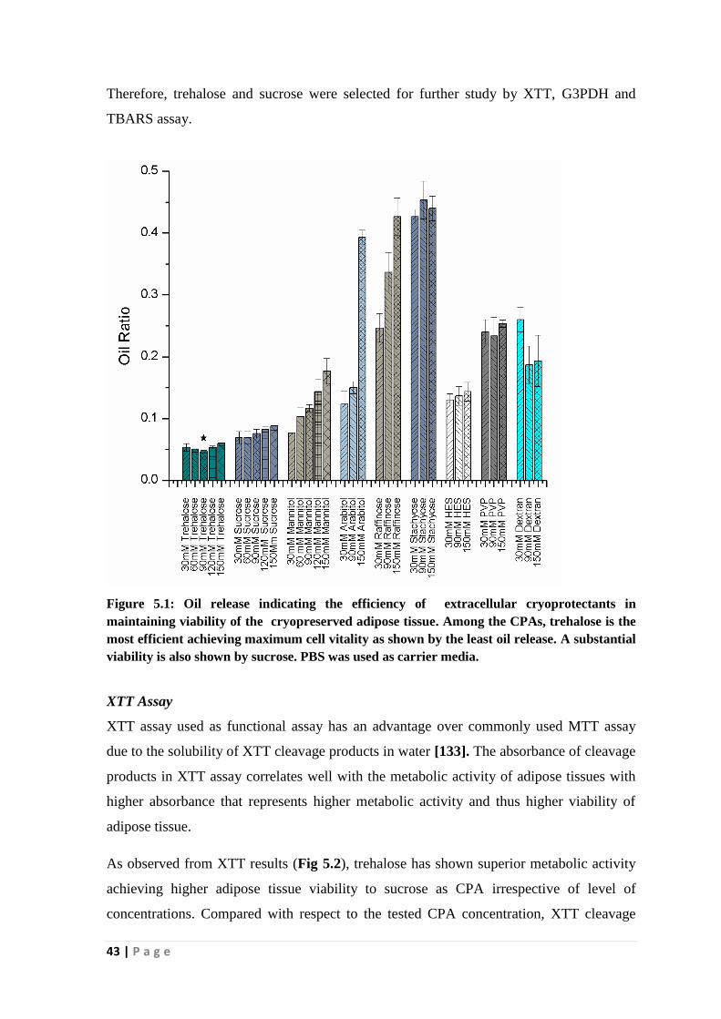
43 | P a g e
Therefore, trehalose and sucrose were selected for further study by XTT, G3PDH and
TBARS assay.
Figure 5.1: Oil release indicating the efficiency of extracellular cryoprotectants in
maintaining viability of the cryopreserved adipose tissue. Among the CPAs, trehalose is the
most efficient achieving maximum cell vitality as shown by the least oil release. A substantial
viability is also shown by sucrose. PBS was used as carrier media.
XTT Assay
XTT assay used as functional assay has an advantage over commonly used MTT assay
due to the solubility of XTT cleavage products in water [133]. The absorbance of cleavage
products in XTT assay correlates well with the metabolic activity of adipose tissues with
higher absorbance that represents higher metabolic activity and thus higher viability of
adipose tissue.
As observed from XTT results (Fig 5.2), trehalose has shown superior metabolic activity
achieving higher adipose tissue viability to sucrose as CPA irrespective of level of
concentrations. Compared with respect to the tested CPA concentration, XTT cleavage

44 | P a g e
product was increasing with increase in concentration up to 90mM and then a declining
trend was observed with trehalose though the variation was not statistically significant
between 30mM to 120mM. Whereas metabolic activity was noticed more or less same,
when adipose tissue was cryopreserved using sucrose as CPA. Among different
concentrations of sucrose, 60mM sucrose (69.4 ± 5.8%) achieved slightly higher viability
than other concentrations of sucrose. However, the difference between 30mM and 60mM
sucrose is not significant (p<0.05) as also revealed by oil ratio analysis. Therefore,
trehalose with 90mM concentration has been proven to be superior CPA than sucrose.
Figure 5.2: Metabolic activity of the cryopreserved adipose tissue treated with
trehalose and sucrose in varying concentrations measured by XTT assay. Trehalose
has shown superior metabolic activity achieving higher adipose tissue viability than
sucrose at all concentration levels. However, among the concentration range,
trehalose performed the best at 90mM concentration
G3PDH assay
G3PDH assay involving adipose specific enzyme is a relatively simple method chosen in
this study to assess the cellular function of the frozen adipose tissue. The cell membrane
integrity is detected by this assay through the marking of diffusion of intracellular
elements through the damaged adipose tissue membrane. The extracellular G3PDH level
was measured and a higher G3PDH level represents lower tissue viability [134].

45 | P a g e
The analysis of G3PDH (Fig 5.3) level measured in the washing solution of cryopreserved
adipose tissue shows that a higher level of enzyme activity was observed when sucrose
was used as CPA at all concentrations than the level of enzyme activity shown by
trehalose. Furthermore, among the different concentration of trehalose level tested,
trehalose at 90mM was found to be more effective than either lower or higher
concentration providing higher membrane integrity and thus higher cellular function (2.25
± 0.10 U/ml) of cryopreserved adipose tissue. Whereas a gradual increase in G3PDH
enzyme level with an increase in CPA concentration was depicted by sucrose when used
as CPA, though the difference in enzyme activity is not statistically significant. Unlike
trehalose, the enzyme level representing the adipose tissue viability obtained with sucrose
at 30mM concentration (2.57 ± 0.26 U/ml) is found to be best among other concentrations.
Figure 5.3: Evaluation of the efficiency of trehalose and sucrose as CPAs for the
cryopreservation of adipose tissue by measuring the extracellular activity of glycerol-
3-phosphate-dehydrogeage (G3PDH) enzyme. A comparatively lower enzyme
activity representing the higher tissue viability was shown by trehalose than sucrose.
90mM was shown to be the most favorable concentration for trehalose giving
maximum adipose tissue viability

46 | P a g e
TBARS assay
TBARS assay is one of the most commonly used tests for assessing the lipid peroxidation
of cells and tissues [135]. Free radical mediated cryo-injury is one of the well-established
models of cells and tissue damage in cryopreservation [136]. Free radicals formed in this
process participate in a cascade of reaction with lipid of the cells and tissues and
peroxidize them to form lipid peroxidation products or malondialdehyde. Therefore, it was
hypothesized that TBARS assay might be a useful test to quantify malondialdehyde in
post-thawed tissue, thereby would provide an indirect indication of the viability of the
adipose tissue.
The malondialdehyde level measured by TBARS assay (Fig 5.4) was also observed to be
least with 90mM trehalose (321 ± 7 nM/g wet wt) representing the superiority of the CPA
over other CPAs. Although, in the concentration range of 30mM to 150mM, the difference
between malondialdehyde production in trehalose and sucrose cryopreserved adipose
tissue is statistically insignificant (p<0.05), similar to G3PDH assay the trend suggests
trehalose is a superior cryoprotectant for adipose tissue cryopreservation than sucrose.
Among the various concentrations of sucrose, 30mM sucrose gave least TBARS
production (345 ± 15 nM/g wet wt) indicating the superiority of 30mM sucrose over other
concentrations of it. The malondialdehyde level of fresh adipose tissue was found to be
142 ± 11 nM/g wet wt. of the tissue.Thus,there was also significant increase in the level of
malondialdehyde in the cryopreserved tissues than the fresh tissues indicating the
significanceof free radical mediated damage in cryopreserved tissues.

47 | P a g e
Figure 5.4: Cellular viability in terms of malondialdehyde production of
cryopreserved adipose tissues using trehalose and sucrose as CPAs was assessed by
TBARS assay. The results indicate that 90mM trehalose is superior to sucrose for
cryopreservation of adipose tissue.
Overall, all the results taken together suggest that trehalose is a better cryoprotectant than
sucrose for adipose tissue cryopreservation with 90mM trehalose as the best concentration
level achieving the highest viability. In the case of sucrose, we could not get uniform
results in all the assays; however, the trends suggest a lower concentration of sucrose,i.e.,
30mM is better than its higher concentrations. Thus, 90mM trehalose was selected as
extracellular CPA for the preparation of freezing solution cocktail for adipose
cryopreservation.
5.1.2 Screening of intracellular CPAs
Organic osmolytes such as betaine, ectoin and hydroxyectoin are known to protect cells
and tissues from osmotic and thermal stresses [137]. Ectoin has also reported being an
excellent intracellular cryoprotectant for embryo and others [138]. Thus, like extracellular
CPAs, organic osmolytes in PBS solution was also evaluated as intracellular CPAs for

48 | P a g e
their efficacy in cryopreservation of adipose tissue through the analysis of oil ratio, XTT,
G3PDH and TBARS assays, which are described below.
Oil ratio
The analysis of the results of oil ratios (Fig 5.5) indicate betaine showed a higher oil ratio
than the other intracellular CPAs representing the lower viability of adipose tissue when
cryopreserved in betaine. The overall viability trend follows
ectoin>hydroxyectoin>betaine. The higher efficiency of ectoin has also been reported
earlier when other tissues cryopreserved in freezing solution containing ectoin [139-143].
A substantial tissue viability was also obtained using hydroxyectoin as CPA. Furthermore,
no significant statistical change in oil ratio value was observed between the concentration
levels of both ectoin and hydroxyectoin in the range of 90mM to 150mM. Therefore,
90mM was selected as the most favorable concentration of ectoin and hydroxyectoin.
However, there was a statistically significant difference in tissue damage between the
organo-osmolites and the conventional 10% Me2SO as CPA, which is evident from
decreased oil ratio value (0.042 ± 0.006) measured with the later. Therefore, ectoin and
hydroxyectoin were selected as the potential organo-osmolites for further study and 10%
Me2SO as a control.

49 | P a g e
Figure 5.5: Screening of intracellular cryoprotectants based on oil ratio analysis. The
higher efficiency of ectoin is evident from its lower oil release than other CPAs. The
overall viability trend follows ectoin>hydroxyectoin>betaine. No statistically
significant change in oil ratio was observed between the concentration levels for both
ectoin and hydroxyectoin the range of 90-150mM. Therefore, 90mM was selected as
the most favorable concentration of ectoin and hydroxyectoin. The viability was
lower than 10% Me2SO used as a control.
XTT assay
The results in Fig 5.6 indicate that the control 10% Me2SO (80.2 ± 3.2%) gave higher
viability than organic osmolytes (p<0.05). However, with an increase in the concentrations
of organic osmolytes, the viability of the post-thawed adipose tissues increased. In the
concentration range of 90mM-150mM, 150mM ectoin (69.8 ± 3%) achieved the highest
viability of tissues, which is slightly higher than 150mM hydroxyectoin (66.7 ± 4.2%).
However, the difference between the adipose tissues cryopreserved using ectoin and
hydroxyectoin is statistically insignificant (p<0.05). Similar to oil ratio, XTT assay
depicted the superiority of the conventional but harmful 10% Me2SO over the organic
osmolytes.

50 | P a g e
Figure 5.6: Evaluation of organic osmolytes in various concentrations for
cryopreservation of adipose tissue by XTT assay. 10% Me2SO was used as a control.
G3PDH assay
The assessment of cell membrane integrity via the measurement of G3PDH level (Fig 5.7)
shows a slightly lower enzyme activity thereby a higher tissue viability was obtained with
ectoin than hydroxyectoin. Thus, it indicates that the cryopreservation of adipose tissue
was superior using ectoin than hydroxyectoin, though a substantial tissue preservation was
also shown by later. In comparison, the viability results are slightly lower than those
obtained with conventional 10% Me2SO. The corresponding G3PDH values are 2.68 ±
0.15 U/ml (150mM ectoin), 2.96 ± 0.12 U/ml (150mM hydroxyectoin) and 2.28 ± 0.07
U/ml (10% Me2SO). Furthermore, it was also analyzed that the protective efficacy
increased with increase in the concentration of organic osmolytes and overall, 150mM
ectoin gave significantly (p<0.05) higher viability than 150mM hydroxyectoin.

51 | P a g e
Figure 5.7: Effect of ectoin and hydroxyectoin as intracellular CPAs on the
cryopreservation of adipose tissue by the assessment of G3PDH enzyme activity.
Ectoin than hydroxyectoin showed a slightly higher viability. Both CPAs show lower
efficiency 10% Me2SO.
TBARS assay
Fig 5.8 depicts the TBARS analysis data, which reveals that that 150mM ectoin (361 ± 11
nM/g wet wt) gave significantly lower lipid peroxidation products than 150mM
hydroxyectoin (397 ± 5 nM/g wet wt) indicating the higher viability achieved with ectoin
than hydroxyectoin. However, the lipid peroxidation with ectoin and hydroxyectoin were
significantly higher than Me2SO (285 ± 19 nM/g wet wt). This represents the lower
efficacy of the organic osmolytes than 10% Me2SO.
The results can be explained by the fact that ectoin possess less number of hydroxyl
groups and thus is lesser water-soluble than hydroxyectoin; hence, the distribution of
ectoin within the lipid-filled adipocytes may be much higher than hydroxyectoin [144].
Thus, 150mM ectoin and 10% Me2SO were selected as intracellular CPAs for formulating
the freezing solution cocktail for the freezing solution cocktail.

52 | P a g e
Figure 5.8: Viability assessment of post-thaw adipose tissues using ectoin and
hydroxyectoin as CPAs by TBARS assay. The viability obtained with ectoin is
slightly higher than hydroxyectoin and 150mM is the most favorable concentration
at which ectoin performed better. However, unlike extracellular CPAs, the
conventional 10% Me2SO (control) is superior to organic osmolytes.
5.1.3 Screening of ionic compounds
Not enough studies were conducted to understand the role of carrier medium in
cryopreservation of cells and tissues. However, the significance of ionic carrier medium is
evident from the literature related to cryopreservation of sperms where extenders
comprised of various ions such NaCl, KCl, NaHCO3,MgCl2 etc. are usually used to
preserve the functionality of cryopreserved sperm [145] . Evidence proving the
significance of ionic solutes in preservation of organs and tissues is apparent from the
literature of low temperature storage of organs for organ transplantation [146, 147].
Earlier developed Euro-Collins and University of Wisconsin preservation solutions are
intracellular type solution having higher potassium ions than sodium ions. Both these
solutions were observed to offer better transplantation outcome when used for
preservation of kidney and liver. However, relatively newer developed solutions like

53 | P a g e
Celsor, IGL-1 (Institute Georges Lopez) and ET-K (Kyoto University solution) that have
higher sodium concentration than potassium were observed to give better protection to
most of the organs including hearts, vessels and pancreas. Therefore, the effect of various
types of ionic compounds such as NaCl, Na2HPO4, KCl, KH2PO4, MgSO4 and MgCl2
towards the cryopreservation of adipose tissue in varying concentration was investigated
in the range of 80mM-240mM concentration. PBS was used as a control. The evaluation
results are described here.
Oil ratio
As described earlier, the damaged adipocytes in adipose tissue cryopreserved in different
ionic components measured by oil ratio is depicted in Fig 5.9. The extent of oil release
thereby representing the level of damaged tissue was different with different ionic
compounds and the trend was observed as NaCl<Na2HPO4<KCl<KH2PO4<MgCl2≈
MgSO4, though there is not much change in viability between NaCl and Na2HPO4. The oil
release was also found to change with a change in concentration of carrier media. The oil
release was increased with increase in concentration from 80mM-120mM and then
decreased with further increase in the concentration of both NaCl and Na2HPO4. However,
the minimum oil release means the maximum viability of adipose tissue was obtained at
160mM. Whereas in the case of KCl, a continual decrease in oil release with an increase
in concentration was observed. Overall, among the ionic compounds used, 160mM NaCl
(0.038 ± 0.002) showed the highest viability of cryopreserved tissue whereas MgSO4 and
MgCl2 showed the lowest tissue viability. Except at 240mM concentration of ionic
compounds, the trend of viability follows NaCl>Na2HPO4>KCl in all concentrations.
Thus, it has been demonstrated that the type of ionic compounds and their concentration
significantly influence the adipose tissue viability. 160mM NaCl and 160mM Na2HPO4
showed superior viability than other ionic solution and hence these compounds were
selected for further study.
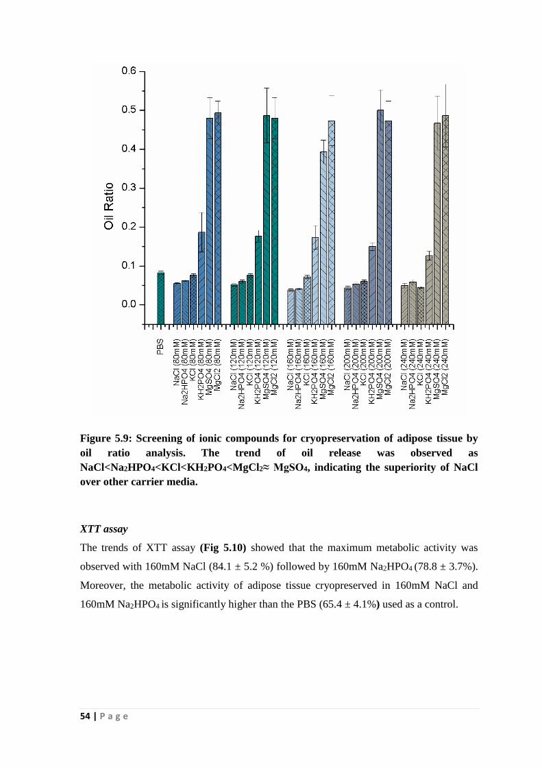
54 | P a g e
Figure 5.9: Screening of ionic compounds for cryopreservation of adipose tissue by
oil ratio analysis. The trend of oil release was observed as
NaCl<Na2HPO4<KCl<KH2PO4<MgCl2≈ MgSO4, indicating the superiority of NaCl
over other carrier media.
XTT assay
The trends of XTT assay (Fig 5.10) showed that the maximum metabolic activity was
observed with 160mM NaCl (84.1 ± 5.2 %) followed by 160mM Na2HPO4 (78.8 ± 3.7%).
Moreover, the metabolic activity of adipose tissue cryopreserved in 160mM NaCl and
160mM Na2HPO4 is significantly higher than the PBS (65.4 ± 4.1%) used as a control.

55 | P a g e
Figure 5.10: Evaluation of selected ionic compounds for cryopreservation of adipose
tissue by XTT assay. The maximum metabolic activity was observed with NaCl
followed by Na2HPO4 at 160mM concentration. Moreover, the metabolic activity of
adipose tissue cryopreserved in NaCl and Na2HPO4 at 160mM concentration is
significantly higher than the PBS used as a control.
G3PDH assay
The results of extracellular G3PDH activity assay (Fig 5.11) of adipose tissue viability
revealed similar trend as assessed by oil ratio and XTT assay. A lower level of enzyme
activity representing a better cellular function of adipose tissue was revealed when
cryopreserved in 160mM NaCl (1.85 ± 0.06 U/ml) than that obtained using 160mM
Na2HPO4 showing the G3PDH activity of 2.01 ± 0.17 U/ml. Although, the difference in
extracellular G3PDH level between NaCl and Na2HPO4 is insignificant (p<0.5), the
difference between the individual ionic compounds and PBS (3.07 ± 0.15 U/ml) is very
much significant (p<0.05) indicating higher protective ability of individual ionic
compounds than PBS.
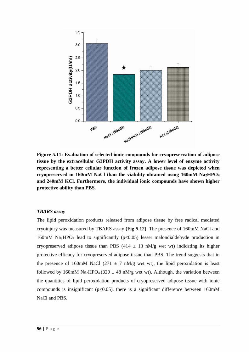
56 | P a g e
Figure 5.11: Evaluation of selected ionic compounds for cryopreservation of adipose
tissue by the extracellular G3PDH activity assay. A lower level of enzyme activity
representing a better cellular function of frozen adipose tissue was depicted when
cryopreserved in 160mM NaCl than the viability obtained using 160mM Na2HPO4
and 240mM KCl. Furthermore, the individual ionic compounds have shown higher
protective ability than PBS.
TBARS assay
The lipid peroxidation products released from adipose tissue by free radical mediated
cryoinjury was measured by TBARS assay (Fig 5.12). The presence of 160mM NaCl and
160mM Na2HPO4 lead to significantly (p<0.05) lesser malondialdehyde production in
cryopreserved adipose tissue than PBS (414 ± 13 nM/g wet wt) indicating its higher
protective efficacy for cryopreserved adipose tissue than PBS. The trend suggests that in
the presence of 160mM NaCl (271 ± 7 nM/g wet wt), the lipid peroxidation is least
followed by 160mM Na2HPO4 (320 ± 48 nM/g wet wt). Although, the variation between
the quantities of lipid peroxidation products of cryopreserved adipose tissue with ionic
compounds is insignificant (p<0.05), there is a significant difference between 160mM
NaCl and PBS.
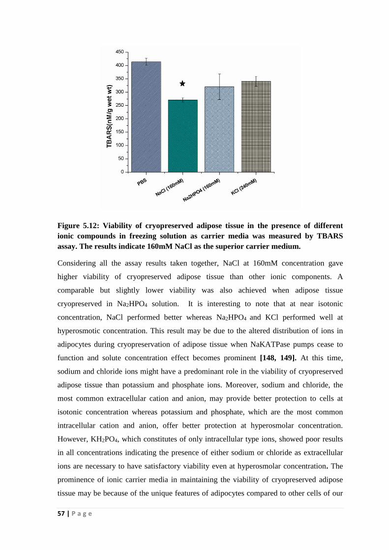
57 | P a g e
Figure 5.12: Viability of cryopreserved adipose tissue in the presence of different
ionic compounds in freezing solution as carrier media was measured by TBARS
assay. The results indicate 160mM NaCl as the superior carrier medium.
Considering all the assay results taken together, NaCl at 160mM concentration gave
higher viability of cryopreserved adipose tissue than other ionic components. A
comparable but slightly lower viability was also achieved when adipose tissue
cryopreserved in Na2HPO4 solution. It is interesting to note that at near isotonic
concentration, NaCl performed better whereas Na2HPO4 and KCl performed well at
hyperosmotic concentration. This result may be due to the altered distribution of ions in
adipocytes during cryopreservation of adipose tissue when NaKATPase pumps cease to
function and solute concentration effect becomes prominent [148, 149]. At this time,
sodium and chloride ions might have a predominant role in the viability of cryopreserved
adipose tissue than potassium and phosphate ions. Moreover, sodium and chloride, the
most common extracellular cation and anion, may provide better protection to cells at
isotonic concentration whereas potassium and phosphate, which are the most common
intracellular cation and anion, offer better protection at hyperosmolar concentration.
However, KH2PO4, which constitutes of only intracellular type ions, showed poor results
in all concentrations indicating the presence of either sodium or chloride as extracellular
ions are necessary to have satisfactory viability even at hyperosmolar concentration. The
prominence of ionic carrier media in maintaining the viability of cryopreserved adipose
tissue may be because of the unique features of adipocytes compared to other cells of our

58 | P a g e
body. During cryopreservation, ice-mediated and osmotic imbalance mediated injuries are
the two major factors, which govern the fate of cryopreserved cells and tissues. While
though ice-mediated injury plays dominant mode of cryoinjury for cells filled with mainly
colloidal water, adipocytes, which mainly contain lipids, may be influenced by the
osmotic factor more dominantly than ice factor during cryopreservation [150]. However,
further investigation on the detailed mechanism is necessary to understand much complex
cryopreservation phenomenon.
Therefore, to formulate ultimate freezing solution cocktail, 160mM NaCl was selected as
the potential carrier medium.
5.1.4 Formulation and evaluation of freezing solution for adipose tissue
cryopreservation
The selected extracellular and intracellular CPAs in 160mM NaCl carrier medium was
evaluated individually as well as in combination to formulate an optimized freezing
solution for adipose tissue cryopreservation. To this end, different batches of freezing
solutions such as 160mM NaCl/90mM trehalose, 160mM NaCl/150mM ectoin, 160mM
NaCl/90mM trehalose/150mM ectoin and 160mM NaCl/10% Me2SO were formulated to
evaluate their efficiency towards adipose tissue cryopreservation.
Oil ratio
The results of oil analysis (Fig 5.13) indicate that among the prepared freezing solutions,
160mM NaCl/10% Me2SO has shown the most effective providing maximum viability of
0.032±0.004 followed by 160mM NaCl/90mM trehalose (0.035 ± 0.002) indicating both
Me2SO and trehalose containing freezing solution are efficient for the preservation of
adipose tissue. Though a slightly higher tissue viability was obtained with 10% Me2SO in
NaCl carrier media than viability achieved with 160mM NaCl/90mM trehalose, the
difference in viability is statistically insignificant. Nevertheless, both these freezing
solutions showed higher viability when compared with viability obtained with 160mM
NaCl, 160mM NaCl /150mM ectoin and 160mM NaCl/90mM trehalose/150mM ectoin.
Interestingly, the presence of ectoin in the formulated freezing solution did not offer any
additional advantage in cryopreservation of adipose tissue.

59 | P a g e
Figure 5.13: Post-thaw viability of cryopreserved adipose tissue in freezing solutions
formulated from trehalose and ectoin in NaCl as carrier media by oil ratio analysis
(D, Me2SO; T, Trehalose; E, Ectoin). The freezing solution comprising of 160mM
NaCl/10% Me2SO has shown the most effective providing maximum viability
followed by 160mM NaCl/90mM trehalose indicating freezing solution containing
either Me2SO or trehalose are efficient for the preservation of adipose tissue.
XTT assay
Unlike oil ratio results, all the formulated freezing solutions have shown more or less
similar XTT values (Fig 5.14) representing the comparable level of adipose tissue
viability. Overall, 160mM NaCl/90mM trehalose achieved a slightly higher viability (84.8
± 4.3%) than other freezing solutions prepared from ectoin and thus proven to be the most
potential. When compared the viability was also comparable to the viability obtained
using 10% Me2SO in NaCl (85.3 ± 3.9%). Thus, 160mM NaCl/90mM trehalose is proven
to be a potential non-toxic freezing solution for adipose tissue.

60 | P a g e
Figure 5.14: Cellular viability of cryopreserved adipose tissue in freezing solutions
prepared from trehalose and ectoin in NaCl carrier media by XTT assay (D, Me2SO;
T, Trehalose; E, Ectoin). Overall, the formulated 160mMNaCl/90mM trehalose
achieved a slightly higher viability (84.8 ± 4.9%) than other freezing solutions and
thus proven to be the most efficient freezing solution. The viability was also
comparable to the viability obtained using 10% Me2SO.
G3PDH assay
The cell membrane integrity of adipose tissue was assessed by G3PDH activity and the
experimental data is shown in Fig 5.15. The enzyme activity was shown by freezing
solution containing ectoin (2.008 ± 0.147 U/ml) as CPA is higher that represents higher
adipocyte damage than the trehalose-containing freezing solution (1.751 ±0.121 U/ml).
The freezing solution containing trehalose has also shown similar viability outcome with
cryopreserved adipose tissue.

61 | P a g e
Figure 5.15: Viability of cryopreserved adipose tissue in freezing solutions prepared
from trehalose and ectoin in NaCl carrier media assessed by G3PDH activity (D,
Me2SO; T, Trehalose; E, Ectoin). A higher adipocyte damage in cryopreserved
adipose tissue was shown by freezing solution containing ectoin as CPA than
trehalose, representing the higher tissue viability achieved with trehalose.
TBARS assay
Fig 5.16 shows the TBARS assay results obtained with adipose tissue cryopreservation in
the formulated freezing solution as 160mM NaCl/10% Me2SO ≈ 160mM NaCl/90mM
trehalose > 160mM NaCl/150 ectoin and 160mM NaCl/90mM trehalose/150M ectoin. The
results follow the trend of viability is comparable to the values obtained with other assays.
Thus, 160mM NaCl/10% Me2SO showing TBARS value of 253 ± 15 nM/g wet wt and
160mM NaCl/90mM trehalose showing TBARS value of 256 ± 16 nM/g wet wt) are
comparable and the solutions provided superior efficacy than other freezing solution for
adipose tissue cryopreservation. Interestingly, similar to other assays, ectoin did not show
any impact as an intracellular cryoprotectant on cryopreservation outcome.

62 | P a g e
Figure 5.16: Viability of cryopreserved adipose tissue in freezing solutions prepared
from trehalose and ectoin in NaCl carrier media measured by TBARS assay. The
results indicate 160mM NaCl/10%Me2SO and 160mM NaCl/90mM trehalose
achieved superior viability than other freezing solutions. (D, Me2SO; T, Trehalose; E,
Ectoin)
Overall, the present study demonstrated that 160mM NaCl/90mM trehalose and 160mM
NaCl/ 10% Me2SO offer better viability of cryopreserved adipose tissue compared to
160mM NaCl alone. However, 160mM NaCl alone offers significantly higher viability
compared to other ionic compounds and cryoprotectants representing the prominent role
of osmolarity and carrier media in the cryopreservation of adipose tissue. The higher
viability obtained with ectoin in PBS than PBS alone was not reflected in the viability
outcome using ectoin in NaCl, indicating the limited role of ectoin in the cryopreservation
of adipose tissue. Further study on enhancement of the protective role of the developed
freezing solution in adipose tissue cryopreservation is important to ensure its higher
viability on long-term storage.

63 | P a g e
5.2 Improvement of adipose tissue viability by the addition of anti-oxidants in
freezing solution
The oxidative stress mediated damage has been reported to be more pronounced in
cryopreservation of adipose tissue than other tissue types as adipose tissue is usually
harvested from obese patient and adipocytes in obese are known to have a higher
concentration of free radicals compared to other body tissues [151, 152]. Furthermore,
mechanical manipulation of adipose tissue during liposuction and further downstream
processes, increase the susceptibility of the tissue towards free radical generation. Besides
ice related injury, the role of free radical mediated cryoinjury due to ROS generation is
also a critical factor that causes cell death during cryopreservation.
The addition of antioxidant as a supplement in freezing solution is reported to be a
promising strategy to overcome cryoinjury during cryopreservation of cells and tissues.
Curcumin, a phytochemical having antioxidative property increased the various cells and
tissue viability as reported earlier [153]. Besides curcumin, other antioxidants such as
carnitine and Vit E decreased cytoplasmic lipid content and increased the cytotolerance of
oocytes during vitrification [154]. L-carnitine also significantly improved epididymal
sperm motility after cryopreservation as reported elsewhere [155].
Keeping the above in view, we hypothesized that supplementation of antioxidants in
160mM NaCl/90mM trehalose freezing solution may improve the cryopreservation
outcome of adipose tissue. Therefore, the present study focusses on exploring potential
antioxidants for achieving improved viability of cryopreserved adipose tissue and
investigates their influence on the cryopreservation efficiency of the previously formulated
freezing solution for adipose tissue preservation. In this study, curcumin, carnitine, lipoic
acid, Vit E, gallic acid, ellagic acid and quercetin were chosen as antioxidants based on the
literature. Similar to the earlier section, oil ratio, XTT, G3PDH and TBARS have been
measured to assess the viability of post-thawed adipose tissue.
5.2.1 Screening of antioxidants
Oil ratio
As in the case of CPAs, the screening of potential antioxidants was done by measuring oil
release during cryopreservation (Fig 5.17). As indicated, a varying oil ratio representing
the varying level of tissue damage was found with different types of antioxidants. Among
the various antioxidants, curcumin and carnitine achieved higher tissue viability than other
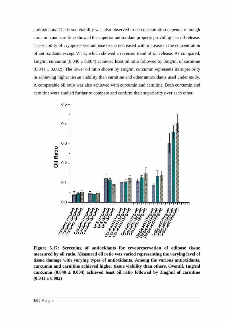
64 | P a g e
antioxidants. The tissue viability was also observed to be concentration dependent though
curcumin and carnitine showed the superior antioxidant property providing less oil release.
The viability of cryopreserved adipose tissue decreased with increase in the concentration
of antioxidants except Vit E, which showed a reversed trend of oil release. As compared,
1mg/ml curcumin (0.040 ± 0.004) achieved least oil ratio followed by 3mg/ml of carnitine
(0.041 ± 0.002). The lower oil ratio shown by 1mg/ml curcumin represents its superiority
in achieving higher tissue viability than carnitine and other antioxidants used under study.
A comparable oil ratio was also achieved with curcumin and carnitine. Both curcumin and
carnitine were studied further to compare and confirm their superiority over each other.
Figure 5.17: Screening of antioxidants for cryopreservation of adipose tissue
measured by oil ratio. Measured oil ratio was varied representing the varying level of
tissue damage with varying types of antioxidants. Among the various antioxidants,
curcumin and carnitine achieved higher tissue viability than others. Overall, 1mg/ml
curcumin (0.040 ± 0.004) achieved least oil ratio followed by 3mg/ml of carnitine
(0.041 ± 0.002)

65 | P a g e
XTT assay
XTT assay data as shown in Fig 5.18 reveals the higher metabolic activity (84.9 ± 2.9%)
of adipose tissue was shown with curcumin and 1mg/ml is the most favorable
concentration of curcumin. The increase in concentration beyond 1mg/ml did not show
any benefit in the preservation of adipose tissue and no change in viability between
3mg/ml and 5mg/ml of curcumin. Whereas a typical trend of viability was observed when
cryopreservation of adipose tissue was performed using carnitine. The viability of adipose
tissue increased with increase in the concentration of carnitine from 1mg/ml to 3mg/ml
(82.78 ± 3.65%) and then decreased with further increase in concentration. In comparison
between curcumin and carnitine, an equivalent level of adipose tissue viability was
achieved at 1mg/ml and 3mg/ml respectively. Thus, the study has demonstrated the
superior efficiency of curcumin using comparatively lesser amount than carnitine.
Figure 5.18: Evaluation of curcumin and carnitine for cryopreservation of adipose
tissue measured by XTT assay. An equivalent level of adipose tissue viability was
achieved at 1mg/ml and 3mg/ml respectively.

66 | P a g e
G3PDH assay
The measurement of the extracellular G3PDH enzymes released by lysed adipocytes
indicates the lower viability of cryopreserved adipose tissues with a higher concentration
of curcumin (Fig 5.19). The results show that quantity of extracellular G3PDH was lowest
with 1mg/ml curcumin (1.91 ± 0.10 U/ml) representing its superior antioxidant efficiency
in reducing ROS generated tissue damage. Furthermore, similar to XTT assay, a non-
linear tissue damage and hence tissue viability of cryopreserved adipose tissue was
observed when carnitine was used as an antioxidant. The viability of cryopreserved tissues
in 3mg/ml carnitine was the maximum, which was also comparable to the viability
achieved with 3mg/ml curcumin. Furthermore, the viability difference between carnitine
and curcumin at 5mg/ml is not statistically significant.
Figure 5.19: Performance evaluation of curcumin and carnitine for cryopreservation
of adipose tissue measured by G3PDH assay and confirm the superior antioxidant
activity shown by curcumin providing higher tissue viability at comparatively lower
concentration than carnitine.
TBARS assay
The quantity of malondialdehyde production during cryopreservation of adipose tissue
measured by TBARS assay corroborated with the findings of both XTT and G3PDH assay

67 | P a g e
(Fig 5.20). The adipose tissue cryopreserved with 1mg/ml curcumin (270 ± 24 nM/g wet
wt) showed lesser lipid peroxidation than tissues cryopreserved with other concentrations
of curcumin and carnitine. However, the difference in the level of protection achieved
with 3mg/ml carnitine (302 ± 15 nM/g wet wt) and 1mg/ml curcumin is statistically
insignificant (p<0.05) that signifies the concentration level at which individual
antioxidants functions well in preventing tissue damage. Therefore, all the assays taken
together curcumin was proven to be the best antioxidants and 1mg/ml is the most effective
concentration.
Figure 5.20: Evaluation of curcumin and carnitine for cryopreservation of adipose
tissue measured by TBARS assay. The assay result corroborated with the findings of
both XTT and G3PDH assay revealing the higher efficiency of curcumin.
The results revealed that curcumin and carnitine were effective and ellagic acid, gallic acid
and quercetin were found to be ineffective in maintaining the viability of cryopreserved
adipose tissue. The result may be explained by the fact that antioxidants when used in
inappropriate concentration may also act as free radical or ROS generator imparting
additional damage to the tissues [156-158]. Furthermore, compared to only water (polar)
soluble antioxidants such as ellagic acid, gallic acid and quercetin, antioxidants that are

68 | P a g e
soluble in only non-polar solvents (curcumin and Vit E) as well as both polar and non-
polar solvents (carnitine and lipoic acid) performed better for cryopreservation of adipose
tissue [159]. The result may be explained by the fact that adipocytes are filled mostly with
triglycerides, a nonpolar molecule and thus, permeation and distributions of non-polar
antioxidants within the cells performed better compared to polar antioxidants [160].
Therefore, 1mg/ml curcumin and 3mg/ml carnitine were selected for further study to
investigate their effectiveness in improving the cryopreservation of adipose tissue by
supplementing with the most efficient 160mM NaCl/90mM trehalose freezing solution.
5.2.2 Improvement of 160mM NaCl/90mM trehalose freezing solution supplemented
with antioxidants
As it has already been mentioned that antioxidants have an important role in overcoming
free radical mediated cryoinjury during cryopreservation of cells and tissues. In our
previous work, carnitine and curcumin were found to be efficient antioxidants performing
well towards cryopreservation of adipose tissue in PBS solution. Therefore, it was
hypothesized that the addition of these antioxidants may improve the cryopreservation
outcome of the 160mM NaCl and the formulated 160mM NaCl/90mM trehalose freezing
solution for adipose tissue cryopreservation. Therefore, in this phase of dissertation work,
cryopreservation experiments were carried out with these freezing solutions with the
addition of carnitine and curcumin to investigate their effect on the cryopreserved tissue
viability.
Oil ratio
Oil ratio data (Fig 5.21) indicates that the supplementation of antioxidants with 160mM
NaCl and 160mM NaCl/90mM trehalose freezing solutions significantly improved the
viability of cryopreserved adipose tissue. Although there was no significant difference
(p<0.05) among oil ratios of 160mM NaCl/1mg/ml curcumin (0.032 ± 0.004), 160mM
NaCl/3mg/ml carnitine (0.033 ± 0.002) and 160mM NaCl/90mM trehalose (0.035 ±
0.002), there was significant difference between the oil ratios obtained with 160mM
NaCl/90mM trehalose/1mg/ml curcumin (0.02 ± 0) and 160mM NaCl/90mM
trehalose/3mg/ml carnitine (0.025 ± 0.002). Overall, freezing solutions containing
curcumin achieved higher viability than carnitine supplemented freezing solutions and the
highest viability was shown by 160mM NaCl/90mM trehalose/1mg/ml curcumin
representing its superior performance than other freezing solution.

69 | P a g e
Figure 5.21: Oil ratio analyses indicate the viability of cryopreserved adipose tissue
using curcumin and carnitine in freezing solution. 160mM NaCl was used as carrier
media. The viability obtained with 160mM NaCl/90mM trehalose/1mg/ml
curcumin(0.02 ± 0.00) and 160mM NaCl/90mM trehalose/3mg/ml carnitine (0.025 ±
0.002) freezing solutions was higher than the viability obtained with 160mM
NaCl/10% Me2SO (0.032 ± 0.004).
XTT assay
The XTT results (Fig 5.22) shows that the difference in metabolic activity achieved with
the cryopreserved adipose tissues in different freezing solutions was not found to be
statistically significant. However, adipose tissue cryopreserved in 160mM NaCl/90mM
trehalose/1mg/ml curcumin freezing solution achieved the highest metabolic activity
(89.7± 4.4%).
Interestingly, as also observed in oil ratio analysis, 160mM NaCl/1mg/ml curcumin (86.2
± 3.4%) freezing solution gave better viability than 160mM NaCl/90mM trehalose (84.8 ±
4.3%) indicating a dominant role of redox state in adipocytes in determining the viability
of cryopreserved adipose tissue. However, combined effect of trehalose and 160mM
NaCl/1mg/ml curcumin improved the viability revealing the independent and additive
cryoprotective mechanisms of curcumin and trehalose. Freezing solutions supplemented

70 | P a g e
with carnitine provided comparable results to that obtained with 160mM NaCl/90mM
trehalose freezing solution as assessed by both oil ratio and XTT assay indicating a similar
level of efficacy of carnitine and trehalose. However, similar to curcumin-supplemented
solutions, the addition of trehalose to carnitine supplemented freezing solution further
improved the viability of cryopreserved adipose tissue.
Figure 5.22: The viability assessment of cryopreserved adipose tissue using curcumin
and carnitine as antioxidants in freezing solution. 160mM NaCl was used as carrier
media. adipose tissue cryopreserved in 160mM NaCl/90mM trehalose/1mg/ml
curcumin freezing solution achieved the highest metabolic activity (89.7 ± 4.4%).
G3PDH assay
As indicated in Fig 5.23 the extracellular G3PDH concentration was the least with the
cryopreserved adipose tissue when cryopreserved in 160mM NaCl/90mM
trehalose/1mg/ml curcumin (1.62 ± 0.07 U/ml) freezing solution. However, the result was
not statistically significantly different (p<0.05) from other freezing solution except when
the adipose tissue cryopreserved in 160mM NaCl/3mg/ml carnitine.

71 | P a g e
Figure 5.23: G3PDH assay indicate the viability of cryopreserved adipose tissue
using curcumin and carnitine in freezing solution. 160mM NaCl was used as carrier
media. The extracellular G3PDH concentration was the least and hence the
maximum viability of post-thawed adipose tissue when cryopreserved in 160mM
NaCl/90mM trehalose/1mg/ml curcumin freezing solution.
TBARS assay
The trend in malondialdehyde production and thus the adipocyte viability in cryopreserved
adipose tissue (Fig 5.24) were similar to the viability results obtained with oil ratio, XTT
and G3PDH assay results proving the addition of antioxidants improved the viability of
cryopreserved adipose tissue. The tissue preserved in 160mM NaCl/90mM
trehalose/1mg/ml curcumin freezing solution undergoes lipid peroxidation (204 ± 11 nM/g
wet wt), which is significantly lower than the conventional 10% Me2SO freezing solution
(253 ± 15 nM/g wet wt). Although, freezing solution comprising of 160mM NaCl/90mM
trehalose/3mg/ml carnitine (219 ± 15 nM/g wet wt) performed significantly higher than
160mM NaCl/10% Me2SO, the adipose tissue viability obtained with 160mM
NaCl/3mg/ml carnitine were comparable to the viability obtained with 160mM NaCl/10%
Me2SO freezing solution. The difference between the concentration of lipid peroxidation
products with 160mM NaCl/90mM trehalose/3mg/ml carnitine and 160mM NaCl/90mM

72 | P a g e
trehalose/1mg/ml curcumin freezing solutions was not statistically significant (p<0.05) as
also revealed by XTT and G3PDH assay.
Figure 5.24: TBARS assay indicate the viability of cryopreserved adipose tissue using
curcumin and carnitine in freezing solution. Among the freezing solutions, 160mM
NaCl/90mM trehalose/1mg/ml curcumin underwent significantly lower lipid
peroxidation (204 ± 11 nM/g wet wt) than the freezing solution containing
conventional 10% Me2SO. A comparable tissue viability using 160mM NaCl/90mM
trehalose containing carnitine at a comparatively higher concentration (3mg/ml) was
obtained.
Overall, considering the results of all assays taken into account, it was observed that
160mM NaCl/90mM trehalose/1mg/ml curcumin freezing solution performed better than
other freezing solution including Me2SO containing freezing solution. Thus, the study
provides a Me2SO free, non-toxic freezing solution that has the ability to cryopreserve
adipose tissue with maintained tissue viability. Furthermore, the presence of curcumin in
the post-thawed adipose tissue may offer the additional advantage of better wound healing
and aesthetic outcome after lipofilling operation. Besides antioxidant property, curcumin
is known to exhibit anti-inflammatory and anti-infective properties, which make it a
unique molecule for wound healing applications [161]. Curcumin treatment resulted in
increased formation of granulation tissue, neovascularization and enhanced biosynthesis of

73 | P a g e
extracellular matrix proteins such as collagen [162]. Curcumin treated cutaneous wound
showed decreased levels of lipid peroxides (LPs) and cross-linking of collagen. Therefore,
all these factors may provide additional benefit in achieving better wound healing in the
lipofilling operation and tissue regeneration by the use of cryopreserved adipose tissue
through tissue engineering technique. Moreover, in the case of autologous fat grafting
needs to be performed at the tumor resected site, curcumin may kill the residual tumor
cells because of its anticancer property [163, 164]. Thus, the presence of curcumin at graft
site may lead to less resorption of grafts, maintaining the contour of the surgical site and
improve aesthetic outcome of the surgery. The hypothesis is supported by the fact that
antioxidants N-acetylcysteine and Coenzyme Q10, treatment resulted in improved graft
retention [165, 166].

74 | P a g e
5.3 Effect of signaling pathway inhibitors
Cryoinjury during cryopreservation accentuates the apoptosis mediated death of
adipocytes in adipose tissue [167]. The initiation of apoptosis is possible by many
mechanisms and pathways such as intrinsic and extrinsic pathways. The intrinsic pathway
contains proteins such as second mitochondria-derived activator of caspase (SMAC) or
cytochrome c, while the extrinsic pathway starts with special TNF-receptors, also called
‘death receptors.' All pathways result in the activation of substrate specific cysteine
proteases, also called caspases that are the effectors of apoptosis [168]. Molecules that
inhibit caspases are known to inhibit apoptosis-mediated cell death [169]. Z-VAD-FMK is
a cell permeable, pan-caspase inhibitor, which prevents apoptosis-mediated cell death
during cryopreservation of cells and tissues thereby, increases the post-thaw viability
[170].
Similar to caspases, Rho/ROC kinase also plays a negative role in maintaining the
viability of adipose tissue [171]. Increased Rho/ROC kinase activity was observed in
adipose tissue of obese and has linkage to adipose hypertrophy, metabolic dysregulation
and adipose tissue inflammation. Furthermore, Rho/ROCK inhibits the cytoskeletal
reorganization and interferes with insulin pathway necessary for adipogenesis of hADSCs.
Rho/ROCK inhibitor such as Y-27632 reverses the negative effects thereby maintains a
healthy state of adipose tissue [172]. The ROCK inhibitor Y-27632 increased the viability
of various cryopreserved cells and tissue [173].
Although the role of caspase and ROCK inhibitors in increasing the viability of
cryopreserved cells and tissues are widely recognized, the molecules have not been
evaluated so far for their efficacy in cryopreservation of adipose tissue. Therefore, it was
hypothesized that the addition of Z-VAD-FMK and Y-27632 in the developed freezing
solution might improve the viability of post-thawed adipose tissue. Thus, an effort was
given to investigate the impact of Z-VAD-FMK and Y-27632 after supplementing them
with the developed freezing solution. Similar to the previous methodology, the viability of
post-thaw adipose tissue was evaluated by oil ratio, XTT, G3PDH and TBARS assays.
Oil ratio
The oil release due to damage of adipocyte present in adipose tissue was assessed by
performing cryopreservation experiment using the most effective freezing solution
160mM NaCl/90mM trehalose/1mg/ml curcumin formulated in the previous chapter

75 | P a g e
supplementing with 10nM Z-VAD-FMK and 100mM Y-27632. The concentrations of
inhibitors were based on literature [174, 175]. The results in Fig 5.25 reveal that there is
no significant difference in oil released from post-thawed adipose tissue cryopreserved
with the most efficient 160mM NaCl/90mM trehalose/1mg/ml curcumin freezing solution
(0.0213 ± 0.0023) with and without inhibitors, though a slightly lower viability was shown
by Y-27632 (0.0227 ± 0.0023). This represents that the addition of inhibitors has
negligible influence on the tissue viability.
Figure 5.25: Effect of Rho kinase and caspases inhibitors supplemented with the
most effective 160mM NaCl/90mM trehalose/1mg/ml curcumin freezing solution on
the viability of cryopreserved adipose tissue by oil ratio. It is indicated that the
addition of inhibitors has no significant influence on the tissue viability.
XTT assay
XTT results (Fig 5.26) shows a marginal increment of adipose tissue viability by adding
inhibitors and the viability showed by both inhibitors is comparable. The corresponding
viability values were found to be 89.6 ± 3.6% and 89.4 ± 3.4% with Z-VAD-FMK and Y-
27632. Though a slightly higher adipose tissue viability obtained with the freezing
solution supplemented with inhibitors but the difference between the viability results with
and without inhibitors are statistically insignificant (p<0.05) thereby demonstrating that
the inhibitors do not play any significant role in maintaining the viability of cryopreserved
adipose tissue.

76 | P a g e
Figure 5.26: XTT assay indicates the viability of cryopreserved adipose tissue using
Rho kinase and caspase inhibitors supplemented with the formulated freezing
solution. Results indicate a slightly higher adipose tissue viability obtained by adding
inhibitors, but the difference between the viability results with and without inhibitors
are statistically insignificant (p<0.05) which signifies that the inhibitors do not play
any significant role in maintaining the viability of cryopreserved adipose tissue.
G3PDH assay
Similar to the findings of oil ratio and XTT assays, the results of G3PDH assay (Fig 5.27)
also indicate that there is no significant difference in enzyme activity during
cryopreservation of adipose tissue using formulated 160mM NaCl/90mM
trehalose/1mg/ml curcumin with and without inhibitors. The enzyme activities of 1.67 ±
0.10 U/ml and 1.61 ± 0.07 U/ml were shown with the addition of Z-VAD-FMK and Y-
27632 to the freezing solution (1.62±0.07 U/ml) respectively. Thus, the results represent
the limited role of Z-VAD-FMK and Y-27632 compounds in the protection of
cryopreserved adipose tissue. However, similar to oil ratio analysis results, the freezing
solution containing Y-27632 gave slightly better performance than Z-VAD-FMK.

77 | P a g e
Figure 5.27: G3PDH assay indicates the viability of cryopreserved adipose tissue
using Rho kinase and caspases inhibitors supplemented with the freezing solution.
TBARS assay
TBARS assay (Fig 5.28) also showed that there was no beneficial effect of Z-VAD-FMK
and Y-27632 when added to 160mM NaCl/90mM trehalose/1mg/ml curcumin freezing
solution. However, 160mM NaCl/90mM trehalose/1mg/ml curcumin/10nM Z-VAD-FMK
(215 ±15 nM/g wet wt) performed slightly higher than 160mM NaCl/90mM
trehalose/1mg/ml curcumin /100mM Y-27632 (217 ±13 nM/g wet wt).
Figure 5.28: TBARS assay indicates the effect of Rho kinase and caspases inhibitors
on the viability of cryopreserved adipose tissue using
160mMNaCl/90mMtrehalose/1mg/ml curcumin freezing solution. Though there is no
significant improvement in viability was observed using inhibitor, Z-VAD-FMK
inhibitor shows slightly better performance than Y-27632.
Overall, the results proved that addition of Z-VAD-FMK and Y-27632 did not improve
the viability of post-thaw adipose tissue.

78 | P a g e
5.4 Control rate freezing of adipose tissue using formulated freezing solution
Cooling rate during cryopreservation has a profound effect on the viability of
cryopreserved cells and tissues. Slow cooling leads to extracellular ice formation and
hence the dominant mechanism of cryoinjury is solute concentration effect [176].
Whereas, rapid cooling leads to intracellular ice formation and thus ice-mediated injury
becomes dominant cause of cell death [177]. Therefore, to achieve maximum cell
viability, the optimal cooling rate is essential.
In the present research, initially the adipose tissue was cryopreserved in -80°C freezer in
isopropanol containing bath so as to maintain 1-2°C/min cooling rate [178]. Keeping this
in view, adipose tissue was subjected to control rate freezing in a controlled rate freezer
(CRF) to achieve more accurate and precise temperature control using the most effective
160mM NaCl/90mM trehalose/1mg/ml curcumin freezing solution [38].
5.4.1 Effect of cooling rate
The effect of cooling rate on the cryopreservation of adipose tissue was studied at varying
cooling rate such as 0.5°C/min, 1°C/min, 2.5°C/min, 5°C/min in CRF from 4°C to -120°C
followed by preservation in LN2 After 48h, the cryopreserved tissues were thawed rapidly
in a 37°C water bath. Rapid thawing method was selected based on the reported literature
that suggests that rapid thawing is superior to slow thawing method. The viability of post-
thawed adipose tissue was assessed as usual by oil ratio, XTT, G3PDH and TBARS
assays.
Oil ratio
The experimental results on oil release from cryopreserved adipose tissue at varying
cooling rate are shown in Fig 5.29. The viability results indicate that the adipose tissue
cryopreserved at -1°C/min cooling rate released less oil from the damaged adipose tissue
than either below or above -1°C/min and thus retained more viability. The trend of oil
release at different cooling rate is as follows- -1°C/min (0.019 ± 0.002) <0.5°C/min (0.021
± 0.002) <2.5°C/min (0.023 ±0.002) < 5°C/min (0.029 ± 0.002). Although, mean oil ratio
at -1°C/min cooling rate is higher than 0.5°C/min, the difference between them is
insignificant (p<0.05). Thus, with increasing cooling rate, the viability of the post-thawed
adipose tissues decreased rapidly. In comparison, oil ratios between adipose tissue
preserved using isopropanol filled container in -80°C freezer (0.021 ± 0.002) and
controlled rate freezer at -1°C/min cooling rate and storage at LN2, is comparable though a

79 | P a g e
slightly lower oil ratio representing superior tissue viability was achieved with the later
condition.
Figure 5.29: Oil ratio analyses indicate the effect of cooling rate on the
cryopreservation of adipose tissue using 160mM NaCl/90mM trehalose/1mg/ml
curcumin freezing solution. The trend of oil release at different cooling rate is as
follows- -1°C/min (0.019 ± 0.002) <0.5°C/min (0.021 ± 0.002) <2.5°C/min (0.023 ±
0.002) <5°C/min (0.029 ± 0.002) indicating -1°C/min is the best cooling rate
providing maximum adipose tissue viability.
XTT assay
As indicated in Fig 5.30, -1°C/min cooling rate showed the maximum metabolic activity
(93.1± 2.2%) followed by adipose tissues cryopreserved at -0.5°C/min (89.7 ± 2.9%), -
2.5°C/min (84.1 ± 3.5%) and -5°C/min (83.9 ± 3.8%). As also observed with oil ratio
measurement, the difference in metabolic activity between adipose tissue preserved in
isopropanol filled container and -1°C/min controlled rate freezing is statistically
insignificant (p<0.05). However, the mean metabolic activity of controlled rate frozen
samples was higher than samples frozen using Mr. Frosty. This indicates the superiority of
the control rate freezing in the maintenance of adipose tissue viability.

80 | P a g e
Figure 5.30: XTT assay showing the effect of cooling rate on the viability pattern of
cryopreserved adipose tissue using 160mM NaCl/90mM trehalose/1mg/ml curcumin
freezing solution. The maximum viability was obtained at -1°C/min.
G3PDH assay
The viability in terms of cell membrane integrity evaluated by G3PDH assay (Fig 5.31)
indicated the similar trend of tissue damage and reconfirmed -1°C/min cooling rate is the
most effective cooling rate providing maximum tissue viability. The tissue damage in
terms of enzyme activity level observed at different cooling rate follows the following
trends -1°C/min (1.57 ± 0.10 U/ml) < -0.5°C/min (1.72 ± 0.07 U/ml) < -2.5°C/min
(1.77±0.03 U/ml) < -5°C/min (1.86 ± 0.12 U/ml) representing the decrease in viability
with rapid cooling rate above -1°C/min as well as below -1°C/min as slow cooling.

81 | P a g e
Figure 5.31: G3PDH assay indicates the effect of cooling rate on the cryopreservation
of adipose tissue using 160mM NaCl/90mM trehalose/1mg/ml curcumin freezing
solution. -1°C/min is observed to provide the maximum post-thaw viability by
showing least extracellular G3PDH enzyme activity.
TBARS assay
The results of TBARS assay (Fig 5.32) corroborate with the findings of other assays,
which suggested slow cooling particularly the cooling rate of -1°C/min is superior to other
cooling rates for cryopreservation of adipose tissue. The lipid peroxidation products in
post-thawed adipose tissue cryopreserved with -1°C/min cooling rate (200 ± 13 nM/g wet
wt) is significantly lower than the adipose tissue cryopreserved with other cooling rates.
Furthermore, the slow cooling rate below -1°C/min did not favor the adipose tissue
viability cryopreserved in 160mM NaCl/90mM trehalose/1mg/ml curcumin freezing
solution.

82 | P a g e
Figure 5.32: Effect of cooling rates on the post-thaw viability adipose tissue
cryopreserved in 160mM NaCl/90mM trehalose/1mg/ml curcumin freezing solution
assessed by TBARS assay. The viability shows a similar trend that evaluated by XTT
and G3PDH assays.
The study on the cooling rate established that the formulated 160mM NaCl/90mM
trehalose/1mg/ml curcumin freezing solution is the most effective at -1°C/min cooling rate
achieving most tissue viability than other cooling rate and -80°C freezer. Therefore, -
1°C/min cooling rate was selected for the subsequent controlled rate freezing of adipose
tissue.
5.4.2 Effect of Seeding
Nucleation of ice is one of the most significant uncontrolled variables in conventional
cryopreservation leading to sample-to-sample variation in cell recovery, viability and
function [179]. Predictable nucleation of ice allows a way of increasing viability of cells
and reducing variability between samples. Therefore, nucleation of ice should be
controlled to allow standardization of cryopreservation protocols of cells and tissues for
biobanking [180].
Seeding or introduction of small ice crystals into the undercooled sample is a way of
controlled nucleation of ice [181, 182]. The seeding is performed manually by generating

83 | P a g e
a cold spot on the outside of a closed cryovials using a stainless steel electrode. To find
out the effect of seeding temperature on the viability of cryopreserved adipose tissue, the
adipose tissue was seeded at -5°C, -7°C, and -9°C while cooling from 4°C to -120°C at -
1°C/min. The freezing solution used was 160mM NaCl/90mM trehalose/1mg/ml
curcumin. The samples were stored at LN2 for 48 h before being thawed rapidly and
assessed for viability.
Oil ratio
The oil release from cryopreserved adipose tissue was found to depend on the seeding
temperature. The viability (Fig 5.33) was initially increased with decreased in seeding
temperature from -5°C to -7°C and the viability remained constant afterward. Therefore, -
7°C is the most effective seeding temperature that provided maximum tissue viability with
minimum oil release. The corresponding oil ratios achieved were 0.021 ± 0.002 (-7°C),
0.024 ± 0.004 (-5°C), 0.021 ± 0.002 (9°C). However, the difference in results between
them is insignificant (p<0.05). Interestingly, when compared to samples frozen with -
1°C/min cooling rate without seeding, seeding decreased the viability of cryopreserved
adipose tissue, albeit insignificantly (p<0.05).
Figure 5.33: Measured oil ratio showing the effect of seeding temperature on the
cryopreservation of adipose tissue using 160mM NaCl/90mM trehalose/1mg/ml
curcumin freezing solution. As observed, -7°C is the most effective seeding
temperature that provided maximum tissue viability with minimum oil release from
adipose tissue.

84 | P a g e
XTT assay
As in the previous assay, the metabolic activity (Fig 5.34) of cryopreserved adipose tissue
indicates that there is no significant change in post-thaw viability (92.1 ± 2.4%).
Furthermore, seeding also decreased the metabolic activity of post-thawed adipose tissue
indicating lesser viability than non-seeded samples.
Figure 5.34: Effect of seeding temperature on the cryopreservation of adipose tissue
using 160mM NaCl/90mM trehalose/1mg/ml curcumin freezing solution evaluated by
XTT measurement. The trend shows the similar viability pattern observed with all
the seeding temperature.
G3PDH assay
The results in Fig 5.35 indicate that the G3PDH activity at different seeding temperature
follows a similar trend as that of the oil ratio. Seeding at -7°C showed the least
extracellular concentration of G3PDH (1.61 ± 0.07 U/ml) in the cryopreserved samples
than other seeded and non-seeded samples. However, the difference is not statistically
significant (p<0.05) between them.

85 | P a g e
Figure 5.35: G3PDH assay showing the effect of seeding temperature on the post-
thaw viability of adipose tissue cryopreserved in 160mMNaCl/90mM
trehalose/1mg/ml curcumin freezing solution. The assay result confirms that seeding
at -7°C is favorable for the maintenance of adipose tissue by offering minimum oil
release.
TBARS assay
The results of TBARS assay (Fig 5.36) also indicate similar finding to oil ratio, XTT and
TBARS assay. Samples preserved at -7°C showed least generation of lipid peroxidation
products (202 ± 10 nM/g wet wt) than others. However, similar to other assays, TBARS
assay also proved the deteriorating effect of seeding on the cryopreservation of adipose
tissue.
Figure 5.36: The viability pattern for post-thaw adipose tissue evaluated by TBARS
assay indicating -7°C as the seeding temperature that provided maximum tissue
viability with producing minimum lipid peroxidation products.

86 | P a g e
Overall, the results show that the seeding temperature does not have any significant
influence on the cryopreservation of adipose tissue. As also observed in the previous
results, maintaining osmotic balance and preventing oxidative stress is more significant in
the case of adipose tissue cryopreservation than preventing ice related cryoinjury.
A comparison of the viability of adipose tissue cryopreserved in different freezing solution
is shown in Table 5.1. As indicated, the maximum tissue viability was achieved using
160mM NaCl /90mM trehalose/1mg/ml curcumin freezing solution with control rate
freezing at -1°C/min cooling rate followed by storage in LN2
Table 5.1 Comparison of viability using different freezing solution
Freezing solution Oil ratio XTT assay
(%)
G3PDH
assay (U/ml)
TBARS assay
(nM/g wet wt)
PBS 0.083±0.005 65.39±4.13 3.069±0.147 414±13
90mM trehalose/PBS 0.047±0.002 82.55±2.97 2.249±0.100 321±7
150mM ectoin/PBS 0.077±0.002 69.79±3.45 2.683±0.154 361±11
160mM NaCl 0.039±0.002 84.14±5.22 1.848±0.056 271±7
90mM trehalose/160mM
NaCl
0.035±0.002 84.76±4.28 1.751±0.121 256±16
10% Me2SO/PBS 0.043±0.006 80.20±3.21 2.281±0.074 285±19
10% Me2SO/160mM NaCl 0.032±0.004 85.35±3.88 1.719±0.169 253±15
160mM NaCl /90mM
trehalose
0.035±0.002 84.76±4.28 1.751±0.121 256±16
160mM NaCl /90mM
trehalose/1mg/ml curcumin
(-80°C)
0.02±0 89.72±4.41 1.622±0.074 204±10
160mM NaCl /90mM
trehalose/1mg/ml curcumin
(-1°C /min cooling rate
followed by storage in LN2)
0.019±0.002 93.13±2.21 1.574±0.100 200±13
160mM NaCl /90mM
trehalose/1mg/ml curcumin
(-1°C/min cooling rate,
seeding at -7°C followed by
storage in LN2)
0.021±0.002 92.09±2.38 1.607±0.074 202±10

87 | P a g e
5.5 Long-term viability of adipose tissue by controlled rate freezing using formulated
freezing solution
The maintenance of viability with long shelve-life is of prime importance. Long-term
preservation is required to hold the potential therapeutic modality of adipose tissue in
reserve for use at a future date. Given increasing application of adipose tissue in a
cosmetic and aesthetic surgery, the significance of long-term preservation of adipose
tissue is increasing. Although the viability of cryopreserved cells and tissues is known to
decrease over time because of free radicle formation and other events, the incidence of
adverse events on the long-term viability of cryopreserved adipose tissue is unclear, often
with conflicting conclusion [183]. Therefore, after achieving a successful result on a short-
term basis, the developed freezing solution having composition 160mM NaCl/90mM
trehalose/1mg/ml curcumin was evaluated for its efficacy towards the maintenance of
tissue viability on a long-term perspective. The adipose tissue was cryopreserved by
control rate freezing using the developed freezing solution and the tissue viability was
assessed in an interval of 7, 15, 30, 60 and 90 days of storage in liquid nitrogen. The
controlled rate freezing was performed at -1°C/min cooling rate from 4°C to -120°C after
equilibrating with the freezing solution for 1h at 4°C.
Oil ratio
The results depicted in Fig 5.37 revealed that the oil release and hence adipocyte damage
of cryopreserved adipose tissue was increased gradually over storage time that represents
the gradual loss in tissue viability with time. However, the viability of adipose tissue after
90 days of storage (0.027 ± 0.002) was not significantly different from viability even after
7 days (0.024 ± 0.004) in terms of oil ratio indicating that the developed freezing solution
is efficient in maintaining adipose tissue viability for a longer period of storage.

88 | P a g e
Figure 5.37:Measured oil ratio showing the effect of 160mMNaCl/90mM trehalose/
1mg/ml curcumin freezing solution on the long-term viability of cryopreserved
adipose tissue, which demonstrates that the developed freezing solution is efficient in
maintaining adipose tissue viability during 90 days period of storage.
XTT assay
The results of XTT assay, as shown in Fig 5.38, corroborate with the finding of oil ratio
analysis. The metabolic activity and hence the viability was gradually decreased with
storage period. However, the difference in viability loss was not statistically significant.
As for example, the viability of 89.7 ± 3.1% after 7 days of storage was decreased only to
88.00 ± 3.6% after 90 days indicating that the maximum viability loss was only 2% during
the 90 days of storage of adipose tissue.
Figure 5.38: Assessment of the long-term viability of cryopreserved adipose tissue
measured by XTT assay. The result indicates the efficacy of the developed 160mM
NaCl/90mM trehalose/1mg/ml curcumin freezing solution towards preserving
adipose tissue during the 90 days storage.

89 | P a g e
G3PDH assay
Fig 5.39 depicted an increase in the concentration of extracellular G3PDH over time. The
activity of the enzyme was shown to be 1.65 ± 0.1 U/ml after 7 days and the activity was
increased to 1.80±0.07 U/ml after 90 days. The nominal change in G3PDH concentration
indicates less damage of cryopreserved adipose tissue even after 90 days of storage.
Therefore, the developed freezing solution is proven to be efficient in preserving adipose
tissue with long shelve life.
Figure 5.39: G3PDH assay shows the efficiency of 160mM NaCl/90mM
trehalose/1mg/ml curcumin freezing solution on the long-term viability of
cryopreserved adipose tissue. The cryopreserved adipose tissue was maintained well
during the 3months period of storage.
TBARS assay
The assay measures the formation of lipid peroxidation products after 7 days, 15 days, 30
days, 60 days and 90 days. As indicated from Fig 5.40, the malondialdehyde concentration
increases as the cryopreserved adipose tissues undergo lipid peroxidation over time. The
malondialdehyde level 209 ± 6 on first week changed to 234 ± 15 nM/g wet wt. of adipose
tissue at the end of 3rd month. The results reveal an increase in the concentration of
malondialdehyde over time further indicating a decrease in viability or continuation of
malondialdehyde formation. However, the reduction in viability is not statistically
significant representing the efficiency of freezing solution for cryopreservation of adipose
tissue.

90 | P a g e
Figure 5.40: TBARS assay shows the viability pattern of adipose tissue
cryopreserved in the developed 160mM NaCl/90mM trehalose/1mg/ml curcumin
freezing solution during the 90 days period of storage.
5.5.1 Morphological assessment of cryopreserved adipose tissue using CLSM
To assess the structural and functional integrity of adipose tissue cryopreserved for 90
days, phase contrast microscopy, fluorescent imaging with PI were performed.
Furthermore, inherent fluorescence of the developed freezing solution, because of the
presence of curcumin was also evaluated and used to assess the morphology of
cryopreserved adipose tissue. The results were also compared with fresh adipose tissue.
Phase contrast images (Fig 5.41) has revealed the distinct morphology of adipocytes in
fresh and cryopreserved adipose tissue.
Figure 5.41: CLSM images of fresh and post-thaw cryopreserved (90 days) adipose
tissue. The images reveal the distinct morphology of adipocytes in fresh and
cryopreserved adipose tissue.
Viability of adipocytes in cryopreserved adipose tissue by PI
The structural integrity of cryopreserved adipose tissue was based mostly on H&E
staining. However, judging adipocyte health by shape or nuclear appearance can be
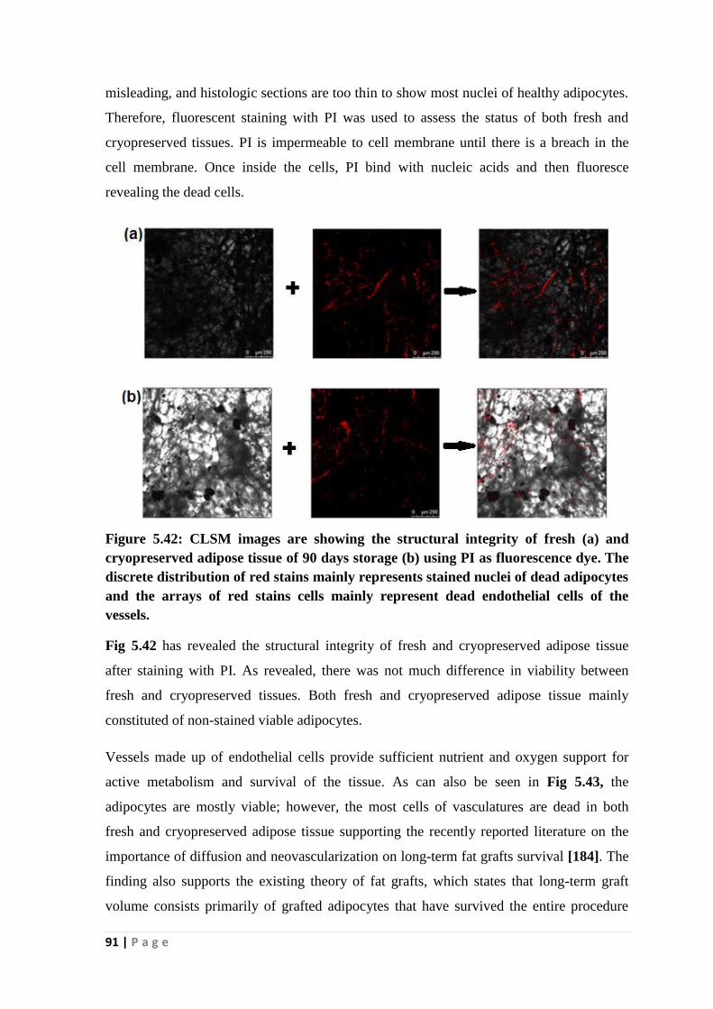
91 | P a g e
misleading, and histologic sections are too thin to show most nuclei of healthy adipocytes.
Therefore, fluorescent staining with PI was used to assess the status of both fresh and
cryopreserved tissues. PI is impermeable to cell membrane until there is a breach in the
cell membrane. Once inside the cells, PI bind with nucleic acids and then fluoresce
revealing the dead cells.
Figure 5.42: CLSM images are showing the structural integrity of fresh (a) and
cryopreserved adipose tissue of 90 days storage (b) using PI as fluorescence dye. The
discrete distribution of red stains mainly represents stained nuclei of dead adipocytes
and the arrays of red stains cells mainly represent dead endothelial cells of the
vessels.
Fig 5.42 has revealed the structural integrity of fresh and cryopreserved adipose tissue
after staining with PI. As revealed, there was not much difference in viability between
fresh and cryopreserved tissues. Both fresh and cryopreserved adipose tissue mainly
constituted of non-stained viable adipocytes.
Vessels made up of endothelial cells provide sufficient nutrient and oxygen support for
active metabolism and survival of the tissue. As can also be seen in Fig 5.43, the
adipocytes are mostly viable; however, the most cells of vasculatures are dead in both
fresh and cryopreserved adipose tissue supporting the recently reported literature on the
importance of diffusion and neovascularization on long-term fat grafts survival [184]. The
finding also supports the existing theory of fat grafts, which states that long-term graft
volume consists primarily of grafted adipocytes that have survived the entire procedure

92 | P a g e
and grafted fats initially lack vascular support and the oxygen; nutrient supply to grafts
occurs primarily through diffusion and neovascularization from surrounding tissue [185].
Figure 5.43: CLSM of fresh (a) and cryopreserved (90 days) adipose tissue (b) using
PI indicating dead endothelial cells.
Assessment of structural integrity of adipocytes using the developed freezing solution
Curcumin is a well-known fluorescence molecule with an excitation wavelength in the
range of 420-480nm and emission band of 490-520nm [186-189]. As the developed
freezing solution contains curcumin, the inherent fluorescence property of the freezing
solution was used to assess the status of the cryopreserved adipose tissue. Fresh adipose
after staining with curcumin acted as a control. As seen in the Fig 5.44a, curcumin was
insoluble in PBS and appeared as heterogeneously distributed fluorescent particulates over
the adipose tissue. As revealed in Fig 5.44b, the curcumin forms homogeneous solution
after incubation with 2.5% Me2SO for 30min and the depth of penetration of curcumin
increased. Furthermore, the non-permeation of homogenously distributed curcumin inside
the cells helped in developing the contrast between adipocytes. The less permeability of
curcumin inside the cell is also resulted in less bioavailability of it because of less
permeation of curcumin inside cells [190].

93 | P a g e
Figure 5.44:CLSM images of adipose tissue using curcumin in PBS (a) and curcumin
in 2.5% Me2SO. The heterogeneous distribution of changed to homogenous
distribution with more depth of penetration after solubilizing curcumin in Me2SO.
To evaluate the potential of curcumin to be able to differentiate between live and dead
cells, adipose tissue was incubated with 0.25% SDS solution in the presence of 0.1%
curcumin/2.5%% Me2SO solution for 30min. SDS, a surfactant molecule is supposed to
compromise the plasma membrane and let the dye permeate inside the cells easily as seen
in Fig 5.45a. Furthermore, because of fat solubility of curcumin, it stains the lipids of
adipocytes, differentiating the dead cells from live cells. Therefore, curcumin may be used
as live/dead differential stains for adipocytes. To evaluate the status of the cryopreserved
adipose tissue frozen with 160mM NaCl/90mM trehalose/1mg/ml curcumin, the post-
thawed tissue was incubated with 2.5% Me2SO for 30min in the presence of the freezing
solution containing curcumin. Fig 5.45b, c reveals that most of the adipocytes of
cryopreserved adipose tissue retained the viability like fresh adipose tissue.

94 | P a g e
Figure 5.45: shows the structural and functional integrity of SDs treated (a), fresh (b)
and cryopreserved (90 days) adipose tissue (c) using 0.1% curcumin/ 2.5% Me2SO.
Arrow indicate dead adipocytes
5.5.3 Survival of stem cells in cryopreserved adipose tissue
The retention of mesenchymal stem cells in the adipose tissue has several advantages. It
has been reported that supplementation of hADSCs along with adipose tissue for
reconstructive and aesthetic surgery improved the outcome of the surgery. Furthermore,
successful isolation of hADSCs from cryopreserved adipose tissue provides an alternative
cheaper source of hADSCs, supplementing the benefits of hADSCs cryobanks. Thus, an
attempt was made to isolate hADSCs from cryopreserved adipose tissue (90 days) and the
isolated hADSCs were compared with hADSCs isolated from fresh adipose tissue.
Morphology of isolated hADSCs
The morphological characteristic of the cultured hADSCs isolated from fresh and
cryopreserved adipose tissue (90 days) was assessed by observation under phase contrast
microscope.

95 | P a g e
Figure 5.46: Phase contrast microscopic images shows gradual change of round to
fibroblastic cell morphology of hADSCs isolated from fresh (a) and cryopreserved
(90 days) adipose tissue (b) observed upon culture in DMEM containing 10% FBS.
The cells reached confluency on the 12th day of culture. (Scale bar 100µm).
Fig 5.46b depicts that the initial round shape like morphology of isolated hADSCs from
cryopreserved adipose tissue was gradually changed to fibroblast-like appearance over 12
day culture period. Moreover, the cells were observed to adhere to the culture dish after 8h
of incubation and fibroblast-like morphology was noticed after 18h. The cultured cells
reached confluence after 12 days of culture. Similar morphology was also observed with
fresh hADSCs (Fig 5.46a).
Proliferation kinetics
As indicated from Fig 5.47, the hADSCs isolated from cryopreserved cells were
proliferated normally as the hADSCs isolated from fresh adipose tissue indicating the
freezing solution has no adverse influence on the proliferation of hADSCs representing its
ability to maintain cryopreserve adipose tissue. Upon seeding at a concentration of 5x10³
cells/well, the hADSCs from fresh adipose tissue proliferated to 67.33±2.08 x10³
cells/well, whereas hADSCs from cryopreserved adipose tissue proliferated to 68.33±3.05
x103 cells/well on the 9th day.

96 | P a g e
Figure 5.47: Proliferation kinetics of hADSCs isolated from fresh and cryopreserved
adipose tissue of 3months storage. Cryopreserved hADSCs were proliferated
normally as the hADSCs isolated from fresh adipose tissue indicating the freezing
solution has no adverse effect on the proliferation of hADSCs of adipose tissue and
thus able to maintain the adipose tissue viability.
Differentiation potential of hADSCs
Osteogenesis (Fig 5.48) of hADSCs was indicated by staining of deposited calcium with
alizarin red whereas staining of lipid droplet by Oil Red O indicates adipogenesis of
hADSCs. Thus, the results showed that the isolated hADSCs from cryopreserved adipose
tissue differentiated successfully to osteogenic and adipogenic lineages paving its
application in tissue engineering and regenerative medicine.
Figure 5.48: Assessment of differentiation potential of hADSCs isolated from fresh
(a, b) and cryopreserved (90 days storage ) adipose tissue(c, d). (a, c) Osteogenic
(Alizarin red staining) and (b, d) adipogenic lineages (Oil Red O staining). As
indicated the isolated hADSCs differentiated successfully to osteogenic and
adipogenic lineages.

97 | P a g e
Overall, though a slightly lower viability of adipose tissue frozen with the developed
160mM NaCl/90mM trehalose/1mg/ml curcumin freezing solution was observed in
comparison to fresh tissue, but the developed freezing solution was able to maintain high
viability even after 3 months of storage in LN2 as evident from Table 5.2. shows the
comparison of viability between fresh and cryopreserved adipose tissue after 3 months of
storage. Furthermore, hADSCs were successfully isolated from the cryopreserved adipose
tissue (90 days) frozen with the developed freezing solution. The isolated hADSCs
showed adherancy with plastics, regular morphology and proliferation and also retained
the differential ability to osteogenic and adipogenic lineages. Thus, the developed freezing
solution has no adverse influence on the mesenchymal stem cells present in the adipose
tissue. Therefore, adipose tissue frozen with 160mM NaCl/90mM trehalose/1mg/ml
curcumin freezing solution is not only suitable for reconstructive and aesthetic surgery but
also a good source of hADSCs when need arises.
Table 5.2 Comparison of viability between fresh and cryopreserved (3months)
adipose tissue
Tests Fresh AT Cryopreserved AT
Oil ratio 0 0.027±0.002
XTT (%) 100% 88 ±3.6
G3PDH assay (U/ml) 1.09±0.05 1.80±0.07
TBARS assay (nM/g wet wt.) 142.47±11.54 233.62±15.13

98 | P a g e
Chapter 6
Cryopreservation of Adipose tissue-
derived stem cells

99 | P a g e
6.1Screening of potential cryoprotectants and carrier media towards the formulation
of freezing solution for cryopreservation of adipose tissue-derived stem cells
Mesenchymal stem cells (MSCs) are potential for tissue regeneration because they are
multipotent and differentiate into different cell types such as osteoblasts, adipocytes,
chondrocytes, myoblasts and the like [191]. Adipose tissue is an attractive source of MSCs
because of its easy availability and accessibility, thereby offers its application in
producing therapeutic and tissue engineering products [192]. In this context, an effective
cryopreservation strategy for preservation of hADSCs with long shelve-life is important
for their clinical application.
Cryopreservation using conventional freezing solution consisting of Me2SO and FBS has
several detrimental effects including, the acute and chronic toxicity of patients and
genotoxicity of preserved cells. Furthermore, the most commonly used DMEM carrier
media having multicomponent constituent creates more complex and unpredictable cell
environment at sub-zero temperature because of ice crystallization. Hence, development of
a Me2SO and serum-free freezing solution in a suitable carrier media for preserving
hADSCs is inevitable.
Therefore, efforts have been made in this phase of the dissertation to investigate the
potentiality of various hydrocolloids and organic osmolytes as natural CPAs and
phosphate buffered saline (PBS) and its individual components as carrier media to
formulate a freezing solution for cryopreservation of hADSCs. The performance of the
formulated freezing solution was evaluated by a battery of routine as well as advanced
tests such as immunophenotype (flow cytometric analysis), cell viability and cell
proliferation (Trypan blue; MTT assay), cytoskeletal analysis (CLSM study) and
differential potential (alizarin red; Oil Red O assays). The experimental results obtained in
the above research are discussed in this chapter.
6.1.1 Morphological and Immunophenotypic characterization of hADSCs
The morphology of the isolated and cultured hADSCs from fresh adipose tissue was
assessed microscopically (Fig 6.46a). The initial round cells gradually changed to
fibroblast-like appearance. The cultured cells have expressed 99% CD90, 89% CD73,
98% CD105 as positive and 1.2% CD34,2% CD45, and 0.5% HLA-DR as negative
surface markers indicating the cells are mesenchymal stem cells type (Fig 6.1).

100 | P a g e
Figure 6.1: Flowcytometric analysis showing the expression of positive CD90 (99%),
CD73 (89%), and CD105 (98%) markers and negative HLA-DR (0.5%), CD34
(1.2%) and CD45 (2%) markers representing the cells are of mesenchymal stem cells
in characteristics.
6.1.2 Evaluation of hydrocolloids
Hydrocolloids are polysaccharides that prevent ice crystallization, thereby act as
extracellular cryoprotectants. The importance of hydrocolloids as cryoprotectants (CPA)
was reported in the literature [193, 194]. Polyvinylpyrrolidone (PVP) and methylcellulose
(MC) hydrocolloids have shown promise in cryopreservation of hADSCs [92, 93].
Furthermore, the hydrocolloids namely gums, CMC, MC and inulin were selected based
on their use as food additives and various medicinal products without any cytotoxicity
[195, 196].

101 | P a g e
Figure 6.2: Effect of types of hydrocolloids and their concentrations on the viability
of cryopreserved hADSCs assessed by Trypan blue assay. Among the hydrocolloids,
has performed best achieving the maximum viability of 55.7 ± 3.2%. The viability
trend observed as 10% PVP > 0.3% tragacanth gum (43 ± 4.6%) > 1% (w/v)
methylcellulose (37% ± 2.1%) > others
Trypan blue assay results (Fig 6.2) shows that 10% (w/v) PVP/ PBS is the most efficient
cryoprotectant achieving maximum viability (55.7 ± 3.2 %) followed by 0.3% (w/v)
tragacanth gum (43 ± 4.6%) and 1% (w/v) methylcellulose (37% ± 2.1%). The viability
obtained with 10% PVP is 12% and 18% higher than tragacanth gum (TG) and
methylcellulose (MC) respectively. Among natural hydrocolloids, tragacanth gum showed
higher viability than other polysaccharides, which may be due to its molecular weight and
neutral characteristics. The viability obtained with 10%PVP/PBS was also higher than
10%Me2SO/PBS (control) and viability achieved with 10%Me2SO/PBS supplemented
with 20%(v/v) FBS was comparable. Inulin, pectin and carrageenan showed the lowest
viability. The viability trend observed by MTT assay was similar to Trypan blue assay

102 | P a g e
(Fig 6.3). The results confirm PVP as the superior cryoprotectant to the other
hydrocolloids used in this study and the 10% Me2SO as the conventional CPA for
hADSCs.
Figure 6.3: Effect of selected hydrocolloids on the viability of cryopreserved hADSCs
assessed by MTT assay. The viability data obtained by MTT assay followed similar
trends as Trypan blue assay. The results confirm PVP as the superior cryoprotectant
to the other hydrocolloids used in this study as well as 10% Me2SO as the
conventional CPA for hADSCs preservation.
6.1.3 Evaluation of PVP in combination with PBS components as carrier media
The most commonly used PBS as carrier media consists of NaCl, Na2HPO4, KCl and
KH2PO4. It was hypothesized that these components may have a specific role in
cryopreservation of stem cells, which has not been explored. Hence, these compounds
were evaluated individually at a buffered isotonic concentration in combination with
10%PVP. As indicated in Fig 6.4a, 10%PVP/ 0.9% NaCl shows the highest viability
(70.3 ± 1.5%) with the lowest viability shown by 10%PVP/ Na2HPO4 (34.3 ±3.1%). The
viability obtained with 10% PVP in isotonic NaCl is 15% more than the viability obtained
using other carrier media. The viability (60.7 ± 2.1%) of hADSCs with 10%PVP/KCl is

103 | P a g e
comparable to the viability (55.7 ± 3.2%) obtained using 10%/PVP/PBS. The results also
revealed that phosphate containing ionic compounds offer lower viability than chloride-
containing compounds. The viability data measured by MTT assay (Fig 6.4b) followed
the similar trend as viability assessed by Trypan blue method thereby confirming
10%PVP/ 0.9% NaCl as a superior freezing solution.
Interestingly, the viability observed with NaCl containing freezing solution in our
experiment is in good agreement with viability achieved with DMEM containing freezing
solution. Thus, the results prove that the effectiveness of DMEM and normal saline on
cryoprocess is comparable, and normal saline may replace DMEM due to its lower price
and easy availability. Furthermore, chloride based ionic compounds performed better than
phosphate based compounds indicating the freezing solution containing extracellular
anions and cations provide better viability compared to the respective intracellular
counterpart. The results may be contrary to many other reports stating that the intracellular
type freezing medium offers better viability [5]. The concept of intracellular preservation
media is mainly for the hypothermic preservation of organs and tissues, and the same
concept may not be appropriate for the cryopreservation of mesenchymal stem cells.
Figure 6.4: Effect of PBS and its individual ionic components as carrier media on the
viability of cryopreserved hADSCs in 10% PVP assessed by Trypan blue assay (a)
and MTT assay (b). 0.9% NaCl is found to be the best among the carrier media. The
results also revealed that phosphate containing ionic compounds performed better
than chloride-containing compounds thus signifies that the freezing solution
containing extracellular anions and cations provide better viability compared to the
respective intracellular counterpart.

104 | P a g e
6.1.4 Improvement of the developed freezing solution by the addition of organic
osmolytes
Natural osmolytes are small organic molecules that maintain cell volume and fluid balance
of cells under stress and act as intracellular cryoprotectants. Organic osmolytes are non-
cytotoxic and used in skin ointments, nasal sprays and eye drop. Although organic
osmolytes namely ectoin and hydroxyectoin as intracellular CPAs have shown promise in
cryopreservation of human endothelial cells and murine embryos, have not so far been
explored for cryopreservation of hADSCs.
The effect of natural organic osmolytes in combination with the best freezing solution
(10%PVP /0.9%NaCl was investigated with the aim of achieving improved viability of
cryopreserved hADSCs.
Fig 6.5a shows that the supplement of ectoin and hydroxyectoin increased the viability of
cryopreserved hADSCs although a slightly higher viability was achieved with ectoin. The
viability was also increased with increase in the concentration of osmolytes in freezing
solution and the maximum cell viability of 81.7 ± 2.1 %) was obtained using ectoin at
60mM concentration. Thus, an increase of 11% viability of 10%PVP/0.9%NaCl was
obtained with the addition of osmolytes. The cell recovery assessed by MTT also
followed the similar trends. (Fig 6.5b). Thus, it is demonstrated that
10%PVP/0.9%NaCl/60mM ectoin was established as the most efficient freezing solution
that provided the highest cell viability than other freezing solutions.
Usually, the supplementation of intracellular CPA with extracellular CPA complements
each other to increase the viability of cryopreserved cells. Therefore, PVP, an extracellular
CPA was supplemented with intracellular CPA such as ectoin and hydroxyectoin. Ectoin
was found to be more efficient than hydroxyectoin, as the superior cryopreservation
outcome was obtained using ectoin. Furthermore, ectoin was also reported to have the
potential to replace Me2SO as a cryoprotectant in a serum-free cryomedium for MSCs
[139]. Thus, the viability obtained with developed freezing solution
10%PVP/0.9%NaCl/60mM ectoin is also comparable to the conventional
10%Me2SO/DMEM supplemented with FBS (80%).

105 | P a g e
Figure 6.5: Effect of organic osmolytes on the viability of cryopreserved hADSCs.
The Trypan blue assay (a) and MTT assay (b) results revealed that
10%PVP/0.9%NaCl freezing supplemented with organic osmolytes improve cell
viability. Furthermo0re, ectoin is found to be more efficient in maintaining cell
viability than hydroxyectoin. Overall, 10%PVP/0.9%NaCl/60mM ectoin was
established as the most efficient freezing solution that provided the highest cell
viability than other freezing solutions.
6.1.5 Cytoskeletal analysis
F-actin, one of the major components of the cytoskeletal system has a significant role in
maintaining cellular integrity and function. At extremely low temperature, the ultra-
structure of F-actin becomes distorted that causes cell damage, which was assessed by
cytoskeletal analysis [197]. Fig 6.6a (Priyanka Goyal M.Tech thesis, 2015) and 6.6b
showed the normal cytoskeletal distribution pattern of F-actin, indicating hADSCs
cryopreserved in 10%PVP/0.9%NaCl/60mM ectoin retain their normal cell morphology
like fresh hADSCs proving the efficiency of the formulated freezing solution towards their
maintenance.

106 | P a g e
Figure 6.6: CLSM images of frozen hADSCs in 10%PVP/0.9%NaCl/60mM ectoin
freezing solution. Nuclear stain (blue) used was DAPI and the cytoskeletal stain was
TRITC-Phalloidin. The distribution of F-actin is predominantly beneath the cell
membrane, indicating its normal distribution in cryopreserved hADSCs.(Scale bar
25µm)
6.1.6 Differentiation Potential
Freezing solutions have a significant influence on the differentiation ability of
cryopreserved cells. The impact of the formulated 10%PVP/0.9%NaCl/60mM ectoin on
the osteogenic and adipogenic differentiation ability of frozen hADSCs was investigated.
When the post-thaw hADSCs were exposed to the adipogenic medium, the cell
morphology underwent a transition from elongated fibroblastic to a round or polygonal
structure. After 14 days of induction, a consistent cell vacuolation was evident in the
induced cells. On 0.36% Oil Red O staining, the differentiated cells showed the deposition
of red lipid droplets throughout the cytoplasm (Fig 6.7b) that indicates the adipogenic
induction of cryopreserved hADSCs.
The osteogenic differentiation potential of cryopreserved hADSCs was evident from the
deposition of calcium as an osteogenic differentiation marker during 21 days of culture
(Fig 6.7a). Thus, the freezing solution can maintain the osteogenic and adipogenic
differentiation potential of the cryopreserved hADSCs

107 | P a g e
Figure 6.7: Osteogenic and adipogenic differentiation potentiality of hADSCs
cryopreserved in 10%PVP/0.9%NaCl/60mM ectoin freezing solution (a) and (b).
Osteogenic potential of hADSCs is represented by the deposition of red calcium on 21
days cell culture (a) with Alizarin red assay. Oil Red O staining shows the deposition
of red lipid droplets throughout the cytoplasm on 14 days (b) indicating the
adipogenesis. (Scale bar 200µm).
Overall, Me2SO-free, serum free and efficient freezing solution 10%PVP/ 0.9%NaCl/
60mM ectoin was formulated, which will be used in subsequent sections for further
improvement in its efficacy in cryopreservation of hADSCs.

108 | P a g e
6.2 Optimization of freezing solution composition to improve cryopreservation
outcome of adipose tissue-derived mesenchymal stem cells
In the previous work, we have screened and evaluated cryoprotectant efficacy of a number
of hydrocolloids and organic osmolytes for preserving hADSCs. The study has shown
polyvinylpyrrolidone (PVP) and ectoin as the potential CPA achieving superior hADSCs
viability upon cryopreservation than other osmolytes as well as conventional Me2SO.
Besides cryoprotectants, the normal saline solution was found to be an effective carrier
media that can replace the complex and much expensive DMEM media used traditionally
in cryopreserved hADSCs. Based on this study, a novel freezing solution was formulated
from the combination of PVP as extracellular CPA, ectoin as intracellular CPA, and NaCl
as the carrier media. The freezing solution (0.9% NaCl/10% PVP/60mM ectoin) was
found to be effective in terms of cell viability, and differentiation ability of cryopreserved
hADSCs. Therefore, an attempt has been given in the present study to improve further the
cryopreservation efficacy of the above-formulated freezing solution by optimizing the
concentration of its individual components following Taguchi orthogonal design method.
Taguchi’s optimization method is an attractable and widely used technique because of its
several advantages; it minimizes trials for generating useful information regarding the
response of interest, reduces the experimental time and robustness of the process [198,
199]. In addition, catalase, a natural antioxidant was incorporated in the formulation of
freezing solution because catalase is reported to mitigate free radicals mediated cell
damage during freezing, thereby, an improved viability of cryopreserved cells on long-
term storage is expected [200].
6.2.1 Formulation of Freezing solution
For the development of an optimal freezing solution, Taguchi’s orthogonal design method
was applied. To this end, four factors namely NaCl, PVP, ectoin, catalase as freezing
media components and three levels of their different concentrations were selected (Table
6.1) to prepare the freezing solutions with varying composition as shown in Table 6.2.

109 | P a g e
Table 6.1: Four control factors and three levels of concentration
NaCl
(Carrier media)
PVP (Extracellular
CPAa)
Ectoin (Intracellular
CPAa)
Catalase
(Antioxidant)
80mM 8% 30mM 50µg/ml
120mM 10% 60mM 100µg/ml
160mM 12% 90Mm 150 µg/ml
a) CPA represents cryoprotectant
Table 6.2: Formulation of freezing solutions based on Taguchi L9 (3(4)) array
Cryopreservation
Solution
NaCl (mM) PVP (%) Ectoin(mM) Catalase (µg/ml)
Solution-1 80 8 30 50
Solution-2 80 10 60 100
Solution-3 80 12 90 150
Solution-4 120 8 60 150
Solution-5 120 10 90 50
Solution-6 120 12 30 100
Solution-7 160 8 90 100
Solution-8 160 10 30 150
Solution-9 160 12 60 50
6.2.2 hADSCs viability assessment by Trypan blue dye exclusion assay
Trypan blue permeates the compromised cell membrane of dead cells and stains them
blue, whereas the intact membrane of live cells excludes the Trypan blue. This property of
the dye distinguishes the stained dead cells from live cells easily under an optical
microscope. The viability of the post-thawed hADSCs evaluated by the Trypan blue dye
assay was also compared with the viability obtained with 10% Me2SO used as a control.
Experimental results (Table 6.3) revealed that the freezing solution consists of 160mM
NaCl/10% PVP/ 30mM ectoin/150 µg/ml catalase (solution 8) provides the highest
viability followed by the freezing solution comprising of 120mM NaCl/10% PVP/90mM
ectoin/50µg/ml catalase (solution 5). The corresponding viability values are measured to
be 81% and 80% respectively. In addition to these solutions, solution 2 (80mM
NaCl/10% PVP/60mM ectoin/100 µg/ml catalase) and solution 7 (160mM NaCl/8%

110 | P a g e
PVP/90mM ectoin/100 µg/ml catalase) also showed good viability of 78%. The viability
of all other freezing solutions was less than 70%. Interestingly, no statistically significant
difference was observed in the viability of solution 8 and solution 5. When compared with
viability results obtained with control (10% Me2SO in PBS), all the freezing solutions
except solution 1(80mM NaCl/8% PVP/30mM ectoin/50 µg/ml catalase), showed
significantly higher viability. An increase of 30% viability was obtained with solution 8 in
comparison to control. The freezing solution 8 has also shown superior cryopreservation
outcome than 0.9% NaCl/10% PVP/60mM ectoin (81%) representing the significant role
of catalase as antioxidant.
Table 6.3: Trypan blue dye exclusion assay of post-thawed hADSCs
Sample Repetition-1 Repetition-2 Repetition-3 Mean
Viability
SDa) S/N
ratiob)
Solution-1 56 55 54 55.00 1.00 34.80
Solution-2 78 79 77 78.00 1.00 37.84
Solution-3 71 65 69 68.33 3.05 36.67
Solution-4 67 64 63 64.66 2.08 36.20
Solution-5 82 80 80 80.66 1.15 38.13
Solution-6 69 67 68 68.00 1.00 36.64
Solution-7 78 76 79 77.66 1.52 37.80
Solution-8 83 80 81 81.33 1.52 38.20
Solution-9 67 68 65 66.66 1.52 36.47
10% Me2SO 52 51 48 50.33 2.08 --
Optimised
Solutionc)
86 83 85 84.66 1.52 --
a)SD denotes Standard Deviation
b) S/N ratio represents Signal to Noise ratio
c)Optimized solution was comprised of 160mM NaCl/10% PVP/90mM ectoin/100µg/ml
catalase
6.2.3 Taguchi statistical analysis
The Trypan blue assay results were used to calculate the signal-to-noise ratio (S/N). S/N
ratio, expressed in decibels, is an important performance indicator of Taguchi statistical
analysis. The S/N ratio was calculated based on Taguchi’s –“larger-the-better” approach

111 | P a g e
that means larger S/N value indicate superior viability results [201]. The results indicate
that S/N ratio follows similar trends as that of the Trypan blue viability assay results
(Table 6.3) obtained with cryopreserved hADSCs. Moreover, the main effects plot (data
mean) for S/N ratios revealed the influence of individual column over the Taguchi results
(Fig 6.8). The results indicate that there is a significant variation in viability with different
concentrations of individual components in freezing solution. Overall, the trend suggests
that the viability is increased with increase in the concentrations of NaCl and ectoin.
Whereas among the concentrations tested, 10% PVP and 100 µg/ml catalase showed the
maximum viability of cryopreserved hADSCs.
Figure 6.8: Illustrates Main effect plot for mean of S/N ratios. S/N ratio increases
with increase in the concentration of NaCl (A) and ectoin (C). PVP (B) and catalase
(D) showed the highest S/N ratio at a concentration of 10% (w/v) and 100µg/ml
respectively.
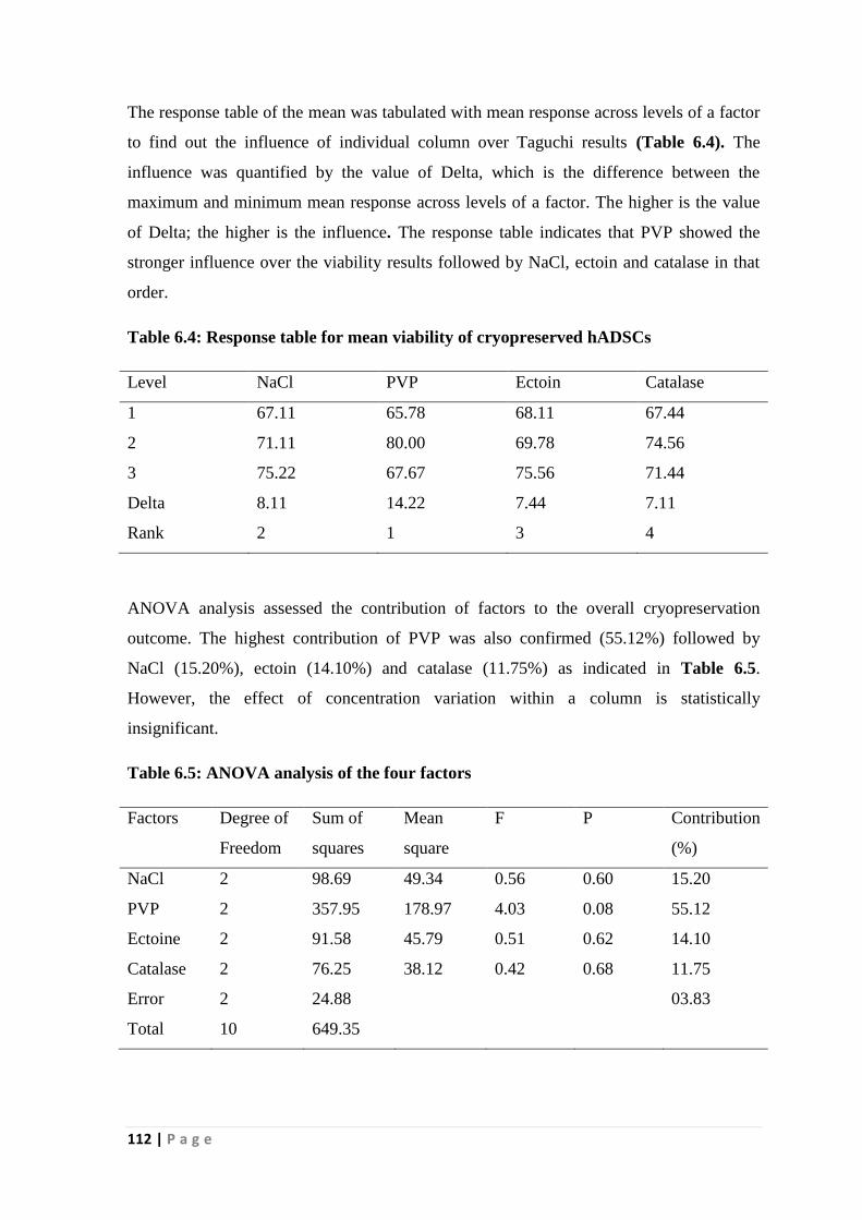
112 | P a g e
The response table of the mean was tabulated with mean response across levels of a factor
to find out the influence of individual column over Taguchi results (Table 6.4). The
influence was quantified by the value of Delta, which is the difference between the
maximum and minimum mean response across levels of a factor. The higher is the value
of Delta; the higher is the influence. The response table indicates that PVP showed the
stronger influence over the viability results followed by NaCl, ectoin and catalase in that
order.
Table 6.4: Response table for mean viability of cryopreserved hADSCs
Level NaCl PVP Ectoin Catalase
1 67.11 65.78 68.11 67.44
2 71.11 80.00 69.78 74.56
3 75.22 67.67 75.56 71.44
Delta 8.11 14.22 7.44 7.11
Rank 2 1 3 4
ANOVA analysis assessed the contribution of factors to the overall cryopreservation
outcome. The highest contribution of PVP was also confirmed (55.12%) followed by
NaCl (15.20%), ectoin (14.10%) and catalase (11.75%) as indicated in Table 6.5.
However, the effect of concentration variation within a column is statistically
insignificant.
Table 6.5: ANOVA analysis of the four factors
Factors Degree of
Freedom
Sum of
squares
Mean
square
F P Contribution
(%)
NaCl 2 98.69 49.34 0.56 0.60 15.20
PVP 2 357.95 178.97 4.03 0.08 55.12
Ectoine 2 91.58 45.79 0.51 0.62 14.10
Catalase 2 76.25 38.12 0.42 0.68 11.75
Error 2 24.88 03.83
Total 10 649.35

113 | P a g e
The result signifies that the post-thaw viability using PVP as extracellular cryoprotectant
was the highest, followed by NaCl as carrier media, ectoin as intracellular cryoprotectant
and catalase as an antioxidant. Further, a small variation in the concentration of individual
factors might have contributed towards non-significance of ANOVA results.
6.2.4 Flow cytometry
PI is excluded by viable cells because of its high molecular weight, i.e. 668.4 D. The cell
membrane of necrotic cells becomes permeable to the dye and the dye binds with nucleic
acids and becomes a fluorescent molecule inside the necrotic cells. This property is
utilized by flow cytometry to detect and quantify the non-viable cells. The flow cytometry
data is presented in Fig 6.9, which indicates a similar trend in the result as that of Trypan
blue dye exclusion assay. The results confirm that the 160mM NaCl/10% PVP/30mM
ectoine/150 µg/ml catalase is the most efficient freezing solution achieving maximum
hADSCs viability of 81 %.

114 | P a g e
Figure 6.9: Flow cytometry analysis of cryopreserved hADSCs using PI as a
fluorescent dye. 10% Me2SO freezing solution was used as control (A). B-J
represents the viability of cells using the freezing solutions formulated by Taguchi’s
orthogonal design. The optimized solution (K) comprising of 160mM NaCl/10%
PVP/90mM ectoin/100µg/ml catalase showed the maximum viability of ~85%.

115 | P a g e
6.2.5 MTT Assay
MTT assay was used to assess the metabolic activity of cells and it is one of the most
commonly used methods to determine cell viability. The experimental viability data is
shown in Fig 6.10.
Figure 6.10: MTT assay of cryopreserved hADSCs. 10% Me2SOwas used as control
freezing solution. The optimized solution comprised of 160mM NaCl/10%
PVP/90mM ectoin/100µg/ml catalase showed the higher metabolic activity of
cryopreserved cells than other freezing solutions after 24h post-thaw culture.
Overall, the trend in viability follows that of the flow cytometry results indicating solution
8 achieves higher metabolic activity than other freezing solution. Furthermore, all the
formulated freezing solutions superseded the viability obtained with control as 10%
Me2SO in PBS. However, the higher metabolic activity, shown by the cryopreserved cells
frozen in solution 1, was statistically insignificant to the metabolic activity shown by the
control group. Similarly, the metabolic activity of hADSCs preserved in solution 8 was
statistically insignificant to that of solution 5.

116 | P a g e
6.2.6 Validation of Taguchi results
The experimental confirmation test is known to be the final step in verifying the results
drawn based on Taguchi’s design approach. Thus, a confirmation experiment was
conducted by utilizing the obtained optimal concentration of the control factors. The
optimal concentration of the components was selected based on the main effect plot (Fig
6.8). The result showed that the optimal concentration of the control factors is 160mM
NaCl, 10% PVP, 90mM ectoin and 100µg/ml catalase. Therefore, for validation of the
results obtained by the Taguchi’s design, a freezing solution comprised of the
concentrations mentioned above of the individual components was prepared. The solution
comprised of 160mM NaCl/ 10%PVP/90mM ectoin/100 µg/ml catalase achieved post-
thawed cell viability of 85% assessed by Trypan blue dye exclusion assay (Table 6.3).
This result was further confirmed by flow cytometry and MTT assay (Fig 6.8K and 6.10),
which showed the superior viability of 160mM NaCl/ 10%PVP/90mM ectoin/100 µg/ml
catalase solution over solution 8. The obtained viability (85%) using the optimized
freezing cocktail is the highest among the solutions used in the experiment.
Overall, the study provides an optimal composition of a novel freezing solution that
comprised of 160mM NaCl/10% PVP/90mM ectoin/100µg/ml catalase achieving
maximum viability of 85%.

117 | P a g e
6.3 Evaluation of signaling pathway inhibitors for cryopreservation of hADSCs
Apoptosis is one of the important mechanisms of cell death during cryopreservation. Low-
temperature stress induces apoptosis, the stepwise-programmed cell death [202]. The
proteins over the cell surface receive the stress signal and send the signals downstream
inside the cells to activate caspases. The activated caspases stimulate cascades of the
proteolytic process to initiate the self-destruction process leading to DNA degradation and
cell membrane disruption. Similar to apoptosis, altered cytoskeleton of cells during
cryopreservation is another important cause of cell death [203-205]. The cytoskeleton
provides strong support to cell plasma membrane maintains the organizational distribution
of organelles and helps in executing signaling cascade properly. Thus, any alteration in the
cytoskeleton because of ice-mediated mechanical damage during cryopreservation alters
the morphology and functions of cells leading to altered rehydration and viability of post-
thawed cells.
Supplementation of molecules that prevent or inhibit the caspases activation and maintains
the stability of cytoskeleton, in freezing solution was reported to increase the viability of
post-thaw cells. Z-VAD-FMK, an irreversible inhibitor of caspases, enhanced the viability
of post-thawed human embryonic stem cells when supplemented in freezing solution and
post-thaw culture. Supplementation of Z-VAD-FMK in freezing solution also improved
the post-thaw viability of porcine hepatocytes and human amniotic fluid-derived
mesenchymal stem cells [169, 206]. Another molecule Y-27632, a Rho kinase inhibitor,
stabilizes the cytoskeleton and observed to improve the survival of post-thawed cells. Li X
et al. demonstrated that Y-27632 added to the post-thaw culture medium for 24h post-
thaw increased human embryonic stem cell survival rate [207]. Studies by Martin-Ibanez
et al. replicated this initial finding [208]. Furthermore, the effect of ROCK inhibitor Y-
27632 was shown to have similar effects on the recovery from cryopreservation of adult
stem cells and bone marrow-derived MSCs as well as human induced pluripotent stem
cells [37, 209, 210].
Given the beneficial effect of Z-VAD-FMK and Y-27632 on the recovery of post-thawed
cells and in the effort to enhance the viability of cryopreserved human adipose-derived
mesenchymal stem cells, it was hypothesized that the supplementation of the above two
molecules might enhance the viability of post-thawed hADSCs. Thus, in this phase of
work, we have supplemented 10Nm Z-VAD-FMK and 100mM Y-27632 with the most
effective 160mM NaCl/ 10%PVP/90mM ectoin/100µg/ml catalase freezing solution

118 | P a g e
obtained in the previous chapter and the efficacy of the new freezing solutions was
evaluated towards for cryopreservation of hADSCs.
6.3.1 Trypan blue dye exclusion
The immediate viability of post-thawed hADSCs was evaluated by Trypan blue dye
exclusion assay. The results (Fig 6.11) reveal that the addition of 10nM Z-VAD-FMK and
100mM Y-27632 has no impact on the viability of post-thawed hADSCs. Although, the
mean viability obtained with Y-27632 (85.33± 3.78%) was higher than Z-VAD-FMK
(84.67± 3.21%), the difference is statistically insignificant (p<0.05). Moreover, the
difference in viability obtained with the ROCK inhibitor and the general caspases inhibitor
was insignificant to the viability shown with control (84.67±2.08).
Figure 6.11: Trypan blue assay results showing the effect of signaling pathway
inhibitors supplemented with the formulated freezing solution on the cryopreserved
hADSCs viability. The results reveal that the addition of Z-VAD-FMK and Y-27632
has no significant impact on the viability of post-thawed hADSCs.
6.3.2 MTT assay
Post-thaw recovery of the cells in culture was assessed after 24h by MTT assay (Fig 6.12).
MTT assay also observed similar findings of no significant benefit with the addition of Z-
VAD-FMK and Y-27632 in the recovery of post-thawed hADSCs.

119 | P a g e
Figure 6.12: MTT assay showing the post-thaw viability of cryopreserved hADSCs
using signaling pathway inhibitor supplemented with the developed freezing solution.
As indicated, no significant benefit with the addition of Z-VAD-FMK and Y-27632 in
the recovery of post-thawed hADSCs was obtained.
The results corroborated with the findings of Lamas NJ et al., which stated that in contrast
to other stem cell types, Y-27632 supplementation is not a suitable strategy to enhance
hADSCs expansion in culture rather a continuous supplementation Y-27632 in culture led
to decrease metabolic activity and numbers of hADSCs [211]. Similar negative findings
on the use of Y-27632 were earlier reported for cord blood-derived hematopoietic
progenitor cells [212]. However, Qu CQ et al. reported that addition of ROCK inhibitor
along with glutathione increased the viability of cryopreserved porcine adipose-derived
stem cells [213]. The beneficial effect may be due to glutathione than ROCK inhibitor.

120 | P a g e
6.4 Optimization of controlled rate freezing parameters for cryopreservation of
hADSCs using the developed freezing solution
The cooling rate is one of the most important factors that controls the viability of cells.
While subjected to slow cooling rate leads to extracellular ice formation and the
segregated solute concentration effect over cell membrane become an important cause of
cryoinjury, fast cooling rate induces intracellular ice formation and ice crystals mediated
injury thereby causes cell death. Thus, a trade-off between fast and slow cooling rate is
essential to achieve an optimized cooling rate providing maximum cell viability. The
problem is further complicated by the fact that the cryoprocessing steps are
interdependent, and thus cannot be optimized independently. For example, the optimal
cooling rate is dependent on the CPA types and concentration.
In this part of research work, an attempt has been given to achieve an optimized cooling
rate for the cryopreservation of hADSCs using the developed 160mM NaCl/
10%PVP/90mM ectoin/100µg/ml catalase freezing solution.
6.4.1 Effect of cooling rate
To achieve the optimized cooling rate, the cryopreservation experiment was carried out in
different prenucleation cooling rate such as -0.5°C/min,-1°C/min,-2.5°C/min,-5°C/min
from 4°C to -120°C. The samples were kept at 4°C for 1h incubation period before being
subjected to controlled rate freezers. The one-hour incubation period was necessary for
equilibrating the freezing solution with the cells. Upon completion of controlled freezing,
the cryovials were transferred immediately into LN2 to avoid recrystallization injury to
cells. The efficiency of the optimum freezing condition on the viability was assessed as
described here.
Trypan blue dye exclusion assay
The viability and recovery of the post-thawed hADSCs were assessed by Trypan blue dye
exclusion and MTT assay after 48h. The results (Fig 6.13) show that cooling at the rate of
-1°C/min achieved the highest viability of 89.3± 3.8%. The viability is 8% higher than the
viability obtained using -0.5°C/min (81.3±3.8%) and 4% better than the sample frozen at -
80°C freezer in Mr. Frosty, an isopropanol containing commercial container for
cryostoring cells. Thirumala et al. also reported similar finding that -1°C/min provided the
maximum viability of hADSCs compared to other cooling rates.

121 | P a g e
Figure 6.13: Trypan blue assays showing the effect of cooling rate on post-thaw
viability of hADSCs cryopreserved in 160mM NaCl/ 10%PVP/90mM
ectoin/100µg/ml catalase freezing solution.
MTT assay
The metabolic activity measured by MTT assay is shown in Fig 6.14. As indicated, the
OD was increased by increasing rate from -0.5 to -1°C/min and then the OD was slowly
decreased with the higher cooling rate. Thus, -1°C/min is confirmed as the most favorable
cooling rate achieving maximum viability of frozen hADSCs.

122 | P a g e
Figure 6.14: Effect of cooling rate on post-thaw viability of hADSCs cryopreserved in
160mM NaCl/ 10%PVP/90mM ectoin/100µg/ml catalase freezing solution assessed by
MTT assay
6.4.2 Effect of seeding temperature
As described in the previous section, at low-temperature seeding causes the quick
formation of extracellular ice that minimizes the detrimental effect of phase change from
liquid to ice in the course of cooling thereby prevents ice-mediated damage. Seeding
should be performed at the temperature above spontaneous intracellular ice formation,
which is usually in the range of −7°C and −12°C. Thirumala et al. reported that for
hADSCs, freezing usually does not start below -6°C. Therefore, to evaluate the effect of
seeding temperature on the viability of hADSCs, the cells were frozen in a controlled rate
freezer at -1°C/min cooling rate from 4°C to -120°C and the seeding was induced
mechanically using the facilities of the CRF at -5°C,-7°C and -9°C. The vials were stored
in LN2 and viability, and recovery of cells was assessed after 48h of storage.
Trypan blue dye exclusion assay
The results in Fig 6.15 indicate that among the seeding temperatures under study, the
maximum cell viability of 89.4 ± 3.6% was obtained with seeding at -7°C. A comparative
hADSCs viability was also shown at -9°C. Therefore,-7C was established as the most

123 | P a g e
favorable seeding temperature. However, compared to non-seeded samples, the seeded
samples at -7°C showed no additional improvement in cell viability.
Figure 6.15: Trypan blue assay showing the effect of seeding temperature on post-
thaw viability of hADSCs cryopreserved in the formulated 160mM
NaCl/10%PVP/90mM ectoin/100µg/ml catalase freezing solution.
MTT assay
MTT assay (Fig 6.16) corroborates with the viability result obtained by Trypan blue assay
that the seeding at -7°C achieved higher viability than other seeding temperature. Similar
to Trypan blue assay, the viability results are not significantly different from each other
indicating the negligible influence of seeding in improving the viability of hADSCs
cryopreservation.

124 | P a g e
Figure 6.16: Effect of seeding temperature on post-thaw viability of hADSCs
cryopreserved in 160mM NaCl/ 10%PVP/90mM ectoin/100µg/ml catalase freezing
solution assessed by MTT assay.
Overall, it has been demonstrated that -1°C/min cooling rate is effective in improving the
viability of hADSCs cryopreserved with 160mM NaCl/ 10%PVP/90mM ectoin/100µg/ml
catalase freezing solution. Furthermore, the study on the effect of seeding temperature
indicates that the influence of seeding temperature on the post-thaw viability temperature
of hADSCs is negligible.

125 | P a g e
6.5 Effect of long-term storage on cryopreserved hADSCs using the developed
freezing solution
The effect of long-term storage on the viability of hADSCs cryopreserved in 160mM
NaCl/10% PVP/90mM ectoin/100 µg/ml catalase freezing solution. Long-term evaluation
is necessary as free radical and apoptosis-mediated cell death cause loss of viability over
time. The cells were equilibrated with the freezing solution for one hour at 4°C before
being controlled rate frozen at -1°C/min from 4°C to -120°C and plunged into LN2.The
vials were retrieved after 15 , 30 , 60 and 90 days of storage in LN2 and thawed rapidly to
assess the viability and recovery of cells.
6.5.1 Trypan blue dye exclusion assay
The results in Fig 6.17 indicate the decrease in viability over time and thus, viability
dropped from 88.6 ± 4.5% from 15 days to 85 ± 5% after 90 days of storage. The
difference in viability over time is insignificant indicating the freezing solution is able to
maintain viability for long-term.
Figure 6.17: Long-term viability study on cryopreserved hADSCs using 160mM
NaCl/10% PVP/90mM ectoin/100 µg/ml catalase freezing solution assessed by
Trypan blue assay. The viability change over 90 days storage time is insignificant
indicating the freezing solution is able to maintain hADSCs viability for long-time.

126 | P a g e
6.5.2 MTT assay
The metabolic activity in term of OD measured during the 15-90 days of storage period is
shown in Fig 6.18. No statistically significant difference in viability was noticed during
this period thus proving the efficiency of the developed freezing solution in the
maintenance of cryopreserved hADSCs.
Figure 6.18: Long-term viability study on cryopreserved hADSCs using 160mM
NaCl/10% PVP/90mM ectoin/100µg/ml catalase freezing solution assessed by MTT
assay. Similar trend in viability as assessed by Trypan blue assay confirming the
suitability of the developed freezing solution in preserving hADSCs for a long time.
6.5.3 Morphology of cryopreserved hADSCs
The regular fibroblastic morphology and adherancy to plastics are important features for
the structural and functional integrity of hADSCs. Therefore, the morphological
characteristics of cryopreserved (90 days) hADSCs were assessed by phase contrast
microscopy.
As observed in Fig 6.19, on retrieval, the cryopreserved hADSCs adhered to the surface
and the changed to fibroblastic appearance before reaching confluency on 12th day like
fresh mesenchymal stem cells proving that the developed freezing solution has no
detrimental or adverse effect on the adherancy and morphology of cryopreserved
hADSCs.

127 | P a g e
Figure 6.19: Morphological changes of cryopreserved (90 days storage) hADSCs in
160mM NaCl/10% PVP/90mM ectoin/100µg/ml catalase freezing solution as
observed under phase contrast microscope. 0-day (a), 6th day (b) and 12th day(c).
(Scale bar 100µm)
6.5.4 Cytoskeleton distribution of cryopreserved hADSCs
The hADSCs were stained with Phalloidin-Alexa fluor-488 and Hoechst to stain the actin
cytoskeleton and nucleus respectively and observed under CLSM. As can be observed in
Fig 6.20, there is the peripheral distribution of actin, which is a regular feature of normal
cells. Thus, the developed freezing solution and the cryopreservation strategy had no
adverse effect over the cytoskeleton of hADSCs and retained the regular cytoskeletal
distribution pattern like fresh cells.
Figure 6.20: Cytoskeleton distribution of cryopreserved (90 days) hADSCs in 160mM
NaCl/10% PVP/90mM ectoin/100 µg/ml catalase freezing solution. The peripheral F-
actin distribution represents regular arrangement of cytoskeleton.

128 | P a g e
6.5.5 Proliferation kinetics
The proliferation kinetics of the cryopreserved (90 days) hADSCs was compared with
non-cryopreserved hADSCs (P6) using Trypan blue assay (Fig 6.21). The cryopreserved
hADSCs has been shown to retain the normal proliferation pattern indicating
cryopreservation process, and the freezing solution has no adverse effect on the functional
integrity of cryopreserved hADSCs.
Figure 6.21: Proliferation kinetics of cryopreserved (90 days) hADSCs in 160mM
NaCl/10% PVP/90mM ectoin/100 µg/ml catalase freezing solution. The hADSCs has
shown to retain the normal proliferation pattern indicating the freezing solution has
no adverse effect on the functional integrity of cryopreserved hADSCs.
6.5.6 Differentiation ability
The differentiation of cryopreserved (90 days) hADSCs into osteogenic and adipogenic
lineages was performed to evaluate the maintenance of differentiation ability of
cryopreserved hADSCs.
The histological results of the osteogenic differentiation of hADSCs are shown in Fig
6.22a. hADSCs cryopreserved in 160mM NaCl/10% PVP/90mM ectoin/100 µg/ml
catalase freezing solution for 90 days is observed to be sufficiently differentiated into
osteoblasts as shown by the deposition of calcium on 21 days of culture. Thus, the

129 | P a g e
cryopreserved hADSCs retained the differentiation ability to osteogenic lineages thereby
proven the ability of formulated freezing solution in maintaining the differentiation
potential of hADSCs. Furthermore, as can be seen in Fig 22b, lipid droplets accumulated
inside the hADSCs on 14th day culture in adipogenic differentiation media proving the
cryopreserved hADSCs also retained the differential ability to adipogenic lineages.
Figure 6.22: Differential ability of frozen (90 days storage) hADSCs in 160mM
NaCl/10% PVP/90mM ectoin/100 µg/ml catalase freezing solution for osteogenesis (a)
and adipogenesis (b) seen under fluorescent microscope. Calcium deposition in
osteogenesis was evaluated by Alizarin red stain (a) and intracellular lipid
accumulation in adipogenesis was assessed by Oil Red O stain. (Scale bar 100µm)
Overall, among the freezing solutions, 160mM NaCl/10% PVP/90mM ectoin/100µg/ml
catalase was found to be the most effective in maintaining cryopreserved adipose tissue as
indicated in Table 6.6 providing the maximum viability of 89% by control rate freezing
at -1°C/min cooling rate followed by storage in LN2. The developed 160mM NaCl/10%
PVP/90mM ectoin/100µg/ml catalase freezing solution may be used for the
cryopreservation of hADSCs thereby paving its future clinical application.
Table 6.6 Comparison of hADSCs viability cryopreserved in various freezing
solution
Freezing solution Trypan blue
(% viability)
PBS+10% Me2SO+20% FBS 60
PBS+ 10% PVP 56
0.9% NaCl+10% PVP 70
0.9% NaCl+10% PVP+60mM ectoin 81
160mM NaCl+10% PVP+90mM ectoin+100µg/ml catalase 85
160mM NaCl+10% PVP+90mM ectoin+100µg/ml catalase:
Cooling at -1°C/min and storage in LN2 for 48h
89
160mM NaCl+10% PVP+90mM ectoin+100µg/ml catalase:
Cooling at -1°C/min and storage in LN2 for 3 months
85

130 | P a g e
Chapter 7
Summary & Conclusion

131 | P a g e
Lipofilling, which is an important surgical procedure, requires a lot of adipose tissue to fill
up the depressed region or scar of patient body. Besides lipofilling, there is a growing
demand of adipose tissue-derived stem cells for tissue engineering and stem cell therapy
applications. Therefore, in recent past, research interest has been generated towards the
harvesting of adipose tissue as well as production of stem cells, (mesenchymal stem cells
in particular) isolated from adipose tissue. In this context, development of an effective
cryopreservation strategy is of utmost importance for preserving adipose tissue and
hADSCs with long shelve-life for the advancement of these products for clinical
applications. Cryopreservation using conventional freezing solution consisting of dimethyl
sulfoxide and fetal bovine serum has several detrimental effects including, the acute and
chronic toxicity of patients and genotoxicity of preserved cells. Furthermore, the most
commonly used DMEM carrier media having multicomponent constituents creates more
complex and unpredictable cell environment at sub-zero temperature because of ice
crystallization.
Keeping the above issues in view, the present research aims to explore potential natural
cryoprotective agents and suitable carrier media for developing a Me2SO-free and serum-
free freezing solution for preserving adipose tissue and adipose tissue derived stem cells
with long storage life.
The most interesting results obtained from the above research is summarized as follows-
A. Cryopreservation of adipose tissue
I. In the first phase of the thesis work, the efficiency of various hydrocolloids and natural
organic osmolytes as extracellular and intracellular CPAs as well as individual PBS
components such as NaCl, Na2HPO4, KCl and KH2PO4 as possible carrier media were
evaluated by performing cryopreservation experiments with adipose tissue. Based on the
viability results measured by oil ratio as a preliminary screening criteria followed by XTT,
G3PDH and TBARS analysis, trehalose, ectoin and NaCl were selected as the most
efficient CPAs and carrier media respectively.
II. The selected extracellular and intracellular CPAs in NaCl as carrier media were
evaluated individually and in combination to formulate an freezing solution cocktail. To
this end, different batches of freezing solutions namely 160mM NaCl/90mM trehalose,
160mM NaCl/150mM ectoin and 160mM NaCl/90mM trehalose/150mM ectoin were

132 | P a g e
prepared and the conventional 10% Me2SO was used as control. Among these, freezing
solution consisting of 160mM NaCl/90mM trehalose was found to be the most efficient
achieving adipose tissue viability of 85%. The viability was comparable to the viability
shown by 160mM NaCl/10% Me2SO as a control. Interestingly, ectoin in freezing
solutions did not show any significant impact in cryopreserving adipose tissue, whereas a
substantial tissue viability (84%) was obtained using 160mM NaCl only.
III. The oxidative stress mediated damage is a critical phenomenon in cryopreservation,
which is expected to be higher in the case of adipose tissue than other body tissues.
Therefore, it was hypothesized that supplementation of antioxidants in freezing solution
might improve the cryopreservation outcome of adipose tissue. Therefore,
cryopreservation experiments were performed with adipose tissue using the formulated
160mM NaCl and 90mM trehalose/160mM NaCl freezing solutions supplemented with
different antioxidants such as curcumin, carnitine, lipoic acid,Vit E, gallic acid, ellagic
acid with varying concentration level. Among these, 1mg/ml curcumin and 3mg/ml
carnitine were found to be the potential antioxidants improving the crypreservation
outcome of both the freezing solutions and the measured viability was higher than the
viability obtained with the conventional 10% Me2SO. Overall, freezing solutions
supplemented with curcumin has shown higher viability than carnitine and 160mM
NaCl/90mM trehalose/1mg/ml curcumin achieved the maximum viability of 90%. Thus,
the study provides a Me2SO free superior freezing solution for adipose tissue
cryopreservation.
IV. An effort has been also given for further improvement of the cryopreservation
efficiency by performing the cryopreservation of adipose tissue in a control rate freezer
which resulted in 93% maximum post-thaw tissue viability at -1°C/min as an optimum
cooling rate.
V. The maintenance of tissue viability for a long time is the most vital aspect of freezing
solution to be used as cryopreservation strategy. Therefore, the efficiency of the
formulated freezing solution towards maintaining tissue viability was evaluated during 90
days of storage of cryopreserved adipose tissue in liquid nitrogen. The result revealed that
a gradual decrease in tissue viability occurred over time. However, the viability after 90
days of storage was not significantly different from viability after 7 days as evident by oil

133 | P a g e
ratio, XTT, G3PDH and TBARS assays representing the ability of the freezing solution in
maintaining long-shelve life. The structural and functional integrity of cryopreserved
adipose tissue were maintained by the freezing solution which are evident from the
morphological characteristics (Laser Scanning Confocal Microscopic image analysis) as
well as ability of frozen adipose tissue to provide hADSCs with retained metabolic
activity (MTT assay), proliferation, and differentiation (Alizarin Red assay and Oil Red O
staining) potential .
Thus, it has been concluded that the formulated freezing solution comprising of 160mM
NaCl/90mM trehalose/1mg/ml curcumin has the ability in maintaining the viability for a
long time and cryopreserved adipose tissue provides hADSCs with desired metabolic
acvitivy, proliferation and differentiation potential.
B. Cryopreservation of hADSCs
I. Besides adipose tissue cryopreservation, an effort has been given to isolate hADSCs
from adipose tissue and develop a cryopreservation strategy for their preservation by
formulating an effective freezing solution. Similar to adipose tissue, the efficiency of
various hydrocolloids and natural organic osmolytes as extracellular and intracellular
CPAs were evaluated for their efficacy in maintaining viability of adipose tissue by
performing cryopreservation experiments. Based on the viability results measured by
Trypan blue and MTT assays, among the hydrocolloids, 10% PVP in PBS as carrier media
was found to be the most effective freezing solution achieving maximum cell viability of
56% followed by tragacanth gum (43%). The viability obtained with 10% PVP/PBS was
higher than 10%Me2SO/PBS solution (control).
II. PBS as carrier media consists of NaCl, Na2HPO4, KCl and KH2PO4. The specific role
of these components as carrier media in cryopreservation of stem cells has not been
explored so far and hence these components were evaluated by supplemented with 10%
PVP. Among the various compositions, 10%PVP/0.9%NaCl showed the maximum cell
viability of 70% measured by Trypan Blus assay. The chloride based ionic compounds
was found to perform better than phosphate based compounds indicating the freezing
solution containing extracellular anions and cations provide better viability compared to
the respective intracellular counterpart. These findings were also supported by MTT assay.
134 | P a g e
III. Efforts have been given for further improvement of the efficiency of the
10%PVP/0.9%NaCl by the addition of ectoin and hydroxyectoin as the potential organic
osmolytes. The experimental study has revealed that the addition of osmolytes in freezing
solution increased the viability of cryopreserved hADSCs and the highest viability of 82%
was achieved by developing 10%PVP/0.9%NaCl/60mM ectoin freezing solution.
The cytoskeletal analysis from confocal microscopic images shows the normal
cytoskeletal distribution pattern of F-actin, that indicate that hADSCs cryopreserved in
10%PVP/0.9%NaCl/60mM ectoin was able to retain normal cell morphology like fresh
hADSCs, proving the suitability of the formulated freezing solution.
The impact of the formulated 10%PVP/0.9%NaCl/60mM ectoin on the differentiation
ability of frozen hADSCs was investigated. The study on the differentiation potential of
cryopreserved hADSCs by incubating in osteogenic and adipogenic media for a period of
21 and 14 days respectively has confirmed that the developed freezing solution is able to
maintain the osteogenic and adipogenic differentiation ability of the cryopreserved
hADSCs as assessed by Alizarin Red assay and Oil Red O staining.
IV. The cryopreservation efficacy of the formulated 10%PVP/0.9%NaCl/60mM ectoin
freezing solution was further optimized by adopting Taguchi orthogonal design
methodology. Catalase, a natural antioxidant was also incorporated into the formulation of
freezing solutions with different concentrations to mitigate free radicals mediated cell
damage during freezing. The optimal composition of the freezing solution was obtained as
160mM NaCl/10% PVP/90mM ectoin/100µg/ml catalase, providing the maximum post-
thaw viability of 85%. Furthermore, the addition of caspase and Rho-kinase inhibitors did
not show any influence on the post- thaw viability of frozen hADSCs. Thus, 160mM
NaCl/ 10% PVP/ 90mM ectoin/100µg/ml catalase was obtained as the most favorable
freezing solution.
V. To achieve better cryopreservation outcome, the cryopreservation experiment was
carried out in a controlled rate freezer at different cooling rate such as -0.5C/min,-
1°C/min,-2.5°C/min,-5°C/min from 4°C to -120°C. The measured viability was enhanced
to 89% in control rate freezing with the optimized cooling rate of -1°C/min.

135 | P a g e
The effect of long-term storage on the viability of hADSCs cryopreserved in 160mM
NaCl/10%PVP/90mM ectoin/100µg/ml catalase freezing solution indicated a slow
reduction of viability over time and the viability was dropped from 89% in 15 days to 85%
after 90 days of storage. The decrease in viability is not significant representing the ability
of the developed freezing solution in maintaining viability of adipose tissue for a long
time.
The structural and functional integrity of cryopreserved (90 days storage) hADSCs
assessed by phase contrast and confocal microscopy, indicate that the cryopreserved
hADSCs maintained the fibroblastic morphology and attained 80-85% confluence on
12thday of culture. The cytoskeletal analysis has revealed the peripheral distribution of F-
actin thereby the maintenance of the structure of hADSCs. The study on the proliferation
and differentiation to osteogenic and adipogenic lineages of 90 days storage cryopreserved
hADSCs were performed and compared with fresh hADSCs. The cryopreserved hADSCs
retained the normal proliferation and differentiation ability similar to fresh hADSCs
confirming that the freezing solution has no adverse effect on the functional integrity of
cryopreserved hADSCs.
All the above taken together, the developed Me2SO free, serum free and non-toxic 160mM
NaCl/10% PVP/90mM ectoin/100µg/ml catalase has been proven to be a potential
freezing solution in maintenaning viability, proliferation and differentiation capability of
hADSCs for long e time.
Overall, a Me2SO free, serum free and non-toxic freezing solution was developed from
natural cryoprotective agents, NaCl as carrier media and curcumin as antioxidant. The
freezing solution comprising of 160mM NaCl/90mM trehalose/1mg/ml curcumin is
effective for long-term preservation of adipose tissue with maintained tissue viability
(89%) and provide hADSCs with retained metabolic activity, proliferation, and
differentiation ability. Similarly, a novel freezing solution that is devoid of the harmful
Me2SO and serum was developed for the cryopreservation of hADSCs. The freezing
solution comprising of 160mM NaCl/10% PVP/90mM ectoin/100µg/ml catalase is
effective for cryopreservation of hADSCs with long-shelve life. It is, thus, concluded that
these developed freezing solutions might provide effective cryopreservation strategies for
the supply of these products for clinical applications in future.

136 | P a g e
Suggested further study
The present research work offers effective freezing solutions for preserving adipose tissue
and hADSCs. However, keeping in view of the limitation of the present work, the
following future work has been suggested-
I. Evaluation of the efficiency of the developed freezing solutions for longer period
of storage time than 3 months period of study undertaken in the present work.
II. Detail study on the parametric sensitivity of the control rate freezing process
III. In-vivo study must be performed using animal model, keeping in view of their
cilinical application

137 | P a g e
Bibliography

138 | P a g e
1. Andre, P., N. Lowe, A. Parc, T. Clerici, and U. Zimmermann, Adverse reactions to
dermal fillers: a review of European experiences. Journal of Cosmetic and Laser
Therapy, 2005. 7(3-4): p. 171-176.
2. Gimble, J.M., A.J. Katz, and B.A. Bunnell, Adipose-derived stem cells for
regenerative medicine. Circulation research, 2007. 100(9): p. 1249-1260.
3. Pegg, D.E., Principles of cryopreservation. Cryopreservation and freeze-drying
protocols, 2007: p. 39-57.
4. Fahy, G.M., The relevance of cryoprotectant “toxicity” to cryobiology.
Cryobiology, 1986. 23(1): p. 1-13.
5. Baust, J.G. and J.M. Baust, Advances in biopreservation. 2006: CRC Press.
6. Van Buskirk, R.G., J.M. Baust, K.K. Snyder, A.J. Mathew, and J.G. Baust,
Hypothermic storage and cryopreservation. BioProcess Int, 2004. 2(10).
7. Cossins, A., Temperature biology of animals. 2012: Springer Science & Business
Media.
8. Foote, R.H., The history of artificial insemination: Selected notes and notables.
Journal of Animal Science, 2002. 80(E-Suppl_2): p. 1-10.
9. Clarke, G.N., ART & history, 1678–1978. Human
Reproduction,2006.21(7):p.1645-1650.
10. Smith, A.U. and C. Polge, Survival of spermatozoa at low temperatures. Nature,
1950. 166: p. 668-669.
11. Lovelock, J., Haemolysis by thermal shock. British journal of haematology, 1955.
1(1): p. 117-129.
12. Mazur, P., Kinetics of water loss from cells at subzero temperatures and the
likelihood of intracellular freezing. The Journal of general physiology, 1963.
47(2): p. 347-369.
13. Tervit, H., D. Whittingham, and L. Rowson, Successful culture in vitro of sheep
and cattle ova. Journal of reproduction and fertility, 1972. 30(3): p. 493-497.
14. Rall, W.F. and G.M. Fahy, Ice-free cryopreservation of mouse embryos at− 196 C
by vitrification. 1985.
15. Pegg, D. The history and principles of cryopreservation. in Seminars in
reproductive medicine. 2002. Copyright© 2002 by Thieme Medical Publishers,
Inc., 333 Seventh Avenue, New York, NY 10001, USA. Tel.:+ 1 (212) 584-4662.
16. Carpenter, J.F. and T.N. Hansen, Antifreeze protein modulates cell survival during
cryopreservation: mediation through influence on ice crystal growth. Proceedings
of the National Academy of Sciences, 1992. 89(19): p. 8953-8957.
139 | P a g e
17. Leibo, S., J. Farrant, P. Mazur, j.M. Hanna, and L. Smith, Effects of preezing on
marrow stem cell suspensions: Interactions of cooling and warming rates in the
presence of pvp, sucrose, or glycerol. Cryobiology, 1970. 6(4): p. 315-332.
18. Meryman, H.T., Cryopreservation of living cells: principles and practice.
Transfusion, 2007. 47(5): p. 935-945.
19. Crowe, J.H., J.F. Carpenter, and L.M. Crowe, The role of vitrification in
anhydrobiosis. Annual review of physiology, 1998. 60(1): p. 73-103.
20. Eriksson, B. and H. Rodriguez-Martinez, Effect of freezing and thawing rates on
the post-thaw viability of boar spermatozoa frozen in FlatPacks and Maxi-straws.
Animal reproduction science, 2000. 63(3): p. 205-220.
21. Elkinton, J.R. and T.S. Danowski, The body fluids: Basic physiology and practical
therapeutics. 1955: Williams & Wilkins Co.
22. Schwartz, A., G.E. Lindenmayer, and J.C. Allen, The sodium-potassium adenosine
triphosphatase: pharmacological, physiological and biochemical aspects.
Pharmacological Reviews, 1975. 27(1): p. 3-134.
23. Lane, M., E.A. Lyons, and B.D. Bavister, Cryopreservation reduces the ability of
hamster 2-cell embryos to regulate intracellular pH. Human Reproduction, 2000.
15(2): p. 389-394.
24. Baumber, J., B.A. Ball, J.J. Linfor, and S.A. Meyers, Reactive oxygen species and
cryopreservation promote DNA fragmentation in equine spermatozoa. Journal of
Andrology, 2003. 24(4): p. 621-628.
25. Bilodeau, J.F., S. Chatterjee, M.A. Sirard, and C. Gagnon, Levels of antioxidant
defenses are decreased in bovine spermatozoa after a cycle of freezing and
thawing. Molecular reproduction and development, 2000. 55(3): p. 282-288.
26. Eroglu, A., T. Toth, and M. Toner, Alterations of the cytoskeleton and polyploidy
induced by cryopreservation of metaphase II mouse oocytes. Fertility and sterility,
1998. 69(5): p. 944-957.
27. Parks, J. and J. Graham, Effects of cryopreservation procedures on sperm
membranes. Theriogenology, 1992. 38(2): p. 209-222.
28. Baust, J.M., R. Van Buskirk, and J.G. Baust, Cell viability improves following
inhibition of cryopreservation-induced apoptosis. In Vitro Cellular &
Developmental Biology-Animal, 2000. 36(4): p. 262-270.
29. Galmes, A., A. Gutiérrez, A. Sampol, M. Canaro, M. Morey, J. Iglesias, N.
Matamoros, M.A. Duran, A. Novo, and M.D. Bea, Long-term hematologic
reconstitution and clinical evaluation of autologous peripheral blood stem cell
transplantation after cryopreservation of cells with 5% and 10% dimethylsulfoxide
at− 80° C in a mechanical freezer. haematologica, 2007. 92(7): p. 986-989.

140 | P a g e
30. Fountain, D., M. Ralston, N. Higgins, J. Gorlin, L. Uhl, C. Wheeler, J. Antin, W.
Churchill, and R. Benjamin, Liquid nitrogen freezers: a potential source of
microbial contamination of hematopoietic stem cell components. Transfusion,
1997. 37(6): p. 585-591.
31. Meryman, H., R. Williams, and M.S.J. Douglas, Freezing injury from “solution
effects” and its prevention by natural or artificial cryoprotection. Cryobiology,
1977. 14(3): p. 287-302.
32. Fuller, B.J., Cryoprotectants: the essential antifreezes to protect life in the frozen
state. CryoLetters, 2004. 25(6): p. 375-388.
33. Taylor, M.J. and M.J. Benton, Interaction of cooling rate, warming rate, and extent
of permeation of cryoprotectant in determining survival of isolated rat islets of
Langerhans during cryopreservation. Diabetes, 1987. 36(1): p. 59-65.
34. Yancey, P.H., Organic osmolytes as compatible, metabolic and counteracting
cytoprotectants in high osmolarity and other stresses. Journal of Experimental
Biology, 2005. 208(15): p. 2819-2830.
35. Hunt, C.J., Cryopreservation of human stem cells for clinical application: a review.
Transfusion Medicine and Hemotherapy, 2011. 38(2): p. 107-123.
36. Stroh, C., U. Cassens, A.K. Samraj, W. Sibrowski, K. Schulze-Osthoff, and M.
Los, The role of caspases in cryoinjury: caspase inhibition strongly improves the
recovery of cryopreserved hematopoietic and other cells. The FASEB Journal,
2002. 16(12): p. 1651-1653.
37. Heng, B.C., Effect of Rho-associated kinase (ROCK) inhibitor Y-27632 on the
post-thaw viability of cryopreserved human bone marrow-derived mesenchymal
stem cells. Tissue and Cell, 2009. 41(5): p. 376-380.
38. Ware, C.B., A.M. Nelson, and C.A. Blau, Controlled-rate freezing of human ES
cells. Biotechniques, 2005. 38(6): p. 879.
39. Wozniak, S.E., L.L. Gee, M.S. Wachtel, and E.E. Frezza, Adipose tissue: the new
endocrine organ? A review article. Digestive diseases and sciences, 2009. 54(9): p.
1847-1856.
40. Gir, P., S.A. Brown, G. Oni, N. Kashefi, A. Mojallal, and R.J. Rohrich, Fat
grafting: evidence-based review on autologous fat harvesting, processing,
reinjection, and storage. Plastic and reconstructive surgery, 2012. 130(1): p. 249-
258.
41. Hyakusoku, H., R. Ogawa, S. Ono, N. Ishii, and K. Hirakawa, Complications after
autologous fat injection to the breast. Plastic and reconstructive surgery, 2009.
123(1): p. 360-370.
42. Reya, T., S.J. Morrison, M.F. Clarke, and I.L. Weissman, Stem cells, cancer, and
cancer stem cells. nature, 2001. 414(6859): p. 105-111.

141 | P a g e
43. Wobus, A.M. and K.R. Boheler, Embryonic stem cells: prospects for
developmental biology and cell therapy. Physiological reviews, 2005. 85(2): p.
635-678.
44. Chamberlain, G., J. Fox, B. Ashton, and J. Middleton, Concise review:
mesenchymal stem cells: their phenotype, differentiation capacity, immunological
features, and potential for homing. Stem cells, 2007. 25(11): p. 2739-2749.
45. Kern, S., H. Eichler, J. Stoeve, H. Klüter, and K. Bieback, Comparative analysis of
mesenchymal stem cells from bone marrow, umbilical cord blood, or adipose
tissue. Stem cells, 2006. 24(5): p. 1294-1301.
46. Zuk, P.A., M. Zhu, P. Ashjian, D.A. De Ugarte, J.I. Huang, H. Mizuno, Z.C.
Alfonso, J.K. Fraser, P. Benhaim, and M.H. Hedrick, Human adipose tissue is a
source of multipotent stem cells. Molecular biology of the cell, 2002. 13(12): p.
4279-4295.
47. Lin, C.-S., G. Lin, and T.F. Lue, Allogeneic and xenogeneic transplantation of
adipose-derived stem cells in immunocompetent recipients without
immunosuppressants. Stem cells and development, 2012. 21(15): p. 2770-2778.
48. Pu, L.L., Cryopreservation of adipose tissue. Organogenesis, 2009. 5(3): p. 138-
142.
49. Leibo, S. and T.B. Pool, The principal variables of cryopreservation: solutions,
temperatures, and rate changes. Fertility and sterility, 2011. 96(2): p. 269-276.
50. Lidagoster, M.I., P.B. Cinelli, E.M. Leveé, and C.S. Sian, Comparison of
autologous fat transfer in fresh, refrigerated, and frozen specimens: an animal
model. Annals of plastic surgery, 2000. 44(5): p. 512-515.
51. Wolter, T.P., D. von Heimburg, I. Stoffels, A. Groeger, and N. Pallua,
Cryopreservation of mature human adipocytes: in vitro measurement of viability.
Annals of plastic surgery, 2005. 55(4): p. 408-413.
52. Erdim, M., E. Tezel, A. Numanoglu, and A. Sav, The effects of the size of
liposuction cannula on adipocyte survival and the optimum temperature for fat
graft storage: an experimental study. Journal of Plastic, Reconstructive &
Aesthetic Surgery, 2009. 62(9): p. 1210-1214.
53. Son, D., J. Oh, T. Choi, J. Kim, K. Han, S. Ha, and K. Lee, Viability of fat cells
over time after syringe suction lipectomy: the effects of cryopreservation. Annals
of plastic surgery, 2010. 65(3): p. 354-360.
54. Pu, L.L., X. Cui, B.F. Fink, M.L. Cibull, and D. Gao, The viability of fatty tissues
within adipose aspirates after conventional liposuction: a comprehensive study.
Annals of plastic surgery, 2005. 54(3): p. 288-292.

142 | P a g e
55. Hwang, S.-M., J.-S. Lee, H.-D. Kim, Y.-H. Jung, and H.-I. Kim, Comparison of
the viability of cryopreserved fat tissue in accordance with the thawing
temperature. Archives of plastic surgery, 2015. 42(2): p. 143-149.
56. MacRae, J.W., S.S. Tholpady, R.C. Ogle, and R.F. Morgan, Ex vivo fat graft
preservation: effects and implications of cryopreservation. Annals of plastic
surgery, 2004. 52(3): p. 281-282.
57. Moscatello, D.K., M. Dougherty, R.S. Narins, and N. Lawrence, Cryopreservation
of human fat for soft tissue augmentation: viability requires use of cryoprotectant
and controlled freezing and storage. Dermatologic surgery, 2005. 31(s4): p. 1506-
1510.
58. Pu, L.L., X. Cui, B.F. Fink, M.L. Cibull, and D. Gao, Long-term preservation of
adipose aspirates after conventional lipoplasty. Aesthetic Surgery Journal, 2004.
24(6): p. 536-541.
59. Pu, L.L., S.R. Coleman, X. Cui, R.E. Ferguson Jr, and H.C. Vasconez,
Cryopreservation of autologous fat grafts harvested with the Coleman technique.
Annals of plastic surgery, 2010. 64(3): p. 333-337.
60. Pu, L.L., X. Cui, B.F. Fink, M.L. Cibull, and D. Gao, Cryopreservation of adipose
tissues: the role of trehalose. Aesthetic surgery journal, 2005. 25(2): p. 126-131.
61. Pu, L.L., X. Cui, J. Li, B.F. Fink, M.L. Cibull, and D. Gao, The fate of
cryopreserved adipose aspirates after in vivo transplantation. Aesthetic Surgery
Journal, 2006. 26(6): p. 653-661.
62. Cui, X. and L.L. Pu, The search for a useful method for the optimal
cryopreservation of adipose aspirates: part I. In vitro study. Aesthetic Surgery
Journal, 2009. 29(3): p. 248-252.
63. Cui, X. and L.L. Pu, The search for a useful method for the optimal
cryopreservation of adipose aspirates: part II. In vivo study. Aesthetic Surgery
Journal, 2010. 30(3): p. 451-456.
64. Shu, Z., D. Gao, and L.L. Pu, Update on cryopreservation of adipose tissue and
adipose-derived stem cells. Clinics in plastic surgery, 2015. 42(2): p. 209-218.
65. Pu, L.L., Commentary on: The Effect of Lipoaspirates Cryopreservation on
Adipose-Derived Stem Cells. Aesthetic Surgery Journal, 2013. 33(7): p. 1056-
1058.
66. Li, B.-W., W.-C. Liao, S.-H. Wu, and H. Ma, Cryopreservation of fat tissue and
application in autologous fat graft: in vitro and in vivo study. Aesthetic plastic
surgery, 2012. 36(3): p. 714-722.
67. Lei, H., D. Zheng, G.-e. Ma, and Q. Li, Assessment of effects of physical or
chemical factors on fat particle viability by glucose transport test. Annals of plastic
surgery, 2014. 73(2): p. 225-230.

143 | P a g e
68. Suga, H., K. Matsumoto D Fau - Inoue, T. Inoue K Fau - Shigeura, H. Shigeura T
Fau - Eto, N. Eto H Fau - Aoi, H. Aoi N Fau - Kato, H. Kato H Fau - Abe, K. Abe
H Fau - Yoshimura, and K. Yoshimura, Numerical measurement of viable and
nonviable adipocytes and other cellular components in aspirated fat tissue. (1529-
4242 (Electronic)).
69. Lee, H.J. and E.K. Kim, Effects of Long-Term Cryopreservation on Fat Grafts: An
Experimental Study. Archives of Aesthetic Plastic Surgery, 2014. 20(2): p. 70-74.
70. Matsumoto, D., K. Shigeura T Fau - Sato, K. Sato K Fau - Inoue, H. Inoue K Fau -
Suga, H. Suga H Fau - Kato, N. Kato H Fau - Aoi, S. Aoi N Fau - Murase, K.
Murase S Fau - Gonda, K. Gonda K Fau - Yoshimura, and K. Yoshimura,
Influences of preservation at various temperatures on liposuction aspirates. (1529-
4242 (Electronic)).
71. Pu, L.L., B.F. Cui X Fau - Fink, D. Fink Bf Fau - Gao, H.C. Gao D Fau -
Vasconez, and H.C. Vasconez, Adipose aspirates as a source for human processed
lipoaspirate cells after optimal cryopreservation. (1529-4242 (Electronic)).
72. Kim, J.B., H.M. Lee, S.Y. Kim, K.S. Park, J. Bae, J.Y. Jang, and S.-S. Kim,
Cryopreservation of Human Adipose Tissues using Human Plasma. Journal of
Biotechnology & Biomaterials, 2012. 2011.
73. Choudhery, M.S., M. Badowski, A. Muise, J. Pierce, and D.T. Harris,
Cryopreservation of whole adipose tissue for future use in regenerative medicine.
(1095-8673 (Electronic)).
74. Devitt, S.M., C.M. Carter, R. Dierov, S. Weiss, R.P. Gersch, and I. Percec,
Successful Isolation of Viable Adipose-Derived Stem Cells from Human Adipose
Tissue Subject to Long-Term Cryopreservation: Positive Implications for Adult
Stem Cell-Based Therapeutics in Patients of Advanced Age. Stem cells
international, 2015. 2015.
75. Uccelli, A., L. Moretta, and V. Pistoia, Mesenchymal stem cells in health and
disease. Nature Reviews Immunology, 2008. 8(9): p. 726-736.
76. da Silva Meirelles, L., P.C. Chagastelles, and N.B. Nardi, Mesenchymal stem cells
reside in virtually all post-natal organs and tissues. Journal of cell science, 2006.
119(11): p. 2204-2213.
77. Dominici, M., K. Le Blanc, I. Mueller, I. Slaper-Cortenbach, F. Marini, D. Krause,
R. Deans, A. Keating, D. Prockop, and E. Horwitz, Minimal criteria for defining
multipotent mesenchymal stromal cells. The International Society for Cellular
Therapy position statement. Cytotherapy, 2006. 8(4): p. 315-317.
78. Bruder, S.P., N. Jaiswal, and S.E. Haynesworth, Growth kinetics, self‐renewal, and
the osteogenic potential of purified human mesenchymal stem cells during
extensive subcultivation and following cryopreservation. Journal of cellular
biochemistry, 1997. 64(2): p. 278-294.
144 | P a g e
79. Perry, B.C., D. Zhou, X. Wu, F.-C. Yang, M.A. Byers, T.-M.G. Chu, J.J.
Hockema, E.J. Woods, and W.S. Goebel, Collection, cryopreservation, and
characterization of human dental pulp-derived mesenchymal stem cells for banking
and clinical use. Tissue Engineering Part C: Methods, 2008. 14(2): p. 149-156.
80. Balci, D. and A. Can, The assessment of cryopreservation conditions for human
umbilical cord stroma-derived mesenchymal stem cells towards a potential use for
stem cell banking. Current stem cell research & therapy, 2013. 8(1): p. 60-72.
81. Hunt Cj Fau - Hunt, C.J., Cryopreservation of Human Stem Cells for Clinical
Application: A Review. (1660-3796 (Print)).
82. Harel, A., Cryopreservation and cell banking for autologous mesenchymal stem
cell-based therapies. Cell & Tissue Transplantation & Therapy, 2013. 5: p. 1.
83. Liu, Y., X. Xu X Fau - Ma, E. Ma X Fau - Martin-Rendon, S. Martin-Rendon E
Fau - Watt, Z. Watt S Fau - Cui, and Z. Cui, Cryopreservation of human bone
marrow-derived mesenchymal stem cells with reduced dimethylsulfoxide and well-
defined freezing solutions. (1520-6033 (Electronic)).
84. Bissoyi, A. and K. Pramanik, Role of the apoptosis pathway in cryopreservation-
induced cell death in mesenchymal stem cells derived from umbilical cord blood.
Biopreservation and biobanking, 2014. 12(4): p. 246-254.
85. Bissoyi, B. and P. Pramanik. Isolation, characterization and preservation of MSCs
derived from umbilical cord blood. in JOURNAL OF TISSUE ENGINEERING
AND REGENERATIVE MEDICINE. 2012. WILEY-BLACKWELL 111 RIVER
ST, HOBOKEN 07030-5774, NJ USA.
86. Bourin, P., B.A. Bunnell, L. Casteilla, M. Dominici, A.J. Katz, K.L. March, H.
Redl, J.P. Rubin, K. Yoshimura, and J.M. Gimble, Stromal cells from the adipose
tissue-derived stromal vascular fraction and culture expanded adipose tissue-
derived stromal/stem cells: a joint statement of the International Federation for
Adipose Therapeutics and Science (IFATS) and the International Society for
Cellular Therapy (ISCT). Cytotherapy, 2013. 15(6): p. 641-648.
87. Goh, B.C., G. Thirumala S Fau - Kilroy, R.V. Kilroy G Fau - Devireddy, J.M.
Devireddy Rv Fau - Gimble, and J.M. Gimble, Cryopreservation characteristics of
adipose-derived stem cells: maintenance of differentiation potential and viability.
(1932-6254 (Print)).
88. Oishi, K., H. Noguchi H Fau - Yukawa, T. Yukawa H Fau - Miyazaki, R.
Miyazaki T Fau - Kato, Y. Kato R Fau - Kitagawa, M. Kitagawa Y Fau - Ueda, S.
Ueda M Fau - Hayashi, and S. Hayashi, Cryopreservation of mouse adipose tissue-
derived stem/progenitor cells. (0963-6897 (Print)).
89. Liu, G., Y. Zhou H Fau - Li, G. Li Y Fau - Li, L. Li G Fau - Cui, W. Cui L Fau -
Liu, Y. Liu W Fau - Cao, and Y. Cao, Evaluation of the viability and osteogenic
differentiation of cryopreserved human adipose-derived stem cells. (1090-2392
(Electronic)).

145 | P a g e
90. De Rosa, A., V. De Francesco F Fau - Tirino, G.A. Tirino V Fau - Ferraro, V.
Ferraro Ga Fau - Desiderio, F. Desiderio V Fau - Paino, G. Paino F Fau - Pirozzi,
F. Pirozzi G Fau - D'Andrea, G. D'Andrea F Fau - Papaccio, and G. Papaccio, A
new method for cryopreserving adipose-derived stem cells: an attractive and
suitable large-scale and long-term cell banking technology. (1937-3392
(Electronic)).
91. Thirumala S Fau - Thirumala, S., J.M. Gimble Jm Fau - Gimble, and R.V.
Devireddy Rv Fau - Devireddy, Cryopreservation of stromal vascular fraction of
adipose tissue in a serum-free freezing medium. (1932-6254 (Print)).
92. Thirumala, S., R.V. Gimble Jm Fau - Devireddy, and R.V. Devireddy, Evaluation
of methylcellulose and dimethyl sulfoxide as the cryoprotectants in a serum-free
freezing media for cryopreservation of adipose-derived adult stem cells. (1557-
8534 (Electronic)).
93. Thirumala, S., J.M. Wu X Fau - Gimble, R.V. Gimble Jm Fau - Devireddy, and
R.V. Devireddy, Evaluation of polyvinylpyrrolidone as a cryoprotectant for
adipose tissue-derived adult stem cells. (1937-3392 (Electronic)).
94. Dariolli, R., V. Bassaneze, J.S. Nakamuta, S.V. Omae, L.C.G. Campos, and J.E.
Krieger, Porcine adipose tissue-derived mesenchymal stem cells retain their
proliferative characteristics, senescence, karyotype and plasticity after long-term
cryopreservation. PloS one, 2013. 8(7): p. e67939.
95. James, A.W., E.R. Levi B Fau - Nelson, M. Nelson Er Fau - Peng, G.W. Peng M
Fau - Commons, M. Commons Gw Fau - Lee, B. Lee M Fau - Wu, M.T. Wu B
Fau - Longaker, and M.T. Longaker, Deleterious effects of freezing on osteogenic
differentiation of human adipose-derived stromal cells in vitro and in vivo. (1557-
8534 (Electronic)).
96. Miyamoto, Y., H. Oishi K Fau - Yukawa, H. Yukawa H Fau - Noguchi, M.
Noguchi H Fau - Sasaki, H. Sasaki M Fau - Iwata, S. Iwata H Fau - Hayashi, and
S. Hayashi, Cryopreservation of human adipose tissue-derived stem/progenitor
cells using the silk protein sericin. (1555-3892 (Electronic)).
97. Ginani, F., D.M. Soares, and C.A.G. Barboza, Effect of a cryopreservation
protocol on the in vitro yield of adipose-derived stem cells. Revista Brasileira de
Cirurgia Plástica, 2012. 27(3): p. 359-363.
98. Minonzio, G., M. Corazza, L. Mariotta, M. Gola, M. Zanzi, E. Gandolfi, D. De
Fazio, and G. Soldati, Frozen adipose-derived mesenchymal stem cells maintain
high capability to grow and differentiate. (1090-2392 (Electronic)).
99. Gonzalez-Fernandez, M.L., S. Perez-Castrillo, P. Ordas-Fernandez, M.E. Lopez-
Gonzalez, B. Colaco, and V. Villar-Suarez, Study on viability and chondrogenic
differentiation of cryopreserved adipose tissue-derived mesenchymal stromal cells
for future use in regenerative medicine. (1090-2392 (Electronic)).

146 | P a g e
100. Yong, K.W., B. Pingguan-Murphy, F. Xu, W.A. Abas, J.R. Choi, S.Z. Omar, M.A.
Azmi, K.H. Chua, and W.K. Wan Safwani, Phenotypic and functional
characterization of long-term cryopreserved human adipose-derived stem cells.
(2045-2322 (Electronic)).
101. Yong, K.W., W.K. Safwani, F. Xu, X. Zhang, J.R. Choi, W.A. Abas, S.Z. Omar,
M.A. Azmi, K.H. Chua, and B. Pingguan-Murphy, Assessment of tumourigenic
potential in long-term cryopreserved human adipose-derived stem cells. LID -
10.1002/term.2120 [doi]. (1932-7005 (Electronic)).
102. Yong, K.W., W.K. Wan Safwani, F. Xu, W.A. Wan Abas, J.R. Choi, and B.
Pingguan-Murphy, Cryopreservation of Human Mesenchymal Stem Cells for
Clinical Applications: Current Methods and Challenges. (1947-5543 (Electronic)).
103. López, M., R.J. Bollag, C.Y. Jack, C.M. Isales, and A. Eroglu, Chemically defined
and xeno-free cryopreservation of human adipose-derived stem cells. PloS one,
2016. 11(3): p. e0152161.
104. Irioda Ac Fau - Irioda, A.C., R. Cassilha R Fau - Cassilha, L. Zocche L Fau -
Zocche, J.C. Francisco Jc Fau - Francisco, R.C. Cunha Rc Fau - Cunha, P.E.
Ferreira Pe Fau - Ferreira, L.C. Guarita-Souza Lc Fau - Guarita-Souza, R.J.
Ferreira Rj Fau - Ferreira, B.F. Mogharbel Bf Fau - Mogharbel, V.N.S. Garikipati
Vns Fau - Garikipati, D. Souza D Fau - Souza, M.P. Beltrame Mp Fau - Beltrame,
and K.A.T. de Carvalho Kat Fau - de Carvalho, Human Adipose-Derived
Mesenchymal Stem Cells Cryopreservation and Thawing Decrease α4-Integrin
Expression. (1687-966X (Print)).
105. Włostowski, T., E. Bonda, and A. Krasowska, Effect of cold on lipid peroxidation
in the brown adipose tissue and liver of rats. Journal of Thermal Biology, 2008.
33(3): p. 180-184.
106. Gutowski, K.A. and A.F.G.T. Force, Current applications and safety of autologous
fat grafts: a report of the ASPS fat graft task force. Plastic and reconstructive
surgery, 2009. 124(1): p. 272-280.
107. Groen, J.-W., V.L. Negenborn, J.W. Twisk, J.C. Ket, M.G. Mullender, and J.M.
Smit, Autologous Fat Grafting in Cosmetic Breast Augmentation: A Systematic
Review on Radiological Safety, Complications, Volume Retention, and
Patient/Surgeon Satisfaction. Aesthetic Surgery Journal, 2016: p. sjw105.
108. Largo, R.D., L.A. Tchang, V. Mele, A. Scherberich, Y. Harder, R. Wettstein, and
D.J. Schaefer, Efficacy, safety and complications of autologous fat grafting to
healthy breast tissue: a systematic review. Journal of Plastic, Reconstructive &
Aesthetic Surgery, 2014. 67(4): p. 437-448.
109. Negenborn, V.L., J.-W. Groen, J.M. Smit, F.B. Niessen, and M.G. Mullender, The
use of autologous fat grafting for treatment of scar tissue and scar-related
conditions: A systematic review. Plastic and reconstructive surgery, 2016. 137(1):
p. 31e-43e.

147 | P a g e
110. Ross, R.J., R. Shayan, K.L. Mutimer, and M.W. Ashton, Autologous fat grafting:
current state of the art and critical review. Annals of plastic surgery, 2014. 73(3):
p. 352-357.
111. Zuk, P., Adipose-derived stem cells in tissue regeneration: a review. ISRN Stem
Cells, 2013. 2013.
112. Mizuno, H., M. Tobita, and A.C. Uysal, Concise review: adipose‐derived stem
cells as a novel tool for future regenerative medicine. Stem cells, 2012. 30(5): p.
804-810.
113. Ong, W.K. and S. Sugii, Adipose-derived stem cells: fatty potentials for therapy.
The international journal of biochemistry & cell biology, 2013. 45(6): p. 1083-
1086.
114. Gimble, J., M.R. Rad, and S. Yao, Adipose Tissue–Derived Stem Cells and Their
Regeneration Potential. Stem Cells in Craniofacial Development and
Regeneration, 2013: p. 241-258.
115. Perdisa, F., N. Gostyńska, A. Roffi, G. Filardo, M. Marcacci, and E. Kon,
Adipose-derived mesenchymal stem cells for the treatment of articular cartilage: a
systematic review on preclinical and clinical evidence. Stem cells international,
2015. 2015.
116. Kim, E.-H. and C.Y. Heo, Current applications of adipose-derived stem cells and
their future perspectives. World J Stem Cells, 2014. 6(1): p. 65-68.
117. Lim, M.H., W.K. Ong, and S. Sugii, The current landscape of adipose-derived
stem cells in clinical applications. Expert reviews in molecular medicine, 2014. 16:
p. e8.
118. Best, B.P., Cryoprotectant toxicity: facts, issues, and questions. Rejuvenation
research, 2015. 18(5): p. 422-436.
119. Galvao, J., M. Davis B Fau - Tilley, E. Tilley M Fau - Normando, M.R. Normando
E Fau - Duchen, M.F. Duchen Mr Fau - Cordeiro, and M.F. Cordeiro, Unexpected
low-dose toxicity of the universal solvent DMSO. (1530-6860 (Electronic)).
120. Burg, M.B. and J.D. Ferraris, Intracellular organic osmolytes: function and
regulation. (0021-9258 (Print)).
121. Ahmad, E. and M. Aksoy, Trehalose as a cryoprotective agent for the sperm cells:
A Mini Review. Animal Health Prod. Hyg, 2012. 1: p. 123-129.
122. Rao, W., H. Huang, H. Wang, S. Zhao, J. Dumbleton, G. Zhao, and X. He,
Nanoparticle-mediated intracellular delivery enables cryopreservation of human
adipose-derived stem cells using trehalose as the sole cryoprotectant. ACS applied
materials & interfaces, 2015. 7(8): p. 5017-5028.

148 | P a g e
123. Eroglu, A., R. Russo Mj Fau - Bieganski, A. Bieganski R Fau - Fowler, S. Fowler
A Fau - Cheley, H. Cheley S Fau - Bayley, M. Bayley H Fau - Toner, and M.
Toner, Intracellular trehalose improves the survival of cryopreserved mammalian
cells. (1087-0156 (Print)).
124. Pu, L.L., B.F. Cui X Fau - Fink, M.L. Fink Bf Fau - Cibull, D. Cibull Ml Fau -
Gao, and D. Gao, Cryopreservation of adipose tissues: the role of trehalose. (1527-
330X (Electronic)).
125. Madden, P.W., C.J. Taylor Mj Fau - Hunt, D.E. Hunt Cj Fau - Pegg, and D.E.
Pegg, The effect of polyvinylpyrrolidone and the cooling rate during corneal
cryopreservation. (0011-2240 (Print)).
126. Halberstadt, M., M. Bohnke, S. Athmann, and M. Hagenah, Cryopreservation of
human donor corneas with dextran. Investigative ophthalmology & visual science,
2003. 44(12): p. 5110-5115.
127. Horn, E.-P., A. Sputtek, T. Standl, B. Rudolf, P. Kuhnl, and J.S. am Esch,
Transfusion of autologous, hydroxyethyl starch-cryopreserved red blood cells.
Anesthesia & Analgesia, 1997. 85(4): p. 739-745.
128. Sputtek, A., G. Singbartl, R. Langer, W. Schleinzer, H. Henrich, and P. Kuhn,
Cryopreservation of red blood cells with the non-penetrating cryoprotectant
hydroxyethyl starch. Cryo-Letters, 1995. 16(5): p. 283-288.
129. Thomas, M.J., E.S. Parry, S.G. Nash, and S.H. Bell, A method for the
cryopreservation of red blood cells using hydroxyethyl starch as a cryoprotectant.
Transfusion science, 1996. 17(3): p. 385-396.
130. Deller, R.C., M. Vatish, D.A. Mitchell, and M.I. Gibson, Glycerol-Free
Cryopreservation of Red Blood Cells Enabled by Ice-Recrystallization-Inhibiting
Polymers. ACS Biomaterials Science & Engineering, 2015. 1(9): p. 789-794.
131. Storey, B.T., E.E. Noiles, and K.A. Thompson, Comparison of glycerol, other
polyols, trehalose, and raffinose to provide a defined cryoprotectant medium for
mouse sperm cryopreservation. Cryobiology, 1998. 37(1): p. 46-58.
132. Utsumi, K., S. Hochi, and A. Iritani, Cryoprotective effect of polyols on rat
embryos during two-step freezing. Cryobiology, 1992. 29(3): p. 332-341.
133. Wang, L., J. Sun, M. Horvat, N. Koutalistras, B. Johnston, and A.R. Sheil,
Evaluation of MTS, XTT, MTT and3HTdR incorporation for assessing hepatocyte
density, viability and proliferation. Methods in cell science, 1996. 18(3): p. 249-
255.
134. Lalikos, J.F., Y.-Q. Li, T.P. Roth, J.W. Doyle, W.E. Matory, and W.T. Lawrence,
Biochemical assessment of cellular damage after adipocyte harvest. Journal of
Surgical Research, 1997. 70(1): p. 95-100.

149 | P a g e
135. Del Rio, D., A.J. Stewart, and N. Pellegrini, A review of recent studies on
malondialdehyde as toxic molecule and biological marker of oxidative stress.
Nutrition, metabolism and cardiovascular diseases, 2005. 15(4): p. 316-328.
136. Reed, B.M., 4. Are antioxidants a magic bullet for reducing oxidative stress during
cryopreservation? Cryobiology, 2012. 65(3): p. 340.
137. Garcia-Perez, A. and M.B. Burg, Importance of organic osmolytes for
osmoregulation by renal medullary cells. Hypertension, 1990. 16(6): p. 595-602.
138. Bissoyi, A., K. Pramanik, N.N. Panda, and S. Sarangi, Cryopreservation of hMSCs
seeded silk nanofibers based tissue engineered constructs. Cryobiology, 2014.
68(3): p. 332-342.
139. Grein, T.A., D. Freimark, C. Weber, K. Hudel, C. Wallrapp, and P. Czermak,
Alternatives to dimethylsulfoxide for serum-free cryopreservation of human
mesenchymal stem cells. The International journal of artificial organs, 2010. 33(6):
p. 370-380.
140. Freimark, D., C. Sehl, C. Weber, K. Hudel, P. Czermak, N. Hofmann, R. Spindler,
and B. Glasmacher, Systematic parameter optimization of a Me 2 SO-and serum-
free cryopreservation protocol for human mesenchymal stem cells. Cryobiology,
2011. 63(2): p. 67-75.
141. Demirci, U., 108 Trends in cell cryopreservation technologies. Cryobiology, 2013.
67(3): p. 428.
142. Seo, J.M., M.Y. Sohn, J.S. Suh, A. Atala, J.J. Yoo, and Y.-H. Shon,
Cryopreservation of amniotic fluid-derived stem cells using natural cryoprotectants
and low concentrations of dimethylsulfoxide. Cryobiology, 2011. 62(3): p. 167-
173.
143. Bissoyi, A. and K. Pramanik, Effects of non-toxic cryoprotective agents on the
viability of cord blood derived MNCs. Cryoletters, 2013. 34(5): p. 453-465.
144. Kuhlmann, A.U., T. Hoffmann, J. Bursy, M. Jebbar, and E. Bremer, Ectoine and
hydroxyectoine as protectants against osmotic and cold stress: uptake through the
SigB-controlled betaine-choline-carnitine transporter-type carrier EctT from
Virgibacillus pantothenticus. Journal of bacteriology, 2011. 193(18): p. 4699-4708.
145. Muchlisin, Z., Review: Current status of extenders and cryoprotectants on fish
spermatozoa cryopreservation. Biodiversitas, 2005. 6(1): p. 66-69.
146. Leuvenink, H.G. and R.J. Ploeg, New solutions in organ preservation.
Transplantation Reviews, 2002. 16(3): p. 131-141.
147. Wilson, C., N. Brook, and D. Talbot, Preservation solutions for solid organ
transplantation. Mini reviews in medicinal chemistry, 2006. 6(10): p. 1081-1090.

150 | P a g e
148. Willis, J., J. Ellory, and J. Becker, Na-K pump and Na-K-ATPase: disparity of
their temperature sensitivity. American Journal of Physiology-Cell Physiology,
1978. 235(5): p. C159-C167.
149. Carey, H.V., M.T. Andrews, and S.L. Martin, Mammalian hibernation: cellular
and molecular responses to depressed metabolism and low temperature.
Physiological reviews, 2003. 83(4): p. 1153-1181.
150. Rosen, E.D. and B.M. Spiegelman, Adipocytes as regulators of energy balance and
glucose homeostasis. Nature, 2006. 444(7121): p. 847-853.
151. Furukawa, S., T. Fujita, M. Shimabukuro, M. Iwaki, Y. Yamada, Y. Nakajima, O.
Nakayama, M. Makishima, M. Matsuda, and I. Shimomura, Increased oxidative
stress in obesity and its impact on metabolic syndrome. The Journal of clinical
investigation, 2004. 114(12): p. 1752-1761.
152. Huang, C.-J., M.J. McAllister, A.L. Slusher, H.E. Webb, J.T. Mock, and E.O.
Acevedo, Obesity-Related Oxidative Stress: the Impact of Physical Activity and
Diet Manipulation. Sports medicine-open, 2015. 1(1): p. 1.
153. Omur, A. and K. Coyan, Protective effects of the antioxidants curcumin, ellagic
acid and methionine on motility, mitochondrial transmembrane potential, plasma
membrane and acrosome integrity in freeze-thawed Merino ram sperm. Veterinarni
Medicina, 2016. 61(1): p. 10-16.
154. Banihani, S., A. Agarwal, R. Sharma, and M. Bayachou, Cryoprotective effect of l‐carnitine on motility, vitality and DNA oxidation of human spermatozoa.
Andrologia, 2014. 46(6): p. 637-641.
155. Manee-In, S., S. Parmornsupornvichit, S. Kraiprayoon, T. Tharasanit, P.
Chanapiwat, and K. Kaeoket, L-carnitine supplemented extender improves
cryopreserved-thawed cat epididymal sperm motility. Asian-Australasian journal
of animal sciences, 2014. 27(6): p. 791.
156. Bouayed, J. and T. Bohn, Exogenous antioxidants—double-edged swords in
cellular redox state: health beneficial effects at physiologic doses versus
deleterious effects at high doses. Oxidative medicine and cellular longevity, 2010.
3(4): p. 228-237.
157. Martin, K. and C. Appel, Polyphenols as dietary supplements: a double-edged
sword. Nutr Diet Suppl, 2010. 2: p. 1-12.
158. Seifried, H.E., D.E. Anderson, E.I. Fisher, and J.A. Milner, A review of the
interaction among dietary antioxidants and reactive oxygen species. The Journal of
nutritional biochemistry, 2007. 18(9): p. 567-579.
159. Massaeli, H., S. Sobrattee, and G.N. Pierce, The importance of lipid solubility in
antioxidants and free radical generating systems for determining lipoprotein
peroxidation. Free Radical Biology and Medicine, 1999. 26(11): p. 1524-1530.

151 | P a g e
160. Ong, A.S. and L. Packer, Lipid-soluble antioxidants: biochemistry and clinical
applications. 1992: Birkhäuser Verlag AG.
161. Maheshwari, R.K., A.K. Singh, J. Gaddipati, and R.C. Srimal, Multiple biological
activities of curcumin: a short review. Life sciences, 2006. 78(18): p. 2081-2087.
162. Akbik, D., M. Ghadiri, W. Chrzanowski, and R. Rohanizadeh, Curcumin as a
wound healing agent. Life sciences, 2014. 116(1): p. 1-7.
163. Agrawal, D.K. and P.K. Mishra, Curcumin and its analogues: potential anticancer
agents. Medicinal research reviews, 2010. 30(5): p. 818-860.
164. Bar-Sela, G., R. Epelbaum, and M. Schaffer, Curcumin as an anti-cancer agent:
review of the gap between basic and clinical applications. Current medicinal
chemistry, 2010. 17(3): p. 190-197.
165. Gillis, J., S. Gebremeskel, K.D. Phipps, L.A. MacNeil, C.J. Sinal, B. Johnston, P.
Hong, and M. Bezuhly, Effect of N-Acetylcysteine on adipose-derived stem cell
and autologous fat graft survival in a mouse model. Plastic and reconstructive
surgery, 2015. 136(2): p. 179e-188e.
166. Witort, E.J., J. Pattarino, L. Papucci, N. Schiavone, M. Donnini, A. Lapucci, M.
Lulli, G.L. Russo, A. Mori, and M. Dini, Autologous lipofilling: Coenzyme Q10
can rescue adipocytes from stress-induced apoptotic death. Plastic and
reconstructive surgery, 2007. 119(4): p. 1191-1199.
167. Martin, G., O. Sabido, P. Durand, and R. Levy, Cryopreservation induces an
apoptosis-like mechanism in bull sperm. Biology of Reproduction, 2004. 71(1): p.
28-37.
168. Herold, C., H.O. Rennekampff, and S. Engeli, Apoptotic pathways in adipose
tissue. Apoptosis, 2013. 18(8): p. 911-916.
169. Yagi, T., J.A. Hardin, Y.M. Valenzuela, H. Miyoshi, G.J. Gores, and S.L. Nyberg,
Caspase inhibition reduces apoptotic death of cryopreserved porcine hepatocytes.
Hepatology, 2001. 33(6): p. 1432-1440.
170. Heng, B.C., M.V. Clement, and T. Cao, Caspase inhibitor Z-VAD-FMK enhances
the freeze-thaw survival rate of human embryonic stem cells. Bioscience reports,
2007. 27(4-5): p. 257-264.
171. Hara, Y., S. Wakino, Y. Tanabe, M. Saito, H. Tokuyama, N. Washida, S.
Tatematsu, K. Yoshioka, K. Homma, and K. Hasegawa, Rho and Rho-kinase
activity in adipocytes contributes to a vicious cycle in obesity that may involve
mechanical stretch. Sci. Signal., 2011. 4(157): p. ra3-ra3.
172. Noda, K., S. Nakajima, S. Godo, H. Saito, S. Ikeda, T. Shimizu, B. Enkhjargal, Y.
Fukumoto, S. Tsukita, and T. Yamada, Rho-kinase inhibition ameliorates
metabolic disorders through activation of AMPK pathway in mice. PloS one, 2014.
9(11): p. e110446.

152 | P a g e
173. Claassen, D.A., M.M. Desler, and A. Rizzino, ROCK inhibition enhances the
recovery and growth of cryopreserved human embryonic stem cells and human
induced pluripotent stem cells. Molecular reproduction and development, 2009.
76(8): p. 722-732.
174. Greco, N.J., S. Seetharaman, J. Kurtz, W.R. Lee, and G. Moroff, Evaluation of the
reactivity of apoptosis markers before and after cryopreservation in cord blood
CD34+ cells. Stem cells and development, 2006. 15(1): p. 124-135.
175. Sart, S., T. Ma, and Y. Li, Cryopreservation of pluripotent stem cell aggregates in
defined protein‐free formulation. Biotechnology progress, 2013. 29(1): p. 143-153.
176. Edgar, D.H. and D.A. Gook, A critical appraisal of cryopreservation (slow cooling
versus vitrification) of human oocytes and embryos. Human reproduction update,
2012. 18(5): p. 536-554.
177. Djuwantono, T., F.F. Wirakusumah, T.H. Achmad, F. Sandra, D. Halim, and A.
Faried, A comparison of cryopreservation methods: Slow-cooling vs. rapid-cooling
based on cell viability, oxidative stress, apoptosis, and CD34+ enumeration of
human umbilical cord blood mononucleated cells. BMC research notes, 2011. 4(1):
p. 1.
178. Murphy, S., S. Rosli, R. Acharya, L. Mathias, R. Lim, E. Wallace, and G. Jenkin,
Amnion epithelial cell isolation and characterization for clinical use. Current
protocols in stem cell biology, 2010: p. 1E. 6.1-1E. 6.25.
179. Chaytor, J.L., J.M. Tokarew, L.K. Wu, M. Leclère, R.Y. Tam, C.J. Capicciotti, L.
Guolla, E. von Moos, C.S. Findlay, and D.S. Allan, Inhibiting ice recrystallization
and optimization of cell viability after cryopreservation. Glycobiology, 2012.
22(1): p. 123-133.
180. Trad, F.S., M. Toner, and J.D. Biggers, Effects of cryoprotectants and ice-seeding
temperature on intracellular freezing and survival of human oocytes. Human
Reproduction, 1999. 14(6): p. 1569-1577.
181. Zhang, J.-M., Y. Sheng, Y.-Z. Cao, H.-Y. Wang, and Z.-J. Chen, Effects of
cooling rates and ice-seeding temperatures on the cryopreservation of whole
ovaries. Journal of assisted reproduction and genetics, 2011. 28(7): p. 627-633.
182. Morris, G.J. and E. Acton, Controlled ice nucleation in cryopreservation–a review.
Cryobiology, 2013. 66(2): p. 85-92.
183. Karlsson, J.O. and M. Toner, Long-term storage of tissues by cryopreservation:
critical issues. Biomaterials, 1996. 17(3): p. 243-256.
184. Khouri Jr, R.K., R.-E.R. Khouri, J.R. Lujan-Hernandez, K.R. Khouri, L.
Lancerotto, and D.P. Orgill, Diffusion and perfusion: the keys to fat grafting.
Plastic and Reconstructive Surgery Global Open, 2014. 2(9).

153 | P a g e
185. Xue, Y., S. Lim, E. Bråkenhielm, and Y. Cao, Adipose angiogenesis: quantitative
methods to study microvessel growth, regression and remodeling in vivo. Nature
protocols, 2010. 5(5): p. 912-920.
186. Khopde, S.M., K. Indira Priyadarsini, and T. Mukherjee, Effect of Solvent on the
Excited‐state Photophysical Properties of Curcumin. Photochemistry and
photobiology, 2000. 72(5): p. 625-631.
187. Kunwar, A., A. Barik, R. Pandey, and K.I. Priyadarsini, Transport of liposomal
and albumin loaded curcumin to living cells: an absorption and fluorescence
spectroscopic study. Biochimica et Biophysica Acta (BBA)-General Subjects,
2006. 1760(10): p. 1513-1520.
188. Chignell, C.F., P. Bilskj, K.J. Reszka, A.G. Motten, R.H. Sik, and T.A. Dahl,
Spectral and photochemical properties of curcumin. Photochemistry and
photobiology, 1994. 59(3): p. 295-302.
189. Priyadarsini, K.I., Photophysics, photochemistry and photobiology of curcumin:
Studies from organic solutions, bio-mimetics and living cells. Journal of
Photochemistry and Photobiology C: Photochemistry Reviews, 2009. 10(2): p. 81-
95.
190. Anand, P., A.B. Kunnumakkara, R.A. Newman, and B.B. Aggarwal,
Bioavailability of curcumin: problems and promises. Molecular pharmaceutics,
2007. 4(6): p. 807-818.
191. Tuan, R.S., G. Boland, and R. Tuli, Adult mesenchymal stem cells and cell-based
tissue engineering. Arthritis Res Ther, 2002. 5(1): p. 1.
192. Sterodimas, A., J. de Faria, B. Nicaretta, and I. Pitanguy, Tissue engineering with
adipose-derived stem cells (ADSCs): current and future applications. Journal of
Plastic, Reconstructive & Aesthetic Surgery, 2010. 63(11): p. 1886-1892.
193. Khudyakov, A.N., T.V. Polezhaeva, O.O. Zaitseva, E.A. Gűnter, O.N. Solomina,
O.V. Popeyko, A.A. Shubakov, and K.A. Vetoshkin, The cryoprotectant effect of
polysaccharides from plants and microalgae on human white blood cells.
Biopreservation and biobanking, 2015. 13(4): p. 240-246.
194. Hubalek, Z., Protectants used in the cryopreservation of microorganisms.
Cryobiology, 2003. 46(3): p. 205-229.
195. Sonnemann, U., M. Möller, and A. Bilstein, Noninterventional open-label trial
investigating the efficacy and safety of ectoine containing nasal spray in
comparison with beclomethasone nasal spray in patients with allergic rhinitis.
Journal of allergy, 2014. 2014.
196. Soma, P.K., P.D. Williams, and Y.M. Lo, Advancements in non-starch
polysaccharides research for frozen foods and microencapsulation of probiotics.
Frontiers of Chemical Engineering in China, 2009. 3(4): p. 413-426.

154 | P a g e
197. LIU, B.L. and J. McGRATH, Response of Cytoskeleton of Murine Osteoblast
Cultures to Two‐step Freezing. Acta biochimica et biophysica Sinica, 2005.
37(12): p. 814-818.
198. Lyu, S.-R., W. Te Wu, C.C. Hou, and W.-H. Hsieh, Study of cryopreservation of
articular chondrocytes using the Taguchi method. Cryobiology, 2010. 60(2): p.
165-176.
199. Kackar, R.N., Off-line quality control, parameter design, and the Taguchi method,
in Quality Control, Robust Design, and the Taguchi Method. 1989, Springer. p. 51-
76.
200. Li, Z., Q. Lin, R. Liu, W. Xiao, and W. Liu, Protective effects of ascorbate and
catalase on human spermatozoa during cryopreservation. Journal of andrology,
2010. 31(5): p. 437-444.
201. Tsui, K.-L., An overview of Taguchi method and newly developed statistical
methods for robust design. Iie Transactions, 1992. 24(5): p. 44-57.
202. Baust, J.M., Molecular mechanisms of cellular demise associated with
cryopreservation failure. Cell Preservation Technology, 2002. 1(1): p. 17-31.
203. Dasiman, R., N.-S.A. Rahman, S. Othman, M.-F. Mustafa, N.J.M. Yusoff, W.-
H.W. Jusof, M.H. Rajikin, G.R.A. Froemming, and N.-A.M.N. Khan, Cytoskeletal
alterations in different developmental stages of in vivo cryopreserved
preimplantation murine embryos. Medical science monitor basic research, 2013.
19: p. 258.
204. Liu, B. and J. McGrath. Effects of freezing on the cytoskeleton, focal adhesions
and gap-junctionsin murine osteoblast cultures. in 2005 IEEE Engineering in
Medicine and Biology 27th Annual Conference. 2006. IEEE.
205. Tharasanit, T., B. Colenbrander, and T. Stout, Effect of cryopreservation on the
cellular integrity of equine embryos. Reproduction, 2005. 129(6): p. 789-798.
206. Seo, J., H. Cho, J. Suh, J. Yoo, and Y. Shon. Enhancement Of Cell Recovery For
Amniotic Fluid-Derived Stem Cells After Cryopreservation. in Tissue Engineering
and Regenerative Medicine International Society Asia Pacific Meeting 2011.
207. Li, X., G. Meng, R. Krawetz, S. Liu, and D.E. Rancourt, The ROCK inhibitor Y-
27632 enhances the survival rate of human embryonic stem cells following
cryopreservation. Stem cells and development, 2008. 17(6): p. 1079-1086.
208. Martin-Ibanez, R., C. Unger, A. Strömberg, D. Baker, J. Canals, and O. Hovatta,
Novel cryopreservation method for dissociated human embryonic stem cells in the
presence of a ROCK inhibitor. Human Reproduction, 2008. 23(12): p. 2744-2754.
209. Broxmeyer, H.E., M.-R. Lee, G. Hangoc, S. Cooper, N. Prasain, Y.-J. Kim, C.
Mallett, Z. Ye, S. Witting, and K. Cornetta, Hematopoietic stem/progenitor cells,
generation of induced pluripotent stem cells, and isolation of endothelial

155 | P a g e
progenitors from 21-to 23.5-year cryopreserved cord blood. Blood, 2011. 117(18):
p. 4773-4777.
210. Katkov, I.I., N.G. Kan, F. Cimadamore, B. Nelson, E.Y. Snyder, and A.V.
Terskikh, DMSO-free programmed cryopreservation of fully dissociated and
adherent human induced pluripotent stem cells. Stem cells international, 2011.
2011.
211. Lamas, N.J., S.C. Serra, A.J. Salgado, and N. Sousa, Failure of Y-27632 to
improve the culture of adult human adipose-derived stem cells. Stem cells and
cloning: advances and applications, 2015. 8: p. 15-26.
212. Bueno, C., R. Montes, and P. Menendez, The ROCK inhibitor Y-27632 negatively
affects the expansion/survival of both fresh and cryopreserved cord blood-derived
CD34+ hematopoietic progenitor cells. Stem Cell Reviews and Reports, 2010.
6(2): p. 215-223.
213. Qu, C.-Q., D.-W. Li, J.-H. Jiangyang, and Y.-T. Ji, Effect of Glutathione and
Y27632 on the Viability of Cryopreserved Porcine Adipose-Derived Stem Cells.
CryoLetters, 2014. 35(4): p. 308-311.
156 | P a g e
List of Publications from thesis
1.Sirsendu Sekhar Ray, Krishna Pramanik, Sunil Kumar Sarangi, Nirved Jain. Serum-free
non-toxic freezing solution for cryopreservation of human adipose tissue-derived
mesenchymal stem cells. Biotechnology letters, May (2016) pp 1-8
2.Sirsendu Sekhar Ray, Krishna Pramanik, Sunil Kumar Sarangi, Nirved Jain.
Optimization of a Dimethylsulfoxide and Serum Free Freezing Solution Composition to
Enhance Viability of Adipose Tissue-Derived Stem Cells. Archives of plastic surgery
(Manuscript under review)
3.Sirsendu Sekhar Ray, Krishna Pramanik, Sunil Kumar Sarangi, Nirved Jain.Cryopreserv
ation of Adipose Tissue: Evaluation of cryoprotectants and ionic compounds. (Manuscript
under review)
4.Sirsendu Sekhar Ray, Krishna Pramanik, Sunil Kumar Sarangi, Nirved Jain.Cryopreserv
ation of Adipose Tissue: Role of antioxidants. (filing of patent under process)
5.Sirsendu Sekhar Ray, Krishna Pramanik, Sunil Kumar Sarangi, Manoj Khanna. Role of
phytochemicals in human Adipose tissue cryopreservation. Advances in low temperature
biology" London, 11th to 13th October 2012

157 | P a g e
Dr. Sirsendu Sekhar Ray
Assistant Professor
Department of Biotechnology and Medical Engineering
National Institute of Technology Rourkela
Educational Qualification
2007: MMST (Master in Medical Science and Technology)
Indian Institute of Technology Kharagpur, India
2003: MBBS (Bachelor in Medicine and Bachelors in Surgery)
Dr. Sampurnanand Medical College, Jodhpur / Rajasthan University
Research Interest: Cryobiology, Tissue Engineering, Cell -Material Interaction,
Nanobiotechnology
Publications: Journal-22, Conference-40, Book chapter-3
Teaching: Medical Science, Clinical Science, Cryo-tissue engineering, Material in
medical science, Health informatics etc.
Address
FR-40, NIT Rourkela campus
Date of birth: 06.12.1979
Nationality: Indian
Related Documents