Cryo-X-ray tomography of vaccinia virus membranes and inner compartments José L. Carrascosa a, * , Francisco Javier Chichón a , Eva Pereiro b , María Josefa Rodríguez a , Jose Jesús Fernández a,c , Mariano Esteban a , Stefan Heim d , Peter Guttmann d , Gerd Schneider d a Centro Nacional de Biotecnología, Consejo Superior de Investigaciones Científicas (CSIC), Campus Universidad Autónoma, Madrid 28049, Spain b ALBA Synchrotron Light Source, 08193 Bellaterra, Barcelona, Spain c Department of Computer Architecture and Electronics, University of Almeria, 04120 Almeria, Spain d Helmholtz-Zentrum Berlin für Materialien und Energie GmbH, Elektronenspeicherring BESSY II, Albert-Einstein-Str. 15, 12489 Berlin, Germany article info Article history: Received 4 December 2008 Received in revised form 6 July 2009 Accepted 10 July 2009 Available online 16 July 2009 Keywords: Vaccinia virus Virion structure Membranes X-ray microscopy Cryo-tomography Electron microscopy Three-dimensional tomographic reconstruction abstract Vitrified unstained purified vaccinia virus particles have been used as a test sample to evaluate the capa- bilities of cryo-X-ray tomography. Embedded in a thick layer of vitreous ice, the viral particles represent- ing the mature form of the virus (MV) were visualized using full-field transmission X-ray tomography. The tomographic reconstructions reveal the viral brick-shaped characteristic structures with a size of 250 270 360 nm 3 . The X-ray tomograms show the presence of a clearly defined external envelope, together with an inner core surrounded by an internal envelope, including areas with clear differential density, which correlate well with those features previously described for these viral particles using elec- tron microscopy analyses. A quantitative assessment of the resolution attained in X-ray and electron tomograms of the viral particles prepared under the same conditions yields values of 25.7 and 6.7 nm half-pitch, respectively. Although the resolution of the X-ray microscope is well above the dimensions of the membranous compartments, the strong differential contrast exhibited makes it possible to pre- cisely reveal them without any contrasting reagent within this small and complex biological sample. Ó 2009 Elsevier Inc. All rights reserved. 1. Introduction The analysis of the cell at a molecular detail level to render a cartographic description of its structural components is a key as- pect in the present efforts to correlate the genomic and proteomic studies into a functional system analysis at the cellular level. Cryo- electron tomography (cryo-ET) has proven to be a suitable tool in this context by combining improved preservation methods to maintain the structure and chemical composition of the samples, together with the tomographic combination of electron micro- scope images to allow three-dimensional reconstruction at resolu- tions on the order of a few nanometers (Frank, 2006; Fung et al., 1996; Grunewald and Cyrklaff, 2006; Subramaniam et al., 2007). One of the main limitations of cryo-ET is derived from the poor penetration due to multiple scattering of electrons in soft materials (Frank, 2006; Lucic et al., 2005; Spence, 2003). Even with high volt- age 300 kV microscopes equipped with energy filters, electron tomographic reconstruction is limited to about 0.5 lm thick samples, thus preventing the direct analysis of most cell types. Alternatively, thicker samples may be frozen to be sectioned at cryogenic temperatures, but this approach is currently limited to provide sections of 50–200 nm thickness (Dubochet et al., 1994; Frank, 2006; Tokuyasu, 1973). A complementary approach to overcome these problems is the use of X-ray microscopy. The higher penetration power of X-rays combined with recent advances in X-ray diffractive optics have led to the implementation of full-field transmission X-ray micro- scopes with spatial resolution in the 20 nm range (Chao et al., 2005; Schneider et al., 2003). This resolving power together with the use of soft X-rays (with wavelengths in the range of the water window, i.e., 2.3–4.4 nm), provides the possibility to obtain high contrast images from several microns thick unstained biological samples, thus opening a most interesting alternative for supramo- lecular resolution analysis of biological material. As in the case of cryo-ET, the combination of X-ray imaging with the preservation of the samples at cryogenic temperatures is required to retrieve structural and chemical information close to physiological condi- tions, as well as to minimize the effect of radiation damage on bio- logical samples (Schneider, 1998). Furthermore, the small numerical aperture and, therefore, relatively large depth of focus of zone plate objectives allows collecting tomographic tilt series to render three-dimensional volumes of thick samples such as 1047-8477/$ - see front matter Ó 2009 Elsevier Inc. All rights reserved. doi:10.1016/j.jsb.2009.07.009 * Corresponding author. Address: Centro Nacional de Biotecnología, Consejo Superior de Investigaciones Científicas (CSIC), Campus Universidad Autónoma, Department of Structure of Macromolecules, Cantoblanco, Madrid 28049, Spain. Fax: +34 915854506. E-mail address: [email protected] (J.L. Carrascosa). Journal of Structural Biology 168 (2009) 234–239 Contents lists available at ScienceDirect Journal of Structural Biology journal homepage: www.elsevier.com/locate/yjsbi

Welcome message from author
This document is posted to help you gain knowledge. Please leave a comment to let me know what you think about it! Share it to your friends and learn new things together.
Transcript
Journal of Structural Biology 168 (2009) 234–239
Contents lists available at ScienceDirect
Journal of Structural Biology
journal homepage: www.elsevier .com/ locate/yjsbi
Cryo-X-ray tomography of vaccinia virus membranes and inner compartments
José L. Carrascosa a,*, Francisco Javier Chichón a, Eva Pereiro b, María Josefa Rodríguez a,Jose Jesús Fernández a,c, Mariano Esteban a, Stefan Heim d, Peter Guttmann d, Gerd Schneider d
a Centro Nacional de Biotecnología, Consejo Superior de Investigaciones Científicas (CSIC), Campus Universidad Autónoma, Madrid 28049, Spainb ALBA Synchrotron Light Source, 08193 Bellaterra, Barcelona, Spainc Department of Computer Architecture and Electronics, University of Almeria, 04120 Almeria, Spaind Helmholtz-Zentrum Berlin für Materialien und Energie GmbH, Elektronenspeicherring BESSY II, Albert-Einstein-Str. 15, 12489 Berlin, Germany
a r t i c l e i n f o
Article history:Received 4 December 2008Received in revised form 6 July 2009Accepted 10 July 2009Available online 16 July 2009
Keywords:Vaccinia virusVirion structureMembranesX-ray microscopyCryo-tomographyElectron microscopyThree-dimensional tomographicreconstruction
1047-8477/$ - see front matter � 2009 Elsevier Inc. Adoi:10.1016/j.jsb.2009.07.009
* Corresponding author. Address: Centro NacionaSuperior de Investigaciones Científicas (CSIC), CamDepartment of Structure of Macromolecules, CantobFax: +34 915854506.
E-mail address: [email protected] (J.L. Carrascos
a b s t r a c t
Vitrified unstained purified vaccinia virus particles have been used as a test sample to evaluate the capa-bilities of cryo-X-ray tomography. Embedded in a thick layer of vitreous ice, the viral particles represent-ing the mature form of the virus (MV) were visualized using full-field transmission X-ray tomography.The tomographic reconstructions reveal the viral brick-shaped characteristic structures with a size of250 � 270 � 360 nm3. The X-ray tomograms show the presence of a clearly defined external envelope,together with an inner core surrounded by an internal envelope, including areas with clear differentialdensity, which correlate well with those features previously described for these viral particles using elec-tron microscopy analyses. A quantitative assessment of the resolution attained in X-ray and electrontomograms of the viral particles prepared under the same conditions yields values of 25.7 and 6.7 nmhalf-pitch, respectively. Although the resolution of the X-ray microscope is well above the dimensionsof the membranous compartments, the strong differential contrast exhibited makes it possible to pre-cisely reveal them without any contrasting reagent within this small and complex biological sample.
� 2009 Elsevier Inc. All rights reserved.
1. Introduction
The analysis of the cell at a molecular detail level to render acartographic description of its structural components is a key as-pect in the present efforts to correlate the genomic and proteomicstudies into a functional system analysis at the cellular level. Cryo-electron tomography (cryo-ET) has proven to be a suitable tool inthis context by combining improved preservation methods tomaintain the structure and chemical composition of the samples,together with the tomographic combination of electron micro-scope images to allow three-dimensional reconstruction at resolu-tions on the order of a few nanometers (Frank, 2006; Fung et al.,1996; Grunewald and Cyrklaff, 2006; Subramaniam et al., 2007).
One of the main limitations of cryo-ET is derived from the poorpenetration due to multiple scattering of electrons in soft materials(Frank, 2006; Lucic et al., 2005; Spence, 2003). Even with high volt-age 300 kV microscopes equipped with energy filters, electrontomographic reconstruction is limited to about 0.5 lm thick
ll rights reserved.
l de Biotecnología, Consejopus Universidad Autónoma,lanco, Madrid 28049, Spain.
a).
samples, thus preventing the direct analysis of most cell types.Alternatively, thicker samples may be frozen to be sectioned atcryogenic temperatures, but this approach is currently limited toprovide sections of 50–200 nm thickness (Dubochet et al., 1994;Frank, 2006; Tokuyasu, 1973).
A complementary approach to overcome these problems is theuse of X-ray microscopy. The higher penetration power of X-rayscombined with recent advances in X-ray diffractive optics haveled to the implementation of full-field transmission X-ray micro-scopes with spatial resolution in the 20 nm range (Chao et al.,2005; Schneider et al., 2003). This resolving power together withthe use of soft X-rays (with wavelengths in the range of the waterwindow, i.e., 2.3–4.4 nm), provides the possibility to obtain highcontrast images from several microns thick unstained biologicalsamples, thus opening a most interesting alternative for supramo-lecular resolution analysis of biological material. As in the case ofcryo-ET, the combination of X-ray imaging with the preservationof the samples at cryogenic temperatures is required to retrievestructural and chemical information close to physiological condi-tions, as well as to minimize the effect of radiation damage on bio-logical samples (Schneider, 1998). Furthermore, the smallnumerical aperture and, therefore, relatively large depth of focusof zone plate objectives allows collecting tomographic tilt seriesto render three-dimensional volumes of thick samples such as
J.L. Carrascosa et al. / Journal of Structural Biology 168 (2009) 234–239 235
algae (Weiss et al., 2000) and other whole cells (Gu et al., 2007;Larabell and Le Gros, 2004; Parkinson et al., 2008; Schneideret al., 2002).
The present potential of cryo-X-ray tomography (cryo-XT) forthree-dimensional imaging of fully hydrated biological samples isunder expansion. Technical challenges involved in different aspectsof the technique are under extensive improvement, as is the use ofhigh resolution X-ray objectives (zone plates), while solutions toother problems such as accurate cryo-sample stages and holdersfor full-field microscopy were developed. An intensive qualitativework has been carried out to explore the possibilities of XT withwhole cells (Gu et al., 2007; Larabell and Le Gros, 2004; Le Groset al., 2005; Schneider et al., 2002; Thieme et al., 2003; Weisset al., 2000) yielding morphological descriptions of cell organellesand segmentation of cellular contents based on differential absorp-tion properties (Weiss et al., 2000; Parkinson et al., 2008). Never-theless, quantitative tests of the cryo-XT resolution capabilityand performance in biology need to be examined. One of theunderlying questions along with these developments is the defini-tion of the resolution actually attained with biological materials,i.e., which are the biological structures resolvable using this imag-ing technique. In an attempt to get an insight into this issue, wehave studied vaccinia virus as test sample. This member of the Pox-viridae family is the largest and most complex animal virus (Moss,2007), with a brick shape, measuring 360 � 270 � 250 nm3
(Cyrklaff et al., 2005; Moss, 2007). There are two infection formsof the virus, called mature virus (MV) and enveloped virus (EV),which differ in the number of membranes and exert different rolesin the virus life cycle (Roberts and Smith, 2008). The structure ofthe most abundant MV virus has been solved using differentmicroscopy techniques, including cryo-ET (Cyrklaff et al., 2005,2007; Dubochet et al., 1994; Griffiths et al., 2001a,b). It comprisesan outer single membrane that encloses an inner core, constitutinga particle containing about 100 different protein species. The coreitself shows an external membrane modified with a layer of pro-tein, and an inner cavity where dense nucleoprotein coils are ar-ranged in an irregular fashion (Cyrklaff et al., 2005). Thus, thedifferent structural components of the virus, including membranesand cavities of different density are used to demonstrate the capa-bilities of cryo-XT.
2. Materials and methods
2.1. Viral preparation
The attenuated vaccinia virus M65 derived from the WR strainduring virus persistence (Dallo et al., 1989; Paez et al., 1985,1987) was grown in BSC40 cells and purified by banding on sucrosegradients as described in Esteban (1984).
2.2. Sample preparation for electron microscopy
Samples of purified vaccinia virus M65 in PBS (phosphate buf-fered saline) were sonicated twice for 5 s in an ultrasonic bath(Branson 250) at 4 �C to disperse virus aggregates. Then the viralsample was submitted to a mild fixation with 0.1% glutaraldehydefor 15 min at 4 �C, followed by an incubation at room temperaturefor 5 min with NH4Cl 10 mM to quench the unreacted aldehydegroups. To check the homogeneity and purity of the prepared sam-ple, an aliquot was negatively stained with 2% uranyl acetate forvisualization on a JEOL JEM-1011 electron microscope operatingat 100 kV. The preparation for the samples used for the resolutioncalculation of the cryo-ET is described in Cyrklaff et al. (2005).
2.3. Sample preparation for cryo X-ray microscopy
Samples of purified vaccinia virus M65 (4 ll) were loaded onglow discharged bare copper grids for 1 min at room temperature.A volume of 2 ll was removed from the grid before frontal blotting(1 s) using Whatman� 1 paper. Fast freezing was performed byplunge-freezing on a Leica EM-CPC using nitrogen-cooled liquidethane (�178 �C). The vitrified grids were maintained at liquidnitrogen temperature in a container for storage before data collec-tion. Note that no staining was used for cryo-XT.
2.4. Cryo-X-ray tomography
The full-field transmission X-ray microscope at the undulatorbeamline U41 at the BESSY II electron storage ring (Berlin, Ger-many) was used (Schneider et al., 2007) to record tilt series of±65� at 2.43 nm wavelength (photon energy E = 510 eV). A com-plete tomographic data set using a constant 1� tilt incrementyielded 131 projections in 35 min. The condenser is a single-bounceellipsoidal glass capillary manufactured by XRADIA (Zeng et al.,2008) with a working distance of about 5 mm and a focal spot sizeof about 1 lm FWHM (Guttmann et al., 2008). Therefore, the con-denser is helically scanned to generate a homogeneously illumi-nated object field (field of view ranging from 7 to 25 lm for thisstudy). The sample stage is constituted by an FEI compustage mod-ified for X-ray synchrotron beam geometry and a Gatan model 630cryo-holder. The advantage of this sample stage is that the speci-men can be adjusted to the eucentric axis of the stage. Therefore,no realignment or refocusing is necessary over the whole tilt rangedue the high stability of the design, allowing us to use an exposuretime of 2 s per projection without image drift artifacts. The image isformed by Fresnel objective zone plates manufactured at theHelmholtz Zentrum Berlin. Two different zone plate objectiveswere used: one high resolution objective with 900 zones, focallength of 0.925 mm (at E = 510 eV) and outermost zone width ofdrN = 25 nm, and another medium resolution objective for largerdepth of field with 560 zones, focal length of 1.5 mm (atE = 510 eV) and outermost zone width of drN = 40 nm. In general,the resolution depends on the numerical apertures of both the con-denser used for the object illumination and the objective (Born andWolf, 1980). As the BESSY full-field transmission X-ray microscopeuses partially coherent light, smallest lines and spaces of 17 nm forthe 25 nm zone plate are detectable with the described optical set-up. The monochromatized photon flux impinging on the sample ison the order of 109 photons/(lm2 s). The enlarged projectionsformed by the zone plate objective were recorded by a Peltier-cooled, back-thinned, direct-illuminated 1340 � 1300 pixel softX-ray CCD camera (Roper Scientific PI-SX 1300) with a pixel sizeof 20 lm. For cryo-XT a magnification of 2305-fold correspondingto an image pixel size of 8.68 nm at 510 eV photon energy was used.
2.5. Image processing
The tilt series were processed in a Fujitsu-Siemens Celius V810bi-opteron with 16 GB of RAM running Linux. The raw images wereconverted to MRC stacks and pre-processed and aligned using theIMOD software package (Kremer et al., 1996) without fiducialmarkers as a first step. The stacks where cropped to avoid CCD bor-der artifacts and realigned using the viral particles as fiducials withan IMOD calculated residual errors mean value of around 4 pixels.The final aligned tilt series were normalized and reconstructedusing the weighted-backprojection algorithms implemented inthe IVE/Priism software package (Chen et al., 1996; Hans et al.,1992).
236 J.L. Carrascosa et al. / Journal of Structural Biology 168 (2009) 234–239
2.6. Resolution assessment
The resolution of the cryo-XT datasets was assessed accordingto the FSCe/o criterion (Cardone et al., 2005) using programs basedon the Bsoft software package (Heymann and Belnap, 2007). In or-der to maximize the signal and to avoid an underestimation of thequality of the dataset, the FSCe/o was computed over a set of se-lected sub-tomograms containing vaccinia virions. The final reso-lution was determined at a threshold of 0.25 of the FSCe/o curve(Cardone et al., 2005; Rosenthal et al., 2003). For comparison,sub-tomograms extracted from cryo-ET datasets (Cyrklaff et al.,2005) were processed using the same procedure to estimate theresolution.
3. Results and discussion
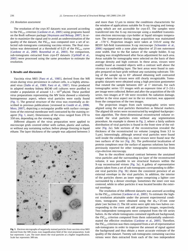
Vaccinia virus M65 (Paez et al., 1985), derived from the WRstrain during virus persistence in culture cells, is a highly attenu-ated virus (Dallo et al., 1989; Paez et al., 1987). Virus producedin adapted monkey kidney BSC40 cell cultures were purified torender a population of around 1.3 � 1010 pfu/ml. These purifiedvirus preparations representing the MV form showed a relativelyhomogeneous aspect, where viral particles were easily found(Fig. 1). The general structure of the virus was essentially as de-scribed in previous publications (reviewed in Condit et al., 2006;Moss, 2007), depicting a rectangular profile with surface corruga-tions of the external membrane well contrasted by the staining re-agent (Fig. 1, inset). Dimensions of the virus ranged from 270 to350 nm, depending on the viewing angle.
Different aliquots of the virus preparation were applied tomicroscope grids covered either with carbon, plastic and carbon,or without any sustaining surface, before plunge-freezing in liquidethane. The layer thickness of the sample was adjusted between 3
Fig. 1. Electron micrographs of negatively stained particles from vaccinia virus M65derived from the WR strain. Low magnification field of the viral preparation. Scalebar represents 1 lm. The inset shows the viral particles at a higher magnification.Scale bar represents 300 nm.
and more than 12 lm to mimic the conditions characteristic forthe window of applications suitable for X-ray imaging and tomog-raphy which are not accessible for cryo-ET. The samples weretransferred into the X-ray microscope using a modified transmis-sion electron microscopy cryo-holder at liquid nitrogen tempera-ture. The temperature during image acquisition was maintainedat �170 �C. Projection images were taken at 510 eV using theBESSY full-field transmission X-ray microscope (Schneider et al.,2006) equipped with a zone plate objective of 25 nm outermostzone width. Due to the flat nature of the sample holder, X-rayimaging was straightforward after focus adjustment for each area.Areas suitable for tomography were selected on the basis of theiraverage density and high contrast. In these areas, viruses werereadily found as rounded objects with a contrast well above thevitreous ice embedding layer. The best areas were found in sam-ples prepared on bare grids without any sustaining substrate. Tilt-ing of the sample up to 65� allowed obtaining well contrastedimages where the viruses were still clearly recognizable. Tomo-graphic datasets were obtained using a high precision goniometerthat rotated the sample between ±65� at 1� intervals. For eachtomographic series 131 images with an exposure time of 2–2.6 sper image were collected. Before and after the acquisition of the tiltseries, two images at 0� tilt were recorded. No radiation damage,neither in the virus aspect nor in the ice structure, was apparentfrom the comparison of the two images.
The projection images from each tomographic series werealigned using the viral particles themselves as fiducial markers.The aligned tilt series were reconstructed by weighted-backprojec-tion algorithm. The three-dimensional reconstructed volume re-vealed the viral particles even without any segmentationprocedure. An example is shown in Fig. 2, where a fairly uniformdistribution of viral particles is found in the front view of the vol-ume (Fig. 2a). Side sections of the reconstruction revealed thethickness of the reconstructed ice volume (ranging from 3.5 to5 lm). Interestingly, although several viral particles were foundwell inside the embedding ice, most viruses were found near thetwo surfaces of the ice (Fig. 2b). The preferential localization ofprotein complexes near the surface of aqueous solutions has beenpreviously reported for other tomographic reconstructions fromcryo-electron microscopy.
Due to the excellent natural contrast between the unstainedvirus particles and the surrounding ice layer of the reconstructedvolume, it was possible to see structural features within theX-ray reconstructed virions (Fig. 3a). A gallery of representativeplanes taken from a set of sub-tomograms corresponding to differ-ent viral particles (Fig. 3b) shows the consistent presence of anexternal envelope in the viral particles. In addition, the interiorof the particles shows an inner region comprising a lighter coresurrounded by a denser envelope. Sometimes the inner core wascentered, while in other particles it was located besides the exter-nal envelope.
The resolution of the different datasets was assessed accordingto the FSCe/o criterion (Cardone et al., 2005). To estimate the high-est resolution attainable under the present experimental condi-tions, tomograms were obtained using the drN = 25 nm zoneplate (see Section 2). The tilt series were split into two halves cor-responding to the even and odd projection images, respectively.Two independent tomograms were computed from the tilt serieshalves. As the whole tomograms contained significant background,the FSCe/o criterion computed from them substantially underesti-mated the effective resolution (Cardone et al., 2005). Therefore,the FSCe/o criterion was then computed from a set of representativesub-tomograms in order to improve the amount of signal againstthe background and thus obtain a more accurate estimate of thequality of the dataset. Twenty sub-tomograms containing vacciniavirions were then extracted from each of the two independent

Fig. 2. Three-dimensional rendering from the X-ray tomographic reconstructed volume using a 40 nm zone plate. (a) Front view. (b) Side view of the reconstructed volumewhere the thickness of the sample is indicated.
Fig. 3. X-ray tomographic reconstruction of the vaccinia virus particles acquired with a 25 nm zone plate. (a) Oblique slice extracted from a tomogram. Scale bar represents1 lm. (b) Central slices of subvolumes extracted from the tomogram, each containing a different viral particle. Internal features within each virion are visible. Scale barrepresents 300 nm.
J.L. Carrascosa et al. / Journal of Structural Biology 168 (2009) 234–239 237
tomograms. The FSCe/o curve was computed from the whole setof 20 sub-tomograms, obtaining a final resolution of 25.7 nmhalf-pitch (Fig. 4). A similar calculation using the datasets obtainedfrom cryo-electron tomograms (Cyrklaff et al., 2005) revealed anominal resolution of 6.7 nm half-pitch. Thus, the equivalentresolution ratio between X-ray and electron tomograms is about3.8 for the same type of samples prepared under the sameconditions.
The structural features found for the X-ray reconstructed vac-cinia virions are fully consistent with the structure of the vacciniavirus known from electron microscopy using either thin sections(Griffiths et al., 2001a), cryo-electron microscopy projections(Cyrklaff et al., 2007; Dubochet et al., 1994) or tomograms (Cyrklaffet al., 2005). Fig. 5a shows two representative planes from cryo-X-ray tomographic virus reconstructions where the inner core isplaced off-centre inside the outer envelope. These images correlate

0
0.2
0.4
0.6
0.8
1
0 0.01 0.02 0.03 0.04 0.05 0.06 0.07 0.08 0.09 0.1
50 25 20 15 10 5
FSC
e/o
spatial frequency / 1/nm
feature size / nm
X-ray tomographyElectron tomography
Fig. 4. Resolution comparison between cryo-X-ray and cryo-electron tomographyresolution estimated with the FSCe/o curves computed from sub-tomograms using athreshold of 0.25. According to the FSCe/o curve, the final resolution of the X-raydataset acquired with a zone plate of 25 nm was found to be 25.7 nm half-pitch and0.0194 1/nm spatial frequency. The FSCe/o curve from the set of cryo-ET wascomputed from the data in Cyrklaff et al. (2005), and it was found to be 6.7 nm half-pitch and 0.0746 1/nm spatial frequency.
238 J.L. Carrascosa et al. / Journal of Structural Biology 168 (2009) 234–239
well with the images from cryo-electron tomographic recon-structed vaccinia virus (Cyrklaff et al., 2005), where the differentcompartments are seen, albeit at higher magnification (Fig. 5b).The viral outer membrane encloses an inner core containing thenucleoprotein of the virus. The inner region of the core shows alighter density, probably consistent with the irregular distributionof the nucleoprotein within the core (Cyrklaff et al., 2005). In the X-ray tomograms, just besides the outer viral envelope, the virus pre-sents one conspicuous structural feature, the so-called lateral body(similar to the observed from the datasets from Cyrklaff et al.,2005, Fig. 5a). Another characteristic view of the X-ray
Fig. 5. Comparison between the structural features of vaccinia virus revealed by cryo-Xplaced off-centre inside the outer envelope from X-ray tomography: central planes from splaced off-centre inside the outer envelope from cryo-electron tomography (the same dainside the outer envelope from X-ray tomography. (d) Viral particles with inner core centrFig. 2 in Cyrklaff et al. (2005)). The scale bars represent 300 nm.
reconstructed virions is the one where the inner core is placed inthe centre of the virion (Fig. 5c), a view that might be readily cor-related with the typical dumbbell shaped core seen in electrontomographic reconstructions (Fig. 5d, obtained from the datasetsin Cyrklaff et al., 2005).
Our results show that, although the resolution limit imposed bythe outermost zone width (25 nm) of the X-ray objective is wellabove the size of the putative virus membranes, even when theyare modified by protein insertion and attachment (6–18 nm), theywere easily identified due to their strong differential absorptioncontrast against the surrounding ice layer. Furthermore, inner de-tails of the virus are distinguishable such as the core shape andits internal distribution of material. Therefore, cryo-XT reveals thethree-dimensional structure of complex biological structuresshowing envelopes and compartments well beyond the nominalX-ray optical resolution of the microscope. Note that this observa-tion is not in contradiction to microscopy principles as these fea-tures smaller than the resolution limit determined by thenumerical aperture of the X-ray objective, together with thenumerical aperture of the condenser, can be visualized in the recon-structions but their size cannot be measured.
In conclusion, we propose that cryo-X-ray tomography at510 eV photon energy is able to retrieve structural informationfrom thick samples containing biological material with such ahigh intrinsic contrast so as to detect membrane-bound compart-ments, or envelopes, in a relatively complex macromolecularaggregate like vaccinia virus. The use of a well-defined biologicalspecimen has allowed comparing the performance of two tomo-graphic approaches (X-rays and electrons) using single-particleanalysis techniques to evaluate the respective attained resolu-tions (X-rays/e� ratio �3.8). Although the present resolution limitimposed by the X-ray objective during these experiments doesnot allow obtaining precise measurements of the features de-tected so far, X-ray microscopy opens an interesting possibilityfor investigating thick samples at sub-cellular level and, in partic-ular, the challenging study of the process of the viral live cyclewithin infected cells.
-ray tomography and cryo-electron tomography. (a) Viral particles with inner coreub-tomograms extracted from X-ray tomography. (b) Viral particles with inner coretaset from Fig. 3 in Cyrklaff et al. (2005)). (c) Viral particles with inner core centreded inside the outer envelope from cryo-electron tomography (the same dataset from

J.L. Carrascosa et al. / Journal of Structural Biology 168 (2009) 234–239 239
Acknowledgments
This work was supported by Grants BFU2008-2328/BMC (toJ.L.C.), LSHG-CT-2004-502828 (to J.L.C.), CT-2006-037536 andFoundation Botín (to M.E.), BESSY-EC-IA-SFS (EU support for theexperiments at BESSY), by the German Federal Ministry of Educa-tion and Research (BMBF) under contract number 05KS4BY1/7,by the Human Frontier Science Program (HFSP) Research GrantRef. RGP0053/2005-C and by the EU in the 6th framework programunder contract number RII3-CT-2004-506008.
References
Born, M., Wolf, E., 1980. Chapter X: Interference and Diffraction with PartiallyCoherent Light, Principles of Optics. Pergamon Press, Oxford.
Cardone, G., Grunewald, K., Steven, A.C., 2005. A resolution criterion for electrontomography based on cross-validation. J. Struct. Biol. 151, 117–129.
Condit, R.C., Moussatche, N., Traktman, P., 2006. In a nutshell: structure andassembly of the vaccinia virion. Adv. Virus Res. 66, 31–124.
Cyrklaff, M., Risco, C., Fernandez, J.J., Jimenez, M.V., Esteban, M., Baumeister, W.,Carrascosa, J.L., 2005. Cryo-electron tomography of vaccinia virus. Proc. Natl.Acad. Sci. USA 102, 2772–2777.
Cyrklaff, M., Linaroudis, A., Boicu, M., Chlanda, P., Baumeister, W., Griffiths, G.,Krijnse-Locker, J., 2007. Whole cell cryo-electron tomography reveals distinctdisassembly intermediates of vaccinia virus. PLoS ONE 2, e420.
Chao, W., Harteneck, B.D., Liddle, J.A., Anderson, E.H., Attwood, D.T., 2005. Soft X-raymicroscopy at a spatial resolution better than 15 nm. Nature 435, 1210–1213.
Chen, H., Hughes, D.D., Chan, T.A., Sedat, J.W., Agard, D.A., 1996. IVE (ImageVisualization Environment): a software platform for all three-dimensionalmicroscopy applications. J. Struct. Biol. 116, 56–60.
Dallo, S., Maa, J.S., Rodriguez, J.R., Rodriguez, D., Esteban, M., 1989. Humoralimmune response elicited by highly attenuated variants of vaccinia virus and byan attenuated recombinant expressing HIV-1 envelope protein. Virology 173,323–329.
Dubochet, J., Adrian, M., Richter, K., Garces, J., Wittek, R., 1994. Structure ofintracellular mature vaccinia virus observed by cryoelectron microscopy. J.Virol. 68, 1935–1941.
Esteban, M., 1984. Defective vaccinia virus particles in interferon-treated infectedcells. Virology 133, 220–227.
Frank, J., 2006. Electron Tomography, Methods for Three-dimensional Visualizationof Structures in the Cell, second ed. Springer, New York.
Fung, J.C., Liu, W., de Ruijter, W.J., Chen, H., Abbey, C.K., Sedat, J.W., Agard, D.A.,1996. Toward fully automated high-resolution electron tomography. J. Struct.Biol. 116, 181–189.
Griffiths, G., Roos, N., Schleich, S., Locker, J.K., 2001a. Structure and assembly ofintracellular mature vaccinia virus: thin-section analyses. J. Virol. 75, 11056–110570.
Griffiths, G., Wepf, R., Wendt, T., Locker, J.K., Cyrklaff, M., Roos, N., 2001b. Structureand assembly of intracellular mature vaccinia virus: isolated-particle analysis. J.Virol. 75, 11034–11055.
Grunewald, K., Cyrklaff, M., 2006. Structure of complex viruses and virus-infectedcells by electron cryo tomography. Curr. Opin. Microbiol. 9, 437–442.
Gu, W., Etkin, L.D., Le Gros, M.A., Larabell, C.A., 2007. X-ray tomography ofSchizosaccharomyces pombe. Differentiation 75, 529–535.
Guttmann, P., Zheng, X., Feser, M., Heim, S., Yun, W., Schneider, G., 2008. Ellipsoidalcapillary as condenser for the BESSY full-field X-ray microscope. In: IOP NinthInternational Conference on X-ray Microscopy, Zürich, Switzerland.
Hans, C., Warren, K.C., John, W.S., Agard, D.A., 1992. In: Raj, S.A. et al. (Eds.), PRIISM:An Integrated System for Display and Analysis of 3D Microscope Images, vol.1660. SPIE, pp. 784–790.
Heymann, J.B., Belnap, D.M., 2007. Bsoft: image processing and molecular modelingfor electron microscopy. J. Struct. Biol. 157, 3–18.
Kremer, J.R., Mastronarde, D.N., McIntosh, J.R., 1996. Computer visualizationof three-dimensional image data using IMOD. J. Struct. Biol. 116, 71–76.
Larabell, C.A., Le Gros, M.A., 2004. X-ray tomography generates 3D reconstructionsof the yeast, Saccharomyces cerevisiae, at 60-nm resolution. Mol. Biol. Cell 15,957–962.
Le Gros, M.A., McDermott, G., Larabell, C.A., 2005. X-ray tomography of whole cells.Curr. Opin. Struct. Biol. 15, 593–600.
Lucic, V., Forster, F., Baumeister, W., 2005. Structural studies by electrontomography: from cells to molecules. Annu. Rev. Biochem. 74, 833–865.
Moss, B., 2007. Poxviridae: the viruses and their replication. In: Knipe, D.N., Howley,P.M. (Eds.), Fields Virology. Lippincott Williams & Wilkins, a Wolters KlumberBusiness, Philadelphia, pp. 2905–2975.
Paez, E., Dallo, S., Esteban, M., 1985. Generation of a dominant 8-MDa deletion atthe left terminus of vaccinia virus DNA. Proc. Natl. Acad. Sci. USA 82, 3365–3369.
Paez, E., Dallo, S., Esteban, M., 1987. Virus attenuation and identification ofstructural proteins of vaccinia virus that are selectively modified during viruspersistence. J. Virol. 61, 2642–2647.
Parkinson, D.Y., McDermott, G., Etkin, L.D., Le Gros, M.A., Larabell, C.A., 2008.Quantitative 3D imaging of eukaryotic cells using soft X-ray tomography. J.Struct. Biol. 162, 380–386.
Roberts, K.L., Smith, G.L., 2008. Vaccinia virus morphogenesis and dissemination.Trends Microbiol. 16, 472–479.
Rosenthal, P.B., Crowther, R.A., Henderson, R., 2003. An objective criterion forresolution assessment in single-particle electron microscopy (Appendix). J. Mol.Biol. 333, 721–745.
Schneider, G., 1998. Cryo X-ray microscopy with high spatial resolution inamplitude and phase contrast. Ultramicroscopy 75, 85–104.
Schneider, G., Guttmann, P., Heim, S., Rehbein, S., Eichert, D., Niemann, B., 2007. X-ray microscopy at BESSY: from nano-tomography to fs-imaging. SynchrotronRadiation and Instrumentation, pp. 1241–1294, In Choi, J.-Y., and Rah, S., (eds.)Synchrotron Radiation and Instrumentation Proceedings IX InternationalConference, Vol. 879. AIP Conf. Proc.
Schneider, G., Anderson, E., Vogt, S., Knochel, C., Weiss, D., Legros, M., Larabell,C., 2002. Computed tomography of cryogenic cells. Surf. Rev. Lett. 9, 177–183.
Schneider, G., Denbeaux, G., Anderson, E., Pearson, A., Bates, W., Vogt, S.,Knochel, C., Meyer, M.A., Zschech, E., 2003. High resolution X-raytomography with applications in biology and materials science. J. Phys. IV104, 607–613.
Spence, J.C.H., 2003. Experimental High-resolution Electron Microscopy, third ed.Oxford University Press, New York.
Subramaniam, S., Bartesaghi, A., Liu, J., Bennett, A.E., Sougrat, R., 2007. Electrontomography of viruses. Curr. Opin. Struct. Biol. 17, 596–602.
Thieme, J., Schneider, G., Knochel, C., 2003. X-ray tomography of a microhabitat ofbacteria and other soil colloids with sub-100 nm resolution. Micron 34, 339–344.
Tokuyasu, K.T., 1973. A technique for ultracryotomy of cell suspensions and tissues.J. Cell Biol. 57, 551–565.
Weiss, D., Schneider, G., Niemann, B., Guttmann, P., Rudolph, D., Schmahl, G., 2000.Computed tomography of cryogenic biological specimens based on X-raymicroscopic images. Ultramicroscopy 84, 185–197.
Zeng, X., Duewer, F., Feser, M., Huang, C., Lyon, A., Tkachuk, A., Yun, W., 2008.Ellipsoidal and parabolic glass capillaries as condensers for X-ray microscopes.Appl. Opt. 47, 2376–2381.
Related Documents