Cryo-Electron Microscopy Reconstruction Shows Poliovirus 135S Particles Poised for Membrane Interaction and RNA Release Carmen Butan,* David J. Filman, James M. Hogle ‹Department of Biological Chemistry and Molecular Pharmacology, Harvard Medical School, Boston, Massachusetts, USA During infection, binding of mature poliovirus to cell surface receptors induces an irreversible expansion of the capsid, to form an infectious cell-entry intermediate particle that sediments at 135S. In these expanded virions, the major capsid proteins (VP1 to VP3) adopt an altered icosahedral arrangement to open holes in the capsid at 2-fold and quasi-3-fold axes, and internal poly- peptides VP4 and the N terminus of VP1, which can bind membranes, become externalized. Cryo-electron microscopy images for 117,330 particles were collected using Leginon and reconstructed using FREALIGN. Improved rigid-body positioning of ma- jor capsid proteins established reliably which polypeptide segments become disordered or rearranged. The virus-to-135S transi- tion includes expansion of 4%, rearrangements of the GH loops of VP3 and VP1, and disordering of C-terminal extensions of VP1 and VP2. The N terminus of VP1 rearranges to become externalized near its quasi-3-fold exit, binds to rearranged GH loops of VP3 and VP1, and attaches to the top surface of VP2. These details improve our understanding of subsequent stages of infec- tion, including endocytosis and RNA transfer into the cytoplasm. P oliovirus is the causative agent of poliomyelitis and the type member of the Enterovirus genus. As such, it shares structural and functional similarities with other members of the Picornaviri- dae family, including the coxsackievirus B viruses, which are asso- ciated with cardiomyopathies, central nervous system (CNS) in- fections, and diabetes; rhinoviruses, which are the most significant cause of the common cold; echoviruses, which can cause aseptic meningitis, gastroenteritis, and respiratory diseases; enterovirus 71 (EV71) and coxsackievirus A16 (CAV16), which have recently caused epidemics in Asia of hand-foot-and-mouth disease, with a high frequency of central nervous system involvement and with high morbidity and mortality; and the foot-and-mouth disease virus, which causes devastating outbreaks of foot-and-mouth dis- ease in livestock (1). Mature poliovirus (which sediments at 160S) is a spherical, nonenveloped virus, with a diameter of approximately 30 nm. Its capsid consists of 60 copies each of 4 proteins (VP1 to VP4) ar- ranged on a T1 (pseudo-T3) icosahedral lattice and encloses a 7,500-base positive-sense single-stranded RNA (ssRNA) viral ge- nome (2, 3). The major proteins (VP1, VP2, and VP3) share a topology, which is an eight-stranded beta barrel. Each protein has a different set of loops connecting the strands in the barrel and unique N- and C-terminal extensions. The C-terminal extensions of the VP1 to -3 subunits are located on the exterior surface of the virus, whereas VP4 and the N-terminal extensions are intertwined on the interior of the shell, making contact with the RNA genome. VP4, which is on the inside surface of the capsid, folds into an elongated structure, due to its contacts on the inner surfaces of the capsid protein that make up the shell. The outer surface of the virus is dominated by star-shaped mesas surrounding each 5-fold axis and three-bladed propeller-like features surrounding each 3-fold axis. Each mesa is formed by 5 molecules of VP1. The hub of each propeller is centered on an icosahedral 3-fold axis and is formed primarily by the VP2 and VP3 beta barrels, while the pro- peller blades are formed by the EF loops of VP2, the GH loop of VP1, and the C terminus of VP1. Separating these projections are deep depressions, or canyons surrounding each 5-fold mesa, and a saddle-shaped depression across each 2-fold axis. The inner surface of the protein shell is decorated by an elab- orate network formed by VP4 and the N-terminal extensions of VP1, VP2, and VP3. During the viral life cycle, native poliovirus attaches to a cell surface receptor, Pvr/CD155 (4). At 37°C, the receptor catalyzes major conformational changes in the 160S capsid (5), leading to the formation of a poliovirus cell-entry intermediate, known as the 135S or A-particle (6, 7). In the 135S particle, both the N-ter- minal extension of VP1 (8, 9) and VP4 (6), which is myristoylated at its N terminus (10), become externalized. Both of these exter- nalized polypeptides then bind to membranes (9, 11, 12) and are capable of creating pores and channels in the membrane (12, 13). Conversion of the virus to its expanded state is required for the uptake of the virus into endosomal compartments of the infected cells (14). Once within an endosome, an interaction of the exter- nalized polypeptides of poliovirus with the endosomal membrane allows the viral RNA genome to transfer efficiently and directly from the capsid interior into the cytoplasm of the infected cell, while remaining protected from RNases (E. Gropelli, H. C. Levy, E. Sun, X. Zhuang, J. M. Hogle, C. Nicol, S. Gold, T. J. Tuthill, and D. J. Rowlands, submitted for publication). Currently, the mech- anism of RNA transfer is not well understood. There are now a number of expanded picornavirus structures at intermediate and higher resolution that sample the process at different points (e.g., crystal structures of EV71 and human rhi- novirus serotype 2 [HRV2] 80S-like particles and CAV16 135S- like particles, as well as cryo-electron microscopy [cryo-EM] structures of poliovirus 80S and 135S particles) (15–19). All such Received 16 July 2013 Accepted 16 November 2013 Published ahead of print 20 November 2013 Address correspondence to James M. Hogle, [email protected]. * Present address: Carmen Butan, Department of Microbial Pathogenesis, Yale University School of Medicine, New Haven, Connecticut, USA. Copyright © 2014, American Society for Microbiology. All Rights Reserved. doi:10.1128/JVI.01949-13 1758 jvi.asm.org Journal of Virology p. 1758 –1770 February 2014 Volume 88 Number 3 on May 13, 2016 by guest http://jvi.asm.org/ Downloaded from

Welcome message from author
This document is posted to help you gain knowledge. Please leave a comment to let me know what you think about it! Share it to your friends and learn new things together.
Transcript

Cryo-Electron Microscopy Reconstruction Shows Poliovirus 135SParticles Poised for Membrane Interaction and RNA Release
Carmen Butan,* David J. Filman, James M. Hogle
‹Department of Biological Chemistry and Molecular Pharmacology, Harvard Medical School, Boston, Massachusetts, USA
During infection, binding of mature poliovirus to cell surface receptors induces an irreversible expansion of the capsid, to forman infectious cell-entry intermediate particle that sediments at 135S. In these expanded virions, the major capsid proteins (VP1to VP3) adopt an altered icosahedral arrangement to open holes in the capsid at 2-fold and quasi-3-fold axes, and internal poly-peptides VP4 and the N terminus of VP1, which can bind membranes, become externalized. Cryo-electron microscopy imagesfor 117,330 particles were collected using Leginon and reconstructed using FREALIGN. Improved rigid-body positioning of ma-jor capsid proteins established reliably which polypeptide segments become disordered or rearranged. The virus-to-135S transi-tion includes expansion of 4%, rearrangements of the GH loops of VP3 and VP1, and disordering of C-terminal extensions ofVP1 and VP2. The N terminus of VP1 rearranges to become externalized near its quasi-3-fold exit, binds to rearranged GH loopsof VP3 and VP1, and attaches to the top surface of VP2. These details improve our understanding of subsequent stages of infec-tion, including endocytosis and RNA transfer into the cytoplasm.
Poliovirus is the causative agent of poliomyelitis and the typemember of the Enterovirus genus. As such, it shares structural
and functional similarities with other members of the Picornaviri-dae family, including the coxsackievirus B viruses, which are asso-ciated with cardiomyopathies, central nervous system (CNS) in-fections, and diabetes; rhinoviruses, which are the most significantcause of the common cold; echoviruses, which can cause asepticmeningitis, gastroenteritis, and respiratory diseases; enterovirus71 (EV71) and coxsackievirus A16 (CAV16), which have recentlycaused epidemics in Asia of hand-foot-and-mouth disease, with ahigh frequency of central nervous system involvement and withhigh morbidity and mortality; and the foot-and-mouth diseasevirus, which causes devastating outbreaks of foot-and-mouth dis-ease in livestock (1).
Mature poliovirus (which sediments at 160S) is a spherical,nonenveloped virus, with a diameter of approximately 30 nm. Itscapsid consists of 60 copies each of 4 proteins (VP1 to VP4) ar-ranged on a T�1 (pseudo-T�3) icosahedral lattice and encloses a7,500-base positive-sense single-stranded RNA (ssRNA) viral ge-nome (2, 3). The major proteins (VP1, VP2, and VP3) share atopology, which is an eight-stranded beta barrel. Each protein hasa different set of loops connecting the strands in the barrel andunique N- and C-terminal extensions. The C-terminal extensionsof the VP1 to -3 subunits are located on the exterior surface of thevirus, whereas VP4 and the N-terminal extensions are intertwinedon the interior of the shell, making contact with the RNA genome.VP4, which is on the inside surface of the capsid, folds into anelongated structure, due to its contacts on the inner surfaces of thecapsid protein that make up the shell. The outer surface of thevirus is dominated by star-shaped mesas surrounding each 5-foldaxis and three-bladed propeller-like features surrounding each3-fold axis. Each mesa is formed by 5 molecules of VP1. The hub ofeach propeller is centered on an icosahedral 3-fold axis and isformed primarily by the VP2 and VP3 beta barrels, while the pro-peller blades are formed by the EF loops of VP2, the GH loop ofVP1, and the C terminus of VP1. Separating these projectionsare deep depressions, or canyons surrounding each 5-foldmesa, and a saddle-shaped depression across each 2-fold axis.
The inner surface of the protein shell is decorated by an elab-orate network formed by VP4 and the N-terminal extensions ofVP1, VP2, and VP3.
During the viral life cycle, native poliovirus attaches to a cellsurface receptor, Pvr/CD155 (4). At 37°C, the receptor catalyzesmajor conformational changes in the 160S capsid (5), leading tothe formation of a poliovirus cell-entry intermediate, known asthe 135S or A-particle (6, 7). In the 135S particle, both the N-ter-minal extension of VP1 (8, 9) and VP4 (6), which is myristoylatedat its N terminus (10), become externalized. Both of these exter-nalized polypeptides then bind to membranes (9, 11, 12) and arecapable of creating pores and channels in the membrane (12, 13).Conversion of the virus to its expanded state is required for theuptake of the virus into endosomal compartments of the infectedcells (14). Once within an endosome, an interaction of the exter-nalized polypeptides of poliovirus with the endosomal membraneallows the viral RNA genome to transfer efficiently and directlyfrom the capsid interior into the cytoplasm of the infected cell,while remaining protected from RNases (E. Gropelli, H. C. Levy,E. Sun, X. Zhuang, J. M. Hogle, C. Nicol, S. Gold, T. J. Tuthill, andD. J. Rowlands, submitted for publication). Currently, the mech-anism of RNA transfer is not well understood.
There are now a number of expanded picornavirus structuresat intermediate and higher resolution that sample the process atdifferent points (e.g., crystal structures of EV71 and human rhi-novirus serotype 2 [HRV2] 80S-like particles and CAV16 135S-like particles, as well as cryo-electron microscopy [cryo-EM]structures of poliovirus 80S and 135S particles) (15–19). All such
Received 16 July 2013 Accepted 16 November 2013
Published ahead of print 20 November 2013
Address correspondence to James M. Hogle, [email protected].
* Present address: Carmen Butan, Department of Microbial Pathogenesis, YaleUniversity School of Medicine, New Haven, Connecticut, USA.
Copyright © 2014, American Society for Microbiology. All Rights Reserved.
doi:10.1128/JVI.01949-13
1758 jvi.asm.org Journal of Virology p. 1758 –1770 February 2014 Volume 88 Number 3
on May 13, 2016 by guest
http://jvi.asm.org/
Dow
nloaded from
expanded virus structures show that 135S conversion causes theparticle to expand by �4% and causes large holes to form at the2-fold axes and the quasi-3-fold axes. Recently, we have generatedlower-resolution asymmetric reconstructions showing that RNAextrusion from poliovirus occurs at a quasi-3-fold axis, both inisolation (20) and in complex with membrane (21). In the lattercomplex, viruses that are attached to membrane show remarkableconsistency in the distance and orientation of the virus, relative tothe bilayer, and are separated from the membrane by two longumbilical connections (21). This consistency suggests that some ofthe polypeptide components that become available symmetrically(in 135S particles) will subsequently reassemble in a very preciseway to form well-defined asymmetric structures for membraneanchoring and RNA transfer (21).
Previous low-resolution cryo-EM studies of the poliovirus135S particle, at about 20-Å and 10-Å resolution, respectively,have implicated the propeller tip as the binding site for the exter-nalized N-terminal extension of VP1 (17, 22). This result has re-cently been confirmed directly by solving the structure of the com-plex of the 135S particle with an Fab directed against residues 24 to40 of VP1 (23). However, the limited resolution of those struc-tures restricted our ability to model the structural transitions, par-ticularly the movement of polypeptide segments not included inthe beta barrel cores of the capsid proteins.
In the present study, we have determined a higher-resolutionstructure of the 135S poliovirus cell-entry intermediate by usingcryo-EM and three-dimensional (3D) image reconstruction at ap-proximately 6-Å resolution. In this new structure, the positions ofthe major capsid proteins and the interactions of many of thestructural components are unambiguously resolved, allowing usto determine with confidence which of the polypeptide segmentshave become shifted, disordered, or rearranged. The reconstruc-tion has clearly interpretable density for a significant portion ofthe externalized N terminus of VP1. Thus, the N terminus exits thevirion at the base of the canyon near the quasi-3-fold axis andparticipates in a novel three-component interaction with the re-arranged GH loops of VP3 and VP1. This occurs at a site corre-sponding to the base of the larger umbilical connection observedin membrane-associated virus. The N terminus of VP1 is thendirected toward the tip of the VP2 propeller, poised at high radiusfor membrane interaction.
MATERIALS AND METHODSSample preparation. The Mahoney strain of type 1 poliovirus was grown inHeLa cell suspension, harvested by centrifugation, and released from cell pel-lets by freeze-thaw lysis. The virus was then purified from the cell lysate byCsCl density gradient fractionation, as described previously (8, 17). The 135Sparticles were made in vitro from the native virus by heating at 50°C for 3 minin a low-salt buffer (2 mM CaCl2, 20 mM HEPES, pH 7.4).
Preparation of grids. The concentration of the 135S poliovirus prep-aration was adjusted to 0.2 to 0.3 mg/ml for cryo-electron microscopy.Aliquots of 4 �l of 135S viruses were applied to glow-discharged holeycarbon grids (C-flat; Protochips, Inc.), thinned by blotting, and plunge-frozen into liquid ethane (maintained at about �180°C to prepare vitri-fied specimens) using a manual gravity plunger. Grids were stored inliquid nitrogen before imaging.
Cryo-electron microscopy. For single-particle analysis, data were col-lected on a Tecnai F20 microscope (FEI Company, Hillsboro, OR) oper-ated at 200 kV using the automated electron microscopy data acquisitionsystem, Leginon (24), at the National Resource for Automated MolecularMicroscopy (NRAMM). A single grid of the 135S preparation was used for
imaging. A total of 1,853 images (each 4,096 by 4,096 pixels) were re-corded using a Tietz 4K-by-4K charge-coupled device (CCD) camera, at anominal magnification of �62,000 (corresponding to a pixel size of 1.37Å), with defocus values between 0.98 and 2.0 �m and an electron dose of15 e�/Å2.
Single-particle image processing. Of the 1,853 CCD images, a subsetof 1,743 nonastigmatic, contamination-free, and artifact-free micro-graphs was selected based on visual inspection. Initially, the contrasttransfer function (CTF) was estimated using the ACE2 (automated CTFestimation) software package (25). Seven hundred twenty-three imageswith an ACE CTF estimation confidence value of less than 0.8 were re-moved from the data set. The remaining 1,020 CCD images were thenused to determine average defocus value, astigmatism, tilt angle, and tiltaxis using a second program, CTFIND3 (26), in Appion (27). Semiauto-mated particle selection using a box width of 300 Å was done using theprogram FindEM (28) from Appion (27). A few representative particleswere selected manually from the first micrograph and used as seeds toinitiate the automated particle-picking routine. The procedure yielded119,079 2D projection images of virus particles. Each virus particle wasthen extracted into a 400-by-400-pixel box to generate a stack, and entriesthat were clearly not virus particles were removed, using statistical filter-ing methods implemented in the program Appion (27). A final set of117,330 particle images was included in the single-particle parameter re-finement. A previous reconstruction at �10-Å resolution of the 135Sparticle (17) was used as a starting model to initiate the search for initialEuler orientation angles and particle origins using FREALIGN V.09 (29).Thereafter, FREALIGN was used iteratively for another 31 cycles, both forparameter refinement and to compute CTF-corrected three-dimensionalreconstructions, with icosahedral symmetry imposed during each recon-struction step.
Inner spherical masks were used to remove density for the RNA ge-nome from the interior of the virus, prior to using the masked map as thereference model for the next iteration. Fourier shell correlation (FSC)between two independent reconstructions (30) was used to estimate theresolution of the final map, which was 7.2 Å (based on an FSC criterion of0.5) and approximately 5.5 Å (based on a 0.143 threshold, per reference31). While portions of the map support the higher-resolution estimate,including much of the N-terminal extension of VP3, which is resolvedfrom the nearby VP1 beta barrel, and other features within the beta barrelsof the three capsid proteins, other portions, particularly the regions cor-responding to the EF loops of VP2, are poorly ordered, resulting in thelower-resolution estimate at the 0.5 FSC cutoff. To improve the interpret-ability of the final reconstruction, average Fourier amplitudes from the finalmap were scaled to those derived from the X-ray coordinates of the poliovirus73S native empty capsid assembly intermediate (PDB 1POV), using the pro-gram bampweigh (32), and then sharpened (increasing Fourier amplitudes athigh resolution) with an inverse B-factor of 300 Å2, to compensate for inter-polation artifacts. FREALIGN V.09 was run on the Clathrin cluster, which is ashared 40-core Linux cluster with 20 GB of RAM managed by SBGrid atHarvard Medical School (http://www.sbgrid.org).
Model building and refinement. The pseudoatomic model consists ofa central protomer (one copy each of the capsid proteins VP1, VP2, andVP3), plus a 5-fold-symmetric copy of the intertwined amino termini ofVP3 (residues 1 to 12). Except as noted below, the main chain coordinatesin these four objects are identical rigid-body copies of crystallographicallydetermined molecules, derived from the poliovirus 73S native empty cap-sid assembly intermediate, PDB entry 1POV (33). For the purpose ofdefining rigid bodies, much of the N-terminal extension of VP3 (residues13 to 42) was regarded as attached to the VP1 core (residues 73 to 207 and234 to 290), the N-terminal hairpin of VP2 (residues 14 to 27, with re-duced occupancy) and C terminus of VP1 (residues 291 to 302) wereregarded as belonging to the VP3 rigid body (residues 43 to 231), and theGH loop of VP1 (residues 208 to 233) was regarded as belonging to theVP2 rigid body (residues 28 to 44 and 58 to 272). Each such rigid body wasfirst placed into the reconstruction by eye, using COOT (34). The correct
Cryo-EM Structure of Poliovirus 135S Particles
February 2014 Volume 88 Number 3 jvi.asm.org 1759
on May 13, 2016 by guest
http://jvi.asm.org/
Dow
nloaded from
placement, assuming no deformation, was unambiguous at this resolu-tion because density features corresponding to the alpha-helical regions ofthe molecules were well resolved and served as fixed pivot points for fittingthe beta sheets. After this preliminary rigid-body fitting, it was confirmedthat the EF loop of VP1 had maintained its conformation but had shiftedrelative to the main barrel, so the loop was refitted manually as a rigidbody, with stereochemically reasonable hinge points, and then reattached.In addition, it was confirmed that shape-similar density features wereabsent at high-contour level for several polypeptide chains. In VP1, miss-ing segments included the N terminus, residues 1 to 51; parts of the GHloop, residues 219 to 223; and the C terminus, residues 291 to 302. In VP2,missing segments included the N terminus, residues 1 to 13; the bottomloop, residues 45 to 57; the C terminus, residues 266 to 272; and unex-pectedly, two segments of the large EF loop, residues 134 to 143 and 158 to172. Later, the residue 134 to 143 segment was visualized at a lower con-tour level and was restored with a lowered occupancy. The core of VP3(residues 43 to 227) was only minimally altered, relative to mature virus,with a slight reorganization of the C-terminal residues 228 to 235. Therigid-body model, constructed as described from portions of the poliovi-rus 73S crystal structure, accounted for most of the electron density in thecapsid, except for the vicinity of the quasi-3-fold axis, where a few alteredchain traces were introduced that were indicated by the density map (seebelow). It should be emphasized that only a small number of residuesdiffer from their rigid-body positions, and those altered polypeptidesshould be regarded as highly approximate.
Automated optimization of the atomic model was accomplished usinglocally developed Unix C-shell scripts that carried out a sequence of Unixutilities and programs from the CCP4 package (35). In each cycle of re-finement, constrained rigid-body copies of the well-ordered residuesfrom VP1, VP2, VP3, and the VP3 beta tube were superimposed by leastsquares (using LSQKAB [36]) onto the central protomer that was outputby the previous refinement cycle. The manually built chain traces (for asmall number of residues) were not replaced during the superposition
step, unless guide coordinates were required to make the subsequent re-finement well behaved. Then, exact icosahedral symmetry operators wereused to generate all 14 polypeptide chains immediately surrounding thefour central objects, for a total of 18 chains in all. A generous mask wasused to isolate the corresponding portion of the cryo-EM reconstruction,which was then enclosed in a padded minimal rectangular box and Fou-rier transformed. Note that none of the mask edges were in direct contactwith the central protomer that was being refined. The resulting Fourieramplitudes were scaled to the Fourier transform of the model, matching asliding window of pseudoreflections by resolution using SFTOOLS (B.Hazes, unpublished observation). The resulting scaled amplitudes and“observed” phases were used as reference standards for refining theatomic model with REFMAC5 (37). By iterating this procedure, a plausi-ble, low-energy structure was obtained, most of whose coordinates closelyresemble the crystallography-based rigid-body model. Main chain seg-ments that refined to deviate significantly from the rigid-body model werenoted. Most likely, this behavior indicates the sites where changes in po-sition or conformation have taken place, though the present resolution isnot sufficient for the exact nature of the change to be known.
Accession numbers. The identification code for the 135S density mapin the EMDataBank is EMD-5710. The Protein Data Bank identificationcode for coordinate modeling results is 3J48.
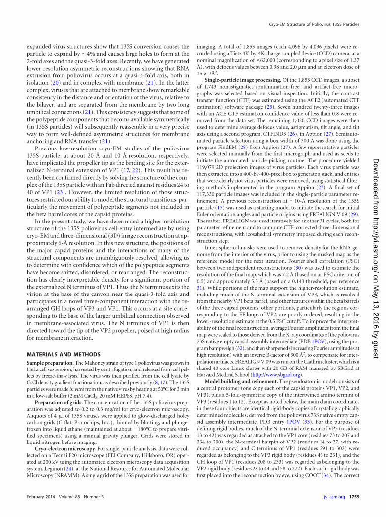
RESULTSOverall appearance of the reconstructed 135S particle. The 135Scapsids appear round or angular in outline when viewed in vitre-ous ice (Fig. 1). A total of 117,330 particle images from 1,020 CCDimages were selected for the cryo-EM single-particle reconstruc-tion, and 99,750 particles were used in the final three-dimensionalreconstruction (Fig. 2A and B). The resulting icosahedrally sym-metric cryo-EM map has a resolution of approximately 5.5 Å ac-cording to the Fourier shell correlation of half-data sets at a
FIG 1 Cryo-electron micrograph showing a field of 135S particles in vitreous ice. The 135S particles are filled by RNA and, depending on orientation, can appear round(B) or angular (C and arrows in panel A). A small number of 80S empty capsids are present (D and asterisks in panel A). Scale bars are included. This representative CCDimage showing 135S particles was recorded at �2.18-�m defocus and an �62,000 nominal magnification, using a Tecnai F20 microscope (FEI).
Butan et al.
1760 jvi.asm.org Journal of Virology
on May 13, 2016 by guest
http://jvi.asm.org/
Dow
nloaded from
threshold of 0.143 (and 7.2 Å for a 0.5 threshold) (Fig. 2C). Theoverall architecture, as expected, is very similar to previously de-termined low-resolution structures of the 135S particles and 80Sparticles of poliovirus (17, 18, 22) and of other picornaviruses(38). It also is consistent with the crystal structures of expandedvirus particles from other picornaviruses (15, 16, 19).
Overall structural changes that accompany expansion. At thepresent resolution, the electron density map (Fig. 3A and 4) showsclearly where the beta sheets are located. It shows several alpha-helices as resolved from the remainder of the beta barrel cores, andit traces the path of a number of well-resolved polypeptide chainsoutside the beta barrels (Fig. 3A). With only a few exceptions(which are depicted in Fig. 3B), rigid-body transformations ofcrystallographic models (specifically, three decorated beta barrels
and one VP3 beta tube; see Materials and Methods) fit well tofeatures of the electron density map. This indicates that the corestructures of the capsid proteins are generally well preserved, andnot deformed, upon the 160S-to-135S transition.
It should be emphasized that a map at the present resolutioncannot be a perfect match to the model, due both to phase errorand to Fourier series termination, unless the model is overparam-eterized inappropriately. Frequently, short stretches of density forcentral strands of beta sheets are interrupted in a way that wouldbe precluded by the beta hydrogen bonding pattern, indicatingthat it is the map, and not the model, that is the less reliable.Therefore, we remodel only longer stretches, where the lack-of-fitof the native conformation is unambiguous, and unassigned den-sity is available that can be assigned to the residue range with some
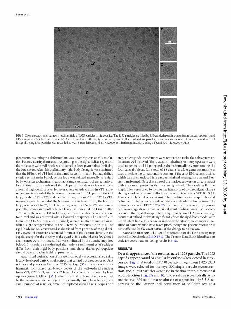
FIG 2 Reconstruction of poliovirus 135S particles. (A and B) Surface rendering colored by radial position, showing the outer (A) and inner (B) surfaces. The maphas been sharpened and displayed at high contour to emphasize higher-resolution details. In many places, individual polypeptide chains are resolved, whichfacilitates the rigid-body fitting of pseudoatomic models. Note the hole at the icosahedral 2-fold axis (center), where red-colored density is visible from theexterior. Labels indicate a mesa and a propeller tip, which are major projections from the outer surface. Separating the projections are canyons surrounding each5-fold mesa and a saddle-shaped depression crossing the 2-fold axis. Symmetry axes are indicated by numbers. (C) A Fourier shell correlation (FSC) wascalculated between randomly selected half-data sets. This curve suggests a resolution of �7 Å (at 0.5 correlation) and that there is meaningful information at �5.5Å (the resolution at which FSC falls below 0.143).
Cryo-EM Structure of Poliovirus 135S Particles
February 2014 Volume 88 Number 3 jvi.asm.org 1761
on May 13, 2016 by guest
http://jvi.asm.org/
Dow
nloaded from
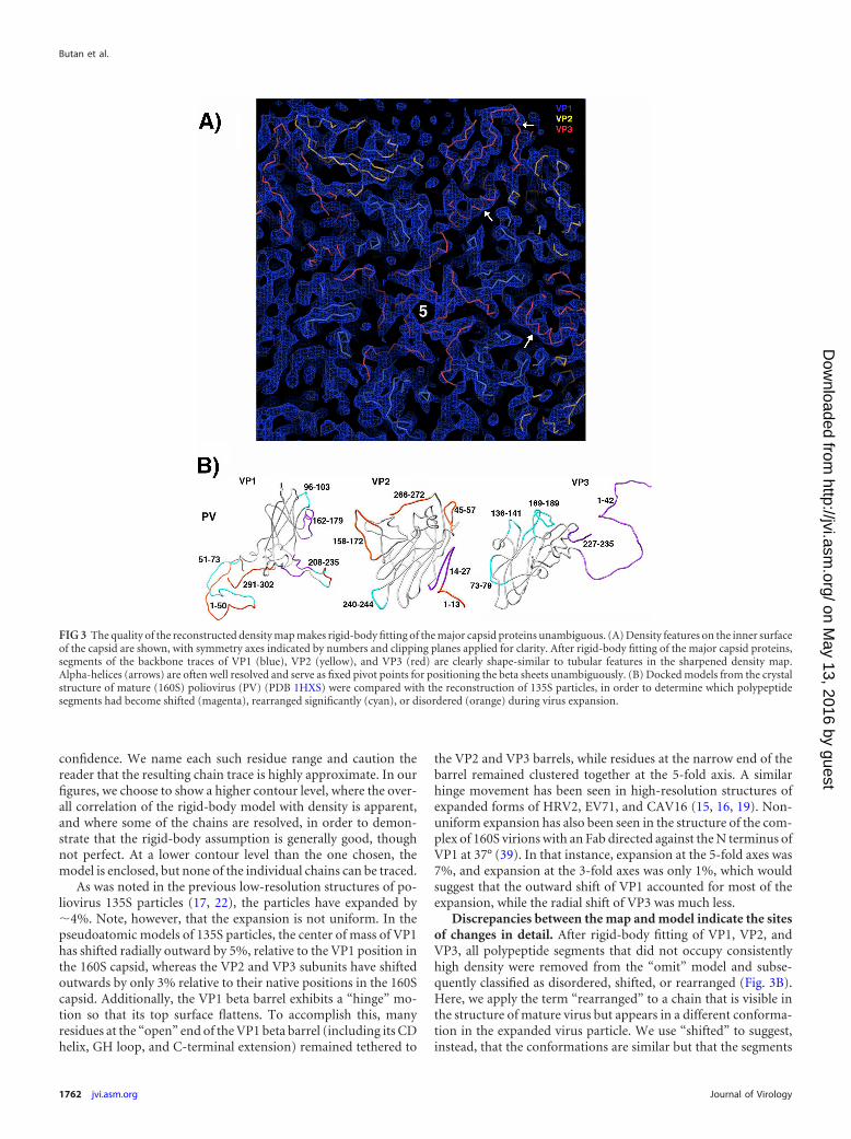
confidence. We name each such residue range and caution thereader that the resulting chain trace is highly approximate. In ourfigures, we choose to show a higher contour level, where the over-all correlation of the rigid-body model with density is apparent,and where some of the chains are resolved, in order to demon-strate that the rigid-body assumption is generally good, thoughnot perfect. At a lower contour level than the one chosen, themodel is enclosed, but none of the individual chains can be traced.
As was noted in the previous low-resolution structures of po-liovirus 135S particles (17, 22), the particles have expanded by�4%. Note, however, that the expansion is not uniform. In thepseudoatomic models of 135S particles, the center of mass of VP1has shifted radially outward by 5%, relative to the VP1 position inthe 160S capsid, whereas the VP2 and VP3 subunits have shiftedoutwards by only 3% relative to their native positions in the 160Scapsid. Additionally, the VP1 beta barrel exhibits a “hinge” mo-tion so that its top surface flattens. To accomplish this, manyresidues at the “open” end of the VP1 beta barrel (including its CDhelix, GH loop, and C-terminal extension) remained tethered to
the VP2 and VP3 barrels, while residues at the narrow end of thebarrel remained clustered together at the 5-fold axis. A similarhinge movement has been seen in high-resolution structures ofexpanded forms of HRV2, EV71, and CAV16 (15, 16, 19). Non-uniform expansion has also been seen in the structure of the com-plex of 160S virions with an Fab directed against the N terminus ofVP1 at 37° (39). In that instance, expansion at the 5-fold axes was7%, and expansion at the 3-fold axes was only 1%, which wouldsuggest that the outward shift of VP1 accounted for most of theexpansion, while the radial shift of VP3 was much less.
Discrepancies between the map and model indicate the sitesof changes in detail. After rigid-body fitting of VP1, VP2, andVP3, all polypeptide segments that did not occupy consistentlyhigh density were removed from the “omit” model and subse-quently classified as disordered, shifted, or rearranged (Fig. 3B).Here, we apply the term “rearranged” to a chain that is visible inthe structure of mature virus but appears in a different conforma-tion in the expanded virus particle. We use “shifted” to suggest,instead, that the conformations are similar but that the segments
FIG 3 The quality of the reconstructed density map makes rigid-body fitting of the major capsid proteins unambiguous. (A) Density features on the inner surfaceof the capsid are shown, with symmetry axes indicated by numbers and clipping planes applied for clarity. After rigid-body fitting of the major capsid proteins,segments of the backbone traces of VP1 (blue), VP2 (yellow), and VP3 (red) are clearly shape-similar to tubular features in the sharpened density map.Alpha-helices (arrows) are often well resolved and serve as fixed pivot points for positioning the beta sheets unambiguously. (B) Docked models from the crystalstructure of mature (160S) poliovirus (PV) (PDB 1HXS) were compared with the reconstruction of 135S particles, in order to determine which polypeptidesegments had become shifted (magenta), rearranged significantly (cyan), or disordered (orange) during virus expansion.
Butan et al.
1762 jvi.asm.org Journal of Virology
on May 13, 2016 by guest
http://jvi.asm.org/
Dow
nloaded from
differ noticeably in location when the coordinates of the maturevirus were superimposed on expanded virus by least squares.“Disordered” residues lie outside a chosen contour, and no shape-similar density feature appears when the contour level is lowered.
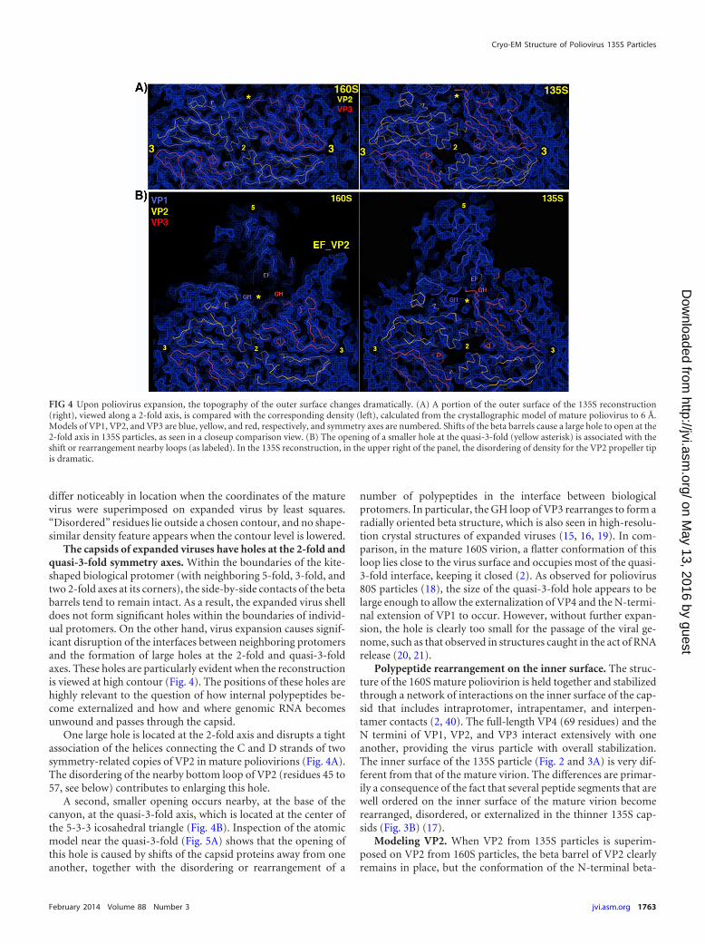
The capsids of expanded viruses have holes at the 2-fold andquasi-3-fold symmetry axes. Within the boundaries of the kite-shaped biological protomer (with neighboring 5-fold, 3-fold, andtwo 2-fold axes at its corners), the side-by-side contacts of the betabarrels tend to remain intact. As a result, the expanded virus shelldoes not form significant holes within the boundaries of individ-ual protomers. On the other hand, virus expansion causes signif-icant disruption of the interfaces between neighboring protomersand the formation of large holes at the 2-fold and quasi-3-foldaxes. These holes are particularly evident when the reconstructionis viewed at high contour (Fig. 4). The positions of these holes arehighly relevant to the question of how internal polypeptides be-come externalized and how and where genomic RNA becomesunwound and passes through the capsid.
One large hole is located at the 2-fold axis and disrupts a tightassociation of the helices connecting the C and D strands of twosymmetry-related copies of VP2 in mature poliovirions (Fig. 4A).The disordering of the nearby bottom loop of VP2 (residues 45 to57, see below) contributes to enlarging this hole.
A second, smaller opening occurs nearby, at the base of thecanyon, at the quasi-3-fold axis, which is located at the center ofthe 5-3-3 icosahedral triangle (Fig. 4B). Inspection of the atomicmodel near the quasi-3-fold (Fig. 5A) shows that the opening ofthis hole is caused by shifts of the capsid proteins away from oneanother, together with the disordering or rearrangement of a
number of polypeptides in the interface between biologicalprotomers. In particular, the GH loop of VP3 rearranges to form aradially oriented beta structure, which is also seen in high-resolu-tion crystal structures of expanded viruses (15, 16, 19). In com-parison, in the mature 160S virion, a flatter conformation of thisloop lies close to the virus surface and occupies most of the quasi-3-fold interface, keeping it closed (2). As observed for poliovirus80S particles (18), the size of the quasi-3-fold hole appears to belarge enough to allow the externalization of VP4 and the N-termi-nal extension of VP1 to occur. However, without further expan-sion, the hole is clearly too small for the passage of the viral ge-nome, such as that observed in structures caught in the act of RNArelease (20, 21).
Polypeptide rearrangement on the inner surface. The struc-ture of the 160S mature poliovirion is held together and stabilizedthrough a network of interactions on the inner surface of the cap-sid that includes intraprotomer, intrapentamer, and interpen-tamer contacts (2, 40). The full-length VP4 (69 residues) and theN termini of VP1, VP2, and VP3 interact extensively with oneanother, providing the virus particle with overall stabilization.The inner surface of the 135S particle (Fig. 2 and 3A) is very dif-ferent from that of the mature virion. The differences are primar-ily a consequence of the fact that several peptide segments that arewell ordered on the inner surface of the mature virion becomerearranged, disordered, or externalized in the thinner 135S cap-sids (Fig. 3B) (17).
Modeling VP2. When VP2 from 135S particles is superim-posed on VP2 from 160S particles, the beta barrel of VP2 clearlyremains in place, but the conformation of the N-terminal beta-
FIG 4 Upon poliovirus expansion, the topography of the outer surface changes dramatically. (A) A portion of the outer surface of the 135S reconstruction(right), viewed along a 2-fold axis, is compared with the corresponding density (left), calculated from the crystallographic model of mature poliovirus to 6 Å.Models of VP1, VP2, and VP3 are blue, yellow, and red, respectively, and symmetry axes are numbered. Shifts of the beta barrels cause a large hole to open at the2-fold axis in 135S particles, as seen in a closeup comparison view. (B) The opening of a smaller hole at the quasi-3-fold (yellow asterisk) is associated with theshift or rearrangement nearby loops (as labeled). In the 135S reconstruction, in the upper right of the panel, the disordering of density for the VP2 propeller tipis dramatic.
Cryo-EM Structure of Poliovirus 135S Particles
February 2014 Volume 88 Number 3 jvi.asm.org 1763
on May 13, 2016 by guest
http://jvi.asm.org/
Dow
nloaded from
hairpin (residues 14 to 27), which participates in interpentamericcontacts below the VP3 beta barrel in the virion, is significantlydisplaced. Additionally, density for the bottom loop (residues 45to 57) is missing, just as in the crystal structure of the 73S nativeempty capsid assembly intermediate (33), in which the N termi-nus of VP1 is also disordered. The electron density map suggestsseveral other localized rearrangements in VP2. These include theHI loop (approximately in residues 240 to 244) in the vicinity ofthe 3-fold symmetry axis, as well as the large double loop betweenbeta strands E and F of VP2, wherein the density for residues 134to 143 has weakened (suggesting that it is flexible) and residues158 to 172 have become disordered. The disordering of the EFloop of VP2 results in the disappearance of the density for the tipsof the blades of the propeller-like features (Fig. 4B, upper right).The disordering of this loop appears to occur in concert with thedisordering or rearrangement of several polypeptides that supportthe EF loop in virions, including the C terminus of VP1 (along thewestern edge of the EF loop), the GH loop of VP1 (along theeastern edge), and the C terminus of VP2 (residues 266 to 272,along the southern edge). In 135S particles, the C terminus of VP1becomes disordered (see below), the GH loop of VP1 rearranges(see below), and there is no visible density corresponding to thenative conformation of the C terminus of VP2 all the way back toits junction with the VP2 beta barrel at residue 265. Instead, justsouth of this junction, a pair of relatively strong, but disconnected,density features are visible in the 135S map, within the 2-fold hole.Due to the proximity of these features to the junction with theC-terminal end of the VP2 beta barrel, we surmise that the hole
might be a preferred binding site for the disordered VP2 C-termi-nal extension. It is not yet clear whether keeping the 2-fold holepartially plugged in 135S particles is functionally relevant.
The two-loop “gate.” The observation that the base of the VP2C terminus (i.e., at residue 265) remains ordered is relevant to thestructural integrity of the icosahedral capsid. This junction ap-pears to remain firmly clamped between two folds of the EF loopof VP3 from a neighboring protomer (Fig. 5A) in both mature andexpanded virions. This persistent interprotomer contact betweenthe VP3 EF loop and the VP2 C terminus is also notable for cre-ating a “gate” between the 2-fold and quasi-3-fold holes that per-sists during the 135S expansion. The gate is much more substan-tial in mature poliovirus, where it also includes the GH loop ofVP3 (residues 169 to 189) and the bottom loop of VP2 (residues 45to 57), which both rearrange upon virus expansion. In our currentmodel for virus expansion (41), the VP1 N-terminal extensionmost likely emerges through the 2-fold hole (wherein polypeptideexternalization is reversible) and transfers through the gate intothe quasi-3-fold hole, where the exposed polypeptide becomessecurely anchored by neighboring structures (see below).
Modeling VP3. The four-body rigid-body model, includingonly well-ordered residues from the poliovirus 73S crystal struc-ture, accounted for most of the electron density in the capsid butnotably failed to account for a number of density features in thevicinity of the quasi-3-fold axis. Density for the GH loop of VP3was clearly not a good fit for the loop conformation that was seenin the mature poliovirus, where a flattish loop path close to thesurface helps to keep the quasi-3-fold hole plugged (Fig. 4B, on the
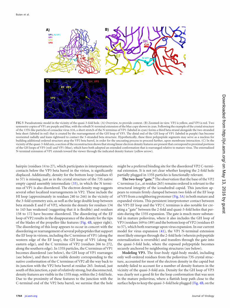
FIG 5 Pseudoatomic model in the vicinity of the quasi-3-fold hole. (A) Overview, to provide context. (B) Zoomed-in view. VP2 is yellow, and VP3 is red. Twosymmetry copies of VP1 are purple and blue, with the rebuilt N-terminal extension of the blue copy shown in cyan. Following the example of the crystal structureof the 135S-like particles of coxsackie virus A16, a short stretch of the N terminus of VP1 (labeled in cyan) forms a third beta strand alongside the two-strandedbeta sheet (labeled in red) that is created by the rearrangement of the GH loop of VP3. The distal end of the GH loop of VP1 (labeled in purple) has becomereoriented radially and leans rightward to contact the 3-stranded beta structure. Hypothetically, these three polypeptide segments may serve as a nucleus forbuilding additional ordered structure atop the VP2 beta barrel, in order for the uncoating process to proceed further, upon membrane interaction. (C) In thevicinity of the quasi-3-fold axis, a section of the reconstruction shows that strong linear electron density features are present that correspond to proximal portionsof the GH loops of VP3 (red) and VP1 (blue), which have both adopted an extended conformation that is rearranged relative to mature virus. The externalizedN-terminal extension of VP1 extends toward the viewer through the indicated density feature (yellow arrow).
Butan et al.
1764 jvi.asm.org Journal of Virology
on May 13, 2016 by guest
http://jvi.asm.org/
Dow
nloaded from
left). A better fit to the density was obtained from the conforma-tion seen in 80S particles of EV71 (16) and in poliovirus (M. Wolf,D. J. Filman, and J. M. Hogle, unpublished data), where the GHloop of VP3 becomes elongated into a radially oriented two-stranded beta structure, but it was clear that two beta strands wereinsufficient to account for the entire volume of density that waspresent. Additionally, the two strands of the 80S hairpin wouldhave to be twisted by about 90 degrees for both strands of thehairpin to fit within our current 135S density envelope. The den-sity was a much better fit for models derived from the recentlypublished structure of a 135S-like particle from coxsackievirusA16 (19). In our current model, residues 169 to 175 and 181 to 188of VP3 and residues 51 to 72 of VP1 are adapted from a super-posed copy of the CAV16 atomic model. For the most part, thesethree polypeptide segments appear to move together as part of theVP3 rigid body. To complete the GH loop of VP3, residues 176 to180 at the distal end were fitted manually to lie within a low-density contour, as a highly approximate chain tracing. Fortu-nately, due to the shortness of this missing loop, there was onlyone available path to choose. Notably, formation of the three-stranded structure has now been observed in both of the knownstructures of picornavirus 135S particles, but in none of the 80S(15, 16) or 73S (33) structures, all of which lack density for theN-terminal extension of VP1.
In VP3, some additional differences between the 135S and 160Smodels (Fig. 3B) are due to the continued noncovalent attach-ment of certain polypeptide chains to neighboring beta barrels, asthe barrels separate during virus expansion. Thus, in 135S parti-cles, both the N-terminal extension of VP3 (residues 13 to 42) andthe C-terminal polypeptide of VP3 (residues 227 to 235) traveltogether with the VP1 rigid body. As a result, both chains becomedisplaced, relative to the VP3 beta barrel, without requiring sig-nificant changes in the intermolecular interactions that the poly-peptides make. Additionally, the VP3 beta tube (five intertwinedcopies of residues 1 to 12) is seen to rise along the 5-fold axis, toremain in contact with the underside of VP1.
Modeling VP1. Compared to its position in the 160S capsid,the center of mass of VP1 has moved outward radially by �6.5 Å.The tips of the DE and HI loops nearest to the 5-fold axis havemoved out by a distance of 5.2 Å and 6.2 Å, respectively. The BCloop, which is located on top of the mesa, but farthest from the5-fold axis, has moved by 7.4 Å but also appears likely to be de-formed, as much of the native conformation of the loop wouldprotrude through the density envelope. Overall, the VP1 wedgemoves outward by about 5 Å along the axis and by 6.6 Å in themiddle. These movements suggest an “umbrella-like” motion,wherein the part of the barrel nearest the canyon rises more thanthe part near the axis, causing the 135S particle to be more angularin profile than the 160S particle and resulting in an elevation anda broadening of the mesa. A similar hinge motion for VP1, and aradial expansion of the capsid protein subunits, was seen in thecrystal structures of 80S particles of EV71 (16) and HRV2 (15) andin the 135S particle of CAV16 (19).
There also is evidence that the EF loop of VP1 (residues 162 to179) undergoes a slight tilting and a lateral shift relative to the restof the VP1 barrel (Fig. 3B). This loop movement might be re-quired so that the EF loop can continue to maintain its contactswith both of the VP1 barrels that are located on either side of it,even as the barrels are situated further apart in the expanded 135Sparticles than in 160S virions. Indeed, the forced separation of the
EF loop from its own beta barrel is hypothesized to be an impor-tant aspect of the mechanism of the receptor-induced 160S-to-135S transition (M. Strauss, D. J. Filman, N. Cheng, D. M. Belnap,and J. M. Hogle, unpublished data).
Polypeptides involved in externalizing the N terminus ofVP1. In 135S particles, the N-terminal extension of VP1 is exter-nalized and is capable of binding to specific Fabs (23) or to mem-brane (9, 11). Our previous 10-Å reconstruction of the 135S par-ticle clearly showed that the N terminus did not exit at the 5-foldaxis, as previously suggested, and led to the conjecture based ongenetic data that the N terminus was instead released from thebase of the canyon near the quasi-3-fold axis (17). However, thelimited resolution of this reconstruction did not allow us to di-rectly visualize the site of externalization. Recently, the crystalstructure of 135S particles from CAV16 (19) showed how theproximal end of the VP1 N-terminal extension rearranges to exitthrough the capsid at the quasi-3-fold hole. The present cryo-EMstructure of 135S particles confirms that a similar rearrangementof the N-terminal extension occurs in poliovirus and that addi-tional polypeptides rearrange to contact the extension and stabi-lize its exit point (Fig. 5A and B). In particular, residues corre-sponding to 59 to 68 from the N-terminal extension of VP1 arereordered in 135S particles from both CAV16 (19) and poliovirus.As this polypeptide extends away from the VP1 beta barrel, severalresidues (63 to 72) extend across the inner surface of the capsiduntil they reach the southern end of the quasi-3-fold hole. Each Nterminus then turns radially outward, with residues 59 to 62 in-teracting with the southern edge of the reorganized GH loop ofVP3 in its extended conformation (Fig. 5A).
Quasi-3-fold anchor. The exiting N-terminal extension, in itsnew arrangement, is further anchored by extensive contacts withthe GH loop of VP2 (residues 216 to 223), together with parts ofthe D strand of the VP2 barrel (residues 100 to 101), the N termi-nus of VP3 (residues 29 to 32), and the end of the G strand fromthe neighboring copy of VP1 (residues 201 to 202). The extendedconformation of the VP3 GH loop is supported by residues fromthe EF loop of the neighboring VP1 (residues 162 to 163) and bythe “doorstop” linker region of the neighboring VP1 GH loop(residues 233 to 236, just prior to the H strand), which normallycontrols access to the stability-regulating “pocket factor” lipid thatoccupies the center of the VP1 beta barrel in mature virions (40).Note that as the VP1 and VP2 barrels move apart during the 160S-to-135S transition, this linker segment provides the slack that al-lows parts of the VP1 GH loop to remain bound to the top of theVP2 barrel. During the transition, the linker shifts to slightly lowerradius in the quasi-3-fold hole (which is apparent in the densitymap) while making contact with the newly rearranged GH loop ofVP3 and N-terminal extension of VP1 and helping to hold the newstructural features in place.
The polypeptide segments that clamp the GH loop of VP3 andthe N terminus of VP1, for the most part, are well ordered, andmost are in their native conformations, so that rigid-body move-ments of the native beta barrels serve to bring them into the properarrangement. The final contact is more tenuous, involving thedistal end of the GH loop of the VP1 (residues 213 to 227) thatbelongs to the neighboring protomer to the west. Evidence fromthe poliovirus 135S map, as detailed below, indicates that the loophas reoriented to project radially outward while tilting sideward tocontact the quasi-3-fold site (Fig. 5). Presumably, this additional
Cryo-EM Structure of Poliovirus 135S Particles
February 2014 Volume 88 Number 3 jvi.asm.org 1765
on May 13, 2016 by guest
http://jvi.asm.org/
Dow
nloaded from
loop rearrangement cannot occur until after the GH loop of VP3and the N terminus of VP1 have rearranged to receive it.
Extreme N terminus of VP1. Once the N terminus of VP1leaves its binding site in the quasi-3-fold hole, there are a numberof directions that it could travel. A pathway through weak densitydoes exist for residues 51 to 58 that extends across the top of theVP2 beta barrel and reaches the large double loop in VP2 thatconnects beta strands E and F. This path is included in the modelas a highly approximate chain trace. This possible path would bein agreement with the observation that an Fab from an antibodyrecognizing residues 24 to 40 of VP1 binds at this propeller tip(23). Additional confirmation of the location of the VP1 N termi-nus was obtained in our previous lower-resolution cryo-EM stud-ies of the 135S poliovirus cell-entry intermediate (17), where re-moval of the N-terminal helix using V8 protease created asignificant-difference electron density feature at the propeller tip.
Our current proposal for the path of the VP1 N-terminal ex-tension differs from our previous suggestion (17), which held thatresidues 42 to 52 (a predicted amphipathic helix) might be responsi-ble for creating the tubular density feature (Fig. 6, open arrowhead)that appears to bridge across the canyon between the mesa and thepropeller tip in our lower-resolution maps. In the present higher-resolution map of 135S virions, in the corresponding location, thereare no interpretable density features above the noise level that wouldhelp to explain the lower-resolution observation.
Rearrangement of the GH loop of VP1. In the present higher-resolution maps, polypeptides from VP1 have been observed toform two structures that were not present in mature virus. Asnoted above, one is an ordered redirection of a portion of the VP1N-terminal extension (residues 58 to 67) that leads to its external-ization through the quasi-3-fold hole. The other involves a signif-icant reorientation of the distal end of the GH loop of VP1. Thus,the rearranged GH loop of VP1 no longer lies flat across the top ofthe VP2 beta barrel, supporting the eastern edge of the EF loop ofVP2, as it does in 160S particles. Instead, the GH loop now projectsoutward and leans sideways, making contacts with the external-ized N-terminal extension of VP1 and the rearranged GH loop ofVP3, both of which belong to a neighboring protomer (Fig. 5).
It is important to explain how the rearrangement of the GHloop of VP1 was found, because modeling a new detailed structureinto a map at this resolution is normally not feasible. Early on,while checking the agreement of the complete rigid-body-dockedatomic model with the map, we noticed that the map included twostrong, parallel, radially oriented tubes of unoccupied density pro-truding from the top surface of the VP2 envelope. At the time, wehad no indication that we would be able to identify the polypep-tide segments to build into them. Some time later, we removed allpolypeptide segments whose native conformation caused them tolie outside the chosen isocontour envelope. This yielded an “omit”model composed exclusively of reliable components that ap-peared not to change conformation (gray tubes in Fig. 3B). Re-markably, the two parallel density tubes joined to the density en-velope very close to the sites where the distal end of the GH loop ofVP1 in the atomic model had been truncated: one tube at the siteof residue 214 and the other tube near residue 229. Initially, wehad assumed that residues 213 to 228 from the distal end of theGH loop of VP1 were disordered because their native conforma-tion placed them outside the contour envelope. However, the ob-served density tubes demonstrated that the distal end of the GHloop must instead be rearranged in a well-defined way (Fig. 5).
In the current model, the stems of the reoriented GH loop(residues 213 to 218 and 224 to 226) have been model built andrefined as a partially occupied chain trace. This is intended toindicate the path of the polypeptide, rather than providing reliableatomic coordinates. The extreme distal end of the loop has beenomitted, for lack of a clear density path. Note that two intermedi-ate segments of the GH loop (residues 208 to 212 and 227 to 229)continue to bind to the top of the VP2 beta barrel, in a way similarto what is seen in the 73S and 160S crystal structures (2, 33).
DISCUSSIONEvidence for the rearranged GH loop of VP1 in lower-resolutionstudies. Our earlier publication of the 10-Å structure of the 135Sparticle included two independent reconstructions (EMDB en-tries 1133 and 1144), both generated using PFT2 (42) andEM3DR2 (http://people.chem.byu.edu/belnap/). When viewed at
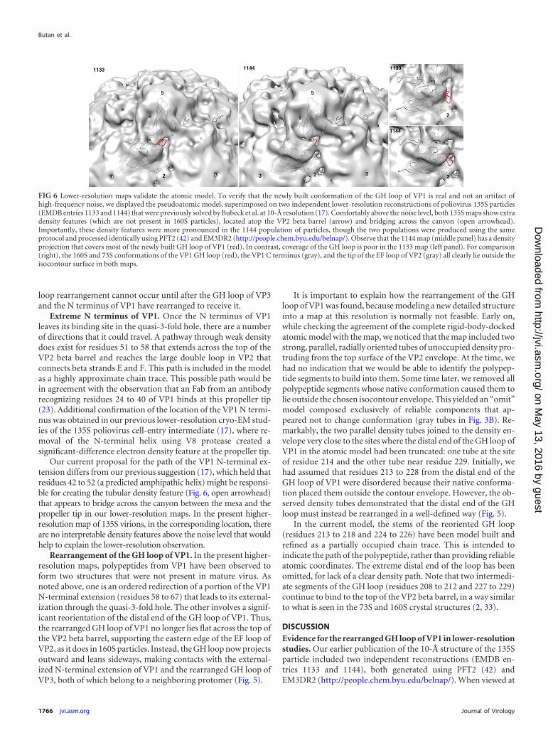
FIG 6 Lower-resolution maps validate the atomic model. To verify that the newly built conformation of the GH loop of VP1 is real and not an artifact ofhigh-frequency noise, we displayed the pseudoatomic model, superimposed on two independent lower-resolution reconstructions of poliovirus 135S particles(EMDB entries 1133 and 1144) that were previously solved by Bubeck et al. at 10-Å resolution (17). Comfortably above the noise level, both 135S maps show extradensity features (which are not present in 160S particles), located atop the VP2 beta barrel (arrow) and bridging across the canyon (open arrowhead).Importantly, these density features were more pronounced in the 1144 population of particles, though the two populations were produced using the sameprotocol and processed identically using PFT2 (42) and EM3DR2 (http://people.chem.byu.edu/belnap/). Observe that the 1144 map (middle panel) has a densityprojection that covers most of the newly built GH loop of VP1 (red). In contrast, coverage of the GH loop is poor in the 1133 map (left panel). For comparison(right), the 160S and 73S conformations of the VP1 GH loop (red), the VP1 C terminus (gray), and the tip of the EF loop of VP2 (gray) all clearly lie outside theisocontour surface in both maps.
Butan et al.
1766 jvi.asm.org Journal of Virology
on May 13, 2016 by guest
http://jvi.asm.org/
Dow
nloaded from
a lower contour level, the 1144 reconstruction shows extra densityfeatures on the top of the VP2 beta barrel that do not appear in the1133 reconstruction (Fig. 6), even though there were no deliberatedifferences in sample preparation or data processing. Note that inboth reconstructions, only a small percentage of the boxed parti-cles were rejected, which ensures that each of the two reconstruc-tions was actually representative of its population. The densityfeatures that appear in the 1144 map (Fig. 6) include a broadtriangular area with a pronounced longitudinal ridge (indicatedby an arrow), extending across the top of the VP2 beta barrel, aswell as a large tubular density feature (indicated by an arrowhead)that appears to cross the canyon bridging the points of the 5-foldmesa and the tip of the propeller (17). In fitting models to thismap, we noted that although we could readily fit the VP1 and VP3rigid bodies into the density, there was no single structure for VP2that would explain the density surrounding the propeller. Themodel that we settled on placed the top surface of the VP2 barrel ina position that turned out to be essentially correct but left the EFloop of VP2 protruding through the contour surface, as well asleaving an outward-projecting blade of unfilled density, corre-sponding to the longitudinal ridge (Fig. 6, rightmost panels). Thisdensity feature was clearly real, icosahedrally reinforced, andstrong. In the original low-resolution study, it was impossible todecide whether VP2 was present in multiple orientations or didnot actually behave as a rigid body. Now, with higher-resolutioninformation available, it is clear that most of the VP2 beta barrel isrigid but that numerous polypeptides around the propeller tipbecome disordered, while other polypeptides (including the GHloop of VP1) rearrange to account for the previously unfilled low-resolution density features.
Although our newest model for the 135S structure accounts forsome of the extra density in the 1144 reconstruction, much of thedensity is still unexplained. In particular, we have no good way tomodel the tubular density feature that appears to span the canyonin lower-resolution maps (Fig. 6, arrowhead) and which we hadpreviously ascribed incorrectly to a helix corresponding to resi-dues 45 to 54 from the N terminus of VP1. In both 1144 and thepresent reconstruction, and with the beta barrels placed correctly,the top of VP2 provides the only strong density connection be-tween the quasi-3-fold hole and the “tubular” region. In contrast,the postulated helix assignment would have approached the tubearea from the opposite direction.
Uncoating of picornavirions may involve a succession ofstepwise changes. The fact that the extra density was observed inone 135S reconstruction (EMDB 1144) and not in another(EMDB 1133) lends support to the idea that those two previous135S reconstructions may have sampled different stages along theuncoating pathway. We hypothesize that the EMDB 1144 recon-struction, with extra density atop VP2 (Fig. 6, arrow), is moresimilar to the population of 135S particles in our current higher-resolution reconstruction, where new density was seen for thereoriented GH loop of VP1. In contrast, the EMDB 1133 recon-struction, which lacks that extra density, may represent an earlierstage of the uncoating pathway that is closer to that of the 135S-like particles of CAV16 (19). Thus, in CAV16, virus expansioncaused structural changes in the quasi-3-fold hole but not on thetop of the VP2 beta barrel. Correspondingly, more of the polypep-tide remained ordered and in a virion-like conformation, includ-ing most of the GH loop of VP1 and all of the EF loop of VP2 (Fig.7). These observations support the idea that uncoating may be a
multistep process, with each 135S structure sampling a differentstage along a continuum of states.
A situation similar to that was previously demonstrated forcryo-EM structures of 80S particles (18), where a “later” popula-tion of particles (which had been incubated longer) had tended todevelop large, visible density features on the inner surface of the cap-sid, near the 2-fold axis. These features were considerably smaller inthe “earlier” population, where the features had less time to develop.We suspect that, in a similar way, the extra density features seen at lowcontour level, both in the current higher-resolution 135S map and inthe previous EMDB 1144 reconstruction, may indicate that thoseimages were derived from a later, and perhaps more conformation-ally heterogeneous, population of virus particles.
Taken together, the maps and the comparisons with theCAV16 135S particle suggest that the rearrangement of 135S par-ticles is likely to be a multistep process, sampled at different pointsalong the uncoating trajectory by different virus preparations andby different picornavirus structures. In that context, the partialordering of the GH loop of VP1 and the development of additionalunexplained density on the top of the VP2 beta barrel place thepresent poliovirus reconstruction further along the trajectorythan the states that the other picornavirus structures represent. Itis not yet clear whether this is due to inherent differences in thetype of virus or to the use of cryo-EM rather than crystallography.
Polypeptide segments that rearrange are displayed at highradius, in a large circle around the canyon. Among the polypep-tides that become disordered or rearranged in the 135S transitionof poliovirus, there are a number of loops that are normally ex-posed on the outer surface. This is consistent with the observa-tions (i) that the capsids of 135S particles are immunologicallydistinct from those of the mature 160S virions and from the 73Snative-antigenic empty capsids (33) and (ii) that the regions of theouter surface that exhibit the greatest structural variability, whenpicornavirus structures are compared, are exposed loops that in-clude some of the most important immunogenic sites (43, 44).
Additionally, many of the polypeptide segments that moved orbecame disordered (Fig. 3B) were those responsible for stabilizingthe mature 160S particle, frequently via the binding of loops andN- and C-terminal extensions to the inner and outer surfaces ofneighboring proteins. Importantly, when the sites of rear-rangement are displayed on the outer surface of the icosahe-drally symmetric capsid, a striking pattern is observed (Fig. 8).The components that rearrange are displayed, for the mostpart, at high radius and form a large circle that extends longi-tudinally over the top of each VP2 beta barrel, across the top ofVP3, and into the next quasi-3-fold axis. Notably, the changedareas tend to exclude the canyon and involve only minimalchanges to the 5-fold mesa.
A model for membrane attachment, internalization, and ge-nome translocation. To develop an updated model for the se-quence of polypeptide rearrangements that occur during poliovi-rus uncoating, we have assumed that the known native andexpanded structures of poliovirus, poliovirus complexes (with re-ceptor, Fabs, or membrane), and other picornaviruses are eachrepresentative of specific states along a common trajectory (Fig.9). Along the trajectory, virus expands, RNA content is dimin-ished, and polypeptide order tends to decrease, except when noni-cosahedral structures for membrane interaction and RNase-pro-tected RNA transfer are being built.
Cryo-EM reconstructions have been determined for various
Cryo-EM Structure of Poliovirus 135S Particles
February 2014 Volume 88 Number 3 jvi.asm.org 1767
on May 13, 2016 by guest
http://jvi.asm.org/
Dow
nloaded from
states of the poliovirus capsid bound to the Fabs of antibodies thatare directed against residues 20 to 40 of VP1 (45) and residues 39to 55 of VP1 (46). These structures have demonstrated that theN-terminal extension of VP1 is externalized at the 2-fold axes in“breathing” particles (39) and shifts to the tips of the propellers inthe 135S particles (23). These observations can be considered to-gether with the observations from expanded virus structures con-cerning the site of egress of the externalized N-terminal extensionof VP1.
Based on these data, we propose the following model for theevents leading to internalization of the virus and translocation ofits genome across the endosomal membrane and into the cyto-plasm. We propose that soon after the virus attaches to receptor,several of the N-terminal extensions of VP1 (each up to 70 resi-dues in length) will exit reversibly at the 2-fold axes (similar to theaction in breathing virus). Due to receptor binding, one or moreof the extensions will become exposed on the side of the virus thatfaces the cell membrane, close enough for their amphipathic heli-
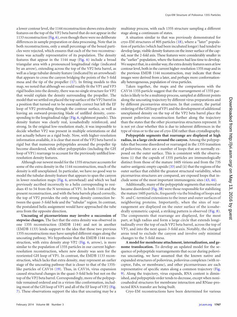
FIG 7 The conformational changes seen in the expanded structures of other picornaviruses (such as the CAV16 135S particle [19]) (B) are less extensive than theones seen in poliovirus 135S particles (A). Main chain traces from crystal structures of mature virions are shown for capsid proteins VP1, VP2, and VP3. Thelocations of polypeptide segments that become shifted, rearranged significantly, or disordered are colored magenta, cyan, and orange, respectively. The greaterextent of changes in poliovirus 135S particles (A) might be the result of inherent lesser stability or simply greater heterogeneity in cryo-EM preparations, versusthe preparations of expanded forms of other viruses that have been crystallized (15, 16, 19). In either case, our hypothesis is that much of the polioviruspopulation must be relatively further along the uncoating pathway. Panel A is Fig. 3B, repeated here for clarity.
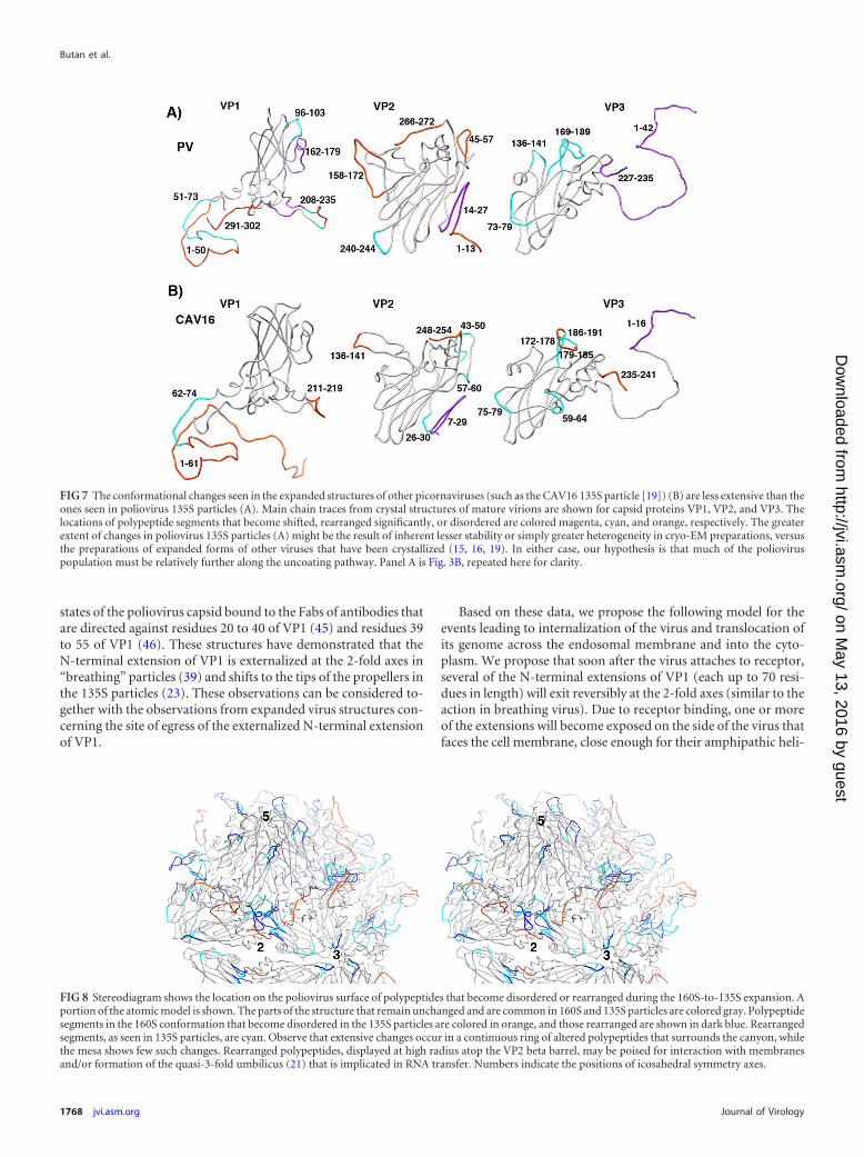
FIG 8 Stereodiagram shows the location on the poliovirus surface of polypeptides that become disordered or rearranged during the 160S-to-135S expansion. Aportion of the atomic model is shown. The parts of the structure that remain unchanged and are common in 160S and 135S particles are colored gray. Polypeptidesegments in the 160S conformation that become disordered in the 135S particles are colored in orange, and those rearranged are shown in dark blue. Rearrangedsegments, as seen in 135S particles, are cyan. Observe that extensive changes occur in a continuous ring of altered polypeptides that surrounds the canyon, whilethe mesa shows few such changes. Rearranged polypeptides, displayed at high radius atop the VP2 beta barrel, may be poised for interaction with membranesand/or formation of the quasi-3-fold umbilicus (21) that is implicated in RNA transfer. Numbers indicate the positions of icosahedral symmetry axes.
Butan et al.
1768 jvi.asm.org Journal of Virology
on May 13, 2016 by guest
http://jvi.asm.org/
Dow
nloaded from
ces to insert into the membrane. These copies will serve as anchorsthat keep the virus attached to the membrane after the virus hasdetached from the receptor during the 160S-to-135S transition.During this transition, VP4 and the other N-terminal extensionsof VP1 will become externalized, and the N-terminal extensions of
VP1 will pass through the two-loop gates that separate the holes atthe 2-fold and quasi-3-fold axes, in a gear-shift-like movement.Once the N-terminal extension has transferred into the quasi-3-fold hole, it will be clamped securely into place by its contacts withthe polypeptide chains that surround it (including the loops of thegate and the rearranged GH loops of VP3 and VP1), thereby mak-ing the formation of the 135S particle irreversible.
Interactions of the VP1 N termini will also direct their terminalamphipathic helices to bind at high radius at the tips of the VP2propellers, where they contribute to the circle of rearranged poly-peptides. We propose that the display at high radius facilitates theinsertion of multiple copies of the helices into the membrane earlyin infection. This may induce a curvature in the membrane thatultimately triggers endocytosis, perhaps explaining the observa-tion that the endocytosis of poliovirus (14) and of CB3 (47) canoccur only after the 135S particle has been formed.
Our model suggests that several of the quasi-3-fold holes willinclude a three-component structure, formed on the outer surfaceof the virus by the exiting N terminus of VP1 and by the GH loopsof VP1 and VP3. This three-component structure lines what willbecome the site of RNA egress from the capsid shell. Based on itslocation, one copy of this complex should participate in formingthe base of the quasi-3-fold umbilicus that is observed in the RNAtranslocation complex (21) and should help to recruit additionalcomponents to build the remainder of the umbilical connector ina well-defined way. These components could include VP4 anddisordered N- and C-terminal polypeptides from the high-radiusring. The resulting umbilical structures may help to catalyze theunwinding of RNA secondary structures at physiological temper-ature, which is essential for permitting one end of the ssRNA ge-nome to exit through the portal in the capsid. We propose that thesame umbilical structure is also responsible for protecting the ge-nome from RNase during RNA translocation (Gropelli et al., sub-mitted). This permits the RNA to safely and efficiently traverse thelarge distance observed between the virus and the endosomalmembrane (21) and to pass through the membrane and into thecytoplasm, which is sufficient for infection.
ACKNOWLEDGMENTS
This work was supported by the grant NIH AI020566 (to J.M.H.). Elec-tron microscopy data collection and processing were performed at theNational Resource for Automated Molecular Microscopy, which is sup-ported by NIH grant P41GM103310.
We thank David Stuart for sharing data prior to publication.
REFERENCES1. Tuthill TJ, Groppelli E, Hogle JM, Rowlands DJ. 2010. Picornaviruses.
Curr. Top. Microbiol. Immunol. 343:43– 89. http://dx.doi.org/10.1007/82_2010_37.
2. Hogle JM, Chow M, Filman DJ. 1985. Three-dimensional structure ofpoliovirus at 2.9 A resolution. Science 229:1358 –1365. http://dx.doi.org/10.1126/science.2994218.
3. Hogle JM. 2002. Poliovirus cell entry: common structural themes in viralcell entry pathways. Annu. Rev. Microbiol. 56:677–702. http://dx.doi.org/10.1146/annurev.micro.56.012302.160757.
4. Mendelsohn CL, Wimmer E, Racaniello VR. 1989. Cellular receptor forpoliovirus: molecular cloning, nucleotide sequence, and expression of anew member of the immunoglobulin superfamily. Cell 56:855– 865. http://dx.doi.org/10.1016/0092-8674(89)90690-9.
5. Tsang SK, McDermott BM, Racaniello VR, Hogle JM. 2001. Kineticanalysis of the effect of poliovirus receptor on viral uncoating: the receptoras a catalyst. J. Virol. 75:4984 – 4989. http://dx.doi.org/10.1128/JVI.75.11.4984-4989.2001.
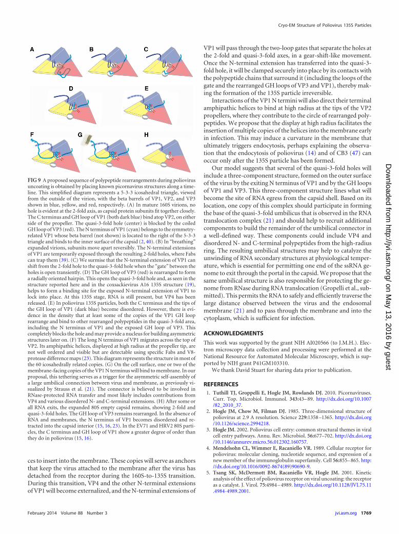
FIG 9 A proposed sequence of polypeptide rearrangements during poliovirusuncoating is obtained by placing known picornavirus structures along a time-line. This simplified diagram represents a 5-3-3 icosahedral triangle, viewedfrom the outside of the virion, with the beta barrels of VP1, VP2, and VP3shown in blue, yellow, and red, respectively. (A) In mature 160S virions, nohole is evident at the 2-fold axis, as capsid protein subunits fit together closely.The C terminus and GH loop of VP1 (both dark blue) bind atop VP2, on eitherside of the propeller. The quasi-3-fold hole (center) is blocked by the coiledGH loop of VP3 (red). The N terminus of VP1 (cyan) belongs to the symmetry-related VP1 whose beta barrel (not shown) is located to the right of the 5-3-3triangle and binds to the inner surface of the capsid (2, 40). (B) In “breathing”expanded virions, subunits move apart reversibly. The N-terminal extensionsof VP1 are temporarily exposed through the resulting 2-fold holes, where Fabscan trap them (39). (C) We surmise that the N-terminal extension of VP1 canshift from the 2-fold hole to the quasi-3-fold hole when the “gate” between theholes is open transiently. (D) The GH loop of VP3 (red) is rearranged to forma radially oriented hairpin. This opens the quasi-3-fold hole and, as seen in thestructure reported here and in the coxsackievirus A16 135S structure (19),helps to form a binding site for the exposed N-terminal extension of VP1 tolock into place. At this 135S stage, RNA is still present, but VP4 has beenreleased. (E) In poliovirus 135S particles, both the C terminus and the tips ofthe GH loop of VP1 (dark blue) become disordered. However, there is evi-dence in the density that at least some of the copies of the VP1 GH looprearrange and bind to other rearranged polypeptides in the quasi-3-fold area,including the N terminus of VP1 and the exposed GH loop of VP3. Thiscompletely blocks the hole and may provide a nucleus for building asymmetricstructures later on. (F) The long N terminus of VP1 migrates across the top ofVP2. Its amphipathic helices, displayed at high radius at the propeller tip, arenot well ordered and visible but are detectable using specific Fabs and V8-protease difference maps (23). This diagram represents the structure in most ofthe 60 icosahedrally related copies. (G) On the cell surface, one or two of themembrane-facing copies of the VP1 N terminus will bind to membrane. In ourproposal, this tethering serves as a trigger for the asymmetric self-assembly ofa large umbilical connection between virus and membrane, as previously vi-sualized by Strauss et al. (21). The connector is believed to be involved inRNase-protected RNA transfer and most likely includes contributions fromVP4 and various disordered N- and C-terminal extensions. (H) After some orall RNA exits, the expanded 80S empty capsid remains, showing 2-fold andquasi-3-fold holes. The GH loop of VP3 remains rearranged. In the absence ofRNA and membranes, the N terminus of VP1 becomes disordered and re-tracted into the capsid interior (15, 16, 23). In the EV71 and HRV2 80S parti-cles, the C terminus and GH loop of VP1 show a greater degree of order thanthey do in poliovirus (15, 16).
Cryo-EM Structure of Poliovirus 135S Particles
February 2014 Volume 88 Number 3 jvi.asm.org 1769
on May 13, 2016 by guest
http://jvi.asm.org/
Dow
nloaded from
6. De Sena J, Mandel B. 1977. Studies on the in vitro uncoating of poliovirus. II.Characteristics of the membrane-modified particle. Virology 78:554–566.
7. Fenwick ML, Cooper PD. 1962. Early interactions between poliovirusand ERK cells. Some observations on the nature and significance of therejected particles. Virology 18:212–223.
8. Curry S, Chow M, Hogle JM. 1996. The poliovirus 135S particle isinfectious. J. Virol. 70:7125–7131.
9. Fricks CE, Hogle JM. 1990. Cell-induced conformational change of po-liovirus: externalization of the amino terminus of VP1 is responsible forliposome binding. J. Virol. 64:1934 –1945.
10. Chow M, Newman JFE, Filman D, Hogle JM, Rowlands DJ, Brown F.1987. Myristylation of picornavirus capsid protein VP4 and its structuralsignificance. Nature 327:482– 486. http://dx.doi.org/10.1038/327482a0.
11. Tuthill TJ, Bubeck D, Rowlands DJ, Hogle JM. 2006. Characterization ofearly steps in the poliovirus infection process: receptor-decorated lipo-somes induce conversion of the virus to membrane-anchored entry-intermediate particles. J. Virol. 80:172–180. http://dx.doi.org/10.1128/JVI.80.1.172-180.2006.
12. Danthi P, Tosteson M, Li QH, Chow M. 2003. Genome delivery and ionchannel properties are altered in VP4 mutants of poliovirus. J. Virol. 77:5266 –5274. http://dx.doi.org/10.1128/JVI.77.9.5266-5274.2003.
13. Tosteson MT, Wang H, Naumov A, Chow M. 2004. Poliovirus bindingto its receptor in lipid bilayers results in particle-specific, temperature-sensitive channels. J. Gen. Virol. 85:1581–1589. http://dx.doi.org/10.1099/vir.0.19745-0.
14. Brandenburg B, Lee LY, Lakadamyali M, Rust MJ, Zhuang X, HogleJM. 2007. Imaging poliovirus entry in live cells. PLoS Biol. 5:e183. http://dx.doi.org/10.1371/journal.pbio.0050183.
15. Garriga D, Pickl-Herk A, Luque D, Wruss J, Caston JR, Blaas D,Verdaguer N. 2012. Insights into minor group rhinovirus uncoating: theX-ray structure of the HRV2 empty capsid. PLoS Pathog. 8:e1002473.http://dx.doi.org/10.1371/journal.ppat.1002473.
16. Wang X, Peng W, Ren J, Hu Z, Xu J, Lou Z, Li X, Yin W, Shen X, PortaC, Walter TS, Evans G, Axford D, Owen R, Rowlands DJ, Wang J,Stuart DI, Fry EE, Rao Z. 2012. A sensor-adaptor mechanism for entero-virus uncoating from structures of EV71. Nat. Struct. Mol. Biol. 19:424 –429. http://dx.doi.org/10.1038/nsmb.2255.
17. Bubeck D, Filman DJ, Cheng N, Steven AC, Hogle JM, Belnap DM.2005. The structure of the poliovirus 135S cell entry intermediate at 10-angstrom resolution reveals the location of an externalized polypeptidethat binds to membranes. J. Virol. 79:7745–7755. http://dx.doi.org/10.1128/JVI.79.12.7745-7755.2005.
18. Levy HC, Bostina M, Filman DJ, Hogle JM. 2010. Catching a virus in theact of RNA release: a novel poliovirus uncoating intermediate character-ized by cryo-electron microscopy. J. Virol. 84:4426 – 4441. http://dx.doi.org/10.1128/JVI.02393-09.
19. Ren J, Wang X, Hu Z, Gao Q, Sun Y, Li X, Porta C, Walter TS, GilbertRJ, Zhao Y, Axford D, Williams M, McAuley K, Rowlands DJ, Yin W,Wang J, Stuart DI, Rao Z, Fry EE. 2013. Picornavirus uncoating inter-mediate captured in atomic detail. Nat. Commun. 4:1929. http://dx.doi.org/10.1038/ncomms2889.
20. Bostina M, Levy H, Filman DJ, Hogle JM. 2011. Poliovirus RNA isreleased from the capsid near a twofold symmetry axis. J. Virol. 85:776 –783. http://dx.doi.org/10.1128/JVI.00531-10.
21. Strauss M, Levy H, Bostina M, Filman DJ, Hogle JM. 2013. RNAtransfer from poliovirus 135S particles across membranes is mediated bylong umbilical connectors. J. Virol. 87:3903–3914. http://dx.doi.org/10.1128/JVI.03209-12.
22. Belnap DM, Filman DJ, Trus BL, Cheng N, Booy FP, Conway JF, Curry S,Hiremath CN, Tsang SK, Steven AC, Hogle JM. 2000. Molecular tectonicmodel of virus structural transitions: the putative cell entry states of poliovirus. J.Virol. 74:1342–1354. http://dx.doi.org/10.1128/JVI.74.3.1342-1354.2000.
23. Lin J, Cheng N, Chow M, Filman DJ, Steven AC, Hogle JM, Belnap DM.2011. An externalized polypeptide partitions between two distinct sites ongenome-released poliovirus particles. J. Virol. 85:9974 –9983. http://dx.doi.org/10.1128/JVI.05013-11.
24. Suloway C, Shi J, Cheng A, Pulokas J, Carragher B, Potter CS, ZhengSQ, Agard DA, Jensen GJ. 2009. Fully automated, sequential tilt-seriesacquisition with Leginon. J. Struct. Biol. 167:11–18. http://dx.doi.org/10.1016/j.jsb.2009.03.019.
25. Mallick SP, Carragher B, Potter CS, Kriegman DJ. 2005. ACE: auto-
mated CTF estimation. Ultramicroscopy 104:8 –29. http://dx.doi.org/10.1016/j.ultramic.2005.02.004.
26. Mindell JA, Grigorieff N. 2003. Accurate determination of local defocusand specimen tilt in electron microscopy. J. Struct. Biol. 142:334 –347.http://dx.doi.org/10.1016/S1047-8477(03)00069-8.
27. Lander GC, Stagg SM, Voss NR, Cheng A, Fellmann D, Pulokas J,Yoshioka C, Irving C, Mulder A, Lau PW, Lyumkis D, Potter CS,Carragher B. 2009. Appion: an integrated, database-driven pipeline tofacilitate EM image processing. J. Struct. Biol. 166:95–102. http://dx.doi.org/10.1016/j.jsb.2009.01.002.
28. Roseman AM. 2004. FindEM—a fast, efficient program for automaticselection of particles from electron micrographs. J. Struct. Biol. 145:91–99.http://dx.doi.org/10.1016/j.jsb.2003.11.007.
29. Grigorieff N. 2007. FREALIGN: high-resolution refinement of single par-ticle structures. J. Struct. Biol. 157:117–125. http://dx.doi.org/10.1016/j.jsb.2006.05.004.
30. van Heel M, Schatz M. 2005. Fourier shell correlation threshold criteria.J. Struct. Biol. 151:250 –262. http://dx.doi.org/10.1016/j.jsb.2005.05.009.
31. Rosenthal PB, Henderson R. 2003. Optimal determination of particleorientation, absolute hand, and contrast loss in single-particle electroncryomicroscopy. J. Mol. Biol. 333:721–745. http://dx.doi.org/10.1016/j.jmb.2003.07.013.
32. Heymann JB, Belnap DM. 2007. Bsoft: image processing and molecularmodeling for electron microscopy. J. Struct. Biol. 157:3–18. http://dx.doi.org/10.1016/j.jsb.2006.06.006.
33. Basavappa R, Syed R, Flore O, Icenogle JP, Filman DJ, Hogle JM. 1994.Role and mechanism of the maturation cleavage of VP0 in poliovirusassembly: structure of the empty capsid assembly intermediate at 2.9Åresolution. Protein Sci. 3:1651–1669. http://dx.doi.org/10.1002/pro.5560031005.
34. Emsley P, Cowtan K. 2004. Coot: model-building tools for moleculargraphics. Acta Crystallogr. Sect. D Biol. Crystallogr. 60:2126 –2132. http://dx.doi.org/10.1107/S0907444904019158.
35. Collaborative Computational Project, Number 4. 1994. The CCP4 suite:programs for protein crystallography. Acta Crystallogr. D 50:760 –763.http://dx.doi.org/10.1107/S0907444994003112.
36. Kabsch W. 1976. A solution for the best rotation to relate two sets ofvectors. Acta Crystallogr. A 72:922–923.
37. Murshudov GN, Vagin AA, Dodson EJ. 1997. Refinement of macromo-lecular structures by the maximum-likelihood method. Acta Crystallogr.D 53:240 –255. http://dx.doi.org/10.1107/S0907444996012255.
38. Hewat EA, Neumann E, Blaas D. 2002. The concerted conformationalchanges during human rhinovirus 2 uncoating. Mol. Cell 10:317–326.http://dx.doi.org/10.1016/S1097-2765(02)00603-2.
39. Lin J, Lee LY, Roivainen M, Filman DJ, Hogle JM, Belnap DM. 2012.Structure of the Fab-labeled “breathing” state of native poliovirus. J. Virol.86:5959 –5962. http://dx.doi.org/10.1128/JVI.05990-11.
40. Filman DJ, Syed R, Chow M, Macadam AJ, Minor PD, Hogle JM. 1989.Structural factors that control conformational transitions and serotypespecificity in type 3 poliovirus. EMBO J. 8:1567–1579.
41. Hogle JM. 2012. A 3D framework for understanding enterovirus 71. Nat.Struct. Mol. Biol. 19:367–368. http://dx.doi.org/10.1038/nsmb.2276.
42. Baker TS, Cheng RH. 1996. A model-based approach for determining orienta-tions of biological macromolecules imaged by cryoelectronmicroscopy. J. Struct. Biol. 116:120 –130. http://dx.doi.org/10.1006/jsbi.1996.0020.
43. Minor PD. 1986. Antigenic structure of poliovirus. Microbiol. Sci. 3:141–144.
44. Page GS, Mosser AG, Hogle JM, Filman DJ, Rueckert RR, Chow M.1988. Three-dimensional structure of the poliovirus serotype 1 neutraliz-ing determinants. J. Virol. 62:1781–1794.
45. Chow M, Yabrov R, Bittle J, Hogle JM, Baltimore D. 1985. Syntheticpeptides from four separate regions of the poliovirus capsid protein VP1induce neutralizing antibodies. Proc. Natl. Acad. Sci. U. S. A. 82:910 –914.http://dx.doi.org/10.1073/pnas.82.3.910.
46. Roivainen M, Piraninen L, Rysa T, Narvanen A, Hovi T. 1993. An immu-nodominant N-terminal region of VP1 protein of poliovirion that isburied in crystal structure can be exposed in solution. Virology 195:762–765.
47. Coyne CB, Shen L, Turner JR, Bergelson JM. 2007. Coxsackievirus entryacross epithelial tight junctions requires occludin and the small GTPasesRab34 and Rab5. Cell Host Microbe 2:181–192. http://dx.doi.org/10.1016/j.chom.2007.07.003.
Butan et al.
1770 jvi.asm.org Journal of Virology
on May 13, 2016 by guest
http://jvi.asm.org/
Dow
nloaded from
Related Documents