www.espanolavet.ca phone: (705) 869-0090 fax: (705) 869-0092 visit us on facebook Dr. PJ Rocheleau, DVM and Associates 138 Tudhope St, Espanola ON, P5E 1S6 Cruciate Ligament Disease The Cranial Cruciate Ligament The cranial cruciate ligament (CrCL, aka anterior cruciate ligament or ACL) is one of several structures in the stifle (equivalent to our knee) that provide joint stability and allow normal function. The stifle is the joint formed by the femur, tibia and patella (“knee-cap”) and is a basic pulley system that allows the lower leg to swing in a backward and forward direction like a pendulum. Four ligaments prevent motion in other planes; two collateral ligaments that prevent side-to-side motion and two cruciate ligaments (because they cross each other) that prevent the tibia moving backward and forward independently of the femur. The cruciate ligaments also help limit internal and external rotation of the joint. The ability of the stifle to sustain motion in any of these directions means that one or more of these ligaments is damaged. Two other structures that help form the contact surface of the joint which are also very important are called the lateral and medial meniscus (plural: menisci). The cruciate ligaments together provide rotational stability to the joint; ie they limit the internal and external rotation that is possible. They do this by locking against each other when excessive rotational force is applied to the joint. If the cranial cruciate ligament is damaged, this motion is not checked and internal rotation may occur. This finding is variable between dogs; some dogs have significant problems with rotational instability while others seem to have far less issues. This is generally determined during the gait exam. The purpose of the CrCL is to prevent cranial tibial thrust – motion of the tibia in a forward and upward direction. Rupture of the CrCL allows this motion to occur, which precipitates most of the problems that happen with cruciate disease. This is a very important concept as it underpins the repair techniques used to correct this problem.

Welcome message from author
This document is posted to help you gain knowledge. Please leave a comment to let me know what you think about it! Share it to your friends and learn new things together.
Transcript
www.espanolavet.ca phone: (705) 869-0090 fax: (705) 869-0092 visit us on facebook
Dr. PJ Rocheleau, DVM and Associates
138 Tudhope St, Espanola ON, P5E 1S6
Cruciate Ligament Disease
The Cranial Cruciate Ligament
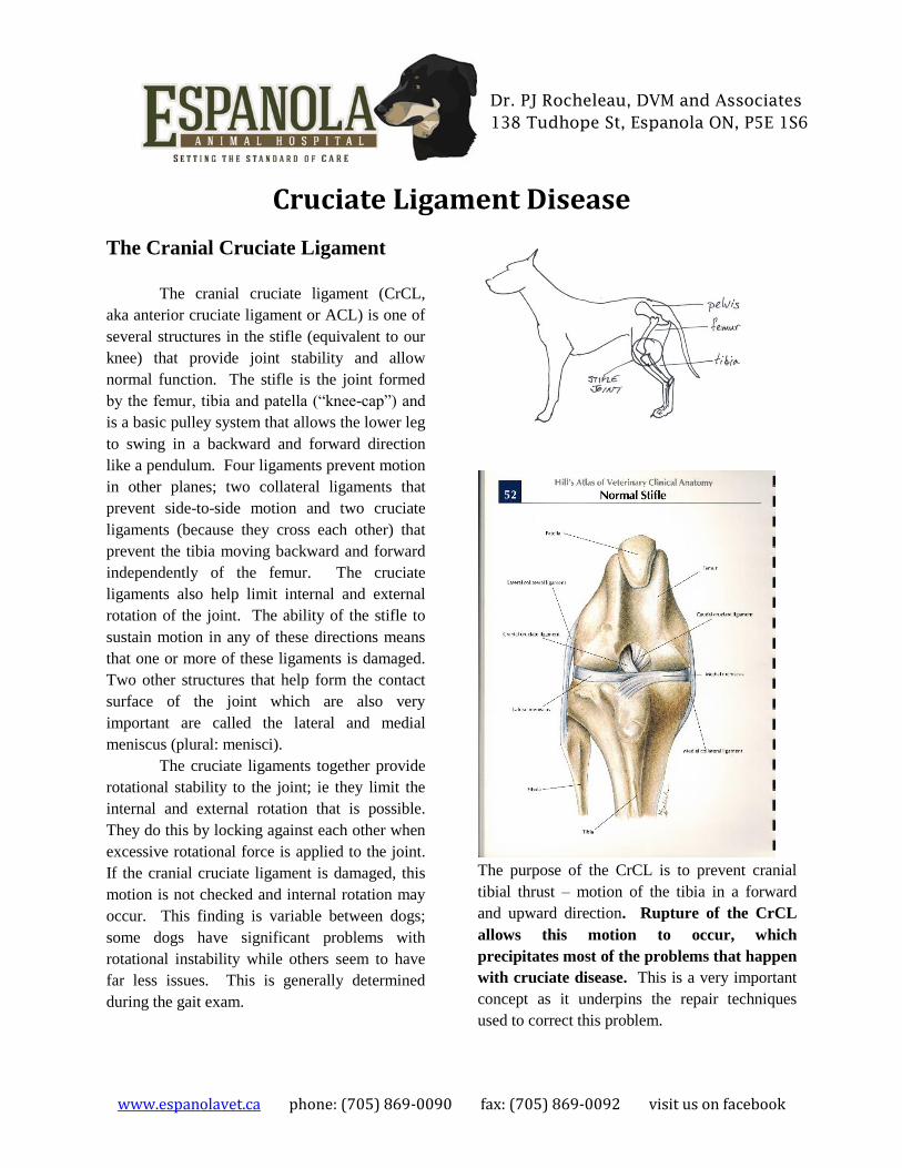
The cranial cruciate ligament (CrCL,
aka anterior cruciate ligament or ACL) is one of
several structures in the stifle (equivalent to our
knee) that provide joint stability and allow
normal function. The stifle is the joint formed
by the femur, tibia and patella (“knee-cap”) and
is a basic pulley system that allows the lower leg
to swing in a backward and forward direction
like a pendulum. Four ligaments prevent motion
in other planes; two collateral ligaments that
prevent side-to-side motion and two cruciate
ligaments (because they cross each other) that
prevent the tibia moving backward and forward
independently of the femur. The cruciate
ligaments also help limit internal and external
rotation of the joint. The ability of the stifle to
sustain motion in any of these directions means
that one or more of these ligaments is damaged.
Two other structures that help form the contact
surface of the joint which are also very
important are called the lateral and medial
meniscus (plural: menisci).
The cruciate ligaments together provide
rotational stability to the joint; ie they limit the
internal and external rotation that is possible.
They do this by locking against each other when
excessive rotational force is applied to the joint.
If the cranial cruciate ligament is damaged, this
motion is not checked and internal rotation may
occur. This finding is variable between dogs;
some dogs have significant problems with
rotational instability while others seem to have
far less issues. This is generally determined
during the gait exam.
The purpose of the CrCL is to prevent cranial
tibial thrust – motion of the tibia in a forward
and upward direction. Rupture of the CrCL
allows this motion to occur, which
precipitates most of the problems that happen
with cruciate disease. This is a very important
concept as it underpins the repair techniques
used to correct this problem.

www.espanolavet.ca phone: (705) 869-0090 fax: (705) 869-0092 visit us on facebook
Dr. PJ Rocheleau, DVM and Associates
138 Tudhope St, Espanola ON, P5E 1S6
How Do I Know If My Dog Has
Cruciate Ligament Disease?
Only a veterinarian can diagnose
cruciate disease by performing a proper
orthopedic examination and obtaining x-rays. A
number of signs can occur that suggest cruciate
disease and any hind limb lameness that occurs
in your dog should be evaluated. The vast
majority of medium and large dogs presented for
hind limb lameness do infact have cruciate
ligament disease. One retrospective study
recently performed at a major university showed
that of all large dogs presented for evaluation of
hip problems 60% had hip problems, while 98%
had cruciate ligament disease.
Cruciate ligament disease can occur in a
dog of any breed and any age, but tends to occur
in larger dogs. Unfortunately, this is often a
bilateral disease; approximately 40% of dogs
that have cruciate disease eventually have it in
both legs. Some breeds of dogs are predisposed
to bilateral disease: Labrador retrievers,
Mastiffs, Newfoundlands and Bernese Mountain
dogs are some examples. Concurrent orthopedic
disease such as hip dysplasia and patellar
luxation (dislocation of the “knee cap”) can
occur and may contribute to the cruciate disease.
It is important that these problems also be
recognized and addressed.
When you present your dog to your
veterinarian for a lameness problem, a complete
orthopedic examination is usually appropriate to
obtain a proper diagnosis. The lameness exam
should include a gait evaluation, a complete
physical examination including a detailed
examination of all 4 legs with the dog awake,
and a proper orthopedic examination. The
orthopedic examination itself is a very detailed
examination of all 4 limbs and all of the joints of
those limbs. This examination must be
performed under sedation and includes a number
of manipulations and physical tests to determine
the full extent of any existing orthopedic
problems. It is important to appreciate that the
dog has 4 legs; all limbs should be examined
thoroughly! Good quality, properly positioned
radiographs of any affected limbs and joints are
then obtained to assist diagnosis and plan
appropriate treatment.
Cruciate Disease, Progression and
Arthritis Rupture of the CrCL can either occur
acutely due to traumatic rupture or chronically
by tearing slowly over time until complete
mechanical failure occurs. We sometimes refer
to these chronic cases as a “partial tear”. When
chronic disease is present or a torn ligament goes
unrepaired, arthritis begins to develop and other
structures in the stifle can become damaged.
This dog has a partially torn CrCL. The tear is the
disorganized tissue to the lower left. The caudal
cruciate is visible in the background.
The meniscus can be torn resulting in
significant pain and worsening of the lameness
with an escalation in the rate at which the joint

www.espanolavet.ca phone: (705) 869-0090 fax: (705) 869-0092 visit us on facebook
Dr. PJ Rocheleau, DVM and Associates
138 Tudhope St, Espanola ON, P5E 1S6
degenerates and arthritis develops.
Approximately 50-60% of dogs with cruciate
disease have a damaged or torn meniscus at the
time of surgery. Meniscal injuries are dealt with
at the time of surgery. In dogs that have intact
meniscuses at the time of surgery, approximately
5% will tear it at some later point.
The severity of disease and the rate at
which it progresses is directly related to the
weight of the dog – the heavier the dog the more
severe and rapid the development of disease.
For this reason, rupture of the CrCL in dogs
that are over 10 Kg healthy body weight
requires surgical repair – period. All dogs,
including those under 10 Kg, will have a better
outcome with surgical treatment. There is no
way around this reality and failure to address
this disease surgically will usually result in
severe and rapid progression of disease.
In a large dog that has a ruptured
cruciate, the dog is usually presented with a non-
or partial weight bearing lameness that fails to
resolve with time. If the injury goes
undiagnosed or unattended, the dog may initially
appear to get better over a period of
approximately six weeks and the lameness may
appear to nearly resolve. If the dog is receiving
medical treatment during this time the lameness
may appear to have been “cured”. It is often
during this period that owners may conclude that
surgical repair is not really necessary and may
cancel surgery if it has already been booked.
Over the course of the next several
months or years the lameness will usually slowly
return and worsen until the dog stops bearing
weight on the leg altogether. As cruciate disease
is often bilateral (occurs in both limbs), and the
unaffected limb is now bearing the weight of
both legs, that CrCL may also rupture. At this
point it becomes very difficult for the dog to
walk and the animal is severely and obviously
lame on both hind limbs. It is advisable to repair
a ruptured cruciate as soon as possible after
positive diagnosis. The goal of surgery is to
restore normal function and mitigate the
development of arthritis. With timely
management the dog can be expected to have a
normal life expectancy with good function and
normal quality of life afterwards, especially with
some of the newer repair techniques in current
use.
This dog has a recently ruptured cruciate. The
joint shows no evidence of arthritis.
This dog has a ruptured cruciate that went
untreated for 12 months. The joint is severely
arthritic. This dog was non-weight-bearing on this
leg at presentation.
www.espanolavet.ca phone: (705) 869-0090 fax: (705) 869-0092 visit us on facebook
Dr. PJ Rocheleau, DVM and Associates
138 Tudhope St, Espanola ON, P5E 1S6
Medical Management
As mentioned previously, the vast
majority of cases of CrCL rupture require
surgical repair. However, medical management
is necessary pending surgery, in the
perioperative period (during recovery) and for
the rest of the dog’s life after surgery. It is
important to understand that surgery is a very
important event, but management of cruciate
disease is life-long. No matter how good a job
the surgeon does, it is important to understand
that arthritis will develop over time. The goal of
all therapy, including surgery, is to minimize the
development of arthritis so that the dog can live
a normal, healthy and pain-free existence.
Simply investing in surgery and failing or
refusing to follow instructions regarding long-
term management will result in poor results and
poor long-term outcome, often within months
following surgery.
It is also important to understand that
arthritis is not a disease. Hip dysplasia,
cruciate ligament disease, elbow dysplasia, etc,
are diseases. These diseases cause
inflammation; arthritis is simply inflammation
with the addition of time. As such, the goal of
all of our therapies is to prevent or suppress
inflammation, thereby preventing the
development of arthritis. Attempting to treat
arthritis is generally unproductive – at that point
it is too late.
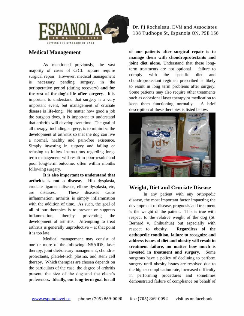
Medical management may consist of
one or more of the following: NSAIDS, laser
therapy, joint diet/dietary management, chondro-
protectants, platelet-rich plasma, and stem cell
therapy. Which therapies are chosen depends on
the particulars of the case, the degree of arthritis
present, the size of the dog and the client’s
preferences. Ideally, our long-term goal for all
of our patients after surgical repair is to
manage them with chondroprotectants and
joint diet alone. Understand that these long-
term treatments are not optional – failure to
comply with the specific diet and
chondroprotectant regimen prescribed is likely
to result in long term problems after surgery.
Some patients may also require other treatments
such as occasional laser therapy or medication to
keep them functioning normally. A brief
description of these therapies is listed below.
Weight, Diet and Cruciate Disease In any patient with any orthopedic
disease, the most important factor impacting the
development of disease, prognosis and treatment
is the weight of the patient. This is true with
respect to the relative weight of the dog (St.
Bernard v. Chihuahua) but especially with
respect to obesity. Regardless of the
orthopedic condition, failure to recognize and
address issues of diet and obesity will result in
treatment failure, no matter how much is
invested in treatment and surgery. Some
surgeons have a policy of declining to perform
surgery until obesity issues are resolved due to
the higher complication rate, increased difficulty
in performing procedures and sometimes
demonstrated failure of compliance on behalf of
www.espanolavet.ca phone: (705) 869-0090 fax: (705) 869-0092 visit us on facebook
Dr. PJ Rocheleau, DVM and Associates
138 Tudhope St, Espanola ON, P5E 1S6
the client. Your veterinarian should provide
specific dietary recommendations including not
only a specific diet(s), strict feeding guidelines
that include specific measuring instructions and
complete diet counselling. Any complicating
medical conditions such as hypothyroidism need
to be diagnosed and treated.
Joint Diets – A prescription veterinary diet
formulated specifically for addressing joint
disease and arthritis in our patients. These diets
are designed not only to deal with inflammation
associated with joint disese but are excellent at
addressing weight issues that will have the most
impact on patient outcomes. Joint diets have
had a major impact on how we manage joint
disease over the past decade, and for many dogs
on monotherapy have allowed us to replace
drugs with food.
Therapuetants Chondroprotectants - All dogs with any type
of joint disease should be on chondroprotectants
(glucosamine, with or without chondroitin) and
this is usually prescribed and supplied in our
hospital. Please note, glucosamine incorporated
into dry dog food is not present in sufficient
quantities to have a therapeutic effect – most of
it is destroyed during processing as it breaks
down under the high temperatures and pressures
used to make dry kibble. It has to be added to
the food after processing, usually as a top-
dressing added at feeding time by the client.
NSAID’s - All dogs presented for cruciate
disease initially start on NSAID’s as this is our
primary means of immediately addressing pain
and inflammation. While our other therapies are
just as good at addressing these issues they all
take a significant amount of time to start having
an effect – drug therapy is immediate. Often we
will withdraw the NSAID’s if possible when
other therapies have had time to take effect. A
number of options are available, including some
newer products that have a reduced incidence of
adverse effects.
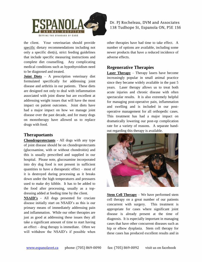
Regenerative Therapies Laser Therapy – Therapy lasers have become
increasingly popular in small animal practice
since they became widely available in the past 5
years. Laser therapy allows us to treat both
acute injuries and chronic disease with often
spectacular results. It is also extremely helpful
for managing post-operative pain, inflammation
and swelling and is included in our post-
operative management for all orthopedic cases.
This treatment has had a major impact on
dramatically lowering our post-op complication
rate for a variety of reasons. A separate hand-
out regarding this therapy is available.
Stem Cell Therapy – We have performed stem
cell therapy on a great number of our patients
concurrent with surgery. This treatment is
appropriate for cases where significant joint
disease is already present at the time of
diagnosis. It is especially important in managing
cases that have other concurrent diseases such as
hip or elbow dysplasia. Stem cell therapy for
these cases has produced excellent results and in

www.espanolavet.ca phone: (705) 869-0090 fax: (705) 869-0092 visit us on facebook
Dr. PJ Rocheleau, DVM and Associates
138 Tudhope St, Espanola ON, P5E 1S6
many cases an obvious reduction in radiographic
findings associated with osteoarthritis has been
noted when follow-up x-rays were taken several
months later. A separate hand-out regarding this
therapy is available.
Stem cells being processed from fatty tissue.
Surgical Management of Cruciate
Ligament Disease
There are a number of surgical
techniques currently available for treatment of
cruciate disease. The most common are divided
into 2 major groups; extracapuslar repair and
geometry modifying techniques. It is important
to understand that when appropriately selected,
no one technique has been demonstrated to have
an overall advantage over any other in the long
term, though there are advantages and
disadvantages of each. There is no “gold
standard” repair at this time. It is imperative
to select the most appropriate repair based on the
specific details of each individual case. We
currently offer Tight-rope®, Tibial Tuberosity
Advancement (TTA) and Tibial Plateau
Levelling Osteotomy (TPLO) in our hospital.
Extracapsular Repair Extracapsular techniques rely on using
very heavy suture materials to construct a
restraint on the external surface of the joint to
provide stability. Most of these repairs rely on
formation of scar tissue and fibrosis of the joint
capsule as the repair is expected to break down
over time. The two currently most common are
the lateral fabellar suture and Tight-rope®. A
number of studies have now demonstrated that
extracapsular techniques generally do not
perform as well as geometry modifying
techniques. We no longer provide extracapsular
repairs as a primary stabilization in our hospital,
regardless of patient size.
However, the current primary advantage
of extracapsular repairs are their ability to
provide rotational stability. In patients with
identified significant rotational stability, we will
implant a Tight-rope® in addition to a TPLO or
occasionally a TTA.
Tight-rope® Details Tight-rope® and several related
procedures that involve various bone anchor
techniques were developed to improve on the
older lateral fabellar suture technique. They are
based on isometric points determined through
careful experimental studies to be the most
stable anchor points for extracapsular joint
stabilization. The points are very specific and
the most challenging part of performing the
procedure is locating these points in the patient.
Newer modifications to the procedure have
made it easier to locate them with high
repeatability. Bone tunnels are drilled through
which a heavy braided suture is passed and
secured in place with titanium buttons. The
suture physically restrains the joint and acts as a
www.espanolavet.ca phone: (705) 869-0090 fax: (705) 869-0092 visit us on facebook
Dr. PJ Rocheleau, DVM and Associates
138 Tudhope St, Espanola ON, P5E 1S6
scaffold for the formation of fibrous scar tissue
and fibrosis of the surrounding joint capsule.
When performed correctly, these are
excellent stabilization techniques. The recovery
time is approximately 1-2 weeks to resume
weight-bearing and 16 weeks to fully heal and
come off exercise restriction. The complication
rate is approximately equal to that of other
extracapsular techniques. When performed as
part of a combined stabilization with another
technique, preference is given to the Tight-
rope® with respect to post-op care.
Tight-rope® in a small dog
Geometry Modifying Techniques A number of geometry modifying
techniques have been developed over the years
but two are currently in common use – TPLO
(tibial plateau levelling osteotomy) and TTA
(tibial tuberosity advancement). The manner in
which these repairs work is quite complex but
both involving cutting and repositioning parts of
the tibia and plating them in place. They
ultimately provide stability by eliminating tibial
thrust. These repairs require much greater
expertise to perform, have greater potential for
serious complications and are generally more
expensive. When properly performed these
techniques provide excellent results and client
satisfaction is very high.
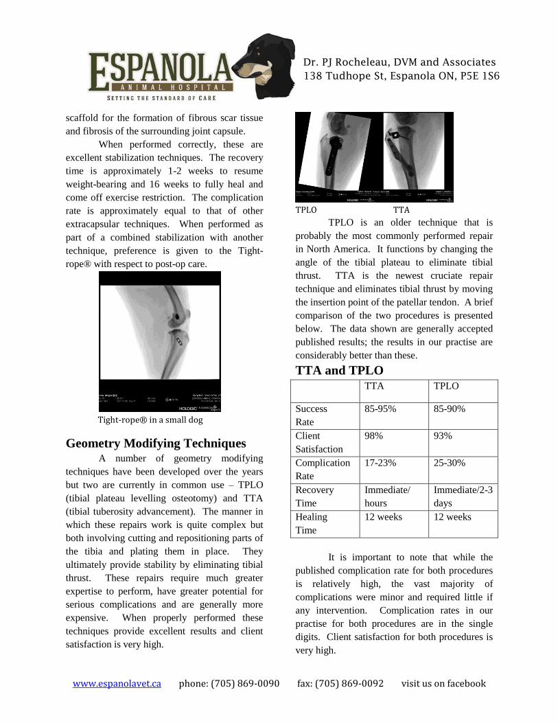
TPLO TTA
TPLO is an older technique that is
probably the most commonly performed repair
in North America. It functions by changing the
angle of the tibial plateau to eliminate tibial
thrust. TTA is the newest cruciate repair
technique and eliminates tibial thrust by moving
the insertion point of the patellar tendon. A brief
comparison of the two procedures is presented
below. The data shown are generally accepted
published results; the results in our practise are
considerably better than these.
TTA and TPLO
TTA TPLO
Success
Rate
85-95% 85-90%
Client
Satisfaction
98% 93%
Complication
Rate
17-23% 25-30%
Recovery
Time
Immediate/
hours
Immediate/2-3
days
Healing
Time
12 weeks 12 weeks
It is important to note that while the
published complication rate for both procedures
is relatively high, the vast majority of
complications were minor and required little if
any intervention. Complication rates in our
practise for both procedures are in the single
digits. Client satisfaction for both procedures is
very high.

www.espanolavet.ca phone: (705) 869-0090 fax: (705) 869-0092 visit us on facebook
Dr. PJ Rocheleau, DVM and Associates
138 Tudhope St, Espanola ON, P5E 1S6
TTA Details
TTA offers many specific advantages
that make it a highly valuable repair option for
many patients. The most attractive advantage to
most of our clients is the rapid recovery time –
most patients are fully weight bearing on the leg
immediately after surgery (as soon as the
epidural wears off –about 6 to 8 hours post-op).
This can be especially important for large dogs,
where it may be more physically difficult to
assist the dog post-operatively. It is also
extremely helpful for dogs that have multiple
joint problems or bilateral cruciate disease and
need to have a functional limb immediately post-
op. TTA will help provide a small degree of
rotational stability.
A TTA is performed by making a cut
through the tibial tuberosity to reposition it in an
outward and upward direction. A special
implant called a cage helps maintain it in the
correct position and a tension band plate secures
it in place. This is an extremely precise and
meticulous technique where correct placement to
the millimeter is necessary. For technical
reasons, all of the implants and screws are made
of titanium. The resulting triangular gap is filled
with bone graft to accelerate healing. The
incisions are closed and the leg is bandaged.
TPLO Details
TPLO is a well-proven and valuable
repair method commonly performed in many
referral centres. When properly performed it
results in elimination of tibial thrust by rotating
(“levelling”) the tibial plateau. For some dogs
that have very high tibial plateau angles, TPLO
is the only appropriate repair method. The cut-
off for TTA and tight-rope is a tibial plateau
angle greater than 30 degrees. We suspect that
dogs with slopes greater than 27 degrees
probably experience more benefit in the long-
term from TPLO than other techniques. Newer
implant designs in recent years such as locking
screws and pre-stressed/precontoured plates
have eliminated a lot of potential complications.
One disadvantage of TPLO is that it does not
account for rotational instability and can actually
make it worse (a complication called pivot shift).
A TPLO is performed by making a
circular cut through the back of the tibia and
rotating the resulting segment by a
predetermined amount to result in a tibial
plateau angle of about 5.5 degrees. The bone
segment is held in that position by a special plate
where it heals permanently. In our hospital the
site is treated with PRP to accelerate healing and
mitigate post-op pain. The incisions are closed
and the leg is bandaged.
Which Repair is Best For My Dog?
The choice of repair is tailored to fit the
specific needs of each individual patient. All of
the available treatments have different
advantages and disadvantages. The patient’s
tibial plateau is a major factor but many other
criteria also play a role in selecting a repair.
These criteria include the presence of caudal
femoral subluxation, the individual anatomy of
the tibia, size/weight, concurrent orthopaedic
disease, and many other individual patient
factors. The surgeon will take all of these
various factors and offer a repair that is most
likely to result in the best outcome for that
patient.
For patients with significant rotational
instability (about 15-20%), a recommendation to
perform a combined approach may be necessary.

www.espanolavet.ca phone: (705) 869-0090 fax: (705) 869-0092 visit us on facebook
Dr. PJ Rocheleau, DVM and Associates
138 Tudhope St, Espanola ON, P5E 1S6
This will involve a Tight-rope® being implanted
after first performing a stabilization with either
TPLO or TTA. There is slightly more cost
involved when this technique is necessary and
the post-operative care is different than with
TTA or TPLO alone. This is discussed in more
detail with clients on a case-by-case basis.
Small dogs and cats get TPLO’s
regardless of plateau angle (which is usually
excessive in any case) due to the technical
difficulties of performing TTA accurately in
very small patients. They are otherwise treated
in the same manner as a larger patient would be.
This includes arthroscopic joint treatment in all
cases. These patients generally do extremely
well; our standard of care does not have a size
limit!
Arthroscopy – Addressing the Joint
Stabilization of the joint with Tight-
rope®, TTA or TPLO is important but is only
half the procedure – the joint itself must be
addressed and tissues inspected for damage and
removed or debrided if necessary. Addressing
the joint is a major part of proper surgical
management and should be performed in every
single case. This is most commonly done by
open arthrotomy; the joint is incised and opened
up so that the surgeon can physically inspect and
operate on the joint with standard surgical
instruments. While this approach will
adequately address the joint if performed
properly, it is highly invasive and traumatic. It
also will not result in as complete or precise a
debridement of damaged tissues. The best
approach for performing this procedure is
arthroscopically.
An arthroscope is both a camera and a
magnifying glass – up to 20X magnification.
Arthroscopy is performed through 3 or 4 very
small holes through which both the scope and
miniaturized surgical instruments are passed into
the joint. The entire joint is visually inspected
and the damaged cruciate ligament is very
carefully debrided or removed. Both menisci are
also inspected for tears and other damage and
treated if necessary. We are also able to perform
these procedures far more accurately and
precisely than by open arthrotomy. Many
lesions not visible to the naked eye are very
easily visualized and treated with this minimally
invasive approach. Unfortunately, routine
arthroscopic treatment of the joint during
cruciate repair procedures is rarely performed in
the majority of referral centres. As part of our
commitment to provide the highest possible
standard of care to our patients, we have
invested the necessary resources so that every
patient undergoing cruciate surgery in our
facility is scoped.
Ruptured cruciate being arthrocopically debrided
with a 3.0mm motorized shaver.

www.espanolavet.ca phone: (705) 869-0090 fax: (705) 869-0092 visit us on facebook
Dr. PJ Rocheleau, DVM and Associates
138 Tudhope St, Espanola ON, P5E 1S6
Torn meniscus being arthroscopically debrided
with a radiofrequency ablation probe.
Post-Operative Care
Client compliance with post-operative
care is extremely important – failure to
meticulously follow instructions can, and
usually does result in severe complications
and treatment failure. It is our preference
whenever possible to provide complete and
comprehensive case management for the entire
post-op period. In our practise, we perform laser
therapy during the first two weeks post-op to aid
with recovery and pain management. Other pain
management such as NSAIDs, opioids
(codeine), bandaging, etc, are provided as is a
short course of antibiotics. Physiotherapy is a
crucial component of post-op management and
is initiated immediately. Physiotherapy
instructions are given at discharge and include
passive range-of-motion exercises and controlled
leash walks. Other than prescribed
physiotherapy, absolute exercise restriction is
necessary and off-leash activity is strictly
forbidden. Unrestricted access to flights of stairs
in the house is to be avoided, however going up
and down exterior stairs to get in or out of the
house is permissible (on-leash only!).
Sutures are removed after 14 days and
post-op x-rays are taken at 6 weeks. If post-op
x-rays are within expectations, owners are
instructed to continue with prescribed treatment
and physiotherapy until 12 weeks post-op, at
which point normal activity may be resumed.
For dogs with bilateral cruciate disease, the
second surgery can be booked at 6 weeks post-
op if the x-rays show sufficient healing. Our
long-term goal for our patients is maintenance
with glucosamine, joint diet and if necessary
annual laser treatments. Patients with more
advanced disease at surgery may require more
aggressive treatment for arthritis in the long
term.
Failure to follow instructions! The photo on the
left is immediately post-op. The photo on the right
is 6 weeks later – note the severely fractured tibial
tuberosity. The owner did not follow instructions
with regard to exercise restriction.

www.espanolavet.ca phone: (705) 869-0090 fax: (705) 869-0092 visit us on facebook
Dr. PJ Rocheleau, DVM and Associates
138 Tudhope St, Espanola ON, P5E 1S6
Cost
The cost of these procedures is as
follows:
Orthopedic exam: $450 + HST
(includes consult, sedation and whatever xrays
are necessary)
Cruciate Surgeries:
(includes laser therapy sessions, all routine post-
op medications, suture removal, rechecks, etc)
TTA or TPLO $3000 + HST
Tight-rope® with TPLO or TTA $3500 + HST
Note that 6-week post-op xrays are not
included in the cost of surgery: $50 + HST
(sedation not included if necessary, usually
xrays can be obtained without).
**A non-refundable deposit of $250.00 is
due at the time of booking any orthopedic
work-up and/or surgery.**
Related Documents