Review Article Cross-Talk between Gut Microbiota and Heart via the Routes of Metabolite and Immunity Jin Bu and Zhaohui Wang Department of Geriatrics, Union Hospital, Tongji Medical College, Huazhong University of Science and Technology, Wuhan 430022, China Correspondence should be addressed to Zhaohui Wang; [email protected] Received 25 March 2018; Accepted 16 May 2018; Published 3 June 2018 Academic Editor: Joanne Bowen Copyright © 2018 Jin Bu and Zhaohui Wang. This is an open access article distributed under the Creative Commons Attribution License, which permits unrestricted use, distribution, and reproduction in any medium, provided the original work is properly cited. Considering the prevalence of cardiovascular disease (CVD), significant interest has been focused on the gut microbiota-heart interaction because the gut microbiota has been recognized as a barometer of human health. Dysbiosis, characterized by changes in the gut microbiota in CVD, has been reported in cardiovascular pathologies, such as atherosclerosis, hypertension, and heart failure. Conversely, gut microbiota-derived metabolites, such as trimethylamine/trimethylamine N-oxide (TMA/TMAO), can impact host physiology. Further, bacterial dysbiosis can disturb gut immunity, which increases the risk of acute arterial events. Moreover, studies of germ-free mice have provided evidence that microbiota diversity and the presence of a specific microbe in the gut can affect immune cells in hosts. Therefore, the changes in the composition of the gut microbiota can affect host metabolism and immunity. Importantly, these effects are not only confined to the gut but also spreaded to distal organs. The purpose of the current review is to highlight the complex interplay between the microbiota and CVD via TMAO and different immune cells and discuss the roles of probiotics and nutrition interventions in modulating the intestinal microbiota as novel therapeutic targets of CVD. 1. Introduction Paralleling the improvement of the social economy and aging population, cardiovascular disease (CVD) becomes the lead- ing cause of death and disability worldwide. According to the American Heart Association, the overall rate of death attrib- utable to CVD in 2013 was 222.9 per 100,000 Americans, accounting for 35% of deaths before the age of 75 years [1]. Cardiovascular health including health behaviors (e.g., healthy dietary pattern, appropriate energy balance, and nonsmoking) and health factors (e.g., optimal blood lipid level and blood pressure (BP)) has been recently defined as the primary goals. The role of the ecological system consist- ing of gut commensal bacteria in cardiovascular health becomes the focus of the current research. In CVD, the ratio and abundance of the gut microbiota change and interventions using prebiotics, probiotics, and synbiotics share similar therapeutic efficacies in attenuating cardiac dysfunctions. Moreover, accumulating evidence indi- cates that bacterial dysbiosis increases cardiometabolic risks. It is likely that the gut microbiota substantially contributes to the global epidemic of CVD. The microbiota in the gut, coevolving with the host, mainly colonizes in the colon. It can perform multiple func- tions, such as fermentation of nondigestible dietary sub- stances, control of intestinal epithelial cell proliferation, and prevention from propagation of pathogenic microorganisms [2, 3]. Accumulating evidence also shows that manipulation of the composition of the gut microbiota affects host metab- olism and immunity [4], whereas the effect is not only con- fined to the intestine but also spreaded to distal organs through different pathways [5, 6]. In this review, we aimed to discuss the compositional and functional changes in the gut microbiota in relation to CVD, determine the effects of Hindawi Gastroenterology Research and Practice Volume 2018, Article ID 6458094, 8 pages https://doi.org/10.1155/2018/6458094

Welcome message from author
This document is posted to help you gain knowledge. Please leave a comment to let me know what you think about it! Share it to your friends and learn new things together.
Transcript

Review ArticleCross-Talk between Gut Microbiota and Heart via the Routes ofMetabolite and Immunity
Jin Bu and Zhaohui Wang
Department of Geriatrics, Union Hospital, Tongji Medical College, Huazhong University of Science and Technology,Wuhan 430022, China
Correspondence should be addressed to Zhaohui Wang; [email protected]
Received 25 March 2018; Accepted 16 May 2018; Published 3 June 2018
Academic Editor: Joanne Bowen
Copyright © 2018 Jin Bu and Zhaohui Wang. This is an open access article distributed under the Creative Commons AttributionLicense, which permits unrestricted use, distribution, and reproduction in any medium, provided the original work isproperly cited.
Considering the prevalence of cardiovascular disease (CVD), significant interest has been focused on the gut microbiota-heartinteraction because the gut microbiota has been recognized as a barometer of human health. Dysbiosis, characterized by changesin the gut microbiota in CVD, has been reported in cardiovascular pathologies, such as atherosclerosis, hypertension, and heartfailure. Conversely, gut microbiota-derived metabolites, such as trimethylamine/trimethylamine N-oxide (TMA/TMAO), canimpact host physiology. Further, bacterial dysbiosis can disturb gut immunity, which increases the risk of acute arterial events.Moreover, studies of germ-free mice have provided evidence that microbiota diversity and the presence of a specific microbe inthe gut can affect immune cells in hosts. Therefore, the changes in the composition of the gut microbiota can affect hostmetabolism and immunity. Importantly, these effects are not only confined to the gut but also spreaded to distal organs. Thepurpose of the current review is to highlight the complex interplay between the microbiota and CVD via TMAO and differentimmune cells and discuss the roles of probiotics and nutrition interventions in modulating the intestinal microbiota as noveltherapeutic targets of CVD.
1. Introduction
Paralleling the improvement of the social economy and agingpopulation, cardiovascular disease (CVD) becomes the lead-ing cause of death and disability worldwide. According to theAmerican Heart Association, the overall rate of death attrib-utable to CVD in 2013 was 222.9 per 100,000 Americans,accounting for 35% of deaths before the age of 75 years [1].Cardiovascular health including health behaviors (e.g.,healthy dietary pattern, appropriate energy balance, andnonsmoking) and health factors (e.g., optimal blood lipidlevel and blood pressure (BP)) has been recently defined asthe primary goals. The role of the ecological system consist-ing of gut commensal bacteria in cardiovascular healthbecomes the focus of the current research.
In CVD, the ratio and abundance of the gut microbiotachange and interventions using prebiotics, probiotics, and
synbiotics share similar therapeutic efficacies in attenuatingcardiac dysfunctions. Moreover, accumulating evidence indi-cates that bacterial dysbiosis increases cardiometabolic risks.It is likely that the gut microbiota substantially contributes tothe global epidemic of CVD.
The microbiota in the gut, coevolving with the host,mainly colonizes in the colon. It can perform multiple func-tions, such as fermentation of nondigestible dietary sub-stances, control of intestinal epithelial cell proliferation, andprevention from propagation of pathogenic microorganisms[2, 3]. Accumulating evidence also shows that manipulationof the composition of the gut microbiota affects host metab-olism and immunity [4], whereas the effect is not only con-fined to the intestine but also spreaded to distal organsthrough different pathways [5, 6]. In this review, we aimedto discuss the compositional and functional changes in thegut microbiota in relation to CVD, determine the effects of
HindawiGastroenterology Research and PracticeVolume 2018, Article ID 6458094, 8 pageshttps://doi.org/10.1155/2018/6458094
the gut microbiota on CVD from the view of trimethylamineN-oxide (TMAO) and immune cells, and evaluate how gutinterventions can lead to novel therapeutic targets for CVD.
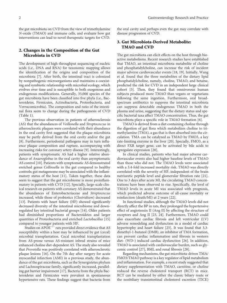
2. Changes in the Composition of the GutMicrobiota in CVD
The development of high-throughput sequencing of nucleicacids (i.e., DNA and RNA) for taxonomic mapping allowsthe identification of the origins and composition of themicrobiota [7]. After birth, the intestinal tract is colonizedby nonpathogenic microorganisms and maintains a coexist-ing and symbiotic relationship with microbial ecology, whichevolves over time and is susceptible to both exogenous andendogenous modifications. Generally, 35,000 species of thegut microbiota have been classified into five phyla (i.e., Bac-teroidetes, Firmicutes, Actinobacteria, Proteobacteria, andVerrucomicrobia). The composition and ratio of the intesti-nal flora seem to change during the pathogenesis of CVD(Table 1).
The previous observation in patients of atherosclerosis(AS) that the abundances of Veillonella and Streptococcus inatherosclerotic plaques were correlated with their abundancein the oral cavity first suggested that the plaque microbiotamay be partly derived from the oral cavity and/or the gut[8]. Furthermore, periodontal pathogens may in turn influ-ence plaque composition and rupture, accompanying withincreasing risks for coronary artery disease [9]. Interestingly,patients with symptomatic AS had a higher relative abun-dance of Anaeroglobus in the oral cavity than asymptomaticAS control [10]. Patients with symptomatic AS demonstratedenriched genus Collinsella in the gut compared to healthycontrols; gut metagenomemay be associated with the inflam-matory status of the host [11]. Taken together, these dataseem to suggest that the gut microbiome is more proinflam-matory in patients with CVD [12]. Specially, large-scale clin-ical research on patients with coronary AS demonstrated thatthe abundances of Enterobacteriaceae and Streptococcusincreased, while those of probiotics (Clostridium) decreased[13]. Patients with heart failure (HF) showed significantlydecreased diversity of the intestinal microbiome and down-regulated key intestinal bacterial groups [14]. Older patientshad diminished proportions of Bacteroidetes and largerquantities of Proteobacteria and enriched Lactobacillus [15]compared to younger patients with HF.
Studies on APOE−/− rats provided direct evidence that ASsusceptibility within a host may be influenced by gut (cecal)microbial transplantation. Cecal microbial transplantationfrom AS-prone versus AS-resistant inbred strains of miceenhanced choline diet-dependent AS. The study also revealedthat Prevotella was positively correlated with atheroscleroticplaque lesions [16]. On the 7th day after surgery for acutemyocardial infarction (AMI) in a previous study, the abun-dance of the gut microbiota, such as the Synergistetes phylumand Lachnospiraceae family, significantly increased, parallel-ing gut barrier impairment [17]. Bacteria from the phyla Bac-teroidetes and Firmicutes were prevalent in spontaneoushypertensive rats. These findings suggest that bacteria from
the oral cavity and perhaps even the gut may correlate withdisease progression of CVD.
3. Gut Microbiota-Derived Metabolite:TMAO and CVD
The gut microbiota can elicit effects on the host through bio-active metabolisms. Recent research studies have establishedthat TMAO, an intestinal microbiota metabolite of cholineand phosphatidylcholine, can increase the risk of incidentmajor adverse cardiovascular events [18, 19]. Initially, Wanget al. found that the three metabolites of the dietary lipidphosphatidylcholine, namely, choline, TMAO, and betaine,predicted the risk for CVD in an independent large clinicalcohort [5]. Then, they found that omnivorous humansubjects produced more TMAO than vegans or vegetariansfollowing the same ingestion. Furthermore, oral broad-spectrum antibiotics to suppress the intestinal microbiotacan suppress detectable endogenous TMAO in both theplasma and urine, suggesting that the dietary status and spe-cific bacterial taxa affect TMAO concentration. Thus, the gutmicrobiota plays a specific role in TMAO formation [6].
TMAO is derived from a diet containing choline throughthe digestion of gut flora which metabolizes choline to tri-methylamine (TMA), a gas that is then absorbed into the cir-culation. TMA can be catalyzed as TMAO by FMO3, a keyrate-limiting enzyme in the liver [20]. Specially, FMO3, as adirect FXR target gene, can be activated by bile acids toupregulate expression [20].
In clinical studies, patients who had major adverse car-diovascular events also had higher baseline levels of TMAOthan those who did not. The TMAO levels were associatedwith a 3.4-fold increased mortality risk. TMAO was directlycorrelated with the severity of HF, independent of the brainnatriuretic peptide level and glomerular filtration rate [21].One to 3 days after acute MI, the circulating TMAO concen-trations have been observed to rise. Specifically, the level ofTMAO levels in acute MI was associated with prognosis,which predicted adverse outcome of all-cause mortality orreinfarction (death/MI) at 2 years [22].
In functional studies, although the TMAO levels did notdirectly affect the BP in rats, they prolonged the hypertensiveeffect of angiotensin II (Ang II) by affecting the structure ofreceptors and Ang II [23, 24]. Furthermore, TMAO couldalso exacerbate cardiac fibrosis and left ventricular (LV)adverse remodeling and dysfunction in a model of cardiachypertrophy and heart failure [25]. It was found that 3,3-dimethyl-1-butanol (DMB), an inhibitor of TMA formation,can prevent cardiac inflammation and fibrosis in westerndiet- (WD-) induced cardiac dysfunction [26]. In addition,TMAO is associated with cardiovascular burden, such as gly-cemic control [27], BMI, and renal fibrosis [28].
Regarding mechanisms, the gut microbiota-driven TMA/FMO3/TMAO pathway is a key regulator of lipid metabolismand inflammation. For example, a recent study suggested thatdietary supplementation with TMAO, carnitine, or cholinereduced the reverse cholesterol transport (RCT) in mice.RCT can be mediated by either the classic biliary route orthe nonbiliary transintestinal cholesterol excretion (TICE)
2 Gastroenterology Research and Practice
pathway [29]. During lipid metabolism, (1) TMAO signifi-cantly increases the expression of ABCA1 and ABCG1 inthe liver which helps cholesterol efflux to apoA1 as the cho-lesterol acceptor; (2) TMAO in the gut also markedly reducesthe mRNA expression of NPC1L1, which transports choles-terol into the enterocyte from the gut lumen; (3) TMAOreduces the bile acid pool in the liver, which is associated withthe classic RCT by reducing synthetic enzymes CYP7A1 andCYP27A1; and (4) TMAO also reduces the expression ofABCG5/8 in the TICE pathway. Therefore, these complexprocesses inhibit RCT [6]. During proinflammation, (1)TMAO can promote the expression of scavenger receptors(SRs) in the macrophage, such as CD36 and SR-A [5, 30];(2) TMAO can also activate the well-known mitogen-activated protein kinase, extracellular signal-related kinase,and nuclear factor-κB signaling cascade in primary humanaortic endothelial cells and vascular smooth muscle cells[31]; (3) TMAO can also promote activation of PKC/NF-KB/VCAM-1, which accelerates endothelial dysfunctionincluding decreased endothelial self-repair and increasedmonocyte adhesion [32]; (4) TMAO may be associated withmacrophage polarization in WD-fed rats by increasingTNF-α and interleukin 1β (IL-1β) levels and decreasinganti-inflammatory factor IL-10 levels [23]; and (5) TMAOcan significantly trigger oxidative stress and activateTXNIP-NLRP3 inflammasomes to release IL-1β and IL-18in a dose- and time-dependent manner [33] in vitro. These
studies in vivo and vitro pave the way for the clinical regula-tion of intestinal microorganisms and dietary interventionsto prevent the formation of TMAO and improve CVD.
4. Gut Microbiota and Immunity in CVD
AS and resulting CVD involve inflammatory reactions inwhich both the innate and acquired immunities are involved,and anti-inflammatory therapy has received concerns [34].The gut mucosa as one of the largest immunological activeorgans in the human body harbors several hundred trillionsof bacteria, which are closely tied with the immune system,each influencing and being influenced by the other [35].Therefore, whether gut microbes interact with CVD in termsof immunity remains unclear.
Recent research revealed that patients with chronicinflammatory bowel diseases, such as Crohn’s disease andulcerative colitis, were at an increased risk of acute arterialevents [36], especially young patients [37], and the diseaseinflammatory state may be an independent risk factor foracute arterial events. Clinical studies and animal experimentshave demonstrated that elevated plasma cholesterol attrib-uted to WDs promoted CVD development. In the process,dietary cholesterol initiates intestinal inflammation in epithe-lial cells [38], disrupts the immune homeostasis, induces gutdysbiosis, and increases CD4+ and CD8+ cell infiltration indistal organs, such as the heart [26]. Chronic inflammation
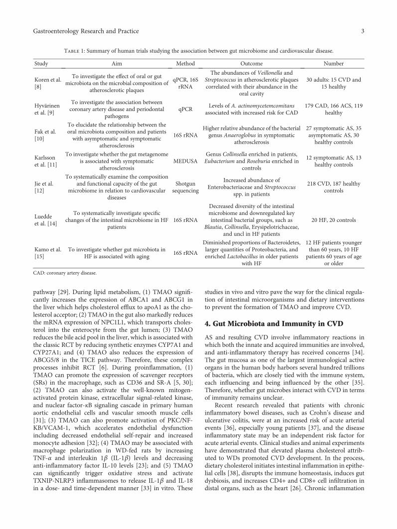
Table 1: Summary of human trials studying the association between gut microbiome and cardiovascular disease.
Study Aim Method Outcome Number
Koren et al.[8]
To investigate the effect of oral or gutmicrobiota on the microbial composition of
atherosclerotic plaques
qPCR, 16SrRNA
The abundances of Veillonella andStreptococcus in atherosclerotic plaquescorrelated with their abundance in the
oral cavity
30 adults: 15 CVD and15 healthy
Hyvärinenet al. [9]
To investigate the association betweencoronary artery disease and periodontal
pathogensqPCR
Levels of A. actinomycetemcomitansassociated with increased risk for CAD
179 CAD, 166 ACS, 119healthy
Fak et al.[10]
To elucidate the relationship between theoral microbiota composition and patientswith asymptomatic and symptomatic
atherosclerosis
16S rRNAHigher relative abundance of the bacterialgenus Anaeroglobus in symptomatic
atherosclerosis
27 symptomatic AS, 35asymptomatic AS, 30
healthy controls
Karlssonet al. [11]
To investigate whether the gut metagenomeis associated with symptomatic
atherosclerosisMEDUSA
Genus Collinsella enriched in patients,Eubacterium and Roseburia enriched in
controls
12 symptomatic AS, 13healthy controls
Jie et al.[12]
To systematically examine the compositionand functional capacity of the gut
microbiome in relation to cardiovasculardiseases
Shotgunsequencing
Increased abundance ofEnterobacteriaceae and Streptococcus
spp. in patients
218 CVD, 187 healthycontrols
Lueddeet al. [14]
To systematically investigate specificchanges of the intestinal microbiome in HF
patients16S rRNA
Decreased diversity of the intestinalmicrobiome and downregulated keyintestinal bacterial groups, such as
Blautia, Collinsella, Erysipelotrichaceae,and uncl in HF patients
20 HF, 20 controls
Kamo et al.[15]
To investigate whether gut microbiota inHF is associated with aging
16S rRNA
Diminished proportions of Bacteroidetes,larger quantities of Proteobacteria, andenriched Lactobacillus in older patients
with HF
12 HF patients youngerthan 60 years, 10 HFpatients 60 years of age
or older
CAD: coronary artery disease.
3Gastroenterology Research and Practice
of the gut in cases of dysbiosis affects not only itself but alsothe systemic circulation and central or peripheral tissues.Therefore, we can conclude that the intervention of intestinalimmunity may be another novel therapeutic target for theprevention of CVD [34].
4.1. Gut Microbiota and Immunity in AS. The relationshipbetween inflammation and immune responses has been clar-ified by our understanding of innate and adaptive immunol-ogy. First, inflammation as a result of innate and adaptiveimmunology is produced from the start to the end in AS.Innate immunology responses are initiated by the body’s rec-ognition of the signature molecules DAMPs and PAMPs,which could be recognized by pattern recognition receptors(PRRs) [39]. Macrophage PRRs, such as SRs, CD36, andSR-A, internalize oxidation-specific epitopes (i.e., exposedphosphocholine, malondialdehyde, and oxidized cardioli-pin). Gut microbe-derived TMAO could enforce the expres-sion of the receptor of SR-A and CD36, which promotes theformation of foam cells whose accumulation within the sub-endothelium or neointima constitutes the first step in AS.However, the study that germ-free mice were as susceptibleto AS as mice with symbiotic bacteria suggested that endog-enous substances initiated the inflammation [40]. Endoge-nous substances, such as cholesterol crystals deposited inmature atherosclerotic lesions, induced inflammation bystimulating the caspase-1-activating NLRP3 inflammasome[41]. In vitro, TMAO can also activate the NLRP3 inflamma-some. Meanwhile, the formation of foam cells and the activa-tion of NLRP3 are both early events. Therefore, theintervention of the gut may be considered therapies for theearly prevention of coronary AS.
Adaptive immune responses contribute to the develop-ment and complications of atherosclerotic lesions. For exam-ple, reduced or functionally impaired regulatory T cells(Treg) lead to an increased incidence of AS [42], and adop-tive transfer of Treg into hypercholesterolemic mice reduceslesion development [43]. A study in Ldlr−/− AS mice showedthat a cholesterol-rich diet can impair the Treg, whereasreversal of a hypercholesterol diet could prevent loss oflesional Treg [44]. Interestingly, an oral anti-CD3 antibodyinduced Treg and inhibited the development of AS in mice[45]. These observations suggest that the gut interventioncan regulate the function and quantity of the Treg for pre-venting atherosclerotic CVD. In addition, it has been estab-lished in germ-free mice or mice treated with antibioticsthat intestinal microbes are associated with the differentia-tion of T cells [46, 47].
The role of natural killer T (NKT) cells represents a linkbetween the innate and adaptive immune systems inresponse to AS, and numerous murine studies have shownthat NKT cells are proatherogenic via the activation of theirsecreted cytokines, such as Th1, Th2, and Th17 cytokines[48]. Adult germ-free mice have increased absolute or rela-tive levels of colonic NKT cells and are immature and hypo-responsive to αGalCer stimulation of splenic NKT cells.Neonatal germ-free mice can restore their NKT cell numberand phenotype when exposed to intestinal microorganismscontaining NKT cell antigens. This suggests that the gut
microbiota can affect the function of the NKT cells in theorgans. Although the direct association of the gut microbiotawith AS remains obscure, these research studies provide aperspective that the intervention for the gut may affect theimmune cells in CVD.
4.2. Gut Microbiota and Immunity in HF. HF is primarily aclinical diagnosis that develops secondary to either LV sys-tolic or diastolic dysfunction, which is the final outcome ofvarious CVDs [49]. HF with a preserved ejection fraction(EF) and HF with a reduced EF account for 50% of cases[50]. The levels of numerous proinflammatory cytokines,such as TNF-α, IL-1, IL-6, and IL-12, have been observedto be elevated in patients and in animal models with HF[51, 52]. Elevated circulating CD14+ monocyte counts 3 daysafter MI predict a failure to recover the LV systolic functionat 3 months post-MI in patients [53]. Endotoxins, an impor-tant stimulus for cytokine production in patients with HF[54], can originate from disrupted intestines resulting froma reduced cardiac output, potentially leading to further exac-erbations. An early study on intestinal microbes and HF dis-played the perspective of intestinal permeability, that is, guthypothesis. Whether manipulation of the gut microbiotacan attenuate HF and improve long-term outcomes remainsto be determined.
5. Gut Microbiota as a Potential TherapeuticTarget in CVD
The experimental evidence and clinical observations provethe functional link between the intestinal microbiota andCVD, suggesting the theoretical basis to manipulating intes-tinal microbiota to prevent CVD. The gut microbiotaconsisting of trillions of commensal microorganisms canexpress enzymes capable of interacting and interfering withthe nutrition and drug we consume, ultimately impactingeach other.
First, the use of prebiotics, probiotics, and synbiotics,which is the easiest way to interfere with microbiota compo-sition, had a similar efficacy in reducing LV dysfunctions inobese insulin-resistant rats [55]. For example, probiotics, asa kind of live beneficial bacteria, are administered to reestab-lish an appropriate intestinal balance (Table 2). In patientswith CVDs, consumption of Lactobacillus plantarum (DSM9843) increased the intestinal microbial diversity comparedto a placebo [56]. Probiotic Bifidobacterium animalis subsp.lactis LKM512 could decrease the fecal TMA concentrationand BMI in individuals [57]. Lab4 probiotic consortium plusL. plantarum CUL66 possessed anticholesterolemic capabili-ties in wild-type C57BL/6J mice fed with a high-fat diet. Pro-biotic Enterobacter aerogenes ZDY01 attenuated choline-induced TMAO levels via remodeling of the gut microbiotain mice [58]. Unfortunately, the treatment using probioticL. casei Shirota in patients with metabolic syndrome [59]and VSL#3 in nonobese men during the consumption of ahypercaloric and high-fat diet cannot attenuate the produc-tion of TMAO [60]. Moreover, probiotics consisting of dif-ferent strains may have various effects on CVD; thus,identifying the appropriate strains is essential for therapy.
4 Gastroenterology Research and Practice

Functional genomics on the most closely related referencestrains provided specific treatment strategies to restrainTMA producers and limit their proliferation [61].
Second, the effects of other components in diets can reg-ulate the composition of the intestinal flora (Table 3). InAPOE−/− mice fed with a high-fat/cholesterol diet[62], plantsterol ester (PSE), which elevated the relative abundance ofHelicobacter, Erysipelotrichaceae, and the genus Roseburiain the gut, is associated with reduced cholesterol levels, aorticplaques, and body fat. Oat β-glucan (OBG) can elevate buty-rate levels and promote Verrucomicrobia population expan-sion. Thus, it appears to protect against high-fat/cholesterol-induced atherogenesis [63]. High-fiber diets can increase theprevalence of acetate-producing bacteria and improve thelevels of Bacteroides acidifaciens, thereby preventing thedevelopment of hypertension and HF in hypertensive mice[64]. Inulin-type fructans (ITFs) can reverse endothelial
dysfunction via increased NO-producing bacteria inAPOE−/− mice [65]. Vegetable/fruit juices promote weightloss, increase vasodilator NO levels, and decrease lipid oxida-tion in healthy individuals by decreasing the proportion ofthe phylum Firmicutes and Proteobacteria and increasingBacteroidetes and Cyanobacteria in the stool [66]. The com-bination of ω-3 PUFAs and proanthocyanidins can providecardiovascular benefits by maintaining the standard propor-tions of bacterial subgroups in the gut of a healthy rat [67].Dietary allicin, a potent antimicrobial compound found ingarlic, also reduces the transformation of TMAO from L-carnitine through impacting the gut microbiota in mice[68]. In addition, 3,3-DMB, a structural analog of cholineprevalent in wine, olive oil, and grapeseed oil, inhibits TMAproduction from the gut microbes by inhibiting distinctmicrobial TMA [69]. Resveratrol, a natural phytoalexin withprebiotic benefits, has been found to attenuate TMAO-
Table 2: Summary of the human randomized controlled trials analyzing the effect of probiotic supplementation on CVD.
Authors Aim Population Main findings Duration
Karlsson et al. [56]To clarify the effect of L. plantarum
(DSM 9843) on intestinal microbiota inpatients with cardiovascular disease
16 males withatherosclerotic plaque
Increased bacterial diversity anddecreased concentration of
isovalericacid and valeric acid
4 weeks
Matsumoto et al. [57]
To investigate the effect of probioticBifidobacterium animalis subsp. lactis
LKM512 on colonic TMA andatherosclerosis-related makers in
healthy subjects
27 healthy adultsReduced fecal TMA
concentration and BMI12 weeks
Tripolt et al. [59]
To investigate the impact ofLactobacillus casei Shirota (LcS) on theformation of TMAO in subjects with
metabolic syndrome
30 subjects with metabolicsyndrome
Not affecting levels of TMAO 12 weeks
Boutagy et al. [60]
To investigate whether multistrainprobiotic VSL#3 would attenuate the
increase in fasting plasma concentrationsof TMAO following a high-fat diet
Nonobese males Not affecting levels of TMAO 2 weeks
Table 3: Nutrition intervention alters gut microbiota composition and improves CVD.
Nutrition intervention Main findings Alterations in gut microbiota composition
PSE (plant sterol ester) Cholesterol and aortic plaque ↓ Erysipelotrichaceae ↑
OBG (oat β-glucan) Cholesterol, aortic plaque, weight, and fat ↓ Butyrate levels and Verrucomicrobia ↑
High-fiber diet Improve hypertension and heart failure Acetate-producing bacteria ↑
ITF (inulin-type fructans) Reverse endothelial dysfunctionAkkermansia ↑Bacterial taxa ↓
Vegetable/fruit juicesPromote weight loss, increase vasodilator
NO, and decrease lipid oxidationFirmicutes/Proteobacteria ↓
Bacteroidetes ↑ Cyanobacteria ↑
ω-3 PUFAs and proanthocyanidins Plasma cholesterol ↓Maintains the standard proportions of bacterial
subgroups and their function
Allicin TMAO ↓ Clostridium ↑
DMB TMAO ↓ Inhibit distinct microbial TMA lyases
Resveratrol TMAO ↓ Lactobacillus and Bifidobacterium ↑
Ganoderma lucidum mycelium Weight, inflammation, and insulin resistance ↓Firmicutes-to-Bacteroidetes ratios and
endotoxin-bearing Proteobacteria levels ↓
5Gastroenterology Research and Practice

induced AS by decreasing plasma TMAO levels and increas-ing hepatic bile acid neosynthesis by changing the intestinalflora [70]. In addition, Ganoderma lucidum (G. lucidum), amedicinal mushroom used in traditional Chinese medicine,has been reported to have antiobesity properties, which aremediated by modulating the composition of the gut microbi-ota [71]. G. lucidum and its high-molecular-weight polysac-charides may be used as prebiotic agents to prevent gutdysbiosis and obesity-related metabolic disorders in micefed with a high-fat diet [72].
Given the abovementioned intervention, the gut can reg-ulate microbial community, which can further improveCVDs. When exposed to poison, such as acrolein [73], thegut increased the levels of intestinal Coprococcus andenhanced macrophage atherogenicity in atheroscleroticmice. Thus, gut microbiota remodeling plays a role in thedevelopment of CVDs and risks.
6. Concluding Remarks and Future Perspectives
The evidence from animal and human studies supports thatgut microbiota is in correlation with cardiovascular disease.Unappreciated complexity and considerable diversity of thebacterial microbiome have been gradually uncovered viaculture-independent methods. However, the direct relation-ship between gut microbiota and cardiovascular diseaseremains obscure. In addition, the diversity of microbiomeenhanced the difficulty in identifying strains in correlationwith disease state, which restricted therapeutic interventionsfor the exact target.
Apart from these restrictions, intestinal flora as a barom-eter of human health is a novel therapeutic target for prevent-ing CVD. Further, tunable expression platforms for theprominent microbiome in which gene expression is con-trolled by a synthetic inducer may be a good tool. Larger ran-domized controlled studies of adequate sample size andduration and well-defined therapeutic schedules and end-points are strongly advisable.
Conflicts of Interest
The authors of this article have nothing to disclose. No con-flicts of interest exist.
References
[1] D. Mozaffarian, E. J. Benjamin, A. S. Go et al., “Heart diseaseand stroke statistics—2016 update: a report from the Ameri-can Heart Association,” Circulation, vol. 133, no. 4, pp. e38–360, 2016.
[2] R. Jumpertz, D. S. Le, P. J. Turnbaugh et al., “Energy-balancestudies reveal associations between gut microbes, caloric load,and nutrient absorption in humans,” The American Journal ofClinical Nutrition, vol. 94, no. 1, pp. 58–65, 2011.
[3] F. Purchiaroni, A. Tortora, M. Gabrielli et al., “The role ofintestinal microbiota and the immune system,” EuropeanReview for Medical and Pharmacological Sciences, vol. 17,no. 3, pp. 323–333, 2013.
[4] J. L. Round and S. K. Mazmanian, “The gut microbiota shapesintestinal immune responses during health and disease,”Nature Reviews Immunology, vol. 9, no. 5, pp. 313–323, 2009.
[5] Z. Wang, E. Klipfell, B. J. Bennett et al., “Gut flora metabolismof phosphatidylcholine promotes cardiovascular disease,”Nature, vol. 472, no. 7341, pp. 57–63, 2011.
[6] R. A. Koeth, Z. Wang, B. S. Levison et al., “Intestinal microbi-ota metabolism of L-carnitine, a nutrient in red meat, pro-motes atherosclerosis,” Nature Medicine, vol. 19, no. 5,pp. 576–585, 2013.
[7] T. H. Hansen, R. J. Gøbel, T. Hansen, and O. Pedersen, “Thegut microbiome in cardio-metabolic health,” Genome Medi-cine, vol. 7, no. 1, p. 33, 2015.
[8] O. Koren, A. Spor, J. Felin et al., “Human oral, gut, and plaquemicrobiota in patients with atherosclerosis,” Proceedings of theNational Academy of Sciences of the United States of America,vol. 108, Supplement 1, pp. 4592–4598, 2011.
[9] K. Hyvärinen, P. Mäntylä, K. Buhlin et al., “A common peri-odontal pathogen has an adverse association with both acuteand stable coronary artery disease,” Atherosclerosis, vol. 223,no. 2, pp. 478–484, 2012.
[10] F. Fak, V. Tremaroli, G. Bergstrom et al., “Oral microbiota inpatients with atherosclerosis,” Atherosclerosis, vol. 243, no. 2,pp. 573–578, 2015.
[11] F. H. Karlsson, F. Fak, I. Nookaew et al., “Symptomatic athero-sclerosis is associated with an altered gut metagenome,”Nature Communications, vol. 3, no. 1, article 1245, 2012.
[12] Z. Jie, H. Xia, S. L. Zhong et al., “The gut microbiome in ath-erosclerotic cardiovascular disease,” Nature Communications,vol. 8, no. 1, article 845, 2017.
[13] W. T. Barrington and A. J. Lusis, “Atherosclerosis: associationbetween the gut microbiome and atherosclerosis,” NatureReviews Cardiology, vol. 14, no. 12, pp. 699-700, 2017.
[14] M. Luedde, T. Winkler, F. A. Heinsen et al., “Heart failure isassociated with depletion of core intestinal microbiota,” ESCHeart Failure, vol. 4, no. 3, pp. 282–290, 2017.
[15] T. Kamo H. Akazawa et al., “Dysbiosis and compositionalalterations with aging in the gut microbiota of patients withheart failure,” PLos One, vol. 12, article e0174099, 2017.
[16] J. C. Gregory, J. A. Buffa, E. Org et al., “Transmission of ath-erosclerosis susceptibility with gut microbial transplantation,”Journal of Biological Chemistry, vol. 290, no. 9, pp. 5647–5660,2015.
[17] Z. X. Wu, S. F. Li, H. Chen et al., “The changes of gut microbi-ota after acute myocardial infarction in rats,” PLoS One,vol. 12, no. 7, article e0180717, 2017.
[18] W. H. Tang, Z. Wang, B. S. Levison et al., “Intestinal microbialmetabolism of phosphatidylcholine and cardiovascular risk,”The New England Journal of Medicine, vol. 368, no. 17,pp. 1575–1584, 2013.
[19] E. Ierardi, C. Sorrentino, M. Principi, F. Giorgio, G. Losurdo,and A. di Leo, “Intestinal microbial metabolism of phosphati-dylcholine: a novel insight in the cardiovascular risk scenario,”Hepatobiliary Surgery and Nutrition, vol. 4, no. 4, pp. 289–292,2015.
[20] B. J. Bennett, T. Q. de Aguiar Vallim, Z. Wang et al., “Tri-methylamine-N-oxide, a metabolite associated with athero-sclerosis, exhibits complex genetic and dietary regulation,”Cell Metabolism, vol. 17, no. 1, pp. 49–60, 2013.
[21] W. H. Tang, Z. Wang, Y. Fan et al., “Prognostic value ofelevated levels of intestinal microbe-generated metabolite
6 Gastroenterology Research and Practice

trimethylamine-N-oxide in patients with heart failure: refiningthe gut hypothesis,” Journal of the American College of Cardi-ology, vol. 64, no. 18, pp. 1908–1914, 2014.
[22] T. Suzuki, L. M. Heaney, D. J. Jones, and L. L. Ng, “Trimethy-lamine N-oxide and risk stratification after acute myocardialinfarction,” Clinical Chemistry, vol. 63, no. 1, pp. 420–428,2017.
[23] M. Ufnal, R. Jazwiec, M. Dadlez, A. Drapala, M. Sikora, andJ. Skrzypecki, “Trimethylamine-N-oxide: a carnitine-derivedmetabolite that prolongs the hypertensive effect of angiotensinII in rats,” Canadian Journal of Cardiology, vol. 30, no. 12,pp. 1700–1705, 2014.
[24] J. Mondal, G. Stirnemann, and B. J. Berne, “When does tri-methylamine N-oxide fold a polymer chain and urea unfoldit?,” The Journal of Physical Chemistry. B, vol. 117, no. 29,pp. 8723–8732, 2013.
[25] C. L. Organ, H. Otsuka, S. Bhushan et al., “Choline dietand its gut microbe–derived metabolite, trimethylamineN-oxide, exacerbate pressure overload–induced heart fail-ure,” Circulation: Heart Failure, vol. 9, no. 1, articlee002314, 2016.
[26] K. Chen, X. Zheng, M. Feng, D. Li, and H. Zhang, “Gutmicrobiota-dependent metabolite trimethylamine N-oxidecontributes to cardiac dysfunction in western diet-inducedobese mice,” Frontiers in Physiology, vol. 8, p. 139, 2017.
[27] W. H. Tang, Z. Wang, X. S. Li et al., “Increased trimethylamineN-oxide portends high mortality risk independent of glycemiccontrol in patients with type 2 diabetes mellitus,” ClinicalChemistry, vol. 63, no. 1, pp. 297–306, 2017.
[28] J. R. Stubbs, J. A. House, A. J. Ocque et al., “Serum trimethyla-mine-N-oxide is elevated in CKD and correlates with coronaryatherosclerosis burden,” Journal of the American Society ofNephrology, vol. 27, no. 1, pp. 305–313, 2016.
[29] M. Warrier, D. M. Shih, A. C. Burrows et al., “The TMAO-generating enzyme flavin monooxygenase 3 is a central regula-tor of cholesterol balance,” Cell Reports, vol. 10, no. 3, pp. 326–338, 2015.
[30] M. Febbraio, E. A. Podrez, J. D. Smith et al., “Targeted disrup-tion of the class B scavenger receptor CD36 protects againstatherosclerotic lesion development in mice,” The Journal ofClinical Investigation, vol. 105, no. 8, pp. 1049–1056, 2000.
[31] M. M. Seldin, Y. Meng, H. Qi et al., “Trimethylamine N-oxidepromotes vascular inflammation through signaling ofmitogen-activated protein kinase and nuclear factor-κB,” Jour-nal of the American Heart Association, vol. 5, no. 2, articlee002767, 2016.
[32] G. Ma, B. Pan, Y. Chen et al., “Trimethylamine N-oxide inatherogenesis: impairing endothelial self-repair capacity andenhancing monocyte adhesion,” Bioscience Reports, vol. 37,no. 2, 2017.
[33] X. Sun, X. Jiao, Y. Ma et al., “Trimethylamine N-oxide inducesinflammation and endothelial dysfunction in human umbilicalvein endothelial cells via activating ROS-TXNIP-NLRP3inflammasome,” Biochemical and Biophysical Research Com-munications, vol. 481, no. 1-2, pp. 63–70, 2016.
[34] T. Yamashita, “Intestinal immunity and gut microbiota inatherogenesis,” Journal of Atherosclerosis and Thrombosis,vol. 24, no. 2, pp. 110–119, 2017.
[35] N. Martin-Orozco, Y. Chung, S. H. Chang, Y. H. Wang, andC. Dong, “Th17 cells promote pancreatic inflammation butonly induce diabetes efficiently in lymphopenic hosts after
conversion into Th1 cells,” European Journal of Immunology,vol. 39, no. 1, pp. 216–224, 2009.
[36] J. Kirchgesner, L. Beaugerie, F. Carrat, N. N. Andersen, T. Jess,andM. Schwarzinger, “Increased risk of acute arterial events inyoung patients and severely active IBD: a nationwide Frenchcohort study,” Gut, 2017.
[37] J. Pindjakova, C. Sartini, O. Lo Re et al., “Gut dysbiosis andadaptive immune response in diet-induced obesity vs. systemicinflammation,” Frontiers in Microbiology, vol. 8, article 1157,2017.
[38] F. Progatzky, N. J. Sangha, N. Yoshida et al., “Dietary choles-terol directly induces acute inflammasome-dependent intesti-nal inflammation,” Nature Communications, vol. 5, article5864, 2014.
[39] J. L. Witztum and A. H. Lichtman, “The influence of innateand adaptive immune responses on atherosclerosis,” AnnualReview of Pathology: Mechanisms of Disease, vol. 9, no. 1,pp. 73–102, 2014.
[40] S. D. Wright, C. Burton, M. Hernandez et al., “Infectiousagents are not necessary for murine atherogenesis,” Journalof Experimental Medicine, vol. 191, no. 8, pp. 1437–1442,2000.
[41] P. Duewell, H. Kono, K. J. Rayner et al., “NLRP3 inflamma-somes are required for atherogenesis and activated by choles-terol crystals,”Nature, vol. 464, no. 7293, pp. 1357–1361, 2010.
[42] H. Ait-Oufella, B. L. Salomon, S. Potteaux et al., “Natural reg-ulatory T cells control the development of atherosclerosis inmice,” Nature Medicine, vol. 12, no. 2, pp. 178–180, 2006.
[43] A. Mor, D. Planer, G. Luboshits et al., “Role of naturally occur-ring CD4+ CD25+ regulatory T cells in experimental athero-sclerosis,” Arteriosclerosis, Thrombosis, and Vascular Biology,vol. 27, no. 4, pp. 893–900, 2007.
[44] E. Maganto-Garcia, M. L. Tarrio, N. Grabie, D. X. Bu, andA. H. Lichtman, “Dynamic changes in regulatory T cells arelinked to levels of diet-induced hypercholesterolemia,” Circu-lation, vol. 124, no. 2, pp. 185–195, 2011.
[45] N. Sasaki, T. Yamashita, M. Takeda et al., “Oral anti-CD3 anti-body treatment induces regulatory T cells and inhibits thedevelopment of atherosclerosis in mice,” Circulation,vol. 120, no. 20, pp. 1996–2005, 2009.
[46] K. Atarashi, J. Nishimura, T. Shima et al., “ATP drives laminapropria TH17 cell differentiation,” Nature, vol. 455, no. 7214,pp. 808–812, 2008.
[47] K. Atarashi, T. Tanoue, T. Shima et al., “Induction of colonicregulatory T cells by indigenous Clostridium species,” Science,vol. 331, no. 6015, pp. 337–341, 2011.
[48] G. S. Getz and C. A. Reardon, “Natural killer T cells in athero-sclerosis,” Nature Reviews Cardiology, vol. 14, no. 5, pp. 304–314, 2017.
[49] S. A. Dick and S. Epelman, “Chronic heart failure and inflam-mation: what do we really know?,” Circulation Research,vol. 119, no. 1, pp. 159–176, 2016.
[50] G. S. Francis, “Neurohormonal control of heart failure,” Cleve-land Clinic Journal of Medicine, vol. 78, Supplement 1,pp. S75–S79, 2011.
[51] D. L. Mann, “Innate immunity and the failing heart: the cyto-kine hypothesis revisited,” Circulation Research, vol. 116, no. 7,pp. 1254–1268, 2015.
[52] N. Glezeva, V. Voon, C.Watson et al., “Exaggerated inflamma-tion and monocytosis associate with diastolic dysfunction inheart failure with preserved ejection fraction: evidence of M2
7Gastroenterology Research and Practice

macrophage activation in disease pathogenesis,” Journal ofCardiac Failure, vol. 21, no. 2, pp. 167–177, 2015.
[53] H. Tsujioka, T. Imanishi, H. Ikejima et al., “Impact of hetero-geneity of human peripheral blood monocyte subsets on myo-cardial salvage in patients with primary acute myocardialinfarction,” Journal of the American College of Cardiology,vol. 54, no. 2, pp. 130–138, 2009.
[54] S. D. Anker, K. R. Egerer, H. D. Volk, W. J. Kox, P. A. Poole-Wilson, and A. J. S. Coats, “Elevated soluble CD14 receptorsand altered cytokines in chronic heart failure,” The AmericanJournal of Cardiology, vol. 79, no. 10, pp. 1426–1430, 1997.
[55] W. Tunapong, N. Apaijai, S. Yasom et al., “Chronic treatmentwith prebiotics, probiotics and synbiotics attenuated cardiacdysfunction by improving cardiac mitochondrial dysfunctionin male obese insulin-resistant rats,” European Journal ofNutrition, pp. 1–4, 2017.
[56] C. Karlsson, S. Ahrne, G. Molin et al., “Probiotic therapy tomen with incipient arteriosclerosis initiates increased bacterialdiversity in colon: a randomized controlled trial,” Atheroscle-rosis, vol. 208, no. 1, pp. 228–233, 2010.
[57] M. Matsumoto, Y. Kitada, Y. Shimomura, and Y. Naito,“Bifidobacterium animalis subsp. lactis LKM512 reduceslevels of intestinal trimethylamine produced by intestinalmicrobiota in healthy volunteers: a double-blind, placebo-controlled study,” Journal of Functional Foods, vol. 36,pp. 94–101, 2017.
[58] L. Qiu, D. Yang, X. Tao, J. Yu, H. Xiong, and H. Wei, “Entero-bacter aerogenes ZDY01 attenuates choline-induced trimethy-lamine N-oxide levels via remodeling gut microbiota in mice,”Journal of Microbiology and Biotechnology, vol. 27, no. 8,pp. 1491–1499, 2017.
[59] N. J. Tripolt, B. Leber, A. Triebl, H. Köfeler, V. Stadlbauer, andH. Sourij, “Effect of Lactobacillus casei Shirota supplementationon trimethylamine-N-oxide levels in patients with metabolicsyndrome: an open-label, randomized study,” Atherosclerosis,vol. 242, no. 1, pp. 141–144, 2015.
[60] N. E. Boutagy, A. P. Neilson, K. L. Osterberg et al., “Probioticsupplementation and trimethylamine-N-oxide production fol-lowing a high-fat diet,” Obesity, vol. 23, no. 12, pp. 2357–2363,2015.
[61] S. Rath, B. Heidrich, D. H. Pieper, and M. Vital, “Uncoveringthe trimethylamine-producing bacteria of the human gutmicrobiota,” Microbiome, vol. 5, no. 1, p. 54, 2017.
[62] P. M. Ryan, L. E. London, T. C. Bjorndahl et al., “Microbiomeand metabolome modifying effects of several cardiovasculardisease interventions in apo-E−/− mice,” Microbiome, vol. 5,no. 1, p. 30, 2017.
[63] A. Everard, C. Belzer, L. Geurts et al., “Cross-talk betweenAkkermansia muciniphila and intestinal epithelium controlsdiet-induced obesity,” Proceedings of the National Academyof Sciences of the United States of America, vol. 110, no. 22,pp. 9066–9071, 2013.
[64] F. Z. Marques, E. Nelson, P. Y. Chu et al., “High-fiber diet andacetate supplementation change the gut microbiota and pre-vent the development of hypertension and heart failure inhypertensive mice,” Circulation, vol. 135, no. 10, pp. 964–977, 2017.
[65] E. Catry, L. B. Bindels, A. Tailleux et al., “Targeting the gutmicrobiota with inulin-type fructans: preclinical demonstra-tion of a novel approach in the management of endothelialdysfunction,” Gut, vol. 67, no. 2, pp. 271–283, 2017.
[66] S. M. Henning, J. Yang, P. Shao et al., “Health benefit of vege-table/fruit juice-based diet: role of microbiome,” ScientificReports, vol. 7, no. 1, p. 2167, 2017.
[67] S. Ramos-Romero, M. Hereu, E. Molinar-Toribio et al.,“Effects of the combination of ω-3 PUFAs and proanthocyani-dins on the gut microbiota of healthy rats,” Food ResearchInternational, vol. 97, pp. 364–371, 2017.
[68] W.-K.Wu, S. Panyod, C.-T. Ho, C. H. Kuo, M. S. Wu, and L. Y.Sheen, “Dietary allicin reduces transformation of L-carnitineto TMAO through impact on gut microbiota,” Journal ofFunctional Foods, vol. 15, pp. 408–417, 2015.
[69] Z.Wang, A. B. Roberts, J. A. Buffa et al., “Non-lethal inhibitionof gut microbial trimethylamine production for the treatmentof atherosclerosis,” Cell, vol. 163, no. 7, pp. 1585–1595, 2015.
[70] M. L. Chen, L. Yi, Y. Zhang et al., “Resveratrol attenuatestrimethylamine-N-oxide (TMAO)-induced atherosclerosis byregulating TMAO synthesis and bile acid metabolism viaremodeling of the gut microbiota,” MBio, vol. 7, no. 2, articlee02210-15, 2016.
[71] D. Holmes, “Obesity: medicinal mushroom reduces obesityby modulating microbiota,” Nature Reviews Endocrinology,vol. 11, no. 9, p. 504, 2015.
[72] C. J. Chang, C. S. Lin, C. C. Lu et al., “Ganoderma lucidumreduces obesity in mice by modulating the composition ofthe gut microbiota,” Nature Communications, vol. 6, no. 1,article 7489, 2015.
[73] O. Rom, H. Korach-Rechtman, T. Hayek et al., “Acroleinincreases macrophage atherogenicity in association with gutmicrobiota remodeling in atherosclerotic mice: protective rolefor the polyphenol-rich pomegranate juice,” Archives of Toxi-cology, vol. 91, no. 4, pp. 1709–1725, 2017.
8 Gastroenterology Research and Practice

Stem Cells International
Hindawiwww.hindawi.com Volume 2018
Hindawiwww.hindawi.com Volume 2018
MEDIATORSINFLAMMATION
of
EndocrinologyInternational Journal of
Hindawiwww.hindawi.com Volume 2018
Hindawiwww.hindawi.com Volume 2018
Disease Markers
Hindawiwww.hindawi.com Volume 2018
BioMed Research International
OncologyJournal of
Hindawiwww.hindawi.com Volume 2013
Hindawiwww.hindawi.com Volume 2018
Oxidative Medicine and Cellular Longevity
Hindawiwww.hindawi.com Volume 2018
PPAR Research
Hindawi Publishing Corporation http://www.hindawi.com Volume 2013Hindawiwww.hindawi.com
The Scientific World Journal
Volume 2018
Immunology ResearchHindawiwww.hindawi.com Volume 2018
Journal of
ObesityJournal of
Hindawiwww.hindawi.com Volume 2018
Hindawiwww.hindawi.com Volume 2018
Computational and Mathematical Methods in Medicine
Hindawiwww.hindawi.com Volume 2018
Behavioural Neurology
OphthalmologyJournal of
Hindawiwww.hindawi.com Volume 2018
Diabetes ResearchJournal of
Hindawiwww.hindawi.com Volume 2018
Hindawiwww.hindawi.com Volume 2018
Research and TreatmentAIDS
Hindawiwww.hindawi.com Volume 2018
Gastroenterology Research and Practice
Hindawiwww.hindawi.com Volume 2018
Parkinson’s Disease
Evidence-Based Complementary andAlternative Medicine
Volume 2018Hindawiwww.hindawi.com
Submit your manuscripts atwww.hindawi.com
Related Documents