Cross talk between focal adhesion kinase and cadherins: Role in regulating endothelial barrier function Sadiqa K. Quadri Lung Biology Laboratory, Division of Pulmonary, Allergy and Critical Care Medicine, Department of Medicine, Columbia University College of Physicians and Surgeons, New York, NY 10032, USA Abstract A layer of endothelial cells attached to their underlying matrices by complex transmembrane structures termed focal adhesion (FA) proteins maintains the barrier property of microvascular endothelium. FAs sense the physical properties of the extracellular matrix (ECM) and organize the cytoskeleton accordingly. The close association of ahderens junction (A) protein, cadherin, with the cytoskeleton is known to be essential in coordinating the appropriate mechanical properties to cell-cell contacts. Recently, it has become clear that a crosstalk exists between focal adhesion kinase (FAK) and cadherin that regulates signaling at intercellular endothelial junctions. This review discusses recent advances in our understanding of the dynamic regulation of the molecular connections between FAK and the cadherin complex and cadherin-catenins-actin interaction- dependent changes as well as the role of small GTPases in endothelial barrier regulation. This review also discusses how a signaling network regulates a range of cellular processes important for barrier function and diseases. Keywords Barrier function; focal adhesion (FA); Focal adhesion kinase(FAK); Adherens junction (AJ); cadherin INRODUCTION Cell adhesion relies upon specialized transmembrane adhesion proteins, the cell adhesion molecules, through which cell-cell and cell-matrix interactions are mediated. These families of molecules act through receptor-ligand interactions that usually extend from the intracellular space to the extracellular space where they bind to other cell membranes (cell- cell) or to the cell-matrix. Adhesion sites that defined structural contact between cells and the ECM were initially described in studies using interference-reflection microscopy and electron microscopy (Abercrombie and Dunn, 1975; Abercrombie et al., 1971; Izzard and Lochner, 1976; Izzard and Lochner, 1980). These studies revealed that matrix adhesion occurs at many specialized, elongated small regions along the ventral plasma membrane tightly connected with the substrate. Moreover, these sites termed FAs, are associated with © 2011 Elsevier Inc. All rights reserved. Corresponding author: Sadiqa Quadri, Division of Pulmonary, Allergy and Critical Care Medicine, Department of Medicine, Columbia University College of Physicians and Surgeons, New York, NY 10032, USA. Publisher's Disclaimer: This is a PDF file of an unedited manuscript that has been accepted for publication. As a service to our customers we are providing this early version of the manuscript. The manuscript will undergo copyediting, typesetting, and review of the resulting proof before it is published in its final citable form. Please note that during the production process errors may be discovered which could affect the content, and all legal disclaimers that apply to the journal pertain. NIH Public Access Author Manuscript Microvasc Res. Author manuscript; available in PMC 2013 January 01. Published in final edited form as: Microvasc Res. 2012 January ; 83(1): 3–11. doi:10.1016/j.mvr.2011.08.001. NIH-PA Author Manuscript NIH-PA Author Manuscript NIH-PA Author Manuscript

Welcome message from author
This document is posted to help you gain knowledge. Please leave a comment to let me know what you think about it! Share it to your friends and learn new things together.
Transcript
Cross talk between focal adhesion kinase and cadherins: Role inregulating endothelial barrier function
Sadiqa K. QuadriLung Biology Laboratory, Division of Pulmonary, Allergy and Critical Care Medicine, Departmentof Medicine, Columbia University College of Physicians and Surgeons, New York, NY 10032,USA
AbstractA layer of endothelial cells attached to their underlying matrices by complex transmembranestructures termed focal adhesion (FA) proteins maintains the barrier property of microvascularendothelium. FAs sense the physical properties of the extracellular matrix (ECM) and organize thecytoskeleton accordingly. The close association of ahderens junction (A) protein, cadherin, withthe cytoskeleton is known to be essential in coordinating the appropriate mechanical properties tocell-cell contacts. Recently, it has become clear that a crosstalk exists between focal adhesionkinase (FAK) and cadherin that regulates signaling at intercellular endothelial junctions. Thisreview discusses recent advances in our understanding of the dynamic regulation of the molecularconnections between FAK and the cadherin complex and cadherin-catenins-actin interaction-dependent changes as well as the role of small GTPases in endothelial barrier regulation. Thisreview also discusses how a signaling network regulates a range of cellular processes importantfor barrier function and diseases.
KeywordsBarrier function; focal adhesion (FA); Focal adhesion kinase(FAK); Adherens junction (AJ);cadherin
INRODUCTIONCell adhesion relies upon specialized transmembrane adhesion proteins, the cell adhesionmolecules, through which cell-cell and cell-matrix interactions are mediated. These familiesof molecules act through receptor-ligand interactions that usually extend from theintracellular space to the extracellular space where they bind to other cell membranes (cell-cell) or to the cell-matrix. Adhesion sites that defined structural contact between cells andthe ECM were initially described in studies using interference-reflection microscopy andelectron microscopy (Abercrombie and Dunn, 1975; Abercrombie et al., 1971; Izzard andLochner, 1976; Izzard and Lochner, 1980). These studies revealed that matrix adhesionoccurs at many specialized, elongated small regions along the ventral plasma membranetightly connected with the substrate. Moreover, these sites termed FAs, are associated with
© 2011 Elsevier Inc. All rights reserved.
Corresponding author: Sadiqa Quadri, Division of Pulmonary, Allergy and Critical Care Medicine, Department of Medicine,Columbia University College of Physicians and Surgeons, New York, NY 10032, USA.
Publisher's Disclaimer: This is a PDF file of an unedited manuscript that has been accepted for publication. As a service to ourcustomers we are providing this early version of the manuscript. The manuscript will undergo copyediting, typesetting, and review ofthe resulting proof before it is published in its final citable form. Please note that during the production process errors may bediscovered which could affect the content, and all legal disclaimers that apply to the journal pertain.
NIH Public AccessAuthor ManuscriptMicrovasc Res. Author manuscript; available in PMC 2013 January 01.
Published in final edited form as:Microvasc Res. 2012 January ; 83(1): 3–11. doi:10.1016/j.mvr.2011.08.001.
NIH
-PA Author Manuscript
NIH
-PA Author Manuscript
NIH
-PA Author Manuscript
actin microfilaments at their cytoplasmic aspects and play an important role in the regulationof actin cytoskeleton organization, the adhesive interaction between integrins and theirextracellular ligand, and the regulation of endothelial barrier integrity. FAK has emerged asa mediator of crosstalk between integrin-mediated FAs and intercellular adherens junction(AJs). FAK plays a central role in initiating and integrating various signaling pathways thatultimately affect barrier function. Evidence points to the importance of FAK activation inthe regulation of microvascular barrier function (Holinstat et al., 2006; Quadri et al., 2003;Quadri and Bhattacharya, 2007). On one hand, FAK activation is essential in themaintenance of endothelial barrier properties, and inhibition of FAK activity leads to leakymicrovessels (Holinstat et al., 2006; Quadri et al., 2003; Quadri and Bhattacharya, 2007).Conversely, FA assembly and activation serve as important signaling events in increasingendothelial permeability under stimulatory conditions, such as in the presence of angiogenicfactors (Eliceiri et al., 2002; Zachary, 2003) and inflammatory mediators (Uehata et al.,1997).
Adhesion between cells is mediated by junctional proteins that constitute the intercellularjunctional complex, which has an important role in defining the physiological function of acell. Cadherins are plasma membrane proteins associated with AJs that make importantcontributions to barrier function, embryogenesis, and tissue homeostasis (Gumbiner, 2005;Halbleib and Nelson, 2006; Nishimura and Takeichi, 2009). AJs are characterizedultrastructurally as plasma membrane associated organelles comprised of opposing denseplaques at cell-cell contacts. The extracellular domains of cadherins are involved inhomotypic interactions required for the formation of AJs; the cytoplasmic domain associateswith catenins that link AJs to the actin cytoskeleton for junctional stabilization (Hirokawaand Heuser, 1981; Miyaguchi, 2000). The close association of the cadherin molecules withthe cytoskeleton is known to be essential in coordinating the appropriate mechanicalproperties to cell-cell contacts. How adhesive interactions between cells generate andmaintain the endothelial barrier remains one of the most challenging questions inunderstanding the basis of endothelial barrier function. AJs and the cadherin-catenincomplex are therefore the subjects of intense research. Recent work has greatly advancedour understanding of the molecular organization of AJs and how cadherin-catenincomplexes engage actin.
We reviewed the molecular structure and function of FAK (Sunita Bhattacharya, 2005) andcadherins (Parthasarathi, 2009). The review addresses FAK and VE/E-cadherin signaling inendothelial barrier regulation. This review also addresses cadherin-actin based adhesion,specifically the association between intracellular VE/E-cadherin molecules and the actincytoskeleton. In addition to their adhesive function, cell adhesion molecules modulate signaltransduction pathways by interacting with receptor tyrosine kinases and Rho-familyGTPases for example (Braga, 2002; Noren et al., 2003; Yap and Kovacs, 2003). Hence,changes in the expression of cell adhesion molecules affect not only the adhesive properties,but also the signal transduction status of a cell. Conversely, signaling pathways modulate thefunction of cell adhesion molecules, altering the interactions between cells and theirenvironment. This leads to changes in cell-cell and cell-matrix interactions, hence,microvascular endothelial barrier regulation. The combined application of new approaches,such as live cell imaging with molecular manipulation by DNA, protein transfection, andgene silencing will continue to provide excellent tools for FA and AJ regulation studies.Quantitative confocal and two photon microscopy methods that allow for simultaneousmeasurements of AJ protein dynamics and permeability in the intact microvessel provide aunique direction of future studies.
Quadri Page 2
Microvasc Res. Author manuscript; available in PMC 2013 January 01.
NIH
-PA Author Manuscript
NIH
-PA Author Manuscript
NIH
-PA Author Manuscript
FAK LOCALIZATION AND FUNCTIONFAK is a major player in mediating signaling initiated at sites of cell-matrix attachment andat activated growth factor receptors, such as those for platelet-derived growth factor(PDGF), epidermal growth factor (EGF), hepatocyte growth factor (HGF), and vascularendothelial growth factor (VEGF) (Chen and Chen, 2006; Garces et al., 2006; Sieg et al.,2000). FAK is most commonly found at the cell membrane, in FAs, in smaller focalcontacts, or in nascent spreading adhesions (de Hoog et al., 2004; Serrels et al., 2007). Inepithelial cell-cell junctions the FAK NH2-terminal domain targeted to the nuclei andintercellular junction (Stewart et al., 2002), raises the possibility that the generation of NH2-terminal FAK fragments by post-translational processing may provide a novel mechanismfor modulating cell junction. Different FAK domains may be postulated to play distinct celltype specific roles. FAK NH2-terminal fragments are generated during apoptosis (Lobo andZachary, 2000). FAK resides in the nucleus, implying that it may ‘travel’ betweensubcellular locations. The role of nuclear localization of the FAK NH2-terminal domain isnot known.
Protein interactions with the FAK carboxy-terminal, the focal adhesion targeting sequence(Hildebrand et al., 1993), are thought to determine its subcellular localization. FAKlocalization to focal adhesions is mediated primarily by the COOH-terminal focal adhesiontargeting (FAT, residues 840–1052) domain (Hildebrand et al., 1993). The COOH-terminaldomain of FAK is expressed in some tissues as an alternative transcript encoding a 41–43kDa protein called FRNK (for FAK-related non-kinase) (Schaller et al., 1993), and thisdomain antagonises FAK signaling by competing for binding to focal contacts (Taylor et al.,2001). Evidence shows (Holinstat et al., 2006) that inhibition of FAK by adenoviralexpression of FRNK (a dominant negative FAK construct) in monolayer preventedp190RhoGAP phosphorylation, increased RhoA activity, induced actin stress fiberformation, and produced an irreversible increase in endothelial permeability in response tothrombin. Expression of FRNK in lung microvessel endothelia increased vascularpermeability. RhoA is known to increase endothelial monolayer permeability by disruptingadherens junctions and reorganizing the cell-ECM attachment sites (Carbajal et al., 2000).
FAK activity is necessary for barrier enhancement (Holinstat et al., 2006; Quadri et al.,2003; Quadri and Bhattacharya, 2007) and controls diverse cellular processes, as well asbiological properties associated with barrier function (Mehta et al., 2002; Quadri et al.,2003) and disease, such as vascular development, cardiomyocyte-induced hypertrophy,fibrosis, and epithelial cancer (Chishti et al., 1998; Lim et al., 2008; Luo and Guan, 2010;van Nimwegen and van de Water, 2007; Zhao and Guan, 2009). This range of functions isevidence that FAK performs fundamentally important roles in cells, the details of whichcontinue to be uncovered. Indeed, at the cell cortex, FAK regulates integrin-dependent cell-matrix interactions, promoting dynamic actin and adhesion changes at the membrane andsignaling to proliferation and survival pathways. Although FAK associated with cadherin,but does not have any binding site for cell-cell junctional protein for example cadherin /catenin, hence FAK does not interact directly with junctional protein. FAK transmit signalsto junctional protein through intermediate molecules. It is not yet clear how the combinedscaffold and kinase functions of FAK integrate signaling outputs that coordinate celladhesion and barrier regulation.
FAK AND ACTIN SIGNALINGFAK influences adhesion by its direct or indirect effects on actin and adhesion regulators,such as the RhoGTPases (Noren et al., 2003). Neural Wiskott–Aldrich syndrome protein (n-WASP), which is an effector for the RhoGTPase CDC42 (Wu et al., 2004) is a binding
Quadri Page 3
Microvasc Res. Author manuscript; available in PMC 2013 January 01.
NIH
-PA Author Manuscript
NIH
-PA Author Manuscript
NIH
-PA Author Manuscript
partner and substrate of FAK. WASP transduces extracellular signals into reorganization ofthe cytoskeleton and regulates actin-related protein (Arp2/3). Phosphorylation of n-WASPon Tyr256 affects its nuclear localization and promotes cell migration (Wu et al., 2004). Inaddition to n-WASP, the FAK-FERM domain (N-terminal) also binds directly to the Arp3,which induces an activating conformational change in the Arp2/3 complex (Fig. 1). Thispromotes nucleation by bringing an actin monomer to Arp2/3 via WASP-homology 2domain, enhancing actin polymerization (Serrels et al., 2007), and stabilizing the newlyformed actin (Winder, 2003). FAK is needed for proper assembly of nascent integrinadhesions. Arp3 is located at the tip; of nascent adhesion structures as they form (Serrels etal., 2007). The FAK FERM–Arp3 interaction is an example that directly links integrinsignaling with actin polymerization machinery in the vicinity of nascent adhesions (Fig. 1).
Receptor for activated kinase C1 (RACK1) is found in nascent integrin adhesions but not inmature focal adhesion structures (de Hoog et al., 2004; Serrels et al., 2007). RACK1 bindsto the FERM domain of FAK, at different FERM sequences to ARP3 (Fig. 1), and thiscomplex is also located at nascent adhesions (Serrels et al., 2010). The FAK FERM–RACK1 interaction enables directional responses, and this may contribute to FAK’s role inan invasive cancer phenotype (Lahlou et al., 2007; Luo and Guan, 2010; McLean et al.,2004). Although this interaction between two molecular scaffolds (FAK and RACK1) isimportant physiologically, it is not clear whether RACK1 binding to the FERM domainactivates FAK kinase activity (Serrels et al., 2010). Key effecter substrates of FAK indifferent functions have not widely been identified. The relative importance of the adaptorand kinase functions in all of FAK’s biological activities and endothelial barrier regulationremains unknown.
CADHERIN ORGANIZATION AND FUNCTIONThe mechanisms of cellular signaling and adhesion are thought to be closely connected,such that adhesion components have double (or more) functions and interconnect in asignaling structural network (Pece and Gutkind, 2000). AJs function as clusters duringzonula adherens assembly and dynamic cell–cell interactions. Cadherins are the principalcomponents of AJs and clusters at sites of cell–cell contact (Parthasarathi, 2009). Thecadherin family consists of classical cadherins, which are the main mediators of calcium-dependent cell–cell adhesion, and non-classical cadherins, which include desmosomalcadherins and the recently discovered large subfamily of protocadherins, which areimplicated in neuronal plasticity. Classical cadherins are a family of single-span,transmembrane-domain glycoproteins that function specifically as cell–cell adhesionmolecules. The classical cadherins are further subdivided into types I and II on the basis ofsequence homology. These are three major cadherins found: vascular endothelial (VE),epithelial cadherin (E), and neuronal cadherins (N) (Corada et al., 1999; Liaw et al., 1990) inthe vascular endothelium. VE-cadherin (also cadherin-5) is located at intercellular junctionsof all endothelial types, and its expression has been confirmed both in vitro and in vivo(Dejana et al., 1999; Liaw et al., 1990). In the intact pulmonary vasculature, large vesselsprimarily express VE-cadherin (Gao et al., 2000; Parker et al., 2006; Safdar et al., 2003).Evidence show strong VE-cadherin expression in arteries, arterioles, and capillaries butalmost no expression in veins and venules, suggesting vessel type-specific expression ofVE-cadherin in regular human lung tissue, independent of age or sex (Herwig et al., 2008).Rat pulmonary microvessels express E-cadherin (Godzich et al., 2006; Ofori-Acquah et al.,2008; Parker et al., 2006; Quadri et al., 2003). VE-cadherin belongs to the type II sub group;only 23% of its sequence is identical with the classical cadherins, E-, and N-cadherins fromthe type I sub group (Breier et al., 1996). N-cadherin is not clustered at cell–cell junctions,but distributed diffusely in the cell membrane (Salomon et al., 1992). A morphological andfunctional endothelial heterogeneity has been proven for micro- and macrovascular
Quadri Page 4
Microvasc Res. Author manuscript; available in PMC 2013 January 01.
NIH
-PA Author Manuscript
NIH
-PA Author Manuscript
NIH
-PA Author Manuscript
endothelial cells of different organs, different species, and different compartments of thesame organ (Cines et al., 1998; Volk and Kox, 2000).
Crystal structure studies show that cadherins contain a N-terminal extracellular region, atransmembrane anchor, and a cytoplasmic intracellular region (Fig. 1). The monitoring ofcell aggregation by the binding of cells to immobilized cadherin ectodomains, or the bindingof beads coated with purified cadherins, have led to the concept that cadherins function ashomotypic cell adhesion molecules (Gumbiner, 2005). Cadherin molecules formhomodimers on the cell surface; homotypic adhesion forms zipper-like adhesion, which mayprogress to extensive multimer formation (Boggon et al., 2002; Gumbiner, 2000). Functionalfeatures of homotypic adhesion might provide the barrier properties; for example, in mouselung endothelial cells, a mutant of VE-cadherin lacking the extracellular domain, ΔEXD,increases vascular permeability (Broman et al., 2006).
Several groups have determined the three dimensional structures of the type I cadherinextracellular domain (Fig. 2) (Haussinger et al., 2004; Nagar et al., 1996; Pertz et al., 1999;Shapiro et al., 1995; Tamura et al., 1998). The extracellular domain consists of fiveectodomains with immunoglobulin-like topology, ranging from the membrane-distal EC1domain to the membrane-proximal EC5 domain (Boggon et al., 2002; Gumbiner, 2000;Harrison et al., 2010) Structural studies (Al-Amoudi and Frangakis, 2008; Nose et al., 1990;Shan et al., 1999), binding affinity measurements (Parisini et al., 2007), sequence analysis(Kitagawa et al., 2000), and molecular simulations (May et al., 2005; Tamura et al., 1998)have provided a detailed picture of the trans dimerization process that mediates cell-cellinteraction. Trans dimerization is mediated by an interface formed between two cadherinmolecules from opposing cells that swap the N-terminal β-strands of their EC1 domains,anchored by binding of the highly conserved Trp2 (Fig. 2). Cadherins show an exquisitespecificity in their homophilic interactions by almost exclusively binding the same type ofcadherin on the adjacent adherence cell. Binding between cadherin extracellular domains isrelatively weak, but cell-cell adhesion may be strengthened by lateral clustering of cadherinsmediated by protein linkages between the cadherin cytoplasmic domain and the actincytoskeleton (Jamora and Fuchs, 2002). Thus, intracellular faces of these contacts areassociated with the actin cytoskeleton in AJs (Fig. 1).
CADHERIN-CATENIN-ACTIN INTERACTIONThe intracellular domain of classical cadherins, which is lacking in non-classical cadherinsand protocadherins, interacts with various catenin proteins to form the cytoplasmic adhesioncomplex (Parthasarathi, 2009). In mice and humans, 5 type I cadherins and 13 type IIcadherins have been described (Posy et al., 2008). Type I and type II cadherins share severalcommon structural features. They both contain an ectodomain region, which is composed offive tandem extracellular cadherin domains, each of about 110 amino acids (Boggon et al.,2002; Nagar et al., 1996). Classical cadherins are anchored by a single-transmembraneregion and have a short cytoplasmic domain with conserved binding sites for β/γ-cateninsand p120 catenins (Ishiyama et al., 2010; Lampugnani et al., 1995), which help to mediateattachment to the cytoskeleton and to control cadherin trafficking (Liu et al., 2007; Reynoldsand Carnahan, 2004). The cytoplasmic C-terminus of cadherin binds to intracellularproteins; β-catenin and p120 catenin (Type I cadherin) or γ-catenin (Typ II cadherin). β-Catenin binds to both the C-terminus of the cadherin intracellular domain and the N-terminus of α-catenin (Fig. 1). The E-cadherin cytoplasmic domain forms a high affinity,1:1 complex with β-catenin, which binds with lower affinity to α-catenin (Aberle et al.,1994; Hinck et al., 1994; Huber and Weis, 2001; Pokutta and Weis, 2000). α-Catenin bindsdirectly to F-actin of the cytoskeleton (Fig. 1) also through number of actin binding proteins,such as α-actinin and vinculin. Absence of α- or β-catenin results in defective cell adhesion
Quadri Page 5
Microvasc Res. Author manuscript; available in PMC 2013 January 01.
NIH
-PA Author Manuscript
NIH
-PA Author Manuscript
NIH
-PA Author Manuscript
and failure of cadherin-catenin complexes to associate with the actin cytoskeleton. TheNelson group has challenged this view and suggested alternative roles for α-catenin in thejunction (Drees et al., 2005; Yamada et al., 2005). They demonstrated with purifiedrecombinant proteins that α-catenin cannot bind to β-catenin and actin simultaneously (Fig.1), even in the presence of actin binding proteins. In fluorescence recovery afterphotobleaching (FRAP) experiments, E-cadherin, β-catenin, and α-catenin displayed verysimilar diffusional behaviors on the membrane, whereas actin associated with cell-cellcontacts diffused more rapidly and was more mobile (Yamada et al., 2005). Moreover,deletion of the cadherin cytoplasmic domain or the actin-binding domain of α-catenin,which would break the link to actin, did not significantly alter the dynamics of the cadherin-catenin complex (Yamada et al., 2005). This evidence independently confirms the lack of astable linkage between the cadherin-catenin complex and actin through α-catenin at cell-cellcontacts. The Nelson group also has shown that the mammalian α-catenin isoform binds toactin preferentially as a dimer, and the α-catenin monomer binds to β-catenin (Drees et al.,2005). Thus, α-catenin may not be able to bind β-catenin and actin at the same time (Fig. 1),suggesting that α-catenin is not directly involved in the linkage between the E-cadherin–β-catenin complex and actin filaments (Drees et al., 2005). How cadherin cytoplasmicdomains contribute to the stabilization of cell adhesion and endothelial barrier regulation isnot clear.
REGULATION OF CADHERIN COMPLEX THROUGH SIGNALINGMOLECULES
Cadherins are considered structural proteins, but there is evidence that cadherins are targetsfor signaling pathways that regulate adhesion, but also signaling molecules may themselvesthat regulate basic cellular processes, such as migration, proliferation, apoptosis and celldifferentiation (Barth et al., 1997; Hulsken et al., 1994; Morin et al., 1997). Cadherin doesnot exhibit any enzymatic activity; therefore, their ability to function as signal transducingreceptors depends on their physical interactions with other effectors. For example,phosphoinositide 3'-kinase (PI3K) is recruited to cell-cell contacts (Singleton et al., 2005;Sovova et al., 2004), activated by cadherin (Singleton et al., 2005; Sovova et al., 2004) andE-cadherin interacts with receptor tyrosine kinases, such as epidermal growth factor receptor(Andl and Rustgi, 2005).
Role of receptor tyrosine kinase (RTK) signalingTyrosine phosphorylation has been implicated in the regulation of cadherin functionresulting in the disassembly of the cytoplasmic adhesion complex and, subsequently, thedisruption of cadherin-mediated cell-cell adhesion. This includes phosphorylation ofreceptor tyrosine kinases, RTKs, which are frequently activated in cancer cells: epidermalgrowth factor receptor, hepatocyte growth factor receptor (c-MET), and fibroblast growthfactor receptor. Converse to the regulation of E-cadherin function by RTKs, functionaladhesion junctions can also affect the RTKs activity. For example, E-cadherin-mediated,cell-cell adhesion has been shown to repress EGF-induced epidermal growth factor receptoractivation (Takahashi and Suzuki, 1996). Ligated E-cadherin also recruits epidermal growthfactor receptor and induces its ligand-independent activation, leading to the activation ofsignal transduction cascades, including the PI3K and mitogen activated protein kinase(MAPK) pathways and to tumor cell survival (Kovacs et al., 2002; Pece and Gutkind, 2000).E-cadherin-mediated cell adhesion also induces the activation and phosphorylation of theRTK, resulting in the repression of cell-matrix adhesion (Zantek et al., 1999). However, thefunctional implication of this mechanism in barrier function is not known.
Quadri Page 6
Microvasc Res. Author manuscript; available in PMC 2013 January 01.
NIH
-PA Author Manuscript
NIH
-PA Author Manuscript
NIH
-PA Author Manuscript
Role of nonreceptor tyrosine kinase (Src)Disassembly of cadherin includes phosphorylation of the non-RTK, Src, whichphosphorylates E-cadherin, neuronal (N)-cadherin, β-catenin, γ-catenin and p120-catenin(Fig. 3), resulting in the disruption of cadherin-mediated cell–cell adhesion (Behrens et al.,1993; Fujita et al., 2002; Hamaguchi et al., 1993). Cadherin molecules are not stablyexposed at the cell surface; rather, they cycle on and off the plasma membrane in a highlydynamic fashion by exo- and endocytic events (Akhtar and Hotchin, 2001; Xiao et al.,2005). Internalization of E-cadherin from AJs is initiated by the Src-mediated tyrosinephosphorylation of E-cadherin (McLachlan et al., 2007; Papkoff, 1997). Thisposttranslational modification induces the dissociation of p120 from E-cadherin (Fig. 3), andthe binding of the cbl-like ubiquitin-ligase, Hakai, which results in the ubiquitination of E-cadherin and internalization within clathrin-coated endosomes (Fujita et al., 2002; Palacioset al., 2005; Pece and Gutkind, 2002). p120 is a Src substrate and member of the cateninfamily (Anastasiadis and Reynolds, 2000) that binds to the juxtamembrane domain of E-cadherin (Fig. 3). p120 is involved in the maintenance of E-cadherin at the plasmamembrane (Anastasiadis and Reynolds, 2000), (Ireton et al., 2002; Xiao et al., 2005). In anE-cadherin-bound state, p120 prevents the internalization of E-cadherin (Fujita et al., 2002;Pece and Gutkind, 2002). How the endocytic machinery regulates adherens junctionformation or opening is an issue that requires further investigation.
Role of small GTPasesE-cadherin, once engaged in cell-cell adhesion, suppresses Rho activity by activating p190Rho-GAP, probably through Src-family kinases, indicating that active signals are induced bythe formation of cell junctions (Fig. 3). In addition to interacting with RhoGTPases throughp190 Rho-GAP and p120-catenin, cadherin also communicates with these moleculesthrough PI3K signaling. Ligation of cadherin molecules between two neighboring cellsrecruits PI3K to the cytoplasmic adhesion complex (Fig. 3), thereby generatingphosphatidylinositol-(3,4,5)-triphosphate (PIP3) at the plasma membrane. Guaninenucleotide exchange factors (GEFs) that contain phosphatidylinositol-(3,4,5)-triphosphate(PIP3) binding pleckstrin-homology domains, such as TIAM1, are then recruited to themembrane, activating Rac1, and possibly activateing CDC42 (Fig. 3). Disrupting Rac1 orRho interrupts AJ assembly (Braga, 2002; Yap and Kovacs, 2003), whereas CDC42 seemsto regulate AJ maintenance (Kouklis et al., 2004). The Malik group demonstrated in mouselung endothelial cells that the mutant of VE-cadherin lacking the extracellular domain(ΔEXD) increases vascular permeability, and coexpression of dominant-negative CDC42(N17CDC42) prevents the increase of permeability induced by ΔEXD (Broman et al.,2006). This was attributed to inhibition of the α-catenin association with the ΔEXD-β-catenin complex, suggesting that CDC42 regulates AJ permeability by controlling thebinding of α-catenin with β-catenin and the consequent interaction of the VE-cadherin/catenin complex with the actin cytoskeleton. Rho GTPases RhoA, Rac1, and CDC42 areimportant in regulating AJ assembly (Fukata and Kaibuchi, 2001; Zigmond, 2004).Cadherin-catenin interactions specifically activate Rac1, as seen in response to cadherin-based cell-cell adhesion (Lampugnani et al., 2002; Noren et al., 2001) and in cells binding tocadherin-coated substrates (Kovacs et al., 2002; Noren et al., 2001). Rac1 and CDC42 maysupport E-cadherin function. There seems to be a fine balance between Rac1 and Rhoactivity during AJ assembly. As cells make contact, Rac1 activation occurs at cell-cellcontacts, whereas Rho acts at later contractile cables (Yamada and Nelson, 2007). Crosstalkbetween Rac1 and Rho helps the actin reconfiguration during AJ assembly.
Unfortunately, the overall picture of Rho proteins and barrier function is still not clear. Forexample, changing the composition of the ECM changed the function of Rac1 from aproadhesive to an anti-adhesive molecule (Sander et al., 1998). Rho family GTPases are
Quadri Page 7
Microvasc Res. Author manuscript; available in PMC 2013 January 01.
NIH
-PA Author Manuscript
NIH
-PA Author Manuscript
NIH
-PA Author Manuscript
certainly involved in many different aspects of the various stages of cell-cell adhesionformation; however, details of their actual functional roles remain to be determined.
Role of the WNT signaling pathwayAssembly of the cadherin, β-catenin, and γ-catenin complex, which mediates cell adhesion,also has important functions in the canonical WNT signaling pathway (Bienz and Clevers,2000) (Fig. 4). Non-sequestered, free β-catenin and γ-catenin are rapidly phosphorylated byglycogen synthase kinase 3β (GSK-3β) in the adenomatous polyposis coli (APC)-axin-GSK-3β complex and are subsequently degraded by the ubiquitin-proteasome pathway. Ifthe tumor suppressor APC is non-functional, as in many colon cancer cells, or if theactivated WNT-signaling pathway blocks GSK-3β activity, β-catenin accumulates at highlevels in the cytoplasm (Fig. 4). The WNT ligand ultimately results in the stabilization ofcytoplasmic β-catenin, which is then free to enter the nucleus. Subsequently, β-catenintranslocates to the nucleus, where it binds to members of the transcription factor TCF/LEF1and modulates the expression of their target genes. This dual function of β-catenin raised thequestion of whether the loss of cadherin function would subsequently lead to the activationof the WNT signaling pathway. In various cellular systems, it has been demonstrated thatsequestration of β-catenin by E-cadherin competes with the β-catenin/TCF-mediatedtranscriptional activity of the canonical WNT signaling pathway (Fig. 4). The fact that E-cadherin does not completely deplete cytoplasmic catenin indicates that β-catenin exists indifferent functional pools (Gottardi et al., 2001; Orsulic et al., 1999; Stockinger et al., 2001).Since the activation of transcription factor Kruppel-like factor (KLF4), regulates VEcadherin expression (Cowan et al. 2010), and also interacts with C-terminal domain of β-catenin (Evans et al. 2010), might inhibits Wnt signaling (Fig. 4). Hence maintains theintegrity of AJs, preventing vascular leakage in response to inflammatory stimuli.
CROSS TALK BETWEEN FAK AND CADHERIN-MEDIATED ADHESIONSFAK is a critical bidirectional linkage between the actin cytoskeleton and the cell-matrixinterface, thus providing stability that maintains endothelial cell barrier integrity. FAKactivation and enhancement of AJs associated with RhoGTPase (Birukov et al., 2002;Shikata et al., 2003). RhoGTPase activity is subject to regulation by GEFs, guaninenucleotide dissociation inhibitors (GDIs), or GTPase activating proteins (GAPs). RhoGDI-1(RhoGDIα) represses RhoA activation and thus protects endothelial cell junctions fromdisassembly (Gorovoy et al., 2007). RhoA activity also inhibited through the activation ofp190RhoGAP (Holinstat et al., 2006). FAK activates p190RhoGAP after thrombinstimulation to inhibit the increase in permeability facilitated by RhoA (Fig. 3) andreassembles disrupted endothelial cell junctions, (Holinstat et al., 2006).
In a barrier protective effect, cAMP directly activates Epac, a Rap1GEF, (de Rooij et al.,1998) enhancing VE-cadherin junctional integrity and actin reorganization to decreaseendothelial permeability (Kooistra et al., 2005). Rap1 decreases basal endothelialpermeability by enhancing distribution of both AJs and tight junctions (Cullere et al., 2005;Kooistra et al., 2005), and Rap1 antagonizes thrombin-induced increased permeability byinhibiting activation of RhoA (Cullere et al., 2005; Kooistra et al., 2005). Since activation ofFAK and CDC42 (Fig. 3) also parallels the time course of re-formation of AJs andendothelial barrier protection following thrombin challenge (Kouklis et al., 2004; Schillinget al., 1992), it is likely that Rap-1, FAK, and CDC42 act in concert to down regulate RhoAactivity and to promote the reformation of endothelial cell junctions.
The effect of FAK, barrier strengthening or weakening, varies depending on the nature ofstimuli and the physical or chemical states of surrounding matrices. For example, inhibitingsignaling through FAK or decreasing FAK expression can promote assembly or disassembly
Quadri Page 8
Microvasc Res. Author manuscript; available in PMC 2013 January 01.
NIH
-PA Author Manuscript
NIH
-PA Author Manuscript
NIH
-PA Author Manuscript
of cadherin-mediated cell-cell adhesions, respectively, depending on cell context andcadherin type (Avizienyte et al., 2002; Yano et al., 2004). In one case, loss of FAK orpaxillin from HeLa cells leads to increased peripheral Rac1 activity and deregulation of N-cadherin-mediated cell–cell adhesion. In contrast, it is reported that integrin-inducedactivation of FAK can also result in activation of Rac1 via a p130Cas/CrkII/DOCK180complex with DOCK180 acting as a Rac1 GEF (Cheresh et al., 1999; Hsia et al., 2003) (Fig.3). This suggests that FAK can signal to Rac1 via different effectors and that these signalingpathways may have distinct, and probably localized, biological consequences.
From our studies using rat lung microvascular endothelial cells, it is evident that E-cadherinacts as a switch to either increase or decrease barrier strength through FAK signaling, whichin turn regulates cadherin accumulation or clustering (Quadri and Bhattacharya, 2007).Moreover, H2O2 exposure induces an immediate loss of surface E-cadherin that thenprogressively increases with time. This response may be due to focal adhesions driving E-cadherin toward the surface. Thus, inhibition of FAK activation may block the signal for E-cadherin translocation to the surface, thereby compromising the integrity of themicrovascular barrier. This suggests that in ECs, FAK activation is required for properlocalization of E-cadherin to the cell periphery and for consequent strengthening of theendothelial cell barrier (Quadri and Bhattacharya, 2007). By contrast in other cell types, asin KM12C colon cancer cells, Src induced deregulation of E-cadherin requires αv/β1integrin and Src-dependent tyrosine phosphorylation of FAK, suggesting Src–FAK has anegative influence on cadherin-mediated intercellular adhesion in motile phenotypes(Avizienyte et al., 2002). Since p120 is a Src substrate (Anastasiadis and Reynolds, 2000),cytoplasmic p120 binds the Vav2 exchange factor (Fig. 5) and regulates the activity of thesmall G-proteins Rac1, CDC42 and RhoA (Noren et al., 2001); could explain the cadherin-mediated intercellular deregulation, but E-cadherin-bound p120 prevents the internalizationof E-cadherin (Fujita et al., 2002; Pece and Gutkind, 2002). This diversity of responses ofFAK to Src could be due to some cell- or context-dependent signaling from FAK to RAC1and on other upstream signaling inputs, such as Src activity. These findings suggest thatFAK’s activation induced signaling positively regulates intercellular adhesion; however, theSrc-induced signaling pathway negatively regulates cell–cell adhesion.
Downstream of FAK, paxillin is also important for endothelial barrier regulation. Paxillin isa multidomain adapter, FA protein that functions as a molecular scaffold for proteinrecruitment to FAs and thereby facilitates protein networking and efficient signaltransmission (Turner, 2000; Turner and Brown, 2001). Evidence shows that in mouse lungsand in HUVEC cells, loss of VE-cadherin junctional assembly in microvessels causespermeability and the reversal of the loss of barrier function after VE-cadherin junctions werereannealed in Ca2+ switch assay in the intact mouse lung (Gao et al., 2000). Reportedfindings has shown that human pulmonary endothelial cells undergo S1P-inducedenhancement of VE-cadherin and association of β-catenin with paxillin (Fig. 3), which iscritically dependent on Rac and CDC42 activities (Birukova et al., 2007) and is abolished bypharmacological or small interfering RNA (siRNA)-mediated inhibition of Rac and CDC42.It is also showed that enhancement of the VE-cadherin interaction with α-catenin and β-catenin was associated with the increased formation of FAK-β-catenin complexes. Depletionof β-catenin by siRNA resulted in loss of S1P-mediated, VE-cadherin association with FAKas well as paxillin rearrangement (Birukova et al., 2007) (Sun et al., 2009). Since β-catenindoes not contain FAK or paxillin binding sites, possibly FAK and paxillin indirectlyinteracts with β-catenin and VE cadherin. Paxillin interacts with signaling proteins Crk,p60Src-kinase, FAK, (Fig. 3), and structural FA-associated proteins such as vinculin,actopaxin, and tubulin (Turner, 2000; Turner and Brown, 2001). Paxillin also binds topaxillin kinase linker (PKL/GIT2). GIT2 is a member of ADP-ribosylation factor GTPaseactivation factors (ARF GAP) family, and participate in Rac- and Rho-mediated signaling
Quadri Page 9
Microvasc Res. Author manuscript; available in PMC 2013 January 01.
NIH
-PA Author Manuscript
NIH
-PA Author Manuscript
NIH
-PA Author Manuscript
events at FAs (Mazaki et al., 2001; Turner, 2000; Turner and Brown, 2001). Enhancementof cadherin and association of β-catenin with paxillin is critically dependent on Rac andCDC42 activities (Fig. 3). These findings suggested that Rac and CDC42 GTPases havebeen implicated in the assembly of these complexes.
In addition to the effects of cadherin-mediated adhesion on Rho GTPase activity,cytoskeleton-associated signaling proteins also have an effect on the stability of thecytoplasmic adhesion complex. GTPase-activating protein, IQGAP1, a downstream effectorof Rac1 and CDC42 (Fig. 5) is known to negatively regulate E-cadherin mediated cell-celladhesion by interacting with β-catenin and displacing α-catenin from the cytoplasmicadhesion complex (Kuroda et al., 1998). Activated GTP-bound forms of Rac1 and CDC42sequester IQGAP1 and prevent its binding to β-catenin, thereby stabilizing cadherin-mediated cell adhesion (Fukata et al., 1999). Indeed, IQGAP1 expression or function hasbeen observed during tumor progression in gastric cancer cells, for example (Takemoto etal., 2001). However, it remains to be determined whether IQGAP1-mediated disruption ofcadherin function is a general process in barrier disruption. The understanding that thelinkage between the cadherin-catenin complex and the actin cytoskeleton (Conacci-Sorrell etal., 2002; Gumbiner, 1996) is important for barrier regulation comes from findings thatbarrier-deteriorating stimuli deplete both the cadherin-catenin complex (Rabiet et al., 1996)and actin (Ehringer et al., 1999) from the cell periphery, thereby raising the possibility thatFAK and cadherin-mediated, cell-cell contacts communicate with each other.
SUMMARYAs described above, FAK and VE/E-cadherin are able to associate with actin and signaltransduction pathways by interacting with molecules such as receptor tyrosine kinases, Rho-family GTPases and components of the WNT signaling pathway. The expression of FAKand cadherin affect not only the adhesive properties of a cell, but also the signal transductionstatus. Conversely, signaling pathways can modulate the function of FAK and cadherin,altering the interactions between cells and their environment. Although many differentexamples of signaling mediated by FAK and cadherin have been reported, the functionalimplications of signaling molecules between FAK and cadherin crosstalk will certainly be akey focus of future research.
FUTURE PERSPECTIVEIn this review, I have discussed topics that appear to be crucial for understanding thestructure and function of adhesions, including the molecular complexity of these sites, theirheterogeneity, and their dynamics. The molecular complexity of FAs is probablyconsiderably greater since many of these components are still unknown and others can bepost-translationally modified or proteolytically processed, undergoing conformationalchanges. To provide an insight into the local molecular architecture of adhesion sites,advanced ‘multi-dimensional microscopy’ is needed; this will allow the simultaneouslocalization of multiple components at a high spatial and temporal resolution. Imaging ofmolecular interactions using fluorescence resonance energy transfer will be needed forstudies of these complex molecular interactions in situ. Such approaches may help uncovernot only the molecular architecture of adhesion sites but also the ways in which theyfunction in matrix rearrangement, adhesion-mediated signaling, and endothelial barrierregulation.
Highlights
FAK and cadherin signaling coordinates appropriate changes at the cell-cell contact.
Quadri Page 10
Microvasc Res. Author manuscript; available in PMC 2013 January 01.
NIH
-PA Author Manuscript
NIH
-PA Author Manuscript
NIH
-PA Author Manuscript
Cadherin organization and endothelial barrier function.
Cadherin-catenins-actin interactions are in question.
The role of small GTpases in the FAK and cadherin mediated cross talk.
The signaling molecules between FAK and cadherin cross talk will be a key focus offuture.
AcknowledgmentsI am grateful to Dr. Bhattacharya for helpful advice. Dr. Sunita Bhattacharya read the manuscript. I am thankful toTara Guclu for critical reading of this manuscript and Feroze Hakim (8th grade) for drawing figures. Supported byNIH by HL 36024 (PI: Jahar Bhattacharya).
REFERENCESAbercrombie M, Dunn GA. Adhesions of fibroblasts to substratum during contact inhibition observed
by interference reflection microscopy. Exp Cell Res. 1975; 92:57–62. [PubMed: 1169157]
Abercrombie M, et al. The locomotion of fibroblasts in culture. IV. Electron microscopy of the leadinglamella. Exp Cell Res. 1971; 67:359–367. [PubMed: 5097522]
Aberle H, et al. Assembly of the cadherin-catenin complex in vitro with recombinant proteins. J CellSci. 1994; 107(Pt 12):3655–3663. [PubMed: 7706414]
Al-Amoudi A, Frangakis AS. Structural studies on desmosomes. Biochem Soc Trans. 2008; 36:181–187. [PubMed: 18363559]
Anastasiadis PZ, Reynolds AB. The p120 catenin family: complex roles in adhesion, signaling andcancer. J Cell Sci. 2000; 113(Pt 8):1319–1334. [PubMed: 10725216]
Andl CD, Rustgi AK. No one-way street: cross-talk between e-cadherin and receptor tyrosine kinase(RTK) signaling: a mechanism to regulate RTK activity. Cancer Biol Ther. 2005; 4:28–31.[PubMed: 15662113]
Avizienyte E, et al. Src-induced de-regulation of E-cadherin in colon cancer cells requires integrinsignalling. Nat Cell Biol. 2002; 4:632–638. [PubMed: 12134161]
Barth AI, et al. Cadherins, catenins and APC protein: interplay between cytoskeletal complexes andsignaling pathways. Curr Opin Cell Biol. 1997; 9:683–690. [PubMed: 9330872]
Behrens J, et al. Loss of epithelial differentiation and gain of invasiveness correlates with tyrosinephosphorylation of the E-cadherin/beta-catenin complex in cells transformed with a temperature-sensitive v-SRC gene. J Cell Biol. 1993; 120:757–766. [PubMed: 8425900]
Bienz M, Clevers H. Linking colorectal cancer to Wnt signaling. Cell. 2000; 103:311–320. [PubMed:11057903]
Birukov KG, et al. Shear stress-mediated cytoskeletal remodeling and cortactin translocation inpulmonary endothelial cells. Am J Respir Cell Mol Biol. 2002; 26:453–464. [PubMed: 11919082]
Boggon TJ, et al. C-cadherin ectodomain structure and implications for cell adhesion mechanisms.Science. 2002; 296:1308–1313. [PubMed: 11964443]
Birukova AA, et al. Paxillin-beta-catenin interactions are involved in Rac/Cdc42-mediated endothelialbarrier-protective response to oxidized phospholipids. Am J Physiol Lung Cell Mol Physiol. 2007;293:L199–L211. [PubMed: 17513457]
Boggon TJ, et al. C-cadherin ectodomain structure and implications for cell adhesion mechanisms.Science. 2002; 296:1308–1313. [PubMed: 11964443]
Braga VM. Cell-cell adhesion and signalling. Curr Opin Cell Biol. 2002; 14:546–556. [PubMed:12231348]
Breier G, et al. Molecular cloning and expression of murine vascular endothelial-cadherin in earlystage development of cardiovascular system. Blood. 1996; 87:630–641. [PubMed: 8555485]
Quadri Page 11
Microvasc Res. Author manuscript; available in PMC 2013 January 01.
NIH
-PA Author Manuscript
NIH
-PA Author Manuscript
NIH
-PA Author Manuscript
Broman MT, et al. Cdc42 regulates adherens junction stability and endothelial permeability byinducing alpha-catenin interaction with the vascular endothelial cadherin complex. Circ Res. 2006;98:73–80. [PubMed: 16322481]
Carbajal JM, et al. ROCK mediates thrombin's endothelial barrier dysfunction. Am J Physiol CellPhysiol. 2000; 279:C195–C204. [PubMed: 10898731]
Chen SY, Chen HC. Direct interaction of focal adhesion kinase (FAK) with Met is required for FAKto promote hepatocyte growth factor-induced cell invasion. Mol Cell Biol. 2006; 26:5155–5167.[PubMed: 16782899]
Cheresh DA, et al. Regulation of cell contraction and membrane ruffling by distinct signals inmigratory cells. J Cell Biol. 1999; 146:1107–1116. [PubMed: 10477763]
Chishti AH, et al. The FERM domain: a unique module involved in the linkage of cytoplasmic proteinsto the membrane. Trends Biochem Sci. 1998; 23:281–282. [PubMed: 9757824]
Cines DB, et al. Endothelial cells in physiology and in the pathophysiology of vascular disorders.Blood. 1998; 91:3527–3561. [PubMed: 9572988]
Conacci-Sorrell M, et al. The cadherin-catenin adhesion system in signaling and cancer. J Clin Invest.2002; 109:987–991. [PubMed: 11956233]
Corada M, et al. Vascular endothelial-cadherin is an important determinant of microvascular integrityin vivo. Proc Natl Acad Sci U S A. 1999; 96:9815–9820. [PubMed: 10449777]
Cowan CE, et al. Kruppel-like factor-4 transcriptionally regulates VE-cadherin expression andendothelial barrier function. Circ Res. 2010; 107:959–966. [PubMed: 20724706]
Cullere X, et al. Regulation of vascular endothelial barrier function by Epac, a cAMP-activatedexchange factor for Rap GTPase. Blood. 2005; 105:1950–1955. [PubMed: 15374886]
de Hoog CL, et al. RNA and RNA binding proteins participate in early stages of cell spreading throughspreading initiation centers. Cell. 2004; 117:649–662. [PubMed: 15163412]
de Rooij J, et al. Epac is a Rap1 guanine-nucleotide-exchange factor directly activated by cyclic AMP.Nature. 1998; 396:474–477. [PubMed: 9853756]
Dejana E, et al. Vascular endothelial (VE)-cadherin: only an intercellular glue? Exp Cell Res. 1999;252:13–19. [PubMed: 10502395]
Drees F, et al. Alpha-catenin is a molecular switch that binds E-cadherin-beta-catenin and regulatesactin-filament assembly. Cell. 2005; 123:903–915. [PubMed: 16325583]
Dudek SM, Garcia JG. Cytoskeletal regulation of pulmonary vascular permeability. J Appl Physiol.2001; 91:1487–1500. [PubMed: 11568129]
Ehringer WD, et al. Quantitative image analysis of F-actin in endothelial cells. Microcirculation. 1999;6:291–303. [PubMed: 10654280]
Eliceiri BP, et al. Src-mediated coupling of focal adhesion kinase to integrin alpha(v)beta5 in vascularendothelial growth factor signaling. J Cell Biol. 2002; 157:149–160. [PubMed: 11927607]
Evans PM, et al. KLF4 interacts with beta-catenin/TCF4 and blocks p300/CBP recruitment by beta-catenin. Mol Cell Biol. 2010; 30:372–381. [PubMed: 19901072]
Fujita Y, et al. Hakai, a c-Cbl-like protein, ubiquitinates and induces endocytosis of the E-cadherincomplex. Nat Cell Biol. 2002; 4:222–231. [PubMed: 11836526]
Fukata M, Kaibuchi K. Rho-family GTPases in cadherin-mediated cell-cell adhesion. Nat Rev MolCell Biol. 2001; 2:887–897. [PubMed: 11733768]
Fukata M, et al. Cdc42 and Rac1 regulate the interaction of IQGAP1 with beta-catenin. J Biol Chem.1999; 274:26044–26050. [PubMed: 10473551]
Gao X, et al. Reversibility of increased microvessel permeability in response to VE-cadherindisassembly. Am J Physiol Lung Cell Mol Physiol. 2000; 279:L1218–L1225. [PubMed:11076812]
Garces CA, et al. Vascular endothelial growth factor receptor-3 and focal adhesion kinase bind andsuppress apoptosis in breast cancer cells. Cancer Res. 2006; 66:1446–1454. [PubMed: 16452200]
Godzich M, et al. Activation of the stress protein response prevents the development of pulmonaryedema by inhibiting VEGF cell signaling in a model of lung ischemia-reperfusion injury in rats.FASEB J. 2006; 20:1519–1521. [PubMed: 16793871]
Quadri Page 12
Microvasc Res. Author manuscript; available in PMC 2013 January 01.
NIH
-PA Author Manuscript
NIH
-PA Author Manuscript
NIH
-PA Author Manuscript
Gorovoy M, et al. RhoGDI-1 modulation of the activity of monomeric RhoGTPase RhoA regulatesendothelial barrier function in mouse lungs. Circ Res. 2007; 101:50–58. [PubMed: 17525371]
Gottardi CJ, et al. E-cadherin suppresses cellular transformation by inhibiting beta-catenin signaling inan adhesion-independent manner. J Cell Biol. 2001; 153:1049–1060. [PubMed: 11381089]
Gumbiner BM. Cell adhesion: the molecular basis of tissue architecture and morphogenesis. Cell.1996; 84:345–357. [PubMed: 8608588]
Gumbiner BM. Regulation of cadherin adhesive activity. J Cell Biol. 2000; 148:399–404. [PubMed:10662767]
Gumbiner BM. Regulation of cadherin-mediated adhesion in morphogenesis. Nat Rev Mol Cell Biol.2005; 6:622–634. [PubMed: 16025097]
Halbleib JM, Nelson WJ. Cadherins in development: cell adhesion, sorting, and tissue morphogenesis.Genes Dev. 2006; 20:3199–3214. [PubMed: 17158740]
Hamaguchi M, et al. p60v-src causes tyrosine phosphorylation and inactivation of the N-cadherin-catenin cell adhesion system. EMBO J. 1993; 12:307–314. [PubMed: 8381351]
Harrison OJ, et al. Two-step adhesive binding by classical cadherins. Nat Struct Mol Biol. 2010;17:348–357. [PubMed: 20190754]
Haussinger D, et al. Proteolytic E-cadherin activation followed by solution NMR and X-raycrystallography. EMBO J. 2004; 23:1699–1708. [PubMed: 15071499]
Herwig MC, et al. Endothelial VE-cadherin expression in human lungs. Pathol Res Pract. 2008;204:725–730. [PubMed: 18639387]
Hildebrand JD, et al. Identification of sequences required for the efficient localization of the focaladhesion kinase, pp125FAK, to cellular focal adhesions. J Cell Biol. 1993; 123:993–1005.[PubMed: 8227154]
Hinck L, et al. Dynamics of cadherin/catenin complex formation: novel protein interactions andpathways of complex assembly. J Cell Biol. 1994; 125:1327–1340. [PubMed: 8207061]
Hirokawa N, Heuser JE. Quick-freeze, deep-etch visualization of the cytoskeleton beneath surfacedifferentiations of intestinal epithelial cells. J Cell Biol. 1981; 91:399–409. [PubMed: 7198124]
Holinstat M, et al. Suppression of RhoA activity by focal adhesion kinase-induced activation ofp190RhoGAP: role in regulation of endothelial permeability. J Biol Chem. 2006; 281:2296–2305.[PubMed: 16308318]
Hsia DA, et al. Differential regulation of cell motility and invasion by FAK. J Cell Biol. 2003;160:753–767. [PubMed: 12615911]
Huber AH, Weis WI. The structure of the beta-catenin/E-cadherin complex and the molecular basis ofdiverse ligand recognition by beta-catenin. Cell. 2001; 105:391–402. [PubMed: 11348595]
Hulsken J, et al. E-cadherin and APC compete for the interaction with beta-catenin and thecytoskeleton. J Cell Biol. 1994; 127:2061–2069. [PubMed: 7806582]
Ireton RC, et al. A novel role for p120 catenin in E-cadherin function. J Cell Biol. 2002; 159:465–476.[PubMed: 12427869]
Ishiyama N, et al. Dynamic and static interactions between p120 catenin and E-cadherin regulate thestability of cell-cell adhesion. Cell. 2010; 141:117–128. [PubMed: 20371349]
Izzard CS, Lochner LR. Cell-to-substrate contacts in living fibroblasts: an interference reflexion studywith an evaluation of the technique. J Cell Sci. 1976; 21:129–159. [PubMed: 932106]
Izzard CS, Lochner LR. Formation of cell-to-substrate contacts during fibroblast motility: aninterference-reflexion study. J Cell Sci. 1980; 42:81–116. [PubMed: 7400245]
Jamora C, Fuchs E. Intercellular adhesion, signalling and the cytoskeleton. Nat Cell Biol. 2002;4:E101–E108. [PubMed: 11944044]
Kitagawa M, et al. Mutation analysis of cadherin-4 reveals amino acid residues of EC1 important forthe structure and function. Biochem Biophys Res Commun. 2000; 271:358–363. [PubMed:10799302]
Kooistra MR, et al. Epac1 regulates integrity of endothelial cell junctions through VE-cadherin. FEBSLett. 2005; 579:4966–4972. [PubMed: 16115630]
Kouklis P, et al. Cdc42 regulates the restoration of endothelial barrier function. Circ Res. 2004;94:159–166. [PubMed: 14656933]
Quadri Page 13
Microvasc Res. Author manuscript; available in PMC 2013 January 01.
NIH
-PA Author Manuscript
NIH
-PA Author Manuscript
NIH
-PA Author Manuscript
Kovacs EM, et al. E-cadherin homophilic ligation directly signals through Rac andphosphatidylinositol 3-kinase to regulate adhesive contacts. J Biol Chem. 2002; 277:6708–6718.[PubMed: 11744701]
Kuroda S, et al. Role of IQGAP1, a target of the small GTPases Cdc42 and Rac1, in regulation of E-cadherin-mediated cell-cell adhesion. Science. 1998; 281:832–835. [PubMed: 9694656]
Lahlou H, et al. Mammary epithelial-specific disruption of the focal adhesion kinase blocks mammarytumor progression. Proc Natl Acad Sci U S A. 2007; 104:20302–20307. [PubMed: 18056629]
Lampugnani MG, et al. The molecular organization of endothelial cell to cell junctions: differentialassociation of plakoglobin, beta-catenin, and alpha-catenin with vascular endothelial cadherin(VE-cadherin). J Cell Biol. 1995; 129:203–217. [PubMed: 7698986]
Lampugnani MG, et al. VE-cadherin regulates endothelial actin activating Rac and increasingmembrane association of Tiam. Mol Biol Cell. 2002; 13:1175–1189. [PubMed: 11950930]
Liaw CW, et al. Identification and cloning of two species of cadherins in bovine endothelial cells.Embo J. 1990; 9:2701–2708. [PubMed: 2390969]
Lim ST, et al. FERM control of FAK function: implications for cancer therapy. Cell Cycle. 2008;7:2306–2314. [PubMed: 18677107]
Liu H, et al. Involvement of p120 carboxy-terminal domain in cadherin trafficking. Cell Struct Funct.2007; 32:127–137. [PubMed: 18159125]
Lobo M, Zachary I. Nuclear localization and apoptotic regulation of an amino-terminal domain focaladhesion kinase fragment in endothelial cells. Biochem Biophys Res Commun. 2000; 276:1068–1074. [PubMed: 11027591]
Luo M, Guan JL. Focal adhesion kinase: a prominent determinant in breast cancer initiation,progression and metastasis. Cancer Lett. 2010; 289:127–139. [PubMed: 19643531]
May C, et al. Identification of a transiently exposed VE-cadherin epitope that allows for specifictargeting of an antibody to the tumor neovasculature. Blood. 2005; 105:4337–4344. [PubMed:15701713]
Mazaki Y, et al. An ADP-ribosylation factor GTPase-activating protein Git2-short/KIAA0148 isinvolved in subcellular localization of paxillin and actin cytoskeletal organization. Mol Biol Cell.2001; 12:645–662. [PubMed: 11251077]
McLachlan RW, et al. E-cadherin adhesion activates c-Src signaling at cell-cell contacts. Mol BiolCell. 2007; 18:3214–3223. [PubMed: 17553930]
McLean GW, et al. Specific deletion of focal adhesion kinase suppresses tumor formation and blocksmalignant progression. Genes Dev. 2004; 18:2998–3003. [PubMed: 15601818]
Mehta D, et al. Modulatory role of focal adhesion kinase in regulating human pulmonary arterialendothelial barrier function. J Physiol. 2002; 539:779–789. [PubMed: 11897849]
Miyaguchi K. Ultrastructure of the zonula adherens revealed by rapid-freeze deep-etching. J StructBiol. 2000; 132:169–178. [PubMed: 11243886]
Morin PJ, et al. Activation of beta-catenin-Tcf signaling in colon cancer by mutations in beta-cateninor APC. Science. 1997; 275:1787–1790. [PubMed: 9065402]
Nagar B, et al. Structural basis of calcium-induced E-cadherin rigidification and dimerization. Nature.1996; 380:360–364. [PubMed: 8598933]
Nishimura T, Takeichi M. Remodeling of the adherens junctions during morphogenesis. Curr Top DevBiol. 2009; 89:33–54. [PubMed: 19737641]
Noren NK, et al. Cadherin engagement inhibits RhoA via p190RhoGAP. J Biol Chem. 2003;278:13615–13618. [PubMed: 12606561]
Noren NK, et al. Cadherin engagement regulates Rho family GTPases. J Biol Chem. 2001;276:33305–33308. [PubMed: 11457821]
Nose A, et al. Localization of specificity determining sites in cadherin cell adhesion molecules. Cell.1990; 61:147–155. [PubMed: 2317870]
Ofori-Acquah SF, et al. Heterogeneity of barrier function in the lung reflects diversity in endothelialcell junctions. Microvasc Res. 2008; 75:391–402. [PubMed: 18068735]
Orsulic S, et al. E-cadherin binding prevents beta-catenin nuclear localization and beta-catenin/LEF-1-mediated transactivation. J Cell Sci. 1999; 112(Pt 8):1237–1245. [PubMed: 10085258]
Quadri Page 14
Microvasc Res. Author manuscript; available in PMC 2013 January 01.
NIH
-PA Author Manuscript
NIH
-PA Author Manuscript
NIH
-PA Author Manuscript
Palacios F, et al. Lysosomal targeting of E-cadherin: a unique mechanism for the down-regulation ofcell-cell adhesion during epithelial to mesenchymal transitions. Mol Cell Biol. 2005; 25:389–402.[PubMed: 15601859]
Papkoff J. Regulation of complexed and free catenin pools by distinct mechanisms. Differential effectsof Wnt-1 and v-Src. J Biol Chem. 1997; 272:4536–4543. [PubMed: 9020180]
Parisini E, et al. The crystal structure of human E-cadherin domains 1 and 2, and comparison withother cadherins in the context of adhesion mechanism. J Mol Biol. 2007; 373:401–411. [PubMed:17850815]
Parker JC, et al. Hydraulic conductance of pulmonary microvascular and macrovascular endothelialcell monolayers. Am J Physiol Lung Cell Mol Physiol. 2006; 291:L30–L37. [PubMed: 16760315]
Parthasarathi, K.; Q, SK. Cadherin and connexin in Pulmonary EndothelialF Function. In: N, FV.;Rounds, S., editors. The Pulmonary Endothelium Function in Health and Disease. Chichester, UK:John Wiley & Sons, Ltd.; 2009.
Pece S, Gutkind JS. Signaling from E-cadherins to the MAPK pathway by the recruitment andactivation of epidermal growth factor receptors upon cell-cell contact formation. J Biol Chem.2000; 275:41227–41233. [PubMed: 10969083]
Pece S, Gutkind JS. E-cadherin and Hakai: signalling, remodeling or destruction? Nat Cell Biol. 2002;4:E72–E74. [PubMed: 11944035]
Pertz O, et al. A new crystal structure, Ca2+ dependence and mutational analysis reveal moleculardetails of E-cadherin homoassociation. EMBO J. 1999; 18:1738–1747. [PubMed: 10202138]
Pokutta S, Weis WI. Structure of the dimerization and beta-catenin-binding region of alpha-catenin.Mol Cell. 2000; 5:533–543. [PubMed: 10882138]
Posy S, et al. Sequence and structural determinants of strand swapping in cadherin domains: do allcadherins bind through the same adhesive interface? J Mol Biol. 2008; 378:954–968. [PubMed:18395225]
Quadri SK, et al. Endothelial barrier strengthening by activation of focal adhesion kinase. J BiolChem. 2003; 278:13342–13349. [PubMed: 12556538]
Quadri SK, Bhattacharya J. Resealing of endothelial junctions by focal adhesion kinase. Am J PhysiolLung Cell Mol Physiol. 2007; 292:L334–L342. [PubMed: 17012369]
Rabiet MJ, et al. Thrombin-induced increase in endothelial permeability is associated with changes incell-to-cell junction organization. Arterioscler Thromb Vasc Biol. 1996; 16:488–496. [PubMed:8630677]
Reynolds AB, Carnahan RH. Regulation of cadherin stability and turnover by p120ctn: implications indisease and cancer. Semin Cell Dev Biol. 2004; 15:657–663. [PubMed: 15561585]
Safdar Z, et al. Hyperosmolarity enhances the lung capillary barrier. J Clin Invest. 2003; 112:1541–1549. [PubMed: 14617755]
Salomon D, et al. Extrajunctional distribution of N-cadherin in cultured human endothelial cells. J CellSci. 1992; 102(Pt 1):7–17. [PubMed: 1500442]
Sander EE, et al. Matrix-dependent Tiam1/Rac signaling in epithelial cells promotes either cell-celladhesion or cell migration and is regulated by phosphatidylinositol 3-kinase. J Cell Biol. 1998;143:1385–1398. [PubMed: 9832565]
Schaller MD, et al. Autonomous expression of a noncatalytic domain of the focal adhesion-associatedprotein tyrosine kinase pp125FAK. Mol Cell Biol. 1993; 13:785–791. [PubMed: 8423801]
Schilling WP, et al. Depletion of the inositol 1,4,5-trisphosphate-sensitive intracellular Ca2+ store invascular endothelial cells activates the agonist-sensitive Ca(2+)-influx pathway. Biochem J.1992; 284(Pt 2):521–530. [PubMed: 1318033]
Serrels B, et al. A complex between FAK, RACK1, and PDE4D5 controls spreading initiation andcancer cell polarity. Curr Biol. 2010; 20:1086–1092. [PubMed: 20493699]
Serrels B, et al. Focal adhesion kinase controls actin assembly via a FERM-mediated interaction withthe Arp2/3 complex. Nat Cell Biol. 2007; 9:1046–1056. [PubMed: 17721515]
Shan WS, et al. The adhesive binding site of cadherins revisited. Biophys Chem. 1999; 82:157–163.[PubMed: 10631798]
Quadri Page 15
Microvasc Res. Author manuscript; available in PMC 2013 January 01.
NIH
-PA Author Manuscript
NIH
-PA Author Manuscript
NIH
-PA Author Manuscript
Shapiro L, et al. Structural basis of cell-cell adhesion by cadherins. Nature. 1995; 374:327–337.[PubMed: 7885471]
Shikata Y, et al. S1P induces FA remodeling in human pulmonary endothelial cells: role of Rac, GIT1,FAK, and paxillin. J Appl Physiol. 2003; 94:1193–1203. [PubMed: 12482769]
Sieg DJ, et al. FAK integrates growth-factor and integrin signals to promote cell migration. Nat CellBiol. 2000; 2:249–256. [PubMed: 10806474]
Singleton PA, et al. Regulation of sphingosine 1-phosphate-induced endothelial cytoskeletalrearrangement and barrier enhancement by S1P1 receptor, PI3 kinase, Tiam1/Rac1, and alpha-actinin. FASEB J. 2005; 19:1646–1656. [PubMed: 16195373]
Sovova V, et al. Transactivation of E-cadherin is not involved in the activity of EGF receptor incolorectal carcinoma cells. Int J Oncol. 2004; 25:1459–1464. [PubMed: 15492839]
Stewart A, et al. The focal adhesion kinase amino-terminal domain localises to nuclei and intercellularjunctions in HEK 293 and MDCK cells independently of tyrosine 397 and the carboxy-terminaldomain. Biochem Biophys Res Commun. 2002; 299:62–73. [PubMed: 12435390]
Stockinger A, et al. E-cadherin regulates cell growth by modulating proliferation-dependent beta-catenin transcriptional activity. J Cell Biol. 2001; 154:1185–1196. [PubMed: 11564756]
Sun X, et al. Enhanced interaction between focal adhesion and adherens junction proteins:involvement in sphingosine 1-phosphate-induced endothelial barrier enhancement. MicrovascRes. 2009; 77:304–313. [PubMed: 19323978]
Sunita Bhattacharya, SQ.; Bhattacharya, Jahar. Endothelial-matrix interactions in the lung. In:Patterson, CE., editor. Perspectives on Lung Endothelial Barrier Function. Vol. Vol. 35. ElsevierB.V; 2005. p. 237-250.
Takahashi K, Suzuki K. Density-dependent inhibition of growth involves prevention of EGF receptoractivation by E-cadherin-mediated cell-cell adhesion. Exp Cell Res. 1996; 226:214–222.[PubMed: 8660958]
Takemoto H, et al. Localization of IQGAP1 is inversely correlated with intercellular adhesionmediated by e-cadherin in gastric cancers. Int J Cancer. 2001; 91:783–788. [PubMed: 11275980]
Tamura K, et al. Structure-function analysis of cell adhesion by neural (N-) cadherin. Neuron. 1998;20:1153–1163. [PubMed: 9655503]
Taylor JM, et al. Selective expression of an endogenous inhibitor of FAK regulates proliferation andmigration of vascular smooth muscle cells. Mol Cell Biol. 2001; 21:1565–1572. [PubMed:11238893]
Turner CE. Paxillin and focal adhesion signalling. Nat Cell Biol. 2000; 2:E231–E236. [PubMed:11146675]
Turner CE, Brown MC. Cell motility: ARNOand ARF6 at the cutting edge. Curr Biol. 2001; 11:R875–R877. [PubMed: 11696346]
Uehata M, et al. Calcium sensitization of smooth muscle mediated by a Rho-associated protein kinasein hypertension. Nature. 1997; 389:990–994. [PubMed: 9353125]
van Nimwegen MJ, van de Water B. Focal adhesion kinase: a potential target in cancer therapy.Biochem Pharmacol. 2007; 73:597–609. [PubMed: 16997283]
Volk T, Kox WJ. Endothelium function in sepsis. Inflamm Res. 2000; 49:185–198. [PubMed:10893041]
Winder SJ. Structural insights into actin-binding, branching and bundling proteins. Curr Opin CellBiol. 2003; 15:14–22. [PubMed: 12517699]
Wojciak-Stothard B, et al. Rho and Rac but not Cdc42 regulate endothelial cell permeability. J CellSci. 2001; 114:1343–1355. [PubMed: 11257000]
Wu X, et al. Focal adhesion kinase regulation of N-WASP subcellular localization and function. J BiolChem. 2004; 279:9565–9576. [PubMed: 14676198]
Xiao K, et al. p120-Catenin regulates clathrin-dependent endocytosis of VE-cadherin. Mol Biol Cell.2005; 16:5141–5151. [PubMed: 16120645]
Yamada S, Nelson WJ. Localized zones of Rho and Rac activities drive initiation and expansion ofepithelial cell-cell adhesion. J Cell Biol. 2007; 178:517–527. [PubMed: 17646397]
Quadri Page 16
Microvasc Res. Author manuscript; available in PMC 2013 January 01.
NIH
-PA Author Manuscript
NIH
-PA Author Manuscript
NIH
-PA Author Manuscript
Yamada S, et al. Deconstructing the cadherin-catenin-actin complex. Cell. 2005; 123:889–901.[PubMed: 16325582]
Yano H, et al. Roles played by a subset of integrin signaling molecules in cadherin-based cell-celladhesion. J Cell Biol. 2004; 166:283–295. [PubMed: 15263022]
Yap AS, Kovacs EM. Direct cadherin-activated cell signaling: a view from the plasma membrane. JCell Biol. 2003; 160:11–16. [PubMed: 12507993]
Zachary I. VEGF signalling: integration and multi-tasking in endothelial cell biology. Biochem SocTrans. 2003; 31:1171–1177. [PubMed: 14641020]
Zantek ND, et al. E-cadherin regulates the function of the EphA2 receptor tyrosine kinase. CellGrowth Differ. 1999; 10:629–638. [PubMed: 10511313]
Zhao J, Guan JL. Signal transduction by focal adhesion kinase in cancer. Cancer Metastasis Rev. 2009;28:35–49. [PubMed: 19169797]
Zigmond S. Formin' adherens junctions. Nat Cell Biol. 2004; 6:12–14. [PubMed: 14704674]
Quadri Page 17
Microvasc Res. Author manuscript; available in PMC 2013 January 01.
NIH
-PA Author Manuscript
NIH
-PA Author Manuscript
NIH
-PA Author Manuscript
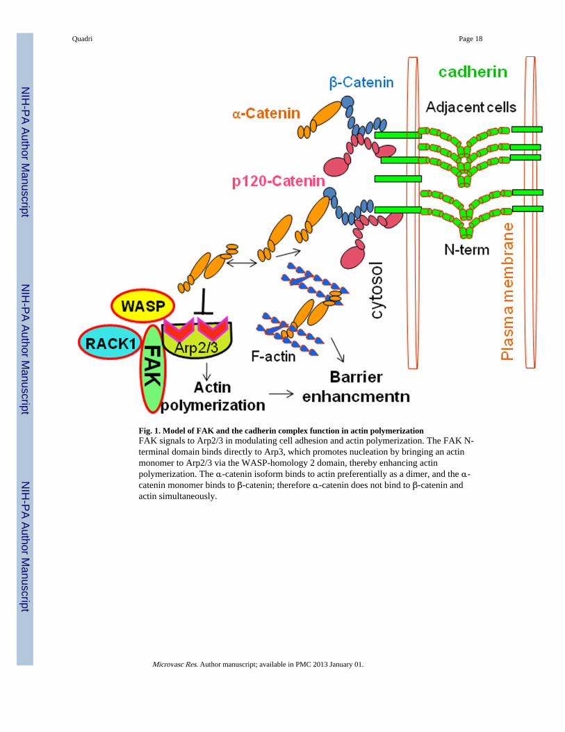
Fig. 1. Model of FAK and the cadherin complex function in actin polymerizationFAK signals to Arp2/3 in modulating cell adhesion and actin polymerization. The FAK N-terminal domain binds directly to Arp3, which promotes nucleation by bringing an actinmonomer to Arp2/3 via the WASP-homology 2 domain, thereby enhancing actinpolymerization. The α-catenin isoform binds to actin preferentially as a dimer, and the α-catenin monomer binds to β-catenin; therefore α-catenin does not bind to β-catenin andactin simultaneously.
Quadri Page 18
Microvasc Res. Author manuscript; available in PMC 2013 January 01.
NIH
-PA Author Manuscript
NIH
-PA Author Manuscript
NIH
-PA Author Manuscript
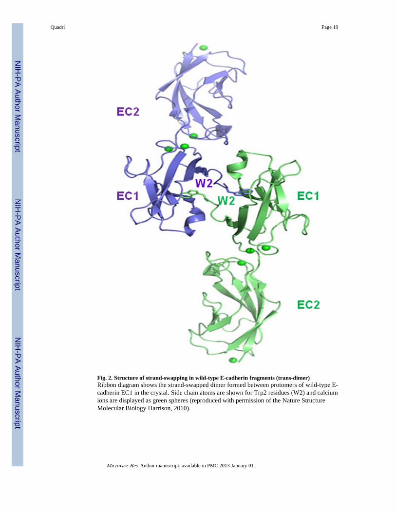
Fig. 2. Structure of strand-swapping in wild-type E-cadherin fragments (trans-dimer)Ribbon diagram shows the strand-swapped dimer formed between protomers of wild-type E-cadherin EC1 in the crystal. Side chain atoms are shown for Trp2 residues (W2) and calciumions are displayed as green spheres (reproduced with permission of the Nature StructureMolecular Biology Harrison, 2010).
Quadri Page 19
Microvasc Res. Author manuscript; available in PMC 2013 January 01.
NIH
-PA Author Manuscript
NIH
-PA Author Manuscript
NIH
-PA Author Manuscript
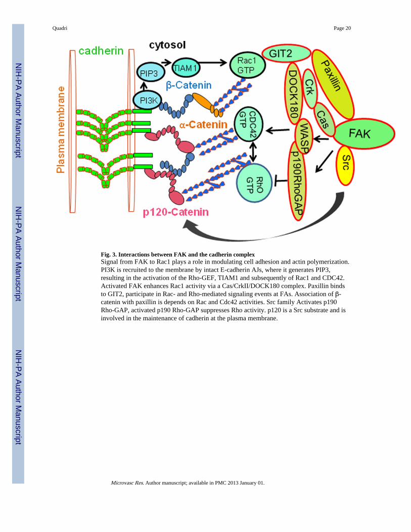
Fig. 3. Interactions between FAK and the cadherin complexSignal from FAK to Rac1 plays a role in modulating cell adhesion and actin polymerization.PI3K is recruited to the membrane by intact E-cadherin AJs, where it generates PIP3,resulting in the activation of the Rho-GEF, TIAM1 and subsequently of Rac1 and CDC42.Activated FAK enhances Rac1 activity via a Cas/CrkII/DOCK180 complex. Paxillin bindsto GIT2, participate in Rac- and Rho-mediated signaling events at FAs. Association of β-catenin with paxillin is depends on Rac and Cdc42 activities. Src family Activates p190Rho-GAP, activated p190 Rho-GAP suppresses Rho activity. p120 is a Src substrate and isinvolved in the maintenance of cadherin at the plasma membrane.
Quadri Page 20
Microvasc Res. Author manuscript; available in PMC 2013 January 01.
NIH
-PA Author Manuscript
NIH
-PA Author Manuscript
NIH
-PA Author Manuscript
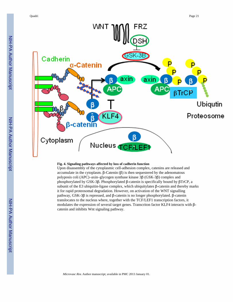
Fig. 4. Signaling pathways affected by loss of cadherin functionUpon disassembly of the cytoplasmic cell-adhesion complex, catenins are released andaccumulate in the cytoplasm. β-Catenin (β) is then sequestered by the adenomatouspolyposis coli (APC)–axin–glycogen synthase kinase 3β (GSK-3β) complex andphosphorylated by GSK-3β. Phosphorylated β-catenin is specifically bound by βTrCP, asubunit of the E3 ubiquitin-ligase complex, which ubiquitylates β-catenin and thereby marksit for rapid proteosomal degradation. However, on activation of the WNT signallingpathway, GSK-3β is repressed, and β-catenin is no longer phosphorylated. β-catenintranslocates to the nucleus where, together with the TCF/LEF1 transcription factors, itmodulates the expression of several target genes. Transcriton factor KLF4 interacts with β-catenin and inhibits Wnt signaling pathway.
Quadri Page 21
Microvasc Res. Author manuscript; available in PMC 2013 January 01.
NIH
-PA Author Manuscript
NIH
-PA Author Manuscript
NIH
-PA Author Manuscript
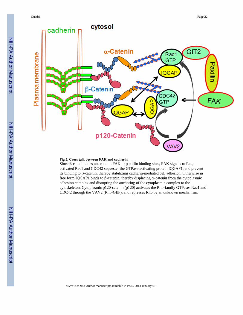
Fig 5. Cross talk between FAK and cadherinSince β-catenin does not contain FAK or paxillin binding sites, FAK signals to Rac,activated Rac1 and CDC42 sequester the GTPase-activating protein IQGAP1, and preventits binding to β-catenin, thereby stabilizing cadherin-mediated cell adhesion. Otherwise infree form IQGAP1 binds to β-catenin, thereby displacing α-catenin from the cytoplasmicadhesion complex and disrupting the anchoring of the cytoplasmic complex to thecytoskeleton. Cytoplasmic p120-catenin (p120) activates the Rho-family GTPases Rac1 andCDC42 through the VAV2 (Rho-GEF), and represses Rho by an unknown mechanism.
Quadri Page 22
Microvasc Res. Author manuscript; available in PMC 2013 January 01.
NIH
-PA Author Manuscript
NIH
-PA Author Manuscript
NIH
-PA Author Manuscript
Related Documents

![Cimetidine Inhibits Cancer Cell Adhesion to Endothelial ...[CANCER RESEARCH 60, 3978–3984, July 15, 2000] Cimetidine Inhibits Cancer Cell Adhesion to Endothelial Cells and Prevents](https://static.cupdf.com/doc/110x72/608554adacb16d34563deda1/cimetidine-inhibits-cancer-cell-adhesion-to-endothelial-cancer-research-60.jpg)