-.s - 75 CROSS RESISTANCE AI{ONGST COLIPHAGES Roberü E.I,rI. Hancock, B.Sc. (Hons.) (Adetaide). J¿ J¿ J¿ J¿ J¿ J4 J¿ J¿ J¿ J4 I¿ A thesis submitted for the degree of Doctor of Philosophy +ê .)É .)+ .)t JÉ .)t tÉ tt Js .)ê )i Department of Microbiology, The University of Adelaide, Adelaide, South Australia. November t L974.

Welcome message from author
This document is posted to help you gain knowledge. Please leave a comment to let me know what you think about it! Share it to your friends and learn new things together.
Transcript
-.s - 75
CROSS RESISTANCE AI{ONGST COLIPHAGES
Roberü E.I,rI. Hancock, B.Sc. (Hons.) (Adetaide).
J¿ J¿ J¿ J¿ J¿ J4 J¿ J¿ J¿ J4 I¿
A thesis submitted for the degree of Doctor of Philosophy
+ê .)É .)+ .)t JÉ .)t tÉ tt Js .)ê )i
Department of Microbiology,The University of Adelaide,
Adelaide,South Australia.
November t L974.
CROSS RE SISTANCE
AMONGST COLIPHAGES
(iii)
CONTENTS
Page
SUMMARY
STATEMENT
ACKNOWLEDGEMENT
CHAPTER 1 INTRODUCTION
Classification and taxonomy ofbacteriophagesReceptors for bacteriophages in thecel1 wal1 . . . . . . . . . . . . . . . o . . . . . . . . . . . . .
Structure and composition of thece1]- wal]- .....oo.o.o....o.........
Structure and composition of thelipopolysaccharide and its abilityto act as a receptor ....... . ..... .
CeI1- wa11 proteins - theircomposition, functions and abilityto act as bacteriophage receptors .
Adsorbtion of BacteriophagesStructure of the bacteriophage tailand changes undergone in adsorbtionKinetics of adsorbtion
Resistance to BacteriophagesReceptor mutantsTolerant mutantsOther types of resistanceCross resistance betweenbacteriophages and colicins
Objects of the study
CHAPTER 2 MATERIALS AND METHODS
MediaBacterial strainsBacteriophage strains ... o o. .. . . . ......General bacteriophage methods .. .. o o r..Isolation of bacteriophages from ser4rage
Serological techniques o.. .. . o. . . .. o...Electron microscopy ...o..... o oo.... o..Isolation and screening ofbacteriophage resistant mutants
(vii)
(i")
(*)
1
10
I4
I4L7
18
18
24
26
29
3o
3z
32
32
34
ó/
39
39
4L
4z
44
2
4
4
7
Nomenclature of resistant mutants aaaaa
CHAPTER 3
CHAPTER 4
( i.r)
Preparation of cel1- envelope and wa11fractions ....o.......o................Lipopolysaccharide preparations .. o....Adsorbtion and neutraLjzat.íon studies .Antibiotic resistance testingPolyacrylamide gel techniques . . . . . . . . .Gas liquid chromatographyGlucosamine analysis . . . .... o. . . .. .... o
Genetic analysis ....oo................
CHARACTERIZATION OF BACTERIOPHAGESISOLATED FROM SEWAGE .. .. .. .. t.... . . ...Introduction ..................o.......Isolation of bacteriophagesElectron microscopy .. .. ... o. ... .... ...Serological classificationSelection and charac1-erízation ofbacteriophages used in resistant mutanttesting ..o.....o.r......o...........ooSummary and conclusions
ISOLATION AND TESTING OF RESISTANTMUTANTS .............o....o............
Introduction ...... o....o......o.......Isolation of mutants . o........... . o o..Phenotypic resistance groups ..........
TonA, TonB, Bfe, Con, Efr and KtngfOtlps . a..... o.. aa. o..o... t... a...
Tsx groupKtw groupTtk groupMiscellaneous group ... .... ... .....Bar group ... o....o................I,{rm group ... o....... ........ . .....
Cross resistance to colicinsSummary and Conclusions
ADSORBTION OF BACTERIOPHAGES TO CELLENVELOPE SUBFRACTIONS .. O' ....... O' ..' O
IntroductionAdsorbtion to
Page
45
48
48
49
5o
51
5z
53
54
54
55
55
58
6T
6+
o5
65
65
66
66
66
6g
6g
6g
7z
7z
76
8o
81
81
82
CHAPTER .5
J-ipopoJ-ysaccharide
(.r)
Adsorbtion to other cell envelopesubfractions .........................o
Bacteriophages unable to lyse theCon group of resistant mutants ....Bacteriophages unable to 1-yse theBfe, TonA or Tsx resistance groupsOther bacteriophages
Enzymat-íc treatment of the receptor ...Summary and conclusions
THE NATURE OF THE ALTERATION INCELL WALL OF RESISTANT MUTANTSLIPOPOLYSACCHARIDE ALTERATIONS
THE
Introducti-onAntibiotic resistance/sensitivity .. o..Alterations in the sensitivity tobacteriophage UJ and resistance tobacteriophage CzL
Analysis of neutral sugarsGlucosamine analysisSummary and Conclusions
THE NATURE OF THE ALTERATION IN THECELL I{ALL OF RESISTANT MUTANTS -PROTEIN ALTERATIONS
IntroductionCon mutants - general properties .....Con- mutants - alterations as revealedby polyacrylamide gel electrophoresis .
Lipopolysaccharide-deficient mutants -protein al-terations ...... .. . . . ... . o...Summary and Conclusions o. .. . . ... o. .. o.
Page
CHAPTER 6
CHAPTER 7
aaaaa
8+
84
88
8g
90
92
94
94
94
97
98
101
LO2
103
103
103
105
110
LLg
I20
L20]-20
l-231-23
L26
L28
13o
CHAPTER 8 VIIPPING OF THE MUTATIONAL LESIONS
Introduction ...o.o................... o
Mapping of Bar and Wrm mutantsMapping of Ktw mutants . o o. . . .. . .. .....Mapping of Con- mutants ..r.ooo.....o..Mapping of other mutantsSummary and Conclusions
CHAPTER 9 DISCUSSION ...........o.... r..o.....o.o
(.ti )
Page
The G-eneral Pattern of BacteriophageResistant Mutants and its Relationshipto Bacteriophage Taxonomy 130The structure of the lipopolysaccharideof E. coli K-Lz and the alterations incertain bacteriophage resistant mutants 138
The receptor specificity ofbacteriophages which adsorb to
145lipopolysaccharidePossible involvernent oflipopolysaccharide protein as areceptor . a.........a.aa...a..... oo.aaaCon- mutanüs .r......o.................A hypothesis concerning the method bywhich DNA and other macromoleculesenter the ce]-1 .o..........o.r.........
APPENDIX PUBLISHED AND SUBMITTED MATERIAL aaoaao
BIBLIOGRAPHY ooaaaaa..aaa... aaaa.aaaaaaaaaaaaaaaaaaa
L46L49
151
L54155
(vii )
SUMMARY
The general pattern of resistance to bacteriophages in
Escherichia coli K-Lz and the effect of mutations to bacter-
iophage resist,ance on the strtrct,ure of the cel1 wal1 has
been studied.
A set of 56 virulent bacteriophages lysing E. coli K-Lz
was obtained from various workers or by isolation from raw
sehrage. Resistant mutants were isolated to 4Z of these
bacteriophages in one strain of E. coli K-Lz and tested for
resistance or sensitivity to the fulJ- set of J6 bacteriophages.
Most of the mutants fell into eleven groups with respect to
their resistance patterns. The bacteriophages that were
isolated from sewage were partially characterízed with
respect to their electron microscopic morphology and
neutralizatíon by various antisera. Similar data about many
of the other bacteriophages was obtained from the literature
and the taxonomic relationships of various bacteriophages
revealed. The pattern of resistant mutants obtained in this
study is discussed with reference to bacteriophage taxonomy.
Various cel-1 envelope subfractions and chemically or
enzymícally modified cell wa1l- preparations were tested in
neutralizat-j-on experiments with many of the 56 bacteriophages.
From the results obtained, conclusions could be made as to
the nature of the receptor for these bacteriophages.
It was found that many of the bacteriophages to which
the Ktw, Ttk, Bar, Itlrm and miscellaneous groups vì¡ere
resistant, urere neutralized by lipopolysaccharide
preparations. Alterations in the response of the above
mutants to a series of antibiotics and to bacteriophages
CZL and U3 indicated that they had lipopolysaccharide
( vr_1_l-,
alterations. This was confirmed by direct sugar analysis of
the mutant lipopolysaccharides using gas liquid chromatography
which showed six dis'l,inct classes. The results are in
agreement with bhe published partial rough core structures of
Salmonel]-a and E. co]-i O 1OO. Twelve reprcscntativc mutants
of the Bar and Wrm( 2) resistance groups r^¡ere shown by PI
transduction or conjugation to be between pgE and mtl which
has been previously shown to be the site of the rfa locus in
E. coli K-Lz. Outer membrane protein defects h¡ere also
demonstrated in some of the above lipopolysaccharide-altered
mutants by polyacrylamide gel- electrophoresis. The possible
involvement of protein in so-called lipopolysaccharide
receptors and vica versa is discussed on the basis of these
and other results presented in this thesis.
Analysis of the protein composition of Con- mutants
revealed that these mutants !üere deficient in two major cell
wall proteins named 3a and 3b in the nomenclature of Schnaitman.
These mutants have been subsequently shown by colleagrres to
be defective as recipients in conjugation and tolerant to
colicins K and L. They have also been partly mapped in this
study. Based on the results obtained for this and other
mutanfs, a hypothesis is put forward concerning the first
steps of infection of cells by bacteriophages.
( ix)
STATEMENT
This thesis contains no material which has been
accepted for the award of any other degree or dipl-oma in any
university and, to the best of my knowledge and belief, itcontains no material previously published or written by
another person, except where due reference is made in thetext.
Robert E. W. Hancock
Novembert I974.
(*)
ACKNOWLEDG EMENT
I wish to sincerely thank Dr. peter Reeves for his
continued advice and discussion throughout this work.
I am índebted to the members of the Department of
Microbiology, University of Adelaide, for useful
discussions and technical advice.
I also wísh to thank Dr. Alex Osmond for introducing
me to the technique of Gas Liquid Chromatography, Miss pam
Dyer for electron microscopyr Dr. Ron Skurray for
assistance with gel analysis on the Con- mutants, Mr. John
Davies for testing the colicin resistance of my strains,
Mr. Tony Richardson for photography and technical advice,
and Miss Jenny Russell for typing.
1
CHAPTER 1
INTRODUCTION
dlHerelle as early as Ag26 divided the infectious
process of the bacteriophage into four stages, (1)
adsorption of the phage particle, (2) penetration of the
phage parti-cle into the host cell, (3) the intracel-lular
multiplication of the bacteriophage, and (4) the lysis ofthe host cel1 and release of the phage progeny (drHerelle, 1,926).
Although it has been since discovered that stages (2) and (3)
involve only the nucleic acid of the phage particle (Herschey
and chase, L952), this picture remains essentially correct.
A bacteriophage resistant mutant can be altered such
that it prevents any of the four stages. Classicall-y
described resistant mutants however are often altered atthe J-evel of adsorbtion to the receptor, i.e. stage (1).
Bacteriophages have been described for a large number
of hosts, and it seems that they are present for most
species in which they have been looked for (Adams, L959;
Ackerman, 1969; Tikhonenko, L97O). They differ in
morphology (see below), nucleic acj-d type and content
(Ackerman, L969; I,rti1dy, L|TI) and ability to lysogenize a
host bacterium. A bacteriophage which is able to infect a
cell and lysogeníze it is called a temperate bacteriophage
(Herschey, I97L; Echolst I972). The resultant lysogenic
strain often has the bacteriophage DNA incorporated into itand replicating under the control of the host replicative
systems (Herschey, L97L). If a bacteriophage is unable to
lysogenize bact'eria, then it is called a virulent bacteriophage
2
and undergoes a lytic cycle of replication.
The studies involved in this thesis have been mainlylimited to double-stranded-DNA-containing, virurentbacteriophages capable of lysing one strain of Escherichiacoli K-12. In this introduction there has been no atternpL
to review the literature about bacteriophages exhaustively,but rather it is concerned with those topics of relevance tothe thesis.
CLASSIFICATI ON AND TAXONOMY OF BACTERIOP HAGES
Ttre first extensive attempt to classify bacteriophages
was made by Burnet and McKie (fq¡¡) and Burnet (1933, I93Ð,who distinguished bacteriophages by means of cross resistance,serology and simple biochemical tests. Burnet (]-gs+) found
some correlation between the latter two criteria; however
many of his bacteriophages ü¡ere subsequently lost and. much ofhis work cannot be related to modern bacteriophage taxonomy.
other early studies of a similar nature were reviewed_ in a
book by Adams (1959). Although many of the early stud.ies
involved serology, which appeared to be a good basis forclassification (Adams, a953), the discovery that there were
antigenic differences between wird type and mutant À
bacteriophages (p"y and waites, 1969), has since demonstrated
its limitations.
Examination of gross bacteriophage morphology under theelectron mícroscope revealed that bacteriophages hrere strikingand characteristic in appearance (Ruska, 194L; Luria,Delbruck and Anderson, rg43). Adams (rgs¡) showed. that thesize and shape of bacteriophages correlated well with other
criteria, and using the various criteria, Adams and l{ade
aJ
(fgSS) divided a limited number of coli-dysentry phages into
four taxonomic groups.
By means of extensive studies on their morphologies,
Bradley (L963, L967 ) found he could divide all of his
bacteriophagcs into six distinct types; three of which included
double-stranded-DNA bacteriophages differentiated on tail
morphology (contractile, long non-contractile or short non-
contractile). Ackerman (L969) extraustively reviewed the
literature concerning the electron microscopy of bacteriophages
and proposed a further differentiation on head morphology
(isometric, l.ong and elongated). Of the nine morphological
permutations of head and tail structure, described by this
system, Ackerman found that he could demonstrate examples
of eight of these morphologies. The ninth category has
since been described (Àckerman, Petrow and Kasatiya, Lg74)
but no other morphologies have been noted except in aberrant
mutant bacteriophages (Cummings g!.jl., L967; Cummings,
Couse and Forrest, L97O).
In L966t ãî International Committee on Nomenclature of
Viruses was set up and this group published their first
report in I97I (Witay, L97f). Their classification system
for bacteriophages is stil1 in the preliminary stages;
however they have proposed generic names for the T-even
bacteriophages (myovirus) and for bacteriophage À
(caudaevirus). Due to the preliminary nature of this
classification, labor"atory strain names have been used in
this thesis.
Limited studies have been done on the DNA homology
(Cowie, Avery, and Champe, L97L; Davis and Hyman, L97L;
Brunovskis, Hyman and Summers, L973; Hyman, Brunovskis and
4
Summers, L974) and genetic recombination (Luria and Dulbecco,
1949; Adams, 1953; Mizobuchi, Anderson and McCorquodale,
L97I) of various strains of bacteriophages. Studies of this
nature will have significance not only to the further
classification of bacteriophages which are similar in
appearance, but also to the sùudy of evolution of bacter-
iophages.
RECEPTORS FOR BACTERIOPHAGES IN THE CELL IIIALL
One of the reasons for the study of bacteriophage
resistance, is so that we can ultimately discover more
about not only the nature of the adsorbtion of bacteriophages
to their receptors in the cell wa1-l (bV studying the defect
in the resistant mutant) but a1-so about the two components
involved ín the interaction; the bacteriophage tail and its
receptor. Thus a discussion of the structure of the ce1l
wa1l, with emphasis on those components of the cel1- wa11
able to act as receptors is relevant to this thesis.
Structure and composition of the ce1l wa11
The ce1l wal1 is defined here as that part of the cell
external to the cytoplasmic membrane. In gram negative
ce1ls iü has been shown by electron microscopy (Sitva and
Sousa, L97 3) to consist of a dense rigid layer, and
external to this a trilaminar membranous structure, call-ed
the outer membrane. The rigid layer or murein sacculus is
thought to be the shape maintaining layer of the celI (Weidel,
Frank and Martin, L96O; Burman, Nordstrtrm and Bloom, I972;
Braun g!3!. , L97 3) , although it does not appear to be such
for the membrane of bacterial ghosts (Henning¡ Hohn and
Sonntag, L97 3). The structure of the murein sacculus is
5
known (Braun and Bosch, L97Z; Braun et aI., 1973). It
appears to be attached to the outer membrane by means of a
lipoprotein (Braun and Rehn, L969; Braun and Bosch, L972t
I973artj). The lipid portion of the murein lipoprotein is
inserted into the outer membrane (Schnal-tman, L971-l'c-; Burman,
Nordstrum and Bloom, 1972). Inouye (L974) has recently
presented an attractive model for the molecular assembly of
the murein lipoprotein, in which six molecules of lipoprotein,
each with a-helix structures form a 12.51 pore. These
pores are proposed to extend through the membrane and thus
provide channels for the passive diffusion of molecules.
However, DNA from bacteriophages is too large to travel down
these pores.
The outer membrane has three classes of components;
proteins, phospholipids and lipopolysaccharide (I-eS),
There are many theories as to the structure of the outer
membrane (de Petris, L967; Schnaitman, L97Lb! Van Gool and
Nanninga, I97L; Trauble and Overath, L973; Costerton,
Ingram and Cheng, 1-974), however most of these consider the
basic structure to be a lipid bilayer, typical of
biological membranes. This has been borne out by
biophysical studies (Forge and Costerton, L973; Forge,
Costerton and Kerrt I973).
Assuming the membrane has as its basic structure a
lipid bilayer, and the membrane is at least partly fluid
(otdeietd, L97L), then it is possible that the integral
proteins are of three types (1) traversing the membrane (Z)
mainly in the outer half of the membrane, and (3) mainly in
the inner half of the membrane (Chapman, L972; Razin, L972;
Singer and Nelson, L972; Coleman, L973). Thus since a
6
bacteriophage attacks from without one would imagine that
its receptor, if a protein, would be in one of the first
two classes. The integral proteins would be capable of
lateral movement within the membrane (rcaback and Barnes,
L97L; Ilarold, I972). The lipid A portions of the
1-ipopolysaccharide molecules probably interact hydrophobically
with the outermost l-ipid layer of the outer membrane (notn-
field and Romeo, L97L; Schnaitman, I97Lb; Benedetto, Shands
and Shah, L973); while the carbohydrate portions of the
molecules either extend ar^¡ay from the surface of the ce]-l
(de Petris, 1967; Schnaitman, I97L}¡) or consist of an
ordered and cross-linked mass of polysaccharide chains on
part of the surface of the celJ- (Costerton, Ingram and Cheng,
L97 4) . I¡ühen the components of the outer membrane are separ-
ated and then mixed, a membrane essentially identical to
the outer membrane, is formed (Aragg and Hou, L972;
Rothf ield g!3!. , L97 2; Sekizawa and Fukui, L97 3) . This is
evidence for the sel-f assembly of outer membrane components.
Electrophoretic measurements have shown ühat the ce1l
possesses a large net negative charge in media suitable for
bacteriophage binding (Brinton, BwzzeLL and Lauffer, L964).
Other studies using isoelectric equilibrium analysis (Sherbet
and Lakshmi, I973) have characterized the number of anion-
ogenic and cationogenic groups more thoroughly. This work
has shown that the phospholipid is probably at a depth of
60 I below the ceJ-1 surface while the proteins and
lipopolysaccharide may be on the cell surface.
The study of phospholipids has revealed differences in
the distribution of various phospholipids in the cell wall
andcytop1asmicmembranesof4(wrr:-te,Lennarz,and
7
Schnaitman, I972). The various functions of phospholipids
have been described (Cronan and Vagelos, L97Z) but as yet no
purified bacteriophage receptor has been shown to be
dependent on a phospholipid. However, the synthesis of
proteins, which can act as bacteriophage receptors ( see for
example Lindberg, L97 3), and phospholipids does appear
co-ordinated (Crowfoot, Esfahani and Wakil, L97Z; Forge,
Costerton and Kerr, I97 3).
Bayer (1Ç6Ba) has shown that when E. coli is
plasmolysed, the cytoplasmic membrane remains attached to
the outer membrane at between 2OO and {OO locations. These
adhesions have been shown to be the site of bacteriophage
adsorbtion for the T-group phages (Bayer 1968a), attachment
of the F pilus (Bayer 1968b), export of newly synthesized
lipopolysaccharide (Uuhlradt et a1., L973) and binding of
DNA to the envelope (Olsen g!3!., 1974\. The number of such
adhesions is also compatible with each of the adhesions being
a component of the membrane subunits which segregate on cell
division (Green and Schaechter, I972). However, recent
evidence has suggested that there is more than one class of
adhesion sites (u. Bayer, manuscript in preparation).
Structure and composítion of the lipopolysaccharide and its
ability to act as a receptor
The outer membrane of E. coli K-LZ comprises approximately
16/" Iípopolysaccharide by weight (Sekizawa and Fukui, I973).
In its purified state it adopts a membranous structure
(Katayama g!3!. r L97L) t however it is generally thought not
to be responsible for the trilaminar structure of the outer
membrane. It is thought to be associated with protein in
the cell wall (hrober and Alaupovic, I|TI; I4Iu and Heath,
8
L97 3), and there is evidence that the lipid A portion of the
lipopolysaccharide molecuJ-e is covalently bound to the
protein.
It has been shown in Salmonella that 1i popo1-ysaccharide
is synthesized sequen'bially (Osborn, L966; Müh1radt, ry71-)
by a number of eîzymes, some of which reside in the cyto-
plasmic membrane (Osborn, 1966). It is then exported to the
outer membrane (Osborn, Gander and Parisi, A972; Mühlradt
et a . , I97 3) via Bayer adhesion sites (trlütrtradt g!3!.,
L97 3) and spreads out laterally over the bacterial cell
surface. There is evidence (notnfield and Takeshita, Lg66t
Rothfield and Romeo, L97L) for the role of phospholipid in
the biosynthesis of lipopolysaccharide.
The structure of the lipopolysaccharide rough core has
been best studied in Salmonella (Holme et al., L968t Droge
. t I97O; Hellerqvist and Lindberg, I97L; Lehman,
Lrrderi.1oz and Westphal, L97L; Hämmerling, Lehman and
Luderít,2, I97 3; Lehman g!3!. , 1973), in which it has been
shown that there is only one type of structure for all
strains. However, Schmidt and co-workers have demonstrated
three different core types, R1, R2 and RJ, for the E. co]-i
strains they have studied (Scfrmiat, Jann and Jann, L97O;
Schmidt, Fromme and Mayer, L97O; Schmidt, 1972). The
structure of the lipopolysaccharide of E. co]-i O 1OO, which
has an R2-type core has been largely characterízed (Hämmerling
Éjl. , I97L), but few other structural studies have been
done. In fact-, in addition to the above, and studies on the
heterogeneous rough core structure of E. co]-i 0111-84
(Fuller, Wu, Wilkinson and Heath, L973; Wu and Heath, L973)t
only a few less detailed studies of lipopolysaccharide
9
composition have been done on strains of E. coli (Malchow
et a ., L969; Morton and Stewart, LgTz).
E. co]-i K-12 is a rough organism, lacking O-specificsugars (frskov and frskov, Lg6Z; Rapin and Mayer, Lg66).
This is probably due to a mutatiorr a'L Lhe r.fb locus
situated. near hi"* on the genetic map (frskov and. frskov,L962; Jones, Koltzow and stocker, ag72). schmidt has shown
that the e'rzymes produced by the gþ gen"s of E. coli K-rzstrain 2578 can synthesize a complete core structure(Sctrmidt, L973) and he has excluded the possibility of a
leaky rfa mutant as suggested by Jones, Koltzow and
stocker (tg7z). He has also shown by serological tests and.
phage typing that the E. coli K-Lz core is different from
other known core types of
(R1-R3 ) .
Salmone]-la (n") and E. coli
The E. coli K-12 core contains glucose, galactose,
heptose and a small amount of rhamnose (Rapin and Mayer,
L966; Monner, Jonsson and Boman, agTI; Eriksson-Grennberg,
Nordstrum and Englund, L97L), although the latter is missing
from one subline of E. coli K-Lz (uit<aido, Nikaido and
Rapin, L965). rn those strains which contain it, rhamnose
has been shown to exist bound to the KDo-lipid A portion ofthe molecule (sugimoto and okazaki, 1967). The KDo-lipid A
portion of the molecule has been chemically but not
structurally characterized (Rooney and Goldfine, LgTZ).
As early as L934, Gough and Burneü (tgS+) were able toshow that a polysaccharide extract of E. coli from an
autolysed culture could inhibit the adsorbtion ofbacteriophages to the E. coli celJ-. Since then, many
bacteriophages have been shown to attach to extracted
10
lipopolysaccharide (a" reviewed by Rapin and Kalckar,
L97L; Lindberg, L97 3\. In addition, electron micrographs
showing bacteriophage T4 whole particles and isolated tail
fibres attached to purified lipopolysaccharide have been
published (Jesaitis and Goebel, 195,3; lrrilson, Luftig and
Wood, A97O). However, for irreversible attachment these
bacteriophages require, as a receptor, the aggregated
lipopolysaccharide complex (tindberg, Lg73). Furthermore,
it has been shown that lipopolysaccharide from bacteriophage
sensitive ce1ls can provide functional receptor for
spheroplasts of ce1ls normally lacking receptor sites
(hratson and Paigen, L972). The requirements for
functional receptor have also been extensively studied
(Rapin and Kalckar, L97I; Lindberg, L973) and this wilJ- be
discussed more fu1ly later.
Lastly, the genetics of lipopolysaccharide biosynthesis
has been studied in Salmonella (fuo and Stockero I972;
Sanderson and Saeed, A97Z; Sùocker and Mäkela, L|TL) and
E. co1i (Schmidt¡ Jann and Jann, L97O; Eriksson-Grennberg,
Nordstrum and Englund, L971,; Schmidt, L973) and it has been
shown that many of the genetic loci responsible for the
enzymes involved in the biosynthesis of the rough core (É
genes) lie between pyrE and mt1 on the respective genetic
maps.
Cel1 wa1l proteins their composition. functions and
abilitv to act as bacteriophage receptors
The protein composition of the outer membrane of E. coli
was first described by Schnaitman (L97OarA) who used the
technique of polyacrylamide gel- electrophoresis. He was
able to show using his solubiliza|-i.on techniques and gel
11
buffer system one major protein band of 44'OOO moJ-ecular
weight accounting for as much as 40% of the total protein
of the cell envelope. By separating cell wall and cyto-
plasmic membrane using sucrose gradient centrifugation, he
showed that there were only six protein bands in the ce1l
wa1l, one of which, the 44.OOO molecular weight protein,
accounted for 70% of l-lr.e total protein of the cell wall.
In a later study, Schnaitman (L97La) was able to show that
the cytoplasmic membrane proteins were soluble in 2%
Triton X-IOO and this provided a simple technique for the
separation of wall and memlorane protein. He was also able
to show that by treating the cel-J- wa1-l with Triton X-IOO
and Ethylenediamine-tetraacetic acid he could reduce the
protein content and also the vesicle-like structure
(Schnaitman, 1-97Il:-).
The first serious challenge to Schnaitmanls ideas of
one major protein band came from Bragg and Hou (tgZZ) who
were able to show that in their buffer system the major
band ran as two distinct major proteins and one minor
protein which had molecular weights ("" estimated using
their polyacrylamide ge1 system) ranging from 44.OOO to
33r4oo when partially purified (oragg and Hou, L97L).
Moldow, Robertson and Rothfield (tglZ) showed that the
major band of the cell envelope contained several poly-
peptides while Inouye and Yee (L97 3) demonstrated the
presence of three bands of varying molecular weights. The
dilemna was finally solved by Schnaitman (tgZ3arb,
I974arb) who showed that the major band consisted of not
one, but four proteins (proteins Ir 21 3a and 3b) of
4OTOOO molecular weight. Proteins 3a and 3b ran together
l2
in all the ge1 systems tried by Schnaitman but he managed
to separate them by column chromatography and proved they
were different by comparing their cyanogen bromide peptides
(Schnaitman, L973b, L974a). The anomolous results
described previously coul-d be expl-ained by the fact that
many solubiliza1l-i-on techniques did not completely
dissociate the sub-units or unravel the polypeptide chains
of the four proteins. E. coli K-12 has only proteins 1,
3a and 3b in its outer membrane (Schnaitman, L974b), as
protein 2 is only found in strains of E. coli lysogenic for
a certain bacteriophage (C,4" Schnaitman, personal
communication). A more thorough discussion of Schnaitmanrs
work is included in the text. The concept that peak 3
contains two proteins has been recently challenged by
Reithmeier and Bragg (L974), who have shown that when they
partially sol-ubilize tl;re ce1l wal-l of E. coli NRC 482 ín
O.5% SDS at 1OOC for t hour, a single protein, with the
characteristics of the acrylamide gel peak J (shown by
Schnaitman to consist of proteins 3a and 3b) can be
isolated. However, they have not studied the residual cell
wa1l proteins, and it is by no means clear whether or not
they have selectively solubilized onJ-y one of the proteins
3a and 3b. Inouye and Lee (f973) have shown that al]- of the
membrane proteins produced by stable mRNA are in the outer
membrane and there are differences not only in the stabiliüy
of the mRNAts of the major proteins but also in their
assembly mechanisms.
The nature and function of the proteins of the ce1l
wal1 of E. coli is poorly understood. The murein
lipoprotein has been we1.1- characterízed (Braun and Bosch,
13
L972, L973arb). It wi1-l on1-y partly run on polyacrylamide
gels, unless the cell wall is treated with lysozyme
(Schnaitman, L97Lb), due to the fact that it exists in two
states; free and bound to the murein sacculus (Hirashima
g!_4. , L973). Phospholipase A1. (tut.w.3OrOOO) has also been
shown to exist in the outer membrane of E4!! K-LZ (Ofrti,
Osamu and Nojima, L972), however none of the other enzymes
involved in phospholipid metabolism appear to be
associated with the ce1l wal1 (neff g!4. , I97L) with the
possible exception of the phosphatidyl serine synthesizj-.ng
eîzymes (v¡trite gkl. , 1-97I) . Both 3 t nucleotidase and
5 I nucleotidase are al-so associated with the outer cell wal1-
layers of E. co]-i (Nisonson, Tannenbarrm and Neu, L969).
Other enzJ¡mes and proteins which have not been localized in
the cel-l wall or cytoplasmic membrane of the envelope are
summarized by Machtiger and Fox (L97 3) and Costerton, Ingram
and Cheng (L974). Recently, Koplow and Goldfine (L974) have
shown that heptose deficient mutants have large alterations
to their protein compositions.
Jesaitis and Goebel (1953) and l,rreidel (fgSS) first
described the receptors of bacteriophages T2 and T6 as
lipoprotein. The fact that bacteriophage T2 resistant
mutants h¡ere sensitive to bacteriophage T6 and vice versa
(Demerec and Fano, L945) suggested they h¡ere probably
different lipoproteins. Chemical characterizaþi-on of the
receptors showed that the specifity of the receptors of
bacteriophages T2 and T6 was determined by different
chemical groupings (Irreltzein and Jesaitis, L97I). Studies
by Michael (1968) and DePamphilis (tgZt) have confirmed
that the receptor for T2 is probably a lipoprotein, while
L4
the receptor for T6 could possibly be associated with
protein 1 of the cell wal1 (C"4. Schnaitman, Ig74b).
The receptor for bacteriophage T5 was isolated
originally as a lipo-g1y"op"åt"in complex by mild alkali
extr.actiorloftlrece11wa11of4B(weicte1,Kochand.
Bobosch, L954). The properties of this complex have been
wel-J- studied (Zarybnickyt Zarybn';cka and Frank, I973).
Braun and co-workers (Braun, Schaller and l,Volff , Lg7 3; Braun
and l{olff, 797 3) have shown that the actual receptor, which
they have isolated from the complex, is a single polypeptide
chain of 85rOOO molecular weight which resides in the outer
membrane.
The receptor for bacteriophage À has also been shown to
be a protein located in the outer membrane (Randall-HazeA-
bauer and Schwartz, I973). Sabet and Schnaitman (t973^,
I97 3b) have isolated a protein of 60, OOO molecular weight
which is missing or altered in bfe- mutants. This protein
is the receptor for colicins E2 and EJ and although it has
not been directly demonstrated, the fact that the bfe-
mutant which is missing it is also resistant to
bacteriophage BF23 (Buxton, L97L), suggests that this
protein is the receptor for bacteriophage BF23 al-so.
ADSORBTION OF BACTERfOPHAGES
In order to understand the nature of resistanceto
bacteriophages, one must consider the process that is
deleted in many of these mutants, the adsorbtion of the
bacteriophage tai1- to the cell wall.
Structure of the bacteri ooha tail and changes undergone
15
in adsorbtion
AJ.l double-stranded DNA bacteriophages have tails that
can be fitted into three main types: (A) contractile,
(g) long non-contractile, and (C) short non-contractile
(Ackerman, L969). Itrithin this simple classification there
appear to be some minor variations (tikhonenko, I97O; Krzywy,
IÇf2arb). This introduction will afford a brief summary of
the three classes.
The proteins of the type (A) tail of T-even bacteriophages
have been extensively characii-erj-ze.d, by molecular weight
(Cummings et al. t L97Oarb, L973; King and Laemmli, Lg73; King
and MykoLajewycz, L973) and functionally (Mason and Haselkorn,
I97L; Poglazov, Rodikova and Sultanova, \972; Beckendorf,
I973; Beckendorf, Kim and Lielausis, L973; Dawes and Go1dberg,
LÇflarb; Kells and Haselkorn, L974). The genes on the
bacteriophage T{ genome which code for these proteins have
been largely characteri-zed (Dawes and Goldberg, I)fJarb; King
and Laemmli, 7973; King and MykoLajewycz, a973).
The main structural features of the type (A) tail are a
tail- core, a contractile sheath (rcellenberger and Arber, 1955)r
a base plate and six pins, as well as both long and short tail
fibres (rells and Haselkorn, L97 4). The adsorbtion process has
been shown by electron microscopy to involve attachment of
long tail fibres to the cell wall, followed by attachment of
the tail pins of the base p1ate, contraction of the sheath,
and injection of the DNA through the tail- tube (Sirnon and
Anderson, 1967). This process occurs at sites of adhesion of
the cel1 wal-1 and cytoplasmic membrane (Bayer, 1!68a). The
major protein of the T-even tail, which itself has contractile
properties (Kozloff and Lute, L959), is the sheath protein
responsible for the contraction of the tail. It has been
16
shor¡¡n that the contraction process 1s similar for another
bacteriophage with a type (A) tail, but with different head
morphology (Donnelli, Guglielini and Pao1-etti, L972).
As a result of these and other studies, Bertz and Goldberg
(197 3) have postulated that there are three types of rcccptors
for bacteriophage T4-like particles. Two of these are
postulated to be in the outer membrane and are the receptors
for long tail fibres, and the subsequent attachment of the tail
pins. The third is postulated to be in the cytoplasmic membrane
and is the receptor for the tail- tube after the bacteriophage
tail has contracted.
The nature of the proteins for the type (e) tail of
bacteriophage T5 (Zweíg and Cummings, I973a) and the type (C)
tails of bacteriophages T7 (Studier, 1-g73) and p2Z (notstein,
Waddell and King, L973) have been studied, but the actual
process of adsorbtion/penetration is not understood-. It is
known that some bacteriophages with type (c) tails have enzSnnj-c
activities associated with these tail-s which have the ability
to digest cel1 wa1-l components (Stirm et al. t L97I;
Kanegasaki and l,{right, L973; Lindberg, L973; Leske, I{allenfels
and Jann, L973). However, whether these findings are of
general significance to these bacteriophages is as yet unknown.
The finding that bacteriophages of all three tail- types
can adsorb to the adhesions between outer and cytoplasmic
membranes that are formed after plasmolysis (Bayer, 1968a) is
probably important. It may be that these adhesions are of
general significance to a1-l bacteriophages, in allowing the
transport of DNA excreted from bacteriophages which attach
around them. It has been shown that isolated receptor
preparations can trigger DNA ejection from bacteriophage T5
L7
in the
Frank,
absence of such pores (Zarybnj-cky¡ Zarybnicka and
Lg7 3) .
Kinetics of adsorbtion
The kinetics of adsorbtion has been dealt with
thoroughly and critically in a number of reviews (Tolmach,
L957; Weidelc L957; Garen and Kozloff, Lgsg; Adams, Lgsg) and
it will be dealt with only briefly in this thesis. rt was
shown by Krueger (fg:f) and subsequently confirmed by
Schlessinger (l-932) that adsorbtion follows the kinetics of
a first order reaction, with the rate of disappearance offree phage from the medium being proportional to the
instantaneous concentration of free phage and the
concentration of host cells.
Adsorbtion probably involves at least two successive
steps, the first of which is reversible (puck, Garen and
Cline, L95I; Stent and Wollman, Lg52; Gamow, Lg6g). The
first step has been postulated to involve the establishment
of electrostatic bonds (salt bridges) between appropriate
configurations of ionic charges on the two bodies (puck, Garen
and cline, 1951). Lindberg (L973) considers that this step
is the binding of tail fibres to a cel1 wa1l receptor. ft isprobably non-enzymatíc as it is not a temperature dependent
step (Tolmacht L957).
The second step is irreversible attachment. For this tooccur with certain bacteriophages which have contractile tails,it requires aggregated lipopolysaccharide complex (lind,berg,
L973). It wi1-1- not occur with alkali- or acid-hydrolysed
lipopolysaccharide (Lindberg, Lg73), nor with lipopoly-
saccharide broken down with polymyxin B (roite and rid.a, LgTr),
sonication, or treaüment with sodium deoxycholate (lindberg,

18
L967). This step probably involves the anchoring of the tail
pins and is dependent, in bacteriophage T4t on functional gene
12 product (Simon, Swan and Flatgaard, L97O). It míght be
enzymatíc as it is dependent on temperature (Tolmach, L957).
Certain organic compounds are ab1-e to act as adsorbtion
co-factors for bacteriophage particles; for example
bacteriophage T4 requires free L-tryptophan (t.f. Anderson,
I945t 1946). These co-factors act by virtue of their ability
to be electron donor compounds (Kanner and Kozloff, Lg64), and
by forming a molecul-ar complex with a component of the tai1-
p1ate, they lower the activation energy (Gamow, Lg6g).
RESISTANCE TO BACTERIOPHAGES
The resistance of cells to bacteriophages can be of three
main types. Firstly, the cel1s can be lacking some component
necessary for adsorbtion, these being referred to below as
receptor mutants. SecondLyt the cells might be lacking a
component that is necessary during some post-adsorbtion step
of bacteriophage multiplication, without which viable progeny
bacteriophage particles are not produced. These are calledrrtolerantrl mutants below in agreement with the nomenclature
for colicins (Reeves, 1-972). The third type is a miscellaneous
type¡ where resistance is due to either lysogeny of the cell-
with a related temperate bacteriophage, restriction by either
the host or a resident plasmid restriction systern, or by some
other mechanism which is not we]-]- understood. Resistance to
bacteriophages may also give rise to resistance or tolerance
to colicins.
Receptor mutants
Early workers (nait, 1923; Burnet and McKie, 1933) were able
I9
to show that mutation of a ce1l to bacteriophage resistance
involved only some of the bacteriophages capable of lysing the
strain. These mutations were shown to occur spontaneously
d.uring ceJ-l reprod.uction at a frequency of LO-7 to 1O-1o
mutations per bacteri-41 cli-vision, ancl prior t,o the addition of
selecting agent (turia and Delbruck, L943; Newcombe, L949;
Lederberg and Lederberg, L952).
Demerec and Fano (tg+S) ¿i¿ tne first comprehensive study
of cross resistance amongst a group of seven bacteriophages
which they called the T-bacteriophages. They hrere able to
show that E. coli B could mutate to resistance to one
bacteriophage or as many as three different bacteriophages in
a single mutational step; and that there r4¡ere a limited
number of resistant mutant types that could be isolated using
these bacteriophages. They described five bacterial colony
types associated with resistant mutants and rlrere able to
correlate one of these types to some extent with resistance to
bacteriophage T7. The fact that they were able to demonstrate
two different mutants resistant to a given bacteriophage (".g.
T1, T3 or T{), showed that bacteriophage resistance could
occur by more than one type of host cel1 al-teration.
Some mutations to bacteriophage resistance hrere shown to
be accompanied by the loss of abil-ity to synthesize growth
factors such as tryptothan (E.H. Anderson, L946; Luria, 1946;
Gots, Koh and Hunt, 1954) or proline (Wo11mant L947; Curtiss,
L965; Baich , L968 ) " A deletion covering the tonB and trp
genes was shown to give rise to the two properties, Tl resist-
ance and tryptothan requirement, for one mutant cell line
(Franklin, Dove and Yanofsky, 1-g65); while Curtiss (1965)
demonstrated a chromosomal aberration in his bacteriophage
resistant pro mutants. Baich (1968), however, considered
20-
the relationship of T4 resistance and proline deficiency to
be structural rather than genetic for some T4 resistant
mutants. Resistance to bacteriophage À was often accompanied
by the inability of the mutant ce1l to utilize maltose as a
sole carbon source (n.U. Lederbergr 1955; Ronen and Raanan-
Ashkenazi, L97I). This was because the maltose biosynthesis
and lambda receptor genes are both under the positive
control of the mal T regulator gene in E. coli K-Iz (thirion
and Hofnungr L972). On]-y about 20% of i.lrre resistant mutants
map inside a gene lam and yield the ^"
r"1.* phenotype (8o%
are ma1 T À"). I{ang and Newton (i-gZt) consider that the
association of resistance to bacteriophages T1 and þ80 with
deficiencies in iron transport rnight be due to the role of
the iron transport system in an early function of these
bacteriophages. In addition t,o these studies, it has been
shown that when sucrose is added to the medium in which a
culture of L_S9!å B is growing, the cel1s become phenotypic-
a1ly resistant to bacteriophages T2, T3t T4t T6 and T7
(Jackson, Buller and Shankel, L967); however the significance
of t'his is notr âs Vett known.
For many years, bacteriophage resistant mutants were
used mainly in genetic studies as they were easily selected
and offered reasonable variety. Linkage relationships were
established for some bacteriophage resistant mutants
including mutants resistant to many of the T-group phages, À
and BF23 (.1. Lederberg, L947; E.M. Lederbergr 1955;
Hayes, L957; I{einberg, L96O; Tamaki¡ Sato and Matsuhashi,
L97I; Curtiss, 1965; Buxtonc L97L; Jasper, lrlleitney and
Silver, I972; see also Taylor and Trot1-er, L972).
Other workers used mutation to bacteriophage resistance
as a means of testing the capability or specificity
2I-
of various mutagenic agents (Novick and Szilard, 195I;
Bryson and Davidson, 1951). However, these studies shed
1ittle light on the naiure of bacteriophage resistance.
Garen and Puck (1951), working on the two step nature
of adsorbtion, showed that the two bacteriophage T1
resistant mutants of E. coli B, B/L (resistant to T1 and now
known as ton B-) andB/IrJ (resistant to T1 and T5 and now
known as ton A-)¡ differed in that the former could adsorb
T1 reversibly. This implied that the resistance of strain
ø/L to bacteriophage T1 was not due solely to the failure of
attachment. The further study of this muùant has been
somewhat hindered by the inability of T1 to adsorb
reversibly or irreversibly to either kil-l-ed cel1s or cell
wa1l fragrnents (Stentr i-963). The B/I.J mutant however is
unable to adsorb either T1 (Garen and Puck, L95L) or T5
(Weidel, Koch and Bobosch, 1954) reversibly or irreversibly.
Irreidel and co-workers (Weidel, Koch and Bobosch, l-954) were
able to extract a lipoglycoprotein complex from their B/Ir 5
mutant and demonstrated that it was unable to adsorb
bacteriophage T5r whi-l-e a similar preparation from the
sensitive strain did adsorb T5. It was later shown by Braun
and co-workers (Braun, Schaller and Wolff, I97 3; Braun and
I{o1f f , L97 3) ttrat the T5 receptor was a protein present in
the outer membrane of sensitive strains. This protein was
also present in bacteriophage T5 resistant mutants, but was
altered in such a hray that it could not adsorb bacteriophage
T5.
Mutants of E. coli selected as resistant to bact,eriophages
T3r T4 or T7 and resistant to all- three of these
bacteriophages lrrere sometimes also resistant to TZ and T6
(Demerec and Fano t L945). In these mutants the structure of
22
the lipopolysaccharide was substantially altered (Weidel,
1955). It was shown in Salmonella (Lindberg, L967; Rapin
and Kalckar, L97I; I{ilkinson, Gemski and Stocker, L972;
Lindberg, L973) and in E. co]-i O8 (Sctrmi-dte Jann and Jann,
I97O) ttrat thc pattcrn of bactcriophagcs lysing mutants with
altered lipopolysaccharide, varied with the composition of
the lipopolysaccharide. Although only some of the mutants
were generated by selecting for bacteriophage resistance
(see for example Wilkinson and Stocker, 1968), one can still
make conclusions about the receptor specificities of the
various bacteriophages and thus the alterations which give
rise to resistance to these bacteriophages. For instance,
bacteriophage BrlO (Scfrmidte Jann and Jannt L97O) is able to
lyse rough core mutants with phosphate attached to the
heptose residues, but not those lacking phosphate. It has
also been shown that bacteriophage FO in Sa]-mone]-la can onlv
lyse strains containing the terminal N-acetyl-D-glucosamine
of the common core polysaccharide (lindberg, L967). Similar
work on rough mutants of Salmonella and E. coli O8 has
contributed much to an understanding of the nature of the
receptors of bacteriophages CzL, P22, Br2, 6SR and g16o
(Sctrmidt, Jann and Jann, L97O; Lindberg and Hellerqvist,
L97U Gemski and Stocker, I972), although the requirements
seem quite complex in some cases (Schmidt, Jann and Jann,
L97O; Lindberg, 1-973). Mutants failing to meet these
receptor requirements are resistant to the bacteriophage.
In E. co]-i K-Iz, studies on the basis of resistance to
bacteriophages with lipopolysaccharide receptors have been
limited; mainly concentrating on a few bacteriophages.
23
Rapin, Kalckar and Alberico (L966) showed that
bacteriophage CzL could lyse E. co1-i K-1-2 on1-y if it was
mutated such that the amount of galactose in the
lipopolysaccharide was reduced. They further demonstrated
a heptose requirement for adsorbtion in agreement with the
findings in Salmone]-]-a (lind¡erg and Hellerqvist, L}TL) and
E. co]-i O8 (Scfrmiat, Jann and Jann, L97O). Other authors
have also related bacteriophage CzI resistance or sensitivityto alterations in the composition of the lipopolysaccharid.e
of E. coli K-r2 (Eriksson-Grennbergr Nordstrum and Englund,
I97L; Rapin and Kal-ckar, L97L).
Mutants with altered lipopolysaccharide compositions,
selected as supersensitive to novobiocin were shown by
Tamaki and co-workers (Tamaki, Sato and Matsutrashi, fgTL)
to be a]-so resistant to bacteriophages T4t T7, p1 and Mu1.
These workers selected many bacteriophage T4 resistant
mutants and fitted them into eight types, rohich were shown
to vary with respect to their sensitivities to novobiocin
and bacteriophages T3 and T7 r âs well as their
lipopolysaccharide compositions. From this work, they
hypothesj-zed that the receptors for bacteriophages T3 t T4
and T7 were not simply residues of lipopolysaccharide but
complex structures with other components of the cel1
surface; which they thought might be lipoproteins. Watson
and Paigen (tgZt) showed that certain mutants of E. coli
K-L2 lacking galactose in their lipopolysaccharide (".g.
gal U and g! E) I^Iere resistant to bacteriophage U3.
Mutants at the !þ g"netic locus are resistant to
bacteriophage BF23 and colicins 81, E2 and E3 (Buxton, ]-97I;
Jasper, I{hitney and Silver, L97 2) . In these mutants the
24
6OTOOO molecular weight protein which is the receptor for
colicins E2 and 83, and possiblyr âs described earlier,
bacteriophage 8F23, is missing or altered (Sabet and
Schnaitman, L973a). However, this protein is probably only
one componcnt of thc rcccptor of colicin Dl (SaUet and
Schnaitman, L97Lb), and this may also hold true for
bacteriophage 8F23. Randall-Hazelbauer and Schwartz (f973)
have shown that the receptor protein of bacteriophage À is
míssing or altered in lam mutants ( À resistant).
Thus, in summary, it appears that in all the cases
above, mutation to resistance leads to loss or alteration of
the receptor.
Tolerant mutants
Although many colicin tolerant mutants have been
described (Hiff and Holland, L967; Nagel de Zwaíg and Luria,
L967; Nomura and ldhitten, L967 ) only one type of
bacteriophage resistant mutant has been described as
tolerant. This was the so-called tet (mnemonic for T-even
tolerant) mutants (Matthews, 1-97O). Bacteriophages T2, T4
and T6 could adsorb to tet- mutants and inject their DNA
(Vtatthews and Hewlett, L97L) but, infectious centres were
not formed. After a thorough study of the properties of tet-
ce1ls, it was found that they lacked uridine diphosphate
glucose phosphorylase and produced non glucosylated progeny
phage which were sensitive to the host restriction enzyme
(Hewlett and Matthews, I973).
Cronan and Vagelos (tgZt) were able to demonstrate a
similar type of tolerance when bacteriophage T4 infected a
mutant, temperature sensitive for membrane lipid
biosynthesis, at the restrictive temperature. If the
25
mntant cel.l was infected by bacteriophage T4 at the per-
missive temperatlre, and at any time during the first 70%
of the latent period the temperature was shifted to the
restrictive 1eve1, abortive infection occurred. Ces,sation
of bacteriophage synuhesis occurr"ed within a few ¡nirruües of
the temperature shift, leading to premature lysis. It was
shown that lysozyme r^¡as required for premature lysis to
occur and thus surmised that phospholipid biosynthesis was
necessary for the integrity of the infected ce1l. Another
tolerant mutant associated with the membrane was bfm which
was tolerant to bacteriophage BF23 (Shinozawa, Ig7 3).
Infection by BF23 r,.ras lethal to the bfm- cel1 and caused,
leakage of potassium, but not permeability to ONPG, Thus
the mutant probably results in a relatively specific change
in the structure or conformation of the cell.
Hausmann (1968) isolated a tolerant mutant sin in
Shieella sonnei D2 37L-48 which could be complemented by the.+sin' gene from E. coli. Infection of this mutant by
llr""rophage ,* *ortive, leading to double-strand.ed
scission of the T7 DNA. Other mutants have been found in
E. coli K-LZ which also affect the DNA of certain infecting
bacteriophages. These include ry N785 and gg P which can
adsorb but not plaque lamboid bacteriophages (Herschey, J-gTL)
and groo which is tolerant for bacteriophage T7 (Hausmann,
L973). A tab A temperature sensitive mutant of E. coli K-Lz
produced non-infective T-even progeny bacteriophage when
infected at the restrictive temperature (nuLitzer and
Yanagida t L97L). This was shown to occur as a result of
defective tail fibre production at this temperatnre¡ A
similar mutant tab B was a1-so able to adsorb bacteriophage T4
26
but at the restrictive temperature the assembly of phage
heads was affected. This mutation was shown to have no
major mutational rol-e in the E. co1-i ce1l. Two similar
mutants, mop (Takano and Kakefuda, L97Z) and gro E
(Georgoponlos, Hendrix and Kaiser, L97Z) could adsorb
bacteriophages T{ and À, but subsequently affected the
morphogenesis of the bacteriophage capsid resulting in
abortive infection. It has been further shown that gro E
(Zweig and Cummings, L973) prevents cleavage of a minor
protein tail component of bacteriophage T5 thus preventing
T5 multiplication.
Tamaki, Sato and Matsuhashi (tglt) have described
bacteriophage T4 tolerant mutants of E. coli K-l-2 but have
not sùudied the defects involved.
Other types of resistance
It was noted, very early ín the study of
bacteriophages (Burnet and Lush, L936), that bacteria
could become resistant to a bacteriophage, after
adsorbtion of that bacteriophage. This process, known as
lysogeny, only occurs with certain ùypes of bacteriophages
(i.". temperate bacteriophages). The immunity to
superinfection, which is mediated by a repressor, has been
thoroughly studied by many workers (see Hershey, L97I;
Echols, L97Z; for reviews).
Other types of resistance, not specifically concerned
with the receptor or some post adsorbtion event, include the
blocking of bacteriophage adsorbtion by slime material
(Gratia, I)22; Koransky and Anderson, L973) or by mutation
from roughness to smoothness (Burnet, L929; Burnet and McKie,
27
1933). The acquisition of slime material or capsular
polysaccharide can be observed after mutation of a ce1l to
lon- (capR-) o" capS- (Lieberman and Markovii'-z, I97O).
Encapsulation can render a ce1l sensitive to a series of
capsular bacteriophages (Park, L956; Suther.land and
I{ilkinson, L965; Stirm et al. t I97L). Conversely E. coli
K-Iz being a rough strain (frskov and frskov, L962), would.
be resistant to smooth-strain-specific bacteriophages and
a female or recipient strain of E. coli K-Lz, being unable
to produce a pilus, would be resistant to the male-specific
bacteriophages which adsorb to sex-pi1i (Curtisst L969).
Thus from this point of view, E. coli K-Lz strains can
exhibit certain types of resistance due to their inherent
properties.
Every wild type bacterium has a restriction system,
which will attack foreign DNA endonucleolytically unless
this DNA has been modified by the host-specific modification
system (Arber, 1965; Arber and Linn, L969; Revel and Luria,
L97O; Boyer, L97I). Idhen bacteriophage À is propagated on
E. coli B or C, it subsequently forms plagues at an
efficiency of plating of 1O-4 on E. coli K-12 (Arber,
Hattman and Dussoix, 1963; S. Lederberg, 1965). This is
because the E. coli K-I2 restriction mechanism recognizes
as foreign the \ DNA which has been" modified by the E. coli
B or C host modification systems. The extent of this
phenomena was shown to be wide and varied when studied for
28 coliphages and four different hosts (Eskridge, Weinfeld
and Paigen, 1-967 ) . It was also shown that if a cell h¡as
made lysogenic for bacteriophages Pl (S. Lederberg, 1957 t
1965; Drexler and Christensen, 1961; Glover et a]-. , L963;
28
Eskridge l¡rleinfe1-d and Paigen, L967) or P2 (S. Lederberg,
1-957), or contained ej-ther F fertility factors (Schnell
99_4., L963) or various R factors (Bannister and Glover,
1968), that the growth of certain bacteriophages previously
propagated on the non-lysogenic, F- or R- ce1ls
respectively was restricted. The bacteriophages that were
restricted varied in each system; thus indicating t'hat the
restriction was specific, and giving rise to various
patterns of tlresistancett.
The Pl restriction errzyme has been shown to attack two
specific areas of the bacteriophage T1 genome (Drexler and
Christensen, 1961). The restriction of T7 t a female
specific bacteriophage, by the F factor has been shown to
result from two sex factor genes (Morrison and Malamy, L97L)
which prevent l-ate mRNA from being translated (Morrison,
Blumberg and Malamy, L974; Blumberg and Malamy, L974), The
restriction of bacteriophages T5 and BF23 by Col Ib plasmid-
containing ce1ls has also been described, and it has been
shown that this episome directly interferes with the
expression of late genes (Mizobuchi, Anderson and
McCorquodale, L97L).
In 1953, I,{ahl (fgS:) described semi resistance which
involved a lowered efficiency of plating and very turbid
plaques when the bacteriophage used was plated on his semi
resistant mutantç This phenomena of inhibition of bacterial
growth without complete lysis has also been demonstrated by
Marsik and Parisi (L97L). I{ah1 (fqS:) explained it by
postulating that the bacteria had a l-ow level of receptor
activity, and thus not every bacterium could be infected.
29
Other authors have described partial resistance ( see for
example Burnet and McKie, 1933) which might be due to a
similar mutation. The lowered level of plaques might also
be due to a host range mutant of the bacteriophage lysing a
sLrain resistant üo the parent bacteriophage strain (Luria,
Lg45). Infectíon of strain B/3r417(zr6) by high
concentrations of bacteriophages T2 and T6 caused lysis of
this strain, however dilution did not yield individual
plaques. This phenomena is also caused by a host range
mutant (Luria and Human, L952).
Cross resistance between bacteriophages and colicins
Colicins are produced by bacteria which contain an
extrachromosomal plasmid ca1led a colicinogenic factor.
They are narrow range antibiotics each capable of killing a
few strains of bacteria which are related to the colicin-producing strain (Reeves, Ig65; Nomura, L96T; Reeves, LgTZ).
They have been shown to adsorb to a receptor on the cell
surface (Konisky and Cowell, L97Z; Sabet and Schnaitman,
IÇlf,arb) and exerù their effects on the ce1-1 either by
staying at the cell surface and transmitting a message through
the cell envelope to the target (Changeux and Thiery, Lg67\
or by entering the ceJ-l- and acting directly on the target(Boon, L97I; Bowman fuf. t 197A). Two types of resistant
cells have been shown to be insensitíve to the ]-ethal effects
of colicins; these being receptor mutants such as bfe
(Sa¡et and Schnaitman, 1-97 3arb) and tolerant mutants (Hiff
and HoJ-l-and, L967; Nagel de Zwaig and Luria, 196T; Nomura
and lVtritten, L967); the categories being analogous in part to
the bacteriophage resistant mutants discussed in this thesis.
30-
The major difference is that the binding of bacteriophages
to tolerant mutants often gives rise to a letha1 event, incontrast to the binding of coricins to colicin tolerant
cells. Certain colicin resistant mutants have been shown tohave alterations in their cell envelopes (SaUet and
Schnaitman, L97 3a; Braun, Schaller and l,rlolf f , ]-97 3) , however
not all of these alterations are in the outer membrane
(Uolland and Tuckett, I97Z).
Fredericq and Gratia (tg+g) first showed that mutants
resistant to colicin E and colicin K were cross-resistant
to their group rr and rrr bacteriophages respectively. sincethen ton A (Fredericq, 1951; Gratia, 1964) ton B (Gratia,
L964), tsx (Weltzein and Jesaitis, ATTL) and E (¡,.xton,
L97t; Jasper, I,rltritney and Silver, LgTZ) mutants have been
shown to be cross resistant to specific bacteriophages and.
colicins. The protein that, Braun and co-workers (Braun
schaller and wolff, L97 3; Braun and l{olff, rg7 3) isolated and.
described as the receptor for bacteriophage T5 was also shown
to be the receptor for colicin M. Neither bacteriophage TJ
or colicin M coul-d adsorb to a similar protein isolated from
a strain (ton A-) cross resistant to both. Thus it is clearthat they have a common receptor protein.
OBJECTS OF THE STUDY
The aims of this study were:
1. To use a wide range of virulent bacteriophages
to select for bacteriophage resistant mutants
in one strain of E. cgl¿ K-Iz and to attempt togroup these mutants in a meaningful wâ¡ro
31
2. To demonstrate that the resistant mutants are
alùered in some way and to characterize not
only the outer membrane defects of these
mutants but also the receptor specificity of
the bacteriophages Lo which bhese mutants are
resistant.
32
CHAPTER 2
MATERIALS AND METHODS
MEDIA
Nutrient broth (nifco OOO3) was prepared double
scrength plus O.5% (r/u) sodium chloride. Nutrient agar
was blood agar base (Oifco OO45) prepared as directed without
the addition of blood. Brain heart infusion medium (Oi-fco
O037-O1) was prepared as directed. Minimal líquid medium
was that of Davis and Mingioli (f95O). Minimal agar r¡ras
prepared by the addition of 2% (r/u) agar (nitco O14O) to
minimal liquid medium. Glucose was added as a carbon source
at O.5% (r/u). Growth supplements and other carbon sources
were used at the following final- concentrations: purines,
pyrimidines and amino acids, 20 ¡tg/mL; galactose, lactose,
mannitol, xylose and maltose, I.O% (*/u). Fermentation
characteristics of recombinants h¡ere determined by plating
cultures on eosin-methylene blue (nMs) agar (oifco-0511)
with sugars added at the concentrations noted above. O.7%
agar for overlays was prepared by diluting mutrient agar
1:1 with nutrient broth.
BACTERTAL STRAINS
The strains of E. coli K-Lz and the single strain of
E. coli B used, together with their source or derivation,
are listed in Table 2-L, with the following exceptions:
the bacteriophage resistant mutants derived from P4OO
(tabtes 4-1r 4-2c 4-3, 4-4, 4-S and 4-6); and the colicin
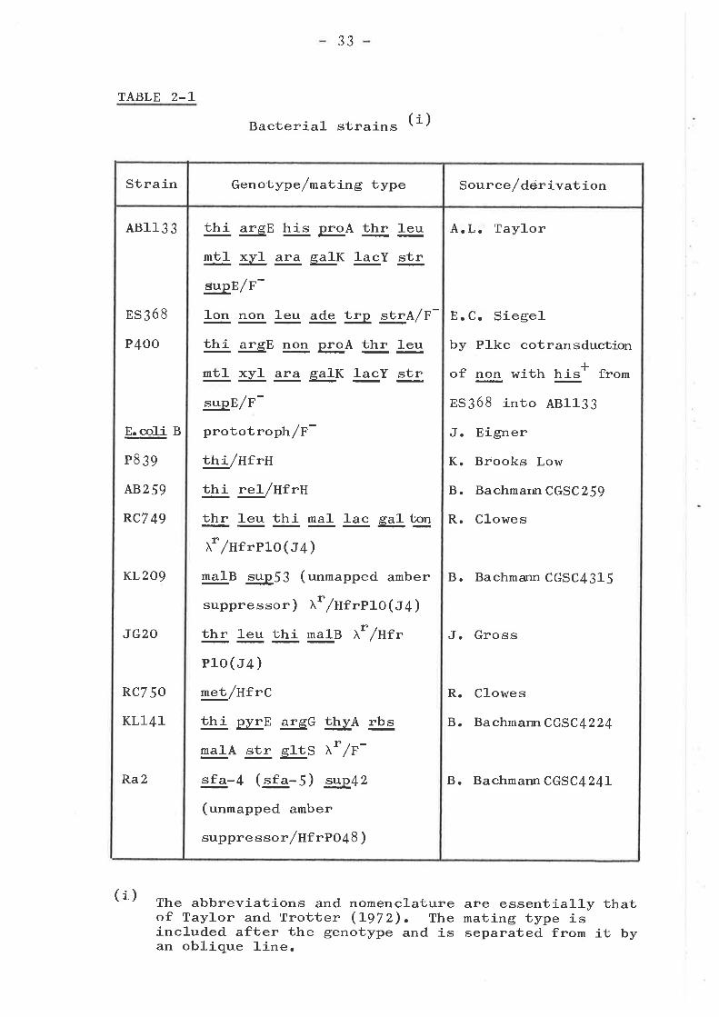
33
TABLE 2-L
Bacterial- strains (i)
The abbreviations and nomenclature are essentially thatof Taylor and Trotter (L972). The mating type isincluded after the genotype and is separated from it byan oblique 1-ine.
(i)
Strain Genotype /matjrng type Source/derivation
48113 3
ES368
P4OO
E.coli B
P8 39
A8259
RC7 49
KL2O9
JG2O
RC7 50
KL141
Ra2
thi argE his EA thr leu
mt]- xyl ara glK lacY str
supE/F-
]-on non leu ade t"p *tI.q/F-
thi argE non proA thr leu
nt]- 5¿! ara galK 1-acY str
.s,rp,E/F-
prototrop}rr/F-
thi/Hf rH
i"tlj- re\/HfrH
thr ]-eu thi ma1 ]-ac gg! t33
xrlHrrero ( J4 )
talB sup53 (unmapped amber
suppressor) xr/Hrrrto(J4 )
thr 1eu thi malB Xr/Hfr
Plo (J4 )
met /uf rC
thi pyrE arsG !þ¿A rbs
r"1A str g]-tS )r* /F-
"f"-4 (-"f"-5) sup42
(unmapped amber
suppress or /ntrto48)
A.L' Taylor
E.C. Siegel
by Plkc cotransductionIof non with LÞ' from
8S368 into 481133
J. Eigner
K. Brooks Low
B. Bachmarrn CGSC259
R. Clowes
B. Bachmann CGSC4315
J. Gross
R. Clowes
B. BachmannCGSC4224
B. Bachmarur CGSC424L
34
resistant and tolerant mutants derived from 481133 (table
4-7) kindly provided by J.K. Davies. All strains r^rere
stored as fteeze-dried cultures and working stocks were
maintained on nutrient agar slopes at 4C. All cultures were
grown by incubation at 37C. Overnight cultures were prepared
by innoculation from a slope or single colony into 10 m1 of
media and aerated by agitating on a reciprocating shaker.
Logarithmic cultures were prepared by diluting an overnight
culture 10- to loo-fold in fresh media and aerating as above.
Strain P4OO was derived as follows: It was known that
non ln¡as over 50% cotransducible with his (Radke and Siegel,
l-}Tl-). Therefore the his* marker from strain ES368 rn¡as
transferred into strain AB113J using Plkc transduction.
Recombinant colonies were tested for their inability to form
T7-resistant colonies by plating with bacteriophage TT and
incubating at 37C overnight. One recombinant with non
properties r,r¡as freeze dried as P4OO.
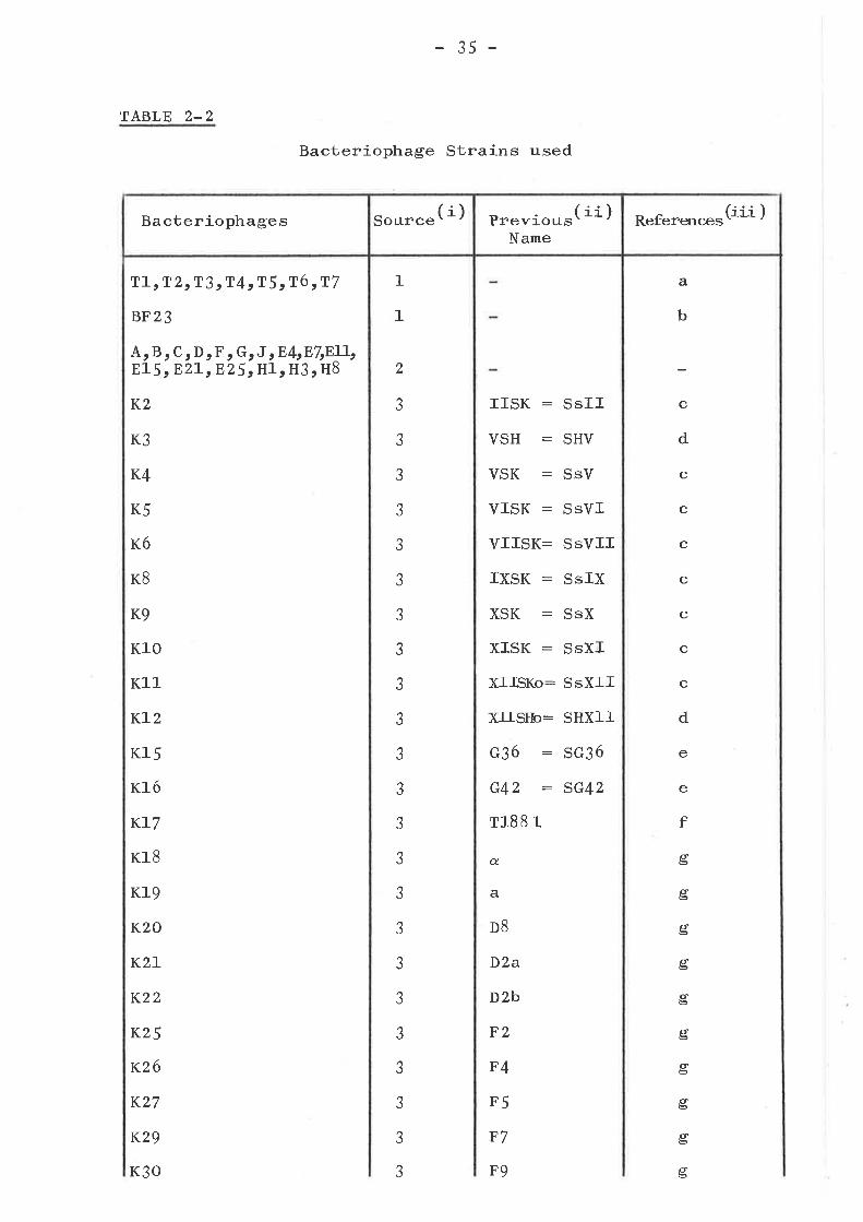
BACTERIOPHAGE STRAINS
The bacteriophage strains used in this study together
with their sources are described in Table 2-2. rn some cases,
bacteriophages have been given laboratory strain names, either
for the sake of simplicity or due to the similarity of names
in the literature. The previous names where appropriate and
a literature reference to the bacteriophages have been listed
in Table 2-2. The bacteriophages listed as having been
isolated frorn sehrage are discussed more thoroughly in Chapter
J together with other bacteriophages shown to be similar.
rn addition to the bacteriophages of Table 2-2 used for the
isolation and testing of mutants, bacteriophages CZL and U3
35 -
TABLE 2-2
T1, T 2tT3 cT4sT5 tT6 rT7
BF23
Bacteriophage Strains used
1
1
a
b
A,E1
B, C, D, F ¡ G, J ,E4rE7rE7l-!.5 ,E27",n25 t Hl, H3, H8
K2
K3
K4
K5
K6
KB
K9
K]-0
K11
K1-2
Kl5
K16
KI7
K18
Kl9
K20
K2L
K22
K25
R26
K27
K29
K30
c
d
c
c
c
c
c
c
c
d
e
e
f
úb
a6
6b
ø'Þ
6è5
út5
út=
11è
t6
úb
2
aJ
aJ
aJ
J
aJ
J
aJ
J
aJ
J
aJ
aJ
J
J
J
aJ
J
J
J
aJ
aJ
ó
J
IISK :
VSH :
VSK :
VISK :
VIISK:
IXSK :
XSK :
XISK :
XIISKo:
XITSIb:
c36 :G42 :r188 Iq.
a
D8
D2a
D2b
F2
F4
F5
F7
F9
SsII
SHV
SsV
SsVI
SSVII
SsIX
SsX
SsXI
SSXII
SHXII
sG36
sG42
Bacteriophages (i)Source Previous( ii )
NameReference.(iii )
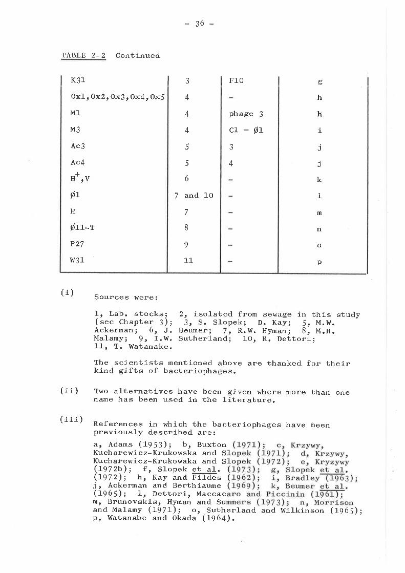
TABLE 2-2 Continued
K31
Oxl, Ox2, Ox3r Ox{, OxJ
M1
M3
Ac3
Ac4
Øt
H
þu-rF27
w31
(i) Sources were:
L, Lab. stocks;( see Chapter 3) ;Ackerman 1 6, J.Malamy; 9, I.W.11, T. Watanake.
2, isolated from3t S. Slopek;
Beumer t 7, R.W.Sutherland; 10,
-IH'rV
36
3
4
4
4
5
5
6
7 and 10
7
8
9
11
F10
phage 3
cL:øLJ
4
aÞ
h
h
i
j
j
k
1
m
n
o
p
sewage f-nD. Kay; 5,Hymani 8,
thMM .H.
is.w.
study
(ii)
(iii)
R. Dettori;
The scientists mentioned above are thanked for theirkind gifts of bacteriophages.
Two alternatives have been given where more than onename has been used in the literature.
References in which the bacteriophages have beenpreviously described are:a, Adams (fgS¡); b, BuxtonKucharewicz-Krukowska and SKucharewicz-Krukowaka and S(1pZZb); f , Slopek et a]-.(tgZz); h, Kay and FTiüãsj, Ackerman and Berthiaume(fq65); I, Dettori, Maccacm, Brunovskis, Hyman and Suand Malamy (1-gZl-); o, Sutherland and Wilkinson (t96il;pt Watanabe and Okada (f964).
3v
(tabte 6-1) kindly provided by Dr. Roy RusselJ-, and
bacteriophage P1-kc from laboratory stocks were used.
Some of the bacteriophages used were originally
isolated on other strains of E. coli or Shigella, a closely
related genus (Ewing, Hucks and Taylor, LgSZ; Luria and
Burrous, Lg57). These included.: bacteriophages H* and- V
originally isolated on Shieella dysenteriae PB (Beumer
et a]-.c Lg65); KZt K3r K4s K5r K6, K8, Kgt KlO, K11, KLz,
rc15r K16 and K17 originally isolated on Shieella sonnei
strains (Krzywy, Kucharewicz-Krukowska and_ Slopek, I97Lt
497 Z; Krzywy, L97 2; Slopek g!3!. o 1-97 3) ; K18, 619 r KZO, KZL,
K22t r25t K26, r27t K28, K29t K3o and K31 originally isolatedon Shigel]-a flexneri strains (Sl-opek É4. t A97z); Oxl, ox2,
Ox3¡ Ox4r Ox5., and M1 originally propagated on E. coli c2
(Bradley t L963); and Ac3 and. Ac4 originally propagated. on
E. coli 0727zB8 (Ackerman and Berthiaume, 1969). These
bacteriophages were all re-isolated three times on strain
P4OO from single plagues and propagated in liquid media
(Adamsr 1959).
GENERAL BACTERIOPHAGE METHODS
The method for assaying bacteriophage solutions was the
agar overlay technique. O.1 ml of each of a series of
1oo-fold dilutions of a bacteriophage solution were ad.ded to
O.1 mI of a standing overnight culture (fO9 ceLLs/mL) ofindicator bacteria in 3 m1 of o.7% Aear at 4sc, mixed by
gently swirling and poured onto a nutrient agar p1ate.
After allowing the agar overlay to set, and incubating
overnight at 37C, the plate containing the largest number
of discrete plaques was selected and the plaques enumerated.
38
A1-l bacteriophages \^¡ere propagated by one of two methods
on4K-L2strainP4oo;withtheexceptionoftheT
group of bacteriophages which are classically grown on
E. coli B and bacteriophages Hlr H3, H8 and ElI which were
also propagated on E. coli B. For the first method, a
solution of a given bacteriophage was assayed by the agar
overlay technique and after overnight incubatj-on a plate
showing semi-confluent lysis selected. The O.7% agar overlay
was scraped with a spreader from two such plates into a sterile
bottle, 1O mI of nutrient broth added, and the bottle allowed
to stand for 30 min at room temperature after which the agar
was removed by centrifugation (3OOOxg, 10 min). The
supernatant was extracted and assayed by t'he agar overlay
technique described above for plaque forming units, (pf*).
The second, method. involved- add,ing approximately fO7-fO8
pfu of a given bacteriophage to 10 ml of a culture of strain
P4OO or 481133 in logarithmic-phase growbh (1-2x1O8 cells/ml).
The mixture lr¡as then incubated at 37C unt-íL lysis occured, or
otherwise incubated overnight. Chloroform (O.5 mt) was added
and after incubating a further 30 min at 37C t}:.e bacteria
pelleted by centrifugation (5OOOxg, 10 min). The supernatant
was then assayed as above. All bacteriophage cultures were
stored for routine use at 4C with chloroform (5% v/u) and
assayed every six months or in some cases more regularly for
pfu. Samples of each bacteriophage were also added to double
strength milk and freeze dried (Chitter and l{olfson, 1960)
for J-ong term maintenance.
Effj-ciency of plating studies were done by plating a
defined number 1fo5-fo6 pfu) of a given bacteriophage, ir a
O.7% agar overlay with enough bacteria (S x Lo7 cel-ls) to
39
normally give confluent growth.
ISOLATION OF BACTERIOPHAGES FROM SEWAGE
Raw sewage tnras obtained on three separate occasions,
with the kind assistance of Mr. Lloyd Goss, from Bolivar
ser{¡age treatment farm near Adelaide. The technique for
isolation of bacteriophages was based on that of Brown and
Parisi (f966). Raw sewage was a1-1-owed to settle and the top
layer filtered through a Sêitz 56 filter. To 9 m1 of this
solution 1 mI of 10 times concentrated brain heart infusion
broth was added after which it rn¡as incubated (37Cr 4 hr).
Chloroform (O.5 mt) was added and the solution incubated
further (37C, 3O min) and then the bacteria removed. by
centrifugation (tOrOOO x gr lJ min) and the supernatant
plated with strain P4OO by the agar overlay technique. In
the third isolation the supernatant r^ras treated with anti-
serum raised against bacteriophages from the first and
second isolations, in order to increase the range of
bacteriophages obtained. After overnight incubation of the
overlaid plates single bacteriophage plaques were picked off
and purified by two single plaque isolations.
SEROLOGIGAL TECHNIOUES
One rabbit was used to raise antiserum against each of
bacteriophages T3¡ T6, H3 and F. The technique used was
based on the three collrse immunization schedule of Barry
(Ig5Ð. Bacteriophage lysates (fO9 pfu/m1) were prepared by
centrifugation (lorOOO x gr 30 rnin) to remove most of the cell
walr fragments. rn the first course of immunization, on five
40-
successive days o.75 ml - 1.o m1 of bacteriophage solution was
injected intravenously into the ear after which the rabbit was
rested 7 days and a test bleed taken. For the second course,
1 ml was injected intravenously on four successive days and
the rabbit again rested 7 days and test bted; while for thc
third course this was repeated with the exception of the
fourth dayt" injection. At this stage, the anti-phage anti-
body titres were sufficiently high for neutraLiza1"íon
experiments and the rabbits were bled out, either through the
ear or by cardiac puncture. The blood from the various
bleeds was allowed to clot at room temperature and then
ringed with a hot loop, stood in the cold for two hours, and
the serum collected and freed from red blood cel1s by
centrifugation (5,OOO * gr 10 min). The supernatant was then
frozen in 1 m1 quantities.
Assay of anti-phage antibodies was done with the
homologous bacteriophage by the method of Barry (I954).
O.9 ml of a solution of bacteriophage (S * 1O5 pfu/mL) was
added to O.1 m]- of each of a series of lO-fold dilutions of
anti-phage sera (o" O.1 ml- of normal sera as a control), and_
the solution incubated (37C, JO min) aituted 100-fo1d., and
O.1 mJ- plated with O.1 ml of a standing overnight culture of
strain P4OO by the agar overlay technique. After overnight
incubation of the plates at 37C tlrey were enumerated, and a
plate with a 90% reduction in plaque count used to calculate
the K value using the formula (Adams, 1959):
K : 2.3 DE
æ,plog
where D : dilution of antiserum; t : time of
po : number of plaques on control plate; and
plaques on plate with dilution D.
incubation;
p : number of
4L
In order to test the neutralization of otherbacteriophages, either a K value was carculated using the
method above, or a neutralization assay was performed. usinga given dilution of antiserum (L/Lo). rn this second. test,
90% neu1cra1-ization (approximatcly equivalent to K>1) or.
greater indicated serological relatedness. Two other anti-sera rnrere used in these experiments; anti-phage T4 sera, a
gift of Dr. A. Osmond; and anti-phage T2 sera, a gift ofDr. P. Reeves.
ELECTRON MICROSCOPY
The technique of preparation of bacteriophages forelectron microscopy, was based on that of Bradley (Lg67).
Bacteriophages rn¡ere harvested from five overlaid plates(O.7% agar overlay) showing semi-confluent lysis by
collecting each overlay into 3 ml of L/" (r/u) ammonium
acetate pH 7.o. They were then purified and concentrated by
one cycle of differential centrifugation ( 5, ooo x g¡ 20 mín
then 5orooo x gr 60 min) and the resultant bacteriophage
pellet resuspended in a small volume of ammonium acetatebuffer. Uranyl acetate was added at Z% (t/u) and the
specimens were then examined with a s¡lemens Elmiskop 1
electron microscope.
Grid preparation, mounting, staining and electronmicroscopy were skilfully performed mainly by Miss p. Dyer,
The Department of Biochemistry, The university of Adelaide.
The electron microscopy of bacteriophage T3 hras done by
Dr. J. casely-smith, rhe Department of zoorogy, The universityof Adelaide.
42
ISOLATION AND SCREENING OF BACTERIOPHAGE RESISTANT MUTANTS
In order to isolate bacteriophage resistant mutants of
independant origin, individual colonies of strain P4OO were
picked off and streaked across nutrient agar plates on whichäQ
10/-1O' pfu of the appropriate bacteriophage had been spread.
From each streak, a single colony was then picked off after
overnight incubation, purified by two successive single colony
isolations, and screened for sensitivity or resistance to the
bacteriophages shown in Table 2-2.
Suspected resistant mutants were screened using a
mul-tiple syringe bacteriophage applicator (tnt.S.BoA. ) shown
in Fig. 2-L. This machine, skilfully made by Mr. Barry
Parker of the Workshop, Medical Building, The UniversiÙy of
Adelaide, was based on that of Zíerdt, Fox and Norris (1960).
The technique of using this machine involved fi1-ling each of
the syringes from a nylon block with 24 weLLs, each
containing 2 mL of a different bacteriophage solution 1 Z * fO5
pfu/ml-). The mutants to be tested were grourr as overnight
standing cultures at 37C and O.1 ml of each plated on a
nutrient agar plate using the agar overlay technique. The
overlaid plates were then dried face downwards ({J min t 37C),
and placed in turn on the platform of the M.S.B.A. and the
micrometer rotated to produce a hanging drop at the end of
each of the syringes. The platform was raised so that each of
the drops were touched off onto a discrete area of the plate.
After the spots of bacteriophage solution had dried into the
surface of the overlay, the plate was incubated overnight at
37C. The parent strain P4OO, treated similarly, acted as
control. A reference line on the plate, served to indicate
the placement of the various spéts. The M.s.B.A. was
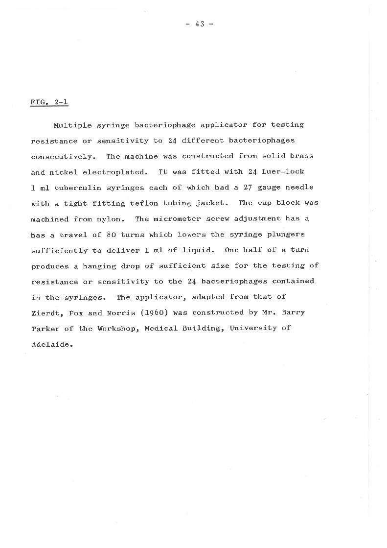
43
FIG. 2.L
Multiple syringe bacteriophage applicator for testing
resistance or sensitivity to 24 different bacteriophages
consecutively. The machine was constructed from solid brass
and nickel electroplated. It was fitted with 24 Luer-lock
1 ml tuberculin syringes each of which had a 27 gauge needle
with a tight fitting teflon tubing jacket. The cup block was
machined from ny1on. The micrometer screlv adjustment has a
has a travel of 80 turns which lowers the syringe plungers
sufficiently to deliver 1 ml of liquid. One half of a turn
produces a hanging drop of sufficient síze for the testing of
resistance or sensitivity to the 24 bacteriophages contained
in the syringes. The applicator, adapted from that of
Zíerdt, Fox and Norris (1960) was constructed by Mr. Barry
Parker of the Workshop, Medical Building, University of
Adelaide.
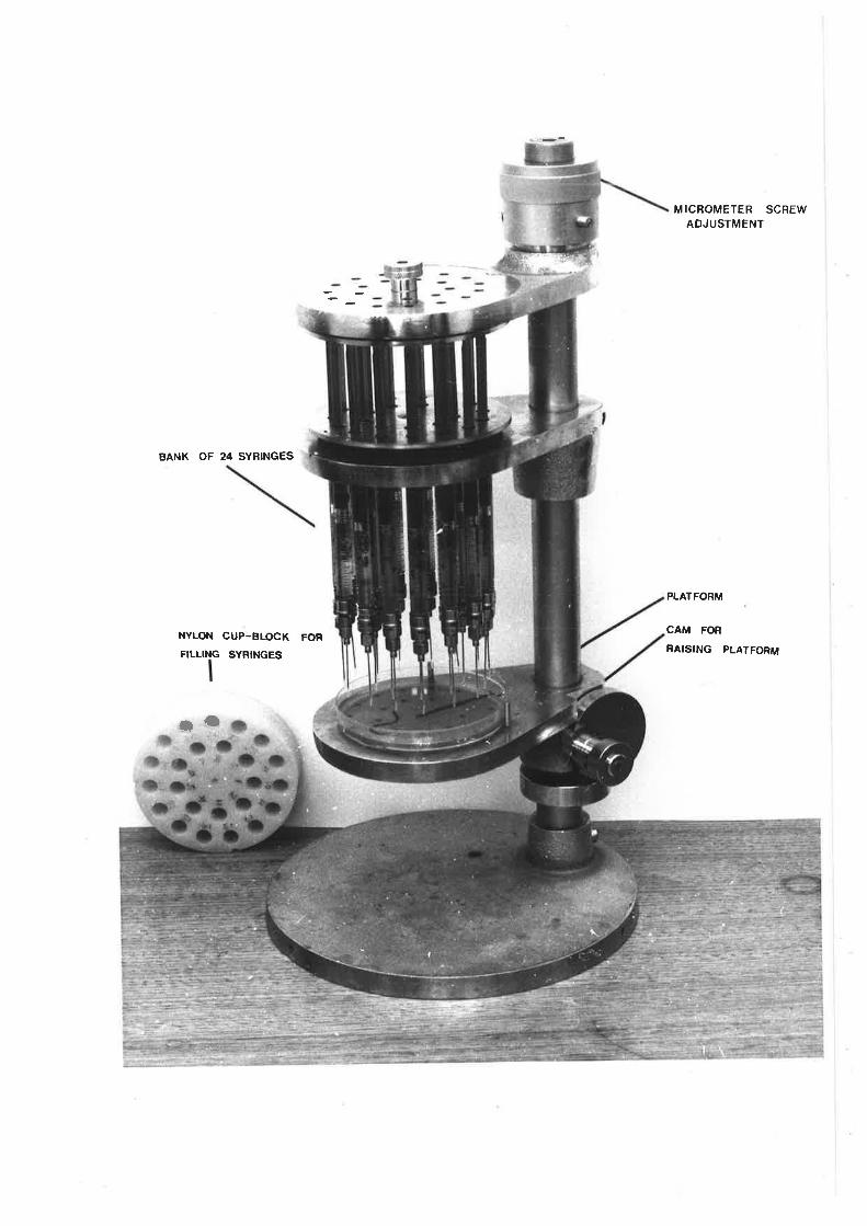
BANK OF 24 SYRINGES
NYLON CUP-BLOCK FOR
FILLING SYRINGES
ro
MICROMETER SCREWADJUSTMENT
PLATFORM
CAM FOR
RAISING PLATFORM
44
sterilized by autoclaving and could be used twice in one day
(allowing resistance or sensitivity of the mutants to 48
bacteriophages to be tested in this time period).
One or two isolates of each type of resistant mutant
were selected for further study and freeze dried. Further
characteri-zat-i-on of the resistance pattern of these selected
strains was done by e.o.p. studies whenever the mutant was
not fully resistanb or sensitive to a bacteriophage ( see
Chapter {,). All mutants isolated arose spontaneously with
the exception of strain P479 which was selected after muta-
genesis of strain P4OO by N-Methyl-Nt-nitro-N-nitrosoguanidine
(mfC) according to the method of Adelberg, Mandel and. Chen
(1965).
NOMENCLATURE OF RESISTANT MUTANTS
The relative efficiency of plating (e.o.p. ) in this
thesis represents the number of plaques formed on a mutant
relative to the number formed on the parent strain p4OO.
The following terms define the patterns of resistance
that occur when a defined ntrmber 1fO4-fo6 pfu) of a given
bacteriophage is plated, in a O.7% agar overlay, with enough
mutant bacteria 15 * fO7 cells) to normally give confluent
growth. The initials or symbols in brackets are the
abbreviations used in Tables 4-It 4-2, 4-3, 4-4, 4-5, 4-6
and 4-7.
Fu1l Resistance (n) The bacteriophage is unable to form
plaques on a particular mutant (e.o.p. ( ro-7 ).bacteriophage plaques are very
is inhibited where a plaqueof inhibition being equal to the
Bacterial Inhibition (r) rhe
turbid (i.e., bacterial growthwould normally form; the area
45
norma]-
Partial
area of a plaque). Plaques occur with an ero.p. of 1.
Resistance (P) The bacteriophage forms wild type
with an êooopr of 1O-2 o" less.plaques
Partial Resistance with Inhibition (fP) A combination of
the above two effects with very turbid plaques and a
lowered €oorpo
Slight Resistance (Sl) The bacteriophage either forms wild
type plaques at an ê.o.po of greater than !O'2, or only
minute plaques are formed with an e.o.p. of approximately 1.
These are very minor alterations in the resistance pattern
and thus they have been grouped.
Sensitivity (-) An ê.o.p¡ of approximately I with normal
plaques.
The terms resistance or resistant, where used in this
thesis, cover any of the above terms except for sensitivity.
These terms are distinguished from full resistance or fully
resistant as defined above.
The designation Wrm (Z) for a strain implies that the
strain belongs to sub-group 2 of the wrm resistance group.
PRBPARATION OF CELL ENVELOPE AND WALL FRACTIONS
For neutralization studies, whole cel-1 envelope was
prepared from ce1ls grown to late logarithmic phase
(ro9 ce1ls/m1) in a 10 litre fermentor. The media used was
phosphate buffer pH 7 .o containing r. S% (r/u) yeast extract
and L/" (*/u) casamino acids. After the cel-1-s had reached
the late logarithmic stage of growth, chloroform (o.S% t/v)
rn¡as added for 10 mín at 37c and the bacteria centrifuged
down (lorooo x gr 10 min). The pe1let was then re-suspend.ed.
in water and treated by a technique similar to that used by
46
Weidel, Koch and Bobosch (tgS+) in the first stage ofpreparation of the bacteriophage T5 receptor complex. The
suspension was stirred vigorously with a glass rod and ühe
pH raised from 6.5 to 9.8 by dropwise add.ition of O.1N
sodium hydroxide and immediately l-owerect to pH 7.O by
bubbling carbon dioxide through the suspension, after which
it was centrifuged (lorooo x g.r 20 min) and the supernatant
set aside. The pellet was re-suspended in distilled water
and extracted again as above. The two supernatants were
combined as the ttsodium hydroxide supernatantrr and thepellet treated according to the technique of weltzein and
Jesaitis (L97I).
The pellet remaining after sodium hydroxide treatment
rt¡as re-suspended in 70 ml- of O.O1M Tris buffer pH 7.5 and
O.4 mg ribonuclease A (Sigrna Chemical Co.¡ St. Louis, Mo.),
O.4 mg of crude beef heart pancreas deoxyribonuclease
(Siema Chemical Co., ) and 1O mg of MgCl, ad.ded. This
solution was then incubated at 37C for JO min, and the
supernatant removed after centrifugation (torooo x gr 20 min).
The pe11et was re-suspended in Tris buffer and sonicated for
JO min at OC, and collected by centrifugation (lOrOOO x g¡
Jo min). After re-suspension of the pe1let, phase contrastmicroscopy revealed that the preparation was not contaminated
with whole cells, and no clumping of the membraneous structure
was observed. The suspension was then dialysed (LZ tr, 4C
then 2{ }:.r, 4C) against fresh changes of water. The cellenvelope was collected by centrifugation (5orooo x g: 6o min)
dissolved by sonication in distilled water and lypholized as
the cell envelope preparation. The yield was 6zo mg (dried
weight) of ce1-1 envelopes.
47
This ce1-1- envelope preparaùion was dissolved at a
concentration of 2 mg/mL in 2% Trj-1.,on X-IOO in 10 rnM Hepes
buffer pH 7.4 and treated by the techniques of Schnaitman
(1-gl1-arb), Suspension was aided by 1O sec sonication. The
solutiorr was incubaLecl for 1O min at 23C and then centrifuged(15O,OOO x g, 60 min). The supernatant was set asid.e as the
Triton x-1oo soluble supernatant. The pellet was re-suspended
in water and washed by centrifugation (tsrOOO * gr 60 min).
Re-suspension of the pellet in the original volume of water
yielded the Triton X-1OO insoluble rrce11 ttralltr preparation.
This ce11 wal1- preparation was then re-extracted with the
same Triton-Hepes buffer, but with 10 mM Ethylene-diamine-
tetraacetic acid (nata) pH 7.4 added (Schnaitman, IgTLb).
This yielded after centrifugation and washing, the Triton-
Edta solub1-e and insoluble preparations. Triton X-1OO was
removed from the Triton-Edta soluble supernatant by alcoholprecipitation and centrifugation (l0rOOO x gr 20 mirn) which
left the Triton X-1OO in the supernatant (SaUet and
Schnaitman, L97It L973a). The protein content of allpreparations was analysed by the technique of schacterl-e and
Pollack (L973).
For polyacrylamide ge1 electrophoresis, whole ce1l
envelope l.ras prepared from 1 litre of ceIls growïr in nutrient
broth to logarithmic-phase (Z x 1O8 cells/rn1). The cel-1-s
r^¡ere harvested by centrifugation (fOrOOO x gr 20 mjrn) irt
O.O5 M Tris-buffêr pH 7.8, washed in the same buffer, and
broken with a French pressure cel1, or after freezíng with
an alcohol-dry ice mixture in an x-press cell disintegrator(L.K.B. Biotec, Stockholm). Ce11 d.ebris was pelleted by
centrifugation (5rOOO x g¡ 15 min) and MeC1, was add.ed to
48
2 mM. The supernatant fluid was centrifuged (78'OOO x gr
6O min), and the pe1-1et re-suspended in Tris-MgCl, as above
and centrifuged (78rOOO x gr 60 nin). The resultant pellet
was re-suspended in deionized distilled water at a protein
concent,ration of IO mg/ml (estimated by the Schacterle and.
Pollack (L973) technique) as the cell envelope preparation.
Cel1- wall was prepared by the method of Schnaitman (Ig7Ia)
as described above using an initial concentration of LO mg/mL.
LIPOPOLYSACCHARIDE PREPARATIONS
For neutralization studies, lipopolysaccharide (leS)
was extracted by the hot phenol-water technique (Westphal,
Laderí1;z and Bister (1-952\. The preparation was treated
with deoxyribonuclease and ribonuclease as described above
for the cel1- envelope preparation. For gas liquid
chromatography, LPS was isolated by the technique of Galanos,
Lrrderítz and l,{estphal (tg6g) using solubiliza1-íon in
phenol- chloro f orm-petro1-eum ether.
ADSORBTION AND NEUTRALIZATION STUDIES
Two methods were used for testing the adsorbtion of
bacteriophages to whole cel-ls. In the first a given
bacteriophage was added to logarithmically growing bacteriaa(2 x 10" cell.s/m1-) at a multiplicity of infection of O.5 to
1.O in the presence of O.OOIM KCN. After 30 min at 37C this
rvas diluted loO-fo1-d, 5% G/u ) chloroform added, the solution
vortexed for 5 sec on a lVtrirlymixer (Fissons Scientific
Industríes Ltd. ), shaken for l-O min at 37C, and suitabl-e
dil-utions plated for infective centres. Nutrient broth
49-
substituted for bacteria in the control. This method. testedonly irreversible adsorbtion. The second method. used tested.
boüh reversible and irreversible adsorbtion. Bacteriophages
and bacteria h¡ere mixed for f mín at 3Tc as described above,
but without the addition of cyanidc. They rn¡ere then
centrifuged and the supernatant assayed for unattached
bacteriophages.
For neutralization studies, o.5 ml of a given bacterio-I
phage (5xto'pfu/mL) ir, nutrient broth was added to o.J m1 ofdistilled water containing 1 mg, 1oo ¡rg or 1o ¡rg of Lps or ofone of the ce1l envelope or wa1l preparations. The mixtures
u/ere then incubated for 3 hr at 37C, diluted loO-fold to stop
further reaction and o,1 ml plated with o.1 ml of an overnight
standing culture of strain P4oo in a o.7% agar overlay, and.
incubated overnight at 37C. In the control O.5 m1 of
distilled water was substituted for the LpS or cel1 envelope
or wall preparation. A 50% or greater reduction in plaque
count relative to t'he control indicated neutralization.
For enzymati-c studies of the receptor, cell wall was
treaüed with sodium periodate according to the technique of
Sabet and Schnaitman (tglt) o" pronase according to thetechnique of l{eltzein and Jesaitis (tgZt) prior to
neutralizati-on studies as above.
ANTTBIOT IC RES ISTANCE TESTING
Oxoid multodisks 11-14D and 3O-9C were used to testalteration in the pattern of resistance or sensitivit,y to a
range of antibiotics. The discs contained chloramphenicol
(So ¡r*¡, erythromycin (So ¡ru¡, tetracycline (So ¡r*¡, colistinmethane sulphonate (SO ¡,*¡, kanamycin (5 ¡rS), neomycin (1O Ag)
5o -
and ampicillin (2 lug) to which strain P4OO is sensitive;
novobiocin (aO 7r*¡ to which it is slightly sensitive; and
sulphafurazoT.e (SOO pa)r oleanodymycin (10 ¡rg), fusidic
acid (1O ¡rg) cloxacillin (5 rtg), and novobiocin (5 ¡tg) to
which it is resistant. In the case of erythromycin a
decrease or increase in zoÍre size of 50% over þ}:re control
strain P{OO was taken to mean resistance or sensitivity
respectively. Any lesser change was described as slight
(tafte 1). A1-1- other antibiotics except for ampicillin showed
absolute changes (i.". resistance going to sensitivity).
Ampicillin resistance was further tested by plating
approximately 1OOO cel-ls on nutrient agar plates containingg.51 1.O, 2, 3t 4, 5, 15r 30 or 75 yS/nL of ampicillin
(Beecham Research Laboratories, Australia). Strain P{OO was
found to be resistant to L.O ug/mJ- but sensitive to 2.O ¡tg/mI.
POLYACRYLA}{IDE GEL TECHNIOUES
The solubil-iza1r-íon of cel1 envelope proteins and the
subsequent analysis of these proteins by gel electrophoresis
were carried out by three techniques, the first of which,
Method (1) r^¡as essentially that of Neville (Tg7L). Samples
of whole envelope, cel1 wall or standard protein preparations,
were made 5O mM in sodium carbonate, and sodium dodecyl
sulphate (SOS) was added at 4 ^e/^e of protein, followed by
the addition of LO% by volume Z-mercaptoethanol; the maximum
time for this step being 2 mín. This preparation was diluted
to 4 mg proi.,eín/mL in upper ge1- buffer (ttevil-1-e, L97L) +
sucrose (2%) * bromophenol blue, and samples containing
1OO-15O ug of protein (ZS-+O pt) were loaded onto gels
51 -
(1O x 0.6 cm) with a Hamilton microsyringe. The gels, in a
SDS borate-sulphate discontinuous buffer system (Nevi11e,
A97I), were run ar, O.5-1.O mA per tube at 25C until the
sample entered the upper gel then the current was increased
to 1.5 mÀ per tube. After electrophoresis for 6 hr the
gels were stained for 15 hr in O.2% coomasie brilliant blue
in fixative (Weber and Osborn, L969) and de-stained with
2O/" methanol in 7 .5% acetic acid.
The solubilizaþion of method (Z) was the complete method
II of Schnaitman (tgZ Za) and involved preliminary heating of
the sample in SDS at 37C for 2 }:.r, dialysis against SDS-urea
and final heating at 1OOC for J min. The solubilizatíon of
method (3) again employed method II of Schnaitman (tgZS^)
omitting only the final 1OOC heating. Samples of cel1 wa11
and ce1l envelope prepared by methods (2) and (3) were run
on 7.5% polyacrylamide gels using either the pH 7.2 NIai-zeL
buffer system (Uaizel, 1966) or the pH 11.4 - pH 4.1 Bragg-
Hou ge1 system (eragg and Hou, L972). These were the
conditions used by Schnaitman (I97 3arA, I97 4arT¡) .
Gel staining and de-staining was by the techniques of
Swank and Munkres (L97I). Stained gels prepared by all
techniques were scanned with either a Joyce Loebel
Chromoscan Mk II densitometer, a Gilford ge1- scanner or a
Helena Quick Scan Jr.
GAS LIQUID CHROMATOGRAPHY
To 2 mg of LPS in deionized distilled water, 1OO ¡g of
arabinose h/as added as an internal standard. The LPS was
then hydroLízed with O.lN HCI (1OOC, 64 nr) itr a total volume
of 2 mI in an ampoule sealed under vacuo. The resultant
52 -
hydrolysate was then centrifuged (3ooo x gr 1J min) to remove
lipid, excess acid neutralized on a column of ion exchange
resin AG1-x2 (ucoJ form) (1o0-20o mesh, Bio. Rad Laboratories,Richmond, Ca1if.) and brought to pH 8.5 with NaHCOr. This
mixture of aldoses was reduced by adcli.ng 1{ mg of sodium
borohydride and incubating for 2 hr at room temperatllre.
Excess borohydride was destroyed with glacial acetic acid,and passage through a coltrmn of ion exchange resin Ac5ow-x4
I(H' form) (so - 1oo mesh, Bio. Rad Laboratories). The ald,oses
thus obtained were converted to their alditol acetates by the
technique of Holme g!3.!. (1968). These hrere then dried down
and dissolved in a small amount of chloroform and injected
into a column of 3% ECNSSM (Applied Science Laboratories,
State College, Pa. ) on Gas ChromQ (1OO-12O mesh) (Applied
science Laboratories), in a varian Aerograph series 1/oo gas
chromatograph connected up to a Hitachi Model QpD 54 chart
recorder. The column temperature was ZOOC as suggested by
Eriksson-Grennberg et a1. (t977). As a control, arabinose
was added to an equal amount of glucose and subjected to the
above procedure, including hydrolysis.
GLUCOSAMINE ANALYSIS
ItDegraded polysaccharidetl r,'¡as extracted from LpS by
treatment with L/o acet-íc acid (IOOC, 2 hr) and lípid A was
removed by centrifugation (utitter-sejtz, Jann and Jann, 1968).
some samples were further extracted 2 tímes with an equal
volume of chloroform. All samples were then lypholized,
after which they were hydrolysed according to the technique
of Monner et a]-. (t97I). The glucosamine content of the
5J
hydrolysate was analysed according to the technique of
Strominger g!3!. ( t9 59 ) .
GENETIC ANALYSIS
The techniques of conjugation and gradient transferanalysis of mutants were those of de Haan g!4. (1969).
For scoring of growth factors replica plating was used;
however, this was found to be inaccurate for bacteriophage
resistance/sensitivity testing and. another technique was
used. single recombinant colonies were picked off, spread
on a nutrient agar plate, grov¡rr overnight at 37C, and a
single colony then spread with a platinum loop on one ninthof a dried nutrient agar plate. A drop (O.Ot mt) of
bacteriophage lysate containing lo6 pfu or more ltras appliedto the spread area and after incubation at 37c overnight, an
area of lysis indicated sensitivity. The resistant mutant
used in the study and its parent strain p4OO acted as
controls. The technique of Plkc transduction was that ofLennox (1955).

54
CH PTER
CHARACTERTZATION OF BACTERIOPHAGES fSOLATED FROM SEWAGE
INTRODUCTION
Bacteriophages capable of lysing E. coli have been
isolated on many other genera of the order Eubacteriales.
These include many members of the family Enterobacteriaceae(of which E. coli is a genus): ShiÉella (Beumer É3!.rL965; Gromkova, 1968; Dhillon and. Dhillon, L9T2); Serratiaand SalmonellÊ (Adams and l,rrade, 1955); Klebsie]-la and
Aerobacter (sutherland and wilkinson, 1965 1 souza, Gj.noza
and Haight, L972); as well as Pasteurella pestis ( Hertman,
L964; Brunovskis, Hyman and Summers, ag| 3) of the familyBrucellaceae. Many of these are enteric organisms and_
hence excreted in the faeces. E. coli K-1-Z too, was
originally isolated as an enteric organism from the stool ofa diphtheria patient at stanford university in rgzz (Jones,
Koltzow and stocker, L972). Therefore, as one might expect,
coliphages have been previously isolated. from the faeces ofvarious animals and man (see for example, Burnet and McKie,
1933)r from sehrage (".s. Dettori, Maccacaro and piccinin,
L96L; Brown and Parisi, Lg66), water (Bradley, Lg63), and a
host of other sources in which one might expect to find.
faecal contamination (ttritton and Dhillon, ag72). In thisstudy, it was decided to isolate coliphages from raw sewage
which appeared to be a valuable source of such bacteriophages.
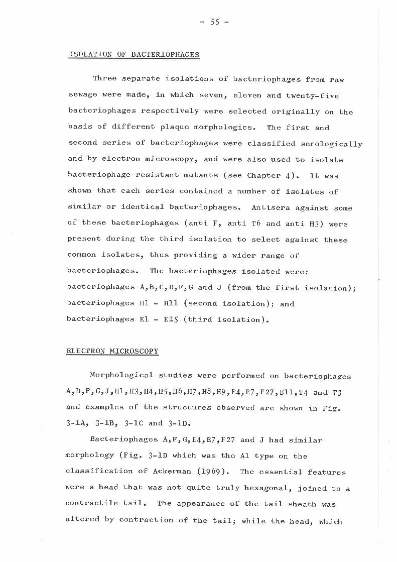
55
ISOLATI ON OF BACTERIOPHAGES
Three separate isolations of bacteriophages from raw
sewage ü¡ere made, in which seven, eleven and twenty-five
bacteriophages respectively were selected originally on the
basis of different plaque morphologies. The first and
second series of bacteriophages ì^rere classified serologically
and by electron microscopy, and were also used to isolatebacteriophage resistant mutants (see chapter 4). rt was
shou¡n that each series contained a number of isolates ofsimilar or identical bacteriophages. Antisera against some
of these bacteriophages (anti F, anti T6 and anti H3) hrere
present during the third isolation to select against these
common isolates, thus providing a wider range of
bacteriophages. The bacteriophages isolated h¡ere3
bacteriophages ArBrcrDrFrG and J (from the first isolation);
bacteriophages H1 - H11- (second isolation); and
bacteriophages El - 825 (tfrira isolation).
ELECTRON M ICROSCOPY
Morphological studies r^¡ere performed on bacteriophages
ArDrF, GrJrHl, H3rH4rH5rH6tH7 ,H8 rP.g,E4rE7 tF27 ,ELLrT4 and T3
and examples of the structures observed are shown in Fig.
3-14, 3-1Br 3-1C and 3-1D.
Bacteriophages ArF, GrE4rET rF2T and J had similar
morphology (pig. 3-1D which was the A1 type on theclassification of Ackerman (L969). The essential features
were a head that was not quite truly hexagonal, joined to a
contractile tail. The appearance of the tail sheath was
altered by contraction of the tail; while the head, which
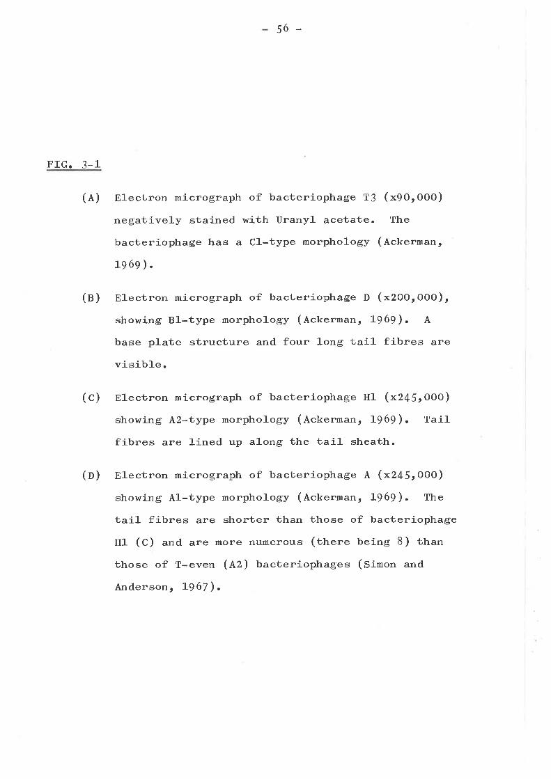
-5O -
FIG. 3-1
(A) El-ectron micrograph of bacteriophage TJ (xgOrOOO)
negatively stained with Uranyl acetate. The
bacteriophage has a C1-type morphology (Ackerman,
1969).
(n) Electron micrograph of bacteriophage D (x2OOrOOO),
showing Bl-type morphology (Ackerman, L969). A
base plate structure and four long tail fibres are
visible.
(C) Electron micrograph of bacteriophage Hl (x245'OOO)
showing A}-i.,ype morphology (Ackerman, 1-969). Tail
fibres are lined up along the tail sheath.
(n) Electron micrograph of bacteriophage A (x245.OOO)
showing Al-type morphology (Ackerman, L969). The
tail fibres are shorter than those of bacteriophage
Hl (C) and are more numerous (there being 8) than
those of T-even (nZ) bacteriophages (Simon and
Anderson s L967).
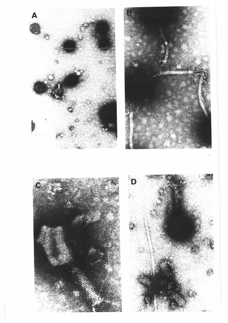
l,rç : .
t
*cs
t.f¡
p(p
.I}"
57
was joined to the tail by a fairly flexible joint, was
often ]-ost after contraction. The tail core could be seen
to partly extend from the contracted bacteriophage taiJ-s
and it was often associated with membranous vesicular-like
material. Thcrc appcarcd to bc a base plate, pins and
eight fibres associated with the tail. The sizes of
bacteriophages ArJrF and E4 r^¡ere similar, with the
arithmetic means of their measurements ( calculated using
greater than ten measurements) varying by a maximum of 3%.
The measurements of bacteriophage A were: Head., g¡Ot¡ql^OrOOx 852!52-A; Tai1, 1159f35O4 x 2c-5!7L; Contracted Tail,
55oi18l x zSOtr6f; Fibres, approximately 7 3oR 1-ong.
Bacteriophages G, E7 and F27 were of similar morphology but
had heads which h¡ere LO% smaller: 8 ¡ Ztgf R x 7 5g!z5o/..
Bacteriophage D (pig. 3-18) had a morphology similar
to bacteriophage T5 (tunt and Kay, I97Z) beíng of the Bl
type according to the classification of Ackerman (f969).
The head appeared to be larger than that of Tj, beingg¿qtSol long and. 9 2È601, when measured d.iagonally from
vertex to vertex; while the taiJ- length (fqA+t6Ol) was also
greater than that reported for T5 (Lunt and Kay, L?TZ). The
length of the tail fibres ( 6O6tfql) and. the width of the
tail (1OOt61) were very similar to the measurements reported.
for T5. The size and morphology (nie. 3-14) of bacteriophage
T3 (of Cl type morphology)r h¡ere similar to that previously
reported (Ackerman, 1969; Tikhonenko, L97O) and to that of
bacteriophage E1-l.
The electron microscopic morphologies of bacteriophages
TAt Hl, H3r H4t H5r H6, H7, H8 and H9 (nig. 3-1C), were all
T-even in nature (lz in the Ackerman (tg6g) classification).The bacteriophages, as well as having this similar morphology,
58
were of similar size, the measurements being: Head., 1164R
(with the arithmetic mean masurement for any one bacteriophage
differing from ühe arithmetic mean measurement of allbacteriophages by a maximum of tS.g%) x 364Î G6.6%); Tail,11091 e7.7%) x îß69/,, (t8.5%). They att had a contracrire rail,a base plate and pins, a tail co11ar, tail fibres, and a tailcore which are the typical features of T-even bacteriophages,
(simon and Anderson, 1967). The measurements noted. above,
although greater than others reported for T-even
bacteriophages in the literature (Bradley, L96T; Ackerman,
L969; Tikhonenko , I97O), h¡ere proven by Miss p. Levy, who
performed the electron microscopy, to be correct to within
5% bV comparison with standard sized styrene beads.
SEROLOGTCAL CLASSIFICATION
I,Íhen the various anti-bacteriophage sera were tested inneutralizatíon experiments with the homologous bacteriophages,
the K values obtained were: anti-T6 (l<:f L2), anti-H3 (f:658),
ant,i-T3 (r:Z3O), anti-F (r:41O), anti-T{ (r:659 ), antí-T}(K:1o91) which is equivalent to Ço/" neutraLi'za1-j.on by a 10-3
to ro-4 dilution of the respective antisera. The fulr set ofresults obtained by using these antisera is presented inTable 3-1.
All of the bacteriophages from the first isolation, withone exception (bacteriophage D), were neutralized by anti-bacteriophage F sera. The degree of neutralization varied.;
however all of these bacteriophages were clearly relatedserologically to each other. It is interesting that
bacteriophage G, which appeared to have a different sized
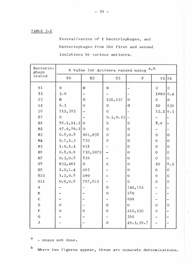
TABLE 3-1
59
Neutraliza1;-i-on of T bacteriophages, and
bacteriophages from the first and second
isolations by various antisera.
Bacterio-phagetested
a
b
T1
T2
T3
T4
T6
T7
H1
H2
H3
H4
H5
H6
H7
H8
H9
H10
Hl1A
B
c
D
F
G
J
means not done.
K Value for Antisera raised using arb
T6
o
5.oao.5712,565o
86.1rgL.597 .6 ,86.2o.8ro.8O.7 ,L.51.Or1.4o.8,o.g0.5ro.8932,462I.O,f-.4I.2 rO.8o.9ro.8
o
o
o
o
66L,6 58
7zo93L7 50,IO7 2
836
o
495
490
767,6L2
o
0
o
o
23O,L57
o
o
o.5, o. 23
o
o
o
o
o
o
o
o
o
o
o
o
o
o
o
o
o
o
o
0
o
o
I44,LL4180
6o6
o
4LO,23O
560
49.5r59.7
o
o
o
109 O
o
20
L2.2
o
o
o
0
o
20
o
o
o
8.9
o
o
o
6.4o
659
9.5
o
o
o
o
o
B.zo
0
o
o
Where two figures appear, these are separate determinations.
6o
head to bacteriophages Ar F and J, was not significantly
serologically different. The reason for this is that anti-genicity appears to be associated with tail components
(Adams, L959); therefore the tai1-s of these bacteriophages,
which are morphologically similar, must also be antigenically
simi]-ar.
Bacteriophages H1 - H1-1- which are morphologically
similar to T-even bacteriophages, seemed to fit into three
serological groups:
1) bacteriophages HJ, H4t H5r H6o H7t H9t HlO and H11,
which were neutralized slightly by anti-T6 sera, wellby anti-H3 sera but not at all by anti-T2 or anti-T{
sera;
2) bacteriophages H1 and H2 which were more closely
serologically related to T6 than T4 and T2 but are not
at a]-]- related to H3;
3) bacteriophage H8 which was essentially serological-ly
identical to T6.
The serological relatedness of the T-even bacteriophages,
and of bacteriophages T3 and T7 (Adams and l{ade, A955;
Adams, L959) were largely borne out by the results shown inTable 3-1. Since bacteriophage T7 had a very low K value
when tested with anti-T3 sera (tabte 3-1) another isolate
of bacteriophage T7 was obtained from Dr. M.H. Malamy, and
shown to have a very similar K value. The 5OO-1OOO fold
difference in K values obtained after neutrali zation ofbacteriophages T/ and T3 by anti-TJ sera has been previously
reported (Adams, 1959). H3-like bacteriophages are as
serologically rerated to bacteriophage T6 as T3 and T7 are
61
to each other, however they are clearl-y not related to T4
and T2 and thus they have been placed on a serologicalgroup of their own j-n Table 3-2,
SELECTION AND CIIARACTERIZATION OF BACTERIOPHAGES USED IN
RESISTANT MUTANT TESTING
Bacteriophages used in resistant mutant testing are
summarized in Table 3-2. I,ftren resistant mutants ü¡ere
isolated against bacteriophages A, B, C, F, G and_ J, and
tested by cross streaking it was shown that mutants
resistant üo one of these bacteriophages r^rere resistant toall of them. Thus, since these bacteriophages are similarbot'h morphologically and serologically, and have common
resistant mutants (i.". they are probably different isolatesof the same bacteriophage), it was decided to use for future
studies only bacteriophage A in addition to bacteriophage D
(which was clearly different from the others) from the firstisolation. Similar logic was applied to bacteriophages
from the second isolation, and bacteriophages H1, H3 and. H8
were selected for future use.
rn the t'hird isolation, twenty-five bacteriophages were
purified. These hrere selected as different from the above
bacteriophages, by the inclusion of antisera during their
iso]-ation.
Idhen these bacteriophages r{¡ere tested againstpreviously isolated resistant mutants, it was found thatthey were of six main types. Examples of each of these
types, whieh included bacteriophages E4t E7 t E11, n15r E21 and
E25 were selected for further study and of these only one r4¡as
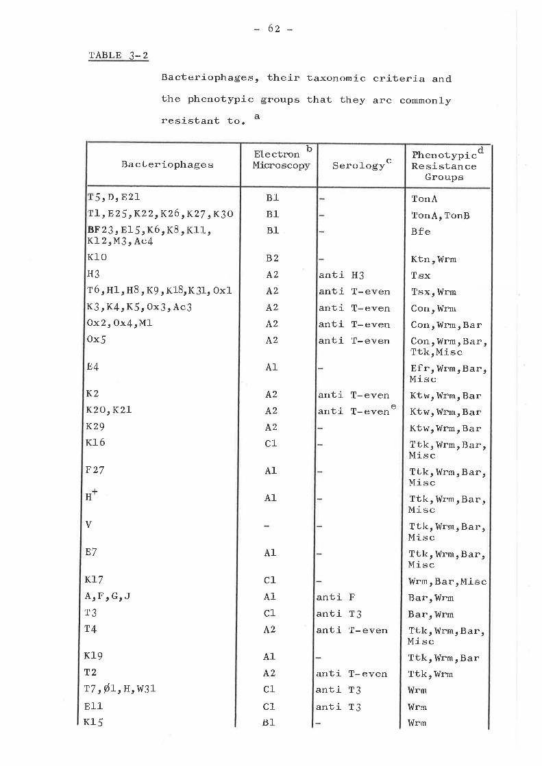
TABLE 3_2
5,DrE21T1, E 25 rK22 rK26 ,K27, K3O
8F23, El5rKórK8rKl1,K1-2rM3 , Ac4
Kl0H3
T6, Hl, H8, Kg, K1B,KJ1, Ox1
K3rK4rK5r ox3rAc3Ox2,0x{rMJ-ox5
E4
K2
KzO,KzLK29
K16
F27
I
H,
E7
KL7
A,F,G,JT3
T4
Kl9T2
T7,ørrHrW31
E11
Kl5
62
Bacteriophages, their taxonomic
the phenotypic groups that they
resistant to. a
criteria and
are commonly
B1
B1
B1
PhenotypicResistance
Groups
d
TonA
TonA, TonB
Bfe
Ktn, Wrm
TsxTsx, Wrm
Con, Wrm
Con , Wrm, Ba rConrWrmrBar,TtkrMiscEfr, Wrm, Bar,MiscKtw, Wrm, BarKtw, Wrm, BarKtw, Wrm, BarTtk, Wrm, Bar,MiscTtk, Wrm, Bar,MiscTtkr l,{rm, Bar,MiscTtk, Wrm, Bar,MiscTtk, Wrm, Bar,MiscIdrm, BarrMiscBar, Wrm
Barr l,{rmTtk, Wrm, Bar,MiscTtk, Wrm, BarTtk, Wrm
Wrm
Wrm
Wrm
B2
4,2
A2
A2
A2
^2
A1
A2
A2
A2
c1
A1
c1
A1
c1
^2
A1
A2
c1
c1B1
antiantiantiantianti
H3
T-evenT-evenT-evenT-even
antianti
T-evenT- even e
A1
A1
v
antiantianti
F
r3T- even
antiantianti
T- evenr3T3
BacteriophagesElectn¡n b
Microscopy SeroJ-ogyc
6S
TABLE 3-Z Continued
K25
þ:-r-rB1
c1 anti T3
a
b
Details of minor variations in resistance pattern areincluded in Tables 4-a, 4-2, 4-3, 4-4, 4-5 and /,-6.
El-ectron microscopicAckerman (L969) z-A1 (nr-ri.re) :A2 (T-even-like) :81 (r5-rire) :
B2
appearance is as described by
l-ong contractile tai1, isometric head.long contractile tail, elongated head.Long non-contractile tail, isometrichead.long non-contractile tail, elongatedhead.short non-contractile tail, isometrichead.
c
means not done.
Rabbit anti-bacteriophage sera tested against eachbacteriophage were anti T2, anti T4 or anti T6 (antiT-even)r- atrti t3, Anti HJ, and. anti F. Antisera withneutra1.izing activity are included in the tab1e.
means not neutral-j-zed by above antisera.
See Chapter 4 for fu1l details and Chapter 9 fordiscussion. Mnemonics are the same as the genotypicmnemonics where one member of the group has previouslybeen mapped:-Con is Conjugational recipient deficiency (Skurray,Hancock and Reeves, f974); Ktw is K two resistance; Wrmis ldide range mutants; Ktn is K ten resistance; Ttk isT2, T4 or K19 resistance; Bar is bacteriophage Aresistance; Misc is miscellaneous group; Efr is E fourresistance;
c1 (r:-like)
Onl-y shows partialanti T2 serum and
d
e neutralizat-j.on (O. 8(rc(f. Z) withanti T6 serum (anti T{ serum not tested).
6+
neutralized by any of the avdílable sera (i... bacteriophage
811).
other bacteriophages used in resistance studies were
imported from various laboratories (see Table z- 2). Many ofthese had beerr rnor.phologically characterj,zed. previously(see Table 2-2 for references and. Table 3-z for summary).
They h¡ere tested for their neutralization by the variousantisera available and it was found that many of thebacteriophages with T-even morphologies were neutralized by
anti-T2, anti-T4 or anti-T6 sera. The only exception
appeared to be bacteriophage rc29 t while bacteriophages K2O
and K21 from the same resistance group (see Table 4-3) were
only poorly neutralized by anti-T2 and. anti-T6 sera (anti-T{sera was not tested). The serological relatedness of TJ and
the other so-called female-specific bacteriophages w31r H,
Øtl-r and þ1', is in agreement with their other closelyrelated properties (Morrison and Malamy, LTTL; Brunovskis,Hyman and Summers, I97 3); but it is interesting thatbacteriophage 811, which is not female-specific, is more
cJ:ósely related to T3 than is bacteriophage TT t which isfemale- specific.
SUMMARY AND CONCLUSIONS
only elementary conclusions that have been made in thetext can be made at t,his point. This work in part bears
out the conclusion (Adams, 1953; Ackerman, LgSg) ttratclassification by serological and by morphological techniquesgive similar results. Discussion of this work in relation toresistance studies is included in Chapter g.
65
CHAPTER 4
ISOLATION AND TESTING OF RESISTANT MUT ANTS
INTRODUCTION
As discussed in Chapterl, studies of bacteriophage
resistance in E. co]-i K-L2 or E. co]-i B have been
restricted to a few limited studies on mutants resistant tothe T bacteriophages or bacteriophage À (Demerec and_ Fano,
1945; Garen and Puck, l-9SLl' Curtiss, L96S; Jackson, Bullerand Shankel, L967; Radke and Siegel, ITTU Ronen and
Raanan-Ashkenazi ¡ L971; Tamaki¡ sato and Matsuhashi, agTL).
For this study the range of virulent bacteriophages lysingE. co i K-12 was increased by the utiliza1.,jon of typingstrains from a closely related genus, shigella (Ewing, Hucks
and Taylor, L95Z; Luria and Burrows, A957 ) and otherbacteriophages isolated from selvage (see Chapter 3).
ISOLATION OF MUTANTS
I{hen bacteriophage resistant mutants are serected inEscherichia co]-i K-12 many of the mutants are mucoid, theirresistance apparently being due to the physical barrierpresented to bacteriophage attachment by a layer of capsularpolysaccharide. This layer is formed in the single step
mutants lon- or capS- (Lieberman and Markoví1-z, IgTO).
These mutants h¡ere avoided by using a non strain p4oo, as
the parent strain for selection of bacteriophage resistantmutants. The non mutation is blocked in capsular synthesis,and the lon- non and caps- non d,ouble mutants have the
66
phenotype of the non single mutant (Radt<e and Siegel, L97L).
Thus, mucoid mutants are not picked up in this strain. It
has been shown that the non- mutation does not alter the
composition of the lipopolysaccharide (tabte 6-Z).
Over 5OO resistant mutants were isolated using {2
differenù virulent bacteriophages to select the mutants.
Between 20 and 2OO apparent mutant colonies were selected
with each bacteriophage used, although many, when purified,
h¡ere found to be ful1y sensitive.
PTilENOTYPIC RESISTANCE GROUPS
Each mutant was tested against the fu1-1- set of
bacteriophages in use at the time, so that about 4OO were
tested against a1-l bacteriophages. Those selected for
further study r^¡ere carefully re-tested against al-l-
bacteriophages. The following phenotypic patterns arose.
TonA. TonB. Bfe. Con Efr and Ktn groupst
The mutants in these phenotypic groups were
resistant to al-l bacteriophages in the group, as
in Table 4-L.
ful1y
summarized
Tsx group
Two sub-groups of mutants were found in this resistance
group, which showed common resistance to a set of 8
bacteriophages (see Table 4-2). The most common mutant type
O9%) is probably (table 4-7) the classical E mutant. The
only mutant of sub-group 2 was fully resistant to bacterio-
phages H1-, H3r H8 and K18, and showed partial or slight,
resistance to the others.
TABLE 4-l-
67
Fu1ly resistant mutants of the TonA, TonB,
Bfe, Con, Ktn and Efr groups of resistant
mutants.
Phenotype IsolatedAgainst
No. ofMutants
RepresentativeMutant
Resistantto
TonA
TonB
Bfe
Con
Ktn
Efr
T1, D, Ezr,n25 tK22,K26
TLrE25rr.Z6
o15rK8rKl2
K3, K5
K]-0
E4
32
10
2L
8
10
10
P4L7
P442
P445
P46O
P466
P 448
TL rE25,K22,K26,K27 tK3OrT5rEzL,D
TL,E25 rK22,K26 rK27 tK30.
8F23, E15,K6, K8, Kll,KLz rNI3, Ac4
K3rK4rK5,Ox2 o Ox3.,Ox4 t Ox5,AcJ rMl
Kl0
E4
68
TABLE 4-2
Bacteriophage resistance of the
Tsx Group of mutants.
Al]- abbreviations as in the Nomenclature section ofChapter 2.
a
3ub-group
Represent-ativeMutant
Isolatedagainst
No.Iso]-ated
Resistances a
H3 T6 Ht rr8 K9 K18 K31ùc1
1
2
P407
P433
T6, HlH8, Kl
t8
H3r
H1
97
l_
R R R RRR R R
R SLR RPR P St
6g
Ktw group
Mutants in this group are resistant to a set of four
bacteriophages. Sub-groups 2 and J (one mutant each) are
distinguished from the other mutants by cross resistance to
bacteriophages E4 and. II* respectively (see Table 4-3). The
mutants in sub group 1 are resistant (to a varying extent)
only to the four bacteriophages.
Ttk group
This group is less well-defined than the previous groups.
Common resistance to bacteriophages gx5r K16, F27 t H* and V
(see Table 4-4) has been used to justify the grouping of
these mutants. They are differentiated into sub-groups
according to their pattern of resistance to bacteriophages
T2, T4 and K19. Resistance to both T4 and K19 is found
(sub-group 3) as wel-J- as resistance to one but not the other
(sub-groups 2 and 4). The bacteriophage T2 resistant mutant
that comprises sub-group 1 of Ttk differs from the others as
it is uniquely cross resistant to bacteriophage F4. Some of
the mutants are cross resistant to bacteriophage E7 and one
is also slightly resistant to bacteriophage KI|.
Miscellaneous group
This group is even less well-defined than the Ttk group
and includes five mutant types, which do not fit wel-l into
other groups. They are resistant to bacteriophages which
are common to the Ttk group. Tluo are resistant to some
extent to bacteriophage T4 (sub-groups 1 and 2). Sub-group
J (containing 3 identical mutants) and sub-group 4 (only 1
mutant) appear to be similar (see Table 4-4).
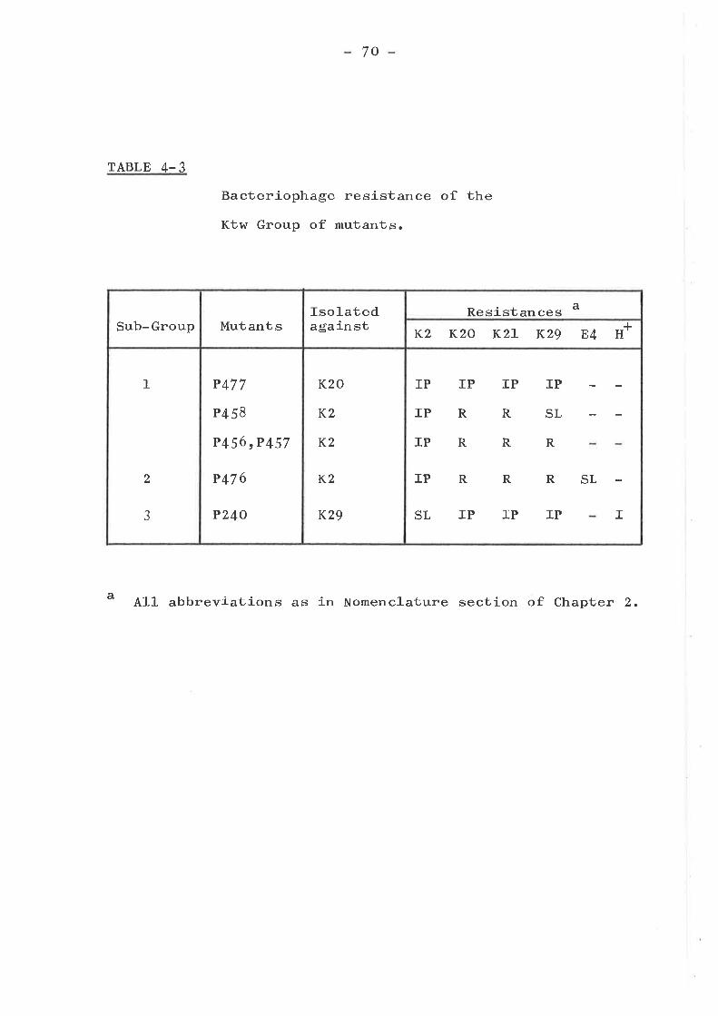
7o
TABLE 4-3
Bacteriophage resistance of the
Ktw Group of mutants.
a All abbreviations as in Nomenclature section of Chapter 2.
Sub-Group MutantsIso]-atedagainst
Resistances a
K2 KzO KzL K29 E4 H+
1
2
aJ
P477
P4 58
P456 rP457
P47 6
PZ40
K20
K2
K2
K2
K29
IP
IP
IP
IP
SL
IP IP IP
RRSL
R
R
R
R
R
RSL
IIP IP IP
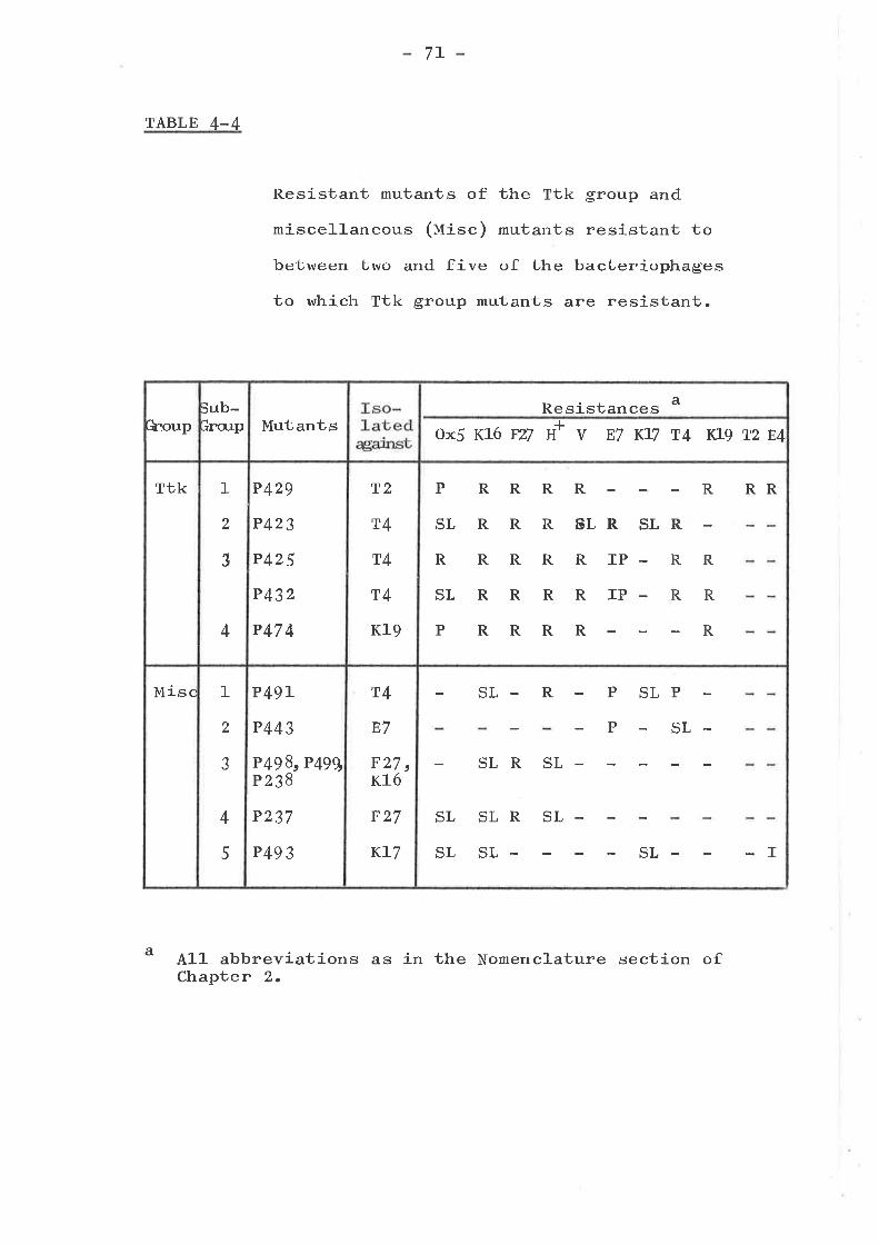
7L
TABLE 4-4
Resistant mutants of the Ttk group and
miscellaneous (Uisc) mutants resistant to
be'bween Lwo and five <¡f the bacteriophages
to which Ttk group mutants are resistant.
Al]- abbreviations as in the Nomenclature section ofChapter 2.
a
houpjub-ìnerp Mutants
Resistances a
ox5 rc16 F2Z u+ v E7 KLT t4 YJg Tz E4
Ttk 1
2
3
4
P429
P423
P425
P43z
P47 4
T2
T4
T4
T4
Kl9
PRR
SLRR
RRR
SLRR
PRR
RR
R ßLR
RRIP
RRIP
RR
SI
R
R
RR
RR
R
RR
Misc 1
2
aJ
4
5
P49L
P443
P 49q" P499P23B
P237
P493
T4
E7
p27 tK16
F27
KL7
SL- R P SLP
P SL-
SLR SL-
SL
SL
SLR SL-
SL- SL I

72
Bar group
Nineteen mutants were sorted into eight sub-groups as
shown in Table 4-5. The mutants were resistant to between
ten and nineteen bacteriophages. Sub-groups 1 to 3 were
sensitive to T3 whil-e sub-groups 4 to 8 were resistant.
Sub-group 1 was distinguished by its sensitivity to
bacteriophage K16, while sub-group 2 was uniquely sensitive
to bacteriophages A and E7. Sub-group 3 contained a1-l the
remaining T3 sensitive strains and showed, at least,
resistance to a set of twelve bacteriophages. Of the T3
resistant strains, sub-group 4 was uniquely sensitive to
bacteriophage K2. Sub-group j was found to be sensitive tol-
H' and only slightly resistant to T3. Sub-group 6 was
sensitive to bacteriophage Kl/ but resistant to T4 and K2.
Sub-group / was resistant to all bacteriophages except K19
while sub-group 8 was resistant to this bacteriophage too.
IVtrile this group is extremely heterogeneous, it nevertheless
seems to fit wel-l as a group. It is interesting that nearly
all of the bacteriophages to which the Misc, Ktw, and Ttk
groups are resistant, are included in the set of bacterio-
phages to which Bar mutants are resistant.
hrrm p
Only one of these mutants, P479, was selected to a
bacteriophage specific to the group, and this after NTG
mutagenesis. These mutants appear to be single mutants as
they do not have the combined properties of the resistance
groups they embrace ( see Chapters 5 and 7 ). They fal1
clearly into two groups as can be seen from Table 4-6. Wrm
mutants are resistant to bacteriophages from eight out of
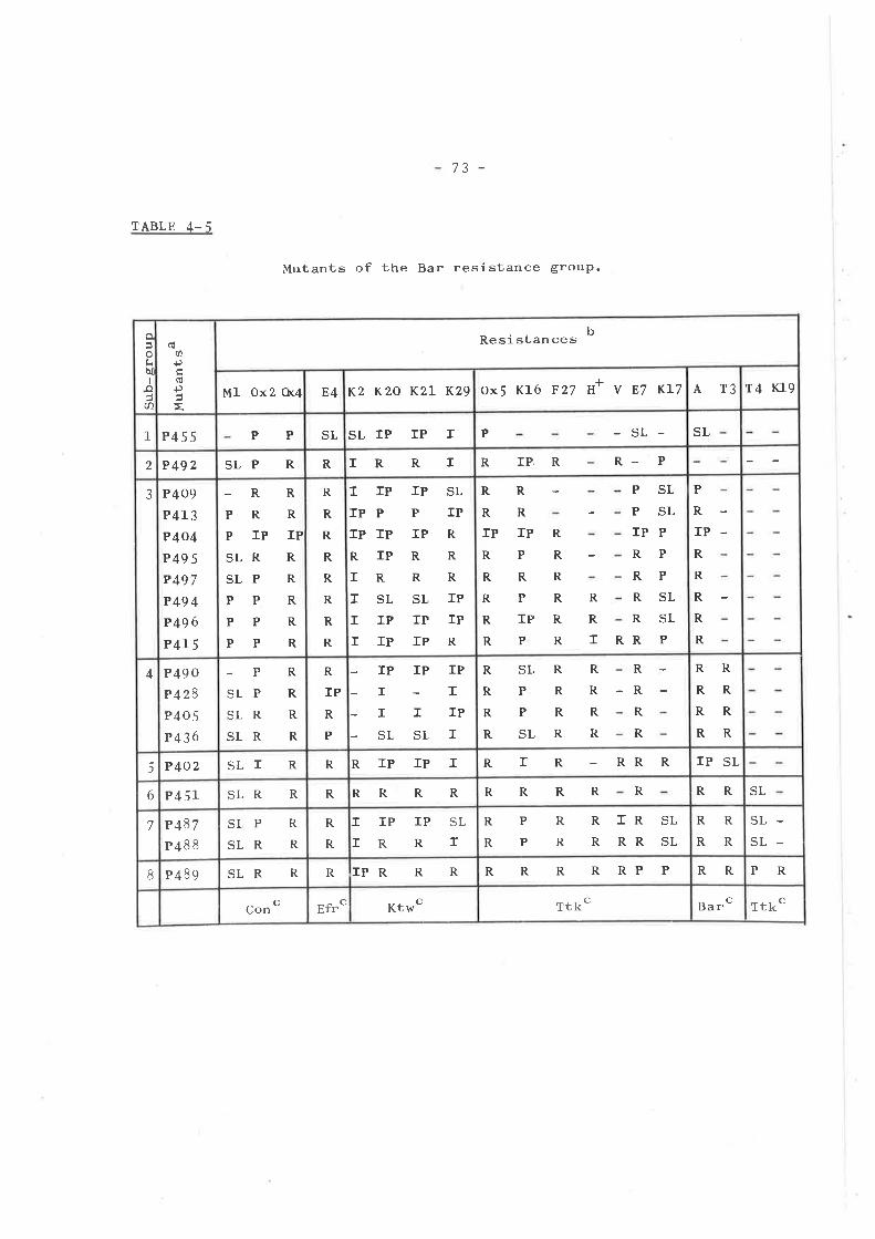
/.t -
TABLE 4-5
Mutants of the Bar resistance group.
o
tibII
.o(n
(úU)
Ð(ú
Ð
Resistances b
Ml Ox2 Or4 E4 K2 KzO KzL K29 ox5 K16 F27 H+ V E7 KL7 AT3 14 r(19
I P455 PP SL SLIP TP T P -SL- SL
2 P492 SLP R R rRRI RIP,R R P
3 P409
P413
P4o4
P495P497
P49 4
P496
P4r 5
-RPRPIPSLRSLPPPPPPP
R
R
IPR
R
R
R
R
R
R
R
R
R
R
R
R
ITPrPPrP TP
RIPrRrSLIIPIIP
rP SL
PIPIPRRRRRSL TP
rP IPrPR
R
R
R
R
R
R
R
R
R
R
R
R
r R
PSLPSLIPPRPRPRSLRSLRP
rP rPRPRRRPRTPRP
P
R
rPR
R
R
R
R
4 P49 0
P 428P405
P 436
-PSLPSLRSLR
R
R
R
R
R
IPR
P
IP IP IPI-IITIPSLSLI
R
R
R
R
SLR R
R
R
R
R
R
R
R
PRPRSL R
RRRRRRRR
5 P4O2 SLÏ R R RTPIPI RTR RR R IP SL
6 P 45L SLR R R RRRR RRRR R RR SL
7 P 187
P48 8
SLP R
SLR R
R
R
II
IP IP SL
R R rR
R
P
P
R
R
R TR SL
R RR SL
R
R
R
R
SL-SL-
8 P 489 SLR R R IPR R R R R R R RP P RR PR
Con c Efrc Ktwc Ttkc Barc Ttkc
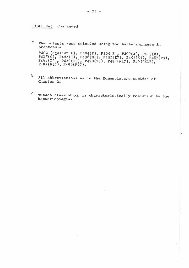
74 -
TABLE 4-5 Continued
The mutants weæe selected using the bacteriophages ina
c
brackets: -P4OZ (againstP415(G)r P428(P488(r¡), P489P497 (F27), P49
P405( F )P451(E
), P494
)' P(rz)4e s(
,P7),(rr
t)'T3
409 (rP455
7)t P
t(r3 ),
b All abbreviations as in the Nomenclature section ofChapter 2.
Mutant crass which is characteristicarly resistant to thebacteriophages.
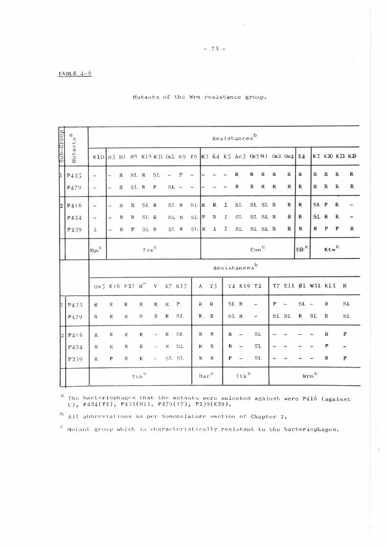
75 -
TABL ,t- 6
Mutants of the Wrm resistance group.
bResistances
Resistan ce sb
o lh" bacLer j oph:rgr:s bha L t,he mutants were selected againstc)t p424(14), p4j5(Í'1), p479("r7), p239ß29).
l' A1J abbr-cviat, ions as p()r NomencLat,ure section of Chapter 2
( vllrt arrt, grr)r,rp which is <:haractr:risLically resistanb to the
K2 KN KzL KD
RRRR
RRRR
SLP R
SLR R
RPP
Ktwc
17 Ell ø1 w31 K15 H
P - SL- R SL
SLSL R SL R SL
were P{16 (against
bacteriophage s.
R
P
P
R
P
R
cWrm
Ê
tr
Io-{,a 3 K4 K5 Ac3 ft3 Ml o(2 0x4 E4
(úU)
ÐrlÐiz
Klo H3 Hl H.8 Kr8 K31 tul K9 16
R
R
RRRRR
RRRRRI P435
P479
R SLR SL
R SLR P SL
P
SI,
SL
SL
SLSLR
SLSLR
SLSLR
R
R
R
RRI
PRI
RII
R
R
R
P 4t6
P 424
P239 I
R
R
R
SLR
SLR
SLR
SLR
SLR
SLR
SL
SL
SL
R
R
P
2
Eft.CCon cc
Ktn r sxc
A T3 T4 KLg T2ox5 K16 F27 H+ v E7 Kr7
RR
RR
SLR
SLR
RRRRRRP
RRRRRRSL
t P435
P479
RR
RR
RR
R
R
P
SL
SL
SL
, P4t6
P424
P239
R
R
R
R
R
P
R
R
R
R
R
R
RSL
RSL
SL SL
Barc Ttkc'ftrkci
76
the eleven other sets defined in this study and show
resistance to between thirty and thirty-three bacteriophages.
These mutants may be similar to other mutants d.escribed inthe literature (Curtiss, L965; Radke and Siegel, IgT1-).
CROSS RESISTANCE TO COLICINS
Surveys on the resistance of E. coJ-i K-L2 Lo a wide
range of bacteriophages, as presented in this chapter, and
colicins (Davies and Reeves, :'974aþ) have provided the
basis for a more extensive study of the phenomenon of cross
resistance between bacteriophages and colicins. The testing
of the colicin resistance of the bacteriophage resistant
mutants in this section of Chapter 4, was done by John Davies
and is the subject of a joint publication (Hancock, Davies
and Reeves, J-97 4) .
Results shown in Table 4-7 confirm and extend the
results of other workers, who found that bacteria mutated inthe tonB (Fredericq, 1951; Gratia, Lg64), t"r.A (Fredericq,
L95L), tsx (Weltzein and Jesaitis, 1-97l-) and bfe (Buxton,
r97L) loci were cross resistant to certain coricins and
bacteriophages, suggesting a coÍrmon cellular component forthe action of both on the E. coli K-1-z ce11. A similarconclusion can be made from the resistance of the con, Ktw(1)
and Ktw( 2) bacteriophage resistant mutants to specificcolicins (ta¡1e 4-7).
Four of the classes of co]-icin resistant mutants
extribiüed the pattern of resistance of Ktw(1) mutants. one
of these classes Tolrb appeared to be identical to Ktw(1)
mutants in that all of the Ktw(1) mutant strains were re-
sistant to colicins K, L, A, 54 and N; however there r,rrere
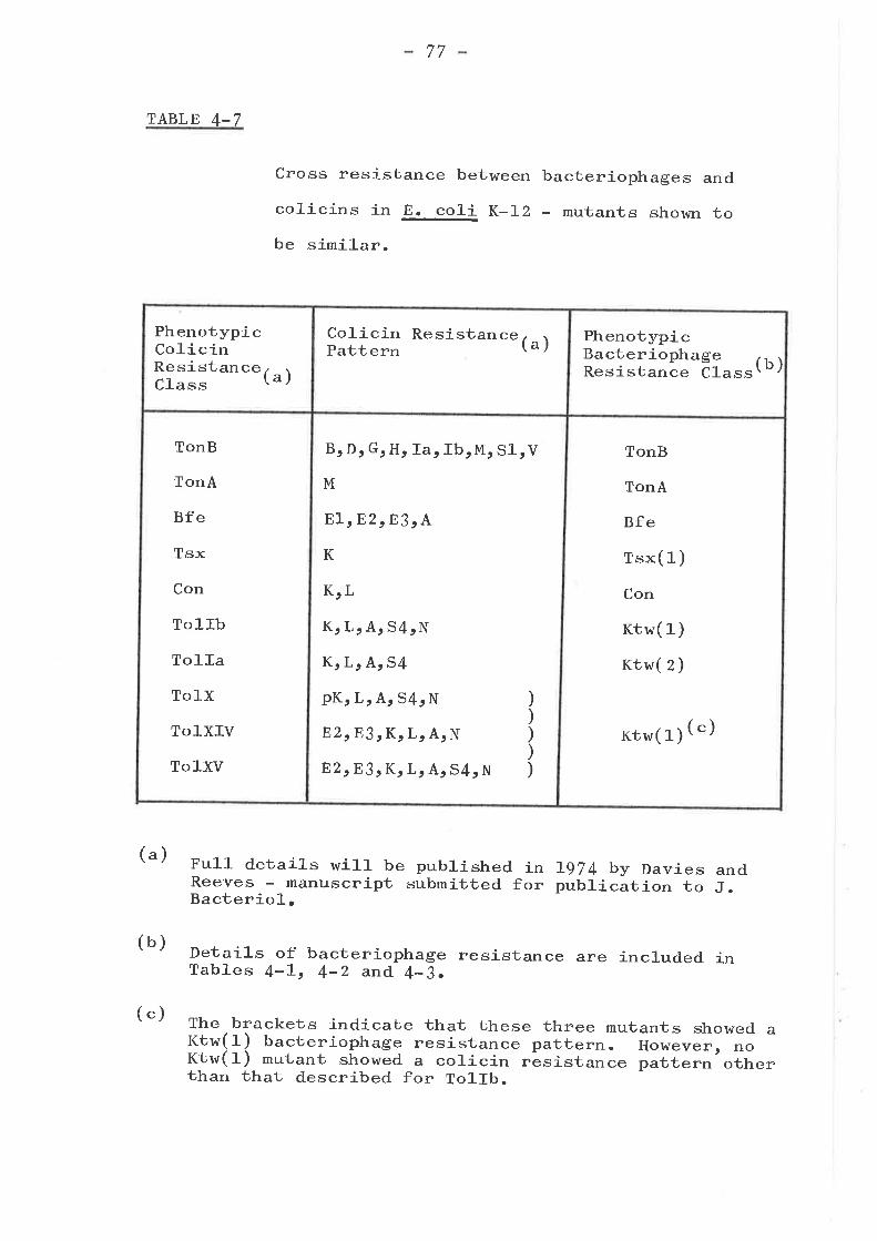
TABLE 4-7
Ful1 details wil1- be published inReeves - manuscript submitted forBacterio]-.
77
Cross resistance between bacteriophages and
colicins in E. co]-i K-IZ - mutants shown tobe similar.
(")
(b)
L974 by Daviespublication to
andJ.
Details of bacteriophage resistanceTables 4-Lt 4-Z and 4-3.
are included in
The brackets indicate that these three mutants showed aKtw(1) bacteriophage resistance pattern. HoÌvever, noKtw(1) mutant showed a colicin rãsistance pattern otherthan that described for To1lb.
PhenotypicColicinResistanceClass (")
Colicin ResistancePattern (") Phenotypic
BacteriophageResistance ct"""(b)
TonB
TonA
Bfe
Tsx
Con
TolIb
To]-Ia
TolX
ToIXIV
ToIXV
B, Dr G, H, Ia, IbrM, SlrV
M
EIrBz,83, A
K
K,L
K, L, A: 54rN
K, Lr A, 54
pKr Lr A, 54, N
E2,E.3, K, L, A, N
E2 rE3 , K, L: A¡ 54, N
)))))
TonB
TonA
Bfe
rsx( 1 )
Con
Ktw( 1 )
rct,w( 2 )
Ktw( 1) (")
(")
78
no bacteriophage resistant mutants with the colicinresistance pattern of the other J classes, suggesting thatthese mutants are altered in a way that cannot be easilyselected using bacteriophages. These mutants might have
simi-l ar defects since they are commonly resistant tocolicins K, L, A and N and sensitive to colicin El (Davies
and Reeves, l-974a)t being the only mutants isolated withthese colicin resistance properties.
Many of the colicin resistant mutants are sensitiveto all of the bacteriophages (tabte z. z) tested, thesebeing: strain p224 (ncx phenotypic crass), p651 (rorrru),p555 (rotrrc), p66o (rotrIl), p6gz (rorrv), p5g6 (rorrx),p653 (rorxrr) , p52o (rotxrll), p516 (rorxvr) , p625 (crr),p575 (nx¡¡)r p535 (nxtrc), p2gS (cut¡, pL2og (cmt), p64S
(rvt) and pIz3S (cvt). The colicin patterns of thesemutants are extremely varied (Davies and Reeves, rg14atlþ).rn addition, mutants from many of the bacteriophage resist-ance groups described in this chapter were sensitive to allcolicins, these grou.ps being the Tsx(2), Ktn, Efr, Ktw(l),rtk(l), (z), (¡) and (Ð, and Misc(l), (z), (3) and (4)groups. strains p494 (nar(3)) and p424 (wrm(z))
',rere alsosensitive to all colicins. These findings suggest thatdiffering requirements for the action of bacteriophages and
colicins on bacterial cells, as j.rag"a by the lack of crossresistance of many mutants, are quite common.
other colicin- and bacteriophage-resistant mutantswere resistant to a set of bacteriophages or colicins whichdo not fit' well into any of the previously defined classes(see Table 4-7). Mutants from the Bar, wrm and Misc(j)groups were resistant to one of six combinations of colicins
79
Ar Lt s4 and N, with no clear pattern arising from thesestudies (Hancock, Davies and Reeves, LgT4). The combinationsto which these mutants h/ere resistant were: (l) colicins A¡
L, 54 and N; (z) A, L and 54; (3) A and L; (Ð L and. 54;(5) A or (6) L. Howcver, as poi.tecr out by Davies and
Reeves (tgz 4a), mutants with relatively minor changes in thestructure or composition of the cerr enveloper âs judged by
their changed sensitivity to a wide range of antibiotics,detergents and surfactants, are often resistant to thesefour colicins.
There are also two types of colicin resistant mutantswhich are resistant to many bacteriophages, the first ofwhich inctudes strains p6B9 (rorvrr), p6oz (rotvrtt) and
P22O (totXf) which are resistant to between 7 and. L3 of thebacteriophages which fail to lyse Bar mutants, and whichpartially inhibit all other bacteriophages. Although thesemutants could be loosely classified- as Bar mutants, theywere not similar to other Bar mutants (tabte 4_il, and_ thefinding that they exhibit slight mucoidicity (J.K. Davies,personal communication) raises the possibility that theirbacteriophage resisüance properties are at least partlycaused by this. Strains p6OZ and p689 appear to be LpS_
altered (Davies and Reeves, r9T4a)t in common with the Bar
mutants ( chapter 6). The other type of colicin resistantmutant with an unusual bacteriophage resistance pattern has
only one representative, strain p6sz (rolxvrt) resistantto colicins 81, 82, K, L, A, s4 and N (Davies and. Reeves,
L97 4a). This mutant shows partial sensitivity tobacteriophages T6, Ox1, Ox2, Ox4> Ox5r ¡c3r ¡c4t Hl,Ml, K2, K2O, K27-, K29t K3¡ K4, and KJ, all of which
have T-even morphology (tabte 3-Z), while it shows
80
resistance to all other bacteriophages. Although
mutant appears to have a large defect in the cel1the nature of this defect is as yet unknown (J.K.personal communication ) .
this
envel-ope,
Davies,
SUMMARY AND CONCLUSIONS
Joo bacteriophage resistant mutants were isolated to 4z
virulent bacteriophages in one strain of Escherichia coliK-rz and tested for resistance or sensitivity to a set of 56
bacteriophages. rt was possible to fit all of these mutants
into 11 main groups, and one group of miscellaneous (uisc)mutants which did not fit well into any of the other groups.
Each group of resistant mutants was characte,rízed. by a
particular set of bacteriophages to which most of the mutantsshowed resistance of one type or another. Some of thesegroups were shown to be cross-resistant to defined sets ofcolicins, while others were either sensitive to all colicinsor showed resistance to a set of bacteriophages that did notfit well into any of the defined phenotypic resistance groups.
81 -
CHAPTER 5
ADSORBTION OF BACTERIOPHAGES TO CELL ENVELOPE SUBFRACTION S
INTRODUCTION
Beumer et a1 (f-965) studied the adsorbtion of various
bacteriophages to cel1- wal-l- and lipopolysaccharide (LpS)
preparations. This enabled them to partially characteríze
the nature of the receptor for these bacteriophages.
Sabet and Schnaitman (L97L) examined the adsorbtion of
colicin E3 to various subfractions of the cell envelope of
E. co]-i K-LZ derived by sequential sol.ubiliza1cj-on with
Triton X-1OO and Triton X-IOO * Edta (SchnaitmaÍr, LgTIarb).
They were able to l-ocalize the receptor activity in the
Triton-Edta solubl-e fraction t at a 1eve1 equivalent to the
receptor activity of the ce1-1- envelope, despite the fact
that this fraction contained only 50% of the LPS and 25% of
the protein of the ce1l envelope. (Schnaitman, L97Lb).
Chemical and enzymatic studies partial1-y characterized tlre
nature of this fraction (Sa¡et and Schnaitman, L97L) which
r^¡as later used as the starting preparation for the isolation
of the receptor protein (Sa¡et and Schnaitman, 1-973a).
Similar but less detailed studies have been done on the
colicin I receptor of E. coli ts.-L2 (Konisky and Liu, L974).
An understanding of the nature of the receptor, for a
given bacteriophage, facilitates the study of the basis of
resistance to that bacteriophage, since one can then study
one component or class of components of the outer membrane
to see if it is altered in resistant strains.
B2
ADSORBTION TO LIPOPOLYSACCHARIDE
LPS isolated by the phenol-water technique usua1-1-y has
some protein associated with it (Wober and Alaupovic, L97L;
Wu and Heath, L97 3b). Galanos, Lüderitz and Westphal (Lg6g)
described a technique for isolating LPS with less protein
attached to ít; however, when this phenol-chloroform-
petrol-eum ether extracted LPS was used in neutraLj-zatíon
experiments with bacteriophages T3 and K1O, it neutral-i-zed
neither bacteriophage. Therefore, phenol-water extracted
LPS was used in these studies.
Twenty-three bacteriophages were tested for neutral-
izatíon by LPS and cel-l wal-l preparations (tabte 5-1). Of
these, seventeen bacteriophages, to which the Ktn, Ktw, Ttk,
Bar and l,{rm groups of mutants are resistant, showed
adsorbtion to LPS. Four other bacteriophages to which some
mutants of these groups are also resistant, namely E{, Ox5r
K15 and T2, were not adsorbed; however, of these on1-y T2
showed appreciable neu1oraLj-za1r-ion by cel1 wall preparations.
Bacteriophage OxJ was neutralized by a cel1- envelope
preparation and this is discussed in the following section.
Two other bacteriophages were included in this testing,
since receptor activity could not be found in any other
subfractions of the cell- envelope (table 5-Z). These were
bacteriophage H3 which appears atypical both sero1-ogically
(tatte 3-1) and with respect to its resistance patterns
(fabte 4-6) from the other bacteriophages fail.ing to lyse
tsx mutants; and K26, a Tl-1-ike bacteriophage (tabte 4-1).
Neither r^¡ere shown to adsorb to LPS preparations.
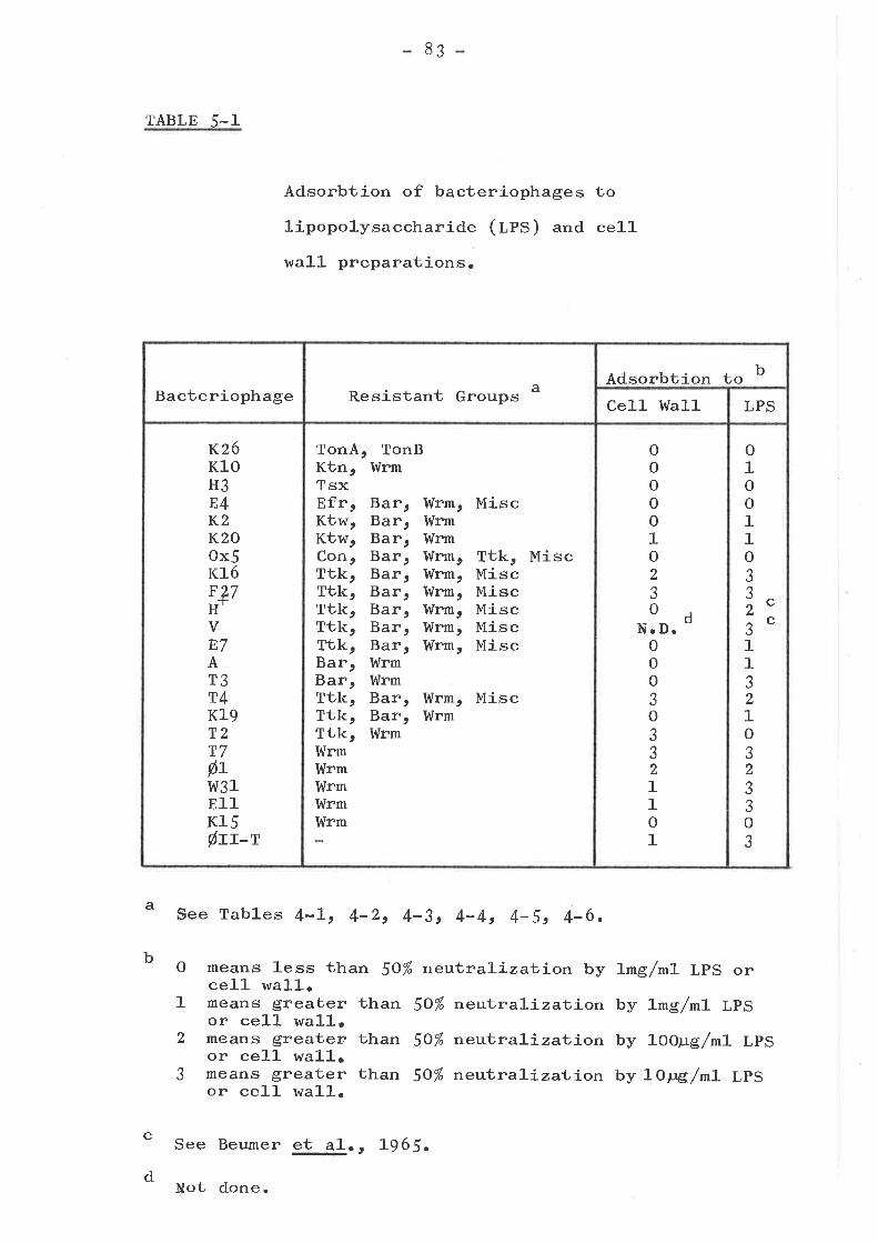
TABLE 5-1
8¡
Adsorbtion of bacteriophages to
lipopolysaccharide (lpS) and ce1-l
wa1.1- prcparations.
Bacteriophage Resistant Groups a Adsorbtion to b
Ce]-l l{all LPS
K26Kl0H3E4K2K20ox5K16F27H-1-
VE7AT3T4Kl9T2T7þtr,v31E11Kl5Øtrr
TonA,Ktn,TsxEfr,Ktw,Ktw,Con,Ttk,Ttk,Ttk,Ttk,Ttk,Bar,Bar,Ttk,Ttk,Ttk,I,{rmWrmWrmI{rmI,ürm
TonBI{rm
Bar,Bar,Bar,Bar,Bar,Bar,Bar,Bar,Bar,IdrmWrmBar,Bar,Wrm
Wrm,WrmI¡ürmWrm,Wrm,Wrm,Idrm,Itlrm,I{rm,
Misc
Ttk, MiscMiscMiscMiscMiscMísc
I{rmWrm
, Misc
ooooo1o2Jo
N.D. doooaóoaJJ211ol_
o1oo11oJ
"rc^cJ11321oJ2aJ3oJ
b
a See Tables 4-Lt 4-2t 4-3, 4-4, 4-5, 4-6.
O means less than 50% neu1.raLj-zatíon byce]-]- wal1.
1 means greater than 50% nelu1r.ral-j-za1"j-onor cell wal-l-,
2 means greater than 50% ne:uioraLj-zat,j.onor ce]-1 wa]-]-.
J means greater than 50% neuþraLi-zat íonor cel-l wall.
1mg/m1- LPS or
by Lmg/ml LPS
by 1OO¡rg /mL LPS
by LOpg/ml LPS
c
d
See Beumer $_jl. , L965.
Not done.
8+-
ADSORBTTON TO OTHER CELL ENVELOPE SUBFRACTIONS
The results included in Table 5-Z are discussed in the
text in terms of sets of bacteriophages failing to lyse
certain resistance groups. The preparations tested r,'rere
those of Schnaitman (1-g71'asb) and a sodium hydroxide super-
natant, prepared by the technique of Weidel, Koch and
Bobosch (1954). The Triton X-1OO solubl-e supernatant of the
cel-l envelope wtrich has been described by Schnaitman as a
cytoplasmic membrane preparation was also tested and
neutralizai.,i.on of bacteriophages (which is analogous with
receptor activity) could often be explained by contamination
with cel.l wa1-1- proteins. Thus it has not been included in
Table 5-2.
Bacteriophages unable to ]-vse the Con group of resistant
mutants
There is some degree of receptor heterogeneity amongst
the set of bacteriophages which do not 1-yse Con- mutants
(fabte 4-L). The bacteriophages separate into two classes
represented by K3 and Ox5 (fable 5-2). These bacteriophages
have different activity spectra on mutants other than Con-
(fabte 4-6). The bacteriophage KJ receptor is found in the
ce1l envelope and is not solubl-e in Triton X-IOO (i.". fu1l
activity is found in the ce1.1- wal1 preparation). trlhen the
cell wal1 is treated with Triton X-IOO and Edta, all of this
receptor activity can be traced to the supernatant (i.e.
soluble fraction) after centrifugation and removal of the
Triton X-IOO. The bacteriophage Ox5 recepüor, however, is
completely destroyed by the first Triton X-IOO treatment.
This may be because the Ox5 receptor lies in the Triton X-IOO
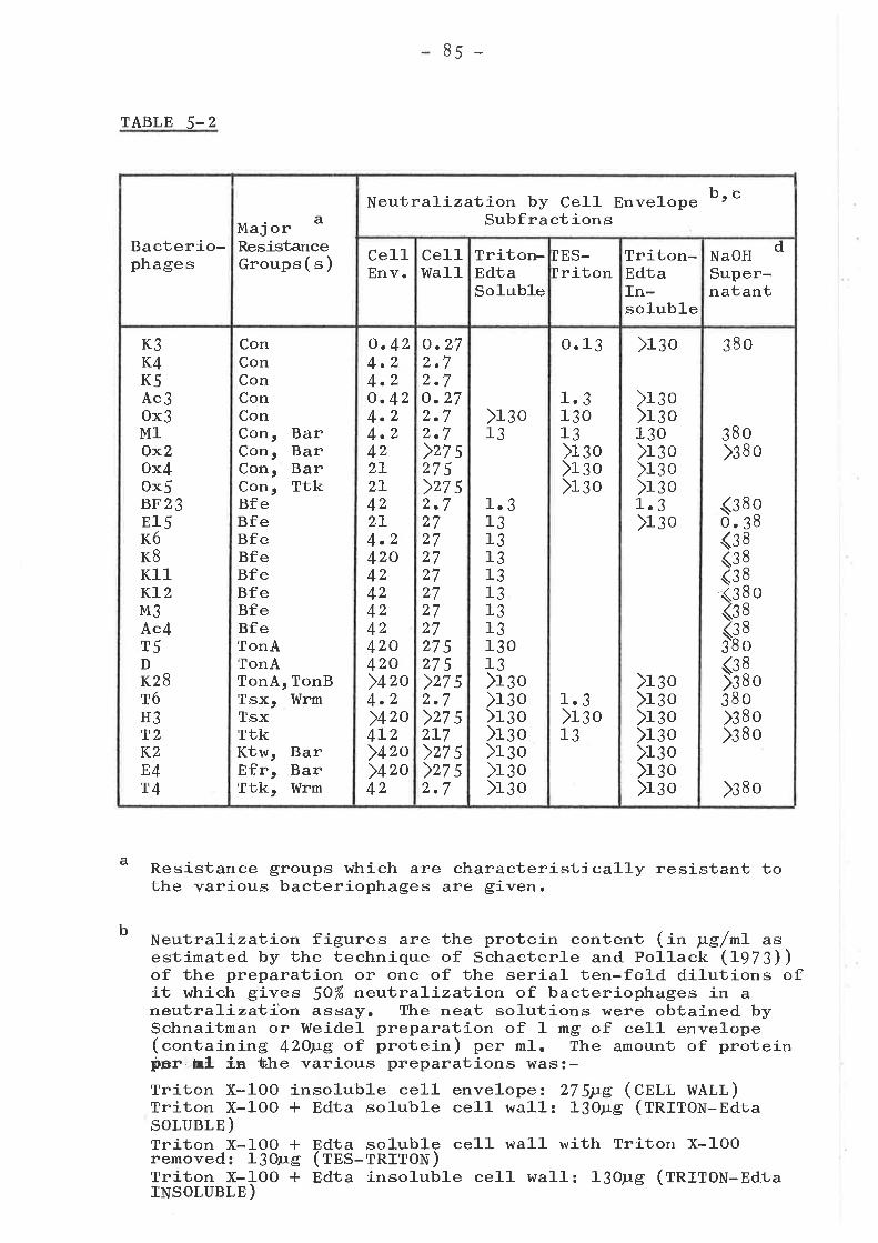
85 -
TABLE 5-Z
Resistance groups which are characteristically resistant tothe various bacteriophages are given.
Neutraliza1"íon figures are the protein content (ín ¡tg/nL asestimated by the technique of Schacterle and Poll-ack (L973))of the preparation or one of the serial ten-fold dilutions ofit which gives 50% neut-raT-izat-íon of bacteriophages in aneutraliza1r-i.on assay. The neat solutions urere obtained bySchnaitman or l,{eidel preparation of 1 mg of cell envelope(containing 42Oyg of protein) per ml. The amount of proteinfier hL ín li¡he various preparations was:-Triton X-IOO insoluble cel-l envelope ¿ 27 Su.e (CnI,l WALL)Triton X-IOO * Edta so1uble ce1-1- wall: 13orrg (fnffOu-EdtaSOLUBLE )Tríton X-IOO + Edta so]-uble ce]-l wa]-]- with Triton X-IOOremoved: l3oug (tnS-rnfTON)Triton X-IOO * Edta insoluble cel1. wall: 13o¡rg (fnffOm-EdtarNsoruBl.E )
a
b
Bacterio-phages
Major a
ResistanceGroups ( s )
b, cNeutralizat-íon by Cell EnvelopeSubfractions
Ce11Env.
Ce11Wa11
Triton-EdtaSoluble
TES-triton
Triton-EdtaIn-soluble
NaOHSuper-natant
d
K3K4K5Ac3ox3M1Ox2ox4ox5BF23El5K6K8Kl1KL2M3Ac4T5DK28T6H3T2K2E4T4
ConConConConConCon, BarCon, BarCon, BarCon, TtkBfeBfeBfeBfeBfeBfeBfeBfeTonATonATonA, TonBTsx, I{rmTsxTtkKtw, BarEfr, BarTtk, hlrm
o.424.24.2o. 424.24.2422L2L422I4.24204z424242420420)+zo4.2)42o4L2)+zo)+zo42
o.272.72.7o.272.72.7)zz s275)zz s2.727272727272727275275>27 52.7>27 52L7>27 5>27 52.7
X¡o13
1.31313131313131313o13)r¡o)r¡o)r¡o)rao)r¡o)r ¡o)r¡o
1.313o13X¡o)r ¡o)rgo
1.3)r¡o13
o.13
)r¡o)r¡o13o)rsoXso>1301.3)r ¡o
)rgo)r¡o)r¡o)r:o)r¡o)r¡o)r¡o
)r¡o
380)sa o
380
<3o.(:<3<3-(3<3
IE<3>338>3)¡
8o388888o88o880o8o8o
)¡so
86-
TABLE 5-Z Continued
Sodium hydroxide supernatant: 13OFg (trlaOU supernatant)Itdnemonics used in the table are given in brackets afterthe amount of protein in the preparation. Preparativetechníques etcr âre given in Chapter 2.
Blanks in table mean not tested.
Figures for sodium hydroxide supernatant often representthe highest dilution tried in a neutraLízatíon assayo
c
d
87
soluble fraction (which is not borne out experimentally),
or it may be that it requires the co-operation of a Triton
X-IOO soluble factor. It has been shown (Schnaitman,
LÇlLarb) that Triton X-IOO wil-l dissolve 50% of i.,1ne LPS,
8O% of the phospholipid and 20% of the protein of the ce1-1-
envelope. Thus one of these components might represent
either the receptor or a requirement for active receptor.
The pattern of neutraLj-zatíon of bacteriophages K4r K5r
Ac3 and Ml is similar to that for K3, while Ox3 differs only
in that the receptor activity is reduced So-fo1-d during
solubiliza.1;-íon with Triton X-IOO and Edta. Bacteriophage
M1- is the only one of these bacteriophages which is
neutralized by the Triton X-1O0 * Edta insolubl-e ce1l wall
preparation, however the actual difference may be minor.
The pattern of neutraLj-zat-j-on for OxZ and Ox4 is similar
to that for Ox5r although Ox4 is neutraLized s1.ight1-y by
the cel1 wa1-1 preparation.
Comparison of activity spectra on resistant mutants
(tables 4-Ic 4-4, 4-5 and {-6) with neutralizal'ion
patterns (table 5-2), divides these bacteriophages into two
sets (with one exception, bacteriophage Ml). These are
(1) bacteriophages KJ, K4, K5r Ac3 and OxJ, and (Z) OxZ,
Ox4 and Ox5. Therefore, these two sets of bacteriophages
probably have a common requirement (mutated in the Con-
mutants), and a different requirement (". demonstrated by
different resistance and neutraLizaþíon patterns).
Bacteriophage Ml which has a similar neutralization pattern
to K3 and a similar activity spectra on resistant mutants
to Ox2 (table 9-1) is, as mentioned above, the exceptional
88
case. Hor,rrever, it is ab1-e to plaque to a limited extent on
a1-l Bar mutants (which Kl can plaque on but Ox2 generally
"anit (ta¡te 4-5)), and this might be evidence that it has
receptor requirements intermediate to the above two classes
of bacteriophages.
Bacteriophages unable to lyse the Bfe, TonA or Tsx
resistance groups
The receptor for the BF23-1ike bacteriophages is found
in the Triton-Edta soluble fraction (taUte 5-2). This is
in agreement with the results of Sabet and Schnaitnarr
(tgZt) for colicin 83, which is cross resistant to ùhese
bacteriophages (table 4-7). There is also considerable
receptor activity in the sodium hydroxide supernatant. In
fact, for bacteriophage B15r this activity is forty times
greater than the receptor activity of the Triton-Edta
soluble fraction. Both of these preparations probably
contain solubilized LPS-lipid-protein complexes (Weidel,
Koch and Bobosch, L954; Schnaitman, 1"97ab) and therefore
the receptor for the BF23-1ike bacteriophages may be assoc-
iated with such complexes. Although the sodium hydroxide
supernatant was originally isolated as the T5 receptor
complex (Weidel, Koch and Bobosch, Lg54) tfre amount of
exposed B1$ receptor in this fraction is considerably
greater. These figures suggest that the sodium hydroxide
supernatant would provide a considerably better starting
point for the isolation of the bacteriophage 815 receptor
than the Triton-Edta so1.uble supernatant, which was used
(SaUet and Schnaitman, L97 3arb) to isolate the colicin E3
receptor.
8g
The receptor of T$-1-ike bacteriophages is also found in
the Triton-Edta sol-ubl-e and sodium hydroxide supernatant
subfractions (fatte 5-2). There are quantitative differences
in the neutralizatíon of bacteriophages T5 and D (both of
which have t"t A as their only resistant mutants) by these
two preparations. Bacteriophage KzB, a T1-1ike bacteriophage,
is also unable to lyse t"nA (or tonB) mutants, but is not
neutralized by any of the preparations tested. This is not
surprising as bacteriophage T1 seems to require a component
provided only by living cells, but not by dead cells or
ce11 wall fragments (Stentt L963).
Two bacteriophages which fail to lyse tsx mutants, T6
and H3, differ considerably in their neutralizat-j-on patterns.
They have also been shown to differ serologically (ta¡1e
4-6). Bacteriophage HJ, l-ike K28, is not neutralized by
any of the preparations listed in Table 5-2. The receptor
activity for T6 is found in the Triton-Edta soluble
fraction, but only after removal of Triton X-IOO by alcohol
precipitation and washing of the proteins (..f. bacteriophage
M1-). This suggests that either bacteriophage T6 is in-
activated by Triton X-IOO or that the Tó receptor is an
aggregation of Triton-Edta so1-ub1-e components and Triton
X-1OO by disaggregating (i.". so1-ubiLj-zi.ng) ttris, destroys
the abi1ity of these components to act as a receptor.
Other Bacteriophages
Bacteriophages K2 and E{, the former of which was
shown to be adsorbed by LPS (tabte 5-1), h¡ere neutralized
by none of the preparations tested (taUte 5-2). Therefore,
while the resul-t for bacteriophage E4 merely mimics the
result for K2B (and thus might a1so indicate a similar
9o
requirement for an active ce1l- function), the result for
K2 tends to indicate that the LPS might be at least partly
masked by other cel1 wall components. This is borne out
by the 10- to 1-OO-fold differences in the neutralizatJ-on of
many bacteriophages by cel1- waJ-J- and LPS (tabte 5-1). As
can be seen from the results for bacteriophage T4 (which is
also neutra1-ízed by LPS)e the LPS must be exposed to some
extent in the cell wal1 and envelope preparations.
The receptor for bacteriophage T2t which has been
shown to be a lipoprotein (Uichael, L968t DePamphilis,
I97L) t is found in the same preparations as the receptors
of bacteriophage T6 and the K3-like bacteriophages. The
presence of Triton X-1OO in the Triton-Edta so1-uble
supernatant prevented the neutralizatíon of T2; which is a
similar result to that obtained using bacteriophages T6 and
ox3 (tabte 5-2).
ENZYMATIC TREATMENT OF THE RECEPTOR
The enzymatíc treatment of the receptors (fa¡te 5-3)
strows that bacteriophages KJ, ¡c3r Ox3 and Ml (of the Con
resistance group), T6 (tsx resistance group), TZ (ttt
resistance group) and E15 (nfe resistance group) have
periodate sensitive receptors whil-e the receptor of T5 is
insensitive to periodate. These results have been
previously demonstrated for TJ (Braun, Schaller and Wolff,
L973)s T6 and T2 (Weltzein and Jesaitis, L97L). The
colicin E3 receptor which is rel-ated to or identical with
the bacteriophage 815 receptor has been also shown to be
periodate sensitive (Sa¡et and Schnaitman, L97I), The
periodate sensitivity of a receptor, suggests the involve-
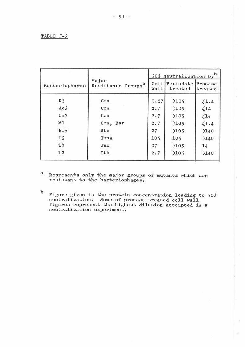
91
TABLE 5-3
Represents only the major groups of mutants which areresistant to the bacteriophages.
Figure given is the protein concentration leading to SO%neutraliza1l-ion. Some of pronase treated cell wallfigures represent, the highest dilution attempted in aneutrali zat, íon experiment .
a
b
BacteriophagesMaj orResistance Groups a
5O% Neut raLiza1-íon b-rb
Cel1Itra1l
Periodatetreated
Pronasetreated
K3
Ac3
ox3
M1
815
T5
T6
T2
Con
Con
Con
Con, BarBfeTonA
TsxTtk
o.272"72.72.727
105
27
2.7
>105
>105
>105
>105
>105105
)ro5)ro5
(r.4<14
<14(r.4)r¿o)r¿oL4
)r+o
92
ment of LPS or some other carbohydrate-associated moeity
in the binding of bacteriophages to this receptor (Sabet
and Schnaitman, L97Lt L973a).
Pronase treatment of the outer membrane destroys three
of bhe proteins of the cell wa1l (nragg and Hou, L972); as
welJ- as modifying one of the major peaks (nragg and Hou,
L97 2) which might contain either two proteins 3a and 3b
(Schnaitman, L973bt L974a) or a single protein (Reithmeier
and Bragg, L97 4). This treatment destroys the B15r T5 and
T2 receptors, but has little effect on the receptors of T6,
K3r ¡c3r Ox3 and Ml (ratrte 5-3). The effect of pronase on
the receptors of T2 and T6 has been dernonstrated previously
by l,rleltzein and Jesaitis (L97L) "
SUMMARY AND CONCLUSIONS
Many of the bacteriophages to which the Ktw, Ttk, Ktn,
Bar, Wrm and miscellaneous groups are resistant, are
neutralized by LPS preparations. I?rere are only four
exceptions, as described in the text. It is interesting
that the T2 receptor which has been described as a
lipoprotein probably requires or interacts with LpS, as
many of the bacteriophages, to which the TZ resistant mutant
shows cross resistance, are neutral,ized by LPS. Further
evidence of this is the periodate sensitivity of the Tz
receptor.
A number of bacteriophages r^¡ere more efficiently
neutralized by LPS than cell- wa1l preparations. This ispartly accountable for by the fact that the ce1-1 wall-
contains only 20% tpS. However, this does not explain the
93
1OO-fold difference in neutralization efficiency that
occurs using bacteriophages þ].I-t, Id31¡ 811 and H*. This
effect might be due to the fact that bacteriophages have
only about 25o-3o0 attachment sites on the ce11 wall (Bayer,
1968a) and, one would cxpect, many more molecules of LpS
per ceIl than this. Thus, other cell wall components must
in some way limít the interaction of bacteriophage and
receptor.
The receptor activity of many of the bacteriophages towhich the Con and Bfe mutant groups are resistant, is
solubilized from the cell wa1-l by Triton X-IOO in the
presence of Edta, which also solubilizes most of the Lps ofthe cel1 wall. This, in addition to the periodate sensitivity
of these receptors, suggests the involvement of Lps or some
other Triton-Edta soluble, carbohydrate-associated moiety inthe adsorbtion of bacteriophages to these receptors. A
similar argrrment applies to the receptor of bacteriophage
T6 (and T2 as above). The receptor for bacteriophage T5r
however, seems to be insensitive to periodate as previously
described (Braun, Schaller and lrrolff , L9T Ð t and itsreceptor probably doesntt involve carbohydrate.
The sodium hydroxide supernatant used as the basis forthe preparation of bacteriophage T5 receptor (weidel, Koch
and Bobosch, Lg 54; Braun, Schaller and l,rtolf f , Ig7 3) seems tobe a better receptor preparation for bacteriophage E15 than
it does for T5.
94
CHAPTER 6
THE NATURE OF THE ALTERATIONS IN THE CELL WALL OF
RESISTANT MUTANTS LIPOPOLYSACCHARIDE ALTERATIONS
INTRODUCTION
It rs wel]- known in Salmonella that there is a
correlation between the composition of the lipopolysaccharide
(leS) and the pattern of sensitivity to certain
bacteriophages (Wilkinson, Gemski and Stocker, L972). Tamaki
and co-workers (tgZt) have demonstrated that some mutants
selected for resistance to bacteriophage T4 in B. coli K-L2,
also have altered LPS. It has been shov¡n (Chapter 5) that
many of the bacteriophages to which the Bar, Ktw, Ttk and
I,{rm groups of mutants are resistant, were neutralized by LPS
preparations. Therefore, in this Chapter, antibiotic
sensitivity and lysis by bacteriophages CZI and U3 were used
as indicators of LPS deficiency. The LPS alterations
suggested by these results u/ere further characterízed by
direct chemical analysis of the representative strains using
gas liquid chromatography.
ANTIBIOTIC RESISTANCE/SENSITIVITY
Tamaki 9!_jf. (1-97L) have shov¡n that LPS-deficient
strains selected as novobiocin supersensitive can a1-so be
resistant to bacteriophages T{ and T7. Eriksson-Grennberg
and co-workers (tgZt) also showed that many of their class
II ampicillin resistant mutants were LPS-deficient. Thus it
r,r¡as decided to test these and other antibiotics to determine
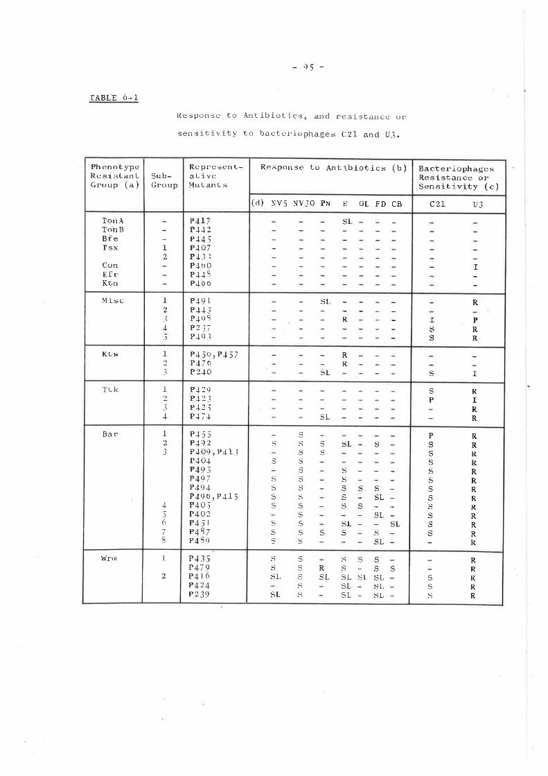
95 -
IABL.E ó- 1
Response to .A,ntlbiot ics, and
sensitivity to bacteriophages
resistance or
C2l and U3.
PhenotypeRe si stantGroup (a)
Sub-Group
Represent-at ivelvlut an t s
Response to Antibiotics (b) BacteriophagesResistance orSensitivity (c)
(d) NV5 nV.lO PN E OL FD CB c2r u3
TonATonBBfeTsx
ConEfrKtn
I2
P4L7P-t.t 2
P4-t 5P.l o7P.t3iP-lo0P4.I S
P.lb o
SL
lMisc I
-1
4J
P+9 rP-t-t -ìPJ9 C
P2 37P+9 i
-SL
R- ISS
R
PRR
Ktw ttt
P45b,P-t57P176P 240
RR
SL S ITrk 1
,3
J
P-l2SP-t 2 -lP-12 ;P-t7+ SL
SP
RIRR
Bar 12
3
4l6
78
Pl5 ;P192P409, P4l3P40.tP49 5P 497P49 4P49ó,P415P405P402P4 i1P 487P439
S
q,
c,
SSq
S
S
S
SSS
Sq
q
SSS
Sq
S
Sq
S
SSL
SSSeq.
SLS e
SL
PSSSSSq
SSSSS
RRRRRRRRRRRRR
ldrrn 1
,P435P479P 416P 424P 239
q.
q
SL
Sqq
Sq
SSSLSLSL
q
SL
SSSLSLSL
SRSL
SL
Sq
q
RRRRR
96
TABLE 6-I Continued
(") Abbreviations as per Table 3-2 and Chapter 4.
(b) means response same as strain P4OO.
means resistant, i. e., producing a smaller zoneof inhibition than strain P4OO.
means sensitive or supersensitive, i.e., largerzoîe of inhibition than strain P4O0.
means slight increase in sensitivity; with theexception of strain P4L7 which shows a slightdecrease in sensitivity to erythromycin.
(") means wild type response, í.e., sensitive tobacteriophage U3 or resistant to CzL.means resistance.means sensitivity.means inhibited.means partial resistance.
Al]. terms are defined in Nomenclature section ofChapter 2.
(d) Symbols, from multodisks, represent:NV5 - Spg of novobiocin; NV3O - 30¡re of novobiocin;PN- ampicillin; E- erythromycin; OL- oleandomycin;FD - fusidic acid; CB - methicillin.
R
S
SL
R
S
IP
97
whether various bacteriophage-resistant mutants lr¡ere altered
in their response to them.
The results obtained using multodisks 3O-9C and 11-14D
showed that alterations occurred in the response to
novobiocin, methicil-lin, fusidic acid, oleanodymycin,
ampicillin and erythromycin (see Table 6-1), of mutants of
the Ktw, Ttk, Bar, I¡úrm and miscellaneous groups.
Novobiocin supersensitivity (to 3o¡rg but not 5¡rg; or to
5,pS and 3Olg) occurred amongst all of the mutants in the Bar
and Wrm groups. In the Wrm group, two patterns occurred
which seemed to correlate with the sub-grouping; however, the
pattern for the Bar group was more complex.
Of the other antibiotics, erythromycin sensitivity
occurred frequently in the Bar and l,tlrm groups, while
resistance onJ-y occurred in two sub-groups of Ktw and one
miscellaneous mutant. Fusidic acid sensitivity occurred
only amongst sub-groups 2, 3t 5, '/ and 8 of the Bar group
and in both sub-groups of Wrm. Ampicillin resistance
occurred only with one Wrm mutant which was only partially
resistant to 2yg/mL ampicillin, whilst ampicillin super-
sensitivity was more widespread. Sensitivity to oleandomycin
(which is related to erythromycin) and methicillin (which is
related to ampicillin) did not seem to fo1low any clear
pattern.
ALTERATIONS IN THE SBNSITIVITY TO BACTERIOPHAGE U3 OR
TO BACTERIOPHAGE CzI
It has been shown that the receptors for
CzL (Rapin, Kalckar and Alberico, 1966; Rapin
I97L) and U3 (Watson and Paigen, L97L) are in
bacteriophages
and Kalckar,
the LPS rough
98
core and that mutants in which the composition of the LPS is
altered may be changed in their response to these
bacteriophages. A large number of bacteriophage resistant
mutants rt¡ere tested by spotting these bacteriophages onto an
agar layer conLaining the mubanl, bacteria and various
resistance patterns were revealed (fabte 6-1). Of particular
interest were the mutants which were resistant to both
bacteriophages CZI and U3, since they differed considerably
in their resistance to other bacteriophages (see Tables 6-L
and Ç-1). Other mutants were sensitive to CzL and resistant
to U3 and these included mutants of the Bar and ltirm groups
(which are resistanü to many bacteriophages) " Thus it was
decided to look at the composition of the LPS of selected
mutants to see if the alterations suggested by both the
antibiotic and bacteriophages CzL and U3 studies, were
detectable.
ANALYSIS OF NEUTRAL SUGARS
I,t/tren the LPS of strain P4OO was hydrolysed with lN
for 12 hr in a boiling water bath, neutraLízed, reduced,
ILs¿
acetylated and run on a gas liquid chromatograph, the amount
of glucose r^ras found to be greater than the amount of heptose
(glu: gal: hept:O.f,9¡ñI/mg LPS: O.15: O.30). This uras a
different result to that found by other workers (Rapin and
Mayer, L966; Eriksson-Grennberg, Nordstrom and Englund,
I97I; Monner, Jonsson and Boman, L97I; Tamaki¡ Sato and
Matsrfrashi, I97L) for E. coli K-Iz. Schmidt, Fromme and
Mayer (tgZt) showed that when O.1-N I{Cl hydrolysis was used
the amount of heptose released from LPS increased with time
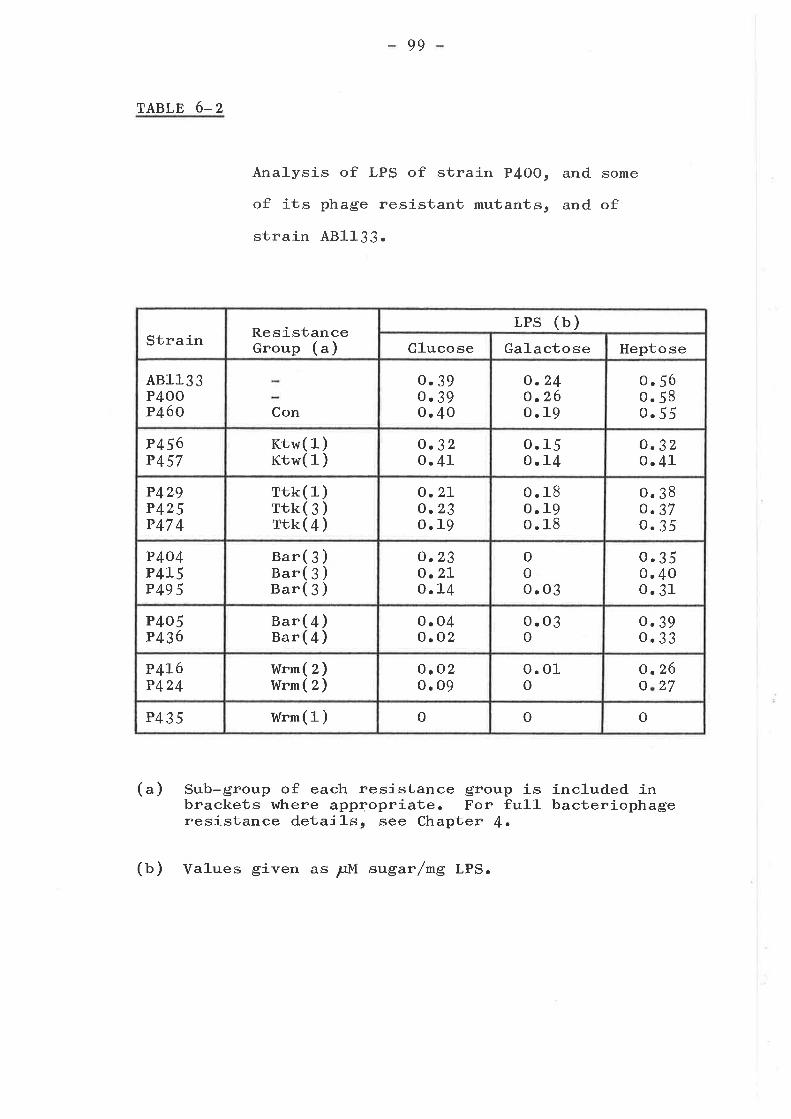
99
TABLE 6-2
(")
Analysis of LPS of straj-n P4OO,
of its phage resistant mutants,
strain 481133.
Sub-group of each resistance group isbrackets where appqopriate. For fu1-1-resistance details, see Chapter {.
and some
and of
included inbacteriophage
(b) Values given as ¡rM sugar/mg LPS.
Strain ResistanceGroup (")
LPS (b)Glucose Galactose Heptose
A81133P4OOP460 Con
o.3go. 3go.40
o.24o.26o. 19
o. 56o.58o.55
P456P457
Ktw( 1 )Ktw( 1 )
o.32o.4L
oo
11
54a
o.32o.4L
P429P425P47 4
rrk( 1ftk( 3ftk(4
)))
o.27-o.23o.19
0. 18o.19o. 1B
o.38o.37o.35
P404P4L5P49 5
Bar( 3 )Bar( 3 )Bar( 3 )
o.23o.2Lo.L4
ooo .o3
o.35o.40o.31
P405P436
Bar( 4 )Bar({)
o. 04o.02
o. 03o
o. 39o.33
P4L6P424
I¡ürmWrm
(2)(z)
o. o2o. 09
o. 01o
oo
a 22
6
7
P435 htrn( 1) o o o
100
up to 48 hr at which time it was much greater than the
amount released by HZSO4 hydrolysis and doubl_e ühe amount
released by O.lN HCI over 12 }:.r. Jackson and Redman
(i!Zl-) used an even milder hyd.rolysis with Dowex 5O (u+) in
O.OIN HCl for 7 days at 1O5C in their studies on the LPS of
Vibrio cholera. It was decíded to use a time of 64 nr at
1OOC using O.lN HCl as the hydrolysing agent. This caused
the amount of heptose, relative to the sulphuric acid
liberation, to double when compared to an internal standard
although the amount of glucose liberated was fairry constant
(rab1e 6-z).
I{hen heptose from E. coli, which has been shou¡n to be
L-glycero-D-manno heptose, is reduced and acetylated, it
forms a peak X which runs just after glucose on a gas
liquid chromatograph (Scfrmi.dt, Fromme and Mayero LgTL).
Under our preparation procedures perseitol and L-glycero-D-
manno heptose form the same heptitol acetate. Equal amounts
of perseitol and glucose were run together in a control ex-
periment, and it u¡as found that the area under the glucose
peak was equal to the sum of the areas under the heptose and
X peaks. Therefore, for each mutant studied, this sum was
used to represent the total amount of heptose (taUle 3). In
addítion to heptose, there was glucose and galactose but no
other sugars in the LPS of strains 481133 and P4OO (see Fig.
6-l-).
It lr¡as interesting to note that the non- mutation made
no difference to the sugar composition as can be seen from
the similarity of strain 481133 and. its his* non
derivative P4OO. The con strain P{6O was also unchanged.
Strains P456 and P457 from the Ktw resistance group
have reduced leve1s of heptose and galactose. Strains P429,
101 -
P425 ar:d P474 from the Ttk group are deficient in glucose and
heptose, while the 3 Bar (3) mutants are additionallydeficient in galactose. The other strains tested., p436 and_
P4o5 (ear (4)), p4I6 andp424 (wrm (z)), and p435 (wrm (1))
had little or no grucose or galactose in their Lps. The
I,{rm group mutants also showed lower levels of heptose than
other mutants tested, with the wrm (1) mutant p435 being
totally deficient in this and all other neutral sugars. when
the other wrm (1) mutant P479 rn¡as tested, it reverted on two
separate occasions and showed not only a wild type bacterio-phage sensitivity pattern but a sugar composition similar tostrain P4OO. Strain P466, a Ktn mutant, ì^¡as also tested inan early experiment and found to be unchanged.
GLUCOSAMINE ANALYSIS
The last sugar to be attached to the rough core of the
LPS of S. typhimurium is glucosamine (Uäteta and Stocker,
L969). The lipid A portions of the LpS molecules of
Sa]-mone]-]-a and E. coli K-Lz also have glucosamine attached tothem (Rooney and Goldfine, a972). Thus degraded polysacchar-
ides, obtained by removing Lipid A from the Lps of selectedstrains, were tested for the presence of glucosamine. There
h/as less than o.o1¡rM of glucosamine per mg of Lps in the
polysaccharide portion of the Lps of strains p4oo, p46o (con)
or P425 (rtt (¡)). The Lps of a smooth strain of s. typhimuri tl¡ll,
kindly provided by R. Davies, hras subjected. to a similaranalysis and it was found that there was o.2opM of glucosamin/
mg LPS which represented I mole of glucosamine per molecule ofLPS (see Chapter Ç). This was a similar result to thatpreviously discovered for Sa1-mone1-1-a ( Lüderit z 4. I97L) .
Io2 -
SUMMARY AND CONCLUSIONS
Some bacteriophage'resistant mutants of the Ktw, Ttk,
Bar, lvrm and miscellaneous groups were shov¿n üo be alteredin their response to a series of antibiotics and tobacteriophages C21 and- U3.. Ehe neutral sugar conrposítions ofthe LPSts of many of the mutants u¡ere investigated, and
shown to fit into six classes. [tre parent strain p40o, has
glucose, galactose and heptose in its LPS but no other
neutral sugars, and no glucosamine in the d.egraded.
polysaccharide,
103
CHAPTER 7
THE N ATURE OF THE ALTERATION IN THE CEL L WALL OF
RESISTANT MUTANTS PROTEIN ALTERATIONS
INTRODUCTION
Mutants resistant to various bacteriophages have been
shown to have a protein band missing or altered when
compared to the sensitive strains. These include bfe(SaUet and Schnaitman, I973aclr)t t"nA (Braun, Schaller and
I,{olff , a97 3; Braun
and Schwartz, L973)
and l,rtol-ff , I97 3) t 1".8 (Randall-Hazelbauer
and a !.84, tsx double mutant
In most cases the bacteriophages, to
are resistant, probably have as their
deleted in these mutants, one
bacteriophages to which tf"A mutants are
thought to have LPS receptors (Beumer
(Schnaitman, L974tl).
which these mutants
receptor the protein
exception being the
resistant (which are
9!-3f. t L965)).
Other workers have shown that heptose-deficient strainsof E. coli K-rz have altered protein patterns (Koprow and
Goldfinet L974). Therefore, it rn¡as decided to look at the
protein composition of the LPs-altered mutants described. inchapter 6 as well as other suspected protein-altered mutants
including Con- mutants.
CON MUTANTS GENERAL PROPERTIES
various of the bacteriophages, which are unable to ryse
con- mutants (tab1e 4-L), h¡ere tested for their ability t,o
adsorb to strain P4OO. Adsorbtion could be readily
demonstrated for all bacteriophages tested, these being:
LO4
K3 09 . 5% adsorbed), M1 (87 . S%) r ox3 (8I.4%) and ox{ (77 .g%) .
In contrast, adsorbtion of bacteriophages K3 (to strains
P459, P46O and P{61), M1 (to strain p459)t Ox3 (to strainP459 ) and Ox{ (to strains p4ig, p46O and p46L) was
undetectable. The two methods used (see chapter z) testedboth reversible and irreversible ad.sorbtion, and thusresistance of the con- strains to these bacteriophages isapparently due to the loss or alteration of receptor such
that adsorbtion cannot occrlr.
rt has been shown that these mutant strains are
unaltered in their response to a variety of antibiotics(taule 6-1) and to bacteriophage czl, while bacteriophage
u3 causes inhibition of con- mutants, but not lysis (taute6-1). This altered response to bacteriophage uJ was not d.ue
to a changed LPS composition, as gas liquid chro¡natographic
analysis revealed that strains p460 (ra¡te 6-z\ and p459 had
identical sugar compositions to strain p4OO.
The con- mutants were found to be uniquely (amongst
bacteriophage resistant mutants) resistant to lethal zygosis,due to their 1oo- to looo-fold decrease in ability to act as
recipients during conjugation with Hfr and. Fr donors (n.
skurray, Ph.D thesis, university of Adelaide, L974; skurray,Hancock and Reeves, Lg7 4). This conjugational recipientdeficiency was not due to recombinational deficiency of thecon- strains, as Plkc transduced. the r"g* arrd p"o+ markers
from strain p83g at the same frequency into strains p4OO and_
P46o (con-). rt was shown by Dr p. Reeves that the inabilityof con- mutants to act as recípients in conjugation was due
to an inability to form mating pairs (skurray, Hancock and.
Reeves, L974).
Con- mutants have a].so been shown to be tolerant to
105
colicins K and L (ta¡1e 4-7).
CON MUTANTS: ALTERATIONS AS REVEALED BY POLYACRYLAIvIIDE GEL
ELECTROPHORESIS
The polyacrylamide ge1 electrophoresis system used
first was the SDS system developed by Nevil-l-e (tglt) for
mammalian membranes (Method 1) and this clearly showed. that
a major band of the cell envelope was dramatically reduced
in amount in strain P46O in comparison to that of the
parent strain P4OO (pie. 7-IA and 7-18). The Triton X-1OO
treatment described by schnaitman (L977a) did not solubirize
the band (pig. 7-LC) which was thus identified. as an outer
membrane or cel-l wa1l component. The molecular weight (lrtW)
of the affected band, when compared to standard proteins
(serum albumin, ovalbumin, carbonic anhydrase and J-ysozyme)
was estimated at 28rOOO. The 28rOOO MW band is apparently
completely absent in the Triton X-IOO treated preparation
of the mutant (pig. 7-1D), and it is possible that the
smal-l band present at this position in the envelope of the
mutant is a different, Triton X-1OO soluble protein which
runs at the same place in the gel and is not a residual
amount of the protein(s) affected by the mutations in strain
The assistance of Ron Skurray in the NevilJ-e gel analysis of
the mutants is acknowledged, as the work above was done in
conjunction with him.
Although the Neville system gave a very good
resolution of bands, the pattern was different from that
found by Schnaitman (t97ta, L973atbt I974arb), Bragg and
Hou (tgZZ) or Inouye and Yee (L973) using various different
SDS ge1 systems. To enable direct comparison with other
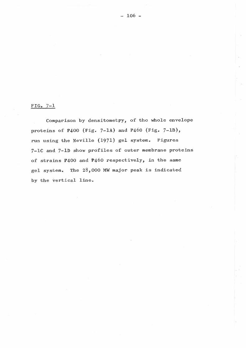
106 -
FIG.7-L
Comparison by densitometpy, of the whole envelope
proteins of P4oO (rig. 7-14) and P46o (pig. 7-LB)t
run using the Neville (1-g71-) eel- system. Figures
7-LC and 7-1D show profiles of outer membrane proteins
of strains P4OO and P46O respectively, in the same
gel system. The 28,OOO MW major peak is indicated
by the vertical J-ine.
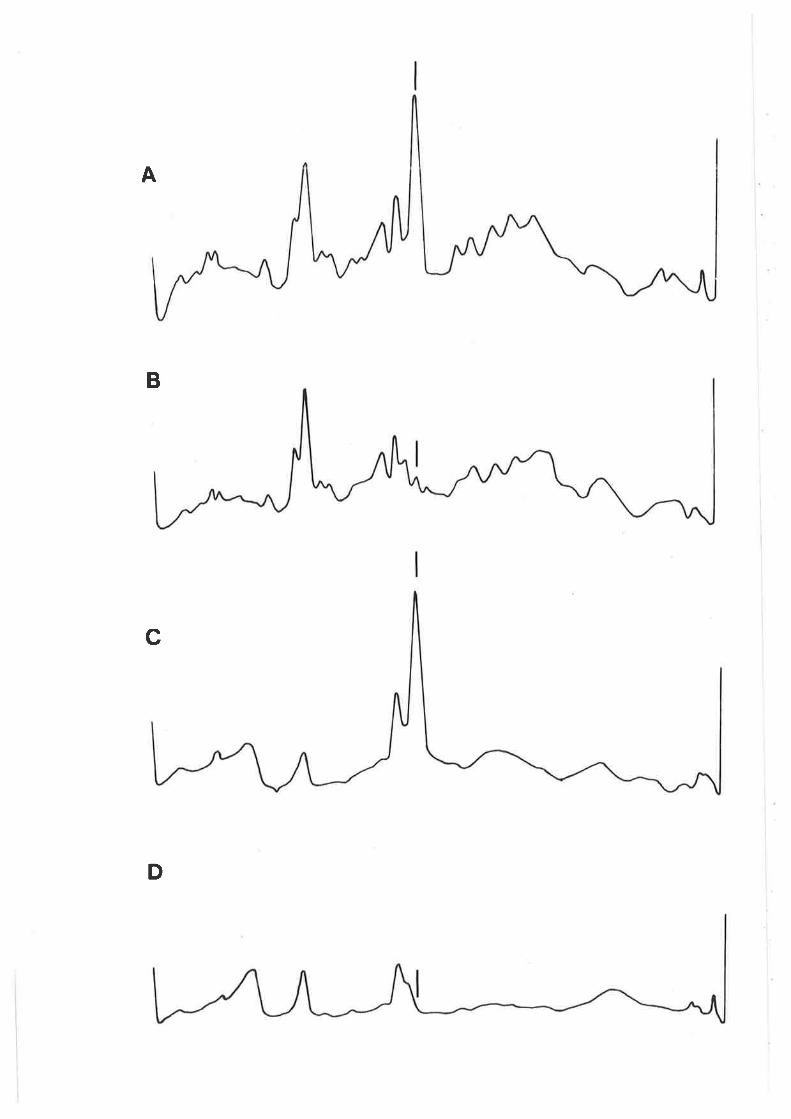
A
B
c
D
LO7
FTG. 7-2
Comparison by densitometry, of stained bands of
outer membrane proteins, run using the pH 7.2 MaízeL
buffer system, and. either heating (method 2) or not
heating (method 3) the samples at IOOC, before load-
ing onto SDS polyacrylamide gels. Figures 7-24 and
7-Zlr- show the outer membrane protei-ns of strains
P4oo (Corr+) and. P460 (Con-) respectively, solubilized
by method 3. Figures 7'2C and 7-2D show the outer
membrane proteins of strains P4OO (Corr+) and. P46O
(Con-) respectively, solubilized by method 2. The
peaks arè labelled À, B and C according to Schnaitman
(t974^) nomenclature as described in the text.
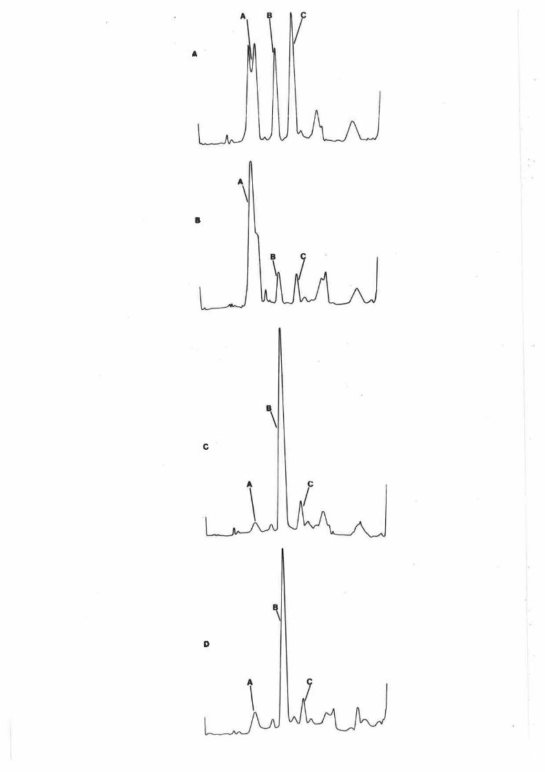
A
¡
c
D
108
r,.rork on Lg!i, cel1 wall preparations l.lere solubilized
and- run und.er the conditions of Schnaitman (pig. 7-2 and
7-3\.E. coli K-12 has 3 maior outer membrane proteins, 1, 3a
and. 3b (Schnaitmanr L974b). Using method I for sample
preparation and the pH 7.2 Maízel buffer system, protein 1
runs as a peak A and proteins 3a and 3b run together as peak
C (Schnaitman, l-973a, L974arb). The Con- mutant P46Ot run
using these cond-itions, had. peak C very much reduced (fig.
7-ZB in comparison wíth strain P4OO (pie. 7-2A). The heated
samples (method 2) run under the same conditions showed that
all- the major proteins ran at peak B (fig. 7-2CrD) as
observed by schnaitman (1-973a). However, both with strains
P4OO and P{6O, a smal1 amount of material ran at the position
of peak C, comparable in amount to that observed with unheated
P46o (pig. 7-28). It seems likely, therefore, that this
residual amount of material in peak C (figs. 7. ZB, C and D),
represents a minor protein and is not a residual amount of
proteins 3a or 3b; although further analysis may be required,
it is provisionally postulated. that strain P469 lacks both
proteins 3a and 3b.
Unheated preparations (method 3) of strains P4OO and
P46O also differed in the amount of peak B and possibly also
peak A (fig. 7-2ArB). The material in peak B is presumably
d-ue to either proteins I, 3a or 3b running in this position,
even when the heating is omitted, since peak B is a product
of these proteins (pig. 7-2CrD). Because there is so much
more peak B in P4OO than in P46O (pig. 7-2LrB), it must be
largely derived. from proteins 3a and 3b. It shoul-d be noted
that the materíal in peaks B and C of P46O (pie. 7-28) is
the maximum amount of proteins 3a and 3b that could be present.
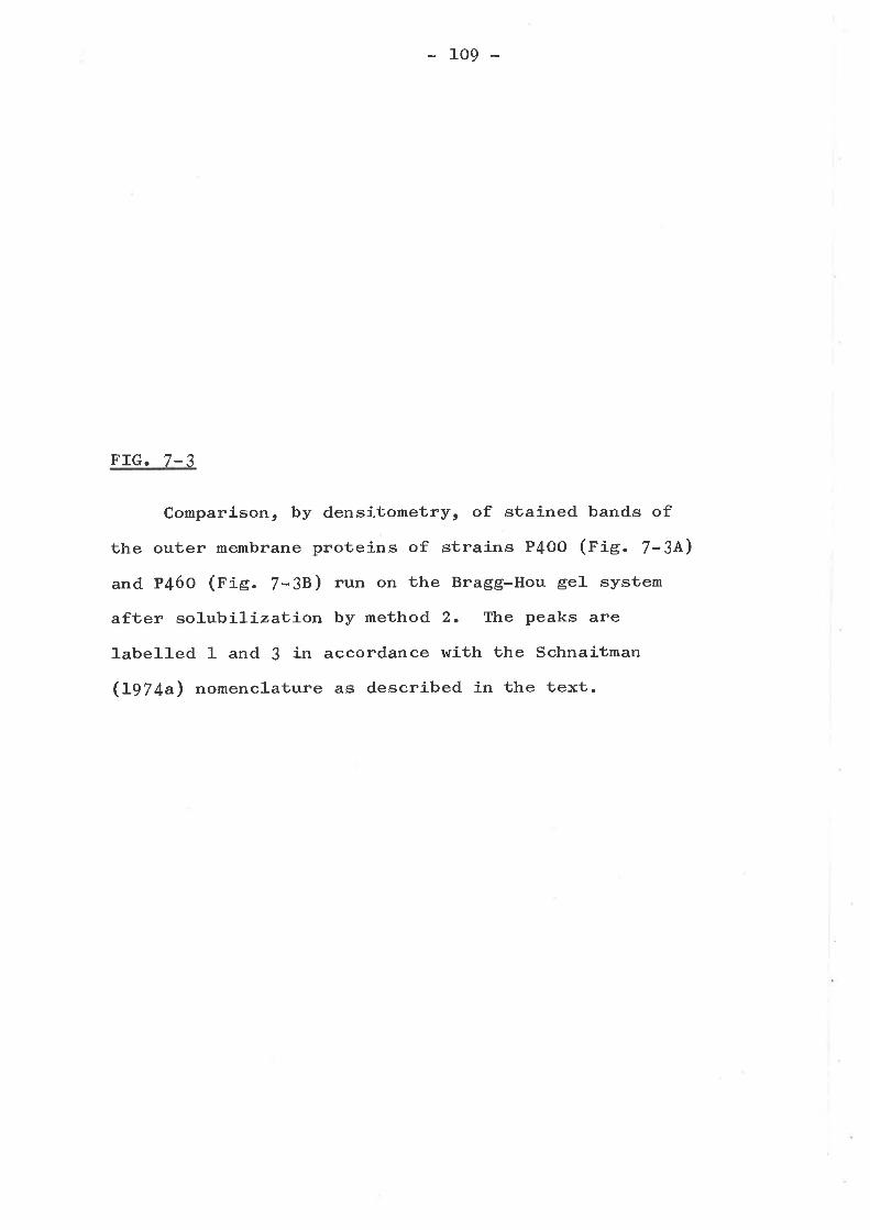
109
FIG.7-3
Comparlson, by densitometry, of stained bands of
the outer membrane proteins of strains P4OO (pig. 7-3A)
and P46O (fie. 7-38) run on the Bragg-Hou gel system
after solubiliza1:-íon by method 2. The peaks are
].abelled 1 and J in accordance with the Schnaitman
(Lg|+a) nomenclature as described in the text.
1
\
A
I
\
B
110
The reduction in one of the components of peak A in P46O is
much J-ess than the reduction in peak C (pig. 7-28), and may
not be significant.
Under alkaline conditions (Oragg and Hou gels), Schnait-
man (L973b, I974a) showed that both proteins 3a and 3b rlrn
together as peak 3, which is again virtually absent in straín
P46O (fig. 7-38) in comparison to strain P4OO (r'ie. 7-3A).
Thus results with both neutral and alkaline ge1.s support the
hypothesis that Con- mutants lack the two outer membrane
proteins 3a and 3b.
LIPOPOLYSACCHARIDE-DEFICIENT MUTANTS PROTEIN ALTERATIONS
A number of representative bacteriophage resistant mutants
were tested for protein alterations in their cel1 walls and
envelopes by the techniques discussed previously in this
chapter. Results obtained are srrmmarized in Table 7-I. It
rtras found that strains r,r¡ere sometimes altered in peak 1
(using the Schnaitman (L97 4a) nomenclature for Bragg-Hou
gels), which consisted of a single protein, protein 1
(Schnaitman, L974a). For demonstrating the alterations, the
Bragg-Hou gel- system was by far the besb, although equivalent
alterations hrere seen in the Neville and Schnaitman gel
systems. Representative results are seen in Figs. 7-4, 7-5,
7-61 7-7 and 7-8. fn each case the gel runs of mutants
(bold lines) are compared with the parent strain P4OO
(dashed lines) prepared and run under the same conditions.
It was demonstrated that, with the exception of the
Con- mutanùs (nigs. 7-L, 7-Z and 7-3), onl-y mutants
previously shown to be LPS-altered (Chapter 6) had
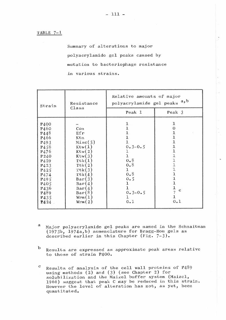
TABLE 7-T
Results areto those of
111 -
Summary of alterations to major
polyacrylamide ge1 peaks caused by
mutation to bacteriophage resistance
in various strains.
a Major polyacrylamide ge1 peaks are named in the Schnaitman(L973bt L974aþ) nomenclature for Bragg-Hou. gels asdescribed. earlier in this Chapter (pig. 7-3).
b
c
expressed as approximate peak areas relativestrain P4OO.
Results of analysis of the ce]-l wal]- proteins of P489using methods (2) and (3) (see Chapter 2) forsolubilizai.-íon and the Maíze1- buffer system (Maizel,1966) suggest that peak C may be reduced in this strain.However the J-evel of alteration has not, as Vett beenquantitated,
Strain ResistanceC]-ass
Relative amounts of majorpolyacrylamide gel peaks a., b
Peak 1 Peak 3
P4OOP46CP448P466P49 3P456P47 6P24OP42gP423P425P47 4P49 5P4o5Pß6P489P4359424
ConBfrKtnuisc( 5)KtwKtwKtwTtkTtkTtkTtkBarBarBarBarWrmWrm
(1(z(¡(1(z(e(+(¡(+(+(8(1(z
))))))
11I11o11oo1oo11o1o
.3-O.5
a 88
8
5
a
a
a
.3_O.5
.1
1o1111111111111?
1o
c
a 1
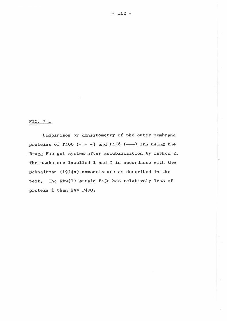
LLz -
FrG.7-4
Comparison by densitometry of the outer membrane
proüeins of P4OO (- -) and P456 (-) run using the
Bragg-Hou gel system after solubil.izat,j-on by method 2.
The peaks are labelIed 1 and 3 in accordance with the
Schnaitman (t97+^) nomenclature as described in the
text. The Kthr(l) strain P456 has relatively less of
protein 1 than has P4OO.
t'
I \ ,
I
I
III
I
I
^i\
tlllt¡
I
II
e
113
alterations in the protein composition of their cell- wall
(ta¡te 7-f). The Efr, Ktn and Misc (5) strains had no
obvious alterations in their cell wal-l proteins (fa¡te 7-I).
This does not necessaríly imply that no proteins are mutated
or abserrt irr these ¡nutarrts, buL rabher ùhat the changes are
too sma1l to be picked up by polyacrylamide gel electro-
phoresis or are masked by other cell wall proteins. This
effect has been demonstrated for bfe- mutants (Sabet and
Schnaitman L973arb) and tsx- mutants (SchnaitmanrLgT4b)
which, although mutated in a protein of the ce11 wa11, have
no apparent defects in cell wal-l composition.
Of the Ktw strains, P456 (Kt,ü¡(l)) was alone shown to
have an altered protein composition (pig. 7-4), having only
30% - 50% of protein 1 remaining in its cel1 wall. This
strain has also been shown to have an altered LPS (taUle
6-Z). The other Ktw strains, P476 (rtw (Z)) and p42O (rtw
(3)) have no such protein deficiency and this might explain
in part the sl-ightly different resistance patterns obtained
for these strains (tafle 4-3).
Three of the four Ttk strains tested had minor changes
in peak 1 sj-ze (tabte 7-L) as shown in Fig. 7-5 for strain
P429 (ftt (1)). However, no correlation was obvious between
the resistance pattern and the presence or absence of this
small protein 1 alteration.
Of the Bar mutants studied, strain P495 (nar (3)) had a
50% reduction in the amount of peak 1 (pig. 7-6) while strain
P489 (nar ( 8 ) ) which was resistant to a greater number of
bacteríophages than strain P495t had a similar alteration in
peak 1 and possibly also an alteration in peak C. However,
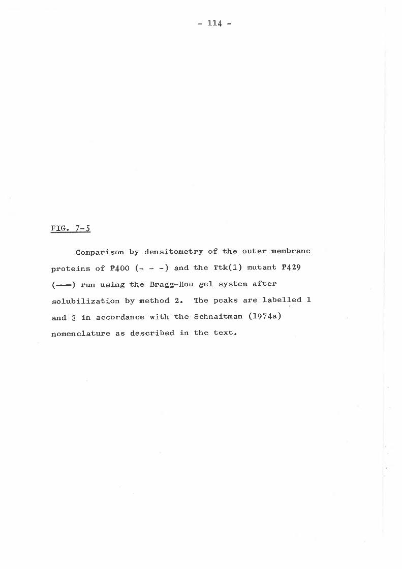
LL4 -
FIG.7-5
Comparíson by densitometry of the outer membrane
proteins of P4OO (- -) and the Ttk(l) mutant P429
(-) run using the Bragg-Hou gel system after
solubílizaluíon by method 2. The peaks are labelled 1
and J in accordance with the Schnaitman (I974a)
nomenclature as described'in the text.
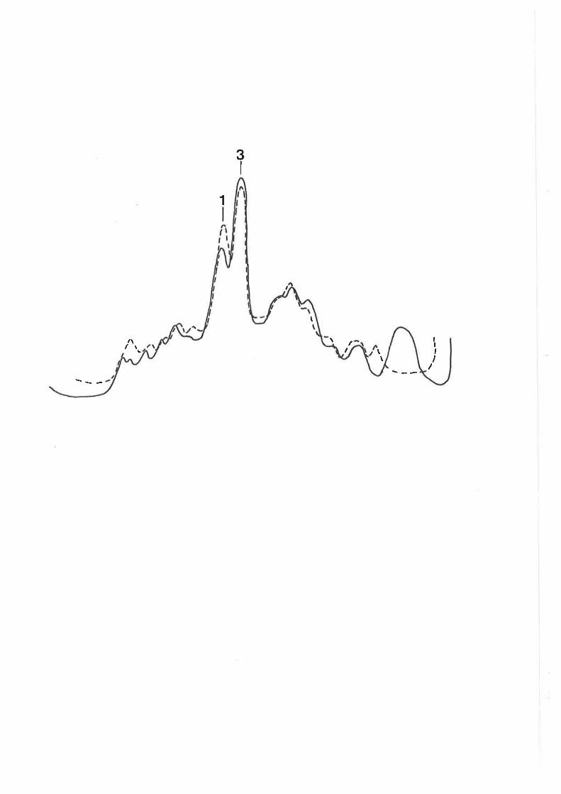
3I
1
t
/\ \ II,II
I \\
115 _
FIG. 7-6
Comparison by densitometry of the outer membrane
proteins of P4OO (- -) and the Bar (3) mutant P495
(-) run using the Bragg-Hou ge1- system after
solubiliza1-ion by method 2. The peaks are labe1Ied
1 and 3 in accord.ance with the Schnaitman (L97 4a)
nomenclature as described in the text.
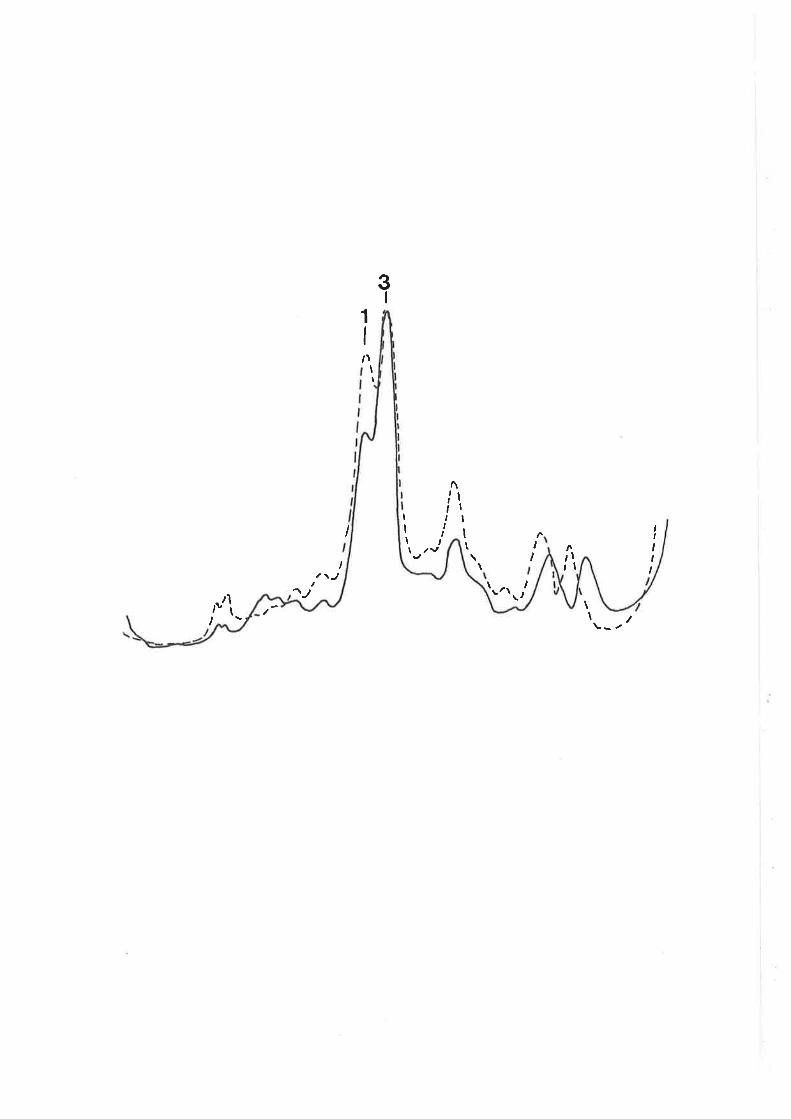
I \i¡ \
II
II
t,1lv
\I \ I
,It,I
ttIt
T
T,
ìI
t,,,
ItIv
t\t,
I
tI
e
116 -
FrG. 7-7
Comparison by densitometry of the outer membrane
proteins of P4OO (- - ) and the IVrm (Z) mutant P424
(-) run using the Bragg-Hou gel system after
solubilizat,j.on by method 2. The peaks are labelled
1 and 3 in accordance with the Schnaitman (Lgl+a)
nomenclature as described. in the text. Peaks 1 and
3 of P424 appear drastically reduced. The increase
in protein in the lower molecular weight region
(right-hand end of the gel) is due to the increased
loading necessary to visualize the bands in P424.
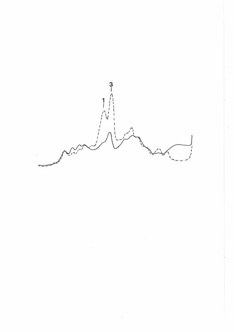
)ì
)<
> -C
^)C
I
I I I \
tL7
FIG.7-8
Comparison by densitometry of the outer membrane
proteins of P4OO (- -) and the I,Vrm (1) strain P435
(-) run on the Bragg-Hou gel system after
solubilizal-jon by method 2. The peaks are labelled
1 and. ! in accordance with the Schnaitman (tgZ+a\
nomenclature as described in the text.
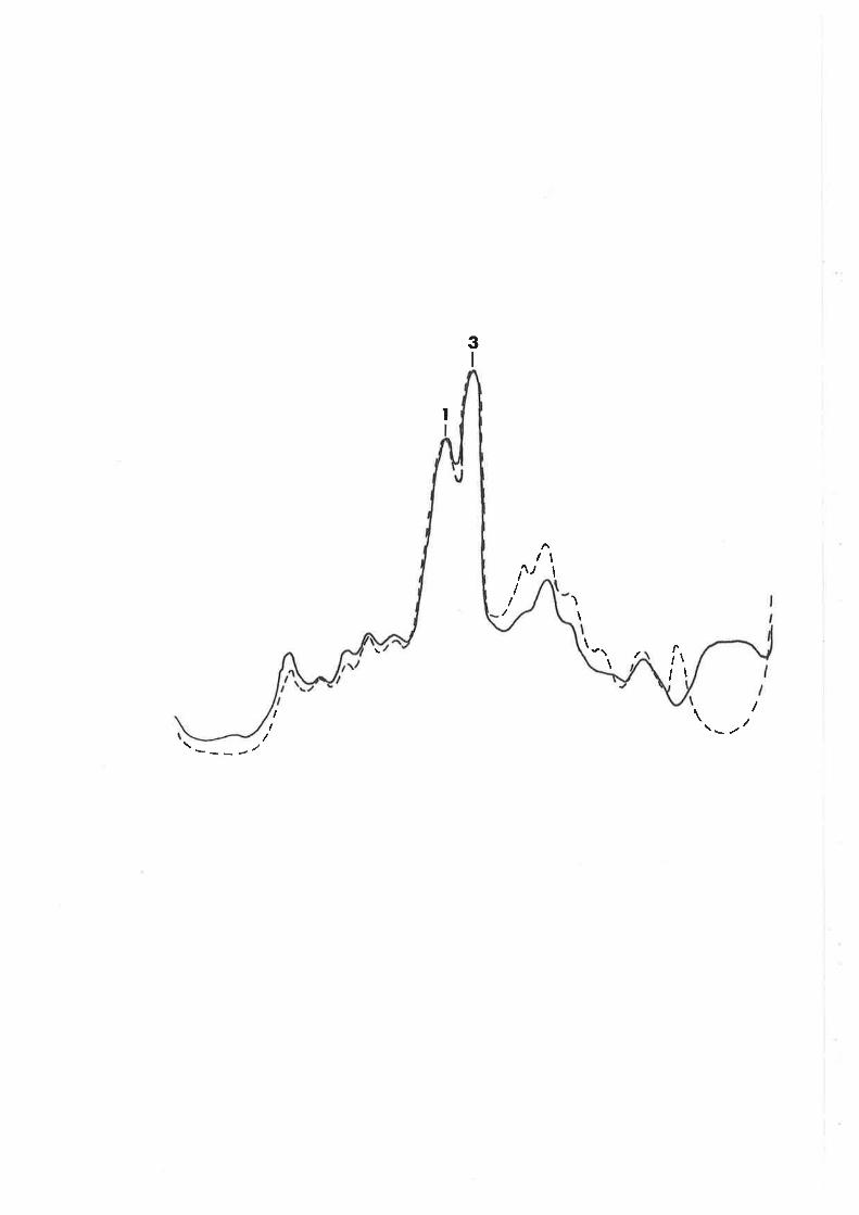
3I
I
II
I/1I \
\
\\I
¡II
\
I\II
---¿/
118
it was shown that two Bar (4) mutants, P4O5
had intermediate resistance patterns (taUte
other Bar mutants studied were unaltered in
and P{J6, which
4-5) to the
cell wal-l protein
composition.
The Wrm mutant P424 (Wrm (Z)) which has a lai.ge LPS
alteration (table 6-Z) al-so had greatly reduced levels of
peaks 1 and 3 (pig. 7-7). Thus it is probably similar to the
heptose deficient mutants of llmes, Spudich and Nikaido (f974)
and Koplow and Goldfine (tg7 +). On the other hand, strain
P435 (Wrm (1)), which has an even larger LPs-defect than
strain P424 (fabte 6-Z) and a similar resistance pattern
(faUte 4-6), has an unaltered protein composition (pig. 7-8).
This result was repeated for a fresh cel1 wall preparation of
strain P435.
There appears to be some correlation between resistance
pattern and protein composition for the l{rm mutants. For
instance, bacteriophages KJ, K4 and K5 which are unable to
lyse Con- mutants, probably have as a receptor or receptor-
component one of the proteins of peak 3 which is deleted in
Con- mutants (pig. 7-3). Strain P424 which has considerably
reduced peak 3 (Fig. 7-7) is resistant to these
bacteriophages (tabte 4-6) while strain P479 with normal
peak 3 (Fig. 7-8) is sensitive to them (tabte 4-6). However,
the correlation does. not hol-d for the other bacteriophages
which fail to lyse Con- mutants, and in this case one might
suggest that these bacteriophages are more sensitive to
alterations in the LPS component (see Chapter 5) of the
receptor. The resistance or sensitivity of the Wr.m strains
to bacteriophages T2 and T6 might al-so be related to the
absence or presence of peaks 1 and 3.
II9
SIMMARY AND CONCLUSIONS
Two of the major proteins of the E. coli K-LZ cell wa11,
proteins 3a and 3b, are missing in the cell walls of con-
mutants. of the other strains tested, only certain mutants
shown in chapter 6 to be LPs-altered., were shown to have
protein alterations and most of these were shown to have
alterations in protein 1. strain p4z4 (wrm (z)) had greatlyreduced amounts of both of the major peaks, 1 and 3, whileof the other peak 1 altered mutants, summarized in Table 7-L,strain P489 had a possible peak 3 alteration also. The pro-
tein alterations probably resulted from specific mutations
rather than being merely the result of increasing Lps
alterations leading to increasing disturbance of the outermembrane. This was shown by the fact that strain p4S6 (rctw
(1)) which had a minor Lps alteration (taute 6- z) had an
altered protein composition (fig. T-4), while p43S (Wrm (1))
with a very large LPS alteration (ta¡le 6-z) had no obvious
ce1-l wa1J- protein defect (pig. 7-8).
There appears to be a limited relationship between
protein composition and bacteriophage resistance pattern forsome mutants.
L20 -
CHAPTER 8
MAPPING OF THE MUTATIONAL LESIONS
INTRODUCTION
The genetic study of bacteriophage resistant mutants i.n
E. coli has been mainly involved with mutants resistant to
the T group of bacteriophages, BF23 and À. Well defined
bacteriophage resistance loci include tonA (J. Lederberg,
L947), t"tB (Curtiss, L965), tsx (J. Lederberg, L947)t larB
(Thirion and Hofnungr I972), tfrA (Curtiss, 1965) and bfe
(Buxton, I97I; Jasper, hftritney and Silver, L97 2) . Apart
from these, only a few limited genetic studies of bacterio-
phageresistancehavebeendonei'4K-12(Hayes,
L957; Curtiss, 1965; Tamaki¡ Sato and Matsutrashi, L97I). The
adaptation of the technique of gradient transfer analysis
(de Haan g!3!., L969) to the mapping of bacteriophage
resistant mutants (Chapter 2), has provided a rapid technique
for the approximate genetic location of mutational lesions.
MAPPING OF Bar AND hrrm MUTANTS
Wren strain P4O4 was mated with various Hfrls, it was
found that only strains KL2O9, RC749 and JG2O (Htr.f4) gave
appreciable transfer of sensitivity to bacteriophage A. A
de Haan gradient transfer analysis (de Haan et a]-., 1969) rtras
performed by mating strains KL}O} (."18 argn+/Hfr) and P4O4
(*g1¡* areï/F- ) using ggE as the selective marker and maJ-B
as the counterselection against the donor (pig. 8-1).
Sensitivity to streptomycin, fermentation of mannitol and
LzT -
FIG. 8-1
de Haan gradient transfer analysis of conjugation
between strain P4O4 (bacteriophage Ar, 3gg,E mtl ¡¿!
48+ "b"" /F- ) and K:.'zog
*É,* ."18 Ss/ne" J4) 2
recombinants. Tttis shows that the bacteriophage
resistance gene (nar (3)) in this strain maps at
approximaüely f2 min.
(bacteriophage AS, g=gE+
Analysis of mal-g* "ggE*
Imt]-'
2-25. .S.q.c. .t e. r i gnh age. . A. . sgnç!!.iy. ity.
vf
2-20/
oFcoC.-.Gt
EoooLrÞo
a
oca
o)o
2.15
s
2-10,64 66
min on þli K-12 genetic map
727068 74
L22 -
xylose, and bacteriophage sensitivity was scored and, by
plotting log1. of the number or recombinants against the
E. coli R-LZ genetic map position, the mutation was mapped at
approximately 72 mín. By Plkc transduction into strain KL141t
it was shown that bacteriophage A resistance was 55% co-
transducible with p.yIE+ which rn¡as itself 20% cotransducible
with I!f, and that bacteriophage resistance lay between
these two markers. Thus, using Taylor and Trotterts (L967\
relationship between map distance and percentage cotransduct-
ion, this places this bacteriophage resistance gene at
approximately 7I.8 minutes. Similar conjugation or trans-
d.uction experiments indicated that strains P455 (¡ar (1)),
p4gz (nar (z)), p413 (nar (3)), P49ot F4o5 (nar (4)r' P4o2
(nar (5)), P45L (nar (6)), P487 (sar (7)), P4B9 (Bar (8)),
P4L6 and. P239 (wr- (z)) all map between pEE and mtl (pig.
g-3).
Many of the rfa genes for biosynthesis of LPS in
Salmonella (Stocker and Mäkela, I97I; Wilkinson, Gernski and
Stocker, Lg72), E. coli O8 (Scfrmidt, Jann and Jann t L97O)
and E. coli K-LZ (Eriksson-Grennbergr Nordstrom and Englund,
LgTL; Schmidt, L97 3) have been previously located in this
region of the respective genetic maps. It is therefore
like1y that the LPs-altered, bacteriophage resistant mutants
of the Bar and. hlrm (Z) groups form a gþ gene cluster in
E. coli K-I?. In support of this hypothesis, it has been
shown in E. co1-i 08 (Scfrrnidt, Jann and Jann, 1971-) and
Salmonella (I,rrilkinson, Gemski and Stocker, L97 2; Lindberg,
ag7 3) tfrat mutants deficient in the enzymes of LPS core
biosynthesis (rfa mutants), often have an altered pattern of
resistance or sensitivity to various bacteriophages.
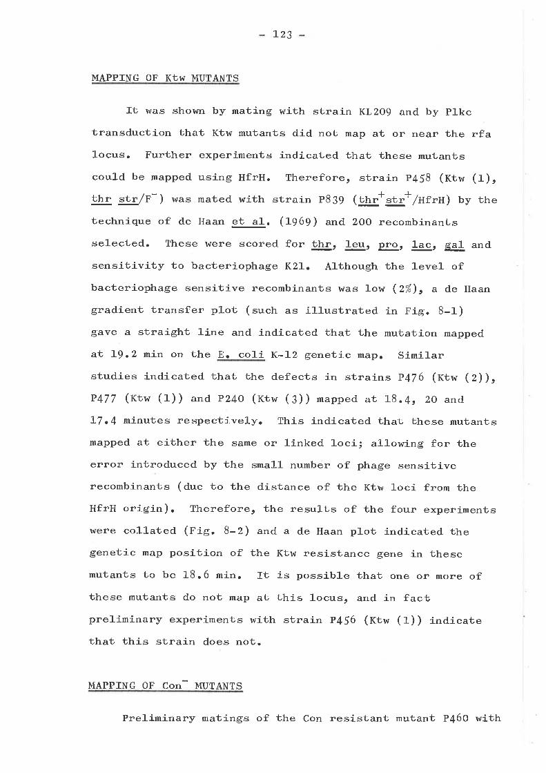
l-23
MAPPING OF KIw MUTANTS
It was shown by mating with strain KL?O9 and by p].kc
transduction that Ktw mutants did not map at or near the rfa
1ocus. Further experiments indicated that these mutants
could be mapped using HfrH. Therefore, strain p458 (rctw (1),
thr str /r-) was mated with strain p839 (ttt"*"a"* /utru) by the
technique of de Haan g!-3!. Gg6g) and 2OO recombinants
selected. These were scored for thr, þ, plgr ]-ac, ga1 and
sensitivity to bacteriophage Kzl. Although the level of
bacteriophage sensitive recombinants was low (Z%), a de Haan
gradient transfer plot (such as illustrated in Fig. 8-1)
gave a straight line and indicated that the mutation mapped
at L9.2 min on the E. coli K-Iz genetic Írâpr Similar
studies indicated that the defects in strains p4T6 (t<tw (Z)),
P477 (rtw (1)) andp24o (rtw (3)) mapped ar L8.4t 2o and
]-7.4 minutes respectively. This indicated that these mutants
mapped at either the same or linked loci; al-lowing for the
error introduced by the smal1 number of phage sensitive
recombinants (due to the distance of the Ktw loci from the
HfrH origin). Therefore, the resurts of the four experiments
u¡ere collated (pig. 8-Z) and a de Haan plot indicated the
genetic map position of the Ktw resistance gene in these
mutants to be 18.6 min. It is possible that one or more of
these mutants do not map at this locus, and in facú
preliminary experiments with strain p456 (tctw (1)) indicate
that this strain does not.
MAPPING OF Con MUTANTS
Preliminary matings of the Con resistant mutant P460 with
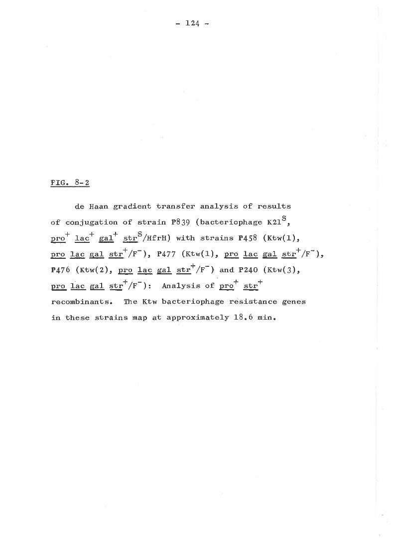
L24 _
FIG. 8.2
de Haan gradient transfer analysis of results
of conjugation of strain P839 (bacteriophage K21S,
gg+ 1""* g1* gs/Hrrn) with strains P458 (rtw(r),
pro ac e"1. rü",+/F ), P477 (rtw(r), pro lac e"1, "t"+/F-),P476 (Kth,(2), pro ]-ac g"1 rt"+ /e-) and, P24o (rtw(3),
pro lac gg! str+ /e- ): Anal-ysis of pg+ .t"+
recombinants. The Ktw bacteriophage rejsistance genes
in these strains map at approximately 18.6 min.
Ø+.gotr.-
.crEoool-ra-o
a
oca
ct)o
2-5
2-O
1.5
1.O
lac
. . .Baç. .te r.iqphage. K?:1. . .çen sltiv!!.v
681012 14 16 18 20
min on E.coli K-12 genetic map
L25
strains KL2o9 (Hfr J4) t ABzSg (HfrH) and RcTSo (Hfrc)
indicated that the latter two could transfer sensitivity to
bacteriophage KJ. Results from gradient transfer analyses
!üere very hard to analyse as Con- mutants or recombinants
gavc spurious results on EMB media, which was used to score
fermentation of galactose and lactose. A number of experi-
ments were done in an attempt to determine the reason for
these spurious results. These included plating strains P4OO
and P46O on various synthetic media or mixtures of EMB agar
with nuürient or minimal agar. It was shown that on EMB
agar the growth of strain P46O was inhibited, relative to
strain P4OO. Furthermore, after 2-3 days, the relatively
smal1 colonies of sùrain P46O took up the BMB stain giving a
pseudo-positive result. Although this appeared to be the
reason for the spurious results, it was not possible to
demonstrate sensitivity to Eosin Y, methylene blue or to the
Eosin Y-methylene blue complex, even at four times the
concentrations that existed in EMB agar.
Scoring of fermentation with minimal agar containing
growth factors and lactose or galactose as the sole carbon
source was difficult (especially for the scoring of @)rbut, using the two above systems together, the Con- genetic
1-oci was shown to be between 1-ac and æf. Gradient transfer
analysis (de Haan g!-3!., a969) of the data indicated the map
position to be somewhere between L2 and 15 min. on the
B. coli K-LZ genetic map. It was shown a1-so that sensitivity
to bacteriophage K3 was transferred by HfrC strain RC75OrI.tusing pro' as a selective marker; but transfer of K3
sensitivity occurred only at a l-ow frequency (S-tO$¡. This
l,'¡as surprising as the HfrC origin lies at about 13.5 min
or a maximum of 1.5 min from the map position of Con- as
r26
indicated by previous experiments. rt was further shown
that when con- was transferred to Hfrc by Plkc transd.uction,
thís strain would not transfer con- into a recipient cell,+even if gE' (I2 min) was used as the selective marker in
the recipient. Furthermore, it could not be shown that the
Con- marker was cotransd-ucible with plEE*, even though 15O
transductants were tested.
To summarize, a hypothesis to explain these results,
would be that the con- defect is a deletion or chromosomal
aberration, such as has been demonstrated for other
bacteriophage resistant mutants (Curtiss, L965; Franklin,
Dove and Yanofsky, 1965). This would help explain why Con+
could be transferred into the Con- strain p46O by HfrC, but
Con- could not be transferred. into a Con* strain. The
mutational lesion probably maps very near to the HfrC origin(pg. 8-3) based on its inability to be cotransd.uced. with the
ry,E marker.
MAPPING OF OTHER MUTANTS
By Plkc transduction or conjugation with strain KLZO}
(Ufr J4) t it was shown that the following mutants do not map
at the rfa locus: strains p4Z3 (rtt (z)), p474 (rtt (4)),
P448 (nrr), p4o7 (rsx (1) ), p433 (rsx (z)), p237 (rurisc (4) )
and P479 (Wrm (1) ) " The mapping of mutations of strain p4OO
by conjugation, is inhibited by the lack of genetic markers
for selection purposes between gal (L6.5 min) and str (64.2),
and thus attempts to locate the above mutations, in addition
to those in strains P42g (rtt (1)) andp443 (Uisc (Z)) have
been 1argely unsuccessful.
L27
FIG. 8.3
Partial genetic map of E. coli K-Lz (adapted from
Taylor and Trotter, L972) including genetic loci and
origins of various Hfrfs mentioned in Chapter 8 and
the approximate genetic locations of various
bacteriophage resisüance loci isolated and mapped in
this study.

t 1 leu
Hf rH o/90 prþA
10
.I!k4loc
Hfra
r ,2r314, Hf rC51617rg.
PY''mtl
xyl 70
gol
Ktwl.2.3.
-
t '
20
L28
The only mutants, of those mentioned above, that have
been located on the E. col-i K-l-Z genetic map are strains
P4O7 and P433 of the Tsx resistance group andP474 of the
Ttk resistance group (pig. 8-3). Gradient transfer analyses
of these mutants indicated that the Tsx mutants mapped at
or close to the previously discovered ùsx 1ocus, while
P47 4 mapped close to ryA. This is interesting as strain
P433 (tsx (Z)) is not fully resistant to bacteriophage T6
after which the tsx locus ís named. Further experiments
are required to determine whether or not the Tsx (Z) mutant
a1Ie1e is in fact linked to the tsx allele. The discovery
that the Ttk (4) mutant P47 4 maps near proA indicates that
it may map close to the tfrA locus despite the fact that it
is not resistant to bacteriophage T{ (tabte 4-4).
SUMMARY AND CONCLUSIONS
A1-l results are summarized in Fig 8-¡. The mapping of
the Bar and Wrm (Z) mutants at a position between pgE and
mt on the E. co]-i K-Lz genetic map¡ suggests that these
mutants which often have altered LPS (faUte 6-Z) are mutated
ir É genes which form a cluster at thís location. The
Con- mutant probably maps close to the HfrC origin, and
genetic experiments suggest that the multip1-e properties of
Con- mutants (i.". deficiency in two cell wa1l proteins,
colicin tolerance, bacteriophage resistance, sensitivity to
EMB agar, conjugational recipient deficiency) might or might
not be due to a deletion or chromosomal aberration, causing
more than one genetic locus to be affected. However, this is
by no means proven and the properties are consistent with the
r29
defect being a point mutation (see Chapter Ç) and thus Con-
mutants have been discussed ín this thesis with this surmise.
Both sub-groups of the Tsx resistance group map at closely-
linked loci or the same locus; while representatives of the
three sub-groups of Ktw map together a'L a dis'binct locus.
Strain P474 (ttt (4)) maps close to the previously discovered
tf"À gene, however neither the Ttk (1) nor the Ttk (3)
mutants do.
130
CHAPTER 9
DISCUSSION
In this work, bacteriophage resistant mutants were
selected in E. coli K-Iz using a wide range of different
virulent bacteriophages. The results summarised in Figure
9-L showed that the mutants fell into 11 main groups, and
on group of miscellaneous mutants which did not fit, welJ-
into any of the other groups. Each group of resistant
mutants was characteri-zed by a particular set of
bacteriophages to which most of the mutants showed resistance
of one type or another. Although the mutants within a group
were not always identical, no difficulty was experienced in
recognising the relationship between them. A number of the
mutant groups have already been described including TonA,
TonB, Bfe and Tsx (Demerec and Fanot 1945; Buxton, L97L).
A few new mutant types are described, but extendíng the
number of bacteriophages studied did not greatly increase
the range of mutant types. It would seem that there are not
many undiscovered types resistant to different sets of
virulent bacteriophages. It remains possible of course that
other types remain undetected, because they occur 1-ess
frequently than those described previously or in ühis paper.
However, selecting a large number of mutants enables one to
try to arrange them in a meaningful h¡ay.
THE GENERAL PATTERN OF BACTER IOPHAGE RESISTANT MUTANTS AND
ITS RELATIONSHIP TO BACTERIOPHAGE TAXONOMY
Each type of mutation must have its effect on
131
bacteríophage resistance by preventing some stage of the
bacteriophage infectious cycle. The best characterj-zed
mechanism is by loss of receptor, although not all mutants
act at the 1evel of adsorbtion. Many examples of receptor
mutants have been found in these studies; however, the poor
adsorbtion, generally, of many of the bacteriophages, using
a wide range of techniques, has inhibited the search for
tolerant mutants similar to those found in studies of
col-icin resistant mutants (see Reeves, L972). Garen and
KozLoff (fqSq) have postulated a class of resistant mutants
with subtle surface modifications. They propose that the rate
of phage attachment to these mutants would be slow, but still-
sufficient to prevent colony formation in the presence of
bacteriophages. Some of the mutants discussed in Chapter 4
appear to be inhibited by certain bacteriophages and it is
possible that they may be of this class. Wahl (fqS¡) has
described semi resistance which may be similar to the partial
resistance with inhibition (fp) pattern of resistance.
The various mutant groups described serve also to define
sets of bacteriophages, most of which are unable to form
plaques on more than one of the mutant groups described.
Correspondingly, some of the mutant groups confer resistance
to more than one set of bacteriophages. In general, the
mutants seem to be of two main types. The majority of
resistance groups confer resistance to one or two sets of
bacteriophages, while the Bar and l,{rm mutants are resistant to
bacteriophages from many sets. Mutants of the first type are
presrrmably affected in a ce1-1- component necessary for only one
or two bacteriophage sets during infection, whereas the Bar
and l,{rm mutants affect a function necessary, either directly
a3z
or indirectly, for infection by many different bacteriophage
sets. One possibilíll-Vc expanded 1-ater in this discussion, is
that Bar and hlrm mutants affect the cel1 surface in such a
way that many different receptors are absent or non-functional.
Bacteríophage taxonomy is based primarily on morphology
(nradleyt A967; Ackerman, L969; Tikhonenko, L97O; I{ilcly,
L97L)t which serves to identify the major natural groupings.
Furthermore, some of the well studied bacteriophages are
known to be close1-y related genetically and ab1.e to undergo
recombination readily. On classical criteria for higher
organisms one can say that they are strains of one species.
Thus BF23 and T5 (Mizobuchi, Anderson and McCorquodale t I97L)
and T2, T4 and T6 (Luria and Dulbecco t L949; Adams, 1953)
recombine readily and constitute two separate species.
Unfortunately, information of this type is not avail-able for
most bacteriophages.
It is interesting that the bacteriophages with 81 type
morphology studied (fabte 3-2), resembled either T1 or T5
and BF23 in their dimensions. Most bacteriophages with
similar dimensions to T1 required both the tonA and tonB
functions for ínfection; the others resembled T5 in requiring
only tonA function t or BF23 in requiring only bfe function.
Both tottA and bfe mutants are known to lack a specific
surface protein (Braun, Schaller and l{olff, a973; Sabet and
Schnaitman, Lg7 3) v¡fricfr may be the bacteriophage receptor.
The nature of tonB, however, is not fully undersbood as Tl
adsorbs reversibly to this mutant (Garen and Puck, 1951).
It seems that many of the bacteriophages resembling T1
morpho1-ogical-J-y have similar requirements for infection,
while those resembling T5 and BF2J, probably all one
133
species, use either the T5 or BF23 receptor and, like the
T1 set, are unaffected by Bar or l{rm mutations. Thus, it
seems that resisüance patterns rnight be the simplest method
of distinguishing strains of the same species of
bacteriophages.
Bacteriophages of A2 morphology (tabte 3- 2) are far
more heterogenous with respect to resistance patterns
(pig. 9-1). Seven of the mutant groups are resistant to
bacteriophages of this type. Three groups, Tsx, Con and
Ktw, are commonly resistant to a set of between four and
nine bacteriophages with A2 morphology. Within the set
of bacteriophages affected by the Tsx mutations, H3 has
only one requirement, for the tsx function. This function
is also required by T6 and six other bacteriophages of
A2 morphology; however these bacteriophages, unlike HJ,
have altered activities on one or both sub-groups of the
Wrm resistance group. Heterogeneity also exists amongst
those bacteriophages which require the Con function.
Only the Con mutants and one sub-group of Wrm mutants have
altered sensitivity towards bacteriophage KJ; however, in
the case of Ox5 there are also mutants in the Ttk, Bar and
miscellaneous groups which are resistant to it. The mutation-
aJ. lesions affecting the infection of these strains by
bacteriophage Ox5r differ considerably (Chapters 6 and 7)
and appear to reside in the outer membrane. Since the
mutants, with the exception of Con, appear to affect the
same component (lpS) of the outer membrane, one might
postulate, as with bacteriophage KJ, that there are only
two factors influencing the infectious cycle, but that
TABLE 9-L
134
General pattern of resistance.
Resisb. c
Group( su¡-group)
BACTERIOPHAGES RESISTANT TO A,b
o]¿OOO
O O'rl o OJ( J4 J( OJ4 J Ff Jt J4'rf 'r'l '-l J4.i.d | 'd 'rlF{ -l r{ 'd-l -l cQ -l Fl I I I FlI INO I I ca N Oo'rô\or\ F\ o. lr.)lôFlÍ¡iF{cOr.{\OceOil X+NNN XF{N+ F-rl ca-+Fl NÞ--l
H H É ld iq Fq ts 14 < E o f'l l¿ M !4 O V t¡. t > t¡l V < H H V H H g E
TonATonBBfeKtnrsx(r)
(z)ConEfrKtw( 1 )
(z)(3)
rrk( 1 )(z)(3)
))))))))))
4I2aJ4512
J456
7812
(Misc(
((((
Bar((((((((
Wrm((
VRVVV
RRRRR
RVV
VV
VVV
RRRVV
RRVV RRVV R RV RV RRV RV RRV V
VRVRV RVRRVRRR VV
VVR V
VV R VVV
VR
VV VRR
RRRRRV RRRVRR RRV
R
VR RVR RVRRVRR
V
VRV RRV VRVRRV RRR RRVRRRRRR RRRR
RVVV
VV R RVVV
R
VVRVVVVVVV
VRRRRRVVRVVVVRVVR R
V RRRRR R RVV V V V R R V V V
RRRR
VVVV
VVRVVVRVVVVV VR R
RRRRV V
R
V
R
RRR
Oj4OOC)O O'FJ O OJq i( Jt OJt J( Ff Jt J.i 'Fl 'rl J{.r'l 'Fl | 'Fl 'rl rl F{ -l 'rlr{F{ce F.{ rl I I I -lI I NO I I ca N Oo.rr)\ot- tr- o' lrôtorlf¡irl coFl\OeoOrl X+NNN Xr.{N+ Þ--l co$r{NÞ\rlH H É l¿ t FÉ H 14< E O t'l V M V O g r¡. t > É4 V < H H V H H 11 iq
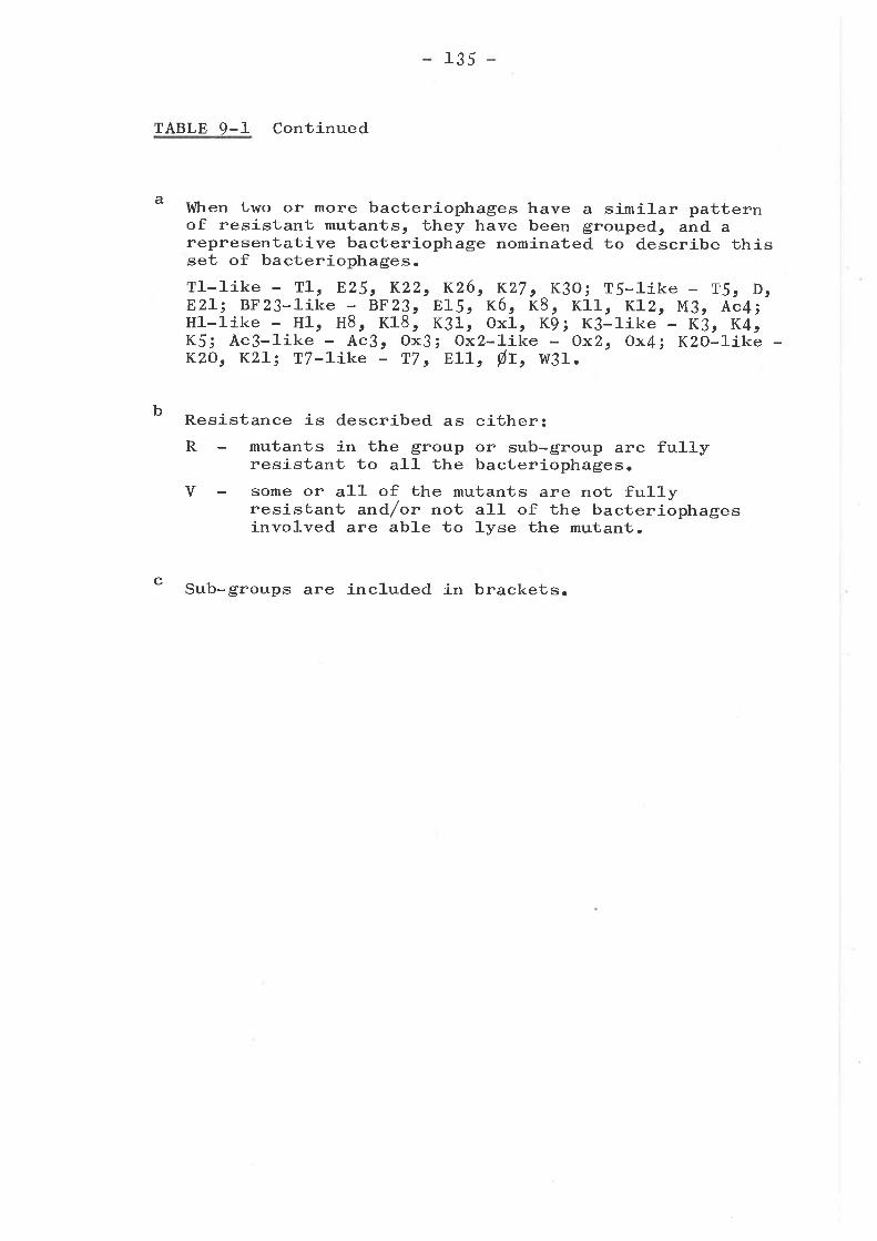
a
135
TABLE 9-1 Continued
Resistance is described as either:R mutants in the group or sub-group are fully
resistant to al-l the bacteriophages.V some or al1- of the mutants are not ful1y
resistant and/or not all of the bacteriophagesinvolved are able to lyse the mutant.
c Sub-groups are included in brackets.
I,tlhen two or more bacteriophages have a similar patternof resistant mutants, they have been grouped, and arepresentative bacteriophage nominated to describe thisset of bacteriophages.Tl-like - T1, E25t K22, K26, K27 c K3O; Ts-like - T5r D,E2L; BF23-like - 8F23, E15, K6, K8, K11, KLz, M3r Ac4iH]--like - Hl, H8, K18, K31r oxl, Kgt K3-like - K3r K4tK5; Ac3-like - Ac3r Ox3i Ox2-like - Ox2, Ox4i K2O-1ike -KzO, KZL; T7-like T7 t E11, Øf , I4I31.
b
136
bacteriophage Ox5 is far more sensitive to one of these
factors.
T-even (42 morphology) bacteriophages probably have
three types of receptors, for initial tail fibre attachment,
for subsequent attachment of the spikes to the traseplate and
for the tail tube in order to position it prior to unplugging
(Benz and Goldberg, L973). This last type of receptor might
reside on the plasma membrane but, the other two receptors
are probably in the outer membrane. Alteration of these two
receptors could occur independently, causing bacteriophage
resistance. This might help to explain the results d-iscussed
above.
Bacteriophage E4 of A1 morphology has a complex set of
requirements for infection if one considers its resistant
mutants. Besides its specific resistant mutant, Efr, mutants
from the Ttk, Ktw, Bar, I{rm and miscellaneous groups are
resistant to it, although in all except the Bar and Wrm
groups, these E4 resistant mutants are limited to one or two
examples per group. It is not known whether these mutants
each represent specific requirements for infection, or
whether they demonstrate the sensitivity of the bacteriophage
E4 receptor to outer membrane alteratiorlso
One might ask whether bacteriophages from various parts
of the world which select for the same resistant rnutants and
are similar using electron microscopic or serological
techniques, are different isolates of the same bacteriophage.
This seems quite likely as most of the differences are minor.
For the enterobacteriaceae cl-osely related to E4.!! K-12,
there are probably a limited number of bacteriophage species
which can be found in most parts of the world with some
437
variation. The 54 bacteriophages studied (labte z-z) vary
considerably in their apparent requirements for i_nfection,from those such as BF23 which requires a receptor (ttre ¡reprotein) but' is apparently unaffected by a change in the
cell which affects most other bacteriophages, through tothose which are affected by up to six different types of host
mutational changes.
The 54 bacteriophages represented in Figure g-1" can be
reduced to five main resistance types based on their abilityto plaque on the resistant mutants shown. These are:-
(1) Those bacteriophages for which there is only one
type of fully resistant mutant. This includes
bacteriophage H3e the BF23-like bacteriophages and
the T5-l-ike bacteriophages.
(Z) Those bacteriophages for which there are two types
of fully resistant mutants, ê.go the T1.-l-ike
bacteriophages.
(e) Those bacteriophages for which there are two types
of resistant mutants, one of which is a specific
fully resistant mutant and another which is cross
resistant to many bacteriophages. This second type
of mutant is not ful1y resistant to all members of
the resistance group. The bacteriophages included
in this category and their ful1y resistant mutants
are T6, Hl, H8, K9t K18, K31¡ ox1 (rsx); K3r K4t
K5r gx3r Ac3 (Con); Klo (t<tn); and T2 (Ttk).
G) Those bacteriophages for which there are a range ofmutants, which are resistant to them in any one of
the ways described in Chapter 2 (Nomenclature).
Most of these bacteriophages are included in the
138
(s)
Bar, Ktw, and Ttk resistance groups (Tables 4-3,
4-4 and {-l), with the exception of bacteriophage
T2 which is designated as a Type 3 bacteriophage
above.
This type is restricted to bacteriophages K15 and
H, and the T7-like bacteriophages. The only
mutants to these bacteriophages found in this
survey are those cross resistant to a wide range
of bacteriophages implying that in order for
resistance to occur a large change in the cel1-
surface must take place such that it interferes with
the binding of other bacteriophages to their
receptors.
THE STRUCTURE OF THE LIPOPOLYSACCHARIDE OF E COLI K-12 AND
THE ALTERATIONS IN CERTAIN BACTERIOPHAGE RESISTANT MUTANTS
Some of the mutants described in this thesis h¡ere
defective in their LPSts (Chapter 6). E. co]-i K-12 is a
rough organism (Rapin and Mayer, L966) with no O-antigen sugars
(Scfrmidtt I973). Reports in the literature vary considerably
as to the exact proportion of glucose, galactose and heptose
in the LPS of E. coli K-Lz (Rapin and Mayer, 1966; Eriksson-
Grennberg, Nordstrom and Englund, L97A; Monner, Jonsson and
Boman, L97l; Tamakie Sato and Matsuhashi, L97I), as well as
the presence of rhamnose in the LPS (Rapin and Mayer, L966t
Tamaki, Sato and Matsuhashi, L97L) o" glucosamine in the
polysaccharide chain (Scfrmi-dtt L973; Koplow and Goldfine,
L974). Thus it appears that there are strain differences in
E. coli K-Lz LPS extracted in various laboratories. The
L39
results, shown in Table 6-2, suggest that the ratio of the
three neutral sugars in the LPS of P4OO is approximately 3
moles of heptose: 2 moles of glucose: 1.3 moles galactosê¡
Nikaido g9_4. (1965) described a strain yl-O which they
showed was deficient in the biosynthesis of thymidine
diphosphate L-rhamnose and thus had no rhamnose in íts Lps;
unlike other derivatives of wild type E. coli K-L2
(ttit<aido g!_jl. , L965; Rapin and Mayer, 1"966; Eriksson-
Grennberg, Nordstrom and Englund, L97L) which had no such
er'zyme deficiency (mi-t<aido et 4., 1965). The fact that
strain 481133 (and hence P4OO) i" directly derived from
strain Y1o (Bachmann, L97z) explains the absence of rhamnose
in the LPS of our mutants. There is also no glucosamine
attached to the polysaccharide portion of the Lps of strains
P4oO, P46O (Con) and P425 (rtt (3)), despite the previous
finding of Schmidt (f97 3) that his strain of E. co1.i K-12 has
a complete core. Glucosamine appears to be an essential part
of the completed rough cores of smooth strains of salmonel-la
typhimurium (Osborn, L966; Lüderitz et aL., LgTt; Stocker and
Mäkela, L97L; Hämmerling, Lehman and Lüderi-1oz, Ag73) and
E. coli O 1OO (Hämmerling *_gf., LTTL) futrot E:gþ O 7I(Mûller-Seí1-z, Jann and Jannt 7968). Rooney and Goldfine(tgZZ) have shown that their E. coli K-I2 strain CR34 has no
glucosamine in the polysaccharide portion of its Lps and it
is interesting that both this strain and 481133 rnrere derived
from strain Y53 (Bachmann, L972). However, whether or not
other E. coli K-LZ strains have glucosamine in their LpSt sr
other than that attached to the lípid A portion, is uncertain.
Rather than propose a model for the structure of the
E. coli LPS in this thesis, it r,r¡as found on examining the
L40
literature that the results obtained (pig. 6-Z) could be
explained using partial rough core structures for the LPS
of Salmone]-la (Hammerlingr Lehman and Luderi-tz, L97 3) o*
E. coli O 1-OO (Hammerling et al. e L97L). Using these
stmctures (pig. 9-1) simple derivations for the LPS!s of
the various resistant mutants could be demonstrated. This
does not imply that the core of E. co1-i K-Iz has the same
linkages or even that the sugar sequence is identical to
that of either model. However, there are almost certainl-y
simi,larities between them. Schmidt (L972, I973) fras shown
that E. coli K.-Lz LPS is serological-ly distinct from the
rough cores of Salmonella Ra mutants and E. co]-i O 1OO, but
Salmonella Rbin this context, it is interesting that
mutants which differ from Salmonella Ra mutants by a single
glucosamine molecule (which is also lacking in the degraded
polysaccharide of P4OO) are also serol-ogically distinct
( Lüderit z *_*. t L97I) .
The most significant difference between the molar
ratios for the pa rtial structures of S. typhimurium and
E. co]-i O 1OO shown (pig. 9-1) and that of P{OO, is that
the ratio of galactose to glucose is lowered in the latter
strain (t.3.. 2 moles as opposed to 2:2 moles). Rapin and
Mayer (t966) have postulated two galactose molecules in the
LPS of E. col-i K-Lz, although they also get a lower ratio
of galactose to gϡcose than would be expected. This is
difficult to explain unl-ess one considers that the galactose
molecule, deleted in the Ktw strains (pig. 9-1) is present
in only 30% of the wi1.d type LPS molecules. Hammerling g!3!.
(L97 3) have discussed previously that the substitution of
the main chain by side chain sugars, including galactose
L4L -
FIG. 9-1
Visualizaioíon of the stages at which the
lipopolysaccharide-deficient, bacteriophage
resistant mutants of E. coli K-L2 are altered. The
lipopolysaccharide models used are the partial rough
core structures of E. coJ.i O lOO (Hammerling g!_gf.,
and S. typhimurium (Hammerlingr Lehman andL97L)
Lr;.deríTczt L973). Phosphate and ethanolamine bound
to the rough cores of these strains have been omitted.
Abbreviationss KDO, 3-deoxy-D-manooctulosonate; Hepr
L-glycero-D-mannoheptose ; GLc, gl-ucose; Ga1., galactose.
Gal Hep Gat(gooô)
t
+
Ê
HeP
Ktw--
E.coliO lOO
S. tvphimurium
!r¡l
-GlcilIIIII¡I
)
Ga
Glc
t(
ep- Hep+ KDO -
( KDO KDO ) -Lipid A
Kt ) KDO
,
- Glc-j-Gal Glc Hep- Hep+KDo-KDO Lipid A
IIIIIIIIIIII
t
L
III
I
T k gar(3) gæ,r(41 wrmþ) wrrn(r)
L42
(Hellerqvist and Lindberg.r L97L)t can be incomplete.
The other anoma1.1-y found is that there is one mole of
heptose less than predicted by the model (pig. 9-1), in al1.
the mutants with altered LPS from the Ktw, Bar and Ttk
groups ( see Table 6-Z). The presence of a branched heptose
trisaccharide in the LPS core has been documented by
Hämmerling g!3!. for R. strains of E. coli O 1OO (Hämmerling
g!-31't L97L)
In Sa]-mone]-]-a
and S. typhimurium (Håmmerling Éjf . r L97 3) .
only mutants with more complete LPS have been
shown to have a heptose III unit and, in these cases, the
degree of substitution of the heptose main chain with
heptose III can vary from 20 to 9O%. It was shown that for
a S. typhímurium Ra mutant most heptose main chains were
substituted by a heptose side chain whil-e for two galE
mutants of S. landau M and S. typhimurium M onl-y 25- 50%
of mo1-ecules had the heptose III unit. It i-s interesting
to note that other authors (Sctrmidte Jann and Jannt L97O;
Eriksson-Grennberg, Nordstrom and Englund, L97L) have also
found reduced heptose levels in mutants of E. coli which
have other more distal sugars stil1 present in their basal
cores. Oneof these mutants (þgA) maps in a similar place
to the resistant mutants of the Bar and l¡úrm (Z) groups
(pis. 8-3).
The simplest assumption is that the various mutants are
bl-ocked at the stages shown in Figure 9-L. Ktw (1) mutants
have reduced l-eve1-s of galactose and thus might be missing
the galactose molecule shown whichr âs discussed above, is
apparently present in only 30% of LPS molecules of strain
P4OO. Rapin and Mayer (L966) have demonstrated that in galE
L43
mutants of E. co1-í K-12 the amount of galactose is halved.
However, the reduced 1.evel of galactose in our Ktw strains
does not lead to bacteriophage CzL sensitivity as it does
in theirs, and genetic experiments (Chapter B) indicate our
mutant is not gal-E. The exact position of the defect in the
Bar (4) mutants is not easily calculated from the results
obtained (tabte 6-Z), but these mutants might be similar to
the rfaF mutant of S. typhimurium (wilkinson, Gemski and
Stocker, L97Z) i.e., defective in the addition of heptose II.
Other cl-asses of mutants might wel-l. correspond with other of
the Salmonella rfa mutants.
Representative LPS-altered mutants of the Bar resistance
group and sub-group 2 of the hlrm resistance group have been
found to map between pEE (72 min) and mtl (7t min) on the
E. coli K-LZ genetic map (pig. 8-3). This is similar to
results obtained by other authors for Salmonella (Stocker
and Mãkela, L97L; Wilkinson, Gemski and Stocker, L972) and
E. coli (Sctrmidt, Fromme and Mayer, L97O; Eriksson-Grennberg,
Nordstrom and Englunds L97L; Schmidtt L973) rfa mutants.
Sa]-monella has been well studied and a series of mutants
affecting LPS core biosynthesis have been shown to map at
various positions, with an g'¡þ cluster cotransducible with
pIgE or g!!. However, the Salmonel].a rf a mutants ( rfaF,
&G, -rfaH) which correspond with Bar and hrrm (Z) mutants do
not all map in this region, and in fact, other mutants (rfaJ)
with similar LPS compositions to Ttk mutants, unlike Ttk
mutants, do map in this region of the Salmonella genetic mâpr
Thus, although anomalies do exist, it is probable that the
Bar and hlrm (z) genes form part of an I&. g.ne cluster, and
the mutants in this region are affected in the enzymes of LPS
L44
core biosynthesis.
The increased sensitivity to novobiocin of many mutants
of the Bar and hlrm groups (taUte 6-1) suggests that this
antibiotic might be useful in revealing the larger changes in
the structure of the LPS. This ís ín agreement with the
results of Tamaki, Sato and Matsuhashi (1-gZt) who studied
novobiocin supersensitive and bacteriophage T4 resistant
mutants.
I{hen the ampici1-1-ín resistance of various bacteriophage
resistant mutants was tested (tatte 6-1) none of the classes
demonstrated by Eriksson-Grennberg g!-3!. Qglt) were found.
Their Class II mutants were galactose deficient, sensitive
to bacteriophage CzL and in one case resistant to
bacteriophages T3 and T4. However, the results in Chapter 6
suggest that alterations in the LPS composition brought about
by mutation to phage resistance, are not sufficient to cause
significant resistance to ampicil-lin, and on1-y slight changes
have been observed. It is probably significant that their
ampicillín resistant class II mutants a1-1. contained two
mutations (AnpAl-, AmpII) .
Other antibiotic changes are consistent with findings
of Davies and Reeves, (L974a) who found alterations in the
pattern of novobiocin, ampicillin, oleandomycin, fusidj-c
acid and methicillin resistance or sensitivity as revealed
by multodisks in a to1 C mutant with an altered response to
bacteriophages C21 and U3. Thus, it seems that these anti-
biotics together with erythromycin (taUle 6-1) are useful
in revealing differences in the LPS composition
K-L2 strains.
of E. coli
L45
RECEPTOR SPECIF I OF OPHAGES WHICH ADS
O LIPO OLYSACCHARIDE
Alterations in the resistance of E. coli K-L2 bo
bacteriophage CzL or sensitivity to U3 have been shown to
be brought about by changes in the LPS (Rapin, Kal_ckar and
Alberico, L966t Rapin and Kalckar, L97L; Watson and. Paigen,
L97L). Strains of three different LPS cornpositions lriere
found in these studies to be resistant to bacteriophage czr.
These were the parent strain P4OO, mutants with a major
defect in the LPS, and mutants with a relatively minor loss
of only one glucose residue. These latter two c21 resistant
mutant types correspond to those of schmidt, Jann and Jann
(L97O). Thus, the actual nature of the bacteriophage C2L
receptor remains obscure, but it probably requires at
least two linked heptoses and possibly also a defined
configuration of the LPS different to that found in the
parent or some Tfk mutant strains which are resistant to
CzL. The bacteriophage U3 receptor appears to be made non-
functional by the loss of galactose as previously described
(I,ttatson and Paigen, L97L).
Many of the bacteriophages, to which the LPS deficient
mutants described in this thesís are resistant, are
neutralised by LPS extracted by hot phenol-water (taf1e 5-1).These bacteriophages all belong to types 4 and 5 as defined
earlier in this discussion. However, the relationship
between resistance to a given bacteriophage and the LPS
composition is not simpl-e. Often a relatively smal-l change
in the LPS of a strain wi1-1. lead to that strain being
resistant to a given bacteriophage whil,e larger changes in
other strains leave the strains sensitive to the bacterio-
L46
phage and very large changes l.ead again to resistance.
Examp1.es of this include bacteriophage CzL as discussed above,
bacteriophage T4 (Tamaki, Sato and Matsuhashi, L97L; and
Table 6-Z) and bacteriophages K2, KzO, K2L and K29 of the Ktw
resistance groupr âs well- as many others. Thus it appears
that for many bacteriophages the receptor requirements are
quite complex.
Furthermore, mutants with similar but not identical-
resistance patterns (e.g., strains P456 and P457 t P425 and
P474t P415 and P{)j, P4O5 and P436, P4l6 and P424 (see
Tables 6-2 and 9-1) ) often have similar amounts of core
sugars. The variation in the resistance patterns of
individual mutants is too great to be accounted for simply
by the sma1l number of possible defects in LPS biosynthesis
indicated in Fig. 9-Lt and there must be some method of
introducing microheterogeneity. Based on the work of
Hellerqvist and Lindberg (1-gl1-) and themselves, Hammerling
. G973) have suggested that there is evidence for
considerable heterogeneity of the basal core structure of
LPS within a strain and this may result in the heterogeneity
noticed in phage resistance patterns.
POSSIBLE INVOLVEMENT OF THE LIPOPOLYSACCHARIDE-PROTEIN AS A
RECEPTOR
It¡u and Heath (L97 3) have postulated that essentially al]-
the LPS of the cell envelope of E. co].i is present as a
LPS-protein in its native state. Hot phenol-water extraction
of the LPS resul-ts in partial but not necessarily compl-ete
degradation of the mo1ecule. Thus it may be that the protein
part of this molecule is the receptor or a component of the
L47
receptor for bacteriophages attaching to trlipopolysacchariderl
extracted by this technique. In this context it is
interesting that bacteriophages TJ and K1.O attach to phenol-
chloroform-petroleum ether extracted LPS, which has
r.el-atively less protein attached to it (Galarros, L:uderj-1"z
and ltlestphal, L969) s with a lower eff iciency than to hot
phenol-water extracted LPS (see Chapter 5). Changes in
either the protein or the LPS might affect the conformation
of the other component and hence its abil-ity to act as a
receptor.
From this study (Chapters 4, 6 and 7) it seems possible
that other proteins are affected by alterations in the LPS,
in that certain LPS mutants can 1.ose their ability to bind
bacteriophages wtrich have been shown to have protein
receptors. There are two main examples of this. The Ttk
mutant P429 has an alteration in its LPS backbone and has
become resistant to bacteriophage T2, which appears to have
a 1-ipoprotein receptor (Uichael , L968t De Pamphilis, L97L;
Lindbergt L973). hlrm mutants, which are a1-so deficient in
their core sugars, are in some cases resistant to
bacteriophage T2 and a1.so to some of the bacteriophages
which, by virtue of their inability to lyse con and tsx-
mutants, probably have protein receptors. (Schnaitman,
A974b; see also Chapter 7).
The mutations which alter the LPS rnight directly affect
the proteinaceous receptors (since some of the proteins of
the ce1-l wal-l may be carbohydrate associated (Schnaitman,
L974a)t and one eîzyme may be involved in the synthesis of
both). Alternativel-y, if the LPS or LPS-protein interacts
with the protein receptor of these bacteriophages in some
L48
wayr then a mutation in the former might affect the ability of
the latter to bind bacteriophages. I,Vu and Heath (L973) have
shown that there are proteins, other than the described Lps-
protein, which are associated with LPS and thus probably
interact with it. The fact that periodate treatment of the
cell wa11 (tatte 5-3) destroys the ability of bacteriophages
T2, Tó, 815 and K3 to bind to their receptors, which have
been described as proteins (tvtichael , LÇ68; Weltzein and
Jesaitis t I97I; Sabet and Schnaitman, I973aþ; Skurray,
Hancock and Reeves, L974)s further suggests the possible
involvement of LPS in these receptors.
By examining the protein composition of the cell wall
of various mutants, it was found that certain LPS-altered
mutants (Chapter 6) were al-so protein-altered (tafte 7-a).The altered protein was often protein I as defined by
Schnaitman (L973b, L974a). In addition, strains p48g (nar
(8)) and P424 (wrm (z)) had peak J (proteins 3a and 3b)
changes. These major peak alterations were possibly not the
only protein changes in the mutants studied, however they were
the only obvious ones, In a few cases it lrras possible
to correlate differences in protein composition with
resistance or sensitivity to specific bacteriophages; for
instance peak 3 differences in Wrm mutants correlated with
resistance or sensitivity of the two sub-groups of ldrm
mutants to bacteriophages K3e K4 and K5 (which had peak
3 as their receptor (Chapter 7)). However, there was no
obvious correlation between LPS alterations and protein
defects, and the alteration of the LPS in the various
mutants generally seemed to correlate far better with the
bacteriophage resistance patterns than did the concomitant
L49
protein al-terationsr
Á'rr alternative hypothesis to explain the observations
that bacteriophages thought to have protein receptors are
also affected by mutants with altered LPS, is that
bacteriophages have two or more receptors of different
specificity. It has been postulated that bacteriophage T{
has two different stages of adsorbtion to the outer membrane
(Benz and Go1.dbergr L97 3) r,*rictr appear to correlate with
the two thermodynamic stages (Lindberg, L97 3). The first
stage of adsorbtion, which is the reversible, non-enzymatic
attachment of tail fibres (Lindberg, L973) has been shown to
occtlr using isolated purified LPS (Wilson, Luftig and trrlood,
L97O). However, it is possible that the second stage, which
is the irreversible and possibly enzyma1.íc (tindberg, L973)
adsorbtion of tail pins, could involve protein in one of the
ways discussed above. It is certainly known that aggregated
LPS complex is required for this stage (Lindberg, L973).
It seems 1-ikely that mutation to phage resistance might
be a useful too1- for selecting strains with altered LPS and
the further bacteriophage receptor, enzynníc and genetic
studies of these mutants might be useful in elucidating not
only the nature of the host-bacteriophage interaction but
also the pathway of LPS biosynthesis in E. coli K-Lz.
coN MUTANTS
Con- mutants were selected as resistant to bacteriophages
K3 or K5 and found to be resistant to ninebacteriophages of
T-even (lZ) morphology (tatte 4-L'). These bacteriophages
had altered efficiencies of p1-ating on LPS-deficient mutants;
some like bacteriophage Ox5 being affected by smal-ler LPS
150 -
defects such as those found in Ttk and Bar mutants, while
others 1ike bacteriophage KJ r{ere affected only by the
larger LPS alterations of Wrm (Z) mutants. The fact that
periodate destroyed the receptor activity of the cel1 wall
for these bacteriophages (tabte 5-3), further indicated the
complicity of LPS in this receptor. I{ork by Koplow and
Goldfine (L97 4) and Ames, Spudich and Nikaido (tgl +) irt
addition to results presented in this thesis (Chapters 6 and
7) has indicated that certain strains had a reduced amount
of major proteins in the outer membrane and al-so an LPS
deficiency resulting from a single mutational step. This,
however, was not the case for Con- mutants which were shown
to 1-ack the major proteins 3a and 3b (Chapter 7) and yet
they had a normal LPS composition (taUte 6-Z).
It is interesting that while relatively minor LPS
changes can alfer the pattern of sensitivity to various
antibiotics (faUte 6-1), and larger LPS changes can caLrse
quite large alterations in this pattern (Tamaki, Sato and
Matsuhashi, L97L; Table 6-1), the l-oss of two of the three
major outer membrane proteins of E. coli K-Lz in Con-
mutants caused no alteration in the pattern of sensitivity to
the antibiotics tested. Furthermore, while large LPS
alterations (e.g. in !ürm mutants) caused resistance to up
to Jd bacteriophages (faOte 4-6), Con- mutants were
resistant to on1.y 9. It is 1-ike1.y that the Con- mutants,
being unable to adsorb these bacteriophages (Chapter 7),
are receptor-al-tered mutants. Thus it is obvious that
relatively few bacteriophages can use the major proteins 3a
or 3b as a receptor or receptor-component when compared with
those using the LPS. In addition, although Con- mutants
_ 151
are tolerant to colicins K and L (labte 6-3), they are
sensitive to L7 colicins (J.K. Davies, personal communication)
which represent nearly all of the knov¡n colicins active on
E. co i K-Iz (Davies and Reeves, L974a). This implies that none
of these 19 colicins have proteins 3a or 3b as their receptor
in the outer membrane. It is therefore very interesting that
these proteins have been implicated as the F pilus recepüor in
the recipient cell (Skurray, Hancock and Reeves, L974).
A HYPOTHESIS CONCERNING THE METHOD BY WHICH DNA AND OTHER
MACROMOLECULES ENTER THE CELL
Past work (summarized ín Chapter 1) has implicated LPS and
protein as the two components of the outer membrane that can
act as receptors for bacteriophages. The results described in
this thesis indicate that many ttproteinrr receptors require
the LPS in some way for activity, while concomitantly ItLPSrr
receptors might involve protein. In bacteriophage resistant
mutants, one or other of these receptor components is missing
or altered ( Chapters 6 and 7 ) . Other studies have shov¡n that
changes brought about by mutation to phage resistance can also
cause colicin tolerance (Reeves, L972; Hancock, Davies and
Reeves, I974; Table 4-7), colicin resistance (see Chapter 4
for summary) and conjugational recipient deficiency (Skurray,
Hancock and Reeves, L97 4).
It is postulated that the receptors for bacteriophages,
colicins and the F pilus are spatiall-y arranged around a
region which is probably a Bayer adhesion site. Bayer adhesion
sites or rrporesll have been visua1-ized in plasmolysed
L52
but not normal ce11s, and are the sites of adsorbtion of the
T bacteriophages (Bayer 1968arb). Evidence for the
arrangement of receptor components around a specific site
includes the co-operation of LPS and protein in various
bacteriophage receptors, and the finding (Chapter 5) that
many of the bacteriophage receptors are associated with LPS-
protein-phospholipid complexes isolated by the extraction of
the ce1-l with dilute sodium hydroxide (Weidel, Koch and
Boboscht L954) or Triton X-IOO * Edta (Schnaitman, LgTLb).
The way in which bacteriophages with noncontractile
tails (e.g. T3 and T5) inject their DNA into the ce1.1 is not
known, however it is postulated in this hypothesis that the
second irreversible and possibly enzymatíc step of
adsorbtion (Lindberg, L97 3) leads to unblocking of the tail
and al1.ows the phage DNA to be released into the Bayer pore
from which it is taken up by an unknown mechanism. Adsorb-
tion of bacteriophage T5 to isolated LPS-protein-phospholipid
complexes (Zarybnícky: Zarybnícka and Frank, L97 3) leads to
release of phage DNA under certain conditions, as does
adsorbtion of I to the partially purified receptor in the
presence of chloroform (Randall-Hazelbauer and Schwartz,
L97 3). The above mechanism might al-so account for the entry
of DNA via the pil.us into the recipient cell. during
conjugation.
Large alterations to the outer membrane such as those
caused by ltrrm mutants (ta¡le 6-Z)e Con mutants (Chapter /)
or Ê tsx double mutants (Schnaitman, L974b) do not lead
to the complete breakdown of Bayer pores since no
bacteriophage resistant mutant isol-ated so far is resistant
to al-l- bacteriophages (ta¡te 9-1). Thus neither the major
153
proteins nor the carbohydrate portion of the LPS of the cell
wall is an essential component of Bayer pores; that is to
say bacteriophage receptors are probably not themselves an
essential component of the Bayer pores. The fact that all-
colicin tolerant mutants permit the entry of some
bacteriophages (see Chapter 4) indicates that the proposed
colicin transmission or entry mechanisms and their
components are a1-so not essential components of the Bayer
pores.
It is postulated therefore that the Bayer pores are
composed not of outer membrane components (wíth the possible
exception of phospholipids, the KDO-lipid A portion of LPS
and certain minor proteins) Out of components derived from
some more internal layer of the ce1l envelope.
l-54
APPENDIX
PUBLISHED AND SUBMITTED MATERIAL
The material contained in this thesis
published or submitted for publication, in
following papers:-
has been
part, in the
1 SKURRAY, R.A. r R.E.I{. HANCOCK, and P. REEVES. L974.
Con
co]-i
2. HÀNCOCK, R.E.W. ¡
resistance
pattern of
and P. REBVES. L974. Bacteriophage
in Escherichia coli K-Lz: General
mutants : class of mutants in Escherichia
K-12 lacking a major cell wâll protein and
defective in conjugation and adsorbtion of a
bacteriophage. J. Bacteriol. 119 . 726-735.
resistance. J. Bacteriol. in press.
3. HANCOCK, R.E.I{., J.K. DAVIES, and P. REEVES.
Cross resistance between bacteriophages
Submitted üo J. Bacteriol.
Lg7 4.
and colicins.
4. HANCOCK, R.E.ïI.e and P. REEVES. L974. Bacteriophage
resistance in Escherichia coli K-12: Preliminar¡r
characterj-zat-ion of bacteriophage resistant
mutants. Submitted to J. Bacteriol.

155
BIBLIOGRAPHY
(i)
ACKERMAN, H. W. L969" Bacteriophages. Propriétds etpremières átapes diune cJ-assification. Path. - Bio1.17:1oo3-1o24.
ACKERMAN, H. W., eI L. BERTHIAIME. 1969. Ullrastructuredes phages de lysotypie des Escherichia co]-i OI27 : 88.Canad. J. Microbiol. 15 : 859-862.
ADAIvIS, M.H. 1953. Criteria for a biological classificationof bacterial virus. Ann. N.Y. Acad. Sci. 56 z 442-447.
ADAI,IS, M.H. l-959. Bacteriophages. Interscience PublishersInc. e New York.
ADAMSe M.H.e and E. I,{ADE. 1955. Classification of bacterialviruses: Characteristics of the T1, DzO species ofcoli-dysentery phages. J. Bacteriol. 7O z 253-259.
ADELBERG, E. 4., M. MANDEL, and G.C. CHEN. L965. Optimalconditions for mutagenesis by N-methyl-Nt-nitro-N-nitrosoguanidine. Biochem. Biophys. Res. Commtln.
18 : 788-795.
AIvIES, G. F. , E.N. SPUDICH, and H. NIKAIDO . L97 4. Proteincomposition of the outer membrane of Salmonellatyphimurium ; effect of lipopolysaccharide mutations.J. Bacteriol. LL7 z 4O6-416.
ANDERSON, E.H. L946. Growth requirements of virus-resistantmutants of Escherichia coli strain ltBlt. Proc. Nat.Aiad. Sci. U.S. 3Z z a2O-L28.
ANDERSONT T.F. 1945. The role of tryptothane in theadsorbtion of two bacterial viruses on their hostEscherichia coli. J. Cel1u]-ar. Comp. Physiol. 25 z L7-26-
ANDERSON, T.F. 1946. Morphological and chemical relations inviruses and bacteriophages. Cold Spring Harbor Symposia.
Quant. Biol. 11 : 1-13.
ARBER, W. 1965. Host-controlled modification of bacteriophage.Ann. Rev. Microbiol. 19 z 365-378.
ARBER, W., S. HATII{AN, and D. DUSSOIX. 1963. On the host-controlled modification of bacteriophage. Virology2L : 30-35,
ARBER, W., and S' LINN' L969' DNA modification and
restriction. Arrn. Rev. Biochem. 38 2 467'5oo.
(ii)
BACHMANN, B.J. L972. Pedigrees of some mutant strains ofEscherichia coli K-12. Bacteriol. Rev. 36 ¿ 525-557.
BAICH, A. 1968. Relationship of proline deficiency toresistance to T4 phage in E. coli. Bact. Proc.: L64.
BAIL, O. L923. Versuche uber die vilheit der bacteriophagen.Zeít. f. Immun. xxxviii z 57-LO4.
BANNISTER, D., and S.W. GLOVER. 1968. Restriction andmod.ification of bacteriophages by R+ strains ofEscherichia coli K-Lz. Biochem. Biophys. Res. Commun.
30 z 735-738.
BARRY, G.T. L954. A study of Antigenicity of T3 and T4
coli-dysentery bacteriophages during the vegetativestage of development. J. Exp. Med. 1OO : 163-18O.
BAYER, M.E. 1968a. Areas of adhesion between wall andmembrane of Escherichia coli. J. Viro]-. 2 : 346- 3 56 .
BAYER, M.E. 1968b. Sites of attachment of bacteriophages toEscherichia co1i. J. CeII. Bio1. 39 z 9a.
BECKENDORF, S.K. L973. Structure of the distal half of thebacteriophage T4 tail fibre. J. Mo1. Biol. 73 | 37-53.
BECKENDORFTS.K., J,S. KnvI, and I. LIELAUSIS. L973. Structureof bacteriophage T4 genes 37 and 38. J. Mol. Bio1.73 | L7-35.
BELL, R.A.e R.D. MAVIS, M.J. OSBORN, and P.R. VAGELOS. L97L.Enzymes of phospholípid metabolism. LocaLízation inthe cytoplasmic and outer membrane of the cell envelopeof Escherichia coli and Salmonella typhimurium.Biochim. Biophys. Acta 249 z 628-635.
BENEDETTO, D.A.e J.W. SHANDS, and D.O. SHAH. L973. The
interaction of bacterial lipopolysaccharide withphospholipid bilayers and monolayers. Biochim.Biophys. Acta 298 . I45-I57.
BENZe W.C., and E.B. GOLDBERG. I973. Interactions betweenmodified phage T4 particles and spheroplasts. Virol.53 ¿ 225-235.
BEIMER, J., M.P. BEIMER-JOCHMANS, J. DIRKX, et D. DEKEGEL.
Etat actuel des connaissances concernant ]-a nature et laLocaLization des rdcepteurs d.es bactdriophages dans la
(iii)
paroi ce1lulaire des ShigeJ-1-a et des Escherichia.8u11. LtAcad. Roy. de Med. (nelg.). VII SerierV :
749-790.
BLUMBERG¡ D.D. ¡ and M.H. MALAI'IY. L97 4. Evidence for thepresence of nontranslated late m RNA in infected Ft
--L(fln-) episome-containing cells. J. Viro1. 13 t 378-
38 5.
BOON, T. L97L. Inactivation of ribosomes in vitro by colicinE3. Proc. Nat. Acad. Sci. U.S.4. 68 2 242L-2425.
BOTSTEIN, D., C.H. WADDELL, and J. KING. L973. Mechanism
of head assembly and DNA encap sulation in Salmonellaphage P22. I. Genes, proteins, structLl-res and DNA
maturation. J. Mol. Biol. 80 z 669-695.
BOI4¡I'IAN, C.M., J.E. DAHLBERG, T. IKEMURA, J. KONISKYT and
M. NOMURA. L97L. Specific inactivation of 165
ribosomal RNA induced by colicin E3 in vivo. Proc. Nat.Acad. Sci. U.S.A. 68 z 964-968.
BOYER, H.W. L97L. DNA restriction and modificationmechanisms in bacteria. Ann. Rev. Microbiol.25 : 153-L76.
BRADLEY, D.E. L963. The structure of coliphages. J. Gen.
Microbiol. 31 z 435-445.
BRADLEY, D.E. L967. Ultrastructure of bacteriophages and
bacteriocins. Bacteriol. Rev. 31 t 23O-3L4.
BRAGG, P.D., and C. HOU. I97L. Purification of J proteinsfrom the outer membrane of the envelope of
. FEBS letters 15 z L42-L44.Escherichia
BRAGG, P.D., and C. HOU. I972. Organiza1l-íon of proteins inthe native and reformed outer membranes of Escherichiaco1i. Biochim. Biophys. Acta 274 z 478-488.
BRAUN, Y., and V. BOSCH. 1972. Repetitive sequences in themurej-n-lipoprotein of the cell wa1l of E. coli. Proc.
Nat. Acad. sci. u.s.A. 6g z 97o-974.
BRAUN, V., and V. BOSCH. 1973a. In vivo biosynthesis ofmurein-lipoprotein of the outer membrane of E. coli.FEBS letters 34 : 302-306.
( i-r)
BRAUN, V., and V. BOSCH. L973b. Distribution of murein-lipoprotein between the cytoplasmic and outermembranes of E. coli¡ FEBS letters 34 : 307-310.
BRAUN, Y., II. GNIRKE, U. HENNING, and K. REHN. L973. Modelfor the structure of the shape maintaining layer of theEscherichia coli ce1l envelope. J. Bacteriol. II4 :
L26 5-127 O .
BRAUN, Y., and K. REHN. L969. Chemical characterízatj.on,spatial distribution and function of a lipoprotein(murein-lipoprotein) of the E. coli ceJ-l wa1l. The
specific effect of trypsin on the membrane structure.Eur. J. Biochem. 10 ? 426-438.
BRAUN, V., K. SCHALLER, and H. WOLFF. L973. A common
receptor protein for phage TJ and colicin M in theouter membrane of Escherichia coli B. Biochim.Biophys. Acta323 | 87-97.
BRAUN, V., and H. I,{OLFF. I973. Characterízat,íon of thereceptor protein for phage T5 and colicin M in the outermembrane of E. coli B. FEBS letters 34 z 77-80.
BRINTON, C.C.¡ A. BUZZELL, and M.A. LAUFFER. a964.Electrophoresis and phage susceptibility studies of a
filiament-producing variant of the E. col-i B
bacterium. Biochim. Biophys. Acta 15 : 533-54O.
BROWN, W.J.¡ and J.T. PARISI . L966. Bacteríophage üyping ofbacteriuric Escherichia coli. Proc. Soc. Exptl. Biol.Med. LzL z 259-262.
BRUNOVSKIS, I., R.W. HYMAN, and I4I. C. SUMMERS . L97 3.PasteurelJ-a pestis phage H and Escherichia co]-i þtt arenearly identica]-. J. Virol. 11 : 306-313.
BRYSON, V., and H. DAVIDSON. 1951. Spontaneous and ultra-violet induced mutations to phage resistance inEscherichia coli. Proc. Nat. Acad. Sci. U.S.
37 z 784-79I.
BURMAN, L.G., K. NORDSTROM, and G.D. BLOOM. L972. Mureinand the outer penetration barrier of Escherichia coli-Proteus mir bi]-is and Pseudomonas aeroginosa. J.Bacteriol. LLz z L364-L374.
(.')
BURNET, F.M. L929. Smooth-rough variation in bacteria andits relation to bacteriophage. J. Pathol. Bacterio1-.32 z 15-42.
BURNET, F.M. L933. The classification of dysentery-co1ibacteriophages. II. The serol-ogical- classification ofcoli dysentery phages. J. Pathol. Bacteriol. 36 z 3O7-
318.
BURNET, F.M. L934. The classification of dysentery-colibacteriophages. III. A correlation of the serologicalclassification with certain biochemical tests. J.Pathol. Bacteriol. 37 z I79-L84.
BURNET, F.M., and D. LUSH, 1936. Induced lysogenicity andmutation of bacteriophage within lysogenic bacteria.Aust. J. Expt1. Bio1. Med. Sci. 6 z 27-48.
BURNET¡ F.M.¡ and M. McKIE. L929. Observations on a
permanently lysogen ic strain of B. enteritidis gaertner.Aust. J. Exptl. Biol. Med. Sci. 6 z 277-284.
BURNET, F.M., and M. McKIE. 1933. The classification ofdysentery-coJ-i bacteriophages. I. The differentiationby Bailrs methods of phages lysing a typical B. colistrain. J. Pathol. Bacteriol. 36 z 299-306.
BUXTON, R.S. I97L. Genetic analysis of Escherichia coli Kl2mutants resistant to bacteriophage BF23 and the E-groupcolicins. Molec. Gen. Genetics 113 z L54-156.
CHANGEUX2 J.P.2 and J. THIERY. L967. On the mode of actionof colicins: model of regulation of the membrane level.J. Theoret. Biol. 17 : 315-318.
CHAPMAN, D. I972. Protein-lipid interactions. CibaFoundation Symp. 7 z 26L-288.
COLEMAN, R. I973. Membrane bound enzymes and membrane
ultrastructure. Biochim. Biophys. Acta 3OO : 1-30.
COPPO, 4., A MANZI, J.F. PULITZER, and H. TAKAHASHI. L973.
Abortive bacteriophage T4 head assembly in mutants ofEscherichia coli. J. Mol. Bio1. 76 z 6L-87.
COSTERTON, J.W.¡ J.M. INGRAIvIT and K.-J. CHENG. I974.Structure and function of ùhe cell envelope of gram
negative bacteria. Bacteriol. Rev. 38 . 87-11O.
(.ri )
COWIE, D.8., R.Jo AVERY, and S.P" CHAMPE. Ig7L. DNA homologyamong the T-even bacteriophages. Virol. 45 3 30_37.
cRoNAN, J.E., and P.R. vAGELos. r97L. Abortive infection bybacteriophage T4 under conditions of defective hostmembrane lipid biosynthesis. Virol. 43 ? 4IZ-ZL.
cRoNAN, J.E.¡ âûd P.R. VAGELOS. r972. Metabolism and. functionof membrane phospholipids. Biochim. Biophys. Acta265 z 25-60.
CRoI'fFooTr P.D. r M. ESFAHANI, and S.J. I{AKIL. I972. Relationbetween protein synthesis and phospholipid synthesisand turnover in Escherichia coli. J. Bacteriol.LIz : 14O8-14L5.
CUMMINGS, D.J. L972. A remeasurement of some of themolecular weights of T-even bacteriophage substrucùuralproteins. J. Virol. Ç z 547-55O.
CUMMINGS¡ D.Jo e N.L. COUSE, and G.L. FORREST. Ig7O.Structural defects of T-even bacteriophages. Adv.Virus Res. 16 t L-AI.
CIMMINGS, D.J:2 V.A. CHAPMAN, S.S. DeLONG, and L. MONDALE.
1967. Induced structural defects in T-evenbacteriophage. J. Virol-. 1 z 193-2O4.
CUMMINGS¡ D.J.¡ A.R. KUSY, V.A. CHAPMAN, S.S. DeLONG, andK.R. STONE. a97Oa. Characterj-za]r-íon of T-evenbacteriophage structures. 1. Tail fibres and tailtubes. J. Viro1. 6 z 534-544.
CIIMMINGS, D.J., V.A. CHAPMAN, S.S. DeLONG, A.R. KUSY, andK.R. STONE. 197Ob. Characterízat-íon of T-evenbacteriophage substructures. II. Tail plates. J.Virol. 6 3 545-555.
CURTISS, R. L965. Chromosomal aberrations associated withmutations to bacteriophage resistance in Escherichiaco1i. J. Bacteriol. 8g z 28- 40.
CURTISS, R. L969. Bacterial conjugation. Ann. Rev.Microbiol. 23 z 6g-t36.
DAvrESr J.K., and P. REEVES. 1974a. Genetics of resistance tocolicins in Escherichia coli K-Lz: Cross resistanceamongst colicins of group A. Submitted to J. Bacùeriol.
(vii)
DAVIES¡ J.K.¡ and P. REEVES. L974b. Genetics of resistanceto colicins in Escherichia coli K-Iz: Cross resistanceamongst colicins of group B. submitted to J. Bacteriol.
DAVIS¡ R.W., and R.I,V. HYMANç L97L. A study in evolution; theDNA base sequence homology between coliphages T7 and TJ.J. Mol. Biol . 62 z 287-3OI.
DAVIS¡ B.D.e and E.So MINGIOLI. 1950. Mutants of Escherichiacoli requiring methionine or vitamin BLz. J. Bacteriol.60 3 17-28.
DAIdES, J., and E.B. GOLDBERG. L973a. Functions of baseplatecomponents in bacteriophage T{ infection. 1.Dihydrofolate reducùase and dihydropteroyhexaglutamate.Virol. 55 3 38O-39o.
DAI,VES, J., and E.B. GOLDBERG. L973b. Functions of baseplatecomponents in bacteriophage T4 infection. II. productsof genes 5, 6, 7, B and 10. Virol. 55 ¿ 39I-396.
DEMEREC, M., and U. FANO. 1945. Bacteriophage-resistantmutants in Escherichia coli. Genetics 30 : 119-136.
DePAMPHILIS, M.L. L97I. Isolation of bacteriophages T2 andT4 attached to the outer membrane of Escherichia coli.J. YiroL. T:683-686.
DETTORI¡ R. r G.Ao MACCACARO, and L. PICCININO. 196I. Sexspecific bacteriophages of Escherichia co1-i K-12.Giorn. Microbiol. 9 z l-4I-15O.
DHILLON, T.S., and E.K.S. DHILLON. L972. Studies on
bacteriophage distribution. II. Isolation and hostrange based classification of phages active on threespecies of Enterobacteriaceae. Japan. J. Microbiol.L6 z 297-306.
DONELLI, G., F. GUGLIELINI, and L. PAOLETTI. 1972. Structureand physico-chemical properties of bacteriophage G.
I. Arrangement of protein subunits and contractionprocess of tail sheath. J. Mo1. Bio1. 7L z LL3-L25.
DREXLER, H., and J.R. CHRISTENSEN. L96L. Genetic crossesbetween restricted and unrestricted phage T1 inlysogenic and non-lysogenic hosts. Virology 13 : 3l-39.
( viii )
DROGE , W., V. LEHMANN, O. rÛnnnrrZ, and. O. WESTPHAL. Lg7O.
Structural investigations on the 2-Keto-J-deoxyoctonateregion of lipopolysaccharides. Eur. J. Biochem.14 | L75-184.
ECHOLS, H. L972. Devel-opmental pathways for the temperatephage: Lysis vs¡ lysogeny. Ann. Rev. Genet.6 z L57-L9O.
ERIKSSON-GRENNBERG, K.G., K. NORDSTROM, and P. ENGLUND. L97L.Resistance of Escherichia coli to penicillins. IX.Genetics and physiology of class II ampicillin-resistantmutants that are galactose negative, or sensitive tobacteriophage CZI, or both. J. Bacteriol. 1O8 z LZLO-
L223.
ESKRIDGE¡ R.l{. e H. WEINFELD and K. PAIGEN. 1967 .
Susceptibility of coliphage genomes to host-controlledvariation. J. Bacteriol. 93 : 835-844.
EWING, I,{.M. , M.C. HUCKS, and M.W. TAYLOR. L9 52.Interrelationship of certainCultures. J. Bacteriol. 63
Shigella and Escherichia. 3L9-325.
FORGE, 4., and J.W. COSTERTON. L973. Biophysicalexamination of the ce1l wall of a gram-negative marinepsedomonad. The effects of various treatments on theisolated double-track Iayer. Canad. J. Microbiol.L9 | 45I-459.
FORGE , A., J.1{. COSTERTON, and K.A. KERR. L97 3. Fîeeze-etching and X-ray diffraction of the isolated double-track layer from the ce11 wa1l of a gram-negativepseudomonad. J. Bacteriol. 113 z 445-45I.
FREDERICQ, P. 195f. Acquisition de propriátls antibiotiquesnouvelles par la souch E. coli V sous llaction desbacteriophages T1, T5 et T7. Antonie van Leeuwenhoek.J. Microbiol. Serol. L7 z LO2-1O6.
FREDERICQ, po! and A. GRATIA. 1949. Resistance croisíe h
certaines colicines et å certains bacteriophages.Compt. Rend. Soc. Biol. I43 : 56O-563.
FULLER, N.4., M. WU, R.G. I{ILKINSON, and E.C. HEATH. L973.The biosynthesis of cel1 wal1 lipopolysaccharide in
Escherichia coli. VII. Characil-erj-zail-j-on of
( i*)
heterogeneolrs llQ6¡sll oligosaccharide structures.J. Biol. Chem. 248 z 7928-7gSo.
GALANOS, C., O. liíOpnrrZ, and O. WESTPHAL. A969. A new
method for the extraction of R. lipopolysaccharides.Eur. J. Biochem. Ç z 245-249.
GAIIOW, R.I. L969. Thermodynamic treatment of bacteriophageT4B adsorbtion kinetics. J. Viro1. { : 113-115.
GAREN, 4., and L.M. KOZLOFF" L959. The initiation ofbacteriophage infection. The Viruses (eds. Burnet,F.M., and W.M. Stanley). 2 z 203-233. Academic PressInc., New York.
GAREN, A., and T.T.PUCK. 1951. The first two steps of theinvasion of host cells by bacterial viruses. J. Exp.
Med. 94 z L77-I89.
GEORGOPOULOS, C.P., R.W. HENDRIX, and A.D. KAISER. Role ofthe host cell in bacteriophage morphogenesis: Effectsof a bacterial mutation on T4 head assembly. Nature(ttew Biol. ) 239 : 38-41.
GHITTER, L.R., and S.I^I. WOLFSON. 1960. A simplifiedapproach to the phage typing of Staphylcoccus aureus.1. Use of lypholized phage. Techn. 8u11. Regist. Med.
Techn. 30 z 93-LO7.
GLOVER, S.üI., J. SCHELLT N. SYMONDS, and K.A. STACEY. L963.
The control of host-induced modification by phage Pl.Genet. Res. Camb.4 z 48O-482.
GOEBEL, I{.F., and M.A. JESAITIS. L952. The somatic antigenof a phage-resistant variant ofJ. Exptl. Med. 96 z 425-438.
Phase II Shíeella sonnei.
GOTS, J.S.r '!V.Y. KOH, and G.R. HUNT. L954. Tryptothanmetabolism and its relationship to phage resistance inEscherichia co1i. J. Gen. Microbiol. 11 t 7-L6.
GOUGH, G.A.C.r and F.M. BURNET. L934. The chemical natureof the phage-inactivating agent in bacterial extracts.J. Pathol. Bacteriol. 38 : 3O1-311.
GRAT:[A, A. L922. The Twort-dtHereJ-1-e phenomenon: IILysis and microbic variation. J. Exp. Med. 35 z 287-
29 5.
(*)
GRATIA, J.P. L964. Rásistance à 1a colicine B chez E. coli.Relations de spácificitd entre col¡ieines B, I, V, etphage T1, ít,ra" gínltique. Ann. Inst . Pasteur. LO7
( Suppl. No. 5 ) I32-L5I.
GREEN, E.W., and M. SCHAECHTER. L972. The mode ofsegregation of the bacterial cel1 membrane. Proc. Nat.Acad. sci. u.s.A.6g t 23L2-23I6.
GROMKOVA, R.H. 1968. T-related bacteriophages isolatedfrom Shigella sonnei. J. Virol. 2 : 692-69+.
de HAAN, P.G., Id.P.M. HOEKSTRA, C. VERHOEF, and H.S. FELIX.L969. Recombination in Escherichia co1i. III.Mappíng by the gradient of transmission. Mutat. Res.8 : 5o5- 5L2.
uiu,ntlnnltuc, G., v. LEHMAN, and o. LÚDERrrz. rg7 3.Structural studies on the heptose region of Salmonellalipopolysaccharides. Eur. J. Biochem. 38 z 453-458.
Hiu,nueRrrmc, G., o. r,üopnrtz, o. WESTPHAL, and P.H. l,llirnlA.L97A. Structural investigations on the corepoly saccharide of Escherichia coli O 10O. Eur. J.Biochem. 22 : 331-344.
HANCOCK, R.E.W., JoK. DAVIES, and P. REEVES. L974. Crossresistance between bacteriophages and colicins.Submitted to J. Bacteriol.
HAROLD, F.M. I972. Conservation and transformation ofenergy by bacterial membranes. Bacteriol. Rev.
36 z I72-23o.
HAUSMANN, R. 1968. Sedimentation analysis of phage T7-
directed DNA synthesized in the presence of a dominantconditional lethal phage gene. Biochem. Biophys. Res.
Commun. 31 : 609- 615.
HAUSMANN, R. 1973. The genetics of T-odd phages. Ann.
Rev. Microbiol. 27 : 51-68.
HAYEST W. 1957. The kinetics of the mating process inEscherichia coli K-]-z. J. Gen. Microbiol. 16 2 97-LL9.
HELLERQVIST, C.G., and A.A. LINDBERG. L97I. Structuralstudies of the coÍrmon core polysaccharide of the cellwa1-1 of lipopolysaccharideCarbohydr. Res. L6 z 39-48.
from Sa lmonella tvohimurium.
(*i)
HENNING, U. B. HOEHN, and I. SONNTAG. 1973. Ce1l envelopeand shape of Escherichia coli K-Lz - The ghostmembrane. Ertr. J. Biochem. 39..27-36.
DTHERELLE, F. f926. The bacteriophage and its behaviortr.Wi1liams and lrrílkins, Baltimore.
HERSCHEY, A.D. L957. Bacteriophages as genetic andbiochemical systems. Adv. Virus Res. 4 | ZS-6L.
HERSCHEY, A.D. L97L. The bacteriophage lambda. ColdSpring Harbor Laboratory, New York.
HERSCHEYe 4.D., and M. CHASE. L952. Independent functionsof viral protein and nucleic acid in growth ofbacteriophage. J. Gen. Physiol. 36 | 39-56.
HERTT4AN, I. L964. Bacteriophage coÍrmon to Pasteurellapestis and Escherichia coli. J. Bacteriol. 8B : 1OO2-
1OO5.
HEWLETT, M.J., and C.K. MATTHEWS. L973. T-evenbacteriophage-tolerant mutants of Escherichia coli B.III.. Nature of the tet defect. J. yiro1.11 | LíO-L52.
HILL, C., and I.B. HOLLAND. L967. Genetic basis of colicinE susceptibility in Escherichia coli. I. Isolationand properties of refractory mutants and preli_minarymapping of their mutations. J. Bacteriol. g4 : 6T9-686.
HIRASHIMA, A., H.C. WlI, P.L. VENKATESWARAN, and M. INOUYE.
L973. Two forms of a structural lipoprotein ín theenvelop e of Escherichia coli. J. Biol. Chem.
248 z 5654-5659.
HOLLAND, I.8., and S. TUCKETT. L972. Study of envelopeproteins in E. coli cet and recA mutants by SDS
Acrylamide gel electrophoresis. J. Supramol.Structure 1 z 77-97.
HOLME, T., A.A. LINDBERG, P.J. GAREGG, and T. ONN. 1968.Chemical composition of celJ- wall polysaccharide ofrough mutants of Salmonella typhimurium. J. Gen.
Microbiol. 5Z z 45-54.
HYMAN, R.I,V., I. BRUNOVSKIS, and I{.C. SUMMERS. 1974. A
biochemical comparison of the related bacteriophagesT7 ¡ Ør, ØlI, I{31¡ H and T3. Virology. 57 z L89-2o6.
(xíi )
INOUYE, M. L974. A three dimensional molecular assemblymodel of a lipoprotein from the Escherichia coliouter membrane. Proc. Nat. Acad. Sci. U.S.A.7T . 2396-2400.
INOUYE, M., and M. YEE. I973. Homogeneity of envelopeproteins of Escherichia coli separated by gelelectrophoresis in Sodium Dodecyl Sulphate.J. Bacteriol. 113 : 3O4- 37-2.
JACKSON, G. D. F. , and J.Id. REDMOND . I97L. Immunochemicalstudies in the O antigens of Vibrio cholera. Theconstitution of a lipopolysaccharide from VC5698(lnaba). FEBS letters 13 . IL7-L}O.
JACKSON, L.E.¡ C.S. BULLER, and D.M. SHANKEL. L967.Mutation to T2 resistance in E. co]-i B. Bact. proc. :
27.
JASPER, P., E. WHITNEY, and S. SILVER. L972. Genetic locusdetermining resistance to phage BF23 and colicins El,E2 and E3 in Escherichia coli. Genet. Res. Cambr.L9 : 3o5- 3L2.
JESAITIS, M.A. and W.F. GOEBEL. 1953. The interactionbetween T{ phage and the specific lipocarbohydrate ofphage II Sh. sonnei. Cold Spring Harbor Symp. Quant.Biol. 18 z 2O5-2O8.
JONES, R.T., D.E. KOLTZOW and B.A.D. STOCKER. I972.Genetic transfer of Salmonella typhimurium andEscherichia co]-i 1i popolysaccharide antigens toEscherichia coli K-L?. J . Bacteriol. 111 z 7 58-770.
KABACK, H.R., and E.M. BARNES. I97L. Mechanisms of activetransport in isolated membrane vesicles II. Themechanism of energy coupling between D-lactic dehydro-genase and B-galactosidase transport in membranespreparations from Escherichia coli. J. Biol. Chem.
246 : 55 23- 553L.
KANEGASAKI, S., and A. WR]GHT. 1973. Studies on themechanism of phage adsorbtion: Interaction betweenphage E15 and. its cellular receptor. Virology5Z z L6O-I73.
( xr-r-r- ,
KANNER, L.C., and L.M. KOZLOFF. L964. The reaction ofindole and T2 bacteriophage. Biochemistry. 3 : 2L5-223 '
KATAYAI{A, Y., H. HATTORI, and A. SUGANUMA. L97L.Membranous structure of purified Escherichia colilipopolysaccharide. J. Bacteriol. 1O8 : L4LZ-L4L5.
KAY, D. r and P. FILDES. 7-962. Hydroxymethylcytosine-containing and tryptophan-dependant bacteriophagesisolated from city effluents. J. Gen. Microbiol.27 2 I43-L46.
KELLS¡ S.S.e and R. HASELKORN. L974. Bacteriophage T4
short tail fibres are the product of gene L2. J. Mol.Bio1. 83 | 473-485.
KING., J., and U.K. LABMMLI. L973. Bacteriophage T4 tailassembly. Structural proteins and their geneticidentification. J. Mol. Bio1. 7 5 z 315-337.
KING, J., and N. MYKOLAJEWYCZ. L973. Bacteriophage T4 tailassembly: Proteins of the sheath, core and baseplate.J. Mol. Bio1. 75 z 339-359.
KOIKE, M., and K. IIDA. L97t. Effect of polymyxin on thebacteriophage receptors of the ce11 wal1s of gram-negative bacteria. J. Bacteriol. 1O8 z L4O2-I4LL.
KONISKY, J., and B.So COI,fELL. I972. firteractíon of colícinIa with bacterial cells. Direct measurement of Iareceptor interaction. J. Bio1. Chem. 247 z 6524-6529.
KONISKY, J., and C. LIU. L974. So]-ubilizat-íon and partialcharacteri-zatíon of the colicin I receptor ofEscherichia coli. J. Biol. Chem. 249 : 835-84O.
KOPLOW, J., and H. GOLDF]NE. L974. Alterations in theouter membrane of the ce1-1- envelope of heptose-deficient mutants of Escherichia co1i. J. Bacteriol.LI7 ¿ 527-543.
KORANSKY, I.H., and D.A. ANDERSON. I973. Resistance tocoliphage infection induced in Escherichia co1-i bygrowbh in the presence of a surfactant. Appl.Microbiol.
25:5o9-51o.
(xiv)
KOZLOFF, L.M. and M. LUTE. L959. A contractile protein inthe tai1- of bacteriophage T2, J. Biol. Chem.
234 : 539- 546.
KRUEGER, A.P. 1931. The sorption of bacteriophage by livingand dead susceptible bacteria. J. Gen. Physiol.1'7 z 49 3- 499 .
KRZYWY, T. L972a. Ultrastructure of Shisella and Klebsiellabacteriophages with contractile tails. Arch. Imm.
et Ther. Exp. 20
KRZYWY, T. I97ZU. Ul
6zL-679.
trastructure of Shigella and Klebsiellabacteriophages II. Bacteriophages with non-contractiletails. Arch. Imm. et Ther. Exp. 20 : 893-920.
KRZYWY, T., A. KUCHAREWYCZ-KRUKOI{SKA, and S. SLOPEK. L97L.Morphology of bacteriophages used for t,yping
!. Arch. Irn¡n. êt Ther. Exp. L9:15-45.Shigella
KRZYWY, T. , A. KUCHAREWYCZ-KRUKOIIISKA, and S. SLOPEK . I97 2.Morphology of bacteriophages of E. Hammarstroms set fortyping Shigell-a sonnei. Arch. Imm. et Ther. Exp.zo ¡t 7 3-B 3.
KUO, T., and B.A.D. STOCKER. 1972. Mapping of rfa genes inSalmonella typhimurium by ES18 and P22 transduction andby conjugation. J. Bacteriol. LLZ z 48-57.
LEDERBERG, E.M. 1955. Pleiotropy for maltose fermentationand phage resistance in E. coli K-Lz. Genetics40:58o-581.
LEDERBERG, J. L947. Gene recombination and linkedsegregations in32 : 5o5- 525.
Escherichia coli. Genetics
LEDERBERG, J., and E.M. LEDERBERG. L952. Replicaand indirect selection of bacterial mutants.Bacteriol. 6S 2 399-406.
p1-atingJ.
LEDERBERG, S. L957. Suppression of the multiplication ofheterologous bacteriophages in lysogenic bacteria.Virology 3 : 496-513.
LEDERBERG, S. 1965. Host controlled restriction andmodification of, DNA in Escherichia coli.27 ¿ 378-387.
Virology
(*.r)
LEHMAN, V., G. HAIVIMERLTNG, M. NURMTNEN, r. MTNNBR, E.
RUSCHMANN, O. lÛOnnrrZ, T. KUO, and. B.A.D. STOCKER.
L973. A new class of heptose-deficient mutants inSalmonella typhimuritrm. Eur. J . Biochem. 3Z z 268-275.
LEHMAN, V., O. fülnnffZ, and O. WESTPHAL. Lg7I. The linkageof pyrophosphorylethanolamine to heptose in the core ofSalmonel]-a minnesota 1i popolysaccharide. Erlr. J.Biochem. 2I z 339-347.
LENNOXT E.S. 1955. Transduction of linked geneticcharacters of the host by bacteriophage Pl. Virology1 : 190-206.
LIEBERMAN, M.M., and A. MARKOVITZ. 197O. Depression ofguanosine diphosphate mannose phosphorylase bymutations in two different regulator gene involved incapsular polysaccharide synthesis inK-Lz. J. Bacteriol. 1O1 . 965-972.
Escherichia coli
LINDBERG, A.A. L967. Studies of a receptor for Felix O-1
phage in Salmonella minnesota. J. Gen. Microbiol.48 z 225-238.
LINDBERG, A.A. I973. Bacteriophage receptors. Ann. Rev.Microbiol. 27 ¿ 205-24I.
LINDBERGe 4.4., and C.G. HELLERQVIST. L97a. Bacteriophageattachment sites, serological specificity and chemicalcomposition of the lipopolysaccharide of semi-roughand rough mutants of Salmone1-J-a typhimurium. J.Bacteriol. lO5 z 57-64.
rÜonnrrz, o., o. I,{ESTPHAL, A.M. STAUB, and H. NTKATDO. Lg7L.Isolation and chemical and immunological character-ization of bacterial lipopolysaccharides. in MicrobialToxins IV (eds. hleinbaum, G., S. Kadis, and S.J. AjJ-) :
L45-233. Academic Press, New York and London.
LUNTe M.R., and D. KAY. 1972. The conversion of viral headsto T$ phage in vivo. J. Gen. Viriol. J : 459-463.
LURIA, S.E. L945. Mutations of bacterial viruses affectingtheir host range. Genetics 30 | 84-99.
LURIA, S.E. L946. Spontaneous bacterial mutations toresistance to antibacterial agents. Cold Spring Harbor
Symp. Quant. Bio1-. 11 : 13O-138.
(xvi )
LURIA, S.E., and J.W.Escherichia coli
BURROUS. L957.and Shi 1]-a.
74 z 46t-476.
LURIA, S.8., and M. DELBRUCK. l-943. Mutations of bacteriafrom virus sensitivity to virus resistance. Genetics28 z 49L-511.
LURIA, S.E., M. DELBRUCKe and T.F. ANDERSON. L943. Electronmicroscope studi-es of bacterial viruses. J. Bacteriol.49 2 "57-77 .
LURIA, S.E. and R. DULBECCO. 1949. Genetic recombinationsleading to production of active bacteriophages fromultraviolet inactivated bacteriophage particles.Genetics 34 . 93-425.
LURIA, S.E., and M.L. HUMAN. a952. A nonhereditary, host-induced variation of bacterial viruses. J. Bacteriol.6+ z 557-569.
MACHTIGER, N.4., and C.F. FOX. L973. Biochemistry ofbacterial membranes. Ann. Rev. Biochem. 42 : 575-600.
NIATZEL, J.V., Jr. L966. Acrylamide gel- electrophoresis bymechanical fractionation: radioactive adenovirusproteins. Science l-57 : 988-99O.
uÄnnfR, P.H., and B.A.D. STOCKER. Lg69. Genetics ofpolysaccharide biosynthesis. Ann. Rev. Genetics3:29L-322.
MALCHOI{, D., O. tÜOnRttZ, n. KTCKHOFEN, O. WESTPHAL, and.
G. GERISH. L969. Polysaccharides in vegetative andaggregation-competent amoebae of Dictvosteliumdiscoideum. 2. Purification and characterízation ofamoeba-degraded bacterial polysaccharide. Eur. J.Biochem. / 3 239-246.
MARSIK, F.J., and J.T. PARISI. L97I. Bacteriophage typesand O-Antigen groups of Escherichia coli from urine.App1. Microbiol. 22 z 26-3L.
MASON, Id.S., and R. HASELKORN. L97L. Product of T4 gene 12.J. Mo1. Biol. 66 ¿ 445-469.
MATTHEWS, C.K. 197O. T-even bacteriophage-tolerant mutants
Hybridizaþj-on betweenJ. Bacteriol.
of Escherichia coli B. I. Isolation and preliminary
(xvii )
characterizal-íon. J. Virol. 6 : 163-168.
MATTHEI4IS, C.K. e and M.J. HEWLETT. L97L. T-even bacteriophagetolerant mutants of Escherichia coli B. II. Nucleicacid metabolism. J. Virol. 8 | 27 5-285.
MICHAEL, J.G. 1968. The surface antigens and phagereceptors in Escherichia coli B. Proc. Soc. Exptl.Bio1. Med. L28 z 434-438.
MIZOBUCHI, K.o G.C. ANDERSON, and D.J. McC0RQUODALE. I97L.Abortive infection by bacteriophage BF23 due to colicinIb factor: Genetic studies of non-restricted and ambermutants of bacteriophage 8F23. Genetics 68 z 323-34O.
MOLDOW, C., J. ROBERTSON, and L. ROTHFIELD. 1972.Purification of bacterial membrane proteins - Use ofguanadinium thiocyanate and urea. J. Membrane Biol.10 2I37-L52.
MONNER, D.4., S. JONSSON, and H.G. BOMAN. L97L. Ampícillin-resistant mutants of Escherichia coli K-Iz withlipopolysaccharide alterations affecting mating abilityand susceptibility to sex-specific bacteriophages.J. Bacteriol. IO7 2 420-432.
MORRISON, T.G.e D.D. BLUMBERG, and M.H. MALAMY. L974. T7
protein synthesis in Ft episome-containing cells:Assignment of specific proteins to three translationalgroups. J. Viro1. 13 : 386-393.
MORRISON, T.G., and M.H. MALAMY. L97I. T7 transLationalcontrol mechanisms and their inhibition by F factors.Nature New Biol . 23L . 37-4L.
MORTON, J.J., and J.C. STEWART. I972. Structural studieson the lipopolysaccharide of a rough strain ofEscherichia coli. Eur. J . Biochem. 29 : 3O8-318.
MUHLRADT, P.F. I97I. Biosynthesis of Salmonella li popoly-saccharide. Studies on the transfer of glucose,galactose and phosphorous to the core in a ce1l freesystem. Eur. J. Bi-ochem. 18 : 20-27.
uüurRlor, P.F., J. MENZEL, J.R. GOLECKT, and V. SPETH. Lg73.Outer membrane of Salmonella - Síte of export of newlysynthesized lipopolysaccharide on the bacterial surface.Eur. J. Biochem. 35 . 47L-48I.
(xviii )
MULLER-SEITZ, 8., B. JANN and K. JANN. 1968. Degradationstudies of the lipopolysaccharides from E. coli O7L :
K? zHLZ. Separation and investigation of O-specificand core polysaccharides. FEBS letters I : 311-314.
NAGEL de ZWAIG, R., and S.E. LURIA. L967. Genetics andphysiology of colicin tolerant mutants of Escherichiag9E. J. Bacteriol-. 94 : LLL2-LL23.
NEVILLE, D.M., Jr. I97I. Molecular weight determination ofprotein-dodecyl sulfate complexes by ge1 electrophoresisin a discontinuous buffer system. J. Biol. Chem.
246 : 6328-6334.
NEWCOMBE, H.B. 1949. Origin of bacterial variants. NatureL64 : 15o.
NIKAIDO, H., K. NIKAIDO, and A.M.Co RAPIN. 1965. Biosynthesisof thyrnidine diphosphate-L-rhamnose in Escherichia coliK-L2. Biochim. Biophys. Acta. 111 : 548-551.
NISONSON, K., M. TANNENBAIM, and H.C. NEU. L969. SurfaceLocaLízation of Escherichia co]-i 5f-nucleotidase byelectron microscopy. J. Bacteriol. 1OO 3 1O83-1O9O.
NOMURA, M. 1967. Colicins and related bacteriocins. Ann.Rev. Microbiol. 2L | 257-284.
NOMURA, M., and C. WITTEN. L967. Interaction of colicinswith bacterial cells. III. Colicin-tolerant mutationsin Escherichia coli. J. Bacteriol" 94 : 1O93-1111.
NOVICK, 4., and L. SZILARD. 195f. Experiments on spontaneousand chemically induced mutations of bacteria growing inthe chemostat. Col-d Spring Harbor Symp. Quant. Biol.16 z 337-343.
OHKI, M., D. OSAMU, and S. NOJIMA. L972. Mutant ofEscherichia coli K-Lz deficient for detergent-resistantphospholipase A. J. Bacteriol" 11O z 864-869.
OLDFIELD, E. L973. Are ce1l membranes fluid? Science18o z g8z-983.
OLSEN, hI.L., H. HEIDRICH, K. HANNIG, and P.H. HOFSCHNEIDER.
L974. Deoxyribonucleic acid-envelope complexes isolatedfrom Escherichia coli by free flow electrophoresis:
Biochemical and Electron Microscopic characterj-zat-íon.J. Bacteriol. 118 z 646-653.
(xix)
fRsrov, F., and r. fRSKov. L962. Behaviour of Escherichiacoli antigens in sexual recombination. Acta Pathol.Microbiol. Scand. 55 : 99-109.
OSBORN, M.J. L966. Biosynthesis and structure of the coreregion of the lipopoly saccharide of Sa]-monellatyphimuritrm. Ann. New York Acad. Sci. 133 : 375-383.
OSBORN, M.J., J.E. GANDER, and E. PARISI. L972. Mechanismof assembly of the outer membrane of Salmonellatyphimurium. Site of synthesis of lipopolysaccharide.J. Bio]-. Chem.247 ¿ 3973-3986.
PARK, B.H. L956. An eîzyme produced by a phage-host cel1system. 1. The properties of a
Virology 2 z 7LI-7L8.K]-ebsiella phage.
de PETRIS, S. L967. Ultrastructure of the ce]-]- wal]- ofEscherichia coli and chemical nature of its constituentlayers. J. Ultrastruct. Res. L9 : 45-83.
POGLAZOV, 8.F., L.P. RODIKOVA, and R.A. SULTANOVA. 4972.Isolation and charac1l-erj-zation of bacteriophage T4
baseplates. J. Virol. 10 : 81O-815.
PUCK, T.T., A. GAREN and J. CLINE. 1951. The mechanism ofvirus attachment to host cells. 1. The ro]-e of ionsin the primary reaction. J. Exptl. Med. 93 : 65-88.
PULITZER, J.F.¡ €Ìrrd M. YANAGIDA. I97L. Inactive T4 progenyvirus formation in a temperature-sensitive mutant ofEscherichia coli K-Lz. Virology {J : 539- 554.
RADKE, K.L., and E.C. SIEGEL. L97L. Mutation preventingcapsular polysaccharide synthesis in Escherichia coliK-Iz, and its effect on bacteriophage resistance.J. Bacterio]-. 106 z 432-437.
RANDALL-HAZELBAUER, L., and M. SCHI,VARTZ. I97 3. Isolation ofthe phage lambda receptor fromJ. Bacteriol. 116 | L436-t446.
Escherichia coli.
RAPIN, A.M.C., and H.M. KALCKAR. L97L. The relation ofbacteriophage attachment to lipopolysaccharidestructure. Microbial Toxins IV (eds. Weinbaum, G.,S. Kadis, and S.J. Ai1. ), 267-307. Academic Press,New York and London.
(**)
RAPIN, A.M.C., H.M. KALCKAR, and L. ALBERICO. L966. Themetabolic basis for the masking of receptor-sites onE. coli K-Lz for CZL, lipopolysaccharide core-specificphage. Arch. Biochem. Biophys. L28 : 95-105.
RAPIN, 4.C., and H. MAYER. L966. Complex polysaccharides indifferent strains of Escherichia co]-i K-Iz. Ann. Neü¡
York Acad. Sci. 133 z 425-437.
RAZIN, S. L972. Reconstitution of biological membranes.Biochim. Biophys. Acta 265 z 24L-296.
REEVES, P.R. 1965. The bacteriocins. Bacteriol. Rev.29 | 24-45.
REEVES, P. r972. The Bacteriocins. springer-verlag, Berlin.REITHMEIER, R.A.F.e and P.D. BRAGG. L974. Purification and
characterization of a heat modifiable protein from theouter membrane of Escherichia coli. FEBS letters4L : 195-198.
RESKE, K., B. WALLENFELS, and K. JANN. 1973. Enzymat-j-cdegradation of O-antigenic lipopolysaccharides bycoliphage Q8. Eur. J. Biochem. 36 ¿ L67-LTI.
REVEL, H.R., and S.E. LURIA. I97O. DNA-glucosylation in T-evenphage: genetic determination and role in phage-hostinteraction. Ann. Rev. Genet. {.. I77-L92.
RONEN, 4., and O. RAANAN-ASHKENAZI. I97I. Temperaturesensitivity of maltose utilization and lambda resistancein Escherichia coli B. J. Bacteriol. 106 z 79A-796.
ROONEY, S.4., and H. GOLDFINE. I972. Isolation andcharacterízal.-j-on of 2-Keto-Jdeoxyoctonate-Lipid A froma heptose deficient mutant of Escherichia co1i.J. Bacteriol. 111 : 531-54I.
ROTHFIELD, L., and M. TAKESHITA. 1966. The role of phospholipidin the biosynthesis of ce11 wall lipopolysaccharide inSa1mone11 a trrphimurium. Ann. New York Acad. Sci.133:384-39o.
ROTHFIELD, L., and D. ROMEO. L97I.biosynthesis of bacterial ce1-1-
Rev. 35 : 14-38.
Role of lipids in theenvelope. Bacteriol.
(xxi)
ROTHFIELD, L., D. ROMEO, and A. HINKLEY. L972. Reassemblyof purified bacterial membrane components. Fed. Proc.31 z L2-L7.
RUSKA, H. L94L. Uber ein neues bei der bakteriophagen lyseim Ubermikroskop. Naturwiss. 28 ? 45-56.
SABET, S.F., and C.A. SCHNAITMAN. a97L. Loca1-izat-ion andsolubilizalr-íon of colicin receptors. J. Bacteriol.1O8 z 422-430.
SABET, S.F., and C.A. SCHNAITMAN. I973a. Purification andproperties of the colicin E3 receptorg9E. J. Bio1. Chem. 248 z 1797-L8O6.
of Escherichia
SABET, S.F., and C.A. SCHNAITMAN. L973b. Chemistry of thecolicin E receptor. in Chemistry and functions ofcolicins. (ed. Hager, L.P. ) : 59-86. Academic Press,New York and London.
SANDERSON, K.E., and H. SAEED. 1972. Insertion of the F.factor into the cluster of rfa (nough A) genes ofSalmonella typhimurium. J. Bacteriol. LLz : 64-7 3.
SCHACTERLE, G.R., and R.L. POLLACK. L973. A simplifiedmethod for the quantitative assay of sma1l amounts ofprotein in biologic material. Anal. Biochem. 51 :
654-655.
SCHELL, J., S.W. GLOVER, K.A. STACEY, P.M.A. BRODA, andN. SYMONDS. L963. The restriction of phage T3 bycertain strains of Escherichia co1i. Genet. Res.Camb. J z 483-484.
SCHLESINGER, M. L932. Ueber die bindung des bakteriophagenan homologe bakterien. I. Die unterscheidung vongrtrppen von verschiedener bindungsaffinitaet innerhalbder bakterien des selben lysats. Die frage derreversibilitaet oder irreversibilitaet der bindung.Z. Hyg. Infektionskrankh. IL4 : 136-148.
SCHMIDT, G. L972. Basalstrukturen von verschiedenenlipopolysacchariden verschiedener Enterobakteriaceen.Genetische und serologische untersuchungen un
R-mutanten. zb]-. Bakt. Hyg. (Orig. A) 22O 2 472-476.
SCHMIDT, G. J-97 3. Genetical studies on the lipopolysaccharide
( xxii )
of Escherichia coli K-Lz. J. Gen astructureMicrobiol. 77 : 151-16o.
SCHMIDT, G., I. FROMME, and H. MAYER. L97O. Immunochemicalstudies on core lipopolysaccharides of Enterobacteria-ceae of different genera. Eur. J. Biochem. 14 ? 3ST-366.
SCHMIDT, G., B. JANN, and K. JANN. L97O. Immunochemistryof R lipopolysaccharides of Escherichia co1i. Studieson R mutants with an incomplete core, derived fnomE. coli 08 | K27. Eur. J. Biochem. L6 z 382-392.
SCHNAITMAN, C.A. L97Oa. Examination of the proteincomposition of the cel1 envelopes of Escherichia coliby polyacrylamide gel electrophoresis. J. Bacteriol.Lo4:882-89o.
SCHNAITMAN, C.A. L97OA. Protein composition of the ce1lwal1 and cytoplasmic membrane of Escherichia co1-i.J. Bacteriol. LO4 : 8gO-901.
SCHNAITMAN, C.A. L97La. Solubilization of the cytoplasmicmembrane of Escherichia coli by Triton X-IOO. J.Bacteriol. 1O8 z 545-552.
SCHNAITMAN, C.A. L97Lb. Effect of Edta, Triton X-IOO andlysozyme on the morphology and chemical composition ofisolated cell wa11s of Escherichia coli. J. Bacteriol.1o8 : 553-563.
SCHNAITMAN, C.A. L973a. Outer membrane proteins ofEscherichia coli. 1. Effect of preparativeconditions on the migration of protein in poly-acrylamide gels. Arch. Biochem. Biophys, L5T :
54r- 552.
SCHNAITMAN, C.A. L973b. Outer membrane proteins ofEscherichia coli. II. Heterogeneity of major outermembrane polypeptides. Arch. Biochem. Biophys.a57 : 553-56o.
SCHNAITMAN, C.A. I974a. Outer membrane proteins ofEscherichia coli. III. Evidence that the majorprotein of Escherichia coli 0111 outer membraneconsists of four distinct polypeptide species.
J. Bacteriol.1l8 z 454-464.
( xxiii )
SCHNAITMAN, C.A. I974b. Outer membrane proteins ofEscherichia co1i. IV. Differences in outer membraneproteins due to strain and cultural differences. J.Bacteriol. 118 z 442-453.
SEKIZAI,{A , J . , and S. FUKUI . L97 3. Isolation, sol-ubili zatíonand reaggregation of the outer membrane of Escherichiacoli. Biochim. Biophys. Acta 3O7 : 1O4-IL7.
SHERBET, G.V., and M.S. LAKSHMI. L973. Characterization ofEscherichia co]-i cel1 surface by isoelectricequilibrium analysis. Biochim. Biophys. Acta 298 :
50- 58 .
sHrNozAwA, T. 1973. A mutant of Escherichia coli K-LZ unableto support the multiplication of bacteriophage 8F23.Virology 54 ? 427-440.
SILVA, M.T., and J.C.F. SOUSA. L973. Ultrastructure of thecel1 wal1 and cytoplasmic membrane of gram negativebacteria h¡ith different fixation techniques. J.Bacteriol. 113 z 953-962.
SIMON, L.D., and T.F. ANDERSON. L967. The ínfection ofEscherichia coli by TZ and T4 bacteriophages as seen inthe electron microscope. I. Attachment andpenetration. Virol. 32 t 279-297.
SIMON, L.D., J.G. SWAN, and J.E. FLATGAARD. I97O. Functionaldefects in T4 bacteriophages lacking the gene 11 andgene 12 products. Virology 41 2 77-gO.
SINGER, S.J., and G.L. NICOLSON. I972. The fluid mosaicmodel of the structure of cell membranes. ScienceL75 z 72O-73L.
SKURRAY, R. A. , R. E. I4I. HANCOCK, and P . REEVES . L97 4. Con-mutants: c]-ass of mutants in Escherichia coli K-Lzlacking a major cell wal.1 protein and defective inconjugation and adsorbtion of a bacteriophage.J. Bacteriol. 119 z 726-735.
SLOPEK, S., I. DURLAKOhIA, A. KUCHAREI4IICZ,/A. SLOPEK, and B WEBER. I972. Phage
T. KRZYWY,
typing of20 : 1-60.flexneri. Arch. Imm . et Ther. Exp.
Shieella
ír.oenr,T.of
s., I. DURLAKOWA,
KRZYWY, A. SíOenrc,
Shise]-]-a sonnel_.
(xxiv)
A. KUCHAREIIIICZ- KRUKOI,{SKA,
and B. WEBER. L973. Phage typingArch. Imm. et Itrer. Exp. 2I : 1-161.
SOUZA, K. A. , H. S. GIN O'¿A, and R. D. HAIGHT . L97 Z. IsoJ-ationof a polyvalent bacteriophage for Escherichia coliKlebsiella pneumonia and Aerobacter aerosenes. J. Virol.9 | 851-856.
STENT, G.S. 1-963 Molecular biology of bacterial viruses.W.H. Freeman and Co., San Francisco and London.
STENT, G.S., and E.L. WOLLMAN. L952. On the two step natureof bacteriophage adsorbtion. Biochim. Biophys. Acta8 : 260-269.
STIRM, S., 1{. BESSLER, F. FEHMEL, E. FREUND-MOLBERT, andH. THUROW. 1974. Isolation of spike-formed particlesfrom bacteriophage lysates. Virology 45 : 3O3-308.
STOCKER, B.A.D., and P.H. MAKELA. I97I. Genetic aspects ofbiosynthesis and structure of Sal-monella Ii popo1-y-
saccharide. In Microbiol. Toxins IV (eds. Weinbaum,G., S. Kadis, and S.J. Aj1) z 369-438.Academic Press, New York and London.
STROMINGER, J.L., J.T. PARK, and R.E. THOMPSON. Lgsg.Composition of the cell wall of Staphylococcus aureus.rts relation to the mechanism of action of penicillin.J. Biol. Chem. 234 z 3263-3268.
STUDIER, F.I,{. L97 Z. Bacteriophage T7 . Science L76 z 367-37 6.
SUGIMOTO, K., and R. OKAZAKI . 1-967. A new rhamnosyl compoundin lipopolysaccharíde preparations of Escherichia coli.J. Biochem. (tot<yo) 6z z 37 3-383.
SUTHERLAND, I.Id. and J.F. WILKINSON. 1965. Depolymerasesfor bacterial exopolysaccharides obtained from phageinfected bacteria. J. Gen. Microbiol. 39 z 373-383.
SI4IANK, R.T., and K.D. MUNKRES. I97I. Molecular weight ofoligopeptides by electrophoresis in polyacrylamide gelswith sodium dodecyl sulfate. Anal. Biochem. 39 z 462.
TAMAKI, S., T. SATO and M. MATSUHASHI. L97L. Role of
(xxv)
lipopolysaccharides in antibiotic resistance andbacteriophage adsorbtion of Escherichia coli K-Lz.J. Bacteriol 1O5 z 968-97 5.
TAKANO, T., and T. KAKEFUDA. L972. Involvement of a
bacterial factor in the morphogenesis of bacteriophagecapsid. Nature (Uew Bio1. ) Z3g z 34-37.
TAYLOR, 4.L., and c.D. TR0TTER. 1967. Revised linkage map ofEscher ichia co1i. Bacteriol. Rev. 31 2 332-353.
TAYLOR, 4.L., and C.D. TROTTER. L972. Linkage map ofEscherichia coli strain K-l-z. Bacteriol. Rev.36 : 5o4- 524.
THIRION, J.P., and M. HOFNUNG. 1972. On some geneticaspects of phage 1 resistance in E. coli K-12.Genetics 7L z 207-216.
TTKHONENKO, A.s. L97o. ul-trastructure of Bacterial viruses.Plenum Press, New York-London.
TOLMACH, L.J. L957. Aùtachment and penetration by viruses.Advances Virus Res. 4 : 63-110.
TRAUBLE, H., and P. OVERATH. L973. The structure ofEscherichia coli membranes studied by fluorescencemeasurements of lipid phase transitionso Biochim.Biophys. Acta 3O7 z 49L-5L2.
VAN GOOL, 4.P., and N. NANNINGA. I97I. Fracture faces inthe cell envelope of Escherichia coli. J. Bacteriol.1O8 z 47 4-48L.
I'{AHL, R. 1953. La semi resistance aux bacteriophages. Ann.Insü. Pasteur 8+ : 51-55.
I,{ANG, C.C., and A. NEWTON. L97I. An additional step in thetransport of iron defined by the tonB locus ofEscherichia co1i. J. Biol. Chem. 246 : 2L47-2L5L.
WATANABLE, T., and M. OKADA. L964. New type of sex factor-specific bacteriophage of Escherichia co1.i. J.Bacterio]-. 87 z 727-736.
I'{ATSON, G., and K. PATGEN. L97r. rsolation and characteríz-ation of an Escherichia coli bacterio phage requiringcel]- wal]- galactose. J. Virol. 8 t 669-674.
WATSON, Ço! and K. PAIGEN. Lg72. Intracellular transfer by
(xxvi )
phage receptor site lipopolysaccharide. Nature (mew
Bio1.) z3g z L2o-Lzz.
I,{EBER, K., and M. OSBORN. L969. The re]-iabi]-ity ofmolecular weight determinations by dodecyl sulfate-polyacrylamide ge1 electrophoresis. J. Biol. Chem.
244 z 4406-44L2.
I,VEIDEL, W. 1955. L-ga1a-D-manno-heptose als baustein vonbakterien zellwanden. Hoppe-Seylerts, Z. physiol.Chem.299 ¿ 253-264.
I4IEIDEL, W. 1958. Bacterial viruses (with particularreference to adsorbtion/penetration). Ann. Rev.Microbiol. 1,2 z 27-48.
WEIDEL, W., H. FRANK, and H.H. MARTIN. 1960. The rigidlayer of the cell wall of Escherichia coli strain B.J. Gen. Microbiol. 22 : 158-166.
I,{EIDEL, W., G. KOCH, and K. BOBOSCH. L954. Uber diereceptorsubstanz fur den phagen T5. 1. Extraktionund reindarstellung aus E. coli B. physikalische,Chemische und funktionelle charakterisierung. Z.Naturforsch. 9b z 573-529.
WEINBERG, R. L96O. Gene transfer and elimination inbacterial crosses with strain K-rz of Escherichia coli.J. Bacteriol. 79 : 558-563.
wELTzErNr H.u.r and M.A. JESArTrs. r97I. The nature of thecolicin K receptor. J. Exptl. Med. 133 : 534-553.
IIIESTPHAL, O., O. LUDERITZ, and F. BISTER. 1952. Uber dieextraktion von bacterien mit phenol/wasser. z. Natur-forsch. 7b : 145-155.
WHITET D"4., F.R. ALBRIGHTT I'{.J. LENNARZT and C.A. SCHNA]TMAN.L97I. Distribution of phospholipid-synthesizingenzymes in the wall and membrane subfractions of theenvelope of Escher ichia coli. Biochim. Biophys. Acta249 ? 636-642.
WHITE, D.A. , I{.J. LENNARZ, and C.A. SCHNAITMAN. LgZ 2.Distribution of J-ipids in the wa1-1- and cytoplasmicmembrane subfractions of the ce1-1- envelope ofEscherichia coli J. Bacteriol. 1O9 : 686-690.
(xxvii )
I{rLDYr P. L97L. Classification and nomenclature of viruses.Monographs in Virology 5. S. Karger. Basel.
I4TILKINSON, R.G., P. GEMSKI, and B.A.D. STOCKER. LgT Z.Non-smooth mutants of Salmonella typhimuriumDifferentiation by phage sensitivity and geneticmapping. J. Gen. Microbiol. 7O z 527-554.
WILKINSON, R.G., and B.A.D. STOCKER. 1968. Genetics andcultural properties of mutants lacking gl-ucosyl andgalactosyl lipopolysaccharide transferases. Nature2L7 : 955-957.
WILSON, J.H., R.B. LUFTIG, and W.B. I,VOOD. Lg7O. Interactionof bacteriophage T4 t,ai1 fiber components with a
lipopolysaccharide fraction from Escherichia coli.J. Mol. Biol. 51 z 423-434.
WOBER, W., and P. ALAUPOVIC. L97I. Studies on the proteinmoiety of endotosin from gram-negative bacteria.Characterízatíon of the protein moiety isolated byphenol treatment of endotoxin from Serratia marcescens08 and Escherichia coli 014l : K8J(b). Eur. J.Biochem. 19 : 34O-356.
WOLLMAN, E. L947. Relations entre 1e pouvoir de synthetiserla proline et la resistance au bacteriophage chez desmutants dtEscherichia coli. Ann. Inst. pasteur73 3 348-363.
W, M., and E.C. HEATH. L973a. Properties of a lipopoly-saccharide-protein complex of Escherichia coli cel1-envelope. Fred. Proc. 32 : 481.
WU, M., and E.C. HEATH. 1973b. Iso]-ation and characterizai.,i.onof 1-ipopolysaccharide-protein from Escherichia co1i.Proc. Nat. Acad. Sci. U.S.7O. ZSTZ-2576.
ZARYBNICKY, V., A. ZARYBNICKA, and H. FRANK. A973. Infectionprocess of T5 phage-s. 1. Ejection of T5 DNA onisolated T5 receptors. Virology 54 : 3L8-329.
ZIERDT, C.H., F.A. FOX, and G. F. NORRIS. 1960. A rnultipl-esyringe bacteriophage applicator. Amer. J. Clin. path.33 z 233-237.
ZWETG, M., and D.J. CUMMINGS. 1973a. Structural proteins ofbacteriophage T5. Virology 51 2 443-453.
(xxviii )
ZWETG, M., and D.J. CIIMMINGS. L973b. Cleavage of head andtail proteins during bacteriophage T5 assembly:selective host involvement in the cleavage of a tailprotein. J. Mol. Biol. 80 : 5O5_518.
Related Documents