Cross-Linked Antioxidant Nanozymes for Improved Delivery to CNS Natalia L. Klyachko, Ph.D. D.Sc. a,b,† , Devika S. Manickam, Ph.D. b,† , Anna M. Brynskikh, M.Sc. b,c , Svetlana V. Uglanova, M.Sc. a , Shu Li, B.Sc. b , Sheila M. Higginbotham, B.Sc. b , Tatiana K. Bronich, Ph.D. b , Elena V. Batrakova, Ph.D. b , and Alexander V. Kabanov, Ph.D., D.Sc. a,b,* a Department of Chemical Enzymology, Faculty of Chemistry, M.V. Lomonosov Moscow State University, Moscow, Russia b Department of Pharmaceutical Sciences and Center for Drug Delivery and Nanomedicine, University of Nebraska Medical Center (UNMC), Omaha, Nebraska, USA c Department of Pharmacology and Experimental Neuroscience, UNMC, Omaha, Nebraska, USA Abstract Formulations of antioxidant enzymes, superoxide dismutase 1 (SOD1, also known as Cu/Zn SOD) and catalase were prepared by electrostatic coupling of enzymes with cationic block copolymers, polyethyleneimine-poly(ethylene glycol) or poly(L-lysine)-poly(ethylene glycol), followed by covalent cross-linking to stabilize nanoparticles. Different cross-linking strategies (using glutaraldehyde, bis-(sulfosuccinimidyl)suberate sodium salt or 1-Ethyl-3-[3- dimethylaminopropyl]carbodiimide hydrochloride with N-hydroxysulfosuccinimide) and reaction conditions (pH and polycation/protein charge ratio) were investigated that allowed immobilizing active enzymes in cross-linked nanoparticles, termed “nanozymes”. Bi-enzyme nanoparticles, containing both SOD1 and catalase were also formulated. Formation of complexes was confirmed using denaturing gel electrophoresis and western blotting and physicochemical characterization was conducted using dynamic light scattering and atomic force microscopy. In vivo studies of 125 I-labeled SOD1-containing nanozymes in mice demonstrated its increased stability in both blood and brain and increased accumulation in brain tissues, compared to non-cross-linked complexes and native SOD1. Future studies will evaluate potential of these formulations for delivery of antioxidant enzymes to central nervous system to attenuate oxidative stress associated with neurological diseases. Keywords polyion complexes; nanozymes; cross-linking; brain delivery Background Many central nervous system (CNS) diseases (i.e., HIV-associated dementia, 1-3 multiple sclerosis, cerebral palsy, schizophrenia, 4 Parkinson's and Alzheimer's diseases, 5-7 and * Corresponding author, Address for correspondence: Center for Drug Delivery and Nanomedicine, College of Pharmacy University of Nebraska Medical Center, 985830 Nebraska Medical Center, Omaha, NE 68198-5830, USA. Tel. (402) 559-9364; Fax. (402) 559-9365; [email protected]. † Equal contribution No competing interests are present. NIH Public Access Author Manuscript Nanomedicine. Author manuscript; available in PMC 2013 January 1. Published in final edited form as: Nanomedicine. 2012 January ; 8(1): 119–129. doi:10.1016/j.nano.2011.05.010. NIH-PA Author Manuscript NIH-PA Author Manuscript NIH-PA Author Manuscript

Welcome message from author
This document is posted to help you gain knowledge. Please leave a comment to let me know what you think about it! Share it to your friends and learn new things together.
Transcript
Cross-Linked Antioxidant Nanozymes for Improved Delivery toCNS
Natalia L. Klyachko, Ph.D. D.Sc.a,b,†, Devika S. Manickam, Ph.D.b,†, Anna M. Brynskikh,M.Sc.b,c, Svetlana V. Uglanova, M.Sc.a, Shu Li, B.Sc.b, Sheila M. Higginbotham, B.Sc.b,Tatiana K. Bronich, Ph.D.b, Elena V. Batrakova, Ph.D.b, and Alexander V. Kabanov, Ph.D.,D.Sc.a,b,*
aDepartment of Chemical Enzymology, Faculty of Chemistry, M.V. Lomonosov Moscow StateUniversity, Moscow, RussiabDepartment of Pharmaceutical Sciences and Center for Drug Delivery and Nanomedicine,University of Nebraska Medical Center (UNMC), Omaha, Nebraska, USAcDepartment of Pharmacology and Experimental Neuroscience, UNMC, Omaha, Nebraska, USA
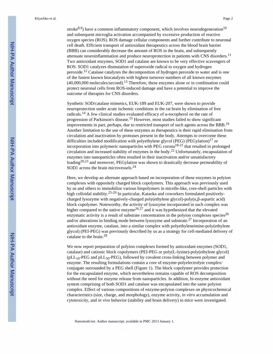
AbstractFormulations of antioxidant enzymes, superoxide dismutase 1 (SOD1, also known as Cu/Zn SOD)and catalase were prepared by electrostatic coupling of enzymes with cationic block copolymers,polyethyleneimine-poly(ethylene glycol) or poly(L-lysine)-poly(ethylene glycol), followed bycovalent cross-linking to stabilize nanoparticles. Different cross-linking strategies (usingglutaraldehyde, bis-(sulfosuccinimidyl)suberate sodium salt or 1-Ethyl-3-[3-dimethylaminopropyl]carbodiimide hydrochloride with N-hydroxysulfosuccinimide) and reactionconditions (pH and polycation/protein charge ratio) were investigated that allowed immobilizingactive enzymes in cross-linked nanoparticles, termed “nanozymes”. Bi-enzyme nanoparticles,containing both SOD1 and catalase were also formulated. Formation of complexes was confirmedusing denaturing gel electrophoresis and western blotting and physicochemical characterizationwas conducted using dynamic light scattering and atomic force microscopy. In vivo studiesof 125I-labeled SOD1-containing nanozymes in mice demonstrated its increased stability in bothblood and brain and increased accumulation in brain tissues, compared to non-cross-linkedcomplexes and native SOD1. Future studies will evaluate potential of these formulations fordelivery of antioxidant enzymes to central nervous system to attenuate oxidative stress associatedwith neurological diseases.
Keywordspolyion complexes; nanozymes; cross-linking; brain delivery
BackgroundMany central nervous system (CNS) diseases (i.e., HIV-associated dementia,1-3 multiplesclerosis, cerebral palsy, schizophrenia,4 Parkinson's and Alzheimer's diseases,5-7 and
*Corresponding author, Address for correspondence: Center for Drug Delivery and Nanomedicine, College of Pharmacy University ofNebraska Medical Center, 985830 Nebraska Medical Center, Omaha, NE 68198-5830, USA. Tel. (402) 559-9364; Fax. (402)559-9365; [email protected].†Equal contributionNo competing interests are present.
NIH Public AccessAuthor ManuscriptNanomedicine. Author manuscript; available in PMC 2013 January 1.
Published in final edited form as:Nanomedicine. 2012 January ; 8(1): 119–129. doi:10.1016/j.nano.2011.05.010.
NIH
-PA Author Manuscript
NIH
-PA Author Manuscript
NIH
-PA Author Manuscript
stroke8,9) have a common inflammatory component, which involves neurodegeneration10
and subsequent microglia activation accompanied by excessive production of reactiveoxygen species (ROS). ROS damage cellular components and further contribute to neuronalcell death. Efficient transport of antioxidant therapeutics across the blood brain barrier(BBB) can considerably decrease the amount of ROS in the brain, and subsequentlyattenuate neuroinflammation and produce neuroprotection in patients with CNS disorders.11
Two antioxidant enzymes, SOD1 and catalase are known to be very effective scavengers ofROS. SOD1 catalyzes dismutation of superoxide radical to oxygen and hydrogenperoxide.12 Catalase catalyzes the decomposition of hydrogen peroxide to water and is oneof the fastest known biocatalysts with highest turnover numbers of all known enzymes(40,000,000 molecules/second).13 Therefore, these enzymes alone or in combination couldprotect neuronal cells from ROS-induced damage and have a potential to improve theoutcome of therapies for CNS disorders.
Synthetic SOD/catalase mimetics, EUK-189 and EUK-207, were shown to provideneuroprotection under acute ischemic conditions in the rat brain by elimination of freeradicals.14 A few clinical studies evaluated efficacy of α-tocopherol on the rate ofprogression of Parkinson's disease.15 However, most studies failed to show significantimprovements in part, perhaps, due to restricted transport of such agents across the BBB.16
Another limitation to the use of these enzymes as therapeutics is their rapid elimination fromcirculation and inactivation by proteases present in the body. Attempts to overcome thesedifficulties included modification with polyethylene glycol (PEG) (PEGylation)17 orincorporation into polymeric nanoparticles with PEG corona18-21 that resulted in prolongedcirculation and increased stability of enzymes in the body.22 Unfortunately, encapsulation ofenzymes into nanoparticles often resulted in their inactivation and/or unsatisfactoryloading20,23 and moreover, PEGylation was shown to drastically decrease permeability ofSOD1 across the brain microvessels.24
Here, we develop an alternate approach based on incorporation of these enzymes in polyioncomplexes with oppositely charged block copolymers. This approach was previously usedby us and others to immobilize various biopolymers in micelle-like, core-shell particles withhigh colloidal stability.25-29 In particular, Kataoka and coworkers formulated positively-charged lysozyme with negatively-charged poly(ethylene glycol)-poly(α,β-aspartic acid)block copolymer. Noteworthy, the activity of lysozyme incorporated in such complex washigher compared to the native enzyme26,27 and it was hypothesized that the elevatedenzymatic activity is a result of substrate concentration in the polyion complexes species26
and/or alterations in binding mode between lysozyme and substrate.27 Incorporation of anantioxidant enzyme, catalase, into a similar complex with polyethyleneimine-poly(ethyleneglycol) (PEI-PEG) was previously described by us as a strategy for cell-mediated delivery ofcatalase to the brain.28
We now report preparation of polyion complexes formed by antioxidant enzymes (SOD1,catalase) and cationic block copolymers (PEI-PEG or poly(L-lysine)-poly(ethylene glycol)(pLL10-PEG and pLL50-PEG), followed by covalent cross-linking between polymer andenzyme. The resulting formulations contain a core of enzyme-polyelectrolyte complex/conjugate surrounded by a PEG shell (Figure 1). The block copolymer provides protectionfor the encapsulated enzyme, which nevertheless remains capable of ROS decompositionwithout the need for enzyme release from nanoparticles. In addition, bi-enzyme antioxidantsystem comprising of both SOD1 and catalase was encapsulated into the same polyioncomplex. Effect of various compositions of enzyme-polyion complexes on physicochemicalcharacteristics (size, charge, and morphology), enzyme activity, in vitro accumulation andcytotoxicity, and in vivo behavior (stability and brain delivery) in mice were investigated.
Klyachko et al. Page 2
Nanomedicine. Author manuscript; available in PMC 2013 January 1.
NIH
-PA Author Manuscript
NIH
-PA Author Manuscript
NIH
-PA Author Manuscript
Based on the results we posit that incorporation of antioxidant enzymes into nanozymes mayimprove transport of active enzymes to the brain.
MethodsMaterials
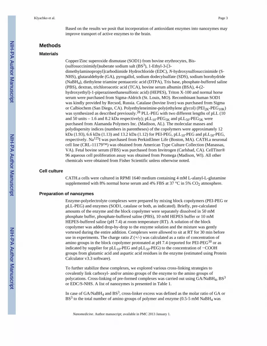
Copper/Zinc superoxide dismutase (SOD1) from bovine erythrocytes, Bis-(sulfosuccinimidyl)suberate sodium salt (BS3), 1-Ethyl-3-[3-dimethylaminopropyl]carbodiimide Hydrochloride (EDC), N-hydroxysulfosuccinimide (S-NHS), glutaraldehyde (GA), pyrogallol, sodium dodecylsulfate (SDS), sodium borohydride(NaBH4), diethylene triamine pentaacetic acid (DTPA), Tris base, phosphate-buffered saline(PBS), dextran, trichloroacetic acid (TCA), bovine serum albumin (BSA), 4-(2-hydroxyethyl)-1-piperazineethanesulfonic acid) (HEPES), Triton X-100 and normal horseserum were purchased from Sigma-Aldrich (St. Louis, MO). Recombinant human SOD1was kindly provided by Recsod, Russia. Catalase (bovine liver) was purchased from Sigmaor Calbiochem (San Diego, CA). Polyethyleneimine-poly(ethylene glycol) (PEI2K-PEG10K)was synthesized as described previously.28 PLL-PEG with two different lengths of pLL (10and 50 units – 1.6 and 8.2 kDa respectively); pLL10-PEG5K and pLL50-PEG5K werepurchased from Alamanda Polymers Inc. (Madison, AL). The molecular masses andpolydispersity indices (numbers in parentheses) of the copolymers were approximately 12kDa (1.93), 6.6 kDa (1.13) and 13.2 kDa (1.12) for PEI-PEG, pLL10-PEG and pLL50-PEG,respectively. Na125I was purchased from PerkinElmer Life (Boston, MA). CATH.a neuronalcell line (CRL-11179™) was obtained from American Type Culture Collection (Manassas,VA). Fetal bovine serum (FBS) was purchased from Invitrogen (Carlsbad, CA). CellTiter®96 aqueous cell proliferation assay was obtained from Promega (Madison, WI). All otherchemicals were obtained from Fisher Scientific unless otherwise noted.
Cell cultureCATH.a cells were cultured in RPMI 1640 medium containing 4 mM L-alanyl-L-glutaminesupplemented with 8% normal horse serum and 4% FBS at 37 °C in 5% CO2 atmosphere.
Preparation of nanozymesEnzyme-polyelectrolyte complexes were prepared by mixing block copolymers (PEI-PEG orpLL-PEG) and enzymes (SOD1, catalase or both, as indicated). Briefly, pre-calculatedamounts of the enzyme and the block copolymer were separately dissolved in 50 mMphosphate buffer, phosphate-buffered saline (PBS), 10 mM HEPES buffer or 10 mMHEPES-buffered saline (pH 7.4) at room temperature (RT). A solution of the blockcopolymer was added drop-by-drop to the enzyme solution and the mixture was gentlyvortexed during the entire addition. Complexes were allowed to sit at RT for 30 min beforeuse in experiments. The charge ratio Z (+/-) was calculated as a ratio of concentration ofamino groups in the block copolymer protonated at pH 7.4 (reported for PEI-PEG30 or asindicated by supplier for pLL10-PEG and pLL50-PEG) to the concentration of −COOHgroups from glutamic acid and aspartic acid residues in the enzyme (estimated using ProteinCalculator v3.3 software).
To further stabilize these complexes, we explored various cross-linking strategies tocovalently link carboxyl- and/or amino groups of the enzyme to the amino groups ofpolycations. Cross-linking of pre-formed complexes was carried out using GA/NaBH4, BS3
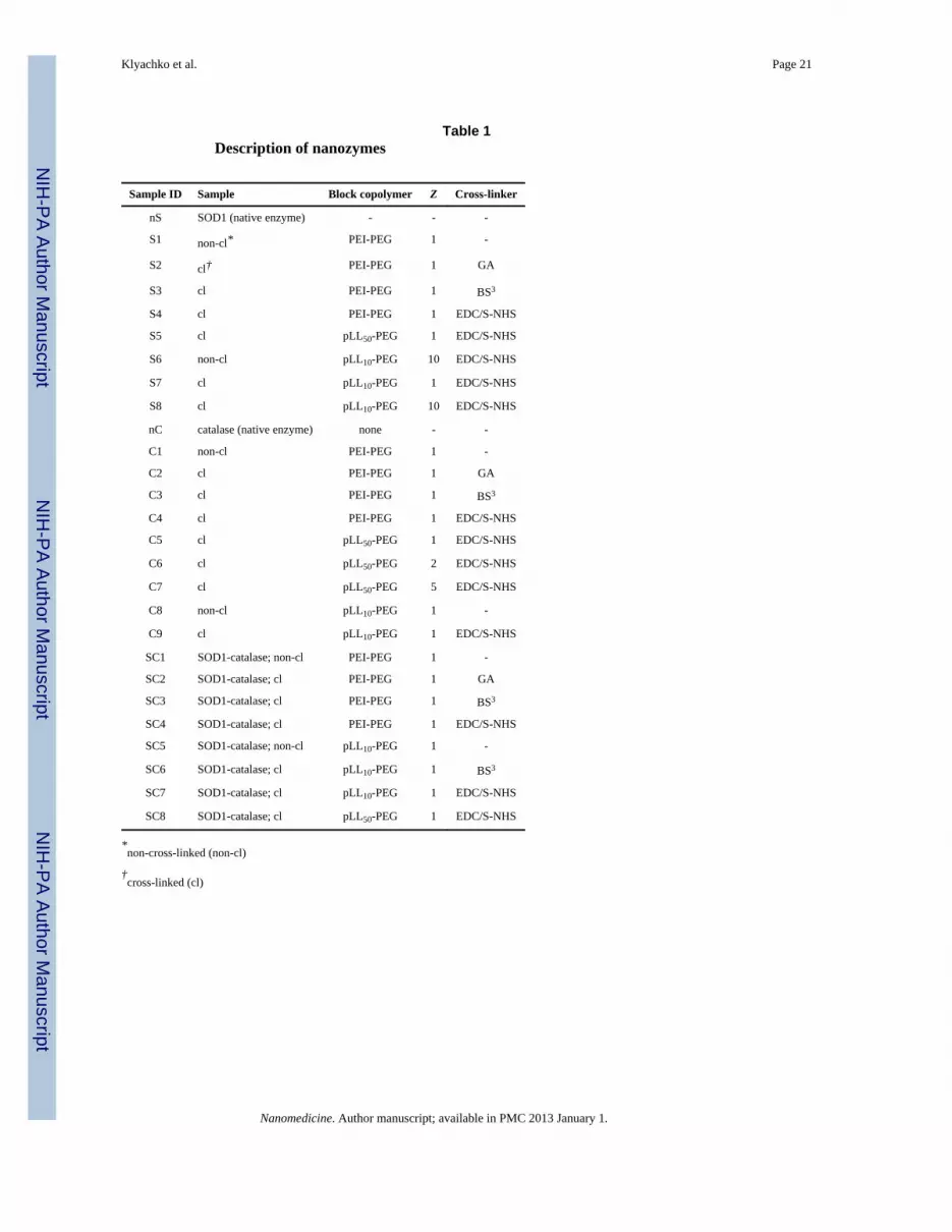
or EDC/S-NHS. A list of nanozymes is presented in Table 1.
In case of GA/NaBH4 and BS3, cross-linker excess was defined as the molar ratio of GA orBS3 to the total number of amino groups of polymer and enzyme (0.5-5 mM NaBH4 was
Klyachko et al. Page 3
Nanomedicine. Author manuscript; available in PMC 2013 January 1.
NIH
-PA Author Manuscript
NIH
-PA Author Manuscript
NIH
-PA Author Manuscript
added for 15-60 min after overnight incubation of the reaction mixture containing 2 to 100-fold GA excess). In case of EDC/S-NHS, it was defined as the molar ratio of EDC to thetotal number of carboxylic groups of the enzyme (5-10 mM S-NHS was added 5 min afterEDC addition). Cross-linker excesses were varied from 2 to 100-fold as indicated. All monoenzyme samples were prepared in PBS, pH 7.4. In case of “bi-enzyme” samples, pH of thereaction buffer was varied from 5.2 to 7.4. Reaction mixtures were incubated overnight at 4°C, and unreacted reagents were removed by desalting using NAP™-10 columns (GEHealthcare Bio-Sciences Corp. Piscataway, NJ). Wherever indicated, samples were filteredusing a 20 nm (SOD1 nanozymes) or 100 nm (catalase nanozymes) pore size filter prior touse in experiments.
Electrophoretic RetentionThe cross-linking of enzyme-polyion complexes was confirmed by polyacrylamide gel shiftassay. Samples were loaded on a 10% polyacrylamide gel under denaturing conditions.Protein bands were visualized with sheep polyclonal anti-SOD (Calbiochem, San Diego,CA) for SOD1, and rabbit polyclonal anti-catalase (Ab 1877, Abcam Inc, Cambridge, MA)for catalase, and secondary horseradish peroxidase anti-rabbit Ig antibody (Amersham LifeSciences, Cleveland, OH).
Enzyme ActivityEnzyme activity in samples was measured using pyrogallol autoxidation followed bysuperoxide radical dismutation (for SOD1) and hydrogen peroxide decomposition (forcatalase) methods, respectively (See SUPPLEMENTARY SECTION for complete details).
Dynamic Light Scattering (DLS)Effective hydrodynamic diameter and zeta-potential (ζ-potential) of nanozymes wasmeasured by photon correlation spectroscopy using Zetasizer Nano ZS (MalvernInstruments Ltd, UK) in a thermostatic cell at a scattering angle of 173° using previouslydescribed methods.31,32
Atomic Force Microscopy (AFM)Five μL of an aqueous dispersion (ca. 0.01 mg/mL) was deposited onto positively-chargedaminopropylytriethoxy silane (APS) mica surface33-35 for 2 min, washed with water anddried under argon atmosphere. AFM imaging was performed using a Multimode NanoScopeIV system (Veeco, Santa Barbara, CA) operated in tapping mode. Particle widths andheights were estimated using Femtoscan software (Advanced Technologies Center, Russia).
Preparation of 125I-labeled SOD1SOD1 was labeled with Na125I using IodoBEADS Iodination reagent (PierceBiotechnology, Rockford, IL). Briefly, Na125I (500 μCi) was pre-incubated withIodoBEADS for 5 min, and then added to pre-calculated amount of SOD1 solution. Thereaction mixture was incubated at RT for 15 min and purified on a NAP™-10 column. 125I-labeled-SOD1 nanozymes were prepared as described earlier.
Cellular AccumulationAccumulation of various nanozymes in bovine brain microvessel endothelial cells(BBMEC) was evaluated as described earlier.36 Briefly, confluent cell monolayers grown on24-well plates were pre-treated with assay buffer for 30 min at 37 °C and then incubatedwith 125I-labeled nanozymes for 1 h. Cells were then washed with ice-cold PBS, andsolubilized in 1% Triton X-100 for subsequent determination of radioactivity counts (countsper minute; CPM) in a gamma counter. The cellular accumulation was normalized to
Klyachko et al. Page 4
Nanomedicine. Author manuscript; available in PMC 2013 January 1.
NIH
-PA Author Manuscript
NIH
-PA Author Manuscript
NIH
-PA Author Manuscript
cellular protein content determined by Pierce® BCA Protein Assay. Results are reported asaverage ± standard error mean (SEM) of quadruplicate samples.
Cytotoxicity EvaluationCytotoxicity of selected nanozymes was determined using the CellTiter® 96 Aqueous CellProliferation Assay (MTS). Twenty-thousand cells seeded in a 96-well plate 24 h before theexperiment was incubated with increasing concentration of the respective samples in 100 μLof complete culture medium. The medium was removed after 24 h and replaced with amixture of 100 μL of fresh RPMI and 20 μL of MTS reagent solution. The cells wereincubated for 1 h at 37 °C in a CO2 incubator. The absorbance of each sample was thenmeasured at 490 nm to determine cell viability. The results are expressed as the meanpercentage cell viability relative to untreated cells ± SEM.
AnimalsMale Balb/c mice (Charles River Laboratories, USA) 6-8 weeks old were used in the study.All procedures were approved by the Institutional Animal Care and Use Committee of theUniversity of Nebraska Medical Center. Animals were housed and humanely handled inaccordance with the Principles of Animal Care outlined by National Institutes of Health.
In Vivo StudiesMale Balb/c mice (8 weeks old) were anesthetized with a cocktail of ketamine (80 mg/kg)and xylazine (5 mg/kg) administered intraperitonially. Mice were injected with eithernative 125I-SOD1, non-cross-linked 125I-SOD1/pLL10-PEG nanozyme or cross-linked 125I-SOD1/pLL10-PEG. The nanozymes were prepared at Z = 10 and the cross-linked using 5-fold EDC excess (S-NHS: 5 mM) as described above. Nanozymes in saline wereadministered intravenously (i.v.) via tail vein in a 100 μL volume (1 mg SOD1/mL). Onehour after injection, the abdomen and rib cage were opened and blood was collected fromthe heart. Twenty mL of lactated ringer's solution (LR) (B Braun Medical Inc., Irvine, CA)was then perfused through the left ventricle of the heart. Whole brains were dissected andweighed and the levels of radioactivity were counted in a gamma counter.
TCA PrecipitationAcid precipitation method was used to estimate stability of SOD1 by following 125I labeldegradation.37 Brain and serum samples were placed in an ice-cold glass homogenizer. Thebrain was homogenized by 10 vertical strokes in 1% w/v BSA acidified LR (pH 4.0).Homogenates were centrifuged at 4000 ×g for 25 min at 4 °C. The resulting supernatant(soluble fraction) and blood serum samples were precipitated with 30% v/v of TCA andcentrifuged at 4000×g for 35 min. Radioactive counts (CPM) of resulting supernatant andprecipitate (pellet) correspond to free 125I label and enzyme-bound 125I, respectively.Stability of nanozymes was assessed as a ratio of enzyme-bound 125I (pellet CPM) tooverall 125I radioactivity (supernatant CPM + pellet CPM). The results obtained in theseexperiments represent average quantities of SOD1 in each sample/fraction withoutdiscrimination of its interstitial or intercellular localization in the organs.
Capillary DepletionMale Balb/c mice were anesthetized as described earlier. Animals were treated and brainand serum samples were collected. The brain was homogenized by 10 vertical strokes in 0.8mL of physiological buffer (10 mM HEPES, 141 mM NaCl, 4 mM KCl, 2.8 mM CaCl2, 1mM MgSO4, 1 mM NaH2PO4, and 10 mM D-Glucose; pH 7.4). Dextran solution (1.6 mL ofa 26% solution) was added to the homogenate, mixed, and homogenized with 3 additionalvertical strokes. The homogenate was centrifuged at 4000×g for 25 min at 4 °C. The
Klyachko et al. Page 5
Nanomedicine. Author manuscript; available in PMC 2013 January 1.
NIH
-PA Author Manuscript
NIH
-PA Author Manuscript
NIH
-PA Author Manuscript
resulting supernatant (brain parenchymal fraction) and pellet (capillary fraction) wereseparated. The levels of radioactivity in the capillary and brain parenchymal fractions werecounted in a gamma counter. The results are expressed as a compartment (parenchyma orcapillaries)/serum ratio. Initial radioactivity values obtained in this experiment correspond tooverall accumulation of complexes in the brain compartment that includes both 125I-labeledcomplexes as well as free label that dissociated from the nanoparticles. In order to quantifythe presence of intact complexes that retain their structure in the brain compartments, initialradioactivity values obtained in capillary depletion experiment were multiplied by a ratio ofenzyme-bound 125I (pellet CPM) to overall 125I radioactivity (supernatant CPM + pelletCPM) obtained in the acid precipitation experiment.
Statistical AnalysisAll data are presented as the average ± SEM (n=4 or 5). Tests for significant differencesbetween the groups were done using one-way ANOVA with multiple comparisons (Kruskal-Wallis) using GraphPad Prism 4.0 (GraphPad software, San Diego, CA). A minimum pvalue of 0.05 was set as the significance level in all cases.
ResultsPreparation of cross-linked nanozymes
The polyion complexes were prepared by simple mixing of aqueous solutions ofcorresponding enzymes (SOD1 or catalase, or both) and block copolymers (PEI-PEG orpLL-PEG). Both SOD1 and catalase are negatively charged under physiological conditions(pI values are 4.95 and 5.8 for SOD1 and catalase, respectively). Unless stated otherwise,polyion complexes were prepared at pH 7.4 that favored electrostatic coupling of theenzyme and the block copolymers. Cross-linking of polyion complexes using GA/NaBH4,BS3, or EDC/S-NHS were done as described earlier.
Electrophoretic retention—The formation of cross-linked polyion complexes wasconfirmed by denaturing gel electrophoresis. Both enzymes are oligomeric proteins anddissociated under denaturing conditions into individual subunits of 16 kDa (SOD1) and 60kDa (catalase) (Figure 2).
The migration pattern was different in case of cross-linked complexes. Usually, there wasdisappearance or retardation of the protein band, presumably due to inability of cross-linkedcomplexes to migrate through the gel (lanes S3, S4, S7, C2 and SC3). Few conjugates didmigrate through the gel, but their estimated molecular masses were greater than those of therespective oligomeric subunits (lanes S2, C3, C4, C9, SC4, SC7, SC6 and SC8). Completedisappearance of free subunits, suggesting nearly 100% conjugation was observed whenPEI-PEG and BS3 (7 to 10-fold excess over NH2-groups) were used with both enzymes(lanes S3, C3, SC3, SC6) or pLL10-PEG and EDC/S-NHS (20 to 25-fold excess over−COOH groups) was used with catalase (lane C9). In contrast, use of GA/NaBH4 (2 to 5-fold excess over NH2 groups) and EDC/S-NHS (as earlier) resulted in partial conjugationunder the experimental conditions (lanes S2, S4, S7, C2, C4, SC4, SC7 and SC8). In somecases, the extent of cross-linking could be improved by increasing the amount of the cross-linkers. For example, at 100-fold excess of GA the conjugation was nearly complete (notshown), however, the loss of enzyme activity (ca. 50%) prompted us to use lower excessesof GA (about 2 to 7-fold).
In order to obtain two enzymes in the same nanoparticle, we tried to enhance interactions ofthese enzymes with each other by rendering them oppositely charged using low pH of ∼5.2,which lies between pI values of 4.95 (SOD1) and 5.8 (catalase). However, this decreased the
Klyachko et al. Page 6
Nanomedicine. Author manuscript; available in PMC 2013 January 1.
NIH
-PA Author Manuscript
NIH
-PA Author Manuscript
NIH
-PA Author Manuscript
efficiency of cross-linking and led to a considerable loss of catalase activity. Therefore, inmost cases, we carried out the cross-linking at relatively high pH ≥ 6.8 where both enzymeswere negatively-charged. Formation of SOD1-catalase bi-enzyme nanoparticles wasconfirmed by visualizing both proteins within the bands of the same molecular mass usinganti-SOD1 and anti-catalase antibodies (lanes SC4, SC7, SC6 and SC8). However, in fewcases these antibodies stained bands of different molecular masses, suggesting that theenzymes segregated into particles of different type (lanes SC2 and SC3).
Characterization of nanozymesEnzyme activity—Catalytic activity of SOD1 and catalase in nanozymes was measured asdescribed earlier. Linear dependence of SOD1 and catalase catalytic activities on enzymeconcentrations (Figure S1) was observed in the concentration range used; this allowed us toanalyze changes in the activity of formulated enzymes using reaction rates that werenormalized to the respective enzyme concentrations. As a quantitative analysis of catalaseactivity, the dependence of the reaction rate on substrate concentration was studied andkinetic parameters, maximal reaction rate (kinetic constant; kcat) and apparent Michaelisconstant (Km) were obtained using double reciprocal coordinates (Figure S2). Apparentkinetic parameters (kcat and Km: 6.5×10⁵ s-1 and 63 mM, respectively) calculated forcatalase were found to be similar to those reported in published literature.38 For SOD1, thedirect measurement of the superoxide dismutation is not possible. Therefore, enzymeactivity was assayed indirectly by autoxidation of pyrogallol and was expressed as theamount of enzyme that resulted in 50% inhibition in rate of autoxidation. Two differentsubstrate systems were also used for quantitative analysis of SOD1 activity, riboflavin/NitroBlue Tetrazolium (Figure S3A) and luminol/xanthine/xanthine oxidase/oxygen systems(Figure S3B).
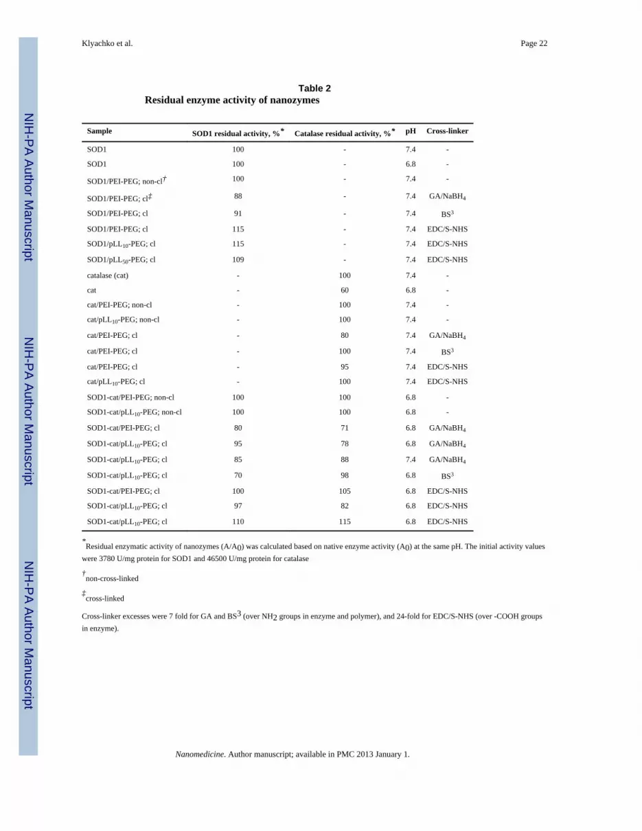
Before cross-linking, both enzymes retained 100% activity in the polyion complexes (Table2). Significantly high levels of activity (at least 70%) were retained after cross-linking inmost cases. Interestingly, the chemical structure of the block copolymer did not affect theactivity after cross-linking. However, the type of cross-linker and reaction conditionsshowed a significant effect. For example, enzymes cross-linked with GA/NaBH4 retained70-100% of activity (Table 2 and Figures S1B and S2) strongly depending not only on GAexcess but also reduction time with NaBH4 (lesser time correlated with lower activity loss).In contrast, conjugation of nanozymes with EDC/S-NHS resulted in mild activation of theenzyme (residual activity 105-115%). Furthermore, as already mentioned low pH ∼5.2resulted in irreversible inactivation of both enzymes (not shown).
Particle size—Hydrodynamic size (size) and surface charge (ζ-potential) of thenanozymes that showed considerable retention of enzyme activity was determined usingDLS (Table 3A). Particles of native SOD1 showed a small size (4.6 nm), which was close tothe theoretical diameter (4.5 nm) of a single protein globule estimated from molecular massof the enzyme (32 kDa). In contrast, DLS analysis of native catalase revealed bimodal sizedistribution with a smaller size of 10 nm corresponding to a single globule and a larger sizeof 63 nm, which most likely represented aggregates. All complexes (cross-linked or non-cross-linked) were nearly electroneutral or weakly-charged. Compared to the native enzymemolecules, both SOD1- and catalase-based complexes displayed an increase in size and inmost cases, their distribution was bimodal. In selected cases, we demonstrated that theparticle uniformity can be improved by filtering nanozymes through a 20/100 nm pore sizefilter. This procedure resulted in SOD1 and catalase complexes of unimodal size distribution(Table 3B). It should be noted that there was no loss of enzyme activity (normalized toprotein concentration) upon filtering.
Klyachko et al. Page 7
Nanomedicine. Author manuscript; available in PMC 2013 January 1.
NIH
-PA Author Manuscript
NIH
-PA Author Manuscript
NIH
-PA Author Manuscript
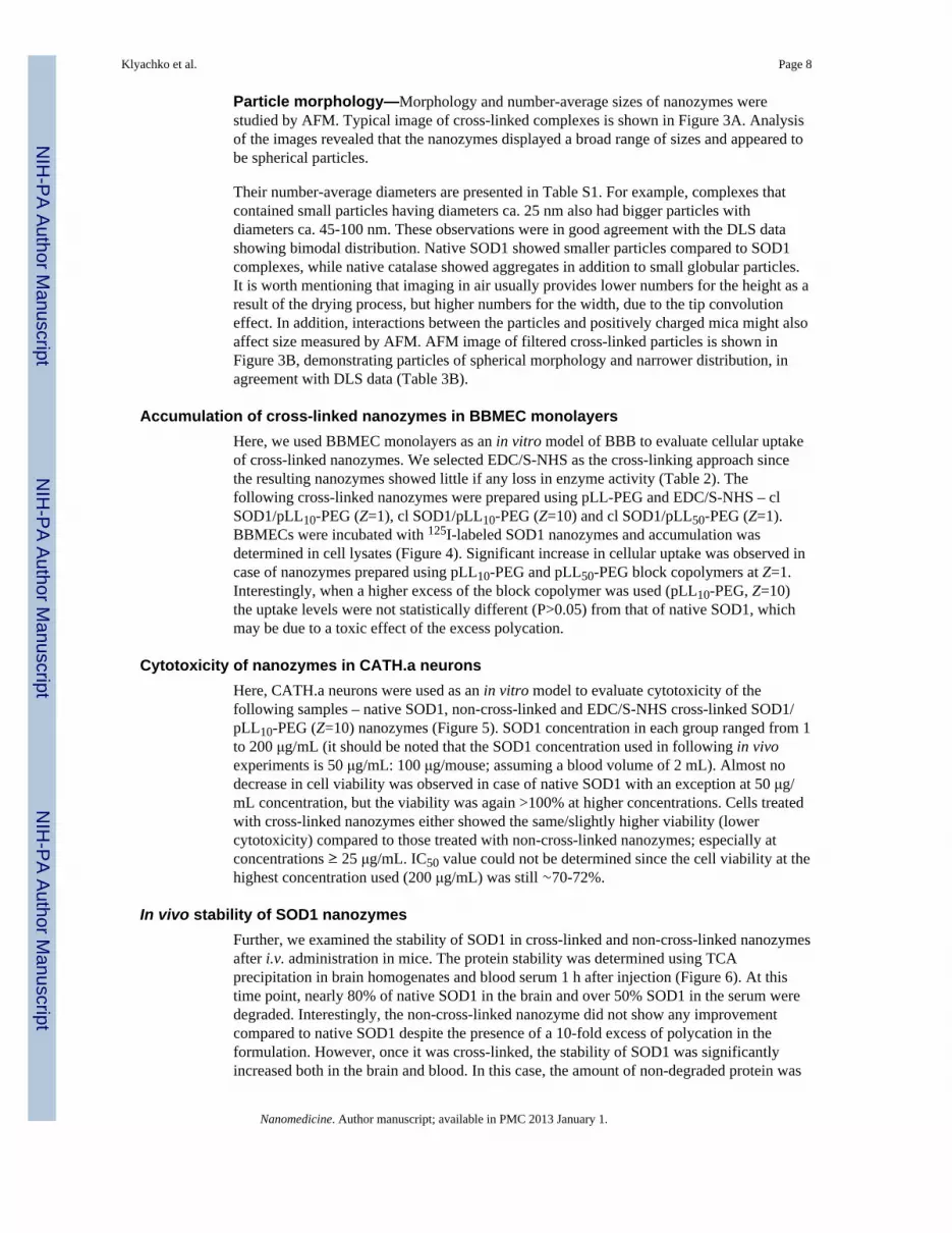
Particle morphology—Morphology and number-average sizes of nanozymes werestudied by AFM. Typical image of cross-linked complexes is shown in Figure 3A. Analysisof the images revealed that the nanozymes displayed a broad range of sizes and appeared tobe spherical particles.
Their number-average diameters are presented in Table S1. For example, complexes thatcontained small particles having diameters ca. 25 nm also had bigger particles withdiameters ca. 45-100 nm. These observations were in good agreement with the DLS datashowing bimodal distribution. Native SOD1 showed smaller particles compared to SOD1complexes, while native catalase showed aggregates in addition to small globular particles.It is worth mentioning that imaging in air usually provides lower numbers for the height as aresult of the drying process, but higher numbers for the width, due to the tip convolutioneffect. In addition, interactions between the particles and positively charged mica might alsoaffect size measured by AFM. AFM image of filtered cross-linked particles is shown inFigure 3B, demonstrating particles of spherical morphology and narrower distribution, inagreement with DLS data (Table 3B).
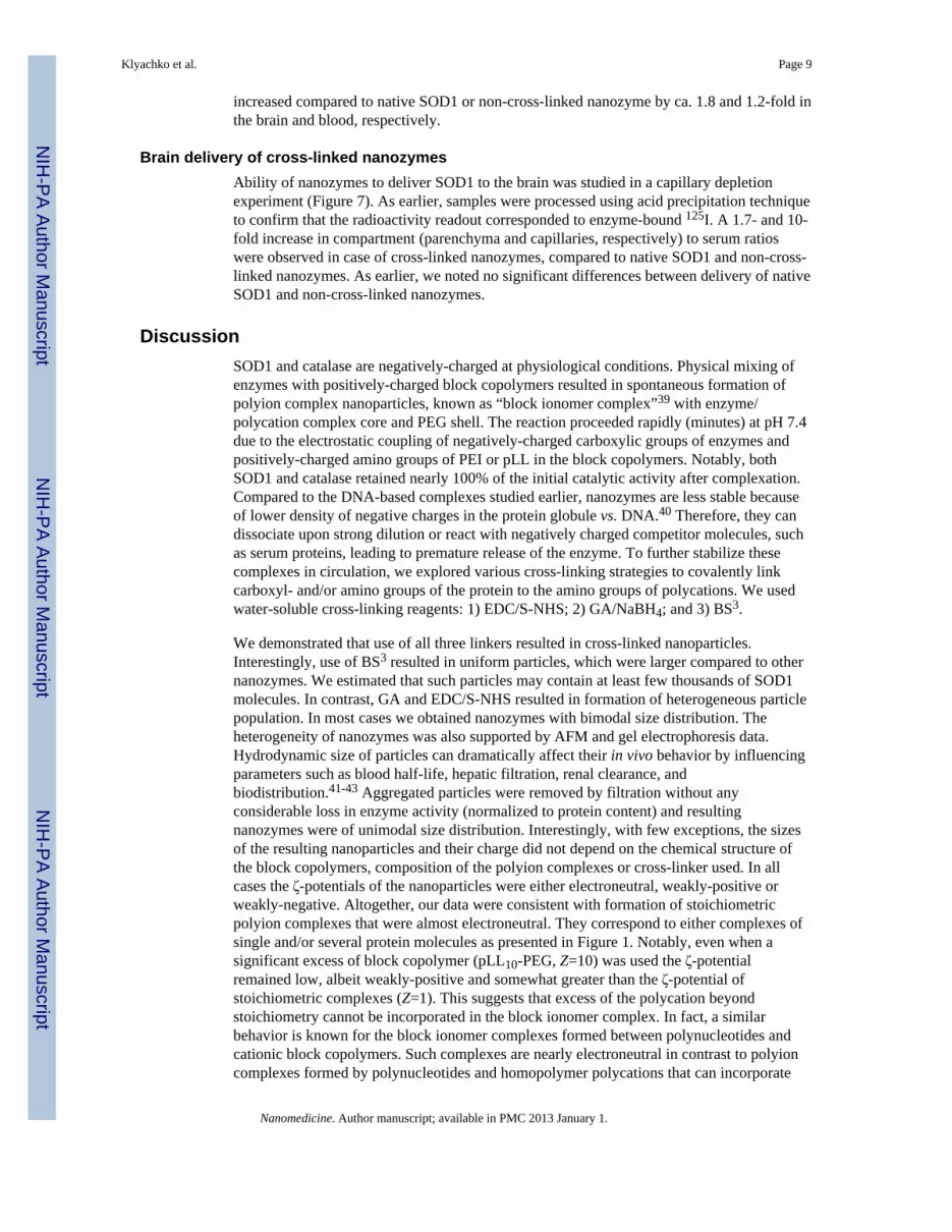
Accumulation of cross-linked nanozymes in BBMEC monolayersHere, we used BBMEC monolayers as an in vitro model of BBB to evaluate cellular uptakeof cross-linked nanozymes. We selected EDC/S-NHS as the cross-linking approach sincethe resulting nanozymes showed little if any loss in enzyme activity (Table 2). Thefollowing cross-linked nanozymes were prepared using pLL-PEG and EDC/S-NHS – clSOD1/pLL10-PEG (Z=1), cl SOD1/pLL10-PEG (Z=10) and cl SOD1/pLL50-PEG (Z=1).BBMECs were incubated with 125I-labeled SOD1 nanozymes and accumulation wasdetermined in cell lysates (Figure 4). Significant increase in cellular uptake was observed incase of nanozymes prepared using pLL10-PEG and pLL50-PEG block copolymers at Z=1.Interestingly, when a higher excess of the block copolymer was used (pLL10-PEG, Z=10)the uptake levels were not statistically different (P>0.05) from that of native SOD1, whichmay be due to a toxic effect of the excess polycation.
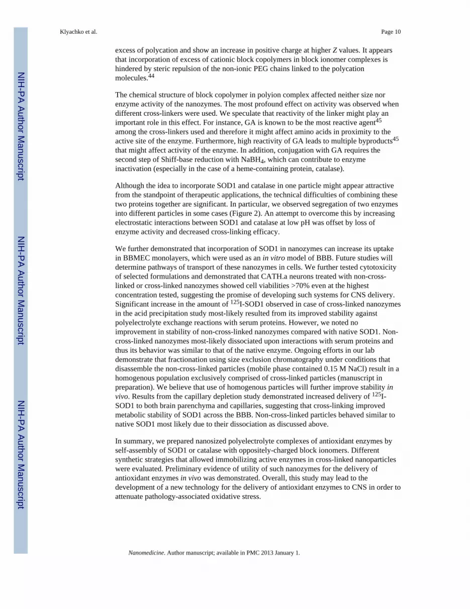
Cytotoxicity of nanozymes in CATH.a neuronsHere, CATH.a neurons were used as an in vitro model to evaluate cytotoxicity of thefollowing samples – native SOD1, non-cross-linked and EDC/S-NHS cross-linked SOD1/pLL10-PEG (Z=10) nanozymes (Figure 5). SOD1 concentration in each group ranged from 1to 200 μg/mL (it should be noted that the SOD1 concentration used in following in vivoexperiments is 50 μg/mL: 100 μg/mouse; assuming a blood volume of 2 mL). Almost nodecrease in cell viability was observed in case of native SOD1 with an exception at 50 μg/mL concentration, but the viability was again >100% at higher concentrations. Cells treatedwith cross-linked nanozymes either showed the same/slightly higher viability (lowercytotoxicity) compared to those treated with non-cross-linked nanozymes; especially atconcentrations ≥ 25 μg/mL. IC50 value could not be determined since the cell viability at thehighest concentration used (200 μg/mL) was still ∼70-72%.
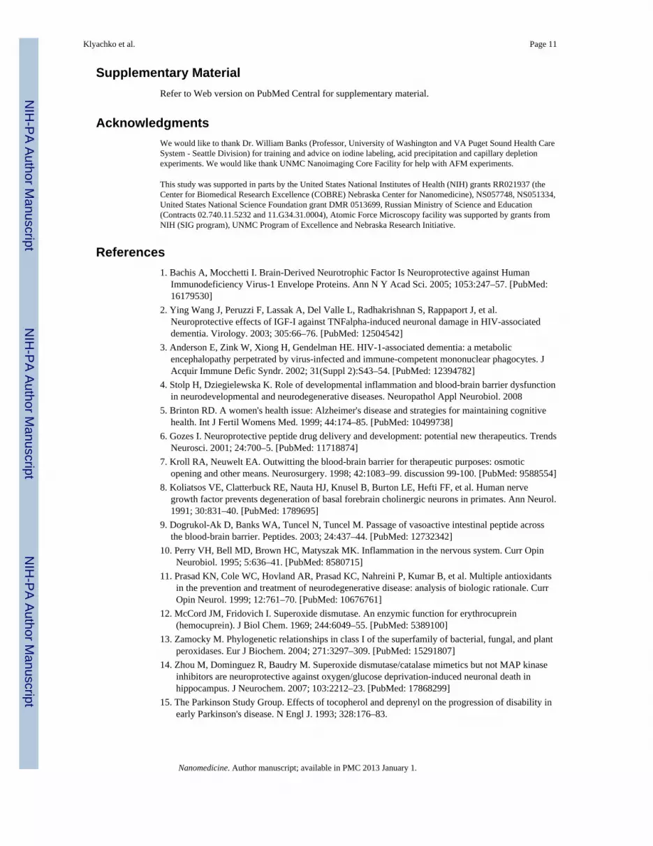
In vivo stability of SOD1 nanozymesFurther, we examined the stability of SOD1 in cross-linked and non-cross-linked nanozymesafter i.v. administration in mice. The protein stability was determined using TCAprecipitation in brain homogenates and blood serum 1 h after injection (Figure 6). At thistime point, nearly 80% of native SOD1 in the brain and over 50% SOD1 in the serum weredegraded. Interestingly, the non-cross-linked nanozyme did not show any improvementcompared to native SOD1 despite the presence of a 10-fold excess of polycation in theformulation. However, once it was cross-linked, the stability of SOD1 was significantlyincreased both in the brain and blood. In this case, the amount of non-degraded protein was
Klyachko et al. Page 8
Nanomedicine. Author manuscript; available in PMC 2013 January 1.
NIH
-PA Author Manuscript
NIH
-PA Author Manuscript
NIH
-PA Author Manuscript
increased compared to native SOD1 or non-cross-linked nanozyme by ca. 1.8 and 1.2-fold inthe brain and blood, respectively.
Brain delivery of cross-linked nanozymesAbility of nanozymes to deliver SOD1 to the brain was studied in a capillary depletionexperiment (Figure 7). As earlier, samples were processed using acid precipitation techniqueto confirm that the radioactivity readout corresponded to enzyme-bound 125I. A 1.7- and 10-fold increase in compartment (parenchyma and capillaries, respectively) to serum ratioswere observed in case of cross-linked nanozymes, compared to native SOD1 and non-cross-linked nanozymes. As earlier, we noted no significant differences between delivery of nativeSOD1 and non-cross-linked nanozymes.
DiscussionSOD1 and catalase are negatively-charged at physiological conditions. Physical mixing ofenzymes with positively-charged block copolymers resulted in spontaneous formation ofpolyion complex nanoparticles, known as “block ionomer complex”39 with enzyme/polycation complex core and PEG shell. The reaction proceeded rapidly (minutes) at pH 7.4due to the electrostatic coupling of negatively-charged carboxylic groups of enzymes andpositively-charged amino groups of PEI or pLL in the block copolymers. Notably, bothSOD1 and catalase retained nearly 100% of the initial catalytic activity after complexation.Compared to the DNA-based complexes studied earlier, nanozymes are less stable becauseof lower density of negative charges in the protein globule vs. DNA.40 Therefore, they candissociate upon strong dilution or react with negatively charged competitor molecules, suchas serum proteins, leading to premature release of the enzyme. To further stabilize thesecomplexes in circulation, we explored various cross-linking strategies to covalently linkcarboxyl- and/or amino groups of the protein to the amino groups of polycations. We usedwater-soluble cross-linking reagents: 1) EDC/S-NHS; 2) GA/NaBH4; and 3) BS3.
We demonstrated that use of all three linkers resulted in cross-linked nanoparticles.Interestingly, use of BS3 resulted in uniform particles, which were larger compared to othernanozymes. We estimated that such particles may contain at least few thousands of SOD1molecules. In contrast, GA and EDC/S-NHS resulted in formation of heterogeneous particlepopulation. In most cases we obtained nanozymes with bimodal size distribution. Theheterogeneity of nanozymes was also supported by AFM and gel electrophoresis data.Hydrodynamic size of particles can dramatically affect their in vivo behavior by influencingparameters such as blood half-life, hepatic filtration, renal clearance, andbiodistribution.41-43 Aggregated particles were removed by filtration without anyconsiderable loss in enzyme activity (normalized to protein content) and resultingnanozymes were of unimodal size distribution. Interestingly, with few exceptions, the sizesof the resulting nanoparticles and their charge did not depend on the chemical structure ofthe block copolymers, composition of the polyion complexes or cross-linker used. In allcases the ζ-potentials of the nanoparticles were either electroneutral, weakly-positive orweakly-negative. Altogether, our data were consistent with formation of stoichiometricpolyion complexes that were almost electroneutral. They correspond to either complexes ofsingle and/or several protein molecules as presented in Figure 1. Notably, even when asignificant excess of block copolymer (pLL10-PEG, Z=10) was used the ζ-potentialremained low, albeit weakly-positive and somewhat greater than the ζ-potential ofstoichiometric complexes (Z=1). This suggests that excess of the polycation beyondstoichiometry cannot be incorporated in the block ionomer complex. In fact, a similarbehavior is known for the block ionomer complexes formed between polynucleotides andcationic block copolymers. Such complexes are nearly electroneutral in contrast to polyioncomplexes formed by polynucleotides and homopolymer polycations that can incorporate
Klyachko et al. Page 9
Nanomedicine. Author manuscript; available in PMC 2013 January 1.
NIH
-PA Author Manuscript
NIH
-PA Author Manuscript
NIH
-PA Author Manuscript
excess of polycation and show an increase in positive charge at higher Z values. It appearsthat incorporation of excess of cationic block copolymers in block ionomer complexes ishindered by steric repulsion of the non-ionic PEG chains linked to the polycationmolecules.44
The chemical structure of block copolymer in polyion complex affected neither size norenzyme activity of the nanozymes. The most profound effect on activity was observed whendifferent cross-linkers were used. We speculate that reactivity of the linker might play animportant role in this effect. For instance, GA is known to be the most reactive agent45
among the cross-linkers used and therefore it might affect amino acids in proximity to theactive site of the enzyme. Furthermore, high reactivity of GA leads to multiple byproducts45
that might affect activity of the enzyme. In addition, conjugation with GA requires thesecond step of Shiff-base reduction with NaBH4, which can contribute to enzymeinactivation (especially in the case of a heme-containing protein, catalase).
Although the idea to incorporate SOD1 and catalase in one particle might appear attractivefrom the standpoint of therapeutic applications, the technical difficulties of combining thesetwo proteins together are significant. In particular, we observed segregation of two enzymesinto different particles in some cases (Figure 2). An attempt to overcome this by increasingelectrostatic interactions between SOD1 and catalase at low pH was offset by loss ofenzyme activity and decreased cross-linking efficacy.
We further demonstrated that incorporation of SOD1 in nanozymes can increase its uptakein BBMEC monolayers, which were used as an in vitro model of BBB. Future studies willdetermine pathways of transport of these nanozymes in cells. We further tested cytotoxicityof selected formulations and demonstrated that CATH.a neurons treated with non-cross-linked or cross-linked nanozymes showed cell viabilities >70% even at the highestconcentration tested, suggesting the promise of developing such systems for CNS delivery.Significant increase in the amount of 125I-SOD1 observed in case of cross-linked nanozymesin the acid precipitation study most-likely resulted from its improved stability againstpolyelectrolyte exchange reactions with serum proteins. However, we noted noimprovement in stability of non-cross-linked nanozymes compared with native SOD1. Non-cross-linked nanozymes most-likely dissociated upon interactions with serum proteins andthus its behavior was similar to that of the native enzyme. Ongoing efforts in our labdemonstrate that fractionation using size exclusion chromatography under conditions thatdisassemble the non-cross-linked particles (mobile phase contained 0.15 M NaCl) result in ahomogenous population exclusively comprised of cross-linked particles (manuscript inpreparation). We believe that use of homogenous particles will further improve stability invivo. Results from the capillary depletion study demonstrated increased delivery of 125I-SOD1 to both brain parenchyma and capillaries, suggesting that cross-linking improvedmetabolic stability of SOD1 across the BBB. Non-cross-linked particles behaved similar tonative SOD1 most likely due to their dissociation as discussed above.
In summary, we prepared nanosized polyelectrolyte complexes of antioxidant enzymes byself-assembly of SOD1 or catalase with oppositely-charged block ionomers. Differentsynthetic strategies that allowed immobilizing active enzymes in cross-linked nanoparticleswere evaluated. Preliminary evidence of utility of such nanozymes for the delivery ofantioxidant enzymes in vivo was demonstrated. Overall, this study may lead to thedevelopment of a new technology for the delivery of antioxidant enzymes to CNS in order toattenuate pathology-associated oxidative stress.
Klyachko et al. Page 10
Nanomedicine. Author manuscript; available in PMC 2013 January 1.
NIH
-PA Author Manuscript
NIH
-PA Author Manuscript
NIH
-PA Author Manuscript
Supplementary MaterialRefer to Web version on PubMed Central for supplementary material.
AcknowledgmentsWe would like to thank Dr. William Banks (Professor, University of Washington and VA Puget Sound Health CareSystem - Seattle Division) for training and advice on iodine labeling, acid precipitation and capillary depletionexperiments. We would like thank UNMC Nanoimaging Core Facility for help with AFM experiments.
This study was supported in parts by the United States National Institutes of Health (NIH) grants RR021937 (theCenter for Biomedical Research Excellence (COBRE) Nebraska Center for Nanomedicine), NS057748, NS051334,United States National Science Foundation grant DMR 0513699, Russian Ministry of Science and Education(Contracts 02.740.11.5232 and 11.G34.31.0004), Atomic Force Microscopy facility was supported by grants fromNIH (SIG program), UNMC Program of Excellence and Nebraska Research Initiative.
References1. Bachis A, Mocchetti I. Brain-Derived Neurotrophic Factor Is Neuroprotective against Human
Immunodeficiency Virus-1 Envelope Proteins. Ann N Y Acad Sci. 2005; 1053:247–57. [PubMed:16179530]
2. Ying Wang J, Peruzzi F, Lassak A, Del Valle L, Radhakrishnan S, Rappaport J, et al.Neuroprotective effects of IGF-I against TNFalpha-induced neuronal damage in HIV-associateddementia. Virology. 2003; 305:66–76. [PubMed: 12504542]
3. Anderson E, Zink W, Xiong H, Gendelman HE. HIV-1-associated dementia: a metabolicencephalopathy perpetrated by virus-infected and immune-competent mononuclear phagocytes. JAcquir Immune Defic Syndr. 2002; 31(Suppl 2):S43–54. [PubMed: 12394782]
4. Stolp H, Dziegielewska K. Role of developmental inflammation and blood-brain barrier dysfunctionin neurodevelopmental and neurodegenerative diseases. Neuropathol Appl Neurobiol. 2008
5. Brinton RD. A women's health issue: Alzheimer's disease and strategies for maintaining cognitivehealth. Int J Fertil Womens Med. 1999; 44:174–85. [PubMed: 10499738]
6. Gozes I. Neuroprotective peptide drug delivery and development: potential new therapeutics. TrendsNeurosci. 2001; 24:700–5. [PubMed: 11718874]
7. Kroll RA, Neuwelt EA. Outwitting the blood-brain barrier for therapeutic purposes: osmoticopening and other means. Neurosurgery. 1998; 42:1083–99. discussion 99-100. [PubMed: 9588554]
8. Koliatsos VE, Clatterbuck RE, Nauta HJ, Knusel B, Burton LE, Hefti FF, et al. Human nervegrowth factor prevents degeneration of basal forebrain cholinergic neurons in primates. Ann Neurol.1991; 30:831–40. [PubMed: 1789695]
9. Dogrukol-Ak D, Banks WA, Tuncel N, Tuncel M. Passage of vasoactive intestinal peptide acrossthe blood-brain barrier. Peptides. 2003; 24:437–44. [PubMed: 12732342]
10. Perry VH, Bell MD, Brown HC, Matyszak MK. Inflammation in the nervous system. Curr OpinNeurobiol. 1995; 5:636–41. [PubMed: 8580715]
11. Prasad KN, Cole WC, Hovland AR, Prasad KC, Nahreini P, Kumar B, et al. Multiple antioxidantsin the prevention and treatment of neurodegenerative disease: analysis of biologic rationale. CurrOpin Neurol. 1999; 12:761–70. [PubMed: 10676761]
12. McCord JM, Fridovich I. Superoxide dismutase. An enzymic function for erythrocuprein(hemocuprein). J Biol Chem. 1969; 244:6049–55. [PubMed: 5389100]
13. Zamocky M. Phylogenetic relationships in class I of the superfamily of bacterial, fungal, and plantperoxidases. Eur J Biochem. 2004; 271:3297–309. [PubMed: 15291807]
14. Zhou M, Dominguez R, Baudry M. Superoxide dismutase/catalase mimetics but not MAP kinaseinhibitors are neuroprotective against oxygen/glucose deprivation-induced neuronal death inhippocampus. J Neurochem. 2007; 103:2212–23. [PubMed: 17868299]
15. The Parkinson Study Group. Effects of tocopherol and deprenyl on the progression of disability inearly Parkinson's disease. N Engl J. 1993; 328:176–83.
Klyachko et al. Page 11
Nanomedicine. Author manuscript; available in PMC 2013 January 1.
NIH
-PA Author Manuscript
NIH
-PA Author Manuscript
NIH
-PA Author Manuscript
16. Pappert EJ, Tangney CC, Goetz CG, Ling ZD, Lipton JW, Stebbins GT, et al. Alpha-tocopherol inthe ventricular cerebrospinal fluid of Parkinson's disease patients: dose-response study andcorrelations with plasma levels. Neurology. 1996; 47:1037–42. [PubMed: 8857741]
17. Moghimi SM, Szebeni J. Stealth liposomes and long circulating nanoparticles: critical issues inpharmacokinetics, opsonization and protein-binding properties. Prog Lipid Res. 2003; 42:463–78.[PubMed: 14559067]
18. Prokop A, Kozlov E, Newman GW, Newman MJ. Water-based nanoparticulate polymeric systemfor protein delivery: permeability control and vaccine application. Biotechnol Bioeng. 2002;78:459–66. [PubMed: 11948453]
19. Klyachko NL, Levashov AV. Bioorganic synthesis in reverse micelles and related systems. CurrOpin Colloid Int Sci. 2003; 8:179–86.
20. Dziubla TD, Karim A, Muzykantov VR. Polymer nanocarriers protecting active enzyme cargoagainst proteolysis. J Control Release. 2005; 102:427–39. [PubMed: 15653162]
21. Simone EA, Dziubla TD, Colon-Gonzalez F, Discher DE, Muzykantov VR. Effect of polymeramphiphilicity on loading of a therapeutic enzyme into protective filamentous and sphericalpolymer nanocarriers. Biomacromolecules. 2007; 8:3914–21. [PubMed: 18038999]
22. Simone EA, Dziubla TD, Arguiri E, Vardon V, Shuvaev VV, Christofidou-Solomidou M, et al.Loading PEG-catalase into filamentous and spherical polymer nanocarriers. Pharm Res. 2009;26:250–60. [PubMed: 18956141]
23. Langer R. Drug delivery and targeting. Nature. 1998; 392:5–10. [PubMed: 9579855]24. Yoshida K, Burton GF, McKinney JS, Young H, Ellis EF. Brain and tissue distribution of
polyethylene glycol-conjugated superoxide dismutase in rats. Stroke. 1992; 23:865–9. [PubMed:1595107]
25. Kabanov A, Vinogradov S, Suzdaltseva Y, Alakhov V. Water-soluble block polycations as carriersfor olygonucleotide delivery Bioconjugate Chemistry. 1995; 6:639–43.
26. Harada A, Kataoka K. Pronounced activity of enzymes through the incorporation into the core ofpolyion complex micelles made from charged block copolymers. J Control Release. 2001; 72:85–91. [PubMed: 11389987]
27. Harada A, Kataoka K. Switching by pulse electric field of the elevated enzymatic reaction in thecore of polyion complex micelles. J Am Chem Soc. 2003; 125:15306–7. [PubMed: 14664571]
28. Batrakova EV, Li S, Reynolds AD, Mosley RL, Bronich TK, Kabanov AV, et al. A macrophage-nanozyme delivery system for Parkinson's disease. Bioconjug Chem. 2007; 18:1498–506.[PubMed: 17760417]
29. Kawamura A, Harada A, Kono K, Kataoka K. Self-assembled nano-bioreactor from blockionomers with elevated and stabilized enzymatic function. Bioconjug Chem. 2007; 18:1555–9.[PubMed: 17696317]
30. Vinogradov S, Bronich T, Kabanov A. Self-assembly of polyamine-poly(ethylene glycol)copolymers with phosphorothioate oligonucleotides. Bioconjugate Chemistry. 1998; 9:805–12.[PubMed: 9815175]
31. Bronich T, Nguyen H, Eisenberg A, Kabanov A. Recognition of DNA topology in reactionsbetween plasmid DNA and cationic copolymers. Journal of American Chemical Society. 2000;122:8339–43.
32. Vinogradov S, Batrakova E, Kabanov A. Poly(ethylene glycol)-polyethyleneimine NanoGel (TM)particles: novel drug delivery systems for antisense oligonucleotides Colloids and Surfaces B-Biointerfaces. 1999; 16:291–304.
33. Lyubchenko YL, Shlyakhtenko LS. AFM for analysis of structure and dynamics of DNA andprotein-DNA complexes. Methods. 2009; 47:206–13. [PubMed: 18835446]
34. Lyubchenko YL, Shlyakhtenko LS, Gall AA. Atomic force microscopy imaging and probing ofDNA, proteins, and protein DNA complexes: silatrane surface chemistry. Methods Mol Biol.2009; 543:337–51. [PubMed: 19378175]
35. Shlyakhtenko LS, Gall AA, Filonov A, Cerovac Z, Lushnikov A, Lyubchenko YL. Silatrane-basedsurface chemistry for immobilization of DNA, protein-DNA complexes and other biologicalmaterials. Ultramicroscopy. 2003; 97:279–87. [PubMed: 12801681]
Klyachko et al. Page 12
Nanomedicine. Author manuscript; available in PMC 2013 January 1.
NIH
-PA Author Manuscript
NIH
-PA Author Manuscript
NIH
-PA Author Manuscript
36. Batrakova E, Miller D, Li S, Alakhov V, Kabanov A, Elmquist W. Pluronic P85 enhances thedelivery of digoxin to the brain: in vitro and in vivo studies. J Pharmacol Exp Ther. 2001;296:551–57. [PubMed: 11160643]
37. Price TO, Samson WK, Niehoff ML, Banks WA. Permeability of the blood-brain barrier to a novelsatiety molecule nesfatin-1. Peptides. 2007; 28:2372–81. [PubMed: 18006117]
38. Jurgen-Lohmann DL, Legge RL. Immobilization of bovine catalase in sol-gels. Enzyme MicrobTech. 2006; 39:626–33.
39. Kabanov A, Bronich T, Kabanov V, Yu K, Eisenberg A. Soluble stoichiometric complexes frompoly(N-ethyl-4-vinylpyridinium) cations and poly(ethylene oxide)-block-poly(methacrylate)anions. Macromolecules. 1996; 29:6797–802.
40. Vinogradov SV, Bronich TK, Kabanov AV. Self-assembly of polyamine-poly(ethylene glycol)copolymers with phosphorothioate oligonucleotides. Bioconjug Chem. 1998; 9:805–12. [PubMed:9815175]
41. Alexis F, Pridgen E, Molnar LK, Farokhzad OC. Factors affecting the clearance andbiodistribution of polymeric nanoparticles. Mol Pharm. 2008; 5:505–15. [PubMed: 18672949]
42. Choi HS, Ipe BI, Misra P, Lee JH, Bawendi MG, Frangioni JV. Tissue- and Organ-SelectiveBiodistribution of NIR Fluorescent Quantum Dots. Nano Lett. 2009; 9:2354–9. [PubMed:19422261]
43. Choi HS, Liu W, Misra P, Tanaka E, Zimmer JP, Itty Ipe B, et al. Renal clearance of quantum dots.Nat Biotechnol. 2007; 25:1165–70. [PubMed: 17891134]
44. Seymour, LW.; Kataoka, K.; Kabanov, AV. Cationic block copolymers as self-assembling vectorsfor gene delivery. John Wiley, Chichester; New York, Weinheim, Brisbane, Singapore, Toronto:1998.
45. Hermanson, GT. Bioconjugate techniques. Acad Press; San Diego, N.-Y., Boston, London,Sydney, Tokyo, Toronto: 1996.
Klyachko et al. Page 13
Nanomedicine. Author manuscript; available in PMC 2013 January 1.
NIH
-PA Author Manuscript
NIH
-PA Author Manuscript
NIH
-PA Author Manuscript
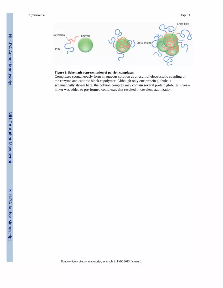
Figure 1. Schematic representation of polyion complexesComplexes spontaneously form in aqueous solution as a result of electrostatic coupling ofthe enzyme and cationic block copolymer. Although only one protein globule isschematically shown here, the polyion complex may contain several protein globules. Cross-linker was added to pre-formed complexes that resulted in covalent stabilization.
Klyachko et al. Page 14
Nanomedicine. Author manuscript; available in PMC 2013 January 1.
NIH
-PA Author Manuscript
NIH
-PA Author Manuscript
NIH
-PA Author Manuscript
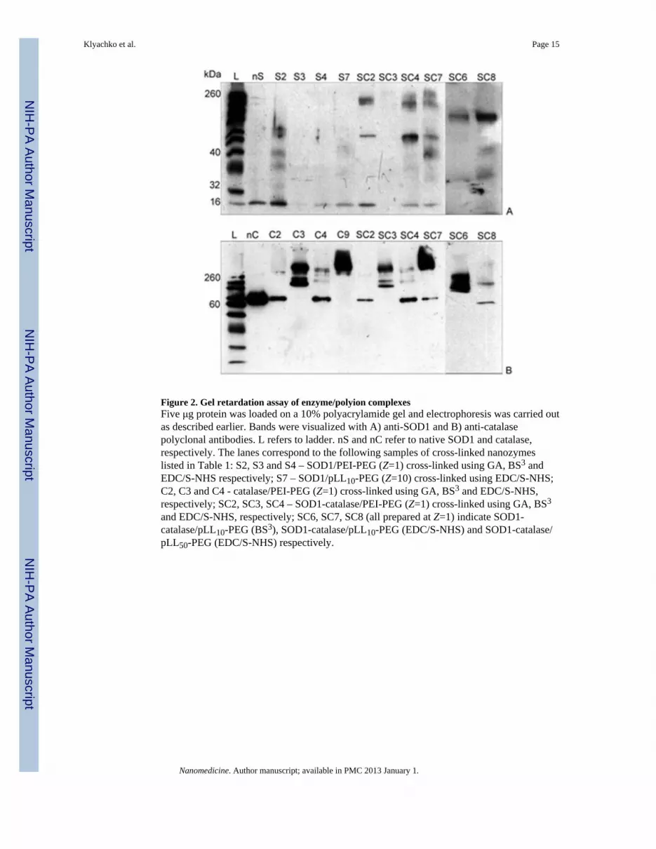
Figure 2. Gel retardation assay of enzyme/polyion complexesFive μg protein was loaded on a 10% polyacrylamide gel and electrophoresis was carried outas described earlier. Bands were visualized with A) anti-SOD1 and B) anti-catalasepolyclonal antibodies. L refers to ladder. nS and nC refer to native SOD1 and catalase,respectively. The lanes correspond to the following samples of cross-linked nanozymeslisted in Table 1: S2, S3 and S4 – SOD1/PEI-PEG (Z=1) cross-linked using GA, BS3 andEDC/S-NHS respectively; S7 – SOD1/pLL10-PEG (Z=10) cross-linked using EDC/S-NHS;C2, C3 and C4 - catalase/PEI-PEG (Z=1) cross-linked using GA, BS3 and EDC/S-NHS,respectively; SC2, SC3, SC4 – SOD1-catalase/PEI-PEG (Z=1) cross-linked using GA, BS3
and EDC/S-NHS, respectively; SC6, SC7, SC8 (all prepared at Z=1) indicate SOD1-catalase/pLL10-PEG (BS3), SOD1-catalase/pLL10-PEG (EDC/S-NHS) and SOD1-catalase/pLL50-PEG (EDC/S-NHS) respectively.
Klyachko et al. Page 15
Nanomedicine. Author manuscript; available in PMC 2013 January 1.
NIH
-PA Author Manuscript
NIH
-PA Author Manuscript
NIH
-PA Author Manuscript
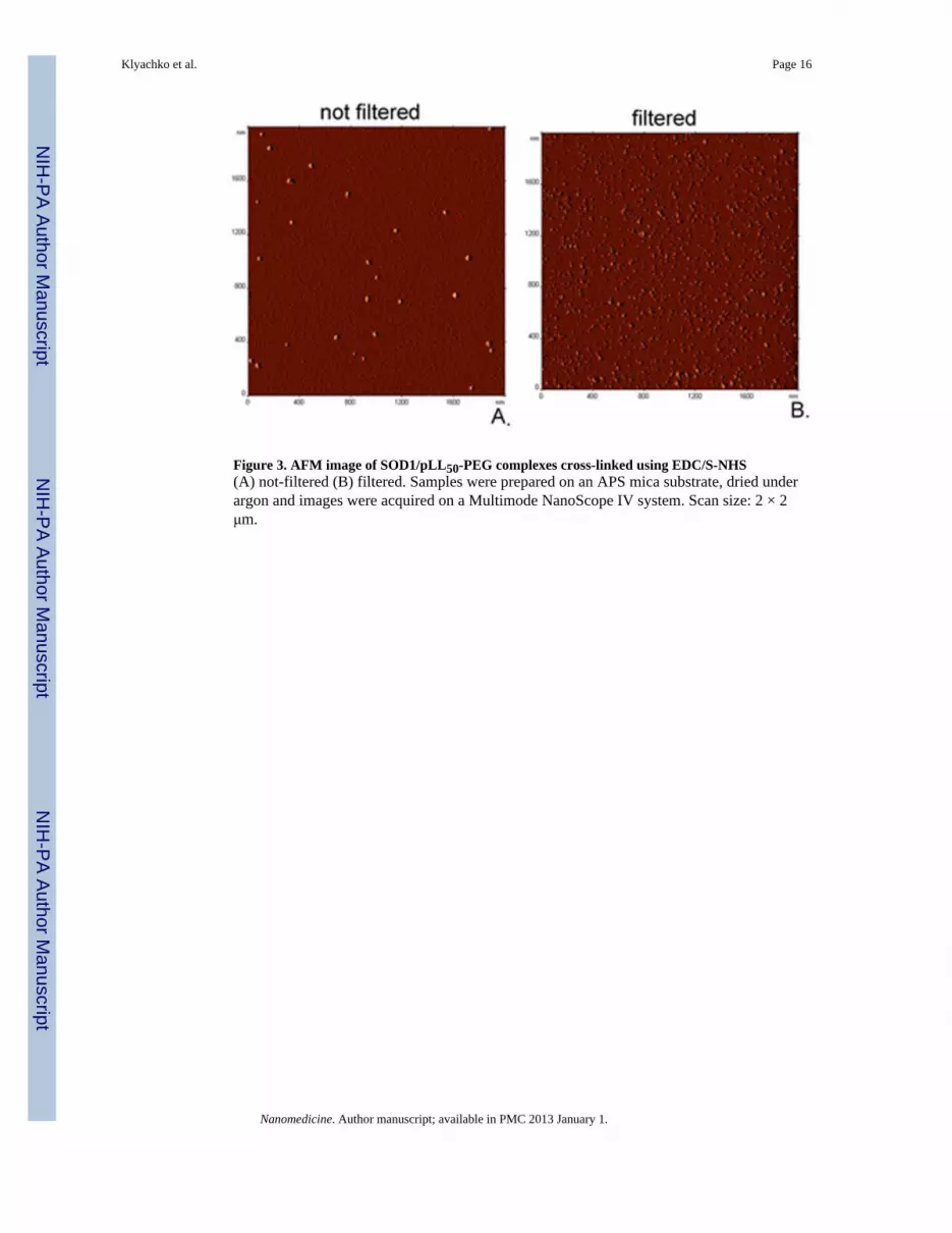
Figure 3. AFM image of SOD1/pLL50-PEG complexes cross-linked using EDC/S-NHS(A) not-filtered (B) filtered. Samples were prepared on an APS mica substrate, dried underargon and images were acquired on a Multimode NanoScope IV system. Scan size: 2 × 2μm.
Klyachko et al. Page 16
Nanomedicine. Author manuscript; available in PMC 2013 January 1.
NIH
-PA Author Manuscript
NIH
-PA Author Manuscript
NIH
-PA Author Manuscript
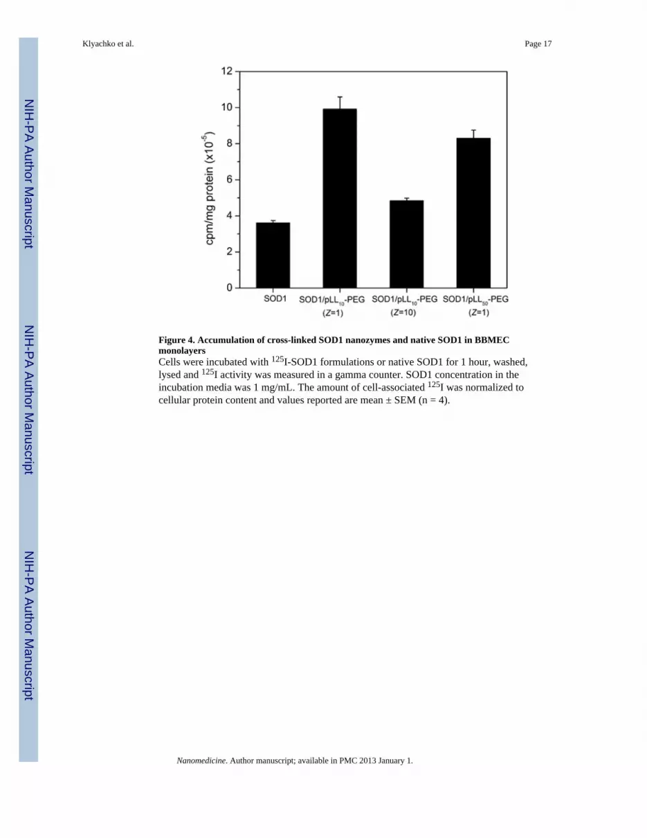
Figure 4. Accumulation of cross-linked SOD1 nanozymes and native SOD1 in BBMECmonolayersCells were incubated with 125I-SOD1 formulations or native SOD1 for 1 hour, washed,lysed and 125I activity was measured in a gamma counter. SOD1 concentration in theincubation media was 1 mg/mL. The amount of cell-associated 125I was normalized tocellular protein content and values reported are mean ± SEM (n = 4).
Klyachko et al. Page 17
Nanomedicine. Author manuscript; available in PMC 2013 January 1.
NIH
-PA Author Manuscript
NIH
-PA Author Manuscript
NIH
-PA Author Manuscript
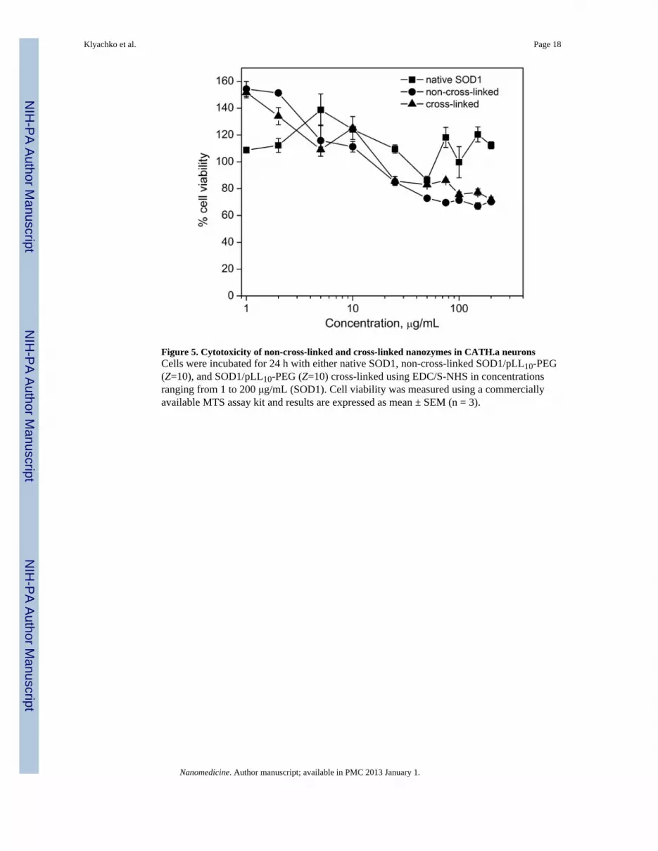
Figure 5. Cytotoxicity of non-cross-linked and cross-linked nanozymes in CATH.a neuronsCells were incubated for 24 h with either native SOD1, non-cross-linked SOD1/pLL10-PEG(Z=10), and SOD1/pLL10-PEG (Z=10) cross-linked using EDC/S-NHS in concentrationsranging from 1 to 200 μg/mL (SOD1). Cell viability was measured using a commerciallyavailable MTS assay kit and results are expressed as mean ± SEM (n = 3).
Klyachko et al. Page 18
Nanomedicine. Author manuscript; available in PMC 2013 January 1.
NIH
-PA Author Manuscript
NIH
-PA Author Manuscript
NIH
-PA Author Manuscript
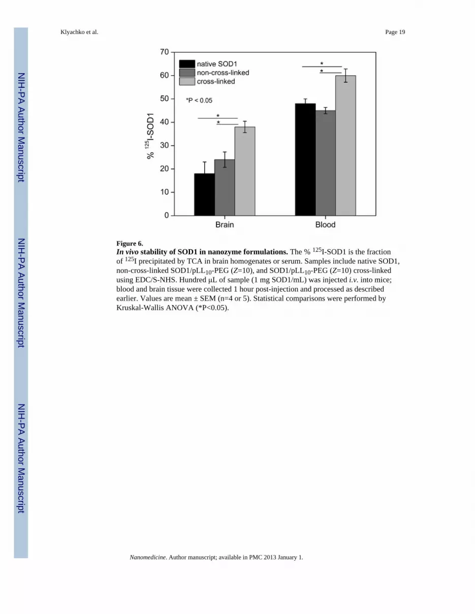
Figure 6.In vivo stability of SOD1 in nanozyme formulations. The % 125I-SOD1 is the fractionof 125I precipitated by TCA in brain homogenates or serum. Samples include native SOD1,non-cross-linked SOD1/pLL10-PEG (Z=10), and SOD1/pLL10-PEG (Z=10) cross-linkedusing EDC/S-NHS. Hundred μL of sample (1 mg SOD1/mL) was injected i.v. into mice;blood and brain tissue were collected 1 hour post-injection and processed as describedearlier. Values are mean ± SEM (n=4 or 5). Statistical comparisons were performed byKruskal-Wallis ANOVA (*P<0.05).
Klyachko et al. Page 19
Nanomedicine. Author manuscript; available in PMC 2013 January 1.
NIH
-PA Author Manuscript
NIH
-PA Author Manuscript
NIH
-PA Author Manuscript
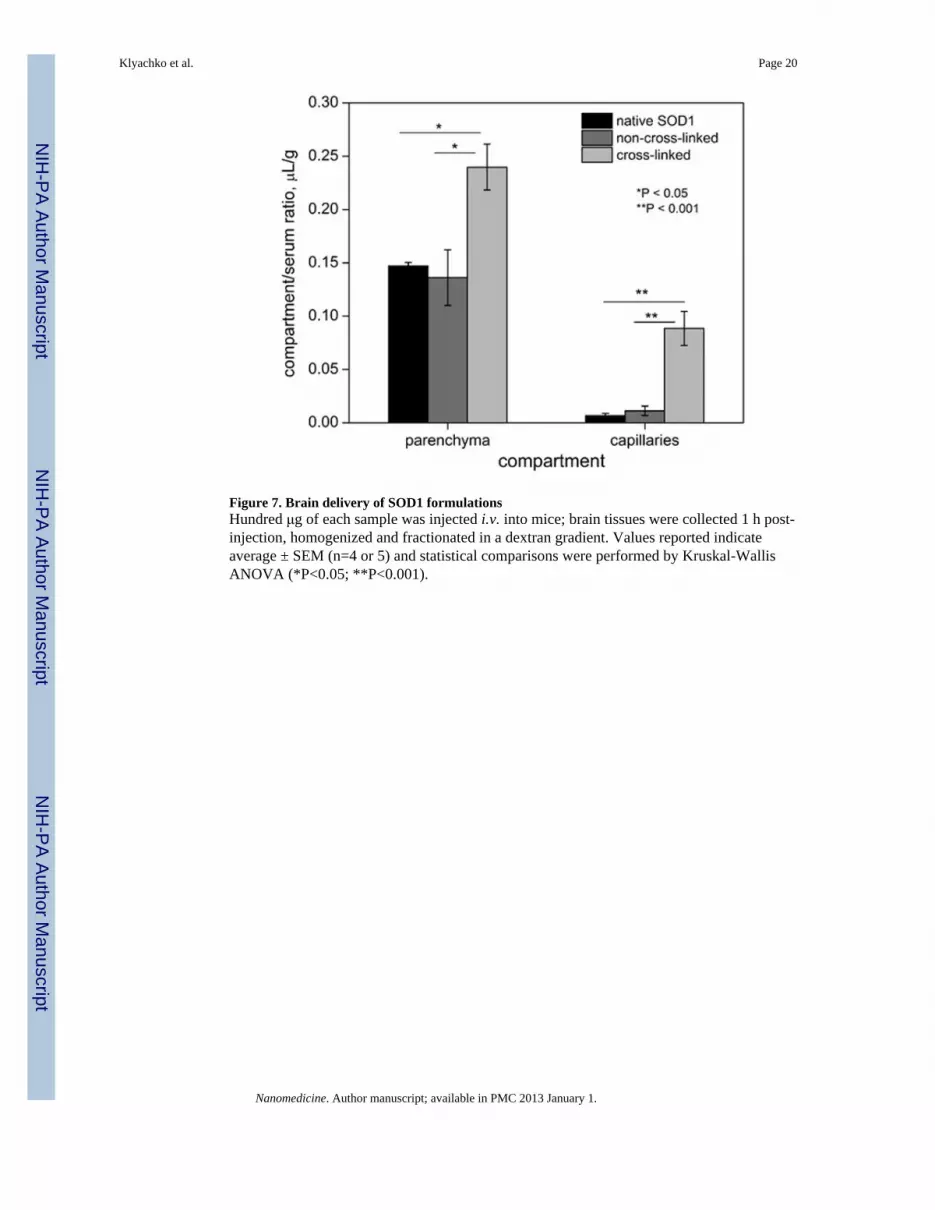
Figure 7. Brain delivery of SOD1 formulationsHundred μg of each sample was injected i.v. into mice; brain tissues were collected 1 h post-injection, homogenized and fractionated in a dextran gradient. Values reported indicateaverage ± SEM (n=4 or 5) and statistical comparisons were performed by Kruskal-WallisANOVA (*P<0.05; **P<0.001).
Klyachko et al. Page 20
Nanomedicine. Author manuscript; available in PMC 2013 January 1.
NIH
-PA Author Manuscript
NIH
-PA Author Manuscript
NIH
-PA Author Manuscript
NIH
-PA Author Manuscript
NIH
-PA Author Manuscript
NIH
-PA Author Manuscript
Klyachko et al. Page 21
Table 1Description of nanozymes
Sample ID Sample Block copolymer Z Cross-linker
nS SOD1 (native enzyme) - - -
S1 non-cl* PEI-PEG 1 -
S2 cl† PEI-PEG 1 GA
S3 cl PEI-PEG 1 BS3
S4 cl PEI-PEG 1 EDC/S-NHS
S5 cl pLL50-PEG 1 EDC/S-NHS
S6 non-cl pLL10-PEG 10 EDC/S-NHS
S7 cl pLL10-PEG 1 EDC/S-NHS
S8 cl pLL10-PEG 10 EDC/S-NHS
nC catalase (native enzyme) none - -
C1 non-cl PEI-PEG 1 -
C2 cl PEI-PEG 1 GA
C3 cl PEI-PEG 1 BS3
C4 cl PEI-PEG 1 EDC/S-NHS
C5 cl pLL50-PEG 1 EDC/S-NHS
C6 cl pLL50-PEG 2 EDC/S-NHS
C7 cl pLL50-PEG 5 EDC/S-NHS
C8 non-cl pLL10-PEG 1 -
C9 cl pLL10-PEG 1 EDC/S-NHS
SC1 SOD1-catalase; non-cl PEI-PEG 1 -
SC2 SOD1-catalase; cl PEI-PEG 1 GA
SC3 SOD1-catalase; cl PEI-PEG 1 BS3
SC4 SOD1-catalase; cl PEI-PEG 1 EDC/S-NHS
SC5 SOD1-catalase; non-cl pLL10-PEG 1 -
SC6 SOD1-catalase; cl pLL10-PEG 1 BS3
SC7 SOD1-catalase; cl pLL10-PEG 1 EDC/S-NHS
SC8 SOD1-catalase; cl pLL50-PEG 1 EDC/S-NHS
*non-cross-linked (non-cl)
†cross-linked (cl)
Nanomedicine. Author manuscript; available in PMC 2013 January 1.
NIH
-PA Author Manuscript
NIH
-PA Author Manuscript
NIH
-PA Author Manuscript
Klyachko et al. Page 22
Table 2Residual enzyme activity of nanozymes
Sample SOD1 residual activity, %* Catalase residual activity, %* pH Cross-linker
SOD1 100 - 7.4 -
SOD1 100 - 6.8 -
SOD1/PEI-PEG; non-cl† 100 - 7.4 -
SOD1/PEI-PEG; cl‡ 88 - 7.4 GA/NaBH4
SOD1/PEI-PEG; cl 91 - 7.4 BS3
SOD1/PEI-PEG; cl 115 - 7.4 EDC/S-NHS
SOD1/pLL10-PEG; cl 115 - 7.4 EDC/S-NHS
SOD1/pLL50-PEG; cl 109 - 7.4 EDC/S-NHS
catalase (cat) - 100 7.4 -
cat - 60 6.8 -
cat/PEI-PEG; non-cl - 100 7.4 -
cat/pLL10-PEG; non-cl - 100 7.4 -
cat/PEI-PEG; cl - 80 7.4 GA/NaBH4
cat/PEI-PEG; cl - 100 7.4 BS3
cat/PEI-PEG; cl - 95 7.4 EDC/S-NHS
cat/pLL10-PEG; cl - 100 7.4 EDC/S-NHS
SOD1-cat/PEI-PEG; non-cl 100 100 6.8 -
SOD1-cat/pLL10-PEG; non-cl 100 100 6.8 -
SOD1-cat/PEI-PEG; cl 80 71 6.8 GA/NaBH4
SOD1-cat/pLL10-PEG; cl 95 78 6.8 GA/NaBH4
SOD1-cat/pLL10-PEG; cl 85 88 7.4 GA/NaBH4
SOD1-cat/pLL10-PEG; cl 70 98 6.8 BS3
SOD1-cat/PEI-PEG; cl 100 105 6.8 EDC/S-NHS
SOD1-cat/pLL10-PEG; cl 97 82 6.8 EDC/S-NHS
SOD1-cat/pLL10-PEG; cl 110 115 6.8 EDC/S-NHS
*Residual enzymatic activity of nanozymes (A/A0) was calculated based on native enzyme activity (A0) at the same pH. The initial activity values
were 3780 U/mg protein for SOD1 and 46500 U/mg protein for catalase
†non-cross-linked
‡cross-linked
Cross-linker excesses were 7 fold for GA and BS3 (over NH2 groups in enzyme and polymer), and 24-fold for EDC/S-NHS (over -COOH groupsin enzyme).
Nanomedicine. Author manuscript; available in PMC 2013 January 1.

NIH
-PA Author Manuscript
NIH
-PA Author Manuscript
NIH
-PA Author Manuscript
Klyachko et al. Page 23
Table 3Physicochemical properties of selected nanozymes measured using DLS
Effective diameter, nm
Sample Linker ζ-potential, mV
Peak 1 Peak 2
SOD1 - 4.6 - -2.6
SOD1/PEI-PEG; non-cl* - 6.1 74.5 -1.5
SOD1/PEI-PEG; cl† BS3 - 97.4 0.3
SOD1/PEI-PEG; cl EDC/S-NHS 6.2 63.6 -1.3
SOD1/pLL10-PEG; cl EDC/S-NHS 6.3 64.2 -2.4
SOD1/pLL10-PEG‡; cl EDC/S-NHS 5.5 72.0 1.3
SOD1/pLL50-PEG; cl EDC/S-NHS 5.8 81.0 2.6
catalase (cat) - 10.0 63 -5.0
cat/PEI-PEG; non-cl - 12.5 68.4 0.5
cat/PEI-PEG; cl GA 40.8 644.3 n.d.§
cat/PEI-PEG; cl BS3 10.6 59.5 -1.2
cat/PEI-PEG; cl EDC/S-NHS 10.6 52.7 -0.3
cat/pLL10-PEG; non-cl - 12.0 71.0 -2.4
cat/pLL10-PEG; cl EDC/S-NHS 12.0 28.2; 504 n.d.§
SOD1-cat/PEI-PE; cl GA - 30.0 n.d.§
SOD1-cat/PEI-PEG; cl BS3 - 91.9 -2.7
SOD1-cat/PEI-PEG; cl EDC/S-NHS 11.7 91.0 -2.9
SOD1-cat/pLL10-PEG; cl EDC/S-NHS 14.0 135.4 -1.3
All nanozymes were prepared at Z=1 (unless indicated otherwise).
*non-cross-linked, †cross-linked, ‡Z=10; §not determined
Measurement error was typically ≤ 5% of reported values.
Table 3B Diameter of filtered nanozymes measured using DLS
Sample* Linker Dz, nm† PdI‡ ζ, mV§
SOD1 - 5.2 ± 0.1¶ 0.1± 0.03 -2.3 ± 0.5
SOD1/pLL50-PEG; non-cl# - 11.2 ± 0.5 0.2 ± 0.02 6.2 ± 0.5
SOD1/pLL50-PEG; cl** EDC/S-NHS 9.7 ± 0.1 0.1 ± 0.02 0.6 ± 0.1
catalase (cat) - 11.5 ± 0.1¶ 0.2± 0.01 n.d††
cat/pLL50-PEG; non-cl - 16.1 ± 0.1 0.2 ± 0.01 n.d.
cat/pLL50-PEG; cl EDC/S-NHS 19.8 ± 0.3 0.2 ± 0.01 n.d.
SOD1 and catalase formulations were prepared in 10 mM HEPES and 10 mM HEPES-buffered saline (pH 7.4), respectively.
*All complexes are stoichiometric (Z=1)
†Z-average diameter (Dz)
‡polydispersity index (PDI) and
Nanomedicine. Author manuscript; available in PMC 2013 January 1.
NIH
-PA Author Manuscript
NIH
-PA Author Manuscript
NIH
-PA Author Manuscript
Klyachko et al. Page 24
§ζ-potential. Data are mean ± SD of triplicate measurements.
¶Theoretical hydrodynamic diameters (Malvern Zetasizer Nano software) are 5.2 nm for SOD1 and 12.5 nm for catalase.
#non-cross-linked;
**cross-linked
††ζ-potential not determined because of high ionic strength.
Nanomedicine. Author manuscript; available in PMC 2013 January 1.
Related Documents