REVIEW ARTICLE Cronobacter species (formerly known as Enterobacter sakazakii) in powdered infant formula: a review of our current understanding of the biology of this bacterium Q.Q. Yan 1 , O. Condell 1 , K. Power 1 , F. Butler 2 , B.D. Tall 3 and S. Fanning 1 1 UCD Centre for Food Safety, WHO Collaborating Centre for Research, Reference & Training on Cronobacter, School of Public Health, Physiotherapy & Population Science, University College Dublin, Dublin, Ireland 2 UCD School of BioSystems Engineering, University College Dublin, Dublin, Ireland 3 US Food and Drug Administration (FDA), Division of Virulence Assessment, OARSA, Centre for Food Safety and Applied Nutrition, MOD 1 Facility, Virulence Mechanisms Branch (HFS-025), 8301 Muirkirk rd, Laurel, MD 20708, USA Introduction Cronobacter species (formerly known as Enterobacter sak- azakii) are Gram-negative rod-shaped, motile pathogenic bacteria of the family Enterobacteriaceae. These organisms are regarded as opportunistic pathogens linked with life- threatening infections predominantly in neonates (infants <4 weeks of age) (Bar-Oz et al. 2001; Gurtler et al. 2005, Anonymous 2006a,b; Mullane et al. 2007a). Clinical syn- dromes of Cronobacter infection include necrotizing enterocolitis (NEC), bacteraemia and meningitis, with case fatality rates ranging between 40 and 80% being reported (Bowen and Braden 2006; Friedemann 2009). Infections in older infants and among immuno-compro- mised adults, mainly the elderly, have also been noted (Bowen and Braden 2006; Gosney et al. 2006; See et al. 2007). The bacterium has been isolated from a range of food sources including dairy-based foods, dried meats, water, rice and others (Baumgartner et al. 2009; Chap et al. 2009; Healy et al. 2010). Surveillance studies detected Cronobacter in a variety of different environ- ments including households, livestock facilities, food manufacturing operations, in particular PIF production facilities (Bar-Oz et al. 2001; Kandhai et al. 2004; Mullane et al. 2007b; Kilonzo-Nthenge et al. 2008). Contaminated powdered infant formula (PIF) has been epidemiologi- cally linked with many of the infections reported (Bowen and Braden 2006). Controlling the microbiological load in infant food products and understanding the optimal growth conditions and epidemiology would contribute positively towards a reduction in the health risk to vulnerable individuals. Classification of Cronobacter Cronobacter species were originally referred to as yellow- pigmented Enterobacter cloacae, later being reclassified as a new species, E. sakazakii in 1980 (Farmer et al. 1980). Keywords Cronobacter, detection protocols, manufacturing environment control, powdered infant formula, taxonomy. Correspondence Se ´ amus Fanning, UCD Centre for Food Safety, School of Public Health, Physiotherapy & Population Science, UCD Veterinary Sciences Centre, University College Dublin, Belfield, Dublin 4, Ireland. E-mail: [email protected] 2011/1466: received 30 August 2011, revised 15 February 2012 and accepted 15 February 2012 doi:10.1111/j.1365-2672.2012.05281.x Summary Cronobacter species (formerly known as Enterobacter sakazakii) are opportunis- tic pathogens that can cause necrotizing enterocolitis, bacteraemia and menin- gitis, predominantly in neonates. Infection in these vulnerable infants has been linked to the consumption of contaminated powdered infant formula (PIF). Considerable research has been undertaken on this organism in the past num- ber of years which has enhanced our understanding of this neonatal pathogen leading to improvements in its control within the PIF production environment. The taxonomy of the organism resulted in the recognition of a new genus, Cro- nobacter, which consists of seven species. This paper presents an up-to-date review of our current knowledge of Cronobacter species. Taxonomy, genome sequencing, current detection protocols and epidemiology are all discussed. In addition, consideration is given to the control of this organism in the manufacturing environment, as a first step towards reducing the occurrence of this pathogen in PIF. Journal of Applied Microbiology ISSN 1364-5072 ª 2012 The Authors Journal of Applied Microbiology 113, 1–15 ª 2012 The Society for Applied Microbiology 1

Welcome message from author
This document is posted to help you gain knowledge. Please leave a comment to let me know what you think about it! Share it to your friends and learn new things together.
Transcript

REVIEW ARTICLE
Cronobacter species (formerly known as Enterobactersakazakii) in powdered infant formula: a review of ourcurrent understanding of the biology of this bacteriumQ.Q. Yan1, O. Condell1, K. Power1, F. Butler2, B.D. Tall3 and S. Fanning1
1 UCD Centre for Food Safety, WHO Collaborating Centre for Research, Reference & Training on Cronobacter, School of Public Health,
Physiotherapy & Population Science, University College Dublin, Dublin, Ireland
2 UCD School of BioSystems Engineering, University College Dublin, Dublin, Ireland
3 US Food and Drug Administration (FDA), Division of Virulence Assessment, OARSA, Centre for Food Safety and Applied Nutrition,
MOD 1 Facility, Virulence Mechanisms Branch (HFS-025), 8301 Muirkirk rd, Laurel, MD 20708, USA
Introduction
Cronobacter species (formerly known as Enterobacter sak-
azakii) are Gram-negative rod-shaped, motile pathogenic
bacteria of the family Enterobacteriaceae. These organisms
are regarded as opportunistic pathogens linked with life-
threatening infections predominantly in neonates (infants
<4 weeks of age) (Bar-Oz et al. 2001; Gurtler et al. 2005,
Anonymous 2006a,b; Mullane et al. 2007a). Clinical syn-
dromes of Cronobacter infection include necrotizing
enterocolitis (NEC), bacteraemia and meningitis, with
case fatality rates ranging between 40 and 80% being
reported (Bowen and Braden 2006; Friedemann 2009).
Infections in older infants and among immuno-compro-
mised adults, mainly the elderly, have also been noted
(Bowen and Braden 2006; Gosney et al. 2006; See et al.
2007). The bacterium has been isolated from a range of
food sources including dairy-based foods, dried meats,
water, rice and others (Baumgartner et al. 2009; Chap
et al. 2009; Healy et al. 2010). Surveillance studies
detected Cronobacter in a variety of different environ-
ments including households, livestock facilities, food
manufacturing operations, in particular PIF production
facilities (Bar-Oz et al. 2001; Kandhai et al. 2004; Mullane
et al. 2007b; Kilonzo-Nthenge et al. 2008). Contaminated
powdered infant formula (PIF) has been epidemiologi-
cally linked with many of the infections reported (Bowen
and Braden 2006). Controlling the microbiological load
in infant food products and understanding the optimal
growth conditions and epidemiology would contribute
positively towards a reduction in the health risk to
vulnerable individuals.
Classification of Cronobacter
Cronobacter species were originally referred to as yellow-
pigmented Enterobacter cloacae, later being reclassified as
a new species, E. sakazakii in 1980 (Farmer et al. 1980).
Keywords
Cronobacter, detection protocols,
manufacturing environment control,
powdered infant formula, taxonomy.
Correspondence
Seamus Fanning, UCD Centre for Food
Safety, School of Public Health, Physiotherapy
& Population Science, UCD Veterinary
Sciences Centre, University College Dublin,
Belfield, Dublin 4, Ireland.
E-mail: [email protected]
2011/1466: received 30 August 2011, revised
15 February 2012 and accepted 15 February
2012
doi:10.1111/j.1365-2672.2012.05281.x
Summary
Cronobacter species (formerly known as Enterobacter sakazakii) are opportunis-
tic pathogens that can cause necrotizing enterocolitis, bacteraemia and menin-
gitis, predominantly in neonates. Infection in these vulnerable infants has been
linked to the consumption of contaminated powdered infant formula (PIF).
Considerable research has been undertaken on this organism in the past num-
ber of years which has enhanced our understanding of this neonatal pathogen
leading to improvements in its control within the PIF production environment.
The taxonomy of the organism resulted in the recognition of a new genus, Cro-
nobacter, which consists of seven species. This paper presents an up-to-date
review of our current knowledge of Cronobacter species. Taxonomy, genome
sequencing, current detection protocols and epidemiology are all discussed.
In addition, consideration is given to the control of this organism in the
manufacturing environment, as a first step towards reducing the occurrence of
this pathogen in PIF.
Journal of Applied Microbiology ISSN 1364-5072
ª 2012 The Authors
Journal of Applied Microbiology 113, 1–15 ª 2012 The Society for Applied Microbiology 1
Using partial 16S ribosomal DNA (rDNA) and hsp60
sequencing, Iversen et al. (2004a) divided 126 Cronobacter
isolates into four clusters, suggesting that the genus may
require re-classification. Later and following further
extensive polyphasic analysis, Iversen et al. (2007a, 2008)
proposed the reclassification of these bacteria into a new
genus called Cronobacter. Originally, six species (C. sak-
azakii, C. malonaticus, C. turicensis, C. muytjensii,
C. dublinensis and C. genomospecies 1) were defined and
comprised of the 16 biogroups described in Table 1.
A new species (C. condimenti) was identified recently by
Joseph et al. (2011), and in addition, C. universalis now
replaces the original C. genomospecies 1.
This re-classification by Iversen et al. (2007a, 2008) was
subsequently supported by both optical mapping and
genome sequencing data which confirmed the revision of
the taxonomy (Kotewicz and Tall 2009; Kucerova et al.
2010). Although C. sakazakii and C. malonaticus were
found to be closely related and difficult to distinguish by
16S rDNA sequence analysis, a seven loci (atpD, fusA,
glnS, gltB, gyrB, infB, ppsA) multilocus sequence typing
(MLST) scheme was developed to discriminate between
these two species (Baldwin et al. 2009). Furthermore,
recent findings reported by Joseph and Forsythe (2011)
identified a highly stable sequence type (denoted as ST4)
within Cronobacter sakazakii and which was responsible
for a large proportion of severe neonatal infections, espe-
cially neonatal meningitis.
Two of the species genomes were subsequently
published (as discussed below), and currently, a collaborative
effort is underway to complete the genome sequences of a
further 15 isolates of six of the seven Cronobacter species
adding substantially to our knowledge of the core genome
of this group of bacteria and highlighting particular
species-specific features of interest.
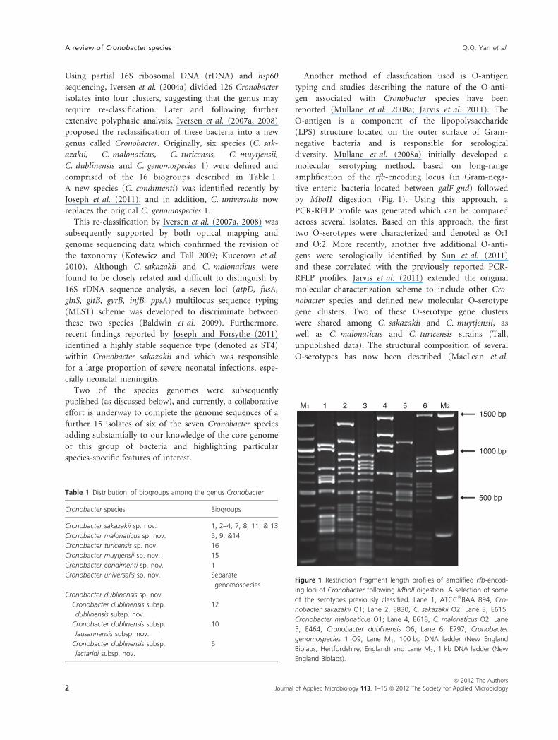
Another method of classification used is O-antigen
typing and studies describing the nature of the O-anti-
gen associated with Cronobacter species have been
reported (Mullane et al. 2008a; Jarvis et al. 2011). The
O-antigen is a component of the lipopolysaccharide
(LPS) structure located on the outer surface of Gram-
negative bacteria and is responsible for serological
diversity. Mullane et al. (2008a) initially developed a
molecular serotyping method, based on long-range
amplification of the rfb-encoding locus (in Gram-nega-
tive enteric bacteria located between galF-gnd) followed
by MboII digestion (Fig. 1). Using this approach, a
PCR-RFLP profile was generated which can be compared
across several isolates. Based on this approach, the first
two O-serotypes were characterized and denoted as O:1
and O:2. More recently, another five additional O-anti-
gens were serologically identified by Sun et al. (2011)
and these correlated with the previously reported PCR-
RFLP profiles. Jarvis et al. (2011) extended the original
molecular-characterization scheme to include other Cro-
nobacter species and defined new molecular O-serotype
gene clusters. Two of these O-serotype gene clusters
were shared among C. sakazakii and C. muytjensii, as
well as C. malonaticus and C. turicensis strains (Tall,
unpublished data). The structural composition of several
O-serotypes has now been described (MacLean et al.
Table 1 Distribution of biogroups among the genus Cronobacter
Cronobacter species Biogroups
Cronobacter sakazakii sp. nov. 1, 2–4, 7, 8, 11, & 13
Cronobacter malonaticus sp. nov. 5, 9, &14
Cronobacter turicensis sp. nov. 16
Cronobacter muytjensii sp. nov. 15
Cronobacter condimenti sp. nov. 1
Cronobacter universalis sp. nov. Separate
genomospecies
Cronobacter dublinensis sp. nov.
Cronobacter dublinensis subsp.
dublinensis subsp. nov.
12
Cronobacter dublinensis subsp.
lausannensis subsp. nov.
10
Cronobacter dublinensis subsp.
lactaridi subsp. nov.
6
M1 M2
1500 bp
1000 bp
500 bp
1 2 3 4 5 6
Figure 1 Restriction fragment length profiles of amplified rfb-encod-
ing loci of Cronobacter following MboII digestion. A selection of some
of the serotypes previously classified. Lane 1, ATCC�BAA 894, Cro-
nobacter sakazakii O1; Lane 2, E830, C. sakazakii O2; Lane 3, E615,
Cronobacter malonaticus O1; Lane 4, E618, C. malonaticus O2; Lane
5, E464, Cronobacter dublinensis O6; Lane 6, E797, Cronobacter
genomospecies 1 O9; Lane M1, 100 bp DNA ladder (New England
Biolabs, Hertfordshire, England) and Lane M2, 1 kb DNA ladder (New
England Biolabs).
A review of Cronobacter species Q.Q. Yan et al.
2 Journal of Applied Microbiology 113, 1–15 ª 2012 The Society for Applied Microbiology
ª 2012 The Authors

2009; Czerwicka et al. 2010; Maclean et al. 2011; Arbat-
sky et al. 2010, 2011; Shashkov et al. 2011).
Genomes of the genus Cronobacter
The first sequenced Cronobacter genome, C. sakazakii
ATCC�BAA-894 was published by Kucerova et al. (2010).
It revealed a single chromosome of 4Æ4 Mb (57% GC)
along with two plasmids, denoted as pESA2 and pESA3
(31-kb, 51% GC and 131-kb, 56% GC, respectively). The
source of this isolate was reported to be contaminated
PIF used in a neonatal intensive care unit and which gave
rise to an outbreak in 2001 in Tennessee, USA. Using
comparative genomic hybridization (CGH) techniques,
representative isolates including C. malonaticus, C. turic-
ensis, C. muytjensii and C. dublinensis were further inves-
tigated, and those genes considered to be part of the core
species genome along with other markers unique to
C. sakazakii were identified (Kucerova et al. 2010).
Cronobacter sakazakii ATCC�BAA-894 contained
approximately 4392 genes as part of its core genome.
However, using the CGH approach, 4382 unique, anno-
tated genes from both the chromosome and plasmids
were noted, and only 54Æ9% of genes were common to all
C. sakazakii with 43Æ3% being common across all Cronob-
acter species. Interestingly, 21 genes were found to be
unique in five of the C. sakazakii tested, and these
encoded proteins were involved in pilus assembly, a phos-
photransferase system (PTS), an acid transporter, N-acet-
ylneuraminate lyase and a toxin ⁄ antitoxin system.
The genome of C. turicensis z3032 was published in
early 2011 (Stephan et al. 2011) in an attempt to further
determine virulence factors and mechanisms of pathoge-
nicity in this bacterium. Following the deaths of two new-
born infants in 2005, the latter isolate was cultured from
the blood of one child with meningitis (Mange et al.
2006). In this sequenced isolate, the genome was 4Æ4 Mb
(57% GC) in size and contained three plasmids of sizes
approx 138-kb pCTU1 (56%), 22-kb pCTU2 (49%) and
54-kb pCTU3 (50% GC). Two hundred and twenty-three
genes were annotated as virulence- and disease-related
encoding open reading frames (ORFs); however, 9Æ27%
(413 of 4455) encoded proteins were of unknown func-
tion. Because these latter ORFs lacked similarity to
sequences already in the current databases, these could
potentially have important pathogenic functions.
Franco et al. (2011a) reported the in silico analysis of
two plasmids, one from C. sakazakii ATCC�BAA-894,
pESA3 and the other from Cronobacter turicensis z3032,
pCTU1, each of which contained a RepFIB replicon. Both
plasmids possessed two iron acquisition systems (eitCBAD
and iucABCD ⁄ iutA) essential for survival and success-
ful pathogenesis. Ninety-seven per cent of 229 strains,
representing seven of the eight Cronobacter species,
possessed a RepFIB plasmid. The presence of a Cronobacter
plasminogen activator-encoding gene (cpa) [encoded on
pESA3], a type 6 secretion system (T6SS) [also encoded
on pESA3] and a filamentous haemagglutinin ⁄ adhesin
(FHA) gene locus (located on pCTU1) suggested the exis-
tence of unique virulence determinants in these species.
The cpa-encoding gene encodes an outer membrane pro-
tease implicated in serum resistance, a feature that would
facilitate Cronobacter species in crossing the blood–brain
barrier and causing meningitis (Franco et al. 2011b). The
T6SS acts to translocate putative effector proteins aiding
in bacterial pathogenesis, while FHA pCTU1 contains
fhaB, fhaC genes (which encode proteins with similarly
identity to a transported and transporter protein as part
of a two-partner secretion system) and five associate
putative adhesins. Further studies are now in progress to
extend our understanding of the functional roles of these
plasmid-encoded loci.
Virulence characteristics of Cronobacter
Information from epidemiological studies along with in
vitro mammalian tissue culture assays has shown that
Cronobacter isolates demonstrate a variable virulence phe-
notype (Caubilla-Barron et al. 2007; Townsend et al.
2007, 2008). Only isolates of C. sakazakii, C. malonaticus
and C. turicensis have been linked with neonatal infec-
tions (Healy et al. 2010; Kucerova et al. 2010).
Currently, few clues as to the mechanisms involved are
known, though, data from genome sequencing efforts
highlighted several potential markers that may be helpful
candidates for future studies (Kucerova et al. 2010;
Stephan et al. 2011).
The first putative Cronobacter virulence factors to be
described were enterotoxin-like compounds produced by
four of 18 isolates studied (Pagotto et al. 2003). Using
conventional tissue culture-based assays, Cronobacter is
known to attach to intestinal cell lines in vitro and survive
within macrophages for periods of time (Townsend et al.
2008). Franco et al. (2011b) recently demonstrated resis-
tance to complement-mediated killing of C. sakazakii,
and this was associated with the presence of cpa which is
contained on the pESA3 plasmid.
The outer membrane protein A, encoded by the ompA
gene, is probably the best characterized virulence marker.
This was originally reported by Nair and Venkitanarayanan
(2006) and shown to be required for binding of the bac-
terium to human brain microvascular endothelial cells
(BMEC) (Nair et al. 2009). More recently, Kim and
Loessner (2008) reported that the disruption of tight
junctions significantly enhanced adherence of C. sakazakii
to Caco2 cells in culture and that the same marker was
Q.Q. Yan et al. A review of Cronobacter species
ª 2012 The Authors
Journal of Applied Microbiology 113, 1–15 ª 2012 The Society for Applied Microbiology 3

required for basolateral invasion (Kim et al. 2010a,b). The
ompA-encoding gene is thought to be present in all Cro-
nobacter strains tested, and this marker has also been
linked with invasive Escherichia coli, which causes neona-
tal meningitis (Prasadarao et al. 1996; Kim 2000).
At the epithelial cell surface, C. sakazakii infection
results in damage of these cells, following the recruitment
of greater numbers of dentritic cells, compared with mac-
rophages and neutrophils (Emami et al. 2011). Using a
NEC mouse model, these effects were shown to be medi-
ated through OmpA and involved inducible NO synthase
(iNOS).
From an analysis of the annotated genes in C. sak-
azakii ATCC�BAA-894, Kucerova et al. (2010) high-
lighted several markers including, ibeA, ibeB, yijP and
ompA, which were previously identified in other organ-
isms associated with invasion of BMEC (Prasadarao
et al. 1996; Huang et al. 1999, 2001; Wang et al. 1999).
Interestingly, ibeB (a gene synonymous with cusC),
which belongs to a cluster of genes encoding a copper
and silver resistance cation efflux system, facilitates the
invasion of BMEC cells (Franke et al. 2003); although
this gene was found in the reference strain C. sakazakii
ATCC�BAA-894, the genes ibeA and yijP produced no
matches. When assessed, it was found that the complete
cation efflux operon (cusA, cusB and cusC) and its regu-
latory gene cusR were present in isolates associated with
neonatal infections (including C. sakazakii, C. turicensis
and C. malonaticus) and absent in the other tested
strains (Kucerova et al. 2010).
Detection protocols
Conventional bacteriological culture
The first detection method developed for Cronobacter spe-
cies was described by Muytjens et al. (1988). Based on
this protocol, the US Food and Drug Administration (US
FDA) recommended a method to isolate and enumerate
E. sakazakii from powdered infant formula in 2002. In
2006, the International Organization for Standardization
(ISO 2009) and the International Dairy Federation devel-
oped a technical standard protocol for the detection of
Cronobacter species from milk-based powdered formula
known as ISO ⁄ TS 22964 (Anonymous 2006a,b) (described
in Table 2). More recently, the US–FDA method was
revised to combine both a PCR assay and two newly
developed chromogenic agars for detection (Chen et al.
2009; Chen 2011).
Pre-enrichment of the PIF samples to be tested is a
requirement in these three protocols, and the time dura-
tion varies from a maximum overnight period (ranging
from 18 to 24 h) to a minimum time period of 6 h, fol-
lowed by selective enrichment and subsequent isolation
using selective agars ⁄ media. Typical colonies are con-
firmed using a selective agar and ⁄ or a suitable real-time
PCR assay, with the final identification based on either
biochemical ⁄ molecular characterization. In the revised
US–FDA protocol, there is one enrichment step which is
then followed by a molecular method used for quick
confirmation. This approach eliminates two days from
Table 2 Detection protocols for Cronobacter in PIF
Procedure FDA (Original) ISO ⁄ TS 22964 FDA (revised)
Pre-enrichment Make 1 : 10 (w ⁄ v) of sample in
distilled water, incubated
overnight at 36�C
Make 1 : 10 (w ⁄ v) of sample in
BPW, incubated at 37�C for
18 ± 2 h
Make 1 : 10 (w ⁄ v) of sample in
BPW, incubated at 36�C for 6 h
Selective
enrichment
Transfer 10 ml pre-enrichment to
90 ml EE broth, incubated
overnight at 36�C
Transfer 100 ll pre-enrichment to
10 ml mLST ⁄ vancomycin medium,
incubated at 44�C for 24 ± 2 h
Selection ⁄isolation
Make an isolation streak and spread
plate from each EE broth onto
VRBG agar, incubated overnight
at 36�C
Streak from the cultured mLST ⁄vancomycin medium one loopful
on the chromogenic agar in Petri
dishes, incubated at 44�C for
24 ± 2 h
Centrifuge 40 ml samples, 3000 g,
10 min and resuspend pellet in
200 ll PBS; Spread 100 ll onto
chromogenic media, incubated
overnight at 36�CConfirmation Pick five presumptive positive
colonies and streak onto TSA,
incubated overnight at 25�C
Select five typical colonies and streak
on TSA agar, incubated at 25�C for
48 ± 4 h
Pick two typical colonies from each
chromogenic media confirmed with
real-time PCR, API 20E, Rapid ID 32 E
Identification Yellow colonies are confirmed
with the API 20E test kit
Select one yellow colony from
each TSA plate for biochemical
characterization
Detection
time (days)
5 6 3
A review of Cronobacter species Q.Q. Yan et al.
4 Journal of Applied Microbiology 113, 1–15 ª 2012 The Society for Applied Microbiology
ª 2012 The Authors

the detection procedure compared with the original
protocol.
Selective media for Cronobacter, including Leuschner–
Bew agar (Leuscher and Bew 2004), Druggan-Fosythe-Iversen
agar (Iversen et al. 2004b), Oh-Kang agar (Oh and Kang
2004), ESPM agar (Restaino et al. 2006) and HiCrome�Cronobacter spp. agar (Sigma-Aldrich, Switzerland), have
been developed, based on the a-glucosidase enzyme marker
(Iversen et al. 2004b) identified by Muytjens et al. (1984)
and b-cellobiosidase activity (Restaino et al. 2006) which is
present in all Cronobacter strains. Moreover, violet red bile
agar (VRBA), MacConkey agar and desoxycholate agar,
which are selective for Gram-negative bacteria, are also
available for the isolation of Cronobacter (Druggan and Iver-
sen 2009; Forsythe 2009) from foods.
However, despite the availability of selective agar
media, some were shown to insufficiently support the
growth of all Cronobacter strains (Iversen and Forsythe
2007) and other related species, such as Enterobacter helv-
eticus, Enterobacter pulveris and Enterobacter turicensis,
which are often found in the same ecological niches.
Therefore, improvements in the design of the selective
media for the isolation and identification of Cronobacter
were required.
O’Brien et al. (2009) described the design of a one-step
pre-enrichment and enrichment protocol using a chromo-
genic medium. In this detection strategy, the specific
broth developed [denoted as Cronobacter enrichment
broth (CEB)] facilitated a shortened two-day culture
method for the detection of Cronobacter species in PIF.
Mullane et al. (2006) utilized a cationic-magnetic-bead
capture technique to improve the sensitivity of detection
for Cronobacter from PIF.
The accuracy and reliability of commercially available
identification kits for Cronobacter have been questioned,
with reports of false-negative and false-positive identifica-
tion (Restaino et al. 2006; Iversen and Forsythe 2007).
However, Gen III is currently the only commercially
available identification kit with the original six species
included (Healy 2010).
Immuno-based detection protocols
Immuno-based assays are convenient for detection meth-
ods that can be applied to detect specific bacteria. Assays
using monoclonal antibodies are widely used in research as
rapid detection tools. These approaches can improve the
sensitivity and specificity for detection. Commercial kits
based on enzyme-linked immunosorbent assay (ELISA)
technology have been developed, and the VITEK� immu-
nodiagnostic assay system (denoted as VIDAS�, bio-
Merieux Vitek Inc., Hazelwood, MO, USA) has been used
as a rapid detection platform for Salmonella, E. coli
O157:H7, Listeria species, Campylobacter jejuni and
Staphylococcus species enterotoxins. The VIDAS� Salmonella
method has been validated and certified by the Association
of Official Analytical Chemists (AOAC) as an approved
method of analysis in foods. This protocol was also
approved by other regulatory organisations including
Health Canada, the European Microbiological Method
Assessment Scheme (EMMAS) and German Normalization
Institute (DIN). Research on a VIDAS�-based Cronobacter
method is currently in progress; early results suggest that
the method is fast, reliable and sensitive for the detection
of Cronobacter in a range of test matrices (Q.Q. Yan,
unpublished data).
Molecular-based detection protocols
Molecular detection techniques have always been regarded
as useful tools to extend our understanding of the epide-
miology of an organism. Usually, these assays are
designed to target unique genes present in the pathogen
of interest. Many of the more recent assay formats are
based on real-time PCR and several have been designed
for the specific detection of Cronobacter (Malorny and
Wagner 2005; Seo and Brackett 2005; Drudy et al. 2006;
Table 3 Gene targets useful for the detection of Cronobacter and
related species
Gene targets Reference
Genus loci
ribosomal
DNA (rDNA)
16S rRNA Iversen et al. (2004a)
23S rRNA Derzelle et al. (2007)
tRNAGlu Hassan et al. (2007)
FISH Iversen et al. (2007a)
Almeida et al. (2009)
1,6 a-glucosidase gluA Iversen et al. (2007b)
MMS operon dnaG Seo and Brackett (2005)
Drudy et al. (2006)
Zinc-containing
metalloprotease
zpx Jaradat et al. (2009)
Outer membrane
protein A
ompA Nair and
Venkitanarayanan
(2006)
Species loci
rfb (O-antigen) wehC [CsakO:1]
& wehI [CsakO:2]
Mullane et al. (2008a)
wzx [CsakO:3;
CturO:1;Cmuy O:1;
and Cmal O:1
and O:2 ]
Jarvis et al. 2011)
b-subunit of RNA
polymerase
rpoB Stoop et al. (2009)
Other gene targets
RNaseP
infB (initiation factor)
Q.Q. Yan et al. A review of Cronobacter species
ª 2012 The Authors
Journal of Applied Microbiology 113, 1–15 ª 2012 The Society for Applied Microbiology 5

Nair and Venkitanarayanan 2006; Kothary et al. 2007;
Zhou et al. 2008). Targets used (Table 3) include the 16S
rRNA gene, 16S -23S rDNA intergenic region, the dnaG
gene, the ompA gene, the 1,6 a-glucosidase-encoding gene
(gluA) and a zinc-containing methalloprotease (zpx).
More recently, a PCR-based method developed by Stoop
et al. (2009) was extended by A. Lehner, C. Fricker-Feer
and R. Stephan (unpublished data) to include the new
species described. The latter protocol facilitated the detec-
tion of all seven known species within the Cronobacter
genus using a mismatch-PCR-based approach. The appli-
cations of these molecular-based protocols can support
the traditional culture-based approaches (Fig. 2). Interest-
ingly, using the latter protocol, C. malonaticus and C. sak-
azakii could not be differentiated using this approach,
and therefore, a second PCR was required to accurately
identify each of these species. In 2011, Yan et al. (2011)
published a PCR- and array-based biomarker verification
study for the detection and identification of Cronobacter
spp. Here, these authors propose to elucidate virulence
markers which may be helpful as biomarkers for differen-
tiating Cronobacter spp. and Salmonella spp. from other
food-borne pathogens. While these putative markers were
identified, further validation experiments are currently
being conducted.
Molecular subtyping has long been regarded as a useful
approach that can be applied to elucidate the nature of
those bacteria colonizing a particular ecological niche.
Mullane et al. (2007b) applied pulsed-field gel electropho-
resis (PFGE) to characterize and track Cronobacter species
in a PIF processing facility (Fig. 3). The study provided a
basis for the development of efficient intervention mea-
sures contributing to the reduction of Cronobacter in the
PIF manufacturing environment. A similar approach
using the second generation subtyping method, multiple-
locus variable-number tandem-repeat analysis (MLVA),
was subsequently applied to subtype a collection of geno-
and phenotypically diverse Cronobacter isolates (Mullane
et al. 2008b). However, a standardized PFGE protocol is
close to completion and has already been validated by
PulseNet, a network of national and regional laboratory
networks dedicated to tracking food-borne infections.
A significant reduction in both time and cost associated
with genome sequencing has made molecular detection
methods increasingly accessable. Multilocus sequence
analysis (MLSA) based on recN, rpoA and thdF genes was
used in describing the genomic similarity of Cronobacter
genes by Kuhnert et al. (2009). El-Sharoud et al. (2009)
applied recN gene sequence analysis to isolate Cronobacter
strains recovered from dried milk and related products.
A similar MLST typing scheme using the following seven
housekeeping genes: atpD, fusA, glnS, gltB, gyrB, infB,
ppsA (3036 nt concatenated length) has been developed
by Baldwin et al. (2009), and a database containing
defined sequence types covering all Cronobacter spp. is
currently being maintained at Oxford University. The
database and MLST analytics can be accessed at
www.pubMLST.org/cronobacter. However, the scheme
has not been applied for use in any ongoing epidemiologic
investigation.
Whole-genome sequencing efforts can be expected to
facilitate the correct identification of the bacterial species
being studied; it can also provide detailed information
regarding the unique geno- and phenotypic features.
Moreover, these approaches can be used for comparative
purposes to rapidly and simultaneously investigate the
presence ⁄ absence of all annotated genes or coding
628 bpM 1 2 3 4 5 6 7 M
514 bp506 bp
251 bp289 bp418 bp
Figure 2 A 1Æ0% agarose gel showing the amplification of rpoB
amplicons used to identify six of the genus species (Stoop et al.
2009). Lane 1, Cronobacter sakazakii SP291; Lane 2, Cronobacter
malonaticus E766; Lane 3, C. malonaticus E766 (following a confirma-
tory second PCR); Lane 4, Cronobacter muytjensii ATCC�51329; Lane
5, Cronobacter dublinensis CFS237; Lane 6, Cronobacter turicensis
E626; Lane 7, Cronobacter genomospecies 1 E797 and Lane M,
100 bp DNA ladder.
1135 kbpM 1 2 3 M 4 5 6 M M7 8 9
452·7 kbp
244·4 kbp
33·3 kbp
138·9 kbp
Figure 3 Pulsed-field gel electrophoresis (PFGE) profiles used to char-
acterize and track Cronobacter species in a PIF processing facility.
Lane 1, factory sample 1; Lane 2, factory sample 2; Lane 3, factory
sample 3; Lane 4, factory sample 4; Lane 5, factory sample 5; Lane 6,
factory sample 6; Lane 7, factory sample 7; Lane 8, factory sample 8;
Lane 9, factory sample 9 and Lane M, Salmonella Braenderup, molec-
ular marker, genomic DNA digested with XbaI. The arrow heads at
the foot of the image show that in lanes 3 and 7, these isolates have
the same PFGE profile and would be considered to be indistinguish-
able. Similarly, isolates contained in lanes 4 and 5 have another indis-
tinguishable profile cluster.
A review of Cronobacter species Q.Q. Yan et al.
6 Journal of Applied Microbiology 113, 1–15 ª 2012 The Society for Applied Microbiology
ª 2012 The Authors

sequences or ⁄ and polymorphisms that may contribute to
a specific morphology or physiology. Microarray-based
comparative genomic indexing analysis of Cronobacter
genus was originally performed by Healy et al. (2009),
and this study identified species-specific genes that could
be evaluated as candidate markers for inclusion in a
molecular-based detection protocol. More recently, next
generation sequencing has been used for the comprehen-
sive analysis of eight Cronobacter genomes to better
understand the pathogenicity and evolution of the genus
alongside the characterization of virulence genes
(Ji 2011). While this approach might reveal species-
specific genomic information, in essence, this is a method
that will provide much more detail necessary for subtyp-
ing an organism.
Epidemiology of Cronobacter species
Cronobacter infections are rare and are often underreported,
especially in developing and less-developed countries (Es-
tuningsih and Sani 2008; Friedemann 2009). Thus, the
epidemiology of Cronobacter species is incomplete and
poorly described. Bowen and Braden (2006) first
attempted to describe the epidemiology related to these
infections. These authors analysed the clinical case notes
of 46 invasive infant E. sakazakii infections between 1961
and 2005. These included 12 infants presenting with bac-
teraemia, 33 with meningitis, and 1 with a urinary tract
infection. Infants presenting with bacteraemia had higher
birth weights (2454 g), and a gestational age of 37 weeks,
with infections occurring at a younger age (6 days), com-
pared with those infants presenting with meningitis. Men-
ingitis was reported to have a high mortality rate (42%)
and many of the survivors (more than 74%) suffer
chronic neurological and developmental complications
(Reij and Zwietering 2008). More recently, Friedemann
(2009) analysed 120–150 neonatal Cronobacter-confirmed
infections based on data published between 2000 and
2008. The overall lethality of the 67 invasive infections
noted was 26Æ9%. Lethality of Cronobacter meningitis,
bacteraemia and necrotizing enterocolitis (NEC) was cal-
culated to be 41Æ9% (P < 0Æ0001), <10% and 19Æ0%
(P < 0Æ05), respectively. Interestingly, this study identified
two key risk factors, a longer gestational age at birth and
parentage not from Europe, as significant factors for a
higher reporting probability of neonatal Cronobacter men-
ingitis based on a logistic regression models.
Many infections in newly born babies are transmitted
from mother to child through the mother’s birth canal,
which has been suspected as a source of Cronobacter
infections (Townsend and Forsythe 2008; Kandhai 2010).
In most cases, both the route of exposure and the incuba-
tion period are generally unclear. Two early described
outbreaks demonstrated a clear relationship between
Cronobacter isolates from infected patients and the
isolates cultured from unopened cans of PIF of the same
batch consumed by these same patients (Clark et al. 1990;
Block et al. 2002). Although powdered infant formula is
regarded as an important source of this pathogen, envi-
ronmental or extrinsic sources of contamination should
not be excluded (Noriega et al. 1990). It has been
reported that plant material may be the natural habitat
for Cronobacter species (Schmid et al. 2009). Moreover,
reports on Cronobacter species infections in immune-
compromised adults (Gosney et al. 2006; See et al. 2007)
may indicate other potential sources of contamination,
such as the home environment or transient carriage states
present in adult care takers, among others (Kandhai 2010;
unpublished data and personal communication with
Anna Bowen, CDC). It was estimated that the annual
incidence rate among the low birth weight infants (i.e.
weight <2500 g; children <12 months of age) was 8Æ7 per
100 000 infants in the United States of America (FAO ⁄WHO 2006). Similarly, another study estimated an inci-
dence rate of 9Æ4 per 100 000 among very low birth
weight infants (i.e. weight < 1500 g) (Stoll et al. 2004).
Additionally, the prevalence of Cronobacter species
infections in adults is increased in the elderly who have
experienced strokes that have affected their abilities to
swallow (dysphagia) and may therefore require reconsti-
tuted powdered protein supplements as part of their daily
diet (Gosney et al. 2006; FAO ⁄ WHO 2008). This is a
problem that is likely to become more common because
of the ageing of the world’s population, and as trends for
consumption of synthetic, dehydrated formulas for such
patients increase.
Control in manufacturing environment
Describing the epidemiology of Cronobacter in a PIF pro-
duction environment can be regarded as a useful first step
in an attempt to reduce the bacterial load and control
dissemination. Mullane et al. (2008c) investigated Cronob-
acter species in a powdered milk protein manufacturing
facility and highlighted the importance attached to the
correct installation and maintenance of air filters to
reduce the dissemination of Cronobacter and other biolog-
ical hazards in the food production setting. Furthermore,
these strategies facilitate the distinction between transient
colonizing bacteria and those that can persist for long
periods of time (Osaili and Forsythe 2009). These latter
organisms could be regarded as having adapted to the
production environment. Data from on-going surveillance
studies identified a Cronobacter sakazakii isolate that dem-
onstrated a remarkable phenotype. This isolate adapted
and acquired a tolerance to temperatures of 60�C for
Q.Q. Yan et al. A review of Cronobacter species
ª 2012 The Authors
Journal of Applied Microbiology 113, 1–15 ª 2012 The Society for Applied Microbiology 7

periods of time. The bacterium also remained in a viable
state when in a desiccated state for several weeks (Coo-
ney, unpublished data). This finding clearly supports the
need to continuously monitor Cronobacter species in the
production environment and also to identify those iso-
lates that persist. An understanding the molecular mecha-
nisms associated with such characteristics may be helpful
as a means of eliminating them.
Walsh et al. (2011) provided further insights into the
variability among strains in terms of their survival charac-
teristics. Environmental strains appeared to survive better
in dry ingredients, when tested in inulin and lecithin over
a 338-day period. Also, clinical strains appeared to be
more thermotolerant compared with their environmental
counterparts. This resistance may be linked to a pheno-
type involving the production of extracellular polysaccha-
ride (EPS). Walsh et al. (2011) proposed, based on these
data, that the ability to produce EPS reduces thermotoler-
ance. Based on these observations, clinical strains may
have patho-adaptation resulting in a less desiccation-
resistant phenotype.
Gajdosova et al. (2011) characterized an 18-kbp region
from a collection of Cronobacter isolates and compared
this locus to members belonging to other genera, such as
Enterobacter, Citrobacter and Escherichia, where its pres-
ence was positively correlated with increased thermotoler-
ance. The contribution of the 22 open reading frames
annotated within this region to the thermotolerance phe-
notype remains unclear and requires further detailed
experimental investigation.
Farmer et al. (1980) observed that many of the isolates
that were studied originally had two different colony
types referred to as types A and B. Type A colonies were
described as ‘either dry or mucoid, crenated (notched or
scalloped edges), and rubbery when touched with a loop’.
Type B colonies were described as possessing ‘a typical
smooth colony appearance, easily removed with a loop’.
Based on similar descriptions of colonies of Salmonella
(Anriany et al. 2006), we currently related these Cronob-
acter colony descriptions to those reported by Zogaj et al.
(2003) as colonies expressing the rugose phenotype. Vari-
ous studies using other enteric organisms such as Salmo-
nella (Anriany et al. 2006), Vibrio cholerae (Ali et al.
2002) and Grimontia hollisae (formerly Vibrio hollisae)
(Curtis et al. 2007) have shown that strains expressing the
rugose phenotype impart: (i) a resistance to desiccation
and antimicrobial agents such as hypochlorite; (ii) an
increased ability to form biofilms; and (iii) the reversible
rugose to smooth colony phase variation that was origi-
nally described by Farmer et al. (1980), respectively. It
has been reported that Cronobacter species possesses a
bcsABZC operon (Grimm et al. 2008) which is required
for cellulose expression. Grimm et al. (2008) showed that
a strain of E. sakazakii, ES5, which was used in develop-
ment of a bacterial artificial chromosome (BAC) library,
possesses the genetic machinery for cellulose biosynthesis.
These studies also suggested that the overexpression of an
exopolysaccharide composed of cellulose may play a role
in rugosity, but the involvement and expression of other
bacterial exopolysaccharides in rugosity should not be
discounted. Understanding how these phenotypes evolve
at a molecular level may facilitate the development of
strategies to eliminate the persistent population of Cro-
nobacter. Several such strategies have been suggested
recently. These include the use of biocides and natural
antibacterial compounds, such as essential oils and
polyphenols, all of which are effective against Cronobacter
(Brul 2004; Manach et al. 2004; Kim et al. 2010a,b).
Biocides to control Cronobacter
In attempts to ensure food safety and improve hygiene
measures, the use of biocides and chemical-based disin-
fection protocols to control the microbial ecology of the
production environment is in widespread use throughout
the modern food industry. Several studies evaluated the
ability of Cronobacter to survive treatment with common
biocides used for this purpose. A study by Condell et al.
(2012) evaluated the efficacy of eight commercially
available biocide formulations against a collection of 90
Cronobacter species cultured from various origins: food,
water, clinical and environmental. This study determined
that each biocide formulation was completely effective in
killing all Cronobacter strains tested, when cultured in a
planktonic state, at the working concentration recom-
mended by the manufacturer (Fig. 4). Mean minimum
inhibitory concentration (MIC) values determined for
these biocides ranged from 0Æ2 to 50% of the recom-
mended working concentration. However, when the bio-
cide formulations were re-analysed for their efficacy
against surface-dried bacterial cells and Cronobacter con-
tained in a biofilm, they all displayed a reduced killing
effect. Five of the biocides were ineffective at killing
Formulation 1Formulation 2Formulation 3Formulation 4Formulation 5Formulation 6Formulation 7Formulation 8
0 20 40MIC (% working concentration)
60 80 100
Figure 4 Minimum inhibitory concentration of planktonic Cronobact-
er measured against eight food industry biocide formulations.
A review of Cronobacter species Q.Q. Yan et al.
8 Journal of Applied Microbiology 113, 1–15 ª 2012 The Society for Applied Microbiology
ª 2012 The Authors

Cronobacter contained in a biofilm at twice the reported
working concentration and three were unable to eradicate
surface-dried Cronobacter after an hour of contact time.
These results further emphasized a critical need to under-
stand the role of stress responses in Cronobacter species.
In a similar approach, Kim et al. (2007) reported that
different disinfectants varied in their lethality to Cronob-
acter and showed reduced activity against Cronobacter in
a biofilm, or when dried on stainless steel.
Both of these studies indicated that biofilm or surface-
dried-associated phenotypes may contribute to the persis-
tence of Cronobacter in the production environment.
Cleaning regimes should consider this possibility, incor-
porating control strategies to prevent the development of
biofilm and surface-dried communities in the food pro-
cessing environment.
Other studies evaluated the use of natural biocides as
food additives as an alternative natural means to control
Cronobacter. Al-Holy et al. (2010) studied the effect of
using natural biocides as food additives for the control of
Cronobacter. In their study, lactic acid (LA), copper sul-
fate and monolaurin were used to inactivate Cronobacter
species. Data showed that the use of a synergistic interac-
tive combination of LA and copper sulfate could be bene-
ficial to control Cronobacter in PIF industry.
Although biocides play an important role, their wide-
spread use is not without its risks. The current scientific
literature continues to report on the links between the
over-use of biocides in the clinical and domestic environ-
ments and the subsequent selection of bacterial isolates
displaying an increased tolerance to these agents concom-
itant with the emergence of cross-resistance to clinically
important antibiotics. Condell et al. (unpublished data)
evaluated the ability of Cronobacter to develop a tolerance
to commercial food-grade biocide formulations and to
active biocidal compounds contained in these products.
Sublethal exposure of Cronobacter failed to increase their
tolerance to any of the formulations tested or the actives
contained therein. Nonetheless, the potential for selection
and dissemination of pathogens, such as Cronobacter in
the food chain, which have become biocide tolerant, is an
area that should be considered and monitored in the
designing and implementation of cleaning regimes.
Essential oils
Essential oils, aromatic-based liquids derived from plants,
have been shown to inhibit some food-borne pathogen
(Brul 2004). These oils may demonstrate potential for use
in the food industry, particularly as consumer demands
for pathogen-free food along with the minimal use of
artificial preservatives continues to increase (Brul 2004).
The capacity for the use of essential oils in the control of
Cronobacter has been evaluated, with work mainly focus-
ing on ‘trans’-cinnamaldehyde (TC), a component of bark
extract from the cinnamon plant (Amalaradjou and
Venkitanarayanan 2011a).
Studies examined the inhibition of Cronobacter on abi-
otic surfaces (Amalaradjou and Venkitanarayanan 2011b)
as well as evaluating its use in reducing the tolerance of
Cronobacter to environmental stresses (Amalaradjou and
Venkitanarayanan 2011c). These studies determined that
TC was an effective agent in the inhibition and inactiva-
tion of Cronobacter biofilms, in the reduction of Cronob-
acter tolerance to desiccation, acid and osmotic stresses
and in enhancing the killing effect of heat treatments.
Indeed, this compound was shown to down-regulate
genes involved in biofilm formation (Amalaradjou and
Venkitanarayanan 2011b) in addition to many important
stress regulators within the genus, such as rpoS, phoP ⁄ -phoQ and ompR (Amalaradjou and Venkitanarayanan
2011c). Further work by Amalaradjou and Venkitanarayanan
examined the effect of TC on Cronobacter by analysing
alterations in the proteome following exposure to TC.
This work determined that TC disrupts Cronobacter
metabolism, down-regulating proteins involved in amino
acid metabolism as well as the F0F1 ATPase, disrupting
the production of ATP. TC was also found to inhibit pro-
teins involved in active transport across the membrane,
flagellar biosynthesis, many genes associated with bacterial
survival and defence such as catalase, superoxide dismu-
tase and metaloprotease, as well as OmpA, a known Cro-
nobacter virulence factor involved in adherence and
invasion. The ability of TC to disrupt proteins associated
with motility and survival in the host indicated that it
may have a compromising effect on Cronobacter virulence
(Amalaradjou and Venkitanarayanan 2011a). These data
suggest that TC may be a promising agent useful in the
control of Cronobacter. However, further work is required
to identify and verify an appropriate application protocol
for essential oils in the control of food-borne pathogens.
Polyphenols
Polyphenols are compounds that are abundant in nature.
They are produced by plants and animals and often
found in food and soil. Plants produce polyphenols as a
defence mechanism to protect against infections, thus
many plant polyphenols elaborate an antibacterial activity
(Manach et al. 2004). Polyphenols have been evaluated as
a potential food additive for the control of Cronobacter.
A study by Kim et al. (2010a,b) concluded that red mus-
cadine juice, a rich source of polyphenols, displayed
strong antimicrobial action against Cronobacter, with tan-
nic acid showing the greatest effect. This study suggested
that the red muscadine juice had the potential for use in
Q.Q. Yan et al. A review of Cronobacter species
ª 2012 The Authors
Journal of Applied Microbiology 113, 1–15 ª 2012 The Society for Applied Microbiology 9

baby food as an inhibitor of Cronobacter (Kim et al.
2010a,b).
Prebiotics
Prebiotics have been emerging over recent years as a ben-
eficial food ingredient. These are known to stimulate the
growth of beneficial bacteria in the intestine and have
been recently reported to inhibit bacterial adherence to
host cell surfaces in vitro (Gibson et al. 2005). Prebiotics,
in particular polydextrose (PDX) and galactooligosaccha-
rides (GOS), have been evaluated for use in the preven-
tion of infection by Cronobacter. A study by Quintero
et al. (2011) determined that GOS and a PDX ⁄ GOS com-
bination had an inhibitory effect on the adherence of
Cronobacter to intestinal-derived cells in tissue culture
experiments. This work indicated that these prebiotics are
inhibitory during the initial step in the establishment of a
Cronobacter infection and therefore show some potential
in the prevention of Cronobacter-related illness (Quintero
et al. 2011). Furthermore, prebiotics have the added
advantage of being food grade agents, and further work
in this area could be important in the development of
prebiotics as a natural, noninvasive and safe method for
the control of Cronobacter infection.
Risk analysis
The implementation of microbiological criteria is one of
the control measures that should be employed to reduce
the risk of Cronobacter infection associated with PIF.
There are several types of sampling plans that can be
employed for the microbiological testing of PIF. The most
common approach is attribute testing, which can be used
to determine the presence ⁄ absence of Cronobacter.
A workshop on Cronobacter was convened in 2004,
jointly by the United Nations (UN)-Food and Agriculture
Organization (FAO) and World Health Organization
(WHO) in response to a request for scientific advice from
the Codex Alimentarius Committee on Food Hygiene
(EC 2005), to provide information or guidance to PIF
manufacturing companies and parents in regard to Cro-
nobacter infections (FAO ⁄ WHO 2004). For any single lot
of PIF, the level of contamination and the within-lot vari-
ability will determine the likelihood that a sample will be
positive for Cronobacter and thus accepted or rejected.
Three parameters can be used to characterize PIF produc-
tion: the mean log concentration (MLC) (CFU g)1) of
Cronobacter across all PIF lots, the between-lot standard
deviation (rb) across all PIF lots and the within-lot stan-
dard deviation (rw) for individual PIF lots. Therefore,
related process controls should be developed according to
these parameters.
In recent years, Hazard Analysis and Critical Control
Point (HACCP) systems designed for prevision and man-
agement of contamination has been widely accepted and
applied. HACCP programmes are mandatory in the food
industry, but infant formula establishments are not
required at this stage to have quality system standards
such as ISO 9000 in place. Nevertheless, all infant formula
industry-related manufactures are required to have in
place effective good manufacturing practice (GMP) and
related quality control procedures, which help these food
factories to monitor the product line, manage the produc-
tion quality and further improve products and processes.
Future considerations
Future attention to improve the control of Cronobacter
should focus on five aspects as follows (FAO ⁄ WHO 2004):
1. For manufacturing and factories:
i Implementing an effective environmental moni-
toring programme, such as GMP and HACCP, to
control the microbiological hazards from the raw
materials, during the entire processing chain,
until the final products, so as to minimize the
entry of Cronobacter into the PIF environment
and avoid the growth ⁄ persistence of this pathogen
in PIF products.
ii Improving PIF product labels and communicat-
ing with consumers to create awareness of the
correct method to be used for reconstituting PIF
products.
iii Collaborating with researchers and governments
to provide assistance on solving Cronobacter-
related issues.
2. For governments and intergovernmental bodies:
i Setting a standard regulation and ⁄ or legislation
directive for Cronobacter to guide food manufac-
turing towards improved control in the quality of
their PIF products and further reduce the risk of
Cronobacter infection.
ii Educating healthcare professionals to provide
high-quality training to parents and professional
caregivers to ensure PIF is prepared, handled and
stored properly.
3. For hospitals:
i Using commercial sterile liquid formula or for-
mula, which has undergone an effective point of
use decontamination procedure and which is to
be given to high-risk infants.
ii Educating parents in relation to the proper way
of raising children being fed with PIF.
iii Assisting developing countries in establishing
effective measures to minimum risk on Cronob-
acter infection.
A review of Cronobacter species Q.Q. Yan et al.
10 Journal of Applied Microbiology 113, 1–15 ª 2012 The Society for Applied Microbiology
ª 2012 The Authors

4. For researchers and public health officials:
i Developing a better understanding of the ecology,
virulence and other characteristics of Cronobacter
as a means of developing effective ways to reduce
contamination in reconstituted PIF.
ii Investigating and reporting of sources and vehi-
cles and establishing laboratory-based domestic
and international networks such as an integrated
food safety system (IFSS), a mandate put forth by
of the United States Food Safety and Moderniza-
tion Act of 2011 (US-FSMA) which will house
the Pathogen-Annotated Tracking Resource Net-
work system (PATRN) (Tall 2010) as a prototype
of an IFSS.
iii Developing effective and rapid Cronobacter detec-
tion protocols for the PIF industry.
4. For consumers:
i Being aware of the healthcare information-related
infants.
ii Obtaining scientifically grounded assistance from
professionals, such as caregivers, doctors, PIF
researchers.
References
Al-Holy, M.A., Castro, L.F. and Al-Qadiri, H.M. (2010) Inacti-
vation of Cronobacter spp. (Enterobacter sakazakii) in
infant formula using lactic acid, copper sulfate and mono-
laurin. Lett Appl Microbiol 50, 246–251.
Ali, A., Rashid, M.H. and Karaolis, D.K. (2002) High-
frequency rugose exopolysaccharide production by Vibrio
cholerae. Appl Environ Microbiol 68, 5773–5778.
Almeida, C., Azevedo, N.F., Iversen, C., Fanning, S., Keevil,
C.W. and Vieira, M.J. (2009) Development and application
of a novel peptide nucleic acid probe for the specific detec-
tion of Cronobacter genomospecies (Enterobacter sakazakii)
in powdered infant formula. Appl Environ Microbiol 75,
2925–2930.
Amalaradjou, M.A. and Venkitanarayanan, K. (2011a) Proteo-
mic analysis of the mode of antibacterial action of trans –
cinnamaldehyde against Cronobacter sakazakii 415. Food-
borne Pathog Dis 8, 1095–1102.
Amalaradjou, M.A. and Venkitanarayanan, K. (2011b) Effect
of trans – cinnamaldehyde on inhibition and inactivation
of Cronobacter sakazakii biofilm on abiotic surfaces. J Food
Prot 74, 200–208.
Amalaradjou, M.A. and Venkitanarayanan, K. (2011c) Effect of
trans – cinnamaldehyde on reducing resistance to environ-
mental stresses in Cronobacter sakazakii. Foodborne Pathog
Dis 8, 403–409.
Anonymous (2006a) Milk and Milk Products – Detection of Ente-
robacter sakazakii. Technical Specification ISO ⁄ TS 22964.
ISO ⁄ TS 22964:2006(E) and IDF ⁄ RM 210:2006(E), 1st edn.
Geneva: International Organization for Standardization.
Anonymous (2006b) Enterobacter sakazakii and Salmonella in
powdered infant formula. Available at:
http: ⁄ ⁄ www.fao.org ⁄ ag ⁄ agn ⁄ agns ⁄ jemra_riskassess-
ment_enterobacter_en.asp. Second Risk Assessment Work-
shop. Joint FAO ⁄ WHO Workshop: Rome, Italy.
Anriany, Y., Ahu, S.N., Wessels, K.R., McCann, L.M. and
Joseph, S.W. (2006) Alteration of the rugose phenotype in
waaG and ddhC mutants of Salmonella enterica serovar
Typhimurium DT104 is associated with inverse production
of curli and cellulose. Appl Environ Microbiol 72, 5002–
5012.
Arbatsky, N.P., Wang, M., Shashkov, A.S., Chizhov, A.O.,
Feng, L., Knirel, Y.A. and Wang, L. (2010) Structure of the
O-polysaccharide of Cronobacter sakazakii O2 with a ran-
domly O-acetylated l-rhamnose residue. Carbohydr Res
345, 2090–2094.
Arbatsky, N.P., Wang, M., Daeva, E.D., Shashkov, A.S., Feng,
L., Knirel, Y.A. and Wang, L. (2011) Elucidation of the
structure and characterization of the gene cluster of the
O-antigen of Cronobacter sakazakii G2592, the reference
strain of C. sakazakii O7 serotype. Carbohydr Res 346,
1169–1172.
Baldwin, A., Loughlin, M., Caubilla–Barron, J., Kucerova, E.,
Manning, G., Dowson, C. and Forsythe, S. (2009) Multilo-
cus sequence typing of Cronobacter sakazakii and Cronob-
acter malonaticus reveals stable clonal structures with
clinical significance which do not correlate with biotypes.
BMC Microbiol 9, 223.
Bar-Oz, B., Preminger, A., Peleg, O., Block, C. and Arad, I.
(2001) Enterobacter sakazakii infection in the newborn.
Acta Paediatr 90, 356–358.
Baumgartner, A., Grand, M., Liniger, M. and Iversen, C.
(2009) Detection and frequency of Cronobacter spp. (Ente-
robacter sakazakii) in different categories of ready-to-eat
foods other than infant formula. Int J Food Microbiol 136,
189–192.
Block, C., Peleg, O., Minster, N., Simhon, A., Arad, I. and
Shapiro, M. (2002) Cluster of neonatal infections in
Jerusalem due to unusual biochemical variant of Enterob-
acter sakazakii. Eur J Clin Microbiol Infect Dis 21, 613–616.
Bowen, A.B. and Braden, C.R. (2006) Invasive Enterobacter
sakazakii disease in infants. Emerg Infect Dis 12,
1185–1189.
Brul, S. (2004) Essential oils: their antibacterial properties and
potential applications in foods – a review. Int J Food
Microbiol 94, 223–253.
Caubilla-Barron, J., Hurrell, E., Townsend, S., Cheetham, P.,
Loc-Carrillo, C., Fayet, O., Prere, M.F. and Forsythe, S.J.
(2007) Genotypic and phenotypic analysis of Enterobacter
sakazakii strains from an outbreak resulting in fatalities in
a neonatal intensive care unit in France. J Clin Microbiol
45, 3979–3985.
Chap, J., Jackson, P., Siqueira, R., Gaspar, N., Quintas, C.,
Park, J., Osaili, T., Shaker, R. et al. (2009) International
survey of Cronobacter sakazakii and other Cronobacter spp.
Q.Q. Yan et al. A review of Cronobacter species
ª 2012 The Authors
Journal of Applied Microbiology 113, 1–15 ª 2012 The Society for Applied Microbiology 11

in follow up formulas and infant foods. Int J Food
Microbiol 136, 185–188.
Chen, Y. (2011) Development and validation of a revised
FDA method for the detection of Cronobacter in pow-
dered infant formula. Department Seminar Series in
University of Helsinki, April 21 2011, Latokartanonkaari,
Finland.
Chen, Y., Hammack, T.S., Song, K.Y. and Lampel, K.A. (2009)
Evaluation of a revised U.S. Food and Drug Administra-
tion for the detection and isolation of Enterobacter sak-
azakii in powdered infant formula: precollaborative study.
J AOAC Int 92, 862–872.
Clark, N.C., Hill, B.C., O’Hara, C.M., Steingrimsson, O. and
Cooksey, R.C. (1990) Epidemiologic typing of Enterobacter
sakazakii in two neonatal nosocomial outbreaks. Diagn
Microbiol Infect Dis 13, 467–472.
Condell, O., Iversen, C., Cooney, S., Power, K.A., Walsh, C.,
Burgess, C. and Fanning, S. (2012) Efficacy of biocides
used in the modern food industry to control Salmonella–
links between biocide tolerance and resistance to clinically
relevant antimicrobial compounds. Appl Environ Microbiol,
in press, doi: 10.1128/AEM.07534-11.
Curtis, S.K., Kothary, M.H., Blodgett, R.J., Raybourne, R.B.,
Ziobro, G.C. and Tall, B.D. (2007) Rugosity in Grimontia
hollisae. Appl Environ Microbiol 73, 1215–1224.
Czerwicka, M., Forsythe, S.J., Bychowska, A., Dziadziuszko, H.,
Kunikowska, D., Stepnowski, P. and Kaczynski, Z. (2010)
Chemical structure of the O-polysaccharide isolated from
Cronobacter sakazakii 767. Carbohydr Res 345, 908–913.
Derzelle, S., Dilasser, F., Maladen, V., Soudrie, N., Leclercq, A.,
Lombard, B. and Lafarge, V. (2007) Comparison of three
chromogenic media and evaluation of two molecular-based
identification systems for the detection of Enterobacter sak-
azakii from environmental samples from infant formulae
factories. J Food Prot 70, 1678–1684.
Drudy, D., O’Rourke, M., Murphy, M., Mullane, N.,
O’Mahony, R., Kelly, L., Fischer, M., Sanjaq, S. et al.
(2006) Characterization of a collection of Enterobacter sak-
azakii isolates from environmental and food sources. Int J
Food Microbiol 110, 127–134.
Druggan, P. and Iversen, C. (2009) Culture media for the iso-
lation of Cronobacter spp. Int J Food Microbiol 136,
169–178.
El-Sharoud, W.M., O’Brien, S., Negredo, C., Iversen, C.,
Fanning, S. and Healy, B. (2009) Characterization of Cro-
nobacter recovered from dried milk and related products.
BMC Microbiol 9, 24.
Emami, C.N., Mittal, R., Wang, L., Ford, H.R. and Prasadarao, N.V.
(2011) Recruitment of dendritic cells is responsible for
intestinal epithelial damage in the pathogenesis of necro-
tizing enterocolitis by Cronobacter sakazakii. J Immunol
186, 7067–7079.
Estuningsih, S. and Sani, N.A. (2008) Powdered infant formula
in developing and other countries – issues and prospects.
In Enterobacter sakazakii In Enterobacter sakazakii (Emerg-
ing Issues in Food Safety) ed. Farber, J.M. and Forsythe,
S.J. pp. 221–234. Washington, DC: ASM Press.
European Commission (EC) (2005) Commission Regulation
(EC) No. 2073 ⁄ 2005 of 15 November 2005 on microbio-
logical criteria for foodstuffs. Available at: http: ⁄ ⁄www.fsai.ie ⁄ uploadedFiles ⁄ Reg2073_2005%281%29.pdf.
FAO ⁄ WHO (2004) Workshop on Enterobacter sakazakii and
other microorganism in powdered infant formula. FAO ⁄WHO, Geneva.
FAO ⁄ WHO (2006) Enterobacter sakazakii and Salmonella in
powdered infant formula: meeting report. Microbiological
Risk Assessment Series No.10, Rome, Italy.
FAO ⁄ WHO (2008) Enterobacter sakazakii (Cronobacter spp.) in
powdered following formula: meeting report. Microbiolog-
ical Risk Assessment Series No.15, Rome, Italy.
Farmer, J.J. III, Hickmann, A.M. and Brenner, D.J. (1980)
Enterobacter sakazakii: a new species of ‘Enterobacteriaceae’
isolated from clinical specimens. Int J Syst Evol Microbiol
30, 569–584.
Forsythe, S. (2009) Cronobacter species. Culture 31, 1.
Franco, A.A., Hu, L., Grim, C.J., Gopinath, G., Sathyamoorthy, V.,
Jarvis, K.G., Lee, C., Sadowski, J. et al. (2011a) Character-
ization of putative virulence genes on the related RepFIB
plasmids harbored by Cronobacter spp. Appl Environ
Microbiol 77, 3255–3267.
Franco, A.A., Kothary, M.H., Gopinath, G., Jarvis, K.G., Grim, C.J.,
Hu, L., Datta, A.R., McCardell, B.A. et al. (2011b) Cpa, the
outer membrane protease of Cronobacter sakazakii, activates
plasminogen and mediates resistance to serum bactericidal
activity. Infect Immun 79, 1578–1587.
Franke, S., Grass, G., Rensing, C. and Nies, D.H. (2003)
Molecular analysis of the copper-transporting efflux system
CusCFBA of Escherichia coli. J Bacteriol 185, 3804–3812.
Friedemann, M. (2009) Epidemiology of invasive neonatal Cro-
nobacter (Enterobacter sakazakii) infections. Eur J Clin
Microbiol Infect Dis 28, 1297–1304.
Gajdosova, J., Benedikovicova, K., Kamodyova, N., Tothova, L.,
Kaclikova, E., Stuchlik, S., Turna, J. and Drahovska, H.
(2011) Analysis of the DNA region mediating increased ther-
motolerance at 58�C in Cronobacter spp. and other entero-
bacterial strains. Antonie Van Leeuwenhoek 100, 279–289.
Gibson, G.R., Mccartney, A.L. and Rastall, R.A. (2005) Prebi-
otics and resistance to gastrointestinal infections. Br J Nutr
93, 31–34.
Gosney, M.A., Martin, M.V., Wright, A.E. and Gallagher, M.
(2006) Enterobacter sakazakii in the mouths of stroke
patients and its association with aspiration pneumonia.
Eur J Intern Med 17, 185–188.
Grimm, M., Stephan, R., Iversen, C., Manzardo, G.G., Rattei, T.,
Riedel, K., Ruepp, A., Frishman, D. et al. (2008) Cellulose as
an extracellular matrix component present in Enterobacter
sakazakii biofilms. J Food Prot 71, 13–18.
Gurtler, J.B., Kornacki, J.L. and Beuchat, L.R. (2005) Enterob-
acter sakazakii: a coliform of increased concern to infant
health. Int J Food Microbiol 104, 1–34.
A review of Cronobacter species Q.Q. Yan et al.
12 Journal of Applied Microbiology 113, 1–15 ª 2012 The Society for Applied Microbiology
ª 2012 The Authors

Hassan, A.A., Akineden, O., Kress, C., Estuningsih, S.,
Schneider, E. and Usleber, E. (2007) Characterization of
the gene encoding the 16S rRNA of Enterobacter sakazakii
and development of a species-specific PCR method. Int J
Food Microbiol 116, 214–220.
Healy, B. (2010) Characterization of Cronobacter spp. (Enterob-
acter sakazakii). Dublin: University College Dublin.
Healy, B., Huynh, S., Mullane, N., O’Brien, S., Iversen, C.,
Lehner, A., Stephan, R., Parker, C.T. et al. (2009) Micro-
array-based comparative genomic indexing of the Cronob-
acter genus (Enterobacter sakazakii). Int J Food Microbiol
136, 159–164.
Healy, B., Cooney, S., O’Brien, S., Iversen, C., Whyte, P.,
Nally, J., Callanan, J.J. and Fanning, S. (2010) Cronobacter
(Enterobacter sakazakii): an opportunistic foodborne path-
ogen. Foodborne Pathog Dis 7, 339–350.
Huang, S.H., Chen, Y.H., Fu, Q., Stins, M., Wang, Y. and
Kim, K.S. (1999) Identification and characterization of an
Escherichia coli invasion gene locus, ibeB, required for pen-
etration of brain microvascular endothelial cells. Infect
Immun 67, 2103–2109.
Huang, S.H., Wan, Z.S., Chen, Y.H., Jong, A.Y. and Kim, K.S.
(2001) Further characterization of Escherichia coli brain
microvascular endothelial cell invasion gene ibeA by dele-
tion, complementation, and protein expression. J Infect Dis
183, 1071–1078.
International Organization for Standardization (2009) ISO
9000 Essentials. Available at: http: ⁄ ⁄ www.iso.org ⁄ iso ⁄iso_catalogue ⁄ management_standards ⁄ iso_9000_iso_
14000 ⁄ iso_9000_essentials.htm.
Iversen, C. and Forsythe, S.J. (2007) Comparison of media for
the isolation of Enterobacter sakazakii. Appl Environ Micro-
biol 73, 48–52.
Iversen, C., Waddington, M., On, S.L. and Forsythe, S. (2004a)
Identification and phylogeny of Enterobacter sakazakii rela-
tive to Enterobacter and Citrobacter Species. J Clin Micro-
biol 42, 5368–5370.
Iversen, C., Druggan, P. and Forsythe, S. (2004b) A selective
differential medium for Enterobacter sakazakii, a prelimin-
ary study. Int J Food Microbiol 96, 133–139.
Iversen, C., Lehner, A., Mullane, N., Bidlas, E., Cleenwerck, I.,
Marugg, J., Fanning, S., Stephan, R. et al. (2007a) The tax-
onomy of Enterobacter sakazakii: proposal of a new genus
Cronobacter gen. nov. and descriptions of Cronobacter
sakazakii comb. nov. Cronobacter sakazakii subsp. sakazakii,
comb. nov., Cronobacter sakazakii subsp. malonaticus subsp.
nov., Cronobacter turicensis sp. nov., Cronobacter muytjensii
sp. nov., Cronobacter dublinensis sp. nov. and Cronobacter
genomospecies 1. BMC Evol Biol 7, 64.
Iversen, C., Lehner, A., Mullane, N., Marugg, J., Fanning, S.,
Stephan, R. and Joosten, H. (2007b) Identification of
‘‘Cronobacter’’ spp. (Enterobacter sakazakii). J Clin Micro-
biol 11, 3814–3816.
Iversen, C., Mullane, N., McCardell, B., Tall, B.D., Lehner, A.,
Fanning, S., Stephan, R. and Joosten, H. (2008) Cronobacter
gen. nov., a new genus to accommodate the biogroups of
Enterobacter sakazakii, and proposal of Cronobacter sakazakii
gen. nov., comb. nov., Cronobacter malonaticus sp. nov.,
Cronobacter turicensis sp. nov., Cronobacter muytjensii sp.
nov., Cronobacter dublinensis sp. nov., Cronobacter genomo-
species 1, and of three subspecies, Cronobacter
dublinensis subsp. dublinensis subsp. nov., Cronobacter
dublinensis subsp. lausannensis subsp. nov. and Cronobacter
dublinensis subsp. lactaridi subsp. nov. Int J Syst Evol
Microbiol 58, 1442–1447.
Jaradat, Z.W., Ababneh, Q.O., Saadoun, I.M., Samara, N.A. and
Rashdan, A.M. (2009) Isolation of Cronobacter spp. (for-
merly Enterobacter sakazakii) from infant food, herbs and
environmental samples and the subsequent identification
and confirmation of the isolates using biochemical, chromo-
genic and molecular methods. BMC Microbiol 9, 225–235.
Jarvis, K.G., Grim, C.J., Franco, A.A., Gopinath, G.,
Sathyamoorthy, V., Hu, L., Sadowski, J.A., Lee, C.S. et al.
(2011) Molecular characterization of Cronobacter lipopoly-
saccharide O-antigen gene clusters and development of
serotype-specific PCR assays. Appl Environ Microbiol 77,
4017–4026.
Ji, Y.M. (2011) Next generation sequencing and comparative
analysis of eight Cronobacter genomes. The American Soci-
ety for Microbiology (ASM) 2011, May 21–24, New Orleans,
LA, USA.
Joseph, S. and Forsythe, S.J. (2011) Predominance of Cronob-
acter sakazakii sequence type 4 in neonatal infections.
Emerg Infect Dis 17, 1713–1715.
Joseph, S., Cetinkaya, E., Drahovska, H., Levican, A.,
Figueras, M.J. and Forsythe, S.J. (2011) Cronobacter con-
dimenti sp. 1 nov., isolated from spiced meat and Cro-
nobacter universalis sp. nov., a novel species designation
for Cronobacter sp. genomospecies 1, recovered from a
leg infection,water, and food ingredients. IJSEM, in
press, doi: 10.1099 ⁄ ijs.0.032292–0.
Kandhai, M.C. (2010) Detection, Occurrence, Growth and Inac-
tivation of Cronobacter spp. (Enterobacter sakazakii).
Wageningen: Wageningen University.
Kandhai, M.C., Reij, M.W., Gorris, L.G., Guillaume-Gentil, O.
and van Schothorst, M. (2004) Occurrence of Enterobacter
sakazakii in food production environments and house-
holds. Lancet 363, 39–40.
Kilonzo-Nthenge, A., Chen, F.C. and Godwin, S.L. (2008)
Occurrence of Listeria and Enterobacteriaceae in domestic
refrigerators. J Food Prot 71, 608–612.
Kim, K.S. (2000) E. coli invasion of brain microvascular endo-
thelial cells as a pathogenetic basis of meningitis. Subcell
Biochem 33, 47–59.
Kim, K.P. and Loessner, M.J. (2008) Enterobacter sakazakii
invasion in human intestinal Caco-2 cells requires the host
cell cytoskeleton and is enhanced by disruption of tight
junction. Infect Immun 76, 562–570.
Kim, H., Ryu, J.H. and Beuchat, L.R. (2007) Effectiveness of
disinfectants in killing Enterobacter sakazakii in suspension,
Q.Q. Yan et al. A review of Cronobacter species
ª 2012 The Authors
Journal of Applied Microbiology 113, 1–15 ª 2012 The Society for Applied Microbiology 13

dried on the surface of stainless steel, and in a biofilm.
Appl Environ Microbiol 73, 1256–1265.
Kim, K., Kim, K.P., Choi, J., Lim, J.A., Lee, J., Hwang, S. and
Ryu, S. (2010a) Outer membrane proteins A (OmpA) and
X (OmpX) are essential for basolateral invasion of Cronob-
acter sakazakii. Appl Environ Microbiol 76, 5188–5198.
Kim, T.J., Weng, W.L., Silva, J.L., Jung, Y.S. and Marshall, D.
(2010b) Identification of natural antimicrobial substances
in red muscadine juice against Cronobacter sakazakii.
J Food Sci 75, 150–154.
Kotewicz, M.L. and Tall, B.D. (2009) Optical maps of sub-
groups of Cronobacter (formerly Enterobacter sakazakii)
show large chromosomal regions of homology and differ-
ences among isolates. 1st International Conference on
Cronobacter, Poster Abstract 60, Dublin, Ireland.
Kothary, M.H., McCardell, B.A., Frazar, C.D., Deer, D. and
Tall, B.D. (2007) Characterization of the zinc–containing
metalloprotease encoded by zpx and development of a spe-
cies–specific detection method for Enterobacter sakazakii.
Appl Environ Microbiol 73, 4142–4251.
Kucerova, E., Clifton, S.W., Xia, X.Q., Long, F., Porwollik, S.,
Fulton, L., Fronick, C., Minx, P. et al. (2010) Genome
sequence of Cronobacter sakazakii BAA–894 and compara-
tive genomic hybridization analysis with other Cronobacter
species. PLoS ONE 5, e9556.
Kuhnert, P., Korczak, B.M., Stephan, R., Joosten, H. and
Iversen, C. (2009) Phylogeny and prediction of genetic
similarity of Cronobacter and related taxa by multilocus
sequence analysis (MLSA). Int J Food Microbiol 136,
152–158.
Leuscher, R.G. and Bew, J. (2004) A medium for the presump-
tive detection of Enterobacter sakazakii in infant formula:
interlaboratory study. J AOAC Int 87, 604–613.
MacLean, L.L., Vinogradov, E., Pagotto, F., Farber, J.M. and
Perry, M.B. (2009) Characterization of the O-antigen in
the lipopolysaccharide of Cronobacter (Enterobacter) malo-
naticus 3267. Biochem Cell Biol 87, 927–932.
Maclean, L.L., Vinogradov, E., Pagotto, F. and Perry, M.B.
(2011) Characterization of the lipopolysaccharide O-anti-
gen of Cronobacter turicensis HPB3287 as a polysaccharide
containing a 5, 7-diacetamido-3, 5, 7, 9-tetradeoxy-d-gly-
cero-d-galacto-non-2-ulosonic acid (legionaminic acid)
residue. Carbohydr Res 346, 2589–2594.
Malorny, B. and Wagner, M. (2005) Detection of Enterobacter
sakazakii strains by real-time PCR. J Food Prot 68, 1623–
1627.
Manach, C., Scalbert, A., Morand, C., Remesy, C. and Jimenez,
L. (2004) Polyphenols: food sources and bioavailability.
Am J Clin Nutr 79, 727–747.
Mange, J.P., Stephan, R., Borel, N., Wild, P., Kim, K.S.,
Pospischil, A. and Lehner, A. (2006) Adhesive properties
of Enterobacter sakazakii to human epithelial and brain
microvascular endothelial cells. BMC Microbiol 6, 58.
Mullane, N.R., Murray, J., Drudy, D., Prentice, N., Whyte, P.,
Wall, P.G., Parton, A. and Fanning, S. (2006) Detection of
Enterobacter sakazakii in dried infant milk formula by
cationic–magnetic–bead capture. Appl Environ Microbiol
72, 6325–6330.
Mullane, N.R., Iversen, C., Healy, B., Walsh, C., Whyte, P.,
Wall, P.G., Quinn, T. and Fanning, S. (2007a) Enterobacter
sakazakii an emerging bacterial pathogen with implications
for infant health. Minerva Pediatr 59, 137–148.
Mullane, N.R., Whyte, P., Wall, P.G., Quinn, T. and Fanning,
S. (2007b) Application of pulsed–field gel electrophoresis
to characterise and trace the prevalence of Enterobacter
sakazakii in an infant formula processing facility. Int J
Food Microbiol 116, 73–81.
Mullane, N.R., O’Gaora, P., Nally, J.E., Iversen, C., Whyte, P.,
Wall, P.G. and Fanning, S. (2008a) Molecular analysis of
the Enterobacter sakazakii O–antigen gene locus. Appl
Environ Microbiol 12, 3783–3794.
Mullane, N.R., Ryan, M., Iversen, C., Murphy, M., O’Gaora, P.,
Quinn, T., Whyte, P., Wall, P.G. et al. (2008b) Develop-
ment of multiple–locus variable–number tandem–repeat
analysis for the molecular subtyping of Enterobacter sak-
azakii. Appl Environ Microbiol 74, 1223–1231.
Mullane, N.R., Healy, B., Meade, J., Whyte, P., Wall, P.G.
and Fanning, S. (2008c) Dissemination of Cronobacter
spp. (Enterobacter sakazakii) in a powdered milk protein
manufacturing facility. Appl Environ Microbiol 74,
5913–5917.
Muytjens, H.L., van der Ros-van de Repe, J. and van Druten,
H.A. (1984) Enzymatic profiles of Enterobacter sakazakii
and related species with special reference to the alpha glu-
cosidase reaction and reproducibility of the test system.
J Clin Microbiol 20, 684–686.
Muytjens, H.L., Roelofs–Willemse, H. and Jaspar, G.H. (1988)
Quality of powdered substitutes for breast milk with
regard to members of the family Enterobacteriaceae. J Clin
Microbiol 26, 743–746.
Nair, M.K. and Venkitanarayanan, K.S. (2006) Cloning and
sequencing of the ompA gene of Enterobacter sakazakii and
development of an ompA-targeted PCR for rapid detection
of Enterobacter sakazakii in infant formula. Appl Environ
Microbiol 72, 2539–2546.
Nair, M.K., Venkitanarayanan, K., Silbart, L.K. and Kim, K.S.
(2009) Outer membrane protein A (OmpA) of Cronobacter
sakazakii binds fibronectin and contributes to invasion of
human brain microvascular endothelial cells. Foodborne
Pathog Dis 6, 495–501.
Noriega, F.R., Kotloff, K.L., Martin, M.A. and Schwalbe, R.S.
(1990) Nosocomial bacteremia caused by Enterobacter sak-
azakii and Leuconostoc mesenteroides resulting from extrin-
sic contamination of infant formula. Pediatr Infect Dis J 9,
447–449.
O’Brien, S., Healy, B., Negredo, C., Fanning, S. and Iversen, C.
(2009) Evaluation of a new one-step enrichment in con-
junction with a chromogenic medium for the detection of
Cronobacter spp. (Enterobacter sakazakii) in powdered
infant formula. J Food Prot 72, 1472–1475.
A review of Cronobacter species Q.Q. Yan et al.
14 Journal of Applied Microbiology 113, 1–15 ª 2012 The Society for Applied Microbiology
ª 2012 The Authors

Oh, S.W. and Kang, D.H. (2004) Fluorogenic selective and
differential medium for isolation of Enterobacter sakazakii.
Appl Environ Microbiol 70, 5692–5694.
Osaili, T. and Forsythe, S. (2009) Desiccation resistance and
persistence of Cronobacter species in infant formula. Int J
Food Microbiol 136, 214–220.
Pagotto, F.J., Nazarowec-White, M., Bidawid, S. and Farber,
J.M. (2003) Enterobacter sakazakii: infectivity and entero-
toxin production in vitro and in vivo. J Food Prot 66, 370–
375.
Prasadarao, N.V., Wass, C.A., Weiser, J.N., Stins, M.F., Huang,
S.H. and Kim, K.S. (1996) Outer membrane protein A of
Escherichia coli contributes to invasion of brain microvas-
cular endothelial cells. Infect Immun 64, 146–153.
Quintero, M., Maldonado, M., Perez–Munoz, M., Jimenez, R.,
Fangman, T., Rupnow, J., Wittke, A., Russell, M. et al.
(2011) Adherence inhibition of Cronobacter sakazakii to
intestinal epithelial cells by prebiotic oligosaccharides. Curr
Microbiol 62, 1448–1454.
Reij, M.W. and Zwietering, M.H. (2008) Integrating concepts:
a case study using Enterobacter sakazakii in infant formula.
In: Microbial Risk Analysis of Foods ed. Schaffner, D.W.
pp. 177–204. Washington, DC: ASM Press.
Restaino, L., Frampton, E.W., Lionberg, W.C. and Becker, R.J.
(2006) A chromogenic plating medium for the isolation
and identification of Enterobacter sakazakii from foods,
food ingredients and environmental sources. J Food Prot
69, 315–322.
Schmid, M., Iversen, C., Gontia, I., Stephan, R., Hofmann, A.,
Hartmann, A., Jha, B., Eberl, L. et al. (2009) Evidence for
a plant-associated natural habitat for Cronobacter spp. Res
Microbiol 160, 608–614.
See, K.C., Than, H.A. and Tang, T. (2007) Enterobacter sak-
azakii bacteremia with multiple splenic abscesses in a
75-year-old woman: a case report. Age Ageing 36, 595–596.
Seo, K.H. and Brackett, R.E. (2005) Rapid, specific detection
of Enterobacter sakazakii in infant formula using a
real-time PCR assay. J Food Prot 68, 59–63.
Shashkov, A.S., Arbatsky, N.P. and Knirel, Y.A. (2011) Struc-
tures and genetics of Kdo-containing O-antigens of Cro-
nobacter sakazakii G2706 and G2704, the reference strains
of serotypes O5 and O6. Carbohydr Res 346, 1924–1929.
Stephan, R., Lehner, A., Tischler, P. and Rattei, T. (2011)
Complete genome sequence of Cronobacter turicensis LMG
23827, a food-borne pathogen causing deaths in neonates.
J Bacteriol 193, 309–310.
Stoll, B.J., Hansen, N., Fanaroff, A.A. and Lemons, J.A. (2004)
Enterobacter sakazakii is a rare cause of neonatal septice-
mia or meningitis in VLBW infants. J Pediatr 144,
821–823.
Stoop, B., Lehner, A., Iversen, C., Fanning, S. and Stephan, R.
(2009) Development and evaluation of rpoB-based PCR
systems to differentiate the six proposed species within the
genus Cronobacter. Int J Food Microbiol 136, 165–168.
Sun, Y.M., Wang, M., Liu, H.B., Wang, J.J., He, X., Zeng, J.,
Guo, X., Li, K. et al. (2011) Development of an O-antigen
serotyping scheme for Cronobacter sakazakii. Appl Environ
Microbiol 77, 2209–2214.
Tall, B.D. (2010) Development and application of a Pathogen
Annotated Tracking Resource Network (PATRN) system
to aid analysis of foodborne pathogens, abstract R-2447.
Abstract. 110th General Meeting of the American Society
for Microbiology. American Society for Microbiology,
Washington, DC.
Townsend, S. and Forsythe, S. (2008) The neonatal intestinal
microbial flora, immunity and infections. In Enterobacter
sakazakii ed. Farber, M. and Forsythe, S. pp. 61–100.
Washington, DC: ASM Press.
Townsend, S.M., Hurrell, E., Gonzalez-Gomez, I., Lowe, J.,
Frye, J.G., Forsythe, S. and Badger, J.L. (2007) Enterobacter
sakazakii invades brain capillary endothelial cells, persists
in human macrophages influencing cytokine secretion and
induces severe brain pathology in the neonatal rat.
Microbiology 153, 3538–3547.
Townsend, S., Hurrell, E. and Forsythe, S. (2008) Virulence
studies of Enterobacter sakazakii isolates associated with a
neonatal intensive care unit outbreak. BMC Microbiol 8, 64.
Walsh, D., Molloy, C., Iversen, C., Carroll, J., Cagney, C.,
Fanning, S. and Duffy, G. (2011) Survival characteristics of
environmental and clinically derived strains of Cronobacter
sakazakii in infant milk formula (IMF) and ingredients.
J Appl Microbiol 110, 697–703.
Wang, Y., Huang, S.H., Wass, C.A., Stins, M.F. and Kim, K.S.
(1999) The gene locus yijP contributes to Escherichia coli
K1 invasion of brain microvascular endothelial cells. Infect
Immun 67, 475–4756.
Yan, X., Gurtler, J., Fratamico, P.M., Hu, J., Gunther IV,
N.W., Juneja, V.K. and Huang, L. (2011) Comprehensive
approaches for molecular biomarker discovery for the
detection and identification of Cronobacter spp. (Enterob-
acter sakazakii) and Salmonella. Appl Environ Microbiol 77,
1833–1843.
Zhou, Y., Wu, Q., Xu, X., Yang, X., Ye, Y. and Zhang, J.
(2008) Development of an immobilization and detection
method of Enterobacter sakazakii from powdered infant
formula. Food Microbiol 25, 648–652.
Zogaj, X., Bokranz, W., Nimitz, M. and Romling, U. (2003)
Production of cellulose and curli fimbriae by members of
the family Enterobacteriaceae isolated from the human
gastrointestinal tract. Infect Immun 71, 415–4158.
Q.Q. Yan et al. A review of Cronobacter species
ª 2012 The Authors
Journal of Applied Microbiology 113, 1–15 ª 2012 The Society for Applied Microbiology 15
Related Documents


