www.intertek.com/green www.cantox.com Critical Review of the Results of the National Toxicology Program (NTP) Rodent Carcinogenicity Studies Conducted with a Specific Ginkgo biloba Leaf Extract Prepared for: American Herbal Products Association (AHPA) Prepared by: Intertek Cantox 2233 Argentia Road, Suite 308 Mississauga, Ontario, Canada L5N 2X7 January 25 th , 2012

Welcome message from author
This document is posted to help you gain knowledge. Please leave a comment to let me know what you think about it! Share it to your friends and learn new things together.
Transcript

www.intertek.com/green www.cantox.com
Critical Review of the Results of the National Toxicology Program (NTP) Rodent Carcinogenicity Studies Conducted
with a Specific Ginkgo biloba Leaf Extract
Prepared for: American Herbal Products Association
(AHPA)
Prepared by: Intertek Cantox
2233 Argentia Road, Suite 308 Mississauga, Ontario, Canada L5N 2X7
January 25th, 2012

American Herbal Products Association (AHPA) January 25
th, 2012
Critical Review of the Results of the National Toxicology Program (NTP) Rodent Carcinogenicity Studies Conducted
with a Specific Ginkgo biloba Leaf Extract
Table of Contents
Page
EXECUTIVE SUMMARY ............................................................................................................. i
1.0 OBJECTIVE ................................................................................................................... 1
2.0 BACKGROUND .............................................................................................................. 1
3.0 EVALUATION OF STUDY DESIGN ............................................................................... 2 3.1 Stability ............................................................................................................... 2 3.2 Heavy Metal, Pesticide, Microbiology, Polyaromatic Hydrocarbons, and
Mycotoxin Analysis ............................................................................................. 3 3.3 Dose Selection for the 2-Year Mouse Study ........................................................ 4
4.0 EVALUATION OF THE CONCLUSIONS PERTAINING TO THE STRENGTH OF EVIDENCE OF CARCINOGENIC ACTIVITY OF A SPECIFIC GINKGO BILOBA LEAF EXTRACT IN THE 2-YEAR RODENT NTP STUDIES .......................................... 5 4.1 NTP’s Definition of the Levels of Evidence for Carcinogenic Activity ................... 5 4.2 Classification of the Strength of Evidence for Carcinogenic Activity of a
Specific Ginkgo biloba Leaf Extract Based on the Results of the 2-Year NTP Studies ........................................................................................................ 6 4.2.1 Classification of Evidence - Mice ............................................................. 6 4.2.2 Classification of Evidence - Rats ............................................................. 8
5.0 ASSESSMENT OF THE RELEVANCE OF THE NTP STUDY RESULTS FOR HUMAN CANCER RISK ................................................................................................10 5.1 Hepatocellular Tumors .......................................................................................10 5.2 Thyroid Tumors ..................................................................................................15
6.0 CONCLUDING REMARKS ............................................................................................17
7.0 REFERENCES ..............................................................................................................19

American Herbal Products Association (AHPA) January 25
th, 2012
List of Tables
Table 4.1-1 NTP’s ‘Strength of Evidence’ Classification of Study Results and the Requirements Associated with Each Category .................................................... 6
Table 4.2.1-1 Summary of the Basis for NTP’s Conclusions Regarding the Strength of Evidence for Carcinogenic Activity of the Specific Ginkgo biloba Extract in B6C3F1/N Mice .................................................................................................. 6
Table 4.2.1-2 Summary of Hepatocellular Neoplasms in Male and Female B6C3F1 Mice in the 2-Year Study with the Specific Ginkgo biloba Extract .......................................... 7
Table 4.2.2-1 Summary of the Basis for NTP’s Conclusions Regarding the Strength of Evidence for Carcinogenic Activity of the Specific Ginkgo biloba Extract in F344/N Rats ........................................................................................................ 8
Table 4.2.2-2 Summary of Thyroid Neoplasms and Nonneoplastic Lesions in Male and Female F344/N Rats in the 2-Year Study with the Specific Ginkgo biloba Extract ........... 9

American Herbal Products Association (AHPA) January 25
th, 2012
i
Critical Review of the Results of the National Toxicology Program (NTP) Rodent Carcinogenicity Studies Conducted
with a Specific Ginkgo biloba Leaf Extract
EXECUTIVE SUMMARY
Intertek Cantox was requested by the American Herbal Products Association (AHPA) to provide
an interpretive and critical review of the 2-year studies with a Ginkgo biloba leaf extract
conducted by the National Toxicology Program (NTP) in F344/N rats and B6C3F1/N mice.
Additionally, Intertek Cantox was requested to assess the relevance of the results of these
studies to the safety of oral consumption of G. biloba extracts by humans. Intertek Cantox has
been informed by AHPA that it believes that the specific G. biloba leaf extract used as the test
article in these studies is dissimilar to commercially available G. biloba leaf extracts and that
AHPA will be submitting comments to the NTP on this topic. It should be noted that the present
report reviews only the actual studies conducted by the NTP with the specifically identified lot of
G. biloba leaf extract and it is uncertain whether the results of the NTP studies can be
extrapolated to assess the safety of other G. biloba leaf extracts.
Intertek Cantox reviewed the results of the NTP studies, as well as the conclusions of the NTP
pertaining to the ‘strength of evidence’ for carcinogenic activity of the specific G. biloba leaf
extract tested by the NTP based on the results of the studies. It is noted that the objective of
the NTP is to only report the results of individual animal studies conducted to assess the
potential toxicity of a chemical agent, rather than to assess the overall risk to humans from the
potential exposure to a tested chemical. Positive results of carcinogenic activity in laboratory
animals under the conditions of a particular study may indicate that exposure to the same
chemical has the potential for hazard to humans.
A number of confounding study design issues were identified in this review that may have
implications on the overall study results; these included:
Absence of adequate demonstration of stability of the test material;
Absence of heavy metal, mycotoxin, microbiology, polyaromatic hydrocarbon, and
pesticide analysis for the test material; and
Administration of high dose levels in the 2-year mouse study.
American Herbal Products Association (AHPA) January 25
th, 2012
ii
In addition, the following observations were made with respect to the study results and NTP’s
conclusions, as well as with respect to the relevance of these results to humans:
Following gavage administration of the specific G. biloba leaf extract tested by the NTP
to F344/N rats and B6C3F1/N mice at dose levels of up to 1,000 and 2,000 mg/kg body
weight/day, respectively, increased incidences of non-neoplastic and neoplastic tumors
of the thyroid gland and liver were observed in the animals.
In mice, the results generated under the conditions of the study with the specific
G. biloba leaf extract do provide clear evidence of carcinogenic activity. However,
administration of the extract at the high dose levels selected in these studies, could have
been predicted to produce effects on the liver, including the development of liver tumors,
particularly in mice even at the lowest dose level tested.
In rats, the NTP concluded some evidence of carcinogenic activity under the conditions
of the study with the specific G. biloba leaf extract; however, in the absence of a
statistically significant response in the increased incidence of rat thyroid gland tumors in
comparison to concurrent control values, it could be argued that the strength of evidence
for carcinogenic activity is only “equivocal”.
The mechanism of action underlying the carcinogenic responses observed in the 2-year
mouse and rat studies was likely related to the stimulatory effects of G. biloba extract on
microsomal liver enzymes; the observed carcinogenicity was therefore considered likely
to be a secondary response to non-genotoxic and threshold-dependent effects in the
rodents.
In light of the underlying non-genotoxic mechanism of action, likely related to enzyme
induction, the responses observed in mice and rats might be deemed irrelevant to
assessment of human safety. This is further corroborated by a number of studies
demonstrating a lack of an enzyme stimulating effect of G. biloba leaf extracts in
humans. The effects observed in these studies might therefore not be predictive of the
extract exerting a similar response in humans at lower levels of exposure.

American Herbal Products Association (AHPA) January 25
th, 2012
1
Critical Review of the Results of the National Toxicology Program (NTP) Rodent Carcinogenicity Studies Conducted
with a Specific Ginkgo biloba Leaf Extract
1.0 OBJECTIVE
The National Toxicology Program (NTP) has released a draft Technical Report (NTP TR 578)
on the ‘Toxicology and Carcinogenesis Studies of Ginkgo biloba Extract in F344/N Rats and
B6C3F1/N Mice’ (NTP, 2011), scheduled for peer review on February 8th and 9th, 2012. Intertek
Cantox was requested by the American Herbal Products Association (AHPA) to provide an
interpretive and critical review of the NTP study results and specifically to determine whether
NTP’s conclusions regarding the ‘strength of evidence’ for carcinogenic activity of the tested
G. biloba leaf extract based on the results of this study are appropriate. Additionally, Intertek
Cantox was requested to assess the relevance of the results of these studies to the safety of
oral consumption of G. biloba extracts by humans.
2.0 BACKGROUND
G. biloba, a slow-growing tree indigenous to Eastern Asia, has a long history of use in herbal
medicines (Barrett et al., 1999; PDRHM, 2007). G. biloba leaf extracts are currently used for
several indications related to improving cognitive function among others, and are commonly
standardized to contain 24% flavonol glycosides and 6% terpene lactones (Barnes et al., 2007;
PDRHM, 2007). The German Commission E recommends dose levels between 120 and
240 mg/day in 2 to 3 divided doses for a G. biloba leaf extract with an average material:extract
ratio of 50:1 and standardized to contain 22 to 27% flavonol glycosides, 5 to 7% terpene
lactones, and <5 ppm ginkgolic acid (Blumenthal et al., 1998). In Canada, the use of G. biloba
leaf extracts in natural health products is limited to extracts that are standardized to contain 22
to 27% flavonoid glycosides and 5 to 7% terpene lactones (Health Canada, 2009). The
recommended dose of standardized G. biloba extracts in Canada is between 4 and 12 g per
day, calculated as dried leaves, which is equivalent to a dose level of 80 to 240 mg for an
extract with a material:extract ratio of 50:1.
According to the NTP draft report, the National Institute of Environmental Health Sciences
(NIEHS) nominated G. biloba leaf extract for evaluation by the NTP due to the widespread use
of G. biloba extracts in herbal supplements to promote mental function and the limited
availability of toxicity and carcinogenicity data. Specific reasons cited for its nomination
included the positions that it is a well-defined product with biologically active ingredients, it is

American Herbal Products Association (AHPA) January 25
th, 2012
2
consumed in rather large quantities over extended periods of time, and that some of its
constituents are known mutagens (e.g., quercetin).
Following the nomination, the NTP initiated 2 dose-range determining 3-month studies, each
conducted in mice and rats, as well as two 2-year carcinogenicity studies, also conducted with
mice and rats in order to assess the safety and potential carcinogenicity of the specific G. biloba
leaf extract used in the studies (hereinafter “the specific G. biloba leaf extract”). In parallel with
the 2-year rat study, a special 14-week rat study was conducted to determine the potential
effects specifically on the thyroid and liver.
Specifically, in the dose-range determining study, male and female F344/N rats (10/sex/group)
were administered the specific G. biloba leaf extract at dose levels of 0 (vehicle control), 62.5,
125, 250, 500, or 1,000 mg/kg body weight/day in corn oil via gavage for 5 days a week for 3
months. Likewise, male and female B6C3F1/N mice (10/sex/group) were administered the
specific G. biloba leaf extract at dose levels of 0 (vehicle control), 125, 250, 1,000, or
2,000 mg/kg body weight/day in corn oil via gavage for 5 days a week for 3 months to determine
the appropriate dosing regimen for the 2-year carcinogenicity study. In the 2-year studies, male
and female F344/N rats (50/sex/group) were administered the specific G. biloba leaf extract at
dose levels of 0 (vehicle control), 100, 300, or 1,000 mg/kg body weight/day in corn oil for 5
days a week for 104 or 105 weeks (females and males, respectively) via gavage. Additional
rats (10/sex/group) were administered the specific G. biloba leaf extract at the same dosage
levels for 14 weeks for special analyses on thyroid hormones, the liver, and the thyroid gland.
Male and female B6C3F1/N mice (50/sex/group) were administered the specific G. biloba leaf
extract by gavage at doses of 0 (vehicle control), 200, 600, or 2,000 mg/kg body weight/day in
corn oil for 5 days a week for 104 weeks.
3.0 EVALUATION OF STUDY DESIGN
3.1 Stability
The dose formulations were prepared by mixing the specific G. biloba leaf extract (Lot No.
020703) with corn oil. As described in the draft report, dose formulations were prepared 3 times
during the 3-month studies and approximately every 4 weeks during the 2-year studies. The
dose formulations were stored at room temperature in sealed plastic bottles enclosed in amber
plastic bags for up to 35 days (3-month studies) or 41 days (2-year studies).
As noted in the draft report, homogeneity and stability were confirmed for at least 42 days under
the described storage conditions (at room temperature in sealed plastic bottles enclosed in
amber plastic bags), as well as at approximately 5°C for at least 3 hours under simulated animal
room conditions. However, the lot of the material used to assess the homogeneity of the dose

American Herbal Products Association (AHPA) January 25
th, 2012
3
formulations is not specified in the draft report, whereas the lot used to assess the stability of
the dose formulations was indicated to be Lot No. GBE-50-001003 (lot used for the methods
development), which was not the lot comprising the test material used to administer to the
animals (i.e., Lot No. 020703). Furthermore, it is noted in the draft report that while the main 7
α-glycosides and terpenoids identified in the bulk material (Lot No. 020703) “appeared to be
stable”, when the material was stored away from light and under elevated temperatures (60°C),
high-performance liquid chromatography analyses employed as part of the bulk stability testing,
yielded highly variable results which consequently were deemed to be inconclusive. Therefore,
the draft report does not provide conclusive support for the homogeneity or the stability of the
dose formulations as used in the studies (i.e., dose formulations prepared with Lot No. 020703).
Additionally, there is very limited data provided regarding the bulk stability of the test material lot
(Lot No. 020703). The absence of clear chemical specifications for the tested material, and
analysis to demonstrate conformity with such specifications following storage is noted herein.
It is also stated in the NTP draft report that each of the dose formulations was analyzed 3 times
during each of the 3-month studies and every 12 weeks during the 2-year studies. All 15 dose
formulations tested in the rat and mouse 3-month studies, and all 30 dose formulations in the rat
and mouse 2-year studies, were determined to show no degradation based on the results
showing that the dose formulations were within 10% of target concentrations. However, it is not
clear how the concentration measures were obtained. It is likely that the concentration
measures for the tested extract were based on measures of quercetin content only.
Furthermore, it is unclear whether the changes in the concentrations, while within 10%, were
simply due to methodological variability or due to the possibility that some of the constituents of
the tested extract were indeed degrading to unidentified compounds.
Thus, the data to support the stability of the dose formulations over the course of the animal
3-month and 2-year studies, as provided in the NTP draft report, is limited and does not
conclusively demonstrate that the dose formulations were stable over the course of the testing
period.
3.2 Heavy Metal, Pesticide, Microbiology, Polyaromatic Hydrocarbons, and
Mycotoxin Analysis
Considering that the specific G. biloba leaf extract is obtained via extraction of leaves, results of
heavy metal, mycotoxin, pesticide, polyaromatic hydrocarbon, and microbiological analyses
should be available for the test material to demonstrate absence of contaminants or compliance
with acceptable limits. Such data, with the exception of polyaromatic hydrocarbon analysis,
were compiled in the NTP draft report on the NTP-2000 rat and mouse rations provided to the
animals during the studies, but not on the specific G. biloba leaf extract.

American Herbal Products Association (AHPA) January 25
th, 2012
4
Thus, in the absences of such data for the specific G. biloba leaf extract it is not possible to
exclude the possibility that the results observed in the NTP studies were not related to the
specific G. biloba leaf extract per se, but rather to unidentified contaminants present in the test
material.
3.3 Dose Selection for the 2-Year Mouse Study
For both rats and mice, the NTP conducted preliminary 3-month studies in order to establish
appropriate doses for the 2-year studies. Specifically in the 3-month studies, the specific
G. biloba leaf extract was administered at dose levels of 0 (vehicle control), 62.5, 125, 250, 500,
or 1,000 mg/kg body weight/day to male and female F344/N rats and 0 (vehicle control), 125,
250, 1,000, or 2,000/kg body weight/day to male and female B6C3F1/N mice.
In female mice, statistically significant and dose-dependent increases in absolute and relative
liver weights were observed at all dose levels tested (125 to 2,000 mg/kg body weight/day)
compared to the concurrent control group. At the lowest dose level tested (125 mg/kg body
weight/day), both absolute and relative liver weights were already 10% greater than the
controls. Likewise in males, dose-dependent and statistically significant increases of at least
14% in comparison to the control group were observed in both absolute and relative liver
weights at dose levels of 250 mg/kg body weight/day and greater; at the lowest dose level, only
the increase in relative liver weight (6%) reached statistical significance. At the highest dose
level (2,000 mg/kg body weight/day), relative liver weights were 37 and 38% greater in males
and females, respectively.
In spite of the presence of significant increases in liver weights even at the lowest dose level
tested in the mouse 3-month study (125 mg/kg body weight/day), the dose levels selected for
purposes of the 2-year mouse assay were 200, 600, and 2,000 mg/kg body weight/day. The
rationale for the dose selection for purposes of the 2-year mouse study was reported in the NTP
report to have been based on the absence of an effect on survival. In a survey of 138 mouse
carcinogenicity studies conducted over a period of 10 years, an association between a positive
result for hepatocellular tumors in mice and an increase in liver weights at 1 year was shown
(Carmichael et al., 1997). The authors suggested that an increase in relative liver weights might
be a good indicator of having achieved a dose high enough to elicit a toxicological response and
might be a suitable alternative to the typical determination of the maximum tolerable dose
(MTD) based on body weight reduction/survival. Considering the overt increases in both
relative and absolute liver weights in mice following only 3 months of administration of the test
material at the lowest dose level tested (125 mg/kg body weight/day), in combination with the
well-documented species-specific susceptibility of the B6C3F1 mouse to develop liver tumors
(Drinkwater et al., 1989; Velazquez et al., 1996) (see Section 5.1), it is likely, especially in the
mouse, that the lowest dose tested (200 mg/kg body weight/day) could be predicted in advance

American Herbal Products Association (AHPA) January 25
th, 2012
5
to produce effects on the liver, including the development of liver tumors, and thus may have
been inappropriate as the lowest dose level selected particularly for the purposes of a long-term
carcinogenicity study.
Furthermore, the tumorigenic effects observed in the 2-year studies were likely secondary to
non-genotoxic, threshold-dependent effects (see Section 5.1 for detailed discussion). In
particular, the mechanism underlying the responses observed in both the liver and thyroid in the
studies may have been related to enzyme stimulation at high dose-level. Since even the lowest
doses applied in the 2-year studies likely mediated enzyme perturbations that could lead to
effects on the liver and/or thyroid, the dose selection in the 2-year studies may have precluded
the establishment of a no-observed-adverse-effect level (NOAEL) from the outset.
4.0 EVALUATION OF THE CONCLUSIONS PERTAINING TO THE
STRENGTH OF EVIDENCE OF CARCINOGENIC ACTIVITY OF A
SPECIFIC GINKGO BILOBA LEAF EXTRACT IN THE 2-YEAR
RODENT NTP STUDIES
4.1 NTP’s Definition of the Levels of Evidence for Carcinogenic Activity
Unlike organizations such as the International Agency for Research on Cancer (IARC), which
evaluate all available data pertaining to the safety of a chemical, including pre-clinical and
clinical data, as well as known human exposure estimates, and assess the overall risk to
humans from exposure to the chemical, the NTP only reports the results of individual animal
studies conducted to assess the potential toxicity of a chemical agent. The NTP notes that
positive results of carcinogenicity obtained in a study demonstrate that the chemical is
“carcinogenic for laboratory animals under the conditions of the study”. Positive results in the
studies “indicate that exposure to the chemical has the potential for hazard to humans”;
however, in their evaluation of the study results, the NTP does not consider the relevance of the
results to human safety per se. The NTP reports do, however, discuss the ‘strength of
evidence’ from the study to support particular conclusions regarding the carcinogenic activity.
The ‘strength of evidence’ can be classified as clear, some, or equivocal. The different criteria
required for each level of the ‘strength of evidence’ classification is summarized in Table 4.1-1.
The study results may also be judged as showing “no evidence” of carcinogenic activity,
meaning that no chemical-related increases in malignant or benign neoplasms were observed.
Finally in cases of major qualitative or quantitative limitations (e.g., excessive mortality, loss of
tissue to autolysis, dosing errors, intercurrent disease, etc.), which would preclude the
interpretation of the study results, a study can be considered to have been “inadequate”.
American Herbal Products Association (AHPA) January 25
th, 2012
6
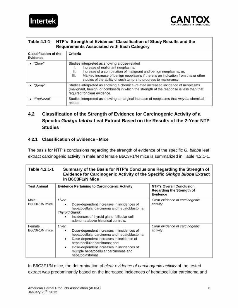
Table 4.1-1 NTP’s ‘Strength of Evidence’ Classification of Study Results and the Requirements Associated with Each Category
Classification of the Evidence
Criteria
“Clear” Studies interpreted as showing a dose-related I. Increase of malignant neoplasms;
II. Increase of a combination of malignant and benign neoplasms; or, III. Marked increase of benign neoplasms if there is an indication from this or other
studies of the ability of such tumors to progress to malignancy.
“Some” Studies interpreted as showing a chemical-related increased incidence of neoplasms (malignant, benign, or combined) in which the strength of the response is less than that required for clear evidence.
“Equivocal” Studies interpreted as showing a marginal increase of neoplasms that may be chemical related.
4.2 Classification of the Strength of Evidence for Carcinogenic Activity of a
Specific Ginkgo biloba Leaf Extract Based on the Results of the 2-Year NTP
Studies
4.2.1 Classification of Evidence - Mice
The basis for NTP’s conclusions regarding the strength of evidence of the specific G. biloba leaf
extract carcinogenic activity in male and female B6C3F1/N mice is summarized in Table 4.2.1-1.
Table 4.2.1-1 Summary of the Basis for NTP’s Conclusions Regarding the Strength of Evidence for Carcinogenic Activity of the Specific Ginkgo biloba Extract in B6C3F1/N Mice
Test Animal Evidence Pertaining to Carcinogenic Activity NTP’s Overall Conclusion Regarding the Strength of Evidence
Male B6C3F1/N mice
Liver:
Dose-dependent increases in incidences of hepatocellular carcinoma and hepatoblastoma.
Thyroid Gland:
Incidences of thyroid gland follicular cell adenoma above historical controls.
Clear evidence of carcinogenic activity
Female B6C3F1/N mice
Liver:
Dose-dependent increases in incidences of hepatocellular carcinoma and hepatoblastoma;
Dose-dependent increases in incidence of hepatocellular carcinoma; and
Dose-dependent increases in incidences of multiple hepatocellular carcinomas and hepatoblastomas.
Clear evidence of carcinogenic activity
In B6C3F1/N mice, the determination of clear evidence of carcinogenic activity of the tested
extract was predominantly based on the increased incidences of hepatocellular carcinoma and
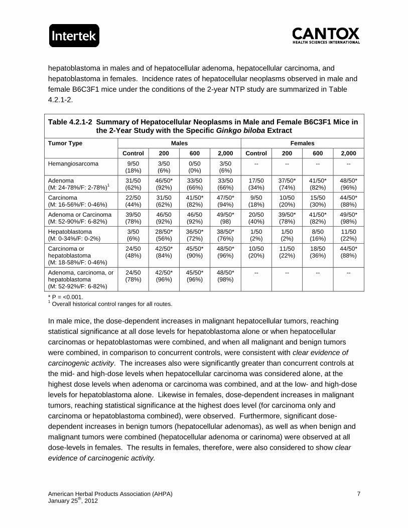
American Herbal Products Association (AHPA) January 25
th, 2012
7
hepatoblastoma in males and of hepatocellular adenoma, hepatocellular carcinoma, and
hepatoblastoma in females. Incidence rates of hepatocellular neoplasms observed in male and
female B6C3F1 mice under the conditions of the 2-year NTP study are summarized in Table
4.2.1-2.
Table 4.2.1-2 Summary of Hepatocellular Neoplasms in Male and Female B6C3F1 Mice in the 2-Year Study with the Specific Ginkgo biloba Extract
Tumor Type Males Females
Control 200 600 2,000 Control 200 600 2,000
Hemangiosarcoma 9/50 (18%)
3/50 (6%)
0/50 (0%)
3/50 (6%)
-- -- -- --
Adenoma (M: 24-78%/F: 2-78%)
1
31/50 (62%)
46/50* (92%)
33/50 (66%)
33/50 (66%)
17/50 (34%)
37/50* (74%)
41/50* (82%)
48/50* (96%)
Carcinoma (M: 16-56%/F: 0-46%)
22/50 (44%)
31/50 (62%)
41/50* (82%)
47/50* (94%)
9/50 (18%)
10/50 (20%)
15/50 (30%)
44/50* (88%)
Adenoma or Carcinoma (M: 52-90%/F: 6-82%)
39/50 (78%)
46/50 (92%)
46/50 (92%)
49/50* (98)
20/50 (40%)
39/50* (78%)
41/50* (82%)
49/50* (98%)
Hepatoblastoma (M: 0-34%/F: 0-2%)
3/50 (6%)
28/50* (56%)
36/50* (72%)
38/50* (76%)
1/50 (2%)
1/50 (2%)
8/50 (16%)
11/50 (22%)
Carcinoma or hepatoblastoma (M: 18-58%/F: 0-46%)
24/50 (48%)
42/50* (84%)
45/50* (90%)
48/50* (96%)
10/50 (20%)
11/50 (22%)
18/50 (36%)
44/50* (88%)
Adenoma, carcinoma, or hepatoblastoma (M: 52-92%/F: 6-82%)
24/50 (78%)
42/50* (96%)
45/50* (96%)
48/50* (98%)
-- -- -- --
* P = <0.001. 1 Overall historical control ranges for all routes.
In male mice, the dose-dependent increases in malignant hepatocellular tumors, reaching
statistical significance at all dose levels for hepatoblastoma alone or when hepatocellular
carcinomas or hepatoblastomas were combined, and when all malignant and benign tumors
were combined, in comparison to concurrent controls, were consistent with clear evidence of
carcinogenic activity. The increases also were significantly greater than concurrent controls at
the mid- and high-dose levels when hepatocellular carcinoma was considered alone, at the
highest dose levels when adenoma or carcinoma was combined, and at the low- and high-dose
levels for hepatoblastoma alone. Likewise in females, dose-dependent increases in malignant
tumors, reaching statistical significance at the highest does level (for carcinoma only and
carcinoma or hepatoblastoma combined), were observed. Furthermore, significant dose-
dependent increases in benign tumors (hepatocellular adenomas), as well as when benign and
malignant tumors were combined (hepatocellular adenoma or carinoma) were observed at all
dose-levels in females. The results in females, therefore, were also considered to show clear
evidence of carcinogenic activity.

American Herbal Products Association (AHPA) January 25
th, 2012
8
4.2.2 Classification of Evidence - Rats
Table 4.2.2-1 presents the basis for NTP’s conclusions regarding the strength of evidence of the
specific G. biloba leaf extract carcinogenic activity in male and female F344/N rats under the
conditions of the 2-year study.
Table 4.2.2-1 Summary of the Basis for NTP’s Conclusions Regarding the Strength of Evidence for Carcinogenic Activity of the Specific Ginkgo biloba Extract in F344/N Rats
Test Animal Evidence Pertaining to Carcinogenic Activity NTP’s Overall Conclusion Regarding the Strength of Evidence
Male F344/N rats
Thyroid Gland:
Incidences of thyroid gland follicular cell adenoma above historical controls.
Some evidence of carcinogenic activity
Female F344/N rats
Thyroid Gland:
Incidences of thyroid gland follicular cell adenoma above historical controls;
Incidences of thyroid gland follicular cell adenoma or carcinoma above historical controls;
Single occurrences of follicular cell carcinoma (rare tumor).
Some evidence of carcinogenic activity
In rats, the NTP based their conclusion of some evidence of carcinogenic activity largely on the
marginal increases in the incidences of benign follicular cell thyroid gland tumors (adenomas) in
both males and females in comparison to control values and on the isolated occurrences of
follicular cell carcinoma in females. The incidences of follicular cell thyroid non-neoplastic and
neoplastic tumors observed in rats in the 2-year NTP study following administration of the
specific G. biloba leaf extract are summarized in Table 4.2.2-2.

American Herbal Products Association (AHPA) January 25
th, 2012
9
Table 4.2.2-2 Summary of Thyroid Neoplasms and Nonneoplastic Lesions in Male and Female F344/N Rats in the 2-Year Study with the Specific Ginkgo biloba Extract
Lesion Type Males Females
Control 100 300 1,000 Control 100 300 1,000
FC hypertrophy 13/50 37/50** 41/49** 41/45** 15/49 41/50** 45/49** 48/49**
Follicle hyperplasia 0/50 7/50** 9/49** 5/49* 3/49 3/50 1/49 5/49
FC Adenoma (M: 0-6%/F: 0-2%)
1
2/50 (4%)
1/50 (2%)
3/49 (6%)
5/45 (11%)
1/49 (2%)
0/50 (0%)
3/49 (6%)
1/49 (2%)
FC Carcinoma (F: 0-4%)
-- -- -- -- 0/49 0/50 1/49 1/49
FC Adenoma or carcinoma (F: 0-4%)
-- -- -- -- 1/49 (2%)
0/50 (0%)
4/49 (8%)
2/49 (4%)
FC, Follicular cell. * P = <0.05; ** P = <0.01. 1 Overall historical control ranges for all routes.
Specifically, the incidence of follicular cell adenomas in males (1/50, 3/49, and 5/45 at 100, 300,
and 1,000 mg/kg body weight/day, respectively) did exceed historical control values for corn oil
gavage studies (0 to 4%) at the mid- and high-dose levels (6 and 11% at 300 and 1,000 mg/kg
body weight/day, respectively) and historical control values for all routes of administration (0 to
6%) at the high-dose level. However, none of the increases reached statistical significance
when compared to the concurrent control group (2/50 or 4%). In females, the incidence of
follicular cell adenoma (0/50, 3/49, and 1/49 at 100, 300, and 1,000 mg/kg body weight/day,
respectively) was increased above historical control values for corn oil gavage studies and all
routes of administration, but only at the mid-dose level (6% versus 0 to 2%). As in males, none
of the increases were statistically significant in comparison to the concurrent control group (1/49
or 2%). Although the single incidence of follicular cell carcinomas observed in each the mid-
and high-dose females was deemed to be a rare occurrence, at 2% it fell within the historical
control ranges for follicular cell carcinomas for corn oil gavage studies (0 to 2%) and for all
routes of administration (0 to 4%). When both malignant and benign follicular cell tumors were
combined (adenoma or carcinoma), only the incidence at the mid-dose level (4/49 or 8%)
exceeded historical control values.
Non-neoplastic lesions of the thyroid gland in the 2-year study consisted of significantly
increased incidence rates of follicular cell hypertrophy at all dose levels in both males and
females, accompanied by significantly increased incidences of follicle hyperplasia in males only.
The hypertrophy noted in both sexes, while occurring at greater levels than in the control group,
was only of minimal to mild severity. Follicle cell hyperplasia in the males was considered as
minimal at the low-dose level and between mild to moderate at the mid- and high-dose levels.
In the 3-month dose selection study, a significant increase in the incidence of minimal to mild

American Herbal Products Association (AHPA) January 25
th, 2012
10
follicular cell hypertrophy also was observed at the 2 highest dose levels in males and at the
highest dose level in females. In the 14-week special study conducted in association with the
2-year rat study, low-dose males exhibited a significant increase in relative thyroid weight.
Thyroid hormone level analysis in the 14-week special study revealed a dose-dependent
increase in thyroid stimulating hormone (TSH), reaching statistical significance at all dose levels
in males and at the highest dose level in females.
The changes observed in parameters assessing thyroid function in the 3-month study and the
14-week special study, together with the non-neoplastic changes in the thyroid of both males
and females are indicative of a compound-related effect of the specific G. biloba leaf extract on
the thyroid, and collectively support that the slight increase in follicular cell neoplasms was likely
related to the administration of the extract. However, given that the incidences of benign
follicular cell tumors were only increased in comparison to historical controls, and in females,
only at the mid-dose level, it could be argued that the evidence for carcinogenic activity is only
“equivocal” based on the absence of a statistically significant response in comparison to
concurrent control values.
5.0 ASSESSMENT OF THE RELEVANCE OF THE NTP STUDY
RESULTS FOR HUMAN CANCER RISK
5.1 Hepatocellular Tumors
In light of the high prevalence of hepatocellular tumors observed in mice, the relevance of the
development of liver tumors in mice has been questioned with regard to human cancer risk
(Maronpot et al., 1987; Velazquez et al., 1996). The NTP has recognized the limitations of data
pertaining to the development of liver tumors in the 2-year mouse bioassays, particularly in
susceptible strains of mice (e.g., B6C3F1), with respect to extrapolating the results to humans in
risk assessments and has noted that alternative rodent strains are being examined to
supplement rat studies (Maronpot et al., 1987). The predictive value of mouse hepatocellular
tumors with respect to human cancer risk has been repeatedly challenged (Velazquez et al.,
1996; Carmichael et al., 1997). This is in part due to the fact that hepatocellular carcinoma in
humans, particularly chemically-induced, is rare. In humans, the major risk factors associated
with liver tumors are viral hepatitis, excessive alcohol consumption, and exposure to aflatoxin, in
most cases accompanied by liver cirrhosis.
Most recently, the European Food Safety Authority (EFSA) reiterated that “hepatic tumors in
mice are generally considered as irrelevant for human risk assessment” as part of their
evaluation of a mouse dietary administration study conducted with aspartame (EFSA, 2011).
Specifically, EFSA indicated in the report that there is general scientific consensus that
American Herbal Products Association (AHPA) January 25
th, 2012
11
induction of hepatocellular tumors in mice by non-genotoxic compounds can be considered as
irrelevant for human risk assessment (Holsapple et al., 2006; Billington et al., 2010). In their
evaluation of the mode of action with respect to the relevance of rodent liver tumors to human
cancer risk, Holsapple et al. (2006) concluded that in the case of chemicals displaying a
phenobarbital-like P450 inducing mode of action, the observed hepatocarcinogenicity in rodents
is not relevant to humans. Indeed, clinical use for over 80 years of phenobarbital, a known
enzyme inducer in the rodent liver, has not been associated with an increased risk of tumor
formation in the liver or any other organ in humans (McClain, 1990). In comparison to rats and
humans, mice also are known to possess higher levels of monooxygenase activities, including
increased levels of aryl hydrocarbon hydroxylase and biphenyl 2-hydroxylase, which are
catalyzed by cytochrome P450 (Lorenz et al., 1984; Parke and Ioannides, 1990; Thorgeirsson et
al., 1997). There are several examples of chemicals that are known to produce hepatocellular
tumors in mice, but not in rats (Maronpot et al., 1987; Velazquez et al., 1996). Consideration
also has been previously given to the possibility of a chemical with neoplastic effects confined to
the mouse liver, inducing carcinogenic effects in humans in another organ. As noted by
Carmichael et al. (1997), while there is an example of a genotoxic rodent (both mice and rats)
liver carcinogen (i.e., benzidine) that is known to produce bladder tumors in humans, there is no
example of a non-genotoxic mouse liver carcinogen that produces a tumorgenic response in
humans in another organ. In the case of compounds inducing hepatocellular tumors via a
cytotoxic mode of action, the carcinogenic response should be considered in the evaluation of
human cancer risk if appropriate metabolism occurs in the animal model and in humans.
Although the specific G. biloba extract tested positive for mutagenic activity in the Ames assay,
in vivo a negative response was obtained under the conditions of the micronucleus assay in
B6C3F1/N male mice and in females the response was determined to be equivocal based on
only a positive trend for increasing levels of micronuclei. While the response in females was
determined to be equivocal based on trend analysis, none of the increases in the incidence
rates of micronuclei in treated females attained statistical significance in comparison to controls
and none of the increases were more than twice the control values at any dose level. The
presence of a positive response in vitro, but not in vivo of the specific G. biloba extract is similar
to the results of genotoxicity assays obtained for quercetin, which is one of the main
constituents of the extract (it should be noted however that in the G. biloba extracts quercetin is
glycosylated). The NTP draft report repeatedly indicates quercetin to be a mutagen; however,
quercetin has been extensively studied for its mutagenic and genotoxic properties, both in vitro
and in vivo, and while in vitro unequivocally positive results have been reported, in vivo
quercetin has consistently tested negative (as reviewed by Harwood et al., 2007). Furthermore,
review of long-term animal studies with quercetin also demonstrated no carcinogenic activity for
the compound. It has been shown that in the case of quercetin adequate metabolic processes,
resulting in low bioavailability of unmetabolized quercetin, are operative in vivo that protect the

American Herbal Products Association (AHPA) January 25
th, 2012
12
organism from genotoxic activity that may be apparent in vitro. Therefore, while the NTP draft
report refers to quercetin as a known mutagen, this is not qualified in terms of in vitro versus in
vivo effects. Furthermore, as already noted, G. biloba extracts contain almost exclusively
quercetin glycosides and only trace amounts of flavonol aglycones (Upton, 2003). The flavonol
glycosides undergo extensive first-pass metabolism and reach the blood and tissues neither as
aglycones nor as glycosides. The glycosides are quickly deglycosylated to the aglycone and
immediately conjugated with glucuronate or sulfate with or without methylation. Based on the
negative in vivo results obtained in male mice following treatment with the specific G. biloba
extract in the assays conducted by the NTP and the equivocal results in female mice, which
interestingly showed a comparatively lower carcinogenic response in the 2-year study than
males, the presence of quercetin glycosides in the extract, and the absence of quercetin-related
in vivo genotoxicity or carcinogenic activity, the ability of the G. biloba extract to exert genotoxic
properties in vivo is very questionable. It is more likely therefore that the effects observed in the
2-year mouse study were the result of non-genotoxic mechanisms. When considered
cohesively, the carcinogenic responses observed in the NTP rodent studies are suggestive of a
compound with enzyme stimulatory properties.
Several in vitro and in vivo studies examining the effects on rodent and human cytochrome
P450 (CYP450) enzymes have been conducted. In rats, G. biloba extracts have been reported
to induce CYP3A enzymes in a dose-dependent manner in vitro with no species difference
between humans and rats (Deng et al., 2008a). In vivo studies conducted with mice and rats
have confirmed the ability of G. biloba extracts and its constituents (specifically bilobalide) to
induce CYP enzymes (Sugiyama et al., 2004; Umegaki et al., 2007; Deng et al., 2008b; Taki et
al., 2009). In a study examining the ability of the 5 major constituents of G. biloba extract
(bilobalide, ginkgolide A, B, quercetin, and kaempferol) on CYP enzyme induction, bilobalide
dose-dependently increased the activity of CYP3A1 and CYP2E1, whereas ginkgolide A, B,
quercetin, and kaempferol dose dependently increased the activity of CYP1A2 (Deng et al.,
2008b). However, in humans, no induction of CYP450 enzymes has been observed based on
the results of a number of clinical trials (Duche et al., 1989; Gurley et al., 2002; Markowitz et al.,
2003; Izzo and Ernst, 2009; Zuo et al., 2010; Zadoyan et al., 2011).
In addition to the species-specific susceptibility of the B6C3F1 mouse to develop liver tumors,
the levels at which the specific G. biloba extract was tested in the 2-year bioassay may be
crucial to the interpretation of the liver tumors for purposes of human safety assessment. Most
commonly, the highest dose level evaluated in a 2-year bioassay is set at the MTD in order to
ensure that the assay is sensitive enough to detect a carcinogenic response without inducing
excessive lethality (Chhabra et al., 1990; Gaylor, 2005). In the NTP studies on the specific G.
biloba extract, the dose levels selected for purposes of the 2-year assays (i.e., 100, 300, and
1,000 mg/kg body weight/day in rats and 200, 600, and 2,000 mg/kg body weight/day in mice)
were based on the results of preliminary 3-month studies. The 3-month studies were conducted
American Herbal Products Association (AHPA) January 25
th, 2012
13
at dose levels of 62.5, 125, 250, 500, and 1,000 mg/kg body weight/day and 125, 250, 500,
1,000, and 2,000 mg/kg body weights/day in rats and mice, respectively. The rationale for the
dose selection in both rodent species for purposes of the 2-year studies was based on the
absence of an effect on survival, and in the case of rats also an absence of an effect on body
weights. Hypertrophy of the liver and thyroid in rats and liver hypertrophy, along with body
weight and organ weight changes in mice, were noted in the 3-month studies, but considered
not to be “life-threatening”. However, as discussed in Section 3.3, considering the overt
increases in both relative and absolute liver weights following only 3 months of administration of
the specific G. biloba extract at the lowest dose level tested (125 mg/kg body weight/day) in
B6C3F1 mice with a demonstrated sensitivity for hepatocellular tumor development, and the
association shown between liver tumor development and relative liver weight in mice
(Carmichael et al., 1997), it is likely that especially in the mouse the lowest dose tested (200
mg/kg body weight/day) in the 2-year study could be predicted in advance to produce effects on
the liver, including the development of liver tumors.
Administration of compounds at the MTD has been argued to result in repeated cell damage
that is followed by reparative hyperplasia and ultimately uncontrolled cell growth (Velazquez et
al., 1996; Carmichael et al., 1997). Such a proliferative response at the MTD leading to tumor
formation may not be relevant to an assessment of risk in humans at lower dose levels of
exposure. A series of degenerative non-neoplastic changes were observed in the livers of male
and female mice in association with the neoplasms, including hypertrophy, erythrophagocytosis,
hematopoietic cell proliferation, inflammation, necrosis, and altered hepatic foci. In a report
published by the National Academy of Sciences Committee on Risk Assessment Methodology
(CRAM), it was stated that “[the MTD bioassay] does not provide…all the information useful for
low-dose human risk assessment” (NRC, 1993) and for chemicals that do induce cancer at the
MTD, it was suggested that additional data are necessary to determine the relevance of the
response to human health risk assessment (Velazquez et al., 1996). Liver enzyme induction
has also been suggested as an alternative non-genotoxic mechanism underlying liver tumor
formation (Carmichael et al., 1997). Liver tumors observed in long-term studies with
compounds with a known proliferative effect on liver enzymes also are considered to be the
result of high dose administration interfering with normal liver function. Overall, these non-
genotoxic mechanisms are thought to be threshold dependent and it should therefore be
possible to establish a dose below which an adverse response is not elicited, which can then be
used in an assessment of human risk. However, since the lowest dose level tested in the
2-year mouse study was likely in excess of a level at which no neoplastic effects of the liver
would be expected in mice, it is not possible to extrapolate the results to a dose level that would
not induce liver cell hypertrophy and subsequent tumor development in B6C3F1 mice.
The results of the 2-year rat study provide additional support that the neoplastic hepatocellular
effects observed in the mice were the results of treatment of a highly susceptible species at high
American Herbal Products Association (AHPA) January 25
th, 2012
14
dose levels. In a less sensitive species, such as the rat, and at lower levels of use, these
effects would likely not be observed. Although there were changes in a number of hepatic
parameters noted in the 13-week rat study, as well as in the 14-week special rat study, along
with non-neoplastic lesions in the 2-year study (hepatocellular hypertrophy, bile duct and oval
cell hyperplasia, necrosis, cystic degeneration in males and hepatocellular hypertrophy, bile
duct hyperplasia, and focal fatty change in females), none of these observed changes
progressed to produce a statistically significant increase in liver tumors in the rat. In female rats
no incidences of benign or malignant liver tumors were reported and in males only a slight
increase in benign liver tumors above historical control values was noted. Specifically, although
incidences of hepatocellular adenoma in male rats exceeded historical control values for corn oil
gavage studies at the low- and mid-dose levels, the incidences were within the historical control
ranges for all routes of administration. Furthermore, no adenomas were identified in male rats
at the highest dose level tested.
It was noted that the hepatoblastomas observed in both males and females in the 2-year mouse
study were considered relatively rare tumors. However, while hepatoblastoma is considered to
be an infrequent tumor type of NTP 2-year studies, it is a variant of hepatocellular carcinoma
and thus appears in animals that have also developed hepatocellular carcinoma (Maronpot et
al., 1987; Turusov et al., 2002). While they are rare, the occurrence of the hepatoblastomas in
the 2-year G. biloba mouse study is not entirely unexpected in light of the overall increase in the
incidence of hepatocellular lesions; however, the magnitude of the increase, given the rarity of
the tumor, is somewhat unprecedented. Markedly higher levels of hepatoblastomas also have
been observed following treatment of male D2B6F1 mice (11/30 or 37%) with phenobarbital,
another enzyme stimulating compound, in the diet at 500 ppm (approximately 62.5 mg/kg body
weight/day), for a period of up to 103 weeks (Diwan et al., 1995). Considering that G. biloba
extract was administered at higher levels (≥200 mg/kg body weight/day), the incidence rates of
56, 72, and 78% for hepatoblastomas at 200, 600, and 2,000 mg/kg body weight/day,
respectively, might not be completely unexpected for a compound with enzyme-inducing
properties in the mouse.
Furthermore, there is indication that there are strain-specific differences in the susceptibility for
the development of hepatoblastomas among mice strains, as supported by an example of a
greater sensitive of B6C3F1 mice compared to Swiss-Webster mice for chemically induced
hepatoblastoma (Turusov et al., 2002). Interestingly, an increase in the spontaneous incidence
of hepatoblastomas in B6C3F1 mice in 2-year NTP studies has been reported (Turusov et al.,
2002). Although Turusov et al. (2002) could not identify an explanation for the apparent
increase in the background incidence of mouse hepatoblastoma, genetic drift in the NTP
B6C3F1 mice was suggested. Also, as in the case of the specific G. biloba extract, 6 chemicals
previously tested by the NTP (i.e., benzofuran, o-nitroanisole, oxazepam, pyridine, primidone,
and anthraquinone) produced high incidences of hepatoblastomas in mice, but failed to produce

American Herbal Products Association (AHPA) January 25
th, 2012
15
any increases in any liver tumors in rats. Additionally, differences pertaining to the onset in life
and development of the tumor have been identified between hepatoblastomas in mice and
humans (Turusov et al., 2002). Specifically, in humans hepatoblastomas are exceptionally rare
in adults, but do develop in young children and sometimes in utero. In contrast,
hepatoblastomas in mice have only been reported in aged animals, with only a limited number
of cases of hepatoblastomas identified at 15-month necropsies. Moreover, unlike in mice in
which hepatoblastomas appear to arise within preexisting hepatocellular adenomas and
carcinomas, in humans the tumor type is formed de novo. It is worth noting however, that a
common molecular alteration involving the β-catenin/Wnt signaling pathway has been reported
in both mice and humans (Koch et al., 1999; Anna et al., 2000).
Considering that the etiology of hepatocellular tumors in humans is generally accepted to be
different from that observed in murine test systems, combined with the fact that the testing of
the specific G. biloba extract in the 2-year studies was likely conducted at excessive dose levels
and that mice, particularly of the B6C3F1 strain are susceptible to the development of liver
tumors, the relevance of the results of the 2-year mouse bioassay with respect to human risk
characterization is questionable. This conclusion is further supported by the fact that the
mechanism for the liver neoplasms in the study is likely to have been the result of prolonged
liver enzyme induction with related hepatocellular hypertrophy. Chronic hepatocellular
hypertrophy is recognized to lead to toxic and degenerative changes that in time result in
necrosis and initiation of reparative processes. In rats, no significant increase in the incidence
of liver tumors was observed, further corroborating that the occurrence of the liver tumors in
mice was likely a species-specific response as a result of genetic predisposition, in combination
with a high dose response associated with enzyme-induction and liver cell hypertrophy. Finally,
while there is some evidence supporting the enzyme stimulatory properties of G. biloba extract
in rodents, in humans, no induction of CYP450 enzymes has been observed based on the
results of a number of clinical trials (Duche et al., 1989; Gurley et al., 2002; Markowitz et al.,
2003; Izzo and Ernst, 2009; Zuo et al., 2010; Zadoyan et al., 2011).
5.2 Thyroid Tumors
As described in Section 4.2.2, incidences of follicular cell hypertrophy (minimal to mild severity)
in male and female rats and follicle hyperplasia in male rats (mild to moderate) were significantly
increased in comparison to control values. The increases in the incidence of follicular cell
adenomas in both sexes of treated rats were slightly elevated above historical control values
only. The single occurrences of follicular cell carcinoma in females at each the mid- and high-
dose level, while rare, were not in excess of the historical control ranges (for either corn oil
gavage studies or all routes of administration). In the 2-year mouse study, dose-dependent
increases also were noted in follicular cell hyperplasia and hypertrophy (minimal to mild),
reaching statistical significance in mid- and high-dose females (hypertrophy only) and in high-

American Herbal Products Association (AHPA) January 25
th, 2012
16
dose males. With respect to the incidence of neoplasms, an increase only in the incidence of
adenomas in mid- and high-dose males was noted and only in comparison to historical controls.
The etiology of follicular cell thyroid tumor formation in humans is generally thought to be similar
to that of experimental animals, with perturbations in hormones of the thyroid and pituitary as
the cause of most thyroid tumors (Velazquez et al., 1996). However, it is also recognized that
while thyroid follicular cell tumors can develop in humans as a result of chronic imbalances in
the thyroid-pituitary feedback mechanism, in humans, this is a threshold-dependant and much
less sensitive response. Conversely, rodents are exceptionally more sensitive to thyroid
hormone imbalances (Alison et al., 1994; McClain, 1995). Critical to this interspecies difference
is the variability in the affinity with which the circulating thyroid hormones are bound to albumin
in humans and other species. Specifically in rodents (mice and rats), the thyroid hormone
plasma half-lives are approximately 10 times shorter than in humans, consequently resulting in
a considerably greater turnover rate of the hormones in rodents.
Enzyme inducers, like phenobarbital, increase the metabolism and excretion of thyroid
hormones (T4 and T3) and are known to promote thyroid tumor formation in rodents secondary
to the effects on hormone balance (Alison et al., 1994). Extended induction of the thyroid by
elevated levels of TSH in response to decreased levels of circulating thyroid hormones leads to
follicular cell hypertrophy and hyperplasia, with eventual progression to thyroid tumors (Capen,
1997). Species that are generally more sensitive to such hormonal imbalance, like rodents, are
particularly prone to secondary tumor formation as a result of chemically-induced (via for
example enzyme inducers) disturbances in the feedback mechanism that regulates thyroid
function. In rodents, only mild to moderate perturbations in thyroid hormone homeostasis will
affect the incidence of thyroid tumor formation (McClain, 1995). Reportedly, only few
compounds are known to result in a TSH-stimulating effect in humans (Alison et al., 1994).
Epidemiological data corroborate that in humans chronic TSH-induced induction of the thyroid is
only rarely associated with neoplasia (Paynter et al., 1988; McClain, 1995). Given the inherent
predisposition of the rodent to thyroid tumor formation, thyroid tumors in rodent studies as a
result of secondary, non-genotoxic mechanisms (i.e., hormone imbalance) bear little relevance
to assessment of carcinogenic risk, particularly at dose levels that do not disrupt thyroid function
in humans (Alison et al., 1994; McClain, 1995).
Considering that under the conditions of the NTP study, gavage administration of the specific G.
biloba extract was associated with dose-dependent increases in levels of TSH in both male and
female rats (as observed in the 14-week special study), combined with in vivo data in rodents
demonstrating that G. biloba extracts possesses enzyme inducing properties, it is therefore
reasonable to conclude that the thyroid tumors observed in rats were a secondary response to
hormonal imbalances leading to prolonged stimulation of the thyroid. Furthermore, the
increased incidence of the thyroid tumors observed in rats and mice did not reach statistical

American Herbal Products Association (AHPA) January 25
th, 2012
17
significance over the course of the 2-year study periods, suggesting that while the specific G.
biloba extract did have an effect on thyroid function, the imbalances, in a highly sensitive
species, did not lead to an overwhelming tumor response in the thyroid. However, in this regard
it is also important to consider whether the absence of a statistically significant response may
have resulted from the occurrence of deaths prior to study termination as a result of non-thyroid
related effects. A decrease in survival was observed in high-dose male rats as a result of
mononuclear cell leukemia; however, the increase in early deaths was only notable beginning at
Week 85 (600 days), which was not much earlier than the first incidence day for thyroid tumors
in the high-dose male (Day 675). In low- and mid-dose male rats, as well as in all groups of
female rats, survival rates were similar to the control group, so the absence of a statistically
significant response is unlikely to have been the effect of animals dying prior to the development
of thyroid gland tumors, particularly since the appearance of thyroid tumors while generally later
in life, was with the exception of mid-dose males, similar between control and test animals. In
fact, in mid-dose male rats in which the first incident day was considerably earlier than in the
other test groups (Day 485) and in which survival was not affected, a statistically significant
response in comparison to the control group also was not generated. Therefore, with the
exception of perhaps the high-dose male rats, it is unlikely that the lack of a statistically
significant response observed in rats in relation to the development of thyroid tumors was due to
pre-term mortality of other causes.
6.0 CONCLUDING REMARKS
Review of the details pertaining to the identity and characterization of the specific G. biloba leaf
extract demonstrated that the stability of the test material based on the data provided in the NTP
draft report could not be adequately verified. Furthermore, the possibility of the presence of
potential degradation products following storage could not be excluded based on the available
data presented in the report. Also, it was noted that data related to heavy metal, microbiology,
mycotoxin, polyaromatic hydrocarbon, and pesticide analysis, were not provided. In the
absences of such data it is not possible to exclude the possibility that the results observed in the
NTP studies were not related to the specific G. biloba leaf extract per se, but rather to other
unidentified contaminants, impurities, or degradants present in the test material.
It was also noted that in the 2-year mouse study, the lowest dose level tested (200 mg/kg body
weigh/day) was higher than the lowest dose tested in the dose-range determining 3-month
study (125 mg/kg body weight/day), at which significant increases in liver weights of mice were
observed. Considering that significant effects were observed at the lowest dose tested in the
3-month study, the rationale for the selection of a dose that is higher than the 3-month low-dose
as the lowest dose for purposes of a 2-year study is questionable.

American Herbal Products Association (AHPA) January 25
th, 2012
18
While the above noted study design issues may have implications on the overall study results
that should be considered before any conclusions regarding the carcinogenic activity of the test
material can be made, assuming that the results presented in the NTP draft report were indeed
related to the specific G. biloba extract tested, oral administration of the specific G. biloba leaf
extract at doses ranging from 200 to 2,000 mg/kg body weight/day under the conditions of the
2-year NTP study, was associated with clear evidence of carcinogenic activity in B6C3F1/N
mice. In F344/N rats, the NTP determined that the results obtained following oral administration
of the specific G. biloba leaf extract at doses ranging from 100 to 1,000 mg/kg body weight/day
was indicative of some evidence of carcinogenic activity. However, while the increases in the
incidence of thyroid tumors were in excess of historical control values, the increases were noted
not to reach levels of statistical significance in comparison to the concurrent control group.
Furthermore, the increases were limited only to benign neoplasms. Given therefore the
absence of a statistically significant response in comparison to the concurrent control group and
no increase in malignant tumor types, the results could be argued to be supportive only of
“equivocal” evidence of carcinogenic activity in F344/N rats.
Furthermore, although under the conditions of the NTP studies the specific G. biloba extract
tested was associated with varying levels of carcinogenic activity in rodents, the carcinogenic
responses observed in these studies at relatively high dose levels are not necessarily predictive
of the extract exerting similar responses in humans at lower levels of exposure. Specifically, the
carcinogenic effects observed in the liver and thyroid in the rodent studies were likely observed
as secondary effects of enzyme perturbations at high-dose levels, rather than as direct
carcinogenic responses. Particularly, the ability of the extract to stimulate P450 enzymes
should be considered as the mechanism underlying the effects observed in the 2-year studies.
Indeed, the overall profile of the carcinogenic responses observed in the 2-year NTP studies is
consistent with that of a chemical with enzyme stimulatory properties.
In light of the underlying non-genotoxic mechanism of action related to enzyme induction, the
hepatocellular response in mice might be deemed to be irrelevant to assessment of human
safety of the extract, whereas the response observed in the thyroid of both rats and mice is
likely to be a threshold dependent effect that is considerably more pronounced in rodents than
in humans. Although the increases in follicular cell tumors above historical control values were
likely related to the administration of G. biloba in rats and mice, similar neoplastic changes of
the thyroid in humans as a result of G. biloba consumption would be unlikely considering the
differences in thyroid hormone homeostastis in humans and the lower levels of human
exposure. Moreover, while in rodents there are data showing enzyme induction related
G. biloba extract exposure, a limited number of studies have demonstrated a lack of an enzyme
stimulating effect of G. biloba in humans.

American Herbal Products Association (AHPA) January 25
th, 2012
19
7.0 REFERENCES
Alison RH, Capen CC, Prentice DE (1994). Neoplastic lesions of questionable significance to humans. Toxicol Pathol 22(2):179-186.
Anna CH, Sills RC, Foley JF, Stockton PS, Ton T-V, Devereux TR (2000). β-Catenin mutations and protein accumulation in all hepatoblastomas examined from mice treated with anthraquinone or oxazepam. Cancer Res 60(11):2864-2868. Cited In: Turusov et al., 2002 [Ref. #1].
Barnes J, Anderson LA, Phillipson JD (2007). Ginkgo. In: Herbal Medicines, 3rd edition. London, Engl.: Pharmaceutical Press, pp. 299-314.
Barrett B, Kiefer D, Rabago D (1999). Assessing the risks and benefits of herbal medicine: an overview of scientific evidence. Altern Ther Health Med 5(4):40-49.
Billington R, Lewis R.W, Mehta J.M, Dewhurst I (2010). The mouse carcinogenicity study is no longer a scientifically justifiable core data requirement for the safety assessment of pesticides Crit Rev Toxicol 40(1):35-49.
Blumenthal M, Busse WR, Goldberg A, Gruenwald J, Hall T, Riggins CW et al., editors (1998). Ginkgo biloba leaf extract. In: The Complete German Commission E Monographs: Therapeutic Guide to Herbal Medicines [Sigrid Klein, Primary Translator]. Austin (TX): American Botanical Council (ABC).
Capen CC (1997). Mechanistic data and risk assessment of selected toxic end points of the thyroid gland. Toxicol Pathol 25(1):39-48.
Carmichael NG, Enzmann H, Pate I, Waechter F (1997). The significance of mouse liver tumor formation for carcinogenic risk assessment: results and conclusions from a survey of ten years of testing by the agrochemical industry. Environ Health Perspect 105(11):1196-1203.
Chhabra RS, Huff JE, Schwetz BS, Selkrik J (1990). An overview of prechronic and chronic toxicity/carcinogenicity experimental study designs and criteria used by the National Toxicology Program. Environ Health Perspect 86:313-321. Cited In: Velazquez et al., 1996.
Deng Y, Bi HC, Zhao LZ, Wang XD, Chen J, Ou ZM et al. (2008a). Induction of cytochrome P450 3A by the Ginkgo biloba extract and bilobalides in human and rat primary hepatocytes. Drug Metab Lett 2(1):60-66 [abstract only].
Deng Y, Bi HC, Zhao LZ, He F, Liu YQ, Yu JJ et al. (2008b). Induction of cytochrome P450s by terpene trilactones and flavonoids of the Ginkgo biloba extract EGb 761 in rats. Xenobiotica 38(5):465-481.
Diwan BA, Henneman JR, Rice JM (1995). Further evidence for promoter-dependent development of hepatoblastoma in the mouse Cancer Lett 89(1&2):29-35.

American Herbal Products Association (AHPA) January 25
th, 2012
20
Drinkwater NR, Hanigan MH, Kemp CJ (1989). Genetic determinants of hepatocarcinogenesis in the B6C3F1 mouse. Toxicol Lett 49(2&3):255-265.
Duche JC, Barre J, Guinot P, Duchier J, Cournot A, Tillement JP (1989). Effect of Ginkgo biloba extract on microsomal enzyme induction. Int J Clin Pharmacol Res 9(3):165-168. Cited In: Zadoyan et al., 2011 [Ref. #23].
EFSA (2011). European Food Safety Authority; EFSA Statement on the scientific evaluation of two studies related to the safety of artificial sweeteners (question no EFSA-Q-2011-00064, approved on 25 February 2011 by European Food Safety Authority). EFSA J 9(2):2089 [16 pp.]. doi:10.2903/j.efsa.2011.2089. Available at: http://www.efsa.europa.eu/en/efsajournal/pub/2089.htm.
Gaylor DW (2005). Are tumor incidence rates from chronic bioassays telling us what we need to know about carcinogens. Regul Toxicol Pharmacol 41(2):128-133.
Gurley BJ, Gardner SF, Hubbard MA, Williams DK, Gentry WB, Cui Y et al. (2002). Cytochrome P450 phenotypic ratios for predicting herb-drug interactions in humans. Clin Pharmacol Ther 72(3):276-287. Cited In: Zadoyan et al., 2011 [Ref. #24].
Harwood M, Danielewska-Nikiel B, Borzelleca JF, Flamm GW, Williams GM, Lines TC (2007). A critical review of the data related to the safety of quercetin and lack of evidence of in vivo toxicity, including lack of genotoxic/carcinogenic properties. Food Chem Toxicol 45(11):2179-2205.
Health Canada (2009). Monograph: Ginkgo Biloba. In: Natural Health Products Ingredients Database - Single Ingredient and Product Monographs. Ottawa (ON): Health Canada, Natural Health Products Directorate (NHPD). Available at: http://webprod.hc-sc.gc.ca/nhpid-bdipsn/monoReq.do?id=100&lang=eng [Date: 2009-06-24; Date Database Last Changed: 2011-12-28].
Holsapple MP, Pitot HC, Cohen SM, Boobis AR, Klaunig JE, Pastoor T et al. (2006). Mode of action in relevance of rodent liver tumors to human cancer risk. Toxicol Sci 89(1):51-56.
Izzo AA, Ernst E (2009). Interactions between herbal medicines and prescribed drugs: an updated systematic review. Drugs 69(13):1777-1798. Cited In: Zadoyan et al., 2011 [Ref. #27].
Koch A, Denkhaus D, Albrecht S, Leuschner I, von Schweinitz D, Pietsch T (1999). Childhood hepatoblastomas frequently carry a mutated degradation targeting box of the β-catenin gene. Cancer Res 59(2):269-273. Cited In: Turusov et al., 2002 [Ref. #22].
Kroon PA, Clifford MN, Crozier A, Day AJ, Donovan JL, Manach C et al. (2004). How should we assess the effects of exposure to dietary polyphenols in vitro? Am J Clin Nutr 80(1):15-21.

American Herbal Products Association (AHPA) January 25
th, 2012
21
Lorenz J, Glatt HR, Fleischmann R, Ferlinz R, Oesch F (1984). Drug metabolism in man and its relationship to that in three rodent species: monooxygenase, epoxide hydrolase, and glutathione S-transferase activities in subcellular fractions of lung and liver. Biochem Med 32(1):43-56. Cited In: Carmichael et al., 1997 [Ref. #27].
Markowitz JS, Donovan JL, Lindsay DeVane C, Sipkes L, Chavin KD (2003). Multiple-dose administration of Ginkgo biloba did not affect cytochrome P-450 2D6 or 3A4 activity in normal volunteers. J Clin Psychopharmacol 23(6):576-581. Cited In: Zadoyan et al., 2011 [Ref. #25].
Maronpot RR, Haseman JK, Boorman GA, Eustis SL, Rao GN, Huff JE (1987). Liver lesions in B6C3F1 mice: the National Toxicology Program, experience and position. Arch Toxicol Suppl 10:10-26.
McClain RM (1990). Mouse liver tumors and microsomal enzyme-inducing drugs: experimental and clinical perspectives with phenobarbital. In: Stevenson DE, Popp JA, Ward JM, McClain RM, Slaga TJ, Pitot HC, editors. Mouse Liver Carcinogenesis: Mechanisms and Species Comparisons. Symposium, Nov. 30-Dec. 3, 1988, Austin, Texas. (Progress in Clinical and Biological Research, vol 331). New York (NY): Wiley-Liss, pp. 345-365. Cited In: Carmichael et al., 1997 [Ref. #34].
McClain RM (1995). Mechanistic considerations for the relevance of animal data on thyroid neoplasia to human risk assessment. Mutat Res 333(1&2):131-142.
NRC (1993). Use of the maximum tolerated dose in animal bioassays for carcinogenicity. In: Issues in Risk Assessment. National Research Council (NRC), Commission on Life Sciences, Committee on Risk Assessment in Methodology. Washington DC: National Academy Press (NAP), pp. 14-77. Cited In: Velazquez et al., 1996.
NTP (2011). Toxicology and Carcinogenesis Studies of Ginkgo biloba Extract (Cas No. 90045-36-6) in F344/N Rats and B6C3F1/N Mice (Gavage studies) – Draft - Scheduled Peer Review Date: February 8-9, 2012. (NTP Technical Report Series, no 578, NIH Publication no 12-5920). Research Triangle Park (NC): National Toxicology Program (NTP). Available at: http://ntp.niehs.nih.gov/Ntp/About_Ntp/Trpanel/2012/February/DraftTR578.pdf.
Parke DV, Ioannides C (1990). Role of cytochromes P-450 in mouse liver tumor production. In: Stevenson DE, Popp JA, Ward JM, McClain RM, Slaga TJ, Pitot HC, editors. Mouse Liver Carcinogenesis: Mechanisms and Species Comparisons. Symposium, Nov. 30-Dec. 3, 1988, Austin, Texas. (Progress in Clinical and Biological Research, vol 331). New York (NY): Wiley-Liss, pp. 215-230. Cited In: Carmichael et al., 1997 [Ref. #26].
Paynter OE, Burin GJ, Jaeger RB, Gregorio CA (1988). Goitrogens and thyroid follicular cell neoplasia: Evidence for a threshold process. Regul Toxicol Pharmacol 8(1):102-119. Cited In: Alison et al., 1994 [Ref. #25].
PDRHM (2007). Ginkgo. In: PDR® for Herbal Medicines, 4th edition. Montvale (NJ): Medical Economics Company, pp. 371-384.
American Herbal Products Association (AHPA) January 25
th, 2012
22
Sugiyama T, Kubota Y, Shinozuka K, Yamada S, Yamada K, Umegaki K (2004). Induction and recovery of hepatic drug metabolizing enzymes in rats treated with Ginkgo biloba extract. Food Chem Toxicol 42(6):953-957.
Taki Y, Yamazaki Y, Shimura F, Yamada S, Umegaki K (2009). Time-dependent induction of hepatic cytochrome P450 enzyme activity and mRNA expression by bilobalide in rats. J Pharmacol Sci 109(3):459-462.
Thorgeirsson SS, Atlas SA, Boobis AR, Felton JS (1997). Species differences in the substrate specificity of hepatic cytochrome P-448 from polycyclic treated animals. Biochem Pharmacol 28(2):217-226. Cited In: Carmichael et al., 1997 [Ref. #28].
Turusov VS, Torii M, Sills RC, Willson GA, Herbert RA, Hailey JR et al. (2002). Hepatoblastomas in mice in the US National Toxicology Program (NTP) studies. Toxicol Pathol 30(5):580-591.
Umegaki K, Taki Y, Endoh K, Taku K, Tanabe H, Shinozuka K et al. (2007). Bilobalide in Ginkgo biloba extract is a major substance inducing hepatic CYPs. J Pharm Pharmacol 59(6):871-877.
Velazquez SF, Schoeny R, Rice GE, Cogliano VJ (1996). Cancer risk assessment: historical perspectives, current issues, and future directions. Drug Chem Toxicol 19(3):161-185.
Upton R, editor (2003). Ginkgo Leaf Ginkgo Leaf Dry Extract (Ginkgo biloba L.) - Standards of Analysis, Quality Control, and Therapeutics. (American Herbal Pharmacopoeia and Therapeutic Compendium). Santa Cruz (CA): American Herbal Pharmacopoeia (AHP).
Zadoyan G, Rokitta D, Klement S, Dienel A, Hoerr R, Gramatté T et al. (2011). Effect of Ginkgo biloba special extract EGb 761® on human cytochrome P450 activity: a cocktail interaction study in healthy volunteers. Eur J Clin Pharmacol [advance electronic publication – Dec. 21, 2011].
Zuo XC, Zhang BK, Jia SJ, Liu SK, Zhou LY, Li J et al. (2010). Effects of Ginkgo biloba extracts on diazepam metabolism: a pharmacokinetic study in healthy Chinese male subjects. Eur J Clin Pharmacol 66(5):503-509. Cited In: Zadoyan et al., 2011 [Ref. #26].
Related Documents