American Journal Of ORTHODONTICS (All rights reserved) VOL. 40 JANUARY, 1954 No. 1 Original Articles A CRITICAL REVIEW OF CLINICAL CEPHALOMETRIC RADIOGRAPHY T. M. GRABER,D.D.S., M.S.D., PH.D.,* CHICAGO, ILL. T HE present high level of appliance construction and comparative efficiency of tooth movement have only accentuated the diversity between treat- ment objectives and treatment results. In our search for a single appliance that will manage all malocclusions, the trend has been strongly toward more and more control of individual teeth. The pendulum has swung all the way. The resulting complexity of appliances, while solving many problems of mechanotherapy, especially in Class I cases, has introduced new and more far- reaching problems and has pointed up the inadequacies of conventional Class II therapy. The controversy over extraction in orthodontic treatment has not been resolved. The increasing recognition of the role played by growth and development and of the limitations of each case is the inevitable result of an era of overmechanization, but there is no easy answer, no prognostic panacea. Let us not expect that cephalometrics will provide all the answers. Used properly, recognizing the limitations imposed by biometrics, the therapeutic road ahead can be made a good deal smoother. Cephalometrics, per se, is not new. Ever since Camper investigated prog- nathism craniometrically in 1791, anthropologists have been interested in the ethnographic determination of facial form and pattern.l Hellman made use of a rich experience in anthropology to study growth and development and to apply his findings to clinical orthodontics.2 Like so many investigators, Hell- man was searching for the so-called “normal” in his anthropologic cephalomet- rics. He lumped together skeletal material of unknown history and dubious ethnic origins, and developed his normal standard in a cross-sectional manner. Despite the obvious limitations of Hellman’s approach, it is a tribute to his astute interpretation of his material that what we know about the mechanism of cranial and facial growth today does not significantly differ from Hellman’s observations on dried skulls. Simon’s recognition of the contributions and limitations of anthropomet- rics permitted him to develop gnathostatics as a diagnost,ic medium, relating Read before the American Association of Orthodontists, Dallas, Texas, in April, 1953. *Associate Professor, Department of Orthodontics, Northwestern University. 1

Critical Review of Cephalometric Radiography
Dec 22, 2015
critical review of cephalometric radiograph
Welcome message from author
This document is posted to help you gain knowledge. Please leave a comment to let me know what you think about it! Share it to your friends and learn new things together.
Transcript

American Journal Of
ORTHODONTICS (All rights reserved)
VOL. 40 JANUARY, 1954 No. 1
Original Articles
A CRITICAL REVIEW OF CLINICAL CEPHALOMETRIC RADIOGRAPHY
T. M. GRABER, D.D.S., M.S.D., PH.D.,* CHICAGO, ILL.
T HE present high level of appliance construction and comparative efficiency of tooth movement have only accentuated the diversity between treat-
ment objectives and treatment results. In our search for a single appliance that will manage all malocclusions, the trend has been strongly toward more and more control of individual teeth. The pendulum has swung all the way. The resulting complexity of appliances, while solving many problems of mechanotherapy, especially in Class I cases, has introduced new and more far- reaching problems and has pointed up the inadequacies of conventional Class II therapy. The controversy over extraction in orthodontic treatment has not been resolved. The increasing recognition of the role played by growth and development and of the limitations of each case is the inevitable result of an era of overmechanization, but there is no easy answer, no prognostic panacea. Let us not expect that cephalometrics will provide all the answers. Used properly, recognizing the limitations imposed by biometrics, the therapeutic road ahead can be made a good deal smoother.
Cephalometrics, per se, is not new. Ever since Camper investigated prog- nathism craniometrically in 1791, anthropologists have been interested in the ethnographic determination of facial form and pattern.l Hellman made use of a rich experience in anthropology to study growth and development and to apply his findings to clinical orthodontics.2 Like so many investigators, Hell- man was searching for the so-called “normal” in his anthropologic cephalomet- rics. He lumped together skeletal material of unknown history and dubious ethnic origins, and developed his normal standard in a cross-sectional manner. Despite the obvious limitations of Hellman’s approach, it is a tribute to his astute interpretation of his material that what we know about the mechanism of cranial and facial growth today does not significantly differ from Hellman’s observations on dried skulls.
Simon’s recognition of the contributions and limitations of anthropomet- rics permitted him to develop gnathostatics as a diagnost,ic medium, relating
Read before the American Association of Orthodontists, Dallas, Texas, in April, 1953. *Associate Professor, Department of Orthodontics, Northwestern University.
1

dent’ures and denture bases IO cq~aniwl landmarks and l)lanes.:’ Gnathostatics has played an important role in making the orthodontist more c*onscious 01 hasal relationships, cant, of the occlusal plane! inclination of the mandibular plane, facial profile, etc. I~‘u~m Simon to I:roadbent was but a short step.
With the introduction in 1931 of the J:roatlI,ent-Koltoll cephalorneter which incorporat’ed a radiographic approach eliminating the itlnccuracies of Simnn’s soft tissue landmarks, developmental changes could be followed longitudinally in the same individual.’ The unfolding of the developmental pat,tern and the strong adherence to hereditary I)redelermination have been reI)eatedly es- plored and clarified by I<roatlbent and many others.“+ The importance ot’ this aspect of cephalometrics is a subject in itself and cannot be ovclrempha- si7ed.“-‘” For the development of t,he clinical phase of cephalometrics, how- . I ever, we must turn to t,he contributions of I:t+odie, Downs, Higley, Margolis, lMayne, Riedel, Thompson, Tweed ilIlt many others to complete the mosaic of cephalornetric diagnosis.
Fig. l.-Tracing of cephalometric radiograph showing landmarks, measure points. and cephalometric criteria used by Brodie and associates in the analysis of treated malocclu- sions.
Early interpretations of lateral headplates were general in nature. Brodie and his associates published a cephalometric appraisal of orthodontic results in 1938 (Fig. l).ll From this preliminary report, it was pointed out that inter- maxillary elastics changed the occlusal plane and inclination of teeth, but that there was a strong tendency for a return to the original inclination aft,er treat- ment. Tooth movement,, per se, was assigned a lesser role, with growth and de- velopmental changes assisting apparent changes in occlusion. Changes induced by tooth movement appeared to be confined to the alveolar process.
Higley, in 1940, showed the advantages of obtaining hard and soft tissues simultaneously, and implied that the gonial or mandibular angle might, be con- sidered in treatment planning.‘? As Higley pointed out, observation alone is
CRITICAL REVIEW OF CLIKICAL CEPHALOMETRIC RADIOGRAPHY 3
not sufficient to determine the nature of the therapeutic result. Certainly this is just as much a challenge to the clinician today.
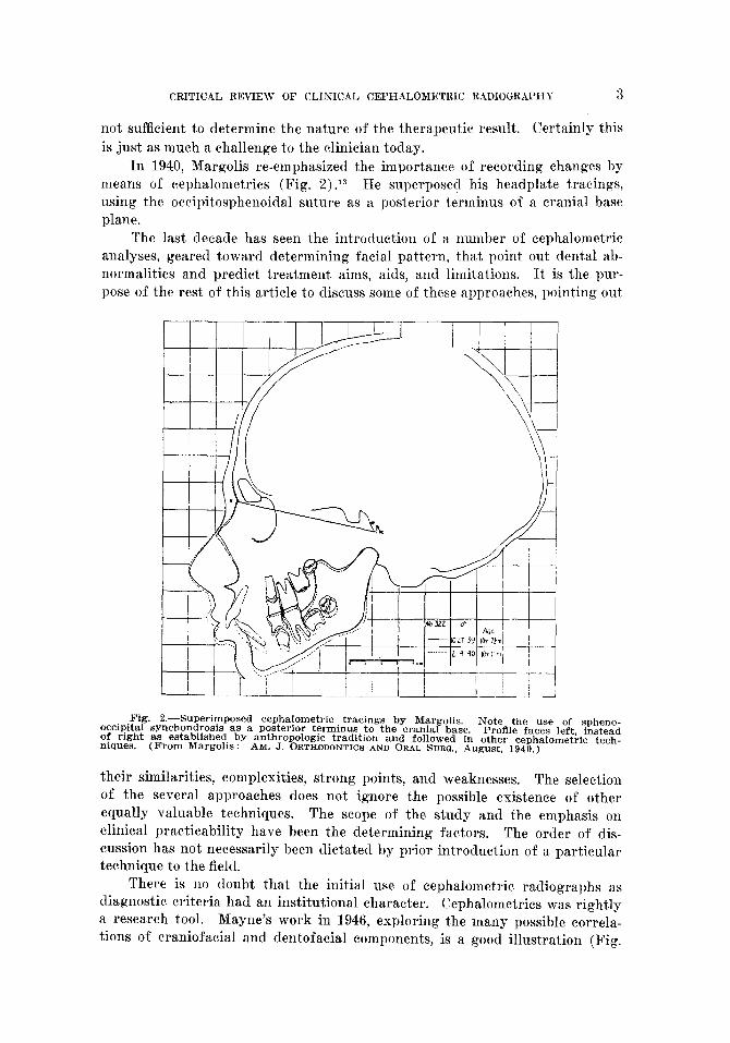
In 1940, Margolis re-emphasized the importance of recording changes by means of cephalometrics (Fig. 2).13 He superposed his headplate tracings, using the occipitosphenoidal suture as a posterior terminus of a cranial base plane.
The last decade has seen the int,roduction of a number of cephalometric analyses, geared toward determining facial pattern, that point out dental ab- normalities and predict treatment aims, aids, and limitations. It is the pur- pose of the rest of this article to discuss some of these approaches, pointing out
Fig. 2.-Superimposed cepholometric tracings by Margolis. occipital synchondrosis as a posterior terminus to the cranial base.
Note the use of spheno- Profile faces left, instead of right as established by anthropologic tradition and followed in other cepbalometric tech-
niques. (From Margolis: AM. J. ORTHODONTICS AND ORAL SURG., August, 1940.)
their similarities, complexities, strong points, and weaknesses. The selection of the several approaches does not ignore the possible existence of other equally valuable techniques. The scope of the study and the emphasis on clinical practicability have been the determining factors. The order of dis- cussion has not necessarily been dictated by prior introduction of a particular t.echnique to the field.
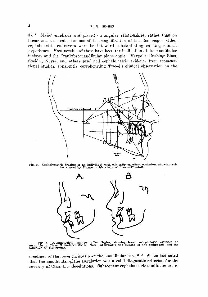
There is no doubt that the initial use of cephalometric radiographs as diagnostic criteria had an institutional character. Cephalometrics was rightly a research tool. Mayne’s work in 1946, exploring the many possible correla- tions of craniofacial and dentofacial components, is a good illust,ration (Fig.
4 T. M. GRABER
:{).I4 Major emphasis was placed on angular relationships, rather than on linear measurements, because of the magnification of the film image. Other cephalometric endeavors were bent toward substantiating existing clinical hypotheses. Most notable of these have been the inclination of the mandibular incisors and the Frankfortmandibula,r plane angle. Margolis, Rushing, Sims, Speidel, Noyes, and others produced cephalometric evidence from cross-sec- tional studies, apparently corroboratin, 0 Tweed’s clinical observation on the
Fig. 3.-Cephalometric tracing of an individual with clinically excellent occlusion, showing cri- teria used by Mayne in his study of “normal” adults.
A.
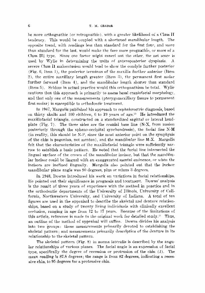
Fig. I.-Cephalometric tracings. after Higley, showing broad morphologic variance of mandible in Class II malocclusions. Note particularly the outline of the symphysis and its influence on the profile.
erectness of the lower incisors over the mandibular base.15-17 Simon had noted that the mandibular planeeangnlation was a valid diagnostic criterion for the severity of Class II malocclusions. Subsequent cephalometric studies on cross-

CRITICAL REVIEW OF CLINICAL CEPHALOMETRiC RADIOGRAPHY 5
sectional groups of patients with Class II malocclusions seemed to verify the gnathostatic use of the Frankfort-mandibular plane angle as a diagnostic criterion. In 1945, Higley showed the high degree of individual variation of mandibular size and shape in Class II malocclusions (Fig. 4) .I8 Mayne cor- roborated this with his study of fifty individuals with clinically excellent oc- clusions. He showed the broad range of variability of all cranial and facial relations from individual to individual. In 1946, Tweed re-emphasized the importance of the Frankfort-mandibular plane angle in orthodontic diag- nosis.19 He observed that the steeper the plane, the poorer the prognosis.
Wendell Wylie wrote in 1947, “Among the students of human cranio- facial morphology, many of whom are orthodontists, there is a growing con- viction that there is no such single entity as a ‘normal facial pattern, and that the dento-facial anomalies are in a large measure occasioned by a random com- bination of facial parts, no one of which is abnormal in size, when taken by itself, but each one of which may fit badly with the other parts to produce a condition which may be called dysplasia.“*O Wylie pointed out that among the variations to be considered was the length of the cranial base between the glenoid fossa of the temporal bone and sella turcica, the over-all length of the maxilla, the position of the maxillary permanent first molar, and the over-all length of the mandible (Fig. 5). He used these criteria in an attempt to assess anteroposterior dysplasia and determine the crux of the malrelationship.
I,7 I,7 I,6 32 I I ! ! /’
Mean Fa Pat tern - Standar
Fig. 5.-Study of Class I malocclusions to assess anteroposterior dysplasia. Measure- ments are made to determine size and position of maxilla and mandible in relation to the cranial base. (From Wylie: Angle Orthodontist, 1947.)
“Standard values” were developed from a comparison of Class I and Class II malocclusions. If measurements were greater than standard for the glenoid fossa-sella turcica length, sella-pterygomaxillary fissure length, total maxillary length, and pterygomaxillary fissure-upper first molar length, the face would
6 T. MM. GRABER
be more orthognathic (or retrognathic), with a greater likelihood of a Class II tendency. This would be coupled with a shortened mandibular length. The opposite trend, with readings less than standard for the first four, and more than standard for the last, would make the face more prognathic, or more of a Class III type. Since one factor might cancel out, the other, the net score is used by Wylie in determining the units of anteroposterior dysplasia. $ severe Class 11 malocclusion would tend to show the condyle further posterior (Fig. 6, Item l), the posterior terminus of the maxilla further anterior (Item 2), the entire maxillary length greater (Item :l), the permanent first molar further forward (Item 4), and t,he mandibular length shorter than standard (Item 5). Seldom in actual practice would this orthognathism be total. Wylie cautions that this approach is primarily to assess basal craniofacial morphology, and that only one of the measurements (pterygomaxillary fissure to permanent first molar) is susceptible to orthodontic treatment.
In 1947, Margolis published his approach to cephalometric diagnosis, based on thirty skulls and 100 children, 6 to 19 years of age.l;’ He introduced the maxillofacial triangle, constructed on a standardized sagittal or lateral head- plate (Fig. 7). The three sides are the cranial base line (N-X, from nasion posteriorly through the spheno-occipital synchondrosis), the facial line N-M (in reality, this should be N-I’, since the most anterior point on the symphysis of the chin is pogonion, not menton), and the mandibular line M-X. Margolis felt that the characteristics of the nmxillofacial triangle were sufficiently nar- row to establish a basic pattern. He noted that the facial line intersected the lingual surface of the crown of the mandibular incisor, but that the mandibu- lar incisor could be lingual with an exaggerated mental eminence, or when the incisors are inclined lingually. Margolis also pointed out that the incisor mandibular plane angle was 90 degrees, plus or minus 3 degrees.
In 1948, Downs introduced his work on \-ariat,ions in facial relationships. He pointed out their significance in prognosis and treatment. Downs’ analysis is the result of three years of experience with the method in practice and in the orthodontic departments of the University of Illinois, University of Ca.li- fornia, Northwestern University, and IJniversity of Indiana. A total of ten figures are used in the appraisal to describe the skeletal and denture relation- ships, based on a study of twenty living individuals with clinically excellent occlusion, ranging in age from 12 to 17 years. Hecause of the limitations of this article, reference is made to the original work for detailed study.21 Thus, an outline of the method of appraisal will x&flee. Downs divides his analysis into two groups: those measurements primarily devoted to establishing the skeletal pattern; and measurements primarily descGptive of the denture in its relationship to the skeletal pattern.
The skeletal pattern (Fig. 8) in norma lateralis is described by the angu- lar relationships of various planes. The facial angle is an expression of facial type, specifically the degree of recession or protrusion of the chin (1). The mean reading is 87.8 degrees; the range is from 82 degrees, indicating a reces- sive chin, to 95 degrees for a protrusive chin.

CRITICAL REVIEW OF CLINICAL CEPHALOME’l!RIC RADIOGRAPHY
ASSESSMENT OF ANTEWPOSlERlOR DYSPLASIA Fcmde
Difference
Units of antcroporterior dysplasia =+wognrthic-ortho~nathic: cxl
Fig. 6.-Method of appraising anteroposterior dysplasia. 1 malocclusion.
Example of a Class II, Division The total number of units is more important than any single measurement,
which might be cancelled out by a compensatory value of another criterion of anteroposterior dysplasia. (Wylie. )
Fig. 7 .-The Margolis MFT (Maxillofacial triangle), the basic facial pattern, as estab- lished by a study of skulls and living children 6 to 19 years of age, all with so-called normal occlusion.

NAM&R.-- CEPHALOMTRIC ANALYSIS
HO
AGE -%.jf!vea--
DATE - -
(DOWNS)
Fig. 8.-Cephalometric tracing, after Downs, showing skeletal criteria of appraisal, 1, Facial angle (FH-NP) ; 9, angle of facial convexity (NAP) ; 3, FH-MP angle; 4, growth axis (SGn-FH) ; 5, apical base relationship (AB-NP).
CLPNALOMETRIC ANALYSIS
Fig. I).--Cephalometric tracing, after Downs, showing dental criteria of aPPraiSa1. 6, In- clination of occlusal plane (FH-Occlusal plane) ; 7, angle formed by intersection of long axes of upper and lower central incisors ; 8, angle of long axis of lower central incisor with mandib- ular plane : 9, angle of long axis of lower central incisor with occIusal plane: 10, a linear measurement of the amount of protrusion of the maxillary central incisors in reference to the facial (NP) plane.

CRITICAL REVIEW OF CLIXICAL, CEPHALOMETRIC RADIOGRAPHY !I
The angle of convexity N-A-P (2) (mean 0 degree, range plus 10 degrees to minus 8.5 degrees) is a measure of the protrusion of the maxillary part of the face to the total profile.
The mandibular plane angle (3) is also a measure of mandibular retrusion, according to Downs. (Mean 21.9 degrees, range l’i to 28 degrees.) In his study of twenty individuals, Downs found a significant tendency toward a larger mandibular angle, with a smaller facial angle.
The Y axis expresses the downward and forward growth of the face from beneath the cranium (4). The angle formed with the Frankfort plane (mean 59.4 degrees, range 55 degrees to 66 degrees) gives an indication of the indi- vidual’s direction of growth-downward and forward, downward, or downward and backward.
The AR, or apical base plane (5) forms an angle with the NP, or facial plane, and gives an appraisal of the relationship of t,he denture bases to each ot,her and to the profile. (Mean -4.8 degrees, range from 0 degree to 9 degrees.)
Tn establishing the relationship of t,he denture to the skeletal pattern, Downs measures the cant of the occlusal plane (6) (Fig. 9), the axial inclina- tion of the upper and lower incisors to each other (7), the. axial inclination of the lower incisors to the mandibular plane (8)) axial inclination of the lower incisors to the occlusal plane (9’)) and the amount of protrusion of the maxillary incisors in reference to the facial plane (10).
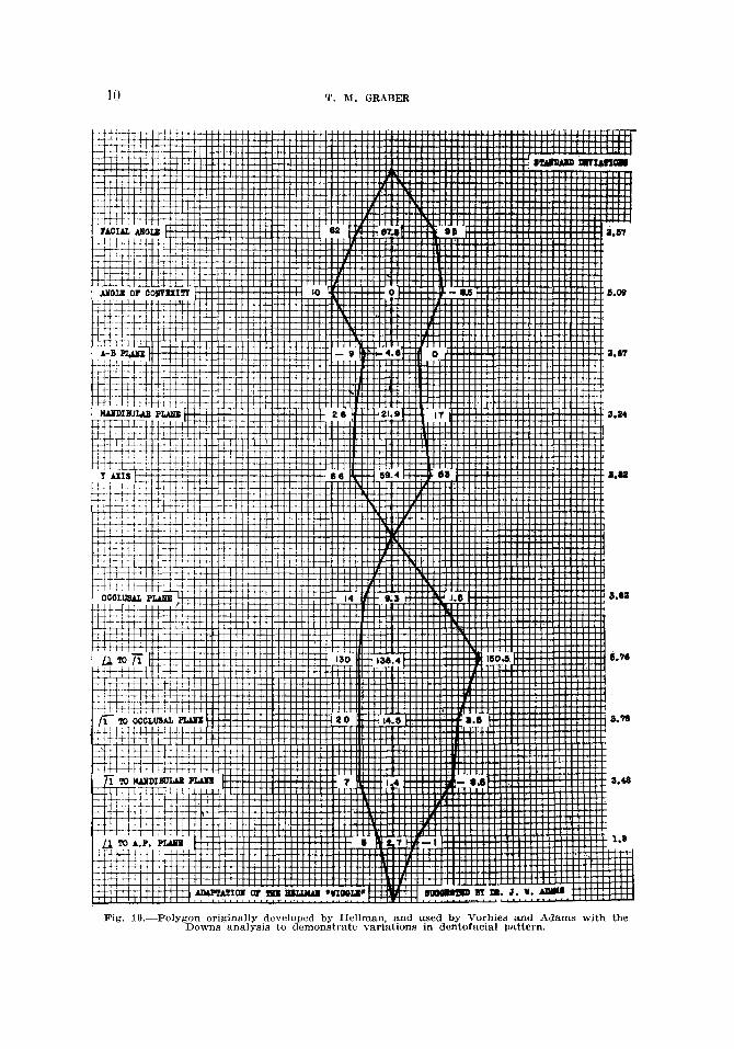
Downs cautions that his study represents a mean or average form. Varia- t,ion is inherent. Most important is the recognition that, no single figure means much by itself. “What counts is the manner in which they all fit together, and their correlation with type, function and esthetics.” Recently, Adams and Vorhies applied Hellman’s “wiggle graphs” to t,he Downs analysis.12 It is possible to plot an individual analysis and tell at a glance where a patient fits in relation to the means and ranges of a representative group of excellent oc- clusions (Fig. 10).
In 1948, Riedel did a study on individuals with clinically excellent occlu- sion, Class II, Division 1, Class II, Division 2, and Class III malocclusions (Fig. 11). He found no significant difference in individuals with normal occlusion or malocclusion of the teeth as far as the anteroposterior position of the maxilla is concerned. The variant is the mandible.2” In his appraisal of 133 individuals, Riedel used S-N as a cranial base. He measured the relative posi- tions of the anterior aspects of the maxillary and mandibular apical bases. Riedel’s work, together with cephalometric criteria gleaned from the Downs analysis, Mayne’s study, and the research of Freeman and Rasmussen? forms t.he basis of the approach used at Nort.hwestern University.
Recently Tweed, introducing his modification of the Margolis maxillo- facial triangle, wrote, “I am confident that, I can now outline a formula or an analysis, which if closely followed by the younger man, will allow him to pro- duce the beaut,iful end results of orthodontic t,reatment that were heretofore only possible after years of experience and much study. In addition, it will accurately tell him when extraction of teeth is indicated, and when such a
T. M. GRABER
Fig. lO.-Polygon originally developed by Hellman, and used by Vorhies and Adams with the Downs analysis to demonstrate variations in dentofacial pattern.

CRITICAL REVIEW OF CLIKICAL CEPHALOMETRIC RADIOGRAPHY 11
Fig. Il.-Measurements employed by Riedel in a study of normal, Class I, Class II, and Class III malocclusions.
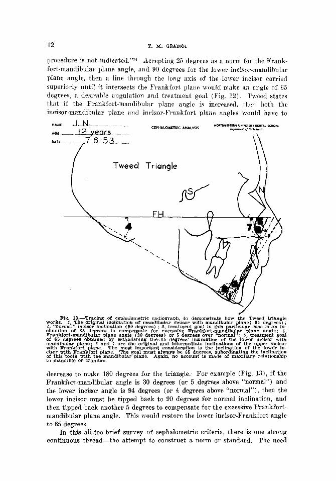
Fig. 12.-Tweed modification of Margolis MFT. The Frankfort horizontal is used as a cranial base. The measurements are based entirely on inclination of the mandibular plane with Frankfort. and inclination of the lower central incisor with Frankfort and mandibular planes. No attempt is made to assess the maxilla or maxillary teeth with respect to the man- dible or cranial base. (From Tweed : Alpha Omegan, 1952.)
12 T. M. GRABER
procedure is not indicated.“2d Accepting 25 degrees as a norm for the Frank- fort-mandibular plane angle, and 90 degrees for the lower incisor-mandibular plane angle, then a line through the long axis of the lower incisor carried superiorly until it intersects the Frankfort, plane would make an angle of 65 degrees, a desirable angulation and treatment goal (Fig. 12). Tweed states that if the Frankfort-mandibular plane angle is increased, then both the incisor-mandibular plane and incisor-Frankfort plane angles would have to
NnME-~m.LN---. ~_ _. _ CEPHALOMETRIC ANALYSIS
NOII”.ESTERN “NI”SIIsl?” wNI*L SC”oaL 0, O,tLd..t,.‘
Tweed Triangle
decrease to make 180 degrees for the triangle. For example (Fig. 13), if the Frankfort-mandibular angle is 30 degrees (or 5 degrees above “normal”) and the lower incisor angle is 94 degrees (or 4 degrees above “normal”), then the lower incisor must be t,ipped back to 90 degrees for normal inclination, and then tipped back another 5 degrees to compensate for the excessive Frankfort- mandibular plane angle. This would restore the lower incisor-Frankfort angle to 65 degrees.
In this all-too-brief survey of cephalometric criteria, there is one strong continuous thread-the attempt to construct a norm or standard. The need
CRITICAL REVIEW OF CLINICAL CXPHALONETRIC RADIO(;RAPHL’ 13
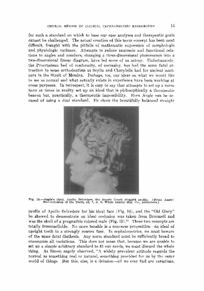
for such a standard on which to base our case analyses and therapeutic goals cannot be challenged. The actual creation of this norm concept has been most difficult, fraught with the pitfalls of mathematic expression of morphologic and physiologic variance. Attempts to reduce anatomic and functional rela- t,ions to angles and numbers, changing a three-dimensional phenomenon into a two-dimensional linear diagram, have led some of us astray. Unfortunately, the Procrustean bed of conformity, of normalcy, has had the same fatal at- traction to some orthodontists as Scylla and Charybdis had for ancient mari- ners in the Strait of Messina. Perhaps, too, our ideas on what we would like to see as normal and what. actually exists in experience have been working at cross purposes. In retrospect, it is easy to say that attempts to set up a norm have at times in reality set up an ideal that is philosophically a therapeutic beacon but, practically, a therapeutic impossibility. Even Angle can he ac- cused of using a dual standard. He chose the beautifully balanced straight
Fig. 14.-Angle’s ideal, Apollo Belvedere, the classic Greek straight profile. (From Angle : Mal-occlusion of the Teeth, ed. ‘7, S. S. White Dental Mfg. Co., publishers.)
profile of Apollo Belvedere for his ideal face (Fig. 14), and the “Old Glory” he showed to demonstrate an ideal occlusion was taken from Broome11 and was the skull of a prognathic colored male (Fig. 15).25 These two concepts are totally irreconcilable. No more tenable is a converse proposition-an ideal of upright teeth in a strongly convex face. In cephalometrics, we must beware of the same fatal diathesis. Any norm standard must be sufficiently broad to encompass all variations. This does not mean that, because we are unable to set up a simple arbitrary standard to fit our needs, we must discard the whole thing. As Simon sagely observed, “A widely prevalent attitude regards the normal as something real or natural, something provided for us by the outer world of things. But this, alas, is a delusion-all we ever find are variations,

endless variations; ill1 esact ideal normal does not exist, cannot exist. And this is our enigma: in theory we u-ill never find the normal; in practice, we forever feel its need and at)ply it constantly.“”
Stutlies at. Northwestern University have shown us the broad range of combination of cranial and facial components. To accept a mean as an abso- lute treatment goal is to ignore a majority of the populace. To arbitrarily select one or two convenient, measurements as prognostic or therapeutic clues is to overlook the interdependence of multiple indiz~idual characteristics, which are unrecognizable in cross-sectional groupings of so-called normals. Our goal must be, then, an individualized norm, using group standards only as a guide.
Fig. 15.-Angle’s “Old Glory” taken from Rroomell. This is the skull of a prognathic colored male. (From Angle : Malocclusion of the Teeth, ed. ‘7. S. S. White Dental Mfg. CO., publishers.)
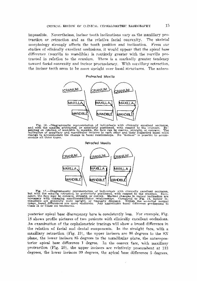
Relations of facial components vary broadly, depending on the facial type- whether the face is forward divergent or backward divergent-whether the face is concave or convex. The implication here is that there are two major considerations, the position of the maxilla anteroposteriorly in the face (with reference to the cranium), and the relation of the mandible to the maxilla, which is responsible for the convex, straight, or concave profile line. If the maxilla is protracted in its relationship to the cranium, the profile is more likely to be convex. If the maxilla is retracted, the profile is more likely t,o be concave. However, with a maxillary protraction, the face can be convex, straight, or concave (Fig. 16). The same holds true in a face where the maxilla is retracted in relation to the cranium (Fig. 17). This is further com- plicated by an appraisal of general facial type, whether it is dolichocephalic (long and narrow) or brachycephalic (short and broad). Observations of large groups would seem to indicate that the dolichocephalic individual (or so-called Nordic type) is more likely to have a straight facial profile. The brachy- cephalic type (for instance, Slavs, eastern European groups, etc.) is more prone to profile convexity. Racial admixtures make any clear-cut correlation
CRITICAL REVIEW OF CLINICAL CEPHALOMETRIC RADIOGRAPHY 15
impossible. Nevertheless, incisor tooth inclinations var’y as the maxillary pro-
traction or retraction and as the relative facial convexity. The skeletal morphology strongly affects the tooth position and inclination. From 0u1 studies of clinically excellent occlusions, it would appear that the apical base difference (maxilla to mandible) is routinely greater with the maxilla pro- tracted in relation to the cranium. There is a markedly greater tendency toward facial convexity and incisor procumbency. With maxillary retraction, the incisor teeth seem to he more upright over basal structures. The antero-
Protracted Moxillo
Fig. 16.-Diagrammatic representation of individuals with clinically excellent occlusion, and with the maxilla protracted. or anteriorly positioned, with respect to the cranium. De- pending on relation of mandible to maxilla. the face can be convex, straight, or concave. The inclination of maxillary and mandibular incisors to each other and their respective bases must change to accommodate the change in basal relationships. modate all three types.
No “normal” is possible to accom-
Fktrocted Maxillo
Fig. 17.-Diagrammatic representation of individuals with clinically excellent occlusion, but with the maxilla retracted, or posteriorly positioned, with respect to the cranium. Here, again, the face may be concave, straight, or convex. Marked changes in incisor inclination are necessary with changing maxillomandibular relationships. clinations are generally more upright, or lingually
Compared to Fig. 16, incisor in-
range, basal differences are more moderate. disposed. Within the so-called normal
Class II or Class III tendencies. Any appreciable basal difference predisposes to
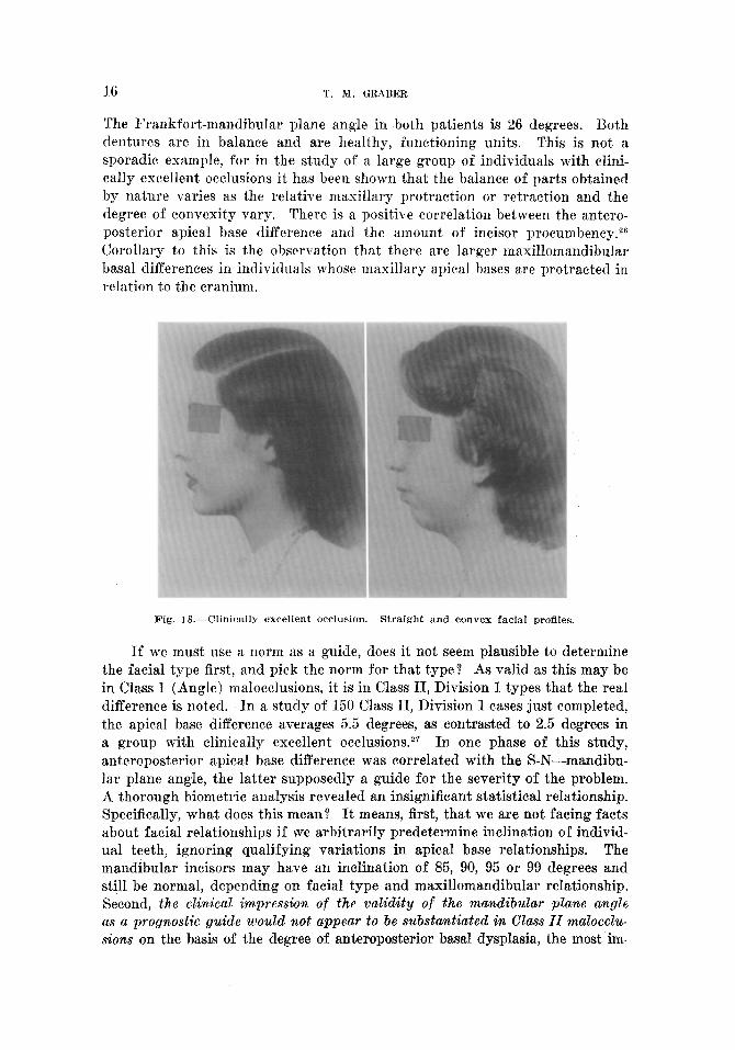
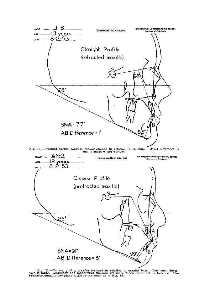
posterior apical base discrepancy here is consistently less. For example, Fig. 18 shows profile pictures of two patients with clinically excellent occlusions. An examination of the cephalometric tracings will show a broad difference in the relation of facial and dental components. In the straight face, with a maxillary retraction (Fig. 19), the upper incisors are 98 degrees to the SN plane, the lower incisors 85 degrees to the mandibular plane, the anteropos- terior apical base difference 1 degree. In the convex face, with maxillary protraction (Fig. 20), the upper incisors are relatively procumbent at 113 degrees, the lower incisors 99 degrees, the apical base difference 5 degrees.
16 T. M. GRABER
The E’ra,nkfort-rnandibLzlar plane angle in both patients is 26 degrees. Both dentures are in balance and are healthy, functioning units. This is not a sporadic example, for in the study of a large group of individuals with clini- cally excellent occlusions it has been shown that the balance of parts obtained by nature varies as t,he relative maxillary protraction or retraction and the degree of convexity vary. There is a positive correlation between the antero- posterior apical base difference and the amount of incisor procumbency.26 Corollary to this is the observation that there are larger maxillomandibular basal differences in individuals whose maxillary apical bases are protracted in relation to the cranium.
Fig. 18.-Clinically excellent occlusion. Straight and convex facial proflles.
If we must use a norm as a guide, does it not seem plausible to determine the facial type first, and pick the norm for that type? As valid as this may be in Class I (Angle) malocclusions, it is in Class II, Division 1 types that the real difference is noted. In a study of 150 Class II, Division 1 cases just completed, the apical base difference averages 5.5 degrees, as contrasted to 2.5 degrees in a group with clinically excellent occlusions.27 In one phase of this study, anteroposterior apical base difference was correlated with the S-N-mandibu- lar plane angle, the latter supposedly a guide for the severity of the problem. A thorough biometric analysis revealed an insignificant statistical relationship. Specifically, what does this mean? It means, first, that we are not facing facts about facial relationships if we arbitrarily predetermine inclination of individ- ual teeth, ignoring qualifying variations in apical base relationships. The mandibular incisors may have an inclination of 85, 90, 95 or 99 degrees and still be normal, depending on facial type and maxillomandibular relationship. Second, the clinical impression of the validity of the ma&ibular plane angle as a prognostic guide would not appear to be substantiated in Class II malocclu- sions on the basis of the degree of anteroposterior basal dysplasia, the most im-
NAME ---JJi--p- CEPHALOMETRIC ANALYSIS
NoanwcITUH “NlYTllsm DENTAL X”ooL
AGE --fyy~ --I
&~,,.,“l ,, hlM,“,,<S
DATE _ -
Straight Profile
(retracted maxilla) \
Fig. lg.-Straight proflle, maxilla retropositioned in relation to cranium. Basal difference is small ; incisors are upright.
CEPHALOMSTRIC ANALYSIS NoRT”wcSTW “NwcR* DENTAL saool
“,#w,“,“, 0, O,d,L”,ir,
Convex Profile
(protracted maxilla)
A6 Difference = 5”
Fig. Zo.-cOnVeX PrOflle, maxilla forward in relation to cranial base. The basal differ- ence is large. MCiXillary and mandibular incisors are more procumbent, but in balance. The Frankfort-mandibular plane angle is the same as in Fig. 19.
1s 1’. &I. GRABEK
ljortant c>haracteristic of a Class II (Angle) malocclusion. It follows, then, that the changing of the inclination of the lower incisors because of the steep- ness of the mandibular plane in a direction that will only increase the overjet and overbite in severe Class II malocclusions, irrespective of inherent pat,tern, irrespective of the posit,ion of the maxilla and maxillary incisors, irrespective of the balancing muscular forces, and irrespective of the possible creation of temporomandibular joint pathology, might be open to some question. This is further substantiated by cephalometric evidence demonstrating insignificant basa.1 changes as a result of conventional orthodontic therapy. limited to intra- oral appliances.
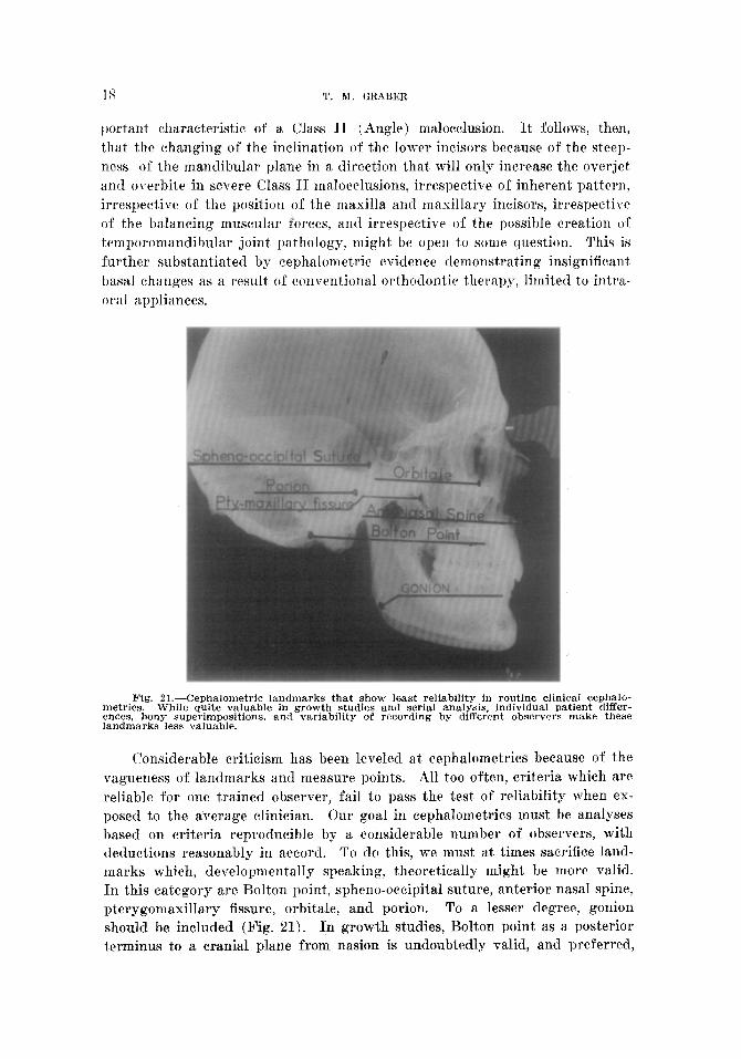
Fig. &l.-Cephalometric landmarks that show least reliability in routine clinical cephalo- metrics. While quite valuable in growth studies and serial analysis, individual patient differ- ences, bony superimpositions, and variability of recording by different observers make these landmarks less valuable.
Considerable criticism has been leveled at cephalometrics because of the vagueness of landmarks and measure points. All too often, criteria which are reliable for one trained observer, fail to pass the test of reliability when ex- posed to the average clinician. Our goal in cephalometrics must be analyses ba,sed on criteria reproducible by a considerable number of observers, with deductions reasonably in accord. To do this, we must at times sacrifice la.nd- marks which, developmentally speaking, theoretically might be more valid. In this category are Bolton point, spheno-occipital suture, anterior nasal spine, pterygomaxillary fissure, orbitale, and porion. To a lesser degree, gonion should be included (Fig. 21). In growth studies, Bolton point as a posterior terminus to a cranial plane from nasion is undoubtedly valid, and preferred,

CRITICAL REVIEW OF CLINICAL CEPHALOMETRIC RADIOGRAPHY 19
as the superlative work of Broadbent has shown. For the clinical orthodontist, who is not able to follow his patients for years and who must make his analysis from a single headplate before treatment, the downward proliferation of the mastoid process, superimposing over Bolton point, makes Bolton point difficult to pick up. The spheno-occipital synchondrosis, posterior terminus of the Margolis cranial plane is in the same category, and is even less reliable. An- terior nasal spine and pterygomaxillary fissure are highly variable. Orbitale, the lowest point on the inferior margin of the orbit, is very hard to see in cer- tain lateral headplates. Porion, the posterior terminus of the Frankfort plane, is a hybrid landmark-part machine, part soft tissue counterpart of the bony external auditory meatus. Porion may vary from time to time, depending on the positioning of the patient. Gonion depends on the curvature of the man- dibular angle. The actual point, bisecting the arc at the angle, may vary sig- nificantly from one time to another, and from one observer to another.
Wylie’s analysis is valuable in determining the area of actual dysplasia. However, in a recent article, 28 he wisely points to the inaccuracy of certain landmarks used (condyle, pterygomaxillary fissure, and anterior nasal spine) which reduce the effectiveness for the average clinician in daily use. There is also contradictory evidence of Buckley and Riedel, showing that in Class II and Class III malocclusions, the mandible is mostly at fault, with the maxilla maintaining a fairly stable relationship to the cranium.
With the goal of reliability of landmarks in mind, with a full knowledge of the limitations imposed by facial types and basal relationships, and with a recognition of the need for a simple and accurate cephalometric analysis, im- parting information that will help the clinician formulate his diagnosis and determine his prognosis, the following cephalometric analysis is offered as one possible approach to the problem. We have borrowed heavily from the Downs analysis, incorporating the research of Thompson,2g-32 Riede1,23 Freemanz3 Ras- musson, 34 Mayne,14 Donovan,s Buckley,35 Carlson, Williams,26 and many others at Northwestern University. Since prior publication has described the North- western technique,37 only an outline of the approach will be given here.
As Downs has done, the Northwestern analysis is divided into two parts, skeletal and dental (see Table I). Recognition is made of those areas which influence prognosis and treatment, and of those areas susceptible to ortho- dontic therapy (Fig. 2.2). The skeletal measurements are the angle of facial convexity (N-A-P), the mandibular plane angle, and the apical base relation- ship (Fig. 23). The use of the maxillary and mandibular incurvations (Points A and B) as the anterior termini of the maxillary and mandibular apical bases would appear to be justified, based on clinical experience. By constructing an angle from the S-N plane to points A and B, and taking the difference, the maxillomandibular anteroposterior relationship can be seen, If the XNA angle is high, meaning a maxillary protraction, then the apical base difference can be higher and still not have unfavorable therapeutic connotations. If the SNA reading is low, any appreciable apical base difference may impose severe limitations on the adjustment of the Class II relationship. An alternate method

20 T. M. GRABER
'I'AZHLE I. MEAN ANI) STANDARI~ lhvJbT10x Vn~.txs I)EKIVEI) IS STUUIE~ 01‘ .~I~I-I,~Is .YNIB ~ILDREN WITH Cl,~h-~car,r,u >~XUELLENT OccJ,r:s~oxs (K~EDEJ.)
L--- --__.. - AI~I’r,TS CHILDREN
hIEAN ST. LJEV. MEAN ST. DEV. - Skeletal : N-a-P +1.62” 4.78’+ 4.22” 5.38” NP-ab -4.6 3.67 S-N-a 82.01 3.89 80.79 3.85 S-N-b 79.9i 3.60 78.02 3.06
Diff. +2.04 i2.77 NS-Go-Gn 31.71 5.19 32.27 4.67 FH-NP 88.56 3.17 ManPL-FH 26.15 5.95 SGn-FH 60.68 3.48 Denture : S-N-Gn 79.29 3.39 1 1 - NS 103.97 5.75 II- IT 130.98 9.24 j 1 - GoGn 93.096 6.78 ;:I- g 69.37 6.43
91.28 3.46 1 l- NP (mm) 5.51 3.15
- (From Graber: AM. J. ORTHODONTICS, August, 1952.)
85.33 2.42 27.06 4.67 60.58 2.89
76.93 3.02 103.54 5.02 130.40 7.34
93.52 5.78 71.79 5.16 92.08 3.64
6.3.5 2.67
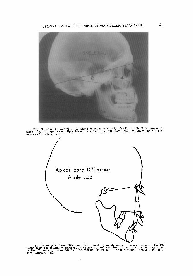
of determining the basal relationship (I3 4 ‘g. 24) is to construct a perpendicular to the SN plane from Point A, and draw a line from the intersection X to Point l3. The resultant angle takes into consideration the facial divergency, and is not dependent on the position of nasion in the face. The measurement of the inclination of the mandibular plane to a cranial base plane (Angle
Fig. 22.-From Downs, showing alveolar bone. basal or supporting bone of the maxilla and mandible, and the cranial base. Onlv the alveolar bone is under direct influence of ortho- dontic therapy. Basal changes depead ori growth increments during therapy. Cranial changes are negltgible at the age most orthodontic treatment is instituted.
angk ence
CRITICAI, REVIEW OF CLINICAL CEPHALOMETRIC RADIOGRAPlIY 2L
Fig. 23.--Skeletal analysis. 1, Angle of facial convexity (NAP) ; 8, Sn-$oGn angle; 3, ! SNA; 4, angle SNB. By subtracting 4 from 5’ (SNB from SNA) the aplcal base tl~ffer- can be ~letermirwi.
Apical Base Difference
Fig. 24.-Apical base difference, cleternlinecl by constfucting, a perpendicular to the SN plane from the maxillary incurvation (Point A) and drawmg a lme from the point of inter- section X down to the mandibular incurvation (Point B) (From Graber : AM. J. ORTHODON- TICS, August, 1952.)
22 T. M. C;RABER
Go&+-NS) does not appear to be the infallible criterion of anteroposterior dysplasia once thought. However, this angle is measured because of other therapeutic considerations. Steep mandibular plane angles impose severe limitations on the correction of excessive overbite; occlusal plane changes induced by intermaxillary elastics are seldom permanent ; functional disturb antes are more likely.
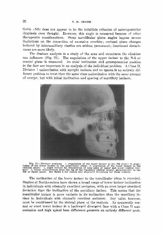
The denture analysis is a study of the area and structures the clinician can influence (Fig. 25). The angulation of the upper incisor to the X-S or cranial plane is measured. Its axial inclination and anteroposterior position in the face are important in an analysis of the individual problem. A Class 11, Division 1 malocclusion with upright incisors and no spaces is an entirely dif- ferent problem to treat than the same class malocclusion with the same amount of overjet, but with labial inclination and spacing of maxillary incisors.
Fig. 25.-Denture analysis. 1, Angulation of the upper incisor to the NS plane ; 2, angu- l&on of the lower incisor to the mandibular plane; 3, angulation of the lower incisor to the occlusal plane: 4, angle formed by intersection of long axes of upper and lower central inci- sors : 5, linear measurement from the tip of the maxillary central incisor, perpendicular to the NP or facial plane. See Table I for means and standard deviations for these criteria.
The inclination of the lower incisor to the mandibular plane is recorded. Studies at Northwestern have shown a broad range of lower incisor inclination in individuals with clinically excellent occlusion, with an even larger standard deviation than the inclination of the maxillary incisor. This means that the mandibular incisor is more variable in its inclination than the maxillary in- cisor in individuals with clinically excellent. occlusion. Any value, however, must be conditioned by the skeletal phase of t,he analysis. An apparently nor- mal or erect lower incisor in a backward divergent face with a Class II mal- occlusion and high apical base difference presents an entirely different prob-

CRITICAL REVIEW OF CLINICAL CEPHALOMETRIC RADIOGRAPHY 23
lem for treatment than it would in a forward divergent face, with a Class I mal- occlusion and a small anteroposterior basal difference. The same standard cannot be used. Some clinicians prefer to measure the inclination of the lower incisor to the occlusal plane, pointing to the smaller standard deviation. The inference in justifying the choice is that the mandibular incisor is more de- pendent on the cant of the occlusal plane than on mandibular morphology. Certainly, the dictates of logic and sound treatment planning mitigate against the arbitrary selection of a “normal” inclination for any toot,h, if in so doing, the clinician ignores equally important or more fundamental considerations such as relation of maxilla to cranium, apical base relationship, limitations of mechanotherapy, etc.
The relation of the upper and lower incisors (angular reading of the inter- section of the long axes) shows the greatest variability of all criteria, and is the least reliable from a diagnostic point of view. However, because control over both upper and lower incisors is possible, changes wrought by treatment are reflected in the greatest magnitude here. Facial type again qualifies the ultimate objective.
More importance can be attached to the position of the maxillary incisor with respect to the facial plane. This is a linear measurement from the in&al edge, perpendicular to the NP or facial plane. Though skeletal limitations may plague any severe Class II malocclusion, it is in the lingual tipping of the maxillary incisors that the greatest esthetic change is produced. Restoration of normal lip posture is often the major accomplishment subsequent to therapy and retention, after the denture has re-established its morphogenetic domi- nance and functional equilibrium.
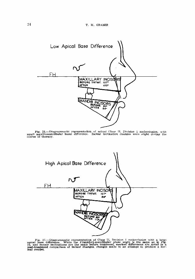
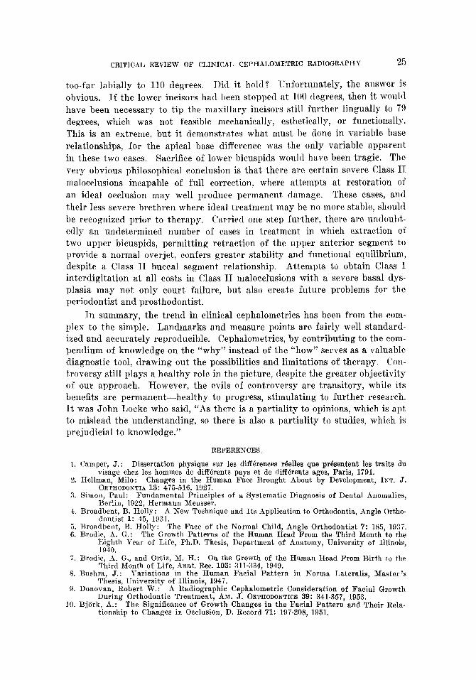
Of necessity, mean values have been determined for all these criteria, along with standard deviations to show relative spread. But these measure- ments serve only as a guide, illustrating central tendency. Prognosis and treatment considerations must be based on the relations of the component parts within the particular patient under consideration. Thus, a patient with a Class II, Division 1 malocclusion, protracted maxilla, and a moderately large n~axillomandibular basal difference, will allow a greater labial inclination of incisor teeth for a stable result. This is recognized when the skeletal cephalo- metric measurements are correlated with those of t,he denture. Second, the limitations of apical base relationship may be wholly inconsistent with the demands of a “normal” occlusion. This may be illustrated by the following dmgrammatic representations of actual cases (Figs. 26 and 27). Both are Class II, Division 1, the Frankfort-mandibular plane angle is 30 degrees, the inclination of incisors to basal planes identical (107 degrees for the upper, 90 degrees for the lower). The variable factor lies in the anteroposterior apical base difference. TO have the same ultimate objectives for incisal inclination would be most unwise. In the low AB case, the upper incisors were tipped lingually 7 degrees, and the lower incisors labially 4 degrees to establish nor- mal overjet and overbite. In the high AB case, tooth movement of the same degree still left a large overjet, which could be corrected only by tipping the upper incisors lingually to an all-too-erect 89 degrees, the lower incisors a]]-
T. M. GRABER
Low Apical Base
Fig. 26.-Diagrammatic reoresentation of actual Class II. Division 1 malocclusion, with small maxillomandib~ alar basal &fference. In&a’1 inclination change IS were slight during the course of therapy.
High Apical Base Difference
Fig. 27.-Diagrammatic representation of Class II. Division 1 malocclusion with a large apical base difference. While the Frankfort-mandibular plane angle is the same as in Fig. 26, and incisor inclinations are t.he same before treatment, marked dffferences are noted in a post-treatment comparison of incisor changes, changes made in an attempt to produce a nor- mal overjet.
CRITICAL REVIEW OF CLINICAT, CEPHALOYETRIC RADIOGRAPHY 25
too-far labially to 110 degrees. I)id it hold? IJnfortunately, the answer is obvious. If the lower incisors had been stopped at 100 degrees, then it would have been necessary to tip the maxillary incisors still further lingually to 79 degrees, which was not feasible mechanically, esthetically, or functionally. This is an extreme, but it demonstrates what must be done in variable base relationships, for the apical base difference was the only variable apparent in these two cases. Sacrifice of lower bicuspids would have been tragic. The very obvious philosophical conclusion is that there are certain severe Class IT malocclusions incapable of full correction, where attempts at restoration of an ideal occlusion may well produce permanent damage. These cases, and their less severe brethren where ideal treatment may be no more stable, should be recognized prior to therapy. Carried one step further, there are undoubt- edly an undetermined number of cases in treatment in which extraction of
two upper bicuspids, permitting retraction of the upper anterior segment to provide a normal overjet, confers greater stability and functional equilibrium, despite a Class 11 buccal segment relationship. Attempts to obtain Class I interdigitation at all costs in Class II malocclusions with a severe basal dys- plasia may not only court failure, but also create future problems for the periodontist and prosthodontist.
In summary, the trend in clinical cephalometrics has been from the com- plex to the simple. Landmarks and measure points are fairly well standard- ized and accurately reproducible. Cephalometrics, by contributing to the com- pendium of knowledge on the “why” instead of the “how” serves as a valuable diagnostic tool, drawing out the possibilities and limitations of therapy. Con- troversy still plays a healthy role in the picture, despit,e the greater objectivity of our approach. However, the evils of controversy are transitory, while its benefits are permanent-healthy to progress, stimulating to further research. It was John Locke who said, “AS there is a partiality to opinions, which is apt, to mislead the understanding, so there is also a partiality to studies, which is prejudicial to knowledge.”
REFERENCES.
1. Camper, J. : Dissertation physique sur les diff&ences r6elles que prbsentent les traits du visage chez les hommes de differents pays et de differents ages, Paris, 1791.
%. Hellman, Milo: Changes in the Human Face Brought About by Development, INT. J. ORTHODONTIA 13: 475-516, 1927.
3. Simon, Paul: Fundamental Principles of a Systematic Diagnosis of Dental Anomalies, Berlin, 1922, Hermann Meusser.
4. Broadbent, B. Holly: A New Technique and Its Application to Orthodontia, Angle Ortho- dontist 1: 45, 1931.
5. Broadbent, B. Holly: 6. Brodie, A. G.:
The Face of the Normal Child, Angle Orthodontist 7: 185, 1937. The Growth Patterns of the Human Head From the Third Month to the
Eighth Year of Life, Ph.D. Thesis, Department of Anatomy, University of Illinois, 1940.
7. Brodie, A. G., and Ortiz, M. I-1.: On the Growth of the Human Head From Birth to the Third Month of Life, Anal;. Rec. 103: 311-334, 1949.
8. Bushra, J.: Variations in the Human Facial Pattern in Norma Lateralis, Master’s Thesis, University of Illinois, 1947.
9. Donovan! Robert W.: A Radiographic Cephalometric Consideration of Facial Growth During Orthodontic Treatment, AM. J. ORTHODONTICS 39: 341-357, 1953.
10. BjBrk? A.: The Significance of Growth Changes in the Facial Pattern and Their Rela- tionship to Changes in Occlusion, D. Record 71: 197-208, 1951.

26 'L'. Y. GRABER
11. Brodie, A. C;., Downs, W. B., Goldstein, A:, and Myer, E :. Cephalomet,ric Appraisal of Orthodontic Results, Angle Orthodontist 8: 261-352, 1938.
12. Higley, L. B.: Lateral Head Roentgenograms and Their Relation to the Orthodontic Problem, i2nc. J. ORTHODONTICS asn OKAL STJILG. 26: 768-778, 1940.
l.‘i. Xsrgolis, Herbert: Standardized X-Ray Cephalographics, SK J. ORTIIODONlWS Axl) ORAL SURG. 26: 725-740, 1940.
1-l. Mayne, Warren: A Study of the Skeletal Pattern of the Human Face, Master’s Thesis, Northwestern University, 1946.
15. Margolis, H. 1.: Basic Facial Pattern and Its Application in Clinical Orthodontics, AM. 5. ORTKOYONTICS AND ORAL SURG. 33: 6X-641,1947.
16. Xoyes, H. J., Rushing, C. H., and Sims, H. A.: The Axial Inclination of the Human Central Incisor Teeth, Angle Orthodontist 13: 60, 1943.
17. Speidel, Tom, and Stoner, M, M.: Variation of Mandibular Incisor Axis in Adult “Normal” Occlusion, AM. J. ORTHODONTICS AND ORAL SURG. 30: 536-542, 1944.
18. Higley, L. B.: Cephalometric Diagnosis and Its Implication in Treatment, J. A. D. A. 32: :3-x, 1945.
19. Twee$ C. W.: Frankfort-Mandibular Plane Angle in Orthodontic Diagnosis, Classifica- tion, Treatment Planning, and Prognosis, ii&i. J. ORTHODONTICS AND ORAL 8rJRG. 32: 175221, 1946.
20. Wylie, Wendell: Assessment of Anteroposterior Dysplasia, Angle Orthodontist 17: !)7-109, 1947.
21. Downs, W. B.: Variations in Facial Relationships; Their Significanoe in Treatment and Prognosis, AM. J. ORTIIODONTICS 34: 812~840,1948.
22. Porhies. J. M.. and Adams. J. W.: Polvgonic Interoretation of Cephalometric Find in& Angle Orthodontist -21: 194-197: T951. L
23. Riedel, Richard A.: A Cephalometric Roentgqnographic Study of the Relation of the Maxilla and Associated Parts to the Cranial Base in Normal and Malocclusion of the Teeth, Master’s Thesis, Northwestern University, 1948.
‘24. Tweed C W . Why I Extract Teeth in the Treatment of Certain Types of Maloeclu- sibn,‘Alpha Omegan 46: 93-104, 1952.
25. Angle, E. H.: Treatment of Malocclusion of the Teeth, ed. 7, Philadelphia, 1907, S. S. Whit,r Dental Mfrr. (“0.
26, Williams, J. E.: A Radiographic Cephalometric Study of the Labiolingual Axial Inclination of the Central Incisors in Relation to the Mandible and Maxilla of Excellent Dentitions. Master ‘Y Thesis. Northwestern Universitv. 1951.
27. Qraber, T. M.: Unpublished data, Northwestern University Orthidontic Department. 28. Wylie, Wendell, and Johnson, E. L.: Rapid Evaluation of Facial Dysplasias in the
Vertical Plane, Angle Orthodontist 22: 165-183. 1952. 29. Thompson, J. R.: ’ C$phalometric Investigation of the Opening Movements of the
Mandible, Angle Orthodontist 18: 30-31, 1948. 30. Thompson, J. R.: Rest Position of the Mandible and Its Application to Analysis and
Correction of Malocclusion, Angle Orthodontist 19: 162-187, 1949. :ll. Thompson, J. R., and Craddock, F. W.: Functional Analysis of Occlusion, J. A. D. A.
39: 40-r-406, 1949. 32. Thompson, J. R.: Oral and Environmental Factors as Etiologic Factors in Malocclusion
of the Teeth, AM. 5. ORTHODONTICS 35: 33-53, 1949. RR. Freeman, R. S.: A Radiographic Method of Analysis of the Relation of the Structures
of the Lower Face to Each Other and to the Occlusal Plane of the Teeth, Master’s Thesis, Northwestern University, 1950.
34. Rasmussen, D. F.: A Radiographic Method of Analysis of the Relation of the Struc- tures of the T,ower Fare t,o the Maxillary Plane: Master’s Thesis, Northwestern Uni- versity, 1950.
13. Buckley, Daniel E.: A Cephalometric Roentgenographic Study of the Facial Skeleton in Class II, Division 1 Malocclusion, Master’s Thesis, Northwestern University, 1948.
36. Carlson, S. D.: A Cephalometric Radiographic Study of the Change in Relation of Mandible to Maxilla in Orthodontic Treatment, Master’s Thesis, Northwestern Uni- versity, 1951.
‘Ii. Graber, T. %I.: New Horizons in Case Analysis ; Clinical Cephalometrics, AM. J. ORTIIO- IWKTICS 38: 603-624, 1952.
311 E. CHICAGO AVE.
Related Documents


