Abstract Vascular changes after acute spinal cord trauma are important factors that predispose quadri- plegia, in most cases irreversible. Repair of the spinal blood flow helps the spinal cord recovery. The average time to arrive and perform surgery is 3 h in most cases. It is important to determine the critical ischemia time in order to offer better functional prognosis. A spinal cord section and vascular clamping of the spinal ante- rior artery at C5–C6 model was used to determine critical ischemia time. The objective was to establish a critical ischemia time in a model of acute spinal cord section. Four groups of dogs were used, anterior ap- proach and vascular clamp of spinal anterior artery with 1, 2, 3, and 4 h of ischemia and posterior hemi- section of spinal cord at C5–C6 was performed. Clinical evaluation was made during 12 weeks and morpho- logical evaluation at the end of this period. We obtained a maximal neurological coordination at 23 days average. Two cases showed sequels of right upper limb paresis at 1 and 3 ischemia hours. There was nerve conduction delay of 56% at 3 h of ischemia. Morphological examination showed 25% of damaged area. The VIII and IX Rexed’s laminae were the most affected. The critical ischemia time was 3 h. Dogs with 4 h did not exhibit any recovery. Keywords Spinal cord injury Á Anterior spinal artery Á Critical ischemia time Introduction In most countries, there are 20–40 acute spinal cord injuries/1,000,000 in adults per year. The main causes of spinal cord trauma are automobile accidents, sports and recreational activities, diving injuries, and home accidents. Nearly half of the patients present a com- plete damage of the spinal cord with motor and sensory dysfunction below the level of lesion; in almost two of three patients the lesion is at a cervical level [1]. In the United States there are 10,000 new cases per year with great cost due to its attention and strategies for decreasing the temporary or permanent disabilities that can have significant effects [2]. A posttraumatic quadriplegia is a paralysis of four limbs which occurs as a consequence of severe cervical lesion and the sequelae are generally irreversible. The present day treatment is focusing on preventing acute and permanent disabilities, which consist of early sta- bilization, handling of neurogenic bladder, early reha- bilitation, tendinous transpositions, and electric peripheral muscle stimulation. In 1671, Stensen made W. E. Bitar Alatorre (&) Instituto Mexicano del Seguro Social, Orthopaedics, Guadalajara, Jalisco, Mexico e-mail: [email protected] W. E. Bitar Alatorre Á D. Garcia Martinez Á E. Portilla de Buen Instituto Mexicano del Seguro Social, CIBO Experimental Surgery, Guadalajara, Jalisco, Mexico W. E. Bitar Alatorre Á S. A. Rosales Corral Á M. E. Flores Soto Instituto Mexicano del Seguro Social, CIBO Neuroscience, Guadalajara, Jalisco, Mexico G. Velarde Silva Rehabilitation Evoked Potentials Department, Hospital Mexico Americano, Guadalajara, Jalisco, Mexico Eur Spine J (2007) 16:563–572 DOI 10.1007/s00586-006-0222-9 123 ORIGINAL ARTICLE Critical ischemia time in a model of spinal cord section. A study performed on dogs Wadih Emilio Bitar Alatorre David Garcia Martinez Sergio A. Rosales Corral Mario E. Flores Soto Gustavo Velarde Silva Eliseo Portilla de Buen Received: 2 February 2006 / Revised: 8 August 2006 / Accepted: 29 August 2006 / Published online: 23 September 2006 Ó Springer-Verlag 2006

Welcome message from author
This document is posted to help you gain knowledge. Please leave a comment to let me know what you think about it! Share it to your friends and learn new things together.
Transcript
Abstract Vascular changes after acute spinal cord
trauma are important factors that predispose quadri-
plegia, in most cases irreversible. Repair of the spinal
blood flow helps the spinal cord recovery. The average
time to arrive and perform surgery is 3 h in most cases.
It is important to determine the critical ischemia time
in order to offer better functional prognosis. A spinal
cord section and vascular clamping of the spinal ante-
rior artery at C5–C6 model was used to determine
critical ischemia time. The objective was to establish a
critical ischemia time in a model of acute spinal cord
section. Four groups of dogs were used, anterior ap-
proach and vascular clamp of spinal anterior artery
with 1, 2, 3, and 4 h of ischemia and posterior hemi-
section of spinal cord at C5–C6 was performed. Clinical
evaluation was made during 12 weeks and morpho-
logical evaluation at the end of this period. We
obtained a maximal neurological coordination at
23 days average. Two cases showed sequels of right
upper limb paresis at 1 and 3 ischemia hours. There
was nerve conduction delay of 56% at 3 h of ischemia.
Morphological examination showed 25% of damaged
area. The VIII and IX Rexed’s laminae were the most
affected. The critical ischemia time was 3 h. Dogs with
4 h did not exhibit any recovery.
Keywords Spinal cord injury �Anterior spinal artery �Critical ischemia time
Introduction
In most countries, there are 20–40 acute spinal cord
injuries/1,000,000 in adults per year. The main causes
of spinal cord trauma are automobile accidents, sports
and recreational activities, diving injuries, and home
accidents. Nearly half of the patients present a com-
plete damage of the spinal cord with motor and sensory
dysfunction below the level of lesion; in almost two of
three patients the lesion is at a cervical level [1]. In the
United States there are 10,000 new cases per year with
great cost due to its attention and strategies for
decreasing the temporary or permanent disabilities
that can have significant effects [2].
A posttraumatic quadriplegia is a paralysis of four
limbs which occurs as a consequence of severe cervical
lesion and the sequelae are generally irreversible. The
present day treatment is focusing on preventing acute
and permanent disabilities, which consist of early sta-
bilization, handling of neurogenic bladder, early reha-
bilitation, tendinous transpositions, and electric
peripheral muscle stimulation. In 1671, Stensen made
W. E. Bitar Alatorre (&)Instituto Mexicano del Seguro Social, Orthopaedics,Guadalajara, Jalisco, Mexicoe-mail: [email protected]
W. E. Bitar Alatorre � D. Garcia Martinez �E. Portilla de BuenInstituto Mexicano del Seguro Social,CIBO Experimental Surgery, Guadalajara,Jalisco, Mexico
W. E. Bitar Alatorre � S. A. Rosales Corral �M. E. Flores SotoInstituto Mexicano del Seguro Social,CIBO Neuroscience, Guadalajara, Jalisco, Mexico
G. Velarde SilvaRehabilitation Evoked Potentials Department,Hospital Mexico Americano, Guadalajara, Jalisco, Mexico
Eur Spine J (2007) 16:563–572
DOI 10.1007/s00586-006-0222-9
123
ORIGINAL ARTICLE
Critical ischemia time in a model of spinal cord section.A study performed on dogs
Wadih Emilio Bitar Alatorre Æ David Garcia Martinez ÆSergio A. Rosales Corral Æ Mario E. Flores Soto ÆGustavo Velarde Silva Æ Eliseo Portilla de Buen
Received: 2 February 2006 / Revised: 8 August 2006 / Accepted: 29 August 2006 / Published online: 23 September 2006� Springer-Verlag 2006
the first experiment on the blood supply of the spinal
cord when, in a dogfish, he showed that ligation of the
aorta caused tail paralysis which recovered when the
ligature was loosened [3].
The pathological changes after cervical lesion in
dogs were described in the following manner: Allen in
1911, was the first to describe this: ‘‘At 15 min a
petechial hemorrhage of gray matter and white matter
edema was observed, at 2 h the gray matter hemor-
rhage increased, and at 4 h there were axis-cylinders
‘‘numerous swollen axis cylinders’’ [1]. Ducker in 1971,
shows changes of severe necrosis at 6 days [1]. Dohr-
mann referred a distention of muscular layer of small
veins by accumulation of red blood cells without ax-
onal changes at 5 min. At 15–30 min small hemor-
rhages with red blood cells extravasation and discrete
axonal changes were observed and at 4 h there is a
disruption of myelin sheaths, axonal degeneration, and
ischemic endothelial lesion.
Nemecek in 1978 defined the severe necrosis as
‘‘self-destruction’’ which occurs after 6 days of acute
lesion. Griffiths and McCulloch showed from the first
few days progressive axonal changes and necrotic areas
[1]. Bresnahan and Balentine, by means of electronic
microscopy, found that the trauma induces granular
dissolution of the axoplasm and vesicular disruption of
myelin, especially in the white matter. With this sup-
port, the treatment is focusing on decreasing the
inflammation using drugs such as methilprednisolone
and nimodipine [1]. The main obstacle in obtaining a
good prognosis is the delay between the moment in
which the trauma occurs and the arrival to the emer-
gency services. It is important to point out that in
cervical trauma additionally to spinal cord lesion; there
is an area of vascular damage.
In previous works [4], we experimentally repro-
duced the quadriplegia in dogs with spinal cord section
and spinal anterior artery section at the cervical level
and observed that the section of only spinal cord with
preservation of vascular supply did not cause irre-
versible neurological lesion in spite of the 1st day after
the trauma, observing signs of quadriplegia, however,
the animals recovered totally. We concluded that the
spinal cord vascular supply at the motor area of the
spinal cord had a principal role in the damage rever-
sion. In our experimental groups of dogs, the micro-
surgical repair of vessels sectioned permit the
neurological recovery in a maximum of 8 weeks [4].
The repair of the spinal blood flow helps the spinal
cord recovery, however, it is unknown if the revascu-
larization had good results after 3 h of spinal section,
which in most cases is the average time to arrive and
perform surgery. For these reasons it is important to
know the critical ischemia time with the purpose of
offering a better functional prognosis.
Vascular supply of cervical spinal cord
The vascular supply in the motor area of the spinal
cord depends on the anterior spinal artery, this is a
branch of vertebral arteries, arising from the brachi-
ocephalic trunk and the subclavian artery [5–7].
The centrifugal artery system of sulcal arteries sup-
plies most of the gray and white matter of ventral and
lateral spinal cord, each sulcal artery supplies one half
of the spinal cord. The centripetal artery system arising
at the posterior spinal arteries supplies the posterior
white and gray matter. In contrast with classic concepts
there is not any pial arterial plexus in the ventral and
ventro-lateral surfaces, except by infrequent transver-
sal branches of anterior spinal artery. In the posterior
columns there are two systems of large veins: the
posteromedial septal vein and the posterior oblique
vein that drains the posterior columns, of the poster
medial gray matter and the posterior gray commissure.
The remainder of gray and white matter is drained
by radial and sulcal veins [8]. It has been postulated
that the vascular mechanisms are important in the
physiopathology of the acute damage of spinal cord
and the progressive necrosis. The vascular flow reduc-
tion had a relationship with the severity of the damage.
The mechanism that causes the posttraumatic ischemia
is unknown, the spasm of the sulcal arteries reduces the
blood supply at the side of the lesion; the intramedul-
lary branches of the posterior spinal arteries at the
level, showed distal occlusions with the resulting gray
and white posterior matter ischemia. It is probable that
capillary occlusion and disruption occur at the lesion
level as a result of the direct mechanic trauma [9].
Materials and methods
A hemi-section of 50% of the right half at the level of
C5–C6 was performed. It was determined that with a
section greater than 50% at this level, the animal in the
model did not recuperate respiratory automatism or
died in the attempt.
Simultaneously with the spinal cord section a vas-
cular mini bulldog clamp angled 45� (20–25 g) was
placed to cause ischemia at the level of the anterior
spinal artery, separating the animals into four groups
with 1, 2, 3, and 4 h of ischemia; clinically noticing the
changes from the time immediately after surgery in the
stage of quadriplegia, by daily clinical analysis, until
564 Eur Spine J (2007) 16:563–572
123
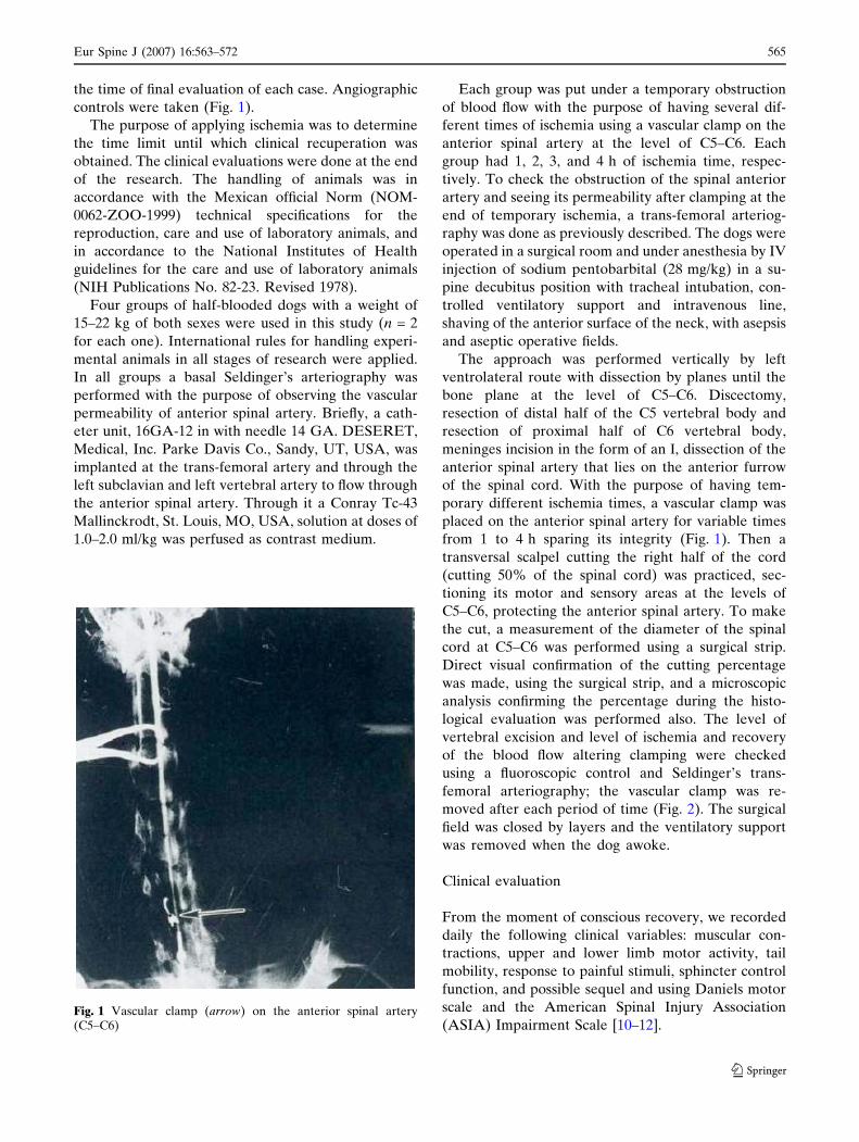
the time of final evaluation of each case. Angiographic
controls were taken (Fig. 1).
The purpose of applying ischemia was to determine
the time limit until which clinical recuperation was
obtained. The clinical evaluations were done at the end
of the research. The handling of animals was in
accordance with the Mexican official Norm (NOM-
0062-ZOO-1999) technical specifications for the
reproduction, care and use of laboratory animals, and
in accordance to the National Institutes of Health
guidelines for the care and use of laboratory animals
(NIH Publications No. 82-23. Revised 1978).
Four groups of half-blooded dogs with a weight of
15–22 kg of both sexes were used in this study (n = 2
for each one). International rules for handling experi-
mental animals in all stages of research were applied.
In all groups a basal Seldinger’s arteriography was
performed with the purpose of observing the vascular
permeability of anterior spinal artery. Briefly, a cath-
eter unit, 16GA-12 in with needle 14 GA. DESERET,
Medical, Inc. Parke Davis Co., Sandy, UT, USA, was
implanted at the trans-femoral artery and through the
left subclavian and left vertebral artery to flow through
the anterior spinal artery. Through it a Conray Tc-43
Mallinckrodt, St. Louis, MO, USA, solution at doses of
1.0–2.0 ml/kg was perfused as contrast medium.
Each group was put under a temporary obstruction
of blood flow with the purpose of having several dif-
ferent times of ischemia using a vascular clamp on the
anterior spinal artery at the level of C5–C6. Each
group had 1, 2, 3, and 4 h of ischemia time, respec-
tively. To check the obstruction of the spinal anterior
artery and seeing its permeability after clamping at the
end of temporary ischemia, a trans-femoral arteriog-
raphy was done as previously described. The dogs were
operated in a surgical room and under anesthesia by IV
injection of sodium pentobarbital (28 mg/kg) in a su-
pine decubitus position with tracheal intubation, con-
trolled ventilatory support and intravenous line,
shaving of the anterior surface of the neck, with asepsis
and aseptic operative fields.
The approach was performed vertically by left
ventrolateral route with dissection by planes until the
bone plane at the level of C5–C6. Discectomy,
resection of distal half of the C5 vertebral body and
resection of proximal half of C6 vertebral body,
meninges incision in the form of an I, dissection of the
anterior spinal artery that lies on the anterior furrow
of the spinal cord. With the purpose of having tem-
porary different ischemia times, a vascular clamp was
placed on the anterior spinal artery for variable times
from 1 to 4 h sparing its integrity (Fig. 1). Then a
transversal scalpel cutting the right half of the cord
(cutting 50% of the spinal cord) was practiced, sec-
tioning its motor and sensory areas at the levels of
C5–C6, protecting the anterior spinal artery. To make
the cut, a measurement of the diameter of the spinal
cord at C5–C6 was performed using a surgical strip.
Direct visual confirmation of the cutting percentage
was made, using the surgical strip, and a microscopic
analysis confirming the percentage during the histo-
logical evaluation was performed also. The level of
vertebral excision and level of ischemia and recovery
of the blood flow altering clamping were checked
using a fluoroscopic control and Seldinger’s trans-
femoral arteriography; the vascular clamp was re-
moved after each period of time (Fig. 2). The surgical
field was closed by layers and the ventilatory support
was removed when the dog awoke.
Clinical evaluation
From the moment of conscious recovery, we recorded
daily the following clinical variables: muscular con-
tractions, upper and lower limb motor activity, tail
mobility, response to painful stimuli, sphincter control
function, and possible sequel and using Daniels motor
scale and the American Spinal Injury Association
(ASIA) Impairment Scale [10–12].Fig. 1 Vascular clamp (arrow) on the anterior spinal artery(C5–C6)
Eur Spine J (2007) 16:563–572 565
123
Neurophysiological evaluation
The electrical activity was evaluated using somato-
sensory evoked potential (SSEP) (Nicolett, mod. Vik-
ing IV) at 30 Hz of low filters frequency and high filters
frequency of 3 kHz, with a sweeping speed of 10 ms,
2 lV of amplitude per division, using stimulus of
2.3 Hz corresponding to 2.3 stimulus per second, with
12 mA of intensity and 0.2 ms of duration. After
anesthesia, the electrodes were implanted by a trench
of the right sciatic nerve. The stimulus was captured at
the brain in a point at the union of the two lines, the
first one being the union between the two mastoid
portions and the second being the union of the nasal
base and the occipital protuberance. The electrical
ground was applied at the frontal level on the dog’s
head. This evaluation was performed before and after
the surgery and then again 3 months later.
Histopathological evaluation
All animals were killed at 12 weeks by anesthetic
overdose. Tissue samples were taken of the spinal cord
1 cm proximal and distal to the cord section.
The percentage of section was measured using
standard cross section of tissue embedded in paraffin
and stained with hematoxylin–eosin, Masson and Clu-
ber-Barrera methods. We studied the morphology of
the spinal cord and with semifine cross sections
embedded in epoxy resin (poly/bed) and stained with
toluidine blue method, the percentage of normal and
altered axons, blood vessels and endothelial charac-
teristics were evaluated in all the groups. The thickness
of the myelin sheaths was determined in histological
sections by using Carl Zeiss Image Analyzer (Zeiss
image 3 = at 400·).
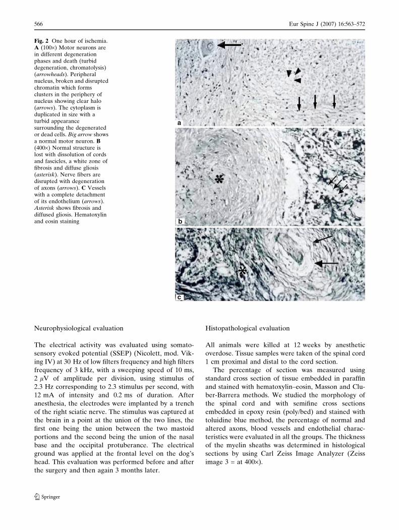
Fig. 2 One hour of ischemia.A (100·) Motor neurons arein different degenerationphases and death (turbiddegeneration, chromatolysis)(arrowheads). Peripheralnucleus, broken and disruptedchromatin which formsclusters in the periphery ofnucleus showing clear halo(arrows). The cytoplasm isduplicated in size with aturbid appearancesurrounding the degeneratedor dead cells. Big arrow showsa normal motor neuron. B(400·) Normal structure islost with dissolution of cordsand fascicles, a white zone offibrosis and diffuse gliosis(asterisk). Nerve fibers aredisrupted with degenerationof axons (arrows). C Vesselswith a complete detachmentof its endothelium (arrows).Asterisk shows fibrosis anddiffused gliosis. Hematoxylinand eosin staining
566 Eur Spine J (2007) 16:563–572
123
Statistical analysis
The statistical analysis (SPSS for Windows) was done
by X2 nonparametrical variables and Student’s t test
for parametrical variables and correlation analysis be-
tween variables: width, latency and percentage of the
delay of SSEP, and ischemia time.
Results
Clinical evaluation
The postsurgical clinical variables observed after 1 h of
ischemia were as follows: On the 1st day there are
spontaneous respiratory movements, and muscular
contractions are present. On the 2nd and/or 3rd day tail
movements and lower limbs motor activity were ob-
served. After 4th and 5th day answer to pain stimuli at
distal level to spinal cord section occurred. On the 21st
day there was a recovery of sphincter control. Maximal
neurological coordination was observed on the 28th
day (Graph 1). The only sequel observed was a right
upper limb paresis.
After 2 h of ischemia, the animals showed on the 1st
day muscular contractions, lower limbs motor activity
was observed on the 2nd postsurgical day. The control
of sphincter was observed on the 11th day and maximal
neurological coordination on the 28th day. (Graph 1)
ASIA E and Daniels 5.
After 3 h of ischemia, the animals presented spon-
taneous respiratory movements at the end of the sur-
gery and muscular contractions on the 1st day. Motor
activity of upper and lower limbs, tail movements, and
answer to pain stimuli were observed on the 2nd to 6th
day postsurgical event. The sphincter control was
present from the 5th to 12th day and only one animal
presented paresis of the right upper limb as sequel
(Graph 1). ASIA E and Daniels 5, except in one case
with right thoracic extremity, ASIA C and Daniels 3.
In the experimental group corresponding to 4 h, all
animals died between the 3rd and 4th hour of post-
surgery. Animal’s death in this group was an occur-
rence not previewed by this study, another seven dogs
were operated in order to obtain survivors without
success. No clinical nor histopathological evaluation
was performed due to the fact of being considered
nonrelevant at that time.
Neurophysiological evaluation
With 1 h of ischemia, a delay of the nervous impulse up
to 65% was observed whereas, with 2 h of ischemia the
delay was 9% and with 3 h of ischemia the delay was
up to 56%. Latency time was increased proportionally
to a longer ischemia time, with slower electrical im-
pulse (r = 0.525 ns), and reduction of its amplitude
(r = 0.179 ns).
Histopathological evaluation
In a coronal section no apparent alterations were ob-
served in one case after 1 h of ischemia. The white
matter showed nerve fibers with well-defined cords and
fascicles. However, with semifine cross sections and
stained with toluidine blue method on a proximal
coronal section of spinal cord lesion some scarce
degenerated axons and vessels having a normal
appearance were observed (Fig. 5). On a distal coronal
section of spinal cord to the level of lesion many axons
with dissociation and elongation of myelin as vacuoli-
zation in different axons were observed. In another
case, motor neurons in different degeneration phases,
death, and important gliosis were observed. (Fig. 2).
After 2 h of ischemia time, an apparent lesion of
0.6 mm of length with fibrosis surrounding it was ob-
served. In the gray matter of ventral horns, neurons
were observed in different degeneration phases and
death. Neurons with the cytoplasm enlargement with-
out Nissl’s bodies, total lysis with clear and thin halo
surrounding the plasmalemma with additional diffused
gliosis was observed as well as an extensive fibrosis
(Fig. 3). At a distal section fasciculation alterations
with abundant cellular and axonal remainders were
observed (Fig. 5).
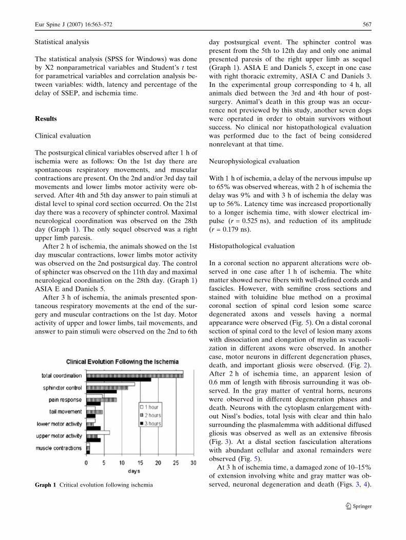
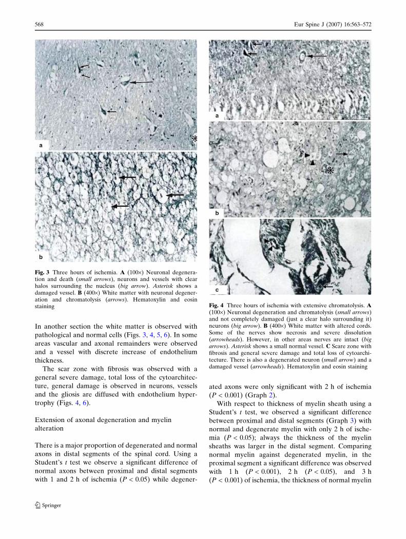
At 3 h of ischemia time, a damaged zone of 10–15%
of extension involving white and gray matter was ob-
served, neuronal degeneration and death (Figs. 3, 4).Graph 1 Critical evolution following ischemia
Eur Spine J (2007) 16:563–572 567
123
In another section the white matter is observed with
pathological and normal cells (Figs. 3, 4, 5, 6). In some
areas vascular and axonal remainders were observed
and a vessel with discrete increase of endothelium
thickness.
The scar zone with fibrosis was observed with a
general severe damage, total loss of the cytoarchitec-
ture, general damage is observed in neurons, vessels
and the gliosis are diffused with endothelium hyper-
trophy (Figs. 4, 6).
Extension of axonal degeneration and myelin
alteration
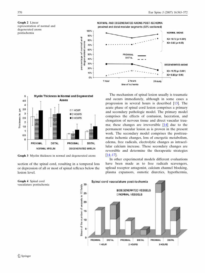
There is a major proportion of degenerated and normal
axons in distal segments of the spinal cord. Using a
Student’s t test we observe a significant difference of
normal axons between proximal and distal segments
with 1 and 2 h of ischemia (P < 0.05) while degener-
ated axons were only significant with 2 h of ischemia
(P < 0.001) (Graph 2).
With respect to thickness of myelin sheath using a
Student’s t test, we observed a significant difference
between proximal and distal segments (Graph 3) with
normal and degenerate myelin with only 2 h of ische-
mia (P < 0.05); always the thickness of the myelin
sheaths was larger in the distal segment. Comparing
normal myelin against degenerated myelin, in the
proximal segment a significant difference was observed
with 1 h (P < 0.001), 2 h (P < 0.05), and 3 h
(P < 0.001) of ischemia, the thickness of normal myelin
Fig. 4 Three hours of ischemia with extensive chromatolysis. A(100·) Neuronal degeneration and chromatolysis (small arrows)and not completely damaged (just a clear halo surrounding it)neurons (big arrow). B (400·) White matter with altered cords.Some of the nerves show necrosis and severe dissolution(arrowheads). However, in other areas nerves are intact (bigarrows). Asterisk shows a small normal vessel. C Scare zone withfibrosis and general severe damage and total loss of cytoarchi-tecture. There is also a degenerated neuron (small arrow) and adamaged vessel (arrowheads). Hematoxylin and eosin staining
Fig. 3 Three hours of ischemia. A (100·) Neuronal degenera-tion and death (small arrows), neurons and vessels with clearhalos surrounding the nucleus (big arrow). Asterisk shows adamaged vessel. B (400·) White matter with neuronal degener-ation and chromatolysis (arrows). Hematoxylin and eosinstaining
568 Eur Spine J (2007) 16:563–572
123
was larger. In the distal segment the thickness of nor-
mal myelin was always larger with a significant differ-
ence at 1 h (P < 0.001), 2 h (P < 0.001), and 3 h
(P < 0.01) of ischemia.
Morphological findings with blood vessels
In group 1, 1 h of ischemia, we did not observe any
apparent alterations. In group 2, 2 h of ischemia, a
10–15% of the vessels showed distal alterations with
breaking or dissolution of its endothelium and
detachment of the intimate tunic of the middle layer
of the vessels. In group 3, 3 h of ischemia, we found
alterations in 10–15% of the vessel with light
detachment of intimate middle layers, endothelial
cells with many vesicles, and some vessels with
complete alteration of all layers. In another case of
3 h of ischemia, it showed damage of distal vessels,
with rupture and dissolution of the layers, the endo-
thelium was observed with multiple vesicles, elonga-
tion of muscular layer with wide spaces between
them. The basal layer was detached in some areas
(Graph 4).
Discussion
The term ‘‘spinal shock’’ applies to all anatomic and
physiological phenomena that surround traumatic
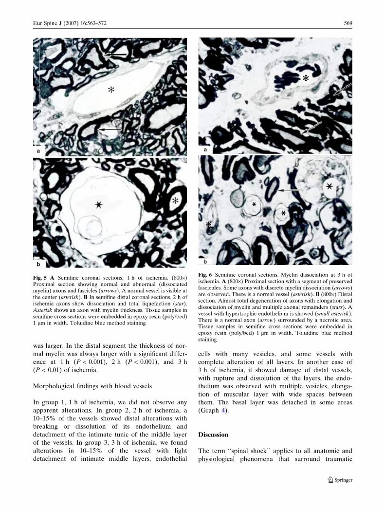
Fig. 5 A Semifine coronal sections, 1 h of ischemia. (800·)Proximal section showing normal and abnormal (dissociatedmyelin) axons and fascicles (arrows). A normal vessel is visible atthe center (asterisk). B In semifine distal coronal sections, 2 h ofischemia axons show dissociation and total liquefaction (star).Asterisk shows an axon with myelin thickness. Tissue samples insemifine cross sections were embedded in epoxy resin (poly/bed)1 lm in width. Toluidine blue method staining
Fig. 6 Semifine coronal sections. Myelin dissociation at 3 h ofischemia. A (800·) Proximal section with a segment of preservedfascicules. Some axons with discrete myelin dissociation (arrows)are observed. There is a normal vessel (asterisk). B (800·) Distalsection. Almost total degeneration of axons with elongation anddissociation of myelin and multiple axonal remainders (stars). Avessel with hypertrophic endothelium is showed (small asterisk).There is a normal axon (arrow) surrounded by a necrotic area.Tissue samples in semifine cross sections were embedded inepoxy resin (poly/bed) 1 lm in width. Toluidine blue methodstaining
Eur Spine J (2007) 16:563–572 569
123
section of the spinal cord, resulting in a temporal loss
or depression of all or most of spinal reflexes below the
lesion level.
The mechanism of spinal lesion usually is traumatic
and occurs immediately, although in some cases a
progression in several hours is described [13]. The
acute phase of spinal cord lesion comprises a primary
and secondary pathologic model. The primary model
comprises the effects of contusion, laceration, and
elongation of nervous tissue and direct vascular trau-
ma; these changes are irreversible [14] due to the
permanent vascular lesion as is proven in the present
work. The secondary model comprises the posttrau-
matic ischemic changes, loss of energetic metabolism,
edema, free radicals, electrolytic changes as intracel-
lular calcium increase. These secondary changes are
reversible and determine the therapeutic strategies
[14–17].
In other experimental models different evaluations
have been made as to free radicals scavengers,
upload receptor antagonist, calcium channel blocking,
plasma expansors, osmotic diuretics, hypothermia,
Graph 3 Myelin thickness in normal and degenerated axons
Graph 4 Spinal cordvasculature postischemia
Graph 2 Linearrepresentation of normal anddegenerated axonspostischemia
570 Eur Spine J (2007) 16:563–572
123
cyclooxygenase inhibitors, serotonin antagonists, and
N-methyl D-aspartate (NMDA) receptor antagonists.
We propose that an alternative handling in order to
break the feedback mechanism and interrupt many
aspects of secondary lesion cascade should include
pharmacological and surgical strategies [14].
In the experimental groups of this work a correla-
tion does not exist after surgery between the clinical
changes, nerve conduction velocity, and histopatho-
logical findings with the different ischemia times, be-
cause it is in the 1, 2, and 3 h of ischemia that the dogs
have a good evolution with sphincter control and vol-
untary motor activity. Only two cases, of the groups 1
and 3, the paresis of the right upper limbs was present
with the SSEP. We observed a delay in the nerve
conduction up to 65% in 1 h of ischemia, 9% in 2 h of
ischemia, and up 56% in 3 h of ischemia. The histo-
pathological findings did not show any correlation with
the clinical findings, although it did show a partial
correlation with the SSEP findings. In group 1, there
was 25% of section, and the Rexed’s area [18] VII,
VIII, and IX were affected, group 2 and 3 showed up to
15% of section. In group 2, the IX Rexed’s area was
involved and in group 3 VIII and IX areas were found
with damage.
Four hours after the clamping and cutting the
recovery was not possible, the animals died before
reaching that time. We repeated the experiment in this
group, always with the same result (a total of 9). In the
first, second, and third groups, the animals were under
experimental control conditions for 3 months, with
almost a total clinical recovery, that did not show
correlation with the SSEP and histopathological find-
ings. In a previous work of the author a similar dis-
cordance was observed, the clinical evolution did not
correspond with the pathological evolution. However,
we consider that there are vascular and neuronal
regeneration changes [4].
The pathological analysis at the level of segment
with the lesion determined an axonic index that indi-
cates neuronal regeneration and this was larger at 3 h
of ischemia as evidence of recovery. The thickness of
the myelin sheaths increased at 3 h of ischemia, but the
normal myelin thickness was always larger than the
degenerated myelin in proximal and distal segments,
the degenerated myelin thickness is larger in a distal
segment in comparison with the proximal segment and
increase with a longer evolution time. These changes
were evident from 1 h of ischemia with distal degen-
eration of axonal cytoskeleton and myelin sheaths, and
were evident in all the experimental groups. In the
proximal segment only with 3 h of ischemia abnormal
vessels were observed, while in the distal segment
abnormal vessels were always evident. At 1 and 3 h
half of the vessels were abnormal, indicating a regen-
eration process. With 2 h a few abnormal vessels were
evident, confirming the statistical difference of abnor-
mal vessels between proximal and distal segments of
the section. The analysis of cases with recovery (n = 5)
a total of nine cases with 4 h of ischemia (n = 9)
without any recuperation. The X2 with Yate’s corre-
lation was 9.98 with P = 0.0015, and the Fisher’s test
showed P = 0.00049, both statistically significant. The
SSEP showed a larger latency time and amplitude
reduction with larger ischemia time with delay in nerve
conduction velocity, thus reducing the nervous signal.
We do not know enough concerning molecular
composition of synapsis of the central nervous system.
An important point being how much of the union
material is energetic and how much is a particular type
of synapsis. The knowledge of this composition will
permit the classification and understanding of them.
Not enough is known yet as to the synapsis joining,
especially what proportion of the molecules are
building as individual molecules locally and which are
preformed as a group. The answer to this question will
help to define the nature of the classification process.
The complete change of functional molecules at
synapses is unknown, including the same synaptic
structure and if these molecules are substitute to indi-
vidual molecules or in a group of related molecules
[19].
There are several hypothesis: (1) a structural change
at synapses (increased or decreased in a particular kind
of synapses, new synapses, removal of synapses, or
configuration changes at synapses); (2) molecular
composition changes in existing synapses; synthesis of
new molecules or new molecular combinations; (3)
functional or state changes in the existing molecules or
synapses (phosphorylation, or posttranscriptional
changes) [19].
We are working with the present model and the
following experiments will give the answers of points A
and B, in order to complete the basic and clinical
analysis of the present model.
Conclusions
The repair of the spinal blood flow helps the spinal
cord to recovery. In this work we concluded that there
is a maximum 3-h period, which we called ‘‘critical
ischemia time’’, for the spinal recovery to be possible.
We have less than 4 h to offer a possible neurological
benefit, by means of a spinal cord revascularization in
cases of acute spinal cord injury with spinal cord sec-
tion without any added treatment.
Eur Spine J (2007) 16:563–572 571
123
References
1. Tator ChH, Fehling MG (1991) Review of the secondaryinjury theory of acute spinal cord trauma with emphasis onvascular mechanisms. J Neurosurg 5:15–26
2. Rhoney DH, Luer MS, Hughes M, Hatton J (1996) Newpharmacological approaches to acute spinal cord injury.Pharmacotherapy 16(3):382–392
3. Hughes JT (1987) Historical review of paraplegia before1918. Paraplegia 25:168–171
4. Bitar-Alatorre WE, Jimenez RM, Ortiz GG, Delgado R,Sanmiguel S, Sanchez-Corona J, Feria-Velazco A (1992)Vascular microsurgery of acute spinal cord injury in dogs.Arch Med Res 23(4):235–236
5. Netter FH (1987) Nervous system anatomy and physiology.Brain areteriography. Salvat, pp 50–51
6. Getty R (1982) Anatomy of domestic animals, vol II, Salvat,pp 1750–1752, 1766–1771, 1832–1847
7. Miller ME (1964) Anatomy of the dog. Saunder W.B., pp315–317, 533–543
8. Koyanagi I, Tator ChH, Lea PJ (1993) Three dimensionalanalysis of the vascular system in the rat spinal cord withscanning electron microscopy of vascular corrosion casts.Part 1: normal spinal cord. Neurosurgery 33:277–284
9. Koyanagi I, Tator ChH, Lea PJ (1993) Three dimensionalanalysis of the vascular system in the rat spinal cord withscanning electron microscopy of vascular corrosion casts.Part 2: acute spinal cord injury. Neurosurgery 33(2):285–292
10. Frankel HL, Hancock DO, Hyslop G (1969) The value ofpostural reduction in the initial management of closed inju-ries of the spine with paraplegia and tetraplegia. Part I.Paraplegia 7:179–192
11. Palapa Garcıa LR, Anaya Vallejo S, Ramırez Gutierrez R,Seanchez Flores L (1997) Lesiones por flexodistraccion de lacolumna cervical tratadas con placa por vıa posterior. RevMex Ortop Traum 11(3):163–169
12. American Spinal Injury Association: International Standardsfor Neurological Classifications of Spinal Cord Injury (re-vised) (2000) American Spinal Injury Association, Chicago,pp 1–23
13. Atkinson PP, Atkinson JL (1996) Spinal shock. Clin Proc71(4):384–389
14. Harat M, Radek A, Kochanowski J (1996) The physiopa-thology of acute spinal cord injury and a hope for a suc-cessful. Neurol Neurochir Pol 30(1):123–135
15. Lee M, Lee E, Kim Y, Choi B, Park S, Park H et al (2005)Ischemic injury-specific gene expression in the rat spinal cordinjury model using hypoxia-inducible system. Spine30(24):2729–2734
16. Park E, Velumian AA, Fehlings MG (2004). The role ofexcitotoxicity in secondary mechanisms of spinal cord injury:a review with an emphasis on the implications for whitematter degeneration. J Neurotrauma 21:754–774
17. Lu K, Liang CL, Chen HJ et al (2004) Injury severity and celldeath mechanisms: effects of concomitant hypovolemichypotension on spinal cord ischemia reperfusion in rats. ExpNeurol 185:120–132
18. Lopez-Antunez (1983) Functional anatomy of nervous sys-tem. Esquema de Rexed, Limusa, Mexico, pp 140–142, 667–670, 677–678
19. Baudry M, Thompson R, Davis J (1983) Synaptic plasticity,molecular, cellular and functional aspects. A Bradford book.MIT Press, Cambridge, pp 13–39
572 Eur Spine J (2007) 16:563–572
123
Related Documents