Cricothyrotomy Indications and Use for the NH Paramedic New Hampshire New Hampshire Division of Fire Standards & Division of Fire Standards & Training and Training and Emergency Medical Services Emergency Medical Services

Cricothyrotomy Indications and Use for the NH Paramedic New Hampshire Division of Fire Standards & Training and Emergency Medical Services.
Dec 13, 2015
Welcome message from author
This document is posted to help you gain knowledge. Please leave a comment to let me know what you think about it! Share it to your friends and learn new things together.
Transcript
Cricothyrotomy
Indications and Use for the NH Paramedic
New HampshireNew Hampshire
Division of Fire Standards & Training andDivision of Fire Standards & Training andEmergency Medical ServicesEmergency Medical Services

Clinical Indications
Inability to adequately oxygenate and ventilate using less invasive methods.

Contraindications
Ability to oxygenate and ventilate using less invasive measures.
Age less than 12 years old
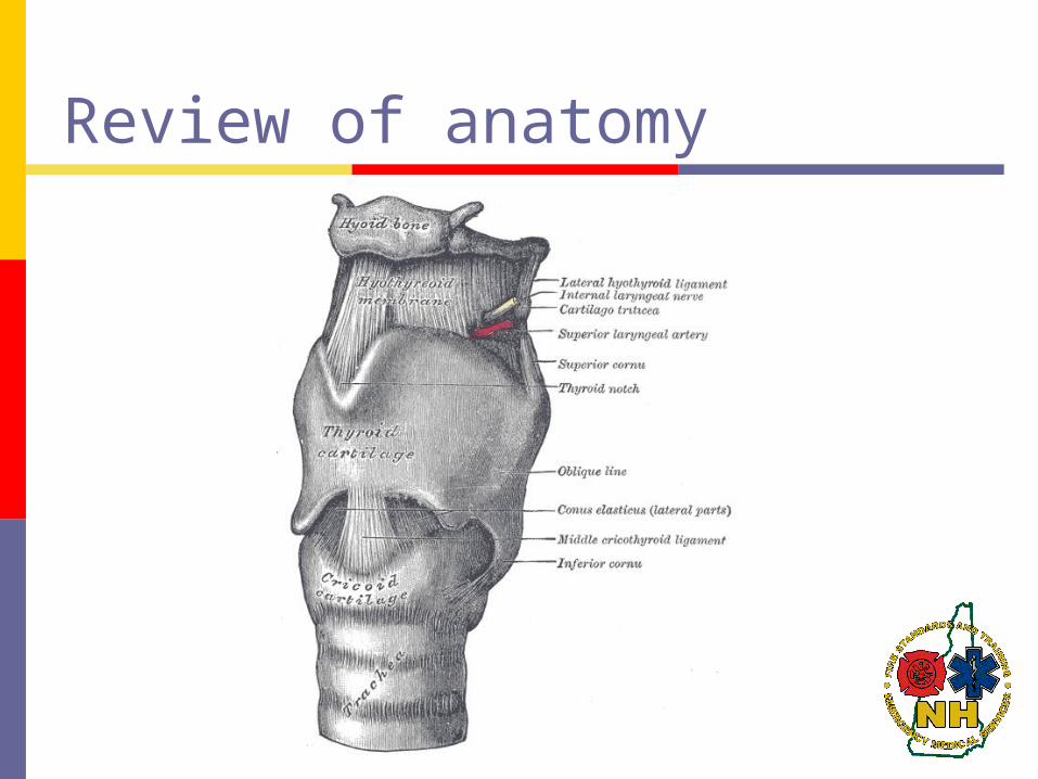
Review of anatomy


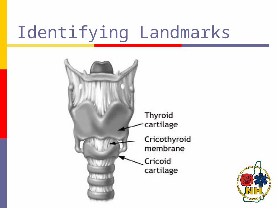
Identifying Landmarks

Find the persons Adam's apple (thyroid cartilage)

Move your fingers about one inch down the neck until you find another bulge.
This is the cricoid cartilage. The indentation between the two is the cricothyroid membrane, where the incision will be made.

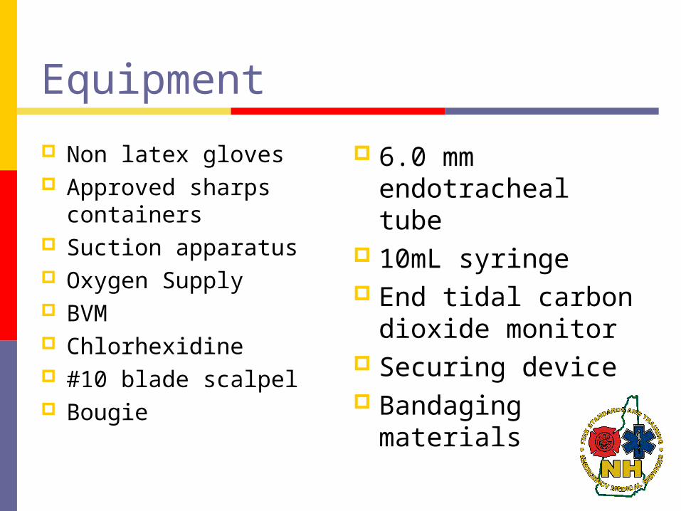
Equipment
Non latex gloves Approved sharps
containers Suction apparatus Oxygen Supply BVM Chlorhexidine #10 blade scalpel Bougie
6.0 mm endotracheal tube
10mL syringe End tidal carbon
dioxide monitor Securing device Bandaging
materials
Procedure
Have all supplies (including suction) available and ready
Proper body substance isolation Places patient supine and hyperextend neck
if no cervical trauma suspected Positions at patient's side and directs
assistant to attempt ventilations with 100% oxygen
Prepare equipment

Procedure
1. Position the patient supine and extend the neck as needed to improve anatomic view.
2. Prep neck with Chlorhexidine 3. Using your non-dominant hand, stabilize the larynx
and locate the following landmarks: thyroid cartilage (Adam’s apple) and cricoid cartilage. The cricothyroid membrane lies between these cartilages.
4. Make an approximately a 3cm vertical incision 0.5cm deep through the skin and fascia, over the cricothyroid membrane. With finger, dissect the tissue and locate the cricothyroid membrane.

Procedure
5. Make approximately a 1.5cm horizontal incision through the cricothyroid membrane.
6. With your finger, bluntly dilate the opening through the cricothyroid membrane.
7. Insert the bougie curved-tip first through the incision and angled towards the patient’s feet.
8. Advance the bougie into the trachea feeling for “clicks” of tracheal rings and until “hangup” when it cannot be advanced any further. This confirms tracheal position.

Procedure
9. Advance a 6.0 mm endotracheal tube (ensure all air aspirated out of cuff) over the bougie and into the trachea.
10. Remove bougie while stabilizing ETT ensuring it does not become dislodged
11. Inflate the cuff with 5 – 10ml of air.

Procedure
12. Confirm appropriate proper placement by symmetrical chest-wall rise, auscultation of equal breath sounds over the chest and a lack of epigastric sounds with ventilations using bag-valve-mask, condensation in the ETT, and quantitative waveform capnography.
13. Secure the ETT.14. Reassess tube placement frequently, especially
after movement of the patient.15. Ongoing monitoring of ETT placement and
ventilation status using waveform

Complications
Incorrect tube placement/ false passage
Thyroid gland damage
Severe bleeding
Subcutaneous emphysema
Laryngeal nerve damage

Questions?
Related Documents