UNIVERSITY OF NAIROBI 1 CRANIOTOMY SURGICAL SITE INFECTIONS AT THE KENYATTA NATIONAL HOSPITAL A DISSERTATION SUBMITTED IN PART FULFILLMENT FOR THE MASTER OF MEDICINE IN NEUROSURGERY DEGREE, UNIVERSITY OF NAIROBI. DR ALEX HENRY NJIRU MBCHB (UON) 2015

Welcome message from author
This document is posted to help you gain knowledge. Please leave a comment to let me know what you think about it! Share it to your friends and learn new things together.
Transcript

UNIVERSITY OF NAIROBI
1
CRANIOTOMY SURGICAL SITE INFECTIONS AT THE KENYATTA NATIONAL HOSPITAL A DISSERTATION SUBMITTED IN PART FULFILLMENT FOR THE MASTER OF MEDICINE IN NEUROSURGERY DEGREE, UNIVERSITY OF NAIROBI. DR ALEX HENRY NJIRU MBCHB (UON) 2015

UNIVERSITY OF NAIROBI
2
SUPERVISORS PROFESSOR NIMROD J MWANG’OMBE Neurosurgeon, Professor of Surgery, Division of Neurosurgery Department of Surgery University ofNairobi. Signature…………………………………. Date……………………. DR AKUKU P.O. Neurosurgeon Lecturer Department of Neurosurgery University of Nairobi Signature…………………………………. Date……………………. DR FLORENCE MUTUA Medical Microbiologist Lecturer Department of Microbiology University of Nairobi Signature…………………………………. Date…………………….

UNIVERSITY OF NAIROBI
3
DECLARATION I declare that this dissertation is my own original work and has not been presented for a degree in any other University. Sign…………………………….. Date…………………. Dr. Alex Henry Njiru. H58/76363/09

UNIVERSITY OF NAIROBI
4
ACKNOWLEDGEMENT I would like to express my heartfelt thanks and appreciation to my supervisors Prof Mwangombe, Dr Akuku, and Dr Florence Mutua, for their kind advice and professional guidance. I sincerely want to thank Mrs.Winnie Mutai tutorial fellow in the Department of Medical Microbiology for dedicating her time to give me guidance. I would also like to thank Mr. Oloo for invaluable assistance in analyzing all the swabs and for answering questions in the field of microbiology.

UNIVERSITY OF NAIROBI
5
TABLE OF CONTENTS SUPERVISORS……………………………………………………………………..2 DECLARATION…………………………………………………………………....3 ACKNOWLEDGEMENT…………………………………………………………..4 TABLE OF CONTENTS…………………………………………………………...5 LIST OF TABLES………………………………...………………………………..6 LIST OF FIGURES………………………………………...……………………….7 LIST OF ABBREVIATIONS…………………………………………………….…8 SUMMARY…………………………………………………...………………….....9 LITERATURE REVIEW………………………………………………………….12 PROBLEM STATEMENT………………………………………………………...21 STUDY JUSTIFICATION………………………………………………………...21 HYPOTHESIS……………………………………………………………………..21 OBJECTIVES……………………………………………………………………...21 Main objectives…………………………………………………………………....21 Specific objectives…………………………………………………………………21 METHODOLOGY……………………….………………………………………..22 STUDY LOCATION……………………………………………………………...22 STUDY DESIGN………………………………………………………….……....22 STUDY POPULATION……………………………….………………….……….22 INCLUSION CRITERIA……………………………………………..…………...22 EXCLUSION CRITERIA…………………………………………..……………..22 SAMPLE SIZE ESTIMATION……………………………………..……………..22 DATA COLLECTION…………………………………….………………………23 MATERIALS AND METHODS……………………………………….…………23 LABORATORY PROCEDURES…………………………………..…………….24 QUALITY ASSURANCE……………………………………………..………….27 DATA MANAGEMENT AND STATISTICAL ANALYSIS……………….…..27 ETHICAL CONSIDERATION……..…………………………………………….28 OUTCOME………………..………………………………………………………28 STUDY LIMITATIONS……………..……………………………………………28 RESULTS……………………………….……………………………...................29 DISCUSSION…………………………….……………………………...………..36 CONCLUSION……………….…………………………………………………...41 RECOMMENDATIONS…………….…………………………………………....42 REFERENCES………………….…………………………………………………43 APPENDIX I:CONSENT………………………………………………………..48 APPENDIX II: QUESTIONNAIRE……………………………………………...52 APPENDIX III: ETHICAL APPROVAL LETTER……………………………....55

UNIVERSITY OF NAIROBI
6
LIST OF TABLES Table 1: Physical status classification for surgical patients…………………………14 Table 2. Association of class of SSI and number of days in hospital……………....30 Table 3. Comparison of days in hospital for patients with and without SSI………...30 Table 4. Risk factors associated with SSI……………………………………………31 Table 5.The ASA score of patients with SSI…………………………………………32 Table 6. Relationship between time and duration of surgery with SSI………………32 Table7. State of the wound in patients with SSI…………………………………….33 Table 8. Growth pattern of the microorganisms……………………………………..34 Table 9. Pattern of resistance of S. aureus………………………………………….35 Table 10. Pattern of resistance of gram negative organisms…………………………35

UNIVERSITY OF NAIROBI
7
LIST OF FIGURES Figure 1: Age group for the respondents (with and without SSI)……………………29
Figure 2: Organisms isolated…………………………………………………………35

UNIVERSITY OF NAIROBI
8
LIST OF ABBREVIATIONS ASA -American Society of Anesthesiology ATCC-American Type Culture Collection BMI- body mass index CDC- Centre for Disease Control and Prevention CI-Confidence interval CNS-Central nervous system ESBL-Exended spectrum beta lactamase ICU- Intensive Care Unit IQR-Interquartile Range KNH-Kenyatta National Hospital MRSA-Methicillin Resistant Staphylococcus aureus NHSN-National Healthcare Safety Network NNIS- National Nosocomial Infections Surveillance System (USA based) NINSS -Nosocomial Infection National Surveillance Service (UK based) OR –Operating Room SSI- Surgical Site Infection SPSS- Statistical Package for the Social Sciences WHO- World Health Organization UON- University Of Nairobi

UNIVERSITY OF NAIROBI
9
SUMMARY This study was designed to evaluate the incidence of surgical site infections in patients undergoing elective craniotomies in Kenyatta National Hospital, associated risk factors, identification of bacterial causative agents and antimicrobial sensitivity patterns. This was a prospective observational study carried out in Kenyatta National Hospital between June 2014-March 2015. All cases of patients done craniotomy, who presented with Surgical Site Infection (SSI), within 30 days after surgery, as defined by the Center for Disease Control and Prevention (CDC) criteria, were included. Patients who died before 30 days after surgery were excluded from the study. The data studied in each patient included age, gender, body mass index (BMI) , history of cigarette smoking, steroid use, the America Society of Anesthesiologist index score, wound classification, preoperative skin preparation solution, antimicrobial prophylaxis, the time when the operation was done (i.e. morning or afternoon) and duration of operation in hours (<4 hours or > 4 hours), use of surgical drains and post-operative SSI. The following data was obtained from all the patients who developed SSI; time of occurrence, type of infection (superficial, deep and organ space), pathogens and antimicrobial sensitivity patterns. Regular surveillance of the craniotomy site was done for atleast two weeks while in hospital and at the outpatient neurosurgical clinic. Bacterial specimens were taken from the wounds that met the CDC criteria. Specimens were obtained using aseptic technique and sterile swabs. The swabs were inoculated on blood agar and Mac Conkey media, isolates identified and antimicrobial sensitivity patterns determined. Data was analyzed using SPSS. Association was tested by applying the Student t-test and the Chi-square test of significance, and the strength of association was expressed as the Odd's Ratio. Significance was set at probability value of 0.05 and 95% confidence interval. Data was presented in the form of graphs, charts and tables. Eighty patients were recruited into the study giving a response rate of 83.33%. 37 (46.25%) were males and 43 (53.75%) were females. The male to female ratio was 4.6:5.8. 28.75% of the respondents were between 20-30 years and this represented the most common age group in this study. Overall, six surgical site infections were identified in the neurosurgical ward of Kenyatta National Hospital during the study period, representing an infection rate of 7.5%. Majority of the infections occurred within the first seven days post operatively. According to CDC definition 4 (66.67%) of the infections were superficial, 1 (16.67%) was deep and 1 (16.67%) involved organ or space. Significant risk factors for developing SSI during hospital stay were: age, smoking, long preoperative stay of patients, wound type, operations lasting for more than 4 hours, those done in the afternoon, those with surgical drains and those that were obese. In this study cultures were positive in all the specimens taken and the most common gram positive isolate was Staphylococcus aureus (33.33%). The most common gram

UNIVERSITY OF NAIROBI
10
negative isolate was Pseudomonas spp (33.33%). One (16.67) SSI was caused by gram positive bacteria alone, two (33.33%) were caused by gram negative bacteria alone and three (50%) were caused by mixed infections. Of the gram positive bacteria S. aureus was resistant to multiple antimicrobials but all were sensitive to Vancomycin and Teicoplanin. The most common gram negative bacterium was Pseudomonas aeruginosa which were resistant to most of antimicrobials tested. P. aeruginosa showed low resistance rates to Amikacin (33.33%) and Meropenem (33.33%). This study has shown that the incidence of craniotomy surgical site infections, risk factors and isolated organisms are similar to studies done in other countries.

UNIVERSITY OF NAIROBI
11
INTRODUCTION Surgical site infection (SSI) is a worldwide problem and a major cause of morbidity, reduced quality of life, financial burden in healthcare and mortality. According to a study done by Kirkland et al, it was estimated that worldwide SSI account for as much as $10 billion annually in direct and indirect medical cost. The knowledge of incidence, risk factors and micro- organisms, will allow health professionals to implement protocols to reduce the infection rates and improve the quality of life of patients. Since the birth of surgery, surgical site infections have been a major complication and these infections account for majority of nosocomial acquired infections. In a prospective study done by Anvikar et al, Surgical site infections were the commonest postoperative infections after urinary tract infections with an overall infection rate of 6.09% and with increased hospitalization time being associated with significant increases in SSI. Craniotomies are clean wounds that heal with primary intention. According to a study carried out between October 1997 and June 2001 by Coello et al, there is an emphasis on the reduction of SSI, which reduces patients’ recovery time, pain and risk of other complications in the future for all categories of surgeries. According to Selwyn S et al, trial at reducing SSI date to the 19th century with the study of the epidemiology and prevention of surgical fever by James Young Hamilton. Despite great efforts being taken to prevent the rate of SSI, they are on the increase because of increased number of operations, increased invasiveness and increased use of implants.

UNIVERSITY OF NAIROBI
12
LITERATURE REVIEW The magnitude of the risk factors associated with SSIs in patients undergoing neurosurgical procedures has not been documented in Kenya and hence their nature is not known locally. A study by Ballard MR et al showed that one of the key components is a multi-displinary approach with everyone being equally committed to the process of improvement as a team. In a randomized controlled trial done by Valentini et al, done with the objective of evaluating the incidence of SSI in neurological institute in Milan it was found that the range of infection in clean neurosurgical operations was 4% to 12% without prophylactic antibiotics and 0.3% to 3% with prophylactic antibiotics. There are reported geographical differences in rates of SSI. In a systemic analysis carried out by Shearwood et al, comparing postoperative infection rates between Northern America and Europe concluded that SSI rates of neurosurgical procedures were 2.2% and 5.7% respectively. No evidence based explanations could explain these differences. This study recommended prospective studies to determine the factors causing a difference in the rates of post-operative wound infections hence the rate of SSI in Africa may be different from that in the western countries. The proportion of SSIs which is preventable is unknown however there are wide variations in infection rates and an international drive to minimize them. In a retrospective review of 48 studies in Europe, by Leaper DJ et al, there was found to be variations in the rates of surgical site infections because of failure of recording all necessary data to allow a comparative assessment of surgical site infections. In a prospective observation study of 390 cases by Buang SS et al, it was found that surgical site infections remained a significant problem in neurosurgery. In the study various risk factors were identified including time of surgery, the surgeon who performed the surgery i.e. resident or qualified consultant, among others. The conclusion was that risk factor identification would help improve patient care and reduce economic burden of cost of health care and associated mortality and morbidity. Cruse, in a prospective study of all surgical wounds of patients at Foothills Hospital over a period of 10 years estimated that a SSI increased a patient’s hospital stay by approximately 10 days with an additional cost of $2000. This shows that SSIs are morbid and expensive on the health system and care should be taken to minimize them. Health care costs of a patient with postoperative infections are almost twice the amount of costs for those without (Broex EC et al). According to De Lissovoy et al, SSI usually leads to significant economic burden in terms of increased length of stay and escalated cost of treatment. Their analysis documented nearly 1 million additional inpatient days and 1.6 dollars in excess cost. According to Valentini et al, the accepted rate of neurosurgical SSI is less than 5% and in a consecutive series of 1747 patients treated with elective neurosurgery and prophylactic antibiotics it was found that majority of neurosurgical SSI typically occur within two weeks to one month. In a retrospective study of consecutive patients undergoing neurosurgery between 1st January 2007 and 1st January 2008, done by Alina P et al, 42 postoperative SSI were identified and most were superficial 52.3%,

UNIVERSITY OF NAIROBI
13
with lower incidence of deep 33.33% and organ/space SSIs 14.28%. According to a study by Siti Suriyati et al, many of the SSIs are identified during inpatient stay and mean time between surgery and onset of infection is 11th to 21st day with a median of the 10th day. Postoperative wound infections are rarely detected before the third day because of the dressing that is still on the wound. According to Hosein et al, postoperative neurosurgical site infections carry a significant morbidity because of proximity to the meninges and brain parenchyma. Despite SSI rate being low it has dire consequences because of extension of infections to involve the calvaria causing osteomyelitis, meninges causing meningitis and cerebrum causing brain abscess which are difficult to treat because of presence of multiple organisms and poor cerebrospinal penetration to antibiotics resulting in serious morbidity and mortality with incidence upto 20 %( shunt associated). In a study of 255 pairs of patients by Kirkland et al, SSI were found to be morbid and expensive with the presence of SSI increasing mortality risk by 2-11 fold. However, not all SSIs are the same. According to Fry De, reports of economic burden which do not take into account the depth of the infection or the manner in which the infection occurs are underestimating the depth of these complications. This often leads to loss of income because of disability associated with these infections. According to Horan et al, (modifications of Centre for Disease Control and Prevention (CDC) definitions of SSI), nosocomial surgical site infections are classified and defined as: Superficial incisional SSI: This is a SSI that occurs within 30 days of an operation and involves only the skin and the subcutaneous tissue. A wound is not considered to have a superficial incisional SSI if a stitch abscess is present, if the infection is at an episiotomy, a circumcision site, or a burn wound; or if the SSI extends into fascia or muscle. The wound also has atleast one of the following: Purulent discharge (culture documentation not required), organisms isolated from fluid/tissue of the superficial incision, at least one sign of inflammation (e.g. pain or tenderness, indurations, erythema, local warmth of the wound) is present, the wound is deliberately opened by the surgeon or the surgeon or attending physician declares the wound infected. Deep incisional SSI. This occurs within 30 days of the operation or within one year if an implant is present and involves deep soft tissues (e.g. fascia and or muscle) of the incision. It presents with at least one of the following: Purulent discharge from the deep incision but without organ/space involvement, fascial dehiscence or fascia is deliberately separated by the surgeon because of signs of inflammation, a deep abscess is identified by direct examination or during reoperation, by histopathology, or by radiologic examination or the surgeon or attending physician declares that a deep incisional infection is present. Organ/space SSI. It occurs within 30 days of the operation or within one year if an implant is present and involves anatomical structure not opened or manipulated during the operation. It also has atleast one of the following: Purulent drainage from a drain placed by a stab wound into the organ/space, organisms are isolated from the

UNIVERSITY OF NAIROBI
14
organ/space by aseptic culturing technique, an abscess in the organ/space is identified by direct examination, during reoperation, or by histopathology or radiologic examination or a diagnosis of organ/space SSI is made by the surgeon or attending physician. Numerous studies have been published on risk factors associated with SSI. They are both patient and procedure related. Examples of risk factors include: Increased age and longer operation duration (Valentini et al 2007), obesity, malnutrition, increased American Society of Anesthesiology (ASA) score, intraoperative blood transfusions and diabetes (Schuster et al, 2010), early reoperation (Lietard et al 2008), male gender, no antimicrobial prophylaxis and post-operative cerebral spinal fluid leak (Korinek et al 2005) and procedure done in late shift (Sanchez-arenas et al, 2010). According to Edwards Wong, CDC developed National Nosocomial Infections Surveillance System (NNIS) risk index in the year 1991 as an improvement over SENIC (Study on Efficacy of Nosocomial Infection Control) risk index which ranges from 0 to 3 points and is defined by three independent and equally weighted variables. One point is scored for each of the following when present: • American Society of Anesthesiology (ASA) physical status classification score >2 • Either contaminated or dirty/infected wound classification • Length of operation > T hours (where T is approximate 75th percentile of duration of the specific operation being performed. In a study by Robert H et al, it has been shown that dichotomization of the American Society of Anesthesiology (ASA) score in NNIS risk index is an advantage over the SENIC risk index. Its ease for collecting and its objectivity seem advantageous. Another advantage is that the NNIS risk index uses a procedure related cut point to indicate a long duration of surgery for an individual procedure, rather than a 2 hour cut point for all procedures. ASA score is an index to assess overall physical status of patient before operation ranging from 1 to 5. It has been shown highly predictive for development of SSI according to a study done by David H et al. TABLE 1: Physical status classification for surgical patients (ASA score)
Class I A patient in normal health
Class II A patient with mild systemic disease resulting in no functional limitations
Class III A patient with severe systemic disease that limits activity, but is not incapacitating
Class IV A patient with severe systemic disease that is a constant threat to life
Class V A moribund patient not likely to survive 24 hours

UNIVERSITY OF NAIROBI
15
The Center for Disease Control and Prevention (CDC) using an adaptation of the American College of Surgeons’ wound classification schema divides surgical wounds into 4 categories (CDC 1999): Clean wounds: The wound is judged to be clean when the operative procedure does not enter into a normally colonized viscus or lumen of the body. The SSI risk kis minimal and originates from contaminants of the operating room (OR) environment or from the surgical team, or most commonly from skin colonists. The most common pathogen is Staphylococcus aureus. SSI rates in this class of procedures should be 2% or less, depending upon other clinical variables. Most neurosurgical elective craniotomies are clean wounds and have no sign of infection or inflammation and are closed primarily. There’s usually no break in aseptic technique. Clean-contaminated wounds: A clean-contaminated surgical site is seen when the operative procedure enters into a colonized viscus or cavity of the body, e.g. paranasal sinuses, trans-nasal and trans-oral procedures and retrosigmoid craniotomies, but under elective and controlled circumstances. The most common contaminants are endogenous bacteria from within the patient. Infection rates for these procedures are in the range of 4% to 10% and can be optimized with specific preventive strategies. Contaminated wounds. Contaminated procedures occur when gross contamination is present at the surgical site in the absence of obvious infection. As with clean-contaminated procedures, the contaminants are the bacteria that are introduced by gross soilage of the surgical field. Infection rates will be greater than 10% for this classification of wound, even with preventive antibiotics and other strategies e.g. compound skull fractures. Dirty wounds: Surgical procedures performed when active infection is already present are considered dirty wounds. Pathogens to be expected are those of the active infection that is encountered. Unusual pathogens are often encountered in dirty wounds, especially if the infection has occurred in a hospital or nursing home setting, or in patients receiving prior antibiotic therapy. E.g. brain abscesses, subdural empyema etc. Prolonged preoperative hospital stay is associated with a higher rate of infection. This is because prolonged preoperative hospital stay leads to colonization with antimicrobial resistant micro-organisms and itself directly affects patient’s susceptibility to infection either by lowering host resistance or by providing increased opportunity for ultimate bacterial colonization. According to a study done by Odhiambo VD, prolonged preoperative hospital stay was associated with a higher risk of postoperative infections. Contamination of surgical sites by microbes is universal despite cutting edge technology. According to Byrd et al, total surgical field sterility is not possible because right from the moment a surgeon performs the ritualistic surgical scrub, bacteria are present in the water and these bacteria are viable but cannot be culture hence contributing to the microbial burden in SSI . Therefore the perception of total

UNIVERSITY OF NAIROBI
16
sterility in the surgical field should be abandoned for clean field because not all the bacteria are eliminated. The study by Bible JE et al, has documented that nearly all surgical gowns used in the operating room contain the presence of microorganisms with the region having the lowest concentration of bacteria being in the region between the chest and operative field, indicating that this may be the cleanest area of the surgical gown. The study by Bible et al, also documented that the "dirtiest" regions, of an operating microscope drape used in craniotomy operations, are the shafts of the optic eyepieces for the primary surgeon, the forehead portion on both viewing stations, and the overhead portion of the drape and have been shown to approach 100% contamination. The choice of antiseptic agent may have a significant effect on the rates of SSI. Chlorhexidine has been found to be better in preventing superficial and deep incisional infections. This is according to a randomized study of 849 subjects by Darouiche et al., who found a lower rate of SSIs when using chlorhexidine alcohol group 9.5% than in the povidone-iodine group which had an infection rate of 16.1%. After production and refinement of Alexander Fleming’s Penicillium mould extract in 1940’s, antibiotic prophylaxis has served as a more advanced form of antisepsis with a further advancement in infection control (Barker et al). A meta-analysis of six prospective randomized trials, by Barker et al, concluded that antimicrobial prophylaxis is recommended for adult and pediatric patients undergoing craniotomy and spinal procedures as it reduces the rates of post-operative meningitis by half. In a randomized clinical trial by Ronald F et al, carried out between Oct 1979 and June 1984 on 846 patients showed a statistically significant difference in post-operative infections between those that were given perioperative antibiotics (4 infections 0.96%) and those that weren’t given(15 wound infections 3.64%). Hence it has been proven that the incidence of post-operative infections can be significantly reduced by giving routine perioperative antibiotics. The conclusion was that routine perioperative antibiotic prophylaxis can significantly reduce incidence of post op infections. Perioperative antimicrobials are safe and effective in reducing surgical site infections as evidenced by a critical appraisal of antibiotic prophylaxis on 4,578 patients which showed a significantly lower infection rate of 4.6% was seen in low-risk patients (clean craniotomy, no implant) with antimicrobial prophylaxis versus those without (4.6% vs 10%, p < 0.0001). According to Harbarth et al, antimicrobial prophylaxis should not routinely exceed 24 hours( 1 dose at induction and 2 more doses post operatively) with published evidence showing that after wound closure antibiotic prophylaxis is unnecessary and may lead to increase in resistant bacteria.

UNIVERSITY OF NAIROBI
17
According to guidelines by Dale et al, the initial dose of prophylactic antibiotics should given within 30 minutes prior to the incision to achieve an adequate tissue concentration at the time of first skin incision. The dose should be based on the patients’ weight because pharmacokinetics may be altered in obese patients who are predisposed to SSI. A study done by Bosco JA III et al, any additional dose of antimicrobial agent should be given intraoperatively if the operation continues after two half-life’s of the initial dose or if massive intraoperative blood losses occur (approx. 1500ml). A study done in Central hospital, Benin City by Isibor et al, showed that increasing age was associated with increased prevalence of post-operative infections probably due to reduced immunity. This study also showed different antimicrobial sensitivity patterns for different organisms. Also postoperative wound infections have become a serious problem in developing countries owing to antibiotic resistant bacteria poor infection prevention programs, crowded hospital environment and the irrational use of antibiotics. Both gram positive and gram negative microorganisms are involved in increasing rates of SSI. In a prospective study done at hospital Kuala Lumpur, by Buang et al out of the 30 infected wounds 25 grew organisms, whereas 5 did not grow any organisms. The most common organisms found in craniotomy SSI in this study include: Staphylococcus aureus 36%, Methicillin resistant S. aureus (MRSA), 17%, Pseudomonas aeruginosa 13% and Acinetobacter spp 10% and others 8%. Skin flora is generally regarded as the most common cause of SSI. But in a prospective study done by Alicia, et al, on skin cultures obtained from the operative sites of 609 neurosurgical patients, no association was found between high bacterial colony counts on the skin of operative sites either before or after the skin preparation and subsequent SSI. Hence longer skin preoperative preparation doesn’t correlate with lower post-operative bacterial colony counts. According to the Nottingham Antibiotic guidelines Committee, the recommended antibiotic dose and route for clean neurosurgical procedures is intravenous cefuroxime single dose 50mg/kg max.1.5g, if MRSA positive intravenous teicoplanin single dose 10mg/kg max.400mg and if severe penicillin and cephalosporin allergy (and covers for MRSA) intravenous teicoplanin single dose 10mg/kg max400mg (Sital Mistry et al 2012). Optimal prophylaxis with antimicrobials for surgery includes: appropriate choice of antimicrobial agent, proper timing of administration of antimicrobial agent just prior to skin incision and limitation of administration of antimicrobial following surgery. A randomized clinical trial by Ronald et al, showed that routine perioperative antibiotic prophylaxis significantly reduced the incidence of postoperative neurosurgical infection. In the perioperative period SSIs may be reduced by preventing several modifiable risk factors.
UNIVERSITY OF NAIROBI
18
A study done by Greene KA et al, shows that in cachectic patients require optimum nutrition. In this study it was found that low serum albumin levels <3.5g/dl was associated with a greater frequency of wound infections. In a prospective randomized double blind trial of 136 patients stratified by Snyderman et al, it was evident that with routine use of preoperative immune boosting supplementation (arginine, omega-3 fatty acids, and dietary nucleotides) in patients undergoing surgery significantly reduced the incidence of post-operative wound infections and therefore decreasing the length of hospitalization. Hence optimizing those with poor nutritional status reduces the incidence of SSIs. Obesity is also a major concern to the neurosurgeon. According to Toma et al, patients with obesity have poor tissue penetration of perioperative antibiotics therefore reducing their efficacy and predisposing to SSI. Weight reduction in the obese is indicated to decrease surgical complications. Several studies have examined the influence of preoperative whole-body bathing with chlorhexidine or other wash products on surgical infection; Webster et al in a randomized controlled trial, found no clear evidence of benefit of chlorhexidine baths over wash products e.g. bar soap. Determining the optimal order of surgical cases performed in a day by any particular surgeon is important. It has been shown that postoperative infections tend to be higher when procedures are performed later in the day. This is because of probable deterioration of operating theater cleanliness, lack of concentration because of fatigue and nursing shift changes (Gruskay J et al 2012) According to the Nottingham antibiotic guidelines the recommended neurosurgical antibiotic prophylaxis regime for clean, clean contaminated and contaminated wounds is cefuroxime and cefazolin. In a multicenter collaborative study ( Steinberg et al ) showed that there was a relationship between the timing of perioperative antibiotics and SSI risk with a lower risk occurring when antimicrobial prophylaxis (with cephalosporins and other antibiotics with short infusion times) were given within 30min prior to incision. Antibiotics given within 30 minutes of skin incision had an infection rate of 1.6% while those given between 31-60 minutes prior to skin incision had infection rates of 2.4%. The choice of antimicrobial prophylaxis has a big impact on reduction on SSI. Cefuroxime is a 2nd generation cephalosporin with an elimination half-life of 1.3 hours and good penetrative ability through the blood brain barrier. A prospective multicenter study by Holloway et al, showed that antibiotic prophylaxis with cefuroxime is well tolerated, has good efficacy and is associated with reduced rates of SSIs. Additional intraoperative doses of antimicrobial agents are recommended if the procedure is more than three or four hours in duration or if the duration of the

UNIVERSITY OF NAIROBI
19
procedure exceeds two half- lives of the antibiotics administered preoperatively or if major blood loss occurs. It has been the norm that hair is removed from the operative site before surgery. This may either be done with razors, clippers or creams. A Cochrane review by Tanner et al, showed that there was no difference in the rates of SSI among patients who had and those who did not have their hair shaved before surgery. If shaving was to be done then using clippers had a lower rate of postoperative infections than when using razors. In a randomized equivalence trial by Parienti et al, there was no difference between hand rubbing with alcohol aqueous alcoholic solution and the ritualistic hand scrubbing with antiseptic soap and antiseptic preparation containing povidone iodine and chlorhexidine in reducing SSI. Locally injected antibiotics, in addition to systemically delivered prophylactic antibiotics, are a promising and low-risk intervention in minimizing the bacterial inoculum. In a prospective study, Miller et al, showed a lower incidence rate of SSI when local neomycin/polymyxin was used together with systemic antimicrobials than when systemic antibiotics were used alone. Redosing antibiotics in long-duration procedures at routine intervals is recommended to maintain antimicrobial action at the internal tissue level. Intraoperative redosing with cefazolin was associated with a 16% reduction in SSI (Zanetti et al). “The solution to pollution is dilution" low-pressure lavage, combined with detergent or antibiotic agents, is indicated in the intraoperative care of routine surgical wounds. Surgical procedures are invasive procedures and may result in transfer of infections between surgical patients and staff. Wearing adequate protective barriers may reduce the chances of transfer of infections. In a Cochrane review by Parkinson et al, double or even triple gloving reduced perforations to the innermost glove which was the last barrier between the surgeon and the patient but its efficacy to reduce surgical site infections has yet to be observed. Surgeries that have more mechanical stress on surgical gloves, such as instrumented spinal fusions, warrant consideration for more than one layer of glove or more robust material. Forceps destruction of skin edges, electrocautery, and retractor-related soft tissue ischemia are major determinants in preventing wound infection. The use of bovie electrocautery is commonplace in neurosurgical operations,given its ease of use and efficiency in providing pinpoint hemostasis. Electrocautery causes local tissue destruction impairing local blood flow and leading increased rates of infection. A study by Joy Ryan (2005), showed a lack of consistency in wound dressing change practices. She concluded that the type of primary / initial dressing-post operative

UNIVERSITY OF NAIROBI
20
dressing should include: non adherent over incision e.g. xeroforms, absorbents for exudates e.g. dry gauze dressing and transparent film, primary dressing should remain for 48-72 hrs after conclusion of surgery (CDC recommendation is 24-48 hrs) and dressing change should be done at the recommended time with sterile dressing and washing hands before and after by a registered nurse. Prolonged courses of antibiotics (beyond 24 hours postoperatively) are sometimes administered in a prophylactic fashion, yet evidence for the efficacy of this practice is lacking. In a retrospective study Evans et al, showed that daily chlorhexidine baths are effective in decreasing skin colonization rates by methicillin-resistant S. aureus and Acinetobacter infection, in addition to lowering rates of catheter-related bloodstream infection.

UNIVERSITY OF NAIROBI
21
PROBLEM STATEMENT Surgical site infections requiring immediate attention are a major problem for many neurosurgical patients undergoing craniotomies and the presence of SSIs leads to increased morbidity and mortality. Craniotomy site superficial wound infections, bone flap osteitis and organ/space infections will result in an additional increase in the health economic burden. STUDY JUSTIFICATION People go to hospital with the expectations of getting better but they do have an anxiety of contracting postoperative infections. Changes in health care delivery may result in shorter hospital stays. Governments and international health organizations are increasingly putting pressure on hospitals to improve patient outcomes and reduce costs. Up till now there has been no study done in KNH determining the burden of SSI and therefore this study will aim to determine the rate, risk factors and micro-organisms involved. It will also be a guide to policy on antibiotic choices in craniotomy SSIs. HYPOTHESIS Rate of infection in KNH will be the same or higher than that given in literature, which is four to twelve per cent. The pathogens and their resistance pattern will be the same as that found in other literature. OBJECTIVES MAIN OBJECTIVE To determine the incidence of SSI in craniotomies and its associated risk factors in KNH. SPECIFIC OBJECTIVES WERE:
a) To determine the rate of surgical wound infections in craniotomies
b) To determine the risk factors associated with SSI
c) To identify bacterial causative agents of SSI and their antimicrobial susceptibility patterns.

UNIVERSITY OF NAIROBI
22
METHODOLGY STUDY LOCATION The study was conducted in the neurosurgical ward and main intensive care unit (ICU) in Kenyatta National Hospital, the largest national referral and teaching hospital in east and central Africa, with a bed capacity of the 22 patients in the main ICU and 47 patients in the neurosurgical ward. The neurosurgical ward can accommodate 34 adult male and female patients, 8 children and 5 critical care patients. STUDY DESIGN This was a prospective observational study. STUDY POPULATION The cases were defined as any patient who met the inclusion criteria and underwent elective clean and clean contaminated craniotomies from June 2014-March 2015. A total of 80 patients were included. INCLUSION CRITERIA
• All patients above 18 years
• Consenting patients.
• All patients with class 1 and 2 wounds as defined by American College of Surgeons.
EXCLUSION CRITERIA
• Patients with primary cerebrospinal fluid shunt implants.
• Patients highly suspicious of CNS infection prior to the procedure e.g. cerebral abscess, subdural empyema, cerebral tuberculosis.
• Patients with dirty wounds.
• Patients who die within 30 days after surgery.
SAMPLE SIZE ESTIMATION Estimation of the proportion of patients undergoing craniotomies enabled estimation of the incidence of SSI in craniotomies and its associated risk factors. There’s no documentation/ publication in Africa/ Kenya on the incidences of Craniotomy SSI and the total population of patients undergoing craniotomy surgery is unknown. Since this is a prospective observational study and the estimated population proportion or prevalence/ incidences are not known,0.05p = was used.

UNIVERSITY OF NAIROBI
23
A margin of error of 10% was used to estimate the sample size that fits the estimated resources. Based on the 95% confidence interval, a precision (margin of error) 10%d = ± and that the estimated population prevalence proportion was unknown, 0.05p = , was used. A significance level 0.05α = implying 1 1.96Z α− = i.e. 95 % confidence level was used.
The following Sample size determination formula for incidence studies (Lwanga SK & Lameshow S, 1991) was used to estimate the proportion of population study size.
221
2
(1 )Z p pn
dα− − =
[ ]2
2
1.96 0.50 (0.50)96
0.1n
× ×= =
The estimated sample size was 96. DATA COLLECTION Data was collected using neurosurgical site infection form that was adapted from National Surveillance of surgical Site Infection following neurosurgical procedure in Scotland.(Appendix II) MATERIALS AND METHODS Consecutive patients that fitted the inclusion criteria were included in the study. All SSI that appear within 30 days after operation were documented. The following variables were measured:
1. Preoperatively: age, sex, ASA score, nutritional status (body mass index), length of preoperative stay in hospital and long term use of steroids (more than one week).
2. Perioperative: antibiotics, type of operation, shift when the operation was done, duration of operation and wound classification.
3. Post-operative: date of SSI, classification of SSI, spectrum of microbes, antimicrobial sensitivity, length of post-operative hospital stay, post discharge surveillance and length of hospitalization.
The SSI was classified as superficial, deep, organ/space infections (intracranial e.g. osteomyelitis, abscesses, meningitis, ventriculitis) according to CDC. Questionnaires were administered to eligible participants. Patients were observed for presence of postoperative infections and samples taken from infected wounds. Samples were cultured on chocolate and blood agar and isolates identified using colonial

UNIVERSITY OF NAIROBI
24
morphology, gram stain and biochemical tests. Sensitivity testing was done on Mueller Hinton agar medium. Disc diffusion was used to test the antimicrobial susceptibility patterns to commonly used drugs against pathogens implicated in infections. LABORATORY PROCEDURES:
a) Sample collection and transportation.
Pus swabs from the surgical site were collected aseptically (after cleaning with saline solution) using a sterile cotton swab and taken to the laboratory within one hour.
b) Isolation and identification of microorganisms
Samples were inoculated on plated chocolate blood agar and blood agar for gram positive bacteria and MacConkey agar for gram negative bacteria. The plates wereincubated and bacteria identified using standard laboratory Identification methods. Culture and identification tests
• The wound was cleaned by thorough flushing with sterile saline and excess saline blotted from wound bed with sterile gauze.
• Soiled gloves were removed and hand sanitizer applied. Sterile gloves were then applied.
• Sterile culture collection/transport kit was opened to remove swab.
• Without touching swab to surrounding wound edges or skin the swab was rotated over a one centimeter area at the center of the open wound for 5 seconds.
• Swab was placed in the transport kit and labeled with identification number, date and time of collection and source of specimen.
• The specimen was immediately transported to the lab within one hour of collection.
• Chocolate blood and MacConkey agar were inoculated by streaking the specimen as soon as it is received in the lab. The swab was rolled over a small portion of the agar surface and streaked for isolation.
• These were incubated in 5-10% carbon dioxide at 35-37 degrees for 24-48 hours.
• Colonies were then be identified by their morphology with the assistance of the microbiologist.

UNIVERSITY OF NAIROBI
25
Gram stain procedure
• Colonies were spread evenly on a slide to form a thin smear.
• The smear was heat fixed by passing it two to three times through a flame
The slides were stained as follows: • They were flooded with crystal violet then gently rinsed off with tap water.
• They were flooded with iodine and left for atleast three seconds. The iodine was be rinsed off with tap water.
• They were then decolorized by adding acetone or 50% alcohol to the smear for 30 seconds while holding the slide at an angle to allow the decolorizer to drain.
• The excess decolorizer was gently rinsed off for 30 seconds with tap water.
• The smear was flooded with neutral red counterstain for 30 seconds and then gently rinsed off with tap water.
• The slide was allowed to dry and then examined under a microscope for gram negative and positive rods and cocci.
c) Antimicrobial susceptibility testing to selected drugs was done by the disc diffusion method on Mueller Hinton agar.
Gram positive isolates were tested for Penicillin, Ampicillin, Augmentin, Levofloxacin, Erythromycin, Vancomycin and Teicoplanin.
Gram negatives were tested for Amikacin, Doxycycline, Gentamycin, Ceftazidime, Cefuroxime, Piperacillin/Tazobactam and Meropenem.
Gram positive organisms were tested for methicillin resistant staphylococcus using cefoxitin disc screen test and gram negative bacteria tested for extended spectrum beta lactamase on muller hinton agar and using disc diffusion with Cefotaxime and ceftriaxone.
Culture and sensitivity method 1st DAY
• Using a sterile wire loop, 3-5 well-isolated colonies were emulsified in 3-4 ml of sterile physiological saline.

UNIVERSITY OF NAIROBI
26
• In a good light the turbidity of the bacterial suspension was matched to the turbidity of to 0.5 McFarland standard (the standard was mixed immediately before use). When comparing turbidities it was easier to view against a printed card or sheet of paper.
• Using a sterile swab, Mueller Hinton and blood agar agar plates were inoculated for gram negative bacteria and gram positive bacteria respectively. Excess fluid was removed by pressing and rotating the swab against the side of the tube above the level of the suspension. The swabs were streaked evenly over the surface of the medium four to six times, rotating the plate approximately 60° to ensure even distribution.
• With the Petri dish lid in place, the Petri dishes were allowed 3-5 minutes (no longer than 15 minutes) for surface of the agar to dry.
• Using sterile forceps, the appropriate antimicrobial discs were placed and evenly distributed in the inoculated plate.
• Within 30 minutes of applying the discs, the plates were inverted and incubated aerobically at 37°C for sixteen to eighteen hours.
2ND DAY:
• After overnight incubation, agar plates were examined to ensure that the growth was confluent or near confluent.
• Using a ruler or caliper on the under of the plate, the diameter of each zone of inhibition was measured in mm. The endpoint of inhibition will be where growth starts.
EXPECTED RESULTS Interpretation of zone sizes Using the Interpretative Chart the zone size of each antimicrobial was interpreted by reporting the organism as 'Resistant' and 'Sensitive (Susceptible)'.
• Resistant: A pathogen reported as 'resistant' implied that the infection it had caused was not responding to treatment with the drug to which it is resistant irrespective dose or site of infection.
• Sensitive (susceptible): A pathogen reported as sensitive suggested that the infection it has caused was likely to respond to treatment when the drug to which it is susceptible was used in normal recommended doses and administered by an appropriate route.

UNIVERSITY OF NAIROBI
27
QUALITY ASSURANCE OF PROCEDURES Specimen collection and inoculation onto media were carried out aseptically All the laboratory procedures were carried out with the assistance of qualified personnel. Quality control measures were carried out at each step of the specimen processing. Standard operating procedures were adhered to for all procedures Reagents and stains. The date of preparation for each batch of reagents and stains were indicated. The gram stain reagents were properly stored and were tested with the control strains ATCC 25923 for gram- positive and ATCC 25922 for gram- negative organisms were used. Culture media Media was prepared as per manufacturer’s instructions and the date of preparation indicated on each batch. Control strains were cultured to assess media quality. To assess for media sterility 2% of plates from each batch were incubated overnight at 37⁰C and checked for contamination. Where contamination was suspected the entire batch was incubated for upto 18 hours at 37⁰C and those that were contaminated were discarded. To assess whether the culture media could support growth of microorganisms 2% of the plates were inoculated with an appropriate control strain. If growth was not supported the particular media was discarded and fresh media prepared. Media was kept at 4-8% for a maximum of three weeks. DATA MANAGEMENT AND STATISTICAL ANALYSIS Data was entered, cleaned, validated and coded. It was checked for any wrong entry and double entry and corrected. External hard disk/drives were used for backup in case of damage and/or loss of original data and it was password protected. All data was stored under lock and key and with password protected files under the custody of the principal investigator to prevent any illicit access to the data. Use of coded data was done to ensure maximum confidentiality. Raw data will be deleted and destroyed from any existing hard copies at the end of the study by paper shredding, and formatting and deleting from any soft copy storage devices including computers, flash discs and hard discs. Data from the structured questionnaire/tool was analyzed using SPSS version 17.0. Descriptive analysis was used to determine means, frequencies and proportions of the various variables and findings presented by means of graphs, tables and charts where appropriate. Univariate and multivariate logistic regression analysis was done to analyze the association of the risk factors and SSIs. Confidence level was taken as 95% (p<0.05) and was included in the multivariate analysis and the Odds Ration and 95% confidence interval was determined

UNIVERSITY OF NAIROBI
28
ETHICAL CONSIDERATION Approval to conduct the study was sought from the UON Department of Surgery and KNH/UON ethics and research committee. The collection of data for the study was begun once approval to conduct the study was given. All the parents or guardians of patients selected for the study were requested to sign an informed consent form before being enrolled for the study. The principal investigator administered the written informed consent. The selected patients were informed that participating in the study was voluntary and that withdrawing from the study would not interfere with their right to receive treatment at the hospital. The patient incurred no extra cost during the study. Information obtained was kept confidential. Results of the study will be made available to members of the medical fraternity and published. OUTCOME The expected outcome was to identify and determine the incidence of SSIs, identify the bacterialcausative agents and sensitivity patterns to commonly used antibiotics. Wound infection was identified by the presence of erythema, localized swelling, and raised local temperature, pain, and purulent discharge. STUDY LIMITATIONS The main limitations encountered in this study were: modest sample size as compared to other studies and loss to follow up for some patients.
UNIVERSITY OF NAIROBI
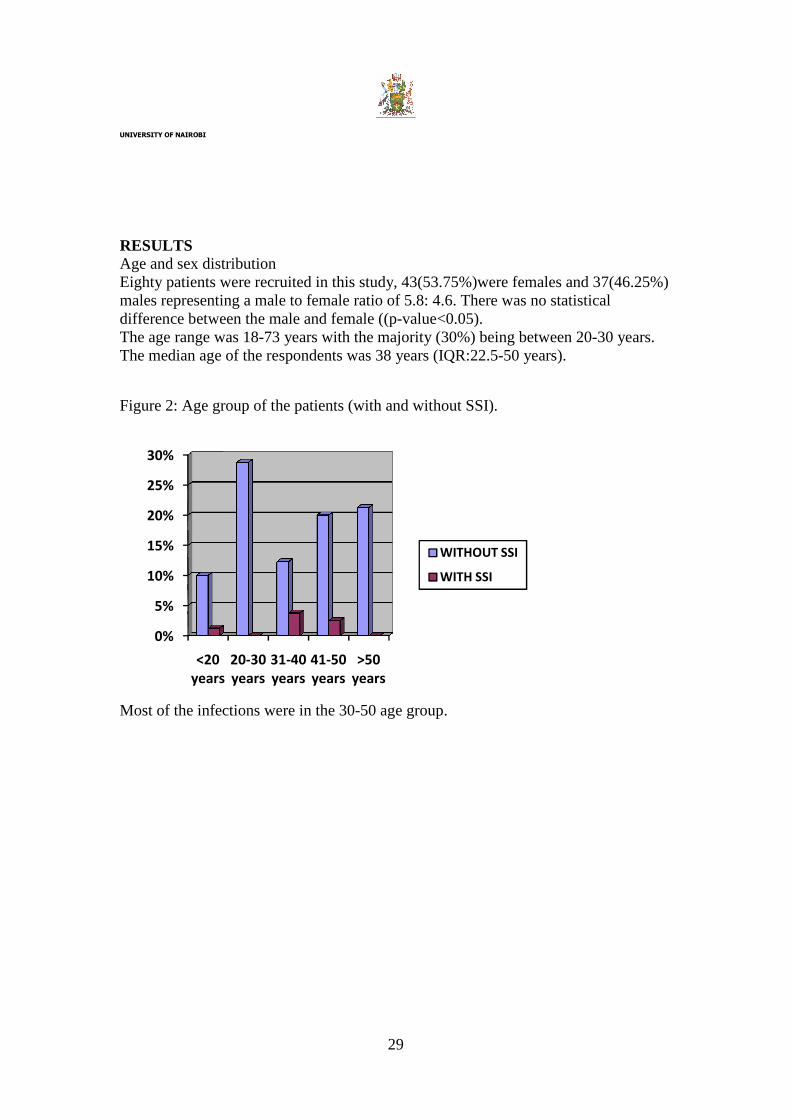
RESULTS Age and sex distribution Eighty patients were recruited in this study, 43males representing a male to female ratio ofdifference between the male and female ((pThe age range was 18-73 years with the majority (30%) The median age of the respondents was 38 years (IQR:22.5
Figure 2: Age group of the patients (with and without SSI
Most of the infections were in the 30
0%
5%
10%
15%
20%
25%
30%
<20
years
20-30
years
31
years
29
Eighty patients were recruited in this study, 43(53.75%)were females and representing a male to female ratio of 5.8: 4.6. There was no statistical
he male and female ((p-value<0.05). years with the majority (30%) being between 20
median age of the respondents was 38 years (IQR:22.5-50 years).
the patients (with and without SSI).
Most of the infections were in the 30-50 age group.
-40
years
41-50
years
>50
years
WITHOUT SSI
WITH SSI
and 37(46.25%) no statistical
being between 20-30 years.

UNIVERSITY OF NAIROBI
30
Table 2. Association of class of SSI and number of days in hospital CLASS OF SSI LENGTH OF STAY IN
HOSPITAL (DAYS)
NUMBER OF PATIENTS
SUPERFICIAL
DEEP
ORGAN/SPACE
15-30 days
31-45 days
31-45 days
>45 days
2 (2.5%)
2 (2.5%)
1 (1.25%)
1 (1.25%)
Of the six patients with SSI four had superficial SSI, one deep and organ/space. Patient who developed deep and organ/space SSI had been in hospital for more than one month.
Table 3. Comparison of days in hospital for patients with and without SSI.
STATE OF WOUND NUMBER OF PATIENTS THE MEAN POST-OP NUMBER OF DAYS IN HOSPITAL
SSI 6 16
NO SSI 74 5
Patients with surgical site infection were associated with a longer duration of hospital stay as compared with those without SSI (11 days longer in hospital). This difference was statistically significant (p<0.05). The average cost of hospital stay in patients with no SSI was 1100 dollars and with SSI it was almost triple the amount.

UNIVERSITY OF NAIROBI
31
Table 4. Risk factors associated with SSI. RISK FACTORS
NUMBER OF PATIENTS p-value SSI (n=6) NO SSI (n=74) TOTAL
BMI (n=80) <18.5
18.5-24.9
25.0- 29.9
> 30
1 (16.67%)
0 (0.0%)
2 (33.33%)
3 (50.00%)
3 (4.05%)
40 (54.05%)
27 (36.49%)
4 (5.41%)
4 40 29 7
0.1724 0.0109 0.8769 0.0002
SMOKING (n=3) 1 (16.67%) 2 (33.33%) 3 0.4004
STEROID USE (n=80)
6 (7.50%) 74 (92.50%) 80 0.000
Body mass index. Fifty percent of the patients who underwent surgery in this study had normal BMI and did not develop SSI .50% of the patients who developed SSI were found to be obese and this was stastistically significant (p<0.05). Smoking. In this study only 3 patients were smokers and one developed SSI.in this study there was no association between smoking and the development of SSI. Steroid use. All the patients who underwent surgery in this study were on steroids and 6 developed SSI while 74 did not develop SSI.

UNIVERSITY OF NAIROBI
32
Table 5.The ASA score of patients with SSI (n=6). ASA SCORE FREQUENCY % OF
PATIENTS WITH SSI IN EACH ASA SCORE
p-value
SSI NO SSI
I. Normal healthy patient (n=23) II. Patient with mild systemic disease (n=49) III. Patient with severe disease that is not Incapacitating (n=8).
1 (16.67%) 3 (50.00%) 2 (33.33%)
22 (29.73%) 46 (62.16%)
6 (8.11%)
4.35% 6.12% 25%
0.4966 0.5565 0.0477
Only majority of the patients (61.25%) had an ASA score of II. These were patients who underwent surgery and had mild systemic disease. SSI was observed in only 4.35% of patients with ASA score of I, i.e normal healthy patientsand in 6.12% of patients with ASA score II and in 25% ofpatients with ASA score of III (patients with severe disease that is not incapacitating). ASA score was associated with the development of SSI. Patients with ASA score of III were more likely to develop SSI and this was stastistically significant (p<0.05). Table 6. Relationship between time and duration of surgery with SSI.
Variable SSI OR (95% CI) p-value
No (-) Yes (+)
Shift when the surgery was done Morning Afternoon
55 (74.32%) 25 (25.68%)
3 (50.00%) 3 (50.00%)
1.00 (Ref) 2.89 (0.54-15.58)
0.216
Duration of operation Less than 4 hrs More than 4 hrs
19 (25.68%) 55 (74.32%)
1 (16.67%) 5 (83.33%)
1.00 (Ref) 1.73 (0.19-15.74)
0.628
Surgical drain insertion Yes No
17 (21.25%) 63 (78.75%)
4 (66.67%) 2 (33.33%)
9.23 (1.53-55.85) 1.00 (Ref)
0.016

UNIVERSITY OF NAIROBI
33
Most of the surgeries (75%) lasted longer than 4 hours, and that was above the recommended NHSN risk index of 219 minutes (Ref 54) (NNIS Report 2004). Most of the SSI occurred in the morning shift and 5% of the patients with drains developed infections. Seventeen (21.25%) out of 80 patients had placement of surgical drains during closure of the scalp. 66.67% of those patients who developed SSI had surgical drains. Patients with surgical drains were more likely to develop SSI compared to those without surgical drains.This association was statistically significant(OR: 9.23; 95% CI: 1.53-55.85; p-value=0.16). CRITERIA FOR IDENTIFYING SSI Table7. State of the wound in patients with SSI Specify criteria used for identifying SSI, n=6 Frequency Percent (%) Purulent discharge Yes No Organisms isolated from aseptically collected aspire or swab from surgical site Yes No Pain or tenderness Yes No Heat Yes No Fever Yes No Redness Yes No Localized swelling Yes No Abscess Yes No Incision deliberately opened by surgeon Yes No Wound dehisces spontaneously Yes No
6 0 6 0 6 0 5 1 4 2 5 1 4 2 2 4 1 5 2 4
100% 100% 100% 83.33% 16.67% 66.67% 33.33% 83.33% 16.67% 66.67% 33.33% 33.33% 66.67% 16.67% 83.33% 33.33% 67.67%

UNIVERSITY OF NAIROBI
34
Other evidence of infection found on direct exam or diagnostic tests e.g. radiological Yes No
1 5
16.67% 83.33%
The six patients with SSI had purulent discharge at the incision site with pain or tenderness at the site. Bacterial microbes were isolated aseptically with a swab. Five presented with heat and/or redness at the site, four with fever and/or localized swelling and two with an abscess and/or spontaneous wound dehiscence. The most common presentation of SSI was purulent discharge through the surgical wound and pain. GROWTH PATTERN OF MICROORGANISMS IN PATIENTS WITH SSI Wound swabs were obtained from all the cases of surgical site infections and taken to the laboratory for analysis. The samples were cultured on blood agar and Mac Conkey agar, followed by identification of the isolates based on their colony characteristics. The disc diffusion method was used for the antimicrobial susceptibility patterns following Clinical Laboratory Standards Institute (CLSI) guidelines. Organisms were isolated from all the samples (100%). (Table 8. Fig 2) Table 8. Growth pattern of the microorganisms MICROORGANISMS FREQUENCY PERCENT (%) Pseudomonas Staphylococcus aureus Escherichia Coli Klebsiella Proteus Total
3 3 1 1 1 9
33.33% 33.33% 11.11% 11.11% 11.11% 100%

UNIVERSITY OF NAIROBI
35
Figure 2: Organisms isolated
The commonest organism was S. aureus and Pseudomonas. Out of the 6 positive cultures 3 were pure and 3 had mixed growth cultures. A total of 9 bacterial isolates were obtained. 3 (33.33%) were gram positive and 6 (66.67%) were gram negative. S.aureus and P. aeruginosa were the predominat organisms isolated. Table 9. Pattern of resistance of S. aureus.
Microorganism
Penicillin
Ampicillin
Augmentin
Levofloxacin
Erythromycin
Vancomycin
Teicoplanin
S.aureus 3 (100%)
3 (100%)
3 (100%)
1 (33%)
3 (100%)
0 0
RESISTANCE PATTERNS IN GRAM POSITIVE ORGANISMS. Among gram positive organisms isolated 33.33% were S.aureus of which 100% were resistant to Penicillin, Ampicillin and Augmentin. None was resistant to vancomycin. None were MRSA positive. Table 10 Pattern of reisistance of gram negative organisms
33.33% 33.33%
11.11% 11.11% 11.11%
0
10
20
30
40
% of positive cultures
Staphylococus aureus Pseudomona Proteus Klebsiella EscherichiOrganisms isolated

UNIVERSITY OF NAIROBI
36
Antibiotic Microorganisms
Amikacin Doxycycline Gentamycin Ceftazidime Cefuroxime Piperacillin/ Tazobactam
Meropenem
Pseudomonas. 1/3 (33.33%)
3/3 (100%)
3/3 (100%)
3/3 (100%)
3/3 (100%)
3/3 (100%)
1/3 (33.33%)
E.coli 0/1 1/1 (100%)
1/1 (100%)
1/1 (100%)
1/1 (100%)
0/1 0/1
Klebsiella 0/1 1/1 (100%)
1/1 (100%)
1/1 (100%)
1/1 (100%)
1/1 (100%)
1/1 (100%)
Proteus 0/1 1/1 (100%)
1/1 (100%)
1/1 (100%)
1/1 (100%)
1/1 (100%)
1/1 (100%)
RESISTANCE PATTERNS IN GRAM NEGATIVE ORGANISMS . Among gram negative organisms isolated 33% were Pseudomonas aeruginosa 11% E.coli, 11%, Klebsiella spp and 11% Proteus mirabilis. 33% of Pseudomonas aeruginosa were sensitive to Amikacin and Meropenem. Extended Spectrum Beta-Lactamase (ESBL) was found in the Klebsiella isolate and was sensitive to a combination of Cefotaxime and ceftriaxone but resistant when tested against the two drugs singly.
DISCUSSION According to Janet M, et al (2011), there has been an upward trend in the number of craniotomy SSI as noted by Infection control & Prevention. During the first six months of 2010 there was an increase from 2.8 infections per 100 cases in late 2009 to 8.4 per 100 cases in second quarter of 2010. Surgical site infections are defined as those that occur within 30 days after operation. SSI are still associated with substantial morbidity, mortality and with increased economic costs that negatively impact on patients and their families.In two studies done on healthcare associated infection in Africa, the most common encountered infection encountered was SSI among patients in surgical wards (Messele ea al). During the surveillance in this current study period, eighty patients underwent elective craniotomies under consultant neurosurgeons. The median age of the respondents was 38 years (IQR: 22.5 – 50 years). Patients who died within 30 days were not included in the study. The starting point was from the time the patient was admitted to the neurosurgical ward and end point being 2 weeks post-operatively.

UNIVERSITY OF NAIROBI
37
The incidence of craniotomy SSI varies widely. This study, carried out between June 2014 and March 2015, recorded an overall infection rate of 7.5% among the eighty patients who underwent the elective craniotomies. Several studies have however shown varying infection rates with the most acceptable rate according to Gaskill (1996) being 5%. In randomized control study by Burnichon and coworkers they reportedinfection range of 4-12%, among the patients who weren’t on antimicrobial prophylaxis and 0.3-3% with prophylaxis. The reported incidence of SSI n a prospective study by Nikolaos S et al was 14.8%.A study done by Korinek (2005), had an overall infection rate of 6.6%. A study by Buang et al revealed an infection rate of 7.7 %. In other studies the incidence of neurosurgical SSI ranges between 0.5-2.5percent (Suri et al). According to a study by Sneh-Arbib (2013), the overall infection rate was 5.6 %( 28 patients).The variation could probably be explained by the differences in patient population. In 1966, Wright and co-workers conducted a study that described a number of identifiable risk factors for SSI in neurosurgery. In the present study patient as well as procedure related risk factors for SSI were identified. This factors included age, nutritional status, smoking, use of steroids, ASA score, type of surgical wound, shift of operation, duration of operation and use of surgical drains. In our current study, the mean post- operative stay for patients with SSI was 16 days and those without was 5 days. This is almost similar to a study done by Suchitra et al, in 2009 which showed an association between SSI and length of hospital stay. The patients who developed SSI had an average length of stay of 17.2 days and those without had an average stay of 5.5 days. The average hospital cost for patients with no SSI was approximately 1100 dollars while those having SSI had an approximate hospital cost of 3300 dollars. This shows that SSI apart from being morbid are also an economic burden. SSI occurring within 30 days and appeared to be related to the procedure were included in this study. Six patients were identified to have SSI of which 4 (66.67%) were males and 2 were females, this being despite that there were more females (53.75%) than males (46.25%) recruited in this study. Females were less likely to have SSI compared to males (Odds ratio, 0.40; 95% Confidence interval 0.19-15.74. P-value 0.31). In studies done by Shinoura et al, (2004) and Korinek et al, (2006) male gender was an independent risk factor. The incidence of SSI is increased when a subsequent procedure is done at a previous operation site. In this study one patient had a reoperation for a recurrent posterior fossa lesion and subsequently got a SSI. Procedure related SSI occurring during reoperation has been reported by studies done by Korinek et al, (1997). Overweight and obesity is defined as having a body mass index greater than or equal to 25kg/m² and greater than 30kg/m² respectively and is a significant risk factor for SSI. (Cheadle et al, 2006). In the current study only 8.75% of patients were obese and 33% of those who developed SSI were obese. Patient education and diets formulated

UNIVERSITY OF NAIROBI
38
by nutritionist may help in reducing obesity and its associated complications like diabetes mellitus that may affect wound healing (Blissmer 2006). In the current study those who were smoking were more likely to have SSI compared to those who were not smoking (OR: 7.20; 95% CI: 0.55-93.69). Cigarette smoking has been associated with poor wound healing because of microvascular obstruction leading to decreased circulation. Current smoking increases the risk of SSI (Nagachinta et al., 1987). According to a study by Slaughter et al, patients receiving perioperative steroids for a prolonged period were predisposed to developing SSI. In the current study majority of the patients used steroids, for reducing vasogenic odema, for more than one week (68.75%). In contrast, Guglielmo et al documented that there was no relationship between steroid use and risk of SSI. In the current study most of the patients were in ASA 2 (61.25%) while the rest were in ASA 1 (28.75%) and ASA 3 (10%). Majority of SSI occurred in patients with clean wounds. A study done by Buang et al., (2012) showed that those with ASA 2 and clean contaminated wounds were at significant risk of developing SSI. According to Alina (2009), 2.24% of clean neurosurgical operations developed SSI while 20% of clean contaminated operations developed infections. In a study by Cheadle 2006 found out that the primary source of infection for most SSI is the patient’s endogenous flora. Gram positive infection could be inoculated from the patients own scalp during skin incision. Although gram negative organism don’t normally colonize skin flora, the may be inoculated to the wound by own patient fecal flora since most of the patients are confused immediately post-operative period. Only a small percentage of wounds become infected despite all surgical wounds becoming contaminated during surgery (Fry DE et al). Contaminated drains may also play a role in SSI. Prolonged hospital stay may predispose the patient to hospital acquired organism through poor hygiene of the patient or health workers. According to Mcleod et al, all hospital patients are colonized with microorganisms. Bathing with chlorhexidine before operation may help in reducing the burden of microorganisms prior to operation (Mangram et al, 1999). The extent of microbial contamination at the surgical site can be classified according to Mangram et al., (1999) into four classes. In the study only patients with clean and clean contaminated wounds were included. 91.25% had clean wounds and 8.75% had clean contaminated wounds. Wounds were classified as clean contaminated if they traversed an air sinus. The infection rate was higher in clean contaminated wounds (14.29%) than in clean wounds (6.84%). Korinek et a,(1997) showed that a wound classification of clean contaminated had a higher infection rate than clean. According to the NHSN (National Healthcare Safety Network) report in 2009 by Edwards et al, the cut off time for elective craniotomies was 225 minutes. In our study

UNIVERSITY OF NAIROBI
39
75% of the operations took more than 4 hours and 25 % took less than 4 hours. The infection rate was higher in those who had operation for more than 4 hours (83.33%) (OR: 1.73; 95% CI: 0.19-15.74 p-value 0.628), compared to those whose operations took less than 4 hours (16.67%). Even though some studies show that patients operations that last for more than “T” hours where T represents 75th percentile of distribution of operation durations have a higher risk of developing SSI (Culver et al), other studies show that operation duration was not a significant rik factor (Sanchez-arenas 2010). Those with surgical drain insertion were more likely to develop a surgical site infection. 4 (66.67%) with SSI had surgical drains (OR: 9.23; 95% CI: 1.53-55.85 p-value 0.016). Surgical drains allow direct access by microorganisms to the craniotomy site may be facilitated by the drains. The drains act as conduits for infections since microorganisms form biofilms that act as growth surfaces for bacteria. Prophylaxis for craniotomies has been examined in several randomized studies by Gaillard et al, (1991) and with one meta-analysis by Barker in 2007 and have all concluded that antimicrobial prophylaxis is important for cranial surgeries. In this current study, antimicrobial prophylaxis was given for each patient intraoperatively at the time of induction with general anesthesia i.e. before skin incision. The dosage was calculated according to the body weight of the patient. Additional antibiotics were not given when the duration of the procedure took more than four hours and this may have contributed to post-operative SSI. Improving antimicrobial prophylaxis may be an important step in reducing SSI. According to Dohem et al, antimicrobial resistance makes preoperative methods to reduce SSI important. The common symptoms in patients with SSI included: purulent discharge (100%), pain and tenderness along the operation site (100%), heat (83.33, fever more than 38⁰C (66.67%), redness (83.33%), localized swelling (66.67%), abscess 33.33%, incision deliberately opened by the surgeon (16.67%), wound dehiscence spontaneously (33.33) % and infection found on diagnostic imaging (16.67%). SSI were categorized as either superficial incisional if they involve only the skin or subcutaneous tissues, deep incisional if the involved the fascial or muscle i.e. Subgaleal infections and organ/space if they involved the bone, dura, brain parenchyma or ventricular system. In a study by Alina et al, 2009, most of the SSI were superficial (52.3%), deep (33.33%) and organ/space in 14.28%. According to the definition of surgical wounds by CDC, our study revealed that 66.67% of infections were superficial, 16.67% were deep and 16.67% were organ/space infections. 83.33% of SSI were identified during inpatient stay and 16.67% were identified during post-operative clinic visit. Majority of the infections were identified within two weeks after operation. This is almost similar to a study done by Korinek et al, which showed the mean time between surgery and onset of SSI to be 18days. According to Agrawal et al.(2003), out of 40 neurosurgical SSI, the most common isolated organism was staphylococcus aureus( 57.5%) followed by Pseudomonas

UNIVERSITY OF NAIROBI
40
aeruginosa (10%), Escherichia coli (15%) and others (17.5%) . In other international studies, Staphylococcus aureus, Escherichia coli, pseudomonas are mentioned as those most commonly causing SSI (Mayon-White et al). Chiang et al, (2012) in retrospective study of patients undergoing craniotomy between 2006 and 2010, found that most of the infections were caused by Staphylococcus aureus alone or in combination with other microorganisms and 27% were caused by gram negatives. In our study cultures were positive in the specimens taken and among the most common gram positive isolate was Staphylococcus aureus (33.33%). The most common gram negative isolate was pseudomonas spp (33.33%). One (16.67%) SSI was caused by gram positive bacterial alone, two (33.33%) were caused by gram negative bacteria alone and three (50%) were caused by mixed infections. According to Dashti et al, 2008, the most common microorganism was staphylococcus aureus (18%). In this current study among the 4 superficial SSI, 1(25%) was caused by only Staphylococcus aureus, 2 (50%) were caused byPseudomonas and 1 (25%) was caused by mixed flora (both Staphylococcus aureus and Escherichia Coli. 1 (100%) deep SSI was caused by mixed flora (pseudomonas and Klebsiella species and 1 (100%) organ/space SSI was also caused by mixed flora (Proteus and Staphylococcus aureus). In a study of bacteriology of infections according to the site of infection, Korinek et al,(2009) identified that scalp infections were mostly caused by staphylococcus aureus, bone flap osteitis by Propionibacterium acnes, intracranial empyema, brain abscess by p. acnes and meningitis by Enterobacteriacaeae. Antibiotic resistance is a worldwide problem (Lim 1994). According to Taylor et al,(1990) there has been a gradual increase in the emergence of antibiotic resistant microorganisms and this is becoming a major headache for physicians. This is making physicians resort to newer and more costly drugs that are increasing the health expenditure burden. In this study the resistance patterns of bacteria was detected among the bacteria isolated. Of the gram positive bacteria, Staphylococcus aureus was resistant to multiple antimicrobials but all were sensitive to Vancomycin and Teicoplanin. This is consistent with other literature that indicates staphylococcus aureus bring the most common isolate, (Nicholas et al., 1998).
None of the three S. aureus isolates, in this study, were MRSA positive. MRSA infections are on the rise and they the increase patients morbidity and mortality as shown by a study on MRSA by Ojulong et al which showed a prevalence rate of 31.5% among S aureus isolates in a national referral hospital in Uganda.
In the current study one case of Escherichia coli, Klebsiella spp and Proteus were each isolated. They were all sensitive to Amikacin. According Gedebou et al, majority of the gram negative bacteria were resistant to commonly used antimicrobials This results show that antibiotic resistant strains are on the increase and judicious selection of antibiotics is required to be able to clear these infections. To reduce multidrug

UNIVERSITY OF NAIROBI
41
infections, strict aseptic techniques should be used. Infections acquired in hospitals are more likely to be multidrug resistant compared to community acquired infections. The most common gram negative bacterium was pseudomonas aeruginosa which was resistant to most of antimicrobials tested. Pseudomonas showed low resistance rates to Amikacin (33.33%) and Meropenem (33.33%). In a study by Cheong et al, (1994) nearly 30% of Pseudomonas aeruginosa are currently resistant to gentamycin which used to be a readily available and effective aminoglycoside. CONCLUSION
1. The study identified SSI infection rate of 7.5% which was comparable to other studies with most bacterial isolates being resistant to commonly used antibiotics
2. SSI increased the hospital stay of patient hence increasing their hospital bills.
3. The risk of SSI is dependent on a number of patient and operation factors. Risk factors for SSI vary in terms of magnitude and significance. Their identification will help in improving the care given to patients
4. SSI are preventable which in turn could help reduce associated morbidity and mortality, reducing length of hospital stay and cost

UNIVERSITY OF NAIROBI
42
5. This study has given useful information that may influence change in operative practices by neurosurgeons thus reducing big burden to both patients and health care system that is resource limited.
6. The results of this study will contribute significantly to literature on craniotomy SSI.
7. Some risk factors are non-modifiable thus neurosurgeons may use them to identify patient who are high risk for developing SSI.
RECOMMENDATIONS
1. To better understand the epidemiology of the epidemiology of craniotomy SSI a case control study with a larger sample size and a longer duration of study period is proposed that would assess more risk factors and confounders related to SSI after craniotomy.
2. Increase the study period and sample size to increase the power of the study.
3. There are other peri-operative risk factors that may contribute to SSI and this need to be assessed and so as to make a difference in the overall incidence of infections.

UNIVERSITY OF NAIROBI
43
4. Meticulous surgical procedure and methods should be improved so as to produce an almost sterile surface and care givers should be educated about proper incision.
5. Get an active surveillance program to identify SSI promptly.
6. Strict implementation of national infection prevention and control policies and guidelines.
REFERENCES 1. Kirkland, Jane P, Briggs, et al. The impact of surgical site infections in the 1990’s. Attributable mortality, excess length of hospitalization and the extra costs. Infection control and hospital epidemiology.1999; 20 (11): 725-730. 2. Anvikar AR, AB Damle, NS Patwardhan, et al. One year prospective study of 3280 surgical wounds. Indian journal of medical microbiology.1999; 17(3):129-32. 3. Coello, Charlett A, Wilson J, et al. Adverse impact of surgical site infections in English hospitals. J.Hosp. Infect 2008; 60: 93-103. 4. Selwyn Sand Bakhtiar M. Hospital infection: the first 2500 years. J Hosp Infect. 1991; 18(suppl A): 5-64.

UNIVERSITY OF NAIROBI
44
5. Ballard MR, Miller NH, Nyquist AC, et al. A Multidisplinary approach improves infection rates in paediatric spine surgery. J Pediatr Orthop. 2012 Apr-May;32(3):266-70. 6. Valentini LG, Casali C, Chatenoud L, et al. Surgical site infections after elective neurosurgy. A review of 1747 patients. Neurosurgery 2008; 62(1): 88-96. 7. Shearwood McClelland III. Postoperative intracranial neurosurgery infection rates in North America versus Europe. A systemic analysis. Am J Infect Control 2008;36:570-3. 8. Leaper DJ, Van Goor H, Reilly J, Petrosillo N, Geiss HK, Torres AJ, Berger A. Surgical site infection-a European perspective of incidence and economic burden. Int Wound J. 2004; 1(4):247-73 9. Buang SS, Siti Suriyati and Haspani MS. Risk factors for neurosurgical site infections after a neurosurgical procedure: A prospective observational study at Hospital Kuala Lumpar. Med J Malaysia. 2012. Aug;67(4):393-8. 10. Cruse PJ. Wound infection surveillance. Rev Infect Dis. 2006; 4(3):734-47. 11. Broex EC, Van Asselt AD, Bruggeman CA, et al. Surgical site infections: how high are the costs? J Hosp Infect. 2009 Jul; 72(3):193-201 12. De Lissovoy G, Fraeman K, Hutchins V, et al. Surgical site infection: incidence and impact on hospital utilization and treatment costs. Am j infect control.2009 Jun,37(5):387-97. 13. Alina P, Mihai I, Cristina B, et al. Surgical site infections surveillance in neurosurgical patients. J Neurosurg. 2009; 62:88-89. 14. Hosien IK, Hill DW and Hatfield RH. Controversies in the prevention of neurosurgical infection. J Hosp Infect. 1999. Sep;43(1):5-11. 15. Fry De. The economic cost of surgical site infection. Surg Infect. 2008; 1:537-543. 16. Horan TC, Gaynes RP, Martone WJ, et al. CDC definitions of nosocomial surgical site infections. A modification of CDC definitions of surgical wound infections. Infect Control Hosp Epidemiol 1992; 13(10): 606-8. 17. Schuster JM, Rechtine G, Norvell DC, et al. The influence of post-operative factors and therapeutic interventions on infection rates after spine surgery. A systemic review. Spine 2010 Apr 20;35:S125-37 18. Lietard C, Thebaud V, Besson G et al. Risk factors for neurosurgical site infections: an 18month prospective survey. J neurosurg 2008 Oct; 109(4):729-3 19. Korinek AM, Golmard JL and Elcheick A. Risk factors for neurosurgical site infections 235 after craniotomy: A critical reappraisal of antibiotic prophylaxis on 4578 patients. Br J 236 Neurosurg. 2005;19(2):155-62. 20.Sanchez-arenas, Blanca ER, Israel GO, et al. Factors associated with nosocomial surgical site infections for craniotomy in Mexico city hospitals. Cir 2010; 78:5-13. 21. Edwards Wong, et al. Surgical site infections. Hospital epidemiology and infection control. 2nd ed. Philadelphia: Lippincott Williams and Wilkins; 1999: 189-210. 22. Robert Haley. Nosocomial infections in surgical patients: Developing valid measures of intrinsic patient risk. The American J of medicine, 1991; 91(sup 3B): 145-157 23. David H, Culver and Teresa Horan. Surgical wound infection rate by wound class, operative procedure and patient risk index. Neurosurg J, 2005; 80:136-45.

UNIVERSITY OF NAIROBI
45
24. Centers for Disease Control and Prevention. Guideline for prevention of surgical site infection,1999. Infect Control Hosp Epidemiol 1999;20(4):247-278. 25. Victor Odhiambo Dinda. Surveillance of surgical site infections at the Aga khan university Hospital, Nairobi. 2009 post graduate thesis. 26. Byrd JJ, Xu HS and Colwell RR. Viable but non culturable bacteria in drinking water. 1991. Appl Environ Microbiol 57:875–878. 27. Bible JE, Biswas D, Whang PG, et al. Which regions of the operating gown should be considered most sterile? Clin Orthop Relat Res 467:825–830, 2009. 28. Darouiche RO, Rabih O, Matthew J, et alChlorhexidine–alcohol versus povidone–iodine for surgical-site antisepsis. N Engl J Med 2010; 362:18–26. 29. Barker II and Fred G. Efficacy of prophylactic antibiotics in cerebrospinal shunt surgery. A meta-analysis. Neurosurgery, 1998; 43(3):694 30. Barker FG 2nd . Efficacy of prophylactic antibiotics against meningitis after craniotomy: a 225 meta-analysis. Neurosurgery. 2007;60:887-94. 226 31. Ronald F, Young and Pablo M. Perioperative antibiotic prophylaxis for prevention of post operative neurosurgical infections. A randomized clinical trial. Journal of neurosurgery. May 1987/vol 66/ no.5/pages 701-705. 32. Harbarth S, Samore MH, Lichtenberg D, et al: Prolonged antibiotic prophylaxis after cardiovascular surgery and its effect on surgical site infections and antimicrobial resistance. Circulation 101:2916–2921, 2000 33. Dale W, Dellinger EP, Olsen KM, et al. clinical practice guidelines for antimicrobial prophylaxis in surgery. Am J Health-Syst Pharm-vol 2013; 70:195-283 34. Bosco JA III, Slover JD and Hass JP. Perioperative strategies for decreasing infection: a comprehensive evidence-based approach. J Bone Joint Surg Am. 2010;92(1):232-239 35. Isibor OJ, Ashietu O, Adevbo E, et al. Incidence of aerobic bacteria and candida albicans in postoperative wound infections. Afr J Microbiol Res. 2008; 2:228-91 36. Alicia B, Jakob, Kathleen, et al. Relationship between skin microbial counts and surgical site infections after neurosurgery. Clin Infect Dis, 2000; 33(8): 1302-1308. 37. Sital Mistry, et al. Surgical antibiotic prophylaxis guidelines within neurosurgery for adult and pediatric patients. 2012. 38. Greene K, Wilde AH and Stulberg BN. Preoperative nutritional status of total joint patients. Relationship to postoperative wound complications. J Arthroplasty. 1991 Dec;6(4):321–325. 39. Snyderman CH, Kachman K, Molseed L, et al. Reduced postoperative infections with an immune-enhancing nutritional supplement. Laryngoscope.1999; 109:915–921. 40. Toma O,Suntrup P, Stefanescu A, et al. Pharmacokinetics and tissue penetration of cefoxitin in obesity: implications for risk of surgical site infection. Anesth Analg.2011 Oct ; 113(4):730–737. 41. Webster J and Osbourne S. Preoperative bathing or showering with skin antiseptics to prevent surgical site infection. Cochrane Database Syst Rev .2006;(2):CD004985. 42. Gruskay J, Kepler C. Smith J, et al. Is surgical case order associated with increased infection rate after spine surgery? Spine (Phila Pa 1976) 2012;37:1170–1174. 43. Nottingham antibiotic guidelines committee. January 2012, Review January 2014

UNIVERSITY OF NAIROBI
46
44. Steinberg JP, Braun BI, Hellinger WC, et al . Timing of antimicrobial prophylaxis and the risk of surgical site infections: Results from the trial to reduce antimicrobial prophylaxis error. Annals of surgery. July 2009;250(1):10-16 45. Holloway KL, Smith KW, Wilberger JE Jr, et al. Antibiotic prophylaxis during clean 222 neurosurgery: a large, multicenter study using cefuroxime. Clin Therap. 1996 Jan-Feb; 18(1):84- 94. 46. Tanner J, Woodings D and Moncaster K. Preoperative hair removal to reduce surgical site infection. Cochrane Database Syst Rev. 2006 Jul 19 ;( 3):CD004122. 47. Parienti JJ, Thibon P, Heller R, et al. . Hand-rubbing with an aqueous alcoholic solution vs traditional surgical hand-scrubbing and 30-day surgical site infection rates: A randomized equivalence study. JAMA.2002 Aug 14;288(6):722–727. 48. Miller JP, Acar F and Burchiel KJ. Significant reduction in stereotactic and functional neurosurgical hardware infection after local neomycin/polymyxin application. Clinical article. J Neurosurg .2009 Feb;110(2):247–250. 49. Zanetti G, Giardina R and Platt R: Intraoperative redosing of cefazolin and risk for surgical site infection in cardiac surgery. Emerg Infect Dis.2001 Sep-Oct;7(5):828–831. 50. Parkinson H and Tanner J. Double gloving to reduce surgical cross-infection. Cochrane Database Syst Rev. 2006 Jul; (3):CD003087. 51. Craniotomy incision care. Joy Ryan. Craniotomy incision care. Evidence based practice fellowship. 2005 52. Evans HL, Dellit TH, Chan J, et al.. Effect of chlorhexidine whole-body bathing on hospital-acquired infections among trauma patients. Arch Surg 145:240–246.2010 53. Wanga SK, Lemeshow S. Sample size determination in health studies. A practical manual. Ginebra: World Health Organization, 1991. 54. NNISS, National infections Surveillance (NNIS) systems report, data summary from January 1992-June 2004. AMJ infection control, 2004; 32:470-485. 55. Janet M, Thomas RT, Titus D, et al. a multidisplinary intervention to reduce post craniotomy surgical site infection rates. American J of Infect Control. 2011; 39(5):35-36) 56Messele G, Woldemedhin Y, Demissie M, Mamo K, Geyid A. Common causes of nosocomial infections and their susceptibility patterns in two hospitals in Addis Ababa.Ethiop J Health Biomed Sci 2009; 2: 3-8. 57. Burnichon G, Lietard C, L’azou D. results of a survey system for neurosurgical site infections, October 1998-january 2003. Neurochirurgie 2007; 53(6):470-64) 58. Gaskill SJ. Prophylactic antibiotics. In: Wilkins RH, Rengachary SS, Editoes. Neurosurgery. New York: McGraw-Hill,1996, p 547-5121 59. Nikolaos S, Stefanos P, Charalampos I, et al. Craniotomy infections: The Venizeleio hospital experience. Ann Gen Psychiatry. 2010 April 22; 9(1):98-105.) 60. Suri A, Mahaparta AK. The changing pattern of organisms in post-operative neurosurgical infections. The National Journal of India. 2000; 13(6):296-300). 61. Sneh-Arbib O, et al. surgical site infections following craniotomy focusing on possible post-operative acquisition of infection: prospective cohort study 2013). 62. Wright RL. Post- operative craniotomy infections. springfield, IL:Charles C Thomas, 1966) 63. Suchitra JB and Lakshmidev N. Surgical site infections: Assessing risk factors, outcomes and antimicrobial sensitivity patterns. Health Infect 2009; 14(1):21-250

UNIVERSITY OF NAIROBI
47
64.Shinoura N, Yamanda R, Okamoto K. Early prediction of infection after craniotomy for brain tumors. British J neurosurg 2004; 18(6):598-603 65. Cheadle WG. Risk factors for surgical site infections. Surg Infect 2006; 7(1):s7-11) 66. Blissmer B, Riebe D, Dye G, et al., Health related quality of life following a clinical weight loss intervention among overweight and obese adults: intervention and 24 month follow up effect. Health Qual Life outcomes 2006; 4:43) 67. Nagachinta T, Stephens M, Reitz B, et al., Risk factors for surgical wound infection following cardiac surgery. Journal of Infectious Diseases 1987; 156(6):967-973). 68. Mangram AJ, Horan TC, Pearson ML, et al. Guideline for prevention of surgical site infection, 1999. Hospital Infection Control Practices Advisory Committee. Infection Control & Hospital Epidemiology 1999; 20(4): 250-278). 69. Slaugher MS, Oslon MM, Lee JT. A fifteen year wound surveillance study after coronary artery bypass. Ann Thorac Surg. 1993; 56(5):1063-8. 70. Fry DE. Surgical site infection: pathogenesis and Prevention CME Program. Medscape from WebMD 2003) 71. Pons VG, Denliner SL, Guglielmo BJ, et al. Ceftizoxime versus Vancomycin and Gentamycin in neurosurgical prophylaxis: a randomized, prospective, blinded clinical study. Neurosurgery 1993; 53(3):416-22 72. Culver DH, Horan TC, Gaynes RP et al. Surgical wound infection rates by wound class, operative procedure and patient risk index. National Nosocomial Infections Surveillance System. Am J Med 1991; 16(3B):1525-1575 73. Gaillard T, Gilsbach JM: intra-operative prophylaxis in neurosurgery. A prospective, randomized, controlled study on cefotiam. Acta Neurochir 1991; 113:103-109) 74. Dohmen P. antibiotic resistance in common pathogens reinforces the need to minimize surgical site infections. J. Hosp Infect, 2008; 70(S2):15-20) 75. Agarwal M and Thomas P the Nursing Journal of India 2003;94(9):197-198. 76. Mayon-White RT, Kereselidze T, Tikomirov E. an international survey of the prevalence of hospital acquired infection. J Hosp Infect. 1998; 11(A):43-8). 77. Chiang HY. Risk factors and outcomes associated with surgical site infections after craniotomy and craniectomy. 2012. 78. Dashiti SR, Baharvahdat H, Spetzler RF at al. operative intracranial infection following craniotomy. Neurosurgical Focus. 2008;24(6):E10. 79. Mcleod J, Nicolle L, Parker S, et al. An outbreak of Staphylococcus aureus sternal wound infections in patients undergoing coronary artery bypass surgery. American Journal of Infection Control 1991; 19(2):92-9736). 80. Lim VKE. Antibiotic resistance. Med J Malaysia 1994; 49(4):315-316). 81. Taylor GJ, Mikell FL, Moses HW, et al. determinants of hospital charges for coronary artery bypass surgery. The economic consequences of post-operative complications. Am J Cardiol 1990; 65:309-313) 82. Nicholas RL. Post-operative infections in the age of drug resistant gram positive bacteria. Am J Med. 1998;104:11S-16S 83.Cheong YM, Lim VKE, SuleimanAB, et al. antimicrobial resistance patterns of 36,000 bacteria isolated from patients in six Malaysian general hospitals. Med j Malaysia 1994; 49:317-26)

UNIVERSITY OF NAIROBI
48
84. J Ojulong, T Mwambu, M Jolobo,et al. Prevalence of Methicillin resistant Staphylococcus aureus (MRSA) among isolates from surgical site infections in Mulago hospital- Kampala, Uganda.The Internet Journal of Infectious Diseases. 2008 Volume 7 Number 2. 85.Gedebou M, Kronvall G, Habte-Gabr E, et al. the bacteriology of nosocomial infections at Tikur Anbessa Teaching Hospital, Addis Ababa. Acta Pathol Microbiol Immunol Scand (B) 1987; 95:331-336 APPENDIX I CONSENT FOR THE STUDY Hospital number………………………………… Study number……………………………… Title of the study Craniotomy surgical site infections at the Kenyatta National Hospital.

UNIVERSITY OF NAIROBI
49
Introduction This study will be carried out by Dr. Alex Henry Njiru, a post graduate student in the department of neurosurgery, University Of Nairobi. Surgical site infections are a worldwide problem and a major cause of morbidity, reduced quality of life, financial burden in healthcare and mortality. There is an emphasis on the reduction of SSI, which are preventable increase a patients’ recovery time, pain and risk of other complications in the future for all categories of surgeries with superficial SSI being the most common. Objectives of the study This study aims to determine the rate of craniotomy surgical site infections, risk factors associated with the infections and identification of the bacterial causative agents and antimicrobial susceptibility patterns. Benefits of the study The participant may directly benefit from this research if and when an infection is detected and associated organisms’ isolated and antimicrobial sensitivity done. The knowledge of incidence, risk factors and micro- organisms, will allow health professionals to implement protocols to reduce the infection rates and improve the quality of life of patients. Risks of the study The participant shall at no time be exposed to any health risk. Costs All the costs of this study will be undertaken by the principal investigator. Voluntarism I understand my participation is voluntary and that I am free to withdraw at any time, without giving reason. This will not affect my future care or treatment. Confidentiality All the obtained information will be held in the strictest confidence and will not be disclosed without the consent of the clinical unit concerned. I will do everything I can to protect your privacy and your identity will not be revealed in any publication arising from this study. Type of specimens and amount to be obtained Patients will be observed for presence of postoperative surgical site infections and samples taken from infected wounds. Pus swabs from the surgical site will be collected aseptically (after cleaning with a sterile saline solution) using a sterile cotton swab and taken to the laboratory within one hour. The collected samples will be cultured to identify the causative bacteria and then inoculation done to determine the sensitivity patterns to commonly used antibiotics. Follow up schedules /expected time in the study

UNIVERSITY OF NAIROBI
50
The study will be conducted in ward 4C and ICU in KNH between June 2014 and May 2014. The participants will be followed up for 2 weeks post operatively. Information on researchers and telephone numbers Any queries, concerns or problems arising from this study should be directed to the following people: Principle investigator: Dr. Alex Njiru 021480662 Supervisors: Professor Nimrod Mwangombe 0736222191 Dr Akuku 0722518844 Dr Florence Mutua 0722843830 The KNH/UON ethics and research committee. 020-2726300 ext 4435 . I…………………………………….. IP No……………………….. Of ……………….. Parent/guardian of ………………………………………….. study no………………… do hereby consent to be included in this study on surveillance of surgical site infections, after being having been satisfactorily explained to about the research in verbal and / or written form by the researcher. The nature of the study has been fully explained to me by Dr………………………………………… I have not been promised any material gain to participate. Signed…………………………………. (Parent/guardian) Date………………………………. IDHINI YA KUJIHUSISHA NA UTAFITI. Namba ya hospitali…………………………………. Namba ya utafiti………………… Jina la utafiti

UNIVERSITY OF NAIROBI
51
Maambukizi tofauti kufuatia upasuaji wa kichwa katika Hospitali ya Taifa ya Kenyatta. Kuanzishwa Utafiti huu utafanywa na Dk Alex Henry Njiru, mwanafunzi katika idara ya upasuaji wa kichwa, chuo kikuu cha Nairobi. Maambukizi tofauti baada ya upasuaji ni tatizo duniani kote na sababu kubwa ya maradhi, kupunguza ubora wa maisha, mzigo wa kifedha katika huduma ya afya na vifo. Kuna msisitizo juu ya upunguzaji wa maambukizi ambayo ni ongezeko la kuzuilika wagonjwa kupona wakati, maumivu na hatari ya matatizo mengine katika siku zijazo kwa makundi yote ya upasuaji. Malengo ya utafiti Utafiti huu unalenga kuamua kiwango cha maambukizi ya upasuaji wa kichwa, mambo yenye yanahusiana na maambukizi na utambulisho wa bakteria zenye zinahusika na mwakala wa madawa. Faida ya utafiti Mshiriki anaweza moja kwa moja kufaidika na utafiti huu ikiwa wakati maambukizi ni wanaona na kuhusishwa viumbe pekee na resistens unyeti kufanyika. maarifa ya matukio, hatari na viumbe micro-, itaruhusu wataalamu wa afya kutekeleza itifaki ya kupunguza viwango vya maambukizi ya na kuboresha ubora wa maisha ya wagonjwa. Hatari ya utafiti Afya ya mshiriki haitakuwa kwa hatari wakati wowote. Gharama Gharama zote za utafiti huu zita simamiwa na mpelelezi mkuu. kujitolea Naelewa ushiriki wangu ni kwa hiari yangu na kwamba niko na uhuru wa kuondoka wakati wowote, bila ya kutoa sababu. Hii haitaathiri huduma yangu ya baadaye ama ya matibabu. Siri Habari zote zilizopatikana zitafanyika katika imani kali na hazitafunuliwa bila ridhaa ya kitengo cha kliniki husika. Mimi nitafanya kila kitu naweza kulinda faragha yako na utambulisho wako hautafafunuliwa kwa uchapishaji kutokana na utafiti huu. Ina ya vielelezo na kiasi kwa kupatikana Wagonjwa watachunguzwa kwa uwepo wa maambukizi kufuatia upasuaji na sampuli za usa zitachukuliwa kutoka kidonda. Usa utachukuliwa kutoka kidonda cha upasuaji kwa njia safi (baada ya kusafisha na kuzaa ufumbuzi chumvi) kutumia pamba na kupelekwa maabara ndani ya saa moja. Sampuli zilizokusanywa zitakuzwa kutambua bakteria na kisha chanjo kufanyika kwa kuamua ruwaza unyeti wa dawa za kawaida zenye zinatumika.

UNIVERSITY OF NAIROBI
52
Kufuatilia ratiba / wakati utafiti utaofuatiliwa Utafiti utafanyika katika cumba cha wagonjwa 4C na chumba cha watu mahututi katika hospitali ya Kuu ya kenyatta kati ya Juni 2014 na Mei 2015. Washiriki watafuatiliwa kwa wiki mbili baada ya upasuaji. Taarifa juu ya watafiti na namba za simu Maswali yoyote, wasiwasi au matatizo yanayotokana na utafiti huu yataelekezwa kwa watu wafuatao: Mpelelezi mkuu: Dr. Alex Njiru 021480662 wasimamizi: Professor Nimrod Mwangombe 0736222191 Dr Akuku 0722518844 Dr Florence Mutua +254 20 2719628 KNH/UON maadili na kamati ya utafiti. 020-2726300 ext 4435 Ushiriki wa mgonjwa katika utafiti huu ni hiari kabisa. Mimi............................................ IP............................. wa.................... Mzazi / mlezi wa....................................numbari ya usajili.............. nimepatiania ruhusa ya kuhusishwa katika utafiti huu juu ya ufuatiliaji wa maambukizi ya upasuaji.baada ya kuelezwa na kuridhika na maelezo yenye nimeelezewakuwa na iliyoandikwa na mtafiti. Mshiriki kwa utafiti huu hatakuwa na hatari yoyote ya afya. Mshiriki anaweza moja kwa moja kufaidika kutokana na utafiti huu ikiwa na wakati maambukizi yanapopatikana na kuhusishwa viumbe pekee na antimikrobiell unyeti kufanyika. Naelewa ushiriki wangu ni wa hiari na kwamba mimi niko na ruhusa ya kujiondoa wakati wowote, bila ya kutoa sababu. Hii si kuathiri huduma yangu ya baadaye au matibabu. Asili ya utafiti nimeelezewa kikamilifu na Dk ................................................ Mimi sija ahidi ali faida yoyote kwa mushirika Saini ........................................ (Mzazi / mlezi) Tarehe .....................................

UNIVERSITY OF NAIROBI
53
APPENDIX II QUESTIONNAIRE Demographic 1. Patient identification number __________ 2. Date of admission (in the hospital) (dd/mm/yr) ___/___/___ 3. Date of discharge (from the unit) (dd/mm/yr) ___/___/___ 4. Gender Male Female 5. Age ____ years 6. Nutritional status (body mass index) ____kg/m² 7. Smoking Yes No 8. Steroid use > 1 week Yes No 9 .Ward 4C ICU HDU Preoperative health status 10. Diagnosis ________________________________________ 11. Procedure ________________________________________ 12. ASA score 1.Normal healthy patient 2.Patient with mild systemic disease 3.Patient with severe disease that is not incapacitating 4.Patient with incapacitating disease that is a constant threat to life 5.Moribund patient not expected to survive for 24 hours with or without operation Procedure data 13. Date of operation dd/mm/yr
__/__/__ 14. Wound classification Clean Contaminated Dirty infected

UNIVERSITY OF NAIROBI
54
15. Preoperative skin preparation solution _____________________________________________________ 16. Antimicrobial prophylaxis yes no u unknown 17. Antibiotic name Time of administration Antibiotic continued for
>24hrs
More than 1hr to incision Within 1hr to incision On induction After incision Not recorded
Yes No
More than 1hr to incision Within 1hr to incision On induction After incision Not recorded
Yes No
18. Duration of operation (minutes) Time of incision (24 hour clock) HH:MM
___:___ Time of closure (24 hour clock) HH:MM
___:___ 19. Shift when the operation was done Morning Afternoon 20. Surgical drain insertion Yes No Postoperative data 21. Infection date dd/mm/yr
__/__/__ Detected During admission
after discharge on readmission
Type of infection Superficial deep organ space

UNIVERSITY OF NAIROBI
55
Specify criteria used for identifying SSI (tick all that apply) Purulent discharge Organisms isolated from aseptically collected aspirate or swab from surgical site Pain or tenderness Heat Fever Redness Localized swelling Abscess Incision deliberately opened by surgeon Wound dehisces spontaneously Other evidence of infection found on direct exam or by diagnostic test e.g., radiological
Yes no
Organism isolated Yes No 22. Organism isolated Antimicrobial sensitivity Organism 1
______ ______ ______ ______ ______ ______ ______ Drug 1 Drug 2 Drug 3 Drug 4 Drug 5 Drug 6 Drug 7 S R N S R N S R N S R N S R N S R N S R N
Organism 2
______ ______ ______ ______ ______ _____ _______ Drug 1 Drug 2 Drug 3 Drug 4 Drug 5 Drug 6 Drug 7 S R N S R N S R N S R N S R N S R N S R N
S=susceptible R=resistance N=not tested
23.
Date of surveillance completion
Dd/mm/yy
__/__/__

UNIVERSITY OF NAIROBI
56
APPENDIX III
ETHICAL APPROVAL LETTER

UNIVERSITY OF NAIROBI
57

UNIVERSITY OF NAIROBI
58
Related Documents