Craniofacial Morphology in Unilateral Cleft Lip and Palate Prior to Palatoplasty ZBYNEK SMAHEL, RNDR., CSc. ZIVA MuLLEROVA, MUDR., CSc. Roentgencephalometric studies were carried out in 30 boys with unilateral cleft lip and palate approximately five years of age, none of whom had had a palatoplasty. A comparison with 27 normal individuals matched in age showed that most basic deviations of the craniofacial configuration recorded in adults developed at an early age, often prior to palatoplasty, i.e., reduced height of the upper face, maxillary dento- alveolar retroinclination, displacement of the upper jaw backwards, widening of some components of the maxillary complex, and a short- ening of the mandibular body and ramus associated with changes in the mandibular shape. Only the length of the upper jaw was not reduced. The shortening of this dimension occurred postoperatively at a more mature age. The described basic deviations exerted untoward effects on further facial parameters (proportions, retrusion of the jaws, limitation of anterior growth rotation, maxillomandibular and other interrelations, and malocclusion). The height of the upper lip showed a similar shortening as in adults, but lip thickness was not reduced. There was good prominence of the upper lip. Changes of the craniofacial configuration in adult males with unilateral cleft lip and palate that were characteristic for their final develop- ment after the completion of all therapeutic procedures were described in an earlier paper (Smahel and Brejcha, 1983). The basic devia- tions observed consisted of a shorter depth of the maxilla and of its displacement backwards, defi- cient vertical growth of the upper face, retroin- clination of the dentoalveolar component of the upper jaw, increase of some width parameters of the zygomaticomaxillary complex (nasal cavi- ty and interorbital distance), and a growth defi- ciency of the lower jaw associated with changes in its shape. These basic deviations led to numer- ous other changes in the facial configuration in clefts (including facial proportions, maxillo- mandibular relations, and profile appearance). Soft-tissue changes consisted mainly of a reduced Drs. Smahel and Mullerova are affiliated with the Institute of Experimental Medicine of the Czechoslovak Academy of Sciences, Laboratory of Congenital Defects in Prague, Czechoslovakia. 2235 thickness and height of the upper lip. The pur- pose of the present study was to ascertain which of these deviations were present prior to the primary surgical repair of the palate and which developed later. This would enable a better in- sight into the causes and mechanisms of their de- velopment. MATERIAL AND METHODS The series included 30 boys with unilateral cleft lip and palate. They were examined on their admission for primary palatoplasty. Fifteen of the boys had complete clefts and 15 incomplete" clefts; 10 of the latter group had a persisting tissue bridge at the lower margin of the nostril and the other five had both soft tissue and skele- tal bridge across the anterior maxilla. The group mean age was 5 years 3 months, and ages ranged from 4.5 to 6 years. All boys still had decidu- ous incisors present, and none of them had any associated inborn malformations. The lip had been repaired according to the technique of Tennison, utilizing a narrow periosteal flap, in

Welcome message from author
This document is posted to help you gain knowledge. Please leave a comment to let me know what you think about it! Share it to your friends and learn new things together.
Transcript
Craniofacial Morphology in Unilateral Cleft Lip andPalate Prior to Palatoplasty
ZBYNEK SMAHEL, RNDR., CSc.ZIVA MuLLEROVA, MUDR., CSc.
Roentgencephalometric studies were carried out in 30 boys withunilateral cleft lip and palate approximately five years of age, none ofwhom had had a palatoplasty. A comparison with 27 normal individualsmatched in age showed that most basic deviations of the craniofacialconfiguration recorded in adults developed at an early age, often priorto palatoplasty, i.e., reduced height of the upper face, maxillary dento-alveolar retroinclination, displacement of the upper jaw backwards,widening of some components of the maxillary complex, and a short-ening of the mandibular body and ramus associated with changes inthe mandibular shape. Only the length of the upper jaw was notreduced. The shortening of this dimension occurred postoperativelyat a more mature age. The described basic deviations exerted untowardeffects on further facial parameters (proportions, retrusion of the jaws,limitation of anterior growth rotation, maxillomandibular and otherinterrelations, and malocclusion). The height of the upper lip showeda similar shortening as in adults, but lip thickness was not reduced.There was good prominence of the upper lip.
Changes of the craniofacial configuration inadult males with unilateral cleft lip and palatethat were characteristic for their final develop-ment after the completion of all therapeuticprocedures were described in an earlier paper(Smahel and Brejcha, 1983). The basic devia-tions observed consisted of a shorter depth of themaxilla and of its displacement backwards, defi-cient vertical growth of the upper face, retroin-clination of the dentoalveolar component of theupper jaw, increase of some width parametersof the zygomaticomaxillary complex (nasal cavi-ty and interorbital distance), and a growth defi-ciency of the lower jaw associated with changesin its shape. These basic deviations led to numer-ous other changes in the facial configuration inclefts (including facial proportions, maxillo-mandibular relations, and profile appearance).Soft-tissue changes consisted mainly of a reduced
Drs. Smahel and Mullerova are affiliated with the Instituteof Experimental Medicine of the Czechoslovak Academy ofSciences, Laboratory of Congenital Defects in Prague,Czechoslovakia.
2235
thickness and height of the upper lip. The pur-pose of the present study was to ascertain whichof these deviations were present prior to theprimary surgical repair of the palate and whichdeveloped later. This would enable a better in-sight into the causes and mechanisms of their de-velopment.
MATERIAL AND METHODSThe series included 30 boys with unilateral
cleft lip and palate. They were examined on theiradmission for primary palatoplasty. Fifteen ofthe boys had complete clefts and 15 incomplete"clefts; 10 of the latter group had a persistingtissue bridge at the lower margin of the nostriland the other five had both soft tissue and skele-tal bridge across the anterior maxilla. The groupmean age was 5 years 3 months, and ages rangedfrom 4.5 to 6 years. All boys still had decidu-ous incisors present, and none of them had anyassociated inborn malformations. The lip hadbeen repaired according to the technique ofTennison, utilizing a narrow periosteal flap, in
226 Cleft Palate Journal, July 1986, Vol. 23 No. 3
most cases at about 8 months of age. Perioplasty
was not used in individuals with a bony bridge
nor in half of those with a soft-tissue bridge
across the cleft. The patients were not subjected
to any other surgical procedures. Presurgical
infant orthopaedics was not used, but in some
individuals limited orthodontic therapy with
removable appliances had been applied during
the primary dentition. All patients were operated
upon at the Clinic for Plastic Surgery in Prague.
The series of controls consisted of 27 normal
boys, matched in age and selected at random
from kindergartens in Prague. Their mean age
was 5 years 2 months. All of the control subjects
still had deciduous incisors. Cases with severe
orthodontic anomalies were not included. None
of these children underwent orthodontic treat-
ment.
Cephalograms were obtained under standard
conditions with the patient in centric occlusion.
All films were measured by one of the authors
with the help of several measuring instruments.
The method was described in one of our earlier
papers (Smahel and Brejcha, 1983). The cranio-
metric points used in the present study are
marked on Figures 1 and 2, and the reference
lines on Figure 3. The middle point between the
two sides was marked in double contours. Meas-
urements from craniometric points to a reference
plane are calculated as the perpendicular distance
from the point to the plane, for example, Cd-
NSL. Angles are designated by three points, for
example, S-N-Ss, or as the angular relationship
of two lines or planes, for example, ML/RL. The
thickness of the soft tissue profile observations
are recorded as follows; Ss, and Pr, are meas-
ured parallel to the palatal plane, Id, and Sm,
represent the smallest dimension, Pg,' is meas-
ured perpendicular to the line N'-Pg'. The over-
jet was measured as the distance between the
incisal edges of the maxillary and mandibular
central incisors, parallel with the plane of occlu-
sion. Observations were not included in the data
when it was not possible to determine accurately
the craniometric points, reference lines, or
dimensions. The maximum deletion of data was
in four cases, where the observation Pr, could
not be adequately determined.
The results were tested with the F-test and the
t-test. The interorbital index was calculated
according to the formula: 100 x Mo - Mo
Ek - Ek
RESULTS
The data are summarized in Tables 1 and 2
and in the schematic illustration, Figure 4. The
only characteristic ofthe neurocranium which
differed significantly from values in controls was
the smaller height of the neurocranium (Ba-Br).
The presellar (N-S) and the postsellar (S-Ba)
parts of the cranial base and the length of the
foramen magnum (Ba-O) were slightly short-
ened. However, the curvature of the base was
unchanged (N-S-Ba).
More marked deviations were found in facial
configuration. The upper face was reduced in
anterior (N-S§p) and posterior (Pmp-NSL)
heights, accompanied by elongation of nasal
bones (N-Rhi). A slight retroinclination of the
palatal plane (PL/VL) was related to the marked
reduction of the posterior height. The depth of
the upper jaw was not reduced (Sp-Pmp, Ss-
Pmp, Ss-P1); however, in relation to the cranial
base, the maxilla was displaced backwards (Ptm-
VL, Pmp-VL, PI-VL). This contributed to the
slight maxillary retrusion (S-N-Ss) and to the
shortening of the bony framework of the naso-
pharynx (Pmp-Ba). There was a rather marked
retroinclination of the alveolar process (ASL/PL)
and of the maxillary incisors (ISL/PL).
The lower jaw was smaller, with a shortening
of mandibular body (Pgn-Go) and ramus (Cd-
Go) lengths. This shortening of the ramus re-
sulted in a steeper slope of the body (ML/VL)
and a more obtuse gonial angle (ML/RL, p
<0.1), as well as other adaptive changes, i.e.,
a more acute chin angle (CL/ML), retroinclina-
tion of lower incisors (IIL/ML) and an increase
in the anterior mandibular height (I1i-Gn, Id-Gn).
Because of this adaptation, the inclination of the
lower incisors (IIL/NSL) and of the anterior wall
of the mandibular symphysis (CL/NSL) toward
the cranial base remained unchanged. The short-
ening of the body contributed to retrogenia
(S-N-Id, S-N-Sm, S-N-Pg). The position of the
mandibular joint (Cd-NSL, N-S-Cd) and the in-
clination of the ramus (RL/NSL) were un-
changed. There was a posterior rotation of the
mandible (N-S-Pgn, ML/VL).
Because of the identical retrusion of both jaws,
the convexity of the profile (N-Ss-Pg) and the
sagittal maxillomandibular relations (Ss-N-Sm)
were not disturbed. The deviation of the verti-
cal maxillomandibular relations (PL/ML,
p<0.1) and the increased height of the lower
Smahel and Mullerova, CRANIOFACIAL MORPHOLOGY PRIOR TO PALATOPLASTY
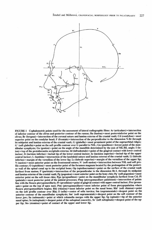
FIGURE 1 Cephalometric points used for the assessment of lateral radiographic films: Ar (articulare)= intersectionof inferior contour of the clivus and posterior contour of the ramus; Ba (basion)=most posteroinferior point on theclivus; Br intersection of the coronal suture and lamina externa of the cranial vault; Cd (condylion)=mostsuperior point on the condylar head; F intersection of the perpendicular to the dimension N-Br throughits midpoint and lamina externa of the cranial vault; G (glabella)=most prominent point of the supraorbital ridges;G' (soft glabella)= point on the soft profile contour over G parallel to NSL; Gn (gnathion)=lowest point of the man-dibular symphysis; Go (gonion)=point on the angle of the mandible determined by the axis of ML/RL angle; I (in-
ion) =top of the protuberantia occipitalis externa; Id (infradentale) = point of the gingival contact with lower centralincisor; Ii (incision inferius) = incisal tip of the lower central incisor; Is (incision superius) =incisal tip of the uppercentral incisor; L (Iambda)=intersection of the lambdoid suture and lamina externa of the cranial vault; Li (labrale
inferius) =margin of the vermilion of the lower lip; Ls (labrale superius)=margin of the vermilion of the upper lip;
N (nasion) =most anterior point on the frontonasal suture; N' (soft nasion) = intersection between NSL and soft pro-
file contour; O (opisthion)=most posterior point of the foramen magnum located by the prolongation of the posteri-
or wall of the spinal canal up to the occipital bone; Op (opisthocranion)=point on the surface of the cranial vault
farthest from nasion; P (parietale)=intersection of the perpendicular to the dimension Br-L through its midpoint
and lamina externa of the cranial vault; Pg (pogonion) =most anterior point on the bony chin; Pg' (soft pogonion) =most
anterior point on the soft tissue chin; Pgn (prognathion)=point on the mandibular symphysis farthest from Cd; Pl
(palatale)=most posterior point of the palatal processes; Pmp (pterygomaxillare palatinum) =intersection of palate
line with the fissura pterygomaxillaris; Pr (prosthion)= point of gingival contact with upper central incisor; Prn (prona-
sale)= point on the top of apex nasi; Ptm (pterygomaxillare)=most inferior point of fossa pterygopalatina where
fissura pterygomaxillaris begins; Rhi (rhinion)=most inferior point on the nasal bone; Rhi' (soft rhinion)= point
on the soft profile contour over Rhi; S (sella)=centre of sella turcica; Sm (supramentale)=deepest point on the
anterior contour of the mandibular symphysis; Sm' (soft supramentale)=deepest point on the soft contour of the
lower jaw; Sn (subnasale)=point at which columella merges with the upper lip; Sp (spinale)=tip of the anterior
nasal spine; Ss (subspinale)=deepest point of the subspinal concavity, Ss' (soft subspinale)=deepest point of the up-
per lip; Sto (stomion)=point of contact of the upper and lower lip. ‘
227
228 _- Cleft Palate Journal, July 1986, Vol. 23 No. 3
Ltfl
Lo
Zy
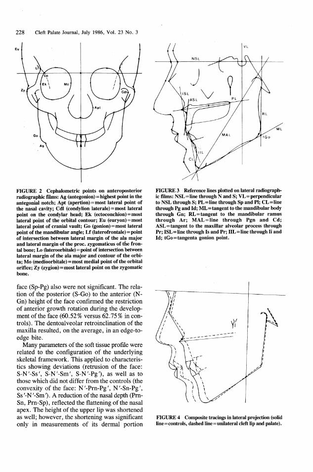
FIGURE 2 Cephalometric points on anteroposterior
radiographic films: Ag (antegonion) = highest point in the
antegonial notch; Apt (apertion)=most lateral point of
the nasal cavity; Cdl (condylion laterale)= most lateral
point on the condylar head; Ek (ectoconchion)=most
lateral point of the orbital contour; Eu (euryon)=most
lateral point of cranial vault; Go (gonion)=most lateral
point of the mandibular angle; Lf (laterofrontale) = point
of intersection between lateral margin of the ala major
and lateral margin of the proc. zygomaticus of the fron-
tal bone; Lo (lateroorbitale)= point of intersection between
lateral margin of the ala major and contour of the orbi-
ta; Mo (medioorbitale) =most medial point of the orbital
orifice; Zy (zygion) =most lateral point on the zygomatic
bone.
face (Sp-Pg) also were not significant. The rela-
tion of the posterior (S-Go) to the anterior (N-
Gn) height of the face confirmed the restriction
of anterior growth rotation during the develop-
ment of the face (60.52% versus 62.75% in con-
trols). The dentoalveolar retroinclination of the
maxilla resulted, on the average, in an edge-to-
edge bite.
Many parameters of the soft tissue profile were
related to the configuration of the underlying
skeletal framework. This applied to characteris-
tics showing deviations (retrusion of the face:
S-N'-Ss', S-N'-Sm', S-N'-Pg"), as well as to
those which did not differ from the controls (the
convexity of the face: N'-Prn-Pg', N'-Sn-Pg!,
Ss '-N'-Sm '). A reduction of the nasal depth (Prn-
Sn, Prn-Sp), reflected the flattening of the nasal
apex. The height of the upper lip was shortened
as well; however, the shortening was significant
only in measurements of its dermal portion
RL
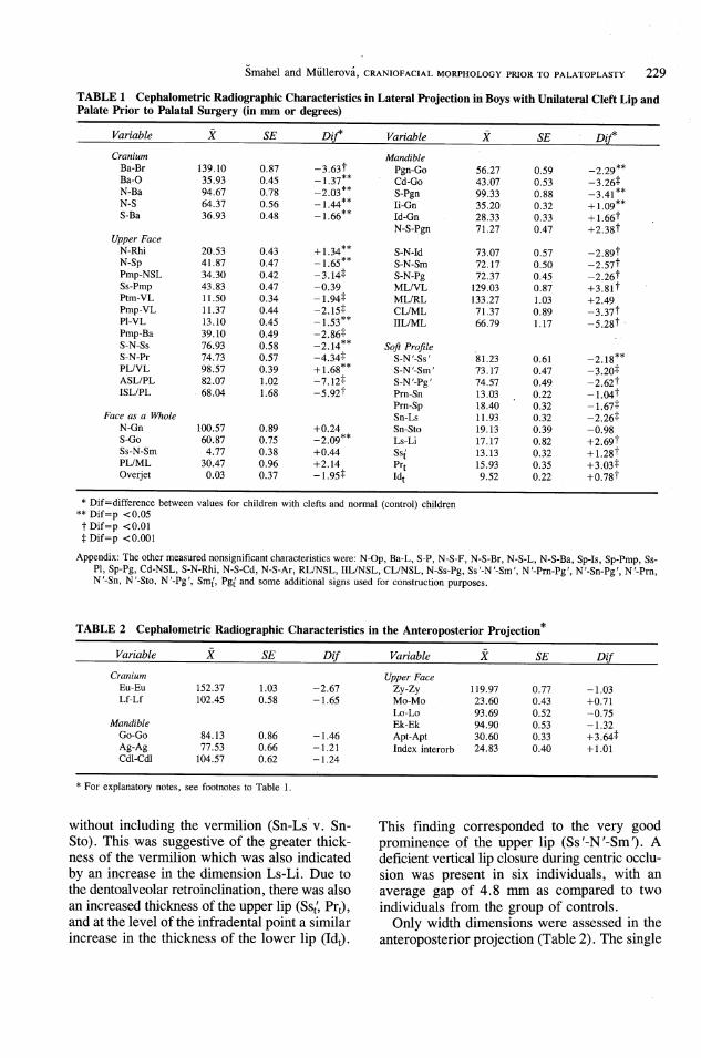
fltGo
FIGURE 3 Reference lines plotted on lateral radiograph-ic films: NSL=line through N and S; VL=perpendicularto NSL through S; PL=line through Sp and PI; CL =linethrough Pg and Id; ML=tangent to the mandibular bodythrough Gn; RL=tangent to the mandibular ramusthrough Ar; MAL=line through Pgn and Cd;ASL=tangent to the maxillar alveolar process throughPr; ISL= line through Is and Pr; IIL = line through Ii andId; tGo=tangenta gonion point.
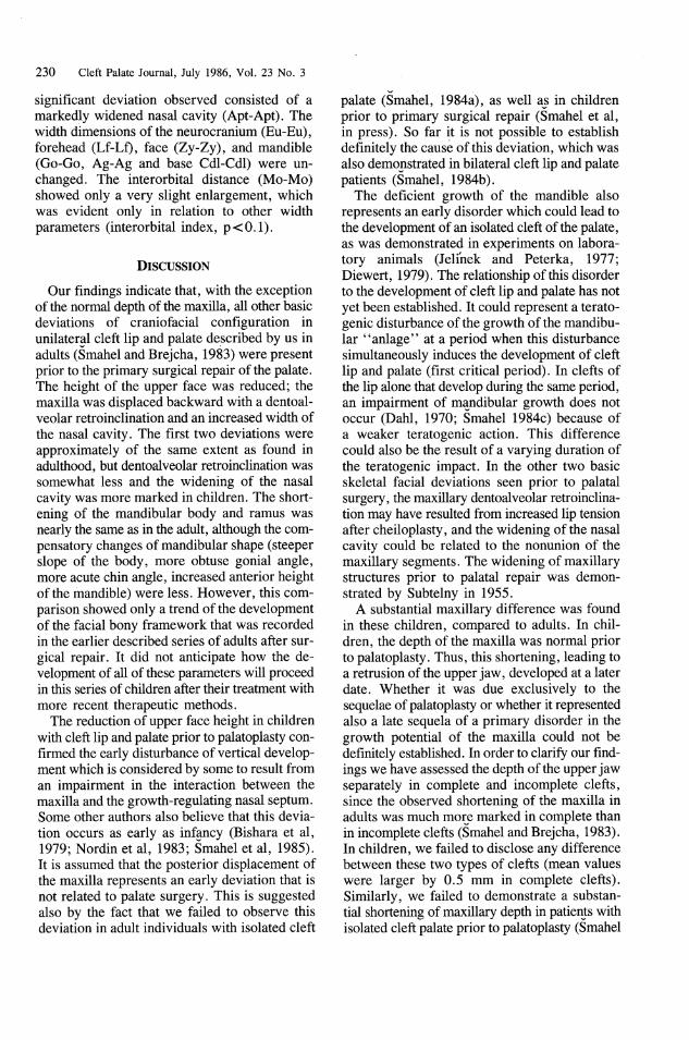
oar-
I
FIGURE 4 Composite tracings in lateralprojection (solidline=controls, dashed line= unilateral cleft lip and palate).
Smahel and Mullerova, CRANIOFACIAL MORPHOLOGY PRIOR TO PALATOPLASTY 229
TABLE 1 Cephalometric Radiographic Characteristics in Lateral Projection in Boys with Umlateral Cleft Lip andPalate Prior to Palatal Surgery (in mm or degrees)
Variable X SE Dif" Variable X SE _ Dif"
Cranium MandibleBa-Br 139.10 0.87 -3.631 Pgn-Go 56.27 0.59 2.29**Ba-0 35.93 0.45 -1.37** Cd-Go 43.07 0.53 -3.26+¢N-Ba 94.67 0.78 2.03** S$-Pgn 99.33 0.88 -3.41"**N-S 64.37 0.56 1.44"** Ii-Gn 35.20 0.32 +1.09**S-Ba 36.93 0.48 -1.66** Id-Gn . 28.33 0.33 +1.661
N-S-Pgn 71.27 0.47 +2.381fUpper FaceN-Rhi 20.53 0.43 +1.34"** S-N-Id 73.07 0.57 -2.8s91N-Sp 41.87 0.47 1.65** S-N-Sm 72.17 0.50 -2.571Pmp-NSL 34.30 0.42 S-N-Pg _ 72.37 0.45 -2.261Ss-Pmp 43.83 0.47 -0.39 ML/VL 129.03 0.87 +3.811Ptm-VL 11.50 0.34 -1.94t¢ ML/RL 133.27 1.03Pmp-VL 11.37 0.44 -2.15¢ CL/ML 71.37 0.89 -3.371PI-VL 13.10 0.45 -1.53** HL/ML 66.79 1.17 -5.281Pmp-Ba 39.10 0.49 -2.864S-N-Ss 76.93 0.58 -2.14** Soft Profile ‘S-N-Pr 74.73 0.57 -4.341E S-N'-Ss' 81.23 0.61 -2.18**PL/VL 98.57 0.39 +1.68** S-N'-Sm' 73.17 0.47 eASL/PL 82.07 1.02 -7.124 S-N -Pg/ 74.57 0.49 -2.621ISL/PL - 68.04 1.68 -5.92t Prn-Sn 13.03 _._ 0.22 -=1.041
Prn-Sp 18.40 0.32 -1.67+4Face as a Whole Sn-Ls 11.93 0.32 -2.26+4
N-Gn 100.57 0.89 +0.24 Sn-Sto 19.13 0.39 -0.98S-Go 60.87 0.75 -2.09** Ls-Li 17.17 0.82 +2.69TSs-N-Sm 4.77 0.38 +0.44 Ssf{ 13.13 0.32 +1.281PL/ML 30.47 0.96 +2.14 Pr{ 15.93 0.35 +3.034Overjet 0.03 0.37 -1.95¢ Id; 9.52 0.22 +0.781 * Dif=difference between values for children with clefts and normal (control) children
** Dif=p <0.05{ Dif=p <0.01{ Dif=p <0.001
Appendix: The other measured nonsignificant characteristics were: N-Op, Ba-L, S-P, N-S-F, N-S-Br, N-S-L, N-S-Ba, Sp-Is, Sp-Pmp, Ss-P1, Sp-Pg, Cd-NSL, S-N-Rhi, N-S-Cd, N-S-Ar, RL/NSL, IIL/NSL, CL/NSL, N-Ss-Pg, Ss -N '-Sm', N'-Prn-Pg', N'-Sn-Pg', N'-Prn,N'-Sn, N'-Sto, N'-Pg!', Smf{, Pg{ and some additional signs used for construction purposes.
TABLE 2 Cephalometric Radiographic Characteristics in the Anteroposterior Projecti0n*
Variable X SE Dif Variable X SE Dif
Cranium Upper FaceEu-Eu 152.37 1.03 -2.67 Zy-Zy 119.97 0.77 -1.03Lf-Lf 102.45 0.58 -1.65 Mo-Mo 23.60 0.43 +0.71
Lo-Lo _ 93.69 0.52 -0.75Mandible Ek-Ek 94.90 0.53 -1.32Go-Go 84.13 0.86 -1.46 Apt-Apt 30.60 0.33 +3.641Ag-Ag 77.53 0.66 -1.21 Index interorb 24.83 0.40 +1.01Cdl-Cdl 104.57 0.62 -1.24
* For explanatory notes, see footnotes to Table 1.
without including the vermilion (Sn-Lsv. Sn-
Sto). This was suggestive of the greater thick-
ness of the vermilion which was also indicated
by an increase in the dimension Ls-Li. Due to
the dentoalveolar retroinclination, there was also
an increased thickness of the upper lip (Ss,, Pr,),
and at the level of the infradental point a similar
increase in the thickness of the lower lip (Id,).
This finding corresponded to the very good
prominence of the upper lip (Ss'-N'-Sm'). A
deficient vertical lip closure during centric occlu-
sion was present in six individuals, with an
average gap of 4.8 mm as compared to two
individuals from the group of controls.
Only width dimensions were assessed in the
anteroposterior projection (Table 2). The single
230 Cleft Palate Journal, July 1986, Vol. 23 No. 3
significant deviation observed consisted of a
markedly widened nasal cavity (Apt-Apt). The
width dimensions of the neurocranium (Eu-Eu),
forehead (Lf-Lf), face (Zy-Zy), and mandible
(Go-Go, Ag-Ag and base CdIl-Cdl) were un-
changed. The interorbital distance (Mo-Mo)
showed only a very slight enlargement, which
was evident only in relation to other width
parameters (interorbital index, p<0.1).
DISCUSSION
Our findings indicate that, with the exception
of the normal depth of the maxilla, all other basic
deviations of craniofacial configuration in
unilateral cleft lip and palate described by us in
adults (Smahel and Brejcha, 1983) were presentprior to the primary surgical repair of the palate.The height of the upper face was reduced; themaxilla was displaced backward with a dentoal-veolar retroinclination and an increased width ofthe nasal cavity. The first two deviations wereapproximately of the same extent as found inadulthood, but dentoalveolar retroinclination wassomewhat less and the widening of the nasalcavity was more marked in children. The short-ening of the mandibular body and ramus wasnearly the same as in the adult, although the com-pensatory changes of mandibular shape (steeperslope of the body, more obtuse gonial angle,more acute chin angle, increased anterior heightof the mandible) were less. However, this com-parison showed only a trend of the developmentof the facial bony framework that was recordedin the earlier described series of adults after sur-gical repair. It did not anticipate how the de-velopment of all of these parameters will proceedin this series of children after their treatment withmore recent therapeutic methods.The reduction of upper face height in children
with cleft lip and palate prior to palatoplasty con-firmed the early disturbance of vertical develop-ment which is considered by some to result froman impairment in the interaction between themaxilla and the growth-regulating nasal septum.Some other authors also believe that this devia-tion occurs as early as infancy (Bishara et al,1979; Nordin et al, 1983; Smahel et al, 1985).It is assumed that the posterior displacement ofthe maxilla represents an early deviation that isnot related to palate surgery. This is suggestedalso by the fact that we failed to observe thisdeviation in adult individuals with isolated cleft
palate (Smahel, 1984a), as well as in childrenprior to primary surgical repair (Smahel et al,in press). So far it is not possible to establishdefinitely the cause ofthis deviation, which wasalso demonstrated in bilateral cleft lip and palatepatients (Smahel 1984b).The deficient growth of the mandible also
represents an early disorder which could lead tothe development of an isolated cleft of the palate,as was demonstrated in experiments on labora-tory animals (Jelinek and Peterka, 1977;Diewert, 1979). The relationship of this disorderto the development of cleft lip and palate has notyet been established. It could represent a terato-genic disturbance of the growth of the mandibu-lar '"'anlage'' at a period when this disturbancesimultaneously induces the development of cleftlip and palate (first critical period). In clefts ofthe lip alone that develop during the same period,an impairment of mandibular growth does notoccur (Dahl, 1970; Smahel 1984c) because ofa weaker teratogenic action. This differencecould also be the result of a varying duration of
the teratogenic impact. In the other two basicskeletal facial deviations seen prior to palatalsurgery, the maxillary dentoalveolar retroinclina-tion may have resulted from increased lip tensionafter cheiloplasty, and the widening of the nasalcavity could be related to the nonunion of themaxillary segments. The widening of maxillarystructures prior to palatal repair was demon-strated by Subtelny in 1955.A substantial maxillary difference was found
in these children, compared to adults. In chil-dren, the depth of the maxilla was normal priorto palatoplasty. Thus, this shortening, leading toa retrusion of the upper jaw, developed at a laterdate. Whether it was due exclusively to thesequelae of palatoplasty or whether it representedalso a late sequela of a primary disorder in the
growth potential of the maxilla could not bedefinitely established. In order to clarify our find-ings we have assessed the depth of the upper jawseparately in complete and incomplete clefts,since the observed shortening of the maxilla inadults was much more marked in complete thanin incomplete clefts (Smahel and Brejcha, 1983).In children, we failed to disclose any differencebetween these two types of clefts (mean valueswere larger by 0.5 mm in complete clefts).Similarly, we failed to demonstrate a substan-tial shortening of maxillary depth in patients withisolated cleft palate prior to palatoplasty (Smahel
Smahel and Mlllerova, CRANIOFACIAL MORPHOLOGY PRIOR TO PALATOPLASTY 231
et al, in press) contrary to our observations in
adult patients (Smahel, 1984a). This evidence
indicates that this deviation develops postopera-
tively. Bishara et al (1979) observed during
longitudinal studies of complete clefts that, at the
age of 5 years, the maxilla shows even a greater
depth than the norm. During subsequent years,
the difference gradually decreased because of the
more exuberant anterior growth of the maxilla
in controls. Throughout the entire observation
period, the angle SNA (S-N-Ss) was more acute,
which, in the presence of an increased depth of
the maxilla, could be accounted for by the dis-
placement of the maxilla backward. Examina-
tions of adult patients who were not treated by
surgery also showed an adequate anterior growth
of the maxilla (Ortiz-Monasterio et al, 1959,
1966; Pitanguy and Franco, 1967; Bishara et al,
1976). Dahl (1970), however, demonstrated a
shortening of the depth of the maxilla in adults
without palatoplasty.
The other described skeletal deviations resulted
from the basic deviations previously described
(retrusion of both jaws, limited anterior growth
rotation of the face, shortening of the depth of
the nasopharyngeal bony framework, and anom-
alies of occlusion) or were of no practical im-
portance. We have observed an elongation of the
nasal bones similar to that found in adults.
Although there was evidence of its correlation
with maxillary retrusion (Smahel and Brejcha,
1983), this does not serve as a full explanation
of its cause. In two facial characteristics, the re-
lation between the preoperative and final find-
ings remained unclear. In adults, we failed to
observe a reduction in the posterior height of the
upper face or a retroinclination of the palatal
plane. However, information reported in the
literature shows that these characteristics can
undergo changes with age. A trend toward these
variations was seen (Hayashi et al, 1976). Fur-
ther, we did not observe the significant increase
in the interorbital distance that has been demon-
strated repeatedly in the literature (in children,
by Graber, 1964; Aduss et al, 1971; Figalova
et al, 1974). We have stated that such an increase
was apparent in relation to facial width
parameters, especially in the orbital region (inter-
orbital index, p <0.1). Contrary to that which
was seen in adults, our children had neither a
shallower slope of the forehead nor posterior
rotation of the cranial vault. The cranial base
showed a shortening of its postsellar part. The
other characteristics of the cranium yielded
similar findings in both children and adults.
Of the variations seen in the soft tissues, the
height of the upper lip showed a similar reduc-
tion both in adults and in children prior to
'palatoplasty. This shortening resulted primarily
from a tissue deficiency. The observations
differed between adults and children in the thick-
ness of the upper lip. It was always narrower in
adults, thus making the retrocheilia even more
conspicuous. The higher mean values recorded
prior to palatoplasty resulted from the retroin-
clination of the maxillary dentoalveolar compo-
nent. At that age the lip appeared visually to be
hypertrophic, with good prominence. This in-
creased thickness resulted from the stretching of
the lip into the vestibular sulcus, which was in-
creased because of retroinclination of the anterior
maxillary segment.
In the literature available, we failed to find a
study dealing in more detail with an analysis of
the craniofacial deviations in cleft lip and palate
patients prior to palatoplasty. Therefore, we did
not have data at our disposal that would allow
a comparison with our findings. The main fea-
tures of our results were, however, in agreement
with present knowledge.
Acknowledgement. We wish to express our gratitude toMr. J. Brzorad from the Clinic of Radiology at the MedicalFaculty of Hygiene, Charles University, for the x-ray filmsand to Mrs. M. Svozilova for her technical assistance.
References
Apuss H, S, Micier® M. Interorbital distancein cleft lip and palate. Teratology 1971; 4:171.
BIsHARA SE, Krause CHJ, Oum WH, WEsTON D, van NEssJ, FELLING CH. Facial and dental relationships of in-dividuals with unoperated clefts of the lip and/or palate.Cleft Palate J 1976; 13:238. .
BISHARA SE, SIERK DL, HUANG K. A longitudinal cephalo-metric study on unilateral cleft lip and palate subjects. CleftPalate J 1979; 16:59.
DaxL E. Craniofacial morphology in congemtal clefts of
the lip and palate. Acta Odont Scand 1970; 28:Suppl 57:1.
DIEWERT VM. Correlation between mandibular retrognathia
and induction of cleft palate with 6-aminonicotinamide in
the rat. Teratology 1979; 19:213.
FigaALOVA P, HAJNIS K, SMAHEL Z. The interocular dis-
tance in children with cleft before the operation. Acta Chir
Plast 1974; 16:65.
GRABER TM. A study of craniofacial growth and develop-
ment in the cleft palate child from birth to six years of age.
In: Hotz R, ed. Early treatment of cleft lip and cleft pa-
late. Berne: Hans Huber, 1964:30.
HavyasHI I, Sakupa M, Takimoto K, MIryaAzAKI T.

232 Cleft Palate Journal, July 1986, Vol. 23 No. 3
Craniofacial growth in complete unilateral cleft lip and pa-late: a roentgenocephalometric study. Cleft Palate J 1976;13:215.
JELINEK R, PETERKA M. The role of the mandible in mousepalatal development revisited. Cleft Palate J 1977; 14:211.
NoRDIN KE, Larson O, NYLEN B, EKLUND G. Early bonegrafting in complete cleft lip and palate cases followingmaxillofacial orthopaedics. I. The method and the skeletaldevelopment from seven to thirteen years of age. ScandJ Plast Reconstr Surg 1983; 17:33.
OrRTIZ-MoONAsTERIO F, REBEIL AS, VANDERRAMA M, CRUZR. Cephalometric measurements on adult patients withnon-operated cleft palates. Plast Reconstr Surg 1959; 24:53.
OrTIZz-MonasTERIO F, SERRANO A, BARRERA G,RopRIQUEZ-HoFFMAN H, VINAGERAS E. A study of un-treated adult cleft palate patients. Plast Reconstr Surg 1966;38:36.
Prranouy I, Franco T. Nonoperated facial fissures inadults. Plast Reconstr Surg 1967; 39:569.
SUBTELNY JD. - Width of the nasopharynx and related ana-tomic structures in normal and unoperated cleft palate chil-
_ dren. Am J Orthod 1955; 41:889.SMAHEL Z. Variations in craniofacial morphology withseverity of isolated cleft palate. Cleft Palate J 1984a;
_ 21:140.SMmaAHEL Z. Craniofacial morphology in adults with bilateral_ complete cleft lip and palate. Cleft Palate J 1984b; 21:159.SmaHEL Z. Craniofacial changes in unilateral cleft lip in_ adults. Acta Chir Plast 1984c; 26:129.SMmAHEL Z, BrEJCHA M. Differences in craniofacial mor-phology between complete and incomplete unilateral cleft
_ lip and palate in adults. Cleft Palate J 1983; 20:113.SmaAHEL Z, PoBISOVA Z, FIGaALOVA P. Basic cephalomet-
ric facial characteristics in cleft lip and/or cleft palate pri-_ or to the first surgical repair. Acta Chir Plast 1985; 27:131.SmaxHEL Z, BrousiLovA M, MUruErovA Z. Craniofacialmorphology in isolated cleft palate prior to palatoplasty,in press.
Related Documents