Cranial humerus translation, deltoid activation, adductor co-activation and rotator cuff disease — Different patterns in rotator cuff tears, subacromial impingement and controls P.B. de Witte a,b, ⁎, J.F. Henseler a,b , E.W. van Zwet c , J. Nagels a , R.G.H.H. Nelissen a , J.H. de Groot b a Department of Orthopaedics, Leiden University Medical Center (LUMC), Postzone J11R, Postbus 9600, 2300 RC Leiden, the Netherlands b Laboratory for Kinematics and Neuromechanics, Department of Orthopaedics & Rehabilitation, LUMC, Postzone B0-Q, Postbus 9600, 2300 RC Leiden, the Netherlands c Department of Medical Statistics and BioInformatics, LUMC, Postbus 9600, 2300 RC Leiden, the Netherlands abstract article info Article history: Received 27 July 2013 Accepted 21 October 2013 Keywords: Rotator cuff Shoulder impingement syndrome Electromyography Diagnostic techniques and procedures Adductor co-activation Background: Arm adductor co-activation during abduction has been reported as a potential compensation mech- anism for a narrow subacromial space in patients with rotator cuff dysfunction. We assessed differences in acromiohumeral distance at rest and the amount of humerus translation during active abduction and adduction in patients with rotator cuff tears (n = 20) and impingement (n = 30) and controls (n = 10), controlled for deltoid, pectoralis major, latissimus dorsi and teres major activation (electromyography). Methods: During the acquirement of shoulder radiographs, subjects performed standardized isometric arm abduction and adduction tasks. EMG's were normalized between −1 and 1 using the “Activation Ratio”, where low values express (pathologic) co-activation, e.g. adductor activation during abduction. Findings: In patients with cuff tears mean rest acromiohumeral distance was 7.6 mm (SD = 1.6): 3.5 mm narrower compared to patients with impingement (95%-CI: 2.4–4.5) and 1.3 mm narrower compared to controls (95%-CI: −0.1–2.7). Both during abduction and adduction tasks, cranial translation was observed with equal magnitudes for patients and controls, with average values of 2.3 and 1.7 mm, respectively. Where patients with cuff tears had lower adductor Activation Ratios (i.e. more adductor co-activation during abduction), no association between abductor/adductor muscle activation and acromiohumeral distance was found. Interpretation: The subacromial space is narrower in patients with rotator cuff tears compared to patients with impingement and controls. We found additional subacromial narrowing during isometric abduction and, to a lesser amount, during adduction in all subjects and more adductor co-activation in patients with cuff tears. We found no association between subacromial space and activation of the deltoid and main adductors. © 2013 Elsevier Ltd. All rights reserved. 1. Introduction The incidence of shoulder complaints in general practice is high, with 22 per 1000 registered patients per year (Sobel and Winters, 1996). 44–65% of shoulder symptoms are diagnosed as “Subacromial Impingement Syndrome” (SIS) (Michener et al., 2003; Neer, 1972) and 36% of subjects with shoulder symptoms have been demonstrated to have rotator cuff (RC) tears (Yamamoto et al., 2010). Both conditions show similar symptoms, including pain and loss of arm abduction force, although symptoms are generally worse in patients with RC tears (de Witte et al., 2012a; Harrison and Flatow, 2011). Some report that both are stages of the same condition, where SIS may progress to a RC tear due to muscle and tendon degeneration (Chang, 2004; Harrison and Flatow, 2011; Hyvonen et al., 1998; Neer, 1983; Ozaki et al., 1988). We assessed similarities and differences in objective biomechanical signs suggestive for RC dysfunction in patients with SIS or RC tears and controls. A narrow subacromial space due to a cranial position of the humerus relative to the acromion has been radiographically demonstrated in patients with RC tears (Bloom, 1991; Keener et al., 2009; Weiner and Macnab, 1970; Yamaguchi et al., 2000). A narrow subacromial space has been associated with shoulder pain, larger RC tear sizes, progression of RC tears to multiple tendons and RC degeneration, and has been reported as a negative prognostic factor for (surgical) treatment (Bloom, 1991; Gladstone et al., 2007; Keener et al., 2009; Vad et al., 2002; van de Sande et al., 2008; Weiner and Macnab, 1970; Yamaguchi et al., 2000). Additional narrowing of the subacromial space (i.e. cranial humerus translation) during active arm abduction has been reported on radiographs and magnetic resonance imaging (MRI) ac- quired in healthy subjects, and patients with RC tears or SIS (Graichen et al., 1999, 2005; Hebert et al., 2003; Paletta et al., 1997). In these stud- ies, it was postulated, that RC deficit patients show more cranial humerus translation than healthy subjects and that this cranial translation may be a diagnostic tool or an objective clinical outcome measure. Clinical Biomechanics 29 (2014) 26–32 ⁎ Corresponding author. E-mail address: [email protected] (P.B. de Witte). 0268-0033/$ – see front matter © 2013 Elsevier Ltd. All rights reserved. http://dx.doi.org/10.1016/j.clinbiomech.2013.10.014 Contents lists available at ScienceDirect Clinical Biomechanics journal homepage: www.elsevier.com/locate/clinbiomech

Welcome message from author
This document is posted to help you gain knowledge. Please leave a comment to let me know what you think about it! Share it to your friends and learn new things together.
Transcript

Clinical Biomechanics 29 (2014) 26–32
Contents lists available at ScienceDirect
Clinical Biomechanics
j ourna l homepage: www.e lsev ie r .com/ locate /c l inb iomech
Cranial humerus translation, deltoid activation, adductor co-activation and rotator cuffdisease — Different patterns in rotator cuff tears, subacromial impingementand controls
P.B. de Witte a,b,⁎, J.F. Henseler a,b, E.W. van Zwet c, J. Nagels a, R.G.H.H. Nelissen a, J.H. de Groot b
a Department of Orthopaedics, Leiden University Medical Center (LUMC), Postzone J11R, Postbus 9600, 2300 RC Leiden, the Netherlandsb Laboratory for Kinematics and Neuromechanics, Department of Orthopaedics & Rehabilitation, LUMC, Postzone B0-Q, Postbus 9600, 2300 RC Leiden, the Netherlandsc Department of Medical Statistics and BioInformatics, LUMC, Postbus 9600, 2300 RC Leiden, the Netherlands
⁎ Corresponding author.E-mail address: [email protected] (P.B. de Witte).
0268-0033/$ – see front matter © 2013 Elsevier Ltd. All rihttp://dx.doi.org/10.1016/j.clinbiomech.2013.10.014
a b s t r a c t
a r t i c l e i n f oArticle history:
Received 27 July 2013Accepted 21 October 2013Keywords:Rotator cuffShoulder impingement syndromeElectromyographyDiagnostic techniques and proceduresAdductor co-activation
Background: Arm adductor co-activation during abduction has been reported as a potential compensationmech-anism for a narrow subacromial space in patients with rotator cuff dysfunction. We assessed differences inacromiohumeral distance at rest and the amount of humerus translation during active abduction and adductionin patients with rotator cuff tears (n = 20) and impingement (n = 30) and controls (n = 10), controlled fordeltoid, pectoralis major, latissimus dorsi and teres major activation (electromyography).Methods: During the acquirement of shoulder radiographs, subjects performed standardized isometric armabduction and adduction tasks. EMG's were normalized between −1 and 1 using the “Activation Ratio”, wherelow values express (pathologic) co-activation, e.g. adductor activation during abduction.Findings: In patients with cuff tears mean rest acromiohumeral distance was 7.6 mm (SD = 1.6): 3.5 mm
narrower compared to patientswith impingement (95%-CI: 2.4–4.5) and 1.3 mmnarrower compared to controls(95%-CI: −0.1–2.7). Both during abduction and adduction tasks, cranial translation was observed with equalmagnitudes for patients and controls, with average values of 2.3 and 1.7 mm, respectively. Where patientswith cuff tears had lower adductor Activation Ratios (i.e. more adductor co-activation during abduction), noassociation between abductor/adductor muscle activation and acromiohumeral distance was found.Interpretation: The subacromial space is narrower in patients with rotator cuff tears compared to patients withimpingement and controls. We found additional subacromial narrowing during isometric abduction and, to alesser amount, during adduction in all subjects and more adductor co-activation in patients with cuff tears. Wefound no association between subacromial space and activation of the deltoid and main adductors.© 2013 Elsevier Ltd. All rights reserved.
1. Introduction
The incidence of shoulder complaints in general practice is high,with 22 per 1000 registered patients per year (Sobel and Winters,1996). 44–65% of shoulder symptoms are diagnosed as “SubacromialImpingement Syndrome” (SIS) (Michener et al., 2003; Neer, 1972)and 36% of subjects with shoulder symptoms have been demonstratedto have rotator cuff (RC) tears (Yamamoto et al., 2010). Both conditionsshow similar symptoms, including pain and loss of arm abduction force,although symptoms are generally worse in patients with RC tears (deWitte et al., 2012a; Harrison and Flatow, 2011). Some report that bothare stages of the same condition, where SIS may progress to a RC teardue to muscle and tendon degeneration (Chang, 2004; Harrison andFlatow, 2011; Hyvonen et al., 1998; Neer, 1983; Ozaki et al., 1988).We assessed similarities and differences in objective biomechanical
ghts reserved.
signs suggestive for RC dysfunction in patients with SIS or RC tearsand controls.
A narrow subacromial space due to a cranial position of the humerusrelative to the acromion has been radiographically demonstrated inpatients with RC tears (Bloom, 1991; Keener et al., 2009; Weiner andMacnab, 1970; Yamaguchi et al., 2000). A narrow subacromial spacehas been associatedwith shoulder pain, larger RC tear sizes, progressionof RC tears to multiple tendons and RC degeneration, and has beenreported as a negative prognostic factor for (surgical) treatment(Bloom, 1991; Gladstone et al., 2007; Keener et al., 2009; Vad et al.,2002; van de Sande et al., 2008; Weiner and Macnab, 1970;Yamaguchi et al., 2000). Additional narrowing of the subacromial space(i.e. cranial humerus translation) during active arm abduction has beenreported on radiographs and magnetic resonance imaging (MRI) ac-quired in healthy subjects, and patients with RC tears or SIS (Graichenet al., 1999, 2005; Hebert et al., 2003; Paletta et al., 1997). In these stud-ies, it was postulated, that RC deficit patients showmore cranial humerustranslation than healthy subjects and that this cranial translationmay bea diagnostic tool or an objective clinical outcome measure.
27P.B. de Witte et al. / Clinical Biomechanics 29 (2014) 26–32
Subacromial narrowing is the subject of an increasing number ofpublications and is more and more applied in clinical practice, but itsunderlying mechanisms remain unclear. RC muscles play a key role inglenohumeral (GH) stabilization and arm mobility; in the healthyshoulder a perfect compromise is assumed betweenmobility and stabil-ity (Veeger and van der Helm, 2007). In patients with RC tears, GH jointmechanics are disrupted as one or more RC muscles are dysfunctional.This may lead to 1) a compensatory increase in deltoid activity(McCully et al., 2007; Steenbrink et al., 2009, 2010a); 2) GH (micro-)instability (Steenbrink et al., 2009); 3) excessive cranial humerus trans-lation with the activation of arm abductors (Bloom, 1991; Graichenet al., 1999, 2005; Gruber et al., 2010; Hebert et al., 2003; Keener et al.,2009; Mayerhoefer et al., 2009; Nagels et al., 2008; Steenbrink et al.,2009; van de Sande and Rozing, 2006) and subsequent pain. It hasbeen hypothesized that co-activation of specific adductor muscles withdownwardly directed lines of action (e.g. teres major and latissimusdorsi) is a compensation mechanism to counteract this excessive cranialtranslation during abduction in patients with RC tears (de Groot et al.,2006; Steenbrink et al., 2006, 2009, 2010a,b).
The actual relation between cranial humerus translation, pain andadductor co-activation as a potential protective mechanism has beenscarcely investigated experimentally (de Groot et al., 2006; Steenbrinket al., 2006, 2010a,b), specifically for patients with SIS, in whom theRC is still anatomically functional. Patients with RC tears can beregarded as ultimate demonstrators for RC dysfunction. If translationand adductor co-activation are also found in patientswith SIS, disruptedGH joint mechanics might exist in these patients, despite intact RCmuscles. This would indicate that in addition to themany reported etiol-ogies of SIS, decreased RC function and altered biomechanics might playa role as well.
We investigated acromiohumeral distance (AH) on radiographsacquired during electromyography (EMG)-recorded isometric abduc-tion, adduction and rest tasks in patients with RC tears, patients withSIS and controls. Our primary goals were to 1) assess AH at rest andhumerus translation (ΔAH) during abduction and adduction forcetasks, 2) assess (pathologic) co-activation of deltoid, latissimus dorsi,teres major and pectoralis major, and 3) assess the associationbetween AH, deltoid activation and adductor (co-)activation. We hy-pothesized that 1) in patients baseline (rest) AH is smaller and ΔAHlarger compared to controls; 2) patients apply more adductor co-activation during abduction compared to controls; and 3) deltoid acti-vation is negatively and adductor co-activation is positively related toAH and ΔAH. We expect healthy controls, patients with SIS and patientswith RC tears to order along the scales of subacromial narrowing andadductor co-activation.
2. Methods
2.1. Subjects
Consecutive patients with a painful shoulder and a full-thickness RCtear (supraspinatus and/or infraspinatus) and patients diagnosed withSIS were included in this study during the period of April 2010 toApril 2012. In addition, 10 controls were recruited in September 2012.Inclusion criteria for the controls were: age between 35 and 60 years(minimizing the prevalence of eventual asymptomatic cuff tears andage differences between controls and patients), no present shouldercomplaints and no history of shoulder complaints treated with physicaltherapy, NSAID's, injections, or surgery. Controls were assessed by anMD for eventual shoulder symptoms.
Patients with SIS or RC tears had to have a positive Neer impinge-ment test, a positive Hawkins test and diffuse unilateral anterosuperiorshoulder pain for N3 months in combination with one or more of thefollowing criteria: pain with overhead activities, abduction, retroflexionand/or internal rotation (e.g. closing the door, putting on jacket); pain atnight or incapable of lying on the shoulder; diffuse pain at palpation of
the greater tuberosity; disturbed scapulohumeral rhythm; classic pain-ful arc; positive Yocum test; positive full or empty can test.
Patients with SIS had to be aged between 35 and 65 years. Exclusioncriteria for SIS were partially based on anMRI arthrogram and standardanteroposterior (AP) shoulder radiographs: no calcific tendonitis, full-thickness RC tear, intra-articular or bony lesions (Hill Sachs, (old) frac-tures, tumors), labrum abnormalities, capsular or ligamentous tears/avulsions, superior labrum tear from anterior to posterior (SLAP lesion),pulley lesion, biceps tendinitis or tear, os acromiale, cartilage lesions, orbony cysts. Patients with RC tears had to be aged 50 years and older.They were symptomatic and had a standard AP shoulder radiographand MRI-arthrogram or ultrasound (US)-proven full-thickness RC tear,without a subscapularis tear and other shoulder pathologies. Subjectswere furthermore excluded in case of insufficient Dutch language skillsor no informed consent, present physical problems influencing muscleactivation and arm mobility (other than the present shoulder condi-tion), any form of inflammatory arthritis of the shoulder, glenohumeralor symptomatic acromio-clavicular osteoarthritis, a history of fracture,dislocations, or surgical interventions of the shoulder, clinical signs ofcervical radiculopathy and frozen shoulder syndrome (b90° of passiveabduction and external rotation).
Radiographic data on acromiohumeral distance in the RC tear groupwere previously published (Henseler et al., in press). In the currentstudy these data are re-assessed with permission of the authors. Addi-tionally, we included simultaneously acquired EMG-recordings in theanalyses and used the data of this group with another goal: to compareacromiohumeral distance in three subject groups and to relate theacromiohumeral distance with muscle activation patterns, includingadductor co-activation.
Patients were clinically assessed using the Western Ontario RotatorCuff score (WORC) (Kirkley et al., 2003) and the Constant Score (CS)(Constant et al., 2008).
The accredited local Medical Ethics Committee (METC, LeidenUniversity Medical Center) approved all stages of the study accordingto the Medical Research Involving Human Subjects Act and writteninformed consentwas obtained fromall participants (Dutch Trial RegistryNTR2283).
2.2. Experimental setup
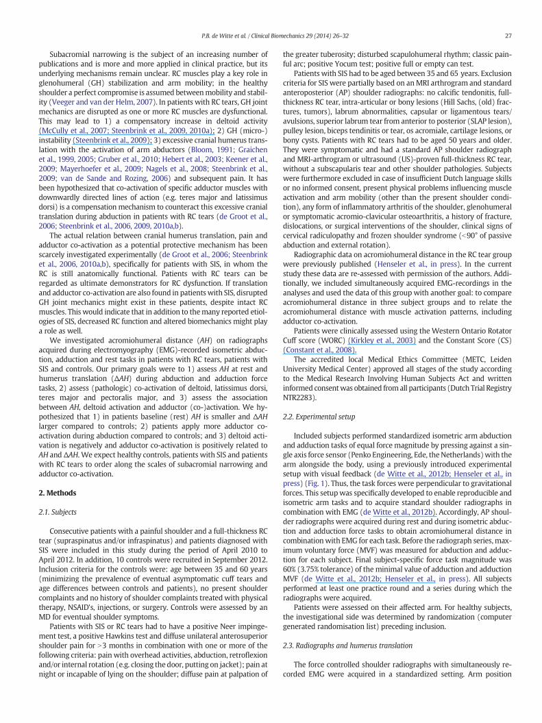
Included subjects performed standardized isometric arm abductionand adduction tasks of equal force magnitude by pressing against a sin-gle axis force sensor (Penko Engineering, Ede, theNetherlands)with thearm alongside the body, using a previously introduced experimentalsetup with visual feedback (de Witte et al., 2012b; Henseler et al., inpress) (Fig. 1). Thus, the task forces were perpendicular to gravitationalforces. This setupwas specifically developed to enable reproducible andisometric arm tasks and to acquire standard shoulder radiographs incombination with EMG (de Witte et al., 2012b). Accordingly, AP shoul-der radiographs were acquired during rest and during isometric abduc-tion and adduction force tasks to obtain acromiohumeral distance incombinationwith EMG for each task. Before the radiograph series,max-imum voluntary force (MVF) was measured for abduction and adduc-tion for each subject. Final subject-specific force task magnitude was60% (3.75% tolerance) of the minimal value of adduction and adductionMVF (de Witte et al., 2012b; Henseler et al., in press). All subjectsperformed at least one practice round and a series during which theradiographs were acquired.
Patients were assessed on their affected arm. For healthy subjects,the investigational side was determined by randomization (computergenerated randomisation list) preceding inclusion.
2.3. Radiographs and humerus translation
The force controlled shoulder radiographs with simultaneously re-corded EMG were acquired in a standardized setting. Arm position

Fig. 1. Subjectswere in standingpositionwith the target arm inexternal rotation at his/herside (i.e. hand in frontal plane), enabling the use of this setup during concomitant acquire-ment of standard shoulder radiographs for clinical or scientific purposes. The arm wasattached to a 1-dimensional force transducer at thewrist. In this setup, subjects performedEMG-recorded isometric abduction and adduction force tasks.
28 P.B. de Witte et al. / Clinical Biomechanics 29 (2014) 26–32
was constant during all tasks for each subject: the upper bodywas rotat-ed 30° from the frontal plane towards the Röntgen focus beamwith thearm at the subjects' side and the hand in the frontal planewith the palmfacing forward (de Witte et al., 2012b; Henseler et al., in press).
Radiograph quality was on-site controlled for using prescribedcriteria (Henseler et al., in press) and eventually re-acquired. On eachradiograph, acromiohumeral distance (AH) and humerus translation(ΔAH) were assessed. For AH, the distance between themost cranial ar-ticular cortex of the humeral head and the caudal cortex marking of thecaudal surface of the acromion was measured in millimetres (Gruberet al., 2010; Nagels et al., 2008; van de Sande et al., 2008). Task specifichumerus translation was calculated from the differences between restAH and abduction or adduction AH, obtainingΔAHab and ΔAHad, respec-tively. When the necessary anatomical landmarks could not be identi-fied, the measurement could not be performed and was reported asmissing.
2.4. Electromyography and adductor co-activation
During the rest, abduction and adduction tasks, EMG of 3 shoulderadductors (pectoralis major, clavicular part (PM); teres major (TM);latissimus dorsi (LD)) and the main shoulder abductor (deltoid, medialpart (DM)) were recorded with bi-polar surface EMG equipment(DelSys system Bagnoli-16, Boston, MA, USA, inter-electrode distance10 mm, bandwidth 20–450 Hz) as described previously (de Witteet al., 2012b).
As absolute magnitude of EMG-signals is hard to interpret and can-not be compared between subjects or related to AH, EMG-recordingswere expressed 1) relative to Maximal Voluntary Contraction EMG(MVC) and 2) in Activation Ratios. For this purpose, EMG was rectified
(rEMG) and averaged (aEMG). Muscle activation during tasks (Amuscle)was determined by subtracting the rest aEMG from the active aEMGduring abduction and adduction, respectively. EMG quality (signal-to-noise-ratio) was controlled for: if rEMG was less than 2 times moreactive during tasks compared to rest activity, the concerning measure-ment was not included in statistical analyses. rEMG's were normalizedto MVC for each task, muscle and subject. Additionally, the ActivationRatio was calculated, which combines the (absolute/not normalized)rEMG's of abduction and adduction tasks, in order to quantify musclespecific activation during agonist and antagonist tasks. For each muscleand subject, the following formula was applied:
ARmuscle ¼AIPmuscle−AOP
muscle
AIPmuscle þ AOP
muscle
−1≤ARmuscle≤1½ � ð1Þ
where AmuscleIP is ‘in phase’ or agonist muscle activation (DM activation
during abduction and TM, PM and LD activation during adduction) andAmuscleOP is ‘out-of-phase’ or antagonist muscle activation (DM activation
during adduction, or TM, PM and LD activation during abduction,i.e. pathologic co-activation) (de Witte et al., 2012b; Steenbrink et al.,2010b). TheActivation Ratios ofmostmuscles in healthy subjects are pos-itive and close to 1. In case of substantial antagonist activation of specificarm adductors during abduction tasks (adductor co-activation), as hasbeen previously described for the LD and TM in patients with RC tears,the Activation Ratios of these adductor muscles are expected to be closeto zero or even negative (Steenbrink et al., 2010b).
2.5. Statistics
Demographic data and clinical scores (patients) were expressed inmeans and standard deviations or medians and ranges whereappropriate.
For radiographicmeasures, themeanAH's (for rest, abduction and ad-duction) and the ΔAHab and ΔAHad in the three groups were all assessedusing one-way ANOVA analyses. A similar approach was applied for theMVC-normalized rEMG's during rest and the Activation Ratios during ab-duction and adduction for the four muscles in patients from the threegroups.
Multivariate Mixed Model analysis for repeated measures wasapplied to study the association between AH (dependent variable) anddisease status (control, impingement, RC tear), task (rest, abduction,adduction) and the MVC-normalized muscle activations of DM, PM, LDand TM.
P-values of ≤0.05 were considered statistically significant. Analyseswere processed using PASW Statistics 20.0 (IBM Inc., Chicago, Illinois).Multivariate analyses were processed using R 2.15.2.
3. Results
60 subjects were included: 20 patients with RC tears, 30 patientswith SIS and 10 controls. Two patients with SIS were excluded due tohardware problems during the experiments, leaving 58 subjects foranalyses (Table 1).
3.1. Acromiohumeral distance
Two adduction radiographs needed to be reacquired in patientswithRC tears, while not fulfilling the pre-set quality criteria. Furthermore,one adduction radiograph could not be assessed in a patient with SIS.
Mean AH at rest conditionwas 7.6 mm (SD = 1.60) in patients withRC tears, 11.1 mm (SD = 1.84) in patients with SIS and 8.9 mm(SD = 1.92) in controls (Table 2, Fig. 2). Average AHRESTwas significant-ly different in the three subject groups (p b 0.001). Post hoc analyses re-vealed that in patients with RC tears mean AHREST was 3.5 mm smaller(95%-CI: 2.4–4.5, p b 0.001) compared to patients with SIS and1.3 mm smaller (95%-CI: −0.1–2.7, p = 0.07) compared to controls.

Table 1Demographic data of patients and controls.
Baseline characteristics SISn = 28
RC tearn = 20
Controln = 10
Age (yrs) 50.1 [1.6] 65 [9.8] 50.2 [6.6]Gender (male/female) 11/17 11/9 5/5Affected side (right/left) 17/13 12/8 6/4Dominant side affected 18 (64%) 18 (90%) NABMI 26.5 [4.3] 28 [4.5] NABaseline clinical scores NAWORC 61.7 [14.8] 57 [22] NAConstant score 76.9 [8.5] 62 [15] NA
WORC, Western Ontario Rotator Cuff index.SIS, subacromial impingement syndrome.RC, rotator cuff.[SD].
Fig. 2. Acromiohumeral distance (AH) during rest and the isometric active abduction andadduction tasks for all subject groups. Overall, AHwas narrower in patients with RC tearscompared to controls and wider in patients with SIS. All subject groups showed similaramounts of cranial translation relative to rest during the abduction and adduction tasks.
29P.B. de Witte et al. / Clinical Biomechanics 29 (2014) 26–32
Mean AHREST for patients with SIS was 2.2 mm larger (95%-CI:0.9–3.5 mm, p = 0.002) compared to controls.
Also during abduction (p b 0.001) and adduction (p b 0.001), AHwas significantly different between the three groups. In post-hoc analy-ses, meanAHduring abduction and adductionwas significantly lower inpatientswith RC tears compared to patientswith SIS, with differences of3.8 mm (95%-CI: 2.6–5.0) and 3.4 mm (95%-CI: 2.2–4.6), respectively.Additionally, AH was significantly lower in patients with RC tearscompared controls: 1.8 mm (95%-CI: 0.3–3.4) and 1.5 mm (95%-CI:0.0–3.1), respectively.
All groups showed significant cranial humerus translation duringthe active tasks, except for the control group during adduction. Meantranslations for the three groups ranged between 2.1 and 2.6 mm forΔAHab (abduction) and between 0.8 and 1.1 for ΔAHad and were notsignificantly different between the subject groups (Table 2).
3.2. Muscle activation
Muscle EMG at rest, normalized to MVC, is displayed in Fig. 3. Over-all, there was more rest activity in patients than in controls. Post-hocanalyses revealed that for PM, there was significantly more rest activityin patientswith RC tears compared to controls (p = 0.007) and patientswith SIS (p = 0.04). For LD, there was significantly more activity inpatients with RC tears compared to controls (p = 0.04).
MeanAR's in patientswith RC tears, assessing “out-of-phase”muscleactivation (lower values), ranged from 0.42 to 0.58 for the adductors.For the abductor (DM), AR was 0.72 in this group. Average SIS AR forTM was 0.51. AR's for the DM and the LD and PM adductors rangedfrom 0.69 to 0.72. In controls, mean AR's ranged from 0.72 to 0.78 forDM and the adductors LD and PM. Mean TM AR in controls was 0.41.(Fig. 4).
Overall, there were no significant differences between AR's in thethree groups with one-way ANOVA analyses. With post-hoc analysesthe AR of the PM was significantly lower in patients with RC tearscompared controls: 0.53 vs. 0.79, leading to a mean difference of 0.26(95%-CI: 0.03–0.24, p = 0.03).
Table 2Distance between humerus and acromion at rest, and amounts of cranial humerus translation
Measurement RC tear SIS
mean 95%-CI mean
AH 7.6 (6.90 – 8.39) 11.1Cranial translationΔAHab 2.6 (1.86 – 3.39) 2.3ΔAHad 1.0 (0.29 –1.79) 1.1
(AH, acromiohumeral distance (at rest);ΔAH, difference in acromiohumeral distance during abtator Cuff; SIS, subacromial impingement syndrome).
3.3. Relation between acromiohumeral distance, muscle activation anddisease status
The alleged association between AH and muscle activation wasassessed in multivariate mixed model analyses for repeated measuresin each subject, leading to significant effects of disease status (control,SIS, RC tear) and performed task (rest, abduction, adduction) on AH(Table 3). For disease status,AHwas largest for patientswith SIS, follow-ed by controls and patients with RC tears, respectively. For performedtasks, AH was smallest during abduction, followed by adduction andrest. We found no significant association of muscle activations of DM,LD, PM or TM on AH (Table 3).
4. Discussion and conclusions
This study reports on muscle activation and the distance betweenhumerus and acromion at rest and during isometric abduction andadduction tasks in patients with RC tears, patientswith SIS and controls.At rest, the acromiohumeral distance (AH) was narrowest in patientswith RC tears. During active abduction, and to a lesser amount duringadduction, cranial humerus translation was observed relative to rest inall subject groups, without statistically significant differences betweenthe three subject groups. The rest activities of the pectoralis andlatissimus dorsi adductor muscles were larger for the patient groups.The EMG Activation Ratios of adductor muscles did not clearly indicatemore pathologic activation (i.e. lower Activation Ratios) in patientgroups compared to the controls, except for the pectoralis andlatissimus dorsi muscles in patients with RC tears, indicating a loss ofmuscle specific activation (i.e. adductor co-activation during arm
during abduction and adduction in patients with RC tears or SIS and in controls.
Control ANOVA
95%-CI mean 95%-CI p-value
(10.40 – 11.83) 8.9 (7.55 – 10.31) b0.001
(1.53 – 3.06) 2.1 (1.46 – 2.68) 0.675(0.52 – 1.73) 0.8 (−0.02 – 1.60) 0.837
duction (ab) or adduction (ad) compared to rest; 95%-CI, 95% Confidence Interval; RC, Ro-

Table 3Multivariate mixed model analyses with AH as dependent variable and disease status,performed task and the MVC-normalized muscle activations of DM, PM, LD and TM as in-dependent variables.
Variable Effect size 95%-CI p-value
Intercept 9.20 8.1 – 10.3 b0.001Disease statusSIS 1.81 0.52 – 3.10 b0.01RC tear −1.47 −2.83 – −0.10 0.04Control Ref. NA NA
TaskABduction −2.29 −3.23 – 1.36 b0.001ADduction −1.40 −2.58 – −0.22 0.02Rest Ref. NA NA
Muscle activationDM −0.25 −2.82 – 2.32 0.85LD 0.80 −1.89 – 3.50 0.56PM 0.86 −1.57 – 3.30 0.48TM −0.35 −3.09 – 2.39 0.80
Disease status and task had significant effects on AH. Patients with SIS had on average alarger AH than controls, whereas patients with RC tears had a smaller AH.Both isometric active abduction and adduction lead to a smaller AH. We found nosignificant effects of muscle activations on AH.Fig. 3.Muscle activations and 95%-CI during rest tasks for eachmuscle and subject groups,
normalized toMVCEMGs. Overall, rest activitywas higher in the patient groups comparedto controls.
30 P.B. de Witte et al. / Clinical Biomechanics 29 (2014) 26–32
abduction).We did not find an association betweenmuscle (co-)activa-tion and cranial humerus translation.
4.1. Acromiohumeral distance at rest
The smallest AH in rest we found in patients with RC tears coincideswith previous reports for RC deficit shoulders (Bloom, 1991; de Witteet al., 2012b; Gladstone et al., 2007; Keener et al., 2009; Vad et al.,2002; van de Sande et al., 2008; Weiner and Macnab, 1970;Yamaguchi et al., 2000). This phenomenon has been relatedwith shoul-der pain, progression of RC tear size, multiple torn RC tendons and RCmuscle degeneration in the literature (Bloom, 1991; Gladstone et al.,2007; Keener et al., 2009; Vad et al., 2002; van de Sande et al., 2008;Weiner and Macnab, 1970, Yamaguchi et al., 2000). Some studies sug-gest a small AH to be an indicator for chronic massive RC tears(Keener et al., 2009; van de Sande and Rozing, 2006; van de Sande etal., 2008;Weiner and Macnab, 1970). For patients with SIS, the average
Fig. 4. “Out-of-phase” muscle activation during abduction and adduction for all subjectgroups and all muscles, expressed in Activation Ratios (AR). Lower AR's indicate morepathologic activation, e.g. adductor co-activation during abduction.
AHwas not smaller than observed in controls, in contrast to our hypoth-esis. Possibly, actual full-thickness RC tearing needs to be present beforean evidently narrower subacromial space in rest ensues. In support ofthis, Keener and colleagues found differences in AH between 1) patientswith asymptomatic tears and symptomatic tears and 2) between pa-tients with full-thickness tears and massive posterosuperior (i.e.supraspinatus and infraspinatus) RC tears (Keener et al., 2009). Anotherexplanation is that there could be thickened RC tendons and bursa tis-sue in patients with SIS due to a subacromial inflammatory reaction,preventing the observation of an evidently smaller AH compared tocontrols despite potential RC deficiency. Subacromial filling may playan important role in determining the distance between acromion andhumerus.
4.2. Cranial humerus translation during active tasks
Cranial humerus translation (ΔAH; additional narrowing of AHduring active tasks compared to rest) wasmost evident during isometricabduction for all subject groups. Similarly, others found significant crani-al translation at different elevation angles during active abduction in pa-tients with SIS or RC tears (Bloom, 1991; Deutsch et al., 1996; Graichenet al., 1999; Hebert et al., 2003). However, the currently applied setuphas the advantage that a standard elevation angle and isometric tasksare applied for all subjects, improving the abilities to compare subjects(Henseler et al., in press). It has been suggested that cranial translationduring abduction is caused by Deltoid activation (Graichen et al., 1999,2005; Hinterwimmer et al., 2003). In patients with RC tears, there is sub-optimal GH stabilization due to the torn RC (Steenbrink et al., 2009) andincreased Deltoid activation, compensatory to lost RC function (McCullyet al., 2007; Steenbrink et al., 2009, 2010a). In theory, this can causeexcessive cranial translation of the humerus in patients, leading to (pain-ful) compression of subacromial tissues (Graichen et al., 2005; Keeneret al., 2009; Steenbrink et al., 2009). However, we found no differencesin the absolute amounts of translation in the three groups, covering acomplete range of subjects from symptomatic patients with RC tears, topatients with SIS with an intact RC and asymptomatic controls. But rela-tive to rest AH, narrowingwasmost prominent in patients with RC tears,suggesting the largest subacromial strains in this group duringabduction.
Surprisingly, there was not only cranial translation during armabduction tasks, but also during arm adduction in all groups. Althoughthe caudally directed TM and LD are primarily active during adduction,this did not lead to an average net caudalisation of the humerus. It isplausible that only limited activity of the deltoid with its large

31P.B. de Witte et al. / Clinical Biomechanics 29 (2014) 26–32
Physiological Cross Sectional Area (PCSA) relative to the TM and the LDleads to cranial translation even during adduction tasks.
4.3. Linking AH and adductor co-activation
Hence,we found group differences betweenAHmeasures at rest, butnot with the absolute ΔAH measurements assessing cranial translationduring tasks. This raises the question whether isometric active abduc-tion and adduction radiographs have any additional value in clinicaland scientific research. This all notwithstanding, e.g. 2.6 mm humerustranslation with a baseline AH of 7.6 mm as observed in patients withRC tears, i.e. a strain of −34%, might have more consequences than2.1–2.3 mm translation in patients with SIS or controls with their largerbaseline AH, i.e. strains of −20.7% and −23.6% respectively. A possibleexplanation for the similar amounts of AH narrowing in the threegroups, despite the theoretical excessive narrowing in patients withRC tears, is adductor co-activation. Several simulation and EMG studiesreported adductor activation during abduction (adductor co-activation)in RC patients, in particular for caudally directed adductors as the TMand LD, supposedly limiting subacromial narrowing by pulling the hu-merus down (de Groot et al., 2006; Steenbrink et al., 2006, 2009,2010a,b). We did find significantly higher adductor activation (relativeto MCV) in rest in patients with RC tears compared to patients withSIS and controls. This might partially be explained by somewhat lowermaximum voluntary forces and subsequent MVC values in patients(data not shown). There also appeared to be relatively more adductorco-activation, expressed in lower Activation Ratios during abduction inpatients with RC tears compared to patients with SIS (LD, PM, TM)and controls (LD, TM), but these differences were not statisticallysignificant.
4.4. Adductor co-activation in patients with RC tears or SIS
In a previous study assessing controls and patients with RC tearswith the same experimental setup, we defined an AR of minimal 0.30points lower compared to average control AR's is indicative for patho-logic co-activation, leading to cut-off values of 0.59 for the DM, 0.48for PM, 0.49 for LD and 0.38 for TM for this setup (de Witte et al.,2012b). In the patients with RC tears of the current study, mean AR'sof all adductors (PM, TM, LD) were around previously reported patho-logic AR cut-off values (de Witte et al., 2012b; Steenbrink et al.,2010b). For patients with SIS, only AR of the TMwas around the report-ed pathologic cut-off values (de Witte et al., 2012b; Steenbrink et al.,2010b). Although the RC is not torn in patients with SIS, tendonitisand partial tears can lead to impaired RC function and increased DMactivation (Myers et al., 2009). Potentially, this causes (excessive) up-wardly directed forces on the humerus during abduction, which canbe compensated by co-activation of only the TM in patients with SIS,where co-activation of several adductors is required to maintainglenohumeral stability in patients with RC tears. Hence, adductor co-activation might be an indicator for RC dysfunction in general, insteadof an indicator for RC tears specifically.
4.5. Unexpected results in the control group
The results of AH and adductor co-activation for patients with SIS orRC tears were overall as hypothesized, but we found unexpected resultsfor the relatively small control group. AH in rest was not largest in con-trols and some controls showed adductor co-activation, in contrast tocontrols in our previous study with the same setup (de Witte et al.,2012b). The current controls were older than in the previous studyand their RC status was not assessed with imaging, in contrast to thepatients' RC status. It is plausible that some controls had an asymptom-atic (early stage) RC tear, with consequent adductor co-activation; theprevalence of asymptomatic RC tears has been reported between 4%and 80% and increases with aging (de Witte et al., 2012b; Milgrom
et al., 1995; Sher et al., 1995; Yamaguchi et al., 2001). Furthermore,also adductor co-activationmight be relatedwith aging, although previ-ous studies have not supported this (de Witte et al., 2012b; Steenbrinket al., 2010b). Future research on asymptomatic controls of various agegroups with ultrasound or MRI evaluation is needed to gain more in-sight in adductor co-activation and its alleged association with RCdysfunction.
4.6. Linking abductor and adductor muscle activation with AH in patientsand controls
In this study we were able to simultaneously record EMG andsubacromial translation in patient groups and controls. Combining allEMG recordings and AHmeasurements inmultivariate analyses, diseasestatus and exerted task had significant effects on AH. Similar to theANOVA results, patients with SIS had on average a larger AH andpatients with RC tears a smaller AH, compared to controls. AH wassmaller during abduction and, to a lesser extent, adduction. We foundno significant effects of muscle activations on AH. We may concludethat AH cannot simply be derived from the relative activation of the ab-ductors and adductors, assuming an isometric linear relation betweenAH and EMG (Hof, 1984). Assessing muscle forces, joint reaction forcesand AH additionally requires e.g. individual anatomy, including tendonthickness, bursa thickness, (subacromial) tissue stiffness, PCSA valuesand force directions of all contributing muscles, which were obviouslynot available in this study. Alternatively, affecting adductor co-activation by administering a subacromial injection with anaestheticsas has been applied in patients with RC tears (Steenbrink et al., 2006)could give more insight in the role of adductor co-activation withregards to subacromial narrowing.
4.7. Strengths and weaknesses
The strengths of this study include the comparison of three moder-ately large and well-defined groups, covering a broad range of rotatorcuff (dys)function, the use of objective outcome measures (EMG, AHand ΔAH) and the use of a newly developed and validated setup with acomprehensible measure for EMG-recordings (AR). Additionally, this isthe first study assessing the association between specific isometrictasks, disease, muscle activation (EMG) and subacromial narrowing.There are also some weaknesses of our study that need to be takeninto account when interpreting our results. Firstly, there was an age dif-ference between patients with RC tears and the two other groups, due tothe fact that impingement and RC tears have an age-dependent preva-lence and the selected controls were relatively young. Nevertheless, ithas been previously reported that adductor co-activation seems not tobe age-dependent (deWitte et al., 2012b; Steenbrink et al., 2010b). Sec-ondly, systematic measurement errors are common in AP shoulder ra-diographs. Patient positioning can greatly influence the projection ofthe subacromial space, with large projection and magnification errorsleading to either over- or underestimating cranial translation of the hu-meral head (Nagels et al., 2008). But earlier studies report that AHmea-surements on AP radiographs are highly reproducible within andbetween investigators (Gruber et al., 2010; Nagels et al., 2008; van deSande and Rozing, 2006). In our study the patient setupwas such tomin-imize repositioning for the three tasks and all the measurements wereconsistently applied in all groups, reducing potential projection andmagnification errors. Thirdly, the subacromial volume (3-dimensional)may be more appropriate to assess the subacromial space compared tothe AH distance (2-dimensional). However, 3-dimensional volumemea-surements are elaborate, require 3-dimensional imaging and are notpractical for clinical use, while it would only enlarge the resolution ofthe measured effects. Fourthly, although applying isometric arm tasksin a similar position for all subjects (without gravity affecting the abduc-tion–adduction comparison) is a strong point, we did not investigatesubacromial narrowing at higher elevation angles unlike others

32 P.B. de Witte et al. / Clinical Biomechanics 29 (2014) 26–32
(Bloom, 1991; Deutsch et al., 1996; Umans et al., 2001). Lastly, the refer-ential ‘rest’ condition is not a fully relaxed position as the arm is rotatedin external rotation, whichmightmask an actually larger humeral trans-lation during abduction and adduction.
4.8. Conclusion
The results of our study show that in patients with RC tears, thesubacromial space as measured on radiographs is generally narrowerthan in controls and patients with SIS and that all three subjects groupsdemonstrate similar absolute amounts of cranial humerus translationduring active isometric abduction and adduction tasks.Muscle activationat rest was relatively high in the two RC disease groups and there ap-peared to be more adductor co-activation in patients (n.s.). We did notfind an association between muscle activation of the deltoid and mainadductors and cranial humerus translation. Future studies assessing therelation between muscle activation and subacromial space should takeinto account e.g. muscle volume and degradation status to make moreaccurate estimations of muscle force, or use interventions such asnerve-blocks or a subacromial injection with anesthetics to realizechanges in muscle activation within subjects and assess how this influ-ences subacromial space.
Conflicts of interest
None.
Acknowledgments
The authors would like to acknowledge Thomas Kroes, M.Sc. (Dept.of Orthopaedics, Leiden University Medical Center, the Netherlands)for his support on the drawing of Fig. 1, and our colleagues of the Radi-ology department for their contribution in acquiring the radiographs.
This study is part of a larger project funded by ZonMw, theNetherlands Organization for health research and development(NOW) (grant number 40-00703-98-8564), and the Dutch Arthritis As-sociation (grant number 09-1-303).
References
Bloom, R.A., 1991. The active abduction view: a newmaneuvre in the diagnosis of rotatorcuff tears. Skelet. Radiol. 20 (4), 255–258.
Chang, W.K., 2004. Shoulder impingement syndrome. Phys. Med. Rehabil. Clin. N. Am. 15(2), 493–510.
Constant, C.R., Gerber, C., Emery, R.J., Sojbjerg, J.O., Gohlke, F., Boileau, P., 2008. A review ofthe constant score: modifications and guidelines for its use. J. Should. Elb. Surg. 17(2), 355–361.
de Groot, J.H., van de Sande, M.A., Meskers, C.G., Rozing, P.M., 2006. Pathological TeresMajor activation in patients with massive rotator cuff tears alters with pain reliefand/or salvage surgery transfer. Clin. Biomech. (Bristol., Avon.) 21 (Suppl. 1), S27–S32.
de Witte, P.B., Henseler, J.F., Nagels, J., Vliet Vlieland, T.P., Nelissen, R.G., 2012a. TheWestern ontario rotator cuff index in rotator cuff disease patients: a comprehensivereliability and responsiveness validation study. Am. J. Sports Med. 40 (7), 1611–1619.
de Witte, P.B., van der Zwaal, P., Visch, W., Schut, J., Nagels, J., Nelissen, R.G., et al., 2012b.Arm adductor with arm abduction in rotator cuff tear patients vs. healthy–design of anew measuring instrument. Hum. Mov. Sci. 31 (2), 461–471.
Deutsch, A., Altchek, D.W., Schwartz, E., Otis, J.C., Warren, R.F., 1996. Radiologic measure-ment of superior displacement of the humeral head in the impingement syndrome.J. Should. Elb. Surg. 5 (3), 186–193.
Gladstone, J.N., Bishop, J.Y., Lo, I.K., Flatow, E.L., 2007. Fatty infiltration and atrophy of therotator cuff do not improve after rotator cuff repair and correlate with poor functionaloutcome. Am. J. Sports Med. 35 (5), 719–728.
Graichen, H., Bonel, H., Stammberger, T., Haubner, M., Rohrer, H., Englmeier, K.H., Reiser,M., Eckstein, F., 1999. Three-dimensional analysis of the width of the subacromialspace in healthy subjects and patients with impingement syndrome. AJ. Am.J. Roentgenol. 172 (4), 1081–1086.
Graichen, H., Hinterwimmer, S., Eisenhart-Rothe, R., Vogl, T., Englmeier, K.H., Eckstein, F.,2005. Effect of abducting and adducting muscle activity on glenohumeral translation,scapular kinematics and subacromial space width in vivo. J. Biomech. 38 (4),755–760.
Gruber, G., Bernhardt, G.A., Clar, H., Zacherl, M., Glehr, M., Wurnig, C., 2010. Measurementof the acromiohumeral interval on standardized anteroposterior radiographs: aprospective study of observer variability. J. Should. Elb. Surg. 19 (1), 10–13.
Harrison, A.K., Flatow, E.L., 2011. Subacromial impingement syndrome. J. Am. Acad.Orthop. Surg. 19 (11), 701–708.
Hebert, L.J., Moffet, H., Dufour, M., Moisan, C., 2003. Acromiohumeral distance in a seatedposition in persons with impingement syndrome. J. Magn. Reson. Imaging 18 (1),72–79.
Henseler, J.F., de Witte, P.B., van Zwet, E.W., Nelissen, R.G.H.H., Nagels, J., de Groot, J.H.,2013. Cranial translation of the humeral head on radiographs in rotator cuff tearpatients: the modified active abduction view. Med. Biol. Eng. Comput. http://dx.doi.org/10.1007/s11517-013-1057-2 (in press).
Hinterwimmer, S., Eisenhart-Rothe, R., Siebert, M., Putz, R., Eckstein, F., Vogl, T., Graichen,H., 2003. Influence of adducting and abducting muscle forces on the subacromialspace width. Med. Sci. Sports Exerc. 35 (12), 2055–2059.
Hof, A.L., 1984. Emg andmuscle force - an introduction. Hum. Mov. Sci. 3 (1–2), 119–153.Hyvonen, P., Lohi, S., Jalovaara, P., 1998. Open acromioplasty does not prevent the pro-
gression of an impingement syndrome to a tear. Nine-year follow-up of 96 cases.J. Bone Joint Surg. Br. 80 (5), 813–816.
Keener, J.D., Wei, A.S., Kim, H.M., Steger-May, K., Yamaguchi, K., 2009. Proximal humeralmigration in shoulders with symptomatic and asymptomatic rotator cuff tears.J. Bone Joint Surg. Am. 91 (6), 1405–1413.
Kirkley, A., Griffin, S., Dainty, K., 2003. Scoring systems for the functional assessment ofthe shoulder. Arthroscopy 19 (10), 1109–1120.
Mayerhoefer, M.E., Breitenseher, M.J., Wurnig, C., Roposch, A., 2009. Shoulder impinge-ment: relationship of clinical symptoms and imaging criteria. Clin. J. Sport Med. 19(2), 83–89.
McCully, S.P., Suprak, D.N., Kosek, P., Karduna, A.R., 2007. Suprascapular nerve block resultsin a compensatory increase in deltoid muscle activity. J. Biomech. 40 (8), 1839–1846.
Michener, L.A., McClure, P.W., Karduna, A.R., 2003. Anatomical and biomechanicalmechanisms of subacromial impingement syndrome. Clin. Biomech. (Bristol.,Avon.) 18 (5), 369–379.
Milgrom, C., Schaffler, M., Gilbert, S., van, H.M., 1995. Rotator-cuff changes in asymptom-atic adults. The effect of age, hand dominance and gender. J. Bone Joint Surg. Br. 77(2), 296–298.
Myers, J.B., Hwang, J.H., Pasquale, M.R., Blackburn, J.T., Lephart, S.M., 2009. Rotator cuffcoactivation ratios in participants with subacromial impingement syndrome. J. Sci.Med. Sport 12 (6), 603–608.
Nagels, J., Verweij, J., Stokdijk, M., Rozing, P.M., 2008. Reliability of proximal migrationmeasurements in shoulder arthroplasty. J. Should. Elb. Surg. 17 (2), 241–247.
Neer, C.S., 1972. Anterior acromioplasty for the chronic impingement syndrome in theshoulder: a preliminary report. J. Bone Joint Surg. Am. 54 (1), 41–50.
Neer, C.S., 1983. Impingement lesions. Clin. Orthop. Relat. Res. 173, 70–77.Ozaki, J., Fujimoto, S., Nakagawa, Y., Masuhara, K., Tamai, S., 1988. Tears of the rotator cuff
of the shoulder associated with pathological changes in the acromion. A study incadavera. J. Bone Joint Surg. Am. 70 (8), 1224–1230.
Paletta Jr., G.A., Warner, J.J., Warren, R.F., Deutsch, A., Altchek, D.W., 1997. Shoulder kine-matics with two-plane x-ray evaluation in patients with anterior instability or rotatorcuff tearing. J. Should. Elb. Surg. 6 (6), 516–527.
Sher, J.S., Uribe, J.W., Posada, A., Murphy, B.J., Zlatkin, M.B., 1995. Abnormal findings onmagnetic resonance images of asymptomatic shoulders. J. Bone Joint Surg. Am. 77(1), 10–15.
Sobel, J.S., Winters, J.C., 1996. Shoulder Complaints in General Practice. Diagnosis andTreatment. Meditekst, Amsterdam.
Steenbrink, F., de Groot, J.H., Veeger, H.E., Meskers, C.G., van de Sande, M.A., Rozing, P.M.,2006. Pathological muscle activation patterns in patients with massive rotator cufftears, with and without subacromial anaesthetics. Man. Ther. 11 (3), 231–237.
Steenbrink, F., de Groot, J.H., Veeger, H.E., van der Helm, F.C., Rozing, P.M., 2009.Glenohumeral stability in simulated rotator cuff tears. J. Biomech. 42 (11), 1740–1745.
Steenbrink, F., Meskers, C.G., Nelissen, R.G., de Groot, J.H., 2010a. The relation between in-creased deltoid activation and adductor muscle activation due to glenohumeral cufftears. J. Biomech. 43 (11), 2049–2054.
Steenbrink, F., Nelissen, R.G., Meskers, C.G., van de Sande, M.A., Rozing, P.M., de Groot, J.H.,2010b. Teres major muscle activation relates to clinical outcome in tendon transfersurgery. Clin. Biomech. (Bristol., Avon.) 25 (3), 187–193.
Umans, H.R., Pavlov, H., Berkowitz, M., Warren, R.F., 2001. Correlation of radiographic andarthroscopic findings with rotator cuff tears and degenerative joint disease. J. Should.Elb. Surg. 10 (5), 428–433.
Vad, V.B., Warren, R.F., Altchek, D.W., O'Brien, S.J., Rose, H.A., Wickiewicz, T.L., 2002.Negative prognostic factors in managing massive rotator cuff tears. Clin. J. SportMed. 12 (3), 151–157.
van de Sande, M.A., de Groot, J.H., Rozing, P.M., 2008. Clinical implications of rotator cuffdegeneration in the rheumatic shoulder. Arthritis Rheum. 59 (3), 317–324.
van de Sande, M.A., Rozing, P.M., 2006. Proximal migration can be measured accuratelyon standardized anteroposterior shoulder radiographs. Clin. Orthop. Relat. Res. 443,260–265.
Veeger, H.E., van der Helm, F.C., 2007. Shoulder function: the perfect compromisebetween mobility and stability. J. Biomech. 40 (10), 2119–2129.
Weiner, D.S., Macnab, I., 1970. Superior migration of the humeral head. A radiological aidin the diagnosis of tears of the rotator cuff. J. Bone Joint Surg. Br. 52 (3), 524–527.
Yamaguchi, K., Sher, J.S., Andersen, W.K., Garretson, R., Uribe, J.W., Hechtman, K., et al.,2000. Glenohumeral motion in patients with rotator cuff tears: a comparison ofasymptomatic and symptomatic shoulders. J. Should. Elb. Surg. 9 (1), 6–11.
Yamaguchi, K., Tetro, A.M., Blam, O., Evanoff, B.A., Teefey, S.A., Middleton, W.D., 2001.Natural history of asymptomatic rotator cuff tears: a longitudinal analysis of asymp-tomatic tears detected sonographically. J. Should. Elb. Surg. 10 (3), 199–203.
Yamamoto, A., Takagishi, K., Osawa, T., Yanagawa, T., Nakajima, D., Shitara, H., et al., 2010.Prevalence and risk factors of a rotator cuff tear in the general population. J. Should.Elb. Surg. 19 (1), 116–120.
Related Documents