Occurrence, integrity and functionality of AcaML1elike viruses infecting extreme acidophiles of the Acidithiobacillus species complex Paulo C. Covarrubias a, b, 1 , Ana Moya-Beltr an a, b , Joaquin Atavales a , Francisco Moya-Flores c , Pablo S. Tapia a , Lillian G. Acu ~ na a, 2 , Silvia Spinelli d , Raquel Quatrini a, 3, * a Fundaci on Ciencia y Vida, Avenida Za~ nartu 1482, ~ Nu~ noa, 7780272, Santiago, Chile b Facultad de Ciencias de la Vida, Universidad Andres Bello, Santiago, Chile c Department of Civil and Environmental Engineering, University of Wisconsin-Madison, Madison, WI, USA d Architecture et Fonction des Macromol ecules Biologiques (AFMB), Aix-Marseille Univ-Centre National de la Recherche Scientifique (CNRS), UMR 7257, Campus de Luminy, Case 932, 13288, Marseille Cedex 09, France article info Article history: Received 1 April 2018 Accepted 23 July 2018 Available online 20 August 2018 Keywords: Acidithiobacilli A. caldus T. tepidarius VLPs Bacteriophage PFGE abstract General knowledge on the diversity and biology of microbial viruses infecting bacterial hosts from extreme acidic environments lags behind most other econiches. In this study, we analyse the AcaML1 virus occurrence in the taxon, its genetic composition and infective behaviour under standard acidic and SOS-inducing conditions to assess its integrity and functionality. Occurrence analysis in sequenced acidithiobacilli showed that AcaML1-like proviruses are confined to the mesothermophiles Acid- ithiobacillus caldus and Thermithiobacillus tepidarius. Among A. caldus strains and isolates this provirus had a modest prevalence (30%). Comparative genomic analysis revealed a significant conservation with the T. tepidarius AcaML1-like provirus, excepting the tail genes, and a high conservation of the virus across strains of the A. caldus species. Such conservation extends from the modules architecture to the gene level, suggesting that organization and composition of these viruses are preserved for functional reasons. Accordingly, the AcaML1 proviruses were demonstrated to excise from their host genomes under DNA-damaging conditions triggering the SOS-response and to produce DNA-containing VLPs. Despite this fact, under the conditions evaluated (acidic) the VLPs obtained from A. caldus ATCC 51756 could not produce productive infections of a candidate sensitive strain (#6) nor trigger it lysis. © 2018 The Authors. Published by Elsevier Masson SAS on behalf of Institut Pasteur. This is an open access article under the CC BY-NC-ND license (http://creativecommons.org/licenses/by-nc-nd/4.0/). 1. Introduction General knowledge of the diversity and biology of microbial viruses from extreme environments lags behind most other eco- niches. Since the early 90s viruses infecting acidophilic archaea (Sulfolobales and Thermoplasmales) have been recovered from enrichment cultures of samples originating in acidic hot springs [1,2] and moderately hot acidic mine drainages [3]. Although the number of known acidophilic archaeal viruses is limited, their morphological diversity is exceptional, comprising spindles, fila- ments, bottles and turreted icosahedral morphotypes never observed among bacterial viruses [2,4]. The genes they encode are underrepresented in current databases and their proteins products carry rather unusual folds [1,5]. Also, they have utterly unique life cycles, with mechanistic innovations in virion egress [6]. Like their hosts, these viruses show adaptations to the extreme acidity (and high temperature) of the econiches they inhabit, being stable at pH values lower than 3 (and temperatures above 80 C) [7]. Their uniqueness has raised many relevant mechanistic and evolutionary questions [4] and their properties, as naturally occurring nano- particles, have promising biotechnologically applications [8]. In contrast, to their archaeal counterparts, very few viruses infecting bacterial acidophiles have been detected or described in Abbreviations: VLPs, viral-like particles; MGEs, mobile genetic elements; ICEs, integrative conjugative elements; RISC, reduced inorganic sulfur compound; PFGE, pulse field gel electrophoresis. * Corresponding author. E-mail address: [email protected] (R. Quatrini). 1 Current address: uBiome Chile, SpA, Santiago, Chile. 2 Current address: Laboratorio de Gen etica y Patog enesis Bacteriana, Facultad de Ciencias de la Vida, Universidad Andres Bello, Santiago, Chile. 3 Millennium Nucleus in the Biology of the Intestinal Microbiota, Facultad de Ciencias de la Vida, Universidad Andres Bello, Santiago, Chile. Contents lists available at ScienceDirect Research in Microbiology journal homepage: www.elsevier.com/locate/resmic https://doi.org/10.1016/j.resmic.2018.07.005 0923-2508/© 2018 The Authors. Published by Elsevier Masson SAS on behalf of Institut Pasteur. This is an open access article under the CC BY-NC-ND license (http:// creativecommons.org/licenses/by-nc-nd/4.0/). Research in Microbiology 169 (2018) 628e637

Welcome message from author
This document is posted to help you gain knowledge. Please leave a comment to let me know what you think about it! Share it to your friends and learn new things together.
Transcript
lable at ScienceDirect
Research in Microbiology 169 (2018) 628e637
Contents lists avai
Research in Microbiology
journal homepage: www.elsevier .com/locate/resmic
Occurrence, integrity and functionality of AcaML1elike virusesinfecting extreme acidophiles of the Acidithiobacillus species complex
Paulo C. Covarrubias a, b, 1, Ana Moya-Beltr�an a, b, Joaquin Atavales a,Francisco Moya-Flores c, Pablo S. Tapia a, Lillian G. Acu~na a, 2, Silvia Spinelli d,Raquel Quatrini a, 3, *
a Fundaci�on Ciencia y Vida, Avenida Za~nartu 1482, ~Nu~noa, 7780272, Santiago, Chileb Facultad de Ciencias de la Vida, Universidad Andres Bello, Santiago, Chilec Department of Civil and Environmental Engineering, University of Wisconsin-Madison, Madison, WI, USAd Architecture et Fonction des Macromol�ecules Biologiques (AFMB), Aix-Marseille Univ-Centre National de la Recherche Scientifique (CNRS), UMR 7257,Campus de Luminy, Case 932, 13288, Marseille Cedex 09, France
a r t i c l e i n f o
Article history:Received 1 April 2018Accepted 23 July 2018Available online 20 August 2018
Keywords:AcidithiobacilliA. caldusT. tepidariusVLPsBacteriophagePFGE
Abbreviations: VLPs, viral-like particles; MGEs, mintegrative conjugative elements; RISC, reduced inorgpulse field gel electrophoresis.* Corresponding author.
E-mail address: [email protected] (R. Quat1 Current address: uBiome Chile, SpA, Santiago, Ch2 Current address: Laboratorio de Gen�etica y Patog�e
Ciencias de la Vida, Universidad Andres Bello, Santiag3 Millennium Nucleus in the Biology of the Intes
Ciencias de la Vida, Universidad Andres Bello, Santiag
https://doi.org/10.1016/j.resmic.2018.07.0050923-2508/© 2018 The Authors. Published by Elsevicreativecommons.org/licenses/by-nc-nd/4.0/).
a b s t r a c t
General knowledge on the diversity and biology of microbial viruses infecting bacterial hosts fromextreme acidic environments lags behind most other econiches. In this study, we analyse the AcaML1virus occurrence in the taxon, its genetic composition and infective behaviour under standard acidic andSOS-inducing conditions to assess its integrity and functionality. Occurrence analysis in sequencedacidithiobacilli showed that AcaML1-like proviruses are confined to the mesothermophiles Acid-ithiobacillus caldus and Thermithiobacillus tepidarius. Among A. caldus strains and isolates this provirushad a modest prevalence (30%). Comparative genomic analysis revealed a significant conservation withthe T. tepidarius AcaML1-like provirus, excepting the tail genes, and a high conservation of the virusacross strains of the A. caldus species. Such conservation extends from the modules architecture to thegene level, suggesting that organization and composition of these viruses are preserved for functionalreasons. Accordingly, the AcaML1 proviruses were demonstrated to excise from their host genomesunder DNA-damaging conditions triggering the SOS-response and to produce DNA-containing VLPs.Despite this fact, under the conditions evaluated (acidic) the VLPs obtained from A. caldus ATCC 51756could not produce productive infections of a candidate sensitive strain (#6) nor trigger it lysis.
© 2018 The Authors. Published by Elsevier Masson SAS on behalf of Institut Pasteur. This is an openaccess article under the CC BY-NC-ND license (http://creativecommons.org/licenses/by-nc-nd/4.0/).
1. Introduction
General knowledge of the diversity and biology of microbialviruses from extreme environments lags behind most other eco-niches. Since the early 90s viruses infecting acidophilic archaea(Sulfolobales and Thermoplasmales) have been recovered fromenrichment cultures of samples originating in acidic hot springs
obile genetic elements; ICEs,anic sulfur compound; PFGE,
rini).ile.nesis Bacteriana, Facultad deo, Chile.tinal Microbiota, Facultad deo, Chile.
er Masson SAS on behalf of Instit
[1,2] and moderately hot acidic mine drainages [3]. Although thenumber of known acidophilic archaeal viruses is limited, theirmorphological diversity is exceptional, comprising spindles, fila-ments, bottles and turreted icosahedral morphotypes neverobserved among bacterial viruses [2,4]. The genes they encode areunderrepresented in current databases and their proteins productscarry rather unusual folds [1,5]. Also, they have utterly unique lifecycles, with mechanistic innovations in virion egress [6]. Like theirhosts, these viruses show adaptations to the extreme acidity (andhigh temperature) of the econiches they inhabit, being stable at pHvalues lower than 3 (and temperatures above 80 �C) [7]. Theiruniqueness has raised many relevant mechanistic and evolutionaryquestions [4] and their properties, as naturally occurring nano-particles, have promising biotechnologically applications [8].
In contrast, to their archaeal counterparts, very few virusesinfecting bacterial acidophiles have been detected or described in
ut Pasteur. This is an open access article under the CC BY-NC-ND license (http://
P.C. Covarrubias et al. / Research in Microbiology 169 (2018) 628e637 629
the literature. These include the ØAcl bacteriophage from Acid-iphilium sp. ATCC 55305 [9], the ThGEI-J prophage from Thiomonassp. 3As [10], the AcaML1 provirus from Acidithiobacillus caldus ATCC51756 [11] and a putative virus (AMDV1), reconstructed frommetagenomic data from an acid mine drainage biofilm, presumablyinfecting group II and III Leptospirillum spp. [12]. Even if virionshave been found to occur in cognate samples [9,10,13] these bac-terial viruses, featuring temperate life cycles, remain poorlycharacterized.
All four aforementioned bacterial species have representativesin the biotechnological process of bioleaching. In particular, theacidithiobacilli are ubiquitous in industrial acidic biotopes, withseveral characterized species that play key roles in metal recovery[14]. Representative members of this group have recently beensequenced [15] and a wide variety of mobile genetic elements[16] have been recognized to populate the genomes of thedifferent species through comparative genomic analyses. Despitethis fact no temperate virus other than the one infecting theA. caldus type strain (AcaML1) has been detected so far in thiscomplex taxon [17], and little is known about the influence of thisbiological agent on the biology of its host and its performance inbioleaching. On the other hand, viruses are useful tools in geneticengineering of cognate hosts and, as such, their description andcharacterization holds the promise of developing reproduciblegenetic tools for these biotechnologically important acidophilicmodel microorganisms.
Herein, we explore the AcaML1 virus occurrence, compositionand behaviour under standard and SOS-inducing conditions, toevaluate its integrity and functionality. Given the absence ofreproducible genetic modification tools for the acidithiobacilli,identification of a lysogenic bacteriophage capable of infecting andtransducing genetic information into members of this genus couldrepresent an important contribution to the field.
Table 1Occurrence of AcaML1 in genomes of Acidithiobacillus spp. and T. tepidarius.
Species Strains Genome ID
T. tepidarius DSM3134 AUIS01A. caldus ATCC51756 NZ_CP005986
DX LZYE01ZBY LZYF01ZJ LZYG01MTH-04 LXQG01SM-1 NC_015850S1 LZYH01
A. ferrooxidans ATCC23270 NC_011761ATCC53993 NC_011206DSM 16786 AFWENGGI-221 AEFB01Hel18 LQRJ01YQH-1 LJBT01DLC-5 JNNH01
A. ferriphilus BY0502 LVXZ01A. ferrivorans SS3 NC_015942
PQ33 LVZL01YL15 MASQ01CF27 CCCS02
A. thiooxidans ATCC 19377 AFOH01ZBY LZYI01A01 AZMO01DMC LWSB01BY-02 LWRZ01JYC-17 LWSD01A02 LWSA01DXS-W LWRY01GD1-3 LWSC01Licanantay JMEB01CLST LGYM01
A. albertensis DSM14366 MOAD01
2. Materials and methods
2.1. Nucleotide sequence data analysis
Completely sequenced genomes of Acidithiobacillus spp. andThermithiobacillus tepidarius (Table 1; Supplementary Table 1) wereobtained from the NCBI website (http://www.ncbi.nlm.nih.gov). Thegenome of strain DSM 16786 was obtained form the site: http://biominingdb.cmm.uchile.cl/genomes/At_ferrooxidans_Wenelen.AcaML1-like proviruses from A. caldus strains were sequenced fromPCR amplicons spanning the different AcaML1 modules, usingprimers detailed in Supplementary Table 2. ORFs were identifiedusing Meta-GeneMark [18] and the predicted protein productsderived were functionally annotated using well-established pipe-lines [11]. AcaML1 (JX507079) CDSs were used as queries to searchacross the predicted proviral ORFs from A. caldus strains using BlastP[19]. Queries producing no matches were searched by TBlastN [19]against the reconstructed proviral genomes. An E-value of 10(-5)was used as cut-off in both of the alignments. Orthologs in predictedproteomes of each strain were cross-compared using bidirectionalBlastP, a reciprocal 95% identity [E,10(-10)] cut-off and in-houseBioPerl scripts. Global similarity between queries and ORFs withsignificant hits was calculated using the Smith-Waterman algorithmimplemented in the SSEARCH [20]. Sequence nucleotide identitybetween proviruses was performed with the Python3 module pyani(https://github.com/widdowquinn/pyani). Gene clusters syntenyanalysis was performed using genoPlotR package [21].
2.2. Bacterial strains, growth conditions and collection
Bacterial strains used in this study are described in Table 2.A. caldus strains were grown in modified 9 K [22] supplementedwith 5 gr/L elemental sulfur at 40 �C and pH 3.5 or in mineral salts
G þ C % Size (Gb) Viral Genes AcaML1
66.8 2,96 34 þ/�61.4 2,99 72 þ61.0 3,12 7 e
61.0 3,16 6 e
61.0 3,14 6 e
61.3 2,87 7 e
60.9 0,32 8 e
60.9 2,79 9 e
58.8 0,30 9 e
58.9 2,89 9 e
58.4 3,7 11 e
58.6 3,17 6 e
58.6 3,11 8 e
58.6 3,11 2 e
57.6 4,18 10 e
56.8 2,98 6 e
56.6 3,21 7 e
56.6 3,30 9 e
56.6 3,00 7 e
56.4 3,43 12 e
53.2 3,02 4 e
53.2 3,79 10 e
53.1 3,82 11 e
53.1 3,85 10 e
53.1 3,81 10 e
53.1 3,8 10 e
53.0 3,72 10 e
52.9 3,95 12 e
52.9 3,95 12 e
52.8 3,9 9 e
52.4 3,97 11 e
52.6 3,50 10 e
Table 2A. caldus strains and isolates used in this study.
Strain Origin DOI
ATCC 51756 Kingsbury coal spoil U.K. 10.1111/j.1574e6968.1983.tb00426.xBC13 Birch Coppice Colliery, U.K. 10.1111/j.1574e6968.1983.tb00426.xMNG Arsenopyrite pilot plant, South Africa 10.1128/AEM.00864-08F Nickel pilot plant, South Africa 10.1128/AEM.00864-08#6 Fairview mine, South Africa 10.1128/AEM.00864-08CSH12 Continuous bioreactor, Australia 10.1128/AEM.00864-08MEL1 Copper bioleaching heap, Chile 10.1016/j.resmic.2014.07.014
P.C. Covarrubias et al. / Research in Microbiology 169 (2018) 628e637630
medium (MSM)with trace elements [23] supplemented with 5mMpotassium tetrathionate at 40 �C and pH 2.5, under aerobic condi-tions (200 rpm). Cultureswere scaled up to a final volume of 500mland brought to late exponential growth phase to obtain sufficientbiomass for downstream procedures. Cell and viral concentrateswere obtained by tangential flow filtration using Minimate filteringcassettes (Pall) with 1000 kDa or 100 KDa pore-size. The subcellularfraction (‘viral fraction’) was processed as in Covarrubias et al. [13].
2.3. General DNA techniques
A. caldus stationary phase cultures to be used for nucleic acidpurification, and/or for PFGE, were centrifuged at 10,000 rpm for15 min at 4 �C. The harvested cells were stored as cell pelletsat �80 �C for DNA isolation or were resuspended in 1X MSM forPFGE applications. DNA isolation and routine manipulations werecarried out following standard protocols [24].
Polymerase chain reaction (PCR) products were amplified withDreamtaq (Thermo Fisher Scientific) or the high-fidelity HerculaseII fusion DNA polymerase (Agilent Technologies) depending on theexperiment. Oligonucleotide primers used in each case are listed inSupplementary Table 2. Each PCR reaction contained 10 ng oftemplate DNA, 0.5 mM of required primers and 0.2 mM of eachdeoxyribonucleotide in a volume of 25 ml of 1X PCR buffer con-taining 1.5 mM MgCl2. PCR conditions were as follows: initialdenaturing step at 95 �C for 5 min followed by 28e30 amplificationcycles (denaturation at 95 �C for 20 s, annealing at the appropriatetemperature depending on the specific primer pairs for 20 s andelongation at 72 �C) and a final elongation step at 72 �C for 10 min.Amplicons to be used in nested PCR reactions or as Southern blotprobes were purified from the agarose gels with the Mini Elute GelExtraction Kit (Qiagen) and the Purelink Purification Kit (ThermoFisher Scientific), respectively. DNA sequencing was carried out atMacrogen Inc.
2.4. Pulsed field gel electrophoresis assays
Cellular suspensions of 10(8) cells/ml were used in the prepa-ration of gel plugs in a 1:1 (v/v) proportion with 1% Pulse FieldCertified Agarose (BioRad). Plugs were incubated for 1 h at 50 �C incell lysis buffer (1X TE; 50 mM Tris, 50 mM EDTA, pH 8.0) with 1%Sarcosyl and 20 mg/ml proteinase K under constant agitation(200 rpm). Plugs were washed 4 times in preheated (50 �C) distilledwater and 3 times with preheated 1X TE buffer. The plugs werestored in 1X TE at 4 �C until use. Samples were digested with the10U/ul XbaI (Thermo Fisher Scientific) in the recommended reactionbuffer at 37 �C for 4 h. DNA fragments were separated in a 1% PulseField Certified Agarose with 0.5X Tris-Borate EDTA (TBE) runningbuffer at 14 �C. The pulsed field gel electrophoresis was performedin a CHEF-DR III apparatus (BioRad). Orthogonal-field alternationwas achieved by varying the pulse time between 1 and 30 s for 13 h,with a rotation angle of 120� and voltage of 6 V/cm. The 5 kb ladderCHEF DNA size standard (BioRad) was used to estimate the
molecular weight of the resolved DNA samples. Gels were stainedwith SYBR Safe DNA gel Stain™ (Thermo Fisher Scientific) for 30minunder constant agitation and washed for 10 min in distilled waterbefore being photographed. Images were stored as TIFF files.
2.5. DNA probe labelling and southern blot
An internal fragment of the gp39 gene (0.3 kb in length) waslabelled with Biotin-14-dATP using the Thermo Fisher ScientificNick Translation System according to manufacturer's recommen-dations. PFGE-resolved DNA was transferred onto positivelycharged nylon membranes Hybond-XL (General Electric) by capil-lary neutral transfer using the ULTRAhyb hybridization buffer atambient temperature for 2 h. Transferred DNAwas immobilized byUV cross-linking in a UV oven (BioLink) at 1134 J/m2. The mem-branes were placed into glass hybridization bottles and prehybri-dized in ULTRAhyb buffer (Thermo Fisher Scientific) at 42 �C for30 min in a hybridization oven. The denaturated (90 �C for 10 min),labelled DNA probe was added to the prehybridization solution andincubated for 16 h at 42 �C under slow-speed rotation. The mem-branes werewashed at 42 �C, twice with the high-stringency bufferfrom the NorthernMax Kit (Thermo Fisher Scientific) for 5 min andtwice with low-stringency buffer from the same Kit for 15 min. Thewashedmembranes were incubatedwith Streptavidine (Biolegend)diluted 1:5000 (v/v) in high-stringency buffer at 42 �C for 1 h underconstant shaking. To remove Streptavidine excess, a final wash withthis buffer was carried on at room temperature for 5 min. Themembrane was revealed using the Pierce ECL western blottingsubstrate (Thermo Fisher Scientific) and conventional autoradio-graphic methods.
2.6. DNA damaging treatments to assess proviral excision
To produce DNA damage, and induce proviral excision, 10(10)total cells concentrated 100 fold were UV irradiated (200 J/m2,254 nm for 3 min) or incubated with iron (III) sulfate (200 mM for45 min) or mitomycin C (1 mg/ml for 8 h). Induced cells wererecovered for one or two generation times (8e16 h, respectively) infresh 1X MSM media (with potassium tetrathionate) at 40 �C and200 rpm. All experiments were performed in triplicate. After re-covery, cells were harvested for excision analysis using eitherNested PCR or PFGE combined with Southern blot as describedabove.
2.7. Infection assays to assess host lysis and virus like particlesproduction
A. caldus ATCC 51756 cultures were processed as in 2.6. to inducethe AcaML1 provirus lytic cycle. Cells were centrifuged to recu-perate the spent supernatants and the supernatants processed as insection 2.2 to obtain the virus-enriched concentrates. Equivalentlytreated strain #6 cultures (virus negative) and untreated A. caldusATCC 51756 supernatant concentrates served as controls. Each
P.C. Covarrubias et al. / Research in Microbiology 169 (2018) 628e637 631
supernatant concentrate (100X) was added to an exponentiallygrowing culture of A. caldus strain #6 (phage sensitive) 24 h afterinoculation [10(5) cell/ml]. Cell counts were recovered every 20 hand plotted against time. The Live/Dead BacLight kit (Thermo FisherScientific) was used to assess the relative numbers of bacteria withintact plasma membranes from those with compromisedmembranes.
Virus enriched supernatants were filtered through a 0.022 mmAnopore Inorganic Membrane (Anodisc) and observed by epi-fluorescence microscopy after staining with SYBR Green I (ThermoFisher Scientific), according to established procedures [24]. Inselected samples the viral like particles (VLPs) were precipitated bythe addition of NaCl (1 M) and polyethylene glycol 8000 (10%) [25],and analysed by electron microscopy using a Tecnai Spirit operatedat 120 kV and a 2 K � 2 K CCD camera (Magnification48,500 � �4,95 Å/pixel). Specimens were prepared for EM usingthe conventional negative staining procedure with 2% uranyl-acetate.
3. Results and discussion
3.1. Search for AcaML1-like viruses in sequenced acidithiobacilli
To investigate whether AcaML1-like lysogenic proviruses werepresent in other strains of the species, their occurrence was eval-uated in all publically available genome sequences of the taxon(Table 1; Supplementary Table 1). Thermithiobacillus tepidarius wasalso included in the analysis as the closest ancestor sequenced.BLASTp and HMM searches failed to reveal the presence of viralproteins related to AcaML1 or other known bacteriophages in thegenomes of the Acidithiobacillus spp. strains analysed. A few viralproteins distantly related to the AcaML1 prophage, encoding inte-grases, excisionases and phage-type regulators were howeverwidespread in all genomes investigated (Supplementary Table 1).These proteins typically conform viral gene modules of larger mo-bile genetic elements of the ICE type (Integrative Conjugative Ele-ments), where they play a role in the control and regulation ofintegration and excision [26,27]. A DNA segment potentiallyencoding a full provirus was predicted in the genome ofT. tepidarius. At the protein level, this provirus presents similaritieswith the A. caldus AcaML1 provirus, with equivalent gene productsrelated to the infectious cycle of the virus (excisionase, recombi-nases), lysis of the host (lysozyme), and a structural gene (capsidprotein), but it lacks orthologous genes related to the
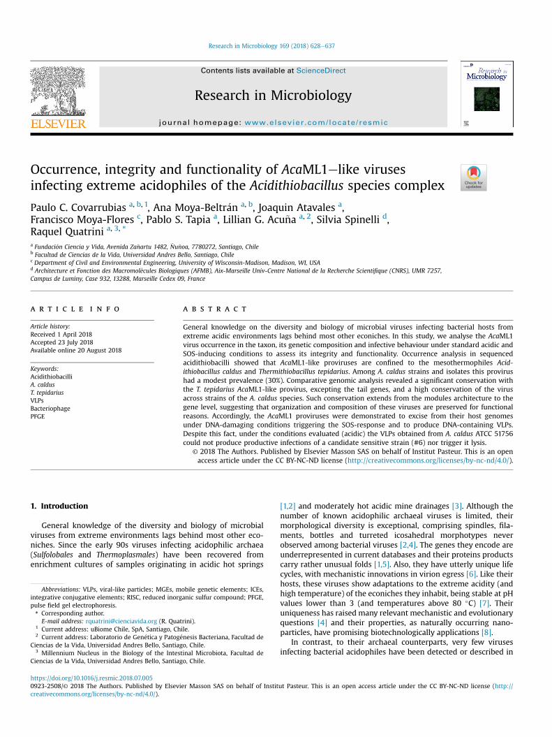
Fig. 1. Prevalence of AcaML1-like prophages in A. caldus strains. A. PFGE profiles of XbaI dmolecular weight standard used is the 5 kb ladder DNA size standard (BioRad). B. Southernthe viral capsid.
morphogenesis of the base plate and tail (Supplementary Table 1).According to available genomic evidence, AcaML1-like provirusesare confined to the mesothermophilic acidithiobacilli, T. tepidariusand A. caldus.
3.2. Prevalence of AcaML1-like proviruses in A. caldus strains andindustrial isolates
To investigate whether lysogenic proviruses like AcaML1 werepresent in other strains of A. caldus, occurrence of AcaML1 signa-tures was evaluated in a collection of seven A. caldus isolates(Table 2) using pulsed-field gel electrophoresis (PFGE) combinedwith Southern blot. Hybridization of a biotinilated probe, directedto the viral gene gp39 (encoding the viral capsid), to the PFGE fin-gerprints of all seven isolates (Fig. 1A) revealed the presence oflysogenic proviruses similar to AcaML1 in four of the strains tested,namely the type strain (ATCC 51756), strain F, BC-3 and CSH12(Fig. 1B). Under the conditions evaluated a single band was iden-tified in the four positive lanes, implying that only the integratedform of the virus could be detected in the type strain and the otherAcaML1 positive isolates. PFGE-based size estimations indicate thatall these proviruses are similar in size to AcaML1 (aprox. 60 kb).This result was confirmed by PCR amplification of selected genemarkers encompassing the different gene modules of the AcaML1provirus genome. All 19 gene markers tested could be amplifiedfrom total DNA preparations obtained from strains BC13, F and CSH-12 indicating, as well, global conservation of the gene content of theAcaML1-like proviruses present in A. caldus strains (Fig. 2).
3.3. Comparative genomics of AcaML1-like proviruses fromA. caldus strains
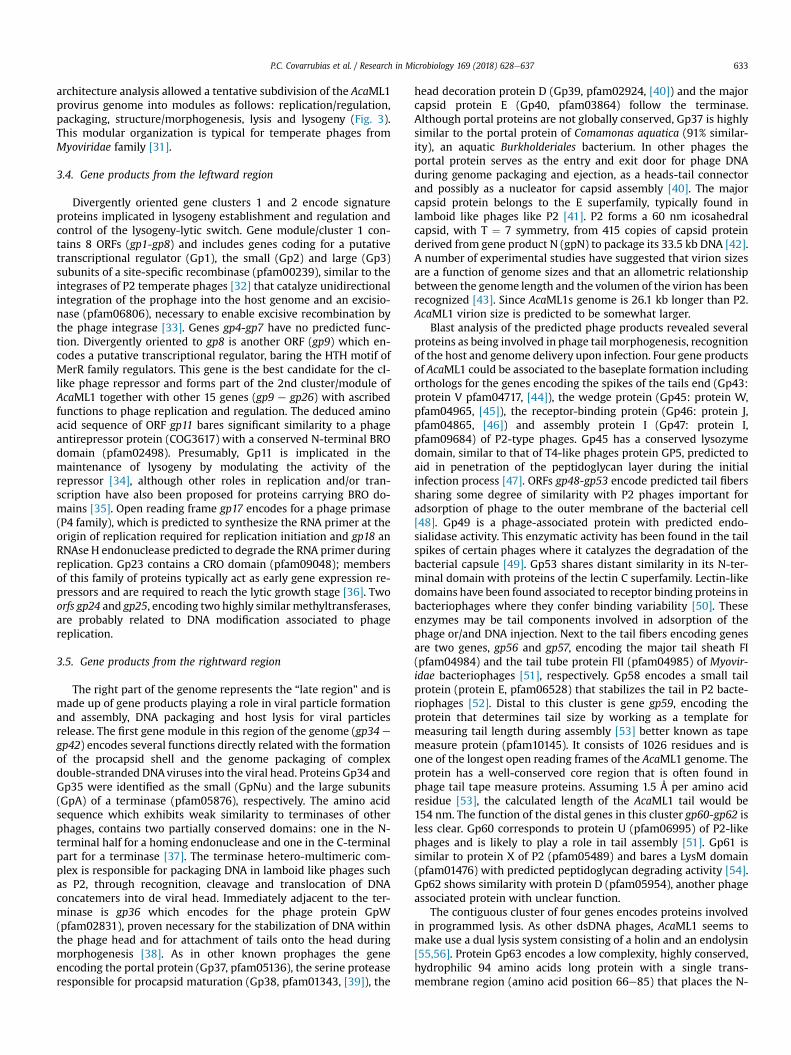
AcaML1 proviruses present in A. caldus strains F, BC-13 and CSH-12 were PCR amplified using high-fidelity and high-processivityHerculase II fusion DNA polymerase and sequenced at Macrogenon a module-to-module basis using primers indicated inSupplementary Table 2. Sequences obtained (MH142219,MH142220) were aligned against the AcaML1 genome (JX507079)and cross-compared to each other. The putative provirus identifiedin the whole genome sequence of the neutrophilic relative ofA. caldus, T. tepidarius, was included in this analysis for comparativereasons. Results of the whole genome multiple alignments areshown in Fig. 3.
igested genomic DNA of A. caldus strains. Staining was done with SYBR Green I. Theblot of the gel in panel A using a 300 bp long probe specific for the gene gp39 encoding
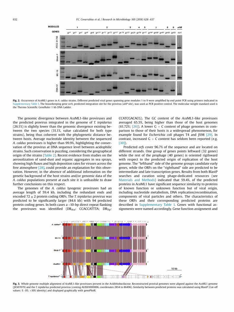
Fig. 2. Occurrence of AcaML1 genes in A. caldus strains. Different predicted viral genes spanning gene modules 1 to 9 were amplified by end point PCR using primers indicated inSupplementary Table 1. The housekeeping gene ssrA, predicted integration site for the provirus (attP site), was used as PCR positive control. The molecular weight standard used isthe Thermo Scientific GeneRuler 1 kb DNA Ladder.
P.C. Covarrubias et al. / Research in Microbiology 169 (2018) 628e637632
The genomic divergence between AcaML1-like proviruses andthe predicted provirus integrated in the genome of T. tepidarius(26.1%) is slightly lower than the genomic divergence existing be-tween the two species (31.1%, value calculated for both typestrains), being thus coherent with the phylogenetic distance be-tween hosts. Average nucleotide identity between the sequencedA. caldus proviruses is higher than 99.9%, highlighting the conser-vation of the provirus at DNA sequence level between acidophilicstrains. Such conservation is puzzling, considering the geographicalorigin of the strains (Table 2). Recent evidence from studies on theaerosolization of sand-dust and organic aggregates in sea sprays,showing high fluxes and high deposition rates for viruses across thefree atmosphere [28], could provide an explanation for this obser-vation. However, in the absence of additional information on thegenetic background of the host strains and/or genomic data of theA. caldus populations present at each site it is unfeasible to drawfurther conclusions on this respect.
The genomes of the A. caldus lysogenic proviruses had anaverage length of 59.4 kb, including the redundant ends andencoded 72 ± 2 protein coding ORFs. The T. tepidarius provirus waspredicted to be significantly larger (84.6 kb) with 94 predictedprotein coding genes. In both cases a ~10-bp direct repeat flankingthe proviruses was identified (DRAca: CCACCATTTA; DRTtp:
Fig. 3. Whole genome multiple alignment of AcaML1-like proviruses present in the Acidithi(JX507079) and the T. tepidarius predicted provirus (conting AUIS01000006, coordenates 285values: E�05; >30% identity) and displayed graphically with genoPlotR.
CCATCGACACG). The GC content of the AcaML1-like provirusesaveraged 65.5%, being higher than those of the host genomes(61.72%; [26]). A lower G þ C content of phage genomes in com-parison to those of their hosts is a widespread phenomenon, forexample found for Escherichia coli phages T4 and JS98 [29]. Incontrast, increased G þ C content has seldom been reported (e.g.[30]).
Predicted orfs cover 96.7% of the sequence and are located ondifferent strands. One group of genes points leftward (32 genes)while the rest of the prophage (40 genes) is oriented rigthwardwith respect to the predicted origin of replication of the hostgenome. The “lefthand” side of the genome groups candidate earlygenes, while the ORFs on the “righthand” side are predicted to beintermediate and late transcription genes. Results from both BlastPsearches and curation using phage-dedicated resources (seeMaterials and Methods) indicated that 59.4%, of the predictedproteins in AcaML1 have significant sequence similarity to proteinsof known function or unknown function but of viral origin,including nucleotide metabolism, DNA replication/recombination,components of viral particles and others. The characteristics ofthese ORFs and their corresponding predicted proteins aredescribed in Supplementary Table 1. Genes with functional as-signments were named accordingly. Gene function assignment and
obacilaceae. Reconstructed proviral genomes were aligned against the AcaML1 genome4 to 86496). Similarity between predicted proteins was calculated using BlastP (Cut-off
P.C. Covarrubias et al. / Research in Microbiology 169 (2018) 628e637 633
architecture analysis allowed a tentative subdivision of the AcaML1provirus genome into modules as follows: replication/regulation,packaging, structure/morphogenesis, lysis and lysogeny (Fig. 3).This modular organization is typical for temperate phages fromMyoviridae family [31].
3.4. Gene products from the leftward region
Divergently oriented gene clusters 1 and 2 encode signatureproteins implicated in lysogeny establishment and regulation andcontrol of the lysogeny-lytic switch. Gene module/cluster 1 con-tains 8 ORFs (gp1-gp8) and includes genes coding for a putativetranscriptional regulator (Gp1), the small (Gp2) and large (Gp3)subunits of a site-specific recombinase (pfam00239), similar to theintegrases of P2 temperate phages [32] that catalyze unidirectionalintegration of the prophage into the host genome and an excisio-nase (pfam06806), necessary to enable excisive recombination bythe phage integrase [33]. Genes gp4-gp7 have no predicted func-tion. Divergently oriented to gp8 is another ORF (gp9) which en-codes a putative transcriptional regulator, baring the HTH motif ofMerR family regulators. This gene is the best candidate for the cI-like phage repressor and forms part of the 2nd cluster/module ofAcaML1 together with other 15 genes (gp9 e gp26) with ascribedfunctions to phage replication and regulation. The deduced aminoacid sequence of ORF gp11 bares significant similarity to a phageantirepressor protein (COG3617) with a conserved N-terminal BROdomain (pfam02498). Presumably, Gp11 is implicated in themaintenance of lysogeny by modulating the activity of therepressor [34], although other roles in replication and/or tran-scription have also been proposed for proteins carrying BRO do-mains [35]. Open reading frame gp17 encodes for a phage primase(P4 family), which is predicted to synthesize the RNA primer at theorigin of replication required for replication initiation and gp18 anRNAse H endonuclease predicted to degrade the RNA primer duringreplication. Gp23 contains a CRO domain (pfam09048); membersof this family of proteins typically act as early gene expression re-pressors and are required to reach the lytic growth stage [36]. Twoorfs gp24 and gp25, encoding two highly similar methyltransferases,are probably related to DNA modification associated to phagereplication.
3.5. Gene products from the rightward region
The right part of the genome represents the “late region” and ismade up of gene products playing a role in viral particle formationand assembly, DNA packaging and host lysis for viral particlesrelease. The first gene module in this region of the genome (gp34 e
gp42) encodes several functions directly related with the formationof the procapsid shell and the genome packaging of complexdouble-stranded DNAviruses into the viral head. Proteins Gp34 andGp35 were identified as the small (GpNu) and the large subunits(GpA) of a terminase (pfam05876), respectively. The amino acidsequence which exhibits weak similarity to terminases of otherphages, contains two partially conserved domains: one in the N-terminal half for a homing endonuclease and one in the C-terminalpart for a terminase [37]. The terminase hetero-multimeric com-plex is responsible for packaging DNA in lamboid like phages suchas P2, through recognition, cleavage and translocation of DNAconcatemers into de viral head. Immediately adjacent to the ter-minase is gp36 which encodes for the phage protein GpW(pfam02831), proven necessary for the stabilization of DNA withinthe phage head and for attachment of tails onto the head duringmorphogenesis [38]. As in other known prophages the geneencoding the portal protein (Gp37, pfam05136), the serine proteaseresponsible for procapsid maturation (Gp38, pfam01343, [39]), the
head decoration protein D (Gp39, pfam02924, [40]) and the majorcapsid protein E (Gp40, pfam03864) follow the terminase.Although portal proteins are not globally conserved, Gp37 is highlysimilar to the portal protein of Comamonas aquatica (91% similar-ity), an aquatic Burkholderiales bacterium. In other phages theportal protein serves as the entry and exit door for phage DNAduring genome packaging and ejection, as a heads-tail connectorand possibly as a nucleator for capsid assembly [40]. The majorcapsid protein belongs to the E superfamily, typically found inlamboid like phages like P2 [41]. P2 forms a 60 nm icosahedralcapsid, with T ¼ 7 symmetry, from 415 copies of capsid proteinderived from gene product N (gpN) to package its 33.5 kb DNA [42].A number of experimental studies have suggested that virion sizesare a function of genome sizes and that an allometric relationshipbetween the genome length and the volumen of the virion has beenrecognized [43]. Since AcaML1s genome is 26.1 kb longer than P2.AcaML1 virion size is predicted to be somewhat larger.
Blast analysis of the predicted phage products revealed severalproteins as being involved in phage tail morphogenesis, recognitionof the host and genome delivery upon infection. Four gene productsof AcaML1 could be associated to the baseplate formation includingorthologs for the genes encoding the spikes of the tails end (Gp43:protein V pfam04717, [44]), the wedge protein (Gp45: protein W,pfam04965, [45]), the receptor-binding protein (Gp46: protein J,pfam04865, [46]) and assembly protein I (Gp47: protein I,pfam09684) of P2-type phages. Gp45 has a conserved lysozymedomain, similar to that of T4-like phages protein GP5, predicted toaid in penetration of the peptidoglycan layer during the initialinfection process [47]. ORFs gp48-gp53 encode predicted tail fiberssharing some degree of similarity with P2 phages important foradsorption of phage to the outer membrane of the bacterial cell[48]. Gp49 is a phage-associated protein with predicted endo-sialidase activity. This enzymatic activity has been found in the tailspikes of certain phages where it catalyzes the degradation of thebacterial capsule [49]. Gp53 shares distant similarity in its N-ter-minal domain with proteins of the lectin C superfamily. Lectin-likedomains have been found associated to receptor binding proteins inbacteriophages where they confer binding variability [50]. Theseenzymes may be tail components involved in adsorption of thephage or/and DNA injection. Next to the tail fibers encoding genesare two genes, gp56 and gp57, encoding the major tail sheath FI(pfam04984) and the tail tube protein FII (pfam04985) of Myovir-idae bacteriophages [51], respectively. Gp58 encodes a small tailprotein (protein E, pfam06528) that stabilizes the tail in P2 bacte-riophages [52]. Distal to this cluster is gene gp59, encoding theprotein that determines tail size by working as a template formeasuring tail length during assembly [53] better known as tapemeasure protein (pfam10145). It consists of 1026 residues and isone of the longest open reading frames of the AcaML1 genome. Theprotein has a well-conserved core region that is often found inphage tail tape measure proteins. Assuming 1.5 Å per amino acidresidue [53], the calculated length of the AcaML1 tail would be154 nm. The function of the distal genes in this cluster gp60-gp62 isless clear. Gp60 corresponds to protein U (pfam06995) of P2-likephages and is likely to play a role in tail assembly [51]. Gp61 issimilar to protein X of P2 (pfam05489) and bares a LysM domain(pfam01476) with predicted peptidoglycan degrading activity [54].Gp62 shows similarity with protein D (pfam05954), another phageassociated protein with unclear function.
The contiguous cluster of four genes encodes proteins involvedin programmed lysis. As other dsDNA phages, AcaML1 seems tomake use a dual lysis system consisting of a holin and an endolysin[55,56]. Protein Gp63 encodes a low complexity, highly conserved,hydrophilic 94 amino acids long protein with a single trans-membrane region (amino acid position 66e85) that places the N-
P.C. Covarrubias et al. / Research in Microbiology 169 (2018) 628e637634
teminus in the cytoplasm and the C-terminal end of the protein inthe periplasm, resembling other known class III holins [57]. Holinsfacilitate the release of phage particles from infected bacteria dur-ing viral-induced cell lysis [58]. A second small ORF (gp64),downstream of the putative holin gene, encodes a 158 aa longprotein of unknown function. Orthologs of this gene frequentlyconcur with gp63 orthologs and the predicted protein products areextremely well conserved (e.g. in T. tepidarius). Predicted proteinGp65 is 160 amino acids long and contains the signature of knownphage endolysins of the muramidase type (pfam00959) and lackpredicted secretory signals. Muramidases cleave the glycosidic beta1,4-bonds between the N-acetylmuramic acid and the N-acetyl-glucosamine of the peptidoglycan from outside the cytoplasm,thereby enabling progeny virions to be liberated [59]. Because oftheir unique ability to cleave peptidoglycan in a generally species-specific manner (shared homology is not found within the catalyticdomain), endolysins provide a means of selective and rapid killingof bacteria with no effect on the rest of the native microflora. Accessto peptidoglycan depends on the small hydrophobic holins, whichenable endolysin molecules to cross the inner membrane. Theseproperties confer a biotechnological potential to both types ofproteins.
The distal gene module in AcaML1 contains three genes (gp66 e
gp69) encoding a DNA restriction-modification system and twoinsertion sequences. Gp66 (pfam00145) is a predicted site-specific
Fig. 4. Excision of AcaML1-like proviruses from the chromosome of A. caldus strains upongenomic DNA of A. caldus strains. Staining was done with SYBR Green I. Molecular weight stathe left panel using a 300 bp long probe specific for the gene gp39, encoding the viral cap
DNAmethylase that modifies cytosine to 5-methylcytosine. In turn,Gp67 and Gp68 encode the two subunits of a predicted McrBC(pfam10117) restriction enzyme cutting DNA in multiple positionswithin two distant locations (between 40 pb and 3 Kb apart) baringthe 5-methylcytosine modification preceded by a purine (RmC[N]RmC) [60]. Presence of RM-systems in phages and other mobilegenetic elements is frequent, playing a relevant role in the stabili-zation of the mobile element within the genome [61] or beingtarget of frequent horizontal transfer between bacteria [62]. Similarto other cases appointed in the literature (e.g. [63]), the RM-systemof AcaML1 is flanked by transposases of the IS5 and IS21 families(gp70-74) (pfam05598 and pfam13751). These genes could facili-tate transfer of the RM gene cassettes between the host chromo-some and horizontal transfer vector such as phages [62].
3.6. Induction of replication intermediaries of AcaML1-likeproviruses by DNA damage
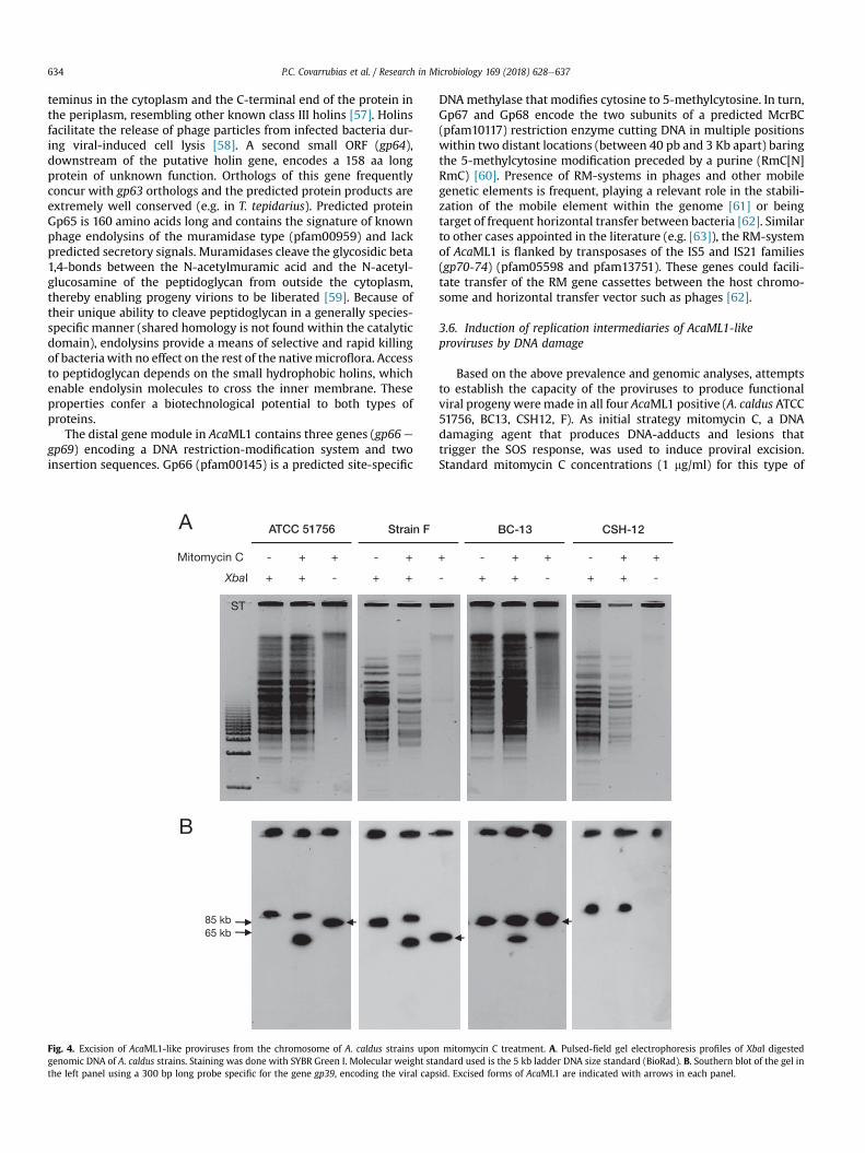
Based on the above prevalence and genomic analyses, attemptsto establish the capacity of the proviruses to produce functionalviral progeny weremade in all four AcaML1 positive (A. caldus ATCC51756, BC13, CSH12, F). As initial strategy mitomycin C, a DNAdamaging agent that produces DNA-adducts and lesions thattrigger the SOS response, was used to induce proviral excision.Standard mitomycin C concentrations (1 mg/ml) for this type of
mitomycin C treatment. A. Pulsed-field gel electrophoresis profiles of XbaI digestedndard used is the 5 kb ladder DNA size standard (BioRad). B. Southern blot of the gel insid. Excised forms of AcaML1 are indicated with arrows in each panel.
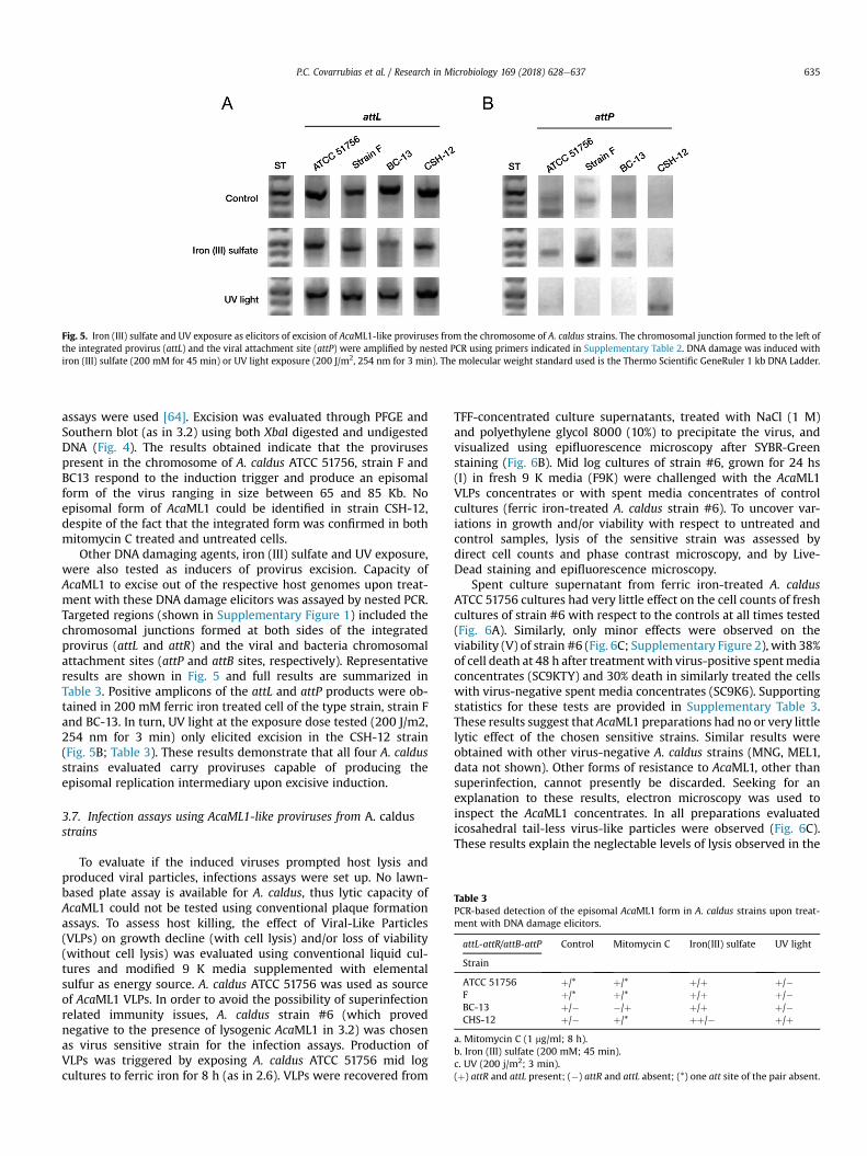
Fig. 5. Iron (III) sulfate and UV exposure as elicitors of excision of AcaML1-like proviruses from the chromosome of A. caldus strains. The chromosomal junction formed to the left ofthe integrated provirus (attL) and the viral attachment site (attP) were amplified by nested PCR using primers indicated in Supplementary Table 2. DNA damage was induced withiron (III) sulfate (200 mM for 45 min) or UV light exposure (200 J/m2, 254 nm for 3 min). The molecular weight standard used is the Thermo Scientific GeneRuler 1 kb DNA Ladder.
Table 3PCR-based detection of the episomal AcaML1 form in A. caldus strains upon treat-ment with DNA damage elicitors.
attL-attR/attB-attP Control Mitomycin C Iron(III) sulfate UV light
Strain
ATCC 51756 þ/* þ/* þ/þ þ/�F þ/* þ/* þ/þ þ/�BC-13 þ/� �/þ þ/þ þ/�CHS-12 þ/� þ/* þþ/� þ/þ
a. Mitomycin C (1 mg/ml; 8 h).b. Iron (III) sulfate (200 mM; 45 min).c. UV (200 j/m2; 3 min).(þ) attR and attL present; (�) attR and attL absent; (*) one att site of the pair absent.
P.C. Covarrubias et al. / Research in Microbiology 169 (2018) 628e637 635
assays were used [64]. Excision was evaluated through PFGE andSouthern blot (as in 3.2) using both XbaI digested and undigestedDNA (Fig. 4). The results obtained indicate that the provirusespresent in the chromosome of A. caldus ATCC 51756, strain F andBC13 respond to the induction trigger and produce an episomalform of the virus ranging in size between 65 and 85 Kb. Noepisomal form of AcaML1 could be identified in strain CSH-12,despite of the fact that the integrated form was confirmed in bothmitomycin C treated and untreated cells.
Other DNA damaging agents, iron (III) sulfate and UV exposure,were also tested as inducers of provirus excision. Capacity ofAcaML1 to excise out of the respective host genomes upon treat-ment with these DNA damage elicitors was assayed by nested PCR.Targeted regions (shown in Supplementary Figure 1) included thechromosomal junctions formed at both sides of the integratedprovirus (attL and attR) and the viral and bacteria chromosomalattachment sites (attP and attB sites, respectively). Representativeresults are shown in Fig. 5 and full results are summarized inTable 3. Positive amplicons of the attL and attP products were ob-tained in 200 mM ferric iron treated cell of the type strain, strain Fand BC-13. In turn, UV light at the exposure dose tested (200 J/m2,254 nm for 3 min) only elicited excision in the CSH-12 strain(Fig. 5B; Table 3). These results demonstrate that all four A. caldusstrains evaluated carry proviruses capable of producing theepisomal replication intermediary upon excisive induction.
3.7. Infection assays using AcaML1-like proviruses from A. caldusstrains
To evaluate if the induced viruses prompted host lysis andproduced viral particles, infections assays were set up. No lawn-based plate assay is available for A. caldus, thus lytic capacity ofAcaML1 could not be tested using conventional plaque formationassays. To assess host killing, the effect of Viral-Like Particles(VLPs) on growth decline (with cell lysis) and/or loss of viability(without cell lysis) was evaluated using conventional liquid cul-tures and modified 9 K media supplemented with elementalsulfur as energy source. A. caldus ATCC 51756 was used as sourceof AcaML1 VLPs. In order to avoid the possibility of superinfectionrelated immunity issues, A. caldus strain #6 (which provednegative to the presence of lysogenic AcaML1 in 3.2) was chosenas virus sensitive strain for the infection assays. Production ofVLPs was triggered by exposing A. caldus ATCC 51756 mid logcultures to ferric iron for 8 h (as in 2.6). VLPs were recovered from
TFF-concentrated culture supernatants, treated with NaCl (1 M)and polyethylene glycol 8000 (10%) to precipitate the virus, andvisualized using epifluorescence microscopy after SYBR-Greenstaining (Fig. 6B). Mid log cultures of strain #6, grown for 24 hs(I) in fresh 9 K media (F9K) were challenged with the AcaML1VLPs concentrates or with spent media concentrates of controlcultures (ferric iron-treated A. caldus strain #6). To uncover var-iations in growth and/or viability with respect to untreated andcontrol samples, lysis of the sensitive strain was assessed bydirect cell counts and phase contrast microscopy, and by Live-Dead staining and epifluorescence microscopy.
Spent culture supernatant from ferric iron-treated A. caldusATCC 51756 cultures had very little effect on the cell counts of freshcultures of strain #6 with respect to the controls at all times tested(Fig. 6A). Similarly, only minor effects were observed on theviability (V) of strain #6 (Fig. 6C; Supplementary Figure 2), with 38%of cell death at 48 h after treatment with virus-positive spentmediaconcentrates (SC9KTY) and 30% death in similarly treated the cellswith virus-negative spent media concentrates (SC9K6). Supportingstatistics for these tests are provided in Supplementary Table 3.These results suggest that AcaML1 preparations had no or very littlelytic effect of the chosen sensitive strains. Similar results wereobtained with other virus-negative A. caldus strains (MNG, MEL1,data not shown). Other forms of resistance to AcaML1, other thansuperinfection, cannot presently be discarded. Seeking for anexplanation to these results, electron microscopy was used toinspect the AcaML1 concentrates. In all preparations evaluatedicosahedral tail-less virus-like particles were observed (Fig. 6C).These results explain the neglectable levels of lysis observed in the
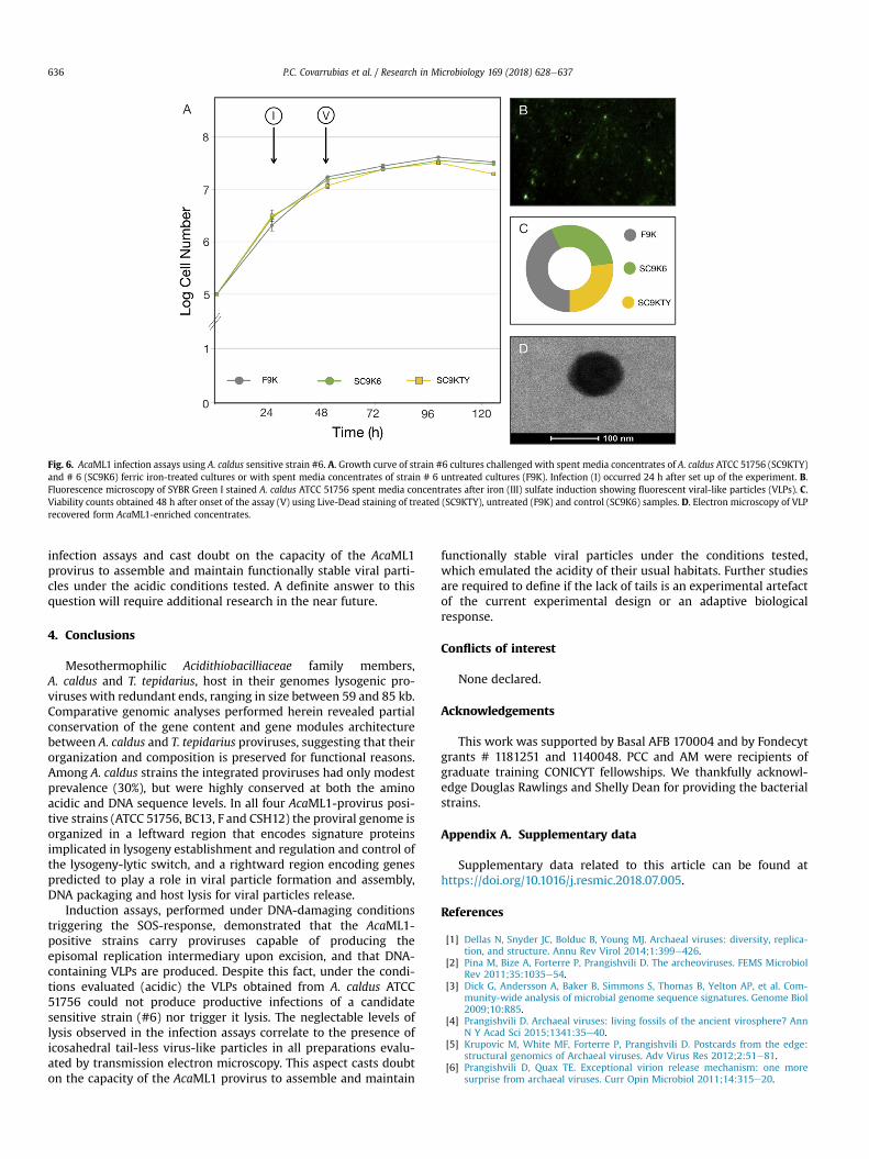
Fig. 6. AcaML1 infection assays using A. caldus sensitive strain #6. A. Growth curve of strain #6 cultures challenged with spent media concentrates of A. caldus ATCC 51756 (SC9KTY)and # 6 (SC9K6) ferric iron-treated cultures or with spent media concentrates of strain # 6 untreated cultures (F9K). Infection (I) occurred 24 h after set up of the experiment. B.Fluorescence microscopy of SYBR Green I stained A. caldus ATCC 51756 spent media concentrates after iron (III) sulfate induction showing fluorescent viral-like particles (VLPs). C.Viability counts obtained 48 h after onset of the assay (V) using Live-Dead staining of treated (SC9KTY), untreated (F9K) and control (SC9K6) samples. D. Electron microscopy of VLPrecovered form AcaML1-enriched concentrates.
P.C. Covarrubias et al. / Research in Microbiology 169 (2018) 628e637636
infection assays and cast doubt on the capacity of the AcaML1provirus to assemble and maintain functionally stable viral parti-cles under the acidic conditions tested. A definite answer to thisquestion will require additional research in the near future.
4. Conclusions
Mesothermophilic Acidithiobacilliaceae family members,A. caldus and T. tepidarius, host in their genomes lysogenic pro-viruses with redundant ends, ranging in size between 59 and 85 kb.Comparative genomic analyses performed herein revealed partialconservation of the gene content and gene modules architecturebetween A. caldus and T. tepidarius proviruses, suggesting that theirorganization and composition is preserved for functional reasons.Among A. caldus strains the integrated proviruses had only modestprevalence (30%), but were highly conserved at both the aminoacidic and DNA sequence levels. In all four AcaML1-provirus posi-tive strains (ATCC 51756, BC13, F and CSH12) the proviral genome isorganized in a leftward region that encodes signature proteinsimplicated in lysogeny establishment and regulation and control ofthe lysogeny-lytic switch, and a rightward region encoding genespredicted to play a role in viral particle formation and assembly,DNA packaging and host lysis for viral particles release.
Induction assays, performed under DNA-damaging conditionstriggering the SOS-response, demonstrated that the AcaML1-positive strains carry proviruses capable of producing theepisomal replication intermediary upon excision, and that DNA-containing VLPs are produced. Despite this fact, under the condi-tions evaluated (acidic) the VLPs obtained from A. caldus ATCC51756 could not produce productive infections of a candidatesensitive strain (#6) nor trigger it lysis. The neglectable levels oflysis observed in the infection assays correlate to the presence oficosahedral tail-less virus-like particles in all preparations evalu-ated by transmission electron microscopy. This aspect casts doubton the capacity of the AcaML1 provirus to assemble and maintain
functionally stable viral particles under the conditions tested,which emulated the acidity of their usual habitats. Further studiesare required to define if the lack of tails is an experimental artefactof the current experimental design or an adaptive biologicalresponse.
Conflicts of interest
None declared.
Acknowledgements
This work was supported by Basal AFB 170004 and by Fondecytgrants # 1181251 and 1140048. PCC and AM were recipients ofgraduate training CONICYT fellowships. We thankfully acknowl-edge Douglas Rawlings and Shelly Dean for providing the bacterialstrains.
Appendix A. Supplementary data
Supplementary data related to this article can be found athttps://doi.org/10.1016/j.resmic.2018.07.005.
References
[1] Dellas N, Snyder JC, Bolduc B, Young MJ. Archaeal viruses: diversity, replica-tion, and structure. Annu Rev Virol 2014;1:399e426.
[2] Pina M, Bize A, Forterre P, Prangishvili D. The archeoviruses. FEMS MicrobiolRev 2011;35:1035e54.
[3] Dick G, Andersson A, Baker B, Simmons S, Thomas B, Yelton AP, et al. Com-munity-wide analysis of microbial genome sequence signatures. Genome Biol2009;10:R85.
[4] Prangishvili D. Archaeal viruses: living fossils of the ancient virosphere? AnnN Y Acad Sci 2015;1341:35e40.
[5] Krupovic M, White MF, Forterre P, Prangishvili D. Postcards from the edge:structural genomics of Archaeal viruses. Adv Virus Res 2012;2:51e81.
[6] Prangishvili D, Quax TE. Exceptional virion release mechanism: one moresurprise from archaeal viruses. Curr Opin Microbiol 2011;14:315e20.
P.C. Covarrubias et al. / Research in Microbiology 169 (2018) 628e637 637
[7] Prangishvili D, Garrett RA, Koonin EV. Evolutionary genomics of archaeal vi-ruses: unique viral genomes in the third domain of life. Virus Res 2006;117:52e67.
[8] Evans DJ. Exploitation of plant and archaeal viruses in bionanotechnology.Biochem Soc Trans 2009;37:665e70.
[9] Ward TE, Bruhn DF, Shean ML, Watkins CS, Bulmer D, Winston V. Charac-terization of a new bacteriophage which infects bacteria of the genus Acid-iphilium. J Gen Virol 1993;74:2419e25.
[10] Ars�ene-Ploetze F, Koechler S, Marchal M, Copp�ee JY, Chandler M, Bonnefoy V,et al. Structure, function, and evolution of the Thiomonas spp. genome. PLoSGenet 2010;6:e1000859.
[11] Tapia P, Flores FM, Covarrubias PC, Acu~na LG, Holmes DS, Quatrini R. Completegenome sequence of temperate bacteriophage AcaML1 from the extremeacidophile Acidithiobacillus caldus ATCC 51756. J Virol 2012;86:12452e3.
[12] Andersson AF, Banfield JF. Virus population dynamics and acquired virusresistance in natural microbial communities. Science 2008;320:1047e50.
[13] Covarrubias P, Mu~noz R, Bobadilla-Fazzini R, Martinez P, Quatrini R. Are thereviruses in industrial bioleaching econiches? Solid State Phenom 2017;262:521e5.
[14] Johnson DB. Biomining-biotechnologies for extracting and recovering metalsfrom ores and waste materials. Curr Opin Biotechnol 2014;30:24e31.
[15] C�ardenas JP, Quatrini R, Holmes DS. Progress in acidophile genomics. In:Quatrini R, Johnson DB, editors. Acidophiles: life in extremely acidic envi-ronments. UK: Caister Academic Press; 2016. p. 179e97.
[16] Quatrini R, Ossandon FJ, Rawlings DE. The Flexible Genome of AcidophilicProkaryotes. In: Quatriniand R, Johnson DB, editors. Acidophiles: life inextremely acidic environments. UK: Caister Academic Press; 2016. p. 199e220.
[17] Nu~nez H, Moya-Beltr�an A, Covarrubias PC, Issotta F, C�ardenas JP, Gonz�alez M,et al. Molecular systematics of the genus Acidithiobacillus: insights into thephylogenetic structure and diversification of the taxon. Front Microbiol2017;8:30.
[18] Besemer J, Lomsadze A, Borodovsky M. GeneMark: a self-training method forprediction of gene starts in microbial genomes. Implications for findingsequence motifs in regulatory regions. Nucleic Acids Res 2001;29:2607e18.
[19] Altschul SF, Gish W, Miller W, Myers EW, Lipman DJ. Basic local alignmentsearch tool. J Mol Biol 1990;215:403e10.
[20] Pearson WR. Searching protein sequence libraries: comparison of the sensi-tivity and selectivity of the SmitheWaterman and FASTA algorithms. Geno-mics 1991;11:635e50.
[21] Guy L, Kultima JR, Andersson SG. genoPlotR: comparative gene and genomevisualization in R. Bioinformatics 2010;26:2334e5.
[22] Silverman MP, Lungren DG. Studies on the chemoautotrophic iron bacteriumFerrobacillus ferrooxidans. I. An improved medium and a harvesting procedurefor securing high cell yields. J Bacteriol 1959;77:642e7.
[23] Dopson M, Lindstr€om EB. Potential role of Thiobacillus caldus in arsenopyritebioleaching. Appl Environ Microbiol 1999;65:36e40.
[24] Nieto PA, Covarrubias PC, Jedlicki E, Holmes DS, Quatrini R. Selection andevaluation of reference genes for improved interrogation of microbial tran-scriptomes: case study with the extremophile Acidithiobacillus ferrooxidans.BMC Mol Biol 2009;10:63.
[25] Noble RT, Fuhrman JA. Use of SYBR Green I for rapid epifluorescence counts ofmarine viruses and bacteria. Aquat Microb Ecol 1998;14:113e8.
[26] Acu~na LG, C�ardenas JP, Covarrubias PC, Haristoy JJ, Flores R, Nu~nez H, et al.Architecture and gene repertoire of the flexible genome of the extremeacidophile Acidithiobacillus caldus. PLoS One 2013;8:e78237.
[27] Bustamante P, Covarrubias P, Levic�an G, Katz A, Tapia P, Bonnefoy V, et al.ICEAfe1, an actively excising integrative-conjugative element from the bio-mining bacterium Acidithiobacillus ferrooxidans. J Mol Microbiol Biotech2012;22:399e407.
[28] Reche I, D'Orta G, Mladenov N, Winget DM, Suttle CA. Deposition rates ofviruses and bacteria above the atmospheric boundary layer. ISME J 2018;12:1154e62.
[29] Zuber S, Ngom-Bru C, Barretto C, Bruttin A, Brüssow H, Denou E. Genomeanalysis of phage JS98 defines a fourth major subgroup of T4-like phages inEscherichia coli. J Bacteriol 2007;189:8206e14.
[30] Zheng Q, Zhang R, Xu Y, White III RA, Wang Y, Luo T, et al. A marine inducibleprophage vB_CibM-P1 isolated from the aerobic anoxygenic phototrophicbacterium Citromicrobium bathyomarinum JL354. Sci Rep 2014;4:7118.
[31] Botstein D. A theory of modular evolution for bacteriophages. Ann N Y AcadSci 1980;354:484e90.
[32] Thorpe HM, Smith MC. In vitro site-specific integration of bacteriophage DNAcatalyzed by a recombinase of the resolvase/invertase family. Proc Natl AcadSci USA 1998;95:5505e10.
[33] Cho EH, Gumport RI, Gardner JF. Interactions between integrase and exci-sionase in the phage lambda excisive nucleoprotein complex. J Bacteriol2002;184:5200e3.
[34] Summer EJ, Enderle CJ, Ahern SJ, Gill JJ, Torres CP, Appel DN, et al. Genomicand biological analysis of phage Xfas53 and related prophages of Xylella fas-tidiosa. J Bacteriol 2010;192:179e90.
[35] Bideshi DK, Renault S, Stasiak K, Federici BA, Bigot Y. Phylogenetic analysisand possible function of bro-like genes, a multigene family widespread amonglarge double-stranded DNA viruses of invertebrates and bacteria. J Gen Virol2003;84:2531e44.
[36] Atsumi S, Little JW. Role of the lytic repressor in prophage induction of phagel as analyzed by a module-replacement approach. Proc Natl Acad Sci USA2006;103:4558e63.
[37] Rao VB, Feiss M. The bacteriophage DNA packaging motor. Annu Rev Genet2008;42:647e81.
[38] Maxwell KL, Yee AA, Booth V, Arrowsmith CH, Gold M, Davidson AR. Thesolution structure of bacteriophage lambda protein W, a small morphogeneticprotein possessing a novel fold. J Mol Biol 2001;308:9e14.
[39] Medina E, Wieczorek D, Medina EM, Yang Q, Feiss M, Catalano CE. Assemblyand maturation of the bacteriophage lambda procapsid: gpC is the viralprotease. J Mol Biol 2010;401:813e30.
[40] Doan DN, Dokland T. The gpQ portal protein of bacteriophage P2 formsdodecameric connectors in crystals. J Struct Biol 2007;157:432e6.
[41] Rajagopala SV, Casjens S, Uetz P. The protein interaction map of bacteriophagelambda. BMC Microbiol 2011;11:213.
[42] Dokland T, Lindqvist BH, Fuller SD. Image reconstruction from cryo-electronmicrographs reveals the morphopoietic mechanism in the P2-P4 bacterio-phage system. EMBO J 1992;11:839e46.
[43] Cui J, Schlub TE, Holmes CE. An allometric relationship between the genomelength and virion volume of viruses. J Virol 2014;88:6403e10.
[44] Haggard-Ljungquist E, Jacobsen E, Rishovd S, Six EW, Nilssen O, Sunshine MG,et al. Bacteriophage P2: genes involved in baseplate assembly. Virology1995;213:109e21.
[45] Szewczyk B, Bienkowska-Szewczyk K, Kozloff LM. Identification of T4 gene 25product, a component of the tail baseplate, as a 15K lysozyme. Mol Gen Genet1986;202:363e7.
[46] Werts C, Michel V, Hofnung M, Charbit A. Adsorption of bacteriophage lambdaon the LamB protein of E. coli K12: point mutations in gene J of lambdaresponsible for extended host range. J Bacteriol 1994;176:941e7.
[47] Ye N, Nemoto N. Processing of the tail lysozyme (gp5) of bacteriophage T4.J Bacteriol 2004;186:6335e9.
[48] Leiman PG, Arisaka F, van Raaij MJ, Kostyuchenko VA, Aksyuk AA, Kanamaru S,et al. Morphogenesis of the T4 tail and tail fibers. Virol J 2010;7:355.
[49] Stummeyer K, Dickmanns A, Mühlenhoff M, Gerardy-Schahn R, Ficner R.Crystal structure of the polysialic acid-degrading endosialidase of bacterio-phage K1F. Nat Struct Mol Biol 2005;12:90e6.
[50] McMahon SA, Miller JL, Lawton JA, Kerkow DE, Hodes A, Marti-Renom MA,et al. The C-type lectin fold as an evolutionary solution for massive sequencevariation. Nat Struct Mol Biol 2005;12:886e92.
[51] Haggard-Ljungquist E, Halling C, Calendar R. DNA sequences of the tail fibergenes of bacteriophage P2: evidence for horizontal transfer of tail fiber genesamong unrelated bacteriophages. J Bacteriol 1992;174:1462e77.
[52] Christie GE, Temple LM, Bartlett BA, Goodwin TS. Programmed translationalframeshift in the bacteriophage P2 FETUD tail gene operon. J Bacteriol2002;184:6522e31.
[53] Katsura I, Hendrix RW. Length determination in bacteriophage lambda tails.Cell 1984;39:691e8.
[54] Bateman A, Bycroft M. The structure of a LysM domain from E. colimembrane-bound lytic murein transglycosylase D (MltD). J Mol Biol 2000;299:1113e9.
[55] Young R. Bacteriophage holins: deadly diversity. J Mol Microbiol Biotechnol2002;4:21e36.
[56] Ziedaite G, Daugelavicius R, Bamford JK, Bamford DH. The holin protein ofbacteriophage PRD1 forms a pore for small-molecule and endolysin trans-location. J Bacteriol 2005;187:5397e405.
[57] Wang IN, Deaton J, Young R. Sizing the holin lesion with an endolysin-beta-galactosidase fusion. J Bacteriol 2003;185:779e87.
[58] Young R. Phage lysis: three steps, three choices, one outcome. J Microbiol2014;52:243e58.
[59] Miller ES, Kutter E, Mosig G, Arisaka F, Kunisawa T, Ruger W. Bacteriophage T4genome. Microbiol Mol Biol Rev 2003;67:86e156.
[60] Sutherland E, Coe L, Raleigh EA. McrBC: a multisubunit GTP-dependent re-striction endonuclease. J Mol Biol 1992;225:327e48.
[61] Kobayashi I. Behavior of restriction-modification systems as selfish mobileelements and their impact on genome evolution. Nucleic Acids Res 2001;29:3742e56.
[62] Kita K, Kawakami H, Tanaka H. Evidence for horizontal transfer of the EcoT38Irestriction-modification gene to chromosomal DNA by the P2 phage and di-versity of defective P2 prophages in Escherichia coli TH38 strains. J Bacteriol2003;185:2296e305.
[63] Brassard S, Paquet H, Roy PH. A transposon-like sequence adjacent to the AccIrestriction-modification operon. Gene 1995;157:69e72.
[64] Bellanger X, Morel C, Decaris B, Gu�edon G. Derepression of excision of inte-grative and potentially conjugative elements from Streptococcus thermophilusby DNA damage response: implication of a cI-related repressor. J Bacteriol2007;189:1478e81.
Related Documents

![[RevistaEnFrancés] Ciencia&Vida - Marzo2013](https://static.cupdf.com/doc/110x72/577cdc0c1a28ab9e78a9ba9d/revistaenfrances-cienciavida-marzo2013.jpg)