ANP 308 METABOLISM OF CARBOHYDRATES, LIPIDS, PROTEINS AND NUCLEIC ACIDS Course Team Prof. Anthony, I. O. Ologhobo(Course Writer)-UI Prof. Jokthan,G.E. (Programme Leader)-NOUN Dr. Salisu, B. Abdu (Course Editor)-ABU, Zaria Dr. Ahmed A. Njidda (Course Coordinator)-NOUN NATIONAL OPEN UNIVERSITY OF NIGERIA COURSE GUIDE

Welcome message from author
This document is posted to help you gain knowledge. Please leave a comment to let me know what you think about it! Share it to your friends and learn new things together.
Transcript

ANP 308METABOLISM OF CARBOHYDRATES, LIPIDS,PROTEINS AND NUCLEIC ACIDS
Course Team Prof. Anthony, I. O. Ologhobo(Course Writer)-UIProf. Jokthan,G.E. (Programme Leader)-NOUNDr. Salisu, B. Abdu (Course Editor)-ABU, ZariaDr. Ahmed A. Njidda (Course Coordinator)-NOUN
NATIONAL OPEN UNIVERSITY OF NIGERIA
COURSEGUIDE

© 2020 by NOUN PressNational Open University of NigeriaHeadquartersUniversity VillagePlot 91, Cadastral ZoneNnamdiAzikiwe ExpresswayJabi, Abuja
Lagos Office14/16 Ahmadu Bello WayVictoria Island, Lagos
e-mail: [email protected]: www.nou.edu.ng
All rights reserved. No part of this book may be reproduced, in any form orby any means, without permission in writing from the publisher.
Printed 2020
ISBN: 978-978-970-183-4

CONTENTS PAGE
Module 1 ……………………………………………………… 1
Unit 1 Definition and Classification of Carbohydrates …… 1Unit 2 Metabolism of Carbohydrates……………………… 14Unit 3 Structure, Properties and Classification of
Proteins…………………………………………….. 40Unit 4 Metabolism of Proteins and Nucleic Acids………… 49Unit 5 Lipids Metabolism ………………………………. 66
MAINCOURSE

ANP 308 METABOLISM OF CARBOHYDRATES, LIPIDS,PROTEINS AND NUCLEIC ACIDS
1
MODULE 1
Unit 1 Definition and Classification of CarbohydratesUnit 2 Metabolism of CarbohydratesUnit 3 Structure, Properties and Classification of ProteinsUnit 4 Metabolism of Proteins and Nucleic AcidsUnit 5 Lipids Metabolism
UNIT 1 DEFINITION AND CLASSIFICATION OFCARBOHYDRATES
CONTENTS
1.0 Introduction2.0 Objectives3.0 Main Content
3.1 Definition of Carbohydrates3.2 Classification of Carbohydrates
3.2.1 Monosaccharides3.2.2 Disaccharides3.2.3 Oligosaccharides3.2.4 Polysaccharides
3.2.4.1 Starch3.2.4.2 Cellulose3.2.4.3 Glycogen
4.0 Conclusion5.0 Summary6.0 Tutor-Marked Assignment7.0 References/Further Reading
1.0 INTRODUCTION
Carbohydrates play an important role in the supply of energy, structuralrigidity and formation of RNA and DNA in living organisms (plants andanimals). Carbohydrates come in different forms (classes) and the abilityof carbohydrates to carry out the above mentioned functions depends onthe type (class) of carbohydrate.
In livestock nutrition, carbohydrates serve as the major energy sources.However, the ability of livestock to utilise the carbohydrate will dependon the type of carbohydrate and the type of livestock (whethermonogastric or ruminant). It is, therefore, imperative for us to study thecarbohydrate type available and some of their chemical reactions.

ANP 308 METABOLISM OF CARBOHYDRATES, LIPIDS,PROTEINS AND NUCLEIC ACIDS
2
2.0 OBJECTIVES
By the end of this unit, you will be able to:
explain the different classes of carbohydrate and their properties describe the structures of the carbohydrates explain that carbohydrates consisting of 10 or more
monosaccharides are referred to as polysaccharides discuss that starch is a storage carbohydrate found in plants.
3.0 MAIN CONTENT
3.1 Definition of Carbohydrates
Carbohydrates simply put, mean hydrated carbon because many of themcan be represented by the simple stoichoimetric formula (CH20)n. Thisformula is an over-simplification because many carbohydrates(saccharides) are modified, and contain amino, sulphate and phosphategroups. Generally speaking, carbohydrates are a group of organiccompounds that include sugars and related compounds. However,chemically, carbohydrates are polyhydroxy aldehydes and ketones, orsubstances which yield them (aldehydes and ketones) upon hydrolysis.In this respect, the group termed carbohydrates include sugars, starches,cellulose, gums, pectins, saponins, glucosinolates, cyanogenicglucosides, lectins, glycogen, chitin, etc.
3.2 Classification of Carbohydrates
Carbohydrates are classified into three broad groups, namely:
1. Monosaccharides2. Oligosaccharides3. Polysaccharides
We will now take each of these carbohydrates (above) and discuss themin details. The type of carbonyl group is denoted by the prefix of aldo-for an aldehyde and keto- for a ketone, e.g. glyceraldehyde is an aldo-triose. The structures of some common monosaccharides are givenbelow:
3.2.1 Monosaccharides
The monosaccharides are also referred to as simple or monomericsugars. The monosaccharide is the fundamental unit from which allcarbohydrates are formed. Monosaccharides are, therefore, the simplestcarbohydrates. The monosaccharide can be represented by the empirical

ANP 308 METABOLISM OF CARBOHYDRATES, LIPIDS,PROTEINS AND NUCLEIC ACIDS
3
formular (CH2O)n, when 'n', a whole number, is equal or greater than thevalue 3. The smallest molecules usually regarded as monosaccharidesare the trioses with n = 3 (The suffix -ose is commonly used to designatecompounds as saccharides). Monosaccharides containing two to 10carbon atoms have been synthesised, and many occur in nature.
Trioses (C3)
Fig.1.1
Naming of sugarsThe chemical names of sugars and many complex carbohydrates endwith the suffix-ose. They are also named on a basis of the number ofcarbon atoms that they contain; tri- for three, and tetra-, petra, hex-, andhept- for 4, 5, 6 and 7, respectively. Note that all the hexoses above arealdehyde except fructose which is a ketone. Because of the presence ofasymmetric carbon atoms (labelled with an asterisk) a number ofstereoisomers are possible. Some monosaccharides occur in nature whileothers are synthetic. The hexoses and pentoses are the most important ofthe simple sugars. The monosaccharides or simple sugars are generallywell-crystallined solids, soluble in water, and have more or less sweettaste.

ANP 308 METABOLISM OF CARBOHYDRATES, LIPIDS,PROTEINS AND NUCLEIC ACIDS
4
Fig.1.2
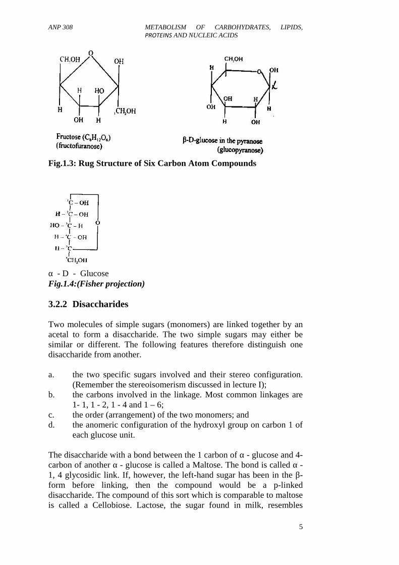
Pentoses and hexoses with five and six carbon atoms respectively havethe potential to form very stable ring structures via internal hemiacetalformation. The bond angles characteristics of carbon and oxygenbonding are such that rings containing fewer than five atoms are strainedto some extent, whereas five- or six-numbered rings are easily formed.In principle, aldotetroses can also form five-numbered ring structure, butthey rarely do. Hemiacetals with five-membered rings are calledFURANOSES, while those hemiacetals with six-membered rings arecalled PYRANOSES (Figure 1.3). However, we should note that incases where either five or six-membered rings are possible, the six-membered ring usually predominates. For example, for glucose less than0.5% of the furanose forms exist at equilibrium. Why? The reason forthis is not yet clear; but furanoses and pyranoses are more realisticallyrepresented by pentagons or hexagons as in Haworth Convention. Inanother way, the structures can also be represented as straight chainshowing the acetal bonding as described in the Fisher Projection.
ANP 308 METABOLISM OF CARBOHYDRATES, LIPIDS,PROTEINS AND NUCLEIC ACIDS
5
Fig.1.3: Rug Structure of Six Carbon Atom Compounds
α - D - GlucoseFig.1.4:(Fisher projection)
3.2.2 Disaccharides
Two molecules of simple sugars (monomers) are linked together by anacetal to form a disaccharide. The two simple sugars may either besimilar or different. The following features therefore distinguish onedisaccharide from another.
a. the two specific sugars involved and their stereo configuration.(Remember the stereoisomerism discussed in lecture I);
b. the carbons involved in the linkage. Most common linkages are1- 1, 1 - 2, 1 - 4 and 1 – 6;
c. the order (arrangement) of the two monomers; andd. the anomeric configuration of the hydroxyl group on carbon 1 of
each glucose unit.
The disaccharide with a bond between the 1 carbon of α - glucose and 4-carbon of another α - glucose is called a Maltose. The bond is called α -1, 4 glycosidic link. If, however, the left-hand sugar has been in the β-form before linking, then the compound would be a p-linkeddisaccharide. The compound of this sort which is comparable to maltoseis called a Cellobiose. Lactose, the sugar found in milk, resembles

ANP 308 METABOLISM OF CARBOHYDRATES, LIPIDS,PROTEINS AND NUCLEIC ACIDS
6
cellobiose, but the left-hand sugar is galactose instead of glucose. Thestructures of some common disaccharides are shown below:
Common disaccharides
a.
b.
c.
d.
Fig.1.5
Note: with the exception of sucrose, the ring of the right-hand glucoseunit can open exposing a free aldehyde group and giving reducingproperties to the sugars. Also, the disaccharides are soluble in water,though to varying levels.
Short notes on some disaccharidesi. Sucrose (Cane Sugar)
Sucrose is made up of a combination of one molecule of D-glucose and one molecule of D-fructose. It occurs in sugar cane;hence, the synonym "cane sugar," and also in beets (majorsources of commercial sugar). Sucrose also occurs in ripe fruits,

ANP 308 METABOLISM OF CARBOHYDRATES, LIPIDS,PROTEINS AND NUCLEIC ACIDS
7
in tree sap (maple sugar), and in many fruits and vegetables.Sucrose is dextrorotatory, but it is not a reducing sugar as it hasno free aldehyde or ketone group. When hydrolysed by diluteacid or the enzyme sucrose, sucrose splits into two constituent’smonosaccharides. The resulting sugar is levorotatory. Since thehydrolysis thus results in a change from destrorotation tolevorotatum, the process is called inversion and the mixture ofglucose and fructose is often termed invert sugar. Such a processis the way by which honey bees convert sucrose of plant nectar tohoney.
ii. Maltose (Malt Sugar)This disaccharide consists of two molecules of a -0- glucosejoined together in an a - I, four linkage. The position of H on thenumber 1 carbon atom molecule (a) is a position. Note that thenumber six carbon atoms are in as configuration. Maltose derivesits name from the fact that it is produced commercially fromstarch by the action of malt, obtained from germination barleywhich contains a starch hydrolysing enzyme distaste.
iii. CellobioseConsist of two molecules of B - D - glucose joined together in a β- 1, four linkages. This linkage is the fundamental one for thecellulose molecule and cannot be split by mammalian enzyme. Itcan be split, however, by microbial and fungal enzymes or acid.Cellobiose does not occur in free form in nature but only as acomponent of glucose polymers.
iv. Lactose (Milk Sugar)This is the sugar of milk and consists of one molecule of α - D-glucose and one molecule of β -D - galactose, joined in a linkage.This linkage can be separated by the enzyme lactase or by theaddition of acid. It is a reducing sugar and is only one sixth assweet as sucrose. Lactose is of special interest in nutrition,because it makes up nearly half of the solids of milk and becauseit does not occur in nature except as a product of the mammarygland. Having discussed the mono-and disaccharides, we shallnow focus on the third and last class of carbohydrates - thepolysaccharides
3.2.3 Oligosaccharides
The oligosaccharides contain sugars with two to ten glucose units joinedtogether by glycosidic bonds. The oligosaccharides are therefore formedby the combination (coming together) of two or more (maximum of 10)of the monomers. The monomer sugars may be of same sugars or

ANP 308 METABOLISM OF CARBOHYDRATES, LIPIDS,PROTEINS AND NUCLEIC ACIDS
8
different monomer sugars. Examples of some common oligosaccharidesare mentioned below:
a. Disaccharides: made up of two monomer sugars, e.g. sucrose,maltose, cellubiose.
b. Trisaccharides: made up of three monomer sugars, e.g. raffinosec. Tetrasaccharides: made up of four monomer sugars, e.g.
stachyose.d. Pentasaccharides: made up of five monomer sugars, e.g.
verbascose.
The simplest and biologically most important oligosaccharides are theDisaccharides, made up of glycose units, e.g. sucrose, lactose, maltose,cellubiose, gentiobiose. The types of monomer sugars that make upthese disaccharidcs are shown in Table 1.1.
Table 1.1: Composition of Some Disaccharide SugarsDisaccharide StructureSucrose Glucose - FructoseLactose Galactose - GlucoseTrehalose Glucose - GlucoseMaltose Glucose - GlucoseCellubiose Glucose - GlucoseGentiobiose Glucose - Glucose
A look at Table 1.1 shows that glucose appeared as a constituent of allthe disaccharide. This underscores the importance of glucose as asubstrate in the nutrition of plants and animals. Secondly, a look atmaltose, cellubiose and gentiobiose showed that these disaccharidescontain only glucose units. The question now is that how can twoglucose units combine to give three different products. This may appearconfusing at first. A little explanation is therefore needed at this stage.This will depend firstly on whether the connecting sugars are α or β typeand secondly, it will also depend on the points at which the sugars areconnected to each other. These points are illustrated by discussing howthe disaccharides are formed from two units of monomers.
The disaccharides derive their name from the fact that they are acombination of two molecules of monosaccharides. Their generalformular, C12H22D11 indicates that one molecule of water has beeneliminated as two monosaccharides combine
H2OC6H12O6 + C6H1206 C12H22 O11

ANP 308 METABOLISM OF CARBOHYDRATES, LIPIDS,PROTEINS AND NUCLEIC ACIDS
9
3.2.4 Polysaccharides
The carbohydrates consisting of 10 or more monosaccharides arereferred to as pol-ysaccharides. They may be considered as condensationof polymers in which the monosaccharides (or their derivatives such asamino sugars and uronic acids) are joined together by glycosidic (acetal)linkages. Polysaccharides are also called glycans and they consist of twotypes namely homogl ycans and heteroglycans. Homoglycans arepolysaccharides that consist of a single kind of monosaccharide, whileheteroglycans consist of more than one kind of monosaccharide.Polysaccharides consisting mainly of glucose are called glucans; whilethose consisting of fructose, mannose and xylose alone are referred to asfructans. mannans and xylans, respectively.
Examples of homoglycans are starches, cellulose, glycogen, insulin,chitin, etc., while examples of heteroglycans are gum acacia, pectins,alginic acids, mucopolysaccharides (hyaluronic acid, heparin,chondroitin sulphates). Generally speaking, polysaccharides areinsoluble in water, but upon hydrolysis by acids or enzymes, they arebroken down into various intermediate products and finally theirconstituent monosaccharide units. In this aspect of the course, we shallbe concerned with starch, cellulose and glycogen. Other polysaccharideswill be discussed in future.
3.2.4.1 Starch
Starch is a storage carbohydrate found in plants. It consists of glucoseunits. It is therefore a homoglucan (remember our earlier discussion onhomoglycans). Starch consists of a mixture of two different types ofmolecules, amylose and amylopectin. Amylose consists of a long chainof glucose units joined by a-I, 4 linkages while amylopectin consists of amixture of α-1, 4 links with occasional α- 1, 6 branches (Figure 1.6).The branches occur after about 25 straight a- 1, 4 bonds. Starches fromdifferent sources vary in the ratio of amylose and amylopectin, in thesize of the individual molecules, in general amylopectin accounts forabout 70 per cent of starch.
Fig. 1.6: Structure of Amylase
The structure above is the glucose units of amylose linked in anunbranched chain. The amylose structure can therefore be considered as

ANP 308 METABOLISM OF CARBOHYDRATES, LIPIDS,PROTEINS AND NUCLEIC ACIDS
10
an expanded maltose structure with a free sugar group on one end
Fig.1.7: Structure of Amylopectin
Amylopectin also contains chains of glucose units like those of amylose,also has branches of these glucose chains linked through the 6 - OH ofglucose in the manner as shown in Figure 1.7. The long chains ofamylose roll themselves into a stable helix shape which is held in placeby hydrogen bonding. The helix is a tube into which other molecules oratoms can fit. One example of this is the fact that iodine can fit insidethe helix and form a blue coloured complex with amylose, a reactionwhich is often used to detect the presence of starch or iodine. The bluerthe colour obtained, the more the amount of amylose component of thestarch. Amylose is soluble in hot water while amylopectin is insoluble inhot water. Starches from different plants when viewed microscopicallyshow difference in shapes and sizes (appearances). This propertyfurnishes the basis for microscopic identification of different types ofstarches.
Some starches show a high degree of hydrogen bonding and suchstarches are quite resistant to rupture. Tuber starch, such as found in thepotato, is extremely resistant and must be cooked before being utilisedby species such as pigs or chickens. Starch type in plants is geneticallydetermined. However, starch modification techniques are available andhave applications in the food industry. Dextrin is an intermediateresulting from the hydrolysis and digestion of starch as well as theaction of heat on starch.

ANP 308 METABOLISM OF CARBOHYDRATES, LIPIDS,PROTEINS AND NUCLEIC ACIDS
11
3.2.3.2 Cellulose
This is the most abundant substance in the plant kingdom and is a majorstructural component of plant cell walls. Cellulose is made up ofpolymerised glucose molecules ranging from 900 - 2,000 molecules.Cellulose is also a glucan. Chemically, cellulose is a polymer of β - 1,4 -linked D - glucose units. As such, the six carbon atoms are in thetransposition which results in cellulose being flat, band-like microfibril.Natural cotton is one of the purest forms of cellulose. Cellulose is notsubject to attack by the digestive enzymes of man and othermonogastrics, hence it is an important source of bulk in the diets.Contrarily, microbes in the rumen of ruminants can secrete celluloseenzyme which can degrade cellulose. Cellulose is not soluble in waterbut soluble in ammonical solution of cupric hydroxide, HCl acidsolution of zinc chloride.
3.2.3.3 Glycogen
This is the storage form of carbohydrates in animals and fungal cells.Glycogen is deposited in the liver, which acts as a central energy storageorgan in many animals. Glycogen is also abundant in muscle tissue,where it is more immediately available for energy release. The structureof glycogen is of D-glucose combined with α - 1, 4 linkage and an α - I,6 cross linkage, very similar to that of amylopectin (component of starchmoeity), except that the molecules are larger and the cross linkagesmove frequently (once every 15 or so straight bonds). Glycogen gives ared-brown, red, or at times, violet colour with iodine and which yields D- glucose upon complete hydrolysis.
4.0 CONCLUSION
Carbohydrates make up most of the organic structures of some plantsand some animals and are produced by the process of photosynthesis.
5.0 SUMMARY
Carbohydrates are classified into three major groups, namely-monosaccharides, oligosaccharides and polysaccharides, respectively.The monosaccharides are the simplest forms of sugars and make upoligosaccharides and the polysaccharides. The carbohydrates can berepresented by chemical and structural formulae. The structuralformulae are either represented in straight chain or in ring forms. This isspecially represented by the hexoses (6 - carbon sugars).

ANP 308 METABOLISM OF CARBOHYDRATES, LIPIDS,PROTEINS AND NUCLEIC ACIDS
12
6.0 TUTOR-MARKED ASSIGNMENT
1. a. What are monosaccharides, oligosaccharides andpolysaccharides?
b. Using necessary chemical structures, give two examples of theclasses of carbohydrates listed in 1 (a) above.
2. Write short notes on the following:a. Glycansb. Starchc. Cellulose.
7.0 REFERENCE/FURTHER READING
Maynard, L.A., Lopsli, J.K., Hintz, H.F. & Warner, R.G. (1983). AnimalNutrition. (7th ed.). Tata McGraw Hill Publishing CompanyLimited.

ANP 308 METABOLISM OF CARBOHYDRATES, LIPIDS,PROTEINS AND NUCLEIC ACIDS
13
UNIT 2 METABOLISM OF CARBOHYDRATES
CONTENTS
1.0 Introduction2.0 Objectives3.0 Main Content
3.1 Digestion3.1.1 Disaccharides3.1.2 Polysaccharides
3.2 Absorption and Transport3.3 Integrated Metabolism in Tissues3.4 Glycogenesis3.5 Glenycogolysis3.6 Glycolysis3.7 Hexosemonophosphate Shunt3.8 Krebs Cycle3.9 Gluconeogenesis
4.0 Conclusion5.0 Summary6.0 Tutor-Marked Assignment7.0 References/Further Reading
1.0 INTRODUCTION
The carbohydrates are source of energy for animal nutrition. Themonosaccharides and oligosaccharides are efficiently metabolised bysimple stomach animals. On the other hand, ruminants contain microbes,which secrete enzymes capable of degrading cellulose. Glycogen is apolysaccharide found in animal and fungal cells. Glycogen is a storageform of carbohydrate and is readily utilised when there is deficiency ofenergy.
2.0 OBJECTIVES
By the end of this unit, you will be able to:
explain the metabolism of carbohydrates in terms of theirdigestion, absorption and transport in the tissues
identify the metabolic pathways of carbohydrate, use and storageof glycogenesis and glycogenolosis.

ANP 308 METABOLISM OF CARBOHYDRATES, LIPIDS,PROTEINS AND NUCLEIC ACIDS
14
3.0 MAIN CONTENT
3.1 Digestion
The dietary carbohydrates that are most important nutritionally arepolysaccharides and disaccharides, since free monosaccharides are notcommonly present in the diet in significant quantities. There is,however, some free glucose and fructose in honey, in certain fruits, andin the carbohydrates that are added to processed foods. The cellular useof carbohydrates depends on their absorption from the Gastrointestinal(GI) tract into the blood stream, a process normally restricted tomonosaccharides. Therefore, polysaccharides and disaccharides must behydrolysed to their constituent monosaccharide units. The hydrolyticenzymes involved are collectively called glycosidases, or, alternatively,carbohydrases.
3.1.1 Disaccharides
Virtually no digestion of disaccharides or small oligo saccharides occursin the mouth or stomach. In the human it takes place entirely in theupper small intestine. Unlike amylase, disaccharidase activity isassociated with the mucosal cells of the microvilli or brush border ratherthan with the intestinal lumen. Among the types of enzyme activitieslocated in the mucosal cells are lactase, invertase (sucrase), andisomaltase. The latter is not a disaccharidase but instead hydrolysesbranched dextrins, as mentioned in an earlier section. Lactase catalysesthe cleavage of lactose to equimolar amounts of galactose and glucose,and sucrase hydrolyses sucrose to yield glucose and one fructoseresidue; sucrase also hydrolyses maltose and maltotriose to free glucose.
3.1.2 Polysaccharides
The glycosidase, a-amylase, assumes a particularly important role inpolysaccharide digestion because of its specific hydrolytic action on theα-1,4 bonds of the starches. Resistant to the action of this enzyme,therefore, are the β-1,4 bonds of cellulose and the α -1,6 linkages thatform branch points in the starch amylopectin. The a-amylase hydrolysesthe unbranched amylose rapidly into units of the disaccharide maltoseand into the trisaccharide malltotriose, the latter subsequentlyundergoing slower hydrolysis to maltose and glucose. The enzyme'shydrolytic action on amylopectin produces, in addition to glucose,maltose, and maltotriose, a mixture of branched oligo saccharides, ordextrins, the smallest of which are tetrasaccharides andpentasaccharides. Together with the complementary activity of anotherglycosidase, α-dextrinase, which hydrolyses the α-1, 6 bonds at thebranches, the dextrins are consequently hydrolysed to free glucose.

ANP 308 METABOLISM OF CARBOHYDRATES, LIPIDS,PROTEINS AND NUCLEIC ACIDS
15
α-amylase
Lactase
Maltase
Digestion of starches actually begins in the mouth, since amylaseactivity is found in saliva.
But considering the short period of time that food is in the mouth priorto being swallowed, this phase of digestion is of little consequence.However, the salivary amylase action continues in the stomach until thegastric acid penetrates the food bolus and lowers the pH sufficiently toinactivate the activity of the enzyme. Starch digestion is resumed in thesmall intestine, where amylase of pancreatic origin is secreted into theduodenal contents. Here, the presence of the bile, made alkaline bypancreatic bicarbonate, makes the pH favourable for enzymatic function,and most of the starch digested is through the action of the pancreaticenzyme.
3.2 Absorption and Transport
The wall of the small intestine is comprised of absorptive mucosal cellsand mucous-secreting goblet cells that line projections, called villi thatextend into the lumen. The absorptive cells have a hairy, projection likesurface on the lumen side called microvilli, or brush border. A squaremillimeter of cell surface is believed to have as many as 2 x 10⁵microvilli projections. The anatomic advantage of the villi/microvillistructure, as it relates to the absorption of nutrients is that it presents anenormous surface area to the intestinal contents. It has been estimatedthat the absorptive capacity of the human intestine amounts to about5,400 g/d for glucose and 4,800 g/d for fructose, a capability that is, ofcourse, never challenged in a normal diet.
α-dextrinase
Starches α-dextrins + Maltose +Glucose
Sucrose Frutose +Glucose
Lactose Galactose +Glucose
Fig.2.1:The enzymatic hydrolysis of dietary carbohydrates,illustrating the importance of glucose as a component of these majornutrients
Invertase (Sucrase)
ANP 308 METABOLISM OF CARBOHYDRATES, LIPIDS,PROTEINS AND NUCLEIC ACIDS
16
Glucose and galactose are absorbed across the gut wall by activetransport whereas fructose moves across by facilitated diffusion.1,2
Active transport implies that the process is energy requiring and that aspecific receptor is involved. The exact nature of the glucose/galactosecarrier is unclear, but is known to be a protein complex connected to theNa+ /K+ - ATPase pump (p. 16), which, at the expense of ATP, furnishesenergy for the transport of sugar through the mucosal cell. Glucose orgalactose cannot attach to the carrier until it has been preloaded withNa+.
Glucose appears to exit the mucosal cell by three different routes:approximately 15 per cent leaks back across the brush border into theintestinal lumen, about 25 per cent diffuses through the basolateralmemmbrane; but the major portion (approximately 60 per cent) leavesvia a carrier in the serosal membrane. If mucosal cells are poisoned withchemical blockers of oxidative phosphorylation, the transport of glucoseand galactose ceases. The fact that some cases of glucose mal-absorptionhave been attributed to reduced numbers of specific carriers testifies tothe importance of this transport mechanism.
Facilitated diffusion, the process by which fructose crosses the mucosalcells, is not energy requiring and can only proceed down a concentrationgradient. There is a carrier involved, however, and so the system issaturable and can be competitively inhibited. Since fructose is veryefficiently trapped and phosphorylated by the liver, there is virtually nocirculating fructose in the bloodstream. Therefore, the downhillconcentration gradient for fructose across the intestinal mucosa isensured.
Following transport across the gut wall, the monosaccharides enter theportal circulation and distribute among various tissues in the body.Gallactose and fructose are readily taken up by liver cells via specifichepatocyte receptors and are subsequently metabolised. Both can beconverted to glucose derivatives through pathways that will be discussedlater and then stored as liver glycogen or catabolised for energyaccording to the body's energy demand. The blood levels of galactoseand fructose are not directly subject to the strict horrmonal regulation,which is such an important part of glucose homeostasis. However, ifthey represent a significantly higher than normal percentage of dietarycarbohydrate, they may be indirectly regulated hormonally as glucosedue to their metabolic conversion to that sugar.
Glucose is nutritionally the most important monosaccharide, since it isthe exclusive constituent of the starches and since it also occurs in eachof three major disaccharides. Following its active transport through theintestinal mucosal cells it is distributed via the blood stream among

ANP 308 METABOLISM OF CARBOHYDRATES, LIPIDS,PROTEINS AND NUCLEIC ACIDS
17
various tissues, primarily liver, muscle, and adipose tissue. It entersthese cells by facilitated diffusion. In skeletal muscle and adipose tissue,the process is insulin dependent, while in the liver it is insulinindependent. The maintenance of normal blood-glucose concentration isthe net effect of metabolic processes that remove glucose from the bloodfor either glycogen synthesis or for energy production and of processesthat return glucose to the blood, such as glycogenolysis andgluconeogenesis. These pathways, which will be examined in detail inthe next section, are hormonally influenced primarily by the antagonisticpancreatic hormones insulin and glucagon and the glucocorticoidhormones of the adrenal cortex.
A rise in blood glucose, for example, following the ingestion ofcarbohydrate triggers the release of insulin while reducing the secretionof glucagon. This results in an increasing uptake of glucose by muscleand adipose tissue, resulting in the return to homeostatic levels of bloodglucose. A fall in blood glucose concentration conversely signals thereversal of the hormonal secretions-decreased insulin and increasedglucagon release. Additionally, an increase in glucocorticoid hormoneproduction occurs in answer to a falling blood glucose level, resulting inthe potentiation of gluconeogenesis, a process to be described in thefollowing sections.
3.3 Integrated Metabolism in Tissues
The metabolic fate of the monosaccharides depends to a great extent onthe energy demands of the body. According to these demands, theactivity of certain metabolic pathways may be stimulated, while othersmay be repressed. The major mediators of this regulation are hormonessuch as insulin, glucagon, and the glucocorticosteroids, which activateor inhibit specific enzymes within the pathways, and allosteric enzymes,which are stimulated or repressed by certain compounds formed withinthe pathway in which the enzymes function. An allosteric, or regulatory,enzyme is said to be positively or negatively modulated by a substance(modulator) according to whether the effect is stimulatory or repressive,respectively.
ATP and its dephosphorylated product, AMP, formed from ADP byadenylate kinase (2 ADP → AMP + ATP) can modulate certainallosteric enzymes through opposing effects. This exemplifies the linkbetween energy demand and allosteric enzyme regulation. As ATPaccumulates, for example during a period of muscular rest, it cannegatively modulate certain regulatory enzymes in energy-producingpathways so as to reduce the production of additional ATP. An increasein AMP concentration conversely signifies a depletion of ATP and theneed to produce more of this energy source. In such a case AMP can act

ANP 308 METABOLISM OF CARBOHYDRATES, LIPIDS,PROTEINS AND NUCLEIC ACIDS
18
as a positive modulator on regulatory enzymes. The enzyme,phosphofructokinase, which catalyses a reaction in the glycolyticpathway, is modulated by both ATP and AMP in the manner described.The ratio of NADH to NAD also has an important regulatory effect.Certain allosteric enzymes, for example, are responsive to an increasedlevel of NADH, which therefore regulates its formation throughnegative modulation. Furthermore, dehydrogenase reactions, whichinvolve the interaction of the reduced and oxidised forms of thecosubstrate, are reversible. If metabolic conditions lead to theaccumulation of one form or the other, the equilibrium is shifted so as toconsume more of the predominant form. The purpose of regulation is toboth maintain homeostasis and to alter the reactions of metabolism insuch a way as to meet the nutritional/biochemical demands of the body.
The metabolic pathways of carbohydrate use and storage consist ofglycogenesis, glycogenolosis, glycolysis and hexose monophosphateshunt, the citric acid cycle, and gluconeogenesis. An integratedoverview of these pathways is illustrated and a detailed review of theirintermediary metabolites, sites of regulation, and, most importantly,their function in the overall scheme of things will now be considered.Reactions within the pathways will be numbered to allow elaboration ofthose that are felt to be particularly significant from a nutritionalstandpoint. Because of the central role of glucose in carbohydratenutrition, its metabolic fate will be featured.
Fig.2.2:An overview of the major pathways of carbohydratemetabolism, emphasising the fate of glucose but also indicating thesites of entry of galactose and fructose into the pathways
ANP 308 METABOLISM OF CARBOHYDRATES, LIPIDS,PROTEINS AND NUCLEIC ACIDS
19
3.4 Glycogenesis
Glycogenesis refers to the pathway by which glucose is ultimatelyconverted into glycogen. This pathway is particularly important inhepatocytes because the liver is the major site of glycogen storage.Glycogen accounts for as much as seven per cent of the wet weight ofthis organ.
The other major site of glycogen storage is skeletal muscle and, to alesser extent, adipose tissue. It is the glycogen stores that are used firstwhen the body is confronted by an energy demand such as physicalexertion or emotional stress, and so the glycogenic pathway is of vitalimportance in ensuring a reserve of instant energy. The following arecomments on selected reactions:
1. Upon entering the cell, glucose is first phosphoorylated by ATP,producing a phosphate ester at the number 6 carbon of theglucose. In muscle cells the enzyme catalysing this phosphatetransfer is hexookinase, an allosteric enzyme that is negativelymodulated by the product of the reaction, glucose 6-phosphate.Glucose phosphorylation in the liver is catalysed by glucokinase,and although the reaction product, glucose 6-phosphate, is thesame, interesting differences distinguish it from hexokinase. Forexample, glucokinase is not inhibited by glucose 6-phosphate.
Also, it has a much higher Km than hexokinase, meaning that itcan convert glucose to its phosphate form even when the cellularconcentration of glucose is raised significantly, (e.g, after acarbohydrate-rich meal). The much lower Km of hexokinaseindicates that it is catalysing at maximum velocity even ataverage glucose concentrations. Therefore the liver has thecapacity to reduce blood glucose concentration when it becomeshigh, and it is noteworthy that glucokinase is deficient in thedisease diabetes mellitus. The hexokinase/glucokinase reaction isenergy consuming, since the glucose was activated(phosphorylated) at the expense of ATP.

ANP 308 METABOLISM OF CARBOHYDRATES, LIPIDS,PROTEINS AND NUCLEIC ACIDS
20
Fig.2.3:Reactions of glycogenesis, by which the formation of glycogenfrom glucose occurs, glycogen appears to be formed principally fromgluconeogenic precursor substances rather than from glucose directly.
2. The phosphate is transferred from the number 6 carbon of theglucose to the number 1 carbon in a complex reaction catalysedby the enzyme phosphoglucomutase.
3. Nucleoside triphosphates sometimes function as activatingsubstances in intermediary metabolism. In this reaction, energyderived from the hydrolysis of the α-β phosphate anhydride bondof uridine triphosphate allows the coupling of the resultinguridine monophosphate to the glucose I-phosphate to formuridine diphosphate glucose (UDP-glucose).
4. As UDP glucose, the glucose moiety can be incorporated directlyinto glycogen. The reaction is catalysed by glycogen synthase,and it requires some preformed glycogen (primer) to which theincoming glucose units can be attached. The reaction isstimulated by insulin.
5. Branching within the glycogen molecule is very importantbecause it increases its solubility and compactness and alsomakes available many non-reducing ends of chains from whichglucose residues can be cleaved and used for energy. Glycogensynthase cannot form the α-1, Six bonds of the branched points.This is left to the action of the branching enzyme, which transferssmall oligosacccharide segments from the end of the mainglycogen chain to carbon number six hydroxyl groups throughoutthe chain. The overall pathway of glycogenesis, like mostsynthetic pathways, consumes energy, since an ATP (reaction(1)) and a UTP (reaction (3)) are consumed.

ANP 308 METABOLISM OF CARBOHYDRATES, LIPIDS,PROTEINS AND NUCLEIC ACIDS
21
3.5 Glenycogolysis
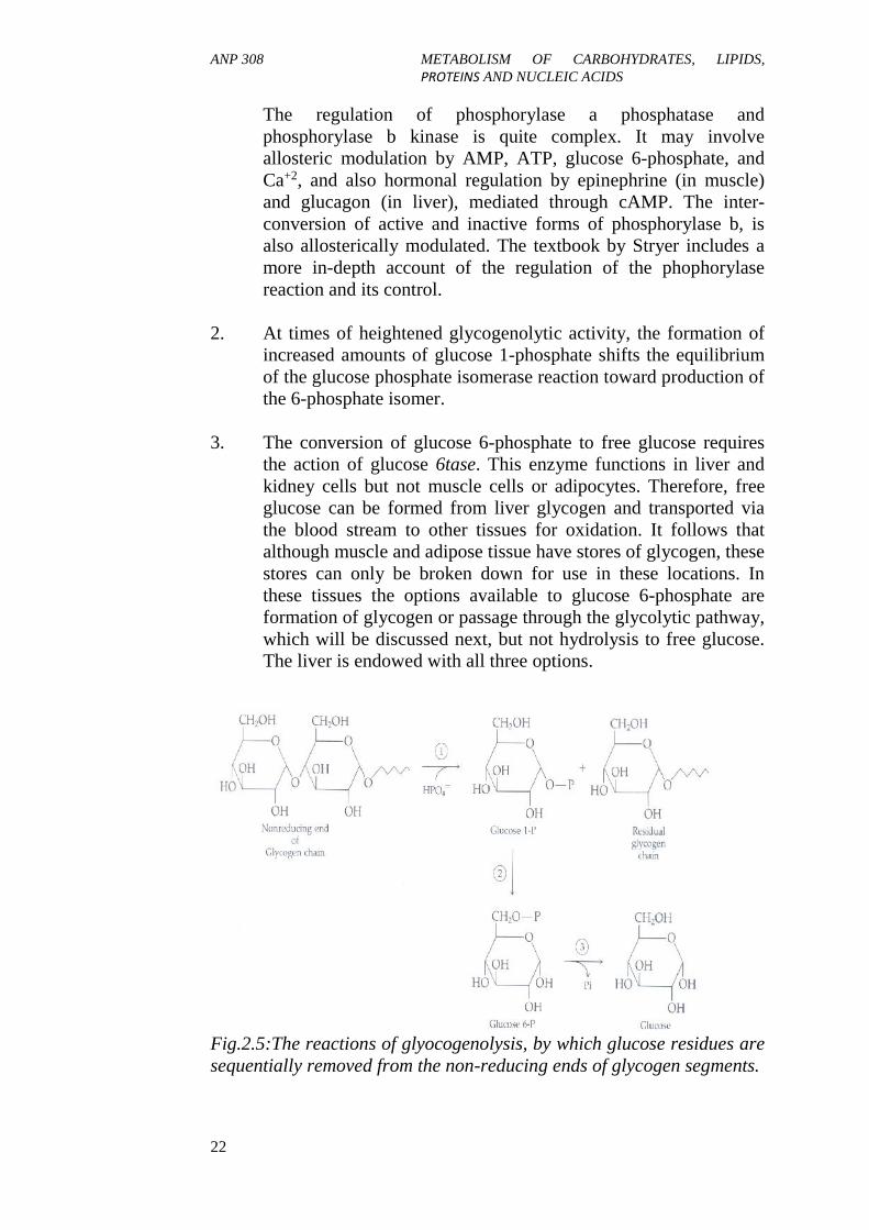
The potential energy of glycogen is contained within the glucoseresidues that comprise its structure. As the body's energy demanddictates, the residues can be systematically cleaved one at a time fromthe ends of the glycogen branches and routed through energy-producingpathways. The breakdown of glycogen into individual glucose units, inthe form of glucose 1-phosphate, is, called glycogenolysis. Like itscounterpart, glycogenesis, it is regulated by hormones, most importantlyby glucagon, of pancreatic origin, and the catecholamine hormoneepinephrine, originating in the adrenal medulla. Both of these hormonesexert positive modulation of the process and are directed at the initialreaction glycogen phosphorylase. They therefore functionantagonistically to insulin in regulating the balance between free andstored glucose. The following are comments on selected reactions:
1. The sequential release of individual glucose units from glycogenis a phosphorolysis process by which the glycosidic bonds arecleaved by phosphate addition. The products of the reaction areglucose 11phosphate and the remainder of the intact glycogenchain minus the one glucose residue. The reaction is catalysed byglycogen phosphorylase, an important site of metabolicregulation by both hormonal and allosteric enzyme modulation.Different forms of glycogen phosphorylase exist, includingphosphorylase a, a phosphorylated active form, andphosphorylase b, an unphosphorylated inactive form. The twoforms are interconvertible by protein phosphatase, whichdephosphorylates phosphoryllase a to its inactive, b form, and byphosphorylase b kinase, which returns the b form to the active, aform. The rate of glycogen breakdown to glucose 11phosphatetherefore depends on the relative activity of these enzymes.
Fig.2.4
ANP 308 METABOLISM OF CARBOHYDRATES, LIPIDS,PROTEINS AND NUCLEIC ACIDS
22
The regulation of phosphorylase a phosphatase andphosphorylase b kinase is quite complex. It may involveallosteric modulation by AMP, ATP, glucose 6-phosphate, andCa+2, and also hormonal regulation by epinephrine (in muscle)and glucagon (in liver), mediated through cAMP. The inter-conversion of active and inactive forms of phosphorylase b, isalso allosterically modulated. The textbook by Stryer includes amore in-depth account of the regulation of the phophorylasereaction and its control.
2. At times of heightened glycogenolytic activity, the formation ofincreased amounts of glucose 1-phosphate shifts the equilibriumof the glucose phosphate isomerase reaction toward production ofthe 6-phosphate isomer.
3. The conversion of glucose 6-phosphate to free glucose requiresthe action of glucose 6tase. This enzyme functions in liver andkidney cells but not muscle cells or adipocytes. Therefore, freeglucose can be formed from liver glycogen and transported viathe blood stream to other tissues for oxidation. It follows thatalthough muscle and adipose tissue have stores of glycogen, thesestores can only be broken down for use in these locations. Inthese tissues the options available to glucose 6-phosphate areformation of glycogen or passage through the glycolytic pathway,which will be discussed next, but not hydrolysis to free glucose.The liver is endowed with all three options.
Fig.2.5:The reactions of glyocogenolysis, by which glucose residues aresequentially removed from the non-reducing ends of glycogen segments.
ANP 308 METABOLISM OF CARBOHYDRATES, LIPIDS,PROTEINS AND NUCLEIC ACIDS
23
3.6 Glycolysis
Glycolysis is, by definition, the pathway by which glucose is convertedinto two units of lactic acid, a triose. The pathway can functionanaerobically, and in situations in which oxygen debt is in effect, as intimes of strenuous exercise, lactate accumulates in the muscle cells,causing the aches and pains associated with overexertion. Theimportance of glycolysis in energy metabolism is that it provides theinitial sequence of reactions necessary for glucose to be oxidisedcompletely to CO2 and H20 via the citric acid cycle. In cells that lackmitochondria, such as the erythrocyte, the pathway of glycolysis is thesole provider of ATP by substrate level phosphorylation of ADP. Theglycolytic enzymes function within the cytoplasmic matrix of the cell,while the enzymes catalysing the citric acid (Krebs) cycle reactions arelocated within the mitochondrion (pp. 8, 9). Further metabolism of theproducts of glycolysis in the Krebs cycle allows complete oxidation ofglucose to CO2 and H20, with maximal energy production. Some of theenergy liberated is salvaged as ATP, while the remainder maintainsbody temperature. Many cell types are involved in glycolysis, but mostof the energy derived from carbohydrates originates in liver, muscle, andadipose tissue.
The pathway of glycolysis, showing the entry of dietary fructose andgalactose,the following are comments on selected reactions:
1. The hexokinase/glucokinase reaction consumes 1mol ATP/molglucose. Hexokinase (not glucokinase) is negatively regulated bythe product of the reaction, glucose 6-phosphate.
2. Glucose phosphate isomerase catalyses this inter-conversion ofisomers.
3. The phosphofructokinase reaction, an important regulatory site, ismodulated negatively by ATP and citrate and positively by AMP.Another ATP is consumed in the reaction.
3. The aldolase reaction results in the splitting of a hexosebisphosphate into two triose phosphates.
4. The isomers glyceraldehyde 3-phosphate and dihydroxyacetonephosphate (DHAP) are interconverted by the enzymetriosephosphate isomerase. In an isolated system the equilibriumfavors DHAP formation. However, in the cellular environment itis shifted completely toward the production of glyceraldehyde 3-phosphate, since this metabolite is being continuously removed

ANP 308 METABOLISM OF CARBOHYDRATES, LIPIDS,PROTEINS AND NUCLEIC ACIDS
24
from the equilibrium by the subsequent reaction catalysed byglyceraldehyde 3-phosphate dehydrogenase.
5. In this reaction, glyceraldehyde 3-phosphate is oxidised to acarboxylic acid, while inorganic phossphate is incorporated as ahigh-energy anhydride bond. The enzyme is glyceraldehyde 3-phosphate dehydrogenase, which uses NAD as itshydrogennaccepting cosubstrate. Under aerobic conditions, theNADH formed is reoxidised to NAD by O2 via the electrontransport chain in the mitochondria. The reason the O2 is notnecessary to sustain this reaction under anaerobic conditions isthat the NAD consumed is restored by a subsequent reaction (see11 below).
6. This reaction, catalysed by phosphoglycerate kinase, exemplifiesa substrate level phosphorylation of ADP. Do a little extensivereading, for a more detailed review of this mechanism by whichATP can be formed from ADP by the transfer of a phosphatefrom a high-energy donor molecule.
7. Phosphoglyceromutase catalyses the transfer of the phosphategroup from the carbon-3 to carbon-2 of the glyceric acid.
8. Dehydration of 2-phosphoglycerate by the enzyme enolaseintroduces a double bond that imparts high energy to thephosphate bond.
9. The product of reaction (9), phosphoenolpyruvate (PEP), donatesits phosphate group to ADP in a reaction catalysed by pyruvatekinase. This is the second site of substrate level phosphorylationof ADP in the glycolytic pathway.
10. The lactate dehydrogenase reaction transfers two hydrogen fromNADH and H+ to pyruvate, reducing it to lactate. NAD is formedin the reaction and can replace the NAD consumed in reaction (6)under anaerobic conditions. It must be emphasised that thisreaction is most active in situations of oxygen debt, as inprolonged muscular activity. Under normal, aerobic conditions,pyruvate enters the mitochondrion for complete oxidation. Athird important option available to pyruvate is its conversion tothe amino acid alanine through trans-amination with the aminogroup donor glutamate. This, together with the fact that pyruvateis also the product of the catabolism of various amino acids,makes it an important link between protein and carbohydratemetabolism.

ANP 308 METABOLISM OF CARBOHYDRATES, LIPIDS,PROTEINS AND NUCLEIC ACIDS
25
11. These two reactions provide the means by which dietary fructoseenters the glycolytic pathway. Fructose is an important factor inthe average American diet, since nearly half of the carbohydrateconsumed is sucrose, and high fructose corn sugar is becomingmore popular as a food sweetener. Reaction 12 functions inextrahepatic tissues and involves the direct phosphorylation byhexokinase to form fructose 6-phosphate. This is a relativelyunimportant reaction. It is slow and occurs only in the presence ofhigh levels of the ketose. Reaction 13 is the major means by whichfructose is converted to glycolysis metabolites.
The phosphorylation occurs at carbon-l and is catalysed byfructokinase, an enzyme found only in hepatocytes. The fructose I-phosphate is subsequently split by aldolase, designated aldolase Bto distinguish it from the enzyme acting on fructose 1,6-bisphosphate, forming DHAP and glyceraldehyde. The latter canthen be phosphorylated by glyceraldehyde kinase (or triokinase) atthe expense of a second ATP to produce glyceraldehyde 3-phosphate. Fructose is therefore converted to glycolyticintermediates and as such can follow the pathway to pyruvateformation and Krebs cycle oxidation. Alternatively, they can beused in the liver to produce free glucose by a reversal of the firstpart of the pathway through the action of gluconeogenic enzymes.Glucose formation from fructose would be particularly importantif fructose provides the major source of carbohydrate in the diet.Since the phosphorylation of fructose is essentially theresponsibility of the liver, the ingestion of large amounts of theketose can cause a depletion of hepaatocyte ATP, leading toreduction in the rate of various biosynthetic processes such asprotein synthesis.
12 Like glucose and fructose, galactose is first phosphorylated. Thetransfer of the phosphate from ATP is catalysed by galactokinaseand the resulting phosphate ester is at carbon-I of the sugar. Themajor dietary source of galactose is lactose, from which themonosaccharide is hydrolytically released by lactase.
13. Galactose 1-phosphate can be converted to glucose I-phosphateby the enzyme galactose 1-phosphate uridyl transferase. Thereaction involves the transfer of a uridyl phosphate residue fromUDP glucose to the galactose I-phosphate, yielding glucose 1-phosphate and UDP galactose. As glucose 1-phosphate, galactosecan be incorporated into glyycogen through reactions discussedpreviously. It can enter the glycolytic pathway followingisomerisation to glucose 6-phosphate and be hydrolysed to freeglucose in liver cells.

ANP 308 METABOLISM OF CARBOHYDRATES, LIPIDS,PROTEINS AND NUCLEIC ACIDS
26
14. This indicates the entry of glucose 6-phosphate into anotherpathway called the hexose monophosphate shunt (pentosephosphate pathway), which will be considered next.
3.7 Hexosemonophosphate Shunt
The purpose of a shunt is to generate biochemically importantintermediates not produced in other pathways. Two consecutivedehydrogenase reactions, glucose 6-phosphate dehydrogenase (G-6-PD),and 6-phosphogluconate dehydrogenase, initiate the sequence ofreactions. Both reactions require NADP as cosubstrate. Consequently, alarge amount of NADPH is formed, and this reduced co substrate is usedfor other important metabolic functions, such as the biosynthesis of fattyacids and the maintenance of reducing substances in red blood cellsnecessary to ensure the functional integrity of the cells. The shunt alsoprovides pentose sugars necessary for the synthesis of DNA and RNA.
This is achieved by the decarboxylation of 6-phosphogluconate to formthe pentose phosphate ribulose 5-phosphate, which in turn is convertedto its aldose isomer, ribose 5-phosphate. In some cells the pathway endsat this point. In other cells, three-, four-, and seven-carbon phosphatesugars are subsequently formed. Through molecular re-arrangementscatalysed by the fragmentring enzymes transketolase and transaldolase,fructose 6-phosphate is ultimately produced, serving as a "return" to theglycolytic sequence of reactions, thus completing the shunt.

ANP 308 METABOLISM OF CARBOHYDRATES, LIPIDS,PROTEINS AND NUCLEIC ACIDS
27
Fig.2.6:Glycolysis, Indicating The Mode of Entry of Glucose, Fructose,and Galactose into the Pathway, as Well as the Alternative DigressionOf Glucose 6-Phosphate into the Hexosemonophosphate Shunt.

ANP 308 METABOLISM OF CARBOHYDRATES, LIPIDS,PROTEINS AND NUCLEIC ACIDS
28
The shunt is active in liver, adipose tissue, adrenal cortex, thyroid gland,testis, and lactating mammary gland. Its activity is low in skeletalmuscle because of the limited demand for NADPH (fatty acid synthesis)in this tissue and also due to muscle's reliance on glucose for energy.
Under anaerobic conditions, the progression of glucose to lactic acid isof low energy yield from the standpoint of ATP formed. This ispredictable on the basis of structural change alone, noting that only onecarbon-carbon bond was cleaved in converting a hexose into two trioses,and only one reaction was an oxidation reaction. The one NADHproduced does not undergo re-oxidation via mitochondrial electrontransport, since molecular oxygen is the ultimate oxidising agent in thatsystem. Instead it is used in the lactate dehydrogenase reduction ofpyruvate to lactate. In the glycolytic pathway, therefore, a net two ATPsare formed, one is consumed in each of the reactions one and three, buttwo are produced by substrate level phosphorylation at each of thereactions seven and 10 because two triose phosphate substrates wereformed from one glucose molecule.
When the system is operating aerobically and the supply of oxygen isample to effect total oxidation of incoming glucose, lactic acid is notformed. Instead, pyruvate enters the mitochondrion, as does a reducingequivalent of the NADH (see below) produced in reaction six. The latterbecomes oxidised by electron transport and consequently generates threeATPs/mol NADH by oxidative phosphorylation. Therefore, sixadditional moles of ATP are formed, assuming two triose units for eachglucose, bringing the total to eight. NADH cannot enter themitochondrion directly. Rather, reducing equivalents formed from theNADH in the cytoplast are shuttled across the mitochondrial membraneand in turn reduce intramitochondrial NAD to NADH.
Shuttle substances that transport the hydrogens removed from cytosolicNADH into the mitochondrion are malate or glyceroI3-phosphate. Themajor shuttle compound, malate, is reoxidised by malate dehydrogenasewithin the mitochondrion as NAD becomes reduced to NADH, thereforegenerating three ATPs/mol, as discussed above. The glycerol3phosphate shuttle, on the other hand, leads to only two ATPs/molNADH because the intramitochondrial reoxidation of the glycerol 3-phosphate is cattalysed by glycerol phosphate dehydrogenase, whichuses FAD instead of NAD as hydrogenacceptor. If the glycerol 3-phosphate shuttle is in effect, therefore, a total of only six ATPs will beformed under aerobic conditions through the glycolytic sequence-two bysubstrate level phosphorylation and four by oxidative phosphorylation.These shuttle systems function in the reoxidation of cytoplasmic NADH.

ANP 308 METABOLISM OF CARBOHYDRATES, LIPIDS,PROTEINS AND NUCLEIC ACIDS
29
If the starting point of glycolysis is glycogen rather than free glucose,the hexokinase reaction is bypassed, and the total energy yield istherefore increased by one ATP for either aerobic oranaerobicglycolysis.

ANP 308 METABOLISM OF CARBOHYDRATES, LIPIDS,PROTEINS AND NUCLEIC ACIDS
30
Fig.2.7:The Portion of The Hexosemonophosphate Shunt Showing theGeneration of NADPH By The G 6-PDH (Glucose 6-PhosphateDehydrogenase) And 6-PGDH (6-Phosphogluconate Dehydrogenase)Reactions. Adding to The Importance of The Latter Reaction is That italso forms Pentose Phosphates by The Decarboxylation of 6 –Phosphogluconate.
3.8 Krebs Cycle
Alternatively designated the tricarboxylic acid cycle or the citric acidcycle, this sequence of reactions represents the forefront of energymetabolism in the body. It can be thought of as the common and finalcatabolic pathway because products of carbohydrate, fat, and aminoacids feed into the cycle where they can be totally oxidised to CO2 andH2O, with the accompanying generation of large amounts of ATP. Notall entrant substances are totally oxidised. Some Krebs cycleintermediates are used to form glucose by the process ofgluconeogenesis, which will be discussed in the next section, and somecan be converted to certain amino acids by transamination. However,the importance of the cycle as the nucleus of energy production isevidenced by the estimation that over 90 per cent of energy releasedfrom food occurs here.
The high energy output of the Krebs cycle is attributed to mitochondrialelectron transport, with oxidative phosphorylation providing the meansfor ATP formation. The oxidation reactions occurring in the cycle areactually dehydrogenations in which an enzyme catalyses the removal oftwo hydrogens to an acceptor co-substrate such as NAD or FAD. Sincethe enzymes of the cycle and the enzymes and electron carriers ofelectron transport are both compartmentalised within the mitochondria,the reduced cosubstrates, NADH and FADH2 are readily reoxidised byO2 via the electron transport chain.

ANP 308 METABOLISM OF CARBOHYDRATES, LIPIDS,PROTEINS AND NUCLEIC ACIDS
31
Fig.2.8:This is The Krebs (Citric Acid) Cycle. This Representation of theCycle is Designed to Emphasise The Formation of Reduced Coenzymesand How Their Reoxidation by Electron Transport Contributes to theSynthesis of ATP.
In addition to its production of the reduced co-substrates NADH andFADH2, which furnish the energy through their oxidation via electrontransport, the Krebs cycle produces most of the carbon dioxide throughdecarboxylation reactions. Viewing this in its proper perspective withregard to glucose metabolism, it must be recalled that two pyruvates areproduced from one glucose during cytoplasmic glycolysis. Thesepyruvates are in turn transferred into the mitochondria, wheredecarboxylation leads to the formation of two acetyl CoA units and twomollecules of CO2. The two carbons represented by the acetyl CoA areadditionally lost as CO2 through Krebs cycle decarboxylations. Most ofthe CO2 produced is exhaled through the lungs, although some is used incertain synthetic reactions called carboxylations.
The Krebs cycle is shown in figure below. It is usually visualised asbeginning with the condensation of acetyl CoA with oxaloacetate toform citrate. The acetyl CoA is formed from numerous sources,including the breakdown of fatty acids, glucose (through pyruvate), andcertain amino acids. Its formation from pyruvate will be considerednow, since this compound links cytoplasmic glycolysis to themitochondrial Krebs cycle activity.

ANP 308 METABOLISM OF CARBOHYDRATES, LIPIDS,PROTEINS AND NUCLEIC ACIDS
32
The reaction shown below is generally referred to as the pyruvatedehydrogenase reaction. In actuality, however, the reaction is a complexone requiring a multienzyme system and various cofactors. The enzymesand cofactors are contained within an isolable unit called the pyruvatedehydrogenase complex. The cofactors include coenzyme A (CoA),thiamine diphosphate (TDP), Mg+2, NAD, FAD, and lipoic acid. Fourvitamins are therefore necessary for the activity of the complex-pantothenic acid (a component of CoA), thiamine, niacin, and riboflavin.The role of these vitamins and others as precursors of coenzymes will bediscussed in another unit. The enzymes include pyruvate decarboxylase,dihydroolipoyl dehydrogenase, and dihydrolipoyl transacetylase. Thenet effect of the complex results in decarboxylation anddehydrogenation of pyruvate with NAD serving as the terminalhydrogen acceptor. This reaction therefore yields energy, since thereoxidation by electron transport of the NADH produces three mol ofATP by oxidative phosphorylation. The reaction is regulated negativelyby ATP and by NADH.
The condensation of acetyl CoA with oxaloacetate initiates the Krebscycle reactions. The following are comments on reactions:
1. The formation of citrate from oxaloacetate and acetyl CoA iscatalysed by citrate synthetase. The reaction is regulatednegatively by ATP. The isomerisation of citrate to isocitrateinvolves cis aconitate as an intermediate. The isomerisation,catalysed by aconitase, involves dehydration followed bysterically reversed hydration, resulting in the repositioning of the-OH group onto an adjacent carbon. The first of fourdehydrogenation reactions within the cycle, the isocitratedehydrogenase reaction supplies energy through the respiratorychain reoxidation of the NADH. Note that the first loss of CO2 inthe cycle occurs at this site. It arises from the spontaneousdecarboxylation of an intermediate compound, oxalosuccinate.The reaction is positively modulated by ADP and negativelymodulated by ATP and NADH.

ANP 308 METABOLISM OF CARBOHYDRATES, LIPIDS,PROTEINS AND NUCLEIC ACIDS
33
Fig.2.9:The Krebs (Citric Acid) Cycle. This Representation of theCycle is Designed to Emphasise The Formation of ReducedCoenzymes and how their Reoxidation by Electron TransportContributes to the Synthesis of ATP.
2. The decarboxylation/dehydrogenation of aglutarate ismechanistically identical to the pyruvate dehydrogenase complexreaction in its multi-enzyme/cofactor requirement. In the reaction,referred to as the α-ketoglutarate dehydrogenase reaction, NADserves as hydrogen acceptor, and a second carbon is lost as CO2.
ANP 308 METABOLISM OF CARBOHYDRATES, LIPIDS,PROTEINS AND NUCLEIC ACIDS
34
The pyruvate dehydrogenase, isocitrate dehydrogenase, andaglutarate dehydrogenase reactions account for the loss of thethree-carbon equivalent of pyruvate as CO2.
3. Energy is conserved in the thioester bond of succcinyl CoA. Thehydrolysis of that bond by succinyl thiokinase releases sufficientenergy to drive the phosphorylation of guanosine diphosphate(GDP) by inorganic phosphate. The resulting GTP is a highenergy phosphate anhydride compound like ATP; as such, GTPcan serve as phosphate donor in certain phosphorylationreactions. One such reaction occurs in the gluconeogenesispathway.
4. The succinate dehydrogenase reaction uses FAD instead of NADas hydrogen acceptor. The FADH2 is reoxidised by electrontransport to O2, but only two ATPs are formed by oxidativephosphorylation instead of three.
5. Fumarase incorporates the elements of H2O across the doublebond of fumarate to form malate.
6. The conversion of malate to oxaloacetate completes the cycle.NAD acts as a hydrogen acceptor in this dehydrogenationreaction catalysed by malate dehydrogenase. It is the fourth siteof reduced co substrate formation and therefore of energy releasein the cycle.
In summary the complete oxidation of glucose to CO2 and H2O can beshown by the equation:C6H12O6 + 6O2 → 6 CO2 + 6 H2O + energy.
This is achieved by the combined reaction sequences of the glycolyticand Krebs cycle pathways. The amount of released energy conserved asATP under aerobic conditions is as follows:
The glycolytic sequence, glucose →2 pyruvates, produces two ATPs bysubstrate level phosphoryllation and either four or six by oxidativephosphoorylation, depending on the shuttle system for NADH-reducingequivalents (p. 86). Generally, six will be formed due to the overallgreater activity of the malate shuttle system. The intra mitochondrialpyruvate dehydrogenase reaction yields two mol of NADH, one for eachpyruvate oxidised and therefore six additional ATPs by oxidativephosphorylation.

ANP 308 METABOLISM OF CARBOHYDRATES, LIPIDS,PROTEINS AND NUCLEIC ACIDS
35
Fig.2.10
The oxidation of 1 mol of acetyl CoA in the Krebs cycle yields a total of12 ATPs. The sites of formation, indicated by reaction number, follow.
3. 3 ATP4. 3 ATP5. 1 ATP (as GTP)6. 2 ATP8. 3 ATP
Total 12 ATP
Since 2 mol acetyl CoA derived from one glucose, however, the actualtotal is 24 ATPs. The total number of ATPs realised for the completeoxidation of 1 mol of glucose is therefore 38, equivalent to 262.8 kcal. Itwill be recalled that this figure represents only about 40% of the totalenergy released by mitochondrial electron transport. The remaining 60per cent, or approximately 394 kcal, is released as heat to maintain bodytemperature.
It has already been mentioned that acetyl CoA is produced by fatty acidoxidation and amino acid catabolism as well as from the glycolyticallyderived pyruvate. This clearly leads to an imbalance between the amountof acetyl CoA and oxaloacetate, which condense one to onestoichiometrically in the citrate synthetase reaction. It is thereforeimportant that oxaloacetate and/or Krebs cycle intermediates, which canform oxaloacetate, be replenished in the cycle. Such a mechanism doesindeed exist. Oxaloacetate, fumarate, succinyl CoA, and arate can all beformed from certain amino acids, but the single most importantmechanism for ensuring an ample supply of oxaloacetate is the reactionby which it is formed directly from pyruvate. This reaction, shownbelow, is catalysed by pyruvate carboxxylase. The "uphill"incorporation of CO2 is accomplished at the expense of ATP, and thereaction requires the participation of biotin. The diversion of pyruvateinto oxaloacetate is called an anaplerotic (filling up) process because of

ANP 308 METABOLISM OF CARBOHYDRATES, LIPIDS,PROTEINS AND NUCLEIC ACIDS
36
its role in restoring oxaloacetate to the cycle. It is of interest thatpyruvate carboxylase is regulated positively by acetyl CoA, therebyaccelerating oxaloacetate formation in answer to increasing levels ofacetyl CoA.
Fig.2.11
3.9 Gluconeogenesis
D-glucose is an essential nutrient for the proper function of most cells,particularly those of the brain and other tissues of the central nervoussystem (CNS). When dietary intake of carbohydrate is reduced andblood glucose concentration declines, a hormonal triggering ofaccelerated glucose synnthesis from noncarbohydate sources occurs.Lactate, pyruvate, glycerol (a catabolic product of triglycerrides), andcertain amino acids represent the important noncarbohydrate sources.The process of producing glucose from such compounds is termedgluconeogenesis. The liver is the major site of this activity, althoughunder certain circumstances, such as starvation, the kidney becomesincreasingly important in gluconeogenesis. Muscle and adipose tissuelack the enzymes necessary for the process. This means, of course, thatmuscle lactate cannot serve as a precursor of glucose within that tissue.How, then, is the high level of muscle lactate that can be encountered insituations of oxygen debt dealt with? The lactate is transported to theliver via the general circulation, where it is able to be converted toglucose. The glucose can then be returned to the muscle cells to re-establish homeostatic concentrations there. This circulatory transport ofmuscle-derived lactate to the liver and the return of glucose to themuscle is referred to as the Cori cycle.
Gluconeogenesis is essentially a reversal of the glycolytic pathway.Most of the cytoplasmic enzymes involved in the conversion of glucoseto pyruvate catalyse their reactions reversibly and therefore provide themeans for also converting pyruvate to glucose. There are three reactionsin the glycolytic sequence that are not reversible-the hexokinase,phosphofructokinase, and pyruvate kinase reactions. They areunidirectional by virtue of the high, negative-free energy change of thereactions. Therefore, the process of gluconeogenesis requires that these

ANP 308 METABOLISM OF CARBOHYDRATES, LIPIDS,PROTEINS AND NUCLEIC ACIDS
37
reactions be bypassed by other enzyme systems. It is the presence orabsence of these circumventing enzymes that determines if a certainorgan or tissue is capable or incapable of conducting gluconeogenesis.As shown below in 4K, the hexokinase and phosphofructokinasereactions are bypassed by specific phosphatases that hydrolysephosphate esters.
The bypass of the pyruvate kinase reaction involves the formation ofoxaloacetate as an intermediate. Mitochondrial pyruvate can beconverted to oxaloacetate by pyruvate carboxylase, the reaction that hasbeen discussed as an anaplerotic process. Oxaloacetate can, in turn, bedecarboxylated and phosphorylated to phosphoenolpyruvate by PEPcarboxykinase, thereby completing the circumvention of the pyruvatekinase reaction. PEP carboxykinase is a cytoplasmic enzyme, and it isconsequently necessary for oxaloacetate to leave the mitochondrion, themembrane of which, however, is impermeable to the α-keto acid. Ittherefore must be converted to either malate (by malate dehydrogenase)or aspartate (by transamination with glutamate;), either of which freelytraverse the mitochondrial membrane.
In the cytoplasm, the malate or aspartate can be converted tooxaloacetate by malate dehydrogenase or aspartate aminotransferase(glutamate oxaloacetate transaminase) respectively. This mechanismallows the carbon skeletons of various amino acids to enter thegluconeogenic pathway and lead to a net synthesis of glucose. Suchamino acids are accordingly called glucogenic. They can bemetabolically converted to pyruvate and to Krebs cycle intermediates.As such, they can ultimately leave the mitochondrion in the form ofmalate or aspartate, as discussed. Reactions showing the entry of non-carbohydrate substances into the gluconeogenic system along with thebypass of the pyruvate kinase reaction will be discussed later.
During the past decade, evidence has emerged from in vitro studies thatglucose, as the sole substrate at physiologic concentrations, has limiteduse by the liver and is, in fact, a poor precursor of liver glycogen.However, use is greatly enhanced if gluconeogenic substances such asfructose, glycerol, or lactate are available along with the glucose. Thefacile incorporation of glucose into glycogen in vivo, but its limitedconversion in vitro, has been referred to as the glucose paradox. It is oneof many examples of the importance of interactions among nutrients.
4.0 CONCLUSION
The cellular use of carbohydrates depends on their absorption from thegastrointestinal (GI) tract into the blood stream, a process normallyrestricted to monosaccharides. Therefore, polysaccharides and

ANP 308 METABOLISM OF CARBOHYDRATES, LIPIDS,PROTEINS AND NUCLEIC ACIDS
38
disaccharides must be hydrolysed to their constituent monosaccharideunits. The maintenance of normal blood-glucose concentration is the neteffect of metabolic processes that remove glucose from the blood foreither glycogen synthesis or for energy production and of processes thatreturn glucose to the blood.
5.0 SUMMARY
In this unit, you have learnt the following:
Dietary starches and disaccharides are ultimately hydrolysed completelyby specific glycosidases to constituent monosaccharide residues that arecapable of being absorbed from the intestine.
The control and regulation of the metabolic pathways, is accomplishedby: hormonal induction or activation of specific enzymes; negative orpositive modulation of allosteric enzymes by effector compounds; andshifts in reaction equilibria by changes in reactant or productconcentrations.
6.0 TUTOR-MARKED ASSIGNMENT
1. What is the metabolic fate of the monosaccharides in the body?2. Using necessary chemical structures, describe the citric acid
cycle.3. Write short notes on the following:
a. Glycogenesisb. Glycogenolosisc. Glycolysisd. Hexose monophosphate shunte. Gluconeogenesis.
7.0 REFERENCES/FURTHER READING
Chewsworth, J.M., Stuchbury, T. & Scaife J.R. (1988). AgriculturalBiochemistry. (1st ed.) Chapman and Hull.
Maynard, L.A., Lopsli, J.K., Hintz, H.F. & Warner, R.G. (1983). AnimalNutrition. (7th ed.).Tata McGraw Hill Publishing CompanyLimited.

ANP 308 METABOLISM OF CARBOHYDRATES, LIPIDS,PROTEINS AND NUCLEIC ACIDS
39
UNIT 3 STRUCTURE, PROPERTIES ANDCLASSIFICATION OF PROTEINS
CONTENTS
1.0 Introduction2.0 Objectives3.0 Main Content
3.1 Structure of Proteins3.1.1 Primary Structure3.1.2 Secondary Structure3.1.3 Tertiary Structure3.1.4 Quaternary Structure
3.2 Properties of Proteins3.2.1 Colloidal3.2.2 Secondary Structure
3.2.3 Denaturation3.3 Classification of Proteins
3.3.1 Simple Proteins3.3.2 Conjugated or Complex Proteins3.3.3 Derived Proteins
4.0 Conclusion5.0 Summary6.0 Tutor-Marked Assignment7.0 References/Further Reading
1.0 INTRODUCTION
This unit will highlight the structure of proteins (primary, secondary,tertiary and quaternary), properties of proteins and classification ofproteins (simple, conjugated and derived). Proteins are a group ofcompounds containing carbon, hydrogen, oxygen, nitrogen (about 16%)and sulphur. In some proteins, phosphorus or iron is present andoccasionally may contain iodine, copper and zinc. Proteins are found inall living cells in plants and animals, where they are intimatelyconnected with all phases of activity that constitute the life of the cell.Each species has its own specific proteins, and a single organism hasmany different proteins in its cells and tissues. It follows therefore that alarge number of proteins occur in nature. All proteins are made upbasically of amino acids and there are 20 standard amino acids in nature.The proteins differ from each other in the number of sequence of thesestandard amino acids.

ANP 308 METABOLISM OF CARBOHYDRATES, LIPIDS,PROTEINS AND NUCLEIC ACIDS
40
2.0 OBJECTIVES
By the end of this unit, you will be able to:
highlight structures of protein, properties of protein explain the major classification of protein.
3.0 MAIN CONTENT
3.1 Structure of Proteins
Protein, as a substance has different frameworks. For your properunderstanding, we shall present that structure of proteins under primary,secondary, tertiary and quaternary proteins.
3.1.1 Primary Structure
This structure comes to existence as a consequence of the linkagebetween the - Carboxyl of one amino acid and the - amino group ofanother acid.
Fig.3.1
This type of linkage is known as peptide linkage and it is a linkageformed between two amino acids followed by the elimination of wateras revealed in above structures. In the illustrated structure, a dipeptidewas formed from two amino acids. Numerous amino acids can bemarried together using this addition procedure. When this happenedwith the removal of one molecule of H20 at every linkage, thepolypeptides are produced.
ANP 308 METABOLISM OF CARBOHYDRATES, LIPIDS,PROTEINS AND NUCLEIC ACIDS
41
Fig.3.2:Polypeptide
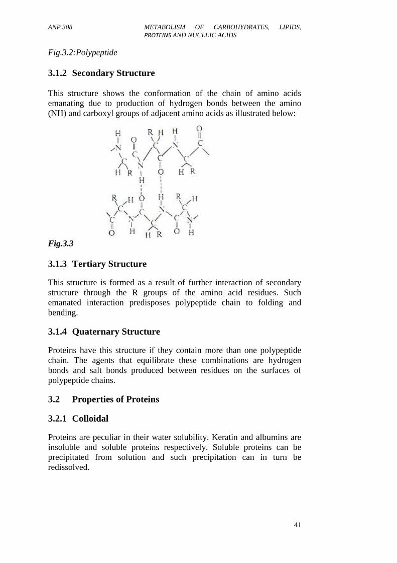
3.1.2 Secondary Structure
This structure shows the conformation of the chain of amino acidsemanating due to production of hydrogen bonds between the amino(NH) and carboxyl groups of adjacent amino acids as illustrated below:
Fig.3.3
3.1.3 Tertiary Structure
This structure is formed as a result of further interaction of secondarystructure through the R groups of the amino acid residues. Suchemanated interaction predisposes polypeptide chain to folding andbending.
3.1.4 Quaternary Structure
Proteins have this structure if they contain more than one polypeptidechain. The agents that equilibrate these combinations are hydrogenbonds and salt bonds produced between residues on the surfaces ofpolypeptide chains.
3.2 Properties of Proteins
3.2.1 Colloidal
Proteins are peculiar in their water solubility. Keratin and albumins areinsoluble and soluble proteins respectively. Soluble proteins can beprecipitated from solution and such precipitation can in turn beredissolved.

ANP 308 METABOLISM OF CARBOHYDRATES, LIPIDS,PROTEINS AND NUCLEIC ACIDS
42
3.2.2 Amphoteric
All proteins possess a certain amount of free amino and carboxylgroups, either as terminal units or in the side-chain of amino acidresidues.
3.2.3 Denaturation
All proteins can be altered or dephased from their natural occurrence. Itis a chemical, physical and biological alteration of a unique structure ofproteins. Coagulation of a protein solution upon heating, shrinking ofmeat when heated or fired and roasting of nuts are few examples ofdenaturation of protein.
3.3 Classification of Proteins
Proteins can conveniently be grouped on the basis of both physical(shape) and chemical properties. The physical properties commonlyemployed for grouping are those of solubility and heat coagulation.Standing on these two characteristics; proteins are classified into threeas simple, conjugated and derived proteins.
3.3.1 Simple Proteins
Simple proteins are the proteins that yield only amino acids or theirderivatives when hydrolysed. Examples are many which includealbumins, globulins, glutelins, albminoids, histones and prolamins. It isimportant we briefly examine these simple proteins.
Albumins
They are soluble in water and coagulable by heat. They are found bothin plants and animals, e.g. myosin of muscle, serum albumin of bloodand lactalbumin of wheat.
Globulins
These are not soluble in pure water but could be dissolved in solution ofalkaline and acid. They are heat coagulated. They generally containglycine. Globulin constitutes an important and widely distributed groupof animal and plant proteins, for example, ovoglobulin of egg yolk,myosin of muscle, phaseolin of beans, legumins of peas and arachin ofpeanuts.

ANP 308 METABOLISM OF CARBOHYDRATES, LIPIDS,PROTEINS AND NUCLEIC ACIDS
43
Glutelins
Axe all plant proteins and soluble in very dilute acids and alkalis, butthey are insoluble in natural solvents.
Prolamins
Prolamins are soluble in alcohol but insoluble in water or neutralsolvents. These proteins generally yield proline and amide nitrogen uponhydrolysis but are deficient in lysine. Prolamins are plant proteins foundprincipally in seeds, e.g. zein of corn, hadein of barley, gleaden ofwheat.
Albuminoids
It is the least soluble of all the proteins. They are generally insoluble inwater, dilute acids, alkalis and alcohol. They are entirely animal proteinand are the chief constituents of skeletal structures such as hair, horn,hoof, and nails. They are also constituents of supporting and connectingof fibrous tissues and of the cartilage and bone.
Histones
The proteins are soluble in water and insoluble in dilute ammonia. Theyare readily soluble in dilute acids and alkali. They are not readilycoagulated by heat. Histones are basic proteins. They yield a largeproportion of basic amino acids upon hydrolysis. They often precipitateother proteins from solution.
Protamins
Protamins are strongly basics and yield mainly basic amino acids onhydrolysis particularly arginine. They are soluble in water, diluteammonia acid and alkalis. They are not coagulated by heat. Theyprecipitate other proteins from their solution.
3.3.2 Conjugated Proteins
Conjugated proteins are composed of simple proteins combined withnon-proteins substance. The non-protein group is referred to asprosthetic group or addition group. The types of conjugated proteinsinclude the following:
i. Nucleo Proteins: They composed of simple basic protein(protamin or histone) in salt with nucleo acid or nucleic. They are
ANP 308 METABOLISM OF CARBOHYDRATES, LIPIDS,PROTEINS AND NUCLEIC ACIDS
44
proteins of cell and apparently the chief constituents ofchromatin. These are the most abundant in tissues of both plantsand animals, having a large proportion of nucleic materials suchas yeast, thymus and other glandular organs.
ii. Mucoproteins or Mucoids: The mucoproteins are composed ofsimple proteins combined with mucopolysaccharide such ashyaluronic acid, chomdrotin sulphates. They generally containlarge amount of N-acetylated henosamine and in addition + or -of such substances are uronic acid, sialic acid and monosaccarid.Water soluble mucoproteins have been obtained from humanurine, serum and egg white. Each water soluble mucoprotein arenot easily denatured by heat or readily precipitated by agentssuch as picric acid and trichloro acetic acid.
iii. Chromoproteins: These proteins are composed of simpleproteins united with coloured prosthetic group. Many proteins ofimportant biological function belong to this group. Examples ofchromoproteins are:
iv. Haemoglobin: Respiratory proteins in which the prosthetic groupis iron containing prophysm called EME.
v. Cytochromes: These are cellular of oxidation, reduction proteinin which the prosthetic group is also HEME.
vi. Flavoproteins: They are cellular oxidation-reduction proteins inwhich the prosthetic groups are riboflavin.
vii. Visual purple of the retina: It is a chromoprotein in which theprosthetic group is carotenoid pigment.
viii. Phosphoproteins: Phosphoric acid is the prosthetic group ofphosphoprotein. Phosphosenine has been isolated from casein(milk) and vitellin (egg).
ix. Lipoproteins: Lipoproteins are formed by combination of proteinswith lipid such as lecithin, cephalin, and fatty acid, etc. phospholipidproteins are widely distributed in plants and animals, milk and inchloroplast of plant.
x. Metalloproteins: This is a large group of enzyme proteins whichcontain metallic element such as Fe, Co, Mn, Zn, Cu, Mg, etc.which are parts of their essential structures.

ANP 308 METABOLISM OF CARBOHYDRATES, LIPIDS,PROTEINS AND NUCLEIC ACIDS
45
3.3.3 Derived Proteins
This class of proteins, as the name implies, includes those substancesderived from simple and conjugated proteins. It is the least well definedof the protein groups. Derived proteins are subdivided into primaryderived proteins and secondary derived proteins.Primary derived proteins
These protein derivatives are formed by processes which cause onlyslight changes in the protein molecule and its properties. There is littleor no hydrolytic cleavage of peptide bonds. The primary derivedproteins are synonymous with denatured proteins.
i. Proteans: The proteans are insoluble products formed by theincipient action of water, very dilute acids, and enzymes. Theyare particularly formed from certain globulins, but differ fromglobulins in being insoluble in dilute salt solutions. In general,they have the physical characteristics of the naturally occurringglutelins. Examples include myosan from myosin, edestan fromedestin, and fibrin from fibrinogen.
ii. Metaproteins: The metaproteins are formed by further action ofacids and alkali upon proteins. They are generally soluble in verydilute acids and alkali but insoluble in neutral solvents. Examplesinclude acid and alkali metaproteins such as acid and alkalialbuminates.
iii. Coagulated proteins: The coagulated proteins are insolubleproducts formed by the action of heat or alcohol upon naturalproteins. Similar substances may also be formed by action ofultraviolet light, x-rays, very high pressure, and mechanicalshaking upon protein solutions at the isoelectric pH. Examplesinclude cooked egg albumin, cooked meat and other proteins, andalcohol-precipitated proteins.
Secondary derived proteinsThese substances are formed in the progressive hydrolytic cleavage ofthe peptide unions of protein molecules. They represent a greatcomplexity of molecules of different sizes and amino acid composition.They are roughly grouped into proteoses, peptones, and peptides,according to relative average molecular complexity. Each group iscomposed of many different substances.
i. Proteoses or albumoses: Proteoses are hydrolytic products ofproteins which are soluble in water, are not coagulated by heat,

ANP 308 METABOLISM OF CARBOHYDRATES, LIPIDS,PROTEINS AND NUCLEIC ACIDS
46
and are precipitated from their solutions by saturation withammonium sulfate.
ii. Peptones: Peptones are hydrolytic products of simpler structuresthan the proteoses. They are soluble in water, are not coagulatedby heat, and are not precipitated by saturation with ammoniumsulfate. They are precipitated by phosphotungstic acid.
iii. Peptides: Peptides are composed of only a relatively few aminoacids united through peptide bonds. They are named according tothe number of amino acid groups present as di-, tri-, tetra peptidesor polypeptides. They are water-soluble, are not coagulated byheat, are not salted out of solution, and are often precipitated byphosphotungstic acid. Various definitely characterised peptideshave been isolated from protein hydrolytic products, and manyhave been synthesised.
The complete hydrolytic decomposition of a natural protein moleculeinto amino acids generally progresses through successive stages asfollows:
protein → protean → metaproteins →proteose → peptone → peptides → amino acids
The synthesis of proteins by plants and animals consists of a progressiveprocess in which amino acid groups are successively joined by peptidelinkages until the molecular size and structure is that of a specific plantor animal protein. On the other hand, in protein catabolism, the proteinsof tissues are continually being broken down to amino acids through thevarious hydrolytic stages. Accordingly, substances belonging to theclasses of proteoses, peptones, and peptides are constituents of tissues,though often in very small amounts.
4.0 CONCLUSION
Proteins are compounds containing carbon hydrogen, oxygen, nitrogensulphur and phosphorus. They are found in all living cells (plants +animals) where they are connected will all phases of activity thatconstitute life of the cell.
5.0 SUMMARY
In this unit, you have learnt that:
Proteins possess four basic structures namely:

ANP 308 METABOLISM OF CARBOHYDRATES, LIPIDS,PROTEINS AND NUCLEIC ACIDS
47
Primary Secondary Tertiary Quaternary Protein
Protein can be classified into the following classes:
Simple proteins Conjugated proteins Derived proteins
Protein serves different functions that include the following:
Provision of major organic structure of major organic structure ofthe protoplasmic mechanic
Involves in chemical process of food digestion Storage of amino acids and their usage as building blocks. Transportation of some specific molecules via the blood.
6.0 TUTOR-MARKED ASSIGNMENT
1. a. What are proteins?b. List 20 hydrolytic products of protein.
2. a. Enumerate the structures of proteins.b. Identify the inherent characteristics of proteins.
3 Based on the hydrolysis products of proteins, describe the threemajor classes of proteins.
7.0 REFERENCES/FURTHER READING
Dickerson, R.E. & Geis, I. (1993). Proteins: Structures, Function andEvolution. Cummings. Menlo Park, California: Benjamin.
Lehninger, A. L. (1992). Principles of Biochemistry. New York: WorthPublishing.
Linder, M.C. (1991). Nutritional Biochemistry and Metabolism.Elsevier.
McDonald, P., Edwards, R. A. & Greenhalgh, J .F. D. (1987). AnimalNutrition.

ANP 308 METABOLISM OF CARBOHYDRATES, LIPIDS,PROTEINS AND NUCLEIC ACIDS
48
UNIT 4 METABOLISM OF PROTEINS AND NUCLEICACIDS
CONTENTS
1.0 Introduction2.0 Objectives3.0 Main Content
3.1 Enzymatic Digestion of Dietary Proteins3.2 Amino Acid Catabolism3.3 Transport of Ammonia3.4 Urea Cycle3.5 Excretory Forms of Nitrogen3.6 Intracellular Protein Turnover3.7 Metabolic Breakdown of Individual Amino Acids3.8 Biosynthesis of Nutritionally Non-Essential Amino Acids
3.8.1 Transamination3.8.2 Assimilation of Free Ammonia3.8.3 Modification of the Carbon Skeletons of Existing
Amino Acids.3.9 Inborn Errors of Amino Acid Metabolism
3.9.1 Phenylketonuria: (PKU)3.9.2 Homocystinuria3.9.3 Albinism3.9.4 Alkaptonuria
4.0 Conclusion5.0 Summary6.0 Tutor-Marked Assignment7.0 References/Further Reading
1.0 INTRODUCTION
The digestion of dietary protein by gastric and intestinal proteasesliberates amino acids which are readily absorbed into the portalcirculation. This can be demonstrated by measurement of total aminonitrogen of the plasma. Between meals, the normal plasma aminonitrogen level is 4-6 mg/100ml, but during the absorption of a proteinmeal the amino nitrogen increases by 2-4 mg/100ml.
The amino acids are rapidly taken up by the tissues particularly the liver,intestine and kidney. By the sixth or seventh hour, the plasma levelreturns to the base line value. The lymph is not an important route oftransport for amino acids. Free amino acids are not stored in the tissuesto any great extent but are metabolised by incorporation into protein orby transamination or deamination and further oxidation. Reserves ofprotein accumulate in the liver and possibly in the muscle. These labile
ANP 308 METABOLISM OF CARBOHYDRATES, LIPIDS,PROTEINS AND NUCLEIC ACIDS
49
reserves, which can be called upon when the protein intake isinadequate, are incorporated into the architecture of the tissues. Theliver is the site of synthesis of several blood proteins, e.g. plasma,albumin, globulins, fibrinogen and oprothrombin. The liver metabolisesany amino acids excess of hepatic needs for protein synthesis. The liverdoes this by converting the nitrogen atoms to intermediates encounteredin carbohydrate and lipid metabolism.
2.0 OBJECTIVES
By the end of this unit, you will be able to:
explain the digestion of dietary protein by gastric and intestinalproteases
explain the amino acids turn-over of body proteins.
3.0 MAIN CONTENT
3.1 Enzymatic Digestion of Dietary Proteins
The degradation of dietary proteins in vertebrates occurs in thegastrointestinal tract. Entry of dietary protein in the stomach stimulatesthe gastric mucosa to secrete the hormone gastrin, which in turnstimulates the secretion of hydrochloric acid by the parietal cells andpepsinogen by the chief cells of the gastric glands. The acidic gastricjuice (pH 1.0-2.5) functions both as an antiseptic, killing most bacteriaand foreign cells and as a denaturing agent, unfolding globular proteinsand rendering their internal peptide bonds more accessible to enzymatichydrolysis. Pepsinogen (an inactive precursor/zymogen) is converted toactive pepsin. In the stomach, pepsin hydrolyses ingested proteins at thepeptide bonds on the amino-terminal side of the aromatic amino acidresidues phenylalanine, tryptophan and tyrosine, cleaving long peptidechains into a mixture of smaller peptides.
As the acidic content pass into the small intestine, the low pH triggerssecretion of the hormone secretin into the blood. Secretin stimulates thepancreas to secrete bicarbonate into the small intestine to neutralise thegastric HCl, abruptly increasing the pH to about 7. The digestion ofproteins continues in the small intestine as the arrival of amino acids inthe upper part of the small intestine (duodenum) causes the release ofthe hormone cholecystokinin into the blood. This stimulates secretion ofseveral enzymes: trypsinogen, chymotrypsinogen, procarboxypeptidasesA and B, the zymogens of trypsin, chymotrypsin and carboxypeptidasesA and B which are synthesised and secreted by the exocrine cells of thepancreas. Trypsinogen is converted to its active form, trypsin byenteropeptidase, a proteolytic enzyme secreted by intestinal cells. Free

ANP 308 METABOLISM OF CARBOHYDRATES, LIPIDS,PROTEINS AND NUCLEIC ACIDS
50
trypsin then catalyses the conversion of additional trypsinogen to trypsinas well as activates chymotrypsinogen, procarboxypeptidase A and Band proelastase.
Trypsin and chymotrypsin further hydrolyse the peptides produced bypepsin in the stomach. Trypsin cleaves the peptides at the carbonyl sideof lysine and arginine while chymotrypsin cleaves at the carbonyl sideof the amino acids phenylalanine, tryptophan and tyrosine. Degradationof the short peptides in the small intestine is completed by otherintestinal peptidases; carboxypeptidases A and B which removesuccessive carboxyl-terminal residues from peptides and anaminopeptidase that hydrolyses successive amino-terminal residuesfrom short peptides. The resulting mixture of free amino acids istransported into the epithelial cells lining the small intestine, throughwhich the amino acids enter the blood capillaries in the villi and travel tothe liver.
3.2 Amino Acid Catabolism
Excess amino acids are not stored or excreted as amino acids, but arerather degraded. Amino acids undergo degradation in three differentmetabolic circumstances:1. During normal body metabolism, some amino acids that are
released from protein breakdown and are not needed for newprotein synthesis undergo oxidative degradation
2. When a diet is rich in protein and the ingested amino acids are inexcess of the body's needs for protein synthesis, the surplus iscatabolised
3. During starvation or in uncontrolled diabetes mellitus, whencarbohydrates are unavailable or not properly utilised, cellularproteins are broken down and used as fuel (energy).
The degradative pathways for most amino acids begin by the removal ofthe α-amino nitrogen by the process of deamination. Deamination occursby two major routes: transamination and oxidative deamination.
Transamination
This step involves the loss of the amino group of an amino acid to forma α-ketoacid (the carbon-skeletons of amino acids). The amino group istaken on by α-ketoglutarate (an intermediate of the citric acid cycle) toform glutamate. This reaction is highly reversible because the reactingfunctional groups of the product are identical to those of the reactants.This reaction is mediated by glutamate aminotransferase. The co-

ANP 308 METABOLISM OF CARBOHYDRATES, LIPIDS,PROTEINS AND NUCLEIC ACIDS
51
enzyme, pyridoxal phosphate (PLP) is present at the catalytic site ofaminotransferases and serves as a carrier of amino groups.
Fig.4.1
Oxidative Deamination of L-glutamate
The release of α-amino nitrogen as ammonia from L-glutamate iscatalysed by L-glutamate dehydrogenase (GDH) which can use eithernicotinamide adenine dinucleotide (NAD+) or nicotinamide adeninedinucleotide phosphate (NADP+) as a co-substrate. This reaction is alsofreely reversible but favours glutamate production. It involves hydridetransfer from glutamate to NAD+, leading to α-iminoglutarate iminefollowed by hydrolysis to α-ketoglutarate.
Glutamate + NAD+ +H2O → α-ketoglutarate + NH4+ + NADH
The α- ketoglutarate formed from this step can be used in the citric acidcycle and for glucose synthesis. The combined action of anaminotransferase and glutamate dehydrogenase is referred to astransdeamination.

ANP 308 METABOLISM OF CARBOHYDRATES, LIPIDS,PROTEINS AND NUCLEIC ACIDS
52
Fig.4.2
3.3 Ammonia Transport in the Blood
Ammonia is quite toxic to animal tissues and the level present in theblood is regulated. In most animals, much of the free ammonia isconverted to a nontoxic compound before export from the extrahepatictissues into the blood and transfer to the liver or kidney. For thistransport function, the free ammonia is combined with glutamate toyield glutamine by the action of glutamine synthetase. This reactionrequires ATP and occurs in two steps.
First, glutamate and ATP react to form ADP and a γ-glutamyl phosphateintermediate, which then reacts with ammonia to produce glutamine andinorganic phosphate. Glutamine is a nontoxic transport form ofammonia and is normally present in blood in much higherconcentrations than amino acids. It serves as a source of amino groups ina variety of biosynthetic reactions.

ANP 308 METABOLISM OF CARBOHYDRATES, LIPIDS,PROTEINS AND NUCLEIC ACIDS
53
Fig.4.3
In most terrestrial animals, glutamine in excess of that required forbiosynthesis is transported in the blood to the intestine, liver and kidneysfor processing. In these tissues, the amide nitrogen in glutamine isreleased as ammonium ion in the mitochondria where the enzymeglutaminase converts glutamine to glutamate and NH4
+. The NH4+ from
intestine and kidney is transported in the blood to the liver. In the liver,the ammonia from all sources is disposed of by urea synthesis.
3.4 The Urea Cycle
The urea cycle is a mechanism for removing unwanted nitrogen. Ureaformation in the liver starts with the multistep conversion of ornithine toarginine. This is followed by the breakdown of arginine into ornithineand urea. The complete urea cycle as it occurs in the mammalian liverrequires five enzymes: argininosuccinate synthase, arginase andarginosuccinate lyase (which function in the cytosol), and ornithinetranscarbamoylase, and carbamoyl phosphate synthase (which functionin the mitochondria). Additional specific transport proteins are requiredfor the mitochondrial uptake of L-ornithine, NH3 and HCO3
- and for therelease of citrulline.

ANP 308 METABOLISM OF CARBOHYDRATES, LIPIDS,PROTEINS AND NUCLEIC ACIDS
54
The free ammonia formed at the end of oxidative deamination isconverted into carbamoyl phosphate in a three-step reaction requiringtwo ATP molecules
NH4+ + HCO3
- + 2ATP → carbamoyl phosphate + HPO42- + 2ADP +
2H+
First, the bicarbonate is activated and subjected to nucleophilic attack byammonia to form carbamoyl phosphate. This reaction is ATP dependentand catalyzed by carbamoyl phosphate synthase I. The carbamoylphosphate now enters the urea cycle. The urea cycle has four enzymaticsteps. First, carbamoyl phosphate donates its carbamoyl group toornithine to form citrulline, with the release of Pi, ①. This reaction iscatalysed by ornithine transcarbomoylase, and the citrulline passes fromthe mitochondrion to the cytosol.
Within the cytosol, citrulline reacts with L-aspartate in another ATP-dependent reaction to form argininosuccinate, AMP, and PPi, ②. ThePPi is subsequently hydrolysed to inorganic phosphate, thus this stepcould be said to require 2 ATP molecules. This reaction is catalysed byargininosuccinate synthetase. The argininosuccinate is then cleaved byargininosuccinase to form free arginine and fumarate, ③. Fumarateenters into the mitochondria to join the pool of citric acid cycleintermediates, this is the only reversible step in the urea cycle. In the lastreaction of the urea cycle, the enzyme arginase cleaves L-arginine toornithine and urea, ④. While the ornithine is transported to themitochondria to be reutilised in further rounds of urea cycle, ureadiffuses into the bloodstream and is ultimately eliminated through thekidneys in the urine. The stoichiometry for the urea cycle is:
CO2 + NH4+ + 3ATP + Aspartate + 2H2O →
Urea + 2ADP + 2Pi + AMP + PPi +Fumarate + 6H+

ANP 308 METABOLISM OF CARBOHYDRATES, LIPIDS,PROTEINS AND NUCLEIC ACIDS
55
Fig.4.4

ANP 308 METABOLISM OF CARBOHYDRATES, LIPIDS,PROTEINS AND NUCLEIC ACIDS
56
3.5 Excretory Forms of Nitrogen
Animals are not the only organisms that normally have a dietary excessof nitrogen but ammonia (which is the end product of nitrogenmetabolism) is toxic to animals so must be gotten rid of. Excess nitrogenfrom amino acid breakdown is excreted on one of three forms dependingon the availability of water.
Most aquatic vertebrates such as bony fishes and the larvae ofamphibians release ammonia into the surrounding water where it getsdiluted to non-toxic concentrations. This group of animals is calledammonotelic animals.
Terrestrial and aerial species convert ammonia to less toxic wasteproducts that require little water for excretion. Most terrestrialvertebrates as well as sharks convert ammonia to urea in the liversynthesised by enzymes of the urea cycle and are called ureotelicanimals.
Birds and reptiles convert and ammonia and excrete uric acid assemisolid guano, hence they are called uricotelic animals.
Fig.4.5
3.6 Intracellular Protein Turnover
The continuous synthesis and degradation of proteins in cells iscollectively termed intracellular protein turnover, which determines

ANP 308 METABOLISM OF CARBOHYDRATES, LIPIDS,PROTEINS AND NUCLEIC ACIDS
57
protein balance in tissues. Protein turnover requires large amounts ofATP (e.g., 20-25% of whole body energy expenditure in adults).However, this costly metabolic cycle fulfils key obligatory functions,including protein homeostasis, cell turnover, removal of aged anddamaged proteins, synthesis of heat-shock and immunological proteins,gluconeogenesis, wound healing, tissue repair, adaptation to nutritionaland pathological alterations, and immune responses.
When nitrogen losses equal intake, the subject is in nitrogen balance.Positive nitrogen balance which is characterised by an excess ofnitrogen intake over loss, occurs during growth, repair of tissue losses(as in convalescence) and during pregnancy. Negative nitrogen balance,where losses exceed intake, is found in starvation, malnutrition, febrilediseases and after burns or trauma. During this period of negativenitrogen balance, the body may draw on its store of labile protein. Whenthese are exhausted, circulating plasma protein will be depleted andhypoproteinemia results.
3.7 Metabolic Breakdown of Individual Amino Acids
Following the removal of amino groups from amino acids, their carbonskeletons are degraded to intermediates. The 20 individual catabolicpathways for protein amino acids converge to form only six majorpathways, culminating in six intermediates all of which enter the citriccycle. These intermediates are oxaloacetate, α-ketoglutarate, pyruvate,fumarate, succinyl CoA, acetyl CoA and acetoacetate. From here thecarbon skeletons are diverted to gluconeogenesis or ketogenesis or arecompletely oxidised to CO2 and H2O.
Amino acids can be classified as glucogenic or ketogenic, or both basedon which seven intermediates are produced during their catabolism.Glucogenic amino acids are amino acids that can be converted intoglucose through gluconeogenesis. Their catabolism yield oxaloacetate,α-ketoglutarate, pyruvate, fumarate, succinyl CoA. These are alanine,arginine, asparagine, aspartate, cysteine, glutamate, glutamine, glycine,proline, serine, histidine, methionine, threonine and valine.
Ketogenic amino acids are amino acids that can be converted to ketonebodies through ketogenesis. Their catabolism yield either acetoacetate oracetyl CoA. These are leucine and lysine. Some amino acids are bothglucogenic and ketogenic and these are tyrosine, isoleucine,phenylalanine and tryptophan.

ANP 308 METABOLISM OF CARBOHYDRATES, LIPIDS,PROTEINS AND NUCLEIC ACIDS
58
Fig.4.6:Metabolic Breakdown of Individual Amino Acids
3.8 Biosynthesis of Nutritionally Non-Essential Amino Acids
All amino acids are derived from intermediates in glycolysis, the citricacid cycle, or the pentose phosphate pathway. Nitrogen enters thesepathways by way of glutamate and glutamine. Organisms vary greatly intheir ability to synthesise the 20 common amino acids. Whereas bacteriaand plants can synthesise all 20, mammals can synthesise only the non-essential amino acids, the essential amino acids must be obtained fromfood.
Nutritionally non-essential amino acids have very short biosyntheticpathways. The non-essential amino acids are formed by three generalmechanisms.

ANP 308 METABOLISM OF CARBOHYDRATES, LIPIDS,PROTEINS AND NUCLEIC ACIDS
59
3.8.1 Transamination
Alanine can be synthesised by transamination of thecorresponding α-keto acid, pyruvate.
This reaction is catalysed by alanine aminotransferase.
Fig.4.7 glutamate can be synthesised by transamination of the
corresponding α-keto acid, α- ketoglutarate. This reaction iscatalyzed by glutamate dehydrogenase
Fig.4.8 Aspartate can be synthesised by transamination of the
corresponding α-keto acid, oxaloacetate. The conversion ofaspartate to asparagine is catalyzed by asparagine synthetase.
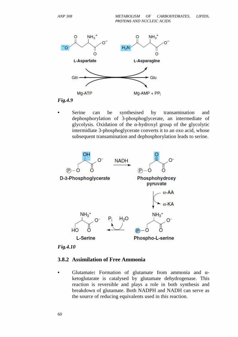
ANP 308 METABOLISM OF CARBOHYDRATES, LIPIDS,PROTEINS AND NUCLEIC ACIDS
60
Fig.4.9
Serine can be synthesised by transamination anddephosphorylation of 3-phosphoglycerate, an intermediate ofglycolysis. Oxidation of the α-hydroxyl group of the glycolyticintermidiate 3-phosphoglycerate converts it to an oxo acid, whosesubsequent transamination and dephosphorylation leads to serine.
Fig.4.10
3.8.2 Assimilation of Free Ammonia
Glutamate: Formation of glutamate from ammonia and α-ketoglutarate is catalysed by glutamate dehydrogenase. Thisreaction is reversible and plays a role in both synthesis andbreakdown of glutamate. Both NADPH and NADH can serve asthe source of reducing equivalents used in this reaction.
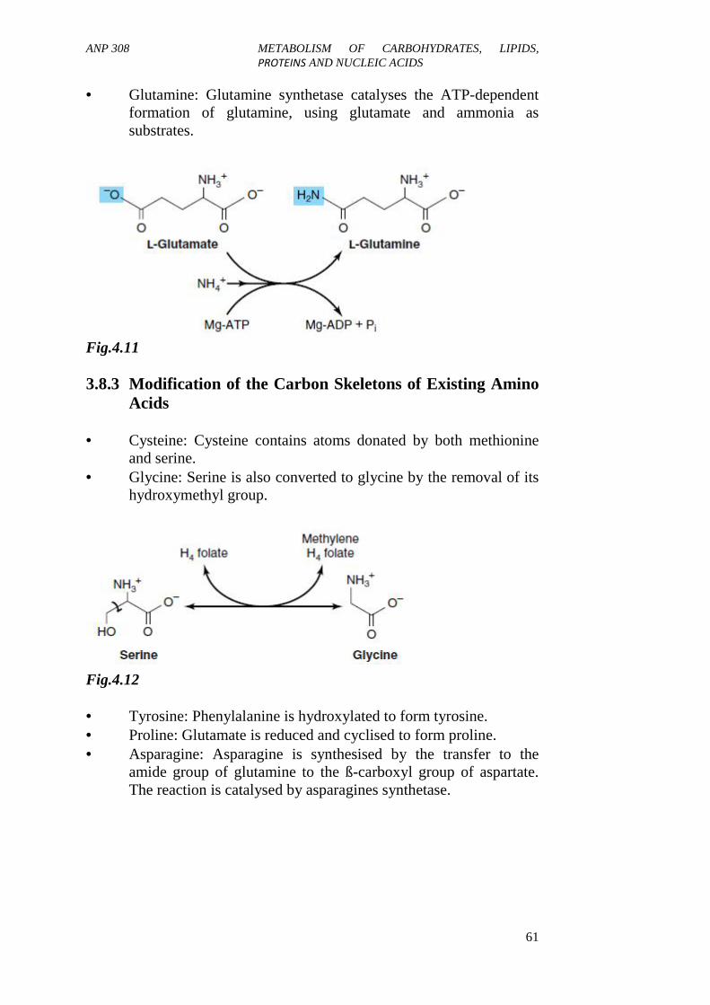
ANP 308 METABOLISM OF CARBOHYDRATES, LIPIDS,PROTEINS AND NUCLEIC ACIDS
61
Glutamine: Glutamine synthetase catalyses the ATP-dependentformation of glutamine, using glutamate and ammonia assubstrates.
Fig.4.11
3.8.3 Modification of the Carbon Skeletons of Existing AminoAcids
Cysteine: Cysteine contains atoms donated by both methionineand serine.
Glycine: Serine is also converted to glycine by the removal of itshydroxymethyl group.
Fig.4.12
Tyrosine: Phenylalanine is hydroxylated to form tyrosine. Proline: Glutamate is reduced and cyclised to form proline. Asparagine: Asparagine is synthesised by the transfer to the
amide group of glutamine to the ß-carboxyl group of aspartate.The reaction is catalysed by asparagines synthetase.

ANP 308 METABOLISM OF CARBOHYDRATES, LIPIDS,PROTEINS AND NUCLEIC ACIDS
62
3.9 Inborn Errors of Amino Acid Metabolism
3.9.1 Phenylketonuria: (PKU)
Phenylketonuria is the result of a deficiency in the enzymephenylalanine hydroxylase. High levels of phenylalanine lead tocompetitive inhibition of the enzymes responsible for melanineproduction from tyrosine. Because little tyrosine converts to melanine,afflicted infants have blonde hair and fair skin and blue eyes (similar toalbinism). Elevated phenylalanine, phenylpyruvate, phenylacetate, andphenyllactate are present in tissues, plasma and urine. Themanifestations of the disease are mental retardation, failure to walk ortalk seizures, hyperactivity and tremor.
3.9.2 Homocystinuria
Homocystinuria is due to a defect in the metabolism of homocysteine,deficiency of cystathionine synthase, high levels of homocysteine andmethionine in plasma and urine. Patients exhibit ectopia (displacementof the lens of the eye), skeletal abnormalities, prematural arterialdisease, osteoporosis and mental retardation. Homocystinuria canbetreated by supplementation with high doses of vitamin B6, B12 andfloate or restriction of methionine and inclusion of cysteine in the diet.
3.9.3 Albinism
Albinism is a condition in which defect in tyrosine metabolism results indeficiency in the production of melanin. Hypopigmentation caused dueto the deficiency in the formation of melanine results in partial or fullabsence of pigment from the skin, hair, and eyes.
3.9.4 Alkaptonuria
This is a rare disease involving deficiency in homogentistic acidoxidase, enzyme in tyrosine degradation pathway, results inaccumulation of homogentistic acidurea, large joint arthritis and dense,black pigments deposited on the intravertebral disks of the vertebrae.
4.0 CONCLUSION
The degradative pathways for most amino acids begin by theremoval of the α-amino nitrogen by the process of deamination.Deamination occurs by two major routes: transamination andoxidative deamination. Transamination plays an essential role inurea formation in connection with the synthesis of glutamate andaspartate as a means of transferring ammonia for the production
ANP 308 METABOLISM OF CARBOHYDRATES, LIPIDS,PROTEINS AND NUCLEIC ACIDS
63
of urea. It should be noted that deamination and transaminationare catabolic reactions while decarboxylation is anabolic(metabolic synthesis).
All amino acids are derived from intermediates in glycolysis, thecitric acid cycle, or the pentose phosphate pathway. Nitrogenenters these pathways by way of glutamate and glutamine.Organisms vary greatly in their ability to synthesise the 20common amino acids. Whereas bacteria and plants can synthesiseall 20, mammals can synthesise only the non-essential aminoacids, the essential amino acids must be obtained from food.
Inborn errors of amino acid metabolism are (i) Phenylketonuria:(PKU), the result of a deficiency in the enzyme phenylalaninehydroxylase and is manifested by mental retardation, failure towalk or talk, seizures, hyperactivity and tremor; (ii)Homocystinuria, due to a defect in the metabolism ofhomocysteine, deficiency of cystathionine synthase, high levelsof homocysteine and methionine in plasma and urine; (iii).Albinism, a condition in which defect in tyrosine metabolismresults in deficiency in the production of melanin.Hypopigmentation caused due to the deficiency in the formationof melanine results in partial or full absence of pigment from theskin, hair, and eyes; and (iv). Alkaptonuria, is a rare diseaseinvolving deficiency in homogentistic acid oxidase, enzyme intyrosine degradation pathway. This disorder results inaccumulation of homogentistic acidurea, large joint arthritis anddense black pigments deposited on the intravertebral disks of thevertebrae.
5.0 SUMMARY
In this unit, you have learnt that the following:
The digestion of dietary protein by gastric and intestinal proteasesliberates amino acids which are readily absorbed into the portalcirculation. The amino acids are rapidly taken up by the tissuesparticularly the liver, intestine and kidney. At least three hormones areknown to affect protein synthesis, pituitary growth hormone, insulin andtestosterone.
All amino acids are derived from intermediates in glycolysis, the citricacid cycle, or the pentose phosphate pathway. They are required as thebuilding blocks for the synthesis of the proteins of the blood and tissues.In addition, many of the amino acids are utilised in the formation ofpolypeptides and proteins of hormones and enzymes. Certain of the

ANP 308 METABOLISM OF CARBOHYDRATES, LIPIDS,PROTEINS AND NUCLEIC ACIDS
64
amino acids found in proteins may be supplied preformed in the diet.These are termed the “nutritionally essentials amino acids”. Otheramino acids are synthesised in the tissues and these are known as thenon-essential amino acids.
6.0 TUTOR-MARKED ASSIGNMENT
1. Define metabolism.2. How are the absorbed nutrients distributed to the body tissues?3. What functions are performed by the absorbed amino acids?4. Describe the oxidation of amino acids in the body.5. What are the functions of the liver?6. Give an illustration of the action of a hormone.7. Describe the disposal of the various waste products of the body.
7.0 REFERENCES/FURTHER READING
Dickerson, R.E. & Geis, I. (1993). Proteins: Structures, Function andEvolution. (Cummings. Menlo Park, California):Benjamin.
Lehninger, A. L. (1992). Principles of Biochemistry. New York: WorthPublishing.
Linder, M.C. (1991). Nutritional Biochemistry and Metabolism.Elsevier.
McDonald, P., Edwards, R. A. & Greenhalgh, J .F. D. (1987). AnimalNutrition.
ANP 308 METABOLISM OF CARBOHYDRATES, LIPIDS,PROTEINS AND NUCLEIC ACIDS
65
UNIT 5 LIPIDS METABOLISM
CONTENTS
1.0 Introduction2.0 Objectives3.0 Main Content
3.1 Definition of Terms3.2 Importance of Lipids3.3 Types of Lipids3.4 Functions of Lipids3.5 Classification of Lipids3.6 Fatty Acids3.7 Digestion of Dietary Lipids3.8 Pancreatic Lipase3.9 Intestinal Absorption of Lipids3.10 Digestion and Absorption of Fat3.11 Factors Affecting Absorption of Lipids3.12 Body Lipid3.13 Blood Lipid3.14 Lipid Transport and Storage3.15 Deposition of Fats in Tissues3.16 Biosynthesis of Fatty Acids3.17 Ketone Bodies
4.0 Conclusion5.0 Summary6.0 Tutor-Marked Assignment7.0 References/Further Reading
1.0 INTRODUCTION
Lipids are organic compounds found in plant and animal tissue, and areoily/greasy substances. They are soluble in organic solvents such asbenzene, ether or chloroform, but only sparingly soluble in water. Inroutine feed analysis, all kinds of lipids are determined together as theether extract. Thus, the lipids include fats, oils, steroids, waxes, andother related compounds. Lipids are important dietary constituents notonly because of their high energy value but also because of fat-solublevitamins and the essential fatty acids contained in the fat of naturalfoods.

ANP 308 METABOLISM OF CARBOHYDRATES, LIPIDS,PROTEINS AND NUCLEIC ACIDS
66
2.0 OBJECTIVES
By the end of this unit, you will be able to:
explain that the energy needed by the body is provided by theoxidation of CHOs and lipids
discuss that the primary objective of lipid digestion is to arrangethe lipids in a form that is water miscible
explain that ketone body production only occurs duringconditions of high circulating free fatty acids.
3.0 MAIN CONTENT
3.1 Definition of Terms
Lipids are biological molecules that are soluble in organic solvents.Lipids therefore include fats, oils, waxes, and related compounds.Occasionally, the term lipoid may be used in place of lipid. These twoterms are synonymous and therefore can be used interchangeably.
Fat- a term referring either to the lipid in the foods or body fat, both ofwhich are composed mostly of triglycerides
Triglyceride- the chief form of fat composed of C, H and O arranged asa molecule of glycerol with three fatty acids attached. The IUPAC(International Union of Pure and Applied Chemistry) name oftriglyceride is triacylglycerol, which is the main storage form of fattyacids.
Glycerol- an organic alcohol composed of a 3carbon chain which canserve as the backbone for triglyceride.
Fatty acid- is a long chain organic acid having 4-24 carbon atoms with asingle carboxyl group (COOH).
3.2 Importance of Lipids
Lipids have the following biological functions:Certain lipids, fats, serve as efficient reserves for storage energy. Suchlipids are found in the adipose tissues.
Fats serve as carriers for the fat soluble vitamins. The fat-solublevitamins are, Vitamins A, D, E and K (A, D, E, K).
ANP 308 METABOLISM OF CARBOHYDRATES, LIPIDS,PROTEINS AND NUCLEIC ACIDS
67
Lipids constitute the major structural element of membranes. Whenlipid is in combination with a protein the resulting substance is termedlipoprotein, i.e. lipid-protein.
Fat serves as insulating material in the subcutaneous tissues aroundcertain organs.
Cholesterol, an example of a lipid, is a major substance from whichVitamin D and sex hormones are synthesised.
3.3 Types of Lipids
1) Simple Lipids: esters of fatty acids with various alcohols; e.g. (a)Fats: esters of fatty acids with glycerol and (b) Waxes: esters offatty acids with higher molecular weight monohydric alcohol
2) Complex Lipids: esters of fatty acids containing groups in dditionto an alcohol and a fatty acid e.g. (a) phospholipids-lipidscontaining, in addition to fatty acid and an alcohol, a phosphoricacid residue. They frequently have nitrogen-containing bases andother substituents e.g., in glycerophoslipids the alcohol isglycerol and in sphingophospholipids the alcohol is spingosine.(b) Glycolipids: lipids containing a fatty acid and carbohydrate(c) Other complex lipids e.g. lipoprotein
3) Derived lipids: these are substances derived from the hydrolysisof simple lipids and compound lipids. The derived lipids includefatty acids, glycerol, steroids, alcohols in addition to glycerol andsterols, fatty aldehydes and ketone bodies.
3.4 Functions of Lipids
i. Lipids as an energy reserve: nearly all of the energyneeded by the human body is provided by the oxidation ofCHOs and lipids. Whereas CHOs provide a readilyavailable source of energy, lipids function primarily asenergy reserve. The amount of lipids stored as an energyreserve far exceeds the energy stored as glycogen since thehuman body is simply not capable of storing as muchglycogen compared to lipids. Lipids yields 9kcal of energyper gram while CHOs and proteins yield only 4kcal ofenergy per gram.
ii. Protection of thermo-sensitive tissues against excessiveheat loss to the environment.iii. Lipid serves as a thermal insulator in thesubcutaneous tissues and around certain organs.

ANP 308 METABOLISM OF CARBOHYDRATES, LIPIDS,PROTEINS AND NUCLEIC ACIDS
68
iv Triacylglycerol also serves as the principal function ofpadding skeletons and vital organs, thus, protecting againstshock. The heart, kidneys, epidydimis and mammaryglands are enfolded by a layer of fat tissues.
v. Phospholipids and cholesterols have their principalfunction in the formation of all Interior and Exterior cellmembranes.
vi. Lipoproteins (fat and protein) are important cellularconstituents occurring both in the cell membrane and inthe mitochondria and in cytoplasm. Also, lipoproteinserves as a means of transporting lipids in the blood.
vii. Fats serve as carriers for the fat-soluble vitamins. The fat-soluble vitamins are, Vitamins A, D, E and K (VitaminsA, D, E and K).
3.5 Classification of Lipids
Bloor classified lipids as follows:
Simple Lipids
These are esters of fatty acids with various alcohols; example of simplelipids include fats (esters of fatty acids with glycerol), waxes.
Compound Lipids
These are ester of fatty acids but containing in addition, alcohol and afatty acid. Examples of compound lipids include phospholipids, e.g.(glycerophospholipids), sphingophospholipids, cerebrosides(glycolipids) – compounds containing the fatty acids with carbohydrates,containing nitrogen but no phosphoric acid. Other compound lipidsinclude sulpholipids, aminolipids, and lipoproteins.
Derived Lipids
These are substances derived from the hydrolysis of simple lipids andcompound lipids. The derived lipids include fatty acids, glycerol,steroids, alcohols in addition to glycerol and sterols, fatty aldehydes andketone bodies; because glycerides (acylglycerols), cholesterol andcholesteryl esters are unchanged, they are also termed neutral lipids.
3.6 Fatty Acids
Fatty acids are a group of aliphatic carboxylic (-COOH) acids, whichcontain from 2 to 24 or more carbon atoms. Fatty acids are obtainedfrom the hydrolysis of fats. However, fatty acids can also occur in

ANP 308 METABOLISM OF CARBOHYDRATES, LIPIDS,PROTEINS AND NUCLEIC ACIDS
69
natural fats and such fatty acids normally contain an even number ofcarbon atoms (i.e. from 2-carbon units) and are also straight-chainderivatives (aliphatic).
Fatty acids can either be saturated or unsaturated. We shall elaborate onthis shortly. Fatty acids can also be straight-chain or branched.However, the most abundant types of fatty acid are saturated andunsaturated straight-chain fatty acids. Before we get to discuss howfatty acids are named (nomenclature), let us first look at saturated andunsaturated fatty acids.
Saturated Fatty Acids
These are fatty acids that do not contain any unsaturated bonds (i.e.having single bonds). The general formula for the saturated fatty acid isCnH2n + 1COOH – the first member of this group is acetic acid.Others will be discussed later.
Unsaturated Fatty Acids
These are fatty acids that contain one or more double bonds (unsaturatedbonds). Unsaturated fatty acids with only one double bond are calledmonounsaturated fatty acids or monoethenoid acids. Those with morethan one double bond are referred to as polyunsaturated fatty acids(PHFA) or polyethenoid acids
Essential Fatty Aacids
Three polyunsaturated fatty acids ImportanceLinoleic acid {C18 H32 O2} Play vital role in CNS peripheralnervous systemLinolenic acid {C18 H30 O2} Essential for some Spp of fish
Arachidonic acid {C20 H32O2} Source of material for thebiosynthesis of
prostaglandins.
3.7 Digestion of Dietary Lipid
The bulk of dietary lipid is triglyceride (approx 90%) of animal orvegetable origin. In the gastrointestinal tract (GIT), a portion undergoeshydrolysis to constituent fatty acid and glycerol. Incompletelyhydrolysis yields a measure of mono- and diglycerides in addition to thefinal product of the process. A group of “esterases called lipases” are ofprimary importance in the hydrolysis of triglycerides.

ANP 308 METABOLISM OF CARBOHYDRATES, LIPIDS,PROTEINS AND NUCLEIC ACIDS
70
Gastric Digestion of Lipids
It has long been known that lipase is present in gastric juice, the optimalaction of this enzyme is near neutrality and at the low pH levelencountered in the stomach it is essentially inactive. Its significance istherefore uncertain, although some fatty acid appears to be liberated inthe stomach. However, it has been suggested that gastric lipase may bemore important in the infant since the gastric acidity is far lower in theinfancy and since the normal lipid of the infant diet occurs in a highlyemulsified state in milk.
Intestinal Digestion of Lipids
The major site of lipid digestion is the small intestine. In the duodenum,the bolus of the food encounters the bile in the pancreatic juices in thelower small intestine, the secretions of the intestinal mucousa are calledSuccus entericus which also participate in lipid digestion.
The role of the bile
Bile apparently contains no lipolytic enzymes. Its function in lipiddigestion and absorption is to promote emulsification and solubilisationof lipids and this function is associated with the salt of bile acids. Thebile and pancreatic juice are somewhat alkaline and serve in part toneutralise the acidic gastric chyme. In the approximately neutralenvironment of the duodenal lumen the bile acids, largely taurocholicand glycocholic acids, exist as anions, and and serve as detergent oremulsifying agents. In the presence of these detergents the churningeffect of peristalsis results in a progressively finer and finer state ofdistribution of the dietary lipid in the continuous aqueous phase,facilitating lipolysis.
Since lipolysis (hydrolysis of lipids) involves participation of water andwater-insoluble lipases, and since dietary lipids are essentially insolublein water in water, hydrolysis occurs only at the interface between thelipid droplet and the aqueous phase. The rate of reaction is in partdetermined by the area of their interface, and the higher the degree ofemulsification, the smaller the individual lipid droplets and the largerthis area will be. The function of bile in lipid digestion is to promotecontact between water-soluble and water-insoluble components of thelipolytic reaction.
3.8 Pancreatic Lipase
The flow of pancreatic juice, like the flow of bile, is regulatedhormonally after the introduction of gastric chyme into the duodenum. A

ANP 308 METABOLISM OF CARBOHYDRATES, LIPIDS,PROTEINS AND NUCLEIC ACIDS
71
precursor of lipase in the pancreatic juice becomes active in theintestinal lumen. The mechanism of activation of pancreatic lipase is notclear, but it has been suggested that a cofactor is needed for its activity.Pancreatic lipase acids best on fatty acid esters in the emulsified state.The degree of unsaturation and chain length has no significant effect onthe rate of hydrolysis. Ca2+ has an accelerating effect on the enzyme,mainly because it forms insoluble soaps with liberated fatty acids.
3.9 Intestinal Absorption of Lipids
After ingestion of a fatty meal, the small intestine contains free fattyacid as their soaps together with a mixture of mono-, di-, andtriglycerides well emulsified by the bile salts and the soaps themselves.A major portion of this mixture is absorbed across the wall of the smallintestine such glycerol as is liberated is water soluble, and together withother water soluble nutrients is absorbed by the portal route. The fattyacids, on the other hand, are delivered to the organism predominantlyvia the intestinal lymph, where they appear in the form of triglycerides.
3.10 Digestion and Absorption of Fats
Fat digestion and absorption differ principally from the respectiveprocesses of digestion of carbohydrates and proteins, since fats are non-polar and are not miscible with water. The primary objective of lipiddigestion is to arrange the lipid in a form that is water miscible and canbe absorbed through the microvilli of the small intestine, which arecovered by an aqueous layer. The sequence of event is the same in allanimals:
i. Lipolysisii. Micellar solubilisation of the products of lipolysisiii. Uptake of the solubilised products by the intestinal mucosaiv. Resynthesis of triglyceride in the mucosal cellsv. Secretion of triglycerides into the blood.
MicellesMicelles are water-soluble aggregates of lipid molecules containingpolar and non-polar groups. The molecules are grouped in the micellesin such a way that the polar groups are on the outside, in contact withthe aqueous phase, while the non-polar parts form the inner lipid core ofthe micelles. The micelles produced in the lumen of the duodenum arevery fine dispersion of lipids in water only 50-100 A in diameter, andcarry the lipid digestion products (fatty acids, monoglycerides) to themucosal cells of the small intestine where they are subsequentlyabsorbed.

ANP 308 METABOLISM OF CARBOHYDRATES, LIPIDS,PROTEINS AND NUCLEIC ACIDS
72
3.11 Factors Affecting Absorption of Lipids
Differences in water solubility Protein interactions Micelle formation Enzyme specificity (with respect to triglyceride resynthesis) Liver dysfunction Extrahepatic biliary obstruction or biliary fistula Defect of the intestinal mucosa Absence of detergents (chiefly bile salts)
Note that: when bile is totally excluded from the intestinal tract due toliver dysfunction-it impedes lipid absorption. The bile salts’ role in fatabsorption is associated with their detergent properties. The bile acids donot enter the lymphatic circulation, rather confined to an enterohepaticcirculation i.e. entering the portal blood from which they are removed bythe liver and reinjected with the bile into the duodenum.
3.12 Body Lipids
In the normal mammal, 10 per cent or more of the body weight may belipid, the bulk of which is triglyceride. This lipid is distributed invarying amounts in all organs as well as in certain depots of highlyspecialised connective tissues (the adipose tissue) in which a largefraction of the cytoplasm of the cell appears to be replaced by thedroplets of the lipid. The large amount of lipid found in most animaltissues is in contrast to the situation in plants, where lipid is found inabundance only in seeds.
3.13 Blood Lipids
Normal blood plasma in the post absorptive state in man contains some500mg of total lipid per 100ml of which about one-quarter istriglyceride. The increase in blood lipid is called LIPEMIA, andspecifically that which transiently follows ingestion of fat is calledABSORPTIVE LIPEMIA
3.14 Lipid Transport and Storage
Fat absorbed from the diet and lipids synthesised by the liver andadipose tissue must be transported between the various tissues andorgans for utilisation and storage. Since lipids are insoluble in water, theproblem arises of how to transport them in an aqueous environment-theblood plasma. Lipids are transported from the mucosal cells of theintestine to various tissues by the circulation mainly as lipoproteins and

ANP 308 METABOLISM OF CARBOHYDRATES, LIPIDS,PROTEINS AND NUCLEIC ACIDS
73
to a lesser extent as free fatty acids. The protein moiety of lipoproteinsimparts water - soluble properties to lipids and allows their transport inthe blood. The lipoprotein can be separated by a density-gradientprocedure into different fractions.
The lipoproteins present in blood range from those of very low densityto those of high density. The density increases as the proportion ofprotein in the lipoprotein increases and the lipid decreases. Theseprotein-coated particles are called chylomicrons; chylomicrons containthe lightest lipoproteins. Lipoprotein transport lipid from the intestine aschylomicrons and from the liver as Very Low Density Lipoprotein(VLDL) to most tissues for oxidation and adipose tissues for storage.
3.15 Deposition of Fats in Tissues
The lipids are very rapidly removed from the blood by ADIPOSETISSUE, LIVER and OTHER TISSUES. Adipose tissue is the mostnotable STORAGE SITE of fats in animals. Adipose tissue is mainlyfound under the skin and also around internal organs (heart, kidney).Adipose tissues help in the assimilation of carbohydrate and lipid andtheir intermediates for fat synthesis and storage. It also helps in themobilisation of lipid as free fatty acids and to a more limited extent asglycerol. Both are influenced by hormones. The free fatty acids can bedisposed of in the tissues in a variety of ways: (1) they may becompletely oxidized to CO2 and H2O for release of energy (2) they maybe esterified to form to reform triglycerides which may be released againinto circulation or deposited in tissues for storage (3) a small proportionis transported in the blood complexed with albumin.
3.16 Biosynthesis of Fatty Acids
a) Liver, adipose tissue and lactating mammary gland are theprimary site of biosynthesis of fatty acids (f.as) and triglycerides.The main starting material for biosynthesis of f.as is acetyl-CoAderived from glucose, degraded fats and certain amino acids.
b) Any substance capable of yielding acetyl-CoA is a potentialsource of C-atoms in the process of fatty acid synthesis(LIPOGENESIS).
c) In monogastric, glucose is the primary substrate for lipogenesisand acetate in ruminant
Different Systems for Biosynthesis of Fatty Acids
There are at least two different systems for synthesis of f.as, both ofwhich involve acetyl-CoA (the principal building block of fatty acids)

ANP 308 METABOLISM OF CARBOHYDRATES, LIPIDS,PROTEINS AND NUCLEIC ACIDS
74
i. A Nonmitochondrial System-this system converts acetyl CoA toa long- chain f.as when supplemented with ATP, CO2, MN2+,
TPNH.ii A Mitochondrial System- this system utilises DPNH, TPNH, and
ATP. This system appears to be mainly involved in elongation off.as by addition of acetyl CoA to fatty acyl CoA compounds.
3.17 Ketone Bodies
After degration of a fatty acid, acetyl CoA is further oxidized in thecitric acid cycle (CAC). If excess acetyl CoA is produced from β –oxidation, some is converted to ketone bodies. These are acetoaceticacid, β – Hydroxybutyric acid,, and acetone. All these products stemfrom acetoacetyl CoA, a normal intermediate in the oxidation of fattyacids.
The Synthesis and Utilisation of Ketone Bodies
Ketone bodies are two molecules, acetoacetate and β-hydroxybutyrate.The term “ketone body” is historical: only acetoacetate is an actualketone. Ketone bodies are synthesised in the liver from acetyl-CoA. Thebrain generally uses 60-70 –per cent of total body glucose requirements,and always requires some glucose for normal functioning. Under mostconditions, glucose is essentially the sole energy source of the brain. Thebrain cannot use fatty acids, which cannot cross the blood-brain barrier;because animals cannot synthesise significant amounts of glucose fromfatty acids, as glucose availability decreases, the brain is forced to useeither amino acids or ketone bodies for fuel. Individuals eating dietsextremely high in fat and low in carbohydrates, or starving, or sufferingfrom a severe lack of insulin (Type I diabetes mellitus) thereforeincrease the synthesis and utilisation of ketone bodies.
Ketone body synthesis occurs normally under all conditions. However,the formation of ketone bodies increases dramatically during starvation.This seems to be due to a combination of factors. Prolonged low levelsof insulin result in both increased fatty acid release from adipose tissue,and increased amounts of the enzymes required to synthesise and utiliseketone bodies. In addition, in the liver, increased demand forgluconeogenesis results in depletion of oxaloacetate, and therefore indecreased capacity for the TCA cycle. This causes a rise in the levels ofacetyl-CoA, the substrate for ketone body production.
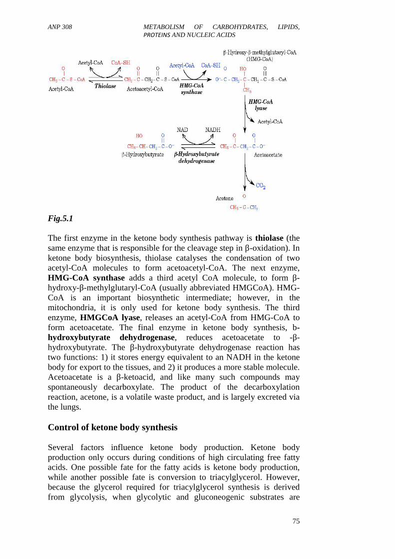
ANP 308 METABOLISM OF CARBOHYDRATES, LIPIDS,PROTEINS AND NUCLEIC ACIDS
75
Fig.5.1
The first enzyme in the ketone body synthesis pathway is thiolase (thesame enzyme that is responsible for the cleavage step in β-oxidation). Inketone body biosynthesis, thiolase catalyses the condensation of twoacetyl-CoA molecules to form acetoacetyl-CoA. The next enzyme,HMG-CoA synthase adds a third acetyl CoA molecule, to form β-hydroxy-β-methylglutaryl-CoA (usually abbreviated HMGCoA). HMG-CoA is an important biosynthetic intermediate; however, in themitochondria, it is only used for ketone body synthesis. The thirdenzyme, HMGCoA lyase, releases an acetyl-CoA from HMG-CoA toform acetoacetate. The final enzyme in ketone body synthesis, b-hydroxybutyrate dehydrogenase, reduces acetoacetate to -β-hydroxybutyrate. The β-hydroxybutyrate dehydrogenase reaction hastwo functions: 1) it stores energy equivalent to an NADH in the ketonebody for export to the tissues, and 2) it produces a more stable molecule.Acetoacetate is a β-ketoacid, and like many such compounds mayspontaneously decarboxylate. The product of the decarboxylationreaction, acetone, is a volatile waste product, and is largely excreted viathe lungs.
Control of ketone body synthesis
Several factors influence ketone body production. Ketone bodyproduction only occurs during conditions of high circulating free fattyacids. One possible fate for the fatty acids is ketone body production,while another possible fate is conversion to triacylglycerol. However,because the glycerol required for triacylglycerol synthesis is derivedfrom glycolysis, when glycolytic and gluconeogenic substrates are

ANP 308 METABOLISM OF CARBOHYDRATES, LIPIDS,PROTEINS AND NUCLEIC ACIDS
76
limiting the liver will make primarily ketone bodies. Since oxidation ofacetyl CoA via the citric acid cycle depends on a source of oxaloacetateand thus may arise by carboxylation of pyruvate derived fromglycolysis. Thus, f.as oxidation also depends on carbohydratemetabolism.
4.0 CONCLUSION
In this unit, you learnt that lipids are organic compounds found in plantand animal tissue, and are oily/greasy substances. They are also solublein organic solvents such as benzene, ether or chloroform, but onlysparingly soluble in water.
5.0 SUMMARY
Lipids are biological substances that are soluble in organic solvents.Apart from being structural components of biological membrane, theyalso play a major role in the nutrition of the animal. The lipids are veryrapidly removed from the blood by adipose tissue, liver and othertissues. Adipose tissue is the most notable storage site of fats in animals.Adipose tissue is mainly found under the skin and also around internalorgans (heart, kidney).
Adipose tissues help in the assimilation of carbohydrate and lipid andtheir intermediates for fat synthesis and storage. It also helps in themobilisation of lipid as free fatty acids and to a more limited extent asglycerol. Both are influenced by hormones. The free fatty acids can bedisposed of in the tissues in a variety of ways: (1) they may becompletely oxidised to CO2 and H2O for release of energy (2) they maybe esterified to form to reform triglycerides which may be released againinto circulation or deposited in tissues for storage (3) a small proportionis transported in the blood complexed with albumin.
6.0 TUTOR-MARKED ASSIGNMENT
1. Distinguish between gastric and intestinal digestion of lipids.2. What is the role of the bile in lipid digestion?3. How would classify lipids?4. Name five factors affecting the absorption of lipids.5 Write short notes on the following:a). Ketone bodiesb). Deposition of fat in tissuesc). Lipid transport and storaged). Control of ketone body synthesise). Deposition of fats in tissuesf). Biosynthesis of fatty acids.

ANP 308 METABOLISM OF CARBOHYDRATES, LIPIDS,PROTEINS AND NUCLEIC ACIDS
77
7.0 REFERENCES/FURTHER READING
Geoffrey, L. Zubay, William, W. Parson & Dennis, E. Vance (1995).Principles of Biochemistry. (1st ed.). Iowa, Melbourne, Australia,Oxford, England:Wm. C. Brown Publishers, Dubuque,. pp 135-224, 562-597.
McDonald, P., Edwards, R.A. & Greenhalgh, J.F.D. (1926). AnimalNutrition.(4th ed.). New York : Longman Scientific andTechnical and John Wiley and Sons, Inc,. pp 58-89. ISBN 0-582-40903-9.
Ross, J.S. & Wilson, K.J.W. (1981). Foundations of Anatomy andPhysiology. (5th ed.). London: Churchill Livingstone.
Roberts, M.B.V. (1972). Biology: A Functional Approach. (2nd ed.).London: Cox and Wyman Ltd.
Related Documents