ORIGINAL RESEARCH ADULT BRAIN Cough-Associated Changes in CSF Flow in Chiari I Malformation Evaluated by Real-Time MRI X R.A. Bhadelia, X S. Patz, X C. Heilman, X D. Khatami, X E. Kasper, X Y. Zhao, and X N. Madan ABSTRACT BACKGROUND AND PURPOSE: Invasive pressure studies have suggested that CSF flow across the foramen magnum may transiently decrease after coughing in patients with symptomatic Chiari I malformation. The purpose of this exploratory study was to demonstrate this phenomenon noninvasively by assessing CSF flow response to coughing in symptomatic patients with Chiari I malformation by using MR pencil beam imaging and to compare the response with that in healthy participants. MATERIALS AND METHODS: Eight symptomatic patients with Chiari I malformation and 6 healthy participants were studied by using MR pencil beam imaging with a temporal resolution of 50 ms. Patients and healthy participants were scanned for 90 seconds (without cardiac gating) to continuously record cardiac cycle–related CSF flow waveforms in real-time during resting, coughing, and postcoughing periods. CSF flow waveform amplitude, CSF stroke volume, and CSF flow rate (CSF Flow Rate CSF Stroke Volume Heart Rate) in the resting and immediate postcoughing periods were determined and compared between patients and healthy participants. RESULTS: There was no significant difference in CSF flow waveform amplitude, CSF stroke volume, and the CSF flow rate between patients with Chiari I malformation and healthy participants during rest. However, immediately after coughing, a significant decrease in CSF flow waveform amplitude (P .001), CSF stroke volume (P .001), and CSF flow rate (P .001) was observed in patients with Chiari I malformation but not in the healthy participants. CONCLUSIONS: Real-time MR imaging noninvasively showed a transient decrease in CSF flow across the foramen magnum after coughing in symptomatic patients with Chiari I malformation, a phenomenon not seen in healthy participants. Our results provide preliminary evidence that the physiology-based imaging method used here has the potential to be an objective clinical test to differentiate symp- tomatic from asymptomatic patients with Chiari I malformation. ABBREVIATIONS: A CSF CSF flow waveform amplitude; CMI Chiari I malformation; FR CSF CSF flow rate; PBI pencil beam imaging; SV CSF CSF stroke volume A lthough the diagnosis of Chiari I malformation (CMI) by MR imaging can be easily made by using a simple definition of 5-mm downward displacement of the cerebellar tonsils through the foramen magnum, management of this condition remains challenging and controversial. 1-6 The issue under debate is that some patients who meet the MR imaging criteria for CMI diag- nosis are asymptomatic and some with 5-mm tonsillar hernia- tion may have typical symptoms of CMI. 5-8 Therefore, in the ab- sence of an objective assessment test for CMI that correlates well with the severity of the clinical findings, a decision for surgery is often based entirely on the clinical judgment and management philosophy of the treating neurosurgeon. This scenario is believed to have led to overuse of surgical treatment. 9 Many of the symptoms and signs associated with CMI are due to abnormal CSF circulation between the head and spine, second- ary to foramen magnum obstruction produced by herniated cer- ebellar tonsils. 10-18 During the past 2 decades, attempts have been made to use cine phase contrast MR imaging to noninvasively assess CSF flow abnormalities in patients with CMI and to provide an objective test for assessment of disease severity. 3,4,11-13,15 De- spite success in showing group differences in CSF flow between symptomatic and asymptomatic patients with CMI, 3,4,15,19 criti- Received September 8, 2015; accepted after revision October 27. From the Department of Radiology (R.A.B., D.K.), Beth Israel Deaconess Medical Center, Boston, Massachusetts; Department of Radiology (S.P.), Brigham and Wom- en’s Hospital, Boston, Massachusetts; Departments of Neurosurgery (C.H.) and Radiology (N.M.), Tufts Medical Center, Boston, Massachusetts; Department of Neurosurgery (E.K.), Beth Israel Deaconess Medical Center, Boston, Massachusetts; and Phillips Healthcare (Y.Z.), Boston, Massachusetts. This work was supported by a grant from the Conquer Chiari Foundation. Please address correspondence to Rafeeque A. Bhadelia, MD, Department of Radi- ology, Beth Israel Deaconess Medical Center, WCB90, 330 Brookline Ave, Boston, MA 02115; e-mail: [email protected]; @rbhadeliaMD Indicates article with supplemental on-line photo. http://dx.doi.org/10.3174/ajnr.A4629 AJNR Am J Neuroradiol ●:● ● 2016 www.ajnr.org 1 Published December 24, 2015 as 10.3174/ajnr.A4629 Copyright 2015 by American Society of Neuroradiology.

Welcome message from author
This document is posted to help you gain knowledge. Please leave a comment to let me know what you think about it! Share it to your friends and learn new things together.
Transcript

ORIGINAL RESEARCHADULT BRAIN
Cough-Associated Changes in CSF Flow in Chiari I MalformationEvaluated by Real-Time MRI
X R.A. Bhadelia, X S. Patz, X C. Heilman, X D. Khatami, X E. Kasper, X Y. Zhao, and X N. Madan
ABSTRACT
BACKGROUND AND PURPOSE: Invasive pressure studies have suggested that CSF flow across the foramen magnum may transientlydecrease after coughing in patients with symptomatic Chiari I malformation. The purpose of this exploratory study was to demonstratethis phenomenon noninvasively by assessing CSF flow response to coughing in symptomatic patients with Chiari I malformation by usingMR pencil beam imaging and to compare the response with that in healthy participants.
MATERIALS AND METHODS: Eight symptomatic patients with Chiari I malformation and 6 healthy participants were studied by using MRpencil beam imaging with a temporal resolution of �50 ms. Patients and healthy participants were scanned for 90 seconds (without cardiacgating) to continuously record cardiac cycle–related CSF flow waveforms in real-time during resting, coughing, and postcoughing periods.CSF flow waveform amplitude, CSF stroke volume, and CSF flow rate (CSF Flow Rate � CSF Stroke Volume � Heart Rate) in the resting andimmediate postcoughing periods were determined and compared between patients and healthy participants.
RESULTS: There was no significant difference in CSF flow waveform amplitude, CSF stroke volume, and the CSF flow rate betweenpatients with Chiari I malformation and healthy participants during rest. However, immediately after coughing, a significant decrease in CSFflow waveform amplitude (P � .001), CSF stroke volume (P � .001), and CSF flow rate (P � .001) was observed in patients with Chiari Imalformation but not in the healthy participants.
CONCLUSIONS: Real-time MR imaging noninvasively showed a transient decrease in CSF flow across the foramen magnum after coughingin symptomatic patients with Chiari I malformation, a phenomenon not seen in healthy participants. Our results provide preliminaryevidence that the physiology-based imaging method used here has the potential to be an objective clinical test to differentiate symp-tomatic from asymptomatic patients with Chiari I malformation.
ABBREVIATIONS: ACSF � CSF flow waveform amplitude; CMI � Chiari I malformation; FRCSF � CSF flow rate; PBI � pencil beam imaging; SVCSF � CSF strokevolume
Although the diagnosis of Chiari I malformation (CMI) by MR
imaging can be easily made by using a simple definition of
�5-mm downward displacement of the cerebellar tonsils through
the foramen magnum, management of this condition remains
challenging and controversial.1-6 The issue under debate is that
some patients who meet the MR imaging criteria for CMI diag-
nosis are asymptomatic and some with �5-mm tonsillar hernia-
tion may have typical symptoms of CMI.5-8 Therefore, in the ab-
sence of an objective assessment test for CMI that correlates well
with the severity of the clinical findings, a decision for surgery is
often based entirely on the clinical judgment and management
philosophy of the treating neurosurgeon. This scenario is believed
to have led to overuse of surgical treatment.9
Many of the symptoms and signs associated with CMI are due
to abnormal CSF circulation between the head and spine, second-
ary to foramen magnum obstruction produced by herniated cer-
ebellar tonsils.10-18 During the past 2 decades, attempts have been
made to use cine phase contrast MR imaging to noninvasively
assess CSF flow abnormalities in patients with CMI and to provide
an objective test for assessment of disease severity.3,4,11-13,15 De-
spite success in showing group differences in CSF flow between
symptomatic and asymptomatic patients with CMI,3,4,15,19 criti-
Received September 8, 2015; accepted after revision October 27.
From the Department of Radiology (R.A.B., D.K.), Beth Israel Deaconess MedicalCenter, Boston, Massachusetts; Department of Radiology (S.P.), Brigham and Wom-en’s Hospital, Boston, Massachusetts; Departments of Neurosurgery (C.H.) andRadiology (N.M.), Tufts Medical Center, Boston, Massachusetts; Department ofNeurosurgery (E.K.), Beth Israel Deaconess Medical Center, Boston, Massachusetts;and Phillips Healthcare (Y.Z.), Boston, Massachusetts.
This work was supported by a grant from the Conquer Chiari Foundation.
Please address correspondence to Rafeeque A. Bhadelia, MD, Department of Radi-ology, Beth Israel Deaconess Medical Center, WCB90, 330 Brookline Ave, Boston,MA 02115; e-mail: [email protected]; @rbhadeliaMD
Indicates article with supplemental on-line photo.
http://dx.doi.org/10.3174/ajnr.A4629
AJNR Am J Neuroradiol ●:● ● 2016 www.ajnr.org 1
Published December 24, 2015 as 10.3174/ajnr.A4629
Copyright 2015 by American Society of Neuroradiology.
cal questions remain concerning the management of an individ-
ual patient with CMI presenting for treatment, such as whether
and when surgery should be performed and how a patient without
typical clinical or MR imaging features should be managed.6,9
Symptoms experienced by patients with CMI can be induced
or exaggerated by physiologic alterations such as coughing or the
Valsalva maneuver.16,18 This result is believed to be related to
transient alterations in CSF flow across the foramen magnum.
Therefore, it is logical to propose that an imaging test for patients
with CMI include an assessment of the CSF flow response after a
physiologic challenge. However, this is difficult, even with a very
fast version of the cine phase contrast sequence, because it pro-
duces images that are weighted averages of the phasic behavior
over many cardiac cycles and is therefore unable to show tran-
sient changes in CSF flow that result from a physiologic chal-
lenge and in which the duration of the transient changes is just
a few cardiac cycles.20 Therefore, we have chosen to assess CSF
flow with the real-time MR imaging technique, pencil beam
imaging (PBI).
Previously performed simultaneous invasive cranial and spi-
nal pressure monitoring showed that in symptomatic patients
with CMI, immediately after coughing or Valsalva maneuver, dis-
sociation develops between intracranial and intraspinal pressures.
During this pressure dissociation, a higher intracranial pressure
compared with intraspinal pressure pushes the already herniated
tonsils downward and further narrows the foramen magnum and
thereby transiently reduces the CSF flow across it.18,20-23 The pur-
pose of this exploratory study was to demonstrate this phenome-
non noninvasively by assessing CSF flow response to coughing in
patients with CMI by using PBI and to compare the response with
that in healthy participants.
MATERIALS AND METHODSPatients and Healthy SubjectsEight patients with CMI (mean age, 41.1 � 7.6 years; 7 women)
and 6 healthy participants (mean age, 38.8 � 13.3 years; 4
women) underwent MR imaging on a 3T scanner (Achieva;
Philips Healthcare, Best, the Netherlands). Institutional review
board approval was obtained for this Health Insurance Portability
and Accountability Act– compliant prospective study, and each
patient and participant signed an informed consent. All patients
with CMI were consecutive patients presenting for treatment who
agreed to a research MR imaging study and were assessed by a
neurosurgeon as having typical features of symptomatic CMI3:
severe cough-associated headache (n � 6/8), syringomyelia (n �
2/8), and objective neurologic signs such as brisk reflexes, muscle
weakness, and nystagmus (n � 4/8). Healthy participants were
recruited by local advertisement, and none had cardiovascular or
neurologic disorders.
CSF Flow Imaging TechniquePBI excites a narrow cylinder or “pencil” region and has been
described in detail previously.20,24,25 For this investigation, a 25-
mm-diameter and 64-mm-length cylinder was excited by using a
2D spatially localized radiofrequency pulse with 8-turn spiral ex-
citations, followed by a bipolar velocity-encoding gradient and a
readout gradient applied along the axis of the cylinder.20 A veloc-
ity-encoding of 5 cm/s along the superior-to-inferior direction
was used. Other imaging parameters were the following: TR,
25–28 ms; TE, 3.8 ms; and flip angle, 10°. The effective temporal
resolution was 2 � TR (ie, 50 –56 ms). Heart rate and respiratory
movements were continuously monitored by using the physio-
logic recording system of the scanner. Cardiac gating was not
required for this PBI study, which recorded pulsatile CSF flow
motion in real-time and, therefore, was able to acquire data much
faster than the gated cine phase contrast sequence.
MR ImagingSagittal 3D T2-weighted images were obtained to provide ana-
tomic details. For PBI, the pencil beam was positioned along the
long axis of the cervical spinal canal, extending from just above
the level of the foramen magnum to the C2–3 disc level (Fig 1).
Using multiplanar image reformatting, we positioned the pencil
beam to cover the entire thecal sac.20 The PBI scan was acquired
for approximately 90 seconds, during which the participant was
asked to do the following: 1) breathe quietly for the first 15–20
seconds (by counting from 1 to 20); 2) then cough as forcefully as
possible consecutively 6 times; and 3) breathe quietly again after
the end of coughing. Each �90-second scan session was repeated
3 times.
Image Analysis3D anatomic images were used to determine the degree of tonsil-
lar herniation below the level of the foramen magnum. CSF flow
FIG 1. Position of the PBI cylinder (white rectangle) and location ofCSF flow evaluation (white line) are shown in a patient with CMI.
2 Bhadelia ● 2016 www.ajnr.org
analysis was performed off-line by using custom software devel-
oped in Matlab (MathWorks, Natick, Massachusetts), which al-
lows simultaneous display of 3D anatomic and physiologic PBI
data along with the heart rate and respiration. Anatomic images
allow selection of a position along the pencil beam cylinder for
assessment of CSF flow (Fig 1 and On-line Figure). By determin-
ing the area of the thecal sac on axial images at the position se-
lected, the software then calculates CSF flow in milliliters per sec-
ond by multiplying average velocity by area and plots the CSF flow
on the y-axis versus time (seconds) on the x-axis, thereby depict-
ing cardiac cycle–related CSF flow pulsations over the entire 90-
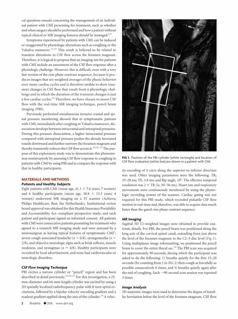
second data acquisition. For further quantitative analysis, we de-
termined 3 CSF flow parameters: 1) peak-to-peak CSF flow
waveform amplitude (ACSF in units of milliliters per minute: sum
of the absolute values of peak craniocaudal and caudocranial flow
rates), 2) CSF stroke volume (SVCSF in milliliters: average of the
absolute values of integrated craniocaudal and caudocranial CSF
flows), and 3) CSF flow rate (FRCSF) � (SVCSF � Heart Rate),
with units of milliliters per minute, as shown in Fig 2. FRCSF was
calculated to eliminate any possible difference attributable to
changes in heart rate after coughing.
The average CSF flow parameters were calculated during 2
separate 5-second periods (see horizontal blue and green color
bars in Fig 3 showing resting and postcoughing periods). For
postcoughing assessment, the 5-second period was selected ap-
proximately 5 seconds after the end of coughing to allow suf-
ficient time for motion-related coughing to subside (Fig 3).
While it is possible to assess CSF flow at multiple levels along
the PBI cylinder, for this exploratory study, our assessment
was limited to the C1 vertebral level for both patients and
healthy participants (Fig 1). The phase images from each PBI
acquisition were reviewed to determine whether there were any
phase discontinuities indicating aliasing from velocities ex-
ceeding the encoding value of 5 cm/s. No aliasing was observed
at the C1 level in either the patients with CMI or healthy
participants.
Data AnalysisResting and immediate postcoughing values for ACSF, SVCSF, and
FRCSF were determined and compared between patients with
CMI and healthy participants. Furthermore, postcoughing values
of all 3 CSF flow parameters were expressed as a percentage of
FIG 2. CSF flow waveforms from a healthy participant during a 5-sec-ond period: craniocaudal CSF flow (green) and caudocranial CSF flow(yellow). ACSF is shown in red; SVCSF is the average of absolute flowfrom yellow and green areas.
FIG 3. The effect of coughing on cardiac cycle–related CSF flow waveforms is seen in a patient with CMI (A) and a healthy participant (B).Left-to-right resting (blue), coughing (red), and immediate postcoughing (green) periods are seen. In the patient with CMI, the CSF flow-pulsationmagnitude in the immediate postcoughing period decreases to �50% of the resting value before gradually returning to the resting level. Incontrast, for the healthy participant, the CSF flow-pulsation magnitude immediately postcoughing is not significantly different compared withthat of the resting period. X-axis indicates time in seconds; y-axis, CSF flow rate in milliliters per minute.
AJNR Am J Neuroradiol ●:● ● 2016 www.ajnr.org 3
resting values and compared between patients with CMI and
healthy participants. A Mann-Whitney U test was used for all
comparisons. All statistical analyses were performed with SPSS
software (IBM, Armonk, New York).
RESULTSThere was no significant difference in age (P � .68) or sex distri-
bution (�2, P � .34) between the patients with CMI and healthy
participants. In all patients with CMI, tonsillar herniation was �5
mm below the level of foramen magnum (mean, 14.5 � 5.5 mm;
range, 9 –22-mm). None of the healthy participants had tonsils
below the level of foramen magnum.
Representative real-time cardiac cycle–related CSF flow wave-
forms from a patient with CMI and a healthy participant are
shown in Fig 3A, -B. In both, minimal heartbeat-to-heartbeat
variation in the magnitude of the CSF flow waveforms is seen
during the resting period (blue color bar) with high-amplitude
haphazard motion during the coughing period (red color bar).
However, in the immediate postcoughing period (green color
bar), CSF flow waveforms are markedly decreased in magnitude
in the patient with CMI for up to 10 seconds before returning to
the resting magnitude (Fig 3A). As opposed to this result, in the
healthy participant, the magnitude of the CSF waveforms imme-
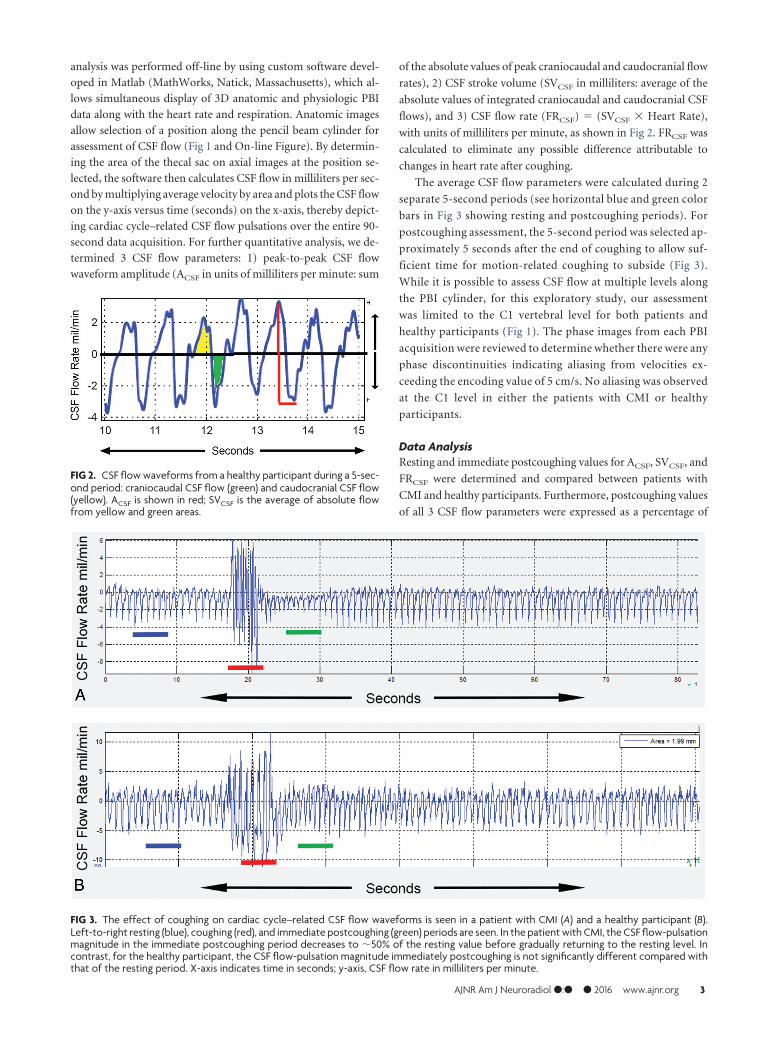
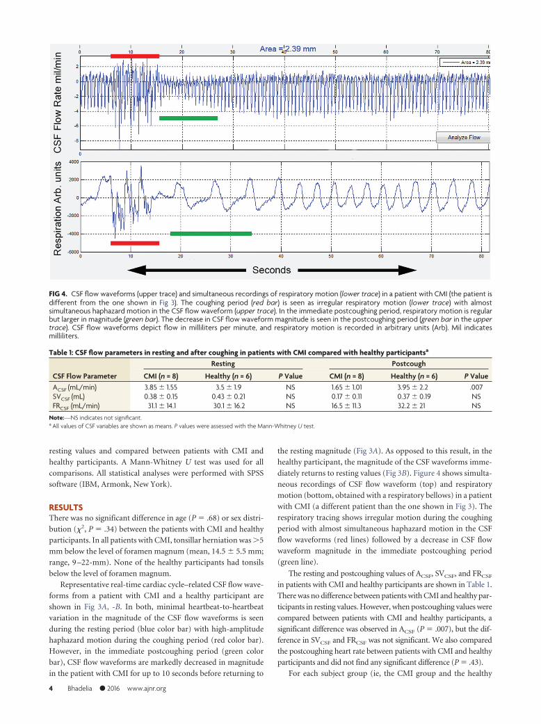
diately returns to resting values (Fig 3B). Figure 4 shows simulta-
neous recordings of CSF flow waveform (top) and respiratory
motion (bottom, obtained with a respiratory bellows) in a patient
with CMI (a different patient than the one shown in Fig 3). The
respiratory tracing shows irregular motion during the coughing
period with almost simultaneous haphazard motion in the CSF
flow waveforms (red lines) followed by a decrease in CSF flow
waveform magnitude in the immediate postcoughing period
(green line).
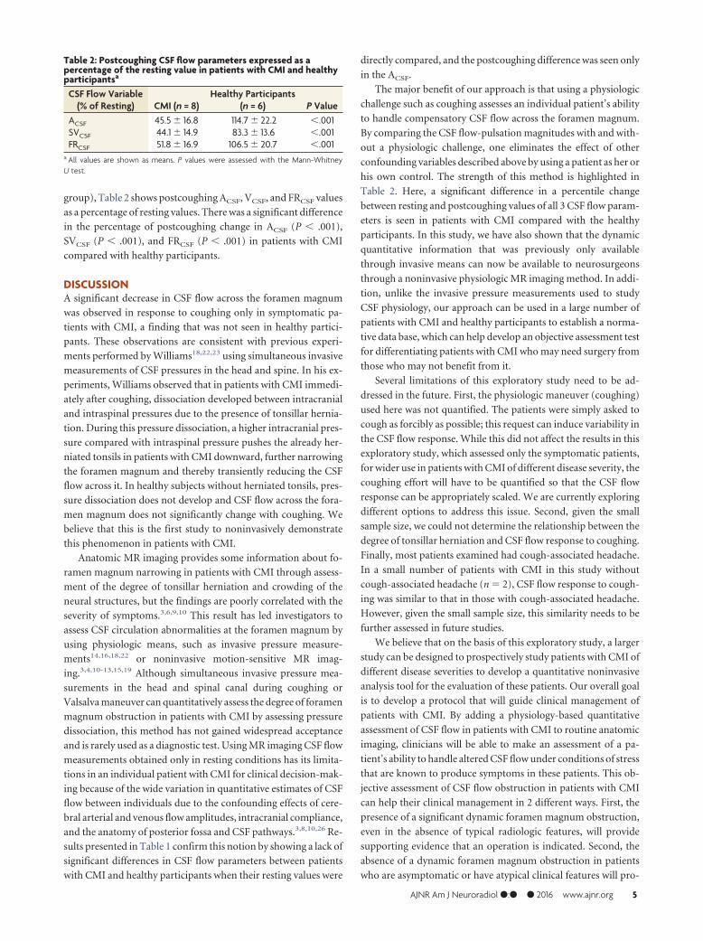
The resting and postcoughing values of ACSF, SVCSF, and FRCSF
in patients with CMI and healthy participants are shown in Table 1.
There was no difference between patients with CMI and healthy par-
ticipants in resting values. However, when postcoughing values were
compared between patients with CMI and healthy participants, a
significant difference was observed in ACSF (P � .007), but the dif-
ference in SVCSF and FRCSF was not significant. We also compared
the postcoughing heart rate between patients with CMI and healthy
participants and did not find any significant difference (P � .43).
For each subject group (ie, the CMI group and the healthy
FIG 4. CSF flow waveforms (upper trace) and simultaneous recordings of respiratory motion (lower trace) in a patient with CMI (the patient isdifferent from the one shown in Fig 3). The coughing period (red bar) is seen as irregular respiratory motion (lower trace) with almostsimultaneous haphazard motion in the CSF flow waveform (upper trace). In the immediate postcoughing period, respiratory motion is regularbut larger in magnitude (green bar). The decrease in CSF flow waveform magnitude is seen in the postcoughing period (green bar in the uppertrace). CSF flow waveforms depict flow in milliliters per minute, and respiratory motion is recorded in arbitrary units (Arb). Mil indicatesmilliliters.
Table 1: CSF flow parameters in resting and after coughing in patients with CMI compared with healthy participantsa
CSF Flow Parameter
Resting Postcough
CMI (n = 8) Healthy (n = 6) P Value CMI (n = 8) Healthy (n = 6) P ValueACSF (mL/min) 3.85 � 1.55 3.5 � 1.9 NS 1.65 � 1.01 3.95 � 2.2 .007SVCSF (mL) 0.38 � 0.15 0.43 � 0.21 NS 0.17 � 0.11 0.37 � 0.19 NSFRCSF (mL/min) 31.1 � 14.1 30.1 � 16.2 NS 16.5 � 11.3 32.2 � 21 NS
Note:—NS indicates not significant.a All values of CSF variables are shown as means. P values were assessed with the Mann-Whitney U test.
4 Bhadelia ● 2016 www.ajnr.org
group), Table 2 shows postcoughing ACSF, VCSF, and FRCSF values
as a percentage of resting values. There was a significant difference
in the percentage of postcoughing change in ACSF (P � .001),
SVCSF (P � .001), and FRCSF (P � .001) in patients with CMI
compared with healthy participants.
DISCUSSIONA significant decrease in CSF flow across the foramen magnum
was observed in response to coughing only in symptomatic pa-
tients with CMI, a finding that was not seen in healthy partici-
pants. These observations are consistent with previous experi-
ments performed by Williams18,22,23 using simultaneous invasive
measurements of CSF pressures in the head and spine. In his ex-
periments, Williams observed that in patients with CMI immedi-
ately after coughing, dissociation developed between intracranial
and intraspinal pressures due to the presence of tonsillar hernia-
tion. During this pressure dissociation, a higher intracranial pres-
sure compared with intraspinal pressure pushes the already her-
niated tonsils in patients with CMI downward, further narrowing
the foramen magnum and thereby transiently reducing the CSF
flow across it. In healthy subjects without herniated tonsils, pres-
sure dissociation does not develop and CSF flow across the fora-
men magnum does not significantly change with coughing. We
believe that this is the first study to noninvasively demonstrate
this phenomenon in patients with CMI.
Anatomic MR imaging provides some information about fo-
ramen magnum narrowing in patients with CMI through assess-
ment of the degree of tonsillar herniation and crowding of the
neural structures, but the findings are poorly correlated with the
severity of symptoms.3,6,9,10 This result has led investigators to
assess CSF circulation abnormalities at the foramen magnum by
using physiologic means, such as invasive pressure measure-
ments14,16,18,22 or noninvasive motion-sensitive MR imag-
ing.3,4,10-13,15,19 Although simultaneous invasive pressure mea-
surements in the head and spinal canal during coughing or
Valsalva maneuver can quantitatively assess the degree of foramen
magnum obstruction in patients with CMI by assessing pressure
dissociation, this method has not gained widespread acceptance
and is rarely used as a diagnostic test. Using MR imaging CSF flow
measurements obtained only in resting conditions has its limita-
tions in an individual patient with CMI for clinical decision-mak-
ing because of the wide variation in quantitative estimates of CSF
flow between individuals due to the confounding effects of cere-
bral arterial and venous flow amplitudes, intracranial compliance,
and the anatomy of posterior fossa and CSF pathways.3,8,10,26 Re-
sults presented in Table 1 confirm this notion by showing a lack of
significant differences in CSF flow parameters between patients
with CMI and healthy participants when their resting values were
directly compared, and the postcoughing difference was seen only
in the ACSF.
The major benefit of our approach is that using a physiologic
challenge such as coughing assesses an individual patient’s ability
to handle compensatory CSF flow across the foramen magnum.
By comparing the CSF flow-pulsation magnitudes with and with-
out a physiologic challenge, one eliminates the effect of other
confounding variables described above by using a patient as her or
his own control. The strength of this method is highlighted in
Table 2. Here, a significant difference in a percentile change
between resting and postcoughing values of all 3 CSF flow param-
eters is seen in patients with CMI compared with the healthy
participants. In this study, we have also shown that the dynamic
quantitative information that was previously only available
through invasive means can now be available to neurosurgeons
through a noninvasive physiologic MR imaging method. In addi-
tion, unlike the invasive pressure measurements used to study
CSF physiology, our approach can be used in a large number of
patients with CMI and healthy participants to establish a norma-
tive data base, which can help develop an objective assessment test
for differentiating patients with CMI who may need surgery from
those who may not benefit from it.
Several limitations of this exploratory study need to be ad-
dressed in the future. First, the physiologic maneuver (coughing)
used here was not quantified. The patients were simply asked to
cough as forcibly as possible; this request can induce variability in
the CSF flow response. While this did not affect the results in this
exploratory study, which assessed only the symptomatic patients,
for wider use in patients with CMI of different disease severity, the
coughing effort will have to be quantified so that the CSF flow
response can be appropriately scaled. We are currently exploring
different options to address this issue. Second, given the small
sample size, we could not determine the relationship between the
degree of tonsillar herniation and CSF flow response to coughing.
Finally, most patients examined had cough-associated headache.
In a small number of patients with CMI in this study without
cough-associated headache (n � 2), CSF flow response to cough-
ing was similar to that in those with cough-associated headache.
However, given the small sample size, this similarity needs to be
further assessed in future studies.
We believe that on the basis of this exploratory study, a larger
study can be designed to prospectively study patients with CMI of
different disease severities to develop a quantitative noninvasive
analysis tool for the evaluation of these patients. Our overall goal
is to develop a protocol that will guide clinical management of
patients with CMI. By adding a physiology-based quantitative
assessment of CSF flow in patients with CMI to routine anatomic
imaging, clinicians will be able to make an assessment of a pa-
tient’s ability to handle altered CSF flow under conditions of stress
that are known to produce symptoms in these patients. This ob-
jective assessment of CSF flow obstruction in patients with CMI
can help their clinical management in 2 different ways. First, the
presence of a significant dynamic foramen magnum obstruction,
even in the absence of typical radiologic features, will provide
supporting evidence that an operation is indicated. Second, the
absence of a dynamic foramen magnum obstruction in patients
who are asymptomatic or have atypical clinical features will pro-
Table 2: Postcoughing CSF flow parameters expressed as apercentage of the resting value in patients with CMI and healthyparticipantsa
CSF Flow Variable(% of Resting) CMI (n = 8)
Healthy Participants(n = 6) P Value
ACSF 45.5 � 16.8 114.7 � 22.2 �.001SVCSF 44.1 � 14.9 83.3 � 13.6 �.001FRCSF 51.8 � 16.9 106.5 � 20.7 �.001
a All values are shown as means. P values were assessed with the Mann-WhitneyU test.
AJNR Am J Neuroradiol ●:● ● 2016 www.ajnr.org 5
vide objective evidence to clinicians that these patients will likely
not benefit from the operation and should be managed by non-
surgical means.
CONCLUSIONSOur results provide preliminary evidence that the physiology-
based imaging method using real-time CSF flow imaging with PBI
has the potential to be an objective clinical test to differentiate
symptomatic from asymptomatic patients with CMI.
Disclosures: Rafeeque A. Bhadelia—RELATED: Grant: Conquer Chiari Foundation,*Comments: A grant was provided to perform MRI scans and develop an analysisprogram for the project. Samuel Patz—RELATED: Consulting Fee or Honorarium:Beth Israel Deaconess Medical Center, Comments: I was a consultant on a grantawarded to R. Bhadelia (first author) from the Conquer Chiari Foundation. The grantwas awarded to Beth Israel Deaconess Medical Center with Dr Bhadelia as the Prin-cipal Investigator. The consultant fees were paid to me directly by Beth Israel Dea-coness Medical Center. Yansong Zhao—UNRELATED: Employment: I am a ClinicalScientist of Philips Healthcare North America. Neel Madan—RELATED: Grant: Con-quer Chiari Foundation,* Comments: Seed grant, with money to pay for MRI acqui-sitions and to give a small amount of money to subjects. *Money paid to theinstitution.
REFERENCES1. Elster AD, Chen MY. Chiari I malformations: clinical and radiologic
reappraisal. Radiology 1992;183:347–53 CrossRef Medline2. Alden TD, Ojemann JG, Park TS. Surgical treatment of Chiari I
malformation: indications and approaches. Neurosurg Focus 2001;11:E2 Medline
3. Alperin N, Loftus JR, Oliu CJ, et al. MRI measures of posterior cra-nial fossa morphology and CSF physiology in Chiari malformationtype I. Neurosurgery 2014 Jul 18. [Epub ahead of print] Medline
4. Hofkes SK, Iskandar BJ, Turski PA, et al. Differentiation betweensymptomatic Chiari I malformation and asymptomatic tonsilar ec-topia by using cerebrospinal fluid flow imaging: initial estimate ofimaging accuracy. Radiology 2007;245:532– 40 CrossRef Medline
5. Meadows J, Kraut M, Guarnieri M, et al. Asymptomatic Chiari typeI malformations identified on magnetic resonance imaging. J Neu-rosurg 2000;92:920 –26 CrossRef Medline
6. Voelker R. Chiari conundrum: researchers tackle a brain puzzle forthe 21st century. JAMA 2009;301:147– 49 CrossRef Medline
7. Sekula RF Jr, Arnone GD, Crocker C, et al. The pathogenesis ofChiari I malformation and syringomyelia. Neurol Res 2011;33:232–39 CrossRef Medline
8. Tubbs RS, Beckman J, Naftel RP, et al. Institutional experience with500 cases of surgically treated pediatric Chiari malformation type I.J Neurosurg Pediatr 2011;7:248 –56 CrossRef Medline
9. Baisden J. Controversies in Chiari I malformations. Surg Neurol Int2012;3(suppl 3):S232–37 CrossRef Medline
10. Alperin N, Kulkarni K, Loth F, et al. Analysis of magnetic resonanceimaging-based blood and cerebrospinal fluid flow measurementsin patients with Chiari I malformation: a system approach. Neuro-surg Focus 2001;11:E6 Medline
11. Armonda RA, Citrin CM, Foley KT, et al. Quantitative cine-modemagnetic resonance imaging of Chiari I malformations: an analysisof cerebrospinal fluid dynamics. Neurosurgery 1994;35:214 –23; dis-cussion 223–24 Medline
12. Bhadelia RA, Bogdan AR, Wolpert SM, et al. Cerebrospinal fluidflow waveforms: analysis in patients with Chiari I malformation bymeans of gated phase-contrast MR imaging velocity measurements.Radiology 1995;196:195–202 CrossRef Medline
13. Haughton VM, Korosec FR, Medow JE, et al. Peak systolic and dia-stolic CSF velocity in the foramen magnum in adult patients withChiari I malformations and in normal control participants. AJNRAm J Neuroradiol 2003;24:169 –76 Medline
14. Oldfield EH, Muraszko K, Shawker TH, et al. Pathophysiology ofsyringomyelia associated with Chiari I malformation of the cere-bellar tonsils: implications for diagnosis and treatment. J Neurosurg1994;80:3–15 CrossRef Medline
15. Quigley MF, Iskandar B, Quigley ME, et al. Cerebrospinal fluid flowin foramen magnum: temporal and spatial patterns at MR imagingin volunteers and in patients with Chiari I malformation. Radiology2004;232:229 –36 CrossRef Medline
16. Sansur CA, Heiss JD, DeVroom HL, et al. Pathophysiology of head-ache associated with cough in patients with Chiari I malformation.J Neurosurg 2003;98:453–58 CrossRef Medline
17. Williams B. Cerebrospinal fluid pressure changes in response tocoughing. Brain 1976;99:331– 46 CrossRef Medline
18. Williams B. Cough headache due to craniospinal pressure dissocia-tion. Arch Neurol 1980;37:226 –30 CrossRef Medline
19. Krueger KD, Haughton VM, Hetzel S. Peak CSF velocities in patientswith symptomatic and asymptomatic Chiari I malformation. AJNRAm J Neuroradiol 2010;31:1837– 41 CrossRef Medline
20. Bhadelia RA, Madan N, Zhao Y, et al. Physiology-based MR imagingassessment of CSF flow at the foramen magnum with a valsalvamaneuver. AJNR Am J Neuroradiol 2013;34:1857– 62 CrossRefMedline
21. Tachibana S, Iida H, Yada K. Significance of positive Queckenstedttest in patients with syringomyelia associated with Arnold-Chiarimalformations. J Neurosurg 1992;76:67–71 CrossRef Medline
22. Williams B. Simultaneous cerebral and spinal fluid pressure record-ings, 2: cerebrospinal dissociation with lesions at the foramen mag-num. Acta Neurochir (Wien) 1981;59:123– 42 CrossRef Medline
23. Williams B. Simultaneous cerebral and spinal fluid pressure record-ings, I: technique, physiology, and normal results. Acta Neurochir(Wien) 1981;58:167– 85 CrossRef Medline
24. Hardy CJ, Pearlman JD, Moore JR, et al. Rapid NMR cardiographywith a half-echo M-mode method. J Comput Assist Tomogr 1991;15:868 –74 CrossRef Medline
25. Maier SE, Hardy CJ, Jolesz FA. Brain and cerebrospinal fluidmotion: real-time quantification with M-mode MR imaging. Radi-ology 1994;193:477– 83 CrossRef Medline
26. Bhadelia RA, Bogdan AR, Kaplan RF, et al. Cerebrospinal fluid pul-sation amplitude and its quantitative relationship to cerebral bloodflow pulsations: a phase-contrast MR flow imaging study. Neurora-diology 1997;39:258 – 64 CrossRef Medline
6 Bhadelia ● 2016 www.ajnr.org
Related Documents