J. Comp. Path. 2007, Vol. 137, 179^210 REVIEW Corynebacterium pseudotuberculosis and its Role in Ovine Caseous Lymphadenitis G. J. Baird * and M. C. Fontaine y * Scottish Agricultural CollegeVeterinary Services, 5 Bertha ParkView, Perth PH1 3FZ, and y Moredun Research Institute, International Research Centre, Pentlands Science Park, Bush Loan, Penicuik EH26 0PZ, Scotland, UK Summary Caseous lymphadenitis (CLA) of sheep, caused by Corynebacterium pseudotuberculosis, has been a signi¢cant disease in the majority of sheep-rearing regions for over a century. Because of the chronic and often sub-clinical nature of the infection, it has proved di⁄cult to control and prevalence is high in many parts of the world, which in turn leads to signi¢cant economic losses for farmers. This review describes the important characteristics of C. pseudotu- berculosis and examines the pathogenesis and epidemiology of the infection in sheep.The review also discusses the immune response to infection and describes the methods that have been developed to control CLA, with particu- lar emphasis on the use of vaccination and serological testing. Crown Copyright r 2007 Published by Elsevier Ltd. All rights reserved. Keywords: bacterial infection; caseous lymphadenitis; Corynebacterium pseudotuberculosis; review; sheep Contents Introduction ........................................................................................... 180 History ............................................................................................... 180 Geographical Distribution ............................................................................... 180 Global Prevalence of CLA ............................................................................... 181 Bacterial Characteristics ................................................................................. 181 Classi¢cation .................................................................................... 181 Virulence Factors ................................................................................. 182 Clinical and Pathological Features of Caseous Lymphadenitis in Sheep ........................................... 184 Pathogenesis in Sheep ................................................................................... 185 Economic Signi¢cance of Disease in Sheep .................................................................. 187 Epidemiology of Infection in Sheep ........................................................................ 188 Transmission of Infection .......................................................................... 188 Comparison with Goat Infection .................................................................... 189 Corynebacterium pseudotuberculosis Infections in Other Animal Species ............................................. 189 Zoonotic Infections ............................................................................... 190 Control and Eradication of Disease in Sheep................................................................. 191 Diagnosis ....................................................................................... 191 Treatment ....................................................................................... 193 Serology ........................................................................................ 193 Vaccination...................................................................................... 194 Caseous Lymphadenitis in the United Kingdom.............................................................. 200 Future Direction ....................................................................................... 201 www.elsevier.com/locate/jcpa ARTICLE IN PRESS 0021-9975/$ - see front matter Crown Copyright r 2007 Published by Elsevier Ltd. All rights reserved. doi: 10.1016/j.jcpa.2007.07.002 Correspondence to: G.J. Baird (e-mail: [email protected])

Welcome message from author
This document is posted to help you gain knowledge. Please leave a comment to let me know what you think about it! Share it to your friends and learn new things together.
Transcript
ARTICLE IN PRESS
J. Comp. Path. 2007,Vol.137,179^210
0021-9975/$ - see fdoi:10.1016/j.jcpa.
Correspondence
REVIEW
Corynebacterium pseudotuberculosis and its Role inOvine Caseous Lymphadenitis
www.elsevier.com/locate/jcpa
G. J. Baird* and M. C. Fontainey
*Scottish Agricultural CollegeVeterinary Services, 5 Bertha ParkView, Perth PH13FZ, and yMoredun Research Institute,
International Research Centre, Pentlands Science Park, Bush Loan, Penicuik EH26 0PZ, Scotland, UK
Summary
Caseous lymphadenitis (CLA) of sheep, caused by Corynebacterium pseudotuberculosis, has been a signi¢cant diseasein themajority of sheep-rearing regions for over a century. Because of the chronic and often sub-clinical nature ofthe infection, it has proved di⁄cult to control and prevalence is high in many parts of the world, which in turnleads to signi¢cant economic losses for farmers.This review describes the important characteristics of C. pseudotu-berculosis and examines the pathogenesis and epidemiology of the infection in sheep.The review also discusses theimmune response to infection and describes the methods that have been developed to control CLA, with particu-lar emphasis on the use of vaccination and serological testing.
Crown Copyrightr 2007 Published by Elsevier Ltd. All rights reserved.
Keywords: bacterial infection; caseous lymphadenitis; Corynebacterium pseudotuberculosis; review; sheep
Contents
Introduction. . . . . . . . . . . . . . . . . . . . . . . . . . . . . . . . . . . . . . . . . . . . . . . . . . . . . . . . . . . . . . . . . . . . . . . . . . . . . . . . . . . . . . . . . . . 180History . . . . . . . . . . . . . . . . . . . . . . . . . . . . . . . . . . . . . . . . . . . . . . . . . . . . . . . . . . . . . . . . . . . . . . . . . . . . . . . . . . . . . . . . . . . . . . . 180Geographical Distribution . . . . . . . . . . . . . . . . . . . . . . . . . . . . . . . . . . . . . . . . . . . . . . . . . . . . . . . . . . . . . . . . . . . . . . . . . . . . . . . 180Global Prevalence of CLA . . . . . . . . . . . . . . . . . . . . . . . . . . . . . . . . . . . . . . . . . . . . . . . . . . . . . . . . . . . . . . . . . . . . . . . . . . . . . . . 181Bacterial Characteristics . . . . . . . . . . . . . . . . . . . . . . . . . . . . . . . . . . . . . . . . . . . . . . . . . . . . . . . . . . . . . . . . . . . . . . . . . . . . . . . . . 181
Classi¢cation . . . . . . . . . . . . . . . . . . . . . . . . . . . . . . . . . . . . . . . . . . . . . . . . . . . . . . . . . . . . . . . . . . . . . . . . . . . . . . . . . . . . 181Virulence Factors . . . . . . . . . . . . . . . . . . . . . . . . . . . . . . . . . . . . . . . . . . . . . . . . . . . . . . . . . . . . . . . . . . . . . . . . . . . . . . . . . 182
Clinical and Pathological Features of Caseous Lymphadenitis in Sheep . . . . . . . . . . . . . . . . . . . . . . . . . . . . . . . . . . . . . . . . . . . 184Pathogenesis in Sheep . . . . . . . . . . . . . . . . . . . . . . . . . . . . . . . . . . . . . . . . . . . . . . . . . . . . . . . . . . . . . . . . . . . . . . . . . . . . . . . . . . . 185Economic Signi¢cance of Disease in Sheep . . . . . . . . . . . . . . . . . . . . . . . . . . . . . . . . . . . . . . . . . . . . . . . . . . . . . . . . . . . . . . . . . . 187Epidemiology of Infection in Sheep . . . . . . . . . . . . . . . . . . . . . . . . . . . . . . . . . . . . . . . . . . . . . . . . . . . . . . . . . . . . . . . . . . . . . . . . 188
Transmission of Infection . . . . . . . . . . . . . . . . . . . . . . . . . . . . . . . . . . . . . . . . . . . . . . . . . . . . . . . . . . . . . . . . . . . . . . . . . . 188Comparisonwith Goat Infection . . . . . . . . . . . . . . . . . . . . . . . . . . . . . . . . . . . . . . . . . . . . . . . . . . . . . . . . . . . . . . . . . . . . 189
Corynebacterium pseudotuberculosis Infections in OtherAnimal Species . . . . . . . . . . . . . . . . . . . . . . . . . . . . . . . . . . . . . . . . . . . . . 189Zoonotic Infections . . . . . . . . . . . . . . . . . . . . . . . . . . . . . . . . . . . . . . . . . . . . . . . . . . . . . . . . . . . . . . . . . . . . . . . . . . . . . . . 190
Control and Eradication of Disease in Sheep. . . . . . . . . . . . . . . . . . . . . . . . . . . . . . . . . . . . . . . . . . . . . . . . . . . . . . . . . . . . . . . . . 191Diagnosis . . . . . . . . . . . . . . . . . . . . . . . . . . . . . . . . . . . . . . . . . . . . . . . . . . . . . . . . . . . . . . . . . . . . . . . . . . . . . . . . . . . . . . . 191Treatment . . . . . . . . . . . . . . . . . . . . . . . . . . . . . . . . . . . . . . . . . . . . . . . . . . . . . . . . . . . . . . . . . . . . . . . . . . . . . . . . . . . . . . . 193Serology . . . . . . . . . . . . . . . . . . . . . . . . . . . . . . . . . . . . . . . . . . . . . . . . . . . . . . . . . . . . . . . . . . . . . . . . . . . . . . . . . . . . . . . . 193Vaccination. . . . . . . . . . . . . . . . . . . . . . . . . . . . . . . . . . . . . . . . . . . . . . . . . . . . . . . . . . . . . . . . . . . . . . . . . . . . . . . . . . . . . . 194
Caseous Lymphadenitis in the United Kingdom. . . . . . . . . . . . . . . . . . . . . . . . . . . . . . . . . . . . . . . . . . . . . . . . . . . . . . . . . . . . . . 200Future Direction . . . . . . . . . . . . . . . . . . . . . . . . . . . . . . . . . . . . . . . . . . . . . . . . . . . . . . . . . . . . . . . . . . . . . . . . . . . . . . . . . . . . . . . 201
ront matter Crown Copyrightr 2007 Published by Elsevier Ltd. All rights reserved.2007.07.002
to: G.J. Baird (e-mail: [email protected])
ARTICLE IN PRESS
G.J. Baird and M.C. Fontaine180
Introduction
The perceived importance of caseous lymphadenitis(CLA) as a disease of sheep varies greatly around theworld. Thus, in the UK, where this disease appearedfor the ¢rst time in the early 1990s, the steady and ap-parently relentless spread of infection in sheep andgoats has prompted great concern amongst both farm-ers and veterinary surgeons. However, in the sheep in-dustries of Australia and New Zealand infection isregarded as relatively minor, albeit widespread. Theroots of these contrasting attitudes lie in di¡erences indisease prevalence, available means of control, andmeat inspection practices.To the veterinary pathologist, the causative organ-
ism of CLA, Corynebacterium pseudotuberculosis, has a va-lid claim to being the ‘‘perfect parasite’’. Oncesuccessfully established within the host, this pathogenwill evade the immune system with apparent ease. Asa result, chronic infections may last for most or all ofan animal’s life, although they are rarely fatal. If leftunchecked, the diseasemay infect themajority of sheepin a £ock. Even away from its mammalian host, the or-ganism is well equipped for long-term survival in theenvironment. As a result of these formidable character-istics, £ock infections by C. pseudotuberculosis are gener-ally ‘‘managed’’, rather than eradicated.
History
In 1888, the French bacteriologist Edward Nocard iso-lated an unusual organism from a case of lymphangitisin a cow (Nocard,1896). Some 3 years later, the Bulgar-ian bacteriologist Hugo von Pre|« sz identi¢ed a similarbacterium in cultures from a renal abscess in a ewe(Pre|« sz and Guinard, 1891). As a consequence of theserelated discoveries, the organism in question becameknown as the ‘‘Pre|« sz^Nocard’’ bacillus, a vernacularname with which it was linked for decades thereafter.Towards the end of the 19th century the bacterium
was describedby theGermanbacteriologists Lehmannand Neumann in the ¢rst edition of their bacteriologi-cal atlas (Lehmann and Neumann,1896). In that pub-lication, the Pre|« sz^Nocard bacillus was renamedBacillus pseudotuberculosisça derivation of the Greekpseudes tuberculosis or ‘‘false tuberculosis’’and a referenceto the supposed clinical similarity of the lesions to case-ous nodules of mycobacterial tuberculosis. In the FirstEdition of Bergey’s Manual of Determinative Bacteriology,published in1923, the organismwas placed in the Cory-nebacteriumgenus, which hadoriginally been createdas acategory for the human pathogen Corynebacterium
diphtheriae. This edition of the Manual referred to workshowing that B. pseudotuberculosis resembled C. diphther-
iae in morphology and cell wall composition, leading
to a further change of name to Corynebacterium ovis. Sub-sequently, however, the organism was isolated frompurulent infections and ulcerative lymphangitis inother mammalian species, including goats, horses andhuman beings. In recognition of this, by the time theSixth Edition of Bergey’s Manual was published in1948, the species name had been changed back fromovis to the earlier designation of pseudotuberculosis. Sincethat point, the o⁄cially recognized designation of Cor-ynebacterium pseudotuberculosis has remained consistent(Euzeby, 2005), notwithstanding the fact that severalauthors continued to refer to the organism as C. ovis un-til the1980s.
Geographical Distribution
The global range of C. pseudotuberculosis re£ects that offarmed small ruminants, the bacterium having beenidenti¢ed in Europe, Australasia, North and SouthAmerica, Africa and the Middle East (Robins, 1991;Paton et al., 2005). In many of these countries CLA hasbeen an established and economically important infec-tion of livestock, particularly sheep, for decades. Asearly as the 1930s it was acknowledged that the disease‘‘very extensively a¡ected’’ the £ocks of most mutton-exporting countries (Cesari, 1930). It is surprisingtherefore, that the ¢rst diagnosed case in an animal ori-ginating from the UK was not con¢rmed until as re-cently as1989 (Lloyd et al.,1990; Meldrum,1990).Some suggestions have been made that the origins of
the infection may lie in Europe, and that spread of C.pseudotuberculosis around the world followed the expor-tation of sheep by the 18th-century colonial powers.The Merino breed, which originated in Spain, waswidely valued as a dual meat and wool animal andwas exported extensively, ¢rst to South Africa and sub-sequently to Australia and the Americas. It has beensuggested that this early exportation may, at the veryleast, have assisted in the spread of C. pseudotuberculosis(Paton, 2000). Such a theory is di⁄cult to prove, butsupporting evidence might be provided by demonstra-tion of a close genotypic relationship between isolatesfrom di¡erent parts of the world. Certainly, it is inter-esting to speculate that the absence of the Merino as acommercial breed in the British Isles may have gonesomeway to protecting this country from the infection.CLA ¢rst became a signi¢cant political issue in the
1920s when mutton carcases, imported into Great Brit-ain from a number of countries, were frequently foundto be a¡ected by the disease. It was noted at the timethat evidence of infection would often remain unob-served at import meat inspection ‘‘only to be revealedwhen the roast leg of mutton appears on the table andis cut through’’ (Rolleston andWooldridge,1926). Mut-ton from Argentina was particularly badly a¡ected,
ARTICLE IN PRESS
Ovine Caseous Lymphadenitis 181
forcing the British Government of the time to take ac-tion in relation to consignments of carcases from thatcountry (Anon, 1929). The extent of the problem wasdemonstrated by a report that in a single season 9770mutton carcases (representing 27% of the totalthroughput) were rejected at one Patagonian meatplant because of CLA (Mills, 1928). Other mutton-ex-porting countries took heed of the concerns in Britainand it was soon acknowledged that ‘‘Argentina, Uru-guay, Chili (sic), Australia and New Zealand are ac-tively occupiedyin a struggle against this tenaciouschronic disease which, completely neglected up tonow, is very extensively a¡ecting the £ocks of thosecountries’’ (Cesari, 1930). These concerns also stimu-lated veterinary research, principally in Australia,leading to important early discoveries on pathogenesis,bacterial survival and risk factors (Seddon et al., 1929;Bull and Dickinson,1935).Research interest in CLAwas renewed in the 1970s,
when the authorities in the USA, Canada and Japanapplied strict regulations relating to the presence of le-sions in imported sheep carcases. A further series ofstudies into disease pathogenesis and epidemiologywas then initiated in Australia, which, as a major ex-porter of sheep and a high CLA prevalence within itsnational £ock, had much to lose from the stricter regu-latory regime. This in turn led to the formulation ofcontrol strategies aimed at reducing disease prevalenceand provided a catalyst for developments in the ¢eld ofvaccination.
Global Prevalence of CLA
C. pseudotuberculosis is recognized as having aworldwidedistribution and it is accepted that CLA is present inthe majority of the sheep-rearing areas. However,farm- and abattoir-based research aimed at establish-ing disease prevalence rates has been limited to rela-tively few countries. The average prevalence of CLAamongst adult sheep inWestern Australiawas recordedas 58% in1973, andas 53% in1984 (Batey,1986a). In anAustralian abattoir survey, 54% of adult ewes and3.4% of lambs showed evidence of infection at meat in-spection (Batey,1986b). In another abattoir survey, in-dividual £ocks in Tasmania and Western Australiademonstrated prevalence levels within the adult popu-lation as high as 61% (Middleton et al., 1991). Subse-quent surveys tended to identify steadily decreasingprevalence rates, this generally being attributed to theintroduction of a CLAvaccine in 1983 and its increas-ing acceptance within the farming community. A ¢eldstudy of 412 sheep £ocks, once again inWestern Austra-lia, recorded an average prevalence of 45% (Pepinet al.,1994a). A combined abattoir and postal survey offarmers, conducted in 2002, suggested that the average
prevalence had fallen to 20% in Western Australia,23% inVictoria and 29% in New SouthWales (Patonet al., 2003).A survey conducted in 1986/1987 by meat inspectors
inNewZealand identi¢ed lesions of CLA in 7.1%of theadult sheep slaughtered and 0.64% of lambs (Nuttall,1988). CLAwas also identi¢ed as the leading cause ofsheep carcase condemnation in South African abat-toirs (Collett et al., 1994) where losses of between0.24% and 0.3% of all sheep carcases were attributedto CLA and substantial additional losses were incurreddue to carcase trimming (Paton et al., 2005). In thewes-tern USA, average disease prevalence amongst adultewes was estimated to be as great as 42.5% (Stoopset al.,1984). Similar studies have been conducted in theCanadian province of Quebec, where the prevalence ofclinical CLA was found to range from 21% to 36%amongst culled adult sheep (Arsenault et al., 2003).Abattoir statistics from Alberta indicated that up to5% of mutton carcases and 0.03% of lamb carcaseswere condemned due to CLA, and that a further 8%of all carcases were trimmed to remove CLA lesions(Stanford et al.,1998). Unfortunately, similar publishedstatistics do not exist in respect of Europe. In the UK,where small abattoir surveys of CLA prevalence havebeen carried out, the results have been of limited scopeand signi¢cance (Mechie,1998).
Bacterial Characteristics
Classification
The Corynebacteriaceae are now considered to belongto the Actinomycetaceae, a family that also containsthe Mycobacterium, Rhodococcus and Nocardia genera(Clarridge and Spiegel, 1995). C. pseudotuberculosis pos-sesses many of the classical features of its genus (Collinsand Cummins, 1986). It consists of non-motile pleo-morphic rods (0.5^0.6 mm by 1.0^3.0 mm) that areGram-positive, although such staining may sometimesbe irregular. Groups of the bacteria tend to show acharacteristic palisade or ‘‘Chinese letter’’arrangementin smears.At a temperature of 37 1C, C. pseudotuberculosis will
grow under aerobic or anaerobic conditions. On solidmedia, the bacterial colonies are pale in colour, dryand friable in consistency, and may be moved freelyover the surface of the agar with the point of a probe(Quinn et al.,1994). After incubation for 24 h, small yel-lowish colonies will appear, increasing to a diameter of1^2mm after 48 h (Coyle et al.,1985). Bacterial growthbene¢ts from the addition of serum or whole blood tonutrient media. When whole blood is used, a narrowband of b-haemolysis is seen around each colony,
ARTICLE IN PRESS
G.J. Baird and M.C. Fontaine182
although it may appear only after incubation for48^72 h.When grown in liquid media or when in aqueous
suspension, C. pseudotuberculosis has a tendency to formclumps. This has been related to the presence of long-chain 2-branched 3-hydroxy fatty acids (so-called ‘‘my-colic acids’’), on the outside of the cellwall (Carne et al.,1956). Mycolic acids were ¢rst identi¢ed in 1939, in thetubercle bacillus (Asselineau and LaneŁ elle, 1998) andwere subsequently found to be a feature common tothe actinomycete family as awhole. Mycolic acids maybe solvent-extracted from C. pseudotuberculosis withoutimpairing the viability of the organism (Carne et al.,1956).The so-called ‘‘chemotaxonomic’’ approach to the
classi¢cation of bacteria, based on analysis of the che-mical composition of the cell wall, was at one time usedrelatively commonly. Analysis of cell wall mycolic acidsgreatly aided clari¢cation of the taxonomy of actino-mycetes, especially those of the genera Corynebacterium,Mycobacterium, Rhodococcus and Nocardia (Minnikinet al., 1975; Goodfellow et al., 1976; Keddie and Cure,1977; Minnikin and Goodfellow, 1980; Collins et al.,1982). In these studies, thin-layer (pyrolysis) chromato-graphic analysis of mycolic acid revealed that fatty acidchain length varied according to the genus and to a les-ser extent the species. It was shown that mycobacterialmycolic acids normally consist of chain lengths of be-tween 60 and 90 C14 atoms, and may possess a numberof distinct functional groups (Minnikin et al., 1978). Incontrast, nocardiae and rhodococciwere shown to pos-sess shorter mycolic acids, consisting of between 36 and66 C14 atoms, and with fewer functional groups (Min-nikin and Goodfellow,1976).Themycolic acids of cory-nebacteria were found to be even smaller, beingbetween 20 and 36 C14 atoms in length, and usually sa-turated or containing a single double bond (Minnikinet al.,1978). Collins et al. (1982) reported that, consistentwith the mycolic acid classi¢cation of corynebacteria,strains of C. pseudotuberculosis possessed mycolic acidswith carbon chain lengths of between C26 and C36,which contained predominantly saturated C14 side-chains (Collins et al.,1982).There are few published data on the resistance of C.
pseudotuberculosis to chemical disinfectants. However,most common disinfectants, including calcium hypo-chlorite, formalin and cresol solution, appear to be ef-fective in killing the organism, but the presence oforganic material necessitates increased exposure time(Ismail and Hamid, 1972). This protective e¡ect isclearly signi¢cant for a pathogen commonly foundwithin a thick matrix of purulent debris.The organismis capable of surviving in commercial sheep dip solu-tions for 24 h or more, a point of relevance to diseasecontrol (Nairn and Robertson,1974).
Early reports showed that C. pseudotuberculosis isolatesfrom di¡erent mammalian species shared identicalbiochemical characteristics, with the exception of ni-trate reduction. Thus, the majority of isolates fromhorses and cattle reduced nitrate to nitrite, while thosefrom sheep andgoats did not (Knight,1969; Sutherlandet al.,1996).This led to the proposal of two distinct bio-types or subspecies (Biberstein et al., 1971). Based onthis property of nitrate reduction, Songer et al. (1988)proposed the designations C. pseudotuberculosis biovarovis (biotype1) and C. pseudotuberculosis biovar equi (bio-type 2). However, the isolation in recent years ofnitrate-negative strains of C. pseudotuberculosis fromcattle (Yeruham et al., 1997) and from horses (Connoret al., 2000) suggests that such categorization may beunsatisfactory.A further minor di¡erence between certain isolates
lies in the area of antibiotic sensitivity. In a comparisonof susceptibility to17 di¡erent antimicrobial agents, theminimum inhibitory concentration of amikacin washigher for nitrate-negative sheep and goat isolates thanfor nitrate-positive equine and bovine isolates (Costaet al., 1998); however, the signi¢cance of this ¢nding isnot clear.
Virulence Factors
No avirulent strain of C. pseudotuberculosis has yet beendescribed; however, the organisms virulence mechan-isms remainpoorly understood. Since noplasmids havebeen identi¢ed in isolates of C. pseudotuberculosis, the ab-sence of plasmid-encoded virulence determinants mustbe assumed. To date, research has focussed mainly ontwo known virulence factors identi¢ed as phospholi-pase D andmycolic acids.The genome of C. pseudotuber-culosis, unlike that of a number of other bacterialpathogens, has yet to be fully sequenced; as a result,there is at present no opportunity to identify novel genesequences that may encode other virulence factors.Phospholipase D. The designation ‘‘phospholipase’’ is
used to describe a varied group of enzymes able to hy-drolyse one or more ester linkages in glycerophospholi-pids; the letters A-D are used to distinguish betweenphospholipases and to denote the speci¢c phospholipidester bond that is cleaved (Ansell and Hawthorne,1964). In eukaryotic cells, phospholipase enzymes playa role in signal transduction and normal membranemaintenance. Some mammalian cells also producephospholipases as part of the cellular in£ammatory re-sponse (Schmiel and Miller, 1999). Eukaryotic cellmembranes are composed of proteins and lipids, whichconstitute a signi¢cant target of attack during micro-bial invasion of host tissues. As part of their invasive ar-senal, many microbes have developed their ownphospholipase enzymes, which may be used to hydro-
ARTICLE IN PRESS
Ovine Caseous Lymphadenitis 183
lyse phosphate bonds within membrane phospholipids(Ghannoum, 2000).The result of this is the damage ordestruction of host cell membranes, which in turn maylead to their dysfunction or disruption, or both (SalyersandWitt, 1994).Various bacterial genera are known tosecrete phospholipase enzymes, and in some cases thesehave been shown to play a role in virulence (Schmieland Miller, 1999). Phospholipase D (PLD) has beenidenti¢ed as a potent exotoxin in C. pseudotuberculosis,and a key virulence factor in the development ofCLA.PLD in this organism was ¢rst characterized by
Carne (1940) and has since been detected in every iso-late of C. pseudotuberculosis that has been studied, includ-ing isolates of both of the suggested biotypes, and allknown strains of the organism recovered from infectedmammalian species (Songer et al., 1988). The conten-tion that PLD represents a signi¢cant virulence factoris supported by much experimental evidence. Isolatesof C. pseudotuberculosis in which the pld gene, encodingPLD, has been deleted from the chromosome or ren-dered inactive by mutation are incapable of causingthe classic lymph node abscesses of CLA in sheep(Hodgson et al.,1992;McNamara et al.,1994). Similarly,the presence of speci¢c antibody to PLD greatly limitsthe progress of the clinical disease.PLD is de¢ned as a sphingomyelin-speci¢c phospho-
lipase that catalyses the dissociation of sphingomyelininto ceramide phosphate and choline (Bernheimeret al., 1980; Pepin et al., 1994a). The C. pseudotuberculosispld gene has been cloned and sequenced. Analysisreveals that it encodes a protein of some 31.4 kDa, pre-ceded by a probable secretory signal sequence of2.7 kDa (Hodgson et al., 1990). The relatively large sizeof the protein molecule assists in its puri¢cation in thelaboratory and enables large quantities to be collected(Egen et al., 1989). Several biological activities havebeen reported for PLD, including dermonecrosis (Carne,1940; Muckle and Gyles,1986), lethality (Brogden andEngen,1990), synergistic lysis of erythrocytes in the pre-sence of an extracellular Rhodococcus equi factor (Fraser,1961), and inhibition of staphylococcal lysin-induced ly-sis of erythrocytes (Zaki,1976); the two latter activitiesare employedas laboratory tests for the identi¢cation ofC. pseudotuberculosis. PLD also interferes with ovine neu-trophil chemotaxis and is lethal to the cells themselves(Yozwiak and Songer,1993). In terms of the signi¢canceof PLD as a virulence factor, the activity that has beenthe focus of most interest is the increase in vascular en-dothelial membrane permeability engendered by thehydrolysis of sphingomyelin.This increasedpermeabil-ity leads to the leakage of plasma from blood vesselsand into the surrounding tissues, and from there intothe lymphatic drainage (Jolly, 1965; Carne and Onon,1978). This e¡ect may assist pathogenesis by favouring
the lymphatic drainage of C. pseudotuberculosis in tissue£uid (Batey,1986c).PLD may assist the organism at the site of initial in-
fection in other ways. It is known to activate the com-plementary pathway of the innate immune system,thereby depleting complement in the region surround-ing the invadingbacteria andprotecting them fromop-sonization (Yozwiak and Songer, 1993). It may alsoimpair the chemotaxis of neutrophils and, as a conse-quence, decrease the likelihood of phagocytosis earlyin infection (Yozwiak and Songer, 1993). In some re-spects this suggestion is at odds with other theories ofpathogenesis, which propose that in the early stages ofdisease the organism parasitizes phagocytic cells andmultiplies within them. Indeed, other authors have in-dicated that PLD may play a role in the escape of thebacterium fromwithin macrophages. It is possible thatthis role is related to the action of PLD on the innerphospholipid layers of the macrophage cell membrane,as indicated by comparable observations on other bac-terial infections (Titball,1993).Exotoxins with similarities to the PLD of C. pseudotu-
berculosis are known to be important virulence factorsfor other bacterial pathogens. For example, there is97% homology between C. pseudotuberculosis PLD andthe active exotoxin of C. ulcerans, a rare cause of hu-man diphtheria (McNamara et al., 1995). A PLD-likeexotoxin is also considered to be an important viru-lence factor in Pseudomonas aeruginosa (Wildermanet al., 2001). Likewise, pld shows homology to the ymt
gene from the plague-causing organism Yersinia pestis
(Hinnebusch et al., 2000). PLDalso demonstrates an in-triguing similarity in structure and biological activityto an enzyme toxin produced by the venomous NorthAmericanbrown recluse spider, Loxosceles reclusa (Bern-heimer et al., 1985). This toxin induces dermonecrosis,haemolysis, platelet-aggregation and, on rare occa-sions, fatal renal failure (Lee and Lynch, 2005). Simi-larly, Hsu et al. (1985) showed that severe haemolyticcrises resulted from the injection of C. pseudotuberculosisculture supernates or crudely puri¢ed exotoxin pre-parations into small ruminantsMycolic acid. C. pseudotuberculosis does not produce a pro-tective capsulebut has insteadawaxymycolic acidcoaton the cell wall surface (described above).This coat haswell-established cytotoxic properties, which play a ma-jor role in pathogenicity (Hard, 1972; Muckle andGiles,1983;Tashjian and Campbell,1983).The subcuta-neous injection into mice of mycolic acid extractedfrom C. pseudotuberculosis results in the production of alocalized swelling, with congestion and a central areaof haemorrhagic necrosis. In addition, mycolic acid in-duces degenerative changes and death in phagocytiz-ing leucocytes (Carne et al.,1956). However, unlike thelethal e¡ect of injection of similar molecules extracted
ARTICLE IN PRESS
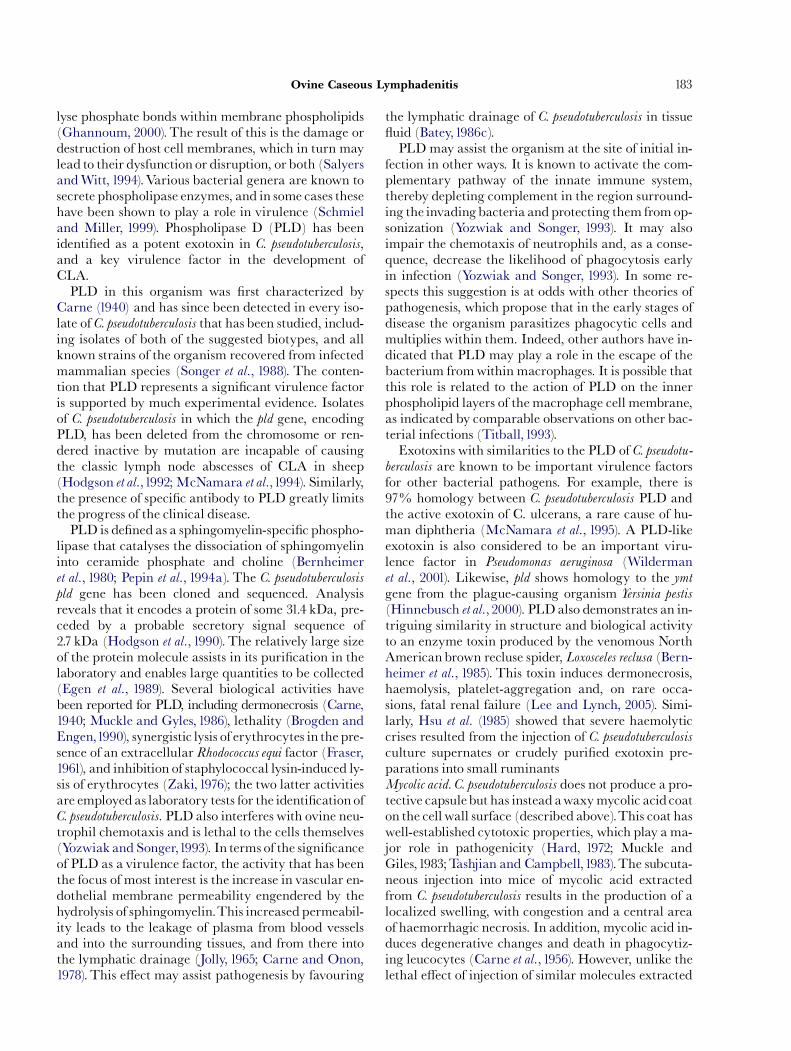
Fig.1. Acaseous lymphadenitis (CLA) abscess in the parotid lymphnode of an adult ewe.
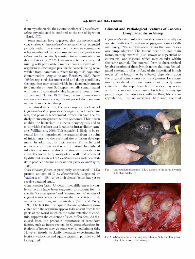
Fig. 2. CLA abscesses in the lung parenchyma. Note the close proxi-mity of the lesion to the airways.
G.J. Baird and M.C. Fontaine184
frommycobacteria, the cytotoxic e¡ect of C. pseudotuber-culosis mycolic acid is con¢ned to the site of injection(Hard,1975).Some authors have suggested that the mycolic acid
coat enables C. pseudotuberculosis to survive for extendedperiods within the environment, a feature common toother members of the actinomycete family. C. pseudotuber-culosis is indeed relatively resistant to environmental con-ditions (West et al.,2002). Lowambient temperatures andmixing with particulate fomites enhance survival of theorganism in discharged pus, viable bacteria being reco-verable from inanimate surfaces for up to 55 days aftercontamination (Augustine and Renshaw, 1986). Batey(1986c) reported that under cold and damp conditions,the organism may remain viable in a farm environmentfor 6 months or more. Soil experimentally contaminatedwith pus still contained viable bacteria 8 months later(Brown and Olander,1987).Thus, the environment mayremain infectious for a signi¢cant period after contami-nationby an a¡ected sheep.In natural infections, the waxy mycolic acid coat of
C. pseudotuberculosis provides the organismwithmechan-ical, and possibly biochemical, protection from the hy-drolytic enzymes present within lysosomes.This in turnenables the bacterium to survive phagocytosis and toexist within the host as a facultative intracellular para-site (Williamson, 2001).This capacity is likely to be es-sential for themigration of the organism fromthe pointof initial entry to the eventual site of lesion develop-ment. In addition, the toxic nature of mycolic acidseems to contribute to abscess formation. In arti¢cialinfections of mice, a direct relationship was demon-strated between the quantity of cell wall lipid producedbydi¡erent isolates of C. pseudotuberculosis and their abil-ity to produce chronic abscessation (Muckle andGyles,1983).Other virulence factors. A previously unreported 40 kDaprotein antigen of C. pseudotuberculosis, suggested byWalker et al. (1994) to be a virulence factor, has yet toreceive detailed study.Othersecondaryfactors. Undetermineddi¡erences inviru-lence factors have been suggested to account for thespeci¢c ‘‘ovine/caprine’’ and ‘‘equine/bovine’’ strains ofC. pseudotuberculosis, which are in other respects (cultural,antigenic and toxigenic) equivalent (Valli and Parry,1993). The fact that the equine disease syndromes asso-ciatedwith the organism appear to be absent from largeparts of the world in which the ovine infection is ende-mic, supports the existence of such di¡erences. As dis-cussed later, the probable importance of secondaryfactors, such as insect vectors, in C. pseudotuberculosis in-fections of horses may go some way to explaining this.However, in order to clarify thematter experimental in-fections with ovine and equine strains in parallel wouldbe required.
Clinical and Pathological Features of CaseousLymphadenitis in Sheep
C. pseudotuberculosis infections in sheep are classically as-sociated with the formation of pyogranulomas (Valliand Parry,1993), and this accounts for the name ‘‘case-ous lymphadenitis’’. The lesions occur in two mainforms, namely external (also known as super¢cial orcutaneous) and visceral, which may co-exist withinthe same animal. The external form is characterizedby abscessation of those lymph nodes that may be pal-pated externally (Fig. 1). Any of the super¢cial lymphnodes of the body may be a¡ected, dependent uponthe original point of entry of the organism. Less com-monly, localized purulent lesions not directly asso-ciated with the super¢cial lymph nodes may occurwithin the subcutaneous tissues. Such lesions may ap-pear as organized abscesses, with swelling, ¢brous en-capsulation, loss of overlying hair and eventual
ARTICLE IN PRESS
Ovine Caseous Lymphadenitis 185
rupture, resulting in the discharge of pus (Radostitset al., 2000).The visceral form is associated with abscesses in
the internal lymph nodes and other organs. In sheep,the principal location of these internal CLA lesions isthe lung parenchyma and mediastinal lymph nodes(Fig. 2). Lesions may also be found in the liver, kidneysor udder, and more rarely the heart, testis, scrotum,uterus, joints, brain or spinal cord (Valli and Parry,1993).
Pathogenesis in Sheep
There is a general consensus among recent reviewersregarding the stages through which C. pseudotuberculosis
infection progresses. After initial entry, the organismspreads rapidly to the local drainage lymph node.Here, multiple microscopic pyogranulomas develop,growing in size and coalescing to form larger abscesses.This is sometimes followedby a further extension of in-fection via the blood or the lymphatic system, leadingto similar lesions in other organs. The nature of theseslowly developing CLA lesions means that chronic,and frequently lifelong, disease is the rule rather thanthe exception.Viable bacteria may be recovered fromabscesses several years after initial infection. Reactiva-tion of disease may also occur, with the development oflesions at new sites after a considerable period of appar-ent quiescence.Numerous routes of inoculation have been used to in-
duce experimental CLA in sheep; intradermal, subcu-taneous, intravenous, intratracheal, intravaginal, andintralymphatic inoculation have all proved successfulin establishing disease (Nagy, 1976; Burrell, 1978a;Pepin et al., 1994b; Fontaine et al., 2006). In natural in-fections, however, the principal route of entry is be-lieved to be through the skin (Batey,1986c; Brown andOlander,1987; Davis,1990; Collett et al., 1994). This in-itial infection is facilitated by minor cutaneous woundsand abrasions, especially those caused by shearing(Paton et al., 1988a).Wounds caused during castrationor docking have also been suggested as an occasionalroute of entry, as has the umbilicus in neonatal animals(Valli and Parry, 1993). Entry via the oral cavity hasbeen postulated to account for the small number ofhead and neck lesions seen in sheep from theAntipodesandNorth America, and themanymore such lesions ingoats (Ashfaq and Campbell, 1979). In contrast, themore distal parts of the intestinal tract are not believedto provide a portal of entry for the organism, even inthe presence of parasitic damage (Valli and Parry,1993).A respiratory route of infection, postulatedby Stoops
et al. (1984), has been widely quoted in subsequent re-views. This theory was based on the observations that
some naturally infected sheep showonly pulmonary le-sions, and that a small number of these lesions are lo-cated within the walls of airways (Fig. 2). Moreover,Brown and Olander (1987) reported the production ofdisseminated pulmonary abscesses by injecting intra-tracheally a broth culture of C. pseudotuberculosis. How-ever, other studies have indicated that such pulmonarylesions may develop as part of a systemic infection in-itiated elsewhere in the body. Thus, after the intrave-nous inoculation of lambs with C. pseudotuberculosis, themajority of internal lesions appeared in the lungs andassociated thoracic lymph nodes (Brogden et al., 1984).It has also been noted that in natural ovine CLA infec-tions, the patterns of distribution of pulmonary lesionsare consistent with haematogenous or lymphogenousspread rather than with aerogenous spread (Nairnand Robertson, 1974). It would therefore appear thatentry of infection via the respiratory tract, although atheoretical risk, is of minor importance.As already described, purulent cutaneous or subcu-
taneous lesions from which C. pseudotuberculosis may beisolated occur infrequently in sheep, even in £ocks inwhich the incidence of classic lymph node lesions ishigh. It therefore follows that, on infecting the host viathe skin, the organism is poorly suited to establishingpersistent infections at the site of entry, and onmost oc-casionsmust progress to the local drainage lymph nodeand beyond if infection is to be sustained (Valli andParry,1993).The means by which the organism spreadsfrom the initial point of entry, eventually to form theclassic lesion of CLA, has received close examination.Of crucial importance is the survival and replicationofC. pseudotuberculosis inmacrophages, which thencarrythe organism to the site of the eventual CLA lesion.The use of radioisotopically labelled in£ammatory
cells and scintigraphic imagery demonstrated that,within a few hours of subcutaneous inoculation withbacteria, huge numbers of neutrophils were recruitedto the injection site, and by 24 h post-inoculation, theseneutrophils began to appear in the local drainagelymph node (Pepin et al.,1992); the relative importanceof neutrophils decreased from day 3, while the relativenumbers of macrophages at the site of inoculation rosedramatically. As suggested above, the ability of C. pseu-dotuberculosis to survive phagocytosisby such cells and toexist as a facultatively intracellular parasite enables theorganism to be carried within these cells via the lym-phatic drainage to the local lymph node (Pepin et al.,1994a). Thus, phagocytic cells recruited to the area inresponse to infection become the means by whichfurther colonization of the body is brought about.Once a lymph node has been colonized by C. pseudo-
tuberculosis it undergoes a short periodof generalized in-£ammation. PLD, the soluble exotoxin produced byC. pseudotuberculosis, is the probable initiator of this
ARTICLE IN PRESS
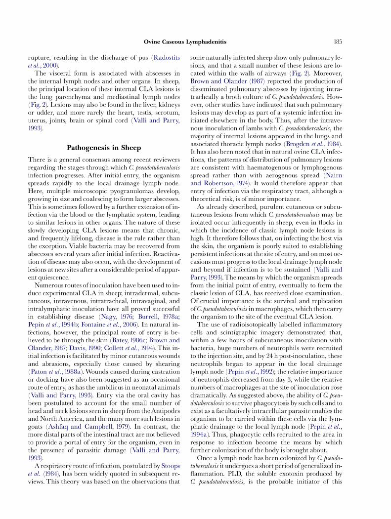
Fig. 3. Transverse histological section of a prescapular lymph nodefrom a case of ovine caseous lymphadenitis. Several distinctconcentric layers are discernible within the lesion. Centrallythere is liquefactive necrosis (liquid pus; A) which is sur-rounded by coagulative necrosis (caseous pus; B), both con-taining multiple foci of mineralization (F), apparently inloosely arranged concentric layers. A thin layer of polymor-phonuclear neutrophils surrounds the periphery of the coa-gulative necrosis; there is a further outer layer of coagulativenecrosis containing polymorphonuclear neutrophils migrat-ing through it at di¡erent densities, forming an apparentbi-layer (E). This is tightly bordered by a layer of immature¢brosis (C) containing mononuclear in£ammatory cells. Athick layer of mature ¢brosis (D) delineates the extent of thelesion. Haematoxylin and eosin (HE).
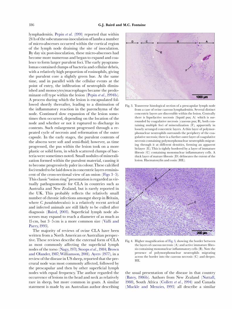
Fig. 4. Higher magni¢cation of Fig. 3, showing the border betweenthe layers of caseous necrosis (A) and active immature ¢bro-sis containing mononuclear in£ammatory cells (B). Note thepresence of polymorphonuclear neutrophils migratingacross the border into the caseous necrosis (C) and deeper.HE.
G.J. Baird and M.C. Fontaine186
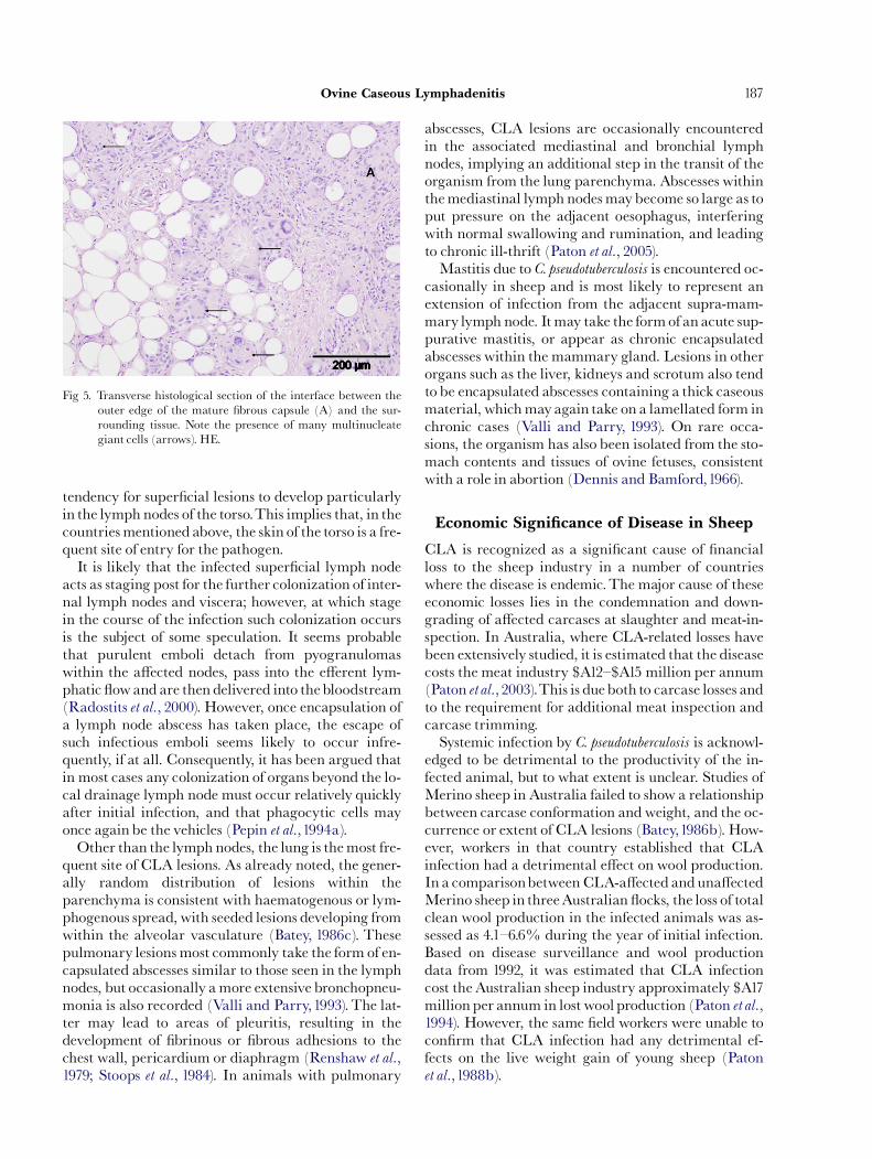
lymphadenitis. Pepin et al. (1991) reported that within24 h of the subcutaneous inoculation of lambs a numberof micro-abscesses occurred within the cortical regionof the lymph node draining the site of inoculation.By day six post-inoculation, these micro-abscesses hadbecome more numerous and began to expand and coa-lesce to form larger purulent foci.The early pyogranu-lomas contained clumps of bacteria and cellular debris,with a relatively high proportion of eosinophils, givingthe purulent core a slightly green hue. At the sametime, and in parallel with the cellular events at thepoint of entry, the in¢ltration of neutrophils dimin-ished and monocytes/macrophages became the predo-minant cell type within the lesion (Pepin et al.,1994b).A process during which the lesion is encapsulated fol-lowed shortly thereafter, leading to a diminution ofthe in£ammatory reaction in the parenchyma of thenode. Continued slow expansion of the lesion some-times then occurred, depending on the location of thenode and whether or not it ruptured to discharge itscontents. Such enlargement progressed through a re-peated cycle of necrosis and reformation of the outercapsule. In the early stages, the purulent contents ofthe abscess were soft and semi-£uid; however, as timeprogressed, the pus within the lesion took on a moreplastic or solid form, in which scattered clumps of bac-teriawere sometimes noted. Small nodules of minerali-zation formed within the purulent material, causing itto become progressively paler in colour.These calci¢edfoci tended tobe laiddown in concentric layers reminis-cent of the cross-sectional view of an onion (Figs 3^5).This classic‘‘onion ring’’presentation is regardedas vir-tually pathognomonic for CLA in countries such asAustralia and New Zealand, but is rarely reported inthe UK. This probably re£ects the relatively smallnumber of chronic infections amongst sheep in Britain,where C. pseudotuberculosis is a relatively recent arrivaland infected animals are still likely to be culled afterdiagnosis (Baird, 2003). Super¢cial lymph node ab-scesses may expand to reach a diameter of as much as15 cm, but 3^5 cm is a more common size (Valli andParry,1993).The majority of reviews of ovine CLA have been
written from a North American orAustralian perspec-tive. These reviews describe the external form of CLAas most commonly a¡ecting the super¢cial lymphnodes of the torso (Nagy,1971; Stoops et al.,1984; Brownand Olander,1987;Williamson, 2001). Ayers (1977), in areviewof the disease inUS sheep, reported that the pre-crural node was most commonly a¡ected, followed bythe prescapular and then by other super¢cial lymphnodes with equal frequency. The author regarded theoccurrence of lesions in the head and neck as relativelyrare in sheep, but more common in goats. A similarstatement is made by an Australian author describing
the usual presentation of the disease in that country(Batey, 1986b). Authors from New Zealand (Nuttall,1988), South Africa (Collett et al., 1994) and Canada(Muckle and Menzies, 1993) all describe a similar
ARTICLE IN PRESS
Fig 5. Transverse histological section of the interface between theouter edge of the mature ¢brous capsule (A) and the sur-rounding tissue. Note the presence of many multinucleategiant cells (arrows). HE.
Ovine Caseous Lymphadenitis 187
tendency for super¢cial lesions to develop particularlyin the lymph nodes of the torso.This implies that, in thecountriesmentioned above, the skin of the torso is a fre-quent site of entry for the pathogen.It is likely that the infected super¢cial lymph node
acts as staging post for the further colonization of inter-nal lymph nodes and viscera; however, at which stagein the course of the infection such colonization occursis the subject of some speculation. It seems probablethat purulent emboli detach from pyogranulomaswithin the a¡ected nodes, pass into the e¡erent lym-phatic £owandare then delivered into thebloodstream(Radostits et al., 2000). However, once encapsulation ofa lymph node abscess has taken place, the escape ofsuch infectious emboli seems likely to occur infre-quently, if at all. Consequently, it has been argued thatin most cases any colonization of organs beyond the lo-cal drainage lymph node must occur relatively quicklyafter initial infection, and that phagocytic cells mayonce again be the vehicles (Pepin et al.,1994a).Other than the lymph nodes, the lung is themost fre-
quent site of CLA lesions. As already noted, the gener-ally random distribution of lesions within theparenchyma is consistent with haematogenous or lym-phogenous spread, with seeded lesions developing fromwithin the alveolar vasculature (Batey, 1986c). Thesepulmonary lesions most commonly take the form of en-capsulated abscesses similar to those seen in the lymphnodes, but occasionally amore extensive bronchopneu-monia is also recorded (Valli and Parry,1993).The lat-ter may lead to areas of pleuritis, resulting in thedevelopment of ¢brinous or ¢brous adhesions to thechest wall, pericardium or diaphragm (Renshaw et al.,1979; Stoops et al., 1984). In animals with pulmonary
abscesses, CLA lesions are occasionally encounteredin the associated mediastinal and bronchial lymphnodes, implying an additional step in the transit of theorganism from the lung parenchyma. Abscesses withinthemediastinal lymph nodesmaybecome so large as toput pressure on the adjacent oesophagus, interferingwith normal swallowing and rumination, and leadingto chronic ill-thrift (Paton et al., 2005).Mastitis due to C. pseudotuberculosis is encountered oc-
casionally in sheep and is most likely to represent anextension of infection from the adjacent supra-mam-mary lymph node. Itmay take the formof an acute sup-purative mastitis, or appear as chronic encapsulatedabscesses within the mammary gland. Lesions in otherorgans such as the liver, kidneys and scrotum also tendto be encapsulated abscesses containing a thick caseousmaterial, whichmayagain take on a lamellated form inchronic cases (Valli and Parry, 1993). On rare occa-sions, the organism has also been isolated from the sto-mach contents and tissues of ovine fetuses, consistentwith a role in abortion (Dennis and Bamford,1966).
Economic Significance of Disease in Sheep
CLA is recognized as a signi¢cant cause of ¢nancialloss to the sheep industry in a number of countrieswhere the disease is endemic.The major cause of theseeconomic losses lies in the condemnation and down-grading of a¡ected carcases at slaughter and meat-in-spection. In Australia, where CLA-related losses havebeen extensively studied, it is estimated that the diseasecosts the meat industry $A12^$A15 million per annum(Paton et al.,2003).This is dueboth to carcase losses andto the requirement for additional meat inspection andcarcase trimming.Systemic infection by C. pseudotuberculosis is acknowl-
edged to be detrimental to the productivity of the in-fected animal, but to what extent is unclear. Studies ofMerino sheep in Australia failed to show a relationshipbetween carcase conformation andweight, and the oc-currence or extent of CLA lesions (Batey,1986b). How-ever, workers in that country established that CLAinfection had a detrimental e¡ect on wool production.In a comparisonbetween CLA-a¡ected anduna¡ectedMerino sheep in threeAustralian £ocks, the loss of totalclean wool production in the infected animals was as-sessed as 4.1^6.6% during the year of initial infection.Based on disease surveillance and wool productiondata from 1992, it was estimated that CLA infectioncost theAustralian sheep industry approximately $A17million per annum in lost wool production (Paton et al.,1994). However, the same ¢eld workers were unable tocon¢rm that CLA infection had any detrimental ef-fects on the live weight gain of young sheep (Patonet al.,1988b).
ARTICLE IN PRESS
G.J. Baird and M.C. Fontaine188
In contrast to much of the Southern Hemisphere,where the systemic e¡ects of CLA on the individualsheep are regarded as marginal, in North America thedisease is considered to be much more signi¢cantclinically. There, the visceral form of infection withC. pseudotuberculosis has been associated with so-called‘‘thin-ewe syndrome’’, a chronic emaciation of ewes,occurring despite good appetite and in the absence ofsigni¢cant parasitic infestation or speci¢c clinicalsigns.Two studies conducted in the US concluded thatCLA infection had an economically signi¢cant e¡ecton culling rates and reproductive e⁄ciency in ewes(Gates et al., 1977; Renshaw et al., 1979), but Arsenaultet al. (2003) considered that these studies lacked experi-mental controls. Certainly, the diagnostic criteriaof ‘‘thin-ewe syndrome’’ are vague, and althoughC. pseudotuberculosis is the principal bacterial causativeagent, other organisms such as species of Moraxella,Arcanobacterium and Staphylococcus may be present inmixed culture (Renshaw et al., 1979). On occasion,Moraxella species may also be isolated in the absence ofC. pseudotuberculosis.More signi¢cantly, therewouldappearto be some evidence of a synergistic relationship betweenCLAandmaedi visna-induced pneumonia in sheep.In the Middle East, considerable economic losses
arise through the condemnation of lamb carcases withCLA lesions, most commonly in the submandibularlymph nodes. Prevalence rates of 20^40% have beenreported amongst lambs from intensively reared £ocksof Najdi sheep (Pepin et al.,1994a). CLA lesions in lambsdestined for slaughter as part of religious festivals mayrender the carcases virtually worthlessçequivalent to aloss of $200 per head.
Epidemiology of Infection in Sheep
Transmission of Infection
MuchCLA research has focussed on how C. pseudotuber-
culosis cells, commonly contained within thick-walledabscesses, are transferred from one animal to another.Several possible routes have been suggested.The num-bers of viable bacteria in purulent discharge from aCLA lesion have been estimated as between 106 and5�107/g (Brown and Olander, 1987). The rupture ofsuper¢cial abscesses therefore releases huge numbersof viable bacteria on to the adjacent skin and £eece,and may readily contaminate the immediate environ-ment. Other animals may then be exposed, either bydirect physical contact with the a¡ected animal, or in-directly via contaminated fomites. In addition, theability of C. pseudotuberculosis to survive in the environ-ment for a number of weeks or months extends the po-tential period of infection well beyond the point ofinitial rupture and discharge.
The recovery of organisms fromthe faeces of infectedanimals was recorded from the earliest days of CLA re-search (Seddon et al., 1929) and its potential signi¢-cance for disease transmission has been noted insubsequent reviews. However, in the absence of CLAlesions in the intestinal tract, the organisms probablyoriginate from the respiratory tract, or represent bac-teria that have traversed the gut, having been ingested.The fact that bacteria have also been isolated from thefaeces of uninfected sheep would tend to support thelatter hypothesis (Benham et al.,1962). Despite the abil-ity of C. pseudotuberculosis to survive in the environmentfor protracted periods of time, attempts to isolate theorganism from the soil in areas of endemic infectionhave been unsuccessful (Knight, 1969). Therefore, thepresence of C. pseudotuberculosis organisms in faeces andthe consequent risk to other animals are likely to be ofminor importance.In CLA-infected sheep £ocks, animals with lung le-
sions are thought to represent the principal source ofinfection for other animals (Pepin et al.,1994a;William-son, 2001). This conclusion is based principally on epi-demiological observations recorded in Australia, inwhich the seroprevalence of CLA increased rapidly ingroups of sheep, despite the absence of super¢cial CLAlesions (Ellis et al.,1987; Paton et al.,1988a). In addition,C. pseudotuberculosis has been isolated from the tracheaeof sheep with pulmonary abscesses, con¢rming thatdischarge from lung lesions into the airways does in-deed occur (Stoops et al.,1984; Pepin et al., 1994a).Theassumption ismade that even a relatively small numberof animals with lung abscesses can create an aerosol ofinfectious organisms. Under conditions of close contactand reduced air£ow, as in a covered shed, such an aero-sol might be capable of infecting a large number of ani-mals in a short period of time (Paton et al.,1996).The usual means of entry of a new infection into a
na|« ve £ock is via the introduction of a clinically or sub-clinically infected carrier animal (Ayers, 1977). A rolefor transmission of infection by farmworkers and theirequipment between animals within the same £ock hasbeen established. It has alsobeen suggested that humanactivity may, on rare occasions, be responsible for in-troducing infection to previously disease-free £ocks. In£ocks with a high prevalence of CLA, shearers andtheir equipment are inevitably exposed to the purulentdischarges of super¢cial CLA lesions. If subsequent de-contamination measures are inadequate, it is possiblethat shearing gear, clothing and mobile handlingequipment may act as a mechanical vector of infectionto other £ocks. A circumstantial link between new dis-ease outbreaks in previously una¡ected closed sheep£ocks, and visits by contract shearers has been sug-gested inWesternAustralia, Canada andEngland; suchreports, however, remain anecdotal.
ARTICLE IN PRESS
Ovine Caseous Lymphadenitis 189
Othermechanical vectors of infection have been sug-gested. The investigation of an outbreak of CLA in aclosed and accredited disease-free goat herd in theNetherlands identi¢ed the most likely source of infec-tion as bales of hay purchased from another farm.Thissecond farmwas known to have a herdof goats inwhichCLAwas highly prevalent, and infected goats hadbeenhoused in the hay shed (Dercksen et al.,1996).Risk factors suggested for the spread of C. pseudotuber-
culosis infection amongst sheep include the breed(Bruere and West, 1990), increasing age of animals(Pepin et al.,1994a; Paton et al.,1995) and a dusty farmenvironment (Bruere and West, 1990). However, theprincipal risk factor identi¢ed in Australia and otherparts of theworld is minor skin damage due to shearingof the £eece (Nagy,1976; Paton et al., 1988a,1996; Seri-kawa et al., 1993; Al Rawashdeh and Al Qudah, 2000).In Australia, sheep are frequently housed or yarded to-gether for a period immediately after shearing, beforebeing returned to their grazings.This spell of close con-¢nement is regarded as the period of greatest risk, dur-ing which the organism may pass via an infectiousaerosol from one animal to another (Paton et al.,1996).The risks inherent in shearing may be increased if aplunge or shower dipping is applied within a few daysof shearing (Nairn and Robertson,1974; Augustine andRenshaw,1986; Ayers, 1986; Paton et al., 1996), the sug-gestion being that organisms from a¡ected animalspersist in the dip solution for a period.
Comparison with Goat Infection
Natural CLA infections in goats demonstrate a numberof similarities with the disease in sheep, not least thatthe same biotype of C. pseudotuberculosis is responsible.However, consideration of the di¡erences in clinicalpresentation between sheep and goats may be instruc-tive in respect of pathogenesis and speci¢c risk factors.This is of particular interest in the UK, where the clin-ical presentation of ovine CLA is closely similar to thatof the caprine infection in other parts of the world(Baird, 2003).In both sheep and goats, the major sites of infection
are the super¢cial lymph nodes, visceral lesions beingencountered in only a minority of animals. In sheep,however, visceral lesions, and especially lung lesions,occur more frequently and in greater numbers than ingoats (Renshaw et al., 1979; Hein and Cargill, 1981;Ayers, 1986; Muckle and Menzies, 1993). In caprineCLA, the principal sites of super¢cial lesions are thenodes of the head and neck (Ayers, 1977; Burrell, 1981;Batey, 1986c; Brown and Olander, 1987; Williamson,2001). As already noted, most reports of ovine CLA in-dicate that head and neck lesions are uncommon, themore frequent site of abscessation being the super¢cial
lymph nodes of the torso. These observations are con-sistent with the notion that the main routes of infectionin goats are viathe oral cavity or via skin of the face andhead. If bacteria are present on the skin or in the im-mediate environment, certainbehavioural traits shownby goats, such as mutual grooming, head butting, andan inquisitive attitude (including browsing andmouth-ing behaviour), would increase the risk of exposure toinfection. In contrast, such behavioural traits are lessfrequently observed in sheep. Most goats are neithershorn nor subjected to plunge or shower dipping.
CorynebacteriumpseudotuberculosisInfections in Other Animal Species
C. pseudotuberculosis is pathogenic for a variety of mam-malian species. Although a wide range of warm-blooded creatures are susceptible to the infection,speci¢c disease syndromes are recognized in a smallersubset. In addition to the globally important diseases ofsheep and goats, signi¢cant syndromes are recognizedin horses and dairy cattle.In horses, C. pseudotuberculosis is the cause of three re-
cognized disease syndromes: ulcerative lymphangitis,contagious folliculitis and furunculosis, and deep-seated subcutaneous abscessation (Miers and Ley,1980). In the US, C. pseudotuberculosis has also beenrecorded as a rare cause of abortion (Poonacha andDonahue, 1995) and mastitis in mares (Addo et al.,1974), and with generalized visceral abscessation(Paton et al., 2005). The three main equine syndromestend to have a distinct geographical distribution, sug-gesting the importance of secondarydisease factors. In-deed, the western areas of Canada and the USrepresent the only region in which all three syndromeshave been recorded (Aleman et al.,1996). In a study bySpier et al. (2004) a real-time polymerase chain reac-tion-based analysis of £ies collected from the vicinityof horses with C. pseudotuberculosis infections detectedthe PLD-encoding gene in a signi¢cant proportionof test subjects; this was considered to indicate activecarriage of C. pseudotuberculosis. In total, three £yspecieswere identi¢edas potential vectors for transmis-sion of disease; signi¢cantly, C. pseudotuberculosis
was identi¢ed in up to 20% of the house£ies (Musca do-
mestica) examined.In ulcerative lymphangitis, infection appears to en-
ter viawounds, generally those of the lower limbs.Thisis followed by extension to the a¡erent lymphatic ves-sels and the formation of abscesses along their course.These abscesses appear as subcutaneous nodular le-sions, which rupture to discharge pus and form necroticulcers (Radostits et al., 2000). Such infections may per-sist for many months, as successive crops of ulcers de-velop and then resolve. Transmission commonly
ARTICLE IN PRESS
G.J. Baird and M.C. Fontaine190
occurs by direct contact between horses; an arthropodvector, however, has also been suggested tobe of impor-tance in Africa (Addo,1983).Equine folliculitis and furunculosis (also referred to
as equine contagious acne, Canadian horsepox andequine contagious pustular dermatitis) is a much lessclinically important form of disease, which probablyrepresents a secondary infection by C. pseudotuberculosis
of a pre-existing seborrhoea or dermatitis. The lesionstend to develop at points of contact with harnesses andother tack, and spread may occur through the use ofcontaminated grooming equipment. Again, the char-acteristic lesion is a small nodule that develops into apustule before rupturing to discharge purulent materi-al.These lesions usually resolve spontaneously but mayoccasionally result in an ulcer at the site of infection.Deep-seated C. pseudotuberculosis abscessation (pigeon
fever, Wyoming strangles, false strangles) is the mostserious manifestation of the infection in horses. It isusually seen as abscesses in themusculature of the chestand shoulder, and is of economic signi¢cance in muchof the western US (Hughes and Biberstein,1959; Rum-baugh et al., 1978; Miers and Ley,1980), where its inci-dence is highest in the months of September toNovember (Aleman et al.,1996; Doherr et al.,1998).Thisprobably re£ects the seasonal incidence of various spe-cies of biting arthropod that act as mechanical vectors(Doherr et al.,1999).The recognized equine syndromes have not been de-
scribed in the UK in recent years, despite the isolationof C. pseudotuberculosis from horses on a number of occa-sions (Connor et al., 2000).The apparent signi¢cance ofseasonality, environmental conditions and arthropodvectors in C. pseudotuberculosis infections of horses hasbeen emphasized by a number of authors. It wouldtherefore appear that, although equine pathogenicstrains exist in the UK, essential contributory risk fac-tors do not. It is noteworthy that ulcerative lymphangi-tis was not uncommon in the UK duringWorldWar I,but this probably resulted from the introduction of in-fected horses from Russia and the Balkans where theconditionwas present (Petrie andMcClean,1934; Ben-ham et al.,1962).In recent years infections of dairy cattle have been
recorded in Israel, commonly in the form of deep sub-cutaneous abscesses that develop into granulating anddischarging ulcers, which respond poorly to systemicantibiotic treatment. The disease is sporadic but maysometimes occur in an epidemic form (Yeruham et al.,2003b). Morbidity rates of up to 35% have been re-corded in some Israeli dairy herds (Yeruham et al.,1997). A less commonmastitic form of disease is also re-cognized.This maybemild, the only signbeing the ap-pearance of clots in the milk, or severe, with a greatlyenlarged and tender mammary gland and complete
cessation of milk production.The severe form often ne-cessitates premature culling, as the response to antibio-tic therapy is poor (Shpigel et al., 1993). In a rarevisceral form of disease, widespread abscessation oc-curs in lymph nodes associatedwith the upper and low-er respiratory tract. In severe cases this may lead toswelling and obstruction of airways (Shpigel et al.,1993). Finally, the organism has also been associatedwith a necrotic and ulcerative dermatitis of the heel inheifers, a morbidity rate of 91% having been reportedin one outbreak (Yeruham et al., 2003a). C. pseudotubercu-losis has been recorded as a cause of an ulcerative lym-phangitis and mastitis of cattle in countries other thanIsrael, albeit rarely (Purchase, 1944; Adekeye et al.,1980; Kariuki and Poulton,1982; Anderson et al., 1990;Shpigel et al.,1993).C. pseudotuberculosis was isolated from house£ies col-
lected over a C. pseudotuberculosis lesion in a cow (Yeru-ham et al., 1996). Braverman et al. (1999) examinedhouse£ies that had fed on a super¢cial C. pseudotubercu-losis lesion in a cow, or on contaminated milk from ani-mals with C. pseudotuberculosis mastitis. The studyrevealed that a signi¢cant proportion of £ies har-boured the organism either on their body surfaces orin their intestines, the bacterium being present in thefaeces. It was concluded that this species of £y had apredilection for feeding on milk residues of cow teats,and therefore played an important role in harbouringand disseminating C. pseudotuberculosis infections indairy herds in Israel.C. pseudotuberculosis is commonly identi¢ed as the
cause of a purulent lymphadenopathy a¡ecting largenumbers of farmed llamas and alpacas in Chile andother South American countries (Braga et al., 2006). Itis also encountered sporadically as the cause of ab-scesses in companion camelids in North America (An-derson et al., 2004). In parts of the Middle East C.
pseudotuberculosis hasbeen isolated from lymph node ab-scesses of bu¡aloes (Ali and Zaitoun, 1999) and fromsimilar lesions in camels (Esterabadi et al.,1975; Nashedand Mahmoud, 1987; Abubakr et al., 1999). Sporadicnatural infections have been recorded in deer (Ham-mersland andJoneschild,1937; Eghetti andMcKenney,1941), pigs (Zhao et al., 1993), coypus, primates andducks (Benham et al., 1962) and a hedgehog (McAllis-ter and Keahey, 1971). Experimental infections arereadily induced in laboratory species such as mice, rab-bits and guinea-pigs (Carne,1940; Jolly,1965).
Zoonotic Infections
Reports of human infectionwith C. pseudotuberculosis arerelatively few, the ¢rst published case appearing some40 years ago (Lopez et al., 1966). Most human caseswere classi¢ed as occupational infections, a¡ecting
ARTICLE IN PRESS
Ovine Caseous Lymphadenitis 191
workers who had regular contact with sheep, such asshepherds, shearers, abattoir workers and butchers(House et al., 1986; Peel et al., 1997). Human infectionstend to be chronic, presenting as a localized suppura-tive granulomatous lymphadenitis and a¡ecting theaxillary, inguinal or cervical lymph nodes (Mills et al.,1997).The lymphadenitis may follow a period of in£u-enza-like symptoms and increasing lethargy.Treatmentwith systemic antibiotics is generally unrewarding, themajority of cases requiring surgical excision of the af-fected lymph node. Even then, the healing of surgicalwounds and infected sinusesmaybe protracted, and re-currence of lesions in other sites is not uncommon(Henderson, 1979). Because a¡ected human lymphnodes are not always cultured after excision and be-cause unspeci¢ed axillary lymphadenitis is not uncom-mon in shearers, it has been speculated that human C.
pseudotuberculosis infections are under-reported in coun-tries such as Australia, where ovine CLA is especiallyprevalent (Hamilton et al., 1968). More serious humaninfections, extending beyond localised lymphadenopa-thy, have been recorded. On one occasion an eosino-philic pneumonia due to C. pseudotuberculosis wasdiagnosed in a veterinary surgeon from the US (Keslinet al., 1979), but there are no published records of fatalhuman infections. A noteworthy case was recorded ina city-dweller, who had no apparent connection with afarming environment or livestock. In this instance, theonly potential cause identi¢ed was the regular con-sumption by the patient of unpasteurized goats’ milk(Goldberger et al., 1981). No case of human infectionwith C. pseudotuberculosis has yet been reported in theUK; there is, however, a reference to an isolate of C.pseudotuberculosis from a human lymph gland submittedto theUKNational Collection ofType Cultures (Hill etal., 1978), but the origin of the sample was not men-tioned.A recent report described a case of C. pseudotuberculo-
sis necrotizing lymphadenitis in a 12-year-old girl inFrance (Join-Lambert et al., 2006), thought to be the¢rst reported childhood case. The girl contracted theinfection after having had contact with sheep while onvacation in a rural part of France.The nitrate-negativeorganism was susceptible in vitro to a range of commonantibiotics, but treatment with amoxicillin and clavu-lanic acid failed to clear the infection. A second courseof amoxicillin/clavulanatewas thenadministeredwith-out success. After the infection had increased in sever-ity and spread beyond the a¡ected inguinal lymphnode, intravenous antimicrobial therapy with imipe-nem/cilastatin, rifampicin and o£oxacin was adminis-tered, followed by surgical resection of a¡ected tissueother than lymphatic tissue (to avoid post-lymphade-nectomy oedema). Intravenous antibiotic administra-tion was continued for a further 4 months, followed by
oral rifampicin and o£oxacin treatment for a further 6months. Two years after the cessation of treatment, norelapse had occurred.
Control and Eradication of Disease in Sheep
Diagnosis
For the successful control of CLA, it is ¢rst necessary toidentify infected animals, so that they canbe preventedfrom coming into contact withuninfected animals.Thediagnostic criterion for CLA is the culture and identi¢-cation of C. pseudotuberculosis. It is usually possible to iso-late the organism from lesions of all ages, although thenumber of viable bacteria present in chronic abscessesmay be low and apparently sterile lesions are occasion-ally encountered. In sheep, the presence of abscesses inexternal lymph nodes is highly suggestive of the dis-ease, particularly if several animals in a group are simi-larly a¡ected. Other bacterial pathogens, such asActinobacillus licheniformis, Arcanobacterium pyogenes, andin some countries Staphylococcus aureus subsp. anaerobius,are all capable of producing suppurative lymphadeno-pathy (Bek-Pederson, 1997; Pekelder, 2000); however,these other infections tend to be sporadic in natureand are rarely seen as a £ock problem.In the past, identi¢cation of Corynebacterium spp. has
proved di⁄cult for several reasons. These reasons in-clude (1) insu⁄cient numbers of reference strains withwhich to compare isolates, (2) inadequacies in the refer-ence data obtained from characterized strains, (3) therelative lack of appropriate criteria, and (4) the inap-propriate use of test criteria, since coryneformbacteriafrequently give rise to negative results with orthodoxtests (Hill et al., 1978). It has long been considered thatC. diphtheriae, C. pseudotuberculosis and C. ulcerans are re-lated, as they share a similar morphology and cell wallcomposition, and all produce a characteristic halo onTinsdale medium, indicating the production of a cysti-nase enzyme (Barksdale,1970). However, on thebasis ofDNA homology, there would seem to be less similaritybetween the three species thanwas at one time thought(Groman et al., 1984). Nonetheless, C. pseudotuberculosisand C. ulcerans canbemade to produce diphtheria toxinby infecting themwith toxin-associated phages from C.
diphtheriae (Petrie and McLean, 1934; Saxholm, 1951;Henriksen and Grelland,1952;Maximescu,1968;Max-imescu et al.,1974a,b).In the laboratory, C. pseudotuberculosis cultured from
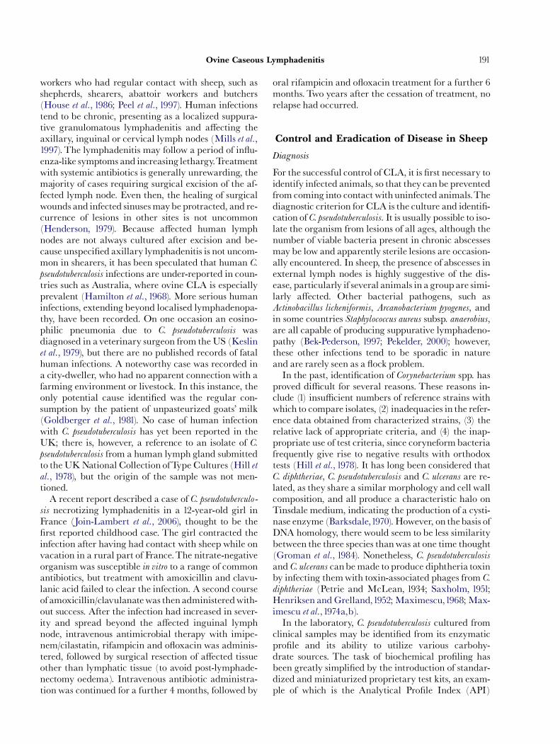
clinical samples may be identi¢ed from its enzymaticpro¢le and its ability to utilize various carbohy-drate sources. The task of biochemical pro¢ling hasbeen greatly simpli¢ed by the introduction of standar-dized and miniaturized proprietary test kits, an exam-ple of which is the Analytical Pro¢le Index (API)
ARTICLE IN PRESS
Fig.6. Synergistic lysis between Rhodococcus equi (1.) and Corynebacter-ium pseudotuberculosis (2.).The zone of enhanced lysis (arrows)is apparent where the soluble factors from each organismcome into contact with each other.
G.J. Baird and M.C. Fontaine192
identi¢cation system (bioMeŁ rieux [UK], Basingstoke,Hampshire, UK). The ‘‘API Coryne’’ kit (used for theidenti¢cation of coryneform bacteria) comprises 21 in-dividual test substrates for the determination of enzy-matic activity or carbohydrate fermentation. Afterinoculation with the test organism and incubation fora de¢ned period, the metabolic end-products of the en-zymatic tests result in the development of speci¢c col-our changes, either spontaneously or following theaddition of reagents. For substrate fermentation tests,pH change is also detected colorimetrically. Subse-quently, a particular combination of positive and nega-tive test results is used to compile a numerical pro¢le(an Analytical Pro¢le Index; API), which is then usedto‘‘interrogate’’a proprietary software database.For theAPItests, computer software is used to calcu-
late the percentage ¢t of the test isolate results to a con-sensus pro¢le for the identi¢ed species. The databasetakes into account natural intra-species variation incertain biochemical attributes. The use of computersto aid in bacterial identi¢cation (so-called ‘‘probabilis-tic’’ identi¢cation) was reported as far back as 1973(Bascomb et al., 1973; Lapage et al., 1973;Willcox et al.,1973). Brie£y, the methodological process described atthe time was to record the results of di¡erent taxo-nomic (e.g., biochemical) tests as the probability of apositive result (ranging from 0.99 to 0.01). The resultsobtained for the test isolate were then recorded aseither ‘‘+’’or ‘‘�’’, anda computer programmewas usedto compare these test results with the probabilities asso-ciated with the corresponding tests for each bacterialtaxon in the database. Subsequently, the probabilitieswere used to calculate an ‘identi¢cation score’ for thetest isolate, whichwas then compared with the identi¢-cation scores for those taxa in the database. The ¢naloutcome was the identi¢cation of the test isolate, com-plete with the probability of a correct identi¢cation.Subsequently, an identical technique was applied tothe identi¢cation of coryneformbacteria, and impress-ive results were obtained, including the successful use ofthe database to identify ¢eld isolates of C. pseudotubercu-losis (Hill et al.,1978).In addition to substrate utilization, further labora-
tory-based tests are available to identify C. pseudotubercu-losis. Some years ago, it was noted that when b-lysin-producing staphylococci were cultured on blood-con-taining solid media in the presence of strains of Strepto-coccus agalactiae that produce a secreted ‘factor’,enhanced zones of haemolysis were observed.This ledto the discovery of an S. agalactiae protein, designatedCAMP-factor (after the initials of its discoverers,Christie, Atkins and Munch-Petersen), which on itsown was unable to cause erythrocyte lysis. The syner-gistic lysis phenomenon was designated the CAMP-re-action (Christie et al., 1944). Similarly, when C.
pseudotuberculosis and Rhodococcus equi are cultured to-gether on solid blood-containing media, colonies ofboth species in close proximity to each other cause sig-ni¢cant zones of erythrocyte lysis (Fraser,1961). A simi-lar phenomenon was noted when C. pseudotuberculosis
was cultured in the presence of d-lysin-producing sta-phylococci (Lovell and Zaki,1966).The R. equi protein(so-called equi factor) involved in synergistic lysis wasfound to be a phospholipase C enzyme (Bernheimeret al.,1980). Currently this assay, whichmay also be per-formed by supplementing blood-containing solid med-ia with R. equi culture supernate (containing equifactor), is used in diagnostic laboratories for the identi-¢cation of C. pseudotuberculosis (Fig. 6). However, itshould be noted that C. ulcerans also produces PLD,although signi¢cantly it is the only other corynebacter-ial species found to do so (Barksdale et al.,1981). Hence,PLD activity is a distinctive marker within the genusCorynebacterium.In contrast to the synergistic lysis described above, it
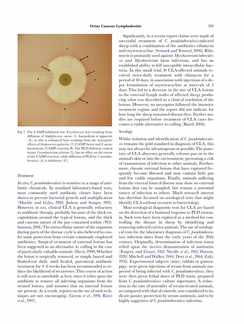
was reported (Soucek et al.,1962) that C. pseudotuberculo-sis exerted an inhibitory e¡ect on the haemolysis pro-duced by staphylococcal b-lysin, and the inhibitoryagent was thought to be associated with PLD. A com-parable inhibition of haemolytic activity was also ex-erted upon the a-toxin of Clostridium perfringens
(Soucek et al., 1967). As with the synergistic lysis test,the so-called reverse-CAMP, or CAMP-inhibition test(Fig.7) is still exploited to this day in diagnostic labora-tories, although the same issue of the production ofPLD by C. ulceransmust again be taken into account.
ARTICLE IN PRESS
Fig. 7. The CAMP-inhibition test. Erythrocyte lysis resulting fromdi¡usion of Staphylococcus aureus (1.) haemolysin is apparent(A), as also is enhanced lysis resulting from the synergistice¡ects of Streptococcus agalactiae (2.) CAMP factor and S. aureushaemolysin (CAMP reaction; B).The PLD-de¢cient controlstrain, Corynebacterium jeikeium (3.), has no e¡ect on the extentof the CAMP reaction, while di¡usion of PLD by C. pseudotu-berculosis (4.) is inhibitory (C).
Ovine Caseous Lymphadenitis 193
Treatment
In vitro, C. pseudotuberculosis is sensitive to a range of anti-biotic chemicals. In standard laboratory-based tests,most commonly used antibiotic classes have beenshown to prevent bacterial growth and multiplication(Muckle and Gyles, 1982; Judson and Songer, 1991).However, in vivo, clinical CLA is generally refractoryto antibiotic therapy, probably because of the thick en-capsulation around the typical lesions, and the thickand caseous nature of the pus contained within (Wil-liamson,2001).The intracellular nature of the organismduring parts of the disease cycle is also believed to con-fer some protection from certain commonly employedantibiotics. Surgical treatment of external lesions hasbeen suggested as an alternative to culling in the caseof particularly valuable animals (Davis,1990).Whetherthe lesion is surgically removed, or simply lanced and£ushed-out daily until healed, parenteral antibiotictreatment for 4^6 weeks has been recommended to re-duce the likelihood of recurrence.This course of actionis still seen as unreliable at best, since it relies upon theantibiotic to remove all infecting organisms from thetreated lesions, and assumes that no internal lesionsare present. As a result, reports on the use of such tech-niques are not encouraging (Gezon et al., 1991; Rizviet al.,1997).
Signi¢cantly, in a recent report claims were made ofsuccessful treatment of C. pseudotuberculosis-infectedsheep with a combination of the antibiotics rifamycinand oxytetracycline (Senturk andTemizel, 2006). Rifa-mycin is primarily used againstMycobacterium tuberculo-
sis and Mycobacterium leprae infections, and has anestablished ability to kill susceptible intracellular bac-teria. In this small trial, 10 CLA-a¡ected animals re-ceived twice-daily treatment with rifamycin for aperiod of 10 days, in association with injections of a de-pot formulation of oxytetracycline at intervals of 3days. This led to a decrease in the size of CLA lesionsin the external lymph nodes of a¡ected sheep, produ-cing what was described as a clinical resolution of thelesions. However, no necropsies followed the intensivetreatment regime and the report did not indicate forhow long the sheep remained disease-free. Further stu-dies are required before treatment of CLA cases be-comes a viable alternative to culling (Baird, 2006).
Serology
Whilst isolation and identi¢cation of C. pseudotuberculo-sis remains the gold standard in diagnosis of CLA, thismay not alwaysbe advantageous or possible.The punc-ture of CLA abscesses generally releases pus on to theanimal’s skin or into the environment, presenting a riskof transmission of infection to other animals. Further-more, chronic external lesions that have ruptured fre-quently become ¢brosed and may contain little pusand few viable organisms. Finally, animals su¡eringfrom the visceral form of disease may show no externallesions that can be sampled, but remain a potentialsource of infection to others. Much research interesthas therefore focussed on serological tests that mightidentify CLAwithout recourse to bacteriology.Most serological diagnostic tests for CLA are based
on the detection of a humoral response to PLD exotox-in. Such tests have been explored as a method for con-trolling the disease in sheep by identifying andremoving infected carrier animals.The use of serologi-cal tests for the laboratory diagnosis of C. pseudotubercu-losis infection dates from the early years of the 20thcentury. Originally, determination of infection statusrelied upon the in-vivo demonstration of antitoxins(Forgeot and Cesari, 1912; Nicolle et al., 1912; Watson,1920; Mitchell andWalker,1944; Doty et al.,1964; Zaki,1976). Experimental subjects (mice, rabbits or guinea-pigs) were given injections of serum from animals sus-pected of being infected with C. pseudotuberculosis; theywere then given lethal doses of PLD toxin, preparedfrom C. pseudotuberculosis culture supernates. A reduc-tion in the rate of mortality of serum-treated animals,as comparedwith that of controls, was considered to in-dicate passive protection by serum antitoxin, and to behighly suggestive of C. pseudotuberculosis infection.
ARTICLE IN PRESS
G.J. Baird and M.C. Fontaine194
In a development that was to facilitate in-vitro sero-logical screening, it was found that inclusion of antitox-in-containing sera in the CAMP-inhibition testresulted in neutralization of the inhibitory e¡ect thatC. pseudotuberculosis exerted on staphylococcal b-lysin;this led to a new method designated the anti-haemoly-sin-inhibition (AHI) test for testing sera from animalssuspected of having CLA. This test has been used fordisease diagnosis in sheep, goats and horses, avoidingthe use of experimental animals (Zaki, 1968; Knight,1978; Burrell,1980a; Brown et al.,1986).Other tests used in the study of CLA include tube ag-
glutination assays (Cameron et al., 1972; Husband andWatson,1977), an indirect haemagglutination test (Shi-gidi,1978) and a double immunodi¡usion test (Burrell,1980b). However, the enzyme-linked immunosorbentassay (ELISA) for use in diagnosis has shown particu-lar promise. Initially CLA-diagnostic ELISAs em-ployed crude C. pseudotuberculosis cell wall preparationsor supernate-derived exotoxin as antigen (Maki et al.,1985; Sutherland et al., 1987; Kuria and Holstad, 1989;Ellis et al., 1990; Sting et al., 1998). Test sensitivity wasusually regarded as good, but speci¢city was relativelypoorçpossibly due to cross-reactions with other pro-teins present in the antigen preparations. However,some ELISAs have been claimed to be su⁄ciently spe-ci¢c and sensitive for ¢eld diagnosis. One such ELISAwas based on a PLD antigen derived by recombinanttechnology from E. coli containing a plasmid bearingthe pld gene (Menzies et al., 1994), a test sensitivity of86.3% and speci¢city of 82.1% being reported.The as-say coming closest to the requirements of a diagnostictest was reported by workers in the Netherlands (TerLaak et al.,1992) and was later re¢ned to improve spe-ci¢city and sensitivity (Dercksen et al., 2000).This is anindirect double antibody sandwich ELISA, based onPLD exotoxin puri¢ed from the supernate of C. pseudo-tuberculosis culture, polyclonal rabbit anti-PLD serumbeing used as a capture antibody.The improved meth-od is reported to have a speci¢city in sheep of 9971%,and a sensitivity of 7975%.Other potential tests include a polymerase chain re-
action (PCR)method (Cetinkaya et al.,2002) andabo-vine interferon (IFN)-gwhole blood ELISA (Menzieset al., 2004).The latter was the ¢rst test inwhich assay ofcellular immunity to C. pseudotuberculosis was used as adiagnostic tool. Over the course of 1 year, the IFN-gtest accurately detected goats experimentally infectedwith C. pseudotuberculosis with a reliability of 89.2%,and non-infected goats with a reliability of 97.1%Overthe same period, a recombinant PLD ELISA detectedthe experimentally infected goats with a reliability of81.0% and non-infected goats with a reliability of97.0%.The PLDELISAwas, however, more predictivethan the IFN-g ELISA of the lesions subsequently
observed at necropsy. A later study of INF-gproductionin cultures of whole blood compared infected goats fol-lowing stimulation with either secreted bacterial anti-gen or somatic antigen; IFN-g production by bloodcells was found to be signi¢cantly higher in responseto the secreted antigen (Meyer et al., 2005).The use of serology as a tool in disease eradication
and control has been pioneered in the Netherlands,principally in relation to dairy goats. Indeed, a CLAHealth Scheme for goats exists in that country, basedupon the use of a PLD antibody ELISA andWesternblot analysis (Dercksen et al., 1996). A similar schemewas proposed for the control and eradication of CLAin Dutch sheep (Schreuder et al., 1994), but the plansfoundered due to poor test sensitivity in certain £ocks(Dercksen et al., 2000). Plans for serological control ofthe disease in sheep have recently been revived in theUK, with the launch in 2005 of a pre-sale testing re-gime incorporating the use of a serodiagnostic ELISAandWesternblot analysis based on a recombinant PLDantigen (Anon, 2005).
Vaccination
Historical perspective. Despite the relative lack of knowl-edge pertaining to C. pseudotuberculosis virulence factors,there exists a signi¢cantbodyof work exploring the po-tential for immunization to prevent disease caused bythis organism. Due to the relatively recent introductionof CLA into the UK, there has been little research ori-ginating from this country; however, the same is nottrue of elsewhere in theworld. In particular, signi¢cantwork has been conducted in both South Africa andAustralia over the last 75 years. Many of the earliest re-ports were somewhat confusing, partly because lack ofknowledge at the time prevented isolation of the organ-ism in a pure form and characterization of the cellularantigens. As stated by Batey (1986c),‘‘it is likely that in-fection by C. pseudotuberculosis involves a uniquemode ofpathogenesis’’; these words continue to bear weight asprogress towards an understanding of this organism isadvanced through a combination of molecular dissec-tion and an accurate analysis of the interaction ofpathogenwith host.One of the earliest reports, cited by Petrie and
McClean (1934), of protection against the deleteriouse¡ects of C. pseudotuberculosis products appeared in1907,when a degree of resistance against the lethal e¡ect ofthe exotoxin (PLD) was apparently produced in gui-nea-pigs by administration of a high dose of horse-de-rived antiserum against the toxin of C. diphtheriae
(Petrie and McClean,1934).This work was performedat a time when it was thought that there was a relation-ship between the exotoxins of C. pseudotuberculosis andC. diphtheriae. It was subsequently shown that the two
ARTICLE IN PRESS
Ovine Caseous Lymphadenitis 195
toxins were distinct, and that the protective e¡ect ob-served at the time was probably due to the prior occur-rence of anti-C. pseudotuberculosis antibodies in the horsefrom which the antitoxin was derived (Petrie andMcClean, 1934). Later work revealed that immuniza-tion with toxoided C. pseudotuberculosis exotoxin gavesome protection to guinea-pigs against the lethal e¡ectsof the active exotoxin (Petrie and McClean, 1934).Despite this observation, the general consensus of opi-nion held that the exotoxin was unsuitable for use as aprotective antigen against CLA in sheep (Carne et al.,1956; Cameron and Smit, 1970) due to the chronicnature of the natural disease, in contrast to the acutenature of C. pseudotuberculosis infection in mice andguinea-pigs.In the early1960s it was suggested that the immuniz-
ing property of C. pseudotuberculosis was associated withthe toxicity of cell endoplasm preparations and withthe ability of formalin-killed cells to produce sterile ab-scesses when injected into guinea-pigs (Cameron,1964).However, it was subsequently shown that these ‘toxicfactors’and those responsible for conferring immunityto infectionwere in fact distinct (Cameron and Buchan,1966). Later studies showed that mice could be signi¢-cantly protected against challenge with C. pseudotubercu-losis by immunization with formalin-killed, washed,whole C. pseudotuberculosis cells, combined with one of avariety of adjuvants (Cameron and Minnaar, 1969).The cellular component believed to confer the mostimmunity was the cell wall.Through a process of elim-ination it was discovered that the antigen responsiblewas an ‘‘integral part of the cell wall’’, but was not thecell wall lipid, which did not appear to induce protec-tive immunity (Cameron et al.,1969).The ability of cellwall components to induce immunity was reduced byprior treatment with formalin; the reason for this wasthought to be the denaturation of cell wall-associatedprotein or proteins, resulting in a loss of biological ac-tivity, which was believed to be required for immuno-genicity (Cameron et al.,1969).In continuing previous work, Cameron and Smit
(1970) investigated toxic components in C. pseudotubercu-
losis protoplasmic preparations, with a view to under-standing the cause of the sterile abscesses formed oninoculation of animals with killed C. pseudotuberculosis
cells. It became apparent that a toxic cytoplasmic com-ponent was identicalwith the exotoxin found in culturesupernates. However, consistent with an earlier report(Petrie and McClean, 1934), Cameron and Smit hy-pothesized that this extracellular exotoxin must havearisen through lysis of bacterial cells, since the conceptof protein secretion was still not understood. Nonethe-less, their work clari¢ed to some extent the confusionsurrounding the various toxic fractions studied in pre-vious years.
In the following section the di¡erent approaches tovaccine development will be discussed. However, it isnot within the scope of this review to discuss the meritsof anygivenapproachor the acceptabilityof the resultingvaccines from a commercial or regulatory standpoint.Bacterin vaccines.The earliest reported vaccination trialsin sheep were conducted in the Patagonia region of Ar-gentina, where a formalin-killed C. pseudotuberculosis
vaccine reduced infection by up to 60% (Quevedoet al., 1957). Cameron et al. (1972) made one of the ¢rstattempts to demonstrate by experimental challengethe ovine immune response to vaccination, the vaccineconsisting of formalin-killed, whole cells of C. pseudotu-berculosis.The intravenous challenge doses investigatedvaried from 2�107 to 2�1010 colony-forming units.However, it was di⁄cult to obtain satisfactory results,the higher doses producing acute death due to pulmon-ary oedema and the lower doses failing to cause lesions.Furthermore, vaccination produced increased titres ofserum antibody that persisted for only 3^4 months.Nonetheless, it was concluded that vaccination couldprotect sheep against the lethal e¡ects of sub-acute in-fection but not against formation of the lesions asso-ciated with chronic infection. It was considered thate¡ective immunitymight not depend solely on circulat-ingantibody, but that cellular immune responsesmightplay a role. It was also suggested that, if used, a wholecell vaccine should be administered shortly beforeshearing.More recently, in a study of several vaccines, immu-
nization of sheep with a formalin-killed, virulent, UK-derived C. pseudotuberculosis isolate and aluminium hy-droxide adjuvant resulted in statistically signi¢cantprotection against homologous challenge; the vaccinehad prevented the spread of infection beyond the siteof inoculation (Fontaine et al., 2006).There have been few investigations of the protective
e¡ect of C. pseudotuberculosis bacterins against disease inthe ¢eld.Menzies et al. (1991) conducted a ¢eld trial of akilled whole-cell vaccine in a £ock of sheep and herd ofgoats, both populations having shown a prevalence ofCLA in adult animals ranging from 15% to 30% inthe pre-trial period. During the trial, lambs and kidswere vaccinated at 2.5^3.5 months of age, with twofurther doses administered at 1 month and 11 monthsafter the initial vaccination. The animals were regu-larly monitored for 36 months after the initial dose ofvaccine. Serum antibody concentrations were signi¢-cantly increased in vaccinated sheep and goats for thefull term of the experiment. Furthermore, vaccinationsigni¢cantly decreased the incidence of CLA in thesheep, and there was a suggestion of a similar e¡ect inthe goats.In a later study, juvenile sheep and goats were vacci-
nated with a C. pseudotuberculosis bacterin formulated
ARTICLE IN PRESS
G.J. Baird and M.C. Fontaine196
in light mineral oil containing muramyl dipeptide(Brogden et al., 1995); the results, however, wereinconclusive.Toxoid vaccines. As noted above, Petrie and McClean(1934) found that vaccination of guinea-pigs with aformalin-inactivated exotoxin (PLD) of C. pseudotuber-culosis induced a degree of protection against homolo-gous challenge. Despite this, it was generallyconsidered that the chronic nature of C. pseudotuberculo-sis infection in sheep rendered toxoid ine¡ective as aprotective antigen. Most subsequent studies on CLAvaccination focussed on the use of bacterins, or on vac-cinesmade fromcomponents of whole cells. However, areport byJolly (1965) showed that the administration oftoxoid, followed by toxin, reduced the extent of experi-mental CLA in sheep.This followed the observationbyCameron (1964) that an unde¢ned ‘‘protoplasmic tox-in’’of C. pseudotuberculosis played a role in the inductionof protective immunity. It was later demonstrated thatC. pseudotuberculosis exotoxin was in reality identicalwith this protoplasmic toxin (Cameron and Smit,1970), adding weight to the hypothesis that PLD couldbe used as a protective antigen.Purelymanagemental strategies having failed to pre-
vent the spread of the disease, studies were conductedin Australia during the1970s to develop a CLAvaccine,work that would eventually lead to the ¢rst commercialCLA vaccine (Eggleton et al., 1991a,c). The ability ofPLD to protect against CLA was reported by bothNairn et al. (1977) and Burrell (1978b).Work at the Divi-sion of Animal Health, Commonwealth Scienti¢c andIndustrial Research Organisation (CSIRO), led to theoptimization of procedures for the culture of C. pseudo-tuberculosis. It also resulted in improvements in theyields and quanti¢cation methods of PLD, matters ofimportance in view of its immunogenic properties(Burrell,1983).Between 1977 and 1978, the technology required for
production of an e¡ective toxoid vaccine was madeavailable to the Commonwealth Serum Laboratories(CSL; Parkville, Victoria, Australia), and details, in-cluding optimal antigen dose and combination withother (clostridial) components, were later published(Eggleton et al., 1991c). For vaccine production, PLDwas obtained from ¢ltered and concentrated C. pseudo-
tuberculosis culture supernates, whichwere subsequentlytoxoided with formalin (Burrell, 1983; Hodgson et al.,1999). A combined clostridial and corynebacterial vac-cine, known as GlanvacTM, was released in 1983(Eggleton et al., 1991a); it subsequently became avail-able in Australia and several other countries but is notlicensed for use in the European Union. In one report,vaccination of goats with GlanvacTM resulted in statis-tically signi¢cant protection against subsequent experi-mental infection with C. pseudotuberculosis, as measured
by a reduction in the number of lesions (Brown et al.,1986). In addition, the results of trials performed in theprocess of the commercial development of GlanvacTM
indicated signi¢cant protection of sheep against CLA(Burrell, 1983; Eggleton et al., 1991a^c). Anderson andNairn (1984), however, found that PLD toxoid gavegreater protection when not combined with clostridialcomponents.The mechanismby which GlanvacTM confers immu-
nity has been the topic of some debate. It has been sug-gested that protection against CLA isboth humoral andcell-mediated, but that PLD is neutralized by circula-ting antibody (Burrell, 1978b; Batey, 1986c; Eggletonet al.,1991a; Sutherland et al.,1992). In contrast, Hodgsonet al. (1999) discussed the possibility that the smallamount (ca 1%) of PLD activity that remained afterformalin treatment (Eggleton et al.,1991b) might be re-sponsible for the protection conferred by GlanvacTM.However, Eggleton et al. (1991b) had previously re-ported that a four-fold variation in the level of activetoxin remaining in a toxoid vaccine ‘‘neither enhancednor reduced the protective potency of the vaccines’’.Others have reported that anti-exotoxin antibodies arenot on their own su⁄cient to prevent infection in the ab-sence of other (probably cell-mediated) immune de-fences (Irwin and Knight, 1975; Cameron and Bester,1984; Ellis et al.,1990). Signi¢cantly, Cameron and Smit(1970) stated that ‘‘yit could be expected that crudeexotoxin preparations could also contain other anti-gens. Therefore, unless an absolutely pure preparationof exotoxin can be produced, its immunizing activitycan never be accurately assessed’’. This statement waslater reiterated by Burrell (1983), who con¢rmed thatthe culture supernates used to prepare GlanvacTM con-tained other C. pseudotuberculosis somatic antigens, whichvaried in amount frombatch to batch. Moreover,Walk-er et al. (1994) intermittently identi¢ed antibodiesagainst a novel C. pseudotuberculosis antigen in sera fromsheep vaccinated with GlanvacTM. It would thereforeseem that the presence of other C. pseudotuberculosis pro-teins in toxoid vaccines prepared from culture super-nates may well contribute to the protective capacity ofthe vaccine.Fontaine et al. (2006) assessed the capacity of a highly
puri¢ed recombinant derivative of PLD to protectsheep against homologous challenge with a UK-derived C. pseudotuberculosis isolate. Protection was simi-lar to that producedby abacterin vaccine. After experi-mental challenges, infection appeared to be localized tothe inoculation site in all but one experimental animal,in which evidence of pulmonary infection was alsofound. Given that PLD does not stimulate cell-media-ted immunity (Hodgson et al., 1999), the protectionobserved in animals vaccinated with the recombinantPLD was postulated to result from antibody-mediated
ARTICLE IN PRESS
Ovine Caseous Lymphadenitis 197
neutralization of toxin activity. Included in thesame study was an assessment of the capacity of thePLD-based proprietary vaccine, Glanvac-3TM (amember of the GlanvacTM vaccine family), to protectagainst challenge with the UK-derived C. pseudotubercu-
losis isolate. Statistically signi¢cant protection wasagain observed with this vaccine, as indicated by a de-creased number of infected loci. GlanvacTM vaccineappeared to be less successful than the recombinantPLD vaccine in preventing the spread of the challengeinfection beyond the site of inoculation and into thesuper¢cial lymph nodes; it was conceded, however,that the immunity produced by the GlanvacTM vac-cine was demonstrated by challenge with a heterolo-gous bacterial strain. However, the high degree ofsequence conservation between the PLD-encodinggenes from di¡erent C. pseudotuberculosis isolates maywell negate the issue of homologous versus heterolo-gous challenge.Combined vaccines. In reviewing CLA vaccine develop-ment, Burrell (1983) stated that, despite the capacity ofcell-associated antigens (Cameron et al.,1969) and PLD(Burrell,1978b) to give some protection, full protectionrequired the presence of both. In defence of this, it wasrevealed that an alhydrogel-adjuvanted vaccine com-posed of formalin-killed whole C. pseudotuberculosis cells,enriched with formalin-treated, PLD-rich culturesupernate, protected completely against experimentalC. pseudotuberculosis infection and outperformed vac-cines consisting of cell-free toxoided culture supernates(Burrell,1983). In contrast, a report of work conductedduring the development of GlanvacTM revealed thatsupplementation of cell-free toxoid vaccine with C. pseu-dotuberculosis cells made no di¡erence to the observedprotection (Eggleton et al.,1991b).A further commercially available CLA vaccine
(Caseous D-TTM; Colorado Serum Co., Denver, CO,USA) contains clostridial toxoids and a combinationof C. pseudotuberculosis bacterin and toxoid. Piontkowskiand Shivvers (1998) reported that this vaccine gavesome protection to sheep against experimental infec-tion, in that it reduced the incidence of external and in-ternal CLA lesions.Fontaine et al. (2006) reported that vaccination of
sheep with a C. pseudotuberculosis bacterin enriched withrecombinant PLD resulted in complete freedom frominfection 3 weeks after experimental homologous chal-lenge. It had been reported previously (Simmons et al.,1998) that C. pseudotuberculosis (even a non-virulent mu-tant) was capable of persisting within granulomas inthe draining lymph node, as a result of the inaccessibil-ity of abscesses to the immunological factors required toclear the infection.Therefore it was suggested that vac-cination hadenabled clearance of the organismprior toestablishment within the lymph node.
Live vaccines. Pepin et al. (1993) reported that sheep in-fected experimentally with C. pseudotuberculosis wereprotected against further challenge, despite being un-able to clear the original infection.This observation in-dicates the possible value of an attenuated vaccine.Because PLD is an important virulence factor
(Carne andOnon,1978), Hodgson et al. (1992) hypothe-sized that genetic inactivation of the pld gene mightprovide thebasis for a live recombinant veterinary vac-cine. Amutant derivative of pldwas constructed in vitro,by cloning the gene into a suitable plasmid vector(pEP2), and disrupting it by insertion of an erythromy-cin-resistance cassette. Subsequently, the resulting re-combinant plasmid (pBTB58) was introduced intoC. pseudotuberculosis by electroporation, and recombina-tion between homologous plasmid-borne and chromo-somal sequences resulted in replacement of the wild-type gene by the mutant copy.To ensure the stability ofthe mutation, pBTB58 vector sequences were removedfrom mutant cells by transformation with the plasmidpEP3; because pEP3 possesses the same origin of repli-cation as that of pBTB58, plasmid segregation resultedin loss of pBTB58 sequences and retention of pEP3.Inoculation of sheep with up to 107 colony-forming
units of the PLD-de¢cient C. pseudotuberculosis strain(so-called ‘‘Toxminus’’) failed to produce the abscessa-tion normally associated with wild-type C. pseudotuber-culosis. However, at higher doses, transient abscesseswere formed at the inoculation site. Despite this, theauthors concluded that the degree of attenuation wasequivalent to at least a 100-fold reduction in infectivedose. The vaccine potential of the attenuated strainwas determined by challenge of Toxminus-immunizedsheep with wild-type C. pseudotuberculosis. A degree ofprotection against challenge and strong humoral andcellular immune responses were observed, but these ef-fects were lower than those produced by wild-type in-fection.It was later postulated (Hodgson et al.,1994) that the
rationale behind the creation of theToxminus mutantwas £awed: while the recombinant strain was attenu-ated for virulence and hence useful as a live vaccinevector, one of the major immunodominant antigens(PLD) had been removed, thus reducing immune sti-mulation. Hodgson et al. (1994) therefore constructed amutant derivative of PLD inwhich the histidine20 ami-no-acid residue (a constituent of the enzyme’s activesite) was substituted for tyrosine, and expression of theresulting plasmid-borne, mutant pld gene produced abiologically inactive derivative of PLD. On transfor-mation of Toxminus with the inactivated pld construct,cells were able to secrete an inactive PLD derivative,albeit at a level lower than that of wild-type PLD.Inanattempt toavoid inoculation-site reactions andto
provide a convenient means of vaccine administration,
ARTICLE IN PRESS
G.J. Baird and M.C. Fontaine198
Hodgson et al. (1994) administered each of two vaccines(Toxminus andToxminus carrying the inactivated pld)to sheep via the oral route. Both vaccines induced sig-ni¢cant and similar humoral immune responses.Furthermore, in animals immunizedwith the strain ex-pressing the inactivated pld, challenge elicited a rapidanamnestic response to PLD; this response was quickerand of greater magnitude than the humoral response(1) to infection in unvaccinated animals, or (2) in thosereceivingToxminus alone. Despite signi¢cant humoralresponses, the protection against challenge conferredby theToxminus strain was negligible; this contrastedwith a previous report of subcutaneous vaccinationwithToxminus (Hodgson et al.,1992).The di¡erence inprotection was attributed to the failure of oral vaccina-tion, unlike subcutaneous vaccination, to stimulate asigni¢cant Th1 immune response, IgG1 having beenidenti¢ed as the dominant IgG isotype. Despite thefailure of Toxminus alone, immunization withToxmi-nus expressing the mutant pld gene resulted in signi¢-cant protection against challenge, con¢rming theearlier observations that antibodies against PLD pro-tected against CLA (Jolly, 1965; Nairn et al., 1977; Bur-rell,1978a; Eggleton et al.,1991b, c).Continued e¡orts to produce an e¡ective live vaccine
vector led to attempts to express a range of foreign anti-gens in theToxminus strain, under the control of severaldi¡erent promoters (Moore et al.,1999). In the course ofthis work, Toxminus-based systems were developed toenable antigens to be expressed, intracellularly or in se-creted form, as native or fusion proteins, either consti-tutively or by controlled induction. Furthermore, geneswere successfully expressed fromplasmids, or followingintegration into theToxminus chromosome. In additionto derivatives of C. pseudotuberculosis pld, genes fromother pathogenic organisms, including Mycobacterium
leprae,Taenia ovis, Babesia bovis, Dichelobacter nodosus andAnaplasma marginale, were successfully expressed. Sub-sequently, it was revealed that subcutaneous immuni-zation of sheep withToxminus expressing inactive pld,orA. marginale or D. nodosus antigens, resulted in the in-duction of a signi¢cant humoral immune response; thesame was not true, however, forT. ovis or B. bovis anti-gens.Other reports of attempts to create live vaccine vec-
tors against CLA have been generally less successfulthan those discussed above. Disruption of the aroQ
gene, encoding 3-dehydroquinase (an essential enzymein aromatic amino-acid biosynthesis), led to the crea-tion of attenuated mutants of two strains of C. pseudotu-berculosis, one possessing and one lacking PLD. Bothstrains were cleared from the liver and spleen of micewithin 8 days of intraperitoneal inoculation (Simmonset al., 1997). In contrast, complementation of the chro-mosomal aroQ mutation, by introduction of a plasmid
encoding the wild-type aroQ gene, was su⁄cient to re-store the virulence of themutant strains to some extent,but not to the level shown by the wild-type parentstrain. Protection studies with one of the attenuatedmutants revealed that high doses induced a degree ofprotection against wild-type challenge, in addition tothe stimulation of INF-g production by murine spleno-cytes. INF-gplays a role in macrophage activation, andthe suppression of C. pseudotuberculosis growth and lesiondevelopment is largely due to the bactericidal action ofactivated macrophages (Hard, 1970). Therefore, thevaccine potential of the attenuated strains was evident;it was observed, however, that both were e¡ectivelycleared from INF-g-de¢cient mice, suggesting that theattenuation allowed clearance by some other, unde-¢ned route.This, in turn, was considered useful, giventhat vaccination would be unlikely to cause infection,even in immunocompromised animals (Simmonset al.,1997).Despite the encouraging initial results in mice, trials
in sheep proved disappointing in respect of the protec-tion against CLA o¡ered by the AroQ mutants (Sim-mons et al., 1998). Consistent with previous results,both mutants showed reduced ability to persist withinlymph nodes and to produce local reactions at the in-oculation site. However, both failed to stimulate detect-able production of speci¢c INF-g-secretinglymphocytes and induced only low concentrations ofanti-C. pseudotuberculosis IgG. Not surprisingly, these im-mune responses did not protect sheep from infection.DNAvaccines.With the advent of DNAvaccination tech-nologies, there have been reports of the successful im-munization of numerous species against variousmicro-organisms (Donnelly et al., 1995), but such re-ports in respect of farm livestock have been relativelyfew in number (Van Drunen Littel-van den Hurk et
al.,2000). In general, DNAvaccineswould seemto havebeen less successful than conventional or sub-unit pro-tein vaccines in protecting against infection, probablydue to a weak and short-lived immune response(Rothel et al.,1997; Schrijver et al.,1997).In an e¡ort to improve the e⁄cacy of DNAvaccina-
tion against intracellular pathogens, fusion of antigen-encoding genes to those encoding speci¢c ‘‘e¡ector’’mo-lecules has been attempted. One such e¡ector moleculeis the cytotoxic T lymphocyte antigen-4 (CTLA-4)(Brunet et al., 1987), which assists in directing secretedvaccine antigens to lymph nodes andantigen-presentingcells (APCs) (Boyle et al.,1998; Drew et al., 2001). CTLA-4, a repressor ofT-lymphocyte proliferation, is expressedon the surface of activatedTcells. In its dimeric form itcompetes with CD28 for binding to the CD80and CD86 (B7 domains) on antigen-presenting cells(Linsley et al.,1991;ThompsonandAllison,1997). Atrun-cated derivative of CTLA-4 (designated CTLA-4-HIg)
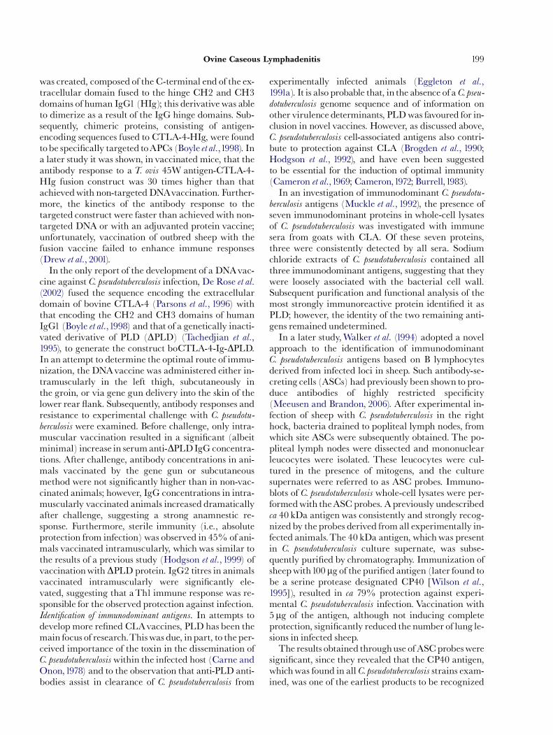
ARTICLE IN PRESS
Ovine Caseous Lymphadenitis 199
was created, composed of the C-terminal end of the ex-tracellular domain fused to the hinge CH2 and CH3domains of human IgG1 (HIg); this derivativewas ableto dimerize as a result of the IgG hinge domains. Sub-sequently, chimeric proteins, consisting of antigen-encoding sequences fused to CTLA-4-HIg, were foundtobe speci¢cally targeted toAPCs (Boyle et al.,1998). Ina later study it was shown, in vaccinated mice, that theantibody response to aT. ovis 45W antigen-CTLA-4-HIg fusion construct was 30 times higher than thatachievedwith non-targeted DNAvaccination. Further-more, the kinetics of the antibody response to thetargeted construct were faster than achieved with non-targeted DNA or with an adjuvanted protein vaccine;unfortunately, vaccination of outbred sheep with thefusion vaccine failed to enhance immune responses(Drew et al., 2001).In the only report of the development of a DNAvac-
cine against C. pseudotuberculosis infection, De Rose et al.(2002) fused the sequence encoding the extracellulardomain of bovine CTLA-4 (Parsons et al., 1996) withthat encoding the CH2 and CH3 domains of humanIgG1 (Boyle et al.,1998) and that of a genetically inacti-vated derivative of PLD (DPLD) (Tachedjian et al.,1995), to generate the construct boCTLA-4-Ig-DPLD.In an attempt to determine the optimal route of immu-nization, the DNAvaccine was administered either in-tramuscularly in the left thigh, subcutaneously inthe groin, or via gene gun delivery into the skin of thelower rear £ank. Subsequently, antibody responses andresistance to experimental challenge with C. pseudotu-
berculosis were examined. Before challenge, only intra-muscular vaccination resulted in a signi¢cant (albeitminimal) increase in serumanti-DPLDIgG concentra-tions. After challenge, antibody concentrations in ani-mals vaccinated by the gene gun or subcutaneousmethod were not signi¢cantly higher than in non-vac-cinated animals; however, IgG concentrations in intra-muscularly vaccinated animals increased dramaticallyafter challenge, suggesting a strong anamnestic re-sponse. Furthermore, sterile immunity (i.e., absoluteprotection from infection) was observed in 45% of ani-mals vaccinated intramuscularly, which was similar tothe results of a previous study (Hodgson et al.,1999) ofvaccinationwith DPLD protein. IgG2 titres in animalsvaccinated intramuscularly were signi¢cantly ele-vated, suggesting that aTh1 immune response was re-sponsible for the observed protection against infection.Identi¢cation of immunodominant antigens. In attempts todevelop more re¢ned CLAvaccines, PLD has been themain focus of research.Thiswas due, inpart, to the per-ceived importance of the toxin in the dissemination ofC. pseudotuberculosiswithin the infected host (Carne andOnon,1978) and to the observation that anti-PLD anti-bodies assist in clearance of C. pseudotuberculosis from
experimentally infected animals (Eggleton et al.,1991a). It is also probable that, in the absence of a C. pseu-dotuberculosis genome sequence and of information onother virulence determinants, PLDwas favoured for in-clusion in novel vaccines. However, as discussed above,C. pseudotuberculosis cell-associated antigens also contri-bute to protection against CLA (Brogden et al., 1990;Hodgson et al., 1992), and have even been suggestedto be essential for the induction of optimal immunity(Cameron et al.,1969; Cameron,1972; Burrell,1983).In an investigation of immunodominant C. pseudotu-
berculosis antigens (Muckle et al., 1992), the presence ofseven immunodominant proteins in whole-cell lysatesof C. pseudotuberculosis was investigated with immunesera from goats with CLA. Of these seven proteins,three were consistently detected by all sera. Sodiumchloride extracts of C. pseudotuberculosis contained allthree immunodominant antigens, suggesting that theywere loosely associated with the bacterial cell wall.Subsequent puri¢cation and functional analysis of themost strongly immunoreactive protein identi¢ed it asPLD; however, the identity of the two remaining anti-gens remained undetermined.In a later study,Walker et al. (1994) adopted a novel
approach to the identi¢cation of immunodominantC. pseudotuberculosis antigens based on B lymphocytesderived from infected loci in sheep. Such antibody-se-creting cells (ASCs) had previously been shown to pro-duce antibodies of highly restricted speci¢city(Meeusen and Brandon, 2006). After experimental in-fection of sheep with C. pseudotuberculosis in the righthock, bacteria drained to popliteal lymph nodes, fromwhich site ASCs were subsequently obtained. The po-pliteal lymph nodes were dissected and mononuclearleucocytes were isolated. These leucocytes were cul-tured in the presence of mitogens, and the culturesupernates were referred to as ASC probes. Immuno-blots of C. pseudotuberculosis whole-cell lysates were per-formedwith theASC probes. Apreviously undescribedca 40 kDa antigen was consistently and strongly recog-nizedby the probes derived fromall experimentally in-fected animals.The 40 kDa antigen, whichwas presentin C. pseudotuberculosis culture supernate, was subse-quently puri¢ed by chromatography. Immunization ofsheepwith100 mg of the puri¢ed antigen (later found tobe a serine protease designated CP40 [Wilson et al.,1995]), resulted in ca 79% protection against experi-mental C. pseudotuberculosis infection. Vaccination with5 mg of the antigen, although not inducing completeprotection, signi¢cantly reduced the number of lung le-sions in infected sheep.The results obtained throughuse of ASCprobeswere
signi¢cant, since they revealed that the CP40 antigen,whichwas found in all C. pseudotuberculosis strains exam-ined, was one of the earliest products to be recognized
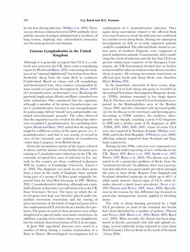
ARTICLE IN PRESS
G.J. Baird and M.C. Fontaine200
by the host during infection (Walker et al.,1994).Therewas no obvious relation between CP40 antibody titresand the amount of antigen administered or incidence oflung lesions, implying that cell-mediated immunitywas the main protective mechanism.
Caseous Lymphadenitis in the UnitedKingdom
Although it is generally accepted that CLA is a rela-tively new arrival to the UK, there exists a tantalizingreport by Heath and Batty (1952) concerning the isola-tion of an ‘‘unusual diphtheroid’’ bacterium from threeSwaledale sheep from the same £ock in southwestCumberland. Based on colony and cell morphologyand biochemical tests, these isolates corresponded al-most exactly to a previous description by Morse (1949)of C. pseudotuberculosis (at that timeC. ovis). Realizing thepotential implications, Heath and Batty consulted twoother authorities. One considered that the organism,although a member of the genus Corynebacterium, wasnot C. pseudotuberculosis because it was non-pathogenicfor guinea-pigs, did not ‘‘attack’’urea andprobably con-tained metachromatic granules. The other observedthat the organismwas not virulent for sheep but other-wise resembled C. pseudotuberculosis. In concluding theirreport, Heath and Batty (1952) stated that the organismmight be a di¡erent variety of the same species (i.e., C.pseudotuberculosis) and that it was worthy of record inview of the extremely rare isolation of Corynebacteria(other than C. pyogenes) from British sheep.Given the inconclusive nature of the report referred
to above, and the absence of any further literature per-taining to C. pseudotuberculosis infections in the UK, thecurrently accepted ¢rst cases of infection in live ani-mals in this country are those con¢rmed in January1990 by workers at Cambridge University (Lloyd et
al., 1990). The diagnosis was made in two dairy goatsfrom a farm in the south of England, these animalsbeing part of a group of 20 Boer goats originally im-ported from the thenWest Germany in 1987 (Robins,1991). C. pseudotuberculosis infection, although not a noti-¢able disease at that time, was still of interest to theUKState Veterinary Service. The farm on which the af-fected goats were kept was therefore placed under im-mediate movement restrictions and the tracing ofprior movements of the batch of imported goats led tothe con¢rmation of CLA in herds on a further ¢ve pre-mises. The goats on each of these premises were eitherslaughtered or placed under movement restrictions. Inaddition, a group of in-contact sheep was slaughtered,but the animals showed no lesions of CLA at necropsy.In June 1991, super¢cial abscesses were noted in a
number of sheep during a routine examination of a£ock in Dorset. Bacteriological investigations led to
con¢rmation of C. pseudotuberculosis infection. Onceagain sheep movements related to the a¡ected £ockwere traced and as a result the infectionwas con¢rmedin a further seven sheep £ocks. During the subsequentinvestigations no link to recently imported animalscould be established.The a¡ected £ocks, found in var-ious parts of southern England, were composed ofpurely indigenous animals. Consequently, after consid-ering the extent of infection and the fact that CLAwaspresent within most countries of the European Com-munity, the UK Government decided that no furtheractionwouldbe taken in relation to the disease in eithersheep or goats. All existing movement restrictions ona¡ected goat herds and sheep £ocks were thereforelifted (Robins,1991).In the immediate aftermath of these events, diag-
noses of CLA in both sheep and goats as recorded onthe nationalVeterinary InvestigationDiagnosis Analy-sis (VIDA) database remained in low single ¢gures(Fig. 8).The ¢rst case of ovine CLA in Scotlandwas re-ported in the Roxburghshire area of the Borders(Anon, 1996) and this coincided with increased publi-city for the disease in the veterinary and farming press.According to VIDA statistics, the incidence subse-quently rose sharply, reaching a peak of 62 diagnosesin 1998 and a further peak of 85 diagnoses in 2005(VIDA, 1995, 2003, 2006, 2007). Cases of ovine CLAwere also reported in Northern Ireland (Malone et al.,2006) and in the IrishRepublic (O’Doherty et al., 2000),a¡ecting animals imported from Scotland and Eng-land, respectively.During the late 1990s, concerns were expressed over
the potential under-reporting of new outbreaks in theUK (Baird, 1997; Rizvi et al., 1997; Smith et al., 1997;Winter, 1997; Binns et al., 2002). The disease was oftenstated to be a particular problem of £ocks from the‘‘terminal sire breeds’’. In addition, there was a percep-tion that infection was particularly prevalent amongstthe rams in these £ocks. Reports from England andScotland identi¢ed outbreaks in which up to 60% ofadult males showed clinical signs of CLA, while fe-males showed a much lower prevalence (Scott et al.,1997; Watson and Preece, 2001; Anon, 2006). Specula-tion on the reasons for this di¡erence has focussed onparticular management systems applied to breedingmales.The risks to sheep farming presented by a high
CLA prevalence in rams of the terminal sire breedshave been identi¢ed by a number of authors (Watsonand Preece, 2001; Binns et al., 2002; Baird, 2003; Bairdet al., 2004). More recently, the disease has been diag-nosed in breeds associated with hill and upland set-tings, several outbreaks being reported in rams fromNorth Country Cheviot £ocks in the north of Scotland(Anon, 2004).
ARTICLE IN PRESS
0
10
20
30
40
50
60
70
80
90
1991 1992 1993 1994 1995 1996 1997 1998 1999 2000 2001 2002 2003 2004 2005 2006
Outb
reaks o
f C
LA
Fig. 8. Recorded outbreaks of CLA in GB sheep £ocks from1991 to 2006: Source:Veterinary Investigation Diagnosis Analysis (VIDA) data-base.
Ovine Caseous Lymphadenitis 201
Future Direction
Debate continues over the best methods of controllingCLA in the UK. Many in the farming community ad-vocate the licensing of a proprietary vaccine.Vaccina-tion is themain strategy inmany other countries whereCLA is endemic. However, recent research would sug-gest that simply licensing pre-existing proprietary vac-cines will not provide the solution that many in thefarming community might expect (Fontaine et al.,2006). Other vaccines may in the future provide en-hanced protection, but much further work is required.It is the authors’opinion that accurate serodiagnosis,
together with segregation or culling of infected ani-mals, o¡ers a method for the complete eradication ofCLA. In the absence of either this approach or the useof vaccination, there would seem little to prevent theeventual spread of CLA to all parts of the sheep indus-try in the UK, providing an object lesson in how a dis-ease may spread from a point source to an endemicstate. Improvements in serodiagnosis and vaccinationwill require continued e¡ort to clarify the mechanismsby which C. pseudotuberculosis interacts with its host,causes disease, and often persists throughout life.
Acknowledgments
The authorswould like to thank the followingmembersof sta¡ of the Moredun Research Institute: Dr MarkDagleish andJeanie Finlayson for assistance in the pre-paration and interpretation of the histological images,and Tammy Lang for preparation of the diagnostic C.pseudotuberculosis erythrocyte lysis images used in thisreview.The work of the authors has been supported bythe Scottish Executive, Environment andRural A¡airsDepartment.
References
Abubakr, M. I., Nayel, M. N. and Fadlalla, M. E. (1999).Corynebacterium abscesses in camels in Bahrain. Journal ofCamel Practice and Research, 6,107^109.
Addo, P. B. (1983). Role of the common house£y (Musca do-
mestica) in the spread of ulcerative lymphangitis.VeterinaryRecord, 113, 496^497.
Addo, P. B.,Wilcox, G. E. andTaussig, R. (1974). Mastitis in amare caused by Corynebacterium ovis.Veterinary Record, 95,193.
Adekeye, J. D., Shannon, D. and Addo, P. B. (1980). Mastitisin a cow caused by Corynebacterium pseudotuberculosis
(C. ovis).Veterinary Record, 106, 270.Aleman,M., Spier, S. J.,Wilson,W.D. andDoherr,M. (1996).
Corynebacterium pseudotuberculosis infection in horses: 538cases (1982^1993). Journal of the AmericanVeterinary Medical
Association, 209, 804^809.Ali, H. S. and Zaitoun, A. M. (1999). Studies on cutaneous
suppurative lymphangitis in bu¡aloes at AssiutGovernorate-Egypt. Assiut Veterinary Medical Journal, 41,208^222.
Al Rawashdeh, O. F. and Al Qudah, K. M. (2000). E¡ect ofshearing on the incidence of caseous lymphadenitis inAwassi sheep inJordan. Journal ofVeterinaryMedicine Series
BçInfectious Diseases and Veterinary Public Health, 47,287^293.
Anderson, M. L., Lean, I. J. and Blanchard, P. C. (1990). Cor-ynebacterium pseudotuberculosis associated skin disease ofHolstein cattle in the San Joaquin Valley, California.Bovine Practitioner, 25,73^75.
Anderson,V. M. and Nairn, M. E. (1984). Control of caseouslymphadenitis in goats by vaccination. In: LesMaladies de
la CheØ vre. Les Colloques de l’INRA, 28, 605^609.Anderson, D. E., Rings, D.M. andKowalski, J. (2004). Infec-
tion with Corynebacterium pseudotuberculosis in ¢ve alpacas.Journal of the American Veterinary Medical Association, 225,1743^1747.
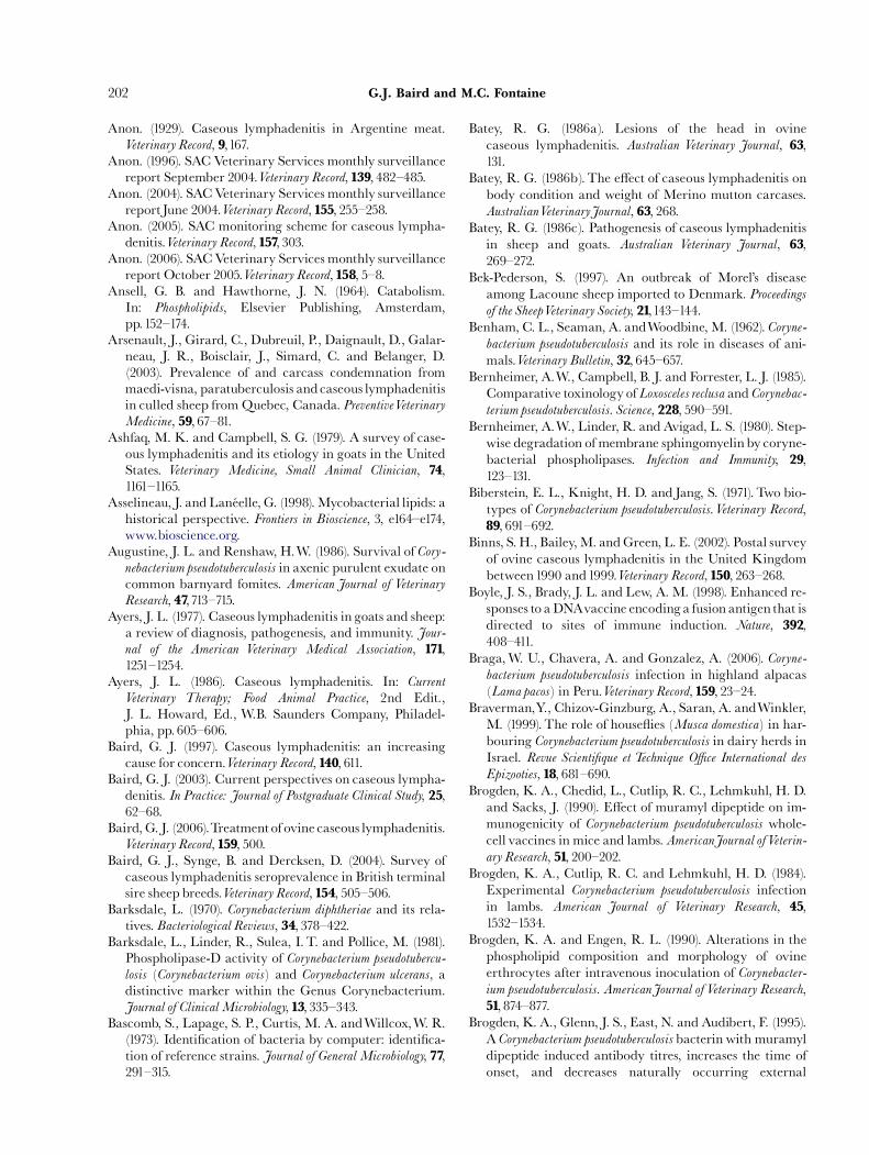
ARTICLE IN PRESS
G.J. Baird and M.C. Fontaine202
Anon. (1929). Caseous lymphadenitis in Argentine meat.Veterinary Record, 9,167.
Anon. (1996). SAC Veterinary Services monthly surveillancereport September 2004.Veterinary Record, 139, 482^485.
Anon. (2004). SACVeterinary Services monthly surveillancereportJune 2004.Veterinary Record, 155, 255^258.
Anon. (2005). SAC monitoring scheme for caseous lympha-denitis.Veterinary Record, 157, 303.
Anon. (2006). SACVeterinary Services monthly surveillancereport October 2005.Veterinary Record, 158, 5^8.
Ansell, G. B. and Hawthorne, J. N. (1964). Catabolism.In: Phospholipids, Elsevier Publishing, Amsterdam,pp.152^174.
Arsenault, J., Girard, C., Dubreuil, P., Daignault, D., Galar-neau, J. R., Boisclair, J., Simard, C. and Belanger, D.(2003). Prevalence of and carcass condemnation frommaedi-visna, paratuberculosis andcaseous lymphadenitisin culled sheep fromQuebec, Canada. PreventiveVeterinaryMedicine, 59, 67^81.
Ashfaq, M. K. and Campbell, S. G. (1979). A survey of case-ous lymphadenitis and its etiology in goats in the UnitedStates. Veterinary Medicine, Small Animal Clinician, 74,1161^1165.
Asselineau, J. andLaneŁ elle, G. (1998).Mycobacterial lipids: ahistorical perspective. Frontiers in Bioscience, 3, e164^e174,www.bioscience.org.
Augustine, J. L. and Renshaw, H.W. (1986). Survival of Cory-nebacterium pseudotuberculosis in axenic purulent exudate oncommon barnyard fomites. American Journal of VeterinaryResearch, 47,713^715.
Ayers, J. L. (1977). Caseous lymphadenitis in goats and sheep:a review of diagnosis, pathogenesis, and immunity. Jour-nal of the American Veterinary Medical Association, 171,1251^1254.
Ayers, J. L. (1986). Caseous lymphadenitis. In: Current
Veterinary Therapy; Food Animal Practice, 2nd Edit.,J. L. Howard, Ed., W.B. Saunders Company, Philadel-phia, pp. 605^606.
Baird, G. J. (1997). Caseous lymphadenitis: an increasingcause for concern.Veterinary Record, 140, 611.
Baird, G. J. (2003). Current perspectives on caseous lympha-denitis. In Practice: Journal of Postgraduate Clinical Study, 25,62^68.
Baird, G. J. (2006).Treatment of ovine caseous lymphadenitis.Veterinary Record, 159, 500.
Baird, G. J., Synge, B. and Dercksen, D. (2004). Survey ofcaseous lymphadenitis seroprevalence in British terminalsire sheep breeds.Veterinary Record, 154, 505^506.
Barksdale, L. (1970). Corynebacterium diphtheriae and its rela-tives. Bacteriological Reviews, 34, 378^422.
Barksdale, L., Linder, R., Sulea, I. T. and Pollice, M. (1981).Phospholipase-D activity of Corynebacterium pseudotubercu-
losis (Corynebacterium ovis) and Corynebacterium ulcerans, adistinctive marker within the Genus Corynebacterium.Journal of ClinicalMicrobiology, 13, 335^343.
Bascomb, S., Lapage, S. P., Curtis, M. A. andWillcox,W. R.(1973). Identi¢cation of bacteria by computer: identi¢ca-tion of reference strains. Journal of General Microbiology, 77,291^315.
Batey, R. G. (1986a). Lesions of the head in ovinecaseous lymphadenitis. Australian Veterinary Journal, 63,131.
Batey, R. G. (1986b).The e¡ect of caseous lymphadenitis onbody condition and weight of Merino mutton carcases.AustralianVeterinaryJournal, 63, 268.
Batey, R. G. (1986c). Pathogenesis of caseous lymphadenitisin sheep and goats. Australian Veterinary Journal, 63,269^272.
Bek-Pederson, S. (1997). An outbreak of Morel’s diseaseamong Lacoune sheep imported to Denmark. Proceedingsof the SheepVeterinary Society, 21,143^144.
Benham, C. L., Seaman, A. andWoodbine,M. (1962). Coryne-bacterium pseudotuberculosis and its role in diseases of ani-mals.Veterinary Bulletin, 32, 645^657.
Bernheimer, A.W., Campbell, B. J. and Forrester, L. J. (1985).Comparative toxinology of Loxosceles reclusa and Corynebac-terium pseudotuberculosis. Science, 228, 590^591.
Bernheimer, A.W., Linder, R. and Avigad, L. S. (1980). Step-wise degradation of membrane sphingomyelin by coryne-bacterial phospholipases. Infection and Immunity, 29,123^131.
Biberstein, E. L., Knight, H. D. and Jang, S. (1971). Two bio-types of Corynebacterium pseudotuberculosis.Veterinary Record,89, 691^692.
Binns, S.H., Bailey,M. andGreen, L. E. (2002). Postal surveyof ovine caseous lymphadenitis in the United Kingdombetween1990 and1999.Veterinary Record, 150, 263^268.
Boyle, J. S., Brady, J. L. and Lew, A. M. (1998). Enhanced re-sponses to aDNAvaccine encodinga fusion antigenthat isdirected to sites of immune induction. Nature, 392,408^411.
Braga,W. U., Chavera, A. and Gonzalez, A. (2006). Coryne-bacterium pseudotuberculosis infection in highland alpacas(Lama pacos) in Peru.Veterinary Record, 159, 23^24.
Braverman,Y., Chizov-Ginzburg, A., Saran, A. andWinkler,M. (1999). The role of house£ies (Musca domestica) in har-bouring Corynebacterium pseudotuberculosis in dairy herds inIsrael. Revue Scienti¢que et Technique O⁄ce International desEpizooties, 18, 681^690.
Brogden, K. A., Chedid, L., Cutlip, R. C., Lehmkuhl, H. D.and Sacks, J. (1990). E¡ect of muramyl dipeptide on im-munogenicity of Corynebacterium pseudotuberculosis whole-cell vaccines in mice and lambs. AmericanJournal ofVeterin-ary Research, 51, 200^202.
Brogden, K. A., Cutlip, R. C. and Lehmkuhl, H. D. (1984).Experimental Corynebacterium pseudotuberculosis infectionin lambs. American Journal of Veterinary Research, 45,1532^1534.
Brogden, K. A. and Engen, R. L. (1990). Alterations in thephospholipid composition and morphology of ovineerthrocytes after intravenous inoculation of Corynebacter-ium pseudotuberculosis. AmericanJournal ofVeterinary Research,51, 874^877.
Brogden, K. A., Glenn, J. S., East, N. and Audibert, F. (1995).A Corynebacterium pseudotuberculosis bacterin with muramyldipeptide induced antibody titres, increases the time ofonset, and decreases naturally occurring external
ARTICLE IN PRESS
Ovine Caseous Lymphadenitis 203
abscesses in sheep and goats. Small Ruminant Research, 19,161^168.
Brown, C. C. andOlander, H. J. (1987). Caseous lymphadeni-tis of goats and sheep: a review.Veterinary Bulletin, 57,1^12.
Brown, C. C., Olander, H. J., Zometa, C. and Alves, S. F.(1986). Serodiagnosis of inapparent caseous lymphadeni-tis in goats and sheep, using the synergistic hemolysis-in-hibition test. American Journal of Veterinary Research, 47,1461^1463.
Bruere, A. N. andWest, D.M. (1990). Caseous lymphadenitis.In:The Sheep: Health, Disease and Production, Massey Uni-versity Press, Palmerston North, pp. 252^257.
Brunet, J. F., Denizot, F., Luciani, M. F., Roux-Dosseto, M.,Suzan, M., Mattei, M. G. and Goldstein, P. (1987). A newmember of the immunoglobulin superfamilyçCTLA-4.Nature, 328, 267^270.
Bull, L. B. andDickinson, C. G. (1935). Studies on infectionbyand resistance to the Pre|« sz-Nocard bacillus. Notes on thetoxin, the pyogenic action and the lipoid content of thebacillus. AustralianVeterinaryJournal, 11,1126^1138.
Burrell, D. H. (1978a). Experimental induction of caseouslymphadenitis in sheep by intralymphatic inoculation ofCorynebacterium ovis. Research in Veterinary Science, 24,269^276.
Burrell, D. H. (1978b).Vaccination against caseous lympha-denitis in sheep. In: Proceedings of the 55th Annual Conferenceof theAustralianVeterinaryAssociation, pp.79^81.
Burrell, D. H. (1980a). A haemolysis inhibition test for detec-tion of antibody to Corynebacterium ovis exotoxin.Research inVeterinary Science, 28,190^194.
Burrell, D. H. (1980b). A simpli¢ed double immunodi¡usiontechnique for detection of Corynebacterium ovis antitoxin.Research inVeterinary Science, 28, 234^237.
Burrell, D. H. (1981). Caseous lymphadenitis in goats. Austra-lianVeterinaryJournal, 57,105^110.
Burrell, D. H. (1983). Caseous lymphadenitis vaccine. NewSouthWalesVeterinary Proceedings, 19, 53^57.
Cameron, C.M. (1964).The signi¢cance of the endotoxin andpyogenic factor of Corynebacterium pseudotuberculosis in im-munity. Onderstepoort Journal of Veterinary Research, 31,119^132.
Cameron, C.M. (1972). Immunity to Corynebacterium pseudotu-
berculosis.Journal of the SouthAfricanVeterinaryAssociation, 43,343^349.
Cameron, C.M. and Bester, F. J. (1984). An improved Coryne-
bacterium pseudotuberculosis vaccine for sheep. OnderstepoortJournal ofVeterinary Research, 51, 263^267.
Cameron, C. M. and Buchan, L. (1966). Identi¢cationof the protective and toxic antigens of Corynebacterium pseu-
dotuberculosis. OnderstepoortJournal of Veterinary Research, 33,39^48.
Cameron, C. M. andMinnaar, J. L. (1969). Immunization ofmice against Corynebacterium pseudotuberculosis infection.OnderstepoortJournal ofVeterinary Research, 36, 207^210.
Cameron, C. M., Minnaar, J. L., Engelbrecht, M. M. andPurdom, M. R. (1972). Immune response of merino sheepto inactivated Corynebacterium pseudotuberculosis vaccine.OnderstepoortJournal ofVeterinary Research, 39,11^24.
Cameron, C. M., Minnaar, J. L. and Purdom, M. R. (1969).Immunizing properties of Corynebacterium pseudotuberculosis
cell walls. Onderstepoort Journal of Veterinary Research, 36,211^216.
Cameron, C. M. and Smit, M. C. (1970). Relationship of Cor-ynebacterium pseudotuberculosis protoplasmic toxins to theexotoxin. Onderstepoort Journal of Veterinary Research, 37,97^103.
Carne, H. R. (1940).The toxin of Corynebacterium ovis. Journalof Pathology and Bacteriology, 51,199^212.
Carne, H. R. and Onon, E. O. (1978). Action of Corynebacter-ium ovis exotoxin on the endothelial cells of blood vessels.Nature, 271, 246^248.
Carne, H. R.,Wickham, N. and Kater, J. C. (1956). A toxiclipid from the surface of Corynebacterium ovis. Nature, 178,701^702.
Cesari, E. (1930). Diagnosis of caseous lymphadenitis by theIntradermal-Reaction Test, after using Preisznocardine.Veterinary Record, 10,1151^1152.
Cetinkaya, B., Karahan,M., Atil, E., Kalin, R., De Baere,T.andVaneechoutte, M. (2002). Identi¢cation of Corynebac-terium pseudotuberculosis isolates from sheep and goats byPCR.VeterinaryMicrobiology, 88,75^83.
Christie, R., Atkins, N. E. and Munch-Petersen, E. (1944). Anote on a lytic phenomenon shownby group B streptococ-ci. Australian Journal of Experimental Biology and Medical
Science, 22,197^200.Clarridge, J. E. and Spiegel, C. A. (1995). Corynebacterium and
related organisms. In: Manual of Clinical Microbiology, 6thEdit., E. J. Barron, Ed., American Society for Microbiol-ogy,Washington, pp. 357^370.
Collett, M. G., Bath, G. F. and Cameron, C. M. (1994). Cory-nebacterium pseudotuberculosis infections. In: Infectious Dis-
eases of Livestock with Special Reference to Southern Africa, 2ndEdit., J. Coetzer, G.R. Thomson and R.C. Tustin, Eds,Oxford University Press, CapeTown, pp.1387^1395.
Collins, M. D. and Cummins, C. S. (1986). Genus Corynebac-terium. In: Bergey’s Manual of Systematic Bacteriology, Vol. 2.P.H.A. Sneath, N.S. Mair, M.E. Sharpe and J.G. Holt,Eds, Williams and Wilkins Company, Baltimore,pp.1266^1276.
Collins,M. D., Goodfellow,M. andMinnikin, D. E. (1982). Asurvey of the structures of mycolic acids in Corynebacteriumand related taxa. Journal of General Microbiology, 128,129^149.
Connor, K. M., Quirie, M. M., Baird, G. and Donachie,W.(2000). Characterization of United Kingdom isolates ofCorynebacterium pseudotuberculosis using pulsed-¢eld gelelectrophoresis. Journal of Clinical Microbiology, 38,2633^2637.
Costa, L. R. R., Spier, S. J. and Hirsh, D. C. (1998). Com-parative molecular characterization of Corynebacterium
pseudotuberculosis of di¡erent origin.Veterinary Microbiology,62,135^143.
Coyle, M. B., Hollis, D. G. and Groman, N. B. (1985). Coryne-bacterium spp. and other coryneform organisms. In: Man-
ual of Clinical Microbiology, 4th Edit., E. H. Lennette, A.Balows,W. J. Hausler and H. J. Shadomy, Eds, AmericanSociety for Microbiology,Washington, pp.198^199.
ARTICLE IN PRESS
G.J. Baird and M.C. Fontaine204
Davis, E.W. (1990). Corynebacteriumpseudotuberculosis infectionsin animals. In: Large Animal Internal Medicine, B.P. Smith,Ed., C.V. Mosby Company,Toronto, pp.1120^1126.
Dennis, S. M. and Bamford,V.W. (1966). The role of Coryne-bacteria in perinatal lamb mortality.Veterinary Record, 79,105^108.
Dercksen, D. P., Brinkhof, J. M. A., Dekker-Nooren,T., vanMaanen, K., Bode, C. F., Baird, G. J. and Kamp, E. M.(2000). A comparison of four serological tests for the diag-nosis of caseous lymphadenitis in sheep and goats.Veterin-aryMicrobiology, 75,167^175.
Dercksen, D. P., ter Laak, E. A. and Schreuder, B. E. (1996).Eradication programme for caseous lymphadenitis ingoats inThe Netherlands.Veterinary Record, 138, 237.
De Rose, R.,Tennent, J., McWaters, P., Chaplin, P. J.,Wood,P. R., Kimpton, W., Cahill, R. and Scheerlinck, J. -P.(2002). E⁄cacy of DNAvaccination by di¡erent routes ofimmunisation in sheep.Veterinary Immunology and Immuno-pathology, 90, 55^63.
Doherr, M. G., Carpenter,T. E.,Wilson,W. D. and Gardner,I. A. (1998). Application and evaluation of a mailed ques-tionnaire for an epidemiologic study of Corynebacteriumpseudotuberculosis infection in horses. Preventive Veterinary
Medicine, 35, 241^253.Doherr, M. G., Carpenter, T. E., Wilson, W. D. and
Gardner, I. A. (1999). Evaluation of temporal and spatialclustering of horses with Corynebacterium pseudotuberculosis
infection. American Journal of Veterinary Research, 60,284^291.
Donnelly, J. J., Ulmer, J. B., Shiver, J. W. and Liu, M. A.(1995). DNA vaccines. Annual Reviews in Immunology, 15,617^648.
Doty, R. B., Dunne, H.W., Hokanson, J. F. and Reid, J. J.(1964). A comparison of toxins produced by various iso-lates of Corynebacterium pseudotuberculosis and the develop-ment of a diagnostic skin test for caseous lymphadenitisof sheep and goats. AmericanJournal of Veterinary Research,25,1679^1985.
Drew, D. R., Boyle, J. S., Lew, A. M., Lightowlers, M.W.,Chaplin, P. J. and Strugnell, R. A. (2001). The compara-tive e⁄cacyof CTLA-4 and L-selectin targetedDNAvac-cines in mice and sheep.Vaccine, 19, 4417^4428.
Egen, N. B., Cuevas,W. A., McNamara, P. J., Sammons, D.W., Humphreys, R. and Songer, J. G. (1989). Puri¢cationof the phospholipase D of Corynebacterium pseudotuberculosis
by recycling isoelectric focusing. AmericanJournal of Veter-inary Research, 50,1319^1322.
Eggleton, D. G., Doidge, C.V., Middleton, H. D. andMinty,D. W. (1991a). Immunisation against ovine caseous lym-phadenitis: e⁄cacy of the monocomponent Corynebacter-ium pseudotuberculosis toxoid vaccine and combinedclostridial-corynebacterial vaccines. Australian VeterinaryJournal, 68, 320^321.
Eggleton, D. G., Haynes, J. A., Middleton, H. D. and Cox,J. C. (1991b). Immunisation against ovine caseous lym-phadenitis: correlation between Corynebacterium pseudotu-
berculosis toxoid contents and protective e⁄cacy incombined clostridial-corynebacterial vaccines. AustralianVeterinaryJournal, 68, 322^325.
Eggleton, D. G., Middleton, H. D., Doidge, C.V. andMinty,D. W. (1991c). Immunisation against ovine caseous lym-phadenitis: comparison of Corynebacteriumpseudotuberculosis
vaccines with and without bacterial cells. AustralianVeter-inaryJournal, 68, 317^319.
Eghetti, L. and McKenney, F. D. (1941). Caseous lymphade-nitis of deer (Odocoileus hemionus) inWashington. Journal oftheAmericanVeterinaryMedical Association, 98,129^131.
Ellis, J. A., Hawk, D. A., Holler, L. D.,Mills, K.W. and Pratt,D. L. (1990). Di¡erential antibody responses to Corynebac-terium pseudotuberculosis in sheep with naturally acquiredcaseous lymphadenitis. Journal of the American VeterinaryMedical Association, 196,1609^1613.
Ellis,T. M., Sutherland, S. S.,Wilkinson, F. C., Mercy, A. R.and Paton,M.W. (1987).The role of Corynebacterium pseudo-
tuberculosis lung lesions in the transmission of this bacter-ium to other sheep. Australian Veterinary Journal, 64,261^263.
Esterabadi, A. H., Entessar, F., Hedayati, H., Narimani, A.A. and Sadri,M. (1975). Isolation of Corynebacterium pseudo-
tuberculosis fromcamel in Iran.Archivesde L’Institut Razi, 27,61^66.
Euzeby, J. P. (2005). List of Bacterial Names with Standing in No-menclature, Society for Systematic andVeterinary Bacter-iology, http://www.bacterio.coct.fr.
Fontaine,M. C., Baird, G., Connor, K.M., Rudge, K., Sales,J. andDonachie,W. (2006).Vaccination confers signi¢cantprotection of sheep against infection with a virulent Uni-ted Kingdom strain of Corynebacterium pseudotuberculosis.Vaccine, 24, 5986^5996.
Forgeot, P. and Cesari, E. (1912). Haemolytic activity of Cory-nebacterium ovis. Annals of the Pasteur Institute, 26,102.
Fraser, G. (1961). Haemolytic activity of Corynebacterium ovis.Nature, 189, 246.
Gates, N. L., Everson, D. O. andHulet, C.V. (1977). E¡ects ofthin ewe syndrome on reproductive e⁄ciency. Journal oftheAmericanVeterinaryMedical Association, 171,1266^1267.
Gezon, H. M., Bither, H. D., Hanson, L. A. andThompson,J. K. (1991). Epizootic of external and internal abscesses ina large goat herd over a16-year period. Journal of theAmer-icanVeterinaryMedical Association, 198, 257^263.
Ghannoum,M. A. (2000). Potential role of phospholipases invirulence and fungal pathogenesis. Clinical Microbiology,13,122^143.
Goldberger, A. C., Lipsky, B. A. and Plorde, J. J. (1981). Sup-purative granulomatous lymphadenitis caused by Coryne-
bacterium ovis (pseudotuberculosis).AmericanJournal of ClinicalPathology, 76, 486^490.
Goodfellow, M., Collins, M. D. and Minnikin, D. E. (1976).Thin-layer chromatographic analysis of mycolic acid andother long-chain components in whole-organism metha-nolysates of coryneform and related taxa. Journal of Gener-alMicrobiology, 96, 351^358.
Groman, N., Schiller, J. and Russell, J. (1984). Corynebacteriumulcerans and Corynebacterium pseudotuberculosis responses toDNAprobes derived fromcorynephagebeta andCoryne-bacterium diphtheriae. Infection and Immunity, 45, 511^517.
Hamilton, N. T., Perceval, A., Aarons, B. J. and Goodyear,J. E. (1968). Pseudotuberculous axillary lymphadenitis
ARTICLE IN PRESS
Ovine Caseous Lymphadenitis 205
caused by Corynebacterium pseudotuberculosis.MedicalJournal
of Australia, 2, 356^361.Hammersland, H. andJoneschild, E.M. (1937). Pseudotuber-
culosis of deer. Journal of theAmericanVeterinaryMedical As-
sociation, 91,186^192.Hard, G. C. (1970). Adoptive transfer of immunity in experi-
mental Corynebacterium ovis infection. Journal of ComparativePathology, 80, 329^334.
Hard, G. C. (1972). Examination by electron microscopy ofthe interaction between peritoneal phagocytes and Cory-
nebacterium ovis. Journal ofMedicalMicrobiology, 5, 483^491.Hard, G. C. (1975). Comparative toxic e¡ect of the surface li-
pid of Corynebacterium ovis on peritoneal macrophages. In-fection and Immunity, 12,1439^1449.
Heath, G. B. S. and Batty, I. (1952). An unusual diphtheroidisolated from sheep.Veterinary Record, 64, 261.
Hein,W. R. and Cargill, C. F. (1981). An abattoir survey ofdiseases of feral goats. Australian Veterinary Journal, 57,498^503.
Henderson, A. (1979). Pseudotuberculous adenitis caused byCorynebacterium pseudotuberculosis. Journal of Medical Micro-
biology, 12,147^149.Henriksen, S. D. and Grelland, R. (1952). Toxigenicity, sero-
logical reactions, and relationships of the diphtheria-likecorynebacteria. Journal of Pathology and Bacteriology, 64,503^511.
Hill, L. R., Lapage, S. P. and Bowie, I. S. (1978). Computeridenti¢cation of coryneform bacteria. In: Coryneform Bac-
teria, I. J. Bouse¢eld and A. G. Callely, Eds, AcademicPress (for the Society for General Microbiology),London, pp.181^215.
Hinnebusch, J., Cherepanov, P., Du,Y., Rudolph, A., Dixon,J. D., Schwan, I. and Forsberg, A. (2000). Murine toxin ofYersinia pestis shows phospholipase D activity but is not re-quired for virulence inmice. InternationalJournal ofMedical
Microbiology, 290, 483^487.Hodgson, A. L., Bird, P. and Nisbet, I.T. (1990). Cloning, nu-
cleotide sequence, and expression in Escherichia coli of thephospholipase D gene from Corynebacterium pseudotuberculo-
sis. Journal of Bacteriology, 172,1256^1261.Hodgson, A. L., Carter, K., Tachedjian, M., Krywult, J.,
Corner, L. A., McColl, M. and Cameron, A. (1999). E⁄-cacy of an ovine caseous lymphadenitis vaccine formu-lated using a genetically inactive form of theCorynebacterium pseudotuberculosis phospholipase D.Vaccine,17, 802^808.
Hodgson, A. L., Krywult, J., Corner, L. A., Rothel, J. S. andRadford, A. J. (1992). Rational attenuation of Corynebacter-ium pseudotuberculosisçpotential cheesy gland vaccine andlive delivery vehicle. Infection and Immunity, 60, 2900^2905.
Hodgson, A. L.,Tachedjian, M., Corner, L. A. and Radford,A. J. (1994). Protection of sheep against caseous lympha-denitis by use of a single oral dose of live recombinant Cor-ynebacterium pseudotuberculosis. Infection and Immunity, 62,5275^5280.
House, R.W., Schousboe, M., Allen, J. P. and Grant, C. C.(1986). Corynebacterium ovis (pseudotuberculosis) lymphadeni-tis in a sheep farmer: a new occupational disease in NewZealand.NewZealandMedicalJournal, 99, 659^662.
Hsu,T.Y., Renshaw, H.W., Livingston, C.W., Augustine, J.L. and Zink, D. L. (1985). Corynebacterium pseudotuberculosis
exotoxin: fatal hemolytic anemia induced in gnotobioticneonatal small ruminants by parenteral administrationof preparations containing exotoxin. American Journal ofVeterinary Research, 46,1206^1211.
Hughes, J. P. and Biberstein, E. L. (1959). Chronic equine ab-scesses associated with Corynebacterium pseudotuberculosis.Journal of the American Veterinary Medical Association, 135,559^562.
Husband, A. J. and Watson, D. L. (1977). Immunologicalevents in the popliteal lymph node of sheep following in-jection of live or killed Corynebacterium ovis into an a¡erentpopliteal lymphatic duct. Research inVeterinary Science, 22,105^112.
Irwin, M. R. and Knight, H. D. (1975). Enhanced resistanceto Corynebacterium pseudotuberculosis infections associatedwith reduced serum immunoglobulin levels in levami-sole-treated mice. Infection and Immunity, 12,1098^1103.
Ismail, A. A. andHamid,Y.M.A. (1972). Studies on the e¡ectof some chemical disinfectants used in veterinary practicein Corynebacterium ovis. Journal of the EgyptianVeterinaryMed-
ical Association, 32,195^202.Join-Lambert, O. F., Ouache,M., Canioni, D., Beretti, J. -L.,
Blanche, S., Berche, P. and Kayal, S. (2006). Corynebacter-ium pseudotuberculosis necrotizing lymphadenitis in atwelve-year-old patient. Pediatric Infectious DiseaseJournal,25, 848^851.
Jolly, R. D. (1965). The pathogenic action of the exotoxin ofCorynebacterium ovis. Journal of Comparative Pathology, 75,417^431.
Judson, R. and Songer, J. G. (1991).Corynebacteriumpseudotuber-
culosis: in-vitro susceptibility to 39 antimicrobial agents.VeterinaryMicrobiology, 27,145^150.
Kariuki, D. P. and Poulton, J. (1982). Corynebacterium infectionof cattle in Kenya.Tropical Animal Health and Production, 14,33^36.
Keddie, R. M. and Cure, G. L. (1977). The cell wall compo-sition and distribution of free mycolic acids in namedstrains of coryneform bacteria and in isolates fromvarious natural sources. Journal of Applied Bacteriology, 42,229^252.
Keslin,M. H.,McCoy, E. L.,McCusker, J. J. and Lutch, J. S.(1979). Corynebacterium pseudotuberculosis. A new cause of in-fectious and eosinophilic pneumonia. AmericanJournal ofMedicine, 67, 228^231.
Knight, H. D. (1969). Corynebacterial infections in the horse:problems of prevention. Journal of the American VeterinaryMedical Association, 155, 446^452.
Knight, H. D. (1978). A serologic method for the detection ofCorynebacterium pseudotuberculosis infections in horses.CornellVeterinarian, 68, 220^237.
Kuria, J. K. N. and Holstad, G. (1989). Serological investiga-tion of Corynebacterium pseudotuberculosis infection insheepçcorrelation between the hemolysis inhibition testand the ELISA test. Acta Veterinaria Scandinavica, 30,109^110.
Lapage, S. P., Bascomb, S.,Willcox,W. R. and Curtis, M. A.(1973). Identi¢cation of bacteria by computer: general
ARTICLE IN PRESS
G.J. Baird and M.C. Fontaine206
aspects andperspectives. Journal of GeneralMicrobiology,77,273^290.
Lee, S. and Lynch, K. R. (2005). Brown recluse spider (Loxos-celes reclusa) venom phospholipase D (PLD) generates ly-sophosphatidic acid (LPA). Biochemical Journal, 391,317^323.
Lehmann, K. B. and Neumann, R. O. (1896). In: Atlas
und Grundriss der Bakteriologie und Lehrbuch der Speciellen
Bakteriologischen Diagnostik, K. B. Lehmann and R. O.Neumann, Eds, J.F. Lehmann, Muº nchen.
Linsley, P. S., Brady,W.,Urnes,M., Grosmaire, L. S., Damle,N. K. and Ledbetter, J. A. (1991). CTLA-4 is a second re-ceptor for the B cell activation antigen B7. Journal of Ex-perimentalMedicine, 174, 561^569.
Lloyd, S., Lindsay, H. J., Slater, J. D. and Jackson, P. G.(1990). Caseous lymphadenitis in goats in England.Veteri-nary Record, 127, 478.
Lopez, J. F.,Wong, F.M. andQuesada, J. (1966). Corynebacter-ium pseudotuberculosis. First case of human infection. Ameri-canJournal of Clinical Pathology, 46, 562^567.
Lovell, R. and Zaki,M.M. (1966). Studies of the growth pro-ducts of Corynebacterium ovis II. Other activities and their rela-tionship. Research inVeterinary Science, 7, 307^311.
Maki, L. R., Shen, S. H., Bergstrom, R. C. and Stetzenbach,L. D. (1985). Diagnosis of Corynebacterium pseudotuberculosis
infections in sheep, using an enzyme-linked immunosor-bent assay. American Journal of Veterinary Research, 46,212^214.
Malone, F. E., Fee, S. A., Kamp, E.M.,King, D. C., Baird, G.J., O’Reilly, K. M. and Murdock, F. E. A. (2006). A sero-logical investigation of caseous lymphadenitis in four£ocks of sheep. IrishVeterinaryJournal, 59,19^21.
Maximescu, P. (1968). New host strains for the lysogenic Cor-ynebacterium diphtheriae ParkWilliams No. 8 strain. Journalof GeneralMicrobiology, 53,125^133.
Maximescu, P., Oprisan, A., Pop, A. and Potorac, E. (1974a).Further studies on Corynebacterium species capable of pro-ducing diphtheria toxin (C. diphtheriae, C. ulcerans, C. ovis).Journal of GeneralMicrobiology, 82, 49^56.
Maximescu, P., Pop, A., Oprisan, A. and Potorac, E. (1974b).Diphtheria tox+ gene expressed in Corynebacterium speciesother than C. diphtheriae. Journal of Hygiene, Epidemiology,Microbiology and Immunology, 18, 324^326.
McAllister, H. A. and Keahey, K. K. (1971). Infection of ahedgehog (Erinaceus albiventris) by Corynebacterium pseudo-
tuberculosis.Veterinary Record, 89, 280.McNamara, P. J., Bradley, G. A. and Songer, J. G. (1994).Tar-
geted mutagenesis of the phospholipase D gene results indecreased virulence of Corynebacterium pseudotuberculosis.MolecularMicrobiology, 12, 921^930.
McNamara, P. J., Cuevas, W. A. and Songer, J. G. (1995).Toxic phospholipases D of Corynebacterium pseudotuberculo-
sis, Corynebacterium ulcerans and Arcanobacterium haemolyti-
cum: cloning and sequence homology. Gene, 156,113^118.Mechie, S. C. (1998). Screening for caseous lymphadenitis.
Veterinary Record, 142, 47.Meeusen, E. N. T. and Brandon, M. R. (2006). The use of
antibody secreting cell probes to reveal tissue restricted
immune responses during infection.EuropeanJournal of Im-munology, 24, 469^475.
Meldrum, K. C. (1990). Caseous lymphadenitis outbreak.Veterinary Record, 126, 369.
Menzies, P. I., Hwanga,Y. -T. and Prescott, J. F. (2004). Com-parison of an interferon-gamma to a phospholipase D en-zyme-linked immunosorbent assay for diagnosis ofCorynebacterium pseudotuberculosis infection in experimen-tally infected goats.VeterinaryMicrobiology, 100,129^137.
Menzies, P. I., Muckle, C. A., Brogden, K. A. and Robinson,L. (1991). A ¢eld trial to evaluate a whole cell vaccine forthe preventionof caseous lymphadenitis in sheep andgoat£ocks. CanadianJournal ofVeterinary Research, 55, 362^366.
Menzies, P. I., Muckle, C. A., Hwang,Y.T. and Songer, J. G.(1994). Evaluation of an enzyme-linked immunosorbentassay using an Escherichia coli recombinant phospholipaseDantigen for the diagnosis ofCorynebacteriumpseudotubercu-
losis infection. Small Ruminant Research, 13,193^198.Meyer, R., Regis, L.,Vale,V., Paule, B., Carminati, R., Ba-
hia, R., Moura-Costa, L., Schaer, R., Nascimento, I. andFreire, S. (2005). In vitro IFN-gamma production by goatblood cells after stimulation with somatic and secretedCorynebacterium pseudotuberculosis antigens.Veterinary Immu-nology and Immunopathology, 107, 249^254.
Middleton, M. J., Epstein,V. M. and Gregory, G. G. (1991).Caseous lymphadenitis on Flanders Island: prevalenceand management surveys. AustralianVeterinaryJournal, 68,311^312.
Miers, K. C. and Ley,W. B. (1980). Corynebacterium pseudotuber-
culosis infection in the horse: study of 117 clinical cases andconsideration of etiopathogenesis. Journal of the AmericanVeterinaryMedical Association, 177, 250^253.
Mills, A. E., Mitchell, R. D. and Lim, E. K. (1997). Corynebac-terium pseudotuberculosis is a cause of human necrotisinggranulomatous lymphadenitis. Pathology, 29, 231^233.
Mills, D.W. (1928). Report on observations made on caseouslymphadenitis in sheep.Veterinary Record, 8, 509^510.
Minnikin, D. E., Alshamaony, L. and Goodfellow, M. (1975).Di¡erentiation of Mycobacterium, Nocardia, and relatedtaxa by thin-layer chromatographic analysis of whole-or-ganism methanolysates. Journal of GeneralMicrobiology, 88,200^204.
Minnikin, D. E. and Goodfellow, M. (1976). Lipid composi-tion in the classi¢cation and identi¢cation of nocardiaeand related taxa. In:The Biology of the Nocardiae, M. Good-fellow, G.H. Brownell and J.A. Serrano, Eds, AcademicPress, London, pp.160^169.
Minnikin, D. E. and Goodfellow, M. (1980). Lipid composi-tion in the classi¢cationand identi¢cationof acid-fast bac-teria. Society for Applied Bacteriology Symposium Series, 8,189^256.
Minnikin, D. E., Goodfellow, M. and Collins, M. D. (1978).Lipid composition in the classi¢cation and identi¢cationof coryneform and related taxa. In:Coryneform Bacteria, I.J. Bouse¢eld and A. G. Callely, Eds, Academic Press(for the Society for General Microbiology), London,pp. 85^160.
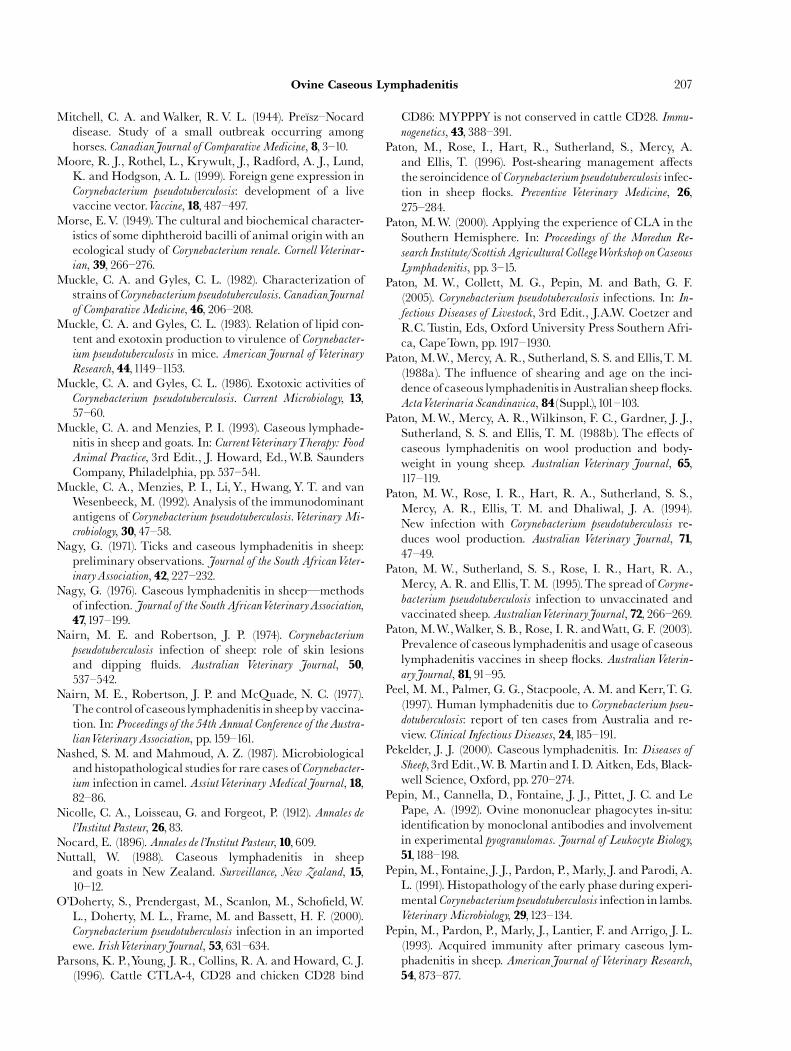
ARTICLE IN PRESS
Ovine Caseous Lymphadenitis 207
Mitchell, C. A. andWalker, R. V. L. (1944). Pre|« sz^Nocarddisease. Study of a small outbreak occurring amonghorses. CanadianJournal of ComparativeMedicine, 8, 3^10.
Moore, R. J., Rothel, L., Krywult, J., Radford, A. J., Lund,K. and Hodgson, A. L. (1999). Foreign gene expression inCorynebacterium pseudotuberculosis: development of a livevaccine vector.Vaccine, 18, 487^497.
Morse, E.V. (1949).The cultural and biochemical character-istics of some diphtheroid bacilli of animal origin with anecological study of Corynebacterium renale. Cornell Veterinar-ian, 39, 266^276.
Muckle, C. A. and Gyles, C. L. (1982). Characterization ofstrains of Corynebacteriumpseudotuberculosis. CanadianJournalof ComparativeMedicine, 46, 206^208.
Muckle, C. A. and Gyles, C. L. (1983). Relation of lipid con-tent and exotoxin production to virulence of Corynebacter-ium pseudotuberculosis in mice. AmericanJournal of VeterinaryResearch, 44,1149^1153.
Muckle, C. A. and Gyles, C. L. (1986). Exotoxic activities ofCorynebacterium pseudotuberculosis. Current Microbiology, 13,57^60.
Muckle, C. A. and Menzies, P. I. (1993). Caseous lymphade-nitis in sheep and goats. In: CurrentVeterinaryTherapy: Food
Animal Practice, 3rd Edit., J. Howard, Ed.,W.B. SaundersCompany, Philadelphia, pp. 537^541.
Muckle, C. A., Menzies, P. I., Li, Y., Hwang,Y. T. and vanWesenbeeck, M. (1992). Analysis of the immunodominantantigens of Corynebacterium pseudotuberculosis.Veterinary Mi-
crobiology, 30, 47^58.Nagy, G. (1971). Ticks and caseous lymphadenitis in sheep:
preliminary observations. Journal of the South AfricanVeter-inaryAssociation, 42, 227^232.
Nagy, G. (1976). Caseous lymphadenitis in sheepçmethodsof infection. Journal of the South AfricanVeterinaryAssociation,47,197^199.
Nairn, M. E. and Robertson, J. P. (1974). Corynebacterium
pseudotuberculosis infection of sheep: role of skin lesionsand dipping £uids. Australian Veterinary Journal, 50,537^542.
Nairn, M. E., Robertson, J. P. and McQuade, N. C. (1977).The control of caseous lymphadenitis in sheepby vaccina-tion. In: Proceedings of the 54th Annual Conference of theAustra-lianVeterinaryAssociation, pp.159^161.
Nashed, S. M. and Mahmoud, A. Z. (1987). Microbiologicaland histopathological studies for rare cases ofCorynebacter-ium infection in camel. AssiutVeterinaryMedicalJournal, 18,82^86.
Nicolle, C. A., Loisseau, G. and Forgeot, P. (1912). Annales del’Institut Pasteur, 26, 83.
Nocard, E. (1896). Annales de l’Institut Pasteur, 10, 609.Nuttall, W. (1988). Caseous lymphadenitis in sheep
and goats in New Zealand. Surveillance, New Zealand, 15,10^12.
O’Doherty, S., Prendergast, M., Scanlon, M., Scho¢eld,W.L., Doherty, M. L., Frame, M. and Bassett, H. F. (2000).Corynebacterium pseudotuberculosis infection in an importedewe. IrishVeterinaryJournal, 53, 631^634.
Parsons, K. P.,Young, J. R., Collins, R. A. and Howard, C. J.(1996). Cattle CTLA-4, CD28 and chicken CD28 bind
CD86: MYPPPY is not conserved in cattle CD28. Immu-nogenetics, 43, 388^391.
Paton, M., Rose, I., Hart, R., Sutherland, S., Mercy, A.and Ellis, T. (1996). Post-shearing management a¡ectsthe seroincidence of Corynebacterium pseudotuberculosis infec-tion in sheep £ocks. Preventive Veterinary Medicine, 26,275^284.
Paton, M.W. (2000). Applying the experience of CLA in theSouthern Hemisphere. In: Proceedings of the Moredun Re-
search Institute/Scottish Agricultural CollegeWorkshop on Caseous
Lymphadenitis, pp. 3^15.Paton, M. W., Collett, M. G., Pepin, M. and Bath, G. F.
(2005). Corynebacterium pseudotuberculosis infections. In: In-fectious Diseases of Livestock, 3rd Edit., J.A.W. Coetzer andR.C.Tustin, Eds, Oxford University Press Southern Afri-ca, CapeTown, pp.1917^1930.
Paton,M.W.,Mercy, A. R., Sutherland, S. S. and Ellis,T.M.(1988a). The in£uence of shearing and age on the inci-dence of caseous lymphadenitis in Australian sheep £ocks.ActaVeterinaria Scandinavica, 84(Suppl.),101^103.
Paton, M.W., Mercy, A. R.,Wilkinson, F. C., Gardner, J. J.,Sutherland, S. S. and Ellis, T. M. (1988b). The e¡ects ofcaseous lymphadenitis on wool production and body-weight in young sheep. Australian Veterinary Journal, 65,117^119.
Paton, M. W., Rose, I. R., Hart, R. A., Sutherland, S. S.,Mercy, A. R., Ellis, T. M. and Dhaliwal, J. A. (1994).New infection with Corynebacterium pseudotuberculosis re-duces wool production. Australian Veterinary Journal, 71,47^49.
Paton, M. W., Sutherland, S. S., Rose, I. R., Hart, R. A.,Mercy, A. R. and Ellis,T. M. (1995).The spread of Coryne-bacterium pseudotuberculosis infection to unvaccinated andvaccinated sheep. AustralianVeterinaryJournal, 72, 266^269.
Paton,M.W.,Walker, S. B., Rose, I. R. andWatt, G. F. (2003).Prevalence of caseous lymphadenitis and usage of caseouslymphadenitis vaccines in sheep £ocks. AustralianVeterin-aryJournal, 81, 91^95.
Peel, M.M., Palmer, G. G., Stacpoole, A. M. and Kerr,T. G.(1997). Human lymphadenitis due to Corynebacterium pseu-
dotuberculosis: report of ten cases from Australia and re-view. Clinical Infectious Diseases, 24,185^191.
Pekelder, J. J. (2000). Caseous lymphadenitis. In: Diseases of
Sheep,3rd Edit.,W. B.Martin and I. D. Aitken, Eds, Black-well Science, Oxford, pp. 270^274.
Pepin, M., Cannella, D., Fontaine, J. J., Pittet, J. C. and LePape, A. (1992). Ovine mononuclear phagocytes in-situ:identi¢cation by monoclonal antibodies and involvementin experimental pyogranulomas. Journal of Leukocyte Biology,51,188^198.
Pepin,M., Fontaine, J. J., Pardon, P.,Marly, J. and Parodi, A.L. (1991). Histopathologyof the early phase during experi-mental Corynebacteriumpseudotuberculosis infection in lambs.VeterinaryMicrobiology, 29,123^134.
Pepin, M., Pardon, P., Marly, J., Lantier, F. and Arrigo, J. L.(1993). Acquired immunity after primary caseous lym-phadenitis in sheep. AmericanJournal of Veterinary Research,54, 873^877.
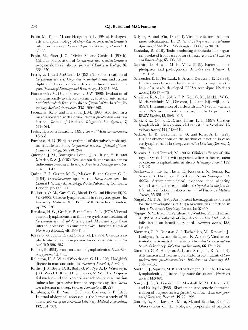
ARTICLE IN PRESS
G.J. Baird and M.C. Fontaine208
Pepin, M., Paton, M. andHodgson, A. L. (1994a). Pathogen-esis and epidemiology of Corynebacterium pseudotuberculosis
infection in sheep. Current Topics in Veterinary Research, 1,63^82.
Pepin, M., Pittet, J. C., Olivier, M. and Gohin, I. (1994b).Cellular composition of Corynebacterium pseudotuberculosis
pyogranulomas in sheep. Journal of Leukocyte Biology, 56,666^670.
Petrie, G. F. and McClean, D. (1934). The inter-relations ofCorynebacterium ovis, Corynebacterium diphtheriae, andcertaindiphtheroid strains derived from the human nasophar-ynx. Journal of Pathology and Bacteriology, 39, 635^663.
Piontkowski, M. D. and Shivvers, D.W. (1998). Evaluation ofa commercially available vaccine against Corynebacteriumpseudotuberculosis for use in sheep. Journal of theAmericanVe-terinaryMedical Association, 212,1765^1768.
Poonacha, K. B. and Donahue, J. M. (1995). Abortion in amare associated with Corynebacterium pseudotuberculosis in-fection. Journal of Veterinary Diagnostic Investigation, 7,563^564.
Pre|« sz, H. and Guinard, L. (1891). Journal MedicineVeterinaire,16, 563.
Purchase, H. D. (1944). An outbreakof ulcerative lymphangi-tis in cattle caused by Corynebacterium ovis. Journal of Com-parative Pathology, 54, 238^244.
Quevedo, J. M., Rodriguez Lostau, J. A., Risso, H. R. andMettler, E. A. J. (1957). Evaluacic¿n de unavacuna contralinfadenite caseosa en la oveja. Revista de Investigaciones Ga-naderas, 1, 47.
Quinn, P. J., Carter, M. E., Markey, B. and Carter, G. R.(1994). Corynebacterium species and Rhodococcus equi. In:ClinicalVeterinaryMicrobiology,Wolfe Publishing Company,London, pp.137^143.
Radostits, O.M., Gay, C. C., Blood, D. C. andHinchcli¡, K.W. (2000). Caseous lymphadenitis in sheep and goats. In:Veterinary Medicine, 9th Edit., W.B. Saunders, London,pp.727^730.
Renshaw, H.W., Gra¡,V. P. and Gates, N. L. (1979).Visceralcaseous lymphadenitis in thin ewe syndrome: isolation ofCorynebacterium, Staphylococcus, and Moraxella spp. frominternal abscesses in emaciated ewes. AmericanJournal ofVeterinary Research, 40,1110^1114.
Rizvi, S., Green, L. E. andGlover,M. J. (1997). Caseous lym-phadenitis: an increasing cause for concern.Veterinary Re-cord, 140, 586^587.
Robins, R. (1991). Focus on caseous lymphadenitis. StateVeter-inaryJournal, 1,7^10.
Rolleston, H. A.W. andWooldridge, G. H. (1926). Hodgkin’sdisease in man and animals.Veterinary Record, 6, 219^223.
Rothel, J. S., Boyle, D. B., Both, G.W., Pye, A. D.,Waterkeyn,J. G.,Wood, P. R. and Lightowlers, M.W. (1997). Sequen-tial nucleic acid and recombinant adenovirus vaccinationinduces host-protective immune responses againstTaeniaovis infection in sheep. Parasite Immunology, 19, 227.
Rumbaugh, G. E., Smith, B. P. and Carlson, G. P. (1978).Internal abdominal abscesses in the horse: a study of 25cases. Journal of the AmericanVeterinary Medical Association,172, 304^309.
Salyers, A. and Witt, D. (1994). Virulence factors that pro-mote colonization. In: Bacterial Pathogenesis: a Molecular
Approach, ASMPress,Washington, D.C., pp. 30^46.Saxholm, R. (1951). Toxin-producing diphtheria-like organ-
isms isolated from cases of sore throat. Journal of Pathologyand Bacteriology, 63, 303^311.
Schmiel, D. H. and Miller, V. L. (1999). Bacterial phos-pholipases and pathogenesis. Microbes and Infection, 1,1103^1112.
Schreuder, B. E.,Ter Laak, E. A. and Dercksen, D. P. (1994).Eradication of caseous lymphadenitis in sheep with thehelp of a newly developed ELISA technique. VeterinaryRecord, 135,174^176.
Schrijver, R. S., Langedijk, J. P., Keil, G. M., Middel,W. G.,Maris-Veldhuis, M., Oirschot, J. T. and Rijsewijk, F. A.(1997). Immunisation of cattle with BHV1vector vaccineor a DNA vaccine both encoding for the G protein ofBRSV.Vaccine, 15,1908^1916.
Scott, P. R., Collie, D. D. and Hume, L. H. (1997). Caseouslymphadenitis in a commercial ram stud in Scotland.Ve-terinary Record, 141, 548^549.
Seddon, H. R., Belschner, H. G. and Rose, A. L. (1929).Further observations on the method of infection in case-ous lymphadenitis in sheep. AustralianVeterinaryJournal, 5,139^149.
Senturk, S. andTemizel, M. (2006). Clinical e⁄cacy of rifa-mycin SVcombinedwith oxytetracycline in the treatmentof caseous lymphadenitis in sheep.Veterinary Record, 159,216^217.
Serikawa, S., Ito, S., Hatta, T., Kusakari, N., Senna, K.,Sawara, S., Hiramune,T., Kikuchi, N. andYanagawa, R.(1993). Seroepidemiological evidence that shearingwounds are mainly responsible for Corynebacterium pseudo-
tuberculosis infection in sheep. Journal of Veterinary Medical
Science, 55, 691^692.Shigidi, M. T. A. (1978). An indirect haemagglutination test
for the sero-diagnosis of Corynebacterium ovis infection insheep. Research inVeterinary Science, 24, 57^60.
Shpigel, N.Y., Elad, D.,Yeruham, I.,Winkler, M. and Saran,A. (1993). An outbreak of Corynebacterium pseudotuberculosis
infection in an Israeli dairy herd. Veterinary Record, 133,89^94.
Simmons, C. P., Dunstan, S. J.,Tachedjian, M., Krywult, J.,Hodgson, A. L. and Strugnell, R. A. (1998).Vaccine po-tential of attenuated mutants of Corynebacterium pseudotu-
berculosis in sheep. Infection and Immunity, 66, 474^479.Simmons, C. P., Hodgson, A. L. and Strugnell, R. A. (1997).
Attenuation andvaccine potential of aroQmutants ofCor-ynebacterium pseudotuberculosis. Infection and Immunity, 65,3048^3056.
Smith, I. J., Squires,M. B. andMcGregor, H. (1997). Caseouslymphadenitis: an increasing cause for concern.VeterinaryRecord, 140, 635.
Songer, J. G., Beckenbach, K.,Marshall, M.M., Olson, G. B.and Kelley, L. (1988). Biochemical and genetic character-ization of Corynebacterium pseudotuberculosis. AmericanJour-nal ofVeterinary Research, 49, 221^226.
Soucek, A., Souckova, A., Mara, M. and Patocka, F. (1962).Observations on the biological properties of atypical
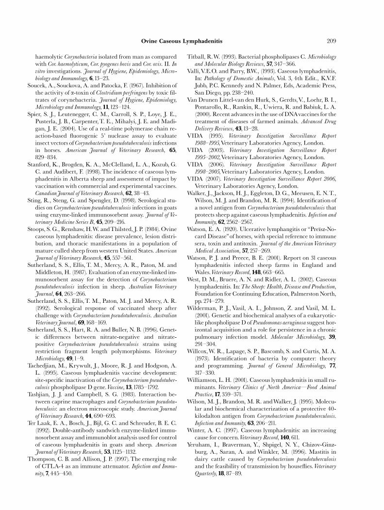
ARTICLE IN PRESS
Ovine Caseous Lymphadenitis 209
haemolytic Corynebacteria isolated from man as comparedwith Cor. haemolyticum, Cor. pyogenes bovis and Cor. ovis. II. Invitro investigations. Journal of Hygiene, Epidemiology, Micro-
biology and Immunology, 6,13^23.Soucek, A., Souckova, A. and Patocka, F. (1967). Inhibition of
the activity of a-toxin of Clostridium perfringens by toxic ¢l-trates of corynebacteria. Journal of Hygiene, Epidemiology,Microbiology and Immunology, 11,123^124.
Spier, S. J., Leutenegger, C. M., Carroll, S. P., Loye, J. E.,Pusterla, J. B., Carpenter,T. E., Mihalyi, J. E. and Madi-gan, J. E. (2004). Use of a real-time polymerase chain re-action-based £uorogenic 5’ nuclease assay to evaluateinsect vectors ofCorynebacterium pseudotuberculosis infectionsin horses. American Journal of Veterinary Research, 65,829^834.
Stanford, K., Brogden, K. A., McClelland, L. A., Kozub, G.C. and Audibert, F. (1998).The incidence of caseous lym-phadenitis in Alberta sheep and assessment of impact byvaccination with commercial and experimental vaccines.CanadianJournal ofVeterinary Research, 62, 38^43.
Sting, R., Steng, G. and Spengler, D. (1998). Serological stu-dies on Corynebacterium pseudotuberculosis infections in goatsusing enzyme-linked immunosorbent assay. Journal of Ve-terinaryMedicine Series B, 45, 209^216.
Stoops, S. G., Renshaw,H.W. andThilsted, J. P. (1984).Ovinecaseous lymphadenitis: disease prevalence, lesion distri-bution, and thoracic manifestations in a population ofmature culled sheep fromwestern United States. AmericanJournal ofVeterinary Research, 45, 557^561.
Sutherland, S. S., Ellis, T. M., Mercy, A. R., Paton, M. andMiddleton, H. (1987). Evaluation of an enzyme-linked im-munosorbent assay for the detection of Corynebacteriumpseudotuberculosis infection in sheep. Australian Veterinary
Journal, 64, 263^266.Sutherland, S. S., Ellis,T. M., Paton, M. J. andMercy, A. R.
(1992). Serological response of vaccinated sheep afterchallenge with Corynebacterium pseudotuberculosis. AustralianVeterinaryJournal, 69,168^169.
Sutherland, S. S., Hart, R. A. and Buller, N. B. (1996). Genet-ic di¡erences between nitrate-negative and nitrate-positive Corynebacterium pseudotuberculosis strains usingrestriction fragment length polymorphisms. VeterinaryMicrobiology, 49,1^9.
Tachedjian, M., Krywult, J., Moore, R. J. and Hodgson, A.L. (1995). Caseous lymphadenitis vaccine development:site-speci¢c inactivation of the Corynebacterium pseudotuber-
culosis phospholipase D gene.Vaccine, 13,1785^1792.Tashjian, J. J. and Campbell, S. G. (1983). Interaction be-
tween caprine macrophages and Corynebacterium pseudotu-
berculosis: an electron microscopic study. AmericanJournalofVeterinary Research, 44, 690^693.
Ter Laak, E. A., Bosch, J., Bijl, G. C. and Schreuder, B. E. C.(1992). Double-antibody sandwich enzyme-linked immu-nosorbent assay and immunoblot analysis used for controlof caseous lymphadenitis in goats and sheep. AmericanJournal ofVeterinary Research, 53,1125^1132.
Thompson, C. B. and Allison, J. P. (1997).The emerging roleof CTLA-4 as an immune attenuator. Infection and Immu-nity, 7, 445^450.
Titball, R.W. (1993). Bacterial phospholipases C.Microbiology
andMolecular Biology Reviews, 57, 347^366.Valli,V.E.O. and Parry, B.W., (1993). Caseous lymphadenitis,
In: Pathology of Domestic Animals, Vol. 3, 4th Edit., K.V.F.Jubb, P.C. Kennedy and N. Palmer, Eds, Academic Press,San Diego, pp. 238^240.
Van Drunen Littel-van den Hurk, S., Gerdts,V., Loehr, B. I.,Pontarollo, R., Rankin, R., Uwiera, R. and Babiuk, L. A.(2000). Recent advances in the use of DNAvaccines for thetreatment of diseases of farmed animals. Advanced Drug
Delivery Reviews, 43,13^28.VIDA (1995). Veterinary Investigation Surveillance Report
1988^1995,Veterinary Laboratories Agency, London.VIDA (2003). Veterinary Investigation Surveillance Report
1995^2002,Veterinary Laboratories Agency, London.VIDA (2006). Veterinary Investigation Surveillance Report
1998^2005,Veterinary Laboratories Agency, London.VIDA (2007). Veterinary Investigation Surveillance Report 2006,
Veterinary Laboratories Agency, London.Walker, J., Jackson, H. J., Eggleton, D. G., Meeusen, E. N.T.,
Wilson, M. J. and Brandon, M. R. (1994). Identi¢cation ofa novel antigen from Corynebacterium pseudotuberculosis thatprotects sheep against caseous lymphadenitis. Infection andImmunity, 62, 2562^2567.
Watson, E. A. (1920). Ulcerative lymphangitis or ‘‘Pre|« sz-No-card Disease’’of horses, with special reference to immunesera, toxin and antitoxin. Journal of the AmericanVeterinaryMedical Association, 57, 257^269.
Watson, P. J. and Preece, B. E. (2001). Report on 31 caseouslymphadenitis infected sheep farms in England andWales.Veterinary Record, 148, 663^665.
West, D. M., Bruere, A. N. and Ridler, A. L. (2002). Caseouslymphadenitis. In:The Sheep: Health, Disease and Production,Foundation for ContinuingEducation, PalmerstonNorth,pp. 274^279.
Wilderman, P. J., Vasil, A. I., Johnson, Z. and Vasil, M. L.(2001). Genetic and biochemical analyses of a eukaryotic-like phospholipase D ofPseudomonas aeruginosa suggest hor-izontal acquisition and a role for persistence in a chronicpulmonary infection model. Molecular Microbiology, 39,291^304.
Willcox,W. R., Lapage, S. P., Bascomb, S. and Curtis, M. A.(1973). Identi¢cation of bacteria by computer: theoryand programming. Journal of General Microbiology, 77,317^330.
Williamson, L. H. (2001). Caseous lymphadenitis in small ru-minants. Veterinary Clinics of North AmericaçFood Animal
Practice, 17, 359^371.Wilson,M. J., Brandon, M. R. andWalker, J. (1995). Molecu-
lar and biochemical characterization of a protective 40-kilodalton antigen from Corynebacterium pseudotuberculosis.Infection and Immunity, 63, 206^211.
Winter, A. C. (1997). Caseous lymphadenitis: an increasingcause for concern.Veterinary Record, 140, 611.
Yeruham, I., Braverman, Y., Shpigel, N. Y., Chizov-Ginz-burg, A., Saran, A. and Winkler, M. (1996). Mastitis indairy cattle caused by Corynebacterium pseudotuberculosis
and the feasibility of transmission by house£ies.VeterinaryQuarterly, 18, 87^89.
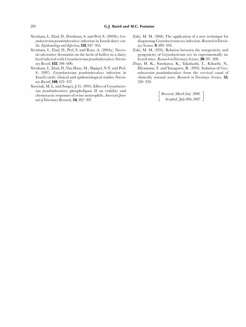
ARTICLE IN PRESS
G.J. Baird and M.C. Fontaine210
Yeruham, I., Elad, D., Friedman, S. and Perl, S. (2003b). Cor-ynebacterium pseudotuberculosis infection in Israeli dairy cat-tle. Epidemiology and Infection, 131, 947^955.
Yeruham, I., Elad, D., Perl, S. and Ram, A. (2003a). Necro-tic-ulcerative dermatitis on the heels of heifers in a dairyherd infectedwith Corynebacterium pseudotuberculosis.Veterin-ary Record, 152, 598^600.
Yeruham, I., Elad, D.,Van Ham,M., Shpigel, N.Y. and Perl,S. (1997). Corynebacterium pseudotuberculosis infection inIsraeli cattle: clinical and epidemiological studies.Veterin-ary Record, 140, 423^427.
Yozwiak,M. L. and Songer, J. G. (1993). E¡ect of Corynebacter-ium pseudotuberculosis phospholipase D on viability andchemotactic responses of ovine neutrophils. AmericanJour-nal ofVeterinary Research, 54, 392^397.
Zaki, M. M. (1968). The application of a new technique fordiagnosingCorynebacterium ovis infection.Research inVeterin-ary Science, 9, 489^493.
Zaki, M. M. (1976). Relation between the toxogenicity andpyogenicity of Corynebacterium ovis in experimentally in-fected mice. Research inVeterinary Science, 20,197^200.
Zhao, H. K., Yonekawa, K., Takahashi, T., Kikuchi, N.,Hiramune,T. andYanagawa, R. (1993). Isolation of Cory-nebacterium pseudotuberculosis from the cervical canal ofclinically normal sows. Research in Veterinary Science, 55,356^359.
Received,March 2nd, 2006
Accepted, July 10th, 2007
� �
Related Documents


![SurvivalAnalysisofClinicalCasesofCaseousLymphadenitisof ...downloads.hindawi.com/journals/vmi/2020/8822997.pdf · problems [11]. 4.7% prevalence of caseous lymphadenitis was also](https://static.cupdf.com/doc/110x72/5fc3fd1695fbe21b461044b4/survivalanalysisofclinicalcasesofcaseouslymphadenitisof-problems-11-47.jpg)


