American Journal of Medical Genetics 10:333-350 (1981) Corticosteroid-induced Cleft Lip in Mice: A Teratologic, Topographic, and Histologic Investigation Michael Melnick, Tina Jaskoll, and Harold C. Slavkin Laboratory for Developmental Biology, University of Southern California, Los Angeles Unlike cleft palate, relatively few teratogens have been found to induce cleft lip in mice. The present study was designed to assess the teratologic, topographic (SEM), and histologic effects on lip morphogenesis following the administration of triamcinolone hexacetonide on the eighth day of gestation. The frequency of cleft lip in treated A/J mice was found to be more than three times greater than the spontaneous frequency in untreated controls. Comparable studies with other murine strains suggest no association between the cleft lip response and either a maternal effect or the H-2 complex. Affected A/J embryos showed a severe reduction in the size of the lateral nasal processes; affected embryos also demonstrated localized cell type-specific alterations, particularly in the epithelia and at the interface between epithelium and mesenchyme. Key words: cleft lip, corticosteroid induced; cleft plate, corticosteroid induced; mice, teratological model; corticosteroids, triamcinolone hexacetonide; H-2 complex; embryopathology, teratological; scanning electron microscopy of lip morphologenesis INTRODUCTION Genetically different inbred strains of mice have different susceptibilities to the induction of isolated cleft plate by a standardized dose of glucocorticosteroids at the critical period of palatal formation, days 11-14 [Fraser and Fainstat, 1951; Kalter, 1957; Walker and Fraser, 1957; Loevy, 1963; Marsk et al, 1971; Bonner and Slavkin, 1975; Biddle and Fraser, 19771. To wit, A/J mice are highly suscepti- ble, whereas C57B1/6 mice are resistant to cortisone-induced cleft palate [Kalter, 1957; Bibble and Fraser, 19771. More recent work on mice clearly rules out a polygenic model in the strict sense of many equal and additive genes [Bibble and Fraser, 19771; this finding parallels recent studies in humans that also suggest that the liability to isolated cleft palate is not likely to include a polygenic component Received for publication December 1, 1980; revision received May 21, 1981. Address reprint requests to Dr. Michael Melnick, Laboratory for Developmental Biology, Andrus Gerontology Center, Rm. 325, University of Southern California, Los Angeles, CA 90007. 0148-7299/81/1004-0333$05.00 0 1981 Alan R. Liss, Inc.

Welcome message from author
This document is posted to help you gain knowledge. Please leave a comment to let me know what you think about it! Share it to your friends and learn new things together.
Transcript

American Journal of Medical Genetics 10:333-350 (1981)
Corticosteroid-induced Cleft Lip in Mice: A Teratologic, Topographic, and Histologic Investigation Michael Melnick, Tina Jaskoll, and Harold C. Slavkin Laboratory for Developmental Biology, University of Southern California, Los Angeles
Unlike cleft palate, relatively few teratogens have been found to induce cleft lip in mice. The present study was designed to assess the teratologic, topographic (SEM), and histologic effects on lip morphogenesis following the administration of triamcinolone hexacetonide on the eighth day of gestation. The frequency of cleft lip in treated A/J mice was found to be more than three times greater than the spontaneous frequency in untreated controls. Comparable studies with other murine strains suggest no association between the cleft lip response and either a maternal effect or the H-2 complex. Affected A/J embryos showed a severe reduction in the size of the lateral nasal processes; affected embryos also demonstrated localized cell type-specific alterations, particularly in the epithelia and at the interface between epithelium and mesenchyme.
Key words: cleft lip, corticosteroid induced; cleft plate, corticosteroid induced; mice, teratological model; corticosteroids, triamcinolone hexacetonide; H-2 complex; embryopathology, teratological; scanning electron microscopy of lip morphologenesis
INTRODUCTION
Genetically different inbred strains of mice have different susceptibilities to the induction of isolated cleft plate by a standardized dose of glucocorticosteroids at the critical period of palatal formation, days 11-14 [Fraser and Fainstat, 1951; Kalter, 1957; Walker and Fraser, 1957; Loevy, 1963; Marsk et al, 1971; Bonner and Slavkin, 1975; Biddle and Fraser, 19771. To wit, A/J mice are highly suscepti- ble, whereas C57B1/6 mice are resistant to cortisone-induced cleft palate [Kalter, 1957; Bibble and Fraser, 19771. More recent work on mice clearly rules out a polygenic model in the strict sense of many equal and additive genes [Bibble and Fraser, 19771; this finding parallels recent studies in humans that also suggest that the liability to isolated cleft palate is not likely to include a polygenic component
Received for publication December 1, 1980; revision received May 21, 1981.
Address reprint requests to Dr. Michael Melnick, Laboratory for Developmental Biology, Andrus Gerontology Center, Rm. 325, University of Southern California, Los Angeles, CA 90007.
0148-7299/81/1004-0333$05.00 0 1981 Alan R. Liss, Inc.

334 Melnick, Jaskoll, and Slavkin
[Melnick and Shields, 1976; Melnick et al, 1977, 19801. One gene complex that ap- pears to be associated with susceptibility to glucocorticosteroid-induced cleft palate is the major histocompatibility complex, H-2 [Bonner and Slavkin, 1975; Tyan and Miller, 1978; Melnick et al, in press]. However, by contrast the production of cleft lip in mice, a more common human malformation, has not as yet met with a similar degree of success.
Recently, Kalter [1979] exhaustively reviewed the biology of the A family of inbred mice. He concluded that, other than 6-aminonicotinamide, phenytoin, and aspirin, teratogens (including corticosteroids) had little or no effect on the frequen- cy of cleft lip and that the development of the lip (unlike the palate) appears to be shielded from developmental interference. However, preliminary studies in our laboratory using corticosteroids suggested the contrary and prompted us to test this hypothesis more extensively.
The present study was designed to assess the teratologic, topographic, and histologic effects on lip morphogenesis following the administration of triam- cinolone hexacetonide on the eighth day of gestation in a dose equivalent to that which produces approximately 50% cleft in A/J fetuses when administered on the 12th day of gestation. The frequency of cleft lip in treated A/J mice was found to be more than three times greater than the spontaneous frequency in untreated con- trols or A/J mice treated on the 12th day of gestation. Furthermore, cleft lip could not be induced with the same dose in C57BL/6, C57BL/10 Sn, BlO.A/SgSn, or A/J-C57BL/6 reciprocal F1 hybrid fetuses, suggesting that the tolerance to triam- cinolone hexacetonide of the C57BL embryos is greater than that of A/J, that there is no maternal effect, and that susceptibility of A/J embryos to cleft lip is not associated with genes in the H-2 complex. Affected A/J embryos demonstrated a severe reduction in the size of the lateral nasal processes and a lesser reduction in the size of the medial nasal and maxillary processes. In addition, day-8 treated A/J embryos demonstrated localized cell type-specific alterations. The embryopatholo- gy following corticosteroid exposure is similar to that described for drug-induced cleft lip using other teratogens [Trader and Leong, 1974; Sulik et al, 19791
MATERIALS AND METHODS Mating, Teratogen Administration, and Fetal Phenotype
Virgin female A/J (A), C57BL/6 (B6), C57BL/10 Sn (BlO), and cogenic partner BlO.A/SgSn (BlO.A), from the Jackson Laboratory (Bar Harbor, Maine), were housed by strain in groups of five and were acclimated to the vivarium en- vironment in our institution during a quarantine period of two weeks. When the animals were 12-13 weeks old, groups of three females were placed in a cage with one male overnight. Females were examined daily in the morning for the presence of vaginal plugs. The date of plug detection was designated as day 0 of gestation. A x A, B6 x B6, A x B6, B6 x A, B1O.A x BlO.A, and B10 x B10 matings were obtained.
Pregnant dams were housed in solid bottom, plastic cages. The vivarium facility is temperature controlled with an average daily temperature of 24°C. Alter- nate 12-hour periods of light and darkness were maintained daily. All animals were

Corticosteroid-induced Cleft Lip in Mice 335
maintained on Wayne’s Mouse Breeder Blox containing 20% crude fat and a max- imum of 2% crude fiber, and were supplied with drinking water ad libitum.
Experimental animals received a single intramuscular injection (in the proxi- mal portion of the hindlimb) of 2 mg/kg body weight triamcinolone hexacetonide, a long-acting synthetic analogue of cortisol, at 9:OO AM on day 8 or day 12 of gestation. Nontreated pregnant animals served as controls.
On the morning of day 17 of gestation, all dams were killed by cervical dislocation and the uterus and its contents were exposed. The uterine horns were examined in situ, and the following was noted for each fetus: location (right or left horn); position (from midline union to distal portion of uterine horn); fetal status (live, dead, resorbed). Each live fetus was carefully dissected out of the uterine decidua, and the trophoblast shell and Reichert membrane were removed. Fetuses were measured (crown-rump), weighed, and immediately placed in a glass recepta- cle with a fixed amount of 10% formalin solution. After fixation for a minimum of 24 hours, the fetuses were examined for all gross external anomalies. Both the developmental staging of the fetuses and the morphologic examinations were per- formed under a dissecting microscope. All abnormal fetuses and representative normal fetuses were routinely photographed (Fig. 1). It should be noted that each fetus was coded and tagged at the time of fixation, and the staging and examina- tion were done blindly with respect to treatment by one investigator.
Statistical Analysis
In all experimental groups within each cross (control, day-8 treated, day-12 treated), the following data were collected: 1) mean number of implants; 2) mean number of live fetuses; 3) mean frequency of fetal loss (resorbed + dead
Fig. 1. Theiler stage 25 (17 days) A/J mouse fetuses: (A) control ( x 7): (B) bilateral cleft lip ( x 7); (C) palatal view (mandible removed) of bilateral cleft palate (PS, palatal shelves that failed to fuse) (magni- fication, x 10).
336 Melnick, Jaskoll, and Slavkin
fetuses/total implants); 4) mean frequency of normal live fetuses (normal live fetuses/total live fetuses); 5) mean frequency of CL + P (live CL + P fetuses/total live fetuses); 6) mean frequency of CP (live CP fetuses/total live fetuses); 7) fetal weight; and 8) mean frequency of developmental immaturity (less than Theiler stage 25).
Mean frequencies were computed using the general equation
where Ij is frequency of a given “event” for the jth litter. An unbiased estimate of the sample standard deviation for the frequency of a given “event” is then
Frequency data were “normalized” prior to analysis by arcsin transformation, 0% and 100% values being replaced by %n and 100 - (?h), respectively, where n is the number of fetuses per litter [Dagg et al, 19661. To test the equivalence of two frequencies, mean weighted arcsins [Dagg et al, 19661 were compared by t or t’ tests, depending on the presence or absence of variance homogeneity [Sokal and Rohlf, 1969; Satterthwaite, 1946; Cochran, 19511.
two-tailed t or t’ tests. Fetal weight was analyzed further by a two-level nested analysis of variance (mixed model) designed for unequal sample sizes [Sokal and Rohlf, 19691. This allowed us to partition the sources of variation: among treatments, among pregnant dams within treatments, and among the mice of one litter (within litters).
Means computed for continuous variables were also compared by one- or
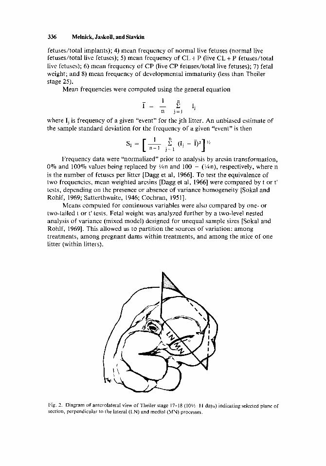
Fig. 2. Diagram of anterolateral view of Theiler stage 17-18 (lo%-] 1 days) indicating selected plane of section, perpendicular to the lateral (LN) and medial (MN) processes.

Corticosteroid-induced Cleft Lip in Mice 337
Light and Scanning Electron Microscopy In replicate studies, on the mornings of gestation days 9-13 treated and con-
trol dams from A x A crosses were killed by cervical dislocation, and the uterus and its contents were exposed. Embryos were rapidly dissected free of uterine and extraembryonic tissue and immersed in Karnovsky’s fixative overnight [Waterman, 19721. Embryos were staged as described above, rinsed in cacodylate buffer, and postfixed in 0.2 M phosphate buffered 1 Vo osmium tetroxide for one to two hours at room temperature. Dehydration in a graded ethanol series at room temperature was followed by a graded isoamyl acetate series and then critical-point dried [Anderson, 19561. Specimens were mounted with silver paint on aluminum stubs, sputter coated with gold-palladium, and then examined with an AMR 1000 scan- ning electron microscope (SEM) at 10-30 kV.
In order to prepare histological sections from embryo specimens previously examined with SEM, they were placed in several changes of propylene oxide and then oriented and embedded in Epon [Meller et al, 19731. Serial cross sections (1 pm), perpendicular to the nasal processes (Fig. 2), were cut with glass knives on a Sorvall MT2-B ultramicrotome, stained with toluidine blue, and then examined with a Zeiss research photomicroscope.
RESULTS Teratologic Response in AIJ x AIJ Crosses
Fetal lethality. Table I lists the number of litters, implants, live fetuses, and resorbed and dead fetuses for each of the three treatment groups, along with means and 95% confidence limits. There were no significant differences in the mean num- ber of implants between control and day-8 treated (tz2 = -0.555, P > 0.50) or day-12 treated (t22 = 0.898, P > 0.40) or between day-8 treated and day-12 treated (tls = 1.75, P > 0.50). The mean frequency of fetal loss was significantly different between control and day-8 treated (t’162.88 = -4.32, P < 0.001) and between con- trol and day-12 treated (t’146.24 = -3.21, P < 0.005). There were no significant dif-
TABLE 1. Implants, Live Fetuses, Resorptions, and Dead Fetuses by Treatment Group
Fetal status at gestation day 17
Number of Number of Number of Number of Treatment litters implantsa live fetusesa resorbed fetusesb
Control
Triam-D8
Triam-D12
14
10
10
131 117 14 (9.36 f 1.21) (8.36 * 0.98) (0.098 t 0.059)
98 79 19 (9.80 f 1.16) (7.90 f 1.14) (0.178 0.128)
87 72 15 (7.20 k 1.42) (0.182 f 0.116) (8.70 f 0.83)
aNumber in parentheses is the mean number of implants or live fetuses plus or minus the 95% con-
bNumber in parentheses is the mean frequency of dead and resorbed fetuses plus or minus the 95% con- fidence limits.
fidence limits.

338 Melnick, Jaskoll, and Slavkin
ferences between day-8 treated and day-12 treated (tIe3 = 0.919, P > 0.20). Since it is possible that a high frequency of fetal loss in day-8 treated and day-12 treated lit- ters occurs because of a concentration of lethality in only a few litters, we considered the distribution of lethality across litters. There were no significant differences in the distributions between control and day-8 treated (x: = 0.046, P > 0.50) or between control and day-12 treated litters (x: = 0.000, P > 0.975).
“normal” and malformed (CL f P, CP, other), along with mean frequencies and 95% confidence limits. The mean frequency of malformed live fetuses was significantly lower in control than in day-8 treated (t’97. ,7 = - 10.19, P > 0.001) and day-12 treated litters (t’s3.83 = - 12.36, P > 0.001). The frequency of malformed fetuses was also greater in day-12 treated than in day-8 treated mice (t149 = -2.80, P < 0.01). The distribution of malformed was uniform across litters for the treated groups, being 90% in day-8 treated and 100% in day-12 treated. Of particular im- portance, the mean frequency of CL k P was significantly greater in the day-8 treated group than in the day-12 treated Group (t 149 = 9.36, P > 0.001). In fact, the frequency of CL k P in the day-12 treated group (0.035) was almost identical to that in the control group (0.038). However, the mean frequency of CP was signifi- cantly greater in the day-12 treated group than in the day-8 treated group (t’132 61 = -4.79, P < 0.001).
Fetal group and development. The mean fetal weights f standard deviation for the three treatment groups were: control = 0.600 gm k 0.155, day-8 treated 0.573 gm f 0.122, and day-12 treated 0.409 gm f 0.153. The fetal weights were nearly equal in the control and day-8 treated groups, whereas in the day-12 treated group there was about a 30% mean weight reduction. A two-level nested analysis of variance (Table 111) revealed a significant component of the total variation among the treatment groups. The variation among pregnant dams within treatment groups was highly significant. However, the variation within litters was comparatively small, even though litters contained both normal and malformed fetuses.
Fetal malformations. Table I1 lists the number of live fetuses that were
Investigation of immaturity (less than Theiler stage 25) by analysis of x 2 [Rao,
TABLE 11. Phenotype of Live A/J Fetuses by Treatment Group
Number of live fetusesa
Treatment Litters “Normal” Malformed = C L k P + C P + Other
4b 108 9 4 1
(0.833 f 0.088) (0.082 f 0.061) (0.035 f 0.035) (0.007 ? 0.016) Control 14
0 50 29 9 2OC Triam-D8 10
(0.539 f 0.200) (0.387 f 0.200) (0.114 k 0.052) (0.270 0.191)
2e 35 31 2 3 3d Triam-D12 10
(0.389 ? 0.215) (0.545 f 0.225) (0.038 f 0.060) (0.484 k 0.225)
aNumber in parentheses is the mean frequency plus or minus the 95% confidence limits. bIncludes one micrognathia, one phocomelia, one severe plagiocephaly, and one micrognathia + rnulti- ple limb anomalies. These fetuses did not have CL f P or CP.
‘Includes one fetus with micrognathia in addition to the CP. dlncludes one fetus with bilateral microtia in addition to the CP. elncludes one exencephaly and one phocomelia. These fetuses did not have CL k P or CP.

Corticosteroid-induced Cleft Lip in Mice 339
TABLE Il l . Fetal Weight: Nested Analysis of Variance
Source of variation df ss MS F P
Among treatment groups 2 1.75 0.875 4.93a < 0.025
31 5.17 0.167 27.83 < 0.001 Among pregnant dams within
Within litters (among fetuses treatment groups
of one dam) 230 1.34 0.006
Total 263 8.26
aNote, for reasons that will not be dealt with here, this test of significance is carried out by synthesizing a new denominator mean square (ie, among pregnant dams) against which to test the mean square of among treatment groups. For a more detailed account of this procedure see Sokal and Rohlf [1969].
19521 revealed that the mean frequency of immaturity in live fetuses was inde- pendent of both phenotype and treatment groups. In addition, the mean frequency of immaturity by phenotype was independent of treatment group.
Teratologic Response in Remaining Crosses
tration on day 8 of gestation in all four inbred and two reciprocal (A.B6, B6.A) hybrid, H-2 defined, strains of mice. Although a dose of 2 mg/kg body weight produces a mean clefting frequency of about 11 To in A fetuses, there were no ap- parent cleft lips in any of the B6, A.B6, B6.A, BlO.A, or B10 fetuses. This could not be explained on the basis of fetal loss since the mean frequency of fetal loss was greater for A fetuses than for any of the others. Reciprocal hybrid crosses pro- duced equivalent responses among F1 progeny with respect to cleft lip or isolated cleft palate, suggesting no significant maternal effect for corticosteroid-induced clefting when the drug is administrated on day 8 of gestation. In addition, the mean CP frequency of A fetuses is many times greater than that of A.B6 or B6.A fetuses. Comparisons between the congenic pair, BI0.A and B10, demonstrate that the association of H-2 with fetal response to corticosteroids is still evident when the assay is isolated cleft palate, not cleft lip f cleft palate. Doses up to 6 mg/kg body weight failed to produce BIO.A embryos with cleft lip; at 8 mg/kg body weight the resorption frequency approached 100Vo.
Topographic (SEM) Comparisons Between Treated and Control AIJ Embryos
1972; Trader and Fraser, 19771. Briefly, facial development begins with the ap- pearance of ectodermal thickenings (nasal placodes) on either side of its anterior aspect. Cranial neural crest-derived mesenchyme condenses and proliferates at the borders of each placode, such that their lateral and medial rims expand outward to form nasal pits. The nasal pit becomes deeper as the lateral and medial processes grow, and it is further bounded on its floor by the maxillary process (Fig. 3). Definitive lip formation begins at the bottom of the nasal pit in the isthmus be- tween the lateral and medial nasal processes (Fig. 3A). The medial surface of the lateral process and the lateral surface of the medial process converge above the
Table IV presents the fetal response to triamcinolone hexacetonide adminis-
The development of the mouse lip has been described in detail [Pourtois,

z = &. E a, - K u
TA
BL
E IV
. R
espo
nse
to T
riam
cino
lone
Hex
acet
onid
e A
dmin
istr
atio
n (2
mg/
kg)
on D
ay 8
of
Ges
tatio
n in
Fou
r In
bred
and
Tw
o H
ybri
d, H
-2 D
efin
ed,
a St
rain
s of
Mic
e a z
Cro
ss
Feta
l los
s/im
plan
ts
CL
I P
/live
fet
uses
C
P/liv
e fe
tuse
s m
E
dam
x s
ire
(H-2
hap
loty
pe)
of l
itter
s N
umbe
r Fr
eque
ncya
N
umbe
r Fr
eque
ncya
N
umbe
r Fr
eque
ncya
a Ra
Fetu
s N
umbe
r
Ax
A
A(H
-2a/
H-2
a)
10
19/9
8 0.
178 I 0
.128
9/
79
0.11
4 k 0
.052
20
/79
0.27
0 -t
0.19
1 B6
x B
6 B
6(H
-2b/
H-2
b)
10
11/8
9 0.
119
rt 0
.087
0/
78
0.00
0 2/
78
0.02
5 0.
057
A x
B6
A.B
6F, (
H-2
a/H
-2b)
9
6/60
0.
115
I 0
.201
0/
54
0.00
0 2/
54
0.02
8 I 0
.064
B1O
.A x
B1O
.A
B 1 0
.A(H
-2a/
H-2
a)
10
8/78
0.
110
L 0
.112
0/
70
0.00
0 14
/70
0.20
0 L
0.1
89
B10
x B1
0 B
10(H
-2b/
H-2
b)
9 6/
62
0.10
7 k 0
.110
0/
56
0.00
0 3/
56
0.03
4 -t 0
.053
aMea
n fr
eque
ncy
I 9
5% c
onfi
denc
e lim
its, c
alcu
late
d as
des
crib
ed in
the
text
.
B6 x
A
B6.
AFI
(H-2
b/H
-2a)
9
1/60
0.
028 I 0
.064
0/
59
0.00
0 01
’59
0.00
0

Corticosteroid-induced Cleft Lip in Mice 341
Fig. 3 . Scanning electron photomicrographs of Theiler stage 17-18 control embryo. A. The lateral (LN) and medial (MN) nasal processes converge, and each is surrounded by a maxillary process (M) (magnification, x 89). B. The isthmus (I) region; the medial surface of the lateral nasal process and the lateral surface of the medial nasal process converge above the isthmus (magnification, x 220). NP, nasal pit; Mn, mandibular process.
342 Melnick, Jaskoll, and Slavkin
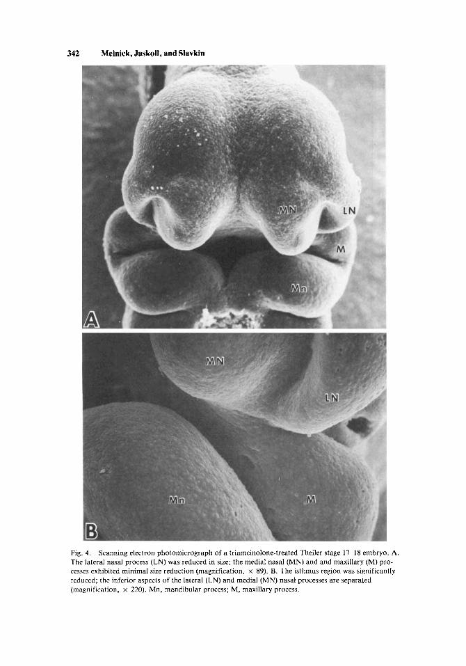
Fig. 4. The lateral nasal process (LN) was reduced in size; the medial nasal (MN) and and maxillary (M) pro- cesses exhibited minimal size reduction (magnification, x 89). B. The isthmus region was significantly reduced; the inferior aspects of the lateral (LN) and medial (MN) nasal processes are separated (magnification, x 220). Mn, mandibular process; M, maxillary process.
Scanning electron photomicrograph of a triamcinolone-treated Theiler stage 17-18 embryo. A.
Corticosteroid-induced Cleft Lip in Mice 343
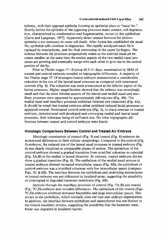
isthmus, with their apposed epithelia forming an epithelial plate or “nasal fin.” Shortly before the epithelia of the opposing processes make contact, cell degenera- tion, characterized by condensation and fragmentation, occurs in this epithelium [Garre and Langman, 19771. Apparently direct contact between the process epithelia is not necessary to cause cell death. After fusion has established the nasal fin, epithelial cells continue to degenerate. The rapidly autolyzed nasal fin is replaced by mesenchyme, and the final contouring of the upper lip begins. The isthmus between the processes progressively widen as the external nasal pit be- comes smaller; at the same time the medial aspects of the two medial nasal pro- cesses are growing and eventually merge with each other to give rise to the central portion of the lip.
Prior to Theiler stages 17-18 (circa 10.5-1 1 days, examination by SEM of treated and control embryos revealed no topographic differences. A majority of the Theiler stage 17-1 8 teratogen-treated embryos demonstrated a considerable reduction in the size of the lateral nasal processes as compared with nontreated controls (Fig. 4). The reduction was most pronounced at the inferior aspect of the lateral processes. Higher magnification showed that the isthmus was exceedingly small and that the most inferior aspects of the lateral and medial nasal and max- illary processes were separated by approximately 100 microns (Fig. 4B). The medial nasal and maxillary processes exhibited minimal size reductions (Fig. 4A). It should be noted that treated embryos either exhibited reduced facial processes or appeared normal. Nontreated control embryos (Fig. 3), in sharp contrast to treated embryos, demonstrated well-developed and converging medial and lateral nasal processes, their isthmuses being of sufficient size. No other topographic dif- ferences between treated and control embryos were found.
Histologic Comparisons Between Control and Treated NJ Embryos
monstrated differences in their cellular morphology. Compared to the control (Fig. 5 ) embryos, the reduced size of the lateral nasal processes in treated embryos (Fig. 6) was clearly visualized at comparable planes of section. The epithelium of the control embryos showed a gradual transition from stratified columnar to cuboidal (Fig. 5A,B) in the medial to lateral direction. In contrast, treated embryos did not show a gradual transition (Fig. 6). The epithelium of the medial nasal process in treated embryos showed increased intercellular spaces (Fig. 6B); the epithelium of control embryos was a stratified columnar with few intercellular spaces (compare Figs. 5C & 6B). The interface between the epithelium and underlying mesenchyme in treated embryos was not delineated in localized areas, suggesting the possibility of interrupted or degraded basement membrane (Fig. 6B).
Sections through the maxillary processes of control (Fig. 7A,B) and treated (Fig. 7C,D) embryos also revealed differences. The epithelium of the treated (Figs. 7C,D) embryos exhibited increased basophilia and large intercellular spaces. This occurs in the epithelium, which normally does not fuse and undergo degeneration. In addition, the interface between epithelium and mesenchyme was not distinct in the treated maxillary process, suggesting the possibility that the basement mem- brane was degraded in localized regions.
Histologic examination of control (Fig. 5 ) and treated (Fig. 6) embryos de-

344 Melnick, Jaskoll, and Slavkin
Fig. 5. tion for these l pm thick sections is shown in Figure 2. A. The lateral nasal (LN) and medial nasal (MN) processes converge in control embryos (magnification, x 178). B. The epithelium of the lateral nasal process in control specimens shows a gradual transition from stratified columnar (SC) to cuboidal (C) cells (magnification, x 832). C. The epithelium of the medial nasal process was a stratified columnar with few intercellular spaces (magnification, x 1275).
Representative histologic sections of Theiler stage 17-18 control mouse embryos. The orienta-

Corticosteroid-induced Cleft Lip in Mice 345
Fig. 6 . Representative histologic sections of Theiler stage 17-18 treated mouse embryos. The orienta- tion for these 1 pm sections is shown in Figure 2. A. The lateral nasal (LN) process was reduced in size and separated from the medial nasal (MN) process in triamcinolone-treated embryos by a greater dis- tance than in controls (compare Fig. 7) ( x 208). B. The nasal process epithelium showed increased in- tercellular spaces. The interface between the epithelium and underlying mesenchyme was not delineated in localized areas (asterisk) (magnification, x 780).

346 Melnick, Jaskoll, and Slavkin
Fig. 7. is shown in Figure 2. A. Control embryo: the lateral (LN) and medial (MN) nasal processes converge forming the epithelial plate (EP) ( x 150). B. The epithelium of the maxillary process was a stratified cuboidal with few intercellular spaces ( x 880). C. Triamcinolone-treated embryo: The lateral nasal (LN) process (magnification, x 177). D. The epithelium of the maxillary process exhibited increased basophilia and intercellular spaces. The interface between the epithelium and underlying mesenchyme was not distinct in localized areas (asterisk) (magnification, x 1086). M, maxillary process.
Histologic sections through the maxillary process. The orientation for these 1 pm thick sections

Corticosteroid-induced Cleft Lip in Mice 347
DISCUSSION Cleft Lip Induction
This study demonstrates, for the first time, a significant (P < 0.001) increase in the frequency of cleft lip in the A/J mouse strain following maternal treaiment with a glucocorticosteroid, triamcinolone hexacetonide, on day 8 of gestation. The frequency of cleft lip among treated fetuses was more than three times greater than that found among nontreated controls. There were also major differences in the phenotypic response to the drug when administered on different days of gestation, 8 vs 12: 1) the mean frequency of normal live fetuses was significantly greater in the day-8 treated group than in the day-12 treated group; 2) the mean frequency of live malformed fetuses was significantly greater for day-1 2 treated than for day-8 treated; 4) the mean frequency of cleft lip with or without cleft palate was signif- icantly greater in the day-8 treated group than in the day-12 treated, the latter group being equivalent to the “backround” incidence in the control group; and 5) the mean fetal weight in the day-I2 treated group was 30% lower than in the day-8 treated group, which was equivalent to the control group. Regarding the cleft lip, these data suggest that the apparent resistance of the A strains to most teratogens thus far tested [Kalter, 19791 may well be a result of the timing of teratogen ad- ministration or availability.
termates with either cleft lip (& cleft palate) or isolated cleft palate were identified. This phenotypic discordance among genetically identical, n-chorionic, n-tuplets might be the result of developmental asynchrony within litters. Alternatively, since triamcinolone hexacetonide is long acting, the isolated cleft palates induced by day-8 treatment may result from residual protracted drug action.
teresting but enigmatic results. Clearly, with respect to cleft lip induction by tri- amcinolone, there is no clear evidence for a maternal effect nor an association with the H-2 complex; instead, susceptibility of A embryos appears to be associated with alleles at other loci. The enigma is with regard to the isolated cleft palate re- sponse. If corticosteroids are administered around day 12 of gestation to A/J and C57BL/6 mice there is a well-established maternal effect [Kalter, 1957; Biddle and Fraser, 19771. Our results indicate that, for CP, if the drug is administered on day 8 the maternal effect is no longer evident. This difference may be indicative of changes in maternal metabolism as the pregnancy progresses. Furthermore, al- though the association of cleft palate induction with H-2 haplotype remains with day-8 drug administration, the association is more likely with the embryonic haplotype than with the maternal haplotype, as has been found with the day-12 ad- ministration [Bonner and Slavkin, 19751. Perhaps the embryonic effect is masked at later stages of pregnancy; this embryonic effect may be related to either drug de- toxification or unique tissue-specific effects (eg, maxillary process mesenchyme, rather than lateral nasal process mesenchyme). However, it should be noted that the role of cell-surface H-2 antigens in embryonic craniofacial development awaits elucidation. If one may be permitted to extrapolate from mouse to man, the com- plexities uncovered with mouse models may partially explain the conflicting results from limited studies of the association of HLA and human clefts [Bonner et al, 1978; Van Dyke et al, 19801.
A most interesting finding was that in the same day-8 treated A/J litter, lit-
Lastly, the studies in other mouse strains (Table IV) have produced some in-

348 Melnick, Jaskoll, and Slavkin
Lip Morphogenesis and Dysmorphogenesis
cesses to allow fusion or merging. Failure of the lateral and medial processes to fuse results in lateral clefts of the lip; failure of the two medial processes to merge results in the more rare medial clefts of the lip. Trasler and Fraser [1977] have drawn up a list of potential developmental aberrations that would result in cleft lip: 1) alteration of the position and/or time of nasal placode induction; 2) decreased rate of neural crest migration to and mitosis in the facial processes; 3) alteration of the relative sizes and positions of facial processes; 4) changes in the rate of autolysis of the nasal fin; 5) mechanical interference with convergence of facial processes; 6) failure of the facial processes to fuse or coalesce upon converg- ing. Some of these possibilities overlap (eg, 2 and 3). These potential aberrations have been investigated for but a few teratogens known to induce cleft lip in mice: 6-aminonicotinamide is associated with a reduction in mitotic index and results in medial nasal processes of reduced size [Trasler and Leong, 19741; and phenytoin is associated with a marked reduction in the size of the lateral nasal processes and a less severe reduction in the size of medial nasal and maxillary processes [Sulik et al, 19791.
The lateral cleft lip induced by triamcinolone hexacetonide appears to be associated with a severe size reduction in the lateral nasal process. In addition, there appear to be cellular changes in medial, lateral, and maxillary processes. The gross topographic changes in viewed with the SEM (lateral process reduction) are not unlike those seen with phenytoin-induced cleft lip [Sulik et al, 19791.
This view is supported by the recent corticoid receptor studies in which the binding of 3H-dexamethasone in palatal cells is depressed specifically only by other corti- coids as well as drugs which are known to induce clefting in sensitive mouse strains, such as phenytoin [Goldman et al, 19781. This group of teratogens appears to be retained in facial processes by their ability to bind tightly to chromatin, which in turn results in decreased transcription [Bekhor et al, 19781.
The available and somewhat limited evidence indicates that interference with later stages of facial process formation is likely to be the most important alteration for the common forms of nonsyndromic human cleft lip [Johnston and Sulik, 19791. Although this assertion may be premature, the evidence from mouse studies would seem to point in this direction.
Clearly the critical event in lip formation is the convergence of the facial pro-
The pathogenetic mechanisms induced by these teratogens may be similar.
ACKNOWLEDGMENTS
This study was supported by grants DE 02848, DE 07006, and DE 05440 from the National Institute of Dental Research, National Institutes of Health. Dr. M. Melnick is a recipient of Public Health Research Career Development Award DE 00083. The authors wish to thank Ms. Julie Grimmett for her fine technical as- sistance, Mr. Pablo Bringas for preparing the illustrations, and Ms. June Sayles for typing the manuscript. A special thanks is due Dr. John Brady (USAIDR, Ft. Meade, Maryland) for generously allowing us to use his scanning electron micro- scope for these studies.
Corticosteroid-induced Cleft Lip in Mice 349
REFERENCES
Anderson TF: Electron microscopy of microorganisms. In Oster G, Pollister, AW (eds): “Physical Techniques in Biological Research,” vol. 3. New York: Academic Press, 1956, pp 200-206.
Bekhor I, Mirell CJ, Anne L: Induction of cleft palates by triamcinolone acetonide: Reexamination of the problem. Cleft Palate J 15:220-232, 1978.
Biddle FG, Fraser FC: Cortisone-induced cleft palate in the mouse. A search for the genetic control of the embryonic response trait. Genetics 85:289-302, 1977.
Bonner JJ, Slavkin HC: Cleft palate susceptibility linked to histocompatibility-2 (H-2) in the mouse. Immunogenetics 2:2 13-21 8, 1975.
Bonner JJ, Terasaki PI, Thompson P, Holve LM, Wilson L, Ebbin AJ, Slavkin HC: HLA phenotype frequencies in individuals with cleft lip and/or cleft palate. Tissue Antigens 12:228-232, 1978.
Cochran WG: Testing a linear relationship among variances. Biometrics 7:17-32, 1951. Dagg CP, Schlager G, Doerr A: Polygenic control of the teratogenicity of 5-fluorouracil in mice.
Fraser FC, Fainstat TD: Production of congenital defects in the offspring of pregnant mice treated with
Gaare JD, Langman J: Fusion of nasal swellings in the mouse embryo: Regression of the nasal fin. Am
Goldman AS, Shapiro BH, Katsumata M: Human foetal palatal corticoid receptors and teratogens for
Johnston MC, Sulik KK: Some abnormal patterns of development in the craniofacial region. In
Genetics 53: 1101-11 17, 1966.
cortisone. Pediatrics 8:527-533, 1951.
J Anat 150:477-500, 1977.
cleft palate. Nature 272:464-466, 1978.
Melnick M, Jorgenson R (eds): “Developmental Aspects of Craniofacial Dysmorphology.” New York: Alan R. Liss, Inc, 1979, pp 23-42.
Kalter H: Factors influencing the frequency of cortisone-induced cleft palate in mice. J Exp Zoo1
Kalter H: The history of the A family of inbred mice and the biology of its congenital malformations. Teratology 20:213-232, 1979.
Loevy H: Genetic influences on induced cleft palate in different strains of mice. Anat Rec 145:117-122, 1963.
Marsk L, Theorell M, Larsson KS: Transfer of blastocysts as applied in experimental teratology. Nature 234:358-359, 1971.
Meller SM, Coppe MR, Ito S, Waterman RE: Transmission electron microscopy of critical point dried tissue after observation in the scanning electron microscope. Anat Rec 176:245-252, 1973.
Melnick M, Shields ED: Allelic restriction: A biologic alternative to multifactorial threshold inheri- tance. Lancet 1:176-179, 1976.
Melnick M, Shields ED, Bixler D, Conneally PM: Facial clefting: An alternative biologic explanation for its complex etiology. In Bergsma D (ed): “Annual Reviews of Birth Defects, 1976.” New York: Alan R. Liss, Inc, for The National Foundation - March of Dimes. BD:OAS 13(3A):93- 112, 1977.
Melnick M, Shields ED, Bixler D: Studies of cleft lip and cleft palate in the population of Denmark. In Melnick M, Bixler D, Shields ED (eds); “Etiology of Cleft Lip and Cleft Palate.” New York: Alan R. Liss, Inc., 1980, pp 225-248.
hap1otype:Maternal and embryonic effects. Immunogenetics (in press).
“Developmental Aspects of Oral Biology.” New York: Academic Press, 1972, pp 81-108.
134:449-467, 1957.
Melnick M, Jaskoll T, Slavkin HC: Corticosteroid-induced cleft palate in mice and H-2
Pourtois M: Morphogenesis of the primary and secondary palate. In Slavkin HC, Bavetta LA (eds):
Rao CR: “Advanced Statistical Methods in Biometric Research.” New York: John Wiley and Sons,
Satterthwaite FW: An approximate distribution of estimates of variance components. Biometrics Bull
Sokal RR, Rohlf FF: “Biometry.” San Francisco: WH Freeman Company, 1969. Sulik KK, Johnston MC, Ambrose LJH, Dorgan D: Phenytoin (Di1antin)-induced cleft lip and palate in
A/J mice: A scanning and transmission electron microscopic study. Anat Rec 195:243-255, 1979.
1952, pp 210-214.
2:110-114, 1946.
Theiler K: “The House Mouse.” Berlin: Springer-Verlag, 1972.

350 Melnick, Jaskoll, and Slavkin
Trasler DG, Fraser FC: Time-position relationships with particular reference to cleft lip and cleft palate. In Wilson JG, Fraser FC (eds): “Handbook of Teratology.” Vol 2. New York: Plenum Press, 1977, pp 271-292.
Trasler DG, Leong S: Face shape and mitotic index in mice with 6-aminonicotinamide-induced and in- herited cleft lip. Teratology 9:A39, 1974.
Tyan ML, Miller KK: Genetic and environmental factors in cortisone induced cleft palate. Proc SOC Exp Biol Med 158:618-621, 1978.
Van Dyke DC, Goldman AS, Spielman RS, Zmijewski CW, Oks SW: Segregation of HLA in sibs with cleft lip or cleft lip and palate: Evidence against genetic linkage. Cleft Palate J17:189-193, 1980.
Walker BE, Fraser FC: The embryology of cortisone-induced cleft palate. J Embryo1 Exp Morphol 5:201-209, 1957.
Waterman RE: Use of the scanning electron microscope for observation of vertebrate embryos. Dev Biol27:276-281, 1972.
Edited by John M. Opitz
Related Documents