
Welcome message from author
This document is posted to help you gain knowledge. Please leave a comment to let me know what you think about it! Share it to your friends and learn new things together.
Transcript
Journal of Pharmacology and Pharmacotherapeutics | April-June 2012 | Vol 3 | Issue 2 161
Cortico-hippocampal salvage in chronic aluminium induced neurodegeneration by Celastrus paniculatus seed oil: Neurobehavioural, biochemical, histological study
Mrinmoy Chakrabarty, Priyanka Bhat, Sweta Kumari, Avin D’Souza, K. L. Bairy, Abhishek Chaturvedi1, Archana Natarajan1, Mohandas Rao K. G.2, Shobha Kamath1
Departments of Pharmacology, 1Biochemistry, Kasturba Medical College, 2Anatomy, Melaka Manipal Medical College, Manipal University, Manipal, Karnataka, India
Research Paper
Address for correspondence: K. L. Bairy, Department of Pharmacology, Kasturba Medical College, Manipal University, Manipal – 576 104, Karnataka, India. E-mail: [email protected]
ABSTRACT
Objective: To investigate the effects of Celastrus paniculatus seed oil in preventing the onset of chronic aluminum induced cortico-hippocampal neurodegeneration and oxidative stress. Materials and Methods: An animal model of senile dementia of Alzheimer’s type was produced by administering aluminum as aluminum chloride (4.2 mg/kg) intraperitoneally to male Wistar rats for 60 days and results compared to untreated control. Neurobehavioral investigations of Morris water maze tests, passive avoidance test, rotarod test and biochemical estimations of acetylcholineterase, malondialdehyde, glucose-6-phosphate dehydrogenase, superoxide dismutase, and hemoglobin in blood were performed fortnightly which gauged the extent of global oxidative stress and progressive neural damage. Findings were fortified by the above enzyme assays and histology of brain at necropsy. Prophylactic oral C. paniculatus in two doses 0.5 ml and 1 ml, were given to animals and the results were analyzed in comparison to a similar rodent model with standard drug donepezil (0.5 mg/kg) intraperitoneally. Results: C. paniculatus showed a significant prevention in onset of aluminum induced neural insult and overall systemic oxidative stress which was corroborated by the enlisted neurobehavioral, biochemical, and histological evidence. Conclusion: C. paniculatus is a putative decelerator of Al-mediated Alzheimer’s like pathobiology.
Key words: Celapanin, celapanigin, celapagin, Morris water maze test, passive avoidance test, pyknosis, rotarod test, senile dementia of Alzheimer’s type, sesquiterpene alkaloids
Access this article onlineQuick Response Code:
Website: www.jpharmacol.com
DOI: 10.4103/0976-500X.95520
INTRODUCTION
The exposure of aluminum (Al) is commonplace by oral intake, inhalation, transdermal introduction, or via potential administration through hemodialysis,[1] in the form of antacids, buffered aspirins, vaccines,[2] sulfate salt as an agent for purification of water,[3] and in cooking utensils in developing and underdeveloped countries. Neurotoxic effects of Al are well-established and specific effects on human central nervous system such as memory loss and impaired coordination
Chakrabarty, et al.: Attenuation of cortico-hippocampal neurodegeneration by C. paniculatus
162 Journal of Pharmacology and Pharmacotherapeutics | April-June 2012 | Vol 3 | Issue 2
are documented.[4] It is known to increase the permeability of blood–brain barrier[5] and traverses it which results in concentration of Al in hippocampus,[6] cortex, and corpus callosum.[7] Chronic Al exposure may lead to neurotoxic symptoms, e.g. speech disturbances, dyspraxia, tremors, psychosis, partial paralysis, loss of memory and cognition, and ultimately death. [8] The pattern of chronic Al neurotoxicity closely simulates that of progressive neurodegeneration as in Senile Dementia of Alzheimer’s Type (SDAT).[9] Hence, long-term dosing of Al salt—aluminum chloride (AlCl3) was chosen to model SDAT in vivo in rodents.
Celastrus paniculatus Willd (Celastraceae) seed oil has been used for decades for its nervine and cognition enhancing properties.[10] C. paniculatus (CP) has reports of enhancing memory, both in rats[11] and in mentally retarded children.[12] Although there have been short-term studies with the plant reporting protective effects in glutamate-induced toxicity in vitro,[13] therapeutic improvement in sodium nitrite-induced amnesia[14] and in scopolamine-induced memory deficits[15] in vivo, explorations to reveal any beneficial effects of CP in retarding cognitive decline in the SDAT model of chronic Al neuronal toxicosis have not been attempted before. The findings were strongly in favor of the experimental intervention.
MATERIALS AND METHODS
Drugs and chemicalsCommercially available pure seed oil was procured from Rajesh Chemicals, Mumbai, India and AlCl3 from Loba Chemie, Mumbai. Donepezil (Alzil 10 mg tabs, Intas Pharmaceuticals, Ahmedabad) was purchased from Manipal Drug House, Manipal, India.
Experimental designAll experiments were designed in strict adherence to the guidelines of the Institutional Animal Ethics Committee. Male albino Wistar rats weighing 290 g to 310 g between 18 and 20 months of age were procured from the Institutional Central Animal Research Facility. The animals were housed in poly propylene cages in a well-ventilated room and provided with standard rat pellet diet and water ad libitum. The animals were maintained at a room temperature of 25 ± 2 °C and a relative humidity of 50 ± 5% with a 12 h light/dark cycle. The animals were carefully chosen in a narrow age range, and the body weight varied between 290 and 310 g. Since the body weights were close enough, there was not much variation in the doses calculated with respect to body weight. Therefore, doses of 0.5 ml (1.6 g/kg) and 1 ml (3.2 g/kg) were employed. The animals were randomly assigned to five groups of eight animals each and received the following treatments:Group I : Vehicle treated groupGroup II : AlCl3 (4.2 mg/kg body wt)
Group III : AlCl3 (4.2 mg/kg body wt) + CP (0.5 ml/animal)Group IV : AlCl3 (4.2 mg/kg body wt) + CP (1.0 ml/animal)Group V : AlCl3 (4.2 mg/kg body wt) + Donepezil (0.5 mg/kg body wt)
All the drugs were given intraperitoneally for 60 days and results compared to untreated control.
Neurobehavioral studiesMorris water maze testIt was carried out as detailed by Sethi et al.[16] with some modifications. The pool was positioned in the middle of a dimly lit testing room with distant visual stimulus. Four points equally spaced along the circumference of the pool were arbitrarily assigned N, E, S, and W. Rats were trained to locate a submerged black platform (10 cm diameter) maintained at a fixed location. The day before training, each rat was allowed to freely swim for 60 s, allowed to climb the platform three times and rest for 30 s. All rats were subjected to one session of four trials/day for four consecutive days. For each trial, the rat was placed in the water facing the wall of the pool at one of the four equally spaced starting points. The starting points for all four trials per day and each such session on consecutive days were randomized, but the location of platform was always centered in one particular quadrant, e.g. NE quadrant. All rats were left on a platform for 30 s. Rats failing to find the platform within 60 s were guided to find it by us. Inter trial interval was kept at a constant of 60 s. At the end of each session of the day, rats were towel dried and returned to a home cage. On the probe trial on fifth day, the platform was removed from the pool and rats were allowed to swim for a 60 s trial period. Data were in the form of escape latencies to find the platform.
Passive avoidance testThis was carried out as reported.[17] The apparatus consisted of two identical chambers, one of them well lit. Rats were placed one at a time, in the lighted chamber facing its back at the dark chamber and at the farthest point from the dark chamber. As soon as the rat entered the dark compartment, the door was closed and foot shock delivered (0.1 mA, 40 V) through a tap key. Thereafter, the rat was replaced in its home cage. After 24 h, the rat was placed again in the lighted compartment and the latency to enter the dark compartment was measured which was taken as a measure of retention of memory of the shock. Cut-off time for each trial was 180 s.
Rotarod testThe procedure followed[18] assessed muscle strength and coordination. Rats were placed on a metallic rod (5 cm diameter) turning at the rate of 8 rpm and checked for their ability to maintain themselves on the rotating rod for three successive attempts of 180 s each. The rats were adjudged positive or motor in coordinated in the event of not being able to do so.
Chakrabarty, et al.: Attenuation of cortico-hippocampal neurodegeneration by C. paniculatus
Journal of Pharmacology and Pharmacotherapeutics | April-June 2012 | Vol 3 | Issue 2 163
Enzyme assaysAcetylcholinesterase activity: Blood and brainThe serum was separated by centrifuging whole clotted blood at 3000 rpm for 5 min at room temperature, and the enzyme estimated in blood. The reaction mixture contained 0.1 M phosphate buffer (pH 7.5), 0.66 mM DTNB (dithiobisnitrobenzoic acid), and the requisite amount of the sample. The reaction was started by addition of acetylthiocholine (0.83 mM), and the increase in absorbance was recorded at 412 nm and finally values expressed as U/g of Hb/L.[19]
It was estimated in brain as described.[20] Rat brain was homogenized in 0.1 M potassium dihydrogen phosphate (KH2PO4) buffer (pH 7.4) containing 1 mM EDTA (ethylene diamine tetraacetate) and 0.25 M sucrose for 10 min. This was kept in an ice chest. The homogenate was centrifuged at 3000 rpm. The cloudy supernatant was taken for the assay. Three milliliters of phosphate buffer (pH 7.4), 200 µl of homogenate was added, placed in ice chest and then vortexed. A 100 µl of DTNB was added, incubated for 5 min, 20 µl of ATC (acetylthiocholine) was added, vortexed and an increase in absorbance was measured at 412 nm for 10 min at 37 °C at 1 min interval. Enzyme activity was calculated based on the changes in absorbance per minute and expressed as moles/L/min/g of tissue.
Malondialdehyde levels: blood and brainIt was estimated in blood as detailed.[21] The reaction mixture contained 1 ml of 0.67% thiobarbutiric acid (TBA), 500 µl of 20% tricarboxylic acid (TCA), and 100 µl. The reaction mixture was incubated at 100 °C for 20 min, centrifuged at 12000 rpm for 5 min, and absorbance of the supernatant was read at 532 nm. MDA was determined by using a molar extinction coefficient of 1.56 × 10⁵ M-1 cm-1 and values were expressed in nmoles/ml.
The brain tissue was taken in 2 ml HBSS, homogenized at 3000 rpm. The homogenate was centrifuged at 710 × g for 10 min, pellets were resuspended in 1 ml HBSS which was then used for the lipid peroxidation (LPO) estimation. LPO was measured in terms of the MDA:thiobarbituric acid (TBA) reaction.[22] The reaction mixture contained 0.1 ml tissue homogenate, 0.2 ml sodium dodecylsulfate, 1.5 ml acetic acid, and 1.5 ml aqueous solution of TBA. The mixture was made up to 4 ml with distilled water and heated at 95 °C for 1 h. After cooling, 1 ml of distilled water and 5 ml mixture of n-butanol and pyridine (15:1) were added and the mixture was vortexed. After centrifugation at 450 × g for 10 min, the absorbance of the upper organic layer was taken at 532 nm and the result was expressed in nmoles/ml.
Glucose-6—Phosphate dehydrogenase levels: blood and brainAn already published method was followed[23] with modifications. Whole blood was centrifuged at 2500 rpm for 10 min.
Erythrocytes were resuspended in cold 0.9% NaCl thrice to hemolyse them. After 90 min of deep freezing, 40 µl of the sample was taken to which 1.8 ml of the stock reagent mixture was added. The stock reagent mixture contained Tris–HCl buffer (1 M, pH 8.0), magnesium chloride (MgCl2) 0.1 M/2 g/dl, NADP (2 mmol/1.53 mg/ml), and water. Glucose-6-phosphate (1.82 mg/ml) was added after 5 min of preincubation and the absorbance was measured. After 10 min of incubation at 37 °C, the increase in the absorbance was again measured at 340 nm.
Hemoglobin (Hb) was also measured and absorbance read at 540 nm and the result was expressed in U/g of Hb.
For brain, the assay mixture contained Tris–HCl (1 M, pH 8), MgCl2 (0.1 M/2 g/dl), NADP solution (10 mM), water, and an appropriate amount of the sample. After 5 min of preincubation, glucose-6-phosphate (31 mM) was added and the absorbance was measured at 340 nm. The increase in the absorbance was measured after 10 min at 340 nm and the result was expressed in µmoles/min/ml of clear homogenate (IU/ml).
Superoxide dismutase levels: blood and brainThis assay is based on an established principle.[24] One milliliter of whole blood suspension was taken, and one milliliter of distilled water was added to it along with 0.5 ml chloroform and 0.3 ml ethanol. It was mixed and kept in an ice chest for 15 min. An aliquot of 0.2 ml of water was added, the mixture was centrifuged at 4000 rpm for 10 min and 0.1 ml of supernatant was taken, 1.9 ml of phosphate buffer was added (0.05 M, pH = 7.8). An aliquot of 0.1 ml of this mixture was taken for the assay. Absorbance was read at 560 nm, and SOD values were calculated as units/ml of blood.
To the brain tissue homogenate,[25] equal volume of 0.5% TritonX-100 was added and kept at 4 °C for 20 min. The homogenate was centrifuged at 3000 rpm for 30 min at 4 °C. One ml of supernatant, one ml of distilled water, 0.5 ml of chloroform, and 0.3 ml of ethanol were mixed and kept in an ice chest for 15 min. This mixture was centrifuged at 4000 rpm for 10 min. Then, 0.1 ml of the supernatant was taken and 1.9 ml of phosphate buffer was added (0.05 M, pH = 7.8). 0.1 ml of this mixture was taken and absorbance was read at 560 nm and values reported in units/ml of homogenate.
HistopathologyOn 60th day the animals were transcardially perfused with formalin solution after a brief pre-rinse with isotonic saline, under deep anesthesia. They were then decapitated and brains extracted on an ice chilled tray, sagittally and coronally sliced. Slices were fixed in formalin for 48 h. Fixed tissues were sectioned, and stained in standard hematoxylin–eosin (H and E). The slides were then air dried and viewed under a light microscope and photomicrographs were taken at a total magnification of 10× and 40×.
Chakrabarty, et al.: Attenuation of cortico-hippocampal neurodegeneration by C. paniculatus
164 Journal of Pharmacology and Pharmacotherapeutics | April-June 2012 | Vol 3 | Issue 2
Statistical analysisOne-way ANOVA was applied for analysis. The parametric data with homogenous variances were applied with Tukey’s post hoc test and those with nonhomogenous variances were analysed using Tamhanhe’s T2 post hoc test. The minimum level of significance was set at P < 0.05 and maximum at P < 0.001. All analyses were done using SPSS software package version 16.0.
RESULTS
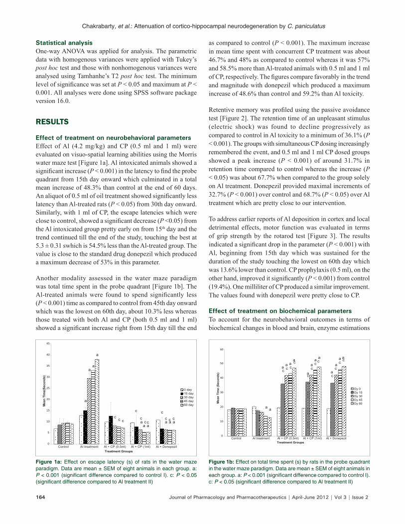
Effect of treatment on neurobehavioral parametersEffect of Al (4.2 mg/kg) and CP (0.5 ml and 1 ml) were evaluated on visuo-spatial learning abilities using the Morris water maze test [Figure 1a]. Al intoxicated animals showed a significant increase (P < 0.001) in the latency to find the probe quadrant from 15th day onward which culminated in a total mean increase of 48.3% than control at the end of 60 days. An aliquot of 0.5 ml of oil treatment showed significantly less latency than Al-treated rats (P < 0.05) from 30th day onward. Similarly, with 1 ml of CP, the escape latencies which were close to control, showed a significant decrease (P <0.05) from the Al intoxicated group pretty early on from 15th day and the trend continued till the end of the study, touching the best at 5.3 ± 0.31 swhich is 54.5% less than the Al-treated group. The value is close to the standard drug donepezil which produced a maximum decrease of 53% in this parameter.
Another modality assessed in the water maze paradigm was total time spent in the probe quadrant [Figure 1b]. The Al-treated animals were found to spend significantly less (P < 0.001) time as compared to control from 45th day onward which was the lowest on 60th day, about 10.3% less whereas those treated with both Al and CP (both 0.5 ml and 1 ml) showed a significant increase right from 15th day till the end
as compared to control (P < 0.001). The maximum increase in mean time spent with concurrent CP treatment was about 46.7% and 48% as compared to control whereas it was 57% and 58.5% more than Al-treated animals with 0.5 ml and 1 ml of CP, respectively. The figures compare favorably in the trend and magnitude with donepezil which produced a maximum increase of 48.6% than control and 59.2% than Al toxicity.
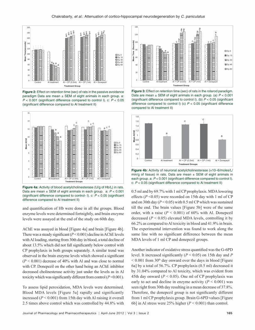
Retentive memory was profiled using the passive avoidance test [Figure 2]. The retention time of an unpleasant stimulus (electric shock) was found to decline progressively as compared to control in Al toxicity to a minimum of 36.1% (P < 0.001). The groups with simultaneous CP dosing increasingly remembered the event, and 0.5 ml and 1 ml CP dosed groups showed a peak increase (P < 0.001) of around 31.7% in retention time compared to control whereas the increase (P < 0.05) was about 67.7% when compared to the group solely on Al treatment. Donepezil provided maximal increments of 32.7% (P < 0.001) over control and 68.7% (P < 0.05) over Al treatment which are pretty close to our intervention.
To address earlier reports of Al deposition in cortex and local detrimental effects, motor function was evaluated in terms of grip strength by the rotarod test [Figure 3]. The results indicated a significant drop in the parameter (P < 0.001) with Al, beginning from 15th day which was sustained for the duration of the study touching the lowest on 60th day which was 13.6% lower than control. CP prophylaxis (0.5 ml), on the other hand, improved it significantly (P < 0.001) from control (19.4%). One milliliter of CP produced a similar improvement. The values found with donepezil were pretty close to CP.
Effect of treatment on biochemical parametersTo account for the neurobehavioral outcomes in terms of biochemical changes in blood and brain, enzyme estimations
0
5
10
15
20
25
30
35
40
45
Control AI treatment Al + CP (0.5ml) Al + CP (1ml) Al + Donepezil
Mea
n Ti
me(
Seco
nds)
Treatment Groups
0 day15 day30 day45 day60 day
a aaa
a
a
aa
a
ac c c
c
cc
c
cc c c
0
10
20
30
40
50
60
Control Al treatment Al + CP (0.5ml) Al + CP (1ml) Al + Donepezil
Mea
n Ti
me
(Sec
onds
)
Treatment Groups
Dy 0Dy 15Dy 30Dy 45Dy 60
c
cc ccc
c
c
cccc
aaa
a
aaa
a
aaaa
a a
Figure 1a: Effect on escape latency (s) of rats in the water maze paradigm. Data are mean ± SEM of eight animals in each group. a: P < 0.001 (significant difference compared to control I). c: P < 0.05 (significant difference compared to Al treatment II)
Figure 1b: Effect on total time spent (s) by rats in the probe quadrant in the water maze paradigm. Data are mean ± SEM of eight animals in each group. a: P < 0.001 (significant difference compared to control I). c: P < 0.05 (significant difference compared to Al treatment II)
Chakrabarty, et al.: Attenuation of cortico-hippocampal neurodegeneration by C. paniculatus
Journal of Pharmacology and Pharmacotherapeutics | April-June 2012 | Vol 3 | Issue 2 165
and quantification of Hb were done in all the groups. Blood enzyme levels were determined fortnightly, and brain enzyme levels were assayed at the end of the study on 60th day.
AChE was assayed in blood [Figure 4a] and brain [Figure 4b]. There was a steady significant (P < 0.001) decline in AChE levels with Al loading, starting from 30th day in blood, a total decline of about 13.5% which did not fall significantly below control with CP prophylaxis in both groups separately. A similar trend was observed in the brain enzyme levels which showed a significant (P < 0.001) decrease of 40% with Al and was close to normal with CP. Donepezil on the other hand being an AChE inhibitor decreased cholinesterase activity just under the levels as in Al toxicity which was significantly different from control (P <0.001).
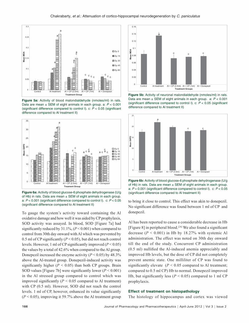
To assess lipid peroxidation, MDA levels were determined. Blood MDA levels [Figure 5a] rapidly and significantly increased (P < 0.001) from 15th day with Al raising it overall 2.5 times above control which was controlled by 44.8% with
0.5 ml and by 69.7% with 1 ml CP prophylaxis. MDA lowering effects (P <0.05) were recorded on 15th day with 1 ml of CP and on 30th day (P < 0.05) with 0.5 ml CP which was sustained till the end. The brain values [Figure 5b] were of the same order, with a raise (P < 0.001) of 60% with Al. Donepezil decreased (P < 0.05) elevated MDA levels, controlling it by 66.2% as compared to Al toxicity in blood and 41.9% in brain. The experimental intervention was found to work along the same line with no significant difference between the mean MDA levels of 1 ml CP and donepezil groups.
Another indicator of oxidative stress quantified was the G-6PD level. It increased significantly (P < 0.05) on 15th day and P < 0.001 from 30th day onward over the days in blood [Figure 6a] by a total of 56.7%. CP prophylaxis (0.5 ml) decreased it by 31.04% compared to Al toxicity, which was evident from 45th day onward (P < 0.05). One ml of CP prophylaxis was early to act and decline in enzyme activity (P < 0.001) was seen right from 30th day resulting in a mean decrease of 37.8%. Therefore, the donepezil group is not significantly different from 1 ml CP prophylaxis group. Brain G-6PD values [Figure 6b] in Al stress were 25% higher (P < 0.001) than control.
0
20
40
60
80
100
120
140
160
180
200
Control Al treatment Al + CP (0.5ml) Al + CP (1ml) Al + Donepezil
Mea
n Ti
me(
seco
nds)
Treatment Group
Dy 0Dy 15Dy 30Dy 45Dy 60
aa a a
a a a aa a a a
a a aac c c cc c c c
c c c c
0
2
4
6
8
10
12
14
16
Control Al treatment Al + CP (0.5ml) Al + CP (1ml) Al + Donepezil
Mea
n A
ChE
Treatment Groups
Dy 0Dy 15Dy 30Dy 45Dy 60
aaa
a
cc
cc cc
c
0
0.2
0.4
0.6
0.8
1
1.2
1.4
1.6
1.8
Control Al treatment Al + CP (0.5ml) Al + CP (1ml) Al + Donepezil
Mea
n A
ChE
Treatment Groups
cc
aa
0
20
40
60
80
100
120
140
Control Al treatment Al + CP (0.5ml) Al + CP (1ml) Al + Donepezil
Mea
n Ti
me
(Sec
onds
)
Treatment Group
Dy 0
Dy 15
Dy 30
Dy 45
Dy 60
c c
c c
c c
cc
c c c
c
b a
aa
aaa
aaa
a
Figure 2: Effect on retention time (sec) of rats in the passive avoidance paradigm Data are mean ± SEM of eight animals in each group. a: P < 0.001 (significant difference compared to control I). c: P < 0.05 (significant difference compared to Al treatment II)
Figure 4a: Activity of blood acetylcholinesterase (U/g of Hb/L) in rats. Data are mean ± SEM of eight animals in each group. a: P < 0.001 (significant difference compared to control- I). c: P < 0.05 (significant difference compared to Al treatment II)
Figure 4b: Activity of neuronal acetylcholinesterase (×10–6/moles/L/min/g of tissue) in rats. Data are mean ± SEM of eight animals in each group. a: P < 0.001 (significant difference compared to control I). c: P < 0.05 (significant difference compared to Al treatment II)
Figure 3: Effect on retention time (sec) of rats in the rotarod paradigm. Data are mean ± SEM of eight animals in each group. (a) P < 0.001 (significant difference compared to control I). (b) P < 0.05 (significant difference compared to control I) (c) P < 0.05 (significant difference compared to Al treatment II)
Chakrabarty, et al.: Attenuation of cortico-hippocampal neurodegeneration by C. paniculatus
166 Journal of Pharmacology and Pharmacotherapeutics | April-June 2012 | Vol 3 | Issue 2
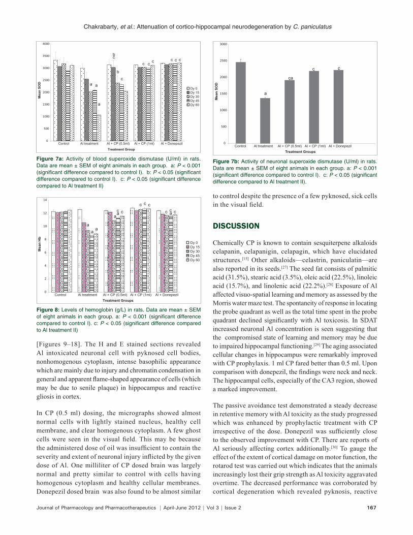
To gauge the system’s activity toward containing the Al oxidative damage and how well it was aided by CP prophylaxis, SOD activity was assayed. In blood, SOD [Figure 7a] had significantly reduced by 31.1%, (P < 0.001) when compared to control from 30th day onward with Al which was prevented by 0.5 ml of CP significantly (P < 0.05), but did not reach control levels. However, 1 ml of CP significantly improved (P < 0.05) the values by a total of 42.6% when compared to the Al group. Donepezil increased the enzyme activity (P < 0.05) by 48.3% above the Al-treated group. Donepezil-induced activity was significantly higher (P < 0.05) than both CP groups. Brain SOD values [Figure 7b] were significantly lower (P < 0.001) in the Al stressed group compared to control which was improved significantly (P < 0.05 compared to Al treatment) with CP (0.5 ml). However, SOD did not reach the control levels. 1 ml of CP, however, enhanced its value significantly (P < 0.05), improving it 59.7% above the Al treatment group
to bring it close to control. This effect was akin to donepezil. No significant difference was found between 1 ml of CP and donepezil.
Al has been reported to cause a considerable decrease in Hb [Figure 8] in peripheral blood.[26] We also found a significant decrease (P < 0.001) in Hb by 18.27% with systemic Al administration. The effect was noted on 30th day onward till the end of the study. Concurrent CP administration (0.5 ml) nullified the Al-induced anemia appreciably and improved Hb levels, but the dose of CP did not completely prevent anemic state. One milliliter of CP was found to significantly elevate (P < 0.05 compared to Al treatment; compared to 0.5 ml CP) Hb to normal. Donepezil improved Hb, but significantly less (P < 0.05) compared to 1 ml CP prophylaxis.
Effect of treatment on histopathologyThe histology of hippocampus and cortex was viewed
0
0.05
0.1
0.15
0.2
0.25
0.3
Control Al treatment Al + CP (0.5ml) Al + CP (1ml) Al + Donepezil
Mea
n M
DA
Treatment Group
Dy 0
Dy 15
Dy 30
Dy 45
Dy 60c
c
c
c c c cc c c
a
a
a
a
a
a
0
5
10
15
20
25
30
35
Control Al treatment Al + CP (0.5ml) Al + CP (1ml) Al + Donepezil
Mea
n G
6PD
Treament Group
Dy 0Dy 15Dy 30Dy 45Dy 60
aaa a
a
a
a
ccccc
c c c
0
5
10
15
20
25
Control Al treatment Al + CP (0.5ml) Al + CP (1ml) Al + Donepezil
Mea
n G
6PD
Treatment Groups
a
c c
0
0.02
0.04
0.06
0.08
0.1
0.12
0.14
0.16
Control Al treatment Al + CP (0.5ml) Al + CP (1ml) Al + Donepezil
Mea
n M
DA
Treatment Groups
ccc
a
Figure 5a: Activity of blood malondialdehyde (nmoles/ml) in rats. Data are mean ± SEM of eight animals in each group. a: P < 0.001 (significant difference compared to control I). c: P < 0.05 (significant difference compared to Al treatment II)
Figure 6a: Activity of blood glucose-6 phosphate dehydrogenase (U/g of Hb) in rats. Data are mean ± SEM of eight animals in each group. a: P < 0.001 (significant difference compared to control I). c: P < 0.05 (significant difference compared to Al treatment II)
Figure 6b: Activity of blood glucose-6 phosphate dehydrogenase (U/g of Hb) in rats. Data are mean ± SEM of eight animals in each group. a: P < 0.001 (significant difference compared to control I). c: P < 0.05 (significant difference compared to Al treatment II)
Figure 5b: Activity of neuronal malondialdehyde (nmoles/ml) in rats. Data are mean ± SEM of eight animals in each group. a: P < 0.001 (significant difference compared to control I). c: P < 0.05 (significant difference compared to Al treatment II)
Chakrabarty, et al.: Attenuation of cortico-hippocampal neurodegeneration by C. paniculatus
Journal of Pharmacology and Pharmacotherapeutics | April-June 2012 | Vol 3 | Issue 2 167
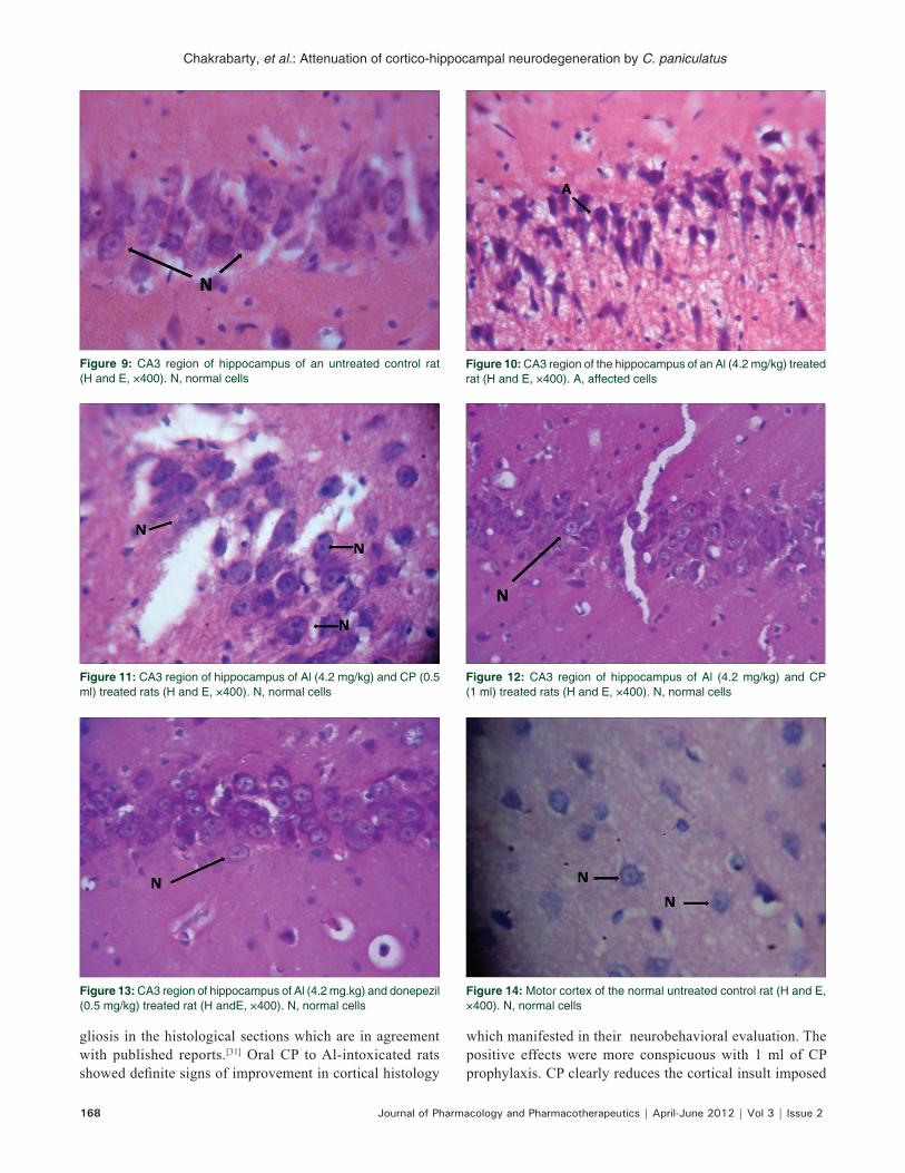
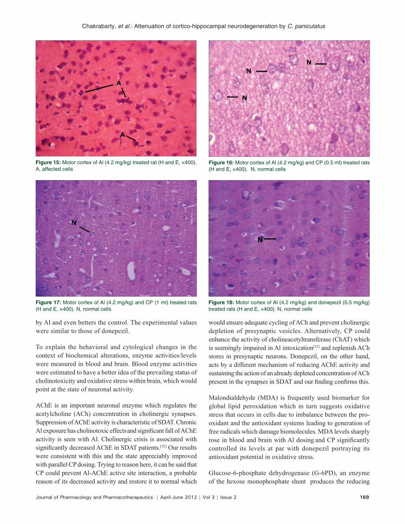
[Figures 9–18]. The H and E stained sections revealed Al intoxicated neuronal cell with pyknosed cell bodies, nonhomogenous cytoplasm, intense basophilic appearance which are mainly due to injury and chromatin condensation in general and apparent flame-shaped appearance of cells (which may be due to senile plaque) in hippocampus and reactive gliosis in cortex.
In CP (0.5 ml) dosing, the micrographs showed almost normal cells with lightly stained nucleus, healthy cell membrane, and clear homogenous cytoplasm. A few ghost cells were seen in the visual field. This may be because the administered dose of oil was insufficient to contain the severity and extent of neuronal injury inflicted by the given dose of Al. One milliliter of CP dosed brain was largely normal and pretty similar to control with cells having homogenous cytoplasm and healthy cellular membranes. Donepezil dosed brain was also found to be almost similar
to control despite the presence of a few pyknosed, sick cells in the visual field.
DISCUSSION
Chemically CP is known to contain sesquiterpene alkaloids celapanin, celapanigin, celapagin, which have elucidated structures.[15] Other alkaloids—celastrin, paniculatin—are also reported in its seeds.[27] The seed fat consists of palmitic acid (31.5%), stearic acid (3.5%), oleic acid (22.5%), linoleic acid (15.7%), and linolenic acid (22.2%).[28] Exposure of Al affected visuo-spatial learning and memory as assessed by the Morris water maze test. The spontaneity of response in locating the probe quadrant as well as the total time spent in the probe quadrant declined significantly with Al toxicosis. In SDAT increased neuronal Al concentration is seen suggesting that the compromised state of learning and memory may be due to impaired hippocampal functioning.[29] The aging associated cellular changes in hippocampus were remarkably improved with CP prophylaxis. 1 ml CP fared better than 0.5 ml. Upon comparison with donepezil, the findings were neck and neck. The hippocampal cells, especially of the CA3 region, showed a marked improvement.
The passive avoidance test demonstrated a steady decrease in retentive memory with Al toxicity as the study progressed which was enhanced by prophylactic treatment with CP irrespective of the dose. Donepezil was sufficiently close to the observed improvement with CP. There are reports of Al seriously affecting cortex additionally.[30] To gauge the effect of the extent of cortical damage on motor function, the rotarod test was carried out which indicates that the animals increasingly lost their grip strength as Al toxicity aggravated overtime. The decreased performance was corroborated by cortical degeneration which revealed pyknosis, reactive
0
500
1000
1500
2000
2500
3000
3500
4000
Control Al treatment Al + CP (0.5ml) Al + CP (1ml) Al + Donepezil
Mea
n SO
D
Treatment Group
Dy 0Dy 15Dy 30Dy 45Dy 60
c
cc c c c c c
b
a
a a
b
0
500
1000
1500
2000
2500
3000
Control Al treatment Al + CP (0.5ml) Al + CP (1ml) Al + Donepezil
Mea
n SO
D
Treatment Groups
a
ca
c c
0
2
4
6
8
10
12
14
Control Al treatment Al + CP (0.5ml) Al + CP (1ml) Al + Donepezil
Mea
n H
b
Treatment Groups
Dy 0Dy 15Dy 30Dy 45Dy 60
c aaa
aaa
cc
c cc c c
Figure 7a: Activity of blood superoxide dismutase (U/ml) in rats. Data are mean ± SEM of eight animals in each group. a: P < 0.001 (significant difference compared to control I). b: P < 0.05 (significant difference compared to control I). c: P < 0.05 (significant difference compared to Al treatment II)
Figure 7b: Activity of neuronal superoxide dismutase (U/ml) in rats. Data are mean ± SEM of eight animals in each group. a: P < 0.001 (significant difference compared to control I). c: P < 0.05 (significant difference compared to Al treatment II).
Figure 8: Levels of hemoglobin (g/L) in rats. Data are mean ± SEM of eight animals in each group. a: P < 0.001 (significant difference compared to control I). c: P < 0.05 (significant difference compared to Al treatment II)
Chakrabarty, et al.: Attenuation of cortico-hippocampal neurodegeneration by C. paniculatus
168 Journal of Pharmacology and Pharmacotherapeutics | April-June 2012 | Vol 3 | Issue 2
Figure 10: CA3 region of the hippocampus of an Al (4.2 mg/kg) treated rat (H and E, ×400). A, affected cells
Figure 12: CA3 region of hippocampus of Al (4.2 mg/kg) and CP (1 ml) treated rats (H and E, ×400). N, normal cells
Figure 14: Motor cortex of the normal untreated control rat (H and E, ×400). N, normal cells
Figure 11: CA3 region of hippocampus of Al (4.2 mg/kg) and CP (0.5 ml) treated rats (H and E, ×400). N, normal cells
Figure 13: CA3 region of hippocampus of Al (4.2 mg.kg) and donepezil (0.5 mg/kg) treated rat (H andE, ×400). N, normal cells
gliosis in the histological sections which are in agreement with published reports.[31] Oral CP to Al-intoxicated rats showed definite signs of improvement in cortical histology
which manifested in their neurobehavioral evaluation. The positive effects were more conspicuous with 1 ml of CP prophylaxis. CP clearly reduces the cortical insult imposed
Figure 9: CA3 region of hippocampus of an untreated control rat (H and E, ×400). N, normal cells
Chakrabarty, et al.: Attenuation of cortico-hippocampal neurodegeneration by C. paniculatus
Journal of Pharmacology and Pharmacotherapeutics | April-June 2012 | Vol 3 | Issue 2 169
by Al and even betters the control. The experimental values were similar to those of donepezil.
To explain the behavioral and cytological changes in the context of biochemical alterations, enzyme activities/levels were measured in blood and brain. Blood enzyme activities were estimated to have a better idea of the prevailing status of cholinotoxicity and oxidative stress within brain, which would point at the state of neuronal activity.
AChE is an important neuronal enzyme which regulates the acetylcholine (ACh) concentration in cholinergic synapses. Suppression of AChE activity is characteristic of SDAT. Chronic Al exposure has cholinotoxic effects and significant fall of AChE activity is seen with Al. Cholinergic crisis is associated with significantly decreased AChE in SDAT patients.[32] Our results were consistent with this and the state appreciably improved with parallel CP dosing. Trying to reason here, it can be said that CP could prevent Al-AChE active site interaction, a probable reason of its decreased activity and restore it to normal which
would ensure adequate cycling of ACh and prevent cholinergic depletion of presynaptic vesicles. Alternatively, CP could enhance the activity of cholineacetyltransferase (ChAT) which is seemingly impaired in Al intoxication[32] and replenish ACh stores in presynaptic neurons. Donepezil, on the other hand, acts by a different mechanism of reducing AChE activity and sustaining the action of an already depleted concentration of ACh present in the synapses in SDAT and our finding confirms this.
Malondialdehyde (MDA) is frequently used biomarker for global lipid peroxidation which in turn suggests oxidative stress that occurs in cells due to imbalance between the pro-oxidant and the antioxidant systems leading to generation of free radicals which damage biomolecules. MDA levels sharply rose in blood and brain with Al dosing and CP significantly controlled its levels at par with donepezil portraying its antioxidant potential in oxidative stress.
Glucose-6-phosphate dehydrogenase (G-6PD), an enzyme of the hexose monophosphate shunt produces the reducing
Figure 16: Motor cortex of Al (4.2 mg/kg) and CP (0.5 ml) treated rats (H and E, ×400). N, normal cells
Figure 17: Motor cortex of Al (4.2 mg/kg) and CP (1 ml) treated rats (H and E, ×400). N, normal cells
Figure 18: Motor cortex of Al (4.2 mg/kg) and donepezil (0.5 mg/kg) treated rats (H and E, ×400). N, normal cells
Figure 15: Motor cortex of Al (4.2 mg/kg) treated rat (H and E, ×400). A, affected cells
Chakrabarty, et al.: Attenuation of cortico-hippocampal neurodegeneration by C. paniculatus
170 Journal of Pharmacology and Pharmacotherapeutics | April-June 2012 | Vol 3 | Issue 2
equivalent NADPH. Its activity is increased in brain[33] and erythrocytes[1] in Al toxicosis like our results. An increased level signifies a heightened oxidative state. One milliliter of CP was found to be better (compared to 0.5 ml) in nullifying the mounted oxidation, which was almost at the same level as donepezil.
Superoxide dismutase (SOD) is an enzyme that forms antioxidant defense by catalyzing the dismutation (a chemical reaction where a species is simultaneously oxidized and reduced to form two different products) of superoxide radicals into oxygen and hydrogen peroxide. SOD levels in Al-intoxicated rats increased significantly with CP prophylaxis which points towards a decreasing level of oxidative stress. One milliliter of CP proved to be better than 0.5 ml in its potential. It can be compared equally in this effect with donepezil. CP, therefore, boosts the antioxidant machinery of the system by an unknown mechanism which atones the progressive underlying oxidative insult.
Low Hb levels are one of the risk factors for impaired cognition especially in elderly and specifically for SDAT.[34] Underlying mechanisms could vaguely be attributed to hypoxic injury. Al by accumulating in bone marrow suppresses erythropoiesis, thereby leading to anemia.[26] CP prophylaxis in Al toxicosis produced a healthy improvement in Hb levels. Donepezil showed only a marginal improvement here. This highlights its role in maintaining adequate neuronal oxygenation and minimizing induction of an adverse oxygen demand–supply ratio in brain.
Collating the results of all the investigations at behavioral, cellular and biochemical levels, it can be concluded here that CP seed oil has a pronounced antidementic effect and could prove to be a potent deterrent against SDAT. Further insight into the molecular mechanism of action of its phytoconstituents awaits exploration and could prove to be fruitful in this context.
ACKNOWLEDGMENTS
The work was supported by departmental fund provided by Kasturba Medical College, Manipal University, Manipal 576104, Karnataka, India. We are grateful to Manipal College of Pharmaceutical Sciences, Manipal, India, for providing us the Morris Water Maze apparatus.
REFERENCES
1. Kaur A, Gill KD. Possible peripheral markers for chronic aluminium toxicity in Wistar rats. Toxicol Ind Health 2006;22:39-46.
2. El-Demerdash FM. Antioxidant effect of vitamin E and selenium on lipid peroxidation, enzyme activities and biochemical parameters in rats exposed to aluminium. J Trace Elem Med Biol 2004;18:113-21.
3. Flaten TP. Aluminium as a risk factor in Alzheimer’s disease, with emphasis on drinking water. Brain Res Bull 2002;55:187-96.
4. Lukiw WJ. Alzheimer’s disease and aluminium. In: Mineral and Metal
Neurotox. Yasui M, Strong MJ, Ota K, editors. Boca Raton, FL: CRC Press; 1997; p. 112-26.
5. Exley C. A molecular mechanism of aluminium induced Alzheimer’s disease. J Inorg Biochem 1999;76:133-40.
6. Struys-Ponsar C, Kerkhofs A, Gauthier A, Soffié M, van den Bosch de Aguilar P. Effect of aluminium exposure on the behavioural parameters in the rat. Pharmacol Biochem Behav 1997;56:643-8.
7. Platt B, Fiddler G, Rieldel G, Henderson Z. Aluminium toxicity in the rat brain: Histochemical and immunocytochemical evidence. Brain Res Bull 2001;55:257-67.
8. Yokel RA. The toxicology of aluminium in the brain: A review. Neurotoxicology 2002;21:913-8.
9. McLachlan DR, Kruck TP, Lukiw WJ, Krishnan SS. Would decreased aluminium ingestion reduce the incidence of Alzeimer’s disease. CMAJ 1991;145:793-829.
10. Nadkarni AK. Nadkarni’s Indian Matera Medica. Vol. 1. Bombay: Popular Prakashan; 1976. p. 296.
11. Karanth KS, Padma TK, Gunsundari MN. Influence of Celastrus paniculatus oil on learning and memory. Arogya 1981;7:83-6.
12. Nalini K, Aroor HR, Kumar KB, Rao A. Studies on biogenic amines and their metabolites on mentally retarded children in Celastrus paniculatus oil therapy. Altern Med 1986;1:355-60.
13. Godkar P, Gordon R, Ravindran A, Doctor B. Celastrus paniculatus seed water soluble extracts protect against glutamate toxicity in neuronal cultures from rat forebrain. J Ethnopharmacol 2004;93:213-9.
14. Bhanumathy M, Harish M, Shivaprasad H, Sushma G. Nootropic activity of Celastrus paniculatus seed. Pharm Biol 2010;48:324-7.
15. Gattu M, Boss KL, Terry AV Jr, Buccafusco JJ. Reversal of scopolamine induced deficits in navigational memory performance by seed oil of Celastrus paniculatus. Pharmacol Biochem Behav 1997;57:793-9.
16. Sethi P, Jyoti A, Hussain E, Sharma D. Curcumin attenuates aluminium induced functional neurotoxicity in rats. Pharmacol Biochem Behav 2009;93:31-9.
17. Piala JJ, High JP, Hessert GL Jr, Burke JC, Craver BN. Pharmacological and toxicological Comparison of trifluperazine and chlorpromazine. J Pharmacol Exp Ther 1959;127:55-65.
18. Dunham NW, Miya TS. A note on a simple appratus for detecting neurological deficits in rat and mice. J Am Pharmacol Assoc 1957;46:208-9.
19. Ellman GL, Courtney AD, Andres V Jr, Featherstone RM. A rapid, new and colorimetric determination of acetylcholinesterase. Biochem Pharmacol 1961;7:88-95.
20. Greenfield S. Acetylcholinesterase may have novel function in brain. Trends in Neurosciences 1984;7:364-8.
21. Nourooz- Jadeh J, Tajjadini- Sharmani J, Mc Carthy S, Batteridge DJ, Wolff SP. Elevated levels of authentic hydroperoxides in NIDDM. Diabetes 1995;44:1054-8.
22. Okhawa H, Ohishi N, Yagi K. Reaction of linoleic acid hydroperoxide with thiobarbituric acids. Anal Biochem 1979;98:351-4.
23. Kornberg L, Horecker BL. Glucose-6-phosphate dehydrogenase. Methods Enzymol 1955;1:323-7.
24. Beauchamp C, Fridovich I. Superoxide dismutasee: Improved assays and an assay applied to acrylamide gels. Anal Biochem 1971;44:276-87.
25. Marklund S, Marklund G. Involvement of superoxide anion radical in autoxidation of pyrogallol and a convenient assay for superoxide dismutase. Eur J Biochem 1974;47:469-74.
26. Zaman K, Zaman W, Siddique H. Haematological and enzymatic results of aluminium intoxication in rats. Comp Biochem Physiol C 1993;105:73-6.
27. Khare CP. Indian Medicinal Plants: An illustrated dictionary. New Delhi: Springer India Pvt. Ltd.; 2007. p. 134-5.
28. Sengupta A, Bhargava HN. Chemical investigation of the seed fat of Celastrus paniculatus. J Sci Food Agric 1970;21:628-31.
29. Perl DP, Brody AR. Alzheimer’s disease, X-ray spectrometric evidence of aluminium accumulation in neurofibrillary tangle bearing neuron. Science 1980;208:297-9.
30. Deloncle R, Guillard O. Mechanism of Alzheimer’s disease: Arguments for a neurotransmitter aluminium complex implication. Neurochem Res 1990;15:1239-45.
31. Rao KS. Effect of aluminium on the brain cells of the rat. Biochem Int 1992;28:58-60.
32. Julka D, Sandheer R, Gill KD. Altered cholinergic metabolism in rat CNS
Chakrabarty, et al.: Attenuation of cortico-hippocampal neurodegeneration by C. paniculatus
Journal of Pharmacology and Pharmacotherapeutics | April-June 2012 | Vol 3 | Issue 2 171
following aluminium exposure: Implications on learning performance. J Neurochem 1995;65:2157-64.
33. Martins RN, Harper CG, Stokes GB, Masters CL. Increased cerebral G-6-PD activity in Alzheimer’s disease may reflect oxidative stress. J Neurochem 1986;46:1042-5.
34. Pandav RS, Chandra V, Dodge HH, De Kosky ST, Ganguli M. Haemoglobin levels and Alzheimer’s disease: An epidemiologic study in India. Am J Geriatr Psychiatry 2004;12:523-6.
How to cite this article: Chakrabarty M, Bhat P, Kumari S, D«SQ»Souza A, Bairy KL, Chaturvedi A, Natarajan A, Mohandas Rao KG, Kamath S. Cortico-hippocampal salvage in chronic aluminium induced neurodegeneration by Celastrus paniculatus seed oil: Neurobehavioural, biochemical, histological study. J Pharmacol Pharmacother 2012;3:161-71.Source of Support: Kasturba Medical College, Manipal University, Manipal 576104, Karnataka, India., Conflict of Interest: None declared.
Related Documents


