Copyrights © 2016 The Korean Society of Radiology 177 Original Article pISSN 1738-2637 / eISSN 2288-2928 J Korean Soc Radiol 2016;74(3):177-184 http://dx.doi.org/10.3348/jksr.2016.74.3.177 INTRODUCTION e rotator interval (RI) is defined as the space between the anterior aspect of the supraspinatus tendon (SST) and superior aspect of the subscapularis tendon (SSC), covered by rotator in- terval capsule (RIC) (1, 2). It represents a region of capsule that is not reinforced by overlying rotator cuff tendon (1) and its com- ponents, coracohumeral ligament (CHL) and superior gleno- humeral ligament (SGHL), are suggested to have a role in gle- nohumeral joint (GHJ) stability. e role of different shoulder stabilizers vary according to different shoulder position. For ex- ample, the CHL is shown to be a stabilizer in superior-inferior directions with arm in external rotation, whereas, the intra-ar- ticular pressure that is maintained by the intact RIC is suggest- ed to act as a stabilizer in superior-inferior directions with the arm in internal and neutral rotations (3). e role of the RI in shoulder mechanopathology remains a much debated subject, with some authors advocating its role as a negative articular pres- sure inducer (3), whereas others, in resisting posteroinferior glenohumeral translation (4-7). Correlation of the Rotator Interval on Direct CT Arthrography in Shoulder Instability Patients with External Rotation Stress Radiographs 견관절 불안정 환자의 CT 관절 조영술상 회전근개 간격과 외회전 부하 영상과의 연관성 Yun-Sik Jung, MD, Hee Seok Choi, MD, Jung-Ah Choi, MD * Department of Radiology, Hallym University College of Medicine, Dongtan Sacred Hospital, Hwaseong, Korea Purpose: To correlate rotator interval dimensions on direct CT arthrography (CTA) with external rotation shoulder stress radiographs in patients with shoulder insta- bility. Materials and Methods: One hundred and twenty-three shoulders of 115 patients with shoulder instability that underwent direct CTA and shoulder stress radiographs in neutral position and external rotation were enrolled. Axial, oblique coronal, oblique sagittal CT images were obtained after intra-articular contrast injection. Width and depth of rotator interval on sagittal CTA images were evaluated and ac- romiohumeral (AH) distance on neutral and external rotation stress radiographs were measured. CTA measurements were compared with AH on stress radiographs according to the group with or without “external rotational instability”. Results: Out of 123 shoulders, 79 with external rotation instability showed in- creased or unchanged AH distance on external rotation compared with neutral po- sition. Width of rotator interval was 2.43 ± 0.31 cm and depth was 0.87 ± 0.2 cm. Forty-four shoulders without external rotation instability showed decreased AH dis- tance on external rotation compared with neutral position. Width of rotator interval was 2.02 ± 0.45 cm and depth was 0.75 ± 0.2 cm. Width and depth of rotator inter- val were significantly greater in the external rotation instability group. Conclusion: Width and depth of rotator interval on CTA were significantly correlat- ed with AH distance on external rotation stress radiographs. Index terms Rotator Cuff Shoulder Arthrography Computed Tomography Radiography Received June 12, 2015 Revised July 31, 2015 Accepted October 13, 2015 *Corresponding author: Jung-Ah Choi, MD Department of Radiology, Hallym University College of Medicine, Dongtan Sacred Hospital, 7 Keunjaebong-gil, Hwaseong 18450, Korea. Tel. 82-31-8086-2580 Fax. 82-31-8086-2584 E-mail: [email protected] This is an Open Access article distributed under the terms of the Creative Commons Attribution Non-Commercial License (http://creativecommons.org/licenses/by-nc/3.0) which permits unrestricted non-commercial use, distri- bution, and reproduction in any medium, provided the original work is properly cited.

Welcome message from author
This document is posted to help you gain knowledge. Please leave a comment to let me know what you think about it! Share it to your friends and learn new things together.
Transcript
Copyrights © 2016 The Korean Society of Radiology 177
Original ArticlepISSN 1738-2637 / eISSN 2288-2928J Korean Soc Radiol 2016;74(3):177-184http://dx.doi.org/10.3348/jksr.2016.74.3.177
INTRODUCTION
The rotator interval (RI) is defined as the space between the anterior aspect of the supraspinatus tendon (SST) and superior aspect of the subscapularis tendon (SSC), covered by rotator in-terval capsule (RIC) (1, 2). It represents a region of capsule that is not reinforced by overlying rotator cuff tendon (1) and its com-ponents, coracohumeral ligament (CHL) and superior gleno-humeral ligament (SGHL), are suggested to have a role in gle-nohumeral joint (GHJ) stability. The role of different shoulder
stabilizers vary according to different shoulder position. For ex-ample, the CHL is shown to be a stabilizer in superior-inferior directions with arm in external rotation, whereas, the intra-ar-ticular pressure that is maintained by the intact RIC is suggest-ed to act as a stabilizer in superior-inferior directions with the arm in internal and neutral rotations (3). The role of the RI in shoulder mechanopathology remains a much debated subject, with some authors advocating its role as a negative articular pres-sure inducer (3), whereas others, in resisting posteroinferior glenohumeral translation (4-7).
Correlation of the Rotator Interval on Direct CT Arthrography in Shoulder Instability Patients with External Rotation Stress Radiographs견관절 불안정 환자의 CT 관절 조영술상 회전근개 간격과 외회전 부하 영상과의 연관성
Yun-Sik Jung, MD, Hee Seok Choi, MD, Jung-Ah Choi, MD*Department of Radiology, Hallym University College of Medicine, Dongtan Sacred Hospital, Hwaseong, Korea
Purpose: To correlate rotator interval dimensions on direct CT arthrography (CTA) with external rotation shoulder stress radiographs in patients with shoulder insta-bility.Materials and Methods: One hundred and twenty-three shoulders of 115 patients with shoulder instability that underwent direct CTA and shoulder stress radiographs in neutral position and external rotation were enrolled. Axial, oblique coronal, oblique sagittal CT images were obtained after intra-articular contrast injection. Width and depth of rotator interval on sagittal CTA images were evaluated and ac-romiohumeral (AH) distance on neutral and external rotation stress radiographs were measured. CTA measurements were compared with AH on stress radiographs according to the group with or without “external rotational instability”.Results: Out of 123 shoulders, 79 with external rotation instability showed in-creased or unchanged AH distance on external rotation compared with neutral po-sition. Width of rotator interval was 2.43 ± 0.31 cm and depth was 0.87 ± 0.2 cm. Forty-four shoulders without external rotation instability showed decreased AH dis-tance on external rotation compared with neutral position. Width of rotator interval was 2.02 ± 0.45 cm and depth was 0.75 ± 0.2 cm. Width and depth of rotator inter-val were significantly greater in the external rotation instability group.Conclusion: Width and depth of rotator interval on CTA were significantly correlat-ed with AH distance on external rotation stress radiographs.
Index termsRotator CuffShoulderArthrographyComputed TomographyRadiography
Received June 12, 2015Revised July 31, 2015Accepted October 13, 2015*Corresponding author: Jung-Ah Choi, MDDepartment of Radiology, Hallym University College of Medicine, Dongtan Sacred Hospital, 7 Keunjaebong-gil, Hwaseong 18450, Korea.Tel. 82-31-8086-2580 Fax. 82-31-8086-2584E-mail: [email protected]
This is an Open Access article distributed under the terms of the Creative Commons Attribution Non-Commercial License (http://creativecommons.org/licenses/by-nc/3.0) which permits unrestricted non-commercial use, distri-bution, and reproduction in any medium, provided the original work is properly cited.

178
Rotator Interval on Direct CTA
jksronline.orgJ Korean Soc Radiol 2016;74(3):177-184
Surgical repair of the RI is suggested to be helpful in surgical treatment of posterior and multidirectional instability (4, 5, 7-10), and attempts have been made to diagnose RI injury both clinically and on imaging (6, 9, 11-13); however, there is no de-finitive means of diagnosing a widened RI objectively yet. RI le-sions are classified into 4 types by orthopedic surgeons includ-ing; type I, contracture; type II, laxity with GHJ instability; type III, traumatic injury: IIIa tear and IIIb coracoid impingement; and type IV, bicipital instability involving CHL, SGHL, SSC (14). Imaging studies on diagnosis of type I and III lesions have used magnetic resonance arthrography (MRA), especially regarding RI deficiency in instability lesions (2, 15, 16). Clinically, authors have suggested that the sulcus sign, which appears as a depres-sion of the posterior aspect of shoulder just below the acromion upon applying traction in an inferior direction that disappears upon external rotation because of tightening of the RI, persist-ing on 45 degree external rotation of the arm, would suggest pathologic laxity and RI widening (1-13, 17). Widening of the RI is clinically identified in the arthroscopic field by an increased dis-tance between the SST and SSC tendons and between the biceps long head tendon and anterior edge of the SSC tendons. However, there are few studies on the correlation between clinical examina-tion findings with dimensions of the RI on imaging.
Reports have indicated that standardized anteroposterior ra-diographs are a reliable and reproducible method for measure-ment of acromiohumeral (AH) interval (18), which is decreased in patients with SST tear and increased in shoulder dislocation. Therefore, the AH distance would be increased in patients with RI laxity with persistence in external rotation of the arm as in the physical examination for sulcus sign.
In this study, we compared the RI dimensions on direct CT arthrography (CTA) and correlated them with AH distances on neutral and external stress radiographs in patients with clinical instability.
MATERIALS AND METHODS
This retrospective study was approved by the Institutional Re-view Board and the requirement for patients’ informed consent was waived.
Patients
Between January 2005 and December 2008, 115 consecutive patients with clinically diagnosed instability of the GHJ with history of dislocation and subluxation (97 shoulders) and un-determined shoulder pain with clinical instability or apprehen-sion (26 shoulders), underwent direct CTA using 16 or 64 chan-nel multidetector CT (MDCT) and shoulder stress radiography in neutral position and external rotation. Eight patients under-went bilateral shoulder examination. Total 123 shoulder CTAs (107 men, 16 women; mean age, 25 years; range, 14–58 years) were enrolled including 78 right and 45 left shoulders.
Imaging Studies: CT and Stress Radiography
After intra-articular positioning of a 22-gauge needle through an anterior approach under fluoroscopic guidance, intra-articular injection of 12–20 mL mixed solution of 13 mL meglumine ioxi-talamate (Telebrix 30 Meglumine; Guerbet, Aulnay-sous-Bois, France) and 7 mL normal saline was performed until the patient complained of pain or the injecting physician felt pressure.
CT scanning was performed with 16- or 64-channel MDCT. In 88 shoulders, axial image were obtained with 16-channel MDCT (Mx 8000 IDT; Philips Medical Systems, Eindhoven, the Netherlands) with the following protocols: rotation speed of 0.75 second per rotation, a current of 240 mAs, a voltage of 120 kVp, and a collimation of 2.5 mm. The field of view (FOV) at ac-quisition was 30 cm, and section thickness was 1.0 mm, with a section increment of 0.5 mm (50% section overlap). In 35 shoul-ders, axial images were obtained with 64-section MDCT (Bril-liance 64; Philips Medical Systems) with the following protocol: rotation speed of 0.75 second per rotation, a current of 300 mAs, a voltage of 140 kVp, and a collimation of 0.625 mm. The FOV at acquisition was 30 cm, and section thickness was 0.67 mm, with a section increment of 0.33 mm (50% section overlap). Oblique sagittal images parallel to the glenoid fossa and oblique coronal images vertical to the glenoid fossa were reconstructed (1024 × 1024 matrix with 2-mm thickness) using an imaging workstation (Rapidia, version 2.8; Infinitt, Seoul, Korea). All patients were placed in the supine position, with the arm adducted along the body, the shoulder in a neutral position with the thumb point-ing upward.
Stress radiographs of the shoulder were obtained in standard-ized anterior-posterior (AP) direction in upright position, with

179
Yun-Sik Jung, et al
jksronline.org J Korean Soc Radiol 2016;74(3):177-184
a voltage of 60 kVp and a current of 8 mAs. The stress was ap-plied by 10 lb sandbags in both hands. The stress radiographs were obtained in neutral position with the patient’s arm adduct-ed alongside the body and in external rotation positions with the arm adducted but elbow and forearm externally rotated at
90 degrees away from the body.
Image Analysis
The CTA images and stress radiographs of the shoulder were reviewed by 2 musculoskeletal radiologists under consensus (a
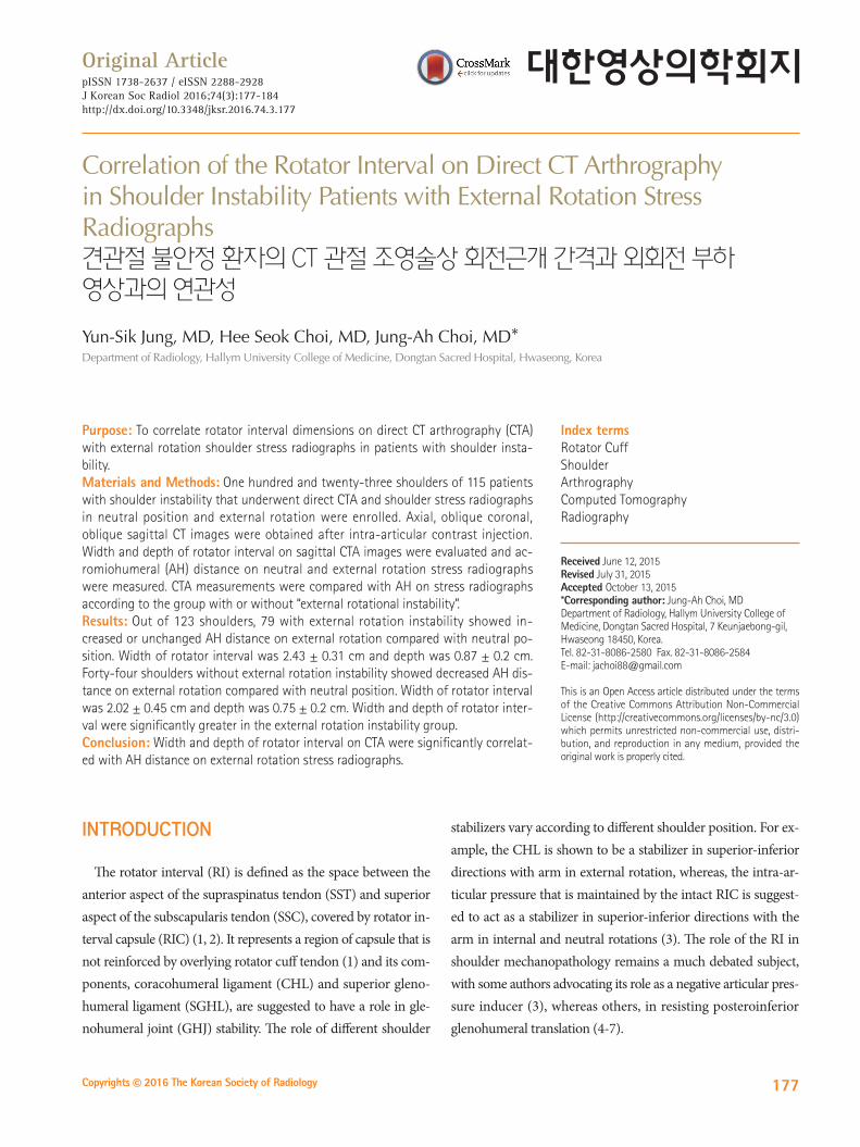
Fig. 1. A case of “external rotation instability positive”.A. The stress radiograph in neutral position is obtained in a 27-year-old man with right shoulder instability. The acromiohumeral (AH) distance (double-headed arrow) is 1.93 cm. B. The AH distance on the stress radiograph in external rotation position (double-headed arrow) is 2.51 cm. There is increased AH distance com-pared to neutral stress radiograph. C. An oblique sagittal CT arthrographic image shows the rotator interval (RI) dimensions. The width of the RI (long arrow) is 2.29 cm and the depth (short arrow) is 0.88 cm. D. RI widening (arrows) is seen along with anteroinferior labral tear (arrowheads) at arthroscopy, so RI closure is performed.
A
C
B
D

180
Rotator Interval on Direct CTA
jksronline.orgJ Korean Soc Radiol 2016;74(3):177-184
fellow in musculoskeletal radiology, a staff radiologist with 8 years of experience, blinded to clinical information) for train-ing, and then independently.
The dimensions of RI on CTA were analyzed on sagittal im-age, at the section just lateral to coracoid process. The width and depth were obtained (Figs. 1, 2) by measuring the most lat-eral image in which the coracoid process was present from the superior border of the SSC to the anterior border of the SST and as the longest perpendicular distance from humeral head to the roof of the RI of the RIC, respectively.
The AH distance was measured as the length between the un-dersurface of acromion and apex of humeral head. The AH dis-tance was measured on neutral and external rotation stress ra-diographs, respectively. If the AH distance was not changed or increased on external rotation, as compared to neutral position, the case was classified as “external rotation instability positive (+)” (Fig. 1). These criteria were based on the sulcus sign on physical exam for patients with instability (19); the sulcus sign disappears normally in external rotation because of tightening of the RI and the presence of persistent sulcus sign although gle-nohumeral external rotation may suggest pathologic laxity when symptoms are elicited (19). So in cases with intact RI without pathologic laxity, the AH distance is expected to decrease upon external rotation. If the AH distance was decreased on external
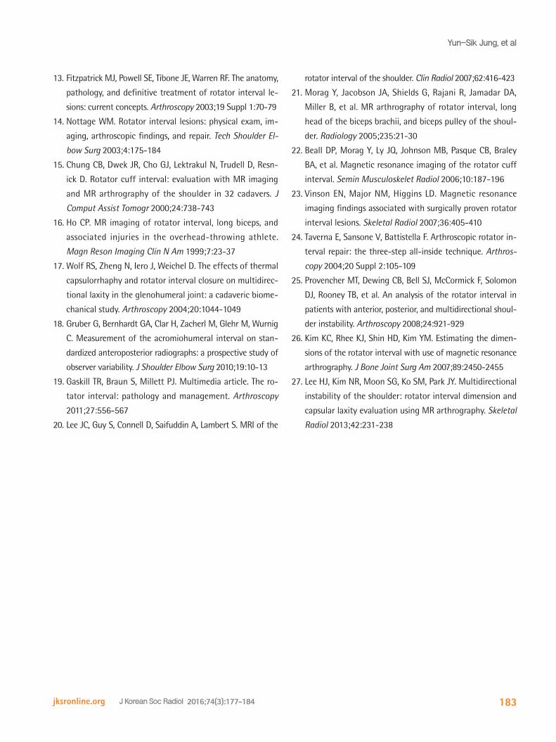
rotation, the case was classified as “external rotation instability negative (-)” (Fig. 2).
Statistical Analysis
The software SPSS (version 15.0; SPSS Inc., Chicago, IL, USA) was used for statistical analysis. The student t-test was used to compare the diameter of RI (AP and longitudinal diameter) be-tween ‘external rotation instability +’ and ‘external rotation in-stability -’ group. A p value of < 0.05 was considered statistically significant. The inter-observer reliability was tested for the 2 observers using intraclass correlation coefficient (ICC); ICC was interpreted as indicating poor (ICC, 0–0.2), fair (ICC, 0.3–0.4), moderate (ICC, 0.5–0.6), strong (ICC, 0.7–0.8), or almost perfect (ICC, > 0.8) agreement.
Arthroscopic surgical reports were subsequently reviewed to evaluate RI pathology and RI closure.
RESULTS
After measurement of the AH distance on external and neutral stress radiographs, 79 shoulders (73 male, 6 females; mean age 25.2 years) among total 123 shoulders, were classified as “exter-nal rotation instability (+)” group. In this group, the AP diame-ter of RI was 2.43–0.31 cm and longitudinal diameter was 0.87–
Fig. 2. A case of “external rotation instability negative”.A. The stress radiograph in neutral position is obtained in 23-year-old man with right shoulder instability. The acromiohumeral (AH) distance (ar-row) is 1.92 cm.B. The AH distance on the stress radiograph (arrow) in external rotation position is 1.66 cm. There is decreased AH distance compared to neutral stress radiograph. C. An oblique sagittal CT arthrographic image shows the rotator interval (RI) dimensions. The width of the RI (long arrow) is 1.88 cm and the depth (short arrow) is 0.54 cm.
A B C

181
Yun-Sik Jung, et al
jksronline.org J Korean Soc Radiol 2016;74(3):177-184
0.2 cm on CTA (Table 1). The “external rotation instability (-)” group included 44 shoulders (34 male; 10 females, mean age 27 years). The width of RI was 2.02–0.45 cm and depth was 0.75–0.2 cm on CTA (Table 1). The width and depth of RI were sig-nificantly greater in external rotation instability (+) group (p = 0.000 and 0.003, respectively).
Accompanied lesions included 94 Bankart and variant type of lesions and 78 superior labral tear from anterior-to-posterior (SLAP) lesions; fifty-five had both Bankart/variants and SLAP lesions. External rotation instability (+) group had significantly more Bankart lesions than (-) group (n = 61 vs. 33, p < 0.05 by Student t-test). SLAP was not significantly increased in external rotation instability (+) group. ICC, used to evaluate agreement be-tween the 2 observers for each measurement, was strong (0.75).
Review of clinical records after image analysis revealed that 92 patients underwent arthroscopic surgery, 63 of whom were external rotation instability (+) on preoperative radiographs; RI closure was performed in 8 patients, all these patients had shown external rotation instability (+).
DISCUSSION
The main function of shoulder stabilizers reportedly vary ac-cording to shoulder position. In one previous report (3), the su-perior-inferior stabilizing functions of the CHL and RIC in 6 cadaver shoulders were studied with use of a material testing machine, where axial translation of the humerus with the supe-rior-inferior translation force of 30N applied were recorded un-der the following joint capsule conditions: 1) intact, 2) vented, 3) the CHL sectioned, and 4) the RIC incised. The conclusion was that the CHL is a stabilizer in superior-inferior directions with the arm in external rotation, and the intact RIC is a stabi-lizer in superior-inferior directions with the arm in internal and neutral rotations. In a cadaveric study, Harryman et al. (4) showed
the importance of the RI in maintaining GHJ stability. They made a transverse incision through the capsule, CHL, and SGHL. Sectioning produced increased translation in all planes particu-larly, after sectioning of the RI capsule posterior and inferior instability with the arm at 60 degrees of abduction and 60 de-grees of external rotation, with dislocation in half of specimens.
Detecting and closing a RI defect arthroscopically may be dif-ficult as the RI is regularly used as the anterior portal in shoul-der arthroscopy, decreasing the chance of discovering lesions in this area (20). Recognition of the normal and widened RI can be difficult and is most frequently based on the surgeon’s expe-rience. The RI is generally considered as widened if the interval extends superior to the biceps, when viewed from the posterior portal. So, preoperative detection of the RI injury is necessary if appropriate surgical repair is to be performed. MRA before sur-gery is helpful in identifying RI injury (21, 22), with the follow-ing findings indicative of RI lesions at surgery: gadolinium ex-tending to the cortex of the undersurface of the coracoid process, especially in the presence of a labral tear and/or thickening of the CHL or superior GHL (23).
Laxity of the rotator cuff interval occurs in patients with a broad spectrum of injury. There may be a history of acute trau-ma, repetitive microtrauma, or underlying ligamentous laxity with superimposed ligament injury. Widening can be seen in a patient with a first-time dislocation, an overhead athlete, or pa-tient with more diffuse multidirectional instability (13). Nobu-hara and Ikeda (12) quantified the interval distance in 5 cadav-ers and reported the average distance from the anterior SST to the superior SSC as 21.6 mm, and 27.8 mm in a distended joint. The reported average distance in distended joint was greater than our study, probably because they measured the distance at the level of glenoid margin (more medial position than our study). Also, in our study, the amount of contrast varied accord-ing to each patient and in patients with RI laxity, more contrast was injected, which probably resulted in greater AH distance, including the position of external rotation. In another study (14), measurements of the width of the RIC were made on the sagittal images of the MRAs of 32 specimens. In all cases, the capsule was measured at the level of the intra-articular biceps tendon on an image just lateral to the coracoid process, as mea-sured in our study. The width of the RI capsule ranged from 1.7 to 2.0 mm (mean 1.8 mm) (14). The reported width was small-
Table 1. Comparison of Width and Depth of Rotator Interval (n = 123)
External Rotation (+)(n = 79)
External Rotation (-)(n = 44)
p Value
Sex (M/F) 73/6 34/10Age (yr) 25.2 27Width (cm) 2.43 ± 0.31 2.02 ± 0.45 0.000Depth (cm) 0.87 ± 0.2 0.75 ± 0.2 0.003
External rotation (+) = instability present on external rotation, External ro-tation (-) = instability absent on external rotation

182
Rotator Interval on Direct CTA
jksronline.orgJ Korean Soc Radiol 2016;74(3):177-184
er than in subjects that showed external rotation instability in our study, probably because these were measured in specimens, as compared with patients with instability.
RI closure in instability surgeries remains controversial with few studies on objective evaluation of the RI dimensions and widening; however, some studies provide evidence for widened RI, which would warrant their closure (10, 13, 24). The widen-ing of the RI may be represented by relatively anterior position of the biceps long head tendon, representing dilatation of the anterior capsule, which would hence lead to failure in this re-gion (25) and increased AH distance. Another study on com-parative RI dimensions between subjects with and without in-stability reported significant differences in the RI dimensions between the 2 groups (26). On MR arthrography, RI dimen-sions were greater in shoulders with MDI as compared to con-trols, according to an earlier study (27). In our study, there was no control group per se; however, differences were found in the CTA dimensions between patients with external stress instabil-ity and those without. To the best of our knowledge, no study has correlated measurements of RI dimensions on CTA and AH distance measurements on external rotation stress radiographs, which would correspond to imaging assessment of the sulcus sign on external rotation.
Our study had several limitations. First, the study was limited by its retrospective nature and not all cases had surgical or ar-throscopic correlation about RI lesion; however, it is difficult to evaluate RI laxity on arthroscopic exam, so the orthopedic sur-geon frequently relies on the imaging findings for determina-tion of RI laxity along with physical examination findings. Sec-ond, there was no analysis of intraobserver agreement of the measurements. Last, there was no true normal control group be-cause external stress radiographs are not routinely taken in nor-mal controls or patients without instability. Furthermore, we could not use the contralateral side because oftentimes the pa-tients were clinically lax or had instability at both shoulders.
In conclusion, the width and depth of RI on CTA were corre-lated with increased AH distance on external rotation stress ra-diographs. Therefore, stress radiographs in neutral and external rotation may be useful in evaluation of RI laxity or injury, sup-plementing clinical findings on physical examination, and help in preoperative planning.
Acknowledgments
Study materials obtained at previous affiliation of corre-sponding author, namely, Seoul National University Bundang Hospital.
This study was supported by Basic Science Research Program through the National Research Foundation of Korea (NRF) funded by the Ministry of Education, Science and Technology (NRF-2012R1A1A3010896).
REFERENCES
1. Bigoni BJ, Chung CB. MR imaging of the rotator cuff in-
terval. Radiol Clin North Am 2006;44:525-536, viii
2. Krief OP. MRI of the rotator interval capsule. AJR Am J
Roentgenol 2005;184:1490-1494
3. Itoi E, Berglund LJ, Grabowski JJ, Naggar L, Morrey BF, An
KN. Superior-inferior stability of the shoulder: role of the
coracohumeral ligament and the rotator interval capsule.
Mayo Clin Proc 1998;73:508-515
4. Harryman DT 2nd, Sidles JA, Harris SL, Matsen FA 3rd. The
role of the rotator interval capsule in passive motion and sta-
bility of the shoulder. J Bone Joint Surg Am 1992;74:53-66
5. Jost B, Koch PP, Gerber C. Anatomy and functional aspects
of the rotator interval. J Shoulder Elbow Surg 2000;9:336-
341
6. Pradhan RL, Itoi E. Rotator interval lesions of the shoulder
joint. Orthopedics 2001;24:798-801; quiz 802-803
7. Van der Reis W, Wolf EM. Arthroscopic rotator cuff inter-
val capsular closure. Orthopedics 2001;24:657-661
8. Cole BJ, Mazzocca AD, Meneghini RM. Indirect arthroscop-
ic rotator interval repair. Arthroscopy 2003;19:E28-E31
9. Field LD, Warren RF, O’Brien SJ, Altchek DW, Wickiewicz
TL. Isolated closure of rotator interval defects for shoulder
instability. Am J Sports Med 1995;23:557-563
10. Millett PJ, Clavert P, Warner JJ. Arthroscopic management of
anterior, posterior, and multidirectional shoulder instability:
pearls and pitfalls. Arthroscopy 2003;19 Suppl 1:86-93
11. Cooper DE, O’Brien SJ, Arnoczky SP, Warren RF. The structure
and function of the coracohumeral ligament: an anatomic
and microscopic study. J Shoulder Elbow Surg 1993;2:70-77
12. Nobuhara K, Ikeda H. Rotator interval lesion. Clin Orthop
Relat Res 1987;(223):44-50
183
Yun-Sik Jung, et al
jksronline.org J Korean Soc Radiol 2016;74(3):177-184
13. Fitzpatrick MJ, Powell SE, Tibone JE, Warren RF. The anatomy,
pathology, and definitive treatment of rotator interval le-
sions: current concepts. Arthroscopy 2003;19 Suppl 1:70-79
14. Nottage WM. Rotator interval lesions: physical exam, im-
aging, arthroscopic findings, and repair. Tech Shoulder El-
bow Surg 2003;4:175-184
15. Chung CB, Dwek JR, Cho GJ, Lektrakul N, Trudell D, Resn-
ick D. Rotator cuff interval: evaluation with MR imaging
and MR arthrography of the shoulder in 32 cadavers. J
Comput Assist Tomogr 2000;24:738-743
16. Ho CP. MR imaging of rotator interval, long biceps, and
associated injuries in the overhead-throwing athlete.
Magn Reson Imaging Clin N Am 1999;7:23-37
17. Wolf RS, Zheng N, Iero J, Weichel D. The effects of thermal
capsulorrhaphy and rotator interval closure on multidirec-
tional laxity in the glenohumeral joint: a cadaveric biome-
chanical study. Arthroscopy 2004;20:1044-1049
18. Gruber G, Bernhardt GA, Clar H, Zacherl M, Glehr M, Wurnig
C. Measurement of the acromiohumeral interval on stan-
dardized anteroposterior radiographs: a prospective study of
observer variability. J Shoulder Elbow Surg 2010;19:10-13
19. Gaskill TR, Braun S, Millett PJ. Multimedia article. The ro-
tator interval: pathology and management. Arthroscopy
2011;27:556-567
20. Lee JC, Guy S, Connell D, Saifuddin A, Lambert S. MRI of the
rotator interval of the shoulder. Clin Radiol 2007;62:416-423
21. Morag Y, Jacobson JA, Shields G, Rajani R, Jamadar DA,
Miller B, et al. MR arthrography of rotator interval, long
head of the biceps brachii, and biceps pulley of the shoul-
der. Radiology 2005;235:21-30
22. Beall DP, Morag Y, Ly JQ, Johnson MB, Pasque CB, Braley
BA, et al. Magnetic resonance imaging of the rotator cuff
interval. Semin Musculoskelet Radiol 2006;10:187-196
23. Vinson EN, Major NM, Higgins LD. Magnetic resonance
imaging findings associated with surgically proven rotator
interval lesions. Skeletal Radiol 2007;36:405-410
24. Taverna E, Sansone V, Battistella F. Arthroscopic rotator in-
terval repair: the three-step all-inside technique. Arthros-
copy 2004;20 Suppl 2:105-109
25. Provencher MT, Dewing CB, Bell SJ, McCormick F, Solomon
DJ, Rooney TB, et al. An analysis of the rotator interval in
patients with anterior, posterior, and multidirectional shoul-
der instability. Arthroscopy 2008;24:921-929
26. Kim KC, Rhee KJ, Shin HD, Kim YM. Estimating the dimen-
sions of the rotator interval with use of magnetic resonance
arthrography. J Bone Joint Surg Am 2007;89:2450-2455
27. Lee HJ, Kim NR, Moon SG, Ko SM, Park JY. Multidirectional
instability of the shoulder: rotator interval dimension and
capsular laxity evaluation using MR arthrography. Skeletal
Radiol 2013;42:231-238

184
Rotator Interval on Direct CTA
jksronline.orgJ Korean Soc Radiol 2016;74(3):177-184
견관절 불안정 환자의 CT 관절 조영술상 회전근개 간격과 외회전 부하 영상과의 연관성
정윤식 · 최희석 · 최정아*
목적: 견관절 불안정 환자의 CT 관절 조영술상 회전근개 간격과 외회전 부하 영상과의 연관성을 알아보고자 하였다.
대상과 방법: 견관절 불안정이 있는 115명의 환자의 123 견관절을 대상으로 하였다. CT arthrography (이하 CTA) 및 중
립 자세 외회전 부하 영상을 분석하였다. CTA는 관절 내 조영제 주입 후에 축상면, 관상면 사위, 시상면 사위 CT 영상을
획득하였다. 회전근개 간격의 넓이와 깊이를 오구돌기 직원위면의 CTA 시상면 사위 영상에서 획득하였고, 견봉상완 거리
는 중립과 외회전 부하 영상에서 측정하였다. CTA 영상에서의 측정치와 중립 및 외회전 부하 영상에 견봉상완 거리를 외
회전 불안정성 유무군과 비교분석하였다.
결과: 중립 자세와 비교하여 외회전시에 123 어깨 중 외회전 불안정성이 있는 79개의 어깨에서 견봉상완 거리가 증가하
거나 변하지 않았다. 회전근개 간격의 넓이는 2.43 ± 0.31 cm, 깊이는 0.87 ± 0.2 cm로 측정되었다. 중립 외회전 자세
에서 외회전 불안정이 없는 44 어깨의 견봉상완거리 감소가 측정되었다. 회전근개 간격의 넓이는 2.02 ± 0.45 cm, 깊이
는 0.75 ± 0.2 cm로 측정되었다. 회전근개 간격의 넓이와 깊이는 외회전 불안정 환자군에서 유의한 증가를 보였다.
결론: 외회전 부하 CTA 촬영에서 회전근개 간격의 넓이와 깊이는 견봉상완 거리와 유의한 연관성이 있다.
한림대학교 의과대학 동탄성심병원 영상의학교실
Related Documents